Patent application title: EXPRESSION SYSTEM FOR ENHANCING SOLUBILITY AND IMMUNOGENEICITY OF RECOMBINANT PROTEINS
Inventors:
Ming-Yu Chen (Miaoli, TW)
Chin-Kai Chuang (Hsinchu City, TW)
Yu-Hsyu Su (Miaoli, TW)
Chiu-Ting Fan (Hsinchu County, TW)
Jung-Chuan Yu (Hsinchu, TW)
Wen-Chuan Lee (Hsinchu, TW)
Assignees:
STRESSPRO BIOMEDICINE INCORPORATION
ANIMAL TECHNOLOGY INSTITUTE TAIWAN
IPC8 Class: AC07K1400FI
USPC Class:
530324
Class name: Chemistry: natural resins or derivatives; peptides or proteins; lignins or reaction products thereof peptides of 3 to 100 amino acid residues 25 or more amino acid residues in defined sequence
Publication date: 2009-07-23
Patent application number: 20090187004
Claims:
1. A chimeric protein comprising:a. a first polypeptidyl fragment at the
N-terminal end of the chimeric protein, containing a protein transduction
domain (PTD) having HIV Tat PTD activity;b. a second polypeptidyl
fragment at the C-terminal end of the first polypeptidyl fragment,
containing a J-domain having heat shock protein 70 (Hsp70)-interacting
activity; andc. a third polypeptidyl fragment at the C-terminal end of
the second polypeptidyl fragment, containing a target protein or
polypeptide.
2. The chimeric protein of claim 1, wherein the first polypeptidyl fragment comprises the amino acid sequence set forth by SEQ ID NO: 1.
3. The chimeric protein of claim 1, wherein the second polypeptidyl fragment comprises the amino acid sequence set forth by SEQ ID NO: 3.
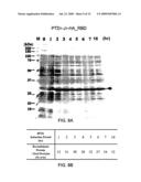
4. The chimeric protein of claim 1, wherein the J-domain is heat shock protein 40 (Hsp40) J-domain.
5. A method for making the protein of claim 1 comprising the steps of:a. generating a protein expression vector comprising a chimeric gene encoding a chimeric protein according to claim 1, wherein the chimeric gene comprises:i. a first DNA sequence encoding a protein transduction domain (PTD) having HIV Tat PTD activity;ii. a second DNA sequence linked in translation frame with the first DNA sequence, encoding a J-domain having heat shock protein 70 (Hsp70)-interacting activity; andiii. a third DNA sequence linked in translation frame with the second DNA sequence, encoding the target protein or polypeptide; andb. transfecting a host cell with the expression vector;c. culturing the host cell transfected with the expression vector under conditions that permit expression of the chimeric protein; andd. isolating the chimeric protein.
Description:
REFERENCES TO RELATED APPLICATION
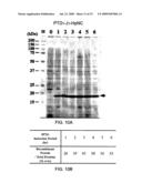
[0001]The present application is a Divisional of U.S. application Ser. No. 12/149,606, filed May 5, 2008, which status is allowed and is herein incorporated by reference in its entirety.
FIELD OF THE INVENTION
[0002]The present invention relates generally to an expression system and more specifically to an expression system for recombinant proteins with enhanced solubility and immunogenicity.
BACKGROUND OF THE INVENTION
[0003]The demand for efficient and large scale production of therapeutic proteins is steadily increasing as more recombinant proteins are approved for use in humans. Using E. coli expression system for production of recombinant proteins, however, frequently results in formation of water-insoluble protein inclusion bodies, instead of functional, soluble proteins. Additionally, resolubilized proteins from inclusion bodies do not elicit as strong an immune responses in rats and mice as soluble, native protein does.
[0004]The Hsp70-peptide antigen complex plays an important role in the antigen presentation process (Mycko et al., 2004; Bendz et al., 2007). The molecular chaperone/co-chaperone pair (Hsp70/Hsp40) is highly conserved throughout evolution. Eukaryotic Hsp70 genes are descents of the bacterial DnaK gene. DnaK and HSP70 have over 50% homology in their amino acid sequences. The structure of DnaK/Hsp70 is composed of a N-terminal ATPase domain, a substrate binding domain, and a C-terminal lid domain (Genevaux et al., 2007; Shaner & Morano, 2007). Hsp40s represent a large protein family that functions to specify the cellular action of Hsp70 chaperone proteins. There are 44 members of Hsp40 genes found in human genome. Twelve of them are descents of E. coli DnaJ. Although their amino acid sequences outside the J-domain are divergent, the 75 amino acid J-domain of the Hsp40 proteins is highly conserved. The J-domain of Hsp40 interacts directly with the Hsp70 ATPase domain to enhance the ATP hydrolysis rate which in turn increases the substrate binding affinity. Hsp40 proteins also bind client substrate and deliver it to the Hsp70 (Fan et al., 2003).
[0005]Protein transduction domain (PTD) peptides are able to ferry large molecules into cells independent of classical endocytosis. They are used to enhance cellular uptake of drugs, proteins, polynucleotides and liposomes (Tilstra et al., 2007).
[0006]Neither the PTD nor the Hsp40 J-domain has been employed in the development of an expression system for improving solubility and immunogenicity of a recombinant protein. A previously unaddressed need exists in the art to address the aforementioned deficiencies and inadequacies, especially in connection with the development of an expression system for a recombinant protein with enhanced solubility and immunogenicity.
SUMMARY OF THE INVENTION
[0007]The invention is related to a protein expression vector for increasing the solubility of a target protein or polypeptide in a host cell expression system, and/or enhancing the immunogenicity of a target protein or polypeptide. The expression vector of the invention has superior performances in producing high yields of soluble recombinant proteins when compared to other commercially available E. coli. expression vectors. In addition, the expression vector of the invention is capable of enhancing the immunogenicity of small peptides that by them-selves are weak in inducing antibodies in animals.
[0008]One aspect of the invention relates to a chimeric protein that contains three polypeptide fragments: (a) a first polypeptidyl fragment located at the N-terminal end of the chimeric protein that contains a protein transduction domain (PTD) or a fragment thereof having HIV Tat PTD activity; (b) a second polypeptidyl fragment located at the C-terminal end of the first polypeptidyl fragment that contains a J-domain or a fragment thereof having heat shock protein 70 (Hsp70)-interacting activity; and (c) a third polypeptidyl fragment at the C-terminal end of the second polypeptidyl fragment that contains a target protein or polypeptide.
[0009]Another aspect of the invention relates to a chimeric gene that includes three DNA sequences: (a) a first DNA sequence encoding a protein transduction domain (PTD) or a fragment thereof having HIV Tat PTD activity; (b) a second DNA sequence encoding, linked in translation frame with the first DNA sequence, encoding a J-domain or a fragment thereof having heat shock protein 70 (Hsp70)-interacting activity; and (c) a third DNA sequence encoding, linked in translation frame with the second DNA sequence, encoding a target protein or polypeptide.
[0010]Another aspect of the invention relates to a chimeric gene that comprises two DNA sequences: (a) a first DNA sequence encoding a protein transduction domain (PTD) or a fragment thereof having HIV Tat PTD activity; and (b) a second DNA sequence encoding, linked in translation frame with the first DNA sequence, encoding a J-domain or a fragment thereof having heat shock protein 70 (Hsp70)-interacting activity.
[0011]Further another aspect of the invention relates to a protein expression vector that comprises a chimeric gene as aforementioned. Yet another aspect of the invention relates to an isolated host cell that contains an expression vector having chimeric gene as aforementioned.
[0012]Another aspect of the invention relates to a method for enhancing immunogenicity and/or solubility of a target protein or polypeptide in a host cell expression system. The method includes the steps of: (a) generating a protein expression vector according to claim 18; (b) transfecting a host cell with the expression vector; (c) culturing the host cell transfected with the expression vector under conditions that permit expression of the target protein or polypeptide; and (d) isolating the target protein or polypeptide.
[0013]Further another aspect of the invention relates to a method of expressing a target protein or polypeptide. The method contains the steps of: (a) transfecting a host cell with the expression vector of claim 18; (b) culturing the host cell transfected with the expression vector under conditions that permit expression of the target protein or polypeptide; and (c) isolating the target protein or polypeptide.
[0014]These and other aspects will become apparent from the following description of the preferred embodiment taken in conjunction with the following drawings, although variations and modifications therein may be affected without departing from the spirit and scope of the novel concepts of the disclosure.
[0015]The accompanying drawings illustrate one or more embodiments of the invention and, together with the written description, serve to explain the principles of the invention. Wherever possible, the same reference numbers are used throughout the drawings to refer to the same or like elements of an embodiment.
BRIEF DESCRIPTION OF THE DRAWINGS
[0016]FIGS. 1A-1C illustrate plasmid maps.
[0017]FIG. 2A shows the nucleotide sequence and the encoded amino acid sequence of the plasmid of FIG. 1A.
[0018]FIG. 2B shows the nucleotide sequence and the encoded amino acid sequence of the plasmid of FIG. 1B.
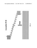
[0019]FIG. 3 is a schematic drawing of the assembly PCR.
[0020]FIG. 4A is a photograph of SDS-PAGE gel electrophoresis of the protein samples from E. coli whole cell lysates. M stands for protein molecular weight marker, lanes 2-9: protein bands from the whole cell lysates after the indicated induction period; "0" stands for "un-induced." The arrow indicates the protein band of the target recombinant protein, PTD1-J1-HSPA1A peptide I.
[0021]FIG. 4B is a chart showing the weight percentage of the recombinant protein relative to the total proteins after the various induction periods.
[0022]FIG. 5A is a photograph of SDS-PAGE gel electrophoresis of the protein samples from E. coli whole cell lysates. M stands for protein molecular weight marker, lanes 2-9: protein bands from the whole cell lysates after the indicated induction period; "0" stands for "un-induced." The arrow indicates the protein band of the target recombinant protein, PTD1-J1-HSPA1A peptide II.
[0023]FIG. 5B is a chart showing the weight percentage of the recombinant protein relative to the total proteins after the various induction periods.
[0024]FIG. 6A is a photograph of SDS-PAGE gel electrophoresis of the protein samples from E. coli whole cell lysates. M stands for protein molecular weight marker; lanes 2-9: protein bands from the whole cell lysates after the indicated induction period; "0" stands for "un-induced." The arrow indicates the protein band of the target recombinant protein, PTD1-J1-HSPA1A peptide III.
[0025]FIG. 6B is a chart showing the weight percentage of the recombinant protein relative to the total proteins after the various induction periods.
[0026]FIG. 7A is a photograph of SDS-PAGE gel electrophoresis of the protein samples from E. coli whole cell lysates. M stands for protein molecular weight marker, lanes 2-9: protein bands from the whole cell lysates after the indicated induction period; "0" stands for "un-induced." The arrow indicates the protein band of the target recombinant protein, PTD1-J1-cIGF-I.
[0027]FIG. 7B is a chart showing the weight percentage of the recombinant protein relative to the total proteins after the various induction periods.
[0028]FIG. 8A is a photograph of SDS-PAGE gel electrophoresis of the protein samples from E. coli whole cell lysates. M stands for protein molecular weight marker; lanes 2-9: protein bands from the whole cell lysates after the indicated induction period; "0" stands for "un-induced." The arrow indicates the protein band of the target recombinant protein, PTD1-J1-HA_RBD.
[0029]FIG. 8B is a chart showing the weight percentage of the recombinant protein relative to the total proteins after the various induction periods.
[0030]FIG. 9A is a photograph of SDS-PAGE gel electrophoresis of the protein samples from E. coli whole cell lysates. M stands for protein molecular weight marker, lanes 2-9: protein bands from the whole cell lysates after the indicated induction period; "0" stands for "un-induced." The arrow indicates the protein band of the target recombinant protein, PTD1-J1-HCV-core protein.
[0031]FIG. 9B is a chart showing the weight percentage of the recombinant protein relative to the total proteins after the various induction periods.
[0032]FIG. 10A is a photograph of SDS-PAGE gel electrophoresis of the protein samples from E. coli whole cell lysates. M stands for protein molecular weight marker, lanes 2-9: protein bands from the whole cell lysates after the indicated induction period; "0" stands for "un-induced." The arrow indicates the protein band of the target recombinant protein, PTD1-J1-HpNC.
[0033]FIG. 10B is a chart showing the weight percentage of the recombinant protein relative to the total proteins after the various induction periods.
[0034]FIG. 11 is a photograph showing the result of a Dot blot analysis.
[0035]FIG. 12A is a photograph of SDS-PAGE gel electrophoresis. "S" stands for supernatant; "P" stands for insoluble pellet fraction. Prior to being loaded into the gel, the insoluble pellet was dissolved in SDS-PAGE loading buffer.
[0036]FIG. 12B is a photograph of SDS-PAGE gel electrophoresis. The labels for lanes 1-10 are the same as FIG. 12A.
[0037]FIG. 13 is a chart showing the effects of the temperature on the recombinant protein expression. The peak area ratio represents the relative ratio of the recombinant proteins in the soluble fraction of E. coli cell lysate against a constitutively expressed internal protein.
[0038]FIG. 14A is a photograph of SDS-PAGE gel electrophoresis.
[0039]FIG. 14B is a photograph showing the result of a Dot blot analysis.
DETAILED DESCRIPTION OF THE INVENTION
Definitions
[0040]The terms used in this specification generally have their ordinary meanings in the art, within the context of the invention, and in the specific context where each term is used. Certain terms that are used to describe the invention are discussed below, or elsewhere in the specification, to provide additional guidance to the practitioner regarding the description of the invention. For convenience, certain terms may be highlighted, for example using italics and/or quotation marks. The use of highlighting has no influence on the scope and meaning of a term; the scope and meaning of a term is the same, in the same context, whether or not it is highlighted. It will be appreciated that same thing can be said in more than one way. Consequently, alternative language and synonyms may be used for any one or more of the terms discussed herein, nor is any special significance to be placed upon whether or not a term is elaborated or discussed herein. Synonyms for certain terms are provided. A recital of one or more synonyms does not exclude the use of other synonyms. The use of examples anywhere in this specification including examples of any terms discussed herein is illustrative only, and in no way limits the scope and meaning of the invention or of any exemplified term. Likewise, the invention is not limited to various embodiments given in this specification.
[0041]Unless otherwise defined, all technical and scientific terms used herein have the same meaning as commonly understood by one of ordinary skill in the art to which this invention pertains. In the case of conflict, the present document, including definitions will control.
[0042]As used herein, "around", "about" or "approximately" shall generally mean within 20 percent, preferably within 10 percent, and more preferably within 5 percent of a given value or range. Numerical quantities given herein are approximate, meaning that the term "around", "about" or "approximately" can be inferred if not expressly stated.
[0043]The term "fusion protein," also known as "chimeric protein," refers to "a protein created through the joining of two or more genes which originally coded for separate proteins."
[0044]The term "immunogenicity" refers to the ability of antigen to provoke an immune response.
[0045]The term "enhancing solubility of a target protein or polypeptide" refers to "a significant increase in the amount of target protein or polypeptide that otherwise form inclusion bodies is expressed in soluble state when compared to a thioredoxin-fused target protein.
[0046]The invention is related to a recombinant protein expression vector that can enhance the immunogenicity of a target protein or polypeptide and increase its solubility in a host cell expression system.
[0047]An expression vector system containing a Hsp40 family proteins J-domain was designed to examine whether a target protein expressed using the J-domain-containing expression system of the invention would have an increased solubility when compared to the target protein expressed using other expression systems where insoluble inclusion bodies were formed. In addition, a protein transduction domain (PTD) (11 amino acid residues in length) (Ryu et al., 2003; Jones et al., 2005) of HIV Tat protein was fused to the N-terminus of the J-domain to facilitate the recombinant protein PTD1J1-polypeptide to penetrate cell membranes when the fusion polypeptide would be used as an antigen (Wadia et al., 2004; Kaplan et al., 2005). After penetrating the cell membrane of antigen presenting cells (APCs), the J-domain would target to the Hsp70 ATPase domain and thereby allow the fusion polypeptide to be caught by the Hsp70 substrate binding site. The Hsp70-peptide antigen complex has been known to plays an important role in the process of antigen presentations.
[0048]The invention is related to a recombinant protein expression vector that can enhance the immunogenicity of a target protein or polypeptide, and increase the solubility of a target protein or polypeptide in a host cell expression system. Utilizing the ability of the J-domain of the mammalian Hsp40s in targeting to bacterial DnaK, an E. coli expression vector was designed to express recombinant proteins that had been proven forming insoluble inclusion bodies in other expression vectors to investigate whether the J-domain would help folding and thus increased the solubility of the fusion polypeptide/protein. In addition, a protein transduction domain (PTD) (11 amino acid residues in length) (Ryu et al., 2003; Jones et al., 2005) of HIV Tat protein was fused to the N-terminus of the J-domain to facilitate the recombinant protein PTD1J1-polypeptide to penetrate cell membranes when the fusion polypeptide would be used as an antigen (Wadia et al., 2004; Kaplan et al., 2005). After penetrating the cell membrane of antigen presenting cells (APCs), the J-domain would target to the Hsp70 ATPase domain and thereby allow the fusion polypeptide to be caught by the Hsp70 substrate binding site. The Hsp70-peptide antigen complex has been known to plays an important role in the process of antigen presentations.
[0049]One aspect of the invention relates to a chimeric protein that contains three polypeptide fragments: (a) a first polypeptidyl fragment located at the N-terminal end of the fusion protein that contains a protein transduction domain (PTD) or a fragment thereof having HIV Tat PTD activity; (b) a second polypeptidyl fragment located at the C-terminal end of the first polypeptidyl fragment that contains a J-domain or a fragment thereof having heat shock protein 70 (Hsp70)-interacting activity; and (c) a third polypeptidyl fragment at the C-terminal end of the second polypeptidyl fragment that contains a target protein or a polypeptide. The first polypeptidyl fragment of the chimeric protein may include an amino acid sequence set forth by SEQ ID NO: 1. The second polypeptidyl fragment of the chimeric protein may contain an amino acid sequence set forth by SEQ ID NO: 3.
[0050]Another aspect of the invention relates to a chimeric gene that includes three DNA sequences: (a) a first DNA sequence encoding a protein transduction domain (PTD) or a fragment thereof having HIV Tat PTD activity; (b) a second DNA sequence encoding, linked in translation frame with the first DNA sequence, encoding a J-domain or a fragment thereof having heat shock protein 70 (Hsp70)-interacting activity; and (c) a third DNA sequence encoding, linked in translation frame with the second DNA sequence, encoding a target protein or polypeptide.
[0051]Another aspect of the invention relates to a chimeric gene that comprises two DNA sequences: (a) a first DNA sequence encoding a protein transduction domain (PTD) or a fragment thereof having HIV Tat PTD activity; and (b) a second DNA sequence encoding, linked in translation frame with the first DNA sequence, encoding a J-domain or a fragment thereof having heat shock protein 70 (Hsp70)-interacting activity.
[0052]The J-domain may be at least one selected from the group consisting of a heat shock protein 40 (Hsp40) J-domain and a simian virus 40 (SV40) large T antigen (TAg) J-domain. The SV40 TAg J-domain is located within the N-terminal domain (residues 1 to 82) of SV40 TAg. It has been reported that the N-terminal J-domain of TAg shares functional homology with the Hsp40 J domain (DeCaprio et al., 1997). In one embodiment of the invention, the J-domain is a human heat shock protein 40 J-domain.
[0053]The first DNA sequence of the chimeric gene may encode a protein transduction domain (PTD) that contains an amino acid sequence set forth by SEQ ID NO: 1. The second DNA sequence of the chimeric gene may encode a J-domain comprising an amino acid sequence set forth by SEQ ID NO: 3.
[0054]Another aspect of the invention relates to a method for enhancing immunogenicity and/or solubility of a target protein or polypeptide in a host cell expression system. The method includes the steps of: (a) generating a protein expression vector according to claim 18; (b) transfecting a host cell with the expression vector; (c) culturing the host cell transfected with the expression vector under conditions that permit expression of the target protein or polypeptide; and (d) isolating the target protein or polypeptide.
[0055]Further another aspect of the invention relates to a method of expressing a target protein or polypeptide. The method contains the steps of: (a) transfecting a host cell with the expression vector of claim 18; (b) culturing the host cell transfected with the expression vector under conditions that permit expression of the target protein or polypeptide; and (c) isolating the target protein or polypeptide.
[0056]Further another aspect of the invention relates to a protein expression vector that comprises one of the aforementioned chimeric genes.
[0057]Yet another aspect of the invention relates to an isolated host cell that contains an expression vector having one of the aforementioned chimeric genes.
EXAMPLES
[0058]Without intent to limit the scope of the invention, exemplary instruments, apparatus, methods and their related results according to the embodiments of the present invention are given below. Note that titles or subtitles may be used in the examples for convenience of a reader, which should in no way limit the scope of the invention. Theories are proposed and disclosed herein should in no way limit the scope of the invention so long as the invention is practiced according to the invention without regard for any particular theory or scheme of action.
Example 1
Construction of pET22b-PTD1-J1 Expression Vector
[0059]FIGS. 1A-1C illustrate three pET22b derivatives plasmids: pET22b-PTD1, pET22b-PTD1-J1, and pET22b-PTD1-target protein (or antigenic protein). These plasmids were generated using the methods as described below.
[0060]Plasmid pET22b-PTD1. The plasmid pET22b-PTD1 contains a HIV tat PTD1 (native form) gene insert between the restriction enzymes NadI and BamH1, as shown in FIG. 2A. The PTD1 gene, which encodes the amino acid sequence of PTD1 (SEQ ID NO: 1), was synthesized using the oligonucleotide pair, PTD1 f (TATGTATGGTCGTAAGAACGTCGTCAGCGTCGTCGTGG; SEQ ID NO: 5) and PTD1 r (GATCCCACGACGACGCTGACGACGTTTCTTACGACCATACA; SEQ ID NO: 6). The oligonucleotide pair encoding the optimized codons of protein transduction domain (PTD1) (SEQ ID NO: 1) for E. coli were phosphorylated by polynucleotide kinase before annealing to the double strand form in order to be inserted into the pET22b vector (Novagen, Madison, Wis.) to generate pET22b-PTD1 (FIG. 1A). Before ligating with the PTD1 gene insert, the pET22b vector was co-digested by NdeI and BamHI and dephosphorylated by calf intestine alkaline phosphatase (CIAP). FIG. 2A shows the detailed nucleotide sequence of the plasmid pET22b-PTD1 and encoded PTD1 amino acid sequence.
[0061]Plasmid pET22b-PTD1-J1. The plasmid pET22b-PTD1-J1 contains a fusion gene with the PTD1 gene being fused to the N-terminus of the HSP40 J domain gene. The fusion gene encodes a chimeric protein PTD1-J1. The J domain of the HSP40 gene was synthesized by assembly PCR using the oligonucleotides that were designed to obtain optimized codons for E. coli. Table 1 lists the nucleotide sequences of the oligonucleotide primers (SEQ ID NOs: 7 to 16) used in the assembly PCR synthesis of the HSP40 J domain. The concentrations of the primers in the assembly PCR reaction mixture were adjusted as follows: 0.5 μM of F1 and R1 and 0.05 μM of F2, F3, R3, and R2, and so on in the reaction mixture. The assembly PCR profile was 2 min at 94° C., 20 sec at 94° C., 40 sec at 40° C., 20 sec at 72° C. of 20 cycles, and 5 min at 72° C.
[0062]The PCR product was cloned into pGEM-T Easy vector (Promega) for colony selection and DNA sequencing. The plasmid with correct DNA sequence encoding Hsp40-J domain (SEQ ID NO: 3) was co-digested by BamHI and EcoRI to remove the 0.2 kb inserted DNA fragment from pGEM-T Easy vector. This DNA fragment was then inserted into pET22b-PTD1 vector, which had been digested by BamHI/EcoRI and CIAP, to generate the pET22b-PTD1-J1 expression vector (FIG. 1B). FIG. 2B shows the detailed nucleotide sequence of the plasmid pET22b-PTD1-J1 and the amino acid sequence of the encoded fusion protein PTD1-J1. FIG. 3 is a schematic drawing of the assembly PCR, which is an artificial synthesis of long DNA sequences by performing PCR on a pool of long oligonucleotides with short overlapping segments.
TABLE-US-00001 TABLE 1 Primer Sequence Sequence ID J1 tggatcctgggtaaagattactaccagactc SEQ ID NO: 7 nBF1 acggtctc F2 tactaccagactcacggtctcgctcgtggtg SEQ ID NO: 8 catctgatgatgaaatc F3 ggtgcatctgatgatgaaatcaaacgtgctt SEQ ID NO: 9 accgtcgtcaggcactg F4 gcttaccgtcgtcaggcactgcgttaccatc SEQ ID NO: 10 cagacaaaaacaaagaa F5 taccatccagacaaaaacaaagaaccgggtg SEQ ID NO: 11 cagaagagaaattc F6 ccgggtgcagaagagaaattcaaagagatcg SEQ ID NO: 12 cagaagcatacgacgtt EcoRI cgaattcgcaccaccagaaccacctttcaga SEQ ID NO: 13 R1 ccttc R2 agaaccacctttcagaccttcttcaccgtaa SEQ ID NO: 14 cggtcgaagatttcacg R3 gtaacggtcgaagatttcacgtttacgtgga SEQ ID NO: 15 tcgctcagaacgtcgta R4 tggatcgctcagaacgtcgtatgcttctgcg SEQ ID NO: 16 atctc
[0063]Plasmid pET22b-PTD1-J1-Target protein. The plasmid pET22b-PTD1-J1-target protein contains a fusion gene encoding a chimeric protein, PTD1-J1-target protein. Target proteins that were inserted into the expression vector pET22b-PTD1-J1 included those proteins that had been proven to form insoluble inclusion bodies due to the solubility issue when using other expression vectors, and small peptides that are weak in inducing immunogenicity and/or having solubility issues in E. coli expression system. A DNA fragment comprising a codon-optimized cDNA sequence encoding a target protein to be expressed in the pET22b-PTD1-J1 expression vector was synthesized by PCR, and the PCR product was then inserted into the pET22b-PTD1-J1 vector at EcoRI and XhoI sites to generate the plasmid pET22b-PTD1-J-target protein. The resulting plasmid was transformed into E. coli Rosetta (Novagen) competent cells to obtain clones.
Example 2
Expression and Analyses of Recombinant Proteins
[0064]Chimeric proteins, PTD1-J1-target protein, were expressed in E. coli cultures containing corresponding expression plasmids. In brief, the colony resistant to ampicillin and chloramphenicol was cultured and amplified in TYD medium (10 g trypton, 20 g yeast extract, 5 g NaCl, 2 g Dextrose per liter, pH 7.2). Once OD600 reached 0.3±0.1, the bacterial culture was induced with isopropylthio-β-D-galactoside (IPTG; Promega, USA) at a final concentration of 1 mM at 37° C. in a rotating incubator shaken at 225 rpm. After the IPTG induction, the cells were collected by centrifugation at 5,000×g for 10 min, washed once with PBS, resuspended in PBS and homogenized by sonication. The sonicated lysates were centrifuged at 15,000×g for 30 min. Twenty microliters of total lysates from 0.3 OD600 unit of E. coli cells were mixed with 30 μL of 2×SDS-PAGE loading buffer, and the mixture was loaded into each well of the gel. The protein bands were visualized by Coomassie brilliant blue R250 staining of the gel, and scanned by laser densitometer (Personal Densitometer SI, Molecular Dynamics). The amount of each recombinant protein over the amount of total protein was calculated and represented as percentage weight.
Example 3
Expression of Human HSPA1A Peptide I
[0065]A DNA fragment comprising a codon-optimized cDNA sequence (SEQ ID NO: 17) encoding the amino acid sequence (SEQ ID NO: 18) of human heat shock 70 kDa protein 1A (HSPA1A) Peptide I was synthesized by assembly PCR using the primers listed in Table 2. The assembly PCR profile was 2 min at 94° C., 20 sec at 94° C., 40 sec at 40° C., 20 sec at 72° C. of 20 cycles, and 5 min at 72° C. The PCR product was then inserted in the expression vector pET22b-PTD1-J1 to generate the plasmid pET22b-PTD1-J1-HSPA1A peptide I using the method described in Example 1.
TABLE-US-00002 TABLE 2 Primer Sequence Sequence ID RI-I-f1 gaattctagcagcagcacccaggcgagcc SEQ ID NO: 19 tggaaattgatagcctgttt I-f2 agcctggaaattgatagcctgtttgaagg SEQ ID NO: 20 cattgat ttttataccagc I-f3 gtttgaaggcattgatttttataccagca SEQ ID NO: 21 tt acccgtgcgcgttttgaa I-r3 gggtgctacgaaacagatcgctgcacagt SEQ ID NO: 22 tcttcaaaacgcgcacgggt I-r2 catcacgcagcgctttttccaccggttcc SEQ ID NO: 23 agggtgctacgaaacagatc Xho-I-r1 ctcgagaatctgcgctttatccagtttcg SEQ ID NO: 24 catc acgcagcgctttttc
[0066]Plasmid pET22b-PTD1-J1-HSPA1A peptide I that carried the genes encoding the recombinant protein PTD1-J1-HSPA1A peptide I was transformed into E. coli Rosetta. The recombinant protein expression was induced by IPTG according to the method described above for up to 16 hours and the amounts of protein induced in the cell extract determined as in Example 2.
[0067]FIG. 4A shows a Coomassie® blue stained SDS-PAGE gel analysis of E. coli whole cell lysates. The protein bands shown between 15 kDa and 20 kDa (indicated by an arrow) correspond to the target protein, pET22b-PTD1-J1-HSPA1A peptide 1. Lane 1: standard molecular weight marker. Lanes 2-9: whole cell lysates after the IPTG induction, where "0 hr" stands for "uninduced."
[0068]The effect of induction period of IPTG induction on the yield of the PTD1-J-HSPA1A peptide I expression was quantitated and represented by the weight percentage of the recombinant PTD1-J1-HSPA1A peptide I over the total proteins. As shown in FIG. 4B, the induction reached a plateau after about 4 hrs. The expression of PTD1-J1-HSPA1A peptide I declined as the IPTG induction was extended to 16 hours.
Example 4
Expression of human HSPA1A Peptide II
[0069]A DNA fragment comprising a codon-optimized cDNA sequence (SEQ ID NO: 25) encoding the amino acid sequence (SEQ ID NO: 26) of human heat shock 70 kDa protein 1A (HSPA1A) peptide II was synthesized by assembly PCR using the primers (SEQ ID NO: 27 to SEQ ID NO: 33) listed in Table 3. The assembly PCR profile was 2 min at 94° C., 20 sec at 94° C., 40 sec at 40° C., 20 sec at 72° C. of 20 cycles, and 5 min at 72° C. The PCR product was then inserted in the expression vector pET22b-PTD1-J1 to generate the plasmid pET22b-PTD1-J-HSPA1A peptide II using the method described in Example 1.
TABLE-US-00003 TABLE 3 Primer Sequence Sequence ID RI-II-f1 gaattctaacgtaaccgctactgacaa SEQ ID NO: 27 atccactggtaaagctaacaag II-f2 tccactggtaaagctaacaagatcacc SEQ ID NO: 28 atcaccaacgacaaaggtcgtc II-g3 atcaccaacgacaaaggtcgtctgtcc SEQ ID NO: 29 aaggaagagatcgagcgtatgg II-f4 aaggaagagatcgagcgtatggttcag SEQ ID NO: 30 gaagctgaaaagtacaag II-r3 acgctgaacttcgtcttcagccttgta SEQ ID NO: 31 cttttcagcttcctgaacc II-r2 agcgttcttagcggaaacacgttcacg SEQ ID NO: 32 ctgaacttcgtcttcagc Xho-II-r1 ctcgaggaaagcgtaggattccagagc SEQ ID NO: 33 gttcttagcggaaacacgttca
[0070]Plasmid pET22b-PTD1-J1-HSPA1A peptide II that carried the genes encoding the recombinant protein PTD1-J1-HSPA1A peptide II was transformed into E. coli Rosetta. The recombinant protein expression was induced by IPTG according to the method described above. The amount of protein induced in the cell lysate over the 16 hours IPTG induction was monitored as in Example 2 above.
[0071]FIG. 5A shows a Coomassie® blue stained SDS-PAGE gel analysis of E. coli whole cell lysates. The protein bands shown between 15 kDa and 20 kDa (indicated by an arrow) correspond to the chimera protein P PTD1-J1-HSPA1A peptide II. Lane 1: standard molecular weight marker. Lanes 2-9: whole cell lysates after the IPTG induction, where "0 hr" stands for "uninduced."
[0072]The amount of the PTD1-J1-HSPA1A peptide II expression after each induction period was quantitated and the yield expressed as the weight percentage of the recombinant PTD1-J1-HSPA1A peptide II over the total proteins. As shown in FIG. 5B, the induction reached a plateau after 4 hrs. The expression of PTD1-J1-HSPA1A peptide II declined as the IPTG induction was extended to 16 hours.
Example 5
Expression of Human HSPA1A Peptide III
[0073]A DNA fragment comprising a codon-optimized cDNA sequence (SEQ ID NO: 34) encoding the amino acid sequence (SEQ ID NO: 35) of human heat shock 70 kDa protein 1A (HSPA1A) peptide III was synthesized by assembly PCR using the primers (SEQ ID NO: 36 to SEQ ID NO: 42) listed in Table 4. The assembly PCR profile was 2 min at 94° C., 20 sec at 94° C., 40 sec at 40° C., 20 sec at 72° C. of 20 cycles, and 5 min at 72° C. The PCR product was then inserted in the expression vector pET22b-PTD1-J1 to generate the plasmid pET22b-PTD1-J-HSPA1A peptide III using the method described in Example 1.
TABLE-US-00004 TABLE 4 Primer Sequence Sequence ID RI-III-f1 gaattctgaagatgaaggcctgaaag SEQ ID NO: 36 gcaaaattagcgaagcggat III-f2 ggcaaaattagcgaagcggataagaa SEQ ID NO: 37 aaaggtgctg gataaatgccag III-f3 ggtgctggataaatgccag gaagtg SEQ ID NO: 38 attagctggctg gatgcgaacacc III-f4 gctggatgcgaacaccctggcggaaa SEQ ID NO: 39 aagatgaattt gaacataaacgt III-r3 ttccagttctttacgtttatgttcaa SEQ ID NO: 40 attcatctttttccgccag III-r2 cgggttgcacacctgttccagttctt SEQ ID NO: 41 tacgtttatgttcaaattcatc Xho-III-r1 ctggagcgcgccctgatacaggccgc SEQ ID NO: 42 taataatcgggttgcacacctg
[0074]Plasmid pET22b-PTD1-J1-HSPA1A peptide III that carried the genes encoding the recombinant protein PTD1-J1-HSPA1A peptide III was transformed into E. coli Rosetta. The recombinant protein expression was induced by IPTG according to the method described above. The amount of protein induced in the cell lysate was monitored over the 16 hours IPTG induction as in Example 2 above.
[0075]FIG. 6A shows a Coomassie® blue stained SDS-PAGE gel analysis of E. coli whole cell lysates. The protein bands shown between 15 kDa and 20 kDa (indicated by an arrow) correspond to the chimera protein PTD1-J1-HSPA1A peptide III. Lane 1: standard molecular weight marker. Lanes 2-9: whole cell lysates after the IPTG induction, where "0 hr" stands for "uninduced."
[0076]The amount of the PTD1-J1-HSPA1A peptide III expression after each induction period was quantitated and the yield represented by the weight percentage of the recombinant PTD1-J1-HSPA1A peptide III over the total proteins. As shown in FIG. 6B, the induction reached a plateau after 2 hrs. The expression of PTD1-J1-HSPA1A peptide III declined as the IPTG induction was extended.
Example 6
Expression of Chicken IGF-I
[0077]A DNA fragment comprising a codon-optimized cDNA sequence (SEQ ID NO: 43) encoding the amino acid sequence (SEQ ID NO: 44) of chicken IGF-I peptide (cIGF-I) was synthesized by assembly PCR using the primers (SEQ ID NO: 45 to SEQ ID NO: 51) listed in Table 5. The assembly PCR profile was 2 min at 94° C., 20 sec at 94° C., 40 sec at 40° C., 20 sec at 72° C. of 20 cycles, and 5 min at 72° C. The PCR product was then inserted in the expression vector pET22b-PTD1-J1 to generate the plasmid pET22b-PTD1-J1-cIGF-I using the method described in Example 1.
TABLE-US-00005 TABLE 5 Primer Sequence Sequence ID RI-F1 gaattctggtccagaaaccctgtgtggtgca SEQ ID NO: 45 new gaactggttgatgcactgcagttcgtg F2 gaactggttgatgcactgcagttcgtgtgtg SEQ ID NO: 46 gtgatcgtggtttctac F3 ggtgatcgtggtttctacttcagcaaaccga SEQ ID NO: 47 ctggttatggtagctctagc F4 ggttatggtagctctagccgtcgtctgcatc SEQ ID NO: 48 acaaaggtattgtggatg F5 cacaaaggtattgtggatgaatgttgctttc SEQ ID NO: 49 agagctgtgatctgcgtcg R2 gattggtgcacagtacatttccagacgacgc SEQ ID NO: 50 agatcacagctctg Xho-R1 ctcgagtgcgcttttcggtggutgattggtg SEQ ID NO: 51 cacagtacatttcc
[0078]Plasmid pET22b-PTD1-J1-cIGF-I that carried the genes encoding the recombinant protein PTD1-J1-cIGF-I was transformed into E. coli Rosetta. The recombinant protein expression was induced by IPTG according to the method described above for up to 6 hours and the amounts of proteins induced in the cell extracts determined as in Example 2 above.
[0079]FIG. 7A shows a Coomassie® blue stained SDS-PAGE gel analysis of E. coli whole cell lysates. The protein bands shown around 20 kDa (indicated by an arrow) correspond to the chimera protein PTD1-J1-cIGF-1. Lane 1: standard molecular weight marker (M). Lanes 2-8: whole cell lysates after the IPTG induction, where "0 hr" stands for "uninduced." The amount of the PTD1-J1-cIGF-I expression after each induction period was quantitated and the yield represented by the weight percentage of the recombinant protein PTD1-J1-cIGF-I over the total proteins. As shown in FIG. 7B, the induction reached a plateau after about 2 to 4 hrs.
Example 7
Expression of Avian Influenza Virus HA (H5) Receptor Binding Domain (RBD)
[0080]A DNA fragment, which comprises a native viral cDNA sequence (SEQ ID NO: 52) encoding the amino acid sequence (SEQ ID NO: 53) of the receptor binding domain (RBD) of avian Influenza virus Hemagglutin (HA) subtype H5, was synthesized by PCR using the primer pair (SEQ ID NOs: 54 and 55) listed in Table 6, and avian influenza virus hemagglutinin (HA) subtype H5 cDNA as a DNA template (GeneBank accessing number: EF419243). The PCR profile was 2 min at 94° C., 20 sec at 94° C., 40 sec at 60° C., 20 sec at 72° C. of 20 cycles, and 5 min at 72° C. The PCR product was inserted in the expression vector pET22b-PTD1-J1 to generate the plasmid pET22b-PTD1-J1-HA_RBD using the method described in Example 1 above.
TABLE-US-00006 TABLE 6 Primer Sequence Sequence ID RI HA-RBD f tgaattcgaaacacctattgagca SEQ ID NO: 54 gaataaac Xho HA-RBD r tctcgagaattgttgagtcccctt SEQ ID NO: 55 tcttgac
[0081]Plasmid pET22b-PTD1-J1-HA_RBD that carried the genes encoding the recombinant protein PTD1-J1-HA_RBD was transformed into E. coli. Rosetta. The recombinant protein expression was induced by IPTG according to the method described above. The amount of protein induced in the cell lysate was monitored over the 16 hours IPTG induction as in Example 2 above.
[0082]FIG. 8A shows a Coomassie® blue stained SDS-PAGE gel analysis of E. coli whole cell lysates. The protein bands shown between around 37 kDa and 25 kDa (indicated by an arrow) correspond to the chimera protein PTD1-J1-HA_RBD. Lane 1: standard molecular weight marker (M). Lanes 2-10: whole cell lysates after the IPTG induction, where "0 hr" stands for "uninduced." The amount of the PTD1-J-HA_RBD expression after each induction period was quantitated and the yield represented by the weight percentage of the recombinant protein PTD1-J1-HA_RBD over the total proteins. As shown in FIG. 8B, the induction reached a plateau after about 3 hrs.
Example 8
Expression of Hepatitis C Virus Core Protein
[0083]A DNA fragment, which comprises a native viral cDNA sequence (SEQ ID NO: 56) encoding the amino acid sequence (SEQ ID NO: 57) of the hepatitis C virus (HCV) core protein, was synthesized by PCR using the primer pair (SEQ ID NOs: 58 and 59) listed in Table 7 and a HCV cDNA that was reversely transcribed from a serum sample of an acute, hepatitis C infected-patent as a DNA template. The PCR profile was 2 min at 94° C., 20 sec at 94° C., 40 sec at 60° C., 20 sec at 72° C. of 40 cycles, and 5 min at 72° C. The PCR product was inserted in the expression vector pET22b-PTD1-J1 to generate the plasmid pET22b-PTD1-J1-HCV-core protein using the method described in Example 1 above.
TABLE-US-00007 TABLE 7 Primer Sequence Sequence ID RI-Core f tgaattcgatgagcacaaatcctaaa SEQ ID NO: 58 cctc Xho-Core r tctcgagagcagagaccggaacggtg SEQ ID NO: 59 at
[0084]Plasmid pET22b-PTD1-J1-HCV-core protein that carried the genes encoding the recombinant protein PTD1-J1-HCV-core protein was transformed into E. coli Rosetta. The recombinant protein expression was induced by IPTG according to the method described above. The amount of protein induced in the cell lysate was monitored over the 16 hours IPTG induction as in Example 2 above.
[0085]FIG. 9A shows a Coomassie® blue stained SDS-PAGE gel analysis of E. coli. whole cell lysates. The protein bands shown between around 37 kDa and 25 kDa (indicated by an arrow) correspond to the chimera protein PTD1-J1-HCV-core protein. Lane 1: standard molecular weight marker (M). Lanes 2-8: whole cell lysates after the IPTG induction, where "0 hr" stands for "uninduced." The amount of the PTD1-J1-HCV core protein expression after each induction period was quantitated and the yield represented by the weight percentage of the recombinant protein PTD1-J1-HCV core protein over the total proteins. As shown in FIG. 9B, the induction reached a plateau after about 5 to 6 hrs.
Example 9
Expression of Human Haptoglobin Fusion Polypeptide
[0086]A fusion gene comprising a nucleotide sequence (SEQ ID NO: 60) that encoded an amino acid sequence (SEQ ID NO: 61) of haptoglobin α-chain fusion polypeptide, HpNC, was synthesized by assembly PCR using the primers (SEQ ID NO: 62 to SEQ ID NO: 65) listed in Table 8. The fusion polypeptide HpNC comprises a peptide fragment from the N-terminal portion (DSGNDVTDIADDG, the amino acid residues 1 to 13 of SEQ ID NO: 61) of the mature haptoglobin, a linker (-GSGG-), and a peptide fragment from the C-terminal portion (RHYEGSTVPEKKTPKS, the amino acid residues 18 to 33 of SEQ ID NO: 61) of the premature haptoglobin α-chain. The assembly PCR profile was 2 min at 94° C., 20 sec at 94° C., 40 sec at 40° C., 20 sec at 72° C. of 20 cycles, and 5 min at 72° C. The PCR product was inserted in the expression vector pET22b-PTD1-J1 to generate the plasmid pET22b-PTD1-J1-HpNC using the method described in Example 1.
[0087]Plasmid pET22b-PTD-J1-HpNC that carried the genes encoding the recombinant protein PTD1-J1-HpNC was transformed into E. coli Rosetta. The recombinant protein expression was induced by IPTG according to the method described above for up to 6 hours, and the amounts of proteins in cell extracts determined as in Example 2.
TABLE-US-00008 TABLE 8 Primer Sequence Sequence ID RI-F1 agaattctgatagcggcaacgatgtgaccga SEQ ID NO: 62 tattgcggatgatggtgg F2 tgaccgatattgcggatgatggtggtagtgg SEQ ID NO: 63 tggtcgtcattat F3 atggtggtagtggtggtcgtcattatgaagg SEQ ID NO: 64 tagcaccgtt R2 ggttttcttctccggaacggtgctaccttca SEQ ID NO: 65 taatgacgaccaccac Xho-R1 tctcgagaccaccgctctttggggttttctt SEQ ID NO: 66 ctccggaacggtgcta
[0088]FIG. 10A shows a Coomassie® blue stained SDS-PAGE gel analysis of E. coli whole cell lysates. The protein bands shown between around 15 kDa and 20 kDa (indicated by an arrow) correspond to the fusion polypeptide PTD1-J1-HpNC. Lane 1: standard molecular weight marker (M). Lanes 2-10: total whole cell lysates after the IPTG induction, where "0 hr" stands for "uninduced." The amount of the PTD1-J1-HpNC expression after each induction period was quantitated and the yield expressed as the weight percentage of the recombinant protein PTD1-J1-HpNC over the total proteins. As shown in FIG. 10B, the induction reached a plateau after about 3 hrs.
Example 10
Expression of Neutrophile Peptide-1
[0089]A cDNA clone comprising a nucleotide sequence (SEQ ID NO: 67) that encodes an amino acid sequence (SEQ ID NO: 68) of Neutrophile peptide-1 (NP-1) was obtained by screening a rabbit intestine cDNA library. The cDNA fragment was inserted into the expression vector pET22b-PTD1-J1 to generate the plasmid pET22b-PTD1-J1-NP-1. Plasmid pET22b-PTD1-J1-NP-1 that carried the genes encoding the recombinant protein PTD1-J1-NP-1 was transformed into E. coli Rosetta. The recombinant protein expression was induced by IPTG according to the method described above.
Example 11
Target Protein-Specific Immunogenicity
[0090]Immunization of animals. Balb/c Mice of 5-week-old were immunized subcutaneously with 50 μg of the recombinant protein PTD1-J1-NP-1 emulsified in TiterMAX Gold adjuvant. Boosts were carried out every 2 weeks. Five days after the second boost, the sera of the mice were collected and assayed for the antibody against the recombinant protein PTD1-J1-NP-1. The specificity of anti-serum against NP-1 was determined using the dot blotting assay.
[0091]Dot Blotting Assay. Solutions containing various amounts of recombinant proteins (antigens), PTD1-J-NP-1, PTD1-J-HCV-core protein, and PTD1-J1, were respectively spotted onto PVDF membranes. The membranes were blocked by 5% skim milk in TBSN (25 mM Tris-HCl, pH 7.4, 150 mM NaCl, 0.02% Tween 20) for 1 hour. After 4 washes with TBSN, the membranes were incubated with mouse anti-serum overnight at a dilution 1:1000, unbound primary antibodies (Ab) removed from the membrane by 4 washes with TBSN, and incubated with HRP-conjugated goat anti-mouse secondary Ab (2000× dilution) for 4 hours. The labeled proteins on washed membrane were detected by chemiluminescence according to manufacturer's instructions (Pierce).
[0092]As shown in FIG. 11, the mouse anti-serum, at 1000× dilution, was highly sensitive in detecting the antigen PTD1-J1-NP-1, less sensitive to PTD1-J1-HCV-core protein, and even less sensitive to PTD1-J1. The antigen PTD1-J1-NP-1 in amounts as small as 320 pg was detectable by the mouse anti-serum (1000× dilution). On the other hand, at least 32 ng of PTD1-J1-HCV-core protein was needed to produce an equivalently weak signal. The signal produced by interacting with PTD1-J1 was very weak even at the amounts as high as 100 ng. This indicates that more than 99% of anti-serum interacted with the target protein NP-1, and less than 1% interacted with PTD1-J1-HCV-core protein and PTD1-J1. Therefore, the recombinant protein PTD1-J1-NP-1 was able to raise a high titer of antibody specific against the target protein NP-1.
Example 12
Enhanced Solubility of Recombinant Protein
[0093]Construction of pET32b-cIGF-I. To compare the solubility of a target protein with the protein fused to a J domain, the construct pET32b-cIGF-I was generated by inserting the EcoRI/XhoI DNA fragment of pET22b-PTD1-J1-cIGF-I into the same restriction enzyme sites of pET32b to form a pET32b-cIGF-I expression vector.
[0094]Expression of Target Protein. Recombinant proteins were expressed in E. coli cultures containing corresponding expression plasmids. In brief, the colony resistant to ampicillin and chloramphenicol was cultured and amplified in TYD medium (10 g trypton, 20 g yeast extract, 5 g NaCl, 2 g Dextrose per liter, pH 7.2) at 37° C. Once the bacterial density reached 0.3±0.1 OD600, the incubation temperature of bacterial cultures were set at 37° C., 30° C., 25° C., 20° C. and 15° C., respectively, and then isopropylthio-β-D-galactoside (IPTG; Promega, USA) was added into the cultures at a final concentration of 1 mM, and induced protein expression for 3, 4, 5, 6 hrs, and overnight, respectively, in a rotating incubator shaken at 225 rpm. After the IPTG induction, the cells were collected by centrifugation at 5,000×g for 10 min, washed once with PBS and resuspended in PBS for homogenization by sonication. The sonicated lysates were centrifuged at 15,000×g for 30 min. Supernatants (soluble fractions) were collected. The pellets (insoluble fractions) were washed once with PBS to remove residual soluble proteins before re-suspending in PBS to obtain the insoluble fractions, which included protein inclusion bodies. About 30 μl of each fraction from the amount equivalent of 0.5 OD600 unit of E. coli cells were loaded into each well of the gel. The gel was stained with Coomassie brilliant blue R-250 and scanned by laser densitometer (Personal Densitometer SI, Molecular Dynamics).
Results:
[0095]Expression of thioredoxin fusion protein of cIGF-I. The mature cIGF-I polypeptide (70 amino acid residues) contains 6 cysteines that make 3 disulfide bonds. This protein is very difficult to be expressed in E. coli in a soluble form. The pET32-series plasmids encodes a thioredoxin A, a disulfide bond isomerase, has been commonly used to improve the solubility of proteins. FIGS. 12A and 12 B show the results of the SDS-PAGE gel electrophoresis of the soluble and insoluble sample fractions. As shown in FIG. 12A, thioredoxin-cIGF-I was found in both supernatant (S) and insoluble pellet (P) fractions, but most of the recombinant protein was in the pellete when the induction temperature was at from 37° C. to 20 C.°. The total amounts of the recombinant protein thioredoxin-cIGF-I produced (in S+P fractions) were similar when the induction temperature was at between 37° C. to 20° C., but declined dramatically at 15° C. These results indicated that thioredoxin fusion protein of cIGF-I expressed in E. coli host cells harboring the vector pET32b-cIGF-I was predominantly present in insoluble form. The weight ratios (S/S+P) of the soluble form (S) fusion protein versus the total fusion protein (soluble form plus insoluble form, S+P) for the recombinant protein thioredoxin A-cIGF-I were 6%, 8%, 7%, 7% and 65% at 37° C., 30° C., 25° C., 20° C., and 15° C., respectively (Table 9). Although the low temperature at 15° C. increased the percentage of soluble recombinant protein (i.e., solubility), the overall yield was lower.
[0096]Expression of J-Domain fusion protein of cIGF-I. Unlike thioredoxin fusion protein of cIGF-1, the weight ratios of soluble PTD1-J1-cIGF-I fusion protein increased dramatically as the induction temperature decreased from 37° C. to 15° C., as shown in FIG. 12B. The total amounts of the recombinant protein PTD1-J1-cIGF-I (S+P) reduced as the induction temperature was decreased from 37° C. to 20° C. Very little of PTD1-J-cIGF-I fusion protein was detected at the induction temperature of 15° C. The amount of the soluble recombinant proteins expressed at various temperatures was calculated and expressed as weight ratio as shown in Table 9. The weight ratios (S/S+P) of the soluble form fusion protein (S) versus the total fusion protein (S+P) for fusion protein PTD1-J1-cIGF-I were 4%, 11%, 70%, 87% and 91% at 37° C., 30° C., 25° C., 20° C., and 15° C., respectively. The data in Table 9 indicates that at the temperature from 25° C. to 15° C., the weight ratio of soluble cIGF-I relative to the total proteins was significantly higher in the J-domain-containing expression system than in the tioredoxin-containing expression system.
TABLE-US-00009 TABLE 9 /Total fusion Protein (% w/w, S/S + P) Soluble fusion protein 37° C. 30° C. 25° C. 20° C. 15° C. PTD1-J1-cIGF-I 4% 11% 70% 87% 91% Thioredoxin A-cIGF-I 6% 8% 7% 7% 65%
[0097]J-domain Enhanced Solubility of Target Protein. The yields of the PTD1-J1-cIGF-I fusion protein in the soluble lysate fractions at different culture temperatures were measure as follows. The amount of PTD1-J-cIGF-I and thioredoxin A-cIGF-I recombinant proteins in the soluble fractions relative to the total soluble lysate proteins was measured by scanning. The peak areas of the PTD1-J1-cIGF-I and thioredoxin A-cIGF-I recombinant protein (soluble fraction) relative to a constitutively expressed internal protein (an internal standard from the same soluble fraction) at different culture temperature were calculated and represented as "peak area ratio," as shown in Table 10. The data indicates that at the temperature from 37° C. to 20° C., the amounts of soluble cIGF-1 were significantly higher in the J-domain-containing expression system than in the tioredoxin-containing expression system.
[0098]Although the protein solubility was increased dramatically at the lower temperature (Table 9), the amount of soluble recombinant protein PTD1-J1-cIGF-decreased as the induction temperature was lowered to 20° C. or 15° C. (Table 10). This indicates that an optimal induction temperature for J-domain fused recombinant protein expression would need to be determined for each target protein expression. The estimated optimal culture temperature for expression of PTD1-J1-cIGF-I fusion protein was at about 25° C., as shown in FIG. 13.
TABLE-US-00010 TABLE 10 Peak area ratio 37° C. 30° C. 25° C. 20° C. 15° C. PTD1-J1-cIGF-I 1.8 4.4 8.5 5.5 0.7 Thioredoxin A-cIGF-I 0.4 0.4 0.5 0.5 1.5
Example 13
Enhanced Immunogenicity of Recombinant Protein
[0099]Haptoglobin α-chain contains antigenic motives located at N-peptide (amino acid residues 20 to 32) and C-peptide (amino acid residues 252 to 267). Neither the N-peptide nor the C-peptide of the haptoglobin α-chain by itself was able to elicit detectable antibodies. To increase their immunogenicities, a fusion polypeptide of N and C peptides, i.e., HpNC, was fused to the carboxyl terminal end of the chimeric gene PTD1-J1 and expressed as a recombinant PTD1-J1-HpNC peptide to raise a high titer of anti-serum in animals. The immunogenicity of the anti-serum was analyzed using SDS-PAGE gel electrophoresis and Western Blotting.
[0100]Immunization of Animals with Haptoglobin Fusion Polypeptide, HpNC.
[0101]The recombinant PTD1-J1-HpNC peptide was isolated to immune rats to raise antibodies against human haptoglobin using the following procedure. Each rat was injected subdermally with 500 μg recombinant polypeptide in 0.4 ml emulsified adjuvant. After three boosts, the rat serum was collected.
[0102]SDS-PAGE gel Analysis and Western Blotting. Each human serum sample (1 μl), in duplicate, with known haptoglobin phenotypes, Hp 1-1, Hp 2-1, and Hp 2-2, was loaded onto 15% SDS polyacryamide gel. After semi-dry electro-transfer and blocking, the PVDF membrane was charged with a 1500-fold diluted rat serum at room temperature for 2 hr followed by an application of a 2500-fold diluted HRP-coupled goat-anti-rat IgG antibody (A-9037, Sigma) at room temperature for 2 h. The signals were detected by chemilumiscence (Cat # 34080, Pierce).
Results:
[0103]There are two haptoglobin (Hp) α-chain alleles, Hp1 and Hp2, in human population. The exon 2 of the Hp1 gene is duplicated once in the Hp2 allele, and the apparent molecular weights of the secreted mature α1 and α2 peptide of haptoglobin were 14 kDa and 20 kDa by SDS PAGE, respectively (Rademacher & Steele 1987; Tseng et al. 2004). Human serum samples of known Hp 1-1, Hp 2-1, and Hp 2-2 phenotypes were utilized to test the rat anti-HpNC serum. The rat anti-HpNC serum could distinctly recognize 14 kDa α1 and 20 kDa α2 secreted forms of haptoglobin α-chain, as indicated in FIG. 14A.
[0104]The immunogenicity of the PTD1-J1 peptide relative to the HpNC peptide was also assayed using dot blotting analyses. The pre-immune serum and rat anti-PTD1-J1-HpNC serum were utilized as primary antibodies in the immuno-blotting assay. Serially diluted amounts of recombinant polypeptides PTD1-J1-HpNC and PTD1-J1 were spotted on the PVDF membrane before subsequent incubations with rat pre-immune serum or rat anti-HpNC serum and followed by HRP-conjugated goat anti-rat IgG. As shown in FIG. 14B, the lowest detectable amount of PTD1-J1 and PTD1-J1-HpNC were 1 ng and 100 pg, respectively. That is, the HpNC region of the recombinant PTD1-J1-HpNC polypeptide was much more immunogenic than the accompanying PTD1-J1 region.
[0105]The foregoing description of the exemplary embodiments of the invention has been presented only for the purposes of illustration and description and is not intended to be exhaustive or to limit the invention to the precise forms disclosed. Many modifications and variations are possible in light of the above teaching.
[0106]The embodiments and examples were chosen and described in order to explain the principles of the invention and their practical application so as to enable others skilled in the art to utilize the invention and various embodiments and with various modifications as are suited to the particular use contemplated. Alternative embodiments will become apparent to those skilled in the art to which the present invention pertains without departing from its spirit and scope. Accordingly, the scope of the present invention is defined by the appended claims rather than the foregoing description and the exemplary embodiments described therein.
[0107]Some references, which may include patents, patent applications and various publications, are cited and discussed in the description of this invention. The citation and/or discussion of such references is provided merely to clarify the description of the present invention and is not an admission that any such reference is "prior art" to the invention described herein. All references cited and discussed in this specification are incorporated herein by reference in their entireties and to the same extent as if each reference was individually incorporated by reference.
REFERENCES
[0108]1. Fan C. Y., Lee S. and Cyr D. M. (2003) Mechanisms for regulation of Hsp70 function by Hsp40. Cell Stress Chaperones, 8, 309-16. [0109]2. Genevaux P., Georgopoulos C. and Kelley W. L. (2007) The Hsp70 chaperone machines of Escherichia coli: a paradigm for the repartition of chaperone functions. Mol Microbiol, 66, 840-57. [0110]3. Jones S. W., Christison R., Bundell K., Voyce C. J., Brockbank S. M., Newham P. and Lindsay M. A. (2005) Characterisation of cell-penetrating peptide-mediated peptide delivery. Br J Pharmacol, 145, 1093-102. [0111]4. Kaplan I. M., Wadia J. S, and Dowdy S. F. (2005) Cationic TAT peptide transduction domain enters cells by macropinocytosis. J Control Release, 102, 247-53. [0112]5. Ryu J., Han K., Park J. and Choi S. Y. (2003) Enhanced uptake of a heterologous protein with an HIV-1 Tat protein transduction domains (PTD) at both termini. Mol Cells, 16, 385-91. [0113]6. Shaner L. and Morano K. A. (2007) All in the family: atypical Hsp70 chaperones are conserved modulators of Hsp70 activity. Cell Stress Chaperones, 12, 1-8. [0114]7. Tilstra. J., Rehman. K. K., Hennon. T., Plevy., S. E., Clemens. P. and Robins. P. D. (2007) Protein transduction: identification, characterization and optimization. Biochem Soc Trans., 35, 811-5. [0115]8. Wadia J. S., Stan R. V. and Dowdy S. F. (2004) Transducible TAT-HA fusogenic peptide enhances escape of TAT-fusion proteins after lipid raft macropinocytosis. Nat Med, 10, 310-5. [0116]9. Campbell K. S., Mullane K. P., Aksoy I. A., Stubdal H., Zalvide J., Pipas J. M., Silver P. A., Roberts T. M., Schaffhausen B. S., and DeCaprio J. A. (1997) DnaJ/hsp40 chaperone domain of SV40 large T antigen promotes efficient viral DNA replication. Genes & Development 11:1098-1110.
Sequence CWU
1
68111PRTHuman immunodeficiency virus 1Tyr Gly Arg Lys Lys Arg Arg Gln Arg
Arg Arg1 5 10233DNAArtificial
SequeneceHuman immunodeficiency virus Tat PTD1 2tatggtcgta agaaacgtcg
tcagcgtcgt cgt 33372PRTHomo sapiens 3Gly
Lys Asp Tyr Tyr Gln Thr Leu Gly Leu Ala Arg Gly Ala Ser Asp1
5 10 15Asp Glu Ile Lys Arg Ala Tyr
Arg Arg Gln Ala Leu Arg Tyr His Pro20 25
30Asp Lys Asn Lys Glu Pro Arg Ala Glu Glu Lys Phe Lys Glu Ile Ala35
40 45Tyr Asp Val Leu Ser Asp Pro Arg Lys Arg
Lys Arg Glu Ile Phe Asp50 55 60Arg Tyr
Gly Glu Glu Gly Leu Lys65 704216DNAArtificial
SequeneceHSP40 J-domain (J1)-encoding sequence 4ggtaaagatt actaccagac
tcacggtctc gctcgtggtg catctgatga tgaaatcaaa 60cgtgcttacc gtcgtcaggc
actgcgttac catccagaca aaaacaaaga accgcgtgca 120gaagagaaat tcaaagagat
cgcagaagca tacgacgttc tgagcgatcc acgtaaacgt 180gaaatctccg accgttacgg
tgaagaaggt ctgaaa 216539DNAArtificial
SequeneceForward primer PTD1 f for synthesis of PTD1 5tatgtatggt
cgtaagaaac gtcgtcagcg tcgtcgtgg
39641DNAArtificial SequeneceForward primer PTD1 r for synthesis of PTD1
6gatcccacga cgacgctgac gacgtttctt acgaccatac a
41739DNAArtificial SequeneceForward primer J1 nBF1 for synthesis of HSP40
J-domain 7tggatcctgg gtaaagatta ctaccagact cacggtctc
39848DNAArtificial SequeneceForward primer nF2 for synthesis
of HSP40 J- domain 8tactaccaga ctcacggtct cgctcgtggt gcatctgatg
atgaaatc 48948DNAArtificial SequeneceForward primer F3
for synthesis of HSP40 J- domain 9ggtgcatctg atgatgaaat caaacgtgct
taccgtcgtc aggcactg 481048DNAArtificial SequeneceForward
primer F4 for synthesis of HSP40 J- domain 10gcttaccgtc gtcaggcact
gcgttaccat ccagacaaaa acaaagaa 481145DNAArtificial
SequeneceForward primer F5 for synthesis of HSP40 J- domain
11taccatccag acaaaaacaa agaaccgggt gcagaagaga aattc
451248DNAArtificial SequeneceForward primer F6 for synthesis of HSP40 J-
domain 12ccgggtgcag aagagaaatt caaagagatc gcagaagcat acgacgtt
481336DNAArtificial SequeneceReverse primer EcoR1 R1 for
synthesis of HSP40 J-domain 13cgaattcgca ccaccagaac cacctttcag
accttc 361448DNAArtificial SequeneceReverse
primer R2 for synthesis of HSP40 J- domain 14agaaccacct ttcagacctt
cttcaccgta acggtcgaag atttcacg 481548DNAArtificial
SequeneceReverse primer R3 for synthesis of HSP40 J- domain
15gtaacggtcg aagatttcac gtttacgtgg atcgctcaga acgtcgta
481636DNAArtificial SequeneceReverse primer R4 for synthesis of HSP40 J-
domain 16tggatcgctc agaacgtcgt atgcttctgc gatctc
3617171DNAArtificial SequeneceHuman HSPA1A peptide I-encoding
sequence 17agcagcagca cccaggcgag cctggaaatt gatagcctgt ttgaaggcat
tgatttttat 60accagcatta cccgtgcgcg ttttgaagaa ctgtgcagcg atctgtttcg
tagcaccctg 120gaaccggtgg aaaaagcgct gcgtgatgcg aaactggata aagcgcagat t
1711857PRTHomo sapiens 18Ser Ser Ser Thr Gln Ala Ser Leu Glu
Ile Asp Ser Leu Phe Glu Gly1 5 10
15Ile Asp Phe Tyr Thr Ser Ile Thr Arg Ala Arg Phe Glu Glu Leu
Cys20 25 30Ser Asp Leu Phe Arg Ser Thr
Leu Glu Pro Val Glu Lys Ala Leu Arg35 40
45Asp Ala Lys Leu Asp Lys Ala Gln Ile50
551949DNAArtificial SequeneceForward primer RI-I-f1 for synthesis of
HSPA1A peptide I 19gaattctagc agcagcaccc aggcgagcct ggaaattgat agcctgttt
492048DNAArtificial SequeneceForward primer I-f2 for
synthesis of HSPA1A peptide I 20agcctggaaa ttgatagcct gtttgaaggc
attgattttt ataccagc 482149DNAArtificial SequeneceForward
primer I-f3 for synthesis of HSPA1A peptide I 21gtttgaaggc
attgattttt ataccagcat tacccgtgcg cgttttgaa
492249DNAArtificial SequeneceReverse primer I-r3 for synthesis of HSPA1A
peptide I 22gggtgctacg aaacagatcg ctgcacagtt cttcaaaacg cgcacgggt
492349DNAArtificial SequeneceReverse primer I-r2 for
synthesis of HSPA1A peptide I 23catcacgcag cgctttttcc accggttcca
gggtgctacg aaacagatc 492448DNAArtificial SequeneceReverse
primer Xho-I-r1 for synthesis of HSPA1A peptide I 24ctcgagaatc
tgcgctttat ccagtttcgc atcacgcagc gctttttc
4825183DNAArtificial SequeneceHuman HSPA1A peptide II-encoding sequence
25aacgtaaccg ctactgacaa atccactggt aaagctaaca agatcaccat caccaacgac
60aaaggtcgtc tgtccaagga agagatcgag cgtatggttc aggaagctga aaagtacaag
120gctgaagacg aagttcagcg tgaacgtgtt tccgctaaga acgctctgga atcctacgct
180ttc
1832661PRTHomo sapiens 26Asn Val Thr Ala Thr Asp Lys Ser Thr Gly Lys Ala
Asn Lys Ile Thr1 5 10
15Ile Thr Asn Asp Lys Gly Arg Leu Ser Lys Glu Glu Ile Glu Arg Met20
25 30Val Gln Glu Ala Glu Lys Tyr Lys Ala Glu
Asp Glu Val Gln Arg Glu35 40 45Arg Val
Ser Ala Lys Asn Ala Leu Glu Ser Tyr Ala Phe50 55
602749DNAArtificial SequeneceForward primer RI-II-f1 for
synthesis of HSPA1A peptide II 27gaattctaac gtaaccgcta ctgacaaatc
cactggtaaa gctaacaag 492849DNAArtificial SequeneceForward
primer II-f2 for synthesis of HSPA1A peptide II 28tccactggta
aagctaacaa gatcaccatc accaacgaca aaggtcgtc
492949DNAArtificial SequeneceForward primer II-f3 for synthesis of HSPA1A
peptide II 29atcaccaacg acaaaggtcg tctgtccaag gaagagatcg agcgtatgg
493045DNAArtificial SequeneceForward primer II-f4 for
synthesis of HSPA1A peptide II 30aaggaagaga tcgagcgtat ggttcaggaa
gctgaaaagt acaag 453146DNAArtificial SequeneceReverse
primer II-r3 for synthesis of HSPA1A peptide II 31acgctgaact
tcgtcttcag ccttgtactt ttcagcttcc tgaacc
463245DNAArtificial SequeneceReverse primer II-r2 for synthesis of HSPA1A
peptide II 32agcgttctta gcggaaacac gttcacgctg aacttcgtct tcagc
453349DNAArtificial SequeneceReverse primer Xho-II-r1 for
synthesis of HSPA1A peptide II 33ctcgaggaaa gcgtaggatt ccagagcgtt
cttagcggaa acacgttca 4934183DNAArtificial SequeneceHuman
HSPA1A peptide III-encoding sequence 34gaagatgaag gcctgaaagg caaaattagc
gaagcggata agaaaaaggt gctggataaa 60tgccaggaag tgattagctg gctggatgcg
aacaccctgg cggaaaaaga tgaatttgaa 120cataaacgta aagaactgga acaggtgtgc
aacccgatta ttagcggcct gtatcagggc 180gcg
1833561PRTHomo sapiens 35Glu Asp Glu
Gly Leu Lys Gly Lys Ile Ser Glu Ala Asp Lys Lys Lys1 5
10 15Val Leu Asp Lys Cys Gln Glu Val Ile
Ser Trp Leu Asp Ala Asn Thr20 25 30Leu
Ala Glu Lys Asp Glu Phe Glu His Lys Arg Lys Glu Leu Glu Gln35
40 45Val Cys Asn Pro Ile Ile Ser Gly Leu Tyr Gln
Gly Ala50 55 603646DNAArtificial
SequeneceForward primer RI-III-f1 for synthesis of HSPA1A peptide
III 36gaattctgaa gatgaaggcc tgaaaggcaa aattagcgaa gcggat
463748DNAArtificial SequeneceForward primer III-f2 for synthesis of
HSPA1A peptide III 37ggcaaaatta gcgaagcgga taagaaaaag gtgctggata
aatgccag 483849DNAArtificial SequeneceForward primer
III-f3 for synthesis of HSPA1A peptide III 38ggtgctggat aaatgccagg
aagtgattag ctggctggat gcgaacacc 493949DNAArtificial
SequeneceForward primer III-f4 for synthesis of HSPA1A peptide III
39gctggatgcg aacaccctgg cggaaaaaga tgaatttgaa cataaacgt
494045DNAArtificial SequeneceReverse primer III-r3 for synthesis of
HSPA1A peptide III 40ttccagttct ttacgtttat gttcaaattc atctttttcc
gccag 454148DNAArtificial SequeneceReverse primer
III-r2 for synthesis of HSPA1A peptide III 41cgggttgcac acctgttcca
gttctttacg tttatgttca aattcatc 484248DNAArtificial
SequeneceReverse primer III-r1 for synthesis of HSPA1A peptide III
42ctcgagcgcg ccctgataca ggccgctaat aatcgggttg cacacctg
4843210DNAArtificial Sequenecechicke IGF-1 encoding sequence 43ggtccagaaa
ccctgtgtgg tgcagaactg gttgatgcac tgcagttcgt gtgtggtgat 60cgtggtttct
acttcagcaa accgactggt tatggtagct ctagccgtcg tctgcatcac 120aaaggtattg
tggatgaatg ttgctttcag agctgtgatc tgcgtcgtct ggaaatgtac 180tgtgcaccaa
tcaaaccacc gaaaagcgca
2104470PRTGallus gallus 44Gly Pro Glu Thr Leu Cys Gly Ala Glu Leu Val Asp
Ala Leu Gln Phe1 5 10
15Val Cys Gly Asp Arg Gly Phe Tyr Phe Ser Lys Pro Thr Gly Tyr Gly20
25 30Ser Ser Ser Ala Ala Leu His His Lys Gly
Ile Val Asp Glu Cys Cys35 40 45Phe Gln
Ser Cys Asp Leu Arg Arg Leu Glu Met Tyr Cys Ala Pro Ile50
55 60Lys Pro Pro Lys Ser Ala65
704558DNAArtificial SequeneceForward primer RI-F1 new for synthesis of
cIGF-I 45gaattctggt ccagaaaccc tgtgtggtgc agaactggtt gatgcactgc
agttcgtg 584648DNAArtificial SequeneceForward primer F2 for
synthesis of cIGF-I 46gaactggttg atgcactgca gttcgtgtgt ggtgatcgtg
gtttctac 484751DNAArtificial SequeneceForward primer F3
for synthesis of cIGF-I 47ggtgatcgtg gtttctactt cagcaaaccg actggttatg
gtagctctag c 514849DNAArtificial SequenceForward primer F4
for cIGF-I 48ggttatggta gctctagccg tcgtctgcat cacaaaggta ttgtggatg
494950DNAArtificial SequeneceForward primer F5 for cIGF-I
49cacaaaggta ttgtggatga atgttgcttt cagagctgtg atctgcgtcg
505045DNAArtificial SequeneceReverse primer R2 for synthesis of cIGF-I
50gattggtgca cagtacattt ccagacgacg cagatcacag ctctg
455146DNAArtificial SequeneceReverse primer Xho-R1 for synthesis of
cIGF-I 51ctcgagtgcg cttttcggtg gtttgattgg tgcacagtac atttcc
4652489DNAArtificial SequeneceAvian Influenza HA (H5) RBD-encoding
sequence 52aaacacctat tgagcagaat aaaccatttt gagaaaattc agatcatccc
caaaagttct 60tggtccagtc atgaagcctc attaggggtg agctcagcat gtccatacca
gggaaagtcc 120tcctttttca gaaatgtggt atggcttatc aaaaagaaca gtacataccc
aacaataaag 180aggagctaca ataataccaa ccaagaagat cttttggtac tgtgggggat
tcaccatcct 240aatgatgcgg cagagcagac aaagctctat caaaacccaa ccacctatat
ttccgttggg 300acatcaacac taaaccagag attggtacca agaatagcta ctagatccga
agtaaacggg 360caaagtggaa ggatggagtt cttctggaca attttaaaac cgaatgatgc
aatcaacttc 420gagagtaatg gaaatttcat tgctccagaa tatgcataca aaattgtcaa
gaaaggggac 480tcaacaatt
48953163PRTAvian Influenza virus HA subtype H5 53Lys His Leu
Leu Ser Arg Ile Asn His Phe Glu Lys Ile Gln Ile Ile1 5
10 15Pro Lys Ser Ser Trp Ser Ser His Glu
Ala Ser Leu Gly Val Ser Ser20 25 30Ala
Cys Pro Tyr Gln Gly Lys Ser Ser Phe Phe Arg Asn Val Val Trp35
40 45Leu Ile Lys Lys Asn Ser Thr Tyr Pro Thr Ile
Lys Arg Ser Tyr Asn50 55 60Asn Thr Asn
Gln Glu Asp Leu Leu Val Leu Trp Gly Ile His His Pro65 70
75 80Asn Asp Ala Ala Glu Gln Thr Lys
Leu Tyr Gln Asn Pro Thr Thr Tyr85 90
95Ile Ser Val Gly Thr Ser Thr Leu Asn Gln Arg Leu Val Pro Arg Ile100
105 110Ala Thr Arg Ser Glu Val Asn Gly Gln Ser
Gly Arg Met Glu Phe Phe115 120 125Trp Thr
Ile Leu Lys Pro Asn Asp Ala Ile Asn Phe Glu Ser Asn Gly130
135 140Asn Phe Ile Ala Pro Glu Tyr Ala Tyr Lys Ile Val
Lys Lys Gly Asp145 150 155
160Ser Thr Ile5432DNAArtificial SequeneceForward primer RI HA-RBD f for
synthesis of avian influenza HARBD 54tgaattcgaa acacctattg
agcagaataa ac 325531DNAArtificial
SequeneceReverse primer Xho HA-RBD r for synthesis of avian
influenza HARBD 55tctcgagaat tgttgagtcc cctttcttga c
3156573DNAArtificial SequeneceHepatitis C virus core
protein-encoding sequence 56atgagcacaa atcctaaacc tcaaagaaaa
accaaaagaa acaccaaccg tcgcccagaa 60gacgttaagt tcccgggcgg cggccagatc
gttggcggag tatacttgtt gccgcgcagg 120ggccccaggt tgggtgtgcg cacgacaagg
aaaacttcgg agcggtccca gccacgtggg 180agacgccagc ccatccccaa agatcggcgc
tccactggca aggcctgggg aaaaccaggt 240cgcccctggc ccctatatgg gaatgaggga
ctcggctggg caggatggct cctgtccccc 300cgaggctctc gcccctcctg gggccccact
gacccccggc ataggtcgcg caacgtgggt 360aaagtcatcg acaccctaac gtgtggcttt
gccgacctca tggggtacat ccccgtcgta 420ggcgccccgc ttagtggcgc cgccagagct
gtcgcgcacg gcgtgagagt cctggaggac 480ggggttaatt atgcaacagg gaacctaccc
ggtttcccct tttctatctt cttgctggcc 540ctgttgtcct gcatcaccgt tccggtctct
gct 57357191PRTHepatitis C virus 57Met
Ser Thr Asn Pro Lys Pro Gln Arg Lys Thr Lys Arg Asn Thr Asn1
5 10 15Arg Arg Pro Glu Asp Val Lys
Phe Pro Gly Gly Gly Gln Ile Val Gly20 25
30Gly Val Tyr Leu Leu Pro Arg Arg Gly Pro Arg Leu Gly Val Arg Thr35
40 45Thr Arg Lys Thr Ser Glu Arg Ser Gln Pro
Arg Gly Arg Arg Gln Pro50 55 60Ile Pro
Lys Asp Arg Arg Ser Thr Gly Lys Ala Trp Gly Lys Pro Gly65
70 75 80Arg Pro Trp Pro Leu Tyr Gly
Asn Glu Gly Leu Gly Trp Ala Gly Trp85 90
95Leu Leu Ser Pro Arg Gly Ser Arg Pro Ser Trp Gly Pro Thr Asp Pro100
105 110Arg His Arg Ser Arg Asn Val Gly Lys
Val Ile Asp Thr Leu Thr Cys115 120 125Gly
Phe Ala Asp Leu Met Gly Tyr Ile Pro Val Val Gly Ala Pro Leu130
135 140Ser Gly Ala Ala Arg Ala Val Ala His Gly Val
Arg Val Leu Leu Asp145 150 155
160Gly Val Asn Tyr Ala Thr Gly Asn Leu Pro Gly Phe Pro Phe Ser
Ile165 170 175Phe Leu Leu Ala Leu Leu Ser
Cys Ile Thr Val Pro Val Ser Ala180 185
1905830DNAArtificial SequeneceForward primer RI-Core f for HCV-core
protein 58tgaattcgat gagcacaaat cctaaacctc
305928DNAArtificial SequeneceReverse primer Xho-Core r for for
HCV-core protein 59tctcgagagc agagaccgga acggtgat
2860105DNAArtificial SequenceFusion gene encoding
Human Haptoglobin Fusion Polypeptide HpNC 60gatagcggta acgatgtgac
cgatattgcg gatgatggtg gtagcggtgg tcgtcattat 60gaaggtagca ccgttccgga
gaagaaaacc ccaaagagcg gtggt 1056135PRTHomo Sapiens
61Asp Ser Gly Asn Asp Val Thr Asp Ile Ala Asp Asp Gly Gly Ser Gly1
5 10 15Gly Arg His Tyr Glu Gly
Ser Thr Val Pro Glu Lys Lys Thr Pro Lys20 25
30Ser Gly Gly356249DNAArtificial SequeneceForward primer RI-F1 for
Human Haptoglobin fusion polypeptide HpNC 62agaattctga tagcggcaac
gatgtgaccg atattgcgga tgatggtgg 496344DNAArtificial
SequeneceForward primer F2 for Human Haptoglobin fusion polypeptide
HpNC 63tgaccgatat tgcggatgat ggtggtagtg gtggtcgtca ttat
446441DNAArtificial SequeneceForward primer F3 for Human Haptoglobin
fusion polypeptide HpNC 64atggtggtag tggtggtcgt cattatgaag
gtagcaccgt t 416547DNAArtificial SequeneceReverse
primer R2 for Human Haptoglobin fusion polypeptide HpNC
65ggttttcttc tccggaacgg tgctaccttc ataatgacga ccaccac
476647DNAArtificial SequeneceReverse primer Xho-R1 for Human Haptoglobin
fusion polypeptide HpNC 66tctcgagacc accgctcttt ggggttttct
tctccggaac ggtgcta 476799DNAOryctolagus cuniculus
67gtggtctgtg cgtgcagacg agccctctgt ttgcctcggg aacgtcgtgc tgggttctgc
60agaatccgtg gaagaatcca cccactctgc tgccgccgc
996833PRTOryctolagus cuniculus 68Val Val Cys Ala Cys Arg Arg Ala Leu Cys
Leu Pro Arg Glu Arg Arg1 5 10
15Ala Gly Phe Cys Arg Ile Arg Gly Arg Ile His Pro Leu Cys Cys Arg20
25 30Arg
User Contributions:
Comment about this patent or add new information about this topic:
 |  |
 |  |
 |  |
 |  |
 |  |
 |  |
 |  |
 |  |
 |  |
 |  |
 |  |
 |  |
 |  |
 |
| Similar patent applications: | |
| Date | Title |
|---|---|
| 2009-01-29 | Expression system for producing collagen |
| 2011-12-01 | Refolding of recombinant proteins |
| 2013-05-16 | Refolding of recombinant proteins |
| 2012-08-23 | High stability streptavidin mutant proteins |
| 2013-05-23 | Expression of secreted human alpha-fetoprotein in transgenic animals |
| New patent applications in this class: | |
| Date | Title |
|---|---|
| 2016-06-23 | Coupling method for peptide synthesis at elevated temperatures |
| 2016-06-23 | Stabilized calcium phosphate complexes |
| 2016-06-16 | Protein ligand for affinity isolation matrix |
| 2016-06-16 | Radiolabelling method |
| 2016-06-09 | Compositions and methods of use for recombinant human secretoglobins |
| New patent applications from these inventors: | |
| Date | Title |
|---|---|
| 2008-11-06 | Expression system for enhancing solubility and immunogeneicity of recombinant proteins |
| Top Inventors for class "Chemistry: natural resins or derivatives; peptides or proteins; lignins or reaction products thereof" | |
| Rank | Inventor's name |
|---|---|
| 1 | Kevin I. Segall |
| 2 | Martin Schweizer |
| 3 | John R. Desjarlais |
| 4 | Brent E. Green |
| 5 | David M. Goldenberg |