Patent application title: Antibody against secreted N-terminal peptide of GPC3 present in blood or C-terminal peptide of GPC3
Inventors:
Hiroyuki Aburatani (Tokyo, JP)
Yutaka Midorikawa (Tokyo, JP)
Kiyotaka Nakano (Shizuoka, JP)
Iwao Ohizumi (Shizuoka, JP)
Yukio Ito (Tokyo, JP)
Susumu Tokita (Tokyo, JP)
Assignees:
Chugai Seiyaku Kabushiki Kaisha
IPC8 Class: AA61K39395FI
USPC Class:
4241331
Class name: Drug, bio-affecting and body treating compositions immunoglobulin, antiserum, antibody, or antibody fragment, except conjugate or complex of the same with nonimmunoglobulin material structurally-modified antibody, immunoglobulin, or fragment thereof (e.g., chimeric, humanized, cdr-grafted, mutated, etc.)
Publication date: 2010-07-22
Patent application number: 20100183595
Claims:
1-18. (canceled)
19. A method for treating cancer comprising administrating an antibody against a peptide consisting of amino acid residues 375-580 of GPC 3 as set forth in SEQ ID NO: 4, which has a cytotoxic activity.
20. The method according to claim 19, the cancer is selected from the group consisting of hepatoma, lung cancer, colon cancer, breast cancer, prostate cancer, pancreatic cancer, and lymphoma.
21. The method according to claim 20, the cancer is hepatoma.
22. The method according to claim 19, the cytotoxic activity is ADCC activity.
23. The method according to claim 19, the cytotoxic activity is CDC activity.
24. The method according to claim 19, the antibody is a recombinant antibody.
25. The method according to claim 24, the recombinant antibody is a humanized antibody.
26. The method according to claim 25, wherein the V region of the humanized antibody is derived from mammals except human and the C region of the humanized antibody is derived from a human antibody.
27. The method according to claim 26, wherein the humanized antibody comprises an H chain comprising CDR1, CDR2 and CDR3 obtained from the V region of the H chain as set forth in SEQ ID NO: 10.
28. The method according to claim 26, wherein the humanized antibody comprises an L chain comprising CDR1, CDR2 and CDR3 obtained from the V region of the L chain as set forth in SEQ ID NO: 18.
29. The method according to claim 27, wherein the humanized antibody further comprises an L chain comprising CDR1, CDR2 and CDR3 obtained from the V region of the L chain as set forth in SEQ ID NO: 18.
30. The method according to claim 26, wherein the humanized antibody comprises an H chain comprising CDR1, CDR2 and CDR3 obtained from the V region of the H chain as set forth in SEQ ID NO: 12.
31. The method according to claim 26, wherein the humanized antibody comprises an L chain comprising CDR1, CDR2 and CDR3 obtained from the V region of the L chain as set forth in SEQ ID NO: 20.
32. The method according to claim 30, wherein the humanized antibody further comprises an L chain comprising CDR1, CDR2 and CDR3 obtained from the V region of the L chain as set forth in SEQ ID NO: 20.
33. The method according to claim 26, wherein the C region of the H chain of the humanized antibody is selected from the group consisting of Cγ1, Cγ2, Cγ3, and Cγ4.
34. The method according to claim 26, wherein the C region of the L chain of the humanized antibody is Cκ or Cλ.
Description:
TECHNICAL FIELD
[0001]The present invention relates to an antibody against an N-terminal peptide or C-terminal peptide of GPC3. More specifically, the invention relates to an antibody against a GPC3 N-terminal peptide of about 40 kDa as found in the soluble form of the GPC3 core protein. Additionally, the invention also relates to an antibody against a GPC3 C-terminal peptide of about 30 kDa as found in the soluble form of the GPC3 core protein.
BACKGROUND ART
[0002]The presence of the glypican family is reported as a new family of heparan sulfate proteoglycan existing on cell surface. Up to now, it is reported that five types of glypican (glypican 1, glypican 2, glypican 3, glypican 4 and glypican 5) exist. The members of the family have a core protein of a uniform size (about 60 kDa) and have unique cysteine residues well conserved in common, and are bound to cell membrane via glycosylphosphatidylinositol (GPI) anchor.
[0003]Glypican 3 (GPC3) is known to be deeply involved in cell division during development and the control of the pattern thereof. Additionally, it is known that the GPC3 gene is highly expressed in hepatoma cell and that the GPC3 gene is possibly used as a marker of hepatocellular carcinoma.
[0004]The present inventors previously found that an anti-GPC3 antibody had an ADCC activity and a CDC activity and was useful as the therapeutic treatment of hepatoma and filed a patent application (Japanese Patent Application 2001-189443).
[0005]However, GPC3 is a membrane-bound protein and it has not been reported that a GPC3 protein of secreted form existed. Thus, no examination has been made about the use of the GPC3 protein itself as a tumor marker in blood.
DISCLOSURE OF THE INVENTION
[0006]The present inventors found a fact that glypican 3 (GPC3) is cleaved at an amino acid residue 358 thereof or at an amino acid residue 374 thereof or a region in the vicinity of the residues. On an assumption that the soluble form of GPC3 would be secreted in the blood of hepatoma patients, the inventors established a GPC3 sandwich ELISA system to show the existence of the secreted form of GPC3 in the culture supernatant of human hepatoma cell HepG2 highly expressing GPC3. Further, the inventors successfully assayed the secreted form of GPC3 not only in the plasma of a mouse transplanted with HepG2 but also in the serum of a human hepatoma patient. Because the expression of the GPC3 gene is observed in hepatoma at an earlier stage compared with the time involving the occurrence of AFP as a hepatoma marker, the inventors considered that the detection of GPC3 would be useful for cancer diagnosis. Additionally because it appears to be hard to detect the secreted form of GPC3 with an anti-GPC3 antibody recognizing a C-terminal peptide fragment, the secreted form of GPC3 was assumed to be dominantly present as an N-terminal peptide fragment. Thus, the inventors considered that an anti-GPC3 antibody recognizing the N terminus was preferably used for detecting the secreted form of GPC3. Accordingly, the inventors made an attempt to develop an antibody recognizing the N-terminal peptide of GPC3, and thus have achieved the invention. Further, the inventors found that an antibody against the C terminus of GPC3 had a high cytotoxic activity and considered that the use of the anti-GPC3 antibody recognizing the C terminus would be preferable for disrupting cancer cell, i.e. for therapeutically treating cancer. Then, the inventors made an attempt of developing an antibody recognizing the C-terminal peptide of GPC3, and thus have achieved the invention.
[0007]Since it is observed that GPC3 is expressed in cancer cell lines other than hepatoma cell lines, such as lung cancer, colon cancer, breast cancer, prostate cancer, pancreatic cancer, and lymphoma, GPC3 may possibly be applied to the diagnosis of cancers other than hepatoma.
[0008]Specifically, the invention relates to an antibody against an N-terminal peptide of GPC3.
[0009]Additionally, the invention relates to the antibody, where the N-terminal peptide of GPC3 is a secreted form of a peptide found in blood.
[0010]Further, the invention relates to the antibody, where the N-terminal peptide of GPC3 is a peptide comprising amino acid residues 1-374 of GPC3 or a peptide comprising amino acid residues 1-358 of GPC3.
[0011]Still further, the invention relates to the antibody, which is a monoclonal antibody.
[0012]Additionally, the invention relates to the antibody, which is immobilized to an insoluble support.
[0013]Still additionally, the invention relates to the antibody, which is labeled with a labeling material.
[0014]Still more additionally, the invention relates to an antibody against a C-terminal peptide of GPC3.
[0015]Still further, the invention relates to the antibody, where the C-terminal peptide of GPC3 is a peptide comprising amino acid residues 359-580 of GPC3 or a peptide comprising amino acid residues 375-580 of GPC3.
[0016]Still further, the invention relates to the antibody, which is a monoclonal antibody.
[0017]Additionally, the invention relates to the antibody, which is a chimera antibody.
[0018]Additionally, the invention relates to the antibody, which is a cytotoxic antibody.
[0019]Still additionally, the invention relates to a cell-disrupting agent comprising the antibody.
[0020]Additionally, the invention relates to the cell disrupting agent, where the cell is a cancer cell.
[0021]Further, the invention relates to an anti-cancer agent comprising the antibody.
[0022]Additionally, the invention relates to a method for inducing cytotoxicity comprising contacting a cell with the antibody.
[0023]Still more additionally, the invention relates to the method, where the cell is a cancer cell.
[0024]The invention is now described in detail hereinbelow.
[0025]The invention provides an antibody against the secreted form of glypican 3 (GPC3), which is capable of detecting the secreted form of GPC3 in a test sample. By detecting the secreted form of GPC3 in vitro in a test sample, it can be diagnosed whether or not the test subject is afflicted with cancer, particularly hepatoma.
[0026]Detection includes quantitative or non-quantitative detection, and includes for example a simple assay for the existence of GPC3 protein, an assay for the existence of GPC3 protein at a given amount or more, and a comparative assay for the amount of GPC3 protein with the amount in other samples (for example, control sample) as a non-quantitative assay; and an assay for the concentration of the GPC3 protein and an assay for the amount of the GPC3 protein as a quantitative assay.
[0027]The test sample includes, but is not limited to, any samples possibly containing the GPC3 protein. A sample collected from biological bodies of mammals is preferable. Further, samples collected from humans are more preferable. Specific examples of such test sample include blood, interstitial fluid, plasma, extravascular fluid, cerebrospinal fluid, synovial fluid, pleural fluid, serum, lymphoid fluid, saliva, and urine. Preferably, the test sample is blood, serum or plasma. Additionally, samples obtained from test samples, such as a culture medium of cells collected from biological bodies are also included in the test sample in accordance with the invention.
[0028]The cancer to be diagnosed using the antibody against the N-terminal peptide of GPC3 in accordance with the invention includes, but is not limited to, hepatoma, pancreatic cancer, lung cancer, colon cancer, breast cancer, prostate cancer, leukemia, and lymphoma. Preferably, the cancer is hepatoma.
[0029]Because the antibody against the C-terminal peptide of GPC3 in accordance with the invention has a high cytotoxic activity, the antibody can be used for disrupting cancer cells, i.e. for therapeutically treating cancer. Cancer possibly treated clinically using the antibody includes, but is not limited to, hepatoma, pancreatic cancer, lung cancer, colon cancer, breast cancer, prostate cancer, leukemia, and lymphoma. Preferably, the cancer is hepatoma.
1. Preparation of the Anti-GPC3 Antibody Against the N-Terminal Peptide or the Anti-GPC3 Antibody Against the C-Terminal Peptide
[0030]The amino acid sequence and nucleotide sequence of GPC3 are described in Lage, H. et al., Gene 188 (1997), 151-156 or GenBank: Z37987.
[0031]The anti-GPC3 antibody against the N-terminal peptide or the anti-GPC3 antibody against the C-terminal peptide used in the invention should be capable of specifically binding to the N-terminal peptide of the GPC3 protein or the C-terminal peptide of the GPC3 protein, respectively. The origin or type thereof (monoclonal, polyclonal) or the shape thereof is not specifically limited. Specifically, known antibodies such as mouse antibody, rat antibody, human antibody, chimera antibody and humanized antibody can be used.
[0032]When GPC3 is cleaved at a cleavage site, the GPC3 is cut into a peptide of about 40 kDa and a peptide of about 30 kDa, which are on the N-terminal side and the C-terminal side, respectively. The cleavage site of GPC3 is the amino acid reside 358, the amino acid residue 374 or a region in the vicinity thereof. The main cleavage site is believed to be the amino acid residue 358.
[0033]The N-terminal peptide of GPC3 is an N-terminal peptide of GPC3 and of about 40 kDa, which is found in the soluble form of the GPC3 core protein. The N-terminal peptide is preferably a peptide of an amino acid sequence comprising from Met 1 to Lys 374, or a peptide of an amino acid sequence comprising from Met 1 to Arg 358. More preferably, the N-terminal peptide is a peptide of an amino acid sequence comprising from Met 1 to Arg 358, because the main cleavage site is predicted to be at the amino acid residue 358. In accordance with the invention, fragments of the N-terminal peptide may also be employed. In this specification, the N-terminal peptide is also referred to as N-terminal fragment or N-terminal peptide fragment.
[0034]In other words, the antibody against the N-terminal peptide of GPC3 in accordance with the invention is an antibody recognizing an epitope existing on the N-terminal peptide of the GPC3 protein. The site of the epitope recognized is not specifically limited.
[0035]The C-terminal peptide of GPC3 is a C-terminal peptide of GPC3 and of about 30 kDa found in the soluble form of the GPC3 core protein. Based on the cleavage site mentioned above, the C-terminal peptide is preferably a peptide of an amino acid sequence of from Ser 359 to His 580 or a peptide of an amino acid sequence of from Val 375 to His 580. More preferably, the C-terminal peptide is a peptide of an amino acid sequence comprising from Ser 359 to His 580, because the main cleavage site is presumed to be at the site of the amino acid residue 358. In accordance with the invention, fragments of such C-terminal peptide may also be employed. In this specification, the C-terminal peptide is also referred to C-terminal fragment or C-terminal peptide fragment.
[0036]In other words, the antibody against the C-terminal peptide of GPC3 in accordance with the invention is an antibody recognizing an epitope existing on the C-terminal peptide of the GPC3 protein, and the site of the epitope recognized is not limited.
[0037]The antibody may be a polyclonal antibody but is preferably a monoclonal antibody.
[0038]The anti-GPC3 N-terminal peptide antibody or the anti-GPC3 C-terminal peptide antibody for use in accordance with the invention can be obtained as a polyclonal antibody or a monoclonal antibody, using known techniques. The anti-GPC3 antibody for use in accordance with the invention is preferably a monoclonal antibody derived from mammals. The monoclonal antibody derived from mammals includes those produced by hybridoma, and those generated in hosts transformed with expression vectors carrying the antibody gene by genetic engineering technology.
[0039]Hybridoma producing a monoclonal antibody is prepared essentially using known techniques as follows. An animal is immunized by a conventional immunization method using GPC3 as a sensitizing antigen to obtain an immune cell, which is then fused to a known parent cell by a conventional cell fusion method. Fused cells are screened for monoclonal antibody-generating cells by a conventional screening method.
[0040]Specifically, a monoclonal antibody is prepared as follows.
[0041]First, GPC3 for use as a sensitizing antigen for obtaining antibody is prepared by expressing the GPC3 (MXR7) gene/amino acid sequence disclosed in Lage, H. et al., Gene 188 (1997), 151-156. Particularly, the gene sequence encoding GPC3 is inserted in a known expression vector to transform an appropriate host cell, then the intended human GPC3 protein is purified from the host cell or a culture supernatant thereof.
[0042]Additionally, naturally occurring GPC3 may also be purified and used.
[0043]Then, the purified GPC3 protein is used as a sensitizing antigen. The whole GPC3 protein may be used as a sensitizing antigen. Because an antibody against the N-terminal peptide of the GPC3 protein and an antibody against the C-terminal peptide thereof are also induced in this case, the antibody against the N-terminal peptide of the GPC3 protein and the antibody against the C-terminal peptide thereof may be separately selected. Alternatively, a partial N-terminal peptide of GPC3 or a partial C-terminal peptide thereof may also be used as a sensitizing antigen. In that case, such partial peptide may be obtained by chemical synthesis on the basis of the amino acid sequence of human GPC3 or by inserting a part of the GPC3 gene into an expression vector or by degrading naturally occurring GPC3 with proteases. The part of GPC3 for use as a partial peptide is the N-terminal GPC3 peptide. A smaller peptide fragment containing the epitope in the part may also be used. Further, a C-terminal peptide of GPC3 may be used as a partial peptide, and a smaller peptide fragment containing the epitope in the part may also be used.
[0044]Mammals for immunization with a sensitizing antigen are preferably selected, with taking account of the compatibility with parent cells for use in cell fusion. The mammals used for immunization preferably include, but are not limited to, rodents such as mouse, rat, hamster or rabbit or monkey.
[0045]For immunization of animals with a sensitizing antigen, known methods may be employed. Generally, for example, a sensitizing antigen is injected intraperitoneally or subcutaneously in mammals. Specifically, a sensitizing antigen is diluted with or suspended in PBS (phosphate-buffered saline) or physiological saline or the like, to an appropriate volume, and mixed with an appropriate volume of conventional adjuvants, such as Freund's complete adjuvant. After emulsification, the emulsified mixture is administered to mammals several times every 4 to 21 days. Additionally, an appropriate carrier may be used during the immunization with a sensitizing antigen. In case that a partial peptide of a very small molecular weight is to be used as a sensitizing antigen, the partial peptide may preferably be bound to carrier proteins, such as albumin and keyhole limpet hemocyanin upon immunization.
[0046]After mammals are immunized as above and the increase in the level of a desired antigen in serum is observed, immune cells are collected from the mammals, which are then subjected to cell fusion. Preferably, the immune cell is splenocyte.
[0047]As another parent cell to be fused to the immune cell, mammalian myeloma cell may be used. As the myeloma cell, known various cell lines are preferably used, including for example P3 (P3x63Ag8. 653) (J. Immunol. (1979) 123, 1548-1550), P3x63Ag8U. 1 (Current Topics in Microbiology and Immunology (1978) 81, 1-7), NS-1 (Kohler G. and Milstein, C. Eur. J. Immunol. (1976) 6, 511-519), MPC-11 (Margulies, D. H. et al., Cell (1976) 8, 405-415), SP2/0 (Shulman, M. et al., Nature (1978) 276, 269-270), F0 (de St. Groth, S. F. et al., J. Immunol. Methods (1980) 35, 1-21), S194 (Trowbridge, I. S. J. Exp. Med. (1978) 148, 313-323), and R210 (Galfre, G. et al., Nature (1979) 277, 131-133).
[0048]The cell fusion of the immune cell to the myeloma cell is essentially done by known methods, for example the method of Kohler & Milstein et al. (Kohler G. and Milstein C., Methods Enzymol. (1981) 73, 3-46).
[0049]More specifically, the cell fusion is carried out in conventional nutritious culture media in the presence of a cell fusion stimulator. Cell fusion stimulator includes, for example, polyethylene glycol (PEG) and Sendai virus (HVJ). If desired, auxiliary agents such as dimethylsulfoxide can be added and used so as to enhance the fusion efficiency.
[0050]The ratio of an immune cell and a myeloma cell to be used can appropriately be determined. For example, an immune cell at a ratio of 1- to 10-fold a myeloma cell is preferable. Culture medium for use in the cell fusion includes, for example, RPMI1640 and MEM, and other conventional culture media suitable for the growth of myeloma cell lines. Further, auxiliary serum agents such as fetal calf serum (FCS) may be used in combination.
[0051]The cell fusion can be done by thoroughly mixing predetermined amounts of immune cells and myeloma cells in the culture medium, adding the resulting mixture to a PEG solution (for example, mean molecular weight of about 1,000 to 6,000) preliminarily heated to about 37° C., generally to a concentration of 30 to 60 w/v %, and subsequently mixing the mixture to allow the intended fusion cell (hybridoma) to be formed. Subsequently, a cell fusion agent and the like unpreferable for the growth of hybridoma are removed by adding appropriate culture medium sequentially and centrifuging the mixture to discard the supernatant, and repeating the procedures described above.
[0052]The hybridoma thus obtained is selected by culturing in a conventional selective culture medium, such as HAT medium (containing hypoxanthine, aminopterin and thymidine). The culturing in the HAT medium is continued for a sufficient period of time (typically several days to several weeks) for killing cells (non-fused cells) other than the intended hybridoma cell. Then, a conventional limited dilution method is carried out for screening and single cloning of a hybridoma producing the intended antibody.
[0053]The screening and the single cloning of the hybridoma may be done by a screening method on the basis of known antigen-antibody reactions. The antigen is bound to carriers such as beads made of polystyrene and the like, or commercially available 96-well microtiter plates, and reacted with a culture supernatant of the hybridoma. After rinsing the carriers, an enzyme-labeled secondary antibody is added to the plate to determine whether an intended antibody reacting with the sensitizing antigen is contained in the culture supernatant. The hybridoma producing the intended antibody can be cloned by limited dilution method. The N-terminal peptide of GPC3 or a fragment thereof or the C-terminal peptide of GPC3 or a fragment thereof may be used as the antigen for screening.
[0054]In addition to obtaining hybridoma by immunizing an animal except humans with an antigen, a human antibody may be prepared by another method. Human lymphocyte is sensitized with GPC3 in vitro and is then fused to myeloma cell with a permanent division potency derived from humans, to obtain a desired human antibody with a binding activity to the N-terminal peptide of GPC3 or the C-terminal peptide of GPC3 (see JP-B-1-59878). Further, a human antibody against the N-terminal peptide of GPC3 or the C-terminal peptide of GPC3 may be obtained by administering GPC3 as an antigen to a transgenic animal bearing all the repertories of the genes of human antibodies to obtain a cell producing an anti-GPC3 antibody against the N-terminal peptide or a cell producing an anti-GPC3 antibody against the C-terminal peptide, and then immortalizing the cell (see International Publications WO 94/25585, WO 93/12227, WO 92/03918, and WO 94/02602).
[0055]The hybridoma producing the monoclonal antibody thus prepared can be subcultured in a conventional culture medium and can be stored in liquid nitrogen for a long period of time.
[0056]One method for obtaining the monoclonal antibody from the hybridoma involves culturing the hybridoma by a conventional method and obtaining the monoclonal antibody from a culture supernatant thereof. Another method involves administering the hybridoma to an animal compatible to the hybridoma for proliferation and obtaining the monoclonal antibody in the form of ascites. The former method is suitable for obtaining the antibody at high purity, while the latter method is suitable for large-scale production of the antibody.
[0057]In accordance with the invention, a monoclonal antibody includes a recombinant antibody produced by gene recombinant technology. A recombinant antibody can be generated by cloning the gene of the antibody from the hybridoma, integrating the gene into an appropriate vector, introducing the gene into a host, and allowing the recombinant antibody to be produced by the host (see for example Vandamme, A. M. et al., Eur. J. Biochem. (1990) 192, 767-775, 1990). Specifically, mRNA encoding the variable (V) region of the anti-GPC3 N-terminal peptide or the anti-GPC3 C-terminal peptide is isolated from the hybridoma generating the anti-GPC3 N-terminal peptide antibody or the hybridoma generating the anti-GPC3 C-terminal peptide antibody, respectively. mRNA isolation can be done by known methods. For example, total RNA is prepared by guanidine ultra-centrifugation method (Chirgwin, J. M. et al., Biochemistry (1979) 18, 5294-5299) or AGPC method (Chomczynski, P. et al., Anal. Biochem. (1987) 162, 156-159), from which the intended mRNA is prepared using the mRNA purification kit (manufactured by Pharmacia). Alternatively, mRNA can directly be prepared using QuickPrep mRNA purification kit (manufactured by Pharmacia).
[0058]cDNA of the V region of the antibody is synthesized from the resulting mRNA, using reverse transcriptase. cDNA can be synthesized, using AMV Reverse Transcriptase First-strand cDNA Synthesis Kit (manufactured by Seikagaku Corporation). cDNA can also be synthesized and amplified using 5'-AmpliFinder Race Kit (manufactured by Clontech) and 5'-RACE method using PCR (Frohman, M. A. et al., Proc. Natl. Acad. Sci. USA (1988) 85, 8998-9002; Belyaysky, A. et al., Nucleic Acids Res. (1989) 17, 2919-2932).
[0059]The intended DNA fragment is purified from the resulting PCR product and linked to vector DNA. A recombinant vector is prepared from the vector DNA and introduced in Escherichia coli and the like to select a colony for preparation of a desired recombinant vector. Subsequently, the nucleotide sequence of the intended DNA can be confirmed by known methods, for example dideoxynucleotide chain termination method.
[0060]After DNA encoding the V region of the intended anti-GPC3 N-terminal peptide antibody or the intended anti-GPC3 C-terminal peptide antibody is obtained, the DNA is inserted into an expression vector containing DNA encoding the desired constant region (C region) of the antibody.
[0061]So as to produce the anti-GPC3 N-terminal peptide antibody or the anti-GPC3 C-terminal peptide antibody for use in accordance with the invention, the gene of the antibody is introduced into an expression vector such that the gene is expressed under the control of an expression-regulating region, for example enhancer and promoter. Then, a host cell is transformed with the expression vector, to express the antibody.
[0062]The gene of the antibody may be expressed by separately inserting DNA encoding the heavy chain (H chain) of the antibody and DNA encoding the light chain (L chain) thereof in expression vectors to simultaneously transform a host cell, or by inserting DNAs encoding the H chain and the L chain in a single expression vector to transform a host cell (see WO 94/11523).
[0063]Additionally, not only such host cells but also transgenic animal can be used for generating a recombinant antibody. For example, the gene of the antibody is inserted intermediately into a gene encoding a protein (e.g., goat β casein) generated inherently in milk to prepare a fusion gene. The DNA fragment comprising the fusion gene with the gene of the antibody as inserted therein is injected in a goat embryo, which is introduced in a female goat. The desired antibody is obtained from the milk produced by a transgenic goat born from the goat having received the embryo or a progeny thereof. So as to increase the amount of milk containing the desired antibody as produced by the transgenic goat, hormone may appropriately be administered to the transgenic goat (Ebert, K. M. et al., Bio/Technology (1994) 12, 699-702)
[0064]In accordance with the invention, artificially modified recombinant antibodies, for example a chimera antibody (e.g., humanized antibody) may also be used. These modified antibodies can be produced, using existing methods. In case that the antibody of the invention is to be used as an antibody for therapeutic treatment, the genetic recombinant type antibody is preferably used.
[0065]Chimera antibody can be obtained by linking the DNA encoding the V region of the antibody as obtained in the manner described above to DNA encoding the C region of a human antibody, inserting the resulting DNA in an expression vector, and introducing the vector in a host for production of the antibody. Using this existing method, a chimera antibody useful in accordance with the invention can be obtained.
[0066]Humanized antibody is also referred to as reshaped human antibody and is prepared by transplanting the complementarity determining region (CDR) of an antibody of mammals except humans, for example mouse, into the complementarity determining region of a human antibody. General genetic recombination techniques thereof are also known in the art (see European Patent Application EP 125023; WO 96/02576).
[0067]Specifically, a DNA sequence designed such that the CDR of mouse antibody can be linked to the framework region (FR) of human antibody is synthetically prepared by PCR, using several oligonucleotides prepared in such a manner that the oligonucleotides might have parts overlapped with the terminal regions of both CDR and FR (see the method described in WO 98/13388).
[0068]The FR region of human antibody to be liked to CDR is selected such that the CDR can form a good antigen binding site. If necessary, the amino acids in the FR in the V region of the antibody may be substituted, so that the CDR of the reshaped human antibody may form an appropriate antigen binding site (Sato, K. et al., Cancer Res. (1993) 53, 851-856).
[0069]As the C regions of chimera antibody and humanized antibody, those of human antibody are used; for example, Cγ1, Cγ2, Cγ3, and Cγ4 can be used for the H chain, while Cκ and Cλ can be used for the L chain. So as to improve the stability of the antibody or the production thereof, the C region of human antibody may be modified.
[0070]Preferably, the chimera antibody contains a sequence of an antibody derived from mammals except humans in the V region, and contains a sequence derived from a human antibody in the C region.
[0071]Humanized antibody comprises the CDR of an antibody derived from mammals except humans, and the FR and C regions derived from a human antibody. Because the antigenicity of chimera antibody such as humanized antibody is reduced in humans, chimera antibody is useful as an active component of a therapeutic agent of the invention.
[0072]The antibody for use in accordance with the invention is not only the whole antibody molecule but also a fragment of the antibody or a modified product thereof, including divalent antibody and monovalent antibody, as long as such fragment or such modified product can bind to the GPC3 N-terminal peptide or the GPC3 C-terminal peptide. For example, the antibody fragment includes Fab, F(ab')2, Fv, Fab/C having one Fab and complete FC, or single chain Fv (scFv) where Fv of the H chain and the L chain are linked via an appropriate linker. Specifically, the antibody is treated with enzymes, for example papain and pepsin, to generate antibody fragments. Otherwise, genes encoding these antibody fragments are constructed, introduced in an expression vector and expressed in an appropriate host cell (see for example, Co, M. S. et al., J. Immunol. (1994) 152, 2968-2976; Better, M. & Horwitz, A. H. Methods in Enzymology (1989) 178, 476-496, Academic Press, Inc.; Plueckthun, A. & Skerra, A. Methods in Enzymology (1989) 178, 476-496, Academic Press, Inc.; Lamoyi, E., Methods in Enzymology (1989) 121, 652-663; Rousseaux, J. et al., Methods in Enzymology (1989) 121, 663-669; Bird, R. E. et al., TIBTECH (1991) 9, 132-137).
[0073]ScFv can be obtained by linking the V region of the H chain and the V region of the L chain of an antibody. In this scFv, the V region of the H chain and the V region of the L chain are linked together via a linker, preferably a peptide linker (Huston, J. S. et al., Proc. Natl. Acad. Sci. U.S.A. (1988) 85, 5879-5883). The V region of the H chain and the V region of the L chain in scFv may be derived from any antibodies described herein. Any appropriate single-stranded peptide comprising 12 to 19 amino acid residues may be used as the peptide linker for linking the V regions.
[0074]DNA encoding scFv is obtained by first amplifying DNA encoding the H chain or the V region of the H chain and the DNA encoding the L chain or the V region of the L chain by using as a template a portion of DNA encoding all the sequences thereof or a desired amino acid sequence therein and a pair of primers defining both the ends, and then amplifying the DNA with DNA encoding the peptide linker and a pair of primers defined in such a manner that both the ends of the peptide linker may be linked respectively to the H chain and the L chain.
[0075]Once the DNA encoding scFv is prepared, an expression vector carrying the DNA and a host transformed with the expression vector can be obtained by conventional methods. scFv can be obtained using the host by conventional methods.
[0076]The antibody fragments can be generated by obtaining and expressing the gene in the same manner as described above and allowing a host to produce the fragments. The "antibody" in accordance with the invention includes such antibody fragments.
[0077]There may also be used a modified product of the antibody, for example, anti-glypican antibodies conjugated with various molecules such as labeling substances, toxin, and radioactive materials. The "antibody" in accordance with the invention includes these modified antibodies. Such modified antibodies can be obtained by chemical modification of an antibody. Methods for modifying antibodies have already been established in the art.
[0078]Further, the antibody for use in accordance with the invention may be a bispecific antibody. The bispecific antibody may include those having antigen binding sites recognizing different epitopes on the N-terminal peptide of GPC3 or the C-terminal peptide of GPC3. Alternatively, one of the antigen binding sites recognizes the N-terminal peptide of GPC3 or the C-terminal peptide of GPC3, while the other antigen binding site may recognize a labeling substance and the like. Such bispecific antibody can be prepared or obtained by linking HL pairs of two types of antibodies or by fusing hybridomas generating different monoclonal antibodies together to prepare a fusion cell capable of producing a bispecific antibody. Further, such bispecific antibody can be prepared by genetic engineering technique.
[0079]In accordance with the invention, an antibody with a modified sugar chain may also be used for the purpose of enhancing cytotoxic activity. Modification technique of the sugar chain of antibody is known in the art (for example, WO 00/61739, WO 02/31140, etc.).
[0080]The antibody gene constructed in the manner described above can be expressed and obtained by known methods. In case of a mammalian cell, a conventional useful promoter, the antibody gene to be expressed and poly (A) signal downstream the 3' side thereof are functionally linked for the expression. For example, the promoter/enhancer includes human cytomegalovirus immediate early promoter/enhancer.
[0081]Additionally, the promoter/enhancer for use in the expression of the antibody for use in accordance with the invention includes, for example, virus promoters including retrovirus, polyoma virus, adenovirus and simian virus 40 (SV40)/enhancer or promoters derived from mammalian cells such as human elongation factor Ia (HEFIa)/enhancer.
[0082]Incase of using SV40 promoter/enhancer, gene expression can readily be done by the method of Mulligan et al. (Nature (1979) 277, 108). In case of using the HEFIa promoter/enhancer, gene expression can readily be done by the method of Mizushima et al. (Nucleic Acids Res. (1990) 18, 5322).
[0083]In case of Escherichia coli, a useful conventional promoter, a signal sequence for antibody secretion and an antibody gene to be expressed are functionally linked for expressing the gene. The promoter includes for example lacz promoter and araB promoter. In case that lacz promoter is to be used, the gene can be expressed by the method of Ward et al. (Nature (1098), 341, 544-546; FASEB J. (1992) 6, 2422-2427). In case that araB promoter is to be used, the gene can be expressed by the method of Better et al. (Science (1988) 240, 1041-1043).
[0084]As the signal sequence for antibody secretion, pelB signal sequence (Lei, S. P. et al. J. Bacteriol. (1987) 169, 4379) may be used when the antibody is generated in the periplasm of Escherichia coli. After the antibody generated in the periplasm is separated, the structure of the antibody is appropriately refolded for use.
[0085]As the replication origin, those from SV40, polyoma virus, adenovirus and bovine papilloma virus (BPV) may be used. For amplification of the copy number of the gene in a host cell system, the expression vector may carry a selective marker, for example, aminoglycoside transferase (APH) gene, thymidine kinase (TK) gene, Escherichia coli xanthine guanine phosphoribosyl transferase (Ecogpt) gene and dehydrofolate reductase (dhfr) gene.
[0086]So as to produce the antibody for use in accordance with the invention, an appropriate expression system, for example eukaryotic cell or prokaryotic cell system can be used. The eukaryotic cell includes for example established animal cell lines such as mammalian cell lines, insect cell lines, fungal cells and yeast cells. The prokaryotic cell includes for example bacterial cells such as Escherichia coli cell.
[0087]Preferably, the antibody for use in accordance with the invention is expressed in mammalian cells, for example CHO, COS, myeloma, BHK, Vero, and HeLa cell.
[0088]The transformed host cell is cultured in vitro or in vivo to produce the intended antibody. The host cell may be cultured by known methods. As the culture medium, for example, DMEM, MEM, RPMI 1640 and IMDM can be used. Auxiliary serum fluid such as fetal calf serum (FCS) may also be used in combination.
[0089]The antibody expressed and generated as described above can be separated from such cells or host animals and can then be purified to homogeneity. The antibody for use in accordance with the invention can be separated and purified using an affinity column. A protein A column includes, for example, Hyper D, POROS, Sepharose F. F. (manufactured by Pharmacia). Additionally, any separation and purification methods generally used for protein may be employed in the invention. For example, chromatography columns other than affinity column, filter, ultrafiltration, salting-out, and dialysis may be used in combination to separate and purify the antibody (Antibodies A Laboratory Manual, Ed. Harlow, David Lane, Cold Spring Harbor Laboratory, 1988).
2. Detection of GPC3
[0090]Using the antibody against the N-terminal peptide of GPC3 in accordance with the invention, GPC3 in a test sample can be detected.
[0091]GPC3 to be detected using the antibody of the invention includes, but is not limited to, full-length GPC3 and fragments thereof. So as to detect GPC3 fragments, preferably, a fragment of the N-terminal peptide is detected.
[0092]The method for detecting the GPC3 protein in a test sample is not specifically limited. The GPC3 protein is preferably detected by an immunoassay method using the ant-GPC3 N-terminal peptide antibody. The immunoassay met hod includes, for example, radioimmunoassay, enzyme immunoassay, fluorescent immunoassay, luminescent immunoassay, immunoprecipitation method, immunonephelometry, western blot technique, immunostaining, and immunodiffusion method. Preferably, the immunoassay method is enzyme immunoassay. Particularly preferably, the immunoassay method is enzyme-linked immunosorbent assay (ELISA) (for example, sandwich ELISA). The immunoassay method such as ELISA as described above can be done by a person skilled in the art according to known methods.
[0093]General detection methods using the anti-GPC3 N-terminal peptide antibody to detect the GPC3 protein in a test sample involve, for example, immobilizing the anti-GPC3 N-terminal peptide antibody on a support, adding a test sample to the support for incubation to bind the GPC3 protein to the anti-GPC3 N-terminal peptide antibody, rinsing the support and detecting the GPC3 protein bound through the anti-GPC3 N-terminal peptide antibody to the support.
[0094]The support for use in accordance with the invention includes, for example, insoluble polysaccharides such as agarose and cellulose, synthetic resins such as silicone resin, polystyrene resin, polyacrylamide resin, nylon resin and polycarbonate resin, and insoluble supports such as glass. These supports can be used in the forms of beads and plates. In case of beads, a column packed with beads can be used. In case of plates, multi-well plate (for example, 96-well multi-well plate) and biosensor chip can be used. The anti-GPC3 N-terminal peptide antibody can be bound to the support by general methods such as chemical binding and physical adsorption. Such supports are commercially available.
[0095]The binding of the anti-GPC3 N-terminal peptide antibody to the GPC3 protein is generally done in buffers. For example, phosphate buffer, Tris buffer, citric acid buffer, borate salt buffer, and carbonate salt buffer may be used as a buffer. Incubation may be carried out under conditions commonly used, for example, 4° C. to ambient temperature for one hour to 24 hours. Rinsing after incubation may be done using any solutions which do not inhibit the binding of the GPC3 protein to the anti-GPC3 N-terminal peptide antibody. For example, buffers containing surfactants such as Tween 20 may be used.
[0096]For the method for detecting the GPC3 protein in accordance with the invention, a control sample may be placed in addition to a test sample containing GPC3 protein to be detected. The control sample includes, for example, a negative control sample containing no GPC3 protein or a positive control sample containing the GPC3 protein. In this case, the GPC3 protein in the test sample can be detected by comparison with the results obtained using the negative control sample containing no GPC3 protein and the results obtained using the positive control sample containing the GPC3 protein. Additionally, a series of control samples having serially varied concentrations are prepared and the results of detection in the individual control samples are obtained in numerical figure to prepare a standard curve. Based on the standard curve, the GPC3 protein contained in the test sample can be determined quantitatively, based on the numerical figure about the test sample.
[0097]A preferable embodiment of the detection of the GPC3 protein bound through the anti-GPC3 N-terminal peptide antibody to the support includes a method using the ant i-GPC3 N-terminal peptide antibody labeled with a labeling substance.
[0098]For example, a test sample is put in contact with the anti-GPC3 antibody immobilized on a support, which is then rinsed, to detect the GPC3 protein using a labeled antibody specifically recognizing the GPC3 protein.
[0099]In this case, the anti-GPC3 N-terminal peptide antibody immobilized on the support and anti-GPC3 N-terminal peptide C antibody labeled with a labeling substance may recognize the same epitope of the GPC3 molecule, but preferably recognize different epitopes.
[0100]The anti-GPC3 N-terminal peptide antibody can be labeled by generally known methods. Any labeling substances known to a person skilled in the art can be used, including for example fluorescent dye, enzyme, coenzyme, chemiluminescent substance and radioactive substance. Specific examples thereof include for example radioisotopes (32P, 14C, 125I, 3H and 131I), fluorescein, rhodamine, dansylchloride, umbelliferone, luciferase, peroxidase, alkaline phosphatase, β-galactosidase, β-glucosidase, horse radish peroxidase, glucoamylase, lysozyme, saccharide oxidase, microperoxidase, and biotin. Preferably, in the case that biotin is used as a labeling substance, avidin bound with enzymes such as alkaline phosphatase is further added after the addition of a biotin-labeled antibody. For binding the anti-GPC3 antibody with a labeling substance, any of the known methods such as glutaraldehyde method, maleimide method, pyridyl disulfide method and periodate method may be used.
[0101]Specifically, a solution containing the anti-GPC3 N-terminal peptide antibody is added to a support, such as a plate, to immobilize anti-GPC3 N-terminal peptide antibody. After rinsing the plate, the plate is blocked with for example BSA, so as to prevent non-specific protein binding. After rinsing again, a test sample is added to the plate. After incubation, the plate is rinsed, to which the labeled anti-GPC3 antibody is added. After appropriate incubation, the plate is rinsed and the labeled anti-GPC3 antibody remaining on the plate is detected. The detection can be done by methods known to a person skilled in the art. For example, in case of labeling with a radioactive substance, the detection can be done by a liquid scintillation or a RIA method. In case of labeling with an enzyme, a substrate for the respective enzyme is added to detect enzymatic substrate changes via for example color development by spectrophotometer. Specific examples of such substrate include 2, 2-adinobis (3-ethylbenzothiazoline-6-sulfonic acid)diammonium salt (ABTS), 1,2-phenylenediamine (ortho-phenylenediamine), and 3,3', 5,5'-tetramethylbenzidine (TME). In case of labeling with a fluorescent substance, the fluorescent substance can be detected with fluorophotometer.
[0102]A particularly preferable embodiment of the method for detecting the GPC3 protein in accordance with the invention involves using anti-GPC3 N-terminal peptide antibody labeled with biotin and avidin.
[0103]Specifically, a solution containing anti-GPC3 N-terminal peptide antibody is added to a support such as plate, to immobilize the anti-GPC3 N-terminal peptide antibody. After rinsing the plate, the antibody is blocked with for example BSA to prevent non-specific protein binding. After rinsing again, a test sample is added to the plate. After incubation, the plate is rinsed, and the biotin-labeled anti-GPC3 antibody is added. After appropriate incubation, the plate is rinsed, and avidin conjugated to an enzyme, such as alkaline phosphatase or peroxidase is added. After incubation, the plate is rinsed, a substrate corresponding to each enzyme conjugated to avidin is added, and the GPC3 protein is detected using an enzymatic substrate change as an indicator.
[0104]Another embodiment of the method for detecting the GPC3 protein in accordance with the invention involves using a primary antibody specifically recognizing the GPC3 protein and a secondary antibody specifically recognizing the primary antibody.
[0105]For example, a test sample is put in contact with the anti-GPC3 N-terminal peptide antibody immobilized on a support. After incubation, the support is rinsed and the GPC3 protein bound to the support after rinsing is detected using a primary anti-GPC3 antibody and a secondary antibody specifically recognizing the primary antibody. In this case, the secondary antibody is preferably labeled with a labeling substance.
[0106]Specifically, a solution containing anti-GPC3 N-terminal peptide antibody is added to a support, such as plate, to immobilize the anti-GPC3 N-terminal peptide antibody. After rinsing the plate, the antibody is blocked with for example BSA to prevent non-specific protein binding. After rinsing again, a test sample is added to the plate. After incubation, the plate is rinsed and a primary anti-GPC3 antibody is added. After appropriate incubation, the plate is rinsed and a secondary antibody specifically recognizing the primary antibody is added. After appropriate incubation, the plate is rinsed and the secondary antibody remaining on the plate is detected. The detection of the secondary antibody can be done by the methods described above.
[0107]Still another embodiment of the method for detecting the GPC3 protein in accordance with the invention involves using an aggregation reaction. In this method, GPC3 can be detected using a carrier sensitized with the anti-GPC3 N-terminal peptide antibody. Any carriers may be used as the carrier to be sensitized with the antibody, as far as the carrier is insoluble and stable and does not undergo non-specific reaction. For example, latex particle, bentonite, collodion, kaolin and immobilized sheep erythrocyte may be used. Latex particle is preferably used. Latex particles include, for example, polystyrene latex particle, styrene-butadiene copolymer latex particle, and polyvinyltoluene latex particle. Polystyrene latex particle is preferably used. After the sensitized particle is mixed with a sample and agitated for a given period of time, GPC3 can be detected by observing the aggregation under naked eyes since the aggregation level of such particle is higher as the GPC3 antibody is contained at a higher concentration in the sample. Additionally, the turbidity due to the aggregation can be measured with spectrophotometer and the like, to detect GPC3.
[0108]Another embodiment of the method for detecting the GPC3 protein in accordance with the invention involves using a biosensor utilizing surf ace plasmon resonance phenomenon. The biosensor utilizing surface plasmon resonance phenomenon enables the observation of the protein-protein interaction as surface plasmon resonance signal on real time using a trace amount of protein without labeling. For example, the binding of the GPC3 protein to the anti-GPC3 N-terminal peptide antibody can be detected by using biosensors such as BIAcore (manufactured by Pharmacia). Specifically, a test sample is put in contact with a sensor chip having the anti-GPC3 N-terminal peptide antibody immobilized thereon, and the GPC3 protein bound to the anti-GPC3 N-terminal peptide antibody is detected as the change of the resonance signal.
[0109]The detection methods in accordance with the invention may be automated using various automatic laboratory apparatuses, so that a large volume of samples can be tested at a time.
[0110]It is an objective of the invention to provide a diagnostic reagent or kit for detecting GPC3 protein in a test sample for cancer diagnosis. The diagnostic reagent or kit contains at least the anti-GPC3 N-terminal peptide antibody. In case that the diagnostic reagent or kit is based on EIA, a carrier for immobilizing the antibody may be contained, or the antibody may be preliminarily bound to a carrier. In case that the diagnostic reagent or kit is based on the aggregation method using carriers such as latex, the reagent of kit may contain a carrier having the antibody adsorbed thereon. Additionally, the kit may appropriately contain, for example, a blocking solution, a reaction solution, a reaction-terminating solution and reagents for treating sample.
3. Disruption of Cancer Cell Using the Anti-GPC3 C-Terminal Peptide Antibody and Cancer Therapy Using the Same
(1) Determination of Antibody Activity
[0111]The antigen binding activity of the antibody for use in accordance with the invention may be assayed using known techniques (Antibodies A Laboratory Manual. Ed. Harlow, David Lane, Cold Spring Harbor Laboratory, 1988) and an activity of inhibiting the ligand-receptor binding thereof (Harada, A. et al., International Immunology (1993) 5, 681-690).
[0112]A method for assaying the antigen binding activity of the anti-GPC3 C-terminal peptide antibody for use in accordance with the invention includes ELISA (enzyme-linked immunosorbent assay), EIA (enzyme immunoassay), RIA (radioimmunoassay) and fluorescent antibody method. In enzyme immunoassay, a sample containing the anti-GPC3 C-terminal peptide antibody, for example a culture supernatant of a cell producing the anti-GPC3 C-terminal peptide antibody or the purified antibody is added to a plate coated with the GPC3 C-terminal peptide. A secondary antibody labeled with an enzyme such as alkali phosphatase is added and the plate is incubated and rinsed, then an enzyme substrate such as p-nitrophenylphosphoric acid is added to measure the absorbance and assess the antigen binding activity.
[0113]So as to determine the activity of the antibody for use in accordance with the invention, the neutralization activity of the anti-GPC3 C-terminal peptide antibody is measured.
(2) Cytotoxicity
[0114]For therapeutic purpose, the antibody for use in accordance with the invention preferably has the ADCC activity or the CDC activity as cytotoxicity.
[0115]The ADCC activity can be assayed by mixing an effector cell, a target cell and the anti-GPC3 C-terminal peptide antibody together and examining the ADCC level. As the effector cell, cell such as mouse splenocyte and mononuclear cell separated from human peripheral blood or bone marrow can be utilized. As the target cell, a human cell line such as human hepatoma line HuH-7 can be used. The target cells are preliminarily labeled with 51Cr and incubated with the anti-GPC3 C-terminal peptide antibody, then effector cells at an appropriate ratio is added to the target cells and incubated. After incubation, the supernatant is collected to count the radioactivity in the supernatant, to assay the ADCC activity.
[0116]Further, the CDC activity can be assayed by mixing the labeled target cell described above with the anti-GPC3 C-terminal peptide antibody, subsequently adding complement, and counting the radioactivity in the supernatant after incubation.
[0117]The Fc moiety is needed for the antibody to exert the cytotoxicity. In case that the inhibitor of cell proliferation in accordance with the invention utilizes the cytotoxicity of the antibody, thus, the anti-GPC3 C-terminal peptide antibody for use in accordance with the invention preferably contains the Fc moiety.
(3) Cell Disruption
[0118]The anti-GPC3 C-terminal peptide antibody of the invention may also be used for cell disruption, particularly the disruption of cancer cell. Further, the anti-GPC3 C-terminal peptide antibody of the invention can be used as an anticancer agent. Cancers to be therapeutically treated and prevented by the antibody of the invention include, but are not limited to, hepatoma, lung cancer, colon cancer, breast cancer, prostate cancer, pancreatic cancer and lymphoma, preferably Hepatoma.
(4) Administration Method and Pharmaceutical Formulation
[0119]The cell disrupting agent or anticancer agent in accordance with the invention is used for the purpose of therapeutically treating or ameliorating diseases caused by abnormal cell growth, particularly cancer.
[0120]The effective dose is selected within a range of 0.001 mg to 1,000 mg per 1 kg body weight. Also the effective dose is selected within a range of 0.01 mg to 100,000 mg/body weight per patient. However, the dose of the therapeutic agents containing the anti-GPC3 C-terminal peptide antibody of the invention are not limited to the above doses.
[0121]The timing for administering the therapeutic agent of the invention is either before or after the onset of clinical symptoms of the diseases.
[0122]The therapeutic agent comprising the anti-GPC3 C-terminal-peptide antibody in accordance with the invention as an active component can be formulated by a conventional method (Remington's Pharmaceutical Science, latest edition, Mark Publishing Company, Easton, USA), and may also contain pharmaceutically acceptable carriers and additives.
[0123]Examples of such carriers and pharmaceutical additives include water, pharmaceutically acceptable organic solvents, collagen, polyvinyl alcohol, polyvinyl pyrrolidone, carboxyvinyl polymer, carboxymethyl cellulose sodium, polyacrylate sodium, sodium alginate, water-soluble dextran, carboxymethyl starch sodium, pectin, methyl cellulose, ethyl cellulose, gum xanthan, gum arabic, casein, agar, polyethylene glycol, diglycerin, glycerin, propylene glycol, vaseline, paraffin, stearyl alcohol, stearic acid, human serum albumin (HSA), mannitol, sorbitol, lactose and surfactants acceptable as pharmaceutical additives.
[0124]In practice, an additive or a combination thereof is selected depending on the dosage form of the therapeutic agent of the invention. However, the additive is not limited to those described above. In case that the therapeutic agent is to be used in an injection formulation, the purified anti-GPC3 C-terminal peptide antibody of the invention is dissolved in a solvent, such as physiological saline, buffers, and glucose solution, and adsorption preventing agents such as Tween 80, Tween 20, gelatin and human serum albumin is added. Alternatively, the therapeutic agent is provided in a freeze-dried form as a dosage form to be dissolved and reconstituted prior to use. As excipients for freeze-drying, for example, sugar alcohols such as mannitol and glucose and sugars may be used.
BRIEF DESCRIPTION OF THE DRAWINGS
[0125]FIG. 1 shows bar graphs depicting the results of the analysis of GPC3 mRNA expression using Gene Chip, where FIG. 1A depicts GPC3 expression and FIG. 1B depicts the expression of alpha-fetoprotein (AFP). NL, CH, LC, WD, MD and PD on the holizontal axis represent normal liver, inflammatory lesion of hepatitis, lesion of liver cirrhosis, well-differentiated cancer, moderately differentiated cancer and poorly differentiated cancer, respectively.
[0126]FIG. 2 shows images of purified soluble GPC3 of heparan sulfate adduct type and the GPC3 core protein, as stained with CBB.
[0127]FIG. 3 shows bar graphs depicting the expression of the GPC3 gene in human hepatoma.
[0128]FIG. 4 shows the results of western blotting of the soluble form of the core protein using the anti-GPC3 antibody.
[0129]FIG. 5 shows the principle of sandwich ELISA using the anti-GPC3 antibody.
[0130]FIG. 6 is a graph of the standard curve for the GPC3 sandwich ELISA using M6B1 and M18D4.
[0131]FIG. 7 is a schematic view of the GPC3 structure.
[0132]FIG. 8 shows combinations of the anti-GPC3 antibodies employed in ELISA.
[0133]FIG. 9 is a graph of the standard curve for the GPC3 sandwich ELISA system using various combinations of the anti-GPC3 antibodies.
[0134]FIG. 10 shows the assay results of the ADCC activity of the anti-GPC3 antibody.
[0135]FIG. 11 shows the assay results of the CDC activity of the anti-GPC3 antibody.
BEST MODE FOR CARRYING OUT THE INVENTION
[0136]The invention is now specifically described in the following Examples. However, the invention is not limited by the Examples.
[0137]In the Examples described in this specification, the following materials were used.
[0138]As expression vectors of the soluble form of GPC3 and the soluble form of the GPC3 core protein, pCXND2 and pCXND3 prepared by integrating the DHFR gene and the neomycin-resistant gene in pCAGGS were used.
[0139]DXB11 was purchased from ATCC. For culturing, 5% FBS (GIBCO BRL CAT#10099-141, Lot#A0275242/Minimum Essential Medium Alpha medium (αMEM (+)) (GIBCO BRL CAT#12571-071)/1% Penicillin-Streptomycin (GIBCO BRL CAT#15140-122) was used. For selection of stable cell line of DXB11 expressing each protein, 500 μg/mL Geneticin (GIBCO BRL CAT#10131-027)/5% FBS/a MEM without ribonucleotides and deoxyribonucleotides (GIBCO BRL CAT#12561-056) (αMEM(-))/PS was used alone or with supplemented with MTX to a final concentration of 25 nM.
[0140]HepG2 was purchased from ATCC and maintained in 10% FBS/Dulbecco's modified Eagle medium (DMEM) (GIBCO BRL CAT#11995-065)/PS.
[0141]The hybridoma was maintained in 10% FBS/RPMI1640/1×HAT media supplement (SIGMA CAT#H-0262)/0.5×BM-Condimed H1 Hybridoma cloning supplement (Roche CAT#1088947).
Example 1
Cloning and Expression Analysis of Human GPC3 (GPC3) cDNA Cloning of Full-Length cDNA Encoding Human Glypican 3 (GPC3 Hereinafter)
[0142]The full-length cDNA encoding human GPC3 was amplified by PCR, using as a template a first strand cDNA prepared from a colon cancer cell line Caco2 by a general method and Advantage 2 kit (Clontech Cat. No. 8430-1). Specifically, 50 μl of a reaction solution containing Caco2-derived cDNA of 2 μl, 1 μl of a sense primer (SEQ ID NO: 1), 1 μl of an antisense primer (SEQ ID NO: 2), 5 μl of Advantage2 10×PCR buffer, 8 μl of dNTP mix (1.25 mM) and 1.0 μl of Advantage polymerase Mix was subjected to 35 cycles of 94° C. for one minute, 63° C. for 30 seconds and 68° C. for 3 minutes. The amplified product from the PCR (inserted in TA vector pGEM-T easy using pGEM-T Easy Vector System I (Promega Cat No. A1360)) was sequenced using ABI3100 DNA sequencer to confirm that cDNA encoding the full-length human GPC3 was isolated. The sequence represented by SEQ ID NO: 3 indicates the nucleotide sequence of the human GPC3 gene, while the sequence represented by SEQ ID NO: 4 indicates the amino acid sequence of human GPC3 protein.
TABLE-US-00001 SEQ ID NO: 1: GATATC-ATGGCCGGGACCGTGCGCACCGCGT SEQ ID NO: 2: GCTAGC-TCAGTGCACCAGGAAGAAGAAGCAC
Expression Analysis of Human GPC3 mRNA Using GeneChip
[0143]mRNA expression was analyzed in 24 cases with hepatoma lesions (well-differentiated cancer: WD; moderately differentiated cancer: MD; poorly differentiated cancer: PD), 16 hepatoma cases with non-cancer lesions (hepatitis lesion: CH, cirrhosis lesion: LC), 8 cases with normal liver: NL (informed consent acquired; available from Tokyo University, School of Medicine and Saitama Cancer Center), using GeneChip® UG-95A Target (Affymetrix). Specifically, total RNA was prepared using ISOGEN (NipponGene) from the individual tissues, from which 15 μg each of total RNA was used for gene expression analysis according to the Expression Analysis Technical Manual (Affymetrix).
[0144]As shown in FIG. 1, the mRNA expression level of human GPC3 gene (Probe Set ID: 39350_at) was apparently higher in many of the cases compared with the expression in normal liver tissue, despite the differentiation stages of hepatoma. Furthermore, comparison was made with the mRNA expression of alpha-fetoprotein (Probe Set ID: 40114_at) most commonly used as a diagnostic marker of hepatoma currently. It was shown that even in well-differentiated cancer showing almost no such mRNA expression of alpha-fetoprotein, sufficiently enhanced mRNA expression of GPC3 was observed, and that the ratio of the activation of the mRNA expression of GPC3 was higher. Thus, it is considered that GPC3 detection is useful as a diagnostic method of hepatoma at an early stage.
Example 2
Preparation of Anti-GPC3 Antibody
Preparation of the Soluble Form of Human GPC3
[0145]As a material for preparing anti-GPC3 antibody, the soluble form of the GPC3 protein lacking the hydrophobic region on the C-terminal side was prepared.
[0146]Using a plasmid DNA containing the complete full-length human GPC3 cDNA supplied from Tokyo University, Advanced Technology Institute, a plasmid DNA for expressing the soluble form of the GPC3 cDNA was constructed. PCR was conducted using a downstream primer (5'-ATA GAA TTC CAC CAT GGC CGG GAC CGT GCG C-3') (SEQ ID NO: 5) designed to remove the hydrophobic region on the C-terminal side (564-580 amino acid), and an upstream primer (5'-ATA GGA TCC CTT CAG CGG GGA ATG AAC GTT C-3') (SEQ ID NO.6) with the EcoRI recognition sequence and the Kozak's sequence having been added. The resulting PCR fragment (1711 bp) was cloned in pCXND2-Flag. The prepared expression plasmid DNA was introduced in a CHO cell line DXB11. Selection with 500 μg/mL Geneticin resulted in a CHO line highly expressing the soluble form of GPC3.
[0147]Using a 1700-cm2 roller bottle, the CHO line highly expressing the soluble form of GPC3 was cultured at a large scale, and the culture supernatant was collected for purification. The culture supernatant was applied to DEAE Sepharose Fast Flow (Amersham CAT#17-0709-01), washed, and eluted with a buffer containing 500 mM NaCl. Subsequently, the product was affinity purified using Anti-Flag M2 agarose affinity gel (SIGMA CAT#A-2220) and eluted with 200 μg/mL Flag peptide. After concentration with Centriprep-10 (Millipore Cat# 4304), the Flag peptide was removed by gel filtration with Superdex 200 HR 10/30 (Amersham CAT#17-1088-01). Finally, the product was concentrated using DEAE Sepharose Fast Flow column, and eluted with PBS (containing 500 mM NaCl) containing no Tween 20 for replacement of the buffer.
Preparation of the Soluble Form of Human GPC3 Core Protein
[0148]Using the wild type human GPC3 cDNA as template, cDNA was prepared by assembly PCR, where Ser 495 and Ser 509 were substituted with Ala. A primer was designed in such a fashion that His tag might be added to the C terminus. The resulting cDNA was cloned in pCXND3 vector. The prepared expression plasmid DNA was introduced in a DXB11 line, followed by selection with 500 μg/mL Geneticin, to obtain the CHO line highly expressing the soluble form of the GPC3 core protein.
[0149]A large scale cultivation was done with a 1700-cm2 roller bottle, and the culture supernatant was collected for purification. The supernatant was applied to Q sepharose Fast Flow (Amersham CAT#17-0510-01), washed, and eluted with a phosphate buffer containing 500 mM NaCl. Subsequently, the product was affinity purified using Chelating Sepharose Fast Flow (Amersham CAT#17-0575-01), and eluted with a gradient of 10-150 mM imidazole. Finally, the product was concentrated with Q sepharose Fast Flow and eluted with a phosphate buffer containing 500 mM NaCl.
[0150]SDS polyacrylamide gel electrophoresis showed a smear-like band of 50 to 300 kDa and a band of about 40 kDa. FIG. 2 shows the results of the electrophoresis. GPC3 is a proteoglycan of 69 kDa and with a heparan sulfate-addition sequence at the C terminus. It was considered that the smear-like band corresponds to GPC3 modified with heparan sulfate. The results of amino acid sequencing indicated that the band of about 40 kDa had an origin in the N-terminal fragment. Thus, it was anticipated that GPC3 was more or less cleaved.
[0151]So as to remove antibodies against heparan sulfate in the following screening for hybridoma, the soluble form of the GPC3 core protein where a heparan sulfate-addition signal sequence Ser 495 and Ser 509 were substituted with Ala. CHO cell line highly expressing the protein was prepared as above, and the culture supernatant was affinity purified utilizing the His-tag. SDS polyacrylamide gel electrophoresis showed three bands of 70 kDa, 40 kDa and 30 kDa. Amino acid sequencing indicated that the band of 30 kDa was the C-terminal fragment of GPC3. The C-terminal fragment starts from serine 359 or from valine 375. Thus, it was anticipated that GPC3 received some enzymatic cleavage. The reason why the band of 30 kDa was not observed in the GPC3 of heparan sulfate-added type was that the fragment formed the smear-like band due to the addition of heparan sulfate. It is a novel finding that GPC3 receives enzymatic cleavage at a specific amino acid sequence, but the biological meaning thereof has not yet been elucidated.
[0152]The inventors made an assumption on the basis of the results that GPC3 on the membrane even in hepatoma patients would be cleaved and secreted as the soluble form in blood. Compared with AFP as a hepatoma marker, the expression of the gene of GPC3 was found higher in hepatoma patients at earlier stages (FIG. 1). So as to examine the possibility as a novel tumor marker with higher clinical utility than that of AFP, an anti-GPC3 antibody was prepared to construct a sandwich ELISA system as described in Example 2 or below.
Preparation of Anti-GPC3 Antibody
[0153]Because the homology of human GPC3 with mouse GPC3 is as high as 94% at the amino acid levels, it was considered that it might be difficult to obtain the anti-GPC3 antibody by the immunization of normal mouse with human GPC3. Thus, MRL/lpr mouse with autoimmune disease was used as an animal to be immunized. Five MRL/lpr mice (CRL) were immunized with the soluble form of GPC3. For the first immunization, the immunogen protein was adjusted to 100 μg/animal and was then emulsified using FCA (Freund's complete adjuvant (H37 Ra), Difco (3113-60), Becton Dickinson (cat#231131)), which was then subcutaneously administered to the mice. Two weeks later, the protein was adjusted to 50 μg/animal and emulsified with FIA (Freund's incomplete adjuvant, Difco (0639-60), Becton Dickinson (cat#263910)) for subcutaneous administration to the mice. At one week interval since then, booster was carried out in total of 5 times. For final booster, the protein was diluted with PBS to 50 μg/animal, which was administered in the caudal vein. By ELISA using an immunoplate coated with the GPC3 core protein, it was confirmed that the serum antibody titer against GPC3 was saturated. A mouse myeloma cell P3U1 and mouse splenocyte were mixed together to allow for cell fusion in the presence of PEG1500 (Roche Diagnostics, cat#783641). The resulting mixture was inoculated in a 96-well culture plate. From the next day, hybridoma was selected with the HAT medium, the culture supernatant was screened by ELISA. Positive clones were subjected to monocloning by limited dilution method. The resulted monoclone was cultured at an enlarged scale and the culture supernatant was collected. The screening by ELISA was done using the binding activity to the GPC3 core protein as a marker to obtain six clones of an anti-GPC3 antibody with a strong binding potency.
[0154]The antibody was purified using Hi Trap Protein G HP (Amersham CAT#17-0404-01). The supernatant from the hybridoma culture was applied directly to a column, washed with a binding buffer (20 mM sodium phosphate, pH 7.0) and eluted with an elution buffer (0.1 M glycine-HCl, pH 2.7). The eluate was collected into a tube containing a neutralization buffer (1 M Tris-HCl, pH 9.0) for immediate neutralization. After antibody fractions were pooled, the resulting pool was dialyzed against 0.05% Tween 20/PBS overnight and for a whole day for buffer replacement. NaN3 was added to the purified antibody to 0.02%. The antibody was stored at 4° C.
Analysis of Anti-GPC3 Antibody
[0155]The antibody concentration was assayed by mouse IgG sandwich ELISA using goat anti-mouse IgG (gamma) (ZYMED CAT#62-6600) and alkali phosphatase-goat anti-mouse IgG (gamma) (ZYMED CAT#62-6622), along with a commercially available purified mouse IgG1 antibody (ZYMED CAT#02-6100) as a standard.
[0156]The isotyping of the anti-GPC3 antibody was done with ImmunoPure Monoclonal Antibody Isotyping Kit II (PIERCE CAT#37502) by the method according to the attached manual. The results of the isotyping indicated that all of the antibodies were of IgG1 type.
[0157]By western blotting using the GPC3 core protein, the epitopes of the anti-GPC3 antibody were classified. The soluble form of the GPC3 core protein was applied to 10% SDS-PAGE mini (TEFCO CAT#01-075) at 100 ng/lane for electrophoresis (60 V for 30 min; 120 V for 90 min), and subsequently transferred on Immobilon-P (Millipore CAT# IPVH R85 10) using Trans-Blot SD Semi-Dry Electrophoretic Transfer Cell (BIO-RAD) (15 V for 60 min). After the membrane was gently rinsed with TBS-T (0.05% Tween 20, TBS), the membrane was shaken with 5% skim milk-containing TBS-T for one hour (at ambient temperature) or overnight (at 4° C.). After shaking with TBS-T for about 10 minutes, each anti-GPC3 antibody diluted with 1% skim milk-containing TBS-T to 0.1 to 10 μg/ml was added for one-hour with shaking. The membrane was rinsed with TBS-T (10 minutes×three times) and shaken with HRP-anti-mouse IgG antibody (Amersham CAT# NA 931) diluted to 1.1000 with 1% skim milk-containing TBS-T for one hour, and rinsed with TBS-T (10 minutes×three times). ECL-Plus (Amersham RPN 2132) was used for chromogenic reaction. Hyperfilm ECL (Amersham CAT# RPN 2103K) was used for detection. FIG. 4 shows the results of the western blotting analysis. For the classification, it was determined that the antibody reacting with the band of 40 kDa has an epitope at the N terminus, while the antibody reacting with the band of 30 kDa has an epitope at the C terminus. As antibodies recognizing the N-terminal side, M6B1, M18D4, and M19B11 were obtained. As antibodies recognizing the C-terminal side, M3C11, M13B3, and M3B8 were obtained. The results of the analysis using BIACORE indicated that the KD values of the individual antibodies were in the range of from 0.2 to 17.6 nM.
Example 3
Detection of the Secreted Form of GPC3
Mouse Xenograft Model
[0158]3,000,000 human hepatoma HepG2 cells were transplanted under the abdominal skin in 6-weeks female SCID mice (Fox CHASE C. B-17/Icr-scidJcl, JapanClair) and nude mice (BALB/cAJcl-nu, Japan Clair). 53 days later when tumor was sufficiently formed, whole blood was drawn out from the posterior cava of HepG2-transplanted SCID mice #1, 3, and 4. Plasma was prepared in the presence of EDTA-2Na and aprotinin (Nipro Neotube vacuum blood tube, NIPRO, NT-EA0205) and stored at -20° C. until assay date. In the case of the HepG2-transplanted SCID mouse #2, whole blood was taken 62 days after HepG2 transplantation. In the case of the HepG2-transplanted nude mice #1 and #2, whole blood was taken 66 days after HepG2 transplantation. As a control, plasma was prepared from normal SCID mouse of the same age by the same procedures.
Sandwich ELISA
[0159]So as to detect the secreted form of GPC3 in blood, a sandwich ELISA system of GPC3 was constructed. M6B1 was used as an antibody to be coated in a 96-well plate. M18D4 labeled with biotin was used as an antibody detecting GPC3 bound to M6B1. For chromogenic reaction, AMPAK of DAKO was used for achieving high detection sensitivity.
[0160]A 96-well immunoplate was coated with the anti-GPC3 antibody diluted with a coating buffer (0.1 M NaHCO3, pH 9.6, 0.02 w/v % NaN3) to obtain a concentration of 10 μg/mL, and incubated at 4° C. overnight. On the next day, the plate was rinsed three times with 300 of rinse buffer (0.05 v/v %, Tween 20, PBS) and 200 μl of dilution buffer (50 mM Tris-HCl, pH 8.1, 1 mM MgCl2, 150 mM NaCl, 0.05 v/v % Tween 20, 0.02 w/v % NaN3, 1 w/v % BSA) was added for blocking. After storage for several hours at ambient temperature or at 4° C. overnight, mouse plasma or the culture supernatant appropriately diluted with a dilution buffer was added and incubated at ambient temperature for one hour. After rinsing with RB at 300 μl/well three times, the biotin-labeled anti-GPC3 antibody diluted with a dilution buffer to 10 μg/mL was added, and incubated at ambient temperature for one hour. After rinsing with RB at 300 μl/well three times, AP-streptoavidin (ZYMED) diluted to 1/1000 with a dilution buffer was added, and incubate d at ambient temperature for one hour. After rinsing with the rinse buffer at 300 μl/well five times, AMPAK (DAKO CAT# K6200) was added for chromogenic reaction according to the attached protocol, and the absorbance was measured with a microplate reader.
[0161]For biotinylation of the antibody, Biotin Labeling Kit (CAT#1 418 165) of Roche was used. A spreadsheet software GlaphPad PRISM (GlaphPad software Inc. ver. 3.0) was used to calculate the concentration of the soluble form of GPC3 in a sample. FIG. 5 shows the principle of the sandwich ELISA in this Example.
[0162]Using the purified soluble form of GPC3, a standard curve was prepared. Consequently, a system with a detection limit of several nanogams/mL could be constructed. FIG. 6 shows a standard curve for the GPC3 sandwich ELISA using M6B1 and M18D4. Using the system, an attempt was made to detect the secreted form of GPC3 in the culture supernatant of HepG2 and the serum of a mouse transplanted with human hepatoma HepG2. The secreted form of GPC3 was detected in the culture supernatant of HepG2 and the serum of the mouse transplanted with human hepatoma HepG2, while the secreted form of GPC3 was below the detection limit in the control culture medium and the control mouse serum. On a concentration basis of the purified soluble form of GPC3, the soluble form of GPC3 was at 1.2 μg/mL in the culture supernatant of HepG2 and at 23 to 90 ng/mL in the serum of the mouse (Table 1).
TABLE-US-00002 TABLE 1 Assay of the secreted form of GPC3 in the plasma of a mouse transplanted with HepG2 (ng/mL) Tumor volume M6B01(N)- M19B11(N)- M6B1(N)- M13B3(C)- M13B3(C)- (mm3) M1BD4(N) M18D4(N) BioM3C11(C) BioM18D4(N) BioM3B8(C) Culture supernatant of HepG2 1190 1736 224 234 <1 HepG2-transplanted SCID mouse #1 2022 65.4 76.9 <10 <10 <10 HepG2-transplanted SCID mouse #2 1706 71.7 94.8 <10 <10 <10 HepG2-transplanted SCID mouse #3 2257 90.3 113.9 <10 <10 <10 HepG2-transplanted SCID mouse #4 2081 87.3 107.3 <10 15.0 <10 HepG2-transplanted nude mouse #1 1994 58.7 53.6 19.7 35.5 102.2 HepG2-transplanted nude mouse #2 190 & 549 22.9 33.6 <10 11.5 40.6 Normal SCID mouse #1 0 <10 <10 <10 <10 <10 Normal SCID mouse #2 0 <10 <10 <10 <10 <10 Normal SCID mouse #3 0 <10 <10 <10 <10 <10
Structure of Secreted Form of GPC3
[0163]It was examined whether or not the blood-secreted GPC3 has the structure of the N-terminal fragment as preliminarily assumed. In case that the secreted form of GPC3 was the N-terminal fragment, it is considered that the secreted form of GPC3 will not be detected by sandwich ELISA with a combination of an antibody recognizing the N terminus and an antibody recognizing the C terminus. Using three types of each antibody recognizing the N-terminal fragment and each antibody recognizing the C-terminal fragment, sandwich ELISA systems with various combinations were constructed. FIG. 7 shows the structure of the secreted form of GPC3 and FIG. 8 shows combinations of the antibodies. FIG. 9 shows a standard curve of the sandwich ELISA. Table 1 shows the assay results. As shown in Table 1, the secreted form of GPC3 was detected at higher values in the culture supernatant of HepG2 and the serum of a mouse transplanted with human hepatoma HepG2 with combinations of antibodies recognizing the N-terminal fragment, while it was detected below the detection limit in many samples from the mice with the systems containing antibodies recognizing the C-terminal fragment. Thus, it was anticipated that the secreted form of GPC3 dominantly comprises the N-terminal fragment. Accordingly, it was suggested that the blood-secreted GPC3 was possibly detected at a high sensitivity by using an antibody against the amino acid sequence comprising the amino acid residue 1 to the amino acid residue 374 of GPC3.
Example 4
Preparation of Anti-GPC3 Mouse-Human Chimera Antibody
[0164]Using total RNA extracted from a hybridoma producing an antibody capable of binding to human GPC3 (human GPC3-antibody recognizing C-terminus: M3C11, M1E07; human GPC3-antibody recognizing N terminus: M19B11, M18D04, M5B09, M10D02), the cDNA of variable region of the antibody was amplified by RT-PCR. The total RNA was extracted from the hybridoma of 1×10⁷ cells, using RNeasy Plant Mini Kits (manufactured by QIAGEN). Using 1 μg of the total RNA and also using SMART RACE cDNA Amplification Kit (manufactured by CLONTECH), a synthetic oligonucleotide MHC-IgG1 (SEQ ID NO:7) complementary to the mouse IgG1 constant region sequence or a synthetic oligonucleotide kappa (SEQ ID NO:8) complementary to the nucleotide sequence of the mouse κ chain constant region, a 5'-terminal fragment of the gene was amplified. The reverse-transcription was done at 42° C. for one hour and 30 minutes. 50 μl of the PCR solution contained 5 μl of 10×Advantage 2 PCR Buffer, 5 μl of 10×Universal Primer A Mix, 0.2 mM dNTPs (dATP, dGTP, dCTP, dTTP), 1 μl of Advantage 2 Polymerase Mix (all manufactured by CLONTECH), 2.5 ml of the reverse-transcription product, and 10 pmole of the synthetic oligonucleotide MHC-IgG1 or kappa. After the initial temperature at 94° C. for 30 seconds, a cycle of 94° C. for 5 seconds and 72° C. for 3 minutes was repeated five times; a cycle of 94° C. for 5 seconds, 70° C. for 10 seconds and 72° C. for 3 minutes was repeated five times; and a cycle of 94° C. for 5 seconds, 68° C. for 10 seconds and 72° C. for 3 minutes was repeated 25 times. Finally, the reaction product was heat'ed at 72° C. for 7 minutes. After the individual PCR products were purified from agarose gel using QIAquick Gel Extraction Kit (manufactured by QIAGEN), the products were cloned in pGEM-T Easy vector (manufactured by Promega), and the nucleotide sequence was determined.
[0165]Then, the sequences of the variable regions of the H chain and L chain were linked to the constant regions of the human H chain and L chain. PCR was done using a synthetic oligonucleotide complementary to the 5'-terminal nucleotide sequence of the H chain variable region of each antibody and having the Kozak's sequence and a synthetic oligonucleotide complementary to the 3'-terminal nucleotide sequence and having an NheI site. The resulting PCR products were cloned in a pB-CH vector with the human IgG1 constant region inserted in pBluescript KS+ vector (manufactured by TOYOBO). The mouse H chain variable region and the human H chain (yl chain) constant region are liked together via the NheI site. The prepared H chain gene fragment was cloned in an expression vector pCXND3. The scheme of the construction of the vector pCXND3 is described below. So as to divide the gene encoding the antibody H chain and the vector sequence from DHFR-ΔE-rvH-PM1-f (see WO 92/19759), the vector was digested at the restriction enzyme EcoRI/SmaI sites to recover only the vector sequence. Subsequently, the vector sequence was cloned in EcoRI-NotI-BamHI adaptor (manufactured by Takara Shuzo Co., Ltd.). This vector was designated as pCHO1. A region from pCHO1 expressing the DHFR gene was cloned in pCXN at the restriction enzyme HindIII site (Niwa et al., Gene 1991:108:193-200). The resulting vector was designated as pCXND3. The nucleotide sequences of the H chains of the anti-GPC3 mouse-human chimera antibodies (M3C11, M1E07, M19B11, M18D04) contained in each plasmid are shown as SEQ ID NOS: 9, 11, 13 and 15, respectively. The amino acid sequences thereof are shown as SEQ ID NOS: 10, 12, 14, and 16, respectively. Additionally, PCR was done using a synthetic oligonucleotide complementary to the 5'-terminal nucleotide sequence of the L chain variable region of each antibody and having the Kozak's sequence and a synthetic oligonucleotide complementary to the 3'-terminal nucleotide sequence and having a BsiWI site. The resulting PCR products were cloned in a pB-CL vector, where the human kappa chain constant region was preliminarily inserted in pBluescript KS+vector (manufactured by TOYOBO). The human L chain variable region and the constant region were linked together via the BsiWI site. The prepared L chain gene fragment was cloned in an expression vector pUCAG. The vector pUCAG is a vector prepared by digesting pCXN (Niwa et al., Gene 1991:108:193-200) with restriction enzyme BamHI to obtain a 2.6-kbp fragment, which is then cloned into the restriction enzyme BamHI site of pUC19 vector (manufactured by TOYOBO). The nucleotide sequences of the L chains of the anti-GPC3 mouse-human chimera antibodies (M3C11, M1E07, M19B11, M18D04) contained in each plasmid are shown as SEQ ID NOS: 17, 19, 21 and 23, respectively. The amino acid sequences thereof are shown as SEQ ID NOS: 18, 20, 22 and 24, respectively.
[0166]So as to prepare an expression vector of the anti-GPC3 mouse-human chimera antibody, a gene fragment obtained by digesting the pUCAG vector having the L chain gene fragment inserted therein with restriction enzyme HindIII II (manufactured by Takara Shuzo Co., Ltd.) was cloned into the restriction enzyme HindIII cleavage site of pCXND3 having the H chain gene inserted therein. The plasmid will express the neomycin-resistant gene, the DHFR gene and the anti-GPC3 mouse-human chimera antibody gene in animal cells.
[0167]A CHO-based cell line for stable expression (DG44 line) was prepared as follows. The gene was introduced by electroporation method using Gene PulserII (manufactured by Bio Rad). 25 μg of each expression vector of the anti-GPC3 mouse-human chimera antibody and 0.75 ml of CHO cells (1×10⁷ cells/ml) suspended in PBS were mixed together, and cooled on ice for 10 minutes, which was then transferred into a cuvette and received a pulse at 1.5 kV and 25 μFD. After a recovery time at ambient temperature for 10 minutes, the cells treated by the electroporation were suspended in 40 mL of a CHO-S-SFMII culture medium (manufactured by Invitrogen) containing 1×HT supplement (manufactured by Invitrogen). A 50-fold dilution was prepared using the same culture medium, and added at 100 μl/well in a 96-well culture plate. After culturing in a CO2 incubator (5% CO2) for 24 hours, Geneticin (manufactured by Invitrogen) was added to 0.5 mg/mL, and continued cultivation for 2 weeks. The IgG in the culture supernatant from the wells of colonies of a Geneticin resistance transformant cell was assayed by the following concentration assay method. A cell line with high productivity was expanded at an enlarged scale. The cell line stably expressing the anti-GPC3 mouse-human chimera antibody was cultured in a large-scale culturing and the culture supernatant was collected.
[0168]The IgG concentration in the culture supernatant was assayed by human IgG sandwich ELISA using Goat Anti-human IgG (manufactured by BIOSORCE) and Goat Anti-human IgG alkaline phosphatase conjugated (manufactured by BIOSORCE) and compared with the commercially available purified human IgG (manufactured by Cappel).
[0169]Each ant i-GPC3 mouse-human chimera antibody was purified using Hi Trap Protein G HP (manufactured by Amersham). A culture supernatant of a CHO cell line producing the anti-GPC3 mouse-human chimera antibody was directly applied to a column and eluted with elution buffer (0.1 M glycine-HCl, pH 2.7). Eluate was collected into a tube containing a neutralization buffer (1 M Tris-HCl, pH 9.0) for immediate neutralization. Antibody fractions were pooled and dialyzed against 0.05% Tween 20/PBS overnight and for a whole day to replace the buffer. NaN3 was added to the purified antibody to 0.02% and stored at 4° C.
Example 5
Preparation of a Cho Cell Line Stably Expressing the Full Length GPC3
[0170]Human GPC3 cDNA was obtained by digesting pGEM-T Easy vector with the full-length human GPC3 cDNA cloned therein with restriction enzyme EcoRI (manufactured by Takara Shuzo Co., Ltd.) and cloned in an expression vector pCOS2. The scheme of the construction of the vector pCOS2 is described below. So as to divide the gene of the antibody H chain of DHFR-AE-rvH-PM1-f (see WO 92/19759) from the vector, the vector was digested at the restriction enzyme EcoRI/SmaI sites, t recover only the vector sequence. Subsequently, the vector sequence was cloned in EcoRI-NotI-BamHI adaptor (manufactured by Takara Shuzo Co., Ltd.). This vector was designated as pCHO1. A region from pCHO1 expressing the DHFR gene was removed, into which the sequence of the neomycin resistant gene in HEF-VH-gγ1 (Sato et al., Mol. Immunol. 1994:31:371-381) was inserted. The vector was designated as pCOS2.
[0171]A cell line stably expressing the full-length human GPC3 was prepared as follows. 10 μl of the full-length human GPC3 gene-expressing vector and 60 μl of SuperFect (manufactured by QIAGEN) were mixed together, to form a complex, which was then added to a CHO cell line DXB11 to introduce the gene. After culturing in a CO2 incubator (5% CO2) for 24 hours, αMEM (manufactured by GIBCO BRL) containing Genetic in (manufactured by Invitrogen) to a final concentration of 0.5 mg/mL and 10% FBS (manufactured by GIBCO BRL) was used to start selection. The resulting Geneticin-resistant colonies were collected and cell cloning was done by limited dilution method. Individual cell clones were solubilized to confirm the expression of the full-length human GPC3 by western blotting using the anti-GPC3 antibody. A cell strain stably expressing human GPC3 was obtained.
Example 6
ADCC Assay Using PBMC Derived from Human Peripheral Blood
(1) Preparation of Human PBMC
[0172]Peripheral blood was collected from normal subjects with heparinized syringes, and diluted to 2 fold with PBS (-), and overlaid on Ficoll-Paque® PLUS (Amersham Pharmacia Biotech AB). This was centrifuged (500×g, 30 minutes, 20° C.), and collected the intermediate layer as a mononuclear cell fraction. After rinsing three times, the resulting fraction was suspended in 10% FBS/RPMI to prepare a human PBMC solution.
(2) Preparation of Target Cell
[0173]HepG2 cell cultured in 10% FBS/RPMI 1640 culture medium was detached from the dish using trypsin-EDTA (Invitrogen Corp), divided in each well at 1×10⁴ cells/well in a U-bottom 96-well plate (Falcon), and cultured for 2 days. After culturing, 5.55 MBq of chromium-51 was added and the cells were incubated in a 5% CO2 gas incubator at 37° C. for one hour. The resulting cells were rinsed once with the culture medium, to which 50 μl of 10% FBS/RPMI 1640 culture medium was added to prepare a target cell.
(3) Chromium Release Test (ADCC Activity)
[0174]50 μl of an antibody solution prepared to each concentration was added to the target cell on ice for 15 minutes. Subsequently, 100 ml of a human PBMC solution was added (5×10⁵ cells/well), and incubated in a 5% CO2 gas incubator at 37° C. for 4 hours. After incubation, the plate was centrifuged and the radioactivity in 100 μl of the culture supernatant was counted with a gamma counter. The specific chromium release ratio was determined by the following formula:
Specific chromium release ratio (%)=(A-C)×100/(B-C)
[0175]"A" represents the mean radioactivity value (cpm) in each well; "B" represents the mean radioactivity value (cpm) in a well where 100 μl of aqueous 2% NP-40 solution (Nonidet P-40, Code No. 252-23, Nakarai Tesque) and 50 μl of 10% FBS/RPMI culture medium were added to the target cell; and "C" represents the mean radioactivity value (cpm) in a well where 150 μl of 10% FBS/RPMI culture medium was added to the target cell. The test was done in triplicate to calculate the mean of the ADCC activity (%) and the standard error.
[0176]The results are shown in FIG. 10. Among the six types of anti-GPC3 chimera antibodies, the antibodies ch. M3C11 and ch. M1E07 recognizing the C terminus exerted the ADCC activity, while the antibodies ch. M19B11, ch. M18D04, ch. M5E09 and ch. M10D02 recognizing the N terminus hardly exerted the ADCC activity. The above results indicate that the ADCC activities of the chimera antibodies depend on the recognition sites of the antibodies. Further, it was expected that the antibodies recognizing the C terminus of GPC3 were possibly useful in clinical applications since the antibodies recognizing the C terminal sides from the cleavage sites exerted the ADCC activity.
Example 7
Assay of Compliment-Dependent Cytotoxic Activity (CDC Activity)
(1) Preparation of Human Albumin Veronal Buffer (HAVB)
[0177]12.75 g of NaCl (superior grade; Wako Pure Chemical Industries, Ltd.), 0.5625 g of Na-barbital (superior grade; Wako Pure Chemical Industries, Ltd.), and 0.8625 g of barbital (superior grade; Wako Pure Chemical Industries, Ltd.) were dissolved in Milli Q water to 200 mL, and autoclaved (121° C., 20 minutes). 100 mL of autoclaved warm Milli Q water was added. Then, it was confirmed that the resulting mixture was at pH 7.43 (pH 7.5 recommended). This was defined as 5×Veronal Buffer. 0.2205 g of CaCl2-2H2O (superior grade; Wako Pure Chemical Industries, Ltd.) was dissolved in 50 mL of Milli Q water to 0.03 mol/L. The resulting solution was defined as CaCl2 solution. 1.0165 g of MgCl2-6H2O (superior grade; Wako Pure Chemical Industries, Ltd.) was dissolved in 50 mL of Milli Q water to 0.1 mol/L. The resulting solution was defined as MgCl2 solution. 100 mL of 5×Veronal Buffer, 4 mL of human serum albumin (BuminateR 25%, 250 mg/mL of human serum albumin concentration, Baxter), 2.5 mL of the CaCl2 solution, 2.5 mL of the MgCl2 solution, 0.1 g of KCl (superior grade; Wako Pure Chemical Industries, Ltd.), and 0.5 g of glucose (D (+)-glucose, anhydrous glucose, superior grade; Wako Pure Chemical Industries, Ltd.) were dissolved in Milli Q water to 500 mL. This was defined as HAVB. After filtration and sterilization, the resulting solution was stored at a set temperature of 5° C.
(2) Preparation of Target Cell
[0178]CHO cell expressing GPC3 on the cell membrane as prepared in Example 4 was cultured in alpha-MEM nucleic acid (+) culture medium (GIBCO) supplemented with 10% FBS and 0.5 mg/mL Geneticin (GIBCO), detached from the dish using a cell dissociation buffer (Invitrogen Corp), and divided at 1×10⁴ cells/well in each well of a 96-well flat bottom plate (Falcon), for culturing for 3 days. After culturing, 5.55 MBq of chromium-51 was added, and incubated in a 5% CO2 gas incubator at 37° C. for one hour. The resulting cell was rinsed twice with HAVB, to which 50 of HAVB was added to prepare a target cell.
(3) Chromium Release Test (CDC Activity)
[0179]Each chimera antibody was diluted with HAVB to prepare an antibody solution of 40 μg/mL. The antibody solution was added in a 50 μl-portion to the target cell, which was then left on ice for 15 minutes. Subsequently, baby rabbit compliment (Cedarlane) diluted with HAVB was added in 100 portions to each well to a final concentration of 30% (final antibody concentration of 10 μg/mL), and incubated in a 5% CO2 gas incubator at 37° C. for 90 minutes. After centrifugation of the plate, a 100-μl portion of the supernatant was recovered from each well, and the radioactivity was measured with a gamma counter. The specific chromium release ratio was determined by the following formula:
Specific chromium release ratio (%)=(A-C)×100/(B-C)
[0180]"A" represents the mean radioactivity value (cpm) in each well; "B" represents the mean radioactivity value (cpm) in a well where 100 μl of aqueous 2% NP-40 solution (Nonidet P-40, Code No. 252-23, Nakarai Tesque) and 50 μl of HAVB were added to the target cell; and "C" represents the mean radioactivity value (cpm) in a well where 150 μl of HAVB was added to the target cell. The test was done in triplicate to calculate the mean of the CDC activity (%) and the standard error.
[0181]The results are shown in FIG. 11. Among the six types of the anti-GPC3 chimera antibodies, the antibodies ch. M3C11 and M1E07 recognizing the C terminus exerted the CDC activity, while the antibodies ch. M19B11, ch. M18D04, ch. M5E09 and ch. M10D02 recognizing the N terminus exerted low CDC activities. The above results indicate that the CDC activities of the chimera antibodies depend on the recognition sites of the antibodies. Further, it was expected that the antibodies recognizing the C terminus of GPC3 were possibly useful in clinical applications since the antibodies recognizing the C terminal sides from the cleavage sites exerted the CDC activity.
INDUSTRIAL APPLICABILITY
[0182]As shown in the Examples, it was suggested such that a portion of GPC3 highly expressed in hepatoma cells may exist as a secreted form in blood. Because the gene expression of GPC3 is observed at an earlier stage than that of AFP, a hepatoma marker, GPC3 detection is expected to be useful for cancer diagnosis. It is observed that GPC3 is expressed in cancer cell lines other than hepatoma cell lines, such as lung cancer, colon cancer, breast cancer, prostate cancer, pancreatic cancer and lymphoma. Accordingly, GPC3 is possibly applicable to the diagnosis of cancers other than hepatoma.
[0183]Additionally, it is also suggested that a secreted form of GPC3 in blood predominantly comprises the N-terminal fragment of about 40 kDa, which is observed in the soluble form of the GPC3 core protein. This indicates that antibodies recognizing the N-terminal fragment are useful as the antibody for use in such diagnosis. In addition, if antibodies recognizing the C-terminal fragment with the ADCC activity and/or the CDC activity are used for treating hepatoma, the antibodies can efficiently reach hepatoma cell without being trapped by the secreted form of GPC3 present in blood. Thus, such antibodies are useful as agents for disrupting cancer cells and as anti-cancer agents.
[0184]The contents of all the publications listed in this specification are entirely included in the specification. Additionally, a person skilled in the art will readily understand that various modifications and variations of the invention are possible without departure from the technical scope and inventive range described in the attached claims. It is intended that the invention also encompasses such modifications and variations.
Sequence CWU
1
24131DNAArtificial SequenceDescription of Artificial Sequence
Synthetic DNA 1gatatcatgg ccgggaccgt gcgcaccgcg t
31231DNAArtificial SequenceDescription of Artificial Sequence
Synthetic DNA 2gctagctcag tgcaccagga agaagaagca c
3132300DNAHomo sapiensCDS(109)..(1851) 3cagcacgtct
cttgctcctc agggccactg ccaggcttgc cgagtcctgg gactgctctc 60gctccggctg
ccactctccc gcgctctcct agctccctgc gaagcagg atg gcc ggg 117
Met Ala Gly
1acc gtg cgc acc gcg tgc ttg gtg gtg gcg
atg ctg ctc agc ttg gac 165Thr Val Arg Thr Ala Cys Leu Val Val Ala
Met Leu Leu Ser Leu Asp 5 10 15ttc
ccg gga cag gcg cag ccc ccg ccg ccg ccg ccg gac gcc acc tgt 213Phe
Pro Gly Gln Ala Gln Pro Pro Pro Pro Pro Pro Asp Ala Thr Cys20
25 30 35cac caa gtc cgc tcc ttc
ttc cag aga ctg cag ccc gga ctc aag tgg 261His Gln Val Arg Ser Phe
Phe Gln Arg Leu Gln Pro Gly Leu Lys Trp 40
45 50gtg cca gaa act ccc gtg cca gga tca gat ttg caa
gta tgt ctc cct 309Val Pro Glu Thr Pro Val Pro Gly Ser Asp Leu Gln
Val Cys Leu Pro 55 60 65aag
ggc cca aca tgc tgc tca aga aag atg gaa gaa aaa tac caa cta 357Lys
Gly Pro Thr Cys Cys Ser Arg Lys Met Glu Glu Lys Tyr Gln Leu 70
75 80aca gca cga ttg aac atg gaa cag ctg
ctt cag tct gca agt atg gag 405Thr Ala Arg Leu Asn Met Glu Gln Leu
Leu Gln Ser Ala Ser Met Glu 85 90
95ctc aag ttc tta att att cag aat gct gcg gtt ttc caa gag gcc ttt
453Leu Lys Phe Leu Ile Ile Gln Asn Ala Ala Val Phe Gln Glu Ala Phe100
105 110 115gaa att gtt gtt
cgc cat gcc aag aac tac acc aat gcc atg ttc aag 501Glu Ile Val Val
Arg His Ala Lys Asn Tyr Thr Asn Ala Met Phe Lys 120
125 130aac aac tac cca agc ctg act cca caa gct
ttt gag ttt gtg ggt gaa 549Asn Asn Tyr Pro Ser Leu Thr Pro Gln Ala
Phe Glu Phe Val Gly Glu 135 140
145ttt ttc aca gat gtg tct ctc tac atc ttg ggt tct gac atc aat gta
597Phe Phe Thr Asp Val Ser Leu Tyr Ile Leu Gly Ser Asp Ile Asn Val
150 155 160gat gac atg gtc aat gaa ttg
ttt gac agc ctg ttt cca gtc atc tat 645Asp Asp Met Val Asn Glu Leu
Phe Asp Ser Leu Phe Pro Val Ile Tyr 165 170
175acc cag cta atg aac cca ggc ctg cct gat tca gcc ttg gac atc aat
693Thr Gln Leu Met Asn Pro Gly Leu Pro Asp Ser Ala Leu Asp Ile Asn180
185 190 195gag tgc ctc cga
gga gca aga cgt gac ctg aaa gta ttt ggg aat ttc 741Glu Cys Leu Arg
Gly Ala Arg Arg Asp Leu Lys Val Phe Gly Asn Phe 200
205 210ccc aag ctt att atg acc cag gtt tcc aag
tca ctg caa gtc act agg 789Pro Lys Leu Ile Met Thr Gln Val Ser Lys
Ser Leu Gln Val Thr Arg 215 220
225atc ttc ctt cag gct ctg aat ctt gga att gaa gtg atc aac aca act
837Ile Phe Leu Gln Ala Leu Asn Leu Gly Ile Glu Val Ile Asn Thr Thr
230 235 240gat cac ctg aag ttc agt aag
gac tgt ggc cga atg ctc acc aga atg 885Asp His Leu Lys Phe Ser Lys
Asp Cys Gly Arg Met Leu Thr Arg Met 245 250
255tgg tac tgc tct tac tgc cag gga ctg atg atg gtt aaa ccc tgt ggc
933Trp Tyr Cys Ser Tyr Cys Gln Gly Leu Met Met Val Lys Pro Cys Gly260
265 270 275ggt tac tgc aat
gtg gtc atg caa ggc tgt atg gca ggt gtg gtg gag 981Gly Tyr Cys Asn
Val Val Met Gln Gly Cys Met Ala Gly Val Val Glu 280
285 290att gac aag tac tgg aga gaa tac att ctg
tcc ctt gaa gaa ctt gtg 1029Ile Asp Lys Tyr Trp Arg Glu Tyr Ile Leu
Ser Leu Glu Glu Leu Val 295 300
305aat ggc atg tac aga atc tat gac atg gag aac gta ctg ctt ggt ctc
1077Asn Gly Met Tyr Arg Ile Tyr Asp Met Glu Asn Val Leu Leu Gly Leu
310 315 320ttt tca aca atc cat gat tct
atc cag tat gtc cag aag aat gca gga 1125Phe Ser Thr Ile His Asp Ser
Ile Gln Tyr Val Gln Lys Asn Ala Gly 325 330
335aag ctg acc acc act att ggc aag tta tgt gcc cat tct caa caa cgc
1173Lys Leu Thr Thr Thr Ile Gly Lys Leu Cys Ala His Ser Gln Gln Arg340
345 350 355caa tat aga tct
gct tat tat cct gaa gat ctc ttt att gac aag aaa 1221Gln Tyr Arg Ser
Ala Tyr Tyr Pro Glu Asp Leu Phe Ile Asp Lys Lys 360
365 370gta tta aaa gtt gct cat gta gaa cat gaa
gaa acc tta tcc agc cga 1269Val Leu Lys Val Ala His Val Glu His Glu
Glu Thr Leu Ser Ser Arg 375 380
385aga agg gaa cta att cag aag ttg aag tct ttc atc agc ttc tat agt
1317Arg Arg Glu Leu Ile Gln Lys Leu Lys Ser Phe Ile Ser Phe Tyr Ser
390 395 400gct ttg cct ggc tac atc tgc
agc cat agc cct gtg gcg gaa aac gac 1365Ala Leu Pro Gly Tyr Ile Cys
Ser His Ser Pro Val Ala Glu Asn Asp 405 410
415acc ctt tgc tgg aat gga caa gaa ctc gtg gag aga tac agc caa aag
1413Thr Leu Cys Trp Asn Gly Gln Glu Leu Val Glu Arg Tyr Ser Gln Lys420
425 430 435gca gca agg aat
gga atg aaa aac cag ttc aat ctc cat gag ctg aaa 1461Ala Ala Arg Asn
Gly Met Lys Asn Gln Phe Asn Leu His Glu Leu Lys 440
445 450atg aag ggc cct gag cca gtg gtc agt caa
att att gac aaa ctg aag 1509Met Lys Gly Pro Glu Pro Val Val Ser Gln
Ile Ile Asp Lys Leu Lys 455 460
465cac att aac cag ctc ctg aga acc atg tct atg ccc aaa ggt aga gtt
1557His Ile Asn Gln Leu Leu Arg Thr Met Ser Met Pro Lys Gly Arg Val
470 475 480ctg gat aaa aac ctg gat gag
gaa ggg ttt gaa agt gga gac tgc ggt 1605Leu Asp Lys Asn Leu Asp Glu
Glu Gly Phe Glu Ser Gly Asp Cys Gly 485 490
495gat gat gaa gat gag tgc att gga ggc tct ggt gat gga atg ata aaa
1653Asp Asp Glu Asp Glu Cys Ile Gly Gly Ser Gly Asp Gly Met Ile Lys500
505 510 515gtg aag aat cag
ctc cgc ttc ctt gca gaa ctg gcc tat gat ctg gat 1701Val Lys Asn Gln
Leu Arg Phe Leu Ala Glu Leu Ala Tyr Asp Leu Asp 520
525 530gtg gat gat gcg cct gga aac agt cag cag
gca act ccg aag gac aac 1749Val Asp Asp Ala Pro Gly Asn Ser Gln Gln
Ala Thr Pro Lys Asp Asn 535 540
545gag ata agc acc ttt cac aac ctc ggg aac gtt cat tcc ccg ctg aag
1797Glu Ile Ser Thr Phe His Asn Leu Gly Asn Val His Ser Pro Leu Lys
550 555 560ctt ctc acc agc atg gcc atc
tcg gtg gtg tgc ttc ttc ttc ctg gtg 1845Leu Leu Thr Ser Met Ala Ile
Ser Val Val Cys Phe Phe Phe Leu Val 565 570
575cac tga ctgcctggtg cccagcacat gtgctgccct acagcaccct gtggtcttcc
1901His580tcgataaagg gaaccacttt cttatttttt tctatttttt tttttttgtt
atcctgtata 1961cctcctccag ccatgaagta gaggactaac catgtgttat gttttcgaaa
atcaaatggt 2021atcttttgga ggaagataca ttttagtggt agcatataga ttgtcctttt
gcaaagaaag 2081aaaaaaaacc atcaagttgt gccaaattat tctcctatgt ttggctgcta
gaacatggtt 2141accatgtctt tctctctcac tccctccctt tctatcgttc tctctttgca
tggatttctt 2201tgaaaaaaaa taaattgctc aaataaaaaa aaaaaaaaaa aaaaaaaaaa
aaaaaaaaaa 2261aaaaaaaaaa aaaaaaaaaa aaaaaaaaaa aaaaaaaaa
23004580PRTHomo sapiens 4Met Ala Gly Thr Val Arg Thr Ala Cys
Leu Val Val Ala Met Leu Leu1 5 10
15Ser Leu Asp Phe Pro Gly Gln Ala Gln Pro Pro Pro Pro Pro Pro
Asp 20 25 30Ala Thr Cys His
Gln Val Arg Ser Phe Phe Gln Arg Leu Gln Pro Gly 35
40 45Leu Lys Trp Val Pro Glu Thr Pro Val Pro Gly Ser
Asp Leu Gln Val 50 55 60Cys Leu Pro
Lys Gly Pro Thr Cys Cys Ser Arg Lys Met Glu Glu Lys65 70
75 80Tyr Gln Leu Thr Ala Arg Leu Asn
Met Glu Gln Leu Leu Gln Ser Ala 85 90
95Ser Met Glu Leu Lys Phe Leu Ile Ile Gln Asn Ala Ala Val
Phe Gln 100 105 110Glu Ala Phe
Glu Ile Val Val Arg His Ala Lys Asn Tyr Thr Asn Ala 115
120 125Met Phe Lys Asn Asn Tyr Pro Ser Leu Thr Pro
Gln Ala Phe Glu Phe 130 135 140Val Gly
Glu Phe Phe Thr Asp Val Ser Leu Tyr Ile Leu Gly Ser Asp145
150 155 160Ile Asn Val Asp Asp Met Val
Asn Glu Leu Phe Asp Ser Leu Phe Pro 165
170 175Val Ile Tyr Thr Gln Leu Met Asn Pro Gly Leu Pro
Asp Ser Ala Leu 180 185 190Asp
Ile Asn Glu Cys Leu Arg Gly Ala Arg Arg Asp Leu Lys Val Phe 195
200 205Gly Asn Phe Pro Lys Leu Ile Met Thr
Gln Val Ser Lys Ser Leu Gln 210 215
220Val Thr Arg Ile Phe Leu Gln Ala Leu Asn Leu Gly Ile Glu Val Ile225
230 235 240Asn Thr Thr Asp
His Leu Lys Phe Ser Lys Asp Cys Gly Arg Met Leu 245
250 255Thr Arg Met Trp Tyr Cys Ser Tyr Cys Gln
Gly Leu Met Met Val Lys 260 265
270Pro Cys Gly Gly Tyr Cys Asn Val Val Met Gln Gly Cys Met Ala Gly
275 280 285Val Val Glu Ile Asp Lys Tyr
Trp Arg Glu Tyr Ile Leu Ser Leu Glu 290 295
300Glu Leu Val Asn Gly Met Tyr Arg Ile Tyr Asp Met Glu Asn Val
Leu305 310 315 320Leu Gly
Leu Phe Ser Thr Ile His Asp Ser Ile Gln Tyr Val Gln Lys
325 330 335Asn Ala Gly Lys Leu Thr Thr
Thr Ile Gly Lys Leu Cys Ala His Ser 340 345
350Gln Gln Arg Gln Tyr Arg Ser Ala Tyr Tyr Pro Glu Asp Leu
Phe Ile 355 360 365Asp Lys Lys Val
Leu Lys Val Ala His Val Glu His Glu Glu Thr Leu 370
375 380Ser Ser Arg Arg Arg Glu Leu Ile Gln Lys Leu Lys
Ser Phe Ile Ser385 390 395
400Phe Tyr Ser Ala Leu Pro Gly Tyr Ile Cys Ser His Ser Pro Val Ala
405 410 415Glu Asn Asp Thr Leu
Cys Trp Asn Gly Gln Glu Leu Val Glu Arg Tyr 420
425 430Ser Gln Lys Ala Ala Arg Asn Gly Met Lys Asn Gln
Phe Asn Leu His 435 440 445Glu Leu
Lys Met Lys Gly Pro Glu Pro Val Val Ser Gln Ile Ile Asp 450
455 460Lys Leu Lys His Ile Asn Gln Leu Leu Arg Thr
Met Ser Met Pro Lys465 470 475
480Gly Arg Val Leu Asp Lys Asn Leu Asp Glu Glu Gly Phe Glu Ser Gly
485 490 495Asp Cys Gly Asp
Asp Glu Asp Glu Cys Ile Gly Gly Ser Gly Asp Gly 500
505 510Met Ile Lys Val Lys Asn Gln Leu Arg Phe Leu
Ala Glu Leu Ala Tyr 515 520 525Asp
Leu Asp Val Asp Asp Ala Pro Gly Asn Ser Gln Gln Ala Thr Pro 530
535 540Lys Asp Asn Glu Ile Ser Thr Phe His Asn
Leu Gly Asn Val His Ser545 550 555
560Pro Leu Lys Leu Leu Thr Ser Met Ala Ile Ser Val Val Cys Phe
Phe 565 570 575Phe Leu Val
His 580531DNAArtificial SequenceDescription of Artificial
Sequence Synthetic DNA 5atagaattcc accatggccg ggaccgtgcg c
31631DNAArtificial SequenceDescription of
Artificial Sequence Synthetic DNA 6ataggatccc ttcagcgggg aatgaacgtt
c 31721DNAArtificial
SequenceDescription of Artificial Sequence Synthetic DNA 7gggccagtgg
atagacagat g
21823DNAArtificial SequenceDescription of Artificial Sequence
Synthetic DNA 8gctcactgga tggtgggaag atg
2391392DNAArtificial SequenceCDS(1)..(1389)Description of
Artificial Sequence Mouse-human chimeric antibody (M3C11 H chain)
9atg aac ttc ggg ctc acc ttg att ttc ctt gtc ctt act tta aaa ggt
48Met Asn Phe Gly Leu Thr Leu Ile Phe Leu Val Leu Thr Leu Lys Gly1
5 10 15gtc cag tgt gag gtg caa
ctg gtg gag tct ggg gga ggc tta gtg aag 96Val Gln Cys Glu Val Gln
Leu Val Glu Ser Gly Gly Gly Leu Val Lys 20 25
30cct gga gga tcc ctg aaa ctc tcc tgt gca gcc tct gga
ttc act ttc 144Pro Gly Gly Ser Leu Lys Leu Ser Cys Ala Ala Ser Gly
Phe Thr Phe 35 40 45agt cgc tat
gcc atg tct tgg gtt cgc cag att cca gag aag ata ctg 192Ser Arg Tyr
Ala Met Ser Trp Val Arg Gln Ile Pro Glu Lys Ile Leu 50
55 60gag tgg gtc gca gcc att gat agt agt ggt ggt gac
acc tac tat tta 240Glu Trp Val Ala Ala Ile Asp Ser Ser Gly Gly Asp
Thr Tyr Tyr Leu65 70 75
80gac act gtg aag gac cga ttc acc atc tcc aga gac aat gcc aat aat
288Asp Thr Val Lys Asp Arg Phe Thr Ile Ser Arg Asp Asn Ala Asn Asn
85 90 95acc ctg cac ctg caa atg
cgc agt ctg agg tct gag gac aca gcc ttg 336Thr Leu His Leu Gln Met
Arg Ser Leu Arg Ser Glu Asp Thr Ala Leu 100
105 110tat tac tgt gta aga cag ggg ggg gct tac tgg ggc
caa ggg act ctg 384Tyr Tyr Cys Val Arg Gln Gly Gly Ala Tyr Trp Gly
Gln Gly Thr Leu 115 120 125gtc act
gtc tct gca gct agc acc aag ggc cca tcg gtc ttc ccc ctg 432Val Thr
Val Ser Ala Ala Ser Thr Lys Gly Pro Ser Val Phe Pro Leu 130
135 140gca ccc tcc tcc aag agc acc tct ggg ggc aca
gcg gcc ctg ggc tgc 480Ala Pro Ser Ser Lys Ser Thr Ser Gly Gly Thr
Ala Ala Leu Gly Cys145 150 155
160ctg gtc aag gac tac ttc ccc gaa ccg gtg acg gtg tcg tgg aac tca
528Leu Val Lys Asp Tyr Phe Pro Glu Pro Val Thr Val Ser Trp Asn Ser
165 170 175ggc gcc ctg acc agc
ggc gtg cac acc ttc ccg gct gtc cta cag tcc 576Gly Ala Leu Thr Ser
Gly Val His Thr Phe Pro Ala Val Leu Gln Ser 180
185 190tca gga ctc tac tcc ctc agc agc gtg gtg acc gtg
ccc tcc agc agc 624Ser Gly Leu Tyr Ser Leu Ser Ser Val Val Thr Val
Pro Ser Ser Ser 195 200 205ttg ggc
acc cag acc tac atc tgc aac gtg aat cac aag ccc agc aac 672Leu Gly
Thr Gln Thr Tyr Ile Cys Asn Val Asn His Lys Pro Ser Asn 210
215 220acc aag gtg gac aag aaa gtt gag ccc aaa tct
tgt gac aaa act cac 720Thr Lys Val Asp Lys Lys Val Glu Pro Lys Ser
Cys Asp Lys Thr His225 230 235
240aca tgc cca ccg tgc cca gca cct gaa ctc ctg ggg gga ccg tca gtc
768Thr Cys Pro Pro Cys Pro Ala Pro Glu Leu Leu Gly Gly Pro Ser Val
245 250 255ttc ctc ttc ccc cca
aaa ccc aag gac acc ctc atg atc tcc cgg acc 816Phe Leu Phe Pro Pro
Lys Pro Lys Asp Thr Leu Met Ile Ser Arg Thr 260
265 270cct gag gtc aca tgc gtg gtg gtg gac gtg agc cac
gaa gac cct gag 864Pro Glu Val Thr Cys Val Val Val Asp Val Ser His
Glu Asp Pro Glu 275 280 285gtc aag
ttc aac tgg tac gtg gac ggc gtg gag gtg cat aat gcc aag 912Val Lys
Phe Asn Trp Tyr Val Asp Gly Val Glu Val His Asn Ala Lys 290
295 300aca aag ccg cgg gag gag cag tac aac agc acg
tac cgt gtg gtc agc 960Thr Lys Pro Arg Glu Glu Gln Tyr Asn Ser Thr
Tyr Arg Val Val Ser305 310 315
320gtc ctc acc gtc ctg cac cag gac tgg ctg aat ggc aag gag tac aag
1008Val Leu Thr Val Leu His Gln Asp Trp Leu Asn Gly Lys Glu Tyr Lys
325 330 335tgc aag gtc tcc aac
aaa gcc ctc cca gcc ccc atc gag aaa acc atc 1056Cys Lys Val Ser Asn
Lys Ala Leu Pro Ala Pro Ile Glu Lys Thr Ile 340
345 350tcc aaa gcc aaa ggg cag ccc cga gaa cca cag gtg
tac acc ctg ccc 1104Ser Lys Ala Lys Gly Gln Pro Arg Glu Pro Gln Val
Tyr Thr Leu Pro 355 360 365cca tcc
cgg gat gag ctg acc aag aac cag gtc agc ctg acc tgc ctg 1152Pro Ser
Arg Asp Glu Leu Thr Lys Asn Gln Val Ser Leu Thr Cys Leu 370
375 380gtc aaa ggc ttc tat ccc agc gac atc gcc gtg
gag tgg gag agc aat 1200Val Lys Gly Phe Tyr Pro Ser Asp Ile Ala Val
Glu Trp Glu Ser Asn385 390 395
400ggg cag ccg gag aac aac tac aag acc acg cct ccc gtg ctg gac tcc
1248Gly Gln Pro Glu Asn Asn Tyr Lys Thr Thr Pro Pro Val Leu Asp Ser
405 410 415gac ggc tcc ttc ttc
ctc tac agc aag ctc acc gtg gac aag agc agg 1296Asp Gly Ser Phe Phe
Leu Tyr Ser Lys Leu Thr Val Asp Lys Ser Arg 420
425 430tgg cag cag ggg aac gtc ttc tca tgc tcc gtg atg
cat gag gct ctg 1344Trp Gln Gln Gly Asn Val Phe Ser Cys Ser Val Met
His Glu Ala Leu 435 440 445cac aac
cac tac acg cag aag agc ctc tcc ctg tct ccg ggt aaa tga 1392His Asn
His Tyr Thr Gln Lys Ser Leu Ser Leu Ser Pro Gly Lys 450
455 46010463PRTArtificial SequenceDescription of
Artificial Sequence Mouse-human chimeric antibody (M3C11 H chain)
10Met Asn Phe Gly Leu Thr Leu Ile Phe Leu Val Leu Thr Leu Lys Gly1
5 10 15Val Gln Cys Glu Val Gln
Leu Val Glu Ser Gly Gly Gly Leu Val Lys 20 25
30Pro Gly Gly Ser Leu Lys Leu Ser Cys Ala Ala Ser Gly
Phe Thr Phe 35 40 45Ser Arg Tyr
Ala Met Ser Trp Val Arg Gln Ile Pro Glu Lys Ile Leu 50
55 60Glu Trp Val Ala Ala Ile Asp Ser Ser Gly Gly Asp
Thr Tyr Tyr Leu65 70 75
80Asp Thr Val Lys Asp Arg Phe Thr Ile Ser Arg Asp Asn Ala Asn Asn
85 90 95Thr Leu His Leu Gln Met
Arg Ser Leu Arg Ser Glu Asp Thr Ala Leu 100
105 110Tyr Tyr Cys Val Arg Gln Gly Gly Ala Tyr Trp Gly
Gln Gly Thr Leu 115 120 125Val Thr
Val Ser Ala Ala Ser Thr Lys Gly Pro Ser Val Phe Pro Leu 130
135 140Ala Pro Ser Ser Lys Ser Thr Ser Gly Gly Thr
Ala Ala Leu Gly Cys145 150 155
160Leu Val Lys Asp Tyr Phe Pro Glu Pro Val Thr Val Ser Trp Asn Ser
165 170 175Gly Ala Leu Thr
Ser Gly Val His Thr Phe Pro Ala Val Leu Gln Ser 180
185 190Ser Gly Leu Tyr Ser Leu Ser Ser Val Val Thr
Val Pro Ser Ser Ser 195 200 205Leu
Gly Thr Gln Thr Tyr Ile Cys Asn Val Asn His Lys Pro Ser Asn 210
215 220Thr Lys Val Asp Lys Lys Val Glu Pro Lys
Ser Cys Asp Lys Thr His225 230 235
240Thr Cys Pro Pro Cys Pro Ala Pro Glu Leu Leu Gly Gly Pro Ser
Val 245 250 255Phe Leu Phe
Pro Pro Lys Pro Lys Asp Thr Leu Met Ile Ser Arg Thr 260
265 270Pro Glu Val Thr Cys Val Val Val Asp Val
Ser His Glu Asp Pro Glu 275 280
285Val Lys Phe Asn Trp Tyr Val Asp Gly Val Glu Val His Asn Ala Lys 290
295 300Thr Lys Pro Arg Glu Glu Gln Tyr
Asn Ser Thr Tyr Arg Val Val Ser305 310
315 320Val Leu Thr Val Leu His Gln Asp Trp Leu Asn Gly
Lys Glu Tyr Lys 325 330
335Cys Lys Val Ser Asn Lys Ala Leu Pro Ala Pro Ile Glu Lys Thr Ile
340 345 350Ser Lys Ala Lys Gly Gln
Pro Arg Glu Pro Gln Val Tyr Thr Leu Pro 355 360
365Pro Ser Arg Asp Glu Leu Thr Lys Asn Gln Val Ser Leu Thr
Cys Leu 370 375 380Val Lys Gly Phe Tyr
Pro Ser Asp Ile Ala Val Glu Trp Glu Ser Asn385 390
395 400Gly Gln Pro Glu Asn Asn Tyr Lys Thr Thr
Pro Pro Val Leu Asp Ser 405 410
415Asp Gly Ser Phe Phe Leu Tyr Ser Lys Leu Thr Val Asp Lys Ser Arg
420 425 430Trp Gln Gln Gly Asn
Val Phe Ser Cys Ser Val Met His Glu Ala Leu 435
440 445His Asn His Tyr Thr Gln Lys Ser Leu Ser Leu Ser
Pro Gly Lys 450 455
460111413DNAArtificial SequenceCDS(1)..(1410)Description of Artificial
Sequence Mouse-human chimeric antibody (M1E07 H chain) 11atg gga tgg
aac tgg atc ttt att tta atc ctg tca gta act aca ggt 48Met Gly Trp
Asn Trp Ile Phe Ile Leu Ile Leu Ser Val Thr Thr Gly1 5
10 15gtc cac tct gag gtc cag ctg cag cag
tct gga cct gag ctg gtg aag 96Val His Ser Glu Val Gln Leu Gln Gln
Ser Gly Pro Glu Leu Val Lys 20 25
30cct ggg gct tca gtg aag ata tcc tgc aag gct tct ggt tac tca ttc
144Pro Gly Ala Ser Val Lys Ile Ser Cys Lys Ala Ser Gly Tyr Ser Phe
35 40 45act ggc tac tac atg cac tgg
gtg aag caa agt cct gaa aag agc ctt 192Thr Gly Tyr Tyr Met His Trp
Val Lys Gln Ser Pro Glu Lys Ser Leu 50 55
60gag tgg att gga gag att aat cct agc act ggt ggt act acc tac aac
240Glu Trp Ile Gly Glu Ile Asn Pro Ser Thr Gly Gly Thr Thr Tyr Asn65
70 75 80cag aag ttc aag
gcc aag gcc aca ttg act gta gac aaa tcc tcc agc 288Gln Lys Phe Lys
Ala Lys Ala Thr Leu Thr Val Asp Lys Ser Ser Ser 85
90 95aca gcc tac atg cag ctc aag agc ctg aca
tct gag gac tct gca gtc 336Thr Ala Tyr Met Gln Leu Lys Ser Leu Thr
Ser Glu Asp Ser Ala Val 100 105
110tat tac tgt gca agg agg ggc gga tta act ggg acg agc ttc ttt gct
384Tyr Tyr Cys Ala Arg Arg Gly Gly Leu Thr Gly Thr Ser Phe Phe Ala
115 120 125tac tgg ggc caa ggg act ctg
gtc act gtc tct gca gct agc acc aag 432Tyr Trp Gly Gln Gly Thr Leu
Val Thr Val Ser Ala Ala Ser Thr Lys 130 135
140ggc cca tcg gtc ttc ccc ctg gca ccc tcc tcc aag agc acc tct ggg
480Gly Pro Ser Val Phe Pro Leu Ala Pro Ser Ser Lys Ser Thr Ser Gly145
150 155 160ggc aca gcg gcc
ctg ggc tgc ctg gtc aag gac tac ttc ccc gaa ccg 528Gly Thr Ala Ala
Leu Gly Cys Leu Val Lys Asp Tyr Phe Pro Glu Pro 165
170 175gtg acg gtg tcg tgg aac tca ggc gcc ctg
acc agc ggc gtg cac acc 576Val Thr Val Ser Trp Asn Ser Gly Ala Leu
Thr Ser Gly Val His Thr 180 185
190ttc ccg gct gtc cta cag tcc tca gga ctc tac tcc ctc agc agc gtg
624Phe Pro Ala Val Leu Gln Ser Ser Gly Leu Tyr Ser Leu Ser Ser Val
195 200 205gtg acc gtg ccc tcc agc agc
ttg ggc acc cag acc tac atc tgc aac 672Val Thr Val Pro Ser Ser Ser
Leu Gly Thr Gln Thr Tyr Ile Cys Asn 210 215
220gtg aat cac aag ccc agc aac acc aag gtg gac aag aaa gtt gag ccc
720Val Asn His Lys Pro Ser Asn Thr Lys Val Asp Lys Lys Val Glu Pro225
230 235 240aaa tct tgt gac
aaa act cac aca tgc cca ccg tgc cca gca cct gaa 768Lys Ser Cys Asp
Lys Thr His Thr Cys Pro Pro Cys Pro Ala Pro Glu 245
250 255ctc ctg ggg gga ccg tca gtc ttc ctc ttc
ccc cca aaa ccc aag gac 816Leu Leu Gly Gly Pro Ser Val Phe Leu Phe
Pro Pro Lys Pro Lys Asp 260 265
270acc ctc atg atc tcc cgg acc cct gag gtc aca tgc gtg gtg gtg gac
864Thr Leu Met Ile Ser Arg Thr Pro Glu Val Thr Cys Val Val Val Asp
275 280 285gtg agc cac gaa gac cct gag
gtc aag ttc aac tgg tac gtg gac ggc 912Val Ser His Glu Asp Pro Glu
Val Lys Phe Asn Trp Tyr Val Asp Gly 290 295
300gtg gag gtg cat aat gcc aag aca aag ccg cgg gag gag cag tac aac
960Val Glu Val His Asn Ala Lys Thr Lys Pro Arg Glu Glu Gln Tyr Asn305
310 315 320agc acg tac cgt
gtg gtc agc gtc ctc acc gtc ctg cac cag gac tgg 1008Ser Thr Tyr Arg
Val Val Ser Val Leu Thr Val Leu His Gln Asp Trp 325
330 335ctg aat ggc aag gag tac aag tgc aag gtc
tcc aac aaa gcc ctc cca 1056Leu Asn Gly Lys Glu Tyr Lys Cys Lys Val
Ser Asn Lys Ala Leu Pro 340 345
350gcc ccc atc gag aaa acc atc tcc aaa gcc aaa ggg cag ccc cga gaa
1104Ala Pro Ile Glu Lys Thr Ile Ser Lys Ala Lys Gly Gln Pro Arg Glu
355 360 365cca cag gtg tac acc ctg ccc
cca tcc cgg gat gag ctg acc aag aac 1152Pro Gln Val Tyr Thr Leu Pro
Pro Ser Arg Asp Glu Leu Thr Lys Asn 370 375
380cag gtc agc ctg acc tgc ctg gtc aaa ggc ttc tat ccc agc gac atc
1200Gln Val Ser Leu Thr Cys Leu Val Lys Gly Phe Tyr Pro Ser Asp Ile385
390 395 400gcc gtg gag tgg
gag agc aat ggg cag ccg gag aac aac tac aag acc 1248Ala Val Glu Trp
Glu Ser Asn Gly Gln Pro Glu Asn Asn Tyr Lys Thr 405
410 415acg cct ccc gtg ctg gac tcc gac ggc tcc
ttc ttc ctc tac agc aag 1296Thr Pro Pro Val Leu Asp Ser Asp Gly Ser
Phe Phe Leu Tyr Ser Lys 420 425
430ctc acc gtg gac aag agc agg tgg cag cag ggg aac gtc ttc tca tgc
1344Leu Thr Val Asp Lys Ser Arg Trp Gln Gln Gly Asn Val Phe Ser Cys
435 440 445tcc gtg atg cat gag gct ctg
cac aac cac tac acg cag aag agc ctc 1392Ser Val Met His Glu Ala Leu
His Asn His Tyr Thr Gln Lys Ser Leu 450 455
460tcc ctg tct ccg ggt aaa tga
1413Ser Leu Ser Pro Gly Lys465 47012470PRTArtificial
SequenceDescription of Artificial Sequence Mouse-human chimeric
antibody (M1E07 H chain) 12Met Gly Trp Asn Trp Ile Phe Ile Leu Ile Leu
Ser Val Thr Thr Gly1 5 10
15Val His Ser Glu Val Gln Leu Gln Gln Ser Gly Pro Glu Leu Val Lys
20 25 30Pro Gly Ala Ser Val Lys Ile
Ser Cys Lys Ala Ser Gly Tyr Ser Phe 35 40
45Thr Gly Tyr Tyr Met His Trp Val Lys Gln Ser Pro Glu Lys Ser
Leu 50 55 60Glu Trp Ile Gly Glu Ile
Asn Pro Ser Thr Gly Gly Thr Thr Tyr Asn65 70
75 80Gln Lys Phe Lys Ala Lys Ala Thr Leu Thr Val
Asp Lys Ser Ser Ser 85 90
95Thr Ala Tyr Met Gln Leu Lys Ser Leu Thr Ser Glu Asp Ser Ala Val
100 105 110Tyr Tyr Cys Ala Arg Arg
Gly Gly Leu Thr Gly Thr Ser Phe Phe Ala 115 120
125Tyr Trp Gly Gln Gly Thr Leu Val Thr Val Ser Ala Ala Ser
Thr Lys 130 135 140Gly Pro Ser Val Phe
Pro Leu Ala Pro Ser Ser Lys Ser Thr Ser Gly145 150
155 160Gly Thr Ala Ala Leu Gly Cys Leu Val Lys
Asp Tyr Phe Pro Glu Pro 165 170
175Val Thr Val Ser Trp Asn Ser Gly Ala Leu Thr Ser Gly Val His Thr
180 185 190Phe Pro Ala Val Leu
Gln Ser Ser Gly Leu Tyr Ser Leu Ser Ser Val 195
200 205Val Thr Val Pro Ser Ser Ser Leu Gly Thr Gln Thr
Tyr Ile Cys Asn 210 215 220Val Asn His
Lys Pro Ser Asn Thr Lys Val Asp Lys Lys Val Glu Pro225
230 235 240Lys Ser Cys Asp Lys Thr His
Thr Cys Pro Pro Cys Pro Ala Pro Glu 245
250 255Leu Leu Gly Gly Pro Ser Val Phe Leu Phe Pro Pro
Lys Pro Lys Asp 260 265 270Thr
Leu Met Ile Ser Arg Thr Pro Glu Val Thr Cys Val Val Val Asp 275
280 285Val Ser His Glu Asp Pro Glu Val Lys
Phe Asn Trp Tyr Val Asp Gly 290 295
300Val Glu Val His Asn Ala Lys Thr Lys Pro Arg Glu Glu Gln Tyr Asn305
310 315 320Ser Thr Tyr Arg
Val Val Ser Val Leu Thr Val Leu His Gln Asp Trp 325
330 335Leu Asn Gly Lys Glu Tyr Lys Cys Lys Val
Ser Asn Lys Ala Leu Pro 340 345
350Ala Pro Ile Glu Lys Thr Ile Ser Lys Ala Lys Gly Gln Pro Arg Glu
355 360 365Pro Gln Val Tyr Thr Leu Pro
Pro Ser Arg Asp Glu Leu Thr Lys Asn 370 375
380Gln Val Ser Leu Thr Cys Leu Val Lys Gly Phe Tyr Pro Ser Asp
Ile385 390 395 400Ala Val
Glu Trp Glu Ser Asn Gly Gln Pro Glu Asn Asn Tyr Lys Thr
405 410 415Thr Pro Pro Val Leu Asp Ser
Asp Gly Ser Phe Phe Leu Tyr Ser Lys 420 425
430Leu Thr Val Asp Lys Ser Arg Trp Gln Gln Gly Asn Val Phe
Ser Cys 435 440 445Ser Val Met His
Glu Ala Leu His Asn His Tyr Thr Gln Lys Ser Leu 450
455 460Ser Leu Ser Pro Gly Lys465
470131416DNAArtificial SequenceCDS(1)..(1413)Description of Artificial
Sequence Mouse-human chimeric antibody (M19B11 H chain) 13atg aac
ttc ggg ctc acc ttg att ttc ctc gtc ctt act tta aaa ggt 48Met Asn
Phe Gly Leu Thr Leu Ile Phe Leu Val Leu Thr Leu Lys Gly1 5
10 15gtc cag tgt gag gtg cag ctg gtg
gag tct ggg gga gac tta gtg aag 96Val Gln Cys Glu Val Gln Leu Val
Glu Ser Gly Gly Asp Leu Val Lys 20 25
30cct gga ggg acc ctg aaa ctc tcc tgt gca gcc tct gga tcc act
ttc 144Pro Gly Gly Thr Leu Lys Leu Ser Cys Ala Ala Ser Gly Ser Thr
Phe 35 40 45agt aac tat gcc atg
tct tgg gtt cgc cag act cca gag aag agg ctg 192Ser Asn Tyr Ala Met
Ser Trp Val Arg Gln Thr Pro Glu Lys Arg Leu 50 55
60gag tgg gtc gca gcc att gat agt aat gga ggt acc acc tac
tat cca 240Glu Trp Val Ala Ala Ile Asp Ser Asn Gly Gly Thr Thr Tyr
Tyr Pro65 70 75 80gac
act atg aag gac cga ttc acc att tcc aga gac aat gcc aag aac 288Asp
Thr Met Lys Asp Arg Phe Thr Ile Ser Arg Asp Asn Ala Lys Asn
85 90 95acc ctg tac ctg caa atg aac
agt ctg agg tct gaa gac aca gcc ttt 336Thr Leu Tyr Leu Gln Met Asn
Ser Leu Arg Ser Glu Asp Thr Ala Phe 100 105
110tat cac tgt aca aga cat aat gga ggg tat gaa aac tac ggc
tgg ttt 384Tyr His Cys Thr Arg His Asn Gly Gly Tyr Glu Asn Tyr Gly
Trp Phe 115 120 125gct tac tgg ggc
caa ggg act ctg gtc act gtc tct gca gct agc acc 432Ala Tyr Trp Gly
Gln Gly Thr Leu Val Thr Val Ser Ala Ala Ser Thr 130
135 140aag ggc cca tcg gtc ttc ccc ctg gca ccc tcc tcc
aag agc acc tct 480Lys Gly Pro Ser Val Phe Pro Leu Ala Pro Ser Ser
Lys Ser Thr Ser145 150 155
160ggg ggc aca gcg gcc ctg ggc tgc ctg gtc aag gac tac ttc ccc gaa
528Gly Gly Thr Ala Ala Leu Gly Cys Leu Val Lys Asp Tyr Phe Pro Glu
165 170 175ccg gtg acg gtg tcg
tgg aac tca ggc gcc ctg acc agc ggc gtg cac 576Pro Val Thr Val Ser
Trp Asn Ser Gly Ala Leu Thr Ser Gly Val His 180
185 190acc ttc ccg gct gtc cta cag tcc tca gga ctc tac
tcc ctc agc agc 624Thr Phe Pro Ala Val Leu Gln Ser Ser Gly Leu Tyr
Ser Leu Ser Ser 195 200 205gtg gtg
acc gtg ccc tcc agc agc ttg ggc acc cag acc tac atc tgc 672Val Val
Thr Val Pro Ser Ser Ser Leu Gly Thr Gln Thr Tyr Ile Cys 210
215 220aac gtg aat cac aag ccc agc aac acc aag gtg
gac aag aaa gtt gag 720Asn Val Asn His Lys Pro Ser Asn Thr Lys Val
Asp Lys Lys Val Glu225 230 235
240ccc aaa tct tgt gac aaa act cac aca tgc cca ccg tgc cca gca cct
768Pro Lys Ser Cys Asp Lys Thr His Thr Cys Pro Pro Cys Pro Ala Pro
245 250 255gaa ctc ctg ggg gga
ccg tca gtc ttc ctc ttc ccc cca aaa ccc aag 816Glu Leu Leu Gly Gly
Pro Ser Val Phe Leu Phe Pro Pro Lys Pro Lys 260
265 270gac acc ctc atg atc tcc cgg acc cct gag gtc aca
tgc gtg gtg gtg 864Asp Thr Leu Met Ile Ser Arg Thr Pro Glu Val Thr
Cys Val Val Val 275 280 285gac gtg
agc cac gaa gac cct gag gtc aag ttc aac tgg tac gtg gac 912Asp Val
Ser His Glu Asp Pro Glu Val Lys Phe Asn Trp Tyr Val Asp 290
295 300ggc gtg gag gtg cat aat gcc aag aca aag ccg
cgg gag gag cag tac 960Gly Val Glu Val His Asn Ala Lys Thr Lys Pro
Arg Glu Glu Gln Tyr305 310 315
320aac agc acg tac cgt gtg gtc agc gtc ctc acc gtc ctg cac cag gac
1008Asn Ser Thr Tyr Arg Val Val Ser Val Leu Thr Val Leu His Gln Asp
325 330 335tgg ctg aat ggc aag
gag tac aag tgc aag gtc tcc aac aaa gcc ctc 1056Trp Leu Asn Gly Lys
Glu Tyr Lys Cys Lys Val Ser Asn Lys Ala Leu 340
345 350cca gcc ccc atc gag aaa acc atc tcc aaa gcc aaa
ggg cag ccc cga 1104Pro Ala Pro Ile Glu Lys Thr Ile Ser Lys Ala Lys
Gly Gln Pro Arg 355 360 365gaa cca
cag gtg tac acc ctg ccc cca tcc cgg gat gag ctg acc aag 1152Glu Pro
Gln Val Tyr Thr Leu Pro Pro Ser Arg Asp Glu Leu Thr Lys 370
375 380aac cag gtc agc ctg acc tgc ctg gtc aaa ggc
ttc tat ccc agc gac 1200Asn Gln Val Ser Leu Thr Cys Leu Val Lys Gly
Phe Tyr Pro Ser Asp385 390 395
400atc gcc gtg gag tgg gag agc aat ggg cag ccg gag aac aac tac aag
1248Ile Ala Val Glu Trp Glu Ser Asn Gly Gln Pro Glu Asn Asn Tyr Lys
405 410 415acc acg cct ccc gtg
ctg gac tcc gac ggc tcc ttc ttc ctc tac agc 1296Thr Thr Pro Pro Val
Leu Asp Ser Asp Gly Ser Phe Phe Leu Tyr Ser 420
425 430aag ctc acc gtg gac aag agc agg tgg cag cag ggg
aac gtc ttc tca 1344Lys Leu Thr Val Asp Lys Ser Arg Trp Gln Gln Gly
Asn Val Phe Ser 435 440 445tgc tcc
gtg atg cat gag gct ctg cac aac cac tac acg cag aag agc 1392Cys Ser
Val Met His Glu Ala Leu His Asn His Tyr Thr Gln Lys Ser 450
455 460ctc tcc ctg tct ccg ggt aaa tga
1416Leu Ser Leu Ser Pro Gly Lys465
47014471PRTArtificial SequenceDescription of Artificial Sequence
Mouse-human chimeric antibody (M19B11 H chain) 14Met Asn Phe Gly Leu
Thr Leu Ile Phe Leu Val Leu Thr Leu Lys Gly1 5
10 15Val Gln Cys Glu Val Gln Leu Val Glu Ser Gly
Gly Asp Leu Val Lys 20 25
30Pro Gly Gly Thr Leu Lys Leu Ser Cys Ala Ala Ser Gly Ser Thr Phe
35 40 45Ser Asn Tyr Ala Met Ser Trp Val
Arg Gln Thr Pro Glu Lys Arg Leu 50 55
60Glu Trp Val Ala Ala Ile Asp Ser Asn Gly Gly Thr Thr Tyr Tyr Pro65
70 75 80Asp Thr Met Lys Asp
Arg Phe Thr Ile Ser Arg Asp Asn Ala Lys Asn 85
90 95Thr Leu Tyr Leu Gln Met Asn Ser Leu Arg Ser
Glu Asp Thr Ala Phe 100 105
110Tyr His Cys Thr Arg His Asn Gly Gly Tyr Glu Asn Tyr Gly Trp Phe
115 120 125Ala Tyr Trp Gly Gln Gly Thr
Leu Val Thr Val Ser Ala Ala Ser Thr 130 135
140Lys Gly Pro Ser Val Phe Pro Leu Ala Pro Ser Ser Lys Ser Thr
Ser145 150 155 160Gly Gly
Thr Ala Ala Leu Gly Cys Leu Val Lys Asp Tyr Phe Pro Glu
165 170 175Pro Val Thr Val Ser Trp Asn
Ser Gly Ala Leu Thr Ser Gly Val His 180 185
190Thr Phe Pro Ala Val Leu Gln Ser Ser Gly Leu Tyr Ser Leu
Ser Ser 195 200 205Val Val Thr Val
Pro Ser Ser Ser Leu Gly Thr Gln Thr Tyr Ile Cys 210
215 220Asn Val Asn His Lys Pro Ser Asn Thr Lys Val Asp
Lys Lys Val Glu225 230 235
240Pro Lys Ser Cys Asp Lys Thr His Thr Cys Pro Pro Cys Pro Ala Pro
245 250 255Glu Leu Leu Gly Gly
Pro Ser Val Phe Leu Phe Pro Pro Lys Pro Lys 260
265 270Asp Thr Leu Met Ile Ser Arg Thr Pro Glu Val Thr
Cys Val Val Val 275 280 285Asp Val
Ser His Glu Asp Pro Glu Val Lys Phe Asn Trp Tyr Val Asp 290
295 300Gly Val Glu Val His Asn Ala Lys Thr Lys Pro
Arg Glu Glu Gln Tyr305 310 315
320Asn Ser Thr Tyr Arg Val Val Ser Val Leu Thr Val Leu His Gln Asp
325 330 335Trp Leu Asn Gly
Lys Glu Tyr Lys Cys Lys Val Ser Asn Lys Ala Leu 340
345 350Pro Ala Pro Ile Glu Lys Thr Ile Ser Lys Ala
Lys Gly Gln Pro Arg 355 360 365Glu
Pro Gln Val Tyr Thr Leu Pro Pro Ser Arg Asp Glu Leu Thr Lys 370
375 380Asn Gln Val Ser Leu Thr Cys Leu Val Lys
Gly Phe Tyr Pro Ser Asp385 390 395
400Ile Ala Val Glu Trp Glu Ser Asn Gly Gln Pro Glu Asn Asn Tyr
Lys 405 410 415Thr Thr Pro
Pro Val Leu Asp Ser Asp Gly Ser Phe Phe Leu Tyr Ser 420
425 430Lys Leu Thr Val Asp Lys Ser Arg Trp Gln
Gln Gly Asn Val Phe Ser 435 440
445Cys Ser Val Met His Glu Ala Leu His Asn His Tyr Thr Gln Lys Ser 450
455 460Leu Ser Leu Ser Pro Gly Lys465
470151413DNAArtificial SequenceCDS(1)..(1410)Description of
Artificial Sequence Mouse-human chimeric antibody (M18D04 H chain)
15atg gaa tct aac tgg ata ctt cct ttt att ctg tcg gta gct tca ggg
48Met Glu Ser Asn Trp Ile Leu Pro Phe Ile Leu Ser Val Ala Ser Gly1
5 10 15gtc tac tca gag gtt cag
ctc cag cag tct ggg act gtg ctg gca agg 96Val Tyr Ser Glu Val Gln
Leu Gln Gln Ser Gly Thr Val Leu Ala Arg 20 25
30cct ggg gct tca gtg aag atg tcc tgc aag gct tct ggc
tac acc ttt 144Pro Gly Ala Ser Val Lys Met Ser Cys Lys Ala Ser Gly
Tyr Thr Phe 35 40 45act ggc tac
tgg atg cgc tgg gta aaa cag agg cct gga cag ggt ctg 192Thr Gly Tyr
Trp Met Arg Trp Val Lys Gln Arg Pro Gly Gln Gly Leu 50
55 60gaa tgg att ggc gct att tat cct gga aat agt gat
aca aca tac aac 240Glu Trp Ile Gly Ala Ile Tyr Pro Gly Asn Ser Asp
Thr Thr Tyr Asn65 70 75
80cag aag ttc aag ggc aag gcc aaa ctg act gca gtc aca tct gtc agc
288Gln Lys Phe Lys Gly Lys Ala Lys Leu Thr Ala Val Thr Ser Val Ser
85 90 95act gcc tac atg gaa ctc
agc agc ctg aca aat gag gac tct gcg gtc 336Thr Ala Tyr Met Glu Leu
Ser Ser Leu Thr Asn Glu Asp Ser Ala Val 100
105 110tat tac tgt tca aga tcg ggg gac cta act ggg ggg
ttt gct tac tgg 384Tyr Tyr Cys Ser Arg Ser Gly Asp Leu Thr Gly Gly
Phe Ala Tyr Trp 115 120 125ggc caa
ggg act ctg gtc act gtc tct aca gcc aaa gct agc acc aag 432Gly Gln
Gly Thr Leu Val Thr Val Ser Thr Ala Lys Ala Ser Thr Lys 130
135 140ggc cca tcg gtc ttc ccc ctg gca ccc tcc tcc
aag agc acc tct ggg 480Gly Pro Ser Val Phe Pro Leu Ala Pro Ser Ser
Lys Ser Thr Ser Gly145 150 155
160ggc aca gcg gcc ctg ggc tgc ctg gtc aag gac tac ttc ccc gaa ccg
528Gly Thr Ala Ala Leu Gly Cys Leu Val Lys Asp Tyr Phe Pro Glu Pro
165 170 175gtg acg gtg tcg tgg
aac tca ggc gcc ctg acc agc ggc gtg cac acc 576Val Thr Val Ser Trp
Asn Ser Gly Ala Leu Thr Ser Gly Val His Thr 180
185 190ttc ccg gct gtc cta cag tcc tca gga ctc tac tcc
ctc agc agc gtg 624Phe Pro Ala Val Leu Gln Ser Ser Gly Leu Tyr Ser
Leu Ser Ser Val 195 200 205gtg acc
gtg ccc tcc agc agc ttg ggc acc cag acc tac atc tgc aac 672Val Thr
Val Pro Ser Ser Ser Leu Gly Thr Gln Thr Tyr Ile Cys Asn 210
215 220gtg aat cac aag ccc agc aac acc aag gtg gac
aag aaa gtt gag ccc 720Val Asn His Lys Pro Ser Asn Thr Lys Val Asp
Lys Lys Val Glu Pro225 230 235
240aaa tct tgt gac aaa act cac aca tgc cca ccg tgc cca gca cct gaa
768Lys Ser Cys Asp Lys Thr His Thr Cys Pro Pro Cys Pro Ala Pro Glu
245 250 255ctc ctg ggg gga ccg
tca gtc ttc ctc ttc ccc cca aaa ccc aag gac 816Leu Leu Gly Gly Pro
Ser Val Phe Leu Phe Pro Pro Lys Pro Lys Asp 260
265 270acc ctc atg atc tcc cgg acc cct gag gtc aca tgc
gtg gtg gtg gac 864Thr Leu Met Ile Ser Arg Thr Pro Glu Val Thr Cys
Val Val Val Asp 275 280 285gtg agc
cac gaa gac cct gag gtc aag ttc aac tgg tac gtg gac ggc 912Val Ser
His Glu Asp Pro Glu Val Lys Phe Asn Trp Tyr Val Asp Gly 290
295 300gtg gag gtg cat aat gcc aag aca aag ccg cgg
gag gag cag tac aac 960Val Glu Val His Asn Ala Lys Thr Lys Pro Arg
Glu Glu Gln Tyr Asn305 310 315
320agc acg tac cgt gtg gtc agc gtc ctc acc gtc ctg cac cag gac tgg
1008Ser Thr Tyr Arg Val Val Ser Val Leu Thr Val Leu His Gln Asp Trp
325 330 335ctg aat ggc aag gag
tac aag tgc aag gtc tcc aac aaa gcc ctc cca 1056Leu Asn Gly Lys Glu
Tyr Lys Cys Lys Val Ser Asn Lys Ala Leu Pro 340
345 350gcc ccc atc gag aaa acc atc tcc aaa gcc aaa ggg
cag ccc cga gaa 1104Ala Pro Ile Glu Lys Thr Ile Ser Lys Ala Lys Gly
Gln Pro Arg Glu 355 360 365cca cag
gtg tac acc ctg ccc cca tcc cgg gat gag ctg acc aag aac 1152Pro Gln
Val Tyr Thr Leu Pro Pro Ser Arg Asp Glu Leu Thr Lys Asn 370
375 380cag gtc agc ctg acc tgc ctg gtc aaa ggc ttc
tat ccc agc gac atc 1200Gln Val Ser Leu Thr Cys Leu Val Lys Gly Phe
Tyr Pro Ser Asp Ile385 390 395
400gcc gtg gag tgg gag agc aat ggg cag ccg gag aac aac tac aag acc
1248Ala Val Glu Trp Glu Ser Asn Gly Gln Pro Glu Asn Asn Tyr Lys Thr
405 410 415acg cct ccc gtg ctg
gac tcc gac ggc tcc ttc ttc ctc tac agc aag 1296Thr Pro Pro Val Leu
Asp Ser Asp Gly Ser Phe Phe Leu Tyr Ser Lys 420
425 430ctc acc gtg gac aag agc agg tgg cag cag ggg aac
gtc ttc tca tgc 1344Leu Thr Val Asp Lys Ser Arg Trp Gln Gln Gly Asn
Val Phe Ser Cys 435 440 445tcc gtg
atg cat gag gct ctg cac aac cac tac acg cag aag agc ctc 1392Ser Val
Met His Glu Ala Leu His Asn His Tyr Thr Gln Lys Ser Leu 450
455 460tcc ctg tct ccg ggt aaa tga
1413Ser Leu Ser Pro Gly Lys465
47016470PRTArtificial SequenceDescription of Artificial Sequence
Mouse-human chimeric antibody (M18D04 H chain) 16Met Glu Ser Asn Trp
Ile Leu Pro Phe Ile Leu Ser Val Ala Ser Gly1 5
10 15Val Tyr Ser Glu Val Gln Leu Gln Gln Ser Gly
Thr Val Leu Ala Arg 20 25
30Pro Gly Ala Ser Val Lys Met Ser Cys Lys Ala Ser Gly Tyr Thr Phe
35 40 45Thr Gly Tyr Trp Met Arg Trp Val
Lys Gln Arg Pro Gly Gln Gly Leu 50 55
60Glu Trp Ile Gly Ala Ile Tyr Pro Gly Asn Ser Asp Thr Thr Tyr Asn65
70 75 80Gln Lys Phe Lys Gly
Lys Ala Lys Leu Thr Ala Val Thr Ser Val Ser 85
90 95Thr Ala Tyr Met Glu Leu Ser Ser Leu Thr Asn
Glu Asp Ser Ala Val 100 105
110Tyr Tyr Cys Ser Arg Ser Gly Asp Leu Thr Gly Gly Phe Ala Tyr Trp
115 120 125Gly Gln Gly Thr Leu Val Thr
Val Ser Thr Ala Lys Ala Ser Thr Lys 130 135
140Gly Pro Ser Val Phe Pro Leu Ala Pro Ser Ser Lys Ser Thr Ser
Gly145 150 155 160Gly Thr
Ala Ala Leu Gly Cys Leu Val Lys Asp Tyr Phe Pro Glu Pro
165 170 175Val Thr Val Ser Trp Asn Ser
Gly Ala Leu Thr Ser Gly Val His Thr 180 185
190Phe Pro Ala Val Leu Gln Ser Ser Gly Leu Tyr Ser Leu Ser
Ser Val 195 200 205Val Thr Val Pro
Ser Ser Ser Leu Gly Thr Gln Thr Tyr Ile Cys Asn 210
215 220Val Asn His Lys Pro Ser Asn Thr Lys Val Asp Lys
Lys Val Glu Pro225 230 235
240Lys Ser Cys Asp Lys Thr His Thr Cys Pro Pro Cys Pro Ala Pro Glu
245 250 255Leu Leu Gly Gly Pro
Ser Val Phe Leu Phe Pro Pro Lys Pro Lys Asp 260
265 270Thr Leu Met Ile Ser Arg Thr Pro Glu Val Thr Cys
Val Val Val Asp 275 280 285Val Ser
His Glu Asp Pro Glu Val Lys Phe Asn Trp Tyr Val Asp Gly 290
295 300Val Glu Val His Asn Ala Lys Thr Lys Pro Arg
Glu Glu Gln Tyr Asn305 310 315
320Ser Thr Tyr Arg Val Val Ser Val Leu Thr Val Leu His Gln Asp Trp
325 330 335Leu Asn Gly Lys
Glu Tyr Lys Cys Lys Val Ser Asn Lys Ala Leu Pro 340
345 350Ala Pro Ile Glu Lys Thr Ile Ser Lys Ala Lys
Gly Gln Pro Arg Glu 355 360 365Pro
Gln Val Tyr Thr Leu Pro Pro Ser Arg Asp Glu Leu Thr Lys Asn 370
375 380Gln Val Ser Leu Thr Cys Leu Val Lys Gly
Phe Tyr Pro Ser Asp Ile385 390 395
400Ala Val Glu Trp Glu Ser Asn Gly Gln Pro Glu Asn Asn Tyr Lys
Thr 405 410 415Thr Pro Pro
Val Leu Asp Ser Asp Gly Ser Phe Phe Leu Tyr Ser Lys 420
425 430Leu Thr Val Asp Lys Ser Arg Trp Gln Gln
Gly Asn Val Phe Ser Cys 435 440
445Ser Val Met His Glu Ala Leu His Asn His Tyr Thr Gln Lys Ser Leu 450
455 460Ser Leu Ser Pro Gly Lys465
47017717DNAArtificial SequenceCDS(1)..(714)Description of
Artificial Sequence Mouse-human chimeric antibody (M3C11 L chain)
17atg agt cct gcc cag ttc ctg ttt ctg tta gtg ctc tgg att cgg gaa
48Met Ser Pro Ala Gln Phe Leu Phe Leu Leu Val Leu Trp Ile Arg Glu1
5 10 15acc aac ggt gat gtt gtg
atg acc cag act cca ctc act ttg tcg gtt 96Thr Asn Gly Asp Val Val
Met Thr Gln Thr Pro Leu Thr Leu Ser Val 20 25
30acc att gga caa cca gcc tcc atc tct tgc aag tca agt
cag agc ctc 144Thr Ile Gly Gln Pro Ala Ser Ile Ser Cys Lys Ser Ser
Gln Ser Leu 35 40 45tta gat agt
gat gga aag aca tat ttg aat tgg ttg tta cag agg cca 192Leu Asp Ser
Asp Gly Lys Thr Tyr Leu Asn Trp Leu Leu Gln Arg Pro 50
55 60ggc cag tct cca aag cgc cta atc tat ctg gtg tct
aaa ttg gac tct 240Gly Gln Ser Pro Lys Arg Leu Ile Tyr Leu Val Ser
Lys Leu Asp Ser65 70 75
80gga gcc cct gac agg ttc act ggc agt gga tca ggg aca gat ttc aca
288Gly Ala Pro Asp Arg Phe Thr Gly Ser Gly Ser Gly Thr Asp Phe Thr
85 90 95ctg aaa atc agt aga gtg
gag gct gag gat ttg gga att tat tat tgc 336Leu Lys Ile Ser Arg Val
Glu Ala Glu Asp Leu Gly Ile Tyr Tyr Cys 100
105 110tgg caa ggt aca cat ttt ccg ctc acg ttc ggt gct
ggg acc aag ctg 384Trp Gln Gly Thr His Phe Pro Leu Thr Phe Gly Ala
Gly Thr Lys Leu 115 120 125gag ctg
aaa cgt acg gtg gct gca cca tct gtc ttc atc ttc ccg cca 432Glu Leu
Lys Arg Thr Val Ala Ala Pro Ser Val Phe Ile Phe Pro Pro 130
135 140tct gat gag cag ttg aaa tct gga act gcc tct
gtt gtg tgc ctg ctg 480Ser Asp Glu Gln Leu Lys Ser Gly Thr Ala Ser
Val Val Cys Leu Leu145 150 155
160aat aac ttc tat ccc aga gag gcc aaa gta cag tgg aag gtg gat aac
528Asn Asn Phe Tyr Pro Arg Glu Ala Lys Val Gln Trp Lys Val Asp Asn
165 170 175gcc ctc caa tcg ggt
aac tcc cag gag agt gtc aca gag cag gac agc 576Ala Leu Gln Ser Gly
Asn Ser Gln Glu Ser Val Thr Glu Gln Asp Ser 180
185 190aag gac agc acc tac agc ctc agc agc acc ctg acg
ctg agc aaa gca 624Lys Asp Ser Thr Tyr Ser Leu Ser Ser Thr Leu Thr
Leu Ser Lys Ala 195 200 205gac tac
gag aaa cac aaa gtc tac gcc tgc gaa gtc acc cat cag ggc 672Asp Tyr
Glu Lys His Lys Val Tyr Ala Cys Glu Val Thr His Gln Gly 210
215 220ctg agc tcg ccc gtc aca aag agc ttc aac agg
gga gag tgt tga 717Leu Ser Ser Pro Val Thr Lys Ser Phe Asn Arg
Gly Glu Cys225 230 23518238PRTArtificial
SequenceDescription of Artificial Sequence Mouse-human chimeric
antibody (M3C11 L chain) 18Met Ser Pro Ala Gln Phe Leu Phe Leu Leu Val
Leu Trp Ile Arg Glu1 5 10
15Thr Asn Gly Asp Val Val Met Thr Gln Thr Pro Leu Thr Leu Ser Val
20 25 30Thr Ile Gly Gln Pro Ala Ser
Ile Ser Cys Lys Ser Ser Gln Ser Leu 35 40
45Leu Asp Ser Asp Gly Lys Thr Tyr Leu Asn Trp Leu Leu Gln Arg
Pro 50 55 60Gly Gln Ser Pro Lys Arg
Leu Ile Tyr Leu Val Ser Lys Leu Asp Ser65 70
75 80Gly Ala Pro Asp Arg Phe Thr Gly Ser Gly Ser
Gly Thr Asp Phe Thr 85 90
95Leu Lys Ile Ser Arg Val Glu Ala Glu Asp Leu Gly Ile Tyr Tyr Cys
100 105 110Trp Gln Gly Thr His Phe
Pro Leu Thr Phe Gly Ala Gly Thr Lys Leu 115 120
125Glu Leu Lys Arg Thr Val Ala Ala Pro Ser Val Phe Ile Phe
Pro Pro 130 135 140Ser Asp Glu Gln Leu
Lys Ser Gly Thr Ala Ser Val Val Cys Leu Leu145 150
155 160Asn Asn Phe Tyr Pro Arg Glu Ala Lys Val
Gln Trp Lys Val Asp Asn 165 170
175Ala Leu Gln Ser Gly Asn Ser Gln Glu Ser Val Thr Glu Gln Asp Ser
180 185 190Lys Asp Ser Thr Tyr
Ser Leu Ser Ser Thr Leu Thr Leu Ser Lys Ala 195
200 205Asp Tyr Glu Lys His Lys Val Tyr Ala Cys Glu Val
Thr His Gln Gly 210 215 220Leu Ser Ser
Pro Val Thr Lys Ser Phe Asn Arg Gly Glu Cys225 230
23519717DNAArtificial SequenceCDS(1)..(714)Description of
Artificial Sequence Mouse-human chimeric antibody (M1E07 L chain)
19atg agt cct gtc cag ttc ctg ttt ctg tta atg ctc tgg att cag gaa
48Met Ser Pro Val Gln Phe Leu Phe Leu Leu Met Leu Trp Ile Gln Glu1
5 10 15acc aac ggt gat gtt gtg
atg acc cag act cca ctg tct ttg tcg gtt 96Thr Asn Gly Asp Val Val
Met Thr Gln Thr Pro Leu Ser Leu Ser Val 20 25
30acc att gga caa cca gcc tct atc tct tgc aag tca agt
cag agc ctc 144Thr Ile Gly Gln Pro Ala Ser Ile Ser Cys Lys Ser Ser
Gln Ser Leu 35 40 45tta tat agt
aat gga aag aca tat ttg aat tgg tta caa cag agg cct 192Leu Tyr Ser
Asn Gly Lys Thr Tyr Leu Asn Trp Leu Gln Gln Arg Pro 50
55 60ggc cag gct cca aag cac cta atg tat cag gtg tcc
aaa ctg gac cct 240Gly Gln Ala Pro Lys His Leu Met Tyr Gln Val Ser
Lys Leu Asp Pro65 70 75
80ggc atc cct gac agg ttc agt ggc agt gga tca gaa aca gat ttt aca
288Gly Ile Pro Asp Arg Phe Ser Gly Ser Gly Ser Glu Thr Asp Phe Thr
85 90 95ctt aaa atc agc aga gtg
gag gct gaa gat ttg gga gtt tat tac tgc 336Leu Lys Ile Ser Arg Val
Glu Ala Glu Asp Leu Gly Val Tyr Tyr Cys 100
105 110ttg caa agt aca tat tat ccg ctc acg ttc ggt gct
ggg acc aag ctg 384Leu Gln Ser Thr Tyr Tyr Pro Leu Thr Phe Gly Ala
Gly Thr Lys Leu 115 120 125gag ctg
aaa cgt acg gtg gct gca cca tct gtc ttc atc ttc ccg cca 432Glu Leu
Lys Arg Thr Val Ala Ala Pro Ser Val Phe Ile Phe Pro Pro 130
135 140tct gat gag cag ttg aaa tct gga act gcc tct
gtt gtg tgc ctg ctg 480Ser Asp Glu Gln Leu Lys Ser Gly Thr Ala Ser
Val Val Cys Leu Leu145 150 155
160aat aac ttc tat ccc aga gag gcc aaa gta cag tgg aag gtg gat aac
528Asn Asn Phe Tyr Pro Arg Glu Ala Lys Val Gln Trp Lys Val Asp Asn
165 170 175gcc ctc caa tcg ggt
aac tcc cag gag agt gtc aca gag cag gac agc 576Ala Leu Gln Ser Gly
Asn Ser Gln Glu Ser Val Thr Glu Gln Asp Ser 180
185 190aag gac agc acc tac agc ctc agc agc acc ctg acg
ctg agc aaa gca 624Lys Asp Ser Thr Tyr Ser Leu Ser Ser Thr Leu Thr
Leu Ser Lys Ala 195 200 205gac tac
gag aaa cac aaa gtc tac gcc tgc gaa gtc acc cat cag ggc 672Asp Tyr
Glu Lys His Lys Val Tyr Ala Cys Glu Val Thr His Gln Gly 210
215 220ctg agc tcg ccc gtc aca aag agc ttc aac agg
gga gag tgt tga 717Leu Ser Ser Pro Val Thr Lys Ser Phe Asn Arg
Gly Glu Cys225 230 23520238PRTArtificial
SequenceDescription of Artificial Sequence Mouse-human chimeric
antibody (M1E07 L chain) 20Met Ser Pro Val Gln Phe Leu Phe Leu Leu Met
Leu Trp Ile Gln Glu1 5 10
15Thr Asn Gly Asp Val Val Met Thr Gln Thr Pro Leu Ser Leu Ser Val
20 25 30Thr Ile Gly Gln Pro Ala Ser
Ile Ser Cys Lys Ser Ser Gln Ser Leu 35 40
45Leu Tyr Ser Asn Gly Lys Thr Tyr Leu Asn Trp Leu Gln Gln Arg
Pro 50 55 60Gly Gln Ala Pro Lys His
Leu Met Tyr Gln Val Ser Lys Leu Asp Pro65 70
75 80Gly Ile Pro Asp Arg Phe Ser Gly Ser Gly Ser
Glu Thr Asp Phe Thr 85 90
95Leu Lys Ile Ser Arg Val Glu Ala Glu Asp Leu Gly Val Tyr Tyr Cys
100 105 110Leu Gln Ser Thr Tyr Tyr
Pro Leu Thr Phe Gly Ala Gly Thr Lys Leu 115 120
125Glu Leu Lys Arg Thr Val Ala Ala Pro Ser Val Phe Ile Phe
Pro Pro 130 135 140Ser Asp Glu Gln Leu
Lys Ser Gly Thr Ala Ser Val Val Cys Leu Leu145 150
155 160Asn Asn Phe Tyr Pro Arg Glu Ala Lys Val
Gln Trp Lys Val Asp Asn 165 170
175Ala Leu Gln Ser Gly Asn Ser Gln Glu Ser Val Thr Glu Gln Asp Ser
180 185 190Lys Asp Ser Thr Tyr
Ser Leu Ser Ser Thr Leu Thr Leu Ser Lys Ala 195
200 205Asp Tyr Glu Lys His Lys Val Tyr Ala Cys Glu Val
Thr His Gln Gly 210 215 220Leu Ser Ser
Pro Val Thr Lys Ser Phe Asn Arg Gly Glu Cys225 230
23521705DNAArtificial SequenceCDS(1)..(702)Description of
Artificial Sequence Mouse-human chimeric antibody (M19B11 L chain)
21atg aga ccc tcc att cag ttc ctg ggg ctc ttg ttg ttc tgg ctt cat
48Met Arg Pro Ser Ile Gln Phe Leu Gly Leu Leu Leu Phe Trp Leu His1
5 10 15ggt gtt cag tgt gac atc
cag atg aca cag tct cca tcc tca ctg tct 96Gly Val Gln Cys Asp Ile
Gln Met Thr Gln Ser Pro Ser Ser Leu Ser 20 25
30gca tct ctg gga ggc aaa gtc acc atc act tgc aag gca
agt cag gac 144Ala Ser Leu Gly Gly Lys Val Thr Ile Thr Cys Lys Ala
Ser Gln Asp 35 40 45att aac aag
aat ata gtt tgg tac caa cac aag cct gga aaa ggt cct 192Ile Asn Lys
Asn Ile Val Trp Tyr Gln His Lys Pro Gly Lys Gly Pro 50
55 60agg ctg ctc ata tgg tac aca tct aca tta cag cca
ggc atc cca tca 240Arg Leu Leu Ile Trp Tyr Thr Ser Thr Leu Gln Pro
Gly Ile Pro Ser65 70 75
80agg ttc agt gga agt ggg tct ggg aga gat tat tcc ttc agc atc agc
288Arg Phe Ser Gly Ser Gly Ser Gly Arg Asp Tyr Ser Phe Ser Ile Ser
85 90 95aac ctg gag cct gaa gat
att gca act tat tac tgt cta cag tat gat 336Asn Leu Glu Pro Glu Asp
Ile Ala Thr Tyr Tyr Cys Leu Gln Tyr Asp 100
105 110aat ctt cca cgg acg ttc ggt gga ggc acc aaa ctg
gaa atc aaa cgt 384Asn Leu Pro Arg Thr Phe Gly Gly Gly Thr Lys Leu
Glu Ile Lys Arg 115 120 125acg gtg
gct gca cca tct gtc ttc atc ttc ccg cca tct gat gag cag 432Thr Val
Ala Ala Pro Ser Val Phe Ile Phe Pro Pro Ser Asp Glu Gln 130
135 140ttg aaa tct gga act gcc tct gtt gtg tgc ctg
ctg aat aac ttc tat 480Leu Lys Ser Gly Thr Ala Ser Val Val Cys Leu
Leu Asn Asn Phe Tyr145 150 155
160ccc aga gag gcc aaa gta cag tgg aag gtg gat aac gcc ctc caa tcg
528Pro Arg Glu Ala Lys Val Gln Trp Lys Val Asp Asn Ala Leu Gln Ser
165 170 175ggt aac tcc cag gag
agt gtc aca gag cag gac agc aag gac agc acc 576Gly Asn Ser Gln Glu
Ser Val Thr Glu Gln Asp Ser Lys Asp Ser Thr 180
185 190tac agc ctc agc agc acc ctg acg ctg agc aaa gca
gac tac gag aaa 624Tyr Ser Leu Ser Ser Thr Leu Thr Leu Ser Lys Ala
Asp Tyr Glu Lys 195 200 205cac aaa
gtc tac gcc tgc gaa gtc acc cat cag ggc ctg agc tcg ccc 672His Lys
Val Tyr Ala Cys Glu Val Thr His Gln Gly Leu Ser Ser Pro 210
215 220gtc aca aag agc ttc aac agg gga gag tgt tga
705Val Thr Lys Ser Phe Asn Arg Gly Glu Cys225
23022234PRTArtificial SequenceDescription of Artificial
Sequence Mouse-human chimeric antibody (M19B11 L chain) 22Met Arg
Pro Ser Ile Gln Phe Leu Gly Leu Leu Leu Phe Trp Leu His1 5
10 15Gly Val Gln Cys Asp Ile Gln Met
Thr Gln Ser Pro Ser Ser Leu Ser 20 25
30Ala Ser Leu Gly Gly Lys Val Thr Ile Thr Cys Lys Ala Ser Gln
Asp 35 40 45Ile Asn Lys Asn Ile
Val Trp Tyr Gln His Lys Pro Gly Lys Gly Pro 50 55
60Arg Leu Leu Ile Trp Tyr Thr Ser Thr Leu Gln Pro Gly Ile
Pro Ser65 70 75 80Arg
Phe Ser Gly Ser Gly Ser Gly Arg Asp Tyr Ser Phe Ser Ile Ser
85 90 95Asn Leu Glu Pro Glu Asp Ile
Ala Thr Tyr Tyr Cys Leu Gln Tyr Asp 100 105
110Asn Leu Pro Arg Thr Phe Gly Gly Gly Thr Lys Leu Glu Ile
Lys Arg 115 120 125Thr Val Ala Ala
Pro Ser Val Phe Ile Phe Pro Pro Ser Asp Glu Gln 130
135 140Leu Lys Ser Gly Thr Ala Ser Val Val Cys Leu Leu
Asn Asn Phe Tyr145 150 155
160Pro Arg Glu Ala Lys Val Gln Trp Lys Val Asp Asn Ala Leu Gln Ser
165 170 175Gly Asn Ser Gln Glu
Ser Val Thr Glu Gln Asp Ser Lys Asp Ser Thr 180
185 190Tyr Ser Leu Ser Ser Thr Leu Thr Leu Ser Lys Ala
Asp Tyr Glu Lys 195 200 205His Lys
Val Tyr Ala Cys Glu Val Thr His Gln Gly Leu Ser Ser Pro 210
215 220Val Thr Lys Ser Phe Asn Arg Gly Glu Cys225
23023720DNAArtificial SequenceCDS(1)..(717)Description of
Artificial Sequence Mouse-human chimeric antibody (M18D04 L chain)
23atg agg ttc tct gct cag ctt ctg ggg ctg ctt gtg ctc tgg atc cct
48Met Arg Phe Ser Ala Gln Leu Leu Gly Leu Leu Val Leu Trp Ile Pro1
5 10 15gga tcc act gca gat att
gtg atg acg cag gct gca ttc tcc aat cca 96Gly Ser Thr Ala Asp Ile
Val Met Thr Gln Ala Ala Phe Ser Asn Pro 20 25
30gtc act ctt gga aca tca act tcc atc tcc tgc agg tct
agt aag agt 144Val Thr Leu Gly Thr Ser Thr Ser Ile Ser Cys Arg Ser
Ser Lys Ser 35 40 45ctc cta cat
agt aat ggc atc act tat ttg tat tgg tat ctg cag aag 192Leu Leu His
Ser Asn Gly Ile Thr Tyr Leu Tyr Trp Tyr Leu Gln Lys 50
55 60cca ggc cag tct cct cag ctc ctg att tat cag atg
tcc aac ctt gcc 240Pro Gly Gln Ser Pro Gln Leu Leu Ile Tyr Gln Met
Ser Asn Leu Ala65 70 75
80tca gga gtc cca gac agg ttc agt agc agt ggg tca gga act gat ttc
288Ser Gly Val Pro Asp Arg Phe Ser Ser Ser Gly Ser Gly Thr Asp Phe
85 90 95aca ctg aga atc agc aga
gtg gag gct gag gat gtg ggt gtt tat tac 336Thr Leu Arg Ile Ser Arg
Val Glu Ala Glu Asp Val Gly Val Tyr Tyr 100
105 110tgt gct caa aat cta gaa ctt ccg tat acg ttc gga
tcg ggg acc aag 384Cys Ala Gln Asn Leu Glu Leu Pro Tyr Thr Phe Gly
Ser Gly Thr Lys 115 120 125ctg gaa
ata aaa cgt acg gtg gct gca cca tct gtc ttc atc ttc ccg 432Leu Glu
Ile Lys Arg Thr Val Ala Ala Pro Ser Val Phe Ile Phe Pro 130
135 140cca tct gat gag cag ttg aaa tct gga act gcc
tct gtt gtg tgc ctg 480Pro Ser Asp Glu Gln Leu Lys Ser Gly Thr Ala
Ser Val Val Cys Leu145 150 155
160ctg aat aac ttc tat ccc aga gag gcc aaa gta cag tgg aag gtg gat
528Leu Asn Asn Phe Tyr Pro Arg Glu Ala Lys Val Gln Trp Lys Val Asp
165 170 175aac gcc ctc caa tcg
ggt aac tcc cag gag agt gtc aca gag cag gac 576Asn Ala Leu Gln Ser
Gly Asn Ser Gln Glu Ser Val Thr Glu Gln Asp 180
185 190agc aag gac agc acc tac agc ctc agc agc acc ctg
acg ctg agc aaa 624Ser Lys Asp Ser Thr Tyr Ser Leu Ser Ser Thr Leu
Thr Leu Ser Lys 195 200 205gca gac
tac gag aaa cac aaa gtc tac gcc tgc gaa gtc acc cat cag 672Ala Asp
Tyr Glu Lys His Lys Val Tyr Ala Cys Glu Val Thr His Gln 210
215 220ggc ctg agc tcg ccc gtc aca aag agc ttc aac
agg gga gag tgt tga 720Gly Leu Ser Ser Pro Val Thr Lys Ser Phe Asn
Arg Gly Glu Cys225 230
23524239PRTArtificial SequenceDescription of Artificial Sequence
Mouse-human chimeric antibody (M18D04 L chain) 24Met Arg Phe Ser Ala
Gln Leu Leu Gly Leu Leu Val Leu Trp Ile Pro1 5
10 15Gly Ser Thr Ala Asp Ile Val Met Thr Gln Ala
Ala Phe Ser Asn Pro 20 25
30Val Thr Leu Gly Thr Ser Thr Ser Ile Ser Cys Arg Ser Ser Lys Ser
35 40 45Leu Leu His Ser Asn Gly Ile Thr
Tyr Leu Tyr Trp Tyr Leu Gln Lys 50 55
60Pro Gly Gln Ser Pro Gln Leu Leu Ile Tyr Gln Met Ser Asn Leu Ala65
70 75 80Ser Gly Val Pro Asp
Arg Phe Ser Ser Ser Gly Ser Gly Thr Asp Phe 85
90 95Thr Leu Arg Ile Ser Arg Val Glu Ala Glu Asp
Val Gly Val Tyr Tyr 100 105
110Cys Ala Gln Asn Leu Glu Leu Pro Tyr Thr Phe Gly Ser Gly Thr Lys
115 120 125Leu Glu Ile Lys Arg Thr Val
Ala Ala Pro Ser Val Phe Ile Phe Pro 130 135
140Pro Ser Asp Glu Gln Leu Lys Ser Gly Thr Ala Ser Val Val Cys
Leu145 150 155 160Leu Asn
Asn Phe Tyr Pro Arg Glu Ala Lys Val Gln Trp Lys Val Asp
165 170 175Asn Ala Leu Gln Ser Gly Asn
Ser Gln Glu Ser Val Thr Glu Gln Asp 180 185
190Ser Lys Asp Ser Thr Tyr Ser Leu Ser Ser Thr Leu Thr Leu
Ser Lys 195 200 205Ala Asp Tyr Glu
Lys His Lys Val Tyr Ala Cys Glu Val Thr His Gln 210
215 220Gly Leu Ser Ser Pro Val Thr Lys Ser Phe Asn Arg
Gly Glu Cys225 230 235
User Contributions:
Comment about this patent or add new information about this topic:
 |  |
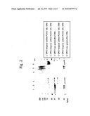 |  |
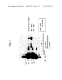 | 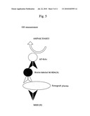 |
 | 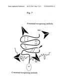 |
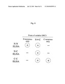 | 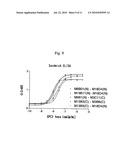 |
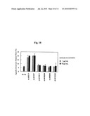 | 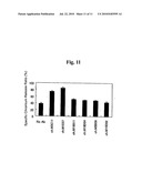 |
 |  |
 |  |
 |  |
 |  |
 |  |
 |  |
 |  |
 |  |
 |  |
 |  |
 |  |
 | 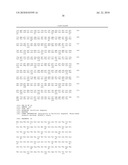 |
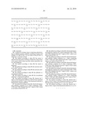 |
| Similar patent applications: | |
| Date | Title |
|---|---|
| 2010-01-14 | Il-16 antagonist peptides and dna encoding the peptides |
| 2010-04-08 | Methods to decrease the risk of metabolic syndrome post immunization |
| 2010-02-18 | Method for obtaining three-dimensional structures for tissue engineering |
| 2010-04-08 | Methods and systems for mineralization of demineralized tissue |
| 2009-11-26 | Antagonists of il-6 to raise albumin and/or lower crp |
| New patent applications in this class: | |
| Date | Title |
|---|---|
| 2022-05-05 | Antibodies to oxidation-specific epitopes |
| 2022-05-05 | Chimeric antigen receptor comprising interleukin-15 intracellular domain and uses thereof |
| 2022-05-05 | Cd38 antibody |
| 2022-05-05 | Antibodies binding to human cd3 at acidic ph |
| 2022-05-05 | Anti-sirp alpha antibodies |
| New patent applications from these inventors: | |
| Date | Title |
|---|---|
| 2021-12-09 | Anti-dll3 antibody |
| 2017-06-01 | Diagnosis and treatment of cancer using anti-desmoglein-3 antibodies |
| 2015-11-19 | Long non-coding rna used for anticancer therapy |
| 2015-10-15 | Cancer diagnosis and treatment of cancer using anti-robo 1 antibody |
| Top Inventors for class "Drug, bio-affecting and body treating compositions" | |
| Rank | Inventor's name |
|---|---|
| 1 | David M. Goldenberg |
| 2 | Hy Si Bui |
| 3 | Lowell L. Wood, Jr. |
| 4 | Roderick A. Hyde |
| 5 | Yat Sun Or |