Patent application title: RECOMBINANT NANOPARTICLE RSV F VACCINE FOR RESPIRATORY SYNCYTIAL VIRUS
Inventors:
Gale Smith (Gaithersburg, MD, US)
Gale Smith (Gaithersburg, MD, US)
Yingyun Wu (Gaithersburg, MD, US)
Michael Massare (Gaithersburg, MD, US)
Ye Liu (Gaithersburg, MD, US)
IPC8 Class: AA61K39155FI
USPC Class:
4241861
Class name: Antigen, epitope, or other immunospecific immunoeffector (e.g., immunospecific vaccine, immunospecific stimulator of cell-mediated immunity, immunospecific tolerogen, immunospecific immunosuppressor, etc.) amino acid sequence disclosed in whole or in part; or conjugate, complex, or fusion protein or fusion polypeptide including the same disclosed amino acid sequence derived from virus
Publication date: 2015-11-26
Patent application number: 20150335730
Abstract:
The present invention is generally related to modified or mutated
respiratory syncytial virus fusion (F) proteins and methods for making
and using them, including immunogenic compositions such as vaccines for
the treatment and/or prevention of RSV infection.Claims:
1-76. (canceled)
77. A method of preventing respiratory syncytial virus (RSV) replication in the lung of a mammal, comprising administering to the mammal an RSV Fusion (F) protein, wherein the RSV F protein comprises (i) a deletion of one or more amino acids of the fusion domain, wherein the fusion domain corresponds to amino acids 137-154 of the wild-type RSV F protein (SEQ ID NO: 2); and (ii) an inactivated primary furin cleavage site, wherein the primary furin cleavage site corresponds to amino acids 131-136 of the wild-type RSV F protein (SEQ ID NO: 2); and wherein an immune response that prevents RSV replication in the lung is obtained.
78. The method of claim 77 wherein the administering is intramuscular.
79. The method of claim 77 wherein the immune response comprises neutralizing antibodies.
80. The method of claim 79 wherein the neutralizing antibodies bind a polypeptide corresponding to amino acids 254-278 (SEQ ID NO:35) of the wild-type RSV F protein.
81. The method of claim 79 wherein the neutralizing antibodies bind at least two RSV F protein epitope sites selected from the group consisting of Site I, Site II, Site IV, Site V, and Site VI.
82. The method of claim 77 wherein about 1 μg to about 30 μg of the RSV F Protein is administered.
83. The method of claim 77 wherein about 1 μg to about 10 μg of the RSV F Protein is administered.
84. The method of claim 77 wherein the RSV F protein is administered without an adjuvant.
85. The method of claim 77 wherein the RSV F protein is administered with an adjuvant.
86. The method of claim 77 wherein the RSV-F protein-stimulated immune response is associated with reduced incidence of an RSV-induced pathological lung response compared to an immune response obtained by administering a formalin-inactivated RSV (FI-RSV).
87. The method of claim 86 wherein the pathological lung response is selected from the group consisting of: bronchiolitis, bronchitis, alveolitis, and pneumonitis.
88. The method of claim 77, wherein the fusion protein deletion is a deletion of the amino acids corresponding to amino acids 137-146 of the wild-type RSV F protein (SEQ ID NO: 2).
89. The method of claim 77 wherein the mammal is a human.
90. The method of claim 89 wherein the human in an infant human.
91. The method of claim 77 further comprising administering one or more additional RSV proteins selected from the group consisting of: an RSV M protein, an RSV N protein, an RSV G protein, and an RSV SH protein.
92. The method of claim 77 wherein the inactivated primary cleavage site comprises at least one amino acid substitution at a position corresponding to amino acids selected from the group consisting of arginine 133, arginine 135, and arginine 136 of the wild-type RSV F protein (SEQ ID NO: 2).
93. The method of claim 92, wherein the arginine is substituted with a glutamine.
94. The method of claim 77 wherein the inactivated primary cleavage site comprises glutamine residues at positions corresponding to arginine 133, arginine 135, and arginine 136 of the wild-type RSV F protein (SEQ ID NO: 2).
95. The method of claim 77 wherein the RSV F protein comprises the amino acid sequence of SEQ ID NO: 8.
96. A method of vaccinating a mammal against a viral infection comprising administering an RSV F protein in a pharmaceutically acceptable formulation to a human subject, wherein the RSV F protein comprises (i) a deletion of one or more amino acids of the fusion domain, wherein said fusion domain corresponds to amino acids 137-154 of the wild-type RSV F protein (SEQ ID NO: 2), and (ii) an inactivated primary furin cleavage site, wherein the primary furin cleavage site corresponds to amino acids 131-136 of the wild-type RSV F protein (SEQ ID NO: 2).
Description:
CROSS-REFERENCE TO RELATED APPLICATIONS
[0001] This application is a continuation of U.S. application Ser. No. 13/629,107, filed Sep. 27, 2012 which claims priority to U.S. Provisional Application Ser. No. 61/542,040, filed Sep. 30, 2011, U.S. Provisional Application Ser. No. 61/542,721 filed Oct. 3, 2011, U.S. Provisional Application Ser. No. 61/611,834 filed Mar. 16, 2012, and to 61/614,286 filed Mar. 22, 2012, the disclosures of which are each incorporated by reference in their entirety for all purposes.
[0002] The contents of the text file submitted electronically are incorporated herein by reference in their entirety: A computer readable format copy of the Sequence Listing (filename: NOVV--048--05US_SeqList.txt, date recorded: Jun. 11, 2015; file size: 73 kilobytes).
TECHNICAL FIELD
[0003] The present invention is generally related to modified or mutated respiratory syncytial virus fusion (F) proteins and methods for making and using them, including immunogenic compositions such as vaccines for the treatment and/or prevention of RSV infection.
BACKGROUND OF THE INVENTION
[0004] Respiratory syncytial virus (RSV) is a member of the genus Pneumovirus of the family Paramyxoviridae. Human RSV (HRSV) is the leading cause of severe lower respiratory tract disease in young children and is responsible for considerable morbidity and mortality in humans. RSV is also recognized as an important agent of disease in immunocompromised adults and in the elderly. Due to incomplete resistance to RSV in the infected host after a natural infection, RSV may infect multiple times during childhood and adult life.
[0005] This virus has a genome comprised of a single strand negative-sense RNA, which is tightly associated with viral protein to form the nucleocapsid. The viral envelope is composed of a plasma membrane derived lipid bilayer that contains virally encoded structural proteins. A viral polymerase is packaged with the virion and transcribes genomic RNA into mRNA. The RSV genome encodes three transmembrane structural proteins, F, G, and SH, two matrix proteins, M and M2, three nucleocapsid proteins N, P, and L, and two nonstructural proteins, NS1 and NS2.
[0006] Fusion of HRSV and cell membranes is thought to occur at the cell surface and is a necessary step for the transfer of viral ribonucleoprotein into the cell cytoplasm during the early stages of infection. This process is mediated by the fusion (F) protein, which also promotes fusion of the membrane of infected cells with that of adjacent cells to form a characteristic syncytia, which is both a prominent cytopathic effect and an additional mechanism of viral spread. Accordingly, neutralization of fusion activity is important in host immunity. Indeed, monoclonal antibodies developed against the F protein have been shown to neutralize virus infectivity and inhibit membrane fusion (Calder et al., 2000, Virology 271: 122-131).
[0007] The F protein of RSV shares structural features and limited, but significant amino acid sequence identity with F glycoproteins of other paramyxoviruses. It is synthesized as an inactive precursor of 574 amino acids (F0) that is cotranslationally glycosylated on asparagines in the endoplasmic reticulum, where it assembles into homo-oligomers. Before reaching the cell surface, the F0 precursor is cleaved by a protease into F2 from the N terminus and F1 from the C terminus. The F2 and F1 chains remain covalently linked by one or more disulfide bonds.
[0008] Immunoaffinity purified full-length F proteins have been found to accumulate in the form of micelles (also characterized as rosettes), similar to those observed with other full-length virus membrane glycoproteins (Wrigley et al., 1986, in Electron Microscopy of Proteins, Vol 5, p. 103-163, Academic Press, London). Under electron microscopy, the molecules in the rosettes appear either as inverted cone-shaped rods (˜70%) or lollipop-shaped (˜30%) structures with their wider ends projecting away from the centers of the rosettes. The rod conformational state is associated with an F glycoprotein in the pre-fusion inactivate state while the lollipop conformational state is associated with an F glycoprotein in the post-fusion, active state.
[0009] Electron micrography can be used to distinguish between the prefusion and postfusion (alternatively designated prefusogenic and fusogenic) conformations, as demonstrated by Calder et al., 2000, Virology 271:122-131. The prefusion conformation can also be distinguished from the fusogenic (postfusion) conformation by liposome association assays. Additionally, prefusion and fusogenic conformations can be distinguished using antibodies (e.g., monoclonal antibodies) that specifically recognize conformation epitopes present on one or the other of the prefusion or fusogenic form of the RSV F protein, but not on the other form. Such conformation epitopes can be due to preferential exposure of an antigenic determinant on the surface of the molecule. Alternatively, conformational epitopes can arise from the juxtaposition of amino acids that are non-contiguous in the linear polypeptide.
[0010] It has been shown previously that the F precursor is cleaved at two sites (site I, after residue 109 and site II, after residue 136), both preceded by motifs recognized by furin-like proteases. Site II is adjacent to a fusion peptide, and cleavage of the F protein at both sites is needed for membrane fusion (Gonzalez-Reyes et al., 2001, PNAS 98(17): 9859-9864). When cleavage is completed at both sites, it is believed that there is a transition from cone-shaped to lollipop-shaped rods.
SUMMARY OF THE INVENTION
[0011] As described herein, the present inventors have found that surprisingly high levels of expression of the fusion (F) protein can be achieved when certain modifications are made to the structure of the RSV F protein. Such modifications also unexpectedly reduce the cellular toxicity of the RSV F protein in a host cell. In addition, the modified F proteins of the present invention demonstrate an improved ability to exhibit the post-fusion "lollipop" morphology as opposed to the pre-fusion "rod" morphology. Thus, in one aspect, the modified F proteins of the present invention can also exhibit improved immunogenicity as compared to wild-type F proteins. These modifications have significant applications to the development of vaccines and methods of using said vaccines for the treatment and/or prevention of RSV. The present invention provides recombinant RSV F proteins that demonstrate increased expression, reduced cellular toxicity, and/or enhanced immunogenic properties as compared to wild-type RSV F proteins.
[0012] In one aspect, the invention provides recombinant RSV F proteins comprising modified or mutated amino acid sequences as compared to wild-type RSV F proteins. In general, these modifications or mutations increase the expression, reduce the cellular toxicity, and/or enhance the immunogenic properties of the RSV F proteins as compared to wild-type RSV F proteins. In certain exemplary embodiments, the RSV F proteins are human RSV F proteins.
[0013] The RSV F protein preferably comprises a modified or mutated amino acid sequence as compared to the wild-type RSV F protein (e.g. as exemplified in SEQ ID NO: 2). In one embodiment, the RSV F protein contains a modification or mutation at the amino acid corresponding to position P102 of the wild-type RSV F protein (SEQ ID NO: 2). In another embodiment, the RSV F protein contains a modification or mutation at the amino acid corresponding to position 1379 of the wild-type RSV F protein (SEQ ID NO: 2). In another embodiment, the RSV F protein contains a modification or mutation at the amino acid corresponding to position M447 of the wild-type RSV F protein (SEQ ID NO: 2).
[0014] In one embodiment, the RSV F protein contains two or more modifications or mutations at the amino acids corresponding to the positions described above. In another embodiment, the RSV F protein contains three modifications or mutations at the amino acids corresponding to the positions described above.
[0015] In one specific embodiment, the invention is directed to RSV F proteins wherein the proline at position 102 is replaced with alanine. In another specific embodiment, the invention is directed to RSV F proteins wherein the isoleucine at position 379 is replaced with valine. In yet another specific embodiment, the invention is directed to RSV F proteins wherein the methionine at position 447 is replaced with valine. In certain embodiments, the RSV F protein contains two or more modifications or mutations at the amino acids corresponding to the positions described in these specific embodiments. In certain other embodiments, the RSV F protein contains three modifications or mutations at the amino acids corresponding to the positions described in these specific embodiments. In an exemplary embodiment, the RSV protein has the amino acid sequence described in SEQ ID NO: 4.
[0016] In one embodiment, the coding sequence of the RSV F protein is further optimized to enhance its expression in a suitable host cell. In one embodiment, the host cell is an insect cell. In an exemplary embodiment, the insect cell is an Sf9 cell.
[0017] In one embodiment, the coding sequence of the codon optimized RSV F gene is SEQ ID NO: 3. In another embodiment, the codon optimized RSV F protein has the amino acid sequence described in SEQ ID NO: 4.
[0018] In one embodiment, the RSV F protein further comprises at least one modification in the cryptic poly(A) site of F2. In another embodiment, the RSV F protein further comprises one or more amino acid mutations at the primary cleavage site (CS). In one embodiment, the RSV F protein contains a modification or mutation at the amino acid corresponding to position R133 of the wild-type RSV F protein (SEQ ID NO: 2) or the codon optimized RSV F protein (SEQ ID NO: 4). In another embodiment, the RSV F protein contains a modification or mutation at the amino acid corresponding to position R135 of the wild-type RSV F protein (SEQ ID NO: 2) or the codon optimized RSV F protein (SEQ ID NO: 4). In yet another embodiment, the RSV F protein contains a modification or mutation at the amino acid corresponding to position R136 of the wild-type RSV F protein (SEQ ID NO: 2) or the codon optimized RSV F protein (SEQ ID NO: 4).
[0019] In one specific embodiment, the invention is directed to RSV F proteins wherein the arginine at position 133 is replaced with glutamine. In another specific embodiment, the invention is directed to RSV F proteins wherein the arginine at position 135 is replaced with glutamine. In yet another specific embodiment, the invention is directed to RSV F proteins wherein arginine at position 136 is replaced with glutamine. In certain embodiments, the RSV F protein contains two or more modifications or mutations at the amino acids corresponding to the positions described in these specific embodiments. In certain other embodiments, the RSV F protein contains three modifications or mutations at the amino acids corresponding to the positions described in these specific embodiments. In an exemplary embodiment, the RSV protein has the amino acid sequence described in SEQ ID NO: 6.
[0020] In another embodiment, the RSV F protein further comprises a deletion in the N-terminal half of the fusion domain corresponding to amino acids 137-146 of SEQ ID NO: 2, SEQ ID NO: 4, and SEQ ID NO: 6. In an exemplary embodiment, the RSV F protein has the amino acid sequence described in SEQ ID NO: 8. In an alternative embodiment, the RSV F protein has the amino acid sequence described in SEQ ID NO: 10.
[0021] Further included within the scope of the invention are RSV F proteins, other than human RSV F protein (SEQ ID NO: 2), which contain alterations corresponding to those set out above. Such RSV F proteins may include, but are not limited to, the RSV F proteins from A strains of human RSV, B strains of human RSV, strains of bovine RSV, and strains of avian RSV.
[0022] In some embodiments, the invention is directed to modified or mutated RSV F proteins that demonstrate increased expression in a host cell as compared to wild-type RSV F proteins, such as the one shown by SEQ ID NO: 2. In other embodiments, the invention is directed to modified or mutated RSV F proteins that demonstrate reduced cellular toxicity in a host cell as compared to wild-type RSV F proteins, such as the one shown by SEQ ID NO: 2. In yet other embodiments, the invention is directed to modified or mutated RSV F proteins that demonstrate enhanced immunogenic properties as compared to wild-type RSV F proteins, such as the one shown by SEQ ID NO: 2.
[0023] In additional aspects, the invention provides immunogenic compositions comprising one or more modified or mutated RSV F proteins as described herein. In one embodiment, the invention provides a micelle comprised of one or more modified or mutated RSV F proteins (e.g. an RSV F micelle).
[0024] In another embodiment, the present invention provides a virus-like particle (VLP) comprising a modified or mutated RSV F protein. In some embodiments, the VLP further comprises one or more additional proteins.
[0025] In one embodiment, the VLP further comprises a matrix (M) protein. In one embodiment, the M protein is derived from a human strain of RSV. In another embodiment, the M protein is derived from a bovine strain of RSV. In other embodiments, the matrix protein may be an M1 protein from an influenza virus strain. In one embodiment, the influenza virus strain is an avian influenza virus strain. In other embodiments, the M protein may be derived from a Newcastle Disease Virus (NDV) strain.
[0026] In additional embodiments, the VLP further comprises the RSV glycoprotein G. In another embodiment, the VLP further comprises the RSV glycoprotein SH. In yet another embodiment, the VLP further comprises the RSV nucleocapsid N protein.
[0027] The modified or mutated RSV F proteins may be used for the prevention and/or treatment of RSV infection. Thus, in another aspect, the invention provides a method for eliciting an immune response against RSV. The method involves administering an immunologically effective amount of a composition containing a modified or mutated RSV F protein to a subject, such as a human or animal subject.
[0028] In another aspect, the present invention provides pharmaceutically acceptable vaccine compositions comprising a modified or mutated RSV F protein, an RSV F micelle comprising a modified or mutated RSV F protein, or a VLP comprising a modified or mutated RSV F protein.
[0029] In one embodiment, the invention comprises an immunogenic formulation comprising at least one effective dose of a modified or mutated RSV F protein. In another embodiment, the invention comprises an immunogenic formulation comprising at least one effective dose of an RSV F micelle comprising a modified or mutated RSV F protein. In yet another embodiment, the invention comprises an immunogenic formulation comprising at least one effective dose of a VLP comprising a modified or mutated RSV F protein.
[0030] In another embodiment, the invention provides for a pharmaceutical pack or kit comprising one or more containers filled with one or more of the ingredients of the vaccine formulations of the invention.
[0031] In another embodiment, the invention provides a method of formulating a vaccine or antigenic composition that induces immunity to an infection or at least one disease symptom thereof to a mammal, comprising adding to the formulation an effective dose of a modified or mutated RSV F protein, an RSV F micelle comprising a modified or mutated RSV F protein, or a VLP comprising a modified or mutated RSV F protein. In a preferred embodiment, the infection is an RSV infection.
[0032] The modified or mutated RSV F proteins of the invention are useful for preparing compositions that stimulate an immune response that confers immunity or substantial immunity to infectious agents. Thus, in one embodiment, the invention provides a method of inducing immunity to infections or at least one disease symptom thereof in a subject, comprising administering at least one effective dose of a modified or mutated RSV F protein, an RSV F micelle comprising a modified or mutated RSV F protein, or a VLP comprising a modified or mutated RSV F protein.
[0033] In yet another aspect, the invention provides a method of inducing substantial immunity to RSV virus infection or at least one disease symptom in a subject, comprising administering at least one effective dose of a modified or mutated RSV F protein, an RSV F micelle comprising a modified or mutated RSV F protein, or a VLP comprising a modified or mutated RSV F protein.
[0034] Compositions of the invention can induce substantial immunity in a vertebrate (e.g. a human) when administered to the vertebrate. Thus, in one embodiment, the invention provides a method of inducing substantial immunity to RSV virus infection or at least one disease symptom in a subject, comprising administering at least one effective dose of a modified or mutated RSV F protein, an RSV F micelle comprising a modified or mutated RSV F protein, or a VLP comprising a modified or mutated RSV F protein. In another embodiment, the invention provides a method of vaccinating a mammal against RSV comprising administering to the mammal a protection-inducing amount of a modified or mutated RSV F protein, an RSV F micelle comprising a modified or mutated RSV F protein, or a VLP comprising a modified or mutated RSV F protein.
[0035] In another embodiment, the invention comprises a method of inducing a protective antibody response to an infection or at least one symptom thereof in a subject, comprising administering at least one effective dose of a modified or mutated RSV F protein, an RSV F micelle comprising a modified or mutated RSV F protein, or a VLP comprising a modified or mutated RSV F protein.
[0036] In another embodiment, the invention comprises a method of inducing a protective cellular response to RSV infection or at least one disease symptom in a subject, comprising administering at least one effective dose of a modified or mutated RSV F protein. In another embodiment, the invention comprises a method of inducing a protective cellular response to RSV infection or at least one disease symptom in a subject, comprising administering at least one effective dose of an RSV F micelle comprising a modified or mutated RSV F protein. In yet another embodiment, the invention comprises a method of inducing a protective cellular response to RSV infection or at least one disease symptom in a subject, comprising administering at least one effective dose of a VLP, wherein the VLP comprises a modified or mutated RSV F protein.
[0037] In yet another aspect, the invention provides an isolated nucleic acid encoding a modified or mutated RSV F protein of the invention. In an exemplary embodiment, the isolated nucleic acid encoding a modified or mutated RSV F protein is selected from the group consisting of SEQ ID NO: 3, SEQ ID NO: 5, SEQ ID NO: 7, or SEQ ID NO: 9.
[0038] In yet another aspect, the invention provides an isolated cell comprising a nucleic acid encoding a modified or mutated RSV F protein of the invention. In an exemplary embodiment, the isolated nucleic acid encoding a modified or mutated RSV F protein is selected from the group consisting of SEQ ID NO: 3, SEQ ID NO: 5, SEQ ID NO: 7, or SEQ ID NO: 9.
[0039] In yet another aspect, the invention provides a vector comprising a nucleic acid encoding a modified or mutated RSV F protein of the invention. In an exemplary embodiment, the isolated nucleic acid encoding a modified or mutated RSV F protein is selected from the group consisting of SEQ ID NO: 3, SEQ ID NO: 5, SEQ ID NO: 7, or SEQ ID NO: 9. In one embodiment, the vector is a baculovirus vector.
[0040] In yet another aspect, the invention provides a method of making a RSV F protein, comprising (a) transforming a host cell to express a nucleic acid encoding a modified or mutated RSV F protein of the invention; and (b) culturing said host cell under conditions conducive to the production of said RSV F protein. In one embodiment, the nucleic acid encoding a modified or mutated RSV F protein is selected from the group consisting of SEQ ID NO: 3, SEQ ID NO: 5, SEQ ID NO: 7, or SEQ ID NO: 9. In another embodiment, the host cell is an insect cell. In a further embodiment, the host cell is an is an insect cell transfected with a baculovirus vector comprising a modified or mutated RSV F protein of the invention.
[0041] In yet another aspect, the invention provides a method of making a RSV F protein micelle, comprising (a) transforming a host cell to express a nucleic acid encoding a modified or mutated RSV F protein of the invention; and (b) culturing said host cell under conditions conducive to the production of said RSV F protein micelle. In one embodiment, the nucleic acid encoding a modified or mutated RSV F protein is selected from the group consisting of SEQ ID NO: 3, SEQ ID NO: 5, SEQ ID NO: 7, or SEQ ID NO: 9. In one embodiment, the host cell is an insect cell. In an exemplary embodiment, the host cell is an is an insect cell transfected with a baculovirus vector comprising a modified or mutated RSV F protein of the invention.
[0042] In one aspect, the present invention is directed to an RSV fusion surface glycoprotein (F) nanoparticle vaccine. In one embodiment, the vaccine comprises full length F protein. In a further embodiment, the full length F protein is cleaved into disulfide linked F1 and F2 trimers. The F1 and F2 trimers, in one embodiment, are present in micelles having a diameter of about 20 nm to about 40 nm.
[0043] In another aspect, an antibody generated by the vaccine of the invention is provided.
[0044] In yet another aspect, a method for vaccinating a subject in need thereof is provided. In one embodiment, the method comprises administering to the subject a recombinant RSV fusion glycoprotein (F) nanoparticle vaccine. In a further embodiment, the nanoparticle vaccine comprises full length F protein. In even a further embodiment, the full length F protein is cleaved into disulfide linked F1 and F2 trimers. The F1 and F2 trimers, in one embodiment, are present in micelles having a diameter of about 20 nm to about 40 nm.
[0045] In one embodiment, the vaccine of the invention is administered at a dose is selected from the group consisting of 5 μg, 15 μg, 30 μg and 60 μg.
[0046] In another aspect, a method for vaccinating a subject in need thereof is provided. In one embodiment, the method comprises administering to the subject a recombinant RSV fusion glycoprotein (F) nanoparticle vaccine comprising full length F protein and an adjuvant. In a further embodiment, the adjuvant is alum.
BRIEF DESCRIPTION OF THE FIGURES
[0047] FIG. 1 depicts the structure of wild type HRSV F0 protein and the primary (SEQ ID NO: 32) and secondary (SEQ ID NO: 33) cleavage sites.
[0048] FIG. 2 depicts structures of modified RSV F0 proteins with cleavage site mutations as described in Example 3, corresponding to SEQ ID NOs: 28 (KKQKQQ), 29 (GRRQQR), 30 (RAQQ), and 31 (KKQKRQ).
[0049] FIG. 3 depicts conservative substitutions (R133Q, R135Q and R136Q) in the primary cleavage site of modified HRSV F protein BV #541 (SEQ ID NO: 6).
[0050] FIG. 4 depicts sequence and structure of modified HRSV F protein BV #541 (SEQ ID NO: 6).
[0051] FIG. 5 depicts sequence and structure of modified HRSV F protein BV #622 (SEQ ID NO: 10).
[0052] FIG. 6 depicts SDS-PAGE coomassie-stained gel of purified recombinant HRSV F protein BV #622 with or without the presence of βME.
[0053] FIGS. 7A, 7B, 7C, and 7D are depicted as follows: FIG. 7A depicts a Western blot analysis of the RSV F fusion domain mutants. FIG. 7B depicts cell surface RSV F protein immunostaining of the RSV F fusion domain mutants. FIG. 7C depicts the structure of modified HRSV F protein BV #683 (SEQ ID NO: 8). FIG. 7D depicts the parent clone BV#541 (Δ0) and mutants with Δ2 Δ4, Δ6, Δ8, Δ10 (BV#683), Δ12, Δ14, Δ16, and Δ18 deletions in the fusion domain. BV#541 comprises a protein in which the amino acid sequence of the fusion domain comprises position 137 to 154 of SEQ ID NO: 6). The amino acid sequences of the fusion domain portions of the deletion mutants comprise position 139 to 154 of SEQ ID NO: 6 (Δ2), position 141 to 154 of SEQ ID NO: 6 (Δ4), position 143 to 154 of SEQ ID NO: 6 (Δ6), position 145 to 154 of SEQ ID NO: 6 (Δ8), position 147 to 154 of SEQ ID NO: 6 (Δ10; BV#683), position 149 to 154 of SEQ ID NO: 6 (Δ12), position 151 to 154 of SEQ ID NO: 6(Δ14), or position 153 to 154 of SEQ ID NO: 6 (Δ16). The entire fusion domain corresponding to position 137-154 in SEQ ID NO: 6 is deleted in the mutant with a Δ18 deletion.
[0054] FIG. 8 depicts SDS-PAGE coomassie-stained gels of purified recombinant HRSV F proteins BV #622 and BV #683 with or without the presence of βME (on the left), and their structures.
[0055] FIG. 9 depicts SDS-PAGE coomassie-stained gel (on the left) and Western Blot (on the right) analysis of purified recombinant HRSV F protein BV #683 with or without the presence of βME.
[0056] FIG. 10 depicts SDS-PAGE coomassie-stained gel used in purity analysis by scanning densitometry (on the left) and Western Blot (on the right) of purified recombinant HRSV F protein BV #683.
[0057] FIG. 11 depicts images of purified recombinant HRSV F protein BV #683 micelles (rosettes) taken in negative stain electron microscopy.
[0058] FIGS. 12A, 12B, and 12C are depicted as follows: FIG. 12A depicts reverse phase HPLC analysis of HRSV F protein BV #683. FIG. 12B depicts size exclusion HPLC analysis of HRSV F protein BV #683. FIG. 12C depicts particle size analysis of HRSV F protein BV #683 micelles.
[0059] FIG. 13 depicts SDS-PAGE coomassie-stained gel (on the left) and Western Blot (on the right) analysis of modified HRSV F proteins BV #622 and BV #623 (SEQ ID NO: 21) with or without co-expression with HRSV N and BRSV M proteins in the crude cell culture harvests (intracellular) or pelleted samples by 30% sucrose gradient separation, and structures of BV #622 and BV #623.
[0060] FIG. 14 depicts SDS-PAGE coomassie-stained gel (on the left) and Western Blot (on the right) analysis of modified HRSV F protein BV #622, double tandem chimeric BV #636 (BV #541+BRSV M), BV #683, BV #684 (BV #541 with YIAL L-domain), and BV #685 (BV #541 with YKKL L-domain) with or without co-expression with HRSV N and BRSV M proteins in the crude cell culture harvests (intracellular) samples, and structure of each analyzed modified HRSV F protein.
[0061] FIG. 15 depicts SDS-PAGE coomassie-stained gel (on the left) and Western Blot (on the right) analysis of modified RSV F protein BV #622 (SEQ ID NO: 10), double tandem chimeric BV #636 (BV #541+BRSV M), BV #683 (SEQ ID NO: 8), BV #684 (BV #541 with YIAL L-domain), and BV #685 (BV #541 with YKKL L-domain) with or without co-expression with HRSV N and BRSV M proteins in the pelleted samples by 30% sucrose gradient separation, and structure of each analyzed modified HRSV F protein.
[0062] FIGS. 16A, 16B, 16C, and 16D depict structure, clone name, description, Western Blot and SDS-PAGE coomassie results, and conclusion for each modified RSV F protein as described in Example 9.
[0063] FIG. 17 depicts experimental procedures of the RSV challenge study as described in Example 10.
[0064] FIG. 18 depicts results of RSV neutralization assay at day 31 and day 46 of mice immunized with PBS, live RSV, FI-RSV, 1 μg PFP, 1 μg PFP+Alum, 10 μg PFP, 10 μg PFP+Alum, 30 μg PFP, and positive control (anti-F sheep).
[0065] FIG. 19 depicts RSV titers in lung tissues of mice immunized with PBS, live RSV, FI-RSV, 1 μg PFP, 1 μg PFP+Alum, 10 μg PFP, 10 μg PFP+Alum, and 30 μg PFP, 4 days after challenge of infectious RSV.
[0066] FIG. 20 depicts SDS-PAGE gel stained with coomassie of purified recombinant RSV F protein BV #683 stored at 2-8° C. for 0, 1, 2, 4, and 5 weeks.
[0067] FIG. 21 depicts RSV A and RSV B neutralizing antibody responses following immunization with live RSV (RSV), formalin inactivated RSV (FI-RSV), RSV-F protein BV #683 with and without aluminum (PFP and PFP+Aluminum Adjuvant), and PBS controls.
[0068] FIG. 22 depicts lung pathology following challenge with RSV in rats immunized with live RSV, formalin inactivated RSV (FI-RSV), RSV-F protein BV #683 with and without aluminum (F-micelle (30 μg) and F-micelle (30 μg)+Aluminum Adjuvant), and PBS controls.
[0069] FIG. 23 is a graph showing the neutralizing antibody responses against RSV A in cotton rat (y-axis, expressed as Log 2 titers) vs. various vaccination treatment groups (x-axis). The line for each group is the geometric mean of end point titer that neutralized 100%.
[0070] FIG. 24 is a graph showing the neutralizing antibody responses against RSV A in cotton rat (y-axis, expressed as Log 2 titers) vs. various vaccination treatment groups (x-axis). The line for each group is the geometric mean of end point titer that neutralized 100%.
[0071] FIG. 25 is a graph showing the lung viral titers of cotton rats (expressed as log 10 pfu/gram of tissue) vs. various vaccination treatment groups (x-axis). The viral titers are shown±SEM.
[0072] FIGS. 26A, 26B, 26C, and 26D are depicted as follows: FIG. 26A is a graph showing ELISA units vs. vaccination group, and provides a measure for antibody production in animals treated with the RSV F vaccine, FI-RSV, live RSV, or PBS. FIG. 26 B is a graph showing antibody production in each vaccine group as measured by RSV-F IgG titer. FIG. 26C is a graph depicting serum neutralizing antibody titers against RSV in each vaccination group. FIG. 26D is a graph showing palivizumab competitive IgG titers from pooled sera from each vaccination group.
[0073] FIG. 27 are representative micrographs of lung tissue harvested from rats after treatment with the nanoparticle vaccine of the invention and a subsequent challenge with RSV.
[0074] FIG. 28 is a graph showing the binding competition between the palivizumab epitope (SEQ ID NO: 35) and antibodies produced by the vaccine of the present invention.
[0075] FIGS. 29A and 29B are depicted as follows: FIG. 29A is a graph showing the binding of various concentrations of Synagis® mAb to palivizumab epitope peptide. FIG. 29B is a graph showing the binding of various concentrations of Synagis® to recombinant RSV F micelles.
[0076] FIG. 30 provides schemes of various assays carried out to test the immunogenicity of the nanoparticle vaccines of the invention.
[0077] FIG. 31 is a graph showing the results of an ELISA study using human sera from subjects treated with the vaccine of the present invention.
[0078] FIG. 32 is a graph showing anti-RSV F (A) and anti-RSV G (B) IgG detected in human sera from subjects treated with the vaccine of the present invention.
[0079] FIG. 33 is a graph showing the geometric mean fold rise in anti-RSV F IgG levels for the alum treatment groups.
[0080] FIG. 34 is a graph showing the plague reduction neutralization titers for subjects at various timepoints, before or after treatment with the nanoparticle vaccine of the invention.
[0081] FIG. 35 shows the reverse cumulative distribution for Day 0, Day 30 and Day 60 PRNTs in the placebo and 30 μg+Alum groups.
[0082] FIGS. 36A and 36B are depicted as follows: FIG. 36A shows the positive assay controls for the BIAcore SPR-based antigen binding assay utilized to assess the avidity of antibodies in the human sera for RSV F. FIG. 36 B shows a sensorgram for sera from Day 0 and placebo controls, in comparison to the positive control palivizumab.
[0083] FIG. 37 shows the binding curve for palivizumab and a representative sample from the vaccine group, as measured using the BIAcore SPR-based antigen binding assay.
[0084] FIG. 38 is a graph showing the geometric mean rise in antibody titer levels for both (1) anti-F IgG (left bar) and (2) MN (right bar) for the various treatment groups.
[0085] FIG. 39 is a graph showing antibody geometric mean titers (GMT) in patients administered RSV nanoparticle vaccine at various dosages. The antibody response is to the antigenic site II peptide 254-278.
[0086] FIGS. 40A and 40B are depicted as follows: FIG. 40A is a graph showing antibodies generated by the RSV F protein nanoparticle vaccine are competitive with Palivizumab. FIG. 40B is a graph showing palivizumab competitive antibodies post dose 1 and post dose 2 in all vaccine groups.
[0087] FIG. 41 is a graph showing the results of a Palivizumab competition assay. The results show that RSV F nanoparticle vaccine induced antibodies correlate with antibodies that compete for the Palivizumab binding site.
[0088] FIG. 42 shows the RSV F binding titers of the indicated monoclonal antibodies and the antibody recognition sites for each monoclonal antibody on the full length RSV F antigen.
[0089] FIG. 43 is a graph showing the anti-RSV F IgG antibody titer in sera from cotton rats immunized with FI-RSV, RSV-F nanoparticle vaccine with or without adjuvant, or live RSV at Day 0, 28, and 49 post-immunization.
[0090] FIG. 44 is a graph showing neutralizing antibody responses at Day 0, 28, and 49 after immunization of cotton rats with FI-RSV, RSV-F nanoparticle vaccine with or without adjuvant, or live RSV.
[0091] FIG. 45 is a graph showing the fusion inhibition titers in sera from cotton rats immunized with FI-RSV, RSV-F nanoparticle vaccine with or without adjuvant, or live RSV.
[0092] FIG. 46 is a graph showing competitive ELISA titers in sera from cotton rats immunized with FI-RSV, RSV-F nanoparticle vaccine with or without adjuvant, or live RSV.
[0093] FIG. 47 shows the titers of vaccine-induced antibodies competitive with the indicated neutralizing RSV F-specific monoclonal antibody in sera from cotton rats immunized with FI-RSV, RSV-F nanoparticle vaccine with or without adjuvant, or live RSV.
[0094] FIG. 48 depicts a visualization of direct ELISA data in which cotton rat sera were serially diluted and incubated in plates with bound RSV antigen, and levels of bound antibody prior to or after a 7 molar urea wash step.
DETAILED DESCRIPTION OF THE INVENTION
Definitions
[0095] As used herein, the term "adjuvant" refers to a compound that, when used in combination with a specific immunogen (e.g. a modified or mutated RSV F protein, an RSV F micelle comprising a modified or mutated RSV F protein, or a VLP comprising a modified or mutated RSV F protein) in a formulation, will augment or otherwise alter or modify the resultant immune response. Modification of the immune response includes intensification or broadening the specificity of either or both antibody and cellular immune responses. Modification of the immune response can also mean decreasing or suppressing certain antigen-specific immune responses.
[0096] As used herein, the term "antigenic formulation" or "antigenic composition" refers to a preparation which, when administered to a vertebrate, especially a bird or a mammal, will induce an immune response.
[0097] As used herein, the term "avian influenza virus" refers to influenza viruses found chiefly in birds but that can also infect humans or other animals. In some instances, avian influenza viruses may be transmitted or spread from one human to another. An avian influenza virus that infects humans has the potential to cause an influenza pandemic, i.e., morbidity and/or mortality in humans. A pandemic occurs when a new strain of influenza virus (a virus against which humans have no natural immunity) emerges, spreading beyond individual localities, possibly around the globe, and infecting many humans at once.
[0098] As used herein, an "effective dose" generally refers to that amount of a modified or mutated RSV F protein, an RSV F micelle comprising a modified or mutated RSV F protein, or a VLP comprising a modified or mutated RSV F protein of the invention sufficient to induce immunity, to prevent and/or ameliorate an infection or to reduce at least one symptom of an infection or disease, and/or to enhance the efficacy of another dose of a modified or mutated RSV F protein, an RSV F micelle comprising a modified or mutated RSV F protein, or a VLP comprising a modified or mutated RSV F protein. An effective dose may refer to the amount of a modified or mutated RSV F protein, an RSV F micelle comprising a modified or mutated RSV F protein, or a VLP comprising a modified or mutated RSV F protein sufficient to delay or minimize the onset of an infection or disease. An effective dose may also refer to the amount of a modified or mutated RSV F protein, an RSV F micelle comprising a modified or mutated RSV F protein, or a VLP comprising a modified or mutated RSV F protein that provides a therapeutic benefit in the treatment or management of an infection or disease. Further, an effective dose is the amount with respect to a modified or mutated RSV F protein, an RSV F micelle comprising a modified or mutated RSV F protein, or a VLP comprising a modified or mutated RSV F protein of the invention alone, or in combination with other therapies, that provides a therapeutic benefit in the treatment or management of an infection or disease. An effective dose may also be the amount sufficient to enhance a subject's (e.g., a human's) own immune response against a subsequent exposure to an infectious agent or disease. Levels of immunity can be monitored, e.g., by measuring amounts of neutralizing secretory and/or serum antibodies, e.g., by plaque neutralization, complement fixation, enzyme-linked immunosorbent, or microneutralization assay, or by measuring cellular responses, such as, but not limited to cytotoxic T cells, antigen presenting cells, helper T cells, dendritic cells and/or other cellular responses. T cell responses can be monitored, e.g., by measuring, for example, the amount of CD4.sup.+ and CD8.sup.+ cells present using specific markers by fluorescent flow cytometry or T cell assays, such as but not limited to T-cell proliferation assay, T-cell cytotoxic assay, TETRAMER assay, and/or ELISPOT assay. In the case of a vaccine, an "effective dose" is one that prevents disease and/or reduces the severity of symptoms.
[0099] As used herein, the term "effective amount" refers to an amount of a modified or mutated RSV F protein, an RSV F micelle comprising a modified or mutated RSV F protein, or a VLP comprising a modified or mutated RSV F protein necessary or sufficient to realize a desired biologic effect. An effective amount of the composition would be the amount that achieves a selected result, and such an amount could be determined as a matter of routine experimentation by a person skilled in the art. For example, an effective amount for preventing, treating and/or ameliorating an infection could be that amount necessary to cause activation of the immune system, resulting in the development of an antigen specific immune response upon exposure to a modified or mutated RSV F protein, an RSV F micelle comprising a modified or mutated RSV F protein, or a VLP comprising a modified or mutated RSV F protein of the invention. The term is also synonymous with "sufficient amount."
[0100] As used herein, the term "expression" refers to the process by which polynucleic acids are transcribed into mRNA and translated into peptides, polypeptides, or proteins. If the polynucleic acid is derived from genomic DNA, expression may, if an appropriate eukaryotic host cell or organism is selected, include splicing of the mRNA. In the context of the present invention, the term also encompasses the yield of RSV F gene mRNA and RSV F proteins achieved following expression thereof.
[0101] As used herein, the term "F protein" or "Fusion protein" or "F protein polypeptide" or "Fusion protein polypeptide" refers to a polypeptide or protein having all or part of an amino acid sequence of an RSV Fusion protein polypeptide. Similarly, the term "G protein" or "G protein polypeptide" refers to a polypeptide or protein having all or part of an amino acid sequence of an RSV Attachment protein polypeptide. Numerous RSV Fusion and Attachment proteins have been described and are known to those of skill in the art. WO/2008/114149, which is herein incorporated by reference in its entirety, sets out exemplary F and G protein variants (for example, naturally occurring variants).
[0102] As used herein, the terms "immunogens" or "antigens" refer to substances such as proteins, peptides, and nucleic acids that are capable of eliciting an immune response. Both terms also encompass epitopes, and are used interchangeably.
[0103] As used herein the term "immune stimulator" refers to a compound that enhances an immune response via the body's own chemical messengers (cytokines). These molecules comprise various cytokines, lymphokines and chemokines with immunostimulatory, immunopotentiating, and pro-inflammatory activities, such as interferons (IFN-γ), interleukins (e.g., IL-1, IL-2, IL-3, IL-4, IL-12, IL-13); growth factors (e.g., granulocyte-macrophage (GM)-colony stimulating factor (CSF)); and other immunostimulatory molecules, such as macrophage inflammatory factor, Flt3 ligand, B7.1; B7.2, etc. The immune stimulator molecules can be administered in the same formulation as VLPs of the invention, or can be administered separately. Either the protein or an expression vector encoding the protein can be administered to produce an immunostimulatory effect.
[0104] As used herein, the term "immunogenic formulation" refers to a preparation which, when administered to a vertebrate, e.g. a mammal, will induce an immune response.
[0105] As used herein, the term "infectious agent" refers to microorganisms that cause an infection in a vertebrate. Usually, the organisms are viruses, bacteria, parasites, protozoa and/or fungi.
[0106] As used herein, the terms "mutated," "modified," "mutation," or "modification" indicate any modification of a nucleic acid and/or polypeptide which results in an altered nucleic acid or polypeptide. Mutations include, for example, point mutations, deletions, or insertions of single or multiple residues in a polynucleotide, which includes alterations arising within a protein-encoding region of a gene as well as alterations in regions outside of a protein-encoding sequence, such as, but not limited to, regulatory or promoter sequences. A genetic alteration may be a mutation of any type. For instance, the mutation may constitute a point mutation, a frame-shift mutation, an insertion, or a deletion of part or all of a gene. In some embodiments, the mutations are naturally-occurring. In other embodiments, the mutations are the results of artificial mutation pressure. In still other embodiments, the mutations in the RSV F proteins are the result of genetic engineering.
[0107] As used herein, the term "multivalent" refers to compositions which have one or more antigenic proteins/peptides or immunogens against multiple types or strains of infectious agents or diseases.
[0108] As used herein, the term "pharmaceutically acceptable vaccine" refers to a formulation which contains a modified or mutated RSV F protein, an RSV F micelle comprising a modified or mutated RSV F protein, or a VLP comprising a modified or mutated RSV F protein of the present invention, which is in a form that is capable of being administered to a vertebrate and which induces a protective immune response sufficient to induce immunity to prevent and/or ameliorate an infection or disease, and/or to reduce at least one symptom of an infection or disease, and/or to enhance the efficacy of another dose of a modified or mutated RSV F protein, an RSV F micelle comprising a modified or mutated RSV F protein, or a VLP comprising a modified or mutated RSV F protein. Typically, the vaccine comprises a conventional saline or buffered aqueous solution medium in which the composition of the present invention is suspended or dissolved. In this form, the composition of the present invention can be used conveniently to prevent, ameliorate, or otherwise treat an infection. Upon introduction into a host, the vaccine is able to provoke an immune response including, but not limited to, the production of antibodies and/or cytokines and/or the activation of cytotoxic T cells, antigen presenting cells, helper T cells, dendritic cells and/or other cellular responses.
[0109] As used herein, the phrase "protective immune response" or "protective response" refers to an immune response mediated by antibodies against an infectious agent or disease, which is exhibited by a vertebrate (e.g., a human), that prevents or ameliorates an infection or reduces at least one disease symptom thereof. Modified or mutated RSV F proteins, RSV F micelles comprising a modified or mutated RSV F protein, or VLPs comprising a modified or mutated RSV F protein of the invention can stimulate the production of antibodies that, for example, neutralize infectious agents, blocks infectious agents from entering cells, blocks replication of the infectious agents, and/or protect host cells from infection and destruction. The term can also refer to an immune response that is mediated by T-lymphocytes and/or other white blood cells against an infectious agent or disease, exhibited by a vertebrate (e.g., a human), that prevents or ameliorates infection or disease, or reduces at least one symptom thereof.
[0110] As used herein, the term "vertebrate" or "subject" or "patient" refers to any member of the subphylum cordata, including, without limitation, humans and other primates, including non-human primates such as chimpanzees and other apes and monkey species. Farm animals such as cattle, sheep, pigs, goats and horses; domestic mammals such as dogs and cats; laboratory animals including rodents such as mice, rats (including cotton rats) and guinea pigs; birds, including domestic, wild and game birds such as chickens, turkeys and other gallinaceous birds, ducks, geese, and the like are also non-limiting examples. The terms "mammals" and "animals" are included in this definition. Both adult and newborn individuals are intended to be covered. In particular, infants and young children are appropriate subjects or patients for a RSV vaccine.
[0111] As used herein, the term "virus-like particle" (VLP) refers to a structure that in at least one attribute resembles a virus but which has not been demonstrated to be infectious. Virus-like particles in accordance with the invention do not carry genetic information encoding for the proteins of the virus-like particles. In general, virus-like particles lack a viral genome and, therefore, are noninfectious. In addition, virus-like particles can often be produced in large quantities by heterologous expression and can be easily purified.
[0112] As used herein, the term "chimeric VLP" refers to VLPs that contain proteins, or portions thereof, from at least two different infectious agents (heterologous proteins). Usually, one of the proteins is derived from a virus that can drive the formation of VLPs from host cells. Examples, for illustrative purposes, are the BRSV M protein and/or the HRSV G or F proteins. The terms RSV VLPs and chimeric VLPs can be used interchangeably where appropriate.
[0113] As used herein, the term "vaccine" refers to a preparation of dead or weakened pathogens, or of derived antigenic determinants that is used to induce formation of antibodies or immunity against the pathogen. A vaccine is given to provide immunity to the disease, for example, influenza, which is caused by influenza viruses. In addition, the term "vaccine" also refers to a suspension or solution of an immunogen (e.g. a modified or mutated RSV F protein, an RSV F micelle comprising a modified or mutated RSV F protein, or a VLP comprising a modified or mutated RSV F protein) that is administered to a vertebrate to produce protective immunity, i.e., immunity that prevents or reduces the severity of disease associated with infection. The present invention provides for vaccine compositions that are immunogenic and may provide protection against a disease associated with infection.
RSV F Proteins
[0114] RSV-F proteins and methods are described in U.S. patent application Ser. No. 12/633,995, filed Dec. 9, 2009 (published Sep. 23, 2010 as U.S. Publication No. 2010/0239617), U.S. Provisional Application Ser. No. 61/121,126, filed Dec. 9, 2008, and U.S. Provisional Application Ser. No. 61/169,077, filed Apr. 14, 2009, and U.S. Provisional Application Ser. No. 61/224,787, filed Jul. 10, 2009, the disclosures of which are each incorporated by reference in their entirety for all purposes.
[0115] Two structural membrane proteins, F and G proteins, are expressed on the surface of RSV, and have been shown to be targets of neutralizing antibodies (Sullender, W., 2000, Clinical Microbiology Review 13, 1-15). These two proteins are also primarily responsible for viral recognition and entry into target cells; G protein binds to a specific cellular receptor and the F protein promotes fusion of the virus with the cell. The F protein is also expressed on the surface of infected cells and is responsible for subsequent fusion with other cells leading to syncytia formation. Thus, antibodies to the F protein can neutralize virus or block entry of the virus into the cell or prevent syncytia formation. Although antigenic and structural differences between A and B subtypes have been described for both the G and F proteins, the more significant antigenic differences reside on the G protein, where amino acid sequences are only 53% homologous and antigenic relatedness is 5% (Walsh et al. (1987) J. Infect. Dis. 155, 1198-1204; and Johnson et al. (1987) Proc. Natl. Acad. Sci. USA 84, 5625-5629). Conversely, antibodies raised to the F protein show a high degree of cross-reactivity among subtype A and B viruses.
[0116] The RSV F protein directs penetration of RSV by fusion between the virion's envelope protein and the host cell plasma membrane. Later in infection, the F protein expressed on the cell surface can mediate fusion with neighboring cells to form syncytia. The F protein is a type I transmembrane surface protein that has a N-terminal cleaved signal peptide and a membrane anchor near the C-terminus. RSV F is synthesized as an inactive F0 precursor that assembles into a homotrimer and is activated by cleavage in the trans-Golgi complex by a cellular endoprotease to yield two disulfide-linked subunits, F1 and F2 subunits. The N-terminus of the F1 subunit that is created by cleavage contains a hydrophobic domain (the fusion peptide) that inserts directly into the target membrane to initiate fusion. The F1 subunit also contains heptad repeats that associate during fusion, driving a conformational shift that brings the viral and cellular membranes into close proximity (Collins and Crowe, 2007, Fields Virology, 5th ed., D. M Kipe et al., Lipincott, Williams and Wilkons, p. 1604). SEQ ID NO: 2 (GenBank Accession No. AAB59858) depicts a representative RSV F protein, which is encoded by the gene shown in SEQ ID NO: 1 (GenBank Accession No. M11486).
[0117] In nature, the RSV F protein is expressed as a single polypeptide precursor, 574 amino acids in length, designated FO. In vivo, FO oligomerizes in the endoplasmic reticulum and is proteolytically processed by a furin protease at two conserved furin consensus sequences (furin cleavage sites), RARR (SEQ ID NO: 23) (secondary) and KKRKRR (SEQ ID NO: 24) (primary) to generate an oligomer consisting of two disulfide-linked fragments. The smaller of these fragments is termed F2 and originates from the N-terminal portion of the FO precursor. It will be recognized by those of skill in the art that the abbreviations FO, F1 and F2 are commonly designated F0, F1 and F2 in the scientific literature. The larger, C-terminal F1 fragment anchors the F protein in the membrane via a sequence of hydrophobic amino acids, which are adjacent to a 24 amino acid cytoplasmic tail. Three F2-F1 dimers associate to form a mature F protein, which adopts a metastable prefusogenic ("prefusion") conformation that is triggered to undergo a conformational change upon contact with a target cell membrane. This conformational change exposes a hydrophobic sequence, known as the fusion peptide, which associates with the host cell membrane and promotes fusion of the membrane of the virus, or an infected cell, with the target cell membrane.
[0118] The F1 fragment contains at least two heptad repeat domains, designated HRA and HRB, and is situated in proximity to the fusion peptide and transmembrane anchor domains, respectively. In the prefusion conformation, the F2-F1 dimer forms a globular head and stalk structure, in which the HRA domains are in a segmented (extended) conformation in the globular head. In contrast, the HRB domains form a three-stranded coiled coil stalk extending from the head region. During transition from the prefusion to the postfusion conformations, the HRA domains collapse and are brought into proximity to the HRB domains to form an anti-parallel six helix bundle. In the postfusion state the fusion peptide and transmembrane domains are juxtaposed to facilitate membrane fusion.
[0119] Although the conformational description provided above is based on molecular modeling of crystallographic data, the structural distinctions between the prefusion and postfusion conformations can be monitored without resort to crystallography. For example, electron micrography can be used to distinguish between the prefusion and postfusion (alternatively designated prefusogenic and fusogenic) conformations, as demonstrated by Calder et al., Virology, 271:122-131 (2000) and Morton et al., Virology, 311: 275-288, which are incorporated herein by reference for the purpose of their technological teachings. The prefusion conformation can also be distinguished from the fusogenic (post-fusion) conformation by liposome association assays as described by Connolly et al, Proc. Natl. Acad. Sci. USA, 103:17903-17908 (2006), which is also incorporated herein by reference for the purpose of its technological teachings. Additionally, prefusion and fusogenic conformations can be distinguished using antibodies (e.g., monoclonal antibodies) that specifically recognize conformation epitopes present on one or the other of the prefusion or fusogenic form of the RSV F protein, but not on the other form. Such conformation epitopes can be due to preferential exposure of an antigenic determinant on the surface of the molecule. Alternatively, conformational epitopes can arise from the juxtaposition of amino acids that are non-contiguous in the linear polypeptide.
Modified or Mutated RSV F Proteins
[0120] The present inventors have found that surprisingly high levels of expression of the fusion (F) protein can be achieved when specific modifications are made to the structure of the RSV F protein. Such modifications also unexpectedly reduce the cellular toxicity of the RSV F protein in a host cell. In addition, the modified F proteins of the present invention demonstrate an improved ability to exhibit the post-fusion "lollipop" morphology as opposed to the pre-fusion "rod" morphology. Thus, in one aspect, the modified F proteins of the present invention can also exhibit improved (e.g. enhanced) immunogenicity as compared to wild-type F proteins (e.g. exemplified by SEQ ID NO: 2, which corresponds to GenBank Accession No. AAB59858). These modifications have significant applications to the development of vaccines and methods of using said vaccines for the treatment and/or prevention of RSV.
[0121] In accordance with the invention, any number of mutations can be made to native or wild-type RSV F proteins, and in a preferred aspect, multiple mutations can be made to result in improved expression and/or immunogenic properties as compared to native or wild-type RSV F proteins. Such mutations include point mutations, frame shift mutations, deletions, and insertions, with one or more (e.g., one, two, three, or four, etc.) mutations preferred.
[0122] The native F protein polypeptide can be selected from any F protein of an RSV A strain, RSV B strain, HRSV A strain, HRSV B strain, BRSV strain, or avian RSV strain, or from variants thereof (as defined above). In certain exemplary embodiments, the native F protein polypeptide is the F protein represented by SEQ ID NO: 2 (GenBank Accession No AAB59858). To facilitate understanding of this disclosure, all amino acid residue positions, regardless of strain, are given with respect to (that is, the amino acid residue position corresponds to) the amino acid position of the exemplary F protein. Comparable amino acid positions of the F protein from other RSV strains can be determined easily by those of ordinary skill in the art by aligning the amino acid sequences of the selected RSV strain with that of the exemplary sequence using readily available and well-known alignment algorithms (such as BLAST, e.g., using default parameters). Numerous additional examples of F protein polypeptides from different RSV strains are disclosed in WO/2008/114149 (which is incorporated herein by reference in its entirety). Additional variants can arise through genetic drift, or can be produced artificially using site directed or random mutagenesis, or by recombination of two or more preexisting variants. Such additional variants are also suitable in the context of the modified or mutated RSV F proteins disclosed herein.
[0123] Mutations may be introduced into the RSV F proteins of the present invention using any methodology known to those skilled in the art. Mutations may be introduced randomly by, for example, conducting a PCR reaction in the presence of manganese as a divalent metal ion cofactor. Alternatively, oligonucleotide directed mutagenesis may be used to create the mutant or modified RSV F proteins which allows for all possible classes of base pair changes at any determined site along the encoding DNA molecule. In general, this technique involves annealing an oligonucleotide complementary (except for one or more mismatches) to a single stranded nucleotide sequence coding for the RSV F protein of interest. The mismatched oligonucleotide is then extended by DNA polymerase, generating a double-stranded DNA molecule which contains the desired change in sequence in one strand. The changes in sequence can, for example, result in the deletion, substitution, or insertion of an amino acid. The double-stranded polynucleotide can then be inserted into an appropriate expression vector, and a mutant or modified polypeptide can thus be produced. The above-described oligonucleotide directed mutagenesis can, for example, be carried out via PCR.
Additional RSV Proteins
[0124] The invention also encompasses RSV virus-like particles (VLPs) comprising a modified or mutated RSV F protein that can be formulated into vaccines or antigenic formulations for protecting vertebrates (e.g. humans) against RSV infection or at least one disease symptom thereof. In some embodiments, the VLP comprising a modified or mutated RSV F protein further comprises additional RSV proteins, such as M, N, G, and SH. In other embodiments, the VLP comprising a modified or mutated RSV F protein further comprises proteins from heterologous strains of virus, such as influenza virus proteins HA, NA, and M1. In one embodiment, the influenza virus protein M1 is derived from an avian influenza virus strain.
[0125] RSV N protein binds tightly to both genomic RNA and the replicative intermediate anti-genomic RNA to form RNAse resistant nucleocapsid. SEQ ID NOs: 16 (wild-type) and 18 (codon-optimized) depict representative amino acid sequences of the RSV N protein and SEQ ID NOs: 15 (wild-type) and 17 (codon-optimized) depict representative nucleic acid sequences encoding the RSV N protein. Encompassed in this invention are RSV N proteins that are at least about 20%, about 30%, about 40%, about 50%, about 60%, about 70% or about 80%, about 85%, about 90%, about 95%, about 96%, about 97%, about 98% or about 99% identical to SEQ ID NO: 18, and all fragments and variants (including chimeric proteins) thereof.
[0126] RSV M protein is a non-glycosylated internal virion protein that accumulates in the plasma membrane that interacts with RSV F protein and other factors during virus morphogenesis. In certain preferred embodiments, the RSV M protein is a bovine RSV (BRSV) M protein. SEQ ID NOs: 12 (wild-type) and 14 (codon-optimized) depict representative amino acid sequences of the BRSV M protein and SEQ ID NOs: 11 (wild-type) and 13 (codon-optimized) depict representative nucleic acid sequences encoding the BRSV M protein. Encompassed in this invention are RSV (including, but not limited to, BRSV) M proteins that are at least about 20%, about 30%, about 40%, about 50%, about 60%, about 70% or about 80%, about 85%, about 90%, about 95%, about 96%, about 97%, about 98% or about 99% identical to SEQ ID NOs: 12 and 14, and all fragments and variants (including chimeric proteins) thereof.
[0127] RSV G protein is a type II transmembrane glycoprotein with a single hydrophobic region near the N-terminal end that serves as both an uncleaved signal peptide and a membrane anchor, leaving the C-terminal two-thirds of the molecule oriented externally. RSV G is also expressed as a secreted protein that arises from translational initiation at the second AUG in the ORF (at about amino acid 48), which lies within the signal/anchor. Most of the ectodomain of RSV G is highly divergent between RSV strains (Id., p. 1607). SEQ ID NO: 26 depicts a representative RSV G protein, which is encoded by the gene sequence shown in SEQ ID NO: 25. Encompassed in this invention are RSV G proteins that are at least about 20%, about 30%, about 40%, about 50%, about 60%, about 70% or about 80%, about 85%, about 90%, about 95%, about 96%, about 97%, about 98% or about 99% identical to SEQ ID NO: 26, and all fragments and variants (including chimeric proteins) thereof.
[0128] The SH protein of RSV is a type II transmembrane protein that contains 64 (RSV subgroup A) or 65 amino acid residues (RSV subgroup B). Some studies have suggested that the RSV SH protein may have a role in viral fusion or in changing membrane permeability. However, RSV lacking the SH gene are viable, cause syncytia formation and grow as well as the wild-type virus, indicating that the SH protein is not necessary for virus entry into host cells or syncytia formation. The SH protein of RSV has shown the ability of inhibit TNF-α signaling. SEQ ID NO: 27 depicts a representative amino acid sequence of the RSV SH protein. Encompassed in this invention are RSV SH proteins that are at least about 20%, about 30%, about 40%, about 50%, about 60%, about 70% or about 80%, about 85%, about 90%, about 95%, about 96%, about 97%, about 98% or about 99% identical to SEQ ID NO: 27, and all fragments and variants (including chimeric proteins) thereof.
RSV Vaccines
[0129] Currently, the only approved approach to prophylaxis of RSV disease is passive immunization. Initial evidence suggesting a protective role for IgG was obtained from observations involving maternal antibody in ferrets (Prince, G. A., Ph.D. diss., University of California, Los Angeles, 1975) and humans (Lambrecht et al., (1976) J. Infect. Dis. 134, 211-217; and Glezen et al. (1981) J. Pediatr. 98, 708-715). Hemming et al. (Morell et al., eds., 1986, Clinical Use of Intravenous Immunoglobulins, Academic Press, London at pages 285-294) recognized the possible utility of RSV antibody in treatment or prevention of RSV infection during studies involving the pharmacokinetics of an intravenous immunoglobulin (IVIG) in newborns suspected of having neonatal sepsis. They noted that one infant, whose respiratory secretions yielded RSV, recovered rapidly after IVIG infusion. Subsequent analysis of the IVIG lot revealed an unusually high titer of RSV neutralizing antibody. This same group of investigators then examined the ability of hyper-immune serum or immunoglobulin, enriched for RSV neutralizing antibody, to protect cotton rats and primates against RSV infection (Prince et al. (1985) Virus Res. 3, 193-206; Prince et al. (1990) J. Virol. 64, 3091-3092. Results of these studies suggested that RSV neutralizing antibody given prophylactically inhibited respiratory tract replication of RSV in cotton rats. When given therapeutically, RSV antibody reduced pulmonary viral replication both in cotton rats and in a nonhuman primate model. Furthermore, passive infusion of immune serum or immune globulin did not produce enhanced pulmonary pathology in cotton rats subsequently challenged with RSV.
[0130] Since RSV infection can be prevented by providing neutralizing antibodies to a vertebrate, a vaccine comprising a modified or mutated RSV F protein may induce, when administered to a vertebrate, neutralizing antibodies in vivo. The modified or mutated RSV F proteins are favorably used for the prevention and/or treatment of RSV infection. Thus, another aspect of this disclosure concerns a method for eliciting an immune response against RSV. The method involves administering an immunologically effective amount of a composition containing a modified or mutated RSV F protein to a subject (such as a human or animal subject). Administration of an immunologically effective amount of the composition elicits an immune response specific for epitopes present on the modified or mutated RSV F protein. Such an immune response can include B cell responses (e.g., the production of neutralizing antibodies) and/or T cell responses (e.g., the production of cytokines). Preferably, the immune response elicited by the modified or mutated RSV F protein includes elements that are specific for at least one conformational epitope present on the modified or mutated RSV F protein. In one embodiment, the immune response is specific for an epitope present on an RSV F protein found in the "lollipop" post-fusion active state. The RSV F proteins and compositions can be administered to a subject without enhancing viral disease following contact with RSV. Preferably, the modified or mutated RSV F proteins disclosed herein and suitably formulated immunogenic compositions elicit a Thl biased immune response that reduces or prevents infection with a RSV and/or reduces or prevents a pathological response following infection with a RSV.
[0131] In one embodiment, the RSV F proteins of the present invention are found in the form of micelles (e.g. rosettes). The micelles obtainable in accordance with the invention consist of aggregates of the immunogenically active F spike proteins having a rosette-like structure. The rosettes are visible in the electron microscope (Calder et al., 2000, Virology 271: 122-131). Preferably, the micelles of the present invention comprising modified or mutated RSV F proteins exhibit the "lollipop" morphology indicative of the post-fusion active state. In one embodiment, the micelles are purified following expression in a host cell. When administered to a subject, the micelles of the present invention preferably induce neutralizing antibodies. In some embodiments, the micelles may be administered with an adjuvant. In other embodiments, the micelles may be administered without an adjuvant.
[0132] In another embodiment, the invention encompasses RSV virus-like particles (VLPs) comprising a modified or mutated RSV F protein that can be formulated into vaccines or antigenic formulations for protecting vertebrates (e.g. humans) against RSV infection or at least one disease symptom thereof. The present invention also relates to RSV VLPs and vectors comprising wild-type and mutated RSV genes or a combination thereof derived from different strains of RSV virus, which when transfected into host cells, will produce virus like particles (VLPs) comprising RSV proteins.
[0133] In some embodiments, RSV virus-like particles may further comprise at least one viral matrix protein (e.g. an RSV M protein). In one embodiment, the M protein is derived from a human strain of RSV. In another embodiment, the M protein is derived from a bovine strain of RSV. In other embodiments, the matrix protein may be an M1 protein from a strain of influenza virus. In one embodiment, the strain of influenza virus is an avian influenza strain. In a preferred embodiment, the avian influenza strain is the H5N1 strain A/Indonesia/5/05. In other embodiments, the matrix protein may be from Newcastle Disease Virus (NDV).
[0134] In some embodiments, the VLPs may further comprise an RSV G protein. In one embodiment, the G protein may be from HRSV group A. In another embodiment, the G protein may be from HRSV group B. In yet another embodiment, the RSV G may be derived from HRSV group A and/or group B.
[0135] In some embodiments, the VLPs may further comprise an RSV SH protein. In one embodiment, the SH protein may be from HRSV group A. In another embodiment, the SH protein may be from HRSV group B. In yet another embodiment, the RSV SH may be derived from HRSV group A and/or group B.
[0136] In some embodiments, VLPs may further comprise an RSV N protein. In one embodiment, the N protein may be from HRSV group A. In another embodiment, the N protein may be from HRSV group B. In yet another embodiment, the RSV N may be derived from HRSV group A and/or group B.
[0137] In further embodiments, VLPs of the invention may comprise one or more heterologous immunogens, such as influenza hemagglutinin (HA) and/or neuraminidase (NA).
[0138] In some embodiments, the invention also comprises combinations of different RSV M, F, N, SH, and/or G proteins from the same and/or different strains in one or more VLPs. In addition, the VLPs can include one or more additional molecules for the enhancement of an immune response.
[0139] In another embodiment of the invention, the RSV VLPs can carry agents such as nucleic acids, siRNA, microRNA, chemotherapeutic agents, imaging agents, and/or other agents that need to be delivered to a patient.
[0140] VLPs of the invention are useful for preparing vaccines and immunogenic compositions. One important feature of VLPs is the ability to express surface proteins of interest so that the immune system of a vertebrate induces an immune response against the protein of interest. However, not all proteins can be expressed on the surface of VLPs. There may be many reasons why certain proteins are not expressed, or are poorly expressed, on the surface of VLPs. One reason is that the protein is not directed to the membrane of a host cell or that the protein does not have a transmembrane domain. As an example, sequences near the carboxyl terminus of influenza hemagglutinin may be important for incorporation of HA into the lipid bilayer of the mature influenza enveloped nucleocapsids and for the assembly of HA trimer interaction with the influenza matrix protein M1 (Ali, et al., (2000) J. Virol. 74, 8709-19).
[0141] Thus, one embodiment of the invention comprises chimeric VLPs comprising a modified or mutated F protein from RSV and at least one immunogen which is not normally efficiently expressed on the cell surface or is not a normal RSV protein. In one embodiment, the modified or mutated RSV F protein may be fused with an immunogen of interest. In another embodiment, the modified or mutated RSV F protein associates with the immunogen via the transmembrane domain and cytoplasmic tail of a heterologous viral surface membrane protein, e.g., MMTV envelope protein.
[0142] Other chimeric VLPs of the invention comprise VLPs comprising a modified or mutated RSV F protein and at least one protein from a heterologous infectious agent. Examples of heterologous infectious agents include but are not limited to a virus, a bacterium, a protozoan, a fungus and/or a parasite. In one embodiment, the immunogen from another infectious agent is a heterologous viral protein. In another embodiment, the protein from a heterologous infectious agent is an envelope-associated protein. In another embodiment, the protein from another heterologous infectious agent is expressed on the surface of VLPs. In another embodiment, the protein from an infectious agent comprises an epitope that will generate a protective immune response in a vertebrate. In one embodiment, the protein from another infectious agent is co-expressed with a modified or mutated RSV F protein. In another embodiment, the protein from another infectious agent is fused to a modified or mutated RSV F protein. In another embodiment, only a portion of a protein from another infectious agent is fused to a modified or mutated RSV F protein. In another embodiment, only a portion of a protein from another infectious agent is fused to a portion of a modified or mutated RSV F protein. In another embodiment, the portion of the protein from another infectious agent fused to modified or mutated RSV F protein is expressed on the surface of VLPs.
[0143] The invention also encompasses variants of the proteins expressed on or in the VLPs of the invention. The variants may contain alterations in the amino acid sequences of the constituent proteins. The term "variant" with respect to a protein refers to an amino acid sequence that is altered by one or more amino acids with respect to a reference sequence. The variant can have "conservative" changes, wherein a substituted amino acid has similar structural or chemical properties, e.g., replacement of leucine with isoleucine. Alternatively, a variant can have "nonconservative" changes, e.g., replacement of a glycine with a tryptophan. Analogous minor variations can also include amino acid deletion or insertion, or both. Guidance in determining which amino acid residues can be substituted, inserted, or deleted without eliminating biological or immunological activity can be found using computer programs well known in the art, for example, DNASTAR software.
[0144] Natural variants can occur due to mutations in the proteins. These mutations may lead to antigenic variability within individual groups of infectious agents, for example influenza. Thus, a person infected with, for example, an influenza strain develops antibody against that virus, as newer virus strains appear, the antibodies against the older strains no longer recognize the newer virus and re-infection can occur. The invention encompasses all antigenic and genetic variability of proteins from infectious agents for making VLPs.
[0145] General texts which describe molecular biological techniques, which are applicable to the present invention, such as cloning, mutation, cell culture and the like, include Berger and Kimmel, Guide to Molecular Cloning Techniques, Methods in Enzymology volume 152 Academic Press, Inc., San Diego, Calif. (Berger); Sambrook et al., Molecular Cloning--A Laboratory Manual (3rd Ed.), Vol. 1-3, Cold Spring Harbor Laboratory, Cold Spring Harbor, N.Y., 2000 ("Sambrook") and Current Protocols in Molecular Biology, F. M. Ausubel et al., eds., Current Protocols, a joint venture between Greene Publishing Associates, Inc. and John Wiley & Sons, Inc., ("Ausubel"). These texts describe mutagenesis, the use of vectors, promoters and many other relevant topics related to, e.g., the cloning and mutating F and/or G molecules of RSV, etc. Thus, the invention also encompasses using known methods of protein engineering and recombinant DNA technology to improve or alter the characteristics of the proteins expressed on or in the VLPs of the invention. Various types of mutagenesis can be used to produce and/or isolate variant nucleic acids that encode for protein molecules and/or to further modify/mutate the proteins in or on the VLPs of the invention. They include but are not limited to site-directed, random point mutagenesis, homologous recombination (DNA shuffling), mutagenesis using uracil containing templates, oligonucleotide-directed mutagenesis, phosphorothioate-modified DNA mutagenesis, mutagenesis using gapped duplex DNA or the like. Additional suitable methods include point mismatch repair, mutagenesis using repair-deficient host strains, restriction-selection and restriction-purification, deletion mutagenesis, mutagenesis by total gene synthesis, double-strand break repair, and the like. Mutagenesis, e.g., involving chimeric constructs, is also included in the present invention. In one embodiment, mutagenesis can be guided by known information of the naturally occurring molecule or altered or mutated naturally occurring molecule, e.g., sequence, sequence comparisons, physical properties, crystal structure or the like.
[0146] The invention further comprises protein variants which show substantial biological activity, e.g., able to elicit an effective antibody response when expressed on or in VLPs of the invention. Such variants include deletions, insertions, inversions, repeats, and substitutions selected according to general rules known in the art so as have little effect on activity.
[0147] Methods of cloning the proteins are known in the art. For example, the gene encoding a specific RSV protein can be isolated by RT-PCR from polyadenylated mRNA extracted from cells which had been infected with a RSV virus. The resulting product gene can be cloned as a DNA insert into a vector. The term "vector" refers to the means by which a nucleic acid can be propagated and/or transferred between organisms, cells, or cellular components. Vectors include plasmids, viruses, bacteriophages, pro-viruses, phagemids, transposons, artificial chromosomes, and the like, that replicate autonomously or can integrate into a chromosome of a host cell. A vector can also be a naked RNA polynucleotide, a naked DNA polynucleotide, a polynucleotide composed of both DNA and RNA within the same strand, a poly-lysine-conjugated DNA or RNA, a peptide-conjugated DNA or RNA, a liposome-conjugated DNA, or the like, that is not autonomously replicating. In many, but not all, common embodiments, the vectors of the present invention are plasmids or bacmids.
[0148] Thus, the invention comprises nucleotides that encode proteins, including chimeric molecules, cloned into an expression vector that can be expressed in a cell that induces the formation of VLPs of the invention. An "expression vector" is a vector, such as a plasmid that is capable of promoting expression, as well as replication of a nucleic acid incorporated therein. Typically, the nucleic acid to be expressed is "operably linked" to a promoter and/or enhancer, and is subject to transcription regulatory control by the promoter and/or enhancer. In one embodiment, the nucleotides encode for a modified or mutated RSV F protein (as discussed above). In another embodiment, the vector further comprises nucleotides that encode the M and/or G RSV proteins. In another embodiment, the vector further comprises nucleotides that encode the M and/or N RSV proteins. In another embodiment, the vector further comprises nucleotides that encode the M, G and/or N RSV proteins. In another embodiment, the vector further comprises nucleotides that encode a BRSV M protein and/or N RSV proteins. In another embodiment, the vector further comprises nucleotides that encode a BRSV M and/or G protein, or influenza HA and/or NA protein. In another embodiment, the nucleotides encode a modified or mutated RSV F and/or RSV G protein with an influenza HA and/or NA protein. In another embodiment, the expression vector is a baculovirus vector.
[0149] In some embodiments of the invention, proteins may comprise mutations containing alterations which produce silent substitutions, additions, or deletions, but do not alter the properties or activities of the encoded protein or how the proteins are made. Nucleotide variants can be produced for a variety of reasons, e.g., to optimize codon expression for a particular host (change codons in the human mRNA to those preferred by insect cells such as Sf9 cells. See U.S. Patent Publication 2005/0118191, herein incorporated by reference in its entirety for all purposes.
[0150] In addition, the nucleotides can be sequenced to ensure that the correct coding regions were cloned and do not contain any unwanted mutations. The nucleotides can be subcloned into an expression vector (e.g. baculovirus) for expression in any cell. The above is only one example of how the RSV viral proteins can be cloned. A person with skill in the art understands that additional methods are available and are possible.
[0151] The invention also provides for constructs and/or vectors that comprise RSV nucleotides that encode for RSV structural genes, including F, M, G, N, SH, or portions thereof, and/or any chimeric molecule described above. The vector may be, for example, a phage, plasmid, viral, or retroviral vector. The constructs and/or vectors that comprise RSV structural genes, including F, M, G, N, SH, or portions thereof, and/or any chimeric molecule described above, should be operatively linked to an appropriate promoter, such as the AcMNPV polyhedrin promoter (or other baculovirus), phage lambda PL promoter, the E. coli lac, phoA and tac promoters, the SV40 early and late promoters, and promoters of retroviral LTRs are non-limiting examples. Other suitable promoters will be known to the skilled artisan depending on the host cell and/or the rate of expression desired. The expression constructs will further contain sites for transcription initiation, termination, and, in the transcribed region, a ribosome-binding site for translation. The coding portion of the transcripts expressed by the constructs will preferably include a translation initiating codon at the beginning and a termination codon appropriately positioned at the end of the polypeptide to be translated.
[0152] Expression vectors will preferably include at least one selectable marker. Such markers include dihydrofolate reductase, G418 or neomycin resistance for eukaryotic cell culture and tetracycline, kanamycin or ampicillin resistance genes for culturing in E. coli and other bacteria. Among vectors preferred are virus vectors, such as baculovirus, poxvirus (e.g., vaccinia virus, avipox virus, canarypox virus, fowlpox virus, raccoonpox virus, swinepox virus, etc.), adenovirus (e.g., canine adenovirus), herpesvirus, and retrovirus. Other vectors that can be used with the invention comprise vectors for use in bacteria, which comprise pQE70, pQE60 and pQE-9, pBluescript vectors, Phagescript vectors, pNH8A, pNH16a, pNH18A, pNH46A, ptrc99a, pKK223-3, pKK233-3, pDR540, pRIT5. Among preferred eukaryotic vectors are pFastBac1 pWINEO, pSV2CAT, pOG44, pXT1 and pSG, pSVK3, pBPV, pMSG, and pSVL. Other suitable vectors will be readily apparent to the skilled artisan. In one embodiment, the vector that comprises nucleotides encoding for RSV genes, including modified or mutated RSV F genes, as well as genes for M, G, N, SH or portions thereof, and/or any chimeric molecule described above, is pFastBac.
[0153] The recombinant constructs mentioned above could be used to transfect, infect, or transform and can express RSV proteins, including a modified or mutated RSV F protein and at least one immunogen. In one embodiment, the recombinant construct comprises a modified or mutated RSV F, M, G, N, SH, or portions thereof, and/or any molecule described above, into eukaryotic cells and/or prokaryotic cells. Thus, the invention provides for host cells which comprise a vector (or vectors) that contain nucleic acids which code for RSV structural genes, including a modified or mutated RSV F; and at least one immunogen such as but not limited to RSV G, N, and SH, or portions thereof, and/or any molecule described above, and permit the expression of genes, including RSV F, G, N, M, or SH or portions thereof, and/or any molecule described above in the host cell under conditions which allow the formation of VLPs.
[0154] Among eukaryotic host cells are yeast, insect, avian, plant, C. elegans (or nematode) and mammalian host cells. Non limiting examples of insect cells are, Spodoptera frugiperda (Sf) cells, e.g. Sf9, Sf21, Trichoplusia ni cells, e.g. High Five cells, and Drosophila S2 cells. Examples of fungi (including yeast) host cells are S. cerevisiae, Kluyveromyces lactis (K. lactis), species of Candida including C. albicans and C. glabrata, Aspergillus nidulans, Schizosaccharomyces pombe (S. pombe), Pichia pastoris, and Yarrowia lipolytica. Examples of mammalian cells are COS cells, baby hamster kidney cells, mouse L cells, LNCaP cells, Chinese hamster ovary (CHO) cells, human embryonic kidney (HEK) cells, and African green monkey cells, CV1 cells, HeLa cells, MDCK cells, Vero and Hep-2 cells. Xenopus laevis oocytes, or other cells of amphibian origin, may also be used. Examples of prokaryotic host cells include bacterial cells, for example, E. coli, B. subtilis, Salmonella typhi and mycobacteria.
[0155] Vectors, e.g., vectors comprising polynucleotides of a modified or mutated RSV F protein; and at least one immunogen including but not limited to RSV G, N, or SH or portions thereof, and/or any chimeric molecule described above, can be transfected into host cells according to methods well known in the art. For example, introducing nucleic acids into eukaryotic cells can be by calcium phosphate co-precipitation, electroporation, microinjection, lipofection, and transfection employing polyamine transfection reagents. In one embodiment, the vector is a recombinant baculovirus. In another embodiment, the recombinant baculovirus is transfected into a eukaryotic cell. In a preferred embodiment, the cell is an insect cell. In another embodiment, the insect cell is a Sf9 cell.
[0156] This invention also provides for constructs and methods that will increase the efficiency of VLP production. For example, the addition of leader sequences to the RSV F, M, G, N, SH, or portions thereof, and/or any chimeric or heterologous molecules described above, can improve the efficiency of protein transporting within the cell. For example, a heterologous signal sequence can be fused to the F, M, G, N, SH, or portions thereof, and/or any chimeric or heterologous molecule described above. In one embodiment, the signal sequence can be derived from the gene of an insect cell and fused to M, F, G, N, SH, or portions thereof, and/or any chimeric or heterologous molecules described above. In another embodiment, the signal peptide is the chitinase signal sequence, which works efficiently in baculovirus expression systems.
[0157] Another method to increase efficiency of VLP production is to codon optimize the nucleotides that encode RSV including a modified or mutated RSV F protein, M, G, N, SH or portions thereof, and/or any chimeric or heterologous molecules described above for a specific cell type. For examples of codon optimizing nucleic acids for expression in Sf9 cell see SEQ ID Nos: 3, 5, 7, 9, 13, 17, 19, and 25.
[0158] The invention also provides for methods of producing VLPs, the methods comprising expressing RSV genes including a modified or mutated RSV F protein, and at least one additional protein, including but not limited to RSV M, G, N, SH, or portions thereof, and/or any chimeric or heterologous molecules described above under conditions that allow VLP formation. Depending on the expression system and host cell selected, the VLPs are produced by growing host cells transformed by an expression vector under conditions whereby the recombinant proteins are expressed and VLPs are formed. In one embodiment, the invention comprises a method of producing a VLP, comprising transfecting vectors encoding at least one modified or mutated RSV F protein into a suitable host cell and expressing the modified or mutated RSV F protein under conditions that allow VLP formation. In another embodiment, the eukaryotic cell is selected from the group consisting of, yeast, insect, amphibian, avian or mammalian cells. The selection of the appropriate growth conditions is within the skill or a person with skill of one of ordinary skill in the art.
[0159] Methods to grow cells engineered to produce VLPs of the invention include, but are not limited to, batch, batch-fed, continuous and perfusion cell culture techniques. Cell culture means the growth and propagation of cells in a bioreactor (a fermentation chamber) where cells propagate and express protein (e.g. recombinant proteins) for purification and isolation. Typically, cell culture is performed under sterile, controlled temperature and atmospheric conditions in a bioreactor. A bioreactor is a chamber used to culture cells in which environmental conditions such as temperature, atmosphere, agitation and/or pH can be monitored. In one embodiment, the bioreactor is a stainless steel chamber. In another embodiment, the bioreactor is a pre-sterilized plastic bag (e.g. Cellbag, Wave Biotech, Bridgewater, N.J.). In other embodiment, the pre-sterilized plastic bags are about 50 L to 1000 L bags.
[0160] The VLPs are then isolated using methods that preserve the integrity thereof, such as by gradient centrifugation, e.g., cesium chloride, sucrose and iodixanol, as well as standard purification techniques including, e.g., ion exchange and gel filtration chromatography.
[0161] The following is an example of how VLPs of the invention can be made, isolated and purified. Usually VLPs are produced from recombinant cell lines engineered to create VLPs when the cells are grown in cell culture (see above). A person of skill in the art would understand that there are additional methods that can be utilized to make and purify VLPs of the invention, thus the invention is not limited to the method described.
[0162] Production of VLPs of the invention can start by seeding Sf9 cells (non-infected) into shaker flasks, allowing the cells to expand and scaling up as the cells grow and multiply (for example from a 125-ml flask to a 50 L Wave bag). The medium used to grow the cell is formulated for the appropriate cell line (preferably serum free media, e.g. insect medium ExCell-420, JRH). Next, the cells are infected with recombinant baculovirus at the most efficient multiplicity of infection (e.g. from about 1 to about 3 plaque forming units per cell). Once infection has occurred, the modified or mutated RSV F protein, M, G, N, SH, or portions thereof, and/or any chimeric or heterologous molecule described above, are expressed from the virus genome, self assemble into VLPs and are secreted from the cells approximately 24 to 72 hours post infection. Usually, infection is most efficient when the cells are in mid-log phase of growth (4-8×10⁶ cells/ml) and are at least about 90% viable.
[0163] VLPs of the invention can be harvested approximately 48 to 96 hours post infection, when the levels of VLPs in the cell culture medium are near the maximum but before extensive cell lysis. The Sf9 cell density and viability at the time of harvest can be about 0.5×10⁶ cells/ml to about 1.5×10⁶ cells/ml with at least 20% viability, as shown by dye exclusion assay. Next, the medium is removed and clarified. NaCl can be added to the medium to a concentration of about 0.4 to about 1.0 M, preferably to about 0.5 M, to avoid VLP aggregation. The removal of cell and cellular debris from the cell culture medium containing VLPs of the invention can be accomplished by tangential flow filtration (TFF) with a single use, pre-sterilized hollow fiber 0.5 or 1.00 μm filter cartridge or a similar device.
[0164] Next, VLPs in the clarified culture medium can be concentrated by ultra-filtration using a disposable, pre-sterilized 500,000 molecular weight cut off hollow fiber cartridge. The concentrated VLPs can be diafiltrated against 10 volumes pH 7.0 to 8.0 phosphate-buffered saline (PBS) containing 0.5 M NaCl to remove residual medium components.
[0165] The concentrated, diafiltered VLPs can be furthered purified on a 20% to 60% discontinuous sucrose gradient in pH 7.2 PBS buffer with 0.5 M NaCl by centrifugation at 6,500×g for 18 hours at about 4° C. to about 10° C. Usually VLPs will form a distinctive visible band between about 30% to about 40% sucrose or at the interface (in a 20% and 60% step gradient) that can be collected from the gradient and stored. This product can be diluted to comprise 200 mM of NaCl in preparation for the next step in the purification process. This product contains VLPs and may contain intact baculovirus particles.
[0166] Further purification of VLPs can be achieved by anion exchange chromatography, or 44% isopycnic sucrose cushion centrifugation. In anion exchange chromatography, the sample from the sucrose gradient (see above) is loaded into column containing a medium with an anion (e.g. Matrix Fractogel EMD TMAE) and eluded via a salt gradient (from about 0.2 M to about 1.0 M of NaCl) that can separate the VLP from other contaminates (e.g. baculovirus and DNA/RNA). In the sucrose cushion method, the sample comprising the VLPs is added to a 44% sucrose cushion and centrifuged for about 18 hours at 30,000 g. VLPs form a band at the top of 44% sucrose, while baculovirus precipitates at the bottom and other contaminating proteins stay in the 0% sucrose layer at the top. The VLP peak or band is collected.
[0167] The intact baculovirus can be inactivated, if desired. Inactivation can be accomplished by chemical methods, for example, formalin or β-propiolactone (BPL). Removal and/or inactivation of intact baculovirus can also be largely accomplished by using selective precipitation and chromatographic methods known in the art, as exemplified above. Methods of inactivation comprise incubating the sample containing the VLPs in 0.2% of BPL for 3 hours at about 25° C. to about 27° C. The baculovirus can also be inactivated by incubating the sample containing the VLPs at 0.05% BPL at 4° C. for 3 days, then at 37° C. for one hour.
[0168] After the inactivation/removal step, the product comprising VLPs can be run through another diafiltration step to remove any reagent from the inactivation step and/or any residual sucrose, and to place the VLPs into the desired buffer (e.g. PBS). The solution comprising VLPs can be sterilized by methods known in the art (e.g. sterile filtration) and stored in the refrigerator or freezer.
[0169] The above techniques can be practiced across a variety of scales. For example, T-flasks, shake-flasks, spinner bottles, up to industrial sized bioreactors. The bioreactors can comprise either a stainless steel tank or a pre-sterilized plastic bag (for example, the system sold by Wave Biotech, Bridgewater, N.J.). A person with skill in the art will know what is most desirable for their purposes.
[0170] Expansion and production of baculovirus expression vectors and infection of cells with recombinant baculovirus to produce recombinant RSV VLPs can be accomplished in insect cells, for example Sf9 insect cells as previously described. In one embodiment, the cells are SF9 infected with recombinant baculovirus engineered to produce RSV VLPs.
Pharmaceutical or Vaccine Formulations and Administration
[0171] The pharmaceutical compositions useful herein contain a pharmaceutically acceptable carrier, including any suitable diluent or excipient, which includes any pharmaceutical agent that does not itself induce the production of an immune response harmful to the vertebrate receiving the composition and which may be administered without undue toxicity, and a modified or mutated RSV F protein, an RSV F micelle comprising a modified or mutated RSV F protein, or a VLP comprising a modified or mutated RSV F protein of the invention. As used herein, the term "pharmaceutically acceptable" means being approved by a regulatory agency of the Federal or a state government or listed in the U.S. Pharmacopia, European Pharmacopia or other generally recognized pharmacopia for use in mammals, and more particularly in humans. These compositions can be useful as a vaccine and/or antigenic compositions for inducing a protective immune response in a vertebrate.
[0172] The invention encompasses a pharmaceutically acceptable vaccine composition comprising VLPs comprising at least one modified or mutated RSV F protein, and at least one additional protein, including but not limited to RSV M, G, N, SH, or portions thereof, and/or any chimeric or heterologous molecules described above. In one embodiment, the pharmaceutically acceptable vaccine composition comprises VLPs comprising at least one modified or mutated RSV F protein and at least one additional immunogen. In another embodiment, the pharmaceutically acceptable vaccine composition comprises VLPs comprising at least one modified or mutated RSV F protein and at least one RSV M protein. In another embodiment, the pharmaceutically acceptable vaccine composition comprises VLPs comprising at least one modified or mutated RSV F protein and at least one BRSV M protein. In another embodiment, the pharmaceutically acceptable vaccine composition comprises VLPs comprising at least one modified or mutated RSV F protein and at least one influenza M1 protein. In another embodiment, the pharmaceutically acceptable vaccine composition comprises VLPs comprising at least one modified or mutated RSV F protein and at least one avian influenza M1 protein.
[0173] In another embodiment, the pharmaceutically acceptable vaccine composition comprises VLPs further comprising an RSV G protein, including but not limited to a HRSV, BRSV or avian RSV G protein. In another embodiment, the pharmaceutically acceptable vaccine composition comprises VLPs further comprising RSV N protein, including but not limited to a HRSV, BRSV or avian RSV N protein. In another embodiment, the pharmaceutically acceptable vaccine composition comprises VLPs further comprising RSV SH protein, including but not limited to a HRSV, BRSV or avian RSV SH protein.
[0174] In another embodiment, the invention encompasses a pharmaceutically acceptable vaccine composition comprising chimeric VLPs such as VLPs comprising BRSV M and a modified or mutated RSV F protein and/or G, H, or SH protein from a RSV and optionally HA or NA protein derived from an influenza virus, wherein the HA or NA protein is a fused to the transmembrane domain and cytoplasmic tail of RSV F and/or G protein.
[0175] The invention also encompasses a pharmaceutically acceptable vaccine composition comprising modified or mutated RSV F protein, an RSV F micelle comprising a modified or mutated RSV F protein, or a VLP comprising a modified or mutated RSV F protein as described above.
[0176] In one embodiment, the pharmaceutically acceptable vaccine composition comprises VLPs comprising a modified or mutated RSV F protein and at least one additional protein. In another embodiment, the pharmaceutically acceptable vaccine composition comprises VLPs further comprising RSV M protein, such as but not limited to a BRSV M protein. In another embodiment, the pharmaceutically acceptable vaccine composition comprises VLPs further comprising RSV G protein, including but not limited to a HRSV G protein. In another embodiment, the pharmaceutically acceptable vaccine composition comprises VLPs further comprising RSV N protein, including but not limited to a HRSV, BRSV or avian RSV N protein. In another embodiment, the pharmaceutically acceptable vaccine composition comprises VLPs further comprising RSV SH protein, including but not limited to a HRSV, BRSV or avian RSV SH protein. In another embodiment, the pharmaceutically acceptable vaccine composition comprises VLPs comprising BRSV M protein and F and/or G protein from HRSV group A. In another embodiment, the pharmaceutically acceptable vaccine composition comprises VLPs comprising BRSV M protein and F and/or G protein from HRSV group B. In another embodiment, the invention encompasses a pharmaceutically acceptable vaccine composition comprising chimeric VLPs such as VLPs comprising chimeric M protein from a BRSV and optionally HA protein derived from an influenza virus, wherein the M protein is fused to the influenza HA protein. In another embodiment, the invention encompasses a pharmaceutically acceptable vaccine composition comprising chimeric VLPs such as VLPs comprising BRSV M, and a chimeric F and/or G protein from a RSV and optionally HA protein derived from an influenza virus, wherein the chimeric influenza HA protein is fused to the transmembrane domain and cytoplasmic tail of RSV F and/or G protein. In another embodiment, the invention encompasses a pharmaceutically acceptable vaccine composition comprising chimeric VLPs such as VLPs comprising BRSV M and a chimeric F and/or G protein from a RSV and optionally HA or NA protein derived from an influenza virus, wherein the HA or NA protein is a fused to the transmembrane domain and cytoplasmic tail of RSV F and/or G protein.
[0177] The invention also encompasses a pharmaceutically acceptable vaccine composition comprising a chimeric VLP that comprises at least one RSV protein. In one embodiment, the pharmaceutically acceptable vaccine composition comprises VLPs comprising a modified or mutated RSV F protein and at least one immunogen from a heterologous infectious agent or diseased cell. In another embodiment, the immunogen from a heterologous infectious agent is a viral protein. In another embodiment, the viral protein from a heterologous infectious agent is an envelope associated protein. In another embodiment, the viral protein from a heterologous infectious agent is expressed on the surface of VLPs. In another embodiment, the protein from an infectious agent comprises an epitope that will generate a protective immune response in a vertebrate.
[0178] The invention also encompasses a kit for immunizing a vertebrate, such as a human subject, comprising VLPs that comprise at least one RSV protein. In one embodiment, the kit comprises VLPs comprising a modified or mutated RSV F protein. In one embodiment, the kit further comprises a RSV M protein such as a BRSV M protein. In another embodiment, the kit further comprises a RSV G protein. In another embodiment, the invention encompasses a kit comprising VLPs which comprises a chimeric M protein from a BRSV and optionally HA protein derived from an influenza virus, wherein the M protein is fused to the BRSV M. In another embodiment, the invention encompasses a kit comprising VLPs which comprises a chimeric M protein from a BRSV, a RSV F and/or G protein and an immunogen from a heterologous infectious agent. In another embodiment, the invention encompasses a kit comprising VLPs which comprises a M protein from a BRSV, a chimeric RSV F and/or G protein and optionally HA protein derived from an influenza virus, wherein the HA protein is fused to the transmembrane domain and cytoplasmic tail of RSV F or G protein. In another embodiment, the invention encompasses a kit comprising VLPs which comprises M protein from a BRSV, a chimeric RSV F and/or G protein and optionally HA or NA protein derived from an influenza virus, wherein the HA protein is fused to the transmembrane domain and cytoplasmic tail of RSV F and/or G protein.
[0179] In one embodiment, the invention comprises an immunogenic formulation comprising at least one effective dose of a modified or mutated RSV F protein. In another embodiment, the invention comprises an immunogenic formulation comprising at least one effective dose of an RSV F micelle comprising a modified or mutated RSV F protein. In yet another embodiment, the invention comprises an immunogenic formulation comprising at least one effective dose of a VLP comprising a modified or mutated RSV F protein as described above.
[0180] The immunogenic formulation of the invention comprises a modified or mutated RSV F protein, an RSV F micelle comprising a modified or mutated RSV F protein, or a VLP comprising a modified or mutated RSV F protein, and a pharmaceutically acceptable carrier or excipient. Pharmaceutically acceptable carriers include but are not limited to saline, buffered saline, dextrose, water, glycerol, sterile isotonic aqueous buffer, and combinations thereof. A thorough discussion of pharmaceutically acceptable carriers, diluents, and other excipients is presented in Remington's Pharmaceutical Sciences (Mack Pub. Co. N.J. current edition). The formulation should suit the mode of administration. In a preferred embodiment, the formulation is suitable for administration to humans, preferably is sterile, non-particulate and/or non-pyrogenic.
[0181] The composition, if desired, can also contain minor amounts of wetting or emulsifying agents, or pH buffering agents. The composition can be a solid form, such as a lyophilized powder suitable for reconstitution, a liquid solution, suspension, emulsion, tablet, pill, capsule, sustained release formulation, or powder. Oral formulation can include standard carriers such as pharmaceutical grades of mannitol, lactose, starch, magnesium stearate, sodium saccharine, cellulose, magnesium carbonate, etc.
[0182] The invention also provides for a pharmaceutical pack or kit comprising one or more containers filled with one or more of the ingredients of the vaccine formulations of the invention. In a preferred embodiment, the kit comprises two containers, one containing a modified or mutated RSV F protein, an RSV F micelle comprising a modified or mutated RSV F protein, or a VLP comprising a modified or mutated RSV F protein, and the other containing an adjuvant. Associated with such container(s) can be a notice in the form prescribed by a governmental agency regulating the manufacture, use or sale of pharmaceuticals or biological products, which notice reflects approval by the agency of manufacture, use or sale for human administration.
[0183] The invention also provides that the formulation be packaged in a hermetically sealed container such as an ampoule or sachette indicating the quantity of composition. In one embodiment, the composition is supplied as a liquid, in another embodiment, as a dry sterilized lyophilized powder or water free concentrate in a hermetically sealed container and can be reconstituted, e.g., with water or saline to the appropriate concentration for administration to a subject.
[0184] In an alternative embodiment, the composition is supplied in liquid form in a hermetically sealed container indicating the quantity and concentration of the composition. Preferably, the liquid form of the composition is supplied in a hermetically sealed container at least about 50 μg/ml, more preferably at least about 100 μg/ml, at least about 200 μg/ml, at least 500 μg/ml, or at least 1 mg/ml.
[0185] As an example, chimeric RSV VLPs comprising a modified or mutated RSV F protein of the invention are administered in an effective amount or quantity (as defined above) sufficient to stimulate an immune response, each a response against one or more strains of RSV. Administration of the modified or mutated RSV F protein, an RSV F micelle comprising a modified or mutated RSV F protein, or VLP of the invention elicits immunity against RSV. Typically, the dose can be adjusted within this range based on, e.g., age, physical condition, body weight, sex, diet, time of administration, and other clinical factors. The prophylactic vaccine formulation is systemically administered, e.g., by subcutaneous or intramuscular injection using a needle and syringe, or a needle-less injection device. Alternatively, the vaccine formulation is administered intranasally, either by drops, large particle aerosol (greater than about 10 microns), or spray into the upper respiratory tract. While any of the above routes of delivery results in an immune response, intranasal administration confers the added benefit of eliciting mucosal immunity at the site of entry of many viruses, including RSV and influenza.
[0186] Thus, the invention also comprises a method of formulating a vaccine or antigenic composition that induces immunity to an infection or at least one disease symptom thereof to a mammal, comprising adding to the formulation an effective dose of a modified or mutated RSV F protein, an RSV F micelle comprising a modified or mutated RSV F protein, or a VLP comprising a modified or mutated RSV F protein. In one embodiment, the infection is an RSV infection.
[0187] While stimulation of immunity with a single dose is possible, additional dosages can be administered, by the same or different route, to achieve the desired effect. In neonates and infants, for example, multiple administrations may be required to elicit sufficient levels of immunity. Administration can continue at intervals throughout childhood, as necessary to maintain sufficient levels of protection against infections, e.g. RSV infection. Similarly, adults who are particularly susceptible to repeated or serious infections, such as, for example, health care workers, day care workers, family members of young children, the elderly, and individuals with compromised cardiopulmonary function may require multiple immunizations to establish and/or maintain protective immune responses. Levels of induced immunity can be monitored, for example, by measuring amounts of neutralizing secretory and serum antibodies, and dosages adjusted or vaccinations repeated as necessary to elicit and maintain desired levels of protection.
[0188] Methods of administering a composition comprising a modified or mutated RSV F protein, an RSV F micelle comprising a modified or mutated RSV F protein, or a VLP comprising a modified or mutated RSV F protein (e.g. vaccine and/or antigenic formulations) include, but are not limited to, parenteral administration (e.g., intradermal, intramuscular, intravenous and subcutaneous), epidural, and mucosal (e.g., intranasal and oral or pulmonary routes or by suppositories). In a specific embodiment, compositions of the present invention are administered intramuscularly, intravenously, subcutaneously, transdermally or intradermally. The compositions may be administered by any convenient route, for example by infusion or bolus injection, by absorption through epithelial or mucocutaneous linings (e.g., oral mucous, colon, conjunctiva, nasopharynx, oropharynx, vagina, urethra, urinary bladder and intestinal mucosa, etc.) and may be administered together with other biologically active agents. In some embodiments, intranasal or other mucosal routes of administration of a composition of the invention may induce an antibody or other immune response that is substantially higher than other routes of administration. In another embodiment, intranasal or other mucosal routes of administration of a composition of the invention may induce an antibody or other immune response that will induce cross protection against other strains of RSV. Administration can be systemic or local.
[0189] In yet another embodiment, the vaccine and/or immunogenic formulation is administered in such a manner as to target mucosal tissues in order to elicit an immune response at the site of immunization. For example, mucosal tissues such as gut associated lymphoid tissue (GALT) can be targeted for immunization by using oral administration of compositions which contain adjuvants with particular mucosal targeting properties. Additional mucosal tissues can also be targeted, such as nasopharyngeal lymphoid tissue (NALT) and bronchial-associated lymphoid tissue (BALT).
[0190] Vaccines and/or immunogenic formulations of the invention may also be administered on a dosage schedule, for example, an initial administration of the vaccine composition with subsequent booster administrations. In particular embodiments, a second dose of the composition is administered anywhere from two weeks to one year, preferably from about 1, about 2, about 3, about 4, about 5 to about 6 months, after the initial administration. Additionally, a third dose may be administered after the second dose and from about three months to about two years, or even longer, preferably about 4, about 5, or about 6 months, or about 7 months to about one year after the initial administration. The third dose may be optionally administered when no or low levels of specific immunoglobulins are detected in the serum and/or urine or mucosal secretions of the subject after the second dose. In a preferred embodiment, a second dose is administered about one month after the first administration and a third dose is administered about six months after the first administration. In another embodiment, the second dose is administered about six months after the first administration. In another embodiment, the compositions of the invention can be administered as part of a combination therapy. For example, compositions of the invention can be formulated with other immunogenic compositions, antivirals and/or antibiotics.
[0191] The dosage of the pharmaceutical composition can be determined readily by the skilled artisan, for example, by first identifying doses effective to elicit a prophylactic or therapeutic immune response, e.g., by measuring the serum titer of virus specific immunoglobulins or by measuring the inhibitory ratio of antibodies in serum samples, or urine samples, or mucosal secretions. The dosages can be determined from animal studies. A non-limiting list of animals used to study the efficacy of vaccines include the guinea pig, hamster, ferrets, chinchilla, mouse and cotton rat. Most animals are not natural hosts to infectious agents but can still serve in studies of various aspects of the disease. For example, any of the above animals can be dosed with a vaccine candidate, e.g. modified or mutated RSV F proteins, an RSV F micelle comprising a modified or mutated RSV F protein, or VLPs of the invention, to partially characterize the immune response induced, and/or to determine if any neutralizing antibodies have been produced. For example, many studies have been conducted in the mouse model because mice are small size and their low cost allows researchers to conduct studies on a larger scale.
[0192] In addition, human clinical studies can be performed to determine the preferred effective dose for humans by a skilled artisan. Such clinical studies are routine and well known in the art. The precise dose to be employed will also depend on the route of administration. Effective doses may be extrapolated from dose-response curves derived from in vitro or animal test systems.
[0193] As also well known in the art, the immunogenicity of a particular composition can be enhanced by the use of non-specific stimulators of the immune response, known as adjuvants. Adjuvants have been used experimentally to promote a generalized increase in immunity against unknown antigens (e.g., U.S. Pat. No. 4,877,611) Immunization protocols have used adjuvants to stimulate responses for many years, and as such, adjuvants are well known to one of ordinary skill in the art. Some adjuvants affect the way in which antigens are presented. For example, the immune response is increased when protein antigens are precipitated by alum. Emulsification of antigens also prolongs the duration of antigen presentation. The inclusion of any adjuvant described in Vogel et al., "A Compendium of Vaccine Adjuvants and Excipients (2nd Edition)," herein incorporated by reference in its entirety for all purposes, is envisioned within the scope of this invention.
[0194] Exemplary, adjuvants include complete Freund's adjuvant (a non-specific stimulator of the immune response containing killed Mycobacterium tuberculosis), incomplete Freund's adjuvants and aluminum hydroxide adjuvant. Other adjuvants comprise GMCSP, BCG, aluminum hydroxide, MDP compounds, such as thur-MDP and nor-MDP, CGP (MTP-PE), lipid A, and monophosphoryl lipid A (MPL). RIBI, which contains three components extracted from bacteria, MPL, trehalose dimycolate (TDM) and cell wall skeleton (CWS) in a 2% squalene/Tween 80 emulsion also is contemplated. MF-59, Novasomes®, MHC antigens may also be used.
[0195] In one embodiment of the invention the adjuvant is a paucilamellar lipid vesicle having about two to ten bilayers arranged in the form of substantially spherical shells separated by aqueous layers surrounding a large amorphous central cavity free of lipid bilayers. Paucilamellar lipid vesicles may act to stimulate the immune response several ways, as non-specific stimulators, as carriers for the antigen, as carriers of additional adjuvants, and combinations thereof. Paucilamellar lipid vesicles act as non-specific immune stimulators when, for example, a vaccine is prepared by intermixing the antigen with the preformed vesicles such that the antigen remains extracellular to the vesicles. By encapsulating an antigen within the central cavity of the vesicle, the vesicle acts both as an immune stimulator and a carrier for the antigen. In another embodiment, the vesicles are primarily made of nonphospholipid vesicles. In other embodiments, the vesicles are Novasomes®. Novasomes® are paucilamellar nonphospholipid vesicles ranging from about 100 nm to about 500 nm. They comprise Brij 72, cholesterol, oleic acid and squalene. Novasomes have been shown to be an effective adjuvant for influenza antigens (see, U.S. Pat. Nos. 5,629,021, 6,387,373, and 4,911,928, herein incorporated by reference in their entireties for all purposes).
[0196] The compositions of the invention can also be formulated with "immune stimulators." These are the body's own chemical messengers (cytokines) to increase the immune system's response Immune stimulators include, but are not limited to, various cytokines, lymphokines and chemokines with immunostimulatory, immunopotentiating, and pro-inflammatory activities, such as interleukins (e.g., IL-1, IL-2, IL-3, IL-4, IL-12, IL-13); growth factors (e.g., granulocyte-macrophage (GM)-colony stimulating factor (CSF)); and other immunostimulatory molecules, such as macrophage inflammatory factor, Flt3 ligand, B7.1; B7.2, etc. The immunostimulatory molecules can be administered in the same formulation as the compositions of the invention, or can be administered separately. Either the protein or an expression vector encoding the protein can be administered to produce an immunostimulatory effect. Thus in one embodiment, the invention comprises antigentic and vaccine formulations comprising an adjuvant and/or an immune stimulator.
Methods of Stimulating an Immune Response
[0197] The modified or mutated RSV F proteins, the RSV F micelles comprising a modified or mutated RSV F protein, or the VLPs of the invention are useful for preparing compositions that stimulate an immune response that confers immunity or substantial immunity to infectious agents. Both mucosal and cellular immunity may contribute to immunity to infectious agents and disease. Antibodies secreted locally in the upper respiratory tract are a major factor in resistance to natural infection. Secretory immunoglobulin A (sIgA) is involved in the protection of the upper respiratory tract and serum IgG in protection of the lower respiratory tract. The immune response induced by an infection protects against reinfection with the same virus or an antigenically similar viral strain. For example, RSV undergoes frequent and unpredictable changes; therefore, after natural infection, the effective period of protection provided by the host's immunity may only be effective for a few years against the new strains of virus circulating in the community.
[0198] Thus, the invention encompasses a method of inducing immunity to infections or at least one disease symptom thereof in a subject, comprising administering at least one effective dose of a modified or mutated RSV F protein, an RSV F micelle comprising a modified or mutated RSV F protein, or a VLP comprising a modified or mutated RSV F protein. In one embodiment, the method comprises administering VLPs comprising a modified or mutated RSV F protein and at least one additional protein. In another embodiment, the method comprises administering VLPs further comprising an RSV M protein, for example, a BRSV M protein. In another embodiment, the method comprises administering VLPs further comprising a RSV N protein. In another embodiment, the method comprises administering VLPs further comprising a RSV G protein. In another embodiment, the method comprises administering VLPs further comprising a RSV SH protein. In another embodiment, the method comprises administering VLPs further comprising F and/or G protein from HRSV group A and/or group B. In another embodiment, the method comprises administering VLPs comprising M protein from BRSV and a chimeric RSV F and/or G protein or MMTV envelope protein, for example, HA or NA protein derived from an influenza virus, wherein the HA and/or NA protein is fused to the transmembrane domain and cytoplasmic tail of the RSV F and/or G protein or MMTV envelope protein. In another embodiment, the method comprises administering VLPs comprising M protein from BRSV and a chimeric RSV F and/or G protein and optionally HA or NA protein derived from an influenza virus, wherein the HA or NA protein is fused to the transmembrane domain and cytoplasmic tail of RSV F and/or G protein. In another embodiment, the subject is a mammal. In another embodiment, the mammal is a human. In another embodiment, RSV VLPs are formulated with an adjuvant or immune stimulator.
[0199] In one embodiment, the invention comprises a method to induce immunity to RSV infection or at least one disease symptom thereof in a subject, comprising administering at least one effective dose of a modified or mutated RSV F protein. In another embodiment, the invention comprises a method to induce immunity to RSV infection or at least one disease symptom thereof in a subject, comprising administering at least one effective dose of an RSV F micelle comprising a modified or mutated RSV F protein. In yet another embodiment, the invention comprises a method to induce immunity to RSV infection or at least one disease symptom thereof in a subject, comprising administering at least one effective dose of RSV VLPs, wherein the VLPs comprise a modified or mutated RSV F protein, M, G, SH, and/or N proteins. In another embodiment, a method of inducing immunity to RSV infection or at least one symptom thereof in a subject, comprises administering at least one effective dose of a RSV VLPs, wherein the VLPs consists essentially of BRSV M (including chimeric M), and RSV F, G, and/or N proteins. The VLPs may comprise additional RSV proteins and/or protein contaminates in negligible concentrations. In another embodiment, a method of inducing immunity to RSV infection or at least one symptom thereof in a subject, comprises administering at least one effective dose of a RSV VLPs, wherein the VLPs consists of BRSV M (including chimeric M), RSV G and/or F. In another embodiment, a method of inducing immunity to RSV infection or at least one disease symptom in a subject, comprises administering at least one effective dose of a RSV VLPs comprising RSV proteins, wherein the RSV proteins consist of BRSV M (including chimeric M), F, G, and/or N proteins, including chimeric F, G, and/or N proteins. These VLPs contain BRSV M (including chimeric M), RSV F, G, and/or N proteins and may contain additional cellular constituents such as cellular proteins, baculovirus proteins, lipids, carbohydrates etc., but do not contain additional RSV proteins (other than fragments of BRSV M (including chimeric M), BRSV/RSV F, G, and/or N proteins. In another embodiment, the subject is a vertebrate. In one embodiment the vertebrate is a mammal. In another embodiment, the mammal is a human. In another embodiment, the method comprises inducing immunity to RSV infection or at least one disease symptom by administering the formulation in one dose. In another embodiment, the method comprises inducing immunity to RSV infection or at least one disease symptom by administering the formulation in multiple doses.
[0200] The composition may be administered in a suitable protein dosage range. In some aspects, the protein dosage range has an upper limit of about 100 μg, about 80 μg, about 60 μg, about 30 μg, about 15 μg, about 10 μg, or about 5 μg. In other aspects, the dosage range has a lower limit of about 30 μg, about 15 μg, about 5 μg, or about 1 μg. Thus, suitable ranges include, for example, about 1 μg to about 100 μg, about 5 μg to about 80 μg, about 5 μg to about 60 μg, about 15 μg to about 60 μg, about 30 μg to about 60 μg, and about 15 μg to about 30 μg of protein.
[0201] As used throughout the disclosure, the term "about" means±10% of the indicated value.
[0202] The invention also encompasses inducing immunity to an infection, or at least one symptom thereof, in a subject caused by an infectious agent, comprising administering at least one effective dose of a modified or mutated RSV F protein, an RSV F micelle comprising a modified or mutated RSV F protein, or a VLP comprising a modified or mutated RSV F protein. In one embodiment, the method comprises administering VLPs comprising a modified or mutated RSV F protein and at least one protein from a heterologous infectious agent. In one embodiment, the method comprises administering VLPs comprising a modified or mutated RSV F protein and at least one protein from the same or a heterologous infectious agent. In another embodiment, the protein from the heterologous infectious agent is a viral protein. In another embodiment, the protein from the infectious agent is an envelope associated protein. In another embodiment, the protein from the infectious agent is expressed on the surface of VLPs. In another embodiment, the protein from the infectious agent comprises an epitope that will generate a protective immune response in a vertebrate. In another embodiment, the protein from the infectious agent can associate with RSV M protein such as BRSV M protein, RSV F, G and/or N protein. In another embodiment, the protein from the infectious agent is fused to a RSV protein such as a BRSV M protein, RSV F, G and/or N protein. In another embodiment, only a portion of a protein from the infectious agent is fused to a RSV protein such as a BRSV M protein, RSV F, G and/or N protein. In another embodiment, only a portion of a protein from the infectious agent is fused to a portion of a RSV protein such as a BRSV M protein, RSV F, G and/or N protein. In another embodiment, the portion of the protein from the infectious agent fused to the RSV protein is expressed on the surface of VLPs. In other embodiment, the RSV protein, or portion thereof, fused to the protein from the infectious agent associates with the RSV M protein. In other embodiment, the RSV protein, or portion thereof, is derived from RSV F, G, N and/or P. In another embodiment, the chimeric VLPs further comprise N and/or P protein from RSV. In another embodiment, the chimeric VLPs comprise more than one protein from the same and/or a heterologous infectious agent. In another embodiment, the chimeric VLPs comprise more than one infectious agent protein, thus creating a multivalent VLP.
[0203] Compositions of the invention can induce substantial immunity in a vertebrate (e.g. a human) when administered to the vertebrate. The substantial immunity results from an immune response against compositions of the invention that protects or ameliorates infection or at least reduces a symptom of infection in the vertebrate. In some instances, if the vertebrate is infected, the infection will be asymptomatic. The response may not be a fully protective response. In this case, if the vertebrate is infected with an infectious agent, the vertebrate will experience reduced symptoms or a shorter duration of symptoms compared to a non-immunized vertebrate.
[0204] In one embodiment, the invention comprises a method of inducing substantial immunity to RSV virus infection or at least one disease symptom in a subject, comprising administering at least one effective dose of a modified or mutated RSV F protein, an RSV F micelle comprising a modified or mutated RSV F protein, or a VLP comprising a modified or mutated RSV F protein. In another embodiment, the invention comprises a method of vaccinating a mammal against RSV comprising administering to the mammal a protection-inducing amount of a modified or mutated RSV F protein, an RSV F micelle comprising a modified or mutated RSV F protein, or a VLP comprising a modified or mutated RSV F protein. In one embodiment, the method comprises administering VLPs further comprising an RSV M protein, such as BRSV M protein. In another embodiment, the method further comprises administering VLPs comprising RSV G protein, for example a HRSV G protein. In another embodiment, the method further comprises administering VLPs comprising the N protein from HRSV group A. In another embodiment, the method further comprises administering VLPs comprising the N protein from HRSV group B. In another embodiment, the method comprises administering VLPs comprising chimeric M protein from BRSV and F and/or G protein derived from RSV wherein the F and/or G protein is fused to the transmembrane and cytoplasmic tail of the M protein. In another embodiment, the method comprises administering VLPs comprising M protein from BRSV and chimeric RSV F and/or G protein wherein the F and/or G protein is a fused to the transmembrane domain and cytoplasmic tail of influenza HA and/or NA protein. In another embodiment, the method comprises administering VLPs comprising M protein from BRSV and chimeric RSV F and/or G protein and optionally an influenza HA and/or NA protein wherein the F and/or G protein is a fused to the transmembrane domain and cytoplasmic tail of the HA protein. In another embodiment, the method comprises administering VLPs comprising M protein from BRSV and chimeric RSV F and/or G protein, and optionally an influenza HA and/or NA protein wherein the HA and/or NA protein is fused to the transmembrane domain and cytoplasmic tail of RSV F and/or G protein.
[0205] The invention also encompasses a method of inducing substantial immunity to an infection, or at least one disease symptom in a subject caused by an infectious agent, comprising administering at least one effective dose of a modified or mutated RSV F protein, an RSV F micelle comprising a modified or mutated RSV F protein, or a VLP comprising a modified or mutated RSV F protein. In one embodiment, the method comprises administering VLPs further comprising a RSV M protein, such as BRSV M protein, and at least one protein from another infectious agent. In one embodiment, the method comprises administering VLPs further comprising a BRSV M protein and at least one protein from the same or a heterologous infectious agent. In another embodiment, the protein from the infectious agent is a viral protein. In another embodiment, the protein from the infectious agent is an envelope associated protein. In another embodiment, the protein from the infectious agent is expressed on the surface of VLPs. In another embodiment, the protein from the infectious agent comprises an epitope that will generate a protective immune response in a vertebrate. In another embodiment, the protein from the infectious agent can associate with RSV M protein. In another embodiment, the protein from the infectious agent can associate with BRSV M protein. In another embodiment, the protein from the infectious agent is fused to a RSV protein. In another embodiment, only a portion of a protein from the infectious agent is fused to a RSV protein. In another embodiment, only a portion of a protein from the infectious agent is fused to a portion of a RSV protein. In another embodiment, the portion of the protein from the infectious agent fused to the RSV protein is expressed on the surface of VLPs. In other embodiment, the RSV protein, or portion thereof, fused to the protein from the infectious agent associates with the RSV M protein. In other embodiment, the RSV protein, or portion thereof, fused to the protein from the infectious agent associates with the BRSV M protein. In other embodiment, the RSV protein, or portion thereof, is derived from RSV F, G, N and/or P. In another embodiment, the VLPs further comprise N and/or P protein from RSV. In another embodiment, the VLPs comprise more than one protein from the infectious agent. In another embodiment, the VLPs comprise more than one infectious agent protein, thus creating a multivalent VLP.
[0206] In another embodiment, the invention comprises a method of inducing a protective antibody response to an infection or at least one symptom thereof in a subject, comprising administering at least one effective dose of a modified or mutated RSV F protein, an RSV F micelle comprising a modified or mutated RSV F protein, or a VLP comprising a modified or mutated RSV F protein as described above.
[0207] As used herein, an "antibody" is a protein comprising one or more polypeptides substantially or partially encoded by immunoglobulin genes or fragments of immunoglobulin genes. The recognized immunoglobulin genes include the kappa, lambda, alpha, gamma, delta, epsilon and mu constant region genes, as well as myriad immunoglobulin variable region genes. Light chains are classified as either kappa or lambda. Heavy chains are classified as gamma, mu, alpha, delta, or epsilon, which in turn define the immunoglobulin classes, IgG, IgM, IgA, IgD and IgE, respectively. A typical immunoglobulin (antibody) structural unit comprises a tetramer. Each tetramer is composed of two identical pairs of polypeptide chains, each pair having one "light" (about 25 kD) and one "heavy" chain (about 50-70 kD). The N-terminus of each chain defines a variable region of about 100 to 110 or more amino acids primarily responsible for antigen recognition. Antibodies exist as intact immunoglobulins or as a number of well-characterized fragments produced by digestion with various peptidases.
[0208] In one embodiment, the invention comprises a method of inducing a protective cellular response to RSV infection or at least one disease symptom in a subject, comprising administering at least one effective dose of a modified or mutated RSV F protein. In another embodiment, the invention comprises a method of inducing a protective cellular response to RSV infection or at least one disease symptom in a subject, comprising administering at least one effective dose an RSV F micelle comprising a modified or mutated RSV F protein. In yet another embodiment, the invention comprises a method of inducing a protective cellular response to RSV infection or at least one disease symptom in a subject, comprising administering at least one effective dose a VLP, wherein the VLP comprises a modified or mutated RSV F protein as described above. Cell-mediated immunity also plays a role in recovery from RSV infection and may prevent RSV-associated complications. RSV-specific cellular lymphocytes have been detected in the blood and the lower respiratory tract secretions of infected subjects. Cytolysis of RSV-infected cells is mediated by CTLs in concert with RSV-specific antibodies and complement. The primary cytotoxic response is detectable in blood after 6-14 days and disappears by day 21 in infected or vaccinated individuals (Ennis et al., 1981). Cell-mediated immunity may also play a role in recovery from RSV infection and may prevent RSV-associated complications. RSV-specific cellular lymphocytes have been detected in the blood and the lower respiratory tract secretions of infected subjects.
[0209] As mentioned above, the immunogenic compositions of the invention prevent or reduce at least one symptom of RSV infection in a subject. Symptoms of RSV are well known in the art. They include rhinorrhea, sore throat, headache, hoarseness, cough, sputum, fever, rales, wheezing, and dyspnea. Thus, the method of the invention comprises the prevention or reduction of at least one symptom associated with RSV infection. A reduction in a symptom may be determined subjectively or objectively, e.g., self assessment by a subject, by a clinician's assessment or by conducting an appropriate assay or measurement (e.g. body temperature), including, e.g., a quality of life assessment, a slowed progression of a RSV infection or additional symptoms, a reduced severity of a RSV symptoms or a suitable assays (e.g. antibody titer and/or T-cell activation assay). The objective assessment comprises both animal and human assessments.
[0210] This invention is further illustrated by the following examples that should not be construed as limiting. The contents of all references, patents and published patent applications cited throughout this application, as well as the Figures and the Sequence Listing, are incorporated herein by reference for all purposes.
EXAMPLES
Example 1
Generating Recombinant Bacmids, Transfection of Insect Cells to Make Recombinant Virus Stocks, Plaque Purification, and Infecting Insect Cells with Primary Virus Stock
[0211] To construct recombinant virus, the viral genes of interest were codon optimized for Sf9 insect cells expression and cloned into pFastBac® vectors.
[0212] Once the desired constructs were identified and purified, one vial of MAX Efficiency® DH10Bac® competent cells for each construct was thawed on ice. Approximately 1 ng (5 μl) of the desired pFastBac® construct plasmid DNA was added to the cells and mixed gently. The cells were incubated on ice for 30 minutes. This was followed by heat-shock of the cells for 45 seconds at 42° C. without shaking. Next, the tubes were transferred to ice and chilled for 2 minutes. Subsequently 900 μl of room temperature S.O.C. Medium was added to each tube. The tubes were put on a shaker at 37° C. at 225 rpm for 4 hours. For each pFastBac® transformation, 10-fold serial dilutions of the cells (10-1, 10-2 and 10-3) were prepared using S.O.C. medium. Next, 100 μl of each dilution was plated on an LB agar plate containing 50 μg/ml kanamycin, 7 μg/ml gentamicin, 10 μg/ml tetracycline, 100 μg/ml Bluo-gal, and 40 μg/ml IPTG. The plates were incubated for 48 hours at 37° C. White colonies were picked for analysis.
[0213] Different bacmid DNAs from above were made for each construct and were isolated. These DNAs were precipitated and added to Sf9 cells for 5 hours.
[0214] Next, 30 ml of Sf9 insect cells (2×10⁶ cells/ml) were infected with baculovirus expressing viral proteins of interest with 0.3 ml of plaque eluate and incubated 48-72 hrs. Approximately 1 ml of crude culture (cells+medium) and clarified culture harvests were saved for expression analysis and the rest were saved for purification purposes.
Example 2
Expression, Purification, and Analysis of Modified HRSV F Proteins
[0215] Genes encoding modified HRSV F proteins of interest were synthesized in vitro as overlapping oligonucleotides, cloned and expressed in host cells. Cloning and expression of the modified RSV F genes were achieved following the methods known in the art.
[0216] Recombinant plaques containing viral proteins of interest were picked and confirmed. The recombinant virus was then amplified by infection of Sf9 insect cells. In some cases, Sf9 insect cells were co-infected by a recombinant virus expressing modified F protein and another recombinant virus expressing other viral proteins (e.g., BRSV M protein and/or HRSV N protein). A culture of insect cells was infected at ˜3 MOI (Multiplicity of infection=virus ffu or pfu/cell) with baculovirus carrying the various constructs. The culture and supernatant were harvested 48-72 hours post-infection. The crude harvest, approximately 30 mL, was clarified by centrifugation for 15 minutes at approximately 800×g. The resulting crude cell harvests containing modified HRSV F protein were purified as described below.
[0217] Modified HRSV F proteins of interest were purified from the infected Sf9 insect cell culture harvests. Non-ionic surfactant Tergitol® NP-9 (Nonylphenol Ethoxylate) was used in a membrane protein extraction protocol. Crude extraction was further purified by passing through anion exchange chromatography, lentil lectin affinity/HIC, and cation exchange chromatography.
[0218] Protein expression was analyzed by SDS-PAGE and stained for total proteins by coomassie stain. Equal volumes of cell samples from crude harvest and 2× sample buffer containing βME (beta-mercaptoehtanol) were loaded, approximately 15 to 20 μl (about to 7.5 to 10 μl of the culture)/lane, onto an SDS Laemmli gel.
[0219] In some cases, instead of chromatography, modified HRSV F proteins in the crude cell harvests were concentrated by 30% sucrose gradient separation method, and then were analyzed by SDS-PAGE stained with coomassie, or Western Blot using anti-RSV F monoclonal antibody.
[0220] Crude cell harvest containing modified recombinant F proteins, purified recombinant F proteins, or recombinant F proteins concentrated by sucrose gradient can be further analyzed by Western Blot using anti-RSV F monoclonal antibody and/or anti-RSV F polyclonal antibody.
Example 3
Modified HRSV F Gene Encoding F Protein BV #541
[0221] Initial attempts to express the full length HRSV F protein proved unsuccessful in achieving high levels of expression. The F gene sequence used in the expression was SEQ ID NO: 1 (wild type HRSV F gene, GenBank Accession No. M11486). It encodes an inactive precursor (F0) of 574 aa. This precursor is cleaved twice by furin-like proteases during maturation to yield two disulfide-linked polypeptides, subunit F2 from the N terminus and F1 from the C terminus (FIG. 1). The two cleavages sites are at residues 109 and 136, which are preceded by furin-recognition motifs (RARR, aa 106-109 (SEQ ID NO: 23) and KKRKRR, aa 131-136 (SEQ ID NO: 24)). The F gene sequence of SEQ ID NO: 1 contains suboptimal codon usage for expression in Sf9 insect cells and harbors 3 errors, producing a protein that can exhibit less than optimal folding (SEQ ID NO: 2, GenBank Accession No. AAB59858). In addition, a possible Poly (A) adenylation site (ATAAAA) was identified at the region encoding the F2 subunit. Moreover, the wild type F gene sequence is approximately 65% AT rich, while desired GC-AT ratio of a gene sequence in Sf9 insect cell expression system is approximately 1:1.
[0222] In attempt to overcome poor expression levels of HRSV F protein, a new F gene sequence was designed so that:
[0223] (a) the three GenBank sequencing errors were corrected;
[0224] (b) the cryptic poly (A) site at the region encoding F2 subunit was modified;
[0225] (c) F gene codons were optimized; and
[0226] (d) the F gene encodes a modified F protein with inactivated primary cleavage site.
[0227] The three corrected amino acids errors were P102A, I379V, and M447V. The cryptic poly (A) site in the HRSV F gene was corrected without changing the amino acid sequence.
[0228] The codon optimization scheme was based on the following criteria: (1) abundance of aminoacyl-tRNAs for a particular codon in Lepidopteran species of insect cells for a given amino acid as described by Levin, D. B. et al. (Journal of General Virology, 2000, vol. 81, pp. 2313-2325), (2) maintenance of GC-AT ratio in gene sequences at approximately 1:1, (3) minimal introduction of palindromic or stem-loop DNA structures, and (4) minimal introduction of transcription and post-transcription repressor element sequences. An example of optimized F gene sequence was shown as SEQ ID NO: 19 (RSV-F BV #368).
[0229] To inactivate the primary cleavage site (1° CS, KKRKRR, aa 131-136) of HRSV F protein, the furin recognition site was mutated to either KKQKQQ (SEQ ID NO: 28) or GRRQQR (SEQ ID NO: 29). Several modified F proteins with such cleavage site mutations were evaluated to determine the efficiency of cleavage prevention. FIG. 2 shows several of the modified F proteins that were evaluated. The results indicate that the primary cleavage site of HRSV F protein can be inactivated by three conservative amino acid changes R133Q, R135Q, and R136Q. These conservative amino acid changes from Arginine (R) which is a polar-charged molecule, to Glutamine (Q) which is a polar-neutral molecule, altered the charge status at these sites and prevented cleavage by furin-like proteases (see FIG. 3), while still preserving the F protein 3D structure. Prevention of cleavage at 1° CS resulted in reduced membrane fusion activity of the F protein.
[0230] A non-limiting exemplary modified HRSV F gene sequence designed to have all modifications mentioned above is shown in FIG. 4. This modified F gene (SEQ ID NO: 5, RSV-F BV #541) encodes a modified F protein of SEQ ID NO: 6. The gene sequence was synthesized in vitro as overlapping oligonucleotides, cloned and expressed in host cells. Modified HRSV F protein BV #541 was purified from the infected Sf9 insect cell culture harvests, and was analyzed by SDS-PAGE stained by coomassie. The method of purification and SDS-PAGE analysis is described in Example 2. The expression level of the F protein RSV-F BV #541 (e.g. F protein 541) was improved as compared to the wild type F0 protein in Sf9 insect cells.
Example 4
Modified HRSV F Protein with F1 Subunit Fusion Domain Partial Deletion
[0231] To further improve expression of the RSV F protein, additional modified HRSV F genes were designed that comprised the following modifications:
[0232] (a) the three GenBank sequencing errors were corrected;
[0233] (b) the cryptic poly (A) site at the region encoding F2 subunit was modified;
[0234] (c) F gene codons were optimized; and
[0235] (d) the nucleotide sequences encoding the F1 subunit fusion domain was partially deleted. In one experiment, the nucleotide sequence encoding the first 10 amino acids of the F1 subunit fusion domain was deleted (corresponding to amino acids 137-146 of SEQ ID NO: 2).
[0236] A non-limiting exemplary modified RSV F gene comprising said modifications is shown in FIG. 5, designated as SEQ ID NO: 9 (RSV-F BV #622, e.g. F protein 622), encoding a modified F protein of SEQ ID NO: 10. The modified HRSV F protein BV #622 was purified from the infected Sf9 insect cell culture harvests, and was analyzed by SDS-PAGE stained with coomassie. The method of purification and SDS-PAGE analysis is described in Example 2. High expression levels of HRSV F protein BV #622 were observed, as displayed in the SDS-PAGE in FIG. 6.
Example 5
Modified HRSV F Protein with Both Inactivated Primary Cleavage Site and F1 Fusion Domain Partial Deletion
[0237] To determine if the combination of inactivated primary cleavage site and F1 fusion domain partial deletion can further promote expression of the RSV F protein, particularly in the Sf9 insect cells, another modified RSV F gene was designed comprising following modifications:
[0238] (a) the three GenBank sequencing errors were corrected;
[0239] (b) the cryptic poly (A) site at the region encoding F2 subunit was modified;
[0240] (c) F gene codons were optimized;
[0241] (d) the primary cleavage site was inactivated; and
[0242] (e) the nucleotide sequence encoding the F1 subunit fusion domain was partially deleted.
In one experiment, the nucleotide sequence encoding the first 10 amino acids of the F1 subunit fusion domain was deleted (corresponding to amino acids 137-146 of SEQ ID NO: 2).
[0243] A non-limiting exemplary modified RSV F gene in which the first 10 amino acids of the F1 subunit fusion domain were deleted (corresponding to amino acids 137-146 of SEQ ID NO: 2) is shown in FIG. 7A, designated as SEQ ID NO: 7 (RSV-F BV #683, e.g. F protein 683), encoding the modified F protein of SEQ ID NO: 8. The modified RSV F protein BV #683 (e.g. F protein 683) was purified from the infected Sf9 insect cell culture harvests and analyzed by SDS-PAGE stained with coomassie. The method of purification and SDS-PAGE analysis is described in Example 2. Further enhancements in the of expression levels were achieved, as displayed in the SDS-PAGE in FIG. 8.
[0244] An non-limiting exemplary modified RSV F gene comprising said in which the first 10 amino acids of the F1 subunit fusion domain were deleted (corresponding to amino acids 137-146 of SEQ ID NO: 2) is shown in FIG. 7A, designated as SEQ ID NO: 7 (RSV-F BV #683, e.g. F protein 683), encoding the modified F protein of SEQ ID NO: 8. The modified RSV F protein BV #683 (e.g. F protein 683) was purified from the infected Sf9 insect cell culture harvests and analyzed by SDS-PAGE stained with coomassie. The method of purification and SDS-PAGE analysis is described in Example 2. Further enhancements in the of expression levels were achieved, as displayed in the SDS-PAGE in FIG. 8.
Alternate Example 5
Modified HRSV F Protein Fusion Domain Deletions
[0245] Deletions in the F1 fusion domain of Δ2, Δ4, Δ6, Δ8, Δ10, Δ12, Δ14, Δ16 or Δ18 amino acids from Phe137-Val154 were introduced into clone #541 (FIG. 7A). Sf9 cells infected with baculovirus expressing these deletion mutants were analyzed for RSV F protein extracted from infected cells with a non-ionic detergent (FIG. 7B) and by FACS analysis of cells stained for RSV F (FIG. 7C). Deletions of up to 10 amino acids Phe137-Ser146 (FIG. 7A Δ2, Δ4, Δ6, Δ8 and Δ10) increased the level of soluble F1 extracted from the cells relative to the parent clone BV#541 (FIG. 7B). A dramatic loss in soluble RSV F was observed with increasing deletions of the fusion domain greater than 10 amino acids, likely due to mis-folding of the molecule. Consistent with these results, constructs with Δ2, Δ6 and Δ10 amino acid deletions of the F1 fusion peptide displayed the highest cell surface expression of RSV F (FIG. 7C).
Example 6
Expression and Purification of Modified HRSV F Protein BV #683
[0246] Modified HRSV F protein BV #683 (e.g. F protein 683, SEQ ID NO: 8) was expressed in baculovirus expression system as described in Example 1, and recombinant plaques expressing HRSV F protein BV #683 were picked and confirmed. The recombinant virus was then amplified by infection of Sf9 insect cells. A culture of insect cells was infected at ˜3 MOI (Multiplicity of infection=virus ffu or pfu/cell) with baculovirus. The culture and supernatant were harvested 48-72 hrs post-infection. The crude harvest, approximately 30 mL, was clarified by centrifugation for 15 minutes at approximately 800×g. The resulting crude cell harvests containing HRSV F protein BV #683 were purified as described below.
[0247] HRSV F protein BV #683 was purified from the infected Sf9 insect cell culture harvests. Non-ionic surfactant Tergitol® NP-9 (Nonylphenol Ethoxylate) was used to in a membrane protein extraction protocol. Crude extraction was further purified by passing through anion exchange chromatography, lentil lectin affinity/HIC and cation exchange chromatography.
[0248] Purified HRSV F protein BV #683 was analyzed by SDS-PAGE stained with coomassie, and Western Blot using anti-RSV F monoclonal antibody as described in Example 2. The results were shown in FIG. 9. Excellent expression levels of the HRSV F protein BV #683 (e.g. F protein 683, SEQ ID NO: 8) were achieved. It was estimated that the expression level was above 10 mg/L in crude cell culture, and recovered F protein BV #683 was about 3.5 mg/L cell culture. In some cases expression levels above 20 mg/L were achieved and about 5 mg/L modified F protein BV #683 was recovered (see FIG. 10). Purity of the recovered F protein BV #683 reached above 98% as determined by scanning densitometry (see FIG. 10).
Example 7
Purified HRSV F Protein BV #683 Micelles (Rosettes)
[0249] Purified HRSV F protein BV #683 was analyzed by negative stain electron microscopy (see FIG. 11). F proteins aggregated in the form of micelles (rosettes), similar to those observed for wild type HRSV F protein (Calder et al., 2000, Virology 271, pp. 122-131), and other full-length virus membrane glycoproteins (Wrigley et al., Academic Press, London, 1986, vol. 5, pp. 103-163). Under electron microscopy, the F spikes exhibited lollipop-shaped rod morphology with their wider ends projecting away from the centers of the rosettes (FIG. 11).
Alternate Example 7
HPLC Analysis of of Purified HRSV F Protein BV #683 Micelles (Rosettes)
[0250] Purified HRSV F protein BV #683 was analyzed by HPLC. Reverse phase high pressure liquid chromatography (RP-HPLC) analysis showed purified RSV F micelles consisting of 90.1% F1+F2 at retention time of 11.195 min and 9.9% F1 polypeptide at retention time of 6.256 min (FIG. 12A). The F1+F2 peak at 11.195 min showed a double peak, suggesting different glycosylation species. The identity of F1+F2 and F1 were confirmed by SDS-PAGE and Western blot analysis of isolated HPLC peak fractions. Intact mass determination by mass spectrometry showed that F1 and F1+F2 had molecular weights of 50 KDa and 61 Kda, respectively, similar to the predicted molecular weights.
[0251] Purified RSV F nanoparticles were further analyzed using HPLC size exclusion chromatography (SEC). The RSV F nanoparticles consisted primarily of covalently linked F1 and F2 (FIG. 12B; F1+F2) with a low level of free F1 subunits. F1+F2 was 95.8% of the total peak area, and F1 was 3.8% of the total peak area. The purity of RSV F particles was estimated to be ≧98%. The F1+F2 peak was eluted in the void volume of this SEC column and the F1 peak had a mass of about 180 Kda, as expected for F1 trimers. An analytical ultracentrifugation (AUC) study showed that the majority of species in RSV F nanoparticles had a molecular weight between about 1 million Da to about 8 million Da.
[0252] The length of the single trimer was about 20 nm, and the micelle particle diameter was about 40 nm (see FIG. 12C). These results indicated that HRSV F protein BV #683 has the correct 3D structure for a native, active protein.
[0253] In summary, a modified recombinant HRSV F protein (e.g., BV #683) has been designed, expressed, and purified. This modified full-length F is glycosylated. Modifications of the primary cleavage site and the fusion domain together greatly enhanced expression level of F protein. In addition, this modified F protein can be cleaved to F1 and F2 subunits, which are disulfide-linked. Trimers of the F1 and F2 subunits form lollipop-shaped spikes of 19.6 nm and particles of 40.2 nm. Moreover, this modified F protein is highly expressed in Sf9 insect cells. Purity of micelles>98% is achieved after purification. The fact that the spikes of this modified protein have a lollipop morphology, which can further form micelles particles of 40 nm, indicates that modified F protein BV #683 has correct 3D structure of a native protein.
Example 8
Co-Expression of Modified HRSV F Protein with BRSV M and/or HRSV N in VLP Production
[0254] The present invention also provides VLPs comprising a modified or mutated RSV F protein. Such VLPs are useful to induce neutralizing antibodies to viral protein antigens and thus can be administered to establish immunity against RSV. For example, such VLPs may comprise a modified RSV F protein, and a BRSV M and/or HRSV N proteins. Codons of genes encoding BRSV M (SEQ ID NO: 14) or HRSV N (SEQ ID NO: 18) proteins can be optimized for expression in insect cells. For example, an optimized BRSV M gene sequence is shown in SEQ ID NO: 13 and an optimized RSV N gene sequence is shown in SEQ ID NO: 17.
[0255] In one experiment, a modified F protein BV #622 and another modified F protein BV #623 (SEQ ID NO: 21, modified such that both cleavage sites are inactivated) were either expressed alone, or co-expressed with HRSV N protein and BRSV M protein. Both crude cell harvests containing VLPs (intracellular) and VLPs pellets collected from 30% sucrose gradient separation were analyzed by SDS-PAGE stained with coomassie, and Western Blot using anti-RSV F monoclonal antibody. FIG. 13 shows the structure of the modified F proteins BV #622 and BV #623, and results of SDS-PAGE and Western Blot analysis. BV #622 was highly expressed by itself or co-expressed with HRSV N protein and BRSV M protein, while BV #623 had very poor expression, indicating that inactivation of both cleavage sites inhibits F protein expression.
[0256] In another experiment, modified F protein BV #622, double tandem gene BV #636 (BV #541+BRSV M), BV #683, BV #684 (BV #541 with YIAL L-domain introduced at the C terminus), and BV #685 (BV #541 with YKKL L-domain introduced at the C terminus) were either expressed alone, or co-expressed with HRSV N protein and BRSV M protein. L-domain (Late domain) is a conserved sequence in retroviruses, and presents within Gag acting in conjunction with cellular proteins to efficiently release virions from the surface of the cell (Ott et al., 2005, Journal of Virology 79: 9038-9045). The structure of each modified F protein is shown in FIG. 14. Both crude cell harvests containing VLPs (intracellular) and VLPs pellets collected from 30% sucrose gradient separation were analyzed by SDS-PAGE stained with coomassie, and Western Blot using anti-RSV F monoclonal antibody. FIG. 14 shows the results of SDS-PAGE and Western Blot analysis of crude cell harvests containing VLPs (intracellular), and FIG. 15 shows results of SDS-PAGE and Western Blot analysis of VLPs pellets collected from 30% sucrose gradient separation. BV #622 and BV #683 were highly expressed by themselves or co-expressed with HRSV N protein and BRSV M protein, while BV #636, BV #684, and BV #685 had poor expression.
Example 9
Screening of Chimeric HRSV F Proteins with High Expression
[0257] Efforts were made to screen for additional RSV F proteins that could be highly expressed in soluble form in insect cells and could form VLPs with better yield. Various F genes were designed, expressed, and analyzed. Both Western Blot and SDS-PAGE were used to evaluate the expression.
[0258] FIG. 16A to FIG. 16D summarize the structure, clone name, description, Western Blot/coomassie analysis results, and conclusion for each chimeric HRSV F clone.
[0259] As the results indicated, wild type full length F protein was poorly expressed; chimeric HRSV F proteins that contain F1 but not F2 subunit could be expressed well, but the products were either insoluble, which might be due to misfolding, or could not assemble with other viral proteins to form VLPs with good yield after co-infections. Inactivation of the primary cleavage site alone did not result in substantial increases in expression, but better expression was achieved when inactivation of the primary cleavage site was combined with other modifications such as deletion of cryptic poly (A) site and correction of GenBank aa errors (e.g., BV #541). Introduction of the YKKL L-domain into the C terminus of BV #541 enhanced the secretion of VLPs containing modified F protein for about 2-3 folds in co-expression with BRSV M and HRSV N proteins. The results further showed that a double tandem chimeric gene consisting of BV #541 and BRSV M genes displayed both improved intracellular expression and improved VLP yield compared to co-infection of BV #541 and BRSV M proteins, indicating that BRSV M protein can facilitate production of VLPs containing a modified HRSV F protein in insect cells when tandemly expressed. A triple tandem chimeric gene consisting of BV #541, BRSV M, and HRSV N had even higher intracellular expression and much better VLP yield compared to above mentioned double tandem chimeric gene or co-infection of BV #541, BRSV M, and HRSV N proteins. Furthermore, the results suggested that chimeric HRSV F protein BV#683 (e.g. F protein 683, SEQ ID NO: 8) had the best intracellular expression. Expression of a double tandem chimeric gene consisting of BV#683 and BRSV M genes, or a triple tandem chimeric gene consisting of BV#683, BRSV M, and HRSV N genes is also embodied herein. These double and triple tandem chimeric gene should further improve VLP production compared to co-infection.
Example 10
RSV Neutralization Assay and RSV Challenge Studies in Mice
[0260] To test the efficiency of a vaccine comprising modified HRSV F protein BV #683 in prohibiting RSV infection, neutralization assays and RSV challenge studies were conducted in mice. The experimental procedures are shown in FIG. 17.
[0261] Groups of mice (n=10) were injected intramuscularly (except for live RSV) with placebo (PBS solution), live RSV (given intranasally), formalin inactivated RSV vaccine (FI-RSV), 1 μg purified F particles (PFP, modified F protein BV #683), 1 μg purified F particles with Alum (PFP+Alum), 10 μg purified F particles, 10 μg purified F particles with Alum (PFP+Alum), or 30 μg purified F particles on day 0 and day 21. Each immunized group was challenged by live RSV on day 42 (21 days after the second immunization). Mouse serum from each group was harvested on day 0, day 31 (10 days after the second immunization), and day 46 (4 days following challenge with live RSV).
[0262] Mouse serum from each treatment group was assayed for the presence of anti-RSV neutralization antibodies. Dilutions of serum from immunized mice were incubated with infectious RSV in 96-well microtiter plates. Serum was diluted from 1:20 to 1:2560. 50 μl diluted serum was mixed with 50 μl live RSV virus (400 pfu) in each well. The virus/serum mixture was incubated first for 60 minutes at room temperature, and then mixed with 100 μl HEp-2 cells and incubated for 4 days. The number of infectious virus plaques were then counted after staining with crystal violet. The neutralization titer for each serum sample, defined as the inverse of the highest dilution of serum that produced 100% RSV neutralization (e.g., no plaques), was determined for each animal. The geometric mean serum neutralizing antibody titer at day 31 (10 days after the boost) and day 46 (4 days following challenge with live RSV) were graphed for each vaccine group. FIG. 18 shows the results of the neutralization assays. The results indicated that 10 μg or 30 μg purified F protein produced much higher neutralization titer as compared to live RSV. In addition, neutralization titers of PFP were enhanced with co-administration of Alum adjuvant.
[0263] RSV challenge studies were carried out to determine if immunization could prevent and/or inhibit RSV replication in the lungs of the immunized animals. The amount of RSV in the lungs of immunized mice was determined by plaque assay using HEp-2 cells Immunized groups of mice mentioned above were infected with 1×10⁶ pfu of infectious RSV long strain intranasally on day 42 (11 days after the second immunization). On day 46 (4 days after RSV infection), lungs of mice were removed, weighed, and homogenized. Homogenized lung tissue was clarified. Supernatant of clarified solution was diluted and subjected to plaque assay using HEp-2 cells to determine RSV titer in lung tissue (calculated as pfu/g lung tissue). Results are shown in FIG. 19, indicating that all mice immunized with recombinant RSV F protein BV #683 had undetectable RSV in the lungs, and even 1 μg purified recombinant HRSV F protein BV #683 without adjuvant exhibited excellent efficiency in inhibiting RSV replication (reduced more then 1000 times compared to placebo).
[0264] To determine the stability of the RSV PFP vaccine used above, the vaccine was stored at 2-8° C. for 0, 1, 2, 4, and 5 weeks, and then analyzed by SDS-PAGE stained with coomassie (FIG. 20). The results show that the RSV PFP vaccine is stable at 2-8° C., and there is no detectable degradation.
Example 11
Recombinant RSV F Micelle Activity in Cotton Rats
[0265] In this example, groups included cotton rats immunized at days 0 and 21 with live RSV (RSV), formalin inactivated RSV (FI-RSV), RSV-F protein BV #683 with and without aluminum (PFP and PFP+Aluminum Adjuvant), and PBS controls.
[0266] As shown in FIG. 21, immunization with 30 μg of the F-micelle vaccine (RSV-F protein BV #683, i.e. F protein 683, SEQ ID NO: 8), with and without aluminum, produced robust neutralizing antibody responses following exposure to both RSV A and RSV B. In addition, it was observed that aluminum significantly enhanced the antibody response. Moreover, neutralizing antibodies were increased following a boost at day 46 or day 49 in RSV A and RSV B, respectively.
[0267] While significant lung pathology was observed in rats immunized with formalin inactivated RSV (FI-RSV), no disease enhancement was seen with the F-micelle vaccine (FIG. 22). The use of the F-micelle vaccine and the F-micelle vaccine with adjuvant produced lower inflammation scores (4.0 and 2.8, respectively) than the primary RSV infection (PBS+RSV challenge) control group (5.8). As noted above, the FI-RSV treated group had a higher inflammation score than the primary RSV infection (PBS+RSV challenge) control group (9.0 versus 5.8). Moreover, the FI-RSV treated group had a significantly higher mean inflammation score (9.0) than the unchallenged placebo controls, live RSV+RSV challenge, F-micelle+RSV challenge, and F-micelle+aluminum+RSV challenge.
Example 12
Preclinical Efficacy of the RSV F Nanoparticle Vaccine in Rats
[0268] Efficacy of the RSV F Nanoparticle vaccine was tested in cotton rats.
[0269] Neutralizing antibody responses against RSV-A were assessed in cotton rats immunized with RSV F vaccine±alum.
[0270] In one study, cotton rats were immunized with one of the following treatment groups:
[0271] (1) PBS;
[0272] (2) RSV;
[0273] (3) formalin inactivated-RSV (FI-RSV);
[0274] (4) RSV F nanoparticle vaccine (1 μg+alum)
[0275] (5) RSV F nanoparticle vaccine (6 μg+alum)
[0276] (6) RSV F nanoparticle vaccine (30 μg+alum)
[0277] Rats were then bled on day 21 and day 49. Serum from day 49 was tested against RSV-A in neutralizing assays by using a plaque-reduction neutralization test. Results of this experiment are provided in FIG. 23. FIG. 23 is a graph showing neutralizing titers vs. each respective treatment group. The line for each treatment group indicates the geometric mean of end point titer that neutralized the RSV-A virus 100%. The results indicated that the vaccine of the invention neutralized RSV to a greater degree than RSV and FI-RSV.
[0278] In another study, cotton rats were immunized with one of the following treatment groups.
[0279] (1) PBS;
[0280] (2) RSV;
[0281] (3) FI-RSV;
[0282] (4) RSV F nanoparticle vaccine (1 μg)
[0283] (5) RSV F nanoparticle vaccine (1 μg+alum)
[0284] (6) RSV F nanoparticle vaccine (10 μg)
[0285] (7) RSV F nanoparticle vaccine (10 μg+alum)
[0286] (8) RSV F nanoparticle vaccine (30 μg)
[0287] Cotton rats were immunized on day 0 and day 21 at 1 with one of the above treatment groups. Rats were subsequently bled on day 31. Sera from all groups were tested against RSV-A in a CPE assay. FIG. 24 shows the neutralizing antibody responses against RSV-A in cotton rats, expressed as Log 2 titers, vs. the respective vaccination group (x-axis). The vaccine of the invention neutralized RSV to a greater degree than RSV and FI-RSV. Additionally, nanoparticle vaccines administered with alum produced a greater number of neutralizing titers than nanoparticle vaccines without alum.
[0288] In another experiment, cotton rats were immunized with one of the following treatment groups:
[0289] (1) PBS;
[0290] (2) RSV;
[0291] (3) FI-RSV;
[0292] (4) RSV F nanoparticle vaccine (1 μg)
[0293] (5) RSV F nanoparticle vaccine (1 μg+alum)
[0294] (6) RSV F nanoparticle vaccine (6 μg)
[0295] (7) RSV F nanoparticle vaccine (6 μg+alum)
[0296] (8) RSV F nanoparticle vaccine (30 μg)
[0297] (9) RSV F nanoparticle vaccine (30 μg+alum)
[0298] Cotton Rats were immunized with one of vaccine treatment groups (1)-(9) on day 0 and day 21, and subsequently challenged with RSV A strain virus on day 49.
[0299] Lung tissues were harvested on day 54 (n=8/group). Tissue was then homogenized and evaluated for the presence of RSV virus using a Hep-2 monolayer plaque assay to detect infectious virus. FIG. 25 shows that neutralizing antibody elicited by RSV F nanoparticles was effective in preventing RSV virus replication in the lungs of challenged animals. In FIG. 25, RSV titers are expressed as log 10 pfu/per gram of lung tissue.
[0300] An ELISA assay was also carried out on rat sera, treated with the above nine vaccination groups, to determine the presence or absence of anti-RSV antibodies. Sera was pooled for animals in each group and subject to the ELISA assay. Results are provided in FIGS. 26A and 26B as measured by ELISA units (corresponding to the 50% titer on a 4 parameter fit dilution curve; FIG. 26A) or RSV-F IgG titer (FIG. 26B). Animals treated with the vaccine of the present invention produced a greater number of detectable anti-RSV antibodies than animals treated with RSV or FI-RSV. Additionally, alum increased the antibody response.
[0301] Functional activity of anti-RSV F antibody was evaluated in a RSV neutralization assay by determining the dilution of antisera capable of inhibiting 100% RSV cytopathic activity on HEp-2 cell monolayers Immunization with all doses of unadjuvanted RSV F nanoparticles resulted in the development of RSV neutralizing antibody (FIG. 26C). Consistent with results for the anti-RSV F IgG, co-administration of aluminum phosphate adjuvant increased RSV neutralizing antibody titers 3.5-18 fold. Neutralizing antibody titers induced with live RSV were comparable to the low dose RSF F adjuvanted group and likely a component was directed against the RSV G protein. Sera from cotton rats immunized with formalin inactivated RSV (FI-RSV) did not exhibit neutralizing activity (FIG. 26C; FI-RSV).
[0302] Antigenic site II on the RSV F protein has been shown to be the target of palivizumab, a humanized RSV neutralizing monoclonal antibody used in prophylaxis for the prevention of RSV disease. To determine whether antibodies directed against antigenic site II were induced by immunization with RSV F nanoparticles, a palivizumab competitive ELISA was performed using pooled sera from the individual animals within each group. Sera obtained from either live RSV treated animals, FI-RSV immunized animals, or PBS control animals did not inhibit palivizumab binding (FIG. 26D). In contrast, serum pools obtained from animals immunized with RSV F nanoparticles had high levels of antibody that inhibited the binding of Palivizumab to RSV F (FIG. 26D). This result was achieved for pools from all doses, with or without aluminum phosphate adjuvant. These data demonstrate that the RSV F protein stimulates antibodies with the same specificity as palivizumab.
Histopathology
[0303] The histopathology of the cotton rat challenged with RSV was also studied. Cotton rats were immunized with one of the following vaccination groups on day 0 and day 21:
[0304] (1) RSV F nanoparticle vaccine (1 μg, 6 μg, or 30 μg; +/-alum)
[0305] (2) FI-RSV
[0306] (3) RSV
[0307] (4) PBS
[0308] (5) PBS
Rats from groups (1)-(4) were subsequently challenged with RSV A strain virus on day forty-nine. Rats from group (5) were not challenged with the virus. Lung tissue was isolated five days after challenge. Tissue were frozen, sectioned and stained with hematoxylin and eosin. Representative micrographs are provided to indicate peribronchiolitis condition in the control animals and 30 μg+alum vaccine groups (FIG. 27).
[0309] Slides were evaluated in a blinded fashion using a score of 0 to 4 (0=no; 1=minimal; 2=mild; 3=moderate; 4=maximum inflammation) in order of increasing severity for each of the following 5 parameters: a) bronchiolitis; b) vasculitis; c) bronchitis; d) alveolitis and e) interstitial pneumonitis (as described in Prince G A, et al. (1986) J Virol 57: 721-728). The summary value for each of the five parameters was added together to arrive at a single summary score for each animal. Summary scores for each group were used to arrive at the total average score/group expressed as the arithmetic mean±the SEM.
[0310] Analysis of the histopathology scores for the various experimental groups (Table 1) demonstrated that FI-RSV exacerbated inflammation upon subsequent challenge with live virus. The total histopathology score for FI-RSV was significantly higher than that observed for non-immune animals challenged with RSV (mean 5.63; p<0.0001, t-test). No significant increase in pathology was observed in any RSV F immunized group either in the presence or absence of alum relative to the control groups. The cotton rats infected with RSV and challenged with RSV had a mean score of 0.75. The next lowest histopathology scores of 1.13 and 1.43 were in the adjuvanted vaccine groups immunized respectively with 6 μg or 30 μg RSV F nanoparticles (Table 1). These data demonstrate that the adjuvanted RSV F nanoparticle vaccine inhibits lung inflammation after RSV challenge.
TABLE-US-00001 TABLE 1 Pulmonary histopathological parameters in immunized cotton rats Total Score for Each Histological Parameter Mean, all Treatment Bronchiolitis Vasculitis Bronchitis Alveolitis Pneumonitis measures PBS alone 0 0 1 0 0 0.13 PBS + Chal 5 0 10 5 1 2.63 RSV + Chal 1 1 2 2 0 0.75 FI-RSV + Chal 15 0 19 9 2 5.63 1 μg RSV-F + 6 0 9 2 0 2.13 Chal 1 μg RSV-F with 7 5 7 5 0 3.43 alum + Chal 6 μg RSV-F + 5 0 11 3 0 2.71 Chal 6 μg RSV-F with 0 0 7 2 0 1.13 alum + Chal 30 μg RSV-F + 6 2 13 6 1 3.50 Chal 30 μg RSV-F with 1 0 6 3 0 1.43 alum + Chal
Example 13
Preclinical Efficacy of RSV F Nanoparticle Vaccine in Mice
[0311] ELISA plates were coated with RSV F micelle at 2 μg/mL. Pre immune and a pool of day 28 serum from mice immunized on day 0 with 30 μg RSV F with alum were mixed with 50 ng/mL biotin-Palivizumab epitope peptide. These samples were then serially diluted and incubated with purified RSV F coated ELISA plate. Streptavidin was used to determine Palivizumab bound to the plate.
[0312] An unweighted four parameter logistic regression curve is presented in FIG. 28. The results indicated that antibodies produced by the nanoparticle vaccine of the invention competed with Palivizumab peptide for binding the target RSV.
Example 14
RSV Nanoparticle Vaccine-Palivizumab Assay
[0313] Binding of the RSV nanoparticle vaccine of the invention to Synagis was assayed.
[0314] Binding of Synagis mAb to Palivizumab Epitope Peptide.
[0315] ELISA plates were coated with streptavidin at 5 μg/mL. Palivizumab peptide at 1 μg/mL was bound to the plate to the streptavidin via a biotin linker. Synagis® at 10 μg/mL was serially diluted four fold and incubated to the peptide on the plate. Synagis® binding was detected using anti human HRP reaction. The results are provided in FIG. 29A (left graph).
[0316] Binding of Synagis® to Recombinant RSV F Micelles.
[0317] ELISA plates were coated with 2 μg/ml RSV F micelle antigen. Synagis® at 10 μg/mL concentration serially diluted four fold and reacted to RSV F on the plate. Anti human HRP reaction was used to determine the Synagis binding to RSV F micelles. An unweighted four parameter logistic regression curve is presented (FIG. 29B, right graph). The results indicated that Synagis® recognized and bound the vaccine of the invention.
Example 15
Clinical Safety of the RSV F Nanoparticle Vaccine
[0318] A Phase 1 randomized, observer blinded placebo controlled trial was carried out to evaluate the safety and immunogenicity of the RSV nanoparticle vaccine in healthy adults in a dose-escalating fashion.
[0319] 150 healthy adults were administered two immunizations at day 0 and at day 30. The treatment groups are provided in Table 2, below. Demographic and subject dispositions for the subjects are provided in Tables 3 and 4, respectively.
TABLE-US-00002 TABLE 2 RSV Group Number of nanoparticle Cohort Designation Subjects dose Alum dose Cohort 1 A 20 5 μg 1.2 mg (N = 25) E 5 0 μg 0 mg Cohort 2 B 20 15 μg 1.2 mg (N = 25) E 5 0 μg 0 mg Cohort 3 C 20 30 μg 1.2 mg (N = 25) E 5 0 μg 0 mg Cohort 4 D 20 30 μg 0 mg (N = 25) E 5 0 μg 0 mg Cohort 5 F 20 60 μg 1.2 mg (N = 25) E 5 0 μg 0 mg Cohort 6 G 20 60 μg 0 mg (N = 25) E 5 0 μg 0 mg
TABLE-US-00003 TABLE 3 Vaccine Group 5 μg + 15 μg + 30 μg + 60 μg + Placebo Alum Alum Alum 30 μg Alum 60 μg (group 1) (group 2) (group 3) (group 4) (group 5) (group 6) (group 7) # of 30 20 20 20 20 20 20 subjects (n) Age (years) Mean (SD) 29.0 35.6 34.9 29.7 31.5 26.4 33.5 (6.92) (7.70) (9.20) (7.41) (8.41) (5.59) (8.55) Min, Max 19, 47 20, 49 20, 49 20, 46 18, 48 19, 43 20, 49 Age group 66.7% 20.0% 30.0% 60.0% 50.0% 85.0% 45.0% (%) 18-30 Age group 30.0% 65.0% 45.0% 30.0% 40.0% 10.0% 30.0% (%) 31-40 Age group 3.3% 15.0% 25.0% 10.0% 10.0% 5.0% 25.0% (%) 41-49 Gender (%) Male 40.00% 50.00% 55.00% 35.00% 55.00% 25.00% 25.00% Female 60.00% 50.00% 45.00% 65.00% 45.00% 75.00% 75.00%
TABLE-US-00004 TABLE 4 Subject Disposition (number (percentage of number of subjects)) Randomized 30 (100.0) 20 (100.0) 20 (100.0) 20 (100.0) 20 (100.0) 20 (100.0) 20 (100.0) Safety 30 (100.0) 20 (100.0) 20 (100.0) 20 (100.0) 20 (100.0) 20 (100.0) 20 (100.0) Modified 28 (93.3) 19 (95.0) 19 (95.0) 20 (100.0) 18 18 20 (100.0 Intent-to- Treat (MiTT) Per-protocol 26 (86.7) 17 (85.0) 16 (80.0) 18 (90.0) 17 (85.0) 13 (63.0) 18 (90.0)
[0320] Adverse events (AEs, both local and systemic) were solicited 1-7 days post immunization.
[0321] Local pain was reported by 6.7% of patients administered placebo. 15% to 55% of patients in the vaccine groups reported pain. One subject reported severe pain (30 μg+alum treatment). There was no dose response effect.
[0322] Tenderness was reported by 10% of patients administered placebo. 20% to 55% of patients in the vaccine groups reported tenderness. One subject reported severe tenderness (60 μg+alum treatment). There was no dose response effect.
[0323] Headache was reported by 16.7% of patients administered placebo. 10% to 35% of patients in the vaccine groups reported tenderness. There was no dose response effect.
[0324] The vaccine was well tolerated overall. The majority of adverse events were local pain and tenderness and the majority were mild. Local adverse events were higher in the vaccine groups compared to placebo. There was no adverse event trend based on dose of vaccine. Additionally, there were no vaccine related severe adverse events.
Example 16
Immunogenicity of the RSV F Nanoparticle Vaccine
[0325] The Phase 1 trial was carried out, as provided in Example 15.
[0326] The immunogenicity of the RSV nanoparticle viruses was assessed. The scheme for various assays utilized to assess immunogenicity is provided in FIG. 30.
RSV F/Synagis (Palivizumab) Peptide ELISA
TABLE-US-00005
[0327] Treatment Placebo 5 μg + 15 μg + 30 μg + 30 μg 60 μg + 60 μg Groups (group 1) Alum Alum Alum (group Alum (group 7) (group 2) (group (group 4) 5) (group 3) 6)
[0328] ELISA plates were coated with streptavidin at 5 μg/mL. Palivizumab peptide at 1 μg/mL was bound to Streptavidin. Human sera from subjects treated with the vaccine of the invention was introduced to the plates. Secondary antibody (anti human HRP) was then added to detect anti-RSV F IgG against the Palivizumab peptide.
[0329] FIG. 31 provides the results of this study. Each of the nanoparticle vaccine treatment groups produced significantly more RSV IgG than the control group, and the alum groups performed better than the non-alum groups. In FIG. 31, each treatment group includes three measurements: (1) sera at day 1 (left bar), (2) sera at day 30 (middle bar), (3) sera at day 60 (right bar).
Anti-RSV F IgG ELISA
TABLE-US-00006
[0330] Treatment Placebo 5 μg + 15 μg + 30 μg + 30 μg 60 μg + 60 μg Groups (group 1) Alum Alum Alum (group Alum (group (group 2) (group 3) (group 4) 5) (group 6) 7)
[0331] ELISA plates were coated with 2 μg/mL RSV F or RSV G antigen. Human sera from subjects treated with the vaccine of the invention followed by challenge with RSV was introduced to the plates. Secondary antibody (anti human HRP) was then added to detect anti-RSV F or anti-RSV G IgG in the human sera.
[0332] Each of the nanoparticle vaccine treatment groups produced IgG antibodies against RSV F at all time points tested (FIG. 32A). In contrast, the vaccine treatment groups induced production of negligible amounts of anti-RSV G antibodies (FIG. 32B). In FIG. 32 A an B, each treatment group includes three measurements: (1) sera at day 1 (left bar), (2) sera at day 30 (middle bar), (3) sera at day 60 (right bar).
[0333] The results of this experiment are also provided in Tables 5A and 5B, below. Table 5A shows the geometric mean of IgG levels in all groups. The geometric mean fold rise in IgG levels was also plotted for the alum groups (FIG. 33). A significant dose response was achieved (p<0.05). Table 5B shows the results of the ELISA (expressed as ELISA Units) for individual subjects in the 60 μg+Alum group.
TABLE-US-00007 TABLE 5A Vaccine Group 5 μg + 15 μg + 30 μg + 60 μg + Placebo Alum Alum Alum 30 μg Alum 60 μg # subjects 26 17 16 18 17 13 18 (n) Study day 1 (pre-vacc) GMT 482 830 559 586 519 485 530 95% CI 357, 651 530, 1302 355, 881 439, 782 286, 943 321, 733 307, 917 Study day 30 (pre-vacc) GMT 585 3627 2618 4695 4554 6795 5652 95% CI 423, 808 1750, 7517 1784, 3841 3622, 6086 2617, 7926 4813, 9592 3993, 8000 P-value [1] <0.001 0.253 0.073 0.723 0.106 0.509 P-value [2] <0.001 0.07 0.014 0.757 0.509 0.303 P-value [3] <0.001 <0.001 <0.001 <0.001 <0.001 <0.001 Study day 60 GMT 601 5913 4224 6521 4079 9256 5249 95% CI 438, 825 3653, 9569 3176, 5616 5282, 8051 2568, 6479 6899, 12419 3632, 7586 P-value [1] <0.001 0.248 0.785 0.018 <0.001 0.323 P-value [2] <0.001 0.848 0.487 0.154 0.323 0.005 P-value [3] <0.001 <0.001 <0.001 <0.001 <0.001 <0.001 GMT is the geometric mean titer. [1] P-value is from t-test on log (base 10) transformed titer values comparing the specified treatment group to the RSV-F 30 μg unadjuvanted group. [2] P-value is from t-test on log (base 10) transformed titer values comparing the specified treatment group to the RSV-F 60 μg unadjuvanted group. [3] P-value is from t-test on log (base 10) transformed titer values comparing the specified treatment group to the Placebo group.
TABLE-US-00008 TABLE 5B Anti F EU* Subject ID Day 30 Day 60 1104 4,249 6,141 1105 4,492 6,300 1107 4,348 9,633 1108 9,400 7,360 1109 21,402 17,930 1110 7,969 8,555 1112 8,032 7,528 1113 11,037 9,058 1115 2,587 3,207 1117 9,505 16,923 1119 6,463 7,435 1120 5,047 8,205 11024 6,041 10,798 *ELISA unit
RSV Plaque Reduction Neutralizing Titers (PRNTs)
[0334] Results from these experiments are provided in FIG. 34 and FIG. 35. FIG. 34 shows that day 60 antibodies were significantly higher than placebo for all groups. Post-immunization PRNTs in vaccine groups exceeded levels that have been estimated to be protective in the elderly, children and infants.
[0335] FIG. 35 shows the reverse cumulative distribution for Day 0, Day 30 and Day 60 PRNTs in the placebo and 30 μg+Alum groups. Minimum titers at day 0 were 5 log2 and minimum titer post-immunization with RSV F recombinant nanoparticles at day 60 was 8.5 log2.
Example 17
Avidity of Antibodies Induced by the RSV F Nanoparticle Vaccine
[0336] The Phase 1 trial was carried out, as provided in Example 15.
[0337] The avidity of antibodies in the human sera for RSV F was determined using a BIAcore SPR-based measurement system. RSV F protein was immobilized on a sensor surface, and sera were passed over the immobilized RSV F. Binding to the immobilized RSV F was measured based on the molecular mass on the sensor surface. Association and dissociation rates were measured as a function of time and plotted on a sensorgram, and the dissociation constant was calculated from the association and dissociation rates.
[0338] FIG. 36 provides the controls for the BIAcore SPR-based assay. FIG. 36A shows the association rates (k-On), dissociation rates (k-Off), and dissociation constants (KD) for positive controls palivizumab antibody and RSV reference sera (available from BEI Resources and described in Yang et al. 2007, Biologicals 35; 183-187). FIG. 36B shows a sensorgram to demonstrate negative results from Day 0 and placebo control sera from the Phase 1 trial, in contrast to positive control palivizumab.
[0339] FIG. 37 provides the binding curves for palivizumab antibody and a representative vaccinee (subject ID#1112, day 30).
[0340] The results of the study for 13 subjects in the 60 μg vaccine+adjuvant group are provided in Table 6, below. At Day 30, the KD for these 13 subjects ranged from 0.11 pmol to 992 pmol; at Day 60, the KD ranged from 0.00194 pmol to 675 pmol. Thus, the avidity of antibodies in the human sera for RSV F was, like the palivizumab antibody control, in the picomolar range.
TABLE-US-00009 TABLE 6 KD Subject ID Day 30 Day 60 1104 1.76E-12 8.26E-13 1105 3.92E-13 8.23E-14 1107 3.91E-13 2.24E-14 1108 6.80E-13 1.94E-15 1109 9.92E-10 NA 1110 1.35E-10 1.62E-10 1112 2.29E-12 3.64E-13 1113 2.72E-13 9.02E-14 1115 4.21E-13 6.75E-10 1117 4.93E-13 4.18E-13 1119 1.11E-13 1.55E-13 1120 1.23E-13 1.70E-13 11024 6.18E-13 4.79E-13 Palivizumab 0.95E-10
Example 18
Palivizumab-like IgG antibodies Induced by the RSV F Nanoparticle Vaccine
[0341] The Phase 1 trial was carried out, as provided in Example 15.
[0342] Palivizumab-like IgG antibodies (antibodies that compete with palivizumab epitope peptide for binding RSV F) in the sera from 13 subjects in the 60 μg+adjuvant group were measured via a competitive binding assay. The results, provided in Table 7 below, indicated that Palivizumab-like antibodies were well above protective levels (≧40 μg/mL) in all subjects tested at both Day 30 and Day 60.
TABLE-US-00010 TABLE 7 Palivizumab-like IgG (μg/ml) Subject ID Day 30 Day 60 1104 57 66 1105 85 106 1107 208 312 1108 169 116 1109 177 253 1110 106 134 1112 190 203 1113 337 315 1115 109 131 1117 76 261 1119 96 140 1120 77 122 11024 94 124
Example 18
Immunogenicity of a Recombinant RSV F Protein Nanoparticle Vaccine Manufactured in Insect Cells: Induction of Palivizumab-Like Activity in Human Subjects
[0343] The vaccine administered in this example comprised nanoparticles comprising near-full length F protein, assembled into trimers. F protein was produced in Sf9 insect cells infected with a recombinant baculovirus. The cells were lysed and solubilized with detergent, and the F protein was purified chromatographically. The antigen was administered by intramuscular injection, with or without adsorption to ALPO4.
[0344] Healthy adults (N=150; mean age 31.3 years; 59% female) were enrolled in 6 cohorts, each including 20 active vaccine and 5 placebo recipients. Test articles were given as 2-dose series at a 30 day interval.
[0345] RSV F antigen was tested at doses of 5, 15, 30 and 60 μg adsorbed to AlPO4, and 30 and 60 μg without adjuvant. Safety was monitored by soliciting local and systemic symptoms for 7 days after each dose, and ascertainment of unsolicited adverse events for 6 months. Functional antibodies were assessed using the plaque reduction neutralization (PRN) and microneutralization (MN) assays (which gave similar results) and ELISA assays for multiple antigens as below.
[0346] The results of the trial indicated that MN responses appeared to lag anti-F responses (FIG. 38). MN antibody responses occurred in the active groups, but anti-F responses were much more dynamic Whereas baseline anti-F IgG titers spanned a more limited range, baseline microneutralization (MN) titers varied over a >32-fold range. The vaccine induced substantial MN responses in subjects with low MN baselines (geometric mean 3.9-fold rises in those in the lowest 1/3 of the population), but overall fold-rises were limited by the 1/3 of subjects with high baseline values (where <2-fold responses were noted).
[0347] The results of the trial also showed that RSV F Nanoparticle Vaccine Elicited antibody Responses to antigenic site II Peptide 254-278 (FIG. 39). Synthetic RSV-F peptide 254-278, which harbors the palivizumab and motavizumab epitopes, was biotinylated and immobilized to streptavidin-coated plates. Serial dilutions of sera were incubated with the peptide coated plates. Bound IgG was detected after washing with enzyme-conjugated goat anti-human IgG. Pre-immunization titers were uniformly low, but increased 5- to 15-fold with receipt of RSV-F nanoparticle vaccine.
[0348] FIG. 40 and Table 8 show the results of a palivizumab competition ELISA with human serum before and after RSV-F nanoparticle immunization. ELISA plates were coated with RSV F protein antigen at 2 μg/mL. Pre- and post-immunization sera were serially diluted, spiked with 50 ng/mL biotinylated palivizumab, and then incubated in the coated plates. Enzyme-conjugated streptavidin was used to detect palivizumab bound to the plate. A four-parameter fit was generated and the serum dilution yielding 50% palivizumab binding inhibition was interpolated. Antibodies competitive with Palivizumab were present in low titer in normal adult sera, but were markedly increased by immunization with the RSV F nanoparticle vaccine (FIG. 40A). FIG. 40B shows the geometric-mean fold increase in palivizumab binding inhibition post dose 1 and post dose 2 in all subject groups. Adjuvant enhanced the palivizumab inhibition of vaccine-induced sera, and the 60 μg+adjuvant group exhibited the highest levels of palivizumab inhibition.
TABLE-US-00011 TABLE 8 Concentration of Palivizumab-competitive antibody in study subject serum samples 5 μg + 15 μg + 30 μg + 60 μg + Placebo AlP04 AlP04 AlP04 30 μg AlP04 60 μg (N = 26) (N = 17) (N = 16) (N = 18) (N = 17) (N = 13) (N = 18) Study Day 1 N 26 17 16 18 17 13 18 GMT 11 12 14 10 14 14 20 95% CI 10, 12 10, 16 11, 20 NA, 21, 20 10, 20 13, 31 NA Study Day 30 N 26 17 16 18 17 13 18 GMT 13 70 81 105 112 227 174 95% CI 11, 17 41, 120 61, 107 80, 137 69, 180 167, 308 117, 260 Study Day 60 N 26 17 16 18 17 13 18 GMT 14 111 128 142 100 337 157 95% CI 11, 18 75, 162 103, 161 114, 175 69, 145 254, 448 108, 230
[0349] The results of the trial also indicated that the induction of anti-RSV F antibodies correlated with the presence of antibodies that competed for the palivizumab binding site (FIG. 41). Despite anti-F titers present at Day 0, most healthy young adult sera yielded values at or near the palivizumab competition assay LLOQ. On Day 60, placebo recipients had an unchanged distribution, but active vaccinees (in red) showed an increase in anti-F antibodies with enrichment of palivizumab-competitive specificities.
[0350] Unlabeled palivizumab, added to normal serum, achieved 50% inhibition in the competitive ELISA at 2.1 μg/mL. This was used in Table 9 to calculate an approximate equivalent of "palivizumab-like" activity in vaccinee serum. In parallel, palivizumab was also spiked into 5 normal adult sera at three different levels, and its impact on MN titer was determined; increases in GMTs based on 2 replicates on each of 2 different days are shown in
TABLE-US-00012 TABLE 9 50% inhibitory GMTs and calculated palivizumab-like activity in subject sera. 15 μg + 30 μg 30 μg + 60 μg + Placebo 5 μg + Al Al only Al 60 μg Al Day 0 GMT 6 6 7 7 5 10 7 (95% CI) (5-6) (5-8) (5-10) (5-10) (--) (7-15) (5-10) Day 0 11 13 15 15 10 21 15 "palivizumab equivalent"(μg/mL) Day 60 GMT 7 55 64 50 71 79 169 (95% CI) (6-9) (38-81) (51-80) (34-73) (57-88) (54-115) (127-224) Day 60 15 116 135 106 149 165 337 "palivizumab equivalent"(μg/mL) Day 0 GMT 6 6 7 7 5 10 7 (95% CI) (5-6) (5-8) (5-10) (5-10) (--) (7-15) (5-10)
TABLE-US-00013 TABLE 10 Impact of defined palivizumab inputs on MN activity in human adult sera. Baseline Fold-increase on MN GMT MN GMT with added palivizumab Palivizumab 0 40 μg/mL 80 μg/mL 120 μg/mL Serum 1 64 2.2 3.8 4.8 Serum 2 136 1.3 2.1 2.7 Serum 3 192 1.5 2.0 2.1 Serum 4 342 1.1 1.3 1.4 Serum 5 456 0.9 1.0 1.3
[0351] The anti-F protein immune responses to the insect cell-derived RSV F protein nanoparticle were enriched in antibodies to the highly-conserved and clinically important F protein antigenic site II, which contains the palivizumab and motavizumab binding sites.
[0352] The levels of palivizumab-like activity achieved by the vaccine were commensurate with those that have been efficacious when passively administered to infants.
Example 19
RSV F-Specific Monoclonal Antibodies Bind the RSV F Nanoparticle Vaccine Antigen
[0353] Various RSV F neutralizing monoclonal antibodies are known in the art and described, for example, in Crowe J E et al., Virology 252; 373 (1998), and in Beeler et al., Journal of Virology 63; 2941 (1989), both of which are incorprated herein by reference for all purposes. FIG. 42 shows a schematic representation of the antibody recognition regions of the RSV F vaccine antigen, which are Site I, Site II, and Site IV/V/IV.
[0354] RSV F vaccine antigen was plated at 2 μg/mL. RSV-specific monoclonal antibodies were serially diluted four fold and incubated with the RSV F vaccine antigen, and antibody binding was detected using anti-mouse HRP. As shown in FIG. 42, the vaccine antigen was able to elicit binding of several neutralizing RSV F antibodies, including antibodies that bind at Site I, II, or IV/V/VI of the antigen.
Example 20
RSV F Nanoparticle Vaccine-Induced Immune Responses in Cotton Rats
[0355] In this example, the efficacy of the RSV F vaccine was further tested in four groups of female cotton rats (5 per group). Group 1 animals received two intramuscular vaccinations, one on Day 0 and one on Day 28, with Formalin Inactivated RSV virus (FI-RSV) adsorbed onto alum at a 1:25 dilution. Animals in Group 2 received intramuscular vaccinations on Day 0 and Day 28 with 30 μg of the recombinant RSV-F nanoparticle vaccine, formulated with adjuvant (2.4 mg/mL AlPO4). Animals in Group 3 received the two vaccinations with 30 μg of the recombinant RSV-F nanoparticle vaccine in the absence of adjuvant. Animals in Group 4 were infected intranasally with 105 p.f.u of RSV-A2 (0.05 mL per nare) on Day 0 and Day 28. Sera were collected from all animals at Days 0, 28, and 49.
[0356] Polyclonal sera samples from immunized and infected cotton rats were serially diluted five fold and incubated with the RSV F antigen, plated at 2 μg/mL. Bound antibody was detected using anti-rat HRP. As shown in FIG. 43, the RSV F vaccine induced robust anti-RSV IgG antibodies in cotton rats. Both RSV F vaccine groups elicited higher IgG titers compared to either immunization with FI-RSV or infection with RSV A2 at Days 28 and 49. The presence of adjuvant further increased the IgG titer at both Day 28 and Day 49 (FIG. 43).
[0357] Neutralizing antibody responses in cotton rats were tested using a Hep-2 cell line infection assay. Serum dilutions were incubated with infectious RSV in plates, then the RSV/serum mixture was incubated with Hep-2 cells. The number of infectious virus plaques were counted, and neutralization titers were calculated as the inverse of the highest dilution of serum that produced 50% RSV inhibition of infection.
[0358] Day 0 serum did not exhibit measureable neutralization titers in any group. FI--RSV groups did not exhibit neutralization titers at any timepoint. At Day 49, mice infected with RSV A2 exhibited similar levels of neutralizing antibody to those immunized with RSV F in the absence of adjuvant. The highest neutralization titers were detected at Day 49 in the RSV F vaccine+adjuvant group, indicating that neutralization titers were induced most robustly in animals receiving RSV F vaccine in the presence of adjuvant (FIG. 44).
[0359] To determine if neutralization is inhibited by the presence of RSV-F, cotton rat sera were pre-incubated with 20 μg/mL of BSA, RSV F protein, RSV G protein, or both RSV F and RSV G protein, prior to incubation with Hep-2 cells in the neutralization assay. As shown in Table 11, pre-incubation with RSV F protein reduced neutralization titers to undetectable or nearly undetectable levels in sera from RSV A2-infected mice as well as mice immunized with the RSV F vaccine, with or without adjuvant.
TABLE-US-00014 TABLE 11 Neutralization of RSV is inhibited by the presence of RSV F protein Cotton Rat BSA + RSVF + RSVG + RSV G + RSV F + Samples Sera Sera Sera Sera FI-RSV <20 <20 <20 <20 RSVF-Adj 1280 40 640 20 RSV F 320 <20 320 <20 RSV-A2 160 40 40 <20 Infection
[0360] A Hep-2 cell line fusion inhibition assay was used to test the ability of antibodies induced by FI-RSV, RSV F vaccine with or without adjuvant, or live RSV A2 to inhibit fusion-mediated RSV infection. Hep-2 cells were briefly pre-incubated with live RSV prior to incubation of the cells with serum dilutions from each group. The number of infectious virus plaques were counted, and fusion inhibition was expressed as the inverse of the dilution that resulted in 50% inhibition of plaque formation. FIG. 45 shows that fusion-mediated infection of Hep-2 cells was inhibited by Day 49 sera from infected rats or rats immunized with RSV F vaccine, with or without adjuvant. However, fusion inhibition was enhanced by the presence of adjuvant. Further, at the Day 28 timepoint, only sera from rats immunized with RSV F vaccine with adjuvant inhibited fusion. Pre-incubation of rat sera with RSV F protein abrogated the fusion inhibition capacity in all groups, as shown in Table 12, indicating that the inhibition of fusion was mediated by RSV F-specific antibodies.
TABLE-US-00015 TABLE 12 Fusion inhibition is abrogated by the presence of RSV-F protein Cotton Rat Samples BSA RSVF FI-RSV <20 <20 RSVF-Adj 2560 20 RSV F 40 <20 RSV-A2 Infection 20 20 RSV-A2/RSV F 80 20 RSVF/RSV-A2 160 20
[0361] Serum samples from individual animals were assessed for the capacity to compete with palivizumab monoclonal antibody for binding to RSV F. Serial dilutions of serum samples were incubated with biotinylated palivizumab prior to incubation with plate-bound RSV F, followed by detection of bound palivizumab with avidin-HRP. Data were expressed as the inverse of the geometric mean titer of antibodies that exhibited 50% inhibition of palivizumab binding. Sera obtained from FI-RSV immunized animals did not inhibit palivizumab binding. Minimal inhibition of palivizumab binding was observed with sera from animals treated with live RSV. In contrast, sera from animals immunized with the RSV F vaccine exhibited competition with palivizumab for binding to RSV F nanoparticles, and competition was further enhanced with sera from animals that also received adjuvant (FIG. 46).
[0362] Cotton rat sera from animals within each group were also assessed for the ability to inhibit the binding of other RSV-F-specific monoclonal antibodies. Serum-mediated inhibition of binding of antibodies 1107, 1153, 1243, 1112, or 1269, which bind to Sites I (1243), II (1107 and 1153), or IV, V, VI (1112 and 1269), of the RSV F protein, was determined Serial dilutions of sera from each group were incubated with biotinylated antibodies 1107, 1153, 1112, 1269, or 1243 prior to incubation with plate-bound RSV F, followed by detection with avidin-HRP. 50% inhibition titers were calculated as the inverse of the highest dilution at which binding to RSV F was 50% inhibited. As shown in FIG. 47, inhibition of neutralizing RSV F-specific monoclonal antibody binding to RSV F was enhanced with sera from rats immunized with RSV F vaccine compared to rats immunized with FI-RSV or live RSV-A2, and was further enhanced in the presence of adjuvant.
[0363] The avidity of anti-RSV antibodies induced by immunization with the RSV F nanoparticle vaccine was compared to the avidity of anti-RSV antibodies induced by immunization with FI-RSV. Direct ELISAs were conducted in which serial dilutions of cotton rat sera were incubated in plates with bound RSV antigen, and levels of bound antibody prior to or after a 7 molar urea wash step were measured. Antibodies in sera from RSV F nanoparticle vaccine immunized animals exhibited higher avidity for RSV in comparison to antibodies in sera from FI-RSV immunized animals (FIG. 48). A larger percentage of the RSV-specific antibodies in sera from vaccine immunized animals was high avidity, as shown by the amount of antibody bound before and after the urea wash (FIG. 48 and Table 13).
TABLE-US-00016 TABLE 13 O.D. 450 and percent high avidity antibodies in RSV F nanoparticle vaccine immunized versus FI-RSV immunized animals Before Urea Wash 5630 603,000 After urea Wash 1014 275,690 % High Avidity 20 46
[0364] The foregoing detailed description has been given for clearness of understanding only and no unnecessary limitations should be understood therefrom as modifications will be obvious to those skilled in the art. It is not an admission that any of the information provided herein is prior art or relevant to the presently claimed inventions, or that any publication specifically or implicitly referenced is prior art.
[0365] Unless defined otherwise, all technical and scientific terms used herein have the same meaning as commonly understood by one of ordinary skill in the art to which this invention belongs.
[0366] Although the application has been broken into sections to direct the reader's attention to specific embodiments, such sections should be not be construed as a division amongst embodiments. The teachings of each section and the embodiments described therein are applicable to other sections.
[0367] While the invention has been described in connection with specific embodiments thereof, it will be understood that it is capable of further modifications and this application is intended to cover any variations, uses, or adaptations of the invention following, in general, the principles of the invention and including such departures from the present disclosure as come within known or customary practice within the art to which the invention pertains and as may be applied to the essential features hereinbefore set forth and as follows in the scope of the appended claims.
Sequence CWU
1
1
3511725DNArespiratory syncytial virus 1atggagttgc taatcctcaa agcaaatgca
attaccacaa tcctcactgc agtcacattt 60tgttttgctt ctggtcaaaa catcactgaa
gaattttatc aatcaacatg cagtgcagtt 120agcaaaggct atcttagtgc tctgagaact
ggttggtata ccagtgttat aactatagaa 180ttaagtaata tcaaggaaaa taagtgtaat
ggaacagatg ctaaggtaaa attgataaaa 240caagaattag ataaatataa aaatgctgta
acagaattgc agttgctcat gcaaagcaca 300ccaccaacaa acaatcgagc cagaagagaa
ctaccaaggt ttatgaatta tacactcaac 360aatgccaaaa aaaccaatgt aacattaagc
aagaaaagga aaagaagatt tcttggtttt 420ttgttaggtg ttggatctgc aatcgccagt
ggcgttgctg tatctaaggt cctgcaccta 480gaaggggaag tgaacaagat caaaagtgct
ctactatcca caaacaaggc tgtagtcagc 540ttatcaaatg gagttagtgt cttaaccagc
aaagtgttag acctcaaaaa ctatatagat 600aaacaattgt tacctattgt gaacaagcaa
agctgcagca tatcaaatat agaaactgtg 660atagagttcc aacaaaagaa caacagacta
ctagagatta ccagggaatt tagtgttaat 720gcaggtgtaa ctacacctgt aagcacttac
atgttaacta atagtgaatt attgtcatta 780atcaatgata tgcctataac aaatgatcag
aaaaagttaa tgtccaacaa tgttcaaata 840gttagacagc aaagttactc tatcatgtcc
ataataaaag aggaagtctt agcatatgta 900gtacaattac cactatatgg tgttatagat
acaccctgtt ggaaactaca cacatcccct 960ctatgtacaa ccaacacaaa agaagggtcc
aacatctgtt taacaagaac tgacagagga 1020tggtactgtg acaatgcagg atcagtatct
ttcttcccac aagctgaaac atgtaaagtt 1080caatcaaatc gagtattttg tgacacaatg
aacagtttaa cattaccaag tgaaataaat 1140ctctgcaatg ttgacatatt caaccccaaa
tatgattgta aaattatgac ttcaaaaaca 1200gatgtaagca gctccgttat cacatctcta
ggagccattg tgtcatgcta tggcaaaact 1260aaatgtacag catccaataa aaatcgtgga
atcataaaga cattttctaa cgggtgcgat 1320tatgtatcaa ataaagggat ggacactgtg
tctgtaggta acacattata ttatgtaaat 1380aagcaagaag gtaaaagtct ctatgtaaaa
ggtgaaccaa taataaattt ctatgaccca 1440ttagtattcc cctctgatga atttgatgca
tcaatatctc aagtcaacga gaagattaac 1500cagagcctag catttattcg taaatccgat
gaattattac ataatgtaaa tgctggtaaa 1560tccaccacaa atatcatgat aactactata
attatagtga ttatagtaat attgttatca 1620ttaattgctg ttggactgct cttatactgt
aaggccagaa gcacaccagt cacactaagc 1680aaagatcaac tgagtggtat aaataatatt
gcatttagta actaa 17252574PRTrespiratory syncytial virus
2Met Glu Leu Leu Ile Leu Lys Ala Asn Ala Ile Thr Thr Ile Leu Thr 1
5 10 15 Ala Val Thr Phe
Cys Phe Ala Ser Gly Gln Asn Ile Thr Glu Glu Phe 20
25 30 Tyr Gln Ser Thr Cys Ser Ala Val Ser
Lys Gly Tyr Leu Ser Ala Leu 35 40
45 Arg Thr Gly Trp Tyr Thr Ser Val Ile Thr Ile Glu Leu Ser
Asn Ile 50 55 60
Lys Glu Asn Lys Cys Asn Gly Thr Asp Ala Lys Val Lys Leu Ile Lys 65
70 75 80 Gln Glu Leu Asp Lys
Tyr Lys Asn Ala Val Thr Glu Leu Gln Leu Leu 85
90 95 Met Gln Ser Thr Pro Pro Thr Asn Asn Arg
Ala Arg Arg Glu Leu Pro 100 105
110 Arg Phe Met Asn Tyr Thr Leu Asn Asn Ala Lys Lys Thr Asn Val
Thr 115 120 125 Leu
Ser Lys Lys Arg Lys Arg Arg Phe Leu Gly Phe Leu Leu Gly Val 130
135 140 Gly Ser Ala Ile Ala Ser
Gly Val Ala Val Ser Lys Val Leu His Leu 145 150
155 160 Glu Gly Glu Val Asn Lys Ile Lys Ser Ala Leu
Leu Ser Thr Asn Lys 165 170
175 Ala Val Val Ser Leu Ser Asn Gly Val Ser Val Leu Thr Ser Lys Val
180 185 190 Leu Asp
Leu Lys Asn Tyr Ile Asp Lys Gln Leu Leu Pro Ile Val Asn 195
200 205 Lys Gln Ser Cys Ser Ile Ser
Asn Ile Glu Thr Val Ile Glu Phe Gln 210 215
220 Gln Lys Asn Asn Arg Leu Leu Glu Ile Thr Arg Glu
Phe Ser Val Asn 225 230 235
240 Ala Gly Val Thr Thr Pro Val Ser Thr Tyr Met Leu Thr Asn Ser Glu
245 250 255 Leu Leu Ser
Leu Ile Asn Asp Met Pro Ile Thr Asn Asp Gln Lys Lys 260
265 270 Leu Met Ser Asn Asn Val Gln Ile
Val Arg Gln Gln Ser Tyr Ser Ile 275 280
285 Met Ser Ile Ile Lys Glu Glu Val Leu Ala Tyr Val Val
Gln Leu Pro 290 295 300
Leu Tyr Gly Val Ile Asp Thr Pro Cys Trp Lys Leu His Thr Ser Pro 305
310 315 320 Leu Cys Thr Thr
Asn Thr Lys Glu Gly Ser Asn Ile Cys Leu Thr Arg 325
330 335 Thr Asp Arg Gly Trp Tyr Cys Asp Asn
Ala Gly Ser Val Ser Phe Phe 340 345
350 Pro Gln Ala Glu Thr Cys Lys Val Gln Ser Asn Arg Val Phe
Cys Asp 355 360 365
Thr Met Asn Ser Leu Thr Leu Pro Ser Glu Ile Asn Leu Cys Asn Val 370
375 380 Asp Ile Phe Asn Pro
Lys Tyr Asp Cys Lys Ile Met Thr Ser Lys Thr 385 390
395 400 Asp Val Ser Ser Ser Val Ile Thr Ser Leu
Gly Ala Ile Val Ser Cys 405 410
415 Tyr Gly Lys Thr Lys Cys Thr Ala Ser Asn Lys Asn Arg Gly Ile
Ile 420 425 430 Lys
Thr Phe Ser Asn Gly Cys Asp Tyr Val Ser Asn Lys Gly Met Asp 435
440 445 Thr Val Ser Val Gly Asn
Thr Leu Tyr Tyr Val Asn Lys Gln Glu Gly 450 455
460 Lys Ser Leu Tyr Val Lys Gly Glu Pro Ile Ile
Asn Phe Tyr Asp Pro 465 470 475
480 Leu Val Phe Pro Ser Asp Glu Phe Asp Ala Ser Ile Ser Gln Val Asn
485 490 495 Glu Lys
Ile Asn Gln Ser Leu Ala Phe Ile Arg Lys Ser Asp Glu Leu 500
505 510 Leu His Asn Val Asn Ala Gly
Lys Ser Thr Thr Asn Ile Met Ile Thr 515 520
525 Thr Ile Ile Ile Val Ile Ile Val Ile Leu Leu Ser
Leu Ile Ala Val 530 535 540
Gly Leu Leu Leu Tyr Cys Lys Ala Arg Ser Thr Pro Val Thr Leu Ser 545
550 555 560 Lys Asp Gln
Leu Ser Gly Ile Asn Asn Ile Ala Phe Ser Asn 565
570 31725DNAArtificial SequenceF Protein 576
3atggagctgc tcatcttgaa ggctaacgcc attaccacta tccttacagc ggtgacgttc
60tgctttgcat ccggtcagaa tattaccgaa gagttctacc aatctacttg tagcgctgtc
120tcaaaaggct atctgtcggc cctccgtaca ggatggtaca cgagtgttat caccatcgaa
180ttgtccaaca ttaaggagaa caagtgcaac ggtactgacg cgaaggtaaa gcttatcaaa
240caggaactgg ataagtacaa gaacgcagtg acagagctcc aattgctgat gcagtctacc
300cccgctacga ataaccgcgc taggagagaa cttccacgat tcatgaacta tactctcaat
360aacgccaaaa agaccaacgt cacattgagc aaaaagcgta agcgcaggtt tctgggcttc
420ctcctgggag ttggttcagc tattgcgtcg ggcgtagccg tgagtaaagt ccttcacttg
480gagggagaag ttaataagat caagtccgca ctcctgtcta ctaacaaagc tgtggtcagc
540ttgtcaaacg gtgtatccgt gctgacctcg aaggttcttg acctcaaaaa ttacatcgat
600aagcaattgc tgccgattgt caacaagcag agttgttcta tcagcaatat tgagacggtg
660atcgagttcc aacagaaaaa caacagactc ctggaaatca cacgtgagtt ttcagtaaat
720gccggcgtta ctacccccgt ctccacgtac atgcttacaa actcggaatt gctcagtctg
780attaacgaca tgcctatcac taatgatcag aagaagctta tgtctaacaa cgtgcaaatt
840gtccgccagc aaagctattc catcatgtca atcattaaag aggaagtgtt ggcgtacgta
900gttcagctcc cactgtacgg agtcatcgac accccgtgct ggaagcttca tacctcgccc
960ttgtgtacga caaatactaa agagggttct aacatttgcc tcaccaggac ggatcgaggc
1020tggtattgcg ataacgctgg aagtgtgagc ttcttccctc aagcagaaac atgtaaggta
1080cagtccaata gagttttttg cgacactatg aactcactga cccttccatc tgaggtcaat
1140ttgtgtaacg tcgatatctt caacccgaag tacgactgca aaattatgac gtccaagaca
1200gatgtgtcga gtagcgtaat cacttcactc ggtgccatcg tttcttgcta cggcaagacc
1260aaatgtacgg cttccaataa gaaccgtgga attatcaaaa cattctcgaa cggttgcgac
1320tatgtcagca ataagggcgt ggacactgtg agtgtaggaa acaccctgta ctacgttaac
1380aagcaagaag gtaaatcact gtatgtcaag ggcgagccca ttatcaattt ttacgatcct
1440cttgtgttcc catccgacga gttcgatgcg tctatcagcc aggtaaacga aaagattaac
1500cagtccttgg catttatccg caaatcggac gagctcctgc acaatgttaa cgccggaaag
1560agtacgacaa acattatgat cactaccatc attatcgtca ttatcgtgat ccttttgtca
1620ctcattgctg taggtctgct tttgtactgt aaagcgaggt ctacgcccgt tacactcagc
1680aaggatcaac tgtccggcat caataacatt gccttctcga attaa
17254574PRTArtificial SequenceF Protein 576 4Met Glu Leu Leu Ile Leu Lys
Ala Asn Ala Ile Thr Thr Ile Leu Thr 1 5
10 15 Ala Val Thr Phe Cys Phe Ala Ser Gly Gln Asn
Ile Thr Glu Glu Phe 20 25
30 Tyr Gln Ser Thr Cys Ser Ala Val Ser Lys Gly Tyr Leu Ser Ala
Leu 35 40 45 Arg
Thr Gly Trp Tyr Thr Ser Val Ile Thr Ile Glu Leu Ser Asn Ile 50
55 60 Lys Glu Asn Lys Cys Asn
Gly Thr Asp Ala Lys Val Lys Leu Ile Lys 65 70
75 80 Gln Glu Leu Asp Lys Tyr Lys Asn Ala Val Thr
Glu Leu Gln Leu Leu 85 90
95 Met Gln Ser Thr Pro Ala Thr Asn Asn Arg Ala Arg Arg Glu Leu Pro
100 105 110 Arg Phe
Met Asn Tyr Thr Leu Asn Asn Ala Lys Lys Thr Asn Val Thr 115
120 125 Leu Ser Lys Lys Arg Lys Arg
Arg Phe Leu Gly Phe Leu Leu Gly Val 130 135
140 Gly Ser Ala Ile Ala Ser Gly Val Ala Val Ser Lys
Val Leu His Leu 145 150 155
160 Glu Gly Glu Val Asn Lys Ile Lys Ser Ala Leu Leu Ser Thr Asn Lys
165 170 175 Ala Val Val
Ser Leu Ser Asn Gly Val Ser Val Leu Thr Ser Lys Val 180
185 190 Leu Asp Leu Lys Asn Tyr Ile Asp
Lys Gln Leu Leu Pro Ile Val Asn 195 200
205 Lys Gln Ser Cys Ser Ile Ser Asn Ile Glu Thr Val Ile
Glu Phe Gln 210 215 220
Gln Lys Asn Asn Arg Leu Leu Glu Ile Thr Arg Glu Phe Ser Val Asn 225
230 235 240 Ala Gly Val Thr
Thr Pro Val Ser Thr Tyr Met Leu Thr Asn Ser Glu 245
250 255 Leu Leu Ser Leu Ile Asn Asp Met Pro
Ile Thr Asn Asp Gln Lys Lys 260 265
270 Leu Met Ser Asn Asn Val Gln Ile Val Arg Gln Gln Ser Tyr
Ser Ile 275 280 285
Met Ser Ile Ile Lys Glu Glu Val Leu Ala Tyr Val Val Gln Leu Pro 290
295 300 Leu Tyr Gly Val Ile
Asp Thr Pro Cys Trp Lys Leu His Thr Ser Pro 305 310
315 320 Leu Cys Thr Thr Asn Thr Lys Glu Gly Ser
Asn Ile Cys Leu Thr Arg 325 330
335 Thr Asp Arg Gly Trp Tyr Cys Asp Asn Ala Gly Ser Val Ser Phe
Phe 340 345 350 Pro
Gln Ala Glu Thr Cys Lys Val Gln Ser Asn Arg Val Phe Cys Asp 355
360 365 Thr Met Asn Ser Leu Thr
Leu Pro Ser Glu Val Asn Leu Cys Asn Val 370 375
380 Asp Ile Phe Asn Pro Lys Tyr Asp Cys Lys Ile
Met Thr Ser Lys Thr 385 390 395
400 Asp Val Ser Ser Ser Val Ile Thr Ser Leu Gly Ala Ile Val Ser Cys
405 410 415 Tyr Gly
Lys Thr Lys Cys Thr Ala Ser Asn Lys Asn Arg Gly Ile Ile 420
425 430 Lys Thr Phe Ser Asn Gly Cys
Asp Tyr Val Ser Asn Lys Gly Val Asp 435 440
445 Thr Val Ser Val Gly Asn Thr Leu Tyr Tyr Val Asn
Lys Gln Glu Gly 450 455 460
Lys Ser Leu Tyr Val Lys Gly Glu Pro Ile Ile Asn Phe Tyr Asp Pro 465
470 475 480 Leu Val Phe
Pro Ser Asp Glu Phe Asp Ala Ser Ile Ser Gln Val Asn 485
490 495 Glu Lys Ile Asn Gln Ser Leu Ala
Phe Ile Arg Lys Ser Asp Glu Leu 500 505
510 Leu His Asn Val Asn Ala Gly Lys Ser Thr Thr Asn Ile
Met Ile Thr 515 520 525
Thr Ile Ile Ile Val Ile Ile Val Ile Leu Leu Ser Leu Ile Ala Val 530
535 540 Gly Leu Leu Leu
Tyr Cys Lys Ala Arg Ser Thr Pro Val Thr Leu Ser 545 550
555 560 Lys Asp Gln Leu Ser Gly Ile Asn Asn
Ile Ala Phe Ser Asn 565 570
51725DNAArtificial SequenceF Protein 541 5atggagctgc tcatcttgaa
ggctaacgcc attaccacta tccttacagc ggtgacgttc 60tgctttgcat ccggtcagaa
tattaccgaa gagttctacc aatctacttg tagcgctgtc 120tcaaaaggct atctgtcggc
cctccgtaca ggatggtaca cgagtgttat caccatcgaa 180ttgtccaaca ttaaggagaa
caagtgcaac ggtactgacg cgaaggtaaa gcttatcaaa 240caggaactgg ataagtacaa
gaacgcagtg acagagctcc aattgctgat gcagtctacc 300cccgctacga ataaccgcgc
taggagagaa cttccacgat tcatgaacta tactctcaat 360aacgccaaaa agaccaacgt
cacattgagc aaaaagcaga agcaacagtt tctgggcttc 420ctcctgggag ttggttcagc
tattgcgtcg ggcgtagccg tgagtaaagt ccttcacttg 480gagggagaag ttaataagat
caagtccgca ctcctgtcta ctaacaaagc tgtggtcagc 540ttgtcaaacg gtgtatccgt
gctgacctcg aaggttcttg acctcaaaaa ttacatcgat 600aagcaattgc tgccgattgt
caacaagcag agttgttcta tcagcaatat tgagacggtg 660atcgagttcc aacagaaaaa
caacagactc ctggaaatca cacgtgagtt ttcagtaaat 720gccggcgtta ctacccccgt
ctccacgtac atgcttacaa actcggaatt gctcagtctg 780attaacgaca tgcctatcac
taatgatcag aagaagctta tgtctaacaa cgtgcaaatt 840gtccgccagc aaagctattc
catcatgtca atcattaaag aggaagtgtt ggcgtacgta 900gttcagctcc cactgtacgg
agtcatcgac accccgtgct ggaagcttca tacctcgccc 960ttgtgtacga caaatactaa
agagggttct aacatttgcc tcaccaggac ggatcgaggc 1020tggtattgcg ataacgctgg
aagtgtgagc ttcttccctc aagcagaaac atgtaaggta 1080cagtccaata gagttttttg
cgacactatg aactcactga cccttccatc tgaggtcaat 1140ttgtgtaacg tcgatatctt
caacccgaag tacgactgca aaattatgac gtccaagaca 1200gatgtgtcga gtagcgtaat
cacttcactc ggtgccatcg tttcttgcta cggcaagacc 1260aaatgtacgg cttccaataa
gaaccgtgga attatcaaaa cattctcgaa cggttgcgac 1320tatgtcagca ataagggcgt
ggacactgtg agtgtaggaa acaccctgta ctacgttaac 1380aagcaagaag gtaaatcact
gtatgtcaag ggcgagccca ttatcaattt ttacgatcct 1440cttgtgttcc catccgacga
gttcgatgcg tctatcagcc aggtaaacga aaagattaac 1500cagtccttgg catttatccg
caaatcggac gagctcctgc acaatgttaa cgccggaaag 1560agtacgacaa acattatgat
cactaccatc attatcgtca ttatcgtgat ccttttgtca 1620ctcattgctg taggtctgct
tttgtactgt aaagcgaggt ctacgcccgt tacactcagc 1680aaggatcaac tgtccggcat
caataacatt gccttctcga attaa 17256574PRTArtificial
SequenceF protein 541 6Met Glu Leu Leu Ile Leu Lys Ala Asn Ala Ile Thr
Thr Ile Leu Thr 1 5 10
15 Ala Val Thr Phe Cys Phe Ala Ser Gly Gln Asn Ile Thr Glu Glu Phe
20 25 30 Tyr Gln Ser
Thr Cys Ser Ala Val Ser Lys Gly Tyr Leu Ser Ala Leu 35
40 45 Arg Thr Gly Trp Tyr Thr Ser Val
Ile Thr Ile Glu Leu Ser Asn Ile 50 55
60 Lys Glu Asn Lys Cys Asn Gly Thr Asp Ala Lys Val Lys
Leu Ile Lys 65 70 75
80 Gln Glu Leu Asp Lys Tyr Lys Asn Ala Val Thr Glu Leu Gln Leu Leu
85 90 95 Met Gln Ser Thr
Pro Ala Thr Asn Asn Arg Ala Arg Arg Glu Leu Pro 100
105 110 Arg Phe Met Asn Tyr Thr Leu Asn Asn
Ala Lys Lys Thr Asn Val Thr 115 120
125 Leu Ser Lys Lys Gln Lys Gln Gln Phe Leu Gly Phe Leu Leu
Gly Val 130 135 140
Gly Ser Ala Ile Ala Ser Gly Val Ala Val Ser Lys Val Leu His Leu 145
150 155 160 Glu Gly Glu Val Asn
Lys Ile Lys Ser Ala Leu Leu Ser Thr Asn Lys 165
170 175 Ala Val Val Ser Leu Ser Asn Gly Val Ser
Val Leu Thr Ser Lys Val 180 185
190 Leu Asp Leu Lys Asn Tyr Ile Asp Lys Gln Leu Leu Pro Ile Val
Asn 195 200 205 Lys
Gln Ser Cys Ser Ile Ser Asn Ile Glu Thr Val Ile Glu Phe Gln 210
215 220 Gln Lys Asn Asn Arg Leu
Leu Glu Ile Thr Arg Glu Phe Ser Val Asn 225 230
235 240 Ala Gly Val Thr Thr Pro Val Ser Thr Tyr Met
Leu Thr Asn Ser Glu 245 250
255 Leu Leu Ser Leu Ile Asn Asp Met Pro Ile Thr Asn Asp Gln Lys Lys
260 265 270 Leu Met
Ser Asn Asn Val Gln Ile Val Arg Gln Gln Ser Tyr Ser Ile 275
280 285 Met Ser Ile Ile Lys Glu Glu
Val Leu Ala Tyr Val Val Gln Leu Pro 290 295
300 Leu Tyr Gly Val Ile Asp Thr Pro Cys Trp Lys Leu
His Thr Ser Pro 305 310 315
320 Leu Cys Thr Thr Asn Thr Lys Glu Gly Ser Asn Ile Cys Leu Thr Arg
325 330 335 Thr Asp Arg
Gly Trp Tyr Cys Asp Asn Ala Gly Ser Val Ser Phe Phe 340
345 350 Pro Gln Ala Glu Thr Cys Lys Val
Gln Ser Asn Arg Val Phe Cys Asp 355 360
365 Thr Met Asn Ser Leu Thr Leu Pro Ser Glu Val Asn Leu
Cys Asn Val 370 375 380
Asp Ile Phe Asn Pro Lys Tyr Asp Cys Lys Ile Met Thr Ser Lys Thr 385
390 395 400 Asp Val Ser Ser
Ser Val Ile Thr Ser Leu Gly Ala Ile Val Ser Cys 405
410 415 Tyr Gly Lys Thr Lys Cys Thr Ala Ser
Asn Lys Asn Arg Gly Ile Ile 420 425
430 Lys Thr Phe Ser Asn Gly Cys Asp Tyr Val Ser Asn Lys Gly
Val Asp 435 440 445
Thr Val Ser Val Gly Asn Thr Leu Tyr Tyr Val Asn Lys Gln Glu Gly 450
455 460 Lys Ser Leu Tyr Val
Lys Gly Glu Pro Ile Ile Asn Phe Tyr Asp Pro 465 470
475 480 Leu Val Phe Pro Ser Asp Glu Phe Asp Ala
Ser Ile Ser Gln Val Asn 485 490
495 Glu Lys Ile Asn Gln Ser Leu Ala Phe Ile Arg Lys Ser Asp Glu
Leu 500 505 510 Leu
His Asn Val Asn Ala Gly Lys Ser Thr Thr Asn Ile Met Ile Thr 515
520 525 Thr Ile Ile Ile Val Ile
Ile Val Ile Leu Leu Ser Leu Ile Ala Val 530 535
540 Gly Leu Leu Leu Tyr Cys Lys Ala Arg Ser Thr
Pro Val Thr Leu Ser 545 550 555
560 Lys Asp Gln Leu Ser Gly Ile Asn Asn Ile Ala Phe Ser Asn
565 570 71695DNAArtificial
SequenceF Protein 683 7atggagctgc tcatcttgaa ggctaacgcc attaccacta
tccttacagc ggtgacgttc 60tgctttgcat ccggtcagaa tattaccgaa gagttctacc
aatctacttg tagcgctgtc 120tcaaaaggct atctgtcggc cctccgtaca ggatggtaca
cgagtgttat caccatcgaa 180ttgtccaaca ttaaggagaa caagtgcaac ggtactgacg
cgaaggtaaa gcttatcaaa 240caggaactgg ataagtacaa gaacgcagtg acagagctcc
aattgctgat gcagtctacc 300cccgctacga ataaccgcgc taggagagaa cttccacgat
tcatgaacta tactctcaat 360aacgccaaaa agaccaacgt cacattgagc aaaaagcaga
agcaacaggc tattgcgtcg 420ggcgtagccg tgagtaaagt ccttcacttg gagggagaag
ttaataagat caagtccgca 480ctcctgtcta ctaacaaagc tgtggtcagc ttgtcaaacg
gtgtatccgt gctgacctcg 540aaggttcttg acctcaaaaa ttacatcgat aagcaattgc
tgccgattgt caacaagcag 600agttgttcta tcagcaatat tgagacggtg atcgagttcc
aacagaaaaa caacagactc 660ctggaaatca cacgtgagtt ttcagtaaat gccggcgtta
ctacccccgt ctccacgtac 720atgcttacaa actcggaatt gctcagtctg attaacgaca
tgcctatcac taatgatcag 780aagaagctta tgtctaacaa cgtgcaaatt gtccgccagc
aaagctattc catcatgtca 840atcattaaag aggaagtgtt ggcgtacgta gttcagctcc
cactgtacgg agtcatcgac 900accccgtgct ggaagcttca tacctcgccc ttgtgtacga
caaatactaa agagggttct 960aacatttgcc tcaccaggac ggatcgaggc tggtattgcg
ataacgctgg aagtgtgagc 1020ttcttccctc aagcagaaac atgtaaggta cagtccaata
gagttttttg cgacactatg 1080aactcactga cccttccatc tgaggtcaat ttgtgtaacg
tcgatatctt caacccgaag 1140tacgactgca aaattatgac gtccaagaca gatgtgtcga
gtagcgtaat cacttcactc 1200ggtgccatcg tttcttgcta cggcaagacc aaatgtacgg
cttccaataa gaaccgtgga 1260attatcaaaa cattctcgaa cggttgcgac tatgtcagca
ataagggcgt ggacactgtg 1320agtgtaggaa acaccctgta ctacgttaac aagcaagaag
gtaaatcact gtatgtcaag 1380ggcgagccca ttatcaattt ttacgatcct cttgtgttcc
catccgacga gttcgatgcg 1440tctatcagcc aggtaaacga aaagattaac cagtccttgg
catttatccg caaatcggac 1500gagctcctgc acaatgttaa cgccggaaag agtacgacaa
acattatgat cactaccatc 1560attatcgtca ttatcgtgat ccttttgtca ctcattgctg
taggtctgct tttgtactgt 1620aaagcgaggt ctacgcccgt tacactcagc aaggatcaac
tgtccggcat caataacatt 1680gccttctcga attaa
16958564PRTArtificial SequenceF protein 683 8Met
Glu Leu Leu Ile Leu Lys Ala Asn Ala Ile Thr Thr Ile Leu Thr 1
5 10 15 Ala Val Thr Phe Cys Phe
Ala Ser Gly Gln Asn Ile Thr Glu Glu Phe 20
25 30 Tyr Gln Ser Thr Cys Ser Ala Val Ser Lys
Gly Tyr Leu Ser Ala Leu 35 40
45 Arg Thr Gly Trp Tyr Thr Ser Val Ile Thr Ile Glu Leu Ser
Asn Ile 50 55 60
Lys Glu Asn Lys Cys Asn Gly Thr Asp Ala Lys Val Lys Leu Ile Lys 65
70 75 80 Gln Glu Leu Asp Lys
Tyr Lys Asn Ala Val Thr Glu Leu Gln Leu Leu 85
90 95 Met Gln Ser Thr Pro Ala Thr Asn Asn Arg
Ala Arg Arg Glu Leu Pro 100 105
110 Arg Phe Met Asn Tyr Thr Leu Asn Asn Ala Lys Lys Thr Asn Val
Thr 115 120 125 Leu
Ser Lys Lys Gln Lys Gln Gln Ala Ile Ala Ser Gly Val Ala Val 130
135 140 Ser Lys Val Leu His Leu
Glu Gly Glu Val Asn Lys Ile Lys Ser Ala 145 150
155 160 Leu Leu Ser Thr Asn Lys Ala Val Val Ser Leu
Ser Asn Gly Val Ser 165 170
175 Val Leu Thr Ser Lys Val Leu Asp Leu Lys Asn Tyr Ile Asp Lys Gln
180 185 190 Leu Leu
Pro Ile Val Asn Lys Gln Ser Cys Ser Ile Ser Asn Ile Glu 195
200 205 Thr Val Ile Glu Phe Gln Gln
Lys Asn Asn Arg Leu Leu Glu Ile Thr 210 215
220 Arg Glu Phe Ser Val Asn Ala Gly Val Thr Thr Pro
Val Ser Thr Tyr 225 230 235
240 Met Leu Thr Asn Ser Glu Leu Leu Ser Leu Ile Asn Asp Met Pro Ile
245 250 255 Thr Asn Asp
Gln Lys Lys Leu Met Ser Asn Asn Val Gln Ile Val Arg 260
265 270 Gln Gln Ser Tyr Ser Ile Met Ser
Ile Ile Lys Glu Glu Val Leu Ala 275 280
285 Tyr Val Val Gln Leu Pro Leu Tyr Gly Val Ile Asp Thr
Pro Cys Trp 290 295 300
Lys Leu His Thr Ser Pro Leu Cys Thr Thr Asn Thr Lys Glu Gly Ser 305
310 315 320 Asn Ile Cys Leu
Thr Arg Thr Asp Arg Gly Trp Tyr Cys Asp Asn Ala 325
330 335 Gly Ser Val Ser Phe Phe Pro Gln Ala
Glu Thr Cys Lys Val Gln Ser 340 345
350 Asn Arg Val Phe Cys Asp Thr Met Asn Ser Leu Thr Leu Pro
Ser Glu 355 360 365
Val Asn Leu Cys Asn Val Asp Ile Phe Asn Pro Lys Tyr Asp Cys Lys 370
375 380 Ile Met Thr Ser Lys
Thr Asp Val Ser Ser Ser Val Ile Thr Ser Leu 385 390
395 400 Gly Ala Ile Val Ser Cys Tyr Gly Lys Thr
Lys Cys Thr Ala Ser Asn 405 410
415 Lys Asn Arg Gly Ile Ile Lys Thr Phe Ser Asn Gly Cys Asp Tyr
Val 420 425 430 Ser
Asn Lys Gly Val Asp Thr Val Ser Val Gly Asn Thr Leu Tyr Tyr 435
440 445 Val Asn Lys Gln Glu Gly
Lys Ser Leu Tyr Val Lys Gly Glu Pro Ile 450 455
460 Ile Asn Phe Tyr Asp Pro Leu Val Phe Pro Ser
Asp Glu Phe Asp Ala 465 470 475
480 Ser Ile Ser Gln Val Asn Glu Lys Ile Asn Gln Ser Leu Ala Phe Ile
485 490 495 Arg Lys
Ser Asp Glu Leu Leu His Asn Val Asn Ala Gly Lys Ser Thr 500
505 510 Thr Asn Ile Met Ile Thr Thr
Ile Ile Ile Val Ile Ile Val Ile Leu 515 520
525 Leu Ser Leu Ile Ala Val Gly Leu Leu Leu Tyr Cys
Lys Ala Arg Ser 530 535 540
Thr Pro Val Thr Leu Ser Lys Asp Gln Leu Ser Gly Ile Asn Asn Ile 545
550 555 560 Ala Phe Ser
Asn 91695DNAArtificial SequenceF protein 622 9atggagctgc tcatcttgaa
ggctaacgcc attaccacta tccttacagc ggtgacgttc 60tgctttgcat ccggtcagaa
tattaccgaa gagttctacc aatctacttg tagcgctgtc 120tcaaaaggct atctgtcggc
cctccgtaca ggatggtaca cgagtgttat caccatcgaa 180ttgtccaaca ttaaggagaa
caagtgcaac ggtactgacg cgaaggtaaa gcttatcaaa 240caggaactgg ataagtacaa
gaacgcagtg acagagctcc aattgctgat gcagtctacc 300cccgctacga ataaccgcgc
taggagagaa cttccacgat tcatgaacta tactctcaat 360aacgccaaaa agaccaacgt
cacattgagc aaaaagcgta agcgcagggc tattgcgtcg 420ggcgtagccg tgagtaaagt
ccttcacttg gagggagaag ttaataagat caagtccgca 480ctcctgtcta ctaacaaagc
tgtggtcagc ttgtcaaacg gtgtatccgt gctgacctcg 540aaggttcttg acctcaaaaa
tcacatcgat aagcaattgc tgccgattgt caacaagcag 600agttgttcta tcagcaatat
tgagacggtg atcgagttcc aacagaaaaa caacagactc 660ctggaaatca cacgtgagtt
ttcagtaaat gccggcgtta ctacccccgt ctccacgtac 720atgcttacaa actcggaatt
gctcagtctg attaacgaca tgcctatcac taatgatcag 780aagaagctta tgtctaacaa
cgtgcaaatt gtccgccagc aaagctattc catcatgtca 840atcattaaag aggaagtgtt
ggcgtacgta gttcagctcc cactgtacgg agtcatcgac 900accccgtgct ggaagcttca
tacctcgccc ttgtgtacga caaatactaa agagggttct 960aacatttgcc tcaccaggac
ggatcgaggc tggtattgcg ataacgctgg aagtgtgagc 1020ttcttccctc aagcagaaac
atgtaaggta cagtccaata gagttttttg cgacactatg 1080aactcactga cccttccatc
tgaggtcaat ttgtgtaacg tcgatatctt caacccgaag 1140tacgactgca aaattatgac
gtccaagaca gatgtgtcga gtagcgtaat cacttcactc 1200ggtgccatcg tttcttgcta
cggcaagacc aaatgtacgg cttccaataa gaaccgtgga 1260attatcaaaa cattctcgaa
cggttgcgac tatgtcagca ataagggcgt ggacactgtg 1320agtgtaggaa acaccctgta
ctacgttaac aagcaagaag gtaaatcact gtatgtcaag 1380ggcgagccca ttatcaattt
ttacgatcct cttgtgttcc catccgacga gttcgatgcg 1440tctatcagcc aggtaaacga
aaagattaac cagtccttgg catttatccg caaatcggac 1500gagctcctgc acaatgttaa
cgccggaaag agtacgacaa acattatgat cactaccatc 1560attatcgtca ttatcgtgat
ccttttgtca ctcattgctg taggtctgct tttgtactgt 1620aaagcgaggt ctacgcccgt
tacactcagc aaggatcaac tgtccggcat caataacatt 1680gccttctcga attaa
169510564PRTArtificial
SequenceF protein 622 10Met Glu Leu Leu Ile Leu Lys Ala Asn Ala Ile Thr
Thr Ile Leu Thr 1 5 10
15 Ala Val Thr Phe Cys Phe Ala Ser Gly Gln Asn Ile Thr Glu Glu Phe
20 25 30 Tyr Gln Ser
Thr Cys Ser Ala Val Ser Lys Gly Tyr Leu Ser Ala Leu 35
40 45 Arg Thr Gly Trp Tyr Thr Ser Val
Ile Thr Ile Glu Leu Ser Asn Ile 50 55
60 Lys Glu Asn Lys Cys Asn Gly Thr Asp Ala Lys Val Lys
Leu Ile Lys 65 70 75
80 Gln Glu Leu Asp Lys Tyr Lys Asn Ala Val Thr Glu Leu Gln Leu Leu
85 90 95 Met Gln Ser Thr
Pro Ala Thr Asn Asn Arg Ala Arg Arg Glu Leu Pro 100
105 110 Arg Phe Met Asn Tyr Thr Leu Asn Asn
Ala Lys Lys Thr Asn Val Thr 115 120
125 Leu Ser Lys Lys Arg Lys Arg Arg Ala Ile Ala Ser Gly Val
Ala Val 130 135 140
Ser Lys Val Leu His Leu Glu Gly Glu Val Asn Lys Ile Lys Ser Ala 145
150 155 160 Leu Leu Ser Thr Asn
Lys Ala Val Val Ser Leu Ser Asn Gly Val Ser 165
170 175 Val Leu Thr Ser Lys Val Leu Asp Leu Lys
Asn His Ile Asp Lys Gln 180 185
190 Leu Leu Pro Ile Val Asn Lys Gln Ser Cys Ser Ile Ser Asn Ile
Glu 195 200 205 Thr
Val Ile Glu Phe Gln Gln Lys Asn Asn Arg Leu Leu Glu Ile Thr 210
215 220 Arg Glu Phe Ser Val Asn
Ala Gly Val Thr Thr Pro Val Ser Thr Tyr 225 230
235 240 Met Leu Thr Asn Ser Glu Leu Leu Ser Leu Ile
Asn Asp Met Pro Ile 245 250
255 Thr Asn Asp Gln Lys Lys Leu Met Ser Asn Asn Val Gln Ile Val Arg
260 265 270 Gln Gln
Ser Tyr Ser Ile Met Ser Ile Ile Lys Glu Glu Val Leu Ala 275
280 285 Tyr Val Val Gln Leu Pro Leu
Tyr Gly Val Ile Asp Thr Pro Cys Trp 290 295
300 Lys Leu His Thr Ser Pro Leu Cys Thr Thr Asn Thr
Lys Glu Gly Ser 305 310 315
320 Asn Ile Cys Leu Thr Arg Thr Asp Arg Gly Trp Tyr Cys Asp Asn Ala
325 330 335 Gly Ser Val
Ser Phe Phe Pro Gln Ala Glu Thr Cys Lys Val Gln Ser 340
345 350 Asn Arg Val Phe Cys Asp Thr Met
Asn Ser Leu Thr Leu Pro Ser Glu 355 360
365 Val Asn Leu Cys Asn Val Asp Ile Phe Asn Pro Lys Tyr
Asp Cys Lys 370 375 380
Ile Met Thr Ser Lys Thr Asp Val Ser Ser Ser Val Ile Thr Ser Leu 385
390 395 400 Gly Ala Ile Val
Ser Cys Tyr Gly Lys Thr Lys Cys Thr Ala Ser Asn 405
410 415 Lys Asn Arg Gly Ile Ile Lys Thr Phe
Ser Asn Gly Cys Asp Tyr Val 420 425
430 Ser Asn Lys Gly Val Asp Thr Val Ser Val Gly Asn Thr Leu
Tyr Tyr 435 440 445
Val Asn Lys Gln Glu Gly Lys Ser Leu Tyr Val Lys Gly Glu Pro Ile 450
455 460 Ile Asn Phe Tyr Asp
Pro Leu Val Phe Pro Ser Asp Glu Phe Asp Ala 465 470
475 480 Ser Ile Ser Gln Val Asn Glu Lys Ile Asn
Gln Ser Leu Ala Phe Ile 485 490
495 Arg Lys Ser Asp Glu Leu Leu His Asn Val Asn Ala Gly Lys Ser
Thr 500 505 510 Thr
Asn Ile Met Ile Thr Thr Ile Ile Ile Val Ile Ile Val Ile Leu 515
520 525 Leu Ser Leu Ile Ala Val
Gly Leu Leu Leu Tyr Cys Lys Ala Arg Ser 530 535
540 Thr Pro Val Thr Leu Ser Lys Asp Gln Leu Ser
Gly Ile Asn Asn Ile 545 550 555
560 Ala Phe Ser Asn 11771DNABovine respiratory syncytial virus
11atggagacat acgtgaacaa actccatgaa ggatcaactt acacagctgc tgttcagtac
60aatgtcatag aaaaagatga tgatcctgca tctctcacaa tatgggttcc tatgttccaa
120tcatccatct ctgctgattt gcttataaaa gaactaatca atgtgaacat attagttcga
180caaatttcta ctctgaaagg tccttcattg aagattatga taaactcaag aagtgctgta
240ctagcccaaa tgcccagcaa atttaccata agtgcaaatg tatcattgga tgaacgaagc
300aaattagcat atgacataac tactccttgt gaaattaagg cttgtagttt aacatgttta
360aaggtgaaaa atatgctcac aactgtgaaa gatctcacca tgaaaacatt caatcctacc
420catgagatca ttgcactgtg tgaatttgaa aatatcatga catccaaaag agttgttata
480ccaactttct taaggtcaat caatgtaaaa gcaaaggatt tggactcact agagaatata
540gctaccacag agttcaaaaa tgccatcact aatgctaaaa ttatacctta tgctgggttg
600gtattagtta tcactgtaac tgacaataaa ggggcattca agtacattaa accacaaagt
660caatttatag tagatcttgg tgcatatcta gagaaagaga gcatatatta tgtaactaca
720aattggaaac acacggccac taaattctcc attaagccta tagaggactg a
77112256PRTBovine respiratory syncytial virus 12Met Glu Thr Tyr Val Asn
Lys Leu His Glu Gly Ser Thr Tyr Thr Ala 1 5
10 15 Ala Val Gln Tyr Asn Val Ile Glu Lys Asp Asp
Asp Pro Ala Ser Leu 20 25
30 Thr Ile Trp Val Pro Met Phe Gln Ser Ser Ile Ser Ala Asp Leu
Leu 35 40 45 Ile
Lys Glu Leu Ile Asn Val Asn Ile Leu Val Arg Gln Ile Ser Thr 50
55 60 Leu Lys Gly Pro Ser Leu
Lys Ile Met Ile Asn Ser Arg Ser Ala Val 65 70
75 80 Leu Ala Gln Met Pro Ser Lys Phe Thr Ile Ser
Ala Asn Val Ser Leu 85 90
95 Asp Glu Arg Ser Lys Leu Ala Tyr Asp Ile Thr Thr Pro Cys Glu Ile
100 105 110 Lys Ala
Cys Ser Leu Thr Cys Leu Lys Val Lys Asn Met Leu Thr Thr 115
120 125 Val Lys Asp Leu Thr Met Lys
Thr Phe Asn Pro Thr His Glu Ile Ile 130 135
140 Ala Leu Cys Glu Phe Glu Asn Ile Met Thr Ser Lys
Arg Val Val Ile 145 150 155
160 Pro Thr Phe Leu Arg Ser Ile Asn Val Lys Ala Lys Asp Leu Asp Ser
165 170 175 Leu Glu Asn
Ile Ala Thr Thr Glu Phe Lys Asn Ala Ile Thr Asn Ala 180
185 190 Lys Ile Ile Pro Tyr Ala Gly Leu
Val Leu Val Ile Thr Val Thr Asp 195 200
205 Asn Lys Gly Ala Phe Lys Tyr Ile Lys Pro Gln Ser Gln
Phe Ile Val 210 215 220
Asp Leu Gly Ala Tyr Leu Glu Lys Glu Ser Ile Tyr Tyr Val Thr Thr 225
230 235 240 Asn Trp Lys His
Thr Ala Thr Lys Phe Ser Ile Lys Pro Ile Glu Asp 245
250 255 13771DNAArtificial SequenceCodon
Optimized Bovine RSV M 13atggagactt acgtgaacaa gctgcacgag ggttccacct
acaccgctgc tgtgcagtac 60aacgtgatcg agaaggacga cgaccccgct tccctgacca
tctgggtgcc catgttccag 120tcctccatct ccgctgacct gctgatcaag gagctgatca
acgtcaacat cctcgtgcgt 180cagatctcca ccctgaaggg tcccagcctg aagatcatga
tcaactcccg ttccgctgtg 240ctggctcaga tgccctccaa gttcaccatc tccgccaacg
tgtccctgga cgagcgttcc 300aagctggctt acgacatcac caccccctgc gagatcaagg
cttgctccct gacctgcctg 360aaggtcaaga acatgctgac caccgtgaag gacctgacca
tgaagacctt caaccccacc 420cacgagatca tcgctctgtg cgagttcgag aacatcatga
cctccaagcg tgtggtcatc 480cccaccttcc tccgctccat caacgtgaag gctaaggacc
tggactccct cgagaacatc 540gctaccaccg agttcaagaa cgctatcacc aacgctaaga
tcatccctta cgctggcctg 600gtgctggtca tcaccgtgac cgacaacaag ggcgctttca
agtacatcaa gccccagtcc 660cagttcatcg tggacctggg cgcttacctc gagaaggagt
ccatctacta cgtcaccacc 720aactggaagc acaccgctac caagttctcc atcaagccca
tcgaggacta a 77114256PRTArtificial SequenceCodon Optimized
Bovine RSV M 14Met Glu Thr Tyr Val Asn Lys Leu His Glu Gly Ser Thr Tyr
Thr Ala 1 5 10 15
Ala Val Gln Tyr Asn Val Ile Glu Lys Asp Asp Asp Pro Ala Ser Leu
20 25 30 Thr Ile Trp Val Pro
Met Phe Gln Ser Ser Ile Ser Ala Asp Leu Leu 35
40 45 Ile Lys Glu Leu Ile Asn Val Asn Ile
Leu Val Arg Gln Ile Ser Thr 50 55
60 Leu Lys Gly Pro Ser Leu Lys Ile Met Ile Asn Ser Arg
Ser Ala Val 65 70 75
80 Leu Ala Gln Met Pro Ser Lys Phe Thr Ile Ser Ala Asn Val Ser Leu
85 90 95 Asp Glu Arg Ser
Lys Leu Ala Tyr Asp Ile Thr Thr Pro Cys Glu Ile 100
105 110 Lys Ala Cys Ser Leu Thr Cys Leu Lys
Val Lys Asn Met Leu Thr Thr 115 120
125 Val Lys Asp Leu Thr Met Lys Thr Phe Asn Pro Thr His Glu
Ile Ile 130 135 140
Ala Leu Cys Glu Phe Glu Asn Ile Met Thr Ser Lys Arg Val Val Ile 145
150 155 160 Pro Thr Phe Leu Arg
Ser Ile Asn Val Lys Ala Lys Asp Leu Asp Ser 165
170 175 Leu Glu Asn Ile Ala Thr Thr Glu Phe Lys
Asn Ala Ile Thr Asn Ala 180 185
190 Lys Ile Ile Pro Tyr Ala Gly Leu Val Leu Val Ile Thr Val Thr
Asp 195 200 205 Asn
Lys Gly Ala Phe Lys Tyr Ile Lys Pro Gln Ser Gln Phe Ile Val 210
215 220 Asp Leu Gly Ala Tyr Leu
Glu Lys Glu Ser Ile Tyr Tyr Val Thr Thr 225 230
235 240 Asn Trp Lys His Thr Ala Thr Lys Phe Ser Ile
Lys Pro Ile Glu Asp 245 250
255 151176DNArespiratory syncytial virus 15atggctctta gcaaagtcaa
gttgaatgat acactcaaca aagatcaact tctgtcatcc 60agcaaataca ccatccaacg
gagcacagga gatagtattg atactcctaa ttatgatgtg 120cagaaacaca tcaataagtt
atgtggcatg ttattaatca cagaagatgc taatcataaa 180ttcactgggt taataggtat
gttatatgcg atgtctaggt taggaagaga agacaccata 240aaaatactca gagatgcggg
atatcatgta aaagcaaatg gagtagatgt aacaacacat 300cgtcaagaca ttaatggaaa
agaaatgaaa tttgaagtgt taacattggc aagcttaaca 360actgaaattc aaatcaacat
tgagatagaa tctagaaaat cctacaaaaa aatgctaaaa 420gaaatgggag aggtagctcc
agaatacagg catgactctc ctgattgtgg gatgataata 480ttatgtatag cagcattagt
aataactaaa ttagcagcag gggacagatc tggtcttaca 540gccgtgatta ggagagctaa
taatgtccta aaaaatgaaa tgaaacgtta caaaggctta 600ctacccaagg acatagccaa
cagcttctat gaagtgtttg aaaaacatcc ccactttata 660gatgtttttg ttcattttgg
tatagcacaa tcttctacca gaggtggcag tagagttgaa 720gggatttttg caggattgtt
tatgaatgcc tatggtgcag ggcaagtgat gttacggtgg 780ggagtcttag caaaatcagt
taaaaatatt atgttaggac atgctagtgt gcaagcagaa 840atggaacaag ttgttgaggt
ttatgaatat gcccaaaaat tgggtggtga agcaggattc 900taccatatat tgaacaaccc
aaaagcatca ttattatctt tgactcaatt tcctcacttc 960tccagtgtag tattaggcaa
tgctgctggc ctaggcataa tgggagagta cagaggtaca 1020ccgaggaatc aagatctata
tgatgcagca aaggcatatg ctgaacaact caaagaaaat 1080ggtgtgatta actacagtgt
actagacttg acagcagaag aactagaggc tatcaaacat 1140cagcttaatc caaaagataa
tgatgtagag ctttga 117616391PRTrespiratory
syncytial virus 16Met Ala Leu Ser Lys Val Lys Leu Asn Asp Thr Leu Asn Lys
Asp Gln 1 5 10 15
Leu Leu Ser Ser Ser Lys Tyr Thr Ile Gln Arg Ser Thr Gly Asp Ser
20 25 30 Ile Asp Thr Pro Asn
Tyr Asp Val Gln Lys His Ile Asn Lys Leu Cys 35
40 45 Gly Met Leu Leu Ile Thr Glu Asp Ala
Asn His Lys Phe Thr Gly Leu 50 55
60 Ile Gly Met Leu Tyr Ala Met Ser Arg Leu Gly Arg Glu
Asp Thr Ile 65 70 75
80 Lys Ile Leu Arg Asp Ala Gly Tyr His Val Lys Ala Asn Gly Val Asp
85 90 95 Val Thr Thr His
Arg Gln Asp Ile Asn Gly Lys Glu Met Lys Phe Glu 100
105 110 Val Leu Thr Leu Ala Ser Leu Thr Thr
Glu Ile Gln Ile Asn Ile Glu 115 120
125 Ile Glu Ser Arg Lys Ser Tyr Lys Lys Met Leu Lys Glu Met
Gly Glu 130 135 140
Val Ala Pro Glu Tyr Arg His Asp Ser Pro Asp Cys Gly Met Ile Ile 145
150 155 160 Leu Cys Ile Ala Ala
Leu Val Ile Thr Lys Leu Ala Ala Gly Asp Arg 165
170 175 Ser Gly Leu Thr Ala Val Ile Arg Arg Ala
Asn Asn Val Leu Lys Asn 180 185
190 Glu Met Lys Arg Tyr Lys Gly Leu Leu Pro Lys Asp Ile Ala Asn
Ser 195 200 205 Phe
Tyr Glu Val Phe Glu Lys His Pro His Phe Ile Asp Val Phe Val 210
215 220 His Phe Gly Ile Ala Gln
Ser Ser Thr Arg Gly Gly Ser Arg Val Glu 225 230
235 240 Gly Ile Phe Ala Gly Leu Phe Met Asn Ala Tyr
Gly Ala Gly Gln Val 245 250
255 Met Leu Arg Trp Gly Val Leu Ala Lys Ser Val Lys Asn Ile Met Leu
260 265 270 Gly His
Ala Ser Val Gln Ala Glu Met Glu Gln Val Val Glu Val Tyr 275
280 285 Glu Tyr Ala Gln Lys Leu Gly
Gly Glu Ala Gly Phe Tyr His Ile Leu 290 295
300 Asn Asn Pro Lys Ala Ser Leu Leu Ser Leu Thr Gln
Phe Pro His Phe 305 310 315
320 Ser Ser Val Val Leu Gly Asn Ala Ala Gly Leu Gly Ile Met Gly Glu
325 330 335 Tyr Arg Gly
Thr Pro Arg Asn Gln Asp Leu Tyr Asp Ala Ala Lys Ala 340
345 350 Tyr Ala Glu Gln Leu Lys Glu Asn
Gly Val Ile Asn Tyr Ser Val Leu 355 360
365 Asp Leu Thr Ala Glu Glu Leu Glu Ala Ile Lys His Gln
Leu Asn Pro 370 375 380
Lys Asp Asn Asp Val Glu Leu 385 390
171176DNAArtificial SequenceCodon Optimized RSV N 17atggctctgt ccaaggtcaa
gctgaacgac accctgaaca aggaccagct gctgtcctcc 60tccaagtaca ccatccagcg
ttccaccggt gactccatcg acacccccaa ctacgacgtg 120cagaagcaca tcaacaagct
gtgcggcatg ctgctgatca ccgaggacgc taaccacaag 180ttcaccggtc tgatcggcat
gctgtacgct atgtcccgtc tgggtcgtga ggacaccatc 240aagatcctgc gtgacgctgg
ttaccacgtg aaggctaacg gtgtcgacgt gaccacccac 300cgtcaggaca tcaacggcaa
ggagatgaag ttcgaggtcc tgaccctggc ttccctgacc 360accgagatcc agatcaacat
cgagatcgag tcccgtaagt cctacaagaa gatgctgaag 420gagatgggcg aggtcgcccc
cgagtaccgt cacgactccc ccgactgcgg catgatcatc 480ctgtgcatcg ctgctctcgt
catcaccaag ctggctgctg gtgaccgttc cggtctgacc 540gctgtgatcc gtcgtgctaa
caacgtgctg aagaacgaga tgaagcgcta caagggtctg 600ctgcccaagg acatcgctaa
cagcttctac gaggtgttcg agaagcaccc ccacttcatc 660gacgtgttcg tgcacttcgg
tatcgctcag tcctccaccc gtggtggttc ccgtgtggag 720ggcatcttcg ctggtctgtt
catgaacgct tacggtgctg gccaggtcat gctgcgttgg 780ggtgtgctgg ctaagtccgt
gaagaacatc atgctgggtc acgcttccgt gcaggctgag 840atggagcagg tggtggaggt
gtacgagtac gctcagaagc tgggcggcga ggctggtttc 900taccacatcc tgaacaaccc
caaggcttcc ctgctgtccc tgacccagtt cccccacttc 960tcctccgtgg tgctgggtaa
cgctgctggt ctgggtatca tgggcgagta ccgtggcacc 1020ccccgtaacc aggacctgta
cgacgctgct aaggcttacg ccgagcagct caaggagaac 1080ggcgtcatca actactccgt
gctggacctg accgctgagg agctggaggc tatcaagcac 1140cagctgaacc ccaaggacaa
cgacgtggag ctgtaa 117618391PRTArtificial
SequenceCodon Optimized RSV N 18Met Ala Leu Ser Lys Val Lys Leu Asn Asp
Thr Leu Asn Lys Asp Gln 1 5 10
15 Leu Leu Ser Ser Ser Lys Tyr Thr Ile Gln Arg Ser Thr Gly Asp
Ser 20 25 30 Ile
Asp Thr Pro Asn Tyr Asp Val Gln Lys His Ile Asn Lys Leu Cys 35
40 45 Gly Met Leu Leu Ile Thr
Glu Asp Ala Asn His Lys Phe Thr Gly Leu 50 55
60 Ile Gly Met Leu Tyr Ala Met Ser Arg Leu Gly
Arg Glu Asp Thr Ile 65 70 75
80 Lys Ile Leu Arg Asp Ala Gly Tyr His Val Lys Ala Asn Gly Val Asp
85 90 95 Val Thr
Thr His Arg Gln Asp Ile Asn Gly Lys Glu Met Lys Phe Glu 100
105 110 Val Leu Thr Leu Ala Ser Leu
Thr Thr Glu Ile Gln Ile Asn Ile Glu 115 120
125 Ile Glu Ser Arg Lys Ser Tyr Lys Lys Met Leu Lys
Glu Met Gly Glu 130 135 140
Val Ala Pro Glu Tyr Arg His Asp Ser Pro Asp Cys Gly Met Ile Ile 145
150 155 160 Leu Cys Ile
Ala Ala Leu Val Ile Thr Lys Leu Ala Ala Gly Asp Arg 165
170 175 Ser Gly Leu Thr Ala Val Ile Arg
Arg Ala Asn Asn Val Leu Lys Asn 180 185
190 Glu Met Lys Arg Tyr Lys Gly Leu Leu Pro Lys Asp Ile
Ala Asn Ser 195 200 205
Phe Tyr Glu Val Phe Glu Lys His Pro His Phe Ile Asp Val Phe Val 210
215 220 His Phe Gly Ile
Ala Gln Ser Ser Thr Arg Gly Gly Ser Arg Val Glu 225 230
235 240 Gly Ile Phe Ala Gly Leu Phe Met Asn
Ala Tyr Gly Ala Gly Gln Val 245 250
255 Met Leu Arg Trp Gly Val Leu Ala Lys Ser Val Lys Asn Ile
Met Leu 260 265 270
Gly His Ala Ser Val Gln Ala Glu Met Glu Gln Val Val Glu Val Tyr
275 280 285 Glu Tyr Ala Gln
Lys Leu Gly Gly Glu Ala Gly Phe Tyr His Ile Leu 290
295 300 Asn Asn Pro Lys Ala Ser Leu Leu
Ser Leu Thr Gln Phe Pro His Phe 305 310
315 320 Ser Ser Val Val Leu Gly Asn Ala Ala Gly Leu Gly
Ile Met Gly Glu 325 330
335 Tyr Arg Gly Thr Pro Arg Asn Gln Asp Leu Tyr Asp Ala Ala Lys Ala
340 345 350 Tyr Ala Glu
Gln Leu Lys Glu Asn Gly Val Ile Asn Tyr Ser Val Leu 355
360 365 Asp Leu Thr Ala Glu Glu Leu Glu
Ala Ile Lys His Gln Leu Asn Pro 370 375
380 Lys Asp Asn Asp Val Glu Leu 385 390
191725DNAArtificial SequenceF protein 368 19atggagctgc tcatcttgaa
ggctaacgcc attaccacta tccttacagc ggtgacgttc 60tgctttgcat ccggtcagaa
tattaccgaa gagttctacc aatctacttg tagcgctgtc 120tcaaaaggct atctatcggc
cttacgtaca ggatggtaca cgagtgttat caccatcgaa 180ctgtccaaca ttaaggagaa
taaatgcaac ggtactgacg cgaaggtaaa gctcatcaaa 240caggaattgg ataagtacaa
gaacgcagtg acagagcttc aactgctaat gcagtctacc 300ccccctacga ataaccgcgc
taggagagaa ctcccacgat tcatgaacta tactttaaat 360aacgccaaaa agaccaacgt
cacattgagc aaaaagcgta agcgcaggtt tctgggcttc 420cttctcggag ttggttcagc
tattgcgtcg ggcgtagccg tgagtaaagt cttgcacctg 480gagggagaag ttaataagat
caagtccgca ctcctgtcta ctaacaaagc tgtggtcagc 540ctatcaaacg gtgtatccgt
gttaacctcg aaggttcttg acttgaaaaa ttacatcgat 600aagcaactcc tgccgattgt
caacaagcag agttgttcta tcagcaatat tgagacggtg 660atcgagttcc aacagaaaaa
caacagattg ctggaaatca cacgtgagtt ttcagtaaat 720gccggcgtta ctacccccgt
ctccacgtac atgctaacaa actcggaatt acttagtctc 780attaacgaca tgcctatcac
taatgatcag aagaagttga tgtctaacaa cgtgcaaatt 840gtccgccagc aaagctattc
catcatgtca atcattaaag aggaagtgct ggcgtacgta 900gttcagctcc cactgtacgg
agtcatcgac accccgtgct ggaagctaca tacctcgccc 960ttatgtacga caaatactaa
agagggttct aacatttgcc ttaccaggac ggatcgaggc 1020tggtattgcg ataacgctgg
aagtgtgagc ttcttccctc aagcagaaac atgtaaggta 1080cagtccaata gagttttttg
cgacactatg aactcattga ccctcccatc tgagatcaat 1140ctgtgtaacg tcgatatctt
caacccgaag tacgactgca aaattatgac gtccaagaca 1200gatgtgtcga gtagcgtaat
cacttcacta ggtgccatcg tttcttgcta cggcaagacc 1260aaatgtacgg cttccaataa
gaaccgtgga attatcaaaa cattctcgaa cggttgcgac 1320tatgtcagca ataagggcat
ggacactgtg agtgtaggaa acaccttata ctacgttaac 1380aagcaagaag gtaaatcact
ttatgtcaag ggcgagccca ttatcaattt ttacgatcct 1440ttggtgttcc catccgacga
gttcgatgcg tctatcagcc aggtaaacga aaagattaac 1500cagtccctcg catttatccg
caaatcggac gagctgctac acaatgttaa cgccggaaag 1560agtacgacaa acattatgat
cactaccatc attatcgtca ttatcgtgat ccttttgtca 1620ctcattgctg taggtctgtt
actatactgt aaagcgaggt ctacgcccgt tacacttagc 1680aaggatcaat tgtccggcat
caataacatt gccttctcga attaa 172520574PRTArtificial
SequenceF protein 368 20Met Glu Leu Leu Ile Leu Lys Ala Asn Ala Ile Thr
Thr Ile Leu Thr 1 5 10
15 Ala Val Thr Phe Cys Phe Ala Ser Gly Gln Asn Ile Thr Glu Glu Phe
20 25 30 Tyr Gln Ser
Thr Cys Ser Ala Val Ser Lys Gly Tyr Leu Ser Ala Leu 35
40 45 Arg Thr Gly Trp Tyr Thr Ser Val
Ile Thr Ile Glu Leu Ser Asn Ile 50 55
60 Lys Glu Asn Lys Cys Asn Gly Thr Asp Ala Lys Val Lys
Leu Ile Lys 65 70 75
80 Gln Glu Leu Asp Lys Tyr Lys Asn Ala Val Thr Glu Leu Gln Leu Leu
85 90 95 Met Gln Ser Thr
Pro Pro Thr Asn Asn Arg Ala Arg Arg Glu Leu Pro 100
105 110 Arg Phe Met Asn Tyr Thr Leu Asn Asn
Ala Lys Lys Thr Asn Val Thr 115 120
125 Leu Ser Lys Lys Arg Lys Arg Arg Phe Leu Gly Phe Leu Leu
Gly Val 130 135 140
Gly Ser Ala Ile Ala Ser Gly Val Ala Val Ser Lys Val Leu His Leu 145
150 155 160 Glu Gly Glu Val Asn
Lys Ile Lys Ser Ala Leu Leu Ser Thr Asn Lys 165
170 175 Ala Val Val Ser Leu Ser Asn Gly Val Ser
Val Leu Thr Ser Lys Val 180 185
190 Leu Asp Leu Lys Asn Tyr Ile Asp Lys Gln Leu Leu Pro Ile Val
Asn 195 200 205 Lys
Gln Ser Cys Ser Ile Ser Asn Ile Glu Thr Val Ile Glu Phe Gln 210
215 220 Gln Lys Asn Asn Arg Leu
Leu Glu Ile Thr Arg Glu Phe Ser Val Asn 225 230
235 240 Ala Gly Val Thr Thr Pro Val Ser Thr Tyr Met
Leu Thr Asn Ser Glu 245 250
255 Leu Leu Ser Leu Ile Asn Asp Met Pro Ile Thr Asn Asp Gln Lys Lys
260 265 270 Leu Met
Ser Asn Asn Val Gln Ile Val Arg Gln Gln Ser Tyr Ser Ile 275
280 285 Met Ser Ile Ile Lys Glu Glu
Val Leu Ala Tyr Val Val Gln Leu Pro 290 295
300 Leu Tyr Gly Val Ile Asp Thr Pro Cys Trp Lys Leu
His Thr Ser Pro 305 310 315
320 Leu Cys Thr Thr Asn Thr Lys Glu Gly Ser Asn Ile Cys Leu Thr Arg
325 330 335 Thr Asp Arg
Gly Trp Tyr Cys Asp Asn Ala Gly Ser Val Ser Phe Phe 340
345 350 Pro Gln Ala Glu Thr Cys Lys Val
Gln Ser Asn Arg Val Phe Cys Asp 355 360
365 Thr Met Asn Ser Leu Thr Leu Pro Ser Glu Ile Asn Leu
Cys Asn Val 370 375 380
Asp Ile Phe Asn Pro Lys Tyr Asp Cys Lys Ile Met Thr Ser Lys Thr 385
390 395 400 Asp Val Ser Ser
Ser Val Ile Thr Ser Leu Gly Ala Ile Val Ser Cys 405
410 415 Tyr Gly Lys Thr Lys Cys Thr Ala Ser
Asn Lys Asn Arg Gly Ile Ile 420 425
430 Lys Thr Phe Ser Asn Gly Cys Asp Tyr Val Ser Asn Lys Gly
Met Asp 435 440 445
Thr Val Ser Val Gly Asn Thr Leu Tyr Tyr Val Asn Lys Gln Glu Gly 450
455 460 Lys Ser Leu Tyr Val
Lys Gly Glu Pro Ile Ile Asn Phe Tyr Asp Pro 465 470
475 480 Leu Val Phe Pro Ser Asp Glu Phe Asp Ala
Ser Ile Ser Gln Val Asn 485 490
495 Glu Lys Ile Asn Gln Ser Leu Ala Phe Ile Arg Lys Ser Asp Glu
Leu 500 505 510 Leu
His Asn Val Asn Ala Gly Lys Ser Thr Thr Asn Ile Met Ile Thr 515
520 525 Thr Ile Ile Ile Val Ile
Ile Val Ile Leu Leu Ser Leu Ile Ala Val 530 535
540 Gly Leu Leu Leu Tyr Cys Lys Ala Arg Ser Thr
Pro Val Thr Leu Ser 545 550 555
560 Lys Asp Gln Leu Ser Gly Ile Asn Asn Ile Ala Phe Ser Asn
565 570 21560PRTArtificial
SequenceF protein 623 21Met Glu Leu Leu Ile Leu Lys Ala Asn Ala Ile Thr
Thr Ile Leu Thr 1 5 10
15 Ala Val Thr Phe Cys Phe Ala Ser Gly Gln Asn Ile Thr Glu Glu Phe
20 25 30 Tyr Gln Ser
Thr Cys Ser Ala Val Ser Lys Gly Tyr Leu Ser Ala Leu 35
40 45 Arg Thr Gly Trp Tyr Thr Ser Val
Ile Thr Ile Glu Leu Ser Asn Ile 50 55
60 Lys Glu Asn Lys Cys Asn Gly Thr Asp Ala Lys Val Lys
Leu Ile Lys 65 70 75
80 Gln Glu Leu Asp Lys Tyr Lys Asn Ala Val Thr Glu Leu Gln Leu Leu
85 90 95 Met Gln Ser Thr
Pro Ala Thr Asn Asn Arg Ala Asn Asn Glu Leu Pro 100
105 110 Arg Phe Met Asn Tyr Thr Leu Asn Asn
Ala Lys Lys Thr Asn Val Thr 115 120
125 Leu Ser Arg Arg Ala Ile Ala Ser Gly Val Ala Val Ser Lys
Val Leu 130 135 140
His Leu Glu Gly Glu Val Asn Lys Ile Lys Ser Ala Leu Leu Ser Thr 145
150 155 160 Asn Lys Ala Val Val
Ser Leu Ser Asn Gly Val Ser Val Leu Thr Ser 165
170 175 Lys Val Leu Asp Leu Lys Asn His Ile Asp
Lys Gln Leu Leu Pro Ile 180 185
190 Val Asn Lys Gln Ser Cys Ser Ile Ser Asn Ile Glu Thr Val Ile
Glu 195 200 205 Phe
Gln Gln Lys Asn Asn Arg Leu Leu Glu Ile Thr Arg Glu Phe Ser 210
215 220 Val Asn Ala Gly Val Thr
Thr Pro Val Ser Thr Tyr Met Leu Thr Asn 225 230
235 240 Ser Glu Leu Leu Ser Leu Ile Asn Asp Met Pro
Ile Thr Asn Asp Gln 245 250
255 Lys Lys Leu Met Ser Asn Asn Val Gln Ile Val Arg Gln Gln Ser Tyr
260 265 270 Ser Ile
Met Ser Ile Ile Lys Glu Glu Val Leu Ala Tyr Val Val Gln 275
280 285 Leu Pro Leu Tyr Gly Val Ile
Asp Thr Pro Cys Trp Lys Leu His Thr 290 295
300 Ser Pro Leu Cys Thr Thr Asn Thr Lys Glu Gly Ser
Asn Ile Cys Leu 305 310 315
320 Thr Arg Thr Asp Arg Gly Trp Tyr Cys Asp Asn Ala Gly Ser Val Ser
325 330 335 Phe Phe Pro
Gln Ala Glu Thr Cys Lys Val Gln Ser Asn Arg Val Phe 340
345 350 Cys Asp Thr Met Asn Ser Leu Thr
Leu Pro Ser Glu Val Asn Leu Cys 355 360
365 Asn Val Asp Ile Phe Asn Pro Lys Tyr Asp Cys Lys Ile
Met Thr Ser 370 375 380
Lys Thr Asp Val Ser Ser Ser Val Ile Thr Ser Leu Gly Ala Ile Val 385
390 395 400 Ser Cys Tyr Gly
Lys Thr Lys Cys Thr Ala Ser Asn Lys Asn Arg Gly 405
410 415 Ile Ile Lys Thr Phe Ser Asn Gly Cys
Asp Tyr Val Ser Asn Lys Gly 420 425
430 Val Asp Thr Val Ser Val Gly Asn Thr Leu Tyr Tyr Val Asn
Lys Gln 435 440 445
Glu Gly Lys Ser Leu Tyr Val Lys Gly Glu Pro Ile Ile Asn Phe Tyr 450
455 460 Asp Pro Leu Val Phe
Pro Ser Asp Glu Phe Asp Ala Ser Ile Ser Gln 465 470
475 480 Val Asn Glu Lys Ile Asn Gln Ser Leu Ala
Phe Ile Arg Lys Ser Asp 485 490
495 Glu Leu Leu His Asn Val Asn Ala Gly Lys Ser Thr Thr Asn Ile
Met 500 505 510 Ile
Thr Thr Ile Ile Ile Val Ile Ile Val Ile Leu Leu Ser Leu Ile 515
520 525 Ala Val Gly Leu Leu Leu
Tyr Cys Lys Ala Arg Ser Thr Pro Val Thr 530 535
540 Leu Ser Lys Asp Gln Leu Ser Gly Ile Asn Asn
Ile Ala Phe Ser Asn 545 550 555
560 22252PRTAvian influenza virus 22Met Ser Leu Leu Thr Glu Val Glu
Thr Tyr Val Leu Ser Ile Ile Pro 1 5 10
15 Ser Gly Pro Leu Lys Ala Glu Ile Ala Gln Lys Leu Glu
Asp Val Phe 20 25 30
Ala Gly Lys Asn Thr Asp Leu Glu Ala Leu Met Glu Trp Leu Lys Thr
35 40 45 Arg Pro Ile Leu
Ser Pro Leu Thr Lys Gly Ile Leu Gly Phe Val Phe 50
55 60 Thr Leu Thr Val Pro Ser Glu Arg
Gly Leu Gln Arg Arg Arg Phe Val 65 70
75 80 Gln Asn Ala Leu Asn Gly Asn Gly Asp Pro Asn Asn
Met Asp Arg Ala 85 90
95 Val Lys Leu Tyr Lys Lys Leu Lys Arg Glu Ile Thr Phe His Gly Ala
100 105 110 Lys Glu Val
Ser Leu Ser Tyr Ser Thr Gly Ala Leu Ala Ser Cys Met 115
120 125 Gly Leu Ile Tyr Asn Arg Met Gly
Thr Val Thr Thr Glu Val Ala Phe 130 135
140 Gly Leu Val Cys Ala Thr Cys Glu Gln Ile Ala Asp Ser
Gln His Arg 145 150 155
160 Ser His Arg Gln Met Ala Thr Ile Thr Asn Pro Leu Ile Arg His Glu
165 170 175 Asn Arg Met Val
Leu Ala Ser Thr Thr Ala Lys Ala Met Glu Gln Met 180
185 190 Ala Gly Ser Ser Glu Gln Ala Ala Glu
Ala Met Glu Val Ala Asn Gln 195 200
205 Ala Arg Gln Met Val Gln Ala Met Arg Thr Ile Gly Thr His
Pro Asn 210 215 220
Ser Ser Ala Gly Leu Arg Asp Asn Leu Leu Glu Asn Leu Gln Ala Tyr 225
230 235 240 Gln Lys Arg Met Gly
Val Gln Met Gln Arg Phe Lys 245 250
234PRTArtificial SequenceSecondary Cleavage Site 23Arg Ala Arg Arg 1
246PRTArtificial SequencePrimary Cleavage Site 24Lys Lys Arg
Lys Arg Arg 1 5 25897DNAArtificial SequenceCodon
Optimized RSV G 25atgtccaaga acaaagacca gcgtaccgct aagactctgg agcgcacatg
ggatacgctc 60aatcacttgc ttttcatctc tagctgcctg tacaaactca acttgaagtc
agtggcccaa 120attacccttt cgatcctggc gatgattatc agtacttccc tcatcattgc
agctatcatt 180tttatcgcct ctgcgaatca taaggtcaca cccacgaccg caatcattca
ggacgctact 240agccaaatca aaaacacaac ccctacgtat ttgactcaga acccacaact
gggtatttca 300ccgtcgaatc ccagtgaaat cacctcccag atcacaacta ttcttgcctc
taccacgcct 360ggcgttaaga gcacactcca atcaactacc gtaaagacga aaaacacaac
taccacccag 420acgcagccat ccaagccgac aactaaacaa aggcagaaca agcccccttc
gaagccaaat 480aacgatttcc acttcgaggt gtttaacttc gtcccgtgta gtatctgctc
taataacccc 540acctgttggg ctatttgcaa aagaatccct aacaagaagc caggaaaaaa
gacgacaact 600aaacccacca agaagcctac gttgaaaaca actaagaagg acccgaaacc
acaaaccacg 660aagagcaaag aagttcccac aactaagcct accgaggaac cgacgatcaa
tacaactaag 720accaacatta tcacgacact gctcacttca aataccactg gtaacccaga
gctgacctcc 780cagatggaaa ccttccattc gacgagttct gagggcaacc ccagcccttc
ccaagtatca 840acaacttcgg aatacccatc tcagcccagt agccctccga ataccccacg
acaataa 89726298PRTArtificial SequenceCodon Optimized RSV G 26Met
Ser Lys Asn Lys Asp Gln Arg Thr Ala Lys Thr Leu Glu Arg Thr 1
5 10 15 Trp Asp Thr Leu Asn His
Leu Leu Phe Ile Ser Ser Cys Leu Tyr Lys 20
25 30 Leu Asn Leu Lys Ser Val Ala Gln Ile Thr
Leu Ser Ile Leu Ala Met 35 40
45 Ile Ile Ser Thr Ser Leu Ile Ile Ala Ala Ile Ile Phe Ile
Ala Ser 50 55 60
Ala Asn His Lys Val Thr Pro Thr Thr Ala Ile Ile Gln Asp Ala Thr 65
70 75 80 Ser Gln Ile Lys Asn
Thr Thr Pro Thr Tyr Leu Thr Gln Asn Pro Gln 85
90 95 Leu Gly Ile Ser Pro Ser Asn Pro Ser Glu
Ile Thr Ser Gln Ile Thr 100 105
110 Thr Ile Leu Ala Ser Thr Thr Pro Gly Val Lys Ser Thr Leu Gln
Ser 115 120 125 Thr
Thr Val Lys Thr Lys Asn Thr Thr Thr Thr Gln Thr Gln Pro Ser 130
135 140 Lys Pro Thr Thr Lys Gln
Arg Gln Asn Lys Pro Pro Ser Lys Pro Asn 145 150
155 160 Asn Asp Phe His Phe Glu Val Phe Asn Phe Val
Pro Cys Ser Ile Cys 165 170
175 Ser Asn Asn Pro Thr Cys Trp Ala Ile Cys Lys Arg Ile Pro Asn Lys
180 185 190 Lys Pro
Gly Lys Lys Thr Thr Thr Lys Pro Thr Lys Lys Pro Thr Leu 195
200 205 Lys Thr Thr Lys Lys Asp Pro
Lys Pro Gln Thr Thr Lys Ser Lys Glu 210 215
220 Val Pro Thr Thr Lys Pro Thr Glu Glu Pro Thr Ile
Asn Thr Thr Lys 225 230 235
240 Thr Asn Ile Ile Thr Thr Leu Leu Thr Ser Asn Thr Thr Gly Asn Pro
245 250 255 Glu Leu Thr
Ser Gln Met Glu Thr Phe His Ser Thr Ser Ser Glu Gly 260
265 270 Asn Pro Ser Pro Ser Gln Val Ser
Thr Thr Ser Glu Tyr Pro Ser Gln 275 280
285 Pro Ser Ser Pro Pro Asn Thr Pro Arg Gln 290
295 2765PRTrespiratory syncytial virus 27Met Gly
Asn Thr Ser Ile Thr Ile Glu Phe Thr Ser Lys Phe Trp Pro 1 5
10 15 Tyr Phe Thr Leu Ile His Met
Ile Leu Thr Leu Ile Ser Leu Leu Ile 20 25
30 Ile Ile Thr Ile Met Ile Ala Ile Leu Asn Lys Leu
Ser Glu His Lys 35 40 45
Thr Phe Cys Asn Asn Thr Leu Glu Leu Gly Gln Met His Gln Ile Asn
50 55 60 Thr 65
286PRTArtificial SequenceFurin Recognition Site Mutation 28Lys Lys Gln
Lys Gln Gln 1 5 296PRTArtificial SequenceFurin
Recognition Site Mutation 29Gly Arg Arg Gln Gln Arg 1 5
304PRTArtificial SequenceFurin Recognition Site Mutation 30Arg Ala Gln
Gln 1 316PRTArtificial SequenceFurin Recognition Site
Mutation 31Lys Lys Gln Lys Arg Gln 1 5
3211PRTArtificial SequenceFurin Recognition Site Mutation 32Leu Ser Lys
Lys Arg Lys Arg Arg Phe Leu Gly 1 5 10
339PRTArtificial SequenceSecondary Cleavage Site 33Asn Asn Arg Ala Arg
Arg Glu Leu Pro 1 5 348PRTArtificial
SequencePrimary Cleavage Site Mutation 34Leu Ser Lys Lys Gln Lys Gln Gln
1 5 3525PRTArtificial SequencePalivizumab
Epitope 35Asn Ser Glu Leu Leu Ser Leu Ile Asn Asp Met Pro Ile Thr Asn Asp
1 5 10 15 Gln Lys
Lys Leu Met Ser Asn Asn Val 20 25
User Contributions:
Comment about this patent or add new information about this topic:
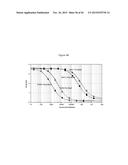 |  |
 |  |
 |  |
 | 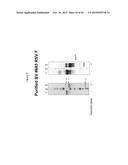 |
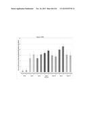 |  |
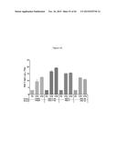 | 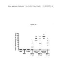 |
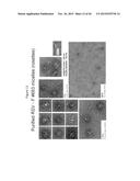 | 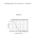 |
 |  |
 |  |
 |  |
 |  |
 |  |
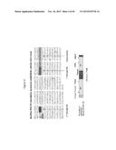 |  |
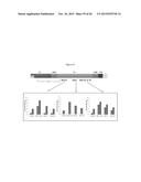 | 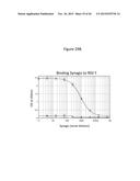 |
 | 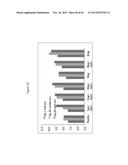 |
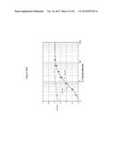 |  |
 | 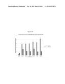 |
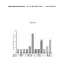 | 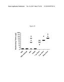 |
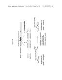 |  |
 | 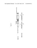 |
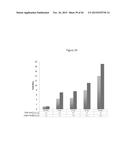 |  |
 | 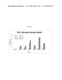 |
 |  |
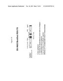 |  |
 | 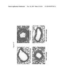 |
 |  |
 |  |
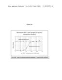 | 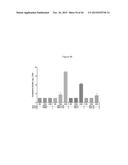 |
 | 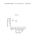 |
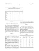 | 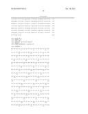 |
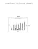 | 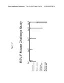 |
 |  |
 |  |
 | 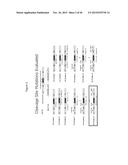 |
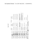 | 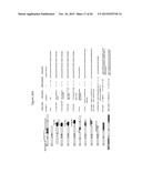 |
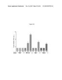 |  |
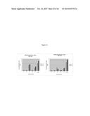 | 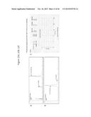 |
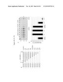 |  |
 | 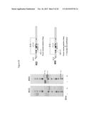 |
 |  |
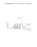 |  |
| Similar patent applications: | |
| Date | Title |
|---|---|
| 2016-01-21 | Cerium oxide nanoparticles accelerate the decay of peroxynitrite (onoo-) |
| 2016-01-21 | Nano- microdelivery systems for oromucosal delivery of an active ingredient |
| 2016-01-21 | Compositions and methods for treating insulin resistance and diabetes mellitus |
| 2015-12-24 | Respiratory syncytial virus f protien epitopes |
| 2016-01-21 | Composition for regenerating normal tissue from fibrotic tissue |
| New patent applications in this class: | |
| Date | Title |
|---|---|
| 2022-05-05 | Sars-cov-2 viruses and methods of use thereof |
| 2022-05-05 | Method for the safe induction of immunity against rsv |
| 2022-05-05 | Methods for preventing disease or disorder caused by rsv infection |
| 2019-05-16 | Duck enteritis virus and the uses thereof |
| 2019-05-16 | Duck enteritis virus and the uses thereof |
| New patent applications from these inventors: | |
| Date | Title |
|---|---|
| 2015-12-17 | Modified rsv f proteins and methods of their use |
| 2015-10-29 | Highly efficient influenza matrix (m1) proteins |
| 2015-10-29 | Recombinant nanoparticle rsv f vaccine for respiratory syncytial virus |
| Top Inventors for class "Drug, bio-affecting and body treating compositions" | |
| Rank | Inventor's name |
|---|---|
| 1 | David M. Goldenberg |
| 2 | Hy Si Bui |
| 3 | Lowell L. Wood, Jr. |
| 4 | Roderick A. Hyde |
| 5 | Yat Sun Or |