Patent application title: EXPRESSION OF ISOFORM 202 OF ERCC1 FOR PREDICTING RESPONSE TO CANCER CHEMOTHERAPY
Inventors:
Ken Olaussen (Paris, FR)
Jean-Charles Soria (Paris, FR)
Luc Friboulet (Le Havre, FR)
Assignees:
INSTITUTE GUSTAVE-ROUSSY
IPC8 Class: AG01N33573FI
USPC Class:
435 611
Class name: Measuring or testing process involving enzymes or micro-organisms; composition or test strip therefore; processes of forming such composition or test strip involving nucleic acid nucleic acid based assay involving a hybridization step with a nucleic acid probe, involving a single nucleotide polymorphism (snp), involving pharmacogenetics, involving genotyping, involving haplotyping, or involving detection of dna methylation gene expression
Publication date: 2014-06-19
Patent application number: 20140170659
Abstract:
An in vitro method for detecting the susceptibility of a tumor cell to a
chemotherapy is disclosed. The method includes the step of measuring the
expression level of the isoform 202 of the ERCC1 protein.Claims:
1. An in vitro method for detecting the susceptibility of a tumor cell to
a chemotherapy, said method comprising the step of measuring the
expression level of the isoform 202 of the ERCC1 protein.
2. The method according to claim 1, wherein the expression level of the isoform 202 of the ERCC1 protein is measured by assessing the mRNA level of the isoform 202 of the ERCC1 protein.
3. The method according to claim 2, wherein said mRNA level is measured by RT-PCR or in situ hybridization.
4. The method according to claim 2, wherein said mRNA is SEQ ID NO:1.
5. The method according to claim 1, wherein the expression level of the isoform 202 of the ERCC1 protein is measured by means of an immunohistochemistry assay or of an immunofluorescence assay.
6. The method according to claim 1, wherein the expression level of the isoform 202 of the ERCC1 protein is measured by means of an immunohistochemistry assay performed on a formalin-fixed paraffin-embedded tumor sample.
7. The method according to claim 6, wherein said immunohistochemistry assay uses a monoclonal ERCC1 antibody recognizing specifically the isoform 202 of the ERCC1 protein or an antibody recognizing specifically an heterodimer selected from the group consisting of: ERCC1/XPF, ERCC1/XPA, ERCC1/MSH2, ERCC1/FANCG, ERCC1/SLX4, ERCC1/Eg5, ERCC1/MAD2A, and ERCC1/TRF2.
8. The method according to claim 6, further including the steps of: (a) obtaining slides from formalin-fixed paraffin-embedded tumor samples; (b) retrieving epitope in buffer; (c) incubating slides with a monoclonal ERCC1 antibody recognizing specifically the isoform 202 of the ERCC1 protein; or with an antibody recognizing an heterodimer selected from the group consisting of: ERCC1/XPF, ERCC1/XPA, ERCC1/MSH2, ERCC1/FANCG, ERCC1/SLX4, ERCC1/Eg5, ERCC1/MAD2A, and ERCC1/TRF2; or with an antibody recognizing specifically the XPF protein, the XPA protein, the MSH2 protein (isoforms 1 or 2), the FANCG protein, the SLX4 protein, the Eg5 protein, the MAD2A protein, or the TRF2 protein; (d) determining an amount of binding antibodies on the formalin-fixed paraffin-embedded tumor samples, using the amount of binding antibodies on an internal positive control as a reference; (e) determining a percentage of labeled nuclei on the formalin-fixed paraffin-embedded tumor samples; (f) multiplying the value estimated in step (d) with the value estimated in step (e); and (g) determining a platinum-based chemotherapy regimen by comparing the value obtained in step (f) to a median score of the values obtained in step (f).
9. The method according to claim 8, wherein the internal positive control consists of stroma cells surrounding the tumor area.
10. The method according to claim 1, wherein the expression level of the isoform 202 of the ERCC1 protein is measured by means of an immunofluorescence assay performed on individual tumor cells.
11. The method according to claim 1, wherein said expression level is measured by means of an antibody that recognizes specifically the isoform 202 of the ERCC1 protein; or with an antibody recognizing specifically an heterodimer selected from the group consisting of: ERCC1/XPF, ERCC1/XPA, ERCC1/MSH2, ERCC1/FANCG, ERCC1/SLX4, ERCC1/Eg5, ERCC1/MAD2A, and ERCC1/TRF2; or with an antibody recognizing specifically the XPF protein, the XPA protein, the MSH2 protein (isoforms 1 or 2), the FANCG protein, the SLX4 protein, the Eg5 protein, the MAD2A protein, or the TRF2 protein.
12. The method according to claim 1, wherein said tumor is a non-small-cell lung cancer.
13. The method according to claim 1, wherein said chemotherapy is a platinum-based cancer chemotherapy.
14. The method according to claim 1, wherein said chemotherapy is based on cisplatin.
15. The method according to claim 1, wherein said chemotherapy is cisplatin associated with other chemotherapeutic agents.
16. The method according to claim 1, wherein said chemotherapy is cisplatin with etoposide or a vinca alkaloid.
17. The method according to claim 1, wherein said patient had undergone a surgical resection of its tumor.
18. A kit for the detection or quantification of the isoform 202 of the ERCC1 protein, comprising: (a) an antibody that recognizes specifically the isoform 202 of the ERCC1 protein, or (b) an antibody recognizing specifically an heterodimer selected from the group consisting of: ERCC1/XPF, ERCC1/XPA, ERCC1/MSH2, ERCC1/FANCG, ERCC1/SLX4, ERCC1/Eg5, ERCC1/MAD2A, and ERCC1/TRF2; or (c) an antibody recognizing specifically the XPF protein, the XPA protein, the MSH2 protein (isoforms 1 or 2), the FANCG protein, the SLX4 protein, the Eg5 protein, the MAD2A protein, or the TRF2 protein.
19. An in vitro method for detecting the susceptibility of a tumor cell to a chemotherapy, using the kit of claim 18.
Description:
[0001] Sequence Listing contained in file D27420362944ST25.txt having a
file size of 80.0 KB is hereby incorporated-by-reference.
TECHNICAL FIELD OF THE INVENTION
[0002] The present invention is directed to the detection of the Excision Repair Cross-Complementation group 1 (ERCC1) enzyme, and more specifically of the isoform 202 of this enzyme, and its use in the detection of the susceptibility of a tumor cell to chemotherapy, especially to platinating agents-based cancer chemotherapy. The invention also concerns a kit for detection, carrying out the method.
BACKGROUND OF THE INVENTION
[0003] Lung cancer is a leading cause of cancer deaths in most industrialized countries (Jemal A, Murray T, Ward E, et al. Cancer statistics, 2005). Despite complete tumor resection in patients with stage I-III non-small-cell lung cancer, distant metastases develop in 50-70 percent of patients.
[0004] Adjuvant chemotherapy has been tested to improve survival in patients with completely resected non-small-cell lung cancer. The recently reported International Adjuvant Lung Cancer Trial (IALT) with 1,867 patients, was designed to assess the potential benefit of adjuvant cisplatin-based chemotherapy after complete resection of non-small cell lung cancer. The IALT demonstrated a 4.1 percent absolute benefit in 5-year overall survival in non-small-cell lung cancer patients treated with adjuvant cisplatin-based chemotherapy (the International Adjuvant Lung Cancer Trial Collaborative Group. Cisplatin-based adjuvant chemotherapy in patients with completely resected non-small-cell lung cancer. N Engl J Med 2004). Several other randomized studies have confirmed the benefit of postoperative platinum-based therapy in non-small-cell lung cancer (Gurubhagavatula S, Lynch T J. Semin Respir Crit. Care Med 2005). However, adjuvant chemotherapy only slightly prolongs survival, with a 5-year overall survival improvement ranging from 4 to 15 percent, and gives rise to significant adverse effects (Winton T, et al. N Engl J Med 2005). The identification and quantification of predictive factors for resistance or sensitivity to adjuvant cisplatin-based chemotherapy were therefore needed.
[0005] Among potential predictive factors are those involved in cisplatin-resistance such as DNA repair mechanisms. Cisplatin induces cytotoxic effects by binding to DNA and creating platinum-DNA adducts. Some of these adducts establish covalent cross-linking between DNA strands, thereby inhibiting DNA replication. Among the DNA repair pathways, nucleotide excision repair plays a central role and has been associated with resistance to platinum-based chemotherapy (Reed E. Cancer Treat Rev 1998). The excision repair cross-complementation group 1 (ERCC1) enzyme plays a rate-limiting role in the nucleotide excision repair pathway which recognizes and removes cisplatin-induced DNA adducts (Zamble D B. et al. Biochemistry 1996). ERCC1 is also an important factor in DNA interstrand cross-link repair, as well as in recombination processes (De Silva I U. et al. Mol Cell Biol 2000).
[0006] For more than a decade, smaller studies have repeatedly reported an association between low ERCC1 mRNA expression levels in several solid tumors and improved clinical outcomes in patients treated with platinum-containing regimens (Dabholkar M. et al J Clin Invest 1994). In particular, Lord et al (Lord R Vet al. Clin Cancer Res 2002) reported that ERCC1 mRNA expression predicts response to chemotherapy in advanced non-small-cell lung cancer. Furthermore, by using methodologies such as DNA isolation, enzymatic digestions, and DNA sequencing, two common polymorphisms of the ERCC1 gene (codon 118 C/T and C8092A) were found to be correlated with response to platinum-based chemotherapy in colorectal (Viguier J. et al. Clin Cancer Res 2005) and non-small-cell lung cancer (Zhou W. et al. Clin Cancer Res 2004). These polymorphisms are mainly associated with lower translation rates of the ERCC1 gene, resulting in low expression levels.
[0007] The invention described in the international application WO 02/061128 (published on 8 Aug. 2002) relates to prognostic methods for cisplatin-based cancer chemotherapy assessing ERCC1 expression levels. These prognostic tests consist of (i) determining a platinum-based chemotherapy by examination of the amount of ERCC1 mRNA in patient's tumor cells and (ii) comparing it to a pre-determined threshold expression level. Such quantitative gene expression studies were developed for formalin-fixed paraffin-embedded pathological samples because most tumor samples are routinely formalin-fixed paraffin-embedded to allow histological analysis and subsequent archival storage. In this method, all the patients were treated with a platinum-based chemotherapy and the ERCC1 level was assessed so as to prognose the survival probability of the treated patients. Nevertheless, (i) techniques for the isolation and analyses of mRNA from formalin-fixed paraffin-embedded tissue samples are frequently inaccurate, costly and time-consuming and (ii) the conservation of mRNA in formalin-fixed paraffin-embedded samples is eventually affected with time. For these reasons, such analyses are carried out with difficulty by the skilled person, especially in large-scale studies. Other techniques, allowing an easier, more accurate and less expensive measure of the expression of ERCC1 are thus particularly needed.
[0008] Moreover, since the study on which the international application WO 02/061128 (published on 8 Aug. 2002) was based did not compare two groups of patients according to whether or not they were treated with cisplatin, the value of ERCC1 mRNA expression as evidenced in this study is only prognostic, and not predictive of the patient response to a chemotherapy.
[0009] A biomarker has a "prognostic value" if it enables to distinguish patients with high probability of survival from those who have low probability of survival, regardless of treatment or in a population where all patients received an identical treatment.
[0010] On another hand, a biomarker as a "predictive value" for a specific treatment if it enables to distinguish patients who have a high probability of clinical benefit (in terms of survival) from those who will take no benefit from said specific treatment. A predictive value of a biomarker can therefore only be demonstrated when two study groups are compared directly (a treated group against a non-treated group).
[0011] Furthermore, other studies have investigated the relation between the expression of different markers, like ERCC1, the platinum resistance and the prognosis in advanced non small cell lung cancer. Indeed, the scientific publication of Huang P Y et al. Chinese Journal of Cancer 2004 aims at determining prognostic values of different markers, like ERCC1, in response of a first-line platinum-based treatment. ERCC1 has been detected by immunohistochemistry. This study indicates that no prognostic value of ERCC1 expression can be demonstrated. On another hand, the publication of Watchers F M et al. Lung Cancer 2005 describes a study to determine a prognostic value of different protein expression involved in DNA repair. Among them, ERCC1 expression is measured in phase III-NSCLC patients by comparing first-line "cisplatin-gemcitabine" and "epirubicin-gemcitabine" chemotherapies. The ERCC1 expression was measured by immunohistochemistry on formalin-fixed, paraffin-embedded tumor biopsies. This document concludes that these markers (including ERCC1) are not prognostic of patient survival after these chemotherapies. Those two documents conclude that ERCC1 has no prognostic value of the efficiency of chemotherapy treatment.
BRIEF SUMMARY OF THE INVENTION
[0012] These and other shortcomings of the prior art are addressed by the present invention, which provides an in vitro method for detecting the susceptibility of a tumor cell to a chemotherapy.
[0013] This invention differs from the anterior art, particularly the scientific publications of Huang P Y et al. Chinese Journal of Cancer 2004 and Watchers F M et al. Lung Cancer 2005 by the fact that the steps which have been carried out in said immunohistochemical method are different from those in these precedent documents wherein ERCC1 was not described as a predictive marker of a chemotherapy efficiency, but as a prognostic marker in patients treated by chemotherapy.
[0014] The method of the invention comprises the step of assessing the expression level of the isoform 202 of the ERCC1 protein. In a preferred embodiment of the invention, the said expression level is assessed by measuring the mRNA level of the isoform 202 of the ERCC1 protein, for example by RT-PCR or in situ hybridization. Alternatively, immunological methods aimed at measuring directly the protein level of the isoform 202 of ERCC1 (e.g., immunohistochemical or immunofluorescence assays) can be used.
[0015] Using an immunohistochemical assay, the inventors demonstrated, in a quantitative and reproducible way, that patients with ERCC1-negative tumors have a risk of death decreased by 33% (hazard ratio 0.67) when cisplatin based chemotherapy is added to surgery. On the other hand, the inventors have found that the risk of death was not decreased by the adjunction of chemotherapy among patients with ERCC1-positive tumors (hazard ratio 1.18) (Olaussen et al, NEJM, 2006). These results had been obtained with an anti-ERCC1 antibody recognizing, possibly among other isoforms, the isoform 202 of ERCC1.
[0016] It was stated in the patent application WO 02/061128 that protein expression levels could be only qualitatively monitored in formalin-fixed paraffin-embedded samples by using immunohistochemical methods. However, the present inventors have developed other quantitative and reproducible methods for assessing ERCC1 expression levels which is the subject matter of the present invention.
[0017] The analyses of the protein or mRNA level by the said methods are predictive of survival in early-stage and completely resected non-small cell lung cancer. Because immunological assays and mRNA analyses can be easily applied in every pathology laboratory, the present invention is therefore widely applicable and a useful test in clinical practice.
[0018] In a particular embodiment, the method of the invention also presents an additional advantage which is to be able to analyze formalin-fixed paraffin-embedded tumor samples, whatever the fixation techniques are.
[0019] The ERCC1 gene generates four isoforms (designated 201, 202, 203, and 204) by alternative splicing. As shown recently in Friboulet et al. (2013), using immunohistochemical analysis, the present Inventors showed that the level of biologic complexity of the ERCC1 protein had been underestimated and that the respective roles of the four ERCC1 protein isoforms had not been correctly assessed. In fact, they showed that the expression of nonfunctional ERCC1 isoforms leads to potential artifacts, with discrepant results.
[0020] More precisely, in this study, they demonstrated that only the reintroduction of the ERCC1-202 isoform rescued nucleotide excision repair activity and the capacity to repair cisplatin-induced DNA damage in established ERCC1-deficient cells. In terms of patient classification and therapeutic applications, these results suggested that evaluation of the expression of the unique functional isoform (ERCC1-202) constitutes a more accurate predictor of cisplatin benefit in patients with NSCLC than any other current approach. In other words, these data showed for the first time that therapeutic decision regarding cisplatin-containing treatment in patients with NSCLC requires the specific detection of the unique functional isoform of ERCC1-202.
[0021] These results explained the contradictory results obtained by the other teams so far, as none of them used antibodies that could distinguish functional ERCC1 isoform 202 from the other nonfunctional isoforms. Accordingly, it was difficult in the past to validate the correlation between the level of ERCC1 expression and overall survival on the basis of immunohistochemical detection, as abundant expression of one or several nonfunctional isoforms led to a false classification of the tumor as ERCC1-positive.
[0022] Using these unique ERCC1 deficient human NSCLC cell lines, the present Inventors further explored the influence of the different ERCC1 protein isoforms on DNA repair, protein-protein interactions and cellular mitotic process. Their data demonstrated that all currently known functions of ERCC1 are fulfilled by the same and unique ERCC1 isoform ERCC1-202 (see example 7 below).
[0023] Thus, the present invention provides an in vitro method for detecting the susceptibility of a tumor cell to a chemotherapy, said method comprising the step of measuring the expression level of the isoform 202 of the ERCC1 protein (hereafter referred to as "ERCC1-202"), for example by immunohistochemistry in a formalin-1-fixed paraffin-embedded tumor sample, by immunofluorescence on tumor cells or by mRNA analysis of tumor cell samples.
BRIEF DESCRIPTION OF THE DRAWINGS
[0024] The subject matter that is regarded as the invention may be best understood by reference to the following description taken in conjunction with the accompanying drawing figures in which:
[0025] FIG. 1A shows an example of ERCC1 staining--Image (400*) of a strongly ERCC1-positive squamous cell carcinoma (Intensity=3);
[0026] FIG. 1B shows an example of ERCC1 staining--Image (400*) of an ERCC1-negative squamous cell carcinoma with positive internal controls (arrow);
[0027] FIG. 2A shows Kaplan-Meier estimates of the proportions of surviving patients--Overall survival according to treatment in all 761 patients--The adjusted hazard ratio for death in the chemotherapy group as compared with the control group was 0.87 (95 percent confidence interval, 0.71 to 1.06, P=0.17);
[0028] FIG. 2B shows Kaplan-Meier Estimates of the proportions of surviving patients--Overall survival according to treatment in patients with ERCC1-negative tumors--The adjusted hazard ratio for death in the chemotherapy group as compared with the control group was 0.67 (95 percent confidence interval, 0.51 to 0.89, P<0.006);
[0029] FIG. 2C shows Kaplan-Meier Estimates of the proportions of surviving patients--Disease-free survival according to treatment in patients with ERCC1-negative tumors. The hazard ratio for disease progression or death was 0.69 (95 percent confidence interval, 0.53 to 0.90, P<0.007);
[0030] FIG. 2D shows Kaplan-Meier Estimates of the proportions of surviving patients--Overall survival according to treatment in patients with ERCC1-positive tumors. The adjusted hazard ratio for death in the chemotherapy group as compared with the control group was 1.18 (95 percent confidence interval, 0.87 to 1.61, P=0.29);
[0031] FIG. 3A shows absence of negative dominant isoform--Western Blot detection of ERCC1 isoforms with 8F11 antibody--Cells expressing the 202 isoform were infected with lentivirus coding another ERCC1 isoform;
[0032] FIG. 3B shows absence of negative dominant isoform--IC50 assessment by WST-1 assay in cells treated for 48 hours with various concentrations of cisplatin;
[0033] FIG. 3C shows absence of negative dominant isoform--Clonogenic growth of isoform expressing cells treated two to three weeks with low cisplatin concentrations. Table specifies cisplatin IC50 (nM) values from clonogenic growth of cells;
[0034] FIG. 4 shows ERCC1 isoforms cellular localization--Western blot detection of ERCC1 protein isoforms after cellular fractionation;
[0035] FIG. 5A shows ERCC1 isoforms stability and expression--Western blot detection of ERCC1 protein isoforms after proteasome inhibition (MG132 at 2 μM for 24 h). An increase in ERCC1-201, -203 and -204 expression level suggested instability and degradation of these isoforms;
[0036] FIG. 5B shows ERCC1 isoforms stability and expression--Assessment by qRT-PCR of ERCC1 isoforms mRNAs in frozen samples from a series of 123 cases of resected NSCLC with matched tumour and normal specimens. The expression of ERCC1 isoform mRNA was determined using the 2-ΔΔCt method and data are presented as the fold-change in ERCC1 isoform mRNA expression relative to total ERCC1 mRNA. ERCC1-201 mRNA isoform was upregulated in tumours samples;
[0037] FIG. 6A shows ERCC1-isoform 202 is essential for proper chromosome segregation--Percentage of cells with nucleus size superior of mean of WT nucleus size determined on Diff Quick® stain cells with ImageJ software;
[0038] FIG. 6B shows ERCC1-isoform 202 is essential for proper chromosome segregation--Percentage of multinucleated cells determined on Diff Quick® stain cells manually counted (n=200);
[0039] FIG. 6C shows ERCC1-isoform 202 is essential for proper chromosome segregation--DNA content in cells blocked in G2/M cell cycle phase with Karyomax colcemid solution at 0,1 μg/ml for 6 h. Percentages of cells with high DNA content are shown;
[0040] FIG. 6D shows ERCC1-isoform 202 is essential for proper chromosome segregation--ERCC1 WT, attenuated and rescued cells. Centrosomes (-tubulin) were manually counted (n=100). Percentages of cells with more than 2 centrosomes are shown;
[0041] FIG. 6E shows ERCC1-isoform 202 is essential for proper chromosome segregation--E) 48 hours proliferation index in untreated ERCC1 WT, attenuated and rescued cells determined by WST-1 assay;
[0042] FIG. 7 shows ERCC1 isoforms function in ICL-R-IC50 assessment by WST-1 assay in cells treated for 48 h with various concentrations of the DNA interstrand cross-linking agent mitomycin-C. MMC IC50 (μM) values are indicated for each clone. Only 202 isoform restored MMC resistance compared to WT cells;
[0043] FIG. 8A shows ERCC1-protein complexes required ERCC1-202 isoform expression--Western blot detection of ERCC1 protein isoforms and ERCC1 interacting proteins;
[0044] FIG. 8B shows ERCC1-protein complexes required ERCC1-202 isoform expression--Proximity ligation assay (PLA, Duolink) detection of ERCC1/XPF heterodimers in A549 WT and ERCC1 deficient clone 216 expressing each of the four ERCC1 isoforms; and
[0045] FIG. 8C shows ERCC1-protein complexes required ERCC1-202 isoform expression--Proximity ligation assay (PLA-Duolink) detection of ERCC1/XPF, ERCC1/XPA, ERCC1/MSH2, ERCC1/FANCG, ERCC1/SLX4, ERCC1/Eg5, ERCC1/MAD2A, ERCC1/SLX4 and ERCC1/TRF2 heterodimers definite the unique binding ability of ERCC1-202 isoform only. ERCC1 was detected using FL297 antibody.
DETAILED DESCRIPTION OF THE INVENTION
ERCC1 Isoforms
[0046] ERCC1 was the first mammalian DNA repair gene to be cloned (Westerveld et al., 1984). ERCC1 gene contains 10 exons and codes for a pre-mRNA which leads by alternative splicing to four different isoforms (201, 202, 203, and 204). Very few data are available concerning the expression and role of the different ERCC1 isoforms.
[0047] The ERCC1-202 isoform is easily detectable. In human, the amino acid sequence of the ERCC1-202 isoform is for example the SEQ ID NO:2 (corresponding to the NCBI number NP--001974.1). It is encoded for example by the mRNA sequence SEQ ID NO:1 (NM--001983.3).
[0048] The alternatively spliced ERCC1-203 isoform, lacking the 72 bp of exon VIII, was first identified in 1986 (van Duin et al.). Van Duin et al. already suggested this isoform to be non-functional to repair ultraviolet (UV) light (NER) and mitomycin-C (interstrand cross-link repair--ICL-R) damages. Later, one report suggested this transcript may have a helper function for DNA repair of UV and mitomycin-C damages (Belt, 1991). In ovarian cancer tissues, highly variable splicing of ERCC1 mRNA was observed, and the alternatively spliced ERCC1-203 varied between 2 to 71% of the total ERCC1 mRNA (Dabholkar, 1994). Interestingly, the mRNA levels of this isoform were strongly inversely related to DNA repair activity, suggesting this shorter isoform to be a negative dominant of ERCC1 DNA repair function (Dabholkar, 1995; Yu, 1998). In small cell lung cancer cell line the ERCC1-203 protein isoform was found upregulated (Stordal 2009) after cisplatin exposure. This upregulation did not seem to have an influence on DNA repair efficiency in this study but the authors proposed this isoform to have a role in cell cycle arrest. Lately, ERCC1-203 isoform was confirmed to negatively affect the NER function of ERCC1 for cisplatin resistance in ovarian cancer cells (Sun et al, 2009). In human, the amino acid sequence of the ERCC1-203 isoform is for example the SEQ ID NO:6 (NP--001159521.1). It is encoded for example by the mRNA sequence SEQ ID NO:5 (NM--001166049.1).
[0049] First in mouse keratinocytes (Song et al., 2011), then in human malignant melanoma cells (Li W and Melton D W, 2011), the ERCC1-201 protein isoform was identified to be encoded by a larger ERCC1 transcript, originated from an upstream promoter. This transcript was found highly upregulated after cisplatin exposure by MAPK pathway in human melanoma cells (Li W and Melton D W, 2011). No functional analysis was accomplished about ERCC1-201 isoform but mutational analysis achieved by Sijbers et al. (1996) suggested this isoform to be non-functional. Indeed, they identified that the 91 N-terminal amino acids of ERCC1-202 isoform are dispensable for repair function, in contrast to a deletion of only four residues from the C-terminus. Since the ERCC1-201 isoform harbours an alternative C-terminus peptide sequence it is more than probable that it is non-functional with respect to NER activity. In human, the amino acid sequence of the ERCC1-201 isoform is for example the SEQ ID NO:4 (NP--973730.1). It is encoded for example by the mRNA sequence SEQ ID NO:3 (NM--202001.2).
[0050] The ERCC1-204 isoform, which lack the long 215 bp exon-3, has never been reported in the literature. This isoform still have the XPF, MSH2 and dsDNA binding domains but lack a part of the XPA and ssDNA binding domains. Thus this isoform could negatively influence DNA repair by sequestration of XPF or MSH2. In human, the amino acid sequence of the ERCC1-204 isoform is for example the SEQ ID NO:8 (NSP00000394875.2). It is encoded for example by the mRNA sequence SEQ ID NO:7 (ENST00000423698.2).
The Method of the Invention
[0051] The present invention provides in vitro methods for detecting the susceptibility of a tumor cell to a chemotherapy, said method comprising the step of measuring the expression level of the isoform 202 of the ERCC1 protein (ERCC1-202).
[0052] This expression level can be assessed either by measuring the mRNA level (e.g., by RT-PCR or by in situ hybridization or Duolink) or by measuring the protein level of this isoform, for example by means of an immunological method.
[0053] By "immunological method", it is herein meant any experimental method involving antibodies that are able to recognize specifically the isoform 202 of the ERCC1 protein. These immunological methods can be in particular an immunohistochemistry assay or an immunofluorescence assay.
[0054] In an "immunohistochemical assay", a section of tissue is tested for specific proteins by exposing the tissue to antibodies that are specific for the protein that is being assayed. The antibodies are then visualized by any of a number of methods to determine the presence and amount of the protein present. Examples of methods used to visualize antibodies are, for example, through enzymes linked to the antibodies (e.g., luciferase, alkaline phosphatase, horseradish peroxidase, or P-galactosidase), or chemical methods (e.g., DAB/Substrate chromagen), gold, fluorescent or labelled antibodies by any of the many different methods known to those skilled in this art. In embodiments of the said immunohistochemical method, detection or assaying the level of the isoform 202 of the ERCC1 protein in a tumor sample includes contacting it with an antibody or antigen-binding fragments directed against said isoform or fragments thereof; and determining the amount of the binding antibody on the tumor sample.
[0055] In contrast thereto, as used herein, an "immunofluorescence assay" is a technique using fluorescence microscopy on cells that have been extracted from the tumor tissue or on cell lines, e.g., on cultured cell lines or on individual cells. This technique uses the specificity of antibodies to their antigen to target fluorescent dyes to specific biomolecule targets within a cell, and therefore allows visualization of the distribution of the target molecule (the isoform 202 of ERCC1) through the sample. Immunofluorescence can be used in combination with other, non-antibody methods of fluorescent staining, for example, use of DAPI to label DNA.
[0056] Several microscope designs can be used for immunohistochemical or immunofluorescence assays; the simplest is the epifluorescence microscope. The confocal microscope is also widely used. Various super-resolution microscope systems that are capable of much higher resolution can also be used.
[0057] As used herein, the term "antibody" includes immunoglobulin molecules and antigen binding fragments thereof. The antibody can be a polyclonal antibody or a monoclonal antibody. The antibody can be labeled by a detectable means and includes enzymatically, radioactively, fluorescently, chemiluminescently or bioluminescently labeled antibodies by any of the many different methods known to those skilled in this art.
[0058] By "antigen-binding fragments" it is intended to encompassed particularly the fragments Fv, Fab, F(ab')2, Fab', scFv, scFv-Fc. These antibody fragments are obtained using conventional techniques well-known to those with skill in the art, and the fragments are screened for utility in the same manner as are intact antibodies.
[0059] In the context of the present invention, an antibody is said to "recognize" or "bind" the ERCC1 isoform 202 of SEQ ID NO:2 if said antibody has an affinity constant Ka (which is the inverted dissociation constant, i.e. 1/Kd) higher than 107 M-1, preferably higher than 108 M-1, more preferably higher than 109 M-1 for said isoform. Also, in the context of the present invention, an antibody is said to "specifically bind" or to "specifically recognize" the ERCC1 isoform 202 of SEQ ID NO:2 if said antibody has an affinity constant Ka higher than 107 M-1, preferably higher than 108 M-1, more preferably higher than 109 M-1 for said isoform and has an affinity constant Ka lower than 105 M-1 for all the other proteins, including the other isoforms of ERCC1.
[0060] The affinity constant which is used to characterize the binding of antibodies (Ab) to a peptide or an antigen (Ag) is the inverted dissociation constant defined as follows:
Ab + Ag AbAg ##EQU00001## K a = [ AbAg ] [ Ab ] [ Ag ] = 1 K d ##EQU00001.2##
[0061] This affinity can be measured for example by equilibrium dialysis or by fluorescence quenching, both technologies being routinely used in the art.
[0062] In a preferred embodiment, the antibody of the invention binds the ERCC1 isoform 202 of SEQ ID NO:2 with a KD of less than 10-9M, preferably of less than 10-10 M.
[0063] The present inventors have shown that only the 202 isoform of ERCC1 can form stable heterodimer complexes with the DNA repair endonuclease XPF (SEQ ID NO: 9, corresponding to NP--005227.1, which is encoded by the ERCC4 gene), with the XPA protein (SEQ ID NO:10, corresponding to NP--000371.1), with the DNA mismatch repair protein MSH2 (SEQ ID NO: 11 corresponding to the isoform 1 (full length) NP--000242.1 or SEQ ID NO: 12 corresponding to the isoform 2 NP--001245210.1), with the Fanconi anemia Group G protein FANC G (SEQ ID NO: 13, corresponding to NP--004620.1), with the SLX4 protein (SEQ ID NO:14 corresponding to NP--115820.2), with the kinesin Eg5 (SEQ ID NO: 15, corresponding to NP--004514.2 encoded by the KIF11 gene), with the MAD2A protein (SEQ ID NO: 16, corresponding to NP--002349.1 encoded by the MAD2L1 gene), and the protein TRF2 (SEQ ID NO: 17, corresponding to NP--005643.2, encoded by the TERF2 gene).
[0064] Their data clearly suggest that detecting the presence of ERCC1/XPF, ERCC1/XPA, ERCC1/MSH2, ERCC1/FANCG, ERCC1/SLX4, ERCC1/Eg5, ERCC1/MAD2A, or of ERCC1/TRF2 heterodimers is helpful for quantifying the level of functional ERCC1-202 isoform in cancer cell.
[0065] Consequently, the detecting method of the invention may comprise the step of detecting and/or quantifying the presence of a stable heterodimers selected from the group consisting of: ERCC1/XPF, ERCC1/XPA, ERCC1/MSH2, ERCC1/FANCG, ERCC1/SLX4, ERCC1/Eg5, ERCC1/MAD2A, and ERCC1/TRF2. This detection/quantification may be performed by conventional means, such as protein complex immunoprecipitation, pull-down assays, Proximity ligation assay (PLA), FRET assays, surface plasmon resonance (SPR) assays, affinity capture mass spectrometry or the Duolink® immunoassay developed by Olink. Immunoassays such as immunoprecipitation, pull-down assays and the Duolink® assay are herein preferred. Proximity ligation assay (PLA) is even more preferred.
[0066] In one embodiment, the skilled person will preferably use an antibody which binds specifically to the XPF protein, for example the anti-XPF antibody clone 3F2/3; or an antibody which binds specifically to the XPA protein, for example the anti-XPA antibody clone 5A2 commercialized by Pierce under the reference "MA1-21460"; or an antibody which binds specifically to the MSH2 protein, for example the anti-MSH2 antibody commercialized by BIORBYT under the reference "orb16010"; or an antibody which binds specifically to the FANCG protein, for example the anti-FANCG antibody commercialized by Abcam under the reference "ab54645"; or an antibody which binds specifically to the Eg5 protein, for example the anti-Eg5 antibody commercialized by Abcam under the reference "ab51976"; or an antibody which binds specifically to the SLX4 protein, for example the anti-SLX4 antibody commercialized by Abnova under the reference "H00084464"; or an antibody which binds specifically to the MAD2A protein, for example the anti-MAD2A antibody clone 17D10 commercialized by Abcam under the reference "10691"; or an antibody which binds specifically to the TRF2 protein, for example the anti-TRF2 antibody clone 4A794 commercialized by Abcam under the reference "ab13579".
[0067] In another embodiment, the skilled person will preferably use an antibody which binds specifically to the ERCC1--XPF heterodimer, the ERCC1/XPA heterodimer, the ERCC1/MSH2 heterodimer, the ERCC1/FANCG heterodimer, the ERCC1/SLX4 heterodimer, the ERCC1/Eg5 heterodimer, the ERCC1/MAD2A heterodimer, and the ERCC1/TRF2 heterodimer.
[0068] In the method of the present invention, the antibody recognizing ERCC1-202 is preferably selected in the group consisting of: the mouse monoclonal antibody clone 8F1 (Thermo Scientific Inc. ERCC1 Ab-2 Cat. MS-671-P1), the mouse monoclonal antibody 3H11 (Novus Biologicals Cat. NB100-117 or Santa Cruz Biotechnology Inc. Cat. sc53281), the rabbit polyclonal antibody FL297 (Santa Cruz Biotechnology Inc. Cat. sc-10785), the rabbit monoclonal antibody EP2143Y (Origene Inc. Cat. TA306972), the mouse monoclonal antibody 4F9 (Origene. Inc. Cat. UM500008), and the mouse monoclonal antibody 2E12 (Origene. Inc. Cat. UM500011).
[0069] Interestingly, the method of the present invention can be carried out on post-operative patient tumor samples. The chemotherapy will then be applied after a surgical resection of the tumor.
[0070] In a preferred embodiment of the invention, the in vitro method of the invention is thus for detecting susceptibility to a chemotherapy of a tumor cell from patients who had undergone a surgical resection of their tumor.
[0071] The method of the invention enables to predict if a platinum-based chemotherapy will be of beneficial use in patients suffering from cancer. As a matter of fact, the present inventors demonstrated that the expression level of ERCC1-202 impacts the efficiency of a platinum-based chemotherapy treatment. In particular, their results suggest that, when the expression level of ERCC1-202 is low, then platinum-based chemotherapy will be efficient and said patient will experience long survival upon treatment with this platinum-based chemotherapy. Conversely, when the expression level of ERCC1-202 is high, then a platinum-based chemotherapy will be useless because the patient's survival will not increase upon treatment with this platinum-based chemotherapy. In this case, a platinum-based chemotherapy is to be avoided and another treatment is to be preferred (for example, surgery, immunotherapy, radiotherapy, platinum-free chemotherapy, etc.).
[0072] Thus, in another aspect, the present invention relates to a method for treating a patient suffering from cancer, containing the steps of:
[0073] i) detecting the susceptibility of the tumor cells of said patient to a chemotherapy by means of the measuring the ERCC1-202 expression level as disclosed above, and
[0074] ii) if said tumor cells express low level of ERCC1-202, then administering to said patient an efficient dose of a platinum-based chemotherapy, whereas
[0075] if said tumor cells express high level of ERCC1-202, then treating said patient with a platinum-free chemotherapy, by surgery, by radiotherapy, or by immunotherapy.
[0076] Determining if the expression level of ERCC1-202 is low or high in a tumor sample can be performed by comparing the expression level of ERCC1-202 in said sample with the expression level of ERCC1-202 obtained in an internal positive control which is used as a reference. Immunostaining intensity is for example multiplied by a proportion score (representative of the percentage of positive tumor nuclei) to obtain a final quantitative H-Score (for "Histology-score"). The median value of the H-Scores was a priori chosen as the cut-off point.
[0077] In immunohistochemical assays, high levels of ERCC1-202 are for example detected when the H Score exceeding median value of H-Score (i.e., tumors with a staining intensity score of 2 and with 50% or more positive nuclei or with a staining intensity score of 3 and 10% or more positive nuclei). Low levels of ERCC1-202 are detected for example when the H-Score is below the median value of H-Score. In Duolink assays, expression level of ERCC1-202 is obtained by assessing the number of fluorescent points in the nuclei of tumoral cells.
[0078] Preferably, internal positive control herein consists of stroma cells surrounding the tumor area.
[0079] In a preferred embodiment of the invention, the method is based on an immunohistochemical assay comprising the following steps:
[0080] (a) obtaining slides from formalin-fixed paraffin-embedded tumor samples,
[0081] (b) retrieving epitope in buffer,
[0082] (c) incubating slides with a monoclonal ERCC1 antibody recognizing specifically the isoform 202 of ERCC1,
[0083] (d) determining the amount of binding antibodies on the formalin-fixed paraffin-embedded tumor samples, using the amount of binding antibodies on an internal positive control as a reference,
[0084] (e) determining the percentage of labeled nuclei on the formalin-fixed paraffin-embedded tumor samples,
[0085] (f) multiplying the value estimated in step (d) with the value estimated in step
[0086] (e), and
[0087] (g) determining a platinum-based chemotherapy regimen by comparing the value obtained in step (f) to a pre-determined threshold level.
[0088] With this method, steps (d) and (f) are used for the first time. They make the detection of ERCC1 surprisingly quantitative and reproducible.
[0089] In a more preferred embodiment of the invention, such cancer is preferably a non-small-cell lung cancer.
[0090] In another preferred embodiment of the invention, the cancer chemotherapy is a platinum-based cancer chemotherapy.
[0091] It is also preferred that the cancer chemotherapy is based on cisplatin alone or associated with other chemotherapeutic agents as etoposide or a vinca alkaloid.
[0092] The invention also relates to a kit for the detection or quantification of the isoform 202 of the ERCC1 protein (ERCC1-202), wherein said kit comprises antibodies and appropriate reagents and buffers. The antibody used in this kit is the monoclonal ERCC1 mouse antibody clone 8F1 commercialized by Neomarkers, the mouse monoclonal antibody 3H11 (distributed by several manufacturers), the rabbit polyclonal antibody FL297 (Santa Cruz), or the rabbit monoclonal antibody EP2143Y (distributed by several manufacturers).
[0093] In a preferred embodiment, said detection kit is used for detecting the susceptibility of a tumor cell to a chemotherapy, for example in the method described above, or in a method for treating a patient suffering from cancer, for example in the method described above.
EXAMPLES
Example 1
Materials and Methods
Patients and Study Design.
[0094] All patients had participated in the IALT study that compared adjuvant cisplatin-based chemotherapy to observation in patients with non-small-cell lung cancer. Inclusion criteria and the results of the IALT have already been reported (The International Adjuvant Lung Cancer Trial Collaborative Group. Cisplatin-based adjuvant chemotherapy in patients with completely resected non-small-cell lung cancer. N Engl J Med 2004; 350:351-60), see table 1. Briefly, 1,867 patients with completely resected stage I-III non-small-cell lung cancer had been randomized to either chemotherapy with cisplatin (total dose 300-400 mg/m<2>) plus another drug (etoposide or a vinca alkaloid), or observation (control group). The median follow-up time was 56 months.
[0095] The IALT-Bio study was subsequently designed by a steering committee to examine whether immunohistochemically assessed tumor markers had the ability to predict a survival benefit from chemotherapy in formalin-fixed paraffin-embedded tumor samples collected from centers that had recruited more than 10 patients. To study whether the effect of chemotherapy was different between patients with a positive or a negative marker status, the estimated power to detect a 20 percent difference in the survival benefit at 5 years in 800 patients was 66 percent (two-sided, type I error 1%). Twenty-eight centers in 14 countries (see table 1) contributed specimens.
[0096] Approval was obtained from the local Institutional Review Boards according to the legal regulations in each participating country.
[0097] All tumors were reviewed centrally (Brambilla E, Lantuejoul S, Dunant A, et al. IALT--International Adjuvant Lung Cancer Trial-: Quality assessment and histopathological review according to the WHO 2004 classification and assessment of prognostic and predictive role of pathological criteria. Lung Cancer 2005; 49. Suppl. 2:S44) according to the W.H.O. 2004 histo-pathological classification.
TABLE-US-00001 TABLE 1 The IALT-Bio participating centers (investigators and pathologists) AUSTRIA: R. Pirker, Internal Medicine I, Vienna, G. Dekan, Institute of Pathology, Vienna BELGIUM: J. Vansteenkiste, University Hospital, Leuven BRAZIL: I. Sathler Pinel, Instituto Nacional de Cancer, Rio de Janeiro R. Younes, Hospital A.C. Camarco, Sao Paulo FRANCE: A.A. Kanoui, Centre Physiotherapie du Rouget, Sarcelles; R. Dachez, Laboratoire L.C.L., Paris; S. Desligneres, Hospital Delafontaine, Saint-Denis; O. Languille-Mimoune, Cabinet Pathologie, Paris; P. Sabatier, Centre Hospitalier Victor Dupouy, Argenteuil T. Le Chevalier, Institut Gustave-Roussy, Villejuif; M. Antoine, Hopital Tenon, Paris P. Boz, Cabinet de Pathologie, Papeete; P. Bruneval, Association Promotion Anatomie Pathologique, Paris; M.C. Charpentier, Cabinet Pathologie Tolbiac, Paris; B. Chetaille, Hopital Sainte Marguerite, Marseille; E. Dulmet, Centre Chirurgical Marie-Lannelongue, Le Plessis Robinson; F. Capron, Groupe Hospitalier Pitie-Salpetriere, Paris; B. Gosselin, C.H.U., Lille; D. Grunenwald, P. Validire, Institut Mutualiste Montsouris, Paris; F. Labrousse, C.H.U., Limoges; N. Pericoli, Roma (Italy); D. Petrot, Cabinet d'Anatomie Pathologique, Niort; N. Rouyer, Cabinet de Pathologie Butet-Rouyer, Nice B. Milleron, M. Antoine, Hopital Tenon, Paris J.F. Morere, M. Kambouchner, Hopital Avicenne, Bobigny G. Ozenne, Ceditrac--CMC du Cedre, Bois Guillaume T. Ducastelle, Laboratoire d'Anatomie et Cytologie, Rouen E. Quoix, Hopital Lyautey, Strasbourg; P. Durand de Grossouvre, Laboratoire d'Anatomie Pathologique, Haguenau; B. Gasser, C.H.U., Strasbourg A. Riviere, Centre Francois Baclesse, Caen; F. Galateau-Salle, CHU, Caen C. Tuchais, P. Janet, G. Bertrand, I. Valo, Centre Paul Papin, Angers GERMANY: W. Eberhardt, University Hospital, Essen; D. Theegarten, Institute of Pathology, Ruhr-University Bochum, Bochum GREECE: P. Christaki, Papanikolaou General Hospital, Pylea T. Dosios, V. Kyriakou, Athens University School of Medicine, Athens E. Papadakis, P. Agelidou, Sotiria Hospital, Athens K. Zarogoulidis, University Hospital, Thessaloniki ITALY: A. Masotti, Azienda Ospedaliera Di Verona, Verona LITHUANIA: A. Jackevicius, Institute of Oncology Vilnius University, Vilnius POLAND: J. Laudanski, L. Chyczewski, M. Kozlowski, J. Niklinski, Medical School, Bialystok T. Grodski, J. Pankowski, Regional Hosp. For Lung Diseases, Szczecin T. Orlowski, M. Chabowski, R. Langfort, Institute of Tuberculosis and Lung Disease, Warsaw; B. Muszczynska-Bernhard, Dolnoslaskiego Centrum Chorob Pluc, Wroclaw ROMANIA: T. Ciuleanu, Oncological Institute "Ion Chiricuta", Cluj-Napoca SLOVAKIA: J. Baumohl, University Teach. Hospital, Kosice SPAIN: F. Cardenal, Hospital Duran I Reynals, Barcelona; R. Bernat, Hospital de Bellvitge, Barcelona J. Salinas, J.B. Lopez, Hospital Virgen de Arrixaca, El Palmar Murcia SWEDEN: B. Bergman, A. Hussein, Sahlgrenska Hospital, Goteborg YUGOSLAVIA: G. Radosavljevic, Institute for Lung Disease, Belgrade
Immunostaining for ERCC1.
[0098] The epitopes were first retrieved in citrate buffer (10 mM, pH 6.0, heated for 30 minutes in a bain marie), then slides were incubated at a 1:300 dilution over 60 minutes with the monoclonal ERCC1 mouse antibody (clone 8F1, NeoMarkers, Fremont Calif., USA) that was raised against the full-length human ERCC1 protein. Antibody binding was detected by means of an ABC-kit with NovaRED® as the substrate (Vectastain Elite, Vector Laboratories, Burlingame Calif., USA) and Mayer's hematoxylin as the counterstain. Sections of normal tonsil tissues were included as external positive controls and stromal cells (endothelium) surrounding the tumor area served as internal positive controls.
Microscopic Analysis
[0099] Two investigators who where blinded to clinical data, independently evaluated ERCC1 staining under the light microscope at *400 magnification. We recorded whether or not tumor or stromal cells expressed ERCC1. In addition, staining intensity was graded on a scale of 0 to 3 (using endothelial cells in tonsil controls as a reference point [intensity 2]). Discordant cases were reviewed. Cases without valid internal controls were excluded. Five images of representative areas were acquired at *400 magnification for each case. All positive or negative tumor nuclei (a total of 500-1,500 tumor nuclei per case) were manually counted on a computer screen using ImageJ freeware edited by the National Institutes of Health (http://rsb.info.nih.gov/ij). The percentage of positive tumor nuclei was calculated per case and a proportion score was attributed (0 if 0 percent; 0.1 if 1 to 9 percent; 0.5 if 10 to 49 percent; 1.0 if 50 percent or more), as previously described (Al Haddad S, Zhang Z, Leygue E, et al. Psoriasin (S100A7) expression and invasive breast cancer. Am J Pathol 1999; 155:2057-66 or Handra-Luca A, Bilal H, Bertrand J C, Fouret P. Extra-cellular signal-regulated ERK-1/ERK-2 pathway activation in human salivary gland mucoepidermoid carcinoma: association to aggressive tumor behavior and tumor cell proliferation. Am J Pathol 2003; 163:957-67). In each case, the proportion score was multiplied by the staining intensity of nuclei to obtain a final quantitative H-score (among 9 possible ones). The median value of the H-scores was a priori chosen as the cut-off point for separating ERCC1-positive from ERCC1-negative tumors.
[0100] Statistical Analysis.
[0101] As in the IALT, the primary endpoint was overall survival after the date of randomization. Disease-free survival was analyzed as a secondary endpoint. In order to study selection bias within the IALT-Bio participating centers, the pre-randomization characteristics and overall survival of the two groups of patients (with or without blocks) were compared using a Cox model. Baseline data according to the ERCC1 status were compared in univariate analyses with Chi-square tests and with a multivariate logistic model.
[0102] Survival rates were estimated using the Kaplan-Meier method. The predictive values of the ERCC1 status and chemotherapy for survival were studied using the Cox model. As in the IALT analysis, the Cox model included every factor used in the stratified randomization (center, disease stage, and type of surgery), plus clinical and histological predictive factors (age, sex, W.H.O. performance status, and revised histopathological type). All other factors that were statistically related to the ERCC1 status in the multivariate logistic model (P<0.05) were added to the survival Cox model (pathological T status, and pleural invasion). The predictive value of ERCC1 was studied by testing the interaction between the ERCC1 status and the attributed treatment (chemotherapy or no chemotherapy) in the same Cox model. All reported P values were two-sided. P values below 0.01 were considered statistically significant in order to limit the risk of false positive results. All analyses were performed using SAS software, version 8.2.
Example 2
Patient Characteristics
[0103] The 28 centers which participated in the IALT-Bio study included 1045 patients in the original IALT study. They were able to provide one tumor block for only 867 patients (83 percent). These 867 patients were comparable to the remaining 178 in terms of pre-randomization characteristics and overall survival. The amount and quality of the 824 blocks were adequate for serial sectioning. Among these blocks, 783 contained tumor material corresponding to non-small-cell lung cancer and were included in the IALT-Bio study. After exclusion of cases without valid positive internal controls, ERCC1 expression was evaluated in 761 cases. All further statistical analyses were based on these 761 patients.
[0104] The characteristics of the IALT-Bio study patient population are summarized in Table 1. A total of 426 cases were squamous-cell carcinomas (56 percent), 242 adenocarcinomas (32 percent), and 93 were of another histological type (12 percent). Median age was 58 years (range 27-77) and the great majority were males (81.6 percent). Three hundred and eighty-nine patients (51 percent) were randomized to receive adjuvant cisplatin-based chemotherapy, whereas 372 (49 percent) were randomized to the control group.
Example 3
Immunohistochemically Assessed ERCC 1 Expression
[0105] As illustrated in FIG. 1, ERCC1 immunostaining was nuclear. The median value of the percentage of stained cells was 24 percent (range 0 to 100 percent), whereas the median value of H-scores was 1.0 Tumors with an H-score exceeding 1.0 (i.e. tumors with a staining intensity score of 2 and 50 percent or more positive nuclei or a staining intensity score of 3 and 10 percent or more positive nuclei) were deemed ERCC1 positive, which was the case in 335 patients (44 percent). The median H-score alone (1.0) was attributed to 164 tumors (22 percent). The main differences in clinico-pathological parameters according to ERCC1 expression are reported in Table 2 (univariate analysis). Using the multivariate logistic model, ERCC1 expression was significantly correlated with age (P=0.02 lower in young patients), sex (P=0.04 lower in females), pathological T status (P=0.04 lower with a higher T status), histological type (lower in adenocarcinomas P<0.0001), and pleural invasion (P=0.01 higher in the case of pleural invasion).
TABLE-US-00002 TABLE 2 Patient Characteristics Total ERCC1+ ERCC1- N = 761 N = 335 N = 426 Characteristic (percent) (percent) (percent) P-value* Age P < 0.003 <55 yr 231 (30) 80 (24) 151 (35) (P for trend: P < 0.008) 55-64 yr 330 (43) 161 (48) 169 (40) >64 yr 200 (26) 94 (28) 106 (25) Sex P < 0.0005 Male 621 (82) 292 (87) 329 (77) Female 140 (18) 43 (13) 97 (23) Pathological TNM P = 0.97 stage Stage I 267 (35) 119 (35) 148 (36) Stage II 175 (23) 76 (23) 99 (23) Stage III 319 (42) 140 (42) 179 (42) T of TNM P = 0.10 1 118 (16) 60 (18) 58 (14) 2 452 (59) 188 (56) 264 (62) 3 181 (24) 85 (25) 96 (23) 4 10 (1) 2 (1) 8 (2) Histological type P < 0.0001 Squamous cell 426 (56) 236 (70) 190 (45) carcinoma Adenocarcinoma 242 (32) 71 (21) 171 (40) Other 93 (12) 28 (8) 65 (15) Performance Status P = 0.06 0 426 (56) 188 (56) 238 (56) 1 276 (36) 113 (34) 163 (38) 2 59 (8) 34 (10) 25 (6) Pleural invasion P < 0.007 Yes 61 (8) 37 (11) 24 (6) No 700 (92) 298 (89) 402 (94) Vascular invasion P = 0.04 Yes 222 (29) 85 (25) 137 (32) No 539 (71) 250 (75) 289 (68) Surgery P = 0.35 Pneumonectomy 306 (40) 141 (42) 165 (39) Segment-/lobectomy 455 (60) 194 (58) 261 (61) Radiotherapy P = 0.35 Yes 199 (26) 82 (24) 117 (27) No 562 (74) 253 (76) 309 (73) Planned cisplatin dose P = 0.67 80 mg/m2 per cycle 139 (18) 58 (17) 81 (19) 100 mg/m2 per cycle 544 (71) 245 (73) 299 (70) 120 mg/m2 per cycle 78 (10) 32 (10) 46 (11)
TABLE-US-00003 TABLE 3 Variation of overall survival according to attributed treatment and ERCC1 status ##STR00001## Cl denotes confidence interval ##STR00002##
Example 4
Overall Survival and ERCC1 Expression
[0106] The 5-year overall survival rate was 43 percent, 95 percent confidence interval [39 to 47 percent] (Table 3) for the total study-population. Using the Cox model, ERCC1 expression had no predictive value for the entire study population (adjusted hazard ratio for death, 0.87; 95 percent confidence interval [0.69 to 1.09], P=0.23).
Example 5
Overall Survival and Adjuvant Chemotherapy
[0107] The 5-year overall survival rates were 44 percent (95 percent confidence interval [39 to 50 percent]) and 42 percent (95 percent confidence interval [37 to 48 percent]) in the chemotherapy group and control group respectively (Table 3). In the Cox model, the adjusted hazard ratio for death was 0.87 (95 percent confidence interval [0.71 to 1.06], P=0.17) in favor of chemotherapy (Table 3, FIG. 2A).
Example 6
Benefit of Adjuvant Chemotherapy According to ERCC1 Expression
[0108] The interaction term between ERCC1 expression and treatment was statistically significant (for overall survival, P<0.009). In patients with ERCC1-negative tumors, overall survival was significantly higher in the chemotherapy group compared to the control group (adjusted hazard ratio for death, 0.67; 95 percent confidence interval [0.51 to 0.89] P<0.006) (Table 3). The 5-year survival rates were 47 percent (95 percent confidence interval [40 to 55 percent]) and 39 percent (95 percent confidence interval [32 to 47 percent]) respectively. Median overall survival was 14 months longer in the adjuvant chemotherapy group compared to the control group of patients with ERCC1-negative tumors (56 and 42 months respectively, FIG. 2B). Disease-free survival in patients with ERCC1-negative tumors was also significantly higher in the chemotherapy group compared to patients randomized to observation (adjusted hazard ratio for recurrence or death, 0.69; 95 percent confidence interval [0.53 to 0.90], P<0.007) (FIG. 2C).
[0109] There was no survival difference between the adjuvant chemotherapy group and the control group among patients with ERCC1-positive tumors (adjusted hazard ratio for death, 1.18; 95 percent confidence interval [0.87 to 1.61], P=0.29) (Table 3, FIG. 2D).
[0110] When the analysis focused exclusively on patients in the control group, the 5-year overall survival rate was significantly higher in patients with ERCC1-positive tumors (46 percent, 95 percent confidence interval [37 to 55 percent]) than in patients with ERCC1-negative tumors (39 percent, 95 percent confidence interval [32 to 47 percent]), with an adjusted hazard ratio of 0.65, 95 percent confidence interval [0.48 to 0.89], P<0.008 (Table 3).
Example 7
[0111] Alternative roles for ERCC1 beyond NER are still currently emerging. It is now well established that ERCC1 is an important factor for DNA interstrand cross-link repair (ICL-R) (Usanova et al 2010), as well as for DNA double-strand breaks (DSB) repair via HR (homologous recombination) subpathway SSA (single-strand annealing) (Motycka, 2004), also via NHEJ (non-homologous end-joining) subpathway MMEJ (microhomology-mediated end-joining) (Ahmad 2008; De Silva I. U. et al, 2002; Sargent et al, 2000) and via activation of the FA (Fanconi anemia) pathway by permitting FANCD2 focus formation (McCabe 2008, Naim 2013). ERCC1/XPF also acts to limit non-LTR retrotransposition (Gasior 2008).
[0112] To achieve all these functions the ERCC1/XPF complex interacts with a wide range of partners. ERCC1 is catalytically inactive but indispensable for the activity of the complex and regulates DNA-/protein-protein interactions, whereas XPF provides the endonuclease activity and is involved in DNA binding and additional protein-protein interactions (see McNeil and Melton 2012 for review). ERCC1 interacts directly with XPA (xeroderma pigmentosum group A) (Li®, 1994) and MAD2A (Mitotic arrest deficient 2) (Fung, 2008) for NER, MSH2 (MutS protein homolog 2) (Lan,2004) and FANCG (Fanconi anemia complementation group G) for ICL-R (Wang and Lambert, 2010). XPF binds to RPA (replication protein A) for NER (Bessho, 1997; Fisher, 2011), TRF2 (telomeric repeat-binding factor 2) for telomere maintenance (Zhu, 2003; Wu 2008), SLX4/BTBD12 (BTB domain-containing protein 12) for ICL-R (Svendsen 2009; Munoz 2009) and RAD52 for SSA (Motycka, 2004). Importantly, the pharmacological inhibition of the ERCC1/XPF interaction leads to increased therapeutic effect from alkylating agents such as cisplatine in cancer cells (Jordheim, 2013).
[0113] A non-repair related role for ERCC1 was also proposed in mitosis process. Studies reported that cells from ERCC1-deficient mice harboured increased genome instability, chromosome aberrations, multinucleation, enlarged nuclei with various degrees of ploidy, disruptions in cell cycle, a decrease rate of cell proliferation, and cytoplasmic morphologic modifications (Weeda, 1997; Melton, 1998; Chipchase,2002). Recently, ERCC1 knockdown in human cells confirmed these observations independently of XPF (Rageul, 2011) or linked to XPF and kinesin Eg5 binding (Li Jing Tan 2012). Although, it is unclear if the ERCC1 impact on mitosis process is dependent or not on ERCC1 DNA-repair functions since unrepaired endogenous DNA damage could lead to these types of abnormal cellular morphology.
[0114] ERCC1 knockout cells have been widely studied from mice and CHO (Chinese hamster ovary) cells and gave important knowledge about ERCC1 functions and alternative roles beyond NER but a human ERCC1 knockout cell line had never been reported. Using Zinc-finger targeting nucleases, our group established the first model of human cancer cells ERCC1-deficient. We recently published the establishment of these A549 (lung carcinoma human cell line) ERCC1-deficient cells that displayed a high sensitivity to cisplatin accompanied with a low rate of cisplatin DNA-adduct repair by NER (Friboulet NEJM 2013). We identified that only the reintroduction of the ERCC1-202 isoform rescued NER activity and capacity to counteract cisplatin treatment (Friboulet et al, NEJM2013). These data provided important insight into the relative function of the four ERCC1 isoforms for removal of cisplatin DNA-adducts and the way they might influence patient survival.
[0115] Since the four isoforms are expressed in human samples, we tempted here to elucidate the implication of these different ERCC1-isoforms on ERCC1 functions beyond NER and DNA repair. We searched for negative dominant isoform, we analysed the interactions between ERCC1 isoforms and previously identified ERCC1-interacting partners, we examined their cellular localization and finally we investigated the influence of each isoform on the cellular mitotic process.
Materials and Methods
[0116] RNA Extraction and Quantitative Reverse Transcriptase PCR (qRT-PCR)
[0117] For ERCC1 isoform mRNA analysis we used frozen patient samples from the CHEMORES initiative (Chemotherapy resistance consortium) previously published (Friboulet, 2011).
[0118] The RNA extraction was performed with Qiagen RNeasy Mini Kit (74004; Qiagen). Total RNA (1 pg) was reverse-transcribed using the MuIV reverse transcriptase (Applied Biosystems). We designed specific TaqMan primers and probes for the different ERCC1 transcripts (sequences previously published) (Friboulet NEJM 2013). The relative expression of ERCC1 isoform mRNA was determined using the Ct value and the 2-ΔΔCt method. The data were presented as the fold-change in gene expression normalized to total ERCC1 mRNA.
Cell Lines and Proliferation Assays
[0119] Cells were grown in DMEM medium (Gibco-Invitrogen) supplemented with 10% fetal calf serum (FCS). Two different tests were used to assess cell viability:
[0120] The clonal growth of NSCLC cells was assessed by plating 500 and 1000 cells per well in six-well plates treated with low concentrations of cisplatin (50 to 2000 nM) for 2 to 3 weeks. Cell colonies were stained with a solution of crystal violet in methanol. Dried plates were then scanned and digitized to allow optical magnification and precise quantification of well area stained.
[0121] Alternatively, the cell proliferation was determined in a short-term assay based on the reduction of WST-1 (water-soluble tetrazolium salt) (Roche Molecular), after 48 hours of treatment with various concentrations of cisplatin (from 0.2 to 40 μM) and mitomycin-C (from 0.75 to 100 nM) and the IC50 was determined.
Cell Cycle and DNA Content
[0122] To study effect of cisplatin on cell cycle arrest, cells were treated with 30 nM or 300 nM of cisplatin for 48 h. For high DNA content analysis, cells were blocked in G2/M cell cycle phase with Karyomax colcemid solution (Gibco-Invitrogen) at 0,1 μg/ml for 6 h.
[0123] DNA content was determined in ethanol-fixed cells, stained with propidium iodide and analyzed using a Becton Dickinson FACScalibur flow cytometer and the CellQuest Pro software.
Cell Protein Extraction and Western Blot Analysis
[0124] Proteins were extracted by lysis in RIPA buffer (50 mM Tris, 150 mM NaCl, 5 mM EDTA, 0.5% sodium deoxycholic acid, 0.5% NP-40, 0.1% SDS) supplemented with a protease inhibitor cocktail (Complete; Roche Molecular). For nucleus and cytoplasm protein, a first extraction and separation was done with a buffer containing 10 mM HEPES, 10 mM KCl, 1 mM DTT, 1 mM PMSF and protease inhibitor cocktail supplemented with 0.3% NP40. The nucleus fraction was next resuspended in a buffer containing 20 mM HEPES and 400 mM NaCl. Protein were then separated by SDS-PAGE and transferred to nitrocellulose membranes by the iBlot® 7-Minute Blotting System (Invitrogen). Blots were incubated with primary and secondary peroxidase-conjugated antibodies and chemiluminescent detection was done using the Dura HRP Substrate (Thermo scientific).
[0125] The antibodies used were ERCC1-3H11 (sc53281; Santa Cruz), XPF-3F2, TRF2, FANCG, Lamin-B1, Eg5, MAD2A (ab85140, ab13579, ab54645, ab16048, ab51976, ab10691; abcam), ERCC1-8F1 (MS-671P1; MM France), SLX4 (H00084464; abnova), XPA (MA1-21460; pierce), MSH2 (orb16010; BIORBYT), MMS19 (66049; proteintech) and β-actin or β-tubulin antibodies (A5441, T8328; Sigma-Aldrich) for loading controls.
Diff Quik Stain
[0126] For cells morphology study, cells were fixed and stained with Diff Quik kit (130832; DadeBehring/Siemens) according to the manufacturer's instructions.
Treatments of Cells with Pharmacological Reagents
[0127] For proteasome inhibition, cells were treated with MG132 (Merck) at 2 μM for 24 h.
[0128] For video microscopy, cells were first stained with cell tracker green 2.5 μM for 30 min (C2925; Invitrogen) and then stained with Hoechst 1/8000 (62249SPCL; thermo scientific).
α And γ Tubulin Immunofluorescence Staining
[0129] Microtubules were first stabilized in PHEM buffer and then cells were fixed and permeabilized in cold methanol for 5 min. After washing with PBS 0.1% Tween, and with IFF buffer (PBS, BSA 2%, FCS 5%), cells were incubated with primary antibody [1:200 for γ-tubulin antibody (T8328; Sigma) and 1:1000 for γ-tubulin (ab27076; abcam)] in IFF for 45 min at room temperature. Cells were washed with PBS 0.1% Tween and incubated with secondary fluorescent antibody Alexa fluor (Invitrogen) in IFF for 30 min at room temperature. After washing with PBS 0.1% Tween, slides were mounted with Antifade ProLong with DAPI (Invitrogen).
ERCC1-XPF Immunofluorescence Staining
[0130] Cells were fixed and permeabilized in formol and SDS 0.1% and then washed with PBS. After blocking with BSA 5%, cells were incubated with primary antibodies (1:200) ERCC1-FL297 (sc-10785; Santa Cruz), XPF-3F2 (ab85140; Abcam) in blocking solution for 1 h at 37° C. Cells were washed with PBS and incubated with secondary fluorescent antibody Alexa fluor (Invitrogen) in blocking solution for 1 h at 37° C. After washing with PBS, slides were mounted with Antifade ProLong with DAPI (Invitrogen).
Proximity Ligation Assay (PLA)
[0131] Protein interactions were studied using the Duolink II proximity ligation assays (PLA) kit (Olink, Uppsala, Sweden). Coverslips were processed according to the manufacturer's instructions. In brief, the cells were fixed with methanol, permeabilized with triton, stained with the primary antibodies, and then incubated with the secondary oligonucleotide-linked antibodies. The oligonucleotides were hybridized, ligated, amplified, and detected using a fluorescent probe.
[0132] For all IF staining images were acquired an Inverted Ti-E fluorescence microscope (Nikon) and were processed with ImageJ software.
RESULTS
Absence of Negative Dominant Isoform for Cisplatin Sensitivity.
[0133] We previously determined that only ERCC1-202 isoform appeared able to allow removal of cisplatin-DNA adducts and to improve survival after cisplatin treatment (Friboulet 2013). Since ERCC1-203 isoform had been proposed to be a negative dominant of ERCC1 DNA repair function, we tried to elucidate what influence could have ERCC1-201, 203 and 204 isoforms on ERCC1-202 DNA repair capacity.
[0134] We selectively re-expressed each isoform with the ERCC1-202 isoform (FIG. 3A). Cell viability analysis after cisplatin exposure in these cells did not bring out any suppressive effect of other isoforms. Indeed, none of the other isoforms decreased cisplatin resistance (IC50) conferred by isoform 202 (FIG. 3B). These data were confirmed by clonogenic growth experiments (FIG. 3C).
Cellular Localization of ERCC1 Isoforms.
[0135] It has been shown in XPF mutant cell lines, that ERCC1-XPF was detected in the cytoplasm of cells likely due to protein misfolding (Ahmad, 2010). We thus explored the cellular localization and the protein stability of the different ERCC1 isoforms.
[0136] Immunofluorescence detection of ERCC1 protein isoforms suggested a main nuclear localization of ERCC1-201 and -202 isoforms whereas ERCC1-203 and -204 isoform were also detected in the cytoplasm. ERCC1 protein isoforms detection by western blot after cellular fractionation confirmed their differential cellular localization (FIG. 4).
[0137] We thus explored the stability of each protein isoform using proteasome inhibition. This inhibition leaded to an increase expression level of ERCC1-201, -203 and -204 suggesting these isoforms are unstable and quickly degraded probably due to protein misfolding (FIG. 5A). These results could suggest the uselessness of these isoforms for human cells.
ERCC1-201 mRNA Isoform is Upregulated in Tumours Samples.
[0138] We previously detected ERCC1 isoforms at the mRNA level in 123 NSCLC patients belonging to the Chemores consortium (Friboulet, 2011). To investigate a possible role of ERCC1 isoforms in the oncogenic process we compared the expression of ERCC1 isoforms between matched tumour and normal specimens by qRT-PCR. The four isoforms were detected at the mRNA level, both in tumor and normal tissues (FIG. 5B). Interestingly, a significant increase in ERCC1-201 isoform expression was observed in all tumor tissues compared to normal counterparts. Other isoforms were homogenously expressed in normal and tumor tissues. This overexpression of ERCC1-201 isoform in tumour samples could suggest an oncogenic role of this isoform.
ERCC1-202 Isoform is Essential for Proper Chromosome Segregation.
[0139] Studies reported that ERCC1-deficient mouse cells and human cells after ERCC1 knockdown harboured nuclear and cytoplasm morphologic alterations at least in part due to abnormal mitosis. We indeed observed strong morphologic modifications in ERCC1 deficient cells: bulky cells with huge nucleus, multinucleation and important spreading of the cytoplasm (not shown). We observed these morphologic modifications in cells re-expressing isoforms 201, 203 and 204. Only ERCC1-202 isoform prevented the appearance of cells with giant nucleus and multinucleated cells (FIGS. 6A and 6B).
[0140] Accordingly, by flow cytometry in cells blocked in metaphase by Colcemid microtubule-depolymerizing drug, we observed a significant increase in the percentage of polyploidy cells (more than 4N DNA) in the absence of ERCC1-202 isoform expression (FIG. 6C). After cisplatin treatment (30 and 300 nM for 48 hours) cell lines without ERCC1-202 isoform expression remained largely (60-80%) blocked in G2/M cell cycle phase (not shown). Altogether, these data confirmed that the ERCC1 functional-deficiency may induce aneuploidy.
[0141] Improper chromosomes alignment for metaphase was proposed to explain multinucleation occurrences in ERCC1 attenuated cells. We analysed mitotic spindle shape in proliferating cells by alpha- and gamma-tubulin immunofluorescent staining. We observed abnormal centrosomes number and many DNA bridges in cells without ERCC1-202 isoform expression (FIG. 6D). DNA bridges have been shown to occlude the division site and are a common cause for cytokinesis failure. It is therefore possible that the increase in DNA bridges observed in ERCC1-deficient cells leads to a failure in cell division. By monitoring the cell division in time-laps experiments, we indeed observed impaired cytokinesis leading to daughter cells fusion (not shown). Accordingly, these mitosis defects reduced strongly the proliferation rate in cells without ERCC1-202 isoform expression (FIG. 6E). Our results clearly suggested that only ERCC1-202 isoform restored chromosome segregation accuracy.
ERCC1 Isoforms Function in ICL-R and HR.
[0142] It is clearly established that Fanconi anemia (FA) pathway-deficient cells are hypersensitive to DNA crosslinking agent such as mitomycin C (MMC). More recently, it has been shown that disruption of the FA pathway results in cytokinesis failure with frequent DNA bridges and an increase in multinucleated cells (Vinciguerra, 2010). It can be speculated that cytokinesis failure observed in ERCC1-deficient cells could arise from defect in ICL-R. We therefore investigated the ICL-R ability of the different ERCC1 isoforms by determining the mitomycin-C IC50-values of cells expressing unique isoform. As we previously observed for cisplatin treatment, MMC cell resistance was rescued only by ERCC1-202 isoform re-expression in short term (48 h) proliferation assays (FIG. 7). It is therefore possible that unrepaired ICL damage in ERCC1-202 deficient cells lead to DNA bridges and cytokinesis failure.
[0143] By immunofluorescence we analysed the amount of H2AX and Rad51 foci after mitomycin-C treatment. Accordingly, we observed an increase in ERCC1-202 isoform expression (not shown).
Interacting Abilities of ERCC1 Isoforms
[0144] Studies suggested that ERCC1 and XPF are unstable in the absence of each partner in mammalian cells (Arora, 2010). Indeed in our ERCC1-deficient cells the expression level of XPF was highly reduced (FIG. 5A). We noticed that only ERCC1-202 isoform expression rescued XPF protein expression levels. Considering many works that proposed ERCC1/XPF as a necessary complex to ensure stability of both proteins we speculated that only isoform 202 was able to interact with and protect against XPF degradation.
[0145] The expression level of others previously described ERCC1 interacting proteins was analyzed in cells expressing only one ERCC1 isoform. Loss of ERCC1 expression and isoform expression rescue did not modify the protein expression level of XPA, SLX4, TRF2, FANCG, MAD2A, Eg5 or MSH2 proteins (FIG. 8A).
[0146] Using proximity ligation assays (PLA-Duolink) technology, we investigated the binding ability of ERCC1 isoforms with ERCC1 interacting proteins. High number of ERCC1/XPF heterodimers were detected only in cells expressing ERCC1-202 isoform (FIG. 8B). These data provided evidence that XPF protein is unstable in the absence of ERCC1-202 isoform and that only this isoform could form a stable heterodimer complex with XPF.
[0147] Similarly, we identified ERCC1/XPA, ERCC1/MSH2, ERCC1/FANCG, ERCC1/SLX4, ERCC1/Eg5, ERCC1/MAD2A and ERCC1/TRF2 complexes only with ERCC1-202 isoform (FIG. 8C). All together these data suggested that ERCC1-protein complexes required ERCC1-202 isoform expression.
DISCUSSION
[0148] Despite the huge interest of ERCC1 biomarker in the cancer research community, the DNA repair functionality and alternative roles of the different human ERCC1 isoforms remained largely uncharacterized. In order to study ERCC1 isoforms individually we established the first ERCC1 knockout NSCLC cell lines (Friboulet NEJM 2013).
[0149] For the first time we brought out that several previously identified functions of ERCC1 are realized by the same ERCC1 isoform, the ERCC1-202. We have shown that ERCC1 201, 203 and 204 isoforms were unable to achieved ERCC1 functions and interactions and none of them seemed to be a negative dominant of the ERCC1-202 isoform for cisplatin DNA damage repair. The reason for a difference from previous studies proposing a negative role of ERCC1-203 isoform is not known but could be due to a difference in experimental methods used and the fact that completely abolished ERCC1 basal expression appeared essential in our hands to elucidate the biological influence of each isoform.
[0150] XPF is essential for the nuclease activity of the ERCC1/XPF complex. Since only ERCC1-202 isoform formed heterodimer with XPF, all functions of the complex linked to nuclease activity can only be observed in cells expressing ERCC1-202 isoform. Other isoforms could be implicated in non-nuclease linked activities but further work is needed to elucidate the specific biological function of each of the other ERCC1 isoforms that seemed to be widely expressed in human samples. The role of specifically overexpression of 201 isoform mRNA in tumors also remains to be clarified.
[0151] Cells deficient in ERCC1 protein displayed high rates of multinucleated cells as a result of DNA bridges and cytokinesis failure. It has been speculated that unrepaired DNA damages may be the source of elevated chromatin bridges and cytokinesis failure. We can therefore hypothesize that ERCC1 implication in mitosis could at least in part account for the nuclease activity of the ERCC1/XPF complex in DNA repair.
[0152] Our data clearly suggested that the development of a diagnostic method recognizing ERCC1/XPF heterodimers should match to functional ERCC1-202 isoform quantification only.
Sequence CWU
1
1
1711101DNAhomo sapiensmisc_featuremRNA sequence of the ERCC1 isoform 202
(corresponding to NM_001983.3) 1ccggaagtgc tgcgagccct gggccacgct
ggccgtgctg gcagtgggcc gcctcgatcc 60ctctgcagtc tttcccttga ggctccaaga
ccagcaggtg aggcctcgcg gcgctgaaac 120cgtgaggccc ggaccacagg ctccagatgg
accctgggaa ggacaaagag ggggtgcccc 180agccctcagg gccgccagca aggaagaaat
ttgtgatacc cctcgacgag gatgaggtcc 240ctcctggagt ggccaagccc ttattccgat
ctacacagag ccttcccact gtggacacct 300cggcccaggc ggcccctcag acctacgccg
aatatgccat ctcacagcct ctggaagggg 360ctggggccac gtgccccaca gggtcagagc
ccctggcagg agagacgccc aaccaggccc 420tgaaacccgg ggcaaaatcc aacagcatca
ttgtgagccc tcggcagagg ggcaatcccg 480tactgaagtt cgtgcgcaat gtgccctggg
aatttggcga cgtaattccc gactatgtgc 540tgggccagag cacctgtgcc ctgttcctca
gcctccgcta ccacaacctg cacccagact 600acatccatgg gcggctgcag agcctgggga
agaacttcgc cttgcgggtc ctgcttgtcc 660aggtggatgt gaaagatccc cagcaggccc
tcaaggagct ggctaagatg tgtatcctgg 720ccgactgcac attgatcctc gcctggagcc
ccgaggaagc tgggcggtac ctggagacct 780acaaggccta tgagcagaaa ccagcggacc
tcctgatgga gaagctagag caggacttcg 840tctcccgggt gactgaatgt ctgaccaccg
tgaagtcagt caacaaaacg gacagtcaga 900ccctcctgac cacatttgga tctctggaac
agctcatcgc cgcatcaaga gaagatctgg 960ccttatgccc aggcctgggc cctcagaaag
cccggaggct gtttgatgtc ctgcacgagc 1020ccttcttgaa agtaccctga tgaccccagc
tgccaaggaa acccccagtg taataataaa 1080tcgtcctccc aggccaggct c
11012297PRThomo sapiensMISC_FEATUREamino
acid sequence of the ERCC1 isoform 202 (corresponding to
NP_001974.1) 2Met Asp Pro Gly Lys Asp Lys Glu Gly Val Pro Gln Pro Ser Gly
Pro 1 5 10 15 Pro
Ala Arg Lys Lys Phe Val Ile Pro Leu Asp Glu Asp Glu Val Pro
20 25 30 Pro Gly Val Ala Lys
Pro Leu Phe Arg Ser Thr Gln Ser Leu Pro Thr 35
40 45 Val Asp Thr Ser Ala Gln Ala Ala Pro
Gln Thr Tyr Ala Glu Tyr Ala 50 55
60 Ile Ser Gln Pro Leu Glu Gly Ala Gly Ala Thr Cys Pro
Thr Gly Ser 65 70 75
80 Glu Pro Leu Ala Gly Glu Thr Pro Asn Gln Ala Leu Lys Pro Gly Ala
85 90 95 Lys Ser Asn Ser
Ile Ile Val Ser Pro Arg Gln Arg Gly Asn Pro Val 100
105 110 Leu Lys Phe Val Arg Asn Val Pro Trp
Glu Phe Gly Asp Val Ile Pro 115 120
125 Asp Tyr Val Leu Gly Gln Ser Thr Cys Ala Leu Phe Leu Ser
Leu Arg 130 135 140
Tyr His Asn Leu His Pro Asp Tyr Ile His Gly Arg Leu Gln Ser Leu 145
150 155 160 Gly Lys Asn Phe Ala
Leu Arg Val Leu Leu Val Gln Val Asp Val Lys 165
170 175 Asp Pro Gln Gln Ala Leu Lys Glu Leu Ala
Lys Met Cys Ile Leu Ala 180 185
190 Asp Cys Thr Leu Ile Leu Ala Trp Ser Pro Glu Glu Ala Gly Arg
Tyr 195 200 205 Leu
Glu Thr Tyr Lys Ala Tyr Glu Gln Lys Pro Ala Asp Leu Leu Met 210
215 220 Glu Lys Leu Glu Gln Asp
Phe Val Ser Arg Val Thr Glu Cys Leu Thr 225 230
235 240 Thr Val Lys Ser Val Asn Lys Thr Asp Ser Gln
Thr Leu Leu Thr Thr 245 250
255 Phe Gly Ser Leu Glu Gln Leu Ile Ala Ala Ser Arg Glu Asp Leu Ala
260 265 270 Leu Cys
Pro Gly Leu Gly Pro Gln Lys Ala Arg Arg Leu Phe Asp Val 275
280 285 Leu His Glu Pro Phe Leu Lys
Val Pro 290 295 31255DNAhomo
sapiensmisc_featuremRNA sequence of the ERCC1 isoform 201
(corresponding to NM_202001.2) 3aaaggggcgt ggcgttacag agcctctagc
gctgggtgtt ggggacctga cgctatggag 60ctctcggagt tttgtggggg acggctgtga
gtggggggtt cctgctgcgg gatgagaacg 120tagacgccag tggctcactc gctcctggca
ccttcccttt caggctccag atggaccctg 180ggaaggacaa agagggggtg ccccagccct
cagggccgcc agcaaggaag aaatttgtga 240tacccctcga cgaggatgag gtccctcctg
gagtggccaa gcccttattc cgatctacac 300agagccttcc cactgtggac acctcggccc
aggcggcccc tcagacctac gccgaatatg 360ccatctcaca gcctctggaa ggggctgggg
ccacgtgccc cacagggtca gagcccctgg 420caggagagac gcccaaccag gccctgaaac
ccggggcaaa atccaacagc atcattgtga 480gccctcggca gaggggcaat cccgtactga
agttcgtgcg caatgtgccc tgggaatttg 540gcgacgtaat tcccgactat gtgctgggcc
agagcacctg tgccctgttc ctcagcctcc 600gctaccacaa cctgcaccca gactacatcc
atgggcggct gcagagcctg gggaagaact 660tcgccttgcg ggtcctgctt gtccaggtgg
atgtgaaaga tccccagcag gccctcaagg 720agctggctaa gatgtgtatc ctggccgact
gcacattgat cctcgcctgg agccccgagg 780aagctgggcg gtacctggag acctacaagg
cctatgagca gaaaccagcg gacctcctga 840tggagaagct agagcaggac ttcgtctccc
gggtgactga atgtctgacc accgtgaagt 900cagtcaacaa aacggacagt cagaccctcc
tgaccacatt tggatctctg gaacagctca 960tcgccgcatc aagagaagat ctggccttat
gcccaggcct gggccctcag aaagtaagag 1020ctctgggaaa gaacccaagg agttggggga
aggagagagc cccaaataaa cacaacctga 1080gaccccaaag ttttaaggtg aaaaaagaac
caaagaccag acacagtggc ttccgcctgt 1140aatcccaaca ttttgggagg ccaaggcggg
aggactgctt gaggccagaa gttggagacc 1200agcctgggca agtggacacc tcatttttac
taaaaataaa aaaaactagc tgggc 12554323PRThomo
sapiensMISC_FEATUREamino acid sequence of the ERCC1 isoform 201
(corresponding to NP_973730.1) 4Met Asp Pro Gly Lys Asp Lys Glu Gly
Val Pro Gln Pro Ser Gly Pro 1 5 10
15 Pro Ala Arg Lys Lys Phe Val Ile Pro Leu Asp Glu Asp Glu
Val Pro 20 25 30
Pro Gly Val Ala Lys Pro Leu Phe Arg Ser Thr Gln Ser Leu Pro Thr
35 40 45 Val Asp Thr Ser
Ala Gln Ala Ala Pro Gln Thr Tyr Ala Glu Tyr Ala 50
55 60 Ile Ser Gln Pro Leu Glu Gly Ala
Gly Ala Thr Cys Pro Thr Gly Ser 65 70
75 80 Glu Pro Leu Ala Gly Glu Thr Pro Asn Gln Ala Leu
Lys Pro Gly Ala 85 90
95 Lys Ser Asn Ser Ile Ile Val Ser Pro Arg Gln Arg Gly Asn Pro Val
100 105 110 Leu Lys Phe
Val Arg Asn Val Pro Trp Glu Phe Gly Asp Val Ile Pro 115
120 125 Asp Tyr Val Leu Gly Gln Ser Thr
Cys Ala Leu Phe Leu Ser Leu Arg 130 135
140 Tyr His Asn Leu His Pro Asp Tyr Ile His Gly Arg Leu
Gln Ser Leu 145 150 155
160 Gly Lys Asn Phe Ala Leu Arg Val Leu Leu Val Gln Val Asp Val Lys
165 170 175 Asp Pro Gln Gln
Ala Leu Lys Glu Leu Ala Lys Met Cys Ile Leu Ala 180
185 190 Asp Cys Thr Leu Ile Leu Ala Trp Ser
Pro Glu Glu Ala Gly Arg Tyr 195 200
205 Leu Glu Thr Tyr Lys Ala Tyr Glu Gln Lys Pro Ala Asp Leu
Leu Met 210 215 220
Glu Lys Leu Glu Gln Asp Phe Val Ser Arg Val Thr Glu Cys Leu Thr 225
230 235 240 Thr Val Lys Ser Val
Asn Lys Thr Asp Ser Gln Thr Leu Leu Thr Thr 245
250 255 Phe Gly Ser Leu Glu Gln Leu Ile Ala Ala
Ser Arg Glu Asp Leu Ala 260 265
270 Leu Cys Pro Gly Leu Gly Pro Gln Lys Val Arg Ala Leu Gly Lys
Asn 275 280 285 Pro
Arg Ser Trp Gly Lys Glu Arg Ala Pro Asn Lys His Asn Leu Arg 290
295 300 Pro Gln Ser Phe Lys Val
Lys Lys Glu Pro Lys Thr Arg His Ser Gly 305 310
315 320 Phe Arg Leu 5960DNAhomo
sapiensmisc_featuremRNA sequence of the ERCC1 isoform 203
(corresponding to NM_001166049.1) 5tggcagtggg ccgcctcgat ccctctgcag
tctttccctt gaggctccaa gaccagcagg 60tgaggcctcg cggcgctgaa accgtgaggc
ccggaccaca ggctccagat ggaccctggg 120aaggacaaag agggggtgcc ccagccctca
gggccgccag caaggaagaa atttgtgata 180cccctcgacg aggatgaggt ccctcctgga
gtggccaagc ccttattccg atctacacag 240agccttccca ctgtggacac ctcggcccag
gcggcccctc agacctacgc cgaatatgcc 300atctcacagc ctctggaagg ggctggggcc
acgtgcccca cagggtcaga gcccctggca 360ggagagacgc ccaaccaggc cctgaaaccc
ggggcaaaat ccaacagcat cattgtgagc 420cctcggcaga ggggcaatcc cgtactgaag
ttcgtgcgca atgtgccctg ggaatttggc 480gacgtaattc ccgactatgt gctgggccag
agcacctgtg ccctgttcct cagcctccgc 540taccacaacc tgcacccaga ctacatccat
gggcggctgc agagcctggg gaagaacttc 600gccttgcggg tcctgcttgt ccaggtggat
gtgaaagatc cccagcaggc cctcaaggag 660ctggctaaga tgtgtatcct ggccgactgc
acattgatcc tcgcctggag ccccgaggaa 720gctgggcggt acctggagac ctacaaggcc
tatgagcaga aaccagcgga cctcctgatg 780gagaagctag agcaggactt cgtctcccgg
tctctggaac agctcatcgc cgcatcaaga 840gaagatctgg ccttatgccc aggcctgggc
cctcagaaag cccggaggct gtttgatgtc 900ctgcacgagc ccttcttgaa agtaccctga
tgaccccagc tgccaaggaa acccccagtg 9606273PRThomo
sapiensMISC_FEATUREamino acid sequence of the ERCC1 isoform 203
(corresponding to NP_001159521.1) 6Met Asp Pro Gly Lys Asp Lys Glu
Gly Val Pro Gln Pro Ser Gly Pro 1 5 10
15 Pro Ala Arg Lys Lys Phe Val Ile Pro Leu Asp Glu Asp
Glu Val Pro 20 25 30
Pro Gly Val Ala Lys Pro Leu Phe Arg Ser Thr Gln Ser Leu Pro Thr
35 40 45 Val Asp Thr Ser
Ala Gln Ala Ala Pro Gln Thr Tyr Ala Glu Tyr Ala 50
55 60 Ile Ser Gln Pro Leu Glu Gly Ala
Gly Ala Thr Cys Pro Thr Gly Ser 65 70
75 80 Glu Pro Leu Ala Gly Glu Thr Pro Asn Gln Ala Leu
Lys Pro Gly Ala 85 90
95 Lys Ser Asn Ser Ile Ile Val Ser Pro Arg Gln Arg Gly Asn Pro Val
100 105 110 Leu Lys Phe
Val Arg Asn Val Pro Trp Glu Phe Gly Asp Val Ile Pro 115
120 125 Asp Tyr Val Leu Gly Gln Ser Thr
Cys Ala Leu Phe Leu Ser Leu Arg 130 135
140 Tyr His Asn Leu His Pro Asp Tyr Ile His Gly Arg Leu
Gln Ser Leu 145 150 155
160 Gly Lys Asn Phe Ala Leu Arg Val Leu Leu Val Gln Val Asp Val Lys
165 170 175 Asp Pro Gln Gln
Ala Leu Lys Glu Leu Ala Lys Met Cys Ile Leu Ala 180
185 190 Asp Cys Thr Leu Ile Leu Ala Trp Ser
Pro Glu Glu Ala Gly Arg Tyr 195 200
205 Leu Glu Thr Tyr Lys Ala Tyr Glu Gln Lys Pro Ala Asp Leu
Leu Met 210 215 220
Glu Lys Leu Glu Gln Asp Phe Val Ser Arg Ser Leu Glu Gln Leu Ile 225
230 235 240 Ala Ala Ser Arg Glu
Asp Leu Ala Leu Cys Pro Gly Leu Gly Pro Gln 245
250 255 Lys Ala Arg Arg Leu Phe Asp Val Leu His
Glu Pro Phe Leu Lys Val 260 265
270 Pro 7816DNAhomo sapiensmisc_featuremRNA sequence of the
ERCC1 isoform 204 (corresponding to ENST00000423698.2)
7tggcagtggg ccgcctcgat ccctctgcag tctttccctt gaggctccaa gaccagcagg
60tgaggcctcg cggcgctgaa accgtgaggc ccggaccaca ggctccagat ggaccctggg
120aaggacaaag agggggtgcc ccagccctca gggccgccag caaggaagaa atttgtgata
180cccctcgacg aggatgaggt ccctcctgga gtgaggggca atcccgtact gaagttcgtg
240cgcaatgtgc cctgggaatt tggcgacgta attcccgact atgtgctggg ccagagcacc
300tgtgccctgt tcctcagcct ccgctaccac aacctgcacc cagactacat ccatgggcgg
360ctgcagagcc tggggaagaa cttcgccttg cgggtcctgc ttgtccaggt ggatgtgaaa
420gatccccagc aggccctcaa ggagctggct aagatgtgta tcctggccga ctgcacattg
480atcctcgcct ggagccccga ggaagctggg cggtacctgg agacctacaa ggcctatgag
540cagaaaccag cggacctcct gatggagaag ctagagcagg acttcgtctc ccgggtgact
600gaatgtctga ccaccgtgaa gtcagtcaac aaaacggaca gtcagaccct cctgaccaca
660tttggatctc tggaacagct catcgccgca tcaagagaag atctggcctt atgcccaggc
720ctgggccctc agaaagcccg gaggctgttt gatgtcctgc acgagccctt cttgaaagta
780ccctgatgac cccagctgcc aaggaaaccc ccagtg
8168225PRThomo sapiensMISC_FEATUREamino acid sequence of the ERCC1
isoform 204 (corresponding to NSP00000394875.2) 8Met Asp Pro
Gly Lys Asp Lys Glu Gly Val Pro Gln Pro Ser Gly Pro 1 5
10 15 Pro Ala Arg Lys Lys Phe Val Ile
Pro Leu Asp Glu Asp Glu Val Pro 20 25
30 Pro Gly Val Arg Gly Asn Pro Val Leu Lys Phe Val Arg
Asn Val Pro 35 40 45
Trp Glu Phe Gly Asp Val Ile Pro Asp Tyr Val Leu Gly Gln Ser Thr 50
55 60 Cys Ala Leu Phe
Leu Ser Leu Arg Tyr His Asn Leu His Pro Asp Tyr 65 70
75 80 Ile His Gly Arg Leu Gln Ser Leu Gly
Lys Asn Phe Ala Leu Arg Val 85 90
95 Leu Leu Val Gln Val Asp Val Lys Asp Pro Gln Gln Ala Leu
Lys Glu 100 105 110
Leu Ala Lys Met Cys Ile Leu Ala Asp Cys Thr Leu Ile Leu Ala Trp
115 120 125 Ser Pro Glu Glu
Ala Gly Arg Tyr Leu Glu Thr Tyr Lys Ala Tyr Glu 130
135 140 Gln Lys Pro Ala Asp Leu Leu Met
Glu Lys Leu Glu Gln Asp Phe Val 145 150
155 160 Ser Arg Val Thr Glu Cys Leu Thr Thr Val Lys Ser
Val Asn Lys Thr 165 170
175 Asp Ser Gln Thr Leu Leu Thr Thr Phe Gly Ser Leu Glu Gln Leu Ile
180 185 190 Ala Ala Ser
Arg Glu Asp Leu Ala Leu Cys Pro Gly Leu Gly Pro Gln 195
200 205 Lys Ala Arg Arg Leu Phe Asp Val
Leu His Glu Pro Phe Leu Lys Val 210 215
220 Pro 225 9916PRThomo sapiensMISC_FEATUREamino acid
sequence of the XPF protein (corresponding to NP_005227.1).
(Note the official symbol of the corresponding coding gene is
ERCC4). 9Met Glu Ser Gly Gln Pro Ala Arg Arg Ile Ala Met Ala Pro Leu Leu
1 5 10 15 Glu Tyr
Glu Arg Gln Leu Val Leu Glu Leu Leu Asp Thr Asp Gly Leu 20
25 30 Val Val Cys Ala Arg Gly Leu
Gly Ala Asp Arg Leu Leu Tyr His Phe 35 40
45 Leu Gln Leu His Cys His Pro Ala Cys Leu Val Leu
Val Leu Asn Thr 50 55 60
Gln Pro Ala Glu Glu Glu Tyr Phe Ile Asn Gln Leu Lys Ile Glu Gly 65
70 75 80 Val Glu His
Leu Pro Arg Arg Val Thr Asn Glu Ile Thr Ser Asn Ser 85
90 95 Arg Tyr Glu Val Tyr Thr Gln Gly
Gly Val Ile Phe Ala Thr Ser Arg 100 105
110 Ile Leu Val Val Asp Phe Leu Thr Asp Arg Ile Pro Ser
Asp Leu Ile 115 120 125
Thr Gly Ile Leu Val Tyr Arg Ala His Arg Ile Ile Glu Ser Cys Gln 130
135 140 Glu Ala Phe Ile
Leu Arg Leu Phe Arg Gln Lys Asn Lys Arg Gly Phe 145 150
155 160 Ile Lys Ala Phe Thr Asp Asn Ala Val
Ala Phe Asp Thr Gly Phe Cys 165 170
175 His Val Glu Arg Val Met Arg Asn Leu Phe Val Arg Lys Leu
Tyr Leu 180 185 190
Trp Pro Arg Phe His Val Ala Val Asn Ser Phe Leu Glu Gln His Lys
195 200 205 Pro Glu Val Val
Glu Ile His Val Ser Met Thr Pro Thr Met Leu Ala 210
215 220 Ile Gln Thr Ala Ile Leu Asp Ile
Leu Asn Ala Cys Leu Lys Glu Leu 225 230
235 240 Lys Cys His Asn Pro Ser Leu Glu Val Glu Asp Leu
Ser Leu Glu Asn 245 250
255 Ala Ile Gly Lys Pro Phe Asp Lys Thr Ile Arg His Tyr Leu Asp Pro
260 265 270 Leu Trp His
Gln Leu Gly Ala Lys Thr Lys Ser Leu Val Gln Asp Leu 275
280 285 Lys Ile Leu Arg Thr Leu Leu Gln
Tyr Leu Ser Gln Tyr Asp Cys Val 290 295
300 Thr Phe Leu Asn Leu Leu Glu Ser Leu Arg Ala Thr Glu
Lys Ala Phe 305 310 315
320 Gly Gln Asn Ser Gly Trp Leu Phe Leu Asp Ser Ser Thr Ser Met Phe
325 330 335 Ile Asn Ala Arg
Ala Arg Val Tyr His Leu Pro Asp Ala Lys Met Ser 340
345 350 Lys Lys Glu Lys Ile Ser Glu Lys Met
Glu Ile Lys Glu Gly Glu Glu 355 360
365 Thr Lys Lys Glu Leu Val Leu Glu Ser Asn Pro Lys Trp Glu
Ala Leu 370 375 380
Thr Glu Val Leu Lys Glu Ile Glu Ala Glu Asn Lys Glu Ser Glu Ala 385
390 395 400 Leu Gly Gly Pro Gly
Gln Val Leu Ile Cys Ala Ser Asp Asp Arg Thr 405
410 415 Cys Ser Gln Leu Arg Asp Tyr Ile Thr Leu
Gly Ala Glu Ala Phe Leu 420 425
430 Leu Arg Leu Tyr Arg Lys Thr Phe Glu Lys Asp Ser Lys Ala Glu
Glu 435 440 445 Val
Trp Met Lys Phe Arg Lys Glu Asp Ser Ser Lys Arg Ile Arg Lys 450
455 460 Ser His Lys Arg Pro Lys
Asp Pro Gln Asn Lys Glu Arg Ala Ser Thr 465 470
475 480 Lys Glu Arg Thr Leu Lys Lys Lys Lys Arg Lys
Leu Thr Leu Thr Gln 485 490
495 Met Val Gly Lys Pro Glu Glu Leu Glu Glu Glu Gly Asp Val Glu Glu
500 505 510 Gly Tyr
Arg Arg Glu Ile Ser Ser Ser Pro Glu Ser Cys Pro Glu Glu 515
520 525 Ile Lys His Glu Glu Phe Asp
Val Asn Leu Ser Ser Asp Ala Ala Phe 530 535
540 Gly Ile Leu Lys Glu Pro Leu Thr Ile Ile His Pro
Leu Leu Gly Cys 545 550 555
560 Ser Asp Pro Tyr Ala Leu Thr Arg Val Leu His Glu Val Glu Pro Arg
565 570 575 Tyr Val Val
Leu Tyr Asp Ala Glu Leu Thr Phe Val Arg Gln Leu Glu 580
585 590 Ile Tyr Arg Ala Ser Arg Pro Gly
Lys Pro Leu Arg Val Tyr Phe Leu 595 600
605 Ile Tyr Gly Gly Ser Thr Glu Glu Gln Arg Tyr Leu Thr
Ala Leu Arg 610 615 620
Lys Glu Lys Glu Ala Phe Glu Lys Leu Ile Arg Glu Lys Ala Ser Met 625
630 635 640 Val Val Pro Glu
Glu Arg Glu Gly Arg Asp Glu Thr Asn Leu Asp Leu 645
650 655 Val Arg Gly Thr Ala Ser Ala Asp Val
Ser Thr Asp Thr Arg Lys Ala 660 665
670 Gly Gly Gln Glu Gln Asn Gly Thr Gln Gln Ser Ile Val Val
Asp Met 675 680 685
Arg Glu Phe Arg Ser Glu Leu Pro Ser Leu Ile His Arg Arg Gly Ile 690
695 700 Asp Ile Glu Pro Val
Thr Leu Glu Val Gly Asp Tyr Ile Leu Thr Pro 705 710
715 720 Glu Met Cys Val Glu Arg Lys Ser Ile Ser
Asp Leu Ile Gly Ser Leu 725 730
735 Asn Asn Gly Arg Leu Tyr Ser Gln Cys Ile Ser Met Ser Arg Tyr
Tyr 740 745 750 Lys
Arg Pro Val Leu Leu Ile Glu Phe Asp Pro Ser Lys Pro Phe Ser 755
760 765 Leu Thr Ser Arg Gly Ala
Leu Phe Gln Glu Ile Ser Ser Asn Asp Ile 770 775
780 Ser Ser Lys Leu Thr Leu Leu Thr Leu His Phe
Pro Arg Leu Arg Ile 785 790 795
800 Leu Trp Cys Pro Ser Pro His Ala Thr Ala Glu Leu Phe Glu Glu Leu
805 810 815 Lys Gln
Ser Lys Pro Gln Pro Asp Ala Ala Thr Ala Leu Ala Ile Thr 820
825 830 Ala Asp Ser Glu Thr Leu Pro
Glu Ser Glu Lys Tyr Asn Pro Gly Pro 835 840
845 Gln Asp Phe Leu Leu Lys Met Pro Gly Val Asn Ala
Lys Asn Cys Arg 850 855 860
Ser Leu Met His His Val Lys Asn Ile Ala Glu Leu Ala Ala Leu Ser 865
870 875 880 Gln Asp Glu
Leu Thr Ser Ile Leu Gly Asn Ala Ala Asn Ala Lys Gln 885
890 895 Leu Tyr Asp Phe Ile His Thr Ser
Phe Ala Glu Val Val Ser Lys Gly 900 905
910 Lys Gly Lys Lys 915 10273PRThomo
sapiensMISC_FEATUREamino acid sequence of the XPA protein
(corresponding to NP_000371.1) 10Met Ala Ala Ala Asp Gly Ala Leu Pro
Glu Ala Ala Ala Leu Glu Gln 1 5 10
15 Pro Ala Glu Leu Pro Ala Ser Val Arg Ala Ser Ile Glu Arg
Lys Arg 20 25 30
Gln Arg Ala Leu Met Leu Arg Gln Ala Arg Leu Ala Ala Arg Pro Tyr
35 40 45 Ser Ala Thr Ala
Ala Ala Ala Thr Gly Gly Met Ala Asn Val Lys Ala 50
55 60 Ala Pro Lys Ile Ile Asp Thr Gly
Gly Gly Phe Ile Leu Glu Glu Glu 65 70
75 80 Glu Glu Glu Glu Gln Lys Ile Gly Lys Val Val His
Gln Pro Gly Pro 85 90
95 Val Met Glu Phe Asp Tyr Val Ile Cys Glu Glu Cys Gly Lys Glu Phe
100 105 110 Met Asp Ser
Tyr Leu Met Asn His Phe Asp Leu Pro Thr Cys Asp Asn 115
120 125 Cys Arg Asp Ala Asp Asp Lys His
Lys Leu Ile Thr Lys Thr Glu Ala 130 135
140 Lys Gln Glu Tyr Leu Leu Lys Asp Cys Asp Leu Glu Lys
Arg Glu Pro 145 150 155
160 Pro Leu Lys Phe Ile Val Lys Lys Asn Pro His His Ser Gln Trp Gly
165 170 175 Asp Met Lys Leu
Tyr Leu Lys Leu Gln Ile Val Lys Arg Ser Leu Glu 180
185 190 Val Trp Gly Ser Gln Glu Ala Leu Glu
Glu Ala Lys Glu Val Arg Gln 195 200
205 Glu Asn Arg Glu Lys Met Lys Gln Lys Lys Phe Asp Lys Lys
Val Lys 210 215 220
Glu Leu Arg Arg Ala Val Arg Ser Ser Val Trp Lys Arg Glu Thr Ile 225
230 235 240 Val His Gln His Glu
Tyr Gly Pro Glu Glu Asn Leu Glu Asp Asp Met 245
250 255 Tyr Arg Lys Thr Cys Thr Met Cys Gly His
Glu Leu Thr Tyr Glu Lys 260 265
270 Met 11934PRThomo sapiensMISC_FEATUREamino acid sequence of
the MSH2 isoform 1 (corresponding to NP_000242.1) 11Met Ala Val
Gln Pro Lys Glu Thr Leu Gln Leu Glu Ser Ala Ala Glu 1 5
10 15 Val Gly Phe Val Arg Phe Phe Gln
Gly Met Pro Glu Lys Pro Thr Thr 20 25
30 Thr Val Arg Leu Phe Asp Arg Gly Asp Phe Tyr Thr Ala
His Gly Glu 35 40 45
Asp Ala Leu Leu Ala Ala Arg Glu Val Phe Lys Thr Gln Gly Val Ile 50
55 60 Lys Tyr Met Gly
Pro Ala Gly Ala Lys Asn Leu Gln Ser Val Val Leu 65 70
75 80 Ser Lys Met Asn Phe Glu Ser Phe Val
Lys Asp Leu Leu Leu Val Arg 85 90
95 Gln Tyr Arg Val Glu Val Tyr Lys Asn Arg Ala Gly Asn Lys
Ala Ser 100 105 110
Lys Glu Asn Asp Trp Tyr Leu Ala Tyr Lys Ala Ser Pro Gly Asn Leu
115 120 125 Ser Gln Phe Glu
Asp Ile Leu Phe Gly Asn Asn Asp Met Ser Ala Ser 130
135 140 Ile Gly Val Val Gly Val Lys Met
Ser Ala Val Asp Gly Gln Arg Gln 145 150
155 160 Val Gly Val Gly Tyr Val Asp Ser Ile Gln Arg Lys
Leu Gly Leu Cys 165 170
175 Glu Phe Pro Asp Asn Asp Gln Phe Ser Asn Leu Glu Ala Leu Leu Ile
180 185 190 Gln Ile Gly
Pro Lys Glu Cys Val Leu Pro Gly Gly Glu Thr Ala Gly 195
200 205 Asp Met Gly Lys Leu Arg Gln Ile
Ile Gln Arg Gly Gly Ile Leu Ile 210 215
220 Thr Glu Arg Lys Lys Ala Asp Phe Ser Thr Lys Asp Ile
Tyr Gln Asp 225 230 235
240 Leu Asn Arg Leu Leu Lys Gly Lys Lys Gly Glu Gln Met Asn Ser Ala
245 250 255 Val Leu Pro Glu
Met Glu Asn Gln Val Ala Val Ser Ser Leu Ser Ala 260
265 270 Val Ile Lys Phe Leu Glu Leu Leu Ser
Asp Asp Ser Asn Phe Gly Gln 275 280
285 Phe Glu Leu Thr Thr Phe Asp Phe Ser Gln Tyr Met Lys Leu
Asp Ile 290 295 300
Ala Ala Val Arg Ala Leu Asn Leu Phe Gln Gly Ser Val Glu Asp Thr 305
310 315 320 Thr Gly Ser Gln Ser
Leu Ala Ala Leu Leu Asn Lys Cys Lys Thr Pro 325
330 335 Gln Gly Gln Arg Leu Val Asn Gln Trp Ile
Lys Gln Pro Leu Met Asp 340 345
350 Lys Asn Arg Ile Glu Glu Arg Leu Asn Leu Val Glu Ala Phe Val
Glu 355 360 365 Asp
Ala Glu Leu Arg Gln Thr Leu Gln Glu Asp Leu Leu Arg Arg Phe 370
375 380 Pro Asp Leu Asn Arg Leu
Ala Lys Lys Phe Gln Arg Gln Ala Ala Asn 385 390
395 400 Leu Gln Asp Cys Tyr Arg Leu Tyr Gln Gly Ile
Asn Gln Leu Pro Asn 405 410
415 Val Ile Gln Ala Leu Glu Lys His Glu Gly Lys His Gln Lys Leu Leu
420 425 430 Leu Ala
Val Phe Val Thr Pro Leu Thr Asp Leu Arg Ser Asp Phe Ser 435
440 445 Lys Phe Gln Glu Met Ile Glu
Thr Thr Leu Asp Met Asp Gln Val Glu 450 455
460 Asn His Glu Phe Leu Val Lys Pro Ser Phe Asp Pro
Asn Leu Ser Glu 465 470 475
480 Leu Arg Glu Ile Met Asn Asp Leu Glu Lys Lys Met Gln Ser Thr Leu
485 490 495 Ile Ser Ala
Ala Arg Asp Leu Gly Leu Asp Pro Gly Lys Gln Ile Lys 500
505 510 Leu Asp Ser Ser Ala Gln Phe Gly
Tyr Tyr Phe Arg Val Thr Cys Lys 515 520
525 Glu Glu Lys Val Leu Arg Asn Asn Lys Asn Phe Ser Thr
Val Asp Ile 530 535 540
Gln Lys Asn Gly Val Lys Phe Thr Asn Ser Lys Leu Thr Ser Leu Asn 545
550 555 560 Glu Glu Tyr Thr
Lys Asn Lys Thr Glu Tyr Glu Glu Ala Gln Asp Ala 565
570 575 Ile Val Lys Glu Ile Val Asn Ile Ser
Ser Gly Tyr Val Glu Pro Met 580 585
590 Gln Thr Leu Asn Asp Val Leu Ala Gln Leu Asp Ala Val Val
Ser Phe 595 600 605
Ala His Val Ser Asn Gly Ala Pro Val Pro Tyr Val Arg Pro Ala Ile 610
615 620 Leu Glu Lys Gly Gln
Gly Arg Ile Ile Leu Lys Ala Ser Arg His Ala 625 630
635 640 Cys Val Glu Val Gln Asp Glu Ile Ala Phe
Ile Pro Asn Asp Val Tyr 645 650
655 Phe Glu Lys Asp Lys Gln Met Phe His Ile Ile Thr Gly Pro Asn
Met 660 665 670 Gly
Gly Lys Ser Thr Tyr Ile Arg Gln Thr Gly Val Ile Val Leu Met 675
680 685 Ala Gln Ile Gly Cys Phe
Val Pro Cys Glu Ser Ala Glu Val Ser Ile 690 695
700 Val Asp Cys Ile Leu Ala Arg Val Gly Ala Gly
Asp Ser Gln Leu Lys 705 710 715
720 Gly Val Ser Thr Phe Met Ala Glu Met Leu Glu Thr Ala Ser Ile Leu
725 730 735 Arg Ser
Ala Thr Lys Asp Ser Leu Ile Ile Ile Asp Glu Leu Gly Arg 740
745 750 Gly Thr Ser Thr Tyr Asp Gly
Phe Gly Leu Ala Trp Ala Ile Ser Glu 755 760
765 Tyr Ile Ala Thr Lys Ile Gly Ala Phe Cys Met Phe
Ala Thr His Phe 770 775 780
His Glu Leu Thr Ala Leu Ala Asn Gln Ile Pro Thr Val Asn Asn Leu 785
790 795 800 His Val Thr
Ala Leu Thr Thr Glu Glu Thr Leu Thr Met Leu Tyr Gln 805
810 815 Val Lys Lys Gly Val Cys Asp Gln
Ser Phe Gly Ile His Val Ala Glu 820 825
830 Leu Ala Asn Phe Pro Lys His Val Ile Glu Cys Ala Lys
Gln Lys Ala 835 840 845
Leu Glu Leu Glu Glu Phe Gln Tyr Ile Gly Glu Ser Gln Gly Tyr Asp 850
855 860 Ile Met Glu Pro
Ala Ala Lys Lys Cys Tyr Leu Glu Arg Glu Gln Gly 865 870
875 880 Glu Lys Ile Ile Gln Glu Phe Leu Ser
Lys Val Lys Gln Met Pro Phe 885 890
895 Thr Glu Met Ser Glu Glu Asn Ile Thr Ile Lys Leu Lys Gln
Leu Lys 900 905 910
Ala Glu Val Ile Ala Lys Asn Asn Ser Phe Val Asn Glu Ile Ile Ser
915 920 925 Arg Ile Lys Val
Thr Thr 930 12868PRThomo sapiensMISC_FEATUREamino
acid sequence of the MSH2 isoform 2 (corresponding to
NP_001245210.1) 12Met Gly Pro Ala Gly Ala Lys Asn Leu Gln Ser Val Val Leu
Ser Lys 1 5 10 15
Met Asn Phe Glu Ser Phe Val Lys Asp Leu Leu Leu Val Arg Gln Tyr
20 25 30 Arg Val Glu Val Tyr
Lys Asn Arg Ala Gly Asn Lys Ala Ser Lys Glu 35
40 45 Asn Asp Trp Tyr Leu Ala Tyr Lys Ala
Ser Pro Gly Asn Leu Ser Gln 50 55
60 Phe Glu Asp Ile Leu Phe Gly Asn Asn Asp Met Ser Ala
Ser Ile Gly 65 70 75
80 Val Val Gly Val Lys Met Ser Ala Val Asp Gly Gln Arg Gln Val Gly
85 90 95 Val Gly Tyr Val
Asp Ser Ile Gln Arg Lys Leu Gly Leu Cys Glu Phe 100
105 110 Pro Asp Asn Asp Gln Phe Ser Asn Leu
Glu Ala Leu Leu Ile Gln Ile 115 120
125 Gly Pro Lys Glu Cys Val Leu Pro Gly Gly Glu Thr Ala Gly
Asp Met 130 135 140
Gly Lys Leu Arg Gln Ile Ile Gln Arg Gly Gly Ile Leu Ile Thr Glu 145
150 155 160 Arg Lys Lys Ala Asp
Phe Ser Thr Lys Asp Ile Tyr Gln Asp Leu Asn 165
170 175 Arg Leu Leu Lys Gly Lys Lys Gly Glu Gln
Met Asn Ser Ala Val Leu 180 185
190 Pro Glu Met Glu Asn Gln Val Ala Val Ser Ser Leu Ser Ala Val
Ile 195 200 205 Lys
Phe Leu Glu Leu Leu Ser Asp Asp Ser Asn Phe Gly Gln Phe Glu 210
215 220 Leu Thr Thr Phe Asp Phe
Ser Gln Tyr Met Lys Leu Asp Ile Ala Ala 225 230
235 240 Val Arg Ala Leu Asn Leu Phe Gln Gly Ser Val
Glu Asp Thr Thr Gly 245 250
255 Ser Gln Ser Leu Ala Ala Leu Leu Asn Lys Cys Lys Thr Pro Gln Gly
260 265 270 Gln Arg
Leu Val Asn Gln Trp Ile Lys Gln Pro Leu Met Asp Lys Asn 275
280 285 Arg Ile Glu Glu Arg Leu Asn
Leu Val Glu Ala Phe Val Glu Asp Ala 290 295
300 Glu Leu Arg Gln Thr Leu Gln Glu Asp Leu Leu Arg
Arg Phe Pro Asp 305 310 315
320 Leu Asn Arg Leu Ala Lys Lys Phe Gln Arg Gln Ala Ala Asn Leu Gln
325 330 335 Asp Cys Tyr
Arg Leu Tyr Gln Gly Ile Asn Gln Leu Pro Asn Val Ile 340
345 350 Gln Ala Leu Glu Lys His Glu Gly
Lys His Gln Lys Leu Leu Leu Ala 355 360
365 Val Phe Val Thr Pro Leu Thr Asp Leu Arg Ser Asp Phe
Ser Lys Phe 370 375 380
Gln Glu Met Ile Glu Thr Thr Leu Asp Met Asp Gln Val Glu Asn His 385
390 395 400 Glu Phe Leu Val
Lys Pro Ser Phe Asp Pro Asn Leu Ser Glu Leu Arg 405
410 415 Glu Ile Met Asn Asp Leu Glu Lys Lys
Met Gln Ser Thr Leu Ile Ser 420 425
430 Ala Ala Arg Asp Leu Gly Leu Asp Pro Gly Lys Gln Ile Lys
Leu Asp 435 440 445
Ser Ser Ala Gln Phe Gly Tyr Tyr Phe Arg Val Thr Cys Lys Glu Glu 450
455 460 Lys Val Leu Arg Asn
Asn Lys Asn Phe Ser Thr Val Asp Ile Gln Lys 465 470
475 480 Asn Gly Val Lys Phe Thr Asn Ser Lys Leu
Thr Ser Leu Asn Glu Glu 485 490
495 Tyr Thr Lys Asn Lys Thr Glu Tyr Glu Glu Ala Gln Asp Ala Ile
Val 500 505 510 Lys
Glu Ile Val Asn Ile Ser Ser Gly Tyr Val Glu Pro Met Gln Thr 515
520 525 Leu Asn Asp Val Leu Ala
Gln Leu Asp Ala Val Val Ser Phe Ala His 530 535
540 Val Ser Asn Gly Ala Pro Val Pro Tyr Val Arg
Pro Ala Ile Leu Glu 545 550 555
560 Lys Gly Gln Gly Arg Ile Ile Leu Lys Ala Ser Arg His Ala Cys Val
565 570 575 Glu Val
Gln Asp Glu Ile Ala Phe Ile Pro Asn Asp Val Tyr Phe Glu 580
585 590 Lys Asp Lys Gln Met Phe His
Ile Ile Thr Gly Pro Asn Met Gly Gly 595 600
605 Lys Ser Thr Tyr Ile Arg Gln Thr Gly Val Ile Val
Leu Met Ala Gln 610 615 620
Ile Gly Cys Phe Val Pro Cys Glu Ser Ala Glu Val Ser Ile Val Asp 625
630 635 640 Cys Ile Leu
Ala Arg Val Gly Ala Gly Asp Ser Gln Leu Lys Gly Val 645
650 655 Ser Thr Phe Met Ala Glu Met Leu
Glu Thr Ala Ser Ile Leu Arg Ser 660 665
670 Ala Thr Lys Asp Ser Leu Ile Ile Ile Asp Glu Leu Gly
Arg Gly Thr 675 680 685
Ser Thr Tyr Asp Gly Phe Gly Leu Ala Trp Ala Ile Ser Glu Tyr Ile 690
695 700 Ala Thr Lys Ile
Gly Ala Phe Cys Met Phe Ala Thr His Phe His Glu 705 710
715 720 Leu Thr Ala Leu Ala Asn Gln Ile Pro
Thr Val Asn Asn Leu His Val 725 730
735 Thr Ala Leu Thr Thr Glu Glu Thr Leu Thr Met Leu Tyr Gln
Val Lys 740 745 750
Lys Gly Val Cys Asp Gln Ser Phe Gly Ile His Val Ala Glu Leu Ala
755 760 765 Asn Phe Pro Lys
His Val Ile Glu Cys Ala Lys Gln Lys Ala Leu Glu 770
775 780 Leu Glu Glu Phe Gln Tyr Ile Gly
Glu Ser Gln Gly Tyr Asp Ile Met 785 790
795 800 Glu Pro Ala Ala Lys Lys Cys Tyr Leu Glu Arg Glu
Gln Gly Glu Lys 805 810
815 Ile Ile Gln Glu Phe Leu Ser Lys Val Lys Gln Met Pro Phe Thr Glu
820 825 830 Met Ser Glu
Glu Asn Ile Thr Ile Lys Leu Lys Gln Leu Lys Ala Glu 835
840 845 Val Ile Ala Lys Asn Asn Ser Phe
Val Asn Glu Ile Ile Ser Arg Ile 850 855
860 Lys Val Thr Thr 865 13622PRThomo
sapiensMISC_FEATUREamino acid sequence of the FANCG protein
(corresponding to NP_004620.1) 13Met Ser Arg Gln Thr Thr Ser Val Gly
Ser Ser Cys Leu Asp Leu Trp 1 5 10
15 Arg Glu Lys Asn Asp Arg Leu Val Arg Gln Ala Lys Val Ala
Gln Asn 20 25 30
Ser Gly Leu Thr Leu Arg Arg Gln Gln Leu Ala Gln Asp Ala Leu Glu
35 40 45 Gly Leu Arg Gly
Leu Leu His Ser Leu Gln Gly Leu Pro Ala Ala Val 50
55 60 Pro Val Leu Pro Leu Glu Leu Thr
Val Thr Cys Asn Phe Ile Ile Leu 65 70
75 80 Arg Ala Ser Leu Ala Gln Gly Phe Thr Glu Asp Gln
Ala Gln Asp Ile 85 90
95 Gln Arg Ser Leu Glu Arg Val Leu Glu Thr Gln Glu Gln Gln Gly Pro
100 105 110 Arg Leu Glu
Gln Gly Leu Arg Glu Leu Trp Asp Ser Val Leu Arg Ala 115
120 125 Ser Cys Leu Leu Pro Glu Leu Leu
Ser Ala Leu His Arg Leu Val Gly 130 135
140 Leu Gln Ala Ala Leu Trp Leu Ser Ala Asp Arg Leu Gly
Asp Leu Ala 145 150 155
160 Leu Leu Leu Glu Thr Leu Asn Gly Ser Gln Ser Gly Ala Ser Lys Asp
165 170 175 Leu Leu Leu Leu
Leu Lys Thr Trp Ser Pro Pro Ala Glu Glu Leu Asp 180
185 190 Ala Pro Leu Thr Leu Gln Asp Ala Gln
Gly Leu Lys Asp Val Leu Leu 195 200
205 Thr Ala Phe Ala Tyr Arg Gln Gly Leu Gln Glu Leu Ile Thr
Gly Asn 210 215 220
Pro Asp Lys Ala Leu Ser Ser Leu His Glu Ala Ala Ser Gly Leu Cys 225
230 235 240 Pro Arg Pro Val Leu
Val Gln Val Tyr Thr Ala Leu Gly Ser Cys His 245
250 255 Arg Lys Met Gly Asn Pro Gln Arg Ala Leu
Leu Tyr Leu Val Ala Ala 260 265
270 Leu Lys Glu Gly Ser Ala Trp Gly Pro Pro Leu Leu Glu Ala Ser
Arg 275 280 285 Leu
Tyr Gln Gln Leu Gly Asp Thr Thr Ala Glu Leu Glu Ser Leu Glu 290
295 300 Leu Leu Val Glu Ala Leu
Asn Val Pro Cys Ser Ser Lys Ala Pro Gln 305 310
315 320 Phe Leu Ile Glu Val Glu Leu Leu Leu Pro Pro
Pro Asp Leu Ala Ser 325 330
335 Pro Leu His Cys Gly Thr Gln Ser Gln Thr Lys His Ile Leu Ala Ser
340 345 350 Arg Cys
Leu Gln Thr Gly Arg Ala Gly Asp Ala Ala Glu His Tyr Leu 355
360 365 Asp Leu Leu Ala Leu Leu Leu
Asp Ser Ser Glu Pro Arg Phe Ser Pro 370 375
380 Pro Pro Ser Pro Pro Gly Pro Cys Met Pro Glu Val
Phe Leu Glu Ala 385 390 395
400 Ala Val Ala Leu Ile Gln Ala Gly Arg Ala Gln Asp Ala Leu Thr Leu
405 410 415 Cys Glu Glu
Leu Leu Ser Arg Thr Ser Ser Leu Leu Pro Lys Met Ser 420
425 430 Arg Leu Trp Glu Asp Ala Arg Lys
Gly Thr Lys Glu Leu Pro Tyr Cys 435 440
445 Pro Leu Trp Val Ser Ala Thr His Leu Leu Gln Gly Gln
Ala Trp Val 450 455 460
Gln Leu Gly Ala Gln Lys Val Ala Ile Ser Glu Phe Ser Arg Cys Leu 465
470 475 480 Glu Leu Leu Phe
Arg Ala Thr Pro Glu Glu Lys Glu Gln Gly Ala Ala 485
490 495 Phe Asn Cys Glu Gln Gly Cys Lys Ser
Asp Ala Ala Leu Gln Gln Leu 500 505
510 Arg Ala Ala Ala Leu Ile Ser Arg Gly Leu Glu Trp Val Ala
Ser Gly 515 520 525
Gln Asp Thr Lys Ala Leu Gln Asp Phe Leu Leu Ser Val Gln Met Cys 530
535 540 Pro Gly Asn Arg Asp
Thr Tyr Phe His Leu Leu Gln Thr Leu Lys Arg 545 550
555 560 Leu Asp Arg Arg Asp Glu Ala Thr Ala Leu
Trp Trp Arg Leu Glu Ala 565 570
575 Gln Thr Lys Gly Ser His Glu Asp Ala Leu Trp Ser Leu Pro Leu
Tyr 580 585 590 Leu
Glu Ser Tyr Leu Ser Trp Ile Arg Pro Ser Asp Arg Asp Ala Phe 595
600 605 Leu Glu Glu Phe Arg Thr
Ser Leu Pro Lys Ser Cys Asp Leu 610 615
620 141833PRThomo sapiensMISC_FEATUREamino acid sequence of the
SLX4 protein (corresponding to NP_115820.2) 14Met Lys Leu Ser
Val Asn Glu Ala Gln Leu Gly Phe Tyr Leu Gly Ser 1 5
10 15 Leu Ser His Leu Ser Ala Cys Pro Gly
Ile Asp Pro Arg Ser Ser Glu 20 25
30 Asp Gln Pro Glu Ser Leu Lys Thr Gly Gln Met Met Asp Glu
Ser Asp 35 40 45
Glu Asp Phe Lys Glu Leu Cys Ala Ser Phe Phe Gln Arg Val Lys Lys 50
55 60 His Gly Ile Lys Glu
Val Ser Gly Glu Arg Lys Thr Gln Lys Ala Ala 65 70
75 80 Ser Asn Gly Thr Gln Ile Arg Ser Lys Leu
Lys Arg Thr Lys Gln Thr 85 90
95 Ala Thr Lys Thr Lys Thr Leu Gln Gly Pro Ala Glu Lys Lys Pro
Pro 100 105 110 Ser
Gly Ser Gln Ala Pro Arg Thr Lys Lys Gln Arg Val Thr Lys Trp 115
120 125 Gln Ala Ser Glu Pro Ala
His Ser Val Asn Gly Glu Gly Gly Val Leu 130 135
140 Ala Ser Ala Pro Asp Pro Pro Val Leu Arg Glu
Thr Ala Gln Asn Thr 145 150 155
160 Gln Thr Gly Asn Gln Gln Glu Pro Ser Pro Asn Leu Ser Arg Glu Lys
165 170 175 Thr Arg
Glu Asn Val Pro Asn Ser Asp Ser Gln Pro Pro Pro Ser Cys 180
185 190 Leu Thr Thr Ala Val Pro Ser
Pro Ser Lys Pro Arg Thr Ala Gln Leu 195 200
205 Val Leu Gln Arg Met Gln Gln Phe Lys Arg Ala Asp
Pro Glu Arg Leu 210 215 220
Arg His Ala Ser Glu Glu Cys Ser Leu Glu Ala Ala Arg Glu Glu Asn 225
230 235 240 Val Pro Lys
Asp Pro Gln Glu Glu Met Met Ala Gly Asn Val Tyr Gly 245
250 255 Leu Gly Pro Pro Ala Pro Glu Ser
Asp Ala Ala Val Ala Leu Thr Leu 260 265
270 Gln Gln Glu Phe Ala Arg Val Gly Ala Ser Ala His Asp
Asp Ser Leu 275 280 285
Glu Glu Lys Gly Leu Phe Phe Cys Gln Ile Cys Gln Lys Asn Leu Ser 290
295 300 Ala Met Asn Val
Thr Arg Arg Glu Gln His Val Asn Arg Cys Leu Asp 305 310
315 320 Glu Ala Glu Lys Thr Leu Arg Pro Ser
Val Pro Gln Ile Pro Glu Cys 325 330
335 Pro Ile Cys Gly Lys Pro Phe Leu Thr Leu Lys Ser Arg Thr
Ser His 340 345 350
Leu Lys Gln Cys Ala Val Lys Met Glu Val Gly Pro Gln Leu Leu Leu
355 360 365 Gln Ala Val Arg
Leu Gln Thr Ala Gln Pro Glu Gly Ser Ser Ser Pro 370
375 380 Pro Met Phe Ser Phe Ser Asp His
Ser Arg Gly Leu Lys Arg Arg Gly 385 390
395 400 Pro Thr Ser Lys Lys Glu Pro Arg Lys Arg Arg Lys
Val Asp Glu Ala 405 410
415 Pro Ser Glu Asp Leu Leu Val Ala Met Ala Leu Ser Arg Ser Glu Met
420 425 430 Glu Pro Gly
Ala Ala Val Pro Ala Leu Arg Leu Glu Ser Ala Phe Ser 435
440 445 Glu Arg Ile Arg Pro Glu Ala Glu
Asn Lys Ser Arg Lys Lys Lys Pro 450 455
460 Pro Val Ser Pro Pro Leu Leu Leu Val Gln Asp Ser Glu
Thr Thr Gly 465 470 475
480 Arg Gln Ile Glu Asp Arg Val Ala Leu Leu Leu Ser Glu Glu Val Glu
485 490 495 Leu Ser Ser Thr
Pro Pro Leu Pro Ala Ser Arg Ile Leu Lys Glu Gly 500
505 510 Trp Glu Arg Ala Gly Gln Cys Pro Pro
Pro Pro Glu Arg Lys Gln Ser 515 520
525 Phe Leu Trp Glu Gly Ser Ala Leu Thr Gly Ala Trp Ala Met
Glu Asp 530 535 540
Phe Tyr Thr Ala Arg Leu Val Pro Pro Leu Val Pro Gln Arg Pro Ala 545
550 555 560 Gln Gly Leu Met Gln
Glu Pro Val Pro Pro Leu Val Pro Pro Glu His 565
570 575 Ser Glu Leu Ser Glu Arg Arg Ser Pro Ala
Leu His Gly Thr Pro Thr 580 585
590 Ala Gly Cys Gly Ser Arg Gly Pro Ser Pro Ser Ala Ser Gln Arg
Glu 595 600 605 His
Gln Ala Leu Gln Asp Leu Val Asp Leu Ala Arg Glu Gly Leu Ser 610
615 620 Ala Ser Pro Trp Pro Gly
Ser Gly Gly Leu Ala Gly Ser Glu Gly Thr 625 630
635 640 Ala Gly Leu Asp Val Val Pro Gly Gly Leu Pro
Leu Thr Gly Phe Val 645 650
655 Val Pro Ser Gln Asp Lys His Pro Asp Arg Gly Gly Arg Thr Leu Leu
660 665 670 Ser Leu
Gly Leu Leu Val Ala Asp Phe Gly Ala Met Val Asn Asn Pro 675
680 685 His Leu Ser Asp Val Gln Phe
Gln Thr Asp Ser Gly Glu Val Leu Tyr 690 695
700 Ala His Lys Phe Val Leu Tyr Ala Arg Cys Pro Leu
Leu Ile Gln Tyr 705 710 715
720 Val Asn Asn Glu Gly Phe Ser Ala Val Glu Asp Gly Val Leu Thr Gln
725 730 735 Arg Val Leu
Leu Gly Asp Val Ser Thr Glu Ala Ala Arg Thr Phe Leu 740
745 750 His Tyr Leu Tyr Thr Ala Asp Thr
Gly Leu Pro Pro Gly Leu Ser Ser 755 760
765 Glu Leu Ser Ser Leu Ala His Arg Phe Gly Val Ser Glu
Leu Val His 770 775 780
Leu Cys Glu Gln Val Pro Ile Ala Thr Asp Ser Glu Gly Lys Pro Trp 785
790 795 800 Glu Glu Lys Glu
Ala Glu Asn Cys Glu Ser Arg Ala Glu Asn Phe Gln 805
810 815 Glu Leu Leu Arg Ser Met Trp Ala Asp
Glu Glu Glu Glu Ala Glu Thr 820 825
830 Leu Leu Lys Ser Lys Asp His Glu Glu Asp Gln Glu Asn Val
Asn Glu 835 840 845
Ala Glu Met Glu Glu Ile Tyr Glu Phe Ala Ala Thr Gln Arg Lys Leu 850
855 860 Leu Gln Glu Glu Arg
Ala Ala Gly Ala Gly Glu Asp Ala Asp Trp Leu 865 870
875 880 Glu Gly Gly Ser Pro Val Ser Gly Gln Leu
Leu Ala Gly Val Gln Val 885 890
895 Gln Lys Gln Trp Asp Lys Val Glu Glu Met Glu Pro Leu Glu Pro
Gly 900 905 910 Arg
Asp Glu Ala Ala Thr Thr Trp Glu Lys Met Gly Gln Cys Ala Leu 915
920 925 Pro Pro Pro Gln Gly Gln
His Ser Gly Ala Arg Gly Ala Glu Ala Pro 930 935
940 Glu Gln Glu Ala Pro Glu Glu Ala Leu Gly His
Ser Ser Cys Ser Ser 945 950 955
960 Pro Ser Arg Asp Cys Gln Ala Glu Arg Lys Glu Gly Ser Leu Pro His
965 970 975 Ser Asp
Asp Ala Gly Asp Tyr Glu Gln Leu Phe Ser Ser Thr Gln Gly 980
985 990 Glu Ile Ser Glu Pro Ser Gln
Ile Thr Ser Glu Pro Glu Glu Gln Ser 995 1000
1005 Gly Ala Val Arg Glu Arg Gly Leu Glu Val
Ser His Arg Leu Ala 1010 1015 1020
Pro Trp Gln Ala Ser Pro Pro His Pro Cys Arg Phe Leu Leu Gly
1025 1030 1035 Pro Pro
Gln Gly Gly Ser Pro Arg Gly Ser His His Thr Ser Gly 1040
1045 1050 Ser Ser Leu Ser Thr Pro Arg
Ser Arg Gly Gly Thr Ser Gln Val 1055 1060
1065 Gly Ser Pro Thr Leu Leu Ser Pro Ala Val Pro Ser
Lys Gln Lys 1070 1075 1080
Arg Asp Arg Ser Ile Leu Thr Leu Ser Lys Glu Pro Gly His Gln 1085
1090 1095 Lys Gly Lys Glu Arg
Arg Ser Val Leu Glu Cys Arg Asn Lys Gly 1100 1105
1110 Val Leu Met Phe Pro Glu Lys Ser Pro Ser
Ile Asp Leu Thr Gln 1115 1120 1125
Ser Asn Pro Asp His Ser Ser Ser Arg Ser Gln Lys Ser Ser Ser
1130 1135 1140 Lys Leu
Asn Glu Glu Asp Glu Val Ile Leu Leu Leu Asp Ser Asp 1145
1150 1155 Glu Glu Leu Glu Leu Glu Gln
Thr Lys Met Lys Ser Ile Ser Ser 1160 1165
1170 Asp Pro Leu Glu Glu Lys Lys Ala Leu Glu Ile Ser
Pro Arg Ser 1175 1180 1185
Cys Glu Leu Phe Ser Ile Ile Asp Val Asp Ala Asp Gln Glu Pro 1190
1195 1200 Ser Gln Ser Pro Pro
Arg Ser Glu Ala Val Leu Gln Gln Glu Asp 1205 1210
1215 Glu Gly Ala Leu Pro Glu Asn Arg Gly Ser
Leu Gly Arg Arg Gly 1220 1225 1230
Ala Pro Trp Leu Phe Cys Asp Arg Glu Ser Ser Pro Ser Glu Ala
1235 1240 1245 Ser Thr
Thr Asp Thr Ser Trp Leu Val Pro Ala Thr Pro Leu Ala 1250
1255 1260 Ser Arg Ser Arg Asp Cys Ser
Ser Gln Thr Gln Ile Ser Ser Leu 1265 1270
1275 Arg Ser Gly Leu Ala Val Gln Ala Val Thr Gln His
Thr Pro Arg 1280 1285 1290
Ala Ser Val Gly Asn Arg Glu Gly Asn Glu Val Ala Gln Lys Phe 1295
1300 1305 Ser Val Ile Arg Pro
Gln Thr Pro Pro Pro Gln Thr Pro Ser Ser 1310 1315
1320 Cys Leu Thr Pro Val Ser Pro Gly Thr Ser
Asp Gly Arg Arg Gln 1325 1330 1335
Gly His Arg Ser Pro Ser Arg Pro His Pro Gly Gly His Pro His
1340 1345 1350 Ser Ser
Pro Leu Ala Pro His Pro Ile Ser Gly Asp Arg Ala His 1355
1360 1365 Phe Ser Arg Arg Phe Leu Lys
His Ser Pro Pro Gly Pro Ser Phe 1370 1375
1380 Leu Asn Gln Thr Pro Ala Gly Glu Val Val Glu Val
Gly Asp Ser 1385 1390 1395
Asp Asp Glu Gln Glu Val Ala Ser His Gln Ala Asn Arg Ser Pro 1400
1405 1410 Pro Leu Asp Ser Asp
Pro Pro Ile Pro Ile Asp Asp Cys Cys Trp 1415 1420
1425 His Met Glu Pro Leu Ser Pro Ile Pro Ile
Asp His Trp Asn Leu 1430 1435 1440
Glu Arg Thr Gly Pro Leu Ser Thr Ser Ser Pro Ser Arg Arg Met
1445 1450 1455 Asn Glu
Ala Ala Asp Ser Arg Asp Cys Arg Ser Pro Gly Leu Leu 1460
1465 1470 Asp Thr Thr Pro Ile Arg Gly
Ser Cys Thr Thr Gln Arg Lys Leu 1475 1480
1485 Gln Glu Lys Ser Ser Gly Ala Gly Ser Leu Gly Asn
Ser Arg Pro 1490 1495 1500
Ser Phe Leu Asn Ser Ala Leu Trp Asp Val Trp Asp Gly Glu Glu 1505
1510 1515 Gln Arg Pro Pro Glu
Thr Pro Pro Pro Ala Gln Met Pro Ser Ala 1520 1525
1530 Gly Gly Ala Gln Lys Pro Glu Gly Leu Glu
Thr Pro Lys Gly Ala 1535 1540 1545
Asn Arg Lys Lys Asn Leu Pro Pro Lys Val Pro Ile Thr Pro Met
1550 1555 1560 Pro Gln
Tyr Ser Ile Met Glu Thr Pro Val Leu Lys Lys Glu Leu 1565
1570 1575 Asp Arg Phe Gly Val Arg Pro
Leu Pro Lys Arg Gln Met Val Leu 1580 1585
1590 Lys Leu Lys Glu Ile Phe Gln Tyr Thr His Gln Thr
Leu Asp Ser 1595 1600 1605
Asp Ser Glu Asp Glu Ser Gln Ser Ser Gln Pro Leu Leu Gln Ala 1610
1615 1620 Pro His Cys Gln Thr
Leu Ala Ser Gln Thr Tyr Lys Pro Ser Arg 1625 1630
1635 Ala Gly Val His Ala Gln Gln Glu Ala Thr
Thr Gly Pro Gly Ala 1640 1645 1650
His Arg Pro Lys Gly Pro Ala Lys Thr Lys Gly Pro Arg His Gln
1655 1660 1665 Arg Lys
His His Glu Ser Ile Thr Pro Pro Ser Arg Ser Pro Thr 1670
1675 1680 Lys Glu Ala Pro Pro Gly Leu
Asn Asp Asp Ala Gln Ile Pro Ala 1685 1690
1695 Ser Gln Glu Ser Val Ala Thr Ser Val Asp Gly Ser
Asp Ser Ser 1700 1705 1710
Leu Ser Ser Gln Ser Ser Ser Ser Cys Glu Phe Gly Ala Ala Phe 1715
1720 1725 Glu Ser Ala Gly Glu
Glu Glu Gly Glu Gly Glu Val Ser Ala Ser 1730 1735
1740 Gln Ala Ala Val Gln Ala Ala Asp Thr Asp
Glu Ala Leu Arg Cys 1745 1750 1755
Tyr Ile Arg Ser Lys Pro Ala Leu Tyr Gln Lys Val Leu Leu Tyr
1760 1765 1770 Gln Pro
Phe Glu Leu Arg Glu Leu Gln Ala Glu Leu Arg Gln Asn 1775
1780 1785 Gly Leu Arg Val Ser Ser Arg
Arg Leu Leu Asp Phe Asp Thr His 1790 1795
1800 Cys Ile Thr Phe Thr Thr Ala Ala Thr Arg Arg Glu
Lys Leu Gln 1805 1810 1815
Gly Arg Arg Arg Gln Pro Arg Gly Lys Lys Lys Val Glu Arg Asn 1820
1825 1830 151056PRThomo
sapiensMISC_FEATUREamino acid sequence of the kinesin-related motor
protein Eg5 (corresponding to NP_004514.2) (Note the official symbol
of the corresponding coding gene is KIF11) 15Met Ala Ser Gln Pro Asn
Ser Ser Ala Lys Lys Lys Glu Glu Lys Gly 1 5
10 15 Lys Asn Ile Gln Val Val Val Arg Cys Arg Pro
Phe Asn Leu Ala Glu 20 25
30 Arg Lys Ala Ser Ala His Ser Ile Val Glu Cys Asp Pro Val Arg
Lys 35 40 45 Glu
Val Ser Val Arg Thr Gly Gly Leu Ala Asp Lys Ser Ser Arg Lys 50
55 60 Thr Tyr Thr Phe Asp Met
Val Phe Gly Ala Ser Thr Lys Gln Ile Asp 65 70
75 80 Val Tyr Arg Ser Val Val Cys Pro Ile Leu Asp
Glu Val Ile Met Gly 85 90
95 Tyr Asn Cys Thr Ile Phe Ala Tyr Gly Gln Thr Gly Thr Gly Lys Thr
100 105 110 Phe Thr
Met Glu Gly Glu Arg Ser Pro Asn Glu Glu Tyr Thr Trp Glu 115
120 125 Glu Asp Pro Leu Ala Gly Ile
Ile Pro Arg Thr Leu His Gln Ile Phe 130 135
140 Glu Lys Leu Thr Asp Asn Gly Thr Glu Phe Ser Val
Lys Val Ser Leu 145 150 155
160 Leu Glu Ile Tyr Asn Glu Glu Leu Phe Asp Leu Leu Asn Pro Ser Ser
165 170 175 Asp Val Ser
Glu Arg Leu Gln Met Phe Asp Asp Pro Arg Asn Lys Arg 180
185 190 Gly Val Ile Ile Lys Gly Leu Glu
Glu Ile Thr Val His Asn Lys Asp 195 200
205 Glu Val Tyr Gln Ile Leu Glu Lys Gly Ala Ala Lys Arg
Thr Thr Ala 210 215 220
Ala Thr Leu Met Asn Ala Tyr Ser Ser Arg Ser His Ser Val Phe Ser 225
230 235 240 Val Thr Ile His
Met Lys Glu Thr Thr Ile Asp Gly Glu Glu Leu Val 245
250 255 Lys Ile Gly Lys Leu Asn Leu Val Asp
Leu Ala Gly Ser Glu Asn Ile 260 265
270 Gly Arg Ser Gly Ala Val Asp Lys Arg Ala Arg Glu Ala Gly
Asn Ile 275 280 285
Asn Gln Ser Leu Leu Thr Leu Gly Arg Val Ile Thr Ala Leu Val Glu 290
295 300 Arg Thr Pro His Val
Pro Tyr Arg Glu Ser Lys Leu Thr Arg Ile Leu 305 310
315 320 Gln Asp Ser Leu Gly Gly Arg Thr Arg Thr
Ser Ile Ile Ala Thr Ile 325 330
335 Ser Pro Ala Ser Leu Asn Leu Glu Glu Thr Leu Ser Thr Leu Glu
Tyr 340 345 350 Ala
His Arg Ala Lys Asn Ile Leu Asn Lys Pro Glu Val Asn Gln Lys 355
360 365 Leu Thr Lys Lys Ala Leu
Ile Lys Glu Tyr Thr Glu Glu Ile Glu Arg 370 375
380 Leu Lys Arg Asp Leu Ala Ala Ala Arg Glu Lys
Asn Gly Val Tyr Ile 385 390 395
400 Ser Glu Glu Asn Phe Arg Val Met Ser Gly Lys Leu Thr Val Gln Glu
405 410 415 Glu Gln
Ile Val Glu Leu Ile Glu Lys Ile Gly Ala Val Glu Glu Glu 420
425 430 Leu Asn Arg Val Thr Glu Leu
Phe Met Asp Asn Lys Asn Glu Leu Asp 435 440
445 Gln Cys Lys Ser Asp Leu Gln Asn Lys Thr Gln Glu
Leu Glu Thr Thr 450 455 460
Gln Lys His Leu Gln Glu Thr Lys Leu Gln Leu Val Lys Glu Glu Tyr 465
470 475 480 Ile Thr Ser
Ala Leu Glu Ser Thr Glu Glu Lys Leu His Asp Ala Ala 485
490 495 Ser Lys Leu Leu Asn Thr Val Glu
Glu Thr Thr Lys Asp Val Ser Gly 500 505
510 Leu His Ser Lys Leu Asp Arg Lys Lys Ala Val Asp Gln
His Asn Ala 515 520 525
Glu Ala Gln Asp Ile Phe Gly Lys Asn Leu Asn Ser Leu Phe Asn Asn 530
535 540 Met Glu Glu Leu
Ile Lys Asp Gly Ser Ser Lys Gln Lys Ala Met Leu 545 550
555 560 Glu Val His Lys Thr Leu Phe Gly Asn
Leu Leu Ser Ser Ser Val Ser 565 570
575 Ala Leu Asp Thr Ile Thr Thr Val Ala Leu Gly Ser Leu Thr
Ser Ile 580 585 590
Pro Glu Asn Val Ser Thr His Val Ser Gln Ile Phe Asn Met Ile Leu
595 600 605 Lys Glu Gln Ser
Leu Ala Ala Glu Ser Lys Thr Val Leu Gln Glu Leu 610
615 620 Ile Asn Val Leu Lys Thr Asp Leu
Leu Ser Ser Leu Glu Met Ile Leu 625 630
635 640 Ser Pro Thr Val Val Ser Ile Leu Lys Ile Asn Ser
Gln Leu Lys His 645 650
655 Ile Phe Lys Thr Ser Leu Thr Val Ala Asp Lys Ile Glu Asp Gln Lys
660 665 670 Lys Glu Leu
Asp Gly Phe Leu Ser Ile Leu Cys Asn Asn Leu His Glu 675
680 685 Leu Gln Glu Asn Thr Ile Cys Ser
Leu Val Glu Ser Gln Lys Gln Cys 690 695
700 Gly Asn Leu Thr Glu Asp Leu Lys Thr Ile Lys Gln Thr
His Ser Gln 705 710 715
720 Glu Leu Cys Lys Leu Met Asn Leu Trp Thr Glu Arg Phe Cys Ala Leu
725 730 735 Glu Glu Lys Cys
Glu Asn Ile Gln Lys Pro Leu Ser Ser Val Gln Glu 740
745 750 Asn Ile Gln Gln Lys Ser Lys Asp Ile
Val Asn Lys Met Thr Phe His 755 760
765 Ser Gln Lys Phe Cys Ala Asp Ser Asp Gly Phe Ser Gln Glu
Leu Arg 770 775 780
Asn Phe Asn Gln Glu Gly Thr Lys Leu Val Glu Glu Ser Val Lys His 785
790 795 800 Ser Asp Lys Leu Asn
Gly Asn Leu Glu Lys Ile Ser Gln Glu Thr Glu 805
810 815 Gln Arg Cys Glu Ser Leu Asn Thr Arg Thr
Val Tyr Phe Ser Glu Gln 820 825
830 Trp Val Ser Ser Leu Asn Glu Arg Glu Gln Glu Leu His Asn Leu
Leu 835 840 845 Glu
Val Val Ser Gln Cys Cys Glu Ala Ser Ser Ser Asp Ile Thr Glu 850
855 860 Lys Ser Asp Gly Arg Lys
Ala Ala His Glu Lys Gln His Asn Ile Phe 865 870
875 880 Leu Asp Gln Met Thr Ile Asp Glu Asp Lys Leu
Ile Ala Gln Asn Leu 885 890
895 Glu Leu Asn Glu Thr Ile Lys Ile Gly Leu Thr Lys Leu Asn Cys Phe
900 905 910 Leu Glu
Gln Asp Leu Lys Leu Asp Ile Pro Thr Gly Thr Thr Pro Gln 915
920 925 Arg Lys Ser Tyr Leu Tyr Pro
Ser Thr Leu Val Arg Thr Glu Pro Arg 930 935
940 Glu His Leu Leu Asp Gln Leu Lys Arg Lys Gln Pro
Glu Leu Leu Met 945 950 955
960 Met Leu Asn Cys Ser Glu Asn Asn Lys Glu Glu Thr Ile Pro Asp Val
965 970 975 Asp Val Glu
Glu Ala Val Leu Gly Gln Tyr Thr Glu Glu Pro Leu Ser 980
985 990 Gln Glu Pro Ser Val Asp Ala Gly
Val Asp Cys Ser Ser Ile Gly Gly 995 1000
1005 Val Pro Phe Phe Gln His Lys Lys Ser His Gly
Lys Asp Lys Glu 1010 1015 1020
Asn Arg Gly Ile Asn Thr Leu Glu Arg Ser Lys Val Glu Glu Thr
1025 1030 1035 Thr Glu His
Leu Val Thr Lys Ser Arg Leu Pro Leu Arg Ala Gln 1040
1045 1050 Ile Asn Leu 1055
16205PRThomo sapiensMISC_FEATUREamino acid sequence of the MAD2A protein
(corresponding to NP_002349.1). (Note the official symbol of the
corresponding coding gene is MAD2L1) 16Met Ala Leu Gln Leu Ser Arg
Glu Gln Gly Ile Thr Leu Arg Gly Ser 1 5
10 15 Ala Glu Ile Val Ala Glu Phe Phe Ser Phe Gly
Ile Asn Ser Ile Leu 20 25
30 Tyr Gln Arg Gly Ile Tyr Pro Ser Glu Thr Phe Thr Arg Val Gln
Lys 35 40 45 Tyr
Gly Leu Thr Leu Leu Val Thr Thr Asp Leu Glu Leu Ile Lys Tyr 50
55 60 Leu Asn Asn Val Val Glu
Gln Leu Lys Asp Trp Leu Tyr Lys Cys Ser 65 70
75 80 Val Gln Lys Leu Val Val Val Ile Ser Asn Ile
Glu Ser Gly Glu Val 85 90
95 Leu Glu Arg Trp Gln Phe Asp Ile Glu Cys Asp Lys Thr Ala Lys Asp
100 105 110 Asp Ser
Ala Pro Arg Glu Lys Ser Gln Lys Ala Ile Gln Asp Glu Ile 115
120 125 Arg Ser Val Ile Arg Gln Ile
Thr Ala Thr Val Thr Phe Leu Pro Leu 130 135
140 Leu Glu Val Ser Cys Ser Phe Asp Leu Leu Ile Tyr
Thr Asp Lys Asp 145 150 155
160 Leu Val Val Pro Glu Lys Trp Glu Glu Ser Gly Pro Gln Phe Ile Thr
165 170 175 Asn Ser Glu
Glu Val Arg Leu Arg Ser Phe Thr Thr Thr Ile His Lys 180
185 190 Val Asn Ser Met Val Ala Tyr Lys
Ile Pro Val Asn Asp 195 200 205
17542PRThomo sapiensMISC_FEATUREamino acid sequence of the TRF2
(corresponding to NP_005643.2). (Note the official symbol of the
corresponding coding gene is TERF2) 17Met Ala Ala Gly Ala Gly Thr
Ala Gly Pro Ala Ser Gly Pro Gly Val 1 5
10 15 Val Arg Asp Pro Ala Ala Ser Gln Pro Arg Lys
Arg Pro Gly Arg Glu 20 25
30 Gly Gly Glu Gly Ala Arg Arg Ser Asp Thr Met Ala Gly Gly Gly
Gly 35 40 45 Ser
Ser Asp Gly Ser Gly Arg Ala Ala Gly Arg Arg Ala Ser Arg Ser 50
55 60 Ser Gly Arg Ala Arg Arg
Gly Arg His Glu Pro Gly Leu Gly Gly Pro 65 70
75 80 Ala Glu Arg Gly Ala Gly Glu Ala Arg Leu Glu
Glu Ala Val Asn Arg 85 90
95 Trp Val Leu Lys Phe Tyr Phe His Glu Ala Leu Arg Ala Phe Arg Gly
100 105 110 Ser Arg
Tyr Gly Asp Phe Arg Gln Ile Arg Asp Ile Met Gln Ala Leu 115
120 125 Leu Val Arg Pro Leu Gly Lys
Glu His Thr Val Ser Arg Leu Leu Arg 130 135
140 Val Met Gln Cys Leu Ser Arg Ile Glu Glu Gly Glu
Asn Leu Asp Cys 145 150 155
160 Ser Phe Asp Met Glu Ala Glu Leu Thr Pro Leu Glu Ser Ala Ile Asn
165 170 175 Val Leu Glu
Met Ile Lys Thr Glu Phe Thr Leu Thr Glu Ala Val Val 180
185 190 Glu Ser Ser Arg Lys Leu Val Lys
Glu Ala Ala Val Ile Ile Cys Ile 195 200
205 Lys Asn Lys Glu Phe Glu Lys Ala Ser Lys Ile Leu Lys
Lys His Met 210 215 220
Ser Lys Asp Pro Thr Thr Gln Lys Leu Arg Asn Asp Leu Leu Asn Ile 225
230 235 240 Ile Arg Glu Lys
Asn Leu Ala His Pro Val Ile Gln Asn Phe Ser Tyr 245
250 255 Glu Thr Phe Gln Gln Lys Met Leu Arg
Phe Leu Glu Ser His Leu Asp 260 265
270 Asp Ala Glu Pro Tyr Leu Leu Thr Met Ala Lys Lys Ala Leu
Lys Ser 275 280 285
Glu Ser Ala Ala Ser Ser Thr Gly Lys Glu Asp Lys Gln Pro Ala Pro 290
295 300 Gly Pro Val Glu Lys
Pro Pro Arg Glu Pro Ala Arg Gln Leu Arg Asn 305 310
315 320 Pro Pro Thr Thr Ile Gly Met Met Thr Leu
Lys Ala Ala Phe Lys Thr 325 330
335 Leu Ser Gly Ala Gln Asp Ser Glu Ala Ala Phe Ala Lys Leu Asp
Gln 340 345 350 Lys
Asp Leu Val Leu Pro Thr Gln Ala Leu Pro Ala Ser Pro Ala Leu 355
360 365 Lys Asn Lys Arg Pro Arg
Lys Asp Glu Asn Glu Ser Ser Ala Pro Ala 370 375
380 Asp Gly Glu Gly Gly Ser Glu Leu Gln Pro Lys
Asn Lys Arg Met Thr 385 390 395
400 Ile Ser Arg Leu Val Leu Glu Glu Asp Ser Gln Ser Thr Glu Pro Ser
405 410 415 Ala Gly
Leu Asn Ser Ser Gln Glu Ala Ala Ser Ala Pro Pro Ser Lys 420
425 430 Pro Thr Val Leu Asn Gln Pro
Leu Pro Gly Glu Lys Asn Pro Lys Val 435 440
445 Pro Lys Gly Lys Trp Asn Ser Ser Asn Gly Val Glu
Glu Lys Glu Thr 450 455 460
Trp Val Glu Glu Asp Glu Leu Phe Gln Val Gln Ala Ala Pro Asp Glu 465
470 475 480 Asp Ser Thr
Thr Asn Ile Thr Lys Lys Gln Lys Trp Thr Val Glu Glu 485
490 495 Ser Glu Trp Val Lys Ala Gly Val
Gln Lys Tyr Gly Glu Gly Asn Trp 500 505
510 Ala Ala Ile Ser Lys Asn Tyr Pro Phe Val Asn Arg Thr
Ala Val Met 515 520 525
Ile Lys Asp Arg Trp Arg Thr Met Lys Arg Leu Gly Met Asn 530
535 540
User Contributions:
Comment about this patent or add new information about this topic:
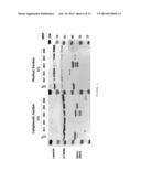 | 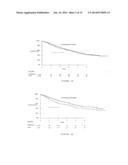 |
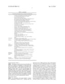 | 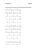 |
 | 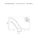 |
 |  |
 | 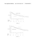 |
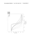 |  |
 |  |
 |  |
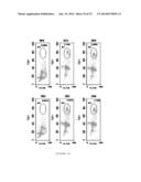 | 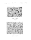 |
 |  |
 |  |
 |  |
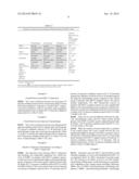 | 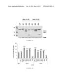 |
 |  |
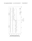 |  |
 | 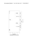 |
 |  |
 | 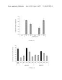 |
 | 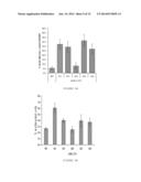 |
 |  |
 |  |
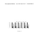 |
| Similar patent applications: | |
| Date | Title |
|---|---|
| 2014-11-27 | Large commercial scale lentiviral vector production system and vectors produced thereby |
| 2014-09-25 | Gene expression markers for breast cancer prognosis |
| 2014-11-27 | Novel process for the preparation of intermediates of hmg-coa reductase inhibitors |
| 2014-09-18 | Biomarkers for detecting the presence of bacteria |
| 2014-11-13 | Streptomyces strain that decomposes proteins recalcitrant to proteolysis |
| New patent applications in this class: | |
| Date | Title |
|---|---|
| 2022-05-05 | Photocleavable mass-tags for multiplexed mass spectrometric imaging of tissues using biomolecular probes |
| 2022-05-05 | Macrophage expression in breast cancer |
| 2022-05-05 | Characterizing methylated dna, rna, and proteins in the detection of lung neoplasia |
| 2022-05-05 | Methods for identifying and improving t cell multipotency |
| 2022-05-05 | Sequence analysis using meta-stable nucleic acid molecules |
| New patent applications from these inventors: | |
| Date | Title |
|---|---|
| 2018-04-19 | Targeted treatment of leiomyosarcoma |
| 2014-05-01 | Expression of isoform 202 of ercc1 for predicting response to cancer chemotherapy |
| 2013-10-24 | Prognostic and/or predictive biomarkers and biological applications thereof |
| Top Inventors for class "Chemistry: molecular biology and microbiology" | |
| Rank | Inventor's name |
|---|---|
| 1 | Marshall Medoff |
| 2 | Anthony P. Burgard |
| 3 | Mark J. Burk |
| 4 | Robin E. Osterhout |
| 5 | Rangarajan Sampath |