Patent application title: CD127 BINDING PROTEINS
Inventors:
Ian Kirby (Stevenage, GB)
Alex H. Taylor (King Of Prussia, PA, US)
Thomas Matthew Webb (Stevenage, GB)
Yu Xue (King Of Prussia, PA, US)
IPC8 Class: AA61K39395FI
USPC Class:
4241301
Class name: Drug, bio-affecting and body treating compositions immunoglobulin, antiserum, antibody, or antibody fragment, except conjugate or complex of the same with nonimmunoglobulin material
Publication date: 2011-08-18
Patent application number: 20110200585
Abstract:
Antigen binding proteins which bind to human IL-7 receptor (CD127) are
provided. The antigen binding proteins are typically antibodies, and are
useful in the treatment of diseases or disorders in humans, particularly
autoimmune diseases such as multiple sclerosis.Claims:
1. An antigen binding protein comprising a heavy chain variable domain
having an amino acid sequence as set out in SEQ ID NO:13, and a light
chain variable domain having an amino acid sequence as set out in SEQ ID
NO:22.
2. The antigen binding protein of claim 1, which comprises a heavy chain having an amino acid sequence selected from the group consisting of SEQ ID NO:114 and SEQ ID NO:118.
3. The antigen binding protein of claim 1, which comprises a light chain having an amino acid sequence as set out in SEQ ID NO:115.
4. A nucleic acid molecule encoding an antigen binding protein of claim 1.
5. An expression vector comprising a nucleic acid molecule according to claim 4.
6. A recombinant host cell comprising an expression vector according to claim 5.
7. An antigen binding protein expressed by a host cell according to claim 6.
8. A method for the production of an antigen binding protein, comprising the steps of culturing a host cell according to claim 7 in a medium to produce the antigen binding protein, and isolating or purifying the antigen binding protein therefrom.
9. A method according to claim 8, wherein said culturing step is performed in conditions conducive for expression of the antigen binding protein from the host cell, and secretion of the antigen binding protein from the cell.
10. A pharmaceutical composition comprising an antigen binding protein according to claim 1 and a pharmaceutically acceptable carrier or excipient.
11. A method of treating a subject having an autoimmune or inflammatory disease, comprising the step of administering to the subject an antigen binding protein according to claim 1.
12. A method according to claim 11, wherein the autoimmune or inflammatory disease is multiple sclerosis.
Description:
[0001] This application claims the benefit of U.S. Provisional Application
No. 61/299,010 filed 28 Jan. 2010, which is incorporated herein in its
entirety.
FIELD OF THE INVENTION
[0002] The present invention relates to antigen binding proteins, in particular immunoglobulins, that specifically bind to the α-chain of the human IL-7 receptor (CD127). The invention also concerns methods of treating diseases or disorders with said proteins, pharmaceutical compositions comprising said proteins and methods of their manufacture. Other aspects of the present invention will be apparent from the description below.
BACKGROUND OF THE INVENTION
[0003] Multiple Sclerosis (MS) is a chronic inflammatory, demyelinating disease that affects the central nervous system. In MS, it is believed that infiltrating inflammatory immune cells are involved in the destruction of oligodendrocytes, which are the cells responsible for creating and maintaining a fatty layer, known as the myelin sheath. MS results in the thinning or complete loss of myelin. When the myelin is lost, the neurons can no longer effectively conduct their electrical signals leading to numerous neurologic dysfunctions. Individuals with MS produce autoreactive T cells that participate in the formation of inflammatory lesions along the myelin sheath of nerve fibres. The cerebrospinal fluid of patients with active MS contains activated T cells, which infiltrate the brain tissue and cause characteristic inflammatory lesions, destroying the myelin. While the multiple sclerosis symptoms and course of illness can vary from person to person, there are three forms of the disease--relapsing-remitting MS, secondary progressive MS, and primary progressive MS.
[0004] In the early stages of MS, inflammatory attacks occur over short intervals of acutely heightened disease activity. These episodes are followed by periods of recovery and remission. During the remission period, the local swelling in the nervous system lesion resolves, the immune cells become less active or inactive, and the myelin-producing cells remyelinate the axons. Nerve signalling improves, and the disability caused by the inflammation becomes less severe or goes away entirely. This phase of the disease is called relapsing-remitting MS (RRMS). The lesions do not all heal completely, though. Some remain as "chronic" lesions, which usually have a demyelinated core region which lacks immune cells. Over time, the cells in the centre of such lesions mostly die, although inflammation often continues at their edges. The brain can adapt well to the loss of some neurons, and permanent disability may not occur for many years. However, more than 50% of patients with MS eventually enter a stage of progressive deterioration, called secondary progressive MS (SPMS). In this stage, the disease no longer responds well to disease-modifying drugs, and patients' disabilities steadily worsen. The destruction of neurons from early in the natural course of MS suggests that the progressive disabilities of SPMS might be the result of an accumulated neuronal loss that eventually overwhelms the brain's compensatory abilities. Primary progressive MS is a type of multiple sclerosis where there are no relapses, but over a period of years, there is gradual loss of physical and cognitive functions.
[0005] The goal of treatment in patients with relapsing-remitting multiple sclerosis is to reduce the frequency and severity of relapses (and thereby prevent exacerbations) as well as to prevent or postpone the onset of the progressive phase of the disease. To achieve this goal, in the past especially, immunomodulatory or immunosuppressive drugs have been used, but they have never found widespread acceptance owing to limited efficacy and considerable toxicity. For example, large randomized controlled trials have been performed successfully with interferon beta-1a, interferon beta-1 b, and glatiramer acetate.
[0006] Both altered autoimmune T cell responses and dysfunction of the regulatory network of the immune system play an important role in human autoimmune pathologies, such as MS and rheumatoid arthritis (Kuchroo et al., (2002) Annu. Rev. Immunol. 20:101-123; Sospedra and Martin (2005) Annu. Rev. Immunol. 23: 683-747; Toh and Miossec (2007) Curr. Opin. Rheumatol. 19:284-288).
[0007] Although the aetiology and pathogenesis of MS remain unknown, it is generally considered an autoimmune pathology in which autoreactive T cells of pathogenic potential, such as TH1 and TH17 cells, are thought to play an important role. There is evidence that these effector T cells are activated in vivo during the disease process and are attributable to the central nervous system (CNS) inflammation. There is also evidence that these T cells mediate destruction of myelin-expressing cells in lesions of EAE and MS during the active phase of the disease. On the other hand, regulatory T cells (Treg) that normally keep pathogenic TH1 and TH17 cells in check are deficient in patients with MS, further tilting the immune system toward an pro-inflammatory state.
[0008] Three separate groups recently reported the results of genome wide single nucleotide polymorphisms (SNPs) scanning in a total of 17,947 donors with or without MS. After scanning 334,923 SNPs, they found a highly significant association (overall P=2.9×10⁻⁷) of a nonsynonymous coding SNP in the human IL-7 receptor alpha chain (IL-7Rα) with MS susceptibility. The SNP corresponds to a change from T to C in exon 6 of CD127 (also known as IL-7Rα). This change enhances the chance of exon 6 skipping during RNA splicing, resulting in a soluble form of CD127. Furthermore, expressions of CD127 and IL-7 RNAs in the cerebrospinal fluids (CSFs) of MS patients are significantly higher relative to CSFs of patients with other neurological disorders.
[0009] IL-7 and IL-7 receptor (IL-7R) are known to play an important role in T cell and B cell development and homeostasis mainly in a thymic environment. Indeed, thymic stromal cells, fetal thymus, and bone marrow are sites of IL-7 of production. The IL-7 receptor consists of two subunits, CD127 and a common chain (gamma chain or γc) which is shared by receptors of IL-2, IL-4, IL-9, IL-15, and IL-21.
[0010] CD127 is also known as IL-7 receptor alpha (IL-7Rα) and p90 IL-7R. Human CD127 (Swiss Prot accession number P16871) has a total of 459 amino acids (20 signal sequence). It comprises a 219 amino acid extra cellular region, a 25 amino acid transmembrane region and a 195 amino acid intracellular region. The numbering of residues within CD127, as used herein (e.g. for the description of antibody epitopes) is based on the full length protein, including signal sequence residues. CD127 may exist in four isoforms, the isoform H20
[0011] (Swissprot accession number P16871-1) has the following amino acid sequence (including signal sequence):
TABLE-US-00001 (SEQ ID NO: 1) MTILGTTFGM VFSLLQVVSG ESGYAQNGDL EDAELDDYSF SCYSQLEVNG SQHSLTCAFE DPDVNTTNLE FEICGALVEV KCLNFRKLQE IYFIETKKFL LIGKSNICVK VGEKSLTCKK IDLTTIVKPE APFDLSVIYR EGANDFVVTF NTSHLQKKYV KVLMHDVAYR QEKDENKWTH VNLSSTKLTL LQRKLQPAAM YEIKVRSIPD HYFKGFWSEW SPSYYFRTPE INNSSGEMDP ILLTISILSF FSVALLVILA CVLWKKRIKP IVWPSLPDHK KTLEHLCKKP RKNLNVSFNP ESFLDCQIHR VDDIQARDEV EGFLQDTFPQ QLEESEKQRL GGDVQSPNCP SEDVVVTPES FGRDSSLTCL AGNVSACDAP ILSSSRSLDC RESGKNGPHV YQDLLLSLGT TNSTLPPPFS LQSGILTLNP VAQGQPILTS LGSNQEEAYV TMSSFYQNQ
[0012] CD127 is also found in the receptor of thymic stromal derived lymphopoietin (TSLP). The TSLP receptor is a heterodimer of CD127 and cytokine receptor-like factor 2 (CRLF2).
[0013] Binding of IL-7 to the IL-7R activates multiple signalling pathways including the activation of JAK kinases 1 and 3 leading to the phosphorylation and activation of Stat5. This pathway is crucial to the survival of thymic developing T cell precursors because Stat5 activation is required in the induction of the anti-apoptotic protein BcI-2 and the prevention of the pro-apoptotic protein Bax entry into the mitochondrion. Another IL-7R mediated pathway is the activation of PI3 kinase, resulting in the phosphorylation of the pro-apoptotic protein Bad and its cytoplasm retention. CD127 is expressed in peripheral resting and memory T cells. The mechanism of IL-7 regulation of T cell survival and homeostasis and the source of IL-7 in the periphery are not completely understood. Furthermore, its potential role in the differentiation and function of pathogenic T cells in autoimmune disease is poorly studied and largely unknown. There are few reports suggesting that IL-7 may contribute to the pathogenesis of autoimmune diseases.
[0014] Recently, Liu and colleagues (Liu et al, (2010) Nature Medicine 16:191-197) have described the role of IL-7 in TH17 survival and expansion. Murine anti-CD127 antibodies (including the anti-CD127 antibodies 1A11 and 6A3) and their role in the treatment of MS and other autoimmune diseases have been described in PCT application number PCT/US2009/053136.
[0015] It is desirable to isolate and develop further monoclonal antibodies that bind to and/or inhibit the biological effect of human CD127. Such antibodies are likely to be therapeutically useful in the treatment of MS and other inflammatory and autoimmune diseases and disorders, particularly those in which pathogenic TH17 cells have been implicated.
SUMMARY OF THE INVENTION
[0016] The invention provides antigen binding proteins which specifically bind to CD127. The antigen binding proteins can be used in therapeutic methods, in particular, in the treatment or prevention of diseases in which pathogenic TH17 cells are implicated. The antigen binding proteins may bind to CD127 and inhibit, e.g. neutralize, the biological function of CD127.
[0017] In a first aspect, the invention provides an antigen binding protein comprising a heavy chain variable domain having an amino acid sequence as set out in SEQ ID NO:13, and a light chain variable domain having an amino acid sequence as set out in SEQ ID NO:22.
[0018] The antigen binding protein may comprise a heavy chain having an amino acid sequence as set out in SEQ ID NO:114 or SEQ ID NO:118. The antigen binding protein may further comprise a light chain having an amino acid sequence as set out in SEQ ID NO:115.
[0019] The invention also provides a nucleic acid molecule encoding an antibody or antigen binding protein according to the invention, an expression vector comprising such a nucleic acid molecule, and a recombinant host cell comprising such an expression vector.
[0020] In a further aspect, the invention provides an antibody or antigen binding protein expressed by a host cell.
[0021] The invention also provides a method for the production of an antibody or antigen binding protein according to claim 1, comprising the step of culturing a host cell according to the invention in a medium to produce the antigen binding protein, and isolating or purifying the antigen binding protein therefrom. The culturing step may be performed in conditions conducive for expression of the antibody or antigen binding protein from the host cell, and secretion of the antibody from the cell.
[0022] In a further aspect, the invention provides a pharmaceutical composition comprising an antigen binding protein according to the invention and a pharmaceutically acceptable carrier or excipient.
[0023] In a still further aspect, the invention provides a method of treating a subject having an autoimmune or inflammatory disease, comprising the step of administering to the subject an antigen binding protein according to the invention. In an embodiment, the autoimmune or inflammatory disease is multiple sclerosis.
[0024] The antigen binding protein may be an antibody, in particular, a humanised or human antibody, or an antigen binding fragment thereof. The antigen binding protein of the invention may not inhibit TSLP signalling. In an embodiment, the antigen binding protein does not inhibit TSLP signalling.
[0025] The invention also provides a nucleic acid molecule encoding an antigen binding protein of the present invention. In an embodiment, the invention provides nucleic acid molecules of SEQ ID NO:32, SEQ ID NO:108, and SEQ ID NO:119. The invention also provides an expression vector comprising a nucleic acid molecule as defined herein, and a recombinant host cell comprising an expression vector as defined herein. The expression vector may comprise the nucleic acid molecule of SEQ ID NO:32, SEQ ID NO:108, and SEQ ID NO:119. In an embodiment, the expression vector comprises a nucleic acid molecule which encodes an antigen binding protein as hereinbefore described. In another embodiment, the invention provides a host cell comprising an expression vector as hereinbefore described. In a further embodiment, the invention provides an antibody expressed by host cell as hereinbefore described.
[0026] The invention also provides a method for the production of an antigen binding protein of the present invention which method comprises the step of culturing a host cell as defined above and recovering the antigen binding protein.
[0027] The invention also provides an antibody or antigen binding protein according to the invention which is expressed by a host cell comprising a nucleic acid sequence or sequence encoding an antibody or antigen binding fragment according to the invention.
[0028] The invention also provides a pharmaceutical composition comprising an antigen binding protein of the present invention and a pharmaceutically acceptable carrier or excipient.
[0029] The invention also provides a method of treating a subject afflicted with an autoimmune or inflammatory disease, which method comprises the step of administering to the subject an antigen binding protein of the present invention.
[0030] The invention also provides a method of treating a subject afflicted with a disease in which pathogenic TH17 cells are implicated, which method comprises the step of administering to the subject an antigen binding protein of the present invention.
[0031] The invention also provides a method of treating a subject afflicted with a disease associated with upregulated expression of IL-17, which method comprises the step of administering to the subject an antigen binding protein of the present invention.
[0032] In particular, the autoimmune or inflammatory disease, the disease in which pathogenic TH17 cells are implicated, or the disease associated with up-regulated expression of IL17 may be multiple sclerosis (MS), SLE, rheumatoid arthritis, Behcet's disease or asthma. In an embodiment, the antigen binding protein of the invention will be useful in a method of treating multiple sclerosis. Other diseases which may be treated by the administration of the antigen binding proteins of the invention are described herein.
[0033] The invention also provides an antigen binding protein as described herein for use in the treatment of a subject afflicted with an autoimmune or inflammatory disease; a disease in which pathogenic TH17 cells are implicated; or a disease associated with up-regulated expression of IL17.
[0034] The invention provides the use of an antigen binding protein as described herein in the manufacture of a medicament for use in the treatment of a subject afflicted with an autoimmune or inflammatory disease; a disease in which pathogenic TH17 cells are implicated; or a disease associated with up-regulated expression of IL17.
[0035] Other aspects and embodiments of the invention will be apparent from the detailed description that follows.
BRIEF DESCRIPTION OF THE DRAWINGS
[0036] FIG. 1 shows the complement dependent cytotoxicity of the anti-IL7R mAb 1A11 H3L4 on HEK293 cells expressing hIL-7R.
[0037] FIG. 2 shows the antibody dependent cell-mediated cytotoxicity of the humanised anti-IL7R mAb 1A11 H3L4 and the Fc-disabled anti-IL7R mAb (1A11 H3L4Fc) on HEK293 cells expressing hIL-7R, in the presence of peripheral blood mononuclear cells.
[0038] FIGS. 3A and 3B show the inhibition of IL-7-induced STAT5 phosphorylation by 1A11 H3L4 in human PBMC provided by two separate donors.
[0039] FIGS. 4A to 4D show the inhibition of IL-7-induced IL-17 production in differentiated human Th17 cells by 1A11 H3L4 (in four different donors).
[0040] FIGS. 5A to 5E show that 1A11 H3L4 does not affect TSLP-induction of TARC (thymus and activation-regulated chemokine).
DETAILED DESCRIPTION OF THE INVENTION
[0041] IL-7/1L-7R signalling is critically required for survival and expansion of committed TH17 cells in both mouse and human systems, while its role in TH17 differentiation is not essential compared to that of IL-6 (Liu et al, (2010) Nature Medicine 16:191-197). Surprisingly, the in vivo effect on the immune system by IL-7R antagonism is highly selective in EAE, an animal model for multiple sclerosis, affecting TH17 cells and, to a lesser extent, TH1 cells predominantly of the memory phenotype, and sparing Treg cells. This selectivity appears to play an important role in rebalancing the ratio of pathogenic TH17 cells and Treg cells by IL-7R antagonism in EAE and is attributable to the treatment efficacy.
[0042] The role of IL-7/IL-7R signalling in TH17 cell survival and expansion supports the treatment efficacy of IL-7R antagonism in human autoimmune diseases, such as MS. IL-7 neutralization or IL-7R antagonism is likely to have unique therapeutic advantages. On one hand, the treatment offers the selectivity that distinguishes pathogenic TH1 and TH17 cells from Treg and unrelated immune cells. On the other hand, additional therapeutic advantages of IL-7R antagonism involve its selective effect on survival and expansion of differentiated TH17 as opposed to TH17 differentiation. It is conceivable that targeting in vivo maintenance of committed TH17 versus TH17 differentiation is more efficacious in a therapeutic context. Inhibition of IL-7 receptor mediated signalling therefore provides a promising therapeutic intervention for the treatment of autoimmune or inflammatory diseases.
[0043] The term IL-7R mediated signalling, as used herein, means the biological effect instigated by the IL-7 receptor complex when bound by its ligand, IL-7. IL-7R mediated signalling therefore includes, but is not necessarily limited to, one or more, or all, of IL-7 induced phosphorylation of STAT-5, IL-7 induced expansion of TH17 cells and IL-7 induced survival of TH17 cells.
[0044] Murine antibodies 1A11 and 6A3 are described in patent application number PCT/US2009/053136 (WO2010/017468). These antibodies specifically bind to the alpha chain of the human IL-7 receptor, CD127 (SEQ ID NO:1). The variable domains of these antibodies are described in SEQ ID NO:8 and 9 (VH, Vκ 1A11, respectively) and SEQ ID NO:46 and 47 (VH, Vκ 6A3, respectively).
[0045] The present invention provides antigen binding proteins comprising one or more of the complementarity determining regions (CDRs) of 1A11 or 6A3, and variants thereof. The antigen binding proteins may bind to and neutralise IL-7R signalling. In one embodiment, the invention provides humanised antibodies, comprising the from one to six of the CDRs from murine antibodies 1A11 or 6A3 (the donor antibody) in an a human acceptor antibody.
[0046] The term "antigen binding protein" as used herein refers to antibodies, antibody fragments and other protein constructs, such as domains, which are capable of binding to CD127. In an embodiment, the antigen binding protein is an antibody.
[0047] The term "antibody" is used herein in the broadest sense to refer to molecules with an immunoglobulin-like domain and includes monoclonal, recombinant, polyclonal, chimeric, humanised, bispecific and heteroconjugate antibodies; a single variable domain, a domain antibody, antigen binding fragments, immunologically effective fragments, single chain Fv, diabodies, Tandabs®, etc (for a summary of alternative "antibody" formats see Holliger and Hudson, Nature Biotechnology, 2005, Vol 23, No. 9, 1126-1136).
[0048] The phrase "single variable domain" refers to an antigen binding protein variable domain (for example, VH, VHH, VL) that specifically binds an antigen or epitope independently of a different variable region or domain.
[0049] A "domain antibody" or "dAb" may be considered the same as a "single variable domain" which is capable of binding to an antigen. A single variable domain may be a human antibody variable domain, but also includes single antibody variable domains from other species such as rodent (for example, as disclosed in WO 00/29004), nurse shark and Camelid VHH dAbs. Camelid VHH are immunoglobulin single variable domain polypeptides that are derived from species including camel, llama, alpaca, dromedary, and guanaco, which produce heavy chain antibodies naturally devoid of light chains. Such VHH domains may be humanised according to standard techniques available in the art, and such domains are considered to be "domain antibodies". As used herein VH includes camelid VHH domains.
[0050] As used herein the term "domain" refers to a folded protein structure which has tertiary structure independent of the rest of the protein. Generally, domains are responsible for discrete functional properties of proteins, and in many cases may be added, removed or transferred to other proteins without loss of function of the remainder of the protein and/or of the domain. A "single variable domain" is a folded polypeptide domain comprising sequences characteristic of antibody variable domains. It therefore includes complete antibody variable domains and modified variable domains, for example, in which one or more loops have been replaced by sequences which are not characteristic of antibody variable domains, or antibody variable domains which have been truncated or comprise N- or C-terminal extensions, as well as folded fragments of variable domains which retain at least the binding activity and specificity of the full-length domain. A domain can bind an antigen or epitope independently of a different variable region or domain.
[0051] An antigen binding fragment may be provided by means of arrangement of one or more CDRs on non-antibody protein scaffolds such as a domain. A non-antibody protein scaffold or domain is one that has been subjected to protein engineering in order to obtain binding to a ligand other than its natural ligand, for example a domain which is a derivative of a scaffold selected from: CTLA-4 (Evibody); lipocalin; Protein A derived molecules such as Z-domain of Protein A (Affibody, SpA), A-domain (Avimer/Maxibody); heat shock proteins such as GroEI and GroES; transferrin (trans-body); ankyrin repeat protein (DARPin); peptide aptamer; C-type lectin domain (Tetranectin); human γ-crystallin and human ubiquitin (affilins); PDZ domains; scorpion toxinkunitz type domains of human protease inhibitors; and fibronectin (adnectin); which has been subjected to protein engineering in order to obtain binding to a ligand other than its natural ligand.
[0052] CTLA-4 (Cytotoxic T Lymphocyte-associated Antigen 4) is a CD28-family receptor expressed on mainly CD4+ T-cells. Its extracellular domain has a variable domain-like Ig fold. Loops corresponding to CDRs of antibodies can be substituted with heterologous sequence to confer different binding properties. CTLA-4 molecules engineered to have different binding specificities are also known as Evibodies. For further details see Journal of Immunological Methods 248 (1-2), 31-45 (2001).
[0053] Lipocalins are a family of extracellular proteins which transport small hydrophobic molecules such as steroids, bilins, retinoids and lipids. They have a rigid β-sheet secondary structure with a number of loops at the open end of the canonical structure which can be engineered to bind to different target antigens. Anticalins are between 160-180 amino acids in size, and are derived from lipocalins. For further details see Biochim Biophys Acta 1482: 337-350 (2000), U.S. Pat. No. 7,250,297B1 and US20070224633.
[0054] An affibody is a scaffold derived from Protein A of Staphylococcus aureus which can be engineered to bind to an antigen. The domain consists of a three-helical bundle of approximately 58 amino acids. Libraries have been generated by randomisation of surface residues. For further details see Protein Eng. Des. Sel. 17, 455-462 (2004) and EP1641818A1.
[0055] Avimers are multidomain proteins derived from the A-domain scaffold family. The native domains of approximately 35 amino acids adopt a defined disulphide bonded structure. Diversity is generated by shuffling of the natural variation exhibited by the family of A-domains. For further details see Nature Biotechnology 23 (12), 1556-1561 (2005) and Expert Opinion on Investigational Drugs 16 (6), 909-917 (June 2007).
[0056] A transferrin is a monomeric serum transport glycoprotein. Transferrins can be engineered to bind different target antigens by insertion of peptide sequences, such as one or more CDRs, in a permissive surface loop. Examples of engineered transferrin scaffolds include the Trans-body. For further details see J. Biol. Chem 274, 24066-24073 (1999).
[0057] Designed Ankyrin Repeat Proteins (DARPins) are derived from Ankyrin which is a family of proteins that mediate attachment of integral membrane proteins to the cytoskeleton. A single ankyrin repeat is a 33 residue motif consisting of two α-helices and a β-turn. They can be engineered to bind different target antigens by: randomising residues in the first α-helix and a β-turn of each repeat; or insertion of peptide sequences, such as one or more CDRs. Their binding interface can be increased by increasing the number of modules (a method of affinity maturation). For further details see J. Mol. Biol. 332, 489-503 (2003), PNAS 100 (4), 1700-1705 (2003) and J. Mol. Biol. 369, 1015-1028 (2007) and US20040132028A1.
[0058] Fibronectin is a scaffold which can be engineered to bind to antigen. Adnectins consists of a backbone of the natural amino acid sequence of the 10th domain of the 15 repeating units of human fibronectin type III (FN3). Three loops at one end of the β-sandwich can be engineered to enable an Adnectin to specifically recognize a therapeutic target of interest. For further details see Protein Eng. Des. Sel. 18, 435-444 (2005), US20080139791, WO2005056764 and U.S. Pat. No. 6,818,418B1.
[0059] Peptide aptamers are combinatorial recognition molecules that consist of a constant scaffold protein, typically thioredoxin (TrxA) which contains a constrained variable peptide loop inserted at the active site. For further details see Expert Opin. Biol. Ther. 5, 783-797 (2005).
[0060] Microbodies are derived from naturally occurring microproteins of 25-50 amino acids in length which contain 3-4 cysteine bridges; examples of microproteins include KalataB1 and conotoxin and knottins. The microproteins have a loop which can be engineered to include up to 25 amino acids without affecting the overall fold of the microprotein. For further details of engineered knottin domains, see WO2008098796.
[0061] Other binding domains include proteins which have been used as a scaffold to engineer different target antigen binding properties include human γ-crystallin and human ubiquitin (affilins), kunitz type domains of human protease inhibitors, PDZ-domains of the Ras-binding protein AF-6, scorpion toxins (charybdotoxin), C-type lectin domain (tetranectins) are reviewed in Chapter 7--Non-Antibody Scaffolds from Handbook of Therapeutic Antibodies (2007, edited by Stefan Dubel) and Protein Science 15:14-27 (2006). Binding domains of the present invention could be derived from any of these alternative protein domains and any combination of the CDRs of the present invention grafted onto the domain.
[0062] An antigen binding fragment or an immunologically effective fragment may comprise partial heavy or light chain variable sequences. Fragments are at least 5, 6, 8 or 10 amino acids in length. Alternatively the fragments are at least 15, at least 20, at least 50, at least 75, or at least 100 amino acids in length.
[0063] The term "specifically binds" as used throughout the present specification in relation to antigen binding proteins means that the antigen binding protein binds to CD127 with no or insignificant binding to other (for example, unrelated) proteins. The term however does not exclude the fact that the antigen binding proteins may also be cross-reactive with CD127 from other species, such as murine CD127, cynomolgus monkey (Macaca fascicularis) or marmoset CD127. In an embodiment, the antigen binding protein binds to both cynomolgus monkey and marmoset CD127. The antigen binding proteins described herein may bind to human CD127 with at least 2, 5, 10, 50, 100, or 1000 fold greater affinity than they bind to CD127 from other species.
[0064] The binding affinity or equilibrium dissociation constant (KD) of the antigen binding protein-CD127 interaction may be 100 nM or less, 10 nM or less, 2 nM or less or 1 nM or less. Alternatively the KD may be between 5 and 10 nM; or between 1 and 2 nM. The KD may be between 1 pM and 500 pM; or between 500 pM and 1 nM. The binding affinity of the antigen binding protein is determined by the association rate constant (ka) and the dissociation rate constant (kd) (KD=kd/ka). The binding affinity may be measured by BIAcore®, for example by antigen capture with CD127 coupled onto a CM5 chip by primary amine coupling and antibody capture onto this surface. The BIAcore® method described in Example 4 may be used to measure binding affinity. Alternatively, the binding affinity can be measured by FORTEbio, for example by antigen capture with CD127 coupled onto a CM5 needle by primary amine coupling and antibody capture onto this surface.
[0065] The kd may be 1×10⁻³ s-1 or less, 1×10⁻⁴ s-1 or less, or 1×10⁻⁵ s-1 or less. The kd may be between 1×10⁻⁵ s-1 and 1×10⁻⁴ s-1; or between 1×10⁻⁴ s-1 and 1×10⁻³ s-1. A slow kd may result in a slow dissociation of the antigen binding protein-ligand complex and improved neutralisation of the ligand.
[0066] It will be apparent to those skilled in the art that the term "derived" is intended to define not only the source in the sense of it being the physical origin for the material but also to define material which is structurally identical to the material but which does not originate from the reference source. Thus "residues found in the donor antibody" need not necessarily have been purified from the donor antibody.
[0067] By isolated it is intended that the molecule, such as an antigen binding protein, is removed from the environment in which it may be found in nature. For example, the molecule may be purified away from substances with which it would normally exist in nature. For example, the antigen binding protein can be purified to at least 95%, 96%, 97%, 98% or 99%, or greater with respect to a culture media containing the antigen binding protein.
[0068] A "chimeric antibody" refers to a type of engineered antibody which contains a naturally-occurring variable region (light chain and heavy chains) derived from a donor antibody in association with light and heavy chain constant regions derived from an acceptor antibody.
[0069] A "humanised antibody" refers to a type of engineered antibody having one or more of its CDRs derived from a non-human donor immunoglobulin, the remaining immunoglobulin-derived parts of the molecule being derived from one or more human immunoglobulin(s). In addition, framework support residues may be altered to preserve binding affinity (see, e.g., Queen et al. Proc. Natl Acad Sci USA, 86:10029-10032 (1989), Hodgson et al. Bio/Technology, 9:421 (1991)). A suitable human acceptor antibody may be one selected from a conventional database, e.g., the KABAT® database, Los Alamos database, and Swiss Protein database, by homology to the nucleotide and amino acid sequences of the donor antibody. A human antibody characterized by a homology to the framework regions of the donor antibody (on an amino acid basis) may be suitable to provide a heavy chain constant region and/or a heavy chain variable framework region for insertion of the donor CDRs. A suitable acceptor antibody capable of donating light chain constant or variable framework regions may be selected in a similar manner. It should be noted that the acceptor antibody heavy and light chains are not required to originate from the same acceptor antibody. The prior art describes several ways of producing such humanised antibodies, see for example EP-A-0239400 and EP-A-054951.
[0070] The term "donor antibody" refers to an antibody which contributes the amino acid sequences of its variable regions, one or more CDRs, or other functional fragments or analogs thereof to a first immunoglobulin partner. The donor therefore provides the altered immunoglobulin coding region and resulting expressed altered antibody with the antigenic specificity and neutralising activity characteristic of the donor antibody.
[0071] The term "acceptor antibody" refers to an antibody which is heterologous to the donor antibody, which contributes all (or any portion) of the amino acid sequences encoding its heavy and/or light chain framework regions and/or its heavy and/or light chain constant regions to the first immunoglobulin partner. A human antibody may be the acceptor antibody.
[0072] The terms "VH" and "VL" are used herein to refer to the heavy chain variable region and light chain variable region respectively of an antigen binding protein. Vκ is also used to refer to the variable light chain domain.
[0073] "CDRs" are defined as the complementarity determining region amino acid sequences of an antigen binding protein. These are the hypervariable regions of immunoglobulin heavy and light chains. There are three heavy chain and three light chain CDRs (or CDR regions) in the variable portion of an immunoglobulin. Thus, "CDRs" as used herein refers to all three heavy chain CDRs, all three light chain CDRs, all heavy and light chain CDRs, or at least two CDRs.
[0074] Throughout this specification, amino acid residues in variable domain sequences and full length antibody sequences are numbered according to the Kabat numbering convention, unless otherwise specified. Similarly, the terms "CDR", "CDRL1", "CDRL2", "CDRL3", "CDRH1", "CDRH2", "CDRH3" used in the Examples follow the Kabat numbering convention. For further information, see Kabat et al., Sequences of Proteins of Immunological Interest, 4th Ed., U.S. Department of Health and Human Services, National Institutes of Health (1987).
[0075] It will be apparent to those skilled in the art that there are alternative numbering conventions for amino acid residues in variable domain sequences and full length antibody sequences. There are also alternative numbering conventions for CDR sequences, for example those set out in Chothia et al. (1989) Nature 342: 877-883. The structure and protein folding of the antibody may mean that other residues are considered part of the CDR sequence and would be understood to be so by a skilled person. Therefore, the term "corresponding CDR" is used herein to refer to a CDR sequence using any numbering convention, for example those set out in Table 1.
[0076] Other numbering conventions for CDR sequences available to a skilled person include "AbM" (University of Bath) and "contact" (University College London) methods. The minimum overlapping region using at least two of the Kabat, Chothia, AbM and contact methods can be determined to provide the "minimum binding unit". The minimum binding unit may be a sub-portion of a CDR.
[0077] Table 1 below represents one definition using each numbering convention for each CDR or binding unit. The Kabat numbering scheme is used in Table 1 to number the variable domain amino acid sequence. It should be noted that some of the CDR definitions may vary depending on the individual publication used.
TABLE-US-00002 TABLE 1 Minimum Kabat Chothia AbM Contact binding CDR CDR CDR CDR unit H1 31-35/ 26-32/ 26-35/ 30-35/ 31-32 35A/35B 33/34 35A/35B 35A/35B H2 50-65 52-56 50-58 47-58 52-56 H3 95-102 95-102 95-102 93-101 95-101 L1 24-34 24-34 24-34 30-36 30-34 L2 50-56 50-56 50-56 46-55 50-55 L3 89-97 89-97 89-97 89-96 89-96
[0078] As used herein, the term "antigen binding site" refers to a site on an antigen binding protein which is capable of specifically binding to an antigen. This may be a single domain (for example, an epitope-binding domain), or single-chain Fv (ScFv) domains or it may be paired VH/VL domains as can be found on a standard antibody.
[0079] The term "epitope" as used herein refers to that portion of the antigen that makes contact with a particular binding domain of the antigen binding protein. An epitope may be linear, comprising an essentially linear amino acid sequence from the antigen. Alternatively, an epitope may be conformational or discontinuous. For example, a conformational epitope comprises amino acid residues which require an element of structural constraint. A discontinuous epitope comprises amino acid residues that are separated by other sequences, i.e. not in a continuous sequence in the antigen's primary sequence. In the context of the antigen's tertiary and quaternary structure, the residues of a discontinuous epitope are near enough to each other to be bound by an antigen binding protein.
[0080] For nucleotide and amino acid sequences, the term "identical" or "sequence identity" indicates the degree of identity between two nucleic acid or two amino acid sequences, and if required when optimally aligned and compared with appropriate insertions or deletions.
[0081] The percent identity between two sequences is a function of the number of identical positions shared by the sequences (i.e., % identity=number of identical positions/total number of positions times 100), taking into account the number of gaps, and the length of each gap, which need to be introduced for optimal alignment of the two sequences. The comparison of sequences and determination of percent identity between two sequences can be accomplished using a mathematical algorithm, as described below.
[0082] The percent identity between two nucleotide sequences can be determined using the GAP program in the GCG software package, using a NWSgapdna.CMP matrix and a gap weight of 40, 50, 60, 70, or 80 and a length weight of 1, 2, 3, 4, 5, or 6. The percent identity between two nucleotide or amino acid sequences can also be determined using the algorithm of E. Meyers and W. Miller (Comput. Appl. Biosci., 4:11-17 (1988)) which has been incorporated into the ALIGN program (version 2.0), using a PAM120 weight residue table, a gap length penalty of 12 and a gap penalty of 4. In addition, the percent identity between two amino acid sequences can be determined using the Needleman and Wunsch (J. Mol. Biol. 48:444-453 (1970)) algorithm which has been incorporated into the
[0083] GAP program in the GCG software package, using either a Blossum 62 matrix or a PAM250 matrix, and a gap weight of 16, 14, 12, 10, 8, 6, or 4 and a length weight of 1, 2, 3, 4, 5, or 6.
[0084] In one method, a polynucleotide sequence may be identical to a reference polynucleotide sequence as described herein (see for example SEQ ID NO: 30-39, SEQ ID NO:76-105), that is be 100% identical, or it may include up to a certain integer number of nucleotide alterations as compared to the reference sequence, such as at least 50, 60, 70, 75, 80, 85, 90, 95, 98, or 99% identical. Such alterations are selected from at least one nucleotide deletion, substitution, including transition and transversion, or insertion, and wherein said alterations may occur at the 5' or 3' terminal positions of the reference nucleotide sequence or anywhere between those terminal positions, interspersed either individually among the nucleotides in the reference sequence or in one or more contiguous groups within the reference sequence. The number of nucleotide alterations is determined by multiplying the total number of nucleotides in the reference polynucleotide sequence as described herein (see for example SEQ ID NO: 30-39, SEQ ID NO:76-105), by the numerical percent of the respective percent identity (divided by 100) and subtracting that product from said total number of nucleotides in the reference polynucleotide sequence as described herein (see for example SEQ ID NO: 30-39, SEQ ID NO:76-105), or:
nn≦xn-(xny),
wherein nn is the number of nucleotide alterations, xn is the total number of nucleotides in the reference polynucleotide sequence as described herein (see for example SEQ ID NO: 30-39, SEQ ID NO:76-105), and y is 0.50 for 50%, 0.60 for 60%, 0.70 for 70%, 0.75 for 75%, 0.80 for 80%, 0.85 for 85%, 0.90 for 90%, 0.95 for 95%, 0.98 for 98%, 0.99 for 99% or 1.00 for 100%, is the symbol for the multiplication operator, and wherein any non-integer product of xn and y is rounded down to the nearest integer prior to subtracting it from xn.
[0085] Similarly, a polypeptide sequence may be identical to a polypeptide reference sequence as described herein (see for example SEQ ID NO:1-29, SEQ ID NO:40-75) that is be 100% identical, or it may include up to a certain integer number of amino acid alterations as compared to the reference sequence such that the % identity is less than 100%, such as at least 50, 60, 70, 75, 80, 85, 90, 95, 98, or 99% identical. Such alterations are selected from the group consisting of at least one amino acid deletion, substitution, including conservative and non-conservative substitution, or insertion, and wherein said alterations may occur at the amino- or carboxy-terminal positions of the reference polypeptide sequence or anywhere between those terminal positions, interspersed either individually among the amino acids in the reference sequence or in one or more contiguous groups within the reference sequence. The number of amino acid alterations for a given % identity is determined by multiplying the total number of amino acids in the polypeptide sequence encoded by the polypeptide reference sequence as described herein (see for example SEQ ID NO:1-29, SEQ ID NO:40-75) by the numerical percent of the respective percent identity (divided by 100) and then subtracting that product from said total number of amino acids in the polypeptide reference sequence as described herein (see for example SEQ ID NO:1-29, SEQ ID NO:40-75), or:
na≦xa-(xay),
wherein na is the number of amino acid alterations, xa is the total number of amino acids in the reference polypeptide sequence as described herein (see for example SEQ ID NO:1-29, SEQ ID NO:40-75), and y is, 0.50 for 50%, 0.60 for 60%, 0.70 for 70%, 0.75 for 75%, 0.80 for 80%, 0.85 for 85%, 0.90 for 90%, 0.95 for 95%, 0.98 for 98%, 0.99 for 99%, or 1.00 for 100%, is the symbol for the multiplication operator, and wherein any non-integer product of xa and y is rounded down to the nearest integer prior to subtracting it from xa.
[0086] The % identity may be determined across the full length of the sequence, or any fragments thereof; and with or without any insertions or deletions.
[0087] The terms "peptide", "polypeptide" and "protein" each refers to a molecule comprising two or more amino acid residues. A peptide may be monomeric or polymeric.
[0088] It is well recognised in the art that certain amino acid substitutions are regarded as being "conservative". Amino acids are divided into groups based on common side-chain properties and substitutions within groups that maintain all or substantially all of the binding affinity of the antigen binding protein are regarded as conservative substitutions, see Table 2 below:
TABLE-US-00003 TABLE 2 Side chain Members Hydrophobic met, ala, val, leu, ile Neutral hydrophilic cys, ser, thr Acidic asp, glu Basic asn, gln, his, lys, arg Residues that influence chain orientation gly, pro Aromatic trp, tyr, phe
[0089] It may be desirable to modify the effector function so of the antigen binding fragment--for instance, to enhance ADCC or CDC, half life, etc.
[0090] In an embodiment, the antigen binding proteins of the invention may be Fc disabled. One way to achieve Fc disablement comprises the substitutions of alanine residues at positions 235 and 237 (EU index numbering) of the heavy chain constant region. Alternatively, the antigen binding protein may be Fc enabled and not comprise the alanine substitutions at positions 235 and 237.
[0091] The antigen binding protein may have a half life of at least 6 hours, at least 1 day, at least 2 days, at least 3 days, at least 4 days, at least 5 days, at least 7 days, or at least 9 days in vivo in humans, or in a murine animal model.
[0092] The antigen binding protein may be derived from rat, mouse, primate (e.g. cynomolgus, Old World monkey or Great Ape) or human. The antigen binding protein may be a human, humanised or chimeric antibody. The antigen binding protein may comprise a constant region, which may be of any isotype or subclass. The constant region may be of the IgG isotype, for example IgG1, IgG2, IgG3, IgG4 or variants thereof. The antigen binding protein constant region may be IgG1.
[0093] Mutational changes to the Fc effector portion of the antibody can be used to change the affinity of the interaction between the FcRn and antibody to modulate antibody turnover.
[0094] The half life of the antibody can be extended in vivo. This could be beneficial to patient populations as maximal dose amounts and maximal dosing frequencies could be achieved as a result of maintaining in vivo IC50 for longer periods of time. The Fc effector function of the antibody may be removed, in its entirety or in part, since it may not be desirable to kill those cells expressing CD127. This removal may result in an increased safety profile.
[0095] The antigen binding protein comprising a constant region may have reduced ADCC and/or complement activation or effector functionality. The constant domain may comprise a naturally disabled constant region of IgG2 or IgG4 isotype or a mutated IgG1 constant domain. Examples of suitable modifications are described in EP0307434. One way to achieve Fc disablement comprises the substitutions of alanine residues at positions 235 and 237 (EU index numbering) of the heavy chain constant region.
[0096] The antigen binding protein may comprise one or more modifications selected from a mutated constant domain such that the antibody has enhanced effector functions/ADCC and/or complement activation. Examples of suitable modifications are described in Shields et al. J. Biol. Chem (2001) 276:6591-6604, Lazar et al. PNAS (2006) 103:4005-4010 and U.S. Pat. No. 6,737,056, WO2004063351 and WO2004029207.
[0097] The antigen binding protein may comprise a constant domain with an altered glycosylation profile such that the antigen binding protein has enhanced effector functions/ADCC and/or complement activation. Examples of suitable methodologies to produce an antigen binding protein with an altered glycosylation profile are described in WO2003/011878, WO2006/014679 and EP1229125.
[0098] The CD127 polypeptide to which the antigen binding protein binds may be a recombinant polypeptide, and may comprise the extracellular domain (ECD), optionally fused to another protein, such as an Fc domain, or may comprise the full length CD127 protein. CD127 may be in solution or may be attached to a solid surface. For example, CD127 may be attached to beads such as magnetic beads. CD127 may be biotinylated. The biotin molecule conjugated to CD127 may be used to immobilize CD127 on a solid surface by coupling biotinstreptavidin on the solid surface.
[0099] The present invention also provides a nucleic acid molecule which encodes an antigen binding protein as described herein. The nucleic acid molecule may comprise a sequence encoding (i) one or more CDRHs, the heavy chain variable sequence, or the full length heavy chain sequence; and (ii) one or more CDRLs, the light chain variable sequence, or the full length light chain sequence, with (i) and (ii) on the same nucleic acid molecule. Alternatively, the nucleic acid molecule which encodes an antigen binding protein described herein may comprise sequences encoding (a) one or more CDRHs, the heavy chain variable sequence, or the full length heavy chain sequence; or (b) one or more CDRLs, the light chain variable sequence, or the full length light chain sequence, with (a) and (b) on separate nucleic acid molecules.
[0100] The present invention also provides an expression vector comprising a nucleic acid molecule as described herein. Also provided is a recombinant host cell comprising an expression vector as described herein.
[0101] The antigen binding protein described herein may be produced in a suitable host cell. A method for the production of the antigen binding protein as described herein may comprise the step of culturing a host cell as described herein and recovering the antigen binding protein. A recombinant transformed, transfected, or transduced host cell may comprise at least one expression cassette, whereby said expression cassette comprises a polynucleotide encoding a heavy chain of the antigen binding protein described herein and further comprises a polynucleotide encoding a light chain of the antigen binding protein described herein. Alternatively, a recombinant transformed, transfected or transduced host cell may comprise at least one expression cassette, whereby a first expression cassette comprises a polynucleotide encoding a heavy chain of the antigen binding protein described herein and further comprise a second cassette comprising a polynucleotide encoding a light chain of the antigen binding protein described herein. A stably transformed host cell may comprise a vector comprising one or more expression cassettes encoding a heavy chain and/or a light chain of the antigen binding protein described herein. For example such host cells may comprise a first vector encoding the light chain and a second vector encoding the heavy chain.
[0102] The host cell may be eukaryotic, for example mammalian. Examples of such cell lines include CHO or NS0. The host cell may be a non-human host cell. The host cell may be a non-embryonic host cell. The host cell may be cultured in a culture media, for example serum-free culture media. The antigen binding protein may be secreted by the host cell into the culture media. The antigen binding protein can be purified to at least 95% or greater (e.g. 98% or greater) with respect to said culture media containing the antigen binding protein.
[0103] A pharmaceutical composition comprising the antigen binding protein and a pharmaceutically acceptable carrier is also provided by the present invention. A kit-of-parts comprising the pharmaceutical composition together with instructions for use is further provided. For convenience, the kit-of-parts may comprise the reagents in predetermined amounts with instructions for use.
Antibody Structures
Intact Antibodies
[0104] The light chains of antibodies from most vertebrate species can be assigned to one of two types called Kappa and Lambda based on the amino acid sequence of the constant region. Depending on the amino acid sequence of the constant region of their heavy chains, human antibodies can be assigned to five different classes, IgA, IgD, IgE, IgG and IgM. IgG and IgA can be further subdivided into subclasses, IgG1, IgG2, IgG3 and IgG4; and IgA1 and IgA2. Species variants exist with mouse and rat having at least IgG2a, IgG2b.
[0105] The more conserved portions of the variable region are called Framework regions (FR). The variable domains of intact heavy and light chains each comprise four FR connected by three CDRs. The CDRs in each chain are held together in close proximity by the FR regions and with the CDRs from the other chain contribute to the formation of the antigen binding site of antibodies.
[0106] The constant regions are not directly involved in the binding of the antibody to the antigen but exhibit various effector functions such as participation in antibody dependent cell-mediated cytotoxicity (ADCC), phagocytosis via binding to Fcγ receptor, half-life/clearance rate via neonatal Fc receptor (FcRn) and complement dependent cytotoxicity via the C1q component of the complement cascade.
[0107] The human IgG2 constant region has been reported to essentially lack the ability to activate complement by the classical pathway or to mediate antibody-dependent cellular cytotoxicity. The IgG4 constant region has been reported to lack the ability to activate complement by the classical pathway and mediates antibody-dependent cellular cytotoxicity only weakly. Antibodies essentially lacking these effector functions may be termed `non-lytic` antibodies. It may be desirable to reduce the effector function of the antibody according to the invention, optionally to the extent that the antibody has essentially no effector function. In an embodiment, the antibody according to the invention is non-lytic. In an embodiment, the antibody according to the invention has essentially no effector function. The antibody may, or may not, be conjugated to another molecule, for instance a molecule intended to modify the effector function such as a cytotoxic moiety or a radioactive moiety. In an embodiment, the antibody is not conjugated to another molecule such as a radiolabel or cytotoxic molecule. In this embodiment, the antibody achieves its functional effect by blocking a natural biological interaction, rather than by a direct cell-killing effect.
Human Antibodies
[0108] Human antibodies may be produced by a number of methods known to those of skill in the art. Human antibodies can be made by the hybridoma method using human myeloma or mouse-human heteromyeloma cells lines see Kozbor (1984) J. Immunol 133, 3001, and Brodeur, Monoclonal Antibody Production Techniques and Applications, 51-63 (Marcel Dekker Inc, 1987). Alternative methods include the use of phage libraries or transgenic mice both of which utilize human variable region repertories (see Winter (1994) Annu. Rev. Immunol 12: 433-455; Green (1999) J. Immunol. Methods 231: 11-23).
[0109] Several strains of transgenic mice are now available wherein their mouse immunoglobulin loci has been replaced with human immunoglobulin gene segments (see Tomizuka (2000) PNAS 97: 722-727; Fishwild (1996) Nature Biotechnol. 14: 845-851; Mendez (1997) Nature Genetics, 15: 146-156). Upon antigen challenge such mice are capable of producing a repertoire of human antibodies from which antibodies of interest can be selected.
[0110] Phage display technology can be used to produce human antigen binding proteins (and fragments thereof), see McCafferty (1990) Nature 348: 552-553 and Griffiths et al. (1994) EMBO 13: 3245-3260.
[0111] The technique of affinity maturation (Marks Bio/technol (1992) 10: 779-783) may be used to improve binding affinity wherein the affinity of the primary human antibody is improved by sequentially replacing the H and L chain variable regions with naturally occurring variants and selecting on the basis of improved binding affinities. Variants of this technique such as "epitope imprinting" are now also available, see for example WO 93/06213; Waterhouse (1993) Nucl. Acids Res. 21: 2265-2266.
Chimeric and Humanised Antibodies
[0112] Chimeric antibodies are typically produced using recombinant DNA methods. DNA encoding the antibodies (e.g. cDNA) is isolated and sequenced using conventional procedures (e.g. by using oligonucleotide probes that are capable of binding specifically to genes encoding the H and L chains of the antibody. Hybridoma cells serve as a typical source of such DNA. Once isolated, the DNA is placed into expression vectors which are then transfected into host cells such as E. coli, COS cells, CHO cells or myeloma cells that do not otherwise produce immunoglobulin protein to obtain synthesis of the antibody. The DNA may be modified by substituting the coding sequence for human L and H chains for the corresponding non-human (e.g. murine) H and L constant regions, see for example Morrison (1984) PNAS 81: 6851.
[0113] A large decrease in immunogenicity can be achieved by grafting only the CDRs of a non-human (e.g. murine) antibodies ("donor" antibodies) onto human framework ("acceptor framework") and constant regions to generate humanised antibodies (see Jones et al. (1986) Nature 321: 522-525; and Verhoeyen et al. (1988) Science 239: 1534-1536). However, CDR grafting per se may not result in the complete retention of antigen-binding properties and it is frequently found that some framework residues (sometimes referred to as "back mutations") of the donor antibody need to be preserved in the humanised molecule if significant antigen-binding affinity is to be recovered (see Queen et al. (1989) PNAS 86: 10,029-10,033: Co et al. (1991) Nature 351: 501-502). In this case, human variable regions showing the greatest sequence homology to the non-human donor antibody are chosen from a database in order to provide the human framework (FR). The selection of human FRs can be made either from human consensus or individual human antibodies. Where necessary, key residues from the donor antibody can be substituted into the human acceptor framework to preserve CDR conformations. Computer modelling of the antibody maybe used to help identify such structurally important residues, see WO 99/48523.
[0114] Alternatively, humanisation maybe achieved by a process of "veneering". A statistical analysis of unique human and murine immunoglobulin heavy and light chain variable regions revealed that the precise patterns of exposed residues are different in human and murine antibodies, and most individual surface positions have a strong preference for a small number of different residues (see Padlan et al. (1991) Mol. Immunol. 28: 489-498; and Pedersen et al. (1994) J. Mol. Biol. 235: 959-973). Therefore it is possible to reduce the immunogenicity of a non-human Fv by replacing exposed residues in its framework regions that differ from those usually found in human antibodies. Because protein antigenicity may be correlated with surface accessibility, replacement of the surface residues may be sufficient to render the mouse variable region "invisible" to the human immune system (see also Mark et al. (1994) in Handbook of Experimental Pharmacology Vol. 113: The pharmacology of Monoclonal Antibodies, Springer-Verlag, 105-134). This procedure of humanisation is referred to as "veneering" because only the surface of the antibody is altered, the supporting residues remain undisturbed. Further alternative approaches include that set out in WO04/006955 and the procedure of Humaneering® (Kalobios) which makes use of bacterial expression systems and produces antibodies that are close to human germline in sequence (Alfenito-M Advancing Protein Therapeutics January 2007, San Diego, Calif.).
Bispecific Antigen Binding Proteins
[0115] A bispecific antigen binding protein is an antigen binding protein having binding specificities for at least two different epitopes. Methods of making such antigen binding proteins are known in the art. Traditionally, the recombinant production of bispecific antigen binding proteins is based on the co-expression of two immunoglobulin H chain-L chain pairs, where the two H chains have different binding specificities, see Millstein et al. (1983) Nature 305: 537-539; WO 93/08829; and Traunecker et al. (1991) EMBO 10: 3655-3659. Because of the random assortment of H and L chains, a potential mixture of ten different antibody structures are produced of which only one has the desired binding specificity. An alternative approach involves fusing the variable domains with the desired binding specificities to heavy chain constant region comprising at least part of the hinge region, CH2 and CH3 regions. The CH1 region containing the site necessary for light chain binding may be present in at least one of the fusions. DNA encoding these fusions, and if desired the L chain are inserted into separate expression vectors and are then co-transfected into a suitable host organism. It is possible though to insert the coding sequences for two or all three chains into one expression vector. In one approach, the bispecific antibody is composed of a H chain with a first binding specificity in one arm and a H-L chain pair, providing a second binding specificity in the other arm, see WO 94/04690. Also see Suresh et al. (1986) Methods in Enzymology 121: 210.
Antigen Binding Fragments
[0116] Fragments lacking the constant region lack the ability to activate complement by the classical pathway or to mediate antibody-dependent cellular cytotoxicity. Traditionally such fragments are produced by the proteolytic digestion of intact antibodies by e.g. papain digestion (see for example, WO 94/29348) but may be produced directly from recombinantly transformed host cells. For the production of ScFv, see Bird et al. (1988) Science 242: 423-426. In addition, antigen binding fragments may be produced using a variety of engineering techniques as described below.
[0117] Fv fragments appear to have lower interaction energy of their two chains than Fab fragments. To stabilise the association of the VH and VL domains, they have been linked with peptides (Bird et al. (1988) Science 242: 423-426; Huston et al. (1988) PNAS 85 (16): 5879-5883), disulphide bridges (Glockshuber et al. (1990) Biochemistry 29: 1362-1367) and "knob in hole" mutations (Zhu et al. (1997) Protein Sci., 6: 781-788). ScFv fragments can be produced by methods well known to those skilled in the art, see Whitlow et al. (1991) Methods Companion Methods Enzymol, 2: 97-105 and Huston et al. (1993) Int. Rev. Immunol 10: 195-217. ScFv may be produced in bacterial cells such as E. coli or in eukaryotic cells. One disadvantage of ScFv is the monovalency of the product, which precludes an increased avidity due to polyvalent binding, and their short half-life. Attempts to overcome these problems include bivalent (ScFv')2 produced from ScFv containing an additional C-terminal cysteine by chemical coupling (Adams et al. (1993) Can. Res 53: 4026-4034; and McCartney et al. (1995) Protein Eng. 8: 301-314) or by spontaneous site-specific dimerisation of ScFv containing an unpaired C-terminal cysteine residue (see Kipriyanov et al. (1995) Cell. Biophys 26: 187-204). Alternatively, ScFv can be forced to form multimers by shortening the peptide linker to 3 to 12 residues to form "diabodies", see Holliger et al. (1993) PNAS 90: 6444-6448. Reducing the linker still further can result in ScFv trimers ("triabodies", see Kortt et al. (1997) Protein Eng 10: 423-433) and tetramers ("tetrabodies", see Le Gall et al. (1999) FEBS Lett, 453: 164-168). Construction of bivalent ScFv molecules can also be achieved by genetic fusion with protein dimerising motifs to form "miniantibodies" (see Pack et al. (1992) Biochemistry 31: 1579-1584) and "minibodies" (see Hu et al. (1996) Cancer Res. 56: 3055-3061). ScFv-Sc-Fv tandems ((ScFv)2) may also be produced by linking two ScFv units by a third peptide linker, see Kurucz et al. (1995) J. Immol. 154: 4576-4582. Bispecific diabodies can be produced through the noncovalent association of two single chain fusion products consisting of VH domain from one antibody connected by a short linker to the VL domain of another antibody, see Kipriyanov et al. (1998) Int. J. Can 77: 763-772. The stability of such bispecific diabodies can be enhanced by the introduction of disulphide bridges or "knob in hole" mutations as described supra or by the formation of single chain diabodies (ScDb) wherein two hybrid ScFv fragments are connected through a peptide linker see Kontermann et al. (1999) J. Immunol. Methods 226:179-188. Tetravalent bispecific molecules are available by e.g. fusing a ScFv fragment to the CH3 domain of an IgG molecule or to a Fab fragment through the hinge region, see Coloma et al. (1997) Nature Biotechnol. 15: 159-163. Alternatively, tetravalent bispecific molecules have been created by the fusion of bispecific single chain diabodies (see Alt et al. (1999) FEBS Lett 454: 90-94. Smaller tetravalent bispecific molecules can also be formed by the dimerization of either ScFv-ScFv tandems with a linker containing a helix-loop-helix motif (DiBi miniantibodies, see Muller et al. (1998) FEBS Lett 432: 45-49) or a single chain molecule comprising four antibody variable domains (VH and VL) in an orientation preventing intramolecular pairing (tandem diabody, see Kipriyanov et al. (1999) J. Mol. Biol. 293: 41-56). Bispecific F(ab')2 fragments can be created by chemical coupling of Fab' fragments or by heterodimerization through leucine zippers (see Shalaby et al. (1992) J. Exp. Med. 175: 217-225; and Kostelny et al. (1992), J. Immunol. 148: 1547-1553). Also available are isolated VH and VL domains (Domantis plc), see U.S. Pat. No. 6,248,516; U.S. Pat. No. 6,291,158; and U.S. Pat. No. 6,172,197.
Heteroconjugate Antibodies
[0118] Heteroconjugate antibodies are composed of two covalently joined antibodies formed using any convenient cross-linking methods. See, for example, U.S. Pat. No. 4,676,980.
Other Modifications
[0119] The antigen binding proteins of the present invention may comprise other modifications to enhance or change their effector functions. The term "Effector Function" as used herein is meant to refer to one or more of Antibody dependant cell mediated cytotoxic activity (ADCC), Complement-dependant cytotoxic activity (CDC) mediated responses, Fc-mediated phagocytosis and antibody recycling via the FcRn receptor. For IgG antibodies, effector functionalities including ADCC and ADCP are mediated by the interaction of the heavy chain constant region with a family of Fcγ receptors present on the surface of immune cells. In humans these include FcγRI (CD64), FcγRII (CD32) and FcγRIII (CD16). Interaction between the antigen binding protein bound to antigen and the formation of the Fc/Fcγ complex induces a range of effects including cytotoxicity, immune cell activation, phagocytosis and release of inflammatory cytokines.
[0120] The interaction between the constant region of an antigen binding protein and various Fc receptors (FcR) is believed to mediate the effector functions of the antigen binding protein. Significant biological effects can be a consequence of effector functionality, in particular, antibody-dependent cellular cytotoxicity (ADCC), fixation of complement (complement dependent cytotoxicity or CDC), and half-life/clearance of the antigen binding protein. Usually, the ability to mediate effector function requires binding of the antigen binding protein to an antigen and not all antigen binding proteins will mediate every effector function.
[0121] Effector function can be measured in a number of ways including for example via binding of the FcγRIII to Natural Killer cells or via FcγRI to monocytes/macrophages to measure for ADCC effector function. For example an antigen binding protein of the present invention can be assessed for ADCC effector function in a Natural Killer cell assay. Examples of such assays can be found in Shields et al, 2001 The Journal of Biological Chemistry, Vol. 276, p 6591-6604; Chappel et al, 1993 The Journal of Biological Chemistry, Vol 268, p 25124-25131; Lazar et al, 2006 PNAS, 103; 4005-4010.
[0122] Examples of assays to determine CDC function include that described in 1995 J Imm Meth 184:29-38.
[0123] Some isotypes of human constant regions, in particular IgG4 and IgG2 isotypes, essentially lack the functions of a) activation of complement by the classical pathway; and b) antibody-dependent cellular cytotoxicity. Various modifications to the heavy chain constant region of antigen binding proteins may be carried out depending on the desired effector property. IgG1 constant regions containing specific mutations have separately been described to reduce binding to Fc receptors and therefore reduce ADCC and CDC (Duncan et al. Nature 1988, 332; 563-564; Lund et al. J. Immunol. 1991, 147; 2657-2662; Chappel et al. PNAS 1991, 88; 9036-9040; Burton and Woof, Adv. Immunol. 1992, 51; 1-84; Morgan et al., Immunology 1995, 86; 319-324; Hezareh et al., J. Virol. 2001, 75 (24); 12161-12168).
[0124] Various modifications to the Fc region of antibodies may be carried out depending on the desired property. For example, specific mutations in the Fc region to render an otherwise lytic antibody, non-lytic are detailed in EP 0629 240 and EP 0307 434 or one may incorporate a salvage receptor binding epitope into the antibody to increase serum half life see U.S. Pat. No. 5,739,277. Human Fcγ receptors include FcγR (I), FcγRIIa, FcγRIIb, FcγRIIIa and neonatal FcRn. Shields et al. (2001) J. Biol. Chem 276: 6591-6604 demonstrated that a common set of IgG1 residues is involved in binding all FcγRs, while FcγRII and FcγRIII utilize distinct sites outside of this common set. One group of IgG1 residues reduced binding to all FcγRs when altered to alanine: Pro-238, Asp-265, Asp-270, Asn-297 and Pro-239. All are in the IgG CH2 domain and clustered near the hinge joining CH1 and CH2. While FcγRI utilizes only the common set of IgG1 residues for binding, FcγRII and FcγRIII interact with distinct residues in addition to the common set. Alteration of some residues reduced binding only to FcγRII (e.g. Arg-292) or FcγRIII (e.g. Glu-293). Some variants showed improved binding to FcγRII or FcγRIII but did not affect binding to the other receptor (e.g. Ser-267Ala improved binding to FcγRII but binding to FcγRIII was unaffected). Other variants exhibited improved binding to FcγRII or FcγRIII with reduction in binding to the other receptor (e.g. Ser-298Ala improved binding to FcγRIII and reduced binding to FcγRII). For FcγRIIIa, the best binding IgG1 variants had combined alanine substitutions at Ser-298, Glu-333 and Lys-334. The neonatal FcRn receptor is believed to be involved in both antibody clearance and the transcytosis across tissues (see Junghans (1997) Immunol. Res 16: 29-57; and Ghetie et al. (2000) Annu. Rev. Immunol. 18: 739-766). Human IgG1 residues determined to interact directly with human FcRn includes Ile253, Ser254, Lys288, Thr307, Gln311, Asn434 and His435. Substitutions at any of the positions described in this section may enable increased serum half-life and/or altered effector properties of the antibodies.
[0125] Other modifications include glycosylation variants of the antibodies. Glycosylation of antibodies at conserved positions in their constant regions is known to have a profound effect on antibody function, particularly effector functioning such as those described above, see for example, Boyd et al. (1996) Mol. Immunol. 32: 1311-1318. Glycosylation variants of the antibodies or antigen binding fragments thereof wherein one or more carbohydrate moiety is added, substituted, deleted or modified are contemplated. Introduction of an asparagine-X-serine or asparagine-X-threonine motif creates a potential site for enzymatic attachment of carbohydrate moieties and may therefore be used to manipulate the glycosylation of an antibody. In Raju et al. (2001) Biochemistry 40: 8868-8876 the terminal sialyation of a TNFR-IgG immunoadhesin was increased through a process of regalactosylation and/or resialylation using beta-1,4-galactosyltransferace and/or alpha, 2,3 sialyltransferase. Increasing the terminal sialylation is believed to increase the half-life of the immunoglobulin. Antibodies, in common with most glycoproteins, are typically produced as a mixture of glycoforms. This mixture is particularly apparent when antibodies are produced in eukaryotic, particularly mammalian cells. A variety of methods have been developed to manufacture defined glycoforms, see Zhang et al. (2004) Science 303: 371: Sears et al. (2001) Science 291: 2344; Wacker et al. (2002) Science 298: 1790; Davis et al. (2002) Chem. Rev. 102: 579; Hang et al. (2001) Acc. Chem. Res 34: 727. The antibodies (for example of the IgG isotype, e.g. IgG1) as herein described may comprise a defined number (e.g. 7 or less, for example 5 or less, such as two or a single) of glycoform(s).
[0126] The antibodies may or may not be coupled to a non-proteinaeous polymer such as polyethylene glycol (PEG), polypropylene glycol or polyoxyalkylene. Conjugation of proteins to PEG is an established technique for increasing half-life of proteins, as well as reducing antigenicity and immunogenicity of proteins. The use of PEGylation with different molecular weights and styles (linear or branched) has been investigated with intact antibodies as well as Fab' fragments, see Koumenis et al. (2000) Int. J. Pharmaceut. 198: 83-95.
Production Methods
[0127] Antigen binding proteins may be produced in transgenic organisms such as goats (see Pollock et al. (1999) J. Immunol. Methods 231: 147-157), chickens (see Morrow (2000) Genet. Eng. News 20: 1-55, mice (see Pollock et al.) or plants (see Doran (2000) Curr. Opinion Biotechnol. 11: 199-204; Ma (1998) Nat. Med. 4: 601-606; Baez et al. (2000) BioPharm 13: 50-54; Stoger et al. (2000) Plant Mol. Biol. 42: 583-590).
[0128] Antigen binding proteins may also be produced by chemical synthesis. However, antigen binding proteins are typically produced using recombinant cell culturing technology well known to those skilled in the art. A polynucleotide encoding the antigen binding protein is isolated and inserted into a replicable vector such as a plasmid for further cloning (amplification) or expression. One expression system is a glutamate synthetase system (such as sold by Lonza Biologics), particularly where the host cell is CHO or NS0. Polynucleotide encoding the antigen binding protein is readily isolated and sequenced using conventional procedures (e.g. oligonucleotide probes). Vectors that may be used include plasmid, virus, phage, transposons, minichromosomes of which plasmids are typically used. Generally such vectors further include a signal sequence, origin of replication, one or more marker genes, an enhancer element, a promoter and transcription termination sequences operably linked to the antigen binding protein polynucleotide so as to facilitate expression. Polynucleotide encoding the light and heavy chains may be inserted into separate vectors and introduced (for example by transformation, transfection, electroporation or transduction) into the same host cell concurrently or sequentially or, if desired both the heavy chain and light chain can be inserted into the same vector prior to said introduction.
[0129] Codon optimisation may be used with the intent that the total level of protein produced by the host cell is greater when transfected with the codon-optimised gene in comparison with the level when transfected with the wild-type sequence. Several methods have been published (Nakamura et al. (1996) Nucleic Acids Research 24: 214-215; W098/34640; W097/11086). Due to the redundancy of the genetic code, alternative polynucleotides to those disclosed herein (particularly those codon optimised for expression in a given host cell) may also encode the antigen binding proteins described herein. The codon usage of the antigen binding protein of this invention thereof can be modified to accommodate codon bias of the host cell such to augment transcript and/or product yield (eg Hoekema et al Mol Cell Biol 1987 7 (8): 2914-24). The choice of codons may be based upon suitable compatibility with the host cell used for expression.
Signal Sequences
[0130] Antigen binding proteins may be produced as a fusion protein with a heterologous signal sequence having a specific cleavage site at the N-terminus of the mature protein. The signal sequence should be recognised and processed by the host cell. For prokaryotic host cells, the signal sequence may be for example an alkaline phosphatase, penicillinase, or heat stable enterotoxin II leaders. For yeast secretion the signal sequences may be for example a yeast invertase leader, a factor leader or acid phosphatase leaders see e.g. WO90/13646. In mammalian cell systems, viral secretory leaders such as herpes simplex gD signal and a native immunoglobulin signal sequence may be suitable. Typically the signal sequence is ligated in reading frame to DNA encoding the antigen binding protein.
Origin of Replication
[0131] Origin of replications are well known in the art with pBR322 suitable for most gram-negative bacteria, 2μ plasmid for most yeast and various viral origins such as SV40, polyoma, adenovirus, VSV or BPV for most mammalian cells. Generally the origin of replication component is not needed for mammalian expression vectors but the SV40 may be used since it contains the early promoter.
Selection Marker
[0132] Typical selection genes encode proteins that (a) confer resistance to antibiotics or other toxins e.g. ampicillin, neomycin, methotrexate or tetracycline or (b) complement auxiotrophic deficiencies or supply nutrients not available in the complex media or (c) combinations of both. The selection scheme may involve arresting growth of the host cell. Cells, which have been successfully transformed with the genes encoding the antigen binding protein, survive due to e.g. drug resistance conferred by the co-delivered selection marker. One example is the DHFR selection marker wherein transformants are cultured in the presence of methotrexate. Cells can be cultured in the presence of increasing amounts of methotrexate to amplify the copy number of the exogenous gene of interest. CHO cells are a particularly useful cell line for the DHFR selection. A further example is the glutamate synthetase expression system (Lonza Biologics). An example of a selection gene for use in yeast is the trp1 gene, see Stinchcomb et al. (1979) Nature 282: 38.
Promoters
[0133] Suitable promoters for expressing antigen binding proteins are operably linked to DNA/polynucleotide encoding the antigen binding protein. Promoters for prokaryotic hosts include phoA promoter, beta-lactamase and lactose promoter systems, alkaline phosphatase, tryptophan and hybrid promoters such as Tac. Promoters suitable for expression in yeast cells include 3-phosphoglycerate kinase or other glycolytic enzymes e.g. enolase, glyceralderhyde 3 phosphate dehydrogenase, hexokinase, pyruvate decarboxylase, phosphofructokinase, glucose 6 phosphate isomerase, 3-phosphoglycerate mutase and glucokinase. Inducible yeast promoters include alcohol dehydrogenase 2, isocytochrome C, acid phosphatase, metallothionein and enzymes responsible for nitrogen metabolism or maltose/galactose utilization.
[0134] Promoters for expression in mammalian cell systems include viral promoters such as polyoma, fowlpox and adenoviruses (e.g. adenovirus 2), bovine papilloma virus, avian sarcoma virus, cytomegalovirus (in particular the immediate early gene promoter), retrovirus, hepatitis B virus, actin, rous sarcoma virus (RSV) promoter and the early or late Simian virus 40. Of course the choice of promoter is based upon suitable compatibility with the host cell used for expression. A first plasmid may comprise a RSV and/or SV40 and/or CMV promoter, DNA encoding light chain variable region (VL), κC region together with neomycin and ampicillin resistance selection markers and a second plasmid comprising a RSV or SV40 promoter, DNA encoding the heavy chain variable region (VH), DNA encoding the γ1 constant region, DHFR and ampicillin resistance markers.
Enhancer Element
[0135] Where appropriate, e.g. for expression in higher eukaryotes, an enhancer element operably linked to the promoter element in a vector may be used. Mammalian enhancer sequences include enhancer elements from globin, elastase, albumin, fetoprotein and insulin. Alternatively, one may use an enhancer element from a eukaroytic cell virus such as SV40 enhancer (at bp100-270), cytomegalovirus early promoter enhancer, polyma enhancer, baculoviral enhancer or murine IgG2a locus (see WO04/009823). The enhancer may be located on the vector at a site upstream to the promoter. Alternatively, the enhancer may be located elsewhere, for example within the untranslated region or downstream of the polyadenylation signal. The choice and positioning of enhancer may be based upon suitable compatibility with the host cell used for expression.
Polyadenylation/Termination
[0136] In eukaryotic systems, polyadenylation signals are operably linked to DNA/polynucleotide encoding the antigen binding protein. Such signals are typically placed 3' of the open reading frame. In mammalian systems, non-limiting examples include signals derived from growth hormones, elongation factor-1 alpha and viral (eg SV40) genes or retroviral long terminal repeats. In yeast systems non-limiting examples of polydenylation/termination signals include those derived from the phosphoglycerate kinase (PGK) and the alcohol dehydrogenase 1 (ADH) genes. In prokaryotic system polyadenylation signals are typically not required and it is instead usual to employ shorter and more defined terminator sequences. The choice of polyadenylation/termination sequences may be based upon suitable compatibility with the host cell used for expression.
Other Methods/Elements for Enhanced Yields
[0137] In addition to the above, other features that can be employed to enhance yields include chromatin remodelling elements, introns and host-cell specific codon modification.
Host cells
[0138] Suitable host cells for cloning or expressing vectors encoding antigen binding proteins are prokaroytic, yeast or higher eukaryotic cells. Suitable prokaryotic cells include eubacteria e.g. enterobacteriaceae such as Escherichia e.g. E. coli (for example ATCC 31,446; 31,537; 27,325), Enterobacter, Erwinia, Klebsiella Proteus, Salmonella e.g. Salmonella typhimurium, Serratia e.g. Serratia marcescans and Shigella as well as Bacilli such as B. subtilis and B. licheniformis (see DD 266 710), Pseudomonas such as P. aeruginosa and Streptomyces. Of the yeast host cells, Saccharomyces cerevisiae, Schizosaccharomyces pombe, Kluyveromyces (e.g. ATCC 16,045; 12,424; 24178; 56,500), yarrowia (EP402, 226), Pichia pastoris (EP 183 070, see also Peng et al. (2004) J. Biotechnol. 108: 185-192), Candida, Trichoderma reesia (EP 244 234), Penicillin, Tolypocladium and Aspergillus hosts such as A. nidulans and A. niger are also contemplated.
[0139] Higher eukaryotic host cells include mammalian cells such as COS-1 (ATCC No. CRL 1650) COS-7 (ATCC CRL 1651), human embryonic kidney line 293, baby hamster kidney cells (BHK) (ATCC CRL. 1632), BHK570 (ATCC NO: CRL 10314), 293 (ATCC NO. CRL 1573), Chinese hamster ovary cells CHO (e.g. CHO-K1, ATCC NO: CCL 61, DHFR-CHO cell line such as DG44 (see Urlaub et al. (1986) Somatic Cell Mol. Genet. 12: 555-556), particularly those CHO cell lines adapted for suspension culture, mouse sertoli cells, monkey kidney cells, African green monkey kidney cells (ATCC CRL-1587), HELA cells, canine kidney cells (ATCC CCL 34), human lung cells (ATCC CCL 75), Hep G2 and myeloma or lymphoma cells e.g. NS0 (see U.S. Pat. No. 5,807,715), Sp2/0, Y0.
[0140] Such host cells may also be further engineered or adapted to modify quality, function and/or yield of the antigen binding protein. Non-limiting examples include expression of specific modifying (e.g. glycosylation) enzymes and protein folding chaperones.
Cell Culturing Methods
[0141] Host cells transformed with vectors encoding antigen binding proteins may be cultured by any method known to those skilled in the art. Host cells may be cultured in spinner flasks, roller bottles or hollow fibre systems but for large scale production that stirred tank reactors are used particularly for suspension cultures. The stirred tankers may be adapted for aeration using e.g. spargers, baffles or low shear impellers. For bubble columns and airlift reactors direct aeration with air or oxygen bubbles maybe used. Where the host cells are cultured in a serum free culture media, the media is supplemented with a cell protective agent such as pluronic F-68 to help prevent cell damage as a result of the aeration process. Depending on the host cell characteristics, either microcarriers maybe used as growth substrates for anchorage dependent cell lines or the cells maybe adapted to suspension culture (which is typical). The culturing of host cells, particularly invertebrate host cells may utilise a variety of operational modes such as fed-batch, repeated batch processing (see Drapeau et al. (1994) Cytotechnology 15: 103-109), extended batch process or perfusion culture. Although recombinantly transformed mammalian host cells may be cultured in serum-containing media such as fetal calf serum (FCS), for example such host cells are cultured in synthetic serum-free media such as disclosed in Keen et al. (1995) Cytotechnology 17: 153-163, or commercially available media such as ProCHO-CDM or UltraCHO® (Cambrex N.J., USA), supplemented where necessary with an energy source such as glucose and synthetic growth factors such as recombinant insulin. The serum-free culturing of host cells may require that those cells are adapted to grow in serum free conditions. One adaptation approach is to culture such host cells in serum containing media and repeatedly exchange 80% of the culture medium for the serum-free media so that the host cells learn to adapt in serum free conditions (see e.g. Scharfenberg et al. (1995) in Animal Cell Technology: Developments towards the 21st century (Beuvery et al. eds, 619-623, Kluwer Academic publishers).
[0142] Antigen binding proteins secreted into the media may be recovered and purified using a variety of techniques to provide a degree of purification suitable for the intended use. For example the use of antigen binding proteins for the treatment of human patients typically mandates at least 95% purity, more typically 98% or 99% or greater purity (compared to the crude culture medium). Cell debris from the culture media is typically removed using centrifugation followed by a clarification step of the supernatant using e.g. microfiltration, ultrafiltration and/or depth filtration. A variety of other techniques such as dialysis and gel electrophoresis and chromatographic techniques such as hydroxyapatite (HA), affinity chromatography (optionally involving an affinity tagging system such as polyhistidine) and/or hydrophobic interaction chromatography (HIC, see U.S. Pat. No. 5,429,746) are available. The antibodies, following various clarification steps, can be captured using Protein A or G affinity chromatography. Further chromatography steps can follow such as ion exchange and/or HA chromatography, anion or cation exchange, size exclusion chromatography and ammonium sulphate precipitation. Various virus removal steps may also be employed (e.g. nanofiltration using e.g. a DV-20 filter). Following these various steps, a purified (for example a monoclonal) preparation comprising at least 75 mg/ml or greater, or 100 mg/ml or greater, of the antigen binding protein is provided. Such preparations are substantially free of aggregated forms of antigen binding proteins.
[0143] Bacterial systems may be used for the expression of antigen binding fragments. Such fragments can be localised intracellularly, within the periplasm or secreted extracellularly. Insoluble proteins can be extracted and refolded to form active proteins according to methods known to those skilled in the art, see Sanchez et al. (1999) J. Biotechnol. 72: 13-20; and Cupit et al. (1999) Lett Appl Microbiol 29: 273-277.
Pharmaceutical Compositions
[0144] The terms diseases, disorders and conditions are used interchangeably. Purified preparations of an antigen binding protein as described herein may be incorporated into pharmaceutical compositions for use in the treatment of the human diseases described herein. The pharmaceutical composition can be used in the treatment of diseases where IL-7 contributes to the disease or where inhibition/neutralisation of IL-7R-mediated signalling will be beneficial. The pharmaceutical composition comprises a therapeutically effective amount of the antigen binding protein described herein.
[0145] The pharmaceutical preparation may comprise an antigen binding protein in combination with a pharmaceutically acceptable carrier. The antigen binding protein may be administered alone, or as part of a pharmaceutical composition.
[0146] Typically such compositions comprise a pharmaceutically acceptable carrier as known and called for by acceptable pharmaceutical practice, see e.g. Remingtons Pharmaceutical Sciences, 16th edition (1980) Mack Publishing Co. Examples of such carriers include sterilised carriers such as saline, Ringers solution or dextrose solution, optionally buffered with suitable buffers to a pH within a range of 5 to 8.
[0147] Pharmaceutical compositions may be administered by injection or continuous infusion (e.g. intravenous, intraperitoneal, intradermal, subcutaneous, intramuscular or intraportal). Such compositions are suitably free of visible particulate matter. Pharmaceutical compositions may comprise between 1 mg to 10 g of antigen binding protein, for example between 5 mg and 1 g of antigen binding protein. Alternatively, the composition may comprise between 5 mg and 500 mg, for example between 5 mg and 50 mg.
[0148] Methods for the preparation of such pharmaceutical compositions are well known to those skilled in the art. Pharmaceutical compositions may comprise between 1 mg to 10 g of antigen binding protein in unit dosage form, optionally together with instructions for use. Pharmaceutical compositions may be lyophilised (freeze dried) for reconstitution prior to administration according to methods well known or apparent to those skilled in the art. Where antibodies have an IgG1 isotype, a chelator of copper, such as citrate (e.g. sodium citrate) or EDTA or histidine, may be added to the pharmaceutical composition to reduce the degree of copper-mediated degradation of antibodies of this isotype, see EP0612251.
[0149] Pharmaceutical compositions may also comprise a solubiliser such as arginine base, a detergent/anti-aggregation agent such as polysorbate 80, and an inert gas such as nitrogen to replace vial headspace oxygen.
[0150] Effective doses and treatment regimes for administering the antigen binding protein are generally determined empirically and may be dependent on factors such as the age, weight and health status of the patient and disease or disorder to be treated. Such factors are within the purview of the attending physician. Guidance in selecting appropriate doses may be found in e.g. Smith et al (1977) Antibodies in human diagnosis and therapy, Raven Press, New York. Thus the antigen binding protein of the invention may be administered at a therapeutically effective amount.
[0151] The dosage of antigen binding protein administered to a subject is generally between 1 μg/kg to 150 mg/kg, between 0.1 mg/kg and 100 mg/kg, between 0.5 mg/kg and 50 mg/kg, between 1 and 25 mg/kg or between 1 and 10 mg/kg of the subject's body weight. For example, the dose may be 10 mg/kg, 30 mg/kg, or 60 mg/kg. The antigen binding protein may be administered parenterally, for example subcutaneously, intravenously or intramuscularly.
[0152] If desired, the effective daily dose of a therapeutic composition may be administered as two, three, four, five, six or more sub-doses administered separately at appropriate intervals, optionally, in unit dosage forms. For example, the dose may be administered subcutaneously, once every 14 or 28 days in the form of multiple sub-doses on each day of administration.
[0153] The administration of a dose may be by intravenous infusion, typically over a period of from 15 minutes to 24 hours, such as from 2 to 12 hours, or from 2 to 6 hours. This may result in reduced toxic side effects.
[0154] The administration of a dose may be repeated one or more times as necessary, for example, three times daily, once every day, once every 2 days, once a week, once a fortnight, once a month, once every 3 months, once every 6 months, or once every 12 months. The antigen binding proteins may be administered by maintenance therapy, for example once a week for a period of 6 months or more. The antigen binding proteins may be administered by intermittent therapy, for example for a period of 3 to 6 months and then no dose for 3 to 6 months, followed by administration of antigen binding proteins again for 3 to 6 months, and so on in a cycle.
[0155] The dosage may be determined or adjusted by measuring the amount of IL-17 in a biological sample. Other means of determining or adjusting dosage may be utilized, including but not limited to biologic markers (`biomarkers`) of pharmacology, measures of muscle mass and/or function, safety, tolerability, and therapeutic response. The antigen binding protein can be administered in an amount and for a duration effective to down-regulate IL-7 mediated signalling activity in the subject.
[0156] The antigen binding protein may be administered to the subject in such a way as to target therapy to a particular site. For example, the antigen binding protein may be injected locally into muscle, for example skeletal muscle.
[0157] The antigen binding protein may be used in combination with one or more other therapeutically active agents, for example: immunomodulators such as interferon beta (IFNβ-1a or IFNβ-1b) and glatiramer acetate, immunosuppresants such as cyclophosphamide, methotrexate, azathioprine, cladribine, cyclosporine and mitoxantrone, other immune therapies such as intravenous immune globulin (IVIg), plasma replacement and sulphasalazine. The additional therapeutic may be administered as in a manner (dosage, timing, mechanism) as prescribed by a physician. In an embodiment, the additional therapeutic agent may be administered simultaneously or sequentially or separately from the antigen binding protein of the present invention. In an embodiment, the additional therapeutic agent and the antigen binding protein are administered such that their pharmacological effects on the patient overlap; in other words, they exert their biological effects on the patient at the same time.
[0158] When the antigen binding protein is used in combination with other therapeutically active agents, the individual components may be administered either together or separately, sequentially or simultaneously, in separate or combined pharmaceutical formulations, by any appropriate route. If administered separately or sequentially, the antigen binding protein and the therapeutically active agent(s) may be administered in any order.
[0159] The combinations referred to above may be presented for use in the form of a single pharmaceutical formulation comprising a combination as defined above optionally together with a pharmaceutically acceptable carrier or excipient.
[0160] When combined in the same formulation it will be appreciated that the components must be stable and compatible with each other and the other components of the formulation and may be formulated for administration. When formulated separately they may be provided in any convenient formulation, for example in such a manner as known for antigen binding proteins in the art.
[0161] When in combination with a second therapeutic agent active against the same disease, the dose of each component may differ from that when the antigen binding protein is used alone. Appropriate doses will be readily appreciated by those skilled in the art.
[0162] The antigen binding protein and the therapeutically active agent(s) may act synergistically. In other words, administering the antigen binding protein and the therapeutically active agent(s) in combination may have a greater effect on the disease, disorder, or condition described herein than the sum of the effect of each alone.
[0163] The pharmaceutical composition may comprise a kit of parts of the antigen binding protein together with other medicaments, optionally with instructions for use. For convenience, the kit may comprise the reagents in predetermined amounts with instructions for use.
[0164] The terms "individual", "subject" and "patient" are used herein interchangeably. The subject is typically a human. The subject may also be a mammal, such as a mouse, rat or primate (e.g. a marmoset or monkey). The subject can be a non-human animal. The antigen binding proteins may also have veterinary use. The subject to be treated may be a farm animal for example, a cow or bull, sheep, pig, ox, goat or horse or may be a domestic animal such as a dog or cat. The animal may be any age, or a mature adult animal.
[0165] Treatment may be therapeutic, prophylactic or preventative. The subject may be one who is in need thereof. Those in need of treatment may include individuals already suffering from a particular medical disease in addition to those who may develop the disease in the future.
[0166] Thus, the antigen binding protein described herein can be used for prophylactic or preventative treatment. In this case, the antigen binding protein described herein is administered to an individual in order to prevent or delay the onset of one or more aspects or symptoms of the disease. The subject can be asymptomatic. The subject may have a genetic predisposition to the disease. A prophylactically effective amount of the antigen binding protein is administered to such an individual. A prophylactically effective amount is an amount which prevents or delays the onset of one or more aspects or symptoms of a disease described herein.
[0167] The antigen binding protein described herein may also be used in methods of therapy. The term "therapy" encompasses alleviation, reduction, or prevention of at least one aspect or symptom of a disease. For example, the antigen binding protein described herein may be used to ameliorate or reduce one or more aspects or symptoms of a disease described herein.
[0168] The antigen binding protein described herein is used in an effective amount for therapeutic, prophylactic or preventative treatment. A therapeutically effective amount of the antigen binding protein described herein is an amount effective to ameliorate or reduce one or more aspects or symptoms of the disease. The antigen binding protein described herein may also be used to treat, prevent, or cure the disease described herein.
[0169] The antigen binding protein described herein may have a generally beneficial effect on the subject's health, for example it can increase the subject's expected longevity.
[0170] The antigen binding protein described herein need not affect a complete cure, or eradicate every symptom or manifestation of the disease to constitute a viable therapeutic treatment. As is recognised in the pertinent field, drugs employed as therapeutic agents may reduce the severity of a given disease state, but need not abolish every manifestation of the disease to be regarded as useful therapeutic agents. Similarly, a prophylactically administered treatment need not be completely effective in preventing the onset of a disease in order to constitute a viable prophylactic agent. Simply reducing the impact of a disease (for example, by reducing the number or severity of its symptoms, or by increasing the effectiveness of another treatment, or by producing another beneficial effect), or reducing the likelihood that the disease will occur (for example by delaying the onset of the disease) or worsen in a subject, is sufficient.
[0171] The antigen binding proteins of the present invention may be used in the therapy of multiple sclerosis and in other autoimmune or inflammatory diseases, particularly those in which pathogenic TH17 cells are implicated. Such diseases are associated with high levels of IL-17 expression. Elevated levels of IL-17 have been reported in serum and CSF of MS patients (Matusevicius, D. et al.; Mult. Scler. 5, 101-104; 1999) and in the synovial fluid obtained from rheumatoid arthritis patients. IL-17 has also been implicated in psoriasis (Homey et al.; J. Immunol. 164 (12):6621-32; 2000), while Hamzaoui et al reported high levels of IL-17 in Behcet's disease (Scand. J. Rhuematol.; 31:4, 205-210; 2002). Elevated IL-17 levels have also been observed in systemic lupus erythrematosus (SLE) (Wong et al.; Lupus 9 (8):589-93; 2000).
[0172] Inhibition of IL-7 receptor mediated signalling may also be useful in the treatment of inflammatory (non-autoimmune) diseases in which elevated IL-17 has been implicated, such as asthma.
[0173] Accordingly, inflammatory and/or autoimmune diseases of the invention include inflammatory skin diseases including psoriasis and atopic dermatitis; systemic scleroderma and sclerosis; inflammatory bowel disease (IBD); Crohn's disease; ulcerative colitis; ischemic reperfusion disorders including surgical tissue reperfusion injury, myocardial ischemic conditions such as myocardial infarction, cardiac arrest, reperfusion after cardiac surgery and constriction after percutaneous transluminal coronary angioplasty, stroke, and abdominal aortic aneurysms; cerebral edema secondary to stroke; cranial trauma, hypovolemic shock; asphyxia; adult respiratory distress syndrome; acute-lung injury; Behcet's Disease; dermatomyositis; polymyositis; multiple sclerosis (MS); dermatitis; meningitis; encephalitis; uveitis; osteoarthritis; lupus nephritis; autoimmune diseases such as rheumatoid arthritis (RA), Sjorgen's syndrome, vasculitis; diseases involving leukocyte diapedesis; central nervous system (CNS) inflammatory disorder, multiple organ injury syndrome secondary to septicaemia or trauma; alcoholic hepatitis; bacterial pneumonia; antigen-antibody complex mediated diseases including glomerulonephritis; sepsis; sarcoidosis; immunopathologic responses to tissue/organ transplantation; inflammations of the lung, including pleurisy, alveolitis, vasculitis, pneumonia, chronic bronchitis, bronchiectasis, diffuse panbronchiolitis, hypersensitivity pneumonitis, idiopathic pulmonary fibrosis (IPF), and cystic fibrosis; psoriatic arthritis; neuromyelitis optica, Guillain-Barre syndrome (GBS), COPD, type 1 diabetes, etc.
[0174] In particular, the antagonists of the present invention may be useful in the therapy of multiple sclerosis, in all its forms, including neuromyelitis optica. Treatment with an antagonist of the present invention is predicted to be most efficacious when administered in the context of active inflammatory disease, i.e. when used in the treatment of clinically isolated syndrome or relapsing forms of MS. These stages of disease can be defined clinically and/or by imaging criteria such as gadolinium enhancement or other more sensitive techniques, and/or other as yet undefined biomarkers of active disease. Particularly, the antagonists of the invention can be used to treat RRMS (via intravenous, subcutaneous, oral or intramuscular delivery) when the patients are entering or are in relapse. In an embodiment, the antagonist of the invention is administered to the patient at the onset of relapse, or within 1 hr, 2 hrs, 3 hrs, 6 hrs, 12 hrs, 24 hrs, 2 days, 3 days, 4 days, 5 days, 6 days, 7 days, 8 days, 9 days or 10 days from the onset of relapse.
[0175] The antigen binding proteins of the invention are capable of binding to CD127. In an embodiment the antigen binding proteins of the invention are capable of antagonising the biological effect of the IL-7 receptor. In an embodiment, the antigen binding proteins are capable of antagonising at least one of: IL-7 receptor mediated TH17 expansion, and IL-7 receptor mediated TH17 survival.
[0176] The term inhibit, antagonise and neutralise are used herein synonymously. No term is intended to suggest the requirement of total neutralisation; partial neutralisation--corresponding to a reduction but not complete abolition of the biological effect--is also contemplated.
[0177] IL-7 receptor mediated TH17 expansion and/or survival can be observed at a cellular level by an increase or maintenance of TH17 cell count, or by an increase in the ratio of TH17 cell numbers compared to the numbers of other CD4+ T cells, or more specifically by an increase in the ratio of TH17:TH1 cells, the ratio of TH17:Treg cells, the ratio of (TH17 plus TH1):Treg cells, and/or the ratio of TH17:(TH1 plus Treg) cells.
[0178] At a molecular level, TH17 expansion and/or survival can be observed by an increase in IL-17 production by a population of CD4+ T cells (or by a population of TH17 cells). In an embodiment, therefore, the antigen binding proteins of the invention reduce IL-17 production by a population of CD4+ T cells. IL-7 receptor mediated TH17 expansion and survival can also be observed by an increase in IFN-γ production by a population of CD4+ T cells (or by a population of TH17 cells). Thus, in an embodiment, the antigen binding proteins of the invention antagonise (reduce) IFN-γ production by a population of CD4+ T cells. At a molecular level, the antigen binding proteins of the invention may inhibit IL-7 receptor mediated STAT-5 phosphorylation.
[0179] At the molecular level, one can observe and measure the blocking effect of the antigen binding proteins of the invention by assays such as IL-7-induced P-STAT5 or BcI-2. At the cellular level, one can observe and measure the blocking effect by assays such as Th17 secretion of IL-17 or IFNγ. Exemplary assays are described in PCT application number PCT/US2009/053136 (WO2010/017468).
[0180] In an exemplary pSTAT-5 assay, PBMCs are stimulated with IL-7 in the presence and absence of a test agent. Cells are subsequently assessed quantitatively for the level of pSTAT-5, e.g. by staining for pSTAT-5 (e.g. with a labelled anti-pSTAT-5 antibody, such as Alexa Fluor® 647 Mouse Anti-Stat5 (pY694, BD [#612599])) followed by fluorescence activated cell sorting. The levels of phosphorylated STAT-5 could also be determined by ELISA. Those agents which reduce the level of phosphorylated STAT-5 may be potential therapeutic candidates for autoimmune disease.
[0181] The antagonist may be capable of reducing levels of phosphorylated STAT-5 by at least 20%, 50%, 75%, 80%, 85%, 90%, 95% or 100% when compared to STAT-5 levels in the absence of the antagonist, or when compared to a negative control, or untreated cells. The antagonist may have an IC50 of 50 μg/ml, 25 μg/ml or less, 10 μg/ml or less, 5 μg/ml or less, or 2 μg/ml or less. In an embodiment, the antagonist has an IC50 of less than or equal to 1 μg/ml, less than or equal to 0.75 μg/ml, less than or equal to 0.5 μg/ml, less than or equal to 0.25 μg/ml, or less than or equal to 0.1 μg/ml.
[0182] The antagonists of the invention are particularly effective in inhibiting the expansion of TH17 cells. Expansion of TH17 cells can be determined in a TH17 expansion assay, which comprises stimulating a population of naive T cells to expand in the presence and absence of a test agent, followed by stimulating the cells to produce IL-17 and assessing the level of IL-17 produced by the cells in the presence and absence of the test agent.
[0183] In an exemplary assay, human CD4+ T cells are differentiated into TH17 by stimulation with T cell receptor activation in the presence of IL-1, IL-6, and IL-23. After 5 days of differentiation, CCR6+ cells are sorted out to produce an enriched TH17 population. This population is then stimulated with human IL-7 and the increase in IL-17 and IFN-γ in the supernatant are determined. The ability of a test agent, such as an antigen binding fragment of the present invention to block the interaction between the IL-7 and CD127 can be determined as the presence of an antagonist of this interaction during the incubation period should prevent the expansion of the TH17 cells leading to the reduction of IL-17 and IFN-γ production.
[0184] The antigen binding proteins of the invention may be capable of from 20% or more inhibition of IL-17 secretion in such an assay, versus a negative control. More typically, the antigen binding protein is capable of from 50%, from 75%, from 85% or from 90% or more inhibition of IL-17 secretion versus the control. The antigen binding fragment may, in some embodiments, exhibit an IC50 of less than or equal to 50 μg/ml in the assay. In other embodiments, the IC50 may be less than or equal to 20 μg/ml, 10 μg/ml or 5 μg/ml.
[0185] Thus, in another aspect, the invention provides a method for the treatment of an autoimmune disease or inflammatory disorder, comprising administering to a patient an antigen binding protein of the invention in an amount sufficient to reduce the TH17 cell count in the patient.
[0186] In another aspect, the invention provides a method for the treatment of an autoimmune disease in a human subject, comprising administering to the subject an antigen binding protein in an amount sufficient to reduce IL-7 receptor mediated STAT-5 phosphorylation.
[0187] In another aspect, the present invention provides a method for treating multiple sclerosis in a patient comprising administering an antigen binding protein of the invention to the patient, wherein the patient is suffering from relapsing remitting multiple sclerosis.
[0188] In another aspect, the invention provides a method of treating an autoimmune or inflammatory disease in a human subject, comprising administering to the subject an antigen binding protein of the invention to the patient in an amount effective to reduce the ratio of TH17 cells relative to TH1 cells.
[0189] In another aspect, the invention provides a method of treating an autoimmune or inflammatory disease in a human subject, comprising administering to the subject an antigen binding protein of the invention to the patient in an amount effective to reduce the ratio of TH cells relative to (Foxp3+) Treg cells.
Diagnostic Methods of Use
[0190] The antigen binding proteins described herein may be used to detect CD127 in a biological sample in vitro or in vivo for diagnostic purposes. For example, the anti-CD127 antigen binding proteins can be used to detect CD127 in cultured cells, in a tissue or in serum. The tissue may have been first removed (for example a biopsy) from a human or animal body. Conventional immunoassays may be employed, including ELISA, Western blot, immunohistochemistry, or immunoprecipitation.
[0191] The antigen binding proteins may be provided in a diagnostic kit comprising one or more antigen binding proteins, a detectable label, and instructions for use of the kit. For convenience, the kit may comprise the reagents in predetermined amounts with instructions for use.
Gene Therapy
[0192] Nucleic acid molecules encoding the antigen binding proteins described herein may be administered to a subject in need thereof. The nucleic acid molecule may express the CDRs in an appropriate scaffold or domain, the variable domain, or the full length antibody. The nucleic acid molecule may be comprised in a vector which allows for expression in a human or animal cell. The nucleic acid molecule or vector may be formulated for administration with a pharmaceutically acceptable excipient and/or one or more therapeutically active agents as discussed above.
EXAMPLES
1.0 Humanization of 1A11
1.1 1A11 Cloning of the Hybridoma Variable Regions
[0193] Total RNA was prepared from a cell pellets of the 1A11 hybridomas and RT-PCR performed to generate cDNA of the variable regions. The amplified variable regions for heavy and light chain of each hybridoma were cloned into a pCR2.1 cloning vector. Sequence for the heavy and light variable regions of each hybridoma was obtained. Sequence analysis predicted the peptide sequences as follows (with the complementarity determining regions highlighted):
TABLE-US-00004 A) 1A11 VH EVQLQQSGPELLKPGASMKISCKASGYSFTGYTMNWVKQSHGKNLEWIGL INPYNGVTSYNQKFKGKATLTVAKSSSTAYMELLSLTSEDSAVYYCARGD GNYWYFDVWGAGTTVTVSS B) 1A11 VL EIVLTQSPAITAASLGQKVTITCSASSSVTYMHWYQQKSGTSPKPWIYEI SKLASGVPVRFSGSGSGTSYSLTISSMEAEDAAIYYCQEWNYPYTFGGGT KLEIK
[0194] A recombinant chimeric form of the antibody was made by fusing the variable heavy and light regions to human IgG1Fc and kappa constant regions respectively.
1.2 1A11 Heavy Chain Humanization Strategy
[0195] Following a BLAST analysis of the human V gene germline databases, human germline IGHV1--2 which had 64% identity (including CDRs) with the mouse 1A11 variable heavy chain sequence was selected as the preferred acceptor framework for humanisation. The germline V region was combined in silico with a suitable FR4, in this case the JH6 minigene (Kabat Vol. II) based on sequence similarity. The first six residues of the JH6 minigene residues preceding the WGQG motif fall within the CDR3 region which is replaced by the incoming CDR from the donor antibody. Eight humanised heavy chain variants were generated on the basis of sequence comparison and possible impact on antibody function. Construct HO was a straight graft of mouse CDRs from 1A11 (using the Kabat definition) into the human acceptor framework selected above. Constructs H1 through H3 are based on H0; each incorporates one additional framework mutation which was different in each construct; positions 71, 66 and 69 respectively. H4 through H7 constructs incorporate two, three, four or five of the above back mutations.
1.3 1A11 Heavy Chain Humanization Rationale for Framework IGHV1-2
TABLE-US-00005 [0196] 1 11 21 CDR1 39 48 47 VH1A11 EVQLQQSGPE LLKPGASMKI SCKASGYSFT .. WVK QSHGKNLEWI IGVH1-2 QVQLVQSGAE VKKPGASVKV SCKASGYTFT .. WVR QAPGQGLEWM 1A11H0 QVQLVQSGAE VKKPGASVKV SCKASGYTFT .. WVR QAPGQGLEWM 1A11H1 QVQLVQSGAE VKKPGASVKV SCKASGYTFT .. WVR QAPGQGLEWM 1A11H2 QVQLVQSGAE VKKPGASVKV SCKASGYTFT .. WVR QAPGQGLEWM 1A11H3 QVQLVQSGAE VKKPGASVKV SCKASGYTFT .. WVR QAPGQGLEWM 1A11H4 QVQLVQSGAE VKKPGASVKV SCKASGYTFT .. WVR QAPGQGLEWM 1A11H5 QVQLVQSGAE VKKPGASVKV SCKASGYTFT .. WVR QAPGQGLEWM 1A11H6 QVQLVQSGAE VKKPGASVKV SCKASGYTFT .. WVR QAPGQGLEWM 1A11H7 QVQLVQSGAE VKKPGASVKV SCKASGYTFT .. WVR QAPGQGLEWM 49 CDR2 66 76 83 92 VH1A11 G .. KATLTVAKSS STAYMELLSL TSEDSAVYYC IGVH1-2 G .. RVTMTRDTSI STAYMELSRL RSDDTAVYYC 1A11H0 G .. RVTMTRDTSI STAYMELSRL RSDDTAVYYC 1A11H1 G .. RVTMTVDTSI STAYMELSRL RSDDTAVYYC 1A11H2 G .. KVTMTRDTSI STAYMELSRL RSDDTAVYYC 1A11H3 G .. RVTLTRDTSI STAYMELSRL RSDDTAVYYC 1A11H4 G .. KVTMTVDTSI STAYMELSRL RSDDTAVYYC 1A11H5 G .. KVTLTVDTSI STAYMELSRL RSDDTAVYYC 1A11H6 G .. KVTLTVDKSI STAYMELSRL RSDDTAVYYC 1A11H7 G .. KVTLTVAKSI STAYMELSRL RSDDTAVYYC 93 CDR3 104 VH1A11 AR ... ... WG AGTTVTVSS JH6 -- WG QGTTVTVSS 1A11H0 AR ... ... WG QGTTVTVSS
1.4 1A11 Light Chain Humanization Strategy
[0197] Following a BLAST analysis of the human V gene germline databases, human germline IGKV3--11 which had 53% identity (including CDRs) with the mouse 1A11 variable light chain sequence was selected as the preferred acceptor framework for humanisation. The germline V region was combined in silico with a suitable FR4, in this case the J-region kappa 4 minigene (Kabat Vol. II) based on sequence similarity. The first three residues of the JK-4 minigene residues fall within the CDR3 region which is replaced by the incoming CDR from the donor antibody. Ten humanised light chain variants were generated on the basis of sequence comparison and possible impact on antibody function. Construct LO was a straight graft of mouse CDRs from 1A11 (using the Kabat definition) into the human acceptor framework selected above. Constructs L1, L2, L4 are based on L0, each incorporates one additional framework mutation which were different in each construct; positions 47, 71 and 46 respectively. Construct L3 incorporates both of the above back mutations 47 and 71. Construct L5 incorporates three of the above back mutations 47 and 71 and 46. Construct L6 through L9 are based on L5, each incorporates one, two, three and four additional framework mutations which were different in each construct; positions 58, 45, 70 and 60 respectively.
1.5 1A11 Light Chain Humanization Rationale for Framework IGKV3-11
TABLE-US-00006 [0198] 1 CDR1 44 Vk1A11 EIVLTQSPAI TAASLGQKVT ITC ... .... WYQQKSGTSP Vk3-11 EIVLTQSPAT LSLSPGERAT LSC ... ... WYQQKPGQAP 1A11L0 EIVLTQSPAT LSLSPGERAT LSC ... .... WYQQKPGQAP 1A11L1-L9 EIVLTQSPAT LSLSPGERAT LSC ... .... WYQQKPGQAP 45 CDR2 CDR3 94 Vk1A11 KPWIY GVPVRFSG SGSGTSYSLT ISSMEAEDAA IYYC Vk3-11 RLLIY GIPARFSG SGSGTDFTLT ISSLEPEDFA VYYC 1A11L0 RLLIY GIPARFSG SGSGTDFTLT ISSLEPEDFA VYYC 1A11L1 RLWIY GIPARFSG SGSGTDFTLT ISSLEPEDFA VYYC 1A11L2 RLLIY GIPARFSG SGSGTDYTLT ISSLEPEDFA VYYC 1A11L3 RLWIY GIPARFSG SGSGTDYTLT ISSLEPEDFA VYYC 1A11L4 RPLIY GIPARFSG SGSGTDFTLT ISSLEPEDFA VYYC 1A11L5 RPWIY GIPARFSG SGSGTDYTLT ISSLEPEDFA VYYC 1A11L6 RPWIY GVPARFSG SGSGTDYTLT ISSLEPEDFA VYYC 1A11L7 KPWIY GVPARFSG SGSGTDYTLT ISSLEPEDFA VYYC 1A11L8 KPWIY GVPARFSG SGSGTSYTLT ISSLEPEDFA VYYC 1A11L9 KPWIY GVPVRFSG SGSGTSYTLT ISSLEPEDFA VYYC 95 Vk Vk1A11 . FGGGT KLEIK 1A11L0 . FGGGT KVEIK hJk1 ----Q-- -V--- hJk2 Y---Q-- ----- hJk3 F---P-- -VD-- hJk4 L---G-- -V--- hJk5 I---Q-- R----
2.0 Humanization of 6A3
2.1 6A3 Heavy Chain Humanisation Strategy
[0199] Following a BLAST analysis of the human V gene germline databases, human germline IGHV4--61 which had 71% identity (including CDRs) with the mouse 6A3 variable heavy chain sequence and human germline IGHV3--33 which had 51% identity (which has been previously shown to express well with IGKV1--39) were selected as the preferred acceptor frameworks for humanisation. The germline V region was combined in silico with a suitable FR4, in this case the JH6 minigene (Kabat Vol. II) based on sequence similarity. The first two residues of the JH6 minigene residues preceding the WGQG motif fall within the CDR3 region which is replaced by the incoming CDR from the donor antibody. Ten humanised heavy chain variants with framework IGHV4--61 and twelve humanised heavy chain variants with framework IGHV3--33 were generated on the basis of sequence comparison and possible impact on antibody function. Construct HO was a straight graft of mouse CDRs from 6A3 (using the Kabat definition) into the human acceptor framework selected above. H1 through H5 constructs with framework IGHV4--61 are based on H0, each incorporates one additional framework mutation which was different in each construct; positions 71, 27, 30, 67 and 48 respectively. H6 through H9 constructs incorporate two, three, four or five of the above back mutations. H1 through H11 constructs with framework IGHV3-33 are based on H0, each incorporates one additional framework mutation which was different in each construct; positions 27, 30, 28, 29, 67, 73, 78, 49, 68, 24 and 48 respectively.
2.2 6A3 Heavy Chain Humanization Rationale for Framework IGHV4 61
TABLE-US-00007 [0200] 1 11 21 CDR1 39 48 VH6A3 DVQLQESGPG LVKPSQSLSL TCTVTGYSIT . WIR QFPGNKLEWM IGHV4_61 QVQLQESGPG LVKPSETLSL TCTVSGGSVS WIR QPPGKGLEWI 6A3-H0 QVQLQESGPG LVKPSETLSL TCTVSGGSVS . WIR QPPGKGLEWI 6A3-H1 QVQLQESGPG LVKPSETLSL TCTVSGGSVS . WIR QPPGKGLEWI 6A3-H2 QVQLQESGPG LVKPSETLSL TCTVSGYSVS . WIR QPPGKGLEWI 6A3-H3 QVQLQESGPG LVKPSETLSL TCTVSGGSVT . WIR QPPGKGLEWI 6A3-H4 QVQLQESGPG LVKPSETLSL TCTVSGGSVS . WIR QPPGKGLEWI 6A3-H5 QVQLQESGPG LVKPSETLSL TCTVSGGSVS . WIR QPPGKGLEWM 6A3-H6 QVQLQESGPG LVKPSETLSL TCTVSGYSVS . WIR QPPGKGLEWI 6A3-H7 QVQLQESGPG LVKPSETLSL TCTVSGYSVT . WIR QPPGKGLEWI 6A3-H8 QVQLQESGPG LVKPSETLSL TCTVSGYSVT . WIR QPPGKGLEWI 6A3-H9 QVQLQESGPG LVKPSETLSL TCTVSGYSVT . WIR QPPGKGLEWM 49 CDR2 66 76 83 92 VH6A3 G ... RISITRDTSK NQFFLQLNSV TTEDTATYYC IGHV4_61 G ... RVTISVDTSK NQFSLKLSSV TAADTAVYYC 6A3-H0 G ... RVTISVDTSK NQFSLKLSSV TAADTAVYYC 6A3-H1 G ... RVTISRDTSK NQFSLKLSSV TAADTAVYYC 6A3-H2 G ... RVTISVDTSK NQFSLKLSSV TAADTAVYYC 6A3-H3 G ... RVTISVDTSK NQFSLKLSSV TAADTAVYYC 6A3-H4 G ... RITISVDTSK NQFSLKLSSV TAADTAVYYC 6A3-H5 G ... RVTISVDTSK NQFSLKLSSV TAADTAVYYC 6A3-H6 G ... RVTISRDTSK NQFSLKLSSV TAADTAVYYC 6A3-H7 G ... RVTISRDTSK NQFSLKLSSV TAADTAVYYC 6A3-H8 G ... RITISRDTSK NQFSLKLSSV TAADTAVYYC 6A3-H9 G ... RITISRDTSK NQFSLKLSSV TAADTAVYYC 93 CDR3 104 VH6A3 AR .... ... GQGTTLTVSS IGHV4_61 AR 6A3 H0-H9 AR .... ... GQGTTVTVSS Framework 4 93 CDR3 104 VH6A3 AGGLAGTL.........DYW GQGTTLTVSS IGHV4_61 AR hJH1 FQH- ----LV---- hJH2 FDL- -R--LV---- hJH3 FDV- ----MV---- hJH4 ---- ----LV---- hJH5 FDS- ----LV---- hJH6 MDV- -----V---- 6A3H0-9 AG ......... W GQGTTVTVSS
2.3 6A3 Heavy Chain Humanization Rationale for Framework IGHV3 33
TABLE-US-00008 [0201] 1 11 21 CDR1 39 48 VH6A3 DVQLQESGPG LVKPSQSLSL TCTVTGYSIT . WIR QFPGNKLEWM IGHV3_33 QVQLVESGGG VVQPGRSLRL SCAASGFTFS ..WVR QAPGKGLEWV 6A3-H0 QVQLVESGGG VVQPGRSLRL SCAASGFTFS . WVR QAPGKGLEWV 6A3-H1 QVQLVESGGG VVQPGRSLRL SCAASGYTFS . WVR QAPGKGLEWV 6A3-H2 QVQLVESGGG VVQPGRSLRL SCAASGFTFT . WVR QAPGKGLEWV 6A3-H3 QVQLVESGGG VVQPGRSLRL SCAASGFSFS . WVR QAPGKGLEWV 6A3-H4 QVQLVESGGG VVQPGRSLRL SCAASGFTIS . WVR QAPGKGLEWV 6A3-H5 QVQLVESGGG VVQPGRSLRL SCAASGFTFS . WVR QAPGKGLEWV 6A3-H6 QVQLVESGGG VVQPGRSLRL SCAASGFTFS . WVR QAPGKGLEWV 6A3-H7 QVQLVESGGG VVQPGRSLRL SCAASGFTFS . WVR QAPGKGLEWV 6A3-H8 QVQLVESGGG VVQPGRSLRL SCAASGFTFS . WVR QAPGKGLEWV 6A3-H9 QVQLVESGGG VVQPGRSLRL SCAASGFTFS . WVR QAPGKGLEWV 6A3-H10 QVQLVESGGG VVQPGRSLRL SCAVSGFTFS . WVR QAPGKGLEWV 6A3-H11 QVQLVESGGG VVQPGRSLRL SCAASGFTFS . WVR QAPGKGLEWM 49 CDR2 66 76 83 92 VH6A3 G ... RISITRDTSK NQFFLQLNSV TTEDTATYYC IGHV3_33 A ... RFTISRDNSK NTLYLQMNSL RAEDTAVYYC 6A3-H0 A ... RFTISRDNSK NTLYLQMNSL RAEDTAVYYC 6A3-H1 A ... RFTISRDNSK NTLYLQMNSL RAEDTAVYYC 6A3-H2 A ... RFTISRDNSK NTLYLQMNSL RAEDTAVYYC 6A3-H3 A ... RFTISRDNSK NTLYLQMNSL RAEDTAVYYC 6A3-H4 A ... RFTISRDNSK NTLYLQMNSL RAEDTAVYYC 6A3-H5 A ... RITISRDNSK NTLYLQMNSL RAEDTAVYYC 6A3-H6 A ... RFTISRDTSK NTLYLQMNSL RAEDTAVYYC 6A3-H7 A ... RFTISRDNSK NTFYLQMNSL RAEDTAVYYC 6A3-H8 G ... RFTISRDNSK NTLYLQMNSL RAEDTAVYYC 6A3-H9 A ... RFSISRDNSK NTLYLQMNSL RAEDTAVYYC 6A3-H10 A ... RFTISRDNSK NTLYLQMNSL RAEDTAVYYC 6A3-H11 A ... RFTISRDNSK NTLYLQMNSL RAEDTAVYYC 93 CDR3 104 VH6A3 AR .... ... GQGTTLTVSS IGHV3_33 AR 6A3 H0-H9 AR .... ... GQGTTVTVSS Framework 4 93 CDR3 104 VH6A3 AG ......... W GQGTTLTVSS IGHV3_33 AR hJH1 FQH- ----LV---- hJH2 FDL- -R--LV---- hJH3 FDV- ----MV---- hJH4 ---- ----LV---- hJH5 FDS- ----LV---- hJH6 MDV- -----V---- 6A3H0-11 AG ......... W GQGTTVTVSS
2.4 6A3 Light Chain Humanisation Strategy
[0202] Following a BLAST analysis of the human V gene germline databases, human germline IGKV1--39 which had 72% identity (including CDRs) with the mouse 6A3 variable light chain sequence was selected as the preferred acceptor framework for humanisation. The germline V region was combined in silico with a suitable FR4, in this case the J-region kappa 2 minigene (Kabat Vol. II) based on sequence similarity. The first two residues of the JK-2 minigene residues fall within the CDR3 region and are identical to the last two residues in the mouse 6A3 light chain CDR3. Five humanised light chain variants were generated on the basis of sequence comparison and possible impact on antibody function. Construct LO was a straight graft of mouse CDRs from 6A3 (using the Kabat definition) into the human acceptor framework selected above. Constructs L1, L2 are based on LO, each incorporates one additional framework mutation which were different in each construct; positions 45, 70 respectively. Construct L3 incorporates both of the above back mutations.
6A3 Light Chain Humanization Rationale for Framework IGKV1--39
TABLE-US-00009 [0203] 1 CDR1 44 Vk 6A3 DIQMTQSPAS QSASLGESVT ITCLASQ... ...TIGAWLA WYQQKPGKSP IGKV1_39 DIQMTQSPSS LSASVGDRVT ITCLASQ... ...TIGAWLA WYQQKPGKAP 6A3 L0 DIQMTQSPSS LSASVGDRVT ITCLASQ... ...TIGAWLA WYQQKPGKAP 6A3 L1 DIQMTQSPSS LSASVGDRVT ITCLASQ... ...TIGAWLA WYQQKPGKAP 6A3 L2 DIQMTQSPSS LSASVGDRVT ITCLASQ... ...TIGAWLA WYQQKPGKAP 6A3 L3 DIQMTQSPSS LSASVGDRVT ITCLASQ... ...TIGAWLA WYQQKPGKAP 45 CDR2 CDR3 94 Vk 6A3 QLLIYAATRL ADGVPSRFSG SGSGTKFSFK ISSLQAEDFV SYYCQQFFST IGKV1_39 KLLIYAATRL ADGVPSRFSG SGSGTDFTLT ISSLQPEDFA TYYCQQFFST 6A3 L0 KLLIYAATRL ADGVPSRFSG SGSGTDFTLT ISSLQPEDFA VYYCQQFFST 6A3 Ll QLLIYAATRL ADGVPSRFSG SGSGTDFTLT ISSLQPEDFA VYYCQQFFST 6A3 L2 KLLIYAATRL ADGVPSRFSG SGSGTKFTLT ISSLQPEDFA VYYCQQFFST 6A3 L3 QLLIYAATRL ADGVPSRFSG SGSGTKFTLT ISSLQPEDFA VYYCQQFFST 95 Vk 6A3 P..WTFGGGT KLEIKR 6A3 L0-L3 P..WTFGQGT KLEIKR
Framework 4
[0204] hJk2 FGQGT KLEIKR
[0205] Five humanised light chain variants were generated on the basis of sequence comparison and possible impact on antibody function. Construct L0 was a straight graft of mouse CDRs from 6A3 (using the Kabat definition) into the human acceptor framework selected above. Constructs L1, L2 are based on LO, each incorporates one additional framework mutation which were different in each construct; positions 45, 70 respectively. Construct L3 incorporates both of the above back mutations; Construct L27 incorporates more mutations include T4L, A31Y, D70M, V85T, T94Y, Q100G, L104V (SEQ ID NO:138).
TABLE-US-00010 TABLE 3 6A3 variable light humanised variants Humanised Backmutations VL Template (Kabat#) L0 Straight graft of None 6A3VLCDRs onto IGKV1- 39 + JK-2 minigene L1 L0 K45Q L2 L0 D70K L3 L1 K45Q, D70K L27 L0 T4L, A31Y, D70M, V85T, T94Y, Q100G, L104V
2.5 Construction of Fc Disabled Variable Heavy Chain
[0206] Two amino acid substitutions, L237A and G239A were made to the 1A11 H3 construct. These modifications render the molecule less able to recruit immune effector cells or complement. The resulting VH construct is identified as 1A11 H3-Fc, and has a sequence as shown in SEQ ID NO:118. The antibody comprising 1A11 VH3-Fc and the 1A11.L4 light chain (1A11 H3L4Fc) was further analysed as described in Example 4 below.
2.6 Affinity Maturation of 1A11 H3L4
2.6.1 Construction of Recombinant Anti-IL7R 1A11 H3L4 CDRH3 Variants
[0207] A number of variants of the humanised anti-IL7R monoclonal antibody 1A11 H3L4 were produced. These all differed by only one amino acid substitution in the CDRH3 region of the heavy chain of the antibody having the heavy chain amino acid sequence set out in SEQ ID NO:114 (H3), and the light chain amino acid sequence set out in SEQ ID NO:115 (L4).
[0208] Humanised CDRH3 variants of 1A11 H3L4 within the pLEFD mammalian expression vector were generated using site-directed mutagenesis.
2.6.2 Small Scale Antibody Expression in HEK 293 6E Cells
[0209] pLEFN and pLEFD plasmids encoding the light and heavy chains of 1A11 H3L4 and CDRH3 variants respectively were transiently co-transfected into HEK 293 6E cells at 96-well scale (500 μl expression volume) using 293fectin (Invitrogen, 12347019). Supernatants were harvested by centrifugation for 10 minutes at 1500 rpm. The supernatants containing antibody were then filtered using a 0.45 μm filtration plate. Antibodies were assessed directly from the tissue culture supernatant.
2.6.3 Proteon Analysis of Anti-IL7R 1A11 H3L4 CDRH3 Variants
[0210] The initial screen to determine the binding affinity of the CDRH3 antibody variants (which were derived from small scale antibody expressions in HEK 293 6E cells and assessed directly from the tissue culture supernatant, as described in Example 2.6.2) was carried out on the ProteOn XPR36 (Biorad). The method was as follows; Protein A was immobilised on a GLC chip by primary amine coupling, CDRH3 mutant antibodies were then captured on this surface and IL7R passed over at 256, 64, 16, 4, 1 nM with a 0 nM injection (i.e. buffer alone) used to double reference the binding curves. 50 mM NaOH was used to regenerate the capture surface, removing the bound CDRH3 mutant antibodies ready for another cycle of capture and analyte injection. The data was fitted to the 1:1 model using the analysis software inherent to the machine. Binding analysis for mutant antibodies was carried out directly from tissue culture supernatants.
[0211] The screen identified several antibodies that appeared to have better kinetic profiles than the parental molecule (1A11 H3L4). The data obtained from this analysis is shown in 4.2.3, showing that several CDRH3 mutations at the N98 and F100b residues appeared to improve the binding affinity to IL7R. From this data set, six molecules were selected for further analysis (Example 2.6.4).
2.6.4 Larger Scale Antibody Expression in HEK 293 6E Cells
[0212] The data highlighted that several CDRH3 mutations at the N98 and F100b residues appeared to improve the binding affinity to IL7R (data shown in 4.4.3). Therefore, purified antibody was produced for these six CDRH3 variants. Constructs encoding the heavy and light chain of 1A11 H3L4 CDRH3 variants were sub-cloned from the pLEFD and pLEFN plasmids into the pTT vector for optimal large scale HEK 293 6E expression. Plasmids were transiently co-transfected into 50-100 ml of HEK 293 6E (plasmid details summarised in Table 7) using 293fectin (Invitrogen, 12347019). A tryptone feed was added to the cell culture after 24 hours and the cells were harvested after a further 72 hours. Antibody was then affinity purified using immobilised Protein A columns and quantified by reading the absorbance at 280 nm.
3.0 Construction of Humanised Vectors
[0213] The DNA sequences of the humanised variable regions were sequence optimised using the LETO 1.0 software (Entelechon GmbH) and synthesised de novo by build up of overlapping oligonucleotide and PCR amplification. Primers included restriction sites for cloning into mammalian expression vectors and human immunoglobulin signal sequences for secretion. The humanised variable heavy regions were cloned into mammalian expression vectors containing the human gamma 1 constant region using Age1/Kas1. In parallel, the humanised variable light regions were cloned into mammalian expression vectors containing the human kappa constant region using HindIII and BsiWI.
4.0 Characterisation of Humanised Antibodies
4.1 Determination of Binding Kinetics of 1A11 and 6A3 Constructs: BIAcore® 3000
[0214] The binding kinetics of the anti-CD127 antibodies for human CD127 ECD was assessed using a BIAcore 3000 device (GE Healthcare). Humanised 6A3 or 1A11 constructs were captured on a CM5 biosensor chip which was already immobilized BIAcore (GE Healthcare cat# BR-1008-39) anti-human IgG (Fc specific) monoclonal antibody using supplied coupling buffer. A range of human CD127 ECD concentrations (512, 256, 128, 64, 32, 16 nM) were injected for 240 s at a flow rate of 30 ul/min. [0215] 1) Capture MAb of interest [0216] 2) Association of Analyte to captured MAb [0217] 3) Dissociation of Analyte (buffer) [0218] 4) Regenerate with BIAcore optimized buffer. Removes all but covalently coupled anti-H Ab. BIAcore Kinetic run Cycles: buffer, 512, 256, 128, 64, 32, 16 nM IL7R ECD; buffer cycle used for double referencing. The antibody surfaces were regenerated with 3 M MgCl2. Kinetics were determined by global fitting of data to the 1:1 Langmuir model using BIAevaluation software. Results are shown in Example 4.2.1 (1A11) and 4.2.2 (6A3).
4.1.1 Determination of Binding Kinetics of Selected 1A11 H3L4 CDRH3 Variants
[0219] BIAcore® analysis was used to determine the binding affinity of the purified CDRH3 mutant antibodies (which were derived from larger scale antibody expressions in HEK 293 6E cells, as described in Example 2.6.4).
4.1.1.1 Method 1: BIAcore® T100
[0220] An anti-human IgG (GE Healthcare/BIAcore® BR-1008-39) was immobilised on a CM3 chip by primary amine coupling to a level of ˜1300 resonance units (RU's), CDRH3 mutant antibodies were then captured on this, all the antibodies were captured to a similar level (44-56 RU's) and IL-7R passed over at 256, 64, 16, 4, 1 nM with a 0 nM injection (i.e. buffer alone) used to double reference the binding curves, regeneration of this surface was achieved using 3M MgCl2. The binding data was fitted to the 1:1 model inherent to the BIAcore® T100 analysis software. The run was carried out using HBS-EP as running buffer and carried out at 25° C. on the BIAcore® T100. Results are shown in 4.2.4.
4.1.1.2: Method 2: BIAcore® 3000
[0221] An anti-human IgG (GE Healthcare/BIAcore® BR-1008-39) was immobilised on a CM5 chip by primary amine coupling to a level of ˜5400 resonance units (RU's), CDRH3 mutant antibodies were then captured on this surface, all the antibodies were captured to a similar level (175-205 RU's) and IL7R passed over at 64, 16, 4, 1 nM with a 0 nM injection (i.e. buffer alone) used to double reference the binding curves, regeneration of this surface was achieved using 3M MgCl2. The binding data was fitted to the 1:1 model inherent to the BIAcore® 3000 analysis software. The run was carried out using HBS-EP as running buffer and carried out at 25° C. on the BIAcore® 3000. Results are shown in 4.2.5.
4.1.2 Determination of Species Cross-Reactivity of 1A11 H3L4 in Cynomolgus and Marmoset
[0222] The binding kinetics of 1A11H3L4 for Cynomolgus and Marmoset CD127 ECD was assessed using a Biacore 3000. 1A11 H3L4 was captured on a CM5 biosensor chip which was already immobilized BIAcore (GE Healthcare cat# BR-1008-39) anti-human IgG (Fc specific) monoclonal antibody. The antibody surfaces were regenerated with 3 M MgCl2. Kinetics was determined by global fitting of data to the 1:1 Langmuir model using BIAevaluation software. Results are shown in 4.3.
4.1.3 IL7 Receptor Inhibition Assay
[0223] BIAcore® analysis was also used to demonstrate that the purified CDRH3 mutant antibodies (which were derived from larger scale antibody expressions in HEK 293 6E cells, as described in Example 2.6.4) were able to inhibit the interaction between IL7 and IL7R.
[0224] IL7 (R&D Systems) was immobilised on a CM5 chip by primary amine coupling; the surface was conditioned with 10 mM glycine, pH3.0 to provide a stable surface for the neutralisation assay. IL7R at 64 nM was incubated with the test antibodies at concentrations of 256 nM, 128 nM, 64 nM, 16 nM, 8 nM, 4 nM, 2 nM and 1 nM in run 1 and of 256 nM, 128 nM, 64 nM, 16 nM, 8 nM, 4 nM, 2 nM, 1 nM, 0.5 nM and 0.25 nM in run 2. Samples were then incubated at room temperature for 3 hrs before being run over the IL7/CM5 chip, 10 mM glycine, pH3.0 was used to regenerate the surface for the next interaction. IC50 values were calculated using Robosage, whereby the binding signals were converted into percentage values based around the maximum signal achieved using IL7R at 64 nM with 0 nM antibody. Results are shown in 4.7.
4.2 Binding Kinetics Results
4.2.1 1A11
TABLE-US-00011 [0225] TABLE 4 Binding kinetics for 1A11 constructs Sample ka (1/Ms) kd (1/s) KD (M) 1A11 Chimera 9.26e4 2.98e-4 3.22e-9 1A11 H0L0 No binding seen -- -- 1A11 H1L1 No binding seen -- -- 1A11 H6L6 No binding seen -- -- 1A11 H7L5 No expression seen -- -- 1A11 H3L4 1.77e5 4.64e-4 2.62e-9 1A11 H3L5 2.94e4 6.07e-3 2.07e-7 1A11 H3L9 4.32e4 1.84e-3 4.25e-8 1A11 H3L6 1.82e4 2.82e-3 1.55e-7 1A11 H4L4 No binding seen -- -- 1A11 H6L4 No binding seen -- -- 1A11 H7L4 No expression seen -- -- Biotin-labelled 1.69e5 4.52e-4 2.67e-9 1A11 H3L4 1A11 H3L4Fc 1.8e5 6.62e-4 3.68e-9
4.2.2 6A3
TABLE-US-00012 [0226] TABLE 5 Binding kinetics for 6A3 constructs Sample ka (1/Ms) kd (1/s) KD (M) H4-61_6L27 1.6e4 2.99e-4 19 H4-61_7L27 3.77e4 1.04e-3 28 H4-61_8L27 2.43e4 3.7e-4 15 H4-61_9L27 6.99e4 1.22e-3 18
4.2.3 Selection of Various Anti-IL7R 1A11 H3L4 CDRH3 Variants of 1A11 H3L4 by BIAcore® Analysis
TABLE-US-00013 [0227] TABLE 6 Proteon analysis of anti-IL-7R 1A11 H3L4 CDRH3 variants (KD, in nM) ##STR00001## ##STR00002## Representative KD value for 1A11 H3L4 = 0.116 nM
TABLE-US-00014 TABLE 7 Selected CDRH3 variant mAbs constructed and expressed DNA Protein Antibody Alternative Batch sequence sequence ID names No. Molecule description ID No. ID No. BPC4398 1A11 H3L4 N98D HEK1023 H chain: Anti-human IL7R 120 121 (CDRH3 variant) 1A11 VH3 N98D L chain: Anti-human IL7R 108 22 1A11 VL4 BPC4399 1A11 H3L4 N98E HEK1024 H chain: Anti-human IL7R 122 123 (CDRH3 variant) 1A11 VH3 N98E L chain: Anti-human IL7R 108 22 1A11 VL4 BPC4400 1A11 H3L4 HEK1025 H chain: Anti-human IL7R 124 125 F100bE (CDRH3 1A11 VH3 F100bE variant) L chain: Anti-human IL7R 108 22 1A11 VL4 BPC4401 1A11 H3L4 HEK1026 H chain: Anti-human IL7R 126 127 F100bH (CDRH3 1A11 VH3 F100bH variant) L chain: Anti-human IL7R 108 22 1A11 VL4 BPC4402 1A11 H3L4 HEK1027 H chain: Anti-human IL7R 128 129 F100bI (CDRH3 1A11 VH3 F100bI variant) L chain: Anti-human IL7R 108 22 1A11 VL4 BPC4403 1A11 H3L4 HEK1028 H chain: Anti-human IL7R 130 131 F100bV (CDRH3 1A11 VH3 F100bV variant) L chain: Anti-human IL7R 108 22 1A11 VL4 BPC1142 1A11 H3L4 HEK1029 H chain: Anti-human IL7R 13 32 1A11 VH3 GRITS37988 L chain: Anti-human IL7R 108 22 1A11 VL4
4.2.4 BIAcore® T100 Analysis of Selected 1A11 H3L4 CDRH3 Variants
[0228] Table 8 shows the data obtained from the 4.1.1.1 study, which shows that all the CDRH3 mutations appeared to have better affinities than the parental molecules with the best construct appearing to be BPC4398 (Anti-IL7R 1A11 H3L4 N98D).
TABLE-US-00015 TABLE 8 Molecule Molecule ka Kd KD identifier/number description (M/s) (1/s) (nM) BPC1142 Anti-IL7R 1A11 1.08E+06 7.06E-05 0.065 (GRITS37988) H3L4 Replicate 1 BPC1142 Anti-IL7R 1A11 1.12E+06 5.86E-05 0.052 (GRITS37988) H3L4 Replicate 2 BPC4398 Anti-IL7R 1A11 1.71E+06 3.70E-05 0.022 H3L4 N98D (CDRH3 variant) BPC4399 Anti-IL7R 1A11 1.45E+06 4.08E-05 0.028 H3L4 N98E (CDRH3 variant) BPC4400 Anti-IL7R 1A11 9.24E+05 2.68E-05 0.029 H3L4 F100bE (CDRH3 variant) BPC4401 Anti-IL7R 1A11 9.10E+05 3.06E-05 0.034 H3L4 F100bH (CDRH3 variant) BPC4402 Anti-IL7R 1A11 8.26E+05 3.32E-05 0.040 H3L4 F100bI (CDRH3 variant) BPC4403 Anti-IL7R 1A11 8.47E+05 3.30E-05 0.039 H3L4 F100bV (CDRH3 variant) BPC1142 Anti-IL7R 1A11 1.16E+06 6.48E-05 0.056 (HEK1029) H3L4 The parental molecule (BPC1142-anti-IL7R 1A11 H3L4) was run multiple times within the experiment using CHO expressed material (GRITS37988)) and HEK expressed material (HEK1029), no significant difference was seen between affinities for the different expression systems for the parental molecule.
4.2.5 BIAcore® 3000 Analysis of Selected 1A11 H3L4 CDHR3 Variants
[0229] Table 9 shows the data obtained from the 4.1.1.2 study and shows that all the CDRH3 mutations appeared to have better affinities than the parental molecules with the best constructs appearing to be BPC4398 (1A11 H3L4 N98D) and BPC4399 (1A11 H3L4 N98E).
TABLE-US-00016 TABLE 9 Molecule Molecule ka kd KD identifier/number description (M/s) (1/s) (nM) BPC1142 Anti-IL7R 1A11 3.60E+05 2.70E-04 0.751 (GRITS37988) H3L4 Replicate 1 BPC1142 Anti-IL7R 1A11 3.62E+05 2.36E-04 0.651 (GRITS37988) H3L4 Replicate 2 BPC4398 Anti-IL7R 1A11 5.44E+05 2.09E-04 0.385 H3L4 N98D (CDRH3 variant) BPC4399 Anti-IL7R 1A11 5.97E+05 2.33E-04 0.39 H3L4 N98E (CDRH3 variant) BPC4400 Anti-IL7R 1A11 3.51E+05 1.91E-04 0.546 H3L4 F100bE (CDRH3 variant) BPC4401 Anti-IL7R 1A11 3.37E+05 1.96E-04 0.582 H3L4 F100bH (CDRH3 variant) BPC4402 Anti-IL7R 1A11 3.00E+05 2.00E-04 0.668 H3L4 F100bI (CDRH3 variant) BPC4403 Anti-IL7R 1A11 2.98E+05 1.89E-04 0.636 H3L4 F100bV (CDRH3 variant) BPC1142 Anti-IL7R 1A11 3.49E+05 2.29E-04 0.656 (HEK1029) H3L4 The parental molecule (BPC1142-anti-IL7R 1A11 H3L4) was run multiple times within the experiment using CHO expressed material (GRITS37988) and HEK expressed material (HEK1029), no significant difference was seen between affinities for the different expression systems for the parental molecule.
[0230] Differences were seen in the overall affinities calculated between the two methods. This is likely to be due to the fact that IL7R is a homodimer and therefore the amount of avidity and cross linking of the antibodies with the antigen may increase or decrease dependent on the different densities of IL7R immobilised by the different capture surfaces used in the two assays. Despite the different affinities seen between the two runs the ranking in the two experiments shows that BPC4398 (1A11 H3L4 N98D) has a better affinity than the parental molecule 1A11 H3L4.
4.3 Species Cross-Reactivity
[0231] 1A11 H3L4 (wild type) was observed to cross react with marmoset and cynomolgus IL-7R tested at a comparable level by Biacore system (Table 10).
TABLE-US-00017 TABLE 10 1A11 H3L4 with Human IL7R, Mouse IL7R and Cynomolgus IL7R Comparison Sample ka (1/Ms) kd (1/s) KD (M) 1A11 H3L4 with 1.77e5 4.64e-4 2.62e-9 Human IL7R 1A11 H3L4 with 2.58e4 2.34e-4 9.06e-9 Cynomolgus IL7R 1A11 H3L4 with 4.93e4 2.99e-4 6.05e-9 Marmoset IL7R
4.4 Epitope Binding by X-ray Crystallography
[0232] Using a 1A11 H3L4 Fab, X-ray crystallography coupled with in silico modelling was used to predict binding interfaces for the mAbs to help provide mechanistic insight into the functional neutralization observed, and to make rational choices for antibody maturation. A high resolution (2.08A) structure of 1A11H3L4 Fab/human IL7 receptor complex was established. Human IL7 Receptor extracellular domain and 1A11H3L4 were expressed in CHO lec cells and purified by affinity chromatography and size exclusion chromatography. The Fab fragment of 1A11H3L4 was generated by papain cleavage. Fab1A11H3L4/IL7R ECD complex was generated by mixing 1:1.2 molar ratio of Fab1A11H3L4 with IL7Receptor ECD. Proteins were concentrated and crystallized using the hanging drop vapor diffusion method. X-ray diffraction data were collected at the Advanced Photon Source in the Argonne National Laboratory. Diffraction data were indexed and scaled using the HKL2000 software. The structure was determined by molecular replacement in the program X-PLOR. The initial molecular replacement solution was subject to multiple rounds of molecular dynamics refinement in CNX and rebuilding with the program WinCoot.
[0233] Based on the high resolution 2.08A crystal structure, it is predicted that 1A11H3L4 binds IL7 Receptor at 4 of the IL-7R extracellular loops, thereby blocking IL7-ligand binding: [0234] Loop 2: 55Gly 56Ala 57Leu 58Val 59Glu 60Val 61Lys [0235] Loop 3: 80Leu 81Leu 8211e 83Gly 84Lys 100Lys [0236] Loop 4: 138Lys 139Tyr 142Val [0237] Loop5: 192Tyr 193Phe These findings are consistent with the observed competition for binding to hIL7 observed between 1A11 and 6A3.
4.5 Analysis of Effector Functions
4.5.1 1A11 H3L4 Lacks Complement-Mediate Cytotoxicity
[0238] A total of six separate experiments showed that 1A11 H3L4 (wild type) had no measurable complement-mediated cytotoxicity. These experiments were performed with a hIL-7r BacMam transduced HEK 293 MSR II cell line used as the target. These cells were transduced (moi 75) for ˜21 hours at 37° C., 5% CO2 in T175 culture flasks. The adherent cells were then removed from the flasks using TrypLE and washed several times before plating at 1×10⁵ cells/50 ul/well into a 96-well plate. 25 μl of antibody was added for 30 minutes at 37° C., 5% CO2. Following this incubation, 20 μl of rabbit complement was added and the plate and then returned to the incubator for 2 hours. An assessment of cell viability was carried out by adding 100 μl of CellTiter-Glo to each well with gentle mixing using a multichannel pipet. The plate was then read for luminescence signal on a Victor V plate reader (viable cells have increased signal). An example of one of those experiments is shown in FIG. 1.
[0239] The positive control antibody (Grits 32092) used in the above experiment was specific towards a cell-surface receptor for HER3 that was co-expressed on the same target cell which expressed the hIL-7Rα. This control antibody was used at the same concentration as 1A11 H3L4 (10 μg/ml), and was combined with the same two sources of rabbit complement (Calbiochem and Invitrogen). These results showed that both the target cells and complement which were used in the assay were able to induce complement dependent cytotoxicity.
4.5.2 1A11 H3L4Fc has Reduced Antibody Dependent Cell-Mediated Cytotoxicity (ADCC)
[0240] Purified peripheral blood mononuclear cells from seven human donors were profiled as effector cells in an ADCC assay. These experiments were performed with a hIL-7r BacMam transduced HEK 293 MSR II cell line used as the target cell. These cells were transduced (moi 75) for ˜21 hours at 37° C., 5% CO2 in T175 culture flasks. These adherent cells were then removed from the flasks using Tryple and washed several times before "loading" with europium. These loaded cells were combined into a 96-well plate (2×10⁴ cells/25 ul/well) which contained anti-IL-7R antibody for 30 minutes at 37° C., 5% CO2. After incubation, effector cells were added at ratios of 200, 100, 50 and 25:1 (100 μl/well) and returned to 37° C., 5% CO2 for 2 hours. Following this incubation, 25 μl of supernatant was removed and added to a 96-well plate containing 100 μl/well of Delfia enhancement solution. The plate was then incubated on a room temperature plate shaker for 5 minutes and then read in a Victor V plate reader. Any europium released by lysed cells into the surrounding supernatant (cell cytotoxicity) was measured as fluorescent units.
[0241] These assays compared the ability of "wild-type" 1A11 H3L4 and the Fc-disabled molecule 1A11 H3L4Fc to bind human effector cells via their Fc receptors, and kill IL-7 receptor positive target cells. The overall results from these experiments showed that the Fc-disabled 1A11 H3L4Fc was at least 2-fold less potent in initiating antibody dependent cell-mediated cytotoxicity than "wild-type" 1A11 H3L4. These results also showed that in six out of seven donors the disabled antibody was capable of inducing some level of ADCC activity (one donor showed little activity with both wild-type and disabled 1A11 H3L4). The results are shown in FIG. 2.
4.5.3 Fc Receptor Binding
[0242] 1A11 H3L4 and 1A11 H3L4Fc were assessed for their ability to bind to multiple Fc effector receptors (Fc Gamma I, IIa and IIIa), and FcRn of numerous species and compared to control wild-type and Fc disabled antibodies. The work was carried out on the ProteOn XPR36 surface plasmon resonance machine (BioRad). The antibodies to be tested were coupled to a GLM biosensor chip by primary amine coupling. The various Fcγreceptors were used as analytes at 2048 nM, 512 nM, 128 nM, 32 nM and 8 nM using HBS-EP (pH7.4) as running buffer. For FcRn receptor binding, human, cyno, mouse and rat FcRn were used as analyte at 2048 nM, 512 nM, 128 nM, 32 nM and 8 nM, with the run being carried out at pH6.0 and pH7.4. All binding sensograms were double referenced with a 0 nM injection (i.e. buffer alone). The data was fitted to the Equilibrium model inherent to the ProteOn analysis software.
[0243] Table 11 shows the affinities generated for the antibody binding to the various Fc receptors assessed in this study, and shows that 1A11 H3L4 and 1A11 H3L4Fc behaved in a comparable manner to their control antibody counterparts. The disabled Fc antibodies (1A11 H3L4Fc) showed either no binding or a much reduced binding for Fcγ receptors and hence accurate analysis could not be carried out. The data for FcRn binding showed that the Fc disabled and Fc wild type had similar affinities for all species tested, the data in the table is for the pH6.0 assay, binding was either absent or much reduced at pH7.4 as expected.
TABLE-US-00018 TABLE 11 Binding affinities of 1A11 H3L4 (Fc disabled and wt Fc) to Fc Receptors (nM) Fcγ2a Fcγ2a Fcγ3a Fcγ Human Cyno Mouse rat Constructs (Arg) (His) (Phe) (Val) Fcγ1 FcRn FcRn FcRn FcRn Control 1290 1500 1840 442 14.9 95 156 160 112 Ab (Fc WT) Control Much no no no Much 154 195 171 118 Ab (Fc Reduced binding binding binding Reduced disabled) Binding Binding 1A11 1250 1040 990 319 21.4 183 210 192 145 H3L4 1A11 Much no no no Much 158 248 207 163 H3L4Fc Reduced binding binding binding Reduced Binding Binding
4.6 In vitro Potency Assays
4.6.1 Inhibition of IL-7 Stimulated STAT5 Phosphorylation by 1A11 and 1A11 H3L4
[0244] For screening functional antibody to IL-7Rα, hybridoma culture medium, positive control antibody or testing supernatant samples were incubated with PBMC cells for 30 mins before stimulating with IL-7. The untreated cells were analyzed as the background signal, while IL-7 treated cells were set as negative control. After 30 mins incubation with the controls or testing samples, the cells were stimulated with IL-7 for 15 mins at 37° C. Cells then were fixed 1.6% of paraformaldehyde/PBS for 10 min at 37° C. and were permeabilized in 100% methanol for 20-30 mins. Cells then were washed twice in stain buffer (1% BSA in PBS) and stained with Alexa-647 labelled anti-pStat5 antibody (BD Biosciences Inc #612599) for 1 hr. Samples were analyzed on BD LSR II FACS instrument.
[0245] The parental 1A11 monoclonal antibody blocks IL-7 induction of STATS phosphorylation in human PBMC with an IC50 of 0.088 ug/ml (data not shown). 1A11 H3L4 was tested in the same assay using PBMCs from two donors at two human IL-7 concentrations (0.1 ng/ml and 1 ng/ml). 1A11 H3L4 demonstrated a very similar IC50 (average=0.087 ug/ml) compared to 1A11 indicating that the humanization process did not affect the ability of the antibody to inhibit IL-7-induced pSTAT5 (FIGS. 3A and 3B).
4.6.2 Inhibition of IL-7-Induced IL-17 Production by 1A11 H3L4
[0246] 1A11 H3L4 was assayed according to the following protocol, to determine its ability to inhibit Th17 expansion. CD4+ cells were isolated according to the manual (#130-091-155, Miltenyi). Approximately 1×10⁶/ml of the CD4+ cells in 100 μl were mixed with equal volume of a 2× concentration of Th17 differentiation medium (2 μg/ml anti-CD28+10 μg/ml anti-IFN-γ+10 μg/ml anti-IL-4+12.5 ng/ml IL-1β+20 ng/ml IL-23+50 ng/ml IL-6) and cultured in 37° C. with 5% CO2 for 5 days. Treatment by the various cytokines and growth factors in the Th17 medium preferentially differentiated the CD4+ cells into Th17 cells. CCR6+ cells from the differentiated cultured cells at day 5 were sorted using BD FACS SORP Aria II. The CCR6+ cells were then adjusted to 2×10⁶/ml for the IL-17 production assay.
[0247] To measure IL-17 and IFN-γ level, 100 μl of CCR6+ cells were pre-incubated with testing antibody for 1 h at 37° C., and then mixed with 100 μl of 10 ng/ml IL-7. The cells were cultured for 24-40 hours in 37° C. with supplement of 5% CO2. IFN-γ and IL-17 levels in 100 μl of culture supernatant were measured by FlowCytomix (Bender MedSystems) at 24 h and 40 h, respectively.
[0248] 1A11 H3L4 was tested in the Th17 expansion assay in a total of four human CD4+ cell samples (FIGS. 4A-D). The humanized antibody demonstrated significant inhibition of IL-17 production in two samples and trends of inhibition in the other two samples. Given the donor-to-donor variation in this assay, we conclude that 1A11 H3L4 is able to block IL-7-mediated Th17 cell expansion.
4.6.3 Effects on TSLP Signalling
[0249] The IL-7Rα subunit is shared by both the IL-7R and the TSLP receptor complex (TSLPR). The effect of 1A11 H3L4 on TSLP signaling was tested in an in vitro assay based on TSLP-induction of TARC production by human blood monocytes. A commercial anti IL-7Rα antibody, R34.34, was used as a positive control for blocking TSLP-induction of TARC. Furthermore an Fc-disabled humanized IgG1 (HulgG1; GRITS39633) was also used as a negative control. Monocytes from 5 donors were used, with TSLP used at 1 ng/ml, 1A11 H3L4 and HulgG1 used at doses of 0.001-30 μg/ml, and R34.34 used at 0.4, 2, and 10 μg/ml. Cell survival was also assessed by cell counting.
[0250] 1A11 H3L4 did not affect TSLP-induction of TARC as shown in FIGS. 5A-E, whilst for the same 5 donors, R34.34 substantially inhibited TARC production. The humanized negative control antibody had no effect on TARC production. This data set shows that 1A11 H3L4 does not neutralize TSLP signaling in human monocytes. Therefore it is anticipated that 1A11 H3L4 is specific for neutralization of IL-7 signaling through IL-7R and does not impact upon TSLP signaling through TSLPR.
4.7 1L7 Receptor Inhibition Assay
[0251] Table 12A shows the IC50 values obtained in run 1 and shows that all of the constructs had better IC50 values than the best value obtained for the parental 1A11 H3L4 molecule and that the top two molecules were BPC4401 (Anti-IL7R 1A11 H3L4 F100bH) and BPC4398 (1A11 VH3 N98D L4). Table 12B shows the IC50 values obtained in run 2, and also shows that both the constructs, BPC4401 (Anti-IL7R 1A11 H3L4 F100bH) and BPC4398 (1A11 VH3 N98D L4) had better IC50 values than the best value obtained for the parental 1A11 H3L4 molecule. Run's 1 and 2 were carried out on separate IL7R/CM5 surfaces.
TABLE-US-00019 TABLE 12A IC50s Receptor inhibition assay (Run1) Molecule identifier/number Molecule description IC50 (nM) BPC1142(GRITS37988) Anti-IL7R 1A11 H3L4 19.42 Replicate 1 BPC4398 Anti-IL7R 1A11 H3L4 N98D 14.94 (CDRH3 variant) BPC4399 Anti-IL7R 1A11 H3L4 N98E 15.8 (CDRH3 variant) BPC4400 Anti-IL7R 1A11 H3L4 F100bE 15.33 (CDRH3 variant) BPC4401 Anti-IL7R 1A11 H3L4 F100bH 14.75 (CDRH3 variant) BPC4402 Anti-IL7R 1A11 H3L4 F100bI 15.23 (CDRH3 variant) BPC4403 Anti-IL7R 1A11 H3L4 F100bV 15.54 (CDRH3 variant) BPC1142 (HEK1029) Anti-IL7R 1A11 H3L4 16.21 BPC1142 (GRITS37988) Anti-IL7R 1A11 H3L4 17.23 Replicate 2 The parental molecule (BPC1142-anti-IL7R 1A11 H3L4) was run multiple times within the experiment using CHO expressed material (GRITS37988) and HEK expressed material (HEK1029).
TABLE-US-00020 TABLE 12B IC50s Receptor inhibition assay (Run 2) Molecule identifier/number Molecule description IC50 (nM) BPC1142(GRITS37988) 1A11 H3L4 16.44 Replicate 1 BPC4398 Replicate1 1A11 VH3 N98D L4 14.86 BPC4401 Replicate1 1A11 VH3 F100bH L4 14.92 BPC1142(GRITS37988) 1A11 H3L4 16.57 Replicate 2 BPC1142(GRITS37988) 1A11 H3L4 16.79 Replicate 3 BPC4398Replicate 2 1A11 VH3 N98D L4 14.17 BPC4401 (Replicate 2 1A11 VH3 F100bH L4 15.15 BPC1142(GRITS37988) 1A11 H3L4 17.35 Replicate 4 The parental molecule (BPC1142-anti-IL7R 1A11 H3L4) was run multiple times within the experiment using CHO expressed material (GRITS37988), replicate runs of BPC4398 (1A11 VH3 N98D L4) and BPC4401 (1A11 VH3 F100bH L4).
4.8 IL-7R Polymorph Binding Assay
[0252] IL-7R exists as two polymorphic forms, variant 1: Thr66-Ile128, variant 2: Ile66-Thr128. Binding of 1A11H3L4 to both polymorphic forms was assayed. An anti-human IgG (GE Healthcare/BIAcore® BR-1008-39) was immobilised on a CM5 chip by primary amine coupling to a level of ˜9000 resonance units (RU's), 1A11H3L4 was then captured on this surface, and IL7R passed over at 512 n, 256 n, 128 n, 64 nnM, 32 nM and 16 nM with a 0 nM injection (i.e. buffer alone) used to double reference the binding curves, regeneration of this surface was achieved using 3M MgCl2. The binding data was fitted to the 1:1 model inherent to the BIAcore® 3000 analysis software. The run was carried out using HBS-EP as running buffer and carried out at 25° C. on the BIAcore® 3000. Table 13 shows the data obtained and showed that 1A11 H3L4 had the same or similar binding affinity to both polymorphic variants (i.e. it binds to both polymorphs).
TABLE-US-00021 TABLE 13 IL-7R polymorph binding 1A11H3L4 with hIL7R variant1: Kon (Ka) Koff (Kd) KD Thr66-Ile128 1.52e5 4.15e-4 2.73e-9 1A11H3L4 with hIL7R variant2: Kon (Ka) Koff (Kd) KD ILE66-Thr128 1.46e5 4.56e-4 3.1e-9
[0253] Within this specification the invention has been described, with reference to embodiments, in a way which enables a clear and concise specification to be written. It is intended and should be appreciated that embodiments may be variously combined or separated without parting from the invention.
Sequence CWU
1
1381455PRTHomo Sapienshuman CD127 1Met Thr Ile Leu Gly Thr Thr Phe Gly Met
Val Phe Ser Leu Leu Gln1 5 10
15Val Val Ser Gly Glu Ser Gly Tyr Ala Gln Asn Gly Asp Leu Glu Asp
20 25 30Ala Glu Leu Asp Asp Tyr
Ser Phe Ser Cys Gln Leu Glu Val Asn Gly 35 40
45Ser Gln His Ser Leu Thr Cys Ala Phe Glu Asp Pro Asp Val
Asn Thr 50 55 60Thr Asn Leu Glu Phe
Glu Ile Cys Gly Ala Leu Val Glu Val Lys Cys65 70
75 80Leu Asn Phe Arg Lys Leu Gln Glu Ile Tyr
Phe Ile Glu Thr Lys Lys 85 90
95Phe Leu Leu Ile Gly Lys Ser Asn Ile Cys Val Lys Val Gly Glu Lys
100 105 110Ser Leu Thr Cys Lys
Lys Ile Asp Leu Thr Thr Ile Val Lys Pro Glu 115
120 125Ala Pro Phe Asp Leu Ser Val Ile Tyr Arg Glu Gly
Ala Asn Asp Phe 130 135 140Val Val Thr
Phe Asn Thr Ser His Leu Gln Lys Lys Tyr Val Lys Val145
150 155 160Leu Met His Asp Val Ala Tyr
Arg Gln Glu Lys Asp Glu Asn Lys Trp 165
170 175Thr His Val Asn Leu Ser Ser Thr Lys Leu Thr Leu
Leu Gln Arg Lys 180 185 190Leu
Gln Pro Ala Ala Met Tyr Glu Ile Lys Val Arg Ser Ile Pro Asp 195
200 205His Tyr Phe Lys Gly Phe Trp Ser Glu
Trp Ser Pro Ser Tyr Tyr Phe 210 215
220Arg Thr Pro Glu Ile Asn Asn Ser Ser Gly Glu Met Asp Pro Ile Leu225
230 235 240Leu Thr Ile Ser
Ile Leu Ser Phe Phe Ser Val Leu Val Ile Leu Ala 245
250 255Cys Val Leu Trp Lys Lys Arg Ile Lys Pro
Ile Val Trp Pro Ser Leu 260 265
270Pro Asp His Lys Lys Thr Leu Glu His Leu Cys Lys Lys Pro Arg Lys
275 280 285Asn Leu Asn Val Ser Phe Asn
Pro Glu Ser Phe Leu Asp Cys Gln Ile 290 295
300His Arg Val Asp Asp Ile Gln Ala Arg Asp Glu Val Glu Gly Phe
Leu305 310 315 320Gln Asp
Thr Phe Pro Gln Gln Leu Glu Glu Ser Glu Lys Gln Arg Leu
325 330 335Gly Gly Asp Val Gln Ser Pro
Asn Cys Pro Ser Glu Asp Val Val Val 340 345
350Thr Pro Glu Ser Phe Gly Arg Asp Ser Ser Leu Thr Cys Leu
Ala Gly 355 360 365Asn Val Ser Ala
Cys Asp Ala Pro Ile Leu Ser Ser Ser Arg Ser Leu 370
375 380Asp Cys Arg Glu Ser Gly Lys Asn Gly Pro His Val
Tyr Gln Asp Leu385 390 395
400Leu Leu Ser Leu Gly Thr Thr Asn Ser Thr Leu Pro Pro Pro Phe Ser
405 410 415Leu Gln Ser Gly Ile
Leu Thr Leu Asn Pro Val Ala Gln Gly Gln Pro 420
425 430Ile Leu Thr Ser Leu Gly Ser Asn Gln Glu Glu Ala
Tyr Val Thr Met 435 440 445Ser Ser
Phe Tyr Gln Asn Gln 450 45525PRTMus musculus1A11 CDR
H1 2Gly Tyr Thr Met Asn1 5317PRTMus musculus1A11 CDR H2
3Leu Ile Asn Pro Tyr Asn Gly Val Thr Ser Tyr Asn Gln Lys Phe Lys1
5 10 15Gly410PRTMus musculus1A11
CDR H3 4Gly Asp Gly Asn Tyr Trp Tyr Phe Asp Val1 5
10510PRTMus musculus1A11 CDR L1 5Ser Ala Ser Ser Ser Val Thr Tyr
Met His1 5 1067PRTMus musculus1A11 CDR L2
6Glu Ile Ser Lys Leu Ala Ser1 578PRTMus musculus1A11 CDR L3
7Gln Glu Trp Asn Tyr Pro Tyr Thr1 58117PRTMus musculus1A11
VH 8Glu Val Gln Leu Gln Gln Ser Gly Pro Glu Leu Leu Lys Pro Gly Ala1
5 10 15Ser Met Lys Ile Ser Cys
Lys Ala Ser Gly Tyr Ser Phe Thr Gly Tyr 20 25
30Thr Met Asn Trp Val Lys Gln Ser His Gly Lys Asn Leu
Glu Trp Ile 35 40 45Gly Leu Ile
Asn Pro Tyr Asn Gly Val Thr Ser Tyr Asn Gln Lys Phe 50
55 60Lys Gly Lys Ala Thr Leu Thr Val Ala Lys Ser Ser
Ser Thr Ala Tyr65 70 75
80Met Glu Leu Leu Ser Leu Thr Ser Glu Asp Ser Ala Val Tyr Tyr Cys
85 90 95Arg Asp Gly Asn Tyr
Trp Tyr Phe Asp Val Trp Gly Ala Gly Thr Thr 100
105 110Val Thr Val Ser Ser 1159105PRTMus
musculus1A11 Vk 9Glu Ile Val Leu Thr Gln Ser Pro Ala Ile Thr Ala Ala Ser
Leu Gly1 5 10 15Gln Lys
Val Thr Ile Thr Cys Ser Ala Ser Ser Ser Val Thr Tyr Met 20
25 30His Trp Tyr Gln Gln Lys Ser Gly Thr
Ser Pro Lys Pro Trp Ile Tyr 35 40
45Glu Ile Ser Lys Leu Ala Ser Gly Val Pro Val Arg Phe Ser Gly Ser 50
55 60Gly Ser Gly Thr Ser Tyr Ser Leu Thr
Ile Ser Ser Met Glu Ala Glu65 70 75
80Asp Ala Ala Ile Tyr Tyr Cys Gln Glu Trp Asn Tyr Pro Tyr
Thr Phe 85 90 95Gly Gly
Gly Thr Lys Leu Glu Ile Lys 100
10510117PRTArtificial Sequence1A11.H0 VH 10Gln Val Gln Leu Val Gln Ser
Gly Ala Glu Val Lys Lys Pro Gly Ala1 5 10
15Ser Val Lys Val Ser Cys Lys Ala Ser Gly Tyr Thr Phe Thr
Gly Tyr 20 25 30Thr Met Asn
Trp Val Arg Gln Ala Pro Gly Gln Gly Leu Glu Trp Met 35
40 45Gly Leu Ile Asn Pro Tyr Asn Gly Val Thr Ser
Tyr Asn Gln Lys Phe 50 55 60Lys Gly
Arg Val Thr Met Thr Arg Asp Thr Ser Ile Ser Thr Ala Tyr65
70 75 80Met Glu Leu Ser Arg Leu Arg
Ser Asp Asp Thr Ala Val Tyr Tyr Cys 85
90 95Arg Asp Gly Asn Tyr Trp Tyr Phe Asp Val Trp Gly Gln
Gly Thr Thr 100 105 110Val Thr
Val Ser Ser 11511117PRTArtificial Sequence1A11.H1 VH 11Gln Val Gln
Leu Val Gln Ser Gly Ala Glu Val Lys Lys Pro Gly Ala1 5
10 15Ser Val Lys Val Ser Cys Lys Ala Ser Gly
Tyr Thr Phe Thr Gly Tyr 20 25
30Thr Met Asn Trp Val Arg Gln Ala Pro Gly Gln Gly Leu Glu Trp Met
35 40 45Gly Leu Ile Asn Pro Tyr Asn Gly
Val Thr Ser Tyr Asn Gln Lys Phe 50 55
60Lys Gly Arg Val Thr Met Thr Val Asp Thr Ser Ile Ser Thr Ala Tyr65
70 75 80Met Glu Leu Ser Arg
Leu Arg Ser Asp Asp Thr Ala Val Tyr Tyr Cys 85
90 95Arg Asp Gly Asn Tyr Trp Tyr Phe Asp Val Trp
Gly Gln Gly Thr Thr 100 105
110Val Thr Val Ser Ser 11512117PRTArtificial Sequence1A11.H2 VH
12Gln Val Gln Leu Val Gln Ser Gly Ala Glu Val Lys Lys Pro Gly Ala1
5 10 15Ser Val Lys Val Ser Cys
Lys Ala Ser Gly Tyr Thr Phe Thr Gly Tyr 20 25
30Thr Met Asn Trp Val Arg Gln Ala Pro Gly Gln Gly Leu
Glu Trp Met 35 40 45Gly Leu Ile
Asn Pro Tyr Asn Gly Val Thr Ser Tyr Asn Gln Lys Phe 50
55 60Lys Gly Lys Val Thr Met Thr Arg Asp Thr Ser Ile
Ser Thr Ala Tyr65 70 75
80Met Glu Leu Ser Arg Leu Arg Ser Asp Asp Thr Ala Val Tyr Tyr Cys
85 90 95Arg Asp Gly Asn Tyr
Trp Tyr Phe Asp Val Trp Gly Gln Gly Thr Thr 100
105 110Val Thr Val Ser Ser 11513117PRTArtificial
Sequence1A11.H3 VH 13Gln Val Gln Leu Val Gln Ser Gly Ala Glu Val Lys Lys
Pro Gly Ala1 5 10 15Ser
Val Lys Val Ser Cys Lys Ala Ser Gly Tyr Thr Phe Thr Gly Tyr 20
25 30Thr Met Asn Trp Val Arg Gln Ala
Pro Gly Gln Gly Leu Glu Trp Met 35 40
45Gly Leu Ile Asn Pro Tyr Asn Gly Val Thr Ser Tyr Asn Gln Lys Phe
50 55 60Lys Gly Arg Val Thr Leu Thr Arg
Asp Thr Ser Ile Ser Thr Ala Tyr65 70 75
80Met Glu Leu Ser Arg Leu Arg Ser Asp Asp Thr Ala Val
Tyr Tyr Cys 85 90 95Arg
Asp Gly Asn Tyr Trp Tyr Phe Asp Val Trp Gly Gln Gly Thr Thr
100 105 110Val Thr Val Ser Ser
11514117PRTArtificial Sequence1A11.H4 VH 14Gln Val Gln Leu Val Gln Ser
Gly Ala Glu Val Lys Lys Pro Gly Ala1 5 10
15Ser Val Lys Val Ser Cys Lys Ala Ser Gly Tyr Thr Phe Thr
Gly Tyr 20 25 30Thr Met Asn
Trp Val Arg Gln Ala Pro Gly Gln Gly Leu Glu Trp Met 35
40 45Gly Leu Ile Asn Pro Tyr Asn Gly Val Thr Ser
Tyr Asn Gln Lys Phe 50 55 60Lys Gly
Lys Val Thr Met Thr Val Asp Thr Ser Ile Ser Thr Ala Tyr65
70 75 80Met Glu Leu Ser Arg Leu Arg
Ser Asp Asp Thr Ala Val Tyr Tyr Cys 85
90 95Arg Asp Gly Asn Tyr Trp Tyr Phe Asp Val Trp Gly Gln
Gly Thr Thr 100 105 110Val Thr
Val Ser Ser 11515117PRTArtificial Sequence1A11.H5 VH 15Gln Val Gln
Leu Val Gln Ser Gly Ala Glu Val Lys Lys Pro Gly Ala1 5
10 15Ser Val Lys Val Ser Cys Lys Ala Ser Gly
Tyr Thr Phe Thr Gly Tyr 20 25
30Thr Met Asn Trp Val Arg Gln Ala Pro Gly Gln Gly Leu Glu Trp Met
35 40 45Gly Leu Ile Asn Pro Tyr Asn Gly
Val Thr Ser Tyr Asn Gln Lys Phe 50 55
60Lys Gly Lys Val Thr Leu Thr Val Asp Thr Ser Ile Ser Thr Ala Tyr65
70 75 80Met Glu Leu Ser Arg
Leu Arg Ser Asp Asp Thr Ala Val Tyr Tyr Cys 85
90 95Arg Asp Gly Asn Tyr Trp Tyr Phe Asp Val Trp
Gly Gln Gly Thr Thr 100 105
110Val Thr Val Ser Ser 11516117PRTArtificial Sequence1A11.H6 VH
16Gln Val Gln Leu Val Gln Ser Gly Ala Glu Val Lys Lys Pro Gly Ala1
5 10 15Ser Val Lys Val Ser Cys
Lys Ala Ser Gly Tyr Thr Phe Thr Gly Tyr 20 25
30Thr Met Asn Trp Val Arg Gln Ala Pro Gly Gln Gly Leu
Glu Trp Met 35 40 45Gly Leu Ile
Asn Pro Tyr Asn Gly Val Thr Ser Tyr Asn Gln Lys Phe 50
55 60Lys Gly Lys Val Thr Leu Thr Val Asp Lys Ser Ile
Ser Thr Ala Tyr65 70 75
80Met Glu Leu Ser Arg Leu Arg Ser Asp Asp Thr Ala Val Tyr Tyr Cys
85 90 95Arg Asp Gly Asn Tyr
Trp Tyr Phe Asp Val Trp Gly Gln Gly Thr Thr 100
105 110Val Thr Val Ser Ser 11517117PRTArtificial
Sequence1A11.H7 VH 17Gln Val Gln Leu Val Gln Ser Gly Ala Glu Val Lys Lys
Pro Gly Ala1 5 10 15Ser
Val Lys Val Ser Cys Lys Ala Ser Gly Tyr Thr Phe Thr Gly Tyr 20
25 30Thr Met Asn Trp Val Arg Gln Ala
Pro Gly Gln Gly Leu Glu Trp Met 35 40
45Gly Leu Ile Asn Pro Tyr Asn Gly Val Thr Ser Tyr Asn Gln Lys Phe
50 55 60Lys Gly Lys Val Thr Leu Thr Val
Ala Lys Ser Ile Ser Thr Ala Tyr65 70 75
80Met Glu Leu Ser Arg Leu Arg Ser Asp Asp Thr Ala Val
Tyr Tyr Cys 85 90 95Arg
Asp Gly Asn Tyr Trp Tyr Phe Asp Val Trp Gly Gln Gly Thr Thr
100 105 110Val Thr Val Ser Ser
11518105PRTArtificial Sequence1A11.L0 Vk 18Glu Ile Val Leu Thr Gln Ser
Pro Ala Thr Leu Ser Leu Ser Pro Gly1 5 10
15Glu Arg Ala Thr Leu Ser Cys Ser Ala Ser Ser Ser Val Thr
Tyr Met 20 25 30His Trp Tyr
Gln Gln Lys Pro Gly Gln Ala Pro Arg Leu Leu Ile Tyr 35
40 45Glu Ile Ser Lys Leu Ala Ser Gly Ile Pro Ala
Arg Phe Ser Gly Ser 50 55 60Gly Ser
Gly Thr Asp Phe Thr Leu Thr Ile Ser Ser Leu Glu Pro Glu65
70 75 80Asp Phe Ala Val Tyr Tyr Cys
Gln Glu Trp Asn Tyr Pro Tyr Thr Phe 85 90
95Gly Gly Gly Thr Lys Val Glu Ile Lys 100
10519105PRTArtificial Sequence1A11.L1 Vk 19Glu Ile Val Leu
Thr Gln Ser Pro Ala Thr Leu Ser Leu Ser Pro Gly1 5
10 15Glu Arg Ala Thr Leu Ser Cys Ser Ala Ser Ser
Ser Val Thr Tyr Met 20 25
30His Trp Tyr Gln Gln Lys Pro Gly Gln Ala Pro Arg Leu Trp Ile Tyr
35 40 45Glu Ile Ser Lys Leu Ala Ser Gly
Ile Pro Ala Arg Phe Ser Gly Ser 50 55
60Gly Ser Gly Thr Asp Phe Thr Leu Thr Ile Ser Ser Leu Glu Pro Glu65
70 75 80Asp Phe Ala Val Tyr
Tyr Cys Gln Glu Trp Asn Tyr Pro Tyr Thr Phe 85
90 95Gly Gly Gly Thr Lys Val Glu Ile Lys
100 10520105PRTArtificial Sequence1A11.L2 Vk 20Glu Ile
Val Leu Thr Gln Ser Pro Ala Thr Leu Ser Leu Ser Pro Gly1 5
10 15Glu Arg Ala Thr Leu Ser Cys Ser Ala
Ser Ser Ser Val Thr Tyr Met 20 25
30His Trp Tyr Gln Gln Lys Pro Gly Gln Ala Pro Arg Leu Leu Ile Tyr
35 40 45Glu Ile Ser Lys Leu Ala Ser
Gly Ile Pro Ala Arg Phe Ser Gly Ser 50 55
60Gly Ser Gly Thr Asp Tyr Thr Leu Thr Ile Ser Ser Leu Glu Pro Glu65
70 75 80Asp Phe Ala Val
Tyr Tyr Cys Gln Glu Trp Asn Tyr Pro Tyr Thr Phe 85
90 95Gly Gly Gly Thr Lys Val Glu Ile Lys
100 10521105PRTArtificial Sequence1A11.L3 Vk 21Glu
Ile Val Leu Thr Gln Ser Pro Ala Thr Leu Ser Leu Ser Pro Gly1
5 10 15Glu Arg Ala Thr Leu Ser Cys Ser
Ala Ser Ser Ser Val Thr Tyr Met 20 25
30His Trp Tyr Gln Gln Lys Pro Gly Gln Ala Pro Arg Leu Trp Ile
Tyr 35 40 45Glu Ile Ser Lys Leu
Ala Ser Gly Ile Pro Ala Arg Phe Ser Gly Ser 50 55
60Gly Ser Gly Thr Asp Tyr Thr Leu Thr Ile Ser Ser Leu Glu
Pro Glu65 70 75 80Asp
Phe Ala Val Tyr Tyr Cys Gln Glu Trp Asn Tyr Pro Tyr Thr Phe
85 90 95Gly Gly Gly Thr Lys Val Glu
Ile Lys 100 10522105PRTArtificial
Sequence1A11.L4 Vk 22Glu Ile Val Leu Thr Gln Ser Pro Ala Thr Leu Ser Leu
Ser Pro Gly1 5 10 15Glu
Arg Ala Thr Leu Ser Cys Ser Ala Ser Ser Ser Val Thr Tyr Met 20
25 30His Trp Tyr Gln Gln Lys Pro Gly
Gln Ala Pro Arg Pro Leu Ile Tyr 35 40
45Glu Ile Ser Lys Leu Ala Ser Gly Ile Pro Ala Arg Phe Ser Gly Ser
50 55 60Gly Ser Gly Thr Asp Phe Thr Leu
Thr Ile Ser Ser Leu Glu Pro Glu65 70 75
80Asp Phe Ala Val Tyr Tyr Cys Gln Glu Trp Asn Tyr Pro
Tyr Thr Phe 85 90 95Gly
Gly Gly Thr Lys Val Glu Ile Lys 100
10523105PRTArtificial Sequence1A11.L5 Vk 23Glu Ile Val Leu Thr Gln Ser
Pro Ala Thr Leu Ser Leu Ser Pro Gly1 5 10
15Glu Arg Ala Thr Leu Ser Cys Ser Ala Ser Ser Ser Val Thr
Tyr Met 20 25 30His Trp Tyr
Gln Gln Lys Pro Gly Gln Ala Pro Arg Pro Trp Ile Tyr 35
40 45Glu Ile Ser Lys Leu Ala Ser Gly Ile Pro Ala
Arg Phe Ser Gly Ser 50 55 60Gly Ser
Gly Thr Asp Tyr Thr Leu Thr Ile Ser Ser Leu Glu Pro Glu65
70 75 80Asp Phe Ala Val Tyr Tyr Cys
Gln Glu Trp Asn Tyr Pro Tyr Thr Phe 85 90
95Gly Gly Gly Thr Lys Val Glu Ile Lys 100
10524105PRTArtificial Sequence1A11.L6 Vk 24Glu Ile Val Leu
Thr Gln Ser Pro Ala Thr Leu Ser Leu Ser Pro Gly1 5
10 15Glu Arg Ala Thr Leu Ser Cys Ser Ala Ser Ser
Ser Val Thr Tyr Met 20 25
30His Trp Tyr Gln Gln Lys Pro Gly Gln Ala Pro Arg Pro Trp Ile Tyr
35 40 45Glu Ile Ser Lys Leu Ala Ser Gly
Val Pro Ala Arg Phe Ser Gly Ser 50 55
60Gly Ser Gly Thr Asp Tyr Thr Leu Thr Ile Ser Ser Leu Glu Pro Glu65
70 75 80Asp Phe Ala Val Tyr
Tyr Cys Gln Glu Trp Asn Tyr Pro Tyr Thr Phe 85
90 95Gly Gly Gly Thr Lys Val Glu Ile Lys
100 10525105PRTArtificial Sequence1A11.L7 Vk 25Glu Ile
Val Leu Thr Gln Ser Pro Ala Thr Leu Ser Leu Ser Pro Gly1 5
10 15Glu Arg Ala Thr Leu Ser Cys Ser Ala
Ser Ser Ser Val Thr Tyr Met 20 25
30His Trp Tyr Gln Gln Lys Pro Gly Gln Ala Pro Lys Pro Trp Ile Tyr
35 40 45Glu Ile Ser Lys Leu Ala Ser
Gly Val Pro Ala Arg Phe Ser Gly Ser 50 55
60Gly Ser Gly Thr Asp Tyr Thr Leu Thr Ile Ser Ser Leu Glu Pro Glu65
70 75 80Asp Phe Ala Val
Tyr Tyr Cys Gln Glu Trp Asn Tyr Pro Tyr Thr Phe 85
90 95Gly Gly Gly Thr Lys Val Glu Ile Lys
100 10526105PRTArtificial Sequence1A11.L8 Vk 26Glu
Ile Val Leu Thr Gln Ser Pro Ala Thr Leu Ser Leu Ser Pro Gly1
5 10 15Glu Arg Ala Thr Leu Ser Cys Ser
Ala Ser Ser Ser Val Thr Tyr Met 20 25
30His Trp Tyr Gln Gln Lys Pro Gly Gln Ala Pro Lys Pro Trp Ile
Tyr 35 40 45Glu Ile Ser Lys Leu
Ala Ser Gly Val Pro Ala Arg Phe Ser Gly Ser 50 55
60Gly Ser Gly Thr Ser Tyr Thr Leu Thr Ile Ser Ser Leu Glu
Pro Glu65 70 75 80Asp
Phe Ala Val Tyr Tyr Cys Gln Glu Trp Asn Tyr Pro Tyr Thr Phe
85 90 95Gly Gly Gly Thr Lys Val Glu
Ile Lys 100 10527105PRTArtificial
Sequence1A11.L9 Vk 27Glu Ile Val Leu Thr Gln Ser Pro Ala Thr Leu Ser Leu
Ser Pro Gly1 5 10 15Glu
Arg Ala Thr Leu Ser Cys Ser Ala Ser Ser Ser Val Thr Tyr Met 20
25 30His Trp Tyr Gln Gln Lys Pro Gly
Gln Ala Pro Lys Pro Trp Ile Tyr 35 40
45Glu Ile Ser Lys Leu Ala Ser Gly Val Pro Val Arg Phe Ser Gly Ser
50 55 60Gly Ser Gly Thr Ser Tyr Thr Leu
Thr Ile Ser Ser Leu Glu Pro Glu65 70 75
80Asp Phe Ala Val Tyr Tyr Cys Gln Glu Trp Asn Tyr Pro
Tyr Thr Phe 85 90 95Gly
Gly Gly Thr Lys Val Glu Ile Lys 100
10528117PRTArtificial Sequence1A11c VH 28Glu Val Gln Leu Gln Gln Ser Gly
Pro Glu Leu Leu Lys Pro Gly Ala1 5 10
15Ser Met Lys Ile Ser Cys Lys Ala Ser Gly Tyr Ser Phe Thr Gly
Tyr 20 25 30Thr Met Asn Trp
Val Lys Gln Ser His Gly Lys Asn Leu Glu Trp Ile 35
40 45Gly Leu Ile Asn Pro Tyr Asn Gly Val Thr Ser Tyr
Asn Gln Lys Phe 50 55 60Lys Gly Lys
Ala Thr Leu Thr Val Ala Lys Ser Ser Ser Thr Ala Tyr65 70
75 80Met Glu Leu Leu Ser Leu Thr Ser
Glu Asp Ser Ala Val Tyr Tyr Cys 85 90
95Arg Asp Gly Asn Tyr Trp Tyr Phe Asp Val Trp Gly Ala Gly
Thr Thr 100 105 110Val Thr Val
Ser Ser 11529105PRTArtificial Sequence1A11c Vk 29Glu Ile Val Leu
Thr Gln Ser Pro Ala Ile Thr Ala Ala Ser Leu Gly1 5
10 15Gln Lys Val Thr Ile Thr Cys Ser Ala Ser Ser
Ser Val Thr Tyr Met 20 25
30His Trp Tyr Gln Gln Lys Ser Gly Thr Ser Pro Lys Pro Trp Ile Tyr
35 40 45Glu Ile Ser Lys Leu Ala Ser Gly
Val Pro Val Arg Phe Ser Gly Ser 50 55
60Gly Ser Gly Thr Ser Tyr Ser Leu Thr Ile Ser Ser Met Glu Ala Glu65
70 75 80Asp Ala Ala Ile Tyr
Tyr Cys Gln Glu Trp Asn Tyr Pro Tyr Thr Phe 85
90 95Gly Gly Gly Thr Lys Leu Glu Ile Lys
100 10530357DNAArtificial Sequencepolynucleotide sequence
encoding 1A11.H1 VH 30caggtgcagc tggtgcagag cggagccgag gtgaagaaac
ccggagccag cgtgaaggtg 60agctgcaagg ccagcggcta caccttcacc ggctacacca
tgaactgggt gaggcaggcc 120cccggccagg gactcgagtg gatgggcctg atcaacccct
acaacggcgt gaccagctac 180aaccagaagt tcaagggcag ggtgaccatg accgtggata
ccagcatcag caccgcttac 240atggaactga gcaggctgag gtccgacgac accgccgtgt
attactgcgc caggggcgac 300ggcaactact ggtacttcga tgtgtggggc cagggcacca
ccgtcacagt gagcagc 35731357DNAArtificial Sequencepolynucleotide
sequence encoding 1A11.H2 VH 31caggtgcagc tggtgcagag cggagccgag
gtgaagaaac ccggagccag cgtgaaggtg 60agctgcaagg ccagcggcta caccttcacc
ggctacacca tgaactgggt gaggcaggcc 120cccggccagg gactcgagtg gatgggcctg
atcaacccct acaacggcgt gaccagctac 180aaccagaagt tcaagggcaa ggtgaccatg
accagggata ccagcatcag caccgcttac 240atggaactga gcaggctgag gtccgacgac
accgccgtgt attactgcgc caggggcgac 300ggcaactact ggtacttcga tgtgtggggc
cagggcacca ccgtcacagt gagcagc 35732357DNAArtificial
Sequencepolynucleotide sequence encoding 1A11.H3 VH 32caggtgcagc
tggtgcagag cggagccgag gtgaagaaac ccggagccag cgtgaaggtg 60agctgcaagg
ccagcggcta caccttcacc ggctacacca tgaactgggt gaggcaggcc 120cccggccagg
gactcgagtg gatgggcctg atcaacccct acaacggcgt gaccagctac 180aaccagaagt
tcaagggcag ggtgaccctg accagggata ccagcatcag caccgcttac 240atggaactga
gcaggctgag gtccgacgac accgccgtgt attactgcgc caggggcgac 300ggcaactact
ggtacttcga tgtgtggggc cagggcacca ccgtcacagt gagcagc
35733357DNAArtificial Sequencepolynucleotide sequence encoding 1A11.H4 VH
33caggtgcagc tggtgcagag cggagccgag gtgaagaaac ccggagccag cgtgaaggtg
60agctgcaagg ccagcggcta caccttcacc ggctacacca tgaactgggt gaggcaggcc
120cccggccagg gactcgagtg gatgggcctg atcaacccct acaacggcgt gaccagctac
180aaccagaagt tcaagggcaa ggtgaccatg accgtggata ccagcatcag caccgcttac
240atggaactga gcaggctgag gtccgacgac accgccgtgt attactgcgc caggggcgac
300ggcaactact ggtacttcga tgtgtggggc cagggcacca ccgtcacagt gagcagc
35734357DNAArtificial Sequencepolynucleotide sequence encoding 1A11.H5 VH
34caggtgcagc tggtgcagag cggagccgag gtgaagaaac ccggagccag cgtgaaggtg
60agctgcaagg ccagcggcta caccttcacc ggctacacca tgaactgggt gaggcaggcc
120cccggccagg gactcgagtg gatgggcctg atcaacccct acaacggcgt gaccagctac
180aaccagaagt tcaagggcaa ggtgaccctg accgtggata ccagcatcag caccgcttac
240atggaactga gcaggctgag gtccgacgac accgccgtgt attactgcgc caggggcgac
300ggcaactact ggtacttcga tgtgtggggc cagggcacca ccgtcacagt gagcagc
35735357DNAArtificial Sequencepolynucleotide sequence encoding 1A11.H6 VH
35caggtgcagc tggtgcagag cggagccgag gtgaagaaac ccggagccag cgtgaaggtg
60agctgcaagg ccagcggcta caccttcacc ggctacacca tgaactgggt gaggcaggcc
120cccggccagg gactcgagtg gatgggcctg atcaacccct acaacggcgt gaccagctac
180aaccagaagt tcaagggcaa ggtgaccctg accgtggata agagcatcag caccgcttac
240atggaactga gcaggctgag gtccgacgac accgccgtgt attactgcgc caggggcgac
300ggcaactact ggtacttcga tgtgtggggc cagggcacca ccgtcacagt gagcagc
35736357DNAArtificial Sequencepolynucleotide sequence encoding 1A11.H7 VH
36caggtgcagc tggtgcagag cggagccgag gtgaagaaac ccggagccag cgtgaaggtg
60agctgcaagg ccagcggcta caccttcacc ggctacacca tgaactgggt gaggcaggcc
120cccggccagg gactcgagtg gatgggcctg atcaacccct acaacggcgt gaccagctac
180aaccagaagt tcaagggcaa ggtgaccctg accgtggcca agagcatcag caccgcttac
240atggaactga gcaggctgag gtccgacgac accgccgtgt attactgcgc caggggcgac
300ggcaactact ggtacttcga tgtgtggggc cagggcacca ccgtcacagt gagcagc
35737357DNAArtificial Sequencepolynucleotide encoding 1A11c VH
37gaggtgcagc tgcagcagag cggccccgaa ctgctgaagc ccggcgctag catgaagatc
60agctgcaagg ccagcggcta cagcttcacc ggctacacca tgaactgggt caagcagtcc
120cacggcaaga acctggagtg gatcggcctg atcaacccct acaacggcgt gacctcctac
180aaccagaagt tcaagggcaa ggccaccctc acagtggcca aaagcagcag caccgcctac
240atggaactgc tgagcctgac cagcgaggac agcgccgtgt actattgcgc caggggcgac
300ggcaattact ggtacttcga cgtgtggggc gccggaacca ccgtgaccgt gtctagc
35738315DNAArtificial Sequencepolynucleotide encoding 1A11c Vk
38gagatcgtgc tgacccagag ccccgcaatt accgccgcca gcctgggcca gaaggtgacc
60atcacctgca gcgcaagcag cagcgtgacc tacatgcact ggtaccagca gaagagcggc
120accagcccca agccctggat ctacgagatc tccaagctcg cctctggagt ccctgtgagg
180ttcagcggca gcggcagcgg cactagctac tcactgacca tcagcagcat ggaggccgaa
240gacgccgcca tctattactg ccaggagtgg aactacccct acaccttcgg cggcggcacc
300aaactggaga tcaag
315396PRTMus musculus6A3 CDR H1 39Thr Asp Tyr Ala Trp Asn1
54016PRTMus musculus6A3 CDR H2 40Tyr Ile Phe Tyr Ser Gly Ser Thr Thr Tyr
Thr Pro Ser Leu Lys Ser1 5 10
154111PRTMus musculus6A3 CDR H3 41Gly Gly Tyr Asp Val Asn Tyr Phe Asp
Tyr Trp1 5 10427PRTMus musculus6A3 CDR L1
42Leu Ala Ser Gln Thr Ile Gly1 5437PRTMus musculus6A3 CDR
L2 43Ala Ala Thr Arg Leu Ala Asp1 5449PRTMus musculus6A3
CDR L3 44Gln Gln Phe Phe Ser Thr Pro Trp Thr1 545117PRTMus
musculus6A3 VH 45Asp Val Gln Leu Gln Glu Ser Gly Pro Gly Leu Val Lys Pro
Ser Gln1 5 10 15Ser Leu
Ser Leu Thr Cys Thr Val Thr Gly Tyr Ser Ile Thr Thr Asp 20
25 30Tyr Ala Trp Asn Trp Ile Arg Gln Phe
Pro Gly Asn Lys Leu Glu Trp 35 40
45Met Gly Tyr Ile Phe Tyr Ser Gly Ser Thr Thr Tyr Thr Pro Ser Leu 50
55 60Lys Ser Arg Ile Ser Ile Thr Arg Asp
Thr Ser Lys Asn Gln Phe Phe65 70 75
80Leu Gln Leu Asn Ser Val Thr Thr Glu Asp Thr Ala Thr Tyr
Tyr Cys 85 90 95Arg Gly
Tyr Asp Val Asn Tyr Phe Asp Tyr Trp Gly Gln Gly Thr Thr 100
105 110Leu Thr Val Ser Ser
11546107PRTMus musculus6A3 Vk 46Asp Ile Gln Met Thr Gln Ser Pro Ala Ser
Gln Ser Ala Ser Leu Gly1 5 10
15Glu Ser Val Thr Ile Thr Cys Leu Ala Ser Gln Thr Ile Gly Ala Trp
20 25 30Leu Ala Trp Tyr Gln Gln
Lys Pro Gly Lys Ser Pro Gln Leu Leu Ile 35 40
45Tyr Ala Ala Thr Arg Leu Ala Asp Gly Val Pro Ser Arg Phe
Ser Gly 50 55 60Ser Gly Ser Gly Thr
Lys Phe Ser Phe Lys Ile Ser Ser Leu Gln Ala65 70
75 80Glu Asp Phe Val Ser Tyr Tyr Cys Gln Gln
Phe Phe Ser Thr Pro Trp 85 90
95Thr Phe Gly Gly Gly Thr Lys Leu Glu Ile Lys 100
10547117PRTArtificial Sequence6A3.IGHV4_61.H0 VH 47Gln Val Gln
Leu Gln Glu Ser Gly Pro Gly Leu Val Lys Pro Ser Glu1 5
10 15Thr Leu Ser Leu Thr Cys Thr Val Ser Gly
Gly Ser Val Ser Thr Asp 20 25
30Tyr Ala Trp Asn Trp Ile Arg Gln Pro Pro Gly Lys Gly Leu Glu Trp
35 40 45Ile Gly Tyr Ile Phe Tyr Ser Gly
Ser Thr Thr Tyr Thr Pro Ser Leu 50 55
60Lys Ser Arg Val Thr Ile Ser Val Asp Thr Ser Lys Asn Gln Phe Ser65
70 75 80Leu Lys Leu Ser Ser
Val Thr Ala Ala Asp Thr Ala Val Tyr Tyr Cys 85
90 95Arg Gly Tyr Asp Val Asn Tyr Phe Asp Tyr Trp
Gly Gln Gly Thr Thr 100 105
110Val Thr Val Ser Ser 11548117PRTArtificial
Sequence6A3.IGHV4_61.H1 VH 48Gln Val Gln Leu Gln Glu Ser Gly Pro Gly Leu
Val Lys Pro Ser Glu1 5 10
15Thr Leu Ser Leu Thr Cys Thr Val Ser Gly Gly Ser Val Ser Thr Asp
20 25 30Tyr Ala Trp Asn Trp Ile Arg
Gln Pro Pro Gly Lys Gly Leu Glu Trp 35 40
45Ile Gly Tyr Ile Phe Tyr Ser Gly Ser Thr Thr Tyr Thr Pro Ser
Leu 50 55 60Lys Ser Arg Val Thr Ile
Ser Arg Asp Thr Ser Lys Asn Gln Phe Ser65 70
75 80Leu Lys Leu Ser Ser Val Thr Ala Ala Asp Thr
Ala Val Tyr Tyr Cys 85 90
95Arg Gly Tyr Asp Val Asn Tyr Phe Asp Tyr Trp Gly Gln Gly Thr Thr
100 105 110Val Thr Val Ser Ser
11549117PRTArtificial Sequence6A3.IGHV4_61.H2 VH 49Gln Val Gln Leu Gln
Glu Ser Gly Pro Gly Leu Val Lys Pro Ser Glu1 5
10 15Thr Leu Ser Leu Thr Cys Thr Val Ser Gly Tyr Ser
Val Ser Thr Asp 20 25 30Tyr
Ala Trp Asn Trp Ile Arg Gln Pro Pro Gly Lys Gly Leu Glu Trp 35
40 45Ile Gly Tyr Ile Phe Tyr Ser Gly Ser
Thr Thr Tyr Thr Pro Ser Leu 50 55
60Lys Ser Arg Val Thr Ile Ser Val Asp Thr Ser Lys Asn Gln Phe Ser65
70 75 80Leu Lys Leu Ser Ser
Val Thr Ala Ala Asp Thr Ala Val Tyr Tyr Cys 85
90 95Arg Gly Tyr Asp Val Asn Tyr Phe Asp Tyr Trp
Gly Gln Gly Thr Thr 100 105
110Val Thr Val Ser Ser 11550117PRTArtificial
Sequence6A3.IGHV4_61.H3 VH 50Gln Val Gln Leu Gln Glu Ser Gly Pro Gly Leu
Val Lys Pro Ser Glu1 5 10
15Thr Leu Ser Leu Thr Cys Thr Val Ser Gly Gly Ser Val Thr Thr Asp
20 25 30Tyr Ala Trp Asn Trp Ile Arg
Gln Pro Pro Gly Lys Gly Leu Glu Trp 35 40
45Ile Gly Tyr Ile Phe Tyr Ser Gly Ser Thr Thr Tyr Thr Pro Ser
Leu 50 55 60Lys Ser Arg Val Thr Ile
Ser Val Asp Thr Ser Lys Asn Gln Phe Ser65 70
75 80Leu Lys Leu Ser Ser Val Thr Ala Ala Asp Thr
Ala Val Tyr Tyr Cys 85 90
95Arg Gly Tyr Asp Val Asn Tyr Phe Asp Tyr Trp Gly Gln Gly Thr Thr
100 105 110Val Thr Val Ser Ser
11551117PRTArtificial Sequence6A3.IGHV4_61.H4 VH 51Gln Val Gln Leu Gln
Glu Ser Gly Pro Gly Leu Val Lys Pro Ser Glu1 5
10 15Thr Leu Ser Leu Thr Cys Thr Val Ser Gly Gly Ser
Val Ser Thr Asp 20 25 30Tyr
Ala Trp Asn Trp Ile Arg Gln Pro Pro Gly Lys Gly Leu Glu Trp 35
40 45Ile Gly Tyr Ile Phe Tyr Ser Gly Ser
Thr Thr Tyr Thr Pro Ser Leu 50 55
60Lys Ser Arg Ile Thr Ile Ser Val Asp Thr Ser Lys Asn Gln Phe Ser65
70 75 80Leu Lys Leu Ser Ser
Val Thr Ala Ala Asp Thr Ala Val Tyr Tyr Cys 85
90 95Arg Gly Tyr Asp Val Asn Tyr Phe Asp Tyr Trp
Gly Gln Gly Thr Thr 100 105
110Val Thr Val Ser Ser 11552117PRTArtificial
Sequence6A3.IGHV4_61.H5 VH 52Gln Val Gln Leu Gln Glu Ser Gly Pro Gly Leu
Val Lys Pro Ser Glu1 5 10
15Thr Leu Ser Leu Thr Cys Thr Val Ser Gly Gly Ser Val Ser Thr Asp
20 25 30Tyr Ala Trp Asn Trp Ile Arg
Gln Pro Pro Gly Lys Gly Leu Glu Trp 35 40
45Met Gly Tyr Ile Phe Tyr Ser Gly Ser Thr Thr Tyr Thr Pro Ser
Leu 50 55 60Lys Ser Arg Val Thr Ile
Ser Val Asp Thr Ser Lys Asn Gln Phe Ser65 70
75 80Leu Lys Leu Ser Ser Val Thr Ala Ala Asp Thr
Ala Val Tyr Tyr Cys 85 90
95Arg Gly Tyr Asp Val Asn Tyr Phe Asp Tyr Trp Gly Gln Gly Thr Thr
100 105 110Val Thr Val Ser Ser
11553117PRTArtificial Sequence6A3.IGHV4_61.H6 VH 53Gln Val Gln Leu Gln
Glu Ser Gly Pro Gly Leu Val Lys Pro Ser Glu1 5
10 15Thr Leu Ser Leu Thr Cys Thr Val Ser Gly Tyr Ser
Val Ser Thr Asp 20 25 30Tyr
Ala Trp Asn Trp Ile Arg Gln Pro Pro Gly Lys Gly Leu Glu Trp 35
40 45Ile Gly Tyr Ile Phe Tyr Ser Gly Ser
Thr Thr Tyr Thr Pro Ser Leu 50 55
60Lys Ser Arg Val Thr Ile Ser Arg Asp Thr Ser Lys Asn Gln Phe Ser65
70 75 80Leu Lys Leu Ser Ser
Val Thr Ala Ala Asp Thr Ala Val Tyr Tyr Cys 85
90 95Arg Gly Tyr Asp Val Asn Tyr Phe Asp Tyr Trp
Gly Gln Gly Thr Thr 100 105
110Val Thr Val Ser Ser 11554117PRTArtificial
Sequence6A3.IGHV4_61.H7 VH 54Gln Val Gln Leu Gln Glu Ser Gly Pro Gly Leu
Val Lys Pro Ser Glu1 5 10
15Thr Leu Ser Leu Thr Cys Thr Val Ser Gly Tyr Ser Val Thr Thr Asp
20 25 30Tyr Ala Trp Asn Trp Ile Arg
Gln Pro Pro Gly Lys Gly Leu Glu Trp 35 40
45Ile Gly Tyr Ile Phe Tyr Ser Gly Ser Thr Thr Tyr Thr Pro Ser
Leu 50 55 60Lys Ser Arg Val Thr Ile
Ser Arg Asp Thr Ser Lys Asn Gln Phe Ser65 70
75 80Leu Lys Leu Ser Ser Val Thr Ala Ala Asp Thr
Ala Val Tyr Tyr Cys 85 90
95Arg Gly Tyr Asp Val Asn Tyr Phe Asp Tyr Trp Gly Gln Gly Thr Thr
100 105 110Val Thr Val Ser Ser
11555117PRTArtificial Sequence6A3.IGHV4_61.H8 VH 55Gln Val Gln Leu Gln
Glu Ser Gly Pro Gly Leu Val Lys Pro Ser Glu1 5
10 15Thr Leu Ser Leu Thr Cys Thr Val Ser Gly Tyr Ser
Val Thr Thr Asp 20 25 30Tyr
Ala Trp Asn Trp Ile Arg Gln Pro Pro Gly Lys Gly Leu Glu Trp 35
40 45Ile Gly Tyr Ile Phe Tyr Ser Gly Ser
Thr Thr Tyr Thr Pro Ser Leu 50 55
60Lys Ser Arg Ile Thr Ile Ser Arg Asp Thr Ser Lys Asn Gln Phe Ser65
70 75 80Leu Lys Leu Ser Ser
Val Thr Ala Ala Asp Thr Ala Val Tyr Tyr Cys 85
90 95Arg Gly Tyr Asp Val Asn Tyr Phe Asp Tyr Trp
Gly Gln Gly Thr Thr 100 105
110Val Thr Val Ser Ser 11556117PRTArtificial
Sequence6A3.IGHV4_61.H9 VH 56Gln Val Gln Leu Gln Glu Ser Gly Pro Gly Leu
Val Lys Pro Ser Glu1 5 10
15Thr Leu Ser Leu Thr Cys Thr Val Ser Gly Tyr Ser Val Thr Thr Asp
20 25 30Tyr Ala Trp Asn Trp Ile Arg
Gln Pro Pro Gly Lys Gly Leu Glu Trp 35 40
45Met Gly Tyr Ile Phe Tyr Ser Gly Ser Thr Thr Tyr Thr Pro Ser
Leu 50 55 60Lys Ser Arg Ile Thr Ile
Ser Arg Asp Thr Ser Lys Asn Gln Phe Ser65 70
75 80Leu Lys Leu Ser Ser Val Thr Ala Ala Asp Thr
Ala Val Tyr Tyr Cys 85 90
95Arg Gly Tyr Asp Val Asn Tyr Phe Asp Tyr Trp Gly Gln Gly Thr Thr
100 105 110Val Thr Val Ser Ser
11557117PRTArtificial Sequence6A3.IGHV3-33.H0 VH 57Gln Val Gln Leu Val
Glu Ser Gly Gly Gly Val Val Gln Pro Gly Arg1 5
10 15Ser Leu Arg Leu Ser Cys Ala Ala Ser Gly Phe Thr
Phe Ser Thr Asp 20 25 30Tyr
Ala Trp Asn Trp Val Arg Gln Ala Pro Gly Lys Gly Leu Glu Trp 35
40 45Val Ala Tyr Ile Phe Tyr Ser Gly Ser
Thr Thr Tyr Thr Pro Ser Leu 50 55
60Lys Ser Arg Phe Thr Ile Ser Arg Asp Asn Ser Lys Asn Thr Leu Tyr65
70 75 80Leu Gln Met Asn Ser
Leu Arg Ala Glu Asp Thr Ala Val Tyr Tyr Cys 85
90 95Arg Gly Tyr Asp Val Asn Tyr Phe Asp Tyr Trp
Gly Gln Gly Thr Thr 100 105
110Val Thr Val Ser Ser 11558117PRTArtificial
Sequence6A3.IGHV3-33.H1 VH 58Gln Val Gln Leu Val Glu Ser Gly Gly Gly Val
Val Gln Pro Gly Arg1 5 10
15Ser Leu Arg Leu Ser Cys Ala Ala Ser Gly Tyr Thr Phe Ser Thr Asp
20 25 30Tyr Ala Trp Asn Trp Val Arg
Gln Ala Pro Gly Lys Gly Leu Glu Trp 35 40
45Val Ala Tyr Ile Phe Tyr Ser Gly Ser Thr Thr Tyr Thr Pro Ser
Leu 50 55 60Lys Ser Arg Phe Thr Ile
Ser Arg Asp Asn Ser Lys Asn Thr Leu Tyr65 70
75 80Leu Gln Met Asn Ser Leu Arg Ala Glu Asp Thr
Ala Val Tyr Tyr Cys 85 90
95Arg Gly Tyr Asp Val Asn Tyr Phe Asp Tyr Trp Gly Gln Gly Thr Thr
100 105 110Val Thr Val Ser Ser
11559117PRTArtificial Sequence6A3.IGHV3-33.H2 VH 59Gln Val Gln Leu Val
Glu Ser Gly Gly Gly Val Val Gln Pro Gly Arg1 5
10 15Ser Leu Arg Leu Ser Cys Ala Ala Ser Gly Phe Thr
Phe Thr Thr Asp 20 25 30Tyr
Ala Trp Asn Trp Val Arg Gln Ala Pro Gly Lys Gly Leu Glu Trp 35
40 45Val Ala Tyr Ile Phe Tyr Ser Gly Ser
Thr Thr Tyr Thr Pro Ser Leu 50 55
60Lys Ser Arg Phe Thr Ile Ser Arg Asp Asn Ser Lys Asn Thr Leu Tyr65
70 75 80Leu Gln Met Asn Ser
Leu Arg Ala Glu Asp Thr Ala Val Tyr Tyr Cys 85
90 95Arg Gly Tyr Asp Val Asn Tyr Phe Asp Tyr Trp
Gly Gln Gly Thr Thr 100 105
110Val Thr Val Ser Ser 11560117PRTArtificial
Sequence6A3.IGHV3-33.H3 VH 60Gln Val Gln Leu Val Glu Ser Gly Gly Gly Val
Val Gln Pro Gly Arg1 5 10
15Ser Leu Arg Leu Ser Cys Ala Ala Ser Gly Phe Ser Phe Ser Thr Asp
20 25 30Tyr Ala Trp Asn Trp Val Arg
Gln Ala Pro Gly Lys Gly Leu Glu Trp 35 40
45Val Ala Tyr Ile Phe Tyr Ser Gly Ser Thr Thr Tyr Thr Pro Ser
Leu 50 55 60Lys Ser Arg Phe Thr Ile
Ser Arg Asp Asn Ser Lys Asn Thr Leu Tyr65 70
75 80Leu Gln Met Asn Ser Leu Arg Ala Glu Asp Thr
Ala Val Tyr Tyr Cys 85 90
95Arg Gly Tyr Asp Val Asn Tyr Phe Asp Tyr Trp Gly Gln Gly Thr Thr
100 105 110Val Thr Val Ser Ser
11561117PRTArtificial Sequence6A3.IGHV3-33.H4 VH 61Gln Val Gln Leu Val
Glu Ser Gly Gly Gly Val Val Gln Pro Gly Arg1 5
10 15Ser Leu Arg Leu Ser Cys Ala Ala Ser Gly Phe Thr
Ile Ser Thr Asp 20 25 30Tyr
Ala Trp Asn Trp Val Arg Gln Ala Pro Gly Lys Gly Leu Glu Trp 35
40 45Val Ala Tyr Ile Phe Tyr Ser Gly Ser
Thr Thr Tyr Thr Pro Ser Leu 50 55
60Lys Ser Arg Phe Thr Ile Ser Arg Asp Asn Ser Lys Asn Thr Leu Tyr65
70 75 80Leu Gln Met Asn Ser
Leu Arg Ala Glu Asp Thr Ala Val Tyr Tyr Cys 85
90 95Arg Gly Tyr Asp Val Asn Tyr Phe Asp Tyr Trp
Gly Gln Gly Thr Thr 100 105
110Val Thr Val Ser Ser 11562117PRTArtificial
Sequence6A3.IGHV3-33.H5 VH 62Gln Val Gln Leu Val Glu Ser Gly Gly Gly Val
Val Gln Pro Gly Arg1 5 10
15Ser Leu Arg Leu Ser Cys Ala Ala Ser Gly Phe Thr Phe Ser Thr Asp
20 25 30Tyr Ala Trp Asn Trp Val Arg
Gln Ala Pro Gly Lys Gly Leu Glu Trp 35 40
45Val Ala Tyr Ile Phe Tyr Ser Gly Ser Thr Thr Tyr Thr Pro Ser
Leu 50 55 60Lys Ser Arg Ile Thr Ile
Ser Arg Asp Asn Ser Lys Asn Thr Leu Tyr65 70
75 80Leu Gln Met Asn Ser Leu Arg Ala Glu Asp Thr
Ala Val Tyr Tyr Cys 85 90
95Arg Gly Tyr Asp Val Asn Tyr Phe Asp Tyr Trp Gly Gln Gly Thr Thr
100 105 110Val Thr Val Ser Ser
11563117PRTArtificial Sequence6A3.IGHV3-33.H6 VH 63Gln Val Gln Leu Val
Glu Ser Gly Gly Gly Val Val Gln Pro Gly Arg1 5
10 15Ser Leu Arg Leu Ser Cys Ala Ala Ser Gly Phe Thr
Phe Ser Thr Asp 20 25 30Tyr
Ala Trp Asn Trp Val Arg Gln Ala Pro Gly Lys Gly Leu Glu Trp 35
40 45Val Ala Tyr Ile Phe Tyr Ser Gly Ser
Thr Thr Tyr Thr Pro Ser Leu 50 55
60Lys Ser Arg Phe Thr Ile Ser Arg Asp Thr Ser Lys Asn Thr Leu Tyr65
70 75 80Leu Gln Met Asn Ser
Leu Arg Ala Glu Asp Thr Ala Val Tyr Tyr Cys 85
90 95Arg Gly Tyr Asp Val Asn Tyr Phe Asp Tyr Trp
Gly Gln Gly Thr Thr 100 105
110Val Thr Val Ser Ser 11564117PRTArtificial
Sequence6A3.IGHV3-33.H7 VH 64Gln Val Gln Leu Val Glu Ser Gly Gly Gly Val
Val Gln Pro Gly Arg1 5 10
15Ser Leu Arg Leu Ser Cys Ala Ala Ser Gly Phe Thr Phe Ser Thr Asp
20 25 30Tyr Ala Trp Asn Trp Val Arg
Gln Ala Pro Gly Lys Gly Leu Glu Trp 35 40
45Val Ala Tyr Ile Phe Tyr Ser Gly Ser Thr Thr Tyr Thr Pro Ser
Leu 50 55 60Lys Ser Arg Phe Thr Ile
Ser Arg Asp Asn Ser Lys Asn Thr Phe Tyr65 70
75 80Leu Gln Met Asn Ser Leu Arg Ala Glu Asp Thr
Ala Val Tyr Tyr Cys 85 90
95Arg Gly Tyr Asp Val Asn Tyr Phe Asp Tyr Trp Gly Gln Gly Thr Thr
100 105 110Val Thr Val Ser Ser
11565117PRTArtificial Sequence6A3.IGHV3-33.H8 VH 65Gln Val Gln Leu Val
Glu Ser Gly Gly Gly Val Val Gln Pro Gly Arg1 5
10 15Ser Leu Arg Leu Ser Cys Ala Ala Ser Gly Phe Thr
Phe Ser Thr Asp 20 25 30Tyr
Ala Trp Asn Trp Val Arg Gln Ala Pro Gly Lys Gly Leu Glu Trp 35
40 45Val Gly Tyr Ile Phe Tyr Ser Gly Ser
Thr Thr Tyr Thr Pro Ser Leu 50 55
60Lys Ser Arg Phe Thr Ile Ser Arg Asp Asn Ser Lys Asn Thr Leu Tyr65
70 75 80Leu Gln Met Asn Ser
Leu Arg Ala Glu Asp Thr Ala Val Tyr Tyr Cys 85
90 95Arg Gly Tyr Asp Val Asn Tyr Phe Asp Tyr Trp
Gly Gln Gly Thr Thr 100 105
110Val Thr Val Ser Ser 11566117PRTArtificial
Sequence6A3.IGHV3-33.H9 VH 66Gln Val Gln Leu Val Glu Ser Gly Gly Gly Val
Val Gln Pro Gly Arg1 5 10
15Ser Leu Arg Leu Ser Cys Ala Ala Ser Gly Phe Thr Phe Ser Thr Asp
20 25 30Tyr Ala Trp Asn Trp Val Arg
Gln Ala Pro Gly Lys Gly Leu Glu Trp 35 40
45Val Ala Tyr Ile Phe Tyr Ser Gly Ser Thr Thr Tyr Thr Pro Ser
Leu 50 55 60Lys Ser Arg Phe Ser Ile
Ser Arg Asp Asn Ser Lys Asn Thr Leu Tyr65 70
75 80Leu Gln Met Asn Ser Leu Arg Ala Glu Asp Thr
Ala Val Tyr Tyr Cys 85 90
95Arg Gly Tyr Asp Val Asn Tyr Phe Asp Tyr Trp Gly Gln Gly Thr Thr
100 105 110Val Thr Val Ser Ser
11567117PRTArtificial Sequence6A3.IGHV3-33.H10 VH 67Gln Val Gln Leu Val
Glu Ser Gly Gly Gly Val Val Gln Pro Gly Arg1 5
10 15Ser Leu Arg Leu Ser Cys Ala Val Ser Gly Phe Thr
Phe Ser Thr Asp 20 25 30Tyr
Ala Trp Asn Trp Val Arg Gln Ala Pro Gly Lys Gly Leu Glu Trp 35
40 45Val Ala Tyr Ile Phe Tyr Ser Gly Ser
Thr Thr Tyr Thr Pro Ser Leu 50 55
60Lys Ser Arg Phe Thr Ile Ser Arg Asp Asn Ser Lys Asn Thr Leu Tyr65
70 75 80Leu Gln Met Asn Ser
Leu Arg Ala Glu Asp Thr Ala Val Tyr Tyr Cys 85
90 95Arg Gly Tyr Asp Val Asn Tyr Phe Asp Tyr Trp
Gly Gln Gly Thr Thr 100 105
110Val Thr Val Ser Ser 11568117PRTArtificial
Sequence6A3.IGHV3-33.H11 VH 68Gln Val Gln Leu Val Glu Ser Gly Gly Gly Val
Val Gln Pro Gly Arg1 5 10
15Ser Leu Arg Leu Ser Cys Ala Ala Ser Gly Phe Thr Phe Ser Thr Asp
20 25 30Tyr Ala Trp Asn Trp Val Arg
Gln Ala Pro Gly Lys Gly Leu Glu Trp 35 40
45Met Ala Tyr Ile Phe Tyr Ser Gly Ser Thr Thr Tyr Thr Pro Ser
Leu 50 55 60Lys Ser Arg Phe Thr Ile
Ser Arg Asp Asn Ser Lys Asn Thr Leu Tyr65 70
75 80Leu Gln Met Asn Ser Leu Arg Ala Glu Asp Thr
Ala Val Tyr Tyr Cys 85 90
95Arg Gly Tyr Asp Val Asn Tyr Phe Asp Tyr Trp Gly Gln Gly Thr Thr
100 105 110Val Thr Val Ser Ser
11569107PRTArtificial Sequence6A3.L0 Vk 69Asp Ile Gln Met Thr Gln Ser
Pro Ser Ser Leu Ser Ala Ser Val Gly1 5 10
15Asp Arg Val Thr Ile Thr Cys Leu Ala Ser Gln Thr Ile Gly
Ala Trp 20 25 30Leu Ala Trp
Tyr Gln Gln Lys Pro Gly Lys Ala Pro Lys Leu Leu Ile 35
40 45Tyr Ala Ala Thr Arg Leu Ala Asp Gly Val Pro
Ser Arg Phe Ser Gly 50 55 60Ser Gly
Ser Gly Thr Asp Phe Thr Leu Thr Ile Ser Ser Leu Gln Pro65
70 75 80Glu Asp Phe Ala Val Tyr Tyr
Cys Gln Gln Phe Phe Ser Thr Pro Trp 85 90
95Thr Phe Gly Gln Gly Thr Lys Leu Glu Ile Lys
100 10570107PRTArtificial Sequence6A3.L1 Vk 70Asp Ile Gln
Met Thr Gln Ser Pro Ser Ser Leu Ser Ala Ser Val Gly1 5
10 15Asp Arg Val Thr Ile Thr Cys Leu Ala Ser
Gln Thr Ile Gly Ala Trp 20 25
30Leu Ala Trp Tyr Gln Gln Lys Pro Gly Lys Ala Pro Gln Leu Leu Ile
35 40 45Tyr Ala Ala Thr Arg Leu Ala Asp
Gly Val Pro Ser Arg Phe Ser Gly 50 55
60Ser Gly Ser Gly Thr Asp Phe Thr Leu Thr Ile Ser Ser Leu Gln Pro65
70 75 80Glu Asp Phe Ala Val
Tyr Tyr Cys Gln Gln Phe Phe Ser Thr Pro Trp 85
90 95Thr Phe Gly Gln Gly Thr Lys Leu Glu Ile Lys
100 10571107PRTArtificial Sequence6A3.L2 Vk 71Asp
Ile Gln Met Thr Gln Ser Pro Ser Ser Leu Ser Ala Ser Val Gly1
5 10 15Asp Arg Val Thr Ile Thr Cys Leu
Ala Ser Gln Thr Ile Gly Ala Trp 20 25
30Leu Ala Trp Tyr Gln Gln Lys Pro Gly Lys Ala Pro Lys Leu Leu
Ile 35 40 45Tyr Ala Ala Thr Arg
Leu Ala Asp Gly Val Pro Ser Arg Phe Ser Gly 50 55
60Ser Gly Ser Gly Thr Lys Phe Thr Leu Thr Ile Ser Ser Leu
Gln Pro65 70 75 80Glu
Asp Phe Ala Val Tyr Tyr Cys Gln Gln Phe Phe Ser Thr Pro Trp
85 90 95Thr Phe Gly Gln Gly Thr Lys
Leu Glu Ile Lys 100 10572107PRTArtificial
Sequence6A3.L3 Vk 72Asp Ile Gln Met Thr Gln Ser Pro Ser Ser Leu Ser Ala
Ser Val Gly1 5 10 15Asp
Arg Val Thr Ile Thr Cys Leu Ala Ser Gln Thr Ile Gly Ala Trp 20
25 30Leu Ala Trp Tyr Gln Gln Lys Pro
Gly Lys Ala Pro Gln Leu Leu Ile 35 40
45Tyr Ala Ala Thr Arg Leu Ala Asp Gly Val Pro Ser Arg Phe Ser Gly
50 55 60Ser Gly Ser Gly Thr Lys Phe Thr
Leu Thr Ile Ser Ser Leu Gln Pro65 70 75
80Glu Asp Phe Ala Val Tyr Tyr Cys Gln Gln Phe Phe Ser
Thr Pro Trp 85 90 95Thr
Phe Gly Gln Gly Thr Lys Leu Glu Ile Lys 100
10573117PRTMus musculus6A3c VH 73Asp Val Gln Leu Gln Glu Ser Gly Pro Gly
Leu Val Lys Pro Ser Gln1 5 10
15Ser Leu Ser Leu Thr Cys Thr Val Thr Gly Tyr Ser Ile Thr Thr Asp
20 25 30Tyr Ala Trp Asn Trp Ile
Arg Gln Phe Pro Gly Asn Lys Leu Glu Trp 35 40
45Met Gly Tyr Ile Phe Tyr Ser Gly Ser Thr Thr Tyr Thr Pro
Ser Leu 50 55 60Lys Ser Arg Ile Ser
Ile Thr Arg Asp Thr Ser Lys Asn Gln Phe Phe65 70
75 80Leu Gln Leu Asn Ser Val Thr Thr Glu Asp
Thr Ala Thr Tyr Tyr Cys 85 90
95Arg Gly Tyr Asp Val Asn Tyr Phe Asp Tyr Trp Gly Gln Gly Thr Thr
100 105 110Leu Thr Val Ser Ser
11574107PRTMus musculus6A3c Vk 74Asp Ile Gln Met Thr Gln Ser Pro Ala
Ser Gln Ser Ala Ser Leu Gly1 5 10
15Glu Ser Val Thr Ile Thr Cys Leu Ala Ser Gln Thr Ile Gly Ala Trp
20 25 30Leu Ala Trp Tyr Gln
Gln Lys Pro Gly Lys Ser Pro Gln Leu Leu Ile 35 40
45Tyr Ala Ala Thr Arg Leu Ala Asp Gly Val Pro Ser Arg
Phe Ser Gly 50 55 60Ser Gly Ser Gly
Thr Lys Phe Ser Phe Lys Ile Ser Ser Leu Gln Ala65 70
75 80Glu Asp Phe Val Ser Tyr Tyr Cys Gln
Gln Phe Phe Ser Thr Pro Trp 85 90
95Thr Phe Gly Gly Gly Thr Lys Leu Glu Ile Lys 100
10575357DNAArtificial Sequencepolynucleotide sequence
encoding 6A3.IGHV4_61.H0 VH 75caggtgcagc tgcaggagag cggacccggc
ctggtgaaac ccagcgagac cctgagcctg 60acctgcacag tgagcggcgg ctccgtgagc
accgactacg cttggaactg gatcaggcag 120cctcccggca agggcctgga gtggatcggc
tacatcttct acagcggcag caccacctac 180acccccagcc tcaagtccag ggtgaccatc
agcgtcgaca ccagcaagaa ccagttcagc 240ctgaagctga gcagcgtgac cgccgccgat
accgccgtgt actactgcgc caggggaggc 300tacgacgtga actacttcga ctactggggc
cagggcacca cctatactgt gagcagc 35776357DNAArtificial
Sequencepolynucleotide sequence encoding 6A3.IGHV4_61.H1 VH
76caggtgcagc tgcaggagag cggacccggc ctggtgaaac ccagcgagac cctgagcctg
60acctgcacag tgagcggcgg ctccgtgagc accgactacg cttggaactg gatcaggcag
120cctcccggca agggcctgga gtggatcggc tacatcttct acagcggcag caccacctac
180acccccagcc tcaagtccag ggtgaccatc agcagggaca ccagcaagaa ccagttcagc
240ctgaagctga gcagcgtgac cgccgccgat accgccgtgt actactgcgc caggggaggc
300tacgacgtga actacttcga ctactggggc cagggcacca cctatactgt gagcagc
35777357DNAArtificial Sequencepolynucleotide sequence encoding
6A3.IGHV4_61.H2 VH 77caggtgcagc tgcaggagag cggacccggc ctggtgaaac
ccagcgagac cctgagcctg 60acctgcacag tgagcggcta ctccgtgagc accgactacg
cttggaactg gatcaggcag 120cctcccggca agggcctgga gtggatcggc tacatcttct
acagcggcag caccacctac 180acccccagcc tcaagtccag ggtgaccatc agcgtcgaca
ccagcaagaa ccagttcagc 240ctgaagctga gcagcgtgac cgccgccgat accgccgtgt
actactgcgc caggggaggc 300tacgacgtga actacttcga ctactggggc cagggcacca
cctatactgt gagcagc 35778357DNAArtificial Sequencepolynucleotide
sequence encoding 6A3.IGHV4_61.H3 VH 78caggtgcagc tgcaggagag
cggacccggc ctggtgaaac ccagcgagac cctgagcctg 60acctgcacag tgagcggcgg
ctccgtgacc accgactacg cttggaactg gatcaggcag 120cctcccggca agggcctgga
gtggatcggc tacatcttct acagcggcag caccacctac 180acccccagcc tcaagtccag
ggtgaccatc agcgtcgaca ccagcaagaa ccagttcagc 240ctgaagctga gcagcgtgac
cgccgccgat accgccgtgt actactgcgc caggggaggc 300tacgacgtga actacttcga
ctactggggc cagggcacca cctatactgt gagcagc 35779357DNAArtificial
Sequencepolynucleotide sequence encoding 6A3.IGHV4_61.H4 VH
79caggtgcagc tgcaggagag cggacccggc ctggtgaaac ccagcgagac cctgagcctg
60acctgcacag tgagcggcgg ctccgtgagc accgactacg cttggaactg gatcaggcag
120cctcccggca agggcctgga gtggatcggc tacatcttct acagcggcag caccacctac
180acccccagcc tcaagtccag gatcaccatc agcgtcgaca ccagcaagaa ccagttcagc
240ctgaagctga gcagcgtgac cgccgccgat accgccgtgt actactgcgc caggggaggc
300tacgacgtga actacttcga ctactggggc cagggcacca cctatactgt gagcagc
35780357DNAArtificial Sequencepolynucleotide sequence encoding
6A3.IGHV4_61.H5 VH 80caggtgcagc tgcaggagag cggacccggc ctggtgaaac
ccagcgagac cctgagcctg 60acctgcacag tgagcggcgg ctccgtgagc accgactacg
cttggaactg gatcaggcag 120cctcccggca agggcctgga gtggatgggc tacatcttct
acagcggcag caccacctac 180acccccagcc tcaagtccag ggtgaccatc agcgtcgaca
ccagcaagaa ccagttcagc 240ctgaagctga gcagcgtgac cgccgccgat accgccgtgt
actactgcgc caggggaggc 300tacgacgtga actacttcga ctactggggc cagggcacca
cctatactgt gagcagc 35781357DNAArtificial Sequencepolynucleotide
sequence encoding 6A3.IGHV4_61.H6 VH 81caggtgcagc tgcaggagag
cggacccggc ctggtgaaac ccagcgagac cctgagcctg 60acctgcacag tgagcggcta
ctccgtgagc accgactacg cttggaactg gatcaggcag 120cctcccggca agggcctgga
gtggatcggc tacatcttct acagcggcag caccacctac 180acccccagcc tcaagtccag
ggtgaccatc agcagggaca ccagcaagaa ccagttcagc 240ctgaagctga gcagcgtgac
cgccgccgat accgccgtgt actactgcgc caggggaggc 300tacgacgtga actacttcga
ctactggggc cagggcacca cctatactgt gagcagc 35782357DNAArtificial
Sequencepolynucleotide sequence encoding 6A3.IGHV4_61.H7 VH
82caggtgcagc tgcaggagag cggacccggc ctggtgaaac ccagcgagac cctgagcctg
60acctgcacag tgagcggcta ctccgtgacc accgactacg cttggaactg gatcaggcag
120cctcccggca agggcctgga gtggatcggc tacatcttct acagcggcag caccacctac
180acccccagcc tcaagtccag ggtgaccatc agcagggaca ccagcaagaa ccagttcagc
240ctgaagctga gcagcgtgac cgccgccgat accgccgtgt actactgcgc caggggaggc
300tacgacgtga actacttcga ctactggggc cagggcacca cctatactgt gagcagc
35783357DNAArtificial Sequencepolynucleotide sequence encoding
6A3.IGHV4_61.H8 VH 83caggtgcagc tgcaggagag cggacccggc ctggtgaaac
ccagcgagac cctgagcctg 60acctgcacag tgagcggcgg ctccgtgagc accgactacg
cttggaactg gatcaggcag 120cctcccggca agggcctgga gtggatcggc tacatcttct
acagcggcag caccacctac 180acccccagcc tcaagtccag ggtgaccatc agcgtcgaca
ccagcaagaa ccagttcagc 240ctgaagctga gcagcgtgac cgccgccgat accgccgtgt
actactgcgc caggggaggc 300tacgacgtga actacttcga ctactggggc cagggcacca
cctatactgt gagcagc 35784357DNAArtificial Sequencepolynucleotide
sequence encoding 6A3.IGHV4_61.H9 VH 84caggtgcagc tgcaggagag
cggacccggc ctggtgaaac ccagcgagac cctgagcctg 60acctgcacag tgagcggcta
ctccgtgacc accgactacg cttggaactg gatcaggcag 120cctcccggca agggcctgga
gtggatcggc tacatcttct acagcggcag caccacctac 180acccccagcc tcaagtccag
gatcaccatc agcagggaca ccagcaagaa ccagttcagc 240ctgaagctga gcagcgtgac
cgccgccgat accgccgtgt actactgcgc caggggaggc 300tacgacgtga actacttcga
ctactggggc cagggcacca cctatactgt gagcagc 35785357DNAArtificial
Sequencepolynucleotide sequence encoding 6A3.IGHV3_33.H0 VH
85caggtgcagc tggtggagag cggcggcggc gtggtgcagc ccggcaggag cctgaggctc
60tcttgcgccg ccagcggatt caccttcagc accgactacg cctggaattg ggtgaggcag
120gcccccggga agggcctgga gtgggtcgcc tacatcttct acagcggcag caccacctac
180acccccagcc tgaagagcag gttcaccatc agcagggaca acagcaagaa caccctgtac
240ctgcagatga acagcctgag ggccgaggac accgccgtgt attactgcgc tagggggggc
300tacgacgtga actacttcga ctactggggc cagggcacaa ccgtgaccgt gagcagc
35786357DNAArtificial Sequencepolynucleotide sequence encoding
6A3.IGHV3_33.H1 VH 86caggtgcagc tggtggagag cggcggcggc gtggtgcagc
ccggcaggag cctgaggctc 60tcttgcgccg ccagcggata caccttcagc accgactacg
cctggaattg ggtgaggcag 120gcccccggga agggcctgga gtgggtcgcc tacatcttct
acagcggcag caccacctac 180acccccagcc tgaagagcag gttcaccatc agcagggaca
acagcaagaa caccctgtac 240ctgcagatga acagcctgag ggccgaggac accgccgtgt
attactgcgc tagggggggc 300tacgacgtga actacttcga ctactggggc cagggcacaa
ccgtgaccgt gagcagc 35787357DNAArtificial Sequencepolynucleotide
sequence encoding 6A3.IGHV3_33.H2 VH 87caggtgcagc tggtggagag
cggcggcggc gtggtgcagc ccggcaggag cctgaggctc 60tcttgcgccg ccagcggatt
caccttcacc accgactacg cctggaattg ggtgaggcag 120gcccccggga agggcctgga
gtgggtcgcc tacatcttct acagcggcag caccacctac 180acccccagcc tgaagagcag
gttcaccatc agcagggaca acagcaagaa caccctgtac 240ctgcagatga acagcctgag
ggccgaggac accgccgtgt attactgcgc tagggggggc 300tacgacgtga actacttcga
ctactggggc cagggcacaa ccgtgaccgt gagcagc 35788357DNAArtificial
Sequencepolynucleotide sequence encoding 6A3.IGHV3_33.H3 VH
88caggtgcagc tggtggagag cggcggcggc gtggtgcagc ccggcaggag cctgaggctc
60tcttgcgccg ccagcggatt cagcttcagc accgactacg cctggaattg ggtgaggcag
120gcccccggga agggcctgga gtgggtcgcc tacatcttct acagcggcag caccacctac
180acccccagcc tgaagagcag gttcaccatc agcagggaca acagcaagaa caccctgtac
240ctgcagatga acagcctgag ggccgaggac accgccgtgt attactgcgc tagggggggc
300tacgacgtga actacttcga ctactggggc cagggcacaa ccgtgaccgt gagcagc
35789357DNAArtificial Sequencepolynucleotide sequence encoding
6A3.IGHV3_33.H4 VH 89caggtgcagc tggtggagag cggcggcggc gtggtgcagc
ccggcaggag cctgaggctc 60tcttgcgccg ccagcggatt caccatcagc accgactacg
cctggaattg ggtgaggcag 120gcccccggga agggcctgga gtgggtcgcc tacatcttct
acagcggcag caccacctac 180acccccagcc tgaagagcag gttcaccatc agcagggaca
acagcaagaa caccctgtac 240ctgcagatga acagcctgag ggccgaggac accgccgtgt
attactgcgc tagggggggc 300tacgacgtga actacttcga ctactggggc cagggcacaa
ccgtgaccgt gagcagc 35790357DNAArtificial Sequencepolynucleotide
sequence encoding 6A3.IGHV3_33.H5 VH 90caggtgcagc tggtggagag
cggcggcggc gtggtgcagc ccggcaggag cctgaggctc 60tcttgcgccg ccagcggatt
caccttcagc accgactacg cctggaattg ggtgaggcag 120gcccccggga agggcctgga
gtgggtcgcc tacatcttct acagcggcag caccacctac 180acccccagcc tgaagagcag
gatcaccatc agcagggaca acagcaagaa caccctgtac 240ctgcagatga acagcctgag
ggccgaggac accgccgtgt attactgcgc tagggggggc 300tacgacgtga actacttcga
ctactggggc cagggcacaa ccgtgaccgt gagcagc 35791357DNAArtificial
Sequencepolynucleotide sequence encoding 6A3.IGHV3_33.H6 VH
91caggtgcagc tggtggagag cggcggcggc gtggtgcagc ccggcaggag cctgaggctc
60tcttgcgccg ccagcggatt caccttcagc accgactacg cctggaattg ggtgaggcag
120gcccccggga agggcctgga gtgggtcgcc tacatcttct acagcggcag caccacctac
180acccccagcc tgaagagcag gttcaccatc agcagggaca ccagcaagaa caccctgtac
240ctgcagatga acagcctgag ggccgaggac accgccgtgt attactgcgc tagggggggc
300tacgacgtga actacttcga ctactggggc cagggcacaa ccgtgaccgt gagcagc
35792357DNAArtificial Sequencepolynucleotide sequence encoding
6A3.IGHV3_33.H7 VH 92caggtgcagc tggtggagag cggcggcggc gtggtgcagc
ccggcaggag cctgaggctc 60tcttgcgccg ccagcggatt caccttcagc accgactacg
cctggaattg ggtgaggcag 120gcccccggga agggcctgga gtgggtcgcc tacatcttct
acagcggcag caccacctac 180acccccagcc tgaagagcag gttcaccatc agcagggaca
acagcaagaa caccttctac 240ctgcagatga acagcctgag ggccgaggac accgccgtgt
attactgcgc tagggggggc 300tacgacgtga actacttcga ctactggggc cagggcacaa
ccgtgaccgt gagcagc 35793357DNAArtificial Sequencepolynucleotide
sequence encoding 6A3.IGHV3_33.H8 VH 93caggtgcagc tggtggagag
cggcggcggc gtggtgcagc ccggcaggag cctgaggctc 60tcttgcgccg ccagcggatt
caccttcagc accgactacg cctggaattg ggtgaggcag 120gcccccggga agggcctgga
gtgggtcggc tacatcttct acagcggcag caccacctac 180acccccagcc tgaagagcag
gttcaccatc agcagggaca acagcaagaa caccctgtac 240ctgcagatga acagcctgag
ggccgaggac accgccgtgt attactgcgc tagggggggc 300tacgacgtga actacttcga
ctactggggc cagggcacaa ccgtgaccgt gagcagc 35794357DNAArtificial
Sequencepolynucleotide sequence encoding 6A3.IGHV3_33.H9 VH
94caggtgcagc tggtggagag cggcggcggc gtggtgcagc ccggcaggag cctgaggctc
60tcttgcgccg ccagcggatt caccttcagc accgactacg cctggaattg ggtgaggcag
120gcccccggga agggcctgga gtgggtcgcc tacatcttct acagcggcag caccacctac
180acccccagcc tgaagagcag gttcagcatc agcagggaca acagcaagaa caccctgtac
240ctgcagatga acagcctgag ggccgaggac accgccgtgt attactgcgc tagggggggc
300tacgacgtga actacttcga ctactggggc cagggcacaa ccgtgaccgt gagcagc
35795357DNAArtificial Sequencepolynucleotide sequence encoding 6
A3.IGHV3_33.H10 VH 95caggtgcagc tggtggagag cggcggcggc gtggtgcagc
ccggcaggag cctgaggctc 60tcttgcgccg tgagcggatt caccttcagc accgactacg
cctggaattg ggtgaggcag 120gcccccggga agggcctgga gtgggtcgcc tacatcttct
acagcggcag caccacctac 180acccccagcc tgaagagcag gttcaccatc agcagggaca
acagcaagaa caccctgtac 240ctgcagatga acagcctgag ggccgaggac accgccgtgt
attactgcgc tagggggggc 300tacgacgtga actacttcga ctactggggc cagggcacaa
ccgtgaccgt gagcagc 35796357DNAArtificial Sequencepolynucleotide
sequence encoding 6 A3.IGHV3_33.H11 VH 96caggtgcagc tggtggagag
cggcggcggc gtggtgcagc ccggcaggag cctgaggctc 60tcttgcgccg ccagcggatt
caccttcagc accgactacg cctggaattg ggtgaggcag 120gcccccggga agggcctgga
gtggatggcc tacatcttct acagcggcag caccacctac 180acccccagcc tgaagagcag
gttcaccatc agcagggaca acagcaagaa caccctgtac 240ctgcagatga acagcctgag
ggccgaggac accgccgtgt attactgcgc tagggggggc 300tacgacgtga actacttcga
ctactggggc cagggcacaa ccgtgaccgt gagcagc 35797321DNAArtificial
Sequencepolynucleotide sequence encoding 6A3.L0 Vk 97gacatccaga
tgacccagag ccccagctcc ctgagcgcca gcgtgggcga tagggtgacc 60atcacctgcc
tggccagcca gaccattggc gcctggctgg cctggtacca gcagaagccc 120ggcaaggccc
ccaaactgct gatctacgcc gcaactaggc tcgccgacgg cgtgccctct 180aggtttagcg
gcagcggaag cggcaccgac ttcaccctga ccatcagcag cctgcagccc 240gaggacttcg
ccacctacta ctgccagcag ttcttcagca ccccctggac cttcggcggg 300ggcacaaagg
tggagatcaa g
32198321DNAArtificial Sequencepolynucleotide sequence encoding 6A3.L1 Vk
98gacatccaga tgacccagag ccccagctcc ctgagcgcca gcgtgggcga tagggtgacc
60atcacctgcc tggccagcca gaccattggc gcctggctgg cctggtacca gcagaagccc
120ggcaaggccc cccagctgct gatctacgcc gcaactaggc tcgccgacgg cgtgccctct
180aggtttagcg gcagcggaag cggcaccgac ttcaccctga ccatcagcag cctgcagccc
240gaggacttcg ccacctacta ctgccagcag ttcttcagca ccccctggac cttcggcggg
300ggcacaaagg tggagatcaa g
32199321DNAArtificial Sequencepolynucleotide sequence encoding 6A3.L2 Vk
99gacatccaga tgacccagag ccccagctcc ctgagcgcca gcgtgggcga tagggtgacc
60atcacctgcc tggccagcca gaccattggc gcctggctgg cctggtacca gcagaagccc
120ggcaaggccc ccaaactgct gatctacgcc gcaactaggc tcgccgacgg cgtgccctct
180aggtttagcg gcagcggaag cggcaccaag ttcaccctga ccatcagcag cctgcagccc
240gaggacttcg ccacctacta ctgccagcag ttcttcagca ccccctggac cttcggcggg
300ggcacaaagg tggagatcaa g
321100321DNAArtificial Sequencepolynucleotide sequence encoding 6A3.L3 Vk
100gacatccaga tgacccagag ccccagctcc ctgagcgcca gcgtgggcga tagggtgacc
60atcacctgcc tggccagcca gaccattggc gcctggctgg cctggtacca gcagaagccc
120ggcaaggccc cccagctgct gatctacgcc gcaactaggc tcgccgacgg cgtgccctct
180aggtttagcg gcagcggaag cggcaccaag ttcaccctga ccatcagcag cctgcagccc
240gaggacttcg ccacctacta ctgccagcag ttcttcagca ccccctggac cttcggcggg
300ggcacaaagg tggagatcaa g
321101357DNAArtificial Sequencepolynucleotide sequence encoding 6A3c VH
101gacgtccagc tgcaggagag cggccccggg ctggtgaagc cctctcagag cctgagcctg
60acctgcaccg tgaccggcta cagcatcacc accgactacg cctggaactg gatcaggcag
120ttccccggca acaagctgga gtggatgggc tacatcttct acagcggcag caccacctat
180acccccagcc tcaagagcag gatcagcatc accagggaca cctccaagaa ccagttcttc
240ctgcagctga acagcgtgac cacagaggac accgccacct actactgcgc caggggcgga
300tacgacgtga actacttcga ctactggggc cagggcacca ctctgaccgt gagcagc
357102321DNAArtificial Sequencepolynucleotide sequence encoding 6A3c Vk
102gacatccaga tgacccagag ccccgccagc cagagcgcct ctctgggcga gagcgtgaca
60atcacctgcc tggccagcca gaccattggc gcttggctgg cctggtacca gcagaagccc
120ggcaagagcc cccagctgct gatctacgcc gccactaggc tggccgacgg cgtgcccagc
180aggtttagcg gcagcggcag cggcaccaag ttcagcttca agatcagcag cctgcaggcc
240gaggacttcg tgtcctacta ctgccagcag ttcttcagca ccccctggac cttcggcggc
300ggaaccaaac tcgagatcaa g
321103357DNAArtificial Sequencenucleic acid sequence encoding 1A11.H0
103caggtgcagc tggtgcagag cggagccgag gtgaagaagc ccggagccag cgtgaaggtg
60agctgcaaag cctccggcta caccttcacc ggctacacca tgaactgggt caggcaggct
120cccggccagg gcctggagtg gatgggcctg atcaacccct acaacggcgt gaccagctac
180aaccagaagt tcaagggcag ggtgaccatg accagggaca ccagcatcag caccgcctac
240atggaactga gcaggctgag gagcgacgac accgccgtgt attactgcgc caggggcgac
300gggaactact ggtacttcga cgtatggggc cagggaacaa ccgtgaccgt gagcagc
357104315DNAArtificial Sequencenucleic acid sequence encoding 1A11.L0
104gagatcgtgc tgacacagag ccccgccacc ctgtctctga gccctggcga gagggccacc
60ctgagctgca gcgccagcag cagcgtgacc tacatgcact ggtaccagca gaaacccggc
120caggcccctc gcctgctgat ctacgagatc tctaagctgg ccagcggcat tcccgctagg
180ttcagcggca gcggctcagg caccgacttc accctcacca tcagcagcct ggagcccgaa
240gacttcgccg tctactactg ccaggagtgg aactacccct ataccttcgg cggcgggacc
300aaggtggaga tcaag
315105315DNAArtificial Sequencenucleic acid sequence encoding 1A11.L1
105gagatcgtgc tgactcagag ccccgccacc ctgagcctga gccccggcga aagggccaca
60ctgagctgca gcgctagcag cagcgtgacc tacatgcact ggtaccagca gaagcccggc
120caggccccta ggctgtggat ctacgagatc agcaagctgg ccagcggcat tcccgccagg
180ttctcaggca gcggaagcgg caccgacttc accctcacca tcagctctct ggagcccgag
240gacttcgccg tctactactg ccaggagtgg aactacccct ataccttcgg cggcggcacc
300aaggtggaga tcaag
315106315DNAArtificial Sequencenucleic acid sequence encoding 1A11.L2
106gagatcgtgc tgacccagag ccccgcaacc ctgagcctca gccctggcga gagggccact
60ctgagctgct ctgccagcag cagcgtgacc tacatgcact ggtaccagca gaagcccgga
120caggccccca ggctgctgat ctacgagatc agcaagctgg cctctggcat tcccgccagg
180ttcagcggct caggcagcgg caccgactac accctgacca tcagcagcct ggaacccgag
240gacttcgccg tctactactg ccaggagtgg aactatccct acaccttcgg cggcggcacc
300aaggtggaga tcaag
315107315DNAArtificial Sequencenucleic acid sequence encoding 1A11.L3
107gagatcgtgc tgacccagag ccccgccaca ctgtcactgt ctcccggcga aagggccacc
60ctgagctgca gcgcctctag cagcgtgacc tacatgcact ggtaccagca gaagcccggc
120caggctccca ggctgtggat ctacgagatc agcaagctgg ccagcggcat ccctgccagg
180ttcagcggca gcggcagcgg caccgactat accctgacca tcagcagcct cgagcccgag
240gacttcgccg tctactactg ccaggagtgg aactacccct acaccttcgg cggcgggacc
300aaagtggaga tcaag
315108315DNAArtificial Sequencenucleic acid sequence encoding 1A11.L4
108gaaattgtgc tgacccagag ccccgccacc ctgagcctgt caccaggcga gagggcaact
60ctgagctgca gcgccagctc tagcgtgacc tacatgcact ggtaccagca gaaacccgga
120caggcccccc ggcccctgat ctacgagatc tccaagctgg ccagcggcat ccccgccagg
180tttagcggca gcggcagcgg caccgacttc accctgacca tcagcagcct cgagcccgag
240gacttcgccg tgtattactg ccaggagtgg aactacccct acaccttcgg cggcggcacc
300aaggtggaga tcaag
315109315DNAArtificial Sequencenucleic acid sequence encoding 1A11.L5
109gaaatcgtgc tgacccagag ccccgcaacc ctgagcctga gccccggaga gagggccact
60ctgagctgca gcgccagcag cagcgtgacc tacatgcact ggtaccagca gaagcccggc
120caggccccaa ggccctggat ctacgagatt agcaagctgg cctccggcat ccctgccagg
180ttcagcggct caggcagcgg caccgactat accctcacca tcagcagcct ggagcccgag
240gacttcgccg tctactactg ccaggagtgg aactacccct acaccttcgg cggcggcacc
300aaggtggaga tcaag
315110315DNAArtificial Sequencenucleic acid sequence encoding 1A11.L6
110gagatcgtgc tgacccagtc acccgcaaca ctgagcctga gcccaggaga gagggccacc
60ctgagctgca gcgcctctag ctccgtgacc tacatgcact ggtaccagca gaagcccggc
120caggccccca gaccctggat ctacgagatc agcaagctcg ccagcggcgt ccccgccagg
180ttcagcggaa gcggcagcgg gaccgactac accctgacca tcagcagcct ggaacccgag
240gacttcgccg tgtattactg ccaggagtgg aactacccct acaccttcgg cggcggcacc
300aaggtggaga tcaag
315111315DNAArtificial Sequencenucleic acid sequence encoding 1A11.L7
111gaaattgtgc tgacccagag ccccgccacc ctgagcctgt caccaggcga gagggcaact
60ctgagctgca gcgccagctc tagcgtgacc tacatgcact ggtaccagca gaaacccgga
120caggccccca agccctggat ctacgagatc tccaagctgg ccagcggcgt gcccgccagg
180tttagcggca gcggcagcgg caccgactac accctgacca tcagcagcct cgagcccgag
240gacttcgccg tgtattactg ccaggagtgg aactacccct acaccttcgg cggcggcacc
300aaggtggaga tcaag
315112315DNAArtificial Sequencenucleic acid sequence encoding 1A11.L8
112gaaatcgtgc tgacccagag ccccgcaacc ctgagcctga gccccggaga gagggccact
60ctgagctgca gcgccagcag cagcgtgacc tacatgcact ggtaccagca gaagcccggc
120caggccccaa agccctggat ctacgagatt agcaagctgg cctccggcgt ccctgccagg
180ttcagcggct caggcagcgg cacctcctat accctcacca tcagcagcct ggagcccgag
240gacttcgccg tctactactg ccaggagtgg aactacccct acaccttcgg cggcggcacc
300aaggtggaga tcaag
315113315DNAArtificial Sequencenucleic acid sequence encoding 1A11.L9
113gagatcgtgc tgacccagtc acccgcaaca ctgagcctga gcccaggaga gagggccacc
60ctgagctgca gcgcctctag ctccgtgacc tacatgcact ggtaccagca gaagcccggc
120caggccccca agccctggat ctacgagatc agcaagctcg ccagcggcgt ccccgtcagg
180ttcagcggaa gcggcagcgg gaccagctac accctgacca tcagcagcct ggaacccgag
240gacttcgccg tgtattactg ccaggagtgg aactacccct acaccttcgg cggcggcacc
300aaggtggaga tcaag
315114441PRTArtificial Sequence1A11.H3FL (Full length) 114Gln Val Gln Leu
Val Gln Ser Gly Ala Glu Val Lys Lys Pro Gly Ala1 5
10 15Ser Val Lys Val Ser Cys Lys Ala Ser Gly Tyr
Thr Phe Thr Gly Tyr 20 25
30Thr Met Asn Trp Val Arg Gln Ala Pro Gly Gln Gly Leu Glu Trp Met
35 40 45Gly Leu Ile Asn Pro Tyr Asn Gly
Val Thr Ser Tyr Asn Gln Lys Phe 50 55
60Lys Gly Arg Val Thr Leu Thr Arg Asp Thr Ser Ile Ser Thr Ala Tyr65
70 75 80Met Glu Leu Ser Arg
Leu Arg Ser Asp Asp Thr Ala Val Tyr Tyr Cys 85
90 95Arg Asp Gly Asn Tyr Trp Tyr Phe Asp Val Trp
Gly Gln Gly Thr Thr 100 105
110Val Thr Val Ser Ser Ala Ser Thr Lys Gly Pro Ser Val Phe Pro Leu
115 120 125Ala Pro Ser Ser Lys Ser Thr
Ser Gly Gly Thr Ala Ala Leu Gly Cys 130 135
140Leu Val Lys Asp Tyr Phe Pro Glu Pro Val Thr Val Ser Trp Asn
Ser145 150 155 160Gly Ala
Leu Thr Ser Gly Val His Thr Phe Pro Ala Val Leu Gln Ser
165 170 175Ser Gly Ser Leu Ser Ser Val
Val Thr Val Pro Ser Ser Ser Leu Gly 180 185
190Thr Gln Thr Tyr Ile Cys Asn Val Asn His Lys Pro Ser Asn
Thr Lys 195 200 205Val Asp Lys Lys
Val Glu Pro Lys Ser Cys Asp Lys Thr His Thr Cys 210
215 220Pro Pro Cys Pro Ala Pro Glu Leu Leu Gly Gly Pro
Ser Val Phe Leu225 230 235
240Phe Pro Pro Lys Pro Lys Asp Thr Leu Met Ile Ser Arg Thr Pro Glu
245 250 255Val Thr Cys Val Val
Val Asp Val Ser His Glu Asp Pro Glu Val Lys 260
265 270Phe Asn Trp Tyr Val Asp Gly Val Glu Val His Asn
Ala Lys Thr Lys 275 280 285Pro Arg
Glu Glu Gln Tyr Asn Ser Tyr Val Val Ser Val Leu Thr Val 290
295 300Leu His Gln Asp Trp Leu Asn Gly Lys Glu Tyr
Lys Cys Lys Val Ser305 310 315
320Asn Lys Ala Leu Pro Ala Pro Ile Glu Lys Thr Ile Ser Lys Ala Lys
325 330 335Gly Gln Pro Arg
Glu Pro Gln Val Tyr Thr Leu Pro Pro Ser Arg Asp 340
345 350Glu Leu Thr Lys Asn Gln Val Ser Leu Thr Cys
Leu Val Lys Gly Phe 355 360 365Tyr
Pro Ser Asp Ile Ala Val Glu Trp Glu Ser Asn Gly Gln Pro Glu 370
375 380Asn Asn Tyr Lys Thr Thr Pro Pro Val Leu
Asp Ser Asp Gly Ser Phe385 390 395
400Phe Lys Lys Leu Thr Val Asp Lys Ser Arg Trp Gln Gln Gly Asn
Val 405 410 415Phe Ser Cys
Ser Val Met His Glu Ala Leu His Asn His Tyr Thr Gln 420
425 430Lys Ser Leu Ser Leu Ser Pro Gly Lys
435 440115212PRTArtificial Sequence1A11.L4FL (Full
length) 115Glu Ile Val Leu Thr Gln Ser Pro Ala Thr Leu Ser Leu Ser Pro
Gly1 5 10 15Glu Arg Ala
Thr Leu Ser Cys Ser Ala Ser Ser Ser Val Thr Tyr Met 20
25 30His Trp Tyr Gln Gln Lys Pro Gly Gln Ala
Pro Arg Pro Leu Ile Tyr 35 40
45Glu Ile Ser Lys Leu Ala Ser Gly Ile Pro Ala Arg Phe Ser Gly Ser 50
55 60Gly Ser Gly Thr Asp Phe Thr Leu Thr
Ile Ser Ser Leu Glu Pro Glu65 70 75
80Asp Phe Ala Val Tyr Tyr Cys Gln Glu Trp Asn Tyr Pro Tyr
Thr Phe 85 90 95Gly Gly
Gly Thr Lys Val Glu Ile Lys Arg Thr Val Ala Ala Pro Ser 100
105 110Val Phe Ile Phe Pro Pro Ser Asp Glu
Gln Leu Lys Ser Gly Thr Ala 115 120
125Ser Val Val Cys Leu Leu Asn Asn Phe Tyr Pro Arg Glu Ala Lys Val
130 135 140Gln Trp Lys Val Asp Asn Ala
Leu Gln Ser Gly Asn Ser Gln Glu Ser145 150
155 160Val Thr Glu Gln Asp Ser Lys Asp Ser Thr Tyr Ser
Leu Ser Ser Thr 165 170
175Leu Thr Leu Ser Lys Ala Asp Tyr Glu Lys His Lys Val Tyr Ala Cys
180 185 190Glu Val Thr His Gln Gly
Leu Ser Ser Pro Val Thr Lys Ser Phe Asn 195 200
205Arg Gly Glu Cys 210116117PRTHomo SapiensHuman
immunoglobulin heavy chain IGHV1-2 V gene (leader plus V region)
sequence 116Met Asp Trp Thr Trp Arg Ile Leu Phe Leu Val Ala Ala Ala Thr
Gly1 5 10 15Ala His Ser
Gln Val Gln Leu Val Gln Ser Gly Ala Glu Val Lys Lys 20
25 30Pro Gly Ala Ser Val Lys Val Ser Cys Lys
Ala Ser Gly Tyr Thr Phe 35 40
45Thr Gly Tyr Tyr Met His Trp Val Arg Gln Ala Pro Gly Gln Gly Leu 50
55 60Glu Trp Met Gly Trp Ile Asn Pro Asn
Ser Gly Gly Thr Asn Tyr Ala65 70 75
80Gln Lys Phe Gln Gly Arg Val Thr Met Thr Arg Asp Thr Ser
Ile Ser 85 90 95Thr Ala
Tyr Met Glu Leu Ser Arg Leu Arg Ser Asp Asp Thr Ala Val 100
105 110Tyr Tyr Cys Ala Arg
115117113PRTHomo SapiensHuman immunoglobulin kappa light chain
IGKV3-11 V gene (leader plus V region)sequence 117Met Glu Ala Pro Ala
Gln Leu Leu Phe Leu Leu Leu Leu Trp Leu Pro1 5
10 15Asp Thr Thr Gly Glu Ile Val Leu Thr Gln Ser Pro
Ala Thr Leu Ser 20 25 30Leu
Ser Pro Gly Glu Arg Ala Thr Leu Ser Cys Arg Ala Ser Gln Ser 35
40 45Val Ser Ser Tyr Leu Ala Trp Tyr Gln
Gln Lys Pro Gly Gln Ala Pro 50 55
60Arg Leu Leu Ile Tyr Asp Asn Arg Ala Thr Gly Ile Pro Ala Arg Phe65
70 75 80Ser Gly Ser Gly Ser
Gly Thr Asp Phe Thr Leu Thr Ile Ser Ser Leu 85
90 95Glu Pro Glu Asp Phe Ala Val Tyr Tyr Cys Gln
Gln Arg Ser Asn Trp 100 105
110Pro118441PRTArtificial Sequence1A11_H3-Fc (FULL LENGTH) 118Gln Val Gln
Leu Val Gln Ser Gly Ala Glu Val Lys Lys Pro Gly Ala1 5
10 15Ser Val Lys Val Ser Cys Lys Ala Ser Gly
Tyr Thr Phe Thr Gly Tyr 20 25
30Thr Met Asn Trp Val Arg Gln Ala Pro Gly Gln Gly Leu Glu Trp Met
35 40 45Gly Leu Ile Asn Pro Tyr Asn Gly
Val Thr Ser Tyr Asn Gln Lys Phe 50 55
60Lys Gly Arg Val Thr Leu Thr Arg Asp Thr Ser Ile Ser Thr Ala Tyr65
70 75 80Met Glu Leu Ser Arg
Leu Arg Ser Asp Asp Thr Ala Val Tyr Tyr Cys 85
90 95Arg Asp Gly Asn Tyr Trp Tyr Phe Asp Val Trp
Gly Gln Gly Thr Thr 100 105
110Val Thr Val Ser Ser Ala Ser Thr Lys Gly Pro Ser Val Phe Pro Leu
115 120 125Ala Pro Ser Ser Lys Ser Thr
Ser Gly Gly Thr Ala Ala Leu Gly Cys 130 135
140Leu Val Lys Asp Tyr Phe Pro Glu Pro Val Thr Val Ser Trp Asn
Ser145 150 155 160Gly Ala
Leu Thr Ser Gly Val His Thr Phe Pro Ala Val Leu Gln Ser
165 170 175Ser Gly Ser Leu Ser Ser Val
Val Thr Val Pro Ser Ser Ser Leu Gly 180 185
190Thr Gln Thr Tyr Ile Cys Asn Val Asn His Lys Pro Ser Asn
Thr Lys 195 200 205Val Asp Lys Lys
Val Glu Pro Lys Ser Cys Asp Lys Thr His Thr Cys 210
215 220Pro Pro Cys Pro Ala Pro Glu Leu Ala Gly Ala Pro
Ser Val Phe Leu225 230 235
240Phe Pro Pro Lys Pro Lys Asp Thr Leu Met Ile Ser Arg Thr Pro Glu
245 250 255Val Thr Cys Val Val
Val Asp Val Ser His Glu Asp Pro Glu Val Lys 260
265 270Phe Asn Trp Tyr Val Asp Gly Val Glu Val His Asn
Ala Lys Thr Lys 275 280 285Pro Arg
Glu Glu Gln Tyr Asn Ser Tyr Val Val Ser Val Leu Thr Val 290
295 300Leu His Gln Asp Trp Leu Asn Gly Lys Glu Tyr
Lys Cys Lys Val Ser305 310 315
320Asn Lys Ala Leu Pro Ala Pro Ile Glu Lys Thr Ile Ser Lys Ala Lys
325 330 335Gly Gln Pro Arg
Glu Pro Gln Val Tyr Thr Leu Pro Pro Ser Arg Asp 340
345 350Glu Leu Thr Lys Asn Gln Val Ser Leu Thr Cys
Leu Val Lys Gly Phe 355 360 365Tyr
Pro Ser Asp Ile Ala Val Glu Trp Glu Ser Asn Gly Gln Pro Glu 370
375 380Asn Asn Tyr Lys Thr Thr Pro Pro Val Leu
Asp Ser Asp Gly Ser Phe385 390 395
400Phe Lys Lys Leu Thr Val Asp Lys Ser Arg Trp Gln Gln Gly Asn
Val 405 410 415Phe Ser Cys
Ser Val Met His Glu Ala Leu His Asn His Tyr Thr Gln 420
425 430Lys Ser Leu Ser Leu Ser Pro Gly Lys
435 4401191407DNAArtificial Sequence1A11_VH3-Fc DNA
(FULL LENGTH, WITH SIGNAL SEQUENCE) 119atgggctggt cctgcatcat
cctgtttctg gtggccaccg ccaccggtgt gcacagccag 60gtgcagctgg tgcagagcgg
agccgaggtg aagaaacccg gagccagcgt gaaggtgagc 120tgcaaggcca gcggctacac
cttcaccggc tacaccatga actgggtgag gcaggccccc 180ggccagggac tcgagtggat
gggcctgatc aacccctaca acggcgtgac cagctacaac 240cagaagttca agggcagggt
gaccctgacc agggatacca gcatcagcac cgcttacatg 300gaactgagca ggctgaggtc
cgacgacacc gccgtgtatt actgcgccag gggcgacggc 360aactactggt acttcgatgt
gtggggccag ggcaccaccg tcacagtgag cagcgccagc 420accaagggcc ccagcgtgtt
ccccctggcg cccagcagca agagcaccag cggcggcaca 480gccgccctgg gctgcctggt
gaaggactac ttccccgagc cagtgaccgt gtcctggaac 540agcggagccc tgaccagcgg
cgtgcacacc ttcccagctg tcctgcagag cagcggcctg 600tacagcctga gcagcgtggt
gaccgtgccc agcagcagcc tgggcaccca gacctacatc 660tgtaacgtga accacaagcc
cagcaacacc aaggtggaca agaaggtgga gcccaagagc 720tgtgacaaga cccacacctg
ccccccctgc cctgcccccg agctggccgg agcccccagc 780gtgttcctgt tcccccccaa
gcctaaggac accctgatga tcagcagaac ccccgaggtg 840acctgtgtgg tggtggatgt
gagccacgag gaccctgagg tgaagttcaa ctggtacgtg 900gacggcgtgg aggtgcacaa
tgccaagacc aagcccaggg aggagcagta caacagcacc 960taccgggtgg tgtccgtgct
gaccgtgctg caccaggatt ggctgaacgg caaggagtac 1020aagtgtaagg tgtccaacaa
ggccctgcct gcccctatcg agaaaaccat cagcaaggcc 1080aagggccagc ccagagagcc
ccaggtgtac accctgcccc ctagcagaga tgagctgacc 1140aagaaccagg tgtccctgac
ctgcctggtg aagggcttct accccagcga catcgccgtg 1200gagtgggaga gcaacggcca
gcccgagaac aactacaaga ccaccccccc tgtgctggac 1260agcgatggca gcttcttcct
gtacagcaag ctgaccgtgg acaagagcag atggcagcag 1320ggcaacgtgt tcagctgctc
cgtgatgcac gaggccctgc acaatcacta cacccagaag 1380agcctgagcc tgtcccctgg
caagtga 1407120357DNAArtificial
Sequencenucleic acid encoding 1A11 VH3 N98D 120caggtgcagc tggtgcagag
cggagccgag gtgaagaaac ccggagccag cgtgaaggtg 60agctgcaagg ccagcggcta
caccttcacc ggctacacca tgaactgggt gaggcaggcc 120cccggccagg gactcgagtg
gatgggcctg atcaacccct acaacggcgt gaccagctac 180aaccagaagt tcaagggcag
ggtgaccctg accagggata ccagcatcag caccgcttac 240atggaactga gcaggctgag
gtccgacgac accgccgtgt attactgcgc caggggcgac 300ggcgactact ggtacttcga
tgtgtggggc cagggcacca ccgtcacagt gagcagc 357121117PRTArtificial
Sequence1A11 VH3 N98D 121Gln Val Gln Leu Val Gln Ser Gly Ala Glu Val Lys
Lys Pro Gly Ala1 5 10
15Ser Val Lys Val Ser Cys Lys Ala Ser Gly Tyr Thr Phe Thr Gly Tyr
20 25 30Thr Met Asn Trp Val Arg Gln
Ala Pro Gly Gln Gly Leu Glu Trp Met 35 40
45Gly Leu Ile Asn Pro Tyr Asn Gly Val Thr Ser Tyr Asn Gln Lys
Phe 50 55 60Lys Gly Arg Val Thr Leu
Thr Arg Asp Thr Ser Ile Ser Thr Ala Tyr65 70
75 80Met Glu Leu Ser Arg Leu Arg Ser Asp Asp Thr
Ala Val Tyr Tyr Cys 85 90
95Arg Asp Gly Asp Tyr Trp Tyr Phe Asp Val Trp Gly Gln Gly Thr Thr
100 105 110Val Thr Val Ser Ser
115122357DNAArtificial Sequencenucleic acid encoding 1A11 VH3 N98E
122caggtgcagc tggtgcagag cggagccgag gtgaagaaac ccggagccag cgtgaaggtg
60agctgcaagg ccagcggcta caccttcacc ggctacacca tgaactgggt gaggcaggcc
120cccggccagg gactcgagtg gatgggcctg atcaacccct acaacggcgt gaccagctac
180aaccagaagt tcaagggcag ggtgaccctg accagggata ccagcatcag caccgcttac
240atggaactga gcaggctgag gtccgacgac accgccgtgt attactgcgc caggggcgac
300ggcgagtact ggtacttcga tgtgtggggc cagggcacca ccgtcacagt gagcagc
357123117PRTArtificial Sequence1A11 VH3 N98E 123Gln Val Gln Leu Val Gln
Ser Gly Ala Glu Val Lys Lys Pro Gly Ala1 5
10 15Ser Val Lys Val Ser Cys Lys Ala Ser Gly Tyr Thr Phe
Thr Gly Tyr 20 25 30Thr Met
Asn Trp Val Arg Gln Ala Pro Gly Gln Gly Leu Glu Trp Met 35
40 45Gly Leu Ile Asn Pro Tyr Asn Gly Val Thr
Ser Tyr Asn Gln Lys Phe 50 55 60Lys
Gly Arg Val Thr Leu Thr Arg Asp Thr Ser Ile Ser Thr Ala Tyr65
70 75 80Met Glu Leu Ser Arg Leu
Arg Ser Asp Asp Thr Ala Val Tyr Tyr Cys 85
90 95Arg Asp Gly Glu Tyr Trp Tyr Phe Asp Val Trp Gly
Gln Gly Thr Thr 100 105 110Val
Thr Val Ser Ser 115124357DNAArtificial Sequencenucleic acid
encoding 1A11 VH3 F100bE 124caggtgcagc tggtgcagag cggagccgag gtgaagaaac
ccggagccag cgtgaaggtg 60agctgcaagg ccagcggcta caccttcacc ggctacacca
tgaactgggt gaggcaggcc 120cccggccagg gactcgagtg gatgggcctg atcaacccct
acaacggcgt gaccagctac 180aaccagaagt tcaagggcag ggtgaccctg accagggata
ccagcatcag caccgcttac 240atggaactga gcaggctgag gtccgacgac accgccgtgt
attactgcgc caggggcgac 300ggcaactact ggtacgagga tgtgtggggc cagggcacca
ccgtcacagt gagcagc 357125117PRTArtificial Sequence1A11 VH3 F100bE
125Gln Val Gln Leu Val Gln Ser Gly Ala Glu Val Lys Lys Pro Gly Ala1
5 10 15Ser Val Lys Val Ser Cys
Lys Ala Ser Gly Tyr Thr Phe Thr Gly Tyr 20 25
30Thr Met Asn Trp Val Arg Gln Ala Pro Gly Gln Gly Leu
Glu Trp Met 35 40 45Gly Leu Ile
Asn Pro Tyr Asn Gly Val Thr Ser Tyr Asn Gln Lys Phe 50
55 60Lys Gly Arg Val Thr Leu Thr Arg Asp Thr Ser Ile
Ser Thr Ala Tyr65 70 75
80Met Glu Leu Ser Arg Leu Arg Ser Asp Asp Thr Ala Val Tyr Tyr Cys
85 90 95Arg Asp Gly Asn Tyr
Trp Tyr Glu Asp Val Trp Gly Gln Gly Thr Thr 100
105 110Val Thr Val Ser Ser 115126357DNAArtificial
Sequencenucleic acid encoding VH3 F100bH 126caggtgcagc tggtgcagag
cggagccgag gtgaagaaac ccggagccag cgtgaaggtg 60agctgcaagg ccagcggcta
caccttcacc ggctacacca tgaactgggt gaggcaggcc 120cccggccagg gactcgagtg
gatgggcctg atcaacccct acaacggcgt gaccagctac 180aaccagaagt tcaagggcag
ggtgaccctg accagggata ccagcatcag caccgcttac 240atggaactga gcaggctgag
gtccgacgac accgccgtgt attactgcgc caggggcgac 300ggcaactact ggtaccacga
tgtgtggggc cagggcacca ccgtcacagt gagcagc 357127117PRTArtificial
Sequence1A11 VH3 F100bH 127Gln Val Gln Leu Val Gln Ser Gly Ala Glu Val
Lys Lys Pro Gly Ala1 5 10
15Ser Val Lys Val Ser Cys Lys Ala Ser Gly Tyr Thr Phe Thr Gly Tyr
20 25 30Thr Met Asn Trp Val Arg Gln
Ala Pro Gly Gln Gly Leu Glu Trp Met 35 40
45Gly Leu Ile Asn Pro Tyr Asn Gly Val Thr Ser Tyr Asn Gln Lys
Phe 50 55 60Lys Gly Arg Val Thr Leu
Thr Arg Asp Thr Ser Ile Ser Thr Ala Tyr65 70
75 80Met Glu Leu Ser Arg Leu Arg Ser Asp Asp Thr
Ala Val Tyr Tyr Cys 85 90
95Arg Asp Gly Asn Tyr Trp Tyr His Asp Val Trp Gly Gln Gly Thr Thr
100 105 110Val Thr Val Ser Ser
115128357DNAArtificial Sequencenucleic acid encoding VH3 F100bI
128caggtgcagc tggtgcagag cggagccgag gtgaagaaac ccggagccag cgtgaaggtg
60agctgcaagg ccagcggcta caccttcacc ggctacacca tgaactgggt gaggcaggcc
120cccggccagg gactcgagtg gatgggcctg atcaacccct acaacggcgt gaccagctac
180aaccagaagt tcaagggcag ggtgaccctg accagggata ccagcatcag caccgcttac
240atggaactga gcaggctgag gtccgacgac accgccgtgt attactgcgc caggggcgac
300ggcaactact ggtacatcga tgtgtggggc cagggcacca ccgtcacagt gagcagc
357129117PRTArtificial SequenceVH3 F100bI 129Gln Val Gln Leu Val Gln Ser
Gly Ala Glu Val Lys Lys Pro Gly Ala1 5 10
15Ser Val Lys Val Ser Cys Lys Ala Ser Gly Tyr Thr Phe Thr
Gly Tyr 20 25 30Thr Met Asn
Trp Val Arg Gln Ala Pro Gly Gln Gly Leu Glu Trp Met 35
40 45Gly Leu Ile Asn Pro Tyr Asn Gly Val Thr Ser
Tyr Asn Gln Lys Phe 50 55 60Lys Gly
Arg Val Thr Leu Thr Arg Asp Thr Ser Ile Ser Thr Ala Tyr65
70 75 80Met Glu Leu Ser Arg Leu Arg
Ser Asp Asp Thr Ala Val Tyr Tyr Cys 85
90 95Arg Asp Gly Asn Tyr Trp Tyr Ile Asp Val Trp Gly Gln
Gly Thr Thr 100 105 110Val Thr
Val Ser Ser 115130357DNAArtificial Sequencenucleic acid encoding
VH3 F100bV 130caggtgcagc tggtgcagag cggagccgag gtgaagaaac ccggagccag
cgtgaaggtg 60agctgcaagg ccagcggcta caccttcacc ggctacacca tgaactgggt
gaggcaggcc 120cccggccagg gactcgagtg gatgggcctg atcaacccct acaacggcgt
gaccagctac 180aaccagaagt tcaagggcag ggtgaccctg accagggata ccagcatcag
caccgcttac 240atggaactga gcaggctgag gtccgacgac accgccgtgt attactgcgc
caggggcgac 300ggcaactact ggtacgtgga tgtgtggggc cagggcacca ccgtcacagt
gagcagc 357131117PRTArtificial SequenceVH3 F100bV 131Gln Val Gln
Leu Val Gln Ser Gly Ala Glu Val Lys Lys Pro Gly Ala1 5
10 15Ser Val Lys Val Ser Cys Lys Ala Ser Gly
Tyr Thr Phe Thr Gly Tyr 20 25
30Thr Met Asn Trp Val Arg Gln Ala Pro Gly Gln Gly Leu Glu Trp Met
35 40 45Gly Leu Ile Asn Pro Tyr Asn Gly
Val Thr Ser Tyr Asn Gln Lys Phe 50 55
60Lys Gly Arg Val Thr Leu Thr Arg Asp Thr Ser Ile Ser Thr Ala Tyr65
70 75 80Met Glu Leu Ser Arg
Leu Arg Ser Asp Asp Thr Ala Val Tyr Tyr Cys 85
90 95Arg Asp Gly Asn Tyr Trp Tyr Val Asp Val Trp
Gly Gln Gly Thr Thr 100 105
110Val Thr Val Ser Ser 11513210PRTArtificial SequenceVH3 N98D
CDRH3 132Gly Asp Gly Asp Tyr Trp Tyr Phe Asp Val1 5
1013310PRTArtificial SequenceVH3 N98E CDRH3 133Gly Asp Gly Glu
Tyr Trp Tyr Phe Asp Val1 5
1013410PRTArtificial SequenceVH3 F100bE CDRH3 134Gly Asp Gly Asn Tyr Trp
Tyr Glu Asp Val1 5 1013510PRTArtificial
SequenceVH3 F100bH CDRH3 135Gly Asp Gly Asn Tyr Trp Tyr His Asp Val1
5 1013610PRTArtificial SequenceVH3 F100bI CDRH3
136Gly Asp Gly Asn Tyr Trp Tyr Ile Asp Val1 5
1013710PRTArtificial SequenceVH3 F100bV CDRH3 137Gly Asp Gly Asn Tyr
Trp Tyr Val Asp Val1 5
10138107PRTArtificial Sequence6A3.L27 Vk 138Asp Ile Gln Met Leu Gln Ser
Pro Ser Ser Leu Ser Ala Ser Val Gly1 5 10
15Asp Arg Val Thr Ile Thr Cys Leu Ala Ser Gln Thr Ile Gly
Tyr Trp 20 25 30Leu Ala Trp
Tyr Gln Gln Lys Pro Gly Lys Ala Pro Lys Leu Leu Ile 35
40 45Tyr Ala Ala Thr Arg Leu Ala Asp Gly Val Pro
Ser Arg Phe Ser Gly 50 55 60Ser Gly
Ser Gly Thr Met Phe Thr Leu Thr Ile Ser Ser Leu Gln Pro65
70 75 80Glu Asp Phe Ala Thr Tyr Tyr
Cys Gln Gln Phe Phe Ser Tyr Pro Trp 85 90
95Thr Phe Gly Gly Gly Thr Lys Val Glu Ile Lys
100 105
User Contributions:
Comment about this patent or add new information about this topic:
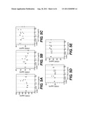 | 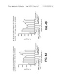 |
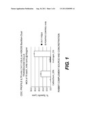 | 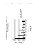 |
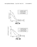 | 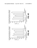 |
| Similar patent applications: | |
| Date | Title |
|---|---|
| 2008-10-16 | Caspase-8 binding protein, its preparation and use |
| 2008-10-30 | Peptides capable of binding to serum proteins |
| 2009-03-19 | Il-17 receptor a antigen binding proteins |
| 2009-04-23 | Chemokine binding activity of viral tnf receptors and related proteins |
| 2008-09-04 | Hk1-binding proteins |
| New patent applications from these inventors: | |
| Date | Title |
|---|---|
| 2017-06-01 | Antigen binding proteins to oncostatin m (osm) |
| 2015-03-05 | Antigen binding proteins to oncostatin m (osm) |
| 2013-09-26 | Antigen binding proteins to oncostatin m (osm) |
| 2013-06-20 | Novel antigen binding proteins |
| 2012-11-08 | Cd127 binding proteins |
| Top Inventors for class "Drug, bio-affecting and body treating compositions" | |
| Rank | Inventor's name |
|---|---|
| 1 | David M. Goldenberg |
| 2 | Hy Si Bui |
| 3 | Lowell L. Wood, Jr. |
| 4 | Roderick A. Hyde |
| 5 | Yat Sun Or |