Patent application title: HUMANIZED ANTI-EGFR ANTIBODY L4-H3 AND CODING GENE THEREOF
Inventors:
Yanwen Jin (Beijing, CN)
David Weaver (Newton, MA, US)
Michael Rynkiewicz (Newton, MA, US)
Cheng Cao (Beijing, CN)
Assignees:
HEFEI TAIRUI BIOTECHNOLOGY CO., LTD
IPC8 Class: AC07K1630FI
USPC Class:
4241741
Class name: Immunoglobulin, antiserum, antibody, or antibody fragment, except conjugate or complex of the same with nonimmunoglobulin material binds eukaryotic cell or component thereof or substance produced by said eukaryotic cell (e.g., honey, etc.) cancer cell
Publication date: 2013-12-12
Patent application number: 20130330362
Abstract:
A humanized anti-EGFR antibody L4-H3 and gene encoding the antibody are
disclosed. The antibody is composed of a heavy chain and a light chain.
The amino acid sequence of the heavy chain variable region is shown as
positions 1-145 of SEQ ID NO: 3, the heavy chain constant region is the
heavy chain constant region of the human antibody IgG1, and the amino
acid sequence of the light chain is shown as SEQ ID NO: 1.Claims:
1. An antibody comprising a heavy chain and a light chain, wherein the
heavy chain comprises the combination of a heavy chain variable region
and a heavy chain constant region, and wherein the amino acid sequence of
the heavy chain variable region is set forth at positions 1-145 of SEQ ID
NO:3, and wherein the heavy chain constant region is a heavy chain
constant region of humanized antibody IgG1, and wherein the amino acid
sequence of the light chain is set forth by SEQ ID NO:1.
2. The antibody according to claim 1, wherein in that the amino acid sequence of the heavy chain of the antibody is set forth by SEQ ID NO:3.
3. An isolated DNA encoding the antibody according to claim 1, the heavy chain of the antibody set forth by SEQ ID NO: 3; or, an isolated DNA encoding the light chain of I), II) or III) below: I) the nucleotides sequence as set forth at positions 85-726 in SEQ ID NO:2 or the nucleotide sequence as set forth at positions 9-729 of SEQ ID NO:2; II) an isolated DNA molecule hybridizing with one of the nucleotide sequences defined in I) under stringent conditions and coding for the light chain; or III) an isolated DNA molecule showing greater than 70% identity to one of the nucleotide sequences defined in I) and coding for the light chain; or, an isolated DNA encoding the heavy chain of 1), 2) or 3) below: 1) the nucleotides sequence set forth by SEQ ID NO:4; 2) an isolated DNA molecule hybridizing with the nucleotide sequence defined in 1) under stringent conditions and coding for the heavy chain; or 3) an isolated DNA molecule showing greater than 70% identity to the nucleotide sequence defined in 1) and coding for the heavy chain.
4. A recombinant vector comprising an isolated DNA of claim 3.
5. The recombinant vectors according to claim 4, wherein the recombinant vectors are recombinant expression vectors prepared by a method comprising the following steps: inserting the coding gene of the light chain between Nhe I and EcoR I restriction sites of the vector pIRES in the direction from Nhe I restriction site to EcoR I restriction site, to obtain a recombinant vector, denoted as an intermediate recombinant vector; and inserting the coding gene of the heavy chain between Xba I and Not I restriction sites of the intermediate recombinant vector in the direction from Xba I restriction site to Not I restriction site, wherein the resultant recombinant vector is a target recombinant expression vector.
6. Recombinant cells obtained by introducing the recombinant expression vectors of claim 5 into starting cells, wherein preferably the starting cells are 293T cells.
7. A method for preparing an antibody comprising the following steps: culturing the recombinant cells of claim 6, and collecting supernatant to obtain the antibody.
8. An inhibitor for inhibiting a signal transduction pathway of an Epidermal Growth Factor Receptor, an inhibitor for inhibiting tumour cells invasion or a product for preventing and/or treating tumours, wherein active ingredients comprise the antibody of claim 1, and wherein the tumour is colon cancer; and preferably the tumour cells are SW480 cells.
9. (canceled)
10. A protein fragment and isolated DNA encoding the protein, wherein the amino acid sequence of the protein fragment is set forth at positions 1-145 of SEQ ID NO:3; and the isolated DNA encoding the protein fragment comprises a), b) or c) below: a) the nucleotides sequence as set forth at positions 1-435 in SEQ ID NO:4; b) an isolated DNA molecule hybridizing with the nucleotide sequence defined in a) under stringent conditions and coding for the protein fragment; c) an isolated DNA molecule having greater than 70% identity to the nucleotide sequence defined in a) and coding for the protein fragment.
11. A method of treating a tumour comprising administering the antibody of claim 1 to a subject in need thereof in an effective amount.
12. The method of claim 11, wherein the tumour is colon cancer.
13. The method of claim 11, wherein the tumour cells are SW480 cells.
14. A recombinant strain comprising an isolated DNA of claim 3.
15. A recombinant cell comprising an isolated DNA of claim 3.
16. A recombinant expression cassette comprising an isolated DNA of claim 3.
17. An inhibitor for inhibiting a signal transduction pathway of an Epidermal Growth Factor Receptor, an inhibitor for inhibiting tumour cells invasion or a product for preventing and/or treating tumours, wherein active ingredients comprise the isolated DNA of claim 3, and wherein the tumour is colon cancer, and preferably the tumour cells are SW480 cells.
18. An inhibitor for inhibiting a signal transduction pathway of an Epidermal Growth Factor Receptor, an inhibitor for inhibiting tumour cells invasion or a product for preventing and/or treating tumours, wherein active ingredients comprise the recombinant vector of claim 4 and wherein the tumour is colon cancer, and preferably the tumour cells are SW480 cells.
19. An inhibitor for inhibiting a signal transduction pathway of an Epidermal Growth Factor Receptor, an inhibitor for inhibiting tumour cells invasion or a product for preventing and/or treating tumours, wherein active ingredients comprise the recombinant strains of claim 14 and wherein the tumour is colon cancer, and preferably the tumour cells are SW480 cells.
20. An inhibitor for inhibiting a signal transduction pathway of an Epidermal Growth Factor Receptor, an inhibitor for inhibiting tumour cells invasion or a product for preventing and/or treating tumours, wherein active ingredients comprise the recombinant cells of claim 15 and wherein the tumour is colon cancer, and preferably the tumour cells are SW480 cells.
Description:
TECHNICAL FIELD
[0001] The present invention relates to an anti-EGFR humanized antibody L4-H3, as well as coding gene and applications thereof.
BACKGROUND OF THE INVENTION
[0002] The Epidermal Growth Factor Receptor (EGFR) is one member of Epidermal Growth Factor Receptor gene (erbB) family, which is over-expressed in about 30% human tumours, especially in non-small cell lung cancer, head and neck squamous cell carcinoma and colorectal cancer, and the like. Many studies at home and abroad have demonstrated that the antibodies against the EGFR show better therapeutic effect to various human tumours caused by EGFR over-expression or/and mutation, especially head and neck squamous cell carcinoma (80%-100%), colorectal cancer (25%-77%), non-small cell lung cancer (40%-80%), and the like, by means of effectively blocking the binding of the ligands extracelluarly to inhibit the EGFR signal transduction pathway. The Epidermal Growth Factor Receptor has become one of tumour treating targets that currently are deeply studied and attract much attention. Using gene engineering to research and prepare the anti-EGFR monoclonal antibody has become one of research hotspots in tumour immune treatment.
[0003] In 2004 and 2006, U.S. FDA successfully approved murine-human chimeric antibody cetuximab and whole human antibody panitumumab against EGFR for the treatment of colorectal cancer; and in 2005, anti-EGFR humanized antibody nimotuzumab obtained the first class of new drug certificate approved by the Chinese State Food and Drug Administration (SFDA), and currently, its II/III phase clinical trials are being performed. When used in human body, the murine derived monoclonal antibody may elicit human anti-murine antibody response, thereby to negatively impacting its function. Engineered murine-human chimeric antibody with gene engineering technique may greatly reduce immunogenicity of murine monoclonal antibody, prolong the half life period of antibody in the body, and mediate immune adjustment and ADCC effect with the help of human immunoglobulin Fc fragments, thereby to enhance the biological effect of the antibody. However, the ability to bind antigen of the chimeric antibody is 98.7% lower than that of murine derived antibody. It has been demonstrated by many pre-clinical trials and clinical trials that cetuximab alone and in combination with chemotherapy/radiotherapy show better therapeutic effect. However, a simple CDR graft tends to cause the decrease of binding affinity between an antigen and an antibody. The panitumumab is a fully human antibody prepared with transgenic mouse technologies, and as compared with chimeric antibody and humanized antibody, has almost 100% humanized sequence, which greatly enhances binding affinity between the antibody and a target. However, such antibody shows drawbacks such as murine glycosylation pattern, short half life period and more hypersensitivity, and the like. Nimotuzumab is a humanized antibody obtained by humanized engineering of anti-EGFR murine derived monoclonal antibody. The light chain gene and heavy chain gene of the antibody are linked to different expression vector, respectively, for expression. Since there is a greater difference between the light chain expression and heavy chain expression, it tends to causes very low expression level of complete antibody molecule.
SUMMARY OF THE INVENTION
[0004] One purpose of the present invention is to provide an antibody and coding gene thereof.
[0005] The antibody provided by the present invention is composed of a heavy chain and a light chain, wherein the heavy chain is formed by binding a heavy chain variable region to a heavy chain constant region; the amino acid sequence of the heavy chain variable region is shown as sites 1-145 of SEQ ID NO:3; the heavy chain constant region is a heavy chain constant region of humanized antibody IgG1; and the amino acid sequence of the light chain is shown as SEQ ID NO:1.
[0006] Specifically, the amino acid sequence of the heavy chain of the antibody described above can be shown as SEQ ID NO:3.
[0007] A coding gene of the light chain of the antibody described above is shown in I), II) or III) below:
[0008] I) nucleotides sequence thereof shown as sites 85-726 in SEQ ID NO:2 or shown as sites 9-729 in SEQ ID NO:2;
[0009] II) a DNA molecule hybridizing with the DNA sequence defined in I) under stringent conditions and coding the light chain;
[0010] III) a DNA molecule showing greater than 70% identity to the DNA sequence defined in I) and coding the light chain;
[0011] A coding gene of the heavy chain of the antibody described above is shown in 1), 2) or 3) below:
[0012] 1) nucleotides sequence thereof shown as SEQ ID NO:4;
[0013] 2) a DNA molecule hybridizing with the DNA sequence defined in 1) under stringent conditions and coding the heavy chain; and
[0014] 3) a DNA molecule showing greater than 70% identity to the DNA sequence defined in 1) and coding the heavy chain.
[0015] The recombinant vectors, recombinant strains, recombinant cells or expression cassettes comprising any one of the coding genes described above also fall into the protection scope of the present invention.
[0016] The recombinant vectors described above can specifically be recombinant expression vectors prepared by a method comprising the following steps: inserting the coding gene of the light chain between the Nhe I and EcoR I restriction sites of vector pIRES in the direction from Nhe I restriction site to EcoR I restriction site, to obtain a recombinant vector, denoted as an intermediate recombinant vector; inserting the coding gene of the heavy chain between Xba I and Not I restriction sites of the intermediate recombinant vector in the direction from Xba I restriction site to Not I restriction site. The resultant recombinant vector is a target recombinant expression vector.
[0017] The recombinant cells described above may specifically be recombinant cells obtained by introducing the recombinant expression vector described above into starting cells.
[0018] Wherein, the starting cells are 293T cells.
[0019] The method for preparing any one of the antibodies described above also falls into the protection scope of the present invention.
[0020] The method for preparing any one of the antibodies described above can comprise the following step: culturing recombinant cells described above, and collecting supernatant to obtain the antibody.
[0021] During culturing the recombinant cells, the light chain of antibody and the heavy chain of antibody are expressed, respectively, and the light chain of antibody and the heavy chain of antibody are self-assembled into the antibody.
[0022] Another purpose of the present invention is to provide an inhibitor for inhibiting a signal transduction pathway of an Epidermal Growth Factor Receptor.
[0023] The active ingredients of the inhibitor for inhibiting a signal transduction pathway of an Epidermal Growth Factor Receptor provided in the present invention are antibody described above, coding gene described above, recombinant vector described above, recombinant strain described above, recombinant cell described above and/or expression cassette described above.
[0024] Another purpose of the present invention is to provide an inhibitor for inhibiting tumour cells invasion.
[0025] The active ingredients of the inhibitor for inhibiting tumour cells invasion provided in the present invention is an antibody described above, a coding gene described above, a recombinant vector described above, a recombinant strain described above, a recombinant cell described above and/or expression cassette described above.
[0026] Another purpose of the present invention is to provide a product for preventing and/or treating tumours.
[0027] The active ingredients of the product for preventing and/or treating tumour provided in the present invention are antibody described above, coding gene described above, recombinant vector described above, recombinant strain described above, recombinant cell described above and/or expression cassette described above.
[0028] In the inhibitor or product described above, the tumour is colon cancer; and the tumour cells are SW480 cells.
[0029] Use of antibody described above, coding gene described above, recombinant vector described above, recombinant strain described above, recombinant cell described above and/or expression cassette described above for preparing an inhibitor for inhibiting a signal transduction pathway of an Epidermal Growth Factor Receptor also falls into the protection scope of the present invention.
[0030] Use of antibody described above, coding gene described above, recombinant vector described above, recombinant strain described above, recombinant cell described above and/or expression cassette described above for preparing an inhibitor for inhibiting a tumour cell invasion also falls into the protection scope of the present invention.
[0031] Use of antibody described above, coding gene described above, recombinant vector described above, recombinant strain described above, recombinant cell described above and/or expression cassette described above for preparing a product for preventing and/or treating tumours also falls into the protection scope of the present invention.
[0032] In uses described above, the tumour is colon cancer; and the tumour cells are SW480 cells.
[0033] The last purpose of the present invention is to provide a protein fragment and coding gene thereof.
[0034] The amino acid sequence of the protein fragment provided in the present invention is shown as sites 1-145 of SEQ ID NO:3.
[0035] The coding gene of the protein fragment provided in the present invention is shown in a), b) or c) below:
[0036] a) nucleotides sequence thereof shown as sites 1-435 in SEQ ID NO:4;
[0037] b) a DNA molecule hybridizing with the DNA sequence defined in a) under stringent conditions and coding the protein fragment;
[0038] c) a DNA molecule having an greater than 70% identity to DNA sequence defined in a) and coding the protein fragment.
DESCRIPTION OF DRAWINGS
[0039] FIG. 1 is an agarose gel electrophoresis plot of the PCR amplified products of the light chain gene and heavy chain variable region gene.
[0040] FIG. 2 is a schematic illustration of the expression vector comprising the antibody of the present invention.
[0041] FIG. 3 is a reduced SDS-PAGE assay of the antibody of the present invention.
[0042] FIG. 4 is an immunoblotting assay of the antibody of the present invention.
PREFERRED EMBODIMENTS
[0043] The experimental methods used in the following Examples are all conventional methods, unless otherwise specified.
[0044] The materials, agents, and the like used in the following Examples are all commercially available, unless otherwise specified.
[0045] pIRES binary expression vector is purchased from Clontech Incorporation, with product catalog number: 631605; and pMD18-T expression vector is purchased from Takara Bio Company, with product catalog number: D504 CA.
[0046] The vector, pIRES-Anti-CD20, has been disclosed in "Construction, Expression and Characterization of Anti-CD20 Chimeric Monoclonal Antibody, China Biotechnology 2005, 25(7):34-39", which is publicly available from Institute of Biotechnology, Academy of Military Medical Sciences.
EXAMPLE 1
The Obtainment of the Coding Genes of the Light Chain and Heavy Chain Variable Region of Antibody
[0047] The protein structure and the amino acid sequence of the murine-human chimeric antibody, cetuximab, is used as a template for computer modeling design of a new antibody. Following design, the process of synthesis of the genes based on these amino acid sequences of the light chain and the amino acid sequence of "variable region +heavy chain constant region 1" of the heavy chain, is undertaken.
[0048] The antibody of the present invention is composed of a light chain L4 and a heavy chain H3; and the heavy chain H3 is composed of a heavy chain variable region (VH), a heavy chain constant region 1 (CH1), a hinge region, a heavy chain constant region 2 (CH2) and a heavy chain constant region 3 (CH3) (H3=VH+CH1+hinge+CH2+CH3). The antibody of the present invention is denoted as L4-H3.
[0049] The light chain L4: the amino acid sequence is shown as SEQ ID NO:1; and the coding gene sequence is shown as sites 85-726 in SEQ ID NO:2; The heavy chain H3: the amino acid sequence is shown as SEQ ID NO:3; and the coding gene sequence shown as SEQ ID NO:4;
[0050] In SEQ ID NO:3, the amino acids of sites 1-145 are the heavy chain variable region (VH), the amino acids of sites 146-243 are the heavy chain constant region 1 (CH1), the amino acids of sites 244-258 are the hinge region, the amino acids of sites 259-369 are the heavy chain constant region 2 (CH2), and the amino acids of sites 370-475 are the heavy chain constant region 3 (CH3).
[0051] In SEQ ID NO:4, the nucleotides of sites 1-435 are the heavy chain variable region (VH) gene, the nucleotides of sites 436-729 are the heavy chain constant region 1 (CH1) gene, the nucleotides of sites 730-739 are a splice donor, the nucleotides of sites 740-1117 are an intron 1, the nucleotides of sites 1118-1162 are hinge region, the nucleotides of sites 1163-1280 are an intron 2, the nucleotides of sites 1281-1613 are heavy chain constant region 2 (CH2), the nucleotides of sites 1614-1710 are an intron 3, the nucleotides of sites 1711-2031 are a heavy chain constant region 3 (CH3), and the nucleotides of sites 2032-2230 are an intron 4.
[0052] The "variable region+constant region 1" of the heavy chain H1: the coding gene sequence is shown as SEQ ID NO:5.
[0053] The coding gene of the light chain L4 was obtained by artificial synthesis (that is, the nucleotides of sites 9-729 in SEQ ID NO:2 are artificially synthesized). The coding gene of the "constant region 2+constant region 3" of heavy chain H3 (that is, the nucleotides of sites 730-2230 in SEQ ID NO:4) can be obtained by artificially synthesis, and it also can be obtained by enzyme digestion of the vector pIRES-Anti-CD20.
[0054] The coding gene of the "variable region+constant region 1" of the heavy chain H3 can be obtained by artificial synthesis, and it also can be obtained according to the following method: artificially synthesizing the coding gene shown as SEQ ID NO:5. The artificially synthesized coding gene shown as SEQ ID NO:5 was used as a template, and the coding gene of "variable region+constant region 1" of the heavy chain H3 was obtained by amplifying from primers 1, 2, 3, 4, 5 and 6 using overlapping PCR method. A fragment (named as A, B, and C) was amplified with the primer pairs 1 and 3, 4 and 5, 6 and 2, respectively. Then, the amplified A, B, and C were mixed as templates, and the target coding gene of "variable region+constant region 1" of the heavy chain H3 was amplified with primers 1 and 2.
TABLE-US-00001 (SEQ ID NO: 6) 1: 5'-gtgtctagagccgccaccatggactgga-3'(XbaI); (SEQ ID NO: 7) 2: 5'-gggatccacttacctgttgctttct-3'(BamHI); (SEQ ID NO: 8) 3: 5'-atccactcaagtctttgtccaggggcctgtcgcacccagtggacgccgtagttagtcaggctgaatcc- ag-3'; (SEQ ID NO: 9) 4: 5'-gagtggatgggagtgatctggagtggtggtaacactgactacaacacccccttcactagcagagtcac- c-3'. (SEQ ID NO: 10) 5: 5'-gaccagggttccctggccccagtaggcgaactcgtagtcgtaataagtcagggctctcgcacag-3' (SEQ ID NO: 11) 6: 5'-cctactggggccagggaaccctggtcaccgtctcctcagcctccaccaagggcccatcg-3'.
[0055] The gel electrophoresis assay was performed on the respective genes obtained, and the results are consistent with the expected size. The fragment size of the light chain L4 is about 720 bp, and the size of the coding gene of the "variable region+constant region 1" of the heavy chain H3 is about 729 bp (FIG. 1). M. relative molecular mass standard; A: lane 1 is the coding gene of the light chain L4; B: lane 1 is the coding gene of the "variable region+constant region 1" of the heavy chain H1, and lane 3 is the coding gene of the "variable region+constant region 1" of the heavy chain H3.
[0056] The obtained coding gene of the light chain and that of the "variable region+constant region 1" of the heavy chain H3 were cloned into vector pMD18-T, respectively, transforming Escherichia coli DH5a, picking monoclonal strain, extracting plasmid and sequencing for identification. The results demonstrated that the DNA shown as nucleotides at sites 9-729 in SEQ ID NO:2 (that is, the coding gene of the light chain) had been inserted into vector pMD18-T, and the recombinant vector was denoted as pMD18-T/L4. The nucleotides shown at sites of 1-729 in SEQ ID NO:4 (that is, the coding gene of the "variable region+constant region 1" of the heavy chain H3) had been inserted into vector pMD18-T, and the recombinant vector was denoted as pMD18-T/VH+CH1.
EXAMPLE 2
The Expression and Purification of the Antibodies
[0057] A. Construction of the Recombinant Expression Vector:
[0058] The recombinant vector pMD 18-T/L4 and pIRES binary expression vector were enzymatically digested with corresponding restriction endonuclease (NheI and EcoRI), respectively. After agarose gel electrophoresis, the purified target fragments were recovered. The light chain gene fragment L4 and vector fragment were mixed, and reacted with each other for 12 h at 16° C., under the action of linking agents. Transforming Escherichia coli DH5a, picking monoclonal strain, extracting plasmid and sequencing for identification. Results: the light chain coding gene shown as nucleotides at sites 9-729 in SEQ ID NO:2 had been inserted between NheI and EcoRI restriction sites of vector pIRES (in the direction from NheI restriction site to EcoRI restriction site). It is demonstrated that the constructed recombinant vector is right, denoted as recombinant expression vector pIRES/L4.
[0059] The plasmid large fragment was recovered by using the pIRES/L4 as a template and enzymatic digestion with XbaI and Not I, denoted as fragment 1. A fragment with about 729 by (that is, the fragment of "variable region+constant region 1" of the heavy chain H3) was recovered by using the pMD18-T/VH+CH1 as a template and enzymatic digestion with Xba I and BamH I, denoted as fragment 2. A fragment with about 1502 by (that is, a fragment comprising "constant region 2+ constant region 3 of the heavy chain") was recovered by using the pIRES-Anti-CD20 as a template and enzymatic digestion with BamH I and Not I, denoted as fragment 3. The fragments 1, 2 and fragment 3 are linked to obtain a recombinant vector, transforming Escherichia coli DH5a, picking monoclonal strain, extracting plasmid and sequencing for identification. The results demonstrated that the heavy chain coding gene shown as SEQ ID NO:4 had been inserted between the XbaI and Not I restriction sites of vector pIRES (in the direction from Xba I to Not I); the light chain coding gene shown as nucleotides at sites 9-729 in SEQ ID NO:2 had been inserted between the EcoRI and NheI restriction sites of vector pIRES (in the direction from Nhe I to EcoR I); and IRES (Internal ribosome entry site, IRES may independently recruit ribosomes to translate the heavy chain mRNA) was located between the light chain coding gene and heavy chain coding gene. The positive recombinant expression vector was denoted as pIRES/L4/H3 (FIG. 2).
[0060] B. The Vector Transformation and the Expression of the Antibodies
[0061] 293T cells (293T human embryonic kidney T cells) were purchased from American Type Culture Collection (ATCC) with product catalog number CRL-11268; Lipofectamine 2000 was purchased from Invitrogen Corporation with product catalog number 12566014; HyQSFM4CHO medium was purchased from HyClone Incorporation with product catalog number SH30518.02; and rProtein A chromatographic column was purchased from GE Incorporation with product catalog number 17-5079-01.
[0062] The 293T cells were inoculated in culture dishes with 10 cm diameter at the density of 1×10⁶/ml, respectively. The culture dishes were filled with DMEM medium containing 10% fetal bovine serum, and were cultured in 5% CO2 incubator at 37° C. 5 μg of plasmid pIRES/L4/H3 obtained in step A was used to transfect 293T cells, with the detail operation according to the description of the agent Lipofectamine 2000. The recombinant cells 293T-pIRES/L4/H3 were obtained.
[0063] The recombinant cells 293T-pIRES/L4/H3 were cultured with serum free DMEM medium. After 6-8 h, the serum free medium was drawn off, and was replaced with HyQSFM4CHO medium. The culture last totally for 84 h under the same conditions, and the cell supernatant was collected once every 12 h. Then the expression of the antibody was roughly assessed using ELISA method. The transfection system was amplified, and cell culture supernatant of 4-5 L was collected, which was adjusted to pH 6.0-7.0, and was filtered via 0.45 μm filter membrane, and the antibody was then purified with a rProtein A chromatographic column, with the detail operation according to the product description.
[0064] ELISA: double antibody sandwich method for assaying unknown antigen:
[0065] 1. Coating: 0.05M PH9.0 carbonate coating buffer was used to dilute the antibody (primary antibody is goat anti-human IgG), until the content of protein is 1˜10 μg/ml. In each reaction well of the polystyrenes plate, 0.1 ml is added, overnight at 4° C. On the following day, the solution inside the well was discarded, and the plate was washed with washing buffer thrice, 3 minutes for each. (Simplified as washing, the same below).
[0066] 2. Loading: the coated reaction wells were loaded with 0.1 ml of the sample to be tested and diluted in certain proportions, and was incubated at 37° C. for 1 hour, and washed. Alternatively, a blank well, a negative control well (the 293T cell supernatant of untransfected plasmid) and positive control well (cetuximab injection (Erbitux), Imported Drug License Number: S20050095. MERCK & CO. INC., Germany, Product Lot Number: 7667201) were loaded.
[0067] 3. Addition of enzyme-labeled antibody (secondary antibody is goat anti-human IgG-HRP (goat anti-human IgG-horseradish peroxidase)): 0.1 ml of freshly diluted enzyme-labeled antibody (the dilution after titration) was added into each reaction well, which was incubated at 37° C. for 0.5-1 hour, and washed.
[0068] 4. Addition of substrate solution for color development: 0.1 ml of temporarily formulated TMB substrate solution was added into each reaction well, at 37° C. for 10-30 minutes.
[0069] 5. Quenching the reaction: 0.05 ml of 2M sulfuric acid was added into each reaction well.
[0070] 6. Assessing the results: the results may be observed by the naked eye directly on the white background, where the deeper the color inside the reaction well indicated a stronger positivity; and the negative reaction was colorless or very light. "+", "-" indicators were used according to the degree of the color. The OD value can be measured: after zero adjustment with blank control well, the OD value of each well was measured on ELISA instrument, at 450 nm (if colored with ABTS, at 410 nm). If the OD value was greater than that of predetermined negative control by a factor of 2.1, it was positive.
[0071] Agents
[0072] (1) The coating buffer (PH9.6, 0.05M of carbonate buffer): Na2CO3 1.59 g, NaHCO3 2.93 g, adding distilled water to 1000 ml.
[0073] (2) The washing buffer (PH7.4, PBS): 0.15M: KH2PO4 0.2 g, Na2HPO4.12H2O 2.9 g, NaCl 8.0 g, KCl 0.2 g, Tween-20 0.05% 0.5 ml, adding distilled water to 1000 ml.
[0074] (3) The dilution: 0.1 g of Bovine Serum Albumin (BSA) was added into the washing buffer, totally 100 ml, or goat serum, rabbit serum, and the like was prepared into 5-10% as along with the washing solution for use.
[0075] (4) The quenching solution (2M H2SO4): 178.3 ml of distilled water was added into 21.7 ml of the concentrated sulfuric acid (98%) dropwise.
[0076] Sodium phosphate buffer (binding buffer): as the binding buffer for purifying Protein A. Method for formulation: 57.7 ml of 1M Na2HPO4 and 42.3 ml of 1M NaH2PO4 were mixed uniformly to obtain 100 ml of 0.1M sodium phosphate buffer, pH7.0, which was then diluted with distilled water to 20 mM ready-for-use.
[0077] The citric acid-sodium citric acid buffer (extracting buffer): as the extracting buffer for purifying Protein A. The method for formulation: 186 ml of 0.1M citric acid and 14 ml of 0.1M sodium citric acid were mixed uniformly to obtain 200 ml of 0.1M citrate buffer, pH3.0.
[0078] The antibody was purified with rProtein A chromatographic column: the purification medium: HiTrap rProtein A FF, 5 ml, was purchased from GE Incorporation, Lot Number: 17-5079-01. For the detailed usage description, please refer the description provided by this company.
[0079] Procedures:
[0080] 1) Washing the tubing with 1M NaOH and ddH2O in this order, boiling the small filter in 0.1M NaOH for 10 min, and then immersing it into ddH2O for 1-2 min;
[0081] 2) Setting the program to connect the rProtein A affinity chromatography column;
[0082] 3) Equilibrating chromatography column with 20 mM binding buffer (pH7.0);
[0083] 4) Loading the prepared cell supernatant at the flow rate of 1-2 ml/min; preparing the cell supernatant: taking out supernatant after centrifugating at 12000 g for 15 min, then passing the supernatant through 0.22 um cellulose nitrate filter membrane for filtration sterilization.
[0084] 5) Extracting the target proteins with 1M extracting buffer (pH3.0) just before the end of the loading;
[0085] 6) Collecting the extracting solution (that is, the target proteins), adjusting the pH to 7.0 with Trise base (pH9.0), and then assaying by electrophoresis; and
[0086] 7) Washing the tubing and small filter according to step 1).
[0087] C. The Protein Assay
[0088] The goat anti-human IgG-HRP antibody was purchased from Sigma Corporation with product catalog number (046K4801); and goat anti-murine IgG1 was purchased from SBA (Southern Biotechnology Associates, Inc.) with product catalog number (1010-05);
[0089] SDS-PAGE: 15 μl extracting solution (that is, the antibody solution) was taken out to perform the reduced SDS-PAGE electrophoresis on 12% gel, staining with Coomassie brilliant blue R-250.
[0090] The results show that the relative molecular weights of the heavy chain and light chain of antibody obtained via purification are 25×103, 50×103, respectively (FIG. 3), consistent with the expected results. In FIG. 3, lanes 1-4 represent the antibody L4-H3 of the present invention; and lane 5 represents positive control cetuximab.
[0091] Immunoblotting assay: another portions of extracting solution was taken out to perform reduced SDS-PAGE electrophoresis on 12% gel, and then was transferred to cellulose nitrate membrane. Then the membrane was taken out and blocked in blocking buffer (1× PBST containing 5% defatted milk powder) at room temperature for 2 h, was incubated with the 1:5000 diluted goat anti-human IgG-HRP antibody for 2 h (at room temperature), and then was washed with 1× PBST thrice. Finally, the membrane was colored with ECL, and was exposed to x-ray film. In FIG. 4, lane 1 represents the positive control cetuximab; and lanes 2-5 represent the antibody L4-H3 of the present invention.
[0092] Immunoblotting (12%) assay demonstrates that the antibody can specifically be bound to the goat anti-human IgG However, there are partial non-specific hybridized lanes that react with anti-human IgG secondary antibody (including the commercialized cetuximab), which may be derived from the mixing of partial non-whole length heavy chain fragments during the purification. However, above results do not influence the assessment to antibody binding affinity. None of antibodies described above reacts with goat anti-murine IgG1.
[0093] Since the light chain and heavy chain of the present antibody are expressed proportionally in the same expression vector, they are self-assembled into the complete antibody containing two light chains and two heavy chains.
[0094] D. Expression Amount of Protein
[0095] 4-5 L of cell culture supernatant of expression antibody L4-H3 was obtained according to the method in steps A and B. The antibody was purified by rProtein A chromatography column to assay the expression amount of the antibody in 293T cells, and the result is 0.6μg/ml extracting solution.
EXAMPLE 3
The Functional Properties of the Antibody
[0096] A. Assay of the Binding Ability of Antibody and Antigen with Surface Plasmon Resonance [Biacore]
[0097] Sensor Chip CM5 was purchased from BD Incorporation with product catalog number Br-1000-14; BD BioCoat® and Matrigel® Invasion Chamber were purchased from BD Incorporation with product catalog number 354480.
[0098] EGFR proteins were purchased from Sigma Corporation with product catalog number E2645-500UN.
[0099] The binding affinity between antibody and EGFR was assayed with Biacore3000 equipment. The EGFR proteins diluted with 10 mmol/L NaAc and having different pH (4.0, 4.5, 5.0 and 5.5) were formulated, and were pre-concentrated on CM5 chip to select the NaAc diluted protein with optimum pH. The purified antibody (that is, the washing solution obtained in step B of Example 2) was covalently bound to CM5 sensor chip. The mobile phase was HBS-EP (pH7.4), and flow rate was 20 μl/min The binding affinities between 5 different concentrations of antibodies (0, 10.55, 21.1, 42.2 and 84.4 nmol/L) and EGFR protein were assayed. The binding affinities were computed with the software attached by Biacore3000. At the same time, the cetuximab was used as a control.
[0100] The experiment repeated thrice, and the results were averaged.
[0101] The results show that there are good binding activities between the antibody and antigen EGFR, and the binding affinity is 2.75×10⁻⁹ M. the binding affinity of cetuximab is 1.1×10⁻⁹ M. The results demonstrate that the humanized antibody of the present invention maintains the good binding activity of the parental antibody, overcoming the adverse response of murine derived antibody, and possesses the well clinical application value.
[0102] B. The Experiment of Tumour Cell Invasion
[0103] SW480 cells were purchased from American Type Culture Collection, (ATCC), with ATCC® Number: CCL228®; and cetuximab antibody was purchased from Merck-Lipha Pharmaceuticals Ltd, Germany (the trade name: ERBITUX; the drug name: Cetuximab; the reference trade name in China: ai bi tuo; the molecule structure name: cetuximab; Origin: Germany; Manufacturer: Merck-Lipha Pharmaceuticals Ltd, Germany).
[0104] The RPMI1640 medium (purchased from Invitrogen Corporation with catalog number 31800-022) was used to culture SW480 cells. The invasion chamber was hydrated with serum-free RPMI1640 medium, and was incubated for 2 h (37° C., 5% CO2). The serum-free RPMI1640 was discarded, 750 μl of RPMI1640 (containing 10% serum) was added into well chambers of invasion Chamber (BD BioCoat® Matrigel® Invasion Chamber, purchased from BD Incorporation with product catalog number 354480); 475 μl of RPMI1640 (containing 1% serum) was added into insert chambers, and then 25 μl digested SW480 cells were added (the numbers of cell >105/500 μl). Finally, the negative control PBS and the antibody of the present invention were separately added into the insert chambers, and each sample had two repeat wells, with the final concentration of antibody 100 ng/ml. After 24 h incubation, the cells that did not penetrate through the basement membrane of invasion cassette were wiped off with sterilized cotton swabs; and the cells that penetrate through the basement membrane of invasion cassette were fixed, stained, dried at room temperature, and then counted under the light microscope.
[0105] Statistics: the number of the corresponding 3 sets of cells of the sample in each insert chamber was counted under 100× microscope. The t-test was performed on the cell invasion experiment data by SPSS12.0 analytic software, and the antibody of the present invention was compared with human IgG set. If the result was P<0.05, the difference between two treatment effects was statistically significant.
[0106] The anti-EGFR antibody of the invention has significant difference, and its inhibition to the invasion of SW480 tumour cell is significant. The experiment was repeated thrice, and the results were averaged. The t-test results show that the P values of the sets of human IgG and the antibody of the present invention are less than 0.01 (Table 1), and as compared with human IgG in a control set,
TABLE-US-00002 TABLE 1 The result of the t-test result of the cell invasion experiment Sets n (number) average number of cell human IgG 8 146 ± 16.531 antibody of 8 67 ± 15.334* the present invention Note: as compared with human IgG *P < 0.01;
INDUSTRY UTILITY
[0107] The experimental results demonstrate that the antibody of the present invention has better antigen binding activities and ability to inhibit tumour cell growth, immigration and invasion. In contrast, the binding affinity of the common anti-EGFR human-murine chimeric antibody cetuximab in international market is 1.1×10⁻⁹ M. The humanized antibody of the present invention can bind to the EGFR better, accordingly to insure the anti-tumour effect thereof. The method for preparing the antibody of the present invention can express the light chain and heavy chain simultaneously, causing the expression proportion of the light chain and heavy chain near 1:1, accordingly to produce more rates of matched double-chain antibody. In conclusion, the antibody of the present invention and the preparing method thereof would have wide utility prospect in the field of preventing and/or treating tumours.
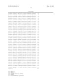
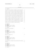
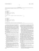
Sequence CWU
1
1
111214PRTArtificial SequenceSynthesized 1Glu Leu Val Met Thr Gln Ser Pro
Ser Ser Leu Ser Ala Ser Val Gly 1 5 10
15 Asp Arg Val Asn Ile Ala Cys Arg Ala Ser Gln Ser Ile
Gly Thr Asn 20 25 30
Ile His Trp Tyr Gln Gln Lys Pro Gly Lys Ala Pro Arg Leu Leu Ile
35 40 45 Lys Tyr Ala Ser
Glu Ser Ile Ser Gly Val Pro Ser Arg Phe Ser Gly 50
55 60 Ser Gly Ser Gly Thr Asp Phe Thr
Leu Thr Ile Ser Ser Leu Gln Pro 65 70
75 80 Glu Asp Phe Ala Ile Tyr Tyr Cys Gln Gln Asn Asn
Asn Trp Pro Thr 85 90
95 Thr Phe Gly Gly Gly Thr Lys Val Glu Ile Lys Arg Thr Val Ala Ala
100 105 110 Pro Ser Val
Phe Ile Phe Pro Pro Ser Asp Glu Gln Leu Lys Ser Gly 115
120 125 Thr Ala Ser Val Val Cys Leu Leu
Asn Asn Phe Tyr Pro Arg Glu Ala 130 135
140 Lys Val Gln Trp Lys Val Asp Asn Ala Leu Gln Ser Gly
Asn Ser Gln 145 150 155
160 Glu Ser Val Thr Glu Gln Asp Ser Lys Asp Ser Thr Tyr Ser Leu Ser
165 170 175 Ser Thr Leu Thr
Leu Ser Lys Ala Asp Tyr Glu Lys His Lys Val Tyr 180
185 190 Ala Cys Glu Val Thr His Gln Gly Leu
Ser Ser Pro Val Thr Lys Ser 195 200
205 Phe Asn Arg Gly Glu Cys 210
2735DNAArtificial SequenceSynthesized 2gtggctagcg ccgccaccat ggacatgagg
gtccccgctc agctcctggg gctcctgctg 60ctctggctcc caggtgccag atgtgaactc
gtcatgaccc agtctccatc ctccctgtct 120gcatctgtag gagacagagt caacattgcc
tgccgggcaa gtcagagcat tggcactaac 180atccactggt atcagcagaa accagggaaa
gcccctagac tcctgatcaa atatgcctcc 240gaaagcatca gtggggtccc atcaagattc
agcggcagtg gatctggcac agatttcact 300ctcaccatca gcagcctgca gcctgaagat
tttgcaatct attactgtca gcaaaataac 360aattggccta ctacgttcgg cggagggacc
aaggtggaaa tcaaacgaac tgtggctgca 420ccatctgtct tcatcttccc gccatctgat
gagcagttga aatctggaac tgcctctgtt 480gtgtgcctgc tgaataactt ctatcccaga
gaggccaaag tacagtggaa ggtggataac 540gccctccaat cgggtaactc ccaggagagt
gtcacagagc aggacagcaa ggacagcacc 600tacagcctca gcagcaccct gacgctgagc
aaagcagact acgagaaaca caaagtctac 660gcctgcgaag tcacccatca gggcctgagc
tcgcccgtca caaagagctt caacagggga 720gagtgttagg aattc
7353475PRTArtificial
SequenceSynthesized 3Val Ser Arg Ala Ala Thr Met Asp Trp Thr Trp Arg Val
Phe Cys Leu 1 5 10 15
Leu Ala Val Ala Pro Gly Ala His Ser Gln Val Lys Leu Leu Glu Gln
20 25 30 Ser Gly Ala Glu
Val Lys Lys Pro Gly Ala Ser Val Lys Val Ser Cys 35
40 45 Lys Ala Ser Gly Phe Ser Leu Thr Asn
Tyr Gly Val His Trp Val Arg 50 55
60 Gln Ala Pro Gly Gln Arg Leu Glu Trp Met Gly Val Ile
Trp Ser Gly 65 70 75
80 Gly Asn Thr Asp Tyr Asn Thr Pro Phe Thr Ser Arg Val Thr Ile Thr
85 90 95 Arg Asp Thr Ser
Ala Thr Thr Ala Tyr Met Gly Leu Ser Ser Leu Arg 100
105 110 Pro Glu Asp Thr Ala Val Tyr Tyr Cys
Ala Arg Ala Leu Thr Tyr Tyr 115 120
125 Asp Tyr Glu Phe Ala Tyr Trp Gly Gln Gly Thr Leu Val Thr
Val Ser 130 135 140
Ser Ala Ser Thr Lys Gly Pro Ser Val Phe Pro Leu Ala Pro Ser Ser 145
150 155 160 Lys Ser Thr Ser Gly
Gly Thr Ala Ala Leu Gly Cys Leu Val Lys Asp 165
170 175 Tyr Phe Pro Glu Pro Val Thr Val Ser Trp
Asn Ser Gly Ala Leu Thr 180 185
190 Ser Gly Val His Thr Phe Pro Ala Val Leu Gln Ser Ser Gly Leu
Tyr 195 200 205 Ser
Leu Ser Ser Val Val Thr Val Pro Ser Ser Ser Leu Gly Thr Gln 210
215 220 Thr Tyr Ile Cys Asn Val
Asn His Lys Pro Ser Asn Thr Lys Val Asp 225 230
235 240 Lys Lys Ala Glu Pro Lys Ser Cys Asp Lys Thr
His Thr Cys Pro Pro 245 250
255 Cys Pro Ala Pro Glu Leu Leu Gly Gly Pro Ser Val Phe Leu Phe Pro
260 265 270 Pro Lys
Pro Lys Asp Thr Leu Met Ile Ser Arg Thr Pro Glu Val Thr 275
280 285 Cys Val Val Val Asp Val Ser
His Glu Asp Pro Glu Val Lys Phe Asn 290 295
300 Trp Tyr Val Asp Gly Val Glu Val His Asn Ala Lys
Thr Lys Pro Arg 305 310 315
320 Glu Glu Gln Tyr Asn Ser Thr Tyr Arg Val Val Ser Val Leu Thr Val
325 330 335 Leu His Gln
Asp Trp Leu Asn Gly Lys Glu Tyr Lys Cys Lys Val Ser 340
345 350 Asn Lys Ala Leu Pro Ala Pro Ile
Glu Lys Thr Ile Ser Lys Ala Lys 355 360
365 Gly Gln Pro Arg Glu Pro Gln Val Tyr Thr Leu Pro Pro
Ser Arg Asp 370 375 380
Glu Leu Thr Lys Asn Gln Val Ser Leu Thr Cys Leu Val Lys Gly Phe 385
390 395 400 Tyr Pro Ser Asp
Ile Ala Val Glu Trp Glu Ser Asn Gly Gln Pro Glu 405
410 415 Asn Asn Tyr Lys Thr Thr Pro Pro Val
Leu Asp Ser Asp Gly Ser Phe 420 425
430 Phe Leu Tyr Ser Lys Leu Thr Val Asp Lys Ser Arg Trp Gln
Gln Gly 435 440 445
Asn Val Phe Ser Cys Ser Val Met His Glu Ala Leu His Asn His Tyr 450
455 460 Thr Gln Lys Ser Leu
Ser Leu Ser Pro Gly Lys 465 470
47542230DNAArtificial SequenceSynthesized 4gtgtctagag ccgccaccat
ggactggacc tggagggtct tctgcttgct ggctgtagct 60ccaggtgctc actcccaggt
gaagctgctg gagcagtctg gggctgaagt gaagaagcct 120ggggcctcag tgaaggtttc
ctgcaaggca tctggattca gcctgactaa ctacggcgtc 180cactgggtgc gacaggcccc
tggacaaaga cttgagtgga tgggagtgat ctggagtggt 240ggtaacactg actacaacac
ccccttcact agcagagtca ccatcaccag ggacacgtcc 300gctactacag cctacatggg
cctgtctagc ctgagacccg aggacacggc cgtatattac 360tgtgcgagag ccctgactta
ttacgactac gagttcgcct actggggcca gggaaccctg 420gtcaccgtct cctcagcctc
caccaagggc ccatcggtct tccccctggc accctcctcc 480aagagcacct ctgggggcac
agcggccctg ggctgcctgg tcaaggacta cttccccgaa 540ccggtgacgg tgtcgtggaa
ctcaggcgcc ctgaccagcg gcgtgcacac cttcccggct 600gtcctacagt cctcaggact
ctactccctc agcagcgtgg tgacagtgcc ctccagcagc 660ttgggcaccc agacctacat
ctgcaacgtg aatcacaagc ccagcaacac caaggtggac 720aagaaagcaa caggtaagtg
gatccggagg gagggtgtct gctggaagca ggctcagcgc 780tcctgcctgg acgcatcccg
gctatgcagc cccagtccag ggcagcaagg caggccccgt 840ctgcctcttc acccggaggc
ctctgcccgc cccactcatg ctcagggaga gggtcttctg 900gctttttccc caggctctgg
gcaggcacag gctaggtgcc cctaacccag gccctgcaca 960caaaggggca ggtgctgggc
tcagacctgc caagagccat atccgggagg accctgcccc 1020tgacctaagc ccaccccaaa
ggccaaactc tccactccct cagctcggac accttctctc 1080ctcccagatt ccagtaactc
ccaatcttct ctctgcagag cccaaatctt gtgacaaaac 1140tcacacatgc ccaccgtgcc
caggtaagcc agcccaggcc tcgccctcca gctcaaggcg 1200ggacaggtgc cctagagtag
cctgcatcca gggacaggcc ccagccgggt gctgacacgt 1260ccacctccat ctcttcctca
gcacctgaac tcctgggggg accgtcagtc ttcctcttcc 1320ccccaaaacc caaggacacc
ctcatgatct cccggacccc tgaggtcaca tgcgtggtgg 1380tggacgtgag ccacgaagac
cctgaggtca agttcaactg gtacgtggac ggcgtggagg 1440tgcataatgc caagacaaag
ccgcgggagg agcagtacaa cagcacgtac cgtgtggtca 1500gcgtcctcac cgtcctgcac
caggactggc tgaatggcaa ggagtacaag tgcaaggtct 1560ccaacaaagc cctcccagcc
cccatcgaga aaaccatctc caaagccaaa ggtgggaccc 1620gtggggtgcg agggccacat
ggacagaggc cggctcggcc caccctctgc cctgggagtg 1680accgctgtac caacctctgt
ccctacaggg cagccccgag aaccacaggt gtacaccctg 1740cccccatccc gggatgagct
gaccaagaac caggtcagcc tgacctgcct ggtcaaaggc 1800ttctatccca gcgacatcgc
cgtggagtgg gagagcaatg ggcagccgga gaacaactac 1860aaggccacgc ctcccgtgct
ggactccgac ggctccttct tcctctacag caagctcacc 1920gtggacaaga gcaggtggca
gcaggggaac gtcttctcat gctccgtgat gcatgaggct 1980ctgcacaacc actacacgca
gaagagcctc tccctgtctc cgggtaaatg agtgcgacgg 2040ccggcaagcc cccgctcccc
gggctctcgc ggtcgcacga ggatgcttgg cacgtacccc 2100gtgtacatac ttcccgggcg
cccagcatgg aaataaagca cccagcgctg ccctgggccc 2160ctgcgagact gtgatggttc
tttccacggg tcaggccgag tctgaggcct gagtggcatg 2220agggaggcag
22305770DNAArtificial
SequenceSynthesized 5gtgtctagag ccgccaccat ggactggacc tggagggtct
tctgcttgct ggctgtagct 60ccaggtgctc actcccaggt gaagctgctg gagcagtctg
gggctgaagt gaagaagcct 120ggggcctcag tgaaggtttc ctgcaaggca tctggattca
gcatcggcaa ctacggcatc 180cactgggtgc gacaggcccc tggacaaaga cttgagtgga
tgggaggaat ctggagtggt 240ggtaacgccg actacgcaca gaaattccag ggcagagtca
ccatcaccag ggacacgtcc 300gctactacag cctacatggg cctgtctagc ctgagacccg
aggacacggc cgtatattac 360tgtgcgagag tgggcactta ttacgactac gagttcgacg
tgtggggcca gggaaccctg 420gtcaccgtct cctcagcctc caccaagggc ccatcggtct
tccccctggc accctcctcc 480aagagcacct ctgggggcac agcggccctg ggctgcctgg
tcaaggacta cttccccgaa 540ccggtgacgg tgtcgtggaa ctcaggcgcc ctgaccagcg
gcgtgcacac cttcccggct 600gtcctacagt cctcaggact ctactccctc agcagcgtgg
tgaccgtgcc ctccagcagc 660ttgggcaccc agacctacat ctgcaacgtg aatcacaagc
ccagcaacac caaggtggac 720aagaaagttg agcccaaatc ttgtgacaaa actacaggta
agtggatccc 770628DNAArtificial SequenceSynthesized
6gtgtctagag ccgccaccat ggactgga
28725DNAArtificial SequenceSynthesized 7gggatccact tacctgttgc tttct
25870DNAArtificial
SequenceSynthesized 8atccactcaa gtctttgtcc aggggcctgt cgcacccagt
ggacgccgta gttagtcagg 60ctgaatccag
70969DNAArtificial SequenceSynthesized
9gagtggatgg gagtgatctg gagtggtggt aacactgact acaacacccc cttcactagc
60agagtcacc
691064DNAArtificial SequenceSynthesized 10gaccagggtt ccctggcccc
agtaggcgaa ctcgtagtcg taataagtca gggctctcgc 60acag
641159DNAArtificial
SequenceSynthesized 11cctactgggg ccagggaacc ctggtcaccg tctcctcagc
ctccaccaag ggcccatcg 59
User Contributions:
Comment about this patent or add new information about this topic:

