Patent application title: Biological Agents Active in Central Nervous System
Inventors:
Roger A. Johns (Reisterstown, MD, US)
Roger A. Johns (Reisterstown, MD, US)
Feng Tao (Baltimore, MD, US)
Assignees:
THE JOHNS HOPKINS UNIVERSITY
IPC8 Class: AA61K3817FI
USPC Class:
514 183
Class name: Peptide (e.g., protein, etc.) containing doai nervous system (e.g., central nervous system (cns), etc.) affecting pain affecting
Publication date: 2012-12-27
Patent application number: 20120329723
Abstract:
Cell-permeant fusion peptides Tat-PDZ can dose-dependently reduce the
threshold for anesthesia. PDZ domain-mediated protein interactions at
synapses in the central nervous system play an important role in the
molecular mechanisms of anesthesia. Moreover, Tat-PDZ cell-permeant
fusion peptides are delivered intracellularly into neurons in the central
nervous system subsequent to intraperitoneally injection. By in vitro and
in vivo binding assays, we found that the Tat-PDZ dose-dependently
inhibited the interactions between NMDARs and PSD-95. Furthermore,
behavior testing showed that animals given Tat-PDZ exhibited
significantly reduced established inflammatory pain behaviors compared to
vehicle-treated group. Our results indicate that by disrupting
NMDAR/PSD-95 protein interactions, the Tat-PDZ cell-permeable fusion
peptides provide a new approach for inflammatory pain therapy.Claims:
1. A method for relieving acute or chronic pain in a human, comprising:
administering to a subject in need thereof an effective amount of a
fusion protein which comprises a cell membrane transduction domain of
HIV1 Tat and a PDZ domain of a protein selected from the group consisting
of PICK1, PSD93 and PSD95, whereby acute or chronic pain experienced by
the subject is relieved.
2. The method of claim 1 wherein the fusion protein comprises a PDZ2 domain of PSD93.
3. The method of claim 1 wherein the fusion protein comprises a PDZ2 domain of PSD95.
4. The method of claim 1 wherein the fusion protein is administered intraperitoneally.
5. The method of claim 1 wherein the fusion protein is administered systemically or intrathecally.
6. A method for treating or preventing allodynia or hyperalgesia in a human, comprising: administering to a subject in need thereof an effective amount of a fusion protein which comprises a cell membrane transduction domain of HIV1 Tat and a PDZ domain of a protein selected from the group consisting of PICK1, PSD93 and PSD95, whereby allodynia or hyperalgesia experienced by the subject is relieved.
7. The method of claim 6 wherein the fusion protein comprises a PDZ2 domain of PSD93.
8. The method of claim 6 wherein the fusion protein comprises a PDZ2 domain of PSD95.
9. The method of claim 6 wherein the fusion protein is administered intraperitoneally.
10. The method of claim 6 wherein the fusion protein is administered systemically or intrathecally.
11. A method of reducing a threshold for anesthesia in a human, comprising: administering to a subject an anesthetic and a fusion protein which comprises a cell membrane transduction domain of HIV1 Tat and a PDZ domain of a protein selected from the group consisting of MUPP1, PSD93 and PSD95, wherein the amount of anesthetic administered is less than the amount required in the absence of the agent to achieve a desired anesthetic effect, whereby the desired anesthetic effect is achieved.
12. The method of claim 11 wherein the fusion protein comprises a PDZ2 domain of PSD93.
13. The method of claim 11 wherein the fusion protein comprises a PDZ2 domain of PSD95.
14. The method of claim 11 wherein the agent is administered intraperitoneally.
15. The method of claim 11 wherein the agent is administered systemically or intrathecally.
16. The method of claim 11 wherein the anesthetic is selected from the group consisting of halothane, isoflurane, desflurane, xenon, and sevoflurane.
17. The method of claim 11 wherein the anesthetic is an inhalational anesthetic.
18. An isolated and purified fusion protein which comprises a cell membrane transduction domain of HIV1 Tat and a PDZ domain of a protein selected from the group consisting of PICK1, MUPP1, PSD95 and PSD93.
19. The isolated and purified fusion protein of claim 18 wherein the fusion protein comprises a PDZ2 domain of PSD93.
20. The isolated and purified fusion protein of claim 18 wherein the fusion protein comprises a PDZ2 domain of PSD95.
21. The isolated and purified fusion protein of claim 18 wherein the fusion protein is administered intraperitoneally.
22. The isolated and purified fusion protein of claim 18 wherein the fusion protein is administered systemically or intrathecally.
23. The isolated and purified fusion protein of claim 18 wherein the fusion protein comprises the PDZ domain of PICK1.
24. The isolated and purified fusion protein of claim 18 wherein the fusion protein comprises PDZ13 of MUPP1.
25. A method of anesthetizing or sedating a human, comprising: administering to a subject a fusion protein which comprises a cell membrane transduction domain of HIV1 Tat and a PDZ domain of a protein selected from the group consisting of MUPP1, PSD93 and PSD95, whereby the agent renders the subject unconscious or sedated.
26. The method of claim 25 wherein the fusion protein comprises a PDZ2 domain of PSD93.
27. The method of claim 25 wherein the fusion protein comprises a PDZ2 domain of PSD95.
28. The method of claim 25 wherein the fusion protein is administered intraperitoneally.
29. The method of claim 25 wherein the fusion protein is administered systemically or intrathecally.
30. The method of claim 1, 6, 11, or 25 wherein the fusion protein comprises PDZ1 domain of PSD95.
31. The method of claim 1, 6, 11, or 25 wherein the fusion protein comprises PDZ3 domain of PSD95.
32. The method of claim 1, 6, 11, or 25 wherein the fusion protein comprises PDZ1 domain of PSD93.
33. The method of claim 1, 6, 11, or 25 wherein the fusion protein comprises PDZ3 domain of PSD93.
34. The method of claim 1, 6, 11, or 25 wherein at least two fusion proteins are administered comprising different PDZ domains.
35. A composition comprising at least two isolated and purified fusion proteins which each comprise a cell membrane transduction domain of HIV1 Tat and a PDZ domain of a protein selected from the group consisting of PICK1, MUPP1, PSD95 and PSD93.
36. The composition of claim 35 comprising at least three fusion proteins.
37. The composition of claim 35 wherein the PDZ domains are of PSD95.
38. The composition of claim 35 wherein the PDZ domains are of PSD93.
39. The composition of claim 35 wherein at least one of the fusion proteins comprises a PDZ2 domain.
Description:
[0001] This application claims the benefit of provisional applications
Ser. No. 60/925,322 filed Apr. 19, 2007, and Ser. No. 60/925,325 filed
Apr. 19, 2007, the entire contents of which are expressly incorporated
herein.
BACKGROUND OF THE INVENTION
[0003] Chronic pain secondary to injury and inflammation is a prevalent problem that can be debilitating to patients. However, many of the currently available pain therapies either are inadequate or cause uncomfortable to deleterious side effects (Eisenberg, et al., 2006; Eisenberg, et al., 2005; Dworkin, et al., 2003; Hansson and Dickenson, 2005; Bertolini, et al., 2002; Laird, et al., 1997; Feldmann, 2002; Reimold, 2003). Several lines of evidence have demonstrated that the activation of N-methyl-D-aspartate receptors (NMDARs) plays an important role in the processing of nociceptive information (Garry, et al., 2000; Mao, et al., 1992; Ren, et al., 1992; Wei, et al., 2001). Postsynaptic density protein-95 (PSD-95), a scaffolding protein, has been identified to attach NMDARs to internal signaling molecules at neuronal synapses of the central nervous system (CNS) (Christopherson, et al., 1999; Kornau, et al., 1995). This function suggests that PSD-95 might be involved in physiological and pathophysiological actions triggered via the activation of NMDARs in the CNS. Therefore, targeting PSD-95 protein represents a potential therapeutic approach for diseases that involve NMDAR signaling.
[0004] NMDAR/PSD-95 protein interactions are mediated by a PDZ domain (a term derived from the names of the first three proteins identified to contain the domain: PSD-95, Dlg, and ZO-1). PSD-95 possesses three PDZ domains. The second PDZ domain of PSD-95 (PSD-95 PDZ2) interacts with the seven-amino acid, COOH-terminal domain containing a terminal tSXV motif (where S is serine, X is any amino acid, and V is valine) common to NR2 subunits of NMDARs (Kornau, et al., 1995). The PSD-95 PDZ2 also forms a heterodimeric PDZ-PDZ interaction with the PDZ domain of neuronal nitric oxide synthase (nNOS) (Brenman, et al., 1996b; Brenman, et al., 1996a). The coupling of nNOS to the NMDARs by the PDZ domain of PSD-95 facilitates NMDA activation of nNOS, which is critical to neuronal plasticity, learning, memory, and behavior (Bliss and Collingridge, 1993; Jaffrey and Snyder, 1995; Nelson, et al., 1995)
[0005] N-methyl-d-aspartate receptor (NMDAR) activation has been demonstrated to play an important role in the processing of spinal nociceptive information1-4 and in the determination of the minimum alveolar anesthetic concentration (MAC) of inhalational anesthetics5-11. Postsynaptic density protein-95 (PSD-95), a scaffolding protein, has been identified to attach NMDARs to internal signaling molecules at neuronal synapses of the central nervous system (CNS)12;13. This function suggests that PSD-95 might be involved in physiological and pathophysiological actions triggered via the activation of NMDARs in the CNS. NMDAR/PSD-95 protein interactions are mediated by a PDZ domain (a term derived from the names of the first three proteins identified to contain the domain: PSD-95, Dlg, and ZO-1). PSD-95 possesses three PDZ domains. The second PDZ domain of PSD-95 (PSD-95 PDZ2) interacts with the seven-amino acid, COOH-terminal domain containing a terminal tSXV motif (where S is serine, X is any amino acid, and V is valine) common to NR2 subunits of NMDARs13. PSD-95 PDZ2 also interacts with the Shaker-type Kv1.4 potassium channel and this interaction regulates the clustering of PSD-95 with the Kv1.4 channel14.
[0006] Our previous studies have shown that the expression of spinal PSD-95 is critical for NMDAR-mediated thermal hyperalgesia (Tao, et al., 2000), and that the knockdown of spinal PSD-95 produced by intrathecal injection of PSD-95 antisense oligodeoxynucleotide delays the onset of neuropathic pain and diminishes the maintenance of pain behaviors (Tao, et al., 2001; Tao, et al., 2003a). In addition, Our previous studies have shown that clinically relevant concentrations of inhalational anesthetics dose-dependently and specifically inhibit the PDZ domain-mediated protein interaction between PSD-95 and NMDARs15. These inhibitory effects are immediate, potent, and reversible and occur at a hydrophobic peptide-binding groove on the surface of the PSD-95 PDZ2 in a manner relevant to anesthetic action15. These findings reveal the PDZ domain as a new molecular target for inhalational anesthetics. We have also found that PSD-95 knockdown significantly reduced MAC for isoflurane and attenuated the NMDA-induced increase in isoflurane MAC16.
[0007] There is a need in the art for new ways of treating and preventing hyperalgesia and chronic and acute pain. In addition, there is a need in the art for new and safer ways of rendering patients unconscious or sedation.
SUMMARY OF THE INVENTION
[0008] One aspect of the invention provides a method for relieving acute or chronic pain in a human. An effective amount of a fusion protein which comprises a cell membrane transduction domain of HIV1 Tat and a PDZ domain of a protein selected from the group consisting of PICK1, PSD93 and PSD95, is administered to a subject in need thereof. Acute or chronic pain experienced by the subject is thereby relieved.
[0009] Another aspect of the invention provides a method for treating or preventing allodynia or hyperalgesia in a human. An effective amount of a fusion protein which comprises a cell membrane transduction domain of HIV1 Tat and a PDZ domain of a protein selected from the group consisting of PICK1, PSD93 and PSD95, is administered to a subject in need thereof. Allodynia or hyperalgesia experienced by the subject is thereby relieved.
[0010] Another aspect of the invention is a method of reducing a threshold for anesthesia in a human. An anesthetic and a fusion protein which comprises a cell membrane transduction domain of HIV1 Tat and a PDZ domain of a protein selected from the group consisting of MUPP1, PSD93 and PSD95, is administered to a subject. The amount of anesthetic administered is less than the amount required in the absence of the agent to achieve a desired anesthetic effect. Nonetheless the desired anesthetic effect is achieved.
[0011] The present invention also provides an isolated and purified fusion protein which comprises a cell membrane transduction domain of HIV1 Tat and a PDZ domain of a protein selected from the group consisting of PICK1, MUPP1, PSD95 and PSD93.
[0012] Another aspect of the invention is a method of anesthetizing or sedating a human. A fusion protein which comprises a cell membrane transduction domain of HIV1 Tat and a PDZ domain of a protein selected from the group consisting of MUPP1, PSD93 and PSD95, is administered to a subject. The agent thereby renders the subject unconscious or sedated.
[0013] Also provided is a composition comprising at least two isolated and purified fusion proteins which each comprise a cell membrane transduction domain of HIV1 Tat and a PDZ domain of a protein selected from the group consisting of PICK1, MUPP1, PSD95 and PSD93.
[0014] These and other aspects of the invention provide the art with biologicals for pain management, sedation, and anesthesia which can replace or be used in conjunction with existing chemical agents.
BRIEF DESCRIPTION OF THE DRAWINGS
[0015] FIG. 1A-1C. In vitro and in vivo intracellular delivery of Tat-PSD-95 PDZ2 into mouse spinal cord neurons. FIG. 1A, After incubation with His-tagged fusion peptides (Tat-PSD-95 PDZ2 or PSD-95 PDZ2 without Tat) for 30 min, Western blotting with anti-His antibody showed that the His-peptide was detected only in the neurons treated with Tat-PSD-95 PDZ2, but not in the neurons treated with PSD-95 PDZ2 or medium alone. Tubulin served as a loading control. FIG. 1B, Western blot analysis demonstrated that after intraperitoneal injection, Tat-linked fusion peptides (Tat-PSD-95 PDZ2 and mutated Tat-PSD-95 PDZ2) were delivered into lumbar spinal cord of mice; PSD-95 PDZ2 without Tat was not detected in the spinal cord. Tat-PSD-95 PDZ2 was dose-dependently delivered into the spinal cord following systemic administration. 1: PSD-95 PDZ2 without Tat (8 mg/kg); 2: mutated Tat-PSD-95 PDZ2 (8 mg/kg); 3: Tat-PSD-95 PDZ2 (2 mg/kg); 4: Tat-PSD-95 PDZ2 (4 mg/kg); 5: Tat-PSD-95 PDZ2 (8 mg/kg). Tubulin served as a loading control. FIG. 1C, Immunohistochemical staining demonstrated that only fusion peptide linked to Tat (Tat-PSD-95 PDZ2) was distributed in the spinal cord after intraperitoneal injection. The Tat fusion peptide was accumulated in the cell bodies of the spinal cord (a & b), but PSD-95 PDZ2 was not detected in the spinal cord after systemic administration (c & d). a & b: Tat-PSD-95 PDZ2; c & d: PSD-95 PDZ2. b & d represent high magnification of the outlined areas in a & c, respectively. Scale bars: 50 μm (×10); 10 μm (×40). The data shown are representative of three independent experiments.
[0016] FIG. 2A-2B. Disruption of NMDAR/PSD-95 protein interactions by Tat-PSD-95 PDZ2. FIG. 2A, GST pull-down showed that Tat-PSD-95 PDZ2 dose-dependently inhibited the interactions between NMDA receptor NR2B and PSD-95 protein; mutated Tat-PSD-95 PDZ2 had no effect. FIG. 2B, Co-immunoprecipitation showed that Tat-PSD-95 PDZ2 (8 mg/kg) markedly blocked the interaction between NR2A/2B and PSD-95 and that mutated Tat-PSD-95 PDZ2 (8 mg/kg) had no effect on this interaction compared to the effect of PSD-95 PDZ2 (8 mg/kg). The specificity of the NR2A/2B antibody was verified by preincubation with NR2 peptide. The amount of sample loaded for the input was 10% of that for the immunoprecipitation. IP: immunoprecipitation; IB: immunoblotting. The data shown are representative of three independent experiments.
[0017] FIG. 3. Intraperitoneal (i.p.) and intrathecal (i.t.) injection with Tat-PSD-95 PDZ2 significantly inhibited CFA-induced inflammatory pain behaviors in both development and maintenance phases. FIG. 3A, After i.p. injection, Tat-PSD-95 PDZ2 at 2 mg/kg (n=8), 4 mg/kg (n=8), and 8 mg/kg (n=8) dose-dependently inhibited the CFA-induced decrease of paw withdrawal threshold on the ipsilateral side. *p<0.05 vs. the vehicle-treated group (0 mg/kg; n=6). FIG. 3B, After i.t. injection, Tat-PSD-95 PDZ2 at 2.5 μg/5 μl (n=8), 5 μg/5 μl (n=8), and 10 μg/5 μl (n=8) dose-dependently inhibited the CFA-induced decrease of paw withdrawal threshold on the ipsilateral side. *p<0.05 vs. the vehicle-treated group (0 μg/5 μl; n=6).
[0018] FIG. 4. Table 1. Intraperitoneal injection of Tat peptides had no effect on locomotor function of unanesthetized mice.
[0019] FIG. 5A-5B. Disruption of NMDAR/PSD-95 protein interactions by Tat-PSD-95 PDZ2. FIG. 5A, GST pull-down showed that Tat-PSD-95 PDZ2 dose-dependently inhibited the interactions between NMDA receptor NR2B and PSD-95 protein; mutated Tat-PSD-95 PDZ2 had no effect. FIG. 5B, Co-immunoprecipitation showed that Tat-PSD-95 PDZ2 (8 mg/kg) markedly blocked the interaction between NR2A/2B and PSD-95 and that mutated Tat-PSD-95 PDZ2 (8 mg/kg) had no effect on this interaction compared to the effect of PSD-95 PDZ2 (8 mg/kg). The specificity of the NR2A/2B antibody was verified by preincubation with NR2 peptide. The amount of sample loaded for the input was 10% of that for the immunoprecipitation. IP: immunoprecipitation; IB: immunoblotting.
[0020] FIG. 6A-6B. Intrathecal injection with Tat-PSD-95 PDZ2 significantly inhibited CFA-induced inflammatory pain behaviors in both development and maintenance phases. FIG. 7.A, Tat-PSD-95 PDZ2 dose-dependently inhibited the CFA-induced decrease of paw withdrawal threshold on the ipsilateral side, but mutated Tat-PSD-95 PDZ2 or PSD-95 PDZ2 had no effect. *p<0.05 VS. the vehicle-treated group. FIG. 7.B, On the contralateral side, these peptides did not significantly influence paw withdrawal threshold after intrathecal injections.
[0021] FIG. 7A-7B. Intraperitoneal injection with Tat-PSD-95 PDZ2 had no effect on the baseline behavior and locomotor function of mice. FIG. 8A, After intraperitoneal injection, these fusion peptides including Tat-PSD-95 PDZ2 had no significant effect on the baseline paw withdrawal threshold of the mice. FIG. 8B, After intraperitoneal injection, these fusion peptides including Tat-PSD-95 PDZ2 did not show any effect on the tests of locomotor function.
DETAILED DESCRIPTION OF THE INVENTION
[0022] It is a discovery of the present inventors that fusion proteins comprising the HIV1 TAT protein cellular permeability domain and a PDZ domain of certain proteins can provide effective inhibition of pain, anesthetic and sedative effects, and reduction of anesthetic thresholds. The PDZ domains are obtained from binding partners of cellular receptors involved in neuronal function, such as the AMPA, NMDA, and GABA receptors and kv1.4 channels. Such binding partners include MUPP1, PICK1, PSD93, and PSD95. Other similar binding partners to cellular receptors involved in neuronal function that have PDZ interactions may also be used. Each of these proteins is known in the art. Exemplary human sequences are provided in the sequence listing portion of this application. Proteins that differ by up to 1, 2, 3, 5, 7, 10, 12, or 15% of their amino acid residues can be used similarly, provided that PDZ binding interactions are maintained. Variants of the sequences disclosed may be polymorphisms that occur in the population or changes that are introduced synthetically.
[0023] PDZ domains from any protein can be used in the fusion proteins of the invention. These include AF-6, AIE-75/harmonin, MAGI-2, MAGI-3, CASK, Delphilin, ERBIN, GIPC, GOPC/PIST, IKEPP, PTPL1, PTPase-MEG1, MP55, Shank1, Shank2, TIP-1, Veli-1, Veli-2, Veli-3, ZO-1, SAP102, SAP97, MUPP1, NHERF-1, NHERF-2, PDZ-RhoGEF, PDZK1, PICK1, PSD-93, PSD-95, alpha-1-syntrophin, beta-2-syntrophin, gamma-1-syntrophin, gamma-2-syntrophin, hDlt, p55, and PTP-H1.
[0024] Fusion proteins may comprise additional sequences, such as linkers, histidine tags, and/or detectable labels. Any moiety which can be useful may be added. These may facilitate efficient synthesis, purification, or tracking within the body when administered. Any suitable protein modification as is known in the art can be used. The modification may be one that can be synthesized as part of the protein within host cells or one that is added chemically after synthesis of the fusion protein.
[0025] Some proteins contain multiple PDZ domains. Any can be used, although they may not be equally potent. One can make fusion proteins that contain multiple PDZ domains, from the same or different proteins. One can make mixtures of fusion proteins, each having a different PDZ domain. Such mixtures may comprise two, three, four, or more individual fusion proteins. They PDZ domains may interact with the same or a different cellular target. Combinations of PDZ domains (in one or more fusion protein) may inhibit a single cellular target more potently. Combinations of PDZ domains (in one or more fusion protein) may inhibit different targets, thus providing greater pain relief, sedation, or anesthetic effect.
[0026] The effective amount of a fusion protein to be used may depend on the subject to be treated and the effect sought. Thus a large subject may require a higher dosage to achieve the same level of effect as would be required for a smaller subject. A severe pain my require a higher dosage than a milder pain. Rendering a subject unconscious may require a higher dosage than sedation. The potency of the fusion proteins may also affect the precise effective amount. The mode of administration may also affect the dose, with compartmental or direct administration to an affected site requiring a lower dosage than systemic delivery.
[0027] Agents according to the present invention can be administered any way known in the art which is convenient and efficient for the particular agent and the application. The agent can be administered intrathecally, per os, intraperitoneally, by inhalation, or intravenously. However, other means can be used as appropriate, including subdermal, subcutaneous, rectal, subarachnoid, caudal, epidural, and intramuscular administrations. Anesthetics and sedatives used in the methods of the present invention can also be administered by any of these same means. Standard anesthetics which may be used in conjunction with the biologicals disclosed herein include inhalational anesthetics, such as halothane, isoflurane, desflurane, xenon, and sevoflurane.
[0028] Particular vehicles which are suitable for intrathecal or inhalational therapy can be advantageously used. The formulations can be in liquid or vapor form. They can be vaporized by bubbling a gas through them. Preferably the formulations of the invention will be manufactured under regulatory-approved conditions for administration to humans. Requirements for such formulations may optionally include sterility and freedom from pyrogens.
[0029] Fusion proteins can be administered to patients in need of anesthesia, those in need of relief from chronic or acute pain, and those who experience hyperalgesia or are at risk of developing hyperalgesia, and those who experience allodynia. Such patients include those whose pain is mechanical, thermal, neuropathic, or inflammatory in origin. In addition, the fusion proteins can be used to sedate or anesthetize patients, in all situations where this may be needed, including but not limited to surgery, shock, parturition, and trauma.
[0030] As an alternative means of treating human subjects, DNA constructs encoding the fusion proteins can be delivered. The DNA constructs may be viral or non-viral vectors as are known in the art. The naturally occurring coding sequences for the portions of the fusion proteins can be used, or other coding sequences which are designed to encode the same amino acids. Liposomes can be used as can DNA-protein complexes and biopolymer complexes. Viruses such as adenovirus, herpes virus, adeno-associated virus, retroviruses, such as lentiviruses, poxviridae, baculovirus, vaccinia, or Epstein-Barr virus can be used. Expression of the fusion protein may be regulated or constitutive. Expression may be regulated by an internal or external stimulus. Expression may be tissue specific.
[0031] This examples below show that intraperitoneally injected fusion peptide Tat-PSD-95 PDZ2 can be delivered into the spinal cord and dose-dependently disrupts the protein-protein interactions between NMDAR NR2 subunits and PSD-95. This peptide significantly inhibits inflammatory sensitization of the behavioral response induced by intraplantar injection of CFA. These results suggest that PDZ domain-mediated protein interactions at spinal synapses might play an important role in the molecular mechanisms of inflammatory pain behaviors.
[0032] PTD-mediated in vivo delivery of biologically active peptides represents a novel and promising strategy to treat CNS diseases. Although the exact mechanism of transduction across the cellular membrane is currently unknown, the first step of the process appears to involve a charge-charge interaction of the basic PTD with acidic motifs on the cellular membrane. It has been demonstrated that fusion peptides containing the PTD sequence derived from HIV Tat protein can be transduced into the CNS after systemic administration (Denicourt and Dowdy, 2003). Previous work also has shown that the PTD can be used to efficiently transduce a biologically active neuroprotectant in experimental cerebral ischemia (Cao, et al., 2002). In our study, Tat-PSD-95 PDZ2 (but not PSD-95 PDZ2 without Tat) was successfully transduced into cultured spinal neurons. After intraperitoneal injection, Tat-PSD-95 PDZ2 and mutated Tat-PSD-95 PDZ2 were detected in lumbar spinal cord and other CNS areas (such as, cerebral cortex and hippocampus, data not shown), but PSD-95 PDZ2 without Tat was not delivered into these tissues. These results support the conclusion that a wide variety of cargo, including peptides and full-length proteins, can be delivered into cells when linked to the PTD sequence (Wadia and Dowdy, 2002).
[0033] Both the NMDAR subunit NR2B and PSD-95 are highly enriched in the postsynaptic density fraction from the spinal cord (Tao, et al., 2000; Boyce, et al., 1999; Luque, et al., 1994; Garry, et al., 2003) and brain (Moon, et al., 1994; Cho, et al., 1992). Specifically, PSD-95 and NMDARs colocalize at putative synapses in hippocampal pyramidal cells (Kornau, et al., 1995). PSD-95 is distributed mainly in lamina I and outer lamina II of the superficial dorsal horn of the spinal cord (Tao, et al., 2000; Garry, et al., 2003). The expression of the postsynaptic NMDAR subunit NR2B is also limited in laminae I and II of the spinal dorsal horn (Boyce, et al., 1999; Luque, et al., 1994). The interactions between NMDAR NR2 subunits and PSD-95 are mediated by the second PDZ domain of PSD-95 protein (Kornau, et al., 1995). Thus, we hypothesized that competition with a peptide consisting of PSD-95 PDZ2 could disrupt this PDZ domain-mediated protein interactions. Our current results support this hypothesis. By in vitro and in vivo binding assays, we show here that fusion peptide Tat-PSD-95 PDZ2 dose-dependently suppresses the NMDAR/PSD-95 protein interactions. However, mutation of three critical amino acids (K165T, L170R, and H182L) of the PDZ2 domain in the fusion peptide eliminated its ability to affect the interaction.
[0034] PDZ domain-mediated protein interactions play a central role in organizing signaling complexes around synaptic receptors for efficient signal transduction. At excitatory synapses of central neurons, ionotropic glutamate receptors are organized into multiprotein signaling complexes within the postsynaptic density (Sheng, 2001). PSD-95 is a prominent organizing protein (Kornau, et al., 1995) that couples the NMDARs to intracellular proteins and signaling enzymes (Brenman, et al., 1996a). Through its second PDZ domain, PSD-95 binds to the COOH-terminus tSXV motif of NMDAR NR2 subunits as well as nNOS (Brenman, et al., 1996a; Kornau, et al., 1995). Therefore, targeting PSD-95 protein and PDZ domain-mediated PSD-95 protein interactions with NMDARs represent potential therapeutic approaches for diseases that involve the dysfunction of NMDA receptors. It has already been shown that disrupting NMDAR/PSD-95 protein interactions reduces focal ischemic brain damage in a stroke model (Aarts, et al., 2002). Also, our previous studies have demonstrated that the expression of spinal PSD-95 is critical for NMDA receptor-mediated hyperalgesia (Tao, et al., 2000), and that the deficiency of spinal PSD-95 inhibits spinal nerve injury-induced pain behavioral responses in both development and maintenance phases (Tao, et al., 2001; Tao, et al., 2003a). Our current data provide additional in vivo evidence to support the novel concept that PDZ domain-mediated protein interactions between NMDARs and PSD-95 is a critical mechanism by which inflammatory sensitization of behavioral response is regulated.
[0035] The examples below demonstrate that by disrupting PDZ domain-mediated NMDAR/PSD-95 protein interactions, the cell-permeable fusion peptide Tat-PSD-95 PDZ2 dose-dependently inhibits CFA-induced established inflammatory pain behaviors. These results provide a novel insight into the molecular mechanisms that underlie the established inflammatory pain states and a new approach for inflammatory pain therapy.
[0036] Results from our present studies indicate that intraperitoneally injected fusion peptide Tat-PSD-95 PDZ2 (1) can be delivered into the CNS; (2) dose-dependently disrupts the protein-protein interactions between NMDAR NR2 subunits and PSD-95; and (3) significantly reduces halothane MAC and RREC50. These results suggest that PDZ domain-mediated protein interactions at synapses in the CNS might play an important role in the molecular mechanisms of halothane anesthesia.
[0037] PTD-mediated in vivo delivery of biologically active peptides represents a novel and promising strategy to treat CNS diseases. Although the exact mechanism of transduction across the cellular membrane is currently unknown, the first step of the transduction appears to involve a charge-charge interaction of the basic PTD with acidic motifs on the cellular membrane. It has been demonstrated that fusion peptides containing the PTD sequence derived from human immunodeficiency virus Tat protein can be transduced into the CNS after systemic administration24. In our current study, we found that after intraperitoneal injection, Tat-PSD-95 PDZ2 and mutated Tat-PSD-95 PDZ2 were detected in cerebral cortex, hippocampus, and lumbar spinal cord of mice, but PSD-95 PDZ2 lacking Tat was not seen in these tissues. These results support the conclusion that a wide variety of cargo, including peptides and full-length proteins, can be delivered into cells when linked to the PTD sequence25.
[0038] The interactions between NMDAR NR2 subunits and PSD-95 are mediated by the second PDZ domain of PSD-95 protein13. The Shaker-type potassium channel, Kv1.4, also binds to the PSD-95 PDZ214. Thus, we hypothesized that competition with a peptide consisting of PSD-95 PDZ2 could disrupt this PDZ domain-mediated protein interaction. Our current results support this hypothesis. By in vivo binding assay, we show here that fusion peptide Tat-PSD-95 PDZ2 dose-dependently suppresses the NMDAR/PSD-95 protein interaction. However, mutation of three critical amino acids (K165T, L170R and H182L) of the PDZ2 domain in the fusion peptide eliminated its ability to affect the interaction. The mutated Tat-PSD-95 PDZ2 and PSD-95 PDZ2 without Tat served as controls for Tat-PSD-95 PDZ2 in our studies.
[0039] Inhalational anesthetics have been in widespread use in modern surgical procedures, but their molecular mechanisms remain poorly understood. PDZ domain-mediated protein interactions play a central role in organizing signaling complexes around synaptic receptors for efficient signal transduction. Our previous studies have demonstrated that halothane dose-dependently and reversibly inhibits PSD-95 PDZ domain-mediated protein interactions, and that the halothane binding site on PSD-95 PDZ2 completely overlaps with the binding pocket of PSD-95 for NMDAR NR2 subunits15, suggesting a new concept that affecting PDZ domain-mediated protein interactions at synapses in the CNS might be one of molecular mechanisms by which the general anesthetic state is achieved. By knocking down PSD-95 expression in the spinal cord, we have shown that the deficiency of spinal PSD-95 reduced isoflurane MAC in rats16. In the present study, we found that fusion peptide Tat-PSD-95 PDZ2, but not mutated Tat-PSD-95 PDZ2 or PSD-95 PDZ2, dose-dependently reduced halothane MAC and RREC50 in mice by disrupting the PDZ domain-mediated protein interactions. These results provide in vivo evidence to support this concept. On the other hand, a key concern with inhalational anesthetics is the narrow relationship between the therapeutic and toxic doses. This concern has negative impact on clinical administration of the inhalational anesthetics. Tat-PSD-95 PDZ2, a novel agent, markedly reduces the amount of inhalational anesthetics needed to induce anesthesia, thereby reducing the dose-dependent toxic side effects of the inhalational anesthetics.
[0040] The examples below demonstrate that by disrupting PDZ domain-mediated protein interactions, intraperitoneal injection of cell-permeable fusion peptide Tat-PSD-95 PDZ2 dose-dependently reduces the threshold for halothane anesthesia. These results provide a novel insight into the molecular mechanisms that underlie the inhalational anesthetic state and a new target for development of anesthetics.
EXAMPLES
Example 1
Materials and Methods
[0041] Animal Preparation. Male C57B1/6J mice 8-10 weeks old were obtained from Jackson Laboratories (Bar Harbor, Mass.) and acclimated in our animal facility for a minimum of 1 week prior to use in experiments. Mice were housed under standard conditions with a 12-h light/dark cycle and allowed food and water ad libitum. All animal experiments were carried out with the approval of the Animal Care and Use Committee at Johns Hopkins University, and adhered to the guidelines of the Committee for Research and Ethical Issues of IASP and the National Institutes of Health guide for the Care and Use of Laboratory Animals (National Institutes of Health Publications No. 8023, revised 1978). All efforts were made to minimize animal suffering, to reduce the number of animals used, and to utilize alternatives to in vivo techniques, if available.
[0042] Construction and Purification of Tat Fusion Peptide. The cDNA encoding the second PDZ domain of PSD-95 was prepared in our laboratory as described previously (Fang, et al., 2003). Here, we used sub-cloning to construct a Tat-PSD-95 PDZ2 plasmid by inserting PSD-95 PDZ2 cDNA into the pTAT-HA expression vector, which contains an amino-terminal, in-frame, 11-amino-acid, minimal transduction domain (residues 47-57 of HIV Tat) termed Tat (Becker-Hapak, et al., 2001). Two control plasmids were also constructed: mutated Tat-PSD-95 PDZ2, in which three sites critical for interactions between NMDARs and PSD-95 were mutated (K165T, L170R and H182L), and PSD-95 PDZ2, which contained the same sequences as Tat-PSD-95 PDZ2 but without Tat PTD. To produce the fusion peptides, these plasmids were transformed into Escherichia coli BL21 cells, and protein expression was induced by 0.5 mM isopropylthiogalactoside at 37° C. for 4 h. The fusion peptides were purified using nickel-nitrilotriacetic acid agarose (Qiagen, Valencia, Calif.) according to a standard 6× histidine (His)-tagged protein purification protocol. The resulting fusion peptides were dialyzed twice against phosphate-buffered saline (PBS). The purified peptides were verified by Coomassie blue staining and Western blot analysis and then stored in 10% glycerol/PBS at -80° C. until use.
[0043] In Vitro Delivery of Tat Fusion Peptide in Cultured Spinal Neurons. The intracellular delivery of Tat fusion peptide was assessed by Western blot analysis 30 min after application of 10 μM Tat-PSD-95 PDZ2 to cultured spinal neurons. The same dose of PSD-95 PDZ2 without Tat served as a control. Spinal cord neuronal cultures were prepared as previously described (O'Brien, et al., 1997) with minor modification. In brief, embryonic day 14 mouse spinal cord was digested for 45 min at 34° C. Cells were gently dissociated with a 5-ml pipette, filtered through a 70-μm filter, and centrifuged through a solution of 1% soybean trypsin inhibitor and 1% bovine serum albumin at 80×g for 10 min. High-density cultures were plated at 2 million cells/60-mm dish. Cell lysates from high-density cultures were solubilized in 1% Triton X-100/0.2% sodium dodecyl sulfate (SDS) and subjected to SDS-polyacrylamide gel electrophoresis (SDS-PAGE). Proteins were immunoblotted with monoclonal anti-His antibody (Sigma) and visualized with enhanced chemiluminescence (Amersham Biosciences, Piscataway, N.J.).
[0044] In Vivo Systemic Administration and Spinal Cord Distribution of Tat Fusion Peptides. Western blot analysis and immunohistochemistry were used to detect the spinal cord distribution of Tat fusion peptides after systemic administration. The purified fusion peptides at the indicated amounts were injected into mice intraperitoneally in 300 μl of PBS and 10% glycerol, as previously described (Cao, et al., 2002). The mice, assigned randomly to the experimental groups of 6-8, were given Tat-PSD-95 PDZ2 or control peptide (mutated Tat-PSD-95 PDZ2 or PSD-95 PDZ2 without Tat PTD) 4 h before sample collection. For Western blotting, lumbar spinal cord tissues were harvested, homogenized according to standard procedures, and centrifuged at 700×g for 15 min at 4° C. The extracted proteins were resolved by SDS-PAGE, electro-transferred to nitrocellulose membranes, and then immunoblotted with above-mentioned monoclonal anti-His antibody. For immunohistochemistry, sections from the spinal lumbar enlargement segments were fixed in 4% paraformaldehyde (in 0.1 M phosphate buffer, pH 7.4) for 10 min, and then incubated with the monoclonal anti-His antibody. Thereafter, the sections were rinsed with 0.01 M PBS, incubated with fluorescein isothiocyanate-conjugated anti-mouse IgG (1:80; Jackson ImmunoResearch Laboratories) for 1 h at 37° C., and rinsed again with PBS. The sections were imaged with confocal laser scanning microscopy at an excitation wavelength of 488 nm for fluorescein isothiocyanate.
[0045] Intrathecal Injection of Tat Fusion Peptides. Intrathecal injection was performed in unanesthetized mice as previously described (Hylden and Wilcox, 1980; Tao, et al., 2003b; Tao, et al., 2004). In brief, the mouse was held firmly by the pelvic girdle in one hand, while a 10-μl Luer tip syringe with a 30 gauge 0.5-inch needle was held in the other hand at an angle of about 20° above the vertebral column. The needle was inserted into the tissue to one side of the L5 or L6 spinous process so that it slipped into the groove between the spinous and transverse processes. The needle was then moved carefully forward to the intervertebral space as the angle of the syringe was decreased to about 10°. A tail flick indicated that the tip of the needle was inserted into the subarachinoid space. The injection volume was 5 μl. The mice, assigned randomly to the experimental groups of 6-8, were given intrathecally Tat-PSD-95 PDZ2 or control peptide (mutated Tat-PSD-95 PDZ2 or PSD-95 PDZ2 without Tat PTD) 30 min before behavioral testing.
[0046] In Vitro and In Vivo Binding Assays. Glutathione S-transferase (GST) and GST fusion peptide GST-PSD-95 PDZ1,2 were prepared with glutathione-agarose as an affinity resin (Fang, et al., 2003). Membrane-bound proteins from the spinal lumbar enlargement segments were extracted as described previously (Tao, et al., 2003a). For in vitro binding experiments (GST pull-down), the solubilized membrane fraction and GST fusion peptide were first preincubated with different concentrations of Tat-PSD-95 PDZ2 at room temperature for 30 min. Then the membrane fraction was mixed with the GST fusion peptide at room temperature for 1 h. The resin was washed five times with washing buffer (PBS plus 500 mM NaCl and 0.1% Triton X-100) and then boiled in 1×SDS-PAGE sample buffer to elute the bound proteins. After being separated by electrophoresis, the proteins were detected by immunoblotting with anti-GST antibody (Santa Cruz Biotechnology, Santa Cruz, Calif.) and anti-NR2B antibody (Upstate Biotechnology, Lake Placid, N.Y.). For in vivo binding experiments (co-immunoprecipitation), 5 μg of the affinity-purified rabbit NR2A/2B antibody (Chemicon, Temecula, Calif.) was incubated with 100 μl of protein A-Sepharose slurry for 1 h, and the complex was spun down at 2000 rpm for 4 min. The solubilized membrane fraction (500 μg) from the different groups of treated mice as mentioned-above then was added to the Sepharose beads, and the mixture was incubated for 2-3 h at 4° C. The mixture was washed once with 1% Triton X-100 in immunoprecipitation buffer [containing (in mM): 137 sodium chloride, 2.7 potassium chloride, 4.3 disodium hydrogen phosphate, 1.4 potassium dihydrogen phosphate, 5 ethylene glycol tetraacetic acid, 1 sodium vanadate, 10 sodium pyrophosphate, 50 sodium fluoride, 0.1 phenylmethylsulfonyl fluoride, and 20 U/ml Trasylol], twice with 1% Triton X-100 in immunoprecipitation buffer plus 300 mM sodium chloride, and three times with immunoprecipitation buffer. The proteins were separated by SDS-PAGE and detected by NR2A/2B or PSD-95 antibody (Upstate Biotechnology, Lake Placid, N.Y.). As a positive control (input), 50 μg of the solubilized membrane fraction was loaded onto the gel.
[0047] Behavioral Analysis. Mechanical sensitivity of the mice was measured with von Frey filaments (Stoelting, Wood Dale, Ill.) as described previously (Yang and Gereau, 2003). Mice were placed in Plexiglas testing boxes with a 1×1 cm2 cm wire-mesh grid floor and habituated for 3 h before experiments. Each von Frey filament was applied to the mouse hind paw in ascending order until it bent at approximately 30° for about 3 s. The smallest filament that evoked a paw withdrawal response was taken as the mechanical threshold (paw withdrawal threshold). Similar sites were selected for measuring mechanical thresholds in all tested animals, and the thresholds were measured at approximately the same site throughout the experiment for each individual animal. Two to three measurements were made before CFA or fusion peptides injection, and the average was calculated as the baseline. Established inflammatory pain behaviors were induced by intraplantar injection of CFA solution (20 μl, 1 mg/ml). To observe the effect of Tat-PSD-95 PDZ2 on the development of CFA-induced established inflammatory pain, the mice were given intraperitoneally Tat-PSD-95 PDZ2 or control peptide (mutated Tat-PSD-95 PDZ2 or PSD-95 PDZ2 without Tat) 2 h before CFA injection or intrathecally these peptides 1.5 h after CFA injection (development protocol); on the other hand, to observe the effect of Tat-PSD-95 PDZ2 on the maintenance of CFA-induced established inflammatory pain, the mice were given intraperitoneally these peptides 20 h after CFA injection or intrathecally these peptides 23.5 h after CFA injection (maintenance protocol). Behavioral measurements were conducted 2 h after CFA injection for the "development protocol" and 24 h after CFA injection for the "maintenance protocol". The effect of Tat fusion peptide on CFA-induced established inflammatory pain behaviors was expressed as % of baseline.
[0048] Tests for Locomotor Function. Four hours after intraperitoneal injection of Tat fusion peptides, their effects on locomotor function were examined with the following tests as described previously (Coderre and Van, I, 1994; Tao, et al., 2003a). 1) Placing reflex: The mouse was held with hind limbs slightly lower than forelimbs and the dorsal surface of the hind paws was brought into contact with the edge of a table. The experimenter recorded whether the mouse placed its hind paws on the table surface reflexively; 2) Grasping reflex: The mouse was placed on a wire grid and the experimenter recorded whether the hind paws grasped the wire on contact; and 3) Righting reflex: The mouse was placed on its back on a flat surface and the experimenter noted whether it immediately assumed the normal upright position. Scores for these reflexes were based on counts of each normal reflex exhibited in six trials.
[0049] Statistical Analysis. Data are expressed as mean±SEM and statistically analyzed with one-way ANOVA followed by Student-Newman-Keuls method. Paired student's t-test was used to compare the difference between pre-injection and post-injection. Statistical significance was set at p<0.05.
Animal Preparation
[0050] Male C57B1/6J mice (8-10 weeks) were obtained from Jackson Laboratories (Bar Harbor, Mass.) and acclimated in our animal facility for a minimum of 1 week prior to use in experiments. The mice were housed under standard conditions with a 12-h light/dark cycle and allowed food and water ad libitum. All animal experiments were carried out with the approval of the Animal Care and Use Committee at Johns Hopkins University and were consistent with the National Institutes of Health Guide for the Care and Use of Laboratory Animals. All efforts were made to minimize the number of animals used and their suffering. The animal assignment was blinded to the observer for all of in vivo testing including MAC measurement, RREC50 determination, and locomotor function test.
Construction and Purification of Tat Fusion Peptides
[0051] The cDNA encoding the PSD-95 PDZ2 was prepared in our laboratory as described previously15. Here, we used sub-cloning to construct a Tat-PSD-95 PDZ2 plasmid by inserting PSD-95 PDZ2 cDNA into the pTAT-HA expression vector, which contains an amino-terminal, in-frame, 11-amino-acid, minimal transduction domain (residues 47-57 of human immunodeficiency virus Tat protein) termed Tat17. Two control plasmids were also constructed: mutated Tat-PSD-95 PDZ2, in which three sites critical for interactions between NMDARs and PSD-95 were mutated (K165T, L170R and H182L), and PSD-95 PDZ2, which contained the same sequences as Tat-PSD-95 PDZ2 but without Tat PTD. To produce the fusion peptides, these plasmids were transformed into Escherichia coli BL21 cells, and protein expression was induced by 0.5 mM isopropylthiogalactoside at 37° C. for 4 h. The fusion peptides were purified using Ni-NTA agarose (Qiagen, Valencia, Calif.) according to a standard 6× histidine-tagged protein purification protocol. The resulting fusion peptides were dialyzed twice against phosphate-buffered saline. The purified peptides were verified by Coomassie blue staining and Western blot analysis and then stored in 10% glycerol/phosphate-buffered saline at -80° C. until use.
In Vivo Administration of Tat Fusion Peptides
[0052] The purified fusion peptides at the indicated amounts were injected intraperitoneally into mice in 300 μl of phosphate-buffered saline and 10% glycerol. The mice were given Tat-PSD-95 PDZ2 or control peptide (mutated Tat-PSD-95 PDZ2 or PSD-95 PDZ2 without Tat) 4 h before MAC measurement and righting reflex testing. All the animals were assigned randomly to experimental groups consisting of 6-8 animals each. Western blot analysis was then used to verify the CNS delivery of these fusion peptides after intraperitoneal injection.
Western Blot Analysis
[0053] Cerebral cortex, hippocampus, and lumbar spinal cord were harvested 4 h after intraperitoneal injection of the fusion peptides. Total proteins from these tissues were extracted. In brief, the tissues were removed and homogenized in homogenization buffer18 (10 mM Tris-HCl, 5 mM MgCl2, 2 mM EGTA, 1 mM phenylmethylsulfonyl fluoride, 1 μM leupeptin, 2 μM pepstatin A, and 320 mM sucrose, pH 7.4). The crude homogenates were centrifuged at 700×g for 15 min at 4° C. The pellets were rehomogenized and spun again at 700×g, and the supernatants were combined and diluted in resuspension buffer18 (10 mM Tris-HCl, 5 mM MgCl2, 2 mM EGTA, 1 mM phenylmethylsulfonyl fluoride, 1 μM leupeptin, 2 μM pepstatin A, and 250 mM sucrose, pH 7.4). Next, the protein extracts were resolved by sodium dodecyl sulfate-polyacrylamide gel electrophoresis, electrotransferred to nitrocellulose membranes, and then immunoblotted with monoclonal anti-His antibody (Sigma, St. Louis, Mo.) diluted (1:1,000) in blocking solution containing 3% nonfat dry milk and 0.1% Tween-20 in Tris-HCl-buffered saline for 1 h at room temperature. After extensive washing, the membranes were incubated with horseradish peroxidase-conjugated anti-mouse immunoglobulin (Bio-Rad Laboratories, Hercules, Calif.) at a dilution of 1:3,000 for another 1 h. Specific proteins were detected by enhanced chemiluminescence (Amersham, Piscataway, N.J.). Tubulin served as a loading control and cerebral cortex was used for its detection.
In Vivo Binding Assay: Co-Immunoprecipitation
[0054] 5 μg of the affinity-purified rabbit NR2A/2B antibody (Chemicon, Temecula, Calif.) was incubated with 100 μl of protein A-Sepharose slurry for 1 h, and the complex was spun down at 2000 rpm for 4 min. The solubilized membrane fraction (500 μg) from the different groups of treated mice as mentioned-above then was added to the Sepharose beads, and the mixture was incubated for 2˜3 h at 4° C. The mixture was washed once with 1% Triton X-100 in immunoprecipitation buffer19 [containing (in mM): 137 NaCl, 2.7 KCl, 4.3 Na2HPO4, 1.4 KH2PO4, 5 EGTA, 1 sodium vanadate, 10 sodium pyrophosphate, 50 NaF, and 0.1 phenylmethylsulfonyl fluoride, and 20 U/ml Trasylol], twice with 1% Triton X-100 in immunoprecipitation buffer plus 300 mM NaCl, and three times with immunoprecipitation buffer. The proteins were separated by sodium dodecyl sulfate-polyacrylamide gel electrophoresis and detected by NR2A/2B or PSD-95 antibody (Upstate, Lake Placid, N.Y.). As a positive control (input), 50 μg of the solubilized membrane fraction was loaded onto the gel. The NR2A/2B antibody was preincubated with excess NR2 peptide (100 μg/ml) to verify its specificity.
Measurement of Halothane MAC
[0055] Measurement of halothane MAC value was carried out as previously described with minor modification20-22. Mice were placed in individual Plexiglas chambers 3 h after the injection of the fusion peptides. Each chamber was fitted with a rubber stopper at one end through which the mouse's tail and a rectal temperature probe protruded. Groups of four mice were given halothane in oxygen (4 l/min total gas flow). A gas sample was continuously drawn, and the anesthetic concentration was measured with an agent analyzer (Ohmeda 5250 RGM, Louisville, Colo.). A rectal temperature probe was inserted under light general anesthesia, and temperature was kept at 36˜38° C. with heat lamps throughout the experiment. Mice initially breathed approximately 1.5% halothane for 60 min. Next, a 15 cm hemostatic forceps was applied to the tail for 1 min, and the mice were observed for movement in response to the stimulation. In each case, the tail was stimulated proximal to the previous test site. Only the middle third of the tail was used for tail-clamping. The concentration of the anesthetic agent at which the mouse exhibited motor activity (gross movements of the head, extremities, and/or body) was considered one that permitted a positive motor response. The anesthetic concentration was increased (or decreased) in steps of 0.1% until the positive response disappeared (or vice versa), with 10 min for equilibration allowed after each change of anesthetic concentration. MAC is defined as the concentration midway between the highest concentration that permitted movement in response to the stimulus and the lowest concentration that prevented movement.
Determination of Halothane RREC50
[0056] Following the measurement of MAC, the halothane concentration was halved for 10 min and the animal turned on its back to test the righting reflex defined as a return onto all four paws within 1 min20-22. The halothane concentration was reduced by 0.1% for 10 min if the animal failed to right itself and the righting reflex subsequently re-tested. RREC50 was calculated for each mouse as the mean value of the anesthetic concentrations that just permitted and just prevented the righting reflex.
Tests for Locomotor Function
[0057] The effects of Tat fusion peptides on locomotor function were examined 4 h after intraperitoneal injection. The following tests were performed as described previously23. 1) Placing reflex: The mouse was held with hind limbs slightly lower than forelimbs, and the dorsal surface of the hind paws was brought into contact with the edge of a table. The experimenter recorded whether the mouse placed its hind paws on the table surface reflexively; 2) Grasping reflex: The mouse was placed on a wire grid, and the experimenter recorded whether the hind paws grasped the wire on contact. Scores for these reflexes were based on counts of each normal reflex exhibited in six trials.
Statistical Analysis
[0058] Data are expressed as mean±S.E.M. and statistically analyzed with one-way analysis of variance followed by Student-Newman-Keuls method. Statistical significance was set at p<0.05. Statistical analysis was conducted using SigmaStat 2.0 software.
Example 2
[0059] Delivery of Tat Fusion Peptides by PTD. After incubation with Tat-PSD-95 PDZ2 or PSD-95 PDZ2 for 30 min, the cultured spinal neurons were processed to determine whether these peptides containing 6×His were transported into the neurons. Western blotting with anti-His antibody showed that the His-peptide was only detected in the neurons treated with Tat-PSD-95 PDZ2 (FIG. 1A), but not in the neurons treated with PSD-95 PDZ2 or medium alone (FIG. 1A). Furthermore, Western blot analysis and immunohistochemistry were employed to define whether Tat fusion peptide was distributed in the spinal cord after systemic administration. We found that after mice were given intraperitoneally injections of the fusion peptides, Tat-linked fusion peptides (Tat-PSD-95 PDZ2 and mutated Tat-PSD-95 PDZ2), but not PSD-95 PDZ2 without Tat, were delivered into lumbar spinal cord (FIG. 1B). Moreover, Tat-PSD-95 PDZ2 was delivered into the spinal cord in a dose-dependent manner (FIG. 1B). No difference was observed in the PTD-mediated spinal delivery of Tat-PSD-95 PDZ2 and mutated Tat-PSD-95 PDZ2 (FIG. 1B). Immunohistochemical staining also demonstrated that only fusion peptide linked to Tat (Tat-PSD-95 PDZ2) was distributed in the spinal cord after intraperitoneal injection (FIG. 1C).
Example 3
[0060] Disruption of NMDAR/PSD-95 Protein Interactions by Tat-PSD-95 PDZ2. GST pull-down and co-immunoprecipitation assays were used to discover whether NMDAR/PSD-95 protein interactions were disrupted by Tat fusion peptides. We found that Tat-PSD-95 PDZ2 markedly disrupted the interactions between NMDAR NR2 subunits and PSD-95 (FIG. 2). However, mutated Tat-PSD-95 PDZ2 had no effect (FIG. 2).
[0061] In the GST pull-down assay, both GST-PSD-95 PDZ1,2 and NMDAR subunit NR2B were pulled down by glutathione-agarose (FIG. 2A). Preincubation with Tat-PSD-95 PDZ2 dose-dependently reduced the amount of NR2B, and the treatment with a high dose (16 μg) of Tat-PSD-95 PDZ2 completely prevented NR2B from being pulled down (FIG. 2A). In contrast, preincubation with different doses of mutated Tat-PSD-95 PDZ2 had no effect on the interactions between GST-PSD-95 PDZ1,2 and NR2B, and NR2B was pulled down by glutathione-agarose at a similar level under these conditions (FIG. 2A).
[0062] After mice were given intraperitoneal injections of Tat-PSD-95 PDZ2, mutated Tat-PSD-95 PDZ2, or PSD-95 PDZ2 without Tat, NR2A/2B antibody was used to immunoprecipitate NR2A/2B and its interacting proteins from spinal cord homogenates (FIG. 2B). We found that Tat-PSD-95 PDZ2 (8 mg/kg) markedly blocked the interaction between NR2A/2B and PSD-95 but that neither mutated Tat-PSD-95 PDZ2 (8 mg/kg) nor PSD-95 PDZ2 (8 mg/kg) had an effect on this interaction. The specificity of the NR2A/2B antibody was verified by preincubation with NR2 peptide (FIG. 2B).
Example 4
[0063] Effect of Tat-PSD-95 PDZ2 on CFA-Induced Inflammatory Pain Behaviors. Both intraperitoneal (systemic) and intrathecal (local) injections were used to assess the effect of Tat-PSD-95 PDZ2 on CFA-induced inflammatory pain behaviors. After being given intraperitoneal injections of Tat-PSD-95 PDZ2 at different doses [2 mg/kg (n=8), 4 mg/kg (n=8), and 8 mg/kg (n=8)], mutated Tat-PSD-95 PDZ2 (8 mg/kg; n=8), or PSD-95 PDZ2 without Tat (8 mg/kg; n=6), mice were tested for paw withdrawal thresholds to examine the effect of these peptides on CFA-induced inflammatory pain behaviors in the development and maintenance phases. We found that Tat-PSD-95 PDZ2 dose-dependently inhibited inflammatory sensitization of the behavioral response induced by CFA injection on the ipsilateral side (FIG. 3A). However, mutated Tat-PSD-95 PDZ2 or PSD-95 PDZ2 without Tat had no effect compared to the vehicle-treated group (n=6). On the contralateral side, paw withdrawal thresholds in these peptides-treated groups were not significantly different from those of the vehicle-treated group. Similarly, intrathecal injection of Tat-PSD-95 PDZ2 at different doses [2.5 μg/5 μl (n=8), 5 μg/5 μl (n=8), and 10 μg/5 μl (n=8)] also dose-dependently inhibited the CFA-induced decrease of paw withdrawal threshold on the ipsilateral side (FIG. 3B), but intrathecal injections of mutated Tat-PSD-95 PDZ2 (10 μg/5 μl; n=8) or PSD-95 PDZ2 without Tat (10 μg/5 μl; n=8) had no effect compared to the vehicle-treated group (n=6). On the contralateral side, intrathecally injected these peptides did not significantly influence paw withdrawal threshold.
[0064] The effects of these fusion peptides on the baseline behavior and locomotor function of mice were tested to serve as controls in our experimental design. The mice showed normal grooming behavior and normal levels of activity after intraperitoneal or intrathecal injections of these peptides. Furthermore, none of these peptides had an effect on the baseline paw withdrawal threshold of the mice or on the tests of locomotor function. The baseline paw withdrawal thresholds in these peptides-treated groups were not significantly different from those of the vehicle-treated group. The scores for placing, grasping, and righting reflexes in these peptides-treated groups were also not significantly different from those of the vehicle-treated group.
Example 5
CNS Delivery of Tat Peptides after Intraperitoneal Injection
[0065] Western blotting showed that after intraperitoneal injection, Tat-linked fusion peptides (Tat-PSD-95 PDZ2 and mutated Tat-PSD-95 PDZ2), but not PSD-95 PDZ2 without Tat, were delivered into cerebral cortex, hippocampus and lumbar spinal cord of the mice (data not shown). Moreover, Tat-PSD-95 PDZ2 was delivered into the spinal cord in a dose-dependent manner (data not shown). No significant difference was observed in the PTD-mediated spinal delivery of Tat-PSD-95 PDZ2 and mutated Tat-PSD-95 PDZ2 (data not shown).
Example 6
Tat-PSD-95 PDZ2 Markedly Disrupted the Interactions Between NMDAR NR2 Subunits and PSD-95
[0066] Co-immunoprecipitation assay was used to discover whether NMDAR/PSD-95 protein interactions were interrupted by Tat fusion peptides. We found that Tat-PSD-95 PDZ2 markedly disrupted the interactions between NMDAR NR2 subunits and PSD-95 (FIG. 5). However, mutated Tat-PSD-95 PDZ2 had no effect (FIG. 5).
[0067] After mice were given intraperitoneal injection of Tat-PSD-95 PDZ2, mutated Tat-PSD-95 PDZ2, or PSD-95 PDZ2 without Tat, NR2A/2B antibody was used to immunoprecipitate NR2A/2B and its interacting proteins from spinal cord homogenates (FIG. 5). We found that Tat-PSD-95 PDZ2 (8 mg/kg) markedly blocked the interaction between NR2A/2B and PSD-95 but that neither mutated Tat-PSD-95 PDZ2 (8 mg/kg) nor PSD-95 PDZ2 (8 mg/kg) had an effect on this interaction. The specificity of the NR2A/2B antibody was verified by preincubation with NR2 peptide. No bands were detected in this condition (data not shown).
Example 7
Effect of Tat Fusion Peptides on the Threshold for Halothane Anesthesia
[0068] After mice were given intraperitoneal injection of the fusion peptides, halothane MAC and RREC50 were measured respectively. We found that Tat-PSD-95 PDZ2 dose-dependently reduced halothane MAC and RREC50 (FIGS. 6, 7). However, mutated Tat-PSD-95 PDZ2 and PSD-95 PDZ2 without Tat had no effect (FIGS. 6, 7). As a control, we observed that these peptides had no effect on locomotor function of unanesthetized mice (FIG. 4(Table 1)). The mice showed normal grooming behavior, normal levels of activity, and no significant change in either blood pressure or heart rate after intraperitoneal injection of these peptides.
[0069] In the MAC study, the value for halothane MAC in vehicle-treated group was 1.12±0.05. In the groups treated with Tat-PSD-95 PDZ2 at the doses of 2, 4, or 8 mg/kg, the halothane MAC values were 1.11±0.05, 0.99±0.05, or 0.77±0.05, respectively (data not shown). One-way analysis of variance showed that halothane MAC was significantly altered after pretreatment with this peptide (p<0.05). The highest dose (8 mg/kg) of Tat-PSD-95 PDZ2 significantly reduced the halothane MAC compared to the vehicle-treated group (p<0.05,). In contrast, intraperitoneal injection with the same dose of mutated Tat-PSD-95 PDZ2 (8 mg/kg) or PSD-95 PDZ2 without Tat (8 mg/kg) had no effect on the halothane MAC (p>0.05).
[0070] In the RREC50 study, the value for halothane RREC50 in vehicle-treated group was 0.48±0.02. In the groups treated with Tat-PSD-95 PDZ2 at the doses of 2, 4, or 8 mg/kg, the halothane RREC50 values were 0.45±0.03, 0.37±0.03, or 0.18±0.02, respectively (data not shown). One-way analysis of variance showed that halothane RREC50 was significantly altered after pretreatment with this peptide (p<0.05, data not shown). The two higher doses (4 and 8 mg/kg) of Tat-PSD-95 PDZ2 significantly reduced the halothane RREC50 compared to vehicle-treated group (p<0.05, data not shown). In contrast, intraperitoneal injection of mutated Tat-PSD-95 PDZ2 (8 mg/kg) or PSD-95 PDZ2 without Tat (8 mg/kg) had no effect on the halothane RREC50 (p>0.05, data not shown).
REFERENCES
[0071] 1. Ren K, Hylden J L, Williams G M, Ruda M A, Dubner R: The effects of a non-competitive NMDA receptor antagonist, MK-801, on behavioral hyperalgesia and dorsal horn neuronal activity in rats with unilateral inflammation. Pain 1992; 50: 331-44 [0072] 2. Mao J, Price D D, Hayes R L, Lu J, Mayer D J: Differential roles of NMDA and non-NMDA receptor activation in induction and maintenance of thermal hyperalgesia in rats with painful peripheral mononeuropathy. Brain Res. 1992; 598: 271-8 [0073] 3. Garry M G, Malik S, Yu J, Davis M A, Yang J: Knock down of spinal NMDA receptors reduces NMDA and formalin evoked behaviors in rat. Neuroreport 2000; 11:49-55 [0074] 4. Wei F, Wang G D, Kerchner G A, Kim S J, Xu H M, Chen Z F, Zhuo M: Genetic enhancement of inflammatory pain by forebrain NR2B overexpression. Nat. Neurosci. 2001; 4: 164-9 [0075] 5. Lukatch H S, Kiddoo C E, Maciver M B: Anesthetic-induced burst suppression EEG activity requires glutamate-mediated excitatory synaptic transmission. Cereb. Cortex 2005; 15: 1322-31 [0076] 6. Nagele P, Metz L B, Crowder C M: Nitrous oxide (N(2)O) requires the N-methyl-D-aspartate receptor for its action in Caenorhabditis elegans. Proc. Natl. Acad. Sci. U.S.A 2004; 101: 8791-6 [0077] 7. Pittson S, Himmel A M, MacIver M B: Multiple synaptic and membrane sites of anesthetic action in the CA1 region of rat hippocampal slices. BMC. Neurosci. 2004; 5: 52 [0078] 8. Ranft A, Kurz J, Deuringer M, Haseneder R, Dodt H U, Zieglgansberger W, Kochs E, Eder M, Hapfelmeier G: Isoflurane modulates glutamatergic and GABAergic neurotransmission in the amygdala. Eur. J. Neurosci. 2004; 20: 1276-80 [0079] 9. Irifune M, Takarada T, Shimizu Y, Endo C, Katayama S, Dohi T, Kawahara M: Propofol-induced anesthesia in mice is mediated by gamma-aminobutyric acid-A and excitatory amino acid receptors. Anesth. Analg. 2003; 97: 424-9 [0080] 10. Stover J F, Sakowitz O W, Kroppenstedt S N, Thomale U W, Kempski O S, Flugge G, Unterberg A W: Differential effects of prolonged isoflurane anesthesia on plasma, extracellular, and CSF glutamate, neuronal activity, 1251-Mk801 NMDA receptor binding, and brain edema in traumatic brain-injured rats. Acta Neurochir. (Wien.) 2004; 146: 819-30 [0081] 11. Whitehead K J, Rose S, Jenner P: Halothane anesthesia affects NMDA-stimulated cholinergic and GABAergic modulation of striatal dopamine efflux and metabolism in the rat in vivo. Neurochem. Res. 2004; 29: 835-42 [0082] 12. Christopherson K S, Hillier B J, Lim W A, Bredt D S: PSD-95 assembles a ternary complex with the N-methyl-D-aspartic acid receptor and a bivalent neuronal NO synthase PDZ domain. J. Biol. Chem. 1999; 274: 27467-73 [0083] 13. Kornau H C, Schenker L T, Kennedy M B, Seeburg P H: Domain interaction between NMDA receptor subunits and the postsynaptic density protein PSD-95. Science 1995; 269: 1737-40 [0084] 14. Imamura F, Maeda S, Doi T, Fujiyoshi Y: Ligand binding of the second PDZ domain regulates clustering of PSD-95 with the Kv1.4 potassium channel. J. Biol. Chem. 2002; 277: 3640-6 [0085] 15. Fang M, Tao Y X, He F, Zhang M, Levine C F, Mao P, Tao F, Chou C L, Sadegh-Nasseri S, Johns R A: Synaptic PDZ domain-mediated protein interactions are disrupted by inhalational anesthetics. J. Biol. Chem. 2003; 278: 36669-75 [0086] 16. Tao Y X, Johns R A: Effect of the deficiency of spinal PSD-95/SAP90 on the minimum alveolar anesthetic concentration of isoflurane in rats. Anesthesiology 2001; 94: 1010-5 [0087] 17. Becker-Hapak M, McAllister S S, Dowdy S F: TAT-mediated protein transduction into mammalian cells. Methods 2001; 24: 247-56 [0088] 18. Tao F, Skinner J, Su Q, Johns R A: New role for spinal Stargazin in alpha-amino-3-hydroxy-5-methyl-4-isoxazolepropionic acid receptor-mediated pain sensitization after inflammation. J. Neurosci. Res. 2006; 84: 867-73 [0089] 19. Tao Y X, Huang Y Z, Mei L, Johns R A: Expression of PSD-95/SAP90 is critical for N-methyl-D-aspartate receptor-mediated thermal hyperalgesia in the spinal cord. Neuroscience 2000; 98: 201-6 [0090] 20. Fukuda T, Saito S, Sato S, Harukuni I, Toyooka H: Halothane minimum alveolar anesthetic concentration and neuronal nitric oxide synthase activity of the dorsal horn and the locus ceruleus in rats. Anesth. Analg. 1999; 89: 1035-9 [0091] 21. Engelhardt T, Lowe P R, Galley H F, Webster N R: Inhibition of neuronal nitric oxide synthase reduces isoflurane MAC and motor activity even in nNOS knockout mice. Br. J. Anaesth. 2006; 96: 361-6 [0092] 22. Ichinose F, Huang P L, Zapol W M: Effects of targeted neuronal nitric oxide synthase gene disruption and nitroG-L-arginine methylester on the threshold for isoflurane anesthesia. Anesthesiology 1995; 83: 101-8 [0093] 23. Tao F, Tao Y X, Mao P, Johns R A: Role of postsynaptic density protein-95 in the maintenance of peripheral nerve injury-induced neuropathic pain in rats. Neuroscience 2003; 117: 731-9 [0094] 24. Denicourt C, Dowdy S F: Protein transduction technology offers novel therapeutic approach for brain ischemia. Trends Pharmacol. Sci. 2003; 24: 216-8 [0095] 25. Wadia J S, Dowdy S F: Protein transduction technology. Curr. Opin. Biotechnol. 2002; 13: 52-6 [0096] 26. Aarts M, Liu Y, Liu L, Besshoh S, Arundine M, Gurd J W, Wang Y T, Salter M W and Tymianski M (2002) Treatment of ischemic brain damage by perturbing NMDA receptor-PSD-95 protein interactions. Science 298:846-850. [0097] 27. Becker-Hapak M, McAllister S S and Dowdy S F (2001) TAT-mediated protein transduction into mammalian cells. Methods 24:247-256. [0098] 28. Bertolini A, Ottani A and Sandrini M (2002) Selective COX-2 inhibitors and dual acting anti-inflammatory drugs: critical remarks. Curr Med Chem 9:1033-1043. [0099] 29. Bliss T V and Collingridge G L (1993) A synaptic model of memory: long-term potentiation in the hippocampus. Nature 361:31-39. [0100] 30. Boyce S, Wyatt A, Webb J K, O'Donnell R, Mason G, Rigby M, Sirinathsinghji D, Hill R G and Rupniak N M (1999) Selective NMDA NR2B antagonists induce antinociception without motor dysfunction: correlation with restricted localisation of NR2B subunit in dorsal horn. Neuropharmacology 38:611-623. [0101] 31. Brenman J E, Chao D S, Gee S H, McGee A W, Craven S E, Santillano D R, Wu Z, Huang F, Xia H, Peters M F, Froehner S C and Bredt D S (1996a) Interaction of nitric oxide synthase with the postsynaptic density protein PSD-95 and alpha1-syntrophin mediated by PDZ domains. Cell 84:757-767. [0102] 32. Brenman J E, Christopherson K S, Craven S E, McGee A W and Bredt D S (1996b) Cloning and characterization of postsynaptic density 93, a nitric oxide synthase interacting protein. J Neurosci 16:7407-7415. [0103] 33. Cao G, Pei W, Ge H, Liang Q, Luo Y, Sharp F R, Lu A, Ran R, Graham S H and Chen J (2002) In Vivo Delivery of a Bcl-xL Fusion Protein Containing the TAT Protein Transduction Domain Protects against Ischemic Brain Injury and Neuronal Apoptosis. J Neurosci 22:5423-5431. [0104] 34. Cho K O, Hunt C A and Kennedy M B (1992) The rat brain postsynaptic density fraction contains a homolog of the Drosophila discs-large tumor suppressor protein. Neuron 9:929-942. [0105] 35. Christopherson K S, Hillier B J, Lim W A and Bredt D S (1999) PSD-95 assembles a ternary complex with the N-methyl-D-aspartic acid receptor and a bivalent neuronal NO synthase PDZ domain. J Biol Chem 274:27467-27473. [0106] 36. Coderre T J and Van E, I (1994) The utility of excitatory amino acid (EAA) antagonists as analgesic agents. I. Comparison of the antinociceptive activity of various classes of EAA antagonists in mechanical, thermal and chemical nociceptive tests. Pain 59:345-352. [0107] 37. Denicourt C and Dowdy S F (2003) Protein transduction technology offers novel therapeutic approach for brain ischemia. Trends Pharmacol Sci 24:216-218. [0108] 38. Dworkin R H, Backonja M, Rowbotham M C, Allen R R, Argoff C R, Bennett G J, Bushnell M C, Farrar J T, Galer B S, Haythornthwaite J A, Hewitt D J, Loeser J D, Max M B, Saltarelli M, Schmader K E, Stein C, Thompson D, Turk D C, Wallace M S, Watkins L R and Weinstein S M (2003) Advances in neuropathic pain: diagnosis, mechanisms, and treatment recommendations. Arch Neurol 60:1524-1534. [0109] 39. Eisenberg E, McNicol E D and Can D B (2005) Efficacy and safety of opioid agonists in the treatment of neuropathic pain of nonmalignant origin: systematic review and meta-analysis of randomized controlled trials. JAMA 293:3043-3052. [0110] 40. Eisenberg E, McNicol E D and Can D B (2006) Efficacy of mu-opioid agonists in the treatment of evoked neuropathic pain: Systematic review of randomized controlled trials. Eur J Pain 10:667-676. [0111] 41. Fang M, Tao Y X, He F, Zhang M, Levine C F, Mao P, Tao F, Chou C L, Sadegh-Nasseri S and Johns R A (2003) Synaptic PDZ domain-mediated protein interactions are disrupted by inhalational anesthetics. J Biol Chem 278:36669-36675. [0112] 42. Feldmann M (2002) Development of anti-TNF therapy for rheumatoid arthritis. Nat Rev Immunol 2:364-371. [0113] 43. Garry E M, Moss A, Delaney A, O'Neill F, Blakemore J, Bowen J, Husi H, Mitchell R, Grant S G and Fleetwood-Walker S M (2003) Neuropathic sensitization of behavioral reflexes and spinal NMDA receptor/CaM kinase II interactions are disrupted in PSD-95 mutant mice. Curr Biol 13:321-328. [0114] 44. Garry M G, Malik S, Yu J, Davis M A and Yang J (2000) Knock down of spinal NMDA receptors reduces NMDA and formalin evoked behaviors in rat. Neuroreport 11:49-55. [0115] 45. Hansson P T and Dickenson A H (2005) Pharmacological treatment of peripheral neuropathic pain conditions based on shared commonalities despite multiple etiologies. Pain 113:251-254. [0116] 46. Hylden J L and Wilcox G L (1980) Intrathecal morphine in mice: a new technique. Eur J Pharmacol 67:313-316. [0117] 47. Jaffrey S R and Snyder S H (1995) Nitric oxide: a neural messenger. Annu Rev Cell Dev Biol 11:417-440. [0118] 48. Kornau H C, Schenker L T, Kennedy M B and Seeburg P H (1995) Domain interaction between NMDA receptor subunits and the postsynaptic density protein PSD-95. Science 269:1737-1740. [0119] 49. Laird J M, Herrero J F, Garcia de la R P and Cervero F (1997) Analgesic activity of the novel COX-2 preferring NSAID, meloxicam in mono-arthritic rats: central and peripheral components. Inflamm Res 46:203-210. [0120] 50. Luque J M, Bleuel Z, Malherbe P and Richards J G (1994) Alternatively spliced isoforms of the N-methyl-D-aspartate receptor subunit 1 are differentially distributed within the rat spinal cord. Neuroscience 63:629-635. [0121] 51. Mao J, Price D D, Hayes R L, Lu J and Mayer D J (1992) Differential roles of NMDA and non-NMDA receptor activation in induction and maintenance of thermal hyperalgesia in rats with painful peripheral mononeuropathy. Brain Res 598:271-278. [0122] 52. Moon I S, Apperson M L and Kennedy M B (1994) The major tyrosine-phosphorylated protein in the postsynaptic density fraction is N-methyl-D-aspartate receptor subunit 2B. Proc Natl Acad Sci USA 91:3954-3958. [0123] 53. Nelson R J, Demas G E, Huang P L, Fishman M C, Dawson V L, Dawson T M and Snyder S H (1995) Behavioural abnormalities in male mice lacking neuronal nitric oxide synthase. Nature 378:383-386. [0124] 54. O'Brien R J, Mammen A L, Blackshaw S, Ehlers M D, Rothstein J D and Huganir R L (1997) The development of excitatory synapses in cultured spinal neurons. J Neurosci 17:7339-7350. [0125] 55. Reimold A M (2003) New indications for treatment of chronic inflammation by TNF-alpha blockade. Am J Med Sci 325:75-92. [0126] 56. Ren K, Hylden J L, Williams G M, Ruda M A and Dubner R (1992) The effects of a non-competitive NMDA receptor antagonist, MK-801, on behavioral hyperalgesia and dorsal horn neuronal activity in rats with unilateral inflammation. Pain 50:331-344. [0127] 57. Sheng M (2001) Molecular organization of the postsynaptic specialization. Proc Natl Acad Sci USA 98:7058-7061. [0128] 58. Tao F, Tao Y X, Gonzalez J A, Fang M, Mao P and Johns R A (2001) Knockdown of PSD-95/SAP90 delays the development of neuropathic pain in rats. Neuroreport 12:3251-3255. [0129] 59. Tao F, Tao Y X, Mao P and Johns R A (2003a) Role of postsynaptic density protein-95 in the maintenance of peripheral nerve injury-induced neuropathic pain in rats. Neuroscience 117:731-739. [0130] 60. Tao F, Tao Y X, Mao P, Zhao C, Li D, Liaw W J, Raja S N and Johns R A (2003b) Intact carrageenan-induced thermal hyperalgesia in mice lacking inducible nitric oxide synthase. Neuroscience 120:847-854. [0131] 61. Tao F, Tao Y X, Zhao C, Dore S, Liaw W J, Raja S N and Johns R A (2004) Differential roles of neuronal and endothelial nitric oxide synthases during carrageenan-induced inflammatory hyperalgesia. Neuroscience 128:421-430. [0132] 62. Tao Y X, Huang Y Z, Mei L and Johns R A (2000) Expression of PSD-95/SAP90 is critical for N-methyl-D-aspartate receptor-mediated thermal hyperalgesia in the spinal cord. Neuroscience 98:201-206. [0133] 63. Wadia J S and Dowdy S F (2002) Protein transduction technology. Curr Opin Biotechnol 13:52-56. [0134] 64. Wei F, Wang G D, Kerchner G A, Kim S J, Xu HM, Chen Z F and Zhuo M (2001) Genetic enhancement of inflammatory pain by forebrain NR2B overexpression. Nat Neurosci 4:164-169. [0135] 65. Yang D and Gereau R W (2003) Peripheral group II metabotropic glutamate receptors mediate endogenous anti-allodynia in inflammation. Pain 106:411-417.
Sequence CWU
1
131870PRTHomo sapiens 1Met Phe Phe Ala Cys Tyr Cys Ala Leu Arg Thr Asn Val
Lys Lys Tyr1 5 10 15Arg
Tyr Gln Asp Glu Asp Ala Pro His Asp His Ser Leu Pro Arg Leu 20
25 30Thr His Glu Val Arg Gly Pro Glu
Leu Val His Val Ser Glu Lys Asn 35 40
45Leu Ser Gln Ile Glu Asn Val His Gly Tyr Val Leu Gln Ser His Ile
50 55 60Ser Pro Leu Lys Ala Ser Pro Ala
Pro Ile Ile Val Asn Thr Asp Thr65 70 75
80Leu Asp Thr Ile Pro Tyr Val Asn Gly Thr Glu Ile Glu
Tyr Glu Phe 85 90 95Glu
Glu Ile Thr Leu Glu Arg Gly Asn Ser Gly Leu Gly Phe Ser Ile
100 105 110Ala Gly Gly Thr Asp Asn Pro
His Ile Gly Asp Asp Pro Gly Ile Phe 115 120
125Ile Thr Lys Ile Ile Pro Gly Gly Ala Ala Ala Glu Asp Gly Arg
Leu 130 135 140Arg Val Asn Asp Cys Ile
Leu Arg Val Asn Glu Val Asp Val Ser Glu145 150
155 160Val Ser His Ser Lys Ala Val Glu Ala Leu Lys
Glu Ala Gly Ser Ile 165 170
175Val Arg Leu Tyr Val Arg Arg Arg Arg Pro Ile Leu Glu Thr Val Val
180 185 190Glu Ile Lys Leu Phe Lys
Gly Pro Lys Gly Leu Gly Phe Ser Ile Ala 195 200
205Gly Gly Val Gly Asn Gln His Ile Pro Gly Asp Asn Ser Ile
Tyr Val 210 215 220Thr Lys Ile Ile Asp
Gly Gly Ala Ala Gln Lys Asp Gly Arg Leu Gln225 230
235 240Val Gly Asp Arg Leu Leu Met Val Asn Asn
Tyr Ser Leu Glu Glu Val 245 250
255Thr His Glu Glu Ala Val Ala Ile Leu Lys Asn Thr Ser Glu Val Val
260 265 270Tyr Leu Lys Val Gly
Lys Pro Thr Thr Ile Tyr Met Thr Asp Pro Tyr 275
280 285Gly Pro Pro Asp Ile Thr His Ser Tyr Ser Pro Pro
Met Glu Asn His 290 295 300Leu Leu Ser
Gly Asn Asn Gly Thr Leu Glu Tyr Lys Thr Ser Leu Pro305
310 315 320Pro Ile Ser Pro Gly Arg Tyr
Ser Pro Ile Pro Lys His Met Leu Val 325
330 335Asp Asp Asp Tyr Thr Arg Pro Pro Glu Pro Val Tyr
Ser Thr Val Asn 340 345 350Lys
Leu Cys Asp Lys Pro Ala Ser Pro Arg His Tyr Ser Pro Val Glu 355
360 365Cys Asp Lys Ser Phe Leu Leu Ser Ala
Pro Tyr Ser His Tyr His Leu 370 375
380Gly Leu Leu Pro Asp Ser Glu Met Thr Ser His Ser Gln His Ser Thr385
390 395 400Ala Thr Arg Gln
Pro Ser Met Thr Leu Gln Arg Ala Val Ser Leu Glu 405
410 415Gly Glu Pro Arg Lys Val Val Leu His Lys
Gly Ser Thr Gly Leu Gly 420 425
430Phe Asn Ile Val Gly Gly Glu Asp Gly Glu Gly Ile Phe Val Ser Phe
435 440 445Ile Leu Ala Gly Gly Pro Ala
Asp Leu Ser Gly Glu Leu Gln Arg Gly 450 455
460Asp Gln Ile Leu Ser Val Asn Gly Ile Asp Leu Arg Gly Ala Ser
His465 470 475 480Glu Gln
Ala Ala Ala Ala Leu Lys Gly Ala Gly Gln Thr Val Thr Ile
485 490 495Ile Ala Gln Tyr Gln Pro Glu
Asp Tyr Ala Arg Phe Glu Ala Lys Ile 500 505
510His Asp Leu Arg Glu Gln Met Met Asn His Ser Met Ser Ser
Gly Ser 515 520 525Gly Ser Leu Arg
Thr Asn Gln Lys Arg Ser Leu Tyr Val Arg Ala Met 530
535 540Phe Asp Tyr Asp Lys Ser Lys Asp Ser Gly Leu Pro
Ser Gln Gly Leu545 550 555
560Ser Phe Lys Tyr Gly Asp Ile Leu His Val Ile Asn Ala Ser Asp Asp
565 570 575Glu Trp Trp Gln Ala
Arg Arg Val Met Leu Glu Gly Asp Ser Glu Glu 580
585 590Met Gly Val Ile Pro Ser Lys Arg Arg Val Glu Arg
Lys Glu Arg Ala 595 600 605Arg Leu
Lys Thr Val Lys Phe Asn Ala Lys Pro Gly Val Ile Asp Ser 610
615 620Lys Gly Ser Phe Asn Asp Lys Arg Lys Lys Ser
Phe Ile Phe Ser Arg625 630 635
640Lys Phe Pro Phe Tyr Lys Asn Lys Glu Gln Ser Glu Gln Glu Thr Ser
645 650 655Asp Pro Glu Arg
Gly Gln Glu Asp Leu Ile Leu Ser Tyr Glu Pro Val 660
665 670Thr Arg Gln Glu Ile Asn Tyr Thr Arg Pro Val
Ile Ile Leu Gly Pro 675 680 685Met
Lys Asp Arg Ile Asn Asp Asp Leu Ile Ser Glu Phe Pro Asp Lys 690
695 700Phe Gly Ser Cys Val Pro His Thr Thr Arg
Pro Lys Arg Asp Tyr Glu705 710 715
720Val Asp Gly Arg Asp Tyr His Phe Val Ile Ser Arg Glu Gln Met
Glu 725 730 735Lys Asp Ile
Gln Glu His Lys Phe Ile Glu Ala Gly Gln Tyr Asn Asp 740
745 750Asn Leu Tyr Gly Thr Ser Val Gln Ser Val
Arg Phe Val Ala Glu Arg 755 760
765Gly Lys His Cys Ile Leu Asp Val Ser Gly Asn Ala Ile Lys Arg Leu 770
775 780Gln Val Ala Gln Leu Tyr Pro Ile
Ala Ile Phe Ile Lys Pro Arg Ser785 790
795 800Leu Glu Pro Leu Met Glu Met Asn Lys Arg Leu Thr
Glu Glu Gln Ala 805 810
815Lys Lys Thr Tyr Asp Arg Ala Ile Lys Leu Glu Gln Glu Phe Gly Glu
820 825 830Tyr Phe Thr Ala Ile Val
Gln Gly Asp Thr Leu Glu Asp Ile Tyr Asn 835 840
845Gln Cys Lys Leu Val Ile Glu Glu Gln Ser Gly Pro Phe Ile
Trp Ile 850 855 860Pro Ser Lys Glu Lys
Leu865 870277PRTHomo sapiens 2Ser Gly Leu Gly Phe Ser Ile
Ala Gly Gly Thr Asp Asn Pro His Ile1 5 10
15Gly Asp Asp Pro Gly Ile Phe Ile Thr Lys Ile Ile Pro Gly
Gly Ala 20 25 30Ala Ala Glu
Asp Gly Arg Leu Arg Val Asn Asp Cys Ile Leu Arg Val 35
40 45Asn Glu Val Asp Val Ser Glu Val Ser His Ser
Lys Ala Val Glu Ala 50 55 60Leu Lys
Glu Ala Gly Ser Ile Val Arg Leu Tyr Val Arg65 70
75386PRTHomo sapiens 3Val Val Glu Ile Lys Leu Phe Lys Gly Pro
Lys Gly Leu Gly Phe Ser1 5 10
15Ile Ala Gly Gly Val Gly Asn Gln His Ile Pro Gly Asp Asn Ser Ile
20 25 30Tyr Val Thr Lys Ile Ile
Asp Gly Gly Ala Ala Gln Lys Asp Gly Arg 35 40
45Leu Gln Val Gly Asp Arg Leu Leu Met Val Asn Asn Tyr Ser
Leu Glu 50 55 60Glu Val Thr His Glu
Glu Ala Val Ala Ile Leu Lys Asn Thr Ser Glu65 70
75 80Val Val Tyr Leu Lys Val
85481PRTHomo sapiens 4Pro Arg Lys Val Val Leu His Lys Gly Ser Thr Gly Leu
Gly Phe Asn1 5 10 15Ile
Val Gly Gly Glu Asp Gly Glu Gly Ile Phe Val Ser Phe Ile Leu 20
25 30Ala Gly Gly Pro Ala Asp Leu Ser
Gly Glu Leu Gln Arg Gly Asp Gln 35 40
45Ile Leu Ser Val Asn Gly Ile Asp Leu Arg Gly Ala Ser His Glu Gln
50 55 60Ala Ala Ala Ala Leu Lys Gly Ala
Gly Gln Thr Val Thr Ile Ile Ala65 70 75
80Gln 5767PRTHomo sapiens 5Met Ser Gln Arg Pro Arg Ala
Pro Arg Ser Ala Leu Trp Leu Leu Ala1 5 10
15Pro Pro Leu Leu Arg Trp Ala Pro Pro Leu Leu Thr Val Leu
His Ser 20 25 30Asp Leu Phe
Gln Ala Leu Leu Asp Ile Leu Asp Tyr Tyr Glu Ala Ser 35
40 45Leu Ser Glu Ser Gln Lys Tyr Arg Tyr Gln Asp
Glu Asp Thr Pro Pro 50 55 60Leu Glu
His Ser Pro Ala His Leu Pro Asn Gln Ala Asn Ser Pro Pro65
70 75 80Val Ile Val Asn Thr Asp Thr
Leu Glu Ala Pro Gly Tyr Glu Leu Gln 85 90
95Val Asn Gly Thr Glu Gly Glu Met Glu Tyr Glu Glu Ile
Thr Leu Glu 100 105 110Arg Gly
Asn Ser Gly Leu Gly Phe Ser Ile Ala Gly Gly Thr Asp Asn 115
120 125Pro His Ile Gly Asp Asp Pro Ser Ile Phe
Ile Thr Lys Ile Ile Pro 130 135 140Gly
Gly Ala Ala Ala Gln Asp Gly Arg Leu Arg Val Asn Asp Ser Ile145
150 155 160Leu Phe Val Asn Glu Val
Asp Val Arg Glu Val Thr His Ser Ala Ala 165
170 175Val Glu Ala Leu Lys Glu Ala Gly Ser Ile Val Arg
Leu Tyr Val Met 180 185 190Arg
Arg Lys Pro Pro Ala Glu Lys Val Met Glu Ile Lys Leu Ile Lys 195
200 205Gly Pro Lys Gly Leu Gly Phe Ser Ile
Ala Gly Gly Val Gly Asn Gln 210 215
220His Ile Pro Gly Asp Asn Ser Ile Tyr Val Thr Lys Ile Ile Glu Gly225
230 235 240Gly Ala Ala His
Lys Asp Gly Arg Leu Gln Ile Gly Asp Lys Ile Leu 245
250 255Ala Val Asn Ser Val Gly Leu Glu Asp Val
Met His Glu Asp Ala Val 260 265
270Ala Ala Leu Lys Asn Thr Tyr Asp Val Val Tyr Leu Lys Val Ala Lys
275 280 285Pro Ser Asn Ala Tyr Leu Ser
Asp Ser Tyr Ala Pro Pro Asp Ile Thr 290 295
300Thr Ser Tyr Ser Gln His Leu Asp Asn Glu Ile Ser His Ser Ser
Tyr305 310 315 320Leu Gly
Thr Asp Tyr Pro Thr Ala Met Thr Pro Thr Ser Pro Arg Arg
325 330 335Tyr Ser Pro Val Ala Lys Asp
Leu Leu Gly Glu Glu Asp Ile Pro Arg 340 345
350Glu Pro Arg Arg Ile Val Ile His Arg Gly Ser Thr Gly Leu
Gly Phe 355 360 365Asn Ile Val Gly
Gly Glu Asp Gly Glu Gly Ile Phe Ile Ser Phe Ile 370
375 380Leu Ala Gly Gly Pro Ala Asp Leu Ser Gly Glu Leu
Arg Lys Gly Asp385 390 395
400Gln Ile Leu Ser Val Asn Gly Val Asp Leu Arg Asn Ala Ser His Glu
405 410 415Gln Ala Ala Ile Ala
Leu Lys Asn Ala Gly Gln Thr Val Thr Ile Ile 420
425 430Ala Gln Tyr Lys Pro Glu Glu Tyr Ser Arg Phe Glu
Ala Lys Ile His 435 440 445Asp Leu
Arg Glu Gln Leu Met Asn Ser Ser Leu Gly Ser Gly Thr Ala 450
455 460Ser Leu Arg Ser Asn Pro Lys Arg Gly Phe Tyr
Ile Arg Ala Leu Phe465 470 475
480Asp Tyr Asp Lys Thr Lys Asp Cys Gly Phe Leu Ser Gln Ala Leu Ser
485 490 495Phe Arg Phe Gly
Asp Val Leu His Val Ile Asp Ala Ser Asp Glu Glu 500
505 510Trp Trp Gln Ala Arg Arg Val His Ser Asp Ser
Glu Thr Asp Asp Ile 515 520 525Gly
Phe Ile Pro Ser Lys Arg Arg Val Glu Arg Arg Glu Trp Ser Arg 530
535 540Leu Lys Ala Lys Asp Trp Gly Ser Ser Ser
Gly Ser Gln Gly Arg Glu545 550 555
560Asp Ser Val Leu Ser Tyr Glu Thr Val Thr Gln Met Glu Val His
Tyr 565 570 575Ala Arg Pro
Ile Ile Ile Leu Gly Pro Thr Lys Asp Arg Ala Asn Asp 580
585 590Asp Leu Leu Ser Glu Phe Pro Asp Lys Phe
Gly Ser Cys Val Pro His 595 600
605Thr Thr Arg Pro Lys Arg Glu Tyr Glu Ile Asp Gly Arg Asp Tyr His 610
615 620Phe Val Ser Ser Arg Glu Lys Met
Glu Lys Asp Ile Gln Ala His Lys625 630
635 640Phe Ile Glu Ala Gly Gln Tyr Asn Ser His Leu Tyr
Gly Thr Ser Val 645 650
655Gln Ser Val Arg Glu Val Ala Glu Gln Gly Lys His Cys Ile Leu Asp
660 665 670Val Ser Ala Asn Ala Val
Arg Arg Leu Gln Ala Ala His Leu His Pro 675 680
685Ile Ala Ile Phe Ile Arg Pro Arg Ser Leu Glu Asn Val Leu
Glu Ile 690 695 700Asn Lys Arg Ile Thr
Glu Glu Gln Ala Arg Lys Ala Phe Asp Arg Ala705 710
715 720Thr Lys Leu Glu Gln Glu Phe Thr Glu Cys
Phe Ser Ala Ile Val Glu 725 730
735Gly Asp Ser Phe Glu Glu Ile Tyr His Lys Val Lys Arg Val Ile Glu
740 745 750Asp Leu Ser Gly Pro
Tyr Ile Trp Val Pro Ala Arg Glu Arg Leu 755 760
765684PRTHomo sapiens 6Glu Ile Thr Leu Glu Arg Gly Asn Ser
Gly Leu Gly Phe Ser Ile Ala1 5 10
15Gly Gly Thr Asp Asn Pro His Ile Gly Asp Asp Pro Ser Ile Phe Ile
20 25 30Thr Lys Ile Ile Pro
Gly Gly Ala Ala Ala Gln Asp Gly Arg Leu Arg 35 40
45Val Asn Asp Ser Ile Leu Phe Val Asn Glu Val Asp Val
Arg Glu Val 50 55 60Thr His Ser Ala
Ala Val Glu Ala Leu Lys Glu Ala Gly Ser Ile Val65 70
75 80Arg Leu Tyr Val 790PRTHomo sapiens
7Val Met Glu Ile Lys Leu Ile Lys Gly Pro Lys Gly Leu Gly Phe Ser1
5 10 15Ile Ala Gly Gly Val Gly
Asn Gln His Ile Pro Gly Asp Asn Ser Ile 20 25
30Tyr Val Thr Lys Ile Ile Glu Gly Gly Ala Ala His Lys
Asp Gly Arg 35 40 45Leu Gln Ile
Gly Asp Lys Ile Leu Ala Val Asn Ser Val Gly Leu Glu 50
55 60Asp Val Met His Glu Asp Ala Val Ala Ala Leu Lys
Asn Thr Tyr Asp65 70 75
80Val Val Tyr Leu Lys Val Ala Lys Pro Ser 85
90881PRTHomo sapiens 8Pro Arg Arg Ile Val Ile His Arg Gly Ser Thr Gly
Leu Gly Phe Asn1 5 10
15Ile Val Gly Gly Glu Asp Gly Glu Gly Ile Phe Ile Ser Phe Ile Leu
20 25 30Ala Gly Gly Pro Ala Asp Leu
Ser Gly Glu Leu Arg Lys Gly Asp Gln 35 40
45Ile Leu Ser Val Asn Gly Val Asp Leu Arg Asn Ala Ser His Glu
Gln 50 55 60Ala Ala Ile Ala Leu Lys
Asn Ala Gly Gln Thr Val Thr Ile Ile Ala65 70
75 80Gln 911PRTHomo sapiens 9Tyr Gly Arg Lys Lys
Arg Arg Gln Arg Arg Arg1 5 1010415PRTHomo
sapiens 10Met Phe Ala Asp Leu Asp Tyr Asp Ile Glu Glu Asp Lys Leu Gly
Ile1 5 10 15Pro Thr Val
Pro Gly Lys Val Thr Leu Gln Lys Asp Ala Gln Asn Leu 20
25 30Ile Gly Ile Ser Ile Gly Gly Gly Ala Gln
Tyr Cys Pro Cys Leu Tyr 35 40
45Ile Val Gln Val Phe Asp Asn Thr Pro Ala Ala Leu Asp Gly Thr Val 50
55 60Ala Ala Gly Asp Glu Ile Thr Gly Val
Asn Gly Arg Ser Ile Lys Gly65 70 75
80Lys Thr Lys Val Glu Val Ala Lys Met Ile Gln Glu Val Lys
Gly Glu 85 90 95Val Thr
Ile His Tyr Asn Lys Leu Gln Ala Asp Pro Lys Gln Gly Met 100
105 110Ser Leu Asp Ile Val Leu Lys Lys Val
Lys His Arg Leu Val Glu Asn 115 120
125Met Ser Ser Gly Thr Ala Asp Ala Leu Gly Leu Ser Arg Ala Ile Leu
130 135 140Cys Asn Asp Gly Leu Val Lys
Arg Leu Glu Glu Leu Glu Arg Thr Ala145 150
155 160Glu Leu Tyr Lys Gly Met Thr Glu His Thr Lys Asn
Leu Leu Arg Ala 165 170
175Phe Tyr Glu Leu Ser Gln Thr His Arg Ala Phe Gly Asp Val Phe Ser
180 185 190Val Ile Gly Val Arg Glu
Pro Gln Pro Ala Ala Ser Glu Ala Phe Val 195 200
205Lys Phe Ala Asp Ala His Arg Ser Ile Glu Lys Phe Gly Ile
Arg Leu 210 215 220Leu Lys Thr Ile Lys
Pro Met Leu Thr Asp Leu Asn Thr Tyr Leu Asn225 230
235 240Lys Ala Ile Pro Asp Thr Arg Leu Thr Ile
Lys Lys Tyr Leu Asp Val 245 250
255Lys Phe Glu Tyr Leu Ser Tyr Cys Leu Lys Val Lys Glu Met Asp Asp
260 265 270Glu Glu Tyr Ser Cys
Ile Ala Leu Gly Glu Pro Leu Tyr Arg Val Ser 275
280 285Thr Gly Asn Tyr Glu Tyr Arg Leu Ile Leu Arg Cys
Arg Gln Glu Ala 290 295 300Arg Ala Arg
Phe Ser Gln Met Arg Lys Asp Val Leu Glu Lys Met Glu305
310 315 320Leu Leu Asp Gln Lys His Val
Gln Asp Ile Val Phe Gln Leu Gln Arg 325
330 335Leu Val Ser Thr Met Ser Lys Tyr Tyr Asn Asp Cys
Tyr Ala Val Leu 340 345 350Arg
Asp Ala Asp Val Phe Pro Ile Glu Val Asp Leu Ala His Thr Thr 355
360 365Leu Ala Tyr Gly Leu Asn Gln Glu Glu
Phe Thr Asp Gly Glu Glu Glu 370 375
380Glu Glu Glu Glu Asp Thr Ala Ala Gly Glu Pro Ser Arg Asp Thr Arg385
390 395 400Gly Ala Ala Gly
Pro Leu Asp Lys Gly Gly Ser Trp Cys Asp Ser 405
410 4151177PRTHomo sapiens 11Val Thr Leu Gln Lys Asp
Ala Gln Asn Leu Ile Gly Ile Ser Ile Gly1 5
10 15Gly Gly Ala Gln Tyr Cys Pro Cys Leu Tyr Ile Val Gln
Val Phe Asp 20 25 30Asn Thr
Pro Ala Ala Leu Asp Gly Thr Val Ala Ala Gly Asp Glu Ile 35
40 45Thr Gly Val Asn Gly Arg Ser Ile Lys Gly
Lys Thr Lys Val Glu Val 50 55 60Ala
Lys Met Ile Gln Glu Val Lys Gly Glu Val Thr Ile65 70
75122042PRTHomo sapiens 12Met Leu Glu Ala Ile Asp Lys Asn
Arg Ala Leu His Ala Ala Glu Arg1 5 10
15Leu Gln Thr Lys Leu Arg Glu Arg Gly Asp Val Ala Asn Glu Asp
Lys 20 25 30Leu Ser Leu Leu
Lys Ser Val Leu Gln Ser Pro Leu Phe Ser Gln Ile 35
40 45Leu Ser Leu Gln Thr Ser Val Gln Gln Leu Lys Asp
Gln Val Asn Ile 50 55 60Ala Thr Ser
Ala Thr Ser Asn Ile Glu Tyr Ala His Val Pro His Leu65 70
75 80Ser Pro Ala Val Ile Pro Thr Leu
Gln Asn Glu Ser Phe Leu Leu Ser 85 90
95Pro Asn Asn Gly Asn Leu Glu Ala Leu Thr Gly Pro Gly Ile
Pro His 100 105 110Ile Asn Gly
Lys Pro Ala Cys Asp Glu Phe Asp Gln Leu Ile Lys Asn 115
120 125Met Ala Gln Gly Arg His Val Glu Val Phe Glu
Leu Leu Lys Pro Pro 130 135 140Ser Gly
Gly Leu Gly Phe Ser Val Val Gly Leu Arg Ser Glu Asn Arg145
150 155 160Gly Glu Leu Gly Ile Phe Val
Gln Glu Ile Gln Glu Gly Ser Val Ala 165
170 175His Arg Asp Gly Arg Leu Lys Glu Thr Asp Gln Ile
Leu Ala Ile Asn 180 185 190Gly
Gln Ala Leu Asp Gln Thr Ile Thr His Gln Gln Ala Ile Ser Ile 195
200 205Leu Gln Lys Ala Lys Asp Thr Val Gln
Leu Val Ile Ala Arg Gly Ser 210 215
220Leu Pro Gln Leu Val Ser Pro Ile Val Ser Arg Ser Pro Ser Ala Ala225
230 235 240Ser Thr Ile Ser
Ala His Ser Asn Pro Val His Trp Gln His Met Glu 245
250 255Thr Ile Glu Leu Val Asn Asp Gly Ser Gly
Leu Gly Phe Gly Ile Ile 260 265
270Gly Gly Lys Ala Thr Gly Val Ile Val Lys Thr Ile Leu Pro Gly Gly
275 280 285Val Ala Asp Gln His Gly Arg
Leu Cys Ser Gly Asp His Ile Leu Lys 290 295
300Ile Gly Asp Thr Asp Leu Ala Gly Met Ser Ser Glu Gln Val Ala
Gln305 310 315 320Val Leu
Arg Gln Cys Gly Asn Arg Val Lys Leu Met Ile Ala Arg Gly
325 330 335Ala Ile Glu Glu Arg Thr Ala
Pro Thr Ala Leu Gly Ile Thr Leu Ser 340 345
350Ser Ser Pro Thr Ser Thr Pro Glu Leu Arg Val Asp Ala Ser
Thr Gln 355 360 365Lys Gly Glu Glu
Ser Glu Thr Phe Asp Val Glu Leu Thr Lys Asn Val 370
375 380Gln Gly Leu Gly Ile Thr Ile Ala Gly Tyr Ile Gly
Asp Lys Lys Leu385 390 395
400Glu Pro Ser Gly Ile Phe Val Lys Ser Ile Thr Lys Ser Ser Ala Val
405 410 415Glu His Asp Gly Arg
Ile Gln Ile Gly Asp Gln Ile Ile Ala Val Asp 420
425 430Gly Thr Asn Leu Gln Gly Phe Thr Asn Gln Gln Ala
Val Glu Val Leu 435 440 445Arg His
Thr Gly Gln Thr Val Leu Leu Thr Leu Met Arg Arg Gly Met 450
455 460Lys Gln Glu Ala Glu Leu Met Ser Arg Glu Asp
Val Thr Lys Asp Ala465 470 475
480Asp Leu Ser Pro Val Asn Ala Ser Ile Ile Lys Glu Asn Tyr Glu Lys
485 490 495Asp Glu Asp Phe
Leu Ser Ser Thr Arg Asn Thr Asn Ile Leu Pro Thr 500
505 510Glu Glu Glu Gly Tyr Pro Leu Leu Ser Ala Glu
Ile Glu Glu Ile Glu 515 520 525Asp
Ala Gln Lys Gln Glu Ala Ala Leu Leu Thr Lys Trp Gln Arg Ile 530
535 540Met Gly Ile Asn Tyr Glu Ile Val Val Ala
His Val Ser Lys Phe Ser545 550 555
560Glu Asn Ser Gly Leu Gly Ile Ser Leu Glu Ala Thr Val Gly His
His 565 570 575Phe Ile Arg
Ser Val Leu Pro Glu Gly Pro Val Gly His Ser Gly Lys 580
585 590Leu Phe Ser Gly Asp Glu Leu Leu Glu Val
Asn Gly Ile Thr Leu Leu 595 600
605Gly Glu Asn His Gln Asp Val Val Asn Ile Leu Lys Glu Leu Pro Ile 610
615 620Glu Val Thr Met Val Cys Cys Arg
Arg Thr Val Pro Pro Thr Thr Gln625 630
635 640Ser Glu Leu Asp Ser Leu Asp Leu Cys Asp Ile Glu
Leu Thr Glu Lys 645 650
655Pro His Val Asp Leu Gly Glu Phe Ile Gly Ser Ser Glu Thr Glu Asp
660 665 670Pro Val Leu Ala Met Thr
Asp Ala Gly Gln Ser Thr Glu Glu Val Gln 675 680
685Ala Pro Leu Ala Met Trp Glu Ala Gly Ile Gln His Ile Glu
Leu Glu 690 695 700Lys Gly Ser Lys Gly
Leu Gly Phe Ser Ile Leu Asp Tyr Gln Asp Pro705 710
715 720Ile Asp Pro Ala Ser Thr Val Ile Ile Ile
Arg Ser Leu Val Pro Gly 725 730
735Gly Ile Ala Glu Lys Asp Gly Arg Leu Leu Pro Gly Asp Arg Leu Met
740 745 750Phe Val Asn Asp Val
Asn Leu Glu Asn Ser Ser Leu Glu Glu Ala Val 755
760 765Glu Ala Leu Lys Gly Ala Pro Ser Gly Thr Val Arg
Ile Gly Val Ala 770 775 780Lys Pro Leu
Pro Leu Ser Pro Glu Glu Gly Tyr Val Ser Ala Lys Glu785
790 795 800Asp Ser Phe Leu Tyr Pro Pro
His Ser Cys Glu Glu Ala Gly Leu Ala 805
810 815Asp Lys Pro Leu Phe Arg Ala Asp Leu Ala Leu Val
Gly Thr Asn Asp 820 825 830Ala
Asp Leu Val Asp Glu Ser Thr Phe Glu Ser Pro Tyr Ser Pro Glu 835
840 845Asn Asp Ser Ile Tyr Ser Thr Gln Ala
Ser Ile Leu Ser Leu His Gly 850 855
860Ser Ser Cys Gly Asp Gly Leu Asn Tyr Gly Ser Ser Leu Pro Ser Ser865
870 875 880Pro Pro Lys Asp
Val Ile Glu Asn Ser Cys Asp Pro Val Leu Asp Leu 885
890 895His Met Ser Leu Glu Glu Leu Tyr Thr Gln
Asn Leu Leu Gln Arg Gln 900 905
910Asp Glu Asn Thr Pro Ser Val Asp Ile Ser Met Gly Pro Ala Ser Gly
915 920 925Phe Thr Ile Asn Asp Tyr Thr
Pro Ala Asn Ala Ile Glu Gln Gln Tyr 930 935
940Glu Cys Glu Asn Thr Ile Val Trp Thr Glu Ser His Leu Pro Ser
Glu945 950 955 960Val Ile
Ser Ser Ala Glu Leu Pro Ser Val Leu Pro Asp Ser Ala Gly
965 970 975Lys Gly Ser Glu Tyr Leu Leu
Glu Gln Ser Ser Leu Ala Cys Asn Ala 980 985
990Glu Cys Val Met Leu Gln Asn Val Ser Lys Glu Ser Phe Glu
Arg Thr 995 1000 1005Ile Asn Ile
Ala Lys Gly Asn Ser Ser Leu Gly Met Thr Val Ser Ala 1010
1015 1020Asn Lys Asp Gly Leu Gly Met Ile Val Arg Ser Ile
Ile His Gly Gly1025 1030 1035
1040Ala Ile Ser Arg Asp Gly Arg Ile Ala Ile Gly Asp Cys Ile Leu Ser
1045 1050 1055Ile Asn Glu Glu Ser
Thr Ile Ser Val Thr Asn Ala Gln Ala Arg Ala 1060
1065 1070Met Leu Arg Arg His Ser Leu Ile Gly Pro Asp Ile
Lys Ile Thr Tyr 1075 1080 1085Val
Pro Ala Glu His Leu Glu Glu Phe Lys Ile Ser Leu Gly Gln Gln 1090
1095 1100Ser Gly Arg Val Met Ala Leu Asp Ile Phe
Ser Ser Tyr Thr Gly Arg1105 1110 1115
1120Asp Ile Pro Glu Leu Pro Glu Arg Glu Glu Gly Glu Gly Glu Glu
Ser 1125 1130 1135Glu Leu
Gln Asn Thr Ala Tyr Ser Asn Trp Asn Gln Pro Arg Arg Val 1140
1145 1150Glu Leu Trp Arg Glu Pro Ser Lys Ser
Leu Gly Ile Ser Ile Val Gly 1155 1160
1165Gly Arg Gly Met Gly Ser Arg Leu Ser Asn Gly Glu Val Met Arg Gly
1170 1175 1180Ile Phe Ile Lys His Val Leu
Glu Asp Ser Pro Ala Gly Lys Asn Gly1185 1190
1195 1200Thr Leu Lys Pro Gly Asp Arg Ile Val Glu Val Asp
Gly Met Asp Leu 1205 1210
1215Arg Asp Ala Ser His Glu Gln Ala Val Glu Ala Ile Arg Lys Ala Gly
1220 1225 1230Asn Pro Val Val Phe Met
Val Gln Ser Ile Ile Asn Arg Pro Arg Lys 1235 1240
1245Ser Pro Leu Pro Ser Leu Leu His Asn Leu Tyr Pro Lys Tyr
Asn Phe 1250 1255 1260Ser Ser Thr Asn
Pro Phe Ala Asp Ser Leu Gln Ile Asn Ala Asp Lys1265 1270
1275 1280Ala Pro Ser Gln Ser Glu Ser Glu Pro
Glu Lys Ala Pro Leu Cys Ser 1285 1290
1295Val Pro Pro Pro Pro Pro Ser Ala Phe Ala Glu Met Gly Ser Asp
His 1300 1305 1310Thr Gln Ser
Ser Ala Ser Lys Ile Ser Gln Asp Val Asp Lys Glu Asp 1315
1320 1325Glu Phe Gly Tyr Ser Trp Lys Asn Ile Arg Glu
Arg Tyr Gly Thr Leu 1330 1335 1340Thr
Gly Glu Leu His Met Ile Glu Leu Glu Lys Gly His Ser Gly Leu1345
1350 1355 1360Gly Leu Ser Leu Ala Gly
Asn Lys Asp Arg Ser Arg Met Ser Val Phe 1365
1370 1375Ile Val Gly Ile Asp Pro Asn Gly Ala Ala Gly Lys
Asp Gly Arg Leu 1380 1385
1390Gln Ile Ala Asp Glu Leu Leu Glu Ile Asn Gly Gln Ile Leu Tyr Gly
1395 1400 1405Arg Ser His Gln Asn Ala Ser
Ser Ile Ile Lys Cys Ala Pro Ser Lys 1410 1415
1420Val Lys Ile Ile Phe Ile Arg Asn Lys Asp Ala Val Asn Gln Met
Ala1425 1430 1435 1440Val
Cys Pro Gly Asn Ala Val Glu Pro Leu Pro Ser Asn Ser Glu Asn
1445 1450 1455Leu Gln Asn Lys Glu Thr Glu
Pro Thr Val Thr Thr Ser Asp Ala Ala 1460 1465
1470Val Asp Leu Ser Ser Phe Lys Asn Val Gln His Leu Glu Leu
Pro Lys 1475 1480 1485Asp Gln Gly
Gly Leu Gly Ile Ala Ile Ser Glu Glu Asp Thr Leu Ser 1490
1495 1500Gly Val Ile Ile Lys Ser Leu Thr Glu His Gly Val
Ala Ala Thr Asp1505 1510 1515
1520Gly Arg Leu Lys Val Gly Asp Gln Ile Leu Ala Val Asp Asp Glu Ile
1525 1530 1535Val Val Gly Tyr Pro
Ile Glu Lys Phe Ile Ser Leu Leu Lys Thr Ala 1540
1545 1550Lys Met Thr Val Lys Leu Thr Ile His Ala Glu Asn
Pro Asp Ser Gln 1555 1560 1565Ala
Val Pro Ser Ala Ala Gly Ala Ala Ser Gly Glu Lys Lys Asn Ser 1570
1575 1580Ser Gln Ser Leu Met Val Pro Gln Ser Gly
Ser Pro Glu Pro Glu Ser1585 1590 1595
1600Ile Arg Asn Thr Ser Arg Ser Ser Thr Pro Ala Ile Phe Ala Ser
Asp 1605 1610 1615Pro Ala
Thr Cys Pro Ile Ile Pro Gly Cys Glu Thr Thr Ile Glu Ile 1620
1625 1630Ser Lys Gly Arg Thr Gly Leu Gly Leu
Ser Ile Val Gly Gly Ser Asp 1635 1640
1645Thr Leu Leu Gly Ala Ile Ile Ile His Glu Val Tyr Glu Glu Gly Ala
1650 1655 1660Ala Cys Lys Asp Gly Arg Leu
Trp Ala Gly Asp Gln Ile Leu Glu Val1665 1670
1675 1680Asn Gly Ile Asp Leu Arg Lys Ala Thr His Asp Glu
Ala Ile Asn Val 1685 1690
1695Leu Arg Gln Thr Pro Gln Arg Val Arg Leu Thr Leu Tyr Arg Asp Glu
1700 1705 1710Ala Pro Tyr Lys Glu Glu
Glu Val Cys Asp Thr Leu Thr Ile Glu Leu 1715 1720
1725Gln Lys Lys Pro Gly Lys Gly Leu Gly Leu Ser Ile Val Gly
Lys Arg 1730 1735 1740Asn Asp Thr Gly
Val Phe Val Ser Asp Ile Val Lys Gly Gly Ile Ala1745 1750
1755 1760Asp Ala Asp Gly Arg Leu Met Gln Gly
Asp Gln Ile Leu Met Val Asn 1765 1770
1775Gly Glu Asp Val Arg Asn Ala Thr Gln Glu Ala Val Ala Ala Leu
Leu 1780 1785 1790Lys Cys Ser
Leu Gly Thr Val Thr Leu Glu Val Gly Arg Ile Lys Ala 1795
1800 1805Gly Ser Ser Thr Ser Glu Ser Leu Glu Ser Ser
Ser Lys Lys Asn Ala 1810 1815 1820Leu
Ala Ser Glu Ile Gln Gly Leu Arg Thr Val Glu Met Lys Lys Gly1825
1830 1835 1840Pro Thr Asp Ser Leu Gly
Ile Ser Ile Ala Gly Gly Val Gly Ser Pro 1845
1850 1855Leu Gly Asp Val Pro Ile Phe Ile Ala Met Met His
Pro Thr Gly Val 1860 1865
1870Ala Ala Gln Thr Gln Lys Leu Arg Val Gly Asp Arg Ile Val Thr Ile
1875 1880 1885Cys Gly Thr Ser Thr Glu Gly
Met Thr His Thr Gln Ala Val Asn Leu 1890 1895
1900Leu Lys Asn Ala Ser Gly Ser Ile Glu Met Gln Val Val Ala Gly
Gly1905 1910 1915 1920Asp
Val Ser Val Val Thr Gly His Gln Gln Glu Pro Ala Ser Ser Ser
1925 1930 1935Leu Ser Phe Thr Gly Leu Thr
Ser Ser Ser Ile Phe Gln Asp Asp Leu 1940 1945
1950Gly Pro Pro Gln Cys Lys Ser Ile Thr Leu Glu Arg Gly Pro
Asp Gly 1955 1960 1965Leu Gly Phe
Ser Ile Val Gly Gly Tyr Gly Ser Pro His Gly Asp Leu 1970
1975 1980Pro Ile Tyr Val Lys Thr Val Phe Ala Lys Gly Ala
Ala Ser Glu Asp1985 1990 1995
2000Gly Arg Leu Lys Arg Gly Asp Gln Ile Ile Ala Val Asn Gly Gln Ser
2005 2010 2015Leu Glu Gly Val Thr
His Glu Glu Ala Val Ala Ile Leu Lys Arg Thr 2020
2025 2030Lys Gly Thr Val Thr Leu Met Val Leu Ser
2035 20401384PRTHomo sapiens 13Lys Ser Ile Thr Leu Glu Arg
Gly Pro Asp Gly Leu Gly Phe Ser Ile1 5 10
15Val Gly Gly Tyr Gly Ser Pro His Gly Asp Leu Pro Ile Tyr
Val Lys 20 25 30Thr Val Phe
Ala Lys Gly Ala Ala Ser Glu Asp Gly Arg Leu Lys Arg 35
40 45Gly Asp Gln Ile Ile Ala Val Asn Gly Gln Ser
Leu Glu Gly Val Thr 50 55 60His Glu
Glu Ala Val Ala Ile Leu Lys Arg Thr Lys Gly Thr Val Thr65
70 75 80Leu Met Val Leu
User Contributions:
Comment about this patent or add new information about this topic:
| People who visited this patent also read: | |
| Patent application number | Title |
|---|---|
| 20210125923 | Passive Devices in Package-on-Package Structures and Methods for Forming the Same |
| 20210125922 | DUMMY FILL STRUCTURES |
| 20210125921 | SEMICONDUCTOR DEVICE AND METHOD OF FABRICATING THE SAME |
| 20210125920 | Memory Arrays And Methods Used In Forming A Memory Array Comprising Strings Of Memory Cells |
| 20210125919 | Memory Arrays And Methods Used In Forming A Memory Array Comprising Strings Of Memory Cells |
 |  |
 |  |
 |  |
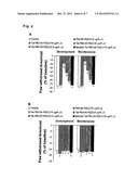 | 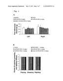 |
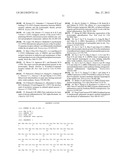 | 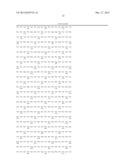 |
 |  |
 |  |
 |  |
 |  |
 |  |
 |  |
| Similar patent applications: | |
| Date | Title |
|---|---|
| 2014-07-17 | Anti-malarial compounds from marine natural products |
| 2014-07-17 | Hydrogel tattoo protection and aftercare system |
| 2014-07-24 | 2'-azido substituted nucleoside derivatives and methods of use thereof for the treatment of viral diseases |
| 2014-07-24 | Methods of use of biomaterial and injectable implant containing biomaterial |
| 2014-07-03 | Mitochondrial ribosomal proteins as aging regulators |
| New patent applications in this class: | |
| Date | Title |
|---|---|
| 2016-05-05 | Conotoxin peptides, pharmaceutical compositions and uses thereof |
| 2016-04-21 | Dynorphin a analogs with bradykinin receptors specificity for modulation of neuropathic pain |
| 2016-01-21 | Biological materials and uses thereof |
| 2015-12-31 | Method for administering omega-conopeptide |
| 2015-10-15 | Novel lantipeptide |
| New patent applications from these inventors: | |
| Date | Title |
|---|---|
| 2021-11-11 | Antibodies to human resistin |
| 2016-05-12 | Antibodies to human resistin |
| 2015-10-01 | Methods for diagnosing pulmonary hypertension |
| 2014-03-20 | Anti-himf antibodies to treat lung diseases |
| 2011-07-21 | Inhibition of interaction of psd93 and psds95 with nnos and nmda receptors |
| Top Inventors for class "Drug, bio-affecting and body treating compositions" | |
| Rank | Inventor's name |
|---|---|
| 1 | Anthony W. Czarnik |
| 2 | Ulrike Wachendorff-Neumann |
| 3 | Ken Chow |
| 4 | John E. Donello |
| 5 | Rajinder Singh |