Patent application title: DETECTION OF UTERINE LEIOMYOSARCOMA USING LMP2
Inventors:
Takuma Hayashi (Nagano, JP)
Yukihiro Kobayashi (Nagano, JP)
Kenji Sano (Nagano, JP)
Akiko Horiuchi (Nagano, JP)
Ikuo Konishi (Nagano, JP)
Assignees:
Shinshu University
IPC8 Class: AC12Q168FI
USPC Class:
435 6
Class name: Chemistry: molecular biology and microbiology measuring or testing process involving enzymes or micro-organisms; composition or test strip therefore; processes of forming such composition or test strip involving nucleic acid
Publication date: 2011-01-27
Patent application number: 20110020792
Claims:
1. A method for detecting uterine leiomyosarcoma using LMP2 as a marker.
2. A method for detecting the presence of uterine leiomyosarcoma using the LMP2 transcription or expression level in uterine smooth muscle tissue as an indicator comprising assaying LMP2 transcription or expression in uterine smooth muscle tissue, and identifying the presence of uterine leiomyosarcoma when the LMP2 transcription or expression level is lower than that in normal uterine smooth muscle tissue.
3. A method for differentiating whether or not a tumor in uterine smooth muscle is uterine leiomyoma or uterine leiomyosarcoma using the LMP2 transcription or expression level in uterine smooth muscle tissue as an indicator comprising assaying LMP2 transcription or expression in uterine smooth muscle tissue, and determining that the tumor is uterine leiomyosarcoma when the LMP2 transcription or expression level is lower than that in normal uterine smooth muscle tissue.
4. A method for determining malignancy of uterine leiomyosarcoma using the LMP2 transcription or expression level in uterine smooth muscle tissue as an indicator comprising assaying LMP2 transcription or expression in uterine smooth muscle tissue, and determining that the tumor is malignant uterine leiomyosarcoma when the LMP2 transcription or expression level is lower than that in normal uterine smooth muscle tissue.
5. The method according to any one of claims 1 to 4, wherein LMP2 transcription in the sampled uterine smooth muscle tissue or cell is assayed via in situ hybridization.
6. The method according to any one of claims 1 to 4, wherein mRNA of LMP2 is extracted from the sampled uterine smooth muscle tissue or cell and subjected to RT-PCR or Northern blotting to assay LMP2 transcription.
7. The method according to any one of claims 1 to 4, wherein the sampled uterine smooth muscle tissue or cell is subjected to immunohistochemical staining or immunocytochemical staining to assay LMP2 expression.
8. The method according to any one of claims 1 to 4, wherein the LMP2 protein is extracted from the sampled uterine smooth muscle tissue or cell and subjected to immunoassay to assay LMP2 expression.
9. The method for detecting the presence of uterine leiomyosarcoma according to claim 1, which further involves the use of myosin as a marker and the use of the LMP2 and myosin transcription or expression levels as an indicator, wherein the presence of uterine leiomyosarcoma is identified when the LMP2 transcription or expression level is lower than that in normal uterine smooth muscle tissue and the myosin transcription or expression level is higher than that in normal uterine smooth muscle tissue.
10. A method for detecting uterine leiomyosarcoma using LMP-2 and cyclin E as markers.
11. A method for detecting the presence of uterine leiomyosarcoma using the degree of transcription or expression of LMP2 and cyclin E in uterine smooth muscle tissue as an indicator comprising assaying transcription or expression of LMP2 and cyclin E in uterine smooth muscle tissue, and identifying the presence of uterine leiomyosarcoma when the LMP2 transcription or expression level is lower than that in normal uterine smooth muscle tissue and the cyclin E transcription or expression level is higher than that in normal uterine smooth muscle tissue.
12. A method for differentiating whether or not a tumor in uterine smooth muscle is uterine leiomyoma or uterine leiomyosarcoma using the degree of transcription or expression of LMP2 and cyclin E in uterine smooth muscle tissue as an indicator comprising assaying transcription or expression of LMP2 and cyclin E in uterine smooth muscle tissue, and determining that the tumor is uterine leiomyosarcoma when the LMP2 transcription or expression level is lower than that in normal uterine smooth muscle tissue and the cyclin E transcription or expression level is higher than that in normal uterine smooth muscle tissue.
13. A method for determining malignancy of uterine leiomyosarcoma using the degree of transcription or expression of LMP2 and cyclin E in uterine smooth muscle tissue as an indicator comprising assaying transcription or expression of LMP2 and cyclin E in uterine smooth muscle tissue, and determining that the tumor is malignant uterine leiomyosarcoma when the LMP2 transcription or expression level is lower than that in normal uterine smooth muscle tissue and the cyclin E transcription or expression level is higher than that in normal uterine smooth muscle tissue.
14. The method according to any one of claims 10 to 13, wherein the sampled uterine smooth muscle tissue or cell is subjected to in situ hybridization to assay transcription of LMP2 and cyclin E.
15. The method according to any one of claims 10 to 13, wherein mRNAs of LMP2 and cyclin E are extracted from the sampled uterine smooth muscle tissue or cell and subjected to RT-PCR or Northern blotting to assay transcription of LMP2 and cyclin E.
16. The method according to any one of claims 10 to 13, wherein the sampled uterine smooth muscle tissue or cell is subjected to immunohistochemical staining or immunocytochemical staining to assay expression of LMP2 and cyclin E.
17. The method according to any one of claims 10 to 13, wherein LMP2 and cyclin E proteins are extracted from the sampled uterine smooth muscle tissue or cell and subjected to immunoassay to assay expression of LMP2 and cyclin E.
18. A detection reagent for detecting uterine leiomyosarcoma using LMP2 as a marker, which comprises at least an LMP2 gene fragment as a probe or primer.
19. A detection reagent for detecting uterine leiomyosarcoma using LMP2 and cyclin E as markers, which comprises at least an LMP2 gene fragment and a cyclin E gene fragment as probes or primers.
20. The detection reagent for detecting uterine leiomyosarcoma according to claim 18 or 19, which is used for in situ hybridization.
21. The detection reagent for detecting uterine leiomyosarcoma using LMP2 and myosin as markers according to claim 18 or 20, which further comprises a myosin gene fragment as a probe or primer.
22. The detection reagent for detecting uterine leiomyosarcoma using LMP2, cyclin E, and myosin as markers according to claim 19 or 20, which further comprises a myosin gene fragment as a probe or primer.
23. A detection reagent for detecting uterine leiomyosarcoma using LMP2 as a marker, which comprises at least an anti-LMP2 antibody.
24. A detection reagent for detecting uterine leiomyosarcoma using LMP2 and cyclin E as a markers, which comprises at least an anti-LMP2 antibody and an anti-cyclin E antibody.
25. The detection reagent for detecting uterine leiomyosarcoma according to claim 23 or 24, which is used for immunohistochemical or immunocytochemical staining.
26. The detection reagent for detecting uterine leiomyosarcoma using LMP2 and myosin as markers according to claim 23 or 25, which further comprises an anti-myosin antibody.
27. The detection reagent for detecting uterine leiomyosarcoma using LMP2, cyclin E, and myosin as markers according to claim 24 or 25, which further comprises an anti-myosin antibody.
28. An oligonucleotide comprising a partial sequence selected from the group consisting of a partial sequence of the JAK1 kinase gene comprising at least one of the mutation sites (A1) to (A6) below of the JAK1 kinase gene, and a partial sequence of the LMP2 promoter comprising at least one of the mutation sites (B1) to (B5) below of the LMP2 promoter or the oligonucleotide which is labeled, the oligonucleotide or labeled oligonucleotide comprising a 10-bp to 30-bp partial sequence or a sequence complementary thereto:(A1) A2612A;(A2) G2626A;(A3) G2642T;(A4) A2967C;(A5) A2960C;(A6) A2985T;(B1) A210G;(B2) C214T;(B3) A216G;(B4) A217G; and(B5) G219A.
29. The oligonucleotide or the oligonucleotide which is labeled according to claim 28, which is used as a probe.
30. A substrate comprising the oligonucleotide or the oligonucleotide which is labeled according to claim 28 immobilized thereon.
31. At least a pair of primer sets used for amplification of a DNA fragment comprising a mutation site selected from the group consisting of at least one of the mutation sites (A1) to (A6) below of the JAK1 kinase gene and at least one of the mutation sites (B1) to (B5) below of the LMP2 promoter, the pair of primer sets being used for amplification of a DNA fragment comprising 10- to 30-bp partial sequences located at sites closer to the 3' end and the 5' end of the mutation site:(A1) A2612A;(A2) G2626A;(A3) G2642T ;(A4) A2967C;(A5) A2960C;(A6) A2985T;(B1) A210G;(B2) C214T;(B3) A216G;(B4) A217G; and(B5) G219A.
32. A kit for detecting uterine leiomyosarcoma comprising the primer sets according to claim 31, the oligonucleotide or the oligonucleotide which is labeled according to claim 28 or 29, and the substrate according to claim 30.
33. A method for detecting uterine leiomyosarcoma comprising: sampling uterine smooth muscle tissue or cell from an animal; detecting a mutation site selected from the group consisting of at least one of the mutation sites (A1) to (A6) below of the JAK1 kinase gene and at least one of the mutation sites (B1) to (B5) below of the LMP2 promoter from the sampled tissue or cell; and, when such mutation is present, determining that the animal is afflicted with or highly susceptible to uterine leiomyosarcoma based on the detection results:(A1) A2612A;(A2) G2626A;(A3) G2642T;(A4) A2967C;(A5) A2960C;(A6) A2985T;(B1) A210G;(B2) C214T;(B3) A216G;(B4) A217G; and(B5) G219A.
34. The method according to claim 33, wherein the mutation in the JAK1 kinase gene or the LMP2 primer is detected using the primer sets according to claim 31, the probe according to claim 28 or 29, or the substrate according to claim 30.
Description:
TECHNICAL FIELD
[0001]The present invention relates to a method for detecting uterine leiomyosarcoma using mutation of LMP2, cyclin E, and the JAK1 kinase gene, the STAT1 gene, and the LMP2 promoter associated with the interferon γ (IFN-γ) signal transduction cascade as markers and a method for differentiation of uterine leiomyoma from uterine leiomyosarcoma.
BACKGROUND ART
[0002]Uterine cancer is the most common type of gynecologic cancer. The frequency of uterine leiomyosarcoma development is low, and it accounts 2% to 5% of cases of uterine body cancer. Uterine leiomyosarcoma develops more often in the muscle layer of the uterine body than in the uterine cervix. The myometrium is composed of smooth muscle. Uterine leiomyoma is a benign tumor that develops in the myometrium, and uterine leiomyosarcoma is a malignant tumor. Differentiation of uterine leiomyoma from uterine leiomyosarcoma was very difficult. In general, tissue has been sampled via surgery, and whether or not a tumor was uterine leiomyoma or uterine leiomyosarcoma has been identified via microscopic cell analysis. Uterine leiomyosarcoma is highly atypical and often allows proliferation of tumor cells that occasionally become gigantic. In some cases, such tumors do not substantially show cellular atypism, and the presence or absence of cellular atypism would not serve as a definite discriminant. Uterine leiomyoma is differentiated from uterine leiomyosarcoma based on the occurrence of coagulative necrosis and the enlargement of an image representing cell division. When a cell density is high, in principle, a tumor is identified to be uterine leiomyosarcoma if 10 or more cell divisions are observed in a 10× wide-field view. The tumor is identified to be uterine leiomyosarcoma if 5 or more atypisms are observed in the tumor cell in a 10× wide-field view. In practice, uterine leiomyoma has been differentiated from uterine leiomyosarcoma based on the degree of cellular atypism, cell density, the number of cell divisions, tumor necrosis, and the presence or absence of tumor. Such differentiation was mainly made by observing tissue morphology microscopically or visually. However, expert skills are required for such differentiation, and such differentiation is not always accurate.
[0003]In the past, the present inventors reported that uterine leiomyosarcoma was observed in 6-month or older female mice each lacking an immunoprotease component; i.e., low molecular mass polypeptide 2 (LMP2), and that the incidence thereof in 12-month-olds would account for about 35% of all LMP2-lacking female mice (see Van Kaer L. et al., 1994, Immunity, 1, 533-541 and Hayashi T. et al., 2002, Cancer Res., 62, 24-27). LMP2 functions in a tissue-specific manner and plays an essential role in MHC class I-mediated tumor rejection by CTLs (see Van Kaer L. et al., 1994, Immunity, 1, 533-541).
[0004]Thus, lack of LMP2 was deduced to serve as a factor for developing uterine leiomyosarcoma by means of certain functions. The 26S proteasome comprising LMP2, however, is involved in a complex manner with activation of a transcription regulator or a cell-cycle regulator, production of a peptide antigen of an MHC class I molecule, and the like, and a direct correlation between LMP2 and development of uterine leiomyosarcoma was unknown. The way that functions of the 26S proteasome would change and the way that transcription and expression of LMP2 would change upon development of uterine leiomyosarcoma were unknown.
DISCLOSURE OF THE INVENTION
[0005]The present invention provides a method for detecting the presence of uterine leiomyosarcoma using the LMP2 transcription or expression level in uterine smooth muscle tissue as an indicator and a detection reagent used therefor.
[0006]The present inventors stained living mice with anti-LMP2 in order to inspect LMP2 expression in the muscle layer. As a result, they discovered that LMP2 expression specific to myogenic tissue, such as smooth muscle, stripped muscle, or cardiac muscle, was observed and that the origin of the tumor cells observed in uterine smooth muscle layer of LMP2-lacking mice was thus a myogenic cell (i.e., the smooth muscle cell).
[0007]Further, the present inventors inspected the conditions of LMP2 expression in biopsy tissue or surgically-removed tissue of the normal uterine smooth muscle layer, human uterine leiomyoma, and human uterine leiomyosarcoma. As a result, they discovered that LMP2 expression levels were significantly lowered only in the case of a malignant tumor, i.e., uterine leiomyosarcoma.
[0008]Subsequently, the present inventors induced forced expression of LMP2 via gene recombination in the uterine leiomyosarcoma (SKN) cells in which no LMP2 expression was observed and examined the morphology of SKN cells, cell proliferative rate thereof, and the changes of fibronectin expression in SKN cells, for the purpose of examining whether or not lowered LMP2 expression levels were directly involved with genetic transformation (canceration) in uterine smooth muscle cells. As a result, they discovered that the configuration and cell proliferative rate of SKN cells became similar to those of normal uterine smooth muscle cells and that fibronectin expression was significantly induced.
[0009]Based on such new findings, the present inventors discovered that differentiation of uterine leiomyoma from uterine leiomyosarcoma would be possible with the use of LMP2 transcription or expression as an indicator and that uterine leiomyosarcoma could be treated via LMP2 expression.
[0010]Further, the present inventors focused on the correlation between cyclin E expression and uterine leiomyosarcoma, and they discovered that transcription and expression of cyclin E would be significantly elevated in uterine smooth muscle tissue depending on the malignancy of uterine leiomyosarcoma.
[0011]The present inventors also examined the correlation of a gene associated with the interferon γ (IFN-γ) signal transmission system involved in LMP2 expression and uterine leiomyosarcoma. As a result, they discovered that mutation had occurred in the signal transmission factor, i.e., JAK1 kinase, the STAT1-encoding gene, and the LMP2 promoter, in uterine leiomyosarcoma tissue cell, and they then discovered that detection of such mutation would enable determination of whether or not a subject is afflicted with uterine leiomyosarcoma and is at high risk of affliction with uterine leiomyosarcoma.
[0012]The present inventors have completed the present invention in such a manner.
[0013]Specifically, the present invention is as follows.
[0014][1] A method for detecting uterine leiomyosarcoma using LMP2 as a marker.
[0015][2] A method for detecting the presence of uterine leiomyosarcoma using the LMP2 transcription or expression level in uterine smooth muscle tissue as an indicator comprising assaying LMP2 transcription or expression in uterine smooth muscle tissue, and identifying the presence of uterine leiomyo sarcoma when the LMP2 transcription or expression level is lower than that in normal uterine smooth muscle tissue.
[0016][3] A method for differentiating whether or not a tumor in uterine smooth muscle is uterine leiomyoma or uterine leiomyosarcoma using the LMP2 transcription or expression level in uterine smooth muscle tissue as an indicator comprising assaying LMP2 transcription or expression in uterine smooth muscle tissue, and determining that the tumor is uterine leiomyosarcoma when the LMP2 transcription or expression level is lower than that in normal uterine smooth muscle tissue.
[0017][4] A method for determining malignancy of uterine leiomyosarcoma using the LMP2 transcription or expression level in uterine smooth muscle tissue as an indicator comprising assaying LMP2 transcription or expression in uterine smooth muscle tissue, and determining that the tumor is malignant uterine leiomyosarcoma when the LMP2 transcription or expression level is lower than that in normal uterine smooth muscle tissue.
[0018][5] The method according to any of [1] to [4], wherein LMP2 transcription in the sampled uterine smooth muscle tissue or cell is assayed via in situ hybridization.
[0019][6] The method according to any of [1] to [4], wherein mRNA of LMP2 is extracted from the sampled uterine smooth muscle tissue or cell and subjected to RT-PCR or Northern blotting to assay LMP2 transcription.
[0020][7] The method according to any of [1] to [4], wherein the sampled uterine smooth muscle tissue or cell is subjected to immunohistochemical staining or immunocytochemical staining to assay LMP2 expression.
[0021][8] The method according to any of [1] to [4], wherein the LMP2 protein is extracted from the sampled uterine smooth muscle tissue or cell and subjected to immunoassay to assay LMP2 expression.
[0022][9] The method for detecting the presence of uterine leiomyosarcoma according to [1], which further involves the use of myosin as a marker and the use of the LMP2 and myosin transcription or expression levels as an indicator, wherein the presence of uterine leiomyosarcoma is identified when the LMP2 transcription or expression level is lower than that in normal uterine smooth muscle tissue and the myosin transcription or expression level is higher than that in normal uterine smooth muscle tissue.
[0023][10] A method for detecting uterine leiomyosarcoma using LMP-2 and cyclin E as markers.
[0024][11] A method for detecting the presence of uterine leiomyosarcoma using the degree of transcription or expression of LMP2 and cyclin E in uterine smooth muscle tissue as an indicator comprising assaying transcription or expression of LMP2 and cyclin E in uterine smooth muscle tissue, and identifying the presence of uterine leiomyosarcoma when the LMP2 transcription or expression level is lower than that in normal uterine smooth muscle tissue and the cyclin E transcription or expression level is higher than that in normal uterine smooth muscle tissue.
[0025][12] A method for differentiating whether or not a tumor in uterine smooth muscle is uterine leiomyoma or uterine leiomyosarcoma using the degree of transcription or expression of LMP2 and cyclin E in uterine smooth muscle tissue as an indicator comprising assaying transcription or expression of LMP2 and cyclin E in uterine smooth muscle tissue, and determining that the tumor is uterine leiomyosarcoma when the LMP2 transcription or expression level is lower than that in normal uterine smooth muscle tissue and the cyclin E transcription or expression level is higher than that in normal uterine smooth muscle tissue.
[0026][13] A method for determining malignancy of uterine leiomyosarcoma using the degree of transcription or expression of LMP2 and cyclin E in uterine smooth muscle tissue as an indicator comprising assaying transcription or expression of LMP2 and cyclin E in uterine smooth muscle tissue, and determining that the tumor is malignant uterine leiomyosarcoma when the LMP2 transcription or expression level is lower than that in normal uterine smooth muscle tissue and the cyclin E transcription or expression level is higher than that in normal uterine smooth muscle tissue.
[0027][14] The method according to any of [10] to [13], wherein the sampled uterine smooth muscle tissue or cell is subjected to in situ hybridization to assay transcription of LMP2 and cyclin E.
[0028][15] The method according to any of [10] to [13], wherein mRNAs of LMP2 and cyclin E are extracted from the sampled uterine smooth muscle tissue or cell and subjected to RT-PCR or Northern blotting to assay transcription of LMP2 and cyclin E.
[0029][16] The method according to any of [10] to [13], wherein the sampled uterine smooth muscle tissue or cell is subjected to immunohistochemical staining or immunocytochemical staining to assay expression of LMP2 and cyclin E.
[0030][17] The method according to any of [10] to [13], wherein LMP2 and cyclin E proteins are extracted from the sampled uterine smooth muscle tissue or cell and subjected to immunoassay to assay expression of LMP2 and cyclin E.
[0031][18] A detection reagent for detecting uterine leiomyosarcoma using LMP2 as a marker, which comprises at least an LMP2 gene fragment as a probe or primer.
[0032][19] A detection reagent for detecting uterine leiomyosarcoma using LMP2 and cyclin E as markers, which comprises at least an LMP2 gene fragment and a cyclin E gene fragment as probes or primers.
[0033][20] The detection reagent for detecting uterine leiomyosarcoma according to [18] or [19], which is used for in situ hybridization.
[0034][21] The detection reagent for detecting uterine leiomyosarcoma using LMP2 and myosin as markers according to [18] or [20], which further comprises a myosin gene fragment as a probe or primer.
[0035][22] The detection reagent for detecting uterine leiomyosarcoma using LMP2, cyclin E, and myosin as markers according to [19] or [20], which further comprises a myosin gene fragment as a probe or primer.
[0036][23] A detection reagent for detecting uterine leiomyosarcoma using LMP2 as a marker, which comprises at least an anti-LMP2 antibody.
[0037][24] A detection reagent for detecting uterine leiomyosarcoma using LMP2 and cyclin E as a markers, which comprises at least an anti-LMP2 antibody and an anti-cyclin E antibody.
[0038][25] The detection reagent for detecting uterine leiomyosarcoma according to [23] or [24], which is used for immunohistochemical or immunocytochemical staining.
[0039][26] The detection reagent for detecting uterine leiomyosarcoma using LMP2 and myosin as markers according to [23] or [25], which further comprises an anti-myosin antibody.
[0040][27] The detection reagent for detecting uterine leiomyosarcoma using LMP2, cyclin E, and myosin as markers according to [24] or [25], which further comprises an anti-myosin antibody.
[0041][28] An oligonucleotide comprising a partial sequence selected from the group consisting of a partial sequence of the JAK1 kinase gene comprising at least one of the mutation sites (A1) to (A6) below of the JAK1 kinase gene, and a partial sequence of the LMP2 promoter comprising at least one of the mutation sites (B1) to (B5) below of the LMP2 promoter or he oligonucleotide which is labeled, the oligonucleotide or labeled oligonucleotide comprising a 10-bp to 30-bp partial sequence or a sequence complementary thereto:
[0042](A1) A2612A;
[0043](A2) G2626A;
[0044](A3) G2642T;
[0045](A4) A2967C;
[0046](A5) A2960C;
[0047](A6) A2985T;
[0048](B1) A210G;
[0049](B2) C214T;
[0050](B3) A216G;
[0051](B4) A217G; and
[0052](B5) G219A.
[0053][29] The oligonucleotide or he oligonucleotide which is labeled according to [28], which is used as a probe.
[0054][30] A substrate comprising the oligonucleotide or he oligonucleotide which is labeled according to [28] immobilized thereon.
[0055][31] At least a pair of primer sets used for amplification of a DNA fragment comprising a mutation site selected from the group consisting of at least one of the mutation sites (A1) to (A6) below of the JAK1 kinase gene and at least one of the mutation sites (B1) to (B5) below of the LMP2 promoter, the pair of primer sets being used for amplification of a DNA fragment comprising 10- to 30-bp partial sequences located at sites closer to the 3' end and the 5' end of the mutation site:
[0056](A1) A2612A;
[0057](A2) G2626A;
[0058](A3) G2642T ;
[0059](A4) A2967C;
[0060](A5) A2960C;
[0061](A6) A2985T;
[0062](B1) A210G;
[0063](B2) C214T;
[0064](B3) A216G;
[0065](B4) A217G; and
[0066](B5) G219A.
[0067][32] A kit for detecting uterine leiomyosarcoma comprising the primer sets according to [31], the oligonucleotide or he oligonucleotide which is labeled according to [28] or [29], and the substrate according to [30].
[0068][33] A method for detecting uterine leiomyosarcoma comprising: sampling uterine smooth muscle tissue or cell from an animal; detecting a mutation site selected from the group consisting of at least one of the mutation sites (A1) to (A6) below of the JAK1 kinase gene and at least one of the mutation sites (B1) to (B5) below of the LMP2 promoter from the sampled tissue or cell; and, when such mutation is present, determining that the animal is afflicted with or highly susceptible to uterine leiomyosarcoma based on the detection results:
[0069](A1) A2612A;
[0070](A2) G2626A;
[0071](A3) G2642T;
[0072](A4) A2967C;
[0073](A5) A2960C;
[0074](A6) A2985T;
[0075](B1) A210G;
[0076](B2) C214T;
[0077](B3) A216G;
[0078](B4) A217G; and
[0079](B5) G219A.
[0080][34] The method according to [33], wherein the mutation in the JAK1 kinase gene or the LMP2 primer is detected using the primer sets according to [31], the probe according to [28] or [29], or the substrate according to [30].
[0081]This description includes part or all of the contents as disclosed in the description and/or drawings of Japanese Patent Application No. 2005-347227, which is a priority document of the present application.
BRIEF DESCRIPTION OF THE DRAWINGS
[0082]FIG. 1a is a photograph showing a lack of IFN-γ-induced TAP1 and LMP2 expression in SKN cells. A cytoplasm extract was prepared from the HeLa, HeLa.S3, SKN, and normal human uterine smooth muscle cells (Hu.USMC) processed with 250 units/ml of IFN-γ for the period of time as shown in the photograph, and 50 μg of the cytoplasm extract was separated via 10% SDS-PAGE. The TAP1, LMP2, and β-actin expression levels were assayed via immunoblot assay using an adequate antibody.
[0083]FIG. 1b is a photograph showing a lack of IFN-γ-induced TAP1 and LMP2 expression in SKN cells and showing the results of mRNA expression assay of TAP1, LMP2, and β-actin in HeLa, HeLa.S3, SKN, and Hu.USMC cells via RT-PCR. After the cells were cultured in the presence or absence of IFN-γ (250 units/ml) for 48 hours, RT-PCR was carried out using primers. The DNA product amplified via RT-PCR was separated on agarose gel. A DNA size marker is shown on the left side of the photograph.
[0084]FIG. 1c is a photograph showing a lack of LMP2 expression in uterine leiomyosarcoma, and showing the results of immunohistochemical assay of LMP2 in normal uterine smooth muscle and uterine leiomyoma. A 5-μm slice of the tissue sample was stained with the anti-LMP2 antibody and the peroxidase-conjugated anti-rabbit IgG antibody.
[0085]FIG. 2 shows differential activity of wt and IRF-E mt promoters shared by IFN-γ-induced TAP1 and LMP2 in the HeLa and SKN cells. This figure shows wt promoters shared by TAP1 and LMP2 (TAP1 593-1/pGL3 and LMP2 1-593/pLG3) and luciferase reporter gene constructs each comprising IRF-E mutant promoters. The reporter genes were introduced into the HeLa and SKN cells, IFN-γ was added 24 hours later, and the cells were incubated for 24 hours prior to recovery thereof. For the purpose of standardization of efficiency of reporter gene introduction, the reporter genes were introduced simultaneously with pSMV-βGAL. The results were standardized with the expression of the luciferase gene assayed separately for the HeLa cells and the SKN cells, and the results are shown as relative TAP1 and LMP2 activities. The average of the results attained from the three independent experiments is shown, and the error bar indicates SE.
[0086]FIG. 3 is a microscopic photograph of normal human uterine smooth muscle tissue.
[0087]FIG. 4 is a microscopic photograph of human uterine leiomyoma and human uterine leiomyosarcoma.
[0088]FIG. 5 is a microscopic photograph of human endometrial stromal sarcoma.
[0089]FIG. 6 is a photograph showing the results of immunohistochemical staining of normal uterine smooth muscle, uterine leiomyoma, and uterine leiomyosarcoma.
[0090]FIG. 7 is a photograph showing the results of immunohistochemical staining of a uterine leiomyosarcoma site and a normal uterine smooth muscle site in the same tissue. Lowering in LMP2 expression is observed at the uterine leiomyosarcoma site.
[0091]FIG. 8 is a photograph showing changes in the configuration of uterine leiomyosarcoma (SKN) cells upon forced expression of LMP2 via gene recombination in SKN cells in which no LMP2 expression is observed (Part 1).
[0092]FIG. 9 is a photograph showing changes in the configuration of uterine leiomyosarcoma (SKN) cells upon forced expression of LMP2 via gene recombination in SKN cells in which no LMP2 expression is observed (Part 2).
[0093]FIG. 10 is a photograph showing changes in fibronectin expression in uterine leiomyosarcoma (SKN) cells upon forced expression of LMP2 via gene recombination in SKN cells in which no LMP2 expression is observed.
[0094]FIG. 11 is a photograph showing changes in fibronectin expression in normal human uterine smooth muscle cells (HuUSMC).
[0095]FIG. 12 shows a summary of changes in morphology, cell proliferative rate, and fibronectin expression in each cell.
[0096]FIG. 13 is a photograph showing LMP2 expression in murine skeletal muscle tissue, cardiac tissue, and smooth muscle tissue.
[0097]FIG. 14 shows LMP2 expression in uterine leiomyosarcoma, uterine leiomyoma, and leiomyosarcoma that had developed in other organs.
[0098]FIG. 15 shows the correlation between the INF-γ signal transmission pathway and LMP2 expression.
[0099]FIG. 16 shows mutations in a JAK1 kinase gene, a STAT1 gene, and a LMP2 promoter derived from uterine leiomyosarcoma tissue.
[0100]FIG. 17 shows mutations in a JAK1 kinase gene, a STAT1 gene, and a LMP2 promoter derived from uterine leiomyosarcoma tissue.
[0101]FIG. 18 shows the conditions of LMP2 expression in each tissue. In FIG. 18, "N" represents the number of tissue samples that were subjected to LMP2 expression assay.
[0102]FIG. 19 shows the number of colonies formed by SKN cell proliferation upon introduction of the pCEM9 vector (containing no LMP2 gene) or the pCEM9-LMP2 vector (containing LMP2 gene) and neomycin-selected vectors into cultured human uterine leiomyosarcoma cells (SKN cells). pCEM9 and pLMP2 DNA (1 μg each) were transfected into 2×10⁵ DT or SKN cells, and the cells were selected in a growth medium containing 0.5 or 0.4 mg/ml G418.
[0103]FIG. 20 is a photograph showing changes in morphology of uterine leiomyosarcoma (SKN) cells upon forced expression of LMP2 via gene recombination in SKN cells in which no LMP2 expression is observed.
[0104]FIG. 21 shows a summary of changes in morphology, cell proliferative rate, and fibronectin expression in each cell.
[0105]FIG. 22 is a photograph showing that SKN cells in which LMP2 is constitutively expressed significantly decrease colony formation, which is an indicator of capacity for tumorigenesis.
[0106]FIG. 23A is a photograph showing that SKN cells in which LMP2 is constitutively expressed significantly decrease capacity for tumorigenesis via a transplant experiment involving nude mice.
[0107]FIG. 23B is a photograph showing that SKN cells in which LMP2 is constitutively expressed significantly decrease capacity for tumorigenesis via a transplant experiment involving nude mice.
[0108]FIG. 23c is a photograph showing that the expression level of the LMP2 gene in colony #121 in cultured human uterine leiomyosarcoma cells (SKN cells) into which the pCEM9-LMP2 vector (containing the LMP2 gene) has been introduced is more significant than that in other SKN cells.
[0109]FIG. 24 shows that the expression level of cyclin E, which induces cell proliferation, is significantly lowered in SKN cells (colony #121) in which LMP2 is constitutively expressed as a result of microarray-based gene expression analysis.
[0110]FIG. 25 shows the structure of a luciferase reporter gene comprising a cyclin E promoter.
[0111]FIG. 26 shows cyclin E promoter activity in SKN cells in which SKN cells and LMP2 are constitutively expressed (colony #121 and #122). The expression level of cyclin E observed to be significant in the cultured uterine leiomyosarcoma SKN cells was significantly lowered via constitutive expression of LMP2.
[0112]FIG. 27 is a photograph showing the results of tissue staining that demonstrate significant cyclin E expression in human uterine leiomyosarcoma tissue. Cyclin E expression is not observed in normal uterine smooth muscle layer; however, significant cyclin E expression is observed in malignant tumor tissue (human uterine leiomyosarcoma tissue).
[0113]FIG. 28 is a photograph showing the results of tissue staining that demonstrate cyclin E expression in the nucleus during the mitotic period in human uterine leiomyosarcoma tissue. In general, cyclin E, induces cell proliferation, is overexpressed in the cytoplasm at the G1 stage, which is the initiation period of cell proliferation, migrates immediately into the nucleus, and initiates the synthesis of chromosomes at the S stage. Thereafter, degradation of cyclin E starts immediately during the later half of the S stage. Accordingly, cyclin E expression is not observed at the G2 and M stages of normal cells; however, significant cyclin E expression is observed in the nucleus during the mitotic period in human uterine leiomyosarcoma tissue.
BEST MODES FOR CARRYING OUT THE INVENTION
[0114]The present invention is intended to detect uterine leiomyosarcoma using LMP2 as a marker. The term "detect (or detection)" refers to, for example, determination of the presence of uterine leiomyosarcoma, determination of whether or not a patient is afflicted with uterine leiomyosarcoma, differentiation of whether or not a tumor in uterine smooth muscle is uterine leiomyoma or uterine leiomyosarcoma, and determination of malignancy of uterine leiomyosarcoma.
[0115]LMP2 is a proteasome subunit, and the method of the present invention is intended to detect whether or not LMP2 is transcribed or expressed in uterine smooth muscle layer. Inspection of LMP2 transcription or expression in the tissue of the human uterine smooth muscle layer reveals that LMP2 expression is positive in normal uterine smooth muscle layer and uterine leiomyoma (i.e., mild to potent expression levels). In contrast, LMP2 expression is negative in a major part of uterine leiomyosarcoma and weak expression is observed in some parts thereof. Accordingly, whether or not a patient is afflicted with uterine leiomyosarcoma or whether a tumor in uterine smooth muscle is uterine leiomyoma or uterine leiomyosarcoma can be evaluated using LMP2 transcription or expression as an indicator. In the case of a benign tumor (uterine leiomyoma), the level of LMP2 transcription or expression is strong. In the case of a malignant tumor (uterine leiomyosarcoma), however, the level of LMP2 transcription or expression is significantly weakened. This indicates that the level of LMP2 transcription or expression can serve as an indicator of malignancy of tumors observed in uterine smooth muscle cells.
[0116]Further, the present invention includes a method for detecting uterine leiomyosarcoma using cyclin E as a marker. Cyclin is a protein that plays an important role in the cell cycle that regulates activity of cyclin-dependent kinase. Cyclin E is a G1 cyclin that acts at the G1 stage in a mammalian cell. The expression level of cyclin E is more significantly elevated in uterine leiomyosarcoma tissue than in normal tissue, but it is not elevated in uterine leiomyoma tissue. In the present invention, whether or not cyclin E is transcribed or expressed in uterine smooth muscle layer is detected as in the case of LMP2. With the use of cyclin E transcription or expression as an indicator, whether or not a patient is afflicted with uterine leiomyosarcoma or whether a tumor in uterine smooth muscle is uterine leiomyoma or uterine leiomyosarcoma can be evaluated. In the case of a benign tumor (uterine leiomyoma), the level of cyclin E transcription or expression is not different from that in normal uterine smooth muscle tissue; however, the level of cyclin E transcription or expression is elevated in the case of a malignant tumor (i.e., uterine leiomyosarcoma). In general, cyclin E expression is not observed at the G2 stage or the M stage in normal cells during the proliferation period. In human uterine leiomyosarcoma tissue, however, significant cyclin E expression is observed in the nucleus during the mitotic period. This indicates that cyclin E transcription or expression can serve as an indicator of malignancy of tumors observed in uterine smooth muscle cells.
[0117]Alternatively, LMP2 and cyclin E may be detected simultaneously. When the level of LMP2 transcription or expression is lowered and the level of cyclin E transcription or expression is elevated, a patient may be determined as being afflicted with uterine leiomyosarcoma. When both LMP-2 and cyclin E are used as indicators, uterine leiomyosarcoma can be detected more accurately than when LMP2 or cyclin E is used alone.
[0118]In the present invention, determination of whether or not a patient is afflicted with uterine leiomyosarcoma, differentiation of whether a tumor in uterine smooth muscle is uterine leiomyoma or uterine leiomyosarcoma, and evaluation of malignancy of tumors observed in uterine smooth muscle cells are referred to as detection of uterine leiomyosarcoma.
[0119]According to the method of the present invention, uterine leiomyosarcoma can be detected by detecting either or both transcription or expression of LMP2 and/or cyclin E in uterine smooth muscle layer. LMP2 or cyclin E transcription can be detected by measuring mRNA encoding LMP2 or cyclin E. LMP2 or cyclin E expression can be detected by measuring an LMP2 or cyclin E protein.
[0120]When LMP2 and/or cyclin E transcription is to be detected, a tissue or cell in uterine smooth muscle layer is collected as a biological sample, and mRNA encoding LMP2 and/or cyclin E contained in such sample may be assayed. In order to assay mRNA, some tissue is obtained from uterine smooth muscle tissue via biopsy or with the use of cotton swabs or the like. The tissue may be obtained, for example, at the time of a usual outpatient visit, by inserting biopsy forceps into the uterine lumen and obtaining an about 1- to 2-mm-square uterine tissue slice. mRNA may be assayed by extracting mRNA from the sampled tissue or cell or by preparing a tissue slice sample. Alternatively, the sampled cells may be immobilized on a glass slide, subjected to in situ hybridization, and then stained. Also, extracted mRNA may be assayed via conventional RNA assay techniques, such as Northern blotting or RT-PCR. mRNA can be extracted by a conventional technique, in accordance with, for example, "Lectures on Biochemical Experiments, 2, Nucleic Acids I, Separation and Purification," Tokyo Kagaku Dojin, Co., Ltd., Jul. 10, 1991 or Molecular Biology Experimental Protocol I, Maruzen Co., Ltd., Jun. 30, 1997. in situ hybridization can be carried out in accordance with, for example, Molecular Biology Experimental Protocol III, Maruzen Co., Ltd., Aug. 30, 1997. In such a case, a probe or primer comprising a partial sequence complementary to a partial sequence of mRNA encoding LMP2 and/or cyclin E is used, in order to specifically assay mRNA encoding LMP2 and/or cyclin E. The nucleotide sequence of LMP2 is known (e.g., GenBank Accession No.: U01025, SEQ ID NO: 1; SEQ ID NO: 2 represents an amino acid sequence of the LMP2 protein), and a probe or primer can be designed in accordance with known nucleotide sequence information. The nucleotide sequence of cyclin E is also known (e.g., GenBank Accession No.: M73812, SEQ ID NO: 8). Such primer or probe is a fragment of the above LMP2 and/or cyclin E gene comprising 5 to 50, preferably 10 to 30, and more preferably 15 to 25 nucleotides.
[0121]When LMP2 and/or cyclin E expression is to be detected, a tissue or cell in uterine smooth muscle layer is obtained as a biological sample, and the LMP2 and/or cyclin E protein contained in the sample may be assayed. The tissue or cell in uterine smooth muscle layer may be obtained in the above-described manner. The LMP2 and/or cyclin E protein may be assayed by extracting a protein from the tissue or cell and assaying the LMP2 and/or cyclin E protein in the extract. Protein assay may be conducted via an immunohistochemical or immunocytochemical means. The extracted protein may be assayed via conventional immunoassay techniques, such as ELISA or radioimmunoassay. In such a case, an antibody reacting with LMP2 and/or cyclin E is necessary. An anti-LMP2 antibody and/or an anti-cyclin E antibody may be prepared via a conventional techniques as a monoclonal or polyclonal antibody. Commercially available anti-LMP2 antibody and/or anti-cyclin E antibody can also be used. An antibody may be labeled with an enzyme, fluorescent substance, or radioisotope via a conventional technique, according to need. Immunohistochemical or immunocytochemical assay may be carried out by preparing a sample of the obtained uterine smooth muscle tissue slice or immobilizing the obtained cell on a glass slide. When immunohistochemical assay is performed, for example, the tissue is immobilized with formalin, embedded in paraffin, and sliced to a thickness of about 1 to 5 μm using a slicer, such as a microtome, to prepare a slice sample. At the time of assay, paraffin may be removed via xylene or ethanol treatment, and the sample may be soaked in physiological saline or buffer for hydrophilization. Staining may be carried out with the use of an anti-LMP2 antibody and/or anti-cyclin E antibody labeled with an enzyme, fluorescent substance, radioisotope, or the like. Alternatively, an anti-LMP2 antibody may be conjugated to LMP2 and/or cyclin E in the slice sample, and a secondary antibody that is conjugated to the anti-LMP2 antibody and/or anti-cyclin E antibody, which is labeled with an enzyme, fluorescent substance, or the like, may be used. Examples of enzymes used for labeling include horseradish peroxidase and alkaline phosphatase. Examples of fluorescent substances include fluorescein and rhodamine. Staining may also be carried out using a known biotin-avidin complex. Immunocytochemical assay may be carried out by immobilizing the obtained cell on a glass slide with the aid of formalin and visualizing LMP2 and/or cyclin E in the cell in the same manner as in the case of immunohistochemical assay. In immunohistochemical or immunocytochemical assay, the results of staining may be evaluated microscopically or visually. An adequate optical apparatus may also be used. Immunohistochemical staining may be carried out in accordance with, for example, Molecular Biology Experimental Protocol III, Maruzen Co., Ltd., Aug. 30, 1997.
[0122]When the LMP2 transcription or expression level is found to be lost or lowered upon detection of LMP2 transcription or expression in uterine smooth muscle tissue or cell as described above, a patient can be determined as being afflicted with uterine leiomyosarcoma. When the cyclin E transcription or expression level is found to be elevated upon detection of cyclin E transcription or expression in uterine smooth muscle tissue or cell, a patient can be determined as being afflicted with uterine leiomyosarcoma. Also, transcription or expression levels of both LMP2 and cyclin E may be detected simultaneously. When the LMP2 transcription or expression level is lost or lowered and the cyclin E transcription or expression level is elevated, a patient can be determined as being afflicted with uterine leiomyosarcoma. When assay of LMP2 mRNA, the LMP2 protein, cyclin E mRNA, or the cyclin E protein in the tissue or cell extract is intended, for example, LMP2 mRNA, the LMP2 protein, cyclin E mRNA, or the cyclin E protein per tissue unit weight or per unit cell count of a healthy individual who is not afflicted with uterine leiomyosarcoma is assayed in advance. When the LMP2 mRNA or LMP2 protein level is significantly lower than the level of a healthy individual, a patient can be determined as being afflicted with uterine leiomyosarcoma. When the cyclin E mRNA or cyclin E protein level is significantly higher than the level of a healthy individual, a patient can be determined as being afflicted with uterine leiomyosarcoma.
[0123]When the tissue or cell is subjected to LMP2 transcription or expression assay via in situ hybridization, immunohistochemical assay, or immunocytochemical assay, the tissue or cell is not stained, and no LMP2 transcription or expression is observed, the tissue or cell at such region is a uterine leiomyosarcoma tissue or cell, and a patient whose tissue or cell was sampled can be determined as being afflicted with uterine leiomyosarcoma. When the tissue or cell is subjected to cyclin E transcription or expression assay via in situ hybridization, immunohistochemical assay, or immunocytochemical assay, the tissue or cell is strongly stained, and strong cyclin E transcription or expression is observed, the tissue or cell at such region is a uterine leiomyosarcoma tissue or cell, and a patient whose tissue or cell was sampled can be determined as being afflicted with uterine leiomyosarcoma. In such a case, normal tissue, which is not uterine leiomyosarcoma tissue, and a stained tissue slice or cell sample of uterine leiomyosarcoma may be prepared in advance, and the tissue or cell obtained from the patient may be compared with such sample.
[0124]Detection of LMP2 transcription or expression can be employed to differentiate whether a tumor of a patient afflicted with a uterine smooth muscle tumor is malignant or benign, i.e., whether a patient is afflicted with uterine leiomyosarcoma or uterine leiomyoma. In such a case, tissue in which the LMP2 transcription or expression level is lost or significantly lowered compared with normal tissue can be determined as corresponding to malignant uterine leiomyosarcoma. Also, detection of cyclin E transcription or expression can be employed to differentiate whether a tumor of a patient afflicted with a uterine smooth muscle tumor is malignant or benign, i.e., whether a patient is afflicted with uterine leiomyosarcoma or uterine leiomyoma. In such a case, tissue in which the cyclin E transcription or expression level is significantly elevated compared with normal tissue can be determined as corresponding to malignant uterine leiomyosarcoma. Detection of both LMP2 and cyclin E enables more accurate determinement of malignancy and more accurate differentiation. If tissue-based in situ hybridization or immunohistochemical assay is performed, a normal region of the tissue can be distinguished from a region afflicted with uterine leiomyosarcoma. For example, the number of cells in which the LMP2 and/or cyclin E transcription or expression level is lost or lowered per tissue unit volume or unit cell count may be determined to determine malignancy of the uterine smooth muscle tumor.
[0125]Further, a conventional method for diagnosing uterine leiomyosarcoma may be carried out in combination with the method for diagnosing uterine leiomyosarcoma using LMP2 and/or cyclin E transcription or expression as an indicator of the present invention to perform more accurate detection. Examples of conventional methods include a method involving observation of cellular morphology, density, and other conditions and a method for diagnosing uterine leiomyosarcoma using myosin transcription or expression as an indicator. Such methods have been primarily carried out by immobilizing tissues or cells. That is, a tissue slice sample or a cell sample on a glass slide may be prepared, the cellular morphology or density of the sample may be assayed, myosin transcription or expression may be assayed, and LMP2 and/or cyclin E transcription or expression may further be assayed.
[0126]The present invention includes a method for treating uterine leiomyosarcoma by administering the LMP2 gene to a patient afflicted with uterine leiomyosarcoma and an agent used for gene therapy comprising the LMP2 gene. At the time of gene therapy, a target gene can be introduced into a patient afflicted with uterine leiomyosarcoma in accordance with a known method. A gene can be introduced into a patient by a method involving the use of a virus vector and a method involving the use of a nonvirus vector, and a variety of such methods are known (Basic Technology of Gene Therapy, Separate Volume of Experimental Medicine published by Yodosha, Japan, 1996; Gene Introduction and Expression Analysis Method, Separate Volume of Experimental Medicine, published by Yodosha, Japan, 1997; Gene Therapy Development Research Handbook, edited by The Japan Society of Gene Therapy, published by NTS, Japan, 1999). An example of a representative method is a method involving the use of a virus vector, such as an adenovirus, adeno-associated virus, or retrovirus vector, for gene introduction.
[0127]A target gene is introduced into a DNA virus or RNA virus such as a neutralized retrovirus (i.e., a virus that cannot replicate), herpes virus, vaccinia virus, pox virus, polio virus, sindbis virus, sendai virus, SV40, immune deficiency disease virus (HIV) and so forth, in order to infect the cell with a recombinant virus to thereby introduce the gene into the cell. This enables introduction of the gene into the cell. Also, a gene expression vector such as a plasmid vector can be used to introduce the LMP2 gene into the cell or tissue. For example, the LMP2 gene can be introduced into the cell by lipofection, the phosphate-calcium coprecipitation method, the DEAE-dextran method, or direct injection of DNA using a micro-glass tube. Also, a recombinant expression vector can be incorporated into the cell via, for example, a method of gene introduction with internal type liposome, a method of gene introduction with electrostatic type liposome, the HVJ-liposome method, the improved HVJ-liposome method (HVJ-AVE liposome method), a method involving the use of an envelop vector (HVJ-E), the receptor-mediated gene introduction method, a method of introducing DNA molecules together with carriers (metal particles) by a particle gun, a method of directly introducing naked-DNA, or a method of introduction with a variety of polymers. Expression vectors as used herein may be any expression vectors as long as they permit the expression in vivo of the gene of interest. Examples include expression vectors such as pCAGGS (Gene 108: 193-200, 1991), pBK-CMV, pcDNA3, pZeoSV (Invitrogen, Stratagene), and pVAX1.
[0128]A vector comprising the LMP2 gene may comprise a marker gene or the like for labeling and/or select a cell into which a promoter or enhancer, poly-A-signal, or gene for transcribing a gene has been introduced. A known promoter can be used.
[0129]A gene therapy agent comprising the LMP2 gene comprises a vector comprising the LMP2 gene and pharmacologically acceptable carriers, a diluent, or an excipient. A carrier, a diluent, and an excipient that are generally used in the field of drug preparation can be used. For example, lactate or magnesium stearate is used as a carrier or excipient for a tablet. An isotonic solution comprising physiological saline, glucose, other adjuvant compositions, or the like is used as an aqueous injection solution, and it may be used in combination with an adequate solubilizer, such as an alcohol, a polyalcohol such as propylene glycol, or a nonionic surfactant. As an oily liquid, sesame oil, soybean oil, or the like may be used, and benzyl benzoate or benzyl alcohol may be used in combination as a solubilizer. The gene therapy agent of the present invention is preferably administered topically to the uterine leiomyosarcoma region. For example, such agent may be injected into the uterine leiomyosarcoma region.
[0130]The dose varies depending on symptoms, age, body weight, and other conditions. For example, the LMP2 gene that may be inserted into an expression vector or the like that expresses LMP2 in the body of a patient afflicted with uterine leiomyosarcoma, and this may be directly injected into the uterine leiomyosarcoma region in amounts of 0.001 mg to 100 mg per dose at intervals of several days, several weeks, or several months.
[0131]The present invention further includes a method for determining whether or not a patient is afflicted with uterine leiomyosarcoma based on mutations of a given factor associated with the interferon γ (IFN-γ) signal transduction cascade or whether or not a patient is at risk of being afflicted with uterine leiomyosarcoma.
[0132]While the LMP2 gene is activated by IFN-γ in a normal cell, IFN-γ-induced LMP2 activation is not substantially observed in a uterine leiomyosarcoma cell. IFN-γ-induced LMP2 activation occurs in the following manner. That is, IRF-1 is induced by JAK1 kinase, JAK2 kinase, or STAT1 to express and bind to the promoter of the LMP2 gene. Such mechanism is shown in FIG. 15. Among the factors that are associated with IFN-γ signal transduction, mutation of the JAK1 kinase and the STAT1 gene and mutation of the promoter region of LMP2 would block signal transduction, and LMP2 expression would be inhibited.
[0133]Mutation is as described below. In the following description, the gene mutation represented by "A210G," for example, indicates substitution of A as residue 21 of the gene nucleotide sequence with G. Mutation in the amino acid sequence of a protein encoded by the gene represented by "G871E" indicates substitution of glycine 871 (G) with glutamic acid (E) in the amino acid sequence.
TABLE-US-00001 Mutation in JAK1 kinase Corresponding amino Domain Gene mutation acid mutation comprising mutation A2612A G781E ATP binding G2626A G876R ATP binding G2642T C881F ATP binding A2967C G986P Active site A2960C Y987S Active site A2985T R995S Active site
[0134]The nucleotide sequence of the JAK1 kinase gene is shown in SEQ ID NO: 3, and the amino acid sequence of the JAK1 kinase is shown in SEQ ID NO: 4.
TABLE-US-00002 Mutation in STAT1 Corresponding amino Domain comprising Gene mutation acid mutation mutation A2104C I702L Non-kinase-active region T2128G S710A Non-kinase-active region T2078G L693R Non-kinase-active region A2148C R716S Non-kinase-active region
[0135]The nucleotide sequence of the STAT1 gene is shown in SEQ ID NO: 5, and the amino acid sequence of the STAT1 is shown in SEQ ID NO: 6.
TABLE-US-00003 LMP2 promoter Corresponding amino Domain comprising Gene mutation acid mutation mutation A210G IRF-E site C214T IRF-E site A216G IRF-E site A217G IRF-E site G219A IRF-E site
[0136]The gene sequence of the LMP2 promoter is shown in SEQ ID NO: 7.
[0137]Full-length DNA of the JAK1 kinase gene, the STAT1 gene, or the LMP2 promoter or a fragment thereof can be easily obtained based on the nucleotide sequence information.
[0138]The present invention includes: a method for detecting whether or not a patient is afflicted with uterine leiomyosarcoma by detecting a nucleotide mutation of the JAK1 kinase gene, the STAT1 gene, or the LMP2 promoter or a method for determining whether or not a patient is at high risk of being afflicted with uterine leiomyosarcoma; and a method for detecting whether or not a patient is afflicted with uterine leiomyosarcoma by detecting an amino acid mutation of the JAK1 kinase or STAT1 gene or a method for determining whether or not a patient is at high risk of being afflicted with uterine myosarcoma.
[0139]A nucleotide mutation in the gene may be detected using a gene fragment containing the above mutation site as a probe or DNA that is immobilized on a DNA chip or DNA microarrays. In such a case, the sequence of the fragment may be a full-length nucleotide. In general, such fragment preferably comprises 15 bp to 100 bp, more preferably 15 bp to 50 bp, and particularly preferably 18 bp to 30 bp. The number of mutation sites to be contained in the fragment may be one or sevaral; i.e., two, three, four, five, or six.
[0140]DNA comprising a sequence complementary to such fragment is within the scope of the present invention. DNA comprising a complementary sequence can be obtained in accordance with the disclosure of the present description.
[0141]The probe of the present invention may be labeled with a fluorescent substance, enzyme, radioisotope, chemiluminescent substance, or the like, to facilitate detection. A known labeling substance may be used, and labeling may be carried out by a known technique. Examples of fluorescent substance include Cy3, Cy5, rhodamine, and fluorescein.
[0142]Further, a primer used for PCR, such as PCR-RFLP, for detecting the above gene mutation is also within the scope of the present invention. Specifically, the present invention includes a pair of primer sets that can be used for amplification of a DNA fragment comprising partial sequences each consisting of 10 to 30 nucleotides and being located at sites that are each closer to the 3' end and the 5' end from the mutation site of the above JAK1 kinase gene, the STAT1 gene, and the LMP2 promoter.
[0143]Mutation can be detected using DNA of the present invention via, for example, PCR, Southern hybridization, Northern hybridization, quantitative PCR, in situ hybridization, fluorescence in situ hybridization (FISH), PCR-RFLP, or PCR-SSCP.
[0144]For example, a probe complementary to a nucleotide sequence comprising a nucleotide mutation site of the JAK1 kinase gene, STAT1 gene, or LMP2 promoter and a probe complementary to a nucleotide sequence comprising a region corresponding to the nucleotide mutation site of a wild-type gene are first prepared. The length of a probe to be used is not limited. It may comprise the full length of a nucleic acid fragment to be amplified via the nucleic acid amplification method described below. In general, such length is preferably 15 bp to 100 bp, more preferably 15 bp to 50 bp, and particularly preferably 18 bp to 30 bp. A probe that is labeled with a radioisotope, fluorescent substance, enzyme, or the like may be used. Subsequently, tissue sampled from uterine smooth muscle tissue or a gene fragment comprising a nucleotide mutation site of the cell specimen sample is amplified via nucleic acid amplification, and the resulting amplified fragment is allowed to react with a probe. By inspecting whether or not DNA in the specimen sample hybridizes to a wild-type or mutant probe, whether or not mutation has occurred in the DNA of the gene can be detected. The probe of the present invention detects a single nucleotide mismatch. Thus, hybridization needs to be carried out under stringent conditions. At the time of hybridization, temperature and salt concentration may be regulated, so that hybridization conditions under which a single nucleotide mismatch can be selectively detected can be selected. Specifically, hybridization can be carried out, for example, at a sodium concentration of 150 mM to 900 mM, and preferably 600 to 900 mM, and at 60° C. to 68° C., and preferably 65° C., although such conditions depend on the length of probe DNA to be used.
[0145]As primers used for nucleic acid amplification, sequences that sandwich the above gene mutation region and that are complementary to the ends of the region to be amplified can be used. The length of the region to be amplified is not limited, and it can be several tens to several hundreds nucleotides. The length of an amplified nucleotide sequence may be determined so as to comprise only one DNA mutation of the JAK1 kinase gene, STAT1 gene, and LMP2 promoter in the region to be amplified. Alternatively, such length may be determined so as to comprise a plurality of mutations; i.e., two, three, four, five, or six mutations. A region comprising a mutation site can be designated as a primer. The primer length is not limited, and it is preferably 15 bp to 50 bp, and more preferably 20 bp to 30 bp.
[0146]Further, DNA complementary to the DNA sequence comprising mutation of the JAK1 kinase gene, STAT1 gene, or LMP2 promoter site of the present invention or a fragment thereof may be used to prepare a DNA that is used for determining whether or not a patient is afflicted with uterine leiomyosarcoma or whether or not a patient is at risk of being afflicted with uterine leiomyosarcoma. In such a case, a DNA fragment complementary to a region comprising mutation of the JAK1 kinase gene, STAT1 gene, or LMP2 promoter site of the present invention may be immobilized on a nitrocellulose or nylon membrane or a glass slide. The nucleotide length of the DNA fragment to be immobilized is preferably 15 bp to 100 bp, more preferably 15 bp to 50 bp, and particularly preferably 15 bp to 25 bp, in general. Subsequently, the DNA chip is brought into contact with DNA or RNA derived from a subject, which is labeled with a radioisotope, enzyme, fluorescent dye, or the like. Whether or not the DNA chip undergoes hybridization may be inspected to determine whether or not the specimen contains a nucleic acid having mutation.
[0147]Mutation may be detected by extracting a nucleic acid from the tissue slice or cell sampled from uterine smooth muscle tissue.
[0148]The present invention is hereafter described in greater detail with reference to the following examples, although the technical scope of the present invention is not limited thereto.
Example 1
LMP2 Transcription and Expression in Human Uterine Leiomyosarcoma
[0149]In this example, the materials and methods described below were used.
Cell Strain and Medium
[0150]Human uterine leiomyosarcoma cell lines, i.e., SKN cells (RCB0513), were purchased from Cell Bank, RIKEN Bio Resource Center, and the cells were retained in F-12 Nutrient Mixture (Ham) medium (Invitrogen) supplemented with 0.6% L-glutamine (Invitrogen) and 15% fetal bovine serum (Sigma-Aldrich, Inc.). HeLa cells and HeLa.S3 cells were retained in Dulbecco's MEM supplemented with 0.6% L-glutamine and 10% fetal bovine serum. Human uterine smooth muscle cells were purchased from Cambrex BioScience Wailersville and retained in accordance with the manufacturer's protocol.
Reverse Transcription Polymerase Chain Reaction (RT-PCR) Analysis
[0151]TAP1, LMP2, β2-m, and β-actin transcripts were inspected via RT-PCR. The cells were either treated or not treated with 250 unit/ml of human IFN-γ (Pepro Tech) for 48 hours, and RNA was recovered. Total RNA was prepared from 5×10⁶ cells using the TRIzol reagent (Invitrogen) in accordance with the manufacturer's protocol. RNA was reverse-transcribed using the Superscript II enzyme (Invitrogen), and single-stranded cDNA was used to amplify the TAP1, LMP2, β2 m, and β-actin transcripts. PCR was carried out using an adequate primer with 35 cycles of 30 seconds at 94° C., 30 seconds at 60° C., 1.5 minutes at 72° C., and an additional 5 minutes to extend the transcripts (Cabrera CM. et al., 2003, Tissue Antigens, 61,211-219; Miyagi T. et al., 2003, J. Gastroenterol. Hepatol., 18, 32-40).
Immunohistochemical Assay (Immunohistochemistry=IHC)
[0152]IHC was carried out using an avidin-biotin complex in accordance with the method described in Hsu S M. et al., 1981, J. Histochem. Cytochem. 29, 577-580. More specifically, 6 representative 5-μm tissue slices were prepared from paraffin-embedded sample of uterine tissue excised from patients afflicted with uterine leiomyosarcoma. Paraffin was removed from the tissue slices, the tissue slices were rehydrated in alcohol, and the tissue slices were then incubated for 20 minutes using normal murine serum. Subsequently, the tissue slices were incubated using the anti-LMP2 antibody (Affinity Res. Products, 100-fold diluted) for 1 hour at room temperature. Thereafter, the slices were incubated with the biotinylated secondary antibody (Dako). The reaction was completed using 3,3'-diaminobenzidine, and the slide was counterstained with hematoxylin. The normal uterine smooth muscle tissue in the sample was used as a positive control. A negative control sample consisting of the tissue slices was incubated with normal rabbit IgG instead of the primary antibody. Immunoprecipitation and immunoblotting
[0153]The cytoplasm extract and the nuclear extract were prepared from 5×10⁶ cells that had been treated or had not been treated with 250 units/ml of human IFN-γ (Brucet M. et al., 2004, Genes Immun., 5, 26-35). The cells were recovered by centrifugation at 1,200 rpm for 10 minutes, washed with 5 ml of ice-cooled PBS, and centrifuged at 12,000 rpm and 4° C. for 5 minutes. The cells were pelletized, washed once in 0.4 ml of buffer A (10 mM Hepes, pH 7.8; 10 mM KCl; 2 mM MgCl2; 1 mM DTT; 0.1 mM EDTA; and the Complete Protease Inhibitor Cocktail (Kirkegaard & Perr Lab)), and incubated at 4° C. for 2 hours. Subsequently, 25 μl of 10% Nonidet P-40 solution was added, the cells were vigorously mixed at 4° C. for 1 hour, and centrifugation was then carried out at 12,000 rpm for 5 minutes. Thereafter, the supernatant was recovered as a cytoplasm extract and stored at -80° C. The pelletized nucleus was resuspended in 40 μl of buffer C (50 mM Hepes, pH 7.8; 50 mM KCl; 300 mM NaCl; 0.1 mM EDTA; 1 mM DTT; and 10% (v/v) glycerol), mixed at 4° C. for 2 hours, and centrifuged at 4° C. and 12,000 rpm for 5 minutes. The supernatant containing a nuclear protein was recovered and stored at -80° C.
[0154]In order to detect STAT1, phosphorylated STAT1, JAK1, JAK2, TAP1, and LMP2 expression, a lytic solution or cytoplasm extract was separated on 10% SDS-polyacrylamide gel (SDS-PAGE), and immunoblotting was carried out in accordance with a conventional technique with the use of anti-STAT1 antibody, anti-phosphorylated STAT1 antibody (Santa-Cruz Biotechnol.), anti-JAK1, anti-JAK2 antibody (Chemicon Int'l), antibody TAP1 antibody (Stressgen), or anti-LMP2 antibody (Affiniti Res. Products). In order to detect IRF1 or IRF2 expression, a nuclear extract was separated on 10% SDS-PAGE, and immunoblotting was carried out in accordance with a conventional technique using the anti-IRF1 antibody (Transduction Lab.) and the anti-IRF2 antibody (Santa-Cruz Biotechnol.). Expression of the target protein was visualized and tested by performing alkaline phosphatase color development in accordance with the manufacturer's protocol using a secondary antibody conjugated to alkaline phosphatase.
[0155]The whole-cell extract obtained from 5×10⁶ cells that had been treated or not treated with 250 units/ml of IFN-γ for the period of time as shown in the figure was lysed in a buffer containing 50 mM Tris-HCl, 0.1 mM EDTA, 200 mM NaCl, 10% glycerol, 0.5% NP-40, 1 mM DTT, and the Complete Protease Inhibitor Cocktail (Kirkegaard & Perr Lab.). The lytic solution was clarified in advance with normal rabbit blood serum (Santa-Cruz Biotechnol.) and 20 ml of protein G sepharose (Amersham Biosciences) and then subjected to immunoprecipitation using 2 μg of anti-JAK1 or anti-JAK2 antibody. The sample was separated on 10% SDS-PAGE and transferred onto the Immobilon-P membrane. The phosphorylated protein was first allowed to react with the anti-tyrosine phosphorylated antibody as a primary antibody and then subjected to alkaline phosphatase color development using a secondary antibody conjugated to alkaline phosphatase in accordance with the manufacturer's protocol to visualize and test the protein. In order to detect expression of the IFN-γ R1 strand, the whole-cell lysate was separated in the manner described above. Blotting was performed using the anti-IFN-γR1 strand antibody (PBL Biomedical Laboratories). SKN cells were transfected with the use of 2 μg of pRK5 control or 2 μg of JAK1 expression vector (provided by Dr. J. Ihle of St. Jude Children Research Hospital, Memphis, Tenn.). IFN-γ was added 24 hours after transfection, and the cells were incubated for an additional 24 hours prior to recovery thereof. Such transfection was carried out simultaneously with pCMVβ-Gal transfection, in order to standardize the transfection efficiency.
Transfection and Reporter Assay
[0156]FIG. 2 shows the structures of TAP1 and LMP2wt (TAP1 593-1/pGL3 and LMP2 1-593/pLG3) and the structure of the IRF-E mutant promoter construct thereof. These plasmid DNAs (2 μg in total, provided by Dr. K. L. Wright of University of South Florida) were transferred into the HeLa or SKN cells using the FuGENE 6 Transfection Reagent (Roche) in accordance with the manufacturer's recommendation. All the transferred DNAs contained 200 ng of pCMVβ-Gal (Tropix) as an internal transfection efficiency control. IFN-γ (final concentration: 250 units/ml) was added 24 hours after transfection, and the cells were incubated for an additional 24 hours. At the last stage, the cells were washed, lysed in 500 μl of lytic buffer, and analyzed using the Dual-Luciferase Reporter Assay System (Promega) in accordance with the manufacturer's instructions. The luciferase activity of cells into which pGL3 had been transfected instead of LMP2 or TAP 1/pGL3 was subtracted as a background.
[0157]The following results were obtained using the above materials and the above method.
Noninduction of INF-γ-Induced TAP1 and LMP2 Expression in Human Uterine Leiomyosarcoma Cells
[0158]LMP2-lacking mice developed uterine leiomyosarcoma (Hayashi T. et al., 2002, Cancer Res., 62, 24-27). Subsequently, demonstration as to whether or not human uterine leiomyosarcoma shows weak expression of TAP1 and LMP2 would be required. The effects of IFN-γ on TAP1 and LMP2 expression were inspected by immunoblotting using 4 types of cell lines. Treatment with HeLa, HeLa.S3, and Hu.USMC (the control) subsequent to treatment with IFN-γ strongly induced TAP1 and LMP2 expression; however, the level of TAP1 and LMP2 expression induced by treatment with IFN-γ was insignificant in SKN cells, i.e., the human leiomyosarcoma cell lines (FIG. 1a). β-actin expression in SKN cells was similar to that in both HeLa and HeLa.S3 cells and in Hu.USMC (i.e., the control). Thus, the process of preparing an extract did not affect the noninduction of TAP1 and LMP2 expression, following the treatment with IFN-γ. The amount of IFN-γ was sufficient to maximally induce the shared bidirectional promoter for both HeLa and HeLa.S3 cells and for both TAP1 and LMP2 genes in Hu.USMC (FIG. 1a). Even if the amount of INF-γ was increased to 500 units/ml, TAP1 and LMP2 expression was not significantly induced in SKN cells. Accordingly, SKN cells were found to have lost the capacity for increasing TAP1 and LMP2 expression via treatment with IFN-γ.
[0159]In order to demonstrate noninduction of TAP1 and LMP2 expression following treatment with IFN-γ, RT-PCR analysis was carried out using 4 types of cell lines. mRNA expression of TAP1 or LMP2 induced by treatment with IFN-γ was clearly detected in HeLa and HeLa.S3 cells and in Hu.USMC; however, mRNA expression of TAP1 and LMP2 induced by treatment with IFN-γ was insignificant in SKN cells (FIG. 1b). The mRNA expression levels of the control β-actin were similarly high in all the tested cells. This indicates that a step of RNA preparation did not affect noninduction of TAP1 and LMP2 expression, following treatment with IFN-γ (FIG. 1b). The IHC experiment demonstrated that LMP2 expression levels were significant in 6 cases of normal uterine smooth muscle cells but LMP2 was not expressed in a uterine leiomyosarcoma cell (FIG. 1c). The results of IHC demonstrate noninduction of TAP1 and LMP2 in SKN cells.
Loss of Shared Bidirectional Promoter Activity for TAP1 and LMP2 Genes Induced by IFN-γ
[0160]IRF-1 directly binds to a cis-element that is referred to as "IRF-E" in the shared bidirectional promoter for the TAP1 and LMP2 genes (Wright K. L. et al., 1995, J. Exp. Med., 181, 1459-1471; White L. C. et al., 1996, Immunity, 5, 365-376; Dovhey S. E. et al., 2000, Cancer Res., 60, 5789-5796; Brucet M. et al., 2004, Genes Immun., 5, 26-35). As shown in FIG. 2, IRF-E is located upstream of the NFKB-like binding site and the GC1 box. The necessity of IRF-E for enhancing TAP1 and LMP2 expression induced by treatment with IFN-γ was demonstrated (Wright K. L. et al., 1995, J. Exp. Med., 181, 1459-1471; White L. C. et al., 1996, Immunity, 5, 365-376; Dovhey SE. et al., 2000, Cancer Res., 60, 5789-5796; Brucet M. et al., 2004, Genes Immun., 5, 26-35). In order to inspect whether or not IFN-γ would assuredly activate the bidirectional promoter shared by TAP1 and LMP2 in SKN cells, DNA of the promoter-luciferase constract comprising wt or mut as IRF-E was transfected in order to induce TAP1 or LMP2 expression in SKN cells and HeLa cells. In HeLa cells, the level of LMP2 promoter activity induced by IFN-γ treatment was 11 times higher than that before transfection, and the level of TAP1 promoter activity induced thereby was 10 times higher than that before transfection (FIG. 2). As a result of treatment with IFN-γ, however, activation of the bidirectional promoter shared by TAP1 and LMP2 was not observed in SKN cells. IRF-E mutation resulted in a loss of capacity of IFN-γ for inducing expression of the TAP1/LMP2 gene that was observed in the HeLa cells (FIG. 2). These results are consistent with the endogenous mRNA level, which demonstrate that activity of the TAP1 and LMP2 genes is elevated in the HeLa cells but is not elevated in SKN cells. Mutation at the IRF-E site somewhat reduced the expression level that serves as a standard expression level in the HeLa cells and SKN cells. This indicates that such site plays a certain role in LMP-2 expression. These results demonstrate that treatment with IFN-γ can strongly induce the promoter activity shared by TAP1 and LMP2 in HeLa cells but does not induce it in SKN cells (FIG. 2).
Example 2
Conditions of LMP2 Expression in the Biopsy Tissue or Surgically-Removed Tissue of the Normal Human Uterine Smooth Muscle Layer, Human Uterine Leiomyoma, and Human Uterine Leiomyosarcoma via Immunohistochemical Assay
Microscopic Observation of the Human Normal Uterine Smooth Muscle Layer, Human Uterine Leiomyoma, and Human Uterine Leiomyosarcoma
[0161]Human uterine smooth muscle tissue was collected by biopsy or surgery to subject the tissue to microscopic observation.
[0162]FIG. 3 is a microscopic photograph showing the normal human uterine smooth muscle tissue, FIG. 4 is a microscopic photograph showing the human uterine leiomyoma and human uterine leiomyosarcoma, and FIG. 5 is a microscopic photograph showing the human endometrial stromal sarcoma.
[0163]Ten cases of normal uterine smooth muscle, 6 cases of uterine leiomyoma, 6 cases of endometrial sarcoma, 3 cases of uterine leiomyosarcoma (low-malignancy), and 4 cases of uterine leiomyosarcoma (high-malignancy) evaluated via the above-described microscopic observation were subjected to immunohistochemical assay to inspect LMP2 expression of each tissue.
[0164]Immunohistochemical assay was carried out in the above-described manner.
[0165]FIG. 6 is a photograph showing the results of immunohistochemical staining of the normal uterine smooth muscle, uterine leiomyoma, and uterine leiomyosarcoma. As shown in the figure, the normal uterine smooth muscle and uterine leiomyoma were stained, which verified expression of LMP2. Since uterine leiomyosarcoma was not stained, LMP2 expression was not verified.
[0166]FIG. 7 is a photograph showing the results of immunohistochemical staining of uterine leiomyosarcoma. As shown in the figure, there were a region that was stained and a region that was not stained within the same tissue.
[0167]The conditions of LMP2 expression in the tissue are as shown below. The symbol; "+" indicates a positive result, the symbol "-" indicates a negative result, and the symbol "-/+" indicates results in the gray area.
TABLE-US-00004 LMP2 expression Normal uterine smooth muscle tissue ++++ Uterine leiomyoma tissue ++++ Endometrial sarcoma tissue -/+ Uterine leiomyosarcoma (low-malignancy) tissue -/+ Uterine leiomyosarcoma (high-malignancy) tissue -
[0168]As shown in the table, a significant lowering in LMP2 expression was observed only in the malignant tumor (uterine leiomyosarcoma). A lowering in the LMP2 expression level in uterine leiomyosarcoma tissue depends on malignancy. Thus, it was demonstrated that a lowering in the LMP2 expression level could be the indicator of malignancy of uterine leiomyosarcoma.
Example 3
Forced Expression of LMP2 in SKN Cells
1. Changes in Cellular Morphology Upon Forced Expression of LMP2 in SKN Cells
[0169]Whether or not a lowering in LMP2 expression was directly correlated with transformation (canceration) in uterine smooth muscle cells was examined. LMP2 was forced to express via genetic recombination in the uterine leiomyosarcoma (SKN) cells in which no LMP2 expression was observed, and the configuration of SKN cells was observed.
Method
[0170]The LMP2 expression plasmid vector (2 μg) was introduced into 5×10⁵ SKN cells using FuGene6 (Roche, Indianapolis, Ind.) in accordance with the manufacturer's protocol. G418 (neomycin) was added to the culture solution 3 days after the introduction of the plasmid vector into the cell (final concentration: 200 μg/ml), and SKN cells into which the LMP2 expression plasmid vector had been introduced were exclusively selected. The cell proliferative capacity and the cell morphogenic capacity of SKN cells in which LMP2 genes had been forced to express were examined. The results are shown in FIGS. 8, 9, and 21. As shown in FIGS. 8, 9, and 21, the SKN cell proliferative rate was 15.2 hours in terms of a doubling time. However, the proliferative capacity of SKN cells in which LMP2 had been forced to express was 17.5 hours in terms of a doubling time. LMP2 expression delayed the SKN cell proliferative rate by 2 hours in terms of a doubling time. Also, narrow rhomboid morphology of SKN cells were found to be converted into flat revertant-like morphology (i.e., rhomboid) by forced expression of LMP2.
2. Change in Fibronectin Expression Upon Forced Expression of LMP2 in SKN Cells
[0171]In highly malignant cancer cells having a metastatic capacity, in general, expression levels of intercellular adhesion factors (i.e., fibronectin) are significantly lowered. LMP2 was forced to express via genetic recombination in the uterine leiomyosarcoma (SKN) cells in which no LMP2 expression was observed, and changes in fibronectin expression were examined.
[0172]LMP2 was forced to express in the same manner as in Example 3. Fibronectin expression was examined via immunostaining using the anti-fibronectin antibody (Rockland). As control samples, normal human uterine smooth muscle cells (HuUSMC) were used.
[0173]FIG. 10 shows the results regarding SKN cells, and FIG. 11 shows the results regarding human HuUSMC. FIG. 12 shows a summary of changes in morphology, cell proliferative rate, and fibronectin expression in each cell.
[0174]When LMP2 was forced to express in SKN cells, the morphology and the proliferative rate of SKN cells became similar to those of normal uterine smooth muscle cells, as shown in the figures. When LMP2 was forced to express in SKN cells, induction of fibronectin expression was found to be significant. This indicates that fibronectin expression induced by forced expression of LMP2 may attenuate the metastatic potential of SKN cells.
Example 4
LMP2 Expression in Tissue
[0175]LMP2 expression is usually observed not only in uterine smooth muscle tissue but also in the skeletal muscle tissue and the cardiac muscle tissue. However, a lack of LMP2 causes leiomyosarcoma only in uterine smooth muscle tissue. Accordingly, LMP2 expression in the skeletal muscle, the cardiac muscle, and the smooth muscle was inspected. Also, LMP2 expression in the skeletal muscle, the cardiac muscle, and the smooth muscle of mice each lacking the Rag1 gene necessary for the immunomechanism was inspected.
[0176]Immunohistochemical assay was carried out using paraffin-embedded samples of 1-day-old newborn mice in accordance with the method described above.
[0177]FIG. 13 is a photograph showing the results of immunohistochemical staining.
Example 5
LMP2 Expression in Leiomyosarcoma Tissue of Uterus and Other Organs
[0178]In the human uterine leiomyosarcoma tissue, significant loss of LMP2 expression was observed. Leiomyosarcoma develops in various organs other than in the uterus. Thus, whether or not loss of LMP2 expression would be observed specifically for uterine leiomyosarcoma was examined.
Method
[0179]Four cases of uterine leiomyosarcoma (high-malignancy), 4 cases of uterine leiomyosarcoma (low-malignancy), 1 case of leiomyosarcoma in the uterine epithelium, 1 case of endometrial stromal sarcoma, 1 case of uterine leiomyoma, 1 case of normal uterine smooth muscle, 1 case of primary retroperitoneal leiomyosarcoma, 1 case of primary omental leiomyosarcoma, 1 case of primary leiomyosarcoma of the small intestine, and 1 case of primary mesenteric leiomyosarcoma were selected from the pathological file. LMP2 expression in the affected tissue samples was examined via immunohistological staining using the anti-human LMP2 antibody.
[0180]Significant LMP2 expression observed in normal uterine smooth muscle tissue or the uterine leiomyoma tissue (a benign tumor) was found to be significantly attenuated in uterine leiomyosarcoma tissue (a malignant tumor), as shown in FIG. 14. Although LMP2 expression was not observed in a case of endometrial stromal sarcoma, significant LMP2 expression was observed in a case of endometrial stromal sarcoma. In leiomyosarcoma that developed in the abdominal and digestive tissue, significant LMP2 expression is occasionally observed; however, such expression is occasionally significantly attenuated. Gene copy number profiling by genomic analysis that had been conducted up to the present indicates that the uterine leiomyosarcoma developing mechanism may be very different from the leiomyosarcoma developing mechanism in other organs. The results of Example 5 indicate that loss of LMP2 expression may be observed specifically in uterine leiomyosarcoma.
Example 6
Correlation of Mutation of IFN-γ Signal Transduction Factor and Uterine Leiomyosarcoma
[0181]The condition such that "significant attenuation of LMP2 expression observed specifically in uterine leiomyosarcoma" was examined again and such specificity was confirmed. More specifically, 21 cases of normal human uterine smooth muscle tissue, 24 cases of uterine leiomyoma, 6 cases of endometrial stromal sarcoma, and 29 cases of uterine leiomyosarcoma were subjected to immunohistological staining using the anti-human LMP2 antibody to examine LMP2 expression. As shown in FIG. 18, significant attenuation in LMP2 expression that is observed specifically in uterine leiomyosarcoma was confirmed.
[0182]LMP2 expression is significantly induced by IFN-γ stimulation as shown in FIG. 15. Significant attenuation of LMP2 expression observed specifically in uterine leiomyosarcoma may result from inactivation caused by mutation or lack of promoter regions of IFN-γ signal transduction factors, i.e., JAK1, JAK2, STAT1, PKC-, PI3K, and LMP2. Thus, mutations in the promoter regions of the IFN-γ signal transduction factors, JAK1, JAK2, STAT1, and LMP2, were examined.
Method
[0183]The leiomyosarcoma tissue and the normal smooth muscle tissue were selectively excised via laser microdissection and recovered from 13 cases of surgically removed tissues of human uterine bodies. A total of 14 specimen samples; i.e., 13 normal uterine smooth muscle tissue samples and a cultured cell sample of the normal human uterine smooth muscle, were used. Also, a total of 14 specimen samples; i.e., 13 uterine leiomyosarcoma tissue samples and a cultured cell sample of human uterine leiomyosarcoma, were used. Thus, mutations in promoter regions of JAK1, JAK2, STAT1, and LMP2 were examined. The samples were added to an HMW solution (10 mM Tris-HCl (pH 8.0), 150 mM NaCl, 10 mM EDTA-NaOH (pH 8.0), 0.1% SDS)-proteinase K (100 μg/ml), the resultant was heated at 55° C. for 24 hours, and impurities were eliminated with the aid of phenol/chloroform to purify genomic DNA. In order to determine the nucleotide sequences of the gene region (A) encoding the ATP-binding region in the JAK1 molecule and the gene region (B) encoding the tyrosine phosphorylation enzyme activation region, PCR-primers reacting with the relevant regions of the gene region (A) and the gene region (B) were used to amplify DNA fragments comprising relevant regions with the use of genomic DNAs obtained from the specimen samples by PCR. The PCR-amplified DNA fragments were extracted and purified via agarose gel electrophoresis, and the nucleotide sequences were determined using a sequencer (ABI Prism3100 Genetic Analyzer). In the same manner as in the case in which the nucleotide sequence of JAK1 was determined, the nucleotide sequence of a gene region (C) encoding the ATP-binding region in the JAK2 molecule and of a gene region (D) encoding a tyrosine phosphorylation enzyme activation region, the nucleotide sequence of a gene region (E) encoding the transcription factor activation region in the STAT1 molecule (i.e., a region between tyrosine 701 and serine 727), and the nucleotide sequence of a gene region (F) encoding the promoter region of LMP2 were determined. In order to detect mutations observed specifically in uterine leiomyosarcoma, the nucleotide sequences (region A to region E) were compared and examined between the uterine leiomyosarcoma tissue and the normal uterine smooth muscle tissue in the same uterine body.
[0184]The results of analysis of mutations in the phosphorylation enzyme or transcription factor activation region and the LMP2 promoter regions caused by the IFN-γ signal transduction factors, i.e., JAK1, JAK2, and STAT1, are shown in FIG. 16 (a summary of mutations regarding each sample) and in FIG. 17 (a summary of mutations regarding each factor).
Example 7
Influence of Constitutive Expression of LMP2 on Cellular Morphology and Cell Proliferation in Human Uterine Leiomyosarcoma Cell
[0185]Significant lowering in LMP2 expression is observed in human uterine leiomyosarcoma tissue. Thus, whether or not lowered LMP2 expression would be associated with development of uterine leiomyosarcoma was examined.
Method
[0186]SKN cells were cultured on a 6-well plate, and 2 ml of Ham's F-12-15% Fcs culture solution was exchanged when the cells reached 70% confluence. On the following day, 2 μg of pCEM9 vector (containing no LMP2 gene) or pCEM-LMP2 vector (containing the LMP2 gene) was transfected into SKN cells per well using the FuGENE6 (Roche) in accordance with the manufacturer's protocol. The vector-transfected SKN cells were cultured in a CO2 incubator at 37° C. for 48 hours, SKN cells were peeled via trypsin treatment, and the peeled cells were cultured in a 100-mm culture petri dish placed in a CO2 incubator at 37° C. for 48 hours. Thereafter, culture was conducted in the Ham's F-12-15% Fcs culture solution comprising 100 μg/ml of G418 (neomycin). After the cells were cultured in a CO2 incubator at 37° C. for 2 weeks, SKN cells into which pCEM9 vector or pCEM-LMP2 vector had been transfected selectively survived with the aid of G418, proliferated, and formed colonies. After selective culture had been conducted in a G418 (neomycin)-containing culture solution for 3 weeks, the number of colonies formed by SKN cells into which the vectors had been transfected in the 100-mm culture petri dish was determined. Cellular morphology of SKN cells was also observed.
[0187]As shown in FIG. 9 and FIG. 20, the morphology of SKN cells into which the pCEM-LMP2 vector had been transfected were found to be converted into flat revertant-like morphology. Generally, (A) colonies consisting of cells that maintain original morphology of SKN cells, (B) colonies comprising some cells having morphologies that have converted into those of flat revertants, and (C) colonies consisting of cells having morphologies that have converted into those of flat revertants were found. The numbers of colonies each belonging to categories (A), (B), and (C) are shown in FIG. 19. The proliferative rates of cells of categories (A), (B), and (C) were examined via trypan blue staining. As a result, the population doubling time (P.D.T.) of category (A) was found to be 15. 2 hours, P.D.T. of category (B) was found to be 16.8 hours, and P.D.T. of category (C) was found to be 17.8 hours. In the cultured human uterine leiomyosarcoma SKN cells, constitutive expression of LMP2 was found to convert the morphology into flat revertant-like morphology, and cell proliferative rate was found to be delayed (FIGS. 9, 20, and 21).
[0188]In general, the intercellular connections of cells become loose in highly malignant cancer cells having metastatic potentials, and proliferation can be realized with a single cell. In particular, expression of fibronectins that form the intercellular matrix structure is known to become significantly lower in highly malignant cancer cells having metastatic potentials. Since SKN cells are highly malignant leiomyosarcoma cells having metastatic potentials, fibronectin expression is considered to be significantly low. Thus, SKN cells of categories (A), (B), and (C) were cultured in the Labtech Chamber Slide (IWAKI), 80% confluence was confirmed under a microscope, and the expression conditions of fibronectins were examined using the anti-human-fibronectin antibody (Rockland) in accordance with a conventional technique. As shown in FIG. 10 and FIG. 11, it was found that fibronectin expression was significant and that fibronectins formed the matrix structure in the cultured normal human uterine smooth muscle cells (N.HuUSMC). In cultured human uterine leiomyosarcoma (SKN) cells, however, fibronectin expression was weak, and formation of the matrix structure was not observed. Fibronectin expression was induced by constitutive expression of LMP2 in the flat revertant-like SKN cells, although formation of the matrix structure by fibronectins was not observed (FIG. 10). FIG. 21 summarizes the results regarding the influences of constitutive expression of LMP2 on cellular morphology, cell proliferation, and fibronectin expression in SKN cells.
[0189]As shown in FIG. 21, the tumorigenic potentials that SKN cells originally have are deduced to become significantly lower upon LMP2 expression. Thus, the colony-forming potentials that can serve as an indicators of the tumorigenic potentials in the flat revertant-like SKN cells (SKN-lmp2 cells) and SKN cells (SKN-pCEM9 cells) were examined using a 24-well Low Attachment Flat Chamber (Costor). SKN-lmp2 cells (#121) (1×10⁴ cells) or SKN-pCEM9 cells (1×10⁴ cells) were cultured per well of the 24-well Low Attachment Flat Chamber (Costor), and colony formation was observed under a microscope. As shown in FIG. 22, the SKN-pCEM9 cells were found to form large colonies and proliferate 3 weeks after the initiation of culture. In contrast, the SKN-lmp2 cells formed colonies; however, such colonies were apparently smaller than those formed by the SKN-pCEM9 cells. This indicates that the proliferation potential is low. As a result of an experiment using the Low Attachment Flat Chamber, SKN-lmp2 cells (colony #121) were found to have small colony-forming potentials. Thus, tumorigenic potentials were found to be significantly lowered. Thus, immunodeficient nude mice (Nippon Clea Co.) were used to examine the tumorigenic potentials in the SKN-pCEM9 cells and in the SKN-lmp2 cells (colony #121). Five female nude mice (6-week-old, Nippon Clea Co.) were purchased, SKN-pCEM9 cells (1×10⁶ cells) were intracutaneously injected into the right sides of the backs of mice, SKN-lmp2 cells (#121) (1×10⁵ cells) were intracutaneously injected into the left sides of the backs of mice, and tumorigenesis was observed with the elapse of time. The SKN-pCEM9 cells (1×10⁶ cells) were found to proliferate intracutaneously in the nude mice 4 weeks after intracutaneous injection, and significant tumorigenesis was observed (tumor volume: 980 mm3). In contrast, the SKN-lmp2 cells (colony #121) (1×10⁵ cells) did not proliferate intracutaneously in the nude mice, and significant tumorigenesis was not observed (tumor volume: 90 mm3) (FIGS. 23A and 23B). It was confirmed that constitutive expression of LMP2 would significantly decrease tumorigenic potentials in the cultured human uterine smooth muscle (SKN) cells (FIGS. 23A to 23C).
Example 8
Significant Cyclin E Expression in Uterine Leiomyosarcoma
[0190]Constitutive LMP2 expression in the cultured human uterine smooth muscle (SKN) cells produced the three 3 biological features: (1) SKN cellular morphology converted into that of flat revertants; (2) a significantly lowered proliferative rate of SKN cells; and (3) significantly lowered tumorigenic potentials of SKN cells. Thus, gene expression profiling was performed on SKN-pCEM9 cells and SKN-lmp2 cells to inspect the types of factors, expression patterns of which would be significantly influenced by constitutive LMP2 expression in SKN cells.
Method
[0191]SKN-pCEM9 cells and SKN-lmp2 cells were cultured on four 100-mm petri dishes each in the CO2 incubator at 37° C. until they reached 80% confluence (2×10⁶ cells proliferated per petri dish). The cells were peeled and recovered with the aid of trypsin and the cells were washed with 1× PBS. The cells were lysed in 2 ml of TRIsol (Invitrogen) per petri dish while mildly shaking for 10 minutes so as to completely dissolve cells in TRIsol. Chloroform (200 μl) was added to 1 ml of the TRIsol solution, and the resultant was mildly shaken for 10 minutes. After shaking for 10 minutes, centrifugation was carried out at 7,000 rpm at room temperature for 20 minutes, an aqueous phase was separated from an organic phase, and an aqueous phase was collected while refraining from collecting an intermediate layer. Total RNA contained in the aqueous phase was purified in accordance with a known technique. As a result, 152.3 μg of total RNA was obtained from the SKN-pCEM9 cells, and 128.8 μg of total RNA was obtained from the SKN-lmp2 cells. Thereafter, 100 μg each of both total RNA samples were prepared and subjected to gene expression profiling.
[0192]FIG. 24 shows part of the results of microarray gene expression profiling. Cyclin E, significant expression of which was observed in the SKN-pCEM9 cells, was not substantially expressed in the SKN-lmp2 cells. This indicates that expression of cyclin E that would significantly induce cell proliferation via constitutive expression of LMP2 was significantly lowered in SKN cells.
Transfection and Reporter Assay
[0193]FIG. 25 shows a structure of a reporter plasmid (p10-4-Luc) (provided by Dr. E A. Thompson of the Department of Cancer Biology, Mayo Clinic Comprehensive Cancer Center) in which expression of the luciferase gene is induced upon activation of cyclin E promoter region. In accordance with the figure, the p10-4-Luc reporter plasmid and the LMP2 expression plasmid pCEM9-Lmp2 (2 μg in total) were transfected into SKN cells and SKN-lmp2 cells (clones #121 and #122) using the FuGENE 6 Transfection Reagent (Roche) in accordance with the manufacturer's recommendation. All the transfected DNA samples contained 200 ng of pCMVβ-Gal (Tropix) as the internal transfection efficiency control. After transfection, the cells were cultured in the CO2 incubator at 37° C. for 48 hours. At the last stage, the cells were washed, lysed with 500 μl of lytic buffer, and then analyzed using the Dual-Luciferase Reporter Assay System (Promega) in accordance with the manufacturer's instructions.
[0194]The results shown below were obtained using the above materials and the above methods.
[0195]Promoter activity of cyclin E was significantly decreased via constitutive expression of LMP2 in the cultured human uterine leiomyosarcoma (SKN) cells.
[0196]FIG. 24 shows part of the results of microarray gene expression profiling. Cyclin E that was found to be significantly expressed in the SKN-pCEM9 cells are not substantially expressed in the SKN-lmp2 cells. Specifically, expression of cyclin E that would induce cell proliferation via constitutive expression of LMP2 was significantly lowered in SKN cells. Subsequently, it should be confirmed that constitutive expression of LMP2 would significantly suppress promoter activity of cyclin E in SKN cells. The effects of constitutive LMP2 expression on promoter activity of cyclin E were examined via reporter assay using 3 types of SKN cell lines (SKN-pCEM9, SKN-lmp2#121, and SKN-lmp2#122). In the absence of LMP2 expression, significantly activated cyclin E promoter activity was observed in SKN cells (FIG. 26). In the presence of LMP2 expression, significant activation of the cyclin E promoter was not observed (FIG. 26). In the SKN-pCEM9 cells, cyclin E promoter was significantly activated; however, such significant activation of cyclin E promoter was not observed in the SKN-lmp2 (FIG. 26). In the cultured human uterine leiomyosarcoma SKN cells, promoter activity of cyclin E was found to be significantly decreased via constitutive LMP2 expression.
[0197]FIG. 26 shows cyclin E promoter activity in SKN cells and in SKN cells in which LMP2 are constitutively expressed (clones #121 and #122). Significant cyclin E expression observed in the cultured uterine leiomyosarcoma SKN cells was significantly lowered via constitutive LMP2 expression. That is, LMP2 expression is not induced in SKN cells, and thus, expression of cyclin E, which is a cell proliferation inducer, may not be suppressed. Cells that lack cyclin E genes usually proliferate; however, it is apparent that such cells are resistant to transformation caused by external stimulus and the like. Accordingly, a lack of cyclin E expression regulation due to a lack of significant LMP2 expression induction is considered to be associated with development of human uterine leiomyosarcoma. The conditions of cyclin E expression in human uterine leiomyosarcoma tissue were examined via immunohistological staining using the anti-human cyclin E antibody. As shown in FIG. 27, cyclin E expression that is not observed in normal human uterine smooth muscle tissue is clearly observed in human uterine leiomyosarcoma tissue within the same tissue slice. The results of examination of 14 cases of human uterine leiomyosarcoma demonstrate that cyclin E expression is not observed in normal uterine smooth muscle layer and that significant expression of cyclin E is observed in the malignant tumor, i.e., the human uterine leiomyosarcoma tissue.
[0198]FIG. 28 is a photograph showing the results of tissue staining that demonstrate cyclin E expression in the nucleus during the mitotic period in human uterine leiomyosarcoma tissue. In general, cyclin E, induces cell proliferation, is overexpressed in the cytoplasm at the G1 stage, which is the initiation period of cell proliferation, migrates immediately into the nucleus, and initiates the synthesis of chromosomes at the S stage. Thereafter, degradation of cyclin E starts immediately at the last half of the S stage. Accordingly, cyclin E expression is not observed at the G2 and M stages of normal cells; however, significant cyclin E expression is observed in the nucleus during the mitotic period in human uterine leiomyosarcoma tissue. More specifically, cyclin E expression is useful as a marker for identifying human uterine leiomyosarcoma.
INDUSTRIAL APPLICABILITY
[0199]Inspection of transcription or expression of LMP2 and/or cyclin E in uterine smooth muscle enables detection of whether or not uterine leiomyosarcoma has developed in uterine smooth muscle tissue. When the LMP2 transcription or expression level is significantly low and/or the cyclin E transcription or expression level is high, development of leiomyosarcoma can be diagnosed. In the past, uterine leiomyosarcoma had been detected using cellular morphology, density, proliferative rate, and other conditions as an indicators. According to the method of the present invention, uterine leiomyosarcoma can be detected easily and assuredly. Further, the method of the present invention enables identification of whether a tumor in uterine smooth muscle is uterine leiomyoma or uterine leiomyosarcoma and determination of malignancy of uterine leiomyosarcoma.
[0200]Further, uterine leiomyosarcoma can be detected assuredly by carrying out the method of the present invention involving the use of LMP2 and/or cyclin E as a marker in combination with a conventional method involving the use of cellular morphology, density, and other conditions as an indicators and/or a method involving the use of myosin as a marker.
[0201]Further, mutation of IFN-γ signal transduction factors; i.e., the JAK1 kinase gene, the STAT1 gene, and the LMP2 promoter, is closely correlated with development of uterine leiomyosarcoma. Thus, detection of mutation of such gene or promoter enables diagnosis of whether or not a patient is afflicted with uterine leiomyosarcoma and a patient is at risk of being afflicted with uterine leiomyosarcoma.
[0202]All publications, patents, and patent applications cited herein are incorporated herein by reference in their entirety.
Sequence CWU
1
81645DNAHomo sapiensCDS(10)..(639) 1cttgcaggg atg ctg cgc gcg gga gaa gtc
cac acc ggg acc acc atc atg 51 Met Leu Arg Ala Gly Glu Val
His Thr Gly Thr Thr Ile Met 1 5
10gca gtg gag ttt gac ggg ggc gtt gtg atg ggt tct gat tcc cga gtg
99Ala Val Glu Phe Asp Gly Gly Val Val Met Gly Ser Asp Ser Arg Val15
20 25 30tct gca ggc gag gcg
gtg gtg aac cga gtg ttt gac aag ctg tcc ccg 147Ser Ala Gly Glu Ala
Val Val Asn Arg Val Phe Asp Lys Leu Ser Pro 35
40 45ctg cac gag cgc atc tac tgt gca ctc tct ggt
tca gct gct gat gcc 195Leu His Glu Arg Ile Tyr Cys Ala Leu Ser Gly
Ser Ala Ala Asp Ala 50 55
60caa gcc gtg gcc gac atg gcc gcc tac cag ctg gag ctc cat ggg ata
243Gln Ala Val Ala Asp Met Ala Ala Tyr Gln Leu Glu Leu His Gly Ile
65 70 75gaa ctg gag gaa cct cca ctt gtt
ttg gct gct gca aat gtg gtg aga 291Glu Leu Glu Glu Pro Pro Leu Val
Leu Ala Ala Ala Asn Val Val Arg 80 85
90aat atc agc tat aaa tat cga gag gac ttg tct gca cat ctc atg gta
339Asn Ile Ser Tyr Lys Tyr Arg Glu Asp Leu Ser Ala His Leu Met Val95
100 105 110gct ggc tgg gac caa
cgt gaa gga ggt cag gta tat gga acc ctg gga 387Ala Gly Trp Asp Gln
Arg Glu Gly Gly Gln Val Tyr Gly Thr Leu Gly 115
120 125gga atg ctg act cga cag cct ttt gcc att ggt
ggc tcc ggc agc acc 435Gly Met Leu Thr Arg Gln Pro Phe Ala Ile Gly
Gly Ser Gly Ser Thr 130 135
140ttt atc tat ggt tat gtg gat gca gca tat aag cca ggc atg tct ccc
483Phe Ile Tyr Gly Tyr Val Asp Ala Ala Tyr Lys Pro Gly Met Ser Pro
145 150 155gag gag tgc agg cgc ttc acc
aca gac gct att gct ctg gcc atg agc 531Glu Glu Cys Arg Arg Phe Thr
Thr Asp Ala Ile Ala Leu Ala Met Ser 160 165
170cgg gat ggc tca agc ggg ggt gtc atc tac ctg gtc act att aca gct
579Arg Asp Gly Ser Ser Gly Gly Val Ile Tyr Leu Val Thr Ile Thr Ala175
180 185 190gcc ggt gtg gac
cat cga gtc atc ttg ggc aat gaa ctg cca aaa ttc 627Ala Gly Val Asp
His Arg Val Ile Leu Gly Asn Glu Leu Pro Lys Phe 195
200 205tat gat gag tga accttc
645Tyr Asp Glu2209PRTHomo sapiens 2Met Leu Arg
Ala Gly Glu Val His Thr Gly Thr Thr Ile Met Ala Val1 5
10 15Glu Phe Asp Gly Gly Val Val Met Gly
Ser Asp Ser Arg Val Ser Ala 20 25
30Gly Glu Ala Val Val Asn Arg Val Phe Asp Lys Leu Ser Pro Leu His
35 40 45Glu Arg Ile Tyr Cys Ala Leu
Ser Gly Ser Ala Ala Asp Ala Gln Ala 50 55
60Val Ala Asp Met Ala Ala Tyr Gln Leu Glu Leu His Gly Ile Glu Leu65
70 75 80Glu Glu Pro Pro
Leu Val Leu Ala Ala Ala Asn Val Val Arg Asn Ile 85
90 95Ser Tyr Lys Tyr Arg Glu Asp Leu Ser Ala
His Leu Met Val Ala Gly 100 105
110Trp Asp Gln Arg Glu Gly Gly Gln Val Tyr Gly Thr Leu Gly Gly Met
115 120 125Leu Thr Arg Gln Pro Phe Ala
Ile Gly Gly Ser Gly Ser Thr Phe Ile 130 135
140Tyr Gly Tyr Val Asp Ala Ala Tyr Lys Pro Gly Met Ser Pro Glu
Glu145 150 155 160Cys Arg
Arg Phe Thr Thr Asp Ala Ile Ala Leu Ala Met Ser Arg Asp
165 170 175Gly Ser Ser Gly Gly Val Ile
Tyr Leu Val Thr Ile Thr Ala Ala Gly 180 185
190Val Asp His Arg Val Ile Leu Gly Asn Glu Leu Pro Lys Phe
Tyr Asp 195 200 205Glu33541DNAHomo
sapiensCDS(76)..(3504) 3tccagtttgc ttcttggaga acactggaca gctgaataaa
tgcagtatct aaatataaaa 60gaggactgca atgcc atg gct ttc tgt gct aaa atg
agg agc tcc aag aag 111 Met Ala Phe Cys Ala Lys Met
Arg Ser Ser Lys Lys 1 5
10act gag gtg aac ctg gag gcc cct gag cca ggg gtg gaa gtg atc ttc
159Thr Glu Val Asn Leu Glu Ala Pro Glu Pro Gly Val Glu Val Ile Phe
15 20 25tat ctg tcg gac agg gag ccc ctc
cgg ctg ggc agt gga gag tac aca 207Tyr Leu Ser Asp Arg Glu Pro Leu
Arg Leu Gly Ser Gly Glu Tyr Thr 30 35
40gca gag gaa ctg tgc atc agg gct gca cag gca tgc cgt atc tct cct
255Ala Glu Glu Leu Cys Ile Arg Ala Ala Gln Ala Cys Arg Ile Ser Pro45
50 55 60ctt tgt cac aac ctc
ttt gcc ctg tat gac gag aac acc aag ctc tgg 303Leu Cys His Asn Leu
Phe Ala Leu Tyr Asp Glu Asn Thr Lys Leu Trp 65
70 75tat gct cca aat cgc acc atc acc gtt gat gac
aag atg tcc ctc cgg 351Tyr Ala Pro Asn Arg Thr Ile Thr Val Asp Asp
Lys Met Ser Leu Arg 80 85
90ctc cac tac cgg atg agg ttc tat ttc acc aat tgg cat gga acc aac
399Leu His Tyr Arg Met Arg Phe Tyr Phe Thr Asn Trp His Gly Thr Asn
95 100 105gac aat gag cag tca gtg tgg
cgt cat tct cca aag aag cag aaa aat 447Asp Asn Glu Gln Ser Val Trp
Arg His Ser Pro Lys Lys Gln Lys Asn 110 115
120ggc tac gag aaa aaa aag att cca gat gca acc cct ctc ctt gat gcc
495Gly Tyr Glu Lys Lys Lys Ile Pro Asp Ala Thr Pro Leu Leu Asp Ala125
130 135 140agc tca ctg gag
tat ctg ttt gct cag gga cag tat gat ttg gtg aaa 543Ser Ser Leu Glu
Tyr Leu Phe Ala Gln Gly Gln Tyr Asp Leu Val Lys 145
150 155tgc ctg gct cct att cga gac ccc aag acc
gag cag gat gga cat gat 591Cys Leu Ala Pro Ile Arg Asp Pro Lys Thr
Glu Gln Asp Gly His Asp 160 165
170att gag aac gag tgt cta ggg atg gct gtc ctg gcc atc tca cac tat
639Ile Glu Asn Glu Cys Leu Gly Met Ala Val Leu Ala Ile Ser His Tyr
175 180 185gcc atg atg aag aag atg cag
ttg cca gaa ctg ccc aag gac atc agc 687Ala Met Met Lys Lys Met Gln
Leu Pro Glu Leu Pro Lys Asp Ile Ser 190 195
200tac aag cga tat att cca gaa aca ttg aat aag tcc atc aga cag agg
735Tyr Lys Arg Tyr Ile Pro Glu Thr Leu Asn Lys Ser Ile Arg Gln Arg205
210 215 220aac ctt ctc acc
agg atg cgg ata aat aat gtt ttc aag gat ttc cta 783Asn Leu Leu Thr
Arg Met Arg Ile Asn Asn Val Phe Lys Asp Phe Leu 225
230 235aag gaa ttt aac aac aag acc att tgt gac
agc agc gtg tcc acg cat 831Lys Glu Phe Asn Asn Lys Thr Ile Cys Asp
Ser Ser Val Ser Thr His 240 245
250gac ctg aag gtg aaa tac ttg gct acc ttg gaa act ttg aca aaa cat
879Asp Leu Lys Val Lys Tyr Leu Ala Thr Leu Glu Thr Leu Thr Lys His
255 260 265tac ggt gct gaa ata ttt gag
act tcc atg tta ctg att tca tca gaa 927Tyr Gly Ala Glu Ile Phe Glu
Thr Ser Met Leu Leu Ile Ser Ser Glu 270 275
280aat gag atg aat tgg ttt cat tcg aat gac ggt gga aac gtt ctc tac
975Asn Glu Met Asn Trp Phe His Ser Asn Asp Gly Gly Asn Val Leu Tyr285
290 295 300tac gaa gtg atg
gtg act ggg aat ctt gga atc cag tgg agg cat aaa 1023Tyr Glu Val Met
Val Thr Gly Asn Leu Gly Ile Gln Trp Arg His Lys 305
310 315cca aat gtt gtt tct gtt gaa aag gaa aaa
aat aaa ctg aag cgg aaa 1071Pro Asn Val Val Ser Val Glu Lys Glu Lys
Asn Lys Leu Lys Arg Lys 320 325
330aaa ctg gaa aat aaa gac aag aag gat gag gag aaa aac aag atc cgg
1119Lys Leu Glu Asn Lys Asp Lys Lys Asp Glu Glu Lys Asn Lys Ile Arg
335 340 345gaa gag tgg aac aat ttt tca
ttc ttc cct gaa atc act cac att gta 1167Glu Glu Trp Asn Asn Phe Ser
Phe Phe Pro Glu Ile Thr His Ile Val 350 355
360ata aag gag tct gtg gtc agc att aac aag cag gac aac aag aaa atg
1215Ile Lys Glu Ser Val Val Ser Ile Asn Lys Gln Asp Asn Lys Lys Met365
370 375 380gaa ctg aag ctc
tct tcc cac gag gag gcc ttg tcc ttt gtg tcc ctg 1263Glu Leu Lys Leu
Ser Ser His Glu Glu Ala Leu Ser Phe Val Ser Leu 385
390 395gta gat ggc tac ttc cgg ctc aca gca gat
gcc cat cat tac ctc tgc 1311Val Asp Gly Tyr Phe Arg Leu Thr Ala Asp
Ala His His Tyr Leu Cys 400 405
410acc gac gtg gcc ccc ccg ttg atc gtc cac aac ata cag aat ggc tgt
1359Thr Asp Val Ala Pro Pro Leu Ile Val His Asn Ile Gln Asn Gly Cys
415 420 425cat ggt cca atc tgt aca gaa
tac gcc atc aat aaa ttg cgg caa gaa 1407His Gly Pro Ile Cys Thr Glu
Tyr Ala Ile Asn Lys Leu Arg Gln Glu 430 435
440gga agc gag gag ggg atg tac gtg ctg agg tgg agc tgc acc gac ttt
1455Gly Ser Glu Glu Gly Met Tyr Val Leu Arg Trp Ser Cys Thr Asp Phe445
450 455 460gac aac atc ctc
atg acc gtc acc tgc ttt gag aag tct gag cag gtg 1503Asp Asn Ile Leu
Met Thr Val Thr Cys Phe Glu Lys Ser Glu Gln Val 465
470 475cag ggt gcc cag aag cag ttc aag aac ttt
cag atc gag gtg cag aag 1551Gln Gly Ala Gln Lys Gln Phe Lys Asn Phe
Gln Ile Glu Val Gln Lys 480 485
490ggc cgc tac agt ctg cac ggt tcg gac cgc agc ttc ccc agc ttg gga
1599Gly Arg Tyr Ser Leu His Gly Ser Asp Arg Ser Phe Pro Ser Leu Gly
495 500 505gac ctc atg agc cac ctc aag
aag cag atc ctg cgc acg gat aac atc 1647Asp Leu Met Ser His Leu Lys
Lys Gln Ile Leu Arg Thr Asp Asn Ile 510 515
520agc ttc atg cta aaa cgc tgc tgc cag ccc aag ccc cga gaa atc tcc
1695Ser Phe Met Leu Lys Arg Cys Cys Gln Pro Lys Pro Arg Glu Ile Ser525
530 535 540aac ctg ctg gtg
gct act aag aaa gcc cag gag tgg cag ccc gtc tac 1743Asn Leu Leu Val
Ala Thr Lys Lys Ala Gln Glu Trp Gln Pro Val Tyr 545
550 555ccc atg agc cag ctg agt ttc gat cgg atc
ctc aag aag gat ctg gtg 1791Pro Met Ser Gln Leu Ser Phe Asp Arg Ile
Leu Lys Lys Asp Leu Val 560 565
570cag ggc gag cac ctt ggg aga ggc acg aga aca cac atc tat tct ggg
1839Gln Gly Glu His Leu Gly Arg Gly Thr Arg Thr His Ile Tyr Ser Gly
575 580 585acc ctg atg gat tac aag gat
gac gaa gga act tct gaa gag aag aag 1887Thr Leu Met Asp Tyr Lys Asp
Asp Glu Gly Thr Ser Glu Glu Lys Lys 590 595
600ata aaa gtg atc ctc aaa gtc tta gac ccc agc cac agg gat att tcc
1935Ile Lys Val Ile Leu Lys Val Leu Asp Pro Ser His Arg Asp Ile Ser605
610 615 620ctg gcc ttc ttc
gag gca gcc agc atg atg aga cag gtc tcc cac aaa 1983Leu Ala Phe Phe
Glu Ala Ala Ser Met Met Arg Gln Val Ser His Lys 625
630 635cac atc gtg tac ctc tat ggc gtc tgt gtc
cgc gac gtg gag aat atc 2031His Ile Val Tyr Leu Tyr Gly Val Cys Val
Arg Asp Val Glu Asn Ile 640 645
650atg gtg gaa gag ttt gtg gaa ggg ggt cct ctg gat ctc ttc atg cac
2079Met Val Glu Glu Phe Val Glu Gly Gly Pro Leu Asp Leu Phe Met His
655 660 665cgg aaa agt gat gtc ctt acc
aca cca tgg aaa ttc aaa gtt gcc aaa 2127Arg Lys Ser Asp Val Leu Thr
Thr Pro Trp Lys Phe Lys Val Ala Lys 670 675
680cag ctg gcc agt gcc ctg agc tac ttg gag gat aaa gac ctg gtc cat
2175Gln Leu Ala Ser Ala Leu Ser Tyr Leu Glu Asp Lys Asp Leu Val His685
690 695 700gga aat gtg tgt
act aaa aac ctc ctc ctg gcc cgt gag gga atc gac 2223Gly Asn Val Cys
Thr Lys Asn Leu Leu Leu Ala Arg Glu Gly Ile Asp 705
710 715agt gag tgt ggc cca ttc atc aag ctc agt
gac ccc ggc atc ccc att 2271Ser Glu Cys Gly Pro Phe Ile Lys Leu Ser
Asp Pro Gly Ile Pro Ile 720 725
730acg gtg ctg tct agg caa gaa tgc att gaa cga atc cca tgg att gct
2319Thr Val Leu Ser Arg Gln Glu Cys Ile Glu Arg Ile Pro Trp Ile Ala
735 740 745cct gag tgt gtt gag gac tcc
aag aac ctg agt gtg gct gct gac aag 2367Pro Glu Cys Val Glu Asp Ser
Lys Asn Leu Ser Val Ala Ala Asp Lys 750 755
760tgg agc ttt gga acc acg ctc tgg gaa atc tgc tac aat ggc gag atc
2415Trp Ser Phe Gly Thr Thr Leu Trp Glu Ile Cys Tyr Asn Gly Glu Ile765
770 775 780ccc ttg aaa gac
aag acg ctg att gag aaa gag aga ttc tat gaa agc 2463Pro Leu Lys Asp
Lys Thr Leu Ile Glu Lys Glu Arg Phe Tyr Glu Ser 785
790 795cgg tgc agg cca gtg aca cca tca tgt aag
gag ctg gct gac ctc atg 2511Arg Cys Arg Pro Val Thr Pro Ser Cys Lys
Glu Leu Ala Asp Leu Met 800 805
810acc cgc tgc atg aac tat gac ccc aat cag agg cct ttc ttc cga gcc
2559Thr Arg Cys Met Asn Tyr Asp Pro Asn Gln Arg Pro Phe Phe Arg Ala
815 820 825atc atg aga gac att aat aag
ctt gaa gag cag aat cca gat att gtt 2607Ile Met Arg Asp Ile Asn Lys
Leu Glu Glu Gln Asn Pro Asp Ile Val 830 835
840tcc aga aaa aaa aac cag cca act gaa gtg gac ccc aca cat ttt gag
2655Ser Arg Lys Lys Asn Gln Pro Thr Glu Val Asp Pro Thr His Phe Glu845
850 855 860aag cgc ttc cta
aag agg atc cgt gac ttg gga gag ggc cac ttt ggg 2703Lys Arg Phe Leu
Lys Arg Ile Arg Asp Leu Gly Glu Gly His Phe Gly 865
870 875aag gtt gag ctc tgc agg tat gac ccc gaa
gac aat aca ggg gag cag 2751Lys Val Glu Leu Cys Arg Tyr Asp Pro Glu
Asp Asn Thr Gly Glu Gln 880 885
890gtg gct gtt aaa tct ctg aag cct gag agt gga ggt aac cac ata gct
2799Val Ala Val Lys Ser Leu Lys Pro Glu Ser Gly Gly Asn His Ile Ala
895 900 905gat ctg aaa aag gaa atc gag
atc tta agg aac ctc tat cat gag aac 2847Asp Leu Lys Lys Glu Ile Glu
Ile Leu Arg Asn Leu Tyr His Glu Asn 910 915
920att gtg aag tac aaa gga atc tgc aca gaa gac gga gga aat ggt att
2895Ile Val Lys Tyr Lys Gly Ile Cys Thr Glu Asp Gly Gly Asn Gly Ile925
930 935 940aag ctc atc atg
gaa ttt ctg cct tcg gga agc ctt aag gaa tat ctt 2943Lys Leu Ile Met
Glu Phe Leu Pro Ser Gly Ser Leu Lys Glu Tyr Leu 945
950 955cca aag aat aag aac aaa ata aac ctc aaa
cag cag cta aaa tat gcc 2991Pro Lys Asn Lys Asn Lys Ile Asn Leu Lys
Gln Gln Leu Lys Tyr Ala 960 965
970gtt cag att tgt aag ggg atg gac tat ttg ggt tct cgg caa tac gtt
3039Val Gln Ile Cys Lys Gly Met Asp Tyr Leu Gly Ser Arg Gln Tyr Val
975 980 985cac cgg gac ttg gca gca aga
aat gtc ctt gtt gag agt gaa cac caa 3087His Arg Asp Leu Ala Ala Arg
Asn Val Leu Val Glu Ser Glu His Gln 990 995
1000gtg aaa att gga gac ttc ggt tta acc aaa gca att gaa acc gat
3132Val Lys Ile Gly Asp Phe Gly Leu Thr Lys Ala Ile Glu Thr
Asp1005 1010 1015aag gag tat tac acc gtc
aag gat gac cgg gac agc cct gtg ttt 3177Lys Glu Tyr Tyr Thr Val
Lys Asp Asp Arg Asp Ser Pro Val Phe1020 1025
1030tgg tat gct cca gaa tgt tta atg caa tct aaa ttt tat att gcc
3222Trp Tyr Ala Pro Glu Cys Leu Met Gln Ser Lys Phe Tyr Ile Ala1035
1040 1045tct gac gtc tgg tct ttt gga gtc
act ctg cat gag ctg ctg act 3267Ser Asp Val Trp Ser Phe Gly Val
Thr Leu His Glu Leu Leu Thr1050 1055
1060tac tgt gat tca gat tct agt ccc atg gct ttg ttc ctg aaa atg
3312Tyr Cys Asp Ser Asp Ser Ser Pro Met Ala Leu Phe Leu Lys Met1065
1070 1075ata ggc cca acc cat ggc cag atg
aca gtc aca aga ctt gtg aat 3357Ile Gly Pro Thr His Gly Gln Met
Thr Val Thr Arg Leu Val Asn1080 1085
1090acg tta aaa gaa gga aaa cgc ctg ccg tgc cca cct aac tgt cca
3402Thr Leu Lys Glu Gly Lys Arg Leu Pro Cys Pro Pro Asn Cys Pro1095
1100 1105gat gag gtt tat cag ctt atg aga
aaa tgc tgg gaa ttc caa cca 3447Asp Glu Val Tyr Gln Leu Met Arg
Lys Cys Trp Glu Phe Gln Pro1110 1115
1120tcc aat cgg aca agc ttt cag aac ctt att gaa gga ttt gaa gca
3492Ser Asn Arg Thr Ser Phe Gln Asn Leu Ile Glu Gly Phe Glu Ala1125
1130 1135ctt tta aaa taa gaagcatgaa
taacatttaa attccacaga ttatcaa 3541Leu Leu Lys114041142PRTHomo
sapiens 4Met Ala Phe Cys Ala Lys Met Arg Ser Ser Lys Lys Thr Glu Val Asn1
5 10 15Leu Glu Ala Pro
Glu Pro Gly Val Glu Val Ile Phe Tyr Leu Ser Asp 20
25 30Arg Glu Pro Leu Arg Leu Gly Ser Gly Glu Tyr
Thr Ala Glu Glu Leu 35 40 45Cys
Ile Arg Ala Ala Gln Ala Cys Arg Ile Ser Pro Leu Cys His Asn 50
55 60Leu Phe Ala Leu Tyr Asp Glu Asn Thr Lys
Leu Trp Tyr Ala Pro Asn65 70 75
80Arg Thr Ile Thr Val Asp Asp Lys Met Ser Leu Arg Leu His Tyr
Arg 85 90 95Met Arg Phe
Tyr Phe Thr Asn Trp His Gly Thr Asn Asp Asn Glu Gln 100
105 110Ser Val Trp Arg His Ser Pro Lys Lys Gln
Lys Asn Gly Tyr Glu Lys 115 120
125Lys Lys Ile Pro Asp Ala Thr Pro Leu Leu Asp Ala Ser Ser Leu Glu 130
135 140Tyr Leu Phe Ala Gln Gly Gln Tyr
Asp Leu Val Lys Cys Leu Ala Pro145 150
155 160Ile Arg Asp Pro Lys Thr Glu Gln Asp Gly His Asp
Ile Glu Asn Glu 165 170
175Cys Leu Gly Met Ala Val Leu Ala Ile Ser His Tyr Ala Met Met Lys
180 185 190Lys Met Gln Leu Pro Glu
Leu Pro Lys Asp Ile Ser Tyr Lys Arg Tyr 195 200
205Ile Pro Glu Thr Leu Asn Lys Ser Ile Arg Gln Arg Asn Leu
Leu Thr 210 215 220Arg Met Arg Ile Asn
Asn Val Phe Lys Asp Phe Leu Lys Glu Phe Asn225 230
235 240Asn Lys Thr Ile Cys Asp Ser Ser Val Ser
Thr His Asp Leu Lys Val 245 250
255Lys Tyr Leu Ala Thr Leu Glu Thr Leu Thr Lys His Tyr Gly Ala Glu
260 265 270Ile Phe Glu Thr Ser
Met Leu Leu Ile Ser Ser Glu Asn Glu Met Asn 275
280 285Trp Phe His Ser Asn Asp Gly Gly Asn Val Leu Tyr
Tyr Glu Val Met 290 295 300Val Thr Gly
Asn Leu Gly Ile Gln Trp Arg His Lys Pro Asn Val Val305
310 315 320Ser Val Glu Lys Glu Lys Asn
Lys Leu Lys Arg Lys Lys Leu Glu Asn 325
330 335Lys Asp Lys Lys Asp Glu Glu Lys Asn Lys Ile Arg
Glu Glu Trp Asn 340 345 350Asn
Phe Ser Phe Phe Pro Glu Ile Thr His Ile Val Ile Lys Glu Ser 355
360 365Val Val Ser Ile Asn Lys Gln Asp Asn
Lys Lys Met Glu Leu Lys Leu 370 375
380Ser Ser His Glu Glu Ala Leu Ser Phe Val Ser Leu Val Asp Gly Tyr385
390 395 400Phe Arg Leu Thr
Ala Asp Ala His His Tyr Leu Cys Thr Asp Val Ala 405
410 415Pro Pro Leu Ile Val His Asn Ile Gln Asn
Gly Cys His Gly Pro Ile 420 425
430Cys Thr Glu Tyr Ala Ile Asn Lys Leu Arg Gln Glu Gly Ser Glu Glu
435 440 445Gly Met Tyr Val Leu Arg Trp
Ser Cys Thr Asp Phe Asp Asn Ile Leu 450 455
460Met Thr Val Thr Cys Phe Glu Lys Ser Glu Gln Val Gln Gly Ala
Gln465 470 475 480Lys Gln
Phe Lys Asn Phe Gln Ile Glu Val Gln Lys Gly Arg Tyr Ser
485 490 495Leu His Gly Ser Asp Arg Ser
Phe Pro Ser Leu Gly Asp Leu Met Ser 500 505
510His Leu Lys Lys Gln Ile Leu Arg Thr Asp Asn Ile Ser Phe
Met Leu 515 520 525Lys Arg Cys Cys
Gln Pro Lys Pro Arg Glu Ile Ser Asn Leu Leu Val 530
535 540Ala Thr Lys Lys Ala Gln Glu Trp Gln Pro Val Tyr
Pro Met Ser Gln545 550 555
560Leu Ser Phe Asp Arg Ile Leu Lys Lys Asp Leu Val Gln Gly Glu His
565 570 575Leu Gly Arg Gly Thr
Arg Thr His Ile Tyr Ser Gly Thr Leu Met Asp 580
585 590Tyr Lys Asp Asp Glu Gly Thr Ser Glu Glu Lys Lys
Ile Lys Val Ile 595 600 605Leu Lys
Val Leu Asp Pro Ser His Arg Asp Ile Ser Leu Ala Phe Phe 610
615 620Glu Ala Ala Ser Met Met Arg Gln Val Ser His
Lys His Ile Val Tyr625 630 635
640Leu Tyr Gly Val Cys Val Arg Asp Val Glu Asn Ile Met Val Glu Glu
645 650 655Phe Val Glu Gly
Gly Pro Leu Asp Leu Phe Met His Arg Lys Ser Asp 660
665 670Val Leu Thr Thr Pro Trp Lys Phe Lys Val Ala
Lys Gln Leu Ala Ser 675 680 685Ala
Leu Ser Tyr Leu Glu Asp Lys Asp Leu Val His Gly Asn Val Cys 690
695 700Thr Lys Asn Leu Leu Leu Ala Arg Glu Gly
Ile Asp Ser Glu Cys Gly705 710 715
720Pro Phe Ile Lys Leu Ser Asp Pro Gly Ile Pro Ile Thr Val Leu
Ser 725 730 735Arg Gln Glu
Cys Ile Glu Arg Ile Pro Trp Ile Ala Pro Glu Cys Val 740
745 750Glu Asp Ser Lys Asn Leu Ser Val Ala Ala
Asp Lys Trp Ser Phe Gly 755 760
765Thr Thr Leu Trp Glu Ile Cys Tyr Asn Gly Glu Ile Pro Leu Lys Asp 770
775 780Lys Thr Leu Ile Glu Lys Glu Arg
Phe Tyr Glu Ser Arg Cys Arg Pro785 790
795 800Val Thr Pro Ser Cys Lys Glu Leu Ala Asp Leu Met
Thr Arg Cys Met 805 810
815Asn Tyr Asp Pro Asn Gln Arg Pro Phe Phe Arg Ala Ile Met Arg Asp
820 825 830Ile Asn Lys Leu Glu Glu
Gln Asn Pro Asp Ile Val Ser Arg Lys Lys 835 840
845Asn Gln Pro Thr Glu Val Asp Pro Thr His Phe Glu Lys Arg
Phe Leu 850 855 860Lys Arg Ile Arg Asp
Leu Gly Glu Gly His Phe Gly Lys Val Glu Leu865 870
875 880Cys Arg Tyr Asp Pro Glu Asp Asn Thr Gly
Glu Gln Val Ala Val Lys 885 890
895Ser Leu Lys Pro Glu Ser Gly Gly Asn His Ile Ala Asp Leu Lys Lys
900 905 910Glu Ile Glu Ile Leu
Arg Asn Leu Tyr His Glu Asn Ile Val Lys Tyr 915
920 925Lys Gly Ile Cys Thr Glu Asp Gly Gly Asn Gly Ile
Lys Leu Ile Met 930 935 940Glu Phe Leu
Pro Ser Gly Ser Leu Lys Glu Tyr Leu Pro Lys Asn Lys945
950 955 960Asn Lys Ile Asn Leu Lys Gln
Gln Leu Lys Tyr Ala Val Gln Ile Cys 965
970 975Lys Gly Met Asp Tyr Leu Gly Ser Arg Gln Tyr Val
His Arg Asp Leu 980 985 990Ala
Ala Arg Asn Val Leu Val Glu Ser Glu His Gln Val Lys Ile Gly 995
1000 1005Asp Phe Gly Leu Thr Lys Ala Ile
Glu Thr Asp Lys Glu Tyr Tyr 1010 1015
1020Thr Val Lys Asp Asp Arg Asp Ser Pro Val Phe Trp Tyr Ala Pro
1025 1030 1035Glu Cys Leu Met Gln Ser
Lys Phe Tyr Ile Ala Ser Asp Val Trp 1040 1045
1050Ser Phe Gly Val Thr Leu His Glu Leu Leu Thr Tyr Cys Asp
Ser 1055 1060 1065Asp Ser Ser Pro Met
Ala Leu Phe Leu Lys Met Ile Gly Pro Thr 1070 1075
1080His Gly Gln Met Thr Val Thr Arg Leu Val Asn Thr Leu
Lys Glu 1085 1090 1095Gly Lys Arg Leu
Pro Cys Pro Pro Asn Cys Pro Asp Glu Val Tyr 1100
1105 1110Gln Leu Met Arg Lys Cys Trp Glu Phe Gln Pro
Ser Asn Arg Thr 1115 1120 1125Ser Phe
Gln Asn Leu Ile Glu Gly Phe Glu Ala Leu Leu Lys 1130
1135 114054157DNAHomo sapiensCDS(352)..(2604)
5agcggggcgg ggcgccagcg ctgccttttc tcctgccggg tagtttcgct ttcctgcgca
60gagtctgcgg aggggctcgg ctgcaccggg gggatcgcgc ctggcagacc ccagaccgag
120cagaggcgac ccagcgcgct cgggagaggc tgcaccgccg cgcccccgcc tagcccttcc
180ggatcctgcg cgcagaaaag tttcatttgc tgtatgccat cctcgagagc tgtctaggtt
240aacgttcgca ctctgtgtat ataacctcga cagtcttggc acctaacgtg ctgtgcgtag
300ctgctccttt ggttgaatcc ccaggccctt gttggggcac aaggtggcag g atg tct
357 Met Ser
1cag tgg tac gaa ctt
cag cag ctt gac tca aaa ttc ctg gag cag gtt 405Gln Trp Tyr Glu Leu
Gln Gln Leu Asp Ser Lys Phe Leu Glu Gln Val 5 10
15cac cag ctt tat gat gac agt ttt ccc atg gaa atc aga
cag tac ctg 453His Gln Leu Tyr Asp Asp Ser Phe Pro Met Glu Ile Arg
Gln Tyr Leu 20 25 30gca cag tgg tta
gaa aag caa gac tgg gag cac gct gcc aat gat gtt 501Ala Gln Trp Leu
Glu Lys Gln Asp Trp Glu His Ala Ala Asn Asp Val35 40
45 50tca ttt gcc acc atc cgt ttt cat gac
ctc ctg tca cag ctg gat gat 549Ser Phe Ala Thr Ile Arg Phe His Asp
Leu Leu Ser Gln Leu Asp Asp 55 60
65caa tat agt cgc ttt tct ttg gag aat aac ttc ttg cta cag cat
aac 597Gln Tyr Ser Arg Phe Ser Leu Glu Asn Asn Phe Leu Leu Gln His
Asn 70 75 80ata agg aaa agc
aag cgt aat ctt cag gat aat ttt cag gaa gac cca 645Ile Arg Lys Ser
Lys Arg Asn Leu Gln Asp Asn Phe Gln Glu Asp Pro 85
90 95atc cag atg tct atg atc att tac agc tgt ctg aag
gaa gaa agg aaa 693Ile Gln Met Ser Met Ile Ile Tyr Ser Cys Leu Lys
Glu Glu Arg Lys 100 105 110att ctg gaa
aac gcc cag aga ttt aat cag gct cag tcg ggg aat att 741Ile Leu Glu
Asn Ala Gln Arg Phe Asn Gln Ala Gln Ser Gly Asn Ile115
120 125 130cag agc aca gtg atg tta gac
aaa cag aaa gag ctt gac agt aaa gtc 789Gln Ser Thr Val Met Leu Asp
Lys Gln Lys Glu Leu Asp Ser Lys Val 135
140 145aga aat gtg aag gac aag gtt atg tgt ata gag cat
gaa atc aag agc 837Arg Asn Val Lys Asp Lys Val Met Cys Ile Glu His
Glu Ile Lys Ser 150 155 160ctg
gaa gat tta caa gat gaa tat gac ttc aaa tgc aaa acc ttg cag 885Leu
Glu Asp Leu Gln Asp Glu Tyr Asp Phe Lys Cys Lys Thr Leu Gln 165
170 175aac aga gaa cac gag acc aat ggt gtg
gca aag agt gat cag aaa caa 933Asn Arg Glu His Glu Thr Asn Gly Val
Ala Lys Ser Asp Gln Lys Gln 180 185
190gaa cag ctg tta ctc aag aag atg tat tta atg ctt gac aat aag aga
981Glu Gln Leu Leu Leu Lys Lys Met Tyr Leu Met Leu Asp Asn Lys Arg195
200 205 210aag gaa gta gtt
cac aaa ata ata gag ttg ctg aat gtc act gaa ctt 1029Lys Glu Val Val
His Lys Ile Ile Glu Leu Leu Asn Val Thr Glu Leu 215
220 225acc cag aat gcc ctg att aat gat gaa cta
gtg gag tgg aag cgg aga 1077Thr Gln Asn Ala Leu Ile Asn Asp Glu Leu
Val Glu Trp Lys Arg Arg 230 235
240cag cag agc gcc tgt att ggg ggg ccg ccc aat gct tgc ttg gat cag
1125Gln Gln Ser Ala Cys Ile Gly Gly Pro Pro Asn Ala Cys Leu Asp Gln
245 250 255ctg cag aac tgg ttc act ata
gtt gcg gag agt ctg cag caa gtt cgg 1173Leu Gln Asn Trp Phe Thr Ile
Val Ala Glu Ser Leu Gln Gln Val Arg 260 265
270cag cag ctt aaa aag ttg gag gaa ttg gaa cag aaa tac acc tac gaa
1221Gln Gln Leu Lys Lys Leu Glu Glu Leu Glu Gln Lys Tyr Thr Tyr Glu275
280 285 290cat gac cct atc
aca aaa aac aaa caa gtg tta tgg gac cgc acc ttc 1269His Asp Pro Ile
Thr Lys Asn Lys Gln Val Leu Trp Asp Arg Thr Phe 295
300 305agt ctt ttc cag cag ctc att cag agc tcg
ttt gtg gtg gaa aga cag 1317Ser Leu Phe Gln Gln Leu Ile Gln Ser Ser
Phe Val Val Glu Arg Gln 310 315
320ccc tgc atg cca acg cac cct cag agg ccg ctg gtc ttg aag aca ggg
1365Pro Cys Met Pro Thr His Pro Gln Arg Pro Leu Val Leu Lys Thr Gly
325 330 335gtc cag ttc act gtg aag ttg
aga ctg ttg gtg aaa ttg caa gag ctg 1413Val Gln Phe Thr Val Lys Leu
Arg Leu Leu Val Lys Leu Gln Glu Leu 340 345
350aat tat aat ttg aaa gtc aaa gtc tta ttt gat aaa gat gtg aat gag
1461Asn Tyr Asn Leu Lys Val Lys Val Leu Phe Asp Lys Asp Val Asn Glu355
360 365 370aga aat aca gta
aaa gga ttt agg aag ttc aac att ttg ggc acg cac 1509Arg Asn Thr Val
Lys Gly Phe Arg Lys Phe Asn Ile Leu Gly Thr His 375
380 385aca aaa gtg atg aac atg gag gag tcc acc
aat ggc agt ctg gcg gct 1557Thr Lys Val Met Asn Met Glu Glu Ser Thr
Asn Gly Ser Leu Ala Ala 390 395
400gaa ttt cgg cac ctg caa ttg aaa gaa cag aaa aat gct ggc acc aga
1605Glu Phe Arg His Leu Gln Leu Lys Glu Gln Lys Asn Ala Gly Thr Arg
405 410 415acg aat gag ggt cct ctc atc
gtt act gaa gag ctt cac tcc ctt agt 1653Thr Asn Glu Gly Pro Leu Ile
Val Thr Glu Glu Leu His Ser Leu Ser 420 425
430ttt gaa acc caa ttg tgc cag cct ggt ttg gta att gac ctc gag acg
1701Phe Glu Thr Gln Leu Cys Gln Pro Gly Leu Val Ile Asp Leu Glu Thr435
440 445 450acc tct ctg ccc
gtt gtg gtg atc tcc aac gtc agc cag ctc ccg agc 1749Thr Ser Leu Pro
Val Val Val Ile Ser Asn Val Ser Gln Leu Pro Ser 455
460 465ggt tgg gcc tcc atc ctt tgg tac aac atg
ctg gtg gcg gaa ccc agg 1797Gly Trp Ala Ser Ile Leu Trp Tyr Asn Met
Leu Val Ala Glu Pro Arg 470 475
480aat ctg tcc ttc ttc ctg act cca cca tgt gca cga tgg gct cag ctt
1845Asn Leu Ser Phe Phe Leu Thr Pro Pro Cys Ala Arg Trp Ala Gln Leu
485 490 495tca gaa gtg ctg agt tgg cag
ttt tct tct gtc acc aaa aga ggt ctc 1893Ser Glu Val Leu Ser Trp Gln
Phe Ser Ser Val Thr Lys Arg Gly Leu 500 505
510aat gtg gac cag ctg aac atg ttg gga gag aag ctt ctt ggt cct aac
1941Asn Val Asp Gln Leu Asn Met Leu Gly Glu Lys Leu Leu Gly Pro Asn515
520 525 530gcc agc ccc gat
ggt ctc att ccg tgg acg agg ttt tgt aag gaa aat 1989Ala Ser Pro Asp
Gly Leu Ile Pro Trp Thr Arg Phe Cys Lys Glu Asn 535
540 545ata aat gat aaa aat ttt ccc ttc tgg ctt
tgg att gaa agc atc cta 2037Ile Asn Asp Lys Asn Phe Pro Phe Trp Leu
Trp Ile Glu Ser Ile Leu 550 555
560gaa ctc att aaa aaa cac ctg ctc cct ctc tgg aat gat ggg tgc atc
2085Glu Leu Ile Lys Lys His Leu Leu Pro Leu Trp Asn Asp Gly Cys Ile
565 570 575atg ggc ttc atc agc aag gag
cga gag cgt gcc ctg ttg aag gac cag 2133Met Gly Phe Ile Ser Lys Glu
Arg Glu Arg Ala Leu Leu Lys Asp Gln 580 585
590cag ccg ggg acc ttc ctg ctg cgg ttc agt gag agc tcc cgg gaa ggg
2181Gln Pro Gly Thr Phe Leu Leu Arg Phe Ser Glu Ser Ser Arg Glu Gly595
600 605 610gcc atc aca ttc
aca tgg gtg gag cgg tcc cag aac gga ggc gaa cct 2229Ala Ile Thr Phe
Thr Trp Val Glu Arg Ser Gln Asn Gly Gly Glu Pro 615
620 625gac ttc cat gcg gtt gaa ccc tac acg aag
aaa gaa ctt tct gct gtt 2277Asp Phe His Ala Val Glu Pro Tyr Thr Lys
Lys Glu Leu Ser Ala Val 630 635
640act ttc cct gac atc att cgc aat tac aaa gtc atg gct gct gag aat
2325Thr Phe Pro Asp Ile Ile Arg Asn Tyr Lys Val Met Ala Ala Glu Asn
645 650 655att cct gag aat ccc ctg aag
tat ctg tat cca aat att gac aaa gac 2373Ile Pro Glu Asn Pro Leu Lys
Tyr Leu Tyr Pro Asn Ile Asp Lys Asp 660 665
670cat gcc ttt gga aag tat tac tcc agg cca aag gaa gca cca gag cca
2421His Ala Phe Gly Lys Tyr Tyr Ser Arg Pro Lys Glu Ala Pro Glu Pro675
680 685 690atg gaa ctt gat
ggc cct aaa gga act gga tat atc aag act gag ttg 2469Met Glu Leu Asp
Gly Pro Lys Gly Thr Gly Tyr Ile Lys Thr Glu Leu 695
700 705att tct gtg tct gaa gtt cac cct tct aga
ctt cag acc aca gac aac 2517Ile Ser Val Ser Glu Val His Pro Ser Arg
Leu Gln Thr Thr Asp Asn 710 715
720ctg ctc ccc atg tct cct gag gag ttt gac gag gtg tct cgg ata gtg
2565Leu Leu Pro Met Ser Pro Glu Glu Phe Asp Glu Val Ser Arg Ile Val
725 730 735ggc tct gta gaa ttc gac agt
atg atg aac aca gta tag agcatgaatt 2614Gly Ser Val Glu Phe Asp Ser
Met Met Asn Thr Val 740 745
750tttttcatct tctctggcga cagttttcct tctcatctgt gattccctcc tgctactctg
2674ttccttcaca tcctgtgttt ctagggaaat gaaagaaagg ccagcaaatt cgctgcaacc
2734tgttgatagc aagtgaattt ttctctaact cagaaacatc agttactctg aagggcatca
2794tgcatcttac tgaaggtaaa attgaaaggc attctctgaa gagtgggttt cacaagtgaa
2854aaacatccag atacacccaa agtatcagga cgagaatgag ggtcctttgg gaaaggagaa
2914gttaagcaac atctagcaaa tgttatgcat aaagtcagtg cccaactgtt ataggttgtt
2974ggataaatca gtggttattt agggaactgc ttgacgtagg aacggtaaat ttctgtggga
3034gaattcttac atgttttctt tgctttaagt gtaactggca gttttccatt ggtttacctg
3094tgaaatagtt caaagccaag tttatataca attatatcag tcctctttca aaggtagcca
3154tcatggatct ggtaggggga aaatgtgtat tttattacat ctttcacatt ggctatttaa
3214agacaaagac aaattctgtt tcttgagaag agaatattag ctttactgtt tgttatggct
3274taatgacact agctaatatc aatagaagga tgtacatttc caaattcaca agttgtgttt
3334gatatccaaa gctgaataca ttctgctttc atcttggtca catacaatta tttttacagt
3394tctcccaagg gagttaggct attcacaacc actcattcaa aagttgaaat taaccataga
3454tgtagataaa ctcagaaatt taattcatgt ttcttaaatg ggctactttg tcctttttgt
3514tattagggtg gtatttagtc tattagccac aaaattggga aaggagtaga aaaagcagta
3574actgacaact tgaataatac accagagata atatgagaat cagatcattt caaaactcat
3634ttcctatgta actgcattga gaactgcata tgtttcgctg atatatgtgt ttttcacatt
3694tgcgaatggt tccattctct ctcctgtact ttttccagac acttttttga gtggatgatg
3754tttcgtgaag tatactgtat ttttaccttt ttccttcctt atcactgaca caaaaagtag
3814attaagagat gggtttgaca aggttcttcc cttttacata ctgctgtcta tgtggctgta
3874tcttgttttt ccactactgc taccacaact atattatcat gcaaatgctg tattcttctt
3934tggtggagat aaagatttct tgagttttgt tttaaaatta aagctaaagt atctgtattg
3994cattaaatat aatatgcaca cagtgctttc cgtggcactg catacaatct gaggcctcct
4054ctctcagttt ttatatagat ggcgagaacc taagtttcag ttgattttac aattgaaatg
4114actaaaaaac aaagaagaca acattaaaac aatattgttt cta
41576750PRTHomo sapiens 6Met Ser Gln Trp Tyr Glu Leu Gln Gln Leu Asp Ser
Lys Phe Leu Glu1 5 10
15Gln Val His Gln Leu Tyr Asp Asp Ser Phe Pro Met Glu Ile Arg Gln
20 25 30Tyr Leu Ala Gln Trp Leu Glu
Lys Gln Asp Trp Glu His Ala Ala Asn 35 40
45Asp Val Ser Phe Ala Thr Ile Arg Phe His Asp Leu Leu Ser Gln
Leu 50 55 60Asp Asp Gln Tyr Ser Arg
Phe Ser Leu Glu Asn Asn Phe Leu Leu Gln65 70
75 80His Asn Ile Arg Lys Ser Lys Arg Asn Leu Gln
Asp Asn Phe Gln Glu 85 90
95Asp Pro Ile Gln Met Ser Met Ile Ile Tyr Ser Cys Leu Lys Glu Glu
100 105 110Arg Lys Ile Leu Glu Asn
Ala Gln Arg Phe Asn Gln Ala Gln Ser Gly 115 120
125Asn Ile Gln Ser Thr Val Met Leu Asp Lys Gln Lys Glu Leu
Asp Ser 130 135 140Lys Val Arg Asn Val
Lys Asp Lys Val Met Cys Ile Glu His Glu Ile145 150
155 160Lys Ser Leu Glu Asp Leu Gln Asp Glu Tyr
Asp Phe Lys Cys Lys Thr 165 170
175Leu Gln Asn Arg Glu His Glu Thr Asn Gly Val Ala Lys Ser Asp Gln
180 185 190Lys Gln Glu Gln Leu
Leu Leu Lys Lys Met Tyr Leu Met Leu Asp Asn 195
200 205Lys Arg Lys Glu Val Val His Lys Ile Ile Glu Leu
Leu Asn Val Thr 210 215 220Glu Leu Thr
Gln Asn Ala Leu Ile Asn Asp Glu Leu Val Glu Trp Lys225
230 235 240Arg Arg Gln Gln Ser Ala Cys
Ile Gly Gly Pro Pro Asn Ala Cys Leu 245
250 255Asp Gln Leu Gln Asn Trp Phe Thr Ile Val Ala Glu
Ser Leu Gln Gln 260 265 270Val
Arg Gln Gln Leu Lys Lys Leu Glu Glu Leu Glu Gln Lys Tyr Thr 275
280 285Tyr Glu His Asp Pro Ile Thr Lys Asn
Lys Gln Val Leu Trp Asp Arg 290 295
300Thr Phe Ser Leu Phe Gln Gln Leu Ile Gln Ser Ser Phe Val Val Glu305
310 315 320Arg Gln Pro Cys
Met Pro Thr His Pro Gln Arg Pro Leu Val Leu Lys 325
330 335Thr Gly Val Gln Phe Thr Val Lys Leu Arg
Leu Leu Val Lys Leu Gln 340 345
350Glu Leu Asn Tyr Asn Leu Lys Val Lys Val Leu Phe Asp Lys Asp Val
355 360 365Asn Glu Arg Asn Thr Val Lys
Gly Phe Arg Lys Phe Asn Ile Leu Gly 370 375
380Thr His Thr Lys Val Met Asn Met Glu Glu Ser Thr Asn Gly Ser
Leu385 390 395 400Ala Ala
Glu Phe Arg His Leu Gln Leu Lys Glu Gln Lys Asn Ala Gly
405 410 415Thr Arg Thr Asn Glu Gly Pro
Leu Ile Val Thr Glu Glu Leu His Ser 420 425
430Leu Ser Phe Glu Thr Gln Leu Cys Gln Pro Gly Leu Val Ile
Asp Leu 435 440 445Glu Thr Thr Ser
Leu Pro Val Val Val Ile Ser Asn Val Ser Gln Leu 450
455 460Pro Ser Gly Trp Ala Ser Ile Leu Trp Tyr Asn Met
Leu Val Ala Glu465 470 475
480Pro Arg Asn Leu Ser Phe Phe Leu Thr Pro Pro Cys Ala Arg Trp Ala
485 490 495Gln Leu Ser Glu Val
Leu Ser Trp Gln Phe Ser Ser Val Thr Lys Arg 500
505 510Gly Leu Asn Val Asp Gln Leu Asn Met Leu Gly Glu
Lys Leu Leu Gly 515 520 525Pro Asn
Ala Ser Pro Asp Gly Leu Ile Pro Trp Thr Arg Phe Cys Lys 530
535 540Glu Asn Ile Asn Asp Lys Asn Phe Pro Phe Trp
Leu Trp Ile Glu Ser545 550 555
560Ile Leu Glu Leu Ile Lys Lys His Leu Leu Pro Leu Trp Asn Asp Gly
565 570 575Cys Ile Met Gly
Phe Ile Ser Lys Glu Arg Glu Arg Ala Leu Leu Lys 580
585 590Asp Gln Gln Pro Gly Thr Phe Leu Leu Arg Phe
Ser Glu Ser Ser Arg 595 600 605Glu
Gly Ala Ile Thr Phe Thr Trp Val Glu Arg Ser Gln Asn Gly Gly 610
615 620Glu Pro Asp Phe His Ala Val Glu Pro Tyr
Thr Lys Lys Glu Leu Ser625 630 635
640Ala Val Thr Phe Pro Asp Ile Ile Arg Asn Tyr Lys Val Met Ala
Ala 645 650 655Glu Asn Ile
Pro Glu Asn Pro Leu Lys Tyr Leu Tyr Pro Asn Ile Asp 660
665 670Lys Asp His Ala Phe Gly Lys Tyr Tyr Ser
Arg Pro Lys Glu Ala Pro 675 680
685Glu Pro Met Glu Leu Asp Gly Pro Lys Gly Thr Gly Tyr Ile Lys Thr 690
695 700Glu Leu Ile Ser Val Ser Glu Val
His Pro Ser Arg Leu Gln Thr Thr705 710
715 720Asp Asn Leu Leu Pro Met Ser Pro Glu Glu Phe Asp
Glu Val Ser Arg 725 730
735Ile Val Gly Ser Val Glu Phe Asp Ser Met Met Asn Thr Val 740
745 7507593DNAHomo sapiens 7tggcactcgg
acgccgtccc ggtcccggcc gggcctggga ctctccgcgc cccgctgggg 60cctgaagctc
cgggtaccgc cgagtcctcc cctactggcg gctgggggag ggaacgaggg 120cggggctctc
ggaaagtccc aggaacaggc tgatcctgcg ctggcgagaa gctcagccat 180ttaggggaaa
gcgaaatcga aagcggccgc ctgctcacta gataacgcct acttccaaaa 240gtggcctgcc
cagactattt tggtagcaag cgtggaaatc agatctgaga atctcgggag 300cagccctggt
gcccaatttt ctccatcacg cacacccttc tcgcctctcc ctgcctcctg 360cctttccact
tgcaccagtt ttcccacccc agcctcaggg cggggctgcc tcgtcacttg 420tctcggggca
gatctgccct acacacgtta gcgccgcgcg caaagcagcc ccgcagcacc 480caggcgcctc
ctggcggcgc cgcgaagggg cggggctgtc ggctgcgcgt tgtgcgctgt 540cccaggttgg
aaaccagtgc cccaggcggc gaggagagcg gtgccttgca ggg 59381680DNAHomo
sapiens 8gtgctcaccc ggcccggtgc cacccgggtc cacagggatg cgaaggagcg
ggacaccatg 60aaggaggacg gcggcgcgga gttctcggct cgctccagga agaggaaggc
aaacgtgacc 120gtttttttgc aggatccaga tgaagaaatg gccaaaatcg acaggacggc
gagggaccag 180tgtgggagcc agccttggga caataatgca gtctgtgcag acccctgctc
cctgatcccc 240acacctgaca aagaagatga tgaccgggtt tacccaaact caacgtgcaa
gcctcggatt 300attgcaccat ccagaggctc cccgctgcct gtactgagct gggcaaatag
agaggaagtc 360tggaaaatca tgttaaacaa ggaaaagaca tacttaaggg atcagcactt
tcttgagcaa 420caccctcttc tgcagccaaa aatgcgagca attcttctgg attggttaat
ggaggtgtgt 480gaagtctata aacttcacag ggagaccttt tacttggcac aagatttctt
tgaccggtat 540atggcgacac aagaaaatgt tgtaaaaact cttttacagc ttattgggat
ttcatcttta 600tttattgcag ccaaacttga ggaaatctat cctccaaagt tgcaccagtt
tgcgtatgtg 660acagatggag cttgttcagg agatgaaatt ctcaccatgg aattaatgat
tatgaaggcc 720cttaagtggc gtttaagtcc cctgactatt gtgtcctggc tgaatgtata
catgcaggtt 780gcatatctaa atgacttaca tgaagtgcta ctgccgcagt atccccagca
aatctttata 840cagattgcag agctgttgga tctctgtgtc ctggatgttg actgccttga
atttccttat 900ggtatacttg ctgcttcggc cttgtatcat ttctcgtcat ctgaattgat
gcaaaaggtt 960tcagggtatc agtggtgcga catagagaac tgtgtcaagt ggatggttcc
atttgccatg 1020gttataaggg agacggggag ctcaaaactg aagcacttca ggggcgtcgc
tgatgaagat 1080gcacacaaca tacagaccca cagagacagc ttggatttgc tggacaaagc
ccgagcaaag 1140aaagccatgt tgtctgaaca aaatagggct tctcctctcc ccagtgggct
cctcaccccg 1200ccacagagcg gtaagaagca gagcagcggg ccggaaatgg cgtgaccacc
ccatccttct 1260ccaccaaaga cagttgcgcg cctgctccac gttctcttct gtctgttgca
gcggaggcgt 1320gcgtttgctt ttacagatat ctgaatggaa gagtgtttct tccacaacag
aagtatttct 1380gtggatggca tcaaacaggg caaagtgttt tttattgaat gcttataggt
tttttttaaa 1440taagtgggtc aagtacacca gccacctcca gacaccagtg cgtgctcccg
atgctgctat 1500ggaaggtgct acttgaccta aaggactccc acaacaacaa aagcttgaag
ctgtggaggg 1560ccacggtggc gtggctctcc tcgcaggtgt tctgggctcc gttgtaccaa
gtggagcagg 1620tggttgcggg caagcgttgt gcagagccca tagccagctg ggcagggggc
tgccctctcc 1680
User Contributions:
Comment about this patent or add new information about this topic:
| People who visited this patent also read: | |
| Patent application number | Title |
|---|---|
| 20140055820 | METHODS AND SYSTEMS FOR CREATING STRUCTURAL DOCUMENTS |
| 20140055819 | IMAGE PROCESSING APPARATUS, METHOD, AND COMPUTER-READABLE MEDIUM |
| 20140055818 | IMAGE PROCESSING APPARATUS, IMAGE PROCESSING METHOD, AND STORAGE MEDIUM STORING A PROGRAM |
| 20140055817 | METHOD OF CONTROLLING COMMUNICATION |
| 20140055816 | SERVER, METHOD FOR TRANSMITTING DATA, AND IMAGE FORMING SYSTEM |
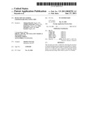 | 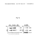 |
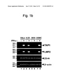 |  |
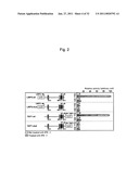 | 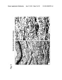 |
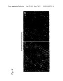 | 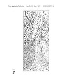 |
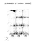 | 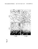 |
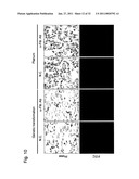 | 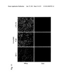 |
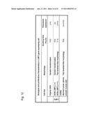 | 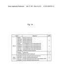 |
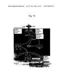 |  |
 | 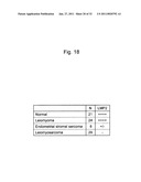 |
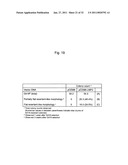 | 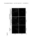 |
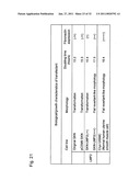 | 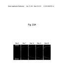 |
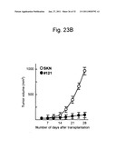 | 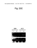 |
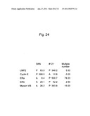 | 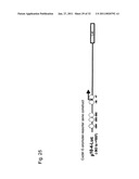 |
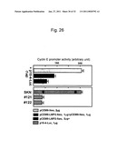 | 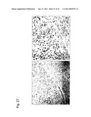 |
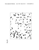 |  |
 |  |
 |  |
 |  |
 |  |
 |  |
 |  |
 |  |
| Similar patent applications: | |
| Date | Title |
|---|---|
| 2011-06-23 | Detection of xenotropic murine leukemia virus |
| 2010-03-11 | Detection of chromosomal inversions |
| 2009-02-05 | Method for the deletion of a large chromosomal region |
| 2010-12-09 | Method for the detection of chromosomal aneuploidies |
| 2011-01-06 | Method for the detection of gene transcripts in blood and uses thereof |
| New patent applications in this class: | |
| Date | Title |
|---|---|
| 2011-06-30 | Apparatus and method of authenticating product using polynucleotides |
| 2011-06-30 | Cyanine compounds, compositions including these compounds and their use in cell analysis |
| 2011-06-30 | Method for detecting multiple small nucleic acids |
| 2011-06-30 | Solid-phase chelators and electronic biosensors |
| 2011-06-30 | Cell-based screening assay to identify molecules that stimulate ifn-alpha/beta target genes |
| New patent applications from these inventors: | |
| Date | Title |
|---|---|
| 2015-07-30 | Image processing device, dither mask generation method, and program |
| 2015-02-26 | Print apparatus, print method, and serial printer |
| Top Inventors for class "Chemistry: molecular biology and microbiology" | |
| Rank | Inventor's name |
|---|---|
| 1 | Marshall Medoff |
| 2 | Anthony P. Burgard |
| 3 | Mark J. Burk |
| 4 | Robin E. Osterhout |
| 5 | Rangarajan Sampath |