Patent application title: CANCER DIAGNOSTIC KIT AND CANCER DIAGNOSTIC METHOD
Inventors:
Toshiro Ono (Okayama, JP)
Eiichi Nakayama (Okayama, JP)
Assignees:
NATIONAL UNIVERSITY CORPORATION OKAYAMA UNIVERSITY
IPC8 Class: AC12Q168FI
USPC Class:
435 6
Class name: Chemistry: molecular biology and microbiology measuring or testing process involving enzymes or micro-organisms; composition or test strip therefore; processes of forming such composition or test strip involving nucleic acid
Publication date: 2010-09-30
Patent application number: 20100248234
Claims:
1-15. (canceled)
16. A primer including a partial sequence of a nucleotide sequence of position 2146 to position 3044 of SEQ ID No: 3, with the proviso that the primer is not an oligonucleotide consisting of a partial sequence of a nucleotide sequence of position 2146 to position 2481 of SEQ ID No: 1.
17. The primer according to claim 16, the primer consisting of a nucleotide sequence of SEQ ID No: 7 or 8.
18. A method for diagnosing a cancer, comprising:a polynucleotide measuring step of, using a primer according to claim 16, measuring a level of presence of a polynucleotide including a nucleotide sequence of SEQ ID No: 3 or a partial sequence thereof in a sample derived from a subject.
19. The method according to claim 18, the method being used for diagnosis of an epithelial tumor or a skin cancer.
20. The method according to claim 18, the method being used for diagnosis of at least one cancer selected from the group consisting of a gastric cancer, a colon cancer, a breast cancer, a head and neck cancer, a lung cancer, a renal cancer, a prostatic cancer, and a malignant melanoma.
21. A polypeptide consisting of a partial sequence of an amino acid sequence of SEQ ID No: 4, the polypeptide including an amino acid sequence of position 668 to position 684 of SEQ ID No: 4, with the proviso that the polypeptide is not a polypeptide consisting of a partial sequence of an amino acid sequence of SEQ ID No: 2.
22. The polypeptide according to claim 21, the polypeptide consisting of an amino acid sequence of position 366 to position 684 of SEQ ID No: 4.
23. A method for diagnosing a cancer, comprising:an antibody measuring step of, using a polypeptide according to claim 21, measuring a level of an antibody binding specifically to a polypeptide consisting of an amino acid sequence of SEQ ID No: 4 or a partial sequence thereof in a sample derived from a subject.
24. The method according to claim 23, the method being used for diagnosis of an epithelial tumor or a skin cancer.
25. The method according to claim 23, the method being used for diagnosis of at least one cancer selected from the group consisting of a gastric cancer, a colon cancer, a breast cancer, a head and neck cancer, a lung cancer, a renal cancer, a prostatic cancer, and a malignant melanoma.
26. An antibody binding specifically to CCDC62-2 protein, but not to CCDC62-1 protein.
27. The antibody according to claim 26, the antibody being initiated by a polypeptide consisting of an amino acid sequence of position 366 to position 684 of SEQ ID No: 4.
28. A method for diagnosing a cancer, comprising:a polypeptide measuring step of, using an antibody according to claim 26, measuring a level of presence of a polypeptide consisting of an amino acid sequence of SEQ ID No: 4 or a partial sequence thereof in a sample derived from a subject.
29. The method according to claim 28, the method being used for diagnosis of an epithelial tumor or a skin cancer.
30. The method according to claim 28, the method being used for diagnosis of at least one cancer selected from the group consisting of a gastric cancer, a colon cancer, a breast cancer, a head and neck cancer, a lung cancer, a renal cancer, a prostatic cancer, and a malignant melanoma.
Description:
TECHNICAL FIELD
[0001]The present invention relates to a novel cancer diagnostic technique. More specifically, the present invention relates to a kit and method for diagnosing a cancer both of which uses the expression of CCDC62 or the presence of an anti-CCDC62 antibody as an index.
BACKGROUND ART
[0002]In order to establish a testing method, diagnostic method, and therapeutic method effective to cancers, it is necessary to find out a cancer antigen that expresses in an objective cancer with high frequency and has high antigenicity. However, there are few cancer antigens useful for testing, diagnosis, or treatment of digestive system cancers, especially gastric cancer and colon cancer. Therefore, it is very important to develop useful cancer antigens for testing, diagnosis, or treatment of digestive system cancers.
[0003]In terms of accompanying side effects, the most useful antigen for testing, diagnosis, or treatment of human cancer is considered to be a cancer-testis antigen (hereinafter referred to as "CT antigen") that expresses in various cancers, but are confined to testis among normal tissues. To date, about 40 kinds of CT antigens have been identified and reported. However, all of these CT antigens do not possess strong immunogenicity to cancer patients, and there are a limited number of CT antigens promising for diagnosis marker of cancers, test marker of cancers, or treatment of cancers.
[0004]Typical CT antigens that have been reported to date include MAGE (see Non Patent Literature 1), SSX (see Non Patent Literature 2), and NY-ESO-1 (see Non Patent Literature 3). MAGE and NY-ESO-1 have been applied in clinical settings in the world, particularly in Europe and the United States, and the clinical settings have achieved some positive results (for example, for clinical application of NY-ESO-1, see Non Patent Literature 4). Thus, it can be said that NY-ESO-1 is currently the most promising CT antigen for applications to treatment of cancer and the like.
[0005]Non Patent Literature 1
[0006]van der Bruggen P, Traversari C, Chomez P, Lurquin C, De Plaen E, van den Eynde B, Knuth A, Boon T. A gene encoding an antigen recognized by cytolytic T lymphocytes on a human melanoma. Science 254: 1643-1647, 1991.
[0007]Non Patent Literature 2
[0008]Gure A O, Tureci O, Sahin U, Tsang S, Scanlan M J, Jager E, Knuth A, Pfreundschuh M, Old L J, Chen Y T. SSX: a multigene family with several members transcribed in normal testis and human cancer. Int. J. Cancer 72: 965-971, 1997.
[0009]Non Patent Literature 3
[0010]Chen Y T, Scanlan, M J, Sahin U, Tureci O, Gure A O, Tsang S, Williamson B, Stockert E, Pfreundschuh M, Old L J. A testicular antigen aberrantly expressed in human cancers detected by autologous antibody screening. Proc. Natl. Acad. Sci. USA 94: 1914-1918, 1997.
[0011]Non Patent Literature 4
[0012]Jager E, Gnjatic S, Nagata Y, Stockert E, Jager D, Karbach J, Neumann A, Rieckenberg J, Chen Y T, Ritter G, Hoffman E, Arand M, Old L J, Knuth A. Induction of primary NY-ESO-1 immunity: CD8+ T lymphocyte and antibody responses in peptide-vaccinated patients with NY-ESO-1+ cancers. Proc. Natl. Acad. Sci. USA 97: 12198-12203, 2000.
[0013]Non Patent Literature 5
[0014]Stockert E, Jager E, Chen Y T, Scanlan M J, Gout I, Karbach J, Arand M, Knuth A, Old L J. A survey of the humoral immune response of cancer patients to a panel of human tumor antigens. J. Exp. Med. 187: 1349-1354, 1998.
[0015]Non Patent Literature 6
[0016]Kurashige T, Noguchi Y, Saika T, Ono T, Nagata Y, Jungbluth A A, Ritter G, Chen Y T, Stockert E, Tsushima T, Kumon H, Old L J, Nakayama E. NY-ESO-1 expression and immunogenicity associated with transitional cell carcinoma: correlation with tumor grade. Cancer Res. 61: 4671-4674, 2001.
[0017]Non Patent Literature 7
[0018]Nakada T, Noguchi Y, Sato S, Ono T, Saika T, Kurashige T, Gnjatic S, Ritter G, Chen Y T, Stockert E, Nasu Y, Tsushima T, Kumon H, Old L J, Nakayama E. NY-ESO-1 mRNA expression and immunogenicity in advanced prostate cancer. Cancer Immunity 3: 10, 2003.
[0019]Non Patent Literature 8
[0020]Sugita Y, Wada S, Fujita S, Nakata T, Sato S, Noguchi Y, Jungbluth A A, Yamaguchi M, Chen Y T, Stockert E, Gnjatic S, Williamson B, Scanlan M J, Ono T, Sakita I, Yasui M, Miyoshi Y, Tamaki Y, Matsuura N, Noguchi S, Old L J, Nakayama E, Monden M. NY-ESO-1 expression and immunogenicity in malignant and benign breast tumors. Cancer Res. 64: 2199-2204, 2004.
[0021]Non Patent Literature 9
[0022]Fujita S, Wada H, Jungbluth A A. Sato S, Nakata T, Noguchi Y, Doki Y, Yasui M, Sugita Y, Yasuda T, Yano M, Ono T, Chen Y T, Higashiyama M, Gnjatic S, Old L J, Nakayama E, Monden M. NY-ESO-1 expression and immunogenicity in esophageal cancer. Clin. Cancer Res. 10: 6551-6558, 2004.
[0023]Non Patent Literature 10
[0024]Nakamura, S, Nouso K, Noguchi Y, Higashi T, Ono T, Jungbluth A A, Chen Y T, Old L J, Nakayama E, Shiratori Y. Expression and immunogenicity of NY-ESO-1 in hepatocellular carcinoma. J. Gastroenterol. Hepatol. 21: 1281-1285, 2006.
SUMMARY OF INVENTION
[0025]The inventors of the present invention tested patients with various epithelial tumors for their capabilities of producing antibodies to NY-ESO-1. The result of the test was as follows: 7% for bladder cancer (9 of 124 cases, see Non Patent Literature 6), 4.6% for prostatic cancer (10 of 218 cases, see Non Patent Literature 7), 1.6% for breast cancer (1 of 62 cases, see Non Patent Literature 8), 3.9% (2 of 51 cases, see Non Patent Literature 9), 2.2% for liver cancer (2 of 92 cases, see Non Patent Literature 10). This reveals that the capabilities of producing antibodies are not so high in all these cancers. Thus, antibodies against the CT antigens reported in the past exist in sera from patients with epithelial tumor with extremely low frequency.
[0026]Further, in some rare cases, the CT antigens reported in the past express in digestive system cancers such as gastric cancer and colon cancer. Moreover, the antibodies against the CT antigens are produced with extremely low frequency in patients with digestive system cancers. For example, it has been reported that the capabilities of producing antibodies to NY-ESO-1, MAGE, and SSX of patients with colon cancer are all 0% (see Non Patent Literature 5). Incidentally, the inventors of the present invention tested sera from 58 patients with colon cancer for their capabilities of producing antibodies to NY-ESO-1. The result of the test showed that the capabilities of producing antibodies to NY-ESO-1 were 0% (not published). Further, even in the preliminary analysis using sera from patients with gastric cancer by the inventors of the present invention, the presence of the antibody to NY-ESO-1 has not been proved yet.
[0027]Thus, it is not easy to find out a useful cancer antigen for diagnosis of epithelial tumors including digestive system cancers, such as gastric cancer and colon cancer. Therefore, there is a very strong demand for the development of a useful cancer antigen for diagnosis of digestive system cancers.
[0028]The present invention has been attained in view of the above problem, and an object of the present invention is to provide a novel cancer diagnostic technique using a useful cancer antigen (preferably CT antigen) for testing, diagnosis, or treatment of epithelial tumors including digestive system cancers.
[0029]In order to solve the above problem, the inventors of the present invention carried out SEREX (serological analysis of cancer antigens by recombinant cDNA expression cloning) using a cDNA library derived from normal testis and a serum from a patient with gastric cancer. Then, the inventors of the present invention accomplished the present invention by finding that CCDC62 in an antibody-positive clone is a CT antigen.
[0030]That is, a kit according to the present invention is a kit for diagnosing a cancer, comprising a polynucleotide including a nucleotide sequence of SEQ ID No: 1 or 3 or a partial sequence thereof.
[0031]A kit according to the present invention is a kit for diagnosing a cancer, comprising a polypeptide consisting of an amino acid sequence of SEQ ID No: 2 or 4 or a partial sequence thereof.
[0032]A kit according to the present invention is a kit for diagnosing a cancer, comprising an antibody binding specifically to a polypeptide consisting of an amino acid sequence of SEQ ID No: 2 or 4 or a partial sequence thereof.
[0033]A kit according to the present invention is preferably used for diagnosis of an epithelial tumor or a skin cancer, more preferably for diagnosis of at least one cancer selected from the group consisting of a gastric cancer, a colon cancer, a breast cancer, a head and neck cancer, a lung cancer, a renal cancer, a prostatic cancer, and a malignant melanoma.
[0034]A method for diagnosing a cancer according to the present invention is characterized by comprising a polynucleotide measuring step of measuring a level of presence of a polynucleotide including a nucleotide sequence of SEQ ID No: 1 or 3 or a partial sequence thereof in a sample derived from a subject.
[0035]A method for diagnosing a cancer according to the present invention is characterized by comprising: a polypeptide measuring step of measuring a level of presence of a polypeptide consisting of an amino acid sequence of SEQ ID No: 2 or 4 or a partial sequence thereof in a sample derived from a subject.
[0036]A method for diagnosing a cancer according to the present invention is characterized by comprising: an antibody measuring step of measuring a level of an antibody binding specifically to a polypeptide consisting of an amino acid sequence of SEQ ID No: 2 or 4 or a partial sequence thereof in a sample derived from a subject.
[0037]A method for diagnosing a cancer according to the present invention is preferably used for diagnosis of an epithelial tumor or a skin cancer, more preferably for diagnosis of at least one cancer selected from the group consisting of a gastric cancer, a colon cancer, a breast cancer, a head and neck cancer, a lung cancer, a renal cancer, a prostatic cancer, and a malignant melanoma.
[0038]Additional objects, features, and strengths of the present invention will be made clear by the description below. Further, the advantages of the present invention will be evident from the following explanation in reference to the drawings.
BRIEF DESCRIPTION OF DRAWINGS
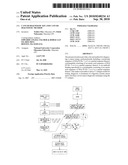
[0039]FIG. 1 is a view schematically showing SEREX.
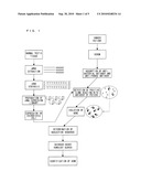
[0040]FIG. 2 is a view showing the result of analysis of CCDC62 mRNA expressions in human normal tissues, wherein (a) shows the result of RT-PCR analysis and (b) shows the result of real-time RT-PCR analysis.
[0041]FIG. 3 is a view showing the result of RT-PCR analysis of CCDC62-2 mRNA expressions in various cancer tissues, wherein (a) shows the result of analysis for lung cancer and (b) shows the result of analysis for colon cancer, and (c) shows the result of analysis for prostatic cancer.
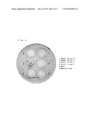
[0042]FIG. 4 is a view showing the result of analysis of reactivity of sera from patients with lung cancer against various antigens by phage plaque assay.
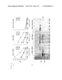
[0043]FIG. 5 is a view showing the result of analysis of humoral immune responses of cancer patients against CCDC62-2 protein, wherein (a) shows the result of ELISA analysis and (b) shows the result of Western blotting analysis.
DESCRIPTION OF EMBODIMENTS
[0044]The inventors of the present invention carried out SEREX for a cDNA library derived from normal testis using a serum from a specific patient with gastric cancer. As a result of SEREX, we found out that a positive clone was a clone expressing CCDC62 and that anti-CCDC62 antibody existed in the serum from the patient with gastric cancer. That is, the inventors of the present invention found out that CCDC62 was a cancer antigen in a patient with gastric cancer. Further, the inventors of the present invention carried out analysis of CCDC62 expression and confirmed that CCDC62 was strongly expressed only in testis among human normal tissues and also was expressed in cancer tissues and cancer cell lines. On the basis of these findings, the inventors of the present invention accomplished the present invention using the CT antigen, CCDC62.
[0045]SEREX (serological analysis of cancer antigens by recombinant cDNA expression cloning) is a method for identifying an antigen gene recognized by an antibody that exists in a patient serum from a cDNA expression library derived from cancer tissue (Sahin U, Tureci O, Schmitt H, Cochlovius B, Johannes T, Schmits R, Stenner F, Luo G, Schobert I, Pfreundschuh M. Human neoplasms elicit multiple specific immune responses in the autologous host. Proc Natl Acad Sci USA. 1995 Dec. 5; 92(25):11810-3). SEREX is known as an excellent method for screening a cancer antigen. It should be noted that the present invention, for the first time, provided the accomplishment using the cDNA library derived from normal testis, not using a cDNA library derived from autologous cancer tissue.
[0046]As above, the sequence of human CCDC62 is known, but the relevance of human CCDC62 to a disease has not been reported at all. The inventors of the present invention first found out the expression of CCDC62 in cancers and the presence of the anti-CCDC62 antibody in a serum from a cancer patient.
[0047]SEQ ID No: 1 represents a nucleotide sequence of transcript variant 1 of the human CCDC62 gene. SEQ ID No: 2 represents an amino acid sequence of a protein coded by transcript variant 1 of the human CCDC62 gene. SEQ ID No: 3 represents a nucleotide sequence of transcript variant 2 of the human CCDC62 gene. SEQ ID No: 4 represents an amino acid sequence of a protein coded by transcript variant 2 of the human CCDC62 gene. As for the nucleotide sequence of the human CCDC62 gene, transcript variant 1 and transcript variant 2 are registered respectively as NM--032573 and NM 201435 in the GenBank.
[0048][1. A Method for Diagnosing a Cancer]
[0049]The present invention provides a method for diagnosing a cancer. In one embodiment, the cancer diagnostic method according to the present invention is a method for diagnosing a cancer by measuring the expression level of CCDC62 in a sample derived from a subject and then comparing the resultant expression level with a control level (e.g. normal level). More specifically, the cancer diagnostic method according to the present embodiment comprises a polynucleotide measuring step or a polypeptide measuring step. The expression level of CCDC62 can be determined by measuring the amount of transcripts (mRNA) or the amount of translation products (protein) by means of a technique known in the art.
[0050]In another embodiment, the cancer diagnostic method according to the present invention is a method for diagnosing cancer by measuring the level of the anti-CCDC62 antibody in a sample derived from a subject and then comparing the resultant level with a control level (e.g. normal level). More specifically, the cancer diagnostic method according to the present embodiment comprises an antibody measuring step. The level of the anti-CCDC62 antibody can be determined by measuring the level of the anti-CCDC62 antibody in a serum derived from the subject.
[0051]A subject objective to the cancer diagnostic method of the present invention, which is not particularly limited and includes a wide range of animals in general, is preferably a human. If the subject is a human, the subject can be not only a cancer patient and a patient suspected of having cancer but also a healthy person.
[0052]The sample derived from the subject used in the cancer diagnostic method of the present invention is not particularly limited as long as it is obtained from the subject (i.e. a sample separated from the subject). Examples of the sample include blood (serum, plasma, hemocyte, etc.), urine, feces, expectoration, ascites, peritoneal lavage fluid, biopsy tissue, and a surgically resected specimen.
[0053]The normal level means the expression level of CCDC62 mRNA, the expression level of CCDC62 protein, or the level of the anti-CCDC62 antibody in a normal healthy individual (healthy person). The normal level is preferably a measurement value obtained by using a normal cell, a normal tissue, or a normal body fluid as a sample that is of the same kind as the sample derived from the subject to be compared with (i.e. the sample used in the polynucleotide measuring step, the polypeptide measuring step, or the antibody measuring step of the present invention). Further, it is more preferable that the normal level is a mean value for the group consisting of normal healthy individuals.
[0054]As used herein, "diagnosis" encompasses not only "determination" but also "testing", "detection", and "prediction". That is, the diagnosis of a cancer means determination or prediction of whether a subject has a cancer, determination or prediction of a stage of cancer progression, and determination or prediction of a cancer treatment result, more preferably inspection/detection/prediction of whether a subject has a cancer, inspection/detection/prediction of a stage of cancer progression, and inspection/detection/prediction of a cancer treatment result.
[0055]The type of a cancer to be diagnosed is not particularly limited, but the cancer is preferably an epithelial tumor or a skin cancer. Examples of the epithelial tumor include a gastric cancer, a colon cancer, a breast cancer, a head and neck cancer, a lung cancer, a liver cancer, a renal cancer, an epithelial ovarian cancer, a prostatic cancer, and the like. Examples of the skin cancer include a malignant melanoma.
[0056](1) Cancer Diagnostic Method Using mRNA Level as Index
[0057]In one embodiment, the cancer diagnostic method according to the present invention may be a method for diagnosing a cancer using a sample derived from a subject, the method including a polynucleotide measuring step of measuring the level of the presence of a polynucleotide including a nucleotide sequence of SEQ ID No: 1 or 3 or a partial sequence thereof in the sample derived from the subject. Note that the polynucleotide includes both DNA and RNA.
[0058]In the present embodiment, mRNA level of CCDC62 may be measured in the polynucleotide measuring step.
[0059]A method of measuring mRNA level, which is not particularly limited as long as it enables the measurement of a specific mRNA level, is selected for use as appropriate from among the known methods. Examples of the method of measuring mRNA level include a method using primers or a probe containing a polynucleotide consisting of nucleotide sequences of mRNAs or cDNAs of CCDC62 or a portion of complementary sequences of nucleotide sequences, wherein the polynucleotide is site-specifically bound to (hybridized with) any of mRNAs or cDNAs of CCDC62. The primers or probe may be subjected to various modifications for the measurement/detection of mRNA, as long as it forms site-specific base pairs with mRNA of CCDC62 or its corresponding cDNA.
[0060]The polynucleotide used as the above-described primers is not limited as long as it is designed on the basis of the nucleotide sequence of SEQ ID No: 1 or 3 or its complementary sequence. Examples of such a polynucleotide include a polynucleotide consisting of a nucleotide sequence of any of SEQ ID Nos: 5 through 8. These polynucleotides are the primers which were actually used by the inventors of the present invention in carrying out RT-PCR, and the polynucleotides are proved to specifically amplify cDNAs of CCDC62. The polynucleotide used as the above-described probe is not limited as long as it is designed on the basis of the nucleotide sequence of SEQ ID No: 1 or 3. Note that it is well known in the art that a polynucleotide available as primers that specifically amplify a mRNA (cDNA) of interest is usable as a probe for specifically detecting the mRNA (cDNA).
[0061]Examples of the known method of measuring mRNA using the primers or probe containing the polynucleotide site-specifically bound to the foregoing mRNA or cDNA include RT-PCR, real-time RT-PCR, competitive PCR, in situ hybridization, in situ PCR, DNA array method and the like.
[0062]For example, the above-described RT-PCR is a method of synthesizing cDNA, using reverse transcriptase, from total RNA or mRNA prepared from a sample, and then amplifying a region of interest by PCR using the syntherized cDNA as a template. Real-time RT-PCR is a method of, during amplification of a region of interest by PCR using cDNA as a template, monitoring in real time and analyzing the generation process of a resulting product of the amplification using a reagent for real-time monitoring. Examples of the reagent for real-time monitoring include SYBR (registered trademark: Molecular Probes Inc.) Green, TaqMan (registered trademark: Applied Biosystems Inc.) probe and the like.
[0063]For example, in the above-described DNA array method, cDNA or its fragment of CCDC62 is immobilized on a support and incubated with mRNA or cDNA prepared from a sample. At this time, the mRNA or cDNA may be labeled with a fluorescent dye or the like so that hybridization between the DNA immobilized on the support and the mRNA or cDNA prepared from the sample can be detected, and the mRNA level in the sample can be measured.
[0064]In the comparing step, the mRNA level of CCDC62 in the sample, which level has been measured in the polynucleotide measuring step, may be compared with the normal level. As described previously, the normal level is preferably measured by the same method using a normal cell, tissue, or body fluid, as a sample, that is of the same kind as the subject-derived sample to be compared (i.e. sample used in the polynucleotide measuring step of the present embodiment). The normal level may be obtained, concurrently with the polynucleotide measuring step, by measurement conducted for a sample derived from a normal and healthy individual, or may be data stored as background data. If mRNA level in the sample derived from the subject is higher than the normal level, it is possible to determine that the subject has a cancer. The level higher than the normal level is preferably a level twice higher than the normal level, more preferably a level three times higher than the normal level.
[0065](2) Cancer Diagnostic Method Using Protein Level as Index
[0066]In one embodiment, the cancer diagnostic method according to the present invention may be a method for diagnosing a cancer using a sample derived from a subject, including a polypeptide measuring step of measuring the level of the presence of a polypeptide consisting of an amino acid sequence of SEQ ID No: 2 or 4 or a partial sequence thereof in the sample derived from the subject.
[0067]In the present embodiment, the level of CCDC62 protein may be measured in the polypeptide measuring step.
[0068]A method of measuring protein expression level, which is not particularly limited as long as it enables the measurement of a specific protein level, is selected for use as appropriate from among the known methods. Examples of the method of measuring protein expression level include a method using an antibody binding specifically to CCDC62 protein. The antibody may be a polyclonal antibody or a monoclonal antibody. Further, the antibody may be a complete antibody molecule or an antibody fragment capable of specifically binding (e.g. Fab fragment or F(ab')2 fragment).
[0069]Examples of the known method of measuring a protein level using an antibody include radioimmunoassay (RIA), ELISA (enzyme linked immunoassay), Western blotting, immunoprecipitation method, immunohistochemical method, and antibody array method. Among these methods, ELISA is preferable in terms of high sensitivity and convenience.
[0070]In the comparing step, the level of the CCDC62 protein in the sample, which level has been measured in the polypeptide measuring step, may be compared with the normal level. As described previously, the normal level is preferably measured by the same method using a normal cell, tissue, or body fluid as a sample that is of the same kind as the subject-derived sample to be compared (i.e. sample used in the polypeptide measuring step of the present embodiment). The normal level may be obtained, concurrently with the polypeptide measuring step, by measurement conducted for a sample derived from a normal and healthy individual, or may be data stored as background data. If the protein level in the sample derived from the subject is higher than the normal level, it is possible to determine that the subject has cancer. The level higher than the normal level is preferably a level twice higher than the normal level, more preferably a level three times higher than the normal level.
[0071](3) Cancer Diagnostic Method Using Antibody Level as Index
[0072]In one embodiment, the cancer diagnostic method according to the present invention may be a method for diagnosing a cancer using a sample derived from a subject, including an antibody measuring step of measuring the level of the presence of an antibody binding specifically to a polypeptide consisting of an amino acid sequence of SEQ ID No: 2 or 4 or a partial sequence thereof in the sample derived from the subject.
[0073]In the present embodiment, the level of anti-CCDC62 antibody may be measured in the antibody measuring step. Note that a sample used in measuring the level of the anti-CCDC62 antibody is preferably a serum, but is not particularly limited as long as it enables the measurement of the antibody level.
[0074]An antibody level measuring method is not particularly limited as long as it enables the measurement of a level of antibody titer against a specific antigen and the measurement using an antibody against an antibody of interest, and is selected for use as appropriate from among the known methods. Examples of the antibody level measuring method include ELISA using CCDC62 protein as an antigen and a protein array. The antigen protein used in measuring the antibody titer can be obtained by purification of a biological sample, but is preferably obtained as a recombinant protein. The recombinant protein can be obtained by introducing an expression vector including a CCDC62 gene into a host for expression and then purifying from the host.
[0075]In the comparing step, the level of the anti-CCDC62 antibody in the sample, which level has been measured in the antibody measuring step, may be compared with the normal level. The normal level may be obtained, concurrently with the antibody measuring step, by measurement conducted for a sample (preferably a serum) derived from a normal and healthy individual, or may be data stored as background data. If the level of the anti-CCDC62 antibody in the sample derived from the subject is higher than the normal level, it is possible to determine that the subject has a cancer. The level higher than the normal level is preferably a level twice higher than the normal level, more preferably a level three times higher than the normal level.
[0076][2. Kit]
[0077]The present invention provides a kit used for diagnosis of cancers. The kit according to the present invention may include a reagent or an instrument required for implementation of the diagnostic method described previously. As used herein, the term "kit" refers to a packaging that includes a container (e.g. bottle, plate, tube, dish, etc.) storing a particular material therein. Preferably, the kit includes instructions for use of each reagent or instrument. As used herein concerning the kit, the term "includes (including)" refers to the inclusion of a reagent or an instrument in any one of individual containers that constitutes the kit. The "instructions" may be printed on a sheet of paper or other medium, or may be recorded in an electronic medium such as a magnetic tape, a computer-readable disk or tape, or a CD-ROM. The kit according to the present invention may further include a reagent or an instrument required for application to diagnosis of cancers.
[0078]In one embodiment, a kit according to the present invention can be a kit applicable to a cancer diagnostic method using the above-described mRNA level as an index. A kit according to the present embodiment may be a kit for diagnosing a cancer using a sample derived from a subject, the kit including a polynucleotide comprising a nucleotide sequence of SEQ ID No: 1 or 3 or a partial sequence thereof. Examples of the polynucleotide comprising a nucleotide sequence of SEQ ID No: 1 or 3 or a partial sequence thereof include primers and a probe both used for measurement of mRNA of CCDC62. The kit according to the present embodiment can be used suitably for measurement of a CCDC62 mRNA expression level.
[0079]A kit according to the present embodiment may be a RT-PCR kit for measurement of mRNA of CCDC62 or a real-time RT-PCR kit for measurement of mRNA of CCDC62. Further, a kit according to the present embodiment can be a kit by which RT-PCR or real-time RT-PCR can be carried out by user selection. In this case, a kit according to the present embodiment may include a primer pair for amplifying mRNA of CCDC62 by RT-PCR. Kit components other than the primer pair are not particularly limited. For example, the kit preferably includes a reagent for preparing RNA from tissues or cells, a reverse transcriptase, a buffer solution used for reverse transcription reaction, a heat-resistant DNA polymerase, a reagent for PCR, a reagent for real-time PCR, a tube for PCR, and a plate for PCR.
[0080]In another embodiment, a kit according to the present invention can be a kit applicable to a cancer diagnostic method using the above-described protein level as an index. A kit according to the present embodiment may be a kit for diagnosing a cancer using a sample derived from a subject, the kit including an antibody binding specifically to a polypeptide consisting of an amino acid sequence of SEQ ID No: 2 or 4 or a partial sequence thereof. The kit according to the present embodiment can be used suitably for measurement of CCDC62 protein level.
[0081]In yet another embodiment, a kit according to the present invention can be a kit applicable to a cancer diagnostic method using the above-described antibody level as an index. A kit according to the present embodiment may be a kit for diagnosing a cancer using a sample derived from a subject, the kit including a polypeptide consisting of an amino acid sequence of SEQ ID No: 2 or 4 or a partial sequence thereof. In this case, a kit according to the present embodiment can be used for measurement of a level of antibody titer of the anti-CCDC62 antibody. Further, a kit according to the present embodiment may include an antibody against the anti-CCDC62 antibody. A technique of preparing such an antibody is well known in the art.
[0082]A kit according to the present embodiment can be a kit for ELISA. Such a kit preferably includes either (i) an ELISA plate having an antibody binding specifically to the CCDC62 protein immobilized (solid-phased) thereon or (ii) an ELISA plate having the CCDC62 protein immobilized (solid-phased) thereon. Further, if a kit according to the present embodiment is a kit including both of the ELISA plates (i) and (ii), such a kit can be a kit capable of measuring both the CCDC62 protein level and the anti-CCDC62 antibody level.
[0083]Kit components other than the ELISA plate are not particularly limited. For example, a kit according to the present embodiment preferably includes a labeled secondary antibody, a coloring reagent, and a washing buffer solution.
[0084]Specific embodiments implemented in the description of the embodiments and the following examples only show technical features of the present invention and are not intended to limit the scope of the invention. Variations can be effected by a person skilled in the art within the spirit of the present invention and the scope of the following claims.
[0085]Further, all of scientific literatures and patent literatures listed herein are herein incorporated by reference.
EXAMPLES
[0086]The following will describe the present invention in more detail by way of Examples. It should be noted however that the present invention is not limited in any way by the following Examples. Unless otherwise specified, methods required for general genetic recombination, such as extraction, cleavage, and connection of nucleic acids, transformation of Escherichia coli, and determination of nucleotide sequence of a gene, are carried out according to a manual attached to a commercially-available reagent, equipment, and others for use in each operation and an experimental manual (e.g. "Molecular Cloning, a Laboratory Manual, 3rd Ed (Sambrook et al. (2001), Cold Spring Harbor Laboratory Press)").
Example 1
SEREX Analysis of Antigen Recognized by Antibody in Gastric Cancer Patient Serum
[0087]As schematically shown in FIG. 1, SEREX is a method of searching for a cancer antigen using a serum from a cancer patient in a cDNA expression library prepared by direct extraction of mRNAs from a cancer tissue isolated from the cancer patient. In Example 1, a cancer-testis antigen useful for diagnosis of gastric cancer was isolated by SEREX using a cDNA library derived from normal testis and a serum from a patient with gastric cancer.
[0088](1) Gastric Cancer Patient Serum
[0089]A serum was obtained from a patient with gastric adenocarcinoma whose primary lesion and liver metastasis both showed a tendency to reduce. The obtained serum was confirmed to strongly react with a protein fraction obtained from normal testis, before selected. The serum was diluted 10-fold with TBS (Tris-Buffered Saline) and then subjected to absorption of anti-bacterial antibodies (nonspecific antibodies against Escherichia coli and a phage) using an affinity column of E. coli Y1090/Y1089 (BioDynamics Lab Inc., Tokyo).
[0090](2) Preparation of cDNA Expression Library
[0091]Using Quick Prep Micro mRNA purification kit (Stratagene, La Jolla, Calif., USA), mRNAs were purified from normal testis Total RNA (BD Biosciences Clontech, Palo Alto, Calif., USA). From 5 μg of mRNAs, cDNAs were synthesized. The cDNAs were then subcloned into γZAP Express vectors (Stratagene). The recombinant γZAP Express vectors were packaged into phages to prepare a cDNA expression library.
[0092](3) Immunoscreening of Normal Testis cDNA Library
[0093]About 4,000 phages were planted on a 150 mm-plate and incubated for 6 to 8 hours. IPTG-induced protein was transferred into a 135 mm-diameter nitrocellulose membrane (Schleicher 86 Schuell, Dassel, Germany). Thereafter, the resultant membrane was reacted for 15 hours with the serum from the patient with gastric cancer (diluted 200-fold with TBS) absorbing the anti-bacterial antibodies, and then subjected to detection using a peroxidase-labeled anti-human IgG antibody (Jackson ImmunoResearch, West Grove, Pa., USA). At this time, in order to remove IgG clones derived from antibody production cells, the membrane was processed with a secondary antibody alone. Antibody-positive clones were isolated therefrom and then subjected to secondary screening and third screening using a 82 mm-diameter membrane and a 47 mm-diameter membrane, respectively, to obtain a single clone.
[0094](4) Determination of Insert cDNA Nucleotide Sequence
[0095]The antibody-positive clone was converted into pBK-CMV plasmid by in vivo excision. Thereafter, a nucleotide sequence of insert cDNA was analyzed (ABI PRISM R310 Genetic Analyzer; PE Applied Biosystems, Foster City, Calif., USA), and homology search was carried out according to a gene database (http://www.ncbi.nih.gov/BLAST/Blast.cgi).
[0096](5) Result
[0097]As a result of screening about 200,000 clones, fifty-five antibody-positive clones shown in Table 1 were isolated.
TABLE-US-00001 TABLE 1 SEREX-identified antigen genes No. of Gene clones Identity/similarities Expression OY-ST-1 10 cytochrome b5 reductase 2 (CYB5R2), transcript variant 2 Ubiquitous OY-ST-2 8 no homology Testis OY-ST-3 2 G kinase anchoring protein 1 (GKAP1) Testis OY-ST-4 3 pleckstrin and Sec7 domain containing 3 Ubiquitous OY-ST-5 4 palladin, cytoskeletal associated protein Ubiquitous OY-ST-6 1 synaptonemal complex protein 1, SYCP1 (SCP-1) Testis OY-ST-7 1 ankyrin repeat domain 13B Ubiquitous OY-ST-8 1 ATP synthase, H.sup.+ transporting, mitochondrial F0 complex, subunit c (subunit 9) pseudogene pseudogene 3 (ATP5GP3) on chromosome 14 OY-ST-9 1 lymphotoxin beta receptor (TNFR superfamily, member 3) Ubiquitous OY-ST-10 1 leucine-rich repeats and calponin homology (CH) domain containing 4 Ubiquitous OY-ST-11 1 heat shock transcription factor 2 Ubiquitous OY-ST-12 1 MAX gene associated, transcript variant 8 (MGA) Ubiquitous OY-ST-13 4 peroxisomal D3, D2-enoyl-CoA isomerase, transcript variant 1 no data OY-ST-14 7 actin related protein 2/3 complex, subunit 2 34 kDa (ARPC2) Ubiquitous OY-ST-15 1 suppressor of Ty 5 homolog (S. cerevisiae) (SUPT5H) Ubiquitous OY-ST-16 1 polymerase (RNA) III (DNA directed) polypeptide H (22.9 KD) (POLR3H) Ubiquitous OY-ST-17 1 centrosomal protein 290 kDa) (CEP290) Ubiquitous OY-ST-18 2 coiled-coil domain containing 62 (CCDC62) Testis OY-ST-19 1 dihydrouridine synthase 1-like (S. cerevisiae) Ubiquitous OY-ST-20 1 leiomodin 1 (smooth muscle) (LMOD1) Ubiquitous OY-ST-21 1 activating signal cointegrator 1 complex subunit 2 (ASCC2) Ubiquitous OY-ST-22 1 chromosome 1 open reading frame 57 (Clorf57) Ubiquitous OY-ST-23 1 F-box and leucine-rich repest protein 5 (FBXL5), transcript variant 1 Ubiquitous
[0098]As the result of the homology search, 23 kinds of genes (OY-ST-1 through OY-ST-23) were identified. Then, normal tissue expressions of these genes were searched according to the database. The result of the search showed that among these 23 kinds of genes 4 kinds of genes were specific to testis (Table 1). Regarding the other 19 kinds of genes, they were expressed in a broad area of normal tissue, or the data on their expressions was not obtained.
[0099]As genes expressing specifically in testis, 2 clones of OY-ST-3 (G kinase anchoring protein 1 (GKAP1)) were obtained. Regarding the invention concerning GKAP1, a patent application was filed by the same inventors of the present invention and is now pending (not published yet). Further, 1 clone of OY-ST-6 (synaptonemal complex protein 1, SYCP1 (SCP-1)) was obtained. SCP-1 (OY-ST-6), which is one of the typical cancer-testis antigens (CT antigens) discovered shortly after the development of SEREX, is known to have a weak immunogenicity (Tureci, et al., Proc. Natl. Acad. Sci. USA, 1998).
[0100]2 clones of OY-ST-18 were isolated, and the homology search showed that these 2 clones were CCDC 62 (coiled-coil domain containing 62), and that they were transcript variant 1 (CCDC62-1, NM--032573: SEQ ID No: 1) and transcript variant 2 (CCDC62-2, NM--201435: SEQ ID No: 3), respectively. It should be noted that it has not been reported that CCDC62 is related to diseases including human cancers.
Example 2
Analysis of CCDC62 Expression
[0101]C62-1-S (sense: 5'-TCCCCGGCAAGTGAGCTAAT-3', SEQ ID No: 5) and C62-1-AS (antisense: 5'-ATACATCCCCATTCCCGAGG-3', SEQ ID No: 6) as primers specific for CCDC62 (variant 1) and C62-2-S (sense: 5'-AAGTCAGAGGTCCCAGAAGA-3', SEQ ID No: 7) and C62-2-AS (antisense: 5'-CTATGCAGGGGTTCTTTCTC-3', SEQ ID No: 8) as primers specific for CCDC62 (variant 2) were synthesized by a DNA synthesizer. By RT-PCR, cDNA derived from human normal tissues (MTC panel, BD Biosciences Clontech) and cDNAs derived from various cancers were amplified to analyze the expression of genes.
[0102]The cancer tissues as used were the donated ones that were obtained by a surgery or a biopsy. Note that all the samples derived from cancer patients or normal healthy individuals in Examples 1 and 2 were obtained by donation only from donors who had given informed consents after they had been directly given oral explanation and explanation in writing by their doctors in charge. The samples derived from cancer patients or normal healthy individuals were held anonymously and used in this study with their personal information strictly protected. The details of this study, an explanatory document for informed consent, and a certificate of informed consent were authorized by Okayama University, Graduate School of Medicine, Dentistry, and Pharmaceutical Sciences, Institutional Review Board of Human Genome and Gene Analysis Research.
[0103]Cancer cell lines as used were 11 kinds of lung cancer cell lines, 8 kinds of malignant melanoma cell lines, and 3 kinds of prostatic cancer cell lines. The malignant melanoma cell lines, which were established in the Sloane-Kettering Cancer Center in the U.S., were obtained therefrom by the inventors of the present invention. The other cell lines are commonly used for cancer research and usually available from a known cell preservation organization or the like.
[0104]From the cancer tissues and the cancer cell lines, respective total RNAs were extracted (RNeasy; Qiagen, Hilden, Germany). From the total RNAs, cDNAs were synthesized with MMLV reverse transcriptase and oligo(DT)15 primers (Ready-To-Go You-Prime First-Strand Beads; Amersham Biosciences, Buckinghamshire, UK). RT-PCR for CCDC62 was carried out under the following conditions: 35 cycles of denaturation at 94° C. for 1 minute; annealing at 60° C. for 1 minute; and extension reaction at 72° C. for 1.5 minutes. The PCR products were analyzed by agarose gel electrophoresis. At the same time, the amplification of G3PDH gene from the syntherized cDNAs was confirmed.
[0105]Regarding CCDC62-1 mRNA and CCDC62-2 mRNA, their expressions in 14 kinds of normal tissues were analyzed using CCDC62-1-specific primers or CCDC62-2-specific primers. The result of the analysis confirmed strong expressions in testis only ((a) of FIG. 2). The expression of CCDC62 genes in human normal tissues was quantitatively analyzed by real-time RT-PCR. Using random primers, cDNAs were synthesized from 2 μg of total RNAs (High-Capacity cDNA Reverse Transcription Kits, Applied Biosystems, Foster City, Calif.). Using CCDC62-specific TagMan probe (TaqMan Gene Expression Assays, Applied Biosystems), the synthesized cDNAs were quantitatively analyzed with ABI PRISM 7700 Sequence Detection System (Applied Biosystems). The TagMan probe (AssayID: Hs00261486) as used is a probe specific to a common sequence between 2 kinds of CCDC62 splice variants (CCDC62-1 and CCDC62-2). As an endogenous control, G3PDH was used (TaqMan Pre-Developed Assay Reagents, Applied Biosystems). The expression of CCDC62 was shown as compared with the expression of CCDC62 in normal testis, which was used as a calibrator. A strong CCDC62 expression in testis was confirmed. The level of such expression was equivalent to the level of expression of NY-ESO-1, which is a typical CT antigen, confirmed by the analysis carried out in the same procedure ((b) of FIG. 2).
[0106]The result of the analysis of CCDC62-1 mRNA and CCDC62-2 mRNA expressions in various cancer tissues showed no CCDC62-1 mRNA expressions. Table 2 shows the result of RT-PCR analysis of CCDC62-2 expression in various cancer tissues using specific primers.
TABLE-US-00002 TABLE 2 CCDC62-2 mRNA expressions in cancer tissues and cancer cell lines (RT-PCR) Positive/total Subject Tissues Cell lines Breast cancer 1/4 (25%) ND Colon cancer 5/40 (12%) ND Esophageal cancer 7/33 (21%) ND Gastric cancer 8/117 (7%) ND Head and neck cancer 4/34 (12%) ND Liver cancer 0/5 (0%) ND Lung cancer 5/19 (26%) 4/11 (36%) Ovarian cancer 0/6 (0%) ND Prostate cancer 3/9 (33%) 1/3 (33%) Renal cancer 1/4 (25%) ND Malignant melanoma ND 3/8 (38%)
[0107]The CCDC62-2 mRNA expression was observed in an oral cavity cancer, an esophagus cancer, a gastric cancer, a breast cancer, a colon cancer, a lung cancer, a prostatic cancer, and a renal cancer (FIG. 3). Particularly, the CCDC62-2 mRNA expression in gastric cancer was confirmed in 8 of 117 cases (7%), the CCDC62-2 mRNA expression in lung cancer was confirmed in 5 of 19 cases (26%), and the CCDC62-2 mRNA expression in prostatic cancer was confirmed in 3 of 9 cases (33%). Further, the CCDC62-2 mRNA expression was also confirmed in cancer cell lines derived from lung cancer and prostatic cancer. Besides, high CCDC62-2 mRNA expression in malignant melanoma was confirmed in 3 of 8 cases (38%).
Example 3
Reactivity Against SEREX-Identified Antigens in Lung Cancer Patient Sera
[0108]Reactivity of sera from lung cancer patients against CCDC62-1, CCDC62-2, and one more kind of identified CT antigen, GKAP1 clone was analyzed by phage plaque assay (FIG. 4).
TABLE-US-00003 TABLE 3 Reactivity against various antigens in sera from lung cancer patients Antigen Positive/total OY-ST-2 (clone 1) 6/29 OY-ST-2 (clone 2) 2/29 CCDC62 (variant 1) 0/29 CCDC62 (variant 2) 4/29 GKAP1 2/29
[0109]Four out of 29 sera from patients with lung cancer were CCDC62-2 positive. Further, two out of 29 sera from patients with lung cancer were GKAP1-positive. Against CCDC62-1, none of the sera from patients with lung cancer reacted (Table 3). These antigens did not react with 7 healthy individual sera at all.
Example 4
Analysis of Humoral Immune Responses of Cancer Patients Against CCDC62-2 Protein
[0110]Into pGEX-6P-1 expression vector, cDNA encoding C terminus (amino acid residue 366-684) of CCDC62-2 protein was inserted. Escherichia coli BL-21 was transformed with the vector. GST fused-CCDC62-2 protein, which had been obtained by IPTG induction, was purified using GSTrap FF column (Amersham Biosciences).
[0111]Humoral immune responses of cancer patients against CCDC62-2 were analyzed by ELISA ((a) of FIG. 5). Specifically, GST-fused recombinant CCDC62-2 protein (1 μg/ml) was adsorbed on a 96-well plate (100 ng per well), subjected to blocking with 5% FCS/PBS for 1 hour, and then reacted with the serum (100 μl) for 2 hours. After the plate was washed, the CCDC62-2 protein on the plate was reacted with peroxidase-labeled anti-human IgG antibody (Jackson ImmunoResearch) for 1 hour. The reactant was made colored with a substrate (1,2-phenylenediamine dihydrochloride). Thereafter, absorbance was measured for 191 of sera from cancer patients and 41 of sera from healthy individuals. Antibody titer was evaluated for 400-fold diluted sera, and an OD value not less than 4 SD, which corresponds to an average OD value (490 nm) of healthy individuals, was determined to be positive. The result is shown in Table 4.
TABLE-US-00004 TABLE 4 Detection of CCDC62-2-specific IgG antibody in sera from cancer patients (ELISA analysis) Subject Positive/total Colon cancer 2/11 (18%) Gastric cancer 6/104 (5.8%) Lung cancer 5/76 (6.6%) Healthy donor 0/41 (0%)
[0112]Out of 191 cancer patients, 13 patients (6.8%) were antibody-positive. Forty-one healthy individuals examined were all negative. Out of 104 gastric cancer patients, 6 patients (5.8%) were antibody-positive. Out of 76 lung cancer patients, 5 patients (6.6%) were antibody-positive. Out of 11 colon cancer patients, 2 patients (18%) were antibody-positive.
[0113]Sera from patients with gastric cancer and sera from patients with lung cancer were examined by Western blotting ((b) of FIG. 5(b)). Specifically, the recombinant CCDC62-2 protein was separated by SDS-PAGE, transferred onto a nitrocellulose membrane, and then reacted with 100-fold diluted ELISA-positive and negative cancer patient sera. The protein after the reaction was detected using peroxidase-labeled anti-human IgG antibody (Jackson ImmunoResearch) to thereby analyze whether the antibodies in the sera were specific to the CCDC62-2 protein. The result of the analysis confirmed that the antibodies of the ELISA-positive patient sera were specific to the CCDC62-2 protein.
[0114]The present invention can provide a novel kit and method for diagnosing a cancer both using a CT antigen effective for diagnosis of digestive system cancer, such as gastric cancer and colon cancer. The CT antigen of interest is effectively used for not only diagnosis of digestive system cancer but also diagnosis of other epithelial tumors, such as breast cancer, head and neck cancer, lung cancer, renal cancer, and prostatic cancer, and diagnosis of skin cancer, such as malignant melanoma.
[0115]Specific embodiments or examples implemented in the description of the embodiments only show technical features of the present invention and are not intended to limit the scope of the invention. Variations can be effected within the spirit of the present invention and the scope of the following claims.
INDUSTRIAL APPLICABILITY
[0116]The present invention can provide panels of a cancer antigen gene, a vector, a protein, an antibody, and cytotoxic T cell (CTL), and the like, for use in testing or diagnosis of cancer (determination of the presence or absence of cancer and a stage of cancer progression or prognostication of a cancer patient), prevention of cancer, treatment of cancer (e.g. vaccine against cancer), or the like. With these used alone or in combination or used as a kit, it is possible to evaluate a malignancy grade of cancer, a form of cancer tissue, treatment result, and prognostication. The use of CCDC62 enables a testing for cancer or a diagnosis of cancer with high accuracy. Moreover, in combination with CT antigens previously identified by the inventors of the present invention (e.g. OY-TES-1, RFX4, AKAP3, XAGE-1, and GKAP1), CCDC62 enables application to a wider variety of cancers.
[0117]The present invention is provided for use in diagnosis of cancer, and the present invention can be not only conductive to advances in medical sciences and medical care objective to cancers but also applicable to clinical test agent industry, reagent industry, medical equipment industry, and the like industries.
Sequence CWU
1
812481DNAhuman 1ggttggagta cggggcgggg gtcggccgag ggcgcggggc cccggggctc
cgggctcgcc 60cccgccgctc ggggcaggcg cgccgatggc gtttctgagg tgacgccgcc
cacaccgggc 120ttctccgggg gcggaggaaa cacctatgaa ccctccggca gccttccttg
ccgggcgcca 180gaacatcggg tcagaagttg agatttccac tatcgagaaa caacggaagg
agctgcagtt 240gctcattgga gaattaaaag atcgagataa agagctcaat gacatggttg
cagtgcacca 300gcaacagctt ctttcatggg aagaggatcg gcagaaagtg ttgacactgg
aagaacgttg 360cagcaaatta gaaggtgaac tacataaaag aactgaaata atcaggtcac
tcacgaagaa 420ggtaaaagct cttgaatcca atcaaatgga atgccaaaca gctctccaaa
agacccaact 480acagcttcag gaaatggctc aaaaggcaac gcattcttct cttctctctg
aagaccttga 540ggctagaaac gaaactctca gcaacacgtt agtggaactt tctgcccagg
taggacagct 600acaagctcga gaacaagctc ttacgacaat gataaagcta aaggacaaag
atattattga 660ggcagttaat cacattgcag attgttcggg taaatttaaa atgctagagc
atgccctacg 720tgatgccaag atggcggaga cttgtattgt gaaagaaaag caagattata
agcagaaatt 780gaaggcactt aagattgaag tcaacaaact aaaagaggac ctcaatgaaa
agacgacaga 840aaataatgag caacgagaag agatcattcg cctcaagcaa gagaaaagtt
gcctgcacga 900tgaattgctt tttactgtag agagagaaaa gaggaaagat gaattgctta
atattgcgaa 960gtcaaagcaa gaacgcacaa attcagaact gcacaatctg agacagattt
atgtaaaaca 1020acagagtgat ctgcagtttc ttaatttcaa tgtggaaaat tctcaggaat
taatacagat 1080gtatgactca aagatggagg aatcaaaggc tctggactcc agcagagaca
tgtgtttatc 1140agaccttgaa aataaccacc caaaagtcga tattaagagg gaaaaaaatc
agaagtcact 1200gtttaaggac cagaaatttg aagccatgtt ggttcagcaa aataggtcag
acaagagctc 1260ttgcgatgaa tgcaaagaga agaaacaaca gatcgatact gtgtttgggg
agaaaagtgt 1320aattacgctg tcatccatat tcaccaaaga cttagtagag aaacacaacc
tcccttggtc 1380tctgggagga aaaacccaga ttgaacccga aaacaaaatt acattgtgca
agatccacac 1440aaaatcacca aaatgtcatg gcactggggt tcagaacgaa ggaaaacaac
cctcagaaac 1500acccacttta tctgatgaga agcagtggca tgatgtcagt gtttacctgg
gcctgaccaa 1560ctgtccaagt tcaaaacatc cagaaaagct ggatgtagaa tgtcaagatc
agatggaaag 1620gtccgaaatc tcatgctgcc agaaaaatga agcctgtctg ggcgaaagtg
gcatgtgtga 1680ctccaagtgc tgccacccga gtaacttcat aattgaagcc ccaggccaca
tgtctgacgt 1740ggagtggatg agtattttca agccttccaa aatgcagaga attgtccgcc
tcaaatctgg 1800gtgcacctgt tcagaaagca tctgtggcac acaacatgac tccccggcaa
gtgagctaat 1860tgccatccaa gattcccact ctttgggttc ttcaaaatct gccttgagag
aagatgagac 1920ggagtcctct tccaataaaa agaactcacc tacgagtttg ttaatctaca
aagatgcacc 1980agcattcaat gaaaaggctt caattgtgtt accctcccag gatgatttct
cgcccacgag 2040caagctccag cgtttgctgg cggaatctcg tcagatggtg acggacctgg
agctgagcac 2100actgctgccc atcagccatg agaatctcac tggcagtgcc acaaatattt
ctcatctatg 2160tggaaggcag aaagcagaca ccaatactga atgaatactt aaccgtaaaa
ctgaaagagg 2220attctagttc ttcataaacg gcacttaatt ccagctggga gcagaactag
aaagttaatt 2280tttaaacatc tacacttcat tttcaagtta accatttttg tgctgaagaa
atattttcat 2340gtgagtaaaa taaggaagga acgttcatca ctttaaactg aacctggcaa
gttaatttcc 2400tcgggaatgg ggatgtattt ttttaagcat tgcagatatc aaagttctat
tgtgctgaat 2460aaatgcccct ttgttaacag g
24812682PRThuman 2Met Asn Pro Pro Ala Ala Phe Leu Ala Gly Arg
Gln Asn Ile Gly Ser1 5 10
15Glu Val Glu Ile Ser Thr Ile Glu Lys Gln Arg Lys Glu Leu Gln Leu
20 25 30Leu Ile Gly Glu Leu Lys Asp
Arg Asp Lys Glu Leu Asn Asp Met Val 35 40
45Ala Val His Gln Gln Gln Leu Leu Ser Trp Glu Glu Asp Arg Gln
Lys 50 55 60Val Leu Thr Leu Glu Glu
Arg Cys Ser Lys Leu Glu Gly Glu Leu His65 70
75 80Lys Arg Thr Glu Ile Ile Arg Ser Leu Thr Lys
Lys Val Lys Ala Leu 85 90
95Glu Ser Asn Gln Met Glu Cys Gln Thr Ala Leu Gln Lys Thr Gln Leu
100 105 110Gln Leu Gln Glu Met Ala
Gln Lys Ala Thr His Ser Ser Leu Leu Ser 115 120
125Glu Asp Leu Glu Ala Arg Asn Glu Thr Leu Ser Asn Thr Leu
Val Glu 130 135 140Leu Ser Ala Gln Val
Gly Gln Leu Gln Ala Arg Glu Gln Ala Leu Thr145 150
155 160Thr Met Ile Lys Leu Lys Asp Lys Asp Ile
Ile Glu Ala Val Asn His 165 170
175Ile Ala Asp Cys Ser Gly Lys Phe Lys Met Leu Glu His Ala Leu Arg
180 185 190Asp Ala Lys Met Ala
Glu Thr Cys Ile Val Lys Glu Lys Gln Asp Tyr 195
200 205Lys Gln Lys Leu Lys Ala Leu Lys Ile Glu Val Asn
Lys Leu Lys Glu 210 215 220Asp Leu Asn
Glu Lys Thr Thr Glu Asn Asn Glu Gln Arg Glu Glu Ile225
230 235 240Ile Arg Leu Lys Gln Glu Lys
Ser Cys Leu His Asp Glu Leu Leu Phe 245
250 255Thr Val Glu Arg Glu Lys Arg Lys Asp Glu Leu Leu
Asn Ile Ala Lys 260 265 270Ser
Lys Gln Glu Arg Thr Asn Ser Glu Leu His Asn Leu Arg Gln Ile 275
280 285Tyr Val Lys Gln Gln Ser Asp Leu Gln
Phe Leu Asn Phe Asn Val Glu 290 295
300Asn Ser Gln Glu Leu Ile Gln Met Tyr Asp Ser Lys Met Glu Glu Ser305
310 315 320Lys Ala Leu Asp
Ser Ser Arg Asp Met Cys Leu Ser Asp Leu Glu Asn 325
330 335Asn His Pro Lys Val Asp Ile Lys Arg Glu
Lys Asn Gln Lys Ser Leu 340 345
350Phe Lys Asp Gln Lys Phe Glu Ala Met Leu Val Gln Gln Asn Arg Ser
355 360 365Asp Lys Ser Ser Cys Asp Glu
Cys Lys Glu Lys Lys Gln Gln Ile Asp 370 375
380Thr Val Phe Gly Glu Lys Ser Val Ile Thr Leu Ser Ser Ile Phe
Thr385 390 395 400Lys Asp
Leu Val Glu Lys His Asn Leu Pro Trp Ser Leu Gly Gly Lys
405 410 415Thr Gln Ile Glu Pro Glu Asn
Lys Ile Thr Leu Cys Lys Ile His Thr 420 425
430Lys Ser Pro Lys Cys His Gly Thr Gly Val Gln Asn Glu Gly
Lys Gln 435 440 445Pro Ser Glu Thr
Pro Thr Leu Ser Asp Glu Lys Gln Trp His Asp Val 450
455 460Ser Val Tyr Leu Gly Leu Thr Asn Cys Pro Ser Ser
Lys His Pro Glu465 470 475
480Lys Leu Asp Val Glu Cys Gln Asp Gln Met Glu Arg Ser Glu Ile Ser
485 490 495Cys Cys Gln Lys Asn
Glu Ala Cys Leu Gly Glu Ser Gly Met Cys Asp 500
505 510Ser Lys Cys Cys His Pro Ser Asn Phe Ile Ile Glu
Ala Pro Gly His 515 520 525Met Ser
Asp Val Glu Trp Met Ser Ile Phe Lys Pro Ser Lys Met Gln 530
535 540Arg Ile Val Arg Leu Lys Ser Gly Cys Thr Cys
Ser Glu Ser Ile Cys545 550 555
560Gly Thr Gln His Asp Ser Pro Ala Ser Glu Leu Ile Ala Ile Gln Asp
565 570 575Ser His Ser Leu
Gly Ser Ser Lys Ser Ala Leu Arg Glu Asp Glu Thr 580
585 590Glu Ser Ser Ser Asn Lys Lys Asn Ser Pro Thr
Ser Leu Leu Ile Tyr 595 600 605Lys
Asp Ala Pro Ala Phe Asn Glu Lys Ala Ser Ile Val Leu Pro Ser 610
615 620Gln Asp Asp Phe Ser Pro Thr Ser Lys Leu
Gln Arg Leu Leu Ala Glu625 630 635
640Ser Arg Gln Met Val Thr Asp Leu Glu Leu Ser Thr Leu Leu Pro
Ile 645 650 655Ser His Glu
Asn Leu Thr Gly Ser Ala Thr Asn Ile Ser His Leu Cys 660
665 670Gly Arg Gln Lys Ala Asp Thr Asn Thr Glu
675 68033044DNAhuman 3ggttggagta cggggcgggg
gtcggccgag ggcgcggggc cccggggctc cgggctcgcc 60cccgccgctc ggggcaggcg
cgccgatggc gtttctgagg tgacgccgcc cacaccgggc 120ttctccgggg gcggaggaaa
cacctatgaa ccctccggca gccttccttg ccgggcgcca 180gaacatcggg tcagaagttg
agatttccac tatcgagaaa caacggaagg agctgcagtt 240gctcattgga gaattaaaag
atcgagataa agagctcaat gacatggttg cagtgcacca 300gcaacagctt ctttcatggg
aagaggatcg gcagaaagtg ttgacactgg aagaacgttg 360cagcaaatta gaaggtgaac
tacataaaag aactgaaata atcaggtcac tcacgaagaa 420ggtaaaagct cttgaatcca
atcaaatgga atgccaaaca gctctccaaa agacccaact 480acagcttcag gaaatggctc
aaaaggcaac gcattcttct cttctctctg aagaccttga 540ggctagaaac gaaactctca
gcaacacgtt agtggaactt tctgcccagg taggacagct 600acaagctcga gaacaagctc
ttacgacaat gataaagcta aaggacaaag atattattga 660ggcagttaat cacattgcag
attgttcggg taaatttaaa atgctagagc atgccctacg 720tgatgccaag atggcggaga
cttgtattgt gaaagaaaag caagattata agcagaaatt 780gaaggcactt aagattgaag
tcaacaaact aaaagaggac ctcaatgaaa agacgacaga 840aaataatgag caacgagaag
agatcattcg cctcaagcaa gagaaaagtt gcctgcacga 900tgaattgctt tttactgtag
agagagaaaa gaggaaagat gaattgctta atattgcgaa 960gtcaaagcaa gaacgcacaa
attcagaact gcacaatctg agacagattt atgtaaaaca 1020acagagtgat ctgcagtttc
ttaatttcaa tgtggaaaat tctcaggaat taatacagat 1080gtatgactca aagatggagg
aatcaaaggc tctggactcc agcagagaca tgtgtttatc 1140agaccttgaa aataaccacc
caaaagtcga tattaagagg gaaaaaaatc agaagtcact 1200gtttaaggac cagaaatttg
aagccatgtt ggttcagcaa aataggtcag acaagagctc 1260ttgcgatgaa tgcaaagaga
agaaacaaca gatcgatact gtgtttgggg agaaaagtgt 1320aattacgctg tcatccatat
tcaccaaaga cttagtagag aaacacaacc tcccttggtc 1380tctgggagga aaaacccaga
ttgaacccga aaacaaaatt acattgtgca agatccacac 1440aaaatcacca aaatgtcatg
gcactggggt tcagaacgaa ggaaaacaac cctcagaaac 1500acccacttta tctgatgaga
agcagtggca tgatgtcagt gtttacctgg gcctgaccaa 1560ctgtccaagt tcaaaacatc
cagaaaagct ggatgtagaa tgtcaagatc agatggaaag 1620gtccgaaatc tcatgctgcc
agaaaaatga agcctgtctg ggcgaaagtg gcatgtgtga 1680ctccaagtgc tgccacccga
gtaacttcat aattgaagcc ccaggccaca tgtctgacgt 1740ggagtggatg agtattttca
agccttccaa aatgcagaga attgtccgcc tcaaatctgg 1800gtgcacctgt tcagaaagca
tctgtggcac acaacatgac tccccggcaa gtgagctaat 1860tgccatccaa gattcccact
ctttgggttc ttcaaaatct gccttgagag aagatgagac 1920ggagtcctct tccaataaaa
agaactcacc tacgagtttg ttaatctaca aagatgcacc 1980agcattcaat gaaaaggctt
caattgtgtt accctcccag gatgatttct cgcccacgag 2040caagctccag cgtttgctgg
cggaatctcg tcagatggtg acggacctgg agctgagcac 2100actgctgccc atcagccatg
agaatctcac tggcagtgcc acaaataagt cagaggtccc 2160agaagagtca gctcaaaaaa
atacctttgt cagttattga aggaaacaaa aggcaacttc 2220agtattcatc gtgatcacga
atttctcatc tatgtggaag gcagaaagca gacaccaata 2280ctgaatgaat acttaaccgt
aaaactgaaa gaggattcta gttcttcata aacggcactt 2340aattccagct gggagcagaa
ctagaaagtt aatttttaaa catctacact tcattttcaa 2400gttaaccatt tttgtgctga
agaaatattt tcatgtgtaa gaaagtagac cttattgtac 2460atatagaaag ttggaattat
gctaagaatg aaaaagactt ctctgtaaag atacggacta 2520cagttaaatg ctagagaagc
tctttaaaaa tgtgaatgtc aaatagagaa agaacccctg 2580catagaaagt gctgttttaa
ctatctgatt tttaaaaaat ctgtgcatac atttaaattc 2640taaacaatag cttatcagag
tcagctcaaa atatatgaga aacagtattc tctcatggtt 2700ttagcttttg actttgctgt
gtaaatagac ataaggtgct ttgatataaa atataaaatg 2760taactggaaa atagctcgag
gtccttctgt cccaagctga gcagagcccc atctttctgg 2820gtctatatta gtcccaccta
ctgacacaaa caaaagcttg ctggaagatc gagttttaga 2880cgcattttta aaaatcttaa
agactaaaac acttccattt taacttgtaa agtaatttaa 2940ttttttaaag attatactat
atgcctctgt gtcttctcta aaagaataga tcaacttcag 3000tccataaaag atatttttaa
tattaaagaa aaaaaaaaaa aaaa 30444684PRThuman 4Met Asn
Pro Pro Ala Ala Phe Leu Ala Gly Arg Gln Asn Ile Gly Ser1 5
10 15Glu Val Glu Ile Ser Thr Ile Glu Lys
Gln Arg Lys Glu Leu Gln Leu 20 25
30Leu Ile Gly Glu Leu Lys Asp Arg Asp Lys Glu Leu Asn Asp Met Val
35 40 45Ala Val His Gln Gln Gln Leu
Leu Ser Trp Glu Glu Asp Arg Gln Lys 50 55
60Val Leu Thr Leu Glu Glu Arg Cys Ser Lys Leu Glu Gly Glu Leu His65
70 75 80Lys Arg Thr Glu
Ile Ile Arg Ser Leu Thr Lys Lys Val Lys Ala Leu 85
90 95Glu Ser Asn Gln Met Glu Cys Gln Thr Ala
Leu Gln Lys Thr Gln Leu 100 105
110Gln Leu Gln Glu Met Ala Gln Lys Ala Thr His Ser Ser Leu Leu Ser
115 120 125Glu Asp Leu Glu Ala Arg Asn
Glu Thr Leu Ser Asn Thr Leu Val Glu 130 135
140Leu Ser Ala Gln Val Gly Gln Leu Gln Ala Arg Glu Gln Ala Leu
Thr145 150 155 160Thr Met
Ile Lys Leu Lys Asp Lys Asp Ile Ile Glu Ala Val Asn His
165 170 175Ile Ala Asp Cys Ser Gly Lys
Phe Lys Met Leu Glu His Ala Leu Arg 180 185
190Asp Ala Lys Met Ala Glu Thr Cys Ile Val Lys Glu Lys Gln
Asp Tyr 195 200 205Lys Gln Lys Leu
Lys Ala Leu Lys Ile Glu Val Asn Lys Leu Lys Glu 210
215 220Asp Leu Asn Glu Lys Thr Thr Glu Asn Asn Glu Gln
Arg Glu Glu Ile225 230 235
240Ile Arg Leu Lys Gln Glu Lys Ser Cys Leu His Asp Glu Leu Leu Phe
245 250 255Thr Val Glu Arg Glu
Lys Arg Lys Asp Glu Leu Leu Asn Ile Ala Lys 260
265 270Ser Lys Gln Glu Arg Thr Asn Ser Glu Leu His Asn
Leu Arg Gln Ile 275 280 285Tyr Val
Lys Gln Gln Ser Asp Leu Gln Phe Leu Asn Phe Asn Val Glu 290
295 300Asn Ser Gln Glu Leu Ile Gln Met Tyr Asp Ser
Lys Met Glu Glu Ser305 310 315
320Lys Ala Leu Asp Ser Ser Arg Asp Met Cys Leu Ser Asp Leu Glu Asn
325 330 335Asn His Pro Lys
Val Asp Ile Lys Arg Glu Lys Asn Gln Lys Ser Leu 340
345 350Phe Lys Asp Gln Lys Phe Glu Ala Met Leu Val
Gln Gln Asn Arg Ser 355 360 365Asp
Lys Ser Ser Cys Asp Glu Cys Lys Glu Lys Lys Gln Gln Ile Asp 370
375 380Thr Val Phe Gly Glu Lys Ser Val Ile Thr
Leu Ser Ser Ile Phe Thr385 390 395
400Lys Asp Leu Val Glu Lys His Asn Leu Pro Trp Ser Leu Gly Gly
Lys 405 410 415Thr Gln Ile
Glu Pro Glu Asn Lys Ile Thr Leu Cys Lys Ile His Thr 420
425 430Lys Ser Pro Lys Cys His Gly Thr Gly Val
Gln Asn Glu Gly Lys Gln 435 440
445Pro Ser Glu Thr Pro Thr Leu Ser Asp Glu Lys Gln Trp His Asp Val 450
455 460Ser Val Tyr Leu Gly Leu Thr Asn
Cys Pro Ser Ser Lys His Pro Glu465 470
475 480Lys Leu Asp Val Glu Cys Gln Asp Gln Met Glu Arg
Ser Glu Ile Ser 485 490
495Cys Cys Gln Lys Asn Glu Ala Cys Leu Gly Glu Ser Gly Met Cys Asp
500 505 510Ser Lys Cys Cys His Pro
Ser Asn Phe Ile Ile Glu Ala Pro Gly His 515 520
525Met Ser Asp Val Glu Trp Met Ser Ile Phe Lys Pro Ser Lys
Met Gln 530 535 540Arg Ile Val Arg Leu
Lys Ser Gly Cys Thr Cys Ser Glu Ser Ile Cys545 550
555 560Gly Thr Gln His Asp Ser Pro Ala Ser Glu
Leu Ile Ala Ile Gln Asp 565 570
575Ser His Ser Leu Gly Ser Ser Lys Ser Ala Leu Arg Glu Asp Glu Thr
580 585 590Glu Ser Ser Ser Asn
Lys Lys Asn Ser Pro Thr Ser Leu Leu Ile Tyr 595
600 605Lys Asp Ala Pro Ala Phe Asn Glu Lys Ala Ser Ile
Val Leu Pro Ser 610 615 620Gln Asp Asp
Phe Ser Pro Thr Ser Lys Leu Gln Arg Leu Leu Ala Glu625
630 635 640Ser Arg Gln Met Val Thr Asp
Leu Glu Leu Ser Thr Leu Leu Pro Ile 645
650 655Ser His Glu Asn Leu Thr Gly Ser Ala Thr Asn Lys
Ser Glu Val Pro 660 665 670Glu
Glu Ser Ala Gln Lys Asn Thr Phe Val Ser Tyr 675
680520DNAArtificial SequenceDescription of Artificial Sequence
synthesized oligonucleotide 5tccccggcaa gtgagctaat
20620DNAArtificial SequenceDescription of
Artificial Sequence synthesized oligonucleotide 6atacatcccc
attcccgagg
20720DNAArtificial SequenceDescription of Artificial Sequence synthesized
oligonucleotide 7aagtcagagg tcccagaaga
20820DNAArtificial SequenceDescription of Artificial
Sequence synthesized oligonucleotide 8ctatgcaggg gttctttctc
20
User Contributions:
Comment about this patent or add new information about this topic:
 |  |
 |  |
 |  |
 |  |
 |  |
 |  |
| Similar patent applications: | |
| Date | Title |
|---|---|
| 2011-01-27 | Biomarkers for diagnostic and therapeutic methods |
| 2010-12-09 | Pcr diagnostics of dermatophytes and other pathogenic fungi |
| 2010-09-09 | Diagnostic test calibration adjustment method |
| 2010-11-18 | Molecular diagnostics system and methods |
| 2010-11-25 | Molecular diagnostics system and methods |
| New patent applications in this class: | |
| Date | Title |
|---|---|
| 2011-06-30 | Apparatus and method of authenticating product using polynucleotides |
| 2011-06-30 | Cyanine compounds, compositions including these compounds and their use in cell analysis |
| 2011-06-30 | Method for detecting multiple small nucleic acids |
| 2011-06-30 | Solid-phase chelators and electronic biosensors |
| 2011-06-30 | Cell-based screening assay to identify molecules that stimulate ifn-alpha/beta target genes |
| New patent applications from these inventors: | |
| Date | Title |
|---|---|
| 2012-03-15 | Peptide inducing xage-1b-specific immune reaction and utilization of same |
| Top Inventors for class "Chemistry: molecular biology and microbiology" | |
| Rank | Inventor's name |
|---|---|
| 1 | Marshall Medoff |
| 2 | Anthony P. Burgard |
| 3 | Mark J. Burk |
| 4 | Robin E. Osterhout |
| 5 | Rangarajan Sampath |