Patent application title: METHODS OF PRODUCING TWO CHAIN PROTEINS IN BACTERIA
Inventors:
James Giulianotti (Belmont, CA, US)
Dorothea Reilly (San Francisco, CA, US)
Assignees:
Genentech, Inc.
IPC8 Class: AC07K1624FI
USPC Class:
435 696
Class name: Micro-organism, tissue cell culture or enzyme using process to synthesize a desired chemical compound or composition recombinant dna technique included in method of making a protein or polypeptide blood proteins
Publication date: 2016-06-09
Patent application number: 20160159898
Abstract:
Provided herein are methods of producing a polypeptide containing two
chains, such as an antibody including a light chain and a heavy chain. In
particular, methods are provided for producing heterologous secretory
proteins in bacteria through utilization of optimized expression vectors
and culture processes.Claims:
1. A method of producing a polypeptide comprising two chains in a
prokaryotic host cell, the method comprising: (a) culturing the host cell
to express the two chains of the polypeptide in a culture medium under
conditions comprising: a growth phase comprising a growth temperature and
a growth agitation rate, and a production phase comprising a production
temperature and a production agitation rate, whereby upon expression the
two chains fold and assemble to form a biologically active polypeptide in
the host cell; wherein the host cell comprises a polynucleotide
comprising (1) a first translational unit encoding a first chain of the
polypeptide; (2) a second translational unit encoding a second chain of
the polypeptide; and (3) a third translational unit encoding at least one
chaperone protein selected from the group consisting of peptidyl-prolyl
isomerases, protein disulfide oxidoreductases, and combinations thereof;
wherein the growth temperature is from 2 to 10.degree. C. above the
production temperature, and the growth agitation rate is from 50 to 250
rpm above the production agitation rate; and (b) recovering the
biologically active polypeptide from the host cell.
2. A method of producing a polypeptide comprising two chains in a prokaryotic host cell, the method comprising: (a) culturing the host cell to express the two chains of the polypeptide in a culture medium under conditions comprising: a growth phase comprising a growth temperature and a growth agitation rate, and a production phase comprising a production temperature and a production agitation rate, whereby upon expression the two chains fold and assemble to form a biologically active polypeptide in the host cell; wherein the host cell comprises a polynucleotide comprising: (1) a first translational unit encoding a first chain of the polypeptide; (2) a second translational unit encoding a second chain of the polypeptide; (3) a third translational unit encoding a first chaperone protein; (4) a fourth translational unit encoding a second chaperone protein; and (5) a fifth translational unit encoding a third chaperone protein, wherein the first, second and third chaperone proteins are selected from the group consisting of peptidyl-prolyl isomerases, protein disulfide oxidoreductases, and combinations thereof; wherein the growth temperature is from 2 to 10.degree. C. above the production temperature, and the growth agitation rate is from 50 to 250 rpm above the production agitation rate; and (b) recovering the biologically active polypeptide from the host cell.
3. The method of claim 1, wherein the polynucleotide further comprises three copies of a promoter, wherein a first copy is in operable combination with the first translational unit, a second copy is in operable combination with the second translational unit, and a third copy is in operable combination with the third translational unit to drive transcription of the first chain, the second chain and the chaperone protein.
4. The method of claim 3, wherein the promoter is an inducible promoter.
5. The method of claim 4, wherein the inducible promoter is an IPTG-inducible promoter that drives transcription of the first chain, the second chain and the chaperone protein in the absence of IPTG induction.
6. The method of claim 4, wherein the inducible promoter is a Pho promoter that drives transcription of the first chain, the second chain and the chaperone protein when phosphate in the culture medium has been depleted.
7. The method of claim 1, wherein the polynucleotide further comprises a selectable marker and the culture medium comprises a selection agent consisting of a single antibiotic to cause the host cell to retain the polynucleotide.
8. The method of claim 1, wherein the first translational unit comprises a first translation initiation region (TIR) in operable combination with a coding region of the first chain, and the second translational unit comprises a second translation initiation region (TIR) in operable combination with a coding region of the second chain, wherein the relative translation strength of the first and second TIR is from about 1.0 to about 3.0.
9. The method of claim 1, wherein the at least one chaperone protein comprises a peptidyl-prolyl isomerase.
10. The method of claim 9, wherein the peptidyl-prolyl isomerase is an FkpA protein.
11. The method of claim 10, wherein the FkpA is E. coli FkpA.
12. The method of claim 9, wherein the at least one chaperone protein further comprises a protein disulfide oxidoreductase.
13. The method of claim 12, wherein the protein disulfide oxidoreductase is one or both of a DsbA protein and a DsbC protein.
14. The method of claim 13, wherein the at least one protein disulfide oxidoreductase is one or both of E. coli DsbA and E. coli DsbC.
15. The method of claim 1, wherein the prokaryotic host cell is a gram-negative bacterium.
16. The method of claim 15, wherein the gram-negative bacterium is E. coli.
17. The method of claim 16, wherein the E. coli is a strain with a degpS210A mutation.
18. The method of claim 17, wherein the E. coli is a strain with a genotype of W3110 .DELTA.fhuA .DELTA.phoA ilvG2096 (Val.sup.r) .DELTA.prc spr43H1 .DELTA.degP .DELTA.manA lacI.sup.Q .DELTA.ompT .DELTA.menE degpS210A.
19. The method of claim 1, wherein the polypeptide is a monomer of a heterodimer.
20. The method of claim 1, wherein the two chains of the polypeptide are linked to each other by at least one disulfide bond.
21. The method of claim 1, wherein the polypeptide is a monovalent antibody in which the first chain and the second chain comprise an immunoglobulin heavy chain and an immunoglobulin light chain.
22. The method of claim 21, wherein the monovalent antibody is capable of specifically binding an antigen.
23. The method of claim 1, wherein the polypeptide is a secretory protein.
24. The method of claim 23, wherein the secretory protein is recovered from the periplasm of the host cell.
25. The method of claim 1, wherein the growth temperature is in the range of about 30.degree. C. to about 34.degree. C. during the growth phase, and the production temperature in the range of about 25.degree. C. to about 29.degree. C. during the production phase.
26. The method of claim 1, wherein the growth agitation rate is sufficient to achieve an oxygen uptake rate in the host cell during the growth phase of from 0.5 to 2.5 mmol/L/min above a peak oxygen uptake rate in the host cell during the production phase.
27. The method of claim 1, wherein the peak oxygen uptake rate of the host cell during the growth phase is in the range of 3.5 to 4.5 mmol/L/min, and the oxygen uptake rate of the host cell during the production phase is in the range of 1.0 to 3.0 mmol/L/min.
28. A method of producing a half antibody comprising a heavy chain and a light chain in a prokaryotic host cell, the method comprising: (a) culturing the host cell to express the heavy chain and the light chain in a culture medium under conditions comprising: a growth phase comprising a growth temperature and a growth agitation rate, and a production phase comprising a production temperature and a production agitation rate, whereby upon expression the heavy chain and the light chain assemble to form a half antibody in the host cell; wherein the host cell comprises a polynucleotide comprising: (1) a first translational unit encoding the heavy chain of the half antibody; (2) a second translational unit encoding the light chain of the half antibody; (3) a third translational unit encoding a first chaperone protein; (4) a fourth translational unit encoding a second chaperone protein; and (5) a fifth translational unit encoding a third chaperone protein, wherein the first, second and third chaperone proteins are selected from the group consisting of peptidyl-prolyl isomerases, protein disulfide oxidoreductases, and combinations thereof; wherein the growth temperature is from 2 to 10.degree. C. above the production temperature, and the growth agitation rate is from 50 to 250 rpm above the production agitation rate; and (b) recovering the half antibody from the host cell.
29. The method of claim 28, wherein the half antibody comprises at least one hole-forming mutation or at least one knob-forming mutation.
30. A method of producing an anti-IL13 half antibody comprising a heavy chain and a light chain in a prokaryotic host cell, the method comprising: (a) culturing the host cell to express the heavy chain and the light chain in a culture medium under conditions comprising: a growth phase comprising a growth temperature and a growth agitation rate, and a production phase comprising a production temperature and a production agitation rate, wherein (i) the heavy chain comprises a heavy chain variable domain comprising an HVR-H1 of SEQ ID NO:9, an HVR-H2 of SEQ ID NO:10, and an HVR-H3 of SEQ ID NO:11; and (ii) the light chain comprises a light chain variable domain comprising an HVR-L1 of SEQ ID NO:12, an HVR-L2 of SEQ ID NO:13, and an HVR-L3 of SEQ ID NO:14, whereby upon expression the heavy chain and light chain assemble to form an anti-IL13 half antibody in the host cell; wherein the host cell comprises a polynucleotide comprising: (1) a first translational unit encoding the heavy chain of the half antibody; (2) a second translational unit encoding the light chain of the half antibody; (3) a third translational unit encoding a first chaperone protein; (4) a fourth translational unit encoding a second chaperone protein; and (5) a fifth translational unit encoding a third chaperone protein, wherein the first, second and third chaperone proteins are selected from the group consisting of peptidyl-prolyl isomerases, protein disulfide oxidoreductases, and combinations thereof; wherein the growth temperature is from 2 to 10.degree. C. above the production temperature, and the growth agitation rate is from 50 to 250 rpm above the production agitation rate; and (b) recovering the anti-IL13 half antibody from the host cell.
31. The method of claim 30, wherein the anti-IL13 half antibody comprises at least one knob-forming mutation.
32. A method of producing an anti-IL17 half antibody comprising a heavy chain and a light chain in a prokaryotic host cell, the method comprising: (a) culturing the host cell to express the heavy chain and the light chain in a culture medium under conditions comprising: a growth phase comprising a growth temperature and a growth agitation rate, and a production phase comprising a production temperature and a production agitation rate, wherein (i) the heavy chain comprises a heavy chain variable domain comprising an HVR-H1 of SEQ ID NO:20, an HVR-H2 of SEQ ID NO:21, and an HVR-H3 of SEQ ID NO:22; and (ii) the light chain comprises a light chain variable domain comprising an HVR-L1 of SEQ ID NO:23, an HVR-L2 of SEQ ID NO:24, and an HVR-L3 of SEQ ID NO:25, whereby upon expression the heavy chain and the light chain assemble to form an anti-IL17 half antibody in the host cell; wherein the host cell comprises a polynucleotide comprising: (1) a first translational unit encoding the heavy chain of the half antibody; (2) a second translational unit encoding the light chain of the half antibody; (3) a third translational unit encoding a first chaperone protein; (4) a fourth translational unit encoding a second chaperone protein; and (5) a fifth translational unit encoding a third chaperone protein, wherein the first, second and third chaperone proteins are selected from the group consisting of peptidyl-prolyl isomerases, protein disulfide oxidoreductases, and combinations thereof; wherein the growth temperature is from 2 to 10.degree. C. above the production temperature, and the growth agitation rate is from 50 to 250 rpm above the production agitation rate; and (b) recovering the anti-IL17 half antibody from the host cell.
33. The method of claim 32, wherein the anti-IL17 half antibody comprises at least one hole-forming mutation.
34. The method of claim 28, wherein the polynucleotide further comprises three copies of a promoter, wherein a first copy is in operable combination with the first translational unit, a second copy is in operable combination with the second translational unit, and a third copy is in operable combination with the third translational unit to drive transcription of the heavy chain, the light chain and the chaperone protein.
35. The method of claim 34, wherein the promoter is an inducible promoter.
36. The method of claim 35, wherein the inducible promoter is an IPTG-inducible promoter that drives transcription of the heavy chain, the light chain and the chaperone protein in the absence of IPTG induction.
37. The method of claim 35, wherein the inducible promoter is a pho promoter that drives transcription of the heavy chain, the light chain and the chaperone protein when phosphate in the culture medium has been depleted.
38. The method of claim 28, wherein the polynucleotide further comprises a selectable marker and the culture medium comprises a selection agent consisting of a single antibiotic to cause the host cell to retain the monovalent antibody.
39. The method of claim 28, wherein the first translational unit comprises a first translation initiation region (TIR) in operable combination with a coding region of the heavy chain, and the second translational unit comprises a second translation initiation region (TIR) in operable combination with a coding region of the light chain, wherein the relative translation strength of the first and second TIR is from about 1.0 to about 3.0.
40. The method of claim 28, wherein the first chaperone protein comprises a peptidyl-prolyl isomerase.
41. The method of claim 40, wherein the peptidyl-prolyl isomerase is an FkpA protein.
42. The method of claim 41, wherein the FkpA is E. coli FkpA.
43. The method of claim 40, wherein one or both of the second chaperone protein and the third chaperone protein comprises a protein disulfide oxidoreductase.
44. The method of claim 43, wherein the protein disulfide oxidoreductase is one or both of a DsbA protein and a DsbC protein.
45. The method of claim 44, wherein the protein disulfide oxidoreductase is one or both of E. coli DsbA and E. coli DsbC.
46. The method of claim 28, wherein the prokaryotic host cell is a gram-negative bacterium.
47. The method of claim 46, wherein the gram-negative bacterium is E. coli.
48. The method of claim 47, wherein the E. coli is of a strain deficient in endogenous protease activity.
49. The method of claim 28, wherein the growth temperature is in the range of about 30.degree. C. to about 34.degree. C. during the growth phase, and the production temperature in the range of about 25.degree. C. to about 29.degree. C. during the production phase.
50. The method of claim 28, wherein the growth agitation rate is sufficient to achieve an oxygen uptake rate in the host cell during the growth phase of from 0.5 to 2.5 mmol/L/min above a peak oxygen uptake rate in the host cell during the production phase.
51. The method of claim 28, wherein the peak oxygen uptake rate of the host cell during the growth phase is in the range of 3.5 to 4.5 mmol/L/min, and the oxygen uptake rate of the host cell during the production phase is in the range of 1.0 to 3.0 mmol/L/min.
52. A method of producing a bi-specific antibody comprising a first half antibody capable of binding a first antigen and a second half antibody capable of binding a second antigen, the method comprising: combining in a reducing condition, the first half antibody with the second half antibody to produce a bi-specific antibody, wherein the first half antibody comprises at least one knob-forming mutation and the second half antibody comprises at least one hole-forming mutation, and wherein both the first half antibody and the second half antibody are produced by the method of claim 28.
53. The method of claim 52, wherein the first half antigen and the second half antigen are different antigens.
54. The method of claim 52, wherein the first half antibody is capable of binding IL-13.
55. The method of claim 52, wherein the second half antibody is capable of binding IL-17.
56. The method of claim 52, further comprising the step of adding a reducing agent to achieve the reducing condition.
57. The method of claim 56, wherein the reducing agent is glutathione.
58. A method of producing a bispecific antibody comprising a first half antibody capable of binding IL13 and a second half antibody capable of binding IL17, the method comprising: (a) culturing a first prokaryotic host cell to express a first heavy chain and a first light chain of the first half antibody, wherein (i) the first heavy chain comprises a first heavy chain variable domain comprising an HVR-H1 of SEQ ID NO:9, an HVR-H2 of SEQ ID NO:10, and an HVR-H3 of SEQ ID NO:11; and (ii) the first light chain comprises a first light chain variable domain comprising an HVR-L1 of SEQ ID NO:12, an HVR-L2 of SEQ ID NO:13, and an HVR-L3 of SEQ ID NO:14, whereby upon expression the first heavy chain and the first light chain assemble to form the first half antibody in the host cell; and (a') culturing a second prokaryotic host cell to express a second heavy chain and a second light chain of the second half antibody, wherein (i) the second heavy chain comprises a second heavy chain variable domain comprising an HVR-H1 of SEQ ID NO:20, an HVR-H2 of SEQ ID NO:21, and an HVR-H3 of SEQ ID NO:22; and (ii) the second light chain comprises a second light chain variable domain comprising an HVR-L1 of SEQ ID NO:23, an HVR-L2 of SEQ ID NO:24, and an HVR-L3 of SEQ ID NO:25, whereby upon expression the second heavy chain and the second light chain assemble to form the second half antibody in the host cell; wherein the first host cell comprises a first polynucleotide comprising: (1) a first translational unit encoding the first heavy chain; (2) a second translational unit encoding the first light chain; and the second host cell comprises a second polynucleotide comprising: (1') a third translational unit encoding the second heavy chain; (2') a fourth translational unit encoding the second light chain; wherein both the first polynucleotide and the second polynucleotide further comprise: (3) a fifth translational unit encoding a first chaperone protein; (4) a sixth translational unit encoding a second chaperone protein; and (5) a seventh translational unit encoding a third chaperone protein, wherein the first, second and third chaperone proteins are selected from the group consisting of peptidyl-prolyl isomerases, protein disulfide oxidoreductases, and combinations thereof; wherein both the first host cell and the second host cell are separately cultured in a culture medium under conditions comprising: a growth phase comprising a growth temperature and a growth agitation rate, and a production phase comprising a production temperature and a production agitation rate, wherein the growth temperature is from 2 to 10.degree. C. above the production temperature, and the growth agitation rate is from 50 to 250 rpm above the production agitation rate; (b) recovering the first half antibody from the first host cell; (b') recovering the second half antibody from the second host cell; and (c) combining the first half antibody with the second half antibody in a reducing condition to produce a bi-specific antibody capable of binding to both IL-13 and IL-17.
59. The method of claim 58, wherein the first half antibody comprises at least one knob-forming mutation, and the second half antibody comprises at least one hole-forming mutation.
60. The method of claim 58, further comprising the step of adding a reducing agent to achieve the reducing condition.
61. The method of claim 60, wherein the reducing agent is glutathione.
62. A composition comprising the bi-specific antibody produced by the method of claim 52.
63. The method of claim 30, wherein the heavy chain variable domain of the anti-IL13 half antibody comprises the amino acid sequence of SEQ ID NO:7 and the light chain variable domain of the anti-IL13 antibody comprises the amino acid sequence of SEQ ID NO:8.
64. The method of claim 30, wherein the heavy chain of the anti-IL13 half antibody comprises the amino acid sequence of SEQ ID NO:15 or SEQ ID NO:16.
65. The method of claim 30, wherein the light chain of the anti-IL13 half antibody comprises the amino acid sequence of SEQ ID NO:17.
66. The method of claim 32, wherein the heavy chain variable domain of the anti-IL17 half antibody comprises the amino acid sequence of SEQ ID NO:18 and the light chain variable domain of the anti-IL17 half antibody comprises the amino acid sequence of SEQ ID NO:19.
67. The method of claim 32, wherein the heavy chain of the anti IL17 half antibody comprises the amino acid sequence of SEQ ID NO:26 or SEQ ID NO:27.
68. The method of claim 32, wherein the light chain of the anti-IL17 half antibody comprises the amino acid sequence of SEQ ID NO:28.
69. The method of claim 1, wherein the growth agitation rate is from about 10% to about 40% higher than the production agitation rate.
Description:
CROSS REFERENCE TO RELATED APPLICATION(S)
[0001] This application claims the benefit of U.S. Provisional Application No. 62/207,882, filed Aug. 20, 2015, and U.S. Provisional Application No. 62/075,792, filed Nov. 5, 2014, which are incorporated herein by reference in their entirety.
SUBMISSION OF SEQUENCE LISTING ON ASCII TEXT FILE
[0002] The content of the following submission on ASCII text file is incorporated herein by reference in its entirety: a computer readable form (CRF) of the Sequence Listing (file name: 146392024000SEQLIST.TXT, date recorded: Nov. 5, 2015, size: 49 KB).
FIELD
[0003] This disclosure relates to methods of producing recombinant polypeptides, such as antibodies. More specifically, this disclosure relates to methods of producing heterologous secretory proteins in bacteria through utilization of optimized expression vectors and culture processes.
BACKGROUND
[0004] Recombinant protein production in prokaryotic host cells has been a source of many important therapeutic agents since the production of human insulin in E. coli in 1978. As molecular biology tools and knowledge has advanced, the complexity of recombinant therapeutics has also increased. Production of these recombinant proteins requires that the products exhibit properties such as proper translation, folding, assembly, disulfide bonding, and transport to the periplasm. It is known that expression of many recombinant proteins, particularly those with disulfide bonds (e.g., two chain proteins, including without limitation antibodies and antibody fragments), leads to the formation of inclusion bodies in prokaryotic host cells (Spadiut et al., Trends in Biotechnology, 32:54, 2014). Accordingly, there is a demand for expression systems and processes for the recombinant production of properly folded and assembled two chain proteins in prokaryotic host cells on an industrial scale.
[0005] Monoclonal antibodies represent one of the fastest growing types of recombinant therapeutic agent, with numerous monoclonal antibodies already approved or under review for the treatment of various diseases (Nelson et al., Nature Review Drug Discovery, 9:767, 2010). Traditional monoclonal antibodies bind a single target antigen. For many diseases, it may be advantageous to employ antibodies that bind more than one target antigen, i.e., multispecific antibodies. Such antibodies can be employed in combinatorial approaches directed against multiple therapeutic targets (see, e.g., Bostrom et al., Science 323:1610, 2009; and Wu et al., Nature Biotechnology, 25:1290, 2007). For instance, bispecific antibodies can be produced that simultaneously bind an epitope expressed on the surface of a cancer cell and an epitope expressed on a T cell to induce T cell-mediated killing of tumor cells (Shalaby et al., Clinical Immunology, 74:185, 1995).
[0006] The use of bispecific antibodies in the clinic requires the ability to produce two chain proteins in industrially relevant amounts. While vector components that improve recombinant protein production in prokaryotic host cells have been described (see, e.g., Schlapschy et al., Protein Engineering, Design and Selection, 19:385, 2006; and Simmons et al., Journal of Immunological Methods 263: 133, 2002), the results described herein demonstrate that modifications to expression vectors alone do not solve all of the production problems encountered during the manufacture of two chain proteins. There remains a need for optimal methods for efficiently producing recombinant two chain proteins, such as antibody fragments and half-antibodies, on a preparative scale.
[0007] All references cited herein, including patent applications, patent publications, and UniProtKB/Swiss-Prot Accession numbers are herein incorporated by reference in their entirety, as if each individual reference were specifically and individually indicated to be incorporated by reference.
SUMMARY
[0008] In one aspect, provided herein are methods of producing a polypeptide comprising two chains in a prokaryotic host cell, the method comprising: (a) culturing the host cell to express the two chains of the polypeptide, whereby upon expression the two chains fold and assemble to form a biologically active polypeptide in the host cell; wherein the host cell comprises a polynucleotide comprising (1) a first translational unit encoding a first chain of the polypeptide; (2) a second translational unit encoding a second chain of the polypeptide; and (3) a third translational unit encoding at least one chaperone protein selected from the group consisting of peptidyl-prolyl isomerases, protein disulfide oxidoreductases, and combinations thereof; wherein the host cell is cultured in a culture medium under conditions comprising: a growth phase comprising a growth temperature and a growth agitation rate, and a production phase comprising a production temperature and a production agitation rate, wherein the growth temperature is from 2 to 10.degree. C. above the production temperature, and the growth agitation rate is from 50 to 250 rpm above the production agitation rate; and (b) recovering the biologically active polypeptide from the host cell. Also provided are methods of producing a polypeptide comprising two chains in a prokaryotic host cell, the method comprising: (a) culturing the host cell to express the two chains of the polypeptide in a culture medium under conditions comprising: a growth phase comprising a growth temperature and a growth agitation rate, and a production phase comprising a production temperature and a production agitation rate, whereby upon expression the two chains fold and assemble to form a biologically active polypeptide in the host cell; wherein the host cell comprises a polynucleotide comprising (1) a first translational unit encoding a first chain of the polypeptide; (2) a second translational unit encoding a second chain of the polypeptide; and (3) a third translational unit encoding at least one chaperone protein selected from the group consisting of peptidyl-prolyl isomerases, protein disulfide oxidoreductases, and combinations thereof; wherein the growth temperature is from 2 to 10.degree. C. above the production temperature, and the growth agitation rate is from 50 to 250 rpm above the production agitation rate; and (b) recovering the biologically active polypeptide from the host cell. Also provided are methods of producing a polypeptide comprising two chains in a prokaryotic host cell, the method comprising: (a) culturing the host cell to express the two chains of the polypeptide, whereby upon expression the two chains fold and assemble to form a biologically active polypeptide in the host cell; wherein the host cell comprises a polynucleotide comprising: (1) a first translational unit encoding a first chain of the polypeptide; (2) a second translational unit encoding a second chain of the polypeptide; (3) a third translational unit encoding a first chaperone protein; (4) a fourth translational unit encoding a second chaperone protein; and (5) a fifth translational unit encoding a third chaperone protein, wherein the first, second and third chaperone proteins are selected from the group consisting of peptidyl-prolyl isomerases, protein disulfide oxidoreductases, and combinations thereof; wherein the host cell is cultured in a culture medium under conditions comprising: a growth phase comprising a growth temperature and a growth agitation rate, and a production phase comprising a production temperature and a production agitation rate, wherein the growth temperature is from 2 to 10.degree. C. above the production temperature, and the growth agitation rate is from 50 to 250 rpm above the production agitation rate; and (b) recovering the biologically active polypeptide from the host cell. Also provided are methods of producing a polypeptide comprising two chains in a prokaryotic host cell, the method comprising: (a) culturing the host cell to express the two chains of the polypeptide in a culture medium under conditions comprising: a growth phase comprising a growth temperature and a growth agitation rate, and a production phase comprising a production temperature and a production agitation rate, whereby upon expression the two chains fold and assemble to form a biologically active polypeptide in the host cell; wherein the host cell comprises a polynucleotide comprising: (1) a first translational unit encoding a first chain of the polypeptide; (2) a second translational unit encoding a second chain of the polypeptide; (3) a third translational unit encoding a first chaperone protein; (4) a fourth translational unit encoding a second chaperone protein; and (5) a fifth translational unit encoding a third chaperone protein, wherein the first, second and third chaperone proteins are selected from the group consisting of peptidyl-prolyl isomerases, protein disulfide oxidoreductases, and combinations thereof; wherein the growth temperature is from 2 to 10.degree. C. above the production temperature, and the growth agitation rate is from 50 to 250 rpm above the production agitation rate; and (b) recovering the biologically active polypeptide from the host cell. In some embodiments, the polypeptide comprises three, four or five chains. In some embodiments, pH of the culture medium is maintained at a pH in the range of between 6.7 and 7.3 during the production phase. In some embodiments, the polynucleotide further comprises three copies of a promoter, wherein a first copy is in operable combination with the first translational unit, a second copy is in operable combination with the second translational unit, and a third copy is in operable combination with the third translational unit to drive transcription of the first chain, the second chain and the chaperone protein. In some embodiments, two of the translational units encoding two of the three chaperone proteins are part of a single transcriptional unit (bicistronic unit). In some embodiments, the polynucleotide further comprises a promoter in operable combination with each translational unit. In some embodiments, the promoter is an inducible promoter. In some embodiments, the inducible promoter is an IPTG-inducible promoter that drives transcription of the first chain, the second chain and the chaperone protein in the absence of IPTG induction. In some embodiments, the inducible promoter is a Pho promoter that drives transcription of the first chain, the second chain and the chaperone protein when phosphate in the culture medium has been depleted. In some embodiments, the polynucleotide further comprises a selectable marker and the culture medium comprises a selection agent consisting of a single antibiotic to cause the host cell to retain the polynucleotide. In some embodiments, the first translational unit comprises a first translation initiation region (TIR) in operable combination with a coding region of the first chain, and the second translational unit comprises a second translation initiation region (TIR) in operable combination with a coding region of the second chain, wherein the relative translation strength of the first and second TIR is from about 1.0 to about 3.0. In some embodiments, the at least one chaperone protein, or the first chaperone protein comprises a peptidyl-prolyl isomerase. In some embodiments, the peptidyl-prolyl isomerase is an FkpA protein. In some embodiments, the FkpA is E. coli FkpA. In some embodiments, the at least one chaperone protein further comprises or one or both of the second chaperone protein and the third chaperone protein comprises a protein disulfide oxidoreductase. In some embodiments, the protein disulfide oxidoreductase is one or both of a DsbA protein and a DsbC protein. In some embodiments, the at least one protein disulfide oxidoreductase is one or both of E. coli DsbA and E. coli DsbC. In some embodiments, the prokaryotic host cell is a gram-negative bacterium. In some embodiments, the gram-negative bacterium is E. coli. In some embodiments, the E. coli is of a strain deficient in endogenous protease activity. In some embodiments, the E. coli is a strain with a degpS210A mutation. In some embodiments, the E. coli is a strain with a genotype of W3110 .DELTA.fhuA .DELTA.phoA ilvG2096 (Val.sup.r) .DELTA.prc spr43H1 .DELTA.degP .DELTA.manA lacI.sup.Q .DELTA.ompT .DELTA.menE degpS210A. In some embodiments, the polypeptide is heterologous to the host cell. In some embodiments, the polypeptide is a monomer of a heterodimer (e.g., bi-specific antibody). In some embodiments, the two chains of the polypeptide are linked to each other by at least one disulfide bond. In some embodiments, the two chains of the polypeptide are linked to each other by a polypeptide linker. In some embodiments, the polypeptide is a monovalent antibody in which the first chain and the second chain comprise an immunoglobulin heavy chain and an immunoglobulin light chain. In some embodiments, the immunoglobulin heavy chain is an IgG1 or an IgG4 isotype. In some embodiments, the monovalent antibody is capable of specifically binding an antigen. In some embodiments, the antigen is a cytokine. In some embodiments, the cytokine is selected from the group consisting of a chemokine, an interferon, an interleukin, a lymphokine, and tumour necrosis factor. In some embodiments, the growth factor is a vascular endothelial growth factor. In some embodiments, the antigen is selected from the group consisting of IL-4, IL13, IL-14, IL-17, VEGFA and VEGFC. In some embodiments, the polypeptide is a secretory protein. In some embodiments, the secretory protein is recovered from the periplasm of the host cell. In some embodiments, the antibody is recovered from the periplasm of the host cell. In some embodiments, the growth temperature is in the range of about 30.degree. C. to about 34.degree. C. during the growth phase, and the production temperature in the range of about 25.degree. C. to about 29.degree. C. during the production phase. In some embodiments, the growth agitation rate is in the range of about 600 to 800 rpm during the growth phase, and the production agitation rate is in the range of about 300 to about 500 rpm during the production phase. In some embodiments, the growth agitation rate is sufficient to achieve an oxygen uptake rate in the host cell during the growth phase of from 0.5 to 2.5 mmol/L/min above a peak oxygen uptake rate in the host cell during the production phase. In some embodiments, the peak oxygen uptake rate of the host cell during the growth phase is in the range of 3.5 to 4.5 mmol/L/min, and the oxygen uptake rate of the host cell during the production phase is in the range of 1.0 to 3.0 mmol/L/min. In some embodiments, the growth agitation rate is from about 10% to about 40% higher than the production agitation rate.
[0009] In another aspect, provided herein are methods of producing a half antibody comprising a heavy chain and a light chain in a prokaryotic host cell, the method comprising: (a) culturing the host cell to express the heavy chain and the light chain in a culture medium under conditions comprising: a growth phase comprising a growth temperature and a growth agitation rate, and a production phase comprising a production temperature and a production agitation rate, whereby upon expression the heavy chain and the light chain assemble to form a half antibody in the host cell; wherein the host cell comprises a polynucleotide comprising: (1) a first translational unit encoding the heavy chain of the half antibody; (2) a second translational unit encoding the light chain of the half antibody; (3) a third translational unit encoding a first chaperone protein; (4) a fourth translational unit encoding a second chaperone protein; and (5) a fifth translational unit encoding a third chaperone protein, wherein the first, second and third chaperone proteins are selected from the group consisting of peptidyl-prolyl isomerases, protein disulfide oxidoreductases, and combinations thereof; wherein the growth temperature is from 2 to 10.degree. C. above the production temperature, and the growth agitation rate is from 50 to 250 rpm above the production agitation rate; and (b) recovering the half antibody from the host cell. In some embodiments, the half antibody comprises at least one hole-forming mutation or at least one knob-forming mutation. Also provided are methods of producing an anti-IL13 half antibody comprising a heavy chain and a light chain in a prokaryotic host cell, the method comprising: (a) culturing the host cell to express the heavy chain and the light chain in a culture medium under conditions comprising: a growth phase comprising a growth temperature and a growth agitation rate, and a production phase comprising a production temperature and a production agitation rate, wherein (i) the heavy chain comprises a heavy chain variable domain comprising an HVR-H1 of SEQ ID NO:9, an HVR-H2 of SEQ ID NO:10, and an HVR-H3 of SEQ ID NO:11; and (ii) the light chain comprises a light chain variable domain comprising an HVR-L1 of SEQ ID NO:12, an HVR-L2 of SEQ ID NO:13, and an HVR-L3 of SEQ ID NO:14, whereby upon expression the heavy chain and light chain assemble to form an anti-IL13 half antibody in the host cell; wherein the host cell comprises a polynucleotide comprising: (1) a first translational unit encoding the heavy chain of the half antibody; (2) a second translational unit encoding the light chain of the half antibody; (3) a third translational unit encoding a first chaperone protein; (4) a fourth translational unit encoding a second chaperone protein; and (5) a fifth translational unit encoding a third chaperone protein, wherein the first, second and third chaperone proteins are selected from the group consisting of peptidyl-prolyl isomerases, protein disulfide oxidoreductases, and combinations thereof; wherein the growth temperature is from 2 to 10.degree. C. above the production temperature, and the growth agitation rate is from 50 to 250 rpm above the production agitation rate; and (b) recovering the anti-IL13 half antibody from the host cell. In some embodiments, the anti-IL13 half antibody comprises at least one knob-forming mutation. Also provided are methods of producing an anti-IL17 half antibody comprising a heavy chain and a light chain in a prokaryotic host cell, the method comprising: (a) culturing the host cell to express the heavy chain and the light chain in a culture medium under conditions comprising: a growth phase comprising a growth temperature and a growth agitation rate, and a production phase comprising a production temperature and a production agitation rate, wherein (i) the heavy chain comprises a heavy chain variable domain comprising an HVR-H1 of SEQ ID NO:20, an HVR-H2 of SEQ ID NO:21, and an HVR-H3 of SEQ ID NO:22; and (ii) the light chain comprises a light chain variable domain comprising an HVR-L1 of SEQ ID NO:23, an HVR-L2 of SEQ ID NO:24, and an HVR-L3 of SEQ ID NO:25, whereby upon expression the heavy chain and the light chain assemble to form an anti-IL17 half antibody in the host cell; wherein the host cell comprises a polynucleotide comprising: (1) a first translational unit encoding the heavy chain of the half antibody; (2) a second translational unit encoding the light chain of the half antibody; (3) a third translational unit encoding a first chaperone protein; (4) a fourth translational unit encoding a second chaperone protein; and (5) a fifth translational unit encoding a third chaperone protein, wherein the first, second and third chaperone proteins are selected from the group consisting of peptidyl-prolyl isomerases, protein disulfide oxidoreductases, and combinations thereof; wherein the growth temperature is from 2 to 10.degree. C. above the production temperature, and the growth agitation rate is from 50 to 250 rpm above the production agitation rate; and (b) recovering the anti-IL17 half antibody from the host cell. In some embodiments, the anti-IL17 half antibody comprises at least one hole-forming mutation. In some embodiments, the polynucleotide further comprises three copies of a promoter, wherein a first copy is in operable combination with the first translational unit, a second copy is in operable combination with the second translational unit, and a third copy is in operable combination with the third translational unit to drive transcription of the heavy chain, the light chain and the chaperone protein. In some embodiments, the promoter is an inducible promoter. In some embodiments, the inducible promoter is an IPTG-inducible promoter that drives transcription of the heavy chain, the light chain and the chaperone protein in the absence of IPTG induction. In some embodiments, the inducible promoter is a pho promoter that drives transcription of the heavy chain, the light chain and the chaperone protein when phosphate in the culture medium has been depleted. In some embodiments, the polynucleotide further comprises a selectable marker and the culture medium comprises a selection agent consisting of a single antibiotic to cause the host cell to retain the monovalent antibody. In some embodiments, the first translational unit comprises a first translation initiation region (TIR) in operable combination with a coding region of the heavy chain variable domain, and the second translational unit comprises a second translation initiation region (TIR) in operable combination with a coding region of the light chain variable domain, wherein the relative translation strength of the first and second TIR is from about 1.0 to about 3.0. In some embodiments, the at least one chaperone protein, or the first chaperone protein comprises a peptidyl-prolyl isomerase. In some embodiments, the peptidyl-prolyl isomerase is an FkpA protein. In some embodiments, the FkpA is E. coli FkpA. In some embodiments, the at least one chaperone protein further comprises or one or both of the second chaperone protein and the third chaperone protein comprises a protein disulfide oxidoreductase. In some embodiments, the protein disulfide oxidoreductase is one or both of a DsbA protein and a DsbC protein. In some embodiments, the at least one protein disulfide oxidoreductase is one or both of E. coli DsbA and E. coli DsbC. In some embodiments, the prokaryotic host cell is a gram-negative bacterium. In some embodiments, the gram-negative bacterium is E. coli. In some embodiments, the E. coli is of a strain deficient in endogenous protease activity. In some embodiments, the E. coli is a strain with a degpS210A mutation. In some embodiments, the E. coli is a strain with a genotype of W3110 .DELTA.fhuA .DELTA.phoA ilvG2096 (Val.sup.r) .DELTA.prc spr43H1 .DELTA.degP .DELTA.manA lacI.sup.Q .DELTA.ompT .DELTA.menE degpS210A. In some embodiments, the growth temperature is in the range of about 30.degree. C. to about 34.degree. C. during the growth phase, and the production temperature in the range of about 25.degree. C. to about 29.degree. C. during the production phase. In some embodiments, the growth agitation rate is sufficient to achieve an oxygen uptake rate in the host cell during the growth phase of from 0.5 to 2.5 mmol/L/min above a peak oxygen uptake rate in the host cell during the production phase. In some embodiments, the peak oxygen uptake rate of the host cell during the growth phase is in the range of 3.5 to 4.5 mmol/L/min, and the oxygen uptake rate of the host cell during the production phase is in the range of 1.0 to 3.0 mmol/L/min. In some embodiments, the growth agitation rate is from about 10% to about 40% higher than the production agitation rate. Also provided are methods of producing a bi-specific antibody comprising a first half antibody capable of binding a first antigen and a second half antibody capable of binding a second antigen, the method comprising: combining in a reducing condition, the first half antibody with the second half antibody to produce a bi-specific antibody, wherein the first half antibody comprises at least one knob-forming mutation and the second half antibody comprises at least one hole-forming mutation, and wherein both the first half antibody and the second half antibody are produced by the method of any one of the preceding embodiments. In some embodiments, the first half antigen and the second half antigen are different antigens. In some embodiments, the first half antibody is capable of binding IL-13. In some embodiments, the second half antibody is capable of binding IL-17. In some embodiments, the method further comprises the step of adding a reducing agent to achieve the reducing condition. In some embodiments, the reducing agent is glutathione. In yet another aspect, provided herein are methods of producing a bispecific antibody comprising a first half antibody capable of binding IL13 and a second half antibody capable of binding IL17, the method comprising: (a) culturing a first prokaryotic host cell to express a first heavy chain and a first light chain of the first half antibody, wherein (i) the first heavy chain comprises a first heavy chain variable domain comprising an HVR-H1 of SEQ ID NO:9, an HVR-H2 of SEQ ID NO:10, and an HVR-H3 of SEQ ID NO:11; and (ii) the first light chain comprises a first light chain variable domain comprising an HVR-L1 of SEQ ID NO:12, an HVR-L2 of SEQ ID NO:13, and an HVR-L3 of SEQ ID NO:14, whereby upon expression the first heavy chain and the first light chain assemble to form the first half antibody in the host cell; and (a') culturing a second prokaryotic host cell to express a second heavy chain and a second light chain of the second half antibody, wherein (i) the second heavy chain comprises a second heavy chain variable domain comprising an HVR-H1 of SEQ ID NO:20, an HVR-H2 of SEQ ID NO:21, and an HVR-H3 of SEQ ID NO:22; and (ii) the second light chain comprises a second light chain variable domain comprising an HVR-L1 of SEQ ID NO:23, an HVR-L2 of SEQ ID NO:24, and an HVR-L3 of SEQ ID NO:25, whereby upon expression the second heavy chain and the second light chain assemble to form the second half antibody in the host cell; wherein the first host cell comprises a first polynucleotide comprising: (1) a first translational unit encoding the first heavy chain; (2) a second translational unit encoding the first light chain; and the second host cell comprises a second polynucleotide comprising: (1') a third translational unit encoding the second heavy chain; (2') a fourth translational unit encoding the second light chain; wherein both the first polynucleotide and the second polynucleotide further comprise: (3) a fifth translational unit encoding a first chaperone protein; (4) a sixth translational unit encoding a second chaperone protein; and (5) a seventh translational unit encoding a third chaperone protein, wherein the first, second and third chaperone proteins are selected from the group consisting of peptidyl-prolyl isomerases, protein disulfide oxidoreductases, and combinations thereof; wherein both the first host cell and the second host cell are separately cultured in a culture medium under conditions comprising: a growth phase comprising a growth temperature and a growth agitation rate, and a production phase comprising a production temperature and a production agitation rate, wherein the growth temperature is from 2 to 10.degree. C. above the production temperature, and the growth agitation rate is from 50 to 250 rpm above the production agitation rate; (b) recovering the first half antibody from the first host cell; (b') recovering the second half antibody from the second host cell; and (c) combining the first half antibody with the second half antibody in a reducing condition to produce a bi-specific antibody capable of binding to both IL-13 and IL-17. In some embodiments, the first half antibody comprises at least one knob-forming mutation, and the second half antibody comprises at least one hole-forming mutation. In some embodiments, the method further comprises the step of adding a reducing agent to achieve the reducing condition. In some embodiments, the reducing agent is glutathione. Also provided is a composition comprising the bi-specific antibody of any one of the preceding embodiments. In some embodiments, the heavy chain variable domain of the anti-IL13 half antibody comprises the amino acid sequence of SEQ ID NO:7 and the light chain variable domain of the anti-IL13 antibody comprises the amino acid sequence of SEQ ID NO:8. In some embodiments, the heavy chain of the anti-IL13 half antibody comprises the amino acid sequence of SEQ ID NO:15 or SEQ ID NO:16. In some embodiments, the light chain of the anti-IL13 half antibody comprises the amino acid sequence of SEQ ID NO:17. In some embodiments, the heavy chain variable domain of the anti-IL17 half antibody comprises the amino acid sequence of SEQ ID NO:18 and the light chain variable domain of the anti-IL17 half antibody comprises the amino acid sequence of SEQ ID NO:19. In some embodiments, the heavy chain of the anti IL17 half antibody comprises the amino acid sequence of SEQ ID NO:26 or SEQ ID NO:27. In some embodiments, the light chain of the anti-IL17 half antibody comprises the amino acid sequence of SEQ ID NO:28.
[0010] It is to be understood that one, some, or all of the properties of the various embodiments described herein may be combined to form other embodiments of the present disclosure. These and other aspects of the disclosure will become apparent to one of skill in the art. These and other embodiments of the disclosure are further described by the detailed description that follows.
BRIEF DESCRIPTION OF THE DRAWINGS
[0011] FIG. 1A-C shows the production of total light chain (LC) and heavy chain (HC) subunits from the xIL13 half-antibody (hAb) production vector. FIG. 1A provides a graph of total subunits for LCs and HCs produced from TIR1,1 (black bars) and TIR2,2 (striped bars) production vectors, as measured by RP-HPLC. FIG. 1B provides a graph of total subunits produced for LC and HC (black bars) or soluble LC and HC (gray bars) from the TIR1,1 production vectors. FIG. 1C provides a graph of total subunits produced for total LC and HC (black bars) or soluble LC and HC (gray bars) from the TIR2,2 production vectors.
[0012] FIG. 2 shows the titer of the xIL13 hAb using TIR1,1 (black bar) or TIR2,2 (striped bar) hAb production vectors as measured by dual column RP-HPLC.
[0013] FIG. 3A-B illustrates the folding and assembly of proteins in bacterial host cells. FIG. 3A is a schematic depicting bacterial protein production, illustrating the folding and assembly of proteins in the periplasm using chaperones. FIG. 3B is a list of chaperone proteins, including peptidyl-prolyl isomerases ("Ppiases"), oxidoreducatases, and other chaperones.
[0014] FIG. 4A shows the compatible system used to screen FkpA variants. FIG. 4B shows the generation of a single xIL13 plasmid (pxIL13.2.2.FkpAc13) encoding an antibody LC, HC, and FkpA.
[0015] FIG. 5A-B shows the production of the xVEGF IgG1 hAb upon titration of FkpA expression. FIG. 5A depicts a Western blot showing hAb and soluble monomeric heavy chain accumulation upon expression of different levels of FkpA, while a Coomassie-stained gel shows total soluble protein production under each condition. FIG. 5B A graph shows the titer of hAb produced upon expression of different levels of FkpA.
[0016] FIG. 6A-B shows the production of the xIL13 IgG4 hAb upon expression of different levels of FkpA. FIG. 6A provides a graph showing the titer of the xIL13 hAb produced using different vector systems, and is accompanied by a Western blot showing hAb and soluble monomeric heavy chain accumulation in each condition. FIG. 6B provides a graph showing the amount of FkpA produced using different vector systems, and is accompanied by a Western blot showing expression of FkpA in each condition. In both panels, "endogenous FkpA levels" refers to bacterial host cells that do not contain a plasmid encoding FkpA; "compatible FkpA levels" refers to expression of xIL13 and FkpA from separate (compatible) plasmids; and "single FkpA levels" refers to a single vector expressing both xIL13 and FkpA.
[0017] FIG. 7A-B shows the production of the xIL4 IgG4 hAb upon inducible expression of FkpA. FIG. 7A depicts a Western blot showing hAb and soluble monomeric heavy chain accumulation. FIG. 7B provides a graph showing the titer of the xIL4 hAb produced using inducible expression of FkpA, and is accompanied by a Western blot showing expression of FkpA. In both panels, sample 1 uses a TIR1,1 vector for the production of the xIL4 hAb and does not overexpress FkpA; sample 2 uses a TIR2,2 vector for the production of the xIL4 hAb and does not overexpress FkpA; sample 3 uses TIR1,1 to produce the xIL4 hAb and IPTG to induce FkpA expression; and sample 4 uses TIR2,2 to produce the xIL4 hAb and IPTG to induce FkpA expression.
[0018] FIG. 8A-C shows the production of the xVEGFC IgG1 hAb upon expression of FkpA. FIG. 8A provides a graph showing the titer of the xVEGFC hAb produced using different vector systems. FIG. 8B depicts a gel showing total soluble protein production under both conditions, with FkpA bands as labeled. FIG. 8C depicts a Western blot showing accumulation of the xVEGFC hAb and soluble monomeric heavy chain. In panels all panels, sample 1 uses a TIR2,2 vector for the production of the xVEGFC hAb and does not contain a plasmid for expression of FkpA; sample 2 uses a TIR2,2 vector for the production of the xIL4 hAb and IPTG to induce FkpA expression.
[0019] FIG. 9 shows the compatible plasmid system employing the a first plasmid for expression of the xIL13 hAb (pxIL13.2.2.FkpAc13) and a second plasmid for expression of DsbA and DsbC (pJJ247).
[0020] FIG. 10 provides a graph showing the production of the xIL13 hAb over time using the xIL13.2.2.FkpAc13 production plasmid and a compatible plasmid for expression of DsbA and DsbC, with and without IPTG induction.
[0021] FIG. 11A shows the xIL14 hAb compatible plasmid system. FIG. 11B shows the generation of a single plasmid encoding the xIL14 hAb LC and HC, FkpA, DsbA, and DsbC.
[0022] FIG. 12A-B shows the production of the xIL4 hAb with the TIR1,1 or TIR2,2 vector in the absence of FkpA, DsbA, and DsbC expression (1 and 2, as labeled); in the presence of IPTG-induced FkpA expression (3 and 4, as labeled); and in the presence of a plasmid with FkpA, DsbA, and DsbC in the absence of IPTG (5 and 6, as labeled). FIG. 12A depicts a Western blot showing accumulation of the xIL4 hAb and soluble monomeric heavy chain under various conditions. FIG. 12B provides a graph showing the titer of the xIL4 hAb produced under various conditions, and is accompanied by a Western blot showing expression of FkpA.
[0023] FIG. 13 shows the production of the xVEGFC hAb with a TIR2,2 vector in the absence of FkpA, DsbA, and DsbC expression (column 1); in the presence of IPTG-induced FkpA expression (column 2); and in the presence of a plasmid with FkpA, DsbA, and DsbC in the absence of IPTG (column 3).
[0024] FIG. 14 shows the production of the xVEGFA IgG1 hAb with a TIR1,1 or TIR2,2 vector in the absence of FkpA, DsbA, and DsbC expression (1 and 3, as labeled); and in the presence of a plasmid with FkpA, DsbA, and DsbC in the absence of IPTG (2 and 4, as labeled).
[0025] FIG. 15 shows the production of the xIL4 hAb with a TIR2,2 vector when FkpA, DsbA, and DsbC are expressed from the same vector ("Single") and when FkpA, DsbA and DsbC are expressed from a second compatible vector ("Compatible"), along with a negative control without the antibody expression vector and without DsbA, DsbC, and FkpA overexpression ("Control"). A Western blot shows expression of DsbA, DsbC, and FkpA.
[0026] FIG. 16A shows the xIL13 compatible plasmid system utilizing the previously described pxIL13.2.2.FkpAc13 production plasmid and compatible oxidoreductase plasmid (pJJ247). FIG. 16B shows the generation of a single plasmid (MD157) incorporating the open reading frames (ORFs) from pxIL13.2.2.FkpAc13 and pJJ247.
[0027] FIG. 17 shows the production over time of the xIL13 hAb with the TIR2,2 vector when FkpA, DsbA, and DsbC are expressed from the same vector ("Single") and when DsbA and DsbC are expressed from a compatible vector and FkpA is expressed from the xIL13.2.2.FkpAc13 vector ("Compatible"). These vectors use a phoA promoter to drive FkpA expression.
[0028] FIG. 18 shows the average oxygen uptake rate (OUR) over time in cultures grown under fixed agitation rate of cells bearing two vectors: a TIR2,2 vector expressing the xIL13 hAb and FkpA, and a vector expressing DsbA and DsbC under an IPTG-inducible promoter. OUR is shown for cultures grown in the presence or absence of IPTG. Number of samples used for each condition is provided ("N").
[0029] FIG. 19 shows the average osmolality in cultures grown under fixed agitation rate of cells bearing two vectors: a TIR2,2 vector expressing the xIL13 hAb and FkpA, and a vector expressing DsbA and DsbC under an IPTG-inducible promoter. Osmolality is shown for cultures grown in the presence or absence of IPTG. Number of samples used for each condition is provided ("N").
[0030] FIG. 20 shows the average titer of the xIL13 hAb produced over time from cells bearing two vectors: a TIR2,2 vector expressing the xIL13 hAb and FkpA, and a vector expressing DsbA and DsbC under an IPTG-inducible promoter. Titer of antibody produced is shown for cultures grown in the presence or absence of IPTG. Number of samples used for each condition is provided ("N").
[0031] FIG. 21 shows the average OUR over time in cultures grown under agitation at 650 rpm for 26 hours, then shifted to a lower agitation rate sufficient to achieve the labeled OUR set point. Cells bore two vectors: a TIR2,2 vector expressing the xIL13 hAb and FkpA, and a vector expressing DsbA and DsbC in the absence of IPTG. Number of samples used for each condition is provided ("N").
[0032] FIG. 22 shows the average osmolality over time in cultures grown under agitation at 650 rpm for 26 hours, then shifted to a lower agitation rate sufficient to achieve the labeled OUR set point. Cells bore two vectors: a TIR2,2 vector expressing the xIL13 hAb and FkpA, and a vector expressing in the absence of IPTG. Number of samples used for each condition is provided ("N").
[0033] FIG. 23 shows the average titer of the xIL13 hAb production at two time points (54 and 72 hours) in cultures grown under agitation at 650 rpm for 26 hours, then shifted to a lower agitation rate sufficient to achieve the labeled OUR set point. Cells bore two vectors: a TIR2,2 vector expressing the xIL13 hAb and FkpA, and a vector expressing DsbA and DsbC in the absence of IPTG. Number of samples used for each condition is provided ("N").
[0034] FIG. 24 shows the average cell density (OD.sub.550nm) over time of cultures producing the xIL13 hAb from a TIR2,2 vector that also encoded FkpA driven by a phoA promoter and DsbA and DsbC driven by a tacII promoter in the absence of IPTG. Cultures were grown at a constant temperature for both growth and production (Tg/Tp) phases of either 28.degree. C. or 30.degree. C. An agitation shift was performed 26 hours into the fermentation. Number of samples used for each condition is provided ("N").
[0035] FIG. 25 shows the average OUR over time of cultures of cells producing the xIL13 hAb from a TIR2,2 vector that also encoded FkpA driven by a phoA promoter and DsbA and DsbC driven by a tacII promoter in the absence of IPTG. Cultures were grown at a constant temperature for both growth and production (Tg/Tp) phases of either 28.degree. C. or 30.degree. C. Number of samples used for each condition is provided ("N").
[0036] FIG. 26 shows the average phosphate concentration over time in cultures of cells producing the xIL13 hAb from a TIR2,2 vector that also encoded FkpA driven by a phoA promoter and DsbA and DsbC driven by a tacII promoter in the absence of IPTG. Cultures were grown at a constant temperature for both growth and production (Tg/Tp) phases of either 28.degree. C. or 30.degree. C. Number of samples used for each condition is provided ("N").
[0037] FIG. 27 shows the average titer of the xIL13 hAb produced in cultures of cells from a TIR2,2 vector that also encoded FkpA driven by a phoA promoter and DsbA and DsbC driven by a tacII promoter in the absence of IPTG. Cultures were grown at a constant temperature for both growth and production (Tg/Tp) phases of either 28.degree. C. or 30.degree. C.
[0038] FIG. 28 shows the average cell density (OD.sub.550nm) over time of cultures producing the xIL13 hAb from a TIR2,2 vector that also encoded FkpA driven by a phoA promoter and DsbA and DsbC driven by a tacII promoter in the absence of IPTG. Cultures were grown at a constant temperature of 28.degree. C. or 30.degree. C. (Tg/Tp 28.degree. C. or Tg/Tp 30.degree. C., respectively), or grown at 30.degree. C. during the growth phase, then shifted to 28.degree. C. for the production phase (Tg 30 Tp 28). Number of samples used for each condition is provided ("N").
[0039] FIG. 29 shows the average phosphate concentration over time in cultures of cells producing the xIL13 hAb from a TIR2,2 vector that also encoded FkpA driven by a phoA promoter and DsbA and DsbC driven by a tacII promoter in the absence of IPTG. Cultures were grown at a constant temperature of 28.degree. C. or 30.degree. C. (Tg/Tp 28.degree. C. or Tg/Tp 30.degree. C., respectively), or grown at 30.degree. C. during the growth phase, then shifted to 28.degree. C. for the production phase (Tg 30 Tp). Number of samples used for each condition is provided ("N").
[0040] FIG. 30 shows the OUR over time of cultures of cells producing the xIL13 hAb from a TIR2,2 vector that also encoded FkpA driven by a phoA promoter and DsbA and DsbC driven by a tacII promoter in the absence of IPTG. Cultures were grown at a constant temperature of 28.degree. C. or 30.degree. C. (Tg/Tp 28.degree. C. or Tg/Tp 30.degree. C., respectively), or grown at 30.degree. C. during the growth phase, then shifted to 28.degree. C. for the production phase (Tg 30 Tp 28). Number of samples used for each condition is provided ("N").
[0041] FIG. 31 shows the average titer of xIL13 hAb produced over time from a TIR2,2 vector that also encoded FkpA driven by a phoA promoter and DsbA and DsbC driven by a tacII promoter in the absence of IPTG. Cultures were grown at a constant temperature of 28.degree. C. or 30.degree. C., as labeled (Tg/Tp 28.degree. C. or Tg/Tp 30.degree. C., respectively), or grown at 30.degree. C. during the growth phase, then shifted to 28.degree. C. for the production phase (Tg 30 Tp 28).
[0042] FIG. 32 shows the results of a partial factorial design of experiment (DoE) analysis of the xIL13 hAb titer with a single plasmid (MD157) under different process conditions identified by the pattern in the accompanying table.
[0043] FIG. 33 shows the titer of xIL4 hAb produced from a TIR2,2 vector that also encoded FkpA, DsbA and DsbC driven by a tacII promoter in the absence of IPTG. Cultures were grown at a constant temperature of 30.degree. C. (Tg/Tp 30.degree. C.), or grown at 34.degree. C. during the growth phase, then shifted to 25.degree. C. for the production phase (Tg 34 Tp 25).
[0044] FIG. 34 shows the results of a partial factorial design of experiment (DoE) analysis of the xIL17 hAb titer with a single plasmid (MD341) under different process conditions identified by the pattern in the accompanying table.
[0045] FIG. 35 shows the effects of first optimizing chaperone protein co-expression and then optimizing the process steps (e.g., agitation rate, Tg, and Tp) on xIL13 hAb titer.
[0046] FIG. 36A shows the soluble xIL13 hAb titer from fermentations performed in the 66F8 and 67A6 host strains. FIG. 36B shows the total xIL13 light chain and heavy chain concentrations at 72 hours in both the 66F8 and 67A6 host strains. N=2 for both conditions.
[0047] FIG. 37A shows the soluble xIL4 hAb titer from fermentations performed in the 66F8 and 67A6 host strains at a constant fermentation temperature. FIG. 37B shows the total soluble xIL4 hAb titer from fermentations performed in the 66F8 and 67A6 host strains under fermentation conditions employing a temperature shift. N=2 for both conditions.
[0048] FIG. 38A shows the xIL4 light chain titer and FIG. 38B shows the xIL4 heavy chain titer from fermentations performed in the 66F8 and 67A6 host strains under fermentation conditions employing a temperature shift. N=2 for both conditions.
[0049] FIG. 39 provides a map of the xIL33 hAb secretion plasmid. LC and HC open reading frames were independently placed in operable combination with TIR2.
[0050] FIG. 40 illustrates accumulation of xIL33 hAb in fermentations performed in the absence of co-expression of the chaperones DsbA, DsbC, FkpA at a constant temperature of 30.degree. C. (base case), and in fermentations performed in the presence of the chaperones DsbA, DsbC and FkpA co-expression under the same process conditions (w/Chaperones).
[0051] FIG. 41 shows the aIL33 hAb titer differences from the Design of Experiment (DoE) performed with the xIL33 hAb single plasmid containing the chaperones FkpA, DsbA, DsbC. DoE factors included pH, growth temperature (Tg), production temperature (Tp), and production phase target oxygen uptake rate (OUR).
[0052] FIG. 42 provides the nucleotide sequence of the TIR1 (SEQ ID NO:42), TIR2 (SEQ ID NO:43) and TIR3 (SEQ ID NO:44) FkpA signal sequence variants. Single nucleotide substitutions were made in the third position of specific codons and represent synonymous codon changes that do not alter the amino acid sequence of the FkpA signal peptide sequence (SEQ ID NO:45).
[0053] FIG. 43 shows the quantitative strength of the FkpA TIR variants relative to the TIR1 FkpA variant (plasmid 19).
[0054] FIG. 44 shows the accumulation of the xIL13 hAb in fermentations performed with the TIR1, TIR2 and TIR3 FkpA TIR variants. The titer produced in each condition was 1.5, 2.5 and 4.0 g/L for the TIR1, TIR2 and TIR3 variants, respectively.
[0055] FIG. 45 shows the accumulation of the xIL33 hAb in fermentations performed with the TIR1, TIR2 and TIR3 FkpA TIR variants.
[0056] FIG. 46 shows a plot of the accumulation of the xIL17 hAb in fermentations performed with the TIR1, TIR2 and TIR3 FkpA variants.
[0057] FIG. 47A shows a plot of the accumulation of the xIL13 hAb in fermentations performed with FkpA TIR variants. FIG. 47B shows the level of FkpA present in the soluble fraction from the xIL13 hAb process at the end of the fermentation.
[0058] FIG. 48A shows the oxygen uptake rates (OUR) for the altered oxygen transfer rate (OTR) and control fermentation conditions. The altered OTR and control fermentations achieved a similar peak OUR of about 5 mmol/L/min and similar post agitation shift target OUR of 2.75 mmol/L/min. FIG. 48B shows the growth profiles for the altered OTR and control fermentation conditions. The altered OTR and control fermentations had similar growth profiles and both achieved peak an OD.sub.550 of 250. xIL13 hAb control (Ctrl) best condition=1 bar back pressure (BP), 20 standard liters per minute (SLPM), and an agitation rate shift of 650 to 475 rpm. The xIL13 hAb altered OTR condition=0.3 bar back pressure, 13 SLPM, and an agitation rate shift of 880 to 650 rpm.
[0059] FIG. 49 shows the xIL13 hAb accumulation profiles for the altered OTR and control conditions. The altered and control conditions had similar accumulation profiles during fermentation and both achieved maximum average titers at 72 hours of 4.1 and 4.2 g/L, respectively.
DETAILED DESCRIPTION
[0060] The examples provided herein demonstrate that co-expression of one or more specific chaperone proteins in combination with translational units encoding each chain of a multiple chain protein (e.g., light chain and heavy chain of a half-antibody) increases the production of an assembled multiple chain protein in a prokaryotic host cell system. The examples further demonstrate that subsequent process improvements, such as specific temperatures and agitation rates for certain phases of the fermentation, result in significant enhancements in production and robustness beyond the expression vector improvements. Overall, the methods described herein achieve an at least 10-fold gain in production of exemplary two chain polypeptides (e.g., half antibody).
[0061] In one aspect, provided herein are methods of producing a polypeptide containing two chains in a prokaryotic host cell by culturing the host cell to express the two chains of the polypeptide, where upon expression the two chains fold and assemble to form a biologically active polypeptide in the host cell; where the host cell contains a polynucleotide including (1) a first translational unit encoding a first chain of the polypeptide; (2) a second translational unit encoding a second chain of the polypeptide; and (3) a third translational unit encoding at least one chaperone protein selected from peptidyl-prolyl isomerases, protein disulfide oxidoreductases, and combinations thereof; where the host cell is cultured in a culture medium under conditions including: a growth phase including a growth temperature and a growth agitation rate, and a production phase including a production temperature and a production agitation rate, where the growth temperature is from 2 to 10.degree. C. above the production temperature, and the growth agitation rate is from 50 to 250 rpm above the production agitation rate; and (b) recovering the biologically active polypeptide from the host cell. In one aspect the polypeptide consists of two chains, while in another aspect the polypeptide comprises three, four, five or more chains.
[0062] In another aspect, provided herein are methods of producing a polypeptide containing two chains in a prokaryotic host cell by culturing the host cell to express the two chains of the polypeptide, where upon expression the two chains fold and assemble to form a biologically active polypeptide in the host cell; where the host cell contains a polynucleotide including (1) a first translational unit encoding a first chain of the polypeptide; (2) a second translational unit encoding a second chain of the polypeptide; (3) a third translational unit encoding a first chaperone protein; (4) a fourth translational unit encoding a second chaperone protein; and (5) a fifth translational unit encoding a third chaperone protein, where the first, second, and third chaperone proteins are selected from peptidyl-prolyl isomerases, protein disulfide oxidoreductases, and combinations thereof; where the host cell is cultured in a culture medium under conditions including: a growth phase including a growth temperature and a growth agitation rate, and a production phase including a production temperature and a production agitation rate, where the growth temperature is from 2 to 10.degree. C. above the production temperature, and the growth agitation rate is from 50 to 250 rpm above the production agitation rate; and (b) recovering the biologically active polypeptide from the host cell. In one aspect the polypeptide consists of two chains, while in another aspect the polypeptide comprises three, four, five or more chains.
I. DEFINITIONS
[0063] Before describing the disclosure in detail, it is to be understood that this disclosure is not limited to particular compositions or biological systems, which can, of course, vary. It is also to be understood that the terminology used herein is for the purpose of describing particular embodiments only, and is not intended to be limiting.
[0064] As used in this specification and the appended claims, the singular forms "a", "an" and "the" include plural referents unless the content clearly dictates otherwise. Thus, for example, reference to "a molecule" optionally includes a combination of two or more such molecules, and the like.
[0065] The term "about" as used herein refers to the usual error range for the respective value readily known to the skilled person in this technical field. Reference to "about" a value or parameter herein includes (and describes) embodiments that are directed to that value or parameter per se. At a maximum, the term "about" as used herein in reference to a value, encompasses from 90% to 110% of that value (e.g., relative translation strength of a first and second TIR of about 1.0 to about 3.0 refers to a relative translation strength in the range of between 0.9 and 3.3).
[0066] It is understood that aspects and embodiments of the disclosure described herein include "comprising," "consisting," and "consisting essentially of" aspects and embodiments.
[0067] The term "polypeptide comprising two chains," (the terms "two chain protein" and "two chain polypeptide" may also be used interchangeably herein), as used herein is intended to refer to any polypeptide containing more than one distinct polypeptide chain. In some embodiments, a two chain protein may include a macromolecular complex of two or more polypeptides linked together through one or more intermolecular linkages, including without limitation a disulfide bond. In some embodiments, a two chain protein may include a single polypeptide with amino acid sequences belonging to two distinct polypeptide chains (e.g., an antibody heavy chain and an antibody light chain) linked by a polypeptide linker. In this case, a two chain protein may physically represent a single chain, but two or more portions of the single chain may functionally behave as if they are two separate protein chains. For example, a single chain antibody may include a functional heavy chain and a functional light chain that, while joined by a polypeptide linker, nonetheless fold and assemble as if they were separate polypeptides associated only by intermolecular linkages (e.g., one or more disulfide bonds).
[0068] The term "vector," as used herein, is intended to refer to a nucleic acid molecule capable of transporting another nucleic acid to which it has been linked. One type of vector is a "plasmid", which refers to a circular double stranded DNA loop into which additional DNA segments may be ligated. Another type of vector is a phage vector. Another type of vector is a viral vector, wherein additional DNA segments may be ligated into the viral genome. Certain vectors are capable of autonomous replication in a host cell into which they are introduced (e.g., bacterial vectors having a bacterial origin of replication and episomal mammalian vectors). Other vectors (e.g., non-episomal mammalian vectors) can be integrated into the genome of a host cell upon introduction into the host cell, and thereby are replicated along with the host genome. Moreover, certain vectors are capable of directing the expression of genes to which they are operatively linked. Such vectors are referred to herein as "recombinant expression vectors" (or simply, "recombinant vectors"). In general, expression vectors of utility in recombinant DNA techniques are often in the form of plasmids. In the present specification, "plasmid" and "vector" may be used interchangeably as the plasmid is the most commonly used form of vector.
[0069] The term "cistron," as used herein, is intended to refer to a genetic element broadly equivalent to a translational unit comprising the nucleotide sequence coding for a polypeptide chain and adjacent control regions. A "cistron" may include, for example, one or more open-reading frames, a translational initiation region (TIR; as defined herein below), a signal sequence and a termination region.
[0070] A "polycistronic" expression vector refers to a single vector that contains and expresses multiple cistrons under the regulatory control of one single promoter. A common example of polycistronic vector is a "dicistronic" vector that contains and expresses two different polypeptides under the control of one promoter. Upon expression of a dicistronic or polycistronic vector, multiple genes are first transcribed as a single transcriptional unit, and then translated separately.
[0071] A "transcriptional unit" refers to a polynucleotide that is transcribed as a single RNA transcript. A "translational unit" refers to a polynucleotide that encodes and, when translated, produces a polypeptide. As described above, a polycistronic polynucleotide may contain a single transcriptional unit with multiple translational units.
[0072] A "separate cistron" expression vector according to the present disclosure refers to a single vector comprising at least two separate promoter-cistron pairs, wherein each cistron is under the control of its own promoter. Upon expression of a separate cistron expression vector, both transcription and translation processes of different genes are separate and independent.
[0073] A "chaperone protein" as used herein refers to any protein that aids in the folding or assembly of other macromolecules, including without limitation two chain proteins. Generally, chaperone proteins may act by many different mechanisms to promote protein folding or assembly. For example, chaperone proteins may promote protein folding and/or assembly, catalyze the formation of intrachain disulfide bonds, promote protein un-folding and/or disassembly (e.g., of aggregated or misfolded proteins or multiprotein complexes), prevent aggregation, aid in protein degradation, and so forth.
[0074] The "translation initiation region" or TIR or translational initiation region or translational initiation sequence, as used herein refers to a nucleic acid region providing the efficiency of translational initiation of a gene of interest. In general, a TIR within a particular cistron encompasses the ribosome binding site (RBS) and sequences 5' and 3' to RBS. The RBS is defined to contain, minimally, the Shine-Dalgarno region and the start codon (AUG). Accordingly, a TIR also includes at least a portion of the nucleic acid sequence to be translated. Preferably, a TIR of the disclosure includes a secretion signal sequence encoding a signal peptide that precedes the sequence encoding for the light or heavy chain within a cistron. A TIR variant contains sequence variants (particularly substitutions) within the TIR region that alter the property of the TIR, such as its translational strength as defined herein below. Preferably, a TIR variant of the disclosure contains sequence substitutions within the first 2 to about 14, preferably about 4 to 12, more preferably about 6 codons of the secretion signal sequence that precedes the sequence encoding for the light or heavy chain within a cistron.
[0075] The term "translational strength" as used herein refers to a measurement of a secreted polypeptide in a control system wherein one or more variants of a TIR is used to direct secretion of a polypeptide and the results compared to the wild-type TIR or some other control under the same culture and assay conditions. Without being limited to any one theory, "translational strength" as used herein can include, for example and without limitation, a measure of mRNA stability, efficiency of ribosome binding to the ribosome binding site, and so forth.
[0076] "Secretion signal sequence" or "signal sequence" refers to a nucleic acid sequence encoding for a short signal peptide that can be used to direct a newly synthesized protein of interest through a cellular membrane, usually the inner membrane or both inner and outer membranes of prokaryotes. As such, the protein of interest such as the immunoglobulin light or heavy chain polypeptide is secreted into the periplasm of the prokaryotic host cells or into the culture medium. The signal peptide encoded by the secretion signal sequence may be endogenous to the host cells, or they may be exogenous, including signal peptides native to the polypeptide to be expressed. Secretion signal sequences are typically present at the amino terminus of a polypeptide to be expressed, and are typically removed enzymatically between biosynthesis and secretion of the polypeptide from the cytoplasm. Thus, the signal peptide is usually not present in a mature protein product.
[0077] "Operably linked" refers to a juxtaposition of two or more components, wherein the components so described are in a relationship permitting them to function in their intended manner. For example, a promoter is operably linked to a coding sequence if it acts in cis to control or modulate the transcription of the linked sequence. Generally, but not necessarily, the DNA sequences that are "operably linked" are contiguous and, where necessary to join two protein coding regions or in the case of a secretory leader, contiguous and in the reading frame. However, although an operably linked promoter is generally located upstream of the coding sequence, it is not necessarily contiguous with it. Operably linked enhancers can be located upstream, within or downstream of coding sequences and at considerable distances from the promoter. Linking is accomplished by recombinant methods known in the art, e.g., using PCR methodology, by annealing, or by ligation at convenient restriction sites. If convenient restriction sites do not exist, then synthetic oligonucleotide adaptors or linkers are used in accord with conventional practice.
[0078] "Regulatory elements" as used herein, refer to nucleotide sequences present in cis, necessary for transcription and translation of a polynucleotide encoding a heterologous polypeptide into polypeptides. The transcriptional regulatory elements normally comprise a promoter 5' of the gene sequence to be expressed, transcriptional initiation and termination sites, and polyadenylation signal sequence. The term "transcriptional initiation site" refers to the nucleic acid in the construct corresponding to the first nucleic acid incorporated into the primary transcript, i.e., the mRNA precursor; the transcriptional initiation site may overlap with the promoter sequences.
[0079] A "promoter" refers to a polynucleotide sequence that controls transcription of a gene or sequence to which it is operably linked. A promoter includes signals for RNA polymerase binding and transcription initiation. The promoters used will be functional in the cell type of the host cell in which expression of the selected sequence is contemplated. A large number of promoters including constitutive, inducible and repressible promoters from a variety of different sources, are well known in the art (and identified in databases such as GenBank) and are available as or within cloned polynucleotides (from, e.g., depositories such as ATCC as well as other commercial or individual sources). With inducible promoters, the activity of the promoter increases or decreases in response to a signal, e.g., the presence of IPTG or phosphate depletion.
[0080] The term "host cell" (or "recombinant host cell"), as used herein, is intended to refer to a cell that has been genetically altered, or is capable of being genetically altered by introduction of an exogenous polynucleotide, such as a recombinant plasmid or vector. It should be understood that such terms are intended to refer not only to the particular subject cell but to the progeny of such a cell. Because certain modifications may occur in succeeding generations due to either mutation or environmental influences, such progeny may not, in fact, be identical to the parent cell, but are still included within the scope of the term "host cell" as used herein.
[0081] The term "pharmaceutical formulation" refers to a preparation which is in such form as to permit the biological activity of the active ingredient to be effective, and which contains no additional components which are unacceptably toxic to a subject to which the formulation would be administered. Such formulations are sterile. "Pharmaceutically acceptable" excipients (vehicles, additives) are those which can reasonably be administered to a subject mammal to provide an effective dose of the active ingredient employed.
[0082] A "subject" or an "individual" for purposes of treatment refers to any animal classified as a mammal, including humans, domestic and farm animals, and zoo, sports, or pet animals, such as dogs, horses, cats, cows, etc. Preferably, the mammal is human.
[0083] The term "antibody" herein is used in the broadest sense and specifically covers monoclonal antibodies (including full length monoclonal antibodies), polyclonal antibodies, multispecific antibodies (e.g., bispecific antibodies), and antibody fragments so long as they exhibit the desired biological activity.
[0084] An "isolated" antibody is one which has been identified and separated and/or recovered from a component of its natural environment. Contaminant components of its natural environment are materials which would interfere with research, diagnostic or therapeutic uses for the antibody, and may include enzymes, hormones, and other proteinaceous or nonproteinaceous solutes. In some embodiments, an antibody is purified (1) to greater than 95% by weight of antibody as determined by, for example, the Lowry method, and in some embodiments, to greater than 99% by weight; (2) to a degree sufficient to obtain at least 15 residues of N-terminal or internal amino acid sequence by use of, for example, a spinning cup sequenator, or (3) to homogeneity by SDS-PAGE under reducing or nonreducing conditions using, for example, Coomassie blue or silver stain. Isolated antibody includes the antibody in situ within recombinant cells since at least one component of the antibody's natural environment will not be present. Ordinarily, however, isolated antibody will be prepared by at least one purification step.
[0085] "Native antibodies" are usually heterotetrameric glycoproteins of about 150,000 daltons, composed of two identical light (L) chains and two identical heavy (H) chains. Each light chain is linked to a heavy chain by one covalent disulfide bond, while the number of disulfide linkages varies among the heavy chains of different immunoglobulin isotypes. Each heavy and light chain also has regularly spaced intrachain disulfide bridges. Each heavy chain has at one end a variable domain (V.sub.H) followed by a number of constant domains. Each light chain has a variable domain at one end (V.sub.L) and a constant domain at its other end; the constant domain of the light chain is aligned with the first constant domain of the heavy chain, and the light chain variable domain is aligned with the variable domain of the heavy chain. Particular amino acid residues are believed to form an interface between the light chain and heavy chain variable domains.
[0086] The term "constant domain" refers to the portion of an immunoglobulin molecule having a more conserved amino acid sequence relative to the other portion of the immunoglobulin, the variable domain, which contains the antigen binding site. The constant domain contains the C.sub.H1, C.sub.H2 and C.sub.H3 domains (collectively, CH) of the heavy chain and the CHL (or CL) domain of the light chain.
[0087] The "variable region" or "variable domain" of an antibody refers to the amino-terminal domains of the heavy or light chain of the antibody. The variable domain of the heavy chain may be referred to as "V.sub.H." The variable domain of the light chain may be referred to as "V.sub.L." These domains are generally the most variable parts of an antibody and contain the antigen-binding sites.
[0088] The term "variable" refers to the fact that certain portions of the variable domains differ extensively in sequence among antibodies and are used in the binding and specificity of each particular antibody for its particular antigen. However, the variability is not evenly distributed throughout the variable domains of antibodies. It is concentrated in three segments called hypervariable regions (HVRs) both in the light-chain and the heavy-chain variable domains. The more highly conserved portions of variable domains are called the framework regions (FR). The variable domains of native heavy and light chains each comprise four FR regions, largely adopting a beta-sheet configuration, connected by three HVRs, which form loops connecting, and in some cases forming part of, the beta-sheet structure. The HVRs in each chain are held together in close proximity by the FR regions and, with the HVRs from the other chain, contribute to the formation of the antigen-binding site of antibodies (see Kabat et al., Sequences of Proteins of Immunological Interest, Fifth Edition, National Institute of Health, Bethesda, Md. (1991)). The constant domains are not involved directly in the binding of an antibody to an antigen, but exhibit various effector functions, such as participation of the antibody in antibody-dependent cellular toxicity.
[0089] The "light chains" of antibodies (immunoglobulins) from any mammalian species can be assigned to one of two clearly distinct types, called kappa (".kappa.") and lambda (".lamda."), based on the amino acid sequences of their constant domains.
[0090] The term IgG "isotype" or "subclass" as used herein is meant any of the subclasses of immunoglobulins defined by the chemical and antigenic characteristics of their constant regions.
[0091] Depending on the amino acid sequences of the constant domains of their heavy chains, antibodies (immunoglobulins) can be assigned to different classes. There are five major classes of immunoglobulins: IgA, IgD, IgE, IgG, and IgM, and several of these may be further divided into subclasses (isotypes), e.g., IgG.sub.1, IgG.sub.2, IgG.sub.3, IgG.sub.4, IgA.sub.1, and IgA.sub.2. The heavy chain constant domains that correspond to the different classes of immunoglobulins are called .alpha., .gamma., .epsilon., .gamma., and .mu., respectively. The subunit structures and three-dimensional configurations of different classes of immunoglobulins are well known and described generally in, for example, Abbas et al. Cellular and Mol. Immunology, 4th ed. (W.B. Saunders, Co., 2000). An antibody may be part of a larger fusion molecule, formed by covalent or non-covalent association of the antibody with one or more other proteins or peptides.
[0092] The terms "full length antibody," "intact antibody" and "whole antibody" are used herein interchangeably to refer to an antibody in its substantially intact form, not antibody fragments as defined below. The terms particularly refer to an antibody with heavy chains that contain an Fc region.
[0093] A "naked antibody" for the purposes herein is an antibody that is not conjugated to a cytotoxic moiety or radiolabel.
[0094] "Antibody fragments" comprise a portion of an intact antibody, preferably comprising the antigen binding region thereof. In some embodiments, the antibody fragment described herein is an antigen-binding fragment. Examples of antibody fragments include Fab, Fab', F(ab').sub.2, and Fv fragments; diabodies; linear antibodies; single-chain antibody molecules; and multispecific antibodies formed from antibody fragments.
[0095] Papain digestion of antibodies produces two identical antigen-binding fragments, called "Fab" fragments, each with a single antigen-binding site, and a residual "Fc" fragment, whose name reflects its ability to crystallize readily. Pepsin treatment yields an F(ab').sub.2 fragment that has two antigen-combining sites and is still capable of cross-linking antigen.
[0096] "Fv" is the minimum antibody fragment which contains a complete antigen-binding site. In one embodiment, a two-chain Fv species consists of a dimer of one heavy- and one light-chain variable domain in tight, non-covalent association. In a single-chain Fv (scFv) species, one heavy- and one light-chain variable domain can be covalently linked by a flexible peptide linker such that the light and heavy chains can associate in a "dimeric" structure analogous to that in a two-chain Fv species. It is in this configuration that the three HVRs of each variable domain interact to define an antigen-binding site on the surface of the VH-VL dimer. Collectively, the six HVRs confer antigen-binding specificity to the antibody. However, even a single variable domain (or half of an Fv comprising only three HVRs specific for an antigen) has the ability to recognize and bind antigen, although at a lower affinity than the entire binding site.
[0097] The Fab fragment contains the heavy- and light-chain variable domains and also contains the constant domain of the light chain and the first constant domain (CH1) of the heavy chain. Fab' fragments differ from Fab fragments by the addition of a few residues at the carboxy terminus of the heavy chain CH1 domain including one or more cysteines from the antibody hinge region. Fab'-SH is the designation herein for Fab' in which the cysteine residue(s) of the constant domains bear a free thiol group. F(ab').sub.2 antibody fragments originally were produced as pairs of Fab' fragments which have hinge cysteines between them. Other chemical couplings of antibody fragments are also known.
[0098] "Single-chain Fv" or "scFv" antibody fragments comprise the VH and VL domains of antibody, wherein these domains are present in a single polypeptide chain. Generally, the scFv polypeptide further comprises a polypeptide linker between the VH and VL domains which enables the scFv to form the desired structure for antigen binding. For a review of scFv, see, e.g., Pluckthun, in The Pharmacology of Monoclonal Antibodies, vol. 113, Rosenburg and Moore eds., (Springer-Verlag, New York, 1994), pp. 269-315.
[0099] The term "diabodies" refers to antibody fragments with two antigen-binding sites, which fragments comprise a heavy-chain variable domain (VH) connected to a light-chain variable domain (VL) in the same polypeptide chain (VH-VL). By using a linker that is too short to allow pairing between the two domains on the same chain, the domains are forced to pair with the complementary domains of another chain and create two antigen-binding sites. Diabodies may be bivalent or bispecific. Diabodies are described more fully in, for example, EP 404,097; WO 1993/01161; Hudson et al., Nat. Med. 9:129-134 (2003); and Hollinger et al., Proc. Natl. Acad. Sci. USA 90: 6444-6448 (1993). Triabodies and tetrabodies are also described in Hudson et al., Nat. Med. 9:129-134 (2003).
[0100] The term "monoclonal antibody" as used herein refers to an antibody obtained from a population of substantially homogeneous antibodies, e.g., the individual antibodies comprising the population are identical except for possible mutations, e.g., naturally occurring mutations, that may be present in minor amounts. Thus, the modifier "monoclonal" indicates the character of the antibody as not being a mixture of discrete antibodies. In certain embodiments, such a monoclonal antibody typically includes an antibody comprising a polypeptide sequence that binds a target, wherein the target-binding polypeptide sequence was obtained by a process that includes the selection of a single target binding polypeptide sequence from a plurality of polypeptide sequences. For example, the selection process can be the selection of a unique clone from a plurality of clones, such as a pool of hybridoma clones, phage clones, or recombinant DNA clones. It should be understood that a selected target binding sequence can be further altered, for example, to improve affinity for the target, to humanize the target binding sequence, to improve its production in cell culture, to reduce its immunogenicity in vivo, to create a multispecific antibody, etc., and that an antibody comprising the altered target binding sequence is also a monoclonal antibody of this disclosure. In contrast to polyclonal antibody preparations, which typically include different antibodies directed against different determinants (epitopes), each monoclonal antibody of a monoclonal antibody preparation is directed against a single determinant on an antigen. In addition to their specificity, monoclonal antibody preparations are advantageous in that they are typically uncontaminated by other immunoglobulins.
[0101] The modifier "monoclonal" indicates the character of the antibody as being obtained from a substantially homogeneous population of antibodies, and is not to be construed as requiring production of the antibody by any particular method. For example, the monoclonal antibodies to be used in accordance with the disclosure may be made by a variety of techniques, including, for example, expression in a prokaryotic host cell, the hybridoma method (e.g., Kohler and Milstein, Nature, 256:495-97 (1975); Hongo et al., Hybridoma, 14 (3): 253-260 (1995), Harlow et al., Antibodies: A Laboratory Manual, (Cold Spring Harbor Laboratory Press, 2nd ed. 1988); Hammerling et al., in: Monoclonal Antibodies and T-Cell Hybridomas 563-681 (Elsevier, N. Y., 1981)), recombinant DNA methods (see, e.g., U.S. Pat. No. 4,816,567), phage-display technologies (see, e.g., Clackson et al., Nature, 352: 624-628 (1991); Marks et al., J. Mol. Biol. 222: 581-597 (1992); Sidhu et al., J. Mol. Biol. 338(2): 299-310 (2004); Lee et al., J. Mol. Biol. 340(5): 1073-1093 (2004); Fellouse, Proc. Natl. Acad. Sci. USA 101(34): 12467-12472 (2004); and Lee et al., J. Immunol. Methods 284 (1-2): 119-132 (2004), and technologies for producing human or human-like antibodies in animals that have parts or all of the human immunoglobulin loci or genes encoding human immunoglobulin sequences (see, e.g., WO 1998/24893; WO 1996/34096; WO 1996/33735; WO 1991/10741; Jakobovits et al., Proc. Natl. Acad. Sci. USA 90: 2551 (1993); Jakobovits et al., Nature 362: 255-258 (1993); Bruggemann et al., Year in Immunol. 7:33 (1993); U.S. Pat. Nos. 5,545,807; 5,545,806; 5,569,825; 5,625,126; 5,633,425; and 5,661,016; Marks et al., Bio/Technology 10: 779-783 (1992); Lonberg et al., Nature 368: 856-859 (1994); Morrison, Nature 368: 812-813 (1994); Fishwild et al., Nature Biotechnol. 14: 845-851 (1996); Neuberger, Nature Biotechnol. 14: 826 (1996); and Lonberg and Huszar, Intern. Rev. Immunol. 13: 65-93 (1995).
[0102] The monoclonal antibodies herein specifically include "chimeric" antibodies in which a portion of the heavy and/or light chain is identical with or homologous to corresponding sequences in antibodies derived from a particular species or belonging to a particular antibody class or subclass, while the remainder of the chain(s) is identical with or homologous to corresponding sequences in antibodies derived from another species or belonging to another antibody class or subclass, as well as fragments of such antibodies, so long as they exhibit the desired biological activity (see, e.g., U.S. Pat. No. 4,816,567; and Morrison et al., Proc. Natl. Acad. Sci. USA 81:6851-6855 (1984)). Chimeric antibodies include PRIMATTZED.RTM. antibodies wherein the antigen-binding region of the antibody is derived from an antibody produced by, e.g., immunizing macaque monkeys with the antigen of interest.
[0103] "Humanized" forms of non-human (e.g., murine) antibodies are chimeric antibodies that contain minimal sequence derived from non-human immunoglobulin. In one embodiment, a humanized antibody is a human immunoglobulin (recipient antibody) in which residues from a HVR of the recipient are replaced by residues from a HVR of a non-human species (donor antibody) such as mouse, rat, rabbit, or nonhuman primate having the desired specificity, affinity, and/or capacity. In some instances, FR residues of the human immunoglobulin are replaced by corresponding non-human residues. Furthermore, humanized antibodies may comprise residues that are not found in the recipient antibody or in the donor antibody. These modifications may be made to further refine antibody performance. In general, a humanized antibody will comprise substantially all of at least one, and typically two, variable domains, in which all or substantially all of the hypervariable loops correspond to those of a non-human immunoglobulin, and all or substantially all of the FRs are those of a human immunoglobulin sequence. The humanized antibody optionally will also comprise at least a portion of an immunoglobulin constant region (Fc), typically that of a human immunoglobulin. For further details, see, e.g., Jones et al., Nature 321:522-525 (1986); Riechmann et al., Nature 332:323-329 (1988); and Presta, Curr. Op. Struct. Biol. 2:593-596 (1992). See also, e.g., Vaswani and Hamilton, Ann. Allergy, Asthma & Immunol. 1:105-115 (1998); Harris, Biochem. Soc. Transactions 23:1035-1038 (1995); Hurle and Gross, Curr. Op. Biotech. 5:428-433 (1994); and U.S. Pat. Nos. 6,982,321 and 7,087,409.
[0104] A "human antibody" is one which possesses an amino acid sequence which corresponds to that of an antibody produced by a human and/or has been made using any of the techniques for making human antibodies as disclosed herein. This definition of a human antibody specifically excludes a humanized antibody comprising non-human antigen-binding residues. Human antibodies can be produced using various techniques known in the art, including phage-display libraries. Hoogenboom and Winter, J. Mol. Biol., 227:381 (1991); Marks et al., J. Mol. Biol., 222:581 (1991). Also available for the preparation of human monoclonal antibodies are methods described in Cole et al., Monoclonal Antibodies and Cancer Therapy, Alan R. Liss, p. 77 (1985); Boerner et al., J. Immunol., 147(1):86-95 (1991). See also van Dijk and van de Winkel, Curr. Opin. Pharmacol., 5: 368-74 (2001). Human antibodies can be prepared by administering the antigen to a transgenic animal that has been modified to produce such antibodies in response to antigenic challenge, but whose endogenous loci have been disabled, e.g., immunized xenomice (see, e.g., U.S. Pat. Nos. 6,075,181 and 6,150,584 regarding XENOMOUSE.TM. technology). See also, for example, Li et al., Proc. Natl. Acad. Sci. USA, 103:3557-3562 (2006) regarding human antibodies generated via a human B-cell hybridoma technology.
[0105] A "species-dependent antibody" is one which has a stronger binding affinity for an antigen from a first mammalian species than it has for a homologue of that antigen from a second mammalian species. Normally, the species-dependent antibody "binds specifically" to a human antigen (e.g., has a binding affinity (Kd) value of no more than about 1.times.10.sup.-7 M, preferably no more than about 1.times.10.sup.-8 M and preferably no more than about 1.times.10.sup.-9 M) but has a binding affinity for a homologue of the antigen from a second nonhuman mammalian species which is at least about 50 fold, or at least about 500 fold, or at least about 1000 fold, weaker than its binding affinity for the human antigen. The species-dependent antibody can be any of the various types of antibodies as defined above, but preferably is a humanized or human antibody.
[0106] The term "hypervariable region," "HVR," or "HV," when used herein refers to the regions of an antibody variable domain which are hypervariable in sequence and/or form structurally defined loops. Generally, antibodies comprise six HVRs; three in the VH (H1, H2, H3), and three in the VL (L1, L2, L3). In native antibodies, H3 and L3 display the most diversity of the six HVRs, and H3 in particular is believed to play a unique role in conferring fine specificity to antibodies. See, e.g., Xu et al., Immunity 13:37-45 (2000); Johnson and Wu, in Methods in Molecular Biology 248:1-25 (Lo, ed., Human Press, Totowa, N. J., 2003). Indeed, naturally occurring camelid antibodies consisting of a heavy chain only are functional and stable in the absence of light chain. See, e.g., Hamers-Casterman et al., Nature 363:446-448 (1993); Sheriff et al., Nature Struct. Biol. 3:733-736 (1996).
[0107] A number of HVR delineations are in use and are encompassed herein. The Kabat Complementarity Determining Regions (CDRs) are based on sequence variability and are the most commonly used (Kabat et al., Sequences of Proteins of Immunological Interest, 5th Ed. Public Health Service, National Institutes of Health, Bethesda, Md. (1991)). Chothia refers instead to the location of the structural loops (Chothia and Lesk J. Mol. Biol. 196:901-917 (1987)). The AbM HVRs represent a compromise between the Kabat HVRs and Chothia structural loops, and are used by Oxford Molecular's AbM antibody modeling software. The "contact" HVRs are based on an analysis of the available complex crystal structures. The residues from each of these HVRs are noted below.
TABLE-US-00001 TABLE 1a Antibody Hypervariable Regions Loop Kabat AbM Chothia Contact L1 L24-L34 L24-L34 L26-L32 L30-L36 L2 L50-L56 L50-L56 L50-L52 L46-L55 L3 L89-L97 L89-L97 L91-L96 L89-L96 H1 H31-H35B H26-H35B H26-H32 H30-H35B (Kabat Numbering) H1 H31-H35 H26-H35 H26-H32 H30-H35 (Chothia Numbering) H2 H50-H65 H50-H58 H53-H55 H47-H58 H3 H95-H102 H95-H102 H96-H101 H93-H101
[0108] HVRs may comprise "extended HVRs" as follows: 24-36 or 24-34 (L1), 46-56 or 50-56 (L2) and 89-97 or 89-96 (L3) in the VL and 26-35 (H1), 50-65 or 49-65 (H2) and 93-102, 94-102, or 95-102 (H3) in the VH. The variable domain residues are numbered according to Kabat et al., supra, for each of these definitions.
[0109] "Framework" or "FR" residues are those variable domain residues other than the HVR residues as herein defined.
[0110] The term "variable domain residue numbering as in Kabat" or "amino acid position numbering as in Kabat," and variations thereof, refers to the numbering system used for heavy chain variable domains or light chain variable domains of the compilation of antibodies in Kabat et al., supra. Using this numbering system, the actual linear amino acid sequence may contain fewer or additional amino acids corresponding to a shortening of, or insertion into, a FR or HVR of the variable domain. For example, a heavy chain variable domain may include a single amino acid insert (residue 52a according to Kabat) after residue 52 of H2 and inserted residues (e.g. residues 82a, 82b, and 82c, etc. according to Kabat) after heavy chain FR residue 82. The Kabat numbering of residues may be determined for a given antibody by alignment at regions of homology of the sequence of the antibody with a "standard" Kabat numbered sequence.
[0111] The Kabat numbering system is generally used when referring to a residue in the variable domain (approximately residues 1-107 of the light chain and residues 1-113 of the heavy chain) (e.g., Kabat et al., Sequences of Immunological Interest. 5th Ed. Public Health Service, National Institutes of Health, Bethesda, Md. (1991)). The "EU numbering system" or "EU index" is generally used when referring to a residue in an immunoglobulin heavy chain constant region (e.g., the EU index reported in Kabat et al., supra). The "EU index as in Kabat" refers to the residue numbering of the human IgG1 EU antibody.
[0112] The expression "linear antibodies" refers to the antibodies described in Zapata et al. (1995 Protein Eng, 8(10):1057-1062). Briefly, these antibodies comprise a pair of tandem Fd segments (VH-CH1-VH-CH1) which, together with complementary light chain polypeptides, form a pair of antigen binding regions. Linear antibodies can be bispecific or monospecific.
II. MOLECULAR OPTIMIZATION
[0113] Provided herein are methods of producing a polypeptide containing two chains in a prokaryotic host cell by culturing the host cell to express the two chains of the polypeptide, where upon expression the two chains fold and assemble to form a biologically active polypeptide in the host cell; where the host cell contains a polynucleotide including (1) a first translational unit encoding a first chain of the polypeptide; (2) a second translational unit encoding a second chain of the polypeptide; and (3) a third translational unit encoding at least one chaperone protein selected from peptidyl-prolyl isomerases, protein disulfide oxidoreductases, and combinations thereof. Also provided herein are methods of producing a polypeptide containing two chains in a prokaryotic host cell by culturing the host cell to express the two chains of the polypeptide, where upon expression the two chains fold and assemble to form a biologically active polypeptide in the host cell; where the host cell contains a polynucleotide including (1) a first translational unit encoding a first chain of the polypeptide; (2) a second translational unit encoding a second chain of the polypeptide; (3) a third translational unit encoding a first chaperone protein; (4) a fourth translational unit encoding a second chaperone protein; and (5) a fifth translational unit encoding a third chaperone protein, where the first, second, and third chaperone proteins are selected from peptidyl-prolyl isomerases, protein disulfide oxidoreductases, and combinations thereof.
[0114] In some embodiments, a host cell is cultured to express the two chains of a polypeptide, where upon expression the two chains fold and assemble to form a biologically active polypeptide in the host cell. As used herein, two chain folding and assembly may refer to any or all steps that promote the ultimate adoption of proper three-dimensional two chain protein conformation, two chain protein assembly, or both. Folding and assembly may refer to the folding and assembly of each chain into its proper conformation and folding, or it may refer to the folding and assembly of the complex created by the intermolecular linkage of two protein chains. Similarly, each chain may fold and assemble to form a biologically active polypeptide, or the complex created by the intermolecular linkage of two protein chains may fold and assemble to form, as a whole, a biologically active polypeptide.
[0115] A biologically active polypeptide may refer to any polypeptide that is able to carry out a function ascribed to the polypeptide. Functions of biologically active polypeptides may include, without limitation, proper folding or assembly, binding or other interaction with another macromolecule, and enzymatic activity. By way of illustration, a biologically active antibody may refer to an antibody that is able to carry out at least one function ascribed to antibodies, including without limitation binding to an epitope or possessing a property of an antibody Fc region, as described in further detail below.
Chaperone Proteins
[0116] In some embodiments, a polynucleotide of the present disclosure contains a translational unit encoding at least one chaperone protein. As described above, a chaperone protein may refer to any protein that aids in the folding or assembly of other macromolecules, including without limitation two chain proteins. Examples of chaperone proteins may include without limitation peptidyl-prolyl isomerases, protein disulfide oxidoreductases, and heat shock proteins (such as Hsp60, Hsp70, Hsp90, and Hsp100 proteins). Chaperone proteins may also aid in transporting proteins across membranes, e.g., translocation of polypeptide chains across the plasma membrane or endoplasmic reticulum membrane.
[0117] In some embodiments, a chaperone protein may be a peptidyl-prolyl isomerase. Peptidyl-prolyl isomerase (the terms "prolyl isomerase," "rotamase," and "PPiase" may be used interchangeably herein) may refer to any enzyme catalyzing the interconversion of cis and trans isomers of proline or prolyl-iminopeptide bonds. The EC number for this reaction is EC 5.2.1.8. Any protein known or predicted to catalyze the reaction described by this EC number may be a peptidyl-prolyl isomerase of the present disclosure. Peptidyl-prolyl isomerase activity may also be described by the GO term ID GO:0003755. Any protein known or predicted to possess the molecular function described by this GO term ID may be a peptidyl-prolyl isomerase of the present disclosure.
[0118] Peptidyl-prolyl isomerase activity is known in the art to promote protein folding and assembly. In some embodiments, peptidyl-prolyl isomerases may aid in protein folding and assembly by converting trans prolyl bonds to cis prolyl bonds for proteins whose properly folded structure includes a cis prolyl bond. Some peptidyl-prolyl isomerases are also known to enhance the folding and assembly of proteins that lack cis prolyl bonds (Bothmann H and Pluckthun A 2000 J. Biol. Chem. 275:17100). In some embodiments, peptidyl-prolyl isomerases may aid in protein folding and assembly of proteins that lack cis prolyl bonds. Thus, while peptidyl-prolyl isomerase activity may serve as a functional characteristic to identify a chaperone protein useful for the methods described herein, the utility of a peptidyl-prolyl isomerase is not necessarily limited to its catalytic activity per se.
[0119] In some embodiments, the peptidyl-prolyl isomerase is an FkpA protein. In some embodiments, the FkpA protein is E. coli FkpA. An E. coli FkpA may refer to any polypeptide encoded by an fkpA gene in any strain or isolate of bacteria belonging to the species E. coli. In some embodiments, E. coli FkpA refers a protein encoded by an fkpA gene described by EcoGene Accession Number EG12900. In some embodiments, E. coli FkpA refers a protein having the sequence described by the NCBI RefSeq Accession Number NP_417806.
[0120] Other FkpA proteins are known in the art. Examples of FkpA proteins may include, without limitation, S. boydii peptidyl-prolyl isomerase (NCBI RefSeq No. WP_000838252), C. youngae peptidyl-prolyl isomerase (NCBI RefSeq No. WP_006687366), K. oxytoca peptidyl-prolyl isomerase (NCBI RefSeq No. WP_004125943), S. enterica peptidyl-prolyl isomerase (NCBI RefSeq No. WP_000838233), K. pneumoniae peptidyl-prolyl isomerase (NCBI RefSeq No. WP_019704642), S. cerevisiae FPR3p (NCBI RefSeq No. NP_013637), M. musculus Fkpb1a (NCBI RefSeq No. NP_032045), M. musculus Fkpb2 (NCBI RefSeq No. NP_032046), H. sapiens FKBP2 (NCBI RefSeq No. NP_001128680), and D. melanogaster CG14715 (NCBI RefSeq No. NP_650101). In some embodiments, an FkpA protein of the present disclosure has at least about 80%, at least about 81%, at least about 82%, at least about 83%, at least about 84%, at least about 85%, at least about 86%, at least about 87%, at least about 88%, at least about 89%, at least about 90%, at least about 91%, at least about 92%, at least about 93%, at least about 94%, at least about 95%, at least about 96%, at least about 97%, at least about 98%, or at least about 99% identity to E. coli FkpA.
[0121] In some embodiments, a chaperone protein may be a protein disulfide oxidoreductase. Protein disulfide oxidoreductase (the terms "protein disulfide isomerase" and "thiol-disulfide isomerase" may be used interchangeably herein) may refer to any enzyme catalyzing the rearrangement of disulfide bonds in proteins. For example, a protein disulfide oxidoreductase may catalyze the oxidation of cysteines to form disulfide bonds in proteins. A protein disulfide oxidoreductase may also catalyze the isomerization of mispaired disulfide bonds in proteins. The EC number for this reaction is EC 5.3.4.1. Any protein known or predicted to catalyze the reaction described by this EC number may be a protein disulfide oxidoreductase of the present disclosure. Protein disulfide oxidoreductase activity may also be described by the GO term ID GO:0015035. Any protein known or predicted to possess the molecular function described by this GO term ID may be a protein disulfide oxidoreductase of the present disclosure.
[0122] Protein disulfide oxidoreductase activity is known in the art to promote protein folding and assembly. For example, protein disulfide oxidoreductase activity promotes the formation of proper intramolecular and intermolecular disulfide bonds during protein folding and assembly. In particular, protein disulfide oxidoreductase activity is important for proteins with disulfide bonds that are expressed in the periplasm of prokaryotic cells.
[0123] In some embodiments, the protein disulfide oxidoreductase is a DsbA protein. In some embodiments, the DsbA protein is E. coli DsbA. An E. coli DsbA may refer to any polypeptide encoded by a dsbA gene in any strain or isolate of bacteria belonging to the species E. coli. In some embodiments, E. coli DsbA refers a protein encoded by a dsbA gene described by EcoGene Accession Number EG11297. In some embodiments, E. coli DsbA refers a protein having the sequence described by the NCBI RefSeq Accession Number NP_418297.
[0124] Other DsbA proteins are known in the art. Examples of DsbA proteins may include, without limitation, S. flexneri thiol-disulfide isomerase (NCBI RefSeq No. WP_000725335), S. dysenteriae thiol-disulfide isomerase (NCBI RefSeq No. WP_000725348), C. youngae thiol-disulfide isomerase (NCBI RefSeq No. WP_006686108), and S. enterica thiol-disulfide isomerase (NCBI RefSeq No. WP_023240584). In some embodiments, a DsbA protein of the present disclosure has at least about 80%, at least about 81%, at least about 82%, at least about 83%, at least about 84%, at least about 85%, at least about 86%, at least about 87%, at least about 88%, at least about 89%, at least about 90%, at least about 91%, at least about 92%, at least about 93%, at least about 94%, at least about 95%, at least about 96%, at least about 97%, at least about 98%, or at least about 99% identity to E. coli DsbA.
[0125] In some embodiments, the protein disulfide oxidoreductase is a DsbC protein. In some embodiments, the DsbC protein is E. coli DsbC. An E. coli DsbC may refer to any polypeptide encoded by a dsbC gene in any strain or isolate of bacteria belonging to the species E. coli. In some embodiments, E. coli DsbC refers a protein encoded by a dsbC gene described by EcoGene Accession Number EG11070. In some embodiments, E. coli DsbC refers a protein having the sequence described by the NCBI RefSeq Accession Number NP_417369.
[0126] Other DsbC proteins are known in the art. Examples of DsbC proteins may include, without limitation, S. sonnei protein-disulfide isomerase (NCBI RefSeq No. WP_000715206), S. dysenteriae protein-disulfide isomerase (NCBI RefSeq No. WP_000715209), E. fergusonii protein-disulfide isomerase (NCBI RefSeq No. WP_000715225), S. bongori thiol:disulfide interchange protein DsbC (NCBI RefSeq No. WP_020845161), and S. enterica protein disulfide isomerase DsbC (NCBI RefSeq No. WP_023183515). In some embodiments, a DsbC protein of the present disclosure has at least about 80%, at least about 81%, at least about 82%, at least about 83%, at least about 84%, at least about 85%, at least about 86%, at least about 87%, at least about 88%, at least about 89%, at least about 90%, at least about 91%, at least about 92%, at least about 93%, at least about 94%, at least about 95%, at least about 96%, at least about 97%, at least about 98%, or at least about 99% identity to E. coli DsbC.
[0127] To determine the percent identity of two amino acid sequences, or of two nucleic acid sequences, the sequences are aligned for optimal comparison purposes (e.g., gaps can be introduced in one or both of a first and a second amino acid or nucleic acid sequence for optimal alignment and non-homologous sequences can be disregarded for comparison purposes). In one embodiment, the length of a reference sequence aligned for comparison purposes is at least 50%, typically at least 75%, and even more typically at least 80%, 85%, 90%, 95% or 100% of the length of the reference sequence. The amino acid residues or nucleotides at corresponding amino acid positions or nucleotide positions are then compared. When a position in the first sequence is occupied by the same amino acid residue or nucleotide as the corresponding position in the second sequence, then the molecules are identical at that position (as used herein amino acid or nucleic acid "identity" is equivalent to amino acid or nucleic acid "homology").
[0128] The percent identity between the two sequences is a function of the number of identical positions shared by the sequences, taking into account the number of gaps, and the length of each gap, which need to be introduced for optimal alignment of the two sequences. For sequence comparison, typically one sequence acts as a reference sequence, to which test sequences are compared. When using a sequence comparison algorithm, test and reference sequences are entered into a computer, subsequence coordinates are designated, if necessary, and sequence algorithm program parameters are designated. Default program parameters can be used, or alternative parameters can be designated. The sequence comparison algorithm then calculates the percent sequence identities for the test sequences relative to the reference sequence, based on the program parameters. When comparing two sequences for identity, it is not necessary that the sequences be contiguous, but any gap would carry with it a penalty that would reduce the overall percent identity. For blastn, the default parameters are Gap opening penalty=5 and Gap extension penalty=2. For blastp, the default parameters are Gap opening penalty=11 and Gap extension penalty=1.
[0129] A "comparison window", as used herein, includes reference to a segment of any one of the number of contiguous positions selected from the group consisting of from 20 to 600, usually about 50 to about 200, more usually about 100 to about 150 in which a sequence may be compared to a reference sequence of the same number of contiguous positions after the two sequences are optimally aligned. Methods of alignment of sequences for comparison are well-known in the art. Optimal alignment of sequences for comparison can be conducted using known algorithms (e.g., by the local homology algorithm of Smith and Waterman, Adv Appl Math, 2:482, 1981; by the homology alignment algorithm of Needleman and Wunsch, J Mol Biol, 48:443, 1970; by the search for similarity method of Pearson and Lipman, Proc Natl Acad Sci USA, 85:2444, 1988; by computerized implementations of these algorithms FASTDB (Intelligenetics), BLAST (National Center for Biomedical Information), GAP, BESTFIT, FASTA, and TFASTA in the Wisconsin Genetics Software Package (Genetics Computer Group, Madison, Wis.), or by manual alignment and visual inspection.
[0130] A preferred example of an algorithm that is suitable for determining percent sequence identity and sequence similarity is the FASTA algorithm (Pearson and Lipman, Proc Natl Acad Sci USA, 85:2444, 1988; and Pearson, Methods Enzymol, 266:227-258, 1996). Preferred parameters used in a FASTA alignment of DNA sequences to calculate percent identity are optimized, BL50 Matrix 15:-5, k-tuple=2; joining penalty=40, optimization=28; gap penalty -12, gap length penalty=-2; and width=16.
[0131] Another preferred example of algorithms suitable for determining percent sequence identity and sequence similarity are the BLAST and BLAST 2.0 algorithms (Altschul et al., Nuc Acids Res, 25:3389-3402, 1977; and Altschul et al., J Mol Biol, 215:403-410, 1990, respectively). BLAST and BLAST 2.0 are used, with the parameters described herein, to determine percent sequence identity for the nucleic acids and proteins of the disclosure. Software for performing BLAST analyses is publicly available through the National Center for Biotechnology Information website. This algorithm involves first identifying high scoring sequence pairs (HSPs) by identifying short words of length W in the query sequence, which either match or satisfy some positive-valued threshold score T when aligned with a word of the same length in a database sequence. T is referred to as the neighborhood word score threshold. These initial neighborhood word hits act as seeds for initiating searches to find longer HSPs containing them. The word hits are extended in both directions along each sequence for as far as the cumulative alignment score can be increased. Cumulative scores are calculated using, for nucleotide sequences, the parameters M (reward score for a pair of matching residues; always >0) and N (penalty score for mismatching residues; always <0). For amino acid sequences, a scoring matrix is used to calculate the cumulative score. Extension of the word hits in each direction are halted when: the cumulative alignment score falls off by the quantity X from its maximum achieved value; the cumulative score goes to zero or below, due to the accumulation of one or more negative-scoring residue alignments; or the end of either sequence is reached. The BLAST algorithm parameters W, T, and X determine the sensitivity and speed of the alignment. The BLASTN program (for nucleotide sequences) uses as defaults a word length (W) of 11, an expectation (E) of 10, M=5, N=-4 and a comparison of both strands. For amino acid sequences, the BLASTP program uses as defaults a word length of 3, and expectation (E) of 10, and the BLOSUM62 scoring matrix (Henikoff and Henikoff, Proc Natl Acad Sci USA, 89:10915, 1989) alignments (B) of 50, expectation (E) of 10, M=5, N=-4, and a comparison of both strands.
[0132] The BLAST algorithm also performs a statistical analysis of the similarity between two sequences (See, e.g., Karlin and Altschul, Proc Natl Acad Sci USA, 90:5873-5787, 1993). One measure of similarity provided by the BLAST algorithm is the smallest sum probability (P(N)), which provides an indication of the probability by which a match between two nucleotide or amino acid sequences would occur by chance. For example, a nucleic acid is considered similar to a reference sequence if the smallest sum probability in a comparison of the test nucleic acid to the reference nucleic acid is less than about 0.2, more preferably less than about 0.01, and most preferably less than about 0.001.
[0133] Another example of a useful algorithm is PILEUP. PILEUP creates a multiple sequence alignment from a group of related sequences using progressive, pairwise alignments to show relationship and percent sequence identity. It also plots a tree or dendogram showing the clustering relationships used to create the alignment. PILEUP uses a simplification of the progressive alignment method (Feng and Doolittle, J Mol Evol, 35:351-360, 1987), employing a method similar to a published method (Higgins and Sharp, CABIOS 5:151-153, 1989). The program can align up to 300 sequences, each of a maximum length of 5,000 nucleotides or amino acids. The multiple alignment procedure begins with the pairwise alignment of the two most similar sequences, producing a cluster of two aligned sequences. This cluster is then aligned to the next most related sequence or cluster of aligned sequences. Two clusters of sequences are aligned by a simple extension of the pairwise alignment of two individual sequences. The final alignment is achieved by a series of progressive, pairwise alignments. The program is run by designating specific sequences and their amino acid or nucleotide coordinates for regions of sequence comparison and by designating the program parameters. Using PILEUP, a reference sequence is compared to other test sequences to determine the percent sequence identity relationship using the following parameters: default gap weight (3.00), default gap length weight (0.10), and weighted end gaps. PILEUP can be obtained from the GCG sequence analysis software package, e.g., version 7.0 (Devereaux et al., Nuc Acids Res, 12:387-395, 1984).
[0134] Another preferred example of an algorithm that is suitable for multiple DNA and amino acid sequence alignments is the CLUSTALW program (Thompson et al., Nucl Acids. Res, 22:4673-4680, 1994). ClustalW performs multiple pairwise comparisons between groups of sequences and assembles them into a multiple alignment based on homology. Gap open and Gap extension penalties were 10 and 0.05 respectively. For amino acid alignments, the BLOSUM algorithm can be used as a protein weight matrix (Henikoff and Henikoff, Proc Natl Acad Sci USA, 89:10915-10919, 1992).
Expression Cassettes and Vectors
[0135] In some embodiments, a host cell contains a polynucleotide including (1) a first translational unit encoding a first chain of the polypeptide; (2) a second translational unit encoding a second chain of the polypeptide; and (3) a third translational unit encoding at least one chaperone protein selected from peptidyl-prolyl isomerases, protein disulfide oxidoreductases, and combinations thereof. In some embodiments, a host cell contains a polynucleotide including (1) a first translational unit encoding a first chain of the polypeptide; (2) a second translational unit encoding a second chain of the polypeptide; (3) a third translational unit encoding a first chaperone protein; (4) a fourth translational unit encoding a second chaperone protein; and (5) a fifth translational unit encoding a third chaperone protein, where the first, second, and third chaperone proteins are selected from peptidyl-prolyl isomerases, protein disulfide oxidoreductases, and combinations thereof. It is a discovery of the present disclosure that increased production of properly folded and assembled two chain proteins may be achieved using a single plasmid system (i.e., a single polynucleotide containing translational units encoding each chain of the two chain protein and one or more translational units encoding one or more chaperone proteins) or a compatible plasmid system (i.e., a first polynucleotide containing translational units encoding each chain of the two chain protein and a second polynucleotide containing one or more translational units encoding one or more chaperone proteins).
[0136] In some embodiments, the polynucleotide further contains three copies of a promoter, where a first copy is in operable combination with the first translational unit, a second copy is in operable combination with the second translational unit, and a third copy is in operable combination with the third translational unit to drive transcription of the first chain, the second chain and the chaperone protein. In some embodiments, two of the translational units encoding two of the three chaperone proteins are part of a single translational unit. In some embodiments, the polynucleotide further contains a promoter in operable combination with each translational unit.
[0137] In some embodiments, the promoter is an inducible promoter. As described above, the activity of an inducible promoter increases or decreases in response to a signal. For example, an inducible promoter may promote transcription in response to the presence of a signal, such as IPTG. An inducible promoter may promote transcription in response to the absence of a signal, such as phosphate. In either of these scenarios, the amount of transcription may or may not be proportional to the amount of signal, or the deficiency thereof. Numerous examples of inducible promoters suitable for prokaryotic host cells are known in the art. These may include, without limitation, lac, tac, trc, trp, pho, recA, tetA, nar, phage P.sub.L, cspA, T7, and P.sub.BAD promoters (see Terpe K. 2006 Appl. Microbiol. Biotechnol. 72:211 for more detailed description). In some embodiments, three copies of an inducible promoter are used to drive expression of separate translational units, e.g., both chains of a two chain protein and a chaperone protein, in a coordinated manner.
[0138] In some embodiments, the inducible promoter is an IPTG-inducible promoter. An IPTG-inducible promoter may refer to any polynucleotide sequence that promotes transcription in a manner responsive to isopropyl .beta.-D-1-thiogalactopyranoside (IPTG) or any other lactose derivative that is able to promote transcription from the lac operon (e.g., allolactose). Many examples of IPTG-inducible promoters are known in the art, including without limitation tac (e.g., tacI, tacII, etc.) promoters, lac promoters, and derivatives thereof (e.g., lacUV5, taclac, and so forth).
[0139] In some embodiments, the inducible promoter is an IPTG-inducible promoter that drives transcription of the first chain, the second chain and the chaperone protein. It is a surprising discovery of the present disclosure that an IPTG-inducible promoter regulating expression of a chaperone protein may, without induction by IPTG, promote expression of the chaperone protein at a level that promotes a higher product titer, as compared to expression of the chaperone protein when the IPTG-inducible promoter is induced by IPTG.
[0140] In some embodiments, the inducible promoter is a pho promoter that drives transcription of the first chain, the second chain and the chaperone protein when phosphate in the culture medium has been depleted. A pho promoter may refer to any polynucleotide sequence that promotes transcription in a manner responsive to extracellular phosphate (for example, inorganic phosphate). For example, the phosphate (Pho) regulon in E. coli includes protein components that sense extracellular phosphate and, in response to phosphate levels, regulate the expression of numerous downstream genes through Pho promoters (see Hsieh Y J and Wanner B L 2010 Curr. Opin. Microbiol. 13(2):198 for more detailed description). When bacteria are grown in a culture medium, expression of this Pho regulon is known to be repressed when phosphate (e.g., inorganic phosphate, Pi) is available in the medium and induced when phosphate has been depleted. One non-limiting example of a pho promoter used in the methods described herein is the E. coli phoA promoter. This promoter is widely known and used in the art to regulate recombinant protein expression in prokaryotic host cells in a manner dependent upon the concentration of phosphate in the cell culture medium (see Lubke C et al. 1995 Enzyme Microb. Technol. 17(10):923 for more detailed description).
[0141] In some embodiments, the polynucleotide further contains a selectable marker and the culture medium includes a selection agent with a single antibiotic to cause the host cell to retain the polynucleotide. Advantageously, the methods described herein allow the production of the chains of a two chain protein with co-expression of one or more chaperone proteins such that the translational units encoding each of these components are all included in a single polynucleotide (e.g., an expression plasmid or a single plasmid system as described herein). The advantage of such a system is that since all of these components are encoded by the same plasmid, only one selectable marker is required for the maintenance of these polynucleotides in a prokaryotic host cell.
[0142] A selectable marker may refer to any polynucleotide that encodes a protein that promotes the survival of a host cell when the cell undergoes selection, i.e., any condition used to preferentially increase the abundance of cell(s) bearing a selectable marker relative to the abundance of cell(s) lacking the selectable marker. Examples of selectable markers are genes that promote host cell survival in the presence of an antibiotic. Numerous selectable markers and corresponding selection agents with single antibiotics are known in the art. For example and without limitation, many selectable markers and corresponding antibiotics are described and cited in Jang C W and Magnuson T 2013 PLoS ONE 8(2):e57075. In some embodiments, a selectable marker may refer to a gene (e.g., a gene expressed from a plasmid) that complements a gene deletion present within the host cell's genome. In these examples, when the cell undergoes selection (i.e., growth under a condition that requires the activity of the gene deleted from the host genome), the copy of the gene supplied by the plasmid complements the deficiency of the host genome, thereby selecting for cell(s) bearing the exogenous complementing gene. Such genes may include auxotrophic markers or genes required to produce a specific nutrient lacking in a cell medium, examples of which are further described herein. Several exemplary selectable markers and antibiotics are further described below.
[0143] In some embodiments, the first translational unit includes a first translation initiation region (TIR) in operable combination with a coding region of the first chain, and the second translational unit includes a second translation initiation region (TIR) in operable combination with a coding region of the second chain. Translational initiation regions (TIRs) are known to be important for translation of recombinant proteins in prokaryotic host cells (see, e.g., Simmons L C and Yansura D G 1996 Nat. Biotechnol. 14:629 and Vimberg V et al. 2007 BMC Mol. Biol. 8:100). A TIR may determine the efficiency of translation of a translational unit. A TIR typically includes translational unit features such as the initiation codon, Shine-Dalgarno (SD) sequence, and translational enhancers. A TIR may further include a secretion signal sequence that encodes a signal peptide. The sequence and spacing between features of a TIR may regulate translational initiation efficiency.
[0144] In some embodiments, the relative translation strength of the first and second TIR is from about 1.0 to about 3.0. Described herein are vectors that include translational units encoding each chain of a two chain protein, and each translational unit may include a TIR. As used herein, "translational strength" may refer to the production of a polypeptide through translation of a translational unit. This production may depend upon a number of features, including without limitation mRNA translation, mRNA stability, efficiency of ribosomal binding to an mRNA, and the folding, assembly, and/or translocation of a polypeptide encoded thereby. Relative translational strength may refer to the production of a polypeptide encoded by a translational unit with a specific or experimental TIR, as compared to the production of a polypeptide encoded by a translational unit with a wild-type or control TIR, when both the experimental TIR and the control TIR are expressed by a similar prokaryotic host cell (e.g., same genus and species) cultured under the same conditions. Further description of TIRs may be found in U.S. Pat. No. 8,361,744.
Recombinant Polypeptides
[0145] Certain aspects of the present disclosure relate to methods of producing polypeptides with two chains. Advantageously, the methods described herein may be useful for promoting the expression, folding and assembly of many different types of proteins, particularly those with disulfide bonds, such as two chain proteins as described above. Particular two chain proteins are described below, but the methods described herein are not limited to these particular embodiments. As used herein, two chain proteins may include proteins containing more than one distinct polypeptide chain. Although many embodiments described herein involve two chain proteins with two polypeptide chains, two chain proteins with more than two polypeptide chains (e.g., three or more polypeptides) are contemplated and may be produced by the methods described herein. As described above, two chain proteins made of a single polypeptide chain that otherwise associate as they would if they were two distinct polypeptide chains (e.g., single chain antibodies, single chain variable fragments, and the like) are also contemplated and may be produced by the methods described herein.
[0146] In some embodiments, the two chains of a two chain polypeptide of the present disclosure are linked to each other by at least one disulfide bond. Disulfide bonds may refer to any covalent bond linking two thiol groups. Disulfide bonds in polypeptides typically form between the thiol groups of cysteine residues. Polypeptide disulfide bonds are known in the art to be important for the folding and assembly of many polypeptides, such as two chain proteins of the present disclosure. Polypeptide disulfide bonds may include disulfide bonds between cysteine residues in a single polypeptide chain (i.e., intramolecular or intra-chain disulfide bonds). Polypeptide disulfide bonds may also include disulfide bonds between cysteine residues found on separate polypeptide chains (i.e., intermolecular or inter-chain disulfide bonds). Therefore, in some embodiments, two chains of a two chain polypeptide are linked to each other by at least one disulfide bond.
[0147] Disulfide bonds are known in the art to be important for the folding and assembly of antibodies and antibody fragments. Different antibody isotopes, and different subclasses within an isotope, are known to possess different patterns of disulfide bonds. For example, IgG antibodies may contain 12 intra-chain disulfide bonds, one inter-chain disulfide bond between each light chain and its corresponding heavy chain, and between 2 and 11 inter-chain disulfide bonds between heavy chains, depending upon the particular IgG subclass (see Liu H and May K 2012 MAbs. 4(1):17 for more detailed description). IgM (see, e.g., Wiersma E J and Shulman M J 1995 J. Immunol. 154(10):5265), IgE (see, e.g., Helm B A et al. 1991 Eur. J. Immunol. 21(6):1543), IgA (see, e.g., Chintalacharuvu K R et al. 2002 J. Immunol. 169(9):5072), and IgD (see, e.g., Shin S U et al. 1992 Hum. Antibodies Hybridomas 3(2):65) are also known to form disulfide bonds during folding and assembly.
[0148] In some embodiments, a two chain polypeptide of the present disclosure is heterologous to the host cell. As used herein, a heterologous polypeptide when used in reference to a host cell may refer to any polypeptide that is not endogenously expressed in the host cell, i.e., when the host cell is isolated from nature. A heterologous polypeptide may also refer to a polypeptide that may be expressed endogenously by the host cell, but is expressed under different regulation than when the host cell is isolated from nature. Examples of different regulation may include without limitation a different amount of expression, expression in response to a different stimulus, or any other altered context of expression, such as by use of a heterologous promoter, such as an inducible promoter.
[0149] In some embodiments, a two chain polypeptide of the present disclosure is a monomer of a heterodimer. As used herein, a heterodimer may refer to any polypeptide complex that contains two distinct polypeptides or polypeptide complexes in operable linkage. A non-limiting example of a heterodimer is a bispecific or bivalent antibody composed of two distinct antibody monomers (i.e., a light chain-heavy chain pair in operable linkage). In this example, the folding and assembly of a first heavy chain-light chain pair recognizing a first antigen produces a first antibody monomer. The folding and assembly of a second heavy chain-light chain pair recognizing a second antigen produces a second antibody monomer. These monomers may be assembled by any means known in the art (described below in more detail with respect to bispecific antibodies) to form a heterodimer. For more details on an illustrative example of heterodimeric antibody formation, see Ridgway J B B et al. 1996 Protein Eng. 9(7):617.
[0150] In some embodiments, a two chain polypeptide of the present disclosure is a monovalent antibody in which the first chain and the second chain represent an immunoglobulin heavy chain and an immunoglobulin light chain. As used herein, a monovalent antibody may refer to any polypeptide complex made from an antibody heavy chain and an antibody light chain operably linked together to form a heavy chain-light chain pair in which the heavy chain-light chain pair is not operably linked to a second heavy chain-light chain pair. The term "half-antibody (hAb)" may be used interchangeably herein.
[0151] In some embodiments, a monovalent antibody of the present disclosure is capable of specifically binding an antigen. As used herein, the term "binds", "specifically binding an," or is "specific for" refers to measurable and reproducible interactions such as binding between a target (i.e., and an antibody, which is determinative of the presence of the target in the presence of a heterogeneous population of molecules including biological molecules. For example, an antibody that binds to or specifically binds to a target (which can be an epitope) is an antibody that binds this target with greater affinity, avidity, more readily, and/or with greater duration than it binds to other targets. In one embodiment, the extent of binding of an antibody to an unrelated target is less than about 10% of the binding of the antibody to the target as measured, e.g., by a radioimmunoassay (RIA). In certain embodiments, an antibody that specifically binds to a target has a dissociation constant (Kd) of .ltoreq.1 .mu.M, .ltoreq.100 nM, .ltoreq.10 nM, .ltoreq.1 nM, or .ltoreq.0.1 nM. In certain embodiments, an antibody specifically binds to an epitope on a protein that is conserved among the protein from different species. In another embodiment, specific binding can include, but does not require, exclusive binding.
[0152] In some embodiments, a two chain polypeptide of the present disclosure is a secretory protein. As used herein, a secretory protein may refer to any protein that is secreted by a host cell into the host cell periplasm or extracellular milieu. A secretory protein may be a protein that is endogenously secreted by a host cell, or a secretory protein may be a protein that is not endogenously secreted by a host cell but is modified in such a way as to promote its secretion. For example, the presence of a signal sequence, typically found at the N-terminus of a polypeptide, may direct a polypeptide to the secretory pathway for secretion. Numerous signal sequences are known in the art and may be useful for promoting the secretion of a secretory protein or allowing the secretion of protein not naturally secreted by a host cell; see, e.g., Picken et al., Infect. Immun. 42:269-275 (1983); Simmons and Yansura, Nature Biotechnology 14:629-634 (1996); and Humphreys D P et al. 2000 Protein Expr. Purif. 20(2):252. One non-limiting example of a signal sequence is a heat stable enterotoxin II (STII) signal sequence.
[0153] In some embodiments, a secretory protein of the present disclosure is recovered from the periplasm of the host cell. Periplasm is known in the art to refer to the space between the inner or cytoplasmic membrane and the outer membrane of a Gram-negative bacterial cell. Without wishing to be bound to theory, it is thought that the periplasm is an oxidizing environment that favors the formation of disulfide bonds. Therefore, it may be advantageous to localize a polypeptide with disulfide bonds as part of its properly folded and assembled structure (e.g., a two chain protein of the present disclosure) to the periplasm (see Schlapschy M et al. 2006 Protein Eng. Des. Sel. 19(8):385 for more detailed description).
[0154] Numerous methods for recovering a periplasmic protein are known in the art. One non-limiting example of large-scale purification of periplasmic proteins is described in European Patent No. EP1356052 B1 (see, e.g., Example 4). Periplasmic proteins may be recovered by extracting a periplasmic fraction from a spheroblast preparation (see, e.g., Schlapschy M et al. 2006 Protein Eng. Des. Sel. 19(8):385). Once a periplasmic extract has been generated, periplasmic proteins may be purified by any standard protein purification technique known in the art, such as affinity purification, chromatography, and the like.
[0155] Antibodies
[0156] The two chain proteins described herein may be prepared by any suitable techniques known in the art. One exemplary class of two chain proteins is the antibody. As described below, antibodies are prepared using techniques available in the art for generating antibodies, exemplary methods of which are described in more detail in the following sections. One of skill in the art will recognize that many of the methods described below may be applied to two chain proteins other than antibodies.
[0157] The antibody is directed against an antigen of interest (e.g., and without limitation, PD-L1 (such as a human PD-L1), HER2, or CD3 (such as a human CD3), IL13, IL4, VEGFC, VEGFA, and VEGF). Preferably, the antigen is a biologically important polypeptide and administration of the antibody to a mammal suffering from a disorder can result in a therapeutic benefit in that mammal.
IL13 and IL17 Antibodies
[0158] In some embodiments, the antibody of the present disclosure is directed against interleukin-13 (referred to herein as IL-13 or IL13). For example, the antibody may be a monovalent antibody or "half-antibody" directed against IL13, a full antibody comprising two monovalent heavy chain-light chain pairs directed against IL13 (e.g., two identical monovalent heavy chain-light chain pairs; two monovalent heavy chain-light chain pairs, each comprising different HVRs or CDRs that recognize identical epitopes of IL13; or two monovalent heavy chain-light chain pairs, each comprising different HVRs or CDRs that recognize non-overlapping or partially overlapping epitopes of IL13), or a bispecific antibody comprising a heavy chain-light chain pair directed against IL13 and a heavy chain-light chain pair directed against a different antigen.
[0159] Examples of IL13 polypeptides are known in the art. In some embodiments, the IL13 polypeptide is a human IL13 polypeptide. In some embodiments, the IL13 polypeptide is a precursor form of IL13. A non-limiting example of a precursor form of an IL13 polypeptide is a human IL13 precursor, as represented by Swiss-Prot Accession No. P35225.2. In some embodiments, the IL13 polypeptide comprises the sequence:
TABLE-US-00002 (SEQ ID NO: 1) MALLLTTVIA LTCLGGFASP GPVPPSTALRELIEEL VNITQNQKAP LCNGSMVWSI NLTAGMYCAA LESLINVSGC SAIEKTQRML SGFCPHKVSA GQFSSLHVRD TKIEVAQFVK DLLLHLKKLF REGRFN.
[0160] In other embodiments, the IL13 is a mature form of IL13 (e.g., lacking a signal sequence). In some embodiments, the IL13 polypeptide comprises the sequence:
TABLE-US-00003 (SEQ ID NO: 2) SPGPVPPSTALR ELIEELVNIT QNQKAPLCNG SMVWSINLTA GMYCAALESL INVSGCSAIE KTQRMLSGFC PHKVSAGQFS SLHVRDTKIE VAQFVKDLLL HLKKLFREGR FN.
[0161] In some embodiments, the antibody of the present disclosure is directed against interleukin-17 (referred to herein as IL-17 or IL17). For example, the antibody may be a monovalent antibody or "half-antibody" directed against IL17, a full antibody comprising two monovalent heavy chain-light chain pairs directed against IL17 (e.g., two identical monovalent heavy chain-light chain pairs; two monovalent heavy chain-light chain pairs, each comprising different HVRs or CDRs that recognize identical epitopes of IL17; or two monovalent heavy chain-light chain pairs, each comprising different HVRs or CDRs that recognize non-overlapping or partially overlapping epitopes of IL17), or a bispecific antibody comprising a heavy chain-light chain pair directed against IL17 and a heavy chain-light chain pair directed against a different antigen.
[0162] There are six members in the IL17 family: IL17A, IL17B, IL17C, IL7D, IL17E and IL17F. IL17 is sometimes used in the field to refer to IL17A, the prototype of the IL17 family. IL17A and IL17F share the highest sequence homology among all IL17 family members. Members of the IL17 family form homodimers, e.g., IL17AA and IL17FF. In addition, IL17A and IL17F form heterodimers (e.g., IL17AF). See e.g., Gaffen, S. L., 2009, Nature Review 9:556-567; Hymowitz et al., 2001, EMBO J. 20:5332-41; and WO 2005/010044, incorporated herein by reference in their entirety. In certain embodiments, the antibody of the present disclosure is directed against IL17A. In certain embodiments, the antibody of the present disclosure is directed against IL17F. In certain embodiments, the anti-IL17 antibody or antibody that binds IL17 refers to an antibody that binds IL17AA, FF and AF (or IL17A F cross reactive antibody). In certain embodiments, the antibody of the present disclosure cross-reacts with both IL17A and IL17F (e.g., anti-IL17AA, AF, and FF). For a further description of IL17AA, IL17FF, and IL17AF antibodies see, e.g., U.S. Pat. No. 8,715,669.
[0163] Examples of IL17 polypeptides (e.g., IL17A or IL17F) are known in the art. In some embodiments, the IL17A polypeptide is a human IL17A polypeptide. In some embodiments, the IL17A polypeptide is a precursor form of IL17A. A non-limiting example of a precursor form of an IL17A polypeptide is a human IL17A precursor, as represented by Swiss-Prot Accession No. Q16552.1. In some embodiments, the IL17A polypeptide comprises the sequence:
TABLE-US-00004 (SEQ ID NO: 3) MTPGKTSLVSLLLLLSLEAIVKAGITIPRNPGCPNSEDKNFPRTVMVNLN IHNRNTNTNPKRSSDYYNRSTSPWNLHRNEDPERYPSVIWEAKCRHLGCI NADGNVDYHMNSVPIQQEILVLRREPPHCPNSFRLEKILVSVGCTCVTPI VHHVA
[0164] In other embodiments, the IL17A is a mature form of IL17A (e.g., lacking a signal sequence). In some embodiments, the IL17A polypeptide comprises the sequence:
TABLE-US-00005 (SEQ ID NO: 4) GITIPRNPGCPNSEDKNFPRTVMVNLNIHNRNTNTNPKRSSDYYNRSTSP WNLHRNEDPERYPSVIWEAKCRHLGCINADGNVDYHMNSVPIQQEILVLR REPPHCPNSFRLEKILVSVGCTCVTPIVHHVA.
[0165] In some embodiments, the IL17F polypeptide is a human IL17F polypeptide. In some embodiments, the IL17F polypeptide is a precursor form of IL17F. A non-limiting example of a precursor form of an IL17F polypeptide is a human IL17F precursor, as represented by Swiss-Prot Accession No. Q96PD4.3. In some embodiments, the IL17F polypeptide comprises the sequence:
TABLE-US-00006 (SEQ ID NO: 5) MTVKTLHGPAMVKYLLLSILGLAFLSEAAARKIPKVGHTFFQKPESCPPV PGGSMKLDIGIINENQRVSMSRNIESRSTSPWNYTVTWDPNRYPSEVVQA QCRNLGCINAQGKEDISMNSVPIQQETLVVRRKHQGCSVSFQLEKVLVTV GCTCVTPVIHHVQ
[0166] In other embodiments, the IL17F is a mature form of IL17F (e.g., lacking a signal sequence). In some embodiments, the IL17F polypeptide comprises the sequence:
TABLE-US-00007 (SEQ ID NO: 6) RKIPKVGHTFFQKPESCPPVPGGSMKLDIGIINENQRVSMSRNIESRSTS PWNYTVTWDPNRYPSEVVQAQCRNLGCINAQGKEDISMNSVPIQQETLVV RRKHQGCSVSFQLEKVLVTVGCTCVTPVIHHVQ.
[0167] In some embodiments, provided herein is an anti-IL13 antibody comprising a heavy chain variable domain and a light chain variable domain, wherein:
[0168] (a) the heavy chain variable domain comprises an HVR-H1, HVR-H2 and an HVR-H3 sequence having at least 85% sequence identity to AYSVN (SEQ ID NO:9), MIWGDGKIVYNSALKS (SEQ ID NO:10) and DGYYPYAMDN (SEQ ID NO:11), respectively, and/or
[0169] (b) the light chain variable domain comprises an HVR-L1, HVR-L2 and an HVR-L3 sequence having at least 85% sequence identity to RASKSVDSYGNSFMH (SEQ ID NO:12), LASNLES (SEQ ID NO:13) and QQNNEDPRT (SEQ ID NO:14), respectively.
In a specific aspect, the sequence identity is at least 86%, 87%, 88%, 89%, 90%, 91%, 92%, 93%, 94%, 95%, 96%, 97%, 98%, 99% or 100% as compared to a reference sequence.
[0170] In some embodiments, the anti-IL13 antibody comprises a heavy chain variable domain sequence of SEQ ID NO:7 and/or a light chain variable domain sequence of SEQ ID NO:8. In a still further embodiment, provided is an isolated anti-IL13 antibody comprising a heavy chain and/or a light chain sequence, wherein:
[0171] (a) the heavy chain variable domain sequence has at least 85%, at least 90%, at least 91%, at least 92%, at least 93%, at least 94%, at least 95%, at least 96%, at least 97%, at least 98%, at least 99% or 100% sequence identity to the reference heavy chain sequence:
TABLE-US-00008 (SEQ ID NO: 7) EVTLRESGPALVKPTQTLTLTCTVSGFSLSAYSVNWIRQPPGKALEWLAM IWGDGKIVYNSALKSRLTISKDTSKNQVVLTMTNMDPVDTATYYCAGDGY YPYAMDNWGQGSLVTVSS,
and/or
[0172] (b) the light chain variable domain sequence has at least 85%, at least 90%, at least 91%, at least 92%, at least 93%, at least 94%, at least 95%, at least 96%, at least 97%, at least 98%, at least 99% or 100% sequence identity to the reference light chain sequence:
TABLE-US-00009 (SEQ ID NO: 8) DIVLTQSPDSLSVSLGERATINCRASKSVDSYGNSFMHWYQQKPGQPPKL LIYLASNLESGVPDRFSGSGSGTDFTLTISSLQAEDVAVYYCQQNNEDPR TFGGGTKVEIKR.
[0173] In some embodiments, provided herein is an anti-IL17A F cross reactive antibody comprising a heavy chain variable domain and a light chain variable domain, wherein:
[0174] (a) the heavy chain variable domain comprises an HVR-H1, HVR-H2 and an HVR-H3 sequence having at least 85% sequence identity to DYAMH (SEQ ID NO:20), GINWSSGGIGYADSVKG (SEQ ID NO:21) and DIGGFGEFYWNFGL (SEQ ID NO:22), respectively, and/or
[0175] (b) the light chain variable domain comprises an HVR-L1, HVR-L2 and an HVR-L3 sequence having at least 85% sequence identity to RASQSVRSYLA (SEQ ID NO:23), DASNRAT (SEQ ID NO:24) and QQRSNWPPAT (SEQ ID NO:25), respectively.
In a specific aspect, the sequence identity is at least 86%, 87%, 88%, 89%, 90%, 91%, 92%, 93%, 94%, 95%, 96%, 97%, 98%, 99% or 100% as compared to a reference sequence.
[0176] In some embodiments, the anti-IL17A F cross reactive antibody comprises a heavy chain variable domain sequence of SEQ ID NO:18 and/or a light chain variable domain sequence of SEQ ID NO:19. In a still further embodiment, provided is an isolated anti-IL17A F cross reactive antibody comprising a heavy chain and/or a light chain sequence, wherein:
[0177] (a) the heavy chain sequence has at least 85%, at least 90%, at least 91%, at least 92%, at least 93%, at least 94%, at least 95%, at least 96%, at least 97%, at least 98%, at least 99% or 100% sequence identity to the reference heavy chain sequence:
TABLE-US-00010 (SEQ ID NO: 18) EVQLVESGGGLVQPGRSLRLSCAASGFTFDDYAMHWVRQAPGKGLEWVSG INWSSGGIGYADSVKGRFTISRDNAKNSLYLQMNSLRAEDTALYYCARDI GGFGEFYWNFGLWGRGTLVTVSS,
and/or
[0178] (b) the light chain sequences has at least 85%, at least 90%, at least 91%, at least 92%, at least 93%, at least 94%, at least 95%, at least 96%, at least 97%, at least 98%, at least 99% or 100% sequence identity to the reference light chain sequence:
TABLE-US-00011 (SEQ ID NO: 19) EIVLTQSPATLSLSPGERATLSCRASQSVRSYLAWYQQKPGQAPRLLIYD ASNRATGIPARFSGSGSGTDFTLTISSLEPEDFAVYYCQQRSNWPPATFG GGTKVEIK.
[0179] In one aspect of the invention, multispecific antibodies are provided, wherein the antibodies comprise a first monovalent or half antibody and a second monovalent or half antibody, wherein the first half-antibody comprises a first VH/VL unit that binds IL-17 and the second half antibody comprises a second VH/VL unit that binds IL-13. In some embodiments, the first half antibody does not bind IL-13, and the second half antibody does not bind IL-17. In certain particular embodiments, the multispecific antibody provided herein binds to IL-17AA, IL-17AF and IL-17FF, inhibits IL-17AA-, IL-17AF, and IL-17FF-induced activity, and inhibits IL-13-induced activity. In certain such embodiments, the anti-IL-13/IL-17AA, AF and FF bispecific antibody advantageously blocks activities induced by all of IL-17A and F cytokines as opposed to activities induced by IL-17A or IL-17F alone. In some embodiments, a multispecific antibody provided herein binds to IL-17AA, IL-17AF, and IL-17FF. In some embodiments, the IL-17AA-induced activity is IL-17AA-induced gene expression and/or proliferation of cells in vivo or in vitro. In some embodiments, the IL-17AF-induced activity is IL-17AF-induced gene expression and/or proliferation of cells in vivo or in vitro. In some such embodiments, the IL-17FF-induced activity is IL-17FF-induced gene expressing and/or proliferation of cells in vivo or in vitro. In some embodiments, the IL-13-induced activity is IL-13-induced gene expression and/or proliferation of cells in vivo or in vitro. In some embodiments, a multispecific antibody provided herein does not inhibit binding of IL-13 to IL-13R.alpha.1.
[0180] In some embodiments, the anti-IL13/IL17AA AF FF bispecific antibody (or anti-IL13/IL17 bispecific antibody) comprises a first half antibody and a second half antibody, wherein the first half-antibody comprises a first VH/VL unit that binds IL-17AA, AF and FF and the second half antibody comprises a second VH/VL unit that binds IL-13, wherein the first VH/VL unit comprises HVR-H1 comprising the amino acid sequence of SEQ ID NO: 20, HVR-H2 comprising the amino acid sequence of SEQ ID NO: 21, HVR-H3 comprising the amino acid sequence of SEQ ID NO: 22, HVR-L1 comprising the amino acid sequence of SEQ ID NO: 23, HVR-L2 comprising the amino acid sequence of SEQ ID NO: 24, and HVR-L3 comprising the amino acid sequence of SEQ ID NO: 25, and wherein the second VH/VL unit comprises HVR-H1 comprising the amino acid sequence of SEQ ID NO:9, HVR-H2 comprising the amino acid sequence of SEQ ID NO: 10, HVR-H3 comprising the amino acid sequence of SEQ ID NO: 11, HVR-L1 comprising the amino acid sequence of SEQ ID NO: 12, HVR-L2 comprising the amino acid sequence of SEQ ID NO: 13, and HVR-L3 comprising the amino acid sequence of SEQ ID NO: 14. In some embodiments, the first half antibody does not bind IL-13, and the second half antibody does not bind IL-17. See U.S. Pat. No. 8,715,669; U.S. Pat. No. 8,771,697; U.S. Pat. No. 8,088,618; and PCT/US2015/017168.
[0181] In some embodiments, the first VH/VL unit comprises a VH sequence having at least 95%, at least 96%, at least 97%, at least 98%, or at least 99% sequence identity to the amino acid sequence of SEQ ID NO:18 and a VL sequence having at least 95%, at least 96%, at least 97%, at least 98%, or at least 99% sequence identity to the amino acid sequence of SEQ ID NO:19, and wherein the second VH/VL unit comprises a VH sequence having at least 95%, at least 96%, at least 97%, at least 98%, or at least 99% sequence identity to the amino acid sequence of SEQ ID NO:7 and a VL sequence having at least 95%, at least 96%, at least 97%, at least 98%, or at least 99% sequence identity to the amino acid sequence of SEQ ID NO:8. In some embodiments, a multispecific antibody is provided, wherein the first VH/VL unit comprises a VH sequence having the amino acid sequence of SEQ ID NO:18 and a VL sequence having the amino acid sequence of SEQ ID NO:19, and wherein the second VH/VL unit comprises a VH sequence having the amino acid sequence of SEQ ID NO:7 and a VL sequence having the amino acid sequence of SEQ ID NO:8.
[0182] In certain embodiments, the first half antibody that binds IL17AA AF and FF comprises a heavy chain comprising the amino acid sequence of SEQ ID NO:26 and a light chain comprising the amino acid sequence of SEQ ID NO:28; and the second half antibody that binds IL13 comprises a heavy chain comprising the amino acid sequence of SEQ ID NO:15 and a light chain comprising the amino acid sequence of SEQ ID NO:17. In certain embodiments, the first half antibody that binds IL17AA AF and FF comprises a heavy chain comprising the amino acid sequence of SEQ ID NO:27 and a light chain comprising the amino acid sequence of SEQ ID NO:28; and the second half antibody that binds IL13 comprises a heavy chain comprising the amino acid sequence of SEQ ID NO:16 and a light chain comprising the amino acid sequence of SEQ ID NO:17.
[0183] In some embodiments, the CH3 and/or CH2 domains of an antibody of the present disclosure are from an IgG (e.g., IgG1 subtype, IgG2 subtype, IgG2A subtype, IgG2B subtype, IgG3, subtype, or IgG4 subtype). In some embodiments, the CH3 and/or CH2 domains of an antibody of the present disclosure may comprise one or more knob- or hole-forming mutations, such as those described in Table 2 below.
[0184] In certain embodiments, the CH3 and/or CH2 domains of an antibody of the present disclosure are from an IgG4 subtype. In some embodiments, the IgG4 CH3 and/or CH2 domains of an antibody of the present disclosure may comprise one or more additional mutations, including without limitation an S228P mutation (EU numbering).
[0185] Further provided herein are polynucleotide sequences encoding an antibody heavy/light-chain variable domain or heavy/light chain of the present disclosure. In certain embodiments, an anti-IL13 antibody of the present disclosure (e.g., a half antibody that binds IL13) comprises a heavy chain variable domain encoded by a polynucleotide sequence comprising SEQ ID NO:29 and/or a light chain variable domain encoded by a polynucleotide sequence comprising SEQ ID NO:30. In certain embodiments, an anti-IL13 antibody of the present disclosure (e.g., a half antibody that binds IL13) comprises a heavy chain encoded by a polynucleotide sequence comprising SEQ ID NO:31 or SEQ ID NO:32 and/or a light chain variable domain encoded by a polynucleotide sequence comprising SEQ ID NO:33. In certain embodiments, an anti-IL17A F cross reactive antibody of the present disclosure (e.g., a half antibody that binds IL17AA, AF, and FF) comprises a heavy chain variable domain encoded by a polynucleotide sequence comprising SEQ ID NO:34 and/or a light chain variable domain encoded by a polynucleotide sequence comprising SEQ ID NO:35. In certain embodiments, an anti-IL17A F cross reactive antibody of the present disclosure (e.g., a half antibody that binds IL17AA, AF, and FF) comprises a heavy chain encoded by a polynucleotide sequence comprising SEQ ID NO:36 or SEQ ID NO:37 and/or a light chain variable domain encoded by a polynucleotide sequence comprising SEQ ID NO:38.
[0186] In some embodiments, an antibody of the present disclosure may contain a polypeptide sequence that is codon optimized for expression in a particular host cell, e.g., a prokaryotic host cell such as E. coli. For example, in some embodiments, an anti-IL17AF antibody of the present disclosure (e.g., a half antibody that binds IL17AA, AF, and FF) comprises a heavy chain encoded by a polynucleotide sequence comprising SEQ ID NO:39 or SEQ ID NO:40 and/or a light chain variable domain encoded by a polynucleotide sequence comprising SEQ ID NO:41.
Antibody Properties
[0187] In certain embodiments, an antibody provided herein has a dissociation constant (Kd) of .ltoreq.1 .mu.M, .ltoreq.150 nM, .ltoreq.100 nM, .ltoreq.50 nM, .ltoreq.10 nM, .ltoreq.1 nM, .ltoreq.0.1 nM, .ltoreq.0.01 nM, or .ltoreq.0.001 nM (e.g. 10.sup.-8M or less, e.g. from 10.sup.-8M to 10.sup.-13M, e.g., from 10.sup.-9M to 10.sup.-13 M).
[0188] In one embodiment, Kd is measured by a radiolabeled antigen binding assay (RIA) performed with the Fab version of an antibody of interest and its antigen as described by the following assay. Solution binding affinity of Fabs for antigen is measured by equilibrating Fab with a minimal concentration of (.sup.125I)-labeled antigen in the presence of a titration series of unlabeled antigen, then capturing bound antigen with an anti-Fab antibody-coated plate (see, e.g., Chen et al., J. Mol. Biol. 293:865-881(1999)). To establish conditions for the assay, MICROTITER.RTM. multi-well plates (Thermo Scientific) are coated overnight with 5 .mu.g/ml of a capturing anti-Fab antibody (Cappel Labs) in 50 mM sodium carbonate (pH 9.6), and subsequently blocked with 2% (w/v) bovine serum albumin in PBS for two to five hours at room temperature (approximately 23.degree. C.). In a non-adsorbent plate (Nunc #269620), 100 pM or 26 pM [.sup.125I]-antigen are mixed with serial dilutions of a Fab of interest. The Fab of interest is then incubated overnight; however, the incubation may continue for a longer period (e.g., about 65 hours) to ensure that equilibrium is reached. Thereafter, the mixtures are transferred to the capture plate for incubation at room temperature (e.g., for one hour). The solution is then removed and the plate washed eight times with 0.1% polysorbate 20 (TWEEN-20.RTM.) in PBS. When the plates have dried, 150 .mu.l/well of scintillant (MICROSCINT-20.TM.; Packard) is added, and the plates are counted on a TOPCOUNT.TM. gamma counter (Packard) for ten minutes. Concentrations of each Fab that give less than or equal to 20% of maximal binding are chosen for use in competitive binding assays.
[0189] According to another embodiment, Kd is measured using surface plasmon resonance assays using a BIACORE.RTM.-2000 or a BIACORE.RTM.-3000 (BIAcore, Inc., Piscataway, N.J.) at 25.degree. C. with immobilized antigen CM5 chips at .about.10 response units (RU). Briefly, carboxymethylated dextran biosensor chips (CM5, BIACORE, Inc.) are activated with N-ethyl-N'-(3-dimethylaminopropyl)-carbodiimide hydrochloride (EDC) and N-hydroxysuccinimide (NHS) according to the supplier's instructions. Antigen is diluted with 10 mM sodium acetate, pH 4.8, to 5 .mu.g/ml (.about.0.2 .mu.M) before injection at a flow rate of 5 .mu.l/minute to achieve approximately 10 response units (RU) of coupled protein. Following the injection of antigen, 1 M ethanolamine is injected to block unreacted groups. For kinetics measurements, two-fold serial dilutions of Fab (0.78 nM to 500 nM) are injected in PBS with 0.05% polysorbate 20 (TWEEN-20.TM.) surfactant (PBST) at 25.degree. C. at a flow rate of approximately 25 .mu.l/min. Association rates (k.sub.on) and dissociation rates (k.sub.off) are calculated using a simple one-to-one Langmuir binding model (BIACORE.RTM. Evaluation Software version 3.2) by simultaneously fitting the association and dissociation sensorgrams. The equilibrium dissociation constant (Kd) is calculated as the ratio k.sub.off/k.sub.on. See, e.g., Chen et al., J. Mol. Biol. 293:865-881 (1999). If the on-rate exceeds 10.sup.6 M.sup.-1 s.sup.-1 by the surface plasmon resonance assay above, then the on-rate can be determined by using a fluorescent quenching technique that measures the increase or decrease in fluorescence emission intensity (excitation=295 nm; emission=340 nm, 16 nm band-pass) at 25.degree. C. of a 20 nM anti-antigen antibody (Fab form) in PBS, pH 7.2, in the presence of increasing concentrations of antigen as measured in a spectrometer, such as a stop-flow equipped spectrophometer (Aviv Instruments) or a 8000-series SLM-AMINCO.TM. spectrophotometer (ThermoSpectronic) with a stirred cuvette.
[0190] (i) Antigen Preparation
[0191] Soluble antigens or fragments thereof, optionally conjugated to other molecules, can be used as immunogens for generating antibodies. For transmembrane molecules, such as receptors, fragments of these (e.g. the extracellular domain of a receptor) can be used as the immunogen. Alternatively, cells expressing the transmembrane molecule can be used as the immunogen. Such cells can be derived from a natural source (e.g. cancer cell lines) or may be cells which have been transformed by recombinant techniques to express the transmembrane molecule. Other antigens and forms thereof useful for preparing antibodies will be apparent to those in the art.
[0192] (ii) Certain Antibody-Based Methods
[0193] Polyclonal antibodies are preferably raised in animals by multiple subcutaneous (sc) or intraperitoneal (ip) injections of the relevant antigen and an adjuvant. It may be useful to conjugate the relevant antigen to a protein that is immunogenic in the species to be immunized, e.g., keyhole limpet hemocyanin, serum albumin, bovine thyroglobulin, or soybean trypsin inhibitor using a bifunctional or derivatizing agent, for example, maleimidobenzoyl sulfosuccinimide ester (conjugation through cysteine residues), N-hydroxysuccinimide (through lysine residues), glutaraldehyde, succinic anhydride, SOCl.sub.2, or R.sup.1N.dbd.C.dbd.NR, where R and R.sup.1 are different alkyl groups.
[0194] Animals are immunized against the antigen, immunogenic conjugates, or derivatives by combining, e.g., 100 .mu.g or 5 .mu.g of the protein or conjugate (for rabbits or mice, respectively) with 3 volumes of Freund's complete adjuvant and injecting the solution intradermally at multiple sites. One month later the animals are boosted with 1/5 to 1/10 the original amount of peptide or conjugate in Freund's complete adjuvant by subcutaneous injection at multiple sites. Seven to 14 days later the animals are bled and the serum is assayed for antibody titer. Animals are boosted until the titer plateaus. Preferably, the animal is boosted with the conjugate of the same antigen, but conjugated to a different protein and/or through a different cross-linking reagent. Conjugates also can be made in recombinant cell culture as protein fusions. Also, aggregating agents such as alum are suitably used to enhance the immune response.
[0195] Monoclonal antibodies of the disclosure can be made using the hybridoma method first described by Kohler et al., Nature, 256:495 (1975), and further described, e.g., in Hongo et al., Hybridoma, 14 (3): 253-260 (1995), Harlow et al., Antibodies: A Laboratory Manual, (Cold Spring Harbor Laboratory Press, 2nd ed. 1988); Hammerling et al., in: Monoclonal Antibodies and T-Cell Hybridomas 563-681 (Elsevier, N. Y., 1981), and Ni, Xiandai Mianyixue, 26(4):265-268 (2006) regarding human-human hybridomas. Additional methods include those described, for example, in U.S. Pat. No. 7,189,826 regarding production of monoclonal human natural IgM antibodies from hybridoma cell lines. Human hybridoma technology (Trioma technology) is described in Vollmers and Brandlein, Histology and Histopathology, 20(3):927-937 (2005) and Vollmers and Brandlein, Methods and Findings in Experimental and Clinical Pharmacology, 27(3):185-91 (2005). Once desired monoclonal antibodies have been isolated from hybridomas, polynucleotides encoding them may be subcloned into a prokaryotic expression vector, and antibodies may be produced by expression in a prokaryotic host cell by any of the methods described herein.
[0196] (iii) Library-Derived Antibodies
[0197] Antibodies of the disclosure may be isolated by screening combinatorial libraries for antibodies with the desired activity or activities. For example, a variety of methods are known in the art for generating phage display libraries and screening such libraries for antibodies possessing the desired binding characteristics such as the methods described in Example 3. Additional methods are reviewed, e.g., in Hoogenboom et al. in Methods in Molecular Biology 178:1-37 (O'Brien et al., ed., Human Press, Totowa, N. J., 2001) and further described, e.g., in the McCafferty et al., Nature 348:552-554; Clackson et al., Nature 352: 624-628 (1991); Marks et al., J. Mol. Biol. 222: 581-597 (1992); Marks and Bradbury, in Methods in Molecular Biology 248:161-175 (Lo, ed., Human Press, Totowa, N. J., 2003); Sidhu et al., J. Mol. Biol. 338(2): 299-310 (2004); Lee et al., J. Mol. Biol. 340(5): 1073-1093 (2004); Fellouse, Proc. Natl. Acad. Sci. USA 101(34): 12467-12472 (2004); and Lee et al., J. Immunol. Methods 284(1-2): 119-132(2004).
[0198] In certain phage display methods, repertoires of VH and VL genes are separately cloned by polymerase chain reaction (PCR) and recombined randomly in phage libraries, which can then be screened for antigen-binding phage as described in Winter et al., Ann. Rev. Immunol., 12: 433-455 (1994). Phage typically display antibody fragments, either as single-chain Fv (scFv) fragments or as Fab fragments. Libraries from immunized sources provide high-affinity antibodies to the immunogen without the requirement of constructing hybridomas. Alternatively, the naive repertoire can be cloned (e.g., from human) to provide a single source of antibodies to a wide range of non-self and also self-antigens without any immunization as described by Griffiths et al., EMBO J, 12: 725-734 (1993). Finally, naive libraries can also be made synthetically by cloning unrearranged V-gene segments from stem cells, and using PCR primers containing random sequence to encode the highly variable CDR3 regions and to accomplish rearrangement in vitro, as described by Hoogenboom and Winter, J. Mol. Biol., 227: 381-388 (1992). Patent publications describing human antibody phage libraries include, for example: U.S. Pat. No. 5,750,373, and US Patent Publication Nos. 2005/0079574, 2005/0119455, 2005/0266000, 2007/0117126, 2007/0160598, 2007/0237764, 2007/0292936, and 2009/0002360.
[0199] Antibodies or antibody fragments isolated from human antibody libraries are considered human antibodies or human antibody fragments herein.
[0200] (iv) Chimeric, Humanized and Human Antibodies
[0201] In certain embodiments, an antibody provided herein is a chimeric antibody. Certain chimeric antibodies are described, e.g., in U.S. Pat. No. 4,816,567; and Morrison et al., Proc. Natl. Acad. Sci. USA, 81:6851-6855 (1984)). In one example, a chimeric antibody comprises a non-human variable region (e.g., a variable region derived from a mouse, rat, hamster, rabbit, or non-human primate, such as a monkey) and a human constant region. In a further example, a chimeric antibody is a "class switched" antibody in which the class or subclass has been changed from that of the parent antibody. Chimeric antibodies include antigen-binding fragments thereof.
[0202] In certain embodiments, a chimeric antibody is a humanized antibody. Typically, a non-human antibody is humanized to reduce immunogenicity to humans, while retaining the specificity and affinity of the parental non-human antibody. Generally, a humanized antibody comprises one or more variable domains in which HVRs, e.g., CDRs, (or portions thereof) are derived from a non-human antibody, and FRs (or portions thereof) are derived from human antibody sequences. A humanized antibody optionally will also comprise at least a portion of a human constant region. In some embodiments, some FR residues in a humanized antibody are substituted with corresponding residues from a non-human antibody (e.g., the antibody from which the HVR residues are derived), e.g., to restore or improve antibody specificity or affinity.
[0203] Humanized antibodies and methods of making them are reviewed, e.g., in Almagro and Fransson, Front. Biosci. 13:1619-1633 (2008), and are further described, e.g., in Riechmann et al., Nature 332:323-329 (1988); Queen et al., Proc. Nat'l Acad. Sci. USA 86:10029-10033 (1989); U.S. Pat. Nos. 5,821,337, 7,527,791, 6,982,321, and 7,087,409; Kashmiri et al., Methods 36:25-34 (2005) (describing SDR (a-CDR) grafting); Padlan, Mol. Immunol. 28:489-498 (1991) (describing "resurfacing"); Dall'Acqua et al., Methods 36:43-60 (2005) (describing "FR shuffling"); and Osbourn et al., Methods 36:61-68 (2005) and Klimka et al., Br. J. Cancer, 83:252-260 (2000) (describing the "guided selection" approach to FR shuffling).
[0204] Human framework regions that may be used for humanization include but are not limited to: framework regions selected using the "best-fit" method (see, e.g., Sims et al. J. Immunol. 151:2296 (1993)); framework regions derived from the consensus sequence of human antibodies of a particular subgroup of light or heavy chain variable regions (see, e.g., Carter et al. Proc. Natl. Acad. Sci. USA, 89:4285 (1992); and Presta et al. J. Immunol., 151:2623 (1993)); human mature (somatically mutated) framework regions or human germline framework regions (see, e.g., Almagro and Fransson, Front. Biosci. 13:1619-1633 (2008)); and framework regions derived from screening FR libraries (see, e.g., Baca et al., J. Biol. Chem. 272:10678-10684 (1997) and Rosok et al., J. Biol. Chem. 271:22611-22618 (1996)).
[0205] In certain embodiments, an antibody provided herein is a human antibody. Human antibodies can be produced using various techniques known in the art. Human antibodies are described generally in van Dijk and van de Winkel, Curr. Opin. Pharmacol. 5: 368-74 (2001) and Lonberg, Curr. Opin. Immunol. 20:450-459 (2008). Human antibodies can be made, for example and without limitation, by expression in a prokaryotic host cell from a prokaryotic expression vector by any of the methods described herein.
[0206] Human antibodies may also be generated by isolating Fv clone variable domain sequences selected from human-derived phage display libraries. Such variable domain sequences may then be combined with a desired human constant domain. Techniques for selecting human antibodies from antibody libraries are described below.
[0207] (v) Antibody Fragments
[0208] Antibody fragments may be generated by traditional means, such as enzymatic digestion, or by recombinant techniques. In certain circumstances there are advantages of using antibody fragments, rather than whole antibodies. The smaller size of the fragments allows for rapid clearance, and may lead to improved access to solid tumors. For a review of certain antibody fragments, see Hudson et al. (2003) Nat. Med. 9:129-134.
[0209] Various techniques have been developed for the production of antibody fragments. Traditionally, these fragments were derived via proteolytic digestion of intact antibodies (see, e.g., Morimoto et al., Journal of Biochemical and Biophysical Methods 24:107-117 (1992); and Brennan et al., Science, 229:81 (1985)). However, these fragments can now be produced directly by recombinant host cells. Fab, Fv and ScFv antibody fragments can all be expressed in and secreted from E. coli, thus allowing the facile production of large amounts of these fragments. Antibody fragments can be isolated from the antibody phage libraries discussed above. Alternatively, Fab'-SH fragments can be directly recovered from E. coli and chemically coupled to form F(ab').sub.2 fragments (Carter et al., Bio/Technology 10:163-167 (1992)). According to another approach, F(ab').sub.2 fragments can be isolated directly from recombinant host cell culture. Fab and F(ab').sub.2 fragment with increased in vivo half-life comprising salvage receptor binding epitope residues are described in U.S. Pat. No. 5,869,046. Other techniques for the production of antibody fragments will be apparent to the skilled practitioner. In certain embodiments, an antibody is a single chain Fv fragment (scFv). See WO 93/16185; U.S. Pat. Nos. 5,571,894; and 5,587,458. Fv and scFv are the only species with intact combining sites that are devoid of constant regions; thus, they may be suitable for reduced nonspecific binding during in vivo use. scFv fusion proteins may be constructed to yield fusion of an effector protein at either the amino or the carboxy terminus of an scFv. See Antibody Engineering, ed. Borrebaeck, supra. The antibody fragment may also be a "linear antibody", e.g., as described in U.S. Pat. No. 5,641,870, for example. Such linear antibodies may be monospecific or bispecific.
[0210] (vi) Multispecific Antibodies
[0211] Multispecific antibodies have binding specificities for at least two different epitopes, where the epitopes are usually from different antigens. While such molecules normally will only bind two different epitopes (i.e. bispecific antibodies, BsAbs), antibodies with additional specificities such as trispecific antibodies are encompassed by this expression when used herein. Bispecific antibodies can be prepared as full length antibodies or antibody fragments (e.g. F(ab').sub.2 bispecific antibodies).
[0212] Methods for making bispecific antibodies are known in the art. Traditional production of full length bispecific antibodies is based on the coexpression of two immunoglobulin heavy chain-light chain pairs, where the two chains have different specificities (Millstein et al., Nature, 305:537-539 (1983)). Because of the random assortment of immunoglobulin heavy and light chains, these hybridomas (quadromas) produce a potential mixture of 10 different antibody molecules, of which only one has the correct bispecific structure. Purification of the correct molecule, which is usually done by affinity chromatography steps, is rather cumbersome, and the product yields are low. Similar procedures are disclosed in WO 93/08829, and in Traunecker et al., EMBO J., 10:3655-3659 (1991).
[0213] One approach known in the art for making bispecific antibodies is the "knobs-into-holes" or "protuberance-into-cavity" approach (see, e.g., U.S. Pat. No. 5,731,168). In this approach, two immunoglobulin polypeptides (e.g., heavy chain polypeptides) each comprise an interface. An interface of one immunoglobulin polypeptide interacts with a corresponding interface on the other immunoglobulin polypeptide, thereby allowing the two immunoglobulin polypeptides to associate. These interfaces may be engineered such that a "knob" or "protuberance" (these terms may be used interchangeably herein) located in the interface of one immunoglobulin polypeptide corresponds with a "hole" or "cavity" (these terms may be used interchangeably herein) located in the interface of the other immunoglobulin polypeptide. In some embodiments, the hole is of identical or similar size to the knob and suitably positioned such that when the two interfaces interact, the knob of one interface is positionable in the corresponding hole of the other interface. Without wishing to be bound to theory, this is thought to stabilize the heteromultimer and favor formation of the heteromultimer over other species, for example homomultimers. In some embodiments, this approach may be used to promote the heteromultimerization of two different immunoglobulin polypeptides, creating a bispecific antibody comprising two immunoglobulin polypeptides with binding specificities for different epitopes.
[0214] In some embodiments, a knob may be constructed by replacing a small amino acid side chain with a larger side chain. In some embodiments, a hole may be constructed by replacing a large amino acid side chain with a smaller side chain. Knobs or holes may exist in the original interface, or they may be introduced synthetically. For example, knobs or holes may be introduced synthetically by altering the nucleic acid sequence encoding the interface to replace at least one "original" amino acid residue with at least one "import" amino acid residue. Methods for altering nucleic acid sequences may include standard molecular biology techniques well known in the art. The side chain volumes of various amino acid residues are shown in the following table. In some embodiments, original residues have a small side chain volume (e.g., alanine, asparagine, aspartic acid, glycine, serine, threonine, or valine), and import residues for forming a knob are naturally occurring amino acids and may include arginine, phenylalanine, tyrosine, and tryptophan. In some embodiments, original residues have a large side chain volume (e.g., arginine, phenylalanine, tyrosine, and tryptophan), and import residues for forming a hole are naturally occurring amino acids and may include alanine, serine, threonine, and valine.
TABLE-US-00012 TABLE 1b Properties of amino acid residues Accessible One-letter Mass.sup.a Volume.sup.b surface area.sup.c Amino Acid abbreviation (daltons) (.ANG..sup.3) (.ANG..sup.2) Alanine (Ala) A 71.08 88.6 115 Arginine (Arg) R 156.20 173.4 225 Asparagine (Asn) N 114.11 117.7 160 Aspartic Acid (Asp) D 115.09 111.1 150 Cysteine (Cys) C 103.14 108.5 135 Glutamine (Gln) Q 128.14 143.9 180 Glutamic Acid (Glu) E 129.12 138.4 190 Glycine (Gly) G 57.06 60.1 75 Histidine (His) H 137.15 153.2 195 Isoleucine (Ile) I 113.17 166.7 175 Leucine (Leu) L 113.17 166.7 170 Lysine (Lys) K 128.18 168.6 200 Methionine (Met) M 131.21 162.9 185 Phenylalanine (Phe) F 147.18 189.9 210 Proline (Pro) P 97.12 122.7 145 Serine (Ser) S 87.08 89.0 115 Threonine (Thr) T 101.11 116.1 140 Tryptophan (Trp) W 186.21 227.8 255 Tyrosine (Tyr) Y 163.18 193.6 230 Valine (Val) V 99.14 140.0 155 .sup.aMolecular weight of amino acid minus that of water. Values from Handbook of Chemistry and Physics, 43.sup.rd ed. Cleveland, Chemical Rubber Publishing Co., 1961. .sup.bValues from A. A. Zamyatnin, Prog. Biophys. Mol. Biol. 24: 107-123, 1972. .sup.cValues from C. Chothia, J. Mol. Biol. 105: 1-14, 1975. The accessible surface area is defined in FIGS. 6-20 of this reference.
[0215] In some embodiments, original residues for forming a knob or hole are identified based on the three-dimensional structure of the heteromultimer. Techniques known in the art for obtaining a three-dimensional structure may include X-ray crystallography and NMR. In some embodiments, the interface is the CH3 domain of an immunoglobulin constant domain. In these embodiments, the CH3/CH3 interface of human IgG.sub.1 involves sixteen residues on each domain located on four anti-parallel .beta.-strands. Without wishing to be bound to theory, mutated residues are preferably located on the two central anti-parallel .beta.-strands to minimize the risk that knobs can be accommodated by the surrounding solvent, rather than the compensatory holes in the partner CH3 domain. In some embodiments, the mutations forming corresponding knobs and holes in two immunoglobulin polypeptides correspond to one or more pairs provided in the following table.
TABLE-US-00013 TABLE 2 Exemplary sets of corresponding knob-and hole-forming mutations CH3 of first immunoglobulin CH3 of second immunoglobulin T366Y Y407T T366W Y407A T366W T366S:L368A:Y407V F405A T394W Y407T T366Y T366Y:F405A T394W:Y407T T366W:F405W T394S:Y407A F405W:Y407A T366W:T394S F405W T394S Mutations are denoted by the original residue, followed by the position using the Kabat numbering system, and then the import residue (all residues are given in single-letter amino acid code). Multiple mutations are separated by a colon.
[0216] In some embodiments, an immunoglobulin polypeptide comprises a CH3 domain comprising one or more amino acid substitutions listed in Table 2 above. In some embodiments, a bispecific antibody comprises a first immunoglobulin polypeptide comprising a CH3 domain comprising one or more amino acid substitutions listed in the left column of Table 2, and a second immunoglobulin polypeptide comprising a CH3 domain comprising one or more corresponding amino acid substitutions listed in the right column of Table 2. As a non-limiting example of a knob-and-hole-forming pair, in some embodiments, a bispecific antibody comprises a first immunoglobulin polypeptide comprising a CH3 domain comprising a T366W mutation, and a second immunoglobulin polypeptide comprising a CH3 domain comprising T366S, L368A, and Y407V mutations.
[0217] Following mutation of the DNA as discussed above, polynucleotides encoding modified immunoglobulin polypeptides with one or more corresponding knob- or hole-forming mutations may be expressed and purified using standard recombinant techniques and cell systems known in the art. See, e.g., U.S. Pat. Nos. 5,731,168; 5,807,706; 5,821,333; 7,642,228; 7,695,936; 8,216,805; U.S. Pub. No. 2013/0089553; and Spiess et al., Nature Biotechnology 31: 753-758, 2013. Modified immunoglobulin polypeptides may be produced using prokaryotic host cells, such as E. coli. Corresponding knob- and hole-bearing immunoglobulin polypeptides may be expressed in host cells in co-culture and purified together as a heteromultimer, or they may be expressed in single cultures, separately purified, and assembled in vitro. In some embodiments, two strains of bacterial host cells (one expressing an immunoglobulin polypeptide with a knob, and the other expressing an immunoglobulin polypeptide with a hole) are co-cultured using standard bacterial culturing techniques known in the art. In some embodiments, the two strains may be mixed in a specific ratio, e.g., so as to achieve equal expression levels in culture. In some embodiments, the two strains may be mixed in a 50:50, 60:40, or 70:30 ratio. After polypeptide expression, the cells may be lysed together, and protein may be extracted. Standard techniques known in the art that allow for measuring the abundance of homo-multimeric vs. hetero-multimeric species may include size exclusion chromatography. In some embodiments, each modified immunoglobulin polypeptide is expressed separately using standard recombinant techniques, recovered, and assembled together in vitro. As described in greater detail below, assembly may be achieved, for example, by purifying each modified immunoglobulin polypeptide, mixing and incubating them together in equal mass, reducing disulfides (e.g., by treating with dithiothreitol), concentrating, and reoxidizing the polypeptides. Formed bispecific antibodies may be purified using standard techniques including cation-exchange chromatography and measured using standard techniques including size exclusion chromatography. For a more detailed description of these methods, see Speiss et al., Nat Biotechnol 31:753-8, 2013.
[0218] Each half-antibody can have either a knob (protuberance) or a hole (cavity) engineered into the heavy chain as described in U.S. Pat. No. 7,642,228. Briefly, a CH3 knob mutant can be generated first. A library of CH3 hole mutants can be then created by randomizing residues 366, 368 and 407 that are in proximity to the knob on the partner CH3 domain. In certain embodiments, the knob mutation comprises T366W, and the hole mutations comprise T366S, L368A and Y407V in an IgG1 or IgG4 backbone. Equivalent mutations in other immunoglobulin isotypes can be made by one skilled in the art. Further, the skilled artisan will readily appreciate that it is preferred that the two half-antibodies used for the bispecific antibody be of the same isotype.
[0219] Half-antibodies containing either the knob or hole mutations are generated in separate cultures by expressing the heavy and light chains constructs in a bacterial host cell, (e.g., E. coli). Each half-antibody can be purified separately by Protein A affinity chromatography. Clarified cell extracts from the knob and hole half-antibody can be purified by a HiTrap MABSELECT SURE.TM. column. Protein A purified half antibodies with different specificity can be assembled to form a bispecific antibody in a redox reaction in vitro in the presence of a reducing agent.
[0220] Any suitable methods can be used to prepare a desired reducing condition. For example, a desired reducing condition can be prepared by adding a reductant/reducing agent to the reaction (such as an assembly mixture of the invention). Suitable reductants include without limitation dithiothreitol (DTT), tris(2-carboxyethyl)phosphine (TCEP), thioglycolic acid, ascorbic acid, thiol acetic acid, glutathione (GSH), Beta-MercaptoEthylAmine, cysteine/cystine, GSH/glutathione disulfide (GSSG), cysteamine/cystamine, glycylcysteine, and beta-mercaptoethanol, preferably GSH. In certain particular embodiments, the reductant is a weak reductant including without limitation GSH, Beta-MercaptoEthylAmine, cysteine/cystine, GSH/GSSG, cysteamine/cystamine, glycylcysteine, and beta-mercaptoethanol, preferably GSH. In certain preferred embodiments, the reductant is GSH. In certain embodiments, the reductant is not DTT. It is within the ability of one of ordinary skill in the art to select suitable reductants at suitable concentrations and under suitable experimental conditions to achieve in a reaction the desired reducing condition. For example, 10 mM L-reduced glutathione in a solution with a bispecific antibody protein concentration of 10 g/L at 20.degree. C. will result in a starting redox potential of about -400 mV. For example, glutathione added to an assembly mixture creates a weakly reducing condition that is advantageous for knob-into-hole bispecific assembly. Other reductants in a similar class such as BMEA (Beta-MercaptoEthylAmine) may have a similar effect. See WO2013/055958, incorporated herein by reference in its entirety. The reducing condition of the reaction can be estimated and measured using any suitable methods known in the art. For example, the reducing condition can be measured using a resazurin indicator (discolorization from blue to colorless in reducing conditions). For more precise measurement, a redox-potential meter (such as an ORP Electrode made by BROADLEY JAMES.RTM.) can be used.
[0221] In certain particular embodiments, the reducing condition is a weak reducing condition. The term "weak reductant" or "weak reducing condition" as used herein refers to a reducing agent or a reducing condition prepared by the reducing agent having a negative oxidation potential at 25.degree. C. The oxidation potential of the reductant is preferably between -50 to -600 mV, -100 to -600 mV, -200 to -600 mV, -100 to -500 mV, -150 to -300 mV, more preferably between about -300 to -500 mV, most preferably about -400 mV, when the pH is between 7 and 9, and the temperature is between 15.degree. C. and 39.degree. C. One skilled in the art will be able to select suitable reductants for preparing a desired reducing condition. The skilled researcher will recognize that a strong reductant, i.e., one that has a more negative oxidation potential than above mentioned reductants for the same concentration, pH and temperature, may be used at a lower concentration. In a preferred embodiment, the proteins will be able to form disulfide bonds in the presence of the reductant when incubated under the above-recited conditions. Examples of a weak reductant include without limitation glutathione, Beta-MercaptoEthylAmine, cystine/cysteine, GSH/GSSG, cysteamine/cystamine, glycylcysteine, and beta-mercaptoethanol. In certain embodiments, an oxidation potential similar to that of 200.times. molar ratio of GSH:Antibody can be used as a point of reference for a weakly reducing condition at which efficient assembly using other reductants can be expected.
[0222] Glutathione concentrations can be expressed in terms of molarity or in terms of molar ratio or molar excess with respect to the amount of the half-antibodies present in the assembly mixture. Using a target molar ratio of reductant controls for the protein concentration in the assembly mixture; this prevents over reducing or under reducing as a result of variable protein concentrations. In certain other embodiments, the reductant is added to the assembly mixture in 2-600.times., 2-200.times., 2-300.times., 2-400.times., 2-500.times., 2-20.times., 2-8.times., 20-50.times., 50-600.times., 50-200.times., or 100-300.times. molar excess, preferably 50-400.times., more preferably 100-300.times., and most preferably 200.times., molar excess with respect to the total amount of the half antibodies. In certain embodiments, the assembly mixture has a pH of between 7 and 9, preferably pH 8.5.
[0223] In certain embodiments, the cultures of the first half antibody and second half antibody can be combined and subsequently lysed in the combined cultures. The released first half antibody and second half antibody in the combination can form a bispecific antibody in a reducing condition. See WO 2011/133886, incorporated herein by reference in its entirety.
[0224] According to a different approach, antibody variable domains with the desired binding specificities (antibody-antigen combining sites) are fused to immunoglobulin constant domain sequences. The fusion preferably is with an immunoglobulin heavy chain constant domain, comprising at least part of the hinge, CH2, and CH3 regions. It is typical to have the first heavy-chain constant region (CH1) containing the site necessary for light chain binding, present in at least one of the fusions. DNAs encoding the immunoglobulin heavy chain fusions and, if desired, the immunoglobulin light chain, are inserted into separate expression vectors, and are co-transfected into a suitable host organism. This provides for great flexibility in adjusting the mutual proportions of the three polypeptide fragments in embodiments when unequal ratios of the three polypeptide chains used in the construction provide the optimum yields. It is, however, possible to insert the coding sequences for two or all three polypeptide chains in one expression vector when the expression of at least two polypeptide chains in equal ratios results in high yields or when the ratios are of no particular significance.
[0225] In one embodiment of this approach, the bispecific antibodies are composed of a hybrid immunoglobulin heavy chain with a first binding specificity in one arm, and a hybrid immunoglobulin heavy chain-light chain pair (providing a second binding specificity) in the other arm. It was found that this asymmetric structure facilitates the separation of the desired bispecific compound from unwanted immunoglobulin chain combinations, as the presence of an immunoglobulin light chain in only one half of the bispecific molecule provides for a facile way of separation. This approach is disclosed in WO 94/04690. For further details of generating bispecific antibodies see, for example, Suresh et al., Methods in Enzymology, 121:210 (1986).
[0226] According to another approach described in WO96/27011, the interface between a pair of antibody molecules can be engineered to maximize the percentage of heterodimers which are recovered from recombinant cell culture. One interface comprises at least a part of the C.sub.H3 domain of an antibody constant domain. In this method, one or more small amino acid side chains from the interface of the first antibody molecule are replaced with larger side chains (e.g. tyrosine or tryptophan). Compensatory "cavities" of identical or similar size to the large side chain(s) are created on the interface of the second antibody molecule by replacing large amino acid side chains with smaller ones (e.g. alanine or threonine). This provides a mechanism for increasing the yield of the heterodimer over other unwanted end-products such as homodimers.
[0227] Bispecific antibodies include cross-linked or "heteroconjugate" antibodies. For example, one of the antibodies in the heteroconjugate can be coupled to avidin, the other to biotin. Such antibodies have, for example, been proposed to target immune system cells to unwanted cells (U.S. Pat. No. 4,676,980), and for treatment of HIV infection (WO 91/00360, WO 92/200373, and EP 03089). Heteroconjugate antibodies may be made using any convenient cross-linking methods. Suitable cross-linking agents are well known in the art, and are disclosed in U.S. Pat. No. 4,676,980, along with a number of cross-linking techniques.
[0228] Techniques for generating bispecific antibodies from antibody fragments have also been described in the literature. For example, bispecific antibodies can be prepared using chemical linkage. Brennan et al., Science, 229: 81 (1985) describe a procedure wherein intact antibodies are proteolytically cleaved to generate F(ab').sub.2 fragments. These fragments are reduced in the presence of the dithiol complexing agent sodium arsenite to stabilize vicinal dithiols and prevent intermolecular disulfide formation. The Fab' fragments generated are then converted to thionitrobenzoate (TNB) derivatives. One of the Fab'-TNB derivatives is then reconverted to the Fab'-thiol by reduction with mercaptoethylamine and is mixed with an equimolar amount of the other Fab'-TNB derivative to form the bispecific antibody. The bispecific antibodies produced can be used as agents for the selective immobilization of enzymes.
[0229] Recent progress has facilitated the direct recovery of Fab'-SH fragments from E. coli, which can be chemically coupled to form bispecific antibodies. Shalaby et al., J. Exp. Med., 175: 217-225 (1992) describe the production of a fully humanized bispecific antibody F(ab').sub.2 molecule. Each Fab' fragment was separately secreted from E. coli and subjected to directed chemical coupling in vitro to form the bispecific antibody.
[0230] Various techniques for making and isolating bispecific antibody fragments directly from recombinant cell culture have also been described. For example, bispecific antibodies have been produced using leucine zippers. Kostelny et al., J. Immunol., 148(5):1547-1553 (1992). The leucine zipper peptides from the Fos and Jun proteins were linked to the Fab' portions of two different antibodies by gene fusion. The antibody homodimers were reduced at the hinge region to form monomers and then re-oxidized to form the antibody heterodimers. This method can also be utilized for the production of antibody homodimers. The "diabody" technology described by Hollinger et al., Proc. Natl. Acad. Sci. USA, 90:6444-6448 (1993) has provided an alternative mechanism for making bispecific antibody fragments. The fragments comprise a heavy-chain variable domain (V.sub.H) connected to a light-chain variable domain (V.sub.L) by a linker which is too short to allow pairing between the two domains on the same chain. Accordingly, the V.sub.H and V.sub.L domains of one fragment are forced to pair with the complementary V.sub.L and V.sub.H domains of another fragment, thereby forming two antigen-binding sites. Another strategy for making bispecific antibody fragments by the use of single-chain Fv (sFv) dimers has also been reported. See Gruber et al, J. Immunol, 152:5368 (1994).
[0231] Another technique for making bispecific antibody fragments is the "bispecific T cell engager" or BiTE.RTM. approach (see, e.g., WO2004/106381, WO2005/061547, WO2007/042261, and WO2008/119567). This approach utilizes two antibody variable domains arranged on a single polypeptide. For example, a single polypeptide chain includes two single chain Fv (scFv) fragments, each having a variable heavy chain (V.sub.H) and a variable light chain (V.sub.L) domain separated by a polypeptide linker of a length sufficient to allow intramolecular association between the two domains. This single polypeptide further includes a polypeptide spacer sequence between the two scFv fragments. Each scFv recognizes a different epitope, and these epitopes may be specific for different cell types, such that cells of two different cell types are brought into close proximity or tethered when each scFv is engaged with its cognate epitope. One particular embodiment of this approach includes a scFv recognizing a cell-surface antigen expressed by an immune cell, e.g., a CD3 polypeptide on a T cell, linked to another scFv that recognizes a cell-surface antigen expressed by a target cell, such as a malignant or tumor cell.
[0232] As it is a single polypeptide, the bispecific T cell engager may be expressed using any prokaryotic cell expression system known in the art. However, specific purification techniques (see, e.g., EP1691833) may be necessary to separate monomeric bispecific T cell engagers from other multimeric species, which may have biological activities other than the intended activity of the monomer. In one exemplary purification scheme, a solution containing secreted polypeptides is first subjected to a metal affinity chromatography, and polypeptides are eluted with a gradient of imidazole concentrations. This eluate is further purified using anion exchange chromatography, and polypeptides are eluted using with a gradient of sodium chloride concentrations. Finally, this eluate is subjected to size exclusion chromatography to separate monomers from multimeric species.
[0233] Antibodies with more than two valencies are contemplated. For example, trispecific antibodies can be prepared. Tuft et al. J. Immunol. 147: 60 (1991).
[0234] In some embodiments, the two chain protein is a part of a multispecific antibody or a bispecific antibody. A multispecific antibody or a bispecific antibody may contain two or more monovalent antibodies of the present disclosure.
[0235] In some embodiments, the first antigen binding domain of the bispecific antibody comprises one or more heavy chain constant domains, wherein the one or more heavy chain constant domains are selected from a first CH1 (CH1.sub.1) domain, a first CH2 (CH2.sub.1) domain, a first CH3 (CH3.sub.1) domain; and the second antigen binding domain of the bispecific antibody comprises one or more heavy chain constant domains, wherein the one or more heavy chain constant domains are selected from a second CH1 (CH1.sub.2) domain, second CH2 (CH2.sub.2) domain, and a second CH3 (CH3.sub.2) domain. In some embodiments, at least one of the one or more heavy chain constant domains of the first antigen binding domain is paired with another heavy chain constant domain of the second antigen binding domain. In some embodiments, the CH3.sub.1 and CH3.sub.2 domains each comprise a protuberance or cavity, and wherein the protuberance or cavity in the CH3.sub.1 domain is positionable in the cavity or protuberance, respectively, in the CH3.sub.2 domain. In some embodiments, the CH3.sub.1 and CH3.sub.2 domains meet at an interface between said protuberance and cavity. Exemplary sets of amino acid substitutions in CH3.sub.1 and CH3.sub.2 domains are shown in Table 2 herein. In some embodiments, the CH2.sub.1 and CH2.sub.2 domains each comprise a protuberance or cavity, and wherein the protuberance or cavity in the CH2.sub.1 domain is positionable in the cavity or protuberance, respectively, in the CH2.sub.2 domain. In some embodiments, the CH2.sub.1 and CH2.sub.2 domains meet at an interface between said protuberance and cavity. In some embodiments, the CH3.sub.1 and/or CH3.sub.2 domain of an IgG contain one or more amino acid substitutions at residues selected from the group consisting of 347, 349, 350, 351, 366, 368, 370, 392, 394, 395, 398, 399, 405, 407, and 409 according to the amino acid numbering as shown in FIG. 5 of the U.S. Pat. No. 8,216,805. In some embodiments, the protuberance comprises one or more introduced residues selected from the group consisting of arginine (R) residue, phenylalanine (F) residue, tyrosine (Y) residue, and tryptophan (W) residue. In some embodiments, the cavity comprises one or more introduced residues selected from the group consisting of alanine (A) residue, serine (S) residue, threonine (T) residue, and valine (V) residue. In some embodiments, the CH3 and/or CH2 domains are from an IgG (e.g., IgG1 subtype, IgG2 subtype, IgG2A subtype, IgG2B subtype, IgG3, subtype, or IgG4 subtype). In some embodiments, one CH3 domain of the bispecific antibody comprises amino acid substitution T366Y, and the other CH3 domain comprises amino acid substitution Y407T. In some embodiments, one CH3 domain comprises amino acid substitution T366W, and the other CH3 domain comprises amino acid substitution Y407A. In some embodiments, one CH3 domain comprises amino acid substitution F405A, and the other CH3 domain comprises amino acid substitution T394W. In some embodiments, one CH3 domain comprises amino acid substitutions T366Y and F405A, and the other CH3 domain comprises amino acid substitutions T394W and Y407T. In some embodiments, one CH3 domain comprises amino acid substitutions T366W and F405W, and the other CH3 domain comprises amino acid substitutions T394S and Y407A. In some embodiments, one CH3 domain comprises amino acid substitutions F405W and Y407A, and the other CH3 domain comprises amino acid substitutions T366W and T394S. In some embodiments, one CH3 domain comprises amino acid substitution F405W, and the other CH3 domain comprises amino acid substitution T394S. The mutations are denoted by the original residue, followed by the position using the Kabat numbering system, and then the import residues. See also numbering in FIG. 5 of U.S. Pat. No. 8,216,805.
[0236] (vii) Single-Domain Antibodies
[0237] In some embodiments, an antibody of the disclosure is a single-domain antibody. A single-domain antibody is a single polypeptide chain comprising all or a portion of the heavy chain variable domain or all or a portion of the light chain variable domain of an antibody. In certain embodiments, a single-domain antibody is a human single-domain antibody (Domantis, Inc., Waltham, Mass.; see, e.g., U.S. Pat. No. 6,248,516 B1). In one embodiment, a single-domain antibody consists of all or a portion of the heavy chain variable domain of an antibody.
[0238] (viii) Antibody Variants
[0239] In some embodiments, amino acid sequence modification(s) of the antibodies described herein are contemplated. For example, it may be desirable to improve the binding affinity and/or other biological properties of the antibody. Amino acid sequence variants of the antibody may be prepared by introducing appropriate changes into the nucleotide sequence encoding the antibody, or by peptide synthesis. Such modifications include, for example, deletions from, and/or insertions into and/or substitutions of, residues within the amino acid sequences of the antibody. Any combination of deletion, insertion, and substitution can be made to arrive at the final construct, provided that the final construct possesses the desired characteristics. The amino acid alterations may be introduced in the subject antibody amino acid sequence at the time that sequence is made.
[0240] (ix) Substitution, Insertion, and Deletion Variants
[0241] In certain embodiments, antibody variants having one or more amino acid substitutions are provided. Sites of interest for substitutional mutagenesis include the HVRs and FRs. Conservative substitutions are shown in Table 1 under the heading of "conservative substitutions." More substantial changes are provided in Table 1 under the heading of "exemplary substitutions," and as further described below in reference to amino acid side chain classes. Amino acid substitutions may be introduced into an antibody of interest and the products screened for a desired activity, e.g., retained/improved antigen binding, decreased immunogenicity, or improved ADCC or CDC.
TABLE-US-00014 TABLE 3 Exemplary Substitutions. Original Preferred Residue Exemplary Substitutions Substitutions Ala (A) Val; Leu; Ile Val Arg (R) Lys; Gln; Asn Lys Asn (N) Gln; His; Asp, Lys; Arg Gln Asp (D) Glu; Asn Glu Cys (C) Ser; Ala Ser Gln (Q) Asn; Glu Asn Glu (E) Asp; Gln Asp Gly (G) Ala Ala His (H) Asn; Gln; Lys; Arg Arg Ile (I) Leu; Val; Met; Ala; Phe; Norleucine Leu Leu (L) Norleucine; Ile; Val; Met; Ala; Phe Ile Lys (K) Arg; Gln; Asn Arg Met (M) Leu; Phe; Ile Leu Phe (F) Trp; Leu; Val; Ile; Ala; Tyr Tyr Pro (P) Ala Ala Ser (S) Thr Thr Thr (T) Val; Ser Ser Trp (W) Tyr; Phe Tyr Tyr (Y) Trp; Phe; Thr; Ser Phe Val (V) Ile; Leu; Met; Phe; Ala; Norleucine Leu
[0242] Amino acids may be grouped according to common side-chain properties:
[0243] a. hydrophobic: Norleucine, Met, Ala, Val, Leu, Ile;
[0244] b. neutral hydrophilic: Cys, Ser, Thr, Asn, Gln;
[0245] c. acidic: Asp, Glu;
[0246] d. basic: His, Lys, Arg;
[0247] e. residues that influence chain orientation: Gly, Pro;
[0248] f. aromatic: Trp, Tyr, Phe.
[0249] Non-conservative substitutions will entail exchanging a member of one of these classes for another class.
[0250] One type of substitutional variant involves substituting one or more hypervariable region residues of a parent antibody (e.g. a humanized or human antibody). Generally, the resulting variant(s) selected for further study will have modifications (e.g., improvements) in certain biological properties (e.g., increased affinity, reduced immunogenicity) relative to the parent antibody and/or will have substantially retained certain biological properties of the parent antibody. An exemplary substitutional variant is an affinity matured antibody, which may be conveniently generated, e.g., using phage display-based affinity maturation techniques such as those described herein. Briefly, one or more HVR residues are mutated and the variant antibodies displayed on phage and screened for a particular biological activity (e.g. binding affinity).
[0251] Alterations (e.g., substitutions) may be made in HVRs, e.g., to improve antibody affinity. Such alterations may be made in HVR "hotspots," i.e., residues encoded by codons that undergo mutation at high frequency during the somatic maturation process (see, e.g., Chowdhury, Methods Mol. Biol. 207:179-196 (2008)), and/or SDRs (a-CDRs), with the resulting variant VH or VL being tested for binding affinity. Affinity maturation by constructing and reselecting from secondary libraries has been described, e.g., in Hoogenboom et al. in Methods in Molecular Biology 178:1-37 (O'Brien et al., ed., Human Press, Totowa, N.J., (2001).) In some embodiments of affinity maturation, diversity is introduced into the variable genes chosen for maturation by any of a variety of methods (e.g., error-prone PCR, chain shuffling, or oligonucleotide-directed mutagenesis). A secondary library is then created. The library is then screened to identify any antibody variants with the desired affinity. Another method to introduce diversity involves HVR-directed approaches, in which several HVR residues (e.g., 4-6 residues at a time) are randomized. HVR residues involved in antigen binding may be specifically identified, e.g., using alanine scanning mutagenesis or modeling. CDR-H3 and CDR-L3 in particular are often targeted.
[0252] In certain embodiments, substitutions, insertions, or deletions may occur within one or more HVRs so long as such alterations do not substantially reduce the ability of the antibody to bind antigen. For example, conservative alterations (e.g., conservative substitutions as provided herein) that do not substantially reduce binding affinity may be made in HVRs. Such alterations may be outside of HVR "hotspots" or SDRs. In certain embodiments of the variant VH and VL sequences provided above, each HVR either is unaltered, or contains no more than one, two or three amino acid substitutions.
[0253] A useful method for identification of residues or regions of an antibody that may be targeted for mutagenesis is called "alanine scanning mutagenesis" as described by Cunningham and Wells (1989) Science, 244:1081-1085. In this method, a residue or group of target residues (e.g., charged residues such as arg, asp, his, lys, and glu) are identified and replaced by a neutral or negatively charged amino acid (e.g., alanine or polyalanine) to determine whether the interaction of the antibody with antigen is affected. Further substitutions may be introduced at the amino acid locations demonstrating functional sensitivity to the initial substitutions. Alternatively, or additionally, a crystal structure of an antigen-antibody complex to identify contact points between the antibody and antigen. Such contact residues and neighboring residues may be targeted or eliminated as candidates for substitution. Variants may be screened to determine whether they contain the desired properties.
[0254] Amino acid sequence insertions include amino- and/or carboxyl-terminal fusions ranging in length from one residue to polypeptides containing a hundred or more residues, as well as intrasequence insertions of single or multiple amino acid residues. Examples of terminal insertions include an antibody with an N-terminal methionyl residue. Other insertional variants of the antibody molecule include the fusion to the N- or C-terminus of the antibody to an enzyme (e.g., for ADEPT) or a polypeptide which increases the serum half-life of the antibody.
[0255] (x) Fc Region Variants
[0256] In certain embodiments, one or more amino acid modifications may be introduced into the Fc region of an antibody provided herein, thereby generating an Fc region variant. The Fc region variant may comprise a human Fc region sequence (e.g., a human IgG1, IgG2, IgG3 or IgG4 Fc region) comprising an amino acid modification (e.g. a substitution) at one or more amino acid positions.
[0257] In certain embodiments, the disclosure contemplates an antibody variant that possesses some but not all effector functions, which make it a desirable candidate for applications in which the half life of the antibody in vivo is important yet certain effector functions (such as complement and ADCC) are unnecessary or deleterious. In vitro and/or in vivo cytotoxicity assays can be conducted to confirm the reduction/depletion of CDC and/or ADCC activities. For example, Fc receptor (FcR) binding assays can be conducted to ensure that the antibody lacks Fc.gamma.R binding (hence likely lacking ADCC activity), but retains FcRn binding ability. The primary cells for mediating ADCC, NK cells, express Fc(RIII only, whereas monocytes express Fc(RI, Fc(RII and Fc(RIII. FcR expression on hematopoietic cells is summarized in Table 3 on page 464 of Ravetch and Kinet, Annu. Rev. Immunol. 9:457-492 (1991). Non-limiting examples of in vitro assays to assess ADCC activity of a molecule of interest is described in U.S. Pat. No. 5,500,362 (see, e.g. Hellstrom, I. et al. Proc. Nat'l Acad. Sci. USA 83:7059-7063 (1986)) and Hellstrom, I et al., Proc. Nat'l Acad. Sci. USA 82:1499-1502 (1985); U.S. Pat. No. 5,821,337 (see Bruggemann, M. et al., J. Exp. Med. 166:1351-1361 (1987)). Alternatively, non-radioactive assays methods may be employed (see, for example, ACTI.TM. non-radioactive cytotoxicity assay for flow cytometry (CellTechnology, Inc. Mountain View, Calif.; and CytoTox 96.RTM. non-radioactive cytotoxicity assay (Promega, Madison, Wis.). Useful effector cells for such assays include peripheral blood mononuclear cells (PBMC) and Natural Killer (NK) cells. Alternatively, or additionally, ADCC activity of the molecule of interest may be assessed in vivo, e.g., in an animal model such as that disclosed in Clynes et al. Proc. Nat'l Acad. Sci. USA 95:652-656 (1998). C1q binding assays may also be carried out to confirm that the antibody is unable to bind C1q and hence lacks CDC activity. See, e.g., C1q and C3c binding ELISA in WO 2006/029879 and WO 2005/100402. To assess complement activation, a CDC assay may be performed (see, for example, Gazzano-Santoro et al., J. Immunol. Methods 202:163 (1996); Cragg, M. S. et al., Blood 101:1045-1052 (2003); and Cragg, M. S. and M. J. Glennie, Blood 103:2738-2743 (2004)). FcRn binding and in vivo clearance/half life determinations can also be performed using methods known in the art (see, e.g., Petkova, S. B. et al., Int'l. Immunol. 18(12):1759-1769 (2006)).
[0258] Antibodies with reduced effector function include those with substitution of one or more of Fc region residues 238, 265, 269, 270, 297, 327 and 329 (U.S. Pat. No. 6,737,056). Such Fc mutants include Fc mutants with substitutions at two or more of amino acid positions 265, 269, 270, 297 and 327, including the so-called "DANA" Fc mutant with substitution of residues 265 and 297 to alanine (U.S. Pat. No. 7,332,581).
[0259] Certain antibody variants with improved or diminished binding to FcRs are described. (See, e.g., U.S. Pat. No. 6,737,056; WO 2004/056312, and Shields et al., J. Biol. Chem. 9(2): 6591-6604 (2001).)
[0260] In certain embodiments, an antibody variant comprises an Fc region with one or more amino acid substitutions which improve ADCC, e.g., substitutions at positions 298, 333, and/or 334 of the Fc region (EU numbering of residues). In an exemplary embodiment, the antibody comprising the following amino acid substitutions in its Fc region: S298A, E333A, and K334A.
[0261] In some embodiments, alterations are made in the Fc region that result in altered (i.e., either improved or diminished) C1q binding and/or Complement Dependent Cytotoxicity (CDC), e.g., as described in U.S. Pat. No. 6,194,551, WO 99/51642, and Idusogie et al. J. Immunol. 164: 4178-4184 (2000).
[0262] Antibodies with increased half lives and improved binding to the neonatal Fc receptor (FcRn), which is responsible for the transfer of maternal IgGs to the fetus (Guyer et al., J. Immunol. 117:587 (1976) and Kim et al., J. Immunol. 24:249 (1994)), are described in US2005/0014934A1 (Hinton et al.)). Those antibodies comprise an Fc region with one or more substitutions therein which improve binding of the Fc region to FcRn. Such Fc variants include those with substitutions at one or more of Fc region residues: 238, 256, 265, 272, 286, 303, 305, 307, 311, 312, 317, 340, 356, 360, 362, 376, 378, 380, 382, 413, 424 or 434, e.g., substitution of Fc region residue 434 (U.S. Pat. No. 7,371,826). See also Duncan & Winter, Nature 322:738-40 (1988); U.S. Pat. No. 5,648,260; U.S. Pat. No. 5,624,821; and WO 94/29351 concerning other examples of Fc region variants.
[0263] (xi) Antibody Derivatives
[0264] The antibodies of the disclosure can be further modified to contain additional nonproteinaceous moieties that are known in the art and readily available. In certain embodiments, the moieties suitable for derivatization of the antibody are water soluble polymers. Non-limiting examples of water soluble polymers include, but are not limited to, polyethylene glycol (PEG), copolymers of ethylene glycol/propylene glycol, carboxymethylcellulose, dextran, polyvinyl alcohol, polyvinyl pyrrolidone, poly-1,3-dioxolane, poly-1,3,6-trioxane, ethylene/maleic anhydride copolymer, polyaminoacids (either homopolymers or random copolymers), and dextran or poly(n-vinyl pyrrolidone)polyethylene glycol, propropylene glycol homopolymers, prolypropylene oxide/ethylene oxide co-polymers, polyoxyethylated polyols (e.g., glycerol), polyvinyl alcohol, and mixtures thereof. Polyethylene glycol propionaldehyde may have advantages in manufacturing due to its stability in water. The polymer may be of any molecular weight, and may be branched or unbranched. The number of polymers attached to the antibody may vary, and if more than one polymer are attached, they can be the same or different molecules. In general, the number and/or type of polymers used for derivatization can be determined based on considerations including, but not limited to, the particular properties or functions of the antibody to be improved, whether the antibody derivative will be used in a therapy under defined conditions, etc.
[0265] (xii) Vectors, Host Cells, and Recombinant Methods
[0266] Antibodies may also be produced using recombinant methods. For recombinant production of an anti-antigen antibody, nucleic acid encoding the antibody is isolated and inserted into a replicable vector for further cloning (amplification of the DNA) or for expression. DNA encoding the antibody may be readily isolated and sequenced using conventional procedures (e.g., by using oligonucleotide probes that are capable of binding specifically to genes encoding the heavy and light chains of the antibody). Many vectors are available. The vector components generally include, but are not limited to, one or more of the following: a signal sequence, an origin of replication, one or more marker genes, an enhancer element, a promoter, and a transcription termination sequence.
[0267] (a) Signal Sequence Component
[0268] An antibody of the disclosure may be produced recombinantly not only directly, but also as a fusion polypeptide with a heterologous polypeptide, which is preferably a signal sequence or other polypeptide having a specific cleavage site at the N-terminus of the mature protein or polypeptide. The heterologous signal sequence selected preferably is one that is recognized and processed (e.g., cleaved by a signal peptidase) by the host cell. For prokaryotic host cells that do not recognize and process a native antibody signal sequence, the signal sequence is substituted by a prokaryotic signal sequence selected, for example, from the group of the alkaline phosphatase, penicillinase, lpp, or heat-stable enterotoxin II leaders.
[0269] (b) Origin of Replication
[0270] Both expression and cloning vectors contain a nucleic acid sequence that enables the vector to replicate in one or more selected host cells. Generally, in cloning vectors this sequence is one that enables the vector to replicate independently of the host chromosomal DNA, and includes origins of replication or autonomously replicating sequences. Such sequences are well known for a variety of prokaryotic host cells. For example, the origin of replication from the plasmid pBR322 is suitable for most Gram-negative bacteria.
[0271] (c) Selection Gene Component
[0272] Expression and cloning vectors may contain a selection gene, also termed a selectable marker. Typical selection genes encode proteins that (a) confer resistance to antibiotics or other toxins, e.g., ampicillin, neomycin, methotrexate, or tetracycline, (b) complement auxotrophic deficiencies, or (c) supply critical nutrients not available from complex media, e.g., the gene encoding D-alanine racemase for Bacilli.
[0273] One example of a selection scheme utilizes a drug to arrest growth of a host cell. Those cells that are successfully transformed with a heterologous gene produce a protein conferring drug resistance and thus survive the selection regimen. Examples of such dominant selection use the drugs neomycin, mycophenolic acid and hygromycin.
[0274] Another selection scheme uses a prokaryotic host cell with a chromosomal deletion removing a gene whose gene product is essential for growth in a particular culture medium. In these examples, those cells that are successfully transformed with a heterologous gene that complements the chromosomal deletion of the host cell will survive when grown in the particular culture medium. Examples of genes useful in this schemes may include auxotrophic marker genes or other genes that are required to generate an essential nutrient when the host cell is grown in a particular culture medium.
[0275] (d) Promoter Component
[0276] Expression and cloning vectors generally contain a promoter that is recognized by the host organism and is operably linked to nucleic acid encoding an antibody. Promoters suitable for use with prokaryotic hosts include the phoA promoter, .beta.-lactamase and lactose promoter systems, alkaline phosphatase promoter, a tryptophan (trp) promoter system, and hybrid promoters such as the tac promoter. However, other known bacterial promoters are suitable. Promoters for use in bacterial systems also will contain a Shine-Dalgarno (S.D.) sequence operably linked to the DNA encoding an antibody.
[0277] (e) Translation Initiation Region Component
[0278] As described above, translational initiation regions (TIRs) are known to be important for translation of recombinant proteins in prokaryotic host cells (see, e.g., Simmons L C and Yansura D G 1996 Nat. Biotechnol. 14:629 and Vimberg V et al. 2007 BMC Mol. Biol. 8:100). A TIR may determine the efficiency of translation of a translational unit. A TIR typically includes translational unit features such as the initiation codon, Shine-Dalgarno (SD) sequence, and translational enhancers. A TIR may further include a secretion signal sequence that encodes a signal peptide. The sequence and spacing between features of a TIR may regulate translational initiation efficiency. For further descriptions of the uses of TIRs in protein production, see, e.g., U.S. Pat. No. 5,840,523 (Simmons et al.), which describes translation initiation region (TIR) and signal sequences for optimizing expression and secretion.
[0279] (f) Transcription Termination Component
[0280] Expression vectors used in prokaryotic host cells may also contain sequences necessary for the termination of transcription and for stabilizing the mRNA. In prokaryotic cells, terminators may include Rho-dependent or Rho-independent terminators. One example of a terminator useful in prokaryotic host cells includes without limitation the .lamda.t0 terminator (Scholtissek and Grosse, Nucleic Acids Res. 15:3185, 1987).
III. PROCESS OPTIMIZATION
[0281] Provided herein are methods of producing a polypeptide containing two chains in a prokaryotic host cell by culturing the host cell to express the two chains of the polypeptide, where upon expression the two chains fold and assemble to form a biologically active polypeptide in the host cell, wherein the host cell is cultured in a culture medium under conditions including: a growth phase including a growth temperature and a growth agitation rate, and a production phase including a production temperature and a production agitation rate, where the growth temperature is from 2 to 10.degree. C. above the production temperature, and the growth agitation rate is from 50 to 250 rpm above the production agitation rate. It is a surprising discovery of the present disclosure that certain production process optimizations have a dramatic increase in the yield of two chain proteins, as demonstrated by the data described herein.
[0282] (g) Selection and Transformation of Host Cells
[0283] Certain aspects of the present disclosure relate to prokaryotic host cells. Suitable prokaryotes for cloning or expressing the DNA in the vectors herein include eubacteria, such as Gram-negative or Gram-positive organisms, for example, Enterobacteriaceae such as Escherichia, e.g., E. coli, Enterobacter, Erwinia, Klebsiella, Proteus, Salmonella, e.g., Salmonella typhimurium, Serratia, e.g., Serratia marcescans, and Shigella, as well as Bacilli such as B. subtilis and B. licheniformis (e.g., B. licheniformis 41P disclosed in DD 266,710 published 12 Apr. 1989), Pseudomonas such as P. aeruginosa, and Streptomyces. One preferred E. coli cloning host is E. coli 294 (ATCC 31,446), although other strains such as E. coli B, E. coli X1776 (ATCC 31,537), and E. coli W3110 (ATCC 27,325) are suitable. These examples are illustrative rather than limiting.
[0284] In some embodiments, the prokaryotic host cell is a gram-negative bacterium. Gram-negative bacterium refers to any bacterium that contains an outer membrane surrounding the peptidoglycan layer detected by Gram staining. Many gram-negative bacterial host cells are known in the art. For example, gram-negative bacteria are known to include without limitation proteobacteria, such as Alphaproteobacteria, Betaproteobacteria, Gammaproteobacteria, Zetaproteobacteria, Epsilonproteobacteria, Deltaproteobacteria, and Acidobacteria; cyanobacteria; and spirochaetes. Well known gram-negative bacteria may include species from genera such as Eschericia, Salmonella, Shigella, Pseudomonas, Heliobacter, Legionella, Neisseria, and Klebsiella.
[0285] In some embodiments, a gram-negative bacterium of the present disclosure is E. coli. As used herein, E. coli may refer to any strain or isolate of bacteria belonging to the species E. coli. E. coli may include naturally occurring strains or strains that have been genetically modified, such as by mutation or transformation with a plasmid as described herein.
[0286] In some embodiments, an E. coli of the present disclosure is of a strain deficient in endogenous protease activity. Without wishing to be bound to theory, it is thought that strains deficient in endogenous protease activity may allow for enhanced production of recombinant proteins, such as periplasmic proteins of the present disclosure, because some endogenous proteases have activity against recombinantly expressed substrates (see Baneyx F and Georgiu G 1990 J. Bacteriol. 172(1):491 for one such example). Strains deficient in endogenous protease activity may include strains in which a gene encoding an endogenous protease is mutated, deleted, or otherwise inactivated. Examples of such genes may include, without limitation, degP, prc, and ompT. Methods for introducing mutations in a wide variety of prokaryotic host cells (e.g., for engineering strains deficient in endogenous protease activity) are well known in the art; see, e.g., Snyder L et al. 2013 Molecular Genetics of Bacteria 4.sup.th ed. ASM Press).
[0287] Two chain proteins such as full length antibody, antibody fusion proteins, and antibody fragments can be produced in bacteria, in particular when glycosylation and Fc effector function are not needed, such as when the therapeutic antibody is conjugated to a cytotoxic agent (e.g., a toxin) that by itself shows effectiveness in tumor cell destruction. Full length antibodies have greater half-life in circulation. Production in E. coli is faster and more cost efficient. For expression of antibody fragments and polypeptides in bacteria, see, e.g., U.S. Pat. No. 5,648,237 (Carter et. al.), U.S. Pat. No. 5,789,199 (Joly et al.), U.S. Pat. No. 5,840,523 (Simmons et al.), which describes translation initiation region (TIR) and signal sequences for optimizing expression and secretion. See also Charlton, Methods in Molecular Biology, Vol. 248 (B. K. C. Lo, ed., Humana Press, Totowa, N. J., 2003), pp. 245-254, describing expression of antibody fragments in E. coli. After expression, the antibody may be isolated from the E. coli cell paste in a soluble fraction and can be purified through, e.g., a protein A or G column depending on the isotype. Final purification can be carried out similar to the process for purifying antibody expressed e.g., in CHO cells.
[0288] Host cells are transformed with the above-described expression or cloning vectors for two chain protein production and cultured in conventional nutrient media modified as appropriate for inducing promoters, selecting transformants, or amplifying the genes encoding the desired sequences.
[0289] (h) Culturing the Host Cells
[0290] Certain aspects of the present disclosure relate to culturing a host cell in a culture medium under conditions including a growth phase and a production phase. Each of these phases may further refer to conditions under which the host cell is grown during the particular phase. For example, as used herein, a growth phase may include a growth temperature and a growth agitation rate, and a production phase may include a production temperature and a production agitation rate.
[0291] A growth phase may refer to any time during which a culture of host cells is exponentially growing. Growth temperature as used herein may refer to the temperature of a culture medium containing a host cell of the present disclosure during a growth phase of the host cell. A growth phase of a host cell culture may be determined by methods commonly known in the art, e.g., by measuring the optical density of the culture (e.g., at a wavelength of about 550 nm, about 600 nm, or a wavelength in between) over time and determining at what time the exponential growth phase ceases. If a host cell contains a vector with a pho promoter, a growth phase may refer to any time during which a culture of host cells is exponentially growing and the concentration of phosphate in the culture medium is sufficient to prevent the induction of pho promoter-mediated gene transcription.
[0292] A production phase may refer to any time during which a culture of host cells is producing a product. Production temperature as used herein may refer to the temperature of a culture medium containing a host cell of the present disclosure during a production phase of the host cell. If a host cell contains a vector with a pho promoter driving expression of a product, a production phase may refer to any time during which the concentration of phosphate in the culture medium is sufficiently low to induce pho promoter-mediated product gene transcription.
[0293] In some embodiments, a host cell of the present disclosure is cultured at a growth temperature from 2.degree. C. to 10.degree. C. above the production temperature. In some embodiments, a host cell of the present disclosure is cultured at a growth temperature 2.degree. C., 3.degree. C., 4.degree. C., 5.degree. C., 6.degree. C., 7.degree. C., 8.degree. C., 9.degree. C., or 10.degree. C. above the production temperature. In some embodiments, the growth temperature is above the production temperature by less than about any of the following amounts (in .degree. C.): 10, 9.5, 9, 8.5, 8, 7.5, 7, 6.5, 6, 5.5, 5, 4.5, 4, 3.5, 3, or 2.5. In some embodiments, the growth temperature is above the production temperature by greater than about any of the following amounts (in .degree. C.): 2, 2.5, 3, 3.5, 4, 4.5, 5, 5.5, 6, 6.5, 7, 7.5, 8, 8.5, 9, or 9.5. That is, the growth temperature is above the production temperature by any of a range of amounts (in .degree. C.) having an upper limit of 10, 9.5, 9, 8.5, 8, 7.5, 7, 6.5, 6, 5.5, 5, 4.5, 4, 3.5, 3, or 2.5 and an independently selected lower limit of 2, 2.5, 3, 3.5, 4, 4.5, 5, 5.5, 6, 6.5, 7, 7.5, 8, 8.5, 9, or 9.5, wherein the lower limit is less than the upper limit.
[0294] In some embodiments, the growth temperature is in the range of about 30.degree. C. to about 34.degree. C. during the growth phase. In some embodiments, the growth temperature is about 30.degree. C., about 30.5.degree. C., about 31.degree. C., about 31.5.degree. C., about 32.degree. C., about 32.5.degree. C., about 33.degree. C., about 33.5.degree. C., or about 34.degree. C. during the growth phase. In some embodiments, the growth temperature during the growth phase is less than about any of the following temperatures (in .degree. C.): 34, 33.5, 33, 32.5, 32, 31.5, 31, or 30.5. In some embodiments, the growth temperature during the growth phase is greater than about any of the following temperatures (in .degree. C.): 30, 30.5, 31, 31.5, 32, 32.5, 33, or 33.5. That is, the growth temperature during the growth phase can be any of a range of temperatures (in .degree. C.) having an upper limit of 34, 33.5, 33, 32.5, 32, 31.5, 31, or 30.5 and an independently selected lower limit of 30, 30.5, 31, 31.5, 32, 32.5, 33, or 33.5, wherein the lower limit is less than the upper limit.
[0295] In some embodiments, the production temperature is in the range of about 25.degree. C. to about 29.degree. C. during the production phase. In some embodiments, the production temperature is about 25.degree. C., about 25.5.degree. C., about 26.degree. C., about 26.5.degree. C., about 27.degree. C., about 27.5.degree. C., about 28.degree. C., about 28.5.degree. C., or about 29.degree. C. during the production phase. In some embodiments, the production temperature during the production phase is less than about any of the following temperatures (in .degree. C.): 29, 28.5, 28, 27.5, 27, 26.5, 26, or 25.5. In some embodiments, the production temperature during the production phase is greater than about any of the following temperatures (in .degree. C.): 25, 25.5, 26, 26.5, 27, 27.5, 28, or 28.5. That is, the production temperature during the production phase can be any of a range of temperatures (in .degree. C.) having an upper limit of 29, 28.5, 28, 27.5, 27, 26.5, 26, or 25.5 and an independently selected lower limit of 25, 25.5, 26, 26.5, 27, 27.5, 28, or 28.5, wherein the lower limit is less than the upper limit.
[0296] An agitation rate refers to the rate at which a cell culture is agitated (e.g., by shaking). Growth agitation rate as used herein may refer to the rate at which a culture containing a host cell of the present disclosure is agitated during a growth phase of the host cell. Production agitation rate as used herein may refer to the rate at which a culture containing a host cell of the present disclosure is agitated during a production phase of the host cell. Cell cultures may be agitated to maintain aeration of the cell culture.
[0297] In some embodiments, a host cell of the present disclosure is cultured at a growth agitation rate from 50 to 250 rpm above the production agitation rate. In some embodiments, a host cell of the present disclosure is cultured at a growth agitation rate of 50 rpm, 75 rpm, 100 rpm, 125 rpm, 150 rpm, 175 rpm, 200 rpm, 225 rpm, or 250 rpm above the production agitation rate. In some embodiments, the growth agitation rate is above the production agitation rate by less than about any of the following rates (in rpm): 250, 225, 200, 175, 150, 125, 100, or 75. In some embodiments, the growth agitation rate is below the production agitation rate by greater than about any of the following rates (in rpm): 50, 75, 100, 125, 150, 175, 200, or 225. That is, the growth agitation rate is above the production agitation rate by any of a range of rates (in rpm) having an upper limit of 250, 225, 200, 175, 150, 125, 100, or 75 and an independently selected lower limit of 50, 75, 100, 125, 150, 175, 200, or 225, wherein the lower limit is less than the upper limit.
[0298] In some embodiments, the growth agitation rate is in the range of about 600 to 800 rpm during the growth phase. In some embodiments, the growth agitation rate is about 600 rpm, about 625 rpm, about 650 rpm, about 675 rpm, about 700 rpm, about 725 rpm, about 750 rpm, about 775 rpm, or about 800 rpm during the growth phase. In some embodiments, the growth agitation rate is less than about any of the following rates (in rpm): 800, 775, 750, 725, 700, 675, 650, or 625. In some embodiments, the growth agitation rate is greater than about any of the following rates (in rpm): 600, 625, 650, 675, 700, 725, 750, or 775 during the growth phase. That is, the growth agitation rate during the growth phase is any of a range of rates (in rpm) having an upper limit of 800, 775, 750, 725, 700, 675, 650, or 625 and an independently selected lower limit of 600, 625, 650, 675, 700, 725, 750, or 775, wherein the lower limit is less than the upper limit.
[0299] In some embodiments, the production agitation rate is in the range of about 300 to 500 rpm during the production phase. In some embodiments, the production agitation rate during the production phase is about 300 rpm, about 325 rpm, about 350 rpm, about 375 rpm, about 400 rpm, about 425 rpm, about 450 rpm, about 475 rpm, or about 500 rpm. In some embodiments, the production agitation rate during the production phase is less than about any of the following rates (in rpm): 500, 475, 450, 425, 400, 375, 350, or 325. In some embodiments, the production agitation rate during the production phase is greater than about any of the following rates (in rpm): 300, 325, 350, 375, 400, 425, 450, or 475. That is, the production agitation rate during the production phase is any of a range of rates (in rpm) having an upper limit of 500, 475, 450, 425, 400, 375, 350, or 325 and an independently selected lower limit of 300, 325, 350, 375, 400, 425, 450, or 475, wherein the lower limit is less than the upper limit.
[0300] Without wishing to be bound to theory, it is thought that when oxygen concentration is limited, the oxygen transfer rate (OTR) is equal to the oxygen uptake rate (OUR) or metabolic rate of the cells. Manipulation of the OTR may be facilitated by adjusting the agitation rate, thus manipulating the OUR. More detailed description of OUR and its relationship to OTR may be found in Ochoa-Garcia et al., Biotechnol. Adv. 27:153, 2009. Techniques for measuring the OUR of a cell culture are known in the art and include, without limitation, using a mass spectrometer to monitor the composition of the off-gas from the cell culture and calculating the oxygen uptake and carbon dioxide evolution rates of the cell culture.
[0301] In some embodiments, a host cell of the present disclosure is cultured at a growth agitation rate sufficient to achieve an oxygen uptake rate in the host cell during the growth phase of from 0.5 to 2.5 mmol/L/min above a peak oxygen uptake rate in the host cell during the production phase. It is a discovery of the present disclosure that decreasing the agitation rate of a cell culture during the production phase to a rate sufficient to achieve an oxygen uptake rate in the host cell less than about 2.5 mmol/L/min as compared to the growth phase greatly enhances the production of a product, such as a two chain polypeptide of the present disclosure.
[0302] In some embodiments, a host cell of the present disclosure is cultured at a growth agitation rate sufficient to achieve an oxygen uptake rate in the host cell during the growth phase of 0.5 mmol/L/min, 1.0 mmol/L/min, 1.5 mmol/L/min, 2.0 mmol/L/min, or 2.5 mmol/L/min above a peak oxygen uptake rate in the host cell during the production phase. In some embodiments, a host cell of the present disclosure is cultured at a growth agitation rate sufficient to achieve an oxygen uptake rate in the host cell during the growth phase of less than about any of the following oxygen uptake rates (in mmol/L/min) above a peak oxygen uptake rate in the host cell during the production phase: 2.5, 2.0, 1.5, or 1.0. In some embodiments, a host cell of the present disclosure is cultured at a growth agitation rate sufficient to achieve an oxygen uptake rate in the host cell during the growth phase of greater than about any of the following oxygen uptake rates (in mmol/L/min) above a peak oxygen uptake rate in the host cell during the production phase: 0.5, 1.0, 1.5, 2.0, or 2.5. That is, a host cell of the present disclosure is cultured at a growth agitation rate sufficient to achieve an oxygen uptake rate in the host cell during the growth phase of any of a range of oxygen uptake rates (in mmol/L/min) having an upper limit of 2.5, 2.0, 1.5, or 1.0 and an independently selected lower limit of 0.5, 1.0, 1.5, 2.0, or 2.5 above a peak oxygen uptake rate in the host cell during the production phase, wherein the lower limit is less than the upper limit.
[0303] In some embodiments, the peak oxygen uptake rate of the host cell during the growth phase is in the range of 3.5 mmol/L/min to 4.5 mmol/L/min. In some embodiments, the peak oxygen uptake rate of the host cell during the growth phase is 3.5 mmol/L/min, 3.75 mmol/L/min, 4.0 mmol/L/min, 4.25 mmol/L/min, or 4.5 mmol/L/min. In some embodiments, the oxygen uptake rate of the host cell during the production phase is in the range of 1.0 mmol/L/min to 3.0 mmol/L/min. In some embodiments, the oxygen uptake rate of the host cell during the production phase is 1.0 mmol/L/min, 1.25 mmol/L/min, 1.5 mmol/L/min, 1.75 mmol/L/min, 2.0 mmol/L/min, 2.25 mmol/L/min, 2.5 mmol/L/min, 2.75 mmol/L/min, or 3.0 mmol/L/min.
[0304] In some embodiments, a host cell of the present disclosure is cultured at a growth agitation rate from about 10% to about 40% (rpm/rpm) higher than the production agitation rate. In some embodiments, the host cell is cultured at a growth agitation rate that has a lower limit of at least 10%, 15%, 20%, 25%, 30%, or 35% (rpm/rpm) and an independently selected upper limit of no more than 40%, 35%, 30%, 25%, 20%, or 15% (rpm/rpm) of a production agitation rate. In a preferred embodiment, a host cell of the present disclosure is cultured in a 10 L fermentor at 1 bar back pressure and an aeration rate of 20 L/min.
[0305] Host cells of the present disclosure may be cultured in a variety of media. "Culture medium" as used herein refers to any composition or broth that supports the growth of the bacteria of the present disclosure. Suitable culture media may be liquid or solid and contain any nutrients, salts, buffers, elements, and other compounds that support the growth and viability of cells. Common nutrients of a culture medium may include sources of nitrogen, carbon, amino acids, carbohydrates, trace elements, vitamins, and minerals. These nutrients may be added as individual components (as in a defined culture medium) or as constituents of a complex extract (for example, yeast extract). A culture medium may be nutrient-rich to support rapid growth or minimal to support slower growth. A culture medium may also contain any agent used to inhibit the growth of or kill contaminating organisms (e.g., an antibiotic). A culture medium may also contain any compound used to control the activity of an inducible promoter or enzyme (as one example, IPTG may be included to induce expression of any polynucleotides controlled by a lac operon or functionally similar promoter). Many examples of suitable culture media are well known in the art and include without limitation M9 medium, Lysogeny Broth (LB), Terrific Broth (TB), NZY broth, SOB medium, and YT broth.
[0306] Any of these media may be supplemented as necessary with salts (such as sodium chloride, calcium, magnesium, and phosphate), buffers (such as HEPES), nucleotides (such as adenosine and thymidine), antibiotics, antimycotics, trace elements (defined as inorganic compounds usually present at final concentrations in the micromolar range), glucose, and/or an appropriate energy source. Typical ingredients found in a prokaryotic cell culture medium include yeast extract, salts (e.g., NaCl), tryptone, buffers (e.g., phosphate buffer), glycerol, and so forth. Any other necessary supplements may also be included at appropriate concentrations that would be known to those skilled in the art. The culture conditions, such as temperature, pH, and the like, are those previously used with the prokaryotic host cell selected for expression, and will be apparent to the ordinarily skilled artisan.
[0307] (i) Purification of Biologically Active Polypeptide
[0308] Certain aspects of the present disclosure relate to recovering a biologically active polypeptide from a host cell. Typically recovering (the terms "purifying" or "purification" may be used interchangeably herein) a biologically active polypeptide of the present disclosure involves isolating the polypeptide from the host cell (or cell culture medium if the polypeptide is excreted into the medium) and purifying the polypeptide from other associated macromolecules, e.g., cellular debris and other polypeptides. Numerous techniques for purifying a variety of proteins from a variety of host cell compartments are known in the art (see, e.g., Evans, Jr., T C and Xu M Q (eds.) Heterologous Gene Expression in E. coli (2011) Methods in Molecular Biology Vol 705, Humana Press). Exemplary techniques are described below, but these are included for illustrative purposes only to supplement the understanding of the skilled artisan and are in no way meant to be limiting.
[0309] When using recombinant techniques, two chain proteins such as secretory proteins can be produced intracellularly, in the periplasmic space, or directly secreted into the medium. If the secretory protein is produced intracellularly, as a first step, the particulate debris, either host cells or lysed fragments, are removed, for example, by centrifugation or ultrafiltration.
[0310] In some embodiments, the secretory protein is recovered from the periplasm of the host cell. Carter et al., Bio/Technology 10:163-167 (1992) describe a procedure for isolating secretory proteins which are secreted to the periplasmic space of E. coli. Briefly, cell paste is thawed in the presence of sodium acetate (pH 3.5), EDTA, and phenylmethylsulfonylfluoride (PMSF) over about 30 min. Cell debris can be removed by centrifugation. Where the secretory protein is secreted into the medium, supernatants from such expression systems are generally first concentrated using a commercially available protein concentration filter, for example, an Amicon or Millipore Pellicon ultrafiltration unit. A protease inhibitor such as PMSF may be included in any of the foregoing steps to inhibit proteolysis and antibiotics may be included to prevent the growth of adventitious contaminants.
[0311] The secretory protein composition prepared from the cells can be purified using, for example, hydroxylapatite chromatography, hydrophobic interaction chromatography, gel electrophoresis, dialysis, and affinity chromatography, with affinity chromatography being among one of the typically preferred purification steps. With regard to antibodies, the suitability of protein A as an affinity ligand depends on the species and isotype of any immunoglobulin Fc domain that is present in the antibody. Protein A can be used to purify antibodies that are based on human .gamma.1, .gamma.2, or .gamma.4 heavy chains (Lindmark et al., J. Immunol. Meth. 62:1-13 (1983)). Protein G is recommended for all mouse isotypes and for human .gamma.3 (Guss et al., EMBO J. 5:15671575 (1986)). The matrix to which the affinity ligand is attached is most often agarose, but other matrices are available. Mechanically stable matrices such as controlled pore glass or poly(styrenedivinyl)benzene allow for faster flow rates and shorter processing times than can be achieved with agarose. Where the antibody comprises a C.sub.H3 domain, the Bakerbond ABX.TM. resin (J. T. Baker, Phillipsburg, N.J.) is useful for purification. Other techniques for protein purification such as fractionation on an ion-exchange column, ethanol precipitation, Reverse Phase HPLC, chromatography on silica, chromatography on heparin SEPHAROSE.TM. chromatography on an anion or cation exchange resin (such as a polyaspartic acid column), chromatofocusing, SDS-PAGE, and ammonium sulfate precipitation are also available depending on the antibody to be recovered. One of skill in the art will recognize that many of these techniques useful for antibody recovery may readily be applied to recover other two chain proteins, such as secretory proteins.
EXAMPLES
[0312] The disclosure will be more fully understood by reference to the following examples. They should not, however, be construed as limiting the scope of the disclosure. It is understood that the examples and embodiments described herein are for illustrative purposes only and that various modifications or changes in light thereof will be suggested to persons skilled in the art and are to be included within the spirit and purview of this application and scope of the appended claims.
[0313] Abbreviations: Ab (antibody), hAb (half antibody), HC (heavy chain), IPTG (isopropyl .beta.-D-1-thiogalactopyranoside), LC (light chain), OD (optical density), ORF (open reading frame), OTR (oxygen transfer rate), OUR (oxygen uptake rate), Tg (growth temperature), Tp (production temperature), TIR (translation initiation region), xIL4 (anti-interleukin-4), xIL13 (anti-interleukin-13), xIL17 (anti-interleukin-17), and xIL33 (anti-interleukin-33).
Example 1
Effect of Chaperone and Oxidoreductase Levels on Half-Antibody Production Titer
[0314] The production of heterologous secreted proteins using recombinant techniques is important for many therapeutic molecules. Multispecific antibodies (e.g., bispecific antibodies) are one non-limiting example of heterologous secreted proteins with many important therapeutic uses, such as immunotherapy in cancer and other applications. Producing multispecific antibodies for therapeutic use requires the ability to produce the building blocks of these antibodies, such as half-antibodies (hAbs), on an industrial scale. To meet this demand, described herein are optimized expression vectors and process steps that yield significant increases in production over standard methods. Importantly, it was found that a single plasmid or a compatible plasmid system for co-expressing the heavy chain (HC) and light chain (LC) of the hAb in combination with chaperone proteins FkpA, DsbA, and DsbC significantly enhances production of the assembled hAb. This system was tested and found to improve the production of multiple hAbs, demonstrating its wide utility for the production of many secreted proteins. Subsequent optimization of process steps (including, e.g., agitation rate, pH, FkpA promoter, and culture temperature at different phases of culturing) resulted in even further significant increases in product yield.
[0315] Materials and Methods
Half-Antibody (hAb) Vector Construction
[0316] Vectors were constructed in a similar fashion to those described in EP1356052. In particular, various expression vectors were made for the expression of hAbs. For each vector, an expression cassette was cloned into the framework of the E. coli plasmid pBR322 at the EcoRI site (Sutcliffe, Cold Spring Harbor Symp. Quant. Biol. 43:77-90, 1978). Each expression cassette contained at least the following components: (1) a phoA promoter for the control of transcription (Kikuchi et al., Nucleic Acids Res. 9:5671-5678, 1981); (2) a Shine-Dalgarno sequence from the E. coli trp, the heat stable enterotoxin II (STII) signal sequence, or a combination of both for translation initiation (Chang et al., Gene 55:189-196, 1987); and (3) a .lamda.t0 terminator to end transcription (Scholtissek and Grosse, Nucleic Acids Res. 15:3185, 1987). Additionally, the STII signal sequence or silent codon variants of that signal sequence preceded the coding sequence for the light or heavy chain. This sequence directs the secretion of the polypeptide into the periplasm (Picken et al., Infect. Immun. 42:269-275, 1983; and Simmons and Yansura, Nature Biotechnology 14:629-634, 1996).
[0317] Vectors with separate cistrons were designed to provide independent expression of the immunoglobulin light and heavy chain genes. In such vectors, the cistron unit for each chain is under the control of its own PhoA promoter and is followed by a .lamda.t0 terminator. Furthermore, each cistron incorporated the TIR (translation initiation region) to modulate expression of both light and heavy chains (Simmons et al., J. Immunol. Meth., 263:133-147, 2002). In an exemplary embodiment, the expression cassette contains, from 5' to 3', a first PhoA promoter followed by the cistron for light chain (TIR-L+Light Chain) and the first .lamda.t0 terminator, and a second PhoA promoter followed by the cistron for heavy chain (TIR-H+Heavy Chain) and the second .lamda.t0 terminator. Alternatively, the expression cassette contains, from 5' to 3', a first PhoA promoter followed by the cistron for heavy chain (TIR-H+Heavy Chain) and the first .lamda.t0 terminator, and a second PhoA promoter followed by the cistron for light chain (TIR-L+Light Chain) and the second .lamda.t0 terminator. Both TIR-L and TIR-H are contained within an STII signal sequence or a variant thereof.
[0318] For the xIL13 and xIL4 IgG4 hAb expression vectors a TIR combination of 1,1 and 2,2 was evaluated. For the xIL17 and xIL33 IgG4 hAb expression vectors a TIR2,2 was evaluated. The first number represents the TIR strength of the light chain and the second represents the TIR strength of the heavy chain. In addition to the xIL13, xIL4, xIL17 and xIL33 IgG4 hAbs, other IgG1 isotype hAb vectors were constructed and tested.
Chaperone Expression Plasmid Construction
[0319] To determine the chaperone expression to be combined with the vectors described above to yield the highest titers of the xIL13, xIL4, xIL17 and xIL33 hAbs, plasmids were provided for co-expression. A number of known chaperones were tested including FkpA protein, a peptidylproyl cis-trans isomerase with chaperone activity. To do so, compatible plasmids (pACYC, Novagen, Madison, Wis.) were constructed containing the ORF for FkpA, as described in EP1356 052 (see, e.g., Examples 9-10, in particular paragraph
[0207]).
[0320] Expanding upon this work for the current process, a set of FkpA compatible vectors were similarly generated to modulate the levels of FkpA co-expression. The modulation of FkpA levels was accomplished through optimization of the signal peptide as previously described (Simmons and Yansura, supra, 1996). Briefly, the 5' end of the FkpA ORF contained either a FkpA signal peptide (native or variant), or an STII signal peptide. All FkpA variant gene constructs were under the control of a tacII promoter.
[0321] These plasmids were then co-transformed with the hAb expression plasmids described above into strain 66F8. The genotype of the host strain 66F8 is W3110 .DELTA.fhuA .DELTA.phoA ilvG2096 (Val.sup.r) .DELTA.prc spr43H1 .DELTA.degP .DELTA.manA lacI.sup.Q .DELTA.ompT .DELTA.menE.
[0322] For the xIL13 hAb, TIR1,1 and TIR2,2 plasmids encoding the LC and HC of the half antibody and FkpA were constructed and used to transform 66F8. In these plasmid constructs, the expression of FkpA was controlled by a phoA promoter upstream of the ORF for the LC. The pBR322 plasmid is typically maintained at approximately 30 copies/cell (Bolivar et al., Gene, 2:95-113, 1977) and the pACYC plasmid is typically maintained at approximately 15 copies/cell (Chang and Cohen, J. Bacteriol., 134:1141-1156, 1978). Without wishing to be bound to theory, it is thought that an increase in copy number when FkpA is moved onto the Ab expression plasmid may result in an increase in the amount of FkpA made.
Oxidoreductase Plasmid Construction
[0323] Similar to the compatible plasmid system described for the co-expression of FkpA, compatible plasmids were utilized to screen various known oxidoreductases in combination with the hAb expression plasmid that incorporated one of the FkpA TIR variants previously described. This work was performed with the TIR2,2 plasmid identified as pxIL13.2.2.FkpAc13. In addition compatible plasmids were utilized to screen FkpA and oxidoreductases in combination with xIL4 and other IgG1 hAbs.
[0324] The construction of the original oxidoreductases compatible plasmids was as described in EP 1356052 (see, e.g., Example 9). The screening of the oxidoreductases included expression from the compatible plasmid JJ247 with hAb expression plasmid xIL13.2.2.FkpAc13. In the xIL13 hAb example, oxidoreductase expression was either induced with 1 mM IPTG or left uninduced to modulate oxidoreductase levels.
[0325] Single plasmids encoding the LC and HC of the hAb and the chaperones FkpA, DsbA and DsbC were also constructed and used to transform 66F8. In the xIL13 hAb example, two TIR2,2 single plasmids were constructed differing in the promoter used to drive the expression of FkpA. The single plasmid MD157 contained a phoA promoter for FkpA expression and the plasmid KA01 contained a tacII promoter. The utilization of different promoters allowed further modulation of FkpA expression. In the xIL17 hAb example, a TIR2,2 single plasmid was constructed (MD341), which utilized a phoA promoter for FkpA expression. In all single plasmid conditions, expression of DsbA and DsbC were under the control of a tacII promoter in a polycistronic fashion. In the xIL4 hAb example, a TIR2,2 single plasmid was constructed that incorporated the ORFs of the compatible chaperone plasmid AH8145, described below, and identified as CB1.
[0326] In addition to the single plasmid system described above, a triple chaperone compatible plasmid system was also evaluated with a number of hAbs of both IgG1 and IgG4 isotype. In the compatible plasmid system one of the FkpA TIR variants described previously was cloned into the polycistronic DsbA and DsbC compatible plasmid (JJ247) and is identified as AH8145.
Fermentation Process
[0327] Large scale production was essentially as described in EP1356052 (see, e.g., Example 4 and paragraphs
[0159]-[160]). For each 10-liter fermentation, 0.5 mL of frozen stock culture (containing 10-15% DMSO) was thawed and used to inoculate a 2 L shake flask containing 500 ml of Soy LB medium supplemented with either 0.5 ml of tetracycline solution (5 mg/ml) and/or 2 mL of kanamycin solution (5 mg/mL) and 2.5 ml 1M sodium phosphate solution. This seed culture was grown for approximately 16 hours at 30.degree. C. with shaking and was then used to inoculate the 10-liter fermentor.
[0328] The fermentor initially contained approximately 7.0 liters of medium containing 1.1 g of glucose, 100 ml of 1M magnesium sulfate, 10 ml of a trace element solution (100 ml hydrochloric acid, 27 g ferric chloride hexahydrate, 8 g zinc sulfate heptahydrate, 7 g cobalt chloride hexahydrate, 7 g sodium molybdate dihydrate, 8 g cupric sulfate pentahydrate, 2 g boric acid, 5 g manganese sulfate monohydrate, in a final volume of 1 liter), either 20 ml of a tetracycline solution (5 mg/ml in ethanol) or 250 mL of an ampicillin solution (2 mg/mL), 1 bag of HCD salts, (37.5 g ammonium sulfate, 19.5 g potassium phosphate dibasic, 9.75 g sodium phosphate monobasic dihydrate, 7.5 g sodium citrate dihydrate, 11.3 g potassium phosphate monobasic), 200 g of BL4 Soy (a soy protein hydrolysate), and 100 grams of Yeast Extract. Fermentations were initially performed at 30.degree. C. with 20 standard liters per minute (slpm) of air flow and were controlled at a pH of 7.0.+-.0.2 (although occasional excursions beyond this range occurred in some cases). The back pressure of the fermentor was maintained at 1 bar gauge and the agitation rate was initially set to 650 rpm. As discussed in detail in Example 2 below, the agitation rate can also be varied to manipulate the oxygen transfer rate in the fermentor, and, consequently, control the cellular respiration rate. In addition as discussed in detail in Example 3 below, the temperature during the growth and production phases can be adjusted to maximize product yield.
[0329] Following inoculation of the fermentor with the cell-containing medium from the shake flask, the culture was grown in the fermentor to high cell densities using a computer-based algorithm to feed a concentrated glucose solution to the fermentor. Ammonium hydroxide (58% solution) and sulfuric acid (24% solution) were also fed to the fermentor as needed to control pH. L-61 antifoam was also added in some cases to control foaming.
[0330] When the culture reached a cell density of approximately 40 OD.sub.550, an additional 100 ml of 1M magnesium sulfate was added to the fermentor. Additionally, a concentrated salt feed (12.5 g ammonium sulfate, 32.5 g potassium phosphate dibasic, 16.25 g sodium phosphate monobasic dihydrate, 2.5 g sodium citrate dihydrate, 18.75 g potassium phosphate monobasic, 10 ml of 2.7% ferric chloride and 10 ml of trace elements in a final volume of 1250 ml) was added to the fermentor and started at a rate of 2.5 ml/min when the culture reached approximately 20 OD.sub.550 and continued until approximately 1250 ml were added to the fermentation. Fermentations were typically continued for 70-80 hours.
Sample Preparation for Electrophoresis, Immunoblot, and HPLC Analysis
[0331] Non-reduced soluble sample prep is similar to what is described in EP1356052 (see, e.g., Example 4, in particular paragraph
[0162]). In particular, non-reduced soluble samples were prepared as follows: frozen, 1 mL whole broth samples taken during the course of the fermentation were thawed at room temperature. 100 .mu.L of the thawed whole broth was added to 500 .mu.L of extraction buffer. (Extraction buffer: 10 mM Tris, pH 6.8, 5 mM EDTA, freshly added 0.2 mg/mL of hen egg lysozyme, and freshly prepared iodoacetic acid to a final concentration of 5-10 mM.) The whole broth samples plus extraction buffer were incubated on ice for 5-10 minutes, then sonicated 2.times.10 pulses, then centrifuged at 4.degree. C. and 14,000 rpm for 15-20 minutes. The supernatant was removed as the soluble fraction. For analysis by SDS-PAGE and immunoblots, the soluble fraction was diluted 1:10 into 2.times. Novex Tricine sample buffer without reducing agent. 10 .mu.L of this prep was loaded onto a 10 well Novex 10% Bis-Tris NuPage gel and electrophoresed at 150 V with MES buffer. The gel was then used for either an immunoblot or stained with Coomassie Blue.
[0332] Samples of the soluble fractions were submitted for analysis by a LC-Kappa/RP assay. This assay is a 2-dimensional HPLC assay where the first column is an affinity column that captures kappa-light-chain containing IgG components and the second column is a reversed-phase column. An integral HPLC workstation was configured in the dual column mode. The solvent reservoirs were: Solvent 1A, affinity loading buffer; Solvent 1B, affinity elution buffer, 0.2% TFA in water; Solvent 2A, reversed-phase aqueous buffer, 0.1% TFA in water; Solvent 2B, reversed-phase organic elution buffer, 0.1% TFA in 80% acetonitrile. The first column was POROS.RTM. CaptureSelect.TM. LC Kappa affinity column (2.1.times.30 mm) purchased from Life Technologies (Carlsbad, Calif.). All procedures involving the affinity column were performed at ambient temperature.
[0333] The second column was POROS.RTM. R2 20 .mu.m reversed-phase column (2.1.times.30 mm) purchased from Life Technologies (Carlsbad, Calif.). The reversed-phase column temperature was maintained at 80.degree. C.
[0334] The affinity column was equilibrated in loading buffer, and a sample was loaded at a flow rate of 2 ml/min. The flow-through was directed to waste. After the sample was loaded, the affinity column was washed with loading buffer (3 ml) to reduce non-specifically bound components. Then, by valve switching, the affinity column was connected to the reversed-phase column and eluted with elution buffer (5 ml) at a flow rate of 2 ml/min to transfer the affinity captured components to the reversed-phase column. During this transfer step, the Integral UV detector was located after the affinity column and before the reversed-phase column, hence monitoring the elution of the affinity column, which became the load to the reversed-phase column. After elution and disconnection from the reversed-phased column, the affinity column was washed by water (2 ml) and subsequently re-equilibrated with loading buffer (4 ml).
[0335] The loaded reversed-phase column was washed with aqueous 0.1% TFA (1.1 ml). The flow rate was set to 2 ml/min and a rapid gradient (0.25 min) was run to 35% solvent 2B (0.1% TFA/80% acetonitrile) followed by a shallow gradient to 50% solvent 2B over 3 min. Elution was completed by a gradient to 100% solvent 2B in 0.5 min and held for 1.5 min. The reversed phase column was then returned to initial conditions in 0.05 min and held for 2 min to re-equilibrate. The column eluate was monitored at 280 and 214 nm. Quantitation was performed by comparison of the integrated peak areas with those of standards of known concentrations based on separation from the reversed-phase column.
[0336] The soluble and total amounts of LC and HC produced during the fermentation process were also quantitated. To perform the total RP-HPLC quantification fermentation broth samples were diluted 10 fold with 100 mM DL-Dithiothreitol (Sigma cat. #43816) in 6 M Guanidine HCL, 360 mM TRIS, 2 mM EDTA pH 8.6) with 200 mM. Samples were vortexed, incubated at 60.degree. C. for 20 mins and centrifuged at 13,000 RPM for 15 min at 4.degree. C. The soluble fraction was filtered using a 0.22 um filter prior to injection on the HPLC. The HPLC system used was an Agilent Technologies 1290 Infinity system. Samples were injected onto a Zorbax 300SB-C3 Rapid Resolution (4.6.times.150 mm 3.5 micron) analytical column (cat. #863973-909). Mobile phase A consisted of 0.1% Trifluoroacetic acid (Thermo cat. #28901) in SQH2O and mobile phase B 0.08% Trifluoroacetic acid in HPLC-grade acetonitrile (Honeywell cat. # AH015-4).
[0337] To perform the total soluble RP-HPLC quantification fermentation broth samples were both homogenized (10,000 RPM) and sonicated (88% amplitude) for 10 sec four times with a Pro-Scientific DPS-20. Samples were then centrifuged at 4.degree. C. and 14,000 rpm for 15-20 minutes. The supernatant was removed as the soluble fraction and denatured and processed on the RP-HPLC as described above.
Bispecific Anti-IL13/IL17 Antibody Assembly
[0338] The affinity-purified anti-IL13 and anti-IL17 half antibodies were combined in a 1:1 mass ratio followed by the titration to pH 8.5. 100.times. excess molar ratio of L-glutathione, reduced (GSH) with respect to the theoretical maximum amount of bispecific antibody expected from the assembly process was added to the mixture and the temperature was adjusted from room temperature to 35.degree. C. The two half antibodies assembled in vitro in the redox reaction to form the anti-IL13/anti-IL17 bispecific antibody. The redox reaction continued for 12-24 hrs at 35.degree. C. The assembly reaction was quenched by adjusting the pH to 6.5 and the pool was diluted with water before the bispecific antibody was subjected to purification by ion exchanger or hydrophobic column chromatography, either alone or in combination
[0339] Results
Effect of FkpA Expression on hAb Titer
[0340] As shown in FIG. 1A, production of light and heavy chain subunits from the xIL13 hAb TIR1,1 vector resulted in total amounts of light and heavy chains production of 2.9 g/L and 1.2 g/L, respectively. For the TIR2,2 the total amount of light and heavy chains produced was 6.4 g/L and 4.1 g/L, respectively.
[0341] However, the amount of total subunit production did not result in significant soluble subunit accumulation. Using the xIL13 TIR1,1 hAb plasmid, the total soluble amount of light and heavy chains produced was 0.2 g/L and less than 0.1 g/L, respectively (FIG. 1B). For the TIR2,2 hAb the total soluble amounts of light and heavy chains produced was 0.3 g/L and 0.3 g/L, respectively (FIG. 1C). The titer of assembled xIL13 hAb produced for the TIR1,1 fermentation was only 0.1 g/L, and for the TIR2,2 fermentation the titer was less than 0.2 g/L (FIG. 2). These results suggest the presence of significant inefficiencies in the folding and/or assembly of the hAb product. Therefore, further work was performed evaluating the effect on titer of using the TIR1,1 and TIR2,2 plasmids with the co-expression of chaperones.
[0342] Several classes of chaperone proteins are known to promote protein folding and assembly (FIG. 3A-B). Particular chaperones of interest include FkpA, which is known to function as a peptidyl-prolyl cis-trans isomerase and a chaperone, and DsbA and DsbC, which are known to function as oxidoreductases. Experiments were undertaken to test whether expression of FkpA, DsbA, and DsbC affected hAb production.
[0343] In particular, the effect of expressing FkpA using a separate plasmid (compatible plasmid system) or the same plasmid (single plasmid system) encoding the hAb HC and LC ORFs was examined (FIG. 4A-B). Use of a single expression plasmid eliminates the need to use multiple antibiotics or other means of selective pressure to ensure plasmid retention. In the single plasmid system used for xIL13 hAb, the promoter driving FkpA expression was changed from an IPTG-inducible tacII promoter to a phoA promoter. Therefore, phosphate depletion in the culture leads to expression of the HC, LC, and FkpA.
[0344] Increased levels of FkpA chaperone co-expression correlated with increased amounts of soluble monomeric heavy chain accumulation in addition to increased assembled hAb for an IgG1 isotype hAb (xVEGF) (FIG. 5A). In this experiment a set of FkpA expression variants, as described previously, was screened with 1 mM IPTG induction. Without FkpA co-expression, the titer of the xVEGF hAb was 0.2 g/L and in the condition with the highest level of FkpA co-expression (FkpA(wt)) the hAb titer was approximately 0.4 g/L (FIG. 5B).
[0345] For the production of xIL13 hAb, a compatible plasmid system (expression of xIL13 HC and LC from one plasmid and FkpA from another, as shown in FIG. 4A) using a TIR1,1 or TIR2,2 antibody expression vector was tested. For the compatible plasmid systems induced with 1 mM IPTG to drive co-expression of FkpA, the titer for the TIR1,1 plasmid was 0.2 g/L and 0.4 g/L for the TIR2,2 plasmid (FIG. 6A, lanes 3 and 4). This resulted in an approximate two-fold increase in titer compared with TIR1,1 and TIR2,2 conditions with endogenous levels of FkpA (FIG. 6A, lanes 1 and 2). FIG. 6B shows the amount of FkpA expression induced in the compatible system, as compared to endogenous expression, as measured by ultra performance liquid chromatography (UPLC) in milliabsorption units (mAu) and Western blot.
[0346] The single plasmid system for FkpA expression and hAb production shown in FIG. 4B was also tested. For the xIL13 hAb, the titers for the single plasmid TIR1,1 and TIR2,2 were 0.4 g/L and 0.7 g/L, respectively (FIG. 6A). This represented an approximately two-fold increase in titer over the compatible plasmid system. The expression level of FkpA in the single plasmid system was also approximately two-fold higher than in the induced compatible plasmid system (FIG. 6B). Increased levels of FkpA expression correlated with increased amounts of soluble monomeric heavy chain accumulation in both TIR conditions.
[0347] A third hAb (xIL4 IgG4) was tested with both TIR1,1 and TIR2,2 conditions with induced FkpA co-expression from a compatible plasmid system. In this experiment FkpA(wt) co-expression increased the amount of soluble monomeric heavy chain in both TIR conditions (FIG. 7A), but did not result in an increase in the titer of hAb produced (FIG. 7B).
[0348] A fourth hAb (xVEGFC IgG1) was tested with induced FkpA co-expression from a compatible plasmid system. In this experiment FkpA(wt) co-expression increased the amount of soluble monomeric heavy chain and increased the titer from 0.5 g/L to 0.8 g/L (FIG. 8A). The increased FkpA expression as determined by Coomassie stain (FIG. 8B) correlated to an increase in soluble monomeric HC chain accumulation (FIG. 8C). In sum, these results suggest that FkpA expression enhances the accumulation of soluble monomeric heavy chain, but that the effect on assembled hAb titer is variable. This indicates that further optimization is desirable.
Effect of DsbA and DsbC Expression on hAb Titer
[0349] xIL13 hAb production was further optimized by combining the pxIL13.2.2.FkpAc13 (single plasmid) described above with a compatible plasmid for the expression of the oxidoreductases DsbA and DsbC (FIG. 9). A plasmid expressing both DsbA and DsbC (JJ247) with pxIL13.2.2.FkpAc13 increased hAb titers. FIG. 10 shows the titer of the xIL13 hAb produced over time, in the presence and absence of IPTG induction of DsbA and DsbC. The highest titer for the pxIL13.2.2.FkpAc13 with JJ247 was achieved at 52 hours into the fermentation. At this time point, in the condition without IPTG (non-induced condition) the xIL13 titer was 1.2.+-.0.2 g/L and in the condition with IPTG (induced condition) the xIL13 titer was 1.0.+-.0.2 g/L (FIG. 10). The drop in titer from 52 to 72 hours for both conditions tested was significant and was attributed to a drop in oxygen uptake rate (OUR) and rise in osmolality during the fermentation process as described in Example 2.
[0350] A compatible system for expressing DsbA, DsbC, and FkpA (AH8145) was also evaluated with the xIL4 TIR1,1 and TIR2,2 Ab expression plasmids (FIG. 11A). The non-induced co-expression of all three chaperones resulted in a TIR1,1 and TIR2,2 titer of 0.8 g/L and 1.2 g/L, respectively (FIG. 12 lanes 5 and 6). This represented an approximately six-fold increase in titer for the TIR2,2 condition as compared to the previously described FkpA compatible TIR2,2 condition (FIG. 12 lanes 3 and 4).
[0351] The compatible non-induced AH8145 plasmid system was further evaluated with another hAb. Using a TIR2,2-based vector to produce the xVEGFC IgG1 hAb, the non-induced compatible AH8145 condition increased the titer to 1.0 g/L from 0.8 g/L, as compared to FkpA co-expression only (FIG. 13). A fifth hAb (xVEGFA IgG1) with both TIR1,1 and TIR2,2 conditions was tested with the AH8145 compatible plasmid. Without chaperone co-expression the titers for both the TIR1,1 and TIR2,2 conditions were similar, about 0.9 g/L (FIG. 14 lanes 1 and 3). With the compatible plasmid in the non-induced AH8145 condition, the titer for the TIR1,1 and TIR2,2 plasmids was 1.2 g/L and 1.7 g/L, respectively (FIG. 14 lanes 2 and 4).
[0352] The creation of a xIL4 TIR2,2 single plasmid that incorporated the chaperone ORFs of AH8145 was generated (CB1) and is illustrated in FIG. 11B. The CB1 plasmid without the addition of IPTG produced slightly higher titers than observed with the compatible plasmid system (FIG. 15). It should be noted that the hAb titers reported in this comparison were generated from fermentations that utilized an optimized agitation strategy as described in Example 2. By western blot, the levels of all three chaperones was slightly higher with CB1 (FIG. 15). Without wishing to be bound to theory, the single plasmid system may allow for higher plasmid copy number, which may result in higher yields.
[0353] The xIL13 single plasmid system MD157 was compared to the uninduced compatible pxIL13.2.2.FkpAc13 and JJ247 compatible plasmid system (FIG. 16). The MD157 single plasmid condition produced titers of 2.1.+-.0.3 g/L compared to 1.9.+-.0.04 g/L in the compatible system (FIG. 17). It should be noted that the titers reported in this comparison were generated from fermentations that utilized an optimized agitation strategy as described in Example 2.
[0354] Taken together, these results demonstrate that co-expression of FkpA along with DsbA and DsbC increases the production of assembled hAb, using multiple hAb constructs to confirm these effects.
Example 2
Effect of Oxygen Uptake on Half-Antibody Production
[0355] The above results demonstrate an improvement in hAb production achieved by the co-expression of FkpA, DsbA, and DsbC along with the hAb HC and LC. However, even with this enhanced production, titers were found to plateau or even diminish at time points after 52 hours of production. Therefore, additional experiments were undertaken to further optimize hAb production.
[0356] As described previously, fermentations were initially performed at 30.degree. C. with 20 standard liters per minute (slpm) of air flow and were controlled at a pH of 7.0.+-.0.2. The back pressure of the fermentor was maintained at 1 bar gauge and the agitation rate was initially set to 650 rpm. The additional bar gauge of backpressure resulted in an initial dissolved oxygen concentration (dO.sub.2) of 200%. The concentrated glucose solution was fed after the dO.sub.2 signal dropped below 2% of the starting level, typically about two hours into the production culture, and was fed continuously over the course of the fermentation such that glucose was a non-limiting nutrient.
[0357] At 12 hours the cell density in the culture was sufficient to maintain the dO.sub.2 concentration at or near zero percent. Without wishing to be bound to theory, it is thought that when oxygen concentration is limited, the oxygen transfer rate (OTR) is equal to the oxygen uptake rate (OUR) or metabolic rate of the cells. Manipulation of the OTR was facilitated by adjusting the agitation rate and had a direct effect on the culture OUR. A mass spectrometer was used to monitor the composition of the off-gas from the fermentations and enabled the calculation of the oxygen uptake and carbon dioxide evolution rates in the fermentations.
[0358] As shown in FIG. 18, in the pxIL13.2.2.FkpAc13 with JJ247 plasmid system described above, both the induced and non-induced TIR2,2 cultures had a peak OUR of 4.25 mmol/L/min at hour 12, after which point the dO.sub.2 became limited. These cultures were grown under agitation at a constant rate of 650 rpm. FIG. 19 shows the osmolality of these cultures over time. Taken together, FIGS. 18-19 show that after 50 hours, cell culture OUR declined sharply and osmolality rose.
[0359] FIG. 20 shows the xIL13 hAb titer produced over time in these cultures. A drop in titer from 1.2 g/L.+-.0.2 g/L at 52 hours to 0.7.+-.0.04 g/L at 72 hours was observed. Similarly, in the induced condition, a drop from 1.0.+-.0.2 g/L at 52 hours to 0.5.+-.0.05 g/L at 72 hours was observed. Intriguingly, this drop in production occurred at the same time OUR dropped and osmolality rose in these cultures.
[0360] To mitigate the drop in titer and eliminate the rise in osmolality, an agitation shift at 26 hours was evaluated with the non-induced TIR2,2 condition. The agitation rate was shifted from 650 rpm to a level that achieved a target OUR set point. Four different OUR target set points were tested: approximately 1.5, 1.75, 2.0 and 2.5 mmol/L/min. Agitation shifts with OUR set points between approximately 1.5 and 2.0 mmol/L/min eliminated the drop in OUR (FIG. 21) and the rise in osmolality (FIG. 22).
[0361] Importantly, in all of the agitation shift conditions, the titer was higher than in the non-shifted condition at 54 hours (FIG. 23). In the approximate 2.5 mmol/L/min condition the OUR dropped at .about.60 hours (FIG. 21) and the osmolality rose to a peak of 1200 mOsm at 72 hours (FIG. 22). At 72 hours, the approximate 2.5 mmol/L/min condition titer (0.7 g/L) dropped to similar levels as the non-shifted condition (0.6.+-.0.1 g/L). The average titer for the approximate 2.0 mmol/L/min condition was the highest (2.4.+-.0.6 g/L) of the four conditions tested; however, there was some variability in the titers and there appeared to be a slight decline in the OUR profile. The approximate 1.5 mmol/L/min condition had both reproduced average titers of 2.1.+-.0.3 g/L, but again there appeared to be some variability in the OUR profile late in the fermentation. The approximate 1.75 mmol/L/min condition had both reproducible titers (1.8.+-.0.2 g/L), as well as consistent OUR trends so it was chosen as the preferred set point.
[0362] These results demonstrate that shifting the agitation rate of cultures mitigates the observed decline in OUR and rise in osmolality. Importantly, these agitation shifts also allow enhanced hAb production titer, particularly at later production time points.
Example 3
Effect of Temperature on Half-Antibody Production
[0363] While the previous Examples demonstrate significant gains in hAb production, additional tests were undertaken to still further optimize the production process. Therefore, for xIL13 hAb production, the high performing single plasmid system described in Example 1 and the OUR set point of approximately 1.75 mmol/L/min described in Example 2 were used as starting points. The process was still further optimized to yield significantly greater production yields, as described below.
[0364] The fermentation process of a preferred embodiment of the present disclosure may be divided into two different segments: the growth phase and the production phase. In the growth phase, most nutrients are in excess, and the culture density increases rapidly. In the production phase, phosphate becomes limited, growth stops, and expression of the product of interest begins. The effect of temperature on hAb production was tested for each of these phases.
[0365] In combination with the temperature experiments a second host was evaluated. In these experiments the MD157 plasmid was transformed into the 67A6 production host. The genotype of the 67A6 strain is W3110 .DELTA.fhuA .DELTA.phoA ilvG+ .DELTA.prc spr43H1 .DELTA.degP .DELTA.manA lacIQ .DELTA.ompT .DELTA.menE742 degPS210A.
[0366] The 67A6 strain contains a knockin of a protease deficient allele of the degP gene (also known as htrA and ptd), which encodes DegP. DegP (also known as Protease Do) is a periplasmic serine protease required for survival at high temperatures. Additionally, DegP acts as a molecular chaperone. The substitution of a alanine for serine eliminates the proteolytic activity of DegP but does not affect its chaperone function (Spiess et al., Cell, 97:339-347, 1999).
[0367] Temperature optimization was performed for both the growth phase (Tg) and production phase (Tp) for the xIL13 TIR2,2 single plasmid system. FIG. 24 shows the growth of cultures grown with a constant Tg/Tp of 30.degree. C. or 28.degree. C. A constant Tg/Tp of 28.degree. C. resulted in a lag in growth rate as compared to the constant Tg/Tp of 30.degree. C. FIG. 25 shows the OUR of cultures grown at these temperatures. Similar to growth rate, OUR is delayed when cultures are grown at a constant Tg/Tp of 28.degree. C., as compared to 30.degree. C.
[0368] As shown in FIG. 26, phosphate was depleted at 22.+-.2 hours using a constant Tg/Tp of 30.degree. C. When the Tg/Tp was shifted down to 28.degree. C., the growth rate of the culture was retarded as described above, and phosphate depletion was shifted to 26.+-.2 hours.
[0369] FIG. 27 shows the xIL13 hAb production of these cultures. For each condition, product expression began at the time when phosphate depletion occurred. The constant 28.degree. C. Tg/Tp condition achieved a final titer of 2.5.+-.0.2 g/L, as compared to 1.3.+-.0.2 g/L for the constant 30.degree. C. Tg/Tp condition.
[0370] In an effort to increase the amount of time in the production phase, a temperature shift strategy was tested. In this experiment the growth phase temperature was set at 30.degree. C. and at 20 hours the temperature was shifted to 28.degree. C. The growth (FIG. 28), phosphate (FIG. 29), and OUR (FIG. 30) profiles between 0 and 20 hours were similar for the Tg30/Tp28.degree. C. shift and constant Tg/Tp 30.degree. C. conditions.
[0371] As shown in FIG. 31, the use of a temperature shift to 28.degree. C. at the time of product induction provided a further 0.6 g/L increase in titer, comparing a final titer of 3.1 g/L produced under the Tg30/Tp28.degree. C. conditions to a final titer of 2.5 g/L produced under the constant Tg/Tp 28.degree. C. condition. These results demonstrate that growing the culture at a higher temperature, then decreasing the culture temperature at the time of production can result in a significant increase in product formation.
[0372] A partial factorial design of experiment (DoE) was performed to determine the optimal operating conditions for the xIL13 hAb utilizing the 67A6 host strain. The DoE focused on three operating parameters and the level of FkpA.
TABLE-US-00015 TABLE 3-1 xIL13 hAb Parameters Growth Production Temp. Temp. FkpA Titer Pattern (Tg) (Tp) pH promoter (g/L) -+-+ 30 28 6.7 phoA 2.8 0001 32 26.5 6.85 phoA 1.9 ---- 30 25 6.7 tac 1.4 +-+- 34 25 7 tac 2.9 +--+ 34 25 6.7 phoA 4.6 --++ 30 25 7 phoA 1.8 -++- 30 28 7 tac 1.6 ++++ 34 28 7 phoA 2.8 ++-- 34 28 6.7 tac 1.6 0002 32 26.5 6.85 phoA 3.3
[0373] As shown in Table 3-1, the operating parameters included Tg and Tp as well as pH. The pattern refers to the operating range for a specific parameter; (-) refers to a low value parameter, (+) refers to a high value parameter, and 0001 and 0002 refer to a center point parameter. The Tg ranged from 30 to 34.degree. C., the Tp ranged from 25 to 28.degree. C., and the pH ranged between 6.7 and 7.0. The amount of FkpA produced was modulated by the promoter used. High levels of FkpA were generated from the phoA promoter on MD157 and low levels of FkpA were generated from the non-induced tac promoter on KA01. Partial factorial analysis was performed by running 10 experiments.
[0374] As shown in FIG. 32, these factors had significant effects on the titer of xIL13 hAb produced. The titer of hAb produced by the conditions used for the DoE experiments described in Table 3-1 ranged from approximately 1.5 to approximately 4.5 g/L.
[0375] The xIL4 single plasmid (CB1) was tested with the optimal fermentation conditions identified from the xIL13 DoE (Tg 34.degree. C., Tp 28.degree. C., pH 7.0). However in the CB1 condition a tacII promoter without IPTG induction was used to drive FkpA expression. The xIL14 hAb titer was compared to a fermentation condition in which the Tg/Tp was held constant at 30.degree. C. and the pH was maintained at 7.0. The xIL4 hAb titer increased from 1.3 g/L to 3.1 g/L with the new process conditions without the addition of IPTG (FIG. 33).
[0376] A partial factorial DoE was performed to determine the optimal operating conditions for the xIL17 hAb. The MD341 single plasmid was constructed by replacing the ORFs for the xIL13 hAb single plasmid (MD157) LC and HC with the ORF of the xIL17 LC and HC. The promoters used for the expression of the antibody fragments and chaperones were identical to the xIL13 (MD157) plasmid. The DoE focused on three operating parameters.
TABLE-US-00016 TABLE 3-2 xIL17 hAb Parameters Growth Production Temp. Temp. Titer Pattern (Tg) (Tp) pH (g/L) -+- 30 30 6.7 0.38 0001 32 27.5 7.0 2.0 --- 30 25 6.7 2.5 --+ 30 25 7.3 2.6 +-+ 34 25 7.3 2.6 0002 32 27.5 7.0 1.9 ++- 34 30 6.7 0.34 +-- 34 25 6.7 2.0 -++ 30 30 7.3 0.2 +++ 34 30 7.3 0.12
[0377] As shown in Table 3-2 the operating parameters included Tg and Tp, as well as pH. The Tg ranged from 30 to 34.degree. C., the Tp ranged from 25 to 30.degree. C., and the pH ranged between 6.7 and 7.3. FIG. 34 shows that the most significant titer accumulation was achieved in conditions with a Tp of 25.degree. C.
[0378] FIG. 35 shows the effects of first optimizing chaperone protein co-expression and then optimizing the process steps (e.g., agitation rate, Tg, and Tp) on production of xIL13 hAb titer. In an exemplary embodiment, molecular optimization (e.g., chaperone protein expression and vector characteristics) provided an approximately 16-fold increase in titer. This level of production was further enhanced by approximately 3.5-fold through process development (e.g., agitation rate, Tg, and Tp). Taken together, these results demonstrate that significant gains in production and robustness can be achieved through optimizing variables including chaperone expression, vector systems, agitation rate, pH, and growth/production temperatures. Ultimately, it was determined that the conditions with a Tg at 34.degree. C., Tp at 25.degree. C., a pH of 6.7, and the level of FkpA produced by the phoA promoter resulted in the highest xIL13 hAb titer.
Example 4
Effect of Host Strain on Half-Antibody Production
[0379] While the previous Examples demonstrate significant gains in hAb production, additional tests were undertaken to characterize the potential differences in hAb production between the host strains 66F8 and 67A6. Therefore, for hAb production, the high performing single plasmid system described in Example 1, the OUR shift described in Example 2, and the temperature shift described in Example 3 were evaluated in the two E. coli host strains: 66F8 and 67A6. The genotype of the 66F8 strain is W3110 .DELTA.fhuA .DELTA.phoA ilvG+ .DELTA.prc spr43H1 .DELTA.degP .DELTA.manA lacIQ .DELTA.ompT .DELTA.menE742. The genotype of the 67A6 strain is W3110 .DELTA.fhuA .DELTA.phoA ilvG+ .DELTA.prc spr43H1 .DELTA.degP .DELTA.manA lacIQ .DELTA.ompT .DELTA.menE742 degPS210A.
[0380] The xIL13 hAb was expressed in both the 66F8 and 67A6 host strains. Fermentations were performed under conditions employing temperature and agitation rate shifts as described above. The use of the 67A6 strain resulted in an increase in soluble xIL13 hAb titer (FIG. 36A) and in total subunit accumulation of both LC and HC as compared to the 66F8 strain (FIG. 36B).
[0381] The xIL4 hAb was also expressed in both the 66F8 and 67A6 host strains. When the xIL4 hAb fermentations were performed at a constant temperature of 30.degree. C., similar titers of about 1.5 g/L were obtained from both strains (FIG. 37A). However, under fermentation conditions in which the Tg was reduced from 34.degree. C. to a Tp of 25.degree. C. the 67A6 strain produced an average of 3.0 g/L and the 66F8 strain produced an average of 2.0 g/L (FIG. 37B). In addition, the total subunit accumulation of both LC and HC in the 67A6 fermentations was greater than in the 66F8 fermentations under conditions employing a temperature shift (FIG. 38A and FIG. 38B).
[0382] Thus, both xIL13 hAb and xIL4 hAb are two examples of a titer benefit provided by the 67A6 host strain and a decreased production temperature relative to the growth temperature. Without being bound by theory, recombinant protein accumulation appeared to plateau in host strains devoid of DegP, while in the host strain with the mutant DegPS210A, recombinant protein appeared to accumulate until the fermentation ended.
Example 5
Production of xIL33 hAb Using an Optimized Expression Vector and Optimized Culture Conditions
[0383] The xIL33 hAb (MD501 plasmid) was constructed by replacing the ORFs for the xIL13 hAb single plasmid (MD157) LC and HC with the ORF of the xIL33 LC and HC. The promoters used for the expression of the antibody fragments and chaperones were identical to the xIL13 (MD157) plasmid (FIG. 39). A single fermentation was performed with an xIL33 hAb expression vector that contained only the ORFs for the xIL33 LC and HC (MD481). The MD481 plasmid did not contain ORFs for the molecular chaperones DsbA, DsbC or FkpA. The fermentation was performed at a constant temperature of 30.degree. C., pH of 7.0, and an agitation rate of 650 RPM (base case of FIG. 40). Next, a single fermentation was performed with an xIL33 hAb expression vector that contained ORFs for the xIL33 LC and HC, as well as ORFs for the molecular chaperones DsbA, DsbC or FkpA (MD501). The same operating conditions were used for the MD501 fermentation as for the MD481 fermentation. The use of the single plasmid (MD501) resulted in an approximate 10-fold increase in xIL-33 hAb titer as compared to the base case.
[0384] A partial factorial DoE was performed to determine the optimal culture conditions for the xIL33 hAb MD501 single plasmid. The DoE focused on four parameters in a fractional factorial with 10 experiments including two center point replicates in the 67A6 host (Table 5-1). Agitation rate and temperature shifts were done at OD.sub.550 of 150.
TABLE-US-00017 TABLE 5-1 xIL33 hAb Parameters Growth Production Temp. Temp. Target Titer Pattern (Tg) (Tp) pH OUR (g/L) -++- 34 30 6.7 1.9 2.7 --++ 30 30 6.7 2.8 1.7 +-+ 30 30 7.3 1.9 3.2 0000 32 27.5 7.0 2.3 4.1 -+-+ 34 25 6.7 2.8 3.0 ++ 30 25 7.3 2.8 3.3 ++-- 34 25 7.3 1.9 3.2 0000 32 27.5 7.0 2.3 4.0 ++++ 34 30 7.3 2.8 2.6 ---- 30 25 6.7 1.9 2.5
[0385] As shown in Table 5-1, the Tg ranged from 30 to 34.degree. C., the Tp ranged from 25 to 30.degree. C., the pH ranged between 6.7 and 7.3, and the OUR set point ranged from 1.9 to 2.8 mmol O.sub.2/L/min. FIG. 41 shows that the center point conditions provided the most significant benefit to titer with the highest titer achieved being 4.0.+-.0.05 g/L, which amounts to a further increase in hAb titer as compared to the base case operating conditions (FIG. 40). The best culture conditions included a pH of 7.0, a Tg of 32.degree. C., a Tp of 27.5.degree. C. (2 hr ramp), and a target OUR of approximately 2.3 mmol/Lmin (2 hr agitation rate ramp).
Example 6
FkpA Optimization
[0386] In an effort to optimize the expression levels of FkpA, two additional FkpA TIR variants were tested in the single plasmids for xIL13, xIL17 and xIL33 hAbs (MD157, MD341 and MD501 plasmids). FkpA TIR variants were characterized as described in comparison to the endogenous FkpA signal sequence (FIG. 42). The MD157, MD341 and MD501 plasmids incorporated the ORF for FkpAc13, which was correlated with a TIR strength of three (FIG. 43). In the MD157, MD341 and MD501 single plasmids the FkpAc13 ORF was replaced with the FkpA TIR1 or TIR2 ORF and tested in the previously identified optimized fermentation conditions for each hAb.
[0387] Increased levels of FkpA expression correlated with increased xIL13 hAb accumulation (FIG. 44). The FkpA TIR1 and TIR2 conditions resulted in final FkpA amounts of 0.5 and 2.5 g/L and xIL13 hAb titers of 1.5 and 2.5 g/L, respectively. The TIR 3 condition produced 4 g/L of FkpA and 3.8 g/L of hAb. Levels of FkpA expression also had an impact on the production phase OUR profiles. Overall the titration of FkpA increased the amount of hAb produced by about 3 fold.
[0388] In the xIL33 hAb fermentations, the lowest titer accumulation profile correlated with the FkpA TIR1 condition with an end of run titer of 2.4 g/L (FIG. 45). The xIL33 TIR2 and TIR3 conditions resulted in an approximate 2 fold increase in xIL33 hAb titer (FIG. 45) when compared to the TIR1 condition. The data suggest that increased levels of FkpA are beneficial to hAb production.
[0389] In the xIL17 hAb fermentations, the lowest titer accumulation profile correlated with the FkpA TIR1 condition with an end of run titer of 2.0 g/L (FIG. 46).
[0390] Two additional FkpA TIR variants were tested (TIR2.3 and TIR6) using the previously described best conditions for the xIL13 hAb. The TIR2.3 titer profile trended higher than the previously tested TIR2 condition, but was still lower than the control TIR3 condition (FIG. 47A). The TIR6 condition resulted in a titer accumulation profile similar to the TIR 2.3 condition, but again lower than the TIR3 control. FkpA accumulation as determined by western blot analysis showed the titration of FkpA across the TIR variants (FIG. 47B). The data suggests there is an optimal amount of FkpA expression that correlates with hAb production, which in the case of the xIL13 hAb (MD157 single plasmid) is TIR3.
Example 7
Effect of Oxygen Transfer Rate (OTR) Conditions
[0391] The xIL13 hAb best conditions identified in Example 3, were also tested with a second OTR strategy. In the experiments (N=3) the vessel backpressure (BP) and sparge rates were decreased from 1.0 to 0.3 bar and 20 to 13 SLPM, respectively. In an effort to recover the loss in OTR due to the decreases in back pressure and sparge rate, the growth and production phase agitation rates were increased from 650 to 880 RPM and 475 to 650 RPM. Different combinations of vessel backpressure, sparge rates, and agitation rates can be used to achieve similar OTR conditions as those cited in previous examples.
[0392] Fermentations were performed with a constant BP for the entirety of the fermentation process. In the altered OTR conditions, the backpressure was maintained at 0.3 bar (N=3) and in the control conditions, the backpressure was maintained at 1.0 bar (N=5). Fermentations were performed with a constant air flow for the entirety of the fermentation process. In the altered OTR conditions, the air flow was maintained at 13 SLPM (N=3) and in the control conditions, the air flow was maintained at 20 SLPM (N=5). Fermentations implemented an agitation shift at 150 OD.sub.550 in both the altered OTR and control conditions. In the altered OTR conditions, the initial agitation rate was set to 880 RPM and shifted to 650 RPM, and in the control conditions, the initial agitation rate was set to 650 RPM and shifted to 475 RPM.
[0393] The increase in agitation rate compensated for the drop in OTR due to the reductions in sparge and backpressure. The altered OTR conditions resulted in similar peak and production phase OURs (FIG. 48A) and growth profiles (FIG. 48B) to the control condition. The altered OTR condition resulted in a similar accumulation profile and peak titer (4.1.+-.0.4 g/L) as the control condition (FIG. 49).
SEQUENCES
[0394] All polypeptide sequences are presented N-terminal to C-terminal unless otherwise noted.
[0395] All polynucleotide sequences are presented 5' to 3' unless otherwise noted.
TABLE-US-00018 Human IL13 precursor polypeptide (SEQ ID NO: 1) MALLLTTVIALTCLGGFASPGPVPPSTALRELIEELVNITQNQKAPLCNGSMVWSINLTAGMYC AALESLINVSGCSAIEKTQRMLSGFCPHKVSAGQFSSLHVRDTKIEVAQFVKDLLLHLKKLFRE GRFN Mature human IL13 polypeptide (SEQ ID NO: 2) SPGPVPPSTALRELIEELVNITQNQKAPLCNGSMVWSINLTAGMYCAALESLINVSGCSAIEKT QRMLSGFCPHKVSAGQFSSLHVRDTKIEVAQFVKDLLLHLKKLFREGRFN Human IL17A precursor polypeptide (SEQ ID NO: 3) MTPGKTSLVSLLLLLSLEAIVKAGITIPRNPGCPNSEDKNFPRTVMVNLNIHNRNTNTNPKRSS DYYNRSTSPWNLHRNEDPERYPSVIWEAKCRHLGCINADGNVDYHMNSVPIQQEILVLRREPPH CPNSFRLEKILVSVGCTCVTPIVHHVA Mature human IL17A polypeptide (SEQ ID NO: 4) GITIPRNPGCPNSEDKNFPRTVMVNLNIHNRNTNTNPKRSSDYYNRSTSPWNLHRNEDPERYPS VIWEAKCRHLGCINADGNVDYHMNSVPIQQEILVLRREPPHCPNSFRLEKILVSVGCTCVTPIV HHVA Human IL17F precursor polypeptide (SEQ ID NO: 5) MTVKTLHGPAMVKYLLLSILGLAFLSEAAARKIPKVGHTFFQKPESCPPVPGGSMKLDIGIINE NQRVSMSRNIESRSTSPWNYTVTWDPNRYPSEVVQAQCRNLGCINAQGKEDISMNSVPIQQETL VVRRKHQGCSVSFQLEKVLVTVGCTCVTPVIHHVQ Mature human IL17F polypeptide (SEQ ID NO: 6) RKIPKVGHTFFQKPESCPPVPGGSMKLDIGIINENQRVSMSRNIESRSTSPWNYTVTWDPNRYP SEVVQAQCRNLGCINAQGKEDISMNSVPIQQETLVVRRKHQGCSVSFQLEKVLVTVGCTCVTPV IHHVQ Anti-IL13 heavy-chain variable domain (SEQ ID NO: 7) EVTLRESGPALVKPTQTLTLTCTVSGFSLSAYSVNWIRQPPGKALEWLAMIWGDGKIVYNSALK SRLTISKDTSKNQVVLTMTNMDPVDTATYYCAGDGYYPYAMDNWGQGSLVTVSS Anti-IL13 light-chain variable domain (SEQ ID NO: 8) DIVLTQSPDSLSVSLGERATINCRASKSVDSYGNSFMHWYQQKPGQPPKLLIYLASNLESGVPD RFSGSGSGTDFTLTISSLQAEDVAVYYCQQNNEDPRTFGGGTKVEIKR Anti-IL13 HVR-H1 (SEQ ID NO: 9) AYSVN Anti-IL13 HVR-H2 (SEQ ID NO: 10) MIWGDGKIVYNSALKS Anti-IL13 HVR-H3 (SEQ ID NO: 11) DGYYPYAMDN Anti-IL13 HVR-L1 (SEQ ID NO: 12) RASKSVDSYGNSFMH Anti-IL13 HVR-L2 (SEQ ID NO: 13) LASNLES Anti-IL13 HVR-L3 (SEQ ID NO: 14) QQNNEDPRT Anti-IL13 IgG4 heavy chain with knob-forming T366W mutation (SEQ ID NO: 15) EVTLRESGPALVKPTQTLTLTCTVSGFSLSAYSVNWIRQPPGKALEWLAMIWGDGKIVYNSALK SRLTISKDTSKNQVVLTMTNMDPVDTATYYCAGDGYYPYAMDNWGQGSLVTVSSASTKGPSVFP LAPCSRSTSESTAALGCLVKDYFPEPVTVSWNSGALTSGVHTFPAVLQSSGLYSLSSVVTVPSS SLGTKTYTCNVDHKPSNTKVDKRVESKYGPPCPPCPAPEFLGGPSVFLFPPKPKDTLMISRTPE VTCVVVDVSQEDPEVQFNWYVDGVEVHNAKTKPREEQFNSTYRVVSVLTVLHQDWLNGKEYKCK VSNKGLPSSIEKTISKAKGQPREPQVYTLPPSQEEMTKNQVSLWCLVKGFYPSDIAVEWESNGQ PENNYKTTPPVLDSDGSFFLYSRLTVDKSRWQEGNVFSCSVMHEALHNHYTQKSLSLSLG Anti-IL13 IgG4 heavy chain with knob-forming T366W mutation (SEQ ID NO: 16) EVTLRESGPA LVKPTQTLTL TCTVSGFSLS AYSVNWIRQP PGKALEWLAM IWGDGKIVYN SALKSRLTIS KDTSKNQVVL TMTNMDPVDT ATYYCAGDGY YPYAMDNWGQ GSLVTVSSAS TKGPSVFPLA PCSRSTSEST AALGCLVKDY FPEPVTVSWN SGALTSGVHT FPAVLQSSGL YSLSSVVTVP SSSLGTKTYT CNVDHKPSNT KVDKRVESKY GPPCPPCPAP EFLGGPSVFL FPPKPKDTLM ISRTPEVTCV VVDVSQEDPE VQFNWYVDGV EVHNAKTKPR EEQFNSTYRV VSVLTVLHQD WLNGKEYKCK VSNKGLPSSI EKTISKAKGQ PREPQVYTLP PSQEEMTKNQ VSLWCLVKGF YPSDIAVEWE SNGQPENNYK TTPPVLDSDG SFFLYSRLTV DKSRWQEGNV FSCSVMHEAL HNHYTQKSLS LSLGK Anti-IL13 light chain (SEQ ID NO: 17) DIVLTQSPDSLSVSLGERATINCRASKSVDSYGNSFMHWYQQKPGQPPKLLIYLASNLESGVPD RFSGSGSGTDFTLTISSLQAEDVAVYYCQQNNEDPRTFGGGTKVEIKRTVAAPSVFIFPPSDEQ LKSGTASVVCLLNNFYPREAKVQWKVDNALQSGNSQESVTEQDSKDSTYSLSSTLTLSKADYEK HKVYACEVTHQGLSSPVTKSFNRGEC Anti-IL17A F cross reactive Ab heavy-chain variable domain (SEQ ID NO: 18) EVQLVESGGGLVQPGRSLRLSCAASGFTFDDYAMHWVRQAPGKGLEWVSGINWSSGGIGYADSV KGRFTISRDNAKNSLYLQMNSLRAEDTALYYCARDIGGFGEFYWNFGLWGRGTLVTVSS Anti-IL17A F cross reactive Ab light-chain variable domain (SEQ ID NO: 19) EIVLTQSPATLSLSPGERATLSCRASQSVRSYLAWYQQKPGQAPRLLIYDASNRATGIPARFSG SGSGTDFTLTISSLEPEDFAVYYCQQRSNWPPATFGGGTKVEIK Anti-IL17A F cross reactive Ab HVR-H1 (SEQ ID NO: 20) DYAMH Anti-IL17A F cross reactive Ab HVR-H2 (SEQ ID NO: 21) GINWSSGGIGYADSVKG Anti-IL17A F cross reactive Ab HVR-H3 (SEQ ID NO: 22) DIGGFGEFYWNFGL Anti-IL17A F cross reactive Ab HVR-L1 (SEQ ID NO: 23) RASQSVRSYLA Anti-IL17A F cross reactive Ab HVR-L2 (SEQ ID NO: 24) DASNRAT Anti-IL17A F cross reactive Ab HVR-L3 (SEQ ID NO: 25) QQRSNWPPAT Anti-IL17A F cross reactive Ab IgG4 heavy chain with hole-forming T366S/L368A/Y407V mutations (SEQ ID NO: 26) EVQLVESGGGLVQPGRSLRLSCAASGFTFDDYAMHWVRQAPGKGLEWVSGINWSSGGIGYADSV KGRFTISRDNAKNSLYLQMNSLRAEDTALYYCARDIGGFGEFYWNFGLWGRGTLVTVSSASTKG PSVFPLAPCSRSTSESTAALGCLVKDYFPEPVTVSWNSGALTSGVHTFPAVLQSSGLYSLSSVV TVPSSSLGTKTYTCNVDHKPSNTKVDKRVESKYGPPCPPCPAPEFLGGPSVFLFPPKPKDTLMI SRTPEVTCVVVDVSQEDPEVQFNWYVDGVEVHNAKTKPREEQFNSTYRVVSVLTVLHQDWLNGK EYKCKVSNKGLPSSIEKTISKAKGQPREPQVYTLPPSQEEMTKNQVSLSCAVKGFYPSDIAVEW ESNGQPENNYKTTPPVLDSDGSFFLVSRLTVDKSRWQEGNVFSCSVMHEALHNHYTQKSLSLSL G Anti-IL17A F cross reactive Ab IgG4 heavy chain with hole-forming T366S/L368A/Y407V mutations (SEQ ID NO: 27) EVQLVESGGGLVQPGRSLRLSCAASGFTFDDYAMHWVRQAPGKGLEWVSGINWSSGGIGYADSV KGRFTISRDNAKNSLYLQMNSLRAEDTALYYCARDIGGFGEFYWNFGLWGRGTLVTVSSASTKG PSVFPLAPCSRSTSESTAALGCLVKDYFPEPVTVSWNSGALTSGVHTFPAVLQSSGLYSLSSVV TVPSSSLGTKTYTCNVDHKPSNTKVDKRVESKYGPPCPPCPAPEFLGGPSVFLFPPKPKDTLMI SRTPEVTCVVVDVSQEDPEVQFNWYVDGVEVHNAKTKPREEQFNSTYRVVSVLTVLHQDWLNGK EYKCKVSNKGLPSSIEKTISKAKGQPREPQVYTLPPSQEEMTKNQVSLSCAVKGFYPSDIAVEW ESNGQPENNYKTTPPVLDSDGSFFLVSRLTVDKSRWQEGNVFSCSVMHEALHNHYTQKSLSLSL GK Anti-IL17A F cross reactive Ab light chain (SEQ ID NO: 28) EIVLTQSPATLSLSPGERATLSCRASQSVRSYLAWYQQKPGQAPRLLIYDASNRATGIPARFSG SGSGTDFTLTISSLEPEDFAVYYCQQRSNWPPATFGGGTKVEIKRTVAAPSVFIFPPSDEQLKS GTASVVCLLNNFYPREAKVQWKVDNALQSGNSQESVTEQDSKDSTYSLSSTLTLSKADYEKHKV YACEVTHQGLSSPVTKSFNRGEC Anti-IL13 heavy-chain variable domain polynucleotide sequence (SEQ ID NO: 29) GAAGTTACCCTGCGCGAGAGCGGCCCAGCCCTGGTGAAGCCAACCCAGACCCTGACCCTGACCT GCACCGTCAGCGGCTTCAGCCTGAGCGCCTACAGCGTGAACTGGATCCGCCAGCCACCAGGCAA GGCCCTGGAGTGGCTGGCCATGATCTGGGGCGACGGCAAGATCGTGTACAACAGCGCCCTGAAG AGCCGCCTGACCATCAGCAAGGACACCAGCAAGAACCAGGTGGTGCTGACCATGACCAACATGG ACCCAGTGGACACCGCCACCTACTACTGCGCCGGCGACGGCTACTACCCATACGCCATGGACAA CTGGGGCCAGGGCAGCCTGGTGACCGTGAGCAGC Anti-IL13 light-chain variable domain polynucleotide sequence (SEQ ID NO: 30) GATATCGTGCTGACCCAGAGCCCAGACAGCCTGTCTGTGAGCCTGGGCGAGCGCGCCACCATCA ACTGCCGCGCCAGCAAAAGCGTGGACAGCTACGGCAACAGCTTCATGCACTGGTATCAGCAGAA GCCAGGCCAGCCACCCAAGCTGCTGATCTACCTGGCCAGCAACCTGGAGAGCGGCGTGCCAGAC CGCTTCAGCGGCAGCGGCAGCGGCACCGACTTCACCCTGACCATCAGCTCTCTGCAGGCCGAGG ATGTGGCCGTGTACTACTGCCAGCAGAACAACGAGGACCCACGCACCTTCGGTGGCGGTACCAA GGTGGAGATCAAA Anti-IL13 IgG4 heavy chain polynucleotide sequence with knob-forming T366W mutation (SEQ ID NO: 31) GAAGTTACCCTGCGCGAGAGCGGCCCAGCCCTGGTGAAGCCAACCCAGACCCTGACCCTGACCT GCACCGTCAGCGGCTTCAGCCTGAGCGCCTACAGCGTGAACTGGATCCGCCAGCCACCAGGCAA GGCCCTGGAGTGGCTGGCCATGATCTGGGGCGACGGCAAGATCGTGTACAACAGCGCCCTGAAG AGCCGCCTGACCATCAGCAAGGACACCAGCAAGAACCAGGTGGTGCTGACCATGACCAACATGG
ACCCAGTGGACACCGCCACCTACTACTGCGCCGGCGACGGCTACTACCCATACGCCATGGACAA CTGGGGCCAGGGCAGCCTGGTGACCGTGAGCAGCGCTTCCACCAAGGGCCCATCGGTCTTCCCC CTGGCACCCTGCTCCCGCAGTACTTCTGAGTCCACAGCGGCCCTGGGCTGCCTGGTCAAGGACT ACTTCCCCGAACCGGTGACGGTGTCGTGGAACTCAGGCGCCCTGACCAGCGGCGTGCACACCTT CCCGGCTGTCCTACAGTCCTCAGGACTCTACTCCCTCAGCAGCGTGGTGACTGTGCCCTCTAGC AGCTTGGGCACCAAGACCTACACGTGCAACGTGGATCACAAGCCCAGCAACACCAAGGTGGACA AACGCGTTGAGTCCAAATATGGTCCCCCATGCCCACCATGCCCAGCACCTGAGTTCCTGGGGGG ACCATCAGTCTTCCTGTTCCCCCCAAAACCCAAGGACACTCTCATGATCTCCCGGACCCCTGAG GTCACGTGCGTGGTGGTGGACGTGAGCCAGGAAGACCCCGAGGTCCAGTTCAACTGGTACGTGG ATGGCGTGGAGGTGCATAATGCCAAGACAAAGCCGCGGGAGGAGCAGTTCAACAGCACGTACCG TGTGGTCAGCGTCCTCACCGTCCTGCACCAGGACTGGCTGAACGGCAAGGAGTACAAGTGCAAG GTCTCCAACAAAGGCCTCCCGTCCTCCATCGAGAAAACCATCTCCAAAGCCAAAGGGCAGCCCC GAGAGCCACAGGTGTACACCCTGCCCCCATCCCAGGAGGAGATGACCAAGAACCAGGTCAGCCT GTGGTGCCTGGTCAAAGGCTTCTACCCCAGCGACATCGCCGTGGAGTGGGAGAGCAATGGGCAG CCGGAGAACAACTACAAGACCACGCCTCCCGTGCTGGACTCCGACGGCTCCTTCTTCCTCTACA GCAGGCTAACCGTGGACAAGAGCAGGTGGCAGGAGGGGAATGTCTTCTCATGCTCCGTGATGCA TGAGGCTCTGCACAACCACTACACACAGAAGAGCCTCTCCCTGTCTCTGGGT Anti-IL13 IgG4 heavy chain polynucleotide sequence with knob-forming T366W mutation (SEQ ID NO: 32) GAAGTTACCCTGCGCGAGAGCGGCCCAGCCCTGGTGAAGCCAACCCAGACCCTGACCCTGACCT GCACCGTCAGCGGCTTCAGCCTGAGCGCCTACAGCGTGAACTGGATCCGCCAGCCACCAGGCAA GGCCCTGGAGTGGCTGGCCATGATCTGGGGCGACGGCAAGATCGTGTACAACAGCGCCCTGAAG AGCCGCCTGACCATCAGCAAGGACACCAGCAAGAACCAGGTGGTGCTGACCATGACCAACATGG ACCCAGTGGACACCGCCACCTACTACTGCGCCGGCGACGGCTACTACCCATACGCCATGGACAA CTGGGGCCAGGGCAGCCTGGTGACCGTGAGCAGCGCTTCCACCAAGGGCCCATCGGTCTTCCCC CTGGCACCCTGCTCCCGCAGTACTTCTGAGTCCACAGCGGCCCTGGGCTGCCTGGTCAAGGACT ACTTCCCCGAACCGGTGACGGTGTCGTGGAACTCAGGCGCCCTGACCAGCGGCGTGCACACCTT CCCGGCTGTCCTACAGTCCTCAGGACTCTACTCCCTCAGCAGCGTGGTGACTGTGCCCTCTAGC AGCTTGGGCACCAAGACCTACACGTGCAACGTGGATCACAAGCCCAGCAACACCAAGGTGGACA AACGCGTTGAGTCCAAATATGGTCCCCCATGCCCACCATGCCCAGCACCTGAGTTCCTGGGGGG ACCATCAGTCTTCCTGTTCCCCCCAAAACCCAAGGACACTCTCATGATCTCCCGGACCCCTGAG GTCACGTGCGTGGTGGTGGACGTGAGCCAGGAAGACCCCGAGGTCCAGTTCAACTGGTACGTGG ATGGCGTGGAGGTGCATAATGCCAAGACAAAGCCGCGGGAGGAGCAGTTCAACAGCACGTACCG TGTGGTCAGCGTCCTCACCGTCCTGCACCAGGACTGGCTGAACGGCAAGGAGTACAAGTGCAAG GTCTCCAACAAAGGCCTCCCGTCCTCCATCGAGAAAACCATCTCCAAAGCCAAAGGGCAGCCCC GAGAGCCACAGGTGTACACCCTGCCCCCATCCCAGGAGGAGATGACCAAGAACCAGGTCAGCCT GTGGTGCCTGGTCAAAGGCTTCTACCCCAGCGACATCGCCGTGGAGTGGGAGAGCAATGGGCAG CCGGAGAACAACTACAAGACCACGCCTCCCGTGCTGGACTCCGACGGCTCCTTCTTCCTCTACA GCAGGCTAACCGTGGACAAGAGCAGGTGGCAGGAGGGGAATGTCTTCTCATGCTCCGTGATGCA TGAGGCTCTGCACAACCACTACACACAGAAGAGCCTCTCCCTGTCTCTGGGTAAATAA Anti-IL13 light chain polynucleotide (SEQ ID NO: 33) GATATCGTGCTGACCCAGAGCCCAGACAGCCTGTCTGTGAGCCTGGGCGAGCGCGCCACCATCA ACTGCCGCGCCAGCAAAAGCGTGGACAGCTACGGCAACAGCTTCATGCACTGGTATCAGCAGAA GCCAGGCCAGCCACCCAAGCTGCTGATCTACCTGGCCAGCAACCTGGAGAGCGGCGTGCCAGAC CGCTTCAGCGGCAGCGGCAGCGGCACCGACTTCACCCTGACCATCAGCTCTCTGCAGGCCGAGG ATGTGGCCGTGTACTACTGCCAGCAGAACAACGAGGACCCACGCACCTTCGGTGGCGGTACCAA GGTGGAGATCAAACGAACTGTGGCTGCACCATCTGTCTTCATCTTCCCGCCATCTGATGAGCAG TTGAAATCTGGAACTGCTTCTGTTGTGTGCCTGCTGAATAACTTCTATCCCCGTGAGGCCAAAG TACAGTGGAAGGTGGATAACGCCCTCCAATCGGGTAACTCCCAGGAGAGTGTCACAGAGCAGGA CAGCAAGGACAGCACCTACAGCCTCAGCAGCACCCTGACGCTGAGCAAAGCAGACTACGAGAAA CACAAAGTCTACGCCTGCGAAGTCACCCATCAGGGCCTGAGCTCGCCCGTCACAAAGAGCTTCA ACAGGGGAGAGTGTTAA Anti-IL17A F cross reactive Ab heavy-chain variable domain polynucleotide (SEQ ID NO: 34) GAAGTGCAGCTGGTGGAGTCTGGGGGAGGCTTGGTACAGCCTGGCAGGTCCCTGAGACTCTCCT GTGCAGCCTCTGGATTCACCTTTGATGATTATGCCATGCACTGGGTCCGGCAAGCTCCAGGGAA GGGCCTGGAGTGGGTCTCAGGTATTAATTGGAGCAGTGGTGGCATAGGCTATGCGGACTCTGTG AAGGGCCGATTCACCATCTCCAGAGACAACGCCAAGAACTCCCTGTATCTGCAAATGAACAGTC TGAGAGCTGAGGACACGGCCTTGTATTACTGTGCAAGAGATATCGGGGGGTTCGGGGAGTTTTA CTGGAACTTCGGTCTCTGGGGCCGTGGCACCCTGGTCACTGTCTCCTCA Anti-IL17A F cross reactive Ab light-chain variable domain polynucleotide (SEQ ID NO: 35) GAAATTGTGTTGACACAGTCTCCAGCCACCCTGTCTTTGTCTCCAGGGGAAAGAGCCACCCTCT CCTGCAGGGCCAGTCAGAGTGTTAGAAGCTACTTAGCCTGGTACCAACAGAAACCTGGCCAGGC TCCCAGGCTCCTCATCTATGATGCATCCAACAGGGCCACTGGCATCCCAGCCAGGTTCAGTGGC AGTGGGTCTGGGACAGACTTCACTCTCACCATCAGCAGCCTAGAGCCTGAAGATTTTGCAGTTT ATTACTGTCAGCAGCGTAGCAACTGGCCTCCGGCCACTTTCGGCGGAGGGACCAAGGTGGAGAT CAAA Anti-IL17A F cross reactive Ab IgG4 heavy chain polynucleotide with hole-forming T366S/L368A/Y407V mutations (SEQ ID NO: 36) GAAGTGCAGCTGGTGGAGTCTGGGGGAGGCTTGGTACAGCCTGGCAGGTCCCTGAGACTCTCCT GTGCAGCCTCTGGATTCACCTTTGATGATTATGCCATGCACTGGGTCCGGCAAGCTCCAGGGAA GGGCCTGGAGTGGGTCTCAGGTATTAATTGGAGCAGTGGTGGCATAGGCTATGCGGACTCTGTG AAGGGCCGATTCACCATCTCCAGAGACAACGCCAAGAACTCCCTGTATCTGCAAATGAACAGTC TGAGAGCTGAGGACACGGCCTTGTATTACTGTGCAAGAGATATCGGGGGGTTCGGGGAGTTTTA CTGGAACTTCGGTCTCTGGGGCCGTGGCACCCTGGTCACTGTCTCCTCAGCCTCCACCAAGGGC CCATCGGTCTTCCCCCTGGCACCCTGCTCCCGCAGTACTTCTGAGTCCACAGCGGCCCTGGGCT GCCTGGTCAAGGACTACTTCCCCGAACCGGTGACGGTGTCGTGGAACTCAGGCGCCCTGACCAG CGGCGTGCACACCTTCCCGGCTGTCCTACAGTCCTCAGGACTCTACTCCCTCAGCAGCGTGGTG ACTGTGCCCTCTAGCAGCTTGGGCACCAAGACCTACACGTGCAACGTGGATCACAAGCCCAGCA ACACCAAGGTGGACAAACGCGTTGAGTCCAAATATGGTCCCCCATGCCCACCATGCCCAGCACC TGAGTTCCTGGGGGGACCATCAGTCTTCCTGTTCCCCCCAAAACCCAAGGACACTCTCATGATC TCCCGGACCCCTGAGGTCACGTGCGTGGTGGTGGACGTGAGCCAGGAAGACCCCGAGGTCCAGT TCAACTGGTACGTGGATGGCGTGGAGGTGCATAATGCCAAGACAAAGCCGCGGGAGGAGCAGTT CAACAGCACGTACCGTGTGGTCAGCGTCCTCACCGTCCTGCACCAGGACTGGCTGAACGGCAAG GAGTACAAGTGCAAGGTCTCCAACAAAGGCCTCCCGTCCTCCATCGAGAAAACCATCTCCAAAG CCAAAGGGCAGCCCCGAGAGCCACAGGTGTACACCCTGCCCCCATCCCAGGAGGAGATGACCAA GAACCAGGTCAGCCTGAGCTGCGCTGTCAAAGGCTTCTACCCCAGCGACATCGCCGTGGAGTGG GAGAGCAATGGGCAGCCGGAGAACAACTACAAGACCACGCCTCCCGTGCTGGACTCCGACGGCT CCTTCTTCCTCGTTAGCAGGCTAACCGTGGACAAGAGCAGGTGGCAGGAGGGGAATGTCTTCTC ATGCTCCGTGATGCATGAGGCTCTGCACAACCACTACACACAGAAGAGCCTCTCCCTGTCTCTG GGT Anti-IL17A F cross reactive Ab IgG4 heavy chain polynucleotide with hole-forming T366S/L368A/Y407V mutations (SEQ ID NO: 37) GAAGTGCAGCTGGTGGAGTCTGGGGGAGGCTTGGTACAGCCTGGCAGGTCCCTGAGACTCTCCT GTGCAGCCTCTGGATTCACCTTTGATGATTATGCCATGCACTGGGTCCGGCAAGCTCCAGGGAA GGGCCTGGAGTGGGTCTCAGGTATTAATTGGAGCAGTGGTGGCATAGGCTATGCGGACTCTGTG AAGGGCCGATTCACCATCTCCAGAGACAACGCCAAGAACTCCCTGTATCTGCAAATGAACAGTC TGAGAGCTGAGGACACGGCCTTGTATTACTGTGCAAGAGATATCGGGGGGTTCGGGGAGTTTTA CTGGAACTTCGGTCTCTGGGGCCGTGGCACCCTGGTCACTGTCTCCTCAGCCTCCACCAAGGGC CCATCGGTCTTCCCCCTGGCACCCTGCTCCCGCAGTACTTCTGAGTCCACAGCGGCCCTGGGCT GCCTGGTCAAGGACTACTTCCCCGAACCGGTGACGGTGTCGTGGAACTCAGGCGCCCTGACCAG CGGCGTGCACACCTTCCCGGCTGTCCTACAGTCCTCAGGACTCTACTCCCTCAGCAGCGTGGTG ACTGTGCCCTCTAGCAGCTTGGGCACCAAGACCTACACGTGCAACGTGGATCACAAGCCCAGCA ACACCAAGGTGGACAAACGCGTTGAGTCCAAATATGGTCCCCCATGCCCACCATGCCCAGCACC TGAGTTCCTGGGGGGACCATCAGTCTTCCTGTTCCCCCCAAAACCCAAGGACACTCTCATGATC TCCCGGACCCCTGAGGTCACGTGCGTGGTGGTGGACGTGAGCCAGGAAGACCCCGAGGTCCAGT TCAACTGGTACGTGGATGGCGTGGAGGTGCATAATGCCAAGACAAAGCCGCGGGAGGAGCAGTT CAACAGCACGTACCGTGTGGTCAGCGTCCTCACCGTCCTGCACCAGGACTGGCTGAACGGCAAG GAGTACAAGTGCAAGGTCTCCAACAAAGGCCTCCCGTCCTCCATCGAGAAAACCATCTCCAAAG CCAAAGGGCAGCCCCGAGAGCCACAGGTGTACACCCTGCCCCCATCCCAGGAGGAGATGACCAA GAACCAGGTCAGCCTGAGCTGCGCTGTCAAAGGCTTCTACCCCAGCGACATCGCCGTGGAGTGG GAGAGCAATGGGCAGCCGGAGAACAACTACAAGACCACGCCTCCCGTGCTGGACTCCGACGGCT CCTTCTTCCTCGTTAGCAGGCTAACCGTGGACAAGAGCAGGTGGCAGGAGGGGAATGTCTTCTC ATGCTCCGTGATGCATGAGGCTCTGCACAACCACTACACACAGAAGAGCCTCTCCCTGTCTCTG GGTAAATAA Anti-IL17A F cross reactive Ab light chain polynucleotide (SEQ ID NO: 38) GAAATTGTGTTGACACAGTCTCCAGCCACCCTGTCTTTGTCTCCAGGGGAAAGAGCCACCCTCT CCTGCAGGGCCAGTCAGAGTGTTAGAAGCTACTTAGCCTGGTACCAACAGAAACCTGGCCAGGC TCCCAGGCTCCTCATCTATGATGCATCCAACAGGGCCACTGGCATCCCAGCCAGGTTCAGTGGC AGTGGGTCTGGGACAGACTTCACTCTCACCATCAGCAGCCTAGAGCCTGAAGATTTTGCAGTTT ATTACTGTCAGCAGCGTAGCAACTGGCCTCCGGCCACTTTCGGCGGAGGGACCAAGGTGGAGAT CAAACGAACTGTGGCTGCACCATCTGTCTTCATCTTCCCGCCATCTGATGAGCAGTTGAAATCT GGAACTGCTTCTGTTGTGTGCCTGCTGAATAACTTCTATCCCCGTGAGGCCAAAGTACAGTGGA AGGTGGATAACGCCCTCCAATCGGGTAACTCCCAGGAGAGTGTCACAGAGCAGGACAGCAAGGA CAGCACCTACAGCCTCAGCAGCACCCTGACGCTGAGCAAAGCAGACTACGAGAAACACAAAGTC TACGCCTGCGAAGTCACCCATCAGGGCCTGAGCTCGCCCGTCACAAAGAGCTTCAACAGGGGAG AGTGTTAA Codon optimized anti-IL17A F cross reactive Ab IgG4 heavy chain
polynucleotide with hole-forming T366S/L368A/Y407V mutations (SEQ ID NO: 39) GAAGTTCAGCTGGTTGAAAGCGGTGGTGGTCTGGTTCAGCCTGGTCGTAGCCTGCGTCTGAGCT GTGCAGCAAGCGGTTTTACCTTTGATGATTATGCCATGCATTGGGTTCGTCAGGCACCGGGTAA AGGTCTGGAATGGGTTAGCGGTATTAATTGGAGCAGCGGTGGTATTGGTTATGCAGATAGCGTT AAAGGTCGTTTTACCATTAGCCGTGATAATGCCAAAAATAGCCTGTACCTGCAGATGAATAGTC TGCGTGCAGAAGATACCGCACTGTATTATTGTGCACGTGATATTGGTGGTTTTGGCGAATTCTA TTGGAATTTTGGTCTGTGGGGTCGTGGCACCCTGGTTACCGTTAGCAGCGCCTCCACCAAGGGC CCATCGGTCTTCCCCCTGGCACCCTGCTCCCGCAGTACTTCTGAGTCCACAGCGGCCCTGGGCT GCCTGGTCAAGGACTACTTCCCCGAACCGGTGACGGTGTCGTGGAACTCAGGCGCCCTGACCAG CGGCGTGCACACCTTCCCGGCTGTCCTACAGTCCTCAGGACTCTACTCCCTCAGCAGCGTGGTG ACTGTGCCCTCTAGCAGCTTGGGCACCAAGACCTACACGTGCAACGTGGATCACAAGCCCAGCA ACACCAAGGTGGACAAACGCGTTGAGTCCAAATATGGTCCCCCATGCCCACCATGCCCAGCACC TGAGTTCCTGGGGGGACCATCAGTCTTCCTGTTCCCCCCAAAACCCAAGGACACTCTCATGATC TCCCGGACCCCTGAGGTCACGTGCGTGGTGGTGGACGTGAGCCAGGAAGACCCCGAGGTCCAGT TCAACTGGTACGTGGATGGCGTGGAGGTGCATAATGCCAAGACAAAGCCGCGGGAGGAGCAGTT CAACAGCACGTACCGTGTGGTCAGCGTCCTCACCGTCCTGCACCAGGACTGGCTGAACGGCAAG GAGTACAAGTGCAAGGTCTCCAACAAAGGCCTCCCGTCCTCCATCGAGAAAACCATCTCCAAAG CCAAAGGGCAGCCCCGAGAGCCACAGGTGTACACCCTGCCCCCATCCCAGGAGGAGATGACCAA GAACCAGGTCAGCCTGAGCTGCGCTGTCAAAGGCTTCTACCCCAGCGACATCGCCGTGGAGTGG GAGAGCAATGGGCAGCCGGAGAACAACTACAAGACCACGCCTCCCGTGCTGGACTCCGACGGCT CCTTCTTCCTCGTTAGCAGGCTAACCGTGGACAAGAGCAGGTGGCAGGAGGGGAATGTCTTCTC ATGCTCCGTGATGCATGAGGCTCTGCACAACCACTACACACAGAAGAGCCTCTCCCTGTCTCTG GGT Codon optimized anti-IL17A F cross reactive Ab IgG4 heavy chain polynucleotide with hole-forming T366S/L368A/Y407V mutations (SEQ ID NO: 40) GAAGTTCAGCTGGTTGAAAGCGGTGGTGGTCTGGTTCAGCCTGGTCGTAGCCTGCGTCTGAGCT GTGCAGCAAGCGGTTTTACCTTTGATGATTATGCCATGCATTGGGTTCGTCAGGCACCGGGTAA AGGTCTGGAATGGGTTAGCGGTATTAATTGGAGCAGCGGTGGTATTGGTTATGCAGATAGCGTT AAAGGTCGTTTTACCATTAGCCGTGATAATGCCAAAAATAGCCTGTACCTGCAGATGAATAGTC TGCGTGCAGAAGATACCGCACTGTATTATTGTGCACGTGATATTGGTGGTTTTGGCGAATTCTA TTGGAATTTTGGTCTGTGGGGTCGTGGCACCCTGGTTACCGTTAGCAGCGCCTCCACCAAGGGC CCATCGGTCTTCCCCCTGGCACCCTGCTCCCGCAGTACTTCTGAGTCCACAGCGGCCCTGGGCT GCCTGGTCAAGGACTACTTCCCCGAACCGGTGACGGTGTCGTGGAACTCAGGCGCCCTGACCAG CGGCGTGCACACCTTCCCGGCTGTCCTACAGTCCTCAGGACTCTACTCCCTCAGCAGCGTGGTG ACTGTGCCCTCTAGCAGCTTGGGCACCAAGACCTACACGTGCAACGTGGATCACAAGCCCAGCA ACACCAAGGTGGACAAACGCGTTGAGTCCAAATATGGTCCCCCATGCCCACCATGCCCAGCACC TGAGTTCCTGGGGGGACCATCAGTCTTCCTGTTCCCCCCAAAACCCAAGGACACTCTCATGATC TCCCGGACCCCTGAGGTCACGTGCGTGGTGGTGGACGTGAGCCAGGAAGACCCCGAGGTCCAGT TCAACTGGTACGTGGATGGCGTGGAGGTGCATAATGCCAAGACAAAGCCGCGGGAGGAGCAGTT CAACAGCACGTACCGTGTGGTCAGCGTCCTCACCGTCCTGCACCAGGACTGGCTGAACGGCAAG GAGTACAAGTGCAAGGTCTCCAACAAAGGCCTCCCGTCCTCCATCGAGAAAACCATCTCCAAAG CCAAAGGGCAGCCCCGAGAGCCACAGGTGTACACCCTGCCCCCATCCCAGGAGGAGATGACCAA GAACCAGGTCAGCCTGAGCTGCGCTGTCAAAGGCTTCTACCCCAGCGACATCGCCGTGGAGTGG GAGAGCAATGGGCAGCCGGAGAACAACTACAAGACCACGCCTCCCGTGCTGGACTCCGACGGCT CCTTCTTCCTCGTTAGCAGGCTAACCGTGGACAAGAGCAGGTGGCAGGAGGGGAATGTCTTCTC ATGCTCCGTGATGCATGAGGCTCTGCACAACCACTACACACAGAAGAGCCTCTCCCTGTCTCTG GGTAAATAA Codon optimized anti-IL17A F cross reactive Ab light chain polynucleotide (SEQ ID NO: 41) GAAATTGTTCTGACCCAGAGTCCGGCAACCCTGAGCCTGAGTCCGGGTGAACGTGCCACCCTGA GCTGTCGTGCAAGCCAGAGCGTTCGTAGCTATCTGGCATGGTATCAGCAGAAACCGGGTCAGGC ACCGCGTCTGCTGATTTATGATGCAAGCAATCGTGCAACCGGTATTCCGGCACGTTTTAGCGGT AGCGGTAGTGGCACCGATTTTACCCTGACCATTAGCAGCCTGGAACCGGAAGATTTTGCAGTGT ATTATTGTCAGCAGCGTAGCAATTGGCCACCGGCAACCTTTGGTGGTGGCACCAAAGTTGAAAT TAAACGAACTGTGGCTGCACCATCTGTCTTCATCTTCCCGCCATCTGATGAGCAGTTGAAATCT GGAACTGCTTCTGTTGTGTGCCTGCTGAATAACTTCTATCCCCGTGAGGCCAAAGTACAGTGGA AGGTGGATAACGCCCTCCAATCGGGTAACTCCCAGGAGAGTGTCACAGAGCAGGACAGCAAGGA CAGCACCTACAGCCTCAGCAGCACCCTGACGCTGAGCAAAGCAGACTACGAGAAACACAAAGTC TACGCCTGCGAAGTCACCCATCAGGGCCTGAGCTCGCCCGTCACAAAGAGCTTCAACAGGGGAG AGTGTTAA FkpA TIR1 (SEQ ID NO: 42) GAATTATGAA GTCCCTGTTT AAAGTGACGC TGCTGGCGAC CACAATGGCC GTTGCCCTGC ATGCACCAAT CACTTTTGCT FkpA TIR2 (SEQ ID NO: 43) GAATTATGAA GTCGCTATTC AAAGTGACGC TGCTGGCGAC CACAATGGCC GTTGCCCTGC ATGCACCAAT CACTTTTGCT FkpA TIR3 (c13) (SEQ ID NO: 44) GAATTATGAA GTCGCTGTTT AAAGTTACGC TGCTGGCGAC CACAATGGCC GTTGCCCTGC ATGCACCAAT CACTTTTGCT FkpA signal peptide (SEQ ID NO: 45) MKSLFKVTLLATTMAVALHAPITFA
Sequence CWU
1
1
451132PRTHomo sapiensSynthetic Construct 1Met Ala Leu Leu Leu Thr Thr Val
Ile Ala Leu Thr Cys Leu Gly Gly1 5 10
15 Phe Ala Ser Pro Gly Pro Val Pro Pro Ser Thr Ala Leu
Arg Glu Leu 20 25 30
Ile Glu Glu Leu Val Asn Ile Thr Gln Asn Gln Lys Ala Pro Leu Cys
35 40 45 Asn Gly Ser Met
Val Trp Ser Ile Asn Leu Thr Ala Gly Met Tyr Cys 50 55
60 Ala Ala Leu Glu Ser Leu Ile Asn Val
Ser Gly Cys Ser Ala Ile Glu65 70 75
80 Lys Thr Gln Arg Met Leu Ser Gly Phe Cys Pro His Lys Val
Ser Ala 85 90 95
Gly Gln Phe Ser Ser Leu His Val Arg Asp Thr Lys Ile Glu Val Ala
100 105 110 Gln Phe Val Lys Asp
Leu Leu Leu His Leu Lys Lys Leu Phe Arg Glu 115
120 125 Gly Arg Phe Asn 130
2114PRTHomo sapiensSynthetic Construct 2Ser Pro Gly Pro Val Pro Pro Ser
Thr Ala Leu Arg Glu Leu Ile Glu1 5 10
15 Glu Leu Val Asn Ile Thr Gln Asn Gln Lys Ala Pro Leu
Cys Asn Gly 20 25 30
Ser Met Val Trp Ser Ile Asn Leu Thr Ala Gly Met Tyr Cys Ala Ala
35 40 45 Leu Glu Ser Leu
Ile Asn Val Ser Gly Cys Ser Ala Ile Glu Lys Thr 50 55
60 Gln Arg Met Leu Ser Gly Phe Cys Pro
His Lys Val Ser Ala Gly Gln65 70 75
80 Phe Ser Ser Leu His Val Arg Asp Thr Lys Ile Glu Val Ala
Gln Phe 85 90 95
Val Lys Asp Leu Leu Leu His Leu Lys Lys Leu Phe Arg Glu Gly Arg
100 105 110 Phe Asn 3155PRTHomo
sapiensSynthetic Construct 3Met Thr Pro Gly Lys Thr Ser Leu Val Ser Leu
Leu Leu Leu Leu Ser1 5 10
15 Leu Glu Ala Ile Val Lys Ala Gly Ile Thr Ile Pro Arg Asn Pro Gly
20 25 30 Cys Pro Asn
Ser Glu Asp Lys Asn Phe Pro Arg Thr Val Met Val Asn 35
40 45 Leu Asn Ile His Asn Arg Asn Thr
Asn Thr Asn Pro Lys Arg Ser Ser 50 55
60 Asp Tyr Tyr Asn Arg Ser Thr Ser Pro Trp Asn Leu His
Arg Asn Glu65 70 75 80
Asp Pro Glu Arg Tyr Pro Ser Val Ile Trp Glu Ala Lys Cys Arg His
85 90 95 Leu Gly Cys Ile Asn
Ala Asp Gly Asn Val Asp Tyr His Met Asn Ser 100
105 110 Val Pro Ile Gln Gln Glu Ile Leu Val Leu
Arg Arg Glu Pro Pro His 115 120
125 Cys Pro Asn Ser Phe Arg Leu Glu Lys Ile Leu Val Ser Val
Gly Cys 130 135 140
Thr Cys Val Thr Pro Ile Val His His Val Ala145 150
155 4132PRTHomo sapiensSynthetic Construct 4Gly Ile Thr Ile Pro
Arg Asn Pro Gly Cys Pro Asn Ser Glu Asp Lys1 5
10 15 Asn Phe Pro Arg Thr Val Met Val Asn Leu
Asn Ile His Asn Arg Asn 20 25
30 Thr Asn Thr Asn Pro Lys Arg Ser Ser Asp Tyr Tyr Asn Arg Ser
Thr 35 40 45 Ser
Pro Trp Asn Leu His Arg Asn Glu Asp Pro Glu Arg Tyr Pro Ser 50
55 60 Val Ile Trp Glu Ala Lys
Cys Arg His Leu Gly Cys Ile Asn Ala Asp65 70
75 80 Gly Asn Val Asp Tyr His Met Asn Ser Val Pro
Ile Gln Gln Glu Ile 85 90
95 Leu Val Leu Arg Arg Glu Pro Pro His Cys Pro Asn Ser Phe Arg Leu
100 105 110 Glu Lys Ile
Leu Val Ser Val Gly Cys Thr Cys Val Thr Pro Ile Val 115
120 125 His His Val Ala 130
5163PRTHomo sapiensSynthetic Construct 5Met Thr Val Lys Thr Leu His Gly
Pro Ala Met Val Lys Tyr Leu Leu1 5 10
15 Leu Ser Ile Leu Gly Leu Ala Phe Leu Ser Glu Ala Ala
Ala Arg Lys 20 25 30
Ile Pro Lys Val Gly His Thr Phe Phe Gln Lys Pro Glu Ser Cys Pro
35 40 45 Pro Val Pro Gly
Gly Ser Met Lys Leu Asp Ile Gly Ile Ile Asn Glu 50 55
60 Asn Gln Arg Val Ser Met Ser Arg Asn
Ile Glu Ser Arg Ser Thr Ser65 70 75
80 Pro Trp Asn Tyr Thr Val Thr Trp Asp Pro Asn Arg Tyr Pro
Ser Glu 85 90 95
Val Val Gln Ala Gln Cys Arg Asn Leu Gly Cys Ile Asn Ala Gln Gly
100 105 110 Lys Glu Asp Ile Ser
Met Asn Ser Val Pro Ile Gln Gln Glu Thr Leu 115
120 125 Val Val Arg Arg Lys His Gln Gly Cys
Ser Val Ser Phe Gln Leu Glu 130 135
140 Lys Val Leu Val Thr Val Gly Cys Thr Cys Val Thr Pro
Val Ile His145 150 155
160 His Val Gln6133PRTHomo sapiensSynthetic Construct 6Arg Lys Ile Pro
Lys Val Gly His Thr Phe Phe Gln Lys Pro Glu Ser1 5
10 15 Cys Pro Pro Val Pro Gly Gly Ser Met
Lys Leu Asp Ile Gly Ile Ile 20 25
30 Asn Glu Asn Gln Arg Val Ser Met Ser Arg Asn Ile Glu Ser
Arg Ser 35 40 45
Thr Ser Pro Trp Asn Tyr Thr Val Thr Trp Asp Pro Asn Arg Tyr Pro 50
55 60 Ser Glu Val Val Gln
Ala Gln Cys Arg Asn Leu Gly Cys Ile Asn Ala65 70
75 80 Gln Gly Lys Glu Asp Ile Ser Met Asn Ser
Val Pro Ile Gln Gln Glu 85 90
95 Thr Leu Val Val Arg Arg Lys His Gln Gly Cys Ser Val Ser Phe
Gln 100 105 110 Leu
Glu Lys Val Leu Val Thr Val Gly Cys Thr Cys Val Thr Pro Val 115
120 125 Ile His His Val Gln
130 7118PRTArtificial SequenceSynthetic Construct 7Glu Val
Thr Leu Arg Glu Ser Gly Pro Ala Leu Val Lys Pro Thr Gln1 5
10 15 Thr Leu Thr Leu Thr Cys Thr
Val Ser Gly Phe Ser Leu Ser Ala Tyr 20 25
30 Ser Val Asn Trp Ile Arg Gln Pro Pro Gly Lys Ala
Leu Glu Trp Leu 35 40 45
Ala Met Ile Trp Gly Asp Gly Lys Ile Val Tyr Asn Ser Ala Leu Lys
50 55 60 Ser Arg Leu
Thr Ile Ser Lys Asp Thr Ser Lys Asn Gln Val Val Leu65 70
75 80 Thr Met Thr Asn Met Asp Pro Val
Asp Thr Ala Thr Tyr Tyr Cys Ala 85 90
95 Gly Asp Gly Tyr Tyr Pro Tyr Ala Met Asp Asn Trp Gly
Gln Gly Ser 100 105 110
Leu Val Thr Val Ser Ser 115 8112PRTArtificial
SequenceSynthetic Construct 8Asp Ile Val Leu Thr Gln Ser Pro Asp Ser Leu
Ser Val Ser Leu Gly1 5 10
15 Glu Arg Ala Thr Ile Asn Cys Arg Ala Ser Lys Ser Val Asp Ser Tyr
20 25 30 Gly Asn Ser
Phe Met His Trp Tyr Gln Gln Lys Pro Gly Gln Pro Pro 35
40 45 Lys Leu Leu Ile Tyr Leu Ala Ser
Asn Leu Glu Ser Gly Val Pro Asp 50 55
60 Arg Phe Ser Gly Ser Gly Ser Gly Thr Asp Phe Thr Leu
Thr Ile Ser65 70 75 80
Ser Leu Gln Ala Glu Asp Val Ala Val Tyr Tyr Cys Gln Gln Asn Asn
85 90 95 Glu Asp Pro Arg Thr
Phe Gly Gly Gly Thr Lys Val Glu Ile Lys Arg 100
105 110 95PRTArtificial SequenceSynthetic
Construct 9Ala Tyr Ser Val Asn1 5 1016PRTArtificial
SequenceSynthetic Construct 10Met Ile Trp Gly Asp Gly Lys Ile Val Tyr Asn
Ser Ala Leu Lys Ser1 5 10
15 1110PRTArtificial SequenceSynthetic Construct 11Asp Gly Tyr Tyr
Pro Tyr Ala Met Asp Asn1 5 10
1215PRTArtificial SequenceSynthetic Construct 12Arg Ala Ser Lys Ser Val
Asp Ser Tyr Gly Asn Ser Phe Met His1 5 10
15 137PRTArtificial SequenceSynthetic Construct 13Leu
Ala Ser Asn Leu Glu Ser1 5 149PRTArtificial
SequenceSynthetic Construct 14Gln Gln Asn Asn Glu Asp Pro Arg Thr1
5 15444PRTArtificial SequenceSynthetic Construct
15Glu Val Thr Leu Arg Glu Ser Gly Pro Ala Leu Val Lys Pro Thr Gln1
5 10 15 Thr Leu Thr Leu
Thr Cys Thr Val Ser Gly Phe Ser Leu Ser Ala Tyr 20
25 30 Ser Val Asn Trp Ile Arg Gln Pro Pro
Gly Lys Ala Leu Glu Trp Leu 35 40
45 Ala Met Ile Trp Gly Asp Gly Lys Ile Val Tyr Asn Ser Ala
Leu Lys 50 55 60
Ser Arg Leu Thr Ile Ser Lys Asp Thr Ser Lys Asn Gln Val Val Leu65
70 75 80 Thr Met Thr Asn Met
Asp Pro Val Asp Thr Ala Thr Tyr Tyr Cys Ala 85
90 95 Gly Asp Gly Tyr Tyr Pro Tyr Ala Met Asp
Asn Trp Gly Gln Gly Ser 100 105
110 Leu Val Thr Val Ser Ser Ala Ser Thr Lys Gly Pro Ser Val Phe
Pro 115 120 125 Leu
Ala Pro Cys Ser Arg Ser Thr Ser Glu Ser Thr Ala Ala Leu Gly 130
135 140 Cys Leu Val Lys Asp Tyr
Phe Pro Glu Pro Val Thr Val Ser Trp Asn145 150
155 160 Ser Gly Ala Leu Thr Ser Gly Val His Thr Phe
Pro Ala Val Leu Gln 165 170
175 Ser Ser Gly Leu Tyr Ser Leu Ser Ser Val Val Thr Val Pro Ser Ser
180 185 190 Ser Leu Gly
Thr Lys Thr Tyr Thr Cys Asn Val Asp His Lys Pro Ser 195
200 205 Asn Thr Lys Val Asp Lys Arg Val
Glu Ser Lys Tyr Gly Pro Pro Cys 210 215
220 Pro Pro Cys Pro Ala Pro Glu Phe Leu Gly Gly Pro Ser
Val Phe Leu225 230 235
240 Phe Pro Pro Lys Pro Lys Asp Thr Leu Met Ile Ser Arg Thr Pro Glu
245 250 255 Val Thr Cys Val
Val Val Asp Val Ser Gln Glu Asp Pro Glu Val Gln 260
265 270 Phe Asn Trp Tyr Val Asp Gly Val Glu
Val His Asn Ala Lys Thr Lys 275 280
285 Pro Arg Glu Glu Gln Phe Asn Ser Thr Tyr Arg Val Val Ser
Val Leu 290 295 300
Thr Val Leu His Gln Asp Trp Leu Asn Gly Lys Glu Tyr Lys Cys Lys305
310 315 320 Val Ser Asn Lys Gly
Leu Pro Ser Ser Ile Glu Lys Thr Ile Ser Lys 325
330 335 Ala Lys Gly Gln Pro Arg Glu Pro Gln Val
Tyr Thr Leu Pro Pro Ser 340 345
350 Gln Glu Glu Met Thr Lys Asn Gln Val Ser Leu Trp Cys Leu Val
Lys 355 360 365 Gly
Phe Tyr Pro Ser Asp Ile Ala Val Glu Trp Glu Ser Asn Gly Gln 370
375 380 Pro Glu Asn Asn Tyr Lys
Thr Thr Pro Pro Val Leu Asp Ser Asp Gly385 390
395 400 Ser Phe Phe Leu Tyr Ser Arg Leu Thr Val Asp
Lys Ser Arg Trp Gln 405 410
415 Glu Gly Asn Val Phe Ser Cys Ser Val Met His Glu Ala Leu His Asn
420 425 430 His Tyr Thr
Gln Lys Ser Leu Ser Leu Ser Leu Gly 435 440
16445PRTArtificial SequenceSynthetic Construct 16Glu Val Thr Leu
Arg Glu Ser Gly Pro Ala Leu Val Lys Pro Thr Gln1 5
10 15 Thr Leu Thr Leu Thr Cys Thr Val Ser
Gly Phe Ser Leu Ser Ala Tyr 20 25
30 Ser Val Asn Trp Ile Arg Gln Pro Pro Gly Lys Ala Leu Glu
Trp Leu 35 40 45
Ala Met Ile Trp Gly Asp Gly Lys Ile Val Tyr Asn Ser Ala Leu Lys 50
55 60 Ser Arg Leu Thr Ile
Ser Lys Asp Thr Ser Lys Asn Gln Val Val Leu65 70
75 80 Thr Met Thr Asn Met Asp Pro Val Asp Thr
Ala Thr Tyr Tyr Cys Ala 85 90
95 Gly Asp Gly Tyr Tyr Pro Tyr Ala Met Asp Asn Trp Gly Gln Gly
Ser 100 105 110 Leu
Val Thr Val Ser Ser Ala Ser Thr Lys Gly Pro Ser Val Phe Pro 115
120 125 Leu Ala Pro Cys Ser Arg
Ser Thr Ser Glu Ser Thr Ala Ala Leu Gly 130 135
140 Cys Leu Val Lys Asp Tyr Phe Pro Glu Pro Val
Thr Val Ser Trp Asn145 150 155
160 Ser Gly Ala Leu Thr Ser Gly Val His Thr Phe Pro Ala Val Leu Gln
165 170 175 Ser Ser Gly
Leu Tyr Ser Leu Ser Ser Val Val Thr Val Pro Ser Ser 180
185 190 Ser Leu Gly Thr Lys Thr Tyr Thr
Cys Asn Val Asp His Lys Pro Ser 195 200
205 Asn Thr Lys Val Asp Lys Arg Val Glu Ser Lys Tyr Gly
Pro Pro Cys 210 215 220
Pro Pro Cys Pro Ala Pro Glu Phe Leu Gly Gly Pro Ser Val Phe Leu225
230 235 240 Phe Pro Pro Lys Pro
Lys Asp Thr Leu Met Ile Ser Arg Thr Pro Glu 245
250 255 Val Thr Cys Val Val Val Asp Val Ser Gln
Glu Asp Pro Glu Val Gln 260 265
270 Phe Asn Trp Tyr Val Asp Gly Val Glu Val His Asn Ala Lys Thr
Lys 275 280 285 Pro
Arg Glu Glu Gln Phe Asn Ser Thr Tyr Arg Val Val Ser Val Leu 290
295 300 Thr Val Leu His Gln Asp
Trp Leu Asn Gly Lys Glu Tyr Lys Cys Lys305 310
315 320 Val Ser Asn Lys Gly Leu Pro Ser Ser Ile Glu
Lys Thr Ile Ser Lys 325 330
335 Ala Lys Gly Gln Pro Arg Glu Pro Gln Val Tyr Thr Leu Pro Pro Ser
340 345 350 Gln Glu Glu
Met Thr Lys Asn Gln Val Ser Leu Trp Cys Leu Val Lys 355
360 365 Gly Phe Tyr Pro Ser Asp Ile Ala
Val Glu Trp Glu Ser Asn Gly Gln 370 375
380 Pro Glu Asn Asn Tyr Lys Thr Thr Pro Pro Val Leu Asp
Ser Asp Gly385 390 395
400 Ser Phe Phe Leu Tyr Ser Arg Leu Thr Val Asp Lys Ser Arg Trp Gln
405 410 415 Glu Gly Asn Val
Phe Ser Cys Ser Val Met His Glu Ala Leu His Asn 420
425 430 His Tyr Thr Gln Lys Ser Leu Ser Leu
Ser Leu Gly Lys 435 440 445
17218PRTArtificial SequenceSynthetic Construct 17Asp Ile Val Leu Thr Gln
Ser Pro Asp Ser Leu Ser Val Ser Leu Gly1 5
10 15 Glu Arg Ala Thr Ile Asn Cys Arg Ala Ser Lys
Ser Val Asp Ser Tyr 20 25 30
Gly Asn Ser Phe Met His Trp Tyr Gln Gln Lys Pro Gly Gln Pro Pro
35 40 45 Lys Leu Leu
Ile Tyr Leu Ala Ser Asn Leu Glu Ser Gly Val Pro Asp 50
55 60 Arg Phe Ser Gly Ser Gly Ser Gly
Thr Asp Phe Thr Leu Thr Ile Ser65 70 75
80 Ser Leu Gln Ala Glu Asp Val Ala Val Tyr Tyr Cys Gln
Gln Asn Asn 85 90 95
Glu Asp Pro Arg Thr Phe Gly Gly Gly Thr Lys Val Glu Ile Lys Arg
100 105 110 Thr Val Ala Ala Pro
Ser Val Phe Ile Phe Pro Pro Ser Asp Glu Gln 115
120 125 Leu Lys Ser Gly Thr Ala Ser Val Val
Cys Leu Leu Asn Asn Phe Tyr 130 135
140 Pro Arg Glu Ala Lys Val Gln Trp Lys Val Asp Asn Ala
Leu Gln Ser145 150 155
160 Gly Asn Ser Gln Glu Ser Val Thr Glu Gln Asp Ser Lys Asp Ser Thr
165 170 175 Tyr Ser Leu Ser
Ser Thr Leu Thr Leu Ser Lys Ala Asp Tyr Glu Lys 180
185 190 His Lys Val Tyr Ala Cys Glu Val Thr
His Gln Gly Leu Ser Ser Pro 195 200
205 Val Thr Lys Ser Phe Asn Arg Gly Glu Cys 210
215 18123PRTArtificial SequenceSynthetic Construct
18Glu Val Gln Leu Val Glu Ser Gly Gly Gly Leu Val Gln Pro Gly Arg1
5 10 15 Ser Leu Arg Leu
Ser Cys Ala Ala Ser Gly Phe Thr Phe Asp Asp Tyr 20
25 30 Ala Met His Trp Val Arg Gln Ala Pro
Gly Lys Gly Leu Glu Trp Val 35 40
45 Ser Gly Ile Asn Trp Ser Ser Gly Gly Ile Gly Tyr Ala Asp
Ser Val 50 55 60
Lys Gly Arg Phe Thr Ile Ser Arg Asp Asn Ala Lys Asn Ser Leu Tyr65
70 75 80 Leu Gln Met Asn Ser
Leu Arg Ala Glu Asp Thr Ala Leu Tyr Tyr Cys 85
90 95 Ala Arg Asp Ile Gly Gly Phe Gly Glu Phe
Tyr Trp Asn Phe Gly Leu 100 105
110 Trp Gly Arg Gly Thr Leu Val Thr Val Ser Ser 115
120 19108PRTArtificial SequenceSynthetic Construct
19Glu Ile Val Leu Thr Gln Ser Pro Ala Thr Leu Ser Leu Ser Pro Gly1
5 10 15 Glu Arg Ala Thr
Leu Ser Cys Arg Ala Ser Gln Ser Val Arg Ser Tyr 20
25 30 Leu Ala Trp Tyr Gln Gln Lys Pro Gly
Gln Ala Pro Arg Leu Leu Ile 35 40
45 Tyr Asp Ala Ser Asn Arg Ala Thr Gly Ile Pro Ala Arg Phe
Ser Gly 50 55 60
Ser Gly Ser Gly Thr Asp Phe Thr Leu Thr Ile Ser Ser Leu Glu Pro65
70 75 80 Glu Asp Phe Ala Val
Tyr Tyr Cys Gln Gln Arg Ser Asn Trp Pro Pro 85
90 95 Ala Thr Phe Gly Gly Gly Thr Lys Val Glu
Ile Lys 100 105 205PRTArtificial
SequenceSynthetic Construct 20Asp Tyr Ala Met His1 5
2117PRTArtificial SequenceSynthetic Construct 21Gly Ile Asn Trp Ser Ser
Gly Gly Ile Gly Tyr Ala Asp Ser Val Lys1 5
10 15 Gly2214PRTArtificial SequenceSynthetic
Construct 22Asp Ile Gly Gly Phe Gly Glu Phe Tyr Trp Asn Phe Gly Leu1
5 10 2311PRTArtificial
SequenceSynthetic Construct 23Arg Ala Ser Gln Ser Val Arg Ser Tyr Leu
Ala1 5 10 247PRTArtificial
SequenceSynthetic Construct 24Asp Ala Ser Asn Arg Ala Thr1
5 2510PRTArtificial SequenceSynthetic Construct 25Gln Gln Arg
Ser Asn Trp Pro Pro Ala Thr1 5 10
26449PRTArtificial SequenceSynthetic Construct 26Glu Val Gln Leu Val Glu
Ser Gly Gly Gly Leu Val Gln Pro Gly Arg1 5
10 15 Ser Leu Arg Leu Ser Cys Ala Ala Ser Gly Phe
Thr Phe Asp Asp Tyr 20 25 30
Ala Met His Trp Val Arg Gln Ala Pro Gly Lys Gly Leu Glu Trp Val
35 40 45 Ser Gly Ile
Asn Trp Ser Ser Gly Gly Ile Gly Tyr Ala Asp Ser Val 50
55 60 Lys Gly Arg Phe Thr Ile Ser Arg
Asp Asn Ala Lys Asn Ser Leu Tyr65 70 75
80 Leu Gln Met Asn Ser Leu Arg Ala Glu Asp Thr Ala Leu
Tyr Tyr Cys 85 90 95
Ala Arg Asp Ile Gly Gly Phe Gly Glu Phe Tyr Trp Asn Phe Gly Leu
100 105 110 Trp Gly Arg Gly Thr
Leu Val Thr Val Ser Ser Ala Ser Thr Lys Gly 115
120 125 Pro Ser Val Phe Pro Leu Ala Pro Cys
Ser Arg Ser Thr Ser Glu Ser 130 135
140 Thr Ala Ala Leu Gly Cys Leu Val Lys Asp Tyr Phe Pro
Glu Pro Val145 150 155
160 Thr Val Ser Trp Asn Ser Gly Ala Leu Thr Ser Gly Val His Thr Phe
165 170 175 Pro Ala Val Leu
Gln Ser Ser Gly Leu Tyr Ser Leu Ser Ser Val Val 180
185 190 Thr Val Pro Ser Ser Ser Leu Gly Thr
Lys Thr Tyr Thr Cys Asn Val 195 200
205 Asp His Lys Pro Ser Asn Thr Lys Val Asp Lys Arg Val Glu
Ser Lys 210 215 220
Tyr Gly Pro Pro Cys Pro Pro Cys Pro Ala Pro Glu Phe Leu Gly Gly225
230 235 240 Pro Ser Val Phe Leu
Phe Pro Pro Lys Pro Lys Asp Thr Leu Met Ile 245
250 255 Ser Arg Thr Pro Glu Val Thr Cys Val Val
Val Asp Val Ser Gln Glu 260 265
270 Asp Pro Glu Val Gln Phe Asn Trp Tyr Val Asp Gly Val Glu Val
His 275 280 285 Asn
Ala Lys Thr Lys Pro Arg Glu Glu Gln Phe Asn Ser Thr Tyr Arg 290
295 300 Val Val Ser Val Leu Thr
Val Leu His Gln Asp Trp Leu Asn Gly Lys305 310
315 320 Glu Tyr Lys Cys Lys Val Ser Asn Lys Gly Leu
Pro Ser Ser Ile Glu 325 330
335 Lys Thr Ile Ser Lys Ala Lys Gly Gln Pro Arg Glu Pro Gln Val Tyr
340 345 350 Thr Leu Pro
Pro Ser Gln Glu Glu Met Thr Lys Asn Gln Val Ser Leu 355
360 365 Ser Cys Ala Val Lys Gly Phe Tyr
Pro Ser Asp Ile Ala Val Glu Trp 370 375
380 Glu Ser Asn Gly Gln Pro Glu Asn Asn Tyr Lys Thr Thr
Pro Pro Val385 390 395
400 Leu Asp Ser Asp Gly Ser Phe Phe Leu Val Ser Arg Leu Thr Val Asp
405 410 415 Lys Ser Arg Trp
Gln Glu Gly Asn Val Phe Ser Cys Ser Val Met His 420
425 430 Glu Ala Leu His Asn His Tyr Thr Gln
Lys Ser Leu Ser Leu Ser Leu 435 440
445 Gly27450PRTArtificial SequenceSynthetic Construct 27Glu
Val Gln Leu Val Glu Ser Gly Gly Gly Leu Val Gln Pro Gly Arg1
5 10 15 Ser Leu Arg Leu Ser Cys
Ala Ala Ser Gly Phe Thr Phe Asp Asp Tyr 20 25
30 Ala Met His Trp Val Arg Gln Ala Pro Gly Lys
Gly Leu Glu Trp Val 35 40 45
Ser Gly Ile Asn Trp Ser Ser Gly Gly Ile Gly Tyr Ala Asp Ser Val
50 55 60 Lys Gly Arg
Phe Thr Ile Ser Arg Asp Asn Ala Lys Asn Ser Leu Tyr65 70
75 80 Leu Gln Met Asn Ser Leu Arg Ala
Glu Asp Thr Ala Leu Tyr Tyr Cys 85 90
95 Ala Arg Asp Ile Gly Gly Phe Gly Glu Phe Tyr Trp Asn
Phe Gly Leu 100 105 110
Trp Gly Arg Gly Thr Leu Val Thr Val Ser Ser Ala Ser Thr Lys Gly
115 120 125 Pro Ser Val Phe
Pro Leu Ala Pro Cys Ser Arg Ser Thr Ser Glu Ser 130
135 140 Thr Ala Ala Leu Gly Cys Leu Val
Lys Asp Tyr Phe Pro Glu Pro Val145 150
155 160 Thr Val Ser Trp Asn Ser Gly Ala Leu Thr Ser Gly
Val His Thr Phe 165 170
175 Pro Ala Val Leu Gln Ser Ser Gly Leu Tyr Ser Leu Ser Ser Val Val
180 185 190 Thr Val Pro
Ser Ser Ser Leu Gly Thr Lys Thr Tyr Thr Cys Asn Val 195
200 205 Asp His Lys Pro Ser Asn Thr Lys
Val Asp Lys Arg Val Glu Ser Lys 210 215
220 Tyr Gly Pro Pro Cys Pro Pro Cys Pro Ala Pro Glu Phe
Leu Gly Gly225 230 235
240 Pro Ser Val Phe Leu Phe Pro Pro Lys Pro Lys Asp Thr Leu Met Ile
245 250 255 Ser Arg Thr Pro
Glu Val Thr Cys Val Val Val Asp Val Ser Gln Glu 260
265 270 Asp Pro Glu Val Gln Phe Asn Trp Tyr
Val Asp Gly Val Glu Val His 275 280
285 Asn Ala Lys Thr Lys Pro Arg Glu Glu Gln Phe Asn Ser Thr
Tyr Arg 290 295 300
Val Val Ser Val Leu Thr Val Leu His Gln Asp Trp Leu Asn Gly Lys305
310 315 320 Glu Tyr Lys Cys Lys
Val Ser Asn Lys Gly Leu Pro Ser Ser Ile Glu 325
330 335 Lys Thr Ile Ser Lys Ala Lys Gly Gln Pro
Arg Glu Pro Gln Val Tyr 340 345
350 Thr Leu Pro Pro Ser Gln Glu Glu Met Thr Lys Asn Gln Val Ser
Leu 355 360 365 Ser
Cys Ala Val Lys Gly Phe Tyr Pro Ser Asp Ile Ala Val Glu Trp 370
375 380 Glu Ser Asn Gly Gln Pro
Glu Asn Asn Tyr Lys Thr Thr Pro Pro Val385 390
395 400 Leu Asp Ser Asp Gly Ser Phe Phe Leu Val Ser
Arg Leu Thr Val Asp 405 410
415 Lys Ser Arg Trp Gln Glu Gly Asn Val Phe Ser Cys Ser Val Met His
420 425 430 Glu Ala Leu
His Asn His Tyr Thr Gln Lys Ser Leu Ser Leu Ser Leu 435
440 445 Gly Lys 450
28215PRTArtificial SequenceSynthetic Construct 28Glu Ile Val Leu Thr Gln
Ser Pro Ala Thr Leu Ser Leu Ser Pro Gly1 5
10 15 Glu Arg Ala Thr Leu Ser Cys Arg Ala Ser Gln
Ser Val Arg Ser Tyr 20 25 30
Leu Ala Trp Tyr Gln Gln Lys Pro Gly Gln Ala Pro Arg Leu Leu Ile
35 40 45 Tyr Asp Ala
Ser Asn Arg Ala Thr Gly Ile Pro Ala Arg Phe Ser Gly 50
55 60 Ser Gly Ser Gly Thr Asp Phe Thr
Leu Thr Ile Ser Ser Leu Glu Pro65 70 75
80 Glu Asp Phe Ala Val Tyr Tyr Cys Gln Gln Arg Ser Asn
Trp Pro Pro 85 90 95
Ala Thr Phe Gly Gly Gly Thr Lys Val Glu Ile Lys Arg Thr Val Ala
100 105 110 Ala Pro Ser Val Phe
Ile Phe Pro Pro Ser Asp Glu Gln Leu Lys Ser 115
120 125 Gly Thr Ala Ser Val Val Cys Leu Leu
Asn Asn Phe Tyr Pro Arg Glu 130 135
140 Ala Lys Val Gln Trp Lys Val Asp Asn Ala Leu Gln Ser
Gly Asn Ser145 150 155
160 Gln Glu Ser Val Thr Glu Gln Asp Ser Lys Asp Ser Thr Tyr Ser Leu
165 170 175 Ser Ser Thr Leu
Thr Leu Ser Lys Ala Asp Tyr Glu Lys His Lys Val 180
185 190 Tyr Ala Cys Glu Val Thr His Gln Gly
Leu Ser Ser Pro Val Thr Lys 195 200
205 Ser Phe Asn Arg Gly Glu Cys 210 215
29354DNAArtificial SequenceSynthetic Construct 29gaagttaccc tgcgcgagag
cggcccagcc ctggtgaagc caacccagac cctgaccctg 60acctgcaccg tcagcggctt
cagcctgagc gcctacagcg tgaactggat ccgccagcca 120ccaggcaagg ccctggagtg
gctggccatg atctggggcg acggcaagat cgtgtacaac 180agcgccctga agagccgcct
gaccatcagc aaggacacca gcaagaacca ggtggtgctg 240accatgacca acatggaccc
agtggacacc gccacctact actgcgccgg cgacggctac 300tacccatacg ccatggacaa
ctggggccag ggcagcctgg tgaccgtgag cagc 35430333DNAArtificial
SequenceSynthetic Construct 30gatatcgtgc tgacccagag cccagacagc ctgtctgtga
gcctgggcga gcgcgccacc 60atcaactgcc gcgccagcaa aagcgtggac agctacggca
acagcttcat gcactggtat 120cagcagaagc caggccagcc acccaagctg ctgatctacc
tggccagcaa cctggagagc 180ggcgtgccag accgcttcag cggcagcggc agcggcaccg
acttcaccct gaccatcagc 240tctctgcagg ccgaggatgt ggccgtgtac tactgccagc
agaacaacga ggacccacgc 300accttcggtg gcggtaccaa ggtggagatc aaa
333311332DNAArtificial SequenceSynthetic Construct
31gaagttaccc tgcgcgagag cggcccagcc ctggtgaagc caacccagac cctgaccctg
60acctgcaccg tcagcggctt cagcctgagc gcctacagcg tgaactggat ccgccagcca
120ccaggcaagg ccctggagtg gctggccatg atctggggcg acggcaagat cgtgtacaac
180agcgccctga agagccgcct gaccatcagc aaggacacca gcaagaacca ggtggtgctg
240accatgacca acatggaccc agtggacacc gccacctact actgcgccgg cgacggctac
300tacccatacg ccatggacaa ctggggccag ggcagcctgg tgaccgtgag cagcgcttcc
360accaagggcc catcggtctt ccccctggca ccctgctccc gcagtacttc tgagtccaca
420gcggccctgg gctgcctggt caaggactac ttccccgaac cggtgacggt gtcgtggaac
480tcaggcgccc tgaccagcgg cgtgcacacc ttcccggctg tcctacagtc ctcaggactc
540tactccctca gcagcgtggt gactgtgccc tctagcagct tgggcaccaa gacctacacg
600tgcaacgtgg atcacaagcc cagcaacacc aaggtggaca aacgcgttga gtccaaatat
660ggtcccccat gcccaccatg cccagcacct gagttcctgg ggggaccatc agtcttcctg
720ttccccccaa aacccaagga cactctcatg atctcccgga cccctgaggt cacgtgcgtg
780gtggtggacg tgagccagga agaccccgag gtccagttca actggtacgt ggatggcgtg
840gaggtgcata atgccaagac aaagccgcgg gaggagcagt tcaacagcac gtaccgtgtg
900gtcagcgtcc tcaccgtcct gcaccaggac tggctgaacg gcaaggagta caagtgcaag
960gtctccaaca aaggcctccc gtcctccatc gagaaaacca tctccaaagc caaagggcag
1020ccccgagagc cacaggtgta caccctgccc ccatcccagg aggagatgac caagaaccag
1080gtcagcctgt ggtgcctggt caaaggcttc taccccagcg acatcgccgt ggagtgggag
1140agcaatgggc agccggagaa caactacaag accacgcctc ccgtgctgga ctccgacggc
1200tccttcttcc tctacagcag gctaaccgtg gacaagagca ggtggcagga ggggaatgtc
1260ttctcatgct ccgtgatgca tgaggctctg cacaaccact acacacagaa gagcctctcc
1320ctgtctctgg gt
1332321338DNAArtificial SequenceSynthetic Construct 32gaagttaccc
tgcgcgagag cggcccagcc ctggtgaagc caacccagac cctgaccctg 60acctgcaccg
tcagcggctt cagcctgagc gcctacagcg tgaactggat ccgccagcca 120ccaggcaagg
ccctggagtg gctggccatg atctggggcg acggcaagat cgtgtacaac 180agcgccctga
agagccgcct gaccatcagc aaggacacca gcaagaacca ggtggtgctg 240accatgacca
acatggaccc agtggacacc gccacctact actgcgccgg cgacggctac 300tacccatacg
ccatggacaa ctggggccag ggcagcctgg tgaccgtgag cagcgcttcc 360accaagggcc
catcggtctt ccccctggca ccctgctccc gcagtacttc tgagtccaca 420gcggccctgg
gctgcctggt caaggactac ttccccgaac cggtgacggt gtcgtggaac 480tcaggcgccc
tgaccagcgg cgtgcacacc ttcccggctg tcctacagtc ctcaggactc 540tactccctca
gcagcgtggt gactgtgccc tctagcagct tgggcaccaa gacctacacg 600tgcaacgtgg
atcacaagcc cagcaacacc aaggtggaca aacgcgttga gtccaaatat 660ggtcccccat
gcccaccatg cccagcacct gagttcctgg ggggaccatc agtcttcctg 720ttccccccaa
aacccaagga cactctcatg atctcccgga cccctgaggt cacgtgcgtg 780gtggtggacg
tgagccagga agaccccgag gtccagttca actggtacgt ggatggcgtg 840gaggtgcata
atgccaagac aaagccgcgg gaggagcagt tcaacagcac gtaccgtgtg 900gtcagcgtcc
tcaccgtcct gcaccaggac tggctgaacg gcaaggagta caagtgcaag 960gtctccaaca
aaggcctccc gtcctccatc gagaaaacca tctccaaagc caaagggcag 1020ccccgagagc
cacaggtgta caccctgccc ccatcccagg aggagatgac caagaaccag 1080gtcagcctgt
ggtgcctggt caaaggcttc taccccagcg acatcgccgt ggagtgggag 1140agcaatgggc
agccggagaa caactacaag accacgcctc ccgtgctgga ctccgacggc 1200tccttcttcc
tctacagcag gctaaccgtg gacaagagca ggtggcagga ggggaatgtc 1260ttctcatgct
ccgtgatgca tgaggctctg cacaaccact acacacagaa gagcctctcc 1320ctgtctctgg
gtaaataa
133833657DNAArtificial SequenceSynthetic Construct 33gatatcgtgc
tgacccagag cccagacagc ctgtctgtga gcctgggcga gcgcgccacc 60atcaactgcc
gcgccagcaa aagcgtggac agctacggca acagcttcat gcactggtat 120cagcagaagc
caggccagcc acccaagctg ctgatctacc tggccagcaa cctggagagc 180ggcgtgccag
accgcttcag cggcagcggc agcggcaccg acttcaccct gaccatcagc 240tctctgcagg
ccgaggatgt ggccgtgtac tactgccagc agaacaacga ggacccacgc 300accttcggtg
gcggtaccaa ggtggagatc aaacgaactg tggctgcacc atctgtcttc 360atcttcccgc
catctgatga gcagttgaaa tctggaactg cttctgttgt gtgcctgctg 420aataacttct
atccccgtga ggccaaagta cagtggaagg tggataacgc cctccaatcg 480ggtaactccc
aggagagtgt cacagagcag gacagcaagg acagcaccta cagcctcagc 540agcaccctga
cgctgagcaa agcagactac gagaaacaca aagtctacgc ctgcgaagtc 600acccatcagg
gcctgagctc gcccgtcaca aagagcttca acaggggaga gtgttaa
65734369DNAArtificial SequenceSynthetic Construct 34gaagtgcagc tggtggagtc
tgggggaggc ttggtacagc ctggcaggtc cctgagactc 60tcctgtgcag cctctggatt
cacctttgat gattatgcca tgcactgggt ccggcaagct 120ccagggaagg gcctggagtg
ggtctcaggt attaattgga gcagtggtgg cataggctat 180gcggactctg tgaagggccg
attcaccatc tccagagaca acgccaagaa ctccctgtat 240ctgcaaatga acagtctgag
agctgaggac acggccttgt attactgtgc aagagatatc 300ggggggttcg gggagtttta
ctggaacttc ggtctctggg gccgtggcac cctggtcact 360gtctcctca
36935324DNAArtificial
SequenceSynthetic Construct 35gaaattgtgt tgacacagtc tccagccacc ctgtctttgt
ctccagggga aagagccacc 60ctctcctgca gggccagtca gagtgttaga agctacttag
cctggtacca acagaaacct 120ggccaggctc ccaggctcct catctatgat gcatccaaca
gggccactgg catcccagcc 180aggttcagtg gcagtgggtc tgggacagac ttcactctca
ccatcagcag cctagagcct 240gaagattttg cagtttatta ctgtcagcag cgtagcaact
ggcctccggc cactttcggc 300ggagggacca aggtggagat caaa
324361347DNAArtificial SequenceSynthetic Construct
36gaagtgcagc tggtggagtc tgggggaggc ttggtacagc ctggcaggtc cctgagactc
60tcctgtgcag cctctggatt cacctttgat gattatgcca tgcactgggt ccggcaagct
120ccagggaagg gcctggagtg ggtctcaggt attaattgga gcagtggtgg cataggctat
180gcggactctg tgaagggccg attcaccatc tccagagaca acgccaagaa ctccctgtat
240ctgcaaatga acagtctgag agctgaggac acggccttgt attactgtgc aagagatatc
300ggggggttcg gggagtttta ctggaacttc ggtctctggg gccgtggcac cctggtcact
360gtctcctcag cctccaccaa gggcccatcg gtcttccccc tggcaccctg ctcccgcagt
420acttctgagt ccacagcggc cctgggctgc ctggtcaagg actacttccc cgaaccggtg
480acggtgtcgt ggaactcagg cgccctgacc agcggcgtgc acaccttccc ggctgtccta
540cagtcctcag gactctactc cctcagcagc gtggtgactg tgccctctag cagcttgggc
600accaagacct acacgtgcaa cgtggatcac aagcccagca acaccaaggt ggacaaacgc
660gttgagtcca aatatggtcc cccatgccca ccatgcccag cacctgagtt cctgggggga
720ccatcagtct tcctgttccc cccaaaaccc aaggacactc tcatgatctc ccggacccct
780gaggtcacgt gcgtggtggt ggacgtgagc caggaagacc ccgaggtcca gttcaactgg
840tacgtggatg gcgtggaggt gcataatgcc aagacaaagc cgcgggagga gcagttcaac
900agcacgtacc gtgtggtcag cgtcctcacc gtcctgcacc aggactggct gaacggcaag
960gagtacaagt gcaaggtctc caacaaaggc ctcccgtcct ccatcgagaa aaccatctcc
1020aaagccaaag ggcagccccg agagccacag gtgtacaccc tgcccccatc ccaggaggag
1080atgaccaaga accaggtcag cctgagctgc gctgtcaaag gcttctaccc cagcgacatc
1140gccgtggagt gggagagcaa tgggcagccg gagaacaact acaagaccac gcctcccgtg
1200ctggactccg acggctcctt cttcctcgtt agcaggctaa ccgtggacaa gagcaggtgg
1260caggagggga atgtcttctc atgctccgtg atgcatgagg ctctgcacaa ccactacaca
1320cagaagagcc tctccctgtc tctgggt
1347371353DNAArtificial SequenceSynthetic Construct 37gaagtgcagc
tggtggagtc tgggggaggc ttggtacagc ctggcaggtc cctgagactc 60tcctgtgcag
cctctggatt cacctttgat gattatgcca tgcactgggt ccggcaagct 120ccagggaagg
gcctggagtg ggtctcaggt attaattgga gcagtggtgg cataggctat 180gcggactctg
tgaagggccg attcaccatc tccagagaca acgccaagaa ctccctgtat 240ctgcaaatga
acagtctgag agctgaggac acggccttgt attactgtgc aagagatatc 300ggggggttcg
gggagtttta ctggaacttc ggtctctggg gccgtggcac cctggtcact 360gtctcctcag
cctccaccaa gggcccatcg gtcttccccc tggcaccctg ctcccgcagt 420acttctgagt
ccacagcggc cctgggctgc ctggtcaagg actacttccc cgaaccggtg 480acggtgtcgt
ggaactcagg cgccctgacc agcggcgtgc acaccttccc ggctgtccta 540cagtcctcag
gactctactc cctcagcagc gtggtgactg tgccctctag cagcttgggc 600accaagacct
acacgtgcaa cgtggatcac aagcccagca acaccaaggt ggacaaacgc 660gttgagtcca
aatatggtcc cccatgccca ccatgcccag cacctgagtt cctgggggga 720ccatcagtct
tcctgttccc cccaaaaccc aaggacactc tcatgatctc ccggacccct 780gaggtcacgt
gcgtggtggt ggacgtgagc caggaagacc ccgaggtcca gttcaactgg 840tacgtggatg
gcgtggaggt gcataatgcc aagacaaagc cgcgggagga gcagttcaac 900agcacgtacc
gtgtggtcag cgtcctcacc gtcctgcacc aggactggct gaacggcaag 960gagtacaagt
gcaaggtctc caacaaaggc ctcccgtcct ccatcgagaa aaccatctcc 1020aaagccaaag
ggcagccccg agagccacag gtgtacaccc tgcccccatc ccaggaggag 1080atgaccaaga
accaggtcag cctgagctgc gctgtcaaag gcttctaccc cagcgacatc 1140gccgtggagt
gggagagcaa tgggcagccg gagaacaact acaagaccac gcctcccgtg 1200ctggactccg
acggctcctt cttcctcgtt agcaggctaa ccgtggacaa gagcaggtgg 1260caggagggga
atgtcttctc atgctccgtg atgcatgagg ctctgcacaa ccactacaca 1320cagaagagcc
tctccctgtc tctgggtaaa taa
135338648DNAArtificial SequenceSynthetic Construct 38gaaattgtgt
tgacacagtc tccagccacc ctgtctttgt ctccagggga aagagccacc 60ctctcctgca
gggccagtca gagtgttaga agctacttag cctggtacca acagaaacct 120ggccaggctc
ccaggctcct catctatgat gcatccaaca gggccactgg catcccagcc 180aggttcagtg
gcagtgggtc tgggacagac ttcactctca ccatcagcag cctagagcct 240gaagattttg
cagtttatta ctgtcagcag cgtagcaact ggcctccggc cactttcggc 300ggagggacca
aggtggagat caaacgaact gtggctgcac catctgtctt catcttcccg 360ccatctgatg
agcagttgaa atctggaact gcttctgttg tgtgcctgct gaataacttc 420tatccccgtg
aggccaaagt acagtggaag gtggataacg ccctccaatc gggtaactcc 480caggagagtg
tcacagagca ggacagcaag gacagcacct acagcctcag cagcaccctg 540acgctgagca
aagcagacta cgagaaacac aaagtctacg cctgcgaagt cacccatcag 600ggcctgagct
cgcccgtcac aaagagcttc aacaggggag agtgttaa
648391347DNAArtificial SequenceSynthetic Construct 39gaagttcagc
tggttgaaag cggtggtggt ctggttcagc ctggtcgtag cctgcgtctg 60agctgtgcag
caagcggttt tacctttgat gattatgcca tgcattgggt tcgtcaggca 120ccgggtaaag
gtctggaatg ggttagcggt attaattgga gcagcggtgg tattggttat 180gcagatagcg
ttaaaggtcg ttttaccatt agccgtgata atgccaaaaa tagcctgtac 240ctgcagatga
atagtctgcg tgcagaagat accgcactgt attattgtgc acgtgatatt 300ggtggttttg
gcgaattcta ttggaatttt ggtctgtggg gtcgtggcac cctggttacc 360gttagcagcg
cctccaccaa gggcccatcg gtcttccccc tggcaccctg ctcccgcagt 420acttctgagt
ccacagcggc cctgggctgc ctggtcaagg actacttccc cgaaccggtg 480acggtgtcgt
ggaactcagg cgccctgacc agcggcgtgc acaccttccc ggctgtccta 540cagtcctcag
gactctactc cctcagcagc gtggtgactg tgccctctag cagcttgggc 600accaagacct
acacgtgcaa cgtggatcac aagcccagca acaccaaggt ggacaaacgc 660gttgagtcca
aatatggtcc cccatgccca ccatgcccag cacctgagtt cctgggggga 720ccatcagtct
tcctgttccc cccaaaaccc aaggacactc tcatgatctc ccggacccct 780gaggtcacgt
gcgtggtggt ggacgtgagc caggaagacc ccgaggtcca gttcaactgg 840tacgtggatg
gcgtggaggt gcataatgcc aagacaaagc cgcgggagga gcagttcaac 900agcacgtacc
gtgtggtcag cgtcctcacc gtcctgcacc aggactggct gaacggcaag 960gagtacaagt
gcaaggtctc caacaaaggc ctcccgtcct ccatcgagaa aaccatctcc 1020aaagccaaag
ggcagccccg agagccacag gtgtacaccc tgcccccatc ccaggaggag 1080atgaccaaga
accaggtcag cctgagctgc gctgtcaaag gcttctaccc cagcgacatc 1140gccgtggagt
gggagagcaa tgggcagccg gagaacaact acaagaccac gcctcccgtg 1200ctggactccg
acggctcctt cttcctcgtt agcaggctaa ccgtggacaa gagcaggtgg 1260caggagggga
atgtcttctc atgctccgtg atgcatgagg ctctgcacaa ccactacaca 1320cagaagagcc
tctccctgtc tctgggt
1347401353DNAArtificial SequenceSynthetic Construct 40gaagttcagc
tggttgaaag cggtggtggt ctggttcagc ctggtcgtag cctgcgtctg 60agctgtgcag
caagcggttt tacctttgat gattatgcca tgcattgggt tcgtcaggca 120ccgggtaaag
gtctggaatg ggttagcggt attaattgga gcagcggtgg tattggttat 180gcagatagcg
ttaaaggtcg ttttaccatt agccgtgata atgccaaaaa tagcctgtac 240ctgcagatga
atagtctgcg tgcagaagat accgcactgt attattgtgc acgtgatatt 300ggtggttttg
gcgaattcta ttggaatttt ggtctgtggg gtcgtggcac cctggttacc 360gttagcagcg
cctccaccaa gggcccatcg gtcttccccc tggcaccctg ctcccgcagt 420acttctgagt
ccacagcggc cctgggctgc ctggtcaagg actacttccc cgaaccggtg 480acggtgtcgt
ggaactcagg cgccctgacc agcggcgtgc acaccttccc ggctgtccta 540cagtcctcag
gactctactc cctcagcagc gtggtgactg tgccctctag cagcttgggc 600accaagacct
acacgtgcaa cgtggatcac aagcccagca acaccaaggt ggacaaacgc 660gttgagtcca
aatatggtcc cccatgccca ccatgcccag cacctgagtt cctgggggga 720ccatcagtct
tcctgttccc cccaaaaccc aaggacactc tcatgatctc ccggacccct 780gaggtcacgt
gcgtggtggt ggacgtgagc caggaagacc ccgaggtcca gttcaactgg 840tacgtggatg
gcgtggaggt gcataatgcc aagacaaagc cgcgggagga gcagttcaac 900agcacgtacc
gtgtggtcag cgtcctcacc gtcctgcacc aggactggct gaacggcaag 960gagtacaagt
gcaaggtctc caacaaaggc ctcccgtcct ccatcgagaa aaccatctcc 1020aaagccaaag
ggcagccccg agagccacag gtgtacaccc tgcccccatc ccaggaggag 1080atgaccaaga
accaggtcag cctgagctgc gctgtcaaag gcttctaccc cagcgacatc 1140gccgtggagt
gggagagcaa tgggcagccg gagaacaact acaagaccac gcctcccgtg 1200ctggactccg
acggctcctt cttcctcgtt agcaggctaa ccgtggacaa gagcaggtgg 1260caggagggga
atgtcttctc atgctccgtg atgcatgagg ctctgcacaa ccactacaca 1320cagaagagcc
tctccctgtc tctgggtaaa taa
135341648DNAArtificial SequenceSynthetic Construct 41gaaattgttc
tgacccagag tccggcaacc ctgagcctga gtccgggtga acgtgccacc 60ctgagctgtc
gtgcaagcca gagcgttcgt agctatctgg catggtatca gcagaaaccg 120ggtcaggcac
cgcgtctgct gatttatgat gcaagcaatc gtgcaaccgg tattccggca 180cgttttagcg
gtagcggtag tggcaccgat tttaccctga ccattagcag cctggaaccg 240gaagattttg
cagtgtatta ttgtcagcag cgtagcaatt ggccaccggc aacctttggt 300ggtggcacca
aagttgaaat taaacgaact gtggctgcac catctgtctt catcttcccg 360ccatctgatg
agcagttgaa atctggaact gcttctgttg tgtgcctgct gaataacttc 420tatccccgtg
aggccaaagt acagtggaag gtggataacg ccctccaatc gggtaactcc 480caggagagtg
tcacagagca ggacagcaag gacagcacct acagcctcag cagcaccctg 540acgctgagca
aagcagacta cgagaaacac aaagtctacg cctgcgaagt cacccatcag 600ggcctgagct
cgcccgtcac aaagagcttc aacaggggag agtgttaa
6484280DNAArtificial SequenceSynthetic Construct 42gaattatgaa gtccctgttt
aaagtgacgc tgctggcgac cacaatggcc gttgccctgc 60atgcaccaat cacttttgct
804380DNAArtificial
SequenceSynthetic Construct 43gaattatgaa gtcgctattc aaagtgacgc tgctggcgac
cacaatggcc gttgccctgc 60atgcaccaat cacttttgct
804480DNAArtificial SequenceSynthetic Construct
44gaattatgaa gtcgctgttt aaagttacgc tgctggcgac cacaatggcc gttgccctgc
60atgcaccaat cacttttgct
804525PRTArtificial SequenceSynthetic Construct 45Met Lys Ser Leu Phe Lys
Val Thr Leu Leu Ala Thr Thr Met Ala Val1 5
10 15 Ala Leu His Ala Pro Ile Thr Phe Ala
20 25
User Contributions:
Comment about this patent or add new information about this topic:
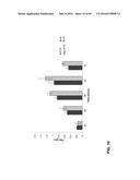 | 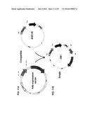 |
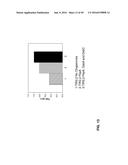 | 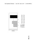 |
 |  |
 |  |
 | 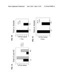 |
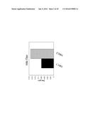 |  |
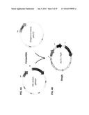 | 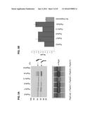 |
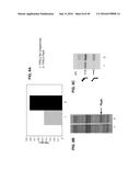 |  |
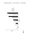 | 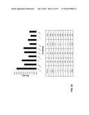 |
 | 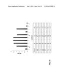 |
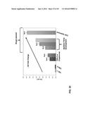 | 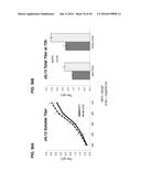 |
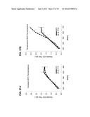 | 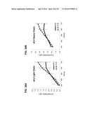 |
 | 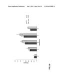 |
 | 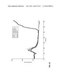 |
 | 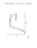 |
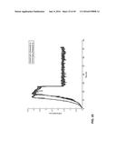 | 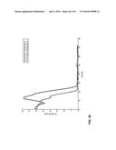 |
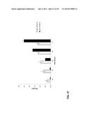 |  |
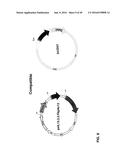 |  |
 |  |
 |  |
 |  |
 |  |
 |  |
 |  |
 |  |
 |  |
 |  |
 |  |
 | 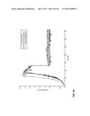 |
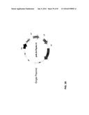 | 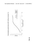 |
 |  |
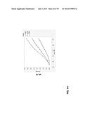 | 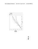 |
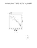 | 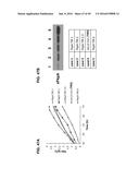 |
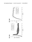 | 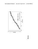 |
 |  |
 |  |
 |  |
 |  |
| Similar patent applications: | |
| Date | Title |
|---|---|
| 2016-06-09 | Methods of producing two chain proteins in bacteria |
| 2016-06-16 | Methods and compositions for detecting antibiotic resistant bacteria |
| 2016-06-30 | Site-specific incorporation of phosphoserine into proteins in escherichia coli |
| 2016-07-14 | Clinical diagnosis of hepatic fibrosis using a novel panel of low abundant human plasma protein biomarkers |
| 2016-06-09 | Method of producing alcohol from cellulose-containing biomass |
| New patent applications in this class: | |
| Date | Title |
|---|---|
| 2022-05-05 | Methods for increasing mannose content of recombinant proteins |
| 2017-08-17 | Polynucleotides encoding anti-notch1 nrr antibody polypeptides |
| 2017-08-17 | Cell line 3m |
| 2017-08-17 | Compositions and methods for phagocyte delivery of anti-staphylococcal agents |
| 2016-12-29 | Cell culture process |
| New patent applications from these inventors: | |
| Date | Title |
|---|---|
| 2010-07-01 | Altered br-3 binding polypeptides |
| Top Inventors for class "Chemistry: molecular biology and microbiology" | |
| Rank | Inventor's name |
|---|---|
| 1 | Marshall Medoff |
| 2 | Anthony P. Burgard |
| 3 | Mark J. Burk |
| 4 | Robin E. Osterhout |
| 5 | Rangarajan Sampath |