Patent application title: ANTI-B7-H5 ANTIBODIES AND THEIR USES
Inventors:
Lieping Chen (Hamden, CT, US)
Lieping Chen (Hamden, CT, US)
Sheng Yao (Columbia, MD, US)
Linda Liu (Clarksville, MD, US)
Linda Liu (Clarksville, MD, US)
Solomon Langermann (Baltimore, MD, US)
Solomon Langermann (Baltimore, MD, US)
IPC8 Class: AC07K1628FI
USPC Class:
4241391
Class name: Drug, bio-affecting and body treating compositions immunoglobulin, antiserum, antibody, or antibody fragment, except conjugate or complex of the same with nonimmunoglobulin material binds antigen or epitope whose amino acid sequence is disclosed in whole or in part (e.g., binds specifically-identified amino acid sequence, etc.)
Publication date: 2016-04-07
Patent application number: 20160096891
Abstract:
The present invention relates to antibodies and their antigen-binding
fragments and to other molecules that are capable of immunospecifically
binding to the B7-H5 ligand of the B7-H5:CD28H pathway, and to the uses
of such molecules in the treatment and diagnosis of autoimmune disease,
transplant rejection and other inflammatory diseases.Claims:
1. An antibody or antigen binding fragment thereof comprising six
complementarity determining regions (CDRs), wherein the CDRs comprise (1)
the three light chain CDRs of SEQ ID NO:11 and the three heavy chain CDRs
of SEQ ID NO:9, or (2) the three light chain CDRs of SEQ ID NO:15 and the
three heavy chain CDRs of SEQ ID NO:13, and wherein the antibody or
antigen binding fragment thereof binds to B7-H5.
2. The antibody or antigen binding fragment thereof of claim 1, wherein the three light chain CDRs comprise a first light chain CDR comprising amino acids 27-38 of SEQ ID NO:11, a second light chain CDR comprising amino acids 56-58 of SEQ ID NO:11, and a third light chain CDR comprising amino acids 95-102 of SEQ ID NO:11.
3. The antibody or antigen binding fragment thereof of one of claim 1 or 2, wherein the three heavy chain CDRs comprise a first heavy chain CDR comprising amino acids 26-33 of SEQ ID NO:9, a second heavy chain CDR comprising amino acids 51-58 of SEQ ID NO:9, and a third heavy chain CDR comprising amino acids 97-106 of SEQ ID NO:9.
4. The antibody or antigen binding fragment thereof of one of claims 1-3 comprising a light chain variable region comprising the amino acid sequence of SEQ ID NO:11.
5. The antibody or antigen binding fragment thereof of one of claims 1-4 comprising a heavy chain variable region comprising the amino acid sequence of SEQ ID NO:9.
6. The antibody or antigen binding fragment thereof of claim 1, wherein the three light chain CDRs comprise a first light chain CDR comprising amino acids 27-38 of SEQ ID NO:15, a second light chain CDR comprising amino acids 56-58 of SEQ ID NO:15, and a third light chain CDR comprising amino acids 95-102 of SEQ ID NO:15.
7. The antibody or antigen binding fragment thereof of one of claim 1 or 2, wherein the three heavy chain CDRs comprise a first heavy chain CDR comprising amino acids 26-33 of SEQ ID NO:13, a second heavy chain CDR comprising amino acids 51-58 of SEQ ID NO:13, and a third heavy chain CDR comprising amino acids 97-106 of SEQ ID NO:13.
8. The antibody or antigen binding fragment thereof of one of claims 1-3 comprising a light chain variable region comprising the amino acid sequence of SEQ ID NO:15.
9. The antibody or antigen binding fragment thereof of one of claims 1-4 comprising a heavy chain variable region comprising the amino acid sequence of SEQ ID NO:13.
10. The antibody or antigen binding fragment thereof of claim 1 comprising a light chain variable region comprising the amino acid sequence of SEQ ID NO:11 and a heavy chain variable region comprising the amino acid sequence of SEQ ID NO:9.
11. The antibody or antigen binding fragment thereof of claim 1 comprising a light chain variable region comprising the amino acid sequence of SEQ ID NO:15 and a heavy chain variable region comprising the amino acid sequence of SEQ ID NO:13.
12. The antibody or antigen binding fragment thereof of claim 1, wherein the bound B7-H5 is arrayed on the surface of a live cell or expressed at an endogenous or transfected concentration.
13. The antibody or antigen binding fragment thereof of claim 7, wherein the live cell is a macrophage or dendritic cell.
14. The antibody or antigen binding fragment thereof of claim 1, wherein the B7-H5 is human B7-H5.
15. The antibody or antigen binding fragment thereof of claim 1 wherein the antibody or antigen binding fragment thereof (A) attenuates the ability of a ligand of B7-H5 to bind to CD28H; (B) antagonizes signal transduction that occurs as a consequence of B7-H5 binding to CD28H; (C) inhibits an allogeneic T cell response; (D) a combination thereof.
16. The antibody or antigen binding fragment thereof of any one of claims 1-15 comprising one or more constant domains from an immunoglobulin constant region (Fc).
17. The antibody or antigen binding fragment thereof of claim 16 wherein the constant domains are human constant domains.
18. The antibody or antigen binding fragment thereof of claim 17 wherein the human constant domains are IgA, IgD, IgE, IgG or IgM domains.
19. The antibody or antigen binding fragment thereof of claim 18 wherein human IgG constant domains are IgG1, IgG2, IgG3, or IgG4 domains.
20. The antibody or antigen binding fragment thereof of any one of claims 1-19 wherein the antibody or antigen binding fragment thereof is detectably labeled or comprises a conjugated toxin, drug, receptor, enzyme, receptor ligand.
21. The antibody or antigen binding fragment thereof of any one of claim 1-20, wherein the antibody is a monoclonal antibody, a human antibody, a chimeric antibody or a humanized antibody.
22. The antibody or antigen binding fragment thereof of any one of claims 1-21, wherein the antibody is a bispecific, trispecific or multispecific antibody.
23. A humanized antibody or antigen binding fragment thereof comprising one or more human IgG4 constant domains and a light chain variable region comprising the amino acid sequence of SEQ ID NO:11, a heavy chain variable region comprising the amino acid sequence of SEQ ID NO:9, or a light chain variable region comprising the amino acid sequence of SEQ ID NO:15, a heavy chain variable region comprising the amino acid sequence of SEQ ID NO:13.
24. A pharmaceutical composition comprising the antibody or antigen binding fragment thereof of any one of claims 1-23, or a CD28H-Ig fusion protein and a physiologically acceptable carrier or excipient.
25. The pharmaceutical composition of claim 24 for use in a method of down-modulating the immune system of a subject.
26. The pharmaceutical composition for use of claim 25 wherein the subject has an autoimmune disease.
27. The pharmaceutical composition of claim 24 for use in a method of treating an autoimmune disease.
28. The pharmaceutical composition for use of claim 26 or 27 wherein the autoimmune disease is selected from the group consisting of alopecia areata, ankylosing spondylitis, antiphospholipid syndrome, autoimmune Addison's disease, autoimmune diseases of the adrenal gland, autoimmune hemolytic anemia, autoimmune hepatitis, autoimmune oophoritis and orchitis, autoimmune thrombocytopenia, Behcet's disease, bullous pemphigoid, cardiomyopathy, celiac sprue-dermatitis, chronic fatigue immune dysfunction syndrome (CFIDS), chronic inflammatory demyelinating polyneuropathy, Churg-Strauss syndrome, cicatrical pemphigoid, CREST syndrome, cold agglutinin disease, Crohn's disease, discoid lupus, essential mixed cryoglobulinemia, fibromyalgia-fibromyositis, glomerulonephritis, Graves' disease, Guillain-Barre, Hashimoto's thyroiditis, idiopathic pulmonary fibrosis, idiopathic thrombocytopenia purpura (ITP), IgA neuropathy, juvenile arthritis, lichen planus, lupus erthematosus, Meniere's disease, mixed connective tissue disease, multiple sclerosis, Neuromyelitis optica (NMO), type 1 or immune-mediated diabetes mellitus, myasthenia gravis, pemphigus vulgaris, pernicious anemia, polyarteritis nodosa, polychrondritis, polyglandular syndromes, polymyalgia rheumatica, polymyositis and dermatomyositis, primary agammaglobulinemia, primary biliary cirrhosis, psoriasis, psoriatic arthritis, Raynauld's phenomenon, Reiter's syndrome, Rheumatoid arthritis, sarcoidosis, scleroderma, Sjogren's syndrome, stiff-man syndrome, systemic lupus erythematosus, lupus erythematosus, takayasu arteritis, temporal arteristis/giant cell arteritis, ulcerative colitis, uveitis, vasculitides such as dermatitis herpetiformis vasculitis, vitiligo, and Wegener's granulomatosis
29. The pharmaceutical composition for use of claim 25 wherein the subject has an inflammatory disease.
30. The pharmaceutical composition of claim 24 for use in a method of treating an inflammatory disease.
31. The pharmaceutical composition for use of claim 29 or 30 wherein the inflammatory disease is selected from the group consisting of asthma, encephilitis, inflammatory bowel disease, chronic obstructive pulmonary disease (COPD), allergic disorders, septic shock, pulmonary fibrosis, undifferentiated spondyloarthropathy, undifferentiated arthropathy, arthritis, inflammatory osteolysis, and chronic inflammation resulting from chronic viral or bacterial infections.
32. The pharmaceutical composition for use of claim 25 wherein the subject has received or will receive a transplant.
33. The pharmaceutical composition of claim 24 for use in a method of treating or preventing a transplant rejection.
34. Use of the antibody or antigen binding fragment thereof of any of claims 1-23 or a CD28H-Ig fusion protein in manufacture of a medicament for down-modulating the immune system in a subject.
35. Use of the antibody or antigen binding fragment thereof of any of claims 1-23 or a CD28H-Ig fusion protein in manufacture of a medicament for treating an autoimmune disease in a subject.
36. Use of the antibody or antigen binding fragment thereof of any of claims 1-23 or a CD28H-Ig fusion protein in manufacture of a medicament for treating an inflammatory disease in a subject.
37. Use of the antibody or antigen binding fragment thereof of any of claims 1-23 or a CD28H-Ig fusion protein in manufacture of a medicament for treating or preventing transplant rejection in a subject.
38. A method of detection or diagnosis of a disease, disorder or infection, comprising: (a) assaying the expression of B7-H5 in cells or in a tissue sample of a subject using the antibody or antigen binding fragment thereof of any one of claims 1-23 and (b) comparing the level of the B7-H5 with a control level, wherein an increase in the assayed level of B7-H5 compared to the control level is indicative of the disease, disorder or infection.
39. The method of claim 38, wherein the expression of B7-H5 is assayed by enzyme linked immunosorbent assay (ELISA), radioimmunoassay (RIA), or fluorescence-activated cell sorting (FACS).
40. A method for monitoring the progression of a disease, disorder or infection, comprising: (a) assaying the expression of B7-H5 in cells or in a tissue sample of a subject a first time point using the antibody or antigen binding fragment thereof of any one of claims 1-23; and (b) comparing the level of expression of B7-H5 in the cells or in the tissue sample of the subject at second time point, or over a time course, wherein an increase in the assayed level of B7-H5 is indicative of the progression of disease, disorder or infection.
41. The method of claim 30, wherein the expression of B7-H5 is assayed by enzyme linked immunosorbent assay (ELISA) radioimmunoassay (RIA), or fluorescence-activated cell sorting (FACS).
42. A method for monitoring the response to treatment, comprising: (a) assaying the expression of B7-H5 in cells or in a tissue sample of a subject prior to treatment using the antibody or antigen binding fragment thereof of any one of claims 1-23; and (b) assaying the expression of B7-H5 in cells or in a tissue sample of a subject prior at one or more time points after treatment, and comparing the level of B7-H5 over time, whereby a decrease in the assayed level of B7-H5 compared to the pre-treatment level of B7-H5 is indicative of a response to treatment.
43. The method of claim 42, wherein the expression of B7-H5 is assayed by enzyme linked immunosorbent assay (ELISA), radioimmunoassay (RIA), or fluorescence-activated cell sorting (FACS).
44. A pharmaceutical composition for use in a method of treating a subject for a disease characterized by increased expression of B7-H5, wherein the method comprises the steps of (i) determining whether the subject has a disease characterized by increased expression of B7-H5 by (a) assaying the expression of B7-H5 in cells or in a tissue sample of the subject using the antibody or antigen binding fragment thereof that binds to B7-H5; (b) comparing the level of the B7-H5 with a control level, wherein an increase in the assayed level of B7-H5 compared to the control level is indicative that the subject has a disease characterized by increased expression of B7-H5; and (ii) administering to the subject a therapeutically effective amount of the pharmaceutical composition of claim 24 if the subject has a disease characterized by increased expression of B7-H5.
45. The method of claim 44 wherein the antibody or antigen binding fragment thereof is the antibody or antigen binding fragment thereof of any one of claims 1-23.
46. The pharmaceutical compositions for use of claim 44, wherein the disease characterized by increased expression of B7-H5 is an autoimmune disease.
47. The pharmaceutical compositions for use of claim 44, wherein the disease characterized by increased expression of B7-H5 is an inflammatory disease.
48. A pharmaceutical composition for use in a method of treating a subject for a disease characterized by increased expression of CD28H, wherein the method comprises the steps of (i) determining whether the subject has a disease characterized by increased expression of CD28H by (a) assaying the expression of CD28H in cells or in a tissue sample of the subject using an anti-CD28H antibody or an antigen binding fragment thereof; (b) comparing the level of the CD28H with a control level, wherein an increase in the assayed level of CD28H compared to the control level is indicative that the subject has a disease characterized by increased expression of CD28H; and (ii) administering to the subject a therapeutically effective amount of the pharmaceutical composition of claim 24 if the subject has a disease characterized by increased expression of CD28H.
49. The pharmaceutical compositions for use of claim 48, wherein the disease characterized by increased expression of CD28H is an autoimmune disease.
50. The pharmaceutical compositions for use of claim 48, wherein the disease characterized by increased expression of CD28H is an inflammatory disease.
Description:
REFERENCE TO SEQUENCE LISTING
[0002] This application includes one or more Sequence Listings pursuant to 37 C.F.R. §1.821 et seq., which are disclosed in both paper and computer-readable media, and which paper and computer-readable disclosures are herein incorporated by reference in their entirety.
FIELD OF THE INVENTION
[0003] The present invention relates to antibodies and their antigen-binding fragments and to other molecules that are capable of binding to the B7-H5 ligand of the B7-H5:CD28H pathway, and to the uses of such molecules in the treatment and diagnosis of autoimmune disease, transplant rejection and other inflammatory diseases.
BACKGROUND OF THE INVENTION
[0004] The immune system of humans and other mammals is responsible for providing protection against infection and disease. Such protection is provided both by a humoral immune response and by a cell-mediated immune response. The humoral response results in the production of antibodies and other biomolecules that are capable of recognizing and neutralizing foreign targets (antigens). In contrast, the cell-mediated immune response involves the activation of macrophages, natural killer cells (NK), and antigen-specific cytotoxic T-lymphocytes by T cells, and the release of various cytokines in response to the recognition of an antigen (Dong, C. et al. (2003) "Immune Regulation by Novel Costimulatory Molecules," Immunolog. Res. 28(1):39-48).
[0005] The ability of T cells to optimally mediate an immune response against an antigen requires two distinct signaling interactions (Viglietta, V. et al. (2007) "Modulating Co-Stimulation," Neurotherapeutics 4:666-675; Korman, A. J. et al. (2007) "Checkpoint Blockade in Cancer Immunotherapy," Adv. Immunol. 90:297-339). First, antigen that has been arrayed on the surface of antigen-presenting cells (APC) must be presented to an antigen-specific naive CD4.sup.+ T cell. Such presentation delivers a signal via the T cell receptor (TCR) that directs the T cell to initiate an immune response that will be specific to the presented antigen. Second, a series of co-stimulatory and co-inhibitory signals, mediated through interactions between the APC and distinct T cell surface molecules, triggers first the activation and proliferation of the T cells and ultimately their inhibition. Thus, the first signal confers specificity to the immune response whereas the second signal serves to determine the nature, magnitude and duration of the response.
[0006] The immune system is tightly controlled by co-stimulatory and co-inhibitory ligands and receptors. These molecules provide the second signal for T cell activation and provide a balanced network of positive and negative signals to maximize immune responses against infection while limiting immunity to self (Wang, L. et al. (Mar. 7, 2011) "VISTA, A Novel Mouse Ig Superfamily Ligand That Negatively Regulates T Cell Responses," J. Exp. Med. 10.1084/jem.20100619:1-16; Lepenies, B. et al. (2008) "The Role Of Negative Costimulators During Parasitic Infections," Endocrine, Metabolic & Immune Disorders--Drug Targets 8:279-288). Of particular importance is binding between the B7.1 (CD80) and B7.2 (CD86) ligands of the Antigen Presenting Cell and the CD28 and CLTA-4 receptors of the CD4.sup.+ T-lymphocyte (Sharpe, A. H. et al. (2002) "The B7-CD28 Superfamily," Nature Rev. Immunol. 2:116-126; Dong, C. et al. (2003) "Immune Regulation by Novel Costimulatory Molecules," Immunolog. Res. 28(1):39-48; Lindley, P. S. et al. (2009) "The Clinical Utility Of Inhibiting CD28-Mediated Costimulation," Immunol. Rev. 229:307-321). Binding of B7.1 or of B7.2 to CD28 stimulates T cell activation; binding of B7.1 or B7.2 to CTLA4 inhibits such activation (Dong, C. et al. (2003) "Immune Regulation by Novel Costimulatory Molecules," Immunolog. Res. 28(1):39-48; Lindley, P. S. et al. (2009) "The Clinical Utility Of Inhibiting CD28-Mediated Costimulation," Immunol. Rev. 229:307-321; Greenwald, R. J. et al. (2005) "The B7 Family Revisited," Ann. Rev. Immunol. 23:515-548). CD28 is constitutively expressed on the surface of T cells (Gross, J., et al. (1992) "Identification And Distribution Of The Costimulatory Receptor CD28 In The Mouse," J. Immunol. 149:380-388), whereas CTLA4 expression is rapidly up-regulated following T-cell activation (Linsley, P. et al. (1996) "Intracellular Trafficking Of CTLA4 And Focal Localization Towards Sites Of TCR Engagement," Immunity 4:535-543). Since CTLA4 is the higher affinity receptor (Sharpe, A. H. et al. (2002) "The B7-CD28 Superfamily," Nature Rev. Immunol. 2:116-126), binding first initiates T cell proliferation (via CD28) and then inhibits it (via nascent expression of CTLA4), thereby dampening the effect when proliferation is no longer needed.
[0007] Further investigations into the ligands of the CD28 receptor have led to the identification and characterization of a set of related B7 molecules (the "B7 Superfamily") (Coyle, A. J. et al. (2001) "The Expanding B7 Superfamily: Increasing Complexity In Costimulatory Signals Regulating T Cell Function," Nature Immunol. 2(3):203-209; Sharpe, A. H. et al. (2002) "The B7-CD28 Superfamily," Nature Rev. Immunol. 2:116-126; Greenwald, R. J. et al. (2005) "The B7 Family Revisited," Ann. Rev. Immunol. 23:515-548; Collins, M. et al. (2005) "The B7 Family Of Immune-Regulatory Ligands," Genome Biol. 6:223.1-223.7; Loke, P. et al. (2004) "Emerging Mechanisms Of Immune Regulation: The Extended B7 Family And Regulatory T Cells." Arthritis Res. Ther. 6:208-214; Korman, A. J. et al. (2007) "Checkpoint Blockade in Cancer Immunotherapy," Adv. Immunol. 90:297-339; Flies, D. B. et al. (2007) "The New B7s: Playing a Pivotal Role in Tumor Immunity," J. Immunother. 30(3):251-260; Agarwal, A. et al. (2008) "The Role Of Positive Costimulatory Molecules In Transplantation And Tolerance," Curr. Opin. Organ Transplant. 13:366-372; Lenschow, D. J. et al. (1996) "CD28/B7 System of T Cell Costimulation," Ann. Rev. Immunol. 14:233-258; Wang, S. et al. (2004) "Co-Signaling Molecules Of The B7-CD28 Family In Positive And Negative Regulation Of T Lymphocyte Responses," Microbes Infect. 6:759-766). There are currently nine known members of the family: B7.1 (CD80), B7.2 (CD86), the inducible co-stimulator ligand (ICOS-L, B7-H2), the programmed death-1 ligand (PD-L1; B7-H1), the programmed death-2 ligand (PD-L2; B7-DC), B7-H3, B7-H4 (also referred to as B7x, B7-H6 and B7S1; Sica, G. L. et al. (2003) "B7-4, A Molecule Of The B7 Family, Negatively Regulates T Cell Immunity," Immunity 18:849-861; Zang, X. et al. (2003) B7x: A Widely Expressed B7 Family Member That Inhibits T Cell Activation," Proc. Natl. Acad. Sci. (USA) 100:10388-10392; Prasad, D. V. et al. (2003) B7S1, A Novel B7 Family Member That Negatively Regulates T Cell Activation," Immunity 18:863-873), B7-H6 (Collins, M. et al. (2005) "The B7 Family Of Immune-Regulatory Ligands," Genome Biol. 6:223.1-223.7) and B7-H5 (Flajnik, M. F. et al. (2012) "Evolution Of The B7 Family: Co-Evolution Of B7H6 And Nkp30, Identification Of A New B7 Family Member, B7H7, And Of B7's Historical Relationship With The MHC," Immunogenetics 64:571-590). The B7 family of genes is essential in the regulation of the adaptive immune system. Most B7 family members contain both variable (V)- and constant (C)-type domains of the immunoglobulin superfamily (IgSF).
[0008] B7 ligands are expressed on the cell surface of many different cell types including antigen presenting cells (APCs) and their interaction with receptor molecules on T cells provide activating and/or inhibitory signals that regulate T cell activation and tolerance (Collins, M. et al. (2005) "The B7 Family Of Immune-Regulatory Ligands," Genome Biol. 6:223.1-223.7). Some inhibitory B7 ligands are also expressed on tumor cells, resulting in suppression of immune responses (Keir, M. E. et al. (2008) "PD-1 And Its Ligands In Tolerance And Immunity," Annu. Rev. Immunol. 26:677-704; Zou, W. et al. (2008) "Inhibitory B7-Family Molecules In The Tumour Microenvironment," Nat. Rev. Immunol. 8:467-477). Therefore, stimulating or attenuating the interactions of B7 ligands and their receptors holds therapeutic potential for autoimmune diseases, cancer and infectious disease (WO 2011/020024; Flajnik, M. F. et al. (2012) "Evolution Of The B7 Family: Co-Evolution Of B7H6 And Nkp30, Identification Of A New B7 Family Member, B7H7, And Of B7's Historical Relationship With The MHC," Immunogenetics 64:571-590).
[0009] Despite all prior advances in the treatment of inflammatory disease or autoimmune disease, a need remains for compositions capable of providing enhanced immunotherapy for the treatment of such conditions. The present invention is directed to such compositions and their use to treat inflammatory disease, autoimmune disease, and similar diseases and conditions characterized by a hyperactive immune system.
SUMMARY OF THE INVENTION
[0010] Antibodies and their antigen-binding fragments and other molecules that are capable of immunospecifically binding to the B7-H5 ligand of the B7-H5:CD28H pathway, and to the uses of such molecules in the treatment and diagnosis of autoimmune disease, transplant rejection and other inflammatory diseases are provided.
[0011] One embodiment provides an antibody or other molecule having an antigen-binding fragment of an antibody that immunospecifically binds to a mammalian B7-H5 (and in particular, a human B7-H5). The B7-H5 can be, for example, arrayed on the surface of a live cell.
[0012] Exemplary live cells include, but are not limited to macrophage or dendritic cells.
[0013] Another embodiment provides a molecule that immunospecifically binds to B7-H5 and is substantially capable of blocking B7-H5's interaction with CD28H. Still another embodiment provides a molecule that immunospecifically binds to B7-H5 and is substantially incapable of blocking B7-H5's interaction with CD28H.
[0014] The B7-H5 binding molecules can have antigen-binding fragments that contain six CDRs. The CDRs can include at least one consensus CDR of the CDRs of anti-B7-H5 antibodies: 2D3 and 18C3, with all remaining CDRs selected from:
[0015] (A) the three light chain and the three heavy chain CDRs of anti-B7-H5 antibody 2D3; or
[0016] (B) the three light chain and the three heavy chain CDRs of anti-B7-H5 antibody 18C3.
[0017] Another embodiment provides B7-H5 binding molecules wherein the six CDRs are:
[0018] (A) the three light chain and the three heavy chain CDRs of anti-B7-H5 antibody 2D3; or
[0019] (B) the three light chain and the three heavy chain CDRs of anti-B7-H5 antibody 18C3.
[0020] In some embodiments, the molecule is a chimeric antibody that includes antigen binding fragments (Fab) from two or more different B7-H5 antibodies.
[0021] The B7-H5 binding molecules can be a monoclonal antibody, a human antibody, a chimeric antibody or a humanized antibody and/or is a bispecific, trispecific or multispecific antibody.
[0022] The B7-H5 binding molecules can be detectably labeled or include a conjugated toxin, drug, receptor, enzyme, or receptor ligand.
[0023] Still another embodiment provides a pharmaceutical composition containing a therapeutically effective amount or a prophylactically effective amount of any of the above-described B7-H5 binding molecules and a physiologically acceptable carrier or excipient.
[0024] In other embodiments, a molecule that is substantially capable of blocking B7-H5's interaction with CD28H is an CD28H fusion protein, preferably an CD28H-Ig fusion protein.
[0025] Methods of using the pharmaceutical compositions in the treatment of a disease are also provided.
[0026] A method for diagnosing a disease in a subject includes assaying cells of the subject for their ability to bind to any of the above-described B7-H5 binding molecules, wherein the method provides a cytologic assay for diagnosing the presence of a disease in the subject.
[0027] The disclosed B7-H5 binding molecules can be used to an autoimmune disease or transplant rejection.
BRIEF DESCRIPTION OF THE DRAWINGS
[0028] FIG. 1 provides a schematic depiction of B7-H5 and its counter-receptor, CD28H.
[0029] FIG. 2, Panels A-J, show that B7-H5 is constitutively expressed on macrophages and inducible on dendritic cells.
[0030] FIG. 3, Panels A-D, show that CD28H is expressed on T cells and natural killer (NK) cells, but not on B cells.
[0031] FIGS. 4A-4B show the ability of the antibodies produced by clones 2D3 and 18C3 to bind human B7-H5 expressed by a CHO transfectants.
[0032] FIGS. 5A-5C show the abilities of the isolated antibodies and control Ig to block the B7-H5:CD28H interaction.
[0033] FIG. 6 shows the ability of a B7-H5 Ig fusion to stimulate a T cell response.
[0034] FIG. 7 shows the ability of the B7-H5 Ig to induce the expression of cytokines: IL-2, IFN-γ and IL-10.
[0035] FIG. 8 shows that the anti-B7-H5 antibody 2D3 of the present invention is capable of binding to B7-H5 so as to block the capacity of B7-H5 to interact with its CD28H counter-receptor.
[0036] FIG. 9 shows the ability of anti-B7-H5 antibody 2D3 to inhibit an allogeneic T cell response.
[0037] FIG. 10A and FIG. 10B show the effect of anti-human B7-H5 antibody on the percentage of CD28H.sup.+ cells among activated (CD45RO.sup.+) human T cells in the spleen (FIG. 10A) and peripheral blood (FIG. 10B) in NSG mice implanted with human PBMC.
[0038] FIG. 11A and FIG. 11B show the effect of anti-human B7-H5 antibody on the percentage of CD28H.sup.+ cells among naive (CD45RO.sup.-) human T cells in the spleen (FIG. 11A) and peripheral blood (FIG. 11B) in NSG mice implanted with human PBMC.
[0039] FIGS. 12A-12C show binding properties of recombinant anti-human B7-H5 chimeric antibodies (2D3 and 18C3). FIGS. 12A-12C show the ability of anti-human B7-H5 antibodies (2D3 and 18C3) that had been recombinantly converted from their murine IgG isotype to a human IgG4 isotype to bind human B7-H5 expressed by CHO cells.
[0040] FIG. 13 (Panels A-C) shows the ability of recombinant anti-human B7-H5 chimeric antibodies (2D3 and 18C3) to completely block the binding of CD28H fusion protein to CHO B7-H5 transfectants. Human B7-H5 FL CHO transfectants were pre-incubated with control supernatant (Panel A) or recombinant chimeric 2D3 (Panel B) and 18C3 supernatants (Panel C), and subsequently stained with biotinylated CD28HhIg fusion protein.
[0041] FIGS. 14A-14B show the epitope recognition site of anti-human B7-H5 antibodies (2D3 and 18C3). FIG. 14A and FIG. 14B show that 2D3 and 18C3 recognize the first IgV domain of B7-H5. FIG. 14C shows B7-H5 IgV domain mediates B7-H5's interaction with CD28H.
[0042] FIG. 15 compares the affinities of antibodies 2D3 and 18C3 for binding to human B7-H5 as expressed on the surface of CHO cells.
[0043] FIGS. 16A-16B show the effect of the endogenous B7-H5:CD28H interaction on the recall of TT-specific memory T cells. FIG. 16A shows TT vaccine-induced T cell proliferation in the presence of the B7-H5:CD28H interaction blocking antibody 2D3 (Panel B) or control Igs (Panel A). FIG. 16B shows the levels of expressed cytokines (Panel A) and the BrdU-positive cells (Panel B) associated with such response.
[0044] FIGS. 17A-17B show the blockade of endogenous B7-H5:CD28H interaction by 2D3 inhibits the allogeneic proliferative responses of both CD8+(FIG. 17A, Panels A-B) and CD4+(FIG. 17B, Panels A-B) T cells in humanized NSG mice.
[0045] FIG. 18 shows simultaneous cross-linking of TCR and B7-H5 fusion protein induced AKT phosphorylation 30 min after stimulation, while TCR cross-linking alone induced minimal AKT phosphorylation. Importantly, inclusion of B7-H5 antibody 2D3 prevented AKT activation, indicating B7-H5 co-stimulation utilizes the AKT pathway to promote T cell response.
[0046] FIG. 19 shows B7-H5-CD28H pathway blockade by decoy receptor fusion protein CD28HIg suppressed the cytotoxic killing activity of allogeneic CD8 T cell against B7-H5 transfected 624Mel melanoma cell line, indicating B7-H5-CD28H pathway promotes cytotoxic killing activity of CD8 T cells on target cells.
[0047] FIG. 20A-B show simultaneous cross-linking of CD16 and B7-H5 fusion protein on Natural Killer cells (NK) in the presence of soluble recombinant human IL-2 induced NK activation and degranulation (CD107a surface upregulation), whereas CD16 engagement alone did not induce significant NK degranulation. A B7-H5 fusion protein dose dependent NK activation was observed (FIG. 20A). Importantly, inclusion of B7-H5 antibody 2D3 prevented NK activation (FIG. 20B), indicating B7-H5 co-stimulates NK activation.
DETAILED DESCRIPTION OF THE INVENTION
[0048] The present invention relates to antibodies and their antigen-binding fragments and to other molecules that are capable of immunospecifically binding to the B7-H5 ligand of the B7-H5:CD28H pathway, and to the uses of such molecules in the treatment and diagnosis of autoimmune disease, transplant rejection and other inflammatory diseases.
[0049] The B7-H5:CD28H pathway is a cellular immunity pathway that involves the ligand B7-H5 and its counter-receptor CD28H (FIG. 1). B7-H5 is expressed on antigen presenting cells; it is constitutively expressed on macrophages and inducible on dendritic cells (FIG. 2, Panels A-J). CD28H is particularly expressed on naive T cells, NK cells, and plasmacytoid dendritic cells (especially in the spleen, lymph node and thymus), and its expression is down-regulated on matured or activated cells. It is not expressed on γδ T cells or B cells (FIG. 3), but is expressed on Tn, TCM, TEM, and TEMRA T cell subsets. Human cord blood T cells express CD28H as do CD4.sup.+, CD8.sup.+ and CD4.sup.+/CD8.sup.+ thymocytes. B7-H5 interacts with the CD28H counter-receptor to stimulate the immune system, thereby promoting enhanced immune responses. Down-regulation of CD28H has been found to impair activated/memory T cell survival in vivo. Thus, the interaction between B7-H5 and CD28H is important for native T cell priming and activated/memory T cell survival in vivo. CD28H is also seen to be down-regulated in chronically antigen-exposed/exhausted T cells. CD28H is constitutively expressed on Natural Killer cell (NK). B7-H5-CD28H pathway promotes NK activation and degranulation in the presence of other NK activation signal, such as CD16 crosslinking.
[0050] The present invention reflects, in part, the recognition that molecules, such as antibodies and their antigen binding fragments that immunospecifically bind to B7-H5 ("anti-B7-H5 antibodies"), and particularly anti-B7-H5 antibodies or decoy receptor fusion protein CD28HIg that block the B7-H5:CD28H interaction so as to impair (i.e., prevent or attenuate) the ability of B7-H5 to bind to CD28H, are capable of impairing the ability of B7-H5 to promote enhanced immune responses via the CD28H interaction, and thus are capable of serving as antagonists of T cell proliferation and cytokine production and NK activation. Such molecules are capable of mediating a physiological reduction in immune system activation and thus have utility in the treatment of inflammatory disease and autoimmune disease. In particular, such antibodies are capable of reducing the percentage of activated T cells and NK cells in vivo.
A. B7-H5
[0051] The B7-H5 amino acid sequence was found to be similar to a previously discovered human gene, HHLA2 (human endogenous retrovirus-H long terminal repeat-associating protein 2 (HHLA2); Mager, D. L. et al. (1999) "Endogenous Retroviruses Provide The Primary Polyadenylation Signal For Two New Human Genes (HHLA2 And HHLA3," Genomics 59:255-263), that had no known function (Flajnik, M. F. et al. (2012) "Evolution Of The B7 Family: Co-Evolution Of B7H6 And Nkp30, Identification Of A New B7 Family Member, B7H7, And Of B7's Historical Relationship With The MHC," Immunogenetics 64:571-590). B7-H5 is also referred to herein and elsewhere as B7-H7. Accordingly, the terms B7-H5 and B7-H7 are used interchangeably herein.
[0052] The human B7-H5 sequence has been found to have homologs in chicken, opossum, hoofed mammals (e.g., horse, pig), salmon, and shark. However, only pseudogenes have been thus far identified in rodents (mouse and rat). The amino acid sequences of such genes reveal a similar domain structures in all species, with conservation of the canonical residues for Ig superfamily domains.
[0053] Human B7-H5 polypeptide is 414 amino acids in length and has been reported to contain the following: a signal sequence, an extracellular domain having 3 immunoglobulin-like (Ig-like) domains, a transmembrane domain, and a cytoplasmic domain. In particular, the human B7-H5 polypeptide has been reported to contain an Ig-like V-type 1 domain, an Ig-like C-1 type domain, and an Ig-like V-type 2 domain. Multiple naturally occurring variants of B7-H5 exist (e.g., Accession No. Q9UM44-1 (homo sapiens), NP_009003 (GI: 5901964, homo sapiens), and AAD48396 (GI: 15726285, homo sapiens); see WO 2011/020024).
[0054] The term "native-B7-H5" (also "native-B7-H7") refers to any naturally occurring B7-H5 amino acid sequence, including immature or precursor and mature forms. The amino acid sequence of a representative human B7-H5, Accession No. Q9UM44-1, is (SEQ ID NO:1):
TABLE-US-00001 MKAQTALSFF LILITSLSGS QGIFPLAFFI YVPMNEQIVI GRLDEDIILP SSFERGSEVV IHWKYQDSYK VHSYYKGSDH LESQDPRYAN RTSLFYNEIQ NGNASLFFRR VSLLDEGIYT CYVGTAIQVI TNKVVLKVGV FLTPVMKYEK RNTNSFLICS VLSVYPRPII TWKMDNTPIS ENNMEETGSL DSFSINSPLN ITGSNSSYEC TIENSLLKQT WTGRWTMKDG LHKMQSEHVS LSCQPVNDYF SPNQDFKVTW SRMKSGTFSV LAYYLSSSQN TIINESRFSW NKELINQSDF SMNLMDLNLS DSGEYLCNIS SDEYTLLTIH TVHVEPSQET ASHNKGLWIL VPSAILAAFL LIWSVKCCRA QLEARRSRHP ADGAQQERCC VPPGERCPSA PDNGEENVPL SGKV
[0055] The human B7-H5 has been reported to contain the following: a signal sequence at approximately amino acid residues 1 to 22 of SEQ ID NO:1, an Ig-like V-type 1 domain (shown underlined above) at approximately amino acid residues 61 to 131 of SEQ ID NO:1, an Ig-like C-1 type domain at approximately amino acid residues 138 to 222 of SEQ ID NO:1, an Ig-like V-type 2 domain at approximately amino acid residues 235 to 328 of SEQ ID NO:1, and a transmembrane domain at approximately amino acid residues 345 to 365 of SEQ ID NO:1. The predicted dimer interface for human B7-H5 polypeptide is amino acid residues 141-144, 156, 158, 160, 162, 193-196, 198, 200, 201, 224, and 225 of SEQ ID NO:1. The predicted N-linked glycosylation sites for human B7-H5 polypeptide are at amino acid residues 90, 103, and 318 of SEQ ID NO:1. Natural variations of human B7-H5 polypeptide include BOT, N344K, and S346R (UniProt Q9UM44) (see, WO 2011/020024, which reference is herein incorporated by reference in its entirety for its teaching of the structure and sequence of human B7-H5).
[0056] A DNA sequence encoding human B7-H5 (SEQ ID NO:1) is (SEQ ID NO:2):
TABLE-US-00002 atgaaggcac agacagcact gtctttcttc ctcattctca taacatctct gagtggatct caaggcatat tccctttggc tttcttcatt tatgttccta tgaatgaaca aatcgtcatt ggaagacttg atgaagatat aattctccct tcttcatttg agaggggatc cgaagtcgta atacactgga agtatcaaga tagctataag gttcatagtt actacaaagg cagtgaccat ttggaaagcc aagatcccag atatgcaaac aggacatccc ttttctataa tgagattcaa aatgggaatg cgtcactatt tttcagaaga gtaagccttc tggacgaagg aatttacacc tgctatgtag gaacagcaat tcaagtgatt acaaacaaag tggtgctaaa ggtgggagtt tttctcacac ccgtgatgaa gtatgaaaag aggaacacaa acagcttctt aatatgcagc gtgttaagtg tttatcctcg tccaattatc acgtggaaaa tggacaacac acctatctct gaaaacaaca tggaagaaac agggtctttg gattcttttt ctattaacag cccactgaat attacaggat caaattcatc ttatgaatgt acaattgaaa attcactgct gaagcaaaca tggacagggc gctggacgat gaaagatggc cttcataaaa tgcaaagtga acacgtttca ctctcatgtc aacctgtaaa tgattatttt tcaccaaacc aagacttcaa agttacttgg tccagaatga aaagtgggac tttctctgtc ctggcttact atctgagctc ctcacaaaat acaattatca atgaatcccg attctcatgg aacaaagagc tgataaacca gagtgacttc tctatgaatt tgatggatct taatctttca gacagtgggg aatatttatg caatatttct tcggatgaat atactttact taccatccac acagtgcatg tagaaccgag ccaagaaaca gcttcccata acaaaggctt atggattttg gtgccctctg cgattttggc agcttttctg ctgatttgga gcgtaaaatg ttgcagagcc cagctagaag ccaggaggag cagacaccct gctgatggag cccaacaaga aagatgttgt gtccctcctg gtgagcgctg tcccagtgca cccgataatg gcgaagaaaa tgtgcctctt tcaggaaaag ta
[0057] The amino acid sequence of a representative human B7-H5 IgV domain human IgG4 fusion protein is (SEQ ID NO:3) (B7-H5 sequences are shown in boldface; IgG4 sequences are shown underlined):
TABLE-US-00003 IFPLAFFIYV PMNEQIVIGR LDEDIILPSS FERGSEVVIH WKYQDSYKVH SYYKGSDHLE SQDPRYANRT SLFYNEIQNG NASLFFRRVS LLDEGIYTCY VGTAIQVITN KVVLKVGVFL TPVMKYEKES KYGPPCPPCP APEFLGGPSV FLFPPKPKDT LMISRTPEVT CVVVDVSQED PEVQFNWYVD GVEVHNAKTK PREEQFNSTY RVVSVLTVLH QDWLNGKEYK CKVSNKGLPS SIEKTISKAK GQPREPQVYT LPPSQEEMTK NQVSLTCLVK GFYPSDIAVE WESNGQPENN YKTTPPVLDS DGSFFLYSRL TVDKSRWQEG NVFSCSVMHE ALHNHYTQKS LSLSPG
[0058] The fusion protein may additionally comprise an N-terminal leader sequence (such residues 1-22 of SEQ ID NO:1, i.e., the naturally occurring B7-H5 leader sequence (SEQ ID NO:4):
TABLE-US-00004 MKAQTALSFF LILITSLSGS QG
[0059] A DNA sequence encoding the naturally occurring B7-H5 leader sequence is (SEQ ID NO:5):
TABLE-US-00005 atgaaggccc agaccgccct gtccttcttc ctgatcctga tcacctccct gtccggcagc caggga
[0060] Although the B7-H5 sequences are shown fused to a human IgG4 region, in accordance with the present invention, such sequences can alternatively be fused to any Ig isotype, or indeed to any other protein.
[0061] A DNA sequence encoding human B7-H5IgV-hIgG4 (SEQ ID NO:3) is (SEQ ID NO:6):
TABLE-US-00006 atcttccctc tggccttctt catctacgtg cccatgaacg agcagatcgt gatcggccgg ctggacgagg atattatcct gccctccagc ttcgagcggg gctccgaggt cgtgatccac tggaagtacc aggactccta caaggtgcac tcctactaca agggctccga ccacctggaa tcccaggacc ccagatacgc caaccggacc agcctgttct acaacgagat ccagaacggc aacgcctccc tgttcttccg gcgagtgtcc ctgctggatg agggcatcta cacctgttac gtgggcaccg ccatccaagt gatcaccaac aaggtggtgc tgaaagtggg cgtgttcctg acccccgtga tgaagtacga gaaagagtct aagtacggcc ctccctgccc cccttgtcct gcccctgaat ttctgggcgg accctctgtg ttcctgttcc ccccaaagcc caaggacacc ctgatgatct cccggacccc cgaagtgacc tgcgtggtgg tggatgtgtc ccaggaagat cccgaggtgc agttcaattg gtacgtggac ggcgtggaag tgcacaacgc caagaccaag cccagagagg aacagttcaa ctccacctac cgggtggtgt ccgtgctgac cgtgctgcac caggattggc tgaacggcaa agagtacaag tgcaaggtgt ccaacaaggg cctgcccagc tccatcgaaa agaccatctc caaggccaag ggccagcccc gggaacccca ggtgtacaca ctgcctccaa gccaggaaga gatgaccaag aaccaggtgt ccctgacctg tctcgtgaag ggcttctacc cctccgatat cgccgtggaa tgggagtcca acggccagcc tgagaacaac tacaagacca ccccccctgt gctggactcc gacggctctt tcttcctgta ctcccgcctg accgtggaca agtccagatg gcaggaaggc aacgtgttct cctgctccgt gatgcacgag gccctgcaca accactacac ccagaagtcc ctgtccctga gccccggc
[0062] An amino acid sequence of a representative human B7-H5 human IgG4P fusion protein is including the extracellular domain of B7-H5 is (SEQ ID NO:24) (B7-H5ECD-hIgG4P with B7-H5 ECD aa1-340 underlined, and the Serine 228 to Proline mutation double underline)
TABLE-US-00007 IFPLAFFIYVPMNEQIVIGRLDEDIILPSSFERGSEVVIHWKYQDSYKVH SYYKGSDHLESQDPRYANRTSLFYNEIQNGNASLFERRVSLLDEGIYTCY VGTAIQVITNKVVLKVGVELTPVMKYEKRNTNSFLICSVLSVYPRPIITW KMDNTPISENNMEETGSLDSFSINSPLNITGSNSSYECTIENSLLKQTWT GRWTMKDGLHKMQSEHVSLSCQPVNDYFSPNQDFKVTWSRMKSGTFSVLA YYLSSSQNTIINESRFSWNKELINQSDFSMNLMDLNLSDSGEYLCNISSD EYTLLTIHTVHVEPSQETESKYGPPCPPCPAPEFLGGPSVFLEPPKPKDT LMISRTPEVTCVVVDVSQEDPEVQFNWYVDGVEVHNAKTKPREEQFNSTY RVVSVLTVLHQDWLNGKEYKCKVSNKGLPSSIEKTISKAKGQPREPQVYT LPPSQEEMTKNQVSLTCLVKGFYPSDIAVEWESNGQPENNYKTTPPVLDS DGSFELYSRLTVDKSRWQEGNVESCSVMHEALHNHYTQKSLSLSPG
[0063] The signal sequence can be the native signal sequence (SEQ ID NO:29)
TABLE-US-00008 MKAQTALSFFLILITSLSGSQG
[0064] The fusion protein can be encoded by the nucleic acid sequence (SEQ ID NO:25)
TABLE-US-00009 atgaaggcccagaccgccctgtccttcttcctgatcctgatcacctccct gtccggcagccagggaatcttccctctggccttcttcatctacgtgccca tgaacgagcagatcgtgatcggccggctggacgaggatattatcctgccc tccagcttcgagcggggctccgaggtcgtgatccactggaagtaccagga ctcctacaaggtgcactcctactacaagggctccgaccacctggaatccc aggaccccagatacgccaaccggaccagcctgttctacaacgagatccag aacggcaacgcctccctgttcttccggcgagtgtccctgctggatgaggg catctacacctgttacgtgggcaccgccatccaagtgatcaccaacaagg tggtgctgaaagtgggcgtgttcctgacccccgtgatgaagtacgagaag cggaataccaactctttcctgatctgctccgtgctgtccgtgtaccctcg gcccatcatcacctggaagatggacaacacccccatctccgagaacaaca tggaagagacaggctccctggactccttctccatcaactcccccctgaac attaccggctccaactcctcctacgagtgcaccatcgagaactccctgct gaagcagacctggaccggcagatggactatgaaggacggcctgcacaaga tgcagtccgagcacgtgtccctgtcctgccagcccgtgaacgactacttc agccccaaccaggacttcaaagtgacctggtcccggatgaagtccggcac cttcagcgtgctggcctactacctgtccagctcccagaacaccatcatca acgagtcccggttctcctggaacaaagagctgatcaaccagtccgacttc tccatgaacctgatggacctgaacctgtccgacagcggcgagtacctgtg caacatctccagcgacgagtacaccctgctgaccatccacaccgtgcacg tggaaccctcccaggaaaccgagtctaagtacggccctccctgcccacct tgtcccgcccctgaatttctgggcggaccctctgtgttcctgttcccccc aaagcccaaggacaccctgatgatctcccggacccccgaagtgacatgcg tggtggtggatgtgtcccaggaagatcccgaggtgcagttcaattggtac gtggacggcgtggaagtgcacaacgccaagaccaagcccagagaggaaca gttcaactccacctaccgggtggtgtctgtgctgaccgtgctgcaccagg actggctgaacggcaaagagtacaagtgcaaggtgtccaacaagggcctg cccagctccatcgaaaagaccatctccaaggccaagggccagccccggga accccaggtgtacacactgcctccaagccaggaagagatgaccaagaacc aggtgtccctgacttgcctcgtgaagggcttctacccctccgatatcgcc gtggaatgggagtccaacggccagcctgagaacaactacaagaccacccc ccctgtgctggactccgacggctctttcttcctgtactcccgcctgaccg tggacaagtccagatggcaggaaggcaacgtgttctcctgcagcgtgatg cacgaggccctgcacaaccactacacccagaagtccctgagcctgtcccc cggctga
[0065] In contrast to human B7-H4 which is widely expressed, human B7-H5 is found to exhibit more limited expression (e.g., expressed in the gut, kidney, lung, epithelial cells and lymphocytes). Human HHLA2 is found on chromosome 3q13.33 near B7.1 and B7.2. B7-H5 is constitutively expressed on macrophages and inducible on dendritic cells (DC).
B. CD28H
[0066] CD28H, also referred to herein and elsewhere as H7CR, is the counter-receptor for B7-H5. Accordingly, the terms CD28H and H7CR are used interchangeably herein. As used herein, the term "native CD28H" (also "native H7CR") refers to the naturally occurring counter-receptor of B7-H5, or variations thereof. CD28H is expressed by T cells, NK cells, and plasmacytoid dendritic cells. The human CD28H polypeptide is otherwise referred to as transmembrane and immunoglobulin domain containing 2 (TMIGD2) in the literature/databases (Rahimi, N. et al. (Epub 2012 March 14) "Identification Of IGPR-1 As A Novel Adhesion Molecule Involved In Angiogenesis," Molec. Biol. Cell. 23(9):1646-1656) but the function of CD28H was not previously elucidated. Non-limiting examples of Accession Numbers for the amino acid sequence of such native CD28H molecules include: Q96BF3-1 (homo sapiens), Q96BF3-2 (homo sapiens), NP_653216.1 (GI: 21389429; homo sapiens) and NP_653216.2 (GI: 281306838; homo sapiens). A representative amino acid sequence (Q96BF3-2) of the native CD28H molecule is provided below as SEQ ID NO:7:
TABLE-US-00010 MGSPGMVLGL LVQIWALQEA SSLSVQQGPN LLQVRQGSQA TLVCQVDQAT AWERLRVKWT KDGAILCQPY ITNGSLSLGV CGPQGRLSWQ APSHLTLQLD PVSLNHSGAY VCWAAVEIPE LEEAEGNITR LFVDPDDPTQ NRNRIASFPG FLFVLLGVGS MGVAAIVWGA WFWGRRSCQQ RDSGNSPGNA FYSNVLYRPR GAPKKSEDCS GEGKDQRGQS IYSTSFPQPA PRQPHLASRP CPSPRPCPSP RPGHPVSMVR VSPRPSPTQQ PRPKGFPKVG EE
[0067] A DNA sequence encoding human CD28H (SEQ ID NO:7) is (SEQ ID NO:8):
TABLE-US-00011 atggggtccc cgggcatggt gctgggcctc ctggtgcaga tctgggccct gcaagaagcc tcaagcctga gcgtgcagca ggggcccaac ttgctgcagg tgaggcaggg cagtcaggcg accctggtct gccaggtgga ccaggccaca gcctgggaac ggctccgtgt taagtggaca aaggatgggg ccatcctgtg tcaaccgtac atcaccaacg gcagcctcag cctgggggtc tgcgggcccc agggacggct ctcctggcag gcacccagcc atctcaccct gcagctggac cctgtgagcc tcaaccacag cggggcgtac gtgtgctggg cggccgtaga gattcctgag ttggaggagg ctgagggcaa cataacaagg ctctttgtgg acccagatga ccccacacag aacagaaacc ggatcgcaag cttcccagga ttcctcttcg tgctgctggg ggtgggaagc atgggtgtgg ctgcgatcgt gtggggtgcc tggttctggg gccgccgcag ctgccagcaa agggactcag gtaacagccc aggaaatgca ttctacagca acgtcctata ccggccccgg ggggccccaa agaagagtga ggactgctct ggagagggga aggaccagag gggccagagc atttattcaa cctccttccc gcaaccggcc ccccgccagc cgcacctggc gtcaagaccc tgccccagcc cgagaccctg ccccagcccc aggcccggcc accccgtctc tatggtcagg gtctctccta gaccaagccc cacccagcag ccgaggccaa aagggttccc caaagtggga gaggag
C. DEFINITIONS
[0068] As used herein, a molecule is said to be able to "immunospecifically bind" a second molecule if such binding exhibits the specificity and affinity of an antibody to its cognate antigen. Antibodies are said to be capable of "immunospecifically binding" to a target region or conformation ("epitope") of an antigen (and in particular, the antigen B7-H5) if such binding involves the antigen recognition site of the immunoglobulin molecule. An antibody that immunospecifically binds to a particular antigen may bind to other antigens with lower affinity if the other antigen has some sequence or conformational similarity that is recognized by the antigen recognition site as determined by, e.g., immunoassays, BIACORE® assays, or other assays known in the art, but would not bind to a totally unrelated antigen. Preferably, however, antibodies (and their antigen binding fragments) will not cross-react with other antigens. Antibodies may also bind to other molecules in a way that is not immunospecific, such as to FcR receptors, by virtue of binding domains in other regions/domains of the molecule that do not involve the antigen recognition site, such as the Fc region.
[0069] The term "substantially," as used in the context of binding or exhibited effect, is intended to denote that the observed effect is physiologically or therapeutically relevant. Thus, for example, a molecule is able to substantially block an activity of B7-H5 if the extent of blockage is physiologically or therapeutically relevant (for example if such extent is greater than 60% complete, greater than 70% complete, greater than 75% complete, greater than 80% complete, greater than 85% complete, greater than 90% complete, greater than 95% complete, or greater than 97% complete). Similarly, a molecule is said to have substantially the same immunospecificity and/or characteristic as another molecule, if such immunospecificities and characteristics are greater than 60% identical, greater than 70% identical, greater than 75% identical, greater than 80% identical, greater than 85% identical, greater than 90% identical, greater than 95% identical, or greater than 97% identical).
[0070] As used herein, the term "subject" is intended to denote a mammal such as a non-primate (e.g., cows, pigs, horses, cats, dogs, rats etc.) and a primate (e.g., monkey and human), most preferably a human. The term "patient" is intended to denote a subject receiving a composition of the present invention for a diagnostic, therapeutic or prophylactic purpose.
[0071] As used herein, the term "antibody" is intended to denote an immunoglobulin molecule that possesses a "variable region" antigen recognition site. The term "variable region" is intended to distinguish such domain of the immunoglobulin from domains that are broadly shared by antibodies (such as an antibody Fc domain). The variable region comprises a "hypervariable region" whose residues are responsible for antigen binding. The hypervariable region comprises amino acid residues from a "Complementarity Determining Region" or "CDR" (i.e., typically at approximately residues 24-34 (L1), 50-56 (L2) and 89-97 (L3) in the light chain variable domain and at approximately residues 27-35 (H1), 50-65 (H2) and 95-102 (H3) in the heavy chain variable domain; Kabat et al., Sequences of Proteins of Immunological Interest, 5th Ed. Public Health Service, National Institutes of Health, Bethesda, Md. (1991)) and/or those residues from a "hypervariable loop" (i.e., residues 26-32 (L1), 50-52 (L2) and 91-96 (L3) in the light chain variable domain and 26-32 (H1), 53-55 (H2) and 96-101 (H3) in the heavy chain variable domain; Chothia and Lesk, 1987, J. Mol. Biol. 196:901-917). "Framework Region" or "FR" residues are those variable domain residues other than the hypervariable region residues as herein defined. The term antibody includes monoclonal antibodies, multi-specific antibodies, human antibodies, humanized antibodies, synthetic antibodies, chimeric antibodies, camelized antibodies (See e.g., Muyldermans et al., 2001, Trends Biochem. Sci. 26:230; Nuttall et al., 2000, Cur. Pharm. Biotech. 1:253; Reichmann and Muyldermans, 1999, J. Immunol. Meth. 231:25; International Publication Nos. WO 94/04678 and WO 94/25591; U.S. Pat. No. 6,005,079), single-chain Fvs (scFv) (see, e.g., see Pluckthun in The Pharmacology of Monoclonal Antibodies, vol. 113, Rosenburg and Moore eds. Springer-Verlag, New York, pp. 269-315 (1994)), single chain antibodies, disulfide-linked Fvs (sdFv), intrabodies, and anti-idiotypic (anti-Id) antibodies (including, e.g., anti-Id and anti-anti-Id antibodies to the disclosed B7-H5 antibodies). In particular, such antibodies include immunoglobulin molecules of any type (e.g., IgG, IgE, IgM, IgD, IgA and IgY), class (e.g., IgG1, IgG2, IgG3, IgG4, IgA1 and IgA2) or subclass.
[0072] As used herein, the term "antigen binding fragment" of an antibody refers to one or more portions of an antibody that contain the antibody's Complementarity Determining Regions ("CDRs") and optionally the framework residues that comprise the antibody's "variable region" antigen recognition site, and exhibit an ability to immunospecifically bind antigen. Such fragments include Fab', F(ab')2, Fv, single chain (ScFv), and mutants thereof, naturally occurring variants, and fusion proteins comprising the antibody's "variable region" antigen recognition site and a heterologous protein (e.g., a toxin, an antigen recognition site for a different antigen, an enzyme, a receptor or receptor ligand, etc.). As used herein, the term "fragment" refers to a peptide or polypeptide comprising an amino acid sequence of at least 5 contiguous amino acid residues, at least 10 contiguous amino acid residues, at least 15 contiguous amino acid residues, at least 20 contiguous amino acid residues, at least 25 contiguous amino acid residues, at least 40 contiguous amino acid residues, at least 50 contiguous amino acid residues, at least 60 contiguous amino residues, at least 70 contiguous amino acid residues, at least 80 contiguous amino acid residues, at least 90 contiguous amino acid residues, at least 100 contiguous amino acid residues, at least 125 contiguous amino acid residues, at least 150 contiguous amino acid residues, at least 175 contiguous amino acid residues, at least 200 contiguous amino acid residues, or at least 250 contiguous amino acid residues.
[0073] Human, chimeric or humanized derivatives of anti-human B7-H5 antibodies are particularly preferred for in vivo use in humans, however, murine antibodies or antibodies of other species may be advantageously employed for many uses (for example, in vitro or in situ detection assays, acute in vivo use, etc.). A humanized antibody may comprises amino acid residue substitutions, deletions or additions in one or more non-human CDRs. The humanized antibody derivative may have substantially the same binding, stronger binding or weaker binding when compared to a non-derivative humanized antibody. In specific embodiments, one, two, three, four, or five amino acid residues of the CDR have been substituted, deleted or added (i.e., mutated). Completely human antibodies are particularly desirable for therapeutic treatment of human subjects.
[0074] Human antibodies can be made by a variety of methods known in the art including phage display methods described above using antibody libraries derived from human immunoglobulin sequences (see U.S. Pat. Nos. 4,444,887 and 4,716,111; and International Publication Nos. WO 98/46645, WO 98/50433, WO 98/24893, WO 98/16654, WO 96/34096, WO 96/33735, and WO 91/10741). Human antibodies can be produced using transgenic mice which are incapable of expressing functional endogenous immunoglobulins, but which can express human immunoglobulin genes. For example, the human heavy and light chain immunoglobulin gene complexes may be introduced randomly or by homologous recombination into mouse embryonic stem cells. Alternatively, the human variable region, constant region, and diversity region may be introduced into mouse embryonic stem cells in addition to the human heavy and light chain genes. The mouse heavy and light chain immunoglobulin genes may be rendered non-functional separately or simultaneously with the introduction of human immunoglobulin loci by homologous recombination. In particular, homozygous deletion of the JH region prevents endogenous antibody production. The modified embryonic stem cells are expanded and microinjected into blastocysts to produce chimeric mice. The chimeric mice are then bred to produce homozygous offspring which express human antibodies. The transgenic mice are immunized using conventional methodologies with a selected antigen, e.g., all or a portion of a B7-H5 polypeptide. Monoclonal antibodies directed against the antigen can be obtained from the immunized, transgenic mice using conventional hybridoma technology (see, e.g., U.S. Pat. No. 5,916,771). The human immunoglobulin transgenes harbored by the transgenic mice rearrange during B cell differentiation, and subsequently undergo class switching and somatic mutation. Thus, using such a technique, it is possible to produce therapeutically useful IgG, IgA, IgM and IgE antibodies. For an overview of this technology for producing human antibodies, see Lonberg and Huszar (1995, Int. Rev. Immunol. 13:65-93, which is incorporated herein by reference in its entirety). For a detailed discussion of this technology for producing human antibodies and human monoclonal antibodies and protocols for producing such antibodies, see, e.g., International Publication Nos. WO 98/24893, WO 96/34096, and WO 96/33735; and U.S. Pat. Nos. 5,413,923, 5,625,126, 5,633,425, 5,569,825, 5,661,016, 5,545,806, 5,814,318, and 5,939,598, which are incorporated by reference herein in their entirety. In addition, companies such as Abgenix, Inc. (Freemont, Calif.) and Medarex (Princeton, N.J.) can be engaged to provide human antibodies directed against a selected antigen using technology similar to that described above.
[0075] A "chimeric antibody" is a molecule in which different portions of the antibody are derived from different immunoglobulin molecules such as antibodies having a variable region derived from a non-human antibody and a human immunoglobulin constant region. Methods for producing chimeric antibodies are known in the art. See e.g., Morrison, 1985, Science 229:1202; Oi et al., 1986, BioTechniques 4:214; Gillies et al., 1989, J. Immunol. Methods 125:191-202; and U.S. Pat. Nos. 6,311,415, 5,807,715, 4,816,567, and 4,816,397. Chimeric antibodies comprising one or more CDRs from a non-human species and framework regions from a human immunoglobulin molecule can be produced using a variety of techniques known in the art including, for example, CDR-grafting (EP 239,400; International Publication No. WO 91/09967; and U.S. Pat. Nos. 5,225,539, 5,530,101, and 5,585,089), veneering or resurfacing (EP 592,106; EP 519,596; Padlan, 1991, Molecular Immunology 28(4/5):489-498; Studnicka et al., 1994, Protein Engineering 7:805; and Roguska et al., 1994, Proc. Natl. Acad. Sci. USA 91:969), and chain shuffling (U.S. Pat. No. 5,565,332).
[0076] The invention particularly concerns "humanized antibodies" (see, e.g., European Patent Nos. EP 239,400, EP 592,106, and EP 519,596; International Publication Nos. WO 91/09967 and WO 93/17105; U.S. Pat. Nos. 5,225,539, 5,530,101, 5,565,332, 5,585,089, 5,766,886, and 6,407,213; and Padlan, 1991, Molecular Immunology 28(4/5):489-498; Studnicka et al., 1994, Protein Engineering 7(6):805-814; Roguska et al., 1994, PNAS 91:969-973; Tan et al., 2002, J. Immunol. 169:1119-1125; Caldas et al., 2000, Protein Eng. 13:353-360; Morea et al., 2000, Methods 20:267-79; Baca et al., 1997, J. Biol. Chem. 272:10678-10684; Roguska et al., 1996, Protein Eng. 9:895-904; Couto et al., 1995, Cancer Res. 55 (23 Supp):5973s-5977s; Couto et al., 1995, Cancer Res. 55:1717-22; Sandhu, 1994, Gene 150:409-10; Pedersen et al., 1994, J. Mol. Biol. 235:959-973; Jones et al., 1986, Nature 321:522-525; Reichmann et al., 1988, Nature 332:323-329; and Presta, 1992, Curr. Op. Struct. Biol. 2:593-596). As used herein, the term "humanized antibody" refers to an immunoglobulin comprising a human framework region and one or more CDR's from a non-human (usually a mouse or rat) immunoglobulin. The non-human immunoglobulin providing the CDR's is called the "donor" and the human immunoglobulin providing the framework is called the "acceptor." Constant regions need not be present, but if they are, they must be substantially identical to human immunoglobulin constant regions, i.e., at least about 85-90%, preferably about 95% or more identical. Hence, all parts of a humanized immunoglobulin, except possibly the CDR's, are substantially identical to corresponding parts of natural human immunoglobulin sequences. A humanized antibody is an antibody comprising a humanized light chain and a humanized heavy chain immunoglobulin. For example, a humanized antibody would not encompass a typical chimeric antibody, because, e.g., the entire variable region of a chimeric antibody is non-human. One says that the donor antibody has been "humanized," by the process of "humanization," because the resultant humanized antibody is expected to bind to the same antigen as the donor antibody that provides the CDR's. For the most part, humanized antibodies are human immunoglobulins (recipient antibody) in which hypervariable region residues of the recipient are replaced by hypervariable region residues from a non-human species (donor antibody) such as mouse, rat, rabbit or a non-human primate having the desired specificity, affinity, and capacity. In some instances, Framework Region (FR) residues of the human immunoglobulin are replaced by corresponding non-human residues. Furthermore, humanized antibodies may comprise residues which are not found in the recipient antibody or in the donor antibody. These modifications are made to further refine antibody performance. In general, the humanized antibody will comprise substantially all of at least one, and typically two, variable domains, in which all or substantially all of the hypervariable regions correspond to those of a non-human immunoglobulin and all or substantially all of the FRs are those of a human immunoglobulin sequence. The humanized antibody optionally also will comprise at least a portion of an immunoglobulin constant region (Fc), typically that of a human immunoglobulin that immunospecifically binds to an Fc RIIB polypeptide, that has been altered by the introduction of amino acid residue substitutions, deletions or additions (i.e., mutations).
[0077] DNA sequences coding for preferred human acceptor framework sequences include but are not limited to FR segments from the human germline VH segment VH1-18 and JH6 and the human germline VL segment VK-A26 and JK4. In a specific embodiment, one or more of the CDRs are inserted within framework regions using routine recombinant DNA techniques. The framework regions may be naturally occurring or consensus framework regions, and preferably human framework regions (see, e.g., Chothia et al., 1998, "Structural Determinants In The Sequences Of Immunoglobulin Variable Domain," J. Mol. Biol. 278: 457-479 for a listing of human framework regions).
[0078] A humanized or chimeric B7-H5 antibody can include substantially all of at least one, and typically two, variable domains in which all or substantially all of the CDR regions correspond to those of a non-human immunoglobulin (i.e., donor antibody) and all or substantially all of the framework regions are those of a human immunoglobulin consensus sequence. Preferably, a B7-H5 antibody also includes at least a portion of an immunoglobulin constant region (Fc), typically that of a human immunoglobulin. The constant domains of the B7-H5 antibodies may be selected with respect to the proposed function of the antibody, in particular the effector function which may be required. In some embodiments, the constant domains of the B7-H5 antibodies are (or comprise) human IgA, IgD, IgE, IgG or IgM domains. In a specific embodiment, human IgG constant domains, especially of the IgG1 and IgG3 isotypes are used, when the humanized B7-H5 antibodies is intended for therapeutic uses and antibody effector functions such as antibody-dependent cell-mediated cytotoxicity (ADCC) and complement-dependent cytotoxicity (CDC) activity are needed. In alternative embodiments, IgG2 and IgG4 isotypes are used when the B7-H5 antibody is intended for therapeutic purposes and antibody effector function is not required. The invention encompasses Fc constant domains comprising one or more amino acid modifications which alter antibody effector functions such as those disclosed in U.S. Patent Application Publication Nos. 2005/0037000 and 2005/0064514.
[0079] In some embodiments, the B7-H5 antibody contains both the light chain as well as at least the variable domain of a heavy chain. In other embodiments, the B7-H5 antibody may further include one or more of the CH1, hinge, CH2, CH3, and CH4 regions of the heavy chain. The antibody can be selected from any class of immunoglobulins, including IgM, IgG, IgD, IgA and IgE, and any isotype, including IgG1, IgG2, IgG3 and IgG4. In some embodiments, the constant domain is a complement fixing constant domain where it is desired that the antibody exhibit cytotoxic activity, and the class is typically IgG1. In other embodiments, where such cytotoxic activity is not desirable, the constant domain may be of the IgG2 class. The B7-H5 antibody may comprise sequences from more than one class or isotype, and selecting particular constant domains to optimize desired effector functions is within the ordinary skill in the art.
[0080] The framework and CDR regions of a humanized antibody need not correspond precisely to the parental sequences, e.g., the donor CDR or the consensus framework may be mutagenized by substitution, insertion or deletion of at least one residue so that the CDR or framework residue at that site does not correspond to either the consensus or the donor antibody. Such mutations, however, are preferably not extensive. Usually, at least 75% of the humanized antibody residues will correspond to those of the parental framework region (FR) and CDR sequences, more often 90%, and most preferably greater than 95%. Humanized antibodies can be produced using variety of techniques known in the art, including, but not limited to, CDR-grafting (European Patent No. EP 239,400; International Publication No. WO 91/09967; and U.S. Pat. Nos. 5,225,539, 5,530,101, and 5,585,089), veneering or resurfacing (European Patent Nos. EP 592,106 and EP 519,596; Padlan, 1991, Molecular Immunology 28(4/5):489-498; Studnicka et al., 1994, Protein Engineering 7(6):805-814; and Roguska et al., 1994, Proc. Natl. Acad. Sci. 91:969-973), chain shuffling (U.S. Pat. No. 5,565,332), and techniques disclosed in, e.g., U.S. Pat. Nos. 6,407,213, 5,766,886, 5,585,089, International Publication No. WO 9317105, Tan et al., 2002, J. Immunol. 169:1119-25, Caldas et al., 2000, Protein Eng. 13:353-60, Morea et al., 2000, Methods 20:267-79, Baca et al., 1997, J. Biol. Chem. 272:10678-84, Roguska et al., 1996, Protein Eng. 9:895-904, Couto et al., 1995, Cancer Res. 55 (23 Supp):5973s-5977s, Couto et al., 1995, Cancer Res. 55:1717-22, Sandhu, 1994, Gene 150:409-10, Pedersen et al., 1994, J. Mol. Biol. 235:959-73, Jones et al., 1986, Nature 321:522-525, Riechmann et al., 1988, Nature 332:323, and Presta, 1992, Curr. Op. Struct. Biol. 2:593-596. Often, framework residues in the framework regions will be substituted with the corresponding residue from the CDR donor antibody to alter, preferably improve, antigen binding. These framework substitutions are identified by methods well known in the art, e.g., by modeling of the interactions of the CDR and framework residues to identify framework residues important for antigen binding and sequence comparison to identify unusual framework residues at particular positions. (See, e.g., Queen et al., U.S. Pat. No. 5,585,089; U.S. Publication Nos. 2004/0049014 and 2003/0229208; U.S. Pat. Nos. 6,350,861; 6,180,370; 5,693,762; 5,693,761; 5,585,089; and 5,530,101 and Riechmann et al., 1988, Nature 332:323).
[0081] The antibodies used in the methods of the present invention may be monospecific. Also of interest are bispecific antibodies, trispecific antibodies or antibodies of greater multispecificity that exhibit specificity to different targets in addition to B7-H5, such as other molecules of the immune system. For example, such antibodies may bind to both B7-H5 and to an antigen that is important for targeting the antibody to a particular cell type or tissue (for example, to an antigen associated with a cancer antigen of a tumor being treated). In another embodiment, such multispecific antibody binds to molecules (receptors or ligands) involved in alternative or supplemental immunomodulatory pathways, such as CTLA4, TIM3, TIM4, OX40, CD40, GITR, 4-1-BB, CD27/CD70, ICOS, B7-H4, LIGHT, PD-1 or LAG3, in order to diminish further modulate the immunomodulatory effects. Furthermore, the multispecific antibody may bind to effecter molecules such as cytokines (e.g., IL-7, IL-15, IL-12, IL-4 TGF-beta, IL-10, IL-17, IFNg, Flt3, BLys) and chemokines (e.g., CCL21), which may be particularly relevant for down-modulating both acute and chronic immune responses.
[0082] The antibodies of the present invention may be produced by any method known in the art useful for the production of polypeptides, e.g., in vitro synthesis, recombinant DNA production, and the like. Preferably, the antibodies are produced by recombinant DNA technology. The B7-H5 antibodies may be produced using recombinant immunoglobulin expression technology. The recombinant production of immunoglobulin molecules, including humanized antibodies are described in U.S. Pat. No. 4,816,397 (Boss et al.), U.S. Pat. Nos. 6,331,415 and 4,816,567 (both to Cabilly et al.), U.K. patent GB 2,188,638 (Winter et al.), and U.K. patent GB 2,209,757. Techniques for the recombinant expression of immunoglobulins, including humanized immunoglobulins, can also be found, in Goeddel et al., Gene Expression Technology Methods in Enzymology Vol. 185 Academic Press (1991), and Borreback, Antibody Engineering, W. H. Freeman (1992). Additional information concerning the generation, design and expression of recombinant antibodies can be found in Mayforth, Designing Antibodies, Academic Press, San Diego (1993).
[0083] An exemplary process for the production of the recombinant chimeric B7-H5 antibodies can include the following: a) constructing, by conventional molecular biology methods, an expression vector that encodes and expresses an antibody heavy chain in which the CDRs and variable region of a murine anti-human B7-H5 monoclonal antibody are fused to an Fc region derived from a human immunoglobulin, thereby producing a vector for the expression of a chimeric antibody heavy chain; b) constructing, by conventional molecular biology methods, an expression vector that encodes and expresses an antibody light chain of the murine anti-human B7-H5 monoclonal antibody, thereby producing a vector for the expression of chimeric antibody light chain; c) transferring the expression vectors to a host cell by conventional molecular biology methods to produce a transfected host cell for the expression of chimeric antibodies; and d) culturing the transfected cell by conventional cell culture techniques so as to produce chimeric antibodies.
[0084] An exemplary process for the production of the recombinant humanized B7-H5 antibodies can include the following: a) constructing, by conventional molecular biology methods, an expression vector that encodes and expresses an anti-human B7-H5 heavy chain in which the CDRs and a minimal portion of the variable region framework that are required to retain donor antibody binding specificity are derived from a non-human immunoglobulin, such as a murine anti-human B7-H5 monoclonal antibody, and the remainder of the antibody is derived from a human immunoglobulin, thereby producing a vector for the expression of a humanized antibody heavy chain; b) constructing, by conventional molecular biology methods, an expression vector that encodes and expresses an antibody light chain in which the CDRs and a minimal portion of the variable region framework that are required to retain donor antibody binding specificity are derived from a non-human immunoglobulin, such as a murine anti-human B7-H5 monoclonal antibody, and the remainder of the antibody is derived from a human immunoglobulin, thereby producing a vector for the expression of humanized antibody light chain; c) transferring the expression vectors to a host cell by conventional molecular biology methods to produce a transfected host cell for the expression of humanized antibodies; and d) culturing the transfected cell by conventional cell culture techniques so as to produce humanized antibodies.
[0085] With respect to either exemplary method, host cells may be co-transfected with such expression vectors, which may contain different selectable markers but, with the exception of the heavy and light chain coding sequences, are preferably identical. This procedure provides for equal expression of heavy and light chain polypeptides. Alternatively, a single vector may be used which encodes both heavy and light chain polypeptides. The coding sequences for the heavy and light chains may comprise cDNA or genomic DNA or both. The host cell used to express the recombinant B7-H5 antibody can be either a bacterial cell such as Escherichia coli, or more preferably a eukaryotic cell (e.g., a Chinese hamster ovary (CHO) cell or a HEK-293 cell). The choice of expression vector is dependent upon the choice of host cell, and may be selected so as to have the desired expression and regulatory characteristics in the selected host cell. Other cell lines that may be used include, but are not limited to, CHO-K1, NSO, and PER.C6 (Crucell, Leiden, Netherlands).
[0086] Any of the above-described antibodies can be used to generate antiidiotype antibodies using techniques well known to those skilled in the art (see, e.g., Greenspan, N. S. et al. (1989) "Idiotypes: Structure And Immunogenicity," FASEB J. 7:437-444; and Nisinoff, A. (1991) "Idiotypes: Concepts And Applications," J. Immunol. 147(8):2429-2438).
[0087] The binding properties of any of the above antibodies can, if desired, be further improved by screening for variants that exhibit such desired characteristics. For example, such antibodies can be generated using various phage display methods known in the art. In phage display methods, functional antibody domains are displayed on the surface of phage particles which carry the polynucleotide sequences encoding them. In a particular embodiment, such phage can be utilized to display antigen binding domains, such as Fab and Fv or disulfide-bond stabilized Fv, expressed from a repertoire or combinatorial antibody library (e.g., human or murine). Phage expressing an antigen binding domain that binds the antigen of interest can be selected or identified with antigen, e.g., using labeled antigen or antigen bound or captured to a solid surface or bead. Phage used in these methods are typically filamentous phage, including fd and M13. The antigen binding domains are expressed as a recombinantly fused protein to either the phage gene III or gene VIII protein. Examples of phage display methods that can be used to make the immunoglobulins, or fragments thereof, of the present invention include those disclosed in Brinkman, U. et al. (1995) "Phage Display Of Disulfide-Stabilized Fv Fragments," J. Immunol. Methods, 182:41-50, 1995; Ames, R. S. et al. (1995) "Conversion Of Murine Fabs Isolated From A Combinatorial Phage Display Library To Full Length Immunoglobulins," J. Immunol. Methods, 184:177-186; Kettleborough, C. A. et al. (1994) "Isolation Of Tumor Cell-Specific Single-Chain Fv From Immunized Mice Using Phage-Antibody Libraries And The Re-Construction Of Whole Antibodies From These Antibody Fragments," Eur. J. Immunol., 24:952-958, 1994; Persic, L. et al. (1997) "An Integrated Vector System For The Eukaryotic Expression Of Antibodies Or Their Fragments After Selection From Phage Display Libraries," Gene, 187:9-18; Burton, D. R. et al. (1994) "Human Antibodies From Combinatorial Libraries," Adv. Immunol. 57:191-280; PCT Publications WO 92/001047; WO 90/02809; WO 91/10737; WO 92/01047; WO 92/18619; WO 93/11236; WO 95/15982; WO 95/20401; and U.S. Pat. Nos. 5,698,426; 5,223,409; 5,403,484; 5,580,717; 5,427,908; 5,750,753; 5,821,047; 5,571,698; 5,427,908; 5,516,637; 5,780,225; 5,658,727; 5,733,743 and 5,969,108.
[0088] As described in the above references, after phage selection, the antibody coding regions from the phage can be isolated and used to generate whole antibodies, including humanized antibodies, or any other desired fragments, and expressed in any desired host, including mammalian cells, insect cells, plant cells, yeast, and bacteria, e.g., as described in detail below. For example, techniques to recombinantly produce Fab, Fab' and F(ab')2 fragments can also be employed using methods known in the art (such as those disclosed in PCT Publication WO 92/22324; Mullinax, R. L. et al. (1992) "Expression Of A Heterodimeric Fab Antibody Protein In One Cloning Step," BioTechniques, 12(6):864-869; and Sawai et al. (1995) "Direct Production Of The Fab Fragment Derived From The Sperm Immobilizing Antibody Using Polymerase Chain Reaction And cDNA Expression Vectors," Am. J. Reprod. Immunol. 34:26-34; and Better, M. et al. (1988) "Escherichia coli Secretion Of An Active Chimeric Antibody Fragment," Science 240:1041-1043). Examples of techniques which can be used to produce single-chain Fvs and antibodies include those described in U.S. Pat. Nos. 4,946,778 and 5,258,498; Huston, J. S. et al. (1991) "Protein Engineering Of Single-Chain Fv Analogs And Fusion Proteins," Methods in Enzymology 203:46-88; Shu, L. et al., "Secretion Of A Single-Gene-Encoded Immunoglobulin From Myeloma Cells," Proc. Natl. Acad. Sci. (USA) 90:7995-7999; and Skerra. A. et al. (1988) "Assembly Of A Functional Immunoglobulin Fv Fragment In Escherichia coli," Science 240:1038-1040.
[0089] Phage display technology can be used to increase the affinity of an antibody for B7-H5. This technique would be useful in obtaining high affinity antibodies that could be used in the disclosed combinatorial methods. This technology, referred to as affinity maturation, employs mutagenesis or CDR walking and re-selection using such receptors or ligands (or their extracellular domains) or an antigenic fragment thereof to identify antibodies that bind with higher affinity to the antigen when compared with the initial or parental antibody (See, e.g., Glaser, S. M. et al. (1992) "Antibody Engineering By Codon-Based Mutagenesis In A Filamentous Phage Vector System," J. Immunol. 149:3903-3913). Mutagenizing entire codons rather than single nucleotides results in a semi-randomized repertoire of amino acid mutations. Libraries can be constructed consisting of a pool of variant clones each of which differs by a single amino acid alteration in a single CDR and which contain variants representing each possible amino acid substitution for each CDR residue. Mutants with increased binding affinity for the antigen can be screened by contacting the immobilized mutants with labeled antigen. Any screening method known in the art can be used to identify mutant antibodies with increased avidity to the antigen (e.g., ELISA) (see, e.g., Wu, H. et al. (1998) "Stepwise In Vitro Affinity Maturation Of Vitaxin, An Alphav Beta3-Specific Humanized Mab," Proc. Natl. Acad. Sci. (USA) 95(11):6037-6042; Yelton, D. E. et al. (1995) "Affinity Maturation Of The BR96 Anti-Carcinoma Antibody By Codon-Based Mutagenesis," J. Immunol. 155:1994-2004). CDR walking which randomizes the light chain may be used possible (see, Schier et al. (1996) "Isolation Of Picomolar Affinity Anti-C-Erbb-2 Single-Chain Fv By Molecular Evolution Of The Complementarity Determining Regions In The Center Of The Antibody Binding Site," J. Mol. Biol. 263:551-567).
[0090] The invention thus contemplates the use of random mutagenesis to identify improved CDRs. Phage display technology can alternatively be used to increase (or decrease) CDR affinity. This technology, referred to as affinity maturation, employs mutagenesis or "CDR walking" and re-selection uses the target antigen or an antigenic fragment thereof to identify antibodies having CDRs that bind with higher (or lower) affinity to the antigen when compared with the initial or parental antibody (see, e.g., Glaser, S. M. et al. (1992) "Antibody Engineering By Codon-Based Mutagenesis In A Filamentous Phage Vector System," J. Immunol. 149:3903-3913). Mutagenizing entire codons rather than single nucleotides results in a semi-randomized repertoire of amino acid mutations. Libraries can be constructed consisting of a pool of variant clones each of which differs by a single amino acid alteration in a single CDR and which contain variants representing each possible amino acid substitution for each CDR residue. Mutants with increased (or decreased) binding affinity for the antigen can be screened by contacting the immobilized mutants with labeled antigen. Any screening method known in the art can be used to identify mutant antibodies with increased (or decreased) avidity to the antigen (e.g., ELISA) (see, Wu, H. et al. (1998) "Stepwise In Vitro Affinity Maturation Of Vitaxin, An Alphav Beta3-Specific Humanized Mab," Proc. Natl. Acad. Sci. (USA) 95(11):6037-6042; Yelton, D. E. et al. (1995) "Affinity Maturation Of The BR96 Anti-Carcinoma Antibody By Codon-Based Mutagenesis," J. Immunol. 155:1994-2004). CDR walking which randomizes the light chain may be used possible (see, Schier et al. (1996) "Isolation Of Picomolar Affinity Anti-C-Erbb-2 Single-Chain Fv By Molecular Evolution Of The Complementarity Determining Regions In The Center Of The Antibody Binding Site," J. Mol. Biol. 263:551-567).
[0091] Methods for accomplishing such affinity maturation are described for example in: Krause, J. C. et al. (2011) "An Insertion Mutation That Distorts Antibody Binding Site Architecture Enhances Function Of A Human Antibody," MBio. 2(1) pii: e00345-10. doi: 10.1128/mBio.00345-10; Kuan, C. T. et al. (2010) "Affinity-Matured Anti-Glycoprotein NMB Recombinant Immunotoxins Targeting Malignant Gliomas And Melanomas," Int. J. Cancer 10.1002/ijc.25645; Hackel, B. J. et al. (2010) "Stability And CDR Composition Biases Enrich Binder Functionality Landscapes," J. Mol. Biol. 401(1):84-96; Montgomery, D. L. et al. (2009) "Affinity Maturation And Characterization Of A Human Monoclonal Antibody Against HIV-1 gp41," MAbs 1(5):462-474; Gustchina, E. et al. (2009) "Affinity Maturation By Targeted Diversification Of The CDR-H2 Loop Of A Monoclonal Fab Derived From A Synthetic Naive Human Antibody Library And Directed Against The Internal Trimeric Coiled-Coil Of Gp41 Yields A Set Of Fabs With Improved HIV-1 Neutralization Potency And Breadth," Virology 393(1):112-119; Finlay, W. J. et al. (2009) "Affinity Maturation Of A Humanized Rat Antibody For Anti-RAGE Therapy: Comprehensive Mutagenesis Reveals A High Level Of Mutational Plasticity Both Inside And Outside The Complementarity-Determining Regions," J. Mol. Biol. 388(3):541-558; Bostrom, J. et al. (2009) "Improving Antibody Binding Affinity And Specificity For Therapeutic Development," Methods Mol. Biol. 525:353-376; Steidl, S. et al. (2008) "In Vitro Affinity Maturation Of Human GM-CSF Antibodies By Targeted CDR-Diversification," Mol. Immunol. 46(1):135-144; and Barderas, R. et al. (2008) "Affinity maturation of antibodies assisted by in silico modeling," Proc. Natl. Acad. Sci. (USA) 105(26):9029-9034.
[0092] The invention particularly contemplates the production and use of "derivatives" of any of the above-described antibodies and their antigen-binding fragments.
[0093] The term "derivative" refers to an antibody or antigen-binding fragment thereof that immunospecifically binds to an antigen but which comprises, one, two, three, four, five or more amino acid substitutions, additions, deletions or modifications relative to a "parental" (or wild-type) molecule. Such amino acid substitutions or additions may introduce naturally occurring (i.e., DNA-encoded) or non-naturally occurring amino acid residues. The term "derivative" encompasses, for example, chimeric or humanized variants of any of antibodies 1.3, 4.5 or 7.8, as well as variants having altered CH1, hinge, CH2, CH3 or CH4 regions, so as to form, for example antibodies, etc., having variant Fc regions that exhibit enhanced or impaired effector or binding characteristics. The term "derivative" additionally encompasses non-amino acid modifications, for example, amino acids that may be glycosylated (e.g., have altered mannose, 2-N-acetylglucosamine, galactose, fucose, glucose, sialic acid, 5-N-acetylneuraminic acid, 5-glycolneuraminic acid, etc. content), acetylated, pegylated, phosphorylated, amidated, derivatized by known protecting/blocking groups, proteolytic cleavage, linked to a cellular ligand or other protein, etc. In some embodiments, the altered carbohydrate modifications modulate one or more of the following: solubilization of the antibody, facilitation of subcellular transport and secretion of the antibody, promotion of antibody assembly, conformational integrity, and antibody-mediated effector function. In a specific embodiment the altered carbohydrate modifications enhance antibody mediated effector function relative to the antibody lacking the carbohydrate modification. Carbohydrate modifications that lead to altered antibody mediated effector function are well known in the art (for example, see Shields, R. L. et al. (2002) "Lack Of Fucose On Human IgG N-Linked Oligosaccharide Improves Binding To Human Fcgamma RIII And Antibody-Dependent Cellular Toxicity," J. Biol. Chem. 277(30): 26733-26740; Davies J. et al. (2001) "Expression Of GnTIII In A Recombinant Anti-CD20 CHO Production Cell Line: Expression Of Antibodies With Altered Glycoforms Leads To An Increase In ADCC Through Higher Affinity For FC Gamma RIII," Biotechnology & Bioengineering 74(4): 288-294). Methods of altering carbohydrate contents are known to those skilled in the art, see, e.g., Wallick, S. C. et al. (1988) "Glycosylation Of A VH Residue Of A Monoclonal Antibody Against Alpha (1----6) Dextran Increases Its Affinity For Antigen," J. Exp. Med. 168(3): 1099-1109; Tao, M. H. et al. (1989) "Studies Of Aglycosylated Chimeric Mouse-Human IgG. Role Of Carbohydrate In The Structure And Effector Functions Mediated By The Human IgG Constant Region," J. Immunol. 143(8): 2595-2601; Routledge, E. G. et al. (1995) "The Effect Of Aglycosylation On The Immunogenicity Of A Humanized Therapeutic CD3 Monoclonal Antibody," Transplantation 60(8):847-53; Elliott, S. et al. (2003) "Enhancement Of Therapeutic Protein In Vivo Activities Through Glycoengineering," Nature Biotechnol. 21:414-21; Shields, R. L. et al. (2002) "Lack Of Fucose On Human IgG N-Linked Oligosaccharide Improves Binding To Human Fcgamma RIII And Antibody-Dependent Cellular Toxicity," J. Biol. Chem. 277(30): 26733-26740).
[0094] In some embodiments, a humanized antibody is a derivative. Such a humanized antibody comprises amino acid residue substitutions, deletions or additions in one or more non-human CDRs. The humanized antibody derivative may have substantially the same binding, better binding, or worse binding when compared to a non-derivative humanized antibody. In specific embodiments, one, two, three, four, or five amino acid residues of the CDR have been substituted, deleted or added (i.e., mutated).
[0095] A derivative antibody or antibody fragment may be modified by chemical modifications using techniques known to those of skill in the art, including, but not limited to, specific chemical cleavage, acetylation, formulation, metabolic synthesis of tunicamycin, etc. In one embodiment, an antibody derivative will possess a similar or identical function as the parental antibody. In another embodiment, an antibody derivative will exhibit an altered activity relative to the parental antibody. For example, a derivative antibody (or fragment thereof) can bind to its epitope more tightly or be more resistant to proteolysis than the parental antibody.
[0096] Derivatized antibodies may be used to alter the half-lives (e.g., serum half-lives) of parental antibodies in a mammal, preferably a human. Preferably such alteration will result in a half-life of greater than 15 days, preferably greater than 20 days, greater than 25 days, greater than 30 days, greater than 35 days, greater than 40 days, greater than 45 days, greater than 2 months, greater than 3 months, greater than 4 months, or greater than 5 months. The increased half-lives of the humanized antibodies of the present invention or fragments thereof in a mammal, preferably a human, results in a higher serum titer of said antibodies or antibody fragments in the mammal, and thus, reduces the frequency of the administration of said antibodies or antibody fragments and/or reduces the concentration of said antibodies or antibody fragments to be administered. Antibodies or fragments thereof having increased in vivo half-lives can be generated by techniques known to those of skill in the art. For example, antibodies or fragments thereof with increased in vivo half-lives can be generated by modifying (e.g., substituting, deleting or adding) amino acid residues identified as involved in the interaction between the Fc domain and the FcRn receptor. The humanized B7-H5 antibodies can be engineered to increase biological half-lives (see, e.g. U.S. Pat. No. 6,277,375). For example, humanized B7-H5 antibodies can be engineered in the Fc-hinge domain to have increased in vivo or serum half-lives.
[0097] Antibodies or fragments thereof with increased in vivo half-lives can be generated by attaching to said antibodies or antibody fragments polymer molecules such as high molecular weight polyethyleneglycol (PEG). PEG can be attached to said antibodies or antibody fragments with or without a multifunctional linker either through site-specific conjugation of the PEG to the N- or C-terminus of said antibodies or antibody fragments or via epsilon-amino groups present on lysine residues. Linear or branched polymer derivatization that results in minimal loss of biological activity will be used. The degree of conjugation will be closely monitored by SDS-PAGE and mass spectrometry to ensure proper conjugation of PEG molecules to the antibodies. Unreacted PEG can be separated from antibody-PEG conjugates by, e.g., size exclusion or ion-exchange chromatography.
[0098] The B7-H5 antibodies may also be modified by the methods and coupling agents described by Davis et al. (See U.S. Pat. No. 4,179,337) in order to provide compositions that can be injected into the mammalian circulatory system with substantially no immunogenic response.
[0099] One embodiment encompasses modification of framework residues of the humanized B7-H5 antibodies. Framework residues in the framework regions may be substituted with the corresponding residue from the CDR donor antibody to alter, preferably improve, antigen binding. These framework substitutions are identified by methods well known in the art, e.g., by modeling of the interactions of the CDR and framework residues to identify framework residues important for antigen binding and sequence comparison to identify unusual framework residues at particular positions. (See, e.g., U.S. Pat. No. 5,585,089; and Riechmann, L. et al. (1988) "Reshaping Human Antibodies For Therapy," Nature 332:323-327).
[0100] Yet another embodiment encompasses anti-human B7-H5 antibodies (and more preferably, humanized antibodies) and antigen-binding fragments thereof that are recombinantly fused or chemically conjugated (including both covalently and non-covalently conjugations) to a heterologous molecule (i.e., an unrelated molecule). The fusion does not necessarily need to be direct, but may occur through linker sequences.
[0101] In one embodiment such heterologous molecules are polypeptides having at least 10, at least 20, at least 30, at least 40, at least 50, at least 60, at least 70, at least 80, at least 90 or at least 100 amino acids. Such heterologous molecules may alternatively be enzymes, hormones, cell surface receptors, drug moieties, such as: toxins (such as abrin, ricin A, pseudomonas exotoxin (i.e., PE-40), diphtheria toxin, ricin, gelonin, or pokeweed antiviral protein), proteins (such as tumor necrosis factor, interferon (e.g., α-interferon, β-interferon), nerve growth factor, platelet derived growth factor, tissue plasminogen activator, or an apoptotic agent (e.g., tumor necrosis factor-α, tumor necrosis factor-β)), biological response modifiers (such as, for example, a lymphokine (e.g., interleukin-1 ("IL-1"), interleukin-2 ("IL-2"), interleukin-6 ("IL-6")), granulocyte macrophage colony stimulating factor ("GM-CSF"), granulocyte colony stimulating factor ("G-CSF"), or macrophage colony stimulating factor, ("M-CSF")), or growth factors (e.g., growth hormone ("GH"))), cytotoxins (e.g., a cytostatic or cytocidal agent, such as paclitaxol, cytochalasin B, gramicidin D, ethidium bromide, emetine, mitomycin, etoposide, tenoposide, vincristine, vinblastine, colchicin, doxorubicin, daunorubicin, dihydroxy anthracin dione, mitoxantrone, mithramycin, actinomycin D, 1-dehydrotestosterone, glucocorticoids, procaine, tetracaine, lidocaine, propranolol, and puromycin and analogs or homologs thereof), antimetabolites (e.g., methotrexate, 6-mercaptopurine, 6-thioguanine, cytarabine, 5-fluorouracil decarbazine), alkylating agents (e.g., mechlorethamine, thioepa chlorambucil, melphalan, BiCNU® (carmustine; BSNU) and lomustine (CCNU), cyclothosphamide, busulfan, dibromomannitol, streptozotocin, mitomycin C, and cisdichlorodiamine platinum (II) (DDP) cisplatin), anthracyclines (e.g., daunorubicin (formerly daunomycin) and doxorubicin), antibiotics (e.g., dactinomycin (formerly actinomycin), bleomycin, mithramycin, and anthramycin (AMC)), or anti-mitotic agents (e.g., vincristine and vinblastine).
[0102] Techniques for conjugating such therapeutic moieties to antibodies are well known; see, e.g., Arnon et al., "Monoclonal Antibodies For Immunotargeting Of Drugs In Cancer Therapy", in MONOCLONAL ANTIBODIES AND CANCER THERAPY, Reisfeld et al. (eds.), 1985, pp. 243-56, Alan R. Liss, Inc.); Hellstrom et al., "Antibodies For Drug Delivery", in CONTROLLED DRUG DELIVERY (2nd Ed.), Robinson et al. (eds.), 1987, pp. 623-53, Marcel Dekker, Inc.); Thorpe, "Antibody Carriers Of Cytotoxic Agents In Cancer Therapy: A Review", in MONOCLONAL ANTIBODIES '84: BIOLOGICAL AND CLINICAL APPLICATIONS, Pinchera et al. (eds.), 1985, pp. 475-506); "Analysis, Results, And Future Prospective Of The Therapeutic Use Of Radiolabeled Antibody In Cancer Therapy", in MONOCLONAL ANTIBODIES FOR CANCER DETECTION AND THERAPY, Baldwin et al. (eds.), 1985, pp. 303-16, Academic Press; and Thorpe et al. (1982) "The Preparation And Cytotoxic Properties Of Antibody-Toxin Conjugates," Immunol. Rev. 62:119-158.
[0103] In one embodiment, the B7-H5 antibodies or B7-H5 fusion molecules include an Fc portion. The Fc portion of such molecules may be varied by isotype or subclass, may be a chimeric or hybrid, and/or may be modified, for example to improve effector functions, control of half-life, tissue accessibility, augment biophysical characteristics such as stability, and improve efficiency of production (and less costly). Many modifications useful in construction of disclosed fusion proteins and methods for making them are known in the art, see for example Mueller, J. P. et al. (1997) "Humanized Porcine VCAM-Specific Monoclonal Antibodies With Chimeric IgG2/G4 Constant Regions Block Human Leukocyte Binding To Porcine Endothelial Cells," Mol. Immun 34(6):441-452, Swann, P. G. (2008) "Considerations For The Development Of Therapeutic Monoclonal Antibodies," Curr. Opin. Immun. 20:493-499 (2008), and Presta, L. G. (2008) "Molecular Engineering And Design Of Therapeutic Antibodies," Curr. Opin. Immun 20:460-470. In some embodiments the Fc region is the native IgG1, IgG2, or IgG4 Fc region. In some embodiments the Fc region is a hybrid, for example a chimeric consisting of IgG2/IgG4 Fc constant regions. Modifications to the Fc region include, but are not limited to, IgG4 modified to prevent binding to Fc gamma receptors and complement, IgG1 modified to improve binding to one or more Fc gamma receptors, IgG1 modified to minimize effector function (amino acid changes), IgG1 with altered/no glycan (typically by changing expression host), and IgG1 with altered pH-dependent binding to FcRn, and IgG4 with serine at amino acid resident #228 in the hinge region changed to proline (S228P) to enhance stability. The Fc region may include the entire hinge region, or less than the entire hinge region.
[0104] The therapeutic outcome in patients treated with rituximab (a chimeric mouse/human IgG1 monoclonal antibody against CD20) for non-Hodgkin's lymphoma or Waldenstrom's macroglobulinemia correlated with the individual's expression of allelic variants of Fcγ receptors with distinct intrinsic affinities for the Fc domain of human IgG1. In particular, patients with high affinity alleles of the low affinity activating Fc receptor CD16A (FcγRIIIA) showed higher response rates and, in the cases of non-Hodgkin's lymphoma, improved progression-free survival. In another embodiment, the Fc domain may contain one or more amino acid insertions, deletions or substitutions that reduce binding to the low affinity inhibitory Fc receptor CD32B (FcγRIIB) and retain wild-type levels of binding to or enhance binding to the low affinity activating Fc receptor CD16A (FcγRIIIA).
[0105] Another embodiment includes IgG2-4 hybrids and .sub.IgG4 mutants that have reduce binding to FcR which increase their half-life. Representative IG2-4 hybrids and IgG4 mutants are described in Angal, S. et al. (1993) "A Single Amino Acid Substitution Abolishes The Heterogeneity Of Chimeric Mouse/Human (Igg4) Antibody," Molec. Immunol. 30(1):105-108; Mueller, J. P. et al. (1997) "Humanized Porcine VCAM-Specific Monoclonal Antibodies With Chimeric Igg2/G4 Constant Regions Block Human Leukocyte Binding To Porcine Endothelial Cells," Mol. Immun 34(6):441-452; and U.S. Pat. No. 6,982,323. In some embodiments the IgG1 and/or IgG2 domain is deleted for example, Angal, s. et al. describe IgG1 and IgG2 having serine 241 replaced with a proline.
[0106] Substitutions, additions or deletions in the derivatized antibodies may be in the Fc region of the antibody and may thereby serve to modify the binding affinity of the antibody to one or more Fc R. Methods for modifying antibodies with modified binding to one or more Fc R are known in the art, see, e.g., PCT Publication Nos. WO 04/029207, WO 04/029092, WO 04/028564, WO 99/58572, WO 99/51642, WO 98/23289, WO 89/07142, WO 88/07089, and U.S. Pat. Nos. 5,843,597 and 5,642,821. In one particular embodiment, the modification of the Fc region results in an antibody with an altered antibody-mediated effector function, an altered binding to other Fc receptors (e.g., Fc activation receptors), an altered antibody-dependent cell-mediated cytotoxicity (ADCC) activity, an altered C1 q binding activity, an altered complement-dependent cytotoxicity activity (CDC), a phagocytic activity, or any combination thereof.
[0107] In some embodiments, the invention encompasses antibodies whose Fc region will have been modified so that the molecule will exhibit altered Fc receptor (FcR) binding activity, for example to exhibit decreased activity toward activating receptors such as FcγRIIA or FcγRIIIA, or increased activity toward inhibitory receptors such as FcγRIIB. Preferably, such antibodies will exhibit decreased antibody-dependent cell-mediated cytotoxicity (ADCC) or complement dependent cytotoxicity (CDC) activities (relative to a wild-type Fc receptor).
[0108] Modifications that affect Fc-mediated effector function are well known in the art (see U.S. Pat. No. 6,194,551, and WO 00/42072; Stavenhagen, J. B. et al. (2007) "Fc Optimization Of Therapeutic Antibodies Enhances Their Ability To Kill Tumor Cells In Vitro And Controls Tumor Expansion In Vivo Via Low-Affinity Activating Fcgamma Receptors," Cancer Res. 57(18):8882-8890; Shields, R. L. et al. (2001) "High Resolution Mapping of the Binding Site on Human IgG1 for FcγRI, FcγRII, FcγRIII, and FcRn and Design of IgG1 Variants with Improved Binding to the FcγR," J. Biol. Chem. 276(9):6591-6604). Exemplary variants of human IgG1 Fc domains with reduced binding to FcγRIIA or FcγRIIIA, but unchanged or enhanced binding to FcγRIIB, include S239A, H268A, S267G, E269A, E293A, E293D, Y296F, R301A, V303A, A327G, K322A, E333A, K334A, K338A, A339A, D376A.
[0109] In some embodiments, the invention encompasses antibodies whose Fc region will have been deleted (for example, an Fab or F(ab)2, etc.).
[0110] Any of the molecules of the present invention can be fused to marker sequences, such as a peptide, to facilitate purification. In preferred embodiments, the marker amino acid sequence is a hexa-histidine peptide, the hemagglutinin "HA" tag, which corresponds to an epitope derived from the influenza hemagglutinin protein (Wilson, I. A. et al. (1984) "The Structure Of An Antigenic Determinant In A Protein," Cell, 37:767-778) and the "flag" tag (Knappik, A. et al. (1994) "An Improved Affinity Tag Based On The FLAG Peptide For The Detection And Purification Of Recombinant Antibody Fragments," Biotechniques 17(4):754-761).
[0111] The present invention also encompasses antibodies or their antigen-binding fragments that are conjugated to a diagnostic or therapeutic agent or any other molecule for which serum half-life is desired to be increased. The antibodies can be used diagnostically (in vivo, in situ or in vitro) to, for example, monitor the development or progression of a disease, disorder or infection as part of a clinical testing procedure to, e.g., determine the efficacy of a given treatment regimen. Detection can be facilitated by coupling the antibody to a detectable substance. Examples of detectable substances include various enzymes, prosthetic groups, fluorescent materials, luminescent materials, bioluminescent materials, radioactive materials, positron emitting metals, and nonradioactive paramagnetic metal ions. The detectable substance may be coupled or conjugated either directly to the antibody or indirectly, through an intermediate (such as, for example, a linker known in the art) using techniques known in the art. See, for example, U.S. Pat. No. 4,741,900 for metal ions which can be conjugated to antibodies for use as diagnostics according to the present invention. Such diagnosis and detection can be accomplished by coupling the antibody to detectable substances including, but not limited to, various enzymes, enzymes including, but not limited to, horseradish peroxidase, alkaline phosphatase, beta-galactosidase, or acetylcholinesterase; prosthetic group complexes such as, but not limited to, streptavidin/biotin and avidin/biotin; fluorescent materials such as, but not limited to, umbelliferone, fluorescein, fluorescein isothiocyanate, rhodamine, dichlorotriazinylamine fluorescein, dansyl chloride or phycoerythrin; luminescent material such as, but not limited to, luminol; bioluminescent materials such as, but not limited to, luciferase, luciferin, and aequorin; radioactive material such as, but not limited to, bismuth (213Bi), carbon (14C), chromium (51Cr), cobalt (57Co), fluorine (18F), gadolinium (153Gd, 159Gd), gallium (68Ga, 67Ga), germanium (68Ge), holmium (166Ho), indium (115In, 113In, 112In, 111In), iodine (131I, 125I, 123I, 121I), lanthanium (140La), lutetium (177Lu), manganese (54Mn), molybdenum (99Mo), palladium (103Pd), phosphorous (32P), praseodymium (142Pr), promethium, (149Pm), rhenium (186Re, 188Re), rhodium (105Rh), ruthemium (97Ru), samarium (153Sm), scandium (47Sc), selenium (75Se), strontium (85Sr), sulfur (35S), technetium (99Tc), thallium (201Ti), tin (113Sn, 117Sn), tritium (3H), xenon (133Xe), ytterbium (169YB, 175Yb) yttrium (90Y), zinc (65Zn); positron emitting metals using various positron emission tomographies, and nonradioactive paramagnetic metal ions.
[0112] The molecules of the present invention can be conjugated to a second antibody to form an antibody heteroconjugate as described by Segal in U.S. Pat. No. 4,676,980. Such heteroconjugate antibodies may additionally bind to haptens (such as fluorescein, etc.), or to cellular markers (e.g., PD-1, 4-1-BB, B7-H4, B7-H5, CD4, CD8, CD14, CD25, CD27, CD40, CD68, CD163, CTLA4, GITR, LAG-3, OX40, TIM3, TIM4, TLR2, LIGHT, etc.) or to cytokines (e.g., IL-7, IL-15, IL-12, IL-4 TGF-beta, IL-10, IL-17, IFNg, Flt3, BLys) or chemokines (e.g., CCL21), etc.
[0113] The molecules of the present invention may be attached to solid supports, which are particularly useful for immunoassays or purification of the target antigen or of other molecules that are capable of binding to target antigen that has been immobilized to the support via binding to an antibody or antigen-binding fragment of the present invention. Such solid supports include, but are not limited to, glass, cellulose, polyacrylamide, nylon, polystyrene, polyvinyl chloride or polypropylene.
[0114] The present invention additionally includes nucleic acid molecules (DNA or RNA) that encode any such antibodies, fusion proteins or fragments, as well as vector molecules (such as plasmids) that are capable of transmitting or of replication such nucleic acid molecules and expressing such antibodies, fusion proteins or fragments in a cell line. The nucleic acids can be single-stranded, double-stranded, may contain both single-stranded and double-stranded portions.
D. PREFERRED MODULATOR COMPOSITIONS OF THE PRESENT INVENTION
[0115] As used herein the term "modulate" relates to a capacity to alter an effect or result. In particular, the invention relates to polypeptides that comprise an anti-human B7-H5 antibody or any of its antigen-binding fragments that immunospecifically binds human B7-H5 or molecules that physiospecifically bind B7-H5 that are capable of modulating the binding between B7-H5 and its cognate ligands and/or of modulating the signal transduction that occurs as a consequence of B7-H5--cognate counter-receptor binding. Additionally, multi-specific anti-B7-H5 antibodies, anti-B7-H5 antigen-binding fragments and their respective fusion products that have the added ability to bind CD28H or other cellular ligands or receptors have particular utility in facilitating the co-localization of cells expressing such ligands or receptors to cells that express CD28H.
[0116] The invention concerns antibodies, or fragments thereof, or fusion molecules that comprise such antibodies or fragments, that immunospecifically bind to B7-H5 and are capable of substantially blocking B7-H5's interaction with CD28H in vitro, or in a recipient subject or patient. As used herein, a molecule that is "capable of substantially blocking B7-H5's interaction with CD28H" denotes that the provision of such molecule attenuates B7-H5-CD28H interactions by more than 50%, more preferably by more than 60%, more than 70%, more than 80%, more than 90%, more than 95%, more than 99% or most preferably completely attenuates such interaction, as measured by any of the assays disclosed herein. Such antibodies, fragments and fusion molecules have particular utility in attenuating the biological effects of B7-H5-CD28H interactions.
[0117] The invention thus particularly relates to the molecules of such embodiments involving humanized antibodies and fragments or human antibodies and fragments.
[0118] Most preferably, such molecules will possess sufficient affinity and avidity to be able to bind to B7-H5 when expressed at an endogenous concentration and arrayed on the surface of a subject's cells. The term "endogenous concentration" refers to the level at which a molecule is natively expressed (i.e., in the absence of expression vectors or recombinant promoters) in a normal, cancer or pathogen-infected cell.
[0119] (1) Preferred Anti-Human B7-H5 Antibodies and their CDRs
[0120] In accordance with the present invention, such molecules can be produced by screening hybridoma lines for those that produce antibody that are immunospecific for human B7-H5, and then optionally screening amongst such lines for those exhibiting modulating activity (e.g., neutralizing activity, agonizing activity, altered signal transducing activity, etc.). The invention particularly provides hamster anti-human B7-H5 clones: 2D3 and 18C3. Antibodies 2D3 and 18C3 are capable of binding to human B7-H5 and are substantially capable of blocking B7-H5's interaction with CD28H. Antibodies that are substantially capable of blocking B7-H5's interaction with CD28H have particular use in the treatment of inflammatory disease and autoimmune disease. Antibodies that are substantially incapable of blocking B7-H5's interaction with CD28H have particular use in diagnostics, since such antibodies can be used to detect the presence, location and prevalence of B7-H5:CD28H interactions.
[0121] Antibodies expressed by the anti-human B7-H5 clones which were capable of blocking B7-H5's interaction with CD28H were sequenced to reveal their variable domains. CDR sequences of the variable domains are shown in bold and underlined:
TABLE-US-00012 Anti-Human B7-H5 Clone 2D3 Heavy Chain Variable Region (SEQ ID NO: 9): QVQLQQSGAE LVKPGASVKL SCKASGYTFT SHDINWVRQR PELGLEWIGW IFPGDGSTKF NEKFKGKATL TTDKSSSTAY IQLSRLTSED SAVYFCARNS FYSMDYWGQG TSVTVSS Polynucleotide Encoding Heavy Chain Variable Region (SEQ ID NO: 10): caggttcaac tgcaacagtc tggagctgaa ctggtaaagc ctggggcttc agtgaagttg tcctgcaagg cttctggcta caccttcaca agccatgata taaactgggt gaggcagagg cctgaactgg gacttgagtg gattggatgg atttttcctg gggatggtag tactaagttc aatgagaagt tcaagggcaa ggccacactg actacagaca aatcctccag cacagcctac atacagctca gcaggctgac gtctgaggac tctgctgtct atttctgtgc aagaaactcc ttctactcta tggactattg gggtcaagga acctcagtca ccgtctcctc a Light Chain Variable Region (SEQ ID NO: 11): DIVMSQSPSS LAVSAGEKVT MSCKSSQSLL NSRTRKNQLA WYQQKPGQSP KLLIYWAFIR ESGVPDRFTG SGSGTDFTLT ISSVQAEDLA VYYCKQSYNL RTFGGGIKLE IK Polynucleotide Encoding Light Chain Variable Region (SEQ ID NO: 12): gacattgtga tgtcacagtc tccatcctcc ctggctgtgt cagcaggaga gaaggtcact atgagctgca aatccagtca gagtctgctc aacagtagaa cccgaaagaa ccagttggct tggtaccagc agaaaccagg acagtctcct aaattactga tctactgggc attcattagg gaatctgggg tccctgatcg cttcacaggc agtggatctg ggacagattt cactctcacc atcagcagtg tgcaggctga agacctggca gtttattact gcaagcaatc ttataatctt cggacgttcg gtggaggcac caagctggaa atcaaac Anti-Human B7-H5 Clone 18C3 Heavy Chain Variable Region (SEQ ID NO: 13): QVQLQQSGAE LVKPGASVKL SCKASGYTFT SHDINWVRQR PEQGLEWIGW IFPGDGSTKF NEKFKGKATL TTDKSSSTAY IQLSRLTSED SAVYFCARNS FYSMDYWGQG TSVTVSS Polynucleotide Encoding Heavy Chain Variable Region (SEQ ID NO: 14): caggttcaac tgcagcagtc tggagctgaa ctggtaaagc ctggggcttc agtgaagttg tcctgcaagg cttctggcta caccttcaca agccatgata taaactgggt gaggcagagg cctgaacagg gacttgagtg gattggatgg atttttcctg gggatggtag tactaagttc aatgagaagt tcaagggcaa ggccacactg actacagaca aatcctccag cacagcctac atacagctca gcaggctgac atctgaggac tctgctgtct atttctgtgc aagaaactcc ttctattcta tggactactg gggtcaagga acctcagtca ccgtctcctc a Light Chain Variable Region (SEQ ID NO: 15): DIVMSQSPSS LAVSAGEKVT MSCKSSQSLL NSRTRKNQLA WYQQKPGQSP KLLIYWASTR ESGVPDRFTG SGSGTDFTLT ISSVQAEDLA VYYCKQSYNL RTFGGGTKLE IK Polynucleotide Encoding Light Chain Variable Region (SEQ ID NO: 28): gacattgtga tgtcacagtc tccatcctcc ctggctgtgt cagcaggaga gaaggtcact atgagctgca aatccagtca gagtctgctc aacagtagaa cccgaaagaa ccagttggct tggtaccagc agaaaccagg gcagtctcct aaactgctga tctactgggc atccactagg gaatctgggg tccctgatcg cttcacaggc agtggatctg ggacagattt cactctcacc atcagcagtg tgcaggctga agacctggca gtttattact gcaagcaatc ttataatctt cggacgttcg gtggaggcac caagctggaa atcaaac
[0122] (2) Consensus CDRs of the Anti-Human B7-H3 Antibodies of the Present Invention that Block B7-H5:CD28H Interaction
[0123] Analyses of the CDRs of the identified antibodies were conducted in order to identify consensus CDR sequences and likely variant CDR sequences that would provide similar binding attributes. Such variant CDRs were computed using Blosum62.iij analysis according to Table 1. Table 1 presents the Blosum62.iij substitution scores. The higher the score the more conservative the substitution and thus the more likely the substitution will not affect function.
TABLE-US-00013 TABLE 1 A R N D C Q E G H I L K M F P S T W Y V A +4 -1 -2 -2 0 -1 -1 0 -2 -1 -1 -1 -1 -2 -1 +1 0 -3 -2 0 R -1 +5 0 -2 -3 +1 0 -2 0 -3 -2 +2 -1 -3 -2 -1 -1 -3 -2 -3 N -2 0 +6 +1 -3 0 0 0 +1 -3 -3 0 -2 -3 -2 +1 0 -4 -2 -3 D -2 -2 +1 +6 -3 0 +2 -1 -1 -3 -4 -1 -3 -3 -1 0 -1 -4 -3 -3 C 0 -3 -3 -3 +9 -3 -4 -3 -3 -1 -1 -3 -1 -2 -3 -1 -1 -2 -2 -1 Q -1 +1 0 0 -3 +5 +2 -2 0 -3 -2 +1 0 -3 -1 0 -1 -2 -1 -2 E -1 0 0 +2 -4 +2 +5 -2 0 -3 -3 +1 -2 -3 -1 0 -1 -3 -2 -2 G 0 -2 0 -1 -3 -2 -2 +6 -2 -4 -4 -2 -3 -3 -2 0 -2 -2 -3 -3 H -2 0 +1 -1 -3 0 0 -2 +8 -3 -3 -1 -2 -1 -2 -1 -2 -2 +2 -3 I -1 -3 -3 -3 -1 -3 -3 -4 -3 +4 +2 -3 +1 0 -3 -2 -1 -3 -1 +3 L -1 -2 -3 -4 -1 -2 -3 -4 -3 +2 +4 -2 +2 0 -3 -2 -1 -2 -1 +1 K -1 +2 0 -1 -3 +1 +1 -2 -1 -3 -2 +5 -1 -3 -1 0 -1 -3 -2 -2 M -1 -1 -2 -3 -1 0 -2 -3 -2 +1 +2 -1 +5 0 -2 -1 -1 -1 -1 +1 F -2 -3 -3 -3 -2 -3 -3 -3 -1 0 0 -3 0 +6 -4 -2 -2 +1 +3 -1 P -1 -2 -2 -1 -3 -1 -1 -2 -2 -3 -3 -1 -2 -4 +7 -1 -1 -4 -3 -2 S +1 -1 +1 0 -1 0 0 0 -1 -2 -2 0 -1 -2 -1 +4 +1 -3 -2 -2 T 0 -1 0 -1 -1 -1 -1 -2 -2 -1 -1 -1 -1 -2 -1 +1 +5 -2 -2 0 W -3 -3 -4 -4 -2 -2 -3 -2 -2 -3 -2 -3 -1 +1 -4 -3 -2 +11 +2 -3 Y -2 -2 -2 -3 -2 -1 -2 -3 +2 -1 -1 -2 -1 +3 -3 -2 -2 +2 +7 -1 V 0 -3 -3 -3 -1 -2 -2 -3 -3 +3 +1 -2 +1 -1 -2 -2 0 -3 -1 +4
[0124] The present invention permits the formation of novel antibodies and antigen-binding fragments having 1, 2, 3, 4, 5 or 6 variant CDRs. Because the methods of the present invention have identified a substantial number of distinct CDRs, the invention permits a recognition of CDR residues that are likely to be required in any variant of a particular identified CDR. Such residues are shown in boldface in Table 2 and Table 3. For those residues that are found to vary among the compared CDRs, the substitution scores of Table 1 provide a means for determining the identities of permitted substitutions. For example, if a particular residue of a particular CDR is found to vary as R or S, then since R and S have a substitution score of -1, any substitution of R or S having a substitution score of -1 or greater are as likely as the observed variants (R or S) (or are more likely than R or S) to create a variant CDR having binding attributes that are sufficiently similar to those of the particular CDR to permit the variant CDR to be employed in lieu thereof so as to form a functional anti-B7-H5 antibody or antigen-binding fragment. For each position, the selection of a residue having a higher substitution score is preferred over the selection of a residue having a lower substitution score.
[0125] Table 2 presents an analysis of the heavy chain CDRs of the anti-B7-H5 antibodies and provides the consensus sequence of the observed and preferred variant light chain ("LC") anti-B7-H5 CDRs of the present invention.
TABLE-US-00014 TABLE 2 Anti-B7-H5 Heavy Chain CDRs Antibody Sequence SEQ ID NO Heavy Chain CDR1 2D3 G Y T F T S H D 16 18C3 G Y T F T S H D 16 HC CDR1 G Y T F T S H D 16 Consensus Sequence: Heavy Chain CDR2 2D3 I F P G D G S T 17 18C3 I F P G D G S T 17 HC CDR2 I F P G D G S T 17 Consensus Sequence: Heavy Chain CDR3 2D3 A R N S F Y S M D Y 18 18C3 A R N S F Y S M D Y 18 HC CDR3 A R N S F Y S M D Y 18 Consensus Sequence:
[0126] Table 3 presents an analysis of the light chain CDRs of the anti-B7-H5 antibodies and provides the consensus sequence of the observed and preferred variant anti-B7-H5 heavy chain ("HC") CDRs of the present invention.
TABLE-US-00015 TABLE 3 Anti-B7-H5 Light Chain CDRs Antibody Sequence SEQ ID NO Light Chain CDR1 2D3 Q S L L N S R T R K N Q 19 18C3 Q S L L N S R T R K N Q 19 HC CDR1 Q S L L N S R T T K N Q 19 Consensus Sequence: Light Chain CDR2 2D3 W A F 20 18C3 W A S 21 LC CDR2 W A X1 22 Consensus Sequence: Light Chain CDR3 2D3 K Q S Y N L R T 23 18C3 K Q S Y N L R T 23 LC CDR3 K Q S Y N L R T 23 Consensus Sequence: X1 is F or S or a substitution having an equal or greater substitution score (i.e., ≧-2): A, C, H, I, L, M, F, S, T, W, Y, or V
[0127] Thus, in addition to antibodies and antigen-binding fragments thereof that possess the CDRs of the anti-B7-H5 antibodies: 2D3 and 13C3, the invention additionally provides antibodies and antigen-binding fragments thereof that possess CDRs having the above-described light and/or heavy chain consensus sequences.
[0128] The present invention encompasses antibodies or fragments thereof comprising an amino acid sequence of a variable heavy chain and/or variable light chain that is at least 45%, at least 50%, at least 55%, at least 60%, at least 65%, at least 70%, at least 75%, at least 80%, at least 85%, at least 90%, at least 95%, or at least 99% identical to the amino acid sequence of the variable heavy chain and/or light chain of the hamster monoclonal antibody produced by any of the above clones, and which exhibit immunospecific binding to B7-H5. The present invention further encompasses antibodies or fragments thereof that comprise a CDR that is at least 45%, at least 50%, at least 55%, at least 60%, at least 65%, at least 70%, at least 75%, at least 80%, at least 85%, at least 90%, at least 95%, or at least 99% identical to the amino acid sequence of a CDR of the above-listed clones and which exhibit immunospecific binding to B7-H5. The determination of percent identity of two amino acid sequences can be determined by BLAST protein comparison.
[0129] In a preferred embodiment, the antibody is a immunoglobulin molecule (e.g., an antibody, diabody, fusion protein, etc.) that comprises one, two or three light chain CDRs and one, two or three heavy chain CDRs (most preferably three light chain CDRs and three heavy chain CDRs), wherein the light chain CDRs include:
[0130] (1) the light chain CDR1 of anti-human B7-H5 antibodies 2D3/18C3;
[0131] (2) a light chain CDR2 of anti-human B7-H5 antibody 2D3 or anti-human B7-H5 antibody 18C3 or a consensus sequence of the CDR2s of such antibodies; and
[0132] (3) the light chain CDR3 of anti-human B7-H5 antibodies 2D3/18C3;
[0133] In an alternative preferred embodiment, the immunoglobulin molecule comprises one, two or three light chain CDRs and one, two or three heavy chain CDRs (most preferably three light chain CDRs and three heavy chain CDRs), wherein the heavy chain CDRs include:
[0134] (1) the heavy chain CDR1 of anti-human B7-H5 antibodies 2D3/18C3;
[0135] (2) the heavy chain CDR2 of anti-human B7-H5 antibodies 2D3/18C3; and
[0136] (2) the heavy chain CDR3 of anti-human B7-H5 antibodies 2D3/18C3.
[0137] In a specific embodiment, an antibody or an antigen-binding fragment thereof of the present invention will comprise one, two, three, four, five, or more preferably, all 6 CDRs of the above-described preferred antibodies and will exhibit the ability to bind to human B7-H5.
[0138] (3) CD28H Fusion Proteins
[0139] It has been discovered that CD28H fusion proteins can be antagonists of signal transduction between B7-H5 and CD28H. Therefore, CD28H fusion proteins and methods of use thereof to down-modulate the immune system are also disclosed. Therefore, receptor fusion polypeptides typically block the ability of ligands to bind to transmembrane CD28H, and induce or maintain B7-H5:CD28H mediated signal transduction.
[0140] CD28H fusion polypeptides disclosed herein have a first fusion partner including all or a part of a CD28H polypeptide fused (i) directly to a second polypeptide or, (ii) optionally, fused to a linker peptide sequence that is fused to the second polypeptide. Such fusion proteins may form dimers or multimers. The peptide/polypeptide linker domain can either be a separate domain, or alternatively can be contained within one of the other domains (CD28H polypeptide or second polypeptide) of the fusion protein. Similarly, the domain that functions to dimerize or multimerize the fusion protein can either be a separate domain, or alternatively can be contained within one of the other domains (CD28H polypeptide, second polypeptide or peptide/polypeptide linker domain) of the fusion protein. In one embodiment, the dimerization/multimerization domain and the peptide/polypeptide linker domain are the same.
[0141] Fusion proteins disclosed herein are of formula I: N--R1-R2-R3-C, wherein "N" represents the N-terminus of the fusion protein, "C" represents the C-terminus of the fusion protein. In the preferred embodiment, "R1" is a CD28H polypeptide, "R2" is an optional peptide/polypeptide linker domain, and "R3" is a second polypeptide. Alternatively, R3 may be a CD28H polypeptide and R1 may be a second polypeptide.
[0142] a. First Fusion Partner
[0143] The receptor fusion proteins can include a full-length CD28H polypeptide, or can contain a fragment or variant of a full length a CD28H. In preferred embodiment the first fusion partner is a soluble fragment of CD28H, for example, part or all of the extracellular domain of CD28H, or a variant thereof. Any mammalian sequence CD28H can be used. As an example, human sequences, as well as known isoforms and variants thereof, are provided in the sequences above. In some embodiments, other mammalian sequences, such as mouse sequences, are known in the art and can be used. Human CD28H polypeptides useful in the disclosed fusion proteins can have an amino acid sequence least 60%, 65%, 70%, 75%, 80%, 85%, 90%, 95%, 99%, or 100% sequence identity to SEQ ID NO:7, or preferably 60%, 65%, 70%, 75%, 80%, 85%, 90%, 95%, 99%, or 100% sequence identity to the extracellular domain of SEQ ID NO:7. Human CD28H polypeptides useful in the disclosed fusion proteins can be encoded by a nucleic acid sequence least 60%, 65%, 70%, 75%, 80%, 85%, 90%, 95%, 99%, or 100% sequence identity to SEQ ID NO:8, or preferably 60%, 65%, 70%, 75%, 80%, 85%, 90%, 95%, 99%, or 100% sequence identity to the extracellular domain encoded by the nucleic acid sequence of SEQ ID NO:8.
[0144] b. Second Fusion Partner
[0145] The CD28H polypeptide may be fused to a second polypeptide. The presence of the second polypeptide can alter the solubility, stability, affinity and/or valency of the fusion polypeptide. As used herein, "valency" refers to the number of binding sites available per molecule. In one embodiment the second polypeptide is a polypeptide from a different source or different protein.
[0146] In one embodiment, the second polypeptide contains one or more domains of an immunoglobulin heavy chain constant region, preferably having an amino acid sequence corresponding to the hinge, CH2 and CH3 regions of a human immunoglobulin Cγ1 chain or to the hinge, CH2 and CH3 regions of a murine immunoglobulin Cγ2a chain.
[0147] In a preferred dimeric fusion protein, the dimer results from the covalent bonding of Cys residue in the hinge region of two of the Ig heavy chains that are the same Cys residues that are disulfide linked in dimerized normal Ig heavy chains. Such proteins can be referred to as CD28H-Ig.
[0148] In one embodiment, the immunoglobulin constant domain may contain one or more amino acid insertions, deletions or substitutions that enhance binding to specific cell types, increase the bioavailablity, or increase the stability of the CD28H polypeptides, fusion proteins, or fragments thereof. Suitable amino acid substitutions include conservative and non-conservative substitutions, as described above.
[0149] In another embodiment the second polypeptide may have a conjugation domain through which additional molecules can be bound to the CD28H fusion proteins. In one such embodiment, the conjugated molecule is capable of targeting the fusion protein to a particular organ or tissue. In another such embodiment the conjugated molecule is another immunomodulatory agent that can enhance or augment the effects of the CD28H fusion protein. In another embodiment the conjugated molecule is Polyethylene Glycol (PEG).
[0150] The Fc portion of the fusion protein may be varied by isotype or subclass, may be a chimeric or hybrid, and/or may be modified, for example to improve effector functions, control of half-life, tissue accessibility, augment biophysical characteristics such as stability, and improve efficiency of production (and less costly). Many modifications useful in construction of disclosed fusion proteins and methods for making them are known in the art, see for example Mueller, et al., Mol. Immun., 34(6):441-452 (1997), Swann, et al., Cur. Opin. Immun., 20:493-499 (2008), and Presta, Cur. Opin. Immun. 20:460-470 (2008). In some embodiments the Fc region is the native IgG1, IgG2, or IgG4 Fc region. In some embodiments the Fc region is a hybrid, for example a chimeric consisting of IgG2/IgG4 Fc constant regions. Medications to the Fc region include, but are not limited to, IgG4 modified to prevent binding to Fc gamma receptors and complement, IgG1 modified to improve binding to one or more Fc gamma receptors, IgG1 modified to minimize effector function (amino acid changes), IgG1 with altered/no glycan (typically by changing expression host), and IgG1 with altered pH-dependent binding to FcRn. The Fc region may include the entire hinge region, or less than the entire hinge region.
[0151] The therapeutic outcome in patients treated with rituximab (a chimeric mouse/human IgG1 monoclonal antibody against CD20) for non-Hodgkin's lymphoma or Waldenstrom's macroglobulinemia correlated with the individual's expression of allelic variants of Fcγ receptors with distinct intrinsic affinities for the Fc domain of human IgG1. In particular, patients with high affinity alleles of the low affinity activating Fc receptor CD16A (FcγRIIIA) showed higher response rates and, in the cases of non-Hodgkin's lymphoma, improved progression-free survival. In another embodiment, the Fc domain may contain one or more amino acid insertions, deletions or substitutions that reduce binding to the low affinity inhibitory Fc receptor CD32B (FcγRIIB) and retain wild-type levels of binding to or enhance binding to the low affinity activating Fc receptor CD16A (FcγRIIIA).
[0152] Another embodiment includes IgG2-4 hybrids and IgG4 mutants that have reduce binding to FcR which increase their half life. Representative IG2-4 hybrids and IgG4 mutants are described in Angal, S. et al., Molecular Immunology, 30(1):105-108 (1993); Mueller, J. et al., Molecular Immunology, 34(6): 441-452 (1997); and U.S. Pat. No. 6,982,323 to Wang et al. In some embodiments the IgG1 and/or IgG2 domain is deleted for example, Angal et al. describe IgG1 and IgG2 having serine 241 replaced with a proline.
[0153] In a preferred embodiment, the Fc domain contains amino acid insertions, deletions or substitutions that enhance binding to CD16A. A large number of substitutions in the Fc domain of human IgG1 that increase binding to CD16A and reduce binding to CD32B are known in the art and are described in Stavenhagen, et al., Cancer Res., 57(18):8882-90 (2007). Exemplary variants of human IgG1 Fc domains with reduced binding to CD32B and/or increased binding to CD16A contain F243L, R929P, Y300L, V3051 or P296L substitutions. These amino acid substitutions may be present in a human IgG1 Fc domain in any combination. In one embodiment, the human IgG1 Fc domain variant contains a F243L, R929P and Y300L substitution. In another embodiment, the human IgG1 Fc domain variant contains a F243L, R929P, Y300L, V3051 and P296L substitution. In another embodiment, the human IgG1 Fc domain variant contains an N297Q substitution, as this mutation abolishes FcR binding.
[0154] c. Peptide or Polypeptide Linker Domain
[0155] The disclosed CD28H fusion proteins optionally contain a peptide or polypeptide linker domain that separates the CD28H polypeptide from the second polypeptide.
[0156] 1. Hinge Region of Antibodies
[0157] In one embodiment, the linker domain contains the hinge region of an immunoglobulin. In a preferred embodiment, the hinge region is derived from a human immunoglobulin. Suitable human immunoglobulins that the hinge can be derived from include IgG, IgD and IgA. In a preferred embodiment, the hinge region is derived from human IgG. Amino acid sequences of immunoglobulin hinge regions and other domains are well known in the art.
[0158] 2. Other Peptide/Polypeptide Linker Domains
[0159] Other suitable peptide/polypeptide linker domains include naturally occurring or non-naturally occurring peptides or polypeptides. Peptide linker sequences are at least 2 amino acids in length. Preferably the peptide or polypeptide domains are flexible peptides or polypeptides. A "flexible linker" herein refers to a peptide or polypeptide containing two or more amino acid residues joined by peptide bond(s) that provides increased rotational freedom for two polypeptides linked thereby than the two linked polypeptides would have in the absence of the flexible linker. Such rotational freedom allows two or more antigen binding sites joined by the flexible linker to each access target antigen(s) more efficiently. Exemplary flexible peptides/polypeptides include, but are not limited to, the amino acid sequences Gly-Ser, Gly-Ser-Gly-Ser (SEQ ID NO:16), Ala-Ser, Gly-Gly-Gly-Ser (SEQ ID NO:17), (Gly4-Ser)3 (SEQ ID NO:18) and (Gly4-Ser)4 (SEQ ID NO:19). Additional flexible peptide/polypeptide sequences are well known in the art.
[0160] In one embodiment, the first fusion partner is a fragment of CD28H. In a preferred embodiment, the fusion protein includes the extracellular domain of CD28H, or a fragment thereof, fused to an Ig Fc region. Recombinant CD28H-Ig fusion proteins can be prepared by fusing the coding region of the extracellular domain of a neuropilin or a plexin or a fragment thereof to the Fc region of human IgG1 or mouse IgG2a, or other suitable Ig domain, as described previously (Chapoval, et al., Methods Mol. Med., 45:247-255 (2000)).
[0161] d. Exemplary Fusion Protein
The amino acid sequence of a representative human CD28H-Ig fusion protein is (SEQ ID NO:20), a CD28H-hIgG4 (CD28H ECD aa23-140, light chain kappa signal peptide: CD28H sequences are shown in boldface; mutated human IgG4 Fc sequences are shown underlined, with Serine 228 to Proline mutation double underline):
TABLE-US-00016 LSVQQGPNLLQVRQGSQATLVCQVDQATAWERLRVKWTKDGAILCQPYIT NGSLSLGVCGPQGRLSWQAPSHLTLQLDPVSLNHSGAYVCWAAVEIPELE EAEGNITRLFVDPDDPTQESKYGPPCPPCPAPEFLGGPSVFLFPPKPKDT LMISRTPEVTCVVVDVSQEDPEVQFNWYVDGVEVHNAKTKPREEQFNSTY RVVSVLTVLHQDWLNGKEYKCKVSNKGLPSSIEKTISKAKGQPREPQVYT LPPSQEEMTKNQVSLTCLVKGFYPSDIAVEWESNGQPENNYKTTPPVLDS DGSFFLYSRLTVDKSRWQEGNVFSCSVMHEALHNHYTQKSLSLSPGK
The fusion protein may additionally comprise an N-terminal leader sequence (light chain kappa leader sequence) (such residues 1-20 of SEQ ID NO:21),
TABLE-US-00017 MSVPTQVLGLLLLWLTDARC
or a naturally occurring CD28H leader sequence such as (SEQ ID NO:22):
TABLE-US-00018 MGSPGMVLGLLVQIWALQEASS
Although the CD28H sequences are shown fused to a human IgG4 region, in accordance with the present invention, such sequences can alternatively be fused to any Ig isotype, or indeed to any other protein. A DNA sequence encoding human CD28H-Ig of SEQ ID NO:20 including the signal sequence of SEQ ID NO:21 is (SEQ ID NO:23):
TABLE-US-00019 ATGTCCGTGCCCACCCAGGTGCTGGGATTGCTGCTGCTGTGGCTGACCG ACGCCAGATGCCTGTCTGTGCAGCAGGGCCCTAACCTGCTGCAAGTGCG GCAGGGCTCTCAGGCTACACTCGTGTGTCAGGTGGACCAGGCCACCGCC TGGGAGAGACTGAGAGTGAAGTGGACCAAGGACGGCGCCATCCTGTGCC AGCCCTACATCACCAACGGCTCCCTGTCCCTGGGCGTGTGTGGACCTCA GGGCAGACTGTCTTGGCAGGCCCCTTCTCACCTGACCCTGCAGCTGGAC CCTGTGTCCCTGAATCACTCCGGCGCCTACGTGTGTTGGGCCGCTGTGG AAATCCCCGAGCTGGAAGAGGCCGAGGGCAACATCACCCGGCTGTTCGT GGACCCTGACGACCCTACCCAGGAATCTAAGTACGGCCCTCCCTGCCCT CCTTGCCCAGCCCCTGAATTTCTGGGCGGACCCTCCGTGTTCCTGTTCC CCCCAAAGCCCAAGGACACCCTGATGATCTCCCGGACCCCCGAAGTGAC CTGCGTGGTGGTGGATGTGTCCCAGGAAGATCCCGAGGTGCAGTTCAAT TGGTACGTGGACGGCGTGGAAGTGCACAACGCCAAGACCAAGCCCAGAG AGGAACAGTTCAACTCCACCTACCGGGTGGTGTCCGTGCTGACCGTGCT GCACCAGGATTGGCTGAACGGCAAAGAGTACAAGTGCAAGGTGTCCAAC AAGGGCCTGCCCAGCTCCATCGAAAAGACCATCTCCAAGGCCAAGGGCC AGCCCCGGGAACCCCAGGTGTACACACTGCCTCCAAGCCAGGAAGAGAT GACCAAGAACCAGGTGTCACTGACCTGTCTCGTGAAGGGCTTCTACCCC TCCGATATCGCCGTGGAATGGGAGTCCAACGGCCAGCCTGAGAACAACT ACAAGACCACCCCCCCTGTGCTGGACTCCGACGGCTCCTTCTTCCTGTA CTCCCGCCTGACCGTGGACAAGTCCAGATGGCAGGAAGGCAACGTGTTC TCCTGCTCCGTGATGCACGAGGCCCTGCACAACCACTACACCCAGAAGT CCCTGAGCCTGTCCCCCGGCAAGTGA
E. THERAPEUTIC AND PROPHYLACTIC USES OF THE PREFERRED COMPOSITIONS OF THE PRESENT INVENTION
[0162] As used herein, the terms "treat," "treating," "treatment" and "therapeutic use" refer to the elimination, reduction or amelioration of one or more symptoms of a disease or disorder that would benefit from an increased or decreased immune response. As used herein, a "therapeutically effective amount" refers to that amount of a therapeutic agent sufficient to mediate an altered immune response, and more preferably, a clinically relevant altered immune response, sufficient to mediate a reduction or amelioration of a symptom of a disease or condition. An effect is clinically relevant if its magnitude is sufficient to impact the health or prognosis of a recipient subject. A therapeutically effective amount may refer to the amount of therapeutic agent sufficient to reduce or minimize disease progression, e.g., delay or minimize an autoimmune response or an inflammatory response or a transplant rejection. A therapeutically effective amount may also refer to the amount of the therapeutic agent that provides a therapeutic benefit in the treatment or management of a disease. Further, a therapeutically effective amount with respect to a therapeutic agent, B7-H5 antibody or B7-H5 fusion protein or CD28H fusion protein, means that amount of therapeutic agent alone, or in combination with other therapies, that provides a therapeutic benefit in the treatment or management of a disease, e.g., sufficient to enhance the therapeutic efficacy of a therapeutic antibody sufficient to treat or manage a disease.
[0163] As used herein, the term "prophylactic agent" refers to an agent that can be used in the prevention of a disorder or disease prior to the detection of any symptoms of such disorder or disease. A "prophylactically effective" amount is the amount of prophylactic agent sufficient to mediate such protection. A prophylactically effective amount may also refer to the amount of the prophylactic agent that provides a prophylactic benefit in the prevention of disease. Further, a prophylactically effective amount with respect to a prophylactic agent means that amount of prophylactic agent alone, or in combination with other agents, that provides a prophylactic benefit in the prevention of disease.
[0164] The dosage amounts and frequencies of administration provided herein are encompassed by the terms therapeutically effective and prophylactically effective. The dosage and frequency further will typically vary according to factors specific for each patient depending on the specific therapeutic or prophylactic agents administered, the severity and type of cancer, the route of administration, as well as age, body weight, response, and the past medical history of the patient. Suitable regimens can be selected by one skilled in the art by considering such factors and by following, for example, dosages reported in the literature and recommended in the Physician's Desk Reference (56th Ed., 2002).
[0165] The present invention relates to molecules, such as anti-B7-H5 antibodies (and fragments of such antibodies that bind to B7-H5) or CD28H Ig that, by binding to B7-H5 antagonize (i.e., attenuate or impair) B7-H5 function and/or binding to CD28H and thus attenuate or impair T cell proliferation and/or cytokine production. The administration of such molecules to a subject down-modulates the immune system of the subject.
[0166] Such down-modulation of the immune system is desirable in the treatment of inflammatory and auto-immune diseases and transplant rejection. Examples of autoimmune disorders that may be treated by administering the antibodies of the present invention include, but are not limited to, alopecia areata, ankylosing spondylitis, antiphospholipid syndrome, autoimmune Addison's disease, autoimmune diseases of the adrenal gland, autoimmune hemolytic anemia, autoimmune hepatitis, autoimmune oophoritis and orchitis, autoimmune thrombocytopenia, Behcet's disease, bullous pemphigoid, cardiomyopathy, celiac sprue-dermatitis, chronic fatigue immune dysfunction syndrome (CFIDS), chronic inflammatory demyelinating polyneuropathy, Churg-Strauss syndrome, cicatrical pemphigoid, CREST syndrome, cold agglutinin disease, Crohn's disease, discoid lupus, essential mixed cryoglobulinemia, fibromyalgia-fibromyositis, glomerulonephritis, Graves' disease, Guillain-Barre, Hashimoto's thyroiditis, idiopathic pulmonary fibrosis, idiopathic thrombocytopenia purpura (ITP), IgA neuropathy, juvenile arthritis, lichen planus, lupus erthematosus, Meniere's disease, mixed connective tissue disease, multiple sclerosis, Neuromyelitis optica (NMO), type 1 or immune-mediated diabetes mellitus, myasthenia gravis, pemphigus vulgaris, pernicious anemia, polyarteritis nodosa, polychrondritis, polyglandular syndromes, polymyalgia rheumatica, polymyositis and dermatomyositis, primary agammaglobulinemia, primary biliary cirrhosis, psoriasis, psoriatic arthritis, Raynauld's phenomenon, Reiter's syndrome, Rheumatoid arthritis, sarcoidosis, scleroderma, Sjogren's syndrome, stiff-man syndrome, systemic lupus erythematosus, lupus erythematosus, takayasu arteritis, temporal arteristis/giant cell arteritis, ulcerative colitis, uveitis, vasculitides such as dermatitis herpetiformis vasculitis, vitiligo, and Wegener's granulomatosis.
[0167] Examples of inflammatory diseases which can be prevented, treated or managed with the disclosed anti-B7-H5 antibodies and antigen binding fragments and CD28H fusion proteins, particularly CD28H-Ig fusion proteins, thereof include, but are not limited to, asthma, encephilitis, inflammatory bowel disease, chronic obstructive pulmonary disease (COPD), allergic disorders, septic shock, pulmonary fibrosis, undifferentiated spondyloarthropathy, undifferentiated arthropathy, arthritis, inflammatory osteolysis, and chronic inflammation resulting from chronic viral or bacterial infections.
[0168] The anti-B7-H5 antibodies can be employed to produce anti-idiotypic peptides or antibodies (Wallmann, J. et al. (2010) "Anti-Ids in Allergy: Timeliness of a Classic Concept," World Allergy Organiz. J. 3(6):195-201; Nardi, M. et al. (2000) "Antiidiotype Antibody Against Platelet Anti-GpIIIa Contributes To The Regulation Of Thrombocytopenia In HIV-1-ITP Patients," J. Exp. Med. 191(12):2093-2100) or mimetics (Zang, Y. C. et al. (2003) "Human Anti-Idiotypic T Cells Induced By TCR Peptides Corresponding To A Common CDR3 Sequence Motif In Myelin Basic Protein-Reactive T Cells," Int. Immunol. 15(9):1073-1080; Loiarro, M. et al. (Epub 2010 Apr. 8) "Targeting TLR/IL-1R Signalling In Human Diseases," Mediators Inflamm. 2010:674363) of B7-H5. Such molecules serve as surrogates for B7-H5, and thus their administration to a subject down-modulates the immune system of such subject by engaging the CD28H counter-receptor and preventing it from binding to endogenous B7-H5 receptor. Such molecules have utility in the treatment of graft vs. host disease, inflammatory, and auto-immune diseases.
[0169] Thus, the antibodies and antigen-binding fragments and fusion proteins of the present invention have utility in the treatment of graft vs. host disease, inflammatory and auto-immune diseases.
[0170] Similarly, agonist antibodies that enhance binding between such antibodies and such receptor/ligand, and other receptor agonists such a B7-H5 fusion proteins have utility as agonists of B7-H5 signaling, and therefore have utility in the treatment of cancer and infectious disease. Accordingly, the present invention also relates to CD28H agonist molecules, such as B7-H5-Ig fusion proteins that, by binding to CD28H agonize CD28H function (i.e. signal transduction) and/or binding to B7-H5 and thus augment or stimulate T cell proliferation and/or cytokine production. The administration of such molecules to a subject up-modulates the immune system of the subject and can be used to treat subjects with cancer or an infectious disease. As the B7-H5 pathway is involved in T cell activation, such molecules are particularly useful in combination with vaccines.
[0171] The CD28H agonists provided herein are generally useful in vivo and ex vivo as immune response-stimulating therapeutics. For example, the CD28H agonists are useful for stimulating or enhancing an immune response in host for treating cancer by administering to subject an amount of CD28H agonists. The types of cancer that may be treated with the provided compositions and methods include, but are not limited to, the following: bladder, brain, breast, cervical, colo-rectal, esophageal, kidney, liver, lung, nasopharangeal, pancreatic, prostate, skin, stomach, uterine, ovarian, testicular and hematologic.
[0172] Malignant tumors which may be treated are classified herein according to the embryonic origin of the tissue from which the tumor is derived. Carcinomas are tumors arising from endodermal or ectodermal tissues such as skin or the epithelial lining of internal organs and glands. Sarcomas, which arise less frequently, are derived from mesodermal connective tissues such as bone, fat, and cartilage. The leukemias and lymphomas are malignant tumors of hematopoietic cells of the bone marrow. Leukemias proliferate as single cells, whereas lymphomas tend to grow as tumor masses. Malignant tumors may show up at numerous organs or tissues of the body to establish a cancer.
[0173] The CD28H agonists can be administered for the treatment of local or systemic viral infections, including, but not limited to, immunodeficiency (e.g., HIV), papilloma (e.g., HPV), herpes (e.g., HSV), encephalitis, influenza (e.g., human influenza virus A), and common cold (e.g., human rhinovirus) viral infections. For example, pharmaceutical formulations including the CD28H agonist compositions can be administered topically to treat viral skin diseases such as herpes lesions or shingles, or genital warts. Pharmaceutical formulations of CD28H agonist compositions can also be administered to treat systemic viral diseases, including, but not limited to, AIDS, influenza, the common cold, or encephalitis.
[0174] Representative infections that can be treated, include but are not limited to infections cause by microorganisms including, but not limited to, Actinomyces, Anabaena, Bacillus, Bacteroides, Bdellovibrio, Bordetella, Borrelia, Campylobacter, Caulobacter, Chlamydia, Chlorobium, Chromatium, Clostridium, Corynebacterium, Cytophaga, Deinococcus, Escherichia, Francisella, Halobacterium, Heliobacter, Haemophilus, Hemophilus influenza type B (HIB), Histoplasma, Hyphomicrobium, Legionella, Leishmania, Leptspirosis, Listeria, Meningococcus A, B and C, Methanobacterium, Micrococcus, Myobacterium, Mycoplasma, Myxococcus, Neisseria, Nitrobacter, Oscillatoria, Prochloron, Proteus, Pseudomonas, Phodospirillum, Rickettsia, Salmonella, Shigella, Spirillum, Spirochaeta, Staphylococcus, Streptococcus, Streptomyces, Sulfolobus, Thermoplasma, Thiobacillus, and Treponema, Vibrio, Yersinia, Cryptococcus neoformans, Histoplasma capsulatum, Candida albicans, Candida tropicalis, Nocardia asteroides, Rickettsia ricketsii, Rickettsia typhi, Mycoplasma pneumoniae, Chlamydial psittaci, Chlamydial trachomatis, Plasmodium falciparum, Plasmodium vivax, Trypanosoma brucei, Entamoeba histolytica, Toxoplasma gondii, Trichomonas vaginalis and Schistosoma mansoni.
[0175] The disclosed CD28H agonists or nucleic acids encoding the same may be administered alone or in combination with any other suitable treatment. In one embodiment the CD28H agonists can be administered in conjunction with, or as a component of, a vaccine composition. Suitable components of vaccine compositions antigens and/or adjuvants. The disclosed CD28H agonists can be administered prior to, concurrently with, or after the administration of a vaccine. In one embodiment the CD28H agonist composition is administered at the same time as administration of a vaccine.
[0176] The disclosed CD28H agonist compositions may be administered in conjunction with prophylactic vaccines, or therapeutic vaccines, which can be used to initiate or enhance a subject's immune response to a pre-existing antigen, such as a tumor antigen in a subject with cancer.
[0177] The desired outcome of a prophylactic, therapeutic or de-sensitized immune response may vary according to the disease, according to principles well known in the art. Similarly, immune responses against cancer, allergens or infectious agents may completely treat a disease, may alleviate symptoms, or may be one facet in an overall therapeutic intervention against a disease. For example, the stimulation of an immune response against a cancer may be coupled with surgical, chemotherapeutic, radiologic, hormonal and other immunologic approaches in order to affect treatment.
F. METHODS OF ADMINISTRATION
[0178] Various delivery systems are known and can be used to administer the therapeutic or prophylactic compositions, e.g., encapsulation in liposomes, microparticles, microcapsules, recombinant cells capable of expressing the antibody or fusion protein, receptor-mediated endocytosis (see, e.g., Wu and Wu, 1987, J. Biol. Chem. 262:4429-4432), construction of a nucleic acid as part of a retroviral or other vector, etc.
[0179] Methods of administering antibodies and fusion proteins include, but are not limited to, parenteral administration (e.g., intradermal, intramuscular, intraperitoneal, intravenous and subcutaneous), epidural, and mucosal (e.g., intranasal and oral routes). In a specific embodiment, the antibodies or fusion proteins are administered intramuscularly, intravenously, or subcutaneously. The compositions may be administered by any convenient route, for example, by infusion or bolus injection, by absorption through epithelial or mucocutaneous linings (e.g., oral mucosa, rectal and intestinal mucosa, etc.) and may be administered together with other biologically active agents. Administration can be systemic or local. In addition, pulmonary administration can also be employed, e.g., by use of an inhaler or nebulizer, and formulation with an aerosolizing agent. See, e.g., U.S. Pat. Nos. 6,019,968; 5,985, 20; 5,985,309; 5,934,272; 5,874,064; 5,855,913; 5,290,540; and 4,880,078; and PCT Publication Nos. WO 92/19244; WO 97/32572; WO 97/44013; WO 98/31346; and WO 99/66903. In a specific embodiment, it may be desirable to administer the pharmaceutical compositions locally to the area in need of treatment; this may be achieved by, for example, and not by way of limitation, local infusion, by injection, or by means of an implant, said implant being of a porous, non-porous, or gelatinous material, including membranes, such as sialastic membranes, or fibers. Preferably, when administering an antibody or fusion protein, care must be taken to use materials to which the antibody or the fusion protein does not absorb.
[0180] In some embodiments, the antibodies or fusions proteins are formulated in liposomes for targeted delivery of the antibodies or fusion proteins. Liposomes are vesicles comprised of concentrically ordered phospholipid bilayers which encapsulate an aqueous phase. Liposomes typically comprise various types of lipids, phospholipids, and/or surfactants. The components of liposomes are arranged in a bilayer configuration, similar to the lipid arrangement of biological membranes. Liposomes are particularly preferred delivery vehicles due, in part, to their biocompatibility, low immunogenicity, and low toxicity. Methods for preparation of liposomes are known in the art and are encompassed within the invention, see, e.g., Epstein et al., 1985, Proc. Natl. Acad. Sci. USA, 82: 3688; Hwang et al., 1980 Proc. Natl. Acad. Sci. USA, 77: 4030-4; U.S. Pat. Nos. 4,485,045 and 4,544,545.
[0181] Methods of preparing liposomes with a prolonged serum half-life, i.e., enhanced circulation time, such as those disclosed in U.S. Pat. No. 5,013,556 can be used to make liposomes-antibody compositions. Preferred liposomes are not rapidly cleared from circulation, i.e., are not taken up into the mononuclear phagocyte system (MPS). The invention encompasses sterically stabilized liposomes which are prepared using common methods known to one skilled in the art. Although not intending to be bound by a particular mechanism of action, sterically stabilized liposomes contain lipid components with bulky and highly flexible hydrophilic moieties, which reduces the unwanted reaction of liposomes with serum proteins, reduces oposonization with serum components and reduces recognition by MPS. Sterically stabilized liposomes are preferably prepared using polyethylene glycol. For preparation of liposomes and sterically stabilized liposome, see, e.g., Bendas et al., 2001 BioDrugs, 15(4): 215-224; Allen et al., 1987 FEBS Lett. 223: 42-6; Klibanov et al., 1990 FEBS Lett., 268: 235-7; Blum et al., 1990, Biochim. Biophys. Acta., 1029: 91-7; Torchilin et al., 1996, J. Liposome Res. 6: 99-116; Litzinger et al., 1994, Biochim. Biophys. Acta, 1190: 99-107; Maruyama et al., 1991, Chem. Pharm. Bull., 39: 1620-2; Klibanov et al., 1991, Biochim Biophys Acta, 1062; 142-8; Allen et al., 1994, Adv. Drug Deliv. Rev, 13: 285-309. The invention also encompasses liposomes that are adapted for specific organ targeting, see, e.g., U.S. Pat. No. 4,544,545, or specific cell targeting, see, e.g., U.S. Patent Application Publication No. 2005/0074403. Particularly useful liposomes for use in the disclosed compositions and methods can be generated by reverse phase evaporation method with a lipid composition comprising phosphatidylcholine, cholesterol, and PEG derivatized phosphatidylethanolamine (PEG-PE). Liposomes are extruded through filters of defined pore size to yield liposomes with the desired diameter. In some embodiments, a fragment of an antibody, e.g., F(ab'), may be conjugated to the liposomes using previously described methods, see, e.g., Martin et al., 1982, J. Biol. Chem. 257: 286-288.
[0182] The B7-H5 antibodies, or B7-H5 or CD28H fusion proteins may also be formulated as immunoliposomes. Immunoliposomes refer to a liposomal composition, wherein an antibody or a fragment thereof is linked, covalently or non-covalently to the liposomal surface. The chemistry of linking an antibody to the liposomal surface is known in the art and encompassed within the invention, see, e.g., U.S. Pat. No. 6,787,153; Allen et al., 1995, Stealth Liposomes, Boca Rotan: CRC Press, 233-44; Hansen et al., 1995, Biochim. Biophys. Acta, 1239: 133-144. In most preferred embodiments, immunoliposomes for use in the disclosed methods and compositions are further sterically stabilized. Preferably, the antibodies or fusion proteins are linked covalently or non-covalently to a hydrophobic anchor, which is stably rooted in the lipid bilayer of the liposome. Examples of hydrophobic anchors include, but are not limited to, phospholipids, e.g., phosoatidylethanolamine (PE), phosphatidylinositol (PI). To achieve a covalent linkage between an antibody and a hydrophobic anchor, any of the known biochemical strategies in the art may be used, see, e.g., J. Thomas August, ed., 1997, Gene Therapy: Advances in Pharmacology, Volume 40, Academic Press, San Diego, Calif., p. 399-435. For example, a functional group on an antibody molecule may react with an active group on a liposome associated hydrophobic anchor, e.g., an amino group of a lysine side chain on an antibody may be coupled to liposome associated N-glutaryl-phosphatidylethanolamine activated with water-soluble carbodiimide; or a thiol group of a reduced antibody can be coupled to liposomes via thiol reactive anchors, such as pyridylthiopropionylphosphatidylethanolamine. See, e.g., Dietrich et al., 1996, Biochemistry, 35: 1100-1105; Loughrey et al., 1987, Biochim. Biophys. Acta, 901: 157-160; Martin et al., 1982, J. Biol. Chem. 257: 286-288; Martin et al., 1981, Biochemistry, 20: 4429-38. Although not intending to be bound by a particular mechanism of action, immunoliposomal formulations including an antibody or fusion protein are particularly effective as therapeutic agents, since they deliver the antibody or fusion protein to the cytoplasm of the target cell, i.e., the cell comprising the receptor to which the antibody or fusion protein binds. The immunoliposomes preferably have an increased half-life in blood, specifically target cells, and can be internalized into the cytoplasm of the target cells thereby avoiding loss of the therapeutic agent or degradation by the endolysosomal pathway.
[0183] The immunoliposomal compositions include one or more vesicle forming lipids, an antibody or a fragment or derivative thereof or a fusion protein, and, optionally, a hydrophilic polymer. A vesicle forming lipid is preferably a lipid with two hydrocarbon chains, such as acyl chains and a polar head group. Examples of vesicle forming lipids include phospholipids, e.g., phosphatidylcholine, phosphatidylethanolamine, phosphatidic acid, phosphatidylinositol, sphingomyelin, and glycolipids, e.g., cerebrosides, gangliosides. Additional lipids useful in the formulations are known to one skilled in the art and encompassed within the invention. In some embodiments, the immunoliposomal compositions further comprise a hydrophilic polymer, e.g., polyethylene glycol, and ganglioside GM1, which increases the serum half-life of the liposome. Methods of conjugating hydrophilic polymers to liposomes are well known in the art and encompassed within the invention. For a review of immunoliposomes and methods of preparing them, see, e.g., U.S. Patent Application Publication No. 2003/0044407; PCT International Publication No. WO 97/38731, Vingerhoeads et al., 1994, Immunomethods, 4: 259-72; Maruyama, 2000, Biol. Pharm. Bull. 23(7): 791-799; Abra et al., 2002, Journal of Liposome Research, 12(1&2): 1-3; Park, 2002, Bioscience Reports, 22(2): 267-281; Bendas et al., 2001 BioDrugs, 14(4): 215-224, J. Thomas August, ed., 1997, Gene Therapy: Advances in Pharmacology, Volume 40, Academic Press, San Diego, Calif., p. 399-435.
[0184] The antibodies and fusion proteins can be packaged in a hermetically sealed container, such as an ampoule or sachette, indicating the quantity of antibody. In one embodiment, the antibodies are supplied as a dry sterilized lyophilized powder or water free concentrate in a hermetically sealed container and can be reconstituted, e.g., with water or saline to the appropriate concentration for administration to a subject. Preferably, the antibodies or fusion proteins are supplied as a dry sterile lyophilized powder in a hermetically sealed container at a unit dosage of at least 5 mg, more preferably at least 10 mg, at least 15 mg, at least 25 mg, at least 35 mg, at least 45 mg, at least 50 mg, or at least 75 mg. The lyophilized antibodies or fusion proteins should be stored at between 2 and 8° C. in their original container and the antibodies should be administered within 12 hours, preferably within 6 hours, within 5 hours, within 3 hours, or within 1 hour after being reconstituted. In an alternative embodiment, antibodies or fusion proteins are supplied in liquid form in a hermetically sealed container indicating the quantity and concentration of the antibody, fusion protein, or conjugated molecule. Preferably, the liquid form of the antibodies or fusion proteins are supplied in a hermetically sealed container at least 1 mg/ml, more preferably at least 2.5 mg/ml, at least 5 mg/ml, at least 8 mg/ml, at least 10 mg/ml, at least 15 mg/kg, at least 25 mg/ml, at least 50 mg/ml, at least 100 mg/ml, at least 150 mg/ml, at least 200 mg/ml of the antibodies of fusion proteins.
[0185] The precise dose to be employed in the formulation will also depend on the route of administration, and the seriousness of the condition, and should be decided according to the judgment of the practitioner and each patient's circumstances. Effective doses may be extrapolated from dose-response curves derived from in vitro or animal model test systems. For antibodies and fusion proteins, the dosage administered to a patient is typically 0.0001 mg/kg to 100 mg/kg of the patient's body weight. Preferably, the dosage administered to a patient is between 0.0001 mg/kg and 20 mg/kg, 0.0001 mg/kg and 10 mg/kg, 0.0001 mg/kg and 5 mg/kg, 0.0001 and 2 mg/kg, 0.0001 and 1 mg/kg, 0.0001 mg/kg and 0.75 mg/kg, 0.0001 mg/kg and 0.5 mg/kg, 0.0001 mg/kg to 0.25 mg/kg, 0.0001 to 0.15 mg/kg, 0.0001 to 0.10 mg/kg, 0.001 to 0.5 mg/kg, 0.01 to 0.25 mg/kg or 0.01 to 0.10 mg/kg of the patient's body weight. Generally, human antibodies have a longer half-life within the human body than antibodies from other species due to the immune response to the foreign polypeptides. Thus, lower dosages of human antibodies and less frequent administration is often possible. Further, the dosage and frequency of administration of antibodies or fragments thereof, or fusion proteins may be reduced by enhancing uptake and tissue penetration of the antibodies or fusion proteins by modifications such as, for example, lipidation.
[0186] In yet another embodiment, the compositions can be delivered in a controlled release or sustained release system. Any technique known to one of skill in the art can be used to produce sustained release formulations comprising one or more antibodies or fusion proteins. See, e.g., U.S. Pat. No. 4,526,938; PCT publication WO 91/05548; PCT publication WO 96/20698; Ning et al., 1996, "Intratumoral Radioimmunotheraphy of a Human Colon Cancer Xenograft Using a Sustained-Release Gel," Radiotherapy & Oncology 39:179-189, Song et al., 1995, "Antibody Mediated Lung Targeting of Long-Circulating Emulsions," PDA Journal of Pharmaceutical Science & Technology 50:372-397; Cleek et al., 1997, "Biodegradable Polymeric Carriers for a bFGF Antibody for Cardiovascular Application," Pro. Int'l. Symp. Control. Rel. Bioact. Mater. 24:853-854; and Lam et al., 1997, "Microencapsulation of Recombinant Humanized Monoclonal Antibody for Local Delivery," Proc. Int'l. Symp. Control Rel. Bioact. Mater. 24:759-760. In one embodiment, a pump may be used in a controlled release system (See Langer, supra; Sefton, 1987, CRC Crit. Ref Biomed. Eng. 14:20; Buchwald et al., 1980, Surgery 88:507; and Saudek et al., 1989, N. Engl. J. Med. 321:574). In another embodiment, polymeric materials can be used to achieve controlled release of antibodies (see e.g., Medical Applications of Controlled Release, Langer and Wise (eds.), CRC Pres., Boca Raton, Fla. (1974); Controlled Drug Bioavailability, Drug Product Design and Performance, Smolen and Ball (eds.), Wiley, New York (1984); Ranger and Peppas, 1983, J., Macromol. Sci. Rev. Macromol. Chem. 23:61; See also Levy et al., 1985, Science 228:190; During et al., 1989, Ann. Neurol. 25:351; Howard et al., 1989, J. Neurosurg. 7 1:105); U.S. Pat. No. 5,679,377; U.S. Pat. No. 5,916,597; U.S. Pat. No. 5,912,015; U.S. Pat. No. 5,989,463; U.S. Pat. No. 5,128,326; PCT Publication No. WO 99/15154; and PCT Publication No. WO 99/20253). Examples of polymers used in sustained release formulations include, but are not limited to, poly(2-hydroxy ethyl methacrylate), poly(methyl methacrylate), poly(acrylic acid), poly(ethylene-co-vinyl acetate), poly(methacrylic acid), polyglycolides (PLG), polyanhydrides, poly(N-vinyl pyrrolidone), poly(vinyl alcohol), polyacrylamide, poly(ethylene glycol), polylactides (PLA), poly(lactide-co-glycolides) (PLGA), and polyorthoesters. In yet another embodiment, a controlled release system can be placed in proximity of the therapeutic target (e.g., the lungs), thus requiring only a fraction of the systemic dose (see, e.g., Goodson, in Medical Applications of Controlled Release, supra, vol. 2, pp. 115-138 (1984)). In another embodiment, polymeric compositions useful as controlled release implants are used according to Dunn et al. (See U.S. Pat. No. 5,945,155). This particular method is based upon the therapeutic effect of the in situ controlled release of the bioactive material from the polymer system. The implantation can generally occur anywhere within the body of the patient in need of therapeutic treatment. In another embodiment, a non-polymeric sustained delivery system is used, whereby a non-polymeric implant in the body of the subject is used as a drug delivery system. Upon implantation in the body, the organic solvent of the implant will dissipate, disperse, or leach from the composition into surrounding tissue fluid, and the non-polymeric material will gradually coagulate or precipitate to form a solid, microporous matrix (See U.S. Pat. No. 5,888,533). Controlled release systems are discussed in the review by Langer (1990, Science 249:1527-1533). Any technique known to one of skill in the art can be used to produce sustained release formulations comprising one or more therapeutic agents, i.e., B7-H5 antibodies or CD28H fusion proteins. See, e.g., U.S. Pat. No. 4,526,938; International Publication Nos. WO 91/05548 and WO 96/20698; Ning et al., 1996, Radiotherapy & Oncology 39:179-189; Song et al., 1995, PDA Journal of Pharmaceutical Science & Technology 50:372-397; Cleek et al., 1997, Pro. Int'l. Symp. Control. Rel. Bioact. Mater. 24:853-854; and Lam et al., 1997, Proc. Int'l. Symp. Control Rel. Bioact. Mater. 24:759-760.
[0187] In a specific embodiment wherein the therapeutic or prophylactic composition is a nucleic acid encoding a B7-H5 antibody or an antigen-binding fragment thereof, or a nucleic acid encoding a CD28H fusion protein, for example, CD28H-Ig fusion protein, the nucleic acid can be administered in vivo to promote expression of its encoded antibody or fusion, by constructing it as part of an appropriate nucleic acid expression vector and administering it so that it becomes intracellular, e.g., by use of a retroviral vector (See U.S. Pat. No. 4,980,286), or by direct injection, or by use of microparticle bombardment (e.g., a gene gun; Biolistic, Dupont), or coating with lipids or cell-surface receptors or transfecting agents, or by administering it in linkage to a homeobox-like peptide which is known to enter the nucleus (See e.g., Joliot et al., 1991, Proc. Natl. Acad. Sci. USA 88:1864-1868), etc. Alternatively, a nucleic acid can be introduced intracellularly and incorporated within host cell DNA for expression by homologous recombination.
[0188] Treatment of a subject with a therapeutically or prophylactically effective amount of antibody or fusion protein can include a single treatment or, preferably, can include a series of treatments.
G. PHARMACEUTICAL COMPOSITIONS
[0189] The compositions include bulk drug compositions useful in the manufacture of pharmaceutical compositions (e.g., impure or non-sterile compositions) and pharmaceutical compositions (i.e., compositions that are suitable for administration to a subject or patient) which can be used in the preparation of unit dosage forms. Such compositions comprise a prophylactically or therapeutically effective amount of a prophylactic and/or therapeutic agent disclosed herein or a combination of those agents and a pharmaceutically acceptable carrier. Preferably, the disclosed compositions include a prophylactically or therapeutically effective amount of antibody or fusion protein and a pharmaceutically acceptable carrier.
[0190] In a specific embodiment, the term "pharmaceutically acceptable" means approved by a regulatory agency of the Federal or a state government or listed in the U.S. Pharmacopeia or other generally recognized pharmacopeia for use in animals, and more particularly in humans. The term "carrier" refers to a diluent, adjuvant (e.g., Freund's adjuvant (complete and incomplete), excipient, or vehicle with which the therapeutic is administered. Such pharmaceutical carriers can be sterile liquids, such as water and oils, including those of petroleum, animal, vegetable or synthetic origin, such as peanut oil, soybean oil, mineral oil, sesame oil and the like. Water is a preferred carrier when the pharmaceutical composition is administered intravenously. Saline solutions and aqueous dextrose and glycerol solutions can also be employed as liquid carriers, particularly for injectable solutions. Suitable pharmaceutical excipients include starch, glucose, lactose, sucrose, gelatin, malt, rice, flour, chalk, silica gel, sodium stearate, glycerol monostearate, talc, sodium chloride, dried skim milk, glycerol, propylene, glycol, water, ethanol and the like. The composition, if desired, can also contain minor amounts of wetting or emulsifying agents, or pH buffering agents. These compositions can take the form of solutions, suspensions, emulsion, tablets, pills, capsules, powders, sustained-release formulations and the like.
[0191] Generally, the ingredients of compositions are supplied either separately or mixed together in unit dosage form, for example, as a dry lyophilized powder or water free concentrate in a hermetically sealed container such as an ampoule or sachette indicating the quantity of active agent. Where the composition is to be administered by infusion, it can be dispensed with an infusion bottle containing sterile pharmaceutical grade water or saline. Where the composition is administered by injection, an ampoule of sterile water for injection or saline can be provided so that the ingredients may be mixed prior to administration.
[0192] The compositions can be formulated as neutral or salt forms. Pharmaceutically acceptable salts include, but are not limited to, those formed with anions such as those derived from hydrochloric, phosphoric, acetic, oxalic, tartaric acids, etc., and those formed with cations such as those derived from sodium, potassium, ammonium, calcium, ferric hydroxides, isopropylamine, triethylamine, 2-ethylamino ethanol, histidine, procaine, etc.
H. KITS
[0193] One embodiment provides a pharmaceutical pack or kit comprising one or more containers filled with antibody or fusion protein. Additionally, one or more other prophylactic or therapeutic agents useful for the treatment of a disease can also be included in the pharmaceutical pack or kit. One embodiment provides a pharmaceutical pack or kit including one or more containers filled with one or more of the ingredients of the pharmaceutical compositions. Optionally associated with such container(s) can be a notice in the form prescribed by a governmental agency regulating the manufacture, use or sale of pharmaceuticals or biological products, which notice reflects approval by the agency of manufacture, use or sale for human administration.
[0194] The present invention provides kits that can be used in the above methods. In one embodiment, a kit comprises one or more antibodies or fusion proteins. In another embodiment, a kit further comprises one or more other prophylactic or therapeutic agents useful for the treatment of cancer, in one or more containers. In another embodiment, a kit further comprises one or more cytotoxic antibodies that bind one or more cancer antigens associated with cancer. In certain embodiments, the other prophylactic or therapeutic agent is a chemotherapeutic. In other embodiments, the prophylactic or therapeutic agent is a biological or hormonal therapeutic.
I. DIAGNOSTIC METHODS
[0195] The B7-H5 antibodies and their antigen-binding fragments can be used for diagnostic purposes, such as to detect, diagnose, or monitor diseases, disorders or infections associated with B7-H5 expression. The invention provides for the detection or diagnosis of a disease, disorder or infection, particularly an autoimmune disease comprising: (a) assaying the expression of B7-H5 in cells or in a tissue sample of a subject using one or more antibodies (or fragments thereof) that immunospecifically bind to such antigens; and (b) comparing the level of the antigen with a control level, e.g., levels in normal tissue samples, whereby an increase or decrease in the assayed level of antigen compared to the control level of the antigen is indicative of the disease, disorder or infection. Such antibodies and fragments are preferably employed in immunoassays, such as the enzyme linked immunosorbent assay (ELISA), the radioimmunoassay (RIA) and fluorescence-activated cell sorting (FACS).
[0196] One embodiment relates to the use of such antibodies and fragments, and particularly such antibodies and fragments that bind to human B7-H5, as reagents for IHC analysis in cells of an in vitro or in situ tissue sample or in vivo. Thus, the antibodies and fragments of the present invention have utility in the detection and diagnosis of a disease, disorder, or infection in a human. In one embodiment, such diagnosis comprises: a) administering to a subject (for example, parenterally, subcutaneously, or intraperitoneally) an effective amount of a labeled antibody or antigen-binding fragment that immunospecifically binds to B7-H5; b) waiting for a time interval following the administration for permitting the labeled molecule to preferentially concentrate at sites in the subject where B7-H5 is expressed (and for unbound labeled molecule to be cleared to background level); c) determining background level; and d) detecting the labeled antibody in the subject, such that detection of labeled antibody above the background level indicates that the subject has the disease, disorder, or infection. In accordance with this embodiment, the antibody is labeled with an imaging moiety which is detectable using an imaging system known to one of skill in the art. Background level can be determined by various methods including, comparing the amount of labeled molecule detected to a standard value previously determined for a particular system.
[0197] It will be understood in the art that the size of the subject and the imaging system used will determine the quantity of imaging moiety needed to produce diagnostic images. In vivo tumor imaging is described in S. W. Burchiel et al., "Immunopharmacokinetics of Radiolabeled Antibodies and Their Fragments," (Chapter 13 in TUMOR IMAGING: THE RADIOCHEMICAL DETECTION OF CANCER, S. W. Burchiel and B. A. Rhodes, eds., Masson Publishing Inc. (1982).
[0198] Depending on several variables, including the type of label used and the mode of administration, the time interval following the administration for permitting the labeled molecule to preferentially concentrate at sites in the subject and for unbound labeled molecule to be cleared to background level is 6 to 48 hours or 6 to 24 hours or 6 to 12 hours. In another embodiment the time interval following administration is 5 to 20 days or 5 to 10 days.
[0199] In one embodiment, monitoring of a disease, disorder or infection is carried out by repeating the method for diagnosing the disease, disorder or infection, for example, one month after initial diagnosis, six months after initial diagnosis, one year after initial diagnosis, etc.
[0200] Presence of the labeled molecule can be detected in the subject using methods known in the art for in vivo scanning. These methods depend upon the type of label used. Skilled artisans will be able to determine the appropriate method for detecting a particular label. Methods and devices that may be used in the disclosed diagnostic methods include, but are not limited to, computed tomography (CT), whole body scan such as position emission tomography (PET), magnetic resonance imaging (MRI), and sonography.
[0201] In a specific embodiment, the molecule is labeled with a radioisotope and is detected in the patient using a radiation responsive surgical instrument (Thurston et al., U.S. Pat. No. 5,441,050). In another embodiment, the molecule is labeled with a fluorescent compound and is detected in the patient using a fluorescence responsive scanning instrument. In another embodiment, the molecule is labeled with a positron emitting metal and is detected in the patient using positron emission-tomography. In yet another embodiment, the molecule is labeled with a paramagnetic label and is detected in a patient using magnetic resonance imaging (MRI).
[0202] Having now generally described the invention, the same will be more readily understood through reference to the following examples, which are provided by way of illustration and are not intended to be limiting of the present invention unless specified.
Example 1
Isolation and Characterization of Anti-Human B7-H5 Antibodies
[0203] Anti-human B7-H5 antibodies were obtained by immunizing hamsters with B7-H5, and isolating hybridomas that express B7-H5-immunoreactive antibodies. Antibody-producing clones 2D3 and 18C3 were isolated. FIGS. 4A-4B show the ability of the antibodies produced by clones 2D3 and 18C3 to bind human B7-H5 expressed by a CHO transfectants.
[0204] The ability of the isolated antibodies to block B7-H5:CD28H interaction was determined by incubating the human B7-H5-expressing CHO transfectants in the presence of such antibodies and increasing concentrations of CD28H Ig (SEQ ID NO:20) and anti-human Ig PE.
[0205] In such an experiment, cells incubated in the presence of non-blocking antibodies are capable of binding the CD28H Ig (seen as a second migration peak) (FIGS. 5A-5B). Antibodies 2D3 and 18C3 were found to block the B7-H5:CD28H interaction.
[0206] An anti-B7-H5 antibody of the present invention (2D3) was tested for its ability to block the B7-H5:CD28H interaction. A B7-H5 Ig fusion was prepared and found to be capable of binding to CD28H and of thereby stimulating a T cell response (FIG. 6). The fusion protein is the extracellular domain of B7-H5 (SEQ ID NO:26)
TABLE-US-00020 IFPLAFFIYVPMNEQIVIGRLDEDIILPSSFFRGSEVVIHWKYQDSYKVH SYYKGSDHLESQDPRYANRTSLFYNEIQNGNASLFERRVSLLDEGIYTCY VGTAIQVITNKVVLKVGVFLTPVMKYEKRNTNSFLICSVLSVYPRPIITW KMDNTPISENNMEETGSLDSFSINSPLNITGSNSSYECTIENSLLKQTWT GRWTMKDGLHKMQSEHVSLSCQPVNDYFSPNQDFKVTWSRMKSGTFSVLA YYLSSSQNTIINESRFSWNKELINQSDFSMNLMDLNLSDSGEYLCNISSD EYTLLTIHTVHVEPSQET
fused to a murine IgG2a sequence.
[0207] An IgG2a sequence can be (SEQ ID NO:27)
TABLE-US-00021 EPRGPTIKPCPPCKCPAPNLLGGPSVFIFPPKIKDVLMISLSPIVTCVVV DVSEDDPDVQISWFVNNVEVHTAQTQTHREDYNSTLRVVSALPIQHQDWM SGKEFKCKVNNKDLPAPIERTISKPKGSVRAPQVYVLPPPEEEMTKKQVT LTCMVTDFMPEDIYVEWTNNGKTELNYKNTEPVLDSDGSYFMYSKLRVEK KNWVERNSYSCSVVHEGLHNHHTTKSFSRTPGK
[0208] mIgG2a does not require the S->P modification (discussed in more detail below and as shown in SEQ ID NO:20) to stabilize the Fab arm or deletion of the C-terminal Lys. Fusion proteins can also be made with other Ig sequences, for example, human IgG4. For human IgG4 it can be preferred to include the S->P modification and/or deletion of the final Lys residue, as illustrated in the exemplary hIgG4 proteins discussed above.
[0209] The fusion protein was expressed from a vector encoding the CD33 signal peptide (not the native signal peptide).
[0210] FIG. 7 shows the ability of the B7-H5 Ig to induce the expression of cytokines (IL-2, IFN-γ and IL-10), thereby confirming the ability of the molecule to stimulate the immune system. The provision of the anti-B7-H5 antibody 2D3 inhibited such stimulation (FIG. 8). The antibody was also found to be capable of inhibiting an allogeneic T cell response (FIG. 9). These results indicate that those antibodies of the present invention that are capable of binding to B7-H5 so as to block the capacity of B7-H5 to interact with its CD28H counter-receptor are capable of mediating a physiological reduction in immune system activation.
Example 2
[0211] Anti-Human B7-H5 Antibodies Reduce the Percentage of Activated T Cells In Vivo
[0212] In order to investigate the in vivo effect of the anti-human B7-H5 antibodies, 20 million peripheral blood mononuclear cells (PBMCs) were injected into the peritoneal cavity of NOD scid IL2 receptor gamma chain knockout (NSG) mice (see, e.g., Toms, E. et al. (Epub 2012 Oct. 31) "A New Mouse Model For The Study Of Human Breast Cancer Metastasis," PLoS One 7(10):e47995; Misharin, A. V. et al. (2012) "Development Of A New Humanized Mouse Model To Study Acute Inflammatory Arthritis," J. Transl. Med. 10:190; Waldron-Lynch, F. et al. (Epub 2012 Aug. 17) "Analysis Of Human Biologics With A Mouse Skin Transplant Model In Humanized Mice," Amer. J. Transplant. 12(10):2652-2662; Volk, A. et al. (2012) "Comparison Of Three Humanized Mouse Models For Adoptive T Cell Transfer," J. Gene Med. 14(8):540-548; Racki, W. J. et al. (2010) "NOD-scid IL2rgamma(null) Mouse Model Of Human Skin Transplantation And Allograft Rejection," Transplantation 89(5):527-536). Phosphate buffered saline (PBS) or anti-human B7-H5 antibody (2D3) was injected into the tail vein of the animals and the percentage of CD28H.sup.+ cells among CD45RO.sup.+ cells was determined.
[0213] As shown in FIG. 10A and FIG. 10B, the percentage of CD28H.sup.+ cells among activated (CD45RO.sup.+) human T cells in the spleen (FIG. 10A) and peripheral blood (FIG. 10B) was reduced upon treatment with the anti-human B7-H5 antibody. Treatment with B7-H5 antibodies thus reduces the percentage of activated T cells in vivo. In contrast, such treatment had only a limited effect on naive (CD45RO.sup.-) T cell populations (FIG. 11A and FIG. 11B).
Example 3
Recombinant Anti-Human B7-H5 Antibodies Retain Antigen Specificity
[0214] IgG4 antibodies have unique structural and functional properties and undergo "half-antibody exchange" in vivo, resulting in recombined antibodies composed of two different binding specificities (Nirula, A. et al. (2011) "What Is Igg4? A Review Of The Biology Of A Unique Immunoglobulin Subtype," Curr. Opin. Rheumatol. 23(1): 119-124; Aalberse, R. C. et al. (2009) "Immunoglobulin G4: An Odd Antibody," Clin. Exp. Allergy 39(4):469-477). Ser228 is mutated to Pro to stabilize the arm. A chimeric hIgG4P does not do "half-antibody exchange".
[0215] The human B7-H5-binding antibodies of the present invention (antibodies 2D3 and 18C3) were recombinantly converted to a human IgG4P isotype from their native hamster isotype. CHO transfectants that express human B7-H5 were then incubated in the presence of the recombinant antibodies. As shown in FIGS. 12A-C, both human IgG4P derivative antibodies were found to retain their immunospecific binding ability toward human B7-H5 and blocking capability disrupting the B7-H5-CD28H interaction (FIG. 13, Panels A-C). FIGS. 14A-14B show that antibodies 2D3 and 18C3 recognize the first IgV domain of B7-H5, which mediates B7-H5's interaction with CD28H (FIG. 14C). FIG. 15 compares the affinities of antibodies 2D3 and 18C3 for binding to human B7-H5 as expressed on the surface of CHO cells.
Example 4
Blockade of B7-H5:CD28H Interaction Inhibits Tetanus Toxoid (TT)-Specific T Cell Response
[0216] In order to determine the effect of the B7-H5:CD28H interaction on the recall of memory-specific T cells, NSG mice reconstituted with human PBMCs plus autologous dendritic cells were immunized with a tetanus toxoid ("TT") vaccine. At the same day, mice were treated with control Ig, or anti-human B7-H5 antibody 2D3. Seven days later, cells in the peritoneal cavity were harvested, and re-stimulated with the TT antigen. T cell division was analyzed by BrdU incorporation. Cytokines in the culture supernatant were examined by a human Th1/Th2 CBA kit.
[0217] The results of such investigations are shown in FIGS. 16A-16B. As shown in these Figures, the endogenous B7-H5:CD28H interaction appears to be important for the recall of TT-specific memory T cells because injection of the mice with B7-H5 blocking mAb 2D3 significantly inhibited TT vaccine-elicited T cell proliferation, as indicated by BrdU incorporation (FIG. 16A, Panels A-B). As a result, cytokines in the culture supernatant including IFN-γ, IL-5 and IL-10 were all substantially reduced by the 2D3 mAb (FIG. 16B, Panels A-B). Taken together, the results indicate that the B7-H5:CD28H interaction plays an important role in the recall of memory T cells.
Example 5
Blockade of B7-H5:CD28H Interaction Inhibits Allogeneic T Cell Response
[0218] In order to determine the effect of the B7-H5:CD28H interaction on the allogeneic response in vivo, humanized NSG mice were challenged with allogeneic human macrophages. On the same day, each mouse was inoculated with control or 2D3 mAb. On day 7, splenocytes were harvested and allogeneic CD4 and CD8 T cell proliferations were evaluated by Ki-67 expression by intracellular staining.
[0219] The results of such investigations are shown in FIG. 17. As shown in FIG. 17, the endogenous B7-H5:CD28H interaction appears to be important for the allogeneic T cell response because injection of the mice with B7-H5 blocking mAb 2D3 significantly inhibited both CD8+(FIG. 17A) and CD4+(FIG. 17B) T cell proliferation, as indicated by Ki-67 expression.
Example 6
Blockade of B7-H5:CD28H Interaction Inhibits AKT Activation in Human T Cells
[0220] The results of such investigations are shown in FIG. 18. As shown in FIG. 18, the endogenous B7-H5:CD28H interaction appears to be important for AKT pathway activation in the presence of TCR signal. Simultaneous Cross-linking of TCR and B7-H5 fusion protein induced AKT phosphorylation 30 min after stimulation, while TCR cross-linking alone induced minimal AKT phosphorylation. Inclusion of B7-H5 blocking antibody 2D3 prevented AKT activation, indicating B7-H5 blockade could block T cell AKT pathway activation.
Example 7
Blockade of B7-H5:CD28H Interaction Suppress Cytotoxic T Lymphocyte Killing Against Tumor Cells
[0221] The results of such investigations are shown in FIG. 19. As shown in FIG. 19, the interaction of tumor associated B7-H5 to cytotoxic T cell expressed CD28H costimulates the cytotoxic killing activity, as the blockade of B7-H5-CD28H pathway by decoy receptor fusion protein inhibits the CTL killing at several effector to target (E:T) ratio conditions.
Example 8
Blockade of B7-H5:CD28H Interaction Suppress Natural Killer Cell Activation
[0222] The results of such investigations are shown in FIG. 20A-B. As shown in FIG. 20A, simultaneous cross-linking of CD16 and B7-H5 fusion protein on Natural Killer cells (NK) in the presence of soluble recombinant human IL-2 induced NK activation and degranulation (CD107a surface upregulation), whereas CD16 engagement alone did not induce significant NK degranulation. B7-H5-CD28H pathway blockade by B7-H5 antibody 2D3 suppressed NK degranulation (FIG. 20B), indicating B7-H5 co-stimulates NK activation.
[0223] All publications and patents mentioned in this specification are herein incorporated by reference to the same extent as if each individual publication or patent application was specifically and individually indicated to be incorporated by reference in its entirety. While the invention has been described in connection with specific embodiments thereof, it will be understood that it is capable of further modifications and this application is intended to cover any variations, uses, or adaptations of the invention following, in general, the principles of the invention and including such departures from the present disclosure as come within known or customary practice within the art to which the invention pertains and as may be applied to the essential features hereinbefore set forth.
Sequence CWU
1
1
291414PRTHomo sapiens 1Met Lys Ala Gln Thr Ala Leu Ser Phe Phe Leu Ile Leu
Ile Thr Ser 1 5 10 15
Leu Ser Gly Ser Gln Gly Ile Phe Pro Leu Ala Phe Phe Ile Tyr Val
20 25 30 Pro Met Asn Glu
Gln Ile Val Ile Gly Arg Leu Asp Glu Asp Ile Ile 35
40 45 Leu Pro Ser Ser Phe Glu Arg Gly Ser
Glu Val Val Ile His Trp Lys 50 55
60 Tyr Gln Asp Ser Tyr Lys Val His Ser Tyr Tyr Lys Gly
Ser Asp His 65 70 75
80 Leu Glu Ser Gln Asp Pro Arg Tyr Ala Asn Arg Thr Ser Leu Phe Tyr
85 90 95 Asn Glu Ile Gln
Asn Gly Asn Ala Ser Leu Phe Phe Arg Arg Val Ser 100
105 110 Leu Leu Asp Glu Gly Ile Tyr Thr Cys
Tyr Val Gly Thr Ala Ile Gln 115 120
125 Val Ile Thr Asn Lys Val Val Leu Lys Val Gly Val Phe Leu
Thr Pro 130 135 140
Val Met Lys Tyr Glu Lys Arg Asn Thr Asn Ser Phe Leu Ile Cys Ser 145
150 155 160 Val Leu Ser Val Tyr
Pro Arg Pro Ile Ile Thr Trp Lys Met Asp Asn 165
170 175 Thr Pro Ile Ser Glu Asn Asn Met Glu Glu
Thr Gly Ser Leu Asp Ser 180 185
190 Phe Ser Ile Asn Ser Pro Leu Asn Ile Thr Gly Ser Asn Ser Ser
Tyr 195 200 205 Glu
Cys Thr Ile Glu Asn Ser Leu Leu Lys Gln Thr Trp Thr Gly Arg 210
215 220 Trp Thr Met Lys Asp Gly
Leu His Lys Met Gln Ser Glu His Val Ser 225 230
235 240 Leu Ser Cys Gln Pro Val Asn Asp Tyr Phe Ser
Pro Asn Gln Asp Phe 245 250
255 Lys Val Thr Trp Ser Arg Met Lys Ser Gly Thr Phe Ser Val Leu Ala
260 265 270 Tyr Tyr
Leu Ser Ser Ser Gln Asn Thr Ile Ile Asn Glu Ser Arg Phe 275
280 285 Ser Trp Asn Lys Glu Leu Ile
Asn Gln Ser Asp Phe Ser Met Asn Leu 290 295
300 Met Asp Leu Asn Leu Ser Asp Ser Gly Glu Tyr Leu
Cys Asn Ile Ser 305 310 315
320 Ser Asp Glu Tyr Thr Leu Leu Thr Ile His Thr Val His Val Glu Pro
325 330 335 Ser Gln Glu
Thr Ala Ser His Asn Lys Gly Leu Trp Ile Leu Val Pro 340
345 350 Ser Ala Ile Leu Ala Ala Phe Leu
Leu Ile Trp Ser Val Lys Cys Cys 355 360
365 Arg Ala Gln Leu Glu Ala Arg Arg Ser Arg His Pro Ala
Asp Gly Ala 370 375 380
Gln Gln Glu Arg Cys Cys Val Pro Pro Gly Glu Arg Cys Pro Ser Ala 385
390 395 400 Pro Asp Asn Gly
Glu Glu Asn Val Pro Leu Ser Gly Lys Val 405
410 21242DNAArtificial SequenceSynthetic Polynucleotide
- human B7-H5 cDNA 2atgaaggcac agacagcact gtctttcttc ctcattctca
taacatctct gagtggatct 60caaggcatat tccctttggc tttcttcatt tatgttccta
tgaatgaaca aatcgtcatt 120ggaagacttg atgaagatat aattctccct tcttcatttg
agaggggatc cgaagtcgta 180atacactgga agtatcaaga tagctataag gttcatagtt
actacaaagg cagtgaccat 240ttggaaagcc aagatcccag atatgcaaac aggacatccc
ttttctataa tgagattcaa 300aatgggaatg cgtcactatt tttcagaaga gtaagccttc
tggacgaagg aatttacacc 360tgctatgtag gaacagcaat tcaagtgatt acaaacaaag
tggtgctaaa ggtgggagtt 420tttctcacac ccgtgatgaa gtatgaaaag aggaacacaa
acagcttctt aatatgcagc 480gtgttaagtg tttatcctcg tccaattatc acgtggaaaa
tggacaacac acctatctct 540gaaaacaaca tggaagaaac agggtctttg gattcttttt
ctattaacag cccactgaat 600attacaggat caaattcatc ttatgaatgt acaattgaaa
attcactgct gaagcaaaca 660tggacagggc gctggacgat gaaagatggc cttcataaaa
tgcaaagtga acacgtttca 720ctctcatgtc aacctgtaaa tgattatttt tcaccaaacc
aagacttcaa agttacttgg 780tccagaatga aaagtgggac tttctctgtc ctggcttact
atctgagctc ctcacaaaat 840acaattatca atgaatcccg attctcatgg aacaaagagc
tgataaacca gagtgacttc 900tctatgaatt tgatggatct taatctttca gacagtgggg
aatatttatg caatatttct 960tcggatgaat atactttact taccatccac acagtgcatg
tagaaccgag ccaagaaaca 1020gcttcccata acaaaggctt atggattttg gtgccctctg
cgattttggc agcttttctg 1080ctgatttgga gcgtaaaatg ttgcagagcc cagctagaag
ccaggaggag cagacaccct 1140gctgatggag cccaacaaga aagatgttgt gtccctcctg
gtgagcgctg tcccagtgca 1200cccgataatg gcgaagaaaa tgtgcctctt tcaggaaaag
ta 12423356PRTArtificial SequenceSynthetic
Polypeptide - B7-H5 fusion protein 3Ile Phe Pro Leu Ala Phe Phe Ile Tyr
Val Pro Met Asn Glu Gln Ile 1 5 10
15 Val Ile Gly Arg Leu Asp Glu Asp Ile Ile Leu Pro Ser Ser
Phe Glu 20 25 30
Arg Gly Ser Glu Val Val Ile His Trp Lys Tyr Gln Asp Ser Tyr Lys
35 40 45 Val His Ser Tyr
Tyr Lys Gly Ser Asp His Leu Glu Ser Gln Asp Pro 50
55 60 Arg Tyr Ala Asn Arg Thr Ser Leu
Phe Tyr Asn Glu Ile Gln Asn Gly 65 70
75 80 Asn Ala Ser Leu Phe Phe Arg Arg Val Ser Leu Leu
Asp Glu Gly Ile 85 90
95 Tyr Thr Cys Tyr Val Gly Thr Ala Ile Gln Val Ile Thr Asn Lys Val
100 105 110 Val Leu Lys
Val Gly Val Phe Leu Thr Pro Val Met Lys Tyr Glu Lys 115
120 125 Glu Ser Lys Tyr Gly Pro Pro Cys
Pro Pro Cys Pro Ala Pro Glu Phe 130 135
140 Leu Gly Gly Pro Ser Val Phe Leu Phe Pro Pro Lys Pro
Lys Asp Thr 145 150 155
160 Leu Met Ile Ser Arg Thr Pro Glu Val Thr Cys Val Val Val Asp Val
165 170 175 Ser Gln Glu Asp
Pro Glu Val Gln Phe Asn Trp Tyr Val Asp Gly Val 180
185 190 Glu Val His Asn Ala Lys Thr Lys Pro
Arg Glu Glu Gln Phe Asn Ser 195 200
205 Thr Tyr Arg Val Val Ser Val Leu Thr Val Leu His Gln Asp
Trp Leu 210 215 220
Asn Gly Lys Glu Tyr Lys Cys Lys Val Ser Asn Lys Gly Leu Pro Ser 225
230 235 240 Ser Ile Glu Lys Thr
Ile Ser Lys Ala Lys Gly Gln Pro Arg Glu Pro 245
250 255 Gln Val Tyr Thr Leu Pro Pro Ser Gln Glu
Glu Met Thr Lys Asn Gln 260 265
270 Val Ser Leu Thr Cys Leu Val Lys Gly Phe Tyr Pro Ser Asp Ile
Ala 275 280 285 Val
Glu Trp Glu Ser Asn Gly Gln Pro Glu Asn Asn Tyr Lys Thr Thr 290
295 300 Pro Pro Val Leu Asp Ser
Asp Gly Ser Phe Phe Leu Tyr Ser Arg Leu 305 310
315 320 Thr Val Asp Lys Ser Arg Trp Gln Glu Gly Asn
Val Phe Ser Cys Ser 325 330
335 Val Met His Glu Ala Leu His Asn His Tyr Thr Gln Lys Ser Leu Ser
340 345 350 Leu Ser
Pro Gly 355 422PRTArtificial SequenceSynthetic Polypeptide -
signal sequence 4Met Lys Ala Gln Thr Ala Leu Ser Phe Phe Leu Ile Leu Ile
Thr Ser 1 5 10 15
Leu Ser Gly Ser Gln Gly 20 566DNAArtificial
SequenceSynthetic Polynucleotide - nucleic acid encoding a signal
sequence 5atgaaggccc agaccgccct gtccttcttc ctgatcctga tcacctccct
gtccggcagc 60caggga
6661068DNAArtificial SequenceSynthetic Polynucleotide -
Sequence encoding a fusion protein 6atcttccctc tggccttctt
catctacgtg cccatgaacg agcagatcgt gatcggccgg 60ctggacgagg atattatcct
gccctccagc ttcgagcggg gctccgaggt cgtgatccac 120tggaagtacc aggactccta
caaggtgcac tcctactaca agggctccga ccacctggaa 180tcccaggacc ccagatacgc
caaccggacc agcctgttct acaacgagat ccagaacggc 240aacgcctccc tgttcttccg
gcgagtgtcc ctgctggatg agggcatcta cacctgttac 300gtgggcaccg ccatccaagt
gatcaccaac aaggtggtgc tgaaagtggg cgtgttcctg 360acccccgtga tgaagtacga
gaaagagtct aagtacggcc ctccctgccc cccttgtcct 420gcccctgaat ttctgggcgg
accctctgtg ttcctgttcc ccccaaagcc caaggacacc 480ctgatgatct cccggacccc
cgaagtgacc tgcgtggtgg tggatgtgtc ccaggaagat 540cccgaggtgc agttcaattg
gtacgtggac ggcgtggaag tgcacaacgc caagaccaag 600cccagagagg aacagttcaa
ctccacctac cgggtggtgt ccgtgctgac cgtgctgcac 660caggattggc tgaacggcaa
agagtacaag tgcaaggtgt ccaacaaggg cctgcccagc 720tccatcgaaa agaccatctc
caaggccaag ggccagcccc gggaacccca ggtgtacaca 780ctgcctccaa gccaggaaga
gatgaccaag aaccaggtgt ccctgacctg tctcgtgaag 840ggcttctacc cctccgatat
cgccgtggaa tgggagtcca acggccagcc tgagaacaac 900tacaagacca ccccccctgt
gctggactcc gacggctctt tcttcctgta ctcccgcctg 960accgtggaca agtccagatg
gcaggaaggc aacgtgttct cctgctccgt gatgcacgag 1020gccctgcaca accactacac
ccagaagtcc ctgtccctga gccccggc 10687282PRTHomo sapiens
7Met Gly Ser Pro Gly Met Val Leu Gly Leu Leu Val Gln Ile Trp Ala 1
5 10 15 Leu Gln Glu Ala
Ser Ser Leu Ser Val Gln Gln Gly Pro Asn Leu Leu 20
25 30 Gln Val Arg Gln Gly Ser Gln Ala Thr
Leu Val Cys Gln Val Asp Gln 35 40
45 Ala Thr Ala Trp Glu Arg Leu Arg Val Lys Trp Thr Lys Asp
Gly Ala 50 55 60
Ile Leu Cys Gln Pro Tyr Ile Thr Asn Gly Ser Leu Ser Leu Gly Val 65
70 75 80 Cys Gly Pro Gln Gly
Arg Leu Ser Trp Gln Ala Pro Ser His Leu Thr 85
90 95 Leu Gln Leu Asp Pro Val Ser Leu Asn His
Ser Gly Ala Tyr Val Cys 100 105
110 Trp Ala Ala Val Glu Ile Pro Glu Leu Glu Glu Ala Glu Gly Asn
Ile 115 120 125 Thr
Arg Leu Phe Val Asp Pro Asp Asp Pro Thr Gln Asn Arg Asn Arg 130
135 140 Ile Ala Ser Phe Pro Gly
Phe Leu Phe Val Leu Leu Gly Val Gly Ser 145 150
155 160 Met Gly Val Ala Ala Ile Val Trp Gly Ala Trp
Phe Trp Gly Arg Arg 165 170
175 Ser Cys Gln Gln Arg Asp Ser Gly Asn Ser Pro Gly Asn Ala Phe Tyr
180 185 190 Ser Asn
Val Leu Tyr Arg Pro Arg Gly Ala Pro Lys Lys Ser Glu Asp 195
200 205 Cys Ser Gly Glu Gly Lys Asp
Gln Arg Gly Gln Ser Ile Tyr Ser Thr 210 215
220 Ser Phe Pro Gln Pro Ala Pro Arg Gln Pro His Leu
Ala Ser Arg Pro 225 230 235
240 Cys Pro Ser Pro Arg Pro Cys Pro Ser Pro Arg Pro Gly His Pro Val
245 250 255 Ser Met Val
Arg Val Ser Pro Arg Pro Ser Pro Thr Gln Gln Pro Arg 260
265 270 Pro Lys Gly Phe Pro Lys Val Gly
Glu Glu 275 280 8846DNAArtificial
SequenceSynthetic Polynucleotide - cDNA encoding human CD28H
8atggggtccc cgggcatggt gctgggcctc ctggtgcaga tctgggccct gcaagaagcc
60tcaagcctga gcgtgcagca ggggcccaac ttgctgcagg tgaggcaggg cagtcaggcg
120accctggtct gccaggtgga ccaggccaca gcctgggaac ggctccgtgt taagtggaca
180aaggatgggg ccatcctgtg tcaaccgtac atcaccaacg gcagcctcag cctgggggtc
240tgcgggcccc agggacggct ctcctggcag gcacccagcc atctcaccct gcagctggac
300cctgtgagcc tcaaccacag cggggcgtac gtgtgctggg cggccgtaga gattcctgag
360ttggaggagg ctgagggcaa cataacaagg ctctttgtgg acccagatga ccccacacag
420aacagaaacc ggatcgcaag cttcccagga ttcctcttcg tgctgctggg ggtgggaagc
480atgggtgtgg ctgcgatcgt gtggggtgcc tggttctggg gccgccgcag ctgccagcaa
540agggactcag gtaacagccc aggaaatgca ttctacagca acgtcctata ccggccccgg
600ggggccccaa agaagagtga ggactgctct ggagagggga aggaccagag gggccagagc
660atttattcaa cctccttccc gcaaccggcc ccccgccagc cgcacctggc gtcaagaccc
720tgccccagcc cgagaccctg ccccagcccc aggcccggcc accccgtctc tatggtcagg
780gtctctccta gaccaagccc cacccagcag ccgaggccaa aagggttccc caaagtggga
840gaggag
8469117PRTArtificial SequenceSynthetic Polynucleotide - Heavy Chain
Variable Region Anti-Human B7-H5 Clone 2D3 9Gln Val Gln Leu Gln Gln
Ser Gly Ala Glu Leu Val Lys Pro Gly Ala 1 5
10 15 Ser Val Lys Leu Ser Cys Lys Ala Ser Gly Tyr
Thr Phe Thr Ser His 20 25
30 Asp Ile Asn Trp Val Arg Gln Arg Pro Glu Leu Gly Leu Glu Trp
Ile 35 40 45 Gly
Trp Ile Phe Pro Gly Asp Gly Ser Thr Lys Phe Asn Glu Lys Phe 50
55 60 Lys Gly Lys Ala Thr Leu
Thr Thr Asp Lys Ser Ser Ser Thr Ala Tyr 65 70
75 80 Ile Gln Leu Ser Arg Leu Thr Ser Glu Asp Ser
Ala Val Tyr Phe Cys 85 90
95 Ala Arg Asn Ser Phe Tyr Ser Met Asp Tyr Trp Gly Gln Gly Thr Ser
100 105 110 Val Thr
Val Ser Ser 115 10351DNAArtificial SequenceSynthetic
Polynucleotide - Polynucleotide Encoding Heavy Chain Variable
Region Anti-Human B7-H5 Clone 2D3 10caggttcaac tgcaacagtc tggagctgaa
ctggtaaagc ctggggcttc agtgaagttg 60tcctgcaagg cttctggcta caccttcaca
agccatgata taaactgggt gaggcagagg 120cctgaactgg gacttgagtg gattggatgg
atttttcctg gggatggtag tactaagttc 180aatgagaagt tcaagggcaa ggccacactg
actacagaca aatcctccag cacagcctac 240atacagctca gcaggctgac gtctgaggac
tctgctgtct atttctgtgc aagaaactcc 300ttctactcta tggactattg gggtcaagga
acctcagtca ccgtctcctc a 35111112PRTArtificial
SequenceSynthetic Polypeptide - Light Chain Variable Region
Anti-Human B7-H5 Clone 2D3 11Asp Ile Val Met Ser Gln Ser Pro Ser Ser Leu
Ala Val Ser Ala Gly 1 5 10
15 Glu Lys Val Thr Met Ser Cys Lys Ser Ser Gln Ser Leu Leu Asn Ser
20 25 30 Arg Thr
Arg Lys Asn Gln Leu Ala Trp Tyr Gln Gln Lys Pro Gly Gln 35
40 45 Ser Pro Lys Leu Leu Ile Tyr
Trp Ala Phe Ile Arg Glu Ser Gly Val 50 55
60 Pro Asp Arg Phe Thr Gly Ser Gly Ser Gly Thr Asp
Phe Thr Leu Thr 65 70 75
80 Ile Ser Ser Val Gln Ala Glu Asp Leu Ala Val Tyr Tyr Cys Lys Gln
85 90 95 Ser Tyr Asn
Leu Arg Thr Phe Gly Gly Gly Thr Lys Leu Glu Ile Lys 100
105 110 12337DNAArtificial
SequenceSynthetic Polynucleotide - Polynucleotide Encoding Light
Chain Variable Region Anti-Human B7-H5 Clone 2D3 12gacattgtga tgtcacagtc
tccatcctcc ctggctgtgt cagcaggaga gaaggtcact 60atgagctgca aatccagtca
gagtctgctc aacagtagaa cccgaaagaa ccagttggct 120tggtaccagc agaaaccagg
acagtctcct aaattactga tctactgggc attcattagg 180gaatctgggg tccctgatcg
cttcacaggc agtggatctg ggacagattt cactctcacc 240atcagcagtg tgcaggctga
agacctggca gtttattact gcaagcaatc ttataatctt 300cggacgttcg gtggaggcac
caagctggaa atcaaac 33713117PRTArtificial
SequenceSynthetic Polypeptide - Heavy Chain Variable Region
Anti-Human B7-H5 Clone 18C3 13Gln Val Gln Leu Gln Gln Ser Gly Ala Glu Leu
Val Lys Pro Gly Ala 1 5 10
15 Ser Val Lys Leu Ser Cys Lys Ala Ser Gly Tyr Thr Phe Thr Ser His
20 25 30 Asp Ile
Asn Trp Val Arg Gln Arg Pro Glu Gln Gly Leu Glu Trp Ile 35
40 45 Gly Trp Ile Phe Pro Gly Asp
Gly Ser Thr Lys Phe Asn Glu Lys Phe 50 55
60 Lys Gly Lys Ala Thr Leu Thr Thr Asp Lys Ser Ser
Ser Thr Ala Tyr 65 70 75
80 Ile Gln Leu Ser Arg Leu Thr Ser Glu Asp Ser Ala Val Tyr Phe Cys
85 90 95 Ala Arg Asn
Ser Phe Tyr Ser Met Asp Tyr Trp Gly Gln Gly Thr Ser 100
105 110 Val Thr Val Ser Ser 115
14351DNAArtificial SequenceSynthetic Polynucleotide -
Polynucleotide Encoding Heavy Chain Variable Region Anti-Human
B7-H5 Clone 18C3 14caggttcaac tgcagcagtc tggagctgaa ctggtaaagc ctggggcttc
agtgaagttg 60tcctgcaagg cttctggcta caccttcaca agccatgata taaactgggt
gaggcagagg 120cctgaacagg gacttgagtg gattggatgg atttttcctg gggatggtag
tactaagttc 180aatgagaagt tcaagggcaa ggccacactg actacagaca aatcctccag
cacagcctac 240atacagctca gcaggctgac atctgaggac tctgctgtct atttctgtgc
aagaaactcc 300ttctattcta tggactactg gggtcaagga acctcagtca ccgtctcctc a
35115112PRTArtificial SequenceSynthetic Polypeptide - Light
Chain Variable Region Anti-Human B7-H5 Clone 18C3 15Asp Ile Val Met
Ser Gln Ser Pro Ser Ser Leu Ala Val Ser Ala Gly 1 5
10 15 Glu Lys Val Thr Met Ser Cys Lys Ser
Ser Gln Ser Leu Leu Asn Ser 20 25
30 Arg Thr Arg Lys Asn Gln Leu Ala Trp Tyr Gln Gln Lys Pro
Gly Gln 35 40 45
Ser Pro Lys Leu Leu Ile Tyr Trp Ala Ser Thr Arg Glu Ser Gly Val 50
55 60 Pro Asp Arg Phe Thr
Gly Ser Gly Ser Gly Thr Asp Phe Thr Leu Thr 65 70
75 80 Ile Ser Ser Val Gln Ala Glu Asp Leu Ala
Val Tyr Tyr Cys Lys Gln 85 90
95 Ser Tyr Asn Leu Arg Thr Phe Gly Gly Gly Thr Lys Leu Glu Ile
Lys 100 105 110
164PRTArtificial SequenceSynthetic Polypeptide - linker 16Gly Ser Gly Ser
1 174PRTArtificial SequenceSynthetic Polypeptide - Linker
17Gly Gly Gly Ser 1 1815PRTArtificial SequenceSynthetic
Polypeptide - linker 18Gly Gly Gly Gly Ser Gly Gly Gly Gly Ser Gly Gly
Gly Gly Ser 1 5 10 15
1920PRTArtificial SequenceSynthetic Polypeptide - Linker 19Gly Gly Gly
Gly Ser Gly Gly Gly Gly Ser Gly Gly Gly Gly Ser Gly 1 5
10 15 Gly Gly Gly Ser 20
20347PRTArtificial SequenceSynthetic Polypeptide - CD28H-hIgG4 fusion
protein 20Leu Ser Val Gln Gln Gly Pro Asn Leu Leu Gln Val Arg Gln Gly
Ser 1 5 10 15 Gln
Ala Thr Leu Val Cys Gln Val Asp Gln Ala Thr Ala Trp Glu Arg
20 25 30 Leu Arg Val Lys Trp
Thr Lys Asp Gly Ala Ile Leu Cys Gln Pro Tyr 35
40 45 Ile Thr Asn Gly Ser Leu Ser Leu Gly
Val Cys Gly Pro Gln Gly Arg 50 55
60 Leu Ser Trp Gln Ala Pro Ser His Leu Thr Leu Gln Leu
Asp Pro Val 65 70 75
80 Ser Leu Asn His Ser Gly Ala Tyr Val Cys Trp Ala Ala Val Glu Ile
85 90 95 Pro Glu Leu Glu
Glu Ala Glu Gly Asn Ile Thr Arg Leu Phe Val Asp 100
105 110 Pro Asp Asp Pro Thr Gln Glu Ser Lys
Tyr Gly Pro Pro Cys Pro Pro 115 120
125 Cys Pro Ala Pro Glu Phe Leu Gly Gly Pro Ser Val Phe Leu
Phe Pro 130 135 140
Pro Lys Pro Lys Asp Thr Leu Met Ile Ser Arg Thr Pro Glu Val Thr 145
150 155 160 Cys Val Val Val Asp
Val Ser Gln Glu Asp Pro Glu Val Gln Phe Asn 165
170 175 Trp Tyr Val Asp Gly Val Glu Val His Asn
Ala Lys Thr Lys Pro Arg 180 185
190 Glu Glu Gln Phe Asn Ser Thr Tyr Arg Val Val Ser Val Leu Thr
Val 195 200 205 Leu
His Gln Asp Trp Leu Asn Gly Lys Glu Tyr Lys Cys Lys Val Ser 210
215 220 Asn Lys Gly Leu Pro Ser
Ser Ile Glu Lys Thr Ile Ser Lys Ala Lys 225 230
235 240 Gly Gln Pro Arg Glu Pro Gln Val Tyr Thr Leu
Pro Pro Ser Gln Glu 245 250
255 Glu Met Thr Lys Asn Gln Val Ser Leu Thr Cys Leu Val Lys Gly Phe
260 265 270 Tyr Pro
Ser Asp Ile Ala Val Glu Trp Glu Ser Asn Gly Gln Pro Glu 275
280 285 Asn Asn Tyr Lys Thr Thr Pro
Pro Val Leu Asp Ser Asp Gly Ser Phe 290 295
300 Phe Leu Tyr Ser Arg Leu Thr Val Asp Lys Ser Arg
Trp Gln Glu Gly 305 310 315
320 Asn Val Phe Ser Cys Ser Val Met His Glu Ala Leu His Asn His Tyr
325 330 335 Thr Gln Lys
Ser Leu Ser Leu Ser Pro Gly Lys 340 345
2120PRTArtificial SequenceSynthetic Polypeptide - signal sequence 21Met
Ser Val Pro Thr Gln Val Leu Gly Leu Leu Leu Leu Trp Leu Thr 1
5 10 15 Asp Ala Arg Cys
20 2222PRTArtificial SequenceSynthetic Polypeptide - signal sequence
22Met Gly Ser Pro Gly Met Val Leu Gly Leu Leu Val Gln Ile Trp Ala 1
5 10 15 Leu Gln Glu Ala
Ser Ser 20 231104DNAArtificial SequenceSynthetic
Polynucleotide - Polynucleotide encoding a CD28H-Ig 23atgtccgtgc
ccacccaggt gctgggattg ctgctgctgt ggctgaccga cgccagatgc 60ctgtctgtgc
agcagggccc taacctgctg caagtgcggc agggctctca ggctacactc 120gtgtgtcagg
tggaccaggc caccgcctgg gagagactga gagtgaagtg gaccaaggac 180ggcgccatcc
tgtgccagcc ctacatcacc aacggctccc tgtccctggg cgtgtgtgga 240cctcagggca
gactgtcttg gcaggcccct tctcacctga ccctgcagct ggaccctgtg 300tccctgaatc
actccggcgc ctacgtgtgt tgggccgctg tggaaatccc cgagctggaa 360gaggccgagg
gcaacatcac ccggctgttc gtggaccctg acgaccctac ccaggaatct 420aagtacggcc
ctccctgccc tccttgccca gcccctgaat ttctgggcgg accctccgtg 480ttcctgttcc
ccccaaagcc caaggacacc ctgatgatct cccggacccc cgaagtgacc 540tgcgtggtgg
tggatgtgtc ccaggaagat cccgaggtgc agttcaattg gtacgtggac 600ggcgtggaag
tgcacaacgc caagaccaag cccagagagg aacagttcaa ctccacctac 660cgggtggtgt
ccgtgctgac cgtgctgcac caggattggc tgaacggcaa agagtacaag 720tgcaaggtgt
ccaacaaggg cctgcccagc tccatcgaaa agaccatctc caaggccaag 780ggccagcccc
gggaacccca ggtgtacaca ctgcctccaa gccaggaaga gatgaccaag 840aaccaggtgt
cactgacctg tctcgtgaag ggcttctacc cctccgatat cgccgtggaa 900tgggagtcca
acggccagcc tgagaacaac tacaagacca ccccccctgt gctggactcc 960gacggctcct
tcttcctgta ctcccgcctg accgtggaca agtccagatg gcaggaaggc 1020aacgtgttct
cctgctccgt gatgcacgag gccctgcaca accactacac ccagaagtcc 1080ctgagcctgt
cccccggcaa gtga
110424546PRTArtificial SequenceSynthetic Polypeptide -
B7-H5ECD-hIgG4P fusion protein 24Ile Phe Pro Leu Ala Phe Phe Ile Tyr Val
Pro Met Asn Glu Gln Ile 1 5 10
15 Val Ile Gly Arg Leu Asp Glu Asp Ile Ile Leu Pro Ser Ser Phe
Glu 20 25 30 Arg
Gly Ser Glu Val Val Ile His Trp Lys Tyr Gln Asp Ser Tyr Lys 35
40 45 Val His Ser Tyr Tyr Lys
Gly Ser Asp His Leu Glu Ser Gln Asp Pro 50 55
60 Arg Tyr Ala Asn Arg Thr Ser Leu Phe Tyr Asn
Glu Ile Gln Asn Gly 65 70 75
80 Asn Ala Ser Leu Phe Phe Arg Arg Val Ser Leu Leu Asp Glu Gly Ile
85 90 95 Tyr Thr
Cys Tyr Val Gly Thr Ala Ile Gln Val Ile Thr Asn Lys Val 100
105 110 Val Leu Lys Val Gly Val Phe
Leu Thr Pro Val Met Lys Tyr Glu Lys 115 120
125 Arg Asn Thr Asn Ser Phe Leu Ile Cys Ser Val Leu
Ser Val Tyr Pro 130 135 140
Arg Pro Ile Ile Thr Trp Lys Met Asp Asn Thr Pro Ile Ser Glu Asn 145
150 155 160 Asn Met Glu
Glu Thr Gly Ser Leu Asp Ser Phe Ser Ile Asn Ser Pro 165
170 175 Leu Asn Ile Thr Gly Ser Asn Ser
Ser Tyr Glu Cys Thr Ile Glu Asn 180 185
190 Ser Leu Leu Lys Gln Thr Trp Thr Gly Arg Trp Thr Met
Lys Asp Gly 195 200 205
Leu His Lys Met Gln Ser Glu His Val Ser Leu Ser Cys Gln Pro Val 210
215 220 Asn Asp Tyr Phe
Ser Pro Asn Gln Asp Phe Lys Val Thr Trp Ser Arg 225 230
235 240 Met Lys Ser Gly Thr Phe Ser Val Leu
Ala Tyr Tyr Leu Ser Ser Ser 245 250
255 Gln Asn Thr Ile Ile Asn Glu Ser Arg Phe Ser Trp Asn Lys
Glu Leu 260 265 270
Ile Asn Gln Ser Asp Phe Ser Met Asn Leu Met Asp Leu Asn Leu Ser
275 280 285 Asp Ser Gly Glu
Tyr Leu Cys Asn Ile Ser Ser Asp Glu Tyr Thr Leu 290
295 300 Leu Thr Ile His Thr Val His Val
Glu Pro Ser Gln Glu Thr Glu Ser 305 310
315 320 Lys Tyr Gly Pro Pro Cys Pro Pro Cys Pro Ala Pro
Glu Phe Leu Gly 325 330
335 Gly Pro Ser Val Phe Leu Phe Pro Pro Lys Pro Lys Asp Thr Leu Met
340 345 350 Ile Ser Arg
Thr Pro Glu Val Thr Cys Val Val Val Asp Val Ser Gln 355
360 365 Glu Asp Pro Glu Val Gln Phe Asn
Trp Tyr Val Asp Gly Val Glu Val 370 375
380 His Asn Ala Lys Thr Lys Pro Arg Glu Glu Gln Phe Asn
Ser Thr Tyr 385 390 395
400 Arg Val Val Ser Val Leu Thr Val Leu His Gln Asp Trp Leu Asn Gly
405 410 415 Lys Glu Tyr Lys
Cys Lys Val Ser Asn Lys Gly Leu Pro Ser Ser Ile 420
425 430 Glu Lys Thr Ile Ser Lys Ala Lys Gly
Gln Pro Arg Glu Pro Gln Val 435 440
445 Tyr Thr Leu Pro Pro Ser Gln Glu Glu Met Thr Lys Asn Gln
Val Ser 450 455 460
Leu Thr Cys Leu Val Lys Gly Phe Tyr Pro Ser Asp Ile Ala Val Glu 465
470 475 480 Trp Glu Ser Asn Gly
Gln Pro Glu Asn Asn Tyr Lys Thr Thr Pro Pro 485
490 495 Val Leu Asp Ser Asp Gly Ser Phe Phe Leu
Tyr Ser Arg Leu Thr Val 500 505
510 Asp Lys Ser Arg Trp Gln Glu Gly Asn Val Phe Ser Cys Ser Val
Met 515 520 525 His
Glu Ala Leu His Asn His Tyr Thr Gln Lys Ser Leu Ser Leu Ser 530
535 540 Pro Gly 545
251707PRTArtificial SequenceSynthetic Polynucleotide - Polynucleotide
encoding a B7-H5ECD-hIgG4P 25Ala Thr Gly Ala Ala Gly Gly Cys Cys Cys
Ala Gly Ala Cys Cys Gly 1 5 10
15 Cys Cys Cys Thr Gly Thr Cys Cys Thr Thr Cys Thr Thr Cys Cys
Thr 20 25 30 Gly
Ala Thr Cys Cys Thr Gly Ala Thr Cys Ala Cys Cys Thr Cys Cys 35
40 45 Cys Thr Gly Thr Cys Cys
Gly Gly Cys Ala Gly Cys Cys Ala Gly Gly 50 55
60 Gly Ala Ala Thr Cys Thr Thr Cys Cys Cys Thr
Cys Thr Gly Gly Cys 65 70 75
80 Cys Thr Thr Cys Thr Thr Cys Ala Thr Cys Thr Ala Cys Gly Thr Gly
85 90 95 Cys Cys
Cys Ala Thr Gly Ala Ala Cys Gly Ala Gly Cys Ala Gly Ala 100
105 110 Thr Cys Gly Thr Gly Ala Thr
Cys Gly Gly Cys Cys Gly Gly Cys Thr 115 120
125 Gly Gly Ala Cys Gly Ala Gly Gly Ala Thr Ala Thr
Thr Ala Thr Cys 130 135 140
Cys Thr Gly Cys Cys Cys Thr Cys Cys Ala Gly Cys Thr Thr Cys Gly 145
150 155 160 Ala Gly Cys
Gly Gly Gly Gly Cys Thr Cys Cys Gly Ala Gly Gly Thr 165
170 175 Cys Gly Thr Gly Ala Thr Cys Cys
Ala Cys Thr Gly Gly Ala Ala Gly 180 185
190 Thr Ala Cys Cys Ala Gly Gly Ala Cys Thr Cys Cys Thr
Ala Cys Ala 195 200 205
Ala Gly Gly Thr Gly Cys Ala Cys Thr Cys Cys Thr Ala Cys Thr Ala 210
215 220 Cys Ala Ala Gly
Gly Gly Cys Thr Cys Cys Gly Ala Cys Cys Ala Cys 225 230
235 240 Cys Thr Gly Gly Ala Ala Thr Cys Cys
Cys Ala Gly Gly Ala Cys Cys 245 250
255 Cys Cys Ala Gly Ala Thr Ala Cys Gly Cys Cys Ala Ala Cys
Cys Gly 260 265 270
Gly Ala Cys Cys Ala Gly Cys Cys Thr Gly Thr Thr Cys Thr Ala Cys
275 280 285 Ala Ala Cys Gly
Ala Gly Ala Thr Cys Cys Ala Gly Ala Ala Cys Gly 290
295 300 Gly Cys Ala Ala Cys Gly Cys Cys
Thr Cys Cys Cys Thr Gly Thr Thr 305 310
315 320 Cys Thr Thr Cys Cys Gly Gly Cys Gly Ala Gly Thr
Gly Thr Cys Cys 325 330
335 Cys Thr Gly Cys Thr Gly Gly Ala Thr Gly Ala Gly Gly Gly Cys Ala
340 345 350 Thr Cys Thr
Ala Cys Ala Cys Cys Thr Gly Thr Thr Ala Cys Gly Thr 355
360 365 Gly Gly Gly Cys Ala Cys Cys Gly
Cys Cys Ala Thr Cys Cys Ala Ala 370 375
380 Gly Thr Gly Ala Thr Cys Ala Cys Cys Ala Ala Cys Ala
Ala Gly Gly 385 390 395
400 Thr Gly Gly Thr Gly Cys Thr Gly Ala Ala Ala Gly Thr Gly Gly Gly
405 410 415 Cys Gly Thr Gly
Thr Thr Cys Cys Thr Gly Ala Cys Cys Cys Cys Cys 420
425 430 Gly Thr Gly Ala Thr Gly Ala Ala Gly
Thr Ala Cys Gly Ala Gly Ala 435 440
445 Ala Gly Cys Gly Gly Ala Ala Thr Ala Cys Cys Ala Ala Cys
Thr Cys 450 455 460
Thr Thr Thr Cys Cys Thr Gly Ala Thr Cys Thr Gly Cys Thr Cys Cys 465
470 475 480 Gly Thr Gly Cys Thr
Gly Thr Cys Cys Gly Thr Gly Thr Ala Cys Cys 485
490 495 Cys Thr Cys Gly Gly Cys Cys Cys Ala Thr
Cys Ala Thr Cys Ala Cys 500 505
510 Cys Thr Gly Gly Ala Ala Gly Ala Thr Gly Gly Ala Cys Ala Ala
Cys 515 520 525 Ala
Cys Cys Cys Cys Cys Ala Thr Cys Thr Cys Cys Gly Ala Gly Ala 530
535 540 Ala Cys Ala Ala Cys Ala
Thr Gly Gly Ala Ala Gly Ala Gly Ala Cys 545 550
555 560 Ala Gly Gly Cys Thr Cys Cys Cys Thr Gly Gly
Ala Cys Thr Cys Cys 565 570
575 Thr Thr Cys Thr Cys Cys Ala Thr Cys Ala Ala Cys Thr Cys Cys Cys
580 585 590 Cys Cys
Cys Thr Gly Ala Ala Cys Ala Thr Thr Ala Cys Cys Gly Gly 595
600 605 Cys Thr Cys Cys Ala Ala Cys
Thr Cys Cys Thr Cys Cys Thr Ala Cys 610 615
620 Gly Ala Gly Thr Gly Cys Ala Cys Cys Ala Thr Cys
Gly Ala Gly Ala 625 630 635
640 Ala Cys Thr Cys Cys Cys Thr Gly Cys Thr Gly Ala Ala Gly Cys Ala
645 650 655 Gly Ala Cys
Cys Thr Gly Gly Ala Cys Cys Gly Gly Cys Ala Gly Ala 660
665 670 Thr Gly Gly Ala Cys Thr Ala Thr
Gly Ala Ala Gly Gly Ala Cys Gly 675 680
685 Gly Cys Cys Thr Gly Cys Ala Cys Ala Ala Gly Ala Thr
Gly Cys Ala 690 695 700
Gly Thr Cys Cys Gly Ala Gly Cys Ala Cys Gly Thr Gly Thr Cys Cys 705
710 715 720 Cys Thr Gly Thr
Cys Cys Thr Gly Cys Cys Ala Gly Cys Cys Cys Gly 725
730 735 Thr Gly Ala Ala Cys Gly Ala Cys Thr
Ala Cys Thr Thr Cys Ala Gly 740 745
750 Cys Cys Cys Cys Ala Ala Cys Cys Ala Gly Gly Ala Cys Thr
Thr Cys 755 760 765
Ala Ala Ala Gly Thr Gly Ala Cys Cys Thr Gly Gly Thr Cys Cys Cys 770
775 780 Gly Gly Ala Thr Gly
Ala Ala Gly Thr Cys Cys Gly Gly Cys Ala Cys 785 790
795 800 Cys Thr Thr Cys Ala Gly Cys Gly Thr Gly
Cys Thr Gly Gly Cys Cys 805 810
815 Thr Ala Cys Thr Ala Cys Cys Thr Gly Thr Cys Cys Ala Gly Cys
Thr 820 825 830 Cys
Cys Cys Ala Gly Ala Ala Cys Ala Cys Cys Ala Thr Cys Ala Thr 835
840 845 Cys Ala Ala Cys Gly Ala
Gly Thr Cys Cys Cys Gly Gly Thr Thr Cys 850 855
860 Thr Cys Cys Thr Gly Gly Ala Ala Cys Ala Ala
Ala Gly Ala Gly Cys 865 870 875
880 Thr Gly Ala Thr Cys Ala Ala Cys Cys Ala Gly Thr Cys Cys Gly Ala
885 890 895 Cys Thr
Thr Cys Thr Cys Cys Ala Thr Gly Ala Ala Cys Cys Thr Gly 900
905 910 Ala Thr Gly Gly Ala Cys Cys
Thr Gly Ala Ala Cys Cys Thr Gly Thr 915 920
925 Cys Cys Gly Ala Cys Ala Gly Cys Gly Gly Cys Gly
Ala Gly Thr Ala 930 935 940
Cys Cys Thr Gly Thr Gly Cys Ala Ala Cys Ala Thr Cys Thr Cys Cys 945
950 955 960 Ala Gly Cys
Gly Ala Cys Gly Ala Gly Thr Ala Cys Ala Cys Cys Cys 965
970 975 Thr Gly Cys Thr Gly Ala Cys Cys
Ala Thr Cys Cys Ala Cys Ala Cys 980 985
990 Cys Gly Thr Gly Cys Ala Cys Gly Thr Gly Gly Ala
Ala Cys Cys Cys 995 1000 1005
Thr Cys Cys Cys Ala Gly Gly Ala Ala Ala Cys Cys Gly Ala Gly
1010 1015 1020 Thr Cys Thr
Ala Ala Gly Thr Ala Cys Gly Gly Cys Cys Cys Thr 1025
1030 1035 Cys Cys Cys Thr Gly Cys Cys Cys
Ala Cys Cys Thr Thr Gly Thr 1040 1045
1050 Cys Cys Cys Gly Cys Cys Cys Cys Thr Gly Ala Ala Thr
Thr Thr 1055 1060 1065
Cys Thr Gly Gly Gly Cys Gly Gly Ala Cys Cys Cys Thr Cys Thr 1070
1075 1080 Gly Thr Gly Thr Thr
Cys Cys Thr Gly Thr Thr Cys Cys Cys Cys 1085 1090
1095 Cys Cys Ala Ala Ala Gly Cys Cys Cys Ala
Ala Gly Gly Ala Cys 1100 1105 1110
Ala Cys Cys Cys Thr Gly Ala Thr Gly Ala Thr Cys Thr Cys Cys
1115 1120 1125 Cys Gly
Gly Ala Cys Cys Cys Cys Cys Gly Ala Ala Gly Thr Gly 1130
1135 1140 Ala Cys Ala Thr Gly Cys Gly
Thr Gly Gly Thr Gly Gly Thr Gly 1145 1150
1155 Gly Ala Thr Gly Thr Gly Thr Cys Cys Cys Ala Gly
Gly Ala Ala 1160 1165 1170
Gly Ala Thr Cys Cys Cys Gly Ala Gly Gly Thr Gly Cys Ala Gly 1175
1180 1185 Thr Thr Cys Ala Ala
Thr Thr Gly Gly Thr Ala Cys Gly Thr Gly 1190 1195
1200 Gly Ala Cys Gly Gly Cys Gly Thr Gly Gly
Ala Ala Gly Thr Gly 1205 1210 1215
Cys Ala Cys Ala Ala Cys Gly Cys Cys Ala Ala Gly Ala Cys Cys
1220 1225 1230 Ala Ala
Gly Cys Cys Cys Ala Gly Ala Gly Ala Gly Gly Ala Ala 1235
1240 1245 Cys Ala Gly Thr Thr Cys Ala
Ala Cys Thr Cys Cys Ala Cys Cys 1250 1255
1260 Thr Ala Cys Cys Gly Gly Gly Thr Gly Gly Thr Gly
Thr Cys Thr 1265 1270 1275
Gly Thr Gly Cys Thr Gly Ala Cys Cys Gly Thr Gly Cys Thr Gly 1280
1285 1290 Cys Ala Cys Cys Ala
Gly Gly Ala Cys Thr Gly Gly Cys Thr Gly 1295 1300
1305 Ala Ala Cys Gly Gly Cys Ala Ala Ala Gly
Ala Gly Thr Ala Cys 1310 1315 1320
Ala Ala Gly Thr Gly Cys Ala Ala Gly Gly Thr Gly Thr Cys Cys
1325 1330 1335 Ala Ala
Cys Ala Ala Gly Gly Gly Cys Cys Thr Gly Cys Cys Cys 1340
1345 1350 Ala Gly Cys Thr Cys Cys Ala
Thr Cys Gly Ala Ala Ala Ala Gly 1355 1360
1365 Ala Cys Cys Ala Thr Cys Thr Cys Cys Ala Ala Gly
Gly Cys Cys 1370 1375 1380
Ala Ala Gly Gly Gly Cys Cys Ala Gly Cys Cys Cys Cys Gly Gly 1385
1390 1395 Gly Ala Ala Cys Cys
Cys Cys Ala Gly Gly Thr Gly Thr Ala Cys 1400 1405
1410 Ala Cys Ala Cys Thr Gly Cys Cys Thr Cys
Cys Ala Ala Gly Cys 1415 1420 1425
Cys Ala Gly Gly Ala Ala Gly Ala Gly Ala Thr Gly Ala Cys Cys
1430 1435 1440 Ala Ala
Gly Ala Ala Cys Cys Ala Gly Gly Thr Gly Thr Cys Cys 1445
1450 1455 Cys Thr Gly Ala Cys Thr Thr
Gly Cys Cys Thr Cys Gly Thr Gly 1460 1465
1470 Ala Ala Gly Gly Gly Cys Thr Thr Cys Thr Ala Cys
Cys Cys Cys 1475 1480 1485
Thr Cys Cys Gly Ala Thr Ala Thr Cys Gly Cys Cys Gly Thr Gly 1490
1495 1500 Gly Ala Ala Thr Gly
Gly Gly Ala Gly Thr Cys Cys Ala Ala Cys 1505 1510
1515 Gly Gly Cys Cys Ala Gly Cys Cys Thr Gly
Ala Gly Ala Ala Cys 1520 1525 1530
Ala Ala Cys Thr Ala Cys Ala Ala Gly Ala Cys Cys Ala Cys Cys
1535 1540 1545 Cys Cys
Cys Cys Cys Thr Gly Thr Gly Cys Thr Gly Gly Ala Cys 1550
1555 1560 Thr Cys Cys Gly Ala Cys Gly
Gly Cys Thr Cys Thr Thr Thr Cys 1565 1570
1575 Thr Thr Cys Cys Thr Gly Thr Ala Cys Thr Cys Cys
Cys Gly Cys 1580 1585 1590
Cys Thr Gly Ala Cys Cys Gly Thr Gly Gly Ala Cys Ala Ala Gly 1595
1600 1605 Thr Cys Cys Ala Gly
Ala Thr Gly Gly Cys Ala Gly Gly Ala Ala 1610 1615
1620 Gly Gly Cys Ala Ala Cys Gly Thr Gly Thr
Thr Cys Thr Cys Cys 1625 1630 1635
Thr Gly Cys Ala Gly Cys Gly Thr Gly Ala Thr Gly Cys Ala Cys
1640 1645 1650 Gly Ala
Gly Gly Cys Cys Cys Thr Gly Cys Ala Cys Ala Ala Cys 1655
1660 1665 Cys Ala Cys Thr Ala Cys Ala
Cys Cys Cys Ala Gly Ala Ala Gly 1670 1675
1680 Thr Cys Cys Cys Thr Gly Ala Gly Cys Cys Thr Gly
Thr Cys Cys 1685 1690 1695
Cys Cys Cys Gly Gly Cys Thr Gly Ala 1700 1705
26318PRTArtificial SequenceArtificial Polypeptide - extracellular
domain 26Ile Phe Pro Leu Ala Phe Phe Ile Tyr Val Pro Met Asn Glu Gln Ile
1 5 10 15 Val Ile
Gly Arg Leu Asp Glu Asp Ile Ile Leu Pro Ser Ser Phe Glu 20
25 30 Arg Gly Ser Glu Val Val Ile
His Trp Lys Tyr Gln Asp Ser Tyr Lys 35 40
45 Val His Ser Tyr Tyr Lys Gly Ser Asp His Leu Glu
Ser Gln Asp Pro 50 55 60
Arg Tyr Ala Asn Arg Thr Ser Leu Phe Tyr Asn Glu Ile Gln Asn Gly 65
70 75 80 Asn Ala Ser
Leu Phe Phe Arg Arg Val Ser Leu Leu Asp Glu Gly Ile 85
90 95 Tyr Thr Cys Tyr Val Gly Thr Ala
Ile Gln Val Ile Thr Asn Lys Val 100 105
110 Val Leu Lys Val Gly Val Phe Leu Thr Pro Val Met Lys
Tyr Glu Lys 115 120 125
Arg Asn Thr Asn Ser Phe Leu Ile Cys Ser Val Leu Ser Val Tyr Pro 130
135 140 Arg Pro Ile Ile
Thr Trp Lys Met Asp Asn Thr Pro Ile Ser Glu Asn 145 150
155 160 Asn Met Glu Glu Thr Gly Ser Leu Asp
Ser Phe Ser Ile Asn Ser Pro 165 170
175 Leu Asn Ile Thr Gly Ser Asn Ser Ser Tyr Glu Cys Thr Ile
Glu Asn 180 185 190
Ser Leu Leu Lys Gln Thr Trp Thr Gly Arg Trp Thr Met Lys Asp Gly
195 200 205 Leu His Lys Met
Gln Ser Glu His Val Ser Leu Ser Cys Gln Pro Val 210
215 220 Asn Asp Tyr Phe Ser Pro Asn Gln
Asp Phe Lys Val Thr Trp Ser Arg 225 230
235 240 Met Lys Ser Gly Thr Phe Ser Val Leu Ala Tyr Tyr
Leu Ser Ser Ser 245 250
255 Gln Asn Thr Ile Ile Asn Glu Ser Arg Phe Ser Trp Asn Lys Glu Leu
260 265 270 Ile Asn Gln
Ser Asp Phe Ser Met Asn Leu Met Asp Leu Asn Leu Ser 275
280 285 Asp Ser Gly Glu Tyr Leu Cys Asn
Ile Ser Ser Asp Glu Tyr Thr Leu 290 295
300 Leu Thr Ile His Thr Val His Val Glu Pro Ser Gln Glu
Thr 305 310 315
27233PRTArtificial SequenceSynthetic Polypeptide - murine IgG2a domain
27Glu Pro Arg Gly Pro Thr Ile Lys Pro Cys Pro Pro Cys Lys Cys Pro 1
5 10 15 Ala Pro Asn Leu
Leu Gly Gly Pro Ser Val Phe Ile Phe Pro Pro Lys 20
25 30 Ile Lys Asp Val Leu Met Ile Ser Leu
Ser Pro Ile Val Thr Cys Val 35 40
45 Val Val Asp Val Ser Glu Asp Asp Pro Asp Val Gln Ile Ser
Trp Phe 50 55 60
Val Asn Asn Val Glu Val His Thr Ala Gln Thr Gln Thr His Arg Glu 65
70 75 80 Asp Tyr Asn Ser Thr
Leu Arg Val Val Ser Ala Leu Pro Ile Gln His 85
90 95 Gln Asp Trp Met Ser Gly Lys Glu Phe Lys
Cys Lys Val Asn Asn Lys 100 105
110 Asp Leu Pro Ala Pro Ile Glu Arg Thr Ile Ser Lys Pro Lys Gly
Ser 115 120 125 Val
Arg Ala Pro Gln Val Tyr Val Leu Pro Pro Pro Glu Glu Glu Met 130
135 140 Thr Lys Lys Gln Val Thr
Leu Thr Cys Met Val Thr Asp Phe Met Pro 145 150
155 160 Glu Asp Ile Tyr Val Glu Trp Thr Asn Asn Gly
Lys Thr Glu Leu Asn 165 170
175 Tyr Lys Asn Thr Glu Pro Val Leu Asp Ser Asp Gly Ser Tyr Phe Met
180 185 190 Tyr Ser
Lys Leu Arg Val Glu Lys Lys Asn Trp Val Glu Arg Asn Ser 195
200 205 Tyr Ser Cys Ser Val Val His
Glu Gly Leu His Asn His His Thr Thr 210 215
220 Lys Ser Phe Ser Arg Thr Pro Gly Lys 225
230 28337DNAArtificial SequenceSynthetic
Polynucleotide - Polynucleotide Encoding Light Chain Variable
Region Anti-Human B7-H5 Clone 18C3 28gacattgtga tgtcacagtc tccatcctcc
ctggctgtgt cagcaggaga gaaggtcact 60atgagctgca aatccagtca gagtctgctc
aacagtagaa cccgaaagaa ccagttggct 120tggtaccagc agaaaccagg gcagtctcct
aaactgctga tctactgggc atccactagg 180gaatctgggg tccctgatcg cttcacaggc
agtggatctg ggacagattt cactctcacc 240atcagcagtg tgcaggctga agacctggca
gtttattact gcaagcaatc ttataatctt 300cggacgttcg gtggaggcac caagctggaa
atcaaac 3372922PRTArtificial
SequenceSynthetic Polypeptide - signal sequence 29Met Lys Ala Gln Thr Ala
Leu Ser Phe Phe Leu Ile Leu Ile Thr Ser 1 5
10 15 Leu Ser Gly Ser Gln Gly 20
User Contributions:
Comment about this patent or add new information about this topic:
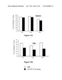 |  |
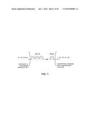 | 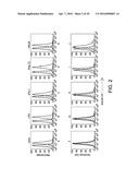 |
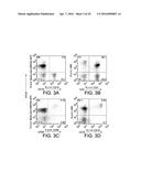 | 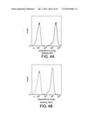 |
 | 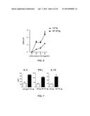 |
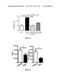 | 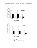 |
 | 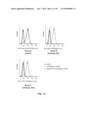 |
 | 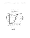 |
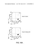 | 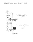 |
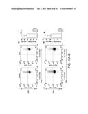 | 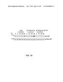 |
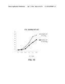 | 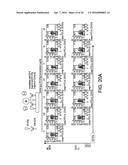 |
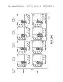 |  |
 |  |
 |  |
 | 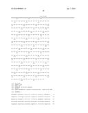 |
 |  |
 |  |
 |  |
 |  |
 |  |
 |  |
 |  |
 |  |
 |
| Similar patent applications: | |
| Date | Title |
|---|---|
| 2016-04-07 | Anti-her2 antibodies and immunoconjugates |
| 2016-04-21 | Environmental clostridial bacteriotherapy and related formulations and methods of manufacture and use |
| 2016-03-10 | Adenoviruses and their use |
| 2016-04-21 | Inhibition of neurodegenerative disease by grape seed extract, green tea extract and probiotic bacteria |
| 2015-04-09 | Anti-fcrh5 antibodies |
| New patent applications in this class: | |
| Date | Title |
|---|---|
| 2022-05-05 | Anti-icos antibodies for the treatment of cancer |
| 2019-05-16 | T cell receptor-like antibodies specific for a wti peptide presented by hla-a2 |
| 2019-05-16 | Immune modulation and treatment of solid tumors with antibodies that specifically bind cd38 |
| 2019-05-16 | Human antibodies to the glucagon receptor |
| 2019-05-16 | Anti-alpha-v integrin antibody for the treatment of fibrosis and/or fibrotic disorders |
| New patent applications from these inventors: | |
| Date | Title |
|---|---|
| 2022-07-28 | Methods and compositions for treating a disease or disorder |
| 2021-12-09 | Costimulatory b7-h1 in renal cell carcinoma patients: indicator of tumor aggressiveness and potential therapeutic target |
| 2021-11-25 | Compositions and methods for modulating lair signal transduction |
| 2021-02-04 | B7-h4 antibodies and methods of use thereof |
| Top Inventors for class "Drug, bio-affecting and body treating compositions" | |
| Rank | Inventor's name |
|---|---|
| 1 | David M. Goldenberg |
| 2 | Hy Si Bui |
| 3 | Lowell L. Wood, Jr. |
| 4 | Roderick A. Hyde |
| 5 | Yat Sun Or |