Patent application title: Ionization of Chemicals in Mixture at Low pH by Ambient Ionization/Mass Spectrometry
Inventors:
Hao Chen (Athens, OH, US)
Ning Pan (Jinan, CN)
Pengyuan Liu (Dandong, CN)
Assignees:
OHIO UNIVERSITY
IPC8 Class: AG01N3368FI
USPC Class:
250282
Class name: Radiant energy ionic separation or analysis methods
Publication date: 2015-10-15
Patent application number: 20150293116
Abstract:
A mass spectrometry-based method for analyzing an acidic organic target
compound includes directing a charged solvent (44) toward a pre-acidified
sample (12) comprising the target compound, to thereby ionize the
pre-acidified sample (12). The method further includes directing the
ionized pre-acidified sample (54) to a mass spectrometer (18), the mass
spectrometer (18) being configured to identify and quantify the target
compound.Claims:
1. A mass spectrometry-based method for analyzing an acidic organic
target compound, the method comprising: directing a charged solvent
toward a pre-acidifed sample comprising the target compound, thereby
ionizing the pre-acidified sample; and directing the ionized
pre-acidified sample to a mass spectrometer, the mass spectrometer being
configured to identify and quantify the target compound.
2. The method of claim 1, wherein the pre-acidified sample comprises a blend of biomolecules and a strong acid.
3. The method of claim 2, wherein the biomolecules comprise a phosphopeptide.
4. The method of claim 2, wherein the biomolecules comprise one of a sialic acid, a sialylated glycan, a phosphorylated protein, a sulphated protein or peptide, and a nucleic acid.
5. The method of claim 2, wherein the strong acid comprises hydrochloric acid.
6. The method of claim 1, wherein the charged solvent is generated by desorption electrospray ionization.
7. The method of claim 1, wherein the charged solvent is generated by one of laser beam ionization, high energy particles ionization, plasma ionization, and ion beam ionization.
8. The method of claim 1, wherein the pre-acidified sample has a pH less than or equal to approximately 2.0.
9. The method of claim 8, wherein the pre-acidified sample has a pH less than or equal to approximately 1.2.
10. The method of claim 1, wherein the addition of the strong acid suppresses deprotonation of phosphate groups in the target compound.
11. The method of claim 1, wherein the pre-acidified sample flows at a rate of approximately 1 μL/min to approximately 5 μL/min.
12. The method of claim 1, wherein ionization of the pre-acidified sample does not suppress ion signals in the pre-acidified sample.
13. The method of claim 1, further comprising: flowing the pre-acidified sample through a conduit, the conduit having an outlet positioned approximately 10 mm from an opening of the mass spectrometer.
14. The method of claim 1, wherein the charged solvent is created by applying a voltage of approximately 5 kV to a solvent.
15. A mass spectrometry-based method for effectively analyzing an acidic organic/biological compound in the presence of complicated matrix and for effectively removing/reducing the ion signal suppression effect, the method comprising: directing a charged solvent toward the pre-acidified sample comprising the target compound, thereby overcoming the ion suppression effect and ionizing the low pH sample with high ionization efficiency; and directing the ionized low pH sample to a mass spectrometer, the mass spectrometer being configured to identify and quantify the target compound.
Description:
CROSS-REFERENCE TO RELATED APPLICATION
[0001] This application claims the benefit of U.S. Provisional Patent Application Ser. No. 61/723,473, filed on Nov. 7, 2012, the disclosure of which is incorporated herein by reference in its entirety.
TECHNICAL FIELD
[0002] The present invention is generally related to a method for ionization of chemicals in a mixture. In particular, the present invention is related to ionization of phosphopeptides in mixtures by ambient ionization/mass spectrometry.
BACKGROUND
[0003] Phosphorylation is one of the most common post-translational modifications (PTM) of proteins (approximately 30% of cellular proteins are phosphorylated), and it plays an important role in a wide range of biological processes, such as signal transduction. Phosphorylation is the addition of a phosphate (PO43-) group to a protein or other organic molecule. Phosphorylation turns many protein enzymes on and off, thereby altering their function and activity. Protein phosphorylation in particular plays a significant role in a wide range of cellular processes. Its prominent role in biochemistry is the subject of a very large body of research.
[0004] Mass spectrometry (MS) has become an increasingly viable technology for phosphoprotein analysis. However, a major challenge in this regard is that phosphopeptide signal in the positive ion mode is severely suppressed by non-phosphorylated peptides when a phosphoprotein digest is ionized by traditional ionization methods such as electrospray ionization (ESI) in a commonly used "bottom-up" approach. Preliminary purification of phosphopeptides prior to MS analysis is often indispensable to solve the problem, using antibodies, affinity chromatography, metal oxides, nanopolymers, or nanoparticles to enrich phosphopeptides. As separation and purification could be time-consuming, a direct, rapid and sensitive method for ionizing phosphopeptides in mixtures would be instrumental to facilitating their analysis and characterization.
[0005] Desorption electrospray ionization (DESI) is a recent advance in the field of MS. DESI provides direct ionization of analytes with little or no sample preparation. Sample ionization by DESI occurs via the interactions with charged microdroplets generated in a pneumatically assisted electrospray of an appropriate solvent. In addition to analysis of solid samples, DESI has been extended to directly ionize liquid samples, and its demonstrated applications include the coupling MS with chromatography, microfluidics, and electrochemistry, probing protein conformation, and developing submilli-second time-resolved MS.
SUMMARY
[0006] The present invention is premised on the realization that an organic compound can be analyzed at low pH (i.e., in strong acidic media) using a mass spectrometer at very low pH by DESI. DESI utilizes a regular solvent as an ionizing electrospray, which, in turn, directs analyte compound to a mass spectrometer configured to analyze the analyte. Potential damage to the mass spectrometer caused by strong acid in sample mixture can be prevented due to the use of regular spray solvent, which is neutral or slightly acidic and can dilute the sample during the DESI ionization process.
[0007] The present invention is further premised on the realization that phosphoproteins can be successfully analyzed utilizing DESI coupled with MS. More particularly, the phosphopeptide can be acidified with a strong acid, such as hydrochloric acid, which suppresses the deprotonization of the peptide phosphate groups in solution. The acidified phosphopeptide can then be ionized using DESI, whereby the ionizing electrospray effectively dilutes the acidified phosphopeptide, allowing it to be analyzed using MS without damaging the mass spectrometer.
[0008] The objects and advantages of the present invention will be further appreciated in light of the following detailed description and drawings provided herein.
BRIEF DESCRIPTION OF THE DRAWINGS
[0009] The accompanying drawings, which are incorporated in and constitute a part of this specification, illustrate embodiments of the invention and, together with a general description of the invention given above and the detailed description given below, serve to explain the principles of the invention.
[0010] FIG. 1 is a diagrammatic cross sectional view of an embodiment of the present invention.
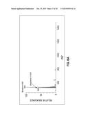
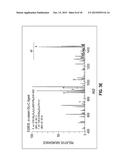
[0011] FIG. 2A is an ESSI-MS spectrum according to Experiment 1.
[0012] FIG. 2B is a DESI-MS spectrum according to Experiment 1.
[0013] FIG. 3A is an α-casein sequence according to Experiment 2.
[0014] FIGS. 3B-3E are α-casein digests according to Experiment 2.
[0015] FIGS. 4A-4B are CID MS/MS spectra according to Experiment 2.
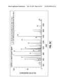
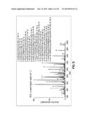
[0016] FIG. 5 is a DESI-MS spectrum according to Experiment 4.
[0017] FIG. 6A is a β-casein sequence according to Experiment 5.
[0018] FIG. 6B is an ESSI-MS spectrum according to Experiment 5.
[0019] FIG. 6C is a DESI-MS spectrum according to Experiment 5.
[0020] FIGS. 7A-7B are ECD MS/MS spectra according to Experiment 5.
[0021] FIGS. 8A-8B are ESSI-MS spectrum according to Experiment 5.
DETAILED DESCRIPTION
[0022] Although the present invention can be used with any acidic compound that suffers ion suppression in the presence of other compounds by traditional ionization method, such as ESI, it is particularly useful in the analysis of phosphoproteins and sulfated proteins. For purpose of description, a target compound, such as phosphopeptides, is made very acidic by addition of an appropriate strong acid to prevent suppression of the phosphopeptide signal by non-phosophorylated peptides. However, in general, this method is applicable to acidic compounds.
[0023] When the present invention is used for analysis of phosphopeptides, the phosphopeptide is combined with a strong acid that does not interfere with subsequent analysis utilizing a mass spectrometer. The phosphopeptide combined with the acid is referred to as a pre-acidified sample. In particular, haloacids, such as hydrochloric acid and hydrobromic acid can be combined with the phosphopeptide to achieve a desired pH of approximately 0 to 2.0. The acids must have a pKal smaller than the pKal of phosphoric acid, which is 2.12. Further, the strong acid must not provide a counter ion that interferes with the detection of the group of the phosphopeptides. Therefore, acids such as sulfuric acid and phosphoric acid are unsuitable for use in the present invention.
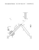
[0024] Briefly, the present invention provides a method for phosphoprotein analysis using DESI-MS. An apparatus 10 suitable for practicing the present invention includes a conduit 14 for the pre-acidified sample 12, an ambient ionizer 16, and the mass spectrometer 18. The pre-acidified sample 12 comprises an acidic organic target compound. The pre-acidified sample 12 is delivered through conduit 14 having an outlet 22. The conduit 14 may comprise, for example, a fused silica transfer capillary having an inner diameter of approximately 0.05 mm to approximately 0.2 mm. More specifically, the capillary may have an inner diameter of approximately 100 μm. The pre-acidified sample 12 may flow through the conduit 14 at a rate of approximately 1 μL/min to approximately 5 μL/min.
[0025] The ambient ionizer 16 includes a spray probe 42, which generates microdroplets of a charged solvent 44 and directs the charged solvent 44 toward the pre-acidified sample 12 that emerges from the conduit outlet 22. The spray probe 42 may be positioned approximately 0.5 mm to approximately 5 mm from the conduit outlet 22, for example. More specifically, the spray probe 42 may be positioned approximately 1 mm from the conduit outlet 22. The charged solvent 44 ionizes the pre-acidified sample 12 that emerges from the conduit outlet 22.
[0026] As shown, the ambient ionizer 16 is a DESI apparatus that includes a housing 46 having a solvent conduit 48 for solvent 49 surrounded by a gas conduit 50. The solvent 49 that is supplied to the ambient ionizer 16 may comprise, for example, methanol/water (1:1 by volume) containing 1% acetic acid. A voltage generator 52 is attached to the housing 46 and is operable to charge the solvent 49 within the solvent conduit 48. A high voltage of approximately 4 kV to approximately 5.5 kV may be applied to the solvent 49. More specifically, a voltage of approximately 5 kV may be applied to the solvent 49. The DESI apparatus generates the nebulized, charged solvent 44 that ionizes the pre-acidified sample 12 by desorption, forming an ionized sample 54. The DESI solvent flow rate may be from about 0.05 μL/min to approximately 50 μL/min. More specifically, the solvent 49 may be injected into the DESI apparatus at a rate of approximately 10 μL/min.
[0027] An LS-DESI-MS system is described in further detail in U.S. Pat. Nos. 7,915,579 and 8,330,119, the disclosures of which are incorporated in their entireties herein by reference.
[0028] The spray impact of the microdroplets of charged solvent 44 from the spray probe 42 with the pre-acidified sample 12 ionizes and deflects an ionized portion of the sample 54 into a mass spectrometer 18, such as a Thermo Finnigan LCQ DECA ion trap mass spectrometer 18 (Thermo Scientific, San Jose, Calif.). The mass spectrometer 18 has a sample entrance or opening 62, such as a heated capillary, which is also positioned near the conduit outlet 22 and the spray probe 42 of the ambient ionizer 16. The opening 62 may be positioned approximately 10 mm from the conduit outlet 22. The ionized sample 54 enters the opening 62, where a pump (not shown) maintains the atmosphere in the mass spectrometer 18 as a vacuum. The mass spectrometer 18 analyzes a mass-to-charge ratio of the ionized sample 54, as described in U.S. Pat. Nos. 7,915,579 and 8,330,119. In this way, the mass spectrometer 18 is configured to identify and quantify an amount of the target compound.
[0029] In use, a pre-acidified sample 12 of, for example, acidified phosphopeptide solution that has been acidified by a strong acid, such as HCl, is provided. The introduction of the strong acid lowers the pH of the phosphopeptide solution and suppresses the deprotonation of peptide phosphate groups in the solution. The pre-acidified sample 12 is delivered through conduit 14 and emerges through a conduit outlet 22, which is positioned proximate a spray probe 42 of the ambient ionizer 16. Solvent 49 is directed through an ambient ionizer 16, such as a DESI ionizer, which contacts the pre-acidified sample 12 and ionizes the pre-acidified sample 12 emerging from the conduit outlet 22. The ionization deflects an ionized portion of the sample 54 into an opening 62 of a mass spectrometer 18. The mass spectrometer 18 analyzes a mass-to-charge ratio of the ionized sample 54. In this way, the mass spectrometer 18 can identify and quantify an amount of the phosphopeptide target compound.
[0030] The method of the present invention helps prevent suppression of phosphopeptide signals during phosphoprotein analysis by MS. The intrinsic cause for the suppression of phosphopeptide ionization is that the phosphate groups of phosphopeptides tend to lose protons to carry negative charges. By adding a stronger acid than phosphoric acid to the target compound, phosphate deprotonation can be inhibited. Because the strong acid is added to the target compound prior to ionization, regular DESI-like solvent may be used for ionization.
[0031] As such, when the pre-acidified sample 12 is impinged by DESI spray 44 during ionization, the pre-acidified sample 12 is diluted by the DESI spray solvent 49, which reduces or prevents corrosion of the instruments in the apparatus 10. With the strong acid additives in the pre-acidified sample 12, phosphopeptides are charged as positive ions in the pre-acidified sample 12, which enables the pre-acidified sample 12 to be quickly converted from liquid phase to gas phase by DESI. Indeed, a 100% coverage of phosphorylated peptides is enabled by DESI-MS using only a few pmol of proteins. In addition, increased charges of phosphopeptide ions are also noted, which is valuable for tandem MS analysis.
[0032] This DESI-MS methodology may also be used for analysis of other acidic biomolecules, such as a sialic acid, a sialylated glycan, a phosphorylated protein, a sulphated protein or peptide, or a nucleic acid.
[0033] The present invention will be further appreciated in light of the following examples.
Experiment 1
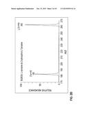
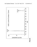
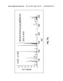
[0034] With reference to FIGS. 2A-2B, O-phospho-L-tyrosine, a phosphorylated amino acid, was chosen as a test sample for a proof-of-principle experiment. When an amino acid mixture containing O-phospho-L-tyrosine and L-tyrosine (molar ratio 3:1) in MeOH/H2O/HOAc (pH=3.3) was ionized by electronsonic spray ionization (ESSI, a variant form of ESI), the corresponding protonated molecules were observed at m/z 262 and 182, respectively (FIG. 2A). However, the intensity ratio of m/z 262 vs. m/z 182 is 0.3 although O-phospho-L-tyrosine is 3 times more concentrated than L-tyrosine, which shows the suppression of the phosphoamino acid in comparison to non-phosphorylated amino acids. Interestingly, when the amino acid mixture was acidified with HCl to pH 2.0 and ionized by DESI with the spray solvent 49 of MeOH/H2O/HOAc, the signal intensity of m/z 262 exceeded m/z 182 (FIG. 2B). In addition, the absolute intensity of m/z, 262 in DESI-MS spectrum (2.0E7, arbitrary units, FIG. 2B) was higher than that in ESSI-MS spectrum (4.9E6, FIG. 2A). Evidently, the addition of HCl suppresses the deprotonation of phosphate group of O-phospho-L-tyrosine so that it can be more easily ionized by DESI.
Experiment 2
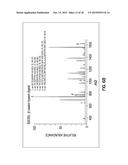
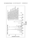
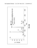
[0035] With reference to FIGS. 3A-4B, phosphoprotein digests were also examined with this DESI approach. α-Casein (sequence shown in FIG. 3A; the subscript "p" indicates that the residue serine is phosphorylated), a phosphoprotein carrying eight phosphate groups, was digested using trypsin following the reported procedure. In a comparison experiment using ESSI, among 16 peptide ions identified based on acquired MS spectrum (FIG. 3B; the figure inset shows the list of identified peptide ions), three phosphopeptide ions [VPQLEIVPNPSAEER+2H]2+ (m/z 832), [YKVPQLEIVPNPSAEER+2H]2+ (m/z 977), and [YKVPQLEIVPNPSAEER+H].sup.+ (m/z 1953) were detected, covering only one phosphorylation site of the protein. Other phosphorylated peptides are missing in the spectrum, emphasizing the well-known ion suppression effect mentioned above. In a stark contrast, when the sample was acidified by HCl to pH 2.0 and analyzed by DESI, besides the 16 peptide ions seen in the ESSI-MS spectrum, two additional doubly charged ions, [DIGPSEPSTEDQAMEDIK+2H]2+ (m/z 965) and [QMEAEPSIPSPSPSEEIVPNPSVEQK+2H]2+ (m/z 1361), were detected (FIG. 3C). The successful visualization of the highly acidic phosphopeptide QMEAEPSIPSPSPSEEIVPNPSVEQK is remarkable as it carries 5 phosphates. Upon collision-induced dissociation (CID), m/z 965 gives rise to fragment ions y4, y5, y6, y8 along with losses of one and two molecules of H3PO4 (FIG. 4A), and m/z 1361 dissociates into b5, b7, b13, b14, y5, y7, y8, y9, y10, y14 along with loss of one H3PO4 molecule (FIG. 4B), confirming their structures. The detection of these two additional phosphopeptides, DIGPSEPSTEDQAMEDIK and QMEAEPSIPSPSPSEEIVPNPSVEQK, allows to cover all eight phosphorylation sites of the protein. In this experiment, only 5 pmol of α-casein digest was injected for ionization to acquire the DESI-MS spectrum, suggesting the high sensitivity of the method.
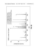
[0036] The feasibility of this DESI method was further confirmed with another sample, the α-casein Glu-C digest. Unlike trypsin digestion producing peptides carry a basic residue at the C-terminal, Glu-C selectively cleaves peptide bonds C-terminal to glutamic acid residues, which may provide phosphopeptides without any basic residues that would be more sensitive to the signal suppression effect. Indeed, only one phosphopeptide ion [IVPNPSAEE+H].sup.+ (m/z 938) appears in the ESSI-MS spectrum (FIG. 3D). After acidification with HCl and being ionized by DESI, two more phosphopeptide ions, [LSKDIGPSEPSTE+H].sup.+ (m/z 1326) and [AEPSIPSPSPSEEIVPNPSVE+2H]2+ (m/z 1039) were detected (FIG. 3E), again covering all phosphate-carrying residues in the protein.
Experiment 3
[0037] The corrosion effect of HCl used in DESI to the mass spectrometer 18 instrument was evaluated in a separate experiment. A piece of stainless steel (316 S.S) was chosen to block the opening 62 of the mass spectrometer 18 to receive the sprayed liquids from DESI. When a sample of MeOH/H2O with pH adjusted to 2.0 by HCl was injected to undergo DESI ionization for 30 min (both the flow rates and the DESI spray solvent were kept the same as in the analysis of phosphopeptide samples), no detectable damage was noted. While the same sample (pH 2.0) was sprayed by ESSI for 30 min, some traces of black marks on the stainless steel surface were seen, indicating the occurrence of some corrosion. This result shows the strength of DESI in analyzing low pH samples. Sulfuric acid (H2SO4), another strong acid, was chosen to replace HCl for the DESI experiments; however, the ionization of the phosphopeptides failed, probably because SO42- anions form adducts with positively charged peptide ions. HCl, a volatile acid, appears to be a better choice for such a DESI experiment. Recently HCl was also reported to be employed as an additive to the counter-flow gas in ESI to change the charge numbers of resulting protein ions.
Experiment 4
##STR00001##
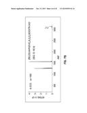
[0039] A proposed mechanism for the efficient ionization of phosphopeptides from protein digests by DESI is depicted in Scheme 1. In a sample solution acidified with weak acids like acetic acid, phosphate groups carry negative charges due to deprotonation, which makes the phosphopeptides to be negatively charged. Once acidified with HCl to a low pH that suppresses the phosphate deprotonation, the phosphopeptides are positively charged in solution and therefore are able to compete with non-phosphorylated peptides to produce abundant positive ions by DESI. According to the Henderson-Hasselbalch equation pH=pKal+log([A.sup.-]/(HA]) (HA and A.sup.- represent an acid and its conjugated base, respectively), the population of HA species (e.g., the intact phosphate group of phosphopeptides) can be further increased if the pH is lowered. This accounts for the contrast between the ionization of phosphopeptides at different pHs. Indeed, when the α-casein tryptic digest was acidified by HCl to pH 1.2, the signal intensity for [QMEAEPSIPSPSPSEEIVPNPSVEQK+2H]2+ (m/z 1361) was further enhanced to 1.8E4 (FIG. 5) in comparison to that obtained via ionization at pH 2.0 (1.1E4, FIG. 3C). In the experiment of the ionization of pH 1.2 sample by DESI, the sample injection rate was reduced to 2 μL/min to avoid instrument corrosion. As shown in Scheme 1, these phosphopeptides positively charged in solution can be rapidly transferred into the gas phase by DESI for MS detection due to the direct sampling capability of DESI. An evidence to support this hypothesis is that, when the α-casein tryptic digest sample was mixed with the DESI spray solvent (1:2 by volume, mimicking the mixing in the DESI ionization process) and then was analyzed by ESSI, the m/z 1361 was not observed (data not shown). Presumably, the m/z 1361 detected in DESI-MS (FIG. 3E) survived the spray/sample solution mixing process in DESI because of the very short DESI ionization time scale (approximately 1-2 milliseconds) that favors the preservation of positively charged phosphopeptides during the ionization.
Experiment 5
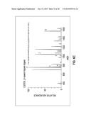
[0040] Besides the efficient ionization of highly acidic phosphopeptides from protein digests, charges of ionized phosphopeptides can also be enhanced using this DESI approach. For instance, in the case of β-casein tryptic digest (protein sequence shown in FIG. 6A), the intensity of the doubly charged ion [RELEELNVPGEIVEPSLPSPSPSEESITR+2H]2+ (m/z 1562) increased from 2.9E5 in ESSI-MS spectrum (FIG. 6B) to 5.1E5 in DESI-MS spectrum (FIG. 6C). More importantly, abundant triply charged ion [RELEELNVPGEIVEPSLPSPSPSEESITR+3H]3+ (m/z 1042) was generated (FIG. 6C). The enhanced charges would be valuable in providing increased sequence coverage via electron-based tandem MS analysis, such as electron-capture dissociation (ECD). Indeed, ECD of m/z 1042 gives rise to fragment ions of z2, z3, z4, z5, z6, z7, c17, c21, c22, c23, c24, and c25 (FIG. 7A) from which the locations of pSer18 and pSer19 of the protein can be clearly pinpointed. In contrast, ECD of m/z 1562 only gives rise to two fragment ions, c25 and z25 (FIG. 7B).
[0041] This DESI method is also applicable to the analysis of acidic sulfated peptides. Using a mixture containing sulfated hirudin (sequence: DFEEIPEE-Y(SO3H)-LQ), bradykinin and angiotensin II as a test sample (1:10:10 by moles), the sulfated hirudin was suppressed and no corresponding peptide ion was observed in ESSI-MS (FIG. 8A). In contrast, the doubly charged ions [DFEEIPEE-Y(SO3H)-LQ+2H]2+ (m/z 746) arose via DESI-MS analysis (FIG. 8B), and its structure was confirmed by CID (the inset of FIG. 8B).
[0042] This invention presents a novel approach for solving the ion signal suppression problem that occurs with phosphopeptide ionization in protein digests by acidifying the target compound with strong acid HCl followed by direct DESI-MS analysis. The methodology is general, fast and sensitive. Efficient ionization can be achieved with the coverage of all phosphate carrying residues without laborious separation or enrichment of phosphopeptides. Thus, it reduces the cost of phosphoprotein analysis and saves time. The methodology of this invention is applicable for improving MS analysis of other important biomolecules with high acidity such as sialic acids, sialylated glycans, sulphated proteins, and nucleic acids.
[0043] This has been a description of the present invention along with the various methods of practicing the present invention. However, the invention itself should only be defined by the appended claims.
Sequence CWU
1
1
31114PRTBos taurus 1Val Pro Gln Leu Glu Ile Val Pro Asn Ser Ala Glu Glu
Arg 1 5 10 216PRTBos
taurus 2Tyr Lys Val Pro Gln Leu Glu Ile Val Pro Asn Ser Ala Glu Glu Arg 1
5 10 15 316PRTBos
taurus 3Asp Ile Gly Ser Glu Ser Thr Glu Asp Gln Ala Met Glu Asp Ile Lys 1
5 10 15 421PRTBos
taurus 4Gln Met Glu Ala Glu Ser Ile Ser Ser Ser Glu Glu Ile Val Pro Asn 1
5 10 15 Ser Val Glu
Gln Lys 20 58PRTBos taurus 5Ile Val Pro Asn Ser Ala Glu
Glu 1 5 611PRTBos taurus 6Leu Ser Lys Asp Ile
Gly Ser Glu Ser Thr Glu 1 5 10
716PRTBos taurus 7Ala Glu Ser Ile Ser Ser Ser Glu Glu Ile Val Pro Asn Ser
Val Glu 1 5 10 15
825PRTBos taurus 8Arg Glu Leu Glu Glu Leu Asn Val Pro Gly Glu Ile Val Glu
Ser Leu 1 5 10 15
Ser Ser Ser Glu Glu Ser Ile Thr Arg 20 25
911PRTBos taurus 9Asp Phe Glu Glu Ile Pro Glu Glu Tyr Leu Gln 1
5 10 10199PRTBos taurus 10Arg Pro Lys His Pro
Ile Lys His Gln Gly Leu Pro Gln Glu Val Leu 1 5
10 15 Asn Glu Asn Leu Leu Arg Phe Phe Val Ala
Pro Phe Pro Glu Val Phe 20 25
30 Gly Lys Glu Lys Val Asn Glu Leu Ser Lys Asp Ile Gly Ser Glu
Ser 35 40 45 Thr
Glu Asp Gln Ala Met Glu Asp Ile Lys Gln Met Glu Ala Glu Ser 50
55 60 Ile Ser Ser Ser Glu Glu
Ile Val Pro Asn Ser Val Glu Gln Lys His 65 70
75 80 Ile Gln Lys Glu Asp Val Pro Ser Glu Arg Tyr
Leu Gly Tyr Leu Glu 85 90
95 Gln Leu Leu Arg Leu Lys Lys Tyr Lys Val Pro Gln Leu Glu Ile Val
100 105 110 Pro Asn
Ser Ala Glu Glu Arg Leu His Ser Met Lys Glu Gly Ile His 115
120 125 Ala Gln Gln Lys Glu Pro Met
Gly Ile Val Asn Gln Glu Leu Ala Tyr 130 135
140 Phe Tyr Pro Glu Leu Phe Arg Gln Phe Tyr Gln Leu
Asp Ala Tyr Pro 145 150 155
160 Ser Gly Ala Trp Tyr Tyr Val Pro Leu Gly Thr Gln Tyr Thr Asp Ala
165 170 175 Pro Ser Phe
Ser Asp Ile Pro Asn Pro Ile Gly Ser Glu Asn Ser Glu 180
185 190 Lys Thr Thr Met Pro Leu Trp
195 118PRTBos taurus 11Glu Gly Ile His Ala Gln Gln
Lys 1 5 124PRTBos taurus 12His Pro Ile Lys 1
134PRTBos taurus 13His Ile Gln Lys 1 145PRTBos
taurus 14Leu His Ser Met Lys 1 5 1510PRTBos taurus 15Tyr
Leu Gly Tyr Leu Glu Gln Leu Leu Arg 1 5
10 1612PRTBos taurus 16Phe Phe Val Ala Pro Phe Pro Glu Val Phe Gly Lys 1
5 10 176PRTBos taurus 17Thr Thr
Met Pro Leu Trp 1 5 1815PRTBos taurus 18His Gln Gly
Leu Pro Gln Glu Val Leu Asn Glu Asn Leu Leu Arg 1 5
10 15 1919PRTBos taurus 19Glu Pro Met Ile Gly
Val Asn Gln Glu Leu Ala Tyr Phe Tyr Pro Glu 1 5
10 15 Leu Phe Arg 206PRTBos taurus 20Asp Ile
Lys Gln Met Glu 1 5 217PRTBos taurus 21Lys Thr Thr
Met Pro Leu Trp 1 5 227PRTBos taurus 22Gln Lys
His Ile Gln Lys Glu 1 5 238PRTBos taurus 23Ile
Val Pro Asn Ser Ala Glu Glu 1 5 2412PRTBos
taurus 24Asp Val Pro Ser Glu Arg Tyr Leu Gly Tyr Leu Glu 1
5 10 2521PRTBos taurus 25Glu Met Glu Ala Glu
Ser Ile Ser Ser Ser Glu Glu Ile Val Pro Asn 1 5
10 15 Ser Val Glu Gln Lys 20
26209PRTBos taurus 26Arg Glu Leu Glu Glu Leu Asn Val Pro Gly Glu Ile Val
Glu Ser Leu 1 5 10 15
Ser Ser Ser Glu Glu Ser Ile Thr Arg Ile Asn Lys Lys Ile Glu Lys
20 25 30 Phe Gln Ser Glu
Glu Gln Gln Gln Thr Glu Asp Glu Leu Gln Asp Lys 35
40 45 Ile His Pro Phe Ala Gln Thr Gln Ser
Leu Val Tyr Pro Phe Pro Gly 50 55
60 Pro Ile Pro Asn Ser Leu Pro Gln Asn Ile Pro Pro Leu
Thr Gln Thr 65 70 75
80 Pro Val Val Val Pro Pro Phe Leu Gln Pro Glu Val Met Gly Val Ser
85 90 95 Lys Val Lys Glu
Ala Met Ala Pro Lys His Lys Glu Met Pro Phe Pro 100
105 110 Lys Val Pro Val Glu Pro Phe Thr Glu
Ser Gln Ser Leu Thr Leu Thr 115 120
125 Asp Val Glu Asn Leu His Leu Pro Leu Pro Leu Leu Gln Ser
Trp Met 130 135 140
His Gln Pro His Gln Pro Leu Pro Pro Thr Val Met Phe Pro Pro Gln 145
150 155 160 Ser Val Leu Ser Leu
Ser Gln Ser Lys Val Leu Pro Val Pro Gln Lys 165
170 175 Ala Val Pro Tyr Pro Gln Arg Asp Met Pro
Ile Gln Ala Phe Leu Leu 180 185
190 Tyr Gln Glu Pro Val Leu Gly Pro Val Arg Gly Pro Phe Pro Ile
Ile 195 200 205 Val
276PRTBos taurus 27Glu Ala Met Ala Pro Lys 1 5
287PRTBos taurus 28Gly Pro Phe Pro Ile Ile Val 1 5
296PRTBos taurus 29Glu Met Pro Phe Pro Lys 1 5
307PRTBos taurus 30Val Leu Pro Val Pro Gln Lys 1 5
3116PRTBos taurus 31Phe Gln Ser Glu Glu Gln Gln Gln Thr Glu Asp Glu Leu
Gln Asp Lys 1 5 10 15
User Contributions:
Comment about this patent or add new information about this topic:
 |  |
 |  |
 |  |
 |  |
 |  |
 |  |
 |  |
 |  |
 |  |
 |  |
 |  |
 |  |
 |  |
| New patent applications in this class: | |
| Date | Title |
|---|---|
| 2022-05-05 | Method and apparatus for ion mobility separations utilizing alternating current waveforms |
| 2022-05-05 | Sample transport apparatus for mass spectrometry |
| 2019-05-16 | Analysis of amino acids in body fluid by liquid chromotography-mass spectrometry |
| 2018-01-25 | Time-of-flight analysis of a continuous beam of ions by a detector array |
| 2018-01-25 | Mass spectrometric determination of eicosapentaenoic acid and docosahexaenoic acid |
| New patent applications from these inventors: | |
| Date | Title |
|---|---|
| 2016-05-05 | Versatile ambient ionization-based interface for lc/ms |
| 2015-11-26 | Microsecond time-resolved mass spectrometry |
| 2015-10-08 | Online monitoring of fuel cell reactions by desorption electrospray mass spectrometry |
| 2014-05-29 | Microsecond time-resolved mass spectrometry |
| Top Inventors for class "Radiant energy" | |
| Rank | Inventor's name |
|---|---|
| 1 | Jason Lee Wildgoose |
| 2 | Osamu Wakabayashi |
| 3 | Toshio Kameshima |
| 4 | Tomoyuki Yagi |
| 5 | Katsuro Takenaka |