Patent application title: THERAPEUTICALLY USEFUL MOLECULES
Inventors:
Hans Josef Stauss (London, GB)
Liquan Gao (London, GB)
Shao-An Xue (London, GB)
IPC8 Class: AC07K14725FI
USPC Class:
424 9321
Class name: Whole live micro-organism, cell, or virus containing genetically modified micro-organism, cell, or virus (e.g., transformed, fused, hybrid, etc.) eukaryotic cell
Publication date: 2015-05-28
Patent application number: 20150147302
Abstract:
A T cell receptor molecule (TCR) containing an alpha chain portion and a
beta chain portion wherein the alpha chain portion contains three
complementarity determining regions (CDRs): CDR1α: SSYSPS
CDR2α: YTSAATL CDR3α: VVSPF-SGGGADGLT or comprising or
consisting of SPPSGGGADGLT and the beta chain portion contains three
complementarity determining regions (CDRs): CDR1β: DFQATT
CDR2β: SNEGSKA CDR3β: comprising SARDGGEG or comprising or
consisting of RDGGEGSETQY, or wherein up to three amino acid residues in
one or more CDRs are replaced by another amino acid residue. The
invention also includes polynucleotides encoding the TCR molecules, and
host cells containing the said polynucleotides. Patient derived T cells
may have the polynucleotides encoding the TCR molecules introduced
therein, and the engineered T cells may be introduced into the patient in
order to combat a WT1-expressing malignancy.Claims:
1. An isolated T cell receptor (TCR) molecule containing an alpha chain
portion and a beta chain portion wherein the alpha chain portion contains
three complementarity determining regions (CDRs): CDR1.alpha.: SSYSPS
(SEQ ID NO: 2); CDR2.alpha.: YTSAATL (SEQ ID NO: 3); and CDR3.alpha.:
VVSPFSGGGADGLT (SEQ ID NO: 4) or wherein the alpha chain portion contains
three CDRs: CDR1.alpha.: SSYSPS (SEQ ID NO: 2); CDR2.alpha.: YTSAATL (SEQ
ID NO: 3); and CDR3.alpha.: comprising or consisting of SPFSGGGADGLT (SEQ
ID NO: 5) and wherein the beta chain portion contains three CDRs:
CDR1.beta.: DFQATT (SEQ ID NO: 6); CDR2.beta.: SNEGSKA (SEQ ID NO: 7);
and CDR3.beta.: comprising SARDGGEG (SEQ ID NO: 8) or wherein the beta
chain portion contains three CDRs: CDR1.beta.: DFQATT (SEQ ID NO: 6);
CDR2.beta.: SNEGSKA (SEQ ID NO: 7); and CDR3.beta.: comprising or
consisting of RDGGEGSETQY (SEQ ID NO: 9), or a variant of said TCR
molecule wherein up to one amino acid residue in one or more of the six
CDRs is replaced by another amino acid residue.
2. The TCR molecule according to claim 1 wherein CDR3.alpha. has the amino acid sequence VVSPFSGGGADGLT (SEQ ID NO: 4).
3. The TCR molecule according to claim 1 wherein CDR3.alpha. has the amino acid sequence SPFSGGGADGLT (SEQ ID NO: 5).
4. The TCR molecule according to claim 1 wherein CDR3.beta. has the amino acid sequence SARDGGEG (SEQ ID NO: 8).
5. The TCR molecule according to claim 1 wherein CDR3.beta. has the amino acid sequence RDGGEGSETQY (SEQ ID NO: 9).
6. The TCR molecule according to claim 1 wherein the alpha chain portion and the beta chain portion are present on different polypeptide chains.
7. The TCR molecule according to claim 1 wherein the alpha chain portion and the beta chain portion are present in the same polypeptide chain.
8. The TCR molecule according to claim 1 wherein the CDRs are grafted to a human framework region.
9. The TCR molecule according to claim 8 wherein the alpha chain portion has the amino acid sequence given in FIG. 2.
10. The TCR molecule according to claim 8 wherein the beta chain portion has the amino acid sequence given in FIG. 4.
11. The TCR molecule according to claim 1 which is soluble.
12. A polynucleotide encoding the alpha chain portion of the TCR molecule of claim 1.
13. A polynucleotide encoding the beta chain portion of the TCR molecule of claim 1.
14. A polynucleotide encoding the TCR molecule of claim 7.
15. An expression vector comprising the polynucleotide of claim 14.
16. (canceled)
17. A host cell comprising the expression vector of claim 15.
18. The host cell according to claim 17 which is a T cell.
19. (canceled)
20. A method of combating a WT1-expressing malignancy in a patient, the method comprising introducing into the patient a T cell, which is modified to express the TCR molecule of claim 1.
21. (canceled)
22. The method according to claim 20 wherein the WT1-expressing malignancy is breast cancer, colon cancer, lung cancer, leukaemia, ovarian cancer, melanoma, head and neck cancer, thyroid cancer, glioblastoma or sarcoma.
23-27. (canceled)
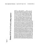
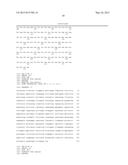
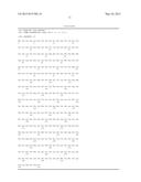
28. The TCR molecule according to claim 1 wherein the variant has up to one amino acid substitution in only CDR3.alpha. and/or CDR3.beta..
29. The TCR molecule according to claim 1 wherein the replaced amino acid residue in one or more of the six CDRs is a conservative amino acid substitution.
Description:
[0001] The present invention relates to therapeutically useful molecules,
in particular to T cell receptors (TCRs) which may be introduced into a
patient's own T cells in order to direct the T cells to kill cancer cells
within the patient, particularly cancer cells which express the Wilms
Tumour antigen-1 (WT1).
[0002] The listing or discussion of a prior-published document in this specification should not necessarily be taken as an acknowledgement that the document is part of the state of the art or is common general knowledge. All of the documents referred to in this specification are hereby incorporated by reference.
[0003] There is evidence that anti-tumour cytotoxic T lymphocytes (CTL) play an important role in vivo. Tumour reactive CTL have been shown to mediate tumour regression in animal models (Kast et al (1989) Cell 59, 603-614) and in man (Kawakami et al (1994) Proc. Natl. Acad Sci. USA 91, 6458-6462; Dudley (2002) Science 298, 850-854). As with all types of anti-tumour therapy, a problem that needs to be overcome is that the therapy must destroy or inactivate the target tumour cells to a useful extent but that the therapy must not destroy or inactivate non-tumour cells to a deleterious extent. In other words, it is desirable if the therapy is selective for tumour cells to a beneficial extent.
[0004] Much of the current work on immunotherapy of cancer makes use of the fact that certain tumours express polypeptides which are not expressed in the equivalent non-tumour tissue, or makes use of the fact than the tumour expresses a mutant form of a polypeptide which is not expressed in the non-tumour tissue. However, it is not always possible to identify polypeptides in a tumour which fall into this category, and so other target polypeptides which can form the basis of an immunotherapeutic approach have been identified.
[0005] In adults, expression of WT1, an embryonic transcription factor, has been observed in renal podocytes, in the testis, in the ovary, in breast myoepithelial cells and in some CD34.sup.+ stem cells in the bone marrow. Aberrant expression was observed in breast cancer, ovarian cancer, melanoma, lung cancer, colon cancer, thyroid cancer, head and neck cancer, glioblastoma, sarcoma and leukaemia including CML and AML (see, for example, Menssen et al (1995) Leukaemia 9, 1060-1067; Inoue et al (1997) Blood 89, 1405-1412; Inoue et al (1996) Blood 88, 2267-2278; Inoue et al (1998) Blood 91, 2969-2976; Menssen et al (1997) Int. J. Cancer 70, 518-523; Menssen et al (1995) Leukemia 9, 1060-1067; Ogawa et al (1998) Transplant 21, 527-527; Rodeck et al (1994) Int. J Cancer 59, 78-82; Silberstein et al (1997) Proc. Natl. Acad. Sci. USA 94, 8132-8137; Tamaki et al (1996) Blood 88, 4396-4398; Viel et al (1994) Int. J. Cancer 57, 515-521; Menssen (2000) J. Cancer Res. Clin Oncol. 126, 226-232; Miyoshi (2002) Clin. Cancer Res. 8, 1167-1171; Oji (1999) Jpn J. Cancer Res. 90, 194-204; Oji (2003) Cancer Sci. 94, 523-529; Oji et al (2003) Cancer Sci. 94, 606-611; Oji et al (2003) Cancer Sci. 94, 712-717; and Ueda (2003) Cancer Sci. 94, 271-276.
[0006] As described in our patent application WO00/26249, using an unconventional approach employing allo-MHC-restricted CTL, we identified peptide epitopes in the WT1 polypeptide which may be presented by HLA-A2 class I molecules and displayed on the surface of tumour cells expressing these proteins endogenously. HLA-A2 negative responder individuals were used as a source of CTL specific for peptides presented by HLA-A2 class I molecule, and this approach allows identification of HLA-A2 presented peptides independent of possible tolerance of autologous CTL.
[0007] One of the peptide epitopes disclosed in WO00/26249 is RMFPNAPYL (which we have also termed pWT126), and we have previously described a CTL which is able to: kill HLA-A2-positive targets coated with the WT1-derived peptide pWT126 (Gao et al (2000) Blood 95, 2198-2203); kill fresh HLA-A2-positive leukaemia cells expressing WT1 (Gao at al (2000) Blood 95, 2198-2203); kill HLA-A2-positive leukemia CFU progenitor cells (Gao et al (2000) Blood 95, 2198-2203; Bellantuono et al (2002) Blood 100, 3835-3837); kill HLA-A2-positive leukaemia LTC-IC stem cells (Bellantuono et al (2002) Blood 100, 3835-3837); kill HLA-A2-positive NOD/SCID leukaemia initiating cells (Gao et al (2003) Transplantation 75, 1429-1436); and do not kill normal HLA-A2-positive NOD/SCID engrafting hematopoietic stem cells (Gao et al (2003) Transplantation 75, 1429-1436). However, none of these publications give molecular information concerning the TCR present in the CTL, and the particular CTL line mentioned in the publications has not been made available to the public in any way and so the structure of the TCR is unknown and could not be derived by the skilled person (since the CTL line was not publicly available).
[0008] The present inventors have now cloned a TCR that is specific to RMFPNAPYL, a peptide of WT1 which is presented by HLA-A2 class I molecules, and have shown that introducing the TCR into either CD4-positive or CD8-positive T cells confers on the engineered T cells the ability to kill cancer cells which express WT1 endogenously. In addition, the inventors have defined the molecular structure of the TCR, identified the complementarity determining regions (CDRs), and describe bow to make recombinant TCRs which are believed to retain the same specificity of the parent molecule.
[0009] The TCRs may usefully be introduced into a T cell derived from a patient (preferably an HLA-A2-positive patient) suffering from a malignancy (where the patient's tumour cells express WT1), and the engineered T cell introduced into the patient in order to combat the malignancy. In particular, it is proposed to take T cells from patients with breast cancer, colon cancer, lung cancer, other solid cancers or leukaemia, transduce them in vitro with a retroviral vector containing the TCR genes, and re-infuse the transduced T cells into the patients. The credibility of this approach is confirmed by the demonstration in the Examples that the WT1-specific TCR genes can be transferred into human T cells, that the genes give raise to TCR expression on the surface of recipient T cells, that the recipient T cells can kill HLA-A2-positive target cells coated with the pWT126 peptide and HLA-A2-positive tumour cells expressing WT1 endogenously.
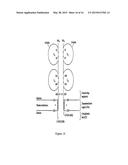
[0010] The general structure of T cell receptors (TCRs), their domain structure and the organisation of genes that encode them is well known, for example see Chapter 11 in Immunology, second edition (1994), by Janis Kuby, W H Freeman & Co, New York, USA, and Garcia et al (1999) Ann. Rev. Immunol. 17, 369-397. One common class of natural TCRs is the αβ class in which the TCRs are made up of a separate alpha chain and a separate beta chain which form a heterodimer which is T cell membrane associated. Each alpha and beta chain is made up of regions which, in order from the N terminus to the C terminus are a leader sequence, a variable region, a constant region, a connecting sequence, a transmembrane region and a cytoplasmic tail region (see FIG. 14 for a graphical representation of αβ TCR structure). The variable region of the alpha chain is called the Vα region and the variable region of the beta chain is called the Vβ region. Similarly, the constant region of the alpha chain is called the Cα region and the constant region of the beta chain is called the Cβ region. The job of the αβ TCR is to recognise and bind to a peptide presented in a HLA molecule of a cell in the body. Generally speaking, the TCR cannot recognise and bind the peptide unless it is presented by a particular HLA molecule, and the TCR cannot recognise a HLA molecule unless it is presenting the specific peptide. T cells harboring a specific TCR will target cells which are presenting a specific peptide in a particular HLA molecule on a cell (ie a peptide-HLA complex), and this is the main principle of T cell-based immunity.
[0011] The peptide-HLA complex is recognised by the combined V regions of the alpha and beta chains of the TCR. In particular, it is the complementarity determining regions (CDRs) of the V regions which mediate recognition of the peptide-HLA complex. The V region of the alpha and beta chains of the natural TCR are made up of, in order in an N-terminal to C-terminal direction, FR1, CDR1, FR2, CDR2, FR3 and CDR3, where FR stands for "framework region" and CDR stands for "complementarity determining region". The FRs and CDRs of the alpha and beta chains are different.
[0012] From the predicted amino acid sequences of the alpha and beta chains of the TCR cloned as mentioned above, the inventors have determined the FRs and CDRs of the alpha and beta chains (see FIGS. 2 and 4). With the knowledge of the CDR sequences, it is possible to produce chimaeric TCRs in which the CDRs are grafted onto framework regions with which the CDRs are not naturally associated, and it is also possible to produce single chain TCR molecules, and in both cases the molecules retain substantially the same binding affinity for the peptide-HLA complex as the parent molecule, as is described in more detail below.
[0013] to A first aspect of the invention provides a T cell receptor (TCR) molecule containing an alpha chain portion and a beta chain portion wherein the alpha chain portion contains three complementarity determining regions (CDRs):
[0014] CDR1α: SSYSPS
[0015] CDR2α: YTSAATL
[0016] CDR3α: VVSPFSGGGADGLT or comprising or consisting of SPFSGGGADGLT and the beta chain portion contains three complementarity determining regions (CDRs):
[0017] CDR1β:DFQAIT
[0018] CDR2β: SNEGSKA
[0019] CDR3β: comprising SARDGGEG, or comprising or consisting of RDGGEGSETQY or wherein up to three amino acid residues in one or more of the CDRs are replaced by another amino acid residue.
[0020] It should be noted that in some nomenclature systems the CDR3 of the β chains may be defined to be longer than in the nomenclature system used in the Immunogenetics (IMGT) database described below. Also, in some nomenclature systems the CDR3 of the α chains may be defined to be shorter than in the IMGT system. Similarly, the constant portion may or may not include framework residues flanking the CDR3 region in the different nomenclature systems.
[0021] Thus, in one embodiment using the IMGT system CDR3α may have the amino acid sequence VVSPFSGGGADGLT and the constant portion includes the framework amino acid sequence FGKGTHLIIQP (see FIG. 5).
[0022] In another embodiment, using the Garcia nomenclature system (Garcia et al (1999) Ann. Rev. Immunol. 17, 369-397, incorporated herein by reference) CDR3α comprises or consists of the amino acid sequence SPFSGGGADGLT, the framework region immediately C-terminal to this has the amino acid sequence FGKGTHLIIQP and the constant region begins with the amino acid sequence YIQNP . . . (see FIG. 5).
[0023] In one embodiment using the IMGT nomenclature system, CDR3β may have the amino acid sequence SARDGGEG and the constant region immediately C-terminal to this includes the framework amino acid sequence SETQY . . . (FIG. 4).
[0024] In another embodiment, using the Garcia nomenclature system as above, CDR3β comprises or consists of the amino acid sequence RDGGEGSETQY and the framework region immediately C-terminal to this has the amino acid sequence FGPGTRLLVL and the immediately C-terminal constant region begins with the amino acid sequence EDLKN . . . (see FIG. 6).
[0025] It will be appreciated that the skilled person can readily design and synthesise TCRs according to the invention using either or any nomenclature systems provided that the framework region (ie region not replaced by the CDRs) is compatible with the CDRs as is well known in the act.
[0026] The standard IUPAC one letter amino acid code is used throughout the specification. For the avoidance of doubt, a reference to a "particular" or "given" CDR means any CDR with the amino acid sequence given above or wherein up to three amino acids have been replaced by another amino acid residue.
[0027] By "TCR molecule" we include any molecule which contains the given CDRs and also contains FRs suitably situated within the molecule so that the CDRs form a recognition site (combining site) which is able to bind to HLA-A2 presenting the peptide RMFPNAPYL (ie a HLA-A2/RMFPNAPYL complex).
[0028] It is particularly preferred if the TCR molecules contain the precise CDR amino acid sequences as given above and in FIGS. 2 and 4 and in FIGS. 5 and 6. Where a variant to this precise sequence is present, it preferably varies by one or two or three (preferably one or two) amino acids in one or two or three or four or Bye or all six CDRs. Typically, in these variants, the amino acids which are replaced are replaced with conservative amino acids. By conservative amino acids we include the groupings: G, A; S, A, T; F, Y, W; D, E; N, Q; and I, L, V.
[0029] A method for making and selecting TCR molecules which have CDRs which vary from the precise CDR sequences given in FIGS. 2 and 4 and in FIGS. 5 and 6 is given below.
[0030] The amino acid sequences, including V regions (and therefore FRs), of numerous TCR alpha chains and TCR beta chains are well known in the art, some of which are described in the IMGT (Immunogenetics) database at http://imgt.cines.fr. See also Lefranc (2003) Dev. Comp. Immunol. 27, 55-77. The structural basis of T cell recognition is reviewed in Garcia et al (1999) Ann. Rev. Immunol. 17, 369-397, and the information contained therein may be used to design and synthesise CDR-grafted TCRs (and CDRs defined on the basis of this nomenclature are noted above). Preferably, the FRs into which the particular CDRs are grafted are FRs of human TCR alpha or beta chains. Conveniently, the alpha chain CDRs are grafted into alpha chain FRs, and beta chain CDRs are grafted to beta chain FRs. Typically, the three CDRs in the alpha chain and the three CDRs in the beta chain replace, in order, CDRs in other human alpha and beta chains, respectively. See Lefranc (2003) Dev. Comp. Immunol. 27, 55-77.
[0031] Typically, T cells expressing thin TCR molecule recognise the HLA-A2 presenting peptide RMFPNAPYL with substantially the same avidity as the TCR molecule which consists of the alpha and beta chains as described in FIGS. 2 and 4. This can be measured by retroviral-mediated transfer of the TCR into T cells followed by peptide titration experiments with the TCR-transduced T cells as outlined, for example, in Gao et al (2000) Blood 95, 2198-2203.
[0032] The TCR molecule preferably contains an alpha chain portion containing, in N-terminal to C-terminal order, FR1α-CDR1α-FR2α-CDR2α-FR3α-CDR3α, and a beta chain portion containing, in N-terminal to C-terminal order, FR1β-CDR1β-FR2β-CDR2β-FR3β-CDR3β as shown in FIGS. 2 and 4, respectively and in FIGS. 5 and 6, respectively. Typically, the TCR molecule contains the V region of both the alpha chain and the beta chain of the TCR polypeptide chains shown in FIGS. 2 and 4, and in FIGS. 5 and 6.
[0033] In a preferred embodiment, the alpha chain portion and the beta chain portion are present on different polypeptide chains. Typically, the TCR molecule contains an alpha chain which contains the V region and the C region of the polypeptide chain shown in FIG. 2 (or FIG. 5), and also contains a beta chain which contains the V region and C region of the polypeptide chain shown in FIG. 4 (or FIG. 6). Preferably, the TCR molecule consists of a molecule containing the complete alpha chain shown in FIG. 2 and the complete beta chain shown in FIG. 4. Typically, however, the leader sequence is cleaved off the mature alpha chain and beta chain.
[0034] In a further embodiment, the alpha chain portion and the beta chain portion of the TCR molecule are present in the same polypeptide chain. Single chain TCR molecules are described in Chung et al (1994) Proc. Not Acad Sci. USA 91, 12654-12658, and the principles described therein may readily be applied to the production of single chain in TCR molecules which contain the specified CDRs. Typically, the single chain TCR molecules contain the Vα, Vβ and Cβ domains fused in the same polypeptide chain, and typically in that order (from N-terminus to C-terminus). For expression of a single chain TCR it is useful to provide a construct encoding the constant domain of the TCR alpha chain.
[0035] An additional strategy is described in Boulter et al (2003) Protein Eng. 16, 707-711 in which a new disulphide bond is introduced between a threonine in the constant region of the alpha chain and a serine in the constant region of the beta chain (by replacing these residues with a cysteine. The disulphide bond in the TCR connecting peptide may be removed or may remain in place.
[0036] The two-chain TCR molecules of the invention (eg ones which contain the alpha and beta chains whose amino acid sequence is given in FIGS. 2 and 4) or chimaeric TCRs which contain the specific CDRs as described above may be used to introduce to create antigen-specific CTL as described in more detail below (by using polynucleotides that encode the relevant chains). Similarly, the single chain TCRs may also be used for this purpose, and have the advantage that they do not pair with endogenous TCRs. Single chain TCRs may also be used as soluble constructs in a way similar to antibodies. In this case, the single chain constructs do not contain a transmembrane region (see Chung et al supra and Boulter et al supra).
[0037] A second aspect of the invention provides a polynucleotide encoding the alpha chain portion as defined in the first aspect of the invention. A third aspect of the invention provides a polynucleotide encoding the beta chain portion as defined in the first aspect of the invention. As discussed above, in a particularly preferred embodiment of the invention, the alpha chain portion and the beta chain portion are present on different polypeptide chains, and it is convenient (but not mandatory) that each polypeptide is encoded by a separate polynucleotide. Preferred polynucleotides encoding the alpha and beta chains are described in FIGS. 1 and 2, respectively. Alternatively, the two polypeptides may be encoded on the same polynucleotide, in which case the two coding regions may be linked by an (Internal Ribosome Entry Site) IRES sequence, and typically would have its own translational start and stop codons. Typically, such constructs contain two promoters, one for each TCR chain.
[0038] As discussed above, in an alternative embodiment the alpha chain portion and the beta chain portion are present in the same polypeptide, in which case a single polynucleotide may encode the single chain polypeptide.
[0039] In any event, the polynucleotide may be DNA or RNA, and it may or may not contain introns. Typically, the polynucleotide does not contain introns within the region that codes for the polypeptide of interest. It will be appreciated that different polynucleotides may encode the same polypeptide because of the degeneracy of the genetic code.
[0040] The invention also provides an expression vector that contains the polynucleotide of the invention. Such expression vectors, when present in a suitable host cell, allow for the expression of the polypeptide(s) of interest. Preferably, the expression vector is an expression vector capable of expressing a polypeptide in a mammalian cell. More preferably, the expression vector is one which is able to express a polypeptide in a T cell, such as a human CTL. Typically, the expression vectors contain a promoter which is active in particular cell types, and which may be controllable (eg inducible).
[0041] The vector is suitably a retroviral vector which is capable of transfection into a mammalian host cell such as a human T cell. Typically, the vector is a lentiviral vector.
[0042] A further aspect of the invention provides a host cell comprising a polynucleotide of the invention or a vector of the invention. The host cell may contain a polynucleotide or vector which encodes only the alpha chain portion or only the beta chain portion. However, if the host cell is to produce a TCR molecule of the invention, it contains one or more polynucleotides or vectors which encode both the alpha chain portion and the beta chain portion. The host cell may be any cell such as a bacterial cell, yeast cell, insect cell, plant cell or mammalian cell, and methods of introducing polynucleotides into such cells are well known in the art. Typically, bacterial cells, such as Escherichia coli cells are used for general propagation and manipulation of the polynucleotides and vectors of the invention. Other host cells may be used to express the TCR molecules of the invention and, in particular, the cell may be a mammalian cell such as a human cell. As described below in relation to the therapeutic methods using the TCR molecules of the invention, it is particularly desirable if the host cell is a T cell such as (and preferably) a T cell derived from a patient to be treated, typically a patient with a WT1-expressing malignancy.
[0043] Typically, a retroviral vector (or, as the case may be vectors) encoding the TCR molecule of the invention is used based on its ability to infect mature human CD4.sup.+ or CD8.sup.+ T lymphocytes and to mediate gene expression: the retroviral vector system Kat is one preferred possibility (see Finer et al (1994) Blood 83, 43). High titre amphotrophic retrovirus are used to infect purified CD8.sup.+ T lymphocytes isolated from the peripheral blood of tumour patients following a protocol published by Roberts et al (1994) Blood 84, 2878-2889, incorporated herein by reference. Anti-CD3 antibodies are used to trigger proliferation T cells, which facilitates retroviral integration and stable expression of single chain TCRs. A combination of anti-CD3 and anti-CD8 antibodies may be more effective than anti-CD3 antibodies alone. Other suitable systems for introducing genes into CTL are described in Moritz et al (1994) Proc. Natl Acad Sci. USA 91, 4318-4322, incorporated herein by reference. Eshhar et al (1993) Proc. Natl. Acad Sci. USA 90, 720-724 and Hwu et al (1993) J. Exp. Med 178, 361-366 also describe the transfection of CTL. The commercially available Nuclofactor system, provided by AMAXA, Germany may be used to transfect T cells. Retroviral transduction of human CD8.sup.+ T cells is described in Stanislawski (2001) Nat. Immunol. 2, 962.
[0044] Methods of cloning and genetic manipulation are well known in the art and are described in detail in standard manuals such as Sambrook & Russell (2001) Molecular Cloning, a laboratory manual, Cold Spring Harbor Press, Cold Spring Harbor, N.Y., USA.
[0045] Patients suffering from a WT1-expressing malignancy may be treated by the introduction of the TCR molecule of the invention into their own T cells (or T cells from a donor), followed by the introduction of these engineered cells into the patient. Thus, a further aspect of the invention provides a method of combating a WT-1 expressing malignancy in a patient, the method comprising introducing into the patient a T cell, preferably derived from the patient, which is modified to express the TCR molecule of the invention. Typically, (1) T cells are obtained from the patient, (2) a polynucleotide or polynucleotides encoding and capable of expressing the TCR molecule of the invention are introduced into the T cells ex vivo and (3) the engineered T cells are introduced into the patient. It is particularly preferred if the T cells are the patient's T cells (ie autologous).
[0046] It is particularly preferred if the patient is HLA-A2 positive.
[0047] In other words, the specificity of the T cell, preferably autologous T cell, is changed by the introduction of the TCR molecule of the invention.
[0048] The T cells (for example of the patient) are typically isolated from peripheral blood mononuclear cells (PBMCs), and may be CD4.sup.+ and CD8.sup.+ cells. Typically, the cells are activated using an antibody (eg an anti-CD3 or anti-CD28 antibody) so that they become receptive to transfection, for example with one or more retroviral vectors encoding the TCR molecules of the invention. The number of cells isolated, transfected and returned to the patient may be determined by the physician.
[0049] Cells may be taken from a patient after a clinical response, cryopreserved, transfected and re-infused if the same patient relapses.
[0050] Whether or not a malignancy is one which expresses WT1 may be determined, for example using reverse transcriptase-polymerase chain reaction (RT-PCR) or using intracellular staining techniques for the WT1 protein (which may be anti-WT1 antibodies).
[0051] The patient is preferably a human patient although animals may be used in a research situation. It is particularly preferred that the patient is HLA-A2 positive. Whether or not a patient is HLA-A2 positive can be determined by methods well known in the art.
[0052] Typically, the patient is suffering from any one or more of leukaemia, breast cancer, colon cancer, lung cancer, ovarian cancer, melanoma, thyroid cancer, head and neck cancer, glioblastoma, and sarcoma.
[0053] A further aspect of the invention provides the use of a T cell, preferably a patient derived T cell, which is modified to express the TCR molecule of the invention in the manufacture of a medicament for combating a WT1-expressing malignancy in the patient.
[0054] As discussed above, TCR molecules in which one or more of the CDRs differ in sequence from the precise CDR sequences given in FIGS. 2 and 4 form part of the invention. Preferably, such TCR molecules are able to recognise the HLA-A2/RMFPNAPYL complex more effectively than a TCR molecule with the precise CDR sequences. Thus, a further aspect of the invention provides a method of selecting a TCR molecule with improved binding to an HLA-A2/RMFPNAPYL complex comprising (a) providing a TCR molecule containing an alpha chain portion and a beta chain portion wherein the alpha chain portion contains three complementarity determining regions (CDRs):
[0055] CDR1α: SSYSPS
[0056] CDR2α: YTSAATL
[0057] CDR3α: VVSPFSGGGADGLT or comprising or consisting of SPFSGGGADGLT and the beta chain portion contains three complementarity determining regions (CDRs):
[0058] CDR1β: DFQATT
[0059] CDR2β: SNEGSKA
[0060] CDR3β: comprising SARDGGEG or comprising or consisting of RDGGEGSETQY wherein at least one amino acid residue in one or more of the CDRs as given is replaced with another amino acid residue, (b) determining whether the TCR molecule binds to an HLA-A2/RFMPNAPYL complex with greater affinity than a TCR molecule without the replacement amino acid(s), and (c) selecting a molecule which binds with greater affinity Preferably, the CDR3β has the amino acid sequence given above in relation to the first aspect of the invention.
[0061] TCR molecules with altered CDRs can readily be made by protein engineering methods. For example, a TCR display library may be made in which the alpha chain and/or beta chain CDR regions are mutagenised and the TCR molecules displayed using retroviral transduction on the surface of a T cell lymphoma (see Kessels at al (2000) Proc. Natl. Acad. Sci. USA 97, 14578-14583), or on the surface of a yeast or a bacteriophage. A HLA-A2/RMFPNAPYL complex may be used to select cells or bacteriophages which bind the complex with high affinity by virtue of the TCR molecule that they present. TCR molecules which have a higher binding affinity (lower KD) than a TCR molecule with the precise CDR sequences are selected for further study.
[0062] The invention will now be descried in more detail by reference to the following figures and non-limiting examples.
[0063] FIG. 1 shows the nucleotide coding sequence of the pWT126-specific TCR-alpha chain (Vα-1.5).
[0064] FIG. 2 shows the protein sequence of the pWT126-specific TCR-alpha chain (Vα-1.5). The position of the CDRs, FRs and constant region are marked. The leader sequence is shown in bold.
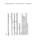
[0065] FIG. 3 shows the nucleotide coding sequence of the pWT126-specific TCR-beta chain (Vβ-2.1).
[0066] FIG. 4 shows the protein sequence of the pWT126-specific TCR-beta chain (Vβ-2.1). The position of the CDRs, FRs and constant region are marked.
[0067] FIG. 5 shows the same protein sequence as in FIG. 2 but the start position of the constant region is indicated to be in a different place. The CDR sequence in this figure, starting after C, is based on IMGT nomenclature (primary sequence based). The Garcia nomenclature is based on structure and does not include the VV after the C (ie it starts SPF . . .). Va8.2 means variable alpha 8.2 gene segment and J45 means joining 45 gene segment.
[0068] FIG. 6 shows the same protein sequence as in FIG. 4 except that CDR3β is indicated as being longer and the start position of the constant region is indicated to be in a different place. The CDR sequence in this figure, starting after C, is based on IMGT nomenclature (primary sequence based). The Garcia nomenclature is based on structure and does not include the SA after the C (ie it starts RDGG . . . ). J2.5 refers to joining 2.5 gene segment.
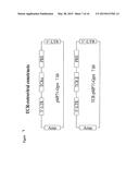
[0069] FIG. 7 is a diagram showing retroviral vectors containing TCR genes. The TCR alpha and beta chains were inserted into the retroviral vector pMP71 (Engels et al (2003) Human Gene Ther. 14, 1155-1168 for gene transfer into human T cells. LTR is a long terminated repeat. PRE is posttranscriptional regulatory element.
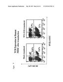
[0070] FIG. 8 is a diagram showing retroviral TCR gene transfer into human T cells. Peripheral blood lymphocytes were activated with anti-CD3 antibodies, IL-2 and IL-7, followed 3 days later by transduction with retroviral vectors encoding the WT1-specific TCR. TCR expression was monitored at day 6 using antibodies specific for the TCR-V-beta 2.1 (present in the transferred TCR). Mock transduced T cells show the percentage of un-manipulated human T cells expressing V-beta 2.1. After transduction both CD8-positive and CD8-negative (i.e. CD4-pos) T cells have an increased percentage of V-beta 2.1 cells. V-beta 2.1 DNA and amino acid sequences are shown in FIGS. 3 and 4.
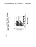
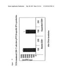
[0071] FIG. 9 shows that repeated stimulation of TCR-transduced T cells (as shown in FIG. 8) with T2 cells presenting the pWT126 peptide leads to an expansion of CD8-positive T cells expressing V-beta 2.1.
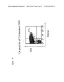
[0072] FIG. 10 shows that TCR-transduced T cells (as shown in FIGS. 8 and 9) stain with HLA-A2/pWT126 tetramers.
[0073] FIG. 11 shows that TCR-transduced T cells (as shown in FIGS. 8 and 9) kill the HLA-A2-positive T2 cells coated with the pWT126 peptide, but not T2 cells coated with the A2-binding pWT235 control peptide. The T cells also kill the HLA-A2-positive BV173 leukaemia cells expressing WT1 endogenously.
[0074] FIG. 12 shows that purified TCR-transduced CD8-positive T cells kill the HLA-A2-positive T2 cells coated with the pWT126 peptide, but not T2 cells coated with the A2-binding pWT235 control peptide. The CD8-positive T cells also kill the HLA-A2-positive BV173 leukaemia cells expressing WT1 endogenously.
[0075] FIG. 13 shows that a small percentage of purified CD4-positive TCR-transduced T cells stain with HLA-A2/pWT126 tetramers.
[0076] FIG. 14 shows that purified TCR-transduced CD4-positive T cells kill the HLA-A2-positive T2 cells coated with the pWT126 peptide, but not T2 cells coated with the A2-binding pWT235 control peptide. The CD4-positive T cells also kill the HLA-A2-positive BV173 leukaemia cells expressing WT1 endogenously.
[0077] FIG. 15 shows that purified TCR-transduced CD8-positive T cells produce IFN-γ after stimulation with the HLA-A2-positive T2 cells coated with the pWT126 peptide, but not T2 cells coated with the A2-binding pWT235 control peptide. Also, the CD8-positive T cells produce IFN-γ after stimulation with the HLA-A2-positive BV173 leukaemia cells expressing WT1 endogenously.
[0078] FIG. 16 is a schematic diagram showing the general structure of αβ TCR molecules. The amino acid numbers mentioned do not necessarily correspond to those in FIGS. 2 and 4.
TABLE-US-00001 Schedule of SEQ ID Nos. 1. RMFPNAPYL 2. SSYSPS 3. YTSAATL 4. VVSPFSGGGADGLT 5. SPFSGGGADGLT 6. DFQATT 7. SNEGSKA S. SARDGGEG 9. RDGGEGSETQY 10. FGKGTHLIIQP 11. YIQNP 12. SETQY 13. FGPGTRLLVL 14. EDLKN 15. FIG. 1 nucleotide sequence 16. FIG. 2 (and FIG. 5) amino acid sequence 17. FIG. 3 nucleotide sequence 18. FIG. 4 (and FIG. 6) amino acid sequence
EXAMPLE 1
Functionally Active T Cell Receptor (TCR) Specific For the WT-1-Derived Peptide pWT126 (RMFPNAPYL)
[0079] We have cloned a T cell receptor (TCR) that is specific for a peptide (pWT126; RMFPNAPYL) of the Wilms Tumour antigen-1 (WT1) presented by HLA-A2 class I molecules. The WT1 transcription factor is expressed in various human malignancies, including leukaemia, breast cancer, colon cancer, lung cancer, ovarian cancer and others. The CTL from which the TCR was cloned show killing activity against human cancer cells that express WT1, but not against normal human cells that express physiological levels of WT1.
[0080] The therapeutic goal is to equip patient T cells with this potent and specific killing activity by transfer of the genes encoding the TCR. For this, we have inserted the TCR genes into retroviral vectors and demonstrated that gene transduced human T cells show killing activity against WT1 expressing human cancer and leukemia cell lines. The specificity profile of this CTL line has been described in several research papers and can be summarized as (1) Killing of HLA-A2-positive targets coated with the WT1-derived peptide pWT126 (Gao et al (2000) Blood 95, 2198-2203); (2) Killing of fresh HLA-A2-positive leukaemia cells expressing WT1 (Gao et al (2000) Blood 95, 2198-2203); (3) Killing of HLA-A2-positive leukemia CFU progenitor cells (Gao et al (2000) Blood 95, 2198-2203; Bellantuono et al (2002) 100, 3835-3837); (4) Killing of HLA-A2-positive leukaemia LTC-IC stem cells (Bellantuono et al (2002) Blood 100, 3835-3837); (5) Killing of HLA-A2-positive NOD/SCID leukaemia initiating cells (Gao et al (2003) Transplantation 75, 1429-1436); and (6) No killing of normal HLA-A2-positive NOD/SCID engrafting hematopoietic stem cells (Gao et al (2003) Transplantation 75, 1429-1436). We have now shown that human T cells transduced with the WT1-specific TCR display similar specificity as the CTL line from which the TCR was cloned.
[0081] The data described in detail in the legends to FIGS. 1 to 15 indicate that TCR gene transfer into human T cells is feasible and that it leads to the surface expression of the introduced TCR chains. The recipient T cells show killing activity against HLA-A2-positive targets coated with the pWT126 peptide. The TCR-transduced T cells also kill human tumour cells expressing WT1 endogenously. In addition, the transduced T cells produce IFN-g in an HLA-A2-restricted, peptide-specific fashion. Finally, the transferred TCR can function in CD4-positive helper T cells. These CD4-positive T cells show HLA-A2-restricted, antigen-specific killing activity and antigen-specific cytokine production (not shown). This indicates that TCR gene transfer can be used to confer HLA class I-restricted antigen-specific effector function to both CD8-positive and CD4-positive human T cells.
EXAMPLE 2
Selection and Treatment of a Patient
[0082] Peripheral blood monocyte cells (PBMCs) are taken from an HLA-A2-positive patient who has a WT1-expressing malignancy. The PBMCs are activated with anti-CD3/CD28 antibodies added to the culture or on beads for 3 days and then transduced with TCR encoding retroviral particles as described in Example 1. At day 5 we can demonstrate that transduced CD4 and CD8 T cells express the introduced TCR. At day 6 we can demonstrate antigen-specific activity of the transduced T cells. At day 6 the transduced T cells are reinfused into the patient.
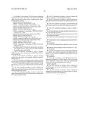
Sequence CWU
1
1
1819PRTArtificial SequencePeptide of WT1 which is presented by HLA-A2
class I molecules 1Arg Met Phe Pro Asn Ala Pro Tyr Leu 1 5
26PRTArtificial SequenceCDR1 of human TCR V -1.5 (V
-8.2) 2Ser Ser Tyr Ser Pro Ser 1 5 37PRTArtificial
SequenceCDR2 of human TCR V -1.5 (V -8.2) 3Tyr Thr Ser Ala Ala Thr Leu 1
5 414PRTArtificial SequenceCDR3 of human TCR V
-1.5 (V -8.2) - 1 4Val Val Ser Pro Phe Ser Gly Gly Gly Ala Asp Gly Leu
Thr 1 5 10
512PRTArtificial SequenceCDR3 of human TCR V -1.5 (V -8.2) - 2 5Ser Pro
Phe Ser Gly Gly Gly Ala Asp Gly Leu Thr 1 5
10 66PRTArtificial SequenceCDR1 of human TCR V -2.1 (V -20.1)
6Asp Phe Gln Ala Thr Thr 1 5 77PRTArtificial
SequenceCDR2 of human TCR V -2.1 (V -20.1) 7Ser Asn Glu Gly Ser Lys Ala
1 5 88PRTArtificial SequenceCDR3 of human TCR V
-2.1 (V -20.1) - 1 8Ser Ala Arg Asp Gly Gly Glu Gly 1 5
911PRTArtificial SequenceCDR3 of human TCR V -2.1 (V -20.1) -
2 9Arg Asp Gly Gly Glu Gly Ser Glu Thr Gln Tyr 1 5
10 1011PRTArtificial SequenceFramework amino acid sequence
of constant portion C-terminal to CDR3 10Phe Gly Lys Gly Thr His
Leu Ile Ile Gln Pro 1 5 10
115PRTArtificial SequenceBeginning of constant region of human TCR V
-1.5 (V -8.2) 11Tyr Ile Gln Asn Pro 1 5 125PRTArtificial
SequenceBeginning of framework amino acid sequence of human TCR V
-2.1 (V -20.1) 12Ser Glu Thr Gln Tyr 1 5
1310PRTArtificial SequencePart of framework amino acid sequence of human
TCR V -2.1 (V -20.1) 13Phe Gly Pro Gly Thr Arg Leu Leu Val Leu 1
5 10 145PRTArtificial SequencePart of
constant region of human TCR V -2.1 (V -20.1) 14Glu Asp Leu Lys Asn
1 5 15830DNAHomo sapiensmisc_featureHuman TCR V -1.5 (V
-8.2) 15atgctcctgc tgctcgtccc agtgctcgag gtgattttta ctctgggagg aaccagagcc
60cagtcggtga cccagcttga cagccacgtc tctgtctctg aaggaacccc ggtgctgctg
120aggtgcaact actcatcttc ttattcacca tctctcttct ggtatgtgca acaccccaac
180aaaggactcc agcttctcct gaagtacaca tcagcggcca ccctggttaa aggcatcaac
240ggttttgagg ctgaatttaa gaagagtgaa acctccttcc acctgacgaa accctcagcc
300catatgagcg acgcggctga gtacttctgt gttgtgagtc ctttttcagg aggaggtgct
360gacggactca cctttggcaa agggactcat ctaatcatcc agccctatat ccagaaccct
420gaccctgccg tgtaccagct gagagactct aaatccagtg acaagtctgt ctgcctattc
480accgattttg attctcaaac aaatgtgtca caaagtaagg attctgatgt gtatatcaca
540gacaaaactg tgctagacat gaggtctatg gacttcaaga gcaacagtgc tgtggcctgg
600agcaacaaat ctgactttgc atgtgcaaac gccttcaaca acagcattat tccagaagac
660accttcttcc ccagcccaga aagttcctgt gatgtcaagc tggtcgagaa aagctttgaa
720acagatacga acctaaactt tcaaaacctg tcagtgattg ggttccgaat cctcctcctg
780aaagtggccg ggtttaatct gctcatgacg ctgcggctgt ggtccagctg
83016276PRTHomo sapiensMISC_FEATUREHuman TCR V -1.5 (V -8.2) 16Met Leu
Leu Leu Leu Val Pro Val Leu Glu Val Ile Phe Thr Leu Gly 1 5
10 15 Gly Thr Arg Ala Gln Ser Val
Thr Gln Leu Asp Ser His Val Ser Val 20 25
30 Ser Glu Gly Thr Pro Val Leu Leu Arg Cys Asn Tyr
Ser Ser Ser Tyr 35 40 45
Ser Pro Ser Leu Phe Trp Tyr Val Gln His Pro Asn Lys Gly Leu Gln
50 55 60 Leu Leu Leu
Lys Tyr Thr Ser Ala Ala Thr Leu Val Lys Gly Ile Asn 65
70 75 80 Gly Phe Glu Ala Glu Phe Lys
Lys Ser Glu Thr Ser Phe His Leu Thr 85
90 95 Lys Pro Ser Ala His Met Ser Asp Ala Ala Glu
Tyr Phe Cys Val Val 100 105
110 Ser Pro Phe Ser Gly Gly Gly Ala Asp Gly Leu Thr Phe Gly Lys
Gly 115 120 125 Thr
His Leu Ile Ile Gln Pro Tyr Ile Gln Asn Pro Asp Pro Ala Val 130
135 140 Tyr Gln Leu Arg Asp Ser
Lys Ser Ser Asp Lys Ser Val Cys Leu Phe 145 150
155 160 Thr Asp Phe Asp Ser Gln Thr Asn Val Ser Gln
Ser Lys Asp Ser Asp 165 170
175 Val Tyr Ile Thr Asp Lys Thr Val Leu Asp Met Arg Ser Met Asp Phe
180 185 190 Lys Ser
Asn Ser Ala Val Ala Trp Ser Asn Lys Ser Asp Phe Ala Cys 195
200 205 Ala Asn Ala Phe Asn Asn Ser
Ile Ile Pro Glu Asp Thr Phe Phe Pro 210 215
220 Ser Pro Glu Ser Ser Cys Asp Val Lys Leu Val Glu
Lys Ser Phe Glu 225 230 235
240 Thr Asp Thr Asn Leu Asn Phe Gln Asn Leu Ser Val Ile Gly Phe Arg
245 250 255 Ile Leu Leu
Leu Lys Val Ala Gly Phe Asn Leu Leu Met Thr Leu Arg 260
265 270 Leu Trp Ser Ser 275
17933DNAHomo sapiensmisc_featureHuman TCR V -2.1 (V -20.1) 17atgctgctgc
ttctgctgct tctggggcca ggctccgggc ttggtgctgt cgtctctcaa 60catccgagct
gggttatctg taagagtgga acctctgtga agatcgagtg ccgttccctg 120gactttcagg
ccacaactat gttttggtat cgtcagttcc cgaaacagag tctcatgctg 180atggcaactt
ccaatgaggg ctccaaggcc acatacgagc aaggcgtcga gaaggacaag 240tttctcatca
accatgcaag cctgaccttg tccactctga cagtgaccag tgcccatcct 300gaagacagca
gcttctacat ctgcagtgct agagatgggg gggagggttc ggagacccag 360tacttcgggc
caggcacgcg gctcctggtg ctcgaggacc tgaaaaacgt gttcccaccc 420gaggtcgctg
tgtttgagcc atcagaagca gagatctccc acacccaaaa ggccacactg 480gtgtgcctgg
ccacaggctt ctaccccgac cacgtggagc tgagctggtg ggtgaatggg 540aaggaggtgc
acagtggggt cagcacagac ccgcagcccc tcaaggagca gcccgccctc 600aatgactcca
gatactgcct gagcagccgc ctgagggtct cggccacctt ctggcagaac 660ccccgcaacc
acttccgctg tcaagtccag ttctacgggc tctcggagaa tgacgagtgg 720acccaggata
gggccaaacc tgtcacccag atcgtcagcg ccgaggcctg gggtagagca 780gactgtggct
tcacctccga gtcttaccag caaggggtcc tgtctgccac catcctctat 840gagatcttgc
tagggaaggc caccttgtat gccgtgctgg tcagtgccct cgtgctgatg 900gccatggtca
agagaaagga ttccagaggc tag 93318310PRTHomo
sapiensMISC_FEATUREHuman TCR V -2.1 (V -20.1) 18Met Leu Leu Leu Leu Leu
Leu Leu Gly Pro Gly Ser Gly Leu Gly Ala 1 5
10 15 Val Val Ser Gln His Pro Ser Trp Val Ile Cys
Lys Ser Gly Thr Ser 20 25
30 Val Lys Ile Glu Cys Arg Ser Leu Asp Phe Gln Ala Thr Thr Met
Phe 35 40 45 Trp
Tyr Arg Gln Phe Pro Lys Gln Ser Leu Met Leu Met Ala Thr Ser 50
55 60 Asn Glu Gly Ser Lys Ala
Thr Tyr Glu Gln Gly Val Glu Lys Asp Lys 65 70
75 80 Phe Leu Ile Asn His Ala Ser Leu Thr Leu Ser
Thr Leu Thr Val Thr 85 90
95 Ser Ala His Pro Glu Asp Ser Ser Phe Tyr Ile Cys Ser Ala Arg Asp
100 105 110 Gly Gly
Glu Gly Ser Glu Thr Gln Tyr Phe Gly Pro Gly Thr Arg Leu 115
120 125 Leu Val Leu Glu Asp Leu Lys
Asn Val Phe Pro Pro Glu Val Ala Val 130 135
140 Phe Glu Pro Ser Glu Ala Glu Ile Ser His Thr Gln
Lys Ala Thr Leu 145 150 155
160 Val Cys Leu Ala Thr Gly Phe Tyr Pro Asp His Val Glu Leu Ser Trp
165 170 175 Trp Val Asn
Gly Lys Glu Val His Ser Gly Val Ser Thr Asp Pro Gln 180
185 190 Pro Leu Lys Glu Gln Pro Ala Leu
Asn Asp Ser Arg Tyr Cys Leu Ser 195 200
205 Ser Arg Leu Arg Val Ser Ala Thr Phe Trp Gln Asn Pro
Arg Asn His 210 215 220
Phe Arg Cys Gln Val Gln Phe Tyr Gly Leu Ser Glu Asn Asp Glu Trp 225
230 235 240 Thr Gln Asp Arg
Ala Lys Pro Val Thr Gln Ile Val Ser Ala Glu Ala 245
250 255 Trp Gly Arg Ala Asp Cys Gly Phe Thr
Ser Glu Ser Tyr Gln Gln Gly 260 265
270 Val Leu Ser Ala Thr Ile Leu Tyr Glu Ile Leu Leu Gly Lys
Ala Thr 275 280 285
Leu Tyr Ala Val Leu Val Ser Ala Leu Val Leu Met Ala Met Val Lys 290
295 300 Arg Lys Asp Ser Arg
Gly 305 310
User Contributions:
Comment about this patent or add new information about this topic:
 |  |
 |  |
 |  |
 |  |
 |  |
 |  |
 |  |
 |  |
 |  |
 |  |
 |  |
 |  |
| Similar patent applications: | |
| Date | Title |
|---|---|
| 2015-05-14 | Human bispecific egfrviii antibody engaging molecules |
| 2015-05-14 | Polymeric nanoparticles useful in theranostics |
| 2015-05-14 | Therapeutic use of chardonnay seed products |
| 2015-02-05 | Method for selecting a pool of molecules |
| 2012-04-19 | Therapeutic vesicles |
| New patent applications in this class: | |
| Date | Title |
|---|---|
| 2022-05-05 | Compositions and methods for treating neurocognitive disorders |
| 2022-05-05 | Administration of tumor infiltrating lymphocytes with membrane bound interleukin 15 to treat cancer |
| 2019-05-16 | Crispr/cas9 complex for genomic editing |
| 2019-05-16 | Chimeric antigen receptor with single domain antibody |
| 2019-05-16 | Chimeric antigen receptors targeting epidermal growth factor receptor variant iii |
| New patent applications from these inventors: | |
| Date | Title |
|---|---|
| 2017-07-13 | Single chain antigen recognizing constructs (scarcs) stabilized by the introduction of novel disulfide bonds |
| 2014-07-17 | Immunotherapeutic methods using epitopes of wt-1 and gata-1 |
| 2014-04-24 | Single chain antigen recognizing constructs (scarcs) stabilized by the introduction of novel disulfide bonds |
| 2013-02-21 | T-cell receptor capable of recognising an antigen from cytomegalovirus |
| 2012-09-27 | T-cell receptor |
| Top Inventors for class "Drug, bio-affecting and body treating compositions" | |
| Rank | Inventor's name |
|---|---|
| 1 | David M. Goldenberg |
| 2 | Hy Si Bui |
| 3 | Lowell L. Wood, Jr. |
| 4 | Roderick A. Hyde |
| 5 | Yat Sun Or |