Patent application title: METHOD FOR ACQUIRING A HEAT-STABLE ANTIBODY-DISPLAYED PHAGE
Inventors:
Junko Wakai (Osaka, JP)
Assignees:
PANASONIC CORPORATION
IPC8 Class: AC07K1400FI
USPC Class:
530324
Class name: Chemistry: natural resins or derivatives; peptides or proteins; lignins or reaction products thereof peptides of 3 to 100 amino acid residues 25 or more amino acid residues in defined sequence
Publication date: 2014-05-08
Patent application number: 20140128569
Abstract:
Using a biopanning method, a heat-stable antibody-displayed phage is
acquired. More particularly, first, an antibody-displayed phage library
aqueous solution containing plural types of antibody-displayed phages is
supplied to a support comprising a polypeptide on the surface thereof, so
as to bind the plural types of the antibody-displayed phages to the
polypeptide specifically. Next, the support is heated to the temperature
of not less than 37 degrees Celsius and not more than 70 degrees Celsius,
so as to release a portion of the antibody-displayed phages from the
support and so as to leave the other antibody-displayed phages on the
support selectively. Finally, the other antibody-displayed phages which
has been left on the support selectively in the previous step is
collected to obtain the heat-stable antibody-displayed phage.Claims:
1. A method for acquiring a heat-stable antibody-displayed phage, the
method comprising steps of: (a) supplying an antibody-displayed phage
library aqueous solution containing plural types of antibody-displayed
phages to a support comprising a polypeptide on the surface thereof, so
as to bind the plural types of the antibody-displayed phages to the
polypeptide specifically; wherein each antibody-displayed phage comprises
proteins containing a phagemid vector in the inside thereof; each
antibody-displayed phage comprises an antibody fragment on the external
surface thereof; and the phagemid vector has a gene sequence coding for
the antibody fragment; (b) heating the support to the temperature of not
less than 37 degrees Celsius and not more than 70 degrees Celsius, so as
to release a portion of the antibody-displayed phages from the support
and so as to leave the other antibody-displayed phages on the support
selectively; and (c) collecting the other antibody-displayed phages which
has been left on the support selectively in the step (b) so as to obtain
the heat-stable antibody-displayed phage.
2. The method according to claim 1, wherein the polypeptide consists of an amino acid sequence represented by SEQ ID NO: 01.
3. The method according to claim 1, further comprising the following step between the step (a) and the step (b), a step of removing an antibody-displayed phages which have not been bound to the polypeptide specifically in the step (a).
4. The method according to claim 1, further comprising the following step between the step (b) and the step (c), a step of washing the support.
5. The method according to claim 1, further comprising the following step after the step (c), a step of amplifying the heat-stable antibody-displayed phages collected in the step (c).
6. A method for acquiring a heat-stable antibody fragment, the method comprising steps of: (a) supplying an antibody-displayed phage library aqueous solution containing plural types of antibody-displayed phages to a support comprising a polypeptide on the surface thereof, so as to bind the plural types of the antibody-displayed phages to the polypeptide specifically; wherein each antibody-displayed phage comprises proteins containing a phagemid vector in the inside thereof; each antibody-displayed phage comprises an antibody fragment on the external surface thereof; and the phagemid vector has a gene sequence coding for the antibody fragment; (b) heating the support to the temperature of not less than 37 degrees Celsius and not more than 70 degrees Celsius, so as to release a portion of the antibody-displayed phages from the support and so as to leave the other antibody-displayed phages on the support selectively; (c) collecting the other antibody-displayed phages which has been left on the support selectively in the step (b) so as to obtain the heat-stable antibody-displayed phage. (d) bringing a cell into contact with the heat-stable antibody-displayed phages collected in the step (c) to produce an infected cell; and (e) incubating the infected cell produced in the step (d) to acquire the heat-stable antibody fragment.
7. A heat-stable peptide consisting of the amino acid sequence represented by SEQ ID NO: 01.
8. The heat-stable peptide according to claim 7, wherein the peptide is stable even when the peptide is heated to 70 degrees Celsius.
Description:
CROSS-REFERENCE TO RELATED APPLICATIONS
[0001] This is a continuation application of International Application No. PCT/JP2012/004306, with an international filing date of Jul. 3, 2012, which claims priority of Japanese Patent Application No. 2012-110220, filed on May 14, 2012, the entire contents of each of which are incorporated herein by reference.
BACKGROUND OF THE INVENTION
Technical Field
[0002] The technical field relates to a method for acquiring a heat-stable antibody-displayed phage.
[0003] FIG. 1B shows an antibody-displayed phage. The antibody-displayed phage is one kind of phages. The antibody-displayed phage comprises proteins containing a phagemid vector in the inside thereof. In other words, these proteins surround the phagemid vector. The antibody-displayed phage comprises an antibody fragment on the external surface thereof. Two or more antibody fragments may be comprised. The phagemid vector has a gene sequence coding for this antibody fragment.
[0004] FIG. 2 shows a method for selecting an antibody-displayed phage having a predetermined property using an antibody phage library which contains plural types of antibody-displayed phages. This method is referred to as "biopanning method".
[0005] Each antibody-displayed phage contained in the antibody phage library has a different antibody fragment and a different gene sequence coding for the different antibody fragment.
[0006] The biopanning method is briefly described below.
[0007] As shown in FIG. 2, first, an aqueous solution containing an antibody phage library is supplied to a support having an antigen corresponding to the predetermined property on the surface thereof. The Antibody-displayed phage having the predetermined property is specifically bound to the antigen. However, the antibody-displayed phage which does not have the predetermined property is removed by washing.
[0008] After the antibody-displayed phage which has been specifically bound to the support is collected, the collected antibody-displayed phage is amplified. After the amplification, the antibody-displayed phage is supplied again to the antibody having the antibody on the surface thereof, if necessary.
[0009] This procedure is repeated. In this way, the antibody-displayed phage having the predetermined property is collected selectively from the antibody phage library.
Citation List
[0010] [Patent Literature 1]
[0011] Japanese Patent Laid-Open Publication No. 2009-280616 bulletin (particularly, paragraph 0040) Family: WO96/40749 (Page 15)
[0012] [Patent Literature 2]
[0013] WO 2003/099999 (particularly, page 160, line 33 to page 162, line 31, more particularly, page 162, line 8 to 9)
SUMMARY
[0014] One non-limiting and exemplary embodiment provides a novel method for acquiring a heat-stable antibody-displayed phage.
[0015] Additional benefits and advantages of the disclosed embodiments will be apparent from the specification and Figures. The benefits and/or advantages may be individually provided by the various embodiments and features of the specification and drawings disclosure, and need not all be provided in order to obtain one or more of the same.
[0016] In one general aspect, the techniques disclosed here feature: A method for acquiring a heat-stable antibody-displayed phage, the method comprising steps of:
[0017] (a) supplying an antibody-displayed phage library aqueous solution containing plural types of antibody-displayed phages to a support comprising a polypeptide on the surface thereof, so as to bind the plural types of the antibody-displayed phages to the polypeptide specifically; wherein
[0018] each antibody-displayed phage comprises proteins containing a phagemid vector in the inside thereof;
[0019] each antibody-displayed phage comprises an antibody fragment on the external surface thereof; and
[0020] the phagemid vector has a gene sequence coding for the antibody fragment;
[0021] (b) heating the support to the temperature of not less than 37 degrees Celsius and not more than 70 degrees Celsius, so as to release a portion of the antibody-displayed phages from the support and so as to leave the other antibody-displayed phages on the support selectively; and
[0022] (c) collecting the other antibody-displayed phages which has been left on the support selectively in the step (b) so as to obtain the heat-stable antibody-displayed phage.
[0023] The present disclosure provides a novel method for acquiring a heat-stable antibody-displayed phage.
BRIEF DESCRIPTION OF THE DRAWINGS
[0024] FIG. 1A shows an antibody.
[0025] FIG. 1B shows an antibody presentation phage.
[0026] FIG. 2 shows a biopanning method.
[0027] FIG. 3 shows a method for selectively acquiring the heat-stable antibody-displayed phage according to the embodiment.
[0028] FIG. 4 shows a method for obtaining the phagemid vector according to the example.
[0029] FIG. 5 shows a graph of the experimental result according to the example 2-2 to the example 2-30.
DETAILED DESCRIPTION
[0030] The embodiment according to the present disclosure is described below.
[0031] Explanation of the Term
[0032] First, the term used in the present specification is described.
[0033] FIG. 1A shows an antibody. As known well, the antibody 1 has a shape of the "Y" letter. The antibody 1 consists of two Fab regions and one Fc region. The antibody 1 consists of two heavy chains 2 and two light chains 3. Each heavy chain 2 consists of a heavy chain constant region 1 (referential sign: 22), a heavy chain constant region 2 (referential sign: 23), and a heavy chain constant region 3 (referential sign: 23), and a heavy chain variable region 21. Each light chain 3 consists of a light chain variable region 31 and a light chain constant region 32.
[0034] Each Fab region consists of the one heavy chain variable region 21, the one heavy chain constant region 1 (referential sign: 22), the one light chain variable region 31, and the one light chain constant region 32. The light chain 3 is connected to the heavy chain 2 through a linker 4. The heavy chain 2 has the heavy chain variable region 21 in the end thereof. The light chain 3 has the light chain variable region 31 in the end thereof. An antigen is specifically bound to the antibody 1. In more detail, the antigen is bound specifically to the Fv region, which consists of the heavy chain variable region 21 and the light chain variable region 31.
[0035] An example of the antibody fragment is an Fab antibody fragment, an F(ab')2 antibody fragment, or an scFv antibody fragment.
[0036] The Fab antibody fragment consists of one Fab region. In other words, the Fab antibody fragment consists of the one light chain variable region 31, the one heavy chain variable region 21, the one light chain constant region 32, the one heavy chain constant region 1 (referential sign: 22), and the linker 4. The light chain constant region 32 is connected to the heavy chain constant region (referential sign: 22) through the linker 4.
[0037] The F(ab')2 antibody fragments consists of two Fab regions. As above, each Fab region consists of the one light chain variable region 31, the one heavy chain variable region 21, the one light chain constant region 32, the one heavy chain constant region 1 (referential sign: 22), and the linker 4. These two Fab regions are connected to each other through another liner (not shown). Preferably, one heavy chain constant region 1 (referential sign: 22) is connected to the other heavy chain constant region 1 (referential sign: 22) through the another linker (not shown).
[0038] The scFv antibody fragment consists of the light chain variable region 31, the heavy chain variable region 21, and a linker. The light chain variable region 31 is connected to the heavy chain variable region 21 through the linker (not shown).
[0039] An example of the linker is a peptide consisting of 5-20 amino acids. More particularly, an example of the linker is the amino acid sequence represented by GGGGSGGGGSGGGGS (SEQ ID NO: 59). Another example of the linker is a disulfide bond (--sulfur atom (S)--sulfur atom (S)--).
[0040] (Step (a))
[0041] FIG. 3 shows a method for selectively acquiring the heat-stable antibody-displayed phage according to the embodiment.
[0042] First, in the step (a), an antibody-displayed phage library aqueous solution is prepared. The antibody-displayed phage library aqueous solution contains plural kinds of antibody-displayed phages.
[0043] As shown in FIG. 1B, each antibody-displayed phage contained in the antibody-displayed phage library aqueous solution has an antibody fragment on the surface thereof. It is desirable that each antibody-displayed phage has an scFv antibody fragment on the surface thereof. Each scFv antibody fragment consists of one heavy chain variable region 21 and one light chain variable region 31 which are randomly selected from plural types of heavy chain variable regions 21 and from plural types of light chain variable regions 31, respectively.
[0044] The plural kinds of antibody-displayed phages have different antibody fragments from each other.
[0045] Each antibody-displayed phage contains a phagemid vector. The phagemid vector has a gene sequence coding for the antibody fragment added on the surface thereof. Desirably, the phagemid vector has a VH gene coding for one heavy chain variable region 21 of the scFv antibody fragment and a VL gene coding for one light chain variable region 31 of the scFv antibody fragment.
[0046] Non Patent Literature 3, Non Patent Literature 4 and, Non Patent Literature 5 disclose a method for preparing such an antibody-displayed phage library aqueous solution. These skilled in the art who have read Non Patent Literature 3, Non Patent Literature 4, and Non Patent Literature 5 could prepare the antibody-displayed phage library aqueous solution easily.
[0047] [Non Patent Literature 3]
[0048] Greg Winter, Andrew D. Griffiths, Robert E. Hawkins, and Hennie R. Hoogenboom (1994) Making Antibodies by Phage Display Technology, Annual Review of Immunology, Vol. 12, 433-455
[0049] [Non Patent Literature 4]
[0050] Takayuki Okamoto, Yohei Mukai, Yasuo Yoshioka, Hiroko Shibata, Maki Kawamura, Yoko Yamamoto, Shinsaku Nakagawa, Haruhiko Kamada, Takao Hayakawa, Tadanori Mayumi, Yasuo Tsutsumi (2004) Optimal construction of non-immune scFv phage display libraries from mouse bone marrow and spleen established to select specific scFvs efficiently binding to antigen, Biochemical and Biophysical Research Communications, Vol. 323(2), 583-91
[0051] [Non Patent Literature 5]
[0052] Phage Display: A Practical Approach (The Practical Approach Series), Oxford University Press, USA (2004/5/6), ISBN 978-0199638734
[0053] As shown in FIG. 3, the antibody-displayed phage library aqueous solution is supplied to a support comprising polypeptide on the surface thereof. The polypeptide serves as an antigen. The antibody fragment added on the surface of the antibody-displayed phage is specifically bound to the polypeptide. In this way, at least a portion of the plural kinds of antibody-displayed phages are specifically bound to the polypeptide. It is desirable that the antibody-displayed phages which have not been bound to the polypeptide are removed by washing. It is desirable that a buffer solution is used for the washing. An example of the buffer solution is a PBS buffer solution containing Tween20 at a concentration of 0.1% by weight.
[0054] The polypeptide is immobilized on the support. It is desirable that the polypeptide is immobilized on the support through covalent bond. For example, a cysteine residue is introduced into the end of the polypeptide. Meanwhile, a gold film is formed on the support. A sulfur atom included in the cysteine residue reacts with the gold film to form a sulfide bond between the polypeptide and the gold film. In this way, the polypeptide can be immobilized on the support. Other immobilization methods can be used.
[0055] An example of the support is a substrate, a microbead, or a nonwoven fabric. An example of the materials of the substrate is plastic. Polypropylene is desirable. More particularly, an example of the support is an ELISA plate.
[0056] (Step (b))
[0057] Next, the support is heated to a temperature of not less than 37 degrees Celsius and not more than 70 degrees Celsius. When the heating temperature is high, the heating period is short. On the other hand, the heating period is long, when the heating temperature is low. More specifically, it is desirable that the support is heated to 60 degrees Celsius for approximately 30 minutes.
[0058] The present inventors discovered that the peptide consisting of the amino acid sequence represented by SEQ ID NO: 01 is heat-stable. The peptide consisting of the amino acid sequence represented by SEQ ID NO: 01 is not denatured, even when the peptide is heated to 70 degrees Celsius. See the example 2, which is described later.
[0059] Some of antibody-displayed phages are released from the support by the heating. The antibody-displayed phages which have been released from the support are not heat-stable antibody-displayed phages. It is desirable that the support is heated, while the support is in contact with a buffer solution. This is because the antibody-displayed phages which have been released from the support are dispersed or dissolved in the buffer solution easily.
[0060] On the other hand, the other antibody-displayed phages are not released from the support by the heating. In other words, the other antibody-displayed phages are left on the support, even when they are heated. The other antibody-displayed phages, which have been left on the support, are heat-stable antibody-displayed phages. In this way, the heat-stable antibody-displayed phages are left on the support selectively.
[0061] It is desirable that the support is washed at the end of the step (b). For the washing, it is desirable that a buffer solution is used. An example of the buffer solution is a PBS buffer solution containing Tween 20 at a concentration of 0.1% by weight.
[0062] (Step (c))
[0063] Finally, the heat-stable antibody-displayed phages left on the support selectively are collected. In order to collect the heat-stable antibody-displayed phages from the support, reagent which denatures proteins is used. An example of the reagent is an acid aqueous solution such as hydrochloric acid, or an urea aqueous solution. A suitable example of the reagent is a 0.1M glycine--hydrochloric acid buffer solution (pH: 2.2).
[0064] The support is immersed in the 0.1M glycine--hydrochloric acid buffer solution (pH: 2.2) for 10-30 minutes to release the heat-stable antibody-displayed phages from the support. In this way, the 0.1M glycine--hydrochloric acid buffer solution which contains the heat-stable antibody-displayed phages is obtained.
[0065] Subsequently, the pH of the glycine--hydrochloric acid buffer solution is adjusted with an alkaline aqueous solution. It is desirable that the pH is adjusted to 7. An example of the alkaline aqueous solution is 1M Tris hydrochloric acid buffer solution (pH: 9.1).
[0066] As shown in FIG. 2, it is desirable that the collected heat-stable antibody-displayed phages are amplified.
[0067] Finally, using the collected heat-stable antibody-displayed phages, a heat-stable antibody fragment is obtained as below.
[0068] First, the collected heat-stable antibody-displayed phages are brought into contact with a cell (desirably, E. coli. TG-1) so as to produce an infected cell. By this infection, the E. coli. TG-1 acquires antibiotic-resistance.
[0069] Then, this infected cell is incubated to obtain a heat-stable antibody fragment.
[0070] Examples of the additional aspect of the present disclosure are as follows.
[0071] 1st aspect: A method for acquiring a heat-stable antibody-displayed phage, the method comprising steps of:
[0072] (a) supplying an antibody-displayed phage library aqueous solution containing plural types of antibody-displayed phages to a support comprising a polypeptide on the surface thereof, so as to bind the plural types of the antibody-displayed phages to the polypeptide specifically; wherein
[0073] each antibody-displayed phage comprises proteins containing a phagemid vector in the inside thereof;
[0074] each antibody-displayed phage comprises an antibody fragment on the external surface thereof; and
[0075] the phagemid vector has a gene sequence coding for the antibody fragment;
[0076] (b) heating the support to the temperature of not less than 37 degrees Celsius and not more than 70 degrees Celsius, so as to release a portion of the antibody-displayed phages from the support and so as to leave the other antibody-displayed phages on the support selectively; and
[0077] (c) collecting the other antibody-displayed phages which has been left on the support selectively in the step (b) so as to obtain the heat-stable antibody-displayed phage.
[0078] 2nd aspect: The method according to claim 1, wherein the polypeptide consists of an amino acid sequence represented by SEQ ID NO: 01.
[0079] 3rd aspect: The method according to claim 1, further comprising the following step between the step (a) and the step (b),
[0080] a step of removing an antibody-displayed phages which have not been bound to the polypeptide specifically in the step (a).
[0081] 4th aspect: The method according to claim 1, further comprising the following step between the step (b) and the step (c),
[0082] a step of washing the support.
[0083] 5th aspect: The method according to claim 1, further comprising the following step after the step (c),
[0084] a step of amplifying the heat-stable antibody-displayed phages collected in the step (c).
[0085] 6th aspect: A method for acquiring a heat-stable antibody fragment, the method comprising steps of:
[0086] (a) supplying an antibody-displayed phage library aqueous solution containing plural types of antibody-displayed phages to a support comprising a polypeptide on the surface thereof, so as to bind the plural types of the antibody-displayed phages to the polypeptide specifically; wherein
[0087] each antibody-displayed phage comprises proteins containing a phagemid vector in the inside thereof;
[0088] each antibody-displayed phage comprises an antibody fragment on the external surface thereof; and
[0089] the phagemid vector has a gene sequence coding for the antibody fragment;
[0090] (b) heating the support to the temperature of not less than 37 degrees Celsius and not more than 70 degrees Celsius, so as to release a portion of the antibody-displayed phages from the support and so as to leave the other antibody-displayed phages on the support selectively;
[0091] (c) collecting the other antibody-displayed phages which has been left on the support selectively in the step (b) so as to obtain the heat-stable antibody-displayed phage.
[0092] (d) bringing a cell into contact with the heat-stable antibody-displayed phages collected in the step (c) to produce an infected cell; and
[0093] (e) incubating the infected cell produced in the step (d) to acquire the heat-stable antibody fragment.
[0094] 7th aspect: A heat-stable peptide consisting of the amino acid sequence represented by SEQ ID NO: 01.
[0095] 8th aspect: The heat-stable peptide according to claim 7, wherein the peptide is stable even when the peptide is heated to 70 degrees Celsius.
EXAMPLES
[0096] The following examples describe the present disclosure in more detail.
Example 1
[0097] Table 1, Table 2, Table 3, and Table 4 show the primers used in the example 1.
[0098] Table 1 shows the forward mixture primers A (primers 1-21, SEQ ID NOs: 02-22) for amplifying a light chain variable region.
[0099] Table 2 shows the forward mixture primers B (primers 22-44, SEQ ID NOs: 23-45) for amplifying a heavy chain variable region.
[0100] Table 3 shows the reverse mixture primers C (primers 45-49, SEQ ID NOs: 46-50) for amplifying a light chain variable region.
[0101] Table 4 shows the reverse mixture primers D (primers 50-55, SEQ ID NOs: 51-56) for amplifying a heavy chain variable region.
TABLE-US-00001 TABLE 1 Primer 1 SEQ ID CCTTTCTATGCGGCCCAGCCGGCCATGGCCGAYA NO: 02 TTGTWCTCWCCCARTC Primer 2 SEQ ID CCTTTCTATGCGGCCCAGCCGGCCATGGCCGAYA NO: 03 TTSTGMTSACYCAGTC Primer 3 SEQ ID CCTTTCTATGCGGCCCAGCCGGCCATGGCCGAYA NO: 04 TTGTGMTMACTCAGTC Primer 4 SEQ ID CCTTTCTATGCGGCCCAGCCGGCCATGGCCGAYA NO: 05 TTGTGHTRWCACAGTC Primer 5 SEQ ID CCTTTCTATGCGGCCCAGCCGGCCATGGCCGAYA NO: 06 TTGTRATGACMCAGTC Primer 6 SEQ ID CCTTTCTATGCGGCCCAGCCGGCCATGGCCGAYA NO: 07 TTMAGATRAMCCAGTC Primer 7 SEQ ID CCTTTCTATGCGGCCCAGCCGGCCATGGCCGAYA NO: 08 TTCAGATGAYDCAGTC Primer 8 SEQ ID CCTTTCTATGCGGCCCAGCCGGCCATGGCCGAYA NO: 09 TTTTGCTGACTCAGTC Primer 9 SEQ ID CCTTTCTATGCGGCCCAGCCGGCCATGGCCGAYA NO: 10 TTGTTCTCAWCCAGTC Primer 10 SEQ ID CCTTTCTATGCGGCCCAGCCGGCCATGGCCGAYA NO: 11 TTGWGCTSACCCAATC Primer 11 SEQ ID CCTTTCTATGCGGCCCAGCCGGCCATGGCCGAYA NO: 12 TTSTRATGACCCARTC Primer 12 SEQ ID CCTTTCTATGCGGCCCAGCCGGCCATGGCCGAY NO: 13 RTTKTGATGACCCAVAC Primer 13 SEQ ID CCTTTCTATGCGGCCCAGCCGGCCATGGCCGAYA NO: 14 TYCAGATGACACAGAC Primer 14 SEQ ID CCTTTCTATGCGGCCCAGCCGGCCATGGCCGAYA NO: 15 TTGTGATGACACAACC Primer 15 SEQ ID CCTTTCTATGCGGCCCAGCCGGCCATGGCCGAYA NO: 16 TCCAGCTGACTCAGCC Primer 16 SEQ ID CCTTTCTATGCGGCCCAGCCGGCCATGGCCGAYA NO: 17 TTGTGATGACBCAGKC Primer 17 SEQ ID CCTTTCTATGCGGCCCAGCCGGCCATGGCCGAYA NO: 18 TTGTGATAACYCAGGA Primer 18 SEQ ID CCTTTCTATGCGGCCCAGCCGGCCATGGCCGAYA NO: 19 TTGTGATGACCCAGWT Primer 19 SEQ ID CCTTTCTATGCGGCCCAGCCGGCCATGGCCGAY NO: 20 GTGSTGMTSACYCAGTC Primer 20 SEQ ID CCTTTCTATGCGGCCCAGCCGGCCATGGCCGAY NO: 21 GCTGTTGTACTCAGGAATC Primer 21 SEQ ID CCTTTCTATGCGGCCCAGCCGGCCATGGCCGAYA NO: 22 TTGTDHTVWCHCAGTC
TABLE-US-00002 TABLE 2 Primer 22 SEQ ID AGCGGCGGCGGCGGCTCTGGTGGTGGTGGATCC NO: 23 GAKGTRMAGCTTCAGGAGYC Primer 23 SEQ ID AGCGGCGGCGGCGGCTCTGGTGGTGGTGGATCC NO: 24 GAGGTNCAGCTBCAGCAGTC Primer 24 SEQ ID AGCGGCGGCGGCGGCTCTGGTGGTGGTGGATCC NO: 25 CAGGTGCAGCTGAAGSASTC Primer 25 SEQ ID AGCGGCGGCGGCGGCTCTGGTGGTGGTGGATCC NO: 26 CAGSTBCAGCTGCAGCAGTC Primer 26 SEQ ID AGCGGCGGCGGCGGCTCTGGTGGTGGTGGATCC NO: 27 GAGGTYCAGCTYCAGCAGTC Primer 27 SEQ ID AGCGGCGGCGGCGGCTCTGGTGGTGGTGGATCC NO: 28 GARGTCCARCTGCAACARTC Primer 28 SEQ ID AGCGGCGGCGGCGGCTCTGGTGGTGGTGGATCC NO: 29 CAGGTYCAGCTBCAGCARTC Primer 29 SEQ ID AGCGGCGGCGGCGGCTCTGGTGGTGGTGGATCC NO: 30 CAGGTYCARCTKCAGCAGTC Primer 30 SEQ ID AGCGGCGGCGGCGGCTCTGGTGGTGGTGGATCC NO: 31 CAGGTCCACGTGAAGCAGTC Primer 31 SEQ ID AGCGGCGGCGGCGGCTCTGGTGGTGGTGGATCC NO: 32 GAGGTGAASSTGGTGGARTC Primer 32 SEQ ID AGCGGCGGCGGCGGCTCTGGTGGTGGTGGATCC NO: 33 GAVGTGAWGYTGGTGGAGTC Primer 33 SEQ ID AGCGGCGGCGGCGGCTCTGGTGGTGGTGGATCC NO: 34 GAGGTGAAGGTCATCGAGTC Primer 34 SEQ ID AGCGGCGGCGGCGGCTCTGGTGGTGGTGGATCC NO: 35 SAGGTGCAGSKGGTGGAGTC Primer 35 SEQ ID AGCGGCGGCGGCGGCTCTGGTGGTGGTGGATCC NO: 36 GAKGTGCAMCTGGTGGAGTC Primer 36 SEQ ID AGCGGCGGCGGCGGCTCTGGTGGTGGTGGATCC NO: 37 GAAGTGCAVCTGGTGGAGTC Primer 37 SEQ ID AGCGGCGGCGGCGGCTCTGGTGGTGGTGGATCC NO: 38 GAGGTGAAGCTGATGGARTC Primer 38 SEQ ID AGCGGCGGCGGCGGCTCTGGTGGTGGTGGATCC NO: 39 GAGGTGCARCTTGTTGAGTC Primer 39 SEQ ID AGCGGCGGCGGCGGCTCTGGTGGTGGTGGATCC NO: 40 GARGTRAAGCTTCTCGAGTC Primer 40 SEQ ID AGCGGCGGCGGCGGCTCTGGTGGTGGTGGATCC NO: 41 GAAGTGAARSTTGAGGAGTC Primer 41 SEQ ID AGCGGCGGCGGCGGCTCTGGTGGTGGTGGATCC NO: 42 GAAGTGATGCTGGTGGAGTC Primer 42 SEQ ID AGCGGCGGCGGCGGCTCTGGTGGTGGTGGATCC NO: 43 CAGGTTACTCTRAAAGWGTSTG Primer 43 SEQ ID AGCGGCGGCGGCGGCTCTGGTGGTGGTGGATCC NO: 44 CAGGTCCAAYTVCAGCARCC Primer 44 SEQ ID AGCGGCGGCGGCGGCTCTGGTGGTGGTGGATCC NO: 45 GATGTGAACTTGGAAGTGTC
TABLE-US-00003 TABLE 3 Primer 45 SEQ ID ACCAGAGCCGCCGCCGCCGCTACCACCACCACCC NO: 46 CGTTTGATTTCCARCTTKG Primer 46 SEQ ID ACCAGAGCCGCCGCCGCCGCTACCACCACCACCC NO: 47 CGTTTTATTTCCAGCTTGG Primer 47 SEQ ID ACCAGAGCCGCCGCCGCCGCTACCACCACCACCC NO: 48 CGTTTSAGCTCCAGCTTGG Primer 48 SEQ ID ACCAGAGCCGCCGCCGCCGCTACCACCACCACCC NO: 49 CGTTYWATTTCCAACTTWG Primer 49 SEQ ID ACCAGAGCCGCCGCCGCCGCTACCACCACCACCC NO: 50 CCTAGGACAGTCAGTTTGG
TABLE-US-00004 TABLE 4 Primer 50 SEQ ID CGGCACCGGCGCACCTGCGGCCGCYGAGGAAACG NO: 51 GTGACCGTGGT Primer 51 SEQ ID CGGCACCGGCGCACCTGCGGCCGCYGAGGAGACT NO: 52 GTGAGAGTGGT Primer 52 SEQ ID CGGCACCGGCGCACCTGCGGCCGCYGAGGAGACG NO: 53 GTGACTGAGRT Primer 53 SEQ ID CGGCACCGGCGCACCTGCGGCCGCYGAGGAAGAC NO: 54 TGTAGAGTGGT Primer 54 SEQ ID CGGCACCGGCGCACCTGCGGCCGCYGCGGAGACA NO: 55 STGACCAGAGT Primer 55 SEQ ID CGGCACCGGCGCACCTGCGGCCGCYGCAGAGACA NO: 56 STGACCAGAGT
[0102] Step (a-1) Preparation of Total RNA from Mouse Spleen Immunized at Troponin I Derived from Human Myocardium
[0103] The polypeptide (SEQ ID NO: 01, available from Sigma Aldrich Japan Co., Ltd., CAPAPIRRRSSNYRAYATEPHAKKKSKISASRKLQLKTLLLQIAK) contained in troponin I derived from human myocardium was connected to human serum albumin (purchased from Sigma Aldrich Japan Co. Ltd.) using a sulfo-SMCC cross linker (purchased from Servo Fischer Scientific Co., Ltd.).
[0104] More particularly, the sulfo-SMCC cross linker (0.5 mg) was dissolved in a phosphate buffered saline of 100 microliter so as to obtain a first aqueous solution. This first aqueous solution was left under a temperature of 50 degrees Celsius for ten minutes.
[0105] The human serum albumin (10 mg) was dissolved in one milliliter of a phosphate buffered saline to obtain a second aqueous solution.
[0106] The first aqueous solution was mixed with the second aqueous solution to obtain the mixture. The mixture was left at rest for 30 minutes. In this way, the sulfo-SMCC cross linker was connected to the human serum albumin.
[0107] The mixture was passed through a column (purchased from GE health care, trade name: PD10) to remove the unreacted sulfo-SMCC cross linker.
[0108] The above-mentioned polypeptide (SEQ ID NO: 01, 1.5 mg) was dissolved in dimethylsulfoxide (hereinafter, referred to as "DMSO") to obtain a DMSO solution. The DMSO solution (100 microliters) was added to the mixture (1 mL) having a concentration of 2 mg/ml. Afterwards, the mixture is left overnight to connect the sulfo-SMCC cross linker to the amino acid (SEQ ID NO: 01).
[0109] In this way, human serum albumin modified with the polypeptide sequence (SEQ ID NO: 01) contained in the troponin I was obtained. Hereinafter, this human serum albumin is referred to as "troponin-modified HSA".
[0110] A complete Freud adjuvant (purchased from Wako Pure Chemical Industries Co., Ltd.) and troponin-modified HSA were mixed to obtain a mixture. This mixture was injected to a BALB/c mouse. The BALB/c mouse is a kind of the albino mouse.
[0111] Two weeks later, a mixture of phosphate buffered saline (hereinafter, referred to as "PBS") and troponin-modified HSA was injected to the BALB/c mouse. This was repeated once again. In this way, the BALB/c mouse was immunized by troponin-modified HSA for one month. In other words, by injecting the mixture to the BALB/c mouse, antibodies for troponin-modified HSA were produced in the body of the BALB/c mouse.
[0112] The spleen of the immunized BALB/c mouse was taken out. One milliliter of TRIzol (Purchased from Invitrogen Co., Ltd.) was added to the spleen and stirred well.
[0113] Then, a chloroform liquid having a volume of 0.2 mL (degree of purity: 99.9%) was added to this mixture containing the spleen, and the mixture was stirred well again.
[0114] The mixture was subjected to a centrifugal separation at an acceleration of gravity of 117,600 m/s2 under a temperature of 4 degrees Celsius for 15 minutes. The supernatant (500 μL) was acquired. Isopropanol (500 μL) was added to the obtained supernatant and left at rest under a room temperature for ten minutes.
[0115] The supernatant was subject to a centrifugal separation having a condition identical to the above-mentioned condition to obtain a precipitate. A seventy-five percent ethanol aqueous solution (1 mL) was added to the obtained precipitate so as to obtain an ethanol solution.
[0116] The ethanol solution was subjected to a centrifugal separation at an acceleration of gravity of 73,500 m/s2 for five minutes. The precipitate was dried. In this way, total mouse RNAs were obtained.
[0117] Step (a-2) Extract of mRNA from the Total Mouse Spleen mRNAs
[0118] Using an Oligotex®-dT30 <Super> mRNA Purification kit (purchased from Takara bio Co., Ltd.), an mRNA was extracted from the total mouse RNAs obtained in the step (a-1).
[0119] RNase-free water (100 μL) was injected into a microtube. This microtube was set at a block incubater (purchased from ASTEC CO. LTD.) and heated under a temperature of 70 degrees Celsius for one hour.
[0120] The total mouse RNAs were dissolved in the RNase-free water (100 μL).
[0121] A 2× binding buffered solution (100 μL) included in the kit and an oligotex (10 μL) included in the kit were mixed with the RNase-free water (100 μL). Subsequently, the mixture was left at rest under a temperature of 70 degrees Celsius for three minutes. Furthermore, the mixture was left at rest under a room temperature for ten minutes.
[0122] The mixture was subjected to a centrifugal separation at an acceleration of gravity of 147,000 m/s2 for five minutes. The supernatant was removed, and the precipitate was suspended in Wash buffer (350 μL) included in the kit. The suspension liquid was supplied to a column included in the kit. The column was subjected to a centrifugal separation at an acceleration of gravity of 147,000 m/s2 for 30 seconds.
[0123] The Wash buffer (350 μL) was supplied to the column to wash the column. The column was subjected to a centrifugal separation at an acceleration of gravity of 147,000 m/s2 again for 30 seconds.
[0124] A microtube for sample collection was attached to the bottom of the column.
[0125] In order to extract mRNA contained in the column, RNase-free water (20 μL) contained in the microtube was supplied to the column. Subsequently, the column was subjected to a centrifugal separation at an acceleration of gravity of 147,000 m/s2 for three minutes. Again, RNase-free water (20 μL) was supplied to the column, and the column was subjected to a centrifugal separation at an acceleration of gravity of 147,000 m/s2 for three minutes.
[0126] Thus, the extract liquid containing the mRNA was obtained in the microtube.
[0127] (Step (a-3) Reverse-Transcription from mRNA to cDNA)
[0128] The mRNA contained in the obtained extract liquid was reverse-transcripted with reverse-transcriptase (purchased from Takara bio Co., Ltd, trade name: Prime Script) to obtain a solution containing cDNA.
[0129] Step (a-4-1) Amplification of the Gene Coding for the Light Chain Variable Region Using the cDNA
[0130] The gene fragment (hereinafter, referred to as "VL gene fragment") coding for the light chain variable region was amplified by a PCR method using the cDNA contained in the solution, the forward mixture primers A (SEQ ID NOs: 02-22), and the reverse mixture primers C (SEQ ID NOs: 46-50). The polymerase used in this PCR method was purchased from Takara bio Co., Ltd as a trade name of TaKaRa Ex Taq Hot start Version.
[0131] The protocol of this PCR method is shown in Table 5.
TABLE-US-00005 TABLE 5 One cycle ninety six degrees Celsius for thirty seconds fifty two degrees Celsius for one minute sixty eight degrees Celsius for one minute
[0132] The number of the cycle: 35 times.
[0133] Finally, the solution was left at 68 degrees Celsius for four minutes. In this way, a PCR solution was obtained. This PCR solution contained the amplified VL gene fragment.
[0134] For the confirmation and purification of the amplified VL gene fragment, the obtained PCR solution was subjected to an electrophoresis using a gel containing agarose having a concentration of 2% by weight.
[0135] Step (a-4-2) Amplification of the Gene Coding for the Heavy Chain Variable Region Using the cDNA
[0136] The gene fragment (hereinafter, referred to as "VH gene fragment") coding for the heavy chain variable region was amplified by a PCR method using the cDNA contained in the solution, the forward mixture primers B (SEQ ID NOs: 23-45), and the reverse mixture primers C(SEQ ID NOs: 51-56). The polymerase used in this PCR method was purchased from Takara bio Co., Ltd as a trade name of TaKaRa Ex Taq Hot start Version.
[0137] The protocol of this PCR method was identical to that of the VL gene fragment.
[0138] Finally, the solution was left at 68 degrees Celsius for four minutes. In this way, a PCR solution was obtained. This PCR solution contained the amplified VH gene fragments.
[0139] For the confirmation of the generation of the VH gene fragments and for the purification of the VH gene fragments, the obtained PCR solution was subjected to an electrophoresis using a gel containing agarose having a concentration of 2% by weight.
[0140] Step (a-5) Connection of the VL Gene Fragment and the VH Gene Fragment
[0141] The purified VH gene fragment was connected to the purified VL gene fragment using an overlap extension PCR method. In this way, the gene fragment (hereinafter, referred to as "scFv gene fragment") coding for the scFv antibody fragment was obtained. The 5'-end and 3'-end of the obtained gene fragment was modified with restriction enzyme sites SfiI and NotI, respectively.
[0142] Step (a-6) Introduction of scfv gene to a vector
[0143] The scFv gene fragment was ligated into a phagemid vector having an ampicilin resistance gene.
[0144] More particularly, used was a vector equivalent to the phagemid vector disclosed in Non Patent Literature 6. The scFv antibody fragment was expressed as a fusion protein of phage coat protein g3p.
[0145] [Non Patent Literature 6]
[0146] Huan Qi, Haiqin Lu, Hua-Ji Qiu, Valery Petrenko, Aihua Liu (2012) Phagemid Vectors for Phage Display: Properties, Characteristics and Construction, Journal of Molecular Biology, Vol. 417(3), 129-143
[0147] First, the scFv gene fragment was treated with restriction enzymes SfiI and NotI (both of which were purchased from Takara bio Co., Ltd.). The scFv gene fragment was purified by an electrophoresis method to obtain an aqueous solution containing the scFv gene fragment.
[0148] The phagemid vector was also treated with restriction enzymes SfiI and NotI (both of which were purchased from Takara bio Co., Ltd.). The phagemid vector was also purified by an electrophoresis method to obtain an aqueous solution containing the phagemid vector.
[0149] These two aqueous solutions were mixed to obtain a mixture.
[0150] An enzyme (purchased from Toyobo Co., Ltd., trade name: Ligation High ver. 2) was added to the mixture, and the mixture was left under a temperature of 16 degrees Celsius for two hours. In this way, the scFv gene fragment was ligated into the phagemid vector.
[0151] Escherichia coli TG-1 cells were transformed with the ligated products by an electroporation method. Subsequently, the Escherichia coli was incubated overnight on a 2TYAG plate culture medium.
[0152] The 2TYAG plate culture medium was an agar medium containing the reagents shown in Table 6.
TABLE-US-00006 TABLE 6 Reagent 2TY culture medium Concentration Ampicillin 100 μg/ml Glucose 2%
[0153] (Step (a-7) Preparation of Antibody-Displayed Phage Library Aqueous Solution)
[0154] All the colonies formed on the 2TYAG plate culture medium were collected. The collected colonies were suspended in a PBS solution containing glycerol at a concentration of 15% by weight.
[0155] A portion of the collected Escherichia coli solution was added to a 2TYAG liquid medium having a volume of 30 milliliters. Subsequently, the Escherichia coli was incubated until it reached log-phase. M13K07 helper phase (available from Invitrogen) was added to the Escherichia coli solution in such a manner that the multiplicity of infection (MOI) was 10. The Escherichia coli solution was left at rest under a temperature of 37 degrees Celsius for thirty minutes. Furthermore, the Escherichia coli solution was incubated with shaking under a temperature of 37 degrees Celsius for thirty minutes. The supernatant was left by centrifugation. The precipitate containing the Escherichia coli was suspended again in the 2TYAK liquid medium. The Escherichia coli was incubated for over night under a temperature of 30 degrees Celsius.
[0156] The 2TYAK liquid culture medium contained the reagents shown in Table 7.
TABLE-US-00007 TABLE 7 Reagent 2TY culture medium Concentration Ampicillin 100 μg/ml Kanamycin 50 μg/ml
[0157] Next, the liquid culture was centrifuged to collect the supernatant. A NaCl aqueous solution having a concentration of 2.5 M and a polyethylene glycol 6000 aqueous solution having a concentration of 20% (weight/volume) were added to this supernatant. After the mixture was stirred well, the mixture was left at rest under a temperature of 4 degrees Celsius for six hours.
[0158] Subsequently, the mixture is centrifuged to collect the precipitate. This precipitate was dissolved in a PBS aqueous solution having a volume of approximately 1 milliliter. Furthermore, the PBS aqueous solution is centrifuged to remove the precipitate of the Escherichia coli. In this way, the supernatant was obtained as the antibody-displayed phage library aqueous solution.
[0159] (Step (a-8): Binding of the Antibody-Displayed Phage Library to the Support)
[0160] A support where a polypeptide was immobilized was prepared as below, using an ELISA plate (trade name: Maleimide Activated 96-well plates, available from Termoscoenmtific company).
[0161] The polypeptide consisting of the amino acid sequences represented by SEQ ID NO: 01 was immobilized on the ELISA plate with accordance with the protocol of the ELISA plate as below.
[0162] First, the ELISA plate was washed three times with a 0.1M sodium phosphate buffer solution (pH: 7.2) containing Tween20 (0.05%) and NaCl (150 mM).
[0163] Polypeptide (SEQ ID NO: 01, powder, 1 milligram) was dissolved in DMSO (100 microliters) to obtain a polypeptide solution.
[0164] Then, the polypeptide aqueous solution containing reagents shown in following Table 8 was prepared.
TABLE-US-00008 TABLE 8 Reagent Concentration Polypeptide solution 2 μg/ml Sodium chloride 150 mM EDTA 15 mM Sodium phosphate buffer solution (pH: 7.2) 0.1M Total volume 150 microliters
[0165] The polypeptide aqueous solution was supplied to the ELISA plate, and the polypeptide aqueous solution was left at rest under temperature of 4 degrees Celsius overnight. In this way, a polypeptide-immobilized support was prepared.
[0166] The ELISA plate was washed three times with a 0.1M sodium phosphate buffer solution (pH: 7.2) containing Tween20 (0.05%) and NaCl (150 mM).
[0167] Then, a cysteine aqueous solution containing reagents shown in the following Table 9 was prepared.
TABLE-US-00009 TABLE 9 Reagent Concentration Cysteine 10 μg/ml Sodium chloride 150 mM EDTA 15 mM Sodium phosphate buffer solution (pH: 7.2) 0.1M Total volume 200 microliters
[0168] The cysteine aqueous solution was supplied to the ELISA plate. Subsequently, the cysteine aqueous solution was left under a room temperature for one hour.
[0169] The ELISA plate was washed three times with a 0.1M sodium phosphate buffer solution (pH: 7.2) containing Tween20 (0.05%) and NaCl (150 mM).
[0170] Then, bovine serum albumin (hereinafter, referred to as "BSA", 15 microliters) having a concentration of 5% (weight/volume) and the antibody-displayed phage library aqueous solution (75 microliter) prepared in the step (a-7) were mixed with a PBS aqueous solution. In the PBS solutions, the number of the antibody-displayed phages was 1×10 11 CFU. The mixed PBS aqueous solution (150 microliters) was added on the ELISA plate.
[0171] The ELISA plate was shook under a room temperature for one hour. Subsequently, the ELISA plate was washed ten times with a PBS aqueous solution containing Tween 20 at a concentration of 0.1% by weight (hereinafter, this PBS aqueous solution is referred is "PBST aqueous solution").
[0172] After the washing, a PBST aqueous solution (200 microliters) was added to the ELISA plate. Subsequently, the ELISA plate was sealed.
[0173] (Step (b): Heat treatment)
[0174] The ELISA plate obtained in the step (a-8) was left under a temperature of 60 degrees Celsius for 30 minutes.
[0175] (Step (c): Collection of heat-stable antibody-displayed phages)
[0176] After the step (b), a PBST aqueous solution was removed from the ELISA plate. The ELISA plate was washed five times with a PBST aqueous solution having a temperature of 60 degrees Celsius.
[0177] Then, a glycine--hydrochloric acid buffer solution (pH: 2.2, 200 microliters) having a concentration of 0.1M was added to the ELISA plate. Subsequently, the ELISA plate was left under a room temperature for ten minutes. This allowed the heat-stable antibody-displayed phages to be released from the ELISA plate. The glycine--hydrochloric acid buffer solution contained the heat-stable antibody-displayed phages.
[0178] Subsequently, the glycine--hydrochloric acid buffer solution was collected from the ELISA plate, and a Tris-HCl buffer solution (pH: 9.1) having a concentration of 1M was immediately added to the glycine-hydrochloric acid buffer solution so as to adjust the pH of the glycine--hydrochloric acid buffer solution to approximately 7.0. In this way, heat-stable antibody-displayed phages were provided.
[0179] In order to amplify the heat-stable antibody-displayed phages, the obtained heat-stable antibody-displayed phages were mixed well with an Escherichia coli TG-1 culture solution (30 mL). In this culture solution, the Escherichia coli TG-1 was incubated in advance so as to reach the log phase Escherichia coli.
[0180] After the mixing, this culture solution was left at rest under a temperature of 37 degrees Celsius for 30 minutes. Subsequently, the culture solution was centrifuged to remove the supernatant.
[0181] The precipitate containing the Escherichia coli TG-1 was suspended again with 2×TY liquid medium (1 mL). In this way, the Escherichia coli TG-1 infected with the heat-stable antibody-displayed phages were prepared.
[0182] This Escherichia coli TG-1 was incubated on the 2TYAG plate culture medium overnight.
[0183] Next, the step (a-7) was repeated next. More particularly, first, all the colonies formed on the 2TYAG plate culture medium were collected. Finally, the antibody-displayed phage library aqueous solution was obtained again.
[0184] A biopanning method was performed using the obtained antibody-displayed phage library aqueous solution. More particularly, prepared was an aqueous solution (150 microliters) of troponin I (purchased from Funakoshi) derived from human heart muscle. The concentration of this aqueous solution was adjusted in advance to 6.7 μg/ml with a 0.1M NaHCO3 (pH: 8.3) aqueous solution.
[0185] The aqueous solution (150 microliters) of troponin I derived from human heart muscle was added to the ELISA plate (purchased from Costar). Subsequently, this ELISA plate was left under a temperature of 4 degrees Celsius overnight.
[0186] After the ELISA plate was washed with a PBS aqueous solution, a PBS aqueous solution (400 microliters) containing BSA at a concentration of 0.5% (weight/volume) was added to the ELISA plate. Subsequently, the ELISA plate was left under a room temperature for two hours. In this way, the ELISA plate where the troponin I was immobilized was prepared.
[0187] Then, BSA (30 microliter) having a concentration of 5% (weight/volume) was mixed with the antibody-displayed phage library aqueous solution (120 microliter). In the aqueous solution, the number of the antibody-displayed phages was 0.2×10 10 CFU. After the mixing, the aqueous solution (150 microliters) was added on the ELISA plate.
[0188] After the ELISA plate was shook under a room temperature for one hour, the ELISA plate was washed ten times with a PBST aqueous solution having a concentration of 0.1% by weight.
[0189] Subsequently, the PBST aqueous solution was collected from the ELISA plate, and a Tris-HCl buffer solution (pH: 9.1) having a concentration of 1M was immediately added to the PBST aqueous solution so as to adjust the pH of the PBST aqueous solution to approximately 7.0. In this way, heat-stable antibody-displayed phages were obtained.
[0190] In order to amplify the heat-stable antibody-displayed phages, the obtained heat-stable antibody-displayed phages were mixed with an Escherichia coli TG-1 culture solution (30 mL). In this culture solution, the Escherichia coli TG-1 was incubated in advance in such a manner that the Escherichia coli TG-1 reached log-phase.
[0191] After the mixing, this culture solution was left at rest under a temperature of 37 degrees Celsius for 30 minutes. The culture solution was centrifuged and the supernatant was removed.
[0192] The precipitate containing the Escherichia coli TG-1 was suspended again using 2×TY liquid medium (1 mL). In this way, the Escherichia coli TG-1 infected with the heat-stable antibody-displayed phage was prepared.
[0193] This Escherichia coli TG-1 was incubated on a 2TYAG plate medium overnight. In this way, a biopanning method was performed.
[0194] The colonies formed on the 2TYAG plate medium were collected. The sequence of the heat-stable antibody gene contained in the Escherichia coli TG-1 was analyzed. As a result, the light chain variable region sequence, the linker sequence, and the heavy chain variable region sequence of the collected heat-stable antibody gene were identified as SEQ ID NO: 60, SEQ ID NO: 61, and SEQ ID NO: 62, respectively.
TABLE-US-00010 Light chain variable region sequence (SEQ ID NO: 60) GACGTGGTGATCACTCAGTCTCCACTCACTTTGTCGGTTACCATTGGACA ACCAGCCTCCATCTCTTGCAAGTCAAGTCAGAGCCTCTTAGATAGTGATG GAAAGACATATTTGAATTGGTTGTTACAGAGGCCAGGCCAGTCTCCAAAG CGCCTAATCTATCTGGTGTCTAAACTGGACTCTGGAGTCCCTGACAGGTT CACTGGCAGTGGATCAGGGACAGATTTCACACTGAAAATCAGCAGAGTGG AGGCTGAGGATTTGGGAGTTTATTATTGCTGGCAAGGTACACATTTTCCT CAGACGTTCGGTGGAGGCACCAAGCTGGAAATAAAACGG Linker sequence (SEQ ID NO: 61) GGTGGTGGTGGTAGCGGCGGCGGCGGCTCTGGTGGTGGTGGATCC Heavy chain variable region sequence (SEQ ID NO: 62) GAGGTTCAGCTTCAGCAGTCTGGGGCGGAGCTTGCGAGGTCAGGGGCCTC AGTCAAGTTGTCCTGCACAGCTTCTGGCTTCAACATTAAAGACTACTATA TGAACTGGATGAGGCAGAGGCCTGAACAGGGCCTGGAGTGGATTGGATGG ATTGATCCTGCGAATGGTGATACTGCATATGCCCCGAGGTTCCAGGTCAA GGCCACTATGACTGCAGACAAATCCTCCAACACAGCCTACCTGCAGCTCA GAAGCCTGACATCTGAGGACACTGCCGTCTATTACTGTAATGCTGATCTC CCTATGGACCAGTGGGGTCAAGGAACCACGGTCACCGTTTCCTCG
[0195] A cell is transfected with the light chain variable region sequence `(SEQ ID NO: 60), the linker sequence (SEQ ID NO: 61), and the heavy chain variable region sequence (SEQ ID NO: 62) to obtain the scFv antibody fragment consisting of the light chain variable region amino acid sequence (SEQ ID NO: 63), the linker amino acid sequence (SEQ ID NO: 64) and the heavy chain variable region amino acid sequence (SEQ ID NO: 65).
Example 2-1
No heat treatment
[0196] Pursuant to the step (a-8), the ELISA plate was prepared. An aqueous solution containing an A2 antibody-displayed phage was supplied to the ELISA plate so that the A2 antibody-displayed phage was immobilized on the polypeptide (SEQ ID NO: 01) on the ELISA plate. The method for preparing the aqueous solution containing the A2 antibody-displayed phage is described later. As shown in FIG. 1B, the A2 antibody-displayed phage contains a plasmid vector having the A2 gene sequence. As shown in FIG. 1B, the A2 antibody-displayed phage comprises the A2 antibody fragment on the external surface thereof.
[0197] The ELISA plate was shook under a room temperature for one hour. Subsequently, the ELISA plate was washed ten times with a PBST aqueous solution having a concentration of 0.1% by weight.
[0198] BSA (500 microliters) having a concentration of 5% (weight/volume) and a HRP-modified anti-M13 antibody (available from GE health care company, 1 microliter) were mixed with a PBS aqueous solution. A part of the mixed PBS aqueous solution (150 microliters) was added on the ELISA plate.
[0199] The ELISA plate was shook under a room temperature for one hour. Subsequently, the ELISA plate was washed five times with a PBST aqueous solution having a concentration of 0.1% by weight.
[0200] 3,3',5,5'-tetramethylbenzidine (hereinafter, referred to as "TMB", trade name: 1-Step Ultra TMB--ELISA Substrate, available from Thermo Scientific Co., Ltd) was added to the ELISA plate. Five minutes later, sulfuric acid was added. The absorbance of the ELISA plate at a wavelength of 450 nanometers was measured three times with a microplate reader (trade name: Infinite M200 PRO, available from Tecan Japan Co., Ltd.). The measured absorbance is shown in Table 10. In more detail, the absorbance measured in the first time, in the second time, and in the third time are shown in the columns of N=1, N=2, and N=3 of Table 10, respectively.
Example 2-2
25 Degrees Celsius and 5 Minutes
[0201] Pursuant to the step (a-8), the ELISA plate was prepared. An aqueous solution containing the A2 antibody-displayed phage was supplied to the ELISA plate so that the A2 antibody-displayed phage was immobilized on the polypeptide (SEQ ID NO: 01) on the ELISA plate.
[0202] The ELISA plate was shook under a room temperature for one hour. Subsequently, the ELISA plate was washed ten times with a PBST aqueous solution having a concentration of 0.1% by weight.
[0203] After the washing, an PBST aqueous solution (200 microliters) was added to the ELISA plate. Subsequently, the ELISA plate was sealed.
[0204] Subsequently, the ELISA plate was left under a temperature of 25 degrees Celsius for five minutes.
[0205] The PBST aqueous solution was removed from the ELISA plate. The ELISA plate was washed five times with a PBST aqueous solution having a temperature of 25 degrees Celsius.
[0206] Subsequently, BSA (500 microliters) having a concentration of 5% (weight/volume) and a HRP-modified anti-M13 antibody (available from GE health care company, 1 microliter) were mixed with a PBS aqueous solution. A part of the mixed PBS aqueous solution (150 microliters) was added on the ELISA plate.
[0207] The ELISA plate was shook under a room temperature for forty five minutes. Subsequently, the ELISA plate was washed five times with a PBST aqueous solution having a concentration of 0.1% by weight.
[0208] TMB was added to the ELISA plate. Five minutes later, sulfuric acid was added. Finally, similarly to that of the example 2-1, the absorbance of the ELISA plate was measured.
Example 2-3
25 Degrees Celsius and 10 Minutes
[0209] An experiment similar to the example 2-2 was performed, except that the ELISA plate was left under a temperature of 25 degrees Celsius for 10 minutes instead of leaving the ELISA plate under a temperature of 25 degrees Celsius for 5 minutes.
Example 2-4
25 Degrees Celsius and 15 Minutes
[0210] An experiment similar to the example 2-2 was performed, except that the ELISA plate was left under a temperature of 25 degrees Celsius for 15 minutes instead of leaving the ELISA plate under a temperature of 25 degrees Celsius for 5 minutes.
Example 2-5
25 Degrees Celsius and 20 Minutes
[0211] An experiment similar to the example 2-2 was performed, except that the ELISA plate was left under a temperature of 25 degrees Celsius for 20 minutes instead of leaving the ELISA plate under a temperature of 25 degrees Celsius for 5 minutes.
Example 2-6
25 Degrees Celsius and 30 Minutes
[0212] An experiment similar to the example 2-2 was performed, except that the ELISA plate was left under a temperature of 25 degrees Celsius for 30 minutes instead of leaving the ELISA plate under a temperature of 25 degrees Celsius for 5 minutes.
Example 2-7
[0213] 25 Degrees Celsius and 60 Minutes
[0214] An experiment similar to the example 2-2 was performed, except that the ELISA plate was left under a temperature of 25 degrees Celsius for 60 minutes instead of leaving the ELISA plate under a temperature of 25 degrees Celsius for 5 minutes.
Example 2-8
37 Degrees Celsius and 5 Minutes
[0215] An experiment similar to the example 2-2 was performed, except that the following (a) and (b). (a): the ELISA plate was left under a temperature of 37 degrees Celsius for 5 minutes, instead of leaving the ELISA plate under a temperature of 25 degrees Celsius for 5 minutes, and
[0216] (b) the ELISA plate was washed with a PBST aqueous solution having a temperature of 37 degrees Celsius, instead of washing the ELISA plate with the PBST aqueous solution having a temperature of 25 degrees Celsius.
Example 2-9
37 Degrees Celsius and 10 Minutes
[0217] An experiment similar to the example 2-8 was performed, except that the ELISA plate was left under a temperature of 37 degrees Celsius for 10 minutes instead of leaving the ELISA plate under a temperature of 37 degrees Celsius for 5 minutes.
Example 2-10
37 Degrees Celsius and 15 Minutes
[0218] An experiment similar to the example 2-8 was performed, except that the ELISA plate was left under a temperature of 37 degrees Celsius for 15 minutes instead of leaving the ELISA plate under a temperature of 37 degrees Celsius for 5 minutes.
Example 2-11
37 Degrees Celsius and 20 Minutes
[0219] An experiment similar to the example 2-8 was performed, except that the ELISA plate was left under a temperature of 37 degrees Celsius for 20 minutes instead of leaving the ELISA plate under a temperature of 37 degrees Celsius for 5 minutes.
Example 2-12
37 Degrees Celsius and 30 Minutes
[0220] An experiment similar to the example 2-8 was performed, except that the ELISA plate was left under a temperature of 37 degrees Celsius for 30 minutes instead of leaving the ELISA plate under a temperature of 37 degrees Celsius for 5 minutes.
Example 2-13
37 Degrees Celsius and 60 Minutes
[0221] An experiment similar to the example 2-8 was performed, except that the ELISA plate was left under a temperature of 37 degrees Celsius for 60 minutes instead of leaving the ELISA plate under a temperature of 37 degrees Celsius for 5 minutes.
Example 2-14
50 Degrees Celsius and 5 Minutes
[0222] An experiment similar to the example 2-2 was performed, except that the following (a) and (b). (a): the ELISA plate was left under a temperature of 50 degrees Celsius for 5 minutes, instead of leaving the ELISA plate under a temperature of 25 degrees Celsius for 5 minutes, and
[0223] (b) the ELISA plate was washed with a PBST aqueous solution having a temperature of 50 degrees Celsius, instead of washing the ELISA plate with the PBST aqueous solution having a temperature of 25 degrees Celsius.
Example 2-15
50 Degrees Celsius and 10 Minutes
[0224] An experiment similar to the example 2-14 was performed, except that the ELISA plate was left under a temperature of 50 degrees Celsius for 10 minutes instead of leaving the ELISA plate under a temperature of 50 degrees Celsius for 5 minutes.
Example 2-16
50 Degrees Celsius and 15 Minutes
[0225] An experiment similar to the example 2-14 was performed, except that the ELISA plate was left under a temperature of 50 degrees Celsius for 15 minutes instead of leaving the ELISA plate under a temperature of 50 degrees Celsius for 5 minutes.
Example 2-17
50 Degrees Celsius and 20 Minutes
[0226] An experiment similar to the example 2-14 was performed, except that the ELISA plate was left under a temperature of 50 degrees Celsius for 20 minutes instead of leaving the ELISA plate under a temperature of 50 degrees Celsius for 5 minutes.
Example 2-18
50 Degrees Celsius and 30 Minutes
[0227] An experiment similar to the example 2-14 was performed, except that the ELISA plate was left under a temperature of 50 degrees Celsius for 30 minutes instead of leaving the ELISA plate under a temperature of 50 degrees Celsius for 5 minutes.
Example 2-19
50 Degrees Celsius and 60 Minutes
[0228] An experiment similar to the example 2-14 was performed, except that the ELISA plate was left under a temperature of 50 degrees Celsius for 60 minutes instead of leaving the ELISA plate under a temperature of 50 degrees Celsius for 5 minutes.
Example 2-20
60 Degrees Celsius and 5 Minutes
[0229] An experiment similar to the example 2-2 was performed, except that the following (a) and (b).
[0230] (a): the ELISA plate was left under a temperature of 60 degrees Celsius for 5 minutes, instead of leaving the ELISA plate under a temperature of 25 degrees Celsius for 5 minutes, and
[0231] (b) the ELISA plate was washed with a PBST aqueous solution having a temperature of 60 degrees Celsius, instead of washing the ELISA plate with the PBST aqueous solution having a temperature of 25 degrees Celsius.
Example 2-21
60 Degrees Celsius and 10 Minutes
[0232] An experiment similar to the example 2-20 was performed, except that the ELISA plate was left under a temperature of 60 degrees Celsius for 10 minutes instead of leaving the ELISA plate under a temperature of 60 degrees Celsius for 5 minutes.
Example 2-22
60 Degrees Celsius and 15 Minutes
[0233] An experiment similar to the example 2-20 was performed, except that the ELISA plate was left under a temperature of 60 degrees Celsius for 15 minutes instead of leaving the ELISA plate under a temperature of 60 degrees Celsius for 5 minutes.
Example 2-23
60 Degrees Celsius and 20 Minutes
[0234] An experiment similar to the example 2-20 was performed, except that the ELISA plate was left under a temperature of 60 degrees Celsius for 20 minutes instead of leaving the ELISA plate under a temperature of 60 degrees Celsius for 5 minutes.
Example 2-24
60 Degrees Celsius and 30 Minutes
[0235] An experiment similar to the example 2-20 was performed, except that the ELISA plate was left under a temperature of 60 degrees Celsius for 30 minutes instead of leaving the ELISA plate under a temperature of 60 degrees Celsius for 5 minutes.
Example 2-25
60 Degrees Celsius and 60 Minutes
[0236] An experiment similar to the example 2-20 was performed, except that the ELISA plate was left under a temperature of 60 degrees Celsius for 60 minutes instead of leaving the ELISA plate under a temperature of 60 degrees Celsius for 5 minutes.
Example 2-26
70 Degrees Celsius and 5 Minutes
[0237] An experiment similar to the example 2-2 was performed, except that the following (a) and (b).
[0238] (a): the ELISA plate was left under a temperature of 70 degrees Celsius for 5 minutes, instead of leaving the ELISA plate under a temperature of 25 degrees Celsius for 5 minutes, and
[0239] (b) the ELISA plate was washed with a PBST aqueous solution having a temperature of 70 degrees Celsius, instead of washing the ELISA plate with the PBST aqueous solution having a temperature of 25 degrees Celsius.
Example 2-27
70 Degrees Celsius and 10 Minutes
[0240] An experiment similar to the example 2-26 was performed, except that the ELISA plate was left under a temperature of 70 degrees Celsius for 10 minutes instead of leaving the ELISA plate under a temperature of 70 degrees Celsius for 5 minutes.
Example 2-28
70 Degrees Celsius and 15 Minutes
[0241] An experiment similar to the example 2-26 was performed, except that the ELISA plate was left under a temperature of 70 degrees Celsius for 15 minutes instead of leaving the ELISA plate under a temperature of 70 degrees Celsius for 5 minutes.
Example 2-29
70 Degrees Celsius and 20 Minutes
[0242] An experiment similar to the example 2-26 was performed, except that the ELISA plate was left under a temperature of 70 degrees Celsius for 20 minutes instead of leaving the ELISA plate under a temperature of 70 degrees Celsius for 5 minutes.
Example 2-30
70 Degrees Celsius and 30 Minutes
[0243] An experiment similar to the example 2-26 was performed, except that the ELISA plate was left under a temperature of 70 degrees Celsius for 30 minutes instead of leaving the ELISA plate under a temperature of 70 degrees Celsius for 5 minutes.
Example 2-31
70 Degrees Celsius and 60 Minutes
[0244] An experiment similar to the example 2-26 was performed, except that the ELISA plate was left under a temperature of 70 degrees Celsius for 60 minutes instead of leaving the ELISA plate under a temperature of 70 degrees Celsius for 5 minutes.
TABLE-US-00011 TABLE 10 Temperature Time (unit: Celsius (unit: Absorbance degree) minute) N = 1 N = 2 N = 3 Example 2-1 No heat -- 2.14 2.50 2.47 treatment Example 2-2 25 5 2.08 2.40 2.21 Example 2-3 25 10 2.05 2.51 2.43 Example 2-4 25 15 2.00 2.33 2.35 Example 2-5 25 20 2.09 2.58 2.44 Example 2-6 25 30 2.08 2.74 2.39 Example 2-7 25 60 1.91 2.63 2.57 Example 2-8 37 5 2.07 2.00 2.19 Example 2-9 37 10 1.90 1.84 2.07 Example 2-10 37 15 1.60 1.69 2.14 Example 2-11 37 20 1.50 1.41 2.08 Example 2-12 37 30 1.28 1.17 1.74 Example 2-13 37 60 0.96 0.92 1.26 Example 2-14 50 5 1.96 2.22 2.32 Example 2-15 50 10 1.56 2.06 2.09 Example 2-16 50 15 0.93 1.84 1.68 Example 2-17 50 20 0.74 1.35 1.31 Example 2-18 50 30 0.71 0.89 0.88 Example 2-19 50 60 0.68 0.85 0.84 Example 2-20 60 5 1.51 1.39 2.20 Example 2-21 60 10 0.71 0.81 1.61 Example 2-22 60 15 0.59 0.63 1.09 Example 2-23 60 20 0.52 0.53 0.82 Example 2-24 60 30 0.35 0.37 0.80 Example 2-25 60 60 0.27 0.29 0.72 Example 2-26 70 5 1.43 1.24 1.22 Example 2-27 70 10 0.72 0.78 0.73 Example 2-28 70 15 0.45 0.50 0.42 Example 2-29 70 20 0.21 0.21 0.21 Example 2-30 70 30 0.20 0.20 0.21 Example 2-31 70 60 0.17 0.18 0.16
[0245] The absorbance corresponds to an amount of the A2 antibody-displayed phage left on the ELISA plate having the peptide consisting of the amino acid sequence represented by SEQ ID NO: 01 on the surface thereof. Higher absorbance means that more A2 antibody-displayed phages were left. On the contrary, lower absorbance means that less A2 antibody-displayed phages were left.
[0246] A phage binding ratio was calculated from Table 10 on the basis of the following formula (I):
Phage binding ratio(%)=Average absorbance of each example/Average absorbance of the example 2-1×100 (I)
[0247] FIG. 5 shows a graph of the phage binding ratio calculated in the example 2-2 to the example 2-31.
[0248] As is clear from FIG. 5, a phage binding ratio of 40% or more is obtained in a condition of 70 degrees Celsius or less and 5 minutes or less, in a condition of 60 degrees Celsius or less and 10 minutes or less, in a condition of 50 degrees Celsius or less and 20 minutes or less, or in a condition of 37 degrees Celsius or less and 60 minutes or less. Accordingly, the peptide consisting of the amino acid sequence represented by SEQ ID NO: 01 can be used in a biopanning method conducted in the heat treatment condition of 70 degrees Celsius or less and 5 minutes or less, desirably in the heat treatment condition of 60 degrees Celsius or less and 10 minutes or less, more desirably in the heat treatment condition of 50 degrees Celsius or less and 20 minutes or less, most desirably in the heat treatment condition of 37 degrees Celsius or less and 60 minutes or less. It is desirable that the lower limit of the heating temperature is 25 degrees Celsius.
[0249] (Preparation of the Aqueous Solution Containing the A2 Antibody-Displayed Phage)
[0250] As above, the A2 antibody-displayed phage contains the plasmid vector having the A2 gene sequence. The A2 antibody-displayed phage comprises the A2 antibody fragment (one of scFv antibody fragment) on the external surface thereof.
[0251] The A2 gene sequence is described below.
TABLE-US-00012 (SEQ ID NO: 66) GACGTGGTGCTCACTCAGTCTCCACTCACTTTGTCGGTTACCATTGGACA ACCAGCCTCCATCTCTTGCAAGTCAAGTCAGAGCCTCTTAGATAGTGATG GAAAGACATATTTGAATTGGTTGTTACAGAGGCCAGGCCAGTCTCCAAAG CGCCTAATCTATCTGGTGTCTAAACTGGACTCTGGAGTCCCTGACAGGTT CACTGGCAGTGGATCAGGGACAGATTTCACACTGAAAATCAGCAGAGTGG AGGCTGAGGATTTGGGAGTTTATTATTGCTGGCAAGGTACACATTTTCCT CTCACGTTCGGTGCTGGGACAAAGTTGGAAATTAAACGGGGTGGTGGTGG TAGCGGCGGCGGCGGCTCTGGTGGTGGTGGATCCCAGCTGCAGCTGCAGC AGTCTGGGGCAGAGCTTGTGAGGTCAGGGGCCTCAGTCAAGTTGTCCTGC ACAGCTTCTGGCTTCAACATTAAAGACTACTATATGAACTGGGTGAAGCA GAGGCCTGAACAGGGCCTGGAGTGGATTGGATGGATTGATCCTGCGAATG GTGATACTGCATATGCCCCGAGGTTCCAGGTCAAGGCCACTATGACTGCA GACAAATCCTCCAACACAGCCTACCTGCAGCTCAGCAGCCTGACATCTGA GGACACTGCCGTCTATTACTGTAATGCTGATCTCCCTATGGACCAGTGGG GTCAAGGAACCTCAGTCACCGTCTCCTCA
[0252] Pursuant to the step (a-1)--step (a-4) of the example 1, the purified VH gene fragment and the purified VL gene fragment were obtained.
[0253] The purified VH gene fragment was connected to the purified VL gene fragment using an overlap extension PCR method. In this way, the gene fragment (hereinafter, referred to as "scFv gene fragment") coding for the scFv antibody fragment of the above-mentioned monoclonal antibody was obtained. The obtained gene fragment were modified with restriction enzyme sites NcoI and NotI at the 5'-end and 3'-end thereof, respectively.
[0254] The scFv gene fragment was ligated into a protein expression vector (purchased from Takara bio Co., Ltd, trade name: pET22b(+)). The detail of the ligation is described below.
[0255] First, the scFv gene fragment was treated with restriction enzymes NcoI and NotI (both of which were purchased from Takara bio Co., Ltd.). The scFv gene fragment was purified by an electrophoresis method to obtain an aqueous solution containing the scFv gene fragment.
[0256] The protein expression vector was also treated with restriction enzymes NcoI and NotI (both of which were purchased from Takara bio Co., Ltd.). The protein expression vector was purified by an electrophoresis method to obtain an aqueous solution containing the protein expression vector.
[0257] These two aqueous solutions were mixed to obtain a mixture.
[0258] An enzyme (purchased from Toyobo Co., Ltd., trade name: Ligation High ver. 2) was added to the mixture, and the mixture was left under a temperature of 16 degrees Celsius for two hours. In this way, the scFv gene fragment was ligated into the protein expression vector.
[0259] Escherichia coli (purchased from Takara bio Co., Ltd., trade name; DH5a competent cell) was transformed with the protein expression vector in which the scFv gene fragment was thus ligated.
[0260] Subsequently, the Escherichia coli was incubated for sixteen hours on a LB plate culture medium containing ampicillin having a concentration of 100 μg/mL. After the incubation, single colony formed on the LB plate culture medium was picked up. The single colony was supplied to a LB liquid culture medium (5 mL) containing ampicillin having a concentration of 100 μg/mL, and the colony was incubated for 16 hours.
[0261] The protein expression vector pET22b(+) was extracted from this LB liquid culture medium using a kit (QIAGEN Co., Ltd. trade name: QIAprep spin miniprep kit).
[0262] The light chain variable region base sequence, the linker base sequence, and the heavy chain variable region base sequence of the A2 antibody gene contained in the extracted protein expression vector pET22b (+) were identified as SEQ ID NO: 67, SEQ ID NO: 68, and SEQ ID NO: 69, respectively.
TABLE-US-00013 Light chain variable region base sequence (SEQ ID NO: 67) GACGTGGTGCTCACTCAGTCTCCACTCACTTTGTCGGTTACCATTGGACA ACCAGCCTCCATCTCTTGCAAGTCAAGTCAGAGCCTCTTAGATAGTGATG GAAAGACATATTTGAATTGGTTGTTACAGAGGCCAGGCCAGTCTCCAAAG CGCCTAATCTATCTGGTGTCTAAACTGGACTCTGGAGTCCCTGACAGGTT CACTGGCAGTGGATCAGGGACAGATTTCACACTGAAAATCAGCAGAGTGG AGGCTGAGGATTTGGGAGTTTATTATTGCTGGCAAGGTACACATTTTCCT CTCACGTTCGGTGCTGGGACAAAGTTGGAAATTAAACGG Linker base sequence (SEQ ID NO: 68) GGTGGTGGTGGTAGCGGCGGCGGCGGCTCTGGTGGTGGTGGATCC Heavy chain variable region sequence (SEQ ID NO: 69) CAGCTGCAGCTGCAGCAGTCTGGGGCAGAGCTTGTGAGGTCAGGGGCCTC AGTCAAGTTGTCCTGCACAGCTTCTGGCTTCAACATTAAAGACTACTATA TGAACTGGGTGAAGCAGAGGCCTGAACAGGGCCTGGAGTGGATTGGATGG ATTGATCCTGCGAATGGTGATACTGCATATGCCCCGAGGTTCCAGGTCAA GGCCACTATGACTGCAGACAAATCCTCCAACACAGCCTACCTGCAGCTCA GCAGCCTGACATCTGAGGACACTGCCGTCTATTACTGTAATGCTGATCTC CCTATGGACCAGTGGGGTCAAGGAACCTCAGTCACCGTCTCCTCA
[0263] Next, the gene fragment coding for the scFv antibody fragment was amplified by a PCR method using the extracted protein expression vector pET22b(+), the forward primer 56 (SEQ ID NO: 57), and the reverse primer 57 (SEQ ID NO: 58). The polymerase used in this PCR method was purchased from Takara bio Co., Ltd as a trade name of TaKaRa Ex Taq Hot start Version.
TABLE-US-00014 TABLE 11 Primer SEQ ID TTAAAGGCCCAGCCGGCCATGGCTGACGTGGTG 56 NO: 57 CTCACTCAGTCT Primer SEQ ID CTCGAGTGCGGCCGCTGA 57 NO: 58
[0264] The protocol of this PCR method is described in Table 12.
TABLE-US-00015 TABLE 12 1 cycle 96 degrees Celsius 30 seconds 52 degrees Celsius 1 minute 68 degrees Celsius 1 minute
[0265] The number of the cycle: 30 times.
[0266] In this way, the gene fragment coding for the scFv antibody fragment was obtained. Hereinafter, this gene fragment is referred to as "scFv gene fragment". The 5'-end and 3'-end of the scfv gene fragment were modified with the restriction enzyme sites SfiI and NotI, respectively.
[0267] The scFv gene fragment was ligated into a phagemid vector having an ampicillin-resistance gene.
[0268] More particularly, a phagemid vector equivalent to the phagemid vector disclosed in Non Patent Literature 6 was used. The scFv antibody fragment was expressed as a fusion protein of the phage coat protein g3p.
[0269] First, the scFv gene fragment was treated with restriction enzymes SfiI and NotI (both of which were purchased from Takara bio Co., Ltd.). The scFv gene fragment was purified by electrophoresis to obtain an aqueous solution containing the scFv gene fragment.
[0270] The phagemid vector was also treated with restriction enzymes SfiI and NotI (both of which were purchased from Takara bio Co., Ltd.). The phagemid vector was also purified by electrophoresis to obtain an aqueous solution containing the phagemid vector.
[0271] These two aqueous solutions were mixed to obtain a mixture.
[0272] An enzyme (purchased from Toyobo Co., Ltd., trade name: Ligation High ver. 2) was added to the mixture, and the mixture was left under a temperature of 16 degrees Celsius for two hours. In this way, the scFv gene fragment was ligated into phagemid vector.
[0273] Escherichia coli TG-1 was transformed with the ligation products by an electroporation method. Subseqently, the Escherichia coli was incubated overnight on a 2TYAG plate medium (See Table 6).
[0274] A single colony formed on the 2TYAG platie medium was picked up.
[0275] The single colony was added to the 2TYAK liquid culture medium (See Table 7) having a volume of 30 milliliters. Subsequently, the Escherichia coli was incubated until the Escherichia coli reach log-phase. M13K07 helper phage (available from Invitrogen) was added to the Escherichia coli aqueous solution in such a manner that multiplicity of infection (MOI) was configured to be 10. The Escherichia coli aqueous solution was left at rest at 37 degrees Celsius for 30 minutes. Furthermore, the Escherichia coli aqueous solution was incubated with shaking at 37 degrees Celsius for 30 minutes. The supernatant was removed by centrifugal separation. Subsequently, the precipitate containing the Escherichia coli was suspended again to a 2TYAK liquid medium. The Escherichia coli was incubated under a temperature of 30 degrees Celsius overnight.
[0276] Then, the culture solution was centrifuged to collect the supernatant. An aqueous solution of NaCl having a concentration of 2.5M and an aqueous solution of polyethylene glycol 6000 having a concentration of 20% (weight/volume) were added to the supernatant. After the mixture was stirred well, the mixture was left at rest at 4 degrees Celsius for six hours.
[0277] Subsequently, the mixture was centrifuged to collect the precipitate. This precipitate was dissolved in a PBS aqueous solution having a volume of approximately 1 mL. Furthermore, the PBS aqueous solution was centrifuged to remove the precipitate of the Escherichia coli. In this way, the supernatant was obtained. This supernatant was the aqueous solution containing the antibody-displayed phage containing the A2 gene sequence.
INDUSTRIAL APPLICABILITY
[0278] The present method for acquiring a heat-stable antibody-displayed phage is useful for the antibody development in a pharmaceutical field and in a diagnostic reagent field.
Sequence CWU
1
1
69145PRTArtificialHeat-stable peptide 1Cys Ala Pro Ala Pro Ile Arg Arg Arg
Ser Ser Asn Tyr Arg Ala Tyr 1 5 10
15 Ala Thr Glu Pro His Ala Lys Lys Lys Ser Lys Ile Ser Ala
Ser Arg 20 25 30
Lys Leu Gln Leu Lys Thr Leu Leu Leu Gln Ile Ala Lys 35
40 45 250DNAArtificialprimer 2cctttctatg
cggcccagcc ggccatggcc gayattgtwc tcwcccartc
50350DNAArtificialprimer 3cctttctatg cggcccagcc ggccatggcc gayattstgm
tsacycagtc 50450DNAArtificialprimer 4cctttctatg cggcccagcc
ggccatggcc gayattgtgm tmactcagtc 50550DNAArtificialprimer
5cctttctatg cggcccagcc ggccatggcc gayattgtgh trwcacagtc
50650DNAArtificialprimer 6cctttctatg cggcccagcc ggccatggcc gayattgtra
tgacmcagtc 50750DNAArtificialprimer 7cctttctatg cggcccagcc
ggccatggcc gayattmaga tramccagtc 50850DNAArtificialprimer
8cctttctatg cggcccagcc ggccatggcc gayattcaga tgaydcagtc
50950DNAArtificialprimer 9cctttctatg cggcccagcc ggccatggcc gayattttgc
tgactcagtc 501050DNAArtificialprimer 10cctttctatg
cggcccagcc ggccatggcc gayattgttc tcawccagtc
501150DNAArtificialprimer 11cctttctatg cggcccagcc ggccatggcc gayattgwgc
tsacccaatc 501250DNAArtificialprimer 12cctttctatg
cggcccagcc ggccatggcc gayattstra tgacccartc
501350DNAArtificialprimer 13cctttctatg cggcccagcc ggccatggcc gayrttktga
tgacccavac 501450DNAArtificialprimer 14cctttctatg
cggcccagcc ggccatggcc gayatycaga tgacacagac
501550DNAArtificialprimer 15cctttctatg cggcccagcc ggccatggcc gayattgtga
tgacacaacc 501650DNAArtificialprimer 16cctttctatg
cggcccagcc ggccatggcc gayatccagc tgactcagcc
501750DNAArtificialprimer 17cctttctatg cggcccagcc ggccatggcc gayattgtga
tgacbcagkc 501850DNAArtificialprimer 18cctttctatg
cggcccagcc ggccatggcc gayattgtga taacycagga
501950DNAArtificialprimer 19cctttctatg cggcccagcc ggccatggcc gayattgtga
tgacccagwt 502050DNAArtificialprimer 20cctttctatg
cggcccagcc ggccatggcc gaygtgstgm tsacycagtc
502152DNAArtificialprimer 21cctttctatg cggcccagcc ggccatggcc gaygctgttg
tactcaggaa tc 522250DNAArtificialprimer 22cctttctatg
cggcccagcc ggccatggcc gayattgtdh tvwchcagtc
502353DNAArtificialprimer 23agcggcggcg gcggctctgg tggtggtgga tccgakgtrm
agcttcagga gyc 532453DNAArtificialprimer 24agcggcggcg
gcggctctgg tggtggtgga tccgaggtnc agctbcagca gtc
532553DNAArtificialprimer 25agcggcggcg gcggctctgg tggtggtgga tcccaggtgc
agctgaagsa stc 532653DNAArtificialprimer 26agcggcggcg
gcggctctgg tggtggtgga tcccagstbc agctgcagca gtc
532753DNAArtificialprimer 27agcggcggcg gcggctctgg tggtggtgga tccgaggtyc
agctycagca gtc 532853DNAArtificialprimer 28agcggcggcg
gcggctctgg tggtggtgga tccgargtcc arctgcaaca rtc
532953DNAArtificialprimer 29agcggcggcg gcggctctgg tggtggtgga tcccaggtyc
agctbcagca rtc 533053DNAArtificialprimer 30agcggcggcg
gcggctctgg tggtggtgga tcccaggtyc arctkcagca gtc
533153DNAArtificialprimer 31agcggcggcg gcggctctgg tggtggtgga tcccaggtcc
acgtgaagca gtc 533253DNAArtificialprimer 32agcggcggcg
gcggctctgg tggtggtgga tccgaggtga asstggtgga rtc
533353DNAArtificialprimer 33agcggcggcg gcggctctgg tggtggtgga tccgavgtga
wgytggtgga gtc 533453DNAArtificialprimer 34agcggcggcg
gcggctctgg tggtggtgga tccgaggtga aggtcatcga gtc
533553DNAArtificialprimer 35agcggcggcg gcggctctgg tggtggtgga tccsaggtgc
agskggtgga gtc 533653DNAArtificialprimer 36agcggcggcg
gcggctctgg tggtggtgga tccgakgtgc amctggtgga gtc
533753DNAArtificialprimer 37agcggcggcg gcggctctgg tggtggtgga tccgaagtgc
avctggtgga gtc 533853DNAArtificialprimer 38agcggcggcg
gcggctctgg tggtggtgga tccgaggtga agctgatgga rtc
533953DNAArtificialprimer 39agcggcggcg gcggctctgg tggtggtgga tccgaggtgc
arcttgttga gtc 534053DNAArtificialprimer 40agcggcggcg
gcggctctgg tggtggtgga tccgargtra agcttctcga gtc
534153DNAArtificialprimer 41agcggcggcg gcggctctgg tggtggtgga tccgaagtga
arsttgagga gtc 534253DNAArtificialprimer 42agcggcggcg
gcggctctgg tggtggtgga tccgaagtga tgctggtgga gtc
534355DNAArtificialprimer 43agcggcggcg gcggctctgg tggtggtgga tcccaggtta
ctctraaagw gtstg 554453DNAArtificialprimer 44agcggcggcg
gcggctctgg tggtggtgga tcccaggtcc aaytvcagca rcc
534553DNAArtificialprimer 45agcggcggcg gcggctctgg tggtggtgga tccgatgtga
acttggaagt gtc 534653DNAArtificialprimer 46accagagccg
ccgccgccgc taccaccacc accccgtttg atttccarct tkg
534753DNAArtificialprimer 47accagagccg ccgccgccgc taccaccacc accccgtttt
atttccagct tgg 534853DNAArtificialprimer 48accagagccg
ccgccgccgc taccaccacc accccgttts agctccagct tgg
534953DNAArtificialprimer 49accagagccg ccgccgccgc taccaccacc accccgttyw
atttccaact twg 535053DNAArtificialprimer 50accagagccg
ccgccgccgc taccaccacc accccctagg acagtcagtt tgg
535145DNAArtificialprimer 51cggcaccggc gcacctgcgg ccgcygagga aacggtgacc
gtggt 455245DNAArtificialprimer 52cggcaccggc
gcacctgcgg ccgcygagga gactgtgaga gtggt
455345DNAArtificialprimer 53cggcaccggc gcacctgcgg ccgcygagga gacggtgact
gagrt 455445DNAArtificialprimer 54cggcaccggc
gcacctgcgg ccgcygagga agactgtaga gtggt
455545DNAArtificialprimer 55cggcaccggc gcacctgcgg ccgcygcgga gacastgacc
agagt 455645DNAArtificialprimer 56cggcaccggc
gcacctgcgg ccgcygcaga gacastgacc agagt
455745DNAArtificialPrimer 57ttaaaggccc agccggccat ggctgacgtg gtgctcactc
agtct 455818DNAArtificialPrimer 58ctcgagtgcg gccgctga
185915PRTArtificialLinker 59Gly Gly Gly Gly Ser Gly Gly Gly Gly Ser Gly
Gly Gly Gly Ser 1 5 10
15 60339DNAArtificialbase sequence coding for the light chain
variable region 60gacgtggtga tcactcagtc tccactcact ttgtcggtta ccattggaca
accagcctcc 60atctcttgca agtcaagtca gagcctctta gatagtgatg gaaagacata
tttgaattgg 120ttgttacaga ggccaggcca gtctccaaag cgcctaatct atctggtgtc
taaactggac 180tctggagtcc ctgacaggtt cactggcagt ggatcaggga cagatttcac
actgaaaatc 240agcagagtgg aggctgagga tttgggagtt tattattgct ggcaaggtac
acattttcct 300cagacgttcg gtggaggcac caagctggaa ataaaacgg
3396145DNAArtificialbase sequence coding for the linker
61ggtggtggtg gtagcggcgg cggcggctct ggtggtggtg gatcc
4562345DNAArtificialbase sequence coding for the heavy chain
variable region 62gaggttcagc ttcagcagtc tggggcggag cttgcgaggt caggggcctc
agtcaagttg 60tcctgcacag cttctggctt caacattaaa gactactata tgaactggat
gaggcagagg 120cctgaacagg gcctggagtg gattggatgg attgatcctg cgaatggtga
tactgcatat 180gccccgaggt tccaggtcaa ggccactatg actgcagaca aatcctccaa
cacagcctac 240ctgcagctca gaagcctgac atctgaggac actgccgtct attactgtaa
tgctgatctc 300cctatggacc agtggggtca aggaaccacg gtcaccgttt cctcg
34563113PRTArtificiallight chain variable region 63Asp Val
Val Ile Thr Gln Ser Pro Leu Thr Leu Ser Val Thr Ile Gly 1 5
10 15 Gln Pro Ala Ser Ile Ser Cys
Lys Ser Ser Gln Ser Leu Leu Asp Ser 20 25
30 Asp Gly Lys Thr Tyr Leu Asn Trp Leu Leu Gln Arg
Pro Gly Gln Ser 35 40 45
Pro Lys Arg Leu Ile Tyr Leu Val Ser Lys Leu Asp Ser Gly Val Pro
50 55 60 Asp Arg Phe
Thr Gly Ser Gly Ser Gly Thr Asp Phe Thr Leu Lys Ile 65
70 75 80 Ser Arg Val Glu Ala Glu Asp
Leu Gly Val Tyr Tyr Cys Trp Gln Gly 85
90 95 Thr His Phe Pro Gln Thr Phe Gly Gly Gly Thr
Lys Leu Glu Ile Lys 100 105
110 Arg 6415PRTArtificialLinker 64Gly Gly Gly Gly Ser Gly Gly Gly
Gly Ser Gly Gly Gly Gly Ser 1 5 10
15 65115PRTArtificialHeavy chain variable region 65Glu Val Gln
Leu Gln Gln Ser Gly Ala Glu Leu Ala Arg Ser Gly Ala 1 5
10 15 Ser Val Lys Leu Ser Cys Thr Ala
Ser Gly Phe Asn Ile Lys Asp Tyr 20 25
30 Tyr Met Asn Trp Met Arg Gln Arg Pro Glu Gln Gly Leu
Glu Trp Ile 35 40 45
Gly Trp Ile Asp Pro Ala Asn Gly Asp Thr Ala Tyr Ala Pro Arg Phe 50
55 60 Gln Val Lys Ala
Thr Met Thr Ala Asp Lys Ser Ser Asn Thr Ala Tyr 65 70
75 80 Leu Gln Leu Arg Ser Leu Thr Ser Glu
Asp Thr Ala Val Tyr Tyr Cys 85 90
95 Asn Ala Asp Leu Pro Met Asp Gln Trp Gly Gln Gly Thr Thr
Val Thr 100 105 110
Val Ser Ser 115 66729DNAArtificialA2 gene sequence 66gacgtggtgc
tcactcagtc tccactcact ttgtcggtta ccattggaca accagcctcc 60atctcttgca
agtcaagtca gagcctctta gatagtgatg gaaagacata tttgaattgg 120ttgttacaga
ggccaggcca gtctccaaag cgcctaatct atctggtgtc taaactggac 180tctggagtcc
ctgacaggtt cactggcagt ggatcaggga cagatttcac actgaaaatc 240agcagagtgg
aggctgagga tttgggagtt tattattgct ggcaaggtac acattttcct 300ctcacgttcg
gtgctgggac aaagttggaa attaaacggg gtggtggtgg tagcggcggc 360ggcggctctg
gtggtggtgg atcccagctg cagctgcagc agtctggggc agagcttgtg 420aggtcagggg
cctcagtcaa gttgtcctgc acagcttctg gcttcaacat taaagactac 480tatatgaact
gggtgaagca gaggcctgaa cagggcctgg agtggattgg atggattgat 540cctgcgaatg
gtgatactgc atatgccccg aggttccagg tcaaggccac tatgactgca 600gacaaatcct
ccaacacagc ctacctgcag ctcagcagcc tgacatctga ggacactgcc 660gtctattact
gtaatgctga tctccctatg gaccagtggg gtcaaggaac ctcagtcacc 720gtctcctca
72967339DNAArtificialbase sequence coding for the light chain
variable region 67gacgtggtgc tcactcagtc tccactcact ttgtcggtta ccattggaca
accagcctcc 60atctcttgca agtcaagtca gagcctctta gatagtgatg gaaagacata
tttgaattgg 120ttgttacaga ggccaggcca gtctccaaag cgcctaatct atctggtgtc
taaactggac 180tctggagtcc ctgacaggtt cactggcagt ggatcaggga cagatttcac
actgaaaatc 240agcagagtgg aggctgagga tttgggagtt tattattgct ggcaaggtac
acattttcct 300ctcacgttcg gtgctgggac aaagttggaa attaaacgg
3396845DNAArtificialbase sequence coding for the linker
68ggtggtggtg gtagcggcgg cggcggctct ggtggtggtg gatcc
4569345DNAArtificialbase sequence coding for the heavy chain
variable region 69cagctgcagc tgcagcagtc tggggcagag cttgtgaggt caggggcctc
agtcaagttg 60tcctgcacag cttctggctt caacattaaa gactactata tgaactgggt
gaagcagagg 120cctgaacagg gcctggagtg gattggatgg attgatcctg cgaatggtga
tactgcatat 180gccccgaggt tccaggtcaa ggccactatg actgcagaca aatcctccaa
cacagcctac 240ctgcagctca gcagcctgac atctgaggac actgccgtct attactgtaa
tgctgatctc 300cctatggacc agtggggtca aggaacctca gtcaccgtct cctca
345
User Contributions:
Comment about this patent or add new information about this topic:
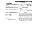 | 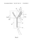 |
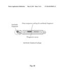 | 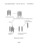 |
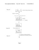 | 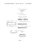 |
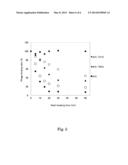 |  |
 |  |
 |  |
 |  |
 |  |
 |  |
 |  |
 |  |
 |  |
| New patent applications in this class: | |
| Date | Title |
|---|---|
| 2016-06-23 | Coupling method for peptide synthesis at elevated temperatures |
| 2016-06-23 | Stabilized calcium phosphate complexes |
| 2016-06-16 | Protein ligand for affinity isolation matrix |
| 2016-06-16 | Radiolabelling method |
| 2016-06-09 | Compositions and methods of use for recombinant human secretoglobins |
| Top Inventors for class "Chemistry: natural resins or derivatives; peptides or proteins; lignins or reaction products thereof" | |
| Rank | Inventor's name |
|---|---|
| 1 | Kevin I. Segall |
| 2 | Martin Schweizer |
| 3 | John R. Desjarlais |
| 4 | Brent E. Green |
| 5 | David M. Goldenberg |