Patent application title: ACIDOTHERMUS CELLULOYTICUS XYLANASE
Inventors:
Rebecca E. Parales (Davis, CA, US)
Alison M. Berry (Davis, CA, US)
Juanito V. Parales, Jr. (Davis, CA, US)
Ravi D. Barabote (Davis, CA, US)
Assignees:
THE REGENTS OF THE UNIVERSITY OF CALIFORNIA
IPC8 Class: AC12P1914FI
USPC Class:
435 99
Class name: Micro-organism, tissue cell culture or enzyme using process to synthesize a desired chemical compound or composition preparing compound containing saccharide radical produced by the action of a carbohydrase (e.g., maltose by the action of alpha amylase on starch, etc.)
Publication date: 2013-11-07
Patent application number: 20130295619
Abstract:
A thermophilic endo-beta-1,4-xylanase derived from Acidothermus
cellulolyticus is disclosed. The xylanase exhibits xylanase activity at
an optimal temperature of 90° C. and an optimal pH range of about
4.5-6.0. The isolated xylanase is useful in the hydrolysis of
lignocellulosic material.Claims:
1. A method for hydrolyzing lignocellulose, the method comprising:
contacting lignocelluloses with a recombinant protein comprising an amino
acid sequence with at least 80% sequence identity to SEQ ID NO: 1.
2. The method of claim 1, wherein the lignocellulose is contacted with the protein at a temperature of at least 80.degree. C. and a pH range from 3.0 to 9.0.
3. The method of claim 2, wherein the pH range is from 4.5 to 6.0.
4. The method of claim 1, wherein the lignocellulose is contacted with the protein at a temperature of at least 90.degree. C.
5. The method of claim 1, wherein the amino acid sequence of the protein is at least 85% percent identical to SEQ ID NO: 1.
6. The method of claim 1, wherein the amino acid sequence of the protein is at least 90% identical to SEQ ID NO: 1.
7. The method of claim 1, wherein the amino acid sequence of the protein is at least 93% identical to SEQ ID NO: 1.
8. The method of claim 1, wherein the amino acid sequence of the protein is at least 95% identical to SEQ ID NO: 1.
9. The method of claim 1, wherein the amino acid sequence of the protein is at least 97% identical to SEQ ID NO: 1.
10. The method of claim 1, wherein the amino acid sequence of the protein is SEQ ID NO: 1.
11. The method of claim 1, wherein the amino acid sequence comprises Pro-Xaa1-Pro-Xaa2-Pro (SEQ ID NO: 40), wherein Xaa1 and Xaa2 are each independently selected from the group consisting of: no amino acid, any 1 amino acid, any 2 amino acids, and any 3 amino acids.
12. The method of claim 1, wherein the amino acid sequence comprises a first Glu at a position corresponding to Glu-142 of SEQ ID NO: 1, and a second Glu at a position corresponding to Glu-259 of SEQ ID NO: 1.
13. The method of claim 1, wherein the amino acid sequence comprises one or more of SEQ ID NO: 23, SEQ ID NO: 24, SEQ ID NO: 25, SEQ ID NO: 26, or SEQ ID NO: 27.
14. The method of claim 1, wherein the protein has a molecular weight of between 40 and 48 kD.
15. The method of claim 1, wherein a source of the lignocellulose is selected from the group consisting of: birchwood, oat spelt, switchgrass, corn stover, miscanthus, energy cane, sorghum, eucalyptus, willow, bagasse, hybrid poplar, short-rotation woody crop, conifer softwood, crop residue, yard waste, and a combination thereof.
16. The method of claim 1, wherein the protein further comprises a signal sequence peptide having the amino acid sequence of SEQ ID NO: 2.
Description:
[0001] This application is a Divisional of U.S. patent application Ser.
No. 12/613,398, with a filing date of Nov. 5, 2009, which claims the
benefit under 35 USC 119(e) of U.S. Provisional Patent Application No.
61/203,528, filed Dec. 22, 2008, the disclosure of which is hereby
incorporated by reference in its entirety.
SUBMISSION OF SEQUENCE LISTING ON ASCII TEXT FILE
[0002] The content of the following submission on ASCII text file is incorporated herein by reference in its entirety: a computer readable form (CRF) of the Sequence Listing (file name: 514112003910SEQLIST.txt, date recorded: Oct. 12, 2012, size: 42 KB).
BACKGROUND
[0003] 1. Field
[0004] The present disclosure relates to xylanases and methods for their expression and use. Specifically, the disclosure is related to a thermophilic xylanase (Xyl-1) and homologs thereof derived from Acidothermus cellulolyticus, and the use of these enzymes in hydrolyzing lignocellulose.
[0005] 2. Related Art
[0006] Lignocellulose is plant biomass composed of cellulose, hemicellulose, and lignin. Lignocellulose serves as an abundant and inexpensive source of fermentable biomass. However, one barrier to the utilization of lignocellulose is the tight crosslinking of the cellulose and hemicellulose to the lignin. Breaking down lignocellulose (i.e. separating cellulose and hemicellulose from lignin) is energy intensive, and thus inefficient. Efficient utilization of lignocellulosic biomass will enhance the economic competitiveness of bioconversion processes which must compete with petrochemical processes. It has been shown that xylanase enzymes can be used to efficiently break down lignocellulose.
[0007] Xylanase enzymes are important in a wide variety of biotechnological and industrial applications. These include prebleaching of kraft pulp in the pulp and paper industry, recovery of cellulose fiber in textiles, enhancing digestibility of animal feed and silage, clarification of juices and beer, separation of cereal gluten and starch, assorted applications in the bakery industry, as well as the production of xylo-oligosaccharides for pharmacological applications and food additives (1, 2, 9, 14, and 21). Furthermore, recent interest in biofuels production from lignocellulosic plant biomass has brought xylanases into renewed prominence (5). Collins et al. (4) recently reviewed the physicochemical and functional characteristics of xylanases from six different families, their mechanism of action, and industrial applications.
[0008] Xylanase production is commonly obtained from Trichoderma reesei and Trichoderma harzianum strain E58, both from the Forintek Canada Corp. culture collection. Although both fungi are prolific producers of extracellular xylanases, fungal growth and enzyme production can only be carried out at mesophilic temperatures (e.g., 28° C.). Consequently the fermentation requires considerable cooling water during fungal growth and is easily subjected to bacterial contamination. The xylanase enzymes produced are also thermally unstable, losing over 90% of their activities within a half hour of incubation at 50° C. As a result, enzymatic hydrolysis of lignocellulose using these enzymes has to be carried out at a lower temperature of about 37-45° C. This in turn lowers the hydrolysis efficiencies, necessitates asceptic conditions during hydrolysis, as well as preventing prolonged enzyme use without replacement. However, higher efficiency of hydrolysis can be obtained by using thermophilic xylanases.
[0009] Recently, several thermophilic xylanases from fungal and bacterial microorganisms have been identified (FIG. 1). For example, U.S. Pat. No. 5,935,836 discloses a thermophilic xylanase isolated from Actinomadura flexuosa that has an optimal pH of 6.0-7.0 and a temperature range of 70-80° C. In addition, U.S. Pat. No. 5,395,765 discloses a xylanase derived from Rhodothermus, having activity over a pH range of 5-8 and thermostability at temperatures from 85-100° C. However, a xylanase with a more acidic pH range is desired for the utilization of hemicellulose biomass in fermentation.
[0010] The thermophilic cellulolytic bacterium Acidothermus cellulolyticus is described in Mohagheghi et al. (12), and the production of cellulase is described in Shiang et al. (19). However, neither reference describes a purified xylanase that may be useful at a low pH and high temperatures. U.S. Pat. No. 5,902,581 discloses a xylanase derived from Acidothermus cellulolyticus that is active at a pH range from 3.6-4.2 and that is thermostable at a range of 70-80° C. However, this A. cellulolyticus xylanase does not have optimal activity at temperatures above 80° C. or at a pH range from 4.5-6.0.
SUMMARY
[0011] Disclosed herein is a recombinant endo-beta-1,4-xylanase having an amino acid sequence with at least 80%, at least 85%, at least 87%, at least 89%, at least 90%, at least 91%, or at least 93% sequence identity to SEQ ID NO: 1. More preferably the amino acid sequence of the recombinant endo-beta-1,4-xylanase has at least 95%, at least 97%, or at least 99% sequence identity to SEQ ID NO: 1. In a particularly preferred embodiment, the amino acid sequence of the recombinant endo-beta-1,4-xylanase is SEQ ID NO: 1.
[0012] In certain embodiments, a region of the amino acid sequence of any of the recombinant endo-beta-1,4-xylanases described above conforms to consensus sequence: Pro-Xaa1-Pro-Xaa2-Pro (SEQ ID NO: 40). Preferably, Xaa1 and Xaa2 are each independently selected from no amino acid, any 1 amino acid, any 2 amino acids, or any 3 amino acids. In preferred embodiments, the amino acid sequence of any of the recombinant endo-beta-1,4-xylanases described above has a first Glu at a position corresponding to Glu-142 of SEQ ID NO: 1 and a second Glu at a position corresponding to Glu-259 of SEQ ID NO: 1. Preferably, the region of the amino acid sequence of any one of the recombinant endo-beta-1,4-xylanases described above is located between the first Glu and the second Glu. In certain embodiments, the first Glu is located within an amino acid region having a sequence of Asp-Val-Ala-Asn-Glu (SEQ ID NO: 25); and the second Glu is located within an amino acid region having a sequence of Thr-Glu-Ala-Asp (SEQ ID NO: 26).
[0013] In other preferred embodiments, Xaa1 is one amino acid selected from His, Lys, or Arg; and Xaa2 is one amino acid selected from Leu, Ala, Val, Ile, Pro, Phe, Met, or Trp. In particularly preferred embodiments, the region of the amino acid sequence of any of the recombinant endo-beta-1,4-xylanases described above is Pro-His-Pro-Leu-Pro (SEQ ID NO: 27).
[0014] In further embodiments, any of the recombinant endo-beta-1,4-xylanases described above can be active at pH values of about 3 to 9. In preferred embodiments, any of the endo-beta-1,4-xylanases described above have a pH optimum of about 4.5-6.0. In yet other embodiments, any of the recombinant endo-beta-1,4-xylanases described above have a molecular weight of about 40-48 kD. In further embodiments, any of the recombinant endo-beta-1,4-xylanases described above have activity at a temperature of at least 80° C. Preferably, any of the recombinant endo-beta-1,4-xylanases described above are active at least from 80-120° C. In particularly preferred embodiments, any of the recombinant endo-beta-1,4-xylanases described above have optimal activity at about 90° C.
[0015] In other embodiments, any of the recombinant endo-beta-1,4-xylanases described above retain at least 50% of initial activity, at 90° C., after incubation for at least 20 min, at least 30 min, at least 45 min, at least 60 min, at least 90 min, at least 2 hr, at least 5 hr, at least 8 hr, at least 12 hr, at least 24 hr, or at least 48 hr, at 90° C.
[0016] In yet other embodiments, any of the recombinant endo-beta-1,4-xylanases described above have a half-life of about 90 min at 90° C. in the presence of a xylan. In preferred embodiments, the xylan is birchwood xylan, beech wood xylan, or oat spelt xylan.
[0017] In certain embodiments, any of the recombinant endo-beta-1,4-xylanases described above further include a signal sequence peptide having amino acid sequence SEQ ID NO: 2.
[0018] In yet other embodiments, any of the recombinant endo-beta-1,4-xylanases described above has a substrate selected from lignocellulosic biomass, xylan-containing material, or xyloglucan-containing material.
[0019] The present disclosure also pertains to a recombinant cell comprising a nucleic acid molecule encoding any of the recombinant endo-beta-1,4-xylanases described above.
[0020] The present disclosure further pertains to a recombinant cell that expresses a nucleic acid molecule that has a nucleotide sequence with at least 80%, at least 85%, at least 87%, at least 89%, at least 90%, at least 91%, or at least 93% sequence identity to SEQ ID NO: 4, or its complementary sequence SEQ ID NO: 5. Preferably, the nucleic acid sequence of the nucleic acid molecule has at least 95%, at least 97%, or at least 99% sequence identity to SEQ ID NO: 4, or its complementary sequence SEQ ID NO: 5. In a particularly preferred embodiment, the nucleic acid sequence of the nucleic acid molecule is SEQ ID NO: 4, or its complementary sequence SEQ ID NO: 5.
[0021] The present disclosure also pertains to a method of hydrolyzing lignocellulose by contacting the lignocellulose with a recombinant endo-beta-1,4-xylanase having an amino acid sequence with at least 80%, at least 85%, at least 87%, at least 89%, at least 90%, at least 91%, at least 93%, at least 95%, at least 97%, or at least 99% sequence identity to SEQ ID NO: 1. Preferably, the lignocellulose is contacted with the recombinant endo-beta-1,4-xylanase at a temperature of at least 80° C., at least 85° C., at least 90° C., at least 95° C., or at least 100° C.; and a pH range from about 4.5-6.0. In a particularly preferred embodiment, the amino acid sequence of the recombinant endo-beta-1,4-xylanase is SEQ ID NO: 1.
[0022] The present disclosure further pertains to a method of hydrolyzing lignocellulose by contacting the lignocellulose with any of the recombinant endo-beta-1,4-xylanases described above. Preferably, the lignocellulose is contacted with the recombinant endo-beta-1,4-xylanase at a temperature of at least 80° C., at least 85° C., at least 90° C., at least 95° C., or at least 100° C.; and a pH range from about 4.5-6.0.
[0023] In other embodiments, the lignocellulose of any of the methods described above is from a source selected from birchwood, oat spelt, switchgrass, corn stover, miscanthus, energy cane, sorghum, eucalyptus, willow, bagasse, hybrid poplar, short-rotation woody crop, conifer softwood, crop residue, yard waste, or a combination thereof. In still other embodiments, the recombinant endo-beta-1,4-xylanase of any of the methods described above further includes a signal sequence peptide having amino acid sequence SEQ ID NO: 2.
DESCRIPTION OF DRAWING FIGURES
[0024] FIG. 1 is a table comparing various xylanases, all of which have temperature optima below 100° C. MW=molecular weight; T opt=optimal temperature range; and H opt=optimal ph range.
[0025] FIG. 2 depicts a diagram of the A. cellulolyticus xyl-1 gene Acel--0372.
[0026] FIG. 3 is an image of an agarose gel, depicting the results of PCR amplification of the xyl-1 gene Acel--0372. Lane 1, molecular weight marker; lanes 2 and 3, PCR products from amplifications containing two different concentrations of A. cellulolyticus genomic DNA as the template.
[0027] FIG. 4 is an image of an agarose gel depicting the results of purified plasmid clones containing the amplified xyl-1 gene Acel--0372. Leftmost lane, molecular weight marker (in kb); lanes 1 and 3-8 contain the correct constructs of pK19 (2.5 kb) containing the 1.4-kb PCR product after restriction digestion with SacI and XbaI. Lane 2 contains a pK19 construct that does not contain the 1.4-kb PCR product.
[0028] FIGS. 5 (A) and 5 (B) depict the nucleotide coding sequence of the PCR product amplified from A. cellulolyticus (SEQ ID NO: 4), the nucleotide complementary sequence of the PCR product amplified from A. cellulolyticus (SEQ ID NO: 5), and the amino acid sequence encoded by the PCR product amplified from A. cellulolyticus (SEQ ID NO: 3).
[0029] FIGS. 6 (A), 6 (B), and 6 (C) depict the expression of Xyl-1 in A. cellulolyticus. (A) RT-PCR analysis of the xyl-1 gene. (B) RT-PCR analysis of an internal control housekeeping gene, gyrB. M: molecular-weight DNA ladder, lanes 1-4: exponential growth phase samples, 5-8: stationary growth phase samples, and 9: no-RT negative control to confirm the absence of contaminating genomic DNA. Lanes 1 and 5: oat-spelt xylan-grown culture sample, 2 and 6: cellobiose-grown culture, 3 and 7: cellulose-grown culture, and 4 and 8: glucose-grown culture. (C) Representation of the peptide coverage of the Xyl-1 protein (389 aa) from tandem mass spectrometry of A. cellulolyticus culture supernatant. Hatched box indicates the N-terminal signal peptide, and black boxes indicate the positions of five non-overlapping peptides identified from the spectra. The peptides are as follows, 1: HGNPPYHPPADSLR (SEQ ID NO: 6), 2: WQVVEPTQGTYDWSGGDR (SEQ ID NO: 7), 3: LVQFAQEHGQLVR (SEQ ID NO: 8), 4: HIVDEVTHFK (SEQ ID NO: 9), and 5: PAYTALQQTLALAAGAPHR (SEQ ID NO: 10).
[0030] FIG. 7 depicts the full 451-bp untranslated intergenic region between the Acel--0373 and Acel--0372 (xyl-1) open reading frames. The sequence contains the xyl-1 promoter region. The putative -10 and -35 sequences are shown in bold and the putative Shine-Dalgarno ribosomal binding site (RBS) is also shown in bold. The Xyl-1 protein coding region is shown as a bold arrow at the end of the sequence. Three inverted repeats (IR-1, IR-2, and IR-3) are shown with bold letters and boxes. IR-1: GAAACTTTC (SEQ ID NO: 11), IR-2: TTTCCGAAA (SEQ ID NO: 12), and IR3: TCCGAAAATTTCGGA (SEQ ID NO: 13).
[0031] FIGS. 8 (A) and 8 (B) depict the purification of Xyl-1 by FPLC following heat treatment at 65° C. for 15 min. (A) Protein elution from the ion exchange column during the FPLC run as KCl concentration was increased. The black line is UV absorbance at 280 nm, the thin grey line indicates conductivity, and the thick grey line indicates the KCl gradient. (B) Activity profile (activity measured at 410 nm) at 52° C., pH 5.2 was used to identify the fractions containing Xyl-1 that were then combined and concentrated.
[0032] FIGS. 9 (A) and 9 (B) depict the purification of Xyl-1 from E. coli. (A) Coomassie stained gel. (B) In-gel assay (carried out at 52° C., pH 5.2). Leftmost lane, molecular weight markers (in kD). CCE, crude cell extract from E. coli clone; ΔH, crude cell extract from E. coli clone after heat treatment (15 min at 65° C.); Main, concentrated active fractions from the ion exchange column; Side, concentrated side fractions. Asterisks indicate Xyl-1.
[0033] FIG. 10 depicts the purification of Xyl-1 by hydroxyapatite chromatography. SDS-PAGE (10% gel) analysis showing molecular weight markers (lane 1), crude cell extract from DH5α (pK19-xyl-1) (lane 2), heat treated (65° C. for 20 min followed by centrifugation) crude cell extract from DH5a (pK19-xyl-1) (lane 3), concentrated fractions from the hydroxyapatite column (lane 4).
[0034] FIG. 11 depicts the relative activity of the partially purified Xyl-1 at various pH values and temperatures. Activity is relative to the highest activity at each pH.
[0035] FIG. 12 depicts a multiple sequence alignment of Xyl-1 with other xylanases. A 295 amino acid fragment of Xyl-1 (residues 67-361) was aligned with eleven other homologs using Clustal X version 2.0 (26). Stars (*) indicate fully conserved sites, while colons (:) and periods (.) indicate sites with high and low degrees of conservation, respectively. The bar above the A_ce sequence indicates the unique Xyl-1 region of five amino acids containing three prolines. The abbreviations used and the accession numbers of the proteins are as follows: A_ce (A. cellulolyticus 11B Xyl-1, GI:117927581) (SEQ ID NO: 28); C_ac (Catenulispora acidiphila DSM 44928, GI:229247007) (SEQ ID NO: 29); C_sp (Cellulosimicrobium sp. HY-12, GI:162414427) (SEQ ID NO: 30); C_fi (Cellulomonas fimi ATCC 484, GI:73427793) (SEQ ID NO: 31); S_th (Streptomyces thermoviolaceus, GI:38524461) (SEQ ID NO: 32); T_au (Thermoascus aurantiacus, GI:13432255) (SEQ ID NO: 33); P_ch (Phanerochaete chrysosporium, GI:167599628) (SEQ ID NO: 34); T_al (Thermobifida alba, GI:1621277) (SEQ ID NO: 35); S_av (Streptomyces avermitilis, GI:29828638) (SEQ ID NO: 36); C_ad (Cryptococcus adeliensis, GI:2624008) (SEQ ID NO: 37); U_ba (Uncultured bacterium, GI:18476191) (SEQ ID NO: 38); and T_ma (Thermotoga maritima, GI:71041762) (SEQ ID NO: 39).
[0036] FIGS. 13 (A) and 13 (B) are images of an agarose gel that depicts an in-gel xylanase activity assay in E. coli crude cell extracts. Lanes ST, molecular weight standards (in kD); (A) lane 1, crude cell extracts from the vector control strain; lane 2, crude cell extracts from the Xyl-1 clone; (B) lane 3, crude cell extracts from the vector control after heat treatment (15 min at 65° C.); lane 4, crude cell extracts from the Xyl-1 clone strain after heat treatment. Assayed at 52° C., pH 5.2.
[0037] FIG. 14 is an image of a polyacrylamide gel that depicts xylanase activity in concentrated culture supernatants from the E. coli clone and Acidothermus (after growth on cellobiose) in the in-gel assay. Lanes ST, molecular weight standards (in kD); lane 1, Acidothermus culture supernatant; lane 2, Xyl-1 clone culture supernatant; lane 3, E. coli vector control culture supernatant. Assayed at 52° C., pH 5.2.
[0038] FIG. 15 is an image of a polyacrylamide gel that depicts the activity of recombinant Xyl-1 in E. coli crude cell extracts at temperatures ranging from 0-100° C. in the in-gel assay at pH 5.2.
[0039] FIG. 16 is an image of a polyacrylamide gel that depicts activity of the recombinant Xyl-1 in E. coli crude cell extracts at pH values from 3-10 in the in-gel assay. Assays were carried out at 52° C.
[0040] FIG. 17 is an image of a polyacrylamide gel that depicts activity of the recombinant Xyl-1 in E. coli crude cell extracts following heat treatment at 80° C. for 20 min. The assay was carried out at 52° C. and at pH 5.2.
[0041] FIG. 18 depicts the activity of the recombinant Xyl-1 in E. coli crude cell extracts following heat treatment for 20 min. The assay was carried out at 52° C., pH 5.2.
[0042] FIG. 19 depicts the specific activity of the recombinant Xyl-1 in E. coli crude cell extracts at temperatures from 0-120° C. at pH 5.2.
[0043] FIGS. 20 (A) and 20 (B) depict the temperature and pH profile of purified Xyl-1. (A) pH 3-6; (B) pH 7-10. Results are the averages of at least three independent experiments using the reducing sugars assay and oat spelt xylan as the substrate. Note that the activity ranges differ in A and B. Error bars indicate standard deviations.
[0044] FIGS. 21 (A) and 21 (B) depict the thermostability of recombinant Xyl-1. (A) Stabilization of the cloned Xyl-1 by xylan substrates. Purified Xyl-1 was diluted 1:20 with oat spelt xylan (light grey), birchwood xylan (grey) or phosphate buffer (white), and incubated at 90° C. for the times indicated. Rates were determined by the reducing sugars assay and are relative to the unheated Xyl-1 (black). Results are the averages of three independent experiments and error bars indicate standard deviations. (B) Activity retained by Xyl-1 in the presence of 3.8% oat spelt xylan. The half-life of Xyl-1 appears to be approximately 1.5 hr at 90° C.
[0045] FIGS. 22 (A) and 22 (B) depict the results of a TLC time course of Xyl-1 activity with oat spelt (A) and birchwood (B) xylans. The TLC was developed as described in Materials and Methods. Lanes 1 and 8, standard compounds: X1, xylose; X3, xylotriose; X4, xylotetraose. Lane 2, unreacted xylan; lanes 3-7, increasing time of incubation in the presence of purified Xyl-1. Lane 3, 10 min; lane 4, 20 min; lane 5, 40 min; lane 6, 60 min; lane 7, 240 min.
BRIEF DESCRIPTION OF THE SEQUENCE LISTING
[0046] SEQ ID NO: 1 shows the amino acid sequence of Xyl-1 without the N-terminal signal sequence.
[0047] SEQ ID NO: 2 shows the amino acid sequence of the N-terminal Xyl-1 signal sequence.
[0048] SEQ ID NO: 3 shows the amino acid sequence encoded by the PCR product amplified from A. cellulolyticus.
[0049] SEQ ID NO: 4 shows the nucleotide coding sequence of the PCR product amplified from A. cellulolyticus.
[0050] SEQ ID NO: 5 shows the nucleotide complementary sequence of the PCR product amplified from A. cellulolyticus.
[0051] SEQ ID NO: 6 shows the amino acid sequence of the first of five non-overlapping Xyl-1 peptides identified by tandem mass spectrometry of A. cellulolyticus culture supernatant.
[0052] SEQ ID NO: 7 shows the amino acid sequence of the second of five non-overlapping Xyl-1 peptides identified by tandem mass spectrometry of A. cellulolyticus culture supernatant.
[0053] SEQ ID NO: 8 shows the amino acid sequence of the third of five non-overlapping Xyl-1 peptides identified by tandem mass spectrometry of A. cellulolyticus culture supernatant.
[0054] SEQ ID NO: 9 shows the amino acid sequence of the fourth of five non-overlapping Xyl-1 peptides identified by tandem mass spectrometry of A. cellulolyticus culture supernatant.
[0055] SEQ ID NO: 10 shows the amino acid sequence of the fifth of five non-overlapping Xyl-1 peptides identified by tandem mass spectrometry of A. cellulolyticus culture supernatant.
[0056] SEQ ID NO: 11 shows the nucleotide sequence of the first of three inverted repeats (IR-1) located in the xyl-1 promoter region.
[0057] SEQ ID NO: 12 shows the nucleotide sequence of the second of three inverted repeats (IR-2) located in the xyl-1 promoter region.
[0058] SEQ ID NO: 13 shows the nucleotide sequence of the third of three inverted repeats (IR-3) located in the xyl-1 promoter region.
[0059] SEQ ID NO: 14 shows the nucleotide sequence of the forward primer used to PCR amplify Acel--0372.
[0060] SEQ ID NO: 15 shows the nucleotide sequence of the reverse primer used to PCR amplify Acel--0372.
[0061] SEQ ID NO: 16 shows the nucleotide sequence of a forward primer specific to the xyl-1 gene.
[0062] SEQ ID NO: 17 shows the nucleotide sequence of a reverse primer specific to the xyl-1 gene.
[0063] SEQ ID NO: 18 shows the nucleotide sequence of a forward gyrB gene-specific primer.
[0064] SEQ ID NO: 19 shows the nucleotide sequence of a reverse gyrB gene-specific primer.
[0065] SEQ ID NO: 20 shows the nucleotide sequence of the putative xyl-1 ribosomal binding site (RBS).
[0066] SEQ ID NO: 21 shows the nucleotide sequence of the conserved sequence found at the 3' end of the A. cellulolyticus 16S ribosomal rRNA that is complimentary to the RBS sequence.
[0067] SEQ ID NO: 22 shows the amino acid sequence of the N-terminal sequence of the recombinant Xyl-1.
[0068] SEQ ID NO: 23 shows the conserved amino acid sequence of the first active site glutamate region of GH10 family xylanases.
[0069] SEQ ID NO: 24 shows the conserved amino acid sequence of the second active site glutamate region of GH10 family xylanases.
[0070] SEQ ID NO: 25 shows the amino acid sequence of the first active site glutamate region of Xyl-1.
[0071] SEQ ID NO: 26 shows the amino acid sequence of the second active site glutamate region of Xyl-1.
[0072] SEQ ID NO: 27 shows the amino acid sequence of the region containing three prolines close to the first active site glutamate of Xyl-1.
[0073] SEQ ID NO: 28 shows the amino acid sequence of a 295 amino acid fragment of Xyl-1 (i.e., residues 67-361)
[0074] SEQ ID NO: 29 shows the amino acid sequence of Catenulispora acidiphila DSM 44928 (GI:229247007) xylanase.
[0075] SEQ ID NO: 30 shows the amino acid sequence of Cellulosimicrobium sp. HY-12 (GI:162414427) xylanase.
[0076] SEQ ID NO: 31 shows the amino acid sequence of Cellulomonas fimi ATCC 484 (GI:73427793) xylanase.
[0077] SEQ ID NO: 32 shows the amino acid sequence of Streptomyces thermoviolaceus (GI:38524461) xylanase.
[0078] SEQ ID NO: 33 shows the amino acid sequence of Thermoascus aurantiacus (GI:13432255) xylanase.
[0079] SEQ ID NO: 34 shows the amino acid sequence of Phanerochaete chrysosporium (GI:167599628) xylanase.
[0080] SEQ ID NO: 35 shows the amino acid sequence of Thermobifida alba (GI:1621277) xylanase.
[0081] SEQ ID NO: 36 shows the amino acid sequence of Streptomyces avermitilis (GI:29828638) xylanase.
[0082] SEQ ID NO: 37 shows the amino acid sequence of Cryptococcus adeliensis (GI:2624008) xylanase.
[0083] SEQ ID NO: 38 shows the amino acid sequence of an uncultured bacterium (GI:18476191) xylanase.
[0084] SEQ ID NO: 39 shows the amino acid sequence of Thermotoga maritima (GI:71041762) xylanase.
[0085] SEQ ID NO: 40 shows the amino acid sequence of a Xyl-1 proline rich consensus sequence.
DETAILED DESCRIPTION
Definitions
[0086] As used herein, the term "thermostable" refers to a threshold level of xylanase activity after an incubation period of 15 min at a temperature of 65° C.
[0087] As used herein, the terms "active" and "activity" refer to an endo-beta-1,4-xylanase giving a positive result using the in-gel xylanase assay or the reducing sugar assay described below.
[0088] As used herein, the term "percent activity" refers to the amount of endo-beta-1,4-xylanase activity measured at given experimental conditions compared to base-line xylanase activity. The measured xylanase activity at experimental conditions is divided by the base-line xylanase activity and multiplied by 100 to obtained percent activity. As used herein, "base-line xylanase activity" and "baseline control" refer to the amount of xylanase activity produced by an endo-beta-1,4-xylanase when assayed at 55° C. and pH 5.2 for 10 min.
[0089] As used herein, the term "half-life" refers to the length of time necessary for an endo-beta-1,4-xylanase activity to drop by 50% (compared to base-line control) at a given temperature.
[0090] As used herein, the term "optimal activity" refers to peak xylanase activity in a given temperature range or range of pH values.
[0091] As used herein, the term percent "identical," "percent identity," and "percent sequence identity" are defined as amount of identity between a reference nucleic acid or amino acid sequence and at least one other nucleic acid or amino acid sequence. Percent sequence identity can be determined by comparing two optimally aligned sequences, wherein the portion of the sequence being compared may comprise additions or deletions (i.e., gaps) as compared to the reference sequence (e.g., a nucleic acid or amino acid sequence of the disclosure), which does not comprise additions or deletions, for optimal alignment of the two sequences. The percentage is calculated by determining the number of positions at which the identical nucleic acid or amino acid residue occurs in both sequences to yield the number of matched positions, dividing the number of matched positions by the total number of positions being compared and multiplying the result by 100 to yield the percentage of sequence identity. Two sequences have percent identity if two sequences have a specified percentage of nucleic acids or amino acid residues that are the same (i.e., 75% identical over a specified region, or, when not specified, over the entire sequence), when compared and aligned for maximum correspondence or designated region as measured using one of the following sequence comparison algorithms or by manual alignment and visual inspection.
[0092] One example of an algorithm that is suitable for determining percent sequence identity is the BLAST algorithm, which is described in Altschul et al. (1977) Nuc. Acids Res. 25:3389-3402. Software for performing BLAST analyses is publicly available through the National Center for Biotechnology Information. The BLASTN program (used for nucleic acid sequences) uses as defaults a wordlength (W) of 11, an expectation (E) or 10, M=5, N=-4 and a comparison of both strands. For amino acid sequences, the BLASTP program is used with default settings of a wordlength of 3, and expectation (E) of 10, and the BLOSUM62 scoring matrix (see Henikoff and Henikoff (1989) Proc. Natl. Acad. Sci. USA 89:10915) alignments (B) of 50, expectation (E) of 10, M=5, N=-4, and a comparison of both strands.
Acidothermus Cellulolyticus Xyl-1 Xylanase
[0093] The following description sets forth numerous exemplary configurations, parameters, and the like. It should be recognized, however, that such description is not intended as a limitation on the scope of the present invention, but is instead provided as a description of exemplary embodiments.
[0094] Acidothermus cellulolyticus 11B (ATCC 43068) is a thermophilic bacterium that was originally isolated from an acidic hot spring in Yellowstone National Park. A number of thermostable endoglucanases are produced by this organism, which are useful for degrading cellulose in the production of ethanol or other hydrocarbons for biofuel. The genome of Acidothermus cellulolyticus 11B (henceforth referred to as A. cellulolyticus) has been sequenced to completion (Refseq NC--008578) and the sequence has been annotated. The sequence and sequence annotation have provided information on the regulation and production of potentially useful enzymes (published as accession number NC--008578). Among these potentially useful enzymes, the A. cellulolyticus sequence annotation predicted a single putative endo-beta-1,4-xylanase, which is encoded by Acel--0372 (from the A. cellulolyticus genome annotation). It is believed that there are no reports of this endo-beta-1,4-xylanase having been previously cloned, and the predicted molecular weight does not match the previously disclosed A. cellulolyticus xylanase (U.S. Pat. No. 5,902,581). Therefore, this predicted endo-beta-1,4-xylanase was cloned from A. cellulolyticus, and expressed as a recombinant protein.
[0095] The expressed protein encoded by Acel--0372 was identified as a glycosyl hydrolase family 10 (GH10) endo-beta-1,4-xylanase enzyme. This A. cellulolyticus GH10 family xylanase was named Xyl-1. Characterization of Xyl-1 revealed that it is a thermophilic xylanase that has a temperature optimum of 90° C. and a pH optimal range of about 4.5-6.0. Xyl-1 is also characterized by retaining xylanase activity at a temperature range of 25-120° C., and a pH range of 3-9. Xyl-1 also has a molecular weight of about 40-48 kD. Furthermore, Xyl-1 is distinct from the A. cellulolyticus xylanase disclosed in U.S. Pat. No. 5,902,581 in that it has an optimal temperature range that is higher than the 70-80° C. range of the xylanase disclosed in U.S. Pat. No. 5,902,581, has a pH range that is less acidic than the pH range of 3.6-4.2 of the xylanase disclosed in U.S. Pat. No. 5,902,581, and is smaller in size than the 50-55 kD xylanase disclosed in U.S. Pat. No. 5,902,581.
[0096] It is surprising and unexpected that A. cellulolyticus produces a second xylanase (i.e., Xyl-1), different in optimal temperature and pH range from the previously identified xylanase (U.S. Pat. No. 5,902,581).
[0097] As stated above, Xyl-1 is useful in degrading (e.g., hydrolyzing) xylan-containing material, xyloglucan-containing material, and lignocellulosic biomass in the production of ethanol or other hydrocarbons for biofuel. For example, Xyl-1 can be used to hydrolyze lignocellulosic biomass from birchwood, oat spelt, switchgrass, corn stover, miscanthus, energy cane, sorghum, eucalyptus, willow, bagasse, hybrid poplar and other short-rotation woody crops, various species of conifer softwood, various crop residues, or yard waste in order to improve the availability of fermentable sugars in these substrates. In industrial applications, the hydrolysis step is preferably performed at temperatures from 50° C.-100° C. and at acidic pH values. The stability of Xyl-1 over a wide range of temperatures and pH values provides versatility in industrial processes. Additionally, Xyl-1 provides long-lived and persistent xylanase activity under fluctuating conditions, which can occur in large-scale bioconversion systems. These conditions may further allow for the advantageous coupling of lignocellulose hydrolysis to lignocellulose fermentation.
[0098] Xyl-1 can also be used to bleach lignocellulosic pulp as a step in the process of making paper, to improve animal feed by making feed more digestible, or Xyl-1 can be used as a component in a detergent composition.
[0099] The xylanases of this disclosure, methods of their manufacture, and methods of their use may be better understood with respect to the following non-limiting examples.
EXAMPLES
Example 1
Cloning of the Xyl-1
[0100] According to the A. cellulolyticus 11B genome annotation, Acel--0372 encodes a predicted secreted xylanase (Xyl-1). In order to clone Acel--0372, A. cellulolyticus 11B (ATCC 43068) genomic DNA was purified as previously described (15), and oligonucleotide primers were designed in order to PCR amplify the complete Acel--0372, the predicted 1.4-kb Xyl-1 gene from A. cellulolyticus (FIG. 2). The primer pair used was Xyl For (5'-GTG GTG GAG CTC GCA ATT CGT TCA CGT TGA GG-3') (SEQ ID NO: 14) and Xyl Rev (5'-GTG GTG TCT AGA ACC ATC GAG TGG GAG TGA CG-3') (SEQ ID NO: 15). The underlined sequences indicate the added restriction sites for cloning, SacI and XbaI, respectively. PCR was performed using the following program using Pfu: an initial denaturation at 95° C. for 3 min, followed by 25 cycles of 95° C. for 1 min, 55° C. for 1 min, 70° C. for 1 min, and then a final extension at 70° C. for 5 min. The PCR yielded a 1.4-kb product, which is the xyl-1 gene (FIG. 3). The resulting 1.4-kb PCR product was purified from an agarose gel with a QIAquick Gel Extraction kit (Qiagen, Valencia, Calif.), digested with SacI and XbaI and ligated to SacI-XbaI-digested pK19 (16). E. coli DH5α cells were transformed with plasmid DNA by standard procedures (18). Clones were confirmed by restriction digestion with SacI and XbaI (FIG. 4).
[0101] DNA sequence analysis was performed on the positive pK19 clones containing the xyl-1 gene (FIG. 5). Fluorescent automated DNA sequencing was carried out at the University of California, Davis sequencing facility with an Applied Biosystems 3730 automated sequencer. Nucleotide and amino acid sequence analyses were performed using the Vector NTI software suite (Invitrogen, Carlsbad, Calif.). The complete sequence of the xyl-1 gene was identical to the sequence reported for Acel--0372.
Example 2
Xyl-1 expression in A. cellulolyticus
Materials and Methods
[0102] A. cellulolyticus cultures were grown in supplemented mineral medium as described previously (12). Oat spelt xylan, cellulose, cellobiose, or glucose were provided individually as carbon sources at 0.5%. Cells were grown to mid exponential phase or stationary phase and cells were harvested by centrifugation at 10,000×g for 15 min at 4° C. The cell pellet was frozen in liquid nitrogen and cells were disrupted by grinding with a mortar and pestle prechilled in liquid nitrogen, in the presence of sterilized sand. RNA was extracted using the RNeasy Plant Mini Kit (Qiagen), with several rounds of DNase digestion with RNase-free DNase (Qiagen), and cleaned with the RNeasy Plant Mini Kit. For the cultures grown on oat spelt xylan or cellulose, high molecular weight polyethylene glycol (at 1% w/v final concentration) was added to the cell lysate to reduce the amount of contaminating genomic DNA. It was found that several rounds of DNase digestion were required to completely eliminate DNA contamination. The quality of the RNA was analyzed using an Agilent Bioanalyzer 2100 (Agilent Technologies) and quantified using a Nanodrop spectrophotometer (Thermo Scientific).
[0103] Primers specific to the xyl-1 gene (primers: 5'-CAAAGGAAAGATCTGGCAATG-3' (SEQ ID NO: 16) and 5'-TGAGCATCCCGTCGTAGTAGT-3' (SEQ ID NO: 17)) were used to amplify a 485-bp product from 100 ng total RNA template using reverse transcriptase polymerase chain reaction (RT-PCR). The Qiagen OneStep RT-PCR kit was used. The amplified products were analyzed on agarose gels and photographed using the Red gel imaging system (Alpha Innotech). The gyrB gene-specific primers 5'-GACCCGACCGAGGTTTATTAC-3' (SEQ ID NO: 18) and 5'-GCCGAACTTGTTCACCAAATA-3' (SEQ ID NO: 19) were used to amplify a 270-bp product that was used to normalize for the RNA loaded as well as for semi-quantification of the xyl-1 expression. For biological replications of the experiment, RT-PCR was repeated using a second batch of RNA that was extracted independently under all growth conditions. Identical results were obtained in duplicate experiments, and data from one of the replicates are presented.
[0104] For proteomics analysis, A. cellulolyticus was grown on oat spelt xylan to stationary phase and culture supernatant was harvested by centrifugation. The culture supernatant was concentrated by a factor of 50 using an Amicon stirred ultrafiltration cell carrying a polyethersulfone membrane with 5-kDa molecular weight cutoff (Millipore). For identification of proteins in the concentrated culture supernatant, the sample was subjected to liquid chromatography followed by tandem mass spectrometry (LC/MS/MS) at the University of California Davis Proteomics Core facility.
Results
[0105] The transcriptional expression of the A. cellulolyticus xyl-1 gene was studied during exponential as well as stationary phases of growth on oat spelt xylan, cellulose, cellobiose, and glucose. The RT-PCR analysis revealed that xyl-1 was expressed in xylan- and cellulose-grown cultures (FIG. 6A). The gene was also expressed at low levels in cellobiose-grown cultures during stationary phase. No expression was detectable in glucose-grown cultures at any time or in cellobiose-grown cultures during exponential phase. In xylan- and cellulose-grown cultures, xyl-1 expression appeared to be essentially the same during the exponential and stationary phases of growth. The gyrB gene was used as an internal control to normalize the expression levels of the xyl-1 gene (FIG. 6B).
[0106] Tandem mass spectrometry verified the presence of Xyl-1 in the culture supernatant (FIG. 6C) and indicated that the N-terminal 25 aa signal peptide, which targets proteins for secretion, was cleaved from the mature protein.
Example 3
Sequence Analysis of Xyl-1 Gene Promoter
[0107] The xyl-1 gene was determined to be on the negative strand and is separated by 147 bp from the divergently oriented upstream gene, and 451 bp from the downstream gene on the negative strand. The functions of both of these flanking genes appear to be unrelated to xylanases. Thus, xyl-1 does not appear to be part of a gene cluster or operon. Analysis of the 451-bp upstream promoter region revealed a putative Shine-Dalgarno ribosomal binding site (RBS) six nucleotides upstream of the translational start. The RBS sequence (5' TGGAGG 3') (SEQ ID NO: 20) is complementary to the conserved CCTCCT (SEQ ID NO: 21) sequence found at the 3' end of the A. cellulolyticus 16S ribosomal rRNA, with only a one-base mismatch (FIG. 7). Putative -35 and -10 sequences were also identified (FIG. 7) that match well with the consensus promoter motifs proposed in the closely-related actinomycete, Streptomyces (20), and are likely to be the σ70 promoter sequences of the A. cellulolyticus xyl-1 gene.
[0108] Several small inverted repeats were found in the promoter region of xyl-1 using the palindrome software in EMBOSS (17). Among these, three inverted repeats (FIG. 7) were found to be conserved based on comparison to the promoter regions of GH10 family xylanases in Streptomyces sp. (6). The sequences of the three inverted repeats, IR-1, IR-2, and IR-3, in the xyl-1 promoter match well with the consensus sequences of Box 1, Box 2, and Box 3, respectively, described for some GH10 family xylanase genes in Streptomyces spp. (6). However, the length and positioning of these putative regulatory elements in the two organisms are very different. In the xyl-1 promoter, the distal IR-1 sequence forms a short palindrome (5' GAAA-C-TTTC 3') (SEQ ID NO: 11) and the downstream IR-3 palindrome extends three nucleotides longer (5' TCCGAAA-A-TTTCGGA 3') (SEQ ID NO: 13), whereas the Box 3 palindrome is one-nucleotide longer than the Box 1 in Streptomyces. The A. cellulolyticus IR-2 sequence (5' TTTC-C-GAAA 3') (SEQ ID NO: 12) is almost identical to the Streptomyces Box 2 sequence. However, unlike in Streptomyces, where Box 1 and Box 2 are separated by about 10 bp, the xyl-1 IR-2 element overlaps with IR-1 (FIG. 7). Furthermore, in A. cellulolyticus, the IR-1 and IR-2 regions are located downstream of the putative -35 and -10 elements and closer to the translational start site, while both of the corresponding boxes are upstream of the promoter, and farther away from the first codon in Streptomyces.
Example 4
Xyl-1 Protein Purification
Materials and Methods
[0109] E. coli DH5α cells containing the cloned xyl-1 gene were grown in a minimal salts medium containing 10 mM glucose, 1 mM thiamine and 100 μg/mlkanamycin at 30° C. for 48 hr. Cells were harvested by centrifugation (14,000×g at 4° C. for 15 min), washed once with 10 mM phosphate buffer (pH 5.2), resuspended in the same buffer and stored frozen at -20° C. Frozen cell suspensions were removed from the freezer and allowed to thaw on ice. The thawed cell suspension was then passed once through a French pressure cell maintaining the internal cell pressure a constant 18,000 psi. Cell debris and membranes were removed by centrifugation at 48,000×g for 90 min at 6° C. The resulting crude cell extract was heat-treated by incubation at 65° C. for 20 min. Insoluble material was removed by centrifugation at 14,000×g at 4° C. for 15 min. Purification procedures were performed at 4° C. using an automated FPLC system (Bio-Rad Laboratories, Hercules, Calif.). Cell extracts were applied to a column containing approximately 100 ml (bed volume) of Unosphere Q (Bio-Rad Laboratories) that had been pre-equilibrated with 10 mM phosphate buffer (pH 5.2). Unbound proteins were eluted from the column with 100 ml of the same buffer at a rate of 1.0 ml/min. Bound proteins were eluted with a linear gradient from 0 to 1.0 M KCl in phosphate buffer at the same flow rate and 5 ml fractions were collected.
Results
[0110] Extracts of the E. coli clone were subjected to heat treatment at 65° C. for 15 min prior to loading the extract onto an ion exchange column. Xyl-1 protein eluted as one major peak with activity (FIG. 8). This heat treatment eliminated many of the E. coli proteins and resulted in a significant purification of Xyl-1 (FIG. 9). The concentrated protein was >90% pure, as determined by SDS-PAGE analysis (FIGS. 9 and 10). Approximately 2 mg of purified Xyl-1 was obtained from 4 L of E. coli culture. The recombinant Xyl-1 had a molecular weight of approximately 40-48 kD on SDS gels (FIGS. 9 and 10), which is consistent with the predicted molecular weight based on the deduced amino acid sequence of the processed protein (40.3 kD).
[0111] A temperature vs. pH profile with the partially purified Xyl-1 demonstrated that the enzyme has a broad temperature range with optimal activity at approximately 90° C. and a broad pH range with an optimum between pH 4.5 and 6.0 (FIG. 11).
Example 5
Xyl-1 N-Terminal Amino Acid Sequence Analysis
[0112] Samples of recombinant Xyl-1 purified from E. coli, as described in Example 4, were used to determine whether Xyl-1, an extracellular enzyme, was properly processed in E. coli. The N-terminal amino acid sequence of the purified Xyl-1 was determined by Edman degradation on an automated sequencer (Applied Biosystems, Foster City, Calif.) at the University of California Davis Molecular Structure Facility after SDS-PAGE and electroblotting of the purified proteins onto a polyvinylidene difluoride membrane (ProBlott; Applied Biosystems).
[0113] The N-terminal sequence of the recombinant Xyl-1 expressed in E. coli was found to be HGNPPYHPPAD (SEQ ID NO: 22). The first amino acid of this sequence mapped to position 26 in the full-length protein sequence, indicating that the predicted signal sequence of Xyl-1 was properly cleaved in the heterologous host, E. coli (FIG. 5). This indicates that the E. coli cells processed Xyl-1 by cleaving the signal sequence, which is the secretion signal sequence. As is the case with the native A. cellulolyticus 11B xylanase, Xyl-1 expressed in E. coli lacks the secretion signal sequence.
Example 6
Xyl-1 Amino Acid Sequence Analysis
[0114] A BLAST search was performed with the A. cellulolyticus GH10 family xylanase Xyl-1 amino acid sequence. The top hit was from a xylanase of another actinomycete, Catenulispora acidiphila DSM 44928, which has 75% sequence identity with Xyl-1. A recent biochemically-characterized xylanase from the actinomycete Cellulosimicrobium sp. HY-12 was the second best hit with 55% sequence identity to Xyl-1 (13). Multiple sequence analyses of the Xyl-1 with the above two homologs as well as other functionally characterized GH10 family xylanases showed the presence of conserved active site glutamates at positions 167 and 284 in Xyl-1 (FIG. 12). The region around the predicted active site glutamates is well-conserved in the GH10 sequences; the conserved motif around the first glutamate is DVVNE (SEQ ID NO: 23) and at the second glutamate is TELD (SEQ ID NO: 24). Xyl-1 has valine-to-alanine and leucine-to-alanine substitutions in the two regions, resulting in DVANE (SEQ ID NO: 25) and TEAD (SEQ ID NO: 26) sequences, respectively (FIG. 12). The uncharacterized C. acidiphila homolog also bears these substitutions.
[0115] It should be appreciated that homologous endo-beta-1,4-xylanases of the present disclosure may have a first glutamate at a position corresponding to Glu-142 of SEQ ID NO: 1. Preferably, the first glutamate is located within an amino acid region having the sequence of Asp-Val-Ala-Asn-Glu (SEQ ID NO: 25). Homologous endo-beta-1,4-xylanases of the present disclosure may have a second glutamate at a position corresponding to Glu-259 of SEQ ID NO: 1. Preferably, the second glutamate is located within an amino acid region having the sequence of Thr-Glu-Ala-Asp (SEQ ID NO: 26).
[0116] Multiple amino acid sequence alignments were performed comparing Xyl-1 with uncharacterized (C_ac, S_av), pyschrophilic (C_ad), mesophilic (C_fi), moderately thermophilic (C_sp, P_ch, S_th, T_al, T_au), and thermostable (A_ce, T_ma, U_ba) xylanases. Analysis of the amino acid composition of the different xylanases revealed that the proportions of thermolabile residues serine and threonine are markedly reduced in Xyl-1 (A_ce) compared to its closest sequence homolog C_ac (FIG. 12). A similar under-representation of these two amino acids was previously noted in the T_ma (7) protein and is thought to contribute to its high thermostability. However, most other features noted to provide thermostability to T_ma could not be identified in Xyl-1.
[0117] It was also determined that Xyl-1 contains a region of 5 amino acids, PHPLP (SEQ ID NO: 27), immediately downstream of the first active site glutamate that is different from its two closest homologs (C_ac and C_sp), as well as from other GH10 family xylanases (FIG. 12). The amino acid region includes three prolines in alternating positions. A closer look at the other xylanase sequences showed that, except for xylanases from fungi Catenulispora acidiphila, Cryptococcus adeliensis, Phanerochaete chrysosporium, and an uncultured bacterium (U_ba) that have a single proline close to the first active site glutamate, none of the other organisms compared have prolines within fifteen residues of the active site (FIG. 12). The unique region containing the three prolines close to the first active site glutamate may play an important role in the thermostability of Xyl-1. Proline residues influence protein folding due to their rigid conformation and have been shown to enhance protein thermostability (22). It may be that the multiple prolines around the active site of Xyl-1 may restrict main chain flexibility of the protein structure and thus increase its thermal stability.
[0118] Homologous endo-beta-1,4-xylanases of the present disclosure may have an amino acid region that includes three prolines that has an amino acid sequence that has at least 80% sequence identity with SEQ ID NO: 27. Preferably, the amino acid region has amino acid sequence SEQ ID NO: 27. Furthermore, homologous endo-beta-1,4-xylanases may have an amino acid region that includes three prolines that has an amino acid sequence homologous to SEQ ID NO: 27, where the amino acid sequence has a conservative amino acid substitution in place of any one of the three prolines. Homologous endo-beta-1,4-xylanases may also have an amino acid region that includes three prolines that has an amino acid sequence homologous to SEQ ID NO: 27, where the two non-proline amino acids of the region may be any amino acids.
Example 7
Xyl-1 Activity Assays
Materials and Methods
[0119] Xyl-1 activity was monitored using a modified version of the previously described zymogram assay (8). Briefly, proteins were separated using SDS polyacrylamide gel electrophoresis (10%). After the gel was run, it was placed in 10 mM phosphate buffer (pH 5.2) and incubated with gentle shaking at 52° C. for 15 min. The buffer was removed and replaced with 1% xylan (1% xylan from oat spelts (Sigma) in 10 mM phosphate buffer [pH 5.2]) and incubated with gentle shaking at 52° C. for 15 min. The xylan solution was removed and the gel briefly rinsed with distilled water. The gel was then placed in a solution of Congo red (0.2% in water) and incubated at room temp with gentle shaking for 20 min. The gel was destained with 1 M NaCl until the "cleared" band(s) could be visualized. To test the range of temperatures at which the enzyme was active, the in-gel assays were carried out from 4° C.-100° C. in heated or cooled water baths at pH 5.2. Activity at 0° C. was monitored in an ice bath. All solutions were preincubated to the assay temperature. The assay was also modified to test the pH range of the enzyme by using phosphate buffer from pH 2.0 to pH 10.
Results
[0120] Transformed E. coli cells expressing the recombinant Xyl-1 (see Example 1) were tested for xylanase activity, using oat spelt xylan as the substrate. Extracts of E. coli carrying the cloned gene had xylanase activity (tested at 52° C., pH 5.2), and activity was retained after a heat treatment of 65° C. for 15 min (FIG. 13). No activity was present in extracts of the strain carrying pK19 only (vector control). Activity was also observed with birchwood xylan (data not shown). Some of the protein was present in the E. coli culture supernatant and it exhibited electrophoretic mobility identical to that of the exported xylanase present in concentrated A. cellulolyticus culture supernatants from cells grown with cellobiose as the carbon source (FIG. 14).
Example 8
Optimal Activity of Xyl-1
Materials and Methods
[0121] In-gel xylanase activity was measured using the zymogram assay described in Example 7.
[0122] Activity was quantified using a modified version of the reducing sugars assay (11). Xylan substrates (oat spelt xylan or birchwood xylan, Sigma) were made by adding 1% xylan to unbuffered 10 mM phosphate solution. The slurry was incubated at 55° C. for 20 min with gentle shaking. Insoluble material was allowed to settle at room temperature. The supernatant was removed and the pH was adjusted with HCl or NaOH. Two ml of xylan suspension was added to a screw cap tube and incubated in a water bath at the desired temperature for 10 min. Xylanase was added and 200 μl samples were taken at various time points and added to 800 μl PABAH (0.5% p-hydroxybenzoic acid hydrazide in 0.5 M NaOH). Samples were boiled for exactly 5 min, allowed to cool at room temp for 10 min and the absorbance at 410 nm was determined. Results were compared to a standard curve for xylose. This assay was carried out at various temperatures and pH values to determine the temperatures and pH values at which the xylanase is active. Activity was also tested in an autoclave at 121° C. To carry out this assay, the components were mixed on ice and placed in the autoclave set to 121° C. for 10 min. The amount of reducing sugars released was determined in the presence of PABAH as described above. Protein concentrations were determined by the Bradford method (3) using bovine serum albumin as the standard.
Results
[0123] Crude cell extracts of transformed E. coli cells expressing the recombinant Xyl-1 had xylanase activity at temperatures from 0-100° C., as measured by zymogram assays (FIG. 15). Utilizing zymogram assays, the optimal activity of Xyl-1 was observed to be between pH 4.5-6.0, however significant activity was present at pH values from 3-9 (FIG. 16). When heated at 80° C. for 20 min in the absence of substrate, Xyl-1 retained activity (FIG. 17). Under these conditions, approximately 59% of the full activity remained (FIG. 18). However, when heated at 100° C. for 20 min in the absence of xylan, no activity remained (FIG. 18). Further investigation suggested that the presence of xylan stabilized Xyl-1 at high temperatures. Specifically, the reducing sugars assay showed that Xyl-1 has an optimal activity at approximately 90° C., which is approximately 225% higher relative to the activity determined at 55° C., when stabilized in the presence of xylan (FIG. 19).
[0124] As determined by the reducing sugars assay, the specific activity of Xyl-1 from crude cell extracts was high, at temperatures between 55 and 100° C., with incubation with substrate, and the optimal activity was at approximately 90° C. (FIG. 19). Specific activity of Xyl-1 in crude cell extracts was measured as mg of xylose per min per mg of unpurified protein. Activity was tested in an autoclave in order to test activity at a temperature higher than 100° C. (the autoclave was set at 121° C.), and some activity was retained even in the autoclave (900 μg xylose/min/mg protein).
[0125] The activity of purified Xyl-1 was also characterized over a range of temperatures and pH values using the reducing sugars assay. Purified Xyl-1 was active from 30 to 100° C., with an optimum temperature for activity of approximately 90° C. (FIG. 20). Specific activity of purified Xyl-1 was measured as mg of xylose per min per mg of purified Xyl-1. Optimal activity was observed between pH 4.5 and pH 6.0 (FIG. 20A), but significant activity was present at pH values between pH 3 and pH 9 (FIG. 20B). The optimum pH for the enzyme varied depending on the assay temperature. At 100° C., Xyl-1 was active between pH 4 to pH 6 with an optimum pH of 5.0. However, when assays were carried out from 70-90° C., the optimum pH was 6.0. It was also noted that Xyl-1 was active at higher pH values (pH 7-10) as the assay temperature decreased (FIG. 20B). The pH of the assay mix was measured at the end of each assay and no significant change in pH was found in assays over the pH 3-8 range. The pH at the end of the pH 9 assays was consistently 8.6.
Example 9
Xyl-1 Thermostability Studies
Materials and Methods
[0126] Purified Xyl-1 (1 mg/ml) was diluted 1:20 with 10 mM phosphate buffer (pH 6), or 4% oat spelt or birchwood xylan in 10 mM phosphate buffer (pH 6), mixed, and incubated on ice for a minimum of 15 min to ensure sufficient time for Xyl-1 to interact with the xylan before heat treatment. Aliquots of the control and xylan-pretreated Xyl-1 were then incubated at 90° C. for 10, 20, 40 or 60 min and immediately returned to ice. Activity was then determined using the reducing sugars assay (described in Example 5 above) at 90° C. and pH 6.
Results
[0127] The contribution of xylan substrates to the thermal stability of Xyl-1 was investigated (FIG. 21A). In the absence of xylan, Xyl-1 retained approximately 74±18%, 63±12%, 24±3%, and 5±1% of its activity after 10, 20, 40 and 60 min, respectively, at 90° C. (FIG. 21A). In contrast, no significant loss of activity was detected even after 1 hr at 90° C. in the presence of either oat spelt or birchwood xylan. Other polysaccharides that did not serve as substrates for Xyl-1 (Sigmacell cellulose, xantham gum [KELCO ZN 85192 A], and carboxymethylcellulose [Fluka]) did not stabilize Xyl-1 (data not shown). These results indicate that xylan substrates significantly stabilize purified Xyl-1 at high temperature.
[0128] Analysis of the activity of Xyl-1 after extended periods of incubation at 90° C. in the presence of oat spelt xylan indicated that the half-life of Xyl-1 is approximately 1.5 hr in the presence of the oat spelt xylan (FIG. 21B).
Example 10
Analysis of the Xylan Cleavage Pattern by Xyl-1
Materials and Methods
[0129] Products of the hydrolysis of xylans were analyzed using thin layer chromatography (TLC) as described previously (10). Oat spelt and birchwood xylans (2% in 10 mM phosphate buffer, pH 6) were each used as substrates. Purified Xyl-1 was added to the xylan substrates and incubated at 90° C. Samples were taken at various time points and quickly frozen in a dry ice-isopropanol bath and stored at -20° C. Samples were allowed to thaw on ice and were then spotted onto silica gel plates. The developing solvent was a mixture of chloroform, acetic acid, and water in a 6:7:1 ratio, respectively. The solvent front was allowed to migrate to the top of the TLC plate and then the TLC plate was allowed to air dry. To improve resolution, each chromatogram was developed this way for a total of three times. Hydrolysis products were detected by dipping the chromatogram into a solution of ethanol and sulfuric acid in a 95:5 ratio, respectively, and incubating at 105° C. for 5 min. Xylose oligomer standards (xylotriose, and xylotetraose) were obtained from Megazyme International Ireland, Ltd. (Wicklow, Ireland).
Results
[0130] Based on comparisons to the TLC mobilities of xylose, xylotriose, and xylotetraose standards (FIG. 22), the major products from the hydrolysis of birchwood xylan were identified as xylobiose and xylotetraose. Small amounts of xylose, xylotriose, and what is likely to be xylopentaose were also formed from birchwood xylan. Additional spots may represent xylan backbone xylosyl residues that are acylated or which bear sugar side groups (e.g. glucuronic acid) in the intact polysaccharide. The major products from oat spelt xylan had Rf values between those of xylose and xylotriose. These may also represent acylated oligosaccharide products of Xyl-1 action or backbone xylosyl residues that carried sugar side groups (e.g., arabinosyl residues) in the intact polysaccharide.
[0131] The results confirm that Xyl-1 is an endo-acting xylanase.
Example 11
Hydrolysis of Lignocellulosic Material by Xylanase Coupled to Fermentation
[0132] Lignocellulosic material, such as switchgrass and corn stover, may be treated with a fermentation organism engineered to express the recombinant Xyl-1, extracts containing the enzyme, or purified enzyme. Treatment may be performed by combining the switchgrass or corn stover with organisms engineered to express the enzyme. Alternatively, the recombinant Xyl-1 may be added during the lignocellulose fermentation process.
REFERENCES
[0133] 1. Beg, Q. K., M. Kapoor, L. Mahajan, and G. S. Hoondal. 2001. Microbial xylanases and their industrial applications: a review. Appl. Microbiol. Biotechnol. 56:326-338.
[0134] 2. Butt, M. S., M. Tahir-Nadeem, Z. Ahmad, and M. T. Sultan. 2008. Xylanases and their applications in baking industry. Food Technol. Biotechnol. 46:22-31.
[0135] 3. Bradford, M. M. 1976. A rapid and sensitive method for the quantitation of microgram quantities of protein utilizing the principle of protein-dye binding. Anal. Biochem. 72:248-254.
[0136] 4. Collins, T., C. Gerday, and G. Fellar. 2005. Xylanases, xylanase families and extremophilic xylanases. FEMS Microbiol. Rev. 29:3-23.
[0137] 5. Dodd, D., and I. O. Cann. 2009. Enzymatic deconstruction of xylan for biofuel production. GCB Bioenergy 1:2-17.
[0138] 6. Giannotta, F., J. Georis, S. Rigali, M. J. Virolle, and J. Dusart. 2003. Site-directed mutagenesis of conserved inverted repeat sequences in the xylanase C promoter region from Streptomyces sp. ECJ. Mol. Genet. Genomics 270:337-346.
[0139] 7. Ihsanawati, T. Kumasaka, T. Kaneko, C. Morokuma, R. Yatsunami, T. Sato, S, Nakamura, and N. Tanaka. 2005. Structural basis of the substrate subsite and the highly thermal stability of xylanase 10B from Thermotoga maritima MSB8. Proteins 61:999-1009.
[0140] 8. Jung, K. H., and M. Y. Pack. 1993. Expression of a Clostridium thermocellum xylanase gene in Bacillus subtilis. Biotechnol. Lett. 15:115-120.
[0141] 9. Kulkarni, N., A. Shendye, and M. Rao. 1999. Molecular and biotechnological aspects of xylanases. FEMS Microbiol. Rev. 23:411-456.
[0142] 10. Lee, J.-W., J.-Y. Park, M. Kwon, and I.-G. Choi. 2009. Purification and characterization of thermostable xylanase from the brown-rot fungus Laetiporus sulphureus. J. Biosci. Bioeng. 107:33-37.
[0143] 11. Lever, M. 1973. Colorimetric and fluorometric carbohydrate determination with p-hydroxybenzoic acid hydrazide. Biochemical Medicine 7:274-281.
[0144] 12. Mohagheghi, A., K. Grohmann, M. Himmel, L. Leighton, and D. M. Updegraff. 1986. Isolation and characterization of Acidothermus cellulolyticus gen. nov., sp. nov., a new genus of thermophilic, acidophilic, cellulolytic bacteria. Int. J. Systematic Bact., vol. 36, no. 3, pp. 435-443.
[0145] 13. Oh, H.-W., S.-Y. Heo, D. Y. Kim, D.-S. Park, K. S. Bae, and H.-Y. Park. 2008. Biochemical characterization and sequence analysis of a xylanase produced by an exo-symbiotic bacterium of Gryllotalpa orientalis, Cellulosimicrobium sp. HY-12. Antonie van Leeuwenhoek 93:437-442.
[0146] 14. Polizeli, M. L. T. M., A. C. S. Rizzatti, R. Monti, H. F. Terenzi, J. A. Jorge, and D. S. Amorim. 2005. Xylanases from fungi: properties and industrial applications. Appl. Microbiol. Biotechnol. 67:577-591.
[0147] 15. Pospiech, A., and B. Neumann. 1995. A versatile quick-prep of genomic DNA from gram-positive bacteria. Trends Genet. 11:217-218.
[0148] 16. Pridmore, R. D. 1987. New and versatile cloning vectors with kanamycin-resistance marker. Gene 56:309-312.
[0149] 17. Rice, P., I. Longden, and A. Bleasby. 2000. EMBOSS: The European Molecular Biology Open Software Suite. Trends Genet. 16:276-277.
[0150] 18. Sambrook, J., E. F. Fritch, and T. Maniatis. 1989. Molecular Cloning: a laboratory manual, 2nd ed. Cold Spring Harbor Laboratory, Cold Spring Harbor, N.Y.
[0151] 19. Shiang, M., J. C. Linden, A. Mohagheghi, K. Grohmann, and M. E. Himmel. 1991. Enhanced production of cellulase using Acidothermus cellulyticus in fed-batch culture. Appl. Microb. Biotech. 34:591-597.
[0152] 20. Strohl, W. R. 1992. Compilation and analysis of DNA sequences associated with apparent Streptomyces promoters. Nucleic Acids Res. 20:961-974.
[0153] 21. Subramaniyan, S., and P. Prema. 2002. Biotechnology of microbial xylanases: enzymology, molecular biology, and application. Crit. Rev. Biotechnol. 22:33-64.
[0154] 22. Watanabe, K., Y. Hata, H. Kizaki, Y. Katsube, and Y. Suzuki. 1997. The refined crystal structure of Bacillus cereus oligo-1,6-glucosidase at 2.0 Å resolution: structural characterization of proline-substitution sites for protein thermostabilization. J. Mol. Biol. 269:42-53.
Sequence CWU
1
1
401364PRTAcidothermus cellulolyticus 1His Gly Asn Pro Pro Tyr His Pro Pro
Ala Asp Ser Leu Arg Ala Leu1 5 10
15 Ala Ala Lys Ile Gly Leu Arg Val Gly Thr Ala Ile Ile Pro
Tyr Asp 20 25 30
Leu Asp His Pro Asp Tyr Ala Ala Ile Ala Ala Ser Gln Phe Ser Val 35
40 45 Val Thr Pro Gly Asn
Glu Met Lys Trp Gln Val Val Glu Pro Thr Gln 50 55
60 Gly Thr Tyr Asp Trp Ser Gly Gly Asp Arg
Leu Val Gln Phe Ala Gln65 70 75
80 Glu His Gly Gln Leu Val Arg Gly His Thr Leu Val Trp His Asn
Gln 85 90 95 Leu
Pro Asp Trp Leu Val Gln Gly Val Asn Asn Gly Thr Ile Ser Asn
100 105 110 Ala Gln Leu Arg Asp
Leu Leu His Lys His Ile Val Asp Glu Val Thr 115
120 125 His Phe Lys Gly Lys Ile Trp Gln Trp
Asp Val Ala Asn Glu Phe Phe 130 135
140 Ala Asn Ser Trp Asp Pro His Pro Leu Pro Asp Gly Ile
Asn Gly Asp145 150 155
160 Asp Phe Trp Val Gln His Leu Gly Glu Gly Ile Ile Ala Asp Ala Phe
165 170 175 Arg Trp Ala His
Gln Ala Asp Pro His Ala Leu Leu Phe Tyr Asn Asp 180
185 190 Tyr Asn Ile Ala Gly Glu Asp Gly Thr
Asn Ala Lys Ala Asp Ala Val 195 200
205 Tyr Asn Trp Val Lys Lys Met Leu Ala Glu Gly Val Pro Ile
Asn Gly 210 215 220
Ile Gly Asp Gln Gly His Leu Asp Thr Gln Tyr Gly Phe Pro Thr Lys225
230 235 240 Met Gln Glu Asp Leu
Gln Arg Tyr Ala Asp Leu Gly Leu Lys Val Ala 245
250 255 Ile Thr Glu Ala Asp Val Arg Thr Phe Val
Thr Asp Ala Thr Thr Gln 260 265
270 Gln Pro Thr Asp Pro Leu Ala Pro Tyr Ala Gln Ala Asn Tyr Tyr
Asp 275 280 285 Gly
Met Leu Lys Ala Cys Leu Ala Val Lys Asn Cys Ile Ser Tyr Thr 290
295 300 Val Trp Gly Phe Gly Asp
Ala Asp Ser Trp Ile Pro Gly Phe Phe Ser305 310
315 320 Gly Glu Gly Tyr Ala Asn Leu Tyr Asp Val Asn
Leu Gln Pro Lys Pro 325 330
335 Ala Tyr Thr Ala Leu Gln Gln Thr Leu Ala Leu Ala Ala Gly Ala Pro
340 345 350 His Arg Ala
Gly Phe Gly His His Ala Leu Arg Arg 355 360
225PRTAcidothermus cellulolyticus 2 Met Arg Met Arg Ser Met Thr
Ala Ala Ala Leu Gly Ala Ala Met Val1 5 10
15 Val Ala Thr Ala Thr Thr Ala Phe Ala
20 25 3389PRTAcidothermus cellulolyticus 3Met Arg Met
Arg Ser Met Thr Ala Ala Ala Leu Gly Ala Ala Met Val1 5
10 15 Val Ala Thr Ala Thr Thr Ala Phe
Ala His Gly Asn Pro Pro Tyr His 20 25
30 Pro Pro Ala Asp Ser Leu Arg Ala Leu Ala Ala Lys Ile
Gly Leu Arg 35 40 45
Val Gly Thr Ala Ile Ile Pro Tyr Asp Leu Asp His Pro Asp Tyr Ala 50
55 60 Ala Ile Ala Ala Ser
Gln Phe Ser Val Val Thr Pro Gly Asn Glu Met65 70
75 80 Lys Trp Gln Val Val Glu Pro Thr Gln Gly
Thr Tyr Asp Trp Ser Gly 85 90
95 Gly Asp Arg Leu Val Gln Phe Ala Gln Glu His Gly Gln Leu Val
Arg 100 105 110 Gly
His Thr Leu Val Trp His Asn Gln Leu Pro Asp Trp Leu Val Gln 115
120 125 Gly Val Asn Asn Gly Thr
Ile Ser Asn Ala Gln Leu Arg Asp Leu Leu 130 135
140 His Lys His Ile Val Asp Glu Val Thr His Phe
Lys Gly Lys Ile Trp145 150 155
160 Gln Trp Asp Val Ala Asn Glu Phe Phe Ala Asn Ser Trp Asp Pro His
165 170 175 Pro Leu
Pro Asp Gly Ile Asn Gly Asp Asp Phe Trp Val Gln His Leu 180
185 190 Gly Glu Gly Ile Ile Ala Asp
Ala Phe Arg Trp Ala His Gln Ala Asp 195 200
205 Pro His Ala Leu Leu Phe Tyr Asn Asp Tyr Asn Ile
Ala Gly Glu Asp 210 215 220
Gly Thr Asn Ala Lys Ala Asp Ala Val Tyr Asn Trp Val Lys Lys Met225
230 235 240 Leu Ala Glu Gly
Val Pro Ile Asn Gly Ile Gly Asp Gln Gly His Leu 245
250 255 Asp Thr Gln Tyr Gly Phe Pro Thr Lys
Met Gln Glu Asp Leu Gln Arg 260 265
270 Tyr Ala Asp Leu Gly Leu Lys Val Ala Ile Thr Glu Ala Asp
Val Arg 275 280 285
Thr Phe Val Thr Asp Ala Thr Thr Gln Gln Pro Thr Asp Pro Leu Ala 290
295 300 Pro Tyr Ala Gln Ala
Asn Tyr Tyr Asp Gly Met Leu Lys Ala Cys Leu305 310
315 320 Ala Val Lys Asn Cys Ile Ser Tyr Thr Val
Trp Gly Phe Gly Asp Ala 325 330
335 Asp Ser Trp Ile Pro Gly Phe Phe Ser Gly Glu Gly Tyr Ala Asn
Leu 340 345 350 Tyr
Asp Val Asn Leu Gln Pro Lys Pro Ala Tyr Thr Ala Leu Gln Gln 355
360 365 Thr Leu Ala Leu Ala Ala
Gly Ala Pro His Arg Ala Gly Phe Gly His 370 375
380 His Ala Leu Arg Arg385
41441DNAAcidothermus cellulolyticus 4cggcaattcg ttcacgttga ggggttgacg
gcgtcggttg ccccgcagta gcgtccgtct 60gtcccgtaag aatttccgaa aatttcggac
acggtggagg aatgcgatga gaatgcgctc 120gatgacggca gcggcgctgg gcgctgccat
ggtcgtcgcg acggccacca cggctttcgc 180gcacggcaat ccgccgtacc acccgccggc
cgattcgctc cgcgcgctgg ctgcgaagat 240aggcctccgc gtcggtaccg ccatcattcc
gtacgacctc gaccatccgg attacgcggc 300aatcgccgcc agtcaattct cggtcgtcac
cccaggcaac gaaatgaagt ggcaggtcgt 360tgaaccgacc caaggtacgt acgattggtc
cggcggcgat cggctcgtgc aattcgcgca 420agaacacgga cagctcgtcc gcggccacac
gctcgtctgg cacaaccagc tccccgactg 480gctggtccag ggcgtcaaca acggtacgat
ttccaacgcg caattgcggg acctgctgca 540caagcacatc gtggacgaag tcacccattt
caaaggaaag atctggcaat gggacgtcgc 600gaatgaattc ttcgccaact cctgggaccc
gcatccactc cccgacggca tcaacggaga 660cgatttctgg gtgcagcatc tcggtgaggg
aatcatcgcc gacgccttcc gctgggcgca 720ccaggccgat ccgcacgcgc tgttgttcta
caacgactac aacatcgccg gcgaagacgg 780cacgaacgcc aaggccgacg cggtgtacaa
ctgggtgaag aagatgctcg ccgagggtgt 840gccgatcaac ggcatcggtg accagggtca
cctggacacc cagtacggct tcccgacaaa 900gatgcaggag gacttgcagc ggtacgccga
cttgggtctg aaggtggcga tcaccgaagc 960ggatgtccgc acctttgtca cggacgcgac
aacgcagcaa ccgaccgacc cgctcgcgcc 1020gtacgcgcag gcgaactact acgacgggat
gctcaaggcc tgcctcgccg tcaagaactg 1080catctcctac acggtctggg gattcgggga
cgcggattcg tggattcccg gattcttctc 1140cggcgaaggg tacgcaaatc tgtacgacgt
gaacctgcag ccgaagccgg cgtacacggc 1200cttgcagcag acccttgcgc tcgcagccgg
cgcgccgcac cgggcgggat tcggtcatca 1260cgccctgcgc cgataagcgt tggtaggaac
gcctgcttgg ggcggcacgg accgtgccgc 1320cccaagcggt tggggtccct ccgccgcaat
tgtgggtttt cgggcactat tccgtgctca 1380gggagaacag gcgcaacccg cgctcaggag
acaacgccgc gcgtcactcc cactcgatgg 1440t
144151441DNAAcidothermus cellulolyticus
5gccgttgtct gtcccgtaag aatttccgaa aatttcggac acggtggagg aatgcgcaga
60cagggcattc ttaaaggctt ttaaagcctg tgccacctcc ttacgctact cttacgcgag
120ctactgccgt cgccgcgacc cgcgacggta ccagcagcgc tgccggtggt gccgaaagcg
180cgtgccgtta ggcggcatgg tgggcggccg gctaagcgag gcgcgcgacc gacgcttcta
240tccggaggcg cagccatggc ggtagtaagg catgctggag ctggtaggcc taatgcgccg
300ttagcggcgg tcagttaaga gccagcagtg gggtccgttg ctttacttca ccgtccagca
360acttggctgg gttccatgca tgctaaccag gccgccgcta gccgagcacg ttaagcgcgt
420tcttgtgcct gtcgagcagg cgccggtgtg cgagcagacc gtgttggtcg aggggctgac
480cgaccaggtc ccgcagttgt tgccatgcta aaggttgcgc gttaacgccc tggacgacgt
540gttcgtgtag cacctgcttc agtgggtaaa gtttcctttc tagaccgtta ccctgcagcg
600cttacttaag aagcggttga ggaccctggg cgtaggtgag gggctgccgt agttgcctct
660gctaaagacc cacgtcgtag agccactccc ttagtagcgg ctgcggaagg cgacccgcgt
720ggtccggcta ggcgtgcgcg acaacaagat gttgctgatg ttgtagcggc cgcttctgcc
780gtgcttgcgg ttccggctgc gccacatgtt gacccacttc ttctacgagc ggctcccaca
840cggctagttg ccgtagccac tggtcccagt ggacctgtgg gtcatgccga agggctgttt
900ctacgtcctc ctgaacgtcg ccatgcggct gaacccagac ttccaccgct agtggcttcg
960cctacaggcg tggaaacagt gcctgcgctg ttgcgtcgtt ggctggctgg gcgagcgcgg
1020catgcgcgtc cgcttgatga tgctgcccta cgagttccgg acggagcggc agttcttgac
1080gtagaggatg tgccagaccc ctaagcccct gcgcctaagc acctaagggc ctaagaagag
1140gccgcttccc atgcgtttag acatgctgca cttggacgtc ggcttcggcc gcatgtgccg
1200gaacgtcgtc tgggaacgcg agcgtcggcc gcgcggcgtg gcccgcccta agccagtagt
1260gcgggacgcg gctattcgca accatccttg cggacgaacc ccgccgtgcc tggcacggcg
1320gggttcgcca accccaggga ggcggcgtta acacccaaaa gcccgtgata aggcacgagt
1380ccctcttgtc cgcgttgggc gcgagtcctc tgttgcggcg cgcagtgagg gtgagctacc
1440a
1441614PRTAcidothermus cellulolyticus 6His Gly Asn Pro Pro Tyr His Pro
Pro Ala Asp Ser Leu Arg1 5 10
718PRTAcidothermus cellulolyticus 7Trp Gln Val Val Glu Pro Thr Gln
Gly Thr Tyr Asp Trp Ser Gly Gly1 5 10
15 Asp Arg813PRTAcidothermus cellulolyticus 8Leu Val
Gln Phe Ala Gln Glu His Gly Gln Leu Val Arg1 5
10 910PRTAcidothermus cellulolyticus 9His Ile Val Asp
Glu Val Thr His Phe Lys1 5 10
1019PRTAcidothermus cellulolyticus 10Pro Ala Tyr Thr Ala Leu Gln Gln Thr
Leu Ala Leu Ala Ala Gly Ala1 5 10
15 Pro His Arg119DNAAcidothermus cellulolyticus 11gaaactttc
9129DNAAcidothermus cellulolyticus 12tttccgaaa
91315DNAAcidothermus cellulolyticus
13tccgaaaatt tcgga
151432DNAArtificial SequenceSynthesized Construct 14gtggtggagc tcgcaattcg
ttcacgttga gg 321532DNAArtificial
SequenceSynthesized Construct 15gtggtgtcta gaaccatcga gtgggagtga cg
321621DNAArtificial SequenceSynthesized
Construct 16caaaggaaag atctggcaat g
211721DNAArtificial SequenceSynthesized Construct 17tgagcatccc
gtcgtagtag t
211821DNAArtificial SequenceSynthesized Construct 18gacccgaccg aggtttatta
c 211921DNAArtificial
SequenceSynthesized Construct 19gccgaacttg ttcaccaaat a
21206DNAAcidothermus cellulolyticus 20tggagg
6216DNAAcidothermus cellulolyticus 21cctcct
62211PRTAcidothermus cellulolyticus
22His Gly Asn Pro Pro Tyr His Pro Pro Ala Asp1 5
10 235PRTAcidothermus cellulolyticus 23Asp Val Val Asn Glu1
5 244PRTAcidothermus cellulolyticus 24Thr Glu Leu Asp1
255PRTAcidothermus cellulolyticus 25Asp Val Ala Asn Glu1
5 264PRTAcidothermus cellulolyticus 26Thr Glu Ala Asp1
275PRTAcidothermus cellulolyticus 27Pro His Pro Leu Pro1 5
28295PRTAcidothermus cellulolyticus 28Ala Ala Ser Gln Phe Ser Val Val Thr
Pro Gly Asn Glu Met Lys Trp1 5 10
15 Gln Val Val Glu Pro Thr Gln Gly Thr Tyr Asp Trp Ser Gly
Gly Asp 20 25 30
Arg Leu Val Gln Phe Ala Gln Glu His Gly Gln Leu Val Arg Gly His 35
40 45 Thr Leu Val Trp His
Asn Gln Leu Pro Asp Trp Leu Val Gln Gly Val 50 55
60 Asn Asn Gly Thr Ile Ser Asn Ala Gln Leu
Arg Asp Leu Leu His Lys65 70 75
80 His Ile Val Asp Glu Val Thr His Phe Lys Gly Lys Ile Trp Gln
Trp 85 90 95 Asp
Val Ala Asn Glu Phe Phe Ala Asn Ser Trp Asp Pro His Pro Leu
100 105 110 Pro Asp Gly Ile Asn
Gly Asp Asp Phe Trp Val Gln His Leu Gly Glu 115
120 125 Gly Ile Ile Ala Asp Ala Phe Arg Trp
Ala His Gln Ala Asp Pro His 130 135
140 Ala Leu Leu Phe Tyr Asn Asp Tyr Asn Ile Ala Gly Glu
Asp Gly Thr145 150 155
160 Asn Ala Lys Ala Asp Ala Val Tyr Asn Trp Val Lys Lys Met Leu Ala
165 170 175 Glu Gly Val Pro
Ile Asn Gly Ile Gly Asp Gln Gly His Leu Asp Thr 180
185 190 Gln Tyr Gly Phe Pro Thr Lys Met Gln
Glu Asp Leu Gln Arg Tyr Ala 195 200
205 Asp Leu Gly Leu Lys Val Ala Ile Thr Glu Ala Asp Val Arg
Thr Phe 210 215 220
Val Thr Asp Ala Thr Thr Gln Gln Pro Thr Asp Pro Leu Ala Pro Tyr225
230 235 240 Ala Gln Ala Asn Tyr
Tyr Asp Gly Met Leu Lys Ala Cys Leu Ala Val 245
250 255 Lys Asn Cys Ile Ser Tyr Thr Val Trp Gly
Phe Gly Asp Ala Asp Ser 260 265
270 Trp Ile Pro Gly Phe Phe Ser Gly Glu Gly Tyr Ala Asn Leu Tyr
Asp 275 280 285 Val
Asn Leu Gln Pro Lys Pro 290 295 29290PRTCatenulispora
acidiphila 29Ala Ala Thr Gln Phe Ser Val Val Thr Pro Gly Asn Glu Met Lys
Trp1 5 10 15 Gln
Val Val Glu Pro Thr Gln Gly Thr Tyr Asp Trp Ser Gly Gly Asp 20
25 30 Arg Leu Val Gln Phe Ala
Gln Gln Asn His Gln Leu Val Arg Gly His 35 40
45 Val Leu Leu Trp His Asn Gln Leu Pro Asp Trp
Leu Thr Thr Gly Val 50 55 60
Thr Asn Gly Thr Ile Ser Asp Ala Gln Leu Arg Asp Leu Leu His
Lys65 70 75 80 His
Ile Thr Asp Glu Val Thr His Phe Lys Gly Lys Ile Trp Gln Trp
85 90 95 Asp Val Ala Asn Glu Phe
Phe Thr Asp Asp Asn Pro Ser Gln Leu Asn 100
105 110 Pro Asn Asp Phe Trp Ile Ser His Leu Gly
Thr Gly Val Ile Ala Asp 115 120
125 Ala Phe Arg Trp Ala His Ala Ala Asp Pro Lys Ala Leu Leu
Phe Tyr 130 135 140
Asn Asp Tyr Asn Ile Ala Gly Glu Asp Gly Thr Asn Ala Lys Ser Asp145
150 155 160 Ala Ala Tyr Thr Phe
Ile Lys Gln Leu Val Ala Gln Gly Val Pro Ile 165
170 175 Ser Gly Val Gly Asp Gln Gly His Leu Asp
Thr Gln Tyr Gly Phe Pro 180 185
190 Thr Lys Met Thr Asp Asp Leu Gln Arg Phe Ala Asn Leu Gly Leu
Lys 195 200 205 Thr
Ala Ile Thr Glu Ala Asp Val Arg Thr Phe Val Asp Ser Pro Thr 210
215 220 Thr Gln Val Pro Thr Asp
His Leu Ala Thr Phe Ala Gln Pro Tyr Glu225 230
235 240 Tyr Ser Gln Met Leu Gln Ala Cys Leu Ala Val
Lys Gln Cys Ile Ser 245 250
255 Phe Thr Val Trp Gly Phe Gly Asp Thr Asp Ser Trp Ile Pro Gly Trp
260 265 270 Phe Thr Asn
Glu Gly Tyr Ala Asn Leu Tyr Asp Val Asn Leu Ala Pro 275
280 285 Lys Pro 290
30274PRTCellulosimicrobium sp. HY-12 30Ala Gly Glu Gln Phe Ser Thr Val
Ser Pro Glu Asn Val Met Lys Trp1 5 10
15 Asp Thr Ile Glu Pro Thr Gln Gly Thr Tyr Asn Phe Ala
Pro Ala Asp 20 25 30
Lys Leu Val Ala Phe Ala Gln Gln His Gly Gln Leu Val Arg Gly His
35 40 45 Thr Leu Val Trp
His Asn Gln Leu Pro Ser Trp Leu Thr Ala Glu Ala 50 55
60 Asp Ser Leu Thr Ala Asp Gln Leu Arg
Ala Ile Leu Lys Lys His Ile65 70 75
80 Gln Thr Glu Val Lys His Phe Lys Gly Lys Ile Trp Gln Trp
Asp Val 85 90 95
Val Asn Glu Ala Phe Ala Asp Asp Gly Thr Leu Arg Asp Asp Ile Trp
100 105 110 Ser Gln Lys Leu Gly
Asp Ser Tyr Ile Ala Asp Ala Phe Arg Trp Ala 115
120 125 His Glu Ala Asp Pro Lys Ala Lys Leu
Phe Tyr Asn Asp Tyr Asn Ile 130 135
140 Glu Tyr Thr Gly Ala Lys Ser Glu Ala Val Tyr Ala Met
Val Lys Lys145 150 155
160 Leu Gln Ala Gln Gly Val Pro Ile Asp Gly Val Gly Phe Gln Asp His
165 170 175 Leu Asp Thr Gln
Tyr Gly Thr Pro Asn Leu Gln Glu Thr Met Gln Lys 180
185 190 Phe Ala Asp Leu Gly Leu Asp Thr Ala
Val Thr Glu Ala Asp Val Arg 195 200
205 Thr Thr Leu Pro Val Thr Thr Val Glu Gln Thr Ala Gln Asn
Ser Met 210 215 220
Trp Ser Gln Ser Leu Ser Ala Cys Leu Leu Val Lys Arg Cys Ile Ser225
230 235 240 Phe Thr Val Trp Gly
Ile Asp Asp Gly Ser Ser Trp Val Pro Ser Thr 245
250 255 Phe Glu Gly Glu Gly Ala Ala Leu Leu Trp
Asp Asp Asp Phe Gln Pro 260 265
270 Lys Ala 31261PRTCellulomonas fimi 31Ala Asn Arg Glu Phe Asn
Met Ile Thr Ala Glu Asn Glu Met Lys Met1 5
10 15 Asp Ala Thr Glu Pro Ser Gln Gly Arg Phe Thr
Phe Thr Asn Gly Asp 20 25 30
Arg Ile Val Asn Trp Ala Leu Ser Asn Gly Lys Arg Val Arg Gly His
35 40 45 Thr Leu Ala
Trp His Ala Gln Gln Pro Gly Trp Met Gln Ser Met Ser 50
55 60 Gly Ser Ala Leu Arg Asn Ala Leu
Ile Asn His Val Thr Gln Val Ala65 70 75
80 Ser Tyr Tyr Arg Gly Lys Val Tyr Ala Trp Asp Val Val
Asn Glu Ala 85 90 95
Phe Ala Asp Asp Gly Arg Gly Ser Arg Arg Asp Ser Asn Leu Gln Arg
100 105 110 Thr Gly Asn Asp Trp
Ile Glu Ala Ala Phe Arg Ala Ala Arg Ala Ala 115
120 125 Asp Pro Asn Ala Lys Leu Cys Tyr Asn
Asp Tyr Asn Thr Asp Asn Trp 130 135
140 Ser His Ala Lys Thr Gln Gly Val Tyr Asn Met Val Lys
Asp Phe Lys145 150 155
160 Ala Arg Gly Val Pro Ile Asp Cys Val Gly Phe Gln Ala His Phe Asn
165 170 175 Ser Gly Asn Pro
Val Pro Ser Asn Tyr His Thr Thr Leu Gln Asn Phe 180
185 190 Ala Asp Leu Gly Val Asp Val Gln Ile
Thr Glu Leu Asp Ile Glu Gly 195 200
205 Ser Gly Ser Ser Gln Ala Gln Gln Tyr Gln Gly Val Val Gln
Ala Cys 210 215 220
Leu Ala Val Ser Arg Cys Thr Gly Ile Thr Val Trp Gly Val Arg Asp225
230 235 240 Thr Asp Ser Trp Arg
Ala Ser Gly Thr Pro Leu Leu Phe Asp Gly Ser 245
250 255 Gly Asn Lys Lys Ala 260
32258PRTStreptomyces thermoviolaceus 32Ala Ser Arg Glu Phe Asn Met Val
Thr Ala Glu Asn Glu Met Lys Ile1 5 10
15 Asp Ala Thr Glu Pro Gln Arg Gly Gln Phe Asp Phe Ser
Ala Gly Asp 20 25 30
Arg Val Tyr Asn Trp Ala Val Gln Asn Gly Lys Glu Val Arg Gly His
35 40 45 Thr Leu Ala Trp
His Ser Gln Gln Pro Tyr Trp Met Gln Ser Leu Ser 50 55
60 Gly Ser Asp Leu Arg Gln Ala Met Ile
Asp His Ile Asn Gly Val Met65 70 75
80 Asn His Tyr Lys Gly Lys Ile Ala Gln Trp Asp Val Val Asn
Glu Ala 85 90 95
Phe Glu Asp Gly Asn Ser Gly Ala Arg Arg Asp Ser Asn Leu Gln Arg
100 105 110 Thr Gly Asn Asp Trp
Ile Glu Val Ala Phe Arg Thr Ala Arg Ala Ala 115
120 125 Asp Pro Ser Ala Lys Leu Cys Tyr Asn
Asp Tyr Asn Ile Glu Asn Trp 130 135
140 Thr Trp Ala Lys Thr Gln Ala Val Tyr Asn Met Val Arg
Asp Phe Lys145 150 155
160 Gln Arg Gly Val Pro Ile Asp Cys Val Gly Phe Gln Ser His Phe Asn
165 170 175 Ser Gly Ser Pro
Tyr Asp Ser Asn Phe Arg Thr Thr Leu Gln Ser Phe 180
185 190 Ala Ala Leu Gly Val Asp Val Ala Ile
Thr Glu Leu Asp Ile Gln Gly 195 200
205 Ala Ser Pro Thr Thr Tyr Ala Asn Val Val Asn Asp Cys Leu
Ala Val 210 215 220
Ser Arg Cys Leu Gly Ile Thr Val Trp Gly Val Arg Asp Thr Asp Ser225
230 235 240 Trp Arg Ser Gly Asp
Thr Pro Leu Leu Phe Asn Gly Asp Gly Ser Lys 245
250 255 Lys Pro 33257PRTThermoascus aurantiacus
33Ile Gln Ala Asp Phe Gly Gln Val Thr Pro Glu Asn Ser Met Lys Trp1
5 10 15 Asp Ala Thr Glu
Pro Ser Gln Gly Asn Phe Asn Phe Ala Gly Ala Asp 20
25 30 Tyr Leu Val Asn Trp Ala Gln Gln Asn
Gly Lys Leu Ile Arg Gly His 35 40
45 Thr Leu Val Trp His Ser Gln Leu Pro Ser Trp Val Ser Ser
Ile Thr 50 55 60
Asp Lys Asn Thr Leu Thr Asn Val Met Lys Asn His Ile Thr Thr Leu65
70 75 80 Met Thr Arg Tyr Lys
Gly Lys Ile Arg Ala Trp Asp Val Val Asn Glu 85
90 95 Ala Phe Asn Glu Asp Gly Ser Leu Arg Gln
Thr Val Phe Leu Asn Val 100 105
110 Ile Gly Glu Asp Tyr Ile Pro Ile Ala Phe Gln Thr Ala Arg Ala
Ala 115 120 125 Asp
Pro Asn Ala Lys Leu Tyr Ile Asn Asp Tyr Asn Leu Asp Ser Ala 130
135 140 Ser Tyr Pro Lys Thr Gln
Ala Ile Val Asn Arg Val Lys Gln Trp Arg145 150
155 160 Ala Ala Gly Val Pro Ile Asp Gly Ile Gly Ser
Gln Thr His Leu Ser 165 170
175 Ala Gly Gln Gly Ala Gly Val Leu Gln Ala Leu Pro Leu Leu Ala Ser
180 185 190 Ala Gly Thr
Pro Glu Val Ala Ile Thr Glu Leu Asp Val Ala Gly Ala 195
200 205 Ser Pro Thr Asp Tyr Val Asn Val
Val Asn Ala Cys Leu Asn Val Gln 210 215
220 Ser Cys Val Gly Ile Thr Val Trp Gly Val Ala Asp Pro
Asp Ser Trp225 230 235
240 Arg Ala Ser Thr Thr Pro Leu Leu Phe Asp Gly Asn Phe Asn Pro Lys
245 250 255 Pro
34275PRTPhanerochaete chrysosporium 34Leu Asp Asp Asn Thr Met Phe Gly Gln
Ile Thr Pro Ala Asn Ser Met1 5 10
15 Lys Trp Asp Ala Thr Glu Pro Gln Gln Gly Val Phe Thr Phe
Ser Gly 20 25 30
Gly Asp Gln Ile Ala Thr Leu Ala Lys Thr Asn Gly Met Leu Leu Arg 35
40 45 Gly His Asn Cys Val
Trp Tyr Asn Gln Leu Pro Ser Trp Val Ser Ser 50 55
60 Gly Ser Phe Thr Ala Ala Gln Leu Thr Ser
Ile Ile Gln Asn His Cys65 70 75
80 Ser Thr Leu Val Thr His Tyr Lys Gly Gln Val Tyr Ala Trp Asp
Val 85 90 95 Val
Asn Glu Pro Phe Asn Asp Asp Gly Thr Trp Arg Thr Asp Val Phe
100 105 110 Tyr Asn Thr Leu Gly
Thr Ser Tyr Val Gln Ile Ala Leu Glu Ala Ala 115
120 125 Arg Ala Ala Asp Pro Asn Ala Lys Leu
Tyr Ile Asn Glu Tyr Asn Ile 130 135
140 Glu Phe Ala Gly Ala Lys Ala Thr Ser Leu Leu Asn Leu
Val Lys Ser145 150 155
160 Leu Lys Ala Ala Asp Val Pro Leu Asp Gly Ile Gly Phe Gln Cys His
165 170 175 Leu Ile Val Gly
Glu Phe Ser Gly Pro Gly Leu Gln Thr Gln Leu Ser 180
185 190 Thr Phe Ala Ala Gln Gly Val Glu Val
Ala Ile Thr Glu Leu Asp Ile 195 200
205 Arg Met Thr Leu Pro Ser Thr Pro Ala Leu Leu Ala Gln Gln
Gln Thr 210 215 220
Asp Tyr Asn Ser Val Ile Thr Ala Cys Met Asn Val Glu Ser Cys Ile225
230 235 240 Gly Val Thr Val Trp
Asp Trp Thr Asp Lys Tyr Ser Trp Val Pro Asn 245
250 255 Thr Phe Ser Gly Gln Gly Ala Ala Cys Pro
Trp Asp Gln Asn Phe Val 260 265
270 Lys Lys Pro 275 35272PRTThermobifida alba 35Ala Ala
Thr Gln Phe Ser Ala Ile Thr His Glu Asn Glu Met Lys Trp1 5
10 15 Glu Ser Leu Glu Pro Gln Arg
Gly Gln Tyr Asn Trp Ser Gln Ala Asp 20 25
30 Asn Ile Ile Asn Phe Ala Lys Ala Asn Asn Gln Ile
Val Arg Gly His 35 40 45
Thr Leu Val Trp His Ser Gln Leu Pro Ser Trp Leu Asn Asn Gly Gly
50 55 60 Phe Ser Gly
Ser Gln Leu Arg Ser Ile Met Glu Asn His Ile Glu Val65 70
75 80 Val Ala Gly Arg Tyr Arg Gly Asp
Val Tyr Ala Trp Asp Val Val Asn 85 90
95 Glu Ala Phe Asn Glu Asp Gly Thr Leu Arg Asp Ser Ile
Trp Tyr Arg 100 105 110
Gly Met Gly Arg Asp Tyr Ile Ala His Ala Phe Arg Lys Ala His Glu
115 120 125 Val Asp Pro Asp
Ala Lys Leu Tyr Ile Asn Asp Tyr Asn Ile Glu Gly 130
135 140 Ile Asn Ala Lys Ser Asn Gly Leu
Tyr Asn Leu Val Val Asp Leu Leu145 150
155 160 Arg Asp Gly Val Pro Ile His Gly Ile Gly Ile Gln
Ser His Leu Ile 165 170
175 Val Gly Gln Val Pro Ser Thr Phe Gln Gln Asn Ile Gln Arg Phe Ala
180 185 190 Asp Leu Gly
Leu Asp Val Ala Ile Thr Glu Leu Asp Ile Arg Met Gln 195
200 205 Met Pro Ala Asp Gln Tyr Lys Leu
Gln Gln Gln Ala Arg Asp Tyr Glu 210 215
220 Ala Val Val Asn Ala Cys Leu Ala Val Thr Arg Cys Ile
Gly Ile Thr225 230 235
240 Val Trp Gly Ile Asp Asp Glu Arg Ser Trp Val Pro Tyr Thr Phe Pro
245 250 255 Gly Glu Gly Ala
Pro Leu Leu Tyr Asp Gly Gln Tyr Asn Arg Lys Pro 260
265 270 36272PRTStreptomyces avermitilis
36Leu Asp Arg Glu Phe Asn Ser Val Thr Ala Glu Asn Ala Met Lys Trp1
5 10 15 Asp Ala Val Glu
Pro Ser Arg Gly Gly Phe Asp Trp Ala Ala Ala Asp 20
25 30 Arg Leu Val Ala His Ala Ser Ala His
Gly Gln Gly Val Arg Gly His 35 40
45 Thr Leu Ala Trp Tyr Ala Gln Leu Pro Ser Trp Leu Lys Asn
Gly Asn 50 55 60
Phe Ser Ala Ser Glu Leu Asn Thr Ile Leu Lys Ser His Ile Asp Thr65
70 75 80 Glu Val Gly Arg Tyr
Lys Gly Lys Val Tyr Ala Trp Asp Val Val Asn 85
90 95 Glu Thr Phe Asn Glu Asp Gly Ser Met Arg
Gly Ser Leu Trp Gln Asp 100 105
110 Lys Leu Gly Thr Gly Tyr Ile Ala Asn Ala Leu Arg Trp Ala His
Ala 115 120 125 Ala
Asp Pro Ala Ala Lys Leu Tyr Ile Asn Asp Tyr Asn Ile Glu Ala 130
135 140 Asp Asn Ala Lys Ser Asp
Ala Leu Tyr Ala Leu Ala Lys Gln Leu Leu145 150
155 160 Ala Asp Gly Val Pro Leu His Gly Ile Gly Phe
Gln Ser His Phe Val 165 170
175 Val Gly Gln Val Pro Ser Thr Met Lys Ala Asn Leu Lys Arg Phe Ser
180 185 190 Asp Leu Gly
Leu Glu Val Ser Val Thr Glu Leu Asp Ile Arg Ile Pro 195
200 205 Leu Pro Ala Ser Ser Asp Glu Leu
Ala Gln Gln Ser Ala Asp Tyr Lys 210 215
220 Thr Ala Ser Glu Asn Cys Leu Gly Val Ala Arg Cys Ala
Gly Ile Thr225 230 235
240 Val Trp Gly Val Ser Asp Lys Tyr Ser Trp Ile Pro Gly Thr Phe Ser
245 250 255 Gly Tyr Gly Ala
Ala Leu Pro Tyr Asn Glu Ser Tyr Ala Ala Lys Pro 260
265 270 37276PRTCryptococcus adeliensis
37Leu Glu Ser Gln Phe Asp Ala Ile Thr Pro Glu Asn Glu Met Lys Trp1
5 10 15 Glu Val Ile Glu
Pro Thr Glu Gly Asn Phe Asp Phe Ser Gly Thr Asp 20
25 30 Lys Ile Ile Ala Glu Ala Lys Lys Thr
Gly Ser Leu Val Arg Gly His 35 40
45 Asn Ile Cys Trp Asp Ser Gln Thr Pro Ser Trp Val Thr Ser
Ile Thr 50 55 60
Asp Pro Thr Arg Leu Lys Gln Val Leu Lys Asn His Ile Gln Gly Val65
70 75 80 Ile Gly Arg Tyr Lys
Asp Asp Leu Glu Tyr Phe Asp Ile Val Asn Glu 85
90 95 Pro Ile Asn Glu Asn Gly Thr Tyr Lys Ser
Asn Val Trp Tyr Asn Val 100 105
110 Leu Gly Glu Ser Tyr Ile Glu Thr Ala Leu Arg Tyr Ala His Glu
Val 115 120 125 Ala
Pro Lys Met Lys Leu Cys Ile Asn Glu Tyr Asn Ile Glu Thr Val 130
135 140 Asn Ala Lys Ser Lys Ser
Met Ala Glu Ile Ala Arg Lys Leu Leu Ala145 150
155 160 Lys Gly Ala Pro Leu His Cys Ile Gly Leu Glu
Ser His Phe Ile Gly 165 170
175 Gly Ser Thr Pro Arg Asp Ile Pro Ala Ala Met Asn Leu Phe Ser Asp
180 185 190 Gln Gly Leu
Glu Val Pro Met Thr Glu Leu Asp Ile Arg Ile Pro Val 195
200 205 Asn Ala Asn Asp Gln Ala Val Asn
Ala Ser Ile Ala Lys Thr Gln Ala 210 215
220 Asp Glu Tyr Tyr Leu Ser Ile Asn Ala Cys Leu Gly Asn
Ser Leu Cys225 230 235
240 Pro Gly Val Ser Ile Trp Gln Phe Ala Asp Pro Thr Ser Trp Ile Pro
245 250 255 Gly Val Phe Pro
Gly Thr Gly Ala Ala Leu Ile Tyr Asp Ala Glu Tyr 260
265 270 Gln Pro Lys Ser 275
38291PRTUnknownUncultured bacterium 38Leu Lys Lys His Phe Asn Ser Leu Thr
Pro Glu Asn Gln Met Lys Trp1 5 10
15 Glu Ile Ile His Pro Thr Pro Ser Thr Tyr Arg Phe Glu Pro
Ala Asp 20 25 30
Lys Ile Val Glu Phe Ala Met Glu Asn Lys Met Arg Val Arg Gly His 35
40 45 Thr Leu Val Trp His
Gln Gln Val Pro Ala Trp Val Phe Arg Asp Asp 50 55
60 Asn Gly Asn Pro Val Ser Lys Glu Val Leu
Leu Gln Arg Leu Lys Glu65 70 75
80 His Ile Met Lys Val Val Gly Tyr Tyr Lys Gly Lys Val Ala Val
Trp 85 90 95 Asp
Val Val Asn Glu Ala Ile Ser Asp Asn Pro Ser Glu Phe Leu Arg
100 105 110 Asp Ala Pro Trp Tyr
Lys Ile Gly Gly Glu Glu Val Ile Glu Lys Ala 115
120 125 Phe Ile Trp Ala His Glu Ala Asp Pro
Asn Ala Leu Leu Phe Tyr Asn 130 135
140 Asp Tyr Asn Leu Glu Glu Pro Ile Lys Arg Asp Lys Ala
Tyr Gln Leu145 150 155
160 Val Lys Lys Leu Lys Glu Lys Gly Ile Pro Ile His Gly Val Gly Ile
165 170 175 Gln Gly His Trp
Leu Leu Gln Trp Pro Thr Pro Glu Met Leu Glu Glu 180
185 190 Ser Ile Lys Lys Phe Ala Ser Leu Gly
Val Lys Val Glu Ile Thr Glu 195 200
205 Leu Asp Val Ser Ile Tyr Arg Asp Arg Tyr Glu Asn Ala Asn
Phe Ser 210 215 220
Lys Asn Pro Pro Gln Asp Arg Leu Glu Ile Gln Ala Gln Val Tyr Lys225
230 235 240 Arg Ile Phe Glu Val
Leu Arg Arg Asn Lys Asn Tyr Val Ser Gly Val 245
250 255 Thr Phe Trp Gly Val Thr Asp Gly Val Thr
Trp Leu Asp Phe Trp Pro 260 265
270 Val Arg Gly Arg Lys Asp Tyr Pro Leu Ile Phe Asp Ala Asn Gln
Asn 275 280 285 Pro
Lys Lys 290 39274PRTThermotoga maritima 39Ala Arg Arg Glu Phe Asn
Ile Leu Thr Pro Glu Asn Gln Met Lys Trp1 5
10 15 Asp Thr Ile His Pro Glu Arg Asp Arg Tyr Asn
Phe Thr Pro Ala Glu 20 25 30
Lys His Val Glu Phe Ala Glu Glu Asn Asp Met Ile Val His Gly His
35 40 45 Thr Leu Val
Trp His Asn Gln Leu Pro Gly Trp Ile Thr Gly Arg Glu 50
55 60 Trp Thr Lys Glu Glu Leu Leu Asn
Val Leu Glu Asp His Ile Lys Thr65 70 75
80 Val Val Ser His Phe Lys Gly Arg Val Lys Ile Trp Asp
Val Val Asn 85 90 95
Glu Ala Val Ser Asp Ser Gly Thr Tyr Arg Glu Ser Val Trp Tyr Lys
100 105 110 Thr Ile Gly Pro Glu
Tyr Ile Glu Lys Ala Phe Arg Trp Ala Lys Glu 115
120 125 Ala Asp Pro Asp Ala Ile Leu Ile Tyr
Asn Asp Tyr Ser Ile Glu Glu 130 135
140 Ile Asn Ala Lys Ser Asn Phe Val Tyr Asn Met Ile Lys
Glu Leu Lys145 150 155
160 Glu Lys Gly Val Pro Val Asp Gly Ile Gly Phe Gln Met His Ile Asp
165 170 175 Tyr Arg Gly Leu
Asn Tyr Asp Ser Phe Arg Arg Asn Leu Glu Arg Phe 180
185 190 Ala Lys Leu Gly Leu Gln Ile Tyr Ile
Thr Glu Met Asp Val Arg Ile 195 200
205 Pro Leu Ser Gly Ser Glu Glu Tyr Tyr Leu Lys Lys Gln Ala
Glu Val 210 215 220
Cys Ala Lys Ile Phe Asp Ile Cys Leu Asp Asn Pro Ala Val Lys Ala225
230 235 240 Ile Gln Phe Trp Gly
Phe Thr Asp Lys Tyr Ser Trp Val Pro Gly Phe 245
250 255 Phe Lys Gly Tyr Gly Lys Ala Leu Leu Phe
Asp Glu Asn Tyr Asn Pro 260 265
270 Lys Pro 409PRTAcidothermus cellulolyticusVARIANT(2)...(4)Xaa
= Any Amino Acid and up to 3 of them can be present or absent 40Pro
Xaa Xaa Xaa Pro Xaa Xaa Xaa Pro1 5
User Contributions:
Comment about this patent or add new information about this topic:
| People who visited this patent also read: | |
| Patent application number | Title |
|---|---|
| 20220215809 | DISPLAY PANEL |
| 20220215808 | DISPLAY DEVICE, DISPLAY PANEL AND MANUFACTURING METHOD THEREOF |
| 20220215807 | ELECTRO-OPTIC DISPLAYS AND DRIVING METHODS |
| 20220215806 | COLOR DISPLAY DEVICE AND DRIVING METHODS THEREFOR |
| 20220215805 | DATA DRIVING CIRCUIT AND DISPLAY DEVICE INCLUDING THE SAME |
 | 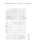 |
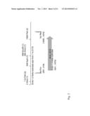 | 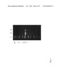 |
 |  |
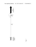 | 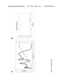 |
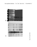 | 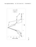 |
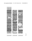 | 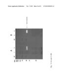 |
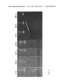 | 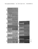 |
 | 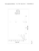 |
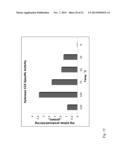 | 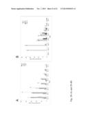 |
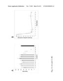 |  |
 |  |
 |  |
 |  |
 |  |
 |  |
 |  |
 |  |
 |  |
 |
| Similar patent applications: | |
| Date | Title |
|---|---|
| 2014-04-10 | Enhanced oxygen cell culture platforms |
| 2010-07-22 | Novel rigidoporus microporus laccase |
| 2009-10-08 | Maize cellulose synthases and uses thereof |
| 2011-02-03 | Maize cellulose synthases and uses thereof |
| 2011-07-28 | Maize cellulose synthases and uses thereof |
| New patent applications in this class: | |
| Date | Title |
|---|---|
| 2018-01-25 | Methods for mitigating the inhibitory effects of lignin and soluble phenolics for enzymatic conversion of cellulose |
| 2018-01-25 | In-situ biostimulation of the hydrolysis of organic matter for optimizing the energy recovery therefrom |
| 2018-01-25 | G24 glucoamylase compositions and methods |
| 2017-08-17 | Cooling and processing materials |
| 2017-08-17 | Enzymes manufactured in transgenic soybean for plant biomass engineering and organopollutant bioremediation |
| New patent applications from these inventors: | |
| Date | Title |
|---|---|
| 2010-07-22 | Acidothermus celluloyticus xylanase |
| Top Inventors for class "Chemistry: molecular biology and microbiology" | |
| Rank | Inventor's name |
|---|---|
| 1 | Marshall Medoff |
| 2 | Anthony P. Burgard |
| 3 | Mark J. Burk |
| 4 | Robin E. Osterhout |
| 5 | Rangarajan Sampath |