Patent application title: MATERIALS AND METHODS FOR DETECTION OF HPV NUCLEIC ACIDS
Inventors:
Brian Lowe (Olney, MD, US)
Anna K. Fulbright (Columbia, MD, US)
Irina Nazarenko (Gaithersburg, MD, US)
Assignees:
QIAGEN GAITHERSBURG INC.
IPC8 Class: AC07H2104FI
USPC Class:
435 5
Class name: Chemistry: molecular biology and microbiology measuring or testing process involving enzymes or micro-organisms; composition or test strip therefore; processes of forming such composition or test strip involving virus or bacteriophage
Publication date: 2012-12-20
Patent application number: 20120322049
Abstract:
Provided are nucleic acids capable of hybridizing to HPV 16 and/or HPV 18
nucleic acids, in particular, mRNA encoding E2 and E6-7 gene products.
Such nucleic acids are useful in methods of isolating RNA from a
biological sample, methods and means for determining the presence of
particular RNA splice-form variants in a biological sample, methods and
means for determining the relative ratio of RNA ratios in a biological
sample, methods and means for predicting the progression of precancerous
cervical lesions, and methods and means for detecting disruption of genes
or gene expression.Claims:
1. An isolated nucleic acid having an overall length of not more than 200
nucleotides comprising at least one nucleotide sequence having at least
75-percent homology to a nucleotide sequence selected from the group
consisting of SEQ ID NO: 1 to SEQ ID NO: 105 and SEQ ID NO: 111 to SEQ ID
NO: 308, RNA equivalents thereof, and a complements thereof.
2. The isolated nucleic acid probe of claim 1 having an overall length of not more than 100 nucleotides.
3. The isolated nucleic acid of claim 1 having an overall length of not more that 50 nucleotides.
4. The isolated nucleic acid according to claim 1 consisting of a nucleotide sequence selected from the group consisting of SEQ ID NO: 1 to SEQ ID NO: 105 and SEQ ID NO: 111 to SEQ ID NO: 308, RNA equivalents thereof, and complements thereof.
5. The isolated nucleic acid of claim 1, wherein the nucleic acid is capable of hybridizing under stringent conditions to: (a) a portion of a human papillomavirus (HPV) genome selected from the group consisting of HPV16 and HPV18, (b) an mRNA transcript derived from said HPV genome, or (c) a complement of said mRNA.
6. The isolated nucleic acid of claim 1, wherein the nucleic acid is capable of hybridizing under high stringency conditions to: (a) a portion of a human papillomavirus (HPV) genome selected from the group consisting of HPV16 and HPV18, (b) an mRNA transcript derived from said HPV genome, or (c) a complement of said mRNA.
7. The isolated nucleic acid of claim 6 wherein the nucleic acid is not capable of hybridizing under stringent conditions to more than one type of human papillomavirus (HPV) genome.
8. The isolated nucleic acid of claim 1 capable of hybridizing under stringent conditions to an HPV 16 or HPV 18 gene and/or mRNA selected from the group consisting of E2 and E6/E7.
9. The isolated nucleic acid of claim 1 having at least 75-percent homology across its entire length to a portion of a HPV 16 or HPV 18 gene selected from the group consisting of E2 and E6/E7.
10. A nucleic acid probe comprising an isolated nucleic acid according to claim 1 and optionally further comprising a detectable label and/or a ligand.
11. The nucleic acid probe of claim 10, wherein the nucleic acid probe is bound to a solid support.
12. A probe set comprising at least one nucleic acid probe according to claim 10.
13. A method of detecting the presence of a target RNA, the method comprising: a) providing at least one DNA capture probe, wherein the at least one DNA capture probe is bound to a support; b) hybridizing the target RNA to said at least one DNA capture probe, yielding a target RNA:DNA capture probe complex; c) isolating the target RNA:DNA capture probe complex; d) providing at least one DNA amplification probe, and hybridizing said at least one DNA amplification probe to said target RNA:DNA capture probe complex, yielding a target RNA:DNA capture/amplification probe complex; e) providing an anti-RNA:DNA hybrid antibody, and incubating said target RNA:DNA capture/amplification probe complex with said antibody, yielding a target RNA:DNA:antibody complex; f) detecting said antibody, wherein said detecting indicates the presence of said target RNA, wherein the capture probe and/or amplification probe comprises an isolated nucleic acid according to claim 1.
14. The method of claim 13, wherein the target RNA is a splice variant, and wherein the at least one DNA capture probe and the at least one DNA amplification probe are selected to detect the presence of said splice variant.
15. A method of detecting the presence of a target RNA, the method comprising: a) providing at least one DNA capture probe; b) providing a first anti-RNA:DNA hybrid antibody, wherein the first anti-RNA:DNA hybrid antibody is bound to a support; c) hybridizing the target RNA to said at least one DNA capture probe, yielding a target RNA:DNA capture probe complex; d) incubating said target RNA:DNA capture probe complex with said anti-RNA:DNA hybrid antibody, yielding a bound target RNA:DNA capture probe complex; e) providing at least one DNA amplification probe, and hybridizing said at least one DNA amplification probe to said bound target RNA:DNA capture probe complex, yielding a bound target RNA:DNA capture/amplification probe complex; f) providing a second anti-RNA:DNA hybrid antibody, and incubating said bound target RNA:DNA capture/amplification probe complex with said second anti-RNA:DNA hybrid antibody, yielding a bound target RNA:DNA:antibody complex; g) detecting said second anti-RNA:DNA hybrid antibody, wherein said detecting indicates the presence of said target RNA, wherein at least one of the capture probes and/or amplification probes comprises an isolated nucleic acid according to claim 1.
16. The method of claim 15, wherein the target RNA is a splice variant, and wherein the at least one DNA capture probe and the at least one DNA amplification probe are selected to detect the presence of said splice variant.
17. A method for determining whether a target nucleic acid is absent from or disrupted in a sample, said method comprising: a) treating a first portion of the sample under conditions sufficient to induce the formation of: i) a first set of DNA:RNA hybrids comprising the target nucleic acid; and ii) a second set of DNA:RNA hybrids comprising a reference nucleic acid; b) treating a second portion of the sample under conditions sufficient to induce the formation the second set of DNA:RNA hybrids, but not the first set of DNA:RNA hybrids; c) generating a detectable signal in the first portion of the sample and the second portion of the sample, wherein the detectable signal has an intensity that correlates with the concentration of DNA:RNA hybrids; and d) comparing the intensity of the detectable signal in the first portion of the sample and the intensity of the detectable signal in the second portion of the sample, wherein: i) the target nucleic acid is intact and present in the sample if the intensity of the detectable signal in the first portion of the sample is greater than the intensity of the detectable signal in the second portion of the sample; and ii) the target nucleic acid is absent from the sample if the intensity of the detectable signal in the first portion of the sample is less than or equal to the intensity of the detectable signal in the second portion of the sample.
18. The method of claim 17, wherein the first portion of the sample and the second portion of the sample are formed by a method comprising contacting the sample with: a) a first capture probe specific for the target nucleic acid under stringent conditions, wherein hybridization of the first capture probe to the target nucleic acid generates a first capture complex; and b) a second capture probe specific for the reference nucleic acid under stringent conditions, wherein hybridization of the second capture probe to the reference nucleic acid generates a second capture complex.
19. The method of claim 18 further comprising capturing the first capture complex and the second capture complex to a support.
20. The method of claim 19 wherein the first and second capture probes are bound to or adapted to be bound to the support.
21. The method of claim 20 wherein the capture probes comprise a ligand and the support comprises a ligand-binding moiety.
22. The method of claim 21 wherein the first capture probe and the second capture probe comprise the same ligand.
23. The method of claim 21 wherein the first capture probe and the second capture probe comprise different ligands, wherein: a) the first portion of the sample is contacted with a first set of solid supports comprising a ligand binding moiety capable of binding the ligand of the first capture probe and a ligand binding moiety capable of binding the ligand of the second capture probe; and b) the second portion of the sample is contacted with a second set of solid supports comprising a ligand binding moiety capable of binding the ligand of the second capture probe, but not the first capture probe.
24. The method of claim 23 wherein the first portion of the sample and the second portion of the sample is contacted with i) a first probe set comprising a plurality of signal probes capable of hybridizing to the target nucleic acid; and ii) a second probe set comprising a plurality of signal probes capable of hybridizing to the reference nucleic acid under stringent conditions.
25. The method of claim 21 wherein the first capture probe and the second capture probe are biotinylated and wherein the first support and the second support comprise a biotin-binding moiety.
26. The method of claim 21 wherein the capture probes are covalently bound to the support.
27. The method of claim 19 wherein: a) the first and second capture complexes comprise a DNA:RNA hybrid; and b) the first and second capture complexes are captured to the first and second supports by a method comprising contacting the first and second capture complexes with an entity capable of specifically binding to a DNA:RNA hybrid, wherein the entity capable of specifically binding to a DNA:RNA hybrid is bound to the support or adapted to be bound to the support.
28. The method of claim 27 wherein the entity capable of specifically binding to a DNA:RNA hybrid comprises a ligand and the first and second supports comprise a ligand-binding moiety.
29. The method of claim 28 wherein the entity capable of specifically binding to a DNA:RNA hybrid is biotinylated and wherein the support comprises a biotin-binding moiety.
30. The method of claim 27 wherein the entity capable of specifically binding to a DNA:RNA hybrid is covalently bound to the support.
31. The method of claim 27 wherein the entity capable of specifically binding to a DNA:RNA hybrid is a DNA:RNA hybrid-specific antibody or a fragment thereof.
32. The method of claim 19 wherein: a) the first capture probe comprises: i) a region capable of hybridizing to the target nucleic acid under stringent conditions; and ii) a region capable of hybridizing to a first nucleic acid sequence of an anchor probe; b) the second capture probe comprises: i) a region capable of hybridizing to the target nucleic acid under stringent conditions; and ii) a region capable of hybridizing to a second nucleic acid sequence of an anchor probe, wherein the anchor probe is bound to or adapted to be bound to the first support and/or second support.
33. The method of claim 32 wherein the first nucleic acid sequence and the second nucleic acid sequence are the same.
34. The method of claim 32 wherein the first nucleic acid sequence and the second nucleic acid sequence are different.
35. The method of claim 34 wherein the first nucleic acid sequence and the second nucleic acid sequence are disposed in the same anchor probe.
36. The method of claim 34 wherein the first nucleic acid sequence and the second nucleic acid sequence are disposed in different anchor probes.
37. The method of claim 32 wherein: a) the first support comprises an anchor probe comprising the first nucleic acid sequence and an anchor probe comprising second nucleic acid sequence; and b) the second support comprises anchor probes comprising the second nucleic acid sequence, but does not comprise anchor probes comprising the first nucleic acid sequence.
38. The method of claim 17 wherein: a) the first set of DNA:RNA hybrids is formed by a method comprising contacting the sample with a first signal probe capable of hybridizing to the target nucleic acid; and b) the second set of DNA:RNA hybrids is formed by a method comprising contacting the sample with a second signal probe capable of hybridizing to the reference nucleic acid.
39. The method of claim 38 wherein: a) the first portion of the sample is contacted with the first signal probe and the second signal probe; and b) the second portion of the sample is contacted with the second signal probe, but not the first signal probe, wherein the first signal probe is specific for the target nucleic acid under stringent conditions and the second signal probe is specific for the reference nucleic acid under stringent conditions.
40. The method of claim 28 wherein: a) the first signal probe is disposed in a first probe set comprising a plurality of signal probes capable of hybridizing to the target nucleic acid; and b) the second signal probe is disposed in a second probe set comprising a plurality of signal probes capable of hybridizing to the reference nucleic acid.
41. The method of claim 40 wherein: a) the plurality of signal probes of the first probe set is capable of hybridizing to at least 70% of the target nucleic acid; b) the plurality of signal probes of the second probe set is capable of hybridizing to at least 70% of the reference nucleic acid.
42. The method of claim 17 wherein the detectable signal is generated by a method comprising contacting the first portion of the sample and the second portion of the sample with an entity capable of specifically binding to a DNA:RNA hybrid.
43. The method of claim 42 wherein the entity capable of specifically binding a DNA:RNA hybrid is an DNA:RNA hybrid-specific antibody or a fragment thereof.
44. The method of claim 17 comprising: a) generating the first portion of the sample and the second portion of the sample by a method comprising: i) contacting the sample with at least a first biotinylated capture probe specific for the target nucleic acid under stringent conditions; ii) contacting the sample with at least a second biotinylated capture probe specific for the reference nucleic acid under stringent conditions; iii) contacting the sample with a streptavidin-coated magnetic bead under conditions sufficient to permit binding of the biotinylated capture probes to the streptavidin coated bead; and iv) separating the streptavidin coated beads into separate containers to form the first portion of the sample and the second portion of the sample; b) forming the first set of DNA:RNA hybrids and the second set of DNA:RNA hybrids in the first portion of the sample by a method comprising contacting the first portion of the sample with a probe cocktail comprising: i) a plurality of detectably labeled nucleic acid probes capable of hybridizing to the target nucleic acid under stringent conditions, wherein said plurality is sufficient to cover the target nucleic acid; and ii) a plurality of detectably labeled nucleic acid probes capable of hybridizing to the reference nucleic acid under stringent conditions, wherein said plurality is sufficient to cover the target nucleic acid; and c) forming the second set of DNA:RNA hybrids in the second portion of the sample by a method comprising contacting the second portion of the sample with a probe cocktail comprising a plurality of detectably labeled signal probes capable of hybridizing to the reference nucleic acid under stringent conditions, wherein said plurality is sufficient to cover the target nucleic acid, wherein the detectable signal is generated by the detectably labeled signal probes.
45. The method of claim 17 wherein: a) the target nucleic acid is an HPV E2 nucleic acid; and b) the reference nucleic acid is selected from the group consisting of: i) HPV E1 nucleic acid ii) HPV E6/E7 nucleic acid iii) HPV L1 nucleic acid iv) HPV L2 nucleic acid.
46. The method of claim 45 wherein a group of reference nucleic acids are detected, the group comprising at least two reference nucleic acids selected from the group consisting of: i) HPV E1 mRNA or cDNA ii) HPV E6/E7 mRNA or cDNA iii) HPV L1 mRNA or cDNA; and iv) HPV L2 mRNA or cDNA.
47. The method of claim 46 wherein the group of reference nucleic acids comprises: i) HPV E1 mRNA; ii) HPV E6/E7 mRNA; iii) HPV L1 mRNA; and iv) HPV L2 mRNA.
48. A method of predicting the onset of HPV-induced cell transformation in a patient, said method comprising detecting the presence or absence of an HPV E2 mRNA in an HPV-infected tissue derived from the patient by the method of claim 45, wherein the absence of HPV E2 mRNA is indicative of the onset of HPV-induced cell transformation.
49. A method of detecting integration of an HPV genome into a genome of a host cell, said method comprising detecting the presence or absence of an HPV E2 mRNA in an HPV-infected tissue derived from the patient by the method of claim 45, wherein the absence of HPV E2 mRNA is indicative of integration of the HPV genome into a genome of a host cell.
Description:
CROSS REFERENCE TO RELATED APPLICATIONS
[0001] This application claims priority to U.S. Provisional Application No. 61/446,306, filed on Feb. 24, 2011, and also U.S. Provisional Application No. 61/486,118, filed on May 13, 2011, which are both hereby incorporated by reference in their entirety.
BACKGROUND
[0002] 1. Field
[0003] The present disclosure relates to methods, compositions, and kits for determining the presence of a nucleic acid in a sample, including nucleic acids derived from Human papillomavirus ("HPV").
[0004] 2. Description of Related Art
[0005] Human papillomavirus (HPV) infection is the most important cause of cervical cancer, 13 types of which cause HPV-related cervical disease and cancer. Screening for oncogenic HPV DNA using molecular tests has been useful to diagnose HPV-related disease. However, current testing methods cannot precisely predict which infections may develop into cancer because most HPV infections are transient and regress and clear spontaneously. Therefore, additional biomarkers are being explored for use in reflex assays to confirm which infections will progress and require further treatment.
[0006] The progression of disease may be related to the expression of certain HPV genes. Detection of HPV mRNA may, therefore, be an additional biomarker for severe infections. Some HPV mRNA assays being developed for diagnostics detect a single type of transcript species, such as the E6 or E7 oncogenic sequences. These assays may not predict severe infections because the abundance of a single species may fluctuate due to the complex pattern of expression that occurs during the course of disease, or due to degradation of HPV from immune responses. In addition, an mRNA target may degrade after collection, or the number of infected cells in the collected specimen may be low, both of which may affect the assay result. As a solution, HPV assays designed to detect simultaneously two species of mRNAs in a ratio may be more predictive of disease than assays that detect a single mRNA species.
[0007] Additionally, HPV DNA is typically maintained as a productive infection in a circular, episomal state at 50-100 copies per cell. In this state, transcription of the HPV oncogenes E6 and E7 is tightly controlled by the E2 protein. E6 and E7 target p53 and pRb, respectively, and thus interfere with the normal cell cycle. Cells in which this transcriptional control is removed have a proliferative advantage over other cells due to their accelerated reentry into the cell cycle. Disruption or deletion of the E2 gene, as frequently occurs during integration of the virus into the host genome, removes the negative feedback on E6 and E7, activates telomerase, and derepresses hTERT expression, and thus clearly contributes to the progression of cell immortalization and ultimately, cancer progression.
[0008] The detection and characterization of specific nucleic acid sequences and sequence changes have been utilized to detect the presence of viral or bacterial nucleic acid sequences indicative of an infection, the presence of variants or alleles of mammalian genes associated with disease and cancers, and the identification of the source of nucleic acids found in forensic samples, as well as in paternity determinations. Characterization of the RNA species involved in normal biological processes may be important to understanding various little known biological processes.
[0009] The detection and characterization of RNA (e.g., messenger RNA, transfer RNA, ribosomal RNA, small nuclear RNA, and other RNAs) is an important tool in many fields including molecular biology, toxicology, and biochemistry. Messenger RNA (mRNA) is an essential functional constituent of a cell; during the process of gene expression, the functional single strand structure of mRNA is synthesized and serves as an intermediate template for the translation process in protein synthesis. The brief existence of an mRNA molecule begins with transcription of DNA into an RNA molecule, and ultimately ends in degradation. During its life, an mRNA molecule may also be processed, edited, and transported prior to translation. Splicing is the process by which pre-mRNA is modified to remove certain stretches of non-coding sequences called introns; the stretches that remain may include protein-coding sequences and are called exons. Sometimes pre-mRNA messages may be spliced in several different ways, allowing a single transcript to encode multiple proteins.
[0010] Detection of messenger RNA (mRNA) is critical in diagnostics because it can provide viral load and gene expression information that DNA detection cannot. These factors often give clues about the progression and prognosis of a disease. The current technologies for mRNA detection present a number of problems including complexity and potential for contamination.
[0011] The most common methods of mRNA detection include Northern blot, ribonuclease protection assay (RPA), and reverse-transcriptase polymerase chain reaction (RT-PCR). However, each of these techniques, while affording some advantages in sensitivity, requires time and material demands. In addition, some techniques require amplification of the target mRNA since total mRNA represents only about 1% of the total RNA and any particular mRNA is a significantly smaller percentage.
[0012] Currently, reverse transcriptase-polymerase chain reaction (RT-PCR) is widely used to characterize RNA transcripts. However the method has the following limitations: 1) only a limited number of the specific regions can be co-amplified; 2) mutations or alternative splicing can limit the ability of specific primers to detect the RNA; and 3) it is difficult to characterize the mRNA structure in a continuous mode method.
[0013] It therefore would be useful to have materials and methods capable of determining whether the a given nucleic acid is present or absent in a sample. Additionally, it would be useful to have materials and methods capable of determining whether a gene--including the HPV E2 gene--is disrupted, deleted, or otherwise is not being expressed in a host cell.
BRIEF SUMMARY
[0014] The present disclosure provides nucleic acids and methods useful in detecting specific nucleic acids in a sample and determining whether those nucleic acids are intact or disrupted.
[0015] In an aspect, an isolated nucleic acid is provided, having an overall length of not more than 200 nucleotides comprising, consisting essentially of, or consisting of at least one nucleotide sequence having at least 75-percent homology to a nucleotide sequence selected from the group consisting of SEQ ID NO: 1 to SEQ ID NO: 105 and SEQ ID NO: 111 to SEQ ID NO: 308, RNA equivalents thereof, and a complements thereof.
[0016] In an aspect, a method of detecting the presence of a target RNA is provided, the method comprising: a) providing at least one DNA capture probe, wherein the at least one DNA capture probe is bound to a support; b) hybridizing the target RNA to said at least one DNA capture probe, yielding a target RNA:DNA capture probe complex; c) isolating the target RNA:DNA capture probe complex; d) providing at least one DNA amplification probe, and hybridizing said at least one DNA amplification probe to said target RNA:DNA capture probe complex, yielding a target RNA:DNA capture/amplification probe complex; e) providing an anti-RNA:DNA hybrid antibody, and incubating said target RNA:DNA capture/amplification probe complex with said antibody, yielding a target RNA:DNA:antibody complex; f) detecting said antibody, wherein said detecting indicates the presence of said target RNA. In one aspect, antibody is conjugated to a detectable marker, and the step of detecting comprises detecting the marker. In one aspect, the detectable marker is selected from the group consisting of alkaline phosphatase and horseradish peroxidase. In one aspect, the step of detecting comprises providing a second antibody that binds to said anti-RNA:DNA hybrid antibody, wherein said second antibody is conjugated to a detectable marker, and wherein said detecting further comprises detecting the marker. In one aspect, the support comprises a magnetic bead. In one aspect, the magnetic bead is conjugated to at least one streptavidin molecule, and the at least one DNA capture probe is conjugated to a biotin molecule. In one aspect, at least one of the capture probes and/or amplification probes is a nucleic acid probe as set forth above.
[0017] In one aspect, the at least one DNA capture probe and the at least one DNA amplification probe are from about 15 to about 200 bases in length.
[0018] In one aspect, the target RNA is a splice variant, and the at least one DNA capture probe and the at least one DNA amplification probe are selected to detect the presence of said splice variant.
[0019] In one aspect, the at least one DNA capture probe and the at least one DNA amplification probe are complementary to RNA from HPV high risk types 16, 18, 31, 33, 35, 39, 45, 51, 52, 56, 58, 59, 68, 26, 66, 73, and 82.
[0020] In another aspect, a kit for the detection of a target RNA is provided, the kit comprising: a) at least one DNA capture probe, bound to a magnetic support; b) at least one DNA amplification probe; c) an anti-RNA:DNA hybrid antibody; and d) a detection reagent. In one aspect, said anti-RNA:DNA hybrid antibody is conjugated to a detectable marker, and said detection reagent comprises a substrate for said detectable marker. In one aspect, the kit further comprises a second antibody that binds to said anti-RNA:DNA hybrid antibody, wherein said second antibody is conjugated to a detectable marker, and wherein said detection reagent comprises a substrate for said detectable marker.
[0021] The present disclosure provides a method of providing target RNA for detection, the method comprising: incubating a biological sample containing the target RNA with carboxyl beads; isolating the beads; lysing the biological sample attached to the isolated beads; and isolating the beads from the lysed biological sample, wherein the resulting supernatant contains the target RNA for detection.
[0022] In another aspect, a method for nucleic acid detection is disclosed that does not rely on target amplification. Nucleic acids of interest are captured by specific nucleic oligonucleotides. Signal amplification is provided by adding DNA probes that cover the captured RNA target (or vice versa of the target is DNA) that is then detected using entities capable of binding specifically to DNA:RNA hybrids. This hybrid capture assay gives linear increases in signal as both quantity and length of transcripts increase. As a result, it can be used to measure deletions that existing technologies cannot. By assaying the extent of target nucleic acid disruption, as compared to total signal from a complete set of reference nucleic acids, one is able to whether, and the extent to which, the target is disrupted.
[0023] In an aspect, disruption of the target is determined by separating a sample into at least a first and second portion. The first portion of the sample is treated under conditions sufficient to generate two sets of DNA:RNA hybrids: one set comprising the target nucleic acid and one set comprising at least one reference nucleic acid. The second portion of the sample is then treated under conditions sufficient to generate the set of DNA:RNA hybrids comprising the reference nucleic acid, but not set comprising the target nucleic acid. The total amount of DNA:RNA hybrid in the first portion of the sample is then compared to the total amount of DNA:RNA hybrid in the second portion of the sample. If the target nucleic acid is missing, there should be the same amount of DNA:RNA hybrid in the first and second portions of the sample. Variations of the method also are presented for determining the extent of disruption, if any, by applying a plurality of probes specific for a substantial portion of the target nucleic acid and progressively removing the probes. The more probes that can be removed before a change in DNA:RNA hybrids is detected, the greater the extent to which the target nucleic acid is disrupted.
[0024] In another aspect, a method is provided to determine whether or not an E2 gene, cDNA, or mRNA is absent or disrupted. Such a method can be applied to, inter alia, determine whether the E2 gene is being expressed, whether the HPV genome is integrated into the host cell genome, assessing the progression of an HPV infection, and/or determining the risk of an HPV infection progressing to cancer.
BRIEF DESCRIPTION OF THE DRAWINGS
[0025] For a further understanding of the nature, objects, and advantages of the present disclosure, reference should be had to the following detailed description, read in conjunction with the following drawings, wherein like reference numerals denote like elements.
[0026] FIG. 1 is a schematic diagram of target RNA (crosshatched bar) captured by biotinylated DNA probes (white bar). "B" represents a biotin moiety; "SA" represents a streptavidin moiety; "AP" represents alkaline phosphatase conjugated to an antibody, but AP could be any other appropriate detectable moiety (e.g., horseradish peroxidase, etc.), and B and SA could be replaced by other linkage moieties.
[0027] FIG. 2 is a diagram depicting the use of DNA capture probe (white bar), multiple DNA amplification probes (black bars), and multiple DNA:RNA hybrid antibodies to "amplify" the signal without the need for amplification of the target RNA (crosshatched bar). "B" represents a biotin moiety; "SA" represents a streptavidin moiety, B and SA may be replaced with other conjugation technology in which DNA probes are conjugated to the bead; "AP" represents alkaline phosphatase conjugated to an antibody, but AP could be any other appropriate detectable moiety (e.g., horseradish peroxidase, etc.).
[0028] FIG. 3 is a diagram of target RNAs (dashed arrows) captured by different DNA capture probes bound to a substrate (S). Non-conjugated DNA amplification probes (black bars) and multiple antibodies that detect and bind to DNA:RNA hybrid regions (conjugated to alkaline phosphatase or any other appropriate detectable moiety, such as horseradish peroxidase, etc.) are also shown. The substrate (e.g., a bead) may bear multiple DNA capture probes, and the DNA capture probes may be the same (i.e., the same sequence and/or length) or different (i.e., different sequences and/or different lengths).
[0029] FIG. 4 provides the results of an experiment showing the effect of adding unbiotinylated DNA probes after RNA capture. In this experiment, a variable number of biotinylated probes were conjugated to streptavidin beads. The target was the E6/7 gene transcript of HPV 16. The assay was performed with each set of beads with (black bars) and without (white bars) the addition of unlabeled signal amplification probes (one- versus two-step assay). When no signal amplification step was added (white bars), the signal increased with the amount of coverage provided by the capture probes. However, when signal amplification probes were added (black bars), the signal was greater than if they were not added, and they enable a higher signal with fewer (3-5) capture probes.
[0030] FIG. 5 shows that endogenous hybrids are often the source of clinical background noise. "RLU"=relative luminescence unit.
[0031] FIG. 6 shows the effect of lysis buffer (wherein 100% buffer contains about 3 M guanidine thiocyanate and about 2% detergent) concentration on assay background when assaying cellular samples in PreservCyt® Solution, and demonstrates that clinical background decreases with decreasing concentrations of lysis buffer.
[0032] FIG. 7 shows that hypotonic lysis of cell pellets ensures that background noise remains low and stable, and that the background does not change significantly regardless of the amount of specimen used. "PC"=PreservCyt® Solution; "PC(-)"=Specimen (cervical scrape) pool fixed in PreservCyt® Solution with no HPV target.
[0033] FIG. 8 shows limit of detection of HPV E6/E7 from HPV positive cells (SiHa). This shows that using the methods of the present disclosure, as little as 1×10³ cells are required for HPV E6/7 RNA detection.
[0034] FIG. 9 shows results from tests of various lysis buffers for the ability to lyse cells captured by COOH beads. The data of FIG. 9, along with that of TABLE 1, below, shows the preferred lysis buffer is about 1M guanidine thiocyanate and about 0.7% detergent.
[0035] FIG. 10 shows cell capture by magnetic carboxylate-modified (COOH) beads (Sera Dyn catalog number 6515-2105-050350), over time, demonstrating that about 95% of the cells have been captured after incubation of 30 minutes.
[0036] FIG. 11 shows comparison of COOH bead capture with hypotonic lysis, and indicates that COOH bead capture is more efficient than hypotonic lysis for obtaining mRNA from cells. "PC-" indicates a pool of cervical scrape specimens that lack presence of HPV.
[0037] FIG. 12 is a diagram depicting capture and signal amplification probe design regions. The length of HPV transcripts can be "characterized" by capture onto magnetic beads with specific capture oligos that capture specific targets and detected with various sets of unlabeled oligonucleotides used to extend the length of the hybrid region. Signal will result if the capture RNA bears the sequence that is complementary to the capture probes that are used. Signal output will increase with successive addition of amplification signal probes until maximum length is reached where the signal will plateau. The various HPV transcripts for HPV 16 are shown. The regions denoted by the dashed boxes are designated for probe design.
[0038] FIG. 13 shows increasing signal as the number of signal amplification probes is increased. In this way, an RNA transcript length may be measured by the increasing signal generated by the increased number of consecutive amplification probes. In FIG. 13, each set of 5 oligos are adjacent to one another and result in the RNA:DNA hybrid getting longer, and signal stronger, as successive sets are added.
[0039] FIG. 14 shows that a fraction of cells with a high early:late HPV mRNA ratio may be detected against a background of cells with a low ratio. For this FIG. 14, SiHa cells (cervical cancer cell line) were added to a pool of cervical specimens (each diagnosed with a high-grade HPV-related lesion). The SiHa cells incorporate a high ratio of HPV early transcripts:HPV late transcripts, which is a common characteristic of cervical cancer. The sample mimicked a specimen that has cancer cells among pre-cancerous lesion cells. The results show that the invented assay will detect cancer cells in a pool of more benign lesion cells.
[0040] FIG. 15 shows HPV RNA stability of SiHa cells preserved in a LBC clinical specimen pool. The RT-PCR plots show the assay signal (y-axis) plotted against PCR cycle number (x-axis) for samples of SiHa cells incubated over the course of 67 days. Symbols are star, 3 days; square, 13 days; triangle, 26 days; filled diamond, 42 days; open diamond, 67 days. Values are an average of two reactions for each day.
[0041] FIG. 16a shows a general scheme for hybrid capture detection of HPV mRNA. HPV mRNA target (dotted line) is annealed to capture oligos (short grey bars) that are coupled to a magnetic bead (circle). The RNA target is annealed with signal amplification oligos (short black bars) to create a longer hybrid. The RNA:DNA hybrid is bound with a hybrid capture antibody conjugated with alkaline phosphatase (Y-shaped AP symbol). A chemiluminescent substrate (not shown) is added to detect the complex in a luminometer.
[0042] FIG. 16b shows a schematic of the HPV genome structure with labeled genes (large grey arrows). The loci for E6-7 probes (1) or E2 probes (2) are shown by black bars underneath. The arrangement of genes and the loci for DNA probes are similar for HPV 16 and HPV 18, but the primary sequences are unique.
[0043] FIG. 17a shows the dependence of luminescence signal output (average RLU, n=4) on the number of complementary signal amplification probes per assay for the same target input (1×10⁵ copies, HPV 16 E6-7 in vitro transcribed RNA). In this experiment, the hybrid length increased in wells with the addition of 5, 10 and 15 probes. The signal did not increase for the well (labeled 5+15) with 5 non-complementary probes added to 15 complementary probes.
[0044] FIG. 17b shows the dependence of luminescence signal output (average RLU, n=3 samples, error bars show standard deviation) on target input (RNA copies per reaction) for a hybrid capture assay. Signal:noise ratio is given above bars.
[0045] FIG. 18a shows the dependence of signal:noise (average, n=3) on number of SiHa cells per assay was plotted for the HPV 16 E6-7 (grey bars) and E2 (black bars); for the two assays in separate wells. For these assays, the background noise was obtained from the signal from a control assay with no target added, approximately 50 RLU.
[0046] FIG. 18b shows the signal: noise values for HPV E6-7 and E2 mRNA assays were plotted as a ratio for the cancer cell lines, SiHa, Caski and HeLa; bars represent the average ratios of three replicate experiments.
[0047] FIG. 19 shows detection of the HPV 16 E6-7:E2 transcript ratio in a mixture of SiHa cells with the cells from a pool of HPV-positive specimens. Cultured SiHa cells were mixed with a pool (2 ml) of HPV-positive, liquid-based cytology specimens (approximately 100,000 total cells in 2 ml).
[0048] FIG. 20 shows the HPV 16 E6-7:E2 ratio in cervical specimens. The E6-7:E2 ratio was plotted from the hybrid capture assay results.
[0049] FIG. 21 illustrates a method for determining whether or not E2 gene expression is absent or disrupted.
[0050] FIG. 22 illustrates a comparison of the integrity of E2 gene expression in SiHa and W12 cells.
[0051] FIG. 23 illustrates a comparison of the integrity of E2 gene expression in LSIL and HSIL samples.
DETAILED DESCRIPTION
[0052] Before the subject disclosure is further described, it is to be understood that the disclosure is not limited to the particular aspects of the disclosure described below, as variations of the particular aspects may be made and still fall within the scope of the appended claims. It is also to be understood that the terminology employed is for the purpose of describing particular aspects, and is not intended to be limiting.
[0053] In this specification and the appended claims, the singular forms "a," "an," and "the" include plural reference unless the context clearly dictates otherwise. Unless defined otherwise, all technical and scientific terms used herein have the same meaning as commonly understood to one of ordinary skill in the art to which this disclosure belongs.
[0054] Isolated Nucleic Acids and Probes Capable of Hybridizing to HPV 16 and/or HPV 18
[0055] Nucleic acids consisting of not more than 200 nucleotides and being capable of hybridizing to HPV 16 or HPV 18 DNA or RNA are provided herein.
[0056] In an aspect, the nucleic acid comprises, consists essentially of, or consists of at least one nucleotide sequence having at least 75%, at least 80%, at least 85%, at least 90%, or at least 95% homology to a nucleotide sequence selected from the group consisting of SEQ ID NO: 1 to SEQ ID NO: 105 and SEQ ID NO: 111 to SEQ ID NO: 308, RNA equivalents thereof, and complements thereof. In a further aspect, the nucleic acid comprises, consists, or consists essentially of a nucleotide sequence selected from the group consisting of SEQ ID NO: 1 to SEQ ID NO: 105 and SEQ ID NO: 111 to SEQ ID NO: 308, RNA equivalents thereof, and complements thereof,
[0057] In an aspect, the nucleic acid is capable of hybridizing under stringent conditions to a nucleic acid at least 75%, at least 80%, at least 85%, at least 90%, at least 95%, at least 96%, at least 97%, at least 98%, at least 98%, at least 99%, or 100% identical to an HPV16 or HPV18 genome or a nucleic acid derived from the same. The sequence of an exemplary HPV 16 genome is disclosed at GenBank NC--01526 (SEQ ID NO: 106). The sequence of an exemplary HPV 18 genome is disclosed at GenBank X05015 (SEQ ID NO: 107).
[0058] In another aspect, the nucleic acid is capable of hybridizing or binding to a nucleic acid at least 75%, at least 80%, at least 85%, at least 90%, at least 95%, at least 96%, at least 97%, at least 98%, at least 99%, or 100% identical to an HPV16 or HPV18 mRNA or a complement thereof. In another aspect, the HPV 16 or HPV 18 mRNA is selected from the group consisting of E2 and E6/E7 mRNA.
[0059] For present purposes, "stringent conditions" encompass conditions under which hybridization will only occur if there is 25% mismatch or less between the hybridization molecule and the target sequence. "Stringent conditions" may be broken down into particular levels of stringency for more precise definition. Thus, as used herein, "moderate stringency" conditions are those under which molecules with more than 25% sequence mismatch will not hybridize; conditions of "medium stringency" are those under which molecules with more than 15% mismatch will not hybridize, and conditions of "high stringency" are those under which sequences with more than 10% mismatch will not hybridize. Conditions of "very high stringency" are those under which sequences with more than 6% mismatch will not hybridize. Calculations regarding hybridization conditions required for attaining particular degrees of stringency are also discussed by Sambrook et al. (ed.), Molecular Cloning: A Laboratory Manual, 2nd ed., vol. 1-3, Cold Spring Harbor Laboratory Press, Cold Spring Harbor, N.Y., 1989, chapters 9 and 11, herein incorporated by reference in its entirety.
[0060] In an aspect, a probe set is provided, said probe set comprising at least one of the isolated nucleic acids disclosed herein. By way of example and not limitation, the probe set may comprise an isolated nucleic acid comprising, consisting essentially of, or consisting of at least one nucleotide sequence having at least 75%, at least 80%, at least 85%, at least 90%, or at least 95% homology to a nucleotide sequence selected from the group consisting of SEQ ID NO: 1 to SEQ ID NO: 105 and SEQ ID NO: 111 to SEQ ID NO: 308, RNA equivalents thereof, and complements thereof. In a further aspect, the probe set may comprise an isolated nucleic acid that comprises, consists, or consists essentially of a nucleotide sequence selected from the group consisting of SEQ ID NO: 1 to SEQ ID NO: 105 and SEQ ID NO: 111 to SEQ ID NO: 308, RNA equivalents thereof, and complements thereof. The isolated nucleic acids may be provided as unmodified probes or may be modified. By way of example and not limitation, the modification may facilitate isolation and/or detection of the probe and a nucleic acid to which it has hybridized, for example, by addition of a ligand and/or detectable labels. In one aspect, the probes may be provided bound to a solid support, such as a plate, tube, bead, microchip, or other solid surface.
[0061] Methods of Identifying HPV mRNA
[0062] Methods of the present disclosure may be used to detect the presence of a target nucleic acid from samples. Such nucleic acid may be an RNA, and such samples may include, without limitation, a specimen or culture (e.g., cellular, microbiological and viral cultures) including biological and environmental samples. Biological samples may be from a eukaryote, a prokaryote, an archaeon, a virus, an animal, including a human, a plant, a fungus, an excavate, and may be from fluid, solid (e.g., stool) or tissue, cell culture, liquid or solid media, as well as liquid and solid food and feed products and ingredients such as dairy items, vegetables, meat and meat by-products, and waste. Environmental samples include environmental material such as surface matter, soil, water, air and industrial samples, as well as samples obtained from food and dairy processing instruments, apparatus, equipment, utensils, disposable and non-disposable items. Particularly preferred are biological samples including, but not limited to, cervical epithelial cells (e.g., a sample obtained from a cervical swab or biopsy), adenoid cells, anal epithelial cells, blood, saliva, cerebral spinal fluid, pleural fluid, milk, lymph, sputum and semen. The sample may comprise a ribonucleic acid including messenger RNA (mRNA).
[0063] The present disclosure provides a method for determining the presence of a target RNA in a sample, wherein the method comprises: a) hybridizing the target RNA with a DNA capture probe having a sequence complementary to the target RNA to form a target RNA:DNA capture probe complex, wherein the DNA capture probe is conjugated to a support; b) separating the target RNA:DNA capture probe complex from unbound RNA (e.g., by washing); c) optionally hybridizing at least one amplification probe to the target RNA:DNA capture probe complex, wherein the at least one amplification probe has a sequence complementary to the target RNA, thereby forming a target RNA:DNA capture/amplification probe complex; d) adding an antibody that recognizes and binds to RNA:DNA hybrids to bind the target RNA:DNA capture/amplification probe complex, thereby forming a target RNA:DNA:antibody complex, wherein the antibody is labeled with a detectable marker; e) detecting the marker on said antibody, wherein the detecting indicates the presence of the target ribonucleic acid; and f) comparing the detection results with results produced from a different combination of amplification probes wherein the comparing indicates the particular RNA splice-form present.
[0064] The present disclosure provides a method for determining the presence of a target RNA in a sample, wherein the method comprises: a) hybridizing the target RNA with a DNA capture probe having a sequence complementary to the target RNA to form a target RNA:DNA capture probe complex, wherein the DNA capture probe is conjugated to a support; b) separating the target RNA:DNA capture probe complex from unbound RNA; c) optionally hybridizing at least one amplification probe to the target RNA:DNA capture probe complex, wherein the at least one amplification probe has a sequence complementary to the target RNA, thereby forming a target RNA:DNA capture/amplification probe complex; d) adding an antibody that recognizes and binds to RNA:DNA hybrids to bind the target RNA:DNA capture/amplification probe complex, thereby forming a target RNA:DNA:antibody complex; e) adding a second antibody that recognizes and binds the first antibody, wherein the second antibody is labeled with a detectable marker; f) detecting the marker on the second antibody, wherein the detecting indicates the presence of the target ribonucleic acid; and g) comparing the detection results with results produced from a different combination of amplification probes wherein the comparing indicates the particular RNA splice-form present.
[0065] The present disclosure also provides a method of detecting the presence of a ribonucleic acid (RNA) splice form in a sample, wherein the method comprises a) hybridizing the target RNA with a DNA capture probe having a sequence complementary to the target RNA under conditions that allow the probe and the target ribonucleic acid to hybridize, thereby forming a target RNA:DNA capture probe complex; b) adding a first antibody that recognizes and binds to RNA:DNA hybrids to bind the target RNA:DNA capture probe complex, thereby forming a target RNA:DNA capture probe:antibody complex, wherein the first antibody is conjugated to a support; c) separating the target RNA:DNA capture probe:antibody complex from unbound RNA; d) hybridizing at least one amplification probe to the target RNA:DNA capture probe:antibody complex, wherein the at least one amplification probe has a sequence complementary to the target RNA and is added in a combination that will cover specific target RNA regions, thereby forming a target RNA:DNA:antibody complex; e) adding a second antibody that recognizes and binds to RNA:DNA duplexes to bind the target RNA:DNA:antibody complex, to form a target RNA:DNA:antibodies complex, wherein the second antibody is labeled with a detectable marker; f) detecting the marker on said second antibody, wherein the detecting indicates the presence of the target RNA; and g) comparing the detection results with results produced from a different combination of amplification probes wherein the comparing indicates the particular RNA splice-form present.
[0066] The present disclosure also provides a method of detecting the presence of a ribonucleic acid (RNA) splice form in a sample, wherein the method comprises a) hybridizing the target RNA with a DNA capture probe having a sequence complementary to the target RNA under conditions that allow the probe and the target ribonucleic acid to hybridize, thereby forming a target RNA:DNA capture probe complex; b) adding a first antibody that recognizes and binds to RNA:DNA hybrids to bind the target RNA:DNA capture probe complex, thereby forming a target RNA:DNA capture probe:antibody complex, wherein the first antibody is conjugated to a support; c) separating the target RNA:DNA capture probe:antibody complex from unbound RNA; d) hybridizing at least one amplification probe to the target RNA:DNA capture probe:antibody complex, wherein the at least one amplification probe has a sequence complementary to the target RNA and is added in a combination that will cover specific target RNA regions, thereby forming a target RNA:DNA:antibody complex; e) adding a second antibody that recognizes and binds to RNA:DNA duplexes to bind the target RNA:DNA:antibody complex, to form a target RNA:DNA:antibodies complex; f) separating the target RNA:DNA:antibodies complex from unbound second antibody; g) adding a third antibody labeled with a detectable marker wherein the third antibody recognizes and binds to the second and/or first antibody; h) detecting the marker on the third antibody, wherein the detecting indicates the presence of the target RNA; and i) comparing the detection results with results produced from a different combination of at least one amplification probe wherein the comparing indicates the RNA splice-form present.
[0067] RNA is often transcribed from different promoters and spliced, thereby generating multiple forms that include the coding regions for different genes. It is important to characterize these multiple spliced forms of RNA for fundamental research and for applications where the detection of specific mRNA isoforms is critical.
[0068] One application of the present disclosure is the detection and characterization of mRNA expression in human papillomavirus (HPV). Carcinoma of the cervix has been shown to be associated with the presence of high-risk HPV types; from about 13 to about 18 high-risk types are currently identified. The HPV DNA test can identify high-risk HPV types, but is a poor predictor for the progression of the disease in pre-cancerous clinical specimens. Thus, additional methods and markers are needed to improve the predictive value of HPV tests. The characterization of mRNA for the presence of the E6/7 oncogene and other mRNAs, as provided by the present disclosure, will allow an accurate and reliable method that determines the ratio of expression of these oncogenes versus other viral genes. The ratio of E6/E7 to E2, E4, and/or L1 mRNA may be a better predictor for the progression of precancerous cervical lesions (see, e.g., U.S. Pat. No. 6,355,424, incorporated by reference herein). Hybrid capture technology is a linear signal amplification method. Thus, the instant disclosure provides valuable methods for guiding therapeutic strategy, while minimizing the number of patients requiring colposcopy. The instant disclosure provides methods of using mixtures of short oligonucleotides capable of hybridizing to the different lengths/genes of RNA (and mRNA in particular) in order to characterize splice forms.
[0069] Target Nucleic Acids
[0070] In one aspect, the target ribonucleic acid to be detected may be mRNA, ribosomal RNA, nucleolar RNA, transfer RNA, viral RNA, heterogeneous nuclear RNA etc., wherein the one or more polynucleotide probes are DNA probes. The target ribonucleic acids include, without limitation, nucleic acids found in specimens or cultures (e.g., cellular, microbiological and viral cultures) including biological and environmental samples. The target ribonucleic acids may be found in biological samples from an animal, including a human, fluid, solid (e.g., stool) or tissue, as well as liquid and solid food and feed products and ingredients such as dairy items, vegetables, meat and meat by-products, and waste. Target ribonucleic acids may be found in environmental samples and include environmental material such as surface matter, soil, water and industrial samples, as well as samples obtained from food and dairy processing instruments, apparatus, equipment, utensils, disposable and non-disposable items. Particularly preferred are target nucleic acids found in biological samples including, but not limited to cervical samples (e.g., a sample obtained from a cervical swab), adenoid cells, anal epithelial cells, blood, saliva, cerebral spinal fluid, pleural fluid, milk, lymph, sputum, urine and semen.
[0071] In other aspects, the target ribonucleic acids are from virus, bacteria, mycobacteria or plasmodia, for example, without intending to be limited thereby, cytomegalovirus (CMV), Herpesviridae, human immunodeficiency virus (HIV), Chlamydia spp., Neisseria spp. (e.g., N. gonorrhea), Staphylococcus aureus, mycobacteria (e.g., Mycobacterium tuberculosis), SARS coronavirus (SARS-CoV), or Orthomixoviridae (e.g., influenza viruses).
[0072] In one aspect, the target ribonucleic acids are human papillomavirus (HPV) and include genetic variants of HPV. A variant includes polymorphisms, mutants, derivatives, modified, altered, or the like forms of the target nucleic acid. In one aspect, the target nucleic acid is an HPV nucleic acid. In another aspect, the HPV nucleic acid is HPV DNA of a high risk HPV type. In another aspect the target nucleic acids are high risk HPV types 16, 18, 31, 33, 35, 39, 45, 51, 52, 56, 58, 59, 68, 26, 66, 73, and 82.
[0073] The RNA may be isolated and prepared for hybridization by a variety of methods and reagents including (but not limited to) guanidinium thiocyanate-phenol-chloroform extraction (e.g., with TRIzol® reagent, also known as TRI Reagent), hypotonic lysis, and carboxyl (COOH) bead capture. The principle of RNA isolation is based on cell/tissue lysis, followed by extraction, precipitation, and washing. While very effective, these techniques require a high level of technical precision and are not candidates for automation. Other RNA preparation methods do not completely eliminate DNA and other potential contaminants, require expensive enzymes, and require many sometimes time-consuming--washing steps. The challenge is to develop a method for mRNA detection that reduces many of the current challenges and can provide rapid information about expression of specific genes. Two primary sample preparation methods have been devised for the present disclosure: hypotonic cell lysis; and carboxyl bead capture. RNA isolated using TRIzol® or QIAGEN resin technology (for example, QIAGEN RNeasy Plus Mini Kit) can also be used in this assay.
[0074] In certain aspects, the biological sample is comprised of cervical cells, especially human cervical cells. The sample can be collected with any method or device known in the art, including a chemically inert collection device such as a Dacron® (poly(ethylene terephthalate)) tipped swab. Other acceptable collection devices may be used including, but not limited, to cotton swab, cervical brush, flocked swab (a swab shaped like a Dacron® swab but made with nylon fibers enabling collection of more cells and easier release of cells), cervical broom, mini broom, lavage, or any collection device often used in PAP smear testing (Papanicolaou's test). The cervical cells may also be part of a biopsy specimen.
[0075] Sample Preparation
[0076] The use of TRIzol® to isolate RNA, as well as other known methods for RNA isolation, may be employed in methods of the present disclosure. Sample preparation by hypotonic lysis of the cell pellet reduces the release of endogenous RNA:DNA hybrids that may interfere with assay detection step, and this is a preferable RNA isolation method. In this sample preparation method, cells are pelleted via centrifuge, the supernatant is removed, and the pellet is resuspended and the cells lysed. After lysis, the cellular debris is pelleted and the supernatant (containing RNA) collected. Reducing the stringency of lysis (as measured by salt and detergent concentrations in a buffer) reduces the clinical background produced from pools of methanol-based cervical specimens (FIGS. 5 & 6). The signal:noise ratios are also higher and the variability in background between pools and in interference is lower. Other studies have shown that hypotonic lysis works by rupturing the cellular membrane because of differences in tonicity between the cell and the milieu, making the cell permeable to macromolecules. Thus, RNA in the cell is released from the cell into the solution, whereas contaminants to the assay (such as endogenous RNA:DNA hybrids) will remain in the insoluble cell debris. This method may be useful in cases where the amount of RNA in a specimen is limited because increasing the amount of specimen does not lead to an increase in background.
[0077] Another method of sample preparation uses magnetic carboxyl (COOH) beads that can be added directly to a biological sample to concentrate cells for DNA isolation. Cells in the sample are attracted to the beads via hydrophobic interactions. After using a magnetic rack to pellet the beads, the supernatant can be removed and the cells lysed. Non-magnetic COOH beads or other adsorptive particles could also be used, substituting centrifugation for pelleting via a magnetic rack. After the lysis (which usually occurs at 65° C. for 15 min) the beads are again pelleted and the remaining supernatant may be used directly in methods of the present disclosure. While decreasing lysis stringency again reduces background in this method, water alone is not enough to release the RNA from the cells. As such, it is preferable to use a lysis buffer comprising about 1 M guanidine thiocyanate and about 0.7% detergent for all sample preparation methods of the present disclosure (see, e.g., FIGS. 5 & 6).
[0078] Hybridization/Capture--Capture Probes
[0079] After the sample is prepared and target RNA is released, it is contacted with at least one polynucleotide DNA capture probe under a condition sufficient for the at least one polynucleotide probe to hybridize to the target RNA in the sample to form a double-stranded nucleic acid hybrid. The DNA capture probes may be full length, truncated, or synthetic DNA. The DNA capture probes are sequence specific for the target RNA. DNA capture probes are ideally about 25 to 35 bases long and may be complementary to any region of the target RNA. The DNA capture probes may range from about 15 to about 200 bases in length. In other aspects, the capture probe may be not more than 100 or not more than 50 nucleotides in length. In yet other aspects, the capture probes may be: 20 to 100, 25 to 100, 30 to 100, 35 to 100, 40 to 100, 45 to 100, or 50 to 100 bases in length.
[0080] By way of example and not limitation, the capture probe may comprise, consist essentially of, or consist of at least one nucleotide sequence having at least 75%, at least 80%, at least 85%, at least 90%, or at least 95% homology to a nucleotide sequence selected from the group consisting of SEQ ID NO: 1 to SEQ ID NO: 20. In a further aspect, the capture probe comprises, consists of, or consists essentially of a nucleotide sequence selected from the group consisting of SEQ ID NO: 1 to SEQ ID NO: 20. In one aspect, a capture probe set specific for HPV 16 is provided, comprising at least one capture probe selected from the group consisting of SEQ ID NO: 1 to SEQ ID NO: 10. In one aspect, a capture probe set specific for HPV 18 is provided, comprising at least one capture probe selected from the group consisting of SEQ ID NO: 11 to SEQ ID NO: 20.
[0081] The DNA capture probes can be bound to a support. "Bound" includes but is not limited to chemically attached, covalently bound, and covalently linked. Multiple DNA capture probes, and multiple different DNA capture probes may be bound to the same support (e.g., the same magnetic bead), as shown schematically in FIG. 3. Only 3-5 different capture probes are required for optimal results (see FIG. 4), thus providing a great deal of flexibility to allow these probes to be sequence-specific and not fall in regions that may be spliced out in some variants. In one aspect, the sequence-specific DNA capture probes are biotinylated and have been bound by conjugation to magnetic streptavidin beads. A capture probe may isolate a particular spliceform if it comprises a single oligo that bridges a splicesite.
[0082] Supports include, but are not limited to beads, magnetic beads, columns, plates, filter paper, polydimethylsiloxane (PDMS), and dipsticks. Any support can be used as long as it allows extraction of the liquid phase and provides the ability to separate out bound and unbound capture probes or antibodies. Magnetic beads are particularly useful in that they can be left in the solution and the liquid phase can be extracted or decanted, if a magnetic field is applied to hold the beads in place. Beads that are small and have a high surface area are preferable, such as beads about 1 μm in diameter. In certain aspects, the support comprises a modified magnetic bead, that is coated or has attached thereto a DNA capture probe complementary and specific to the target mRNA. A magnetic field is used to separate the double-stranded nucleic acid/magnetic bead complex from non-bound ribonucleic acid. In certain aspects, the support comprises a modified magnetic bead, wherein the magnetic beads are modified by coating the beads with a first antibody immunospecific for double-stranded hybrid nucleic acids. A magnetic field is used to separate the nucleic acid hybrid/antibody/magnetic bead complex from unbound ribonucleic acid. Other beads that employ charge switching or silica capture (as opposed to magnetic fields) may be used as well. In another aspect, magnetic beads with detection capacity (such as magnetic Lumonex beads) may capture and detect specific spliceforms.
[0083] Following capture of the target RNA or the target RNA:DNA hybrid as described above, the captured target RNA or RNA:DNA hybrid may be separated from the rest of the sample by application of a magnetic field (in the case of magnetic beads), and washing away of non-captured nucleic acids. Washing away unwanted interfering substances may be accomplished with buffers containing salt and or detergent that are used at various temperatures. When using supports other than magnetic beads, alternative methods of separating captured hybrid from the rest of the sample are conducted, including but not limited to, washing. Enzymatic processes, such as dnase for double-stranded DNA or RNA:DNA may be used to facilitate isolation of target RNA.
[0084] Hybridization/Capture--Amplification Probes
[0085] After the wash step to ensure that only the target remains, signal amplification DNA probes are hybridized to the target mRNA, wherein the signal amplification probes are unlabeled DNA probes complementary and/or specific to the target mRNA. The amplification probe need not be specific to the target nucleic acid. For example, the DNA amplification probe may be able to bind other nucleic acids other than the designed target. The DNA signal amplification probes complementary to the mRNA regions are designed and combined in mixtures that will cover specific genes. By extending and varying the coverage, one can determine which genes are present and the particular splice forms of the RNA. "Coverage" is defined as the extent or length of target sequence which is flanked by the complementary signal probes. The signal amplification probes are roughly 40 bases in length, but because they are designed around the capture probes, some may be more or less than 40 bases. Signal amplification probes may be about 15 to about 200 bases in length. In yet other aspects, the signal amplification probes may be: 20 to 100, 25 to 100, 30 to 100, 35 to 100, 40 to 100, 45 to 100, or 50 to 100 bases in length. Increasing coverage (i.e., hybridizing more signal probes to complementary regions of the target RNA) will lead to an increase in signal. Therefore, it is preferable to use more probes to obtain an amplified signal. The limit of detection depends, in part, on the length of the target nucleic acid (i.e., the target gene).
[0086] By way of example and not limitation, the amplification probe may comprise, consist essentially of, or consist of at least one nucleotide sequence having at least 75%, at least 80%, at least 85%, at least 90%, or at least 95% homology to a nucleotide sequence selected from the group consisting of SEQ ID NO: 21 to SEQ ID NO: 105. In a further aspect, the amplification probe comprises, consists of, or consists essentially of a nucleotide sequence selected from the group consisting of SEQ ID NO: 21 to SEQ ID NO: 105. In one aspect, an amplification probe set specific for HPV 16 is provided, comprising at least one amplification probe selected from the group consisting of SEQ ID NO: 21 to SEQ ID NO: 62. In one aspect, an amplification probe set specific for HPV 18 is provided, comprising at least one amplification probe selected from the group consisting of SEQ ID NO: 63 to SEQ ID NO: 105.
[0087] Amplification signal probes are added in combinations which would extend over the genetic sequence of known RNA splice-forms. The combination of signal amplification probes will determine the extent of coverage on the target mRNA and hence, signal output. Comparison of the resulting signal output from different combinations of amplification probes will indicate the presence of particular mRNA splice-form variants. In this way, this method is a "molecular ruler" in that the signal output is dependent on the splice form present. For example, capture probe 3 is expected to hybridize with E6/7 target mRNA, but not with E1, E2, E4, E5, L1, or L2 (see, e.g., TABLE 3 and FIG. 12). Signal amplification probes 1 and 6, used after hybridization with capture probe 3, will generate a strong signal from the spliced E6/7 form, and a weak signal from the spliced/integrated E6/7 form. By varying the combinations and numbers of capture probes and amplification probes, the signal output provides information about which viral genes are being expressed (e.g., the ratio thereof), as well as which splice forms of those genes are expressed. Such information, coupled with clinical and experimental data, is expected to provide a better predictor for progression of precancerous cervical lesions.
[0088] The characterization of gene expression in cells via measurement of mRNA levels is a useful tool in determining whether cells are infected with a pathogen, and the state of disease progression.
[0089] The present disclosure provides a method of determining lengths of gene transcripts for known and unknown splice form variants. A reliable and robust method for measuring the expression of alternatively spliced transcripts is an important step in investigating the significance of each variant. So far, accurate quantification of splice variants, such as Northern blotting, RT-PCR and real time RT-PCR, has been laborious and difficult due to the intrinsic limitations of conventional methods. The present disclosure provides methods of determining the presence of splice form variants. For example, the question of whether an early HPV transcript (for example HPV E6*I) bears late-gene sequences may be determined by capturing the transcript with capture probes complimentary to the early region, then detecting with amplification probes that are complementary to the late region; resulting signal may indicate the presence of late regions on early gene transcripts. Furthermore, by providing a combination of degenerate signal amplification probes that would cover predicted splice form sequences, the presence of a splice variant could be determined. Furthermore, the absence of a region may be indicated by lack of capture by select DNA probes.
[0090] The resulting hybrids are captured/detected using molecules that recognize RNA:DNA hybrids. Molecules specific for the double stranded nucleic acid hybrids include, but are not limited to, monoclonal antibodies, polyclonal antibodies, proteins such as but not limited to RNAse H, nucleic acids including but not limited to aptamers, or sequence specific nucleic acids. Aptamers are short oligonucleotide or peptide molecules that bind to a particular target molecule. They are often created by selecting them from large pools of random sequences, although naturally-occurring aptamers (e.g., riboswitch aptamers) are known.
[0091] Hybridization/Capture--Anti-Hybrid Antibody
[0092] In one aspect the molecule specific for the double stranded nucleic acid hybrid is an antibody ("anti-hybrid antibody"). The hybrids are incubated with the anti-hybrid antibody for a sufficient amount of time to allow binding to the double-stranded nucleic acid hybrids. The anti-hybrid antibody may be monoclonal or polyclonal. In a most preferred aspect the antibody is monoclonal.
[0093] In another aspect, the first antibody is bound to a support. In this aspect, after the sample is prepared and RNA is released, it is contacted with at least one polynucleotide DNA capture probe under conditions sufficient for the at least one polynucleotide probe to hybridize to the target RNA in the sample to form a double-stranded nucleic acid hybrid. The target RNA, in the form of a target RNA:DNA capture probe complex is separated from unbound RNA by washing. After the wash step to ensure that the only RNA remaining is target RNA, signal amplification DNA probes are hybridized to the target RNA, wherein the signal amplification probes are unlabeled DNA probes that are complementary and/or specific to the target RNA. The hybridization of capture and amplification probes to the target RNA creates double stranded nucleic acid hybrids. The resulting hybrids are detected using molecules that recognize RNA:DNA hybrids. In a preferred aspect the molecule specific for the double stranded nucleic acid hybrid is an antibody ("anti-hybrid antibody"). The hybrids are incubated with the anti-hybrid antibody for a sufficient amount of time to allow binding to the double-stranded nucleic acid hybrid regions. The anti-hybrid antibody is conjugated to a support and binding to the RNA:DNA hybrids forms an RNA:DNA hybrid:antibody complex. The complex is separated from unbound antibody. In applications where the support is a magnetic bead, a magnetic field is used to separate out any unbound antibody.
[0094] Detection
[0095] After unbound anti-hybrid antibody is removed, a second antibody is added, wherein the second antibody is labeled with a detectable marker and recognizes and binds to the first antibody. The label present on the second antibody is detected to thus indicate the presence of the target ribonucleic acid. Methods for detecting various labels are known in the art. For example, colorimetry, radioactive, surface plasmon resonance, or chemiluminescence methods are described by e.g., Coutlee, et al., J. Clin. Microbiol. 27:1002-1007 (1989).
[0096] For example, antibodies conjugated with at least one alkaline phosphatase molecule can be detected by chemiluminescence with a reagent such as a Lumi-Phos® 530 reagent (Lumigen, Detroit, Mich.) or DR2 (Applied Biosystems, Foster City, Calif.) using a detector such as an E/Lumina® luminometer (Source Scientific Systems, Inc., Garden Grove, Calif.), an Optocomp I® Luminometer (MGM Instruments, Hamden, Conn.), or the like. As described herein, detection of the label on the second antibody is indicative of the presence of one or more of the target ribonucleic acids in the sample that are complementary to the one or more probes. Following washing, the sample is suspended in a detection buffer that for example, contains the substrate for the label on the second antibody.
[0097] Anti-hybrid antibodies can be used and/or coupled to magnetic beads and/or immobilized on a support in the present assay as described below. In a preferred aspect, the antibodies used for capture and detection of the target nucleic acid are monoclonal antibodies. The first and second antibodies may be the same for capture and detection (i.e., produced by the same hybrid myeloma cell line) or may be from different and produced by different hybrid myeloma cell lines. In a most preferred aspect, the first and second monoclonal antibodies used for capture and/or detection are the same and are specific for RNA/DNA hybrids. Also included are immunofragments or derivatives of antibodies specific for double-stranded hybrids, where such fragments or derivatives contain binding regions of the antibody.
[0098] For example, a monoclonal RNA:DNA hybrid antibody derived from myeloma cells fused to spleen cells that are immunized with an RNA:DNA hybrid can be used. The hybrid-specific antibody can be purified by affinity purification against RNA:DNA hybrids immobilized on a solid support, for example as described in Kitawaga et al., Mol. Immunology, 19:413 (1982); and U.S. Pat. No. 4,732,847, each of which is incorporated herein by reference.
[0099] Other suitable methods of producing or isolating antibodies, including human or artificial antibodies, can be used, including, for example, methods that select recombinant antibody (e.g., single chain Fv or Fab, or other fragments thereof) from a library, or which rely upon immunization of transgenic animals (e.g., mice) capable of producing a repertoire of human antibodies (see, e.g., Jakobovits et al., Proc. Natl. Acad. Sci. USA, 90:2551 (1993); Jakobovits et al., Nature, 362: 255 (1993); and U.S. Pat. Nos. 5,545,806 and 5,545,807).
[0100] In yet another aspect, the present disclosure provides kits that allow for the detection of ribonucleic acids in a biological sample or a sample containing nucleic acids. In a preferred aspect, the kit comprises a) a DNA capture probe conjugated to a magnetic bead; b) a DNA amplification probe; c) a first anti-hybrid antibody; d) a detection reagent comprising a second antibody, wherein the second antibody binds the first antibody and is detectably labeled; e) a detergent-based wash buffer and; f) a second detection reagent comprising a substrate for the label on the second antibody. A preferred detergent-based wash buffer is 40 mM Tris-HCl, 100 mM NaCl, 0.5% Triton X-100.
[0101] In certain aspects, detection methods of the present disclosure detect RNA by first capturing the target onto complementary biotinylated DNA probes that are conjugated to magnetic streptavidin beads. This probe-bead complex may be preconjugated and is stable at 4° C. for several months. This capture step is preferably performed at 60° C. with constant shaking and allowed to proceed for about 30 minutes (a time sufficient to allow capture). The beads with the captured target are then washed so that any non-target RNA sequences are removed. Because the hybrid capture antibody binds to individual DNA-RNA hybrids, it is preferable to cover the target region with DNA amplification probes to achieve the maximal signal (see FIGS. 1 & 2). Thus, additional probes are then hybridized to the target mRNA. Because only the target is captured at this point, these probes need not be sequence-specific but rather may cover the full length of the gene, excluding regions that are already covered by the biotinylated specific probes. The signal amplification probes are complementary to the mRNA regions and are designed and combined in mixtures that will cover specific genes. By extending and varying the coverage, particular genes and particular splice variants can be determined. These "signal amplification" probes are preferably used at concentration of 4.2 nM. This hybridization also preferably occurs at 60° C. for 30 min at a pH of around 7.8. The hybridization is then followed by detection with the hybrid capture antibody system discussed above (use of anti-hybrid antibody and a second antibody to detect the anti-hybrid antibody).
[0102] Method for Determining the Presence, Disruption, or Absence of a Target Nucleic Acid
[0103] In another aspect, a method for determining the presence or absence of a target nucleic acid in a sample is provided, said method comprising: (a) treating a first portion of the sample under conditions sufficient to induce the formation of: (α) a first set of DNA:RNA hybrids comprising the target nucleic acid; and (β) a second set of DNA:RNA hybrids comprising a reference nucleic acid; (b) treating a second portion of the sample under conditions sufficient to induce the formation of the second set of DNA:RNA hybrids, but not the first set of DNA:RNA hybrids; (c) generating a detectable signal in the first portion of the sample and the second portion of the sample, wherein the detectable signal has an intensity that correlates with the concentration of DNA:RNA hybrids; and (d) comparing the intensity of the detectable signal in the first portion of the sample and the intensity of the detectable signal in the second portion of the sample, wherein: (α) the target nucleic acid is present in the sample if the intensity of the detectable signal in the first portion of the sample is greater than the intensity of the detectable signal in the second portion of the sample; and (β) the target nucleic acid is absent from the sample if the intensity of the detectable signal in the first portion of the sample is less than or equal to the intensity of the detectable signal in the second portion of the sample.
[0104] As used herein, a "portion of a sample" shall refer to the sample separated in any manner. For example, the sample may be separated into equal portions according volume and/or mass. Alternatively, the different portions may be generated by extracting different constituents from the sample. By way of example and not limitation, the "portion of a sample" may refer to a collection of target nucleic acids and reference nucleic acids bound to a support and separated from the rest of the sample. Regardless of how the portion is generated, each portion should comprise roughly equal amounts of reference nucleic acid.
[0105] In one exemplary aspect, the first portion of the sample and the second portion of the sample are formed by a method comprising contacting the sample with: (a) a first capture probe specific for the target nucleic acid under stringent conditions, wherein hybridization of the first capture probe to the target nucleic acid generates a first capture complex; and (b) a second capture probe specific for the reference nucleic acid under stringent conditions, wherein hybridization of the second capture probe to the reference nucleic acid generates a second capture complex. The capture complexes may then be bound to the support.
[0106] The first and/or second capture probes may be provided covalently bound to the support or may alternatively be adapted to be bound to the support. By way of example and not limitation, the capture probes may be modified with a ligand and the support coated with a moiety capable of binding to the ligand. In such a configuration, the capture probe is bound to the support by virtue of the association between the ligand and the ligand binding moiety. By way of example and not limitation, the ligand may be biotin and the ligand binding moiety is a molecule capable of binding biotin, such as avidin and streptavidin. If desired, the first and second capture probes may have different ligands. In such a case, a first support can be provided capable of binding to both the first and second capture probes, while a second support is provided capable of binding only the second capture probe. In such a manner, a further level of specificity may be added.
[0107] In certain other aspects, the capture probe forms a DNA:RNA hybrid with the target and/or reference nucleic acids. In such a configuration, the portions of the sample may be formed by contacting the sample with a support modified by an entity capable of binding to a DNA:RNA hybrid, such as an antibody (or fragment thereof) immunospecific for double-stranded hybrid nucleic acids. The DNA:RNA hybrid formed by the capture probe and the target and/or reference nucleic acid may then be bound to the support and separated from the rest of the sample via binding of the antibody. The antibody may be covalently bound to the support, bound by virtue of a ligand/ligand-binding moiety, or bound by an entity capable of binding to an antibody, such as an Ig-specific antibody, that is coated to the support.
[0108] In another aspect, the support is coated with a nucleic acid, referred to herein as an anchor probe. In such a configuration, the capture probes may be designed with sequences capable of hybridizing to at least a portion of the anchor nucleic acid, thereby binding the capture complex to the support. In such a configuration, the capture probe may comprises: (α) a region capable of hybridizing to the target and/or reference nucleic acid under stringent conditions; and (β) a region capable of hybridizing to a sequence of the anchor probe. The anchor probe for each capture probe may be the same, or it may be different. Additionally, each capture probe may comprise a sequence capable of hybridizing to a different sequence of the same anchor probe. The different sequences may be disposed in the same or in different anchor probes.
[0109] In another exemplary aspect: (a) the first portion of the sample is formed by a method comprising capturing the first capture complex and the second capture complex to a first support; and (b) the second portion of the sample is formed by a method comprising capturing the second capture complex, but not the first capture complex, to a second support. In such an aspect, the first support may comprise the first and second capture probes covalently bound thereto (or entities capable of capturing the same), while the second support may comprise the second capture probe, but not first capture probes (or entities capable of capturing the same), bound thereto. Alternatively, the first and second supports may be substantially identical. In such a case, the sample should be first separated and then contacted with the appropriate capture probes before being contacted with the respective supports.
[0110] In another aspect, the first and second portions of the sample are formed by a method comprising: (a) capturing the first capture complex and the second capture complex to a first support to form the first portion of the sample; and (b) capturing the first capture complex and the second capture complex to a second support to form the second portion of the sample.
[0111] Where the portions of the samples are formed by capture to a support, the capture complexes may optionally be washed to remove non-captured nucleic acids. Washing away unwanted interfering substances may be accomplished with buffers containing salt and or detergent that are used at various temperatures.
[0112] Once the sample has been separated into the first and second portions and optionally washed, the target and/or reference nucleic acids are detected by forming a first set of DNA:RNA hybrids comprising the target nucleic acid and a second set of DNA:RNA hybrids comprising the reference nucleic acid.
[0113] In one aspect, the DNA:RNA hybrids are formed by contacting the portions of the sample with a signal probe capable of forming a DNA:RNA hybrid with the target and/or reference nucleic acid. As used herein, the term "signal probe" refers to any oligo- or polynucleotide capable of hybridizing to the target or reference nucleic acid under stringent conditions to form a DNA:RNA hybrid. The signal probe may be, but is not required to be, specific for the target or reference nucleic acid. For example, the signal probe may be able to bind other nucleic acids other than the designed target. Signal probes preferably are about 15 to about 200 bases in length. In some aspects, the signal probes are designed to be from 35 to 40 nucleotides in length. In other aspect, a signal probe set is provided, comprising a plurality of signal probes capable of hybridizing to distinct regions of the target and/or reference nucleic acids
[0114] In one aspect: (a) the first set of DNA:RNA hybrids is formed by a method comprising contacting the sample with a first signal probe capable of hybridizing to the target nucleic acid; and (b) the second set of DNA:RNA hybrids is formed by a method comprising contacting the sample with a second signal probe capable of hybridizing to the reference nucleic acid. In each case, the first portion of the sample should be contacted with both the first and the second signal probes. Where the second portion of the sample comprises both the target and the reference nucleic acids, it should be not be contacted with the first signal probe.
[0115] Once the DNA:RNA hybrids are formed, a detectable signal is generated, the intensity of which correlates with the total concentration of DNA:RNA hybrids in the portion of the sample. Where the intensity of the detectable signal is the same or greater in the second portion of the sample as compared to the first portion of the sample, the target nucleic acid is absent. On the other hand, where the intensity of the detectable signal is the less in the second portion of the sample as compared to the first portion of the sample, the target nucleic acid is present.
[0116] In some aspects, a plurality of signal probes are designed so as to cover a substantial portion of the target and/or reference nucleic acid. By extending and varying the coverage, one can determine the approximate portion of the target nucleic acid present. Increasing coverage (i.e., hybridizing more signal probes to complementary regions of the target nucleic acid) will lead to an increase in signal. Therefore, it is preferable to use more probes to obtain an amplified signal. The limit of detection depends, in part, on the length of the target nucleic acid (i.e., the target gene). In an aspect, the probe sets comprise probes sufficient to cover at least 70-percent of the target and/or reference nucleic acids. In other aspects, the probe sets comprise sufficient to cover at least at least 75-percent, at least 80-percent, at least 85-percent, at least 90-percent, and at least 95-percent of the target and/or reference nucleic acids. In other aspects, the signal probes of the probe sets are designed to have an average length of from 20 to 50 nucleotides in length.
[0117] In an aspect, signal probes are added in combinations which would extend over the genetic sequence of a target mRNA suspected of being truncated or alternately spliced. The combination of signal probes will determine the extent of coverage on the target mRNA and hence, signal output. Comparison of the resulting signal output from different combinations of signal probes will indicate the presence of particular mRNA splice-form variants. In this way, this method is a "molecular ruler" in that the signal output is dependent on the splice form present.
[0118] The present disclosure also provides an assay for determining whether a high-risk HPV E2 gene is expressed or disrupted in a host cell, wherein the target nucleic acid is an E2 mRNA and the reference nucleic acid is selected from the group consisting of HPV E1, HPV E6/E7, HPV L1, and HPV L2 mRNAs. Such methods may also be applied to detecting integration of HPV into a host cell genome and/or predicting onset of HPV-related cell transformation and/or cancer, for example cervical cancer. High-risk HPV types include, but are not necessarily limited to, HPV 16, 18, 31, 33, 35, 39, 45, 51, 52, 56, 58, 59, 66, 68, and 82.
[0119] In certain aspects, detection methods of the present disclosure detect mRNA by contacting the sample with a biotinylated DNA capture probe complementary to the target nucleic acid and a biotinylated DNA capture probe complementary to the reference nucleic acid, wherein the capture probes are conjugated to magnetic streptavidin beads. The probe-bead complexes may be preconjugated and are stable at 4° C. for several months. This capture step is preferably performed at 60° C. with constant shaking and allowed to proceed for about 30 minutes (a time sufficient to allow capture). The beads with the captured target are then washed so that any non-target/reference RNA sequences are removed. The bead captured targets/reference nucleic acid complexes are then separated into multiple equal portions. Each portion of the sample is then contacted with DNA signal probes sufficient to cover a significant portion of the reference mRNA. The portions are also contacted with DNA signal probes sufficient to cover a progressively increasing portion of the target mRNA. At least one portion should not be contacted with signal probes capable of hybridizing to target mRNA. Because only the target and/or reference mRNA are captured at this point, these probes need not be sequence-specific but rather may cover the full length of the mRNA, excluding regions that are already covered by the biotinylated specific probes. These signal probes are preferably used at concentration of around 4.2 nM. This hybridization also preferably occurs at 60° C. for 30 min at a pH of around 7.8. The hybridization is then followed by detection with the hybrid capture antibody system discussed above (use of anti-hybrid antibody and a second antibody to detect the anti-hybrid antibody).
[0120] It will be understood to those skilled in the art that the present invention can be carried out on a number of platforms including, but not limited to, tubes, dipsticks, microarrays, microplates, 384 well plates, other microtiter plates and microfluidic systems. It will be understood to those skilled in the art that the present, as relevant to developing countries, can utilize low technology methods such as dropper bottles, rubber bulbs, Pasteur pipettes, or squirt bottles for steps involving movement of liquid. These devices deliver relatively precise volumes within the approximate ranges that are needed for the assay. In an aspect, the methods of the disclosure do not include automatic pipettors or other battery powered or energy powered pipetting devices.
Example 1
[0121] Sample Preparation Via Hypotonic Lysis of Cell Pellet
[0122] Endogenous hybrids present a unique challenge to detection assays because they will be detected by the hybrid capture antibody. Thus, sample preparation preferably inactivates the background of endogenouse hybrids by preventing them from adding to signal by sequestration, binding, or degradation. Hypotonic lysis relies on the former strategy. In this method, cells are pelleted via centrifuge, the supernatant is removed, and the pellet is lysed. As is shown in FIG. 6, reducing the stringency of lysis by varying salt and detergent concentrations in a buffer reduces the clinical background produced from pools of methanol-based cervical specimens. The signal:noise ratios are also higher and the variability in background between pools and in interference is lower (TABLE 2). Other studies have shown that hypotonic lysis works by rupturing the cellular membrane because of differences in cellular tonicity compared to the milieu, making the cells permeable to more soluble mRNA, but less soluble to endogenous hybrids and nuclear DNA. Thus, RNA in the cell is released from the cell into solution, whereas contaminants to the assay such as hybrids will remain with the insoluble cell debris. This method may be useful in cases where the amount of RNA in a specimen is limited because increasing the amount of specimen does not lead to an increase in background (FIG. 7). Using a model of spiking HPV positive cells into pools of negative cervical specimens, hypotonic lysis followed by detection methods of the present disclosure can detect HPV E6/7 RNA from just 1000 cells (FIG. 8).
Example 2
[0123] Sample Preparation Via Magnetic Carboxyl Beads
[0124] Another sample preparation method that has been characterized for use in the methods of the present disclosure uses magnetic carboxyl modified (COOH) beads that can be added directly to a biological sample (e.g., Sera-Mag® Magnetic Carboxylate-Modified Particles; Thermo Fisher Scientific, Inc.). Cells in the sample are attracted to the beads via hydrophobic interactions. After using a magnetic rack to pellet the beads, the supernatant can be removed and the cells lysed. After lysis, the beads are again pelleted and the remaining supernatant is transferred for use in methods of the present disclosure. While decreasing lysis stringency again reduces background in this method (see TABLE 1), water alone is insufficient to release RNA from the cells. Figures in Table 1 represent percents of a 2% solution, not final solutions. Rather, a preferred lysis buffer is about 1 M guanidine thiocyanate and about 0.7% detergent (see FIG. 9), as it supports both lysis and hybridization. Stronger lysis buffer concentrations may be used if it is diluted before the hybridization capture step. As shown in FIG. 10, the capture of cells onto the beads is a biphasic reaction. Carboxyl beads were spiked directly into PreservCyt®-based samples of cervical cells. Approximately 50-60% of all the cells in the samples were attracted to the beads within the first minute of exposure. This process plateaus for at least 15 min, but approximately 30 min after adding the beads at least 95% of the cells have been captured (as measured by counting cells remaining in the supernatant; see FIG. 10). FIG. 11 shows that using methods of the present disclosure results could be obtained using only approximately 1000 HPV positive cells; carboxyl bead cell capture, followed by detection methods of the present disclosure, is more efficient at obtaining mRNA from cells than hypotonic cell lysis followed by detection methods of the present disclosure (see FIG. 11).
TABLE-US-00001 TABLE 1 % Lysis Buffer S/N 100 1.6 50 3.2 32.5 7.0 25 1.7 0 0.9
Example 3
[0125] Effects of Endogenous Hybrids on Assay Background
[0126] Endogenous hybrids are often the source of clinical background noise (see FIG. 5). When HPV 16 E6/7 RNA is spiked into clinical pools (with no HPV; KPSTM(-)), the background is high and the signal is masked. However, when the pools are denatured (1.75 M NaOH) and neutralized before the RNA addition, the background is low and the signal is rescued. This reveals the need to eliminate or prevent release of endogenous nucleic acid hybrids before utilizing a detection method that employs antibodies that recognize nucleic acid hybrids.
Example 4
[0127] Effect of Lysis Buffer Concentration on Background
[0128] Reducing lysis stringency reduces clinical background noise (see FIG. 6). One mL of methanol-based cervical specimens were spun down and the pellets resuspended in buffer at various concentrations (100% buffer=about 3 M guanidine thiocyanate+about 2% detergent), as shown along the x-axis. Pelleted cells were heated for 15 min at 65° C. The final concentration of lysis buffer was then adjusted to 32.5% for the capture of RNA according to methods of the present disclosure. As shown in FIG. 6, the background decreased with decreasing concentrations of lysis buffer. This experiment provides evidence that hypotonic lysis of cells was successful in preventing release of endogenous nucleic acid hybrids. RNA in the cytoplasm is released from the cell whereas contaminants to the assay such as hybrids will remain in the nucleus.
[0129] In addition, water lysis gives lower background and variability and higher signal:noise than more stringent lysis (see TABLE 2, below). Values in TABLE 2 are averaged across results from four different clinical pools of cervical specimens. Typically, these pools vary greatly in background.
TABLE-US-00002 TABLE 2 Background Lysis (RLUs) Background S/N Ratio Condition 1 mL PC- pools Variability (104 SiHa Cells) Water 71 21.8% 6.6 100% Lysis 652.3 53.2% 4.7 Buffer
Example 5
[0130] Hypotonic Lysis of Cell Pellets
[0131] FIG. 7 shows that hypotonic lysis of cell pellets ensures that background noise remains stable. Varying amounts of cervical specimens (250 ul-10 ml) were spun down, lysed with water, and subjected to RNA detection assays of the present disclosure. As shown in the graph in FIG. 7, the background does not change significantly regardless of the amount of specimen used.
Example 6
[0132] Limit of Detection
[0133] The limit of detection for HPV 16 E6/7 RNA from HPV positive cells (SiHa cells) was tested (see FIG. 8). Cells were spiked into 1 mL of a pool of negative cervical specimens to model a clinical sample. After spinning down and being lysed with water and heated, buffer was added to the cells (to a concentration of 32.5% buffer, or about 1M guanidine thiocyanate and about 0.7% detergent) and they were placed in a plate to begin the RNA detection assay of the present disclosure. The results show that using the methods of the present disclosure, as few as 1×10³ cells are required for HPV E6/7 RNA detection.
Example 7
[0134] Lysing Cells Captured by COOH Beads
[0135] Various lysis buffers were compared for the ability to lyse cells captured by COOH beads (see FIG. 9). The results show that water alone is not enough to lyse cells captured by COOH beads. Either HPV negative or HPV positive cells were spiked into 1 mL of a negative cervical pool. After cells were captured by beads and the supernatant removed, varying concentrations of buffer (containing guanidine thiocyanate and detergent) were added to the samples which were then heated for 15 min at 65° C. Buffer concentration was adjusted to a total of 32.5% for RNA detection using methods of the present disclosure. As seen with the spin-down method, background does decrease with decreasing amounts of salt and detergent. However, at least 32.5% buffer (totaling approximately 1 M salt and 0.7% detergent) is required to lyse the cells enough to release RNA.
Example 8
[0136] Time Course of Cell Capture by COOH Beads Shows that Capture of Cells onto the Beads is a Biphasic Reaction
[0137] A time course of cell capture by COOH beads was conducted (see FIG. 10). Cells were spiked into 1 mL of a negative cervical pool. The baseline number of cells was counted, and at each time point after addition of COOH beads, beads were pelleted for 1.5 min and then the supernatant removed and diluted for counting. Approximately 50% of cells are captured within a minute. Capture then plateaus but at 30 min at least 95% of the cells have been captured. More beads provide slightly more efficient capture.
Example 9
[0138] Carboxyl (COOH) Bead Capture is More Efficient than Hypotonic Lysis
[0139] HPV 18 positive (HeLa) cells in 1 mL of a pool of negative cervical specimens were prepared with either COOH bead capture or with pelleting and hypotonic lysis. The limit of detection for the carboxyl bead capture method is also approximately 1000 HPV positive cells and the results of the reverse hybrid capture assay show that this method is more efficient for obtaining mRNA from cells (see FIG. 11). While the background is slightly higher when COOH bead capture is used (271 RLUs versus 163 RLUs for hypotonic lysis), both signal:noise and signal--noise (a measure of the total RNA detected) were much higher than when hypotonic lysis is used.
Example 10
[0140] Pretreatment Procedure (Hypotonic Lysis) Combined with Detection of Target RNA
[0141] The following protocol combines a sample pretreatment procedure (using hypotonic cell lysis) with an RNA detection method of the present disclosure. Spin down cells in tubes for 3 minutes at 1500 relative centrifugal force (RCF). Supernatant was removed and 33.75 μL water was added and pipetted gently to resuspend the pellet. Then, heat for 15 minutes at 65° C. with gentle shaking Next, add 16.25 μL buffer (about 3 M guanidine thiocyanate and about 2% detergent) and transfer 50 μL sample to wells on the plate. Then, add 10 μL preconjugated streptavidin beads with biotinylated capture probes and incubate the plate for 30 minutes at 60° C. with shaking at 1150 revolutions per minute (RPM). Place the plate on a magnetic rack and let the beads pellet for 1.5 min and then decant and blot plate. Wash twice with Sharp Wash buffer (1 M Tris-HCl, 0.6 M NaCl, 0.25% Tween-20); the first wash should be 2 minutes and the second wash should be 5 minutes. After washing, decant and dry plate well by blotting. To each well, add 65 μL signal amplification probes diluted to 4.2 nM in RNA hybridization buffer. Then, incubate the plate for 30 minutes at 60° C. with shaking at 1150 RPM. Place the plate on magnetic rack for 3 min, decant, and dry wells. Add 35 μL Digene Hybrid Capture 2 kit Detection Reagent 1 (alkaline phosphatase-conjugated antibodies to RNA:DNA hybrids in buffered solution with 0.05% (w/v) of sodium azide, and with no RNase) into each well and incubate the plate for 30 minutes at 45° C. Place the plate on the magnetic rack, decant, and blot. Wash the plate five times with buffer comprising 40 mM Tris-HCl, 100 mM NaCl, 0.5% Triton X-100, allow plate to sit 1 minute per wash. Then, decant and dry the wells. Next, add 45 μL Digene Hybrid Capture 2 kit Detection Reagent 2 (CDP-Star® reagent with Emerald II®, a chemiluminescent substrate) to each well. Protect from light and incubate the plate for 15 minutes at room temperature with shaking at 300 RPM. Read the plate on a luminometer.
Example 11
[0142] Pretreatment Procedure (COOH Bead Capture) Combined with Detection of Target RNA
[0143] The following protocol combines carboxyl bead capture sample preparation with an RNA detection method of the present disclosure. To each sample, add 8 μL carboxyl (COOH) beads (2 mL well plate) and shake at 800 RPM for 30 minutes at room temperature. Place the plate on a magnetic rack for 2 minutes to pellet beads. Remove supernatant with vacuum and resuspend in 50 μL 32.5% buffer (about 1M guanidine thiocyanate and about 0.7% detergent). Then, shake at 1000 RPM for 15 minutes at 65° C. Place the plate on a magnetic rack, pellet the beads, and transfer supernatant to new wells. Then, add 10 μL preconjugated streptavidin beads with biotinylated capture probes and incubate the plate for 30 minutes at 60° C. with shaking at 1150 RPM. Place the plate on a magnetic rack and let the beads pellet for 1.5 min and then decant and blot plate. Wash twice with Sharp Wash buffer (1 M Tris-HCl, 0.6 M NaCl, 0.25% Tween-20); the first wash should be 2 minutes and the second wash should be 5 minutes. After washing, decant and dry plate well by blotting. To each well, add 65 μL signal amplification probes diluted to 4.2 nM in RNA hybridization buffer. The, incubate the plate for 30 minutes at 60° C. with shaking at 1150 RPM. Place the plate on magnetic rack for 3 min, decant, and dry wells. Add 35 μL Detection Reagent 1 (alkaline phosphatase-conjugated antibodies to RNA:DNA hybrids in buffered solution with 0.05% (w/v) of sodium azide, and with no RNase) into each well and incubate the plate for 30 minutes at 45° C. Place the plate on the magnetic rack, decant, and blot. Wash the plate five times with buffer comprising 40 mM Tris-HCl, 100 mM NaCl, 0.5% Triton X-100, allow plate to sit 1 minute per wash. Then, decant and dry the wells. Next, add 45 μL Detection Reagent 2 (CDP-Star® reagent with Emerald II®, a chemiluminescent substrate) to each well. Protect from light and incubate the plate for 15 minutes at room temperature with shaking at 300 RPM. Read the plate on a luminometer.
Example 12
[0144] Streptavidin Bead-Biotinylated Probe Conjugation
[0145] The following protocol provides a method of forming DNA capture probes bound to magnetic beads. Vortex and sonicate Seradyn dsMag streptavidin beads (Seradyn part #3015210301050, Thermo Fisher Scientific, Inc.). Add 5 μL beads to 250 μL bead conjugation buffer (1×PBS; 0.15 M NaCl). Pull down beads on magnetic rack and was twice with bead conjugation wash buffer (above 0.5% Tween-20). Resuspend beads with 45 nM of each DNA capture probe in bead conjugation buffer. Incubate for 30 minutes at 37° C. with shaking at 1150 RPM. Pull down beads and wash three times with bead conjugation wash buffer. Resuspend in 250 μL Blocker buffer (casein-based) from Digene Hybrid Capture 2 to yield 50× beads.
Example 13
[0146] Reverse Hybrid Capture Assay
[0147] Reverse hybrid capture detects mRNA by first capturing the target RNA onto complementary biotinylated DNA probes that are conjugated to magnetic streptavidin beads. This probe-bead complex may be preconjugated and is stable at 4° C. for several months. This capture step requires 30 min and should occur at 60° C. with constant shaking. The beads with the captured target are then washed so that any non-target RNA sequences are removed. Because the hybrid capture antibody binds to individual DNA-RNA hybrids, it is preferable to cover the target RNA with DNA probes (e.g., DNA capture probe and amplification probes) to achieve the maximal signal (see, e.g., FIGS. 1 & 2). Thus, additional probes are then hybridized to the target mRNA. Because only the target is present at this point (because non-target RNA has been washed away), these probes need not be sequence-specific but rather may cover the full length of the gene, excluding regions that are already covered by the biotinylated DNA probes. These "signal amplification" probes are diluted to a working concentration of 4.2 nM. This hybridization also occurs at 60° C. for 30 min at a pH of around 7.8, preferably with shaking. The hybridization is then followed by detection with the hybrid capture antibody system: exposure to Detection Reagent 1 (alkaline phosphatase-conjugated antibodies to RNA:DNA hybrids in buffered solution with 0.05% (w/v) of sodium azide, and with no RNase) for 30 min at 45° followed by extensive washing and subsequent addition of Detection Reagent 2 (CDP-Start reagent with Emerald II®, a chemiluminescent substrate) for 15 min at room temperature. The signal is read on a luminometer. This post-analytic portion of the assay takes approximately 2 h 15 min.
Example 14
[0148] Effect of Adding Unlabeled Signal Amplification Probe
[0149] The signal is relatively low for a RNA target captured with only 3 or 5 biotinylated DNA capture probes and no unlabeled signal probes. The signal is substantially higher when unlabeled probes are hybridized to the target before detection with hybrid-capture antibody and luminescence technology. The reverse hybrid-capture assay is used to detect RNA. In this experiment, a variable number of biotinylated DNA capture probes were conjugated to streptavidin beads (see FIG. 4). The target was the E6/7 gene of HPV 16. The assay was performed with each set of beads with and without the addition of signal amplification probes (one- versus two-step assay, respectively). When no unlabeled DNA probes for signal amplification were added (one-step assay; gray bars), the signal increased with the amount of coverage provided by the biotinylated capture probes. However, when unlabeled DNA probes for signal amplification were added (two-step assay; black bars), the signal was much higher than in the one-step assay when only 1, 3, or 5 capture probes were used. In the two-step assay, optimal signal was achieved with as few as 3 to 5 capture probes.
Example 15
[0150] Length of mRNA Transcript Determined by Molecular Ruler Method
[0151] The length of HPV transcripts can be "measured" by capture onto magnetic beads and detection with unlabeled oligonucleotides used in order to extend the length of the hybrid region. Signal output will increase with successive addition of amplification signal probes until maximum length is reached, where the signal will plateau. The various HPV transcripts for HPV 16 are shown schematically in FIG. 12. The numbered regions 1 through 7 (FIG. 12) are designated for probe design. For instance, the E6/7 gene transcript can be captured from a sample using the DNA capture probe 3 and the combination of signal amplification probes will determine the signal output. If the variant form present is full length and the combination of amplification probes covers the entire length of the transcript, the signal will be strong. If E6/7 the variant form present is spliced and a subset of signal probes is used (e.g., probes 1 and 6), then the signal output will be somewhat weaker compared to signal from full-length/unspliced E6/7 (see TABLE 3). If the E6/7 variant form is spliced and integrated, it will provide a much weaker signal (see TABLE 3). The stronger signal is indicative of a greater number of targets and a certain disease state. E6/7 spliced integrated variant provides a weaker signal and is indicative of fewer targets captured, and thus less expression of this gene. It is also indicative of a different disease state. TABLE 3 shows the expected signal resulting from the combined use of the listed probes (shown in FIG. 12) from various regions of HPV 16.
TABLE-US-00003 TABLE 3 mRNA Capture Signal Signal Target Splice Form Probes Probes Output E6/7 unspliced full length 2 1, 2, 3, 4, ++++++ 5, 6, 7 spliced 3 1, 6 ++++ spliced/integrated 3 1, 6 + E2 episomal 5 1, 5, 6 ++++++ integrated 5 1, 5, 6 + L1 spliced 7 4, 6, 7 ++++++
[0152] Referring again to FIG. 12 and TABLE 3, the signal contributed by non-spliced transcripts hybridizing to capture probe #2 (for example) may be subtracted from the signal generated using other capture probes to determine the degree of signal arising from spliced transcripts alone. The combination of signal amplification probes will determine the extent of coverage on the target mRNA and hence, signal output. Comparison of the signal output resulting from different combinations of amplification probes will indicate the presence of particular mRNA splice form variants. In this way, this method is a "molecular ruler" in that the signal output is dependent upon the splice form present and can indicate progression of disease state.
Example 16
[0153] Detection of Elevated Early:Late mRNA Ratio
[0154] The methods of the present disclosure enable detection of a ratio of early and late HPV mRNA transcripts, which may be indicative of progressing HPV-related cervical disease. The described assay detected a high early:late mRNA ratio of SiHa cells (cancer cell line) against a background of HPV-positive specimens (FIG. 14). Capture and detection DNA probes were designed to detect early transcripts and late transcripts of HPV. These two assays were performed concurrently on the same samples, and the ratio of the resulting signals indicates the ratio of the early and late HPV transcripts. To mimic specimens comprising a few cancer cells mixed with cells of pre-cancerous lesion, pools of HSIL specimens (high-grade squamous intraepithelial lesion, per Bethesda System for cervical cytology) were spiked with known numbers of SiHa cells (as indicated along the x-axis), and then assayed via the methods of the present disclosure (see, e.g., EXAMPLE 12). As indicated by FIG. 14, a fraction of cells with a high E6/7 mRNA ratio may be detected against a background of cells with a low ratio.
[0155] All references cited in this specification are herein incorporated by reference as though each reference was specifically and individually indicated to be incorporated by reference. The citation of any reference is for its disclosure prior to the filing date and should not be construed as an admission that the present disclosure is not entitled to antedate such reference by virtue of prior invention.
[0156] It will be understood that each of the elements described above, or two or more together may also find a useful application in other types of methods differing from the type described above. Without further analysis, the foregoing will so fully reveal the gist of the present disclosure that others can, by applying current knowledge, readily adapt it for various applications without omitting features that, from the standpoint of prior art, fairly constitute essential characteristics of the generic or specific aspects of this disclosure set forth in the appended claims. The foregoing aspects are presented by way of example only; the scope of the present disclosure is to be limited only by the following claims.
Example 17
[0157] Use of Nucleic Acids According to the Present Application
[0158] Samples and Specimens
[0159] Cell lines of SiHa (HTB-35), CaSki (CRL-1550), HeLa (CCL-2) and HCC 1806 (CRL-2335) were obtained from ATCC (Manassas, Va.) and cultured by standard techniques. Residual cervical specimens in liquid-based cytology (LBC) medium (PreservCyt®, Hologics, Ma; 20 ml original volume) were obtained after routine testing from Cytology Services of Maryland. Specimen pools were composed of several of these specimens. These specimens were 5-8 months old and stored at room temperature before use. HPV genotyping of some clinical specimens was done according to Nazarenko et al (2008) to confirm single HPV 16 infection or to confirm the lack of HPV DNA.
[0160] RNA Target Isolation
[0161] The in vitro transcribed HPV 16 or HPV 18 RNAs for E6 (1-790 nt) and E2 (2755-3852 nt) regions were prepared with standard cloning techniques using HPV 16 (SEQ ID NO: 106) or HPV 18 (SEQ ID NO: 107) as a template. RNA was prepared from samples, cell lines and specimens using either the Rneasy® Plus Mini Kit, or QIAzol lysis reagent (QIAGEN, Valencia, Ca). For QIAzol RNA isolation, the cells preserved in LBC were isolated by centrifugation, the cells were extracted and the precipitated RNA was then resuspended in tris-buffer (pH 7).
[0162] Cell Concentration
[0163] Some cells preserved in LBC medium (1 ml) from specimens were concentrated in microfuge tubes by adsorption onto carboxyl-modified magnetic beads (8 μl of 5% solids; catalog #65162105050350, Seradyn). The specimen-bead suspension was incubated at 22° C. for 30 min in a rotating microfuge block (1100 rpm, Eppendorf). The cells adsorbed onto beads were pelleted by a magnetic tube holder (Promega, Madison, Wis.). The percent of cells pelleted from mixtures of known cell number was determined by counting the cells in the leftover supernatant using a hemocytometer. The cells were washed with saline and resuspended in lysis buffer then transferred to a 96-well assay plate.
[0164] Oligodeoxyribonucleotides
[0165] The oligodeoxyribonucleotide (oligo) probes were designed to be specific for HPV 16 (or 18) mRNA targets by using either Blast (NCBI) comparisons. The design of capture probes was adjusted to avoid cross-hybridization with other HPV types. The signal amplification oligos were complimentary to their targets, but not designed to avoid cross-reactivity with other HPV types. Capture and amplification probe sequences are shown below in Table 4. Capture oligos were modified with a 5' biotin.
TABLE-US-00004 TABLE 4 SEQ ID Target Type NO: 5'-3' Sequence HPV 16 Capture 1 GTTTGCAGCTCTGTGCATAACTGTGGTAACTTTCT E6-7 HPV 16 Capture 2 CAGTAACTGTTGCTTGCAGTACACACATTCTAATA E6-7 HPV 16 Capture 3 ACATATATTCATGCAATGTAGGTGTATCTCCATGC E6-7 HPV 16 Capture 4 AAGGTTACAATATTGTAATGGGCTCTGTCCGGTTC E6-7 HPV 16 Capture 5 ATTAACAGGTCTTCCAAAGTACGAATGTCTACGTG E6-7 HPV 16 Capture 6 CAATAGTCTATATGGTCACGTAGGTCTGTACTATC E2 HPV 16 Capture 7 CAAGGCTAACGTCTTGTAATGTCCACTTTTCATTA E2 HPV 16 Capture 8 TATAAACCATAATAGTCAACTTGACCCTCTACCAC E2 HPV 16 Capture 9 TTGGTCACGTTGCCATTCACTATCATATGTAAGTG E2 HPV 16 Capture 10 CTGATCTTGGTCGCTGGATAGTCGTCTGTGTTTCT E2 HPV 18 Capture 11 TCATAGTGGTCTATGATTTTGTCCTGCACGCAACT E6-7 HPV 18 Capture 12 TCCAATCCTCGGTTTTGTATCGACTTTGTGCAAGG E6-7 HPV 18 Capture 13 TGTGACTTACACAGGTAGCGGTTTTGTCCCATGTT E6-7 HPV 18 Capture 14 TGGGTTGACAGGTCCACAATGCTGCTTCTCCGCGA E6-7 HPV 18 Capture 15 CCACCAATATTTGTACACTATCTGGAATTGCAACA E6-7 HPV 18 Capture 16 ATACACAGGTTATTTCTATGTCTTGCAGTGAAGTG E2 HPV 18 Capture 17 GCACTGGCCTCTATAGTGCCCAGCTATGTTGTGAA E2 HPV 18 Capture 18 CATAGAAGGTCAACCGGAATTTCATTTTGGGGCTC E2 HPV 18 Capture 19 CGGGCTGGTAAATGTTGATGATTAACTCCATCTAT E2 HPV 18 Capture 20 CAGGGTGTTCAGAAACAGCTGCTGGAATGCTCGA E2 A HPV 16 Amplification 21 TTTTATACATCCTGTTGGTGCAGTTAAATACACTT E2 HPV 16 Amplification 22 CCATCAAACTGCACTTCCACTGTATATCCATGTTT E2 HPV 16 Amplification 23 TCCAGTTTGTATAATGCATTGTATTGCATATGTCT E2 HPV 16 Amplification 24 AGTTACTGATGCTTCTTCACAAATATATATATGTG E2 HPV 16 Amplification 25 CTTTATTTTTACTATATTTTTCTGCATCATCTTTAA E2 A HPV 16 Amplification 26 CATAATATTACCTGACCACCCGCATGAACTTCCCA E2 TA HPV 16 Amplification 27 AGAGGATACTTCGTTGCTGCTAAACACAGATGTA E2 GGA HPV 16 Amplification 28 CGGGGTGGTTGGCCAAGTGCTGCCTAATAATTTCA E2 GG HPV 16 Amplification 29 CTGCACAAAATATGTTCGTATTCCTTCATGAACAT E2 AA HPV 16 Amplification 30 TCGGTGCCCAAGGCGACGGCTTTGGTATGGGTCG E2 CGG HPV 16 Amplification 31 CACACATTTAAACGTTGGCAAAGAGTCTCCAT E2 HPV 16 Amplification 32 ATTTTCATAATGTGTTAGTATTTTGTCCTGA E2 HPV 16 Amplification 33 TAGTTTTTGGTATTTTAACTTGAGACAAAAA E2 HPV 16 Amplification 34 TCATATAGACATAAATCCAGTAGACACTGTAA E2 HPV 16 Amplification 35 TAATAAATAGCACATTCTAGGCGCATGTGTTTC E2 HPV 16 Amplification 36 TTAATATGTTTAAATCCCATTTCTCTGGCCTTG E2 HPV 16 Amplification 37 TTTGATACAGCCAGTGTTGGCACCACTTGGTGG E2 HPV 16 Amplification 38 AGTTGCAGTTCAATTGCTTGTAATGCTTTATTC E2 HPV 16 Amplification 39 CTATATTGTGAGTTATATATTGTTTCTAACGTT E2 HPV 16 Amplification 40 TAGTGGTGTGGCAGGGGTTTCCGGTGTCTGGCT E2 HPV 16 Amplification 41 TAACAATTGCACTTTTATGTTTTACATTATGTC E2 HPV 16 Amplification 42 GGAGCACTGTCCACTGAGTCTCTGTGCAACAACT E2 HPV 16 Amplification 43 TCCTTTGTGTGAGCTGTTAAATGCAGTGAGGATT E2 HPV 16 Amplification 44 CTATGGGTGTAGTGTTACTATTACAGTTAATCCG E2 HPV 16 Amplification 45 CATTTTAAAGTATTAGCATCACCTTTTAAATGTA E2 HPV 16 Amplification 46 CAATGTACAATGCTTTTTAAATCTATATCTTAAA E2 HPV 16 Amplification 47 CTGTCCAATGCCATGTAGACGACACTGCAGTATA E2 HPV 16 Amplification 48 ATACTATGCATAAATCCCGAAAAGCAAAGTCATA E6-7 TAC HPV 16 Amplification 49 ATTTATCACATACAGCATATGGATTCCCATCTCTA E6-7 T HPV 16 Amplification 50 GTCTATACTCACTAATTTTAGAATAAAACTTTAAA E6-7 C HPV 16 Amplification 51 GTTCTAATGTTGTTCCATACAAACTATAACAATAA E6-7 T HPV 16 Amplification 52 CTAATTAACAAATCACACAACGGTTTGTTGTATTG E6-7 CT HPV 16 Amplification 53 CCTGTGGGTCCTGAAACATTGCAGTTCTCTTTTGG E6-7 TGCAT HPV 16 Amplification 54 TGTGCTTTGTACGCACAACCGAAGCGTAGAGTCA E6-7 CACTTG HPV 16 Amplification 55 TTATGGTTTCTGAGAACAGATGGGGCACACAATTC E6-7 CTAGT HPV 16 Amplification 56 TTTTCTTCAGGACACAGTGGCTTTTGACAGTTAAT E6-7 ACAC HPV 16 Amplification 57 ATATTATGGAATCTTTGCTTTTTGTCCAGATGTCTT E6-7 TGC HPV 16 Amplification 58 CTGCAACAAGACATACATCGACCGGTCCACCGAC E6-7 CCCTT HPV 16 Amplification 59 ATGATTACAGCTGGGTTTCTCTACGTGTTCTTGAT E6-7 GAT HPV 16 Amplification 60 CTCCTCTGAGCTGTCATTTAATTGCTCATAAC E6-7 HPV 16 Amplification 61 AGTAGAGATCAGTTGTCTCTGGTTGCAAATCTA E6-7 HPV 16 Amplification 62 TGCTTGTCCAGCTGGACCATCTATTTCATCCTC E6-7 HPV 18 Amplification 63 TAAACGTTCCGAAAGGGTTTCCTTCGGTGTCTGCA E2 T HPV 18 Amplification 64 ATACTGTATTTGGCTGTCTATGTCTTTACTGTCATT E2 T HPV 18 Amplification 65 AAAGAATATTGCATTTTCCCAACGTATTAGTTGCC E2 A HPV 18 Amplification 66 GGTGGTTTAATGTCTGTATGCCATGTTCCCTTGCT E2 GC HPV 18 Amplification 67 CTTTACTTTTTGAAATGTTATAGGCTGGCACCACC E2 T HPV 18 Amplification 68 CCTTGTAGGGCCATTTGCAGTTCAATAGCTTTATG E2 TG HPV 18 Amplification 69 GTTCTGTATTCCATAGTTCCTCGCATGTGTCTTGCA E2 GTG HPV 18 Amplification 70 TTGTACTGTTTGGCCACCTTTTTTAAAGCAGTGAG E2 TAG HPV 18 Amplification 71 TAGGTCATACAATTGTCTTTGTTGCCATCAAAATA E2 TAC HPV 18 Amplification 72 CCTGCATCAGTCATATAATACACACTGTCCCATGC E2 TACA HPV 18 Amplification 73 ACGTGTTGTACCCTTCCTTTACATAATACAATCCC E2 C HPV 18 Amplification 74 ATATTTTTCACATTCACTTTTAAATTCTATATAAA E2 HPV 18 Amplification 75 TTCCCAAAATGTACTTCCCACGTACCTGTGTTCCC E2 HPV 18 Amplification 76 TACTGCACATAGAGTCATTACAATCAATTACATTA E2 HPV 18 Amplification 77 AACAAGCTGAGTAGCGGATACCGTGTCGTCACTG E2 G HPV 18 Amplification 78 CTGGAATACGGTGAGGGGGTGTGCTGTAGCTGTTT E2 HPV 18 Amplification 79 GGCCGTAGGTCTTTGCGGTGCCCACGGACACGGT E2 G HPV 18 Amplification 80 GTCCACAGTGTCCAGGTCGTGTAGCAGCCGACGT E2 CT HPV 18 Amplification 81 TTTGTTGTTGCCTGTAGGTGTAGCTGCACCGAGAA E2 G HPV 18 Amplification 82 TATAGGCGTAGTGTTACCACTACAGAGTTTCCGTC
E2 T HPV 18 Amplification 83 ACATTTTAAACTGTTTCTGTCACCTTTTAAATGTAT E2 HPV 18 Amplification 84 TAGTGGTCGCTATGTTTTCGCAATCTGTACCGTAA E2 HPV 18 Amplification 85 GCACCTGTCCAATGCCAGGTGGATGATATATCTCT E2 A HPV 18 Amplification 86 TATGTTACAGTCAGTATTCCTGTTTTTTCATTGCCT E2 HPV 18 Amplification 87 GTATTTAAAAATTTTGTTCTTTGTGTTTCACTATGG E2 HPV 18 Amplification 88 GGGTCGCCGTGTTGGATCCTCAAAGCGCGCCAT E6-7 HPV 18 Amplification 89 TTCAGTTCCGTGCACAGATCAGGTAGCTTGTA E6-7 HPV 18 Amplification 90 TCTGTAAGTTCCAATACTGTCTTGCAAT E6-7 HPV 18 Amplification 91 CACCACAAATAAATCTTTAAATGCAAATTCAAAT E6-7 ACC HPV 18 Amplification 92 ATTTATGGCATGCAGCATGGGGTATACTGTCTCTA E6-7 TA HPV 18 Amplification 93 GTCTTAATTCTCTAATTCTAGAATAAAAATCTATA E6-7 C HPV 18 Amplification 94 TTTTCCAATGTGTCTCCATACACAGAGTCTGAATA E6-7 AT HPV 18 Amplification 95 CCTTATTAATAAATTGTATAACCCAGTGTTAGTTA E6-7 GT HPV 18 Amplification 96 TGCTGGATTCAACGGTTTCTGGCACCGCAGGCA E6-7 HPV 18 Amplification 97 ATCGTCGTTTTTCATTAAGGTGTCTAAGTTTTTC E6-7 HPV 18 Amplification 98 TTGGAGTCGTTCCTGTCGTGCTCGGTTGCAGCACG E6-7 AATG HPV 18 Amplification 99 ATGCATACTTAATATTATACTTGTGTTTCTCTGCGT E6-7 CG HPV 18 Amplification 100 TAAATGCAATACAATGTCTTGCAATGTTGCCTTAG E6-7 GTCC HPV 18 Amplification 101 TTCATCGTTTTCTTCCTCTGAGTCGCTTAATTGCTC E6-7 GTGA HPV 18 Amplification 102 CAACATTGTGTGACGTTGTGGTTCGGCTCGT E6-7 HPV 18 Amplification 103 AATTCTGGCTTCACACTTACAACACATACA E6-7 HPV 18 Amplification 104 GGTCGTCTGCTGAGCTTTCTACTACTAGCTC E6-7 HPV 18 Amplification 105 TTACTGCTGGGATGCACACCACGGACACACAAAG E6-7 GA
[0166] The reverse-transcription, PCR primers and TaqMan probes were designed by PrimerQuest (IDT, Coralville, Iowa) and Beacon Designer (Palo Alto, Calif.). All oligonucleotides were synthesized by IDT (Coralville, Iowa).
[0167] Realtime, Reverse-Transcription and PCR (RT-PCR)
[0168] One-step RT-PCR was performed using the QuantiTect® 5× Virus Mix (no rox; QIAGEN, Valencia, Calif.), according to vendor protocol. Primer and probe sets were designed for the E6-7 region and the E2 region using software. Realtime, RT-PCR was performed using either a Stratagene MX3000P (Stratagene, LaJolla, Calif.) or Bio-Rad iQ®5 (Bio-Rad, Hercules, Calif.) realtime PCR instrument. RT-PCR volumes were 25 μl. Consensus PCR (Nazarenko et al., 2008) was used to indicate whether a cervical specimen pool contained HPV DNA types.
[0169] Hybrid Capture Assay
[0170] The RNA isolation occurred in 60 μl of lysis buffer (RLT Plus, QIAGEN Inc) with the addition of 10 μl of magnetic beads (streptavidin-modified, 0.01% solids, 1 μl, Seradyn). Biotinylated, capture oligos were coupled to the magnetic beads using standard procedures. There were five sequence-specific capture probes per target. The target RNA was captured onto these oligo-modified beads during incubation at 60° C. for 30 min with 1100 rpm rotation. This sample was diluted 1:3 with pure water and split into two wells of a 96-well microtiter plate. Amplification DNA probes (4.2 mM each, 33-45 nt) were hybridized to the target RNA in a buffer composed of a 5:8 mixture of Denaturation Reagent: Probe Diluent (QIAGEN Inc). There were 15 amplification probes for the E6-7 target and 27amplification probes for the E2 target. The resulting hybrids affixed to beads were pelleted using a magnetic plate holder (Ambion). The hybrid-bead complex was washed on the magnetic plate with a saline, detergent-based buffer (pH 7.5). The complex was incubated (45° C., 30 min) with monoclonal Hybrid Capture antibodies conjugated to alkaline phosphatase (DR1; QIAGEN Inc). This complex was then washed with HC2 wash buffer (QIAGEN Inc). The complex was then incubated (22° C., 15 min, rotation 300 rpm) with a chemiluminescent, alkaline phosphatase substrate (DR2, QIAGEN Inc). The signal was measured in relative luminescence units (RLU) using a DML 2000 luminometer (QIAGEN Inc).
[0171] Results
[0172] Stability of HPV mRNA
[0173] The stability of the HPV mRNA in cells that were fixed in LBC medium was determined using a realtime, RT-PCR assay. SiHa cells contain 2 copies of integrated HPV 16 genome (no episomal) and express HPV E6-7 mRNA. Fresh SiHa cells were preserved in pooled LBC cervical specimens that previously contained no HPV as indicated by PCR. These samples were incubated at room temperature for up to 67 days. Two aliquots (1 ml) were removed periodically (3, 13, 26, 42 and 67 days) and the RNA was isolated by QIAzol reagent. The HPV mRNA level was determined using a realtime, RT-PCR (5'-3'; Forward primer GCACCAAAAGAGAACTGCAATGT (SEQ ID NO: 108), reverse primer CATATACCTCACGTCGCAGTAACT (SEQ ID NO: 109), TaqMan probe FAM-CAGGACCCACAGGAGCGACCCAGA-BHQ1 (SEQ ID NO: 110)). Each reaction contained the mRNA from approximately 125,000 SiHa cells. The cycle threshold, a measure of mRNA abundance, of the RT-PCRs was relatively stable up to 42 days and then shifted by approximately 1-2 cycles for the 42 and 67 day aliquots (FIG. 15). This shift may account for a reduction in target mRNA of approximately 3-fold based on theoretical PCR kinetics.
[0174] Analytical Performance of the Hybrid Capture mRNA Detection Assay
[0175] A schematic diagram for the hybrid-capture assay for mRNA is shown in FIG. 16a. The assay is loosely based on the digene HC2 HPV DNA Test® (QIAGEN Inc), except mRNA is the target and the probes consist of synthetic DNA (not RNA), the alkali denaturation of target is not included, and the formed RNA:DNA hybrids are captured on magnetic beads instead of an ELISA plate. Four hybrid capture assays for HPV mRNAs were designed to be specific to either HPV 16 E6-7 or E2, or HPV 18 E6-7 or E2, by using specific capture probes (Table 4). The specificity of the capture probes was confirmed by the Blast program. Cell pellets or RNA were lysed to release and unwind the RNA. The lysate was split equally into separate wells for detection of either E6-7 or E2 mRNA. Each well received a unique set of sequence-specific, capture probes (five, 35 nt) affixed to magnetic beads. After washing un-bound material, some amplification probes (33-45 nt) were added to complement the entire length of the E6-7 or E2 coding region for each captured mRNA target. These amplification probes were not designed to avoid cross-reactivity with other HPV types. Their function was to provide signal amplification via increased binding of hybrid capture antibody with alkaline phosphatase. The lengths of the formed hybrid targets were approximately 740 bp for the E6-7 and 1500 bp for the E2. The probe loci for hybrid capture probes are indicated in FIG. 16b.
[0176] The hybrid capture assay was first performed for pure, in vitro transcribed HPV RNA targets for HPV 16 E6-7, HPV 16 E2, HPV 18 E6-7 or HPV18 E2. The results for the HPV 16 E6-7 assay are shown in FIG. 17b. The assay detected approximately 1000 copies of RNA per reaction. There was a linear dependence of signal on target input with a dynamic range of 3-4 logs. A similar dependence of signal on target input was detected for the other three RNA targets including HPV 16 E2, HPV 18 E6-7, and HPV 18 E2 transcripts. No signal above background resulted when an HPV 18 RNA target was probed with the HPV 16 specific probes, or visa versa.
[0177] In addition to the amount of target, the assay signal depended on length of formed hybrid allowing the assay to be used as a molecular ruler. To demonstrate this, the relative length of the HPV 16 E6-7 in vitro transcribed RNA was measured by the dependence of the signal on the number of adjacent amplification probes used to lengthen the hybrids. Equivalent amounts of HPV 16 E6-7 RNA were captured by magnetic beads (five capture oligos) in several wells. An increasing number of adjacent amplification probe types were added to each separate well. Thus, each well had RNA: DNA hybrids of successively longer length, until some wells contained completely hybridized RNA targets (FIG. 17a). The signals for wells increased until a plateau was reached at the well in which the target was completely hybridized (15 amplification probes added). The further addition of five, non-complementary probes did not increase the assay signal.
[0178] The hybrid capture assay detected the HPV 16 mRNA of SiHa cells preserved in a pool of LBC clinical specimens which previously did not contain HPV. The cell concentration procedure using magnetic, carboxyl-coated beads was applied to pellet the SiHa and other cells, as described in methods. Ninety-five percent of the cells were pelleted in 30 min using this procedure; as determined by cell counting with a hemocytometer. The resulting cell pellets were lysed and the lysate was divided equally (by volume) into two wells. HPV 16 E6-7 transcripts were assayed in one well and HPV 16 E2 were assayed in a second well. The SiHa mixture expressed abundant E6-7 transcripts, but not E2 (FIG. 18a). These assays for HPV 16 detected only a negligible signal when HPV 18 mRNA of HeLa cells (1×10⁶ cells) was used as a target (S:N<2; not shown). Cross-reactivity with other HPV types was not tested. The ratio of HPV 16 E6-7 and E2 in SiHa cells may be calculated from this data. The maximum signal for the E2 hybrid was proportionally greater than for E6-7 hybrid due to its increased length. For this reason the E2 signal was divided by a factor of 0.51 when calculating E6-7:E2 ratios for cells and specimens. The HPV 16 E6-7:E2 ratio was 8.2 or higher for SiHa cells depending upon the number of cells in the assay. This method was used to calculate the HPV E6-7:E2 ratio for other cancer cell lines that express HPV transcripts. These include Caski and HeLa, which express HPV 16 and HPV 18, respectively (FIG. 18b). The ratio for the SiHa and HeLa cell lines was relatively higher than for Caski cells.
[0179] Experiments were performed to determine the HPV E6-7:E2 ratios in heterogeneous mixtures of cancer cells and non-cancer cells that both express HPV E6-7 and E2 transcripts in un-equal ratios. The SiHa cells, which have a relatively high E6-7:E2 transcript ratio, were added and preserved in a pool of clinical specimens (LBC medium) that was positive for only HPV 16. The HPV 16 E6:E2 ratio of the pooled specimens was approximately 1, with no added SiHa cells. Serial dilutions of SiHa cells were added to 2 ml aliquots of this specimen pool. The sample RNA was isolated by QIAzol extraction. The results of the HPV E6-7 and E2 assays were expressed as a ratio (FIG. 19). The addition of SiHa cells to the HPV-positive pool resulted in an increased E6-7:E2 ratio, with a substantial increase of approximately 4.2-fold upon addition of 100,000 SiHa cells. In comparison, the ratio for SiHa cells alone (33,000 cells) was about 9 (FIG. 18b).
[0180] The HPV 16 E6-7:E2 ratio was determined also in a limited number (n=13) of cervical specimens using the hybrid capture assay for HPV 16 E6-7 and E2. The histological diagnoses of the specimens were known and all specimens were confirmed by PCR to include only HPV 16. The specimen RNA was isolated by QIAzol extraction. There was a broad distribution of ratios for all histological grades, but some specimens had a relatively high ratio (FIG. 20).
[0181] This hybrid capture assay detected in vitro transcribed RNA with good linearity and dynamic range of approximately 3-4 logs. This analytical performance is similar to that of hybrid capture detection of DNA. There was no cross-reactivity between the HPV 16 and HPV 18 mRNA due to the specificity of the capture oligos. The cross-reactivity of all the various HPV types was not tested. Detection of E6-7 or E2 mRNA from either HPV 16 or HPV 18 was demonstrated by assays in separate wells. HPV E6-7:E2 ratios may be calculated from these separate assays. The use of short DNA probes for target capture and detection allow flexibility for design with various targets. The assays may be designed to detect a single HPV type (typing) in a single well or to detect simultaneously multiple, specific HPV sequences of various types (screening).
Example 18
[0182] Method for Determining the Presence or Absence of a Target Nucleic Acid
[0183] Examples 18-21 utilize a two hybridization step assay as exemplified in FIG. 2. In the first hybridization step, an RNA is captured by biotinylated DNA probes that have been conjugated to magnetic streptavidin beads. After extraneous RNA has been washed away, a second round of DNA probes that cover the full length of the RNA target is added. While the first set of DNA probes must be specific in order to ensure that only the desired RNA target is captured, the second round of probes does not need to be specific because only the target RNA is present in the wells for this step. The hybrids are then detected by the two-step Hybrid Capture antibody system (Qiagen Gaithersburg, Inc., Gaithersburg, M(D) and the signal is read on a luminometer. This method allows for linear detection of RNA based on both quantity and length. One may apply a "molecular ruler" concept to this assay in which increasing amounts of signal probes may be added to determine, for example, the length of a transcript.
[0184] Although the following Examples use RNA, the general concept may be applied to any form of nucleic acid.
Example 18
Materials and Methods
[0185] In Vitro Transcribed RNA
[0186] In vitro transcribed RNA from HPV 16 RNA for E6/E7 (790 nucleotides) was used in some of the following examples. The RNA was prepared with standard cloning techniques using HPV 16 plasmid as a template (GenBank NC--01526, X05015)
[0187] Clinical Samples
[0188] Cervical specimens in PRESERVCYT® media testing positive for high-risk HPV via the Hybrid Capture II test were obtained during 2009. All samples were genotyped using gp+ consensus primers. Representative samples testing positive for HPV 16 were used in this study. Of these, 14 were diagnosed as LSIL and 35 as high-grade cervical interepithelial neoplasia (HSIL). Each sample was analyzed for the integrity of E2 gene expression.
[0189] RNA Extraction
[0190] RNA was extracted from samples using the QIAZOL® reagent (Qiagen GmbH, Hilden, Germany). The entire contents of the sample (ranging from 2-16 ml) was centrifuged for 15 min. The cell pellet was resuspended in 3 ml QIAZOL® and incubated at RT for 5 min to achieve complete lysis. 0.6 ml of chloroform was then added and the samples were shaken vigorously, then incubated again for 2-3 min at RT and centrifuged for 15 min at 12,000×g. The colorless aqueous layer was transferred to a new tube containing 1.5 ml isopropanol and was incubated at RT for 10 min. Another centrifugation at 12,000×g for 10 min at 4° C. was then performed during which a precipitate formed on the side of the tube. The supernatant was removed and the pellet was washed once with 3 ml 75% ethanol. After the pellet was allowed to air-dry for 10 min, it was resuspended in 50 μl molecular biology grade water.
[0191] DNA Probes
[0192] DNA capture probes for HPV 16 of 35 nucleotides each were designed using BLAST (NCBI) to be specific against other HPV types. These probes were spaced along the HPV gene so that each possible RNA transcript would be captured by a probe. These capture probes were synthesized with a biotin on the 5' end. Signal probes were then designed to cover the remainder of the HPV 16 gene. Length of these probes varied from 28 to 42 nucleotides and the OligoAnalyzer program (Integrated DNA Technologies, Inc., Coralville, Iowa) was used to achieve optimal thermodynamic stability and consistency. These probes were then pooled in a probe cocktail. A separate probe cocktail, lacking all probes in the E2 region, was also made.
TABLE-US-00005 TABLE 5 Position Region Detected HPV 16 (GenBank NC 001526) All early genes 130-164 E6 (unspliced, E6*1, E6*2) 592-626 E1 1803-1837 E2-amino 3014-3048 E2-hinge 3359-3393 E2-carboxyl 3851-3885 L2 4907-4941 L1 6476-6510
DNA probes were used in the reverse hybrid capture HPV 16 E2 disruption assay. Two probe sets were used in the assay, set one included probes spread along the HPV 16 genome and set two was a subset with no probes included for the E2 gene region. The probes are listed in Tables 6 & 7, with the biotinylated capture probes listed in Table 7.
TABLE-US-00006 TABLE 6 SEQ ID Probe NO Name Sequence 5'-3' antisense set 111 HPV16-129-33 GGTCGCTCCTGTGGGTCCTGAAACATTGCAGTT 1, 2 112 HPV16-193-28 CATTCTAATATTATATCATGTATAGTTG 1, 2 113 HPV16-225-32 TCACGTCGCAGTAACTGTTGCTTGCAGTACAC 1, 2 114 HPV16-262-37 TACTATGCATAAATCCCGAAAAGCAAAGTCATATA 1, 2 CC 115 HPV16-298-36 TTTATCACATACAGCATATGGATTCCCATCTCTATA 1, 2 116 HPV16-335-37 GTCTATACTCACTAATTTTAGAATAAAACTTTAAAC 1, 2 A 51 HPV16-371-36 GTTCTAATGTTGTTCCATACAAACTATAACAATAAT 1, 2 52 HPV16-408-37 CTAATTAACAAATCACACAACGGTTTGTTGTATTGC 1, 2 T 56 HPV16-447-39 TTTTCTTCAGGACACAGTGGCTTTTGACAGTTAATA 1, 2 CAC 57 HPV16-486-39 ATATTATGGAATCTTTGCTTTTTGTCCAGATGTCTT 1, 2 TGC 58 HPV16-525-39 CTGCAACAAGACATACATCGACCGGTCCACCGACC 1, 2 CCTT 59 HPV16-556-31 CAGCTGGGTTTCTCTACGTGTTCTTGATGAT 1, 2 117 HPV16-591-35 TTCATGCAATGTAGGTGTATCTCCATGCATGATTA 1, 2 118 HPV16-669-42 ATCCTCCTCCTCTGAGCTGTCATTTAATTGCTCATA 1, 2 ACAGTAG 119 HPV16-700-31 GTTCTGCTTGTCCAGCTGGACCATCTATTTC 1, 2 120 HPV16-731-31 AAGGTTACAATATTGTAATGGGCTCTGTCCG 1, 2 121 HPV16-762-31 CAACCGAAGCGTAGAGTCACACTTGCAACAA 1, 2 122 HPV16-838-41 TGGGGCACACAATTCCTAGTGTGCCCATTAACAGG 1, 2 TCTTCC 123 HPV16-880-42 CTGCAGGATCAGCCATGGTAGATTATGGTTTCTGA 1, 2 GAACAGA 124 HPV16-915-35 TCCATTACATCCCGTACCCTCTTCCCCATTGGTAC 1, 2 125 HPV16-950-35 GTTTTTTTTTCCACTACAGCCTCTACATAAAACCA 1, 2 126 HPV16-985-35 CATTTTCGTTCTCGTCATCTGATATAGCATCCCCT 1, 2 127 HPV16-1020-35 TATAAAATCTACCAAATCTTCACCTGTATCACTGT 1, 2 128 HPV16-1055-35 GTTTCTGCCTGTGTTAAATAATCATTATCATTTAC 1, 2 129 HPV16-1090-35 CTTCCTGTGCAGTAAACAACGCATGTGCTGTCTCT 1, 2 130 HPV16-1125-35 TTTTAGAACCTGTACTGCATCTCTATGTTGTTTTG 1, 2 131 HPV16-1160-35 ACTAATATCACTAAGTGGACTACCAAATACTTTCG 1, 2 132 HPV16-1195-35 TTTAATCTAGGACTAATATTATTGTCTACACATCC 1, 2 133 HPV16-1230-35 TTGCAGCTCTACTTTGTTTTTCTATACATATAGCT 1, 2 134 HPV16-1265-35 ATACCCGCTGTCTTCGCTTTCAAATAATCTCCTTT 1, 2 135 HPV16-1300-35 TGTAACATCTGCTGAGTTTCCACTTCAGTATTGCC 1, 2 136 HPV16-1335-35 TACATGGTGTTTCAGTCTCATGGCGCCCTTCTACC 1, 2 137 HPV16-1370-35 ACTGCAACCACCCCCACTTCCACCACTATACTGAC 1, 2 138 HPV16-1405-35 CTAACACCCTCTCCCCCACTTCCACTACTGTACTG 1, 2 139 HPV16-1440-35 TTGTAAGTGGTGTTTGGCATATAGTGTGTCTTTCA 1, 2 140 HPV16-1475-35 CTTTGCATTACTAGTTTTTAGTACATTTAAAATAT 1, 2 141 HPV16-1510-35 CCGTATAACTCTTTAAATTTTGCTAACATTGCTGC 1, 2 142 HPV16-1545-35 TTTTAAATGGTCTTACTAATTCTGAAAAACTCACC 1, 2 143 HPV16-1580-35 AGCAATACACCAATCGCAACACGTTGATTTATTAC 1, 2 144 HPV16-1615-35 ATACTGTCAGCTATACTGGGTGTAAGTCCAAATGC 1, 2 145 HPV16-1650-35 GTAAATATAAACAATATTGTTGTAATAGTGTTTTT 1, 2 146 HPV16-1685-35 AACCATTCCCCATGAACATGCTAAACTTTGAATGT 1, 2 147 HPV16-1724-39 TCTATTTTTTCCACATTTATATCTTACTAATAGTAA 1, 2 CAC 148 HPV16-1763-39 CACACATAATAGTTTAGACAGCAATTTTTCAATTGT 1, 2 TAC 149 HPV16-1802-39 ACGCAATTTTGGAGGCTCTATCATCATACACATTG 1, 2 GAGA 150 HPV16-1872-35 GCGTGTCTCCATACACTTCACTAATATTTGATATA 1, 2 151 HPV16-1907-35 ATGTTGTAATACTGTTTGTCTTTGTATCCATTCTG 1, 2 152 HPV16-1942-35 ATCTGTGATAATTCAAATGTACAATCATTAAAACT 1, 2 153 HPV16-1977-35 CGTCTACTATGTCATTATCGTAGGCCCATTGTACC 1, 2 154 HPV16-2012-35 TGCCAATTGTGCATATTTATATGCAATTTCACTAT 1, 2 155 HPV16-2047-35 CTTTTTAGAAAGGCACTTGCATTACTATTAGTGTC 1, 2 156 HPV16-2082-35 TTGCACAATCCTTTACAATTTTTGCCTGTGAATTA 1, 2 157 HPV16-2117-35 TTTTTTTTCTGCTCGTTTATAATGTCTACACATTG 1, 2 158 HPV16-2152-35 CATCTATATTTTATCCATTGACTCATACTCATTTG 1, 2 159 HPV16-2187-35 TTTGCTTCCAATCACCTCCATCATCTACCCTATCA 1, 2 160 HPV16-2222-35 AAACTCTACACCTTGATACCTTAAAAACATAACAA 1, 2 161 HPV16-2257-35 TGCAAAAATCTTTTTAATGCAGTTAAAAATGACAT 1, 2 162 HPV16-2292-35 CATATAGTAATATGCAATTTTTTTTAGGTATGCCT 1, 2 163 HPV16-2327-35 CATACCAAATAATGATTTACCTGTGTTAGCTGCAC 1, 2 164 HPV16-2362-35 CATATTACAGACCCTTGCAGAAATTTCATTAAACT 1, 2 165 HPV16-2397-35 GTTGTAACCAAAAATGGCTTTTAGAATTTACAAAA 1, 2 166 HPV16-2432-35 ATCATCTAACATACCTATTTTGGCATCTGCTAATG 1, 2 167 HPV16-2467-35 TTGTCATCTATGTAGTTCCAACAGGGCACTGTAGC 1, 2 168 HPV16-2502-35 TAGAAACTAAATTTCCATCCAATGCATTTCTTAAA 1, 2 169 HPV16-2537-35 TTTTAGTTGTACCAATGGTCTATGCTTTACATCCA 1, 2 170 HPV16-2572-35 GCATTAATGTTAGATGTAATTAATAATGGAGGGCA 1, 2 171 HPV16-2607-35 TATTATGTAAATAAGGCCACCTAGAATCTGTACCA 1, 2 172 HPV16-2642-35 TGGAAACTCATTAGGAAATGTAAACACCACCAATC 1, 2 173 HPV16-2677-35 TTAAGCTCATACACTGGATTTCCGTTTTCGTCAAA 1, 2 174 HPV16-2709-32 TCCTTGAGAAAAAGGATTTCCAGTTCTTATCA 1, 2 175 HPV16-2743-34 TCCTCGTCCTCGTGCAAACTTAATCTGGACCACG 1 176 HPV16-2777-34 AAACGTTGGCAAAGAGTCTCCATCGTTTTCCTTG 1 177 HPV16-2810-33 TAATGTGTTAGTATTTTGTCCTGACACACATTT 1 178 HPV16-2844-34 TATATGGTCACGTAGGTCTGTACTATCATTTTCA 1 179 HPV16-2878-34 TAGCACATTCTAGGCGCATGTGTTTCCAATAGTC 1 180 HPV16-2912-34 TGTTTAAATCCCATTTCTCTGGCCTTGTAATAAA 1 181 HPV16-2945-33 ACAGCCAGTGTTGGCACCACTTGGTGGTTAATA 1 182 HPV16-2979-34 CAGTTCAATTGCTTGTAATGCTTTATTCTTTGAT 1 183 HPV16-3013-34 ATTGTGAGTTATATATTGTTTCTAACGTTAGTTG 1 184 HPV16-3088-40 TTTTTATACATCCTGTTGGTGCAGTTAAATACACTT 1 CAAG 185 HPV16-3129-41 TATGTCTCCATCAAACTGCACTTCCACTGTATATCC 1 ATGTT 186 HPV16-3169-40 TATATATATGTGTCCAGTTTGTATAATGCATTGTAT 1 TGCA 187 HPV16-3204-35 ACCCTCTACCACAGTTACTGATGCTTCTTCACAAA 1 188 HPV16-3235-31 ATGAACATAATATAAACCATAATAGTCAACTTG 1 189 HPV16-3301-29 CTTTATTTTTACTATATTTTTCTGCATCA 1 190 HPV16-3329-28 ACCTGACCACCCGCATGAACTTCCCATA 1 191 HPV16-3358-29 TGCTAAACACAGATGTAGGACATAATATT 1 192 HPV16-3425-32 GTATGGGTCGCGGCGGGGTGGTTGGCCAAGTG 1 193 HPV16-3460-35 TCTGTGTTTCTTCGGTGCCCAAGGCGACGGCTTTG 1 194 HPV16-3494-34 GTGTCTGGCTCTGATCTTGGTCGCTGGATAGTCG 1 195 HPV16-3528-34 GTGCAACAACTTAGTGGTGTGGCAGGGGTTTCCG 1 196 HPV16-3562-34 CAGTGAGGATTGGAGCACTGTCCACTGAGTCTC 1 197 HPV16-3598-36 TACAGTTAATCCGTCCTTTGTGTGAGCTGTTAAATG 1 198 HPV16-3632-34 CCTTTTAAATGTACTATGGGTGTAGTGTTACTAT 1 199 HPV16-3667-35 ATCTATATCTTAAACATTTTAAAGTATTAGCATCA 1 200 HPV16-3703-36 ACGACACTGCAGTATACAATGTACAATGCTTTTTA 1 A 201 HPV16-3738-35 ATGTTTTACATTATGTCCTGTCCAATGCCATGTAG 1 202 HPV16-3774-36 TTCACTATCATATGTAAGTGTAACAATTGCACTTTT 1 203 HPV16-3812-38 GGTATTTTAACTTGAGACAAAAATTGGTCACGTTG 1 CCA 204 HPV16-3850-38 ATATAGACATAAATCCAGTAGACACTGTAATAGTT 1 TTT 205 HPV16-3918-33 CACAAAAGCACACAAAGCAAAGCAAAAAGCACG 1 206 HPV16-3952-34 AGACAAAAGCAGCGGACGTATTAATAGGCAGACA 1 207 HPV16-3988-36 TAATACCAATATTATTAATGATGTGTATGTAGACA 1 C 208 HPV16-4023-35 CACCTAAACGCAGAGGCTGCTGTTATCCACAATAG 1 209 HPV16-4059-36 AATGGTATATAAACAAATATAATATATACAATAAA 1 A 210 HPV16-4094-35 TAATTAAAAAGCGTGCATGTGTATGTATTAAAAAT 1 211 HPV16-4130-36 ATATGTAACAATTACATTATGTACATATACATTATG 1 212 HPV16-4165-35 AAAGAAAAAATAGTAAGTTATGGTATACAACAATT 1 213 HPV16-4201-36 AAACAAACAAAAAAAAAAATTATATATGAAAATA 1 AA 214 HPV16-4236-35 ATTGTTAAGTAATAACAGTTTATTAAAAAACAAAC 1, 2 215 HPV16-4271-35 CACGTTTTGTGCGTTTTGCAGAACGTTTGTGTCGC 1, 2 216 HPV16-4306-35 CTGTTTGCATGTTTTATAAAGTTGGGTAGCCGATG 1, 2
217 HPV16-4341-35 ACCTTAGGTATAATGTCAGGTGGACATGTACCTGC 1, 2 218 HPV16-4376-35 ATTGTAATATTTGTTCAGCAATAGTTTTGCCTTCA 1, 2 219 HPV16-4411-35 TCCTAACCCACCAAAAAATACACCCATACTTCCAT 1, 2 220 HPV16-4446-35 CCAGTGCGTCCGCCTGTACCCGACCCTGTTCCAAT 1, 2 221 HPV16-4481-35 TAGCTGTGGGAGGCCTTGTTCCCAATGGAATATAC 1, 2 222 HPV16-4516-35 TGTTAAAGGGGGTCTTACAGGAGCAAGTGTATCTG 1, 2 223 HPV16-4551-35 ACTATAGAAGGATCAGAAGGGCCCACAGGATCTAC 1, 2 224 HPV16-4586-35 CAGCATCAATAAAACTAGTTTCTTCCACTAAAGAA 1, 2 225 HPV16-4621-35 ATCTGGGGGAATGGAAGGTACAGATGTTGGTGCAC 1, 2 226 HPV16-4656-35 GTATCAGTTGAAGTAGTAATACTAAATCCTGATAC 1, 2 227 HPV16-4691-35 TAACAGTATTATTAATATCTAATATAGCAGGTGTG 1, 2 228 HPV16-4726-35 AGTGAAAGTGGGATTATTATGTGTAGTAACAGTAG 1, 2 229 HPV16-4762-36 TTCTGCAGGTGTTGGAGGCTGCAATACAGATGGGT 1, 2 C 230 HPV16-4798-36 AATAGTGGATGATGAAAGTGTAAAATGCCCTCCAG 1, 2 T 231 HPV16-4834-36 TGTATCCATAGGAATTTCTTCATAATTATGTGTACT 1, 2 232 HPV16-4870-36 ACTAGTTACTGTGTTAGGGTTTGTGCTAACAATAA 1, 2 A 233 HPV16-4906-36 GCGTGCCACTGGGCGAGACCCTGGTATGGGTGTGC 1, 2 T 234 HPV16-4975-34 AGTGGGAGTGGTTACAAAAGCAGGGTCTACAACT 1, 2 235 HPV16-5010-35 CCTTCATATGCAGGATTATCATATGTAATAAGTTT 1, 2 236 HPV16-5045-35 TACTAGAAAAATATAATGTATTATCCACATCTATA 1, 2 237 HPV16-5080-35 GTCAGGATCTGGAGCTATATTAATACTATTATCAT 1, 2 238 HPV16-5115-35 AATGCTGGCCTATGTAAAGCAACTATATCCAAAAA 1, 2 239 HPV16-5150-35 TTCTACTGTACCTAATGCCAGTACGCCTAGAGGTT 1, 2 240 HPV16-5185-35 TCCACTACGAGTACGTAGTGTTTGTTTATTACCAA 1, 2 241 HPV16-5220-35 TCATAATAATAATGTACCTTAGCACCTATAGATTT 1, 2 242 HPV16-5255-35 ATTCTATTTCTTCTGCAGGATCAATAGTACTTAAA 1, 2 243 HPV16-5290-35 AGTGGTAGTATATGTAGAAGGTGTTATAGTTTGTA 1, 2 244 HPV16-5325-35 CCATTATTAATAGAAGTAGGTGAGGCTGCATGTGA 1, 2 245 HPV16-5360-35 CTGTAATAAAGTCATCTGCATAAATATCATATAAT 1, 2 246 HPV16-5395-35 AGAGGGTACAGATGGTACCGGGGTTGTAGAAGTAT 1, 2 247 HPV16-5430-35 GTTGTATTTGCAGGAATATAACCTGATAAAGATGT 1, 2 248 HPV16-5465-35 CTAAAGGAATATTGTATGCACCACCAAAAGGAATT 1, 2 249 HPV16-5500-35 GTCAGTTATATTAATGGGTATATCAGGACCTGATA 1, 2 250 HPV16-5535-35 GACCCTGGAACTATAGGAATTAATGAAGGAGCTTG 1, 2 251 HPV16-5570-35 AGTCACCTGCATCAGCAATAATTGTATATTGTGGA 1, 2 252 HPV16-5604-34 TTTCGTAACATGTAATAACTAGGATGTAAATAAA 1, 2 253 HPV16-5638-34 ATCTGAAAAAAAATATGGTAAACGTTTACGTCGT 1, 2 254 HPV16-5675-37 CAAGTAGACAGTGGCCTCACTAGGCAGCCAAAGA 1, 2 GAC 255 HPV16-5713-38 TCATCCGTGCTTACAACCTTAGATACTGGGACAGG 1, 2 AGG 256 HPV16-5750-37 TCCTGCATGATAATATATGTTTGTGCGTGCAACATA 1, 2 T 257 HPV16-5788-38 GGAAAATAGGGATGTCCAACTGCAAGTAGTCTGGA 1, 2 TGT 258 HPV16-5823-35 GAACTAATATTTTGTTATTGTTAGGTTTTTTAATA 1, 2 259 HPV16-5857-34 CTAAATACCCTGTATTGTAATCCTGATACTTTAG 1, 2 260 HPV16-5891-34 AAAACCAAACTTATTGGGGTCAGGTAAATGTATT 1, 2 261 HPV16-5926-35 CGCTGTGTATCTGGATTATAAAATGAGGTGTCAGG 1, 2 262 HPV16-5960-34 ACCTACCTCAACACCTACACAGGCCCAAACCAGC 1, 2 263 HPV16-5994-34 GGCCACTAATGCCCACACCTAATGGCTGACCACG 1, 2 264 HPV16-6029-35 ATTTTCTGTGTCATCCAATTTATTTAATAAAGGAT 1, 2 265 HPV16-6063-34 TATCCACACCTGCATTTGCTGCATAAGCACTAGC 1, 2 266 HPV16-6097-34 GTTTGTTTGTAATCCATAGATATACATTCTCTAT 1, 2 267 HPV16-6132-35 CTATAGGTGGTTTGCAACCAATTAAACACAATTGT 1, 2 268 HPV16-6166-34 TTGGTACATGGGGATCCTTTGCCCCAGTGTTCCC 1, 2 269 HPV16-6200-34 TAATGGTGGACAATCACCTGGATTTACTGCAACA 1, 2 270 HPV16-6235-35 ATATCACCATCCTGAATAACTGTGTTTATTAACTC 1, 2 271 HPV16-6269-34 AGTAAAGTCCATAGCACCAAAGCCAGTATGAACC 1, 2 272 HPV16-6303-34 CCAGTGGAACTTCACTTTTGTTAGCCTGTAATGT 1, 2 273 HPV16-6338-35 ATAATCTGGATATTTGCAAATAGATGTACAAATAT 1, 2 274 HPV16-6372-34 AGCTGTCGCCATATGGTTCTGACACCATTTTAAT 1, 2 275 HPV16-6406-34 ACAAACATTTGTTCCCTTCGTAAATAAAAAAATA 1, 2 276 HPV16-6441-35 CACCAACAGTACCAGCCCTATTAAATAAATGTCTA 1, 2 277 HPV16-6475-34 GAGCCTTTAATGTATAAATCGTCTGGTACATTTT 1, 2 278 HPV16-6546-36 CATCAGAGGTAACCATAGAACCACTAGGTGTAGGA 1, 2 A 279 HPV16-6582-36 CTCGTTGTAACCAATAAGGTTTATTGAATATTTGGG 1, 2 280 HPV16-6618-36 GGTTACCCCAACAAATGCCATTATTGTGGCCCTGT 1, 2 G 281 HPV16-6653-35 ACTGCGTGTAGTATCAACAACAGTAACAAATAGTT 1, 2 282 HPV16-6689-36 TGAAGTAGATATGGCAGCACATAATGACATATTTG 1, 2 T 283 HPV16-6725-36 GTACTCCTTAAAGTTAGTATTTTTATATGTAGTTTC 1, 2 284 HPV16-6761-36 AATAAACTGTAAATCATATTCCTCCCCATGTCGTAG 1, 2 285 HPV16-6796-35 ACGTCTGCAGTTAAGGTTATTTTGCACAGTTGAAA 1, 2 286 HPV16-6832-36 AAAATAGTGGAATTCATAGAATGTATGTATGTCAT 1, 2 A 287 HPV16-6868-36 CCTGGGGGAGGTTGTAGACCAAAATTCCAGTCCTC 1, 2 C 288 HPV16-6903-35 GGGTTACAAACCTATAAGTATCTTCTAGTGTGCCT 1, 2 289 HPV16-6939-36 GTGCTGGAGGTGTATGTTTTTGACAAGCAATTGCCT 1, 2 290 HPV16-6975-36 AAAAAGTGTATTTTTTAAGGGGATCATCTTCTTTAG 1, 2 291 HPV16-7011-36 GGTCTGCAGAAAACTTTTCCTTTAAATTTACTTCCC 1, 2 292 HPV16-7047-36 GTAGTAAAAATTTGCGTCCTAAAGGAAACTGATCT 1, 2 293 HPV16-7082-35 TAATGTAAATTTTGGTTTGGCCTTCAATCCTGCTT 1, 2 294 HPV16-7115-33 AGATGAGGTGGTGGGTGTAGCTTTTCGTTTTCC 1, 2 295 HPV16-7141-25 CACATACAACTTAATATTTACAAGC 1, 2 296 HPV16-7148-33 CTTACGTTTTTTGCGTTTAGCAGTTGTAGAGGT 1, 2 297 HPV16-7181-33 AACACTAATTCAACATACATACAATACTTACAG 1, 2 298 HPV16-7216-35 ACATACAAGCACATACAAACATATACACAACAAAC 1, 2 299 HPV16-7265-24 TTTATTATACCATACATACAAACA 1, 2
TABLE-US-00007 TABLE 7 SEQ ID NO Name Sequence 300 HPV16 CP1 ACAACTATACATGATATAATATTAGAATGTGTGTAC 301 HPV16 CP2 TCTACTGTTATGAGCAATTAAATGACAGCTCAGAGG 302 HPV16 CP3 TGGAAGACCTGTTAATGGGCACACTAGGAATTGTGT 303 HPV16 CP4 GTATATCAAATATTAGTGAAGTGTATGGAGACACGC 304 HPV16 CP5 CCTTGAAGTGTATTTAACTGCACCAACAGGATGTAT 305 HPV16 CP6 ACTTGGCCAACCACCCCGCCGCGACCCATACCAAAG 306 HPV16 CP7 GCGTGCTTTTTGCTTTGCTTTGTGTGCTTTTGTGTG 307 HPV16 CP8 AAGTTGTAGACCCTGCTTTTGTAACCACTCCCACTA 308 HPV16 CP9 TTCCTACACCTAGTGGTTCTATGGTTACCTCTGATG
[0193] Bead/Probe Conjugation
[0194] Magnetic streptavidin beads (5% solids, Seradyn, Inc., Indianapolis, Ind.), were vortexed and washed twice in a Tween-based wash buffer. They were then incubated with a cocktail of biotinylated capture probes comprising 180 nM/probe and incubated at 37° C. for 30 min with shaking at 1150 RPM. The beads were pelleted, washed three more times, and resuspended in a casein blocking solution for storage at 4° C.
[0195] E2 Integrity Assay
[0196] 20 μl of Qiazol extracted RNA in 30 μl of a chaotropic salt solution was captured onto 10 μl of the bead-conjugated capture probes for 30 min at 60° C. with shaking. After this step the sample was split by pipetting 30 μl of the reaction into two separate clean wells on the same 96-well plate. A magnetic rack was then used to pull the bead-probe-RNA complex down and the resulting pellet was washed with a buffered saline-detergent solution twice, once for 2 min and once for 5 min. The signal probe cocktails, one with all probes and one without E2-region probes, were then added at 4.2 nM in 65 μl of a nucleic acid hybridization buffer and the hybridization reaction was performed for 30 min at 60° C. with shaking. After this reaction the bead complex was again pelleted and the plate was dried on absorbent paper towels. A solution of anti-DNA:RNA nucleic acid hybrid antibody conjugated with alkaline phosphatase, was added at 45 μl/well and the plate was incubated for 30 min at 45°. The beads were again pelleted and washed five times for one min/wash, this time with a Tris-based wash buffer. 35 μl of chemiluminescent alkaline phosphatase substrate was then added and the plate was shaken at 350 RPM for 15 min at room temperature under a foil seal to protect the samples from light. The luminescence from the wells was then read on a QIAGEN® DML 3000® microplate luminometer (Qiagen Gaithersburg, Inc., Gaithersburg, Md.).
[0197] The signal:noise ("S/N") of the sample values in the two wells (with and without E2 probes) was determined. Samples with a signal:noise value below 2 in the all-probes well were excluded from statistical analyses. The extent of E2 disruption was determined with the following formula percentage difference: [(S/N(all)-S/N(no E2))/S/N(all)]×100.
Example 19
[0198] An in vitro transcribed HPV E6/E7 was provided as described above. Three biotinylated capture probes of 40 nucleotides each, evenly spaced along the transcript were conjugated to magnetic streptavidin beads as described above. 1×10⁵ copies of the HPV 16 E6/E7 transcript was then captured to the streptavidin beads as described above. Probe cocktails comprising 0, 5, 10, 15, and 19 signal probes for HPV E6/E7 were generated. Excluding the capture probe, 19 signal probes is sufficient to hybridize to the full length of the E6/E7 transcript. Additionally, a probe cocktail comprising 19 signal probes for HPV E6/E7 plus 5 signal probes for HPV 16 L1 was generated. The S/N ratio was calculated for each probe cocktail. Results are shown at FIG. 13. As can be seen, signal intensity increases in a somewhat linear fashion with increasing numbers of probes.
Example 20
[0199] E2 integrity in SiHa and W12 cells was compared using the methods described at Example 18. SiHa cells comprise an integrated HPV 16 genome, resulting in disruption of the majority of the E2 gene maintained in the cell. In contrast, the HPV 16 genome is maintained in episomal form in W12 cells; thus the E2 gene is intact. Data are shown at FIG. 22. When E2 probes are removed from the signal amplification cocktail, there is no drop in signal in SiHa cells because none of the signal with all of the probes is from the E2 region. However, the signal from W12 drops significantly, indicating that approximately 50% of the RNA transcripts detected are from E2.
Example 21
[0200] LSIL and HSIL samples were tested in the E2 integrity assay described at Example 18. Data are shown at FIG. 23 and summarized below at Table 8.
TABLE-US-00008 TABLE 8 Level Minimum 25% Median 75% Maximum HSIL 2 11 22.9 38.1 50.2 LSIL 16.5 34 42.9 47.7 57.2
[0201] As can be seen, there is a significant difference between LSIL and HSIL samples (p=0.0012). More noteworthy, however, is the distribution of the samples in each lesion category. While the maximum percentage difference for the lesion categories is similar, the minimums are much more different (2 for HSIL and 16.5 for LSIL). This pattern makes sense given that only a small percentage of HSIL samples eventually progress to cervical cancer.
Sequence CWU
1
308135DNAArtificial SequenceNucleic acid probe 1gtttgcagct ctgtgcataa
ctgtggtaac tttct 35235DNAArtificial
SequenceNucleic acid probe 2cagtaactgt tgcttgcagt acacacattc taata
35335DNAArtificial SequenceNucleic acid probe
3acatatattc atgcaatgta ggtgtatctc catgc
35435DNAArtificial SequenceNucleic acid probe 4aaggttacaa tattgtaatg
ggctctgtcc ggttc 35535DNAArtificial
SequenceNucleic acid probe 5attaacaggt cttccaaagt acgaatgtct acgtg
35635DNAArtificial SequenceNucleic acid probe
6caatagtcta tatggtcacg taggtctgta ctatc
35735DNAArtificial SequenceNucleic acid probe 7caaggctaac gtcttgtaat
gtccactttt catta 35835DNAArtificial
SequenceNucleic acid probe 8tataaaccat aatagtcaac ttgaccctct accac
35935DNAArtificial SequenceNucleic acid probe
9ttggtcacgt tgccattcac tatcatatgt aagtg
351035DNAArtificial SequenceNucleic acid probe 10ctgatcttgg tcgctggata
gtcgtctgtg tttct 351135DNAArtificial
SequenceNucleic acid probe 11tcatagtggt ctatgatttt gtcctgcacg caact
351235DNAArtificial SequenceNucleic acid probe
12tccaatcctc ggttttgtat cgactttgtg caagg
351335DNAArtificial SequenceNucleic acid probe 13tgtgacttac acaggtagcg
gttttgtccc atgtt 351435DNAArtificial
SequenceNucleic acid probe 14tgggttgaca ggtccacaat gctgcttctc cgcga
351535DNAArtificial SequenceNucleic acid probe
15ccaccaatat ttgtacacta tctggaattg caaca
351635DNAArtificial SequenceNucleic acid probe 16atacacaggt tatttctatg
tcttgcagtg aagtg 351735DNAArtificial
SequenceNucleic acid probe 17gcactggcct ctatagtgcc cagctatgtt gtgaa
351835DNAArtificial SequenceNucleic acid probe
18catagaaggt caaccggaat ttcattttgg ggctc
351935DNAArtificial SequenceNucleic acid probe 19cgggctggta aatgttgatg
attaactcca tctat 352035DNAArtificial
SequenceNucleic acid probe 20cagggtgttc agaaacagct gctggaatgc tcgaa
352135DNAArtificial SequenceNucleic acid probe
21ttttatacat cctgttggtg cagttaaata cactt
352235DNAArtificial SequenceNucleic acid probe 22ccatcaaact gcacttccac
tgtatatcca tgttt 352335DNAArtificial
SequenceNucleic acid probe 23tccagtttgt ataatgcatt gtattgcata tgtct
352435DNAArtificial SequenceNucleic acid probe
24agttactgat gcttcttcac aaatatatat atgtg
352537DNAArtificial SequenceNucleic acid probe 25ctttattttt actatatttt
tctgcatcat ctttaaa 372637DNAArtificial
SequenceNucleic acid probe 26cataatatta cctgaccacc cgcatgaact tcccata
372737DNAArtificial SequenceNucleic acid probe
27agaggatact tcgttgctgc taaacacaga tgtagga
372837DNAArtificial SequenceNucleic acid probe 28cggggtggtt ggccaagtgc
tgcctaataa tttcagg 372937DNAArtificial
SequenceNucleic acid probe 29ctgcacaaaa tatgttcgta ttccttcatg aacataa
373037DNAArtificial SequenceNucleic acid probe
30tcggtgccca aggcgacggc tttggtatgg gtcgcgg
373132DNAArtificial SequenceNucleic acid probe 31cacacattta aacgttggca
aagagtctcc at 323231DNAArtificial
SequenceNucleic acid probe 32attttcataa tgtgttagta ttttgtcctg a
313331DNAArtificial SequenceNucleic acid probe
33tagtttttgg tattttaact tgagacaaaa a
313432DNAArtificial SequenceNucleic acid probe 34tcatatagac ataaatccag
tagacactgt aa 323533DNAArtificial
SequenceNucleic acid probe 35taataaatag cacattctag gcgcatgtgt ttc
333633DNAArtificial SequenceNucleic acid probe
36ttaatatgtt taaatcccat ttctctggcc ttg
333733DNAArtificial SequenceNucleic acid probe 37tttgatacag ccagtgttgg
caccacttgg tgg 333833DNAArtificial
SequenceNucleic acid probe 38agttgcagtt caattgcttg taatgcttta ttc
333933DNAArtificial SequenceNucleic acid probe
39ctatattgtg agttatatat tgtttctaac gtt
334033DNAArtificial SequenceNucleic acid probe 40tagtggtgtg gcaggggttt
ccggtgtctg gct 334133DNAArtificial
SequenceNucleic acid probe 41taacaattgc acttttatgt tttacattat gtc
334234DNAArtificial SequenceNucleic acid probe
42ggagcactgt ccactgagtc tctgtgcaac aact
344334DNAArtificial SequenceNucleic acid probe 43tcctttgtgt gagctgttaa
atgcagtgag gatt 344434DNAArtificial
SequenceNucleic acid probe 44ctatgggtgt agtgttacta ttacagttaa tccg
344534DNAArtificial SequenceNucleic acid probe
45ctatgggtgt agtgttacta ttacagttaa tccg
344634DNAArtificial SequenceNucleic acid probe 46caatgtacaa tgctttttaa
atctatatct taaa 344734DNAArtificial
SequenceNucleic acid probe 47ctgtccaatg ccatgtagac gacactgcag tata
344837DNAArtificial SequenceNucleic acid probe
48atactatgca taaatcccga aaagcaaagt catatac
374936DNAArtificial SequenceNucleic acid probe 49atttatcaca tacagcatat
ggattcccat ctctat 365036DNAArtificial
SequenceNucleic acid probe 50gtctatactc actaatttta gaataaaact ttaaac
365136DNAArtificial SequenceNucleic acid probe
51gttctaatgt tgttccatac aaactataac aataat
365237DNAArtificial SequenceNucleic acid probe 52ctaattaaca aatcacacaa
cggtttgttg tattgct 375340DNAArtificial
SequenceNucleic acid probe 53cctgtgggtc ctgaaacatt gcagttctct tttggtgcat
405440DNAArtificial SequenceNucleic acid probe
54tgtgctttgt acgcacaacc gaagcgtaga gtcacacttg
405540DNAArtificial SequenceNucleic acid probe 55ttatggtttc tgagaacaga
tggggcacac aattcctagt 405639DNAArtificial
SequenceNucleic acid probe 56ttttcttcag gacacagtgg cttttgacag ttaatacac
395739DNAArtificial SequenceNucleic acid probe
57atattatgga atctttgctt tttgtccaga tgtctttgc
395839DNAArtificial SequenceNucleic acid probe 58ctgcaacaag acatacatcg
accggtccac cgacccctt 395938DNAArtificial
SequenceNucleic acid probe 59atgattacag ctgggtttct ctacgtgttc ttgatgat
386032DNAArtificial SequenceNucleic acid probe
60ctcctctgag ctgtcattta attgctcata ac
326133DNAArtificial SequenceNucleic acid probe 61agtagagatc agttgtctct
ggttgcaaat cta 336233DNAArtificial
SequenceNucleic acid probe 62tgcttgtcca gctggaccat ctatttcatc ctc
336336DNAArtificial SequenceNucleic acid probe
63taaacgttcc gaaagggttt ccttcggtgt ctgcat
366437DNAArtificial SequenceNucleic acid probe 64atactgtatt tggctgtcta
tgtctttact gtcattt 376536DNAArtificial
SequenceNucleic acid probe 65aaagaatatt gcattttccc aacgtattag ttgcca
366637DNAArtificial SequenceNucleic acid probe
66ggtggtttaa tgtctgtatg ccatgttccc ttgctgc
376736DNAArtificial SequenceNucleic acid probe 67ctttactttt tgaaatgtta
taggctggca ccacct 366837DNAArtificial
SequenceNucleic acid probe 68ccttgtaggg ccatttgcag ttcaatagct ttatgtg
376939DNAArtificial SequenceNucleic acid probe
69gttctgtatt ccatagttcc tcgcatgtgt cttgcagtg
397038DNAArtificial SequenceNucleic acid probe 70ttgtactgtt tggccacctt
ttttaaagca gtgagtag 387138DNAArtificial
SequenceNucleic acid probe 71taggtcatac aattgtcttt gttgccatca aaatatac
387239DNAArtificial SequenceNucleic acid probe
72cctgcatcag tcatataata cacactgtcc catgctaca
397336DNAArtificial SequenceNucleic acid probe 73acgtgttgta cccttccttt
acataataca atcccc 367435DNAArtificial
SequenceNucleic acid probe 74atatttttca cattcacttt taaattctat ataaa
357535DNAArtificial SequenceNucleic acid probe
75ttcccaaaat gtacttccca cgtacctgtg ttccc
357635DNAArtificial SequenceNucleic acid probe 76tactgcacat agagtcatta
caatcaatta catta 357735DNAArtificial
SequenceNucleic acid probe 77aacaagctga gtagcggata ccgtgtcgtc actgg
357835DNAArtificial SequenceNucleic acid probe
78ctggaatacg gtgagggggt gtgctgtagc tgttt
357935DNAArtificial SequenceNucleic acid probe 79ggccgtaggt ctttgcggtg
cccacggaca cggtg 358036DNAArtificial
SequenceNucleic acid probe 80gtccacagtg tccaggtcgt gtagcagccg acgtct
368136DNAArtificial SequenceNucleic acid probe
81tttgttgttg cctgtaggtg tagctgcacc gagaag
368236DNAArtificial SequenceNucleic acid probe 82tataggcgta gtgttaccac
tacagagttt ccgtct 368336DNAArtificial
SequenceNucleic acid probe 83acattttaaa ctgtttctgt caccttttaa atgtat
368435DNAArtificial SequenceNucleic acid probe
84tagtggtcgc tatgttttcg caatctgtac cgtaa
358536DNAArtificial SequenceNucleic acid probe 85gcacctgtcc aatgccaggt
ggatgatata tctcta 368636DNAArtificial
SequenceNucleic acid probe 86tatgttacag tcagtattcc tgttttttca ttgcct
368736DNAArtificial SequenceNucleic acid probe
87gtatttaaaa attttgttct ttgtgtttca ctatgg
368833DNAArtificial SequenceNucleic acid probe 88gggtcgccgt gttggatcct
caaagcgcgc cat 338932DNAArtificial
SequenceNucleic acid probe 89ttcagttccg tgcacagatc aggtagcttg ta
329028DNAArtificial SequenceNucleic acid probe
90tctgtaagtt ccaatactgt cttgcaat
289137DNAArtificial SequenceNucleic acid probe 91caccacaaat aaatctttaa
atgcaaattc aaatacc 379237DNAArtificial
SequenceNucleic acid probe 92atttatggca tgcagcatgg ggtatactgt ctctata
379336DNAArtificial SequenceNucleic acid probe
93gtcttaattc tctaattcta gaataaaaat ctatac
369437DNAArtificial SequenceNucleic acid probe 94ttttccaatg tgtctccata
cacagagtct gaataat 379537DNAArtificial
SequenceNucleic acid probe 95ccttattaat aaattgtata acccagtgtt agttagt
379633DNAArtificial SequenceNucleic acid probe
96tgctggattc aacggtttct ggcaccgcag gca
339734DNAArtificial SequenceNucleic acid probe 97atcgtcgttt ttcattaagg
tgtctaagtt tttc 349839DNAArtificial
SequenceNucleic acid probe 98ttggagtcgt tcctgtcgtg ctcggttgca gcacgaatg
399938DNAArtificial SequenceNucleic acid probe
99atgcatactt aatattatac ttgtgtttct ctgcgtcg
3810039DNAArtificial SequenceNucleic acid probe 100taaatgcaat acaatgtctt
gcaatgttgc cttaggtcc 3910140DNAArtificial
SequenceNucleic acid probe 101ttcatcgttt tcttcctctg agtcgcttaa ttgctcgtga
4010231DNAArtificial SequenceNucleic acid probe
102caacattgtg tgacgttgtg gttcggctcg t
3110330DNAArtificial SequenceNucleic acid probe 103aattctggct tcacacttac
aacacataca 3010431DNAArtificial
SequenceNucleic acid probe 104ggtcgtctgc tgagctttct actactagct c
3110536DNAArtificial SequenceNucleic acid probe
105ttactgctgg gatgcacacc acggacacac aaagga
361067905DNAHuman papillomavirus type 16 106actacaataa ttcatgtata
aaactaaggg cgtaaccgaa atcggttgaa ccgaaaccgg 60ttagtataaa agcagacatt
ttatgcacca aaagagaact gcaatgtttc aggacccaca 120ggagcgaccc agaaagttac
cacagttatg cacagagctg caaacaacta tacatgatat 180aatattagaa tgtgtgtact
gcaagcaaca gttactgcga cgtgaggtat atgactttgc 240ttttcgggat ttatgcatag
tatatagaga tgggaatcca tatgctgtat gtgataaatg 300tttaaagttt tattctaaaa
ttagtgagta tagacattat tgttatagtt tgtatggaac 360aacattagaa cagcaataca
acaaaccgtt gtgtgatttg ttaattaggt gtattaactg 420tcaaaagcca ctgtgtcctg
aagaaaagca aagacatctg gacaaaaagc aaagattcca 480taatataagg ggtcggtgga
ccggtcgatg tatgtcttgt tgcagatcat caagaacacg 540tagagaaacc cagctgtaat
catgcatgga gatacaccta cattgcatga atatatgtta 600gatttgcaac cagagacaac
tgatctctac tgttatgagc aattaaatga cagctcagag 660gaggaggatg aaatagatgg
tccagctgga caagcagaac cggacagagc ccattacaat 720attgtaacct tttgttgcaa
gtgtgactct acgcttcggt tgtgcgtaca aagcacacac 780gtagacattc gtactttgga
agacctgtta atgggcacac taggaattgt gtgccccatc 840tgttctcaga aaccataatc
taccatggct gatcctgcag gtaccaatgg ggaagagggt 900acgggatgta atggatggtt
ttatgtagag gctgtagtgg aaaaaaaaac aggggatgct 960atatcagatg acgagaacga
aaatgacagt gatacaggtg aagatttggt agattttata 1020gtaaatgata atgattattt
aacacaggca gaaacagaga cagcacatgc gttgtttact 1080gcacaggaag caaaacaaca
tagagatgca gtacaggttc taaaacgaaa gtatttggta 1140gtccacttag tgatattagt
ggatgtgtag acaataatat tagtcctaga ttaaaagcta 1200tatgtataga aaaacaaagt
agagctgcaa aaaggagatt atttgaaagc gaagacagcg 1260ggtatggcaa tactgaagtg
gaaactcagc agatgttaca ggtagaaggg cgccatgaga 1320ctgaaacacc atgtagtcag
tatagtggtg gaagtggggg tggttgcagt cagtacagta 1380gtggaagtgg gggagagggt
gttagtgaaa gacacactat atgccaaaca ccacttacaa 1440atattttaaa tgtactaaaa
actagtaatg caaaggcagc aatgttagca aaatttaaag 1500agttatacgg ggtgagtttt
tcagaattag taagaccatt taaaagtaat aaatcaacgt 1560gttgcgattg gtgtattgct
gcatttggac ttacacccag tatagctgac agtataaaaa 1620cactattaca acaatattgt
ttatatttac acattcaaag tttagcatgt tcatggggaa 1680tggttgtgtt actattagta
agatataaat gtggaaaaaa tagagaaaca attgaaaaat 1740tgctgtctaa actattatgt
gtgtctccaa tgtgtatgat gatagagcct ccaaaattgc 1800gtagtacagc agcagcatta
tattggtata aaacaggtat atcaaatatt agtgaagtgt 1860atggagacac gccagaatgg
atacaaagac aaacagtatt acaacatagt tttaatgatt 1920gtacatttga attatcacag
atggtacaat gggcctacga taatgacata gtagacgata 1980gtgaaattgc atataaatat
gcacaattgg cagacactaa tagtaatgca agtgcctttc 2040taaaaagtaa ttcacaggca
aaaattgtaa aggattgtgc aacaatgtgt agacattata 2100aacgagcaga aaaaaaacaa
atgagtatga gtcaatggat aaaatataga tgtgataggg 2160tagatgatgg aggtgattgg
aagcaaattg ttatgttttt aaggtatcaa ggtgtagagt 2220ttatgtcatt tttaactgca
ttaaaaagat ttttgcaagg catacctaaa aaaaattgca 2280tattactata tggtgcagct
aacacaggta aatcattatt tggtatgagt ttaatgaaat 2340ttctgcaagg gtctgtaata
tgttttgtaa attctaaaag ccatttttgg ttacaaccat 2400tagcagatgc caaaataggt
atgttagatg atgctacagt gccctgttgg aactacatag 2460atgacaattt aagaaatgca
ttggatggaa atttagtttc tatggatgta aagcatagac 2520cattggtaca actaaaatgc
cctccattat taattacatc taacattaat gctggtacag 2580attctaggtg gccttattta
cataatagat tggtggtgtt tacatttcct aatgagtttc 2640catttgacga aaacggaaat
ccagtgtatg agcttaatga taagaactgg aaatcctttt 2700tctcaaggac gtggtccaga
ttaagtttgc acgaggacga ggacaaggaa aacgatggag 2760actctttgcc aacgtttaaa
tgtgtgtcag gacaaaatac taacacatta tgaaaatgat 2820agtacagacc tacgtgacca
tatagactat tggaaacaca tgcgcctaga atgtgctatt 2880tattacaagg ccagagaaat
gggatttaaa catattaacc accaagtggt gccaacactg 2940gctgtatcaa agaataaagc
attacaagca attgaactgc aactaacgtt agaaacaata 3000tataactcac aatatagtaa
tgaaaagtgg acattacaag acgttagcct tgaagtgtat 3060ttaactgcac caacaggatg
tataaaaaaa catggatata cagtggaagt gcagtttgat 3120ggagacatat gcaatacaat
gcattataca aactggacac atatatatat ttgtgaagaa 3180gcatcagtaa ctgtggtaga
gggtcaagtt gactattatg gtttatatta tgttcatgaa 3240ggaatacgaa catattttgt
gcagtttaaa gatgatgcag aaaaatatag taaaaataaa 3300gtatgggaag ttcatgcggg
tggtcaggta atattatgtc ctacatctgt gtttagcagc 3360aacgaagtat cctctcctga
aattattagg cagcacttgg ccaaccaccc cgccgcgacc 3420cataccaaag ccgtcgcctt
gggcaccgaa gaaacacaga cgactatcca gcgaccaaga 3480tcagagccag acaccggaaa
cccctgccac accactaagt tgttgcacag agactcagtg 3540gacagtgctc caatcctcac
tgcatttaac agctcacaca aaggacggat taactgtaat 3600agtaacacta cacccatagt
acatttaaaa ggtgatgcta atactttaaa atgtttaaga 3660tatagattta aaaagcattg
tacattgtat actgcagtgt cgtctacatg gcattggaca 3720ggacataatg taaaacataa
aagtgcaatt gttacactta catatgatag tgaatggcaa 3780cgtgaccaat ttttgtctca
agttaaaata ccaaaaacta ttacagtgtc tactggattt 3840atgtctatat gacaaatctt
gatactgcat ccacaacatt actggcgtgc tttttgcttt 3900gcttttgtgt gcttttgtgt
gtctgcctat taatacgtcc gctgcttttg tctgtgtcta 3960catacacatc attaataata
ttggtattac tattgtggat aacagcagcc tctgcgttta 4020ggtgttttat tgtatatatt
atatttgttt atataccatt atttttaata catacacatg 4080cacgcttttt aattacataa
tgtatatgta cataatgtaa ttgttacata taattgttgt 4140ataccataac ttactatttt
ttctttttta ttttcatata taattttttt ttttgtttgt 4200ttgtttgttt tttaataaac
tgttattact taacaatgcg acacaaacgt tctgcaaaac 4260gcacaaaacg tgcatcggct
acccaacttt ataaaacatg caaacaggca ggtacatgtc 4320cacctgacat tatacctaag
gttgaaggca aaactattgc tgaacaaata ttacaatatg 4380gaagtatggg tgtatttttt
ggtgggttag gaattggaac agggtcgggt acaggcggac 4440gcactgggta tattccattg
ggaacaaggc ctcccacagc tacagataca cttgctcctg 4500taagaccccc tttaacagta
gatcctgtgg gcccttctga tccttctata gtttctttag 4560tggaagaaac tagttttatt
gatgctggtg caccaacatc tgtaccttcc attcccccag 4620atgtatcagg atttagtatt
actacttcaa ctgataccac acctgctata ttagatatta 4680ataatactgt tactactgtt
actacacata ataatcccac tttcactgac ccatctgtat 4740tgcagcctcc aacacctgca
gaaactggag ggcattttac actttcatca tccactatta 4800gtacacataa ttatgaagaa
attcctatgg atacatttat tgttagcaca aaccctaaca 4860cagtaactag tagcacaccc
ataccagggt ctcgcccagt ggcacgccta ggattatata 4920gtcgcacaac acaacaggtt
aaagttgtag accctgcttt tgtaaccact cccactaaac 4980ttattacata tgataatcct
gcatatgaag gtatagatgt ggataataca ttatattttt 5040ctagtaatga taatagtatt
aatatagctc cagatcctga ctttttggat atagttgctt 5100tacataggcc agcattaacc
tctaggcgta ctggcattag gtacagtaga attggtaata 5160aacaaacact acgtactcgt
agtggaaaat ctataggtgc taaggtacat tattattatg 5220atttaagtac tattgatcct
gcagaagaaa tagaattaca aactataaca ccttctacat 5280atactaccac ttcacatgca
gcctcaccta cttctattaa taatggatta tatgatattt 5340atgcagatga ctttattaca
gatacttcta caaccccggt accatctgta ccctctacat 5400ctttatcagg ttatattcct
gcaaatacaa caattccttt tggtggtgca tacaatattc 5460ctttagtatc aggtcctgat
atacccatta atataactga ccaagctcct tcattaattc 5520ctatagttcc agggtctcca
caatatacaa ttattgctga tgcaggtgac ttttatttac 5580atcctagtta ttacatgtta
cgaaaacgac gtaaacgttt accatatttt ttttcagatg 5640tctctttggc tgcctagtga
ggccactgtc tacttgcctc ctgtcccagt atctaaggtt 5700gtaagcacgg atgaatatgt
tgcacgcaca aacatatatt atcatgcagg aacatccaga 5760ctacttgcag ttggacatcc
ctattttcct attaaaaaac ctaacaataa caaaatatta 5820gttcctaaag tatcaggatt
acaatacagg gtatttagaa tacatttacc tgaccccaat 5880aagtttggtt ttcctgacac
ctcattttat aatccagata cacagcggct ggtttgggcc 5940tgtgtaggtg ttgaggtagg
tcgtggtcag ccattaggtg tgggcattag tggccatcct 6000ttattaaata aattggatga
cacagaaaat gctagtgctt atgcagcaaa tgcaggtgtg 6060gataatagag aatgtatatc
tatggattac aaacaaacac aattgtgttt aattggttgc 6120aaaccaccta taggggaaca
ctggggcaaa ggatccccat gtaccaatgt tgcagtaaat 6180ccaggtgatt gtccaccatt
agagttaata aacacagtta ttcaggatgg tgatatggtt 6240catactggct ttggtgctat
ggactttact acattacagg ctaacaaaag tgaagttcca 6300ctggatattt gtacatctat
ttgcaaatat ccagattata ttaaaatggt gtcagaacca 6360tatggcgaca gcttattttt
ttatttacga agggaacaaa tgtttgttag acatttattt 6420aatagggctg gtactgttgg
tgaaaatgta ccagacgatt tatacattaa aggctctggg 6480tctactgcaa atttagccag
ttcaaattat tttcctacac ctagtggttc tatggttacc 6540tctgatgccc aaatattcaa
taaaccttat tggttacaac gagcacaggg ccacaataat 6600ggcatttgtt ggggtaacca
actatttgtt actgttgttg atactacacg cagtacaaat 6660atgtcattat gtgctgccat
atctacttca gaaactacat ataaaaatac taactttaag 6720gagtacctac gacatgggga
ggaatatgat ttacagttta tttttcaact gtgcaaaata 6780accttaactg cagacgttat
gacatacata cattctatga attccactat tttggaggac 6840tggaattttg gtctacaacc
tcccccagga ggcacactag aagatactta taggtttgta 6900acccaggcaa ttgcttgtca
aaaacataca cctccagcac ctaaagaaga tgatcccctt 6960aaaaaataca ctttttggga
agtaaattta aaggaaaagt tttctgcaga cctagatcag 7020tttcctttag gacgcaaatt
tttactacaa gcaggattga aggccaaacc aaaatttaca 7080ttaggaaaac gaaaagctac
acccaccacc tcatctacct ctacaactgc taaacgcaaa 7140aaacgtaagc tgtaagtatt
gtatgtatgt tgaattagtg ttgtttgttg tgtatatgtt 7200tgtatgtgct tgtatgtgct
tgtaaatatt aagttgtatg tgtgtttgta tgtatggtat 7260aataaacacg tgtgtatgtg
tttttaaatg cttgtgtaac tattgtgtca tgcaacataa 7320ataaacttat tgtttcaaca
cctactaatt gtgttgtggt tattcattgt atataaacta 7380tatttgctac atcctgtttt
tgttttatat atactatatt ttgtagcgcc aggcccattt 7440tgtagcttca accgaattcg
gttgcatgct ttttggcaca aaatgtgttt ttttaaatag 7500ttctatgtca gcaactatgg
tttaaacttg tacgtttcct gcttgccatg cgtgccaaat 7560ccctgttttc ctgacctgca
ctgcttgcca accattccat tgttttttac actgcactat 7620gtgcaactac tgaatcacta
tgtacattgt gtcatataaa ataaatcact atgcgccaac 7680gccttacata ccgctgttag
gcacatattt ttggcttgtt ttaactaacc taattgcata 7740tttggcataa ggtttaaact
tctaaggcca actaaatgtc accctagttc atacatgaac 7800tgtgtaaagg ttagtcatac
attgttcatt tgtaaaactg cacatgggtg tgtgcaaacc 7860gattttgggt tacacattta
caagcaactt atataataat actaa 79051077857DNAHuman
papillomavirus type 18 107attaatactt ttaacaattg tagtatataa aaaagggagt
aaccgaaaac ggtcgggacc 60gaaaacggtg tatataaaag atgtgagaaa cacaccacaa
tactatggcg cgctttgagg 120atccaacacg gcgaccctac aagctacctg atctgtgcac
ggaactgaac acttcactgc 180aagacataga aataacctgt gtatattgca agacagtatt
ggaacttaca gaggtatttg 240aatttgcatt taaagattta tttgtggtgt atagagacag
tataccccat gctgcatgcc 300ataaatgtat agatttttat tctagaatta gagaattaag
acattattca gactctgtgt 360atggagacac attggaaaaa ctaactaaca ctgggttata
caatttatta ataaggtgcc 420tgcggtgcca gaaaccgttg aatccagcag aaaaacttag
acaccttaat gaaaaacgac 480gatttcacaa catagctggg cactatagag gccagtgcca
ttcgtgctgc aaccgagcac 540gacaggaacg actccaacga cgcagagaaa cacaagtata
atattaagta tgcatggacc 600taaggcaaca ttgcaagaca ttgtattgca tttagagccc
caaaatgaaa ttccggttga 660ccttctatgt cacgagcaat taagcgactc agaggaagaa
aacgatgaaa tagatggagt 720taatcatcaa catttaccag cccgacgagc cgaaccacaa
cgtcacacaa tgttgtgtat 780gtgttgtaag tgtgaagcca gaattgagct agtagtagaa
agctcagcag acgaccttcg 840agcattccag cagctgtttc tgaacaccct gtcctttgtg
tgtccgtggt gtgcatccca 900gcagtaagca acaatggctg atccagaagg tacagacggg
gagggcacgg gttgtaacgg 960ctggttttat gtacaagcta ttgtagacaa aaaaacagga
gatgtaatat cagatgacga 1020ggacgaaaat gcaacagaca cagggtcgga tatggtagat
tttattgata cacaaggaac 1080attttgtgaa caggcagagc tagagacagc acaggcattg
ttccatgcgc aggaggtcca 1140caatgatgca caagtgttgc atgttttaaa acgaaagttt
gcaggaggca gcacagaaaa 1200cagtccatta ggggagcggc tggaggtgga tacagagtta
agtccacggt tacaagaaat 1260atctttaaat agtgggcaga aaaaggcaaa aaggcggctg
tttacaatat cagatagtgg 1320ctatggctgt tctgaagtgg aagcaacaca gattcaggta
actacaaatg gcgaacatgg 1380cggcaatgta tgtagtggcg gcagtacgga ggctatagac
aacgggggca cagagggcaa 1440caacagcagt gtagacggta caagtgacaa tagcaatata
gaaaatgtaa atccacaatg 1500taccatagca caattaaaag acttgttaaa agtaaacaat
aaacaaggag ctatgttagc 1560agtatttaaa gacacatatg ggctatcatt tacagattta
gttagaaatt ttaaaagtga 1620taaaaccacg tgtacagatt gggttacagc tatatttgga
gtaaacccaa caatagcaga 1680aggatttaaa acactaatac agccatttat attatatgcc
catattcaat gtctagactg 1740taaatgggga gtattaatat tagccctgtt gcgttacaaa
tgtggtaaga gtagactaac 1800agttgctaaa ggtttaagta cgttgttaca cgtacctgaa
acttgtatgt taattcaacc 1860accaaaattg cgaagtagtg ttgcagcact atattggtat
agaacaggaa tatcaaatat 1920tagtgaagta atgggagaca cacctgagtg gatacaaaga
cttactatta tacaacatgg 1980aatagatgat agcaattttg atttgtcaga aatggtacaa
tgggcatttg ataatgagct 2040gacagatgaa agcgatatgg catttgaata tgccttatta
gcagacagca acagcaatgc 2100agctgccttt ttaaaaagca attgccaagc taaatattta
aaagattgtg ccacaatgtg 2160caaacattat aggcgagccc aaaaacgaca aatgaatatg
tcacagtgga tacgatttag 2220atgttcaaaa atagatgaag ggggagattg gagaccaata
gtgcaattcc tgcgatacca 2280acaaatagag tttataacat ttttaggagc cttaaaatca
tttttaaaag gaacccccaa 2340aaaaaattgt ttagtatttt gtggaccagc aaatacagga
aaatcatatt ttggaatgag 2400ttttatacac tttatacaag gagcagtaat atcatttgtg
aattccacta gtcatttttg 2460gttggaaccg ttaacagata ctaaggtggc catgttagat
gatgcaacga ccacgtgttg 2520gacatacttt gatacctata tgagaaatgc gttagatggc
aatccaataa gtattgatag 2580aaagcacaaa ccattaatac aactaaaatg tcctccaata
ctactaacca caaatataca 2640tccagcaaag gataatagat ggccatattt agaaagtaga
ataacagtat ttgaatttcc 2700aaatgcattt ccatttgata aaaatggcaa tccagtatat
gaaataaatg acaaaaattg 2760gaaatgtttt tttgaaagga catggtccag attagatttg
cacgaggaag aggaagatgc 2820agacaccgaa ggaaaccctt tcggaacgtt taagttgcgt
gcaggacaaa atcatagacc 2880actatgaaaa tgacagtaaa gacatagaca gccaaataca
gtattggcaa ctaatacgtt 2940gggaaaatgc aatattcttt gcagcaaggg aacatggcat
acagacatta aaccaccagg 3000tggtgccagc ctataacatt tcaaaaagta aagcacataa
agctattgaa ctgcaaatgg 3060ccctacaagg ccttgcacaa agtcgataca aaaccgagga
ttggacactg caagacacat 3120gcgaggaact atggaataca gaacctactc actgctttaa
aaaaggtggc caaacagtac 3180aagtatattt tgatggcaac aaagacaatt gtatgaccta
tgtagcatgg gacagtgtgt 3240attatatgac tgatgcagga acatgggaca aaaccgctac
ctgtgtaagt cacaggggat 3300tgtattatgt aaaggaaggg tacaacacgt tttatataga
atttaaaagt gaatgtgaaa 3360aatatgggaa cacaggtacg tgggaagtac attttgggaa
taatgtaatt gattgtaatg 3420actctatgtg cagtaccagt gacgacacgg tatccgctac
tcagcttgtt aaacagctac 3480agcacacccc ctcaccgtat tccagcaccg tgtccgtggg
caccgcaaag acctacggcc 3540agacgtcggc tgctacacga cctggacact gtggactcgc
ggagaagcag cattgtggac 3600ctgtcaaccc acttctcggt gcagctacac ctacaggcaa
caacaaaaga cggaaactct 3660gtagtggtaa cactacgcct ataatacatt taaaaggtga
cagaaacagt ttaaaatgtt 3720tacggtacag attgcgaaaa catagcgacc actatagaga
tatatcatcc acctggcatt 3780ggacaggtgc aggcaatgaa aaaacaggaa tactgactgt
aacataccat agtgaaacac 3840aaagaacaaa atttttaaat actgttgcaa ttccagatag
tgtacaaata ttggtgggat 3900acatgacaat gtaatacata tgctgtagta ccaatatgtt
atcacttatt tttttatttt 3960gcttttgtgt atgcatgtat gtgtgctgcc atgtcccgct
tttgccatct gtctgtatgt 4020gtgcgtatgc atgggtattg gtatttgtgt atattgtggt
aataacgtcc cctgccacag 4080cattcacagt atatgtattt tgttttttat tgcccatgtt
actattgcat atacatgcta 4140tattgtcttt acagtaattg tataggttgt tttatacagt
gtattgtaca ttgtatattt 4200tgttttatac cttttatgct ttttgtattt ttgtaataaa
agtatggtat cccaccgtgc 4260cgcacgacgc aaacgggctt cggtaactga cttatataaa
acatgtaaac aatctggtac 4320atgtccacct gatgttgttc ctaaggtgga gggcaccacg
ttagcagata aaatattgca 4380atggtcaagc cttggtatat ttttgggtgg acttggcata
ggtactggca gtggtacagg 4440gggtcgtaca gggtacattc cattgggtgg gcgttccaat
acagtggtgg atgttggtcc 4500tacacgtccc ccagtggtta ttgaacctgt gggccccaca
gacccatcta ttgttacatt 4560aatagaggac tccagtgtgg ttacatcagg tgcacctagg
cctacgttta ctggcacgtc 4620tgggtttgat ataacatctg cgggtacaac tacacctgcg
gttttggata tcacaccttc 4680gtctacctct gtgtctattt ccacaaccaa ttttaccaat
cctgcatttt ctgatccgtc 4740cattattgaa gttccacaaa ctggggaggt ggcaggtaat
gtatttgttg gtacccctac 4800atctggaaca catgggtatg aggaaatacc tttacaaaca
tttgcttctt ctggtacggg 4860ggaggaaccc attagtagta ccccattgcc tactgtgcgg
cgtgtagcag gtccccgcct 4920ttacagtagg gcctaccaac aagtgtcagt ggctaaccct
gagtttctta cacgtccatc 4980ctctttaatt acatatgaca acccggcctt tgagcctgtg
gacactacat taacatttga 5040tcctcgtagt gatgttcctg attcagattt tatggatatt
atccgtctac ataggcctgc 5100tttaacatcc aggcgtggga ctgttcgctt tagtagatta
ggtcaacggg caactatgtt 5160tacccgcagc ggtacacaaa taggtgctag ggttcacttt
tatcatgata taagtcctat 5220tgcaccttcc ccagaatata ttgaactgca gcctttagta
tctgccacgg aggacaatga 5280cttgtttgat atatatgcag atgacatgga ccctgcagtg
cctgtaccat cgcgttctac 5340tacctccttt gcatttttta aatattcgcc cactatatct
tctgcctctt cctatagtaa 5400tgtaacggtc cctttaacct cctcttggga tgtgcctgta
tacacgggtc ctgatattac 5460attaccatct actacctctg tatggcccat tgtatcaccc
acggcccctg cctctacaca 5520gtatattggt atacatggta cacattatta tttgtggcca
ttatattatt ttattcctaa 5580gaaacgtaaa cgtgttccct atttttttgc agatggcttt
gtggcggcct agtgacaata 5640ccgtatatct tccacctcct tctgtggcaa gagttgtaaa
taccgatgat tatgtgactc 5700ccacaagcat attttatcat gctggcagct ctagattatt
aactgttggt aatccatatt 5760ttagggttcc tgcaggtggt ggcaataagc aggatattcc
taaggtttct gcataccaat 5820atagagtatt tagggtgcag ttacctgacc caaataaatt
tggtttacct gatactagta 5880tttataatcc tgaaacacaa cgtttagtgt gggcctgtgc
tggagtggaa attggccgtg 5940gtcagccttt aggtgttggc cttagtgggc atccatttta
taataaatta gatgacactg 6000aaagttccca tgccgccacg tctaatgttt ctgaggacgt
tagggacaat gtgtctgtag 6060attataagca gacacagtta tgtattttgg gctgtgcccc
tgctattggg gaacactggg 6120ctaaaggcac tgcttgtaaa tcgcgtcctt tatcacaggg
cgattgcccc cctttagaac 6180ttaaaaacac agttttggaa gatggtgata tggtagatac
tggatatggt gccatggact 6240ttagtacatt gcaagatact aaatgtgagg taccattgga
tatttgtcag tctatttgta 6300aatatcctga ttatttacaa atgtctgcag atccttatgg
ggattccatg tttttttgct 6360tacggcgtga gcagcttttt gctaggcatt tttggaatag
agcaggtact atgggtgaca 6420ctgtgcctca atccttatat attaaaggca caggtatgcc
tgcttcacct ggcagctgtg 6480tgtattctcc ctctccaagt ggctctattg ttacctctga
ctcccagttg tttaataaac 6540catattggtt acataaggca cagggtcata acaatggtgt
ttgctggcat aatcaattat 6600ttgttactgt ggtagatacc actcccagta ccaatttaac
aatatgtgct tctacacagt 6660ctcctgtacc tgggcaatat gatgctacca aatttaagca
gtatagcaga catgttgagg 6720aatatgattt gcagtttatt tttcagttgt gtactattac
tttaactgca gatgttatgt 6780cctatattca tagtatgaat agcagtattt tagaggattg
gaactttggt gttccccccc 6840ccccaactac tagtttggtg gatacatatc gttttgtaca
atctgttgct attacctgtc 6900aaaaggatgc tgcaccggct gaaaataagg atccctatga
taagttaaag ttttggaatg 6960tggatttaaa ggaaaagttt tctttagact tagatcaata
tccccttgga cgtaaatttt 7020tggttcaggc tggattgcgt cgcaagccca ccataggccc
tcgcaaacgt tctgctccat 7080ctgccactac gtcttctaaa cctgccaagc gtgtgcgtgt
acgtgccagg aagtaatatg 7140tgtgtgtgta tatatatata catctattgt tgtgtttgta
tgtcctgtgt ttgtgtttgt 7200tgtatgattg cattgtatgg tatgtatggt tgttgttgta
tgttgtatgt tactatattt 7260gttggtatgt ggcattaaat aaaatatgtt ttgtggttct
gtgtgttatg tggttgcgcc 7320ctagtgagta acaactgtat ttgtgtttgt ggtatgggtg
ttgcttgttg ggctatatat 7380tgtcctgtat ttcaagttat aaaactgcac accttacagc
atccatttta tcctacaatc 7440ctccattttg ctgtgcaacc gatttcggtt gcctttggct
tatgtctgtg gttttctgca 7500caatacagta cgctggcact attgcaaact ttaatctttt
gggcactgct cctacatatt 7560ttgaacaatt ggcgcgcctc tttggcgcat ataaggcgca
cctggtatta gtcattttcc 7620tgtccaggtg cgctacaaca attgcttgca taactatatc
cactccctaa gtaataaaac 7680tgcttttagg cacatatttt agtttgtttt tacttaagct
aattgcatac ttggcttgta 7740caactacttt catgtccaac attctgtcta cccttaacat
gaactataat atgactaagc 7800tgtgcataca tagtttatgc aaccgaaata ggttgggcag
cacatactat acttttc 785710823DNAArtificial SequenceNucleic acid
primer 108gcaccaaaag agaactgcaa tgt
2310924DNAArtificial SequenceNucleic acid primer 109catatacctc
acgtcgcagt aact
2411024DNAArtificial SequenceNucleic acid primer 110caggacccac aggagcgacc
caga 2411133DNAArtificial
SequenceNucleic acid probe 111ggtcgctcct gtgggtcctg aaacattgca gtt
3311228DNAArtificial SequenceNucleic acid probe
112cattctaata ttatatcatg tatagttg
2811332DNAArtificial SequenceNucleic acid probe 113tcacgtcgca gtaactgttg
cttgcagtac ac 3211437DNAArtificial
SequenceNucleic acid probe 114tactatgcat aaatcccgaa aagcaaagtc atatacc
3711536DNAArtificial SequenceNucleic acid probe
115tttatcacat acagcatatg gattcccatc tctata
3611637DNAArtificial SequenceNucleic acid probe 116gtctatactc actaatttta
gaataaaact ttaaaca 3711735DNAArtificial
SequenceNucleic acid probe 117ttcatgcaat gtaggtgtat ctccatgcat gatta
3511843DNAArtificial SequenceNucleic acid probe
118atcctcctcc tctgagctgt catttaattg ctcataacag tag
4311931DNAArtificial SequenceNucleic acid probe 119gttctgcttg tccagctgga
ccatctattt c 3112031DNAArtificial
SequenceNucleic acid probe 120aaggttacaa tattgtaatg ggctctgtcc g
3112131DNAArtificial SequenceNucleic acid probe
121caaccgaagc gtagagtcac acttgcaaca a
3112241DNAArtificial SequenceNucleic acid probe 122tggggcacac aattcctagt
gtgcccatta acaggtcttc c 4112342DNAArtificial
SequenceNucleic acid probe 123ctgcaggatc agccatggta gattatggtt tctgagaaca
ga 4212435DNAArtificial SequenceNucleic acid probe
124tccattacat cccgtaccct cttccccatt ggtac
3512535DNAArtificial SequenceNucleic acid probe 125gttttttttt ccactacagc
ctctacataa aacca 3512635DNAArtificial
SequenceNucleic acid probe 126cattttcgtt ctcgtcatct gatatagcat cccct
3512735DNAArtificial SequenceNucleic acid probe
127tataaaatct accaaatctt cacctgtatc actgt
3512835DNAArtificial SequenceNucleic acid probe 128gtttctgcct gtgttaaata
atcattatca tttac 3512935DNAArtificial
SequenceNucleic acid probe 129cttcctgtgc agtaaacaac gcatgtgctg tctct
3513035DNAArtificial SequenceNucleic acid probe
130ttttagaacc tgtactgcat ctctatgttg ttttg
3513135DNAArtificial SequenceNucleic acid probe 131actaatatca ctaagtggac
taccaaatac tttcg 3513235DNAArtificial
SequenceNucleic acid probe 132tttaatctag gactaatatt attgtctaca catcc
3513335DNAArtificial SequenceNucleic acid probe
133ttgcagctct actttgtttt tctatacata tagct
3513435DNAArtificial SequenceNucleic acid probe 134atacccgctg tcttcgcttt
caaataatct ccttt 3513535DNAArtificial
SequenceNucleic acid probe 135tgtaacatct gctgagtttc cacttcagta ttgcc
3513635DNAArtificial SequenceNucleic acid probe
136tacatggtgt ttcagtctca tggcgccctt ctacc
3513735DNAArtificial SequenceNucleic acid probe 137actgcaacca cccccacttc
caccactata ctgac 3513835DNAArtificial
SequenceNucleic acid probe 138ctaacaccct ctcccccact tccactactg tactg
3513935DNAArtificial SequenceNucleic acid probe
139ttgtaagtgg tgtttggcat atagtgtgtc tttca
3514035DNAArtificial SequenceNucleic acid probe 140ctttgcatta ctagttttta
gtacatttaa aatat 3514135DNAArtificial
SequenceNucleic acid probe 141ccgtataact ctttaaattt tgctaacatt gctgc
3514235DNAArtificial SequenceNucleic acid probe
142ttttaaatgg tcttactaat tctgaaaaac tcacc
3514335DNAArtificial SequenceNucleic acid probe 143agcaatacac caatcgcaac
acgttgattt attac 3514435DNAArtificial
SequenceNucleic acid probe 144atactgtcag ctatactggg tgtaagtcca aatgc
3514535DNAArtificial SequenceNucleic acid probe
145gtaaatataa acaatattgt tgtaatagtg ttttt
3514635DNAArtificial SequenceNucleic acid probe 146aaccattccc catgaacatg
ctaaactttg aatgt 3514739DNAArtificial
SequenceNucleic acid probe 147tctatttttt ccacatttat atcttactaa tagtaacac
3914839DNAArtificial SequenceNucleic acid probe
148cacacataat agtttagaca gcaatttttc aattgtttc
3914939DNAArtificial SequenceNucleic acid probe 149acgcaatttt ggaggctcta
tcatcataca cattggaga 3915035DNAArtificial
SequenceNucleic acid probe 150gcgtgtctcc atacacttca ctaatatttg atata
3515135DNAArtificial SequenceNucleic acid probe
151atgttgtaat actgtttgtc tttgtatcca ttctg
3515235DNAArtificial SequenceNucleic acid probe 152atctgtgata attcaaatgt
acaatcatta aaact 3515335DNAArtificial
SequenceNucleic acid probe 153cgtctactat gtcattatcg taggcccatt gtacc
3515435DNAArtificial SequenceNucleic acid probe
154tgccaattgt gcatatttat atgcaatttc actat
3515535DNAArtificial SequenceNucleic acid probe 155ctttttagaa aggcacttgc
attactatta gtgtc 3515635DNAArtificial
SequenceNucleic acid probe 156ttgcacaatc ctttacaatt tttgcctgtg aatta
3515735DNAArtificial SequenceNucleic acid probe
157ttttttttct gctcgtttat aatgtctaca cattg
3515835DNAArtificial SequenceNucleic acid probe 158catctatatt ttatccattg
actcatactc atttg 3515935DNAArtificial
SequenceNucleic acid probe 159tttgcttcca atcacctcca tcatctaccc tatca
3516035DNAArtificial SequenceNucleic acid probe
160aaactctaca ccttgatacc ttaaaaacat aacaa
3516135DNAArtificial SequenceNucleic acid probe 161tgcaaaaatc tttttaatgc
agttaaaaat gacat 3516235DNAArtificial
SequenceNucleic acid probe 162catatagtaa tatgcaattt tttttaggta tgcct
3516335DNAArtificial SequenceNucleic acid probe
163cataccaaat aatgatttac ctgtgttagc tgcac
3516435DNAArtificial SequenceNucleic acid probe 164catattacag acccttgcag
aaatttcatt aaact 3516535DNAArtificial
SequenceNucleic acid probe 165gttgtaacca aaaatggctt ttagaattta caaaa
3516635DNAArtificial SequenceNucleic acid probe
166atcatctaac atacctattt tggcatctgc taatg
3516735DNAArtificial SequenceNucleic acid probe 167ttgtcatcta tgtagttcca
acagggcact gtagc 3516835DNAArtificial
SequenceNucleic acid probe 168tagaaactaa atttccatcc aatgcatttc ttaaa
3516935DNAArtificial SequenceNucleic acid probe
169ttttagttgt accaatggtc tatgctttac atcca
3517035DNAArtificial SequenceNucleic acid probe 170gcattaatgt tagatgtaat
taataatgga gggca 3517135DNAArtificial
SequenceNucleic acid probe 171tattatgtaa ataaggccac ctagaatctg tacca
3517235DNAArtificial SequenceNucleic acid probe
172tggaaactca ttaggaaatg taaacaccac caatc
3517335DNAArtificial SequenceNucleic acid probe 173ttaagctcat acactggatt
tccgttttcg tcaaa 3517432DNAArtificial
SequenceNucleic acid probe 174tccttgagaa aaaggatttc cagttcttat ca
3217534DNAArtificial SequenceNucleic acid probe
175tcctcgtcct cgtgcaaact taatctggac cacg
3417634DNAArtificial SequenceNucleic acid probe 176aaacgttggc aaagagtctc
catcgttttc cttg 3417733DNAArtificial
SequenceNucleic acid probe 177taatgtgtta gtattttgtc ctgacacaca ttt
3317834DNAArtificial SequenceNucleic acid probe
178tatatggtca cgtaggtctg tactatcatt ttca
3417934DNAArtificial SequenceNucleic acid probe 179tagcacattc taggcgcatg
tgtttccaat agtc 3418034DNAArtificial
SequenceNucleic acid probe 180tgtttaaatc ccatttctct ggccttgtaa taaa
3418133DNAArtificial SequenceNucleic acid probe
181acagccagtg ttggcaccac ttggtggtta ata
3318234DNAArtificial SequenceNucleic acid probe 182cagttcaatt gcttgtaatg
ctttattctt tgat 3418334DNAArtificial
SequenceNucleic acid probe 183attgtgagtt atatattgtt tctaacgtta gttg
3418440DNAArtificial SequenceNucleic acid probe
184tttttataca tcctgttggt gcagttaaat acacttcaag
4018541DNAArtificial SequenceNucleic acid probe 185tatgtctcca tcaaactgca
cttccactgt atatccatgt t 4118640DNAArtificial
SequenceNucleic acid probe 186tatatatatg tgtccagttt gtataatgca ttgtattgca
4018735DNAArtificial SequenceNucleic acid probe
187accctctacc acagttactg atgcttcttc acaaa
3518833DNAArtificial SequenceNucleic acid probe 188atgaacataa tataaaccat
aatagtcaac ttg 3318929DNAArtificial
SequenceNucleic acid probe 189ctttattttt actatatttt tctgcatca
2919028DNAArtificial SequenceNucleic acid probe
190acctgaccac ccgcatgaac ttcccata
2819129DNAArtificial SequenceNucleic acid probe 191tgctaaacac agatgtagga
cataatatt 2919232DNAArtificial
SequenceNucleic acid probe 192gtatgggtcg cggcggggtg gttggccaag tg
3219335DNAArtificial SequenceNucleic acid probe
193tctgtgtttc ttcggtgccc aaggcgacgg ctttg
3519434DNAArtificial SequenceNucleic acid probe 194gtgtctggct ctgatcttgg
tcgctggata gtcg 3419534DNAArtificial
SequenceNucleic acid probe 195gtgcaacaac ttagtggtgt ggcaggggtt tccg
3419633DNAArtificial SequenceNucleic acid probe
196cagtgaggat tggagcactg tccactgagt ctc
3319736DNAArtificial SequenceNucleic acid probe 197tacagttaat ccgtcctttg
tgtgagctgt taaatg 3619834DNAArtificial
SequenceNucleic acid probe 198ccttttaaat gtactatggg tgtagtgtta ctat
3419935DNAArtificial SequenceNucleic acid probe
199atctatatct taaacatttt aaagtattag catca
3520036DNAArtificial SequenceNucleic acid probe 200acgacactgc agtatacaat
gtacaatgct ttttaa 3620135DNAArtificial
SequenceNucleic acid probe 201atgttttaca ttatgtcctg tccaatgcca tgtag
3520236DNAArtificial SequenceNucleic acid probe
202ttcactatca tatgtaagtg taacaattgc actttt
3620338DNAArtificial SequenceNucleic acid probe 203ggtattttaa cttgagacaa
aaattggtca cgttgcca 3820438DNAArtificial
SequenceNucleic acid probe 204atatagacat aaatccagta gacactgtaa tagttttt
3820533DNAArtificial SequenceNucleic acid probe
205cacaaaagca cacaaagcaa agcaaaaagc acg
3320634DNAArtificial SequenceNucleic acid probe 206agacaaaagc agcggacgta
ttaataggca gaca 3420736DNAArtificial
SequenceNucleic acid probe 207taataccaat attattaatg atgtgtatgt agacac
3620835DNAArtificial SequenceNucleic acid probe
208cacctaaacg cagaggctgc tgttatccac aatag
3520936DNAArtificial SequenceNucleic acid probe 209aatggtatat aaacaaatat
aatatataca ataaaa 3621035DNAArtificial
SequenceNucleic acid probe 210taattaaaaa gcgtgcatgt gtatgtatta aaaat
3521136DNAArtificial SequenceNucleic acid probe
211atatgtaaca attacattat gtacatatac attatg
3621235DNAArtificial SequenceNucleic acid probe 212aaagaaaaaa tagtaagtta
tggtatacaa caatt 3521336DNAArtificial
SequenceNucleic acid probe 213aaacaaacaa aaaaaaaaat tatatatgaa aataaa
3621435DNAArtificial SequenceNucleic acid probe
214attgttaagt aataacagtt tattaaaaaa caaac
3521535DNAArtificial SequenceNucleic acid probe 215cacgttttgt gcgttttgca
gaacgtttgt gtcgc 3521635DNAArtificial
SequenceNucleic acid probe 216ctgtttgcat gttttataaa gttgggtagc cgatg
3521735DNAArtificial SequenceNucleic acid probe
217accttaggta taatgtcagg tggacatgta cctgc
3521835DNAArtificial SequenceNucleic acid probe 218attgtaatat ttgttcagca
atagttttgc cttca 3521935DNAArtificial
SequenceNucleic acid probe 219tcctaaccca ccaaaaaata cacccatact tccat
3522035DNAArtificial SequenceNucleic acid probe
220ccagtgcgtc cgcctgtacc cgaccctgtt ccaat
3522135DNAArtificial SequenceNucleic acid probe 221tagctgtggg aggccttgtt
cccaatggaa tatac 3522235DNAArtificial
SequenceNucleic acid probe 222tgttaaaggg ggtcttacag gagcaagtgt atctg
3522335DNAArtificial SequenceNucleic acid probe
223actatagaag gatcagaagg gcccacagga tctac
3522435DNAArtificial SequenceNucleic acid probe 224cagcatcaat aaaactagtt
tcttccacta aagaa 3522535DNAArtificial
SequenceNucleic acid probe 225atctggggga atggaaggta cagatgttgg tgcac
3522635DNAArtificial SequenceNucleic acid probe
226gtatcagttg aagtagtaat actaaatcct gatac
3522735DNAArtificial SequenceNucleic acid probe 227taacagtatt attaatatct
aatatagcag gtgtg 3522835DNAArtificial
SequenceNucleic acid probe 228agtgaaagtg ggattattat gtgtagtaac agtag
3522936DNAArtificial SequenceNucleic acid probe
229ttctgcaggt gttggaggct gcaatacaga tgggtc
3623036DNAArtificial SequenceNucleic acid probe 230aatagtggat gatgaaagtg
taaaatgccc tccagt 3623136DNAArtificial
SequenceNucleic acid probe 231tgtatccata ggaatttctt cataattatg tgtact
3623236DNAArtificial SequenceNucleic acid probe
232actagttact gtgttagggt ttgtgctaac aataaa
3623336DNAArtificial SequenceNucleic acid probe 233gcgtgccact gggcgagacc
ctggtatggg tgtgct 3623434DNAArtificial
SequenceNucleic acid probe 234agtgggagtg gttacaaaag cagggtctac aact
3423535DNAArtificial SequenceNucleic acid probe
235ccttcatatg caggattatc atatgtaata agttt
3523635DNAArtificial SequenceNucleic acid probe 236tactagaaaa atataatgta
ttatccacat ctata 3523735DNAArtificial
SequenceNucleic acid probe 237gtcaggatct ggagctatat taatactatt atcat
3523835DNAArtificial SequenceNucleic acid probe
238aatgctggcc tatgtaaagc aactatatcc aaaaa
3523935DNAArtificial SequenceNucleic acid probe 239ttctactgta cctaatgcca
gtacgcctag aggtt 3524035DNAArtificial
SequenceNucleic acid probe 240tccactacga gtacgtagtg tttgtttatt accaa
3524135DNAArtificial SequenceNucleic acid probe
241tcataataat aatgtacctt agcacctata gattt
3524235DNAArtificial SequenceNucleic acid probe 242attctatttc ttctgcagga
tcaatagtac ttaaa 3524335DNAArtificial
SequenceNucleic acid probe 243agtggtagta tatgtagaag gtgttatagt ttgta
3524435DNAArtificial SequenceNucleic acid probe
244ccattattaa tagaagtagg tgaggctgca tgtga
3524535DNAArtificial SequenceNucleic acid probe 245ctgtaataaa gtcatctgca
taaatatcat ataat 3524635DNAArtificial
SequenceNucleic acid probe 246agagggtaca gatggtaccg gggttgtaga agtat
3524735DNAArtificial SequenceNucleic acid probe
247gttgtatttg caggaatata acctgataaa gatgt
3524835DNAArtificial SequenceNucleic acid probe 248ctaaaggaat attgtatgca
ccaccaaaag gaatt 3524935DNAArtificial
SequenceNucleic acid probe 249gtcagttata ttaatgggta tatcaggacc tgata
3525035DNAArtificial SequenceNucleic acid probe
250gaccctggaa ctataggaat taatgaagga gcttg
3525135DNAArtificial SequenceNucleic acid probe 251agtcacctgc atcagcaata
attgtatatt gtgga 3525234DNAArtificial
SequenceNucleic acid probe 252tttcgtaaca tgtaataact aggatgtaaa taaa
3425334DNAArtificial SequenceNucleic acid probe
253atctgaaaaa aaatatggta aacgtttacg tcgt
3425437DNAArtificial SequenceNucleic acid probe 254caagtagaca gtggcctcac
taggcagcca aagagac 3725538DNAArtificial
SequenceNucleic acid probe 255tcatccgtgc ttacaacctt agatactggg acaggagg
3825637DNAArtificial SequenceNucleic acid probe
256tcctgcatga taatatatgt ttgtgcgtgc aacatat
3725738DNAArtificial SequenceNucleic acid probe 257ggaaaatagg gatgtccaac
tgcaagtagt ctggatgt 3825835DNAArtificial
SequenceNucleic acid probe 258gaactaatat tttgttattg ttaggttttt taata
3525934DNAArtificial SequenceNucleic acid probe
259ctaaataccc tgtattgtaa tcctgatact ttag
3426034DNAArtificial SequenceNucleic acid probe 260aaaaccaaac ttattggggt
caggtaaatg tatt 3426135DNAArtificial
SequenceNucleic acid probe 261cgctgtgtat ctggattata aaatgaggtg tcagg
3526234DNAArtificial SequenceNucleic acid probe
262acctacctca acacctacac aggcccaaac cagc
3426334DNAArtificial SequenceNucleic acid probe 263ggccactaat gcccacacct
aatggctgac cacg 3426435DNAArtificial
SequenceNucleic acid probe 264attttctgtg tcatccaatt tatttaataa aggat
3526534DNAArtificial SequenceNucleic acid probe
265tatccacacc tgcatttgct gcataagcac tagc
3426634DNAArtificial SequenceNucleic acid probe 266gtttgtttgt aatccataga
tatacattct ctat 3426735DNAArtificial
SequenceNucleic acid probe 267ctataggtgg tttgcaacca attaaacaca attgt
3526834DNAArtificial SequenceNucleic acid probe
268ttggtacatg gggatccttt gccccagtgt tccc
3426934DNAArtificial SequenceNucleic acid probe 269taatggtgga caatcacctg
gatttactgc aaca 3427035DNAArtificial
SequenceNucleic acid probe 270atatcaccat cctgaataac tgtgtttatt aactc
3527134DNAArtificial SequenceNucleic acid probe
271agtaaagtcc atagcaccaa agccagtatg aacc
3427234DNAArtificial SequenceNucleic acid probe 272ccagtggaac ttcacttttg
ttagcctgta atgt 3427335DNAArtificial
SequenceNucleic acid probe 273ataatctgga tatttgcaaa tagatgtaca aatat
3527434DNAArtificial SequenceNucleic acid probe
274agctgtcgcc atatggttct gacaccattt taat
3427534DNAArtificial SequenceNucleic acid probe 275acaaacattt gttcccttcg
taaataaaaa aata 3427635DNAArtificial
SequenceNucleic acid probe 276caccaacagt accagcccta ttaaataaat gtcta
3527734DNAArtificial SequenceNucleic acid probe
277gagcctttaa tgtataaatc gtctggtaca tttt
3427836DNAArtificial SequenceNucleic acid probe 278catcagaggt aaccatagaa
ccactaggtg taggaa 3627936DNAArtificial
SequenceNucleic acid probe 279ctcgttgtaa ccaataaggt ttattgaata tttggg
3628036DNAArtificial SequenceNucleic acid probe
280ggttacccca acaaatgcca ttattgtggc cctgtg
3628135DNAArtificial SequenceNucleic acid probe 281actgcgtgta gtatcaacaa
cagtaacaaa tagtt 3528236DNAArtificial
SequenceNucleic acid probe 282tgaagtagat atggcagcac ataatgacat atttgt
3628336DNAArtificial SequenceNucleic acid probe
283gtactcctta aagttagtat ttttatatgt agtttc
3628436DNAArtificial SequenceNucleic acid probe 284aataaactgt aaatcatatt
cctccccatg tcgtag 3628535DNAArtificial
SequenceNucleic acid probe 285acgtctgcag ttaaggttat tttgcacagt tgaaa
3528636DNAArtificial SequenceNucleic acid probe
286aaaatagtgg aattcataga atgtatgtat gtcata
3628736DNAArtificial SequenceNucleic acid probe 287cctgggggag gttgtagacc
aaaattccag tcctcc 3628835DNAArtificial
SequenceNucleic acid probe 288gggttacaaa cctataagta tcttctagtg tgcct
3528936DNAArtificial SequenceNucleic acid probe
289gtgctggagg tgtatgtttt tgacaagcaa ttgcct
3629036DNAArtificial SequenceNucleic acid probe 290aaaaagtgta ttttttaagg
ggatcatctt ctttag 3629136DNAArtificial
SequenceNucleic acid probe 291ggtctgcaga aaacttttcc tttaaattta cttccc
3629236DNAArtificial SequenceNucleic acid probe
292gtagtaaaaa tttgcgtcct aaaggaaact gatcta
3629335DNAArtificial SequenceNucleic acid probe 293taatgtaaat tttggtttgg
ccttcaatcc tgctt 3529433DNAArtificial
SequenceNucleic acid probe 294agatgaggtg gtgggtgtag cttttcgttt tcc
3329525DNAArtificial SequenceNucleic acid probe
295cacatacaac ttaatattta caagc
2529633DNAArtificial SequenceNucleic acid probe 296cttacgtttt ttgcgtttag
cagttgtaga ggt 3329733DNAArtificial
SequenceNucleic acid probe 297aacactaatt caacatacat acaatactta cag
3329835DNAArtificial SequenceNucleic acid probe
298acatacaagc acatacaaac atatacacaa caaac
3529924DNAArtificial SequenceNucleic acid probe 299tttattatac catacataca
aaca 2430036DNAArtificial
SequenceNucleic acid probe 300acaactatac atgatataat attagaatgt gtgtac
3630136DNAArtificial SequenceNucleic acid probe
301tctactgtta tgagcaatta aatgacagct cagagg
3630236DNAArtificial SequenceNucleic acid probe 302tggaagacct gttaatgggc
acactaggaa ttgtgt 3630336DNAArtificial
SequenceNucleic acid probe 303gtatatcaaa tattagtgaa gtgtatggag acacgc
3630436DNAArtificial SequenceNucleic acid probe
304ccttgaagtg tatttaactg caccaacagg atgtat
3630536DNAArtificial SequenceNucleic acid probe 305acttggccaa ccaccccgcc
gcgacccata ccaaag 3630636DNAArtificial
SequenceNucleic acid probe 306gcgtgctttt tgctttgctt tgtgtgcttt tgtgtg
3630736DNAArtificial SequenceNucleic acid probe
307aagttgtaga ccctgctttt gtaaccactc ccacta
3630836DNAArtificial SequenceNucleic acid probe 308ttcctacacc tagtggttct
atggttacct ctgatg 36
User Contributions:
Comment about this patent or add new information about this topic:
 | 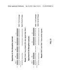 |
 |  |
 | 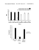 |
 |  |
 |  |
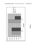 |  |
 |  |
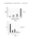 |  |
 |  |
 |  |
 |  |
 |  |
 |  |
 |  |
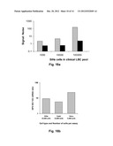 |  |
 |  |
 |  |
 | 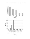 |
 |  |
 |  |
 |  |
 |  |
 | 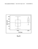 |
 |  |
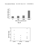 |  |
 |  |
 |  |
 | 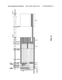 |
 |  |
 |  |
 |  |
 |  |
 | 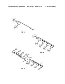 |
 | 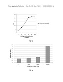 |
 |  |
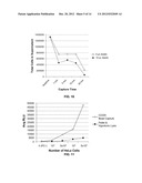 |  |
 |  |
 |  |
 |  |
 |  |
 |  |
 |
| Similar patent applications: | |
| Date | Title |
|---|---|
| 2011-11-24 | Transposon end compositions and methods for modifying nucleic acids |
| 2011-11-24 | Systems and methods for identifying a portion of a molecule |
| 2011-11-24 | Compositions and methods for diagnosis and treatment of epilepsy |
| 2011-11-24 | Methods, devices and kits for detecting or monitoring acute kidney injury |
| 2011-11-24 | Nucleic acid probes and methods for detecting plasmodium parasites |
| New patent applications in this class: | |
| Date | Title |
|---|---|
| 2022-05-05 | Method for diagnosing human t-cell leukemia virus type 1 (htlv-1) associated diseases |
| 2022-05-05 | Systems and methods for assay processing |
| 2022-05-05 | Rapid pathology/cytology without wash |
| 2019-05-16 | Biofluidic triggering system and method |
| 2019-05-16 | Method and system for detection of disease agents in blood |
| New patent applications from these inventors: | |
| Date | Title |
|---|---|
| 2016-04-21 | Primers and methods for the detection and discrimination of nucleic acids |
| 2015-04-02 | Thermophilic helicase dependent amplification technology with endpoint homogenous fluorescent detection |
| 2015-02-05 | Primers and methods for the detection and discrimination of nucleic acids |
| 2013-09-26 | Primers and methods for the detection and discrimination of nucleic acids |
| 2013-08-08 | Detection of nucleic acids by target-specific hybrid capture method |
| Top Inventors for class "Chemistry: molecular biology and microbiology" | |
| Rank | Inventor's name |
|---|---|
| 1 | Marshall Medoff |
| 2 | Anthony P. Burgard |
| 3 | Mark J. Burk |
| 4 | Robin E. Osterhout |
| 5 | Rangarajan Sampath |