Patent application title: PROMOTER FOR REGENERATION OF TENDON-BONE JUNCTION TISSUE OR LIGAMENT-BONE JUNCTION TISSUE
Inventors:
Katsuro Tomita (Ishikawa, JP)
Hiroyuki Tsuchiya (Ishikawa, JP)
Katsuhiko Kitaoka (Ishikawa, JP)
Junsuke Nakase (Ishikawa, JP)
Keigo Hanada (Osaka, JP)
Kunio Matsumoto (Ishikawa, JP)
Assignees:
KRINGLE PHARMA INC.
IPC8 Class: AA61K3818FI
USPC Class:
514 95
Class name: Peptide (e.g., protein, etc.) containing doai growth factor or derivative affecting or utilizing hepatocyte growth factor (hgf) or derivative
Publication date: 2011-12-22
Patent application number: 20110312887
Abstract:
An object of the present invention is to provide a drug for promoting the
regeneration of tendon-bone junction tissue or ligament-bone junction
tissue. The present invention relates to a promoter for regeneration of
tendon-bone junction tissue or ligament-bone junction tissue including
the following (1) or (2) as an active ingredient: (1) the following
(1-a), (1-b), or (1-c) (1-a) HGF protein, (1-b) a partial peptide of HGF
protein, the peptide having an effect of promoting regeneration of
tendon-bone junction tissue or ligament-bone junction tissue, (1-c) a
salt of (1-a) or (1-b); (2) DNA including the following (2-a), (2-b),
or (2-c), (2-a) DNA encoding HGF protein, (2-b) DNA encoding a partial
peptide of HGF protein, the peptide having an effect of promoting
regeneration of tendon-bone junction tissue or ligament-bone junction
tissue, (2-c) DNA encoding a protein or a peptide, the protein or the
peptide having an effect of promoting regeneration of tendon-bone
junction tissue or ligament-bone junction tissue, and the DNA hybridizing
with DNA comprising a base sequence complementary to (2-a) or (2-b) under
a stringent condition.Claims:
1. A promoter for regeneration of tendon-bone junction tissue or
ligament-bone junction tissue, comprising the following (1) or (2) as an
active ingredient: (1) the following (1-a), (1-b), or (1-c) (1-a) HGF
(Hepatocyte Growth Factor) protein, (1-b) a partial peptide of HGF
protein, the peptide having an effect of promoting regeneration of
tendon-bone junction tissue or ligament-bone junction tissue, (1-c) a
salt of (1-a) or (1-b); (2) DNA comprising the following (2-a), (2-b), or
(2-c), (2-a) DNA encoding HGF protein, (2-b) DNA encoding a partial
peptide of HGF protein, the peptide having an effect of promoting
regeneration of tendon-bone junction tissue or ligament-bone junction
tissue, (2-c) DNA encoding a protein or a peptide, the protein or the
peptide having an effect of promoting regeneration of tendon-bone
junction tissue or ligament-bone junction tissue, and the DNA hybridizing
with DNA comprising a base sequence complementary to (2-a) or (2-b) under
a stringent condition.
2. The promoter according to claim 1, wherein the active ingredient is the following (1-a), (1-b), or (1-c): (1-a) HGF protein, (1-b) a partial peptide of HGF protein, the peptide having an effect of promoting regeneration of tendon-bone junction tissue or ligament-bone junction tissue, (1-c) a salt of (1-a) or (1-b).
3. The promoter according to claim 1, wherein the HGF protein is the following (1-d) or (1-e): (1-d) a protein having an amino acid sequence represented by SEQ ID NO: 3 or 4, (1-e) a protein having an amino acid sequence at least 85% homologous to the amino acid sequence represented by SEQ ID NO: 3 or 4, and having an effect of promoting regeneration of tendon-bone junction tissue or ligament-bone junction tissue.
4. The promoter according to claim 1, wherein the active ingredient is DNA comprising the following (2-a), (2-b), or (2-c): (2-a) DNA encoding HGF protein, (2-b) DNA encoding a partial peptide of HGF protein, the peptide having an effect of promoting regeneration of tendon-bone junction tissue or ligament-bone junction tissue, (2-c) DNA encoding a protein or a peptide, the protein or the peptide having an effect of promoting regeneration of tendon-bone junction tissue or ligament-bone junction tissue, and the DNA hybridizing with DNA comprising a base sequence complementary to (2-a) or (2-b) under a stringent condition.
5. The promoter according to claim 1, wherein the DNA encoding HGF protein is the following (2-d) or (2-e): (2-d) DNA having a base sequence represented by SEQ ID NO: 1 or 2, (2-e) DNA that encodes a protein having an effect of promoting regeneration of tendon-bone junction tissue or ligament-bone junction tissue, and that hybridizes with DNA having a base sequence complementary to the base sequence represented by SEQ ID NO: 1 or 2 under a stringent condition.
6. The promoter according to claim 1, wherein the DNA is inserted into a herpes simplex virus type 1 (HSV-1) vector, a Sendai virus envelope (HVJ-E) vector, an adenovirus vector, or an adeno-associated virus vector.
7. The promoter according to claim 1, which is in a form of topical application.
8. Use of the following (1) or (2) for manufacturing a promoter for regeneration of tendon-bone junction tissue or ligament-bone junction tissue: (1) the following (1-a), (1-b), or (1-c), (1-a) HGF protein, (1-b) a partial peptide of HGF protein, the peptide having an effect of promoting regeneration of tendon-bone junction tissue or ligament-bone junction tissue, (1-c) a salt of (1-a) or (1-b), (2) DNA comprising the following (2-a), (2-b), or (2-c), (2-a) DNA encoding HGF protein, (2-b) DNA encoding a partial peptide of HGF protein, the peptide having an effect of promoting regeneration of tendon-bone junction tissue or ligament-bone junction tissue, (2-c) DNA encoding a protein or a peptide, the protein or the peptide having an effect of promoting regeneration of tendon-bone junction tissue or ligament-bone junction tissue, and the DNA hybridizing with DNA comprising a base sequence complementary to (2-a) or (2-b) under a stringent condition.
9. The following (1) or (2) for use in a method of promoting regeneration of tendon-bone junction tissue or ligament-bone junction tissue: (1) the following (1-a), (1-b), or (1-c), (1-a) HGF protein, (1-b) a partial peptide of HGF protein, the peptide having an effect of promoting regeneration of tendon-bone junction tissue or ligament-bone junction tissue, (1-c) a salt of (1-a) or (1-b), (2) DNA comprising the following (2-a), (2-b), or (2-c), (2-a) DNA encoding HGF protein, (2-b) DNA encoding a partial peptide of HGF protein, the peptide having an effect of promoting regeneration of tendon-bone junction tissue or ligament-bone junction tissue, (2-c) DNA encoding a protein or a peptide, the protein or the peptide having an effect of promoting regeneration of tendon-bone junction tissue or ligament-bone junction tissue, and the DNA hybridizing with DNA comprising a base sequence complementary to (2-a) or (2-b) under astringent condition.
10. A method for promoting regeneration of tendon-bone junction tissue or ligament-bone junction tissue comprising: administering the following (1) or (2) to a patient with tendon-bone junction tissue injury or ligament-bone junction tissue injury: (1) the following (1-a), (1-b), or (1-c), (1-a) HGF protein, (1-b) a partial peptide of HGF protein, the peptide having an effect of promoting regeneration of tendon-bone junction tissue or ligament-bone junction tissue, (1-c) a salt of (1-a) or (1-b), (2) DNA comprising the following (2-a), (2-b), or (2-c), (2-a) DNA encoding HGF protein, (2-b) DNA encoding a partial peptide of HGF protein, the peptide having an effect of promoting regeneration of tendon-bone junction tissue or ligament-bone junction tissue, (2-c) DNA encoding a protein or a peptide, the protein or the peptide having an effect of promoting regeneration of tendon-bone junction tissue or ligament-bone junction tissue, and the DNA hybridizing with DNA comprising a base sequence complementary to (2-a) or (2-b) under a stringent condition.
Description:
TECHNICAL FIELD
[0001] The present invention relates to a promoter for the regeneration of tendon-bone junction tissue or ligament-bone junction tissue.
BACKGROUND ART
[0002] Recently, injuries such as tendon or ligament ruptures in sports, traffic accidents, etc., are increasing. In regard to ligaments, since the anterior cruciate ligament (ACL) and the posterior cruciate ligament (PCL) are tissues that connect the femur to the tibia through a knee joint, when the ACL or PCL is ruptured, the stability of the knee joint cannot be maintained. Once ruptured, ligaments cannot be oversewn; therefore, in many cases, ligament reconstructive surgery is utilized to repair or replace injured ligaments.
[0003] For example, in ACL reconstructive surgery in which a damaged ACL is reconstructed by replacing it with a ligament graft, the patellar tendon with bone blocks is conventionally utilized as a graft. Such ACL reconstructive surgery allows rehabilitation to start early because the patellar tendon can function as a ligament when the bones attach to each other. However, the surgery also has disadvantages such as postoperative pain and muscle weakness.
[0004] In recent years, ACL reconstructive surgery that utilizes as a tendon graft, a hamstring tendon (knee flexor tendon, semitendinosus tendon, gracilis tendon), which is one of the muscle tendons used in bending the knees has been reported (for example, see Non Patent Literature 1). More specifically, the surgery includes the steps of forming a bone tunnel (hole) in the upper end of the tibia and the lower end of the femur, placing one end of the tendon graft in the femur side of the bone tunnel, and placing the other end of the tendon graft in the tibia side of the bone tunnel. Thus, the tendon graft runs between the femur and the tibia, and thereby functions. The tendon graft performs substantially the same function as the original ACL, thereby allowing the recovery of normal function in the knee joints. However, since the object of ACL reconstructive surgery is to reconstruct the normal ACL function and the kinematics of the knee joints, it is necessary to reconstruct the tendon-bone joint tissue in the part where the tendon graft comes in contact with the bone surface in the bone tunnel with enough strength.
[0005] For this reason, ACL reconstructive surgery has a disadvantage such that a long period of time is required before the tendon graft can function as a ligament.
[0006] As a drug for promoting the regeneration of tendon-bone junction tissue, BMP-2 (Bone Morphogenetic Protein-2: Non-Patent Literature 2), TGF-β1 (Transforming Growth Factor-β1: Non-Patent Literature 3), etc., are known; however, they have not yet been used in practice as a drug for promoting the regeneration of tendon-bone junction tissue.
[0007] HGF (Hepatocyte Growth Factor, hereinafter referred to as "HGF protein") was first identified as a potent mitogen for mature hepatocytes, and was determined by DNA cloning in 1989 (Non-Patent Literatures 4 and 5). Thereafter, HGF has been reported as having various effects such as angiogenesis, cell differentiation, cell proliferation, anti-apoptosis, etc., in various tissues. As to its effect in tendon tissue, it is reported that when HGF gene plasmid DNA is introduced into a wound made in the center of rat patellar tendon, the orientation of the developing collagen fibers in the wound is improved (Non-Patent Literature 6). However, tendon-bone junction tissue has a structure different from the tendon itself, and is a complicated tissue to adhere (fixate or fuse) a bone and a tendon, which are completely different from each other histologically. Non-Patent Literature 6 does not disclose or suggest the regeneration of such complicated tendon-bone junction tissue. Further, HGF protein is reported to inhibit the expression of TGF-β1, which, as mentioned previously, is known as a drug that promotes the regeneration of tendon-bone junction tissue (Non Patent Literature 7).
CITATION LIST
Non-Patent Literatures
[0008] NPL 1: S. A. Rodeo et al., The Journal of Bone and Joint Surgery; JBJS, 1993, Vol. 75-A, Issue 12, pp. 1795-1803. [0009] NPL 2: S. A. Rodeo et al., The American Journal of Sports Medicine, 1999, Vol. 27, pp. 476-488 [0010] NPL 3: Shuji Yamazaki et al., The Journal of Arthroscopic and Related Surgery; JBJS, 2005, Vol. 21, Issue 9, pp. 1034-1041 [0011] NPL 4: Toshikazu Nakamura et al., Biochemical and Biophysical Research Communications, 1984, Vol. 122, pp. 1450-1459 [0012] NPL 5: Toshikazu Nakamura et al., Nature, 1989, Vol. 342, pp. 440-443 [0013] NPL 6: Takashi Natsu-ume et al., Journal of the Japanese Orthopaedic Association, 1998, Vol. 72, Issue 8, 51254 [0014] NPL 7: Kunio Matsumoto and Toshikazu Nakamura, Biochemical and Biophysical Research Communications, 1997, Vol. 239, pp. 639-644
SUMMARY OF INVENTION
Technical Problem
[0015] An object of the present invention is to provide a drug for promoting the regeneration of tendon-bone junction tissue or ligament-bone junction tissue. Particularly, an object of the present invention is to provide a drug for promoting the regeneration of tendon-bone junction tissue or ligament-bone junction tissue at a contact site between a bone and a tendon or ligament graft after ACL reconstructive surgery etc., or in a space between a bone and a tendon or ligament graft after ACL reconstructive surgery etc.
Solution to Problem
[0016] The present inventors conducted extensive research to solve the above problems. Consequently, they found that HGF protein has an effect of promoting the regeneration of tendon-bone junction tissue or ligament-bone junction tissue, and accomplished the invention.
[0017] Specifically, the present invention relates to a promoter for the regeneration of tendon-bone junction tissue or ligament-bone junction tissue.
1. A promoter for regeneration of tendon-bone junction tissue or ligament-bone junction tissue, comprising the following (1) or (2) as an active ingredient:
[0018] (1) the following (1-a), (1-b), or (1-c) [0019] (1-a) HGF (Hepatocyte Growth Factor) protein, [0020] (1-b) a partial peptide of HGF protein, the peptide having an effect of promoting regeneration of tendon-bone junction tissue or ligament-bone junction tissue, [0021] (1-c) a salt of (1-a) or (1-b);
[0022] (2) DNA comprising the following (2-a), (2-b), or (2-c), [0023] (2-a) DNA encoding HGF protein, [0024] (2-b) DNA encoding a partial peptide of HGF protein, the peptide having an effect of promoting regeneration of tendon-bone junction tissue or ligament-bone junction tissue, (2-c) DNA encoding a protein or a peptide, the protein or the peptide having an effect of promoting regeneration of tendon-bone junction tissue or ligament-bone junction tissue, and the DNA hybridizing with DNA comprising a base sequence complementary to (2-a) or (2-b) under a stringent condition. 2. The promoter according to Item 1, wherein the active ingredient is the following (1-a), (1-b), or (1-c): [0025] (1-a) HGF protein, [0026] (1-b) a partial peptide of HGF protein, the peptide having an effect of promoting regeneration of tendon-bone junction tissue or ligament-bone junction tissue, (1-c) a salt of (1-a) or (1-b). 3. The promoter according to Item 1 or 2, wherein the HGF protein is the following (1-d) or (1-e): [0027] (1-d) a protein having an amino acid sequence represented by SEQ ID NO: 3 or 4, [0028] (1-e) a protein having an amino acid sequence that is substantially equal to an amino acid sequence represented by SEQ ID NO: 3 or 4, and having an effect of promoting regeneration of tendon-bone junction tissue or ligament-bone junction tissue.
[0029] Herein, an example of protein (1-e) includes "a protein that has an amino acid sequence at least 85% homologous to an amino acid sequence represented by SEQ ID NO: 3 or 4, and has an effect of promoting the regeneration of tendon-bone junction tissue or ligament-bone junction tissue".
4. The promoter according to Item 1, wherein the active ingredient is DNA comprising the following (2-a), (2-b), or (2-c): [0030] (2-a) DNA encoding HGF protein, [0031] (2-b) DNA encoding a partial peptide of HGF protein, the peptide having an effect of promoting regeneration of tendon-bone junction tissue or ligament-bone junction tissue, [0032] (2-c) DNA encoding a protein or a peptide, the protein or the peptide having an effect of promoting regeneration of tendon-bone junction tissue or ligament-bone junction tissue, and the DNA hybridizing with DNA comprising a base sequence complementary to (2-a) or (2-b) under a stringent condition. 5. The promoter according to Item 1 or 4, wherein the DNA encoding HGF protein is the following (2-d) or (2-e): [0033] (2-d) DNA having a base sequence represented by SEQ ID NO: 1 or 2, [0034] (2-e) DNA that encodes a protein having an effect of promoting regeneration of tendon-bone junction tissue or ligament-bone junction tissue, and that hybridizes with DNA having a base sequence complementary to the base sequence represented by SEQ ID NO: 1 or 2 under a stringent condition. 6. The promoter according to any one of Items 1, 4, and 5, wherein the DNA is inserted into a herpes simplex virus type 1 (HSV-1) vector, a Sendai virus envelope (HVJ-E) vector, an adenovirus vector, or an adeno-associated virus vector. 7. The promoter according to any one of Items 1 to 6, which is in a form of topical application.
[0035] Further, the present invention relates to a use of HGF protein, or DNA encoding HGF protein.
8. Use of the following (1) or (2) for manufacturing a promoter for regeneration of tendon-bone junction tissue or ligament-bone junction tissue:
[0036] (1) the following (1-a), (1-b), or (1-c), [0037] (1-a) HGF protein, [0038] (1-b) a partial peptide of HGF protein, the peptide having an effect of promoting regeneration of tendon-bone junction tissue or ligament-bone junction tissue, [0039] (1-c) a salt of (1-a) or (1-b),
[0040] (2) DNA comprising the following (2-a), (2-h), or (2-c), [0041] (2-a) DNA encoding HGF protein, [0042] (2-b) DNA encoding a partial peptide of HGF protein, the peptide having an effect of promoting regeneration of tendon-bone junction tissue or ligament-bone junction tissue, [0043] (2-c) DNA encoding a protein or a peptide, the protein or the peptide having an effect of promoting regeneration of tendon-bone junction tissue or ligament-bone junction tissue, and the DNA hybridizing with DNA comprising a base sequence complementary to (2-a) or (2-b) under a stringent condition. 9. The following (1) or (2) for use in a method of promoting regeneration of tendon-bone junction tissue or ligament-bone junction tissue:
[0044] (1) the following (1-a), (1-b), or (1-c), [0045] (1-a) HGF protein, [0046] (1-b) a partial peptide of HGF protein, the peptide having an effect of promoting regeneration of tendon-bone junction tissue or ligament-bone junction tissue, [0047] (1-c) a salt of (1-a) or (1-b),
[0048] (2) DNA comprising the following (2-a), (2-b), or (2-c), [0049] (2-a) DNA encoding HGF protein, [0050] (2-b) DNA encoding a partial peptide of HGF protein, the peptide having an effect of promoting regeneration of tendon-bone junction tissue or ligament-bone junction tissue, [0051] (2-c) DNA encoding a protein or a peptide, the protein or the peptide having an effect of promoting regeneration of tendon-bone junction tissue or ligament-bone junction tissue, and the DNA hybridizing with DNA comprising a base sequence complementary to (2-a) or (2-b) under a stringent condition. 10. A method for promoting regeneration of tendon-bone junction tissue or ligament-bone junction tissue comprising: administering the following (1) or (2) to a patient with tendon-bone junction tissue injury or ligament-bone junction tissue injury:
[0052] (1) the following (1-a), (1-b), or (1-c), [0053] (1-a) HGF protein, [0054] (1-b) a partial peptide of HGF protein, the peptide having an effect of promoting regeneration of tendon-bone junction tissue or ligament-bone junction tissue, [0055] (1-c) a salt of (1-a) or (1-b),
[0056] (2) DNA comprising the following (2-a), (2-b), or (2-c), [0057] (2-a) DNA encoding HGF protein, [0058] (2-b) DNA encoding a partial peptide of HGF protein, the peptide having an effect of promoting regeneration of tendon-bone junction tissue or ligament-bone junction tissue, [0059] (2-c) DNA encoding a protein or a peptide, the protein or the peptide having an effect of promoting regeneration of tendon-bone junction tissue or ligament-bone junction tissue, and the DNA hybridizing with DNA comprising a base sequence complementary to (2-a) or (2-b) under a stringent condition.
Advantageous Effects of Invention
[0060] The promoter for the regeneration of tendon-bone junction tissue or ligament-bone junction tissue of the present invention can enhance the regeneration of the tendon-bone junction tissue or the ligament-bone junction tissue at a site in which a bone is in contact with a ligament or tendon graft separated from the bone, or in a space between the bone and the ligament or tendon graft. Thus, the present invention can promote adhesion (fixation or fusion) between a bone and a tendon or ligament that is separated from the bone by injury, rupture, etc.
[0061] Further, since the present invention can enhance the regeneration of tendon-bone junction tissue or ligament-bone junction tissue in ligament reconstructive surgery of limb joints (e.g., knee, ankle, elbow, and shoulder) including ACL reconstructive surgery, PCL reconstructive surgery, collateral ligament reconstructive surgery, MPFL (medial patellofemoral ligament) reconstructive surgery, ankle lateral ligament reconstructive surgery, ulnar ligament reconstructive surgery, and tendon repair surgery etc., adhesion (fixation or fusion) between a bone and a tendon or ligament graft can be enhanced.
[0062] Therefore, the present invention results in a faster recovery of patients after ligament reconstructive surgery, who have unstable joints because of ligament rupture etc., and thereby have difficulty in sporting activities and daily life.
DESCRIPTION OF EMBODIMENTS
HGF Protein
[0063] As described above, "HGF protein" has been identified as a potent mitogen for adult hepatocytes, and is called "Hepatocyte Growth Factor" (see, for example, Non-patent Literatures 4 and 5). In addition to HGF, it is referred to as SF (scatter factor), TCF (Tumor Cytotoxic Factor), etc.
[0064] "HGF protein" as used herein is a known substance, and can be prepared by any method as long as it is purified enough to be used as a medicament.
[0065] HGF protein can be obtained by culturing primary cultured cells or cell lines capable of producing HGF protein, followed by separation of the HGF protein from culture supernatant etc., and purification. Alternatively, the protein can be obtained by genetic engineering techniques, for example, by inserting the gene encoding HGF protein into an appropriate vector, introducing the vector into an appropriate host cell to be transformed, and isolating a desired recombinant HGF protein from the culture supernatant of the transformant, etc. (see, for example, Japanese Unexamined Patent Publication No. H5-111382, and Biochem. Biophys. Res. Commun., 1989, Vol. 163, p. 967). The above-mentioned host cell is not particularly limited and includes various host cells conventionally used in genetic engineering techniques, for example, Escherichia coli, yeast, animal cells, and the like.
[0066] In the present invention, a preferable example of HGF protein is a protein obtained from a gene encoding human-derived HGF (hHGF). Preferable examples of the gene encoding hHGF include DNA having the base sequence represented by SEQ ID NO: 1 or 2.
[0067] Specific examples of such HGF protein include, according to recombinant DNA techniques, the HGF protein represented by SEQ ID NO: 3 or 5 produced by a cell into which DNA having the base sequence represented by SEQ ID NO: 1 has been introduced, and the HGF protein represented by SEQ ID NO: 4 or 6 produced by a cell into which DNA having the base sequence represented by SEQ ID NO: 2 has been introduced.
[0068] The HGF protein represented by SEQ ID NOs. 3 to 6 is a native HGF protein of human origin having mitogen activity and motogen activity as HGF. Such HGF protein is registered, for example, as Accession No. P14210 (SEQ ID NO: 3) or Accession No. NP--001010932 (SEQ ID NO: 4) in the NCBI database (NCBI-GenBank Flat File Release 164.0), or the like. HGF protein having the amino acid sequence represented by SEQ ID NO: 4 is a five amino acid-deleted HGF protein, in which five amino acid residues, i.e., from the 161st to the 165th residues in the amino acid sequence represented by SEQ ID NO: 3 are deleted. In addition, the aforementioned native HGF protein is glycoprotein. For example, in the HGF protein represented by Accession No. NP--001010932 (SEQ ID NO: 4), a sugar chain is added to Asn 289, Asn 397, Thr 471, Asn 561, and Asn 648.
[0069] The amino acid sequence represented by SEQ ID NO: 5 or 6 is an amino acid sequence of an adult protein obtained by cleaving the 1st to 31st amino acid region (signal sequence) from the N terminus in the amino acid sequence represented by SEQ ID NO: 3 or 4.
[0070] As long as the HGF protein used in the present invention has an effect of promoting the regeneration of tendon-bone junction tissue and ligament-bone junction tissue, one or more ("more" means, for example, 2 to 35 amino acids, preferably 2 to 20 amino acids, and more preferably 2 to 10 amino acids; the same shall apply hereinafter) amino acids in the amino acid sequence represented by SEQ ID NO: 3 or 4 may be deleted, substituted, inserted, or added, and similarly, its sugar chain may be deleted, substituted, inserted, or added. Such HGF protein can be produced by known technical methods such as genetic engineering techniques, site specific mutagenesis, etc. An amino acid to be inserted, substituted, or added may be an unnatural amino acid other than 20 kinds of natural amino acids. The unnatural amino acid may be any compound as long as it has an amino group and a carboxyl group, and for example, γ-amino butyric acid etc., is included. In addition, the HGF protein having the amino acid sequence represented by SEQ ID NO: 4 is, as described above, a five amino acid-deleted type HGF protein, in which five amino acid residues in the amino acid sequence represented by SEQ ID NO: 3 are deleted.
[0071] The HGF protein used in the present invention may have an amino acid sequence at least 85% homologous to the amino acid sequence represented by SEQ ID NO: 3 or 4 provided that the protein has an effect of promoting the regeneration of tendon-bone junction tissue or ligament-bone junction tissue. HGF protein having an amino acid sequence at least 90% homologous to the amino acid sequence represented by SEQ ID NO: 3 or 4 is preferred, and HGF protein having an amino acid sequence at least 95% homologous to the amino acid sequence represented by SEQ ID NO: 3 or 4 is more preferred. The amino acid sequence represented by SEQ ID NO: 5 is 95.7% and 96.4% homologous to the amino acid sequence represented by SEQ ID NO: 3 and 4, respectively; and the amino acid sequence represented by SEQ ID NO: 6 is 95.1% and 95.7% homologous to the amino acid sequence represented by SEQ ID NO: 3 and 4, respectively. Herein, "homologous to" indicates the degree of identity between the amino acid residues that form each of the sequences when the primary structures (amino acid sequences) of the proteins are compared.
[0072] Other examples of HGF protein having an amino acid sequence that is highly homologous to the amino acid sequence represented by SEQ ID NO: 3 or 4 include human-derived HGF registered as Accession No. BAA14348 or AAC71655 in the NCBI database.
[0073] As long as the HGF protein used in the present invention has an effect of promoting the regeneration of tendon-bone junction tissue or ligament-bone junction tissue, the signal sequence comprising the 1st to 31st amino acid region in the amino acid sequence represented by SEQ ID NO: 3 or 4 may be replaced with a signal sequence of another protein. Examples of the signal sequence include a signal sequence of human serum albumin, interferon, human amylase, etc.
[0074] As long as the HGF protein used in the present invention has an effect of promoting the regeneration of tendon-bone junction tissue or ligament-bone junction tissue, it may be a protein produced by a cell having DNA that hybridizes with DNA comprising a base sequence complementary to the base sequence represented by SEQ ID NO: 1 or 2 under a stringent condition.
[0075] The stringent condition is as follows.
[0076] Hybridization is carried out at about 65° C. in the presence of about 0.7 to 1.0 M sodium chloride, and then washing is conducted at about 65° C. in SSC solution at about a 0.1- to 2-fold concentration (a one fold concentration of SSC solution consists of 150 mM sodium chloride and 15 mM sodium citrate).
[0077] An example of a method for producing the HGF protein of the present invention using a cell that comprises a gene coding the HGF protein, for example DNA having the base sequence represented by SEQ ID NO: 1 or 2, or DNA that hybridizes with DNA having a base sequence complementary to the aforementioned DNA under a stringent condition is a method in which primary cultured cells or cell lines having the aforementioned DNA are cultured, followed by separation of a desired HGF protein from the culture supernatant etc., and purification. Alternatively, the protein can be obtained by genetic engineering techniques, for example, by inserting the aforementioned DNA into an appropriate vector, introducing the vector into an appropriate host cell to be transformed, and isolating a desired HGF protein (recombinant protein) from the culture supernatant of the transformant (see, for example, Japanese Unexamined Patent Publication No. H5-111382, Japanese Unexamined Patent Publication No. H11-1499, and Biochem. Biophys. Res. Commun., 1989, Vol. 163, p. 967).
[0078] The aforementioned host cell is not particularly limited and various host cells conventionally used in genetic engineering techniques, for example, Escherichia coli, yeast, animal cells, and the like can be used. Since native HGF protein is a glycoprotein, it is preferred to use an animal cell as a host cell to produce glycoprotein as in the case of using a cell.
[0079] Examples of the animal cell include CHO cells, COS cells, mouse L cells, mouse C127 cells, mouse FM3A2 cells, and the like. The expression vector is introduced into an animal cell by transfection methods, microinjection methods, etc. Of these, the most commonly used method is a phosphoric-acid calcium method. For the animal cell that is transformed by transfection, floating cultivation, or adhesion cultivation can be used in accordance with an ordinary method. As a medium, MEM, RPMI 1640, and the like are commonly used.
[0080] Whether the HGF protein has an effect of promoting the regeneration of tendon-bone junction tissue or ligament-bone junction tissue can be evaluated as follows.
[0081] For example, at least a bone and a tendon or ligament separated from the bone are kept immobilized using a ligament fastener etc. The target HGF protein is then reacted according to the method described in the Example below. Compared to the case where nothing is reacted (control), if effective regeneration of tendon-bone junction tissue or ligament-bone junction tissue is observed when the HGF protein is reacted, the HGF protein is considered to have an effect of promoting the regeneration of tendon-bone junction tissue or ligament-bone junction tissue.
[0082] The presence or absence of glycosylation, and the number of glycosylation sites are not particularly limited as long as the HGF protein used in the present invention has an effect of enhancing bone elongation.
[0083] Specifically, the HGF protein may be protein in which naturally occurring sugar chains (one or more) are deleted, substituted, inserted or added. Examples of the HGF protein in which a sugar chain is deleted, substituted, inserted, or added include HGF protein in which a sugar chain attached to native HGF protein has been deleted by treatment with an enzyme or the like, HGF protein in which the amino acid sequence at the glycosylation site has been mutated so as to prevent glycosylation, or HGF protein in which the amino acid sequence has been mutated so that glycosylation occurs at any other site than the naturally-occurring glycosylation site. Specific examples of such HGF protein include HGF protein that is designed to prevent glycosylation by replacing Asn289, Asn397, Thr471, Asn561, and Asn648 with Gln289, Gln397, Gly471, Gln561, and Gln648, respectively in human HGF protein registered as Accession No. NP--001010932 in the NCBI database (NCBI-GenBank Flat File Release 164.0) (Fukuta, K. et al., Biochemical Journal, 2005, Vol. 388, pp. 555-562).
[0084] The HGF protein used in the present invention has any one of a carboxyl group (--COOH), a carboxylate (--COOM (M represents a metal)), an amide (--CONH2), or an ester (--COOR) in the C-terminus. Herein, R in the ester may be a C1-6 alkyl group such as methyl, ethyl, n-propyl, isopropyl, and n-butyl; a C3-8 cycloalkyl group such as cyclopentyl and cyclohexyl; a C6-12 aryl group such as phenyl and α-naphthyl; a C7-14 aralkyl group such as a phenyl-(C1-2 alkyl) group including benzyl and phenethyl, and an α-naphthyl-(C1-2 alkyl) group including α-naphthylmethyl; a C2-6 alkanoylmethyl group such as acetyloxymethyl and pivaloyloxymethyl; etc.
[0085] When the HGF protein used in the present invention has a carboxyl group or a carboxylate in any other site than the C-terminus, the carboxyl group or carboxylate may be amidated or esterified. Such HGF protein is also included in the HGF protein used in the present invention. In this case, examples of the ester include the above-mentioned examples of the ester in the C-terminus.
[0086] The HGF protein used in the present invention includes the above-mentioned protein having an amino group of the N-terminal methionine residue protected with a protecting group (for example, a C1-6 acyl group including a formyl group and a C2-6 alkanoyl group such as acetyl, etc.), the above-mentioned protein having a glutamyl group pyroglutamated after being produced by cleaving the N-terminal side in vivo, the above-mentioned protein having a side chain reactive group of the amino acid in a molecule (for example, --OH, --SH, an amino group, an imidazolyl group, an indolyl group, a guanidino group, etc.) protected with an appropriate protecting group (for example, a C1-6 acyl group including a formyl group and a C2-6 alkanoyl group (e.g., acetyl), etc.), and a complex protein such as a glycoprotein, which is produced by glycosylating the above-mentioned protein.
[0087] As the HGF protein used in the present invention, the above-mentioned protein of human origin is suitably used for human application. In addition, HGF protein derived from mammals other than humans, such as monkeys, cattle, horses, pigs, sheep, dogs, cats, rats, mice, rabbits, hamsters, guinea pigs, and chimpanzees, may be used.
[0088] Such HGF protein includes, but is not limited to, HGF protein registered in the NCBI database, for example, mouse HGF protein (for example, registered as Accession No. AAB31855, NP--034557, BAA01065, BAA01064, or the like), rat HGF protein (for example, registered as Accession No. NP--058713, bovine HGF protein (for example, registered as Accession No. NP--001026921, BAD02475, or the like), feline HGF protein (for example, registered as Accession No. NP--001009830, BAC10545, BAB21499, or the like), canine HGF protein (for example, registered as Accession No. NP--001002964, BAC57560, or the like), chimpanzee HGF protein (for example, registered as Accession No. XP 519174 or the like), etc.
[0089] When the HGF protein is used as the active ingredient of the promoter of the present invention, the HGF protein can be produced by any methods as long as it is purified enough to be used as a medicament. The purification methods are not limited, and examples thereof include column chromatography using heparin sepharose, hydroxyapatite, etc.
[0090] In the promoter of the present invention, HGF protein may be used alone, or as a mixed protein with various proteins as long as the effect of promoting the regeneration of tendon-bone junction tissue or ligament-bone junction tissue is not impaired.
HGF Partial Peptide Having Effect of Promoting Regeneration of Tendon-Bone Junction Tissue or Ligament-Bone Junction Tissue
[0091] The partial peptide of HGF protein used in the present invention (sometimes hereinafter abbreviated as HGF partial peptide), the peptide having an effect of promoting the regeneration of tendon-bone junction tissue or ligament-bone junction tissue is not limited as long as it is a partial peptide of the aforementioned HGF protein and has an effect of promoting the regeneration of tendon-bone junction tissue or ligament-bone junction tissue as in the aforementioned HGF protein. In the present invention, the HGF partial peptide has an amino acid sequence that constitutes the above-mentioned HGF protein and comprises about 20 amino acids or more, preferably about 50 amino acids or more, and more preferably about 100 amino acids or more. Specifically, examples of such an HGF partial peptide include a peptide having an amino acid sequence between the 32nd and the 210th residues from the N-terminus in the human HGF amino acid sequence represented by SEQ ID NO: 3 (an amino acid sequence from the N-terminal hairpin loop to the 1st kringle domain of HGF), a peptide having the amino acid sequence between the 32nd and the 288th residues from the N-terminus in the human HGF amino acid sequence represented by SEQ ID NO: 3 (an amino acid sequence from the N-terminal hairpin loop to the 2nd kringle domain of HGF), and the like.
[0092] Examples of the HGF partial peptide of the present invention include a partial peptide having an amino acid sequence at least about 80%, preferably at least about 90%, and more preferably at least about 95% homologous to the amino acid sequence of the aforementioned HGF partial peptide, and having an effect of promoting the regeneration of tendon-bone junction tissue or ligament-bone junction tissue.
[0093] The HGF partial peptide of the present invention has any one of a carboxyl group (--COOH), a carboxylate (--COOM (M is the same as defined above)), an amide (--CONH2), or an ester (--COOR(R is the same as defined above)) in the C-terminus. Further, as in the above-mentioned HGF protein, the HGF partial peptide includes a peptide having an amino group of the N-terminal methionine residue protected with a protecting group, a peptide having a glutamyl group pyroglutamated after being produced by cleaving the N-terminal side in vivo, a peptide having a side chain substituent group of the amino acid within a molecule protected with an appropriate protecting group, and a complex protein such as a glycoprotein, which is produced by glycosylating the above-mentioned peptide.
[0094] Regarding the HGF partial peptide, "an effect of promoting the regeneration of tendon-bone junction tissue or ligament-bone junction tissue" can be evaluated in the same manner as described above. The HGF partial peptide of the present invention can be prepared by known peptide synthesis methods or by cleaving HGF protein with an appropriate peptidase. A peptide synthesis method may be, for example, a solid- or liquid-phase synthesis method. Namely, the desired peptide can be prepared by condensing a partial peptide or an amino acid that is capable of constituting HGF protein and optionally having a protecting group with a remaining part optionally having a protecting group; and then by removing the protecting group, if any, from the product. Known condensation or protecting group removal methods include those described in, for example, M. Bodanszky and M. A. Ondetti, Peptide Synthesis, Interscience Publishers, New York (1966); Schroeder and Luebke, The Peptide, Academic Press, New York (1965); etc. After the reaction, HGF partial peptide can be separated and purified by a combination of ordinal purification methods such as solvent extraction, distillation, column chromatography, liquid chromatography, crystallization, or recrystallization.
Salt of HGF Protein or Partial Peptide
[0095] The HGF protein or its partial peptide of the present invention may be in a free form (loose body), or the form of a salt.
[0096] Examples of the salt of HGF protein or its partial peptide used in the present invention include salts that are physiologically acceptable with an acid or base. Particularly, physiologically acceptable acid adduct salts are preferred. Examples of such salts include salts with inorganic acid (such as hydrochloric acid, phosphoric acid, hydrobromic acid, sulfuric acid, and the like) and salts with organic acid (such as acetic acid, formic acid, propionic acid, fumaric acid, maleic acid, succinic acid, tartaric acid, citric acid, malic acid, oxalic acid, benzoic acid, methanesulfonic acid, benzenesulfonic acid, and the like).
[0097] In the production, when the HGF protein or the HGF partial peptide of the present invention is used in a free form, it can be converted into an appropriate salt by a known method. Meanwhile, when the HGF protein or HGF partial peptide is obtained in the form of a salt, it can be converted into a free form by a known method.
DNA Encoding HGF Protein
[0098] Herein, "DNA encoding HGF protein" refers to DNA capable of expressing the HGF protein. Preferable examples of DNA that contains DNA encoding HGF protein include DNA encoding human-derived HGF protein described in, for example, Nature, Vol. 342, p. 440 (1989); Japanese Patent No. 2777678; Biochem. Biophys. Res. Commun., 1989, Vol. 163, pp. 967-973; and Proc. Natl. Acad. Sci. U.S.A., 1991, Vol. 88 (16), pp. 7001-7005, and registered as Accession No. M69718, M73240, AC004960, AY246560, M29145, M73240, or the like in GenBank/EMBL/DDBJ.
[0099] As DNA encoding HGF protein used in the present invention, the above-mentioned DNA of human origin is suitably used for human application. In addition, DNA encoding HGF protein derived from mammals other than humans, such as monkeys, cattle, horses, pigs, sheep, dogs, cats, rats, mice, rabbits, hamsters, guinea pigs, and chimpanzees may be used.
[0100] Such DNA encoding HGF protein includes, but is not limited to, those registered in the NCBI database, for example, DNA encoding mouse HGF protein (for example, registered as Accession Nos. 571816, NM--010427, D10213, D10212, or the like), DNA encoding rat HGF protein (for example, registered as Accession No. NM--017017 or the like), DNA encoding bovine HGF protein (for example, registered as Accession Nos. NM--001031751, AB110822, or the like), DNA encoding feline HGF protein (for example, registered as Accession Nos. NM--001009830, AB080187, AB046610, or the like), DNA encoding canine HGF protein (for example, registered as Accession Nos. NM--001002964, AB090353, or the like), and DNA encoding chimpanzee HGF protein (for example, registered as Accession No. XM 519174 or the like).
[0101] Specific examples of DNA encoding HGF protein include DNA having the base sequence represented by SEQ ID NO: 1 or 2. The base sequence represented by SEQ ID NO: 1 corresponds to the region from the 73rd to the 2259th of the base sequence registered as Accession No. M60718, and also corresponds to DNA encoding HGF protein having the amino acid sequence represented by SEQ ID NO: 3. In recombinant DNA techniques, the HGF protein (SEQ ID NO: 3) that is expressed and produced in a cell is converted into adult HGF protein having the amino acid sequence represented by SEQ ID NO: 5 because the signal sequence is cleaved when the HGF protein is secreted outside the cell. Accordingly, DNA having the base sequence represented by SEQ ID NO: 1 corresponds to DNA encoding (producing) HGF protein having the amino acid sequence represented by SEQ ID NO: 5.
[0102] The base sequence represented by SEQ ID NO: 2 corresponds to the region from the 66th to the 2237th of the base sequence registered as Accession No. M73240, and corresponds to DNA encoding HGF protein comprising the amino acid sequence represented by SEQ ID NO: 4. Similarly, in recombinant DNA techniques, the HGF protein (SEQ ID NO: 4) is converted into adult HGF protein having the amino acid sequence represented by SEQ ID NO: 6 because the signal sequence is cleaved when the HGF protein is secreted outside the cell. Accordingly, DNA having the base sequence represented by SEQ ID NO: 2 corresponds to DNA encoding (producing) HGF protein having the amino acid sequence represented by SEQ ID NO: 6.
[0103] DNA encoding HGF protein is not limited to the aforementioned DNA, and any DNA encoding protein having an effect of promoting the regeneration of tendon-bone junction tissue or ligament-bone junction tissue can be used as the DNA encoding HGF protein of the present invention. Herein, "the effect of promoting the regeneration of tendon-bone junction tissue or ligament-bone junction tissue" can be evaluated in the same manner as described above.
[0104] Such DNA is not particularly limited, but examples include DNA that has a base sequence at least about 80%, preferably at least about 85%, more preferably at least about 90%, and even more preferably at least about 95% homologous to the base sequence of the aforementioned DNA encoding HGF protein, and encodes a protein having an effect of promoting the regeneration of tendon-bone junction tissue or ligament-bone junction tissue.
[0105] The DNA encoding HGF protein can be easily obtained by a general hybridization method or PCR method using a cDNA library containing the DNA. Specifically, the DNA can be obtained with reference to Molecular Cloning, A laboratory Manual, Third Edition (J. Sambrook et al., Cold Spring Harbor Lab. Press, 2001; hereinafter abbreviated as Third Edition Molecular Cloning) and other basic manuals.
[0106] Examples of the cDNA library comprising HGF protein-encoding DNA include a human liver cDNA library, a human spleen cDNA library, a human placentas cDNA library, and the like. These libraries can be commercially available from Clonetech, Co., Ltd. or the like. Other than the above, cDNA libraries produced in compliance with a known method by cell strains or tissue materials that express HGF protein can be used. According to the method described in "Third Edition Molecular Cloning", a λ phage in which such cDNA has been incorporated infects Escherichia coli for culture. The plaque formed is then subjected to plaque hybridization or PCR using, as a probe, an oligonucleotide that is produced by a base sequence based on the partial amino acid sequence of HGF protein, thereby yielding a desired DNA encoding HGF protein.
[0107] In the present invention, RNA encoding HGF protein can also be used as long as the HGF protein can be expressed by reverse transcriptase. Examples of the RNA include RNA obtained by RT-PCR amplification of mRNA fractions harvested from cells or tissues, which is within the scope of the present invention. The RNA also can be obtained by known methods.
[0108] As described below, the DNA encoding HGF protein is administered to a patient in the form of a recombinant expression vector in which DNA is inserted. Examples of the expression vector include, but are not limited to, naked plasmids, and DNA or RNA viruses such as detoxified retroviruses, adenoviruses, adeno-associated viruses, herpes viruses (herpes simplex virus type 1, etc.), vaccinia viruses, poxviruses, polioviruses, sindbis viruses, Sendai viruses, SV40, and human immunodeficiency viruses (HIV). Of these, herpes simplex virus type 1 (HSV-1) vectors, Sendai virus envelope (HVJ-E) vectors, adenovirus vectors, adeno-associated virus (AAV) vectors, etc., are preferred.
DNA Encoding Protein Having Effect of Promoting Regeneration of Tendon-Bone Junction Tissue or Ligament-Bone Junction Tissue, and Hybridizing with DNA Comprising Base Sequence Complementary to DNA Encoding HGF Protein Under Stringent Condition
[0109] The promoter for the regeneration of tendon-bone junction tissue or ligament-bone junction tissue of the present invention may contain DNA coding protein that has an effect of promoting the regeneration of tendon-bone junction tissue or ligament-bone junction tissue as in the HGF protein, and hybridizing with DNA comprising a base sequence complementary to the DNA encoding HGF protein under a stringent condition.
[0110] Preferred examples of such DNA include DNA coding protein that has an effect of promoting the regeneration of tendon-bone junction tissue or ligament-bone junction tissue, and hybridizing with DNA comprising a base sequence complementary to the DNA having the base sequence represented by SEQ ID NO: 1 or 2 under a stringent condition.
[0111] Herein, "effect of promoting the regeneration of tendon-bone junction tissue or ligament-bone junction tissue" can be evaluated in the same manner as described above.
[0112] "The DNA that hybridizes under a stringent condition with DNA comprising a base sequence complementary to the DNA encoding HGF protein, or with DNA comprising a base sequence complementary to the DNA having the base sequence represented by SEQ ID NO: 1 or 2" indicates DNA obtained by using a partial sequence of DNA comprising a base sequence complementary to the DNA encoding HGF protein, or with DNA comprising a base sequence complementary to the DNA having the base sequence represented by SEQ ID NO: 1 or 2 as a probe, and then carrying out colony hybridization, plaque hybridization, or southern blot hybridization. Specifically, DNA identified by the following procedures is included. A filter on which colony- or plaque-derived DNA has been immobilized is subjected to, using the probe, hybridization at about 65° C. in the presence of about 0.7 to 1.0M sodium chloride, and then the filter is washed at about 65° C. in SSC solution at about 0.1- to 2-fold concentration (a one fold concentration of SSC solution consists of 150 mM sodium chloride and 15 mM sodium citrate). The stringent condition will be the same hereinafter.
[0113] Specifically, DNA that hybridizes under such a stringent condition includes DNA having a base sequence at least about 80%, preferably at least about 85%, more preferably at least about 90%, and even more preferably at least about 95% homologous to the base sequence of the aforementioned DNA encoding HGF protein. More specifically, DNA that hybridizes with DNA comprising a base sequence complementary to the DNA having the base sequence represented by SEQ ID NO: 1 or 2 under a stringent condition includes DNA having a base sequence at least about 80%, preferably at least about 85%, more preferably at least about 90%, and even more preferably at least about 95% homologous to the base sequence represented by SEQ ID NO: 1 or 2.
[0114] Hybridization can be performed according to known methods, for example the method described in Molecular Cloning, Third Edition. When a commercially available library is used, hybridization also can be performed in compliance with the method described in the attached instruction manual.
DNA Encoding HGF Partial Peptide Having Effect of Promoting Regeneration of Tendon-Bone Junction Tissue or Ligament-Bone Junction Tissue
[0115] The promoter for the regeneration of tendon-bone junction tissue or ligament-bone junction tissue of the present invention may include DNA encoding HGF partial peptide that has an effect of promoting the regeneration of tendon-bone junction tissue or ligament-bone junction tissue. Herein, "effect of promoting the regeneration of tendon-bone junction tissue or ligament-bone junction tissue" can be evaluated in the same manner as described above.
[0116] The DNA is not limited as long as it encodes a peptide that has a base sequence encoding the partial peptide and has an effect of promoting the regeneration of tendon-bone junction tissue or ligament-bone junction tissue. Specifically, examples of the DNA include DNA that has a partial base sequence of DNA having the base sequence represented by SEQ ID NO: 1 or 2, and encodes a peptide having an effect of promoting the regeneration of tendon-bone junction tissue or ligament-bone junction tissue as in the HGF protein.
[0117] Preferred examples of the DNA include DNA having the region from the 94th to the 630th of the human HGF base sequence represented by SEQ ID NO: 1 (DNA encoding a peptide from the N-terminal hairpin loop to the 1st kringle domain of HGF protein), and DNA having the region from the 94th to the 864th of the human HGF base sequence represented by SEQ ID NO: 1 (DNA encoding a peptide from the N-terminal hairpin loop to the 2nd kringle domain of HGF).
[0118] Such DNA is not particularly limited to those described above, and includes DNA encoding a peptide that has an effect of promoting the regeneration of tendon-bone junction tissue or ligament-bone junction tissue, and has a base sequence at least about 80%, preferably at least about 85%, more preferably at least about 90%, and even more preferably at least about 95% homologous to the base sequence of the DNA encoding HGF partial peptide having an effect of promoting the regeneration of tendon-bone junction tissue or ligament-bone junction tissue.
[0119] The DNA can be easily obtained by, for example, a general hybridization or PCR method. Specifically, the DNA can be obtained with reference to basic manuals, for example, the above-mentioned Third Edition Molecular Cloning and the like.
[0120] Examples of DNA that contains DNA encoding HGF partial peptide having an effect of promoting the regeneration of tendon-bone junction tissue or ligament-bone junction tissue preferably include genomic DNA, genomic DNA library, cell- or tissue-derived cDNA, cell- or tissue-derived cDNA library, synthetic DNA, and the like. Examples of vectors used for the cloning of genomic DNA fragments into the above-mentioned library include bacteriophages, plasmids, cosmids, phagemids, and the like.
[0121] In the present invention, any RNA encoding HGF partial peptide having an effect of promoting the regeneration of tendon-bone junction tissue or ligament-bone junction tissue can be used as long as HGF protein can be expressed by reverse transcriptase. Examples of the RNA include RNA obtained by RT-PCR amplification of mRNA fractions harvested from cells or tissues, which is within the scope of the present invention. The RNA also can be obtained by known methods.
DNA that Encodes Peptide Having Effect of Promoting Regeneration of Tendon-Bone Junction Tissue or Ligament-Bone Junction Tissue, and that Hybridizes Under Stringent Condition with DNA Comprising Complementary Base Sequence of DNA Encoding HGF Partial Peptide Having Effect of Promoting Regeneration of Tendon-Bone Junction Tissue or Ligament-Bone Junction Tissue
[0122] The promoter for the regeneration of tendon-bone junction tissue or ligament-bone junction tissue of the present invention may contain DNA that encodes a peptide having an effect of promoting the regeneration of tendon-bone junction tissue or ligament-bone junction tissue, and that hybridizes under a stringent condition with DNA comprising a base sequence complementary to the DNA encoding HGF partial peptide having an effect of promoting the regeneration of tendon-bone junction tissue or ligament-bone junction tissue.
[0123] Such DNA includes DNA that has a base sequence at least about 80%, preferably at least about 85%, more preferably at least about 90%, and even more preferably at least about 95% homologous to the base sequence of the aforementioned DNA encoding HGF partial peptide, and encodes a peptide having an effect of promoting the regeneration of tendon-bone junction tissue or ligament-bone junction tissue.
[0124] Examples of such DNA include DNA that encodes a peptide having an effect of promoting the regeneration of tendon-bone junction tissue or ligament-bone junction tissue, and hybridizes under a stringent condition with DNA comprising a base sequence complementary to DNA having a partial base sequence of DNA comprising the base sequence represented by SEQ ID NO: 1 or 2.
[0125] Specific examples of such DNA include DNA that has a base sequence at least about 80%, preferably at least about 85%, more preferably at least about 90%, and even more preferably at least about 95% homologous to the base sequence of DNA encoding a partial peptide of DNA having the base sequence represented by SEQ ID NO: 1 or 2, and encodes a peptide having an effect of promoting the regeneration of tendon-bone junction tissue or ligament-bone junction tissue.
[0126] Herein, "effect of promoting the regeneration of tendon-bone junction tissue or ligament-bone junction tissue" can be evaluated in the same manner as described above. In addition, hybridization under a stringent condition is the same as defined above.
Regeneration of Tendon-Bone Junction Tissue or Ligament-Bone Junction Tissue
[0127] "Tendon" can usually be defined as tissue that connects skeletal muscles to bones, and "ligament" can usually be defined as tissue that connects bones to bones. In the present invention, however, "tendon" may include the meaning of a ligament that connects bones to bones. The tendon used in the present invention may include a graft of tendon (hereinafter sometimes referred to as a "tendon graft"), and the ligament used in the present invention may include a graft of ligament (hereinafter sometimes referred to as a "ligament graft").
[0128] Tendon grafts may be Lendon autografts or allografts. Typically, tendon autografts to be used may be taken from hamstrings, patellar tendons, or femoral flexor tendons. Tendon allografts generally used are extracted from cadavers, particularly from their hamstrings, patellar tendons, femoral flexor tendons, Achilles tendons, tendons of tibiae or elbows, etc. Synthetic (or artificial) tendon grafts or tendon xenografts may be used. Tendon grafts in which a tendon autograft (or tendon allograft) is hybridized with a synthetic tendon graft etc., may be used. The tendon graft may be multiply folded for use.
[0129] Ligament grafts may be ligament autografts or allograft. Typically, ligament autografts to be used may be extracted from iliotibial tracts etc. Ligament allografts generally used are extracted from cadavers, particularly from their iliotibial tracts, medial collateral ligaments (MCLs), lateral collateral ligaments (LCLs), anterior cruciate ligaments (ACLS), posterior cruciate ligaments (PCLs), outside ligaments, triangular ligaments, tibiofibular ligaments, coracoclavicular ligaments, ligaments of the heads of femora, and the like. Synthetic (or artificial) ligament grafts or ligament xenografts may be used. Ligament grafts in which a ligament autograft (or allograft) is hybridized with a synthetic ligament graft may be used. The ligament graft may be multiply folded for use.
[0130] The "tendon-bone junction" and "ligament-bone junction" are not particularly limited as long as they refer to a region in which a tendon or a ligament is adhered to (fixated or fused with) a bone. Examples thereof include a region where a tendon graft is adhered to the inner surface of a bone tunnel (hole) that is made for securing the tendon graft in ligament reconstructive surgery. The bone tunnel is drilled using, for example, a drill guide, drill, etc.
[0131] Examples of ligament reconstructive surgery include ligament reconstructive surgery of the knee joint, ankle joint, elbow joint, wrist joint, shoulder joint, and the like. Specific examples thereof include ACL, PCL, or collateral ligament reconstructive surgery of the knee joint, MPFL reconstructive surgery for recurrent patellar dislocation, lateral ankle ligament reconstructive surgery, ulnar elbow ligament reconstructive surgery, surgery for repairing the rotator cuff of the shoulder joint, and the like.
[0132] For example, in ACL reconstructive surgery using a tendon graft, a bone tunnel is established at the upper end (proximal end) of the tibia and the lower end of the femur. After the bone tunnel is drilled, the tendon graft is passed through the bone tunnel and secured so that the graft runs between the tibia and the femur while having substantially the same function as the original ACL. Since the tendon-bone junction tissue is reconstructed at a site where the immobilized tendon graft is in contact with the inner surface of the bone tunnel, or in a space between the tendon graft and the bone tunnel, the tendon graft can serve as a ligament, allowing the recovery of the normal function of the knee joint. Thereby, a ligament is reconstructed between the tibia and the femur.
[0133] Immobilization can be performed using a bone screw (interference fit screw) or a similar fastener, a ligament fastener, suture (for example, nylon thread, silk thread, etc.), etc. A fastener, ligament fastener, and suture can be used alone or in combination. Examples of the ligament fastener include a stainless steel washer (produced by Zimmer K. K.), Endo Button (produced by Smith and Nephew Endoscopy Co., Ltd.), etc.
[0134] The tendon-bone junction or the ligament-bone junction has complicated anatomic features; for example, they include a collagen fiber layer that contains Sharpey-like fibers.
[0135] It is also possible to express "regeneration" as "reconstruction". "Regeneration" indicates that tendon-bone junction tissue or ligament-bone junction tissue is reconstructed. For example, in ligament reconstructive surgery, "regeneration" includes any condition in which the grafted tendon (or grafted ligament) having sufficient strength is adhered to (fixated or fused with) a bone, and physiologically functions in vivo.
[0136] Regeneration of the tendon-bone junction tissue or the ligament-bone junction tissue includes the following steps (1) to (5):
(1) a step in which non-directional granulation tissues are formed between the bone and the tendon or ligament graft; (2) a step in which collagen fibers are formed between the bone and the tendon or the ligament graft; (3) a step in which the collagen fibers are oriented toward the bone; (4) a step in which Sharpey-like fibers that enter into bone tissue emerge from the tendon or ligament graft; and (5) a step in which the Sharpey-like fibers mature.
[0137] The same applies to regeneration in surgeries other than ligament reconstructive surgery.
Promoter for Regeneration of Tendon-Bone Junction Tissue or Ligament-Bone Junction Tissue
[0138] Depending on the type of active ingredient, the promoter for regenerating tendon-bone junction tissue or ligament-bone junction tissue of the present invention can be classified into (a) a promoter comprising HGF protein/partial peptide as an active ingredient, and (b) a promoter comprising a HGF gene as an active ingredient.
(a) Promoter Comprising HGF Protein/Partial Peptide as Active Ingredient
[0139] A promoter that comprises as an active ingredient, HGF protein as explained in Item (2) above, partial peptide of HGF protein (HGF partial peptide) as explained in Item (3) above, or a salt of at least one of the HGF protein or partial peptide (hereinbelow sometimes referred to as "HGF protein/partial peptide").
(b) Promoter Comprising HGF Gene as Active Ingredient
[0140] A promoter comprising as an active ingredient, DNA encoding HGF protein as explained in Item (4) above, (5) DNA hybridizing with the DNA under a stringent condition, DNA encoding HGF partial peptide as explained in Item (6) above, (7) DNA hybridizing with the DNA under a stringent condition (hereinafter, each refereed to as "HGF gene").
[0141] In administering the promoter for regeneration of tendon-bone junction tissue or ligament-bone junction tissue of the present invention to a patient, the dosage form, dosing method, dose, etc., may vary when the aforementioned "HGF protein/partial peptide" is used as an active ingredient or the aforementioned "HGF gene" is used as an active ingredient.
[0142] The dosage form, dosing method, dose, etc., of the promoter of the present invention can be suitably designed or modified depending on the type of active ingredient.
(a) Promoter Containing HGF Protein/Partial Peptide as Active Ingredient
[0143] The promoter (a) can be in any of various dosage forms such as a liquid or solid form. In general, it is preferred that HGF protein, HGF partial peptide, or a salt thereof is formulated in combination with a known carrier into an injection, spray, sustained-release formulation (for example, depot formulation), or the like. The injection or spray may be an aqueous or oily formulation.
[0144] The aqueous injection can be prepared by known methods. For example, an aqueous solvent such as water for injection and purified water, is optionally added a pharmaceutically acceptable additive, such as a tonicity agent (e.g., sodium chloride, potassium chloride, glycerin, mannitol, sorbitol, boric acid, borax, glucose, and propylene glycol), a buffer solution (e.g., phosphate buffer solution, acetate buffer solution, borate buffer solution, carbonate buffer solution, citrate buffer solution, Tris-buffer solution, glutamic acid buffer solution, and epsilon-aminocaproic acid buffer solution), a preservative (e.g., methyl parahydroxybenzoate, ethyl parahydroxybenzoate, propyl parahydroxybenzoate, butyl parahydroxybenzoate, chlorobutanol, benzyl alcohol, benzalkonium chloride, sodium dehydroacetate, sodium edetate, boric acid, and borax), a thickener (e.g., hydroxyethyl cellulose, hydroxypropyl cellulose, polyvinyl alcohol, and polyethylene glycol), a stabilizer (e.g., sucrose, sodium bisulfite, sodium thiosulfate, sodium edetate, sodium citrate, ascorbic acid, and dibutyl hydroxytoluene), a pH adjuster (e.g., hydrochloric acid, sodium hydroxide, phosphoric acid, and acetic acid) or the like. After HGF protein is dissolved, the solution is sterile-filtered with a filter or the like. The filtered solution is then filled into a sterile container.
[0145] Additionally, an appropriate solubilizing agent, such as an alcohol (e.g., ethanol), polyalcohol (e.g., propylene glycol, and polyethylene glycol), a nonionic surfactant (e.g., polysorbate 80 and polyoxyethylene 50 hydrogenated castor oil) or the like may also be added. To prepare an oily injection, sesame oil, soy bean oil, or the like may be used as an oily solvent, and benzyl benzoate, benzyl alcohol, or the like may be added as a solubilizing agent. The prepared injection is usually filled into an appropriate ampoule, vial, etc. The amount of the HGF protein in the injection is not limited, but usually can be adjusted to about 0.0002 to 0.5 w/v %, preferably about 0.001 to 0.2 w/v %, based on the total amount of the injection. A liquid formulation such as an injection is preferably freeze-stored, or stored after removing moisture by lyophilization or the like. The lyophilized formulation can be used by adding distilled water for injection or the like as needed and redissolving the formulation.
[0146] A spray also can be prepared by common methods in the formulation practice. To prepare a spray, any additive may be added to the spray as long as the additive is usually used for an inhaled formulation. For example, in addition to a propellant, the above-mentioned solvent, preservative, stabilizer, tonicity agent, pH adjuster, etc., can be added. Examples of the propellant include a liquefied gas propellant or a compressed gas. Examples of the liquefied gas propellant include a fluorohydrocarbon (e.g. alternative freon such as HCFC22, HCFC-123, HCFC-134a, dHCFC142, etc.), liquefied petroleum, dimethyl ether, or the like. Examples of the compressed gas include a soluble gas (e.g., carbon dioxide gas and nitrous oxide gas) and an insoluble gas (e.g., nitrogen gas). The amount of the HGF protein in the spray usually can be adjusted to about 0.0002 to 5 w/v %, preferably about 0.001 to 2 w/v %, based on the total amount of the spray.
[0147] The HGF protein/partial peptide used in the present invention can be formulated into a sustained-release formulation (e.g., a depot formulation) together with a biodegradable polymer. Specifically, a depot formulation of HGF protein/partial peptide can be expected to reduce dose frequency, prolong effects, and reduce side effects. The sustained-release formulation can be prepared by known methods. The biodegradable polymer to be used in the sustained-release formulation can be appropriately selected from known biodegradable polymers, for example, polysaccharides such as starch, dextran, or chitosan; proteins such as collagen or gelatin; polyamino acids such as polyglutamic acid, polylysine, polyleucine, polyalanine, or polymethionine; polyesters such as polylactic acid, polyglycolic acid, lactic acid-glycolic acid copolymer, polycaprolactone, poly-β-hydroxybutyric acid, polymaleic acid, polyanhydride, or fumaric acid-polyethylene glycol-vinylpyrrolidone copolymer; polyortho esters; polyalkyl cyanoacrylates such as polymethyl-α-cyanoacrylate; polycarbonates such as polyethylene carbonate or polypropylene carbonate. Preferable examples include polyester, polylactic acid, and a lactic acid-glycolic acid copolymer; and more preferable examples include polylactic acid and a lactic acid-glycolic acid copolymer. When a lactic acid-glycolic acid copolymer is used, the composition ratio based on the mole percentage (lactic acid/glycolic acid) varies depending on the duration of sustained release. For example, when the duration of sustained release is from about 2 weeks to 3 months, preferably from about 2 weeks to 1 month, the preferable ratio is from about 100/0 to 50/50. In general, the weight-average molecular weight of the polylactic acid or lactic acid-glycolic acid copolymer is preferably from about 5,000 to 20,000. The polylactic acid or lactic acid-glycolic acid copolymer can be prepared by known methods, for example, the method disclosed in Japanese Unexamined Patent Publication No. S61-28521. The addition ratio of HGF protein and the biodegradable polymer is not particularly limited, but the amount of the HGF protein is generally from about 0.001 to 50 w/w %, and preferably from about 0.01 to 30 w/w %, relative to the biodegradable polymer.
[0148] Preferable dosing methods include topical application (direct injection or spray) of an injection or spray to a region (interface) where the bone is in contact with the tendon or ligament graft, or a space between the bone and the tendon or ligament graft, or the surrounding area, and topical application (embedding) of a sustained-release formulation (depot formulation) to the interface or its surrounding area. The dose is appropriately selected according to dosage form, disease progression, age, or the like, and the amount of HGF protein included in the promoter for the regeneration of tendon-bone junction tissue or ligament-bone junction tissue of the present invention is usually 0.1 μg to 500 mg, preferably 1 μg to 50 mg, more preferably 10 μg to 25 mg per dose. In addition, the dose frequency is also appropriately selected according to dosage form, disease progression, age, or the like. A single dosing or continuous dosing at a certain interval can be selected. The continuous dosing may be performed between once daily and once every several months. For example, dosing with the sustained-release formulation (a depot formulation) or continuous dosing with a sustained-release pump may be performed once every several months.
(b) Promoter Containing HGF Gene as Active Ingredient
[0149] The HGF gene is delivered to a patient in compliance with conventional methods, for example, the method described in "Idenshi Chiryo No Kiso-gijyutsu (Basic Technique for Gene Therapy)", a separate volume of Experimental Medicine, Yodosha Co., Ltd., 1996; "Idenshi Dounyu & Hatsugen Kaiseki Jikken-hou (Experimental Method for Gene Delivery and Expression Analysis)", a separate volume of Experimental Medicine, Yodosha Co., Ltd., 1997; and "Idenshi Chiryo Kaihatsu Kenkyu Handbook (Handbook for Research & Development in Gene Therapy)" edited by the Japan Society of Gene Therapy, NTS Inc., 1999; etc.
[0150] Specific examples thereof include topical application (topical injection) of a recombinant expression vector in which the HGF gene is introduced to the interface or its surrounding tissue (for example, bone, muscle, etc.).
[0151] Examples of the expression vector include, but are not limited to, naked plasmids, and DNA or RNA viruses such as detoxified retroviruses, adenoviruses, adeno-associated viruses, herpes viruses (herpes simplex virus type 1, etc.), vaccinia viruses, poxviruses, polioviruses, sindbis viruses, Sendai viruses, SV40, human immunodeficiency viruses (HIV), etc. Of these, preferable examples include herpes simplex virus type 1 (HSV-1) vectors, Sendai virus envelope (HVJ-E) vectors, adenovirus vectors, adeno-associated virus (AAV) vectors, etc.
[0152] A specific HSV-1 vector includes a replication-incompetent HSV-1 (HSV1764/4-/pR19) vector that is severely impaired by the deletion of the three respective genes encoding ICR4, ICP34.5 and VP16 (vmw65), all of which are essential for viral replication (see also Coffin, R. S. et al., J. Gen. Virol. 1998, Vol. 79, pp. 3019-3026; Palmer, J. A. et al., J. Virol., 2000, Vol. 74, pp. 5604-5618; Lilley, C. E. et al., J. Virol., 2001, Vol. 75, pp. 4343-4356; etc.). The HVJ-E vector is produced, for example, by the method described in U.S. Pat. No. 6,913,923. For example, as the HVJ-E vector, GenomONE-Neo EX HVJ Envelope Transfection Kit (produced by Cosmo Bio Co., Ltd.) is preferably used. The AAV vector, which is a non-pathogenic virus, is highly safe and efficient in gene delivery into a cell. Examples of the AAV vector include AAV-2, AAV-4, and AAV-5. Such an HSV-1, HVJ-E, or AAV vector is capable of expressing the target gene in a safe manner for a prolonged period of time. An HSV-1, HVJ-E, or AAV vector capable of safe and prolonged expression is most preferable as a vector used in the present invention.
[0153] The form of delivering HGF gene into a patient can be selected from various known forms (for example, an injection, spray, sustained-release formulation (depot formulation), microcapsule, etc.) in response to each of the above-mentioned dosing methods. The injection, spray, and sustained-release formulation (depot formulation) can be prepared in the same manner as described in the section HGF protein. The amount of HGF gene delivery vector varies depending on the type of the HGF gene delivery vector and is not limited. For example, when the formulation is in the form of an injection, the amount of gene delivery vector can be generally adjusted to about 1×10⁵ to 1×10¹² pfu/mL, and preferably about 1×10⁶ to 1×10¹¹ pfu/mL.
[0154] A microcapsule can be prepared as a fine particle with a diameter of about 1 to 500 μm, preferably about 100 to 400 μm, by coating a core substance, for example, a host cell etc., transfected with the HGF gene-containing expression plasmid, with a coating material in accordance with known methods (for example, a coacervation method, interfacial polycondensation, and a method using a double nozzle). Examples of the coating material include a membranous polymer such as carboxymethyl cellulose, cellulose acetate phthalate, ethyl cellulose, alginic acid and a salt thereof, gelatin, gelatin-gum arabic, nitrocellulose, polyvinyl alcohol, hydroxypropyl cellulose, polylactic acid, polyglycolic acid, lactic acid-glycolic acid copolymer, chitosan-alginate, cellulose sulfate-poly(dimethyldiallyl)ammonium chloride, hydroxyethyl methacrylate-methylmethacrylate, chitosan-carboxymethyl cellulose, alginate-polylysine-alginate, and the like.
[0155] The amount of HGF gene in the formulation and its dose are appropriately adjusted depending on the type of disease intended to be treated, the age and body weight of the patient, etc. The dose can vary depending on the kind of the HGF gene delivery vector. The HGF gene delivery vector is usually administered in an amount of 1×10⁶ pfu to 1×10¹² pfu, preferably 1×10⁷ pfu to 2×10¹¹ pfu, more preferably 1.5×10⁷ pfu to 1.5×10¹¹ pfu once every several days to once every several months.
[0156] The promoter of the present invention is suitably applied to humans, as well as other mammals, such as monkeys, cattle, horses, pigs, sheep, dogs, cats, rats, mice, rabbits, hamsters, guinea pigs, chimpanzees, etc.
[0157] As described above, the promoter of the present invention is preferably used at a site (interface) where the bone is in contact with the tendon or ligament graft after surgery, or in the surrounding area. The promoter for the regeneration of tendon-bone junction tissue or ligament-bone junction tissue of the present invention can regenerate tendon-bone junction tissue or ligament-bone junction tissue in the interface or the space between the bone and the tendon or ligament graft.
[0158] The present invention will be described in more detail below by way of examples; however, the scope of the invention is not limited by these examples.
Example 1
HGF Protein Effect on Regeneration of Tendon-Bone Junction Tissue of Tendon Autograft in Rabbit Model
[0159] Method: After intramuscular injection of 3 mg of midazolam (Dormicum; produced by Astellas Pharma Inc.) and 1 mg of medetomidine hydrochloride (Domitor: produced by Nippon Zenyaku Kogyo Co., Ltd.), Japanese white rabbits (2.5 to 3.0 kg) were anesthetized by continuous intravenous injection of about 260 mg/h sodium pentobarbital (somnopentyl; produced by Kyoritsu Seiyaku Corporation). Their hind legs were shaved and disinfected, and each leg was subjected to the following surgery under clean condition. First, an approximately 5 cm incision was made on the skin of the anterior aspect of the hind leg. The extension digitorum longus (hereinbelow, abbreviated as EDL) tendon attached to the lateral femoral condyle was cut at the origin (attachment) of the femur. Subsequently, the lateral surface of the tibia was exposed, and a hole (hereinafter referred to as "bone tunnel") was made from the lateral surface to the internal surface of the proximal tibia, using a drill of 2.5 mm in diameter. As shown in FIG. 1, the free end of the EDL tendon (tendon graft) that had been previously cut was pulled into the bone tunnel from the lateral surface of the tibia, then pulled out of the medial surface of the tibia, and secured using a stainless steel washer (produced by Zimmer K.K.) and a 3-0 nylon thread (produced by Bear Medic Corporation). The leg was secured in ankle neutral position. The bone tunnel into which the tendon graft had been pulled was fully washed with physiological saline solution (produced by Otsuka Pharmaceutical Co., Ltd.). Next, cancellous bone containing 10 μL of a mixture obtained by dissolving 10 μg of HGF protein in 10 μL of physiological saline was transplanted to the contact site and space between the bone tunnel and the tendon graft of the right hind leg (HGF administration group). As the HGF protein, HGF protein having the amino acid sequence represented by SEQ ID No. 6 was used. In the contact site and space between the bone tunnel and the tendon graft of the left hind leg, cancellous bone containing 10 μL of physiological saline was transplanted (control group). The cancellous bone collected in the formation of the bone tunnel was used as the cancellous bone (0.05 g). After the transplantation of the cancellous bone, the wound was closed by suture. After the surgery, the rabbits were returned to their cages, and were free to move about without any restriction or immobilization of their extremities.
[0160] At 2, 4, 6, 8, and 12 weeks following surgery, nine rabbits each were sacrificed. The tendon graft including the tibia and EDL was taken as a lump of tissue. The distance from the entrance (lateral surface) to the outlet (medial surface) of the hole (bone tunnel) of the proximal tibia was measured using a caliper. Four rabbits were used for histological examination and five rabbits were used for biomechanical testing.
(1) Histological Examination
[0161] The extracted tissue was preserved in a 10% (v/v) formalin solution for 2 days, washed with physiological saline, and then immersed in 80% (v/v) methyl alcohol for two days. Subsequently, decalcification was performed using a Plank-Rychlo solution over 7 days. After decalcification, sections were made along the long axis of the bone tunnel of the tibia. The obtained sections were stained with hematoxylin-eosin (HE) and Masson trichrome. The stained sections were observed with an optical microscope and scored using the following criteria.
Score Criteria
[0162] -: The formation of new tissue was not observed in the interface (the contact site and the space between the tendon graft and the bone tunnel). +: The formation of non-directional granulation tissues was observed in the interface. ++: collagen fibers being oriented toward the bone appeared in the interface. +++: Sharpey-like fibers appeared between collagen fibers oriented toward the bone in the interface. ++++: The Sharpey-like fibers matured into thick fibers. Results: FIG. 2 shows optical micrographs at 8 weeks after surgery. In comparison with the control group (left figure B), the HGF administration group (right figure A) clearly indicates that Sharpey-like fibers and collagen fibers that were oriented toward the bone appeared in the interface. Additionally, the Sharpey-like fibers matured into thick fibers.
[0163] Table 1 shows the interface tissue score. Table 1 clearly indicates that, in the HGF administration group, the formation (regeneration) of tendon-bone junction tissue in the interface was promoted at an early postoperative stage.
TABLE-US-00001 TABLE 1 Score Six Eight Twelve Two weeks Four weeks weeks weeks weeks after after after after after Group surgery surgery surgery surgery surgery Control + ++ +++ ++++ ++++ HGF ++ +++ ++++ ++++ ++++ Administration
(2) Biomechanical Testing
[0164] Biomechanical testing was carried out by comparing the rupture strength of tendon-bone junction tissue formed when the tendon graft was attached to the bone tunnel. The ultimate load (Newton: N) per millimeter of the length of the bone tunnel, which was obtained when the tendon graft was pulled out of the bone tunnel, was recorded, and the rupture strength was calculated according to the following formula.
Rupture strength (N/mm)=Ultimate load/Bone tunnel length
[0165] The ultimate load is obtained as follows. Using a tensile testing machine (4482 model, produced by Instron Co., Ltd.), the tibia of the extracted tissue was secured to the tensile testing machine, and then the EDL was held and pulled along the long axis of the bone tunnel. The load obtained when the tendon graft was pulled out of the bone tunnel was expressed as the ultimate load. Tendon grafts that had ruptured before being pulled out of the bone tunnel were excluded.
Results: Table 2 shows the rupture strength at 2 and 6 weeks after surgery. At both 2 and 6 weeks after surgery, the rupture strength in the HGF administration group was higher than in the control group.
TABLE-US-00002 TABLE 2 Rupture Weeks after Strength* surgery Group Number (N/mm) 2 weeks HGF 5 3.59 ± 0.28 administration Control 5 2.61 ± 0.91 4 weeks HGF 5 5.71 ± 0.37 administration Control 4 4.24 ± 0.43 6 weeks HGF 4 6.16 ± 1.17 administration Control 5 5.27 ± 1.64 8 weeks HGF 4 6.72 ± 1.19 administration Control 4 5.94 ± 1.25 12 weeks HGF 3 7.83 ± 1.78 administration Control 4 6.50 ± 1.55 *Each value indicates average value ± standard deviation
[0166] The results reveal that the administration of HGF protein allows for the quick regeneration of strong tendon-bone junction tissue.
Example 2
HGF Protein Effect on Regeneration of Tendon-Bone Junction Tissue of Tendon Autograft in Rabbit Model
[0167] An experiment was conducted in the same manner as in Example 1 except that the HGF protein having the amino acid sequence represented by SEQ ID NO: 4 was used in place of the HGF protein used above.
[0168] The results reveal that the HGF protein had the effect of promoting the regeneration of tendon-bone junction tissue or ligament-bone junction tissue as in Example 1.
[0169] Preparation Examples of the promoter of the present invention are described below. In the Preparation Examples, any HGF protein having an amino acid sequence represented by SEQ ID Nos. 3 to 6 can be used as HGF protein.
Preparation Example 1
[0170] A solution in which 1 mg of HGF protein, 1 g of mannitol, and 10 mg of polysolvate 80 were added to 100 mL of physiological saline was aseptically prepared. The solution was placed in a vial in an amount of 1 mL, freeze-dried, and sealed to obtain a promoter in the form of a freeze-dried formulation.
Preparation Example 2
[0171] An aqueous solution in which 1 mg of HGF protein (SEQ ID No. 6) and 100 mg of human serum albumin were added to 100 mL of 0.02 M phosphate buffer solution was aseptically prepared. The aqueous solution was placed in a vial in an amount of 1 mL, freeze-dried, and sealed to obtain a promoter in the form of a freeze-dried formulation.
Preparation Example 3
[0172] 1.9 g of lactic acid-glycolic acid copolymer (lactic acid/glycolic acid=50/50, weight average molecular weight=10,000; produced by Wako Pure Chemical Ind. Ltd.) is dissolved in 3.0 mL of dichloromethane. 100 mg of freeze-dried HGF protein powder is added to the resulting organic solvent mixture, and pulverized using a mixer mill (produced by Retsch) to prepare a HGF dispersion. The dispersion is added to 800 mL of 0.1 w/v % PVA aqueous solution, and the mixture was stirred and emulsified using a homomixer. Dichloromethane is then vaporized by stirring at room temperature for 3 hours, and centrifugation (about 2,000 rpm) is performed to separately collect microcapsules. Subsequently, the microcapsules are washed twice using 400 mL of distilled water, and 0.2 g of D-mannitol is added. The mixture is then freeze-dried. To further remove the solvent residue, the resultant is vacuum-dried at 40° C. for 3 days. Thereby, sustained release microcapsules containing HGF protein are obtained (the addition ratio of HGF to biodegradable polymer: 5.3 w/w %).
Preparation Example 4
[0173] 1.89 g of lactic acid-glycolic acid copolymer (lactic acid/glycolic acid=50/50, weight average molecular weight=10,000; produced by Wako Pure Chemical Ind. Ltd.) and 10 mg of zinc oxide are dissolved in 3.0 mL of dichloromethane. 100 mg of freeze-dried HGF protein powder is added to the resulting organic solvent mixture, and pulverized using a mixer mill (produced by Retsch) to prepare a HGF dispersion. The dispersion is added to 800 mL of 0.1 w/v % PVA aqueous solution, and the mixtureis stirred and emulsified using a homomixer. Dichloromethane is then vaporized by stirring at room temperature for 3 hours, and centrifugation (about 2,000 rpm) is performed to separately collect microcapsules. Subsequently, the microcapsules are washed twice using 400 mL of distilled water, and 0.2 g of D-mannitol is added. The mixture was then freeze-dried. To further remove the solvent residue, the resultant is vacuum-dried at 40° C. for 3 days. Thereby, sustained release microcapsules containing HGF protein are obtained (the addition ratio of HGF to biodegradable polymer: 5.3 w/w %).
Preparation Example 5
[0174] 1.7 g of lactic acid-glycolic acid copolymer (lactic acid/glycolic acid=75/25, weight average molecular weight=15,000; produced by Wako Pure Chemical Ind. Ltd.) is dissolved in 2.7 mL of dichloromethane. 300 mg of freeze-dried HGF protein powder is added to the resulting organic solvent mixture, and pulverized using a mixer mill (produced by Retsch) to prepare a HGF dispersion. The dispersion is added to 800 mL of 0.1 w/v % PVA aqueous solution, and the mixture is stirred and emulsified using a homomixer. Dichloromethane isvaporized by stirring at room temperature for 3 hours, and centrifugation (about 2,000 rpm) is performed to separately collect microcapsules (about 2,000 rpm). Subsequently, the microcapsules are washed twice using 400 mL of distilled water, and 0.2 g of D-mannitol is added. The mixture is then freeze-dried. To further remove the solvent residue, the resultant is vacuum-dried at 40° C. for 3 days to obtain a sustained release microcapsule containing HGF protein (the addition ratio of HGF to biodegradable polymer: 17.6 w/w %).
Preparation Example 6
[0175] 1.69 g of lactic acid-glycolic acid copolymer (lactic acid/glycolic acid=75/25, weight average molecular weight=15,000; produced by Wako Pure Chemical Ind. Ltd.) and 10 mg of zinc oxide are dissolved in 2.7 mL of dichloromethane. 300 mg of freeze-dried HGF protein powder is added to the resulting organic solvent mixture, and pulverized using a mixer mill (produced by Retsch) to prepare a HGF dispersion. The dispersion is added to 800 mL of 0.1 w/v % PVA aqueous solution, and the mixture was stirred and emulsified using a homomixer. Dichloromethane is vaporized by stirring at room temperature for 3 hours and centrifugation (about 2,000 rpm) is performed to separately collect microcapsules. Subsequently, the microcapsules are washed twice using 400 mL of distilled water, and 0.2 g of D-mannitol is added. The mixture was then freeze-dried. To further remove the solvent residue, the resultant is vacuum-dried at 40° C. for 3 days to obtain sustained release microcapsules containing HGF protein (the addition ratio of HGF to a biodegradable polymer: 17.8 w/w %).
Preparation Example 7
[0176] 5 g of DL-lactic acid polymer (lactic acid/glycolic acid=100/0, weight average molecular weight=5,000; produced by Wako Pure Chemical Ind. Ltd.) is dissolved in 50 mL of methylene chloride to obtain a 10 w/v % solution. Subsequently, 2.5 mg of freeze-dried HGF protein powder is added to the mixture. The resulting mixture is added to a 0.5 w/v % chitosan aqueous solution that had been separately heated to 40° C., and stirred and emulsified by a homomixer at a stirring rate of 1000 rpm. The emulsion obtained is further stirred for 3 hours at room temperature to evaporate methylene chloride. Subsequently, microspheres obtained by centrifuging (about 2,000 rpm) are collected and washed five times with distilled water that had been heated to 40° C. Subsequently, drying under reduced pressure is carried out at room temperature, and microspheres containing HGF are obtained (the addition ratio of HGF to biodegradable polymer: 0.05 w/w %).
Preparation Example 8
[0177] 10 g of lactic acid-glycolic acid copolymer (lactic acid/glycolic acid=75/25, weight average molecular weight=5,000; produced by Wako Pure Chemical Ind. Ltd.) is dissolved in 200 mL of a mixture containing methylene chloride and ethanol (4:1) to prepare a 5 w/v % solution. Subsequently, 2.5 mg of freeze-dried HGF protein powder is added to the solution. The mixture is then gradually added to a 1 w/v % gelatin aqueous solution that had been separately heated to 40° C. while stirring at a rate of 500 rpm using a homomixer, and emulsified. The emulsion obtained is further stirred for 3 hours at room temperature to evaporate methylene chloride and ethanol. Subsequently, microspheres obtained by centrifuging (about 2,000 rpm) are collected. The collected microspheres are washed five times with distilled water that had been heated to 40° C., and dried under reduced pressure at room temperature to obtain microspheres containing HGF protein (the addition ratio of HGF to biodegradable polymer: 0.025 w/w %).
Preparation Example 9
[0178] 0.2 mL of 2 w/v % HGF-containing aqueous solution and 2 mL of 2 w/v % atelocollagen-containing phosphate buffer solution are mixed and freeze-dried. The HGF-containing aqueous solution can be prepared by the method according to Preparation Example 1. The resulting freeze-dried product is pulverized under low temperature using liquid nitrogen, and then subjected to molding compression using a mold to obtain a cylindrical sustained release formulation containing HGF protein (the addition ratio of HGF to a biodegradable polymer: 10 w/w %).
Preparation Example 10
[0179] 100 mL of 0.01 w/v % HGF-containing aqueous solution and 50 g of 2 w/v % collagen aqueous solution are uniformly mixed while stirring, and freeze-dried. Thereafter, low temperature pulverization is carried out using liquid nitrogen. The resultant is subjected to compression molding to form a rod shape. The sustained release formulation containing HGF protein (addition ratio of HGF to biodegradable polymer: 1 w/w %) was thus obtained.
Preparation Example 11
[0180] 1 mg of HGF protein is dissolved in 2 mL of 2 w/v % atelocollagen solution, and the mixture is freeze-dried. The resulting freeze-dried product is pulverized, and then subjected to compression molding to form a cylindrical shape. The sustained release formulation containing HGF protein is thus obtained (the addition ratio of HGF to biodegradable polymer: 2.5 mass %).
Preparation Example 12
[0181] 0.58 g of sodium salt of hyaluronan (limiting viscosity number: 45,000 cc/g) and 20 mL of water are mixed and allowed to swell. 2 mL of 2N sodium hydrate is added to the mixture, and stirred to form a homogeneous solution. A solution prepared by adding 0.10 g of divinyl sulfone to 2.4 mL of water while stirring is added to the aforementioned solution to form a mixture. The mixture is allowed to stand for 70 min to obtain a gel. The gel is introduced into 223 mL of BioTris buffer solution (0.15M NaCl phosphate buffer, pH: about 7.2) and allowed to swell for 3 hours. Subsequently, 1 mL of 2N HCl is added to the swollen gel. One hour later, 0.6 mL of 2N HCl is added thereto and allowed to stand for 16 hours. Then, 0.35 mL of 2N HCl is added, and the swollen gel was stirred slowly in the buffer solution for three days to obtain a soft gel with homogeneous viscoelasticity. The gel is dialyzed against 0.15M NaCl for five days. The gel is mixed with 1 w/v % HGF in buffered saline, and the final concentration of HGF protein is set at 0.25 w/v %. The formulation containing HGF is thus obtained (the addition ratio of HGF to biodegradable polymer: about 25 w/v %).
INDUSTRIAL APPLICABILITY
[0182] The promoter for the regeneration of a tendon- or ligament-bone junction tissue of the present invention is useful as a medical drug for promoting the regeneration of a tendon- or ligament-bone junction tissue.
BRIEF DESCRIPTION OF DRAWINGS
[0183] FIG. 1 shows the EDL tendon of the rabbit ankle joint in Example 1 (left figure), and an embodiment in which the EDL tendon has been transplanted into the bone (right figure).
[0184] FIG. 2 shows the optical microscope photographs of the tendon-bone junction tissue at 8 weeks following EDL tendon transplantation in Example 1. Specifically, FIG. 2A shows the HE-staining image of the HGF administration group and FIG. 2B shows the HE-staining image of the control group.
EXPLANATION OF NUMERALS
[0185] 1. Extension digitorum longus tendon (EDL tendon) [0186] 5. Bone tunnel [0187] 10. Stainless steel washer [0188] 12. Extension digitorum longus (EDL) [0189] 14. Lateral surface of tibia [0190] 16. Medial surface of tibia [0191] 18. Proximal tibia [0192] 20. Tibia [0193] 22. Lateral femoral condyle [0194] 24. Fibula
Sequence CWU
1
612187DNAHomo sapiens 1atgtgggtga ccaaactcct gccagccctg ctgctgcagc
atgtcctcct gcatctcctc 60ctgctcccca tcgccatccc ctatgcagag ggacaaagga
aaagaagaaa tacaattcat 120gaattcaaaa aatcagcaaa gactacccta atcaaaatag
atccagcact gaagataaaa 180accaaaaaag tgaatactgc agaccaatgt gctaatagat
gtactaggaa taaaggactt 240ccattcactt gcaaggcttt tgtttttgat aaagcaagaa
aacaatgcct ctggttcccc 300ttcaatagca tgtcaagtgg agtgaaaaaa gaatttggcc
atgaatttga cctctatgaa 360aacaaagact acattagaaa ctgcatcatt ggtaaaggac
gcagctacaa gggaacagta 420tctatcacta agagtggcat caaatgtcag ccctggagtt
ccatgatacc acacgaacac 480agctttttgc cttcgagcta tcggggtaaa gacctacagg
aaaactactg tcgaaatcct 540cgaggggaag aagggggacc ctggtgtttc acaagcaatc
cagaggtacg ctacgaagtc 600tgtgacattc ctcagtgttc agaagttgaa tgcatgacct
gcaatgggga gagttatcga 660ggtctcatgg atcatacaga atcaggcaag atttgtcagc
gctgggatca tcagacacca 720caccggcaca aattcttgcc tgaaagatat cccgacaagg
gctttgatga taattattgc 780cgcaatcccg atggccagcc gaggccatgg tgctatactc
ttgaccctca cacccgctgg 840gagtactgtg caattaaaac atgcgctgac aatactatga
atgacactga tgttcctttg 900gaaacaactg aatgcatcca aggtcaagga gaaggctaca
ggggcactgt caataccatt 960tggaatggaa ttccatgtca gcgttgggat tctcagtatc
ctcacgagca tgacatgact 1020cctgaaaatt tcaagtgcaa ggacctacga gaaaattact
gccgaaatcc agatgggtct 1080gaatcaccct ggtgttttac cactgatcca aacatccgag
ttggctactg ctcccaaatt 1140ccaaactgtg atatgtcaca tggacaagat tgttatcgtg
ggaatggcaa aaattatatg 1200ggcaacttat cccaaacaag atctggacta acatgttcaa
tgtgggacaa gaacatggaa 1260gacttacatc gtcatatctt ctgggaacca gatgcaagta
agctgaatga gaattactgc 1320cgaaatccag atgatgatgc tcatggaccc tggtgctaca
cgggaaatcc actcattcct 1380tgggattatt gccctatttc tcgttgtgaa ggtgatacca
cacctacaat agtcaattta 1440gaccatcccg taatatcttg tgccaaaacg aaacaattgc
gagttgtaaa tgggattcca 1500acacgaacaa acataggatg gatggttagt ttgagataca
gaaataaaca tatctgcgga 1560ggatcattga taaaggagag ttgggttctt actgcacgac
agtgtttccc ttctcgagac 1620ttgaaagatt atgaagcttg gcttggaatt catgatgtcc
acggaagagg agatgagaaa 1680tgcaaacagg ttctcaatgt ttcccagctg gtatatggcc
ctgaaggatc agatctggtt 1740ttaatgaagc ttgccaggcc tgctgtcctg gatgattttg
ttagtacgat tgatttacct 1800aattatggat gcacaattcc tgaaaagacc agttgcagtg
tttatggctg gggctacact 1860ggattgatca actatgatgg cctattacga gtggcacatc
tctatataat gggaaatgag 1920aaatgcagcc agcatcatcg agggaaggtg actctgaatg
agtctgaaat atgtgctggg 1980gctgaaaaga ttggatcagg accatgtgag ggggattatg
gtggcccact tgtttgtgag 2040caacataaaa tgagaatggt tcttggtgtc attgttcctg
gtcgtggatg tgccattcca 2100aatcgtcctg gtatttttgt ccgagtagca tattatgcaa
aatggataca caaaattatt 2160ttaacatata aggtaccaca gtcatag
218722172DNAHomo sapiens 2atgtgggtga ccaaactcct
gccagccctg ctgctgcagc atgtcctcct gcatctcctc 60ctgctcccca tcgccatccc
ctatgcagag ggacaaagga aaagaagaaa tacaattcat 120gaattcaaaa aatcagcaaa
gactacccta atcaaaatag atccagcact gaagataaaa 180accaaaaaag tgaatactgc
agaccaatgt gctaatagat gtactaggaa taaaggactt 240ccattcactt gcaaggcttt
tgtttttgat aaagcaagaa aacaatgcct ctggttcccc 300ttcaatagca tgtcaagtgg
agtgaaaaaa gaatttggcc atgaatttga cctctatgaa 360aacaaagact acattagaaa
ctgcatcatt ggtaaaggac gcagctacaa gggaacagta 420tctatcacta agagtggcat
caaatgtcag ccctggagtt ccatgatacc acacgaacac 480agctatcggg gtaaagacct
acaggaaaac tactgtcgaa atcctcgagg ggaagaaggg 540ggaccctggt gtttcacaag
caatccagag gtacgctacg aagtctgtga cattcctcag 600tgttcagaag ttgaatgcat
gacctgcaat ggggagagtt atcgaggtct catggatcat 660acagaatcag gcaagatttg
tcagcgctgg gatcatcaga caccacaccg gcacaaattc 720ttgcctgaaa gatatcccga
caagggcttt gatgataatt attgccgcaa tcccgatggc 780cagccgaggc catggtgcta
tactcttgac cctcacaccc gctgggagta ctgtgcaatt 840aaaacatgcg ctgacaatac
tatgaatgac actgatgttc ctttggaaac aactgaatgc 900atccaaggtc aaggagaagg
ctacaggggc actgtcaata ccatttggaa tggaattcca 960tgtcagcgtt gggattctca
gtatcctcac gagcatgaca tgactcctga aaatttcaag 1020tgcaaggacc tacgagaaaa
ttactgccga aatccagatg ggtctgaatc accctggtgt 1080tttaccactg atccaaacat
ccgagttggc tactgctccc aaattccaaa ctgtgatatg 1140tcacatggac aagattgtta
tcgtgggaat ggcaaaaatt atatgggcaa cttatcccaa 1200acaagatctg gactaacatg
ttcaatgtgg gacaagaaca tggaagactt acatcgtcat 1260atcttctggg aaccagatgc
aagtaagctg aatgagaatt actgccgaaa tccagatgat 1320gatgctcatg gaccctggtg
ctacacggga aatccactca ttccttggga ttattgccct 1380atttctcgtt gtgaaggtga
taccacacct acaatagtca atttagacca tcccgtaata 1440tcttgtgcca aaacgaaaca
attgcgagtt gtaaatggga ttccaacacg aacaaacata 1500ggatggatgg ttagtttgag
atacagaaat aaacatatct gcggaggatc attgataaag 1560gagagttggg ttcttactgc
acgacagtgt ttcccttctc gagacttgaa agattatgaa 1620gcttggcttg gaattcatga
tgtccacgga agaggagatg agaaatgcaa acaggttctc 1680aatgtttccc agctggtata
tggccctgaa ggatcagatc tggttttaat gaagcttgcc 1740aggcctgctg tcctggatga
ttttgttagt acgattgatt tacctaatta tggatgcaca 1800attcctgaaa agaccagttg
cagtgtttat ggctggggct acactggatt gatcaactat 1860gatggcctat tacgagtggc
acatctctat ataatgggaa atgagaaatg cagccagcat 1920catcgaggga aggtgactct
gaatgagtct gaaatatgtg ctggggctga aaagattgga 1980tcaggaccat gtgaggggga
ttatggtggc ccacttgttt gtgagcaaca taaaatgaga 2040atggttcttg gtgtcattgt
tcctggtcgt ggatgtgcca ttccaaatcg tcctggtatt 2100tttgtccgag tagcatatta
tgcaaaatgg atacacaaaa ttattttaac atataaggta 2160ccacagtcat ag
21723728PRTHomo sapiens 3Met
Trp Val Thr Lys Leu Leu Pro Ala Leu Leu Leu Gln His Val Leu1
5 10 15Leu His Leu Leu Leu Leu Pro
Ile Ala Ile Pro Tyr Ala Glu Gly Gln 20 25
30Arg Lys Arg Arg Asn Thr Ile His Glu Phe Lys Lys Ser Ala
Lys Thr 35 40 45Thr Leu Ile Lys
Ile Asp Pro Ala Leu Lys Ile Lys Thr Lys Lys Val 50 55
60Asn Thr Ala Asp Gln Cys Ala Asn Arg Cys Thr Arg Asn
Lys Gly Leu65 70 75
80Pro Phe Thr Cys Lys Ala Phe Val Phe Asp Lys Ala Arg Lys Gln Cys
85 90 95Leu Trp Phe Pro Phe Asn
Ser Met Ser Ser Gly Val Lys Lys Glu Phe 100
105 110Gly His Glu Phe Asp Leu Tyr Glu Asn Lys Asp Tyr
Ile Arg Asn Cys 115 120 125Ile Ile
Gly Lys Gly Arg Ser Tyr Lys Gly Thr Val Ser Ile Thr Lys 130
135 140Ser Gly Ile Lys Cys Gln Pro Trp Ser Ser Met
Ile Pro His Glu His145 150 155
160Ser Phe Leu Pro Ser Ser Tyr Arg Gly Lys Asp Leu Gln Glu Asn Tyr
165 170 175Cys Arg Asn Pro
Arg Gly Glu Glu Gly Gly Pro Trp Cys Phe Thr Ser 180
185 190Asn Pro Glu Val Arg Tyr Glu Val Cys Asp Ile
Pro Gln Cys Ser Glu 195 200 205Val
Glu Cys Met Thr Cys Asn Gly Glu Ser Tyr Arg Gly Leu Met Asp 210
215 220His Thr Glu Ser Gly Lys Ile Cys Gln Arg
Trp Asp His Gln Thr Pro225 230 235
240His Arg His Lys Phe Leu Pro Glu Arg Tyr Pro Asp Lys Gly Phe
Asp 245 250 255Asp Asn Tyr
Cys Arg Asn Pro Asp Gly Gln Pro Arg Pro Trp Cys Tyr 260
265 270Thr Leu Asp Pro His Thr Arg Trp Glu Tyr
Cys Ala Ile Lys Thr Cys 275 280
285Ala Asp Asn Thr Met Asn Asp Thr Asp Val Pro Leu Glu Thr Thr Glu 290
295 300Cys Ile Gln Gly Gln Gly Glu Gly
Tyr Arg Gly Thr Val Asn Thr Ile305 310
315 320Trp Asn Gly Ile Pro Cys Gln Arg Trp Asp Ser Gln
Tyr Pro His Glu 325 330
335His Asp Met Thr Pro Glu Asn Phe Lys Cys Lys Asp Leu Arg Glu Asn
340 345 350Tyr Cys Arg Asn Pro Asp Gly
Ser Glu Ser Pro Trp Cys Phe Thr Thr 355 360
365Asp Pro Asn Ile Arg Val Gly Tyr Cys Ser Gln Ile Pro Asn Cys
Asp 370 375 380Met Ser His Gly Gln Asp
Cys Tyr Arg Gly Asn Gly Lys Asn Tyr Met385 390
395 400Gly Asn Leu Ser Gln Thr Arg Ser Gly Leu Thr
Cys Ser Met Trp Asp 405 410
415Lys Asn Met Glu Asp Leu His Arg His Ile Phe Trp Glu Pro Asp Ala
420 425 430Ser Lys Leu Asn Glu Asn
Tyr Cys Arg Asn Pro Asp Asp Asp Ala His 435 440
445Gly Pro Trp Cys Tyr Thr Gly Asn Pro Leu Ile Pro Trp Asp
Tyr Cys 450 455 460Pro Ile Ser Arg Cys
Glu Gly Asp Thr Thr Pro Thr Ile Val Asn Leu465 470
475 480Asp His Pro Val Ile Ser Cys Ala Lys Thr
Lys Gln Leu Arg Val Val 485 490
495Asn Gly Ile Pro Thr Arg Thr Asn Ile Gly Trp Met Val Ser Leu Arg
500 505 510Tyr Arg Asn Lys His
Ile Cys Gly Gly Ser Leu Ile Lys Glu Ser Trp 515
520 525Val Leu Thr Ala Arg Gln Cys Phe Pro Ser Arg Asp
Leu Lys Asp Tyr 530 535 540Glu Ala Trp
Leu Gly Ile His Asp Val His Gly Arg Gly Asp Glu Lys545
550 555 560Cys Lys Gln Val Leu Asn Val
Ser Gln Leu Val Tyr Gly Pro Glu Gly 565
570 575Ser Asp Leu Val Leu Met Lys Leu Ala Arg Pro Ala
Val Leu Asp Asp 580 585 590Phe
Val Ser Thr Ile Asp Leu Pro Asn Tyr Gly Cys Thr Ile Pro Glu 595
600 605Lys Thr Ser Cys Ser Val Tyr Gly Trp
Gly Tyr Thr Gly Leu Ile Asn 610 615
620Tyr Asp Gly Leu Leu Arg Val Ala His Leu Tyr Ile Met Gly Asn Glu625
630 635 640Lys Cys Ser Gln
His His Arg Gly Lys Val Thr Leu Asn Glu Ser Glu 645
650 655Ile Cys Ala Gly Ala Glu Lys Ile Gly Ser
Gly Pro Cys Glu Gly Asp 660 665
670Tyr Gly Gly Pro Leu Val Cys Glu Gln His Lys Met Arg Met Val Leu
675 680 685Gly Val Ile Val Pro Gly Arg
Gly Cys Ala Ile Pro Asn Arg Pro Gly 690 695
700Ile Phe Val Arg Val Ala Tyr Tyr Ala Lys Trp Ile His Lys Ile
Ile705 710 715 720Leu Thr
Tyr Lys Val Pro Gln Ser 7254723PRTHomo sapiens 4Met Trp
Val Thr Lys Leu Leu Pro Ala Leu Leu Leu Gln His Val Leu1 5
10 15Leu His Leu Leu Leu Leu Pro Ile
Ala Ile Pro Tyr Ala Glu Gly Gln 20 25
30Arg Lys Arg Arg Asn Thr Ile His Glu Phe Lys Lys Ser Ala Lys
Thr 35 40 45Thr Leu Ile Lys Ile
Asp Pro Ala Leu Lys Ile Lys Thr Lys Lys Val 50 55
60Asn Thr Ala Asp Gln Cys Ala Asn Arg Cys Thr Arg Asn Lys
Gly Leu65 70 75 80Pro
Phe Thr Cys Lys Ala Phe Val Phe Asp Lys Ala Arg Lys Gln Cys
85 90 95Leu Trp Phe Pro Phe Asn Ser
Met Ser Ser Gly Val Lys Lys Glu Phe 100 105
110Gly His Glu Phe Asp Leu Tyr Glu Asn Lys Asp Tyr Ile Arg
Asn Cys 115 120 125Ile Ile Gly Lys
Gly Arg Ser Tyr Lys Gly Thr Val Ser Ile Thr Lys 130
135 140Ser Gly Ile Lys Cys Gln Pro Trp Ser Ser Met Ile
Pro His Glu His145 150 155
160Ser Tyr Arg Gly Lys Asp Leu Gln Glu Asn Tyr Cys Arg Asn Pro Arg
165 170 175Gly Glu Glu Gly Gly
Pro Trp Cys Phe Thr Ser Asn Pro Glu Val Arg 180
185 190Tyr Glu Val Cys Asp Ile Pro Gln Cys Ser Glu Val
Glu Cys Met Thr 195 200 205Cys Asn
Gly Glu Ser Tyr Arg Gly Leu Met Asp His Thr Glu Ser Gly 210
215 220Lys Ile Cys Gln Arg Trp Asp His Gln Thr Pro
His Arg His Lys Phe225 230 235
240Leu Pro Glu Arg Tyr Pro Asp Lys Gly Phe Asp Asp Asn Tyr Cys Arg
245 250 255Asn Pro Asp Gly
Gln Pro Arg Pro Trp Cys Tyr Thr Leu Asp Pro His 260
265 270Thr Arg Trp Glu Tyr Cys Ala Ile Lys Thr Cys
Ala Asp Asn Thr Met 275 280 285Asn
Asp Thr Asp Val Pro Leu Glu Thr Thr Glu Cys Ile Gln Gly Gln 290
295 300Gly Glu Gly Tyr Arg Gly Thr Val Asn Thr
Ile Trp Asn Gly Ile Pro305 310 315
320Cys Gln Arg Trp Asp Ser Gln Tyr Pro His Glu His Asp Met Thr
Pro 325 330 335Glu Asn Phe
Lys Cys Lys Asp Leu Arg Glu Asn Tyr Cys Arg Asn Pro 340
345 350Asp Gly Ser Glu Ser Pro Trp Cys Phe Thr
Thr Asp Pro Asn Ile Arg 355 360
365Val Gly Tyr Cys Ser Gln Ile Pro Asn Cys Asp Met Ser His Gly Gln 370
375 380Asp Cys Tyr Arg Gly Asn Gly Lys
Asn Tyr Met Gly Asn Leu Ser Gln385 390
395 400Thr Arg Ser Gly Leu Thr Cys Ser Met Trp Asp Lys
Asn Met Glu Asp 405 410
415Leu His Arg His Ile Phe Trp Glu Pro Asp Ala Ser Lys Leu Asn Glu
420 425 430Asn Tyr Cys Arg Asn Pro
Asp Asp Asp Ala His Gly Pro Trp Cys Tyr 435 440
445Thr Gly Asn Pro Leu Ile Pro Trp Asp Tyr Cys Pro Ile Ser
Arg Cys 450 455 460Glu Gly Asp Thr Thr
Pro Thr Ile Val Asn Leu Asp His Pro Val Ile465 470
475 480Ser Cys Ala Lys Thr Lys Gln Leu Arg Val
Val Asn Gly Ile Pro Thr 485 490
495Arg Thr Asn Ile Gly Trp Met Val Ser Leu Arg Tyr Arg Asn Lys His
500 505 510Ile Cys Gly Gly Ser
Leu Ile Lys Glu Ser Trp Val Leu Thr Ala Arg 515
520 525Gln Cys Phe Pro Ser Arg Asp Leu Lys Asp Tyr Glu
Ala Trp Leu Gly 530 535 540Ile His Asp
Val His Gly Arg Gly Asp Glu Lys Cys Lys Gln Val Leu545
550 555 560Asn Val Ser Gln Leu Val Tyr
Gly Pro Glu Gly Ser Asp Leu Val Leu 565
570 575Met Lys Leu Ala Arg Pro Ala Val Leu Asp Asp Phe
Val Ser Thr Ile 580 585 590Asp
Leu Pro Asn Tyr Gly Cys Thr Ile Pro Glu Lys Thr Ser Cys Ser 595
600 605Val Tyr Gly Trp Gly Tyr Thr Gly Leu
Ile Asn Tyr Asp Gly Leu Leu 610 615
620Arg Val Ala His Leu Tyr Ile Met Gly Asn Glu Lys Cys Ser Gln His625
630 635 640His Arg Gly Lys
Val Thr Leu Asn Glu Ser Glu Ile Cys Ala Gly Ala 645
650 655Glu Lys Ile Gly Ser Gly Pro Cys Glu Gly
Asp Tyr Gly Gly Pro Leu 660 665
670Val Cys Glu Gln His Lys Met Arg Met Val Leu Gly Val Ile Val Pro
675 680 685Gly Arg Gly Cys Ala Ile Pro
Asn Arg Pro Gly Ile Phe Val Arg Val 690 695
700Ala Tyr Tyr Ala Lys Trp Ile His Lys Ile Ile Leu Thr Tyr Lys
Val705 710 715 720Pro Gln
Ser5697PRTHomo sapiens 5Gln Arg Lys Arg Arg Asn Thr Ile His Glu Phe Lys
Lys Ser Ala Lys1 5 10
15Thr Thr Leu Ile Lys Ile Asp Pro Ala Leu Lys Ile Lys Thr Lys Lys
20 25 30Val Asn Thr Ala Asp Gln Cys
Ala Asn Arg Cys Thr Arg Asn Lys Gly 35 40
45Leu Pro Phe Thr Cys Lys Ala Phe Val Phe Asp Lys Ala Arg Lys
Gln 50 55 60Cys Leu Trp Phe Pro Phe
Asn Ser Met Ser Ser Gly Val Lys Lys Glu65 70
75 80Phe Gly His Glu Phe Asp Leu Tyr Glu Asn Lys
Asp Tyr Ile Arg Asn 85 90
95Cys Ile Ile Gly Lys Gly Arg Ser Tyr Lys Gly Thr Val Ser Ile Thr
100 105 110Lys Ser Gly Ile Lys Cys
Gln Pro Trp Ser Ser Met Ile Pro His Glu 115 120
125His Ser Phe Leu Pro Ser Ser Tyr Arg Gly Lys Asp Leu Gln
Glu Asn 130 135 140Tyr Cys Arg Asn Pro
Arg Gly Glu Glu Gly Gly Pro Trp Cys Phe Thr145 150
155 160Ser Asn Pro Glu Val Arg Tyr Glu Val Cys
Asp Ile Pro Gln Cys Ser 165 170
175Glu Val Glu Cys Met Thr Cys Asn Gly Glu Ser Tyr Arg Gly Leu Met
180 185 190Asp His Thr Glu Ser
Gly Lys Ile Cys Gln Arg Trp Asp His Gln Thr 195
200 205Pro His Arg His Lys Phe Leu Pro Glu Arg Tyr Pro
Asp Lys Gly Phe 210 215 220Asp Asp Asn
Tyr Cys Arg Asn Pro Asp Gly Gln Pro Arg Pro Trp Cys225
230 235 240Tyr Thr Leu Asp Pro His Thr
Arg Trp Glu Tyr Cys Ala Ile Lys Thr 245
250 255Cys Ala Asp Asn Thr Met Asn Asp Thr Asp Val Pro
Leu Glu Thr Thr 260 265 270Glu
Cys Ile Gln Gly Gln Gly Glu Gly Tyr Arg Gly Thr Val Asn Thr 275
280 285Ile Trp Asn Gly Ile Pro Cys Gln Arg
Trp Asp Ser Gln Tyr Pro His 290 295
300Glu His Asp Met Thr Pro Glu Asn Phe Lys Cys Lys Asp Leu Arg Glu305
310 315 320Asn Tyr Cys Arg
Asn Pro Asp Gly Ser Glu Ser Pro Trp Cys Phe Thr 325
330 335Thr Asp Pro Asn Ile Arg Val Gly Tyr Cys
Ser Gln Ile Pro Asn Cys 340 345
350Asp Met Ser His Gly Gln Asp Cys Tyr Arg Gly Asn Gly Lys Asn Tyr
355 360 365Met Gly Asn Leu Ser Gln Thr
Arg Ser Gly Leu Thr Cys Ser Met Trp 370 375
380Asp Lys Asn Met Glu Asp Leu His Arg His Ile Phe Trp Glu Pro
Asp385 390 395 400Ala Ser
Lys Leu Asn Glu Asn Tyr Cys Arg Asn Pro Asp Asp Asp Ala
405 410 415His Gly Pro Trp Cys Tyr Thr
Gly Asn Pro Leu Ile Pro Trp Asp Tyr 420 425
430Cys Pro Ile Ser Arg Cys Glu Gly Asp Thr Thr Pro Thr Ile
Val Asn 435 440 445Leu Asp His Pro
Val Ile Ser Cys Ala Lys Thr Lys Gln Leu Arg Val 450
455 460Val Asn Gly Ile Pro Thr Arg Thr Asn Ile Gly Trp
Met Val Ser Leu465 470 475
480Arg Tyr Arg Asn Lys His Ile Cys Gly Gly Ser Leu Ile Lys Glu Ser
485 490 495Trp Val Leu Thr Ala
Arg Gln Cys Phe Pro Ser Arg Asp Leu Lys Asp 500
505 510Tyr Glu Ala Trp Leu Gly Ile His Asp Val His Gly
Arg Gly Asp Glu 515 520 525Lys Cys
Lys Gln Val Leu Asn Val Ser Gln Leu Val Tyr Gly Pro Glu 530
535 540Gly Ser Asp Leu Val Leu Met Lys Leu Ala Arg
Pro Ala Val Leu Asp545 550 555
560Asp Phe Val Ser Thr Ile Asp Leu Pro Asn Tyr Gly Cys Thr Ile Pro
565 570 575Glu Lys Thr Ser
Cys Ser Val Tyr Gly Trp Gly Tyr Thr Gly Leu Ile 580
585 590Asn Tyr Asp Gly Leu Leu Arg Val Ala His Leu
Tyr Ile Met Gly Asn 595 600 605Glu
Lys Cys Ser Gln His His Arg Gly Lys Val Thr Leu Asn Glu Ser 610
615 620Glu Ile Cys Ala Gly Ala Glu Lys Ile Gly
Ser Gly Pro Cys Glu Gly625 630 635
640Asp Tyr Gly Gly Pro Leu Val Cys Glu Gln His Lys Met Arg Met
Val 645 650 655Leu Gly Val
Ile Val Pro Gly Arg Gly Cys Ala Ile Pro Asn Arg Pro 660
665 670Gly Ile Phe Val Arg Val Ala Tyr Tyr Ala
Lys Trp Ile His Lys Ile 675 680
685Ile Leu Thr Tyr Lys Val Pro Gln Ser 690
6956692PRTHomo sapiens 6Gln Arg Lys Arg Arg Asn Thr Ile His Glu Phe Lys
Lys Ser Ala Lys1 5 10
15Thr Thr Leu Ile Lys Ile Asp Pro Ala Leu Lys Ile Lys Thr Lys Lys
20 25 30Val Asn Thr Ala Asp Gln Cys
Ala Asn Arg Cys Thr Arg Asn Lys Gly 35 40
45Leu Pro Phe Thr Cys Lys Ala Phe Val Phe Asp Lys Ala Arg Lys
Gln 50 55 60Cys Leu Trp Phe Pro Phe
Asn Ser Met Ser Ser Gly Val Lys Lys Glu65 70
75 80Phe Gly His Glu Phe Asp Leu Tyr Glu Asn Lys
Asp Tyr Ile Arg Asn 85 90
95Cys Ile Ile Gly Lys Gly Arg Ser Tyr Lys Gly Thr Val Ser Ile Thr
100 105 110Lys Ser Gly Ile Lys Cys
Gln Pro Trp Ser Ser Met Ile Pro His Glu 115 120
125His Ser Tyr Arg Gly Lys Asp Leu Gln Glu Asn Tyr Cys Arg
Asn Pro 130 135 140Arg Gly Glu Glu Gly
Gly Pro Trp Cys Phe Thr Ser Asn Pro Glu Val145 150
155 160Arg Tyr Glu Val Cys Asp Ile Pro Gln Cys
Ser Glu Val Glu Cys Met 165 170
175Thr Cys Asn Gly Glu Ser Tyr Arg Gly Leu Met Asp His Thr Glu Ser
180 185 190Gly Lys Ile Cys Gln
Arg Trp Asp His Gln Thr Pro His Arg His Lys 195
200 205Phe Leu Pro Glu Arg Tyr Pro Asp Lys Gly Phe Asp
Asp Asn Tyr Cys 210 215 220Arg Asn Pro
Asp Gly Gln Pro Arg Pro Trp Cys Tyr Thr Leu Asp Pro225
230 235 240His Thr Arg Trp Glu Tyr Cys
Ala Ile Lys Thr Cys Ala Asp Asn Thr 245
250 255Met Asn Asp Thr Asp Val Pro Leu Glu Thr Thr Glu
Cys Ile Gln Gly 260 265 270Gln
Gly Glu Gly Tyr Arg Gly Thr Val Asn Thr Ile Trp Asn Gly Ile 275
280 285Pro Cys Gln Arg Trp Asp Ser Gln Tyr
Pro His Glu His Asp Met Thr 290 295
300Pro Glu Asn Phe Lys Cys Lys Asp Leu Arg Glu Asn Tyr Cys Arg Asn305
310 315 320Pro Asp Gly Ser
Glu Ser Pro Trp Cys Phe Thr Thr Asp Pro Asn Ile 325
330 335Arg Val Gly Tyr Cys Ser Gln Ile Pro Asn
Cys Asp Met Ser His Gly 340 345
350Gln Asp Cys Tyr Arg Gly Asn Gly Lys Asn Tyr Met Gly Asn Leu Ser
355 360 365Gln Thr Arg Ser Gly Leu Thr
Cys Ser Met Trp Asp Lys Asn Met Glu 370 375
380Asp Leu His Arg His Ile Phe Trp Glu Pro Asp Ala Ser Lys Leu
Asn385 390 395 400Glu Asn
Tyr Cys Arg Asn Pro Asp Asp Asp Ala His Gly Pro Trp Cys
405 410 415Tyr Thr Gly Asn Pro Leu Ile
Pro Trp Asp Tyr Cys Pro Ile Ser Arg 420 425
430Cys Glu Gly Asp Thr Thr Pro Thr Ile Val Asn Leu Asp His
Pro Val 435 440 445Ile Ser Cys Ala
Lys Thr Lys Gln Leu Arg Val Val Asn Gly Ile Pro 450
455 460Thr Arg Thr Asn Ile Gly Trp Met Val Ser Leu Arg
Tyr Arg Asn Lys465 470 475
480His Ile Cys Gly Gly Ser Leu Ile Lys Glu Ser Trp Val Leu Thr Ala
485 490 495Arg Gln Cys Phe Pro
Ser Arg Asp Leu Lys Asp Tyr Glu Ala Trp Leu 500
505 510Gly Ile His Asp Val His Gly Arg Gly Asp Glu Lys
Cys Lys Gln Val 515 520 525Leu Asn
Val Ser Gln Leu Val Tyr Gly Pro Glu Gly Ser Asp Leu Val 530
535 540Leu Met Lys Leu Ala Arg Pro Ala Val Leu Asp
Asp Phe Val Ser Thr545 550 555
560Ile Asp Leu Pro Asn Tyr Gly Cys Thr Ile Pro Glu Lys Thr Ser Cys
565 570 575Ser Val Tyr Gly
Trp Gly Tyr Thr Gly Leu Ile Asn Tyr Asp Gly Leu 580
585 590Leu Arg Val Ala His Leu Tyr Ile Met Gly Asn
Glu Lys Cys Ser Gln 595 600 605His
His Arg Gly Lys Val Thr Leu Asn Glu Ser Glu Ile Cys Ala Gly 610
615 620Ala Glu Lys Ile Gly Ser Gly Pro Cys Glu
Gly Asp Tyr Gly Gly Pro625 630 635
640Leu Val Cys Glu Gln His Lys Met Arg Met Val Leu Gly Val Ile
Val 645 650 655Pro Gly Arg
Gly Cys Ala Ile Pro Asn Arg Pro Gly Ile Phe Val Arg 660
665 670Val Ala Tyr Tyr Ala Lys Trp Ile His Lys
Ile Ile Leu Thr Tyr Lys 675 680
685Val Pro Gln Ser 690
User Contributions:
Comment about this patent or add new information about this topic:
| People who visited this patent also read: | |
| Patent application number | Title |
|---|---|
| 20210191037 | OPTICAL WAVEGUIDE FABRICATION PROCESS |
| 20210191036 | MICROMACHINED WAVEGUIDE AND METHODS OF MAKING AND USING |
| 20210191035 | ILLUMINATING DEVICE |
| 20210191034 | BACKLIGHT MODULE AND DISPLAY APPARATUS |
| 20210191033 | EDGE COUPLED LIGHT COLLIMATION FOR BACKLIGHT |
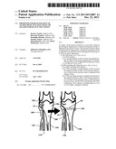 | 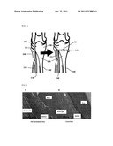 |
 |  |
 |  |
 |  |
 |  |
 |  |
 |  |
| Top Inventors for class "Drug, bio-affecting and body treating compositions" | |
| Rank | Inventor's name |
|---|---|
| 1 | Anthony W. Czarnik |
| 2 | Ulrike Wachendorff-Neumann |
| 3 | Ken Chow |
| 4 | John E. Donello |
| 5 | Rajinder Singh |