Patent application title: HYDROLASE AND METHODS FOR ITS USE
Inventors:
Joel Moss (Bethesda, MD, US)
Shunya Oka (Niigata, JP)
Jiro Kato (Rockville, MD, US)
Jianfeng Zhu (Frederick, MD, US)
Atsushi Kasamatsu (Rockville, MD, US)
IPC8 Class: AA61K3170FI
USPC Class:
514 44
Class name: N-glycoside nitrogen containing hetero ring polynucleotide (e.g., rna, dna, etc.)
Publication date: 2008-08-28
Patent application number: 20080207555
Inventors list |
Agents list |
Assignees list |
List by place |
Classification tree browser |
Top 100 Inventors |
Top 100 Agents |
Top 100 Assignees |
Usenet FAQ Index |
Documents |
Other FAQs |
Patent application title: HYDROLASE AND METHODS FOR ITS USE
Inventors:
Joel Moss
Shunya Oka
Jiro Kato
Jianfeng Zhu
Atsushi Kasamatsu
Agents:
KLARQUIST SPARKMAN, LLP
Assignees:
Origin: PORTLAND, OR US
IPC8 Class: AA61K3170FI
USPC Class:
514 44
Abstract:
This disclosure provides methods for catalyzing the release of ADP-ribose
from poly(ADP-ribose) or O-acetyl-ADP-ribose. Also provided are methods
for modifying DNA repair or chromatin structure by introducing into the
cell an agent that modifies the activity of an ARH3 polypeptide, or
variant or fragment thereof. Further provided are methods for screening
molecules involved in the poly(ADP-ribosyl)ation of proteins or
O-acetyl-ADP-ribose content, and method for treating disorders by
altering activity of an ARH3 protein.Claims:
1. A method for catalyzing the release of ADP-ribose from poly(ADP-ribose)
or O-acetyl-ADP-ribose, comprising contacting the poly(ADP-ribose) or
O-acetyl-ADP-ribose with an isolated ARH3 polypeptide, or variant or
fragment thereof, thereby catalyzing the release of ADP ribose.
2. The method of claim 1, wherein the ARH3 polypeptide, or variant or fragment thereof, is selected from the group consisting of:a) a polypeptide encoded by a nucleotide sequence at least 90% identical to the nucleotide sequence set forth as SEQ ID NO: 1 or 5, wherein the polypeptide has poly(ADP-ribose) glycohydrolase activity or O-acetyl-ADP-ribose hydrolase activity;b) a polypeptide comprising the amino acid sequence set forth as SEQ ID NO: 2, 3, 4, or 6; and,c) a polypeptide at least 90% identical to the amino acid sequence set forth as SEQ ID NO: 2, 3, 4, or 6, wherein the polypeptide has poly(ADP-ribose) glycohydrolase activity or O-acetyl-ADP-ribose hydrolase activity.
3. The method of claim 1, further comprising the addition of magnesium.
4. The method of claim 2, wherein the ARH3 polypeptide, or variant or fragment thereof, is encoded by a nucleotide sequence at least 90% identical to the nucleotide sequence set forth as SEQ ID NO: 1 or 5.
5. The method of claim 2, wherein the ARH3 polypeptide, or variant or fragment thereof, is a polypeptide comprising the amino acid sequence set forth as SEQ ID NO: 2, 3, 4, or 6.
6. The method of claim 2, wherein the ARH3 polypeptide, or variant or fragment thereof, is a polypeptide at least 90% identical to the amino acid sequence set forth as SEQ ID NO: 2, 3, 4, or 6.
7. A method for producing a polypeptide that catalyzes the release of ADP-ribose from poly(ADP-ribose) or O-acetyl-ADP-ribose, comprising culturing a host cell under conditions in which a nucleic acid molecule comprising a nucleotide sequence operably linked to a heterologous promoter is expressed, wherein the nucleotide sequence encodes a polypeptide that catalyzes the release of ADP-ribose from poly(ADP-ribose) or O-acetyl-ADP-ribose, and wherein the polypeptide is an ARH3 polypeptide, or variant or fragment thereof.
8. The method of claim 7, wherein the ARH3 polypeptide, or variant or fragment thereof, is selected from the group consisting of:a) a polypeptide comprising the amino acid sequence set forth as SEQ ID NO: 2, 3, 4, or 6;b) a polypeptide comprising an amino acid sequence at least 90% identical to the amino acid sequence set forth as SEQ ID NO: 2, 3, 4, or 6, wherein the polypeptide has poly(ADP-ribose) glycohydrolase activity or O-acetyl-ADP-ribose hydrolase activity; and,c) a polypeptide that is encoded by a nucleic acid molecule comprising a nucleotide sequence at least 90% identical to the nucleotide sequence set forth as SEQ ID NO: 1 or 5, wherein the polypeptide has poly(ADP-ribose) glycohydrolase activity or O-acetyl-ADP-ribose hydrolase activity.
9. A method for modifying DNA repair in a mammalian cell, comprising introducing into the cell an agent that modifies the activity of an ARH3 polypeptide, or variant or fragment thereof.
10. The method of claim 9, wherein the ARH3 polypeptide, or variant or fragment thereof, is selected from the group consisting of:a) a polypeptide comprising the amino acid sequence set forth as SEQ ID NO: 2, 3, 4, or 6;b) a polypeptide comprising an amino acid sequence at least 90% identical to the amino acid sequence set forth as SEQ ID NO: 2, 3, 4, or 6, wherein the polypeptide has poly(ADP-ribose) glycohydrolase activity or O-acetyl-ADP-ribose hydrolase activity; and,c) a polypeptide that is encoded by a nucleic acid molecule comprising a nucleotide sequence at least 90% identical to the nucleotide sequence set forth as SEQ ID NO: 1 or 5, wherein the polypeptide has poly(ADP-ribose) glycohydrolase activity or O-acetyl-ADP-ribose hydrolase activity.
11. The method of claim 9, wherein expression of the ARH3 polypeptide, or variant or fragment thereof, is increased.
12. The method of claim 9, wherein expression of the ARH3 polypeptide, or variant or fragment thereof, is decreased.
13. The method of claim 11, wherein at least one nucleotide sequence is introduced into the cell, wherein the nucleotide sequence causes increased expression of the ARH3 polypeptide, or variant or fragment thereof.
14. The method of claim 13, wherein the nucleotide sequence encodes an ARH3 polypeptide, or variant or fragment thereof.
15. The method of claim 12, wherein at least one nucleotide sequence is introduced into the cell, wherein the nucleotide sequence inhibits expression of the ARH3 polypeptide, or variant or fragment thereof.
16. The method of claim 15, wherein the nucleotide sequence hybridizes to a nucleotide sequence encoding the ARH3 polypeptide, or variant or fragment thereof.
17. The method of claim 15, wherein the nucleotide sequence encodes a polypeptide that inhibits expression of the ARH3 polypeptide, or variant or fragment thereof.
18. A method for modifying chromatin structure in a mammalian cell, comprising introducing into the cell an agent that modifies the activity of an ARH3 polypeptide, or variant or fragment thereof.
19. The method of claim 18, wherein the ARH3 polypeptide, or variant or fragment thereof, is selected from the group consisting of:a) a polypeptide comprising the amino acid sequence set forth as SEQ ID NO: 2, 3, 4, or 6;b) a polypeptide comprising an amino acid sequence at least 90% identical to the amino acid sequence set forth as SEQ ID NO: 2, 3, 4, or 6, wherein the polypeptide has poly(ADP-ribose) glycohydrolase activity or O-acetyl-ADP-ribose hydrolase activity; and,c) a polypeptide that is encoded by a nucleic acid molecule comprising a nucleotide sequence at least 90% identical to the nucleotide sequence set forth as SEQ ID NO: 1 or 5, wherein the polypeptide has poly(ADP-ribose) glycohydrolase activity or O-acetyl-ADP-ribose hydrolase activity.
20. The method of claim 18, wherein expression of the ARH3 polypeptide, or variant or fragment thereof, is increased.
21. The method of claim 18, wherein expression of the ARH3 polypeptide, or variant or fragment thereof, is decreased.
22. The method of claim 20, wherein at least one nucleotide sequence is introduced into the cell, wherein the nucleotide sequence causes increased expression of the ARH3 polypeptide, or variant or fragment thereof.
23. The method of claim 22, wherein the nucleotide sequence encodes an ARH3 polypeptide, or variant or fragment thereof.
24. The method of claim 21, wherein at least one nucleotide sequence is introduced into the cell, wherein the nucleotide sequence inhibits expression of the ARH3 polypeptide, or variant or fragment thereof.
25. The method of claim 24, wherein the nucleic acid sequence hybridizes to a nucleic acid sequence encoding the ARH3 polypeptide, or variant or fragment thereof.
26. The method of claim 24, wherein the nucleic acid sequence encodes a polypeptide that inhibits expression of the ARH3 polypeptide, or variant or fragment thereof.
27. A method for screening a candidate molecule for its use in altering the hydrolysis activity of ARH3, altering differentiation of a cell, altering apoptosis, altering DNA repair in a cell, treating a disorder associated with excessive DNA damage, modifying chromatin structure or treating cancer comprisingeither (1) contacting an ARH3 polypeptide with poly(ADP-ribose) or O-acetyl-ADP-ribose in the presence and absence of the molecule, and assessing the ability of ARH3 to produce ADP-ribose; or (2) contacting an ARH3 polypeptide with ADP-riobose and assessing the ability of ARH3 to bind ADP-ribose,wherein the ability of the molecule to alter the ability of ARH3 to either (1) produce ADP-ribose from poly(ADP-ribose) or O-acetyl-ADP-ribose; or (2) bind ADP-ribose, indicates that the candidate molecule is of use for altering the hydrolysis activity of ARH3, altering the differentiation of a cell, altering apoptosis, altering DNA repair in the cell, treating a disorder associated with excessive DNA damage, modifying chromatin structure or treating cancer.
28. The method of claim 27, wherein the ARH3 polypeptide is selected from the group consisting of:a) a polypeptide comprising the amino acid sequence set forth as SEQ ID NO: 2, 3, 4, or 6;b) a polypeptide comprising an amino acid sequence at least 90% identical to the amino acid sequence set forth as SEQ ID NO: 2, 3, 4, or 6, wherein the polypeptide has poly(ADP-ribose) glycohydrolase activity or O-acetyl-ADP-ribose hydrolase activity; and,c) a polypeptide that is encoded by a nucleic acid molecule comprising a nucleotide sequence at least 90% identical to the nucleotide sequence set forth as SEQ ID NO: 1 or 5, wherein the polypeptide has poly(ADP-ribose) glycohydrolase activity or O-acetyl-ADP-ribose hydrolase activity.
29. The method of claim 28, wherein the candidate molecule is a small molecule or a peptide.
30. A method for treating a disorder in a subject associated with excessive DNA damage comprising administering a therapeutically effective amount an agent that alters the activity of ARH3, or variant or fragment thereof.
31. The method of claim 30, wherein the disorder is selected from the group consisting of cancer, an autoimmune disease, acute pain, arthritis, atherosclerosis, cachexia, cardiovascular disorders, chronic pain, degenerative diseases, diabetes, head trauma, hyperglycemia, immune senescence, inflammatory bowel disorders, ischemia, macular degeneration, muscular dystrophy, tissue damage resulting from ischemia and reperfusion injury, neurological disorders and neurodegenerative diseases, neuronal tissue damage or disease, neuropathic pain, nervous insult, osteoarthritis, osteoporosis, peripheral nerve injury, renal failure, resuscitated hemorrhagic shock, retinal ischemia, septic shock, skin aging, vascular stroke, diseases or disorders relating to lifespan or proliferative capacity of cells, and diseases or disease conditions induced or exacerbated by cellular senescence.
32. A method for treating cancer, comprising administering a therapeutically effective amount of an agent that decrease the activity of an ARH3 polypeptide, or variant or fragment thereof.
33. The method of claim 32, wherein the agent that decreases ARH3 activity is administered in combination with at least one DNA-binding antitumor drug.
34. A method for treating a subject with cancer or inflammation, comprising administering to the subject a therapeutically effective amount of an agent that increases the activity of an ARH3 polypeptide, or variant or fragment thereof, thereby treating the cancer or inflammation in the subject.
35. The method of claim 35, wherein the subject has inflammation, and wherein the inflammation is an allergic response.
36. The method of claim 35, wherein the subject has inflammation, and wherein the inflammation is graft-versus-host disease.
Description:
PRIORITY CLAIM
[0001]This is a continuation-in-part of PCT Application No. PCT/US2006/035771, filed Sep. 12, 2006, which claims the benefit of U.S. Provisional Application No. 60/716,807, filed Sep. 13, 2005. The prior applications are incorporated herein by reference in their entirety.
FIELD
[0002]This invention relates to the field of molecular biology, specifically to ADP-ribose acceptor hydrolases and their use.
BACKGROUND
[0003]ADP-ribosylation is the post-translational modification of a protein, resulting from transfer of the ADP-ribose moiety of NAD to a specific amino acid in the protein, thereby altering its structure and function (Williamson and Moss (1990) In ADP-ribosylating Toxins and G Proteins: Insights into Signal Transduction (Moss, J. and Vaughan, M., eds) pp. 493-510, American Society for Microbiology, Washington, D.C.). Mammalian cells contain mono-ADP-ribosyltransferases (ART) that catalyze the formation of ADP-ribose-(arginine) protein, which can be cleaved by a 39-kDa ADP-ribose-(arginine) protein hydrolase (ARH1) that releases free ADP-ribose and regenerates the unmodified protein.
[0004]In addition to mono-ADP-ribosyltransferases, mammalian cells contain enzymes that poly-ADP-ribosylate proteins. Poly-ADP-ribosylation is catalyzed by a family of enzymes termed poly(ADP-ribose) polymerases (PARP) (Ame et al. (2004) Bioessays 26:882-893), that synthesize polymers of ADP-ribose in carboxylate linkage (Ogata et al. (1980) J. Biol. Chem. 255:7610-7615; Ogata et al. (1980) J. Biol. Chem. 255:7616-7620), usually to PARP-1 (Ogata et al. (1981) J. Biol. Chem. 256:4135-4137). Multiple poly(ADP-ribose) polymerases (PARPs) have been identified in the human genome, but there is only one known poly(ADP-ribose) glycohydrolase (PARG) that degrades the (ADP-ribose) polymer to ADP-ribose. Poly-ADP-ribosylation is involved in a number of critical biological processes including DNA repair, carcinogenesis, and cellular differentiation (Diefenbach and Burkle (2005) Cell Mol Life Sci. 62:721-730; Masutani et al. (2005) Cell Mol. Life. Sci. 62:769-783; Nguewa et al. (2005) Prog. Biophys. Mol Biol. 88:143-172).
[0005]Sir2 (silent information regulator 2) family proteins are involved in gene silencing, life span extension, and chromosomal stability (Guarente (2000) Genes Dev. 14:1021-1026; Bitterman et al. (2003) Microbiology and Molecular Biology Reviews 67:376-399). In the presence of NAD, Sir2 couples protein deacetylation with formation of O-acetyl-ADP-ribose and release of nicotinamide (Imai et al. (2000) Nature 403:795-800; Jackson and Denu (2002) J. Biol. Chem. 21:18535-18544). In many biological systems, specific enzymes are believed to be involved in the degradation of small molecules that are generated in signaling cascades, and thus, in termination of their effects. Thus far, enzymatic destruction of O-acetyl-ADP-ribose has been shown only with the Nudix family (O'Handley et al. (1998) J. Biol. Chem. 273:3192-3197) of ADP-ribose pyrophosphatases (Rafty et al. (2002) J. Biol. Chem. 277:47114-47122) (nucleoside diphosphate linked to another moiety, hence the acronym Nudix) and perhaps other less selective pyrophosphatases.
[0006]Proteins capable of hydrolyzing other ADP-ribose linkages are important in the regulation of ADP-ribose metabolism, which is involved in many cellular processes including chromatin decondensation, DNA replication and repair, transcription, centrosome duplication, regulation of telomere function, mitosis, necrosis and caspase-dependent and -independent apoptosis (Bonicalzi et al. (2005) Cell Mol. Life Sci. 62:739-750; Virag and Szabo (2002) Pharmacol. Rev. 54:375-429). In addition, drugs targeting polymer synthesis and turnover can be used for treating disorders associated with excessive tissue damage or as anticancer agents, radiosensitizers and antiviral agents (Southan and Szabo (2003) Curr Med Chem. 10:321-40). Furthermore, proteins that specifically target signaling molecules in the Sir2 pathway could be used in regulating chromatin.
SUMMARY
[0007]The ARH3 protein has been discovered to have poly(ADP-ribose) glycohydrolase and O-acetyl-ADP-ribose hydrolase enzymatic activity. Methods are provided herein for catalyzing the release of ADP-ribose from poly(ADP-ribose) or O-acetyl-ADP-ribose utilizing an ARH3 polypeptide. Methods are also provided for producing polypeptides with poly(ADP-ribose) or O-acetyl-ADP-ribose hydrolase activity.
[0008]Methods are disclosed for altering a variety of biological activities affecting the release of ADP-ribose from poly(ADP-ribose) or O-acetyl-ADP-ribose hydrolase. For example, methods are disclosed for modifying DNA repair or chromatin structure in a mammalian cell. Methods are provided for treating cancer, or for treating a disorder in a subject associated with excessive DNA damage, or to affect aging and longevity. Methods are also provided for the treatment of inflammation, such as graft-versus-host disease, inflammatory arthropathy, allergy and atherosclerosis.
[0009]Methods are further provided for screening molecules for use in altering cellular differentiation, DNA repair, apoptosis, chromatin structure, or for use in the treatment of cancer or a disorder associated with excessive DNA damage. Methods are also provided for screening molecules capable of altering the hydrolysis activity of ARH3. The methods are useful in studying the regulation of ADP-ribose metabolism and for identifying new molecules useful in modifying various cellular processes, as well as for identifying agents of use in the treatment of disorders that may benefit from activation or inhibition of poly(ADP-ribosyl)ation or from modifying O-acetyl-ADP-ribose content.
[0010]The foregoing and other features and advantages will become more apparent from the following detailed description of several embodiments, which proceeds with reference to the accompanying figures.
DESCRIPTION OF FIGURES
[0011]FIG. 1A shows an alignment of the amino acid sequences of human ARH1 (NCBI/NIH Accession No. AAA35555) (SEQ ID NO: 7), human ARH2 (NCBI/NIH Accession No. CAC86114) (SEQ ID NO: 8), and human ARH3 (NCBI/NIH Accession No. CAC85940) (SEQ ID NO: 2). Amino acid acids critical for ARH1 activity are indicated by an asterisk (Konczalik and Moss (1999) J. Biol. Chem. 274:16736-16740).
[0012]FIG. 1B shows an alignment of the amino acid sequences of human ARH3 (SEQ ID NO: 2) and the catalytic domain of PARG (amino acids 421-976 of NCBI/NIH Accession No. AAT66422) (SEQ ID NO: 9). The amino acids mutated in a variant of ARH3 (D77N/D78N) are indicated by an asterisk. The amino acids reported to be critical for activity based on mutagenesis of PARG are indicated by a plus sign (Patel et al. (2005) Biochem. J. 388:493-500). The amino acids mutated in a variant of ARH3 (E238Q/E239Q) (SEQ ID NO: 4) are indicated by a number sign. The amino acids mutated in a variant of ARH3 (E261Q/E262Q) (SEQ ID NO: 3) are indicated by a carat. Identical sequences are in white letters on black. Conserved amino acids (see Table 2, legend) are shaded gray.
[0013]FIG. 2 shows detection of ARH3 mRNA and proteins in mouse tissues and HepG2 cells. FIG. 2A shows a Northern blot of mouse poly (A).sup.+ RNA with an ARH3 cDNA probe. Positions of RNA standards are on the left. FIG. 2B is a digital image of a Western blot of the detection of ARH3 protein in the indicated tissues. FIG. 2C is a digital image of a Western blot of ARH3 protein detected in nuclear (HepG2, and mouse brain or liver tissue), cytosolic, and membrane fractions. N; nuclei, C; cytosol, M; membranes.
[0014]FIG. 3 shows the hydrolysis by ARH1, 2, or 3 of proteins that were mono-ADP-ribosylated by bacterial toxins. FIG. 3A is a digital image of an autoradiograph depicting samples from mouse brain membranes that were [32P]ADP-ribosylated by incubation with cholera toxin and incubated with recombinant ARH 1, 2, or 3 or BSA. Cont: brain membranes that had been incubated with [32P]-labeled NAD without CTA. FIG. 3B is a digital image of an autoradiograph depicting samples of [32P] ADP-ribosylated Gαi/Gαo synthesized by pertussis toxin (PT), [32P] ADP-ribosylated EF-2 in mouse brain cytosol synthesized by Pseudomonas aeruginosa exotoxin A (ExoA), or [32P] ADP-ribosylated Rho in mouse brain cytosol synthesized by Clostridium botulinum C3 toxin. Cont: Substrate that had been incubated with [32P]-labeled NAD without bacterial toxin.
[0015]FIG. 4 shows the effects of DTT and MgCl2 on poly(ADP-ribose) glycohydrolase activity of human ARH1, 2, and 3. FIG. 4A is a digital image of a Western blot of [32P]poly(ADP-ribose)PARP degradation by human ARH1, 2, 3 and PARG in the presence or absence of DTT and/or MgCl2. [32P]poly(ADP-ribose)PARP is at the top of each lane. FIG. 4B shows a graph depicting the detection of [14C]ADP-ribose by HPLC after [14C]poly(ADP-ribose)PARP degradation by human ARH3 in the presence or absence of DTT and/or MgCl2.
[0016]FIG. 5 shows HPLC analyses of [14C]ADP-ribose released from ARH3- and PARG-catalyzed reactions. FIG. 5A shows a graph depicting products eluted from C18 HPLC and FIG. 5B shows a graph depicting products from a Zorbax SAX column.
[0017]FIG. 6 shows graphs depicting the effect of hydrolase concentration (FIG. 6A) and time (FIG. 6B) on hydrolysis of poly(ADP-ribose) by human ARH3.
[0018]FIG. 7 is a digital image of an autoradiograph depicting the size of ADP-ribose polymers after incubation with mouse or human ARH3 or PARG from calf thymus. Cont, reaction without enzyme incubated for 60 min. On the right, [32P]-labeled standards are ADPR (ADP-ribose), NAD (β-NAD, Perkin Elmer), AMP, PRAMP (phosphoribosyl-AMP). Bromophenol blue (BPB) and xylene cyanol (XC) co-migrated with (ADP-ribose)8 and (ADP-ribose)18, respectively.
[0019]FIG. 8 shows the effect of mutation of human ARH3 on its hydrolysis of poly(ADP-ribose). FIG. 8A is a digital image of an autoradiograph showing detection of poly(ADP-ribose) hydrolysis products without enzyme (Cont) or with 1 μM ARH3 (wild-type or mutant) or 1.5 nM PARG. Positions of standards are indicated as in FIG. 7. FIG. 8B is a graph showing the amount of ADP-ribose released without enzyme (Cont) or with 50 nM human ARH3 (wild-type or mutant) or 1 nM PARG. *D77N/D78N 5 μM samples were incubated overnight with 5 μM mutant ARH3.
[0020]FIG. 9 shows a graph depicting [14C]ADP-ribose binding by human ARH3 (WT or D77N/D78N mutant) in the presence or absence of DTT and/or MgCl2.
[0021]FIG. 10 is a digital image of an immunoblot showing the localization of ARH3 in HEK293T cells. Localization was investigated by cell fractionation, followed by immunoblotting. After cell fractionation, immunoreactive 39-kDa ARH3 was identified using antibodies against mouse ARH3 amino acids 355-370; the antibodies did not react with ARH1 and ARH2. ARH3 was present in both cytosol and membrane fractions.
[0022]FIG. 11 shows the identification of products of Sir2 and ARH3-catalyzed reactions. FIG. 11A shows synthesis of O-acetyl-[14C]ADP-ribose catalyzed by Sir2. FIG. 11B shows hydrolysis of O-acetyl-[14C]ADP-ribose catalyzed by ARH3. Peaks: 1, ADP-ribose; 2, O-acetyl-[32P]ADP-ribose; 3, β-NAD; 4, ADP-ribose; 5, O-acetyl-ADP-ribose. FIG. 11C is a digital image showing high resolution-polyacrylamide gel electrophoresis of substrates and products in reactions involving O-acetyl-[32P]ADP-ribose. Lanes: 6) [32P]β-NAD; 7) [32P]AMP, produced by pyrophosphatase cleavage of [32P]β-NAD; 8) [32P]ADP-ribose produced from [32P]β-NAD by CTA glycohydrolase activity; 9) O-acetyl-[32P]ADP-ribose synthesized by Sir2 as in FIG. 11A; 10) [32P]ADP-ribose produced by ARH3 from O-acetyl-[32P]ADP-ribose as in FIG. 11B.
[0023]FIG. 12 shows hydrolysis of O-acetyl-ADP-ribose by ARH3. FIG. 12A is a graph of the results from incubation of 2.5 μM O-acetyl-[14C]ADP-ribose and the indicated amount of mouse ARH3 for 1 hour. FIG. 12B is a graph of the results from incubation of 1.5 pmol of mouse ARH3 and 2.5 μM substrate at 30° C. for the indicated time.
[0024]FIG. 13 shows the hydrolysis of O-acetyl-ADP-ribose by wild type and mutant forms of ARH3 or PARG. FIG. 13A is a graph illustrating the results from incubation of 1.5 pmol of mouse ARH3 with or without 5 mM DTT and/or 10 mM MgCl2 as described in FIG. 11B with incubation for 1 h at 30° C. FIG. 13B is a graph illustrating the results from incubation of 1.5 pmol of wild type or mutant human (D77-78) ARH3 or 20 mU of PARG for 2 h at 30° C. *D77,78, assays incubated with 15 pmol of mutant human ARH3 (D77, D78).
[0025]FIG. 14 shows the hydrolysis of O-acetyl-[14C]ADP-ribose by ARH 1, 2 and 3. FIG. 14A is a graph of the results from incubation with the indicated amount of mouse ARH1, 2, or 3 for 2 h at 30° C. FIG. 14B is a graph of the results from incubation with 230 pmol of mouse ARH1 at 30° C. for the indicated time.
[0026]FIG. 15 shows inhibition of ARH3 hydrolysis by ADP-ribose and β-NAD. FIG. 15A is a graph of the results from an assay with 2 pmol of ARH3 and the indicated amount of ADP-ribose ( ) or β-NAD (∘) in 200 μl for 2 h at 30° C., as described in FIG. 11B. FIG. 15B is a graph of the inhibition of ARH3 hydrolysis by ADP-ribose data schematized for Lineweaver-Burk plot.
[0027]FIG. 16 is a line graph showing the effect of H2O2 on ARH3-transfected NIH3T3 cells. NIH3T3 cells were transfected with ARH3 cDNA using Lipofectamin transfection reagent (INVITROGEN®). After transfection, the cells were cultured and exposed to 200 μg/ml of Hygromycin G (INVITROGEN®) for 3-4 weeks to select stably transfected clones. Positive clones derived from single Hygromycin G-resistant cells were then isolated by cloning rings and further grown under the same conditions. As controls, NIH3T3 cells were transfected with an empty pcDNA3.1 vector (INVITROGEN®) and subjected to the same selection and cloning procedures as described above. The cells were plated in 96-well plates at 1×10⁴ cells/well, and incubated for 24 h at 37° C. in a humidified incubator. The cells were then challenged at the indicated concentrations of H2O2 for 24 hours. Cell survival was determined by using Cell-Counting Kit-8 (Dojindo). The experiments were repeated twice with similar results.
SEQUENCE LISTING
[0028]The nucleic and amino acid sequences listed in the accompanying sequence listing are shown using standard letter abbreviations for nucleotide bases, and three letter code for amino acids, as defined in 37 C.F.R. 1.822. Only one strand of each nucleic acid sequence is shown, but the complementary strand is understood as included by any reference to the displayed strand.
DETAILED DESCRIPTION
I. Abbreviations
[0029]AIDS acquired immune deficiency syndrome
[0030]ADP adenosine diphosphate
[0031]ADPR ADP-ribose
[0032]AMP adenosine monophosphate
[0033]ARH1 ADP-ribose-(arginine) hydrolase
[0034]ARH3 ADP-ribosyl acceptor hydrolase
[0035]ART ADP-ribosyltransferase
[0036]ATP adenosine triphosphate
[0037]BHA butyl hydroxy anisole
[0038]BHT butyl hydroxy toluene
[0039]bp base pair(s)
[0040]BPB Bromophenol blue
[0041]BSA bovine serum albumin
[0042]CaMV cauliflower mosaic virus
[0043]cDNA complementary DNA
[0044]CTA cholera toxin A
[0045]DEAE diethylaminoethyl
[0046]DHBB Dihydroxyboronyl-Bio-Rex 70
[0047]DMEM Dulbecco's Minimal Essential Medium
[0048]DNA deoxyribonucleic acid
[0049]DTT dithiothreitol
[0050]EBV Epstein Barr virus
[0051]EDTA ethylenediamine tetraacetic acid
[0052]EEA Early endosomal antigen
[0053]ExoA exotoxin A
[0054]FBS fetal bovine serum
[0055]GAPDH glyceraldehyde-3-phosphate dehydrogenase
[0056]GST glutathione-S transferase
[0057]HPLC high pressure liquid chromatography
[0058]ip intraperitoneal
[0059]iv intravenous
[0060]kb kilobase pair(s)
[0061]kDa kiloDalton
[0062]LAMP Lysosomal-associated membrane protein
[0063]mRNA messenger RNA
[0064]NAD nicotinamide adenine dinucleotide
[0065]NMDA N-methyl-D-aspartate
[0066]NO nitric oxide
[0067]pADPr poly(ADP-ribose) polymers
[0068]PAGE polyacrylamide-gel electrophoresis
[0069]PARG poly(ADP-ribose) glycohydrolase
[0070]PARP poly(ADP-ribose) polymerase
[0071]PBS phosphate buffered saline
[0072]PCR polymerase chain reaction
[0073]PRAMP phosphoribosyl-AMP
[0074]PT pertussis toxin
[0075]Rab Ras-like GTP-binding protein
[0076]RNA ribonucleic acid
[0077]RNase ribonuclease
[0078]RP-HPLC reverse phase HPLC
[0079]SDS sodium dodecyl sulfate
[0080]SSC sodium citrate buffer
[0081]SV40 simian virus 40
[0082]TBE tris-borate EDTA
[0083]TEMED tetramethylethylenediamine
[0084]TKMS Tris-KCl--MgCl2-Sucrose
[0085]TMV tobacco mosaic virus
[0086]WT wild type
[0087]XC xylene cyanol
II. Terms
[0088]Unless otherwise noted, technical terms are used according to conventional usage. Definitions of common terms in molecular biology may be found in Benjamin Lewin, Genes V, published by Oxford University Press, 1994 (ISBN 0-19-854287-9); Kendrew et al. (eds.), The Encyclopedia of Molecular Biology, published by Blackwell Science Ltd., 1994 (ISBN 0-632-02182-9); and Robert A. Meyers (ed.), Molecular Biology and Biotechnology: a Comprehensive Desk Reference, published by VCH Publishers, Inc., 1995 (ISBN 1-56081-569-8).
[0089]In order to facilitate review of the various embodiments of this disclosure, the following explanations of specific terms are provided:
[0090]By "ADP-ribosylation activity" is intended the enzyme-catalyzed post-translational protein modification in which the ADP-ribose moiety is transferred from NAD+ to a specific amino acid in a target protein while the nicotinamide moiety is released.
[0091]By "agonist" is intended a molecule which, when bound to a protein, increases or prolongs the effect of the protein. Agonist may include proteins, nucleic acid molecules, carbohydrates, or any other molecules that bind to and modulate the effect of a protein.
[0092]By "ARH3" or "ARH3 protein" is intended a protein having both PARG activity and O-acetyl-ADP-ribose activity. By "an ARH3 hydrolysis activity" or "an ARH3 activity" is intended either PARG activity or O-acetyl-ADP-ribose activity. By "O-acetyl-ADP-ribose hydrolase activity" is intended the ability to generate ADP-ribose from O-acetyl-ADP-ribose. By "PARG activity" or "poly(ADP-ribose) glycohydrolase activity" is intended the ability to generate ADP-ribose from poly(ADP-ribose). Proteins with PARG activity have both exoglycosidase and endoglycosidase activity, and are therefore capable of hydrolyzing ribose-ribosyl glycosidic bonds between poly(ADP-ribose) polymer units located at the end and within the polymer. The protein PARG (an exemplary non-limiting protein sequence is set forth as Genbank Accession No. AAT66422, Jul. 5, 2004) is differentiated from an ARH3 protein in that it does not have O-acetyl-ADP-ribose hydrolysis activity. The terms "protein" and "polypeptide" are used interchangeably herein.
[0093]Atherosclerosis" refers to the progressive narrowing and hardening of a blood vessel over time. Atherosclerosis is a common form of arteriosclerosis in which deposits of yellowish plaques (atheromas) containing cholesterol, lipoid material, and lipophages are formed within the intima and inner media of large and medium-sized arteries.
[0094]Treatment of atherosclerosis includes reversing or slowing the progression of atherosclerosis, for example as measured by the presence of atherosclerotic lesions and/or functional signs of the disease, such as improvement in cardiovascular function as measured by signs (such as peripheral capillary refill), symptoms (such as chest pain and intermittent claudication), or laboratory evidence (such as that obtained by EKG, angiography, or other imaging techniques). Inflammation plays a role in the development of atherosclerosis, for example by coupling dislipidemia to atheroma formation. Inflammatory pathways promoter early artherogenesis and thrombosis. Hence interfering with inflammation can inhibit the development and progression of atherosclerosis.
[0095]The term "cancer" is interpreted broadly. For example, the methods provided herein are useful for treating cancers and radiosensitizing tumor cells in cancers such as ACTH-producing tumors, acute lymphocytic leukemia, acute nonlymphocytic leukemia, cancer of the adrenal cortex, bladder cancer, brain cancer, breast cancer, cervical cancer, chronic lymphocytic leukemia, chronic myelocytic leukemia, colorectal cancer, cutaneous T-cell lymphoma, endometrial cancer, esophageal cancer, Ewing's sarcoma, gallbladder cancer, hairy cell leukemia, head & neck cancer, Hodgkin's lymphoma, Kaposi's sarcoma, kidney cancer, liver cancer, lung cancer (small and/or non-small cell), malignant peritoneal effusion, malignant pleural effusion, melanoma, mesothelioma, multiple myeloma, neuroblastoma, non-Hodgkin's lymphoma, osteosarcoma, ovarian cancer, ovary (germ cell) cancer, prostate cancer, pancreatic cancer, penile cancer, retinoblastoma, skin cancer, soft-tissue sarcoma, squamous cell carcinomas, stomach cancer, testicular cancer, thyroid cancer, trophoblastic neoplasms, uterine cancer, vaginal cancer, cancer of the vulva, Wilm's tumor, and neoplastic disorders.
[0096]A neoplastic disorder is any new and abnormal growth; specifically, a new growth of tissue in which the growth is uncontrolled and progressive. Neoplastic disorders may include, but are not limited to, neoplastic disorders of the adrenal gland, bladder, bone, bone marrow, brain, breast, cervix, gall bladder, ganglia, gastrointestinal tract, heart, kidney, liver, lung, muscle, ovary, pancreas, parathyroid, penis, prostate, salivary glands, skin, spleen, testis, thymus, thyroid, and uterus. Cancer includes a benign or malignant neoplasm, and thus includes adenocarcinoma, leukemia, lymphoma, melanoma, myeloma, sarcoma, teratocarcinoma, hyperplasia and hypertrophy. Malignant cancer is a subset of neoplastic disorders that show a greater degree of anaplasia and have the properties of invasion and metastasis.
[0097]By "chromatin structure" is intended the physical arrangement of chromatin in a cell. Chromatin is the substance of a chromosome and consists of a complex of DNA and protein in eukaryotic cells. The nucleic acids are generally in the form of double-stranded DNA. The major proteins involved in chromatin are histone proteins. In a eukaryotic cell, nearly all DNA is found compacted in chromatin. DNA is packaged into chromatin both to constrain the size of the molecule and to allow the cell to control expression of the chromatin packaged genes.
[0098]A "conservative amino acid substitution" is one in which the amino acid residue is replaced with an amino acid residue having a similar side chain. A "nonessential" amino acid residue is a residue that can be altered from the wild-type sequence of an ARH3 protein without altering the biological activity, whereas an "essential" amino acid residue is required for biological activity. Families of amino acid residues having similar side chains have been defined in the art. These families include amino acids with basic side chains (for example, lysine, arginine, histidine), acidic side chains (for example, aspartic acid, glutamic acid), uncharged polar side chains (for example, asparagine, glutamine, serine, threonine, tyrosine, cysteine), nonpolar side chains (for example, alanine, valine, leucine, isoleucine, proline, phenylalanine, methionine, tryptophan), beta-branched side chains (for example, threonine, valine, isoleucine) and aromatic side chains (for example, tyrosine, phenylalanine, tryptophan, histidine).
[0099]By "DNA repair" is intended the reconstruction of a continuous two-stranded DNA molecule without mismatch from a molecule that contained damaged regions. This damage may occur, for example, due to normal metabolic activities, such as DNA replication, or from environmental factors, such as chemical insults or UV rays. The major repair mechanisms are excision repair, in which defective regions in one strand are excised and resynthesized using the complementary base pairing information in the intact strand; photoreactivation repair, in which the lethal and mutagenic effects of ultraviolet light are eliminated; and post-replication repair, in which the primary lesions are not repaired, but the gaps in one daughter duplex are filled in by incorporation of portions of the other (undamaged) daughter duplex.
[0100]By "fragment" is intended a portion of a nucleotide sequence encoding a protein, or a portion of the amino acid sequence of the protein.
[0101]Heterologous" generally refers to the nucleic acid sequences that are not endogenous to the cell or part of the native genome in which they are present, and have been added to the cell by infection, transfection, microinjection, electroporation, microprojection, or the like.
[0102]By "homologue" or "variant" is intended a nucleotide or amino acid sequence sufficiently identical to the reference nucleotide or amino acid sequence, respectively. "Homologues" or "variants" of an ARH3 polypeptide are encoded by a nucleotide sequence sufficiently identical to the nucleotide sequence of SEQ ID NO: 1 or 5. By "sufficiently identical" is intended an amino acid or nucleotide sequence that has at least about 60% or 65% sequence identity, about 70% or 75% sequence identity, about 80% or 85% sequence identity, about 90%, 91%, 92%, 93%, 94%, 95%, 96%, 97%, 98% or 99% sequence identity over its full length as compared to a reference sequence, for example using the NCBI Blast 2.0 gapped BLAST set to default parameters, or an equivalent program. By "equivalent program" is intended any sequence comparison program that, for any two sequences in question, generates an alignment having identical nucleotide or amino acid residue matches and an identical percent sequence identity when compared to the corresponding alignment generated by gapped BLAST. Alignment may also be performed manually by inspection. For comparisons of amino acid sequences of greater than about 30 amino acids, the Blast 2 sequences function is employed using the default BLOSUM62 matrix set to default parameters (gap existence cost of 11, and a per residue gap cost of 1). When aligning short peptides (fewer than around 30 amino acids), the alignment should be performed using the Blast 2 sequences function, employing the PAM30 matrix set to default parameters (open gap 9, extension gap 1 penalties). Proteins with even greater similarity to the reference sequences will show increasing percentage identities when assessed by this method, such as at least 80%, at least 85%, at least 90%, at least 95%, at least 98%, or at least 99% sequence identity. When less than the entire sequence is being compared for sequence identity, homologues and variants will typically possess at least 80% sequence identity over short windows of 10-20 amino acids, and may possess sequence identities of at least 85% or at least 90% or 95% depending on their similarity to the reference sequence. Methods for determining sequence identity over such short windows are available at the NCBI website on the internet. One of skill in the art will appreciate that these sequence identity ranges are provided for guidance only; it is entirely possible that strongly significant homologues could be obtained that fall outside of the ranges provided.
[0103]Inflammation" is usually a part of the host defense response. When damage to tissue occurs, the body's response to the damage may result in inflammation. The damage may be due to trauma, lack of blood supply, hemorrhage, autoimmune attack, transplanted exogenous tissue or infection. This generalized response by the body includes the release of many components of the immune system (e.g., IL-1, TNF), attraction of cells to the site of the damage, swelling of tissue due to the release of fluid and other processes. Inflammation can be induced by pathogens that act on a cell of the innate immune systems or can be induced by cells of the adaptive immune system. Specific, non-limiting examples of agents that induce inflammation are cytokines, chemokines and pathogens.
[0104]Disease states that are associated with inflammation include, but may not be limited to, autoimmune diseases as defined above; atherosclerosis; chronic hepatitis, transplanted foreign tissues; drug or other hypersensitivity reaction. Conditions such as autoimmunity are characterized by the body's immune responses being directed against its own tissues and can cause prolonged inflammation.
[0105]Inflammatory arthropathy" refers to an inflammatory disease affecting one or more joints, for example an inflammatory disease that affects the synovial membranes of one or more joints. Inflammatory arthropathies include, for example, arthritis, ankylosing spondylitis, Reiter's syndrome, psoriatic arthropathy, enteropathy spondylitis, juvenile arthropathy, and reactive arthropathy. Chronic inflammation is also believed to contribute to the aging of many tissues. Interfering with chronic inflammation can therefore slow physiological aging as compared to chronological aging.
[0106]By "introducing" is intended introduction into cells via conventional transformation or transfection techniques, or by phage-mediated infection, or, in the case of chemical compounds, by contacting the cell with the compound. As used herein, the terms "transformation," "transduction," "conjugation," and "protoplast fusion" are intended to refer to a variety of art-recognized techniques for introducing foreign nucleic acid (for example, DNA) into a host cell, including calcium phosphate or calcium chloride co-precipitation, DEAE-dextran-mediated transfection, lipofection, or electroporation. Suitable methods for transforming or transfecting host cells can be found in Sambrook et al. (1989) Molecular Cloning: A Laboratory Manual (2d ed., Cold Spring Harbor Laboratory Press, Plainview, N.Y.) and other laboratory manuals.
[0107]An "isolated" or "purified" nucleic acid molecule or protein, or biologically active portion thereof, is substantially free of other cellular material, or culture medium when produced by recombinant techniques, or substantially free of chemical precursors or other chemicals when chemically synthesized. Preferably, an "isolated" nucleic acid is free of sequences (preferably protein encoding sequences) that naturally flank the nucleic acid (for example, sequences located at the 5' and 3' ends of the nucleic acid) in the genomic DNA of the organism from which the nucleic acid is derived. "Isolated," when used to refer to nucleic acid molecules, excludes isolated chromosomes. For example, in various embodiments, the isolated ARH3-encoding nucleic acid molecule can contain less than about 5 kb 4 kb, 3 kb, 2 kb, 1 kb, 0.5 kb, or 0.1 kb of nucleotide sequences that naturally flank the nucleic acid molecule in genomic DNA of the cell from which the nucleic acid is derived. An ARH3 protein that is substantially free of cellular material includes preparations of protein having less than about 30%, 20%, 10%, or 5% (by dry weight) of non-ARH3 protein (also referred to herein as a "contaminating protein").
[0108]By "modifies" or "modifying" is intended that an activity is altered in some manner.
[0109]By "mono-ADP-ribosyltransferase activity" is intended the transfer of a single ADP-ribose moiety onto a specific amino acid side chain of a target protein. Generally this amino acid is an arginine residue.
[0110]As used herein, the term "nucleic acid molecule" is intended to include DNA molecules (for example, cDNA or genomic DNA) and RNA molecules (for example, mRNA) and analogs of the DNA or RNA generated using nucleotide analogs. The nucleic acid molecule can be single-stranded or double-stranded, but preferably is double-stranded DNA. The terms "nucleic acid", "nucleotide" and "polynucleotide" are used interchangeably. By "complement" is intended a nucleotide sequence that is sufficiently complementary to a given nucleotide sequence such that it can hybridize to the given nucleotide sequence to thereby form a stable duplex. By "contiguous" nucleotides is intended nucleotide residues that are immediately adjacent to one another.
[0111]By "operably linked" is intended a functional linkage between a promoter and a second sequence, wherein the promoter sequence initiates and mediates transcription of the DNA sequence corresponding to the second sequence. Generally, operably linked means that the nucleic acid sequences being linked are contiguous and, where necessary to join two protein coding regions, contiguous and in the same reading frame.
[0112]By "PARP activity" or "(poly(ADP-ribose) polymerase) activity" is intended the ability to catalyze the elongation and branching of ADP-ribose units on ADP-ribosylated targets. PARP generally acts on glutamic acid residues of target proteins, but can also act on aspartic acid or lysine or on a mono-ADP ribosylated arginine.
[0113]Promoter" refers to a nucleic acid sequence that functions to direct transcription of a downstream coding sequence. The promoter, together with other transcriptional and translational regulatory nucleic acid sequences (also termed "control sequences") are necessary for the expression of a DNA sequence of interest.
[0114]By "retains" activity is intended that a fragment or variant of a protein of interest will have at least about 30%, preferably at least about 50%, more preferably at least about 70%, even more preferably at least about 80% of the activity of the native protein. In the case of ARH3, this would be ARH3 hydrolysis activity.
[0115]By "stringent conditions" or "stringent hybridization conditions" is intended conditions under which a probe will hybridize to its target sequence to a detectably greater degree than to other sequences (for example, at least 2-fold over background).
[0116]Subject" includes both human and animal subjects. An "animal" is a living multicellular vertebrate organism, a category that includes, for example, mammals and birds. A "mammal" includes both human and non-human mammals, such as dogs, cats, cows, horses, rabbits, monkeys, and humans.
[0117]By "transgenic cells" or "transformed cells" is intended cells that have incorporated or integrated exogenous nucleic acid sequences or DNA fragments. These nucleic acid sequences include those that are exogenous, or not present in the untransformed cell, as well as those that may be endogenous, or present in the untransformed cell.
[0118]By "transplantation" is intended the transfer of a tissue or an organ, or a portion thereof, from one body or part of the body to another body or part of the body. An "allogeneic transplantation" or a "heterologous transplantation" is transplantation from one individual to another, wherein the individuals have genes at one or more loci that are not identical in sequence in the two individuals. An allogeneic transplantation can occur between two individuals of the same species, who differ genetically, or between individuals of two different species. An "autologous transplantation" is a transplantation of a tissue or a portion thereof from one location to another in the same individual, or transplantation of a tissue or a portion thereof from one individual to another, wherein the two individuals are genetically identical. The tissue to be transplanted may be cells that were isolated from an individual and subjected to treatments in vitro before being transferred back into the same or another body.
[0119]Treatment" refers to a therapeutic intervention that ameliorates a sign or symptom of a disease or pathological condition after it has begun to develop. As used herein, the term "ameliorating," with reference to a disease or pathological condition, refers to any observable beneficial effect of the treatment. The beneficial effect can be evidenced, for example, by a delayed onset of clinical symptoms of the disease or disorder in a susceptible subject, a reduction in severity of some or all clinical symptoms of the disease or disorder, a slower progression of the disease or disorder, a reduction in the number of relapses of the disease or disorder, an improvement in the overall health or well-being of the subject, or by other parameters well known in the art that are specific to the particular disease or disorder.
[0120]By "vector" is intended a nucleic acid construct designed for transfer between different host cells. By "expression vector" is intended a vector that has the ability to incorporate, integrate and express heterologous DNA sequences or fragments in a foreign cell. Typically these contain a promoter and a coding sequence. Often, such constructs will also contain a 3' untranslated region. Such constructs may contain a `signal sequence` or `leader sequence` to facilitate co-translational or post-translational transport of the peptide to certain intracellular structures such as the endoplasmic reticulum or Golgi apparatus. By "signal sequence" is intended a sequence that is known or suspected to result in cotranslational or post-translational peptide transport across the cell membrane. In eukaryotes, this typically involves secretion into the Golgi apparatus, with some resulting glycosylation. By "leader sequence" is intended any sequence that when translated, results in an amino acid sequence sufficient to trigger co-translational transport of the peptide chain to a sub-cellular organelle. Thus, this includes leader sequences targeting transport and/or glycosylation by passage into the endoplasmic reticulum, passage to vacuoles, plastids, mitochondria, and the like. By "transformation vector" is intended a DNA molecule that is necessary for efficient transformation of a cell. Such a molecule may consist of one or more expression cassettes, and may be organized into more than one vector DNA molecule. In some instances, the expression vector may include a nucleotide sequence that itself, or by encoding a protein, affects expression of a protein or a nucleotide sequence. These sequences may include, for example, siRNA, complementary DNA, a protein that stabilizes mRNA, and the like.
[0121]The above term descriptions are provided solely to aid the reader, and should not be construed to have a scope less than that understood by a person of ordinary skill in the art or as limiting the scope of the appended claims.
[0122]Unless otherwise explained, all technical and scientific terms used herein have the same meaning as commonly understood by one of ordinary skill in the art to which this disclosure belongs. The singular terms "a," "an," and "the" include plural references unless context clearly indicates otherwise. Similarly, the word "or" is intended to include "and" unless the context clearly indicates otherwise. It is further to be understood that all base sizes or amino acid sizes, and all molecular weight or molecular mass values, given for nucleic acids or polypeptides are approximate, and are provided for description. Although methods and materials similar or equivalent to those described herein can be used in the practice or testing of this disclosure, suitable methods and materials are described below. The term "comprises" means "includes." All publications, patent applications, patents, and other references mentioned herein are incorporated by reference in their entirety. In case of conflict, the present specification, including explanations of terms, will control. In addition, the materials, methods, and examples are illustrative only and not intended to be limiting.
ARH3 and Its Activity
[0123]Human ARH3 is a 39-kDa protein that was initially identified by sequence similarity as an ADP-ribosyl-acceptor-hydrolase. For exemplary amino acid sequences, see Genbank Accession Nos. AJ313333 (Jun. 21, 2002) and CAC85940 (Jun. 21, 2002), both herein incorporated by reference in their entirety. Human ARH3 shares amino acid sequence identity with both human ARH1 and the catalytic domain of poly(ADP-ribose) glycohydrolase (PARG) (see, for example, Genbank Accession No. AAT66422, Jul. 5, 2004). Amino acid sequences of exemplary human ARH1, 2, and 3, and the catalytic domain of PARG are aligned for comparison in FIG. 1. Amino acid sequences of human ARH1 and ARH2 are about 45% identical but only about 20% identical to that of ARH3 (see Examples, see Table 2).
[0124]Generally, ARH3 is more identical to the catalytic region of the PARG than to ARH1 or ARH2. In one example human ARH3 is about 20% identical to human PARG catalytic domain (111 kDa form).
[0125]It has surprisingly been found that ARH3 exhibits poly(ADP-ribose) glycohydrolase (PARG) activity, generating ADP-ribose from poly(ADP-ribose), and that it is capable of acting on O-acetyl-ADP-ribose to generate ADP-ribose. However, while ARH3 is capable of binding to ADP-ribose, ARH3 does not hydrolyze ADP-ribosylarginine. It also does not hydrolyze ADP-ribosyl-asparagine, -diphthamide or -cysteine. ARH3 PARG activity, like the activity of ARH1, is enhanced by magnesium (Mg2+). Thus, this protein can be used to regulate poly(ADP-ribose) levels in a cell
[0126]ARH3 proteins include, but are not limited to, those set forth in SEQ ID NOS:2 and 6, which are encoded by the nucleotide sequences set forth in SEQ ID NO: 1 and 5, respectively. In several examples, an ARH3 protein is at least about 85%, at least about 90%, at least about 95%, at least about 98%, at least about 99% identical to SEQ ID NO: 2 or SEQ ID NO: 6, and cleaves O-acetyl-ADP-ribose to generate ADP-ribose and can produce ADP-ribose from poly(ADP-ribose). Similarly, a nucleic acid encoding an ARH3 protein can be at least about 85%, at least about 90%, at least about 95%, at least about 98%, at least about 99% identical to SEQ ID NO: 1 or SEQ ID NO: 5, and encode a polypeptide that cleaves O-acetyl-ADP-ribose to generate ADP-ribose and can produce ADP-ribose from poly(ADP-ribose). Examples of specific variants that may be used are set forth as SEQ ID NOS: 3 and 4. Fragments or variants of the amino acid sequence of ARH3 can be used in the methods provided herein.
[0127]The ARH1, ARH3, and PARG catalytic domains all contain pairs of vicinal acidic amino acids, aspartate or glutamate (Konczalik and Moss (1999) J. Biol. Chem. 274:16736-16740; Patel et al. (2005) Biochem. J. 388:493-500). Critical vicinal acidic amino acids in ARH3, identified by mutagenesis (D77, D78), are located in a region similar to that required for activity in ARH1, but different from the location of the critical vicinal glutamates in the PARG catalytic site. In one example, variants of ARH3 proteins will not have mutations at these two residues critical for activity.
[0128]In several non-limiting examples, an ARH3 polypeptide, or variant or fragment thereof, can be a polypeptide having the amino acid sequence set forth as SEQ ID NO: 2, 3, 4, or 6; a polypeptide having an amino acid sequence at least about 90%, 95%, 98% or 99% identical to the amino acid sequence set forth as SEQ ID NO: 2, 3, 4, or 6; or a polypeptide that is encoded by a nucleic acid molecule having a nucleotide sequence at least about 90%, 95%, 98% or 99% identical to the nucleotide sequence set forth as SEQ ID NO: 1 or 5. It should be noted that fusion polypeptides can be utilized, such as polypeptides including six histidine residues or covalently linked to a carrier, such as beta-galactosidase.
[0129]An exemplary human ARH3-encoding nucleotide sequence (SEQ ID NO: 1) is set forth below:
TABLE-US-00001 atggc cgcagcggcg atggcggcag cggcaggtgg aggggctggc gcggcccgct ccctctcgcg cttccgaggc tgcctggctg gcgcgctgct cggggactgc gtgggctcct tctacgaggc ccacgacacc gtcgacctga cgtcagtcct gcgtcatgtc cagagtctgg agccggaccc cggcacgccc gggagtgagc ggacagaagc cttgtactac acagatgaca cagccatggc cagggccctg gtgcagtccc tgctagccaa ggaggccttt gacgaggtgg acatggctca cagatttgct caggagtaca agaaagaccc tgacaggggc tatggtgctg gagtagtcac tgtcttcaag aagctcctga accccaaatg tcgcgatgtc tttgagcctg cccgggccca gtttaacggg aaaggctcct atggcaatgg aggtgccatg cgggtggctg gcatctccct ggcctatagc agtgtccagg atgtgcagaa gtttgcccgg ctctcggccc agctgacaca cgcctcctcc ctgggttaca atggcgccat cctgcaggcc ctggctgtgc acctggcctt gcagggcgag tcttccagcg agcactttct caagcaactc ctgggccaca tggaggatct ggagggtgat gcccagtccg tcttggatgc cagggagttg ggcatggagg agcgtccata ctccagccgc ctgaagaaga ttggagagct tctagaccag gcatcggtga ccagggagga agtggtgtct gagctaggga atggcattgc tgcctttgag tcggtaccca ccgccatcta ctgcttccta cgctgcatgg agccagaccc tgagatccct tctgccttca atagcctcca aaggactctc atttattcca tctcacttgg tggggacaca gacaccattg ccaccatggc tggggccatt gctggtgcct actatgggat ggatcaggtg ccagagagct ggcagcaaag ctgtgaaggc tacgaggaga cagacatcct ggcccaaagc ctgcaccgtg tcttccagaa gagttga
[0130]An exemplary sequence human ARH3 amino acid sequence (SEQ ID NO: 2), encoded by SEQ ID NO: 1, is set forth below:
TABLE-US-00002 MAAAAMAAAAGGGAGAARSLSRFRGCLAGALLGDCVGSFYEAHDTVDLTS VLRHVQSLEPDPGTPGSERTEALYYTDDTAMARALVQSLLAKEAFDEVDM AHRFAQEYKKDPDRGYGAGVVTVFKKLLNPKCRDVFEPARAQFNGKGSYG NGGAMRVAGISLAYSSVQDVQKFARLSAQLTHASSLGYNGAILQALAVHL ALQGESSSEHFLKQLLGHMEDLEGDAQSVLDARELGMEERPYSSRLKKIG ELLDQASVTREEVVSELGNGIAAFESVPTAIYCFLRCMEPDPEIPSAFNS LQRTLIYSISLGGDTDTIATMAGAIAGAYYGMDQVPESWQQSCEGYEETD ILAQSLHRVFQKS.
[0131]An exemplary murine ARH3-encoding nucleotide sequence (SEQ ID NO: 5) is set forth below:
TABLE-US-00003 ATGGCGG TGGCTGCGGC GGCAGCAGCT ACAGCGATGT CGGCGGCGGG GGGCGGCGGG GCAAGTGCGG CCCGCTCCAT CTCGCGCTTC CGAGGTTGCC TGGCGGGCGC GCTGCTGGGA GATTGCGTGG GCGCTGTCTA CGAGGCACAC GATACCGTCA GCCTGGCATC AGTCCTGAGT CACGTCGAGA GCCTGGAGCC GGACCCGGGC ACGCCGGGCA GCGCGCGGAC AGAGACACTG TACTACACAG ATGACACTGC CATGACCAGG GCCCTGGTAC AGTCCCTGCT GGCCAAGGAG GCCTTCGACG AGGTGGACAT GGCTCACAGG TTTGCCCAGG AATACAAGAA GGACCCTGAC AGAGGGTATG GGGCCGGAGT CATCACTGTC TTCAAGAAAC TCCTGAATCC CAAGTGCCGT GATGTCTATG AGCCTGCCCG GGCCCAGTTC AACGGGAAGG GTTCCTATGG CAATGGGGGT GCCATGCGGG TAGCAGGCAT CTCGCTGGCC TATAGCAGTG TCCAAGATGT ACAGAAGTTT GCCCGGCTCT CAGCCCAGCT GACCCACGCC TCTTCCCTGG GCTATAACGG TGCCATCTTG CAGGCCCTGG CTGTGCACCT TGCTCTGCAG GGTGTATCAT CCAGTGAGCA CTTCCTCGAG CAGCTTCTGG GCCACATGGA GGAGCTGGAA GGTGATGCCC AGTCAGTCTT GGACGCCAAG GAGTTGGGTA TGGAGGAGCG TCCGTACTCC AGCAGGCTGA AGAAGGTCGG AGAGCTGCTG GACCAGGACG TGGTGAGCCG AGAGGAAGTG GTGTCCGAGC TAGGGAATGG CATTGCCGCC TTTGAATCTG TGCCCACCGC CATCTACTGC TTCCTGCGCT GCATGGAGCC TCACCCTGAG ATCCCCTCCA CCTTCAACAG TCTCCAGAGG ACTCTCATCT ACTCCATCTC ACTTGGTGGG GACACAGACA CCATAGCCAC CATGGCTGGG GCCATTGCTG GAGCTTACTA TGGGATGGAA CAGGTGCCGG AGAGCTGGCA GCAAAGTTGT GAAGGCTTTG AGGAGACAGA CGTCCTGGCC CAGAGCCTGC ACCGAGTCTT CCAGGAGAGC TCGTAA
[0132]An exemplary murine ARH3 amino acid sequence (SEQ ID NO: 6), encoded by SEQ ID NO: 5 is as follows:
TABLE-US-00004 MAVAAAAAATAMSAAGGGGASAARSISRFRGCLAGALLGDCVGAVYEAHD TVSLASVLSHVESLEPDPGTPGSARTETLYYTDDTAMTRALVQSLLAKEA FDEVDMAHRFAQEYKKDPDRGYGAGVITVFKKLLNPKCRDVYEPARAQFN GKGSYGNGGAMRVAGISLAYSSVQDVQKFARLSAQLTHASSLGYNGAILQ ALAVHLALQGVSSSEHFLEQLLGHMEELEGDAQSVLDAKELGMEERPYSS RLKKVGELLDQDVVSREEVVSELGNGIAAFESVPTAIYCFLRCMEPHPEI PSTFNSLQRTLIYSISLGGDTDTIATMAGAIAGAYYGMEQVPESWQQSCE GFEETDVLAQSLHRVFQESS
[0133]Fragments and variants of the ARH3 amino acid sequence for use in the methods provided herein are biologically active, that is they continue to possess the desired biological activity of the native protein, that is, retain the ARH3 hydrolysis activity. Methods for measuring the hydrolysis of poly(ADP-ribose) or O-acetyl-ADP-ribose are well known in the art. See, the Examples section as well as U.S. Pat. Nos. 6,337,202, and 6,887,675, herein incorporated by reference in their entirety.
[0134]To determine the percent identity of two amino acid sequences, the sequences are aligned for optimal comparison purposes. The percent identity between the two sequences is a function of the number of identical positions shared by the sequences (percent identity=number of identical positions/total number of positions×100). In one embodiment, the two sequences are the same length. The percent identity between two sequences can be determined using techniques similar to those described below, with or without allowing gaps. In calculating percent identity, typically exact matches are counted.
[0135]Methods of alignment of sequences for comparison are well known in the art. Various programs and alignment algorithms are described in: Smith and Waterman (1981) Adv. Appl. Math. 2:482; Needleman and Wunsch (1970) J. Mol. Biol. 48:443; Pearson and Lipman (1988) Proc. Natl. Acad. Sci. U.S.A. 85:2444; Higgins and Sharp (1988) Gene 73:237; Higgins and Sharp (1989) CABIOS 5:151; Corpet et al. (1988) Nucleic Acids Research 16:10881; and Pearson and Lipman (1988) Proc. Natl. Acad. Sci. U.S.A. 85:2444. Altschul et al. (1994) Nature Genet. 6:119 presents a detailed consideration of sequence alignment methods and homology calculations.
[0136]The NCBI Basic Local Alignment Search Tool (BLAST) (Altschul et al. (1990) J. Mol. Biol. 215:403) is available from several sources, including the National Center for Biotechnology Information (NCBI, Bethesda, Md.) and on the internet, for use in connection with the sequence analysis programs blastp, blastn, blastx, tblastn and tblastx. A description of how to determine sequence identity using this program is available on the NCBI website on the internet. Alternatively, alterations may be made to the protein sequence of many proteins at the amino or carboxy terminus without substantially affecting activity.
[0137]The protein sequences added can include entire protein-coding sequences, such as those used commonly in the art to generate protein fusions. Such fusion proteins are often used to: (1) increase expression of a protein of interest; (2) introduce a binding domain, enzymatic activity, or epitope to facilitate either protein purification, protein detection, or other experimental uses known in the art; (3) target secretion or translation of a protein to a subcellular organelle, such as the periplasmic space of Gram-negative bacteria, or the endoplasmic reticulum of eukaryotic cells, the latter of which often results in glycosylation of the protein.
[0138]For example, conservative amino acid substitutions may be made at one or more predicted, preferably nonessential amino acid residues. Amino acid substitutions may be made in nonconserved regions that retain function. In general, such substitutions would not be made for conserved amino acid residues, or for amino acid residues residing within a conserved motif, where such residues are essential for protein activity. One of skill in the art would understand that functional variants may have minor conserved or nonconserved alterations in the conserved residues. Any nucleotide sequence variant can include, for example, no more than twenty, no more than ten, no more than five, nor more than three, no more than two, or a single amino acid substitutions into the encoded polypeptide.
[0139]The methods disclosed herein can also use nucleic acids encoding an ARH3, or a fragment thereof. A fragment of a nucleotide sequence useful in the methods provided herein encodes a biologically active portion of an ARH3 protein. Nucleic acid molecules that are fragments of an ARH3 nucleotide sequence have at least about 50, 100, 200, 300, 400, 500, 600, 700, 800, 900, 1000, 1200, 1400, 1600, 1800, 2000, 2500, 3000, 3500, 4000, 4200, 4400, 4600, 4800, 5000 contiguous nucleotides, or up to the number of nucleotides present in a full-length ARH3-encoding nucleotide sequence disclosed herein (for example, 1092 nucleotides for SEQ ID NO: 1), depending upon the intended use. A fragment of an ARH3-encoding nucleotide sequence that encodes a biologically active portion of a protein will encode at least about 15, 25, 30, 50, 75, 100, 125, 150, 175, 200, 250, 300, or 350 contiguous amino acids, or up to the total number of amino acids present in a full-length ARH3 protein (for example, 363 amino acids for SEQ ID NO: 2).
[0140]Nucleic acids encoding an ARH3 that are at least about 90%, 95%, 98% or 99% identical to SEQ ID NO: 1 or SEQ ID NO: 5 can also be utilized in the methods disclosed herein. Computer programs for determining sequence identity are disclosed above. Variants of the ARH3-encoding nucleotide sequences include those sequences that encode the ARH3 proteins disclosed herein but that differ conservatively because of the degeneracy of the genetic code as well as those that are sufficiently identical as discussed above. Naturally occurring allelic variants can be identified with the use of well-known molecular biology techniques, such as polymerase chain reaction (PCR) and hybridization techniques as outlined below. Variant nucleotide sequences also include synthetically derived nucleotide sequences that have been generated, for example, by using site-directed mutagenesis but which still encode the ARH3 proteins disclosed herein as discussed below. Variants also include a nucleic acid molecule that hybridizes to the nucleic acid molecule of SEQ ID NO: 1 or 5, or a complement thereof, under stringent conditions that encodes an ARH3.
[0141]One of skill in the art can readily introduce changes by mutation into the ARH3 nucleotide sequences, thereby leading to changes in the amino acid sequence of the encoded ARH3 proteins, without altering the biological activity of the proteins. Thus, variant isolated nucleic acid molecules can be created by introducing one or more nucleotide substitutions, additions, or deletions into the corresponding nucleotide sequence disclosed herein, such that one or more amino acid substitutions, additions or deletions are introduced into the encoded protein. Mutations can be introduced by standard techniques, such as site-directed mutagenesis and PCR-mediated mutagenesis, including PCR amplifications that alter or extend the protein coding sequence by virtue of inclusion of amino acid encoding sequences in the oligonucleotides utilized in the PCR amplification. Such variant nucleotide sequences may also be used in the methods provided herein.
[0142]Variant nucleotide sequences can be made by introducing mutations randomly along all or part of the coding sequence, such as by saturation mutagenesis, and the resultant mutants can be screened for ability to confer ARH3 hydrolysis activity to identify mutants that retain activity. Following mutagenesis, the encoded protein can be expressed recombinantly, and the activity of the protein can be determined using standard assay techniques.
[0143]Variant nucleotide and amino acid sequences for use in the methods provided herein also encompass sequences derived from mutagenic and recombinogenic procedures such as DNA shuffling. With such a procedure, one or more different ARH3 protein coding regions can be used to create a new ARH3 protein possessing the desired properties. In this manner, libraries of recombinant polynucleotides are generated from a population of related sequence polynucleotides having sequence regions that have substantial sequence identity and can be homologously recombined in vitro or in vivo. For example, using this approach, sequence motifs encoding a domain of interest may be shuffled between an ARH3 gene disclosed herein and other known ARH3 genes to obtain a new gene coding for a protein with an improved property of interest, such as an increased ARH3 hydrolysis activity. Strategies for such DNA shuffling are known in the art. See, for example, Stemmer (1994) Proc. Natl. Acad. Sci. U.S.A. 91:10747-10751; Stemmer (1994) Nature 370:389-391; Crameri et al. (1997) Nature Biotech. 15:436-438; Moore et al. (1997) J. Mol. Biol. 272: 336-347; Zhang et al. (1997) Proc. Natl. Acad. Sci. U.S.A. 94:4504-4509; Crameri et al. (1998) Nature 391:288-291; and U.S. Pat. Nos. 5,605,793 and 5,837,458.
[0144]Using methods such as PCR, hybridization, and the like, corresponding ARH3 sequences can be identified, such sequences having substantial identity to the sequences disclosed herein. See, for example, Sambrook J., and Russell, D. W. (2001) Molecular Cloning: A Laboratory Manual. (Cold Spring Harbor Laboratory Press, Cold Spring Harbor, N.Y.) and Innis, et al. (1990) PCR Protocols: A Guide to Methods and Applications (Academic Press, NY).
[0145]In a hybridization method, all or part of the ARH3 nucleotide sequence can be used to screen cDNA or genomic libraries. Methods for construction of such cDNA and genomic libraries are generally known in the art and are disclosed in Sambrook and Russell, 2001, supra. The so-called hybridization probes may be genomic DNA fragments, cDNA fragments, RNA fragments, or other oligonucleotides, and may be labeled with a detectable group such as 32P, or any other detectable marker, such as other radioisotopes, a fluorescent compound, an enzyme, or an enzyme co-factor. Probes for hybridization can be made by labeling synthetic oligonucleotides based on the known ARH3-encoding nucleotide sequence disclosed herein.
[0146]Degenerate primers designed on the basis of conserved nucleotides or amino acid residues in the nucleotide sequence or encoded amino acid sequence can additionally be used. The probe typically includes a region of nucleotide sequence that hybridizes under stringent conditions to at least about 12, about 25, at least about 50, 75, 100, 125, 150, 175, 200, 250, 300, 350, or 400 consecutive nucleotides of ARH3-encoding nucleotide sequence disclosed herein or a fragment or variant thereof. Preparation of Probes for Hybridization is Generally Known in the Art and is Disclosed in Sambrook and Russell, 2001, herein incorporated by reference.
[0147]For example, an entire ARH3 nucleic acid sequence, or one or more portions thereof, may be used as a probe capable of specifically hybridizing to corresponding ARH3-like sequences and messenger RNAs. To achieve specific hybridization under a variety of conditions, such probes include sequences that are unique and are preferably at least about 10 nucleotides in length, and most preferably at least about 20 nucleotides in length. Such probes may be used to amplify corresponding ARH3 sequences from a chosen organism by PCR.
[0148]This technique may be used to isolate additional coding sequences from a desired organism or as a diagnostic assay to determine the presence of coding sequences in an organism. Hybridization techniques include hybridization screening of plated DNA libraries (either plaques or colonies; see, for example, Sambrook et al. (1989) Molecular Cloning: A Laboratory Manual (2d ed., Cold Spring Harbor Laboratory Press, Plainview, N.Y.).
[0149]Hybridization of such sequences may be carried out under stringent conditions. Stringent conditions are sequence-dependent and will be different in different circumstances. By controlling the stringency of the hybridization and/or washing conditions, target sequences that are 100% complementary to the probe can be identified (homologous probing). Alternatively, stringency conditions can be adjusted to allow some mismatching in sequences so that lower degrees of similarity are detected (heterologous probing). Generally, a probe is less than about 1000 nucleotides in length, preferably less than 500 nucleotides in length.
[0150]Typically, stringent conditions will be those in which the salt concentration is less than about 1.5 M Na ion, typically about 0.01 to 1.0 M Na ion concentration (or other salts) at pH 7.0 to 8.3 and the temperature is at least about 30° C. for short probes (for example, 10 to 50 nucleotides) and at least about 60° C. for long probes (for example, greater than 50 nucleotides). Stringent conditions may also be achieved with the addition of destabilizing agents such as formamide. Exemplary low stringency conditions include hybridization with a buffer solution of 30 to 35% formamide, 1 M NaCl, 1% SDS (sodium dodecyl sulphate) at 37° C., and a wash in 1× to 2×SSC (20×SSC=3.0 M NaCl/0.3 M trisodium citrate) at 50 to 55° C. Exemplary moderate stringency conditions include hybridization in 40 to 45% formamide, 1.0 M NaCl, 1% SDS at 37° C., and a wash in 0.5× to 1×SSC at 55 to 60° C. Exemplary high stringency conditions include hybridization in 50% formamide, 1 M NaCl, 1% SDS at 37° C., and a wash in 0.1×SSC at 60 to 65° C. Optionally, wash buffers may have about 0.1% to about 1% SDS. Duration of hybridization is generally less than about 24 hours, usually about 4 to about 12 hours.
[0151]Specificity is typically the function of post-hybridization washes, the critical factors being the ionic strength and temperature of the final wash solution. For DNA-DNA hybrids, the Tm can be approximated from the equation of Meinkoth and Wahl (1984) Anal. Biochem. 138:267-284: Tm=81.5° C.+16.6(log M)+0.41 (% GC)-0.61 (% form)-500/L; where M is the molarity of monovalent cations, % GC is the percentage of guanosine and cytosine nucleotides in the DNA, % form is the percentage of formamide in the hybridization solution, and L is the length of the hybrid in base pairs. The Tm is the temperature (under defined ionic strength and pH) at which 50% of a complementary target sequence hybridizes to a perfectly matched probe. Tm is reduced by about 1° C. for each 1% of mismatching; thus, Tm, hybridization, and/or wash conditions can be adjusted to hybridize to sequences of the desired identity. For example, if sequences with >90% identity are sought, the Tm can be decreased 10° C. Generally, stringent conditions are selected to be about 5° C. lower than the thermal melting point (Tm) for the specific sequence and its complement at a defined ionic strength and pH. However, severely stringent conditions can utilize a hybridization and/or wash at 1, 2, 3, or 4° C. lower than the thermal melting point (Tm); moderately stringent conditions can utilize a hybridization and/or wash at 6, 7, 8, 9, or 10° C. lower than the thermal melting point (Tm); low stringency conditions can utilize a hybridization and/or wash at 11, 12, 13, 14, 15, or 20° C. lower than the thermal melting point (Tm). Using the equation, hybridization and wash compositions, and desired Tm, those of ordinary skill will understand that variations in the stringency of hybridization and/or wash solutions are inherently described. If the desired degree of mismatching results in a Tm of less than 45° C. (aqueous solution) or 32° C. (formamide solution), it is preferred to increase the SSC concentration so that a higher temperature can be used. An extensive guide to the hybridization of nucleic acids is found in Tijssen (1993) Laboratory Techniques in Biochemistry and Molecular Biology Hybridization with Nucleic Acid Probes, Part I, Chapter 2 (Elsevier, New York); and Ausubel et al., eds. (1995) Current Protocols in Molecular Biology, Chapter 2 (Greene Publishing and Wiley-Interscience, New York). See Sambrook et al. (1989) Molecular Cloning: A Laboratory Manual (2d ed., Cold Spring Harbor Laboratory Press, Plainview, N.Y.).
[0152]Expression vectors can be used to deliver nucleotides that encode an ARH3 polypeptide, or a polypeptide that acts as an activator or inhibitor of ARH3. Expression systems and expression vectors are known in the art. In general, an expression vector will include in the 5'-3' direction of transcription, a transcriptional and translational initiation region (a promoter), a DNA sequence encoding a protein of interest, and a transcriptional and translational termination region (termination region). The expression vector may be any expression vector that is capable of directing expression of a gene in a host cell, including prokaryotic, eukaryotic, or viral vector. These include, for example, microorganisms such as bacteria transformed with recombinant bacteriophage, plasmid, or cosmid DNA expression vectors; yeast transformed with yeast expression vectors; insect cell systems infected with virus expression vectors (for example, baculovirus), insects infected with virus expression vectors (for example, fall army worm infected with baculovirus); plant cell systems transformed with virus expression vectors (for example, cauliflower mosaic virus, CaMV; tobacco mosaic virus; TMV) or with bacterial expression vectors (for example, Ti or bacterial plasmids); or animal cell systems.
[0153]Examples of such vectors include pCMV-Script cytomeglovirus expression vectors for expression in mammalian cells, pESP and pESC vectors for expression in S. pombe and S. cerevesiae, pET vectors for expression in bacteria, pSPUTK vectors for high-level transient expression, and pPbac and pMbac vectors for expression in fall army worm (SF9) cells. Such vectors are available commercially from suppliers such as, for example, Invitrogen (Carlsbad, Calif.) or Stratagene (La Jolla, Calif.). In the use of viral vectors, it is understood that defective viral vectors (vectors that are genetically engineered to deliver a gene or gene product to a host but which cannot replicate in a host) are preferred. Procedures for the practice of in vitro and in vivo expression are well known to those of skill in the art and are further available with the specific expression products and cell lines from commercial suppliers.
[0154]Host cells may be transformed with a vector containing a nucleic acid molecule with a sequence that encodes, for example, an ARH3 polypeptide having poly(ADP-ribose) glycohydrolase activity. The host cell may be any eukaryotic or prokaryotic cell such as, for example a human, murine, rattus, bovine, insect, yeast or bacteria. Specific cell lines are well known to those of skill in the art and are available from suppliers such as the American Tissue Type Collection (ATCC, Manassas, Va.) and Stratagene (La Jolla, Calif.) and the like.
[0155]The control elements or regulatory sequences necessary for the proper expression of the insert may include promoters or enhancers (including both proximal and distal control elements) that interact with the host proteins to carry out transcription and translation. Such elements may vary in their strength and specificity and are known to those in the art. Depending on the vectors system and host utilized, any number of suitable transcription and translation elements, including constitutive and inducible promoters, may be used. For example, the LacZ promoter may be used in a bacterial cell; the baculovirus polyhedrin promoter may be used in an insect cell; plant promoters such as heat shock promoters, and storage protein promoters, plant virus promoters and the like may be used in a plant cell. In a mammalian cell expression system, an SV40 promoter or EBV promoter may be used, for example.
[0156]Methods and protocols for both prokaryotic and eukaryotic expression systems are generally known to those in the art. Further, the cells, vectors, growth medium may be purchased from commercial suppliers. The catalogs and product literature of commercial suppliers provide detailed protocols to enable the expression of proteins in prokaryotic and eukaryotic systems including bacterial, yeast, insect, insect cell, and mammalian cell systems.
[0157]Methods that are known to those skilled in the art may be used to construct expression vectors containing sequences encoding, for example, ARH3 and appropriate transcriptional and translational control elements. These methods include in vitro recombinant DNA techniques, synthetic techniques, and in vivo genetic recombination.
Methods of Use
[0158]It is disclosed herein that ARH3 has poly(ADP-ribose) glycohydrolase (PARG) activity. Thus, ARH3, like PARG, may regulate poly(ADP-ribose) levels in a cell. It is also disclosed herein that ARH3 degrades O-acetyl-ADP-ribose, generating ADP-ribose, and can regulate acetyl-ADP-ribose levels. ARHS also binds ADP-ribose and can alter ADP-ribose flux.
[0159]A method is provided herein for catalyzing the release of ADP-ribose from poly(ADP-ribose) or O-acetyl-ADP-ribose, comprising contacting the poly(ADP-ribose) or O-acetyl-ADP-ribose with an isolated ARH3 polypeptide, or fragment or variant thereof, thereby catalyzing the release of ADP ribose. In one embodiment, magnesium is provided. Also provided is a method for producing a polypeptide that catalyzes the release of ADP-ribose from poly(ADP-ribose) or O-acetyl-ADP-ribose, comprising culturing a host cell expressing a nucleotide sequence encoding an ARH3 polypeptide, or fragment or variant thereof, that catalyzes the release of ADP-ribose from poly(ADP-ribose) or O-acetyl-ADP-ribose, wherein the nucleotide sequence encoding ARH3 is operably linked to a heterologous promoter.
[0160]Methods for modifying DNA repair in a mammalian cell are provided, as well as methods for modifying chromatin structure in a mammalian cell. These methods include introducing into the cell an agent that modifies the activity of an ARH3 polypeptide, or variant or fragment thereof. Methods for modifying other cellular processes, such as apoptosis and cellular differentiation, are also encompassed. These methods would similarly include contacting the cell with an agent that modifies the activity of an ARH3 polypeptide, or variant or fragment thereof.
[0161]Methods to assess cellular differentiation, DNA repair, changes in chromatin structure and/or apoptosis are well known in the art. For example, Moravec and Riss (1998) Promega Notes 68:13, describe an assay for detecting apoptosis and cell death. Greenwalt et al. (2001) J. Biomol. Screen. 6:383-92 discloses a screening assay for assessing involvement in cellular differentiation of hematopoietic stem cells. Olive (1999) Int. J. Radiat. Biol. 75:395-405 disclose an assay for measuring DNA strand breaks and repair. Lu and Richardson (2004) Biol. Proced. Online 6:189-203 and Tollefsbol (2004) Epigenetics Protocols, Vol. 287, published by Human Press (ISBN: 1-59259-828-5) describe methods for analyzing changes in chromatin structure. One of skill in the art can readily identify and use assays that evaluate cellular differentiation, DNA repair, changes in chromatin structure and apoptosis
[0162]ARH3 activity can be increased by increasing in the expression levels of ARH3 and/or increasing an enzymatic activity of ARH3. Many proteins have been identified that are modified with poly(ADP-ribose) polymers (pADPr), including PARP-1, histones, topoisomerase I, DNA polymerases α and β, and p53 (Ogata et al. (1981) J. Biol. Chem. 256:4135-4137; Althaus and Richter (1987) Mol. Biol. Biochem. Biophys. 37:1-237; Krupitza and Cerutti (1989) Biochemistry 28:2034-2040). Increased PARP activity is associated with increased DNA repair activity, as well as increased cell replication (Tanuma et al. (1978) Exp. Cell Res. 117:421-430; Leduc et al. (1988) Biochim. Biophys. Acta 968:275-282) telomere elongation (Smith and de Lange (2000) Curr. Biol. 10:1299-1302), and gene expression (Virag and Szabo (2002) Pharmacol. Rev. 54:375-429). Without being bound by theory, as histones are a major acceptor of poly(ADP-ribose), increased ARH3 PARG activity could lead to changes in chromatin structure, thereby altering the transcription of genes. An increase in ARH3 expression and/or activity results in increased cellular differentiation. An increase in the ARH3 activity also increases the degradation of the Sir2 signaling molecule, O-acetyl-ADP-ribose, and could inhibit Sir2-dependent pathways. Without being bound by theory, inhibition of Sir2-dependent pathways would result from the increased nicotinamide and decreased NAD+ that results from PARP activity (Zhang (2003) BioEssays 25:808-814). Thus, an increase in ARH3 activity can result in increased apoptosis.
[0163]In one embodiment, the poly(ADP-ribose) glycohydrolase activity of ARH3 is increased. In another embodiment, the O-acetyl-ADP-ribose hydrolysis activity of ARH3 is increased. In yet another embodiment, the poly(ADP-ribose) glycohydrolase and O-acetyl-ADP-ribose hydrolysis activities are increased.
[0164]Increased activity of ARH3 may be obtained, for example, by increasing expression of the ARH3 polypeptide, or variant or fragment thereof. For example, a nucleotide sequence may be introduced into a cell that encodes a protein that leads to increased ARH3 expression. The protein may be an ARH3 protein, or variant or fragment thereof, or a protein that acts to increase expression of endogenous ARH3, such as a transcription factor. A non protein-encoding nucleotide sequence that increases expression of ARH3 may also be introduced into a cell, such as an enhancer. Alternatively, a chemical compound that is capable of increasing ARH3 expression may be introduced into the cell.
[0165]Increased activity of ARH3 may also be obtained by increasing the enzymatic activity of the ARH3 protein with or without increasing expression levels. For example, a nucleotide sequence encoding an ARH3 protein variant that has greater activity than native ARH3 may be introduced into the cell, or a nucleotide sequence encoding an activator of ARH3 may be introduced into the cell. Alternatively, a chemical compound that is capable of increasing ARH3 activity may be introduced into the cell, such as an ARH3 agonist.
[0166]ARH3 activity can be decreased by decreasing in the expression levels of ARH3 and/or decreasing an enzymatic activity of ARH3. Inhibition of ARH3 could result in enhanced apoptosis in damaged cells.
[0167]Decreased activity of ARH3 can be obtained, for example, by decreasing expression of the ARH3 polypeptide, or variant or fragment thereof. For example, a nucleotide sequence may be introduced into a cell that encodes a protein that leads to decreased ARH3 expression, such as a protein that interferes with the ARH3 promoter. Alternatively, a non protein-encoding nucleotide sequence that decreases (inhibits) expression of ARH3, or variant or fragment thereof, may be introduced into a cell. The inhibitory nucleotide sequence may hybridize to a nucleotide sequence encoding the ARH3 polypeptide, or variant or fragment thereof. Inhibitory nucleotide sequences include, but are not limited to, an antisense nucleotide sequence, an siRNA or a ribozyme. A chemical compound that is capable of decreasing ARH3 expression may be introduced into the cell.
[0168]Antisense oligonucleotides as a method of suppression are well known in the art. It is well known that oligonucleotides, when administered to animals and humans, can have a useful therapeutic effect. In one embodiment, the oligonucleotide is at least about 10 nucleotides in length, such as, greater than about 20 bases in length, greater than about 30 bases in length, greater than about 40 bases in length, greater than about 50 bases in length, greater than about 100 bases in length, greater than about 200 bases in length or greater than about 300 bases in length. In one embodiment, the oligonucleotide has a ribozyme activity.
[0169]In one embodiment, inhibition of ARH3 activity is obtained through the use of antisense technology. An antisense oligonucleotide may be used therapeutically to inhibit translation of mRNA encoding ARH3. Synthetic antisense oligonucleotides may be produced, for example, in a commercially available oligonucleotide synthesizer. Herein is provided a means to therapeutically alter levels of expression of a human or other mammalian ARH3 by the use of a synthetic antisense oligonucleotide drug that inhibits translation of mRNA encoding ARH3. Synthetic antisense oligonucleotides, or other antisense chemical structures designed to recognize and selectively bind to mRNA, are constructed to be complementary to portions of the nucleotide sequence shown in SEQ ID NO: 1 or 5. An antisense oligonucleotide may be designed to be stable in the blood stream for administration to subjects by injection, or in laboratory cell culture conditions, for administration to cells removed from the subject. The antisense may be designed to be capable of passing through cell membranes in order to enter the cytoplasm and nucleus of the cell by virtue of physical and chemical properties of the antisense oligonucleotide which render it capable of passing through cell membranes (for example, by designing small, hydrophobic antisense oligonucleotide chemical structures) or by virtue of specific transport systems in the cell which recognize and transport the antisense oligonucleotide into the cell. In addition, the antisense oligonucleotide may be designed for administration only to certain selected cell populations by targeting the antisense oligonucleotide to be recognized by specific cellular uptake mechanisms that bind and take up the antisense oligonucleotide only within certain selected cell populations. For example, the antisense oligonucleotide may be designed to bind to transporter found only in a certain cell type, as discussed above. The antisense oligonucleotide may be designed to inactivate the ARH3 mRNA by (1) binding to the ARH3 mRNA and thus inducing degradation of the mRNA by intrinsic cellular mechanisms such as RNase I digestion, (2) by inhibiting translation of the mRNA target by interfering with the binding of translation-regulating factors or of ribosomes, or (3) by inclusion of other chemical structures, such as ribozyme sequences or reactive chemical groups, which either degrade or chemically modify the target mRNA. Synthetic antisense oligonucleotide drugs have been shown to be capable of the properties described above when directed against mRNA targets (Cohen (1989) Trends Pharmacol. Sci. 10:435-7). In addition, coupling of ribozymes to antisense oligonucleotides is a promising strategy for inactivating target mRNA (Sarver et al. (1990) Science 247:1222-5). In this manner, an antisense oligonucleotide directed to ARH3 can be used to reduce ARH3 expression in particular target cells of a subject and in any clinical condition that may benefit from reduced expression of ARH3 (see below).
[0170]The synthesis of effective anti-sense inhibitors is known. Numerous approaches have been previously described and generally involve altering the backbone of the polynucleotide to increase its stability in vivo. Exemplary oligonucleotides and methods of synthesis are described in U.S. Pat. Nos. 5,661,134; 5,635,488; and 5,599,797 (phosphorothioate linkages), U.S. Pat. Nos. 5,587,469 and 5,459,255 (N-2 substituted purines), U.S. Pat. No. 5,539,083 (peptide nucleic acids) and U.S. Pat. Nos. 5,629,152; 5,623,070; and 5,610,289 (miscellaneous approaches).
[0171]Decreased activity of ARH3 may also be obtained by decreasing the enzymatic activity of the ARH3 protein with or without decreasing expression levels. For example, a nucleotide sequence encoding a protein that inhibits or inactivates ARH3 activity may be introduced into the cell.
[0172]Methods to assay for either an increase or decrease in activity of ARH3 are known in the art. For example, transcript levels can be assayed using standard molecular biology techniques. Alternatively, assays for enzymatic activity are known in the art. In an exemplary, non-limiting assay, poly(ADP-ribose) glycohydrolase activity is measured by incubating poly(ADP-ribose)PARP at 37° C. for 2 h with ARH3 in 50 mM potassium phosphate buffer, pH 7.5, with or without 10 mM MgCl2 and/or 5 mM DTT (see Putt and Hergenrother (2004) Analytical Biochemistry 333:256-264; and U.S. Pat. Nos. 6,337,202 and 6,635,786). O-acetyl-ADP-ribose and ARH3 can be incubated in buffer (see Moss et al. (1985) Proc. Natl. Acad. Sci. USA 82: 5603-7).
In Vivo Use
[0173]Methods are also provided to increase ARH3 activity in a subject. These methods include administering a therapeutically effective amount of an agent that increases the activity of an ARH3 polypeptide, or variant or fragment thereof. In several embodiments, methods are provided herein for treating cancer, or a disorder associated with mild DNA damage such as cancer. These methods include administering a therapeutically effective amount of an agent that increases the activity of an ARH3 polypeptide, or variant or fragment thereof. Activation of ARH3 will provide enhanced chemotherapeutic benefit in cancer therapy or in the therapy of other diseases associated with enhanced poly (ADP-ribose) activity. Agents that increase the expression and/or activity of ARH3 are discussed above. These agents include, but are not limited to, nucleic acids encoding ARH3.
[0174]Methods are also provided herein for treating inflammation. The methods include selecting a subject with inflammation, and administering a therapeutically effective amount of an agent that increases the activity of an ARH3 polypeptide, or variant or fragment thereof. Agents that increase the expression and/or activity of ARH3 are discussed above. These agents include, but are not limited to, nucleic acids encoding ARH3. Thus, methods are provided herein for treating disorders associated with inflammation, such as graft-versus host disease, atherosclerosis, transplant rejection, allergy or inflammatory arthritis. In any of these methods an agent that increases the activity of an ARH3 polypeptide can be used in conjunction with another immunosuppressive agent.
[0175]Rejection of transplanted organs and tissues are an example of an undesired consequence of normal immunity, which can often result in damage to and/or rejection of the transplant. Tissue rejection, also called host-versus-graft disease, is a consequence of organ or tissue transplantation caused by the transplant recipient's (host's) immune response to the transplanted organ/tissue which can damage or destroy it. Ordinarily, the immune response protects the body from potentially harmful substances (antigens) such as microorganisms, toxins, and cancer cells. The immune system distinguishes "self" from "foreign" by reacting to proteins on the surfaces of cells. It reacts against substances it recognizes as foreign (antigens). The presence of foreign blood or tissue in the body triggers an immune response that can result in blood transfusion reactions and transplant rejection when antibodies are formed against foreign antigens on the transplanted or transfused material. Before transplant, tissue is "typed" according to the antigens it contains (histocompatibility antigens).
[0176]No two people (except identical twins) have identical tissue antigens. Therefore, in the absence of immunosuppressive drugs, organ and tissue transplantation would almost always causes an immune response against the foreign tissue (rejection), which would result in destruction of the transplant. Though tissue typing ensures that the organ or tissue is as similar as possible to the tissues of the recipient, unless the donor is an identical twin, no match is perfect and the possibility of organ/tissue rejection remains. Immunosuppressive therapy is used to prevent organ rejection. In one embodiment, methods are provided for treating transplant rejection. The methods include selecting a subject with a transplant, such as a heart, lung, pancreas or kidney transplant, and administering to the subject a therapeutically effective amount of an agent that increases the expression of ARH3, thereby treating the rejection in the subject with the transplant. The methods can include administering an additional immunotherapeutic agent, such as, but not limited to Cyclosporine A, FK506, or analogs thereof, or antibodies such as a monoclonal antibody that specifically binds CD3 (such as OKT3), CD4, or CD8.
[0177]Methods are also provided herein for treating graft-versus-host disease. Graft-versus host disease (GVHD) can be a complication of allogenic bone marrow transplantation in which cells from the transplanted bone marrow recognize the host tissue is foreign and produce an immune response against host tissue. Briefly, T cells from the bone marrow graft produce cytokines, such as Tumor Necrosis Factor (TNF)-alpha and interferon-gamma (IFNg). A wide range of host antigens can initiate GVHD, such as the human leukocyte antigens (HLAs). However, GVHD has been documented to occur even when HLA-matched siblings are the donor and recipients, due to differences in the minor histocompatibility antigens. GVHD occurs in two forms, an acute form that occurs within 100 days of bone marrow transplant and a chronic form that occurs after more than 100 days of bone marrow transplant. Acute GVHD is characterized by selective damage to the liver, skin, mucosa and the gastrointestinal tract. Other target organs of GVHD include the organs of the immune system, such as the thymus, and the lungs (in the form of idiopathic pneumonitis).
[0178]Acute GVHD of the gastrointestinal tract can result in watery diarrhea, abdominal pain, nausea, and vomiting. This is typically diagnosed via intestinal biopsy. Liver GVHD is measured by the bilirubin level in acute patients. Skin GVHD results in a diffuse maculopapular rash, sometimes in a lacy pattern. Acute GVHD can be staged as an overall grade (skin-liver-gut, with each organ staged individually from a low grade of I to a high grade of IV. Patients with grade IV GVHD usually have a poor prognosis. If the GVHD is severe and requires intense immunosuppression involving steroids and additional agents to get under control, the bone marrow recipient can develop severe infections as a result of the immunosuppression and may die of infection. Chronic GVHD damages the same organs as the acute form of the disease, but also causes changes to the connective tissue, the skin and the exocrine glands. Any of the forms of GVHD can be treated using the methods disclosed herein.
[0179]The methods include selecting a subject with GVHD, such as acute or chronic GVHD, and administering to the subject a therapeutically effective amount of an agent that increases the expression of ARH3, thereby treating the subject with acute or chronic GVHD.
[0180]Generally, intensive prophylaxis with immunosuppressive drugs is used for all subjects undergoing allogeneic bone marrow transplantation. Compounds in use include cyclosporine, tacrolimus, methotrexate, mycophenolate mofetil, corticosteroids or antithymocyte globulin (ATG). The decrease in the incidence and severity of acute GVHD is in large part due to the widespread prophylactic use of these drugs, particularly cyclosporine and methotrexate. Additionally, monoclonal antibodies (for example, anti-CD3, anti-CD5, and anti-IL-2 antibodies), Mycophenolate mofetil, Alemtuzumab, Antithymocyte globulin (ATG), and Sirolimus are of use to treat acute GVHD. Tacrolimus, Mycophenolate mofetil, Antithymocyte globulin (ATG), Thalidomide, Daclizumab, Extracorporeal photopheresis, Infliximab, and Clofazimine are of use to treat chronic GVHD. The present methods can be combined with the use of a therapeutically effective amount of one or more of these compounds.
[0181]Allergy is another example of an immune-mediated disorder. An allergy is a collection of symptoms caused by an exaggerated immune response or reaction to substances that do not trigger an immune response in most people. The term "allergy" has become synonymous with Type I hypersensitivity (IgE-mediated allergy). Four different types of hypersensitivity were described by Coomb and Gell (Types I, II, III and IV), as a pedagogical way to increase the understanding of different immune reactions, which could be provoked by many antigens. In practice these types do not necessarily occur in isolation from each other.
[0182]Allergic diseases generally begin in childhood, although they can arise at any age. Development of allergic disease is associated with an allergic constitution due to heredity and to environmental and health factors. An allergic response involves an increased production of allergen-specific IgE antibodies, which may lead to clinical symptoms such as rhinitis, asthma, eczema, colic pains or diarrhea. A state of hyperreactivity often accompanies an allergic reaction. If this hyperreactivity occurs in the respiratory tract, everyday stimuli like dust, tobacco smoke, cold air and perfumes may lead to allergy-like symptoms. Thus, in one embodiment, methods are provided for treating allergy. The methods include selecting a subject with an allergy, and administering to the subject a therapeutically effective amount of an agent that increases the expression of ARH3, thereby treating the allergy.
[0183]In another embodiment, a method is provided for treating inflammatory arthritis. The methods include selecting a subject with an inflammatory arthritis, and administering to the subject a therapeutically effective amount of an agent that increases the expression of ARH3, thereby treating the inflammatory arthritis. The methods can include administering another (different) immunosuppressive agent to the subject. In particular examples, the immunosuppressive agent is a non-steroidal anti-inflammatory agent, such as diclofenac, diflunisal, etodolac, flurbiprofen, ibuprofen, indomethacin, ketoprofen, ketorolac, nabumetone, naproxen, oxaprozin, piroxicam, sulindac, tolmetin, celecoxib, or rofecoxib, a steroid, such as cortisone, dexamethasone, hydrocortisone, methylprednisolone, prednisolone, prednisone, or triamcinolone, or an immunosuppressive agent, for example cyclosporin, tacrolimus, mycophenolic acid, or sirolimus.
[0184]Methods are also disclosed herein for treating atherosclerosis. The methods can include selecting a subject with atherosoclersis, and administering to the subject a therapeutically effective amount of an agent that increases the expression of ARH3, thereby treating the atherosclerosis. The methods can include administering another (different) agent to the subject, such as a cholesterol-lowering agent (for example, a statin).
[0185]The agent can be administered locally. By way of example, one method of administration to the knee, hip and/or shoulder of an individual is by intra-articular injection. For administration to the knee, for example, the joint to be injected is washed with a betadine solution or other antiseptic. A solution of about one percent lidocaine hydrochloride is injected into the skin and subcutaneous tissue. A 3-way stopcock/needle assembly is utilized to administer the compound via an 18-30 gauge needle. The therapeutic agent is injected into the joint space using a standard lateral approach well known to those skilled in the art. The needle and needle tract are cleansed by flushing with 1% lidocaine hydrochloride through the 3-way stopcock assembly as the needle is withdrawn. The knee is then moved through a flexion-extension arc and then immobilized in full extension. The patient can be confined to bed for approximately 24 hours to minimize movement and minimize leakage of the agent from the joint.
[0186]Methods are also provided to decrease ARH3 activity and/or expression in a subject. These methods include administering a therapeutically effective amount of an agent that inhibits or decreases the activity of an ARH3 polypeptide, or variant or fragment thereof.
[0187]Over-activation of PARP enzymes has been implicated in the pathogenesis of numerous diseases, and inhibitors of PARP and/or PARG activity have been shown to play a role in treating these diseases. See, for example, U.S. Pat. Nos. 6,635,786, 6,337,202; see also, for example, U.S. Published Application Nos. 20030078212 and 20050148575; WIPO International Publication No. WO 98/27975. PARP inhibitors have also been reported to be effective in treating cancer. See U.S. Pat. Nos. 5,032,617; 5,177,075; 5,215,738; and 5,041,653.
[0188]In several embodiments, methods are provided for treating a disorder associated with excessive DNA damage in a subject by administering an agent that decreases ARH3 activity and/or expression. In several examples, the disease condition or disorder is associated with excessive DNA damage. Disorders associated with excessive DNA damage include, but are not limited to, an autoimmune disease, acute pain, arthritis, atherosclerosis, cachexia, cardiovascular disorders, chronic pain, degenerative diseases, diabetes, head trauma, hyperglycemia, immune senescence, inflammatory bowel disorders, ischemia, macular degeneration, muscular dystrophy, myocardial infarction, tissue damage resulting from ischemia and reperfusion injury, neurological disorders and neurodegenerative diseases, such as Alzheimer's disease, Huntington's disease, Parkinson's disease, and neurotoxicity generally, neuronal tissue damage or disease, neuropathic pain, nervous insult, osteoarthritis, osteoporosis, peripheral nerve injury, renal failure, resuscitated hemorrhagic shock, retinal ischemia, septic shock, skin aging, stroke, diseases or disorders relating to lifespan or proliferative capacity of cells or organisms, diseases or disease conditions induced or exacerbated by cellular senescence, neoplastic disorders, inborn genetic errors, myocardial infarctions, and aging.
[0189]In several embodiments, methods are provided for treating a tumor in a subject. Tumors include, but are not limited to, a cancer of the adrenal gland, bladder, bone, bone marrow, brain, breast, central nervous system, cervix, gall bladder, ganglia, gastrointestinal tract, heart, kidney, liver, lung, muscle, ovary, pancreas, parathyroid, penis, prostate, salivary glands, skin, spleen, testis, thymus, thyroid, and uterus. Exemplary cancers include adenocarcinoma, leukemia, lymphoma, melanoma, myeloma, sarcoma, teratocarcinoma, hyperplasia and hypertrophy. Exemplary cancers also include ACTH-producing tumors, acute lymphocytic leukemia, acute nonlymphocytic leukemia, cancer of the adrenal cortex, bladder cancer, brain cancer, breast cancer, cervical cancer, chronic lymphocytic leukemia, chronic myelocytic leukemia, colorectal cancer, cutaneous T-cell lymphoma, endometrial cancer, esophageal cancer, Ewing's sarcoma, gallbladder cancer, gliomas, hairy cell leukemia, head & neck cancer, Hodgkin's lymphoma, Kaposi's sarcoma, kidney cancer, liver cancer, lung cancer (small and/or non-small cell), malignant peritoneal effusion, malignant pleural effusion, melanoma, mesothelioma, multiple myeloma, neuroblastoma, non-Hodgkin's lymphoma, osteosarcoma, ovarian cancer, ovary (germ cell) cancer, prostate cancer, pancreatic cancer, penile cancer, retinoblastoma, skin cancer, soft-tissue sarcoma, squamous cell carcinomas, stomach cancer, testicular cancer, thyroid cancer, trophoblastic neoplasms, uterine cancer, vaginal cancer, cancer of the vulva, and Wilm's tumor.
[0190]The presently disclosed methods include administering an agent that increases or decreases ARH3 activity and/or expression with or without one or more other pharmaceutical agents, to the subject in a pharmaceutically compatible carrier. The administration is made in an amount effective to treat cancer, or a disorder associated with excessive DNA damage. In one example, antisense oligonucleotides for ARH3 inhibition are used alone or in combination with other chemotherapeutic agents to treat neoplastic disorder. Inhibitors of ARH3 activity also can be used in combination with DNA-binding antitumor drugs for treating cancer and for the treatment of disorders associated with excessive DNA damage.
[0191]The vehicle in which the agent that increases or decreases ARH3 activity and/or expression includes any pharmaceutically acceptable compositions known to one of skill in the art. Any of the common carriers, such as sterile saline or glucose solution, can be utilized with the agents disclosed herein. For use in any of the therapeutic methods disclosed herein, administration of the agent can be systemic or local. Routes of administration include but are not limited to oral and parenteral routes, such as intravenous (iv), intraperitoneal (ip), rectal, topical, ophthalmic, nasal, and transdermal.
[0192]Pharmacological compositions for use can be formulated in a conventional manner using one or more pharmacologically (for example, physiologically or pharmaceutically) acceptable carriers including excipients, as well as optional auxiliaries that facilitate processing of the active compounds into preparations that can be used pharmaceutically. Proper formulation is dependent upon the route of administration chosen. In addition, one of skill in the art can readily select a suitable route of administration, including, but not limited to, intravenous, intramuscular, intraperitoneal, transmucosal, subcutaneous, transdermal, transnasal, inhalation, and oral administration.
[0193]Thus, for injection, the active ingredient can be formulated in aqueous solutions, preferably in physiologically compatible buffers. For example, intravenous injection may be by an aqueous saline medium. The medium may also contain conventional pharmaceutical adjunct materials such as, for example, pharmaceutically acceptable salts to adjust the osmotic pressure, lipid carriers such as cyclodextrins, proteins such as serum albumin, hydrophilic agents such as methyl cellulose, detergents, buffers, preservatives, surfactants, antioxidants (for example, ascorbyl palmitate, butyl hydroxy anisole (BHA), butyl hydroxy toluene (BHT) and tocopherols), chelating agents, viscomodulators, tonicifiers, flavorants, colorants, odorants, and the like. A more complete explanation of parenteral pharmaceutical carriers can be found in Remington: The Science and Practice of Pharmacy (19th Edition, 1995) in chapter 95.
[0194]For transmucosal administration, penetrants appropriate to the barrier to be permeated are used in the formulation. Such penetrants are generally known in the art. For oral administration, the active ingredient can be combined with carriers suitable for inclusion into tablets, pills, dragees, capsules, liquids, gels, syrups, slurries, suspensions and the like. A 2-(4-piperazinyl)-substituted 4H-1-benzopyran-4-one compound can also be formulated for use in inhalation therapy, such as for the treatment of subjects with inflammation of the lungs. For administration by inhalation, the active ingredient is conveniently delivered in the form of an aerosol spray presentation from pressurized packs or a nebuliser, with the use of a suitable propellant.
[0195]The agent that increases or decreases the expression and/or activity of ARH3 can be formulated for parenteral administration by injection, for example, by bolus injection or continuous infusion. Similarly, ARH3 inhibitors or activators can be formulated for intratracheal or for inhalation. Such compositions can take such forms as suspensions, solutions or emulsions in oily or aqueous vehicles, and can contain formulatory agents such as suspending, stabilizing and/or dispersing agents. Other pharmacological excipients are known in the art.
[0196]Examples of other pharmaceutical compositions can be prepared with conventional pharmaceutically acceptable carriers, adjuvants and counterions as would be known to those of skill in the art. The compositions are preferably in the form of a unit dose in solid, semi-solid and liquid dosage forms such as tablets, pills, powders, liquid solutions or suspensions. Semi-solid formulations can be any semi-solid formulation including, for example, gels, pastes, creams and ointments. Liquid dosage forms may include solutions, suspensions, liposome formulations, or emulsions in organic or aqueous vehicles.
[0197]The therapeutically effective amount of an agent that increases or decreases ARH3 expression and/or activity, or a pharmaceutically acceptable salt thereof, may be administered in conjunction with an additional agent. This administration can be simultaneous or sequential, in any order. This agent may be, for example, a chemotherapeutic agent, including, but not limited to, chemical agents, anti-metabolites and antibodies.
[0198]Therapeutically effective doses of the presently described compounds can be determined by one of skill in the art. The relative toxicities of the compounds make it possible to administer in various dosage ranges. In one example, the compound is administered orally in single or divided doses. The specific dose level and frequency of dosage for any particular subject may be varied and will depend upon a variety of factors, including the activity of the specific compound, the extent of existing disease activity, the age, body weight, general health, sex, diet, mode and time of administration, rate of excretion, drug combination, and severity of the condition of the host undergoing therapy.
[0199]A therapeutically effective dose may be sufficient to inhibit ARH3 PARP or O-acetyl-ADP-ribose hydrolase activity; to treat or prevent tissue damage resulting from cell damage or death due to necrosis or apoptosis; to treat tissue damage resulting from ischemia and reperfusion injury, neurological disorders and neurodegenerative diseases; to prevent or treat vascular stroke; to treat or prevent cardiovascular disorders; to treat other conditions and/or disorders such as those listed above; to extend the lifespan and proliferative capacity of cells; to alter gene expression of senescent cells; or to radiosensitize hypoxic tumor cells.
Methods for Screening for Modulators of ARH3 Activity
[0200]Methods are provided for screening a candidate molecule for use in altering differentiation of a cell, for altering DNA repair in a cell, for altering apoptosis, modifying chromatin structure, or affecting aging, longevity, or senescence. Methods are provided for screening a candidate molecule for use in altering or for use in treating cancer or a disorder associated with excessive DNA damage. These methods include contacting an ARH3 polypeptide with poly(ADP-ribose) or O-acetyl-ADP-ribose in the presence and absence of the molecule, and assessing the ability of ARH3 to produce ADP-ribose. A change in the amount of ADP-ribose produced in the presence of the candidate molecule indicates that the molecule is capable of altering the hydrolysis activity of ARH3. An ARH3 modulator is a compound that can activate or inhibit ARH3 activity. In one embodiment, the molecule is an inhibitor of ARH3 hydrolysis activity. In one embodiment, the molecule is an activator of ARH3 hydrolysis activity.
[0201]The ability of a molecule to alter the poly(ADP-ribose) glycohydrolase (PARG) activity or O-acetyl-ADP-ribose hydrolysis activity of ARH3 is indicative that the molecule is of use to modulating cellular processes involving poly(ADP-ribosyl)ation, or a Sir-related pathway such as cellular differentiation, DNA repair, and apoptosis, and also in treating cancer, or disorders associated with excessive DNA damage. The ability of a molecule to alter the O-acetyl-ADP-ribose hydrolase activity of ARH3 is indicative that the molecule is of use for modulating gene silencing and chromatin structure in a cell.
[0202]Further provided is a method for screening a candidate molecule for its use in altering the ability of ARH3 to bind ADP-ribose. This method comprises contacting an ARH3 polypeptide with ADP-ribose in the presence and absence of the molecule, and assessing the ability of ARH3 to bind ADP-ribose. A molecule that interferes with the ability of ARH3 to bind free ADP-ribose would decrease the activity of ARH3. Alternatively, a molecule that increases the ability of ARH3 to bind free ADP-ribose may increase ARH3 activity. Methods are provided for screening a candidate molecule for use in altering differentiation of a cell, for altering DNA repair in a cell, for altering apoptosis, modifying chromatin structure, or affecting aging, longevity, or senescence. Methods are provided for screening a candidate molecule for use in altering or for use in treating cancer or a disorder associated with excessive DNA damage. These methods include assessing the ability of ARH3 to bind ADP-ribose. The ability of a molecule to alter the ability of ARH3 to bind ADP-ribose is indicative that the molecule is of use in altering differentiation of a cell, for altering DNA repair in a cell, for altering apoptosis, modifying chromatin structure, affecting aging, affecting longevity, affecting senescence, treating cancer or treating a disorder associated with excessive DNA damage.
[0203]It should be noted that methods are provided for screening a candidate molecule for use in altering differentiation of a cell, altering DNA repair in a cell, altering apoptosis, modifying chromatin structure, affecting aging, affecting longevity, or affecting senescence, treating cancer, or treating a disorder associated with excessive DNA damage that include either (1) contacting an ARH3 polypeptide with poly(ADP-ribose) or O-acetyl-ADP-ribose in the presence and absence of the molecule, and assessing the ability of ARH3 to produce ADP-ribose; or (2) contacting an ARH3 polypeptide with ADP-ribose and assessing the ability of ARH3 to bind ADP-ribose. The method an also include both (1) contacting an ARH3 polypeptide with poly(ADP-ribose) or O-acetyl-ADP-ribose in the presence and absence of the molecule, and assessing the ability of ARH3 to produce ADP-ribose; and (2) contacting an ARH3 polypeptide with ADP-ribose and assessing the ability of ARH3 to bind ADP-ribose, in any order.
[0204]Methods for assessing the ability of ARH3 to bind ADP-ribose are well known in the art. For example, ARH3 may be incubated with [14C]ADP-ribose at 30° C. for 16 hours. Unbound ADP-ribose may be removed by binding to Affi-gel boronate (Bio-Rad), and the ARH3 with ADP-ribose bound may be collected for radioassay. See, for example, Example 4.
[0205]A method is also provided for screening for a candidate molecule capable of altering the hydrolysis activity of ARH3, comprising contacting a candidate molecule with an ARH3 polypeptide in the presence of poly(ADP-ribose) or O-acetyl-ADP-ribose; and determining the amount of ADP-ribose produced by the ARH3 polypeptide, wherein a change in the amount of ADP-ribose produced in the presence of the candidate molecule indicates that the molecule is capable of altering the hydrolysis activity of ARH3.
[0206]The screening may be conducted using any assay for PARG activity or O-acetyl-ADP-ribose hydrolase activity known to those skilled in the art. Methods for assaying for PARG or O-acetyl-ADP-ribose hydrolase activity are well known in the art. See, for example, Examples 1 and 2. For example, a known amount of ARH3 may be incubated under standardized conditions with poly(ADP-ribose) or O-acetyl-ADP-ribose in the presence of the candidate molecule. After an appropriate period of time, the reaction is stopped and the reaction mixture separated by gel electrophoresis. The amount of ADP-ribose released in the reaction is quantified and the effect of the inhibitor or activator on enzymatic activity therefore determined. The concentration of the inhibitor or activator can be varied as necessary to determine the Ki value of the inhibitor or activator according to standard procedures.
[0207]Methods to assess cellular differentiation, DNA repair, changes in chromatin structure and/or apoptosis are well known in the art. See above for examples. Methods to assess the use of a molecule in treating cancer or a disorder associated with excessive DNA damage are well known in the art. See, for example, U.S. Published Application Nos. 20050148575 and 20030078212.
[0208]The ARH3 polypeptide may be generated in vitro by culturing a cell transformed with a nucleic acid molecule encoding ARH3 under conditions effective to express the polypeptide. The assay can be performed in cells or cell extracts. In another embodiment, an isolated ARH3 polypeptide is immobilized on a solid support. In a further embodiment, an isolated ARH3 polypeptide is in solution.
[0209]The candidate molecule can be any compound of interest, including chemical compounds, small molecules, polypeptides or other biological agents (for example antibodies or cytokines). In several examples, a panel of potential chemotherapeutic agents, or a panel of potential immunosuppressive agents are screened. In other embodiments a panel of polypeptide variants is screened.
[0210]The disclosure is illustrated by the following non-limiting Examples.
EXAMPLES
[0211]The Sir2 family of NAD-dependent N-acetyl-protein deacetylases participates in the regulation of gene silencing, chromatin structure, and longevity. In the Sir2-catalyzed reaction, the acetyl moiety of N-acetyl-histone is transferred to the ADP-ribose of NAD, yielding O-acetyl-ADP-ribose and nicotinamide. It is disclosed herein that the poly(ADP-ribose) glycohydrolase ARH3, hydrolyzed O-acetyl-ADP-ribose to produce ADP-ribose in a time- and Mg2+-dependent reaction and thus participated in two signaling pathways. This O-acetyl-ADP-ribose hydrolase belongs to a family of three, structurally related 39-kDa ADP-ribose-binding proteins (ARH1-3). ARH1 hydrolyzes ADP-ribosylarginine, whereas ARH3 degraded poly(ADP-ribose). ARH3-catalyzed generation of ADP-ribose from O-acetyl-ADP-ribose was significantly faster than from poly(ADP-ribose). Like the degradation of poly(ADP-ribose) by ARH3, hydrolysis of O-acetyl-ADP-ribose was abolished by replacement of the vicinal aspartates at positions 77 and 78 of ARH3 with alanine. The rate of O-acetyl-ADP-ribose hydrolysis by recombinant ARH3 was 250-fold that observed with ARH1; ARH2 and poly-ADP-ribose glycohydrolase were inactive. Thus, the Sir2 reaction product, O-acetyl-ADP-ribose, is degraded by ARH3.
Example 1
Methods for Examples 2-5
[0212]Mouse and human ADP-ribosyl hydrolase constructs and mutagenesis. Based on DNA sequences (GenBank) of mouse ARH1 (mARH1, L13290), mouse ARH2 (mARH2, AJ427360), mouse ARH3 (mARH3, AJ427296) (SEQ ID NO: 5), human ARH1 (hARH1, L13291), human ARH2 (hARH2, AJ313429), and human ARH3 (hARH3, AJ313333) (SEQ ID NO: 1), forward and reverse PCR primers with unique restriction enzyme sites were designed (see Table 1). Mouse and human hydrolase cDNAs were amplified from a Marathon-Ready brain cDNA library (Clontech) using the Advantage 2 PCR Enzyme Systems (Clontech). PCR products were subcloned using a Zero Blunt TOPO PCR Cloning kit (Invitrogen). Plasmid cDNAs were isolated (QIAprep Spin Miniprep Kit, Qiagen). Mutations were generated using the Stratagene QuikChange site-directed mutagenesis method, according to the manufacturer's protocol. Complementary mutant primers used to generate ARH3 mutants are shown in Table 1. The entire coding regions were ligated into pGEX-2T expression vector (Amersham Pharmacia Biotech), for transfection into E. coli BL21 Rosetta supercompetent cells (Novagen). Positive clones were confirmed by DNA sequencing (ABI PRISM 377, Perkin-Elmer) of the entire open reading frames in both directions. Proteins synthesized as GST-fusion products were purified using glutathione-Sepharose 4B according to the manufacturer's instructions (Amersham Pharmacia Biotech).
[0213]Anti-ARH3 Antibodies. Rabbits were immunized with a peptide (CTDVLAQSLHRVFQESS) (SEQ ID NO: 10) representing amino acids 355-370 of mouse ARH3 with cysteine added at the N-terminus to facilitate coupling to keyhole limpet hemocyanin. Antibodies were purified from sera of two rabbits, using a peptide affinity column.
[0214]Tissue Fractionation and Western Blotting. To prepare proteins for Western blotting, tissue from one to four C57BL6J mice of each sex, 1 g per 5 ml of homogenizing buffer (20 mM Tris-HCl, pH 8.0/1 mM EDTA/1 mM NaN3/1 mM DTT/250 mM sucrose), containing 0.5 mM 4-(2-aminoethyl)benzenesulfonyl fluoride, leupeptin (10 μg/ml), aprotinin (10 μg/ml), and pepstatin A (1 μg/ml) was homogenized with 20 strokes of a Dounce tissue grinder (Wheaton Scientific). The homogenate was centrifuged (1000×g, 10 min), and the postnuclear supernatant was centrifuged (100,000×g, 90 min) to separate cytosol and membrane fractions. Membranes were homogenized in 0.5 ml of homogenizing buffer.
[0215]HepG2 cells were grown (37° C., 5% CO2/95% air) in DMEM with 10% FBS (GIBCO), penicillin G (100 units/ml), and streptomycin (100 μg/ml) on collagen I (Sigma)-coated dishes (Becton Dickinson). Unless otherwise indicated, cells were incubated overnight (16-18 h) in the same medium without FBS before experiments. Confluent cells from ten 15-cm plates (3×10⁸ cells total) were harvested by scraping in ice-cold PBS (0.14 M NaCl/8.1 mM Na2HPO4/1.5 mM KH2PO4, BioSource International, Camarillo, Calif.), washed twice with the same solution, sedimented by centrifugation (1,000×g, 5 min), and homogenized with 10 strokes in a 7-ml Dounce tissue grinder (Wheaton Scientific) in 4 ml of TKMS buffer (50 mM Tris, pH 7.5/25 mM
TABLE-US-00005 TABLE 1 PCR primers. Restriction Primer PCR primers used to generate recombinant ARH enzyme mARH1 forward TAGGATCC1ATGGTGGGGGGCTGATT17 BamHI (SEQ ID NO: 11) mARH1 reverse TAGAATTC1089CTAGGGATCTAATACGGA1072 EcoRI (SEQ ID NO: 12) mARH2 forward TACCCGCCA1ATGGAGAAGTTCAAGGCTGCA21 Sma I (SEQ ID NO: 13) mARH2 reverse TAGAATTC1062TTACTTTTCTTCTGTGGACAG1042 EcoRI (SEQ ID NO: 14) mARH3 forward TAGGATCC1ATGGCGGTGGCTGCGGCGGCA21 BamHI (SEQ ID NO: 15) mARH3 reverse TAGAATTC1113TTACGAGCTCTCCTGGAAGAC1093 EcoRI (SEQ ID NO: 16) hARH1 forward TACCCGCCA1ATGGAGAAGTATGTGGCTGC20 Sma I (SEQ ID NO: 17) hARH1 reverse TAGAATTC1074CTAAAGGGAAATTACAGTGTCTTC1053 EcoRI (SEQ ID NO: 18) hARH2 forward TAGGATCC1ATGGAGAAATTTAAGGCTGCG21 Sma I (SEQ ID NO: 19) hARH2 reverse TAGAATTC1065TTACTTCTCCTCTGTGGACAG1045 EcoRI (SEQ ID NO: 20) hARH3 forward TAGGATCC1ATGGCCGCAGCGGCGATGGCG21 BamHI (SEQ ID NO: 21) hARH3 reverse TAGAATTC1092TCAACTCTTCTGGAAGACACG1072 EcoRI (SEQ ID NO: 22) PCR primers used to generate human ARH3 mutants Mutation D77N/D78N forward G→A D77N/D78N reverse C→T E261Q/E262Q forward G→C E261Q/E262Q reverse C→G E238Q/E239Q forward G→C E238Q/E239Q reverse C→G Number 1 is A of the translation initiation codon. Restriction enzyme sites are in italics. Mutated nucleotides are shaded and mutated amino acids are underlined. m; mouse, h; human.
KCl/5 mM MgCl2/250 mM sucrose) containing 0.5 mM 4-(2)-aminomethylbenzenesulfonyl fluoride, leupeptin (10 μg/ml), aprotinin (10 μg/ml), and pepstatin A (1 μg/ml). The homogenate was centrifuged (4,000×g, 10 min) to sediment nuclei, unbroken cells, and cell debris (crude nuclear fraction), and supernatant was centrifuged (100,000×g, 90 min, 4° C.) to separate cytosol and membranes.
[0216]To prepare pure nuclei, the crude nuclear fraction was washed once with TKMS buffer, incubated at 37° C. for 45 min in 2 ml of TKMS buffer, washed twice with TKMS buffer, and applied to the top of a sucrose gradient (2-ml layers of TKMS buffer containing 2.5, 2.25, 2.0, 1.75, and 1.5 M sucrose), which was then centrifuged at 100,000×g for 90 min at 4° C. Pure nuclei were collected at the 1.75-2 M interface and washed twice with TKMS buffer.
[0217]Samples (25 μg) of homogenate proteins and recombinant ARH3 (25 ng) were subjected to SDS-PAGE in 4-12% gels and transferred to nitrocellulose membranes, which were reacted with antibodies against ARH3 (0.5 μg/ml). Secondary goat anti-rabbit IgG antibodies conjugated to horseradish peroxidase (Southern Biotech) were detected using SuperSignal Chemiluminescent substrate (Pierce), followed by exposure to X-ray films (Kodak).
[0218]Northern blotting. Poly (A).sup.+ RNA (2 μg) from mouse tissues (Ambion Inc.) was hybridized at 42° C. in 30 ml of hybridization buffer (Ambion) overnight, with 25 ng of [32P]-labeled ARH3 cDNA. In separate experiment, [32P]-labeled (10 ng) GAPDH and β-actin cDNA were hybridized to the blot as a loading control.
[0219]ADP-ribose-proteins synthesized by bacterial toxins as potential ARH substrates. ADP-ribose-proteins were synthesized using toxin ADP-ribosyltransferases. Synthesis of [32P]ADP-ribose-arginine-protein, was catalyzed by cholera toxin A subunit (250 μg), that had been activated by incubation (30°, 15 min) with 100 mM DTT (Ohno et al. (1995) Anal. Biochem. 231:115-122). Mouse brain membrane fraction (1 mg protein) as ADP-ribose acceptor, plus activated cholera toxin, 10 μM [32P] NAD (10 μCi/reaction), 10 mM MgCl2, and 5 mM DTT in 50 mM potassium phosphate, pH 7.5 (total volume, 1 ml) were incubated at 37° for 2 h.
[0220]Gαi and Gαo were extracted from brain membrane fraction (50 mg) by stirring on ice for 1 h in 4 ml of homogenizing buffer plus 1% sodium cholate. After centrifugation (100,000×g, 1 h), the supernatant containing G-proteins was collected (Waldo et al. (1987) Biochem. J. 246:431-439). Synthesis of ADP-ribose-cysteine-Gαi/Gαo was catalyzed by pertussis toxin that had been activated by incubation (30° C., 15 min) with 100 mM DTT. Extracted G-protein (1 mg) as ADP-ribose acceptor, plus activated pertussis toxin (50 μg), 10 μM [32P] NAD (10 μCi/reaction), 10 mM MgCl2, and 5 mM DTT in 50 mM potassium phosphate, pH 7.5 (total volume, 1 ml) were incubated at 37° for 2 h.
[0221]Synthesis of ADP-ribose-diphthamide-elongation factor II was catalyzed by Pseudomonas exotoxin A, that had been activated by incubation (30° C., 15 min) with 100 mM DTT (Lee and Iglewski (1984) Proc. Natl. Acad. Sci. U.S.A 81:2703-2707). Mouse brain cytosol (1 mg protein), as ADP-ribose acceptor, plus activated Pseudomonas exotoxin A (400 μg), 10 μM [32P] NAD (10 μCi/reaction), 10 mM MgCl2, and 5 mM DTT in 50 mM potassium phosphate, pH 7.5 (total volume, 1 ml) were incubated at 37° C. for 2 h.
[0222]Synthesis of ADP-ribose-asparagine-Rho was catalyzed by Clostridium botulinum C3 enzyme that had been activated by incubation (30° C., 15 min) with 100 mM DTT (Morii et al. (1988) J. Biol. Chem. 263:12420-12426). Brain cytosol (1 mg protein), as ADP-ribose acceptor, plus Clostridium botulinum C3 toxin (10 μg), 10 μM [32P] NAD (10 μCi/reaction), 10 mM MgCl2, and 5 mM DTT in 50 mM potassium phosphate, pH 7.5 (total volume, 1 ml) were incubated at 37° C. for 2 h.
[0223]Reactions were stopped by addition of 20% trichloroacetic acid (1 ml) and after 1 h on ice, precipitated proteins were sedimented by centrifugation (16,000×g, 4° C., 30 min), washed three times with ice-cold acetone, and dissolved with 50 mM potassium phosphate, pH 7.5, to be used as substrates for ARH assays.
[0224]Assay of ARH activity using autoradiography. Samples (50 μg) of [32P]ADP-ribosylated proteins synthesized by bacterial toxins were incubated with the indicated ARH (5 μM) in 50 mM potassium phosphate, pH 7.5, 10 mM MgCl2, and 5 mM DTT (total volume 25 μl), at 37° C. for 2-2.5 h or at 30° C. for 2 h or overnight. After termination of the reactions with addition of 6 μl of 5× Laemmli buffer, samples of proteins (30 μg) were separated by SDS-PAGE, in 12% or 4-20% gels, and transferred to nitrocellulose membranes, which were exposed to X-ray films for 10 h (Kodak).
[0225]Preparation of radiolabeled poly(ADP-ribose)PARP. [32P]auto-poly-ADP-ribosylated PARP ([32P]poly(ADP-ribose)PARP), synthesized by incubation (37° C., 10 min), in a 300 μl volume of 100 mM Tris-HCl, pH 8.0, 10 μM [32P] NAD (10 mCi/reaction), 10 mM MgCl2, 5 mM DTT, containing 6 μg of poly(ADP-ribose)polymerase (PARP, Biomol), and 6 μg of calf thymus DNA was collected as described for [32P]-mono-ADP-ribosylated proteins.
[0226]Protein-free poly(ADP-ribose) was prepared using dihydroboronyl-Bio Rex (DHBB) affinity resin as described (Alvarez-Gonzalez et al. (1983) Anal. Biochem. 135:69-77) with minor modifications. Briefly, [32P]poly(ADP-ribose)PARP (12 μg) in 300 μl of 50 mM MOPS, pH 8.6, containing 6 M guanidine HCl, 0.5 M NH2OH was incubated (37° C., 2 h), before addition of 200 μl of DHBB, and rotation of the mixture at 25° C. overnight. The matrix was washed sequentially with 5 ml of 6 M guanidine HCl in 50 mM MOPS, pH 8.6, 5 ml of 1 M NH4HCO3, pH 8.8, and 5 ml of 250 mM NH4CO3, pH 8.8, followed by elution with 3 ml of water; the eluate containing ˜85% of radiolabeled poly(ADP-ribose), was concentrated by evaporation to 500 μl and stored at -20° C.
[0227]To prepare [14C]poly(ADP-ribose)PARP for HPLC experiments, [32P] NAD was replaced by 10 μM [14C] NAD (0.5 μCi/reaction).
[0228]Poly(ADP-ribose) hydrolysis by autoradiography. [32P]poly(ADP-ribose)PARP (500 ng of PARP with poly(ADP-ribose) was incubated (37° C., 2 h) with 2 μM ARH in 25 μl of 50 mM potassium phosphate buffer, pH 7.5, with or without 10 mM MgCl2 and/or 5 mM DTT. Reactions were stopped by adding 6 μl of 5× Laemmli buffer. Samples (20 μl) of proteins were separated by SDS-PAGE in 4-20% gels and transferred to nitrocellulose membranes that were exposed to X-ray film (Kodak) for 10 h.
[0229]Hydrolysis of poly(ADP-ribose) by PARG and ARH3. Purified [32P] poly(ADP-ribose) (5.5×10⁵ cpm, ˜300 nM ADP-ribose) was incubated for the indicated time at 37° C. in 25 μl of 50 mM potassium phosphate, pH 7.5, containing 10 mM MgCl2 and 5 mM DTT with enzyme or other additions as indicated and terminated by addition of 25 μl of 2× electrophoresis buffer (Invitrogen). [32P]AMP was generated by incubation (37° C., 3 h) of 10 μM [32P] NAD (10 μCi/reaction) with pyrophosphatase (40 units/mg from SIGMA, 1 mg/ml) in 25 μl of 50 mM KPO4, pH 7.5, and 10 mM MgCl2. [32P] ADP-ribose was generated in a 25 μl mixture containing 50 mM KPO4, pH 7.5, 25 μg of activated CTA, 10 μM [32P] NAD (10 μCi/reaction), 10 mM MgCl2, and 5 mM DTT, which was incubated at 30° C. overnight. [32P] phosphoribosyl-AMP was generated by incubation (37° C., 3 h) of [32P]poly(ADP-ribose) (5.5×10⁵ cpm, ˜300 nM ADP-ribose) with Crotalus adamanteus phosphodiesterase (10 mU/reaction), 25 μl of 50 mM KPO4, pH 7.5, 10 mM MgCl2, and 5 mM DTT. Identities of AMP and ADP-ribose standards were confirmed by HPLC on C18 and SAX columns respectively.
[0230]High resolution Polyacrylamide Gel Electrophoresis (PAGE). Polyacrylamide (20%) gels (20×20×0.15 cm) containing polyacrylamide and bis(acrylamide), in a ratio of 19:1, 100 mM Tris-borate, pH 8.3, 2 mM EDTA, 4.4 mM ammonium persulfate, and 3.4 mM TEMED with electrophoresis carried out for 2 h at 400V with 50 mM Tris-borate, pH 8.3, 1 mM EDTA, before application of samples (15 μl) were used for analysis of poly(ADP-ribose) degradation. Subsequent electrophoresis at 400V was stopped when bromophenol blue (BPB) had moved 9 cm from the origin. Gels were exposed to X-ray films (Kodak).
[0231]HPLC analysis. HPLC, Hewlett-Packard series 1100 equipped with a diode-array spectrophotometric detector set at 254 nm, was used for separation of reaction products. For anion exchange perfusion chromatography, Zorbax SAX column (4.6×250 mm; DuPont, Wilmington Del.) was washed with 20 mM potassium phosphate, pH 4.5, for 30 min, followed by a linear gradient of 0-1M NaCl in the same buffer for 10 min (30-40 min), then same buffer with 1M NaCl for 10 min (40-50 min) at a flow rate of 1 ml/min. Nicotinamide was eluted at 3 min, NAD at 10 min, and ADP-ribose at 40 min. For reverse phase, Discovery C18 column (4.6×250 mm; SUPELCO, Bellefonte Pa.) was used. Samples were separated isocratically with 100 mM potassium phosphate, pH 6.0 containing 7% methanol at a same flow rate of 1 ml/min. ADP-ribose was eluted at 6 min, NAD at 8 min, nicotinamide 9 min.
[0232]ADP-ribose binding assay. [14C]ADP-ribose was generated by incubation overnight at 30° C. of 80 μM (10 μCi) [adenine-U-14C]NAD with 50 μl of RT6.2 solution (5 nmol/h NADase activity; gift from Dr. Sunhee Park, NHLBI, National Institutes of Health, Bethesda, Md.) in 250 μl of 50 mM potassium phosphate, pH 7.5, and purified by HPLC using Zorbax Sax column as described above. [14C]ADP-ribose, eluted at 40 min, was stored at -20° C.
[0233]To assess protein binding, 1 μM human wild type or D77N/D78N mutant ARH3 was incubated (30° C., 16 h) with 3 μM [14C]ADP-ribose in 50 mM potassium phosphate buffer, pH 7.5, without or with 10 mM MgCl2, and/or 5 mM DTT (total volume 100 μl). A sample (90 μl) was applied to a column (0.5×4 cm) of Affi-Gel boronate (100 mg, Bio-Rad) which was equilibrated with 0.1 M glycine, pH 9.0, containing 0.1 M NaCl, with or without 10 mM MgCl2, and/or 5 mM DTT, and eluted with five 1-ml portions of the same solution. The total eluate, containing ARH with bound ADP-ribose was collected for liquid scintillation counting.
[0234]Immunofluorescence Microscopy. HepG2 cells (5×10⁴ cells per well) were grown for 24 h in DMEM with penicillin G (100 units/ml) and streptomycin (100 μg/ml) without or with 10% FBS (GIBCO) in four-well collagen-coated culture slides (Becton Dickinson), and washed three times with 2 ml of PBSCM (PBS with 1 mM CaCl2 and 1 mM MgCl2) before fixation for 20 min with 3% paraformaldehyde (Electron Microscopy Services, Washington, Pa.) in PBSCM. Fixed cells were washed three times with PBSCM, followed by washing three times with PBSCM, and incubation for 1-2 h in 0.5 ml of blocking buffer (PBSCM with 3% BSA, 5% goat serum). After three washes with PBSCM, cells were incubated (4° C., 16 h) with anti-ARH3 antibodies, 5 μg/ml, in blocking buffer and washed three times with PBSCM before incubation for 2 h with fluorescein isothiocyanate-labeled anti-rabbit IgG or Texas red-labeled anti-mouse IgG antibodies (Vector Laboratories), which had been diluted in blocking buffer. After washing with PBSCM, mounting medium without or with 4',6-diamidino-2-phenylindole dihydrochloride (Vectashield, Vector Laboratories) was added, and coverslips were sealed with clear nail polish (Electron Microscopy Services). Images were collected using a Zeiss LSM 510 laser-scanning confocal microscope.
Example 2
Detection of ARH3 mRNA and Protein
[0235]ARH3 mRNA and protein in tissues and cells were detected using, respectively, Northern and Western blot analyses. The full-length ARH3 cDNA hybridized with a single ˜1.6 kb band in ten mouse tissues tested (FIG. 2A). Similarly, ˜39 kDa immunoreactive ARH3 was seen on Western blots of the same tissues (FIG. 2B). Prominent second bands were present in brain and liver with minor secondary bands in heart and kidney (FIG. 2B). The anti-ARH3 antibodies did not react with ARH1 or ARH2 on Western blots.
[0236]Immunoreactive ARH3 was present in both cytosolic and nuclear, but not membrane, fractions from mouse brain and liver. In brain, the cytosolic protein migrated slightly faster than that in the nuclear fraction whereas in liver, two proteins of similar size were present in both fractions (FIG. 2C). The cytosol fraction from HepG2 cells contained a band of ˜38 kDa, apparently corresponding to the smaller of the immunoreactive proteins in brain and liver cytosol (FIG. 2C).
[0237]No ARH3 was detected in the purified HepG2 cell nuclei, in contrast to its presence in the crude and pure nuclear fractions from tissues. The amino acid identity of ARH3 with other enzymes is shown below (Table 2).
TABLE-US-00006 TABLE 2* Amino Acid Identity (%) Amino Acid Similarity (%) hPARG mPARG hARH1 mARH1 hARH2 mARH2 hARH3 mARH3 hPARG -- 94 10 11 13 12 19 19 mPARG 98 -- 10 11 13 13 20 19 hARH1 29 29 -- 82 47 47 22 22 mARH1 30 30 92 -- 45 45 19 19 hARH2 35 34 68 66 -- 85 23 24 mARH2 35 34 68 66 93 -- 23 24 hARH3 47 47 41 39 40 38 -- 92 mARH3 49 48 41 39 41 40 97 -- *Identity and similarity of amino acid sequences of ARH family proteins and PARG catalytic domain. Percentage identity and similarity of deduced amino acid sequences of ARH proteins and PARG catalytic domain were calculated by the LaserGene software package. Percentage identity is above the diagonal and similarity below. The sets of amino acids considered to be similar are [C], [S, T, P, A, G], [N, D, E, Q], [H, R, K], [M, I, L, V], [F, Y, W]. h; human, m; mouse
Example 3
ARH3 Hydrolysis Activity
[0238]Mono-ADP-ribosylated substrate proteins for assay were synthesized by cholera toxin A subunit (ADP-ribose-(arginine)-Gαs), pertussis toxin (ADP-ribose-(cysteine)-Gαi/Gαo), Pseudomonas aeruginosa exotoxin A (ADP-ribose-(diphthamide)-elongation factor II), or Clostridium botulinum C3 enzyme (ADP-ribose-(asparagines)-Rho) (FIG. 3). Each substrate was incubated at 30° C. overnight with mouse or human ARH1, 2, or 3 (5 μM) or BSA, or GST. Radiolabeled products were separated by SDS-PAGE and analyzed by autoradiography. Mouse and human ARH1, as shown previously, hydrolyzed the ADP-ribose-arginine protein products of the cholera toxin-catalyzed reaction but ARH2 or ARH3 did not (FIG. 3A). ARH1, 2, and 3 all failed to hydrolyze mono-ADP-ribosylated proteins synthesized by pertussis toxin, P. aeruginosa exotoxin A, or C. botulinum C3 enzyme (FIG. 3B).
[0239]Poly(ADP-ribose)PARP was used as substrate to assay potential poly(ADP-ribose) hydrolase activities of recombinant ARH1, 2 or 3. Products were identified by autoradiography and HPLC. Fractions (1 ml) were collected for liquid scintillation counting to quantify [14C]ADP-ribose, which was eluted in fractions from 34 min to 43 min. Because ARH1 activity may require both DTT and Mg2+, depending on species (Takada et al. (1993) J. Biol. Chem. 268:17837-17843; Moss et al. (1992) J. Biol. Chem. 267:10481-10488), these reactions were performed with or without 10 mM MgCl2, and/or 5 mM DTT. Autoradiography showed that human ARH3 hydrolyzed [32P]poly(ADP-ribose) PARP, whereas ARH1 and 2 did not (FIG. 4A). Degradation of [32P]poly(ADP-ribose) by ARH3 was enhanced by 10 mM MgCl2 (FIG. 4A). Data were similar with mouse ARH3.
[0240]HPLC established that ADP-ribose was the product of the ARH3-catalyzed reaction, based on its co-elution with authentic ADP-ribose, and its co-migration with the product of the poly(ADP-ribose) glycohydrolase (PARG)-catalyzed reaction. Release of ADP-ribose from poly(ADP-ribose) catalyzed by ARH3 was markedly enhanced by 10 mM MgCl2, consistent with its effect in the experiment designed in FIG. 4A (FIG. 4B).
[0241]Identity of the main product of the ARH3-catalyzed reaction was confirmed by HPLC using C18 and SAX columns. [14C]poly(ADP-ribose)PARP (900 ng, 52,000 cpm, ˜2.5 μM ADP-ribose) was incubated (37° C., 2 h) with 2 μM human or mouse ARH3 or 1 nM calf thymus PARG (Biomol), 10 mM MgCl2 and 5 mM DTT, in 100 μl of 50 mM potassium phosphate, pH 7.5, followed by separation of products on a Discovery C18 column as described in Experimental Procedures. Quantification of 14C in samples (100 μl) of fractions (1 ml) by liquid scintillation counting revealed a peak with a retention time (6 min) corresponding to that of ADP-ribose. Pooled peak fractions (fractions 5 to 7) were concentrated by evaporation to 200 μl, and then separated by Zorbax SAX columns where it was eluted with a retention time of 40 min, corresponding to that of ADP-ribose (FIG. 5B).
[0242]Samples of [14C] ADP-ribosylated PARP (250 ng, 15,000 cpm, ˜850 nM ADP-ribose) were incubated at 37° C. (total volume, 100 μl) with human ARH3 as indicated. Reactions were terminated with addition of 5 μl of o-phosphoric acid (final pH 2-2.5) and placed on dry ice. Just before HPLC analysis, 100 μl of 100 mM potassium phosphate buffer, pH 6.0, containing 7% methanol, were added to each sample and 200 μl of the mixture were applied to HPLC on a Discovery C18 column as described in Experimental Procedures. The rate of release (after 60 min) of ADP-ribose from [14C]poly(ADP-ribose)PARP was directly related to hydrolase concentration (FIG. 6A). With 40 nM hydrolase, the initial rate slowed before 10 min, but hydrolysis was continuing at 60 min, with ˜75% of the substrate remaining (FIG. 6B).
[0243]To obtain reaction products for characterization, protein-free [32P]poly(ADP-ribose) (5.5×10⁵ cpm, ˜300 nM ADP-ribose) purified by DHB-Bio-Rex (DHBB) affinity resin was used as substrate. After incubation for 2, 10, or 60 min with human ARH3 (1 μM), mouse ARH3 (1 μM), or PARG (1.5 nM), products were analyzed by high resolution PAGE and quantified by autoradiography. Purified [32P]poly(ADP-ribose) was hydrolyzed to lower molecular weight species in a time-dependent manner with all three enzymes (FIG. 7). Migration of the smallest molecular product, and with time the most abundant corresponded to that of ADP-ribose, not phosphoribosyl-AMP, which is generated by phosphodiesterase cleavage of poly(ADP-ribose).
Example 4
Identification of Amino Acids Critical for Activity
[0244]To identify amino acids critical for hydrolase activity of ARH3, three mutants with dual amino acid replacements (D77N/D78N, E261Q/E262Q (SEQ ID NO: 3), E238Q/E239Q (SEQ ID NO: 4)) were prepared. Each of the three˜39-kDa proteins was ˜94% pure as determined by SDS-polyacrylamide gel electrophoresis. It was shown previously that critical vicinal carboxylic amino acids are D60/D61 in ARH1 (Konczalik and Moss (1999) J. Biol. Chem. 274:16736-16740) and E755/E756 in PARG (Patel et al. (2005) Biochem. J. 388:493-500). PARG activity of these mutants was assessed by incubation with protein-free [32P]poly(ADP-ribose), and analysis of products by high-resolution PAGE (20%) (FIG. 8A). Samples without enzyme (Cont) or with 1 μM ARH3 (wild-type or mutant) or 1.5 nM PARG were incubated for 10, or 60 min, as described in Experimental Procedures, before separation of products and radioautography. Both ARH3 (E261Q/E262Q) and ARH3 (E238Q/E239Q) double mutant proteins hydrolyzed poly(ADP-ribose), with catalytic activities similar to that of the wild-type enzyme. ARH3 (D77N/D78N), however, was inactive (FIG. 8A).
[0245]Assays were carried out with [14C]poly(ADP-ribose)PARP (600 ng, 35,000 cpm, ˜2 μM ADP-ribose) replacing [32P]poly(ADP-ribose). Samples were incubated without enzyme (Cont) or with 50 nM human ARH3 (wild-type or mutant) or 1 nM PARG before radioassay of ADP-ribose separated by HPLC on a Discovery C18 column as described in Experimental Procedures. In agreement, release of [14C]ADP-ribose from [14C]poly(ADP-ribose)PARP (600 ng, 35,000 cpm, ˜2 μM ADP-ribose) catalyzed by mutants E261Q/E262Q (˜98%) and E238Q/E239Q (˜97%) was similar to that by wild-type ARH3 (100%). No activity of ARH3 (D77N/D78N) was detected, even when assays containing 5 μM enzyme were incubated overnight (FIG. 8B). Thus, amino acids D77/D78 are critical for ARH3 activity, whereas replacement of the vicinal glutamates corresponding to those that were required for PARG activity in ARH3 (E261Q/E262Q) had no effect on activity (FIG. 8B).
[0246]To determine whether the catalytically inactive ARH3 (D77N/D78N) protein was structurally intact, binding of ADP-ribose was measured. After incubation of wild-type or mutant ARH3 (1 μM) with [14C]ADP-ribose (3 μM) (30° C., 16 h) without or with 10 mM MgCl2 and/or 5 mM DTT (total volume 100 μl), unbound ADP-ribose was removed by binding to Affi-gel boronate (Bio-Rad), and ARH3 with ADP-ribose bound, was collected for radioassay. In the absence of magnesium, ADP-ribose binding by D77N/D78N and WT ARH3 was increased somewhat by DTT (FIG. 9). Binding was much lower in the presence of magnesium; DTT increased [14C]ADP-ribose binding by WT ARH3, but not by the D77N/D78N mutant (FIG. 9). Boiling abolished binding. Binding by WT or mutant ARH3 (D77N/D78N) was similar even though the mutant enzyme was catalytically inactive.
Example 5
Cellular Distribution of ARH3
[0247]The localization of ARH3 in HEK293T cells was investigated by cell fractionation, followed by immunoblotting, and by immunofluorescence. After cell fractionation, immunoreactive 39-kDa ARH3 was identified using antibodies against mouse ARH3 amino acids 355-370 of mouse; the antibodies did not react with ARH1 and ARH2. ARH3 was present in both cytosol and membrane fractions (FIG. 10).
[0248]Confocal immunofluorescence microscopy with affinity-purified, anti-peptide antibodies was used to evaluate the distribution of ARH3 in HEK293T cells where it was widely distributed in the cytosol. ARH3 immunoreacivity did not colocalize with that of PARP-1 (nuclear), nucleoporin (nuclear envelope), GM130 (Golgi), EEA-1 (endosome), b-catenin (plasma membrane), calnexin (endoplasmic reticula), GAPDH (cytosol), or mitochondrial marker MitoTracker.
Example 6
Methods Used in Experiments Described in Example 7
[0249]For synthesis of O-acetyl-[14C]ADP-ribose, 2.5 mM [14C] β-NAD (5,000,000 cpm) (200 μl) was purified by high performance liquid chromatography (HPLC Hewlett-Packard series 1100) using an Agilent Zorbax Sax column (4.6×250 mm) and isocratic elution with 20 mM sodium phosphate (pH 4.5).
[0250]To generate O-acetyl-[14C]ADP-ribose, 100 μM [14C] β-NAD (200,000 cpm) and acetyl-histone peptide H3 (100 μg) were incubated with Sir2 (25 U, 6.1 μg) in 200 μl of buffer containing 50 mM Tris-HCl (pH 7.0), 2.7 mM KCl, 1 mM MgCl2, and 0.2 mg of BSA for 4 h at 30° C., before separation of substrate and products by reverse-phase high performance liquid chromatography (HPLC Hewlett-Packard series 1100) on a Vydac C18 column (1×25 cm). Isocratic elution (1 ml/min) with 0.05% trifluoroacetic acid in water for 5 min, was followed by linear gradient of 0.05% trifluoroacetic acid in water to 0.05% trifluoroacetic acid in 40% acetonitrile/60% water from 5 min to 47 min, during which the first 15 fractions (1 ml/min) were collected for quantification of 14C using a liquid scintillation counter (Packard Bioscience Liquid scintillation Analyzer Tri-Carb1600TR). Digital records of chromatograms (absorbance at 214 nm) were analyzed with a Hewlett Packard ChemStation. Data are expressed as pmol of 14C per fraction; peak: 1, ADP-ribose; 2, O-acetyl-ADP-ribose; 3, β-NAD (FIG. 11).
[0251]Mouse ARH3 (2 pmol) and 2.5 μM O-acetyl-[14C]ADP-ribose (5,000 cpm) in 200 μl of buffer containing 50 mM potassium phosphate (pH 7.0), 10 mM MgCl2 and 5 mM DTT were incubated for 2 h at 30° C., before separation of substrate and products using RP-HPLC as described below.
[0252]To generate O-acetyl-[32P]ADP-ribose, 10 μM [32P]β-NAD (10 μCi/reaction), acetyl-histone peptide H3 (100 μg) and Sir2 (25 U, 6.1 μg) in 25 μl of buffer containing 50 mM potassium phosphate (pH 7.0), 10 mM MgCl2 and 5 mM DTT were incubated at 30° C. for 4 h. To quantify hydrolysis, 2.5 μM O-acetyl-[32P]ADP-ribose and 2 pmol of mouse ARH3 in 25 μl of buffer containing 50 mM KPO4 (pH 7.0), 10 mM MgCl2 and 5 mM DTT were incubated for 2 h at 30° C.
[0253]Nucleotides were separated in 20% polyacrylamide gels (200×200×1.5 mm). Electrophoresis was carried out for 2 h at 400V in 50 mM Tris-borate buffer, pH 8.3, with 1 mM EDTA before nucleotides diluted in urea--Tris-borate EDTA (TBE) sample buffer (Novex) were applied to the gel; then electrophoresis was carried for 6 h at 200V. Gels were exposed to X-ray film at -80° C. for autoradiography.
[0254]To produce [32P]AMP (see FIG. 11C), 10 μM[32P]-NAD (10 μCi/reaction) and pyrophosphatase (25 μg) in 25 μl of buffer containing 50 mM potassium phosphate, pH 7.5 and 10 mM MgCl2 were incubated at 37° C. for 3 h.
[0255]To produce [32P]ADP-ribose (see FIG. 11C), 10 μM [32P]NAD (10 μCi/reaction) and 25 μg activated CTA (Prior to assay, CTA was incubated with 100 mM DTT at 30° C. for 15 min.) in 25 μl of buffer containing 50 mM potassium phosphate (pH 7.5), 10 mM MgCl2 and 5 mM DTT were incubated at 30° C. overnight.
[0256]Hydrolysis of O-acetyl-[14C]ADP-ribose catalyzed by ARH3. Assays containing the indicated concentration of O-acetyl-[14C]-ADP-ribose (0.1 to 2 nmol, 1,000 to 20,000 cpm) and 1.5 pmol of ARH3 in 200 μl of buffer (50 mM KPO4 (pH 7.0), 10 mM MgCl2, 5 mM DTT) were incubated at 30° C. for 20 min. Substrate and products were separated using RP-HPLC. Amounts of product were corrected for ADP-ribose produced in the absence of ARH3 before calculation of hydrolysis rates.
Example 7
O-acetyl-ADP-ribose Hydrolase Activity
[0257]Because O-acetyl-ADP-ribose was known to be hydrolyzed at alkaline pH Tanny and Moazed (2001), Proc Natl Acad Sci USA. 98(2):415-20, Epub Dec. 26, 2000), it was generated at pH 7.0 in the presence of [14C-adenine] β-NAD, Sir2, and acetylated histone H3 peptide (FIG. 11A), and the products of Sir2 and ARH3 were analyzed using reverse-phase HPLC at the same pH (FIGS. 11A,B). The products of the ARH3-catalyzed reaction were also analyzed by high resolution-polyacrylamide gel electrophoresis (FIG. 11C). O-acetyl-[14C]ADP-ribose hydrolase activity was time and ARH3 dependent (FIG. 12) and essentially completely dependent on Mg2+ (FIG. 13A).
[0258]O-acetyl-ADP-ribose was hydrolyzed by ARH3 with Vmax=173±8.4 nmolmin-1mg-1, Km=7.4±0.7 μM, kcat=47.0±0.8 nmolmin-1 mg1 (Data are means ±1/2 the range of values from duplicate assays). ARH3 has a lower Km for O-acetyl-ADP-ribose than does murine Nudix (Rafty et al. (2002), J. Biol. Chem. 277(49):47114-22, Epub Oct. 4, 2002 (7.4μ±0.7 vs. 45.0±0.9 μM), although mNudT5 has a higher kcat (0.78±0.18 s-1 vs. 0.0052±0.0033 s-1). The kcat/Km of ARH3 is significantly less than that of mNudT5 (7.02×10² M-1s-1 vs. 4.51×10⁴ M-1s-1). These differences may reflect suboptimal assay conditions for ARH3 as well as the alternative site for enzyme action.
[0259]ARH3 mutants E261-262Q and E238-239Q hydrolyzed of O-acetyl-ADP-ribose at a rate similar to wild type, whereas ARH3 D77-78N was apparently inactive as was PARG (FIG. 13b). ARH3 hydrolyzed O-acetyl-ADP-ribose at a rate >250 times that of ARH1 and ARH2 appeared to be inactive (FIG. 14a). O-acetyl-ADP-ribose hydrolysis by ARH1 was time and ARH1 dependent (FIG. 14b).
[0260]ADP-ribose inhibited ARH3-catalyzed hydrolysis of 2.5 μM O-acetyl-ADP-ribose, ˜50% at a concentration of ca. 10 μM. Inhibition by ADP-ribose exhibited a Ki=8.6 μM whereas β-NAD had relatively little effect (FIG. 15).
[0261]Thus, the ˜39-kDa ARH3 encoded protein in the human and mouse genomes synthesized in E. coli exhibited poly(ADP-ribose) glycohydrolase activity. ARH3 was ubiquitously expressed in mouse and human tissues by both Northern and Western analyses. The protein specifically cleaved the ADP-ribose linkage, as ADP-ribose-arginine, -cysteine, -asparagine, and -diphthamide bonds synthesized enzymatically by bacterial toxin ADP-ribosyltransferases, were not hydrolyzed. The results presented herein demonstrate that the substrate specificity of ARH1, which cleaves the ADP-ribose-arginine linkage, was different from the substrate specificity ARH3. ARH1 and ARH3 are otherwise similar in molecular size (39 kDa), in amino acid sequence and in ability to bind free ADP-ribose. Although PARG and ARH3 appear to be structurally very different with sizes at 111 kDa and 39 kDa, respectively, in fact, the catalytic domains of both proteins exhibit some similarities, with identities in amino acid sequences between PARG and ARH3 being similar to those observed between ARH3 and ARH1. The ARH1, ARH3, and PARG catalytic domains all contain pairs of vicinal acidic amino acids, aspartate or glutamate (Konczalik and Moss (1999) J. Biol. Chem. 274:16736-16740; Patel et al. (2005) Biochem. J. 388:493-500). In ARH1, the first pair of vicinal aspartates are conserved among ADP-ribose-(arginine) protein hydrolases from bacteria (Rhodospirillum rubrum) to humans, and are required for activity (Konczalik and Moss (1999) J. Biol. Chem. 274:16736-16740). In PARG, the last pair of glutamates, rather than the first two, are critical (Patel et al. (2005) Biochem. J. 388:493-500). Among the three pairs of acidic residues in ARH3, the first set, D77 and D78, is the one necessary for activity. Replacement of the third pair (E261 and E262) with alanine did not affect hydrolase activity significantly. Thus, in this regard, the 39-kDa ARH3 appears to be more similar in structure to ARH1, than to the PARG catalytic site.
[0262]Activities of mammalian ADP-ribose-(arginine)protein hydrolases (ARH1) exhibit a dependency on Mg2+, and in some species, also require thiol (Takada et al. (1993) J. Biol. Chem. 268:17837-17843; Moss et al. (1992) J. Biol. Chem. 267:10481-10488). ARH3 activity appears to need Mg2+, but not thiol, for cleavage of poly- and generation of mono-ADP-ribose. In this respect, ARH3 and ARH1 from the same species are different. Mutagenesis had shown that replacement of a critical cysteine with serine in the rat ARH1 resulted in loss of the thiol dependence; human ARH1 has a serine at that position and is not thiol dependent (Takada et al. (1993) J. Biol. Chem. 268:17837-17843). ARH3 from mouse and human contain cysteines, but not in the position that determines thiol sensitivity in ARH1; thus, not all cysteines conserved across the ARH family.
[0263]ARH3 can catalyze the specific hydrolysis of O-acetyl-ADP-ribose, a product of the Sir2-catalyzed, NAD-dependent histone deacetylation reaction. ARH3, although Mg2+-dependent, did not require thiol and, indeed, although it contains cysteines, no cysteine is aligned with the critical cysteine that determines thiol dependency in ARH1.
Example 8
Effect of H2O2 on ARH3-Transfected NIH3T3 cells
[0264]To test the role of ARH3 in the cellular response to oxidative stress, NIH3T3 cells were transfected with ARH3 cDNA using Lipofectamin transfection reagent (Invitrogen). After transfection, the cells were cultured and exposed to 200 μg/ml of Hygromycin G (Invitrogen) for 3-4 weeks to select stably transfected clones. Positive clones derived from single Hygromycin G-resistant cells were then isolated by cloning rings and further grown under the same conditions. As controls, NIH3T3 cells were transfected with an empty pcDNA3.1 vector (Invitrogen) and subjected to the same selection and cloning procedures as described above.
[0265]The cells were plated in 96-well plates at 1×10⁴ cells/well, and incubated for 24 hours at 37° C. in a humidified incubator. The cells were then challenged with different concentrations of H2O2 for 24 hours (see FIG. 16). Cell viability was determined by using Cell-Counting Kit-8 (Dojindo). The experiments were repeated twice with similar results. Expression of ARH3 significantly reduced the toxic effect of H2O2, as indicated by cell viability.
Example 9
ARH3 Inhibits Collagen-Induced Arthritis
[0266]This example demonstrates how ARH3 can be used to reduce inflammation in a collagen-induced arthritis (an animal model of rheumatoid arthritis).
[0267]DBA/1 LacJ mice provide a murine model of human inflammation. Ten to twenty DBA/1 LacJ mice per group are injected with type II collagen in complete Freund's adjuvant (CII/CFA) on day 0, and with type II collagen in incomplete Freund's adjuvant (CII/IFA) on day 21 to induce arthritis (see FIG. 17). The study groups include animals treated with an expression vector encoding ARH3, control vector or PBS on days -3, 0, 3, 7, 10, 14, 18 and 21. The incidence of arthritis and clinical score are monitored twice weekly. Antigen-specific humoral and cellular immune responses, and local expression of pro-inflammatory cytokines, are also investigated. Treatment with a nucleic acid encoding ARH3 significantly reduces both the percentage of mice that developed arthritis and/or the arthritis clinical score.
[0268]In vivo (local) expression of proinflammatory cytokines can also be suppressed by treatment with ARH3. The hind paws of treated animals are removed on day 35. Total RNA is extracted from tissue homogenates, and mRNA of the pro-inflammatory cytokines, such as IL-1 β, are detected by RT-PCR.
[0269]It will be apparent that the precise details of the methods or compositions described may be varied or modified without departing from the spirit of the described invention. We claim all such modifications and variations that fall within the scope and spirit of the claims below.
Sequence CWU
1
2811092DNAHomo sapiens 1atggccgcag cggcgatggc ggcagcggca ggtggagggg
ctggcgcggc ccgctccctc 60tcgcgcttcc gaggctgcct ggctggcgcg ctgctcgggg
actgcgtggg ctccttctac 120gaggcccacg acaccgtcga cctgacgtca gtcctgcgtc
atgtccagag tctggagccg 180gaccccggca cgcccgggag tgagcggaca gaagccttgt
actacacaga tgacacagcc 240atggccaggg ccctggtgca gtccctgcta gccaaggagg
cctttgacga ggtggacatg 300gctcacagat ttgctcagga gtacaagaaa gaccctgaca
ggggctatgg tgctggagta 360gtcactgtct tcaagaagct cctgaacccc aaatgtcgcg
atgtctttga gcctgcccgg 420gcccagttta acgggaaagg ctcctatggc aatggaggtg
ccatgcgggt ggctggcatc 480tccctggcct atagcagtgt ccaggatgtg cagaagtttg
cccggctctc ggcccagctg 540acacacgcct cctccctggg ttacaatggc gccatcctgc
aggccctggc tgtgcacctg 600gccttgcagg gcgagtcttc cagcgagcac tttctcaagc
aactcctggg ccacatggag 660gatctggagg gtgatgccca gtccgtcttg gatgccaggg
agttgggcat ggaggagcgt 720ccatactcca gccgcctgaa gaagattgga gagcttctag
accaggcatc ggtgaccagg 780gaggaagtgg tgtctgagct agggaatggc attgctgcct
ttgagtcggt acccaccgcc 840atctactgct tcctacgctg catggagcca gaccctgaga
tcccttctgc cttcaatagc 900ctccaaagga ctctcattta ttccatctca cttggtgggg
acacagacac cattgccacc 960atggctgggg ccattgctgg tgcctactat gggatggatc
aggtgccaga gagctggcag 1020caaagctgtg aaggctacga ggagacagac atcctggccc
aaagcctgca ccgtgtcttc 1080cagaagagtt ga
10922363PRTHomo sapiens 2Met Ala Ala Ala Ala Met
Ala Ala Ala Ala Gly Gly Gly Ala Gly Ala1 5
10 15Ala Arg Ser Leu Ser Arg Phe Arg Gly Cys Leu Ala
Gly Ala Leu Leu20 25 30Gly Asp Cys Val
Gly Ser Phe Tyr Glu Ala His Asp Thr Val Asp Leu35 40
45Thr Ser Val Leu Arg His Val Gln Ser Leu Glu Pro Asp Pro
Gly Thr50 55 60Pro Gly Ser Glu Arg Thr
Glu Ala Leu Tyr Tyr Thr Asp Asp Thr Ala65 70
75 80Met Ala Arg Ala Leu Val Gln Ser Leu Leu Ala
Lys Glu Ala Phe Asp85 90 95Glu Val Asp
Met Ala His Arg Phe Ala Gln Glu Tyr Lys Lys Asp Pro100
105 110Asp Arg Gly Tyr Gly Ala Gly Val Val Thr Val Phe
Lys Lys Leu Leu115 120 125Asn Pro Lys Cys
Arg Asp Val Phe Glu Pro Ala Arg Ala Gln Phe Asn130 135
140Gly Lys Gly Ser Tyr Gly Asn Gly Gly Ala Met Arg Val Ala
Gly Ile145 150 155 160Ser
Leu Ala Tyr Ser Ser Val Gln Asp Val Gln Lys Phe Ala Arg Leu165
170 175Ser Ala Gln Leu Thr His Ala Ser Ser Leu Gly
Tyr Asn Gly Ala Ile180 185 190Leu Gln Ala
Leu Ala Val His Leu Ala Leu Gln Gly Glu Ser Ser Ser195
200 205Glu His Phe Leu Lys Gln Leu Leu Gly His Met Glu
Asp Leu Glu Gly210 215 220Asp Ala Gln Ser
Val Leu Asp Ala Arg Glu Leu Gly Met Glu Glu Arg225 230
235 240Pro Tyr Ser Ser Arg Leu Lys Lys Ile
Gly Glu Leu Leu Asp Gln Ala245 250 255Ser
Val Thr Arg Glu Glu Val Val Ser Glu Leu Gly Asn Gly Ile Ala260
265 270Ala Phe Glu Ser Val Pro Thr Ala Ile Tyr Cys
Phe Leu Arg Cys Met275 280 285Glu Pro Asp
Pro Glu Ile Pro Ser Ala Phe Asn Ser Leu Gln Arg Thr290
295 300Leu Ile Tyr Ser Ile Ser Leu Gly Gly Asp Thr Asp
Thr Ile Ala Thr305 310 315
320Met Ala Gly Ala Ile Ala Gly Ala Tyr Tyr Gly Met Asp Gln Val Pro325
330 335Glu Ser Trp Gln Gln Ser Cys Glu Gly
Tyr Glu Glu Thr Asp Ile Leu340 345 350Ala
Gln Ser Leu His Arg Val Phe Gln Lys Ser355 3603363PRTHomo
sapiens 3Met Ala Ala Ala Ala Met Ala Ala Ala Ala Gly Gly Gly Ala Gly Ala1
5 10 15Ala Arg Ser Leu
Ser Arg Phe Arg Gly Cys Leu Ala Gly Ala Leu Leu20 25
30Gly Asp Cys Val Gly Ser Phe Tyr Glu Ala His Asp Thr Val
Asp Leu35 40 45Thr Ser Val Leu Arg His
Val Gln Ser Leu Glu Pro Asp Pro Gly Thr50 55
60Pro Gly Ser Glu Arg Thr Glu Ala Leu Tyr Tyr Thr Asp Asp Thr Ala65
70 75 80Met Ala Arg Ala
Leu Val Gln Ser Leu Leu Ala Lys Glu Ala Phe Asp85 90
95Glu Val Asp Met Ala His Arg Phe Ala Gln Glu Tyr Lys Lys
Asp Pro100 105 110Asp Arg Gly Tyr Gly Ala
Gly Val Val Thr Val Phe Lys Lys Leu Leu115 120
125Asn Pro Lys Cys Arg Asp Val Phe Glu Pro Ala Arg Ala Gln Phe
Asn130 135 140Gly Lys Gly Ser Tyr Gly Asn
Gly Gly Ala Met Arg Val Ala Gly Ile145 150
155 160Ser Leu Ala Tyr Ser Ser Val Gln Asp Val Gln Lys
Phe Ala Arg Leu165 170 175Ser Ala Gln Leu
Thr His Ala Ser Ser Leu Gly Tyr Asn Gly Ala Ile180 185
190Leu Gln Ala Leu Ala Val His Leu Ala Leu Gln Gly Glu Ser
Ser Ser195 200 205Glu His Phe Leu Lys Gln
Leu Leu Gly His Met Glu Asp Leu Glu Gly210 215
220Asp Ala Gln Ser Val Leu Asp Ala Arg Glu Leu Gly Met Glu Glu
Arg225 230 235 240Pro Tyr
Ser Ser Arg Leu Lys Lys Ile Gly Glu Leu Leu Asp Gln Ala245
250 255Ser Val Thr Arg Gln Gln Val Val Ser Glu Leu Gly
Asn Gly Ile Ala260 265 270Ala Phe Glu Ser
Val Pro Thr Ala Ile Tyr Cys Phe Leu Arg Cys Met275 280
285Glu Pro Asp Pro Glu Ile Pro Ser Ala Phe Asn Ser Leu Gln
Arg Thr290 295 300Leu Ile Tyr Ser Ile Ser
Leu Gly Gly Asp Thr Asp Thr Ile Ala Thr305 310
315 320Met Ala Gly Ala Ile Ala Gly Ala Tyr Tyr Gly
Met Asp Gln Val Pro325 330 335Glu Ser Trp
Gln Gln Ser Cys Glu Gly Tyr Glu Glu Thr Asp Ile Leu340
345 350Ala Gln Ser Leu His Arg Val Phe Gln Lys Ser355
3604363PRTHomo sapiens 4Met Ala Ala Ala Ala Met Ala Ala Ala
Ala Gly Gly Gly Ala Gly Ala1 5 10
15Ala Arg Ser Leu Ser Arg Phe Arg Gly Cys Leu Ala Gly Ala Leu
Leu20 25 30Gly Asp Cys Val Gly Ser Phe
Tyr Glu Ala His Asp Thr Val Asp Leu35 40
45Thr Ser Val Leu Arg His Val Gln Ser Leu Glu Pro Asp Pro Gly Thr50
55 60Pro Gly Ser Glu Arg Thr Glu Ala Leu Tyr
Tyr Thr Asp Asp Thr Ala65 70 75
80Met Ala Arg Ala Leu Val Gln Ser Leu Leu Ala Lys Glu Ala Phe
Asp85 90 95Glu Val Asp Met Ala His Arg
Phe Ala Gln Glu Tyr Lys Lys Asp Pro100 105
110Asp Arg Gly Tyr Gly Ala Gly Val Val Thr Val Phe Lys Lys Leu Leu115
120 125Asn Pro Lys Cys Arg Asp Val Phe Glu
Pro Ala Arg Ala Gln Phe Asn130 135 140Gly
Lys Gly Ser Tyr Gly Asn Gly Gly Ala Met Arg Val Ala Gly Ile145
150 155 160Ser Leu Ala Tyr Ser Ser
Val Gln Asp Val Gln Lys Phe Ala Arg Leu165 170
175Ser Ala Gln Leu Thr His Ala Ser Ser Leu Gly Tyr Asn Gly Ala
Ile180 185 190Leu Gln Ala Leu Ala Val His
Leu Ala Leu Gln Gly Glu Ser Ser Ser195 200
205Glu His Phe Leu Lys Gln Leu Leu Gly His Met Glu Asp Leu Glu Gly210
215 220Asp Ala Gln Ser Val Leu Asp Ala Arg
Glu Leu Gly Met Gln Gln Arg225 230 235
240Pro Tyr Ser Ser Arg Leu Lys Lys Ile Gly Glu Leu Leu Asp
Gln Ala245 250 255Ser Val Thr Arg Glu Glu
Val Val Ser Glu Leu Gly Asn Gly Ile Ala260 265
270Ala Phe Glu Ser Val Pro Thr Ala Ile Tyr Cys Phe Leu Arg Cys
Met275 280 285Glu Pro Asp Pro Glu Ile Pro
Ser Ala Phe Asn Ser Leu Gln Arg Thr290 295
300Leu Ile Tyr Ser Ile Ser Leu Gly Gly Asp Thr Asp Thr Ile Ala Thr305
310 315 320Met Ala Gly Ala
Ile Ala Gly Ala Tyr Tyr Gly Met Asp Gln Val Pro325 330
335Glu Ser Trp Gln Gln Ser Cys Glu Gly Tyr Glu Glu Thr Asp
Ile Leu340 345 350Ala Gln Ser Leu His Arg
Val Phe Gln Lys Ser355 36051113DNAMus musculus
5atggcggtgg ctgcggcggc agcagctaca gcgatgtcgg cggcgggggg cggcggggca
60agtgcggccc gctccatctc gcgcttccga ggttgcctgg cgggcgcgct gctgggagat
120tgcgtgggcg ctgtctacga ggcacacgat accgtcagcc tggcatcagt cctgagtcac
180gtcgagagcc tggagccgga cccgggcacg ccgggcagcg cgcggacaga gacactgtac
240tacacagatg acactgccat gaccagggcc ctggtacagt ccctgctggc caaggaggcc
300ttcgacgagg tggacatggc tcacaggttt gcccaggaat acaagaagga ccctgacaga
360gggtatgggg ccggagtcat cactgtcttc aagaaactcc tgaatcccaa gtgccgtgat
420gtctatgagc ctgcccgggc ccagttcaac gggaagggtt cctatggcaa tgggggtgcc
480atgcgggtag caggcatctc gctggcctat agcagtgtcc aagatgtaca gaagtttgcc
540cggctctcag cccagctgac ccacgcctct tccctgggct ataacggtgc catcttgcag
600gccctggctg tgcaccttgc tctgcagggt gtatcatcca gtgagcactt cctcgagcag
660cttctgggcc acatggagga gctggaaggt gatgcccagt cagtcttgga cgccaaggag
720ttgggtatgg aggagcgtcc gtactccagc aggctgaaga aggtcggaga gctgctggac
780caggacgtgg tgagccgaga ggaagtggtg tccgagctag ggaatggcat tgccgccttt
840gaatctgtgc ccaccgccat ctactgcttc ctgcgctgca tggagcctca ccctgagatc
900ccctccacct tcaacagtct ccagaggact ctcatctact ccatctcact tggtggggac
960acagacacca tagccaccat ggctggggcc attgctggag cttactatgg gatggaacag
1020gtgccggaga gctggcagca aagttgtgaa ggctttgagg agacagacgt cctggcccag
1080agcctgcacc gagtcttcca ggagagctcg taa
11136370PRTMus musculus 6Met Ala Val Ala Ala Ala Ala Ala Ala Thr Ala Met
Ser Ala Ala Gly1 5 10
15Gly Gly Gly Ala Ser Ala Ala Arg Ser Ile Ser Arg Phe Arg Gly Cys20
25 30Leu Ala Gly Ala Leu Leu Gly Asp Cys Val
Gly Ala Val Tyr Glu Ala35 40 45His Asp
Thr Val Ser Leu Ala Ser Val Leu Ser His Val Glu Ser Leu50
55 60Glu Pro Asp Pro Gly Thr Pro Gly Ser Ala Arg Thr
Glu Thr Leu Tyr65 70 75
80Tyr Thr Asp Asp Thr Ala Met Thr Arg Ala Leu Val Gln Ser Leu Leu85
90 95Ala Lys Glu Ala Phe Asp Glu Val Asp Met
Ala His Arg Phe Ala Gln100 105 110Glu Tyr
Lys Lys Asp Pro Asp Arg Gly Tyr Gly Ala Gly Val Ile Thr115
120 125Val Phe Lys Lys Leu Leu Asn Pro Lys Cys Arg Asp
Val Tyr Glu Pro130 135 140Ala Arg Ala Gln
Phe Asn Gly Lys Gly Ser Tyr Gly Asn Gly Gly Ala145 150
155 160Met Arg Val Ala Gly Ile Ser Leu Ala
Tyr Ser Ser Val Gln Asp Val165 170 175Gln
Lys Phe Ala Arg Leu Ser Ala Gln Leu Thr His Ala Ser Ser Leu180
185 190Gly Tyr Asn Gly Ala Ile Leu Gln Ala Leu Ala
Val His Leu Ala Leu195 200 205Gln Gly Val
Ser Ser Ser Glu His Phe Leu Glu Gln Leu Leu Gly His210
215 220Met Glu Glu Leu Glu Gly Asp Ala Gln Ser Val Leu
Asp Ala Lys Glu225 230 235
240Leu Gly Met Glu Glu Arg Pro Tyr Ser Ser Arg Leu Lys Lys Val Gly245
250 255Glu Leu Leu Asp Gln Asp Val Val Ser
Arg Glu Glu Val Val Ser Glu260 265 270Leu
Gly Asn Gly Ile Ala Ala Phe Glu Ser Val Pro Thr Ala Ile Tyr275
280 285Cys Phe Leu Arg Cys Met Glu Pro His Pro Glu
Ile Pro Ser Thr Phe290 295 300Asn Ser Leu
Gln Arg Thr Leu Ile Tyr Ser Ile Ser Leu Gly Gly Asp305
310 315 320Thr Asp Thr Ile Ala Thr Met
Ala Gly Ala Ile Ala Gly Ala Tyr Tyr325 330
335Gly Met Glu Gln Val Pro Glu Ser Trp Gln Gln Ser Cys Glu Gly Phe340
345 350Glu Glu Thr Asp Val Leu Ala Gln Ser
Leu His Arg Val Phe Gln Glu355 360 365Ser
Ser3707357PRTHomo sapiens 7Met Glu Lys Tyr Val Ala Ala Met Val Leu Ser
Ala Ala Gly Asp Ala1 5 10
15Leu Gly Tyr Tyr Asn Gly Lys Trp Glu Phe Leu Gln Asp Gly Glu Lys20
25 30Ile His Arg Gln Leu Ala Gln Leu Gly Gly
Leu Asp Ala Leu Asp Val35 40 45Gly Arg
Trp Arg Val Ser Asp Asp Thr Val Met His Leu Ala Thr Ala50
55 60Glu Ala Leu Val Glu Ala Gly Lys Ala Pro Lys Leu
Thr Gln Leu Tyr65 70 75
80Tyr Leu Leu Ala Lys His Tyr Gln Asp Cys Met Glu Asp Met Asp Gly85
90 95Arg Ala Pro Gly Gly Ala Ser Val His Asn
Ala Met Gln Leu Lys Pro100 105 110Gly Lys
Pro Asn Gly Trp Arg Ile Pro Phe Asn Ser His Glu Gly Gly115
120 125Cys Gly Ala Ala Met Arg Ala Met Cys Ile Gly Leu
Arg Phe Pro His130 135 140His Ser Gln Leu
Asp Thr Leu Ile Gln Val Ser Ile Glu Ser Gly Arg145 150
155 160Met Thr His His His Pro Thr Gly Tyr
Leu Gly Ala Leu Ala Ser Ala165 170 175Leu
Phe Thr Ala Tyr Ala Val Asn Ser Arg Pro Pro Leu Gln Trp Gly180
185 190Lys Gly Leu Met Glu Leu Leu Pro Glu Ala Lys
Lys Tyr Ile Val Gln195 200 205Ser Gly Tyr
Phe Val Glu Glu Asn Leu Gln His Trp Ser Tyr Phe Gln210
215 220Thr Lys Trp Glu Asn Tyr Leu Lys Leu Arg Gly Ile
Leu Asp Gly Glu225 230 235
240Ser Ala Pro Thr Phe Pro Glu Ser Phe Gly Val Lys Glu Arg Asp Gln245
250 255Phe Tyr Thr Ser Leu Ser Tyr Ser Gly
Trp Gly Gly Ser Ser Gly His260 265 270Asp
Ala Pro Met Ile Ala Tyr Asp Ala Val Leu Ala Ala Gly Asp Ser275
280 285Trp Lys Glu Leu Ala His Arg Ala Phe Phe His
Gly Gly Asp Ser Asp290 295 300Ser Thr Ala
Ala Ile Ala Gly Cys Trp Trp Gly Val Met Tyr Gly Phe305
310 315 320Lys Gly Val Ser Pro Ser Asn
Tyr Glu Lys Leu Glu Tyr Arg Asn Arg325 330
335Leu Glu Glu Thr Ala Arg Ala Leu Tyr Ser Leu Gly Ser Lys Glu Asp340
345 350Thr Val Ile Ser Leu3558354PRTHomo
sapiens 8Met Glu Lys Phe Lys Ala Ala Met Leu Leu Gly Ser Val Gly Asp Ala1
5 10 15Leu Gly Tyr Arg
Asn Val Cys Lys Glu Asn Ser Thr Val Gly Met Lys20 25
30Ile Gln Glu Glu Leu Gln Arg Ser Gly Gly Leu Asp His Leu
Val Leu35 40 45Ser Pro Gly Glu Trp Pro
Val Ser Asp Asn Thr Ile Met His Ile Ala50 55
60Thr Ala Glu Ala Leu Thr Thr Asp Tyr Trp Cys Leu Asp Asp Leu Tyr65
70 75 80Arg Glu Met Val
Arg Cys Tyr Val Glu Ile Val Glu Lys Leu Pro Glu85 90
95Arg Arg Pro Asp Pro Ala Thr Ile Glu Gly Cys Ala Gln Leu
Lys Pro100 105 110Asn Asn Tyr Leu Leu Ala
Trp His Thr Pro Phe Asn Glu Lys Gly Ser115 120
125Gly Phe Gly Ala Ala Thr Lys Ala Met Cys Ile Gly Leu Arg Tyr
Trp130 135 140Lys Pro Glu Arg Leu Glu Thr
Leu Ile Glu Val Ser Val Glu Cys Gly145 150
155 160Arg Met Thr His Asn His Pro Thr Gly Phe Leu Gly
Ser Leu Cys Thr165 170 175Ala Leu Phe Val
Ser Phe Ala Ala Gln Gly Lys Pro Leu Val Gln Trp180 185
190Gly Arg Asp Met Leu Arg Ala Val Pro Leu Ala Glu Glu Tyr
Cys Arg195 200 205Lys Thr Ile Arg His Thr
Ala Glu Tyr Gln Glu His Trp Phe Tyr Phe210 215
220Glu Ala Lys Trp Gln Phe Tyr Leu Glu Glu Arg Lys Ile Ser Lys
Asp225 230 235 240Ser Glu
Asn Lys Ala Ile Phe Pro Asp Asn Tyr Asp Ala Glu Glu Arg245
250 255Glu Lys Thr Tyr Arg Lys Trp Ser Ser Glu Gly Arg
Gly Gly Arg Arg260 265 270Gly His Asp Ala
Pro Met Ile Ala Tyr Asp Ala Leu Leu Ala Ala Gly275 280
285Asn Ser Trp Thr Glu Leu Cys His Arg Ala Met Phe His Gly
Gly Glu290 295 300Ser Ala Ala Thr Gly Thr
Ile Ala Gly Cys Leu Phe Gly Leu Leu Tyr305 310
315 320Gly Leu Asp Leu Val Pro Lys Gly Leu Tyr Gln
Asp Leu Glu Asp Lys325 330 335Glu Lys Leu
Glu Asp Leu Gly Ala Ala Leu Tyr Arg Leu Ser Thr Glu340
345 350Glu Lys9868PRTHomo sapiens 9Met Met Ser Ser Val
Gln Lys Asp Asn Phe Tyr Gln His Asn Val Glu1 5
10 15Lys Leu Glu Asn Val Ser Gln Leu Ser Leu Asp
Lys Ser Pro Thr Glu20 25 30Lys Ser Thr
Gln Tyr Leu Asn Gln His Gln Thr Ala Ala Met Cys Lys35 40
45Trp Gln Asn Glu Gly Lys His Thr Glu Gln Leu Leu Glu
Ser Glu Pro50 55 60Gln Thr Val Thr Leu
Val Pro Glu Gln Phe Ser Asn Ala Asn Ile Asp65 70
75 80Arg Ser Pro Gln Asn Asp Asp His Ser Asp
Thr Asp Ser Glu Glu Asn85 90 95Arg Asp
Asn Gln Gln Phe Leu Thr Thr Val Lys Leu Ala Asn Ala Lys100
105 110Gln Thr Thr Glu Asp Glu Gln Ala Arg Glu Ala Lys
Ser His Gln Lys115 120 125Cys Ser Lys Ser
Cys Asp Pro Gly Glu Asp Cys Ala Ser Cys Gln Gln130 135
140Asp Glu Ile Asp Val Val Pro Glu Ser Pro Leu Ser Asp Val
Gly Ser145 150 155 160Glu
Asp Val Gly Thr Gly Pro Lys Asn Asp Asn Lys Leu Thr Arg Gln165
170 175Glu Ser Cys Leu Gly Asn Ser Pro Pro Phe Glu
Lys Glu Ser Glu Pro180 185 190Glu Ser Pro
Met Asp Val Asp Asn Ser Lys Asn Ser Cys Gln Asp Ser195
200 205Glu Ala Asp Glu Glu Thr Ser Pro Gly Phe Asp Glu
Gln Glu Asp Gly210 215 220Ser Ser Ser Gln
Thr Ala Asn Lys Pro Ser Arg Phe Gln Ala Arg Asp225 230
235 240Ala Asp Ile Glu Phe Arg Lys Arg Tyr
Ser Thr Lys Gly Gly Glu Val245 250 255Arg
Leu His Phe Gln Phe Glu Gly Gly Glu Ser Arg Thr Gly Met Asn260
265 270Asp Leu Asn Ala Lys Leu Pro Gly Asn Ile Ser
Ser Leu Asn Val Glu275 280 285Cys Arg Asn
Ser Lys Gln His Gly Lys Lys Asp Ser Lys Ile Thr Asp290
295 300His Phe Met Arg Leu Pro Lys Ala Glu Asp Arg Arg
Lys Glu Gln Trp305 310 315
320Glu Thr Lys His Gln Arg Thr Glu Arg Lys Ile Pro Lys Tyr Val Pro325
330 335Pro His Leu Ser Pro Asp Lys Lys Trp
Leu Gly Thr Pro Ile Glu Glu340 345 350Met
Arg Arg Met Pro Arg Cys Gly Ile Arg Leu Pro Leu Leu Arg Pro355
360 365Ser Ala Asn His Thr Val Thr Ile Arg Val Asp
Leu Leu Arg Ala Gly370 375 380Glu Val Pro
Lys Pro Phe Pro Thr His Tyr Lys Asp Leu Trp Asp Asn385
390 395 400Lys His Val Lys Met Pro Cys
Ser Glu Gln Asn Leu Tyr Pro Val Glu405 410
415Asp Glu Asn Gly Glu Arg Thr Ala Gly Ser Arg Trp Glu Leu Ile Gln420
425 430Thr Ala Leu Leu Asn Lys Phe Thr Arg
Pro Gln Asn Leu Lys Asp Ala435 440 445Ile
Leu Lys Tyr Asn Val Ala Tyr Ser Lys Lys Trp Asp Phe Thr Ala450
455 460Leu Ile Asp Phe Trp Asp Lys Val Leu Glu Glu
Ala Glu Ala Gln His465 470 475
480Leu Tyr Gln Ser Ile Leu Pro Asp Met Val Lys Ile Ala Leu Cys
Leu485 490 495Pro Asn Ile Cys Thr Gln Pro
Ile Pro Leu Leu Lys Gln Lys Met Asn500 505
510His Ser Ile Thr Met Ser Gln Glu Gln Ile Ala Ser Leu Leu Ala Asn515
520 525Ala Phe Phe Cys Thr Phe Pro Arg Arg
Asn Ala Lys Met Lys Ser Glu530 535 540Tyr
Ser Ser Tyr Pro Asp Ile Asn Phe Asn Arg Leu Phe Glu Gly Arg545
550 555 560Ser Ser Arg Lys Pro Glu
Lys Leu Lys Thr Leu Phe Cys Tyr Phe Arg565 570
575Arg Val Thr Glu Lys Lys Pro Thr Gly Leu Val Thr Phe Thr Arg
Gln580 585 590Ser Leu Glu Asp Phe Pro Glu
Trp Glu Arg Cys Glu Lys Pro Leu Thr595 600
605Arg Leu His Val Thr Tyr Glu Gly Thr Ile Glu Glu Asn Gly Gln Gly610
615 620Met Leu Gln Val Asp Phe Ala Asn Arg
Phe Val Gly Gly Gly Val Thr625 630 635
640Ser Ala Gly Leu Val Gln Glu Glu Ile Arg Phe Leu Ile Asn
Pro Glu645 650 655Leu Ile Ile Ser Arg Leu
Phe Thr Glu Val Leu Asp His Asn Glu Cys660 665
670Leu Ile Ile Thr Gly Thr Glu Gln Tyr Ser Glu Tyr Thr Gly Tyr
Ala675 680 685Glu Thr Tyr Arg Trp Ser Arg
Ser His Glu Asp Gly Ser Glu Arg Asp690 695
700Asp Trp Gln Arg Arg Cys Thr Glu Ile Val Ala Ile Asp Ala Leu His705
710 715 720Phe Arg Arg Tyr
Leu Asp Gln Phe Val Pro Glu Lys Met Arg Arg Glu725 730
735Leu Asn Lys Ala Tyr Cys Gly Phe Leu Arg Pro Gly Val Ser
Ser Glu740 745 750Asn Leu Ser Ala Val Ala
Thr Gly Asn Trp Gly Cys Gly Ala Phe Gly755 760
765Gly Asp Ala Arg Leu Lys Ala Leu Ile Gln Ile Leu Ala Ala Ala
Ala770 775 780Ala Glu Arg Asp Val Val Tyr
Phe Thr Phe Gly Asp Ser Glu Leu Met785 790
795 800Arg Asp Ile Tyr Ser Met His Ile Phe Leu Thr Glu
Arg Lys Leu Thr805 810 815Val Gly Asp Val
Tyr Lys Leu Leu Leu Arg Tyr Tyr Asn Glu Glu Cys820 825
830Arg Asn Cys Ser Thr Pro Gly Pro Asp Ile Lys Leu Tyr Pro
Phe Ile835 840 845Tyr His Ala Val Glu Ser
Cys Ala Glu Thr Ala Asp His Ser Gly Gln850 855
860Arg Thr Gly Thr8651017PRTMus musculus 10Cys Thr Asp Val Leu Ala
Gln Ser Leu His Arg Val Phe Gln Glu Ser1 5
10 15Ser1125DNAArtificial SequenceSynthetic DNA primer
11taggatccat ggtggggggc tgatt
251226DNAArtificial sequenceSynthetic DNA primer 12tagaattcct agggatctaa
tacgga 261330DNAArtificial
SequenceSynthetic DNA primer 13tacccgccaa tggagaagtt caaggctgca
301429DNAArtificial SequenceSynthetic DNA
primer 14tagaattctt acttttcttc tgtggacag
291529DNAArtificial SequenceSynthetic DNA primer 15taggatccat
ggcggtggct gcggcggca
291629DNAArtificial SequenceSynthetic DNA primer 16tagaattctt acgagctctc
ctggaagac 291729DNAArtificial
SequenceSynthetic DNA primer 17tacccgccaa tggagaagta tgtggctgc
291832DNAArtificial SequenceSynthetic DNA
primer 18tagaattcct aaagggaaat tacagtgtct tc
321929DNAArtificial SequenceSynthetic DNA primer 19taggatccat
ggagaaattt aaggctgcg
292029DNAArtificial SequenceSynthetic DNA primer 20tagaattctt acttctcctc
tgtggacag 292129DNAArtificial
SequenceSynthetic DNA primer 21taggatccat ggccgcagcg gcgatggcg
292229DNAArtificial SequenceSynthetic DNA
primer 22tagaattctc aactcttctg gaagacacg
292331DNAArtificial SequenceSynthetic DNA primer 23cttgtactac
acaaataaca cagccatggc c
312431DNAArtificial SequenceSynthetic DNA primer 24ggccatggct gtgttatttg
tgtagtacaa g 312529DNAArtificial
SequenceSynthetic DNA primer 25cggtgaccag gcagcaagtg gtgtctgag
292629DNAArtificial SequenceSynthetic DNA
primer 26ctcagacacc acttgctgcc tggtcaccg
292730DNAArtificial SequenceSynthetic DNA primer 27gagttgggca
tgcagcagcg tccatactcc
302830DNAArtificial SequenceSynthetic DNA primer 28ggagtatgga cgctgctgca
tgcccaactc 30
User Contributions:
comments("1"); ?> comment_form("1"); ?>Inventors list |
Agents list |
Assignees list |
List by place |
Classification tree browser |
Top 100 Inventors |
Top 100 Agents |
Top 100 Assignees |
Usenet FAQ Index |
Documents |
Other FAQs |
User Contributions:
Comment about this patent or add new information about this topic:
 |  |
 |  |
 | 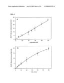 |
 |  |
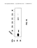 |  |
 | 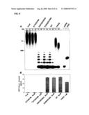 |
 |  |
 |  |
 | 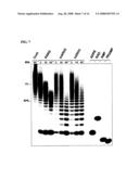 |
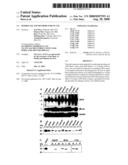 | 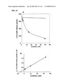 |
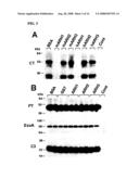 | 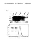 |
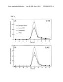 |  |
 |  |
 |  |
 |  |
 |
| Similar patent applications: | |
| Date | Title |
|---|---|
| 2010-09-23 | Ophthalmic formulations of ketotifen and methods of use |
| 2010-09-23 | Ophthalmic formulations of cetirizine and methods of use |
| 2010-09-23 | Novel 1,3-dihydro-5-isobenzofurancarbonitrile derivatives and pharmaceutical composition thereof for the treatment of premature ejaculation |
| 2010-09-16 | Substituted benzimidazoles and methods of their use |
| 2010-02-18 | Isolated mcpip and methods of use |
| New patent applications in this class: | |
| Date | Title |
|---|---|
| 2009-12-10 | Compounds and methods for modulating expression of pcsk9 |
| 2009-12-03 | Dsrna as insect control agent |
| 2009-11-26 | Oligomeric compounds and compositions for use in modulation of small non-coding rnas |
| 2009-11-26 | Oligomeric compounds and compositions for use in modulation of small non-coding rnas |
| 2009-11-19 | Novel oligonucleotide compositions and probe sequences useful for detection and analysis of micrornas and their target mrnas |
| New patent applications from these inventors: | |
| Date | Title |
|---|---|
| 2014-11-20 | Method for the treatment of pulmonary disease and method of producing proteins of use therein |
| 2012-06-28 | Method for the treatment of pulmonary disease and method of producing proteins of use therein |
| 2012-05-03 | Modified defensins and their use |
| 2012-02-16 | Methods for suppressing an immune response or treating a proliferative disorder |
| 2009-08-13 | Tryptophan as a functional replacement for adp-ribose-arginine in recombinant proteins |
| Top Inventors for class "Drug, bio-affecting and body treating compositions" | |
| Rank | Inventor's name |
|---|---|
| 1 | Anthony W. Czarnik |
| 2 | Ulrike Wachendorff-Neumann |
| 3 | Ken Chow |
| 4 | John E. Donello |
| 5 | Rajinder Singh |