Patent application title: Recombinant Monovalent Antibodies
Inventors:
Bernard Vanhove (Reze, FR)
Bernard Vanhove (Reze, FR)
Caroline Mary (Sainte Pazanne, FR)
Caroline Mary (Sainte Pazanne, FR)
Flora Coulon (Saint Georges De Montaigu, FR)
Assignees:
OSE Immunotherapeutics
INSTITUT NATIONAL DE LA SANTE ET DE LA RECHERCHE MEDICALE (INSERM)
IPC8 Class: AC07K1628FI
USPC Class:
1 1
Class name:
Publication date: 2017-06-15
Patent application number: 20170166643
Abstract:
The invention relates to recombinant monovalent antibodies which are
heterodimers of a first protein chain comprising the variable domain of
the heavy chain of an antibody of interest and the CH2 and CH3 domains of
an IgG immunoglobulin and a second protein chain comprising the variable
domain of the light chain of said immunoglobulin of interest and the CH2
and CH3 domains of said IgG immunoglobulin. These antibodies can be used
in particular as therapeutic agents in all cases where monovalent binding
to a ligand such a cellular receptor is required.Claims:
1. A recombinant antibody derived from a parent antibody directed against
an antigen of interest, wherein said recombinant antibody is an
heterodimer of: a first protein chain consisting essentially of, from its
N-terminus to its C-terminus: a region A having the structure of the
variable domain of the heavy chain of an immunoglobulin, said region A
comprising the CDRs of the heavy chain of said parent antibody; a region
B consisting of a peptide linker and the CH2 and CH3 domains of an IgG
immunoglobulin; a second protein chain consisting essentially of, from
its N-terminus to its C-terminus: a region A' having the structure of the
variable domain of the light chain of an immunoglobulin, said region A'
comprising the CDRs of the light chain of said parent antibody; a region
B identical to the region B of the first polypeptide.
2. A recombinant monovalent antibody of claim 1, wherein the peptide linker is a peptide sequence of 0 to 16 amino acids.
3. A recombinant antibody of claim 1, wherein the CH2 and CH3 domains are those of an immunoglobulin of the IgG1 subclass, or of the IgG4 subclass.
4. A recombinant monovalent antibody of claim 1, wherein the region A consists of the variable domain of the heavy chain of the parent antibody.
5. A recombinant monovalent antibody of claim 1, wherein the region A' consists of the variable domain of the light chain of the parent antibody.
6. A recombinant monovalent antibody of claim 1, wherein the parent antibody is the monoclonal immunoglobulin CD28.3, produced by the hybridoma deposited at Collection Nationale de Cultures de Microorganismes under Accession No. CNCM 1-2582.
7. A recombinant monovalent antibody according to claim 6, wherein the polypeptide sequence of the first protein chain is SEQ ID NO: 2, and the polypeptide sequence of the second protein chain is SEQ ID NO: 4.
8. A polynucleotide selected from the group consisting of: (a) a polynucleotide comprising a sequence encoding the first protein chain of a recombinant monovalent antibody according to claim 1; and (b) a polynucleotide comprising a sequence encoding the second protein chain of a recombinant monovalent antibody according to claim 1.
9. An expression vector comprising a polynucleotide of claim 8.
10. A cell transformed with the vector of claim 9.
11. A method for preparing a recombinant monovalent antibody, wherein said method comprises culturing the transformed cell of claim 10, and recovering said recombinant monovalent antibody from said culture.
12. A medicinal product comprising the recombinant monovalent antibody of claim 1.
13. A method of treating a pathological condition selected from the group consisting of transplant rejection, graft-versus-host disease, T-lymphocyte-mediated autoimmune diseases, allergic phenomena and chronic inflammatory diseases in a human subject suffering from said pathological condition comprising administering the recombinant monovalent antibody of claim 6 to the human subject.
Description:
[0001] The invention relates to recombinant monovalent antibodies, in
particular IgG antibodies, and to their therapeutic uses.
[0002] An antibody (immunoglobulin) molecule is a Y-shaped tetrameric protein composed of two heavy (H) and two light (L) polypeptide chains held together by covalent disulfide bonds and noncovalent interactions.
[0003] Each light chain is composed of one variable domain (VL) and one constant domain (CL). Each heavy chain has one variable domain (VH) and a constant region, which in the case of IgG, IgA, and IgD, comprises three domains termed CH1, CH2, and CH3 (IgM and IgE have a fourth domain, CH4). In IgG, IgA, and IgD classes the CHI and CH2 domains are separated by a flexible hinge region, which is a proline and cysteine rich segment of variable length (generally from about 10 to about 60 amino acids in IgG).
[0004] The variable domains show considerable variation in amino acid composition from one antibody to another. Each of the VH and the VL variable domains comprises three regions of extreme variability, which are termed the complementarity-determining regions (CDRs), separated by less variable regions called the framework regions (FRs). The non-covalent association between the VH and the VL region forms the Fv fragment (for "fragment variable") which contains one of the two antigen-binding sites of the antibody. ScFv fragments (for single chain fragment variable) , which can be obtained by genetic engineering, associates in a single polypeptide chain the VH and the VL region of an antibody, separated by a peptide linker.
[0005] Other functional immunoglobulin fragments can be obtained by proteolytic fragmentation of the immunoglobulin molecule. Papain treatment splits the molecule into three fragments: two heterodimeric Fab fragments (for `fragment antigen binding`), each associating the VL and CL domains of the light chain with the VH and CHI domains of the heavy chain, and one homodimeric Fc fragment (for "fragment crystalline"), which comprises the CH2 and CH3 (and eventually CH4) domains of the light chain. Pepsin treatment produces the F(ab)'2 fragment which associates two Fab fragments, and several small fragments.
[0006] The Fc fragment does not bind the antigen, but is responsible for the effector functions of the antibody, including in particular binding to Fc receptors and complement fixation. The Fv, Fab, and F(ab)'2 fragments retain the antigen-binding ability of the whole antibody. However, the F(ab)'2 fragments, like the whole immunoglobumin molecule, are divalent (i.e. they contain two antigen binding sites and can bind and precipitate the antigen), while the Fv and Fab fragments are monovalent (they contain one antigen binding site, and can bind but cannot precipitate the antigen).
[0007] Antibodies directed against cell-surface receptors are of great interest for the development of therapeutic agents for various disorders and diseases. They are generally used for their properties to mimic the structure of a biological ligand of a target receptor. In some cases this structural similarity may result in agonistic effects leading to the activation of the target receptor; in other cases it may result in antagonistic effects, leading to the blocking of the target receptor.
[0008] However, many antibodies having antagonistic properties when used as monovalent fragments may also show agonistic effects when used as full length antibodies. These agonistic effects result from the bivalency of the full-length antibodies, which induces the crosslinking of the target receptors on the cell surface, leading to receptor activation. This phenomenon is unwanted when the desired therapeutic activity relies upon an antagonistic effect. Examples of receptors that are activated by crosslinking include CD28, CD3 (DAMLE et al., J. Immunol., 140, 1753-61, 1988; ROUTLEDGE et al., Eur J Immunol, 21, 2717-25, 1991), TNF receptors, etc . . . .
[0009] The monovalent forms of antagonistic antibodies, such as Fab or scFv fragments, are devoid of agonistic activity. Therefore, they are useful therapeutic agents to block a cell receptor without inducing its cross-linking. However, their therapeutic use is hampered by their short half-life in vivo; they are eliminated within minutes and would require a continuous administration. To overcome this problem, it has been proposed to fuse these monovalent fragments with large molecules such as water-soluble proteins (PCT WO02051871) or polyethylene glycol (BLICK & CURRAN, BioDrugs, 21, 195-201; discussion 02-3, 2007).
[0010] Another approach for producing monovalent antibodies has been to construct fusion proteins associating one Fab fragment (i.e an heterodimer comprising the VL and the CL regions of the light chain, and the VH and the CH1 region of the heavy chain) with one Fc fragment (i.e an homodimer comprising the CH2 and CH3 regions of the heavy chains). ROUTLEDGE et al. (ROUTLEDGE et al., Eur J Immunol, 21, 2717-25, 1991) describe the construction of a monovalent antibody by introduction into an antibody-producing cell of a truncated Ig heavy chain gene encoding only the hinge, CH2 and CH3 domains; the expression of this gene in the antibody producing cell results in N-terminally truncated heavy chains (devoid of the VH and CH1 domains) which can either associate between them to form Fc molecules, or with full length heavy chains produced by the antibody producing cell to form a monovalent antibody molecules comprising a full-length light chain, a full-length heavy chain, and a N-terminally truncated heavy chain. PCT WO 2007/048037 describes monovalent antibodies which are heterodimers resulting from the association of an immunoglobulin heavy chain with a fusion protein comprising an immunoglobulin light chain and a Fc molecule.
[0011] An advantage of this approach is that the resulting antibodies contain an IgG Fc domain, which in some cases, is useful if one desires to retain some of the effector functions of the IgG molecule, and which also allows to target the molecule to the neonatal Fc receptor (FcRn) expressed by endothelial cells. This receptor actively traps several macromolecules, including antibodies, inside the blood stream conferring them an extended serum half-live. The binding of IgG molecules to this receptor facilitates their transport, and allows their protection from degradation.
[0012] The IgG Fc domain of immunoglobulins has also been utilized to form fusion proteins with molecules other than antibodies, for instance cytokines, growth factors, soluble growth factors, allowing to extend their half-life in the bloodstream, and also to deliver them by non-invasive routes, for instance by pulmonary administration (DUMONT et al., BioDrugs, 20, 151-60, 2006).
[0013] In the case of monovalent antibodies, the fusion proteins containing the IgG Fc domain which have been described until now also comprise the CL and/or the CH1 region. It is generally believed that these regions, which are part of the Fab fragment, play an important part in the correct assembly of the IgG molecule, and can also influence the antigen/antibody interaction.
[0014] As indicated above, one of the cell surface receptors known to be stimulated after its engagement by bivalent antibodies, and which can be efficiently blocked by certain monovalent fragments of some antibodies, is the CD28 receptor. By way of example, it has been shown that it was possible to efficiently block CD28 with Fab fragments or with a fusion protein comprising a scFv fragment of the anti-CD28 monoclonal antibody CD28.3, fused with alphal-antitrypsin (VANHOVE et al., Blood, 102, 564-70, 2003). This approach demonstrated an efficacy in vitro as well as in organ transplantation in mice and in primates (POIRIER et al., World Transplant Congress, Sydney, Australia. August 16-21, 2008).
[0015] The inventors have sought to further improve the pharmacokinetics properties of monovalent fragments of CD28.3. With this purpose, they have first attempted to construct a recombinant monovalent antibody similar to those disclosed in the prior art, by fusing the each of the VH and VL domains of CD28.3 to the CH1-CH2-CH3 domains of an heterologous IgG molecule. However this attempt failed to result in a protein with the required antibody activity.
[0016] The inventors then tried to remove the CH1 domains of these fusion proteins and found that the resulting monovalent antibody was secreted and active, and that it behaves in vitro like its corresponding Fab fragment. Further, after intravenous injection in mice, it showed an elimination half-live that was significantly longer than Fab fragments and not significantly different from IgG antibodies.
[0017] These results show that combining the variable domains of a monoclonal antibody with only the CH2-CH3 domains rather than with all the constant domains of an IgG molecule allows to obtain a functional monovalent antibody, having the prolonged in vivo half-live that is conferred by the presence of an Fc fragment. This format can be used to generate therapeutic antibodies in all cases where monovalent binding to a ligand, for instance a cellular receptor, is required. Therefore, an object of the present invention is a recombinant monovalent antibody derived from a parent antibody directed against an antigen of interest, wherein said recombinant antibody is an heterodimer of:
[0018] a first protein chain consisting essentially of, from its N-terminus to its C-terminus:
[0019] a region A having the structure of the VH domain of an immunoglobulin, said region A comprising the CDRs of the heavy chain of said parent antibody;
[0020] a region B consisting of a peptide linker and the CH2 and CH3 domains of an IgG immunoglobulin;
[0021] a second protein chain consisting essentially of, from its N-terminus to its C-terminus:
[0022] a region A' having the structure of the VL domain of an immunoglobulin, said region A' comprising the CDRs of the light chain of said parent antibody;
[0023] a region B identical to the region B of the first polypeptide.
[0024] The parent antibody can be any antibody directed against the antigen of interest; it can be a native monoclonal antibody; it can also be a recombinant or synthetic antibody, such as a chimeric antibody, a humanized antibody, or an antibody originating from phage-display or ribosome display technologies.
[0025] A region having the structure of the VH or of the VL domain of an immunoglobulin comprises, as indicated above, four framework regions (FRs), connected by three hypervariable regions or complementarity determining regions (CDRs) which are involved in antigen recognition specificity. In a recombinant monovalent antibody of the invention, regions A and A' can consist of the native VH or VL domains of the parent antibody; however, they can also be obtained by incorporating the CDRs of the parent antibody into the framework regions (FRs) of another antibody, in particular of an antibody of human origin, using techniques, known in themselves, of CDR grafting.
[0026] The peptide linker of region B may comprises from 0 to 16 amino acids. It comprises preferably 5 to 7 amino acids. Examples of suitable peptide linkers are those which are used in the construction of scFv fragments, such are those disclosed for instance by FREUND et al. (FEBS Lett. 320, 97-100, 1993) or by SHAN et al. (J Immunol. 162, 6589-95, 1999).
[0027] Said peptide linker may be devoid of cysteine residues, or may comprise one or more cysteine residue(s). A peptide linker devoid of cysteine residues will be preferred if the monovalent antibody is to be produced in the E. coli periplasm. An example of a peptide linker devoid of cysteine residues is a peptide having the sequence TVAAPS (SEQ ID NO: 5).
[0028] Alternatively, a peptide linker comprising cysteine residues allows the formation of inter-chain disulfide bonds, which help to stabilize the heterodimer. As a peptide linker comprising cysteine residues, one can use for instance the hinge region of a naturally occurring IgG. A preferred hinge region is the hinge region of IgG2 immunoglobulins having the sequence ERKCCVECPPCP (SEQ ID NO: 12), which provides a high stability.
[0029] The CH2 and CH3 domains are preferably those of an immunoglobulin of human origin of the IgG isotype. Said IgG can belong to any of the IgG subclasses (IgG1, IgG2, IgG3 or IgG4). Preferably, it belongs to the IgG1 subclass or the IgG4 subclass.
[0030] Besides the essential constituents listed above, the first and/or the second protein chain can further comprise one or more optional polypeptide sequence(s) which is (are) not involved in the biological properties of the recombinant monovalent antibody, but may facilitate its detection or purification. For instance said polypeptide sequence can be a tag polypeptide, such as a streptavidin-binding peptide, an hexa-histidine (His.sub.6) tag, or a FLAG-tag.
[0031] The first and/or the second protein chain can be glycosylated or not.
[0032] According to a particular embodiment of the invention, the parent antibody is the monoclonal antibody CD28.3, produced by the hybridoma CNCM 1-2582. The hybridoma CNCM 1-2582 is disclosed in PCT WO02051871, and has been deposited, according to the terms of the Treaty of Budapest, on Nov. 28, 2000, with the CNCM (Collection Nationale de Cultures de Microorganismes, 25 rue du Docteur Roux, 75724 PARIS CEDEX 15).
[0033] A particular example of a recombinant monovalent antibody of the invention, which is described in detail in the Examples below, is an antibody wherein the polypeptide sequence of the first protein chain is SEQ ID NO: 2, and the polypeptide sequence of the second protein chain is SEQ ID NO: 4. Another example of a recombinant monovalent antibody of the invention, is an antibody wherein the polypeptide sequence of the first protein chain is SEQ ID NO: 13, and the polypeptide sequence of the second protein chain is SEQ ID NO: 14.
[0034] Another object of the invention is a polynucleotide comprising a sequence encoding the first protein chain and/or a sequence encoding the second protein chain of a recombinant monovalent antibody of the invention. Said polynucleotides may also comprise additional sequences: for instance they may advantageously comprise a sequence encoding a leader sequence or signal peptide allowing secretion of said protein chain. They may optionally also comprise one or more sequence(s) encoding one or more tag polypeptide(s).
[0035] The present invention also encompasses recombinant vectors, in particular expression vectors, comprising a polynucleotide of the invention, associated with transcription- and translation-controlling elements which are active in the host cell chosen. Vectors which can be used to construct expression vectors in accordance with the invention are known in themselves, and will be chosen in particular as a function of the host cell intended to be used.
[0036] The present invention also encompasses host-cells transformed with a polynucleotide of the invention. Preferably, said host cell is transformed with a polynucleotide comprising a sequence encoding the first protein chain of a recombinant monovalent antibody of the invention and a polynucleotide comprising a sequence encoding the second protein chain of a recombinant monovalent antibody of the invention, and expresses said recombinant antibody. Said polynucleotides can be inserted in the same expression vector, or in two separate expression vectors.
[0037] Host cells which can be used in the context of the present invention can be prokaryotic or eukaryotic cells. Among the eukaryotic cells which can be used, mention will in particular be made of plant cells, cells from yeast, such as Saccharomyces, insect cells, such as Drosophila or Spodoptera cells, and mammalian cells such as HeLa, CHO, 3T3, C127, BHK, COS, etc., cells.
[0038] The construction of expression vectors of the invention and the transformation of the host cells can be carried out by the conventional techniques of molecular biology.
[0039] Still another objet of the invention is a method for preparing a recombinant monovalent antibody of the invention, Said method comprises culturing an host-cell transformed with a polynucleotide comprising a sequence encoding the first protein chain of a recombinant monovalent antibody of the invention, and with a polynucleotide comprising a sequence encoding the second protein chain of a recombinant monovalent antibody of the invention, and recovering said recombinant monovalent antibody from said culture.
[0040] If the protein is secreted by the host-cell, it can be recovered directly from the culture medium; if not, cell lysis will be carried out beforehand. The protein can then be purified from the culture medium or from the cell lysate, by conventional procedures, known in themselves to those skilled in the art, for example by fractionated precipitation, in particular precipitation with ammonium sulfate, electrophoresis, gel filtration, affinity chromatography, etc.
[0041] A subject of the invention is also a method for producing a protein in accordance with the invention, characterized in that it comprises culturing at least one cell in accordance with the invention, and recovering said protein from said culture.
[0042] The recombinant monovalent antibodies of the invention can be used to obtain medicinal products. These medicinal products are also part of the object of the invention.
[0043] For instance, recombinant monovalent antibodies of the invention derived from the parent antibody CD28.3 can be used to obtain immunosuppressant medicinal products which selectively blocks T cell activation phenomena involving the CD28 receptor. Such immunosuppressant medicinal products which act by selective blocking of CD28 have applications in all T lymphocyte-dependent pathological conditions, including in particular transplant rejection, graft-versus-host disease, T lymphocyte-mediated autoimmune diseases, such as type I diabetes, rheumatoid arthritis or multiple sclerosis, and type IV hypersensitivity, which is involved in allergic phenomena and also in the pathogenesis of chronic inflammatory diseases, in particular following infection with a pathogenic agent (in particular leprosy, tuberculosis, leishmaniasis, listeriosis, etc.).
[0044] The present invention will be understood more clearly from the further description which follows, which refers to nonlimiting examples of the preparation and properties of a recombinant monovalent antibody (hereafter referred to as Mono28Fc) in accordance with the invention.
LEGENDS OF THE DRAWINGS
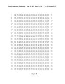
[0045] FIG. 1A: Nucleotidic and amino acid sequence of Mono28Fc, VH-CH2CH3 chain.
[0046] Underlined: VH domain. Bold: linker. Double underlining: IgG1 CH2-CH3 domains.
[0047] FIG. 1B: Nucleotidic and amino acid sequence of Mono28Fc, VL-CH2CH3 chain.
[0048] Underlined: VL domain. Bold: linker. Double underlining: IgG1 CH2-CH3 domains.
[0049] FIG. 1C: Molecular constructions allowing the expression of Mono28Fc after transfection into eukaryotic host cells.
[0050] pCMV: promoter of the cytomegalovirus. Igk leader: signal sequence from the mouse immunoglobulin kappa light chain. VH: variable domain of the heavy chain of the CD28.3 antibody. VL: variable domain of the light chain of the CD28.3 antibody. CH2 and CH3 represent the corresponding domains of the IgG1 human immunoglobulin.
[0051] FIG. 1D: Expression plasmids for the synthesis of Mono28Fc in eukaryotic cells.
[0052] A: plasmid for the synthesis of the VH(Hc)-CH2-CH3 protein. B: plasmid for the synthesis of the VL(Lc)-CH2-CH3 protein. pCMV: promoter of the cytomegalovirus. Igk leader: signal sequence from the mouse immunoglobulin kappa light chain. Hc: VL variable domain of the heavy chain of the CD28.3 antibody. Lc: VL variable domain of the light chain of the CD28.3 antibody. CH2 and CH3 represent the corresponding domains of the IgG1 human immunoglobulin. BGH pA: signal for the initiation of the 3' polyadenylation of the mRNA molecule, from the bovine growth hormone. Zeocin, ampicillin: resistance genes for the corresponding antibiotic.
[0053] FIG. 2: Western blot analysis of pSecVHFc and pSecVLFc expression.
[0054] A: Supernatants from Cos cells transfected with the indicated plasmids were collected and reduced before analysis by 10 min. incubation at 100.degree. C. with 10 mM DTT. B: no reduction. Molecular weights are indicated on the left sides.
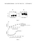
[0055] FIG. 3: Activity ELISA. Recombinant CD28 was immobilized on microtitration plates.
[0056] A: Supernatants from control, transfected or co-transfected Cos cells were added at the indicated dilutions, washed and revealed with rabbit anti-VHNL antibodies plus anti-rabbit immunoglobulins-HRP. GFP: negative control; transfection with an irrelevant GFP plasmid. Sc28AT: positive control; transfection with a plasmid coding for a single-chain Fv against CD28. VLFc: transfection with the pSec-VLFc plasmid. VHFc: transfection with the pSec-VHFc plasmid. VH-(CH2-CH3)+VL-CH2-CH3: co-transfection with the pSec-VLFc and the pSec-VHFc plasmids. B: Binding ELISA on recombinant CD28 of purified Mono28Fc molecules at the indicated concentration. Revelation is as in A. Dots are means of triplicates.
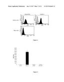
[0057] FIG. 4: Flow cytometry.
[0058] CD28.sup.+ Jurkat T cells and CD28.sup.- Raji B cells were incubated with purified Mono28Fc or with CD28.3 Fab fragments at 10 .mu.g/ml for 30 min. at 4.degree. C., washed and revealed with rabbit anti-VHNH antibodies plus FITC-labeled goat anti-rabbit immunoglobulins (black profiles). As a control, cells were incubated with rabbit anti-VHNH antibodies plus FITC-labeled goat anti-rabbit immunoglobulins only (grey profiles). Cells were then washed, fixed and analyzed by Facs.
[0059] FIG. 5: Activation assay.
[0060] Human PBMC (10.sup.5/well) were cultivated in medium or in medium plus 10 .mu.g/ml Mono28Fc, sc28AT monovalent antibodies or with ANC28.1 superagonist antibodies for 3 days. 0.5 .mu.Ci .sup.3H-tymidine was added for the last 16 h of the culture. Incorporated radioactivity was evaluated on a scintillation counter after transfer on nitrocellulose membranes.
[0061] FIG. 6: Pharmacokinetic in mice.
[0062] A: Indicated proteins were injected i.v. into swiss mice and blood samples were collected after the indicated time points. CD28 binding activity was measured by
[0063] ELISA. N=4 for each point, dots are means of the 4 measurements. B: Elimination half-lives (T.sub.1/2.beta.) were calculated from the curves in A.
[0064] FIG. 7: Molecular constructions combining VH, VL with CH1-CH2-CH3 are non-functional.
[0065] A: RT-PCR analysis of the VH and VL mRNA chains expression after transfection of Cos cells. B: Western blot analysis of supernatants (right panel) and lysates (left panel) of Cos cell transfected with pSecVH-CH1-CH2-CH3 and pSecVL-CH1-CH2-CH3 plasmids. Revelation was performed as in FIG. 2. C: Immunofluorescence analysis of Cos cells transfected with pSec-VH-CH1-CH2-CH3 and pSec-VL-CH1-CH2-CH3; revelation with rabbit anti-VH/VL antibody plus anti-PE. Magnification: 20.times.. D: Activity ELISA of supernatants of Cos cell co-transfected with pSecVH-CHI-CH2-CH3 and pSecVL-CH1-CH2-CH3. Revelation was performed as in FIG. 3.
[0066] FIG. 8: Amino acid sequence of Mono28Fc with IgG2 hinge and IgG4 CH2CH3 domains.
[0067] A: VH-CH2CH3 chain: Underlined: VH domain. Bold: linker. Double underlining: IgG4 CH2-CH3 domains.
[0068] B: VL-CH2CH3 chain: Underlined: VL domain. Bold: linker. Double underlining: IgG4 CH2-CH3 domains.
EXAMPLE 1: CONSTRUCTION OF THE MONOVALENT ANTIBODY MONO28Fc
[0069] The CH2-CH3 domains of a human IgG1 gene (NCBI Accession BC018747) was amplified using the following primers introducing NheI/XbaI sites: CH2CH3-5':
TABLE-US-00001 (SEQ ID NO: 6) 5'-ATATGCTAGCCCAGCACCTGAACTCCTG-3'; CH2CH3-3': (SEQ ID NO: 7) 5'-ATATTCTAGATTATTTACCCGGAGA-3'.
[0070] The resulting fragment was introduced into the pSC-A vector (Stratagene, Amsterdam, The Netherlands), resulting in the pSC-A-CH2-CH3 vector.
[0071] VH and VL domains corresponding to the CD28.3 antibody anti-human CD28 were amplified from the previously described CNCM 1-2762 scFv cDNA (VANHOVE et al., Blood, 102, 564-70, 2003) and Nhel cloning sites were introduced by PCR with the following primers: VH:
TABLE-US-00002 Hc28.3-5': (SEQ ID NO: 8) 5'-ATATGCTAGCGGATCCGATATCGTCAAGCTGCAGCAGTCA-3'; Hc28.3-3': (SEQ ID NO: 9) 5'-ATATGCTAGCAGATGGTGCAGCCACAGTTGAGGAGACGGTGACCA T-3'; VL: Lc28.3-5': (SEQ ID NO: 10) 5'-ATATGCTAGCGGATCCGATATCGACATCCAGATGACCCAG-3'; Lc28.3-3': (SEQ ID NO: 11) 5'-ATATGCTAGCAGATGGTGCAGCCACAGTCCGTTTTATTTCCAGCTTG G-3'.
[0072] The VH and VL fragments were cloned individually 5' to the CH2-CH3 domains into the NheI site of the pSC-A-CH2-CH3 vector, resulting in VH-pSC-A-CH2-CH3 and VL-pSC-A-CH2-CH3 plasmids. The nucleotidic and amino acid sequences of the resulting VH-CH2CH3 and VL-CH2CH3 constructs are indicated respectively on FIGS. 1A and 1B. They are also indicated as SEQ ID NO: 1 and 3.
[0073] Each construct was then subcloned in the EcoRV restriction site of the pSecTag2B eukaryotic pCMV-based expression plasmid (Invitrogen, Cergy Pontoise, France), enabling a fusion at the N-terminus with the secretion signal from the V-J2-C region of the mouse Ig kappa-chain provided by the pSecTag2 vector. The constructs were proofread by sequencing. The resulting expression cassettes and the plasmids pSec-VH-Fc(CH2-CH3) and pSec-VL-Fc(CH2-CH3) containing these constructs are schematized respectively on FIGS. 1C and 1D.
EXAMPLE 2: EUCARYOTIC EXPRESSION OF MONO28Fc
[0074] COS cells were transfected separately with pSec-VH-Fc(CH2-CH3) (VH-Fc) or pSec-VL-Fc(CH2-CH3) (VL-Fc), or co-transfected with pSec-VH-Fc(CH2-CH3) and pSec-VL-Fc(CH2-CH3) or, as a control, transfected with a plasmid coding for an irrelevant green fluorescent protein (GFP), using the Fugene lipofection kit (Roche Diagnostics, Basel, Switzerland) according to the manufacturer's instructions. Cultures were maintained for 3 days at 37.degree. C., divided one third, and put back into culture for an additional 3 days, after which time the cell supernatants were collected, electrophoresed in 10% polyacrylamide gels and blotted onto nitrocellulose membranes.
[0075] Blots were revealed with rabbit anti-CD28.3VH/VL (1:5000 dilution) and an HRP-conjugated donkey antirabbit Ig antibody (Jackson Immuno-Research Laboratories) and developed by chemiluminescence (Amersham Pharmacia Biotech).
[0076] The results are shown on FIG. 2. Immunoreactive proteins of the expected molecular weight (42 KDa for VL-CH2-CH3 and 44KDa for VH-CH2-CH3 under reducing conditions) could be observed in the cell supernatant. A parallel analysis with non-reducing conditions indicated an apparent molecular weight compatible with the formation of both homodimers and heterodimers.
EXAMPLE 3: DETECTION OF MONO28Fc BINDING ACTIVITY BY ELISA
[0077] Recombinant human CD28 (R&D Systems, Abingdon, United Kingdom) was used at 1.mu.g/mL in borate buffer (pH 9.0) to coat 96-well microtiter plates (Immulon, Chantilly, VA) overnight at 4.degree. C. These immobilized CD28 target molecules will bind only immunoreactive molecules with anti-CD28 activity.
[0078] Reactive sites were blocked with 5% skimmed milk in PBS for 2 hours at 37.degree. C. and supernatants from control cells transfected with the plasmid coding for GFP, from cells transfected with only one of the plasmids pSec-VH-Fc(CH2-CH3) or pSec-VL-Fc(CH2-CH3), and from cells co-transfected with pSec-VH-Fc(CH2-CH3) and pSec-VL-Fc(CH2- CH3) were added at different dilutions and reacted for 2 hours at 37.degree. C. Bound Fc fusion proteins with anti-28 activity were revealed with successive incubations (1 hour, 37.degree. C.) with rabbit anti-CD28.3VH/VL (1:2000 dilution; custom preparation at Agrobio, Orleans, France) and horseradish peroxidase (HRP)--conjugated donkey antirabbit Ig antibodies (1:500 dilution; Jackson ImmunoResearch Laboratories, Bar Harbor, Me.). Bound antibody was revealed by colorimetry using the TMB substrate (Sigma, L'Isle d'Abeau Chesnes, France) read at 450 nm.
[0079] The results are shown on FIG. 3 A.
[0080] Supernatants from control cells (transfected with the plasmid coding for GFP) or from cells transfected with only one of the plasmids pSec-VH-Fc(CH2-CH3) or pSec-VL-Fc(CH2-CH3) did not contain any detectable level of immunoreactive molecule. This indicated that VH-Fc or VL-Fc homodimers cannot bind CD28. In contrast, supernatants from cells co-transfected with pSec-VH-Fc(CH2-CH3) and pSec-VL-Fc(CH2-CH3) contained dilution-dependant levels of immunoreactive molecules.
[0081] Mono28Fc was purified from culture supernatants of COS cells co-transfected with pSec-VH-Fc(CH2-CH3) and pSec-VL-Fc(CH2-CH3) and maintained for 3 days at 37.degree. C.
[0082] Supernatants were passed through G-Protein Sepharose columns (Amersham) at a rate of 1 ml/min. The columns were rinsed with PBS and proteins were eluted with glycine buffer (pH 2.8), concentred by osmotic water retrieval using polyethylene glycol (Fluka, Riedel-de Haen, Germany) and dialysed extensively against PBS at 4.degree. C.
[0083] After purification, the Mono28Fc molecules were tested by ELISA as described above. The results are shown on FIG. 3B.
[0084] These results show that 50% of the binding activity to CD28 could be reached at a concentration of 100 ng/ml, which represents 1.16 nM.
EXAMPLE 4: DETECTION OF MONO28Fc BINDING ACTIVITY BY FLOW CYTOMETRY
[0085] The binding of Mono28Fc was confirmed by flow cytometry using CD28+ Jurkat human T cells, which express CD28, or on Raji cells, a human B cell line that does not express CD28.
[0086] Jurkat T cells or Raji cells were incubated for 1 hour at 4.degree. C. with purified Mono28Fc proteins or with Fab fragments of CD28.3 (VANHOVE et al., Blood, 102, 564-70, 2003), at 10 .mu.g/ml for 30 min. As a control, cells were incubated with rabbit anti-VII/VH antibodies plus FITC-labeled goat anti-rabbit immunoglobulins only. Bound Fc fusion monomers were detected with a rabbit anti-CD28.3VH/VL and a fluorescein isothiocyanate (FITC)--conjugated donkey anti-rabbit Ig antibody (dilution 1:200; Jackson ImmunoResearch Laboratories) for 30 minutes at 4.degree. C. Cells were then analyzed by fluorescence-activated cell sorting (FACS).
[0087] The results are shown on FIG. 4. Both mono28Fc and the Fab fragment of CD28.3 bind Jurkat T cells. In contrast, no binding of the mono28Fc protein could be observed on Raji cells, a human B cell line that does not express CD28. These data demonstrate mono28Fc that binds specifically to CD28.sup.+ cells.
EXAMPLE 5: MONO28Fc HAS NO AGONIST ACTIVITY ON HUMAN T CELLS
[0088] To verify that mono28Fc binds to CD28 and does not induce activation of the target T cell, we compared the biological effect of Mono28Fc with those of the superagonistic antibody ANC28.1 (WAIBLER et al., PLoS ONE, 3, e1708, 2008), or of sc28AT, a monovalent anti-CD28 ligand without Fc domain (VANHOVE et al., Blood, 102, 564-70, 2003).
[0089] Human PBMC (10.sup.5/well) were cultivated in culture medium without additive (control), or in culture medium with 10 .mu.g/ml of mono28Fc, of sc28AT, or of ANC28.1 for 3 days. 0.5 .mu.Ci .sup.3H-tymidine was added for the last 16 h of the culture. Incorporated radioactivity was evaluated on a scintillation counter after transfer on nitrocellulose membranes. The results are shown on FIG. 5.
[0090] As expected, ANC28.1 induced a robust proliferation of the target cells. In contrast, Mono28Fc, as well as sc28AT did not induce any response in this assay.
EXAMPLE 6: PHARMACOKINETICS OF MONO28Fc IN MICE
[0091] Recombinant proteins fused with an Fc fragment and immunoglobulins usually present an extended half-life in vivo because they are recognised by the FcRn receptor presented on endothelial and epithelial cells allowing the recycling of that molecules back in the circulation. To determine if our Mono28FC molecule also presents an extended half-live, we followed the distribution in mice of Mono28Fc in comparison with monovalent Fab 28.3 antibody fragments and native IgG CD28.3 antibodies.
[0092] Each protein tested (288 .mu.g per injection) was injected into the tail vein of male Swiss mice. Blood samples (2 .mu.L) were collected at different times from the tail vein. The proteins were quantified by measuring the CD28 binding activity in blood samples by
[0093] ELISA. The data were analyzed by Siphar software (Simed, Utrecht, The Netherlands) with the use of a 2-compartment model. Significance was evaluated with an non-parametric ANOVA test followed by a Bonferroni's Multiple Comparison Test.
[0094] The results are shown on FIG. 6.
[0095] The distribution half-live (T.sub.1/2.alpha.) was of 2.5.+-.1.1; 5.1.+-.0.3 and 5.4.+-.1.2 hours for IgG, Fab and Mono28Fc, respectively. The elimination half-live (T.sub.1/2.beta.) was of 119.+-.19; 39.+-.6 and 83.+-.26 hours for IgG, Fab and Mono28Fc, respectively (FIG. 6). The data reveal a significant increase of the elimination half-live of Mono28Fc, as compared with Fab fragments, whereas no statistical difference is pointed out when Mono28Fc is compared with a divalent IgG.
EXAMPLE 7: COMPARISON OF MONO28Fc WITH A CONSTRUCTION COMPRISING THE CH1-CH2-CH3 IG HEAVY CHAIN DOMAINS
[0096] The human IgG1 CH1-CH2-CH3 cDNA was given by Dr. S. Birkle (Univ. Nantes, France). It was inserted into the pcDNA3.1 into the HindIII/BamHI restriction sites, resulting in the pcDNA3.1-CH1-CH2-CH3 plasmid. VH and VL domains corresponding to the CD28.3 antibody anti-human CD28 (NUNES et al., Int Immunol, 5, 311-5, 1993) were amplified as described in Example 1 above, digested with the Nhel enzyme and inserted separately into the Nhel site of the pcDNA3.1-CH1-CH2-CH3 plasmid. The VH-CH1-CH2-CH3 and VL-CH1-CH2-CH3 cassettes were then excised by EcoRV/XbaI digestion and inserted into the EcoR V digested pSecTag2B vector (Invitrogen), as disclosed in Example 1.
[0097] After transfection in Cos cells, messenger RNA molecules corresponding to the two chains were equally synthesised (FIG. 7A). The analysis of proteins by western blotting revealed the synthesis of some corresponding molecules, although clearly more abundant within the cell (FIG. 7B, left panel) than in the supernatant (FIG. 7B, right panel) for the light chain (VL-CH1-CH2-CH3). By immunohistology, the synthesis of both heavy and light chains by transfected Cos cells could be confirmed (FIG. 7C). By ELISA, no CD28 binding activity could be detected in the supernatant (data not shown) nor in transfected cell lysates (FIG. 7D).
Sequence CWU
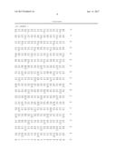
1
1
1411041DNAArtificial SequenceChimeric construct 1gtc aag ctg cag cag tca
gga gct gag ctg gtg aaa ccc ggg gcg tcg 48Val Lys Leu Gln Gln Ser
Gly Ala Glu Leu Val Lys Pro Gly Ala Ser 1 5
10 15 gtg agg ctg tcc tgc aag gcg
tct ggt tac acc ttc act gaa tat att 96Val Arg Leu Ser Cys Lys Ala
Ser Gly Tyr Thr Phe Thr Glu Tyr Ile 20
25 30 ata cac tgg ata aag ctg agg tct
gga cag ggt ctt gag tgg att ggg 144Ile His Trp Ile Lys Leu Arg Ser
Gly Gln Gly Leu Glu Trp Ile Gly 35 40
45 tgg ttt tac cct gga agt aat gat ata
cag tac aat gcg aaa ttc aag 192Trp Phe Tyr Pro Gly Ser Asn Asp Ile
Gln Tyr Asn Ala Lys Phe Lys 50 55
60 ggc aag gcc aca ttg act gcg gac aaa tcc
tcc agc acc gtc tat atg 240Gly Lys Ala Thr Leu Thr Ala Asp Lys Ser
Ser Ser Thr Val Tyr Met 65 70
75 80 gaa ctt act gga ttg aca tct gag gac tct
gcg gtc tat ttc tgt gca 288Glu Leu Thr Gly Leu Thr Ser Glu Asp Ser
Ala Val Tyr Phe Cys Ala 85 90
95 aga cgc gac gat ttc tct ggt tac gac gcc ctt
cct tac tgg ggc caa 336Arg Arg Asp Asp Phe Ser Gly Tyr Asp Ala Leu
Pro Tyr Trp Gly Gln 100 105
110 ggg acc atg gtc acc gtc tcc tca act gtg gct gca
cca tct gct agc 384Gly Thr Met Val Thr Val Ser Ser Thr Val Ala Ala
Pro Ser Ala Ser 115 120
125 cca gca cct gaa ctc ctg ggg gga ccg tca gtc ttc
ctc ttc ccc cca 432Pro Ala Pro Glu Leu Leu Gly Gly Pro Ser Val Phe
Leu Phe Pro Pro 130 135 140
aaa ccc aag gac acc ctc atg atc tcc cgg acc cct gag
gtc aca tgc 480Lys Pro Lys Asp Thr Leu Met Ile Ser Arg Thr Pro Glu
Val Thr Cys 145 150 155
160 gtg gtg gtg gac gtg agc cac gaa gac cct gag gtc aag ttc
aac tgg 528Val Val Val Asp Val Ser His Glu Asp Pro Glu Val Lys Phe
Asn Trp 165 170
175 tac gtg gac ggc gtg gag gtg cat aat gcc aag aca aag ccg
cgg gag 576Tyr Val Asp Gly Val Glu Val His Asn Ala Lys Thr Lys Pro
Arg Glu 180 185 190
gag cag tac aac agc acg tac cgt gtg gtc agc gtc ctc acc gtc
ctg 624Glu Gln Tyr Asn Ser Thr Tyr Arg Val Val Ser Val Leu Thr Val
Leu 195 200 205
cac cag gac tgg ctg aat ggc aag gag tac aag tgc aag gtc tcc aac
672His Gln Asp Trp Leu Asn Gly Lys Glu Tyr Lys Cys Lys Val Ser Asn
210 215 220
aaa gcc ctc cca gcc ccc atc gag aaa acc atc tcc aaa gcc aaa ggg
720Lys Ala Leu Pro Ala Pro Ile Glu Lys Thr Ile Ser Lys Ala Lys Gly
225 230 235 240
cag ccc cga gaa cca cag gtg tac acc ctg ccc cca tcc cgg gag gag
768Gln Pro Arg Glu Pro Gln Val Tyr Thr Leu Pro Pro Ser Arg Glu Glu
245 250 255
atg acc aag aac cag gtc agc ctg acc tgc ctg gtc aaa ggc ttc tat
816Met Thr Lys Asn Gln Val Ser Leu Thr Cys Leu Val Lys Gly Phe Tyr
260 265 270
ccc agc gac atc gcc gtg gag tgg gag agc aat ggg cag ccg gag aac
864Pro Ser Asp Ile Ala Val Glu Trp Glu Ser Asn Gly Gln Pro Glu Asn
275 280 285
aac tac aag acc acg cct ccc gtg ctg gac tcc gac ggc tcc ttc ttc
912Asn Tyr Lys Thr Thr Pro Pro Val Leu Asp Ser Asp Gly Ser Phe Phe
290 295 300
ctc tat agc aag ctc acc gtg gac aag agc agg tgg cag cag ggg aac
960Leu Tyr Ser Lys Leu Thr Val Asp Lys Ser Arg Trp Gln Gln Gly Asn
305 310 315 320
gtc ttc tca tgc tcc gtg atg cat gag gct ctg cac aac cac tac acg
1008Val Phe Ser Cys Ser Val Met His Glu Ala Leu His Asn His Tyr Thr
325 330 335
cag aag agc ctc tcc ctg tct ccg ggt aaa taa
1041Gln Lys Ser Leu Ser Leu Ser Pro Gly Lys
340 345
2346PRTArtificial SequenceSynthetic Construct 2Val Lys Leu Gln Gln Ser
Gly Ala Glu Leu Val Lys Pro Gly Ala Ser 1 5
10 15 Val Arg Leu Ser Cys Lys Ala Ser Gly Tyr Thr
Phe Thr Glu Tyr Ile 20 25
30 Ile His Trp Ile Lys Leu Arg Ser Gly Gln Gly Leu Glu Trp Ile
Gly 35 40 45 Trp
Phe Tyr Pro Gly Ser Asn Asp Ile Gln Tyr Asn Ala Lys Phe Lys 50
55 60 Gly Lys Ala Thr Leu Thr
Ala Asp Lys Ser Ser Ser Thr Val Tyr Met 65 70
75 80 Glu Leu Thr Gly Leu Thr Ser Glu Asp Ser Ala
Val Tyr Phe Cys Ala 85 90
95 Arg Arg Asp Asp Phe Ser Gly Tyr Asp Ala Leu Pro Tyr Trp Gly Gln
100 105 110 Gly Thr
Met Val Thr Val Ser Ser Thr Val Ala Ala Pro Ser Ala Ser 115
120 125 Pro Ala Pro Glu Leu Leu Gly
Gly Pro Ser Val Phe Leu Phe Pro Pro 130 135
140 Lys Pro Lys Asp Thr Leu Met Ile Ser Arg Thr
Pro Glu Val Thr Cys 145 150 155
160 Val Val Val Asp Val Ser His Glu Asp Pro Glu Val Lys Phe Asn Trp
165 170 175 Tyr Val
Asp Gly Val Glu Val His Asn Ala Lys Thr Lys Pro Arg Glu 180
185 190 Glu Gln Tyr Asn Ser Thr Tyr
Arg Val Val Ser Val Leu Thr Val Leu 195 200
205 His Gln Asp Trp Leu Asn Gly Lys Glu Tyr Lys Cys
Lys Val Ser Asn 210 215 220
Lys Ala Leu Pro Ala Pro Ile Glu Lys Thr Ile Ser Lys Ala Lys Gly 225
230 235 240 Gln Pro Arg
Glu Pro Gln Val Tyr Thr Leu Pro Pro Ser Arg Glu Glu 245
250 255 Met Thr Lys Asn Gln Val Ser Leu
Thr Cys Leu Val Lys Gly Phe Tyr 260 265
270 Pro Ser Asp Ile Ala Val Glu Trp Glu Ser Asn Gly Gln
Pro Glu Asn 275 280 285
Asn Tyr Lys Thr Thr Pro Pro Val Leu Asp Ser Asp Gly Ser Phe Phe 290
295 300 Leu Tyr Ser Lys
Leu Thr Val Asp Lys Ser Arg Trp Gln Gln Gly Asn 305 310
315 320 Val Phe Ser Cys Ser Val Met His Glu
Ala Leu His Asn His Tyr Thr 325 330
335 Gln Lys Ser Leu Ser Leu Ser Pro Gly Lys 340
345 31005DNAArtificial SequenceChimeric construct
3gac atc cag atg acc cag tct cca gcc tcc cta tct gtt tct gtg gga
48Asp Ile Gln Met Thr Gln Ser Pro Ala Ser Leu Ser Val Ser Val Gly
1 5 10 15
gaa act gtc acc atc acg tgt cga aca aat gaa aat att tac agt aat
96Glu Thr Val Thr Ile Thr Cys Arg Thr Asn Glu Asn Ile Tyr Ser Asn
20 25 30
tta gca tgg tat cag cag aaa cag gga aaa tct cct cag ctc ctg atc
144Leu Ala Trp Tyr Gln Gln Lys Gln Gly Lys Ser Pro Gln Leu Leu Ile
35 40 45
tat gct gca aca cac tta gta gag ggt gtg cca tca agg ttc agt ggc
192Tyr Ala Ala Thr His Leu Val Glu Gly Val Pro Ser Arg Phe Ser Gly
50 55 60
agt gga tca ggc aca cag tat tcc ctc aag atc acc agc ctg cag tct
240Ser Gly Ser Gly Thr Gln Tyr Ser Leu Lys Ile Thr Ser Leu Gln Ser
65 70 75 80
gaa gat ttt ggg aat tat tac tgt caa cac ttt tgg ggt act ccg tgc
288Glu Asp Phe Gly Asn Tyr Tyr Cys Gln His Phe Trp Gly Thr Pro Cys
85 90 95
acg ttc gga ggg ggg acc aag ctg gaa ata aaa cgg act gtg gct gca
336Thr Phe Gly Gly Gly Thr Lys Leu Glu Ile Lys Arg Thr Val Ala Ala
100 105 110
cca tct gct agc cca gca cct gaa ctc ctg ggg gga ccg tca gtc ttc
384Pro Ser Ala Ser Pro Ala Pro Glu Leu Leu Gly Gly Pro Ser Val Phe
115 120 125
ctc ttc ccc cca aaa ccc aag gac acc ctc atg atc tcc cgg acc cct
432Leu Phe Pro Pro Lys Pro Lys Asp Thr Leu Met Ile Ser Arg Thr Pro
130 135 140
gag gtc aca tgc gtg gtg gtg gac gtg agc cac gaa gac cct gag gtc
480Glu Val Thr Cys Val Val Val Asp Val Ser His Glu Asp Pro Glu Val
145 150 155 160
aag ttc aac tgg tac gtg gac ggc gtg gag gtg cat aat gcc aag aca
528Lys Phe Asn Trp Tyr Val Asp Gly Val Glu Val His Asn Ala Lys Thr
165 170 175
aag ccg cgg gag gag cag tac aac agc acg tac cgt gtg gtc agc gtc
576Lys Pro Arg Glu Glu Gln Tyr Asn Ser Thr Tyr Arg Val Val Ser Val
180 185 190
ctc acc gtc ctg cac cag gac tgg ctg aat ggc aag gag tac aag tgc
624Leu Thr Val Leu His Gln Asp Trp Leu Asn Gly Lys Glu Tyr Lys Cys
195 200 205
aag gtc tcc aac aaa gcc ctc cca gcc ccc atc gag aaa acc atc tcc
672Lys Val Ser Asn Lys Ala Leu Pro Ala Pro Ile Glu Lys Thr Ile Ser
210 215 220
aaa gcc aaa ggg cag ccc cga gaa cca cag gtg tac acc ctg ccc cca
720Lys Ala Lys Gly Gln Pro Arg Glu Pro Gln Val Tyr Thr Leu Pro Pro
225 230 235 240
tcc cgg gag gag atg acc aag aac cag gtc agc ctg acc tgc ctg gtc
768Ser Arg Glu Glu Met Thr Lys Asn Gln Val Ser Leu Thr Cys Leu Val
245 250 255
aaa ggc ttc tat ccc agc gac atc gcc gtg gag tgg gag agc aat ggg
816Lys Gly Phe Tyr Pro Ser Asp Ile Ala Val Glu Trp Glu Ser Asn Gly
260 265 270
cag ccg gag aac aac tac aag acc acg cct ccc gtg ctg gac tcc gac
864Gln Pro Glu Asn Asn Tyr Lys Thr Thr Pro Pro Val Leu Asp Ser Asp
275 280 285
ggc tcc ttc ttc ctc tat agc aag ctc acc gtg gac aag agc agg tgg
912Gly Ser Phe Phe Leu Tyr Ser Lys Leu Thr Val Asp Lys Ser Arg Trp
290 295 300
cag cag ggg aac gtc ttc tca tgc tcc gtg atg cat gag gct ctg cac
960Gln Gln Gly Asn Val Phe Ser Cys Ser Val Met His Glu Ala Leu His
305 310 315 320
aac cac tac acg cag aag agc ctc tcc ctg tct ccg ggt aaa taa
1005Asn His Tyr Thr Gln Lys Ser Leu Ser Leu Ser Pro Gly Lys
325 330
4334PRTArtificial SequenceSynthetic Construct 4Asp Ile Gln Met Thr Gln
Ser Pro Ala Ser Leu Ser Val Ser Val Gly 1 5
10 15 Glu Thr Val Thr Ile Thr Cys Arg Thr Asn Glu
Asn Ile Tyr Ser Asn 20 25
30 Leu Ala Trp Tyr Gln Gln Lys Gln Gly Lys Ser Pro Gln Leu Leu
Ile 35 40 45 Tyr
Ala Ala Thr His Leu Val Glu Gly Val Pro Ser Arg Phe Ser Gly 50
55 60 Ser Gly Ser Gly Thr Gln
Tyr Ser Leu Lys Ile Thr Ser Leu Gln Ser 65 70
75 80 Glu Asp Phe Gly Asn Tyr Tyr Cys Gln His Phe
Trp Gly Thr Pro Cys 85 90
95 Thr Phe Gly Gly Gly Thr Lys Leu Glu Ile Lys Arg Thr Val Ala Ala
100 105 110 Pro Ser
Ala Ser Pro Ala Pro Glu Leu Leu Gly Gly Pro Ser Val Phe 115
120 125 Leu Phe Pro Pro Lys Pro Lys
Asp Thr Leu Met Ile Ser Arg Thr Pro 130 135
140 Glu Val Thr Cys Val Val Val Asp Val Ser His
Glu Asp Pro Glu Val 145 150 155
160 Lys Phe Asn Trp Tyr Val Asp Gly Val Glu Val His Asn Ala Lys Thr
165 170 175 Lys Pro
Arg Glu Glu Gln Tyr Asn Ser Thr Tyr Arg Val Val Ser Val 180
185 190 Leu Thr Val Leu His Gln Asp
Trp Leu Asn Gly Lys Glu Tyr Lys Cys 195 200
205 Lys Val Ser Asn Lys Ala Leu Pro Ala Pro Ile Glu
Lys Thr Ile Ser 210 215 220
Lys Ala Lys Gly Gln Pro Arg Glu Pro Gln Val Tyr Thr Leu Pro Pro 225
230 235 240 Ser Arg Glu
Glu Met Thr Lys Asn Gln Val Ser Leu Thr Cys Leu Val 245
250 255 Lys Gly Phe Tyr Pro Ser Asp Ile
Ala Val Glu Trp Glu Ser Asn Gly 260 265
270 Gln Pro Glu Asn Asn Tyr Lys Thr Thr Pro Pro Val Leu
Asp Ser Asp 275 280 285
Gly Ser Phe Phe Leu Tyr Ser Lys Leu Thr Val Asp Lys Ser Arg Trp 290
295 300 Gln Gln Gly Asn
Val Phe Ser Cys Ser Val Met His Glu Ala Leu His 305 310
315 320 Asn His Tyr Thr Gln Lys Ser Leu Ser
Leu Ser Pro Gly Lys 325 330
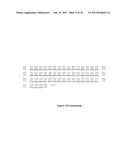
56PRTArtificial SequencePeptide linker 5Thr Val Ala Ala Pro Ser 1
5 628DNAArtificial SequencePCR primer 6atatgctagc
ccagcacctg aactcctg
28725DNAArtificial SequencePCR primer 7atattctaga ttatttaccc ggaga
25840DNAArtificial SequencePCR primer
8atatgctagc ggatccgata tcgtcaagct gcagcagtca
40946DNAArtificial SequencePCR primer 9atatgctagc agatggtgca gccacagttg
aggagacggt gaccat 461040DNAArtificial SequencePCR
primer 10atatgctagc ggatccgata tcgacatcca gatgacccag
401148DNAArtificial SequencePCR primer 11atatgctagc agatggtgca
gccacagtcc gttttatttc cagcttgg 481212PRTHomo sapiens
12Glu Arg Lys Cys Cys Val Glu Cys Pro Pro Cys Pro 1 5
10 13349PRTArtificial SequenceChimeric protein 13Val
Gln Leu Gln Gln Ser Gly Ala Glu Leu Val Lys Pro Gly Ala Ser 1
5 10 15 Val Arg Leu Ser Cys Lys
Ala Ser Gly Tyr Thr Phe Thr Glu Tyr Ile 20
25 30 Ile His Trp Ile Lys Leu Arg Ser Gly Gln
Gly Leu Glu Trp Ile Gly 35 40
45 Trp Phe Tyr Pro Gly Ser Asn Asp Ile Gln Tyr Asn Ala Lys
Phe Lys 50 55 60
Gly Lys Ala Thr Leu Thr Ala Asp Lys Ser Ser Ser Thr Val Tyr Met 65
70 75 80 Glu Leu Thr Gly Leu
Thr Ser Glu Asp Ser Ala Val Tyr Phe Cys Ala 85
90 95 Arg Arg Asp Asp Phe Ser Gly Tyr Asp Ala
Leu Pro Tyr Trp Gly Gln 100 105
110 Gly Thr Leu Val Thr Val Ser Ala Glu Arg Lys Cys Cys Val Glu
Cys 115 120 125 Pro
Pro Cys Pro Ala Pro Glu Phe Leu Gly Gly Pro Ser Val Phe Leu 130
135 140 Phe Pro Pro Lys Pro
Lys Asp Thr Leu Met Ile Ser Arg Thr Pro Glu 145 150
155 160 Val Thr Cys Val Val Val Asp Val Ser Gln
Glu Asp Pro Glu Val Gln 165 170
175 Phe Asn Trp Tyr Val Asp Gly Val Glu Val His Asn Ala Lys Thr
Lys 180 185 190 Pro
Arg Glu Glu Gln Phe Asn Ser Thr Tyr Arg Val Val Ser Val Leu 195
200 205 Thr Val Leu His Gln Asp
Trp Leu Asn Gly Lys Glu Tyr Lys Cys Lys 210 215
220 Val Ser Asn Lys Gly Leu Pro Ser Ser Ile Glu
Lys Thr Ile Ser Lys 225 230 235
240 Ala Lys Gly Gln Pro Arg Glu Pro Gln Val Tyr Thr Leu Pro Pro Ser
245 250 255 Gln Glu
Glu Met Thr Lys Asn Gln Val Ser Leu Thr Cys Leu Val Lys 260
265 270 Gly Phe Tyr Pro Ser Asp Ile
Ala Val Glu Trp Glu Ser Asn Gly Gln 275 280
285 Pro Glu Asn Asn Tyr Lys Thr Thr Pro Pro Val Leu
Asp Ser Asp Gly 290 295 300
Ser Phe Phe Leu Tyr Ser Arg Leu Thr Val Asp Lys Ser Arg Trp Gln 305
310 315 320 Glu Gly Asn
Val Phe Ser Cys Ser Val Met His Glu Ala Leu His Asn 325
330 335 His Tyr Thr Gln Lys Ser Leu Ser
Leu Ser Leu Gly Lys 340 345
14337PRTArtificial SequenceChimeric protein 14Asp Ile Gln Met Thr Gln Ser
Pro Ala Ser Leu Ser Val Ser Val Gly 1 5
10 15 Glu Thr Val Thr Ile Thr Cys Arg Thr Asn Glu
Asn Ile Tyr Ser Asn 20 25
30 Leu Ala Trp Tyr Gln Gln Lys Gln Gly Lys Ser Pro Gln Leu Leu
Ile 35 40 45 Tyr
Ala Ala Thr His Leu Val Glu Gly Val Pro Ser Arg Phe Ser Gly 50
55 60 Ser Gly Ser Gly Thr Gln
Tyr Ser Leu Lys Ile Thr Ser Leu Gln Ser 65 70
75 80 Glu Asp Phe Gly Asn Tyr Tyr Cys Gln His Phe
Trp Gly Thr Pro Cys 85 90
95 Thr Phe Gly Gly Gly Thr Lys Leu Glu Ile Lys Arg Glu Arg Lys Cys
100 105 110 Cys Val
Glu Cys Pro Pro Cys Pro Ala Pro Glu Phe Leu Gly Gly Pro 115
120 125 Ser Val Phe Leu Phe Pro Pro
Lys Pro Lys Asp Thr Leu Met Ile Ser 130 135
140 Arg Thr Pro Glu Val Thr Cys Val Val Val Asp
Val Ser Gln Glu Asp 145 150 155
160 Pro Glu Val Gln Phe Asn Trp Tyr Val Asp Gly Val Glu Val His Asn
165 170 175 Ala Lys
Thr Lys Pro Arg Glu Glu Gln Phe Asn Ser Thr Tyr Arg Val 180
185 190 Val Ser Val Leu Thr Val Leu
His Gln Asp Trp Leu Asn Gly Lys Glu 195 200
205 Tyr Lys Cys Lys Val Ser Asn Lys Gly Leu Pro Ser
Ser Ile Glu Lys 210 215 220
Thr Ile Ser Lys Ala Lys Gly Gln Pro Arg Glu Pro Gln Val Tyr Thr 225
230 235 240 Leu Pro Pro
Ser Gln Glu Glu Met Thr Lys Asn Gln Val Ser Leu Thr 245
250 255 Cys Leu Val Lys Gly Phe Tyr Pro
Ser Asp Ile Ala Val Glu Trp Glu 260 265
270 Ser Asn Gly Gln Pro Glu Asn Asn Tyr Lys Thr Thr Pro
Pro Val Leu 275 280 285
Asp Ser Asp Gly Ser Phe Phe Leu Tyr Ser Arg Leu Thr Val Asp Lys 290
295 300 Ser Arg Trp Gln
Glu Gly Asn Val Phe Ser Cys Ser Val Met His Glu 305 310
315 320 Ala Leu His Asn His Tyr Thr Gln Lys
Ser Leu Ser Leu Ser Leu Gly 325 330
335 Lys
User Contributions:
Comment about this patent or add new information about this topic:
 |  |
 |  |
 |  |
 |  |
 |  |
 |  |
 |  |
 |  |
 |  |
 |
| New patent applications in this class: | |
| Date | Title |
|---|---|
| 2022-09-22 | Electronic device |
| 2022-09-22 | Front-facing proximity detection using capacitive sensor |
| 2022-09-22 | Touch-control panel and touch-control display apparatus |
| 2022-09-22 | Sensing circuit with signal compensation |
| 2022-09-22 | Reduced-size interfaces for managing alerts |
| New patent applications from these inventors: | |
| Date | Title |
|---|---|
| 2022-09-22 | Methods and compositions for modifying macrophage polarization into pro-inflammatory cells to treat cancer |
| 2022-09-08 | Method and compositions for inducing differentiation of myeloid derived suppressor cell to treat cancer and infectious diseases |
| 2022-09-08 | Anti-sirpa antibodies and their therapeutic applications |
| 2022-09-08 | Methods for promoting t cells response |