Patent application title: Abeta PROTOFIBRIL BINDING ANTIBODIES
Inventors:
IPC8 Class: AC07K1618FI
USPC Class:
4241721
Class name: Drug, bio-affecting and body treating compositions immunoglobulin, antiserum, antibody, or antibody fragment, except conjugate or complex of the same with nonimmunoglobulin material binds eukaryotic cell or component thereof or substance produced by said eukaryotic cell (e.g., honey, etc.)
Publication date: 2016-01-14
Patent application number: 20160009793
Abstract:
The present invention relates to the amyloid beta peptide (Aβ) and
more specifically to antibodies binding to Aβ protofibrils and their
use in therapy and/or prophylactic treatment of Alzheimer's disease and
other disorders associated with Aβ protein aggregation. Further the
invention may relate to diagnosis of such diseases as well as monitoring
of disease progression by use of the antibodies of the invention.
Further, the invention may relate to veterinary use of the antibodies of
the invention.Claims:
1. An antibody or antigen binding fragment thereof having affinity
against Aβ protofibrils, wherein the antibody or antigen binding
fragment thereof has a variable light chain according to SEQ ID NO: 8,
wherein x1 is selected from A, D, E and Q, or a functional analogue
thereof; x2 is selected from R, T, K, A and G, or a functional
analogue thereof; x3 is selected from R, S, C, G and N, or a
functional analogue thereof; y1 is selected from V and A; y2 is
selected from I and V; y3 is selected from S and Q; y4 is
selected from E and D; and optionally a variable heavy chain according to
SEQ ID NO: 14, wherein z1 is selected from V and I; z2 is
selected from R and Q; and z3 is selected from A, N and T1;
with the exception for the combination x1=A, x2=R and
x3=R.
2. The antibody or antigen binding fragment according to claim 1, wherein x1 is selected from A, D, E and Q; x2 is selected from R, T, K, A and G; x3 is selected from R, S, C, G and N; y1 is selected from V and A; y2 is selected from I and V; y3 is selected from S and Q; and y4 is selected from E and D; with the exception for the combination x1=A, x2=R and x3=R.
3. The antibody or antigen binding fragment according to claim 1, wherein the light chain comprises a combination of mutations selected from: x1 and (y1 and/or y2); x1 and (y1 and/or y2) and x2 and (y3 and/or y4); x1 and (y1 and/or y2) and x2 and x3 and (y3 and/or y4); x1 and (y1 and/or y2) and x3 and (y3 and/or y4); x2 (y3 and/or y4); x2 and x3 and (y3 and/or y4); and x3 and (y3 and/or y4); with the exception for the combination x1=A, x2=R and x3=R.
4. The antibody or antigen binding fragment according to claim 1, wherein the variable light chain comprises one or more mutations selected from: x1 is D and (y1 and/or y2); x1 is D and (y1 and/or y2), x2 is T and (y3 and/or y4); x1 is D and (y1 and/or y2), x2 is T, x3 is S and (y3 and/or y4); x1 is D and (y1 and/or y2) and x3 is S and (y3 and/or y4); x2 is T (y3 and/or y4); x2 is T and x3 is S and (y3 and/or y4); and x3 is S and (y3 and/or y4); wherein y1 is V or A and y2 is V or I, with exclusion of the combination y1=V and y2=I; y3 is S or Q and y4 is E or D with exclusion of the combination y3=S and Y4=E.
5. The antibody or antigen binding fragment according to claim 1, wherein x1 is D; x2 is T; x3 is R; y1 is selected from V and A; y2 is selected from V and I; y3 is selected from Q and S; y4 is selected from D and E; z1 is V; z2 is R; and z3 is selected from N, T and A; with exclusion of the combination wherein y1 is V, y2 is I, y3, is S, y4 is E, z1 is V, z2 is R and z3 is A.
6. An antibody or antigen binding fragment thereof, according to claim 1, comprising a variable light chain comprising an amino acid sequence as set out in SEQ ID NO: 12; and a variable heavy chain comprising an amino acid sequence as set out in SEQ ID NO: 16.
7. An antibody or antigen binding fragment thereof, according to claim 1, comprising a variable light chain comprising an amino acid sequence as set out in SEQ ID NO: 9; and a variable heavy chain comprising an amino acid sequence as set out in SEQ ID NO: 15.
8. An antibody or antigen binding fragment thereof, according to claim 1, comprising a variable light chain comprising an amino acid sequence as set out in SEQ ID NO: 10; and a variable heavy chain comprising an amino acid sequence as set out in SEQ ID NO: 15.
9. An antibody or antigen binding fragment thereof, according to claim 1, comprising a variable light chain comprising an amino acid sequence as set out in SEQ ID NO: 11; and a variable heavy chain comprising an amino acid sequence as set out in SEQ ID NO: 15.
10. An antibody or antigen binding fragment thereof, according to claim 1, comprising a variable light chain comprising an amino acid sequence as set out in SEQ ID NO: 12; and a variable heavy chain comprising an amino acid sequence as set out in SEQ ID NO: 15.
11. An antibody or antigen binding fragment thereof, according to claim 1, comprising a variable light chain comprising an amino acid sequence as set out in SEQ ID NO: 9; and a variable heavy chain comprising an amino acid sequence as set out in SEQ ID NO: 16.
12. An antibody or antigen binding fragment thereof, according to claim 1, comprising a variable light chain comprising an amino acid sequence as set out in SEQ ID NO: 10; and a variable heavy chain comprising an amino acid sequence as set out in SEQ ID NO: 16.
13. An antibody or antigen binding fragment thereof, according to claim 1, comprising a variable light chain comprising an amino acid sequence as set out in SEQ ID NO: 11; and a variable heavy chain comprising an amino acid sequence as set out in SEQ ID NO: 16.
14. The antibody or antigen binding fragment according to claim 1, wherein the antibody or the antigen binding fragment comprises an IgG heavy chain constant region.
15. (canceled)
16. (canceled)
17. (canceled)
18. (canceled)
19. (canceled)
20. A method of reducing the amount of Aβ protofibrils in a subject, comprising administering to said subject a therapeutically effective amount of the antibody or antigen binding fragment according to claim 1.
21. A method for treatment and/or prophylaxis of Alzheimer's disease or another disorder associated with Aβ protein aggregation in a subject having, or being at risk of developing said disease or disorder, comprising administering to said subject a therapeutically effective amount of the antibody or antigen binding fragment according to claim 1.
22. A method for treatment and/or prophylaxis of traumatic brain injury (TBI), Lewy body dementia (LBD), Down syndrome (DS), Amyotrophic lateral sclerosis (ALS), Frontotemporal dementia, tauopathies, systemic amyloidosis, atherosclerosis and Parkinson's disease dementia (PDD) in a subject having, or being at risk of developing said disease, comprising administering to said subject a therapeutically effective amount of the antibody or antigen binding fragment according to claim 1.
23. A method for measuring the amount of Aβ protofibrils and/or aggregated Aβ protein in a person, comprising contacting the person's tissue or body fluid, in vivo or in vitro, with the antibody or antigen binding fragment according to claim 1 and measuring the amount of antibody or antigen binding fragment bound to said Aβ protofibrils and/or aggregated Aβ protein.
24. A method for diagnosis of Alzheimer's disease in a person having or at risk of developing the disease, comprising contacting the person's tissue or body fluid, in vivo or in vitro, with the antibody or antigen binding fragment according to claim 1, or a fragment thereof, and measuring the amount of said antibody or antigen binding fragment bound to aggregated Aβ protein.
25. A method for diagnosis of traumatic brain injury (TBI), Lewy body dementia (LBD), Down syndrome (DS), Amyotrophic lateral sclerosis (ALS), Frontotemporal dementia, tauopathies, systemic amyloidosis, atherosclerosis and Parkinson's disease dementia (PDD) in a person having or at risk of developing any of said diseases, comprising contacting the person's tissue or body fluid, in vivo or in vitro, with the antibody or antigen binding fragment according to claim 1, or a fragment thereof, and measuring the amount of said antibody or antigen binding fragment bound to aggregated Aβ protein.
26. A pharmaceutical composition comprising the antibody or antigen binding fragment according to claim 1, together with a pharmaceutically acceptable excipient and/or diluent.
27. The method according to claim 20, wherein the subject is a veterinary subject.
Description:
FIELD OF THE INVENTION
[0001] The present invention relates to the amyloid beta peptide (Aβ) and more specifically to antibodies that bind to Aβ protofibrils and their use in therapy and/or prophylactic treatment of Alzheimer's disease and other disorders associated with Aβ protein aggregation. Further the invention may relate to diagnosis of such diseases as well as monitoring of disease progression by use of the antibodies of the invention. Further, the invention may relate to veterinary use of the antibodies of the invention.
BACKGROUND OF THE INVENTION
[0002] Alzheimer's disease (AD) belongs to a group of neurodegenerative disorders and causes cognitive, memory and behavioral impairments. The hallmarks of Alzheimer's disease include extracellular amyloid plaques, intraneuronal neurofibrillary tangles, neuronal dysfunction and ultimately brain atrophy. The risk for developing AD increases with age and with increased number of persons reaching high age, a condition with increasing impact on the quality of life for elderly people. In addition the society faces a situation with accelerating costs.
[0003] In spite of the fact that the disease has been known for many years and several suggestions for treatment have been made, even today, there is no such efficient disease modifying therapy available today but only drugs which at best may provide symptomatic treatment. The mechanism behind the disease has been subject to a lot of studies. Briefly, Aβ for some reason starts to aggregate and via several intermediate forms, finally produces insoluble fibril/plaque deposits in the brain. It was early believed that the plaques, as such, affect the neurons and the signals transmitted by these, but today the results of the extensive studies indicate that soluble, aggregated, intermediate forms of Aβ most likely are a major cause of the disease and the symptoms observed in the patients.
[0004] One such intermediate form in the cascade of aggregated forms from Aβ monomers to insoluble Aβ fibrils is the soluble, high molecular weight Aβ protofibril, which was first described by Walsh in 1997 in The Journal of Biological Chemistry (Vol. 272(35) p. 22364-72). The importance of the Aβ protofibril for the development of AD was identified by the group of scientists headed by Lars Lannfelt, Uppsala University, in their studies of the Arctic mutation, which is an E693G mutation in the amyloid precursor protein (APP) causing increased formation of Aβ protofibrils. A family in northern Sweden carrying this mutation was found to develop severe Alzheimer's disease early in life and the finding of this combination provided the basic ideas for a new therapy. Accordingly, the Aβ protofibril was identified as strongly related to the disease and an important target for therapy. Based on their studies with Aβ peptides comprising the Arctic mutation, Lannfelt et al were able to produce Aβ protofibrils in vitro, Arctic as well as wild-type, in sufficient amounts for immunization and subsequent selection of antibodies with high affinity for Aβ protofibrils compared to other species in the Aβ system. Examples of methods for production of Aβ protofibrils and antibodies that bind to these are disclosed in WO02/03911 and WO2005/123775.
[0005] Of special importance was the development of the mouse monoclonal antibody mAb158, an antibody that binds to Aβ protofibrils, which is disclosed in EP2004688, which comprises the following CDR sequences:
TABLE-US-00001 VH-CDR1: SEQ ID NO: 1 SFGMH VH-CDR2: SEQ ID NO: 2 YISSGSSTIYYGDTVKG VH-CDR3: SEQ ID NO: 3 EGGYYYGRSYYTMDY VL-CDR1: SEQ ID NO: 4 RSSQSIVHSNGNTYLE VL-CDR2: SEQ ID NO: 5 KVSNRFS VL-CDR3: SEQ ID NO: 6 FQGSHVPPT
[0006] The high affinity and selectivity of the humanized version of mAb158, BAN2401, makes it a very important candidate for use in therapy and/or prevention of Alzheimer's disease in particular, and it is presently subject to clinical trials in preparation for use as a pharmaceutical product. Characteristics of BAN2401 are described in EP2004688.
[0007] The efficacy of an antibody depends on several pharmacokinetic and pharmacodynamics factors, see e.g. Deng et al, Expert Opin. Drug Metab. Toxicol 8(2) (2012): p. 141-160; Boswell et al, Bioconjugate Chem. 21(2010): p. 2153-2163; Konterman, Current Opinion in Biotechnology 22 (2011): p. 1-9 and Igawa et al, mAbs 3:3 (2011): p. 243-252. Among these, extended serum half-life with increased systemic exposure often provides a considerable potential for improvements of significant therapeutic value. It also provides an opportunity for reduction of the dose which has systemic, important implications.
DESCRIPTION OF THE INVENTION
[0008] The present invention provides antibodies that bind to Aβ protofibrils and their use in therapy and/or prophylactic treatment of Alzheimer's disease and other disorders associated with Aβ protein aggregation. Further the invention may relate to antibodies useful in diagnosis of such diseases as well as their use in monitoring of disease progression of such diseases, as well as veterinary use of said antibodies. It has been identified that surprisingly the half-life as well as exposure of the humanized antibody based on mAb158, i.e. BAN2401, is considerably enhanced, e.g. about twice or more, primarily, by introducing one or more mutations in certain positions, i.e 17, 79 and/or 82, of the variable light chain of the BAN2401 antibody (Kabat positions 17, 74 and 77), respectively, see further FIG. 9 where these positions are referred to as x1, x2 and x3. In BAN2401 the amino acid in position 17 (Kabat position 17) is A, the amino acid in position 79 (Kabat position 74) is R and the amino acid in position 82 (Kabat position 77) is R. The Kabat numbering is given in accordance with Kabat et al., Sequences of Proteins of Immunological Interest, 1991 (NIH Publications No. 91-3242).
[0009] Optionally, further improvements of an antibody according to the invention can be achieved by combining each of the mutations providing increased half-life with one or more neighboring mutations, i.e 13, 21, 81 and/or 84, of the variable light chain of the antibody (Kabat positions 13, 21, 76 and 79), respectively, see further FIG. 9 where these positions are referred to as referred to as y1-4, 37, 38 and/or 40, of the variable Heavy chain of the antibody (Kabat positions 37, 38 and 40), respectively, see further FIG. 10 where these positions are referred to as referred to as z1-3, for further improvements of immunological significance, i.e. low immunogenicity. When, compared to the BAN2401 sequence, x1 is mutated the mutations y1 and/or y2 may be introduced and when x2 and/or x3 are mutated, the mutations y3 and/or y4 may be introduced. Further, the mutations z1-3 may be introduced.
[0010] The present invention is as follows:
[0011] [1] An antibody or antigen binding fragment thereof having affinity against Aβ protofibrils, wherein the antibody or antigen binding fragment thereof has a variable light chain according to SEQ ID NO: 8, wherein
x1 is selected from A, D, E and Q, or a functional analogue thereof; x2 is selected from R, T, K, A and G, or a functional analogue thereof; x3 is selected from R, S, C, G and N, or a functional analogue thereof; y1 is selected from V and A; y2 is selected from I and V; y3 is selected from S and Q; y4 is selected from E and D; and optionally a variable heavy chain according to SEQ ID NO: 14, wherein z1 is selected from V and I; z2 is selected from R and Q; and z3 is selected from A, N and T. with the exception for the combination x1=A, x2=R and x3=R.
[0012] [2] The antibody or antigen binding fragment according to [1], wherein
x1 is selected from A, D, E and Q; x2 is selected from R, T, K, A and G; x3 is selected from R, S, C, G and N; y1 is selected from V and A; y2 is selected from I and V; y3 is selected from S and Q; y4 is selected from E and D; and a variable heavy chain according to SEQ ID NO: 14, wherein z1 is selected from V and I; z2 is selected from R and Q; and z3 is selected from A, N and T; with the exception for the combination x1=A, x2=R and x3=R.
[0013] [3] The antibody or antigen binding fragment according to [1] or [2], wherein the light chain a comprises a combination of mutations selected from:
x1 and (y1 and/or y2); x1 and (y1 and/or y2) and x2 and (y3 and/or y4); x1 and (y1 and/or y2) and x2 and x3 and (y3 and/or y4); x1 and (y1 and/or y2) and x3 and (y3 and/or y4); x2 (y3 and/or y4); x2 and x3 and (y3 and/or y4); and x3 and (y3 and/or y4); with the exception for the combination x1=A, x2=R and x3=R.
[0014] [4] The antibody or antigen binding fragment according to any one of [1] to [3], wherein the variable light chain comprises one or more mutations, selected from:
x1 is D and (y1 and/or y2); x1 is D and (y1 and/or y2), x2 is T and (y3 and/or y4); x1 is D and (y1 and/or y2), x2 is T, x3 is S and (y3 and/or y4); x1 is D and (y1 and/or y2) and x3 is S and (y3 and/or y4); x2 is T (y3 and/or y4); x2 is T and x3 is S and (y3 and/or y4); and x3 is S and (y3 and/or y4); wherein y1 is V or A and y2 is V or I, with exclusion of the combination y1=V and y2=I; y3 is S or Q and y4 is E or D with exclusion of the combination y3=S and Y4=E.
[0015] [5] The antibody or antigen binding fragment according to any one of [1] to [4], wherein
x1 is D;
x2 is T;
x3 is R;
[0016] y1 is selected from V and A; y2 is selected from V and I; y3 is selected from Q and S; y4 is selected from D and E;
z1 is V;
z2 is R; and
[0017] z3 is selected from N, T and A; with exclusion of the combination wherein y1 is V, y2 is I, y3, is S, y4 is E, z1 is V, z2 is R and z3 is A.
[0018] [6] An antibody or antigen binding fragment thereof, according to [1], comprising a variable light chain comprising an amino acid sequence as set out in SEQ ID NO: 12; and a variable heavy chain comprising an amino acid sequence as set out in SEQ ID NO: 16.
[0019] [7] An antibody or antigen binding fragment thereof, according to [1], comprising a variable light chain comprising an amino acid sequence as set out in SEQ ID NO: 9; and a variable heavy chain comprising an amino acid sequence as set out in SEQ ID NO: 15.
[0020] [8] An antibody or antigen binding fragment thereof, according to [1], comprising a variable light chain comprising an amino acid sequence as set out in SEQ ID NO: 10; and a variable heavy chain comprising an amino acid sequence as set out in SEQ ID NO: 15.
[0021] [9] An antibody or antigen binding fragment thereof, according to [1], comprising a variable light chain comprising an amino acid sequence as set out in SEQ ID NO: 11; and a variable heavy chain comprising an amino acid sequence as set out in SEQ ID NO: 15.
[0022] [10] An antibody or antigen binding fragment thereof, according to [1], comprising a variable light chain comprising an amino acid sequence as set out in SEQ ID NO: 12; and a variable heavy chain comprising an amino acid sequence as set out in SEQ ID NO: 15.
[0023] [11] An antibody or antigen binding fragment thereof, according to [1], comprising a variable light chain comprising an amino acid sequence as set out in SEQ ID NO: 9; and a variable heavy chain comprising an amino acid sequence as set out in SEQ ID NO: 16.
[0024] [12] An antibody or antigen binding fragment thereof, according to [1], comprising a variable light chain comprising an amino acid sequence as set out in SEQ ID NO: 10; and a variable heavy chain comprising an amino acid sequence as set out in SEQ ID NO: 16.
[0025] [13] An antibody or antigen binding fragment thereof, according to [1], comprising a variable light chain comprising an amino acid sequence as set out in SEQ ID NO: 11; and a variable heavy chain comprising an amino acid sequence as set out in SEQ ID NO: 16.
[0026] [14] The antibody or antigen binding fragment according to any one of [1] to [13], wherein the antibody or the antigen binding fragment comprises an IgG heavy chain constant region.
[0027] [15] An antibody according to any one of [1] to [14], for use in therapy.
[0028] [16] An antibody according to any one of [1] to [14], for use in treatment and/or prophylaxis of Alzheimer's disease and other disorders associated with Aβ protein aggregation.
[0029] [17] An antibody according to [16], for use, wherein such other disorders associated with Aβ protein aggregation are selected from traumatic brain injury (TBI), Lewy body dementia (LBD),
[0030] Downs syndrome (DS), Amyotrophic lateral sclerosis (ALS), Frontotemporal dementia, tauopathies, systemic amyloidoses, atherosclerosis and Parkinson's disease dementia (PDD).
[0031] [18] Use of an antibody according to any one of [1] to [14], in the manufacture of a medicament useful in the treatment and/or prophylaxis of Alzheimer's disease and other disorders associated with Aβ protein aggregation.
[0032] [19] The use according to [18], wherein such other disorders associated with Aβ protein aggregation are selected from Traumatic brain injury (TBI), Lewy body dementia (LBD), Downs syndrome (DS), Amyotrophic lateral sclerosis (ALS), Frontotemporal dementia, tauopathies, systemic amyloidoses, atherosclerosis and Parkinson's disease dementia (PDD).
[0033] [20] A method of reducing amount of Aβ protofibrils in a subject, comprising administering to said subject a therapeutically effective amount of the antibody or antigen binding fragment according to [1] to [14].
[0034] [21] A method for treatment and/or prophylaxis of Alzheimer's disease in a subject having, or being at risk of developing said disease, comprising administering to said subject a therapeutically effective amount of the antibody or antigen binding fragment according to [1] to [14].
[0035] [22] A method for treatment and/or prophylaxis of traumatic brain injury (TBI), Lewy body dementia (LBD), Down syndrome (DS), Amyotrophic lateral sclerosis (ALS), Frontotemporal dementia, tauopathies, systemic amyloidosis, atherosclerosis and Parkinson's disease dementia (PDD) in a subject having, or being at risk of developing said disease, comprising administering to said subject a therapeutically effective amount of the antibody or antigen binding fragment according to [1] to [14].
[0036] [23] A method for measuring amount of Aβ protofibrils and/or aggregated Aβ protein in a person, comprising contacting the person's tissue or body fluid, in vivo or in vitro, with the antibody or antigen binding fragment according to [1] to [14] and measuring the amount of antibody or antigen binding fragment bound to said Aβ protofibrils and/or aggregated Aβ protein.
[0037] [24] A method for diagnosis of Alzheimer's disease in persons having or at risk of developing the disease, comprising contacting the person's tissue or body fluid, in vivo or in vitro, with the antibody or antigen binding fragment according to [1] to [14], or a fragment thereof, and measuring the amount of said antibody bound to aggregated Aβ protein.
[0038] [25] A method for diagnosis of traumatic brain injury (TBI), Lewy body dementia (LBD), Down syndrome (DS), Amyotrophic lateral sclerosis (ALS), Frontotemporal dementia, tauopathies, systemic amyloidosis, atherosclerosis and Parkinson's disease dementia (PDD) in persons having or at risk of developing any of said diseases, comprising contacting the person's tissue or body fluid, in vivo or in vitro, with the antibody or antigen binding fragment according to [1] to [14], or a fragment thereof, and measuring the amount of said antibody bound to aggregated Aβ protein.
[0039] [26] A pharmaceutical composition comprising the antibody or antigen binding fragment according to any one of [1] to [14], together with pharmaceutically acceptable excipient and/or diluents.
[0040] [27] An antibody according to any one of [1] to [14], for veterinary use.
BRIEF DESCRIPTION OF THE DRAWINGS
[0041] FIG. 1 provides analysis of binding and selectivity for Aβ protofibrils compared to Aβ monomers for A17D, A17D/R79T, A17D/R82S and A17D/R79T/R82S compared to BAN2401 (control). Binding inhibition by Aβ1-42 protofibrils in solution is shown by open circles and open squares and binding inhibition by Aβ1-40 monomers in solution are shown by closed circles and closed squares.
[0042] FIG. 2 provides plasma drug exposure of BAN2401 and antibodies of the invention in mice presented as time vs concentration graphs. Plasma levels after single i.v. injection of BAN2401, A17D, A17D/R79T and A17D/R79T/R82S collected at time points 0.5 h, 2 days, 7 days, 15 days and 29 days post administration, and of A17D/R82S collected at time points 0.5 h, 2 days, 7 days, 14 days and 28 days post administration are shown in the graph. A17D/R82S was not included in the same PK study as the other antibodies shown here, but was instead given at a separate occasion. However, with exception of two plasma sampling time points, the same study design was used for the two separate studies. All plasma samples were analyzed by ELISA at the same occasion to avoid inter-assay variation. The plasma drug concentration in μg/ml is shown on the y-axis (logarithmic scale) and the time post administration in hours (h) is shown on the x-axis. Mean group values are shown with error bars indicating standard deviations. Mean AUC0-inf values and terminal half-lives were calculated by non-compartment analysis using Phoenix WinNonlin 6.3 (Pharsight) and are shown in Table 1.
[0043] FIG. 3 provides plasma drug exposure of BAN2401 and antibodies of the invention, in rats presented as time vs concentration graphs. Plasma levels after single i.v. injection of BAN2401, A17D, A17D/R79T and A17D/R79T/R82S collected at time-points 0.5 h, 2 h, 7 h, 24 h, 2 days, 4 days, 7 days, 14 days and 29 days post administration are shown in the graph. The plasma drug concentration in μg/ml is shown on the y-axis (logarithmic scale) and the time post administration in hours (h) is shown on the x-axis. Mean group values are shown with error bars indicating standard deviations. Mean AUC0-inf values and mean terminal half-lives were calculated by non-compartment analysis using Phoenix WinNonlin 6.3 (Pharsight) and are shown in Table 2.
[0044] FIG. 4 provides data with analysis of binding and selectivity for Aβ protofibrils compared to Aβ monomers for the deimmunized variants of A17D/R79T (A17D/R79T_DI 1-8) compared to BAN2401. Binding inhibition by Aβ1-42 protofibrils in solution is shown by open circles and open squares, and binding inhibition by Aβ1-40 monomers in solution are shown by closed circles and closed squares. A) A17D/R79T_DI 1, B) A17D/R79T_DI 2, C) A17D/R79T_DI 3, D) A17D/R79T_DI 4, E) A17D/R79T_DI 5, F) A17D/R79T_DI 6, G) A17D/R79T_DI 7, H) A17D/R79T_DI 8.
[0045] FIG. 5 provides plasma drug exposure of BAN2401 and antibodies of the invention, in mice presented as time vs concentration graphs. Plasma levels after a single i.v. injection of BAN2401, A17D/R79T and 8 deimmunized variants of A17D/R79T (A17D/R79T_DI 1-8) collected at time points 0.5 h, 2 days, 7 days, 14 days and 28 days post administration. The plasma drug concentration in μg/ml is shown on the y-axis (logarithmic scale) and the time post administration in hours (h) is shown on the x-axis. Mean group values are shown with error bars indicating standard deviations. Mean AUC0-inf values and mean terminal half-lives were calculated by non-compartment analysis using Phoenix WinNonlin 6.3 (Pharsight) and are shown in Table 6.
[0046] FIG. 6 provides plasma drug exposure of BAN2401 and mutants in rat presented as time vs concentration graphs. Plasma levels after a single i.v. injection of BAN2401, A17D/R79T and 8 deimmunized variants of A17D/R79T (A17D/R79T_DI 1-8) collected at time points 0.5 h, 2 days, 7 days, 14 days and 28 days post administration. The plasma drug concentration in μg/ml is shown on the y-axis (logarithmic scale) and the time post administration in hours (h) is shown on the x-axis. Mean group values are shown with error bars indicating standard deviations. Mean AUC0-inf values and mean terminal half-lives were calculated by non-compartment analysis using Phoenix WinNonlin 6.3 (Pharsight) and are shown in Table 7.
[0047] FIG. 7 provides dose corrected plasma drug exposure of BAN2401, A17D/R79T_DI 3, A17D/R79T_DI 4 and A17D/R79T_DI 8 in Cynomolgus monkey presented as time vs concentration graphs. Plasma evels of antibody after single i.v. infusion of BAN2401 collected at time points 5 min, 1 h, 2 h, 8 h, 24 h, 2 days, 4 days, 7 days, 14 days, 21 days and 28 days post administration, and A17D/R79T_DI 3, A17D/R79T_DI 4 and A17D/R79T_DI 8 collected at time points 5 min, 2 h, 8 h, 24 h, 3 days, 7 days, 14 days, 21 days and 28 days post administration. The antibodies of the invention were administered to the monkey in a different study and at a different dose (10 mg/kg) compared to BAN2401 (5 mg/kg). Therefore the plasma exposure graphs have been dose adjusted. The dose corrected plasma drug concentration in μg/ml per mg/kg injected dose is shown on the y-axis (logarithmic scale) and the time post administration in hours (h) is shown on the x-axis. Mean group values are shown with error bars indicating standard deviations. Dose adjusted mean AUC0-inf values and terminal half-lives were calculated by non-compartment analysis using Phoenix WinNonlin 6.3 (Pharsight) and are shown in Table 8.
[0048] FIG. 8 provides amino acid sequences in relation to the present invention.
[0049] FIG. 9 provides a table, split on two pages, with the amino acid sequence of the light variable chain with VL-CDR1-3 sequences in grey. BAN2401: SEQ ID NO: 7. Novel antibodies with light variable chain according to the invention: SEQ ID NO: 8. Specific examples of such variable light chains: i): SEQ ID NO: 9; ii): SEQ ID NO: 10; iii): SEQ ID NO: 11; and iv): SEQ ID NO: 12.
[0050] FIG. 10 provides a table, split on three pages, with the amino acid sequence of the heavy variable chain with the VH-CDR1-3 sequences in grey. BAN2401: SEQ ID NO: 13. Novel antibodies with heavy variable chain according to the invention SEQ ID NO: 14. Specific examples of such variable heavy chains: i): SEQ ID NO: 15; ii): SEQ ID NO: 16; iii): SEQ ID NO: 17; and iv): SEQ ID NO: 18.
[0051] FIG. 11 provides simple allometric scaling of central clearance (CL) of BAN2401, A17D/R79T_DI 3, A17D/R79T_DI 4 and A17D/R79T_DI 8 including preclinical species mouse, rat and cynomolgus. The CLs of the different species are plotted against their weights (diamonds), respectively. The regression line has been extrapolated to indicate CL in a man with a body weight of 70 kg. For BAN2401 the true central CL measured is indicated by an open square, and deviates from the linear regression line based on CL of BAN2401 in mouse, rat and cynomolgus monkey. BAN2401 showed a poor linear correlation of CL indicating uncertain prediction of half-life, in contrast to the excellent linear correlation of CL of the antibodies of the invention.
[0052] The mutations A17D, R79T and R82S, represent positions in the BAN2401 antibody, wherein amino acids in positions 17, 79 and 82 are mutated in the variable light chain.
[0053] With "BAN2401" is meant a humanized monoclonal antibody of the mouse antibody mAb158 comprising a variable light chain with an amino acid sequence as set out in SEQ ID NO: 7 and a variable heavy chain as set out in SEQ ID NO: 13. Both BAN2401 and mAb158 and their characteristics, including VL-CDR1-3 and VH-CDR1-3 are described in EP2004688. BAN2401 is excluded from the present invention.
[0054] With the following abbreviations is meant:
[0055] BAN2401: an antibody comprising a variable light chain comprising an amino acid sequence as set out in SEQ ID NO: 7; and a variable heavy chain comprising an amino acid sequence as set out in SEQ ID NO: 13.
[0056] A17D: an antibody comprising a variable light chain comprising an amino acid sequence as set out in SEQ ID NO: 19 and a variable heavy chain comprising an amino acid sequence as set out in SEQ ID NO: 13.
[0057] A17D/R79T: an antibody comprising a variable light chain comprising an amino acid sequence as set out in SEQ ID NO: 20 and a variable heavy chain comprising an amino acid sequence as set out in SEQ ID NO: 13.
[0058] A17D/R79T/R82S: an antibody comprising a variable light chain comprising an amino acid sequence as set out in SEQ ID NO: 21 and a variable heavy chain comprising an amino acid sequence as set out in SEQ ID NO: 13.
[0059] A17D/R82S: an antibody comprising a variable light chain comprising an amino acid sequence as set out in SEQ ID NO: 22 and a variable heavy chain comprising an amino acid sequence as set out in SEQ ID NO: 13.
[0060] A17D/R79T_DI 1: an antibody comprising a variable light chain comprising an amino acid sequence as set out in SEQ ID NO: 9; and a variable heavy chain comprising an amino acid sequence as set out in SEQ ID NO: 15.
[0061] A17D/R79T_DI 2: an antibody comprising a variable light chain comprising an amino acid sequence as set out in SEQ ID NO: 10; and a variable heavy chain comprising an amino acid sequence as set out in SEQ ID NO: 15.
[0062] A17D/R79T_DI 3: an antibody comprising a variable light chain comprising an amino acid sequence as set out in SEQ ID NO: 11; and a variable heavy chain comprising an amino acid sequence as set out in SEQ ID NO: 15.
[0063] A17D/R79T_DI 4: an antibody comprising a variable light chain comprising an amino acid sequence as set out in SEQ ID NO: 12; and a variable heavy chain comprising an amino acid sequence as set out in SEQ ID NO: 15.
[0064] A17D/R79T_DI 5: an antibody comprising a variable light chain comprising an amino acid sequence as set out in SEQ ID NO: 9; and a variable heavy chain comprising an amino acid sequence as set out in SEQ ID NO: 16.
[0065] A17D/R79T_DI 6: an antibody comprising a variable light chain comprising an amino acid sequence as set out in SEQ ID NO: 10; and a variable heavy chain comprising an amino acid sequence as set out in SEQ ID NO: 16.
[0066] A17D/R79T_DI 7: an antibody comprising a variable light chain comprising an amino acid sequence as set out in SEQ ID NO: 11; and a variable heavy chain comprising an amino acid sequence as set out in SEQ ID NO: 16.
[0067] A17D/R79T_DI 8: an antibody comprising a variable light chain comprising an amino acid sequence as set out in SEQ ID NO: 12; and a variable heavy chain comprising an amino acid sequence as set out in SEQ ID NO: 16.
[0068] An antibody, or an antigen binding fragment thereof, according to the present invention comprises, in the light variable chain, in position 17 (Kabat position 17) the amino acid A, D, E, Q or a functional analogue, in position 79 (Kabat position 74) amino acid R, T, K, A, G or a functional analogue and in position 82 (Kabat position 77) amino acid R, S, C, G, N or a functional analogue. A functional analogue is an amino acid providing a lower pI value of the antibody compared to A (position 17) resp. R (position 79 and 82) without negatively affecting the binding to the antigen.
[0069] The amino acid sequences in the present disclosure are represented as follows:
SEQ ID NO: 1: variable heavy chain VH-CDR1 of BAN2401. SEQ ID NO: 2: variable heavy chain VH-CDR2 of BAN2401. SEQ ID NO: 3: variable heavy chain VH-CDR3 of BAN2401. SEQ ID NO: 4: variable light chain VL-CDR1 of BAN2401. SEQ ID NO: 5: variable light chain VL-CDR2 of BAN2401. SEQ ID NO: 6: variable light chain VL-CDR3 of BAN2401. SEQ ID NO: 7: variable light chain of BAN2401. SEQ ID NO: 8: generic variable light chain sequence in antibodies of the invention. SEQ ID NO: 9: specific variable light chain sequence in antibodies of the invention. SEQ ID NO: 10: specific variable light chain sequence in antibodies of the invention. SEQ ID NO: 11: specific variable light chain sequence in antibodies of the invention. SEQ ID NO: 12: specific variable light chain sequence in antibodies of the invention. SEQ ID NO: 13: variable heavy chain of BAN2401. SEQ ID NO: 14: generic variable heavy chain sequence in antibodies of the invention. SEQ ID NO: 15: specific variable heavy chain sequence in antibodies of the invention. SEQ ID NO: 16: specific variable heavy chain sequence in antibodies of the invention. SEQ ID NO: 17: specific variable heavy chain sequence in antibodies of the invention. SEQ ID NO: 18: specific variable heavy chain sequence in antibodies of the invention. SEQ ID NO: 19: specific variable light chain sequence in antibodies of the invention. SEQ ID NO: 20: specific variable light chain sequence in antibodies of the invention. SEQ ID NO: 21: specific variable light chain sequence in antibodies of the invention. SEQ ID NO: 22: specific variable light chain sequence in antibodies of the invention. SEQ ID NO: 23: amino acid sequence of human IgG1 constant region comprised in antibodies of the invention. SEQ ID NO: 24: amino acid sequence of human K chain constant region comprised in antibodies of the invention.
[0070] The variable light chain (SEQ ID NO: 7) of BAN2401 and the antibodies of the invention comprises the three CDR-sequences (VL-CDR1-3):
TABLE-US-00002 VL-CDR1: SEQ ID NO: 4 RSSQSIVHSNGNTYLE VL-CDR2: SEQ ID NO: 5 KVSNRFS VL-CDR3: SEQ ID NO: 6 FQGSHVPPT
and the variable heavy chain (SEQ ID NO: 13) of BAN2401 and the antibodies of the invention, comprises the three CDR-sequences (VH-CDR1-3):
TABLE-US-00003 VH-CDR1: SEQ ID NO: 1 SFGMH VH-CDR2: SEQ ID NO: 2 YISSGSSTIYYGDTVKG VH-CDR3: SEQ ID NO: 3 EGGYYYGRSYYTMDY
[0071] According to one aspect of the invention, antibodies binding to Aβ protofibrils are provided, having the following CDR sequence combinations:
TABLE-US-00004 VH-CDR1: SEQ ID NO: 1 SFGMH VH-CDR2: SEQ ID NO: 2 YISSGSSTIYYGDTVKG VH-CDR3: SEQ ID NO: 3 EGGYYYGRSYYTMDY VL-CDR1: SEQ ID NO: 4 RSSQSIVHSNGNTYLE VL-CDR2: SEQ ID NO: 5 KVSNRFS VL-CDR3: SEQ ID NO: 6 FQGSHVPPT
[0072] and comprising the variable light chain with SEQ ID NO: 8, wherein x1 is A, D, E, Q or a functional analogue, x2 is R, T, K, A, G or a functional analogue and x3 is R, S, C, G, N or a functional analogue, with the exception for the combination x1=A, x2=R and x3=R.
[0073] A functional analogue is an amino acid providing a lower pI value of the antibody compared to A (x1) resp. R (x2 and x3) without negatively affecting the binding to the antigen.
[0074] The variable heavy chain has the amino acid sequence presented in SEQ ID NO: 14, comprising the z1, z2 and z3 in any combination and wherein
z1 is selected from V and I; z2 is selected from R and Q; and z3 is selected from A, N and T; with the exception for the combination z1=V, z2=R and z3=A.
[0075] It should be pointed out that based on this teaching, the identification of a functional analogue to the specific amino acids defined above for each of the positions is easily done by a person skilled in the art, as methods for introducing an amino acid in a specific position in accordance with the present invention, as well as testing the resulting product, e.g. with regard to affinity for Aβ protofibrils and other characteristics of importance, are disclosed, see below.
[0076] Accordingly, in one aspect of the invention there is provided an antibody, or antigen binding fragment thereof having affinity against Aβ protofibrils, wherein the antibody, or antigen binding fragment thereof has a variable light chain according to SEQ ID NO: 8, wherein
x1 is selected from A, D, E and Q, or a functional analogue thereof; x2 is selected from R, T, K, A and G, or a functional analogue thereof; x3 is selected from R, S, C, G and N, or a functional analogue thereof; y1 is selected from V and A; y2 is selected from I and V; y3 is selected from S and Q; y4 is selected from E and D; and optionally a variable heavy chain according to SEQ ID NO: 14, wherein z1 is selected from V and I; z2 is selected from R and Q; and z3 is selected from A, N and T; with the exception for the combination x1=A, x2=R and x3=R.
[0077] In one embodiment of this aspect, there is provided an antibody or an antigen binding fragment thereof, wherein
x1 is selected from A, D, E and Q; x2 is selected from R, T, K, A and G; x3 is selected from R, S, C, G and N; y1 is selected from V and A; y2 is selected from I and V; y3 is selected from S and Q; y4 is selected from E and D; and a variable heavy chain according to SEQ ID NO: 14, wherein z1 is selected from V and I; z2 is selected from R and Q; and z3 is selected from A, N and T; with the exception for the combination x1=A, x2=R and x3=R.
[0078] In one embodiment of this aspect, there is provided an antibody, or an antigen binding fragment thereof, wherein the light chain comprises a combination of mutations selected from:
x1 and (y1 and/or y2); x1 and (y1 and/or y2) and x2 and (y3 and/or y4); x1 and (y1 and/or y2) and x2 and x3 and (y3 and/or y4); x1 and (y1 and/or y2) and x3 and (y3 and/or y4); x2 (y3 and/or y4); x2 and x3 and (y3 and/or y4); and x3 and (y3 and/or y4); with the exception for the combination x1=A, x2=R and x3=R.
[0079] In one embodiment of this aspect, there is provided an antibody, or an antigen binding fragment thereof, wherein the variable light chain comprises one or more mutations, selected from:
x1 is D and (y1 and/or y2); x1 is D and (y1 and/or y2), x2 is T and (y3 and/or y4); x1 is D and (y1 and/or y2), x2 is T, x3 is S and (y3 and/or y4); x1 is D and (y1 and/or y2) and x3 is S and (y3 and/or y4); x2 is T (y3 and/or y4); x2 is T and x3 is S and (y3 and/or y4); and x3 is S and (y3 and/or y4); wherein y1 is V or A and y2 is V or I, with exclusion of the combination y1=V and y2=I; y3 is S or Q and y4 is E or D with exclusion of the combination y3=S and Y4=E.
[0080] In one embodiment of this aspect, there is provided an antibody, or an antigen binding fragment thereof, wherein y1 is A, y2 is V, y3 is Q and y4 is D.
[0081] In one embodiment of this aspect, there is provided an antibody, or an antigen binding fragment thereof, wherein
x1 is D; x2 is T; x3 is R; y1 is selected from V and A; y2 is selected from V and I; y3 is selected from Q and S; y4 is selected from D and E; z1 is V; z2 is R; and z3 is selected from N, T and A; with exclusion of the combination wherein y1 is V, y2 is I, y3, is S, y4 is E, z1 is V, z2 is R and z3 is A.
[0082] In one embodiment of this aspect, there is provided an antibody, or an antigen binding fragment thereof, wherein
x1 is D; x2 is T; x3 is R; y1 is V; y2 is V; y3 is Q; y4 is E; z1 is V; z2 is R; and z3 is N.
[0083] In one embodiment of this aspect, there is provided an antibody, or an antigen binding fragment thereof, wherein
x1 is D; x2 is T; x3 is R; y1 is V; y2 is V; y3 is S; y4 is D; z1 is V; z2 is R; and z3 is N.
[0084] In one embodiment of this aspect, there is provided an antibody, or an antigen binding fragment thereof, wherein
x1 is D; x2 is T; x3 is R; y1 is A; y2 is I; y3 is Q; y4 is E; z1 is V; z2 is R; and z3 is N.
[0085] In one embodiment of this aspect, there is provided an antibody, or an antigen binding fragment thereof, wherein
x1 is D; x2 is T; x3 is R; y1 is A; y2 is I; y3 is S; y4 is D; z1 is V; z2 is R; and z3 is N.
[0086] In one embodiment of this aspect, there is provided an antibody, or an antigen binding fragment thereof, wherein
x1 is D; x2 is T; x3 is R; y1 is V; y2 is V; y3 is Q; y4 is E; z1 is V; z2 is R; and z3 is T.
[0087] In one embodiment of this aspect, there is provided an antibody, or an antigen binding fragment thereof, wherein
x1 is D; x2 is T; x3 is R; y1 is V; y2 is V; y3 is S; y4 is D; z1 is V; z2 is R; and z3 is T.
[0088] In one embodiment of this aspect, there is provided an antibody or an antigen binding fragment thereof, wherein
x1 is D; x2 is T; x3 is R; y1 is A; y2 is I; y3 is Q; y4 is E; z1 is V; z2 is R; and z3 is T.
[0089] In one aspect of this embodiment, there is provided an antibody, or an antigen binding fragment thereof, wherein
x1 is D; x2 is T; x3 is R; y1 is A; y2 is I; y3 is S; y4 is D; z1 is V; z2 is R; and z3 is T.
[0090] According to one aspect of the invention, each mutation x1-x3 is combined with one or more additional mutations y1-y4:
when x1 is not A, the mutations y1 and/or y2 are introduced; when x2 is not R and/or x3 is not R, the mutations y3 and/or y4 are introduced; providing variable light chains comprising the following combinations of mutants compared to SEQ ID NO: 7: x1 and (y1 and/or y2); or x1 and (y1 and/or y2) and x2 and (y3 and/or y4); or x1 and (y1 and/or y2) and x2 and x3 and (y3 and/or y4); or x1 and (y1 and/or y2) and x3 and (y3 and/or y4); or x2 and (y3 and/or y4); or x2 and x3 and (y3 and/or y4); or x3 and (y3 and/or y4); wherein the parameters x and y are as defined above.
[0091] In one embodiment, the light chain (SEQ ID NO: 8) of an antibody, or an antigen binding fragment thereof according to the invention comprises only one or more of the mutations x1-x3 in the light variable chain (using N-terminal numbering): A17D (x1), R79T (x2) and R82S (x3):
A17D; or
A17D and R79T; or
A17D and R79T and R82S; or
A17D and R82S; or
R79T; or
R79T and R82S; or
R82S;
[0092] wherein y1 is V, y2 is I, y3 is S and y4 is E (no changes compared to SEQ ID NO: 7).
[0093] According to a further embodiment, mutations y1-y4 are introduced providing any one of the following combinations:
A17D and (y1 and/or y2); A17D and (y1 and/or y2) and R79T and (y3 and/or y4); A17D and (y1 and/or y2) and R79T and R82S and (y3 and/or y4); A17D and (y1 and/or y2) and R82S (y3 and/or y4); R79T (y3 and/or y4); R79T and R82S and (y3 and/or y4); R82S and (y3 and/or y4); wherein y1 is V or A and y2 is V or I, with exclusion of the combination y1=V and y2=1, y3 is S or Q and y4 is E or D, with exclusion of the combination y3=S and Y4=E.
[0094] Further specific combinations are disclosed in SEQ ID NOS: 9-12.
[0095] The heavy, variable chain (VH) of antibodies according to the present invention has the amino acid sequence SEQ ID NO: 14, wherein z1 is V or I, z2 is R or Q and z3 is A, N or T, e.g. SEQ ID NO: 15-18.
[0096] In one embodiment of this aspect, there is provided an antibody or an antigen binding fragment thereof, wherein the antibody or antigen binding fragment comprises a variable light chain selected from an amino acid sequence as set out in any one of SEQ ID NOS: 9-12.
[0097] In one embodiment of this aspect, there is provided an antibody or an antigen binding fragment thereof, wherein the antibody or antigen binding fragment comprises a variable heavy chain selected from an amino acid sequence as set out in any one of SEQ ID NOS: 15-18.
[0098] In one embodiment of this aspect, there is provided an antibody or an antigen binding fragment thereof, wherein the antibody or antigen binding fragment comprises a variable light chain selected from an amino acid sequence as set out in any one of SEQ ID NOS: 9-12; and a variable heavy chain selected from an amino acid sequence as set out in any one of SEQ ID NOS: 15-18.
[0099] In one embodiment of this aspect, there is provided an antibody or an antigen binding fragment thereof, wherein the antibody or antigen binding fragment comprises a variable light chain comprising an amino acid sequence as set out in SEQ ID NO: 9; and a variable heavy chain comprising an amino acid sequence as set out in SEQ ID NO: 15.
[0100] In one embodiment of this aspect, there is provided an antibody or an antigen binding fragment thereof, wherein the antibody or antigen binding fragment comprises a variable light chain comprising an amino acid sequence as set out in SEQ ID NO: 10; and a variable heavy chain comprising an amino acid sequence as set out in SEQ ID NO: 15.
[0101] In one embodiment of this aspect, there is provided an antibody or an antigen binding fragment thereof, wherein the antibody or antigen binding fragment comprises a variable light chain comprising an amino acid sequence as set out in SEQ ID NO: 11; and a variable heavy chain comprising an amino acid sequence as set out in SEQ ID NO: 15.
[0102] In one embodiment of this aspect, there is provided an antibody or an antigen binding fragment thereof, wherein the antibody or antigen binding fragment comprises a variable light chain comprising an amino acid sequence as set out in SEQ ID NO: 12; and a variable heavy chain comprising an amino acid sequence as set out in SEQ ID NO: 15.
[0103] In one embodiment of this aspect, there is provided an antibody or an antigen binding fragment thereof, wherein the antibody or antigen binding fragment comprises a variable light chain comprising an amino acid sequence as set out in SEQ ID NO: 9; and a variable heavy chain comprising an amino acid sequence as set out in SEQ ID NO: 16.
[0104] In one embodiment of this aspect, there is provided an antibody or an antigen binding fragment thereof, wherein the antibody or antigen binding fragment comprises a variable light chain comprising an amino acid sequence as set out in SEQ ID NO: 10; and a variable heavy chain comprising an amino acid sequence as set out in SEQ ID NO: 16.
[0105] In one embodiment of this aspect, there is provided an antibody or an antigen binding fragment thereof, wherein the antibody or antigen binding fragment comprises a variable light chain comprising an amino acid sequence as set out in SEQ ID NO: 11; and a variable heavy chain comprising an amino acid sequence as set out in SEQ ID NO: 16.
[0106] In one embodiment of this aspect, there is provided an antibody or an antigen binding fragment thereof, wherein the antibody or antigen binding fragment comprises a variable light chain comprising an amino acid sequence as set out in SEQ ID NO: 12; and a variable heavy chain comprising an amino acid sequence as set out in SEQ ID NO: 16.
[0107] In one embodiment, the antibody or antigen binding fragment according to the present invention, comprises a heavy chain constant region selected from the group consisting of IgG1, IgG2, IgG3, IgG4, IgM, IgA and IgE constant regions or any allelic variation thereof as discussed in Kabat et al. (Kabat, E. A., et al. (1991) Sequences of Proteins of Immunological Interest, Fifth Edition, U.S. Department of Health and Human Services, NIH Publication No. 91-3242), included herein by reference. Any of such sequences may be used in the present invention. In a more preferred embodiment, the antibody heavy chain constant region is IgG1. The amino acid sequence of human IgG1 constant region is known in the art and set out in SEQ ID NO: 23.
[0108] In one embodiment, the antibody or antigen binding fragment according to the present invention comprises a light chain constant region selected from the group consisting of κ- and λ-chain constant regions or any allelic variation thereof as discussed in Kabat et al. (Kabat, E. A., et al. (1991) Sequences of Proteins of Immunological Interest, Fifth Edition, U.S. Department of Health and Human Services, NIH Publication No. 91-3242), included herein by reference. Any of such sequences may be used in the present invention. In a more preferred embodiment, the antibody light chain constant region is K. The amino acid sequence of human K chain constant region is known in the art and set out in SEQ ID: 24.
[0109] In one embodiment, the antigen binding fragment according to the present invention is a Fab fragment, or a F(ab')2 fragment or a single chain Fv fragment.
[0110] Antibodies or antigen binding fragments according to the invention can comprise any combination of the variable light and heavy chains defined above.
[0111] According to one aspect of the invention there is provided improved antibodies, or antigen binding fragments with high affinity for Aβ protofibrils for use in therapy e.g. by administration of one or more antibodies, or antigen binding fragments according to the invention to a patient having or at risk of developing Alzheimer's disease and other disorders associated with Aβ protein aggregation, such as traumatic brain injury (TBI), dementia with Lewy body (DLB), Down syndrome (DS), Amyotrophic lateral sclerosis (ALS), Frontotemporal dementia, tauopathies, systemic amyloidosis, atherosclerosis and Parkinson's disease dementia (PDD). A suitable dose may vary within broad ranges, e.g. from 0.01 to 100 mg/kg/dose, depending on the medical indication and the patient's status, the route of administration, e.g. i.v., s.c., infusion or by local administration, in addition to the frequency chosen, e.g. single dose, daily, weekly, quarterly or even less frequent administration.
[0112] In one aspect, there is provided an antibody, or an antigen binding fragment thereof of the invention, for use in therapy.
[0113] In one aspect, there is provided an antibody, or an antigen binding fragment thereof of the invention, for use in treatment and/or prophylaxis of Alzheimer's disease and other disorders associated with Aβ protein aggregation. Typically, such other disorders may be selected from traumatic brain injury (TBI), Lewy body dementia (LBD), Down syndrome (DS), Amyotrophic lateral sclerosis (ALS), Frontotemporal dementia, tauopathies, systemic amyloidosis, atherosclerosis and Parkinson's disease dementia (PDD).
[0114] In one aspect, there is provided use of an antibody, or an antigen binding fragment thereof of the invention, in the manufacture of a medicament useful in the treatment and/or prophylaxis of Alzheimer's disease and other disorders associated with Aβ protein aggregation. Typically, such other disorders may be selected from traumatic brain injury (TBI), Lewy body dementia (LBD), Down syndrome (DS), Amyotrophic lateral sclerosis (ALS), Frontotemporal dementia, tauopathies, systemic amyloidosis, atherosclerosis and Parkinson's disease dementia (PDD).
[0115] In one aspect, there is provided a method of reducing amount of Aβ protofibrils in persons, comprising administering to the person a therapeutically effective amount of an antibody, or an antigen binding fragment thereof of the invention.
[0116] In one aspect, there is provided a method for treatment and/or prophylaxis of Alzheimer's disease and other disorders associated with Aβ protein aggregation in a subject having or at risk of developing the disease, comprising administering to the person a therapeutically effective amount of an antibody, or an antigen binding fragment thereof, of the invention. Typically, such other disorders may be selected from traumatic brain injury (TBI), Lewy body dementia (LBD), Down syndrome (DS), Amyotrophic lateral sclerosis (ALS), Frontotemporal dementia, tauopathies, systemic amyloidosis, atherosclerosis and Parkinson's disease dementia (PDD).
[0117] A "subject" is typically a mammal, such as a human. Other mammals represent such mammals, where veterinary use/treatment/propfylaxis would apply.
[0118] In one aspect, there may be provided a method for measuring amount of Aβ protofibrils and/or aggregated Aβ protein in a person, comprising contacting the person's tissue or body fluid, in vivo or in vitro, with a labeled antibody, or an antigen binding fragment thereof of the invention and measuring the amount of antibodies, or antigen binding fragments bound to said Aβ protofibrils and/or aggregated Aβ protein. The antibodies or antigen binding fragments could be labeled with a radioactive ligand such as I131, C11, C14, H3, F18, or Gallium68, but not limited to these radioisotopes, for detection purposes.
[0119] In one aspect, there may be provided a method for diagnosis of Alzheimer's disease and other disorders associated with Aβ protein aggregation, such as traumatic brain injury (TBI), dementia with Lewy body (DLB), Down syndrome (DS), Amyotrophic lateral sclerosis (ALS), Frontotemporal dementia, tauopathies, systemic amyloidosis, atherosclerosis and Parkinson's disease dementia (PDD), in persons having or at risk of developing the disease comprising contacting the person's tissue or body fluid, in vivo or in vitro, with an antibody of the invention, or an antigen binding fragment thereof, and measuring the amount of antibody or antigen binding fragment bound to aggregated protein. Typically, said other disorders are associated with Aβ protein aggregation may be selected from traumatic brain injury (TBI), dementia with Lewy body (DLB), Down syndrome (DS), Amyotrophic lateral sclerosis (ALS), Frontotemporal dementia, tauopathies, systemic amyloidosis, atherosclerosis and Parkinson's disease dementia (PDD). Typically, a person's body fluid or tissue would be analysed in vivo or in vitro (in a sample taken from the patient) by contact with a preparation of one or more antibodies or antigen binding fragments of the invention and the amount of antibodies or antigen binding fragments bound to Aβ protofibrils would be measured. Quantification of protofibrils would be used in diagnosis of diseases mentioned above, follow up of various treatments as well as in the development of new medicines. Optionally, the antibodies or antigen binding fragments thereof, in such a preparation, would be labelled with an agent, which would be detected and measured by any of the techniques known in the art, e.g. analysis by ELISA, Biacore and/or imaging with SPECT, PET, MRI. The antibodies or antigen binding fragments could be labeled with a radioactive ligand such as I131, C11, C14, H3, F18 or Gallium68, but not limited to these radioisotopes, for detection purposes.
[0120] According to a further aspect of the invention a pharmaceutical composition is prepared, comprising an effective amount of one or more of the antibodies or antigen binding fragment thereof according to the invention. A medical composition comprising an antibody according to the invention may comprise, in addition to an effective amount of the antibody, other components known for use in such preparations, e.g. buffers, components for preservation and stability.
[0121] In another aspect there may be provided an antibody of the invention, for veterinary use. Typically, said veterinary use would include treatment and/or prophylaxis of disorders associated with Aβ protein aggregation.
[0122] According to a further aspect of the invention there may be provided therapy utilizing antibodies according to the invention in combination with symptomatic treatments, such as acetylcholine esterase inhibitors, NMDA antagonists and 5HT6 inhibitors.
[0123] Combination with other disease modifying treatments, such as γ-secretase inhibitors (GSI), γ-secretase modulators (GSM), β-secretase (BACE) inhibitors, BACE modulators, vaccines, other antibodies, drugs targeting tau or neuroinflammatory processes, antihypertensives, etc., offers additional possibilities for efficient therapy.
[0124] Combination with nutrition products may offer additional possibilities for efficient therapy.
[0125] In one aspect, there is provided a pharmaceutical composition comprising an antibody of the invention, together with pharmaceutically acceptable excipient and/or diluents, said composition further may comprise an additional, therapeutic agent. Typically, said additional therapeutic agent may be selected from acetylcholine esterase inhibitors, NMDA antagonists, 5HT6 inhibitors, GSI, GSM, BACE inhibitors, BACE modulators, vaccines, other antibodies, drugs targeting tau or neuroinflammatory processes, antihypertensives and a nutrition product. The composition may be provided as a single or sequential dose.
[0126] The present invention will be illustrated by a number of non-limiting examples:
Example 1
Production of Antibodies and Methods Used
Production of Reference Antibody
[0127] The reference antibody BAN2401 was produced according to previously described methods in EP2004688.
Production of the Antibodies of the Invention
[0128] The antibodies of the invention were produced by transient and/or stable production in Chinese Hamster Ovary (CHO) cells using the CHOK1SV GS and CHOK1SV GS-KO Xceed® expression systems (Lonza), respectively. The following mutants were produced by transient transfection using the CHOK1SV GS-KO Xceed® expression system: A17D, A17D/R79T and A17D/R79T/R82S. The following mutants were produced by both transient and stable transfection using the CHOK1SV GS-KO Xceed® expression system: A17D/R79T_DI 1, A17D/R79T_DI 2, A17D/R79T_DI 3, A17D/R79T_DI 4, A17D/R79T_DI 5, A17D/R79T_DI 6, A17D/R79T_DI 7 and A17D/R79T_DI 8. The A17D/R82S mutant was produced by stable transfection using the CHOK1SV GS expression system.
[0129] Sequences of the light and heavy chain encoding regions of the mutants were synthesized by using conventional methods.
[0130] For transient transfections in the CHOK1SV GS-KO Xeed® expression system, light chain encoding regions were sub-cloned into the pXC-17.4 vector and heavy chain encoding regions into the pXC-18.4 vector. Expression cultures were harvested 6 days post-transfection and clarified by centrifugation and sterile filtration. The clarified cell culture supernatants were subjected to purification using Protein A chromatography. Eluted antibody was provided in PBS (pH 7.4). The products were further purified by preparative Size Exclusion Chromatography (SEC) to remove aggregates. The monomer peak collected was thereafter analysed by analytical SEC, and aggregate levels were determined to be below 2% for all products.
[0131] Stable expression in the CHOK1SV GS-KO Xeed® system was performed essentially according to the manufacturer's recommendations. In brief, the two vectors containing the light and heavy chains (pXC-17.4 and pXC-18.4) were ligated into one double gene vector containing both genes. CHOK1SV GS-KO cells were transfected by electroporation with the linearized double gene vector. Screening of clones and productivity screening of death cultures were analyzed by ELISA. Productions were performed with CD-CHO medium supplemented with 15% Feed A and 15% Feed B (Life Technologies). Supernatants were harvested by centrifugation and sterile filtration. The clarified cell culture supernatants were purified using Protein G chromatography and buffer exchanged to Dulbecco's PBS (Gibco).
[0132] For stable transfection using the CHOK1SV GS expression system (Lonza), the heavy chain gene was ligated into the pEE6.4 vector and the light chain gene in pEE12.4 vector. The two vectors were ligated to form a double gene vector. CHOK1SV cells were transfected by electroporation with the linearized double gene vector. In essence, transfections were performed according to the manufacturer's recommendations. Screening of clones and productivity screening of death cultures were analyzed by ELISA. Productions were performed with CD-CHO medium supplemented with 15% Feed A and 15% Feed B (Life Technologies). Supernatants were harvested by centrifugation and sterile filtration. The clarified cell culture supernatants were purified using Protein G chromatography and buffer exchanged to Dulbecco's PBS (Gibco). Product quality analysis by Size Exclusion HPLC and SDS-PAGE were carried out using purified material of all mutants produced.
Target Binding Analysis by Inhibition ELISA
[0133] The binding characteristics towards Aβ protofibrils and Aβ monomers of the antibodies of the invention compared to BAN2401 were analyzed using an inhibition ELISA in which antibodies were pre-incubated in solution with Aβ protofibrils or Aβ monomers and then transferred to Aβ coated ELISA plates, as described in Tucker et. al. J Alzheimers Dis. 2015; 43(2):575-88. doi: 10.3233/JAD-140741. PubMed PMID: 25096615, and references cited therein.
Pharmacokinetic Studies in Wild Type Mice
[0134] 8-10 weeks old female C57BL/6 mice were grouped and given single intravenous (i.v.) injections of BAN2401 or antibodies of the invention at a dosage of 10 mg/kg. Plasma from all animals was collected at time points varying from 30 minutes to 29 days post injection and used for measurements of antibody concentrations and subsequent calculations of pharmacokinetic (PK) parameters. Mice were sacrificed at the terminal plasma collection time point.
Pharmacokinetic Studies in Rats
[0135] 8 weeks old female Sprague Dawley rats were grouped and given single i.v. injections of BAN2401 or antibodies of the invention at a dosage of 10 mg/kg. Plasma from all animals was collected at time points varying from 30 minutes to 29 days post injection and used for measurements of antibody concentrations and subsequent calculations of PK parameters. Rats were sacrificed at the terminal plasma collection time point.
Pharmacokinetic Studies in Monkeys
[0136] Male cynomolgus monkeys were grouped (N=3) and subjected to single i.v. infusions of 5 mg/kg BAN2401 or 10 mg/kg of the antibodies of the invention. Serum from all animals was collected at time points varying from 5 minutes to 28 days post injection and used for measurements of antibody concentrations. Serum levels of BAN2401 and the antibodies of the invention were determined by ELISA. Biotinylated Aβ1-42 protofibrils were added to an avidin immobilized microplate for coating. After blocking, monkey serum samples were added to the wells. After washing away any unbound substances, alkaline Phosphatase (AP) labeled goat anti-human IgG was added to the wells. Following a wash to remove any unbound reagents, p-nitrophenylphosphate, a substrate for AP, was added to the wells. The reaction was stopped with sodium hydroxide solution and absorbance was measured at 405 nm and 492 nm. Absorbance at 492 nm was subtracted from that at 405 nm. Values were translated to concentrations by means of a standard curve and used for subsequent calculations of PK parameters.
Direct ELISA for Measurement of Aβ Antibodies
[0137] Levels of BAN2401 and antibodies of the invention in cell culture media, purified antibody product, and plasma collected in the mouse and rat PK studies were measured by direct ELISA for measurement of anti-Aβ antibodies. Samples were serially diluted and incubated in microtiter plate wells coated with Aβ1-40 to allow for BAN2401 and the antibodies of the invention to bind. Horse radish peroxidase (HRP)-conjugated goat anti-human IgG was utilized as detection antibody and TMB, a substrate for HRP, was added. The reaction was stopped by addition of 2M H2SO4, which results in a yellow color that is measured at 450 nm. The method was employed in a quantitative manner, where OD450 values are translated into concentrations by means of a standard curve.
Calculations of PK Parameters by Non-Compartmental Analysis
[0138] Individual terminal half-life calculations were performed using a non-compartmental model with the Phoenix WinNonLin 6.3 software (Pharsight). Area under the curve (AUC0-inf) calculations were performed with Phoenix WinNonLin using the lin-up log-down method. Group means and standard deviations of AUCs and terminal half-lives were calculated using GraphPad Prism (v 6.04).
Statistical Analyses
[0139] Statistical analyses of group means of individually determined terminal half-lives and AUCs were performed using the GraphPad Prism software (v. 6.04). One-way ANOVA followed by Bonferroni's multiple comparisons post-test was used in the studies. Tests were performed at significance levels * P<0.05, ** P<0.01, *** P<0.001 and **** P<0.0001.
Example 2
Target Binding Characterization
Aβ-Protofibrils Binding Preserved in A17D, A17D/R79T, A17D/R82S and A17D/R79T/R82S Compared to BAN2401
[0140] The target binding profiles of the antibodies of the invention were analyzed next to BAN2401 (control) by inhibition ELISA as described in Example 1 (inhibition ELISA). Results are presented in FIG. 1, where analysis of binding and selectivity for Aβ protofibrils compared to Aβ monomers for A17D, A17D/R79T, A17D/R82S and A17D/R79T/R82S and BAN2401 are shown. Results showed that the binding and selectivity of binding to Aβ protofibrils as compared to binding to the Aβ monomer was preserved in antibodies of the invention (A17D, A17D/R79T, A17D/R82S and A17D/R79T/R82S).
Example 3
Pharmacokinetic Profile of Antibodies in Mice
Pharmacokinetic Study of BAN2401, A17D, A17D/R79T, A17D/R82S, A17D/R79T/R82S in Wild-Type Mice
[0141] In order to study the pharmacokinetic profile of the antibodies of the invention in wild-type mice, 8-10 weeks old C57BL/6 female mice were dosed with single i.v. injections of 10 mg/kg BAN2401 (N=8), A17D (N=7), A17D/R79T (N=6), A17D/R82S (N=8) or A17D/R79T/R82S (N=7). Animals were bled after 0.5 h, 2 days, 7 days, 14 or 15 days, and 28 or 29 days and levels of BAN2401 and antibodies of the invention were analysed by ELISA using Aβ1-40 for capture and HRP-coupled goat-anti-human IgG for detection as described in Example 1 (direct ELISA). A17D/R82S was administered in a study started at a separate occasion compared to the other antibodies. However, plasma samples from the two different studies were analyzed by ELISA at the same time to avoid inter-assay variation.
[0142] As shown in the time vs concentration graphs in FIG. 2 and the calculated PK parameters presented in Table 1, the PK profiles of the antibodies of the invention (A17D, A17D/R79T, A17D/R82S and A17D/R79T/R82S) differ substantially from BAN2401, especially during the first 48 hours post injection. Whereas a 6-fold increase in exposure, measured as area under the curve (AUC0-inf), was seen for A17D compared to BAN2401 the AUCs of A17D/R79T, A17D/R82S and A17D/R79T/R82S were improved by 10-11 times (Table 1). Also, the terminal plasma half-lives of the antibodies of the invention were considerably prolonged compared to BAN2401 (Table 1).
TABLE-US-00005 TABLE 1 Plasma PK parameters of BAN2401, A17D, A17D/R79T, A17D/R82S and A17D/R79T/R82S in mice. Half-life and AUC0-inf for all antibodies were calculated individually for all animals by non-compartmental analysis using Phoenix WinNonlin 6.3 (Pharsight) and subjected to statistical analysis by one-way ANOVA followed by Bonferroni's Multiple Comparisons post-test. Mean AUC0-inf values and mean terminal half-lives are shown in the table. Statistical differences in terminal half-life and AUC0-inf between BAN2401 and the mutants are indicated in the table, where *** denotes p < 0.001, and ****p < 0.0001. Antibody AUC0-inf (mg*h/ml) Terminal half-life (days) BAN2401 3.7 4.5 A17D 23.3**** 8.7*** A17D/R79T 40.2**** 10.7**** A17D/R82S 37.9**** 11.1**** A17D/R79T/R82S 41.0**** 10.9****
Example 4
Pharmacokinetic Profile of Antibodies in Rat
Pharmacokinetic Study of BAN2401, A17D, A17D/R79T and A17D/R79T/R82S in Rats
[0143] In order to study the pharmacokinetic profile of the antibodies of the invention in rats, 8 weeks old female Sprague Dawley rats were dosed with single i.v. injections of 10 mg/kg BAN2401 (N=3), A17D (N=4), A17D/R79T (N=6) or A17D/R79T/R82S (N=5). Animals were bled after 0.5 h, 2 h, 7 h, 24 h, 2 days, 4 days, 7 days, 14 days and 29 days post injection. Levels of BAN2401 and antibodies of the invention were analysed by ELISA using Aβ1-40 for capture and HRP-coupled goat-anti-human IgG for detection as described in Example 1 (direct ELISA). The rapid reduction of BAN2401 levels in plasma of wild-type mice during the first 48 hours post administration leading to low exposure (FIG. 2) is not seen in rat, and instead BAN2401 and the antibodies of the invention display similar PK profiles (FIG. 3). Statistical analysis of plasma half-lives and AUC0-inf values calculated individually for all rats in the study, suggested no major differences in AUC0-inf or terminal half-life between BAN2401 and A17D or A17D/R79T/R82S (Table 2). While, a significant increase in AUC0-inf was indicated for A17D/R79T compared to BAN2401 there was no statistical difference in terminal half-life between the two of them (Table 2).
TABLE-US-00006 TABLE 2 Plasma PK parameters of BAN2401, A17D, A17D/R79T and A17D/R79T/ R82S in rats. Half-lives and AUC0-inf values for all antibodies were calculated individually for all animals by non-compartmental analysis using Phoenix WinNonlin 6.3 (Pharsight) and subjected to statistical analysis by one-way ANOVA followed by Bonferroni's Multiple Comparisons post-test. Mean AUC0-inf values and mean terminal half-lives of BAN2401, A17D, A17D/R79T and A17D/R79T/R82S calculated are shown in the table. Statistical differences in terminal half-life and AUC0-inf between BAN2401 and the mutants are indicated in the table, where ** denotes P < 0.01. Antibody AUC0-inf (mg*h/ml) Terminal half-life (days) BAN2401 31.1 10.2 A17D 38.0 10.9 A17D/R79T 47.6** 12.8 A17D/R79T/R82S 36.6 11.2
Example 5
Deimmunization
Ex Vivo Whole Protein T Cell Assay of BAN2401, A17D, A17D/R79T and A17D/R79T/R82S
[0144] In order to evaluate whether mutations introduced in the antibodies of the invention had led to an increased risk for an immunogenic response in humans, A17D, A17D/R79T and A17D/R79T/R82S were analyzed next to BAN2401 by an ex vivo whole protein T cell activation assay. The EpiScreen® time course T cell assay measures the capacity of an antibody to induce CD4+ T cell responses (Antitope Ltd, Cambridge, UK). The samples were tested against CD8+ depleted peripheral blood mononucleated cells (PBMC) from a cohort of 25 healthy donors (Donor 1-25, Table 3) with a broad HLA-diversity. The ability of the antibodies to induce CD4+ T cell responses was measured by proliferation and IL-2 secretion.
[0145] Results from the study indicated that the overall potential risk of immunogenicity was low for BAN2401 and borderline low for A17D with 8% and 12% of donors responding positively, respectively (Table 3). Analysis of A17D/R79T and A17D/R79T/R82S revealed unexpectedly somewhat higher risks of immunogenicity, as the combined frequency of proliferation and IL-2 secretion was 24% and 20% of the study cohort, respectively.
TABLE-US-00007 TABLE 3 Summary of healthy donor T cell proliferation and IL-2 ELISpot responses for BAN2401, A17D, A17D/R79T and A17D/R79T/R82S. BAN2401 A17D A17D/R79T A17D/R79T/R82S A33 KLH PHA Donor 1 PE PE PE Donor 2 PE P*E PE PE PE Donor 3 E* PE PE Donor 4 PE PE Donor 5 PE PE Donor 6 PE PE Donor 7 PE PE Donor 8 PE PE PE PE Donor 9 PE PE Donor 10 PE PE Donor 11 E PE Donor 12 P* PE Donor 13 PE PE PE Donor 14 PE PE PE PE PE Donor 15 P PE PE Donor 16 PE P P P E PE PE Donor 17 PE PE PE PE PE Donor 18 PE PE Donor 19 PE PE PE PE PE PE Donor 20 PE PE Donor 21 P PE* P P PE PE Donor 22 E PE Donor 23 PE PE PE Donor 24 E PE PE PE Donor 25 PE PE Proliferation % 12 16 32 28 24 92 100 ELISpot % 8 12 28 24 24 96 100 Proliferation 8 12 24 20 20 88 100 and ELISpot % Correlation % 67 75 75 71 83 96 100 Positive T cell responses for proliferation are indicated with a "P" and positive ELISpot responses are indicated with an "E". Borderline responses are indicated (*). Correlation is expressed as the percentage of proliferation responses also positive in the ELISpot assay. To be considered a response in one donor, both the proliferation and ELISpot assays have to be positive. Humanized A33 (Welt S et al., Clin Cancer Res. 2003 April; 9(4): 1347-53, 2003), which is a therapeutic antibody control with a known mean immunogenicity of 32% in the clinic, typically stimulates 20-30% of donors to respond positively in the T cell proliferation assay. Phytohaemagglutinin (PHA) and Keyhole Limpet Hemocyanin (KLH) are potent antigens used as positive controls.
T-Cell Epitope Screening of BAN2401 in Silico
[0146] To further address the immunogenicity risk inferred by the mutations and to find relevant positions to deimmunize, BAN2401, A17D, A17D/R79T, A17D/R82S and A17D/R79T/R82S were subjected to in silico T cell epitope screening. Variable region sequences of BAN2401, A17D, A17D/R79T, A17D/R82S and A17D/R79T/R82S were provided to Antitope Ltd (Cambridge, UK) for analysis by their proprietary in silico technologies iTope® and TCED®. Non-germline promiscuous MHC class II binding sequences were identified in both the heavy chains and the light chains of the antibodies analyzed. BLAST search analysis of the TCED® revealed two partial matches to previously identified epitopes in the database of peptides with known immunogenicity ex vivo.
Ex Vivo T Cell Epitope Mapping of BAN2401, A17D, A17D/R79T, A17D/R82S and A17D/R79T/R82S
[0147] In order to verify the immunogenicity risk inferred by the new mutations and to identify positions to deimmunize, 44 peptides (15-mers) derived from variable regions of BAN2401, A17D, A17D/R79T, A17D/R82S and A17D/R79T/R82S were assessed for the presence of CD4+ T cell epitopes using EpiScreen® T cell epitope mapping technology (Antitope Ltd, Cambridge, UK). The peptides were chosen to cover all the potential T cell epitopes identified by the in silico screen and also the two regions covering the areas of the A17D substitution and the R79T and R82S substitutions (including peptides with or without mutations).
[0148] The peptides were tested against a cohort of 11 human donors selected from the Episcreen® whole antibody analysis described in the previous section. T cell responses were measured for each donor against each peptide using a proliferation assay that measures 3[H]-thymidine incorporation. The results identified the presence of three potential T cell epitopes in the sequences (Epitope 1, 5 and 8). "Epitope 1" present in the heavy chain was considered weak. "Epitope 5" including the A17D mutation was weak and no donor responses were observed to the related peptide of the wild-type BAN2401 sequence. "Epitope 8" was the most significant epitope based upon frequency of T cell responses and was identified in the peptides of antibodies A17D/R79T, A17D/R82S and A17D/R79T/R82S but not in wild-type BAN2401 or A17D.
[0149] The data from the ex vivo T cell epitope mapping supported the conclusion from the whole antibody time course T cell assay that A17D/R79T and A17D/R79T/R82S are associated with an increased overall risk of immunogenicity. In addition, also A17D/R82S (not included in the whole antibody T cell assay) appeared to have increased risk of immunogenicity due to the R82S mutation in "Epitope 8".
Deimmunization of Peptides Identified as Potential T Cells Epitopes by Ex Vivo T Cell Epitope Mapping
[0150] In general, deimmunization is achieved by changing a single amino acid in one of the important anchoring positions (p1, p4, p6, p7 and p9 of a 9-mer) of the potential MHC class II binding peptide. Specific deimmunizing mutations in the three epitopes identified in the ex vivo peptide mapping (Epitope 1, 5 and 8) described above were chosen. The substitutions were chosen based on anticipated reduced binding affinity of the peptide to the binding pocket of the MHC class II molecule. Two deimmunizing substitutions were tested for each peptide that had been indicated as potentially immunogenic, and these peptides were analyzed alongside with the original peptides of BAN2401, A17D, A17D/R79T, A17D/R82S and A17D/R79T/R82S using the Episcreen® peptide mapping technology.
[0151] T cell responses were measured for each donor against each peptide using a proliferation assay that measures 3[H]-thymidine incorporation. The results from this study confirmed the earlier findings, but all epitopes (Epitope 1, 5 and 8) were considered weak in this study (Table 4). Nevertheless, all deimmunizing substitutions successfully reduced the risk of immunogenicity for the peptides covering the three epitopes compared to the original peptides, with no or very few T cell responders (Table 4). This indicates that the chosen substitutions were effective and that the deimmunizations were successful.
TABLE-US-00008 TABLE 4 Deimmunization results at peptide level of epitopes identified as immunogenic using the EpiScreen ® T cell epitope mapping technology. Shown are the peptides derived from BAN2401 and the antibodies of the invention that were identified as immunogenic, and the same peptides with deimmunization substitutions introduced (underlined). Half-life improving mutations are shown in bold. All deimmunized peptides were tested in the ex vivo T cell assay and the results are shown in the rightmost column (donor response frequency). Epitope Antibody of peptide origin Response area and deimmunization substitutions Peptide frequency (%) 1. BAN2401 and antibodies of the invention* SFGMHWVRQAPGKGL 12 A→N SFGMHWVRQNPGKGL 0 A→T SFGMHWVRQTPGKGL 4 5. All antibodies of the invention (A17D mutation) PVTPGDPASISCRSS 16 I→V PVTPGDPASVSCRSS 0 V→A PATPGDPASISCRSS 0 8. A17D/R79T (R79T mutation) SGSGTDFTLTISRVE 16 S→Q SGSGTDFTLTIQRVE 4 E→D SGSGTDFTLTISRVD 4 8. A17D/R79T (R79T mutation) GTDFTLTISRVEAED 20 S→Q GTDFTLTIQRVEAED 0 E→D GTDFTLTISRVDAED 0 8. A17D/R79T/R82S (R79T/R82S mutations) SGSGTDFTLTISSVE 12 S→Q SGSGTDFTLTIQSVE 0 E→D SGSGTDFTLTISSVD 0 8. A17D/R79T/R82S (R79T/R82S mutations) GTDFTLTISSVEAED 16 S→Q GTDFTLTIQSVEAED 0 E→D GTDFTLTISSVDAED 0 8. A17D/R82S (R82S mutation) SGSGTDFTLRISSVE 12 S→Q SGSGTDFTLRIQSVE 0 E→D SGSGTDFTLRISSVD 4 8. A17D/R82S (R82S mutation) GTDFTLRISSVEAED 16 S→Q GTDFTLRIQSVEAED 4 E→D GTDFTLRISSVDAED 4 *Non-deimmunized antibodies
Design and Production of Deimmunized Variants of A17D/R79T
[0152] Eight deimmunized variants of A17D/R79T were designed based on the results from the T cell epitope mapping and peptide deimmunization described above. In all three epitopes identified as potentially immunogenic, deimmunization mutations shown to reduce immunogenicity in the ex vivo T cells epitope mapping were introduced in the combinations indicated in Table 5.
TABLE-US-00009 TABLE 5 Summary of the eight deimmunized variants of the double mutant A17D/R79T. Deimmunization mutations for the epitopes 1, 5 and 8, chosen and functionally verified in the ex vivo T cell assay on peptide level, are indicated for the specific antibodies. N-terminal numbering has been used. VH = variable heavy chain, VL = variable light chain. Deimmu- Deimmu- Deimmu- nization nization nization A17D/R79T in epitope in epitope in epitope variants 1 (VH) 5 (VL) 8 (VL) A17D/R79T _DI 1 A40N I21V S81Q A17D/R79T _DI 2 A40N I21V E84D A17D/R79T _DI 3 A40N V13A S81Q A17D/R79T _DI 4 A40N V13A E84D A17D/R79T _DI 5 A40T I21V S81Q A17D/R79T _DI 6 A40T I21V E84D A17D/R79T _DI 7 A40T V13A S81Q A17D/R79T _DI 8 A40T V13A E84D
Example 6
Target Binding Characterization of Deimmunized Antibodies
Aβ-Protofibril Binding Preserved in Eight Deimmunized Variants of A17D/R79T (A17D/R79T_DI 1-8) Compared to BAN2401
[0153] The target binding profiles of the eight deimmunized variants of A17D/R79T (A17D/R79T_DI 1-8) were analyzed next to BAN2401 (control) by inhibition method described in Example 1 (Inhibition ELISA). Results are presented in FIG. 4, where analysis of binding and selectivity for Aβ protofibrils compared to Aβ monomers for A17D/R79T_DI 1-8 and BAN2401 are shown. Results showed that binding and selectivity of binding to Aβ protofibrils as compared to binding to the Aβ monomer was preserved in antibodies of the invention (A17D/R79T_DI 1-8).
Example 7
Pharmacokinetic Profile of Deimmunized Antibodies in Mice
Pharmacokinetic Study of BAN2401, A17D/R79T and Eight Deimmunized Variants of A17D/R79T (A17D/R79T_DI 1-8) in Wild-Type Mice
[0154] In order to compare the PK profiles of the eight variants of deimmunized A17D/R79T (A17D/R79T_DI 1-8) to the PK profile of BAN2401 and A17D/R79T in wild-type mice, 8-10 weeks old C57BL/6 female mice were grouped (N=5) and subjected to single i.v. injections of 10 mg/kg antibody. Animals were bled after 0.5 h, 2 days, 7 days, 14 days and 28 days and levels of BAN2401 and antibodies of the invention were analysed by ELISA using Aβ1-40 for capture and HRP-coupled goat-anti-human IgG for detection as described in Example 1 (direct ELISA). As shown in the time vs concentration graph in FIG. 5, the PK profile of the deimmunized A17D/R79T mutants (A17D/R79T_DI 1-8) resembles the PK profile of A17D/R79T and differs from BAN2401. Results suggested that both AUC0-inf and terminal half-life of A17D/R79T and the deimmunized variants (A17D/R79T_DI 1-8) were significantly enhanced compared to BAN2401 (Table 6). Hence, all deimmunized variants showed an improved PK profile compared to BAN2401 and displayed PK profiles that were similar to the PK profile of A17D/R79T, with increased exposure and prolonged elimination half-life.
TABLE-US-00010 TABLE 6 Plasma PK parameters of BAN2401, A17D/R79T and deimmunized variants of A17D/R79T (A17D/R79T_DI 1-8) in mice. Half-life and AUC0-inf for all antibodies were calculated individually for all animals by non-compartmental analysis using Phoenix WinNonlin 6.3 (Pharsight) and subjected to statistical analysis by one-way ANOVA followed by Bonferroni's Multiple Comparisons post-test. Mean AUC0-inf and mean terminal half-lives are shown in the table. Statistical differences in terminal half-life and AUC0-inf between BAN2401 and the mutants are indicated in the table, where * denotes P < 0.05, **P < 0.01, ***P < 0.001 and ****P < 0.0001. Antibody AUC0-inf (mg* h/ml) Terminal half-life (days) BAN2401 3.6 3.5 A17D/R79T 38.0**** 10.7** A17D/R79T_DI 1 42.3**** 13.0*** A17D/R79T_DI 2 40.4**** 10.5** A17D/R79T_DI 3 44.9**** 12.6*** A17D/R79T_DI 4 43.7**** 12.7*** A17D/R79T_DI 5 32.6**** 10.4* A17D/R79T_DI 6 33.4**** 12.2*** A17D/R79T_DI 7 41.7**** 14.1**** A17D/R79T_DI 8 43.8**** 10.0*
Example 8
Pharmacokinetic Profile of Deimmunized Antibodies in Rat
Pharmacokinetic Profiles of BAN2401, A17D/R79T and Eight Deimmunized Variants of A17D/R79T (A17D/R79T DI 1-8) in Rats
[0155] In order to compare the PK profiles of the eight variants of deimmunized A17D/R79T (A17D/R79T_DI 1-8) to the PK profile of BAN2401 and A17D/R79T in rats, 8 weeks old female Sprague Dawley rats were grouped (N=5) and subjected to single i.v. injections of 10 mg/kg antibody. Animals were bled after 0.5 h, 2 days, 7 days, 14 days and 28 days and levels of BAN2401 and antibodies of the invention were analysed by ELISA using Aβ1-40 for capture and HRP-coupled goat-anti-human IgG for detection (Example 1, direct ELISA).
[0156] As shown in FIG. 6, BAN2401, A17D/R79T and the deimmunized variants of A17D/R79T (A17D/R79T_DI 1-8) displayed fairly similar PK profiles in rat. Results suggested that A17D/R79T_DI 1 had a significantly longer terminal half-life than BAN2401 in rats (p<0.05), whereas AUC0-inf was significantly larger for A17D/R79T, A17D/R79T_DI 1 and A17D/R79T_DI 4 in comparison to BAN2401 (p<0.01) (Table 7).
TABLE-US-00011 TABLE 7 Plasma PK parameters of BAN2401, A17D/R79T and deimmunized variants of A17D/R79T (A17D/R79T_DI 1-8) in rats. Half-life and AUC0-inf for all antibodies were calculated individually for all animals by non-compartmental analysis using Phoenix WinNonlin 6.3 (Pharsight) and subjected to statistical analysis by one-way ANOVA followed by Bonferroni's Multiple Comparison post-test. Mean AUCs and mean terminal half-lives are shown in the table. Statistical differences in terminal half-life and AUC between BAN2401 and antibodies of the invention are indicated in the table, where * denotes P < 0.05 and **P < 0.01. Antibody AUC0-inf (mg*h/ml) Terminal half-life (days) BAN2401 35.1 10.2 A17D/R79T 48.6** 11.0 A17D/R79T_DI 1 47.3** 13.3* A17D/R79T_DI 2 33.7 11.8 A17D/R79T_DI 3 43.1 11.7 A17D/R79T_DI 4 47.3** 11.7 A17D/R79T_DI 5 44.1 11.6 A17D/R79T_DI 6 39.6 11.3 A17D/R79T_DI 7 35.6 11.8 A17D/R79T_DI 8 40.9 10.6
Example 9
Pharmacokinetic Profile of Deimmunized Antibodies in Monkey
Pharmacokinetic Profiles of BAN2401, A17D/R79T_DI 3, A17D/R79T_DI 4 and A17D/R79T_DI 8 in Cynomolgus Monkeys.
[0157] In order to compare the PK profiles of A17D/R79T_DI 3, A17D/R79T_DI 4 and A17D/R79T_DI 8 to the PK profile of BAN2401 in monkeys, male cynomolgus monkeys were grouped (N=3) and subjected to single i.v. infusions of 5 mg/kg BAN2401 or 10 mg/kg of the antibodies of the invention. BAN2401 injected monkeys were bled after 5 min, 1 h, 2 h, 8 h, 24 h, 2 days, 4 days, 7 days, 14 days, 21 days and 28 days post administration, whereas the monkeys injected with the antibodies of the invention were bled after 5 min, 2 h, 8 h, 24 h, 3 days, 7 days, 14 days, 21 days and 28 days. Plasma concentration of the administered antibodies were analysed by ELISA as described in Example 1 (direct ELISA). The PK profiles are shown as time vs concentration graphs in FIG. 7, whereas half-life and AUC0-inf for all antibodies were calculated by non-compartment analysis using Phoenix WinNonlin 6.3 (Pharsight) (Table 8). PK parameters from BAN2401 shown in Table 8, are results from a separate study in which BAN2401 was administered by i.v. infusion at a dose of 5 mg/kg BAN2401.
TABLE-US-00012 TABLE 8 Plasma PK parameters of BAN2401, A17D/R79T_DI 3, A17D/R79T_DI 4 and A17D/R79T_DI 8 in cynomolgus monkeys. Terminal half-life and AUC0-inf for all antibodies were calculated by non-compartment analysis using Phoenix WinNonlin 6.3 (Pharsight). PK parameters from BAN2401 shown here is from a separate study in which BAN2401 was administered by i.v. infusion at a dose of 5 mg/kg BAN2401. To simplify AUC comparisons between BAN2401 and the antibodies of the invention dose adjusted mean AUC0-inf and mean terminal half-life values are shown in the table. Dose corrected AUC0-inf Antibody (mg*h/ml) per (mg/kg) Terminal half-life (days) BAN2401 5.3 12.0 A17D/R79T_DI 3 6.1 12.0 A17D/R79T_DI 4 6.5 12.6 A17D/R79T_DI 8 7.1 17.2
Example 10
Simple Allometric Scaling--Mouse, Rat and Monkey to Man
[0158] In order to predict human half-life of the antibodies of the invention (here shown for A17D/R79T_DI 3, A17D/R79T_DI 4 and A17D/R79T_DI 8), 2-comparmental modeling and simple allometric scaling were performed. A 2-compartmental model was applied to plasma PK profiles of BAN2401 and the antibodies of the invention using Phoenix WinNonlin 6.3 (Pharsight), in order to estimate clearance (CL) and volume (V) values in mouse rat and monkey. CL and V values were plotted versus body weight and simple allometric scaling was applied (Deng et al. mAbs 2011: 3(1): p. 61-66. doi:10.4161/mabs.3.1.13799). Terminal half-life was calculated following the equation:
t 1 / 2 = ln ( 2 ) * Volume Clearance ##EQU00001##
[0159] In simple allometric scaling of volume or clearance, the values of the estimated parameters in preclinical species are plotted versus body weight. The constant and the exponent from the regression line are used to predict V and CL in man at a body weight of 70 kg. Both the exponent (which should preferably be around 0.85 for clearance and 1 for volume) and the adherence to the regression line are measures of confidence of the estimate of the specific parameters in man.
[0160] As shown in FIG. 11, BAN2401 has a poor linear correlation of central CL among the species tested, resulting in uncertain simple allometric scaling. The deviation from the exponent (which should be close to 0.85) suggests that the poor correlation is an effect of the high clearance of BAN2401 in mice. This led to an underestimation of the expected CL in man and hence an overestimation of the predicted terminal half-life. The terminal half-life of BAN2401 was estimated to 41 days, which deviated considerably from the half-life measured in the clinic (5-7 days). The actual CL of BAN2401 has been indicated in FIG. 11 (open square). In contrast to the poor linear correlation between mouse, rat and cynomolgus for BAN2401, the antibodies of the invention (A17D/R79T_DI 3, A17D/R79T_DI 4 and A17D/R79T_DI 8) showed an excellent correlation (FIG. 11). The calculated terminal half-life from the predicted PK parameters in man suggested half-life in humans of 13, 15 and 20 days for A17D/R79T_DI 3, A17D/R79T_DI 4 and A17D/R79T_DI 8, respectively. Simple allometric scaling of volume (V) suggested good linear correlation and an exponent of approximately 1 for BAN2401 and the antibodies of the invention (not shown).
[0161] The antibodies of the invention show a linear correlation of CL among species with a more confident allometric scaling, and they behave like other therapeutic IgG1s, such as bevacizumab, omalizumab and trastuzumab, as described in Deng et al, Expert Opin. Drug Metab. Toxicol 8(2) (2012): p. 141-160, with half-lives in man of about 15-30 days. In contrast to BAN2401, with a suboptimal PK profile in mouse and man deviating from many other therapeutic IgG1s, the antibodies of the present invention have been engineered resulting in normalized PK profiles in mouse. They show PK profiles and half-lives in mouse, rat and cynomolgus similar to other therapeutic IgG1s with half-lives in man of about 15-30 days. This finding would suggest that the prolonged half-life of the antibodies of the invention in mice will translate to man.
Sequence CWU
1
1
2415PRTArtificial SequenceDescription of Artificial Sequence Synthetic
polypeptide 1Ser Phe Gly Met His 1 5 217PRTArtificial
SequenceDescription of Artificial Sequence Synthetic polypeptide
2Tyr Ile Ser Ser Gly Ser Ser Thr Ile Tyr Tyr Gly Asp Thr Val Lys 1
5 10 15 Gly
315PRTArtificial SequenceDescription of Artificial Sequence Synthetic
polypeptide 3Glu Gly Gly Tyr Tyr Tyr Gly Arg Ser Tyr Tyr Thr Met Asp Tyr
1 5 10 15
416PRTArtificial SequenceDescription of Artificial Sequence Synthetic
polypeptide 4Arg Ser Ser Gln Ser Ile Val His Ser Asn Gly Asn Thr Tyr Leu
Glu 1 5 10 15
57PRTArtificial SequenceDescription of Artificial Sequence Synthetic
polypeptide 5Lys Val Ser Asn Arg Phe Ser 1 5
69PRTArtificial SequenceDescription of Artificial Sequence Synthetic
polypeptide 6Phe Gln Gly Ser His Val Pro Pro Thr 1 5
7112PRTArtificial SequenceDescription of Artificial Sequence
Synthetic polypeptide 7Asp Val Val Met Thr Gln Ser Pro Leu Ser Leu
Pro Val Thr Pro Gly 1 5 10
15 Ala Pro Ala Ser Ile Ser Cys Arg Ser Ser Gln Ser Ile Val His Ser
20 25 30 Asn Gly
Asn Thr Tyr Leu Glu Trp Tyr Leu Gln Lys Pro Gly Gln Ser 35
40 45 Pro Lys Leu Leu Ile Tyr Lys
Val Ser Asn Arg Phe Ser Gly Val Pro 50 55
60 Asp Arg Phe Ser Gly Ser Gly Ser Gly Thr Asp Phe
Thr Leu Arg Ile 65 70 75
80 Ser Arg Val Glu Ala Glu Asp Val Gly Ile Tyr Tyr Cys Phe Gln Gly
85 90 95 Ser His Val
Pro Pro Thr Phe Gly Pro Gly Thr Lys Leu Glu Ile Lys 100
105 110 8112PRTArtificial
SequenceDescription of Artificial Sequence Synthetic polypeptide
8Asp Val Val Met Thr Gln Ser Pro Leu Ser Leu Pro Xaa Thr Pro Gly 1
5 10 15 Xaa Pro Ala Ser
Xaa Ser Cys Arg Ser Ser Gln Ser Ile Val His Ser 20
25 30 Asn Gly Asn Thr Tyr Leu Glu Trp Tyr
Leu Gln Lys Pro Gly Gln Ser 35 40
45 Pro Lys Leu Leu Ile Tyr Lys Val Ser Asn Arg Phe Ser Gly
Val Pro 50 55 60
Asp Arg Phe Ser Gly Ser Gly Ser Gly Thr Asp Phe Thr Leu Xaa Ile 65
70 75 80 Xaa Xaa Val Xaa Ala
Glu Asp Val Gly Ile Tyr Tyr Cys Phe Gln Gly 85
90 95 Ser His Val Pro Pro Thr Phe Gly Pro Gly
Thr Lys Leu Glu Ile Lys 100 105
110 9112PRTArtificial SequenceDescription of Artificial
Sequence Synthetic polypeptide 9Asp Val Val Met Thr Gln Ser Pro Leu
Ser Leu Pro Val Thr Pro Gly 1 5 10
15 Asp Pro Ala Ser Val Ser Cys Arg Ser Ser Gln Ser Ile Val
His Ser 20 25 30
Asn Gly Asn Thr Tyr Leu Glu Trp Tyr Leu Gln Lys Pro Gly Gln Ser
35 40 45 Pro Lys Leu Leu
Ile Tyr Lys Val Ser Asn Arg Phe Ser Gly Val Pro 50
55 60 Asp Arg Phe Ser Gly Ser Gly Ser
Gly Thr Asp Phe Thr Leu Thr Ile 65 70
75 80 Gln Arg Val Glu Ala Glu Asp Val Gly Ile Tyr Tyr
Cys Phe Gln Gly 85 90
95 Ser His Val Pro Pro Thr Phe Gly Pro Gly Thr Lys Leu Glu Ile Lys
100 105 110
10112PRTArtificial SequenceDescription of Artificial Sequence Synthetic
polypeptide 10Asp Val Val Met Thr Gln Ser Pro Leu Ser Leu Pro Val Thr
Pro Gly 1 5 10 15
Asp Pro Ala Ser Val Ser Cys Arg Ser Ser Gln Ser Ile Val His Ser
20 25 30 Asn Gly Asn Thr Tyr
Leu Glu Trp Tyr Leu Gln Lys Pro Gly Gln Ser 35
40 45 Pro Lys Leu Leu Ile Tyr Lys Val Ser
Asn Arg Phe Ser Gly Val Pro 50 55
60 Asp Arg Phe Ser Gly Ser Gly Ser Gly Thr Asp Phe Thr
Leu Thr Ile 65 70 75
80 Ser Arg Val Asp Ala Glu Asp Val Gly Ile Tyr Tyr Cys Phe Gln Gly
85 90 95 Ser His Val Pro
Pro Thr Phe Gly Pro Gly Thr Lys Leu Glu Ile Lys 100
105 110 11112PRTArtificial
SequenceDescription of Artificial Sequence Synthetic polypeptide
11Asp Val Val Met Thr Gln Ser Pro Leu Ser Leu Pro Ala Thr Pro Gly 1
5 10 15 Asp Pro Ala Ser
Ile Ser Cys Arg Ser Ser Gln Ser Ile Val His Ser 20
25 30 Asn Gly Asn Thr Tyr Leu Glu Trp Tyr
Leu Gln Lys Pro Gly Gln Ser 35 40
45 Pro Lys Leu Leu Ile Tyr Lys Val Ser Asn Arg Phe Ser Gly
Val Pro 50 55 60
Asp Arg Phe Ser Gly Ser Gly Ser Gly Thr Asp Phe Thr Leu Thr Ile 65
70 75 80 Gln Arg Val Glu Ala
Glu Asp Val Gly Ile Tyr Tyr Cys Phe Gln Gly 85
90 95 Ser His Val Pro Pro Thr Phe Gly Pro Gly
Thr Lys Leu Glu Ile Lys 100 105
110 12112PRTArtificial SequenceDescription of Artificial
Sequence Synthetic polypeptide 12Asp Val Val Met Thr Gln Ser Pro Leu
Ser Leu Pro Ala Thr Pro Gly 1 5 10
15 Asp Pro Ala Ser Ile Ser Cys Arg Ser Ser Gln Ser Ile Val
His Ser 20 25 30
Asn Gly Asn Thr Tyr Leu Glu Trp Tyr Leu Gln Lys Pro Gly Gln Ser
35 40 45 Pro Lys Leu Leu
Ile Tyr Lys Val Ser Asn Arg Phe Ser Gly Val Pro 50
55 60 Asp Arg Phe Ser Gly Ser Gly Ser
Gly Thr Asp Phe Thr Leu Thr Ile 65 70
75 80 Ser Arg Val Asp Ala Glu Asp Val Gly Ile Tyr Tyr
Cys Phe Gln Gly 85 90
95 Ser His Val Pro Pro Thr Phe Gly Pro Gly Thr Lys Leu Glu Ile Lys
100 105 110
13124PRTArtificial SequenceDescription of Artificial Sequence Synthetic
polypeptide 13Glu Val Gln Leu Val Glu Ser Gly Gly Gly Leu Val Gln Pro
Gly Gly 1 5 10 15
Ser Leu Arg Leu Ser Cys Ser Ala Ser Gly Phe Thr Phe Ser Ser Phe
20 25 30 Gly Met His Trp Val
Arg Gln Ala Pro Gly Lys Gly Leu Glu Trp Val 35
40 45 Ala Tyr Ile Ser Ser Gly Ser Ser Thr
Ile Tyr Tyr Gly Asp Thr Val 50 55
60 Lys Gly Arg Phe Thr Ile Ser Arg Asp Asn Ala Lys Asn
Ser Leu Phe 65 70 75
80 Leu Gln Met Ser Ser Leu Arg Ala Glu Asp Thr Ala Val Tyr Tyr Cys
85 90 95 Ala Arg Glu Gly
Gly Tyr Tyr Tyr Gly Arg Ser Tyr Tyr Thr Met Asp 100
105 110 Tyr Trp Gly Gln Gly Thr Thr Val Thr
Val Ser Ser 115 120
14124PRTArtificial SequenceDescription of Artificial Sequence Synthetic
polypeptide 14Glu Val Gln Leu Val Glu Ser Gly Gly Gly Leu Val Gln Pro
Gly Gly 1 5 10 15
Ser Leu Arg Leu Ser Cys Ser Ala Ser Gly Phe Thr Phe Ser Ser Phe
20 25 30 Gly Met His Trp Xaa
Xaa Gln Xaa Pro Gly Lys Gly Leu Glu Trp Val 35
40 45 Ala Tyr Ile Ser Ser Gly Ser Ser Thr
Ile Tyr Tyr Gly Asp Thr Val 50 55
60 Lys Gly Arg Phe Thr Ile Ser Arg Asp Asn Ala Lys Asn
Ser Leu Phe 65 70 75
80 Leu Gln Met Ser Ser Leu Arg Ala Glu Asp Thr Ala Val Tyr Tyr Cys
85 90 95 Ala Arg Glu Gly
Gly Tyr Tyr Tyr Gly Arg Ser Tyr Tyr Thr Met Asp 100
105 110 Tyr Trp Gly Gln Gly Thr Thr Val Thr
Val Ser Ser 115 120
15124PRTArtificial SequenceDescription of Artificial Sequence Synthetic
polypeptide 15Glu Val Gln Leu Val Glu Ser Gly Gly Gly Leu Val Gln Pro
Gly Gly 1 5 10 15
Ser Leu Arg Leu Ser Cys Ser Ala Ser Gly Phe Thr Phe Ser Ser Phe
20 25 30 Gly Met His Trp Val
Arg Gln Asn Pro Gly Lys Gly Leu Glu Trp Val 35
40 45 Ala Tyr Ile Ser Ser Gly Ser Ser Thr
Ile Tyr Tyr Gly Asp Thr Val 50 55
60 Lys Gly Arg Phe Thr Ile Ser Arg Asp Asn Ala Lys Asn
Ser Leu Phe 65 70 75
80 Leu Gln Met Ser Ser Leu Arg Ala Glu Asp Thr Ala Val Tyr Tyr Cys
85 90 95 Ala Arg Glu Gly
Gly Tyr Tyr Tyr Gly Arg Ser Tyr Tyr Thr Met Asp 100
105 110 Tyr Trp Gly Gln Gly Thr Thr Val Thr
Val Ser Ser 115 120
16124PRTArtificial SequenceDescription of Artificial Sequence Synthetic
polypeptide 16Glu Val Gln Leu Val Glu Ser Gly Gly Gly Leu Val Gln Pro
Gly Gly 1 5 10 15
Ser Leu Arg Leu Ser Cys Ser Ala Ser Gly Phe Thr Phe Ser Ser Phe
20 25 30 Gly Met His Trp Val
Arg Gln Thr Pro Gly Lys Gly Leu Glu Trp Val 35
40 45 Ala Tyr Ile Ser Ser Gly Ser Ser Thr
Ile Tyr Tyr Gly Asp Thr Val 50 55
60 Lys Gly Arg Phe Thr Ile Ser Arg Asp Asn Ala Lys Asn
Ser Leu Phe 65 70 75
80 Leu Gln Met Ser Ser Leu Arg Ala Glu Asp Thr Ala Val Tyr Tyr Cys
85 90 95 Ala Arg Glu Gly
Gly Tyr Tyr Tyr Gly Arg Ser Tyr Tyr Thr Met Asp 100
105 110 Tyr Trp Gly Gln Gly Thr Thr Val Thr
Val Ser Ser 115 120
17124PRTArtificial SequenceDescription of Artificial Sequence Synthetic
polypeptide 17Glu Val Gln Leu Val Glu Ser Gly Gly Gly Leu Val Gln Pro
Gly Gly 1 5 10 15
Ser Leu Arg Leu Ser Cys Ser Ala Ser Gly Phe Thr Phe Ser Ser Phe
20 25 30 Gly Met His Trp Val
Gln Gln Ala Pro Gly Lys Gly Leu Glu Trp Val 35
40 45 Ala Tyr Ile Ser Ser Gly Ser Ser Thr
Ile Tyr Tyr Gly Asp Thr Val 50 55
60 Lys Gly Arg Phe Thr Ile Ser Arg Asp Asn Ala Lys Asn
Ser Leu Phe 65 70 75
80 Leu Gln Met Ser Ser Leu Arg Ala Glu Asp Thr Ala Val Tyr Tyr Cys
85 90 95 Ala Arg Glu Gly
Gly Tyr Tyr Tyr Gly Arg Ser Tyr Tyr Thr Met Asp 100
105 110 Tyr Trp Gly Gln Gly Thr Thr Val Thr
Val Ser Ser 115 120
18124PRTArtificial SequenceDescription of Artificial Sequence Synthetic
polypeptide 18Glu Val Gln Leu Val Glu Ser Gly Gly Gly Leu Val Gln Pro
Gly Gly 1 5 10 15
Ser Leu Arg Leu Ser Cys Ser Ala Ser Gly Phe Thr Phe Ser Ser Phe
20 25 30 Gly Met His Trp Ile
Arg Gln Ala Pro Gly Lys Gly Leu Glu Trp Val 35
40 45 Ala Tyr Ile Ser Ser Gly Ser Ser Thr
Ile Tyr Tyr Gly Asp Thr Val 50 55
60 Lys Gly Arg Phe Thr Ile Ser Arg Asp Asn Ala Lys Asn
Ser Leu Phe 65 70 75
80 Leu Gln Met Ser Ser Leu Arg Ala Glu Asp Thr Ala Val Tyr Tyr Cys
85 90 95 Ala Arg Glu Gly
Gly Tyr Tyr Tyr Gly Arg Ser Tyr Tyr Thr Met Asp 100
105 110 Tyr Trp Gly Gln Gly Thr Thr Val Thr
Val Ser Ser 115 120
19112PRTArtificial SequenceDescription of Artificial Sequence Synthetic
polypeptide 19Asp Val Val Met Thr Gln Ser Pro Leu Ser Leu Pro Val Thr
Pro Gly 1 5 10 15
Asp Pro Ala Ser Ile Ser Cys Arg Ser Ser Gln Ser Ile Val His Ser
20 25 30 Asn Gly Asn Thr Tyr
Leu Glu Trp Tyr Leu Gln Lys Pro Gly Gln Ser 35
40 45 Pro Lys Leu Leu Ile Tyr Lys Val Ser
Asn Arg Phe Ser Gly Val Pro 50 55
60 Asp Arg Phe Ser Gly Ser Gly Ser Gly Thr Asp Phe Thr
Leu Arg Ile 65 70 75
80 Ser Arg Val Glu Ala Glu Asp Val Gly Ile Tyr Tyr Cys Phe Gln Gly
85 90 95 Ser His Val Pro
Pro Thr Phe Gly Pro Gly Thr Lys Leu Glu Ile Lys 100
105 110 20112PRTArtificial
SequenceDescription of Artificial Sequence Synthetic polypeptide
20Asp Val Val Met Thr Gln Ser Pro Leu Ser Leu Pro Val Thr Pro Gly 1
5 10 15 Asp Pro Ala Ser
Ile Ser Cys Arg Ser Ser Gln Ser Ile Val His Ser 20
25 30 Asn Gly Asn Thr Tyr Leu Glu Trp Tyr
Leu Gln Lys Pro Gly Gln Ser 35 40
45 Pro Lys Leu Leu Ile Tyr Lys Val Ser Asn Arg Phe Ser Gly
Val Pro 50 55 60
Asp Arg Phe Ser Gly Ser Gly Ser Gly Thr Asp Phe Thr Leu Thr Ile 65
70 75 80 Ser Arg Val Glu Ala
Glu Asp Val Gly Ile Tyr Tyr Cys Phe Gln Gly 85
90 95 Ser His Val Pro Pro Thr Phe Gly Pro Gly
Thr Lys Leu Glu Ile Lys 100 105
110 21112PRTArtificial SequenceDescription of Artificial
Sequence Synthetic polypeptide 21Asp Val Val Met Thr Gln Ser Pro Leu
Ser Leu Pro Val Thr Pro Gly 1 5 10
15 Asp Pro Ala Ser Ile Ser Cys Arg Ser Ser Gln Ser Ile Val
His Ser 20 25 30
Asn Gly Asn Thr Tyr Leu Glu Trp Tyr Leu Gln Lys Pro Gly Gln Ser
35 40 45 Pro Lys Leu Leu
Ile Tyr Lys Val Ser Asn Arg Phe Ser Gly Val Pro 50
55 60 Asp Arg Phe Ser Gly Ser Gly Ser
Gly Thr Asp Phe Thr Leu Thr Ile 65 70
75 80 Ser Ser Val Glu Ala Glu Asp Val Gly Ile Tyr Tyr
Cys Phe Gln Gly 85 90
95 Ser His Val Pro Pro Thr Phe Gly Pro Gly Thr Lys Leu Glu Ile Lys
100 105 110
22112PRTArtificial SequenceDescription of Artificial Sequence Synthetic
polypeptide 22Asp Val Val Met Thr Gln Ser Pro Leu Ser Leu Pro Val Thr
Pro Gly 1 5 10 15
Asp Pro Ala Ser Ile Ser Cys Arg Ser Ser Gln Ser Ile Val His Ser
20 25 30 Asn Gly Asn Thr Tyr
Leu Glu Trp Tyr Leu Gln Lys Pro Gly Gln Ser 35
40 45 Pro Lys Leu Leu Ile Tyr Lys Val Ser
Asn Arg Phe Ser Gly Val Pro 50 55
60 Asp Arg Phe Ser Gly Ser Gly Ser Gly Thr Asp Phe Thr
Leu Arg Ile 65 70 75
80 Ser Ser Val Glu Ala Glu Asp Val Gly Ile Tyr Tyr Cys Phe Gln Gly
85 90 95 Ser His Val Pro
Pro Thr Phe Gly Pro Gly Thr Lys Leu Glu Ile Lys 100
105 110 23330PRTHomo sapiens 23Ala Ser Thr
Lys Gly Pro Ser Val Phe Pro Leu Ala Pro Ser Ser Lys 1 5
10 15 Ser Thr Ser Gly Gly Thr Ala Ala
Leu Gly Cys Leu Val Lys Asp Tyr 20 25
30 Phe Pro Glu Pro Val Thr Val Ser Trp Asn Ser Gly Ala
Leu Thr Ser 35 40 45
Gly Val His Thr Phe Pro Ala Val Leu Gln Ser Ser Gly Leu Tyr Ser 50
55 60 Leu Ser Ser Val
Val Thr Val Pro Ser Ser Ser Leu Gly Thr Gln Thr 65 70
75 80 Tyr Ile Cys Asn Val Asn His Lys Pro
Ser Asn Thr Lys Val Asp Lys 85 90
95 Arg Val Glu Pro Lys Ser Cys Asp Lys Thr His Thr Cys Pro
Pro Cys 100 105 110
Pro Ala Pro Glu Leu Leu Gly Gly Pro Ser Val Phe Leu Phe Pro Pro
115 120 125 Lys Pro Lys Asp
Thr Leu Met Ile Ser Arg Thr Pro Glu Val Thr Cys 130
135 140 Val Val Val Asp Val Ser His Glu
Asp Pro Glu Val Lys Phe Asn Trp 145 150
155 160 Tyr Val Asp Gly Val Glu Val His Asn Ala Lys Thr
Lys Pro Arg Glu 165 170
175 Glu Gln Tyr Asn Ser Thr Tyr Arg Val Val Ser Val Leu Thr Val Leu
180 185 190 His Gln Asp
Trp Leu Asn Gly Lys Glu Tyr Lys Cys Lys Val Ser Asn 195
200 205 Lys Ala Leu Pro Ala Pro Ile Glu
Lys Thr Ile Ser Lys Ala Lys Gly 210 215
220 Gln Pro Arg Glu Pro Gln Val Tyr Thr Leu Pro Pro Ser
Arg Glu Glu 225 230 235
240 Met Thr Lys Asn Gln Val Ser Leu Thr Cys Leu Val Lys Gly Phe Tyr
245 250 255 Pro Ser Asp Ile
Ala Val Glu Trp Glu Ser Asn Gly Gln Pro Glu Asn 260
265 270 Asn Tyr Lys Thr Thr Pro Pro Val Leu
Asp Ser Asp Gly Ser Phe Phe 275 280
285 Leu Tyr Ser Lys Leu Thr Val Asp Lys Ser Arg Trp Gln Gln
Gly Asn 290 295 300
Val Phe Ser Cys Ser Val Met His Glu Ala Leu His Asn His Tyr Thr 305
310 315 320 Gln Lys Ser Leu Ser
Leu Ser Pro Gly Lys 325 330 24107PRTHomo
sapiens 24Arg Thr Val Ala Ala Pro Ser Val Phe Ile Phe Pro Pro Ser Asp Glu
1 5 10 15 Gln Leu
Lys Ser Gly Thr Ala Ser Val Val Cys Leu Leu Asn Asn Phe 20
25 30 Tyr Pro Arg Glu Ala Lys Val
Gln Trp Lys Val Asp Asn Ala Leu Gln 35 40
45 Ser Gly Asn Ser Gln Glu Ser Val Thr Glu Gln Asp
Ser Lys Asp Ser 50 55 60
Thr Tyr Ser Leu Ser Ser Thr Leu Thr Leu Ser Lys Ala Asp Tyr Glu 65
70 75 80 Lys His Lys
Val Tyr Ala Cys Glu Val Thr His Gln Gly Leu Ser Ser 85
90 95 Pro Val Thr Lys Ser Phe Asn Arg
Gly Glu Cys 100 105
User Contributions:
Comment about this patent or add new information about this topic:
 |  |
 | 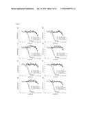 |
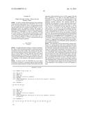 | 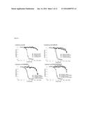 |
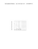 | 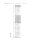 |
 |  |
 |  |
 |  |
 |  |
 | 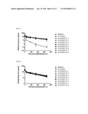 |
 |  |
 |  |
 |  |
 | 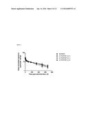 |
 |  |
 |
| Top Inventors for class "Drug, bio-affecting and body treating compositions" | |
| Rank | Inventor's name |
|---|---|
| 1 | David M. Goldenberg |
| 2 | Hy Si Bui |
| 3 | Lowell L. Wood, Jr. |
| 4 | Roderick A. Hyde |
| 5 | Yat Sun Or |