Patent application title: Antibody for Targeted induction of Apoptosis, CDC and ADCC mediated killing of Cancer cells, TBL-CLN1
Inventors:
Koteswara Rao Kollipara (Hyderabad, IN)
Ramesh Babu Batchu (Royal Oak, MI, US)
Assignees:
TRANSGENE BIOTEK LTD.
IPC8 Class: AC07K1630FI
USPC Class:
4241381
Class name: Drug, bio-affecting and body treating compositions immunoglobulin, antiserum, antibody, or antibody fragment, except conjugate or complex of the same with nonimmunoglobulin material binds expression product or fragment thereof of cancer-related gene (e.g., oncogene, proto-oncogene, etc.)
Publication date: 2012-02-02
Patent application number: 20120027763
Abstract:
Antibody for targeted induction of Apoptosis, CDC and ADCC mediated
killing of Cancer cells, TBL-CLN1, is disclosed. The antibodies,
TBL-CLN1, are monoclonal antibodies which can specifically target and
bind to the epitope of SEQ ID NO:1 expressed on cancer cells which
further leads to killing of cancer cells. TBL-CLN1 is not conjugated to
toxin or cytotoxic molecules, and provides selective killing of cancer
cells just by binding to cancer cell surface. Also, disclosed herein is
SEQ ID NO: 2 which is an engineered epitope which comprises of
polypeptide sequence of SEQ ID NO: 1 and a cysteine residue which is
added at the carboxyl end of the SEQ ID NO: 1. The epitope of SEQ ID NO:
2 is used to generate monoclonal antibodies described herein.Claims:
1. A polypeptide sequence of SEQ ID NO: 2.
2. The polypeptide sequence of claim 1, wherein said polypeptide sequence is capable of being administered in an immunologically sufficient amount for eliciting an immune response in a mammalian subject.
3. An antibody that specifically binds to a polypeptide sequence selected from the group consisting of SEQ ID NO: 1 and SEQ ID NO: 2.
4. The antibody as claimed in claim 3, wherein said antibody is humanized to generate chimeric antibody, humanized antibody and de-immunized antibody.
5. The antibody as claimed in claim 3, comprising: a heavy chain polypeptide sequence of SEQ ID NO:3; and a light chain polypeptide sequence of SEQ ID NO:5.
6. The antibody as claimed in claim 3, comprising: a variable heavy chain polypeptide sequence of SEQ ID NO:7; and a variable light chain polypeptide sequence of SEQ ID NO:9.
7. The antibody as claimed in claim 3, comprising at least one CDR sequence selected from the group consisting of variable heavy chain CDR1 of SEQ ID NO:11, variable heavy chain CDR2 of SEQ ID NO:12, variable heavy chain CDR3 of SEQ ID NO:13, variable light chain CDR1 of SEQ ID NO:17, variable light chain CDR2 of SEQ ID NO:18 and variable light chain CDR3 of SEQ ID NO:19.
8. The antibody as claimed in claim 3, wherein said antibody is a monoclonal antibody.
9. The antibody as claimed in claim 3, wherein said antibody is selected from the group consisting of IgG1, IgG2, IgG3, IgG4, IgM, IgA1, IgA2, secretory IgA, IgD, and IgE antibody.
10. The antibody as claimed in claim 3, wherein said antibody has at least one of the characteristics selected from the group consisting of: capable of inducing complement dependent cytotoxicity (CDC) of cancer cells expressing TNFRSF17; capable of inducing apoptosis of cancer cells expressing TNFRSF17; and capable of inducing antibody dependent cellular cytotoxicity (ADCC) of cancer cells expressing TNFRSF17;
11. The antibody as claimed in claim 3, wherein said antibody is expressed in a vector, wherein the vector comprises of a polynucleotide sequence of SEQ ID NO:8 and SEQ ID NO:10.
12. The antibody as claimed in claim 11, wherein said vector is transfected into a cell line.
13. The antibody as claimed in claim 12, wherein said cell line is a microbial cell line.
14. The antibody as claimed in claim 3, wherein said antibody is present in a pharmaceutical composition.
15. The antibody as claimed in claim 14, wherein said pharmaceutical composition further comprises of a pharmaceutically acceptable carrier, wherein the pharmaceutically acceptable carrier is selected from the group consisting of water, salt solutions, gelatins, oils, alcohols and mixtures thereof.
16. The antibody as claimed in claim 3, wherein said antibody is capable of being administered in a therapeutically effective amount for treating of cancer.
17. The antibody as claimed in claim 16, for the treatment of cancer, wherein said cancer expresses a protein with a region comprising a polypeptide sequence selected from the group consisting of SEQ ID NO: 1 and SEQ ID NO:2.
18. The antibody as claimed in claim 16, for the treatment of cancer, wherein said cancer is selected from the group consisting of Multiple Myeloma, Colon Cancer and Non-Hodgkin's Lymphoma.
19. A method for diagnosing the presence of TNFRSF17 in a sample, said method comprising: contacting said sample with antibody under conditions that allow for formation of a complex between said antibody and TNFRSF17, wherein said antibody specifically binds to a polypeptide sequence selected from the group consisting of SEQ ID NO: 1 and SEQ ID NO:2; and detecting the formation of said complex.
Description:
CROSS-REFERENCE TO PRIOR APPLICATION
[0001] This application claims priority to Indian Application No.: 568/CHE/2010 filed Mar. 4, 2010.
FIELD OF INVENTION
[0002] The present invention relates to the field of immunotherapy for cancers, and more particularly to monoclonal antibodies which specifically target an epitope expressed on cancer cells.
BACKGROUND OF INVENTION
[0003] Important breakthroughs in cancer therapy include clinical application of antibodies. The therapeutic strategy relies on the deliberate and selective induction of apoptosis or killing by antibody-dependent cell-mediated cytotoxicity (hereinafter referred to as ADCC) and complement-dependent cytotoxicity (hereinafter referred to as CDC) of malignant cells. Importantly, therapy-resistance in cancer is frequently associated with de-regulation in the mechanisms that control apoptosis. However, cancer cells are often reliant on these molecular aberrations for survival. Therefore, selective induction of apoptosis, CDC and ADCC in cancer cells but not normal cells is a challenge to be addressed.
[0004] Induction of apoptosis in tumor cells by tumor necrosis factor [TNF]-related apoptosis-inducing ligand (hereinafter referred to as TRAIL) is a promising therapeutic principle in oncology. Programmed cell death, known as apoptosis, is an essential cellular homeostasis mechanism that ensures correct development and function of multi-cellular organisms. The pivotal importance of correct execution of apoptosis is apparent from the many human diseases with aberrancies in apoptosis, including cancer.
[0005] One possible treatment for cancer involves monoclonal antibodies (mAb) that bind only to cancer cell-specific antigens and induce an immunological response against the target cancer cell. Such mAb could also be modified for delivery of a toxin, radioisotope, cytokine or other active conjugate. It is also possible to design bispecific antibodies that can bind with their fragment antigen binding (hereinafter referred to as Fab) regions both to target antigen and to a conjugate or effector cell.
[0006] Monoclonal antibody drugs are a relatively new innovation in cancer treatment. While several monoclonal antibody drugs are available for treating certain cancers, the best way to use these new drugs isn't always clear. The immune system attacks foreign invaders in our body, but it doesn't always recognize cancer cells as enemies. A monoclonal antibody can be directed to attach to certain parts of a cancer cell. In this way, the antibody marks the cancer cell and makes it easier for the immune system to find.
[0007] Monoclonal antibody developed to the specific cancer cell surface target can kill the cell with or without toxin attached just by binding to cell surface target. The antibody can initiate lysis of the cancer cell through apoptosis, CDC and ADCC. Monoclonal antibody therapy can be used to destroy malignant tumor cells and prevent tumor growth by blocking specific cell receptors or by delivering a conjugated toxin.
[0008] There is a need for an antibody which can selectively target and induce killing of cancer cells.
OBJECT OF INVENTION
[0009] The principal object of this invention is to provide a method for targeted and selective killing of cancer cells, wherein the killing may be induced by apoptosis, ADCC and CDC of cancer cells.
[0010] Another object of the invention is to provide a method for selective elimination of cancer cells without affecting the normal cell population.
STATEMENT OF INVENTION
[0011] Accordingly the invention provides a polypeptide sequence of SEQ ID NO: 2, which is an engineered polypeptide sequence comprising of SEQ ID NO: 1 and a cysteine residue at the carboxyl end of SEQ ID NO: 1, and SEQ ID NO: 1 is an epitope on the Tumor necrosis factor receptor super family (hereinafter referred to as TNFRSF17).
[0012] There is also provided a polynucleotide sequence which is characterized in that to encode the polypeptide sequence of SEQ ID NO: 2.
[0013] In another embodiment, the invention provides an antibody that specifically binds to a polypeptide sequence of SEQ ID NO: 1 and/or SEQ ID NO: 2, wherein SEQ ID NO: 1 is an epitope on the TNFRSF17 and SEQ ID NO: 2, is an engineered polypeptide produced by adding a cysteine residue at the carboxyl end of SEQ ID NO: 1.
[0014] In another embodiment, the invention provides a polynucleotide sequence that encodes an antibody which binds to a polypeptide sequence selected from the group consisting of SEQ ID NO: 1 and SEQ ID NO:2, wherein the polynucleotide sequence comprises of the sequence of SEQ ID NO: 4 and SEQ ID NO: 6.
[0015] In another embodiment, the invention provides an antibody comprising a heavy chain polypeptide sequence of SEQ ID NO:3 and a light chain polypeptide sequence of SEQ ID NO:5, wherein the variable heavy chain polypeptide sequence is of SED ID NO:7, variable light chain polypeptide sequence is of SED ID NO:9, variable heavy chain CDR1 (complementarity determining region 1) is of SEQ ID NO:11, variable heavy chain CDR2 (complementarity determining region 2) is of SEQ ID NO:12, variable heavy chain CDR3 (complementarity determining region 3) is of SEQ ID NO:13, variable light chain CDR1 is of SEQ ID NO:17, variable light chain CDR2 is of SEQ ID NO:18, variable light chain CDR3 is of SEQ ID NO:19.
[0016] In another embodiment, the invention provides an antibody, specifically a monoclonal antibody, which can induce complement dependent cytotoxicity (CDC) of cancer cells expressing TNFRSF17, apoptosis of cancer cells expressing TNFRSF17 and/or antibody dependent cellular cytotoxicity (ADCC) of cancer cells expressing TNFRSF17.
[0017] In another embodiment, the invention provides a pharmaceutical composition comprising the antibody, specifically monoclonal antibody, and a pharmaceutically acceptable carrier.
[0018] In yet another embodiment, the invention provides a method of eliciting an immune response, in a mammalian subject, directed against the polypeptide sequence of SEQ ID NO: 1 or SEQ ID NO: 2, wherein the method comprises of administering the polypeptide sequence of SEQ ID NO: 1 or SEQ ID NO: 2 into the mammalian subject.
[0019] In another embodiment, the invention provides a method of treating cancer by administering a therapeutically effective amount of the antibody, specifically monoclonal antibody, of the present invention, wherein the cancer is Multiple myeloma (hereinafter referred to as MM), Colon cancer (hereinafter referred to as CC) or Non-Hodgkin's lymphoma (hereinafter referred to as NHL).
[0020] In another embodiment, the invention provides a diagnostic method for diagnosing the presence of TNFRSF17 or cell expressing TNFRSF17 in a sample by contacting the sample with antibody of the present invention, under conditions that allow for formation of a complex between said antibody and TNFRSF17, and detecting the formation of the complex in the sample, wherein the sample is any mammalian tissue sample or an extract of any mammalian tissue sample suspected of having cancer, wherein the cells expressing TNFRSF17 include MM cancer cells, CC cancer cells and NHL cancer cells.
[0021] In yet another embodiment, the invention provides a diagnostic kit for detecting the presence of TNFRSF17 or cell expressing TNFRSF17 in a sample, wherein said diagnostic kit comprises of antibody, specifically monoclonal antibody, of the present invention.
[0022] These and other aspects of the embodiments herein will be better appreciated and understood when considered in conjunction with the following description and the accompanying drawings. It should be understood, however, that the following descriptions, while indicating preferred embodiments and numerous specific details thereof, are given by way of illustration and not of limitation. Many changes and modifications may be made within the scope of the embodiments herein without departing from the spirit thereof, and the embodiments herein include all such modifications.
BRIEF DESCRIPTION OF FIGURES
[0023] This invention is illustrated in the accompanying drawings, throughout which like reference letters indicate corresponding parts in the various FIGURES. The embodiments herein will be better understood from the following description with reference to the drawings, in which:
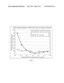
[0024] FIG. 1 is a dose response graph depicting the results of direct cytotoxicity and dose dependent assay in IM9 cells with 1.2 mg/mL of TBL-CLN1 sample;
[0025] FIG. 2 is a dose response graph depicting the results of direct cytotoxicity and dose dependent assay in IM9 cells with 2.4 mg/mL of TBL-CLN1 sample;
[0026] FIG. 3 is a dose response graph depicting the results of direct cytotoxicity and dose dependent assay in RPMI-8266 cells with 1.2 mg/mL of TBL-CLN1 sample;
[0027] FIG. 4 is a dose response graph depicting the results of direct cytotoxicity and dose dependent assay in RPMI-8266 cells with 2.4 mg/mL of TBL-CLN1 sample;
[0028] FIG. 5 is a dose response graph depicting the results of direct cytotoxicity and dose dependent assay in SW620 cell line with 1.2 mg/mL of TBL-CLN1 sample;
[0029] FIG. 6 is a dose response graph depicting the results of direct cytotoxicity and dose dependent assay in SW620 cell line with 2.4 mg/mL of TBL-CLN1 sample;
[0030] FIG. 7 is a dose response graph depicting the results of direct cytotoxicity and dose dependent assay in LOVO cell line with 2.4 mg/mL of TBL-CLN1 sample;
[0031] FIG. 8 is a dose response graph depicting the results of complement dependent cytotoxicity in IM9 cells;
[0032] FIG. 9 is a dose response graph depicting the results of complement dependent cytotoxicity in RPMI-8266 cells;
[0033] FIG. 10 is a dose response graph depicting the results of complement dependent cytotoxicity in SW620 cell line; and
[0034] FIG. 11 is a dose response graph depicting the results of complement dependent cytotoxicity in LOVO cell line.
DETAILED DESCRIPTION OF INVENTION
[0035] The embodiments herein and the various features and advantageous details thereof are explained more fully with reference to the non-limiting embodiments that are illustrated in the accompanying drawings and detailed in the following description. Descriptions of well-known components and processing techniques are omitted so as to not unnecessarily obscure the embodiments herein. The examples used herein are intended merely to facilitate an understanding of ways in which the embodiments herein may be practiced and to further enable those of skill in the art to practice the embodiments herein. Accordingly, the examples should not be construed as limiting the scope of the embodiments herein.
[0036] It is to be understood that the present disclosure is not limited in its application to the details of construction and the arrangement of components set forth in the following description or illustrated in the drawings. The present disclosure is capable of other embodiments and of being practiced or of being carried out in various ways. Also, it is to be understood that the phraseology and terminology used herein is for the purpose of description and should not be regarded as limiting.
[0037] Embodiments of the present invention are directed towards an antibody and a method for targeting and killing of cancer cells including; Colon cancer, Multiple Myeloma and Non-Hodgkin's lymphoma cancer cells. The main focus of the present invention is to selectively target and kill cancer cells by monoclonal antibodies produced against a unique surface antigen over expressed on Colon cancer (CC), Multiple Myeloma (MM) and Non-Hodgkin's lymphoma (NHL) cancer cells. According to a non limiting aspect, the invention focuses on enhancing the immunogenicity of the surface antigen by adding a cysteine residue to the carboxyl end of the 14 amino acid long extra-cellular antigenic peptide epitope.
[0038] Tumor necrosis factor receptors (TNFR) are single transmembrane-spanning glycoproteins that bind cytokines and trigger multiple signal transduction pathways. Tumor necrosis factor (TNF) is a pro-inflammatory cytokine whose role is established in the pathogenesis of malignant diseases like cancer. TNF has been found to have a pro-cancerous effect and gene polymorphisms which increase or decrease TNF production results either in increased risk or protective effect on a number of different cancers and precancerous diseases including gastric cancer, lymphoma and cervical cancer.
[0039] Certain TNFR member proteins are expressed on specific cancer cells. TNFR has been detected in a number of different solid tumor types as well as hematological malignancies.
[0040] TNF is a cytokine that is produced early in the inflammatory cascade and has been shown to promote carcinogenesis. TNF has a wide range of activities in cancer. TNF has converse actions that induce a number of pro-inflammatory genes, which the tumors utilize to promote cancer such as cytokines, angiogenic factors and MMPs. These factors contribute to tumor formation, growth, invasion and metastasis to other sites. Many of the actions of TNF may occur by the stimulation of stromal tissue, tumor-associated macrophages and fibroblasts. These cells may then produce inflammatory cytokines including TNF itself, as well as some of the angiogenic factors described above, contributing to tumor proliferation and invasion.
[0041] Anti-TNF mAbs are widely used against a range of cancers like breast cancer, metastatic colorectal cancer, NHL and other lymphoblastic malignancies. However, the anti-TNF monoclonal antibodies are chimeric antibodies either conjugated to a toxin moiety or a radioactive substance which kill targeted cancer cells by selectively binding to TNFRs and rendering the toxic or radioactive effect on the tumor cells with specific and non-specific surface antigen epitope, thus affecting the bystander lymphocytes.
[0042] The embodiments disclosed herein aims at targeting a certain class of surface antigens expressed on Colon cancer, Multiple Myeloma and Non-Hodgkin's lymphoma cancer cells. The surface antigen is TNFRSF17. The invention ensures specific targeting of lymphocytes which express TNFRSF17. The surface antigen epitope on TNFRSF17 is a 14 amino acid sequence of the order as provided under SEQ ID NO:1, from amino terminal to carboxyl terminal end.
[0043] In an embodiment, the addition of a cysteine residue to the carboxyl end enhances the immunogenicity of the antigen epitope. The addition of cysteine results in an antigenic peptide of 15 amino acid sequence (also referred to as TNFRS17-epag) of the order as provided under SEQ ID NO:2, from amino terminal to carboxyl terminal end. The antigen epitope of SEQ ID NO:1 is modified and purified by techniques known in the art.
[0044] The disclosed embodiments underline the production of monoclonal antibodies, generated against cancer cells including Colon cancer, Multiple Myeloma and Non-Hodgkin's lymphoma cancer cells from patients and cell lines but not normal tissues according to a non limiting aspect of the present invention. The monoclonal antibody has a binding domain specific for TNFRS17-epag. The therapeutic antibodies specifically bind to the TNFRS17 on the targeted cancer cells and kill them mediated by targeted induction of apoptosis, antibody-dependent cytotoxic immune responses and complement-dependent cytotoxic immune response. This antigen specific antibody response does not harm the bystander B cells and other immune cells like T cell, NK cells, mast cells and stimulated neutrophills.
[0045] In an embodiment, an epitope of the polypeptide sequence of SEQ ID NO: 2 is disclosed. SEQ ID NO: 2 is an engineered epitope which comprises of polypeptide sequence of SEQ ID NO: 1 and a cysteine residue which is added at the carboxyl end of the SEQ ID NO: 1. The polypeptide sequence of SEQ ID NO: 2 is more immunogenic due to the addition of cysteine residue. The epitope of SEQ ID NO: 2 is further used to generate antibodies described herein.
Identification of the Epitope of SEQ Id NO: 1 and Preparation of Engineered Epitope of SEQ ID NO: 2:
[0046] The 15 amino acid sequence of SEQ ID NO: 2, TNFRSF17-epag, of the present invention can be produced by adding a cysteine residue to carboxyl end of the 14 amino acid sequence of SEQ ID NO: 1. SEQ ID NO: 2 is more immunogenic and may be used to produce antibodies, specifically monoclonal antibodies. The monoclonal antibodies of the present invention can specifically bind to polypeptide sequence of SEQ ID NO: 2.
[0047] The polypeptide sequence of SEQ ID NO: 2 may be prepared by techniques known in the art including chemical peptide synthesis and genetic engineering.
[0048] SEQ ID NO: 1 is the 14 amino acid sequence which is an epitope present on the extracellular domain of TNFRSF17. TNFRSF17 is a unique cell surface antigen expressed on MM, CC and NHL cancer cells. The monoclonal antibodies of the present invention can specifically bind to polypeptide sequence of SEQ ID NO: 1 present on MM, CC and NHL cancer cells and induce killing of such cancer cells.
Production of the Antibodies:
[0049] The antibody disclosed herein can be produced by various methods familiar to those skilled in the art, such as hybridoma methodology.
[0050] In an embodiment of the invention, the antibodies are produced by the conventional hybridoma technology, wherein a mouse is immunized with an antigen. The spleen cells are then isolated and fused with myeloma cells lacking Hypoxanthine-guanine phosphoribosyltransferase (HGPRT) expression. The hybrid cells are selected using hypoxanthine, aminopterin and thymine (HAT) containing media.
[0051] The antibodies disclosed herein can also be made by other methods familiar to those skilled in the art including recombinant DNA methods.
[0052] The disclosed monoclonal antibodies against the polypeptide sequence of SEQ ID NO: 2 (TNFRS17-epag), which can induce targeted killing of cancer cells are referred to as TBL-CLN1.
Polynucleotide and Amino Acid Sequences of TBL-CLN1
[0053] The disclosed antibodies, in an embodiment, comprises of at least two heavy (H) chains and two light (L) chains inter-connected by disulfide bonds, or an antigen binding fragment. Each heavy chain sequence comprises of a leading sequence, heavy chain variable sequence (abbreviated as VH) and a heavy chain constant sequence. Each light chain comprises of a leading sequence, light chain variable sequence (abbreviated herein as VL) and a light chain constant sequence.
[0054] The VH and VL regions can be further subdivided into regions of hypervariability, termed as complementarity determining regions (CDR) which are interspersed with regions that are more conserved, termed as framework regions (FR). Each VH and VL is composed of three CDRs and four FRs, arranged from amino-terminus to carboxy-terminus in the following order: FR1, CDR1, FR2, CDR2, FR3, CDR3, FR4. The variable regions of the heavy and light chains contain a binding domain that interacts with an antigen. The constant regions of the antibodies mediate the binding of the antibodies to host tissues or factors, including various cells of the immune system (e.g. effector cells) and components of the complement system.
[0055] The cDNAs encoding the variable region and constant region, including heavy and light chains, of the antibodies were amplified by PCR, cloned, and sequenced. The polypeptide and polynucleotide sequences of the heavy and light chains of TBL-CLN1 are as follows:
[0056] SEQ ID NO:3 and SEQ ID NO:5 are the amino acid sequences for the heavy chain and light chain of TBL-CLN1, respectively.
[0057] SEQ ID NO:4 and SEQ ID NO:6 are the nucleotide sequence encoding the heavy chain and light chain of TBL-CLNI, respectively. SEQ ID NO:7 is the amino acid sequences of variable region of heavy chain of TBL-CLN1. SEQ ID NO:9 is the amino acid sequences of variable region of light chain of TBL-CLN1. SEQ ID NO:8 is the nucleotide sequence encoding the variable region of heavy chain of TBL-CLN1. SEQ ID NO:10 is the nucleotide sequence encoding the variable region of light chain of TBL-CLN1. The variable heavy chain (complementarity determining region) CDR regions of TBL-CLN1 comprises of CDR1, CDR2 and CDR3 which has the amino acid sequence of SEQ ID NO:11, SEQ ID NO:12 and SEQ ID NO:13. The nucleotide sequence encoding SEQ ID NO:11, SEQ ID NO:12 and SEQ ID NO:13 are SEQ ID NO: 14, SEQ ID NO: 15 and SEQ ID NO: 16. The variable light chain (complementarity determining region) CDR regions of TBL-CLN1 also comprises of CDR1, CDR2 and CDR3 which has the amino acid sequence of SEQ ID NO:17, SEQ ID NO:18 and SEQ ID NO:19. The nucleotide sequence encoding SEQ ID NO:17, SEQ ID NO:18 and SEQ ID NO:19 are SEQ ID NO: 20, SEQ ID NO: 21 and SEQ ID NO: 22. The constant heavy and light chain region of TBL-CLN1 has the amino acid sequence of SEQ ID NO:23 and SEQ ID NO:24, respectively. The nucleotide sequence encoding the constant heavy and light chain region of TBL-CLN1 are SEQ ID NO:25 and SEQ ID NO:26.
[0058] In an embodiment, the polynucleotide sequences SEQ ID NO:8 and SEQ ID NO:10 may be present in a vector.
[0059] In another embodiment, the vector consisting of polynucleotide sequences SEQ ID NO:8 and SEQ ID NO:10 may be present in a cell line.
Characteristics of the Antibodies:
[0060] The antibodies of the present invention are capable of inducing targeted killing of cancer cells that express TNFRSF17 by CDC, ADCC and apoptosis.
[0061] The term "antibody", as used herein, includes antigen-binding fragment. The term "antigen-binding fragment", as used herein, refers to one or more fragments of an antibody that retain the ability to bind to an antigen. It's known that the antigen-binding function of an antibody can be performed by fragments of an intact antibody. The term "antigen-binding fragment" includes () a Fab fragment and (u) a F(ab')2 fragment.
[0062] In an embodiment disclosed herein, the antibodies are preferably IgG1 and may include IgG3 and IgG4 antibodies. Further, other antibody isotypes are also encompassed by the invention, including IgG2, IgM, IgA1, IgA2, secretory IgA, IgD, and IgE.
[0063] In cancer cells expressing TNFRSF17, the antibodies of the present invention may induce complement dependent cytotoxicity (CDC) in the presence of complement system, apoptosis and antibody dependent cellular cytotoxicity (ADCC) in the presence of effector cells. The antibodies may also be capable of prolonging the survival of a subject having tumor cells expressing TNFRSF17 and further be capable of depleting the cancer cells expressing TNFRSF17.
[0064] The effector cells include all the effector cells of the immune system such as NK cells, T cells, monocytes, macrophages, etc.
Nucleic Acid
[0065] In an embodiment, a polynucleotide sequence which encodes the polypeptide sequence of SEQ ID NO: 2 is provided.
Pharmaceutical Compositions of the Antibodies
[0066] A disclosed embodiment provides a pharmaceutical composition. The pharmaceutical composition may comprise antibody of the present invention (specifically monoclonal antibody, TBL-CLN1) and may also contain a pharmaceutically acceptable carrier.
[0067] The pharmaceutically acceptable carriers in the pharmaceutical composition include generally used carriers well known in the art including water, salt solutions, gelatins, oils, alcohols, and other excipients and auxiliaries that facilitate processing of the active compounds into preparations that may be used pharmaceutically.
[0068] The pharmaceutical composition may further consist of certain stabilizers, salts such as sodium chloride, sodium citrate and polysorbate, and water for injection. The pH of the composition may preferably range from 6 to 7.5.
[0069] The pharmaceutical composition may be administered to a patient as injections in a range of doses starting from 0.05 to 10 mg/Kg of body weight.
[0070] In addition to the antibody of the present invention, the pharmaceutical composition may also comprise of pharmaceutically acceptable diluents, preservatives, solubilizers, emulsifiers, adjuvant and/or other carriers well known in the art.
Treatment
[0071] Further, the embodiments disclosed herein provide the use of the isolated antibody (specifically monoclonal antibody, TBL-CLN1) in the treatment of cancer.
[0072] The disclosed antibody (specifically monoclonal antibody, TBL-CLN1), in an embodiment, is used to in treating cancer by administering a therapeutically effective amount of TBL-CLN1.
[0073] The antibody (specifically monoclonal antibody, TBL-CLN1), in an embodiment, can be isolated by techniques well known in the art. The types of cancer that can be treated include but are not limited to Multiple Myeloma (MM), Colon Cancer (CC) and Non-Hodgkin's lymphoma (NHL).
[0074] The term "therapeutically effective amount" includes an amount of the composition having therapeutic effect on a subject upon administration of the composition to the subject. The composition may be administered in doses preferably ranging from 0.05 to 10 mg/Kg of body weight
[0075] Further, a method of eliciting an immune response in a mammalian subject directed against polypeptide sequence of SEQ ID NO:1 or SEQ ID NO:2, wherein the subject is administered with an immunologically sufficient amount of polypeptide sequence of SEQ ID NO: 1 or SEQ ID NO: 2, is also provided in an embodiment.
[0076] The term "immunologically sufficient amount" includes an amount of the composition which is capable of inducing an immune response in a subject upon administration of the composition to the subject.
[0077] Further, the disclosed antibody, in an embodiment may be used for diagnosing TNFRSF17, including cells expressing TNFRSF17.
[0078] The cells expressing TNFRSF17 typically include MM, CC and NHL cancer cells.
[0079] The diagnostic method comprises of contacting TNFRSF17 present in a sample with the antibody, specifically monoclonal antibody, of the present invention to form a complex, under conditions favorable for such complex formation, and detecting the complex formed by the antibody and TNFRSF17.
[0080] The disclosed antibody, in an embodiment may be provided in a diagnostic kit for detecting the presence of TNFRSF17, including cells expressing TNFRSF17.
[0081] The diagnostic kit for detecting the presence of TNFRSF17 in a sample comprises of antibodies that binds to a polypeptide sequence selected from the group consisting of SEQ ID NO: 1 and SEQ ID NO:2 in order to form a complex. The complex formed between the antibodies and the polypeptide sequence may further be detected by methods known in the art.
[0082] The "sample" may originate from a mammal, and may be a tissue sample (such as bone marrow tissue, colon tissue, etc.) or an extract of any tissue sample suspected of having cancer.
[0083] The detection of the complex as described herein can be performed by apparatus capable of detecting specific signals emitted by detectable labels generally known in the art such as radiation emission, color change, fluorescence, etc.
[0084] According to a non limiting aspect, the therapeutic monoclonal antibodies, TBL-CLN1, can be given as an adjuvant therapy either through intravenous infusion or in to the target tissue directly.
[0085] The invention is further defined by reference to the following example. It will be apparent to those skilled in the art that many modifications, both to materials and methods, may be practiced without departing from the scope of the invention.
EXAMPLES
Example 1
Identification and Isolation of SEQ ID NO:1
[0086] TBL-CLN1 target was identified and validated over a period of 3 years by studying patient databases from numerous hospitals.
[0087] The identification and isolation of TNFRSF17 was carried out by:
[0088] a) obtaining around 20 cancer tissues from different hospitals,
[0089] b) isolating cancer cells using laser capture microscopy and,
[0090] c) conducting gene profile analysis.
Example 2
Production of Monoclonal Antibodies
[0091] Development of Myeloma cells: Mice were immunized with SEQ ID NO:2 with attached adjuvant. Mice serum was then screened using various techniques such as ELISA.
[0092] After sufficient titers were reached the mice were euthanized to remove the spleen as a source of cells for cell fusion. These cells were then treated with 8-azaguanine to ensure sensitivity to HAT.
[0093] Fusion procedure: Spleen cells harvested from mice were fused with myeloma cells. Fusion was done through co-centifusing in polyethylene glycol.
[0094] Cells were then plated in selection medium hypoxanthine-aminopterin-thymidine (HAT) selection medium-inhibitor of aminopterin which blocks nucleotide synthesis. Cells were distributed on feeder cells (murine bone-marrow) to promote growth of the hybridoma cells. Mice were then inoculated with the cells. The produced ascites was then collected from the mice and mAbs were screened using specific Ag binding activity.
[0095] The antibodies, isotype IgG1, having optimal binding/activity were identified. The antibodies were identified based on their ability to induced targeted ADCC and CDC mediated killing of cancer cells.
Example 3
Polynucleotide and Amino Acid Sequences of TBL-CLN1
[0096] The total RNA was extracted from hybridoma cells. The cDNAs encoding the variable region and constant region, including heavy and light chains, of the antibodies were amplified by PCR, cloned, and sequenced. The polypeptide and polynucleotide sequences of the heavy and light chains of TBL-CLN1 are as follows:
[0097] SEQ ID NO:3 and SEQ ID NO:5 are the amino acid sequences for the heavy chain and light chain of TBL-CLN1, respectively. SEQ ID NO:4 and SEQ ID NO:6 are the nucleotide sequence encoding the heavy chain and light chain of TBL-CLNI, respectively. SEQ ID NO:7 is the amino acid sequences of variable region of heavy chain of TBL-CLN1. SEQ ID NO:9 is the amino acid sequences of variable region of light chain of TBL-CLN1. SEQ ID NO:8 is the nucleotide sequence encoding the variable region of heavy chain of TBL-CLN1. SEQ ID NO:10 is the nucleotide sequence encoding the variable region of light chain of TBL-CLN1. The variable heavy chain CDR regions of TBL-CLN1 comprises of CDR1, CDR2 and CDR3 which has the amino acid sequence of SEQ ID NO:11, SEQ ID NO:12 and SEQ ID NO:13. The nucleotide sequence encoding CDR1, CDR2 and CDR3 are SEQ ID NO: 14, SEQ ID NO: 15 and SEQ ID NO: 16. The variable light chain CDR regions of TBL-CLN1 also comprises of CDR1, CDR2 and CDR3 which has the amino acid sequence of SEQ ID NO:17, SEQ ID NO:18 and SEQ ID NO:19. The nucleotide sequence encoding CDR1, CDR2 and CDR3 are SEQ ID NO: 20, SEQ ID NO: 21 and SEQ ID NO: 22. The constant heavy and light chain region of TBL-CLN1 has the amino acid sequence of SEQ ID NO:23 and SEQ ID NO:24, respectively. The nucleotide sequence encoding the constant heavy and light chain region of TBL-CLN1 are SEQ ID NO:25 and SEQ ID NO:26.
Example 4
Functional Characteristics of the Antibodies
[0098] ADCC Activity of TBL-CLN1: ADCC Assay
[0099] Procedure for Preparation of peripheral Blood Mononuclear Cells (PBMC): Blood from normal volunteer was collected in heparinized syringe. 10 mL of whole blood was transferred to a falcon tube. Blood and plasma was diluted to 30 mL in phosphate buffered saline (PBS) and mixed. 10 mL of room temp Ficoll Hypaque solution was then added to a 50 mL Falcon tube. Diluted blood was further overlaid on top of Ficoll Hypaque. Tubes were then centrifuged for 30 minutes, at room temperature (RT). PBMCs were collected from interphase of PBS/Ficoll and PBS was added. Cells were centrifuged at RT and supernatant was decanted. 5 mL RBC Lysis Solution was added to pellet and incubated for 5 min. at RT. Cells were diluted to 10 mL with PBS and centrifuged. Cells were then counted as per standard procedure. The cells were resuspended in assay medium containing fetal bovine serum (FBS), glutamine, HEPES, and a known antibiotic such as Penicillin and/or Streptomycin etc for ADCC assay.
[0100] The cancerous cell line was revived from the frozen stock stored in liquid nitrogen. The cancerous cell line was cultured with 10% serum containing a known medium. After reaching the required confluence the cells were collected and washed with a known medium. The cells were then centrifuged and the assay medium decanted.
[0101] Cancerous cell line was resuspended in assay medium with a specified cell density. Four specific known controls were employed in the assay a) PBMC control, b) Cancer cell control, c) Total cell lysis control and d) Background Control.
[0102] The samples were assayed in 96 well microtitre plate using serial dilution method. The plate was incubated for 2-6 hrs at 25° C.-40° C. temperature in an incubator to facilitate Antibody dependent cell lysis.
[0103] After incubation, the plate was centrifuged at RT and the supernatants were harvested. The harvested supernatants were then analyzed for LDH activity by using a commercial LDH detection kit.
[0104] The percent ADCC was calculated with the following equation:
Percentage CDC=((O.D of test-O.D of PBMC control-O.D of cancer cell control)/(O.D of total cell lysis-O.D of Cancer cell control))×100
[0105] The activity of the test antibody was examined by plotting the % ADCC activity against the log of antibody concentration using a four parameter curve fitting program.
[0106] Results of direct cytotoxicity and dose dependent assays against different cell lines are as follows:
[0107] FIG. 1 is a graph depicting the results of direct cytotoxicity in IM9 cells with 1.2 mg/mL of TBL-CLN1 sample. The control used was Mabthera®. The graph depicts the percentage cell cytotoxicity exhibited by TBL-CLN1 antibody at various TBL-CLN1 sample concentrations, which is plotted against the control.
[0108] FIG. 2 is a graph depicting the results of direct cytotoxicity in IM9 cells with 2.4 mg/mL of TBL-CLN1 sample. The control used was Mabthera®. The graph depicts the percentage cell cytotoxicity exhibited by TBL-CLN1 antibody at various TBL-CLN1 sample concentrations, which is plotted against the control.
[0109] FIG. 3 is a graph depicting the results of direct cytotoxicity in RPMI-8226 cells with 1.2 mg/mL of TBL-CLN1 sample. The control used was Mabthera®. The graph depicts the percentage cell cytotoxicity exhibited by TBL-CLN1 antibody at various TBL-CLN1 sample concentrations, which is plotted against the control.
[0110] FIG. 4 is a graph depicting the results of direct cytotoxicity in RPMI-8226 cells with 2.4 mg/mL of TBL-CLN1 sample. The control used was Mabthera®. The graph depicts the percentage cell cytotoxicity exhibited by TBL-CLN1 antibody at various TBL-CLN1 sample concentrations, which is plotted against the control.
[0111] FIG. 5 is a graph depicting the results of direct cytotoxicity in SW620 cell line with 1.2 mg/mL of TBL-CLN1 sample. The graph depicts the percentage cell cytotoxicity exhibited by TBL-CLN1 antibody at various TBL-CLN1 sample concentrations.
[0112] FIG. 6 is a graph depicting the results of direct cytotoxicity in SW620 cell line with 2.4 mg/mL of TBL-CLN1 sample. The control used was Mabthera®. The graph depicts the percentage cell cytotoxicity exhibited by TBL-CLN1 antibody at various TBL-CLN1 sample concentrations, which is plotted against the control.
[0113] FIG. 7 is a graph depicting the results of direct cytotoxicity in LOVO cell line with 2.4 mg/mL of TBL-CLN1 sample. The control used was Mabthera®. The graph depicts the percentage cell cytotoxicity exhibited by TBL-CLN1 antibody at various TBL-CLN1 sample concentrations, which is plotted against the control.
[0114] CDC Activity of TBL-CLN1: CDC Assay
[0115] Procedure: The cancerous cell line was cultured in a tissue culture flask with serum containing growth medium. After reaching a targeted confluence, the cells were collected and counted. Ringer-Hepes buffer (RHB buffer) was prepared as per methods known in the art (the composition includes 20 mM HEPES, 2 mM glutamine, BSA and known antibiotics such as penicillin, streptomycin etc).
[0116] The cells were washed with RHB buffer and centrifuged. The buffer was then decanted. The cells were diluted to a density of 1 million cells/ml.
[0117] The samples were arranged in 96 wells of microtitre plate employing serial dilution method using four different controls. 50 μl of human serum complement was added to all the wells except the medium control, total lysis control and antibody control. 50 μl of 0.4% NP40 was further added to all the wells of column 11. 50 μl of cell suspension was then added to all the wells. After adding all the components, the plate was incubated in a CO2 incubator at a range between 2-10% at a temperature between 30° C.-40° C.
[0118] After incubation, a known quantity of XTT was added to the wells and incubated for 2-10 hrs with intermittent shaking for few minutes. After incubation, readings were taken in an ELISA plate reader at 450 nm wavelength.
[0119] CDC was calculated with the following equation:
Percentage CDC=(O.D of test-O.D of Complement control)/(O.D of total cell lysis-O.D of complement control)×100
[0120] The activity of the test antibody was examined by plotting the % CDC activity against the log of antibody concentration using a four parameter curve fitting program.
[0121] The results of CDC assays demonstrating minimal CDC effect against different cell lines using a control are as follows:
[0122] FIG. 8 is a graph depicting the results of complement dependent cytotoxicity in IM9 cell line. The control used was Mabthera®. The graph depicts the percentage CDC activity exhibited by TBL-CLN1 antibody at various TBL-CLN1 sample concentrations, which is plotted against control.
[0123] FIG. 9 is a graph depicting the results of complement dependent cytotoxicity in RPMI-8226 cell line. The control used was Mabthera®. The graph depicts the percentage CDC activity exhibited by TBL-CLN1 antibody at various TBL-CLN1 sample concentrations, which is plotted against control.
[0124] FIG. 10 is a graph depicting the results of complement dependent cytotoxicity in SW620 cell line. The control used was Mabthera®. The graph depicts the percentage CDC activity exhibited by TBL-CLN1 antibody at various TBL-CLN1 sample concentrations, which is plotted against control.
[0125] FIG. 11 is a graph depicting the results of complement dependent cytotoxicity in LOVO cell line. The control used was Mabthera®. The graph depicts the percentage CDC activity exhibited by TBL-CLN1 antibody at various TBL-CLN1 sample concentrations, which is plotted against control.
[0126] The cytotoxicity further demonstrated a reasonable titre of mouse antibody of LD50<50 ug/ml.
[0127] When injected to SCID mice with human myeloma xenograft, the antibody specifically bound to the cell surface antigen of multiple myeloma cancer cells initiating ADCC and apoptosis to kill the cancer cells.
[0128] Significant cytotoxicity was observed towards the target cancer cells. The results of the assays demonstrated that the antibody (TBL-CLN1) has cytotoxicity towards specific cancer cell lines. Binding assays confirmed TBL-CLN1's cytotoxicity effect against Non-Hodgkins Lymphoma, Multiple Myeloma, and Colon cancers.
[0129] Humanization was performed in order to generate chimeric antibody, humanized antibody and de-immunized antibody. The activity of humanized antibody was tested at each step to make sure original cytotoxicity was retained.
Example 5
Humanized Antibodies
[0130] Humanized antibody expressed in microbial/murine cell lines. The humanized monoclonal antibody was cloned and expressed in the selected and genetically modified microbial and/or murine cells. The methods for production and purification of the expressed/secreted protein are known in the art. The production and purification of the expressed/secreted protein was carried out in a bioreactor providing optimized conditions such as pH, DO, CO2, air etc.
[0131] The expressed protein was then purified through series of steps such as centrifugation, chromatography systems etc to arrive at the purified protein, humanized monoclonal antibodies.
[0132] The foregoing description of the specific embodiments will so fully reveal the general nature of the embodiments herein that others can, by applying current knowledge, readily modify and/or adapt for various applications such specific embodiments without departing from the generic concept, and, therefore, such adaptations and modifications should and are intended to be comprehended within the meaning and range of equivalents of the disclosed embodiments. It is to be understood that the phraseology or terminology employed herein is for the purpose of description and not of limitation. Therefore, while the embodiments herein have been described in terms of preferred embodiments, those skilled in the art will recognize that the embodiments herein can be practiced with modification within the spirit and scope of the embodiments as described herein.
Sequence CWU
1
26114PRTUnknownDescription of Unknown Tumor necrosis factor receptor
super family peptide 1Lys Ser Arg Thr Gly Asp Glu Ile Ile Leu Pro Arg Gly
Leu1 5 10215PRTArtificial
SequenceDescription of Artificial Sequence Synthetic peptide 2Lys
Ser Arg Thr Gly Asp Glu Ile Ile Leu Pro Arg Gly Leu Cys1 5
10 153462PRTArtificial
SequenceDescription of Artificial Sequence Synthetic polypeptide
3Met Asn Phe Gly Phe Ser Leu Ile Phe Leu Val Leu Val Leu Lys Gly1
5 10 15Val Gln Cys Glu Val Lys
Leu Val Glu Ser Gly Gly Gly Leu Val Lys 20 25
30Pro Gly Gly Ser Leu Lys Leu Ser Cys Ala Ala Ser Gly
Ile Thr Phe 35 40 45Ser Thr Tyr
Ala Met Ser Trp Val Arg Gln Thr Pro Glu Lys Arg Leu 50
55 60Glu Trp Val Ala Ser Ile Thr Thr Gly Gly Asn Val
Tyr Phe Pro Asp65 70 75
80Ser Val Lys Gly Arg Phe Thr Ile Ser Arg Asp Asn Ala Arg Asn Ile
85 90 95Leu Tyr Leu Gln Met Ser
Ser Leu Arg Ser Glu Asp Thr Ala Met Tyr 100
105 110Tyr Cys Ala Gly Gly Ile Thr Ala Thr Arg Gly Thr
Phe Ala Tyr Trp 115 120 125Gly Gln
Gly Thr Thr Leu Thr Val Ser Ser Ala Lys Thr Thr Pro Pro 130
135 140Ser Val Tyr Pro Leu Ala Pro Gly Ser Ala Ala
Gln Thr Asn Ser Met145 150 155
160Val Thr Leu Gly Cys Leu Val Lys Gly Tyr Phe Pro Glu Pro Val Thr
165 170 175Val Thr Trp Asn
Ser Gly Ser Leu Ser Ser Gly Val His Thr Phe Pro 180
185 190Ala Val Leu Gln Ser Asp Leu Tyr Thr Leu Ser
Ser Ser Val Thr Val 195 200 205Pro
Ser Ser Thr Trp Pro Ser Glu Thr Val Thr Cys Asn Val Ala His 210
215 220Pro Ala Ser Ser Thr Lys Val Asp Lys Lys
Ile Val Pro Arg Asp Cys225 230 235
240Gly Cys Lys Pro Cys Ile Cys Thr Val Pro Glu Val Ser Ser Val
Phe 245 250 255Ile Phe Pro
Pro Lys Pro Lys Asp Val Leu Thr Ile Thr Leu Thr Pro 260
265 270Lys Val Thr Cys Val Val Val Asp Ile Ser
Lys Asp Asp Pro Glu Val 275 280
285Gln Phe Ser Trp Phe Val Asp Asp Val Glu Val His Thr Ala Gln Thr 290
295 300Gln Pro Arg Glu Glu Gln Phe Asn
Ser Thr Phe Arg Ser Val Ser Glu305 310
315 320Leu Pro Ile Met His Gln Asp Trp Leu Asn Gly Lys
Glu Phe Lys Cys 325 330
335Arg Val Asn Ser Ala Ala Phe Pro Ala Pro Ile Glu Lys Thr Ile Ser
340 345 350Lys Thr Lys Gly Arg Pro
Lys Ala Pro Gln Val Tyr Thr Ile Pro Pro 355 360
365Pro Lys Glu Gln Met Ala Lys Asp Lys Val Ser Leu Thr Cys
Met Ile 370 375 380Thr Asp Phe Phe Pro
Glu Asp Ile Thr Val Glu Trp Gln Trp Asn Gly385 390
395 400Gln Pro Ala Glu Asn Tyr Lys Asn Thr Gln
Pro Ile Met Asp Thr Asp 405 410
415Gly Ser Tyr Phe Val Tyr Ser Lys Leu Asn Val Gln Lys Ser Asn Trp
420 425 430Glu Ala Gly Asn Thr
Phe Thr Cys Ser Val Leu His Glu Gly Leu His 435
440 445Asn His His Thr Glu Lys Ser Leu Ser His Ser Pro
Gly Lys 450 455 46041389DNAArtificial
SequenceDescription of Artificial Sequence Synthetic polynucleotide
4atgaacttcg ggttcagctt gattttcctt gtccttgttt taaaaggtgt ccagtgtgaa
60gtgaagctgg tggagtctgg gggaggctta gtgaagcctg gagggtccct gaaactctcc
120tgtgcagcct ctggaatcac tttcagtaca tatgccatgt cttgggttcg ccagacccca
180gagaagaggc tggagtgggt cgcatccatt actactggtg gtaatgtcta ctttccagac
240agtgtgaagg gccgattcac catctccaga gataatgcca ggaacatcct gtacctgcaa
300atgagcagtc tgaggtctga ggacacggcc atgtattact gtgcaggagg aattacggct
360acaaggggga cctttgccta ctggggccaa ggcaccactc tcacagtctc ctcagccaaa
420acgacacccc catctgtcta tccactggcc cctggatctg ctgcccaaac taactccatg
480gtgaccctgg gatgcctggt caagggctat ttccctgagc cagtgacagt gacctggaac
540tctggatccc tgtccagcgg tgtgcacacc ttcccagctg tcctgcagtc tgacctctac
600actctgagca gctcagtgac tgtcccctcc agcacctggc ccagcgagac cgtcacctgc
660aacgttgccc acccggccag cagcaccaag gtggacaaga aaattgtgcc cagggattgt
720ggttgtaagc cttgcatatg tacagtccca gaagtatcat ctgtcttcat cttcccccca
780aagcccaagg atgtgctcac cattactctg actcctaagg tcacgtgtgt tgtggtagac
840atcagcaagg atgatcccga ggtccagttc agctggtttg tagatgatgt ggaggtgcac
900acagctcaga cgcaaccccg ggaggagcag ttcaacagca ctttccgctc agtcagtgaa
960cttcccatca tgcaccagga ctggctcaat ggcaaggagt tcaaatgcag ggtcaacagt
1020gcagctttcc ctgcccccat cgagaaaacc atctccaaaa ccaaaggcag accgaaggct
1080ccacaggtgt acaccattcc acctcccaag gagcagatgg ccaaggataa agtcagtctg
1140acctgcatga taacagactt cttccctgaa gacattactg tggagtggca gtggaatggg
1200cagccagcgg agaactacaa gaacactcag cccatcatgg acacagatgg ctcttacttc
1260gtctacagca agctcaatgt gcagaagagc aactgggagg caggaaatac tttcacctgc
1320tctgtgttac atgagggcct gcacaaccac catactgaga agagcctctc ccactctcct
1380ggtaaatga
13895238PRTArtificial SequenceDescription of Artificial Sequence
Synthetic polypeptide 5Met Lys Leu Pro Val Arg Leu Leu Val Leu Met
Phe Trp Ile Pro Ala1 5 10
15Ser Ser Ser Asp Val Leu Met Thr Gln Thr Pro Leu Ser Leu Pro Val
20 25 30Ser Leu Gly Asp Gln Ala Ser
Ile Ser Cys Arg Ser Ser Gln Ser Ile 35 40
45Gly His Ser Asn Gly Ile Thr Tyr Leu Glu Trp Tyr Leu Gln Lys
Pro 50 55 60Gly Gln Ser Pro Lys Leu
Leu Ile Tyr Lys Val Ser Asn Arg Phe Ser65 70
75 80Gly Val Pro Asp Arg Phe Ser Gly Ser Gly Ser
Gly Thr Asp Phe Thr 85 90
95Leu Lys Ile Ser Arg Val Glu Ala Glu Asp Leu Gly Val Tyr Tyr Cys
100 105 110Phe Glu Gly Ser His Val
Pro Leu Thr Phe Gly Ala Gly Thr Lys Leu 115 120
125Glu Leu Lys Arg Ala Asp Ala Ala Pro Thr Val Ser Ile Phe
Pro Pro 130 135 140Ser Ser Glu Gln Leu
Thr Ser Gly Gly Ala Ser Val Val Cys Phe Leu145 150
155 160Asn Asn Phe Tyr Pro Lys Asp Ile Asn Val
Lys Trp Lys Ile Asp Gly 165 170
175Ser Glu Arg Gln Asn Gly Val Leu Asn Ser Trp Thr Asp Gln Asp Ser
180 185 190Lys Asp Ser Thr Tyr
Ser Met Ser Ser Thr Leu Thr Leu Thr Lys Asp 195
200 205Glu Tyr Glu Arg His Asn Ser Tyr Thr Cys Glu Ala
Thr His Lys Thr 210 215 220Ser Thr Ser
Pro Ile Val Lys Ser Phe Asn Arg Asn Glu Cys225 230
2356717DNAArtificial SequenceDescription of Artificial Sequence
Synthetic polynucleotide 6atgaagttgc ctgttaggct gttggtgctg
atgttctgga ttcctgcttc cagcagtgat 60gtcttgatga cccaaactcc actctccctg
cctgtcagtc ttggagatca agcctccatc 120tcttgcagat ctagtcagag cattggacat
agtaatggaa tcacctattt agaatggtac 180ctgcagaaac caggccagtc tccaaagctc
ctgatctaca aagtttccaa ccgattttct 240ggggtcccag acaggttcag tggcagtgga
tcagggacag atttcacact caagatcagc 300agagtggagg ctgaggatct gggagtttat
tactgctttg aaggttcaca tgttccgctc 360acgttcggtg ctgggaccaa gctggaactg
aaacgggctg atgctgcacc aactgtatcc 420atcttcccac catccagtga gcagttaaca
tctggaggtg cctcagtcgt gtgcttcttg 480aacaacttct accccaaaga catcaatgtc
aagtggaaga ttgatggcag tgaacgacaa 540aatggcgtcc tgaacagttg gactgatcag
gacagcaaag acagcaccta cagcatgagc 600agcaccctca cgttgaccaa ggacgagtat
gaacgacata acagctatac ctgtgaggcc 660actcacaaga catcaacttc acccattgtc
aagagcttca acaggaatga gtgttag 7177119PRTArtificial
SequenceDescription of Artificial Sequence Synthetic polypeptide
7Glu Val Lys Leu Val Glu Ser Gly Gly Gly Leu Val Lys Pro Gly Gly1
5 10 15Ser Leu Lys Leu Ser Cys
Ala Ala Ser Gly Ile Thr Phe Ser Thr Tyr 20 25
30Ala Met Ser Trp Val Arg Gln Thr Pro Glu Lys Arg Leu
Glu Trp Val 35 40 45Ala Ser Ile
Thr Thr Gly Gly Asn Val Tyr Phe Pro Asp Ser Val Lys 50
55 60Gly Arg Phe Thr Ile Ser Arg Asp Asn Ala Arg Asn
Ile Leu Tyr Leu65 70 75
80Gln Met Ser Ser Leu Arg Ser Glu Asp Thr Ala Met Tyr Tyr Cys Ala
85 90 95Gly Gly Ile Thr Ala Thr
Arg Gly Thr Phe Ala Tyr Trp Gly Gln Gly 100
105 110Thr Thr Leu Thr Val Ser Ser
1158357DNAArtificial SequenceDescription of Artificial Sequence Synthetic
polynucleotide 8gaagtgaagc tggtggagtc tgggggaggc ttagtgaagc
ctggagggtc cctgaaactc 60tcctgtgcag cctctggaat cactttcagt acatatgcca
tgtcttgggt tcgccagacc 120ccagagaaga ggctggagtg ggtcgcatcc attactactg
gtggtaatgt ctactttcca 180gacagtgtga agggccgatt caccatctcc agagataatg
ccaggaacat cctgtacctg 240caaatgagca gtctgaggtc tgaggacacg gccatgtatt
actgtgcagg aggaattacg 300gctacaaggg ggacctttgc ctactggggc caaggcacca
ctctcacagt ctcctca 3579112PRTArtificial SequenceDescription of
Artificial Sequence Synthetic polypeptide 9Asp Val Leu Met Thr Gln
Thr Pro Leu Ser Leu Pro Val Ser Leu Gly1 5
10 15Asp Gln Ala Ser Ile Ser Cys Arg Ser Ser Gln Ser
Ile Gly His Ser 20 25 30Asn
Gly Ile Thr Tyr Leu Glu Trp Tyr Leu Gln Lys Pro Gly Gln Ser 35
40 45Pro Lys Leu Leu Ile Tyr Lys Val Ser
Asn Arg Phe Ser Gly Val Pro 50 55
60Asp Arg Phe Ser Gly Ser Gly Ser Gly Thr Asp Phe Thr Leu Lys Ile65
70 75 80Ser Arg Val Glu Ala
Glu Asp Leu Gly Val Tyr Tyr Cys Phe Glu Gly 85
90 95Ser His Val Pro Leu Thr Phe Gly Ala Gly Thr
Lys Leu Glu Leu Lys 100 105
11010336DNAArtificial SequenceDescription of Artificial Sequence
Synthetic polynucleotide 10gatgtcttga tgacccaaac tccactctcc
ctgcctgtca gtcttggaga tcaagcctcc 60atctcttgca gatctagtca gagcattgga
catagtaatg gaatcaccta tttagaatgg 120tacctgcaga aaccaggcca gtctccaaag
ctcctgatct acaaagtttc caaccgattt 180tctggggtcc cagacaggtt cagtggcagt
ggatcaggga cagatttcac actcaagatc 240agcagagtgg aggctgagga tctgggagtt
tattactgct ttgaaggttc acatgttccg 300ctcacgttcg gtgctgggac caagctggaa
ctgaaa 336118PRTArtificial
SequenceDescription of Artificial Sequence Synthetic peptide 11Gly
Ile Thr Phe Ser Thr Tyr Ala1 5128PRTArtificial
SequenceDescription of Artificial Sequence Synthetic peptide 12Ile
Thr Thr Gly Gly Asn Val Tyr1 51312PRTArtificial
SequenceDescription of Artificial Sequence Synthetic peptide 13Gly
Gly Ile Thr Ala Thr Arg Gly Thr Phe Ala Tyr1 5
101424DNAArtificial SequenceDescription of Artificial Sequence
Synthetic oligonucleotide 14ggaatcactt tcagtacata tgcc
241524DNAArtificial SequenceDescription of
Artificial Sequence Synthetic oligonucleotide 15attactactg
gtggtaatgt ctac
241636DNAArtificial SequenceDescription of Artificial Sequence Synthetic
oligonucleotide 16ggaggaatta cggctacaag ggggaccttt gcctac
361711PRTArtificial SequenceDescription of Artificial
Sequence Synthetic peptide 17Gln Ser Ile Gly His Ser Asn Gly Ile Thr
Tyr1 5 10183PRTArtificial
SequenceDescription of Artificial Sequence Synthetic peptide 18Lys
Val Ser1199PRTArtificial SequenceDescription of Artificial Sequence
Synthetic peptide 19Phe Glu Gly Ser His Val Pro Leu Thr1
52033DNAArtificial SequenceDescription of Artificial Sequence Synthetic
oligonucleotide 20cagagcattg gacatagtaa tggaatcacc tat
33219DNAArtificial SequenceDescription of Artificial
Sequence Synthetic oligonucleotide 21aaagtttcc
92227DNAArtificial
SequenceDescription of Artificial Sequence Synthetic oligonucleotide
22tttgaaggtt cacatgttcc gctcacg
2723324PRTArtificial SequenceDescription of Artificial Sequence Synthetic
polypeptide 23Ala Lys Thr Thr Pro Pro Ser Val Tyr Pro Leu Ala Pro
Gly Ser Ala1 5 10 15Ala
Gln Thr Asn Ser Met Val Thr Leu Gly Cys Leu Val Lys Gly Tyr 20
25 30Phe Pro Glu Pro Val Thr Val Thr
Trp Asn Ser Gly Ser Leu Ser Ser 35 40
45Gly Val His Thr Phe Pro Ala Val Leu Gln Ser Asp Leu Tyr Thr Leu
50 55 60Ser Ser Ser Val Thr Val Pro Ser
Ser Thr Trp Pro Ser Glu Thr Val65 70 75
80Thr Cys Asn Val Ala His Pro Ala Ser Ser Thr Lys Val
Asp Lys Lys 85 90 95Ile
Val Pro Arg Asp Cys Gly Cys Lys Pro Cys Ile Cys Thr Val Pro
100 105 110Glu Val Ser Ser Val Phe Ile
Phe Pro Pro Lys Pro Lys Asp Val Leu 115 120
125Thr Ile Thr Leu Thr Pro Lys Val Thr Cys Val Val Val Asp Ile
Ser 130 135 140Lys Asp Asp Pro Glu Val
Gln Phe Ser Trp Phe Val Asp Asp Val Glu145 150
155 160Val His Thr Ala Gln Thr Gln Pro Arg Glu Glu
Gln Phe Asn Ser Thr 165 170
175Phe Arg Ser Val Ser Glu Leu Pro Ile Met His Gln Asp Trp Leu Asn
180 185 190Gly Lys Glu Phe Lys Cys
Arg Val Asn Ser Ala Ala Phe Pro Ala Pro 195 200
205Ile Glu Lys Thr Ile Ser Lys Thr Lys Gly Arg Pro Lys Ala
Pro Gln 210 215 220Val Tyr Thr Ile Pro
Pro Pro Lys Glu Gln Met Ala Lys Asp Lys Val225 230
235 240Ser Leu Thr Cys Met Ile Thr Asp Phe Phe
Pro Glu Asp Ile Thr Val 245 250
255Glu Trp Gln Trp Asn Gly Gln Pro Ala Glu Asn Tyr Lys Asn Thr Gln
260 265 270Pro Ile Met Asp Thr
Asp Gly Ser Tyr Phe Val Tyr Ser Lys Leu Asn 275
280 285Val Gln Lys Ser Asn Trp Glu Ala Gly Asn Thr Phe
Thr Cys Ser Val 290 295 300Leu His Glu
Gly Leu His Asn His His Thr Glu Lys Ser Leu Ser His305
310 315 320Ser Pro Gly
Lys24107PRTArtificial SequenceDescription of Artificial Sequence
Synthetic polypeptide 24Arg Ala Asp Ala Ala Pro Thr Val Ser Ile Phe
Pro Pro Ser Ser Glu1 5 10
15Gln Leu Thr Ser Gly Gly Ala Ser Val Val Cys Phe Leu Asn Asn Phe
20 25 30Tyr Pro Lys Asp Ile Asn Val
Lys Trp Lys Ile Asp Gly Ser Glu Arg 35 40
45Gln Asn Gly Val Leu Asn Ser Trp Thr Asp Gln Asp Ser Lys Asp
Ser 50 55 60Thr Tyr Ser Met Ser Ser
Thr Leu Thr Leu Thr Lys Asp Glu Tyr Glu65 70
75 80Arg His Asn Ser Tyr Thr Cys Glu Ala Thr His
Lys Thr Ser Thr Ser 85 90
95Pro Ile Val Lys Ser Phe Asn Arg Asn Glu Cys 100
10525975DNAArtificial SequenceDescription of Artificial Sequence
Synthetic polynucleotide 25gccaaaacga cacccccatc tgtctatcca
ctggcccctg gatctgctgc ccaaactaac 60tccatggtga ccctgggatg cctggtcaag
ggctatttcc ctgagccagt gacagtgacc 120tggaactctg gatccctgtc cagcggtgtg
cacaccttcc cagctgtcct gcagtctgac 180ctctacactc tgagcagctc agtgactgtc
ccctccagca cctggcccag cgagaccgtc 240acctgcaacg ttgcccaccc ggccagcagc
accaaggtgg acaagaaaat tgtgcccagg 300gattgtggtt gtaagccttg catatgtaca
gtcccagaag tatcatctgt cttcatcttc 360cccccaaagc ccaaggatgt gctcaccatt
actctgactc ctaaggtcac gtgtgttgtg 420gtagacatca gcaaggatga tcccgaggtc
cagttcagct ggtttgtaga tgatgtggag 480gtgcacacag ctcagacgca accccgggag
gagcagttca acagcacttt ccgctcagtc 540agtgaacttc ccatcatgca ccaggactgg
ctcaatggca aggagttcaa atgcagggtc 600aacagtgcag ctttccctgc ccccatcgag
aaaaccatct ccaaaaccaa aggcagaccg 660aaggctccac aggtgtacac cattccacct
cccaaggagc agatggccaa ggataaagtc 720agtctgacct gcatgataac agacttcttc
cctgaagaca ttactgtgga gtggcagtgg 780aatgggcagc cagcggagaa ctacaagaac
actcagccca tcatggacac agatggctct 840tacttcgtct acagcaagct caatgtgcag
aagagcaact gggaggcagg aaatactttc 900acctgctctg tgttacatga gggcctgcac
aaccaccata ctgagaagag cctctcccac 960tctcctggta aatga
97526324DNAArtificial
SequenceDescription of Artificial Sequence Synthetic polynucleotide
26cgggctgatg ctgcaccaac tgtatccatc ttcccaccat ccagtgagca gttaacatct
60ggaggtgcct cagtcgtgtg cttcttgaac aacttctacc ccaaagacat caatgtcaag
120tggaagattg atggcagtga acgacaaaat ggcgtcctga acagttggac tgatcaggac
180agcaaagaca gcacctacag catgagcagc accctcacgt tgaccaagga cgagtatgaa
240cgacataaca gctatacctg tgaggccact cacaagacat caacttcacc cattgtcaag
300agcttcaaca ggaatgagtg ttag
324
User Contributions:
Comment about this patent or add new information about this topic:
 |  |
 |  |
 |  |
 |  |
 |  |
 |  |
 |  |
 |  |
 |  |
 |  |
 |
| New patent applications in this class: | |
| Date | Title |
|---|---|
| 2022-05-05 | Methods of treating locally advanced or metastatic breast cancers using pd-1 axis binding antagonists and taxanes |
| 2019-05-16 | Methods of treating diseases associated with ilc3 cells |
| 2018-01-25 | Monoclonal antibodies against her2 epitope |
| 2016-12-29 | Protein purification |
| 2016-09-01 | Dosage and administration of anti-egfr therapeutics |
| New patent applications from these inventors: | |
| Date | Title |
|---|---|
| 2015-02-19 | Novel method for peroral delivery of insulin and its analogues for therapeutic usage |
| 2013-10-10 | Adeno-associated virus 2/8 - micro rna-101 therapy for liver cancer |
| Top Inventors for class "Drug, bio-affecting and body treating compositions" | |
| Rank | Inventor's name |
|---|---|
| 1 | David M. Goldenberg |
| 2 | Hy Si Bui |
| 3 | Lowell L. Wood, Jr. |
| 4 | Roderick A. Hyde |
| 5 | Yat Sun Or |