Patent application title: Isolated luciferase gene of L. ITALICA
Inventors:
Bruce R. Branchini (Old Lyme, CT, US)
Tara L. Southworth (Norwich, CT, US)
Jennifer P. Deangelis (Whitestone, NY, US)
Aldo Roda (Bologna, IT)
Elisa Michelini (Bologna, IT)
IPC8 Class: AC12Q166FI
USPC Class:
435 8
Class name: Chemistry: molecular biology and microbiology measuring or testing process involving enzymes or micro-organisms; composition or test strip therefore; processes of forming such composition or test strip involving luciferase
Publication date: 2011-07-14
Patent application number: 20110171669
Abstract:
The present invention relates to an isolated nucleic acid and polypeptide
sequence that encodes for a luciferase of Luciola italica, as well as
mutants thereof. The luciferase proteins of the present invention have
been found to have extended bioluminescence emission that is red- or
blue-shifted, and are useful as a bioluminescent marker or as an additive
to selected materials.Claims:
1-9. (canceled)
10. An isolated luciferase polypeptide comprising the amino acid sequence of SEQ ID NO: 2, except that Gly 216 is changed to Ala, Thr 217 is changed to Leu, Ser 234 is changed to Ala, Val 243 is changed to Ile, Gly 248 is changed to Ala, Phe252 is changed to Ser, Glu 356 is changed to Lys, Lys 547 is changed to Gly and Met 548 is changed to Gly and wherein said polypeptide produces a bioluminescence emission maximum of approximately 554 nm.
11-20. (canceled)
21. An isolated nucleic acid that encodes the isolated polypeptide of claim 10.
22. A vector comprising the nucleic acid of claim 21.
23. A cell transformed with the vector of claim 22.
24. A method for detecting the presence of a L. italica luciferase in a cell, the method comprising: introducing a vector of claim 22 into said cell; contacting said cell with a luciferase substrate; and detecting a bioluminescence emission at a wavelength of approximately 554 nm.
25. A method for assaying the presence of ATP in a sample, the method comprising the steps of: combining a sample with the isolated polypeptide of claim 10; measuring a bioluminescence emission of said sample at a selected wavelength that corresponds to a bioluminescence emission max of said isolated polypeptide; and correlating said bioluminescence emission max with the presence of ATP.
Description:
CROSS REFERENCE TO RELATED APPLICATIONS
[0001] This application claims priority to U.S. Provisional Patent Application No. 60/772,804, filed on Feb. 13, 2006, the entirety of which is incorporated by reference herein.
BACKGROUND OF THE INVENTION
[0003] 1. Field of the Invention
[0004] The present invention relates to a luciferase gene, polypeptide, and mutants thereof. More particularly, the present invention relates to a luciferase gene, polypeptide, and mutants thereof from the firefly Luciola italica.
[0005] 2. Brief Description of Art
[0006] Bioluminescence is the emission of light from an organic molecule, such as luciferin, which has been oxidized by oxygen or one of its metabolites. The reaction is catalyzed by the luciferase protein (Luc), a naturally occurring protein that is found in beetles, fireflies and other living organisms, to form luciferyl-adenylate (LH2-AMP) from substrates luciferin (LH2) and ATP (eq. 1). Through a multi-step oxidative process, LH2-AMP is converted to excited state oxyluciferin, the light-emitting product (eq. 2-3) (H. H. Seliger et al., Arch. Biochem. Biophys. 88 (1960) 136-141; E. H. White et al., Bioorg. Chem. 1 (1971) 92-122; M. DeLuca; Adv. Enzymol. 44 (1976) 37-68; J.-Y. Koo et al., Proc. Natl. Acad. Sci. USA 75 (1978) 30-33; J. W. Hastings; Bioluminescence, in: Sperelakis, N., (Ed.), Cell Physiology Source Book, Academic Press, New York, 1995, pp. 665-681; K. V. Wood; Photochem. Photobiol. 62 (1995) 662-673).
##STR00001## Luc.LH2-AMP+O2→Luc.AMP.Oxyluciferin*+CO2 (2)
Luc.AMP.Oxyluciferin*→Luc+Oxyluciferin+AMP+hυ (3)
[0007] The generation of light from LH2 is highly efficient (Seliger et al., supra) affording great sensitivity for the detection of the luciferase protein using available light measuring technology. Thus, the luciferase gene is extremely suitable for reporter gene applications (S. J. Gould et al., Anal. Biochem. 175 (1988) 5-13) and in vivo bioluminescence imaging (C. H. Contag et al., Annu. Rev. Biomed. Eng. 4 (2002) 235-260). Luciferases have been used to study gene delivery (Y. Taniyama et al., Gene Ther. 9 (2002) 372-380), gene silencing (P. J. Paddison et al., Proc. Natl. Acad. Sci. USA 99 (2002) 1443-1448) and real-time imaging of luciferase expression in live animals (L. F. Greer, III, et al., Luminescence 17 (2002) 43-74).
[0008] Currently, luciferase genes from various species, including Photinus pyralis (common North American firefly), Pyrophorus plagiophathalamus (Jamaican click beetle), Luciola mingrelica (European beetle), and Lampyris noctiluca (glow worm), are used to generate luminescent reporter moieties.
[0009] Typically, these reporter moieties emit light in the blue to yellow-green range (400-560 nm) or the green to orange range (510-590 nm). However, emission of light at other wavelengths is useful in many applications. For example, light emitted at a wavelength closer to the red range ("red shifted") is known to be transmitted through live tissue more efficiently than other wavelengths of visible light. Similarly, light emitted at a wavelength closer to the blue range ("blue shifted") provides increased sensitivity to standard photomultiplier tubes and is important for dual color assays where it is important to maximize the wavelength separation of the signals. By shifting the bioluminescence emission toward the red or blue wavelengths, it is possible to enhance the utility of luciferase genes in in vivo monitoring and gene expression assays.
[0010] There are many uses of luciferase known in the art. Luciferase, from various sources, has been used in various assays and reporting capacities. For example, U.S. Pat. No. 6,387,675 discloses the use of the luciferase gene of the elaterid beetle, P. plagiophthalamus, in eukaryotic cells for biosensing. U.S. Pat. No. 6,812,012 discloses a method of using luciferase to assay intracellular ATP, while a method of using a luciferase gene as a reporter gene is disclosed in U.S. Pat. No. 6,495,355.
[0011] Expression of luciferase genes has also been shown in the art. For example, U.S. Pat. No. 6,132,983 discloses the expression of luciferase genes in cells of various host organisms, while U.S. Pat. No. 4,9,68,613 discloses production of luciferase by incorporating a luciferase gene into a vector inserted into E. coli. U.S. Pat. No. 5,229,285 discloses the expression of a thermostable luciferase of a firefly.
[0012] Alteration of the bioluminescence emission of currently used luciferases can be obtained by amino acid substitutions and other mutations in the active sites of the luciferase genes. Although a number of such luciferases are available commercially, additional luciferase derivatives with altered spectral properties would be desirable.
SUMMARY OF THE INVENTION
[0013] In one aspect, the present invention is directed to an isolated polypeptide comprising the sequence of SEQ ID NO: 2, wherein said polypeptide produces a bioluminescence emission maximum of approximately 566 nm. Specific mutations of the polypeptide of SEQ ID NO: 2 are also encompassed in the claimed invention, as well as the nucleic acid sequences that correspond to the polypeptide of SEQ ID NO: 2 and the specific mutants thereof.
[0014] In another aspect, the present invention is directed to a cell transformed with a vector containing one of the aforementioned nucleic acid sequences.
[0015] In another aspect, the present invention is directed to a method for detecting the presence of a L. italica luciferase in a cell, the method comprising: introducing one of the aforementioned vectors into the cell; contacting said cell with a luciferase substrate; and detecting a bioluminescence emission at a wavelength between about 551 to about 612 nm.
[0016] In another aspect, the present invention is directed to a method for assaying the presence of ATP in a sample, the method comprising the steps of: combining a sample with one of the aforementioned polypeptides; measuring a bioluminescence emission of said sample at a selected wavelength that corresponds to a bioluminescence emission max of said isolated polypeptide; and correlating said bioluminescence emission max with the presence of ATP.
[0017] In another aspect, the present invention is directed to an article of manufacture comprising a container containing any of the isolated polypeptides described above.
[0018] These and other aspects will become apparent upon reading the following detailed description of the invention.
BRIEF DESCRIPTION OF THE DRAWINGS
[0019] The invention will be more fully understood from the following detailed description taken in conjunction with the accompanying drawings in which:
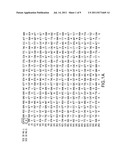
[0020] FIG. 1 shows a nucleic acid sequence of the Italian firefly, L. italica (SEQ ID NO: 1) and the translated polypeptide sequence (SEQ ID NO: 2);
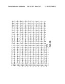
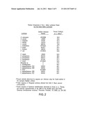
[0021] FIG. 2 shows a table comparing the L. italica luciferase protein to the known beetle luciferases;
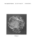
[0022] FIG. 3 shows glowing colonies of E. coli infused with luciferin on nitrocellulose filters;
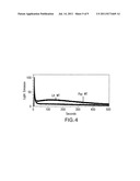
[0023] FIG. 4 shows decay of bioluminescence emission of Lit WT and Ppy WT luciferases;
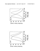
[0024] FIG. 5 shows bioluminescence emission spectra for L. italica (Panel A) and P. pyralis (Panel B);
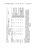
[0025] FIG. 6 shows bioluminescence activity of Lit WT and mutants thereof;
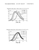
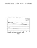
[0026] FIG. 7 shows normalized bioluminescence emission spectra produced by luciferases from Lit WT, LitGF-G-10, and LitS-1-11 mutants at 25° C. (Panel 7A) and 37° C. (Panel 7B); and
[0027] FIG. 8 shows the thermostability of Lit WT and LitS-S-2 and LitGF-G-11.
DETAILED DESCRIPTION OF THE INVENTION
[0028] Isolated nucleic acid and polypeptide compositions encoding a L. italica firefly luciferase are disclosed herein, as well as mutants of these isolated nucleic acid and polypeptide sequences. It has been found that the wild type L. italica (Lit WT) luciferase has a bioluminescence emission maximum that is red-shifted and 566 nm with a shoulder at 607 nm. Further, various substitution mutants of Lit WT exhibit distinct emission spectra that are red- or blue-shifted depending on the specific mutation(s). As defined herein, the term "red-shifted" means that the bioluminescence emission has been shifted toward the red wavelength range as compared to other luciferases known in the art. Similarly, "blue-shifted" means that the bioluminescence emission has been shifted toward the blue wavelength range as compared to other luciferases known in the art.
[0029] Lit WT is encoded by a nucleic acid having a sequence of SEQ. ID. NO.: 1 as shown in FIG. 1. The nucleic acid of SEQ ID NO: 1 was isolated from the Italian firefly, L. italica and is available in GenBank at Accession No. DQ138966. After isolation of SEQ ID NO: 1, the nucleic acid was amplified by amplification techniques known in the art, such as PCR. The nucleic acid was then inserted into an appropriate vector, such as a pGEX-6p-2 plasmid, and transformed into competent cells and subsequently plated. DNA sequencing revealed a 1647 bp open reading frame corresponding to a polypeptide containing 548 amino acids, having a sequence of SEQ ID NO: 2, also shown in FIG. 1. The Lit WT protein was found to have a molecular mass of 60,908±6 Da, as determined by electrospray ionization mass spectrometry. This value corresponds to the calculated mass of 60,907±6 Da of Lit WT, which contains the additional N-terminal peptide GlyProLeuGlySer-.
[0030] As shown in FIG. 2, SEQ ID NO: 2 is highly homologous to other luciferase amino acid sequences, including H. unmunsana (95.8%), H. parvula (95.6%) and L. mingrelica (95.3%). The luciferases least homologous to L. italica were the orange and yellow emitting isozymes of the click beetle P. plagiophthalamis (45.5%). Generally higher homology to the L. italica luciferase was found with true fireflies. The homologies were determined by sequence pair distances using the Clustal V method (Higgins et al., Computer Applications in the Biosciences, vol. 5, p. 151-153 (1989)).
[0031] To analyze the bioluminescence emission of L. Italica luciferase, proteins were expressed as a GST-fusion protein in bacterial colonies. Expression of the luciferase was demonstrated by the observation of bioluminescent colonies of E. coli infused with a luciferase substrate, such as luciferin, on nitrocellulose fibers as shown in FIG. 3. The bioluminescence emission of Lit WT was measured, using methods known in the art, and determined to be approximately 566 nm with a shoulder at 607 nm.
[0032] Preliminary characterization showed that Lit WT demonstrates bioluminescence emission similar in intensity to P. pyralis (Ppy WT). Specifically, it was determined that the relative flash height specific activity of Lit WT, which relates to the maximum achievable overall reaction rate, was found to be approximately 95% of that of Ppy WT. However, compared to Ppy WT, Lit WT has an extended light emission decay and is red-shifted by 9 nm. As shown in FIG. 4, Ppy WT and Lit WT decay to approximately 20% of their maximum flash height in about 0.2 minutes. Ppy WT continues to rapidly decay and reaches 10% of its initial activity in about 0.35 minute. In contrast, Lit WT reaches 10% of its flash height at about 7 minutes, illustrating its extended light emission decay.
[0033] In addition to its extended bioluminescence emission decay as compared to other luciferases, the Lit WT has been red-shifted. As shown in FIG. 5, bioluminescence emission spectra at pH 7.8 showed an emission maximum of 566 nm with a shoulder at 607 nm for Lit WT (FIG. 5, Panel A). This represents a 9 nm shift from Ppy WT, which has an emission maximum of 557 nm (FIG. 5, Panel B). At a pH of 6.0, Lit WT was found to be very sensitive, shifting to 614 nm, thereby producing a spectrum very similar to Ppy WT at this pH.
[0034] Further, it has also been found that mutants of Lit WT produce bioluminescence emissions that are red-shifted or blue-shifted, and have extended bioluminescence emission decay. As shown in FIG. 6, substitutions of various amino acids in Lit WT result in bioluminescence emissions of different wavelengths. In addition, as shown in FIG. 6, several of the disclosed mutants display excellent thermostability at 37° C., a property of great importance for many applications, including but not limited to in vivo imaging of tumors, biosensor applications, reporter gene applications in mammalian cell lines, and the like. As shown in FIG. 6, the half-lives of LitWT, LitS-S-2 and LitGF-G-11 were measured. In the first mutant, Lit-Gly 248 Ala+Phe 252Ser, Gly 248 was changed to alanine and Phe 252 was changed to serine. This mutant produced an emission maximum of about 563 nm (a "blue-shift" relative to the emission maximum of 566 nm in Lit WT). This mutant was not thermostable at 37° C., having a half-life of 0.06 hr at this temperature.
[0035] In the second mutant, Lit GF-G-4, Gly 216 was changed to alanine, Thr 217 was changed to leucine, Ser 234 was changed to alanine, Gly 248 was changed to alanine and Phe 252 was changed to serine. This mutant has an emission maximum of approximately 551 nm (a blue-shift relative to the emission maximum of 566 nm in Lit WT), and was thermostable at 37° C.
[0036] In the third mutant, Lit GF-G-5, Gly 216 was changed to alanine, Thr 217 was changed to leucine, Ser 234 was changed to alanine, Gly 248 was changed to alanine, Phe 252 was changed to serine and Glu 356 was changed to lysine. This mutant has an emission maximum of approximately 563 nm (a blue shift relative to the emission maximum of 566 nm in Lit WT), and was thermostable at 37° C.
[0037] In the fourth mutant, Lit GF-G-10, Gly 216 was changed to alanine, Thr 217 was changed to leucine, Ser 234 was changed to alanine, Gly 248 was changed to alanine, Phe 252 was changed to serine, and Val 241 was changed to isoleucine. This mutant had an emission maximum of approximately 553 nm (a blue shift relative to the emission maximum of 566 nm in Lit WT), and was thermostable at 37° C.
[0038] In the fifth mutant, Lit-Ser 286 Thr, Ser 286 was changed to threonine. This mutant had an emission maximum of approximately 611 nm (a "red-shift" relative to the emission maximum of 566 nm in Lit WT), but was not thermostable at 37° C.
[0039] In the sixth mutant, Lit S-S-2, Gly 216 was changed to alanine, Thr 217 was changed to leucine, Ser 234 was changed to alanine, Ser 286 was changed to threonine and Glu 356 was changed to lysine. This mutant had an emission maximum of approximately 611 nm (a red-shift relative to the emission maximum of 566 nm in Lit WT), and was thermostable at 37° C., having a half-life of 2.3 hours at this temperature compared to 0.06 hr for Lit WT.
[0040] In the seventh mutant, Lit S-S-10, Gly 216 was changed to alanine, Thr 217 was changed to leucine, Ser 286 was changed to threonine, and Glu 356 was changed to lysine. This mutant had an emission maximum of approximately 612 nm (a red-shift relative to the emission maximum of 566 nm in Lit WT), and was thermostable at 37° C.
[0041] In the eighth mutant, LitS-S-11, Gly 216 was changed to alanine, Thr 217 was changed to leucine, Ser 234 was changed to alanine, Ser 286 was changed to threonine, Glu 356 was changed to lysine, Lys 547 was changed to glycine and Met 548 was changed to glycine. This mutant had an emission maximum of approximately 609 nm (a red-shift relative to the emission maximum of 566 nm in Lit WT), and was thermostable at 37° C. The changes at positions 547 and 548 removed a signal sequence that targets the enzyme for export to the peroxisome.
[0042] In the ninth mutant, LitGF-G-11, Gly 216 was changed to alanine, Thr 217 was changed to leucine, Ser 234 was changed to alanine, Val 243 was changed to isoleucine, Gly 248 was changed to alanine, Phe 252 was changed to serine, Glu 356 was changed to lysine, Lys 547 was changed to glycine and Met 548 was changed to glycine. This mutant had an emission maximum of approximately 554 nm (a blue-shift relative to the emission maximum of approximately 566 nm in Lit WT), and was thermostable at 37° C., having a half-life of 2.0 hours at this temperature as compared to 0.06 hr for Lit WT. The changes at positions 547 and 548 removed a signal sequence that targets the enzyme for export to the peroxisome.
[0043] The bioluminescence emission of the Lit GF-G-11 and Lit S-S-11 mutants was compared to the emission of Lit WT. As shown in FIG. 7A, at 25° C., the emission maximum of the LitGF-G-11 mutant was approximately 554, while the LitS-S-11 mutant was approximately 609 nm, thus demonstrating the blue- and red-shifts shown with these mutants relative to Lit WT. As shown in FIG. 7B, at 37° C., all the spectra are red-shifted. It is noted that the red-shifting of red emitting luciferases is favorable for in vivo imaging.
[0044] The mutants of Lit WT as shown in FIG. 6 and described in more detail above were made through processes known in the art, such as site directed mutagenesis and multi-site directed mutagenesis. These processes are discussed in more detail in the Examples that follow.
[0045] Further modifications and changes beyond those specifically disclosed herein may be made to the nucleic acid sequence or polypeptide sequence of the L. italica luciferase to obtain a molecule having the desired bioluminescence emission and extended light decay. For example, certain amino acids may be substituted for other amino acids without any loss of function. So long as the mutation or change maintains a red-shifted luciferase, the resulting protein will be considered a biologically functional equivalent for the purposes of the present invention.
[0046] Additionally, the nucleic acid sequence of Lit WT may be modified chemically or by genetic engineering to enable the luciferase to be targeted into a specific subcellular compartment. For example, L. italica luciferase contains the peptide Ala-Lys-Met at the C-terminus, which likely directs the luciferase to the peroxisomes. However, a suitable sequence at the N-terminus will locate the luciferase in the mitochondria while other peptide sequences will direct the luciferase to the endoplasmic reticulum. Direction of the luciferase to certain subcellular compartments may have applicability in different assays and tests known in the art.
[0047] Transformation, transduction or transfection of a cell with nucleic acid segments encoding Lit WT or any mutants thereof, can be used to express the luciferase protein in various eukaryotic and prokaryotic cells. The nucleic acid can be inserted into vectors such as bacteriophages, cosmids, or plasmids which can then be used for transformation of prokaryotic or eukaryotic cells according to methods known in the art.
[0048] After transforming, transfecting or transducing a host cell with a vector containing Lit WT or a variant thereof, the presence of the L. italica luciferase in a host cell can be determined by contacting the host cell with a luciferase substrate. Luciferase substrates include luciferin and ATP. Through a multi-step oxidative process, the luciferase is converted to an excited state oxyluciferin, which is the bioluminescent product. In the present invention, the bioluminescence emission of the L. italica luciferase is between about 563 nm to about 612 nm. The bioluminescence emission is detected and calculated by processes known to those with ordinary skill in the art.
[0049] Alternatively, the luciferase of the present invention may be used to determine the presence and quantify the amount of ATP present in a sample. To do so, a sample that contains, or is thought to contain, ATP is combined with Lit WT or a mutant thereof. The Lit WT or mutant thereof may be in combination with other materials, such as a carrier. Upon contact of the luciferase with the sample, bioluminescence emission is measured to determine whether ATP is present in the sample, and if so, how much ATP is present. Examples of such assays are known in the art (Lundin, A. et al., Meth. Enzymol., Vol. 305, pp. 346-370 (2000), Academic Press, NY; Stanley, P. Journal of Bioluminescence and Chemiluminescence, vol. 4(1), pp. 375-80, (1989); Leach, F., J. Appl. Biochem., vol. 3(6), pp. 473-517 (1989)).
[0050] For applicability in assays, tests, methods and techniques known in the art, and those subsequently discovered, the isolated polypeptides of the present invention may be manufactured and place into kits, testing products, or other articles of manufacture. Typically the isolated polypeptides are placed in a container with or without a carrier, such as a buffer. In'most instances the article of manufacture contains an instruction booklet.
[0051] The wild type and mutant species of the luciferases derived from L. italica as disclosed herein are useful in many applications. For example, the present invention is useful in biotechnological applications, including reporter genes, dual-reporter systems, bioluminescence resonance energy transfer (BRET), microarrays, in vivo and ex vivo bioluminescence imaging, tumor research, whole animal imaging, infectious disease monitoring, biosensors for pollutants and biological disease markers, immunoassays, drug development and bioprocessing. (Roda et al., Trends in Biotechnology, vol. 22, No. 6, 295-303, 2004). The present invention is also useful in any application based on the monitoring of ATP levels either directly or through coupled enzyme reactions, such as microbiological tests; assays of enzymes, substrates and cofactors; monitoring of bacterial contamination of food; DNA probes assays; protein blotting and photographic assays (see, for example, L. J. Kricka, Anal. Biochem., vol. 175, pp. 14-21 (1988)). The present invention is also useful in any application based on the emission of light for devices which provide illumination without heat, spark or flame (for example, Cyalume technology). The present invention is also useful in any application in which light emission is used to create a novelty item, e.g. a toy or device that can be worn as jewelry. The present invention is also useful in any materials that could be used in tagging applications or anti-tampering applications.
[0052] The thermostable mutant luciferase enzyme of the present invention can also be used as a label for biospecific assays including immunoassays, nucleic acid hybridization both in vitro or microplate plate formats or for imaging purposes (immunohistochemistry or in situ hybridization). The luciferase enzyme of the present invention may possess a histidine tail that may be used for specific coupling with biomolecules, proteins, peptides, nucleic acids and in general organic molecules with suitable reactive groups. The red emitting thermostable luciferase mutant will enhance the performance of assays thanks to its relative high stability up to 42° C. associated with an high turnover of the enzyme and to the possibility to orient the coupling of this enzyme with histidine tail, leaving the active site free for substrate access. In addition, different thermostable mutants with different colours can be simultaneously used for multiplexed formats assays in which the reaction is triggered by only one substrate and the signal is selectively recorded at different wavelength.
[0053] The following examples are intended to illustrate, but in no way limit the scope of the present invention. All parts and percentages are by weight, and temperatures are in degrees Celsius unless explicitly stated otherwise.
EXAMPLES
Example 1
Isolation of L. italica Luciferase Gene
Collection and RNA Extraction
[0054] L. italica fireflies were collected from the Bologna-Paderno region of Italy and flash-frozen alive in liquid nitrogen and stored at -80° C. Sixteen lanterns were removed from the fireflies, and were refrozen in liquid nitrogen. The lanterns were ground to a powder using a mortar and pestle cooled with liquid nitrogen. A total of 18 μg of RNA was extracted from the ground lanterns using an RNeasy mini kit (Quiagen) and following the manufacturer's enclosed instructions.
RT-PCR
[0055] First strand cDNA synthesis was carried out by utilizing approximately 5 μg of RNA and Oligo (dT)20 primers and the Superscript TM III First-Strand Synthesis System for RT-PCR (Invitrogen). The following primers based on the luciferase coding sequence of L. Mingrelica, were used to amplify the cDNA:
TABLE-US-00001 (SEQ ID NO: 3) 5'-GTC CCT AAA CGG TAG AGG AAA A G-3' (SEQ ID NO: 4) 5'-GTC TTC TTA TGA GTA GTT TAG TTA C-3'
[0056] To amplify the cDNA, polymerase chain reaction (PCR) was used. The initial denaturation cycle was at 94° C. for 5 min. A 35 cycle amplification was then carried out at 94° C. for 30 sec.; 52° C. for 45 sec.; and 68° C. for 1.5 min. A final extension was carried out at 68° C. for 5 min.
[0057] The PCR products were then analyzed on a 1.0% agarose gel containing ethidium bromide. The samples corresponding to the expected size were purified using a QIA quick PCR purification kit (Qiagen) and following the manufacturer's instructions. Finally, the amplified cDNA was sequenced using a capillary array sequencer CEQ2000XL.
Insertion of cDNA into a Vector
[0058] To insert the luciferase cDNA into a vector, a primer set was used to introduce SmaI and XhoI restriction endonuclease sites at the 5' and 3' ends, respectively, of the PCR-amplified cDNA. The following primer set was used:
TABLE-US-00002 (SEQ ID NO: 5) 5'-TTT AAT CCC GGG GTC CCT AAA CGG TAG-3' (SEQ ID NO: 6) 5'-CTA AGC CTC GAG TCT TCT TAT GAG TAG TT-3'
PCR amplification and purification were performed as described above.
[0059] The PCR product was then digested with SmaI and XhoI restriction endonucleases and ligated into the corresponding cloning sites on a pGEX-6P-2 plasmid. The ligation reaction was transformed into Escherichia coli XL-10 Gold ultra competent cells and plated on Luria-Bertani (LB) plates containing 100 μg/mL ampicillin.
[0060] Ten colonies of the E. coli were selected randomly and plasmid DNA was purified and screened by agarose gel electrophoresis. Several plasmid DNA samples of the expected size were sequenced. One sample containing the entire L. italica cDNA was identified.
Alignment of the cDNA Reading Frame
[0061] Since the pGEX expression plasmid is designed to produce proteins as N-terminal glutathione-S-transferase (GST)-fusion products, the reading frame of the luciferase gene was realigned so the corresponding protein would contain the same N-terminal polypeptide as Ppy WT. A Quik Change® Site-Directed Mutagenesis kit (Stratagene), was used to realign the reading frame of the L. italica cDNA in the pGEX-6P-2 plasmid.
[0062] First, the primer, 5'-GA TTC TCA CAC GCT AAG GAC CCA ATT TAC GGA AAC CAA GTT TC-3' (SEQ ID NO: 7) and its reverse complement, were used to remove the Barn HI restriction endonuclease site within the L. italica luciferase gene. Next, the primer 5'-CG GTA GAG GAA AAG TTT GGA TCC ATG GAA ACG GAA AGG GAG G-3' (SEQ ID NO: 8) and its respective reverse complement, were used to introduce a Barn HI site immediately preceding the start codon of the L. italica luciferase gene. Finally, the product was digested with Barn HI and XhoI restriction endonucleases and ligated into corresponding cloning sites on the pGEX-6P-2 plasmid.
Expression Of L. Italica Luciferase As A GST-Fusion Protein In Bacterial Colonies
[0063] The ligated plasmid DNA was transformed into E. coli, which were plated on nitrocellulose filters placed on LB plates containing 100 μg/mL ampicillin. The transformation was done by adding the plasmid DNA to the bacteria at ice temperature, heat shocking the mixture at 42° C., cooling on ice for 2 minutes and recovering for 1 hour at 37° C. in SOC media. The E. coli were screened for bioluminescence emission as described in Branchini et al., "Rational and random mutagenesis of firefly luciferase to identify an efficient emitter of red bioluminescence," Proceedings of SPIE, Genetically Engineered and Optical Probes for Biomedical Applications II 5329 (2004), 170-177 (Alexander P. Savitsky et al., eds.).
[0064] Colonies having bioluminescence emission were identified by visual screening in a darkroom. Plasmid DNA was isolated and purified therefrom by using a GenElute® Plasmid Miniprep kit (Sigma), and following the manufacturer's instructions. A single plasmid was selected and the entire L. italica luciferase gene was sequenced. The sequence of the L. italica luciferase gene in the pGEX plasmid was compared to the sequence obtained from the original cDNA. The comparison showed no differences in the sequence, thereby insuring no additional mutations had been introduced.
Example 2
Preparation of Lit WT Mutants
[0065] Nine mutants of Lit WT, shown in FIG. 6 and discussed in more detail above, were prepared using the following methods:
Site Directed Mutagenesis
[0066] The Lit-S286T and Lit-G248A and F252S mutants were created by using a QuikChange® Site-Directed Mutagenesis kit (Stratagene). Site-directed mutagenesis was carried out according to the manufacturer's instructions using the L. italica wild-type DNA sequence in the pGEX-6P-2 vector as a template. The following primers and their respective reverse complements were used:
TABLE-US-00003 Lit-S286T: (SEQ ID NO: 9) 5'-GAT TAT AAG TGT ACC ACT GTT ATT CTG GTA CCA ACG TTA TTT GC-3' (Kpn1) Lit-G248A & F252S: (SEQ ID NO: 10) 5'-CCG TTC CAT CAC GCG TTT GGA ATG TCT ACC ACT TTA GGA TAC-3' (Mlu1)
[0067] The LitS-S-11 mutant was created by using the QuikChange® Site-Directed Mutagenesis kit (Stratagene). Site-directed mutagenesis was carried out according to the manufacture's instructions using the LitS-S-2 mutant as the template. The 546AlaLysMet548 signal sequence was changed to 546AlaGlyGly548. The following primer and its respective reverse compliment was used to accomplish this:
TABLE-US-00004 Lys547Gly & Met548Gly: (SEQ ID NO: 11) 5'-AG AAA CCA CAA GCC GGG GGG TAA ATC GGT CAA AAT G-3' [BsaB1]
[0068] The LitGF-G-11 mutant was created by using the QuikChange® Site-Directed Mutagenesis kit (Stratagene). Site-directed mutagenesis was carried out according to the manufacture's instructions using the LitGF-G-10 mutant as the template. First, Glu356Lys was introduced into the template. Once this mutation was confirmed, the 546AlaLysMet548 signal sequence was changed to 546AlaGlyGly548. The following primers and their respective reverse compliments were used to accomplish this:
TABLE-US-00005 Glu356Lys: (SEQ ID NO: 12) 5'-GCA TTT ATT ATT ACC CCA AAA GGT GAT GAT AAA CCT GG-3' Lys547Gly & Met548Gly: (SEQ ID NO: 13) 5'-AG AAA CCA CAA GCC GGG GGG TAA ATC GGT CAA AAT G-3' [BsaB1]
[0069] Underlining represents silent changes creating a unique screening endonuclease site. Bolded codons represent the mutated codons. Brackets indicate the screening endonuclease.
Multi-Site Directed Mutagenesis
[0070] The QuikChange® MultiSite-Directed Mutagenesis kit (Stratagene) was used to create the remaining mutants identified as entries 2 through 8 in FIG. 6. Mutagenesis was carried out according to the manufacturer's instructions for using 1 to 4 primers simultaneously. The Lit-S286T and the Lit-G248A and F252L mutants in the pGEX-6P-2 vector were used as templates with the following primers:
TABLE-US-00006 Glu356Lys (SEQ ID NO: 14) 5'-GCA TTT ATT ATT ACC CCA AAA GGT GAT GAT AAA CCT GG-3' Gly216Ala & T217L (SEQ ID NO: 15) 5'-GAG ATT ACC CAC GAA GCA CTA GTT ACA AGA TTC TCA CAC G-3' Ser234 Ala (SEQ ID NO: 16) 5'-TAC GGA AAC CAA GTT GCA CCT GGT ACT GC-3' Val243Ile (SEQ ID NO: 17) 5'-TA ACT GTC ATT CCG TTC CAT CAC GCG TTT GGA ATG-3'
[0071] The primer for V243I also includes the G248A mutation (bold and italics) for correct annealing. This primer was only used with the Lit-G248A and F252L template.
Transformation and Screening of Lit WT Mutants
[0072] The above-described mutants were transformed into E. coli XL-10 Gold ultracompetent cells. Duplicate sets of transformed cells were plated on nitrocellulose filters on LB plates that contained 100 μg/ml ampicillin. The plates were incubated at 37° C. for 18 hrs. Isopropyl B-D-1-thiogalactopyranoside (IPTG) was used to induce transcription.
[0073] For each set of transformed mutagenesis product-containing cells, one plate was incubated at room temperature and the other at 37° C. for at least 2 hours. The plates grown at 37° C. were then placed on a plate warmer set to 37° C. (Barnstead Lab Line) and those at room temperature were kept at 22° C.
[0074] The nitrocellulose filters were soaked in 1 mM of luciferin in 100 mM NaCitrate buffer (pH 5.5) to induce bioluminescence. Colonies for sequencing were selected on the intensity and color of the emitted light. Plasmid DNA of selected clones was isolated by a standard mini-prep procedure. The DNA was sequenced at Yale University.
Example 3
Protein Purification
[0075] Glutathione-S-transferase (GST) fusion constructs of L. italica wild-type and mutants thereof selected for sequencing were expressed in XL10-Gold ultracompetent cells. 7 mL cultures of the cells were grown in 10 mL culture tubes at 37° C. in LB medium supplemented with 100 μg/mL ampicillin.
[0076] These cultures were grown from starter cultures prepared from the picked colonies to mid log phase (A600=0.4-0.5) with vigorous shaking, induced with 0.1 mM IPTG, and incubated at 22° C. for 8-10 h. The cells were harvested by centrifugation and placed at -80° C. for no less than 30 min. Cell pellets were resuspended in 0.5 mL of phosphate-buffered saline (PBS), pH 7.3 containing 0.1 mM phenylmethylsulfonyl fluoride (PMSF) and 0.5 mM dithiothreitol (DTT). The resuspended cells were mixed with lysozyme (0.05 volume of 10 mg/mL) and incubated on ice 30 min. The lysated cells were treated with Rnase (10 μg/mL) and Dnase 1 (5 μg/mL). Triton X-100 (2% final volume) was added to the lysate and the whole-cell extract was isolated by centrifugation at 20,000×g for 45 min at 4° C. The supernatant was collected and aliquots were used to determine molecular mass, bioluminescence activity, bioluminescence emission spectra, protein content and GST-activity. Lit WT, LitGF-G-11, LitS-S-2 and LitS-S-11 enzymes were purified to homogeneity as described in Branchini, B. R., et al., "Rational and random mutagenesis of firefly luciferase to identify an efficient emitter of red, bioluminescence," Proceedings of SPIE, Genetically Engineered and Optical Probes for Biomedical Applications II, 5329 (2004), 170-177 (Alexander P. Savitsky et al., eds.).
Molecular Mass of Lit WT Protein
[0077] The Lit WT protein was found to have a molecular mass of 60 908±6 Da. This value corresponds to the calculated mass of 60 907±6 Da of Lit WT, which contains the additional N-terminal peptide GlyProLeuGlySer-. The mass was determined by Electrospray Ionization Mass Spectrometry.
Glutathione-S-Transferase (GST) Activity Assay to Quantitate GST-Fusion Proteins
[0078] GST-fusion proteins were quantitated by enzymatic assay of glutathione-S-transferase (GST) activity using the GST substrate 1-chloro-2,4-dinitrobenzene (CDNB). GST catalyzes the conjugation of reduced glutathione (GSH) and CDNB to yield a dinitrophenolthioether, a chromogenic substance with λλmax=340 nm. A dual beam PerkinElmer Lambda 25 UV/Vis spectrophotometer was used to perform these studies.
[0079] 500 μL of 1 mM reduced glutathione (GSH) and 1 mM 1-chloro-2,4-dinitrobenzene (CDNB) in 0.1M potassium phosphate buffer (KPB), pH 6.5 were placed into 0.7 mL UV transparent sample and reference cuvettes.
[0080] 50 μl of KPB was added to the reference cuvette. 5 μL of bacterial cell lysate containing the GST-luciferase fusion protein and 45 μL of KPB was added to the sample cuvette. The samples were well mixed and the absorbance at 340 nm was monitored using the Time Drive program of the Lambda 25 spectrophotometer. Data was recorded for 4 min, and the velocity of the reaction, the rate of change in absorbance per minute, was determined. The concentration of GST-fusion protein is proportional to the velocity of the reaction.
Bioluminescence Emission Spectra of Partially Purified Proteins
[0081] Bioluminescence emission spectra were obtained using a PerkinElmer LS55 luminescence spectrometer operated in the "bioluminescence" mode. Data was collected over the wavelength range 480-680 nm in a 1 mL optical glass cuvette. Gate and delay times, detector voltage, scan rate, and slit width were adjusted to optimize instrument response. Reaction mixtures containing partially purified L. italica wild type and its mutants (0.005 mL) in luciferin and Mg-ATP were brought to a final volume of 0.5 mL with 25 mM glycylglycine, pH 7.8. The spectral data reported in FIG. 6 were measured with purified proteins from Lit WT, LitGF-G-11, LitS-S-2 and LitS-S-11 enzymes.
Measurement of L. italica Thermostability
[0082] In a 0.2 mL PCR tube, solutions of 0.1 mg luciferase in 10 μl. CBA (50 mM Tris, pH 7.0, 150 mM NaCl, 1 mM EDTA, 1 mM DTT, 0.8M (NH4)2 SO4, 2.0% glycerol) and 200 μL 25 mM glycyl-glycine buffer, pH 7.8, were prepared. An aliquot (50 μL) of the solution was reserved on ice. Initial activity values were obtained by flash height activity assays performed on 2-10 μL samples of enzyme. The sample was placed in a thermocycler set at 37° C. Aliquots were withdrawn over an 8 hour period and compared to a controls kept in ice. Thermostability was evaluated by plotting residual flash height activity at 37° C. as a function of time. Half-lives were calculated from the first order rate constant obtained by plotting the 1n residual flash height activity versus time. The half-lives of LitWT, LitS-S-2 and LitGF-G-11 were determined as shown in FIGS. 6 and 8.
[0083] While the invention has been described above with reference to specific embodiments thereof, it is apparent that many changes, modifications, and variations can be made without departing from the inventive concept disclosed herein. Accordingly, it is intended to embrace all such changes, modifications, and variations that fall within the spirit and broad scope of the appended claims. All patent applications, patents, and other publications cited herein are incorporated by reference in their entireties.
Sequence CWU
1
1711647DNAL. italicaCDS(1)...(1644) 1atg gaa acg gaa agg gag gaa aat gtt
gta tat ggc cct ctg cca ttc 48Met Glu Thr Glu Arg Glu Glu Asn Val
Val Tyr Gly Pro Leu Pro Phe1 5 10
15tac ccc att gaa gaa gga tca gct gga att cag ttg cat aag tac
atg 96Tyr Pro Ile Glu Glu Gly Ser Ala Gly Ile Gln Leu His Lys Tyr
Met 20 25 30caa caa tat gcc
aaa ctt gga gca att gct ttt agt aac gcc ctt act 144Gln Gln Tyr Ala
Lys Leu Gly Ala Ile Ala Phe Ser Asn Ala Leu Thr 35
40 45gga gtg gat att tct tac caa caa tac ttt gat att
aca tgt cgt tta 192Gly Val Asp Ile Ser Tyr Gln Gln Tyr Phe Asp Ile
Thr Cys Arg Leu 50 55 60gct gag gca
atg aaa aac tac ggt atg aaa ccg gaa gga cat att gct 240Ala Glu Ala
Met Lys Asn Tyr Gly Met Lys Pro Glu Gly His Ile Ala65 70
75 80ttg tgc agt gaa aat tgt gaa gaa
ttt ttc atc cct gtg ctt gct ggt 288Leu Cys Ser Glu Asn Cys Glu Glu
Phe Phe Ile Pro Val Leu Ala Gly 85 90
95ctt tac att gga gta act gtc gca cct act aat gaa att tac
aca ttg 336Leu Tyr Ile Gly Val Thr Val Ala Pro Thr Asn Glu Ile Tyr
Thr Leu 100 105 110cgt gaa ctt
aat cac agt ttg ggc atc gca caa cca act att gta ttc 384Arg Glu Leu
Asn His Ser Leu Gly Ile Ala Gln Pro Thr Ile Val Phe 115
120 125agc tcc aga aaa ggc tta cct aaa gtt tta gaa
gtg caa aaa aca gtt 432Ser Ser Arg Lys Gly Leu Pro Lys Val Leu Glu
Val Gln Lys Thr Val 130 135 140aca tgc
atc aaa aca att gtt att tta gat agt aaa gta aac ttt gga 480Thr Cys
Ile Lys Thr Ile Val Ile Leu Asp Ser Lys Val Asn Phe Gly145
150 155 160ggc tac gat tgt gtg gaa act
ttt att aag aaa cat gta gaa tta ggt 528Gly Tyr Asp Cys Val Glu Thr
Phe Ile Lys Lys His Val Glu Leu Gly 165
170 175ttt cca gca act agc ttt gta ccc att gat gta aag
gac cgt aaa cat 576Phe Pro Ala Thr Ser Phe Val Pro Ile Asp Val Lys
Asp Arg Lys His 180 185 190cac
att gct ttg ctt atg aat tct tct ggc tct act ggt tta cct aaa 624His
Ile Ala Leu Leu Met Asn Ser Ser Gly Ser Thr Gly Leu Pro Lys 195
200 205ggt gta gag att acc cac gaa gga aca
gtt aca aga ttc tca cac gct 672Gly Val Glu Ile Thr His Glu Gly Thr
Val Thr Arg Phe Ser His Ala 210 215
220aag gat cca att tac gga aac caa gtt tca cct ggt act gct att tta
720Lys Asp Pro Ile Tyr Gly Asn Gln Val Ser Pro Gly Thr Ala Ile Leu225
230 235 240act gtc gtt ccg
ttc cat cat gga ttt gga atg ttt acc act tta gga 768Thr Val Val Pro
Phe His His Gly Phe Gly Met Phe Thr Thr Leu Gly 245
250 255tac ttt gct tgt gga tac cgt att gta atg
tta aca aaa ttc gat gaa 816Tyr Phe Ala Cys Gly Tyr Arg Ile Val Met
Leu Thr Lys Phe Asp Glu 260 265
270gaa cta ttt ttg aga act ttg caa gat tat aag tgt acc agt gtt att
864Glu Leu Phe Leu Arg Thr Leu Gln Asp Tyr Lys Cys Thr Ser Val Ile
275 280 285ctt gta cca acg tta ttt gct
att ctc aac agg agt gaa ttg ctc gat 912Leu Val Pro Thr Leu Phe Ala
Ile Leu Asn Arg Ser Glu Leu Leu Asp 290 295
300aag ttc gat tta tct aat cta act gaa att gct tct ggt gga gct cct
960Lys Phe Asp Leu Ser Asn Leu Thr Glu Ile Ala Ser Gly Gly Ala Pro305
310 315 320ttg gca aaa gaa
att ggt gaa gca gtc gct aga aga ttt aat cta ccc 1008Leu Ala Lys Glu
Ile Gly Glu Ala Val Ala Arg Arg Phe Asn Leu Pro 325
330 335ggt gtc cgt cag ggt tac gga ttg aca gaa
acg aca tct gca ttt att 1056Gly Val Arg Gln Gly Tyr Gly Leu Thr Glu
Thr Thr Ser Ala Phe Ile 340 345
350att acc cca gaa ggt gat gat aaa cct gga gca tct gga aaa gta gta
1104Ile Thr Pro Glu Gly Asp Asp Lys Pro Gly Ala Ser Gly Lys Val Val
355 360 365ccc tta ttc aaa gta aaa att
att gat ctt gac act aaa aaa act ttg 1152Pro Leu Phe Lys Val Lys Ile
Ile Asp Leu Asp Thr Lys Lys Thr Leu 370 375
380ggt gtc aac cga cga gga gag atc tgt gta aaa ggt ccg agt ctt atg
1200Gly Val Asn Arg Arg Gly Glu Ile Cys Val Lys Gly Pro Ser Leu Met385
390 395 400tta ggc tac aca
aac aat ccg gaa gca aca aga gaa act att gat gaa 1248Leu Gly Tyr Thr
Asn Asn Pro Glu Ala Thr Arg Glu Thr Ile Asp Glu 405
410 415gag ggt tgg ttg cac acc gga gat att gga
tat tac gac gaa gac gaa 1296Glu Gly Trp Leu His Thr Gly Asp Ile Gly
Tyr Tyr Asp Glu Asp Glu 420 425
430cat ttc ttc att gta gat cgt ttg aaa tca tta atc aaa tac aag ggg
1344His Phe Phe Ile Val Asp Arg Leu Lys Ser Leu Ile Lys Tyr Lys Gly
435 440 445tac cag gta cca cct gct gaa
ttg gaa tcc gtt ctt ttg caa cat cca 1392Tyr Gln Val Pro Pro Ala Glu
Leu Glu Ser Val Leu Leu Gln His Pro 450 455
460aat atc ttt gat gct ggt gtg gct ggt gtc ccc gat tct gaa gct ggt
1440Asn Ile Phe Asp Ala Gly Val Ala Gly Val Pro Asp Ser Glu Ala Gly465
470 475 480gaa ctt cca ggg
gct gta gtt gta atg gaa aaa gga aaa act atg act 1488Glu Leu Pro Gly
Ala Val Val Val Met Glu Lys Gly Lys Thr Met Thr 485
490 495gaa aag gaa att gtg gat tat gtt aat agt
caa gta gtg aac cac aaa 1536Glu Lys Glu Ile Val Asp Tyr Val Asn Ser
Gln Val Val Asn His Lys 500 505
510cgt ctg cgt ggt ggc gtt cgt ttt gtg gat gaa gta cct aaa ggt cta
1584Arg Leu Arg Gly Gly Val Arg Phe Val Asp Glu Val Pro Lys Gly Leu
515 520 525act ggt aaa att gat gct aaa
gta att aga gaa att ctt aag aaa cca 1632Thr Gly Lys Ile Asp Ala Lys
Val Ile Arg Glu Ile Leu Lys Lys Pro 530 535
540caa gcc aag atg taa
1647Gln Ala Lys Met5452548PRTL. italica 2Met Glu Thr Glu Arg Glu Glu
Asn Val Val Tyr Gly Pro Leu Pro Phe1 5 10
15Tyr Pro Ile Glu Glu Gly Ser Ala Gly Ile Gln Leu His
Lys Tyr Met 20 25 30Gln Gln
Tyr Ala Lys Leu Gly Ala Ile Ala Phe Ser Asn Ala Leu Thr 35
40 45Gly Val Asp Ile Ser Tyr Gln Gln Tyr Phe
Asp Ile Thr Cys Arg Leu 50 55 60Ala
Glu Ala Met Lys Asn Tyr Gly Met Lys Pro Glu Gly His Ile Ala65
70 75 80Leu Cys Ser Glu Asn Cys
Glu Glu Phe Phe Ile Pro Val Leu Ala Gly 85
90 95Leu Tyr Ile Gly Val Thr Val Ala Pro Thr Asn Glu
Ile Tyr Thr Leu 100 105 110Arg
Glu Leu Asn His Ser Leu Gly Ile Ala Gln Pro Thr Ile Val Phe 115
120 125Ser Ser Arg Lys Gly Leu Pro Lys Val
Leu Glu Val Gln Lys Thr Val 130 135
140Thr Cys Ile Lys Thr Ile Val Ile Leu Asp Ser Lys Val Asn Phe Gly145
150 155 160Gly Tyr Asp Cys
Val Glu Thr Phe Ile Lys Lys His Val Glu Leu Gly 165
170 175Phe Pro Ala Thr Ser Phe Val Pro Ile Asp
Val Lys Asp Arg Lys His 180 185
190His Ile Ala Leu Leu Met Asn Ser Ser Gly Ser Thr Gly Leu Pro Lys
195 200 205Gly Val Glu Ile Thr His Glu
Gly Thr Val Thr Arg Phe Ser His Ala 210 215
220Lys Asp Pro Ile Tyr Gly Asn Gln Val Ser Pro Gly Thr Ala Ile
Leu225 230 235 240Thr Val
Val Pro Phe His His Gly Phe Gly Met Phe Thr Thr Leu Gly
245 250 255Tyr Phe Ala Cys Gly Tyr Arg
Ile Val Met Leu Thr Lys Phe Asp Glu 260 265
270Glu Leu Phe Leu Arg Thr Leu Gln Asp Tyr Lys Cys Thr Ser
Val Ile 275 280 285Leu Val Pro Thr
Leu Phe Ala Ile Leu Asn Arg Ser Glu Leu Leu Asp 290
295 300Lys Phe Asp Leu Ser Asn Leu Thr Glu Ile Ala Ser
Gly Gly Ala Pro305 310 315
320Leu Ala Lys Glu Ile Gly Glu Ala Val Ala Arg Arg Phe Asn Leu Pro
325 330 335Gly Val Arg Gln Gly
Tyr Gly Leu Thr Glu Thr Thr Ser Ala Phe Ile 340
345 350Ile Thr Pro Glu Gly Asp Asp Lys Pro Gly Ala Ser
Gly Lys Val Val 355 360 365Pro Leu
Phe Lys Val Lys Ile Ile Asp Leu Asp Thr Lys Lys Thr Leu 370
375 380Gly Val Asn Arg Arg Gly Glu Ile Cys Val Lys
Gly Pro Ser Leu Met385 390 395
400Leu Gly Tyr Thr Asn Asn Pro Glu Ala Thr Arg Glu Thr Ile Asp Glu
405 410 415Glu Gly Trp Leu
His Thr Gly Asp Ile Gly Tyr Tyr Asp Glu Asp Glu 420
425 430His Phe Phe Ile Val Asp Arg Leu Lys Ser Leu
Ile Lys Tyr Lys Gly 435 440 445Tyr
Gln Val Pro Pro Ala Glu Leu Glu Ser Val Leu Leu Gln His Pro 450
455 460Asn Ile Phe Asp Ala Gly Val Ala Gly Val
Pro Asp Ser Glu Ala Gly465 470 475
480Glu Leu Pro Gly Ala Val Val Val Met Glu Lys Gly Lys Thr Met
Thr 485 490 495Glu Lys Glu
Ile Val Asp Tyr Val Asn Ser Gln Val Val Asn His Lys 500
505 510Arg Leu Arg Gly Gly Val Arg Phe Val Asp
Glu Val Pro Lys Gly Leu 515 520
525Thr Gly Lys Ile Asp Ala Lys Val Ile Arg Glu Ile Leu Lys Lys Pro 530
535 540Gln Ala Lys Met545323DNAArtificial
SequenceSynthetic Primer sequence 3gtccctaaac ggtagaggaa aag
23425DNAArtificial SequenceSynthetic
Primer sequence 4gtcttcttat gagtagttta gttac
25527DNAArtificial SequenceSynthetic Primer sequence
5tttaatcccg gggtccctaa acggtag
27629DNAArtificial SequenceSynthetic Primer sequence 6ctaagcctcg
agtcttctta tgagtagtt
29743DNAArtificial SequenceSynthetic Primer sequence 7gattctcaca
cgctaaggac ccaatttacg gaaaccaagt ttc
43842DNAArtificial SequenceSynthetic Primer sequence 8cggtagagga
aaagtttgga tccatggaaa cggaaaggga gg
42944DNAArtificial SequenceSynthetic Primer sequence 9gattataagt
gtaccactgt tattctggta ccaacgttat ttgc
441042DNAArtificial SequenceSynthetic Primer sequence 10ccgttccatc
acgcgtttgg aatgtctacc actttaggat ac
421136DNAArtificial SequenceSynthetic Primer sequence 11agaaaccaca
agccgggggg taaatcggtc aaaatg
361238DNAArtificial SequenceSynthetic Primer sequence 12gcatttatta
ttaccccaaa aggtgatgat aaacctgg
381336DNAArtificial SequenceSynthetic Primer sequence 13agaaaccaca
agccgggggg taaatcggtc aaaatg
361438DNAArtificial SequenceSynthetic Primer sequence 14gcatttatta
ttaccccaaa aggtgatgat aaacctgg
381540DNAArtificial SequenceSynthetic Primer sequence 15gagattaccc
acgaagcact agttacaaga ttctcacacg
401629DNAArtificial SequenceSynthetic Primer sequence 16tacggaaacc
aagttgcacc tggtactgc
291735DNAArtificial SequenceSynthetic Primer sequence 17taactgtcat
tccgttccat cacgcgtttg gaatg 35
User Contributions:
Comment about this patent or add new information about this topic:
| People who visited this patent also read: | |
| Patent application number | Title |
|---|---|
| 20160039478 | SPOILER UNIT AND MANUFACTURING METHOD THEREOF |
| 20160039477 | ZERO AERODYNAMIC DRAG VEHICLES |
| 20160039476 | LOGISTICS POST |
| 20160039475 | METHOD OF MANUFACTORING A VEHICLE AND VEHICLE |
| 20160039474 | HEIGHT ADJUSTABLE LOAD COMPARTMENT FLOOR SYSTEM |
 |  |
 |  |
 |  |
 |  |
 |
| New patent applications in this class: | |
| Date | Title |
|---|---|
| 2022-05-05 | Compositions and methods for identifying metabolically active agents |
| 2019-05-16 | Novel cell-based assay for determining activity in the retinoblastoma pathway |
| 2017-08-17 | Biosensor based on g-beta-gamma-interacting proteins to monitor g-protein activation |
| 2016-12-29 | Thienopyrrole compounds and uses thereof |
| 2016-12-29 | Cyan-excitable orange-red fluorescent proteins and bioluminescent resonance energy transfer systems |
| New patent applications from these inventors: | |
| Date | Title |
|---|---|
| 2012-02-23 | Portable device based on immobilized cells for the detection of analytes |
| 2010-08-26 | Lanthanide complexes, preparation and uses thereof |
| Top Inventors for class "Chemistry: molecular biology and microbiology" | |
| Rank | Inventor's name |
|---|---|
| 1 | Marshall Medoff |
| 2 | Anthony P. Burgard |
| 3 | Mark J. Burk |
| 4 | Robin E. Osterhout |
| 5 | Rangarajan Sampath |