Patent application title: ANTI-MIF IMMUNOHISTOCHEMISTRY
Inventors:
Alexander Schinagl (Wien, AT)
Michael Thiele (Wien, AT)
Patrice Douillard (Wien, AT)
Gerhard Antoine (Gross Enzersdorf, AT)
Randolf Kerschbaumer (Klosterneuburg, AT)
Randolf Kerschbaumer (Klosterneuburg, AT)
IPC8 Class: AG01N3368FI
USPC Class:
1 1
Class name:
Publication date: 2016-11-17
Patent application number: 20160334414
Abstract:
The present invention pertains to the specific detection of MIF, in
particular oxMIF, and of anti-oxMIF antibodies in tissues. A detection
method is provided which uses immunohistochemistry or immunofluorescence
and wherein specific anti-oxMIF antibodies and specific idiotypic
monoclonal rabbit antibodies are used.Claims:
1. An immunohistochemistry (IHC) assay method for in vitro detection of
oxMIF, wherein oxMIF is MIF which is differentially binding to antibody
RAB4, RAB9 and/or RAB0, in a tissue sample of a subject, comprising the
determination of the binding of a compound to oxMIF in said sample in
vitro, wherein one or more of the following steps are carried out: a)
Optional Blocking step with blocking buffer and b) Binding step with
primary anti-oxMIF antibody without a previous fixation step, in
particular either formaldehyde or acetone; c) Optionally fixation step;
d) Incubation with secondary antibody, wherein the secondary antibody is
an anti-idiotypic rabbit monoclonal antibody directed against an
anti-oxMIF antibody; and/or e) Detection of binding between the primary
anti-oxMIF antibody and the secondary antibody.
2. The IHC assay of claim 1, wherein no fixation with an organic or inorganic fixation agent, in particular either formaldehyde or acetone, is carried out before the binding step b).
3. The IHC assay of claim 1 or 2, wherein the samples is air dried, preferably for about 30 min.
4. The IHC assay of any one or more of claims 1-3, wherein the primary antibody is not labelled, e.g. not biotinylated, and/or is preferably comprised in a primary dilution buffer and/or wherein the primary antibody is incubated with the sample preferably for 45 to 90 minutes, more preferred for approximately 60 minutes.
5. The IHC assay of any one or more of claims 1-4, wherein a washing step is carried out after binding step b) to wash away excess antibody and wherein optionally further washing steps are carried out after all further steps.
6. The IHC assay of any one or more of claims 1-5, wherein the detection step e) comprises or consists of a staining step and/or wherein a further (counter) staining step is carried out after the detection step, which can include the use of a tertiary antibody.
7. The IHC assay of any one or more of claims 1-6, wherein the (counter) staining step is carried out with hematoxylin, after step e).
8. The IHC assay of any one of the preceding claims, wherein the binding between the primary and secondary antibody is detected by fluorescence, or a directly fluorescence labelled secondary antibody (e.g. labelled anti-Ram9), preferably by use of a fluorophore labelled tertiary antibody directed against the secondary antibody.
9. The IHC assay of any one of claims 1-8, wherein said secondary antibody is a secondary monoclonal antibody selected from the group consisting of an anti-RAM9 antibody, an anti-RAM0 antibody and an anti-RAM4 antibody and is most preferred an anti-RAM9 antibody, even more preferred a rabbit anti-RAM9 antibody.
10. The IHC assay of any one of claims 1-9, wherein the primary antibody binds to oxMIF, but does not bind to redMIF.
11. The IHC assay of claim 10, wherein the differential binding is a binding to oxMIF which occurs with a K.sub.D value of less than 100 nM, preferably less than 50 nM, even more preferred less than 10 nM and a non-binding to redMIF which is characterized by a K.sub.D of more than 400 nM.
12. The IHC assay of any one of claims 1 to 11, wherein the primary antibodies are selected from the group consisting of oxMIF binders, like e.g. antibodies RAB4, RAB9 and/or RAB0 and/or RAM4, RAM9 and/or RAM0.
13. The IHC assay of any of the preceding claims, wherein the sample is a tissue biopsy, preferably a frozen tissue biopsy, preferably an OCT embedded section, or a core needle biopsy.
14. An idiotypic antibody, specifically binding to an anti-oxMIF antibody, preferably to an anti-RAM9 antibody.
15. The idiotypic antibody of claim 14, wherein the antibody is characterized by an IC.sub.50 of .ltoreq.60 ng/ml in a neutralizing ELISA with immobilized MIF and RAM9.
16. An idiotypic antibody, preferably according to claim 14 or 15 e.g. for use in the method or kit of any one or more of the preceding claims which is characterized by SEQ ID NO: 15 and/or 16, or by SEQ ID NO: 17 and/or 18.
17. An anti-idiotypic antibody, e.g. for use in the method or kit of any one or more of the preceding claims, which is characterized by a heavy chain which has been deposited at the DSMZ with deposit No. DSM 28180 and/or by a light chain, which has been deposited at the DSMZ with deposit No. DSM 28181; or which is characterized by a heavy chain which has been deposited at the DSMZ with deposit No. DSM 28182 and/or a light chain which has been deposited at the DSMZ with a deposit no. DSM 28183.
18. A method for the detection of an anti-oxMIF antibody, preferably RAM9, RAM0 or RAM4, with the use of an idiotypic monoclonal rabbit antibody directed against said anti-oxMIF antibodies, preferably as described in any one of claims 14-17.
19. An IHC assay kit, adapted to carry out the method according to any one or more of the preceding claims.
Description:
[0001] The present invention pertains to the specific detection of MIF, in
particular oxMIF, in tissues. The present detection method also allows
the detection of anti-MIF antibodies, e.g. RAM9, in tissue. A detection
method is provided which uses immunohistochemistry or immunofluorescence
and wherein specific anti-oxMIF antibodies and monoclonal rabbit
anti-oxMIF-antibody antibodies are used. The invention also provides
advantageous detection antibodies and a method to provide such detection
antibodies. The invention furthermore pertains to a method for the
determination of target saturation.
BACKGROUND
[0002] Macrophage migration inhibitory factor (MIF) is a cytokine initially isolated based upon its ability to inhibit the in vitro random migration of peritoneal exudate cells from tuberculin hypersensitive guinea pigs (containing macrophages) (Bloom et al. Science 1966, 153, 80-2; David et al, PNAS 1966, 56, 72-7). Today, MIF is known as a critical upstream regulator of the innate and acquired immune response that exerts a pleiotropic spectrum of activities.
[0003] The human MIF-cDNA was cloned in 1989 (Weiser et al., PNAS 1989, 86, 7522-6), and its genomic localization was mapped to chromosome 22. The product of the human MIF gene is a protein with 114 amino acids (after cleavage of the N-terminal methionine) and an apparent molecular mass of about 12.5 kDa. MIF has no significant sequence homology to any other protein. The protein crystallizes as a trimer of identical subunits. Each monomer contains two antiparallel alpha-helices that pack against a four-stranded beta-sheet. The monomer has additional two beta-strands that interact with the beta-sheets of adjacent subunits to form the interface between monomers. The three subunits are arranged to form a barrel containing a solvent-accessible channel that runs through the centre of the protein along a molecular three-fold axis (Sun et al. PNAS 1996, 93, 5191-5196).
[0004] It was reported that MIF secretion from macrophages was induced at very low concentrations of glucocorticoids (Calandra et al. Nature 1995, 377, 68-71). However, MIF also counter-regulates the effects of glucocorticoids and stimulates the secretion of other cytokines such as tumor necrosis factor TNF-.alpha. and interleukin IL-1 .beta. (Baugh et al., Crit Care Med 2002, 30, S27-35). MIF was also shown e.g. to exhibit pro-angiogenic, pro-proliferative and anti-apoptotic properties, thereby promoting tumor cell growth (Mitchell, R. A., Cellular Signalling, 2004. 16(1): p. 13-19; Lue, H. et al., Oncogene 2007. 26(35): p. 5046-59). It is also e.g. directly associated with the growth of lymphoma, melanoma, and colon cancer (Nishihira et al. J Interferon Cytokine Res, 2000, 20:751-62). MIF is a mediator of many pathologic conditions and thus associated with a variety of diseases including inter alia inflammatory bowel disease (IBD), rheumatoid arthritis (RA), acute respiratory distress syndrome (ARDS), asthma, glomerulonephritis, IgA nephropathy, myocardial infarction (MI), sepsis and cancer, though not limited thereto.
[0005] Polyclonal and monoclonal anti-MIF antibodies have been developed against recombinant human MIF (Shimizu et al., FEBS Lett. 1996; 381, 199-202; Kawaguchi et al, Leukoc. Biol. 1986, 39, 223-232, and Weiser et al., Cell. Immunol. 1985, 90, 167-78).
[0006] Anti-MIF antibodies have been suggested for therapeutic use. Calandra et al., (J. Inflamm. (1995), 47, 39-51) reportedly used anti-MIF antibodies to protect animals from experimentally induced gram-negative and gram-positive septic shock. Anti-MIF antibodies were suggested as a means of therapy to modulate cytokine production in septic shock and other inflammatory disease states.
[0007] U.S. Pat. No. 6,645,493 discloses monoclonal anti-MIF antibodies derived from hybridoma cells, which neutralize the biological activity of MIF. It could be shown in an animal model that these mouse-derived anti-MIF antibodies had a beneficial effect in the treatment of endotoxin-induced shock.
[0008] US 200310235584 discloses methods of preparing high affinity antibodies to MIF in animals in which the MIF gene has been homozygously knocked-out.
[0009] Glycosylation-inhibiting factor (GIF) is a protein described by Galat et al. (Eur. J. Biochem, 1994, 224, 417-21). MIF and GIF are now recognized to be identical. Watarai et al. (PNAS 2000, 97, 13251-6) described polyclonal antibodies binding to different GIF epitopes to identify the biochemical nature of the posttranslational modification of GIF in Is cells. Watarai et al, supra, reported that GIF occurs in different conformational isoforms in vitro. One type of isomer occurs by chemical modification of a single cysteine residue. The chemical modification leads to conformational changes within the GIF protein.
[0010] Elevated MIF levels--i.e., levels of MIF in general--are detected after the onset of various diseases, inter alia after the onset of inflammatory diseases or cancer. However, MIF circulates also in healthy subjects, which makes a clear differentiation difficult, oxMIF, on the contrary, is not present in healthy subjects. oxMIF is increased in disease states and detectable in samples of patients, like e.g. blood, serum and urine.
[0011] It has been discovered after thorough research of MIF and antibodies thereto that the antibodies RAB9, RAB4 and RAB0 specifically bind to oxMIF (and are incapable of binding to redMIF).
[0012] In earlier experiments carried out by the inventors, it could be shown that oxidative procedures like cystine-mediated oxidation, GSSG (ox. Glutathione)-mediated oxidation or incubation of MIF with Proclin300 or protein crosslinkers (e.g. BMOE) causes binding of MIF to the above-mentioned antibodies.
[0013] The surprising conclusions reached by the present inventors are:
[0014] Redox modulation (Cystine/GSSG-mediated mild oxidation) of recombinant MIF (human, murine, rat, CHO, monkey)) or treatment of recombinant MIF with Proclin300 or protein crosslinkers leads to the binding of Baxter's anti-MIF antibodies RAB9, RAB4 and RAB0
[0015] Reduction of oxMIF leads to the loss of Ab binding
[0016] Specificity for oxMIF-isoforms correlates with biological Ab efficacy in vivo.
[0017] oxMIF levels can be correlated with a disease state.
[0018] This additional knowledge regarding (ox)MIF served as a basis for the further studies of the present inventors.
[0019] It has been shown that the MIF protein exists in different isoforms. The specific detection of native occurring oxMIF, which is considered a strong and reliable marker for MIF related disease states, in tissues, like e.g. tissue sections on glass slides) by immunohistochemistry (in the following also IHC) or immunofluorescence (IF) approaches is hindered by the fact that the structure of oxMIF is influenced or frequently completely lost when standard IHC or IF approaches are applied. In some IHC or IF approaches, there is also a high background noise in tissues with endogenous biotin or avidin binding proteins and endogenous peroxidases.
[0020] Thus, there is a clear need for a reliable detection method for the oxMIF isoform. This need has been addressed by the present inventors and the goal has been achieved by the invention as described in the following. In particular, the invention described below is capable of highly sensitive and specific detection of oxMIF as well as anti-oxMIF antibodies in tissues with low background noise. Also, it is possible to carry out this method even in tissues of patients which were already treated with anti-oxMIF antibodies.
SUMMARY OF THE INVENTION
[0021] The present invention is directed to a detection method for the detection of oxMIF (ox macrophage migration inhibitory factor). The invention is also directed to the detection of anti-oxMIF antibodies. The detection method is based on the principle of an immunohistochemical detection. It is used on tissue samples, in particular tissue sections.
[0022] Preferably, these tissue sections are provided on a glass or plastic carrier, e.g. a glass or plastic slide.
[0023] The method uses specific oxMIF binding antibodies ("oxMIF binders", anti-oxMIF antibodies or primary antibodies; these expressions are used interchangeably throughout this application) and specific idiotypic anti-oxMIF binder antibodies ("idiotypic antibodies" or secondary antibodies; these expressions are used interchangeably throughout this application).
[0024] Preferred antibodies for use as oxMIF binders in the present invention are monoclonal antibodies. In a particularly preferred embodiment, the monoclonal anti-oxMIF antibodies are selected from the group consisting of RAB9, RAB0 and/or RAB4, or from the group consisting of RAM9, RAM0 and/or RAM4, as described in more detail below.
[0025] Furthermore, the present invention is particularly advantageous, as the inventors were successful in identifying idiotypic monoclonal antibodies directed against the above oxMIF binding antibodies which could reliably and specifically detect these oxMIF binding antibodies which are bound to oxMIF, Thus, the invention--in one embodiment is directed to antibodies which are idiotypic for the herein described primary antibodies, They also bind specifically to these primary antibodies. Preferably, they bind specifically to these primary antibodies, when the primary antibodies are also bound to oxMIF. In a preferred embodiment, the idiotypic antibodies are rabbit idiotypic antibodies.
[0026] The most preferred secondary antibodies are those designated 54-5 and 68-1, both described in more detail below.
[0027] In a preferred embodiment, the invention provides a method for the provision of such secondary antibodies, wherein an immunization protocol with a Fab-fragment of an oxMIF antibody is performed, followed by a neutralizing ELISA screening. Antibodies are considered suitable as secondary antibodies if they have an IC.sub.50 of .ltoreq.60 ng/ml in the neutralizing ELISA.
[0028] The advantageous specificity of the present method has been shown (see also example section below) by control stainings with isotype control antibodies (which are not able to detect MIF/oxMIF and are thus suitable as a negative control) or another anti-oxMIF monoclonal antibody (which is able to bind to oxMIF but cannot be detected by e.g. anti-RAM9 idiotypic antibody) and has been further verified by additional findings of the present inventors with the demonstration that oxMIF is detected only in diseased, e.g. cancerous tissue.
[0029] The detection method comprises in a preferred embodiment a staining step. This inventive detection/staining protocol itself was designed to conserve the native oxMIF structure in tissue sections. Standard techniques which had been known up to the present invention would lead to a conversion of MIF to oxMIF and would thus give false positive staining in immunohistochemistry techniques.
DETAILED DESCRIPTION OF THE INVENTION
[0030] The present invention is, in part, described by the following items:
[0031] 1. An immunohistochemistry (IHC) assay method for in vitro detection of oxMIF, wherein oxMIF is MIF which is differentially binding to antibody RAB4, RAB9 and/or RAB0, in a tissue sample of a subject, comprising the determination of the binding of a compound to oxMIF in said sample in vitro, wherein one or more of the following steps are carried out:
[0032] a) Optional Blocking step with blocking buffer and
[0033] b) Binding step with primary anti-oxMIF antibody without a previous fixation step with an organic or inorganic fixation agent, in particular either formaldehyde or acetone;
[0034] c) Optionally fixation step;
[0035] d) Incubation with secondary antibody, wherein the secondary antibody is an anti-idiotypic rabbit monoclonal antibody directed against an anti-oxMIF antibody; and/or
[0036] e) detection of binding between the primary anti-oxMIF antibody and the secondary antibody.
[0037] 2. The IHC assay of item 1 wherein no fixation is carried out with an organic or inorganic fixation agent, in particular either formaldehyde or acetone, before the first binding step b).
[0038] 3. The HC assay of item 1 or 2, wherein the sample is air dried, preferably for about 30 min.
[0039] 4. The IHC assay of any one or more of items 1-3, wherein the primary antibody is not labelled e.g. not biotinylated and/or is preferably comprised in a primary dilution buffer and/or wherein the primary antibody is incubated with the sample preferably for 45 to 90 minutes, more preferred for approximately 60 minutes.
[0040] Alternatively, the incubation time of primary antibody with sample can be 30 to 90 minutes, more preferred approximately 30 minutes.
[0041] 5. The IHC assay of any one or more of items 1-4, wherein a washing step is carried out after binding step b) to wash away excess antibody, and wherein optionally further washing steps are carried out after all further steps.
[0042] 6. The IHC assay of any one or more of items 1-5, wherein the detection step e) comprises or consists of a staining step and/or wherein a further (counter)staining step is carried out after the detection step, which can include the use of a tertiary antibody.
[0043] 7. The IHC assay of any one or more of items 1-6, wherein the (counter)-staining step is carried out with hematoxylin after step e).
[0044] 8. The IHC assay of any one of the preceding items, wherein the binding between primary and secondary antibody is detected by fluorescence, preferably by use of a fluorophore labelled tertiary antibody directed against the secondary antibody.
[0045] 9. The IHC assay of any one of items 1-8, wherein said secondary antibody is selected from the group consisting of an anti-RAM9 antibody, an anti-RAM4 antibody and is most preferred an anti-RAM9 antibody, even more preferred a rabbit anti-RAM9 antibody.
[0046] 10. The IHC assay of any one of items 1-9, wherein the primary antibody binds to oxMIF, but does not bind to redMIF.
[0047] 11. The IHC assay of item 10, wherein the differential binding is a binding to oxMIF which occurs with a K.sub.D value of less than 100 nM, preferably less than 50 nM, even more preferred less than 10 nM and a non-binding to redMIF which is characterized by a K.sub.D of more than 400 nM.
[0048] 12. The IHC assay of any one of items 1 to 11, wherein the primary antibodies are selected from the group consisting of oxMIF binders, like e.g. antibodies RAB4, RAB9 and/or RAB0 and/or RAM4, RAM9 and/or RAM0.
[0049] 13. The IHC assay of any of the preceding items, wherein the sample is a tissue biopsy, preferably a frozen tissue biopsy, preferably an OCT embedded section, or a core needle biopsy.
[0050] 14. An idiotypic antibody, specifically binding to an anti-oxMIF antibody, preferably to an anti-RAM9 antibody.
[0051] 15. The idiotypic antibody of item 14, wherein the antibody is characterized by an IC.sub.50 of s 60 ng/ml in a neutralizing ELISA with immobilized MIF and RAM9.
[0052] 16. An idiotypic antibody, preferably according to item 14 or 15, e.g. for use in the method or kit of any one or more of the preceding items, which is characterized by SEQ ID NO: 15 and/or 16, or by SEQ ID NO: 17 and/or 18.
[0053] 17. An anti-idiotypic antibody, e.g. for use in the method or kit of any one or more of the preceding items, which is characterized by a heavy chain which has been deposited at the DSMZ with deposit No. DSM 28180 and/or by a light chain, which has been deposited at the DSMZ with deposit No. DSM 28181; or which is characterized by a heavy chain which has been deposited at the DSMZ with deposit No. DSM 28182 and/or a light chain which has been deposited at the DSMZ with a deposit no. DSM 28183.
[0054] 18. A method for the detection of anti-oxMIF antibodies, preferably RAM9, RAM0 or RAM4, with the use of an idiotypic monoclonal rabbit antibody directed against said anti-oxMIF antibodies, preferably as described in any one of items 14-17.
[0055] 19. In a preferred embodiment, the detection method of item 18 above detects RAM 9.
[0056] 20. An IHC assay kit, adapted to carry out the method according to any one or more of the preceding items and/or comprising one or more antibodies as defined in the items above.
[0057] The present inventors also provide in a further particularly preferred embodiments the following method:
[0058] 21. A method for the determination of target saturation of a target tissue with an anti-MIF antibody (preferably as defined in the items 1 and 10 to 12 above), wherein the target saturation is determined by the method as described in any one of items 1-13 above.
[0059] This method for the determination of target saturation is suitable to determine whether and how much anti-MIF antibody has actually reached the target tissue after administration of said anti-MIF antibody.
[0060] This method is also, in a particularly preferred embodiment, suitable to determine differences in target tissue saturation, e.g. between stoma and tumor cells.
[0061] One possible example for such a method is shown hereinbelow in Example 5 and FIG. 6 subsequent.
[0062] The above mentioned (primary) antibodies ("oxMIF binders") are characterized and supported by both their sequences as well as by deposits as plasmids in E.coli (strain TG1), comprising either the light or the heavy chain of each of the above mentioned antibodies RAB0, RAB4 and RAB9, respectively, as well as of RAM0, RAM4 and RAM9, respectively.
[0063] The plasmids are characterized by their DSM number which is the official number as obtained upon deposit under the Budapest Treaty with the German Collection of Microorganisms and Cell Cultures (DSMZ), Mascheroder Weg 1b, Braunschweig, Germany. The plasmids were deposited in E. coli strains, respectively.
[0064] The plasmid with the DSM 25110 number comprises the light chain sequence of the anti-MIF antibody RAB4.
[0065] The plasmid with the DSM 25112 number comprises the heavy chain (IgG4) sequence of the anti-MIF antibody RAB4.
[0066] The co-expression of plasmids DSM 25110 and DSM 25112 in a suitable host cell results in the production of preferred anti-MIF antibody RAB4.
[0067] The plasmid with the DSM 25111 number comprises the light chain sequence of the anti-MIF antibody RAB9.
[0068] The plasmid with the DSM 25113 number comprises the heavy chain (IgG4) sequence of the anti-MIF antibody RAB9.
[0069] The co-expression of plasmids DSM 25111 and DSM 25113 in a suitable host cell results in the production of preferred anti-MIF antibody RAB9.
[0070] The plasmid with the DSM 25114 number comprises the light chain sequence of the anti-MIF antibody RAB0.
[0071] The plasmid with the DSM 25115 number comprises the heavy chain (IgG4) sequence of the anti-MIF antibody RAB0.
[0072] The co-expression of plasmids DSM 25114 and DSM 25115 in a suitable host cell results in the production of preferred anti-MIF antibody RAB0.
[0073] Also deposited are antibodies RAM0, RAM9 and RAM4; all have been deposited with the DSZM, Braunschweig, Germany on Apr. 12, 2012 according to the Budapest Treaty, with the following designations:
RAM9--heavy chain: E. coli GA.662-01.pRAM9hc--DSM 25860. RAM4--light chain: E. coli GA.906-04.pRAM4lc--DSM 25861. RAM9--light chain: E. coli GA.661-01.pRAM9lc--DSM 25859. RAM4--heavy chain: E. coli GA.657-02.pRAM4hc--DSM 25862. RAM0--light chain: E. coli GA.906-01.pRAM0lc--DSM 25863. RAM0--heavy chain: E. coli GA.784-01.pRAM0hc--DSM 25864.
[0074] Also deposited are antibodies 54-5, and 68-1; they have been deposited with the DSZM, Braunschweig, Germany on Dec. 19, 2013 according to the Budapest Treaty, with the following designations:
54-5--heavy chain: as E.coli TG1 anti-RAM9 antibody--DSM 28180. 54-5--light chain: as E.coli TG1 anti-RAM9 antibody--DSM 28181. 68-1--light chain: as E.coli TG1 anti-RAM9 antibody--DSM 28183. 68-1--heavy chain: as E.coli TG1 anti-RAM9 antibody--DSM 28182.
[0075] A biological sample in the context of this application in a preferred embodiment, is a tissue sample, preferably a tissue biopsy, a cryo-section of a tissue biopsy (freshly frozen or e.g. OCT embedded), or a core needle biopsy. However, in addition to the above mentioned preferred samples, all further known tissue or cell samples can be used in the present method, as known to a person skilled in the art. OCT embedding in this context refers to an embedding medium for embedding frozen tissue, which is a procedure amply used and well known in the art. OCT stands for Optimal Cutting Temperature, which is ensured by using e.g. this medium. An OCT medium will prevent the formation of freezing artefacts, e.g. destroyal of tissue by water. OCT medium is comprised of 10.24% polyvinyl alcohol, 4.26% polyethylene glycol and 85.50% non-reactive ingredient. This medium, or a similar medium according to general knowledge, is used to embed tissue before sectioning on an e.g. cryostat. Slight variations of this medium will have no influence on the present invention.
[0076] The detection of oxMIF in a sample from a patient is an important step for providing a reliable diagnosis of a disease or disorder, in particular to diagnose a patient with being afflicted with a MIF-related disease, i.e. a disease with a participation of (ox)MIF.
[0077] The term "prophylactic" or "therapeutic" treatment is art-recognized and refers to administration of a drug to a patient. If a given compound is administered prior to clinical manifestation of the unwanted condition (e.g. disease or other unwanted state of the host, e.g. a human or an animal) then the treatment is prophylactic, i.e., it protects the host against developing the unwanted condition, whereas if administered after manifestation of the unwanted condition, the treatment is therapeutic (i.e., it is intended to diminish, ameliorate or maintain the existing unwanted condition or side effects thereof).
[0078] As used herein an anti-(ox)MIF compound refers to any agent that attenuates, inhibits, opposes, counteracts, or decreases the biological activity of (ox)MIF. An anti(ox)MIF compound may be an agent that inhibits or neutralizes (ox)MIF activity, for example an antibody, particularly preferred, the antibodies as described herein, even more preferred the antibodies RAB9, RAB4 and/or RAB0, or RAM9, RAM4 and/or RAM0.
[0079] The present invention is further described by way of figures which are listed herein below:
[0080] FIG. 1A: in situ detection of oxMIF by immunohistochemistry in infiltrating ductal carcinoma of the pancreas, RAM9 primary antibody, plus rabbit anti-RAM9 (idiotypic, 68-1) as secondary antibody
[0081] FIG. 1B: negative control for FIG. 1A
[0082] FIG. 2A: in situ detection of oxMIF by IHC in normal lung, primary antibody: RAM9, secondary antibody: rabbit anti-RAM9 (idiotypic)
[0083] FIG. 2B: in situ detection of oxMIF by IHC in squamous cell carcinoma, primary antibody: RAM9, secondary antibody: rabbit anti-RAM9 (idiotypic)
[0084] FIG. 3: Primary Screening ELISA of monoclonal rabbit anti-RAM9 antibodies.
[0085] FIG. 4a: Secondary Screening ELISA of monoclonal rabbit anti-RAM9 antibodies.
[0086] FIG. 4b: Secondary Screening ELISA of monoclonal rabbit anti-RAM9 antibodies 68-1 and 54-5, finally purified.
[0087] FIG. 5: in situ detection of oxMIF by immunohistochemistry in infiltrating ductal carcinoma of the pancreas, RAM9 primary antibody, plus rabbit anti-RAM9 (idiotypic), Clone 54-5 (left) and Clone 68-1 (right) as secondary antibody
[0088] FIG. 6: Detection of anti-MIF antibody RAM9 in tissue
[0089] FIG. 6a: CLL lymph node, RAM9 treated
[0090] FIG. 6b: CLL lymph node, vehicle treated
[0091] FIG. 7: Exemplary Figures and steps for target saturation calculation after immunohistochemistry of the present invention
[0092] FIG. 8: Column 1 shows full slide scans of the CLL lymph node (see also example 5) tissue micro array. One lymph node per group and staining was selected to be shown in detail (black rectangle in first column, to be seen in second column). The calculation described in example 5 was however done on all lymph nodes. In the second column the dark gray mark up highlights the tissue area, that was annotated manually (3.sup.rd and 4.sup.th picture from the top) or automatically by the software (1.sup.st, and 2.sup.nd picture from top). Light gray areas were excluded because of e.g. necrosis, tissue folding, etc. The third column shows representative original pictures from the annotated area in column 2 at higher magnification. The same representative pictures are shown in column 4 after automatic annotation of the brown chromogen (=marker area for RAM9 or oxMIF staining) by the software algorithm (dark gray to black areas). In the last column, the mean marker area of all lymph nodes is plotted in a bar chart for both RAM9 and oxMIF staining.
[0093] FIG. 9: Calculation of target saturation
[0094] FIG. 10: Target saturation in tumor, in stroma and overall target saturation, Data indicate the median with interquartilrange
DEFINITIONS AND GENERAL TECHNIQUES
[0095] Unless otherwise defined herein, scientific and technical terms used in connection with the present invention shall have the meanings that are commonly understood by those of ordinary skill in the art. Generally, nomenclatures used in connection with, and techniques of, cell and tissue culture, molecular biology, immunology, microbiology, genetics and protein and nucleic acid chemistry described herein are those well known and commonly used in the art. The methods and techniques of the present invention are generally performed according to conventional methods well known in the art and as described in various general and more specific references that are cited and discussed throughout the present specification unless otherwise indicated. See, e.g., Sambrook et al., Molecular Cloning: A Laboratory Manual, 2nd ed., Cold Spring Harbor Laboratory Press, Cold Spring Harbor, N.Y. (1989) and Ausubel et al., Current Protocols in Molecular Biology, Greene Publishing Associates (1992), and Harlow and Lane Antibodies: A Laboratory Manual, Cold Spring Harbor Laboratory Press, Cold Spring Harbor, N.Y. (1990), which are incorporated herein by reference.
[0096] "MIF" or "macrophage migration inhibitory factor" refers to the protein, which is known as a critical mediator in the immune and inflammatory response, and as a counterregulator of glucocorticoids. MIF includes mammalian MIF, specifically human MIF (Swiss-Prot primary accession number: P14174), wherein the monomeric form is encoded as a 115 amino acid protein but is produced as a 114 amino acid protein due to cleavage of the initial methionine.
[0097] "MIF" also includes "GIF" (glycosylation-inhibiting factor) and other forms of MIF such as fusion proteins of MIF. The numbering of the amino acids of MIF starts with the N-terminal methionine (amino acid 1) and ends with the C-terminal alanine (amino acid 115).
[0098] "oxidized MIF" or oxMIF is defined for the purposes of the invention as an isoform of MIF that occurs by treatment of MIF with mild oxidizing reagents, such as Cystine. As has been shown by the present invention, recombinant oxMIF that has been treated this way comprises isoform(s) of MIF that share structural rearrangements with oxMIF that (e.g.) occurs in vivo after challenge of animals with bacteria.
[0099] redMIF is defined for the purposes of this invention as reduced MIF and is MIF which does not bind to RAB0, RAB9 and/or RAB4.
[0100] The anti-oxMIF antibodies described in this invention are able to discriminate between ox and redMIF, which are generated by mild oxidation or reduction, respectively, and are useful to specifically detect oxMIF. Discrimination between these conformers is assessed by ELISA or surface plasmon resonance.
Assessing Differential Binding of the Antibodies by Biacore.
[0101] Binding kinetics of oxMIF and redMIF to antibody RAB9 and RAB0 are examined by surface plasmon resonance analysis using a Biacore 3000 System. The antibodies were coated on a CM5 (=carboxymethylated dextran) chip and recombinant MIF protein, pre-incubated with 0.2% Proclin300, were injected. (Proclin300 consists of oxidative isothiazolones that stabilize the oxMIF structure by avoiding a conversion of oxMIF to redMIF). In native HBS-EP buffer (=Biacore running buffer) without addition of ProClin300, none of the recombinant MIF proteins bound to RAB9, RAB0 or to the reference antibody (irrelevant isotype control antibody) used as negative (background) binding control.
[0102] In a preferred embodiment, oxMIF is MIF which is differentially bound by antibody RAB9, RAB4 and/or RAB0 or an antigen-binding fragment thereof, meaning that these antibodies do bind to oxMIF while redMIF is not bound by either one of these antibodies.
[0103] In other embodiments, the anti-oxMIF antibodies ("oxMIF binders"), e.g. the antibodies mentioned above or an antigen-binding portion thereof bind oxMIF with a K.sub.D of less than 100 nM, preferably a K.sub.D of Less than 50 nM, even more preferred with a K.sub.D of less than 10 nM. Particularly preferred, the antibodies of this invention bind to oxMIF with a K.sub.D of less than 5 nM.
[0104] (Non-)binding of an antibody, e.g. RAB9, RAB4 or RAB0 (to oxMIF or redMIF) can be determined as generally known to a person skilled in the art, examples being any one of the following methods: Differential Binding ELISA with recombinant MIF, or surface plasmon resonance using recombinant MIF in its reduced or oxidized state, like the well known Biacore assay, described above.
[0105] A preferred method for the determination of binding is surface plasmon resonance of an antibody to e.g. rec. (ox)MIF whereupon "binding" is meant to be represented by a K.sub.D of less than 100 nM preferably less than 50 nM, even more preferred less than 10 nM whereas the non-binding to redMIF is characterized by a K.sub.D of more than 400 nM. "Binding" and "specific binding" is used interchangeably here to denote the above. "Differential binding" in the context of this application means that a compound, in particular the antibodies as described herein, bind to oxMIF (e.g. with the K.sub.D values mentioned above) while they do not bind to redMIF (with non-binding again being defined as above).
[0106] An "antibody" refers to an intact antibody or an antigen-binding portion that competes with the intact antibody for (specific) binding. See generally, Fundamental Immunology, Ch. 7 (Paul, W., ed., 2nd ed. Raven Press, N.Y. (1989)) (incorporated by reference). The term antibody includes human antibodies, mammalian antibodies, isolated antibodies and genetically engineered forms such as chimeric, camelized or humanized antibodies, though not being limited thereto.
[0107] The term "antigen-binding portion" of an antibody refers to one or more fragments of an antibody that retain the ability to specifically bind to an antigen (e.g. (ox)MIF). Antigen-binding portions may be produced by recombinant DNA techniques or by enzymatic or chemical cleavage of intact antibodies. Antigen-binding portions include e.g. though not limited thereto the following: Fab, Fab', F(ab')2, Fv, and complementarity determining region (CDR) fragments, single-chain antibodies (scFv), chimeric antibodies, antibodies and polypeptides that contain at least a portion of an antibody that is sufficient to confer specific antigen binding to the polypeptide, i.e. ox or redMIF. From N-terminus to C-terminus, both the mature light and heavy chain variable domains comprise the regions FR1, CDR1, FR2, CDR2, FR3, CDR3 and FR4. The assignment of amino acids to each domain is in accordance with the definitions of Kabat, Sequences of Proteins of Immunological Interest (National Institutes of Health, Bethesda, Md. (1987 and 1991)), Chothia et al. J. Mol. Biol. 196:901-917 (1987), or Chothia et al., Nature 342:878-883 (1989). An antibody or antigen-binding portion thereof can be derivatized or linked to another functional molecule (e.g., another peptide or protein). For example, an antibody or antigen-binding portion thereof can be functionally linked to one or more other molecular entities, such as another antibody (e.g., a bispecific antibody or a diabody), a detectable agent, a cytotoxic agent, a pharmaceutical agent, and/or a linking molecule.
[0108] The term "K.sub.D" refers here, in accordance with the general knowledge of a person skilled in the art to the equilibrium dissociation constant of a particular antibody with the respective antigen. This equilibrium dissociation constant measures the propensity of a larger object (here: complex ox or red MIF/antibody) to separate, i.e. dissociate into smaller components (here: ox or redMIF and antibody).
[0109] The term "human antibody" refers to any antibody in which the variable and constant domains are human sequences. The term encompasses antibodies with sequences derived from human genes, but which have been changed, e.g. to decrease possible immunogenicity, increase affinity, eliminate cysteins that might cause undesirable folding, etc. The term encompasses such antibodies produced recombinantly in non-human cells, which might e.g. impart glycosylation not typical of human cells.
[0110] The term "humanized antibody" refers to antibodies comprising human sequences and containing also non-human sequences.
[0111] The term "camelized antibody" refers to antibodies wherein the antibody structure or sequences has been changed to more closely resemble antibodies from camels, also designated camelid antibodies. Methods for the design and production of camelized antibodies are part of the general knowledge of a person skilled in the art.
[0112] The term "chimeric antibody" refers to an antibody that comprises regions from two or more different species. The term "isolated antibody" or "isolated antigen-binding portion thereof" refers to an antibody or an antigen-binding portion thereof that has been identified and selected from an antibody source such as a phage display library or a B-cell repertoire.
[0113] The production of the anti-(ox)MIF antibodies ("oxMIF binders") according to the present invention includes any method for the generation of recombinant DNA by genetic engineering, e.g. via reverse transcription of RNA and/or amplification of DNA and cloning into expression vectors. In some embodiments, the vector is a viral vector, wherein additional DNA segments may be ligated into the viral genome. In some embodiments, the vector is capable of autonomous replication in a host cell into which it is introduced (e.g. bacterial vectors having a bacterial origin of replication and episomal mammalian vectors). In other embodiments, the vector (e.g. non-episomal mammalian vectors) can be integrated into the genome of a host cell upon introduction into the host cell, and thereby replicated along with the host genome. Moreover, certain vectors are capable of directing the expression of genes to which they are operatively linked. Such vectors are referred to herein as "recombinant expression vectors" (or simply, "expression vectors").
[0114] Anti-(ox)MIF antibodies ("oxMIF binders") can be produced inter alia by means of conventional expression vectors, such as bacterial vectors (e.g., pBR322 and its derivatives), or eukaryotic vectors. Those sequences that encode the antibody can be provided with regulatory sequences that regulate the replication, expression and/or secretion from the host cell. These regulatory sequences comprise, for instance, promoters (e.g., CMV or SV40) and signal sequences. The expression vectors can also comprise selection and amplification markers, such as the dihydrofolate reductase gene (DHFR), hygromycin-B-phosphotransferase, and thymidine-kinase. The components of the vectors used, such as selection markers, replicons, enhancers, can either be commercially obtained or prepared by means of conventional methods. The vectors can be constructed for the expression in various cell cultures, e.g., in mammalian cells such as CHO, COS, HEK293, NSO, fibroblasts, insect cells, yeast or bacteria such as E.coli. In some instances, cells are used that allow for optimal glycosylation of the expressed protein.
[0115] The anti-(ox)MIF antibody light chain gene(s) and the anti-(ox)MIF antibody heavy chain gene(s) can be inserted into separate vectors or the genes are inserted into the same expression vector. The antibody genes are inserted into the expression vector by standard methods, e.g., ligation of complementary restriction sites on the antibody gene fragment and vector, or blunt end ligation if no restriction sites are present.
[0116] The production of anti-(ox)MIF antibodies ("oxMIF binders") or antigen-binding fragments thereof may include any method known in the art for the introduction of recombinant DNA into eukaryotic cells by transfection, e.g. via electroporation or microinjection. For example, the recombinant expression of anti-(ox)MIF antibody can be achieved by introducing an expression plasmid containing the anti-(ox)MIF antibody encoding DNA sequence under the control of one or more regulating sequences such as a strong promoter, into a suitable host cell line, by an appropriate transfection method resulting in cells having the introduced sequences stably integrated into the genome. The lipofection method is an example of a transfection method which may be used according to the present invention.
[0117] The production of anti-(ox)MIF antibodies ("oxMIF binders") may also include any method known in the art for the cultivation of said transformed cells, e.g. in a continuous or batchwise manner, and the expression of the anti-(ox)MIF antibody, e.g. constitutive or upon induction. It is referred in particular to WO 2009/086920 for further reference for the production of anti-(ox)MIF antibodies. In a preferred embodiment, the anti-(ox)MIF antibodies as produced according to the present invention bind to oxMIF or an epitope thereof. Particularly preferred antibodies in accordance with the present invention are antibodies RAB9, RAB4 and/or RAB0 as well as RAM9, RAM4 and/or RAM0.
[0118] The sequences of these antibodies are partly also disclosed in WO 2009/086920; see in addition the sequence list of the present application and the following:
TABLE-US-00001 SEQ ID NO: 1 for the amino acid sequence of the light chain of RAB9: DIQMTQSPSS LSASVGDRVT ITCRSSQRIM TYLNWYQQKP GKAPKLLIFV ASHSQSGVPS RFRGSGSETD FTLTISGLQP EDSATYYCQQ SFWTPLTFGG GTKVEIKRTV AAPSVFIFPP SDEQLKSGTA SVVCLLNNFY PREAKVQWKV DNALQSGNSQ ESVTEQDSKD STYSLSSTLT LSKADYEKHK VYACEVTHQG LSSPVTKSFN RGEC, SEQ ID NO: 2 for the amino acid sequence of the light chain of RAB4: DIQMTQSPGT LSLSPGERAT LSCRASQGVS SSSLAWYQQK PGQAPRLLIY GTSSRATGIP DRFSGSASGT DFTLTISRLQ PEDFAVYYCQ QYGRSLTFGG GTKVEIKRTV AAPSVFIFPP SDEQLKSGTA SVVCLLNNFY PREAKVQWKV DNALQSGNSQ ESVTEQDSKD STYSLSSTLT LSKADYEKHK VYACEVTHQG LSSPVTKSFN RGEC, SEQ ID NO: 3 for the amino acid sequence of the light chain of RAB0: DIQMTQSPGT LSLSPGERAT LSCRASQGVS SSSLAWYQQK PGQAPRLLIY GTSSRATGIP DRFSGSASGT DFTLTISRLQ PEDFAVYYCQ QYGRSLTFGG GTKVEIKRTV AAPSVFIFPP SDEQLKSGTA SVVCLLNNFY PREAKVQWKV DNALQSGNSQ ESVTEQDSKD STYSLSSTLT LSKADYEKHK VYACEVTHQG LSSPVTKSFN RGEC, SEQ ID NO: 4 for the amino acid sequence of the light chain of RAB2: DIQMTQSPVT LSLSPGERAT LSCRASQSVR SSYLAWYQQK PGQTPRLLIY GASNRATGIP DRFSGSGSGT DFTLTISRLE PEDFAVYYCQ QYGNSLTFGG GTKVEIKRTV AAPSVFIFPP SDEQLKSGTA SVVCLLNNFY PREAKVQWKV DNALQSGNSQ ESVTEQDSKD STYSLSSTLT LSKADYEKHK VYACEVTHQG LSSPVTKSFN RGEC, SEQ ID NO: 5 for the amino acid sequence of the heavy chain of RAB9: EVQLLESGGG LVQPGGSLRL SCAASGFTFS IYSMNWVRQA PGKGLEWVSS IGSSGGTTYY ADSVKGRFTI SRDNSKNTLY LQMNSLRAED TAVYYCAGSQ WLYGMDVWGQ GTTVTVSSAS TKGPSVFPLA PCSRSTSEST AALGCLVKDY FPEPVTVSWN SGALTSGVHT FPAVLQSSGL YSLSSVVTVP SSSLGTKTYT CNVDHKPSNT KVDKRVESKY GPPCPPCPAP EFLGGPSVFL FPPKPKDTLM ISRTPEVTCV VVDVSQEDPE VQFNWYVDGV EVHNAKTKPR EEQFNSTYRV VSVLTVLHQD WLNGKEYKCK VSNKGLPSSI EKTISKAKGQ PREPQVYTLP PSQEEMTKNQ VSLTCLVKGF YPSDIAVEWE SNGQPENNYK TTPPVLDSDG SFFLYSRLTV DKSRWQEGNV FSCSVMHEAL HNHYTQKSLS LSLGK, SEQ ID NO: 6 for the amino acid sequence of the heavy chain of RAB4: EVQLLESGGG LVQPGGSLRL SCAASGFTFS TYAMDWVRQA PGKGLEWVSG IVPSGGFTKY ADSVKGRFTI SRDNSKNTLY LQMNSLRAED TAVYYCARVN VIAVAGTGYY YYGMDVWGQG TTVTVSSAST KGPSVFPLAP CSRSTSESTA ALGCLVKDYF PEPVTVSWNS GALTSGVHTF PAVLQSSGLY SLSSVVTVPS SSLGTKTYTC NVDHKPSNTK VDKRVESKYG PPCPPCPAPE FLGGPSVFLF PPKPKDTLMI SRTPEVTCVV VDVSQEDPEV QFNWYVDGVE VHNAKTKPRE EQFNSTYRVV SVLTVLHQDW LNGKEYKCKV SNKGLPSSIE KTISKAKGQP REPQVYTLPP SQEEMTKNQV SLTCLVKGFY PSDIAVEWES NGQPENNYKT TPPVLDSDGS FFLYSRLTVD KSRWQEGNVF SCSVMHEALH NHYTQKSLSL SLGK, SEQ ID NO: 7 for the amino acid sequence of the heavy chain of RAB0: EVQLLESGGG LVQPGGSLRL SCAASGFTFS WYAMDWVRQA PGKGLEWVSG IYPSGGRTKY ADSVKGRFTI SRDNSKNTLY LQMNSLRAED TAVYYCARVN VIAVAGTGYY YYGMDVWGQG TTVTVSSAST KGPSVFPLAP CSRSTSESTA ALGCLVKDYF PEPVTVSWNS GALTSGVHTF PAVLQSSGLY SLSSVVTVPS SSLGTKTYTC NVDHKPSNTK VDKRVESKYG PPCPPCPAPE FLGGPSVFLF PPKPKDTLMI SRTPEVTCVV VDVSQEDPEV QFNWYVDGVE VHNAKTKPRE EQFNSTYRVV SVLTVLHQDW LNGKEYKCKV SNKGLPSSIE KTISKAKGQP REPQVYTLPP SQEEMTKNQV SLTCLVKGFY PSDIAVEWES NGQPENNYKT TPPVLDSDGS FFLYSRLTVD KSRWQEGNVF SCSVMHEALH NHYTQKSLSL SLGK, SEQ ID NO: 8 for the amino acid sequence of the heavy chain of RAB2: EVQLLESGGG LVQPGGSLRL SCAASGFTFS IYAMDWVRQA PGKGLEWVSG IVPSGGFTKY ADSVKGRFTI SRDNSKNTLY LQMNSLRAED TAVYYCARVN VIAVAGTGYY YYGMDVWGQG TTVTVSSAST KGPSVFPLAP CSRSTSESTA ALGCLVKDYF PEPVTVSWNS GALTSGVHTF PAVLQSSGLY SLSSVVTVPS SSLGTKTYTC NVDHKPSNTK VDKRVESKYG PPCPPCPAPE FLGGPSVFLF PPKPKDTLMI SRTPEVTCVV VDVSQEDPEV QFNWYVDGVE VHNAKTKPRE EQFNSTYRVV SVLTVLHQDW LNGKEYKCKV SNKGLPSSIE KTISKAKGQP REPQVYTLPP SQEEMTKNQV SLTCLVKGFY PSDIAVEWES NGQPENNYKT TPPVLDSDGS FFLYSRLTVD KSRWQEGNVF SCSVMHEALH NHYTQKSLSL SLGK, SEQ ID NO: 9 for the amino acid sequence of RAM0hc: EVQLLESGGG LVQPGGSLRL SCAASGFTFS WYAMDWVRQA PGKGLEWVSG IYPSGGRTKY ADSVKGRFTI SRDNSKNTLY LQMNSLRAED TAVYYCARVN VIAVAGTGYY YYGMDVWGQG TTVTVSSAST KGPSVFPLAP SSKSTSGGTA ALGCLVKDYF PEPVTVSWNS GALTSGVHTF PAVLQSSGLY SLSSVVTVPS SSLGTQTYIC NVNHKPSNTK VDKRVEPKSC DKTHTCPPCP APELLGGPSV FLFPPKPKDT LMISRTPEVT CVVVDVSHED PEVKFNWYVD GVEVHNAKTK PREEQYNSTY RVVSVLTVLH QDWLNGKEYK CKVSNKALPA PIEKTISKAK GQPREPQVYT LPPSREEMTK NQVSLTCLVK GFYPSDIAVE WESNGQPENN YKTTPPVLDS DGSFFLYSKL TVDKSRWQQG NVFSCSVMHE ALHNHYTQKS LSLSPGK, SEQ ID NO: 10 for the amino acid sequence of RAM01c: DIQMTQSPGT LSLSPGERAT LSCRASQGVS SSSLAWYQQK PGQAPRLLIY GTSSRATGIP DRFSGSASGT DFTLTISRLQ PEDFAVYYCQ QYGRSLTFGG GTKVEIKRTV AAPSVFIFPP SDEQLKSGTA SVVCLLNNFY PREAKVQWKV DNALQSGNSQ ESVTEQDSKD STYSLSSTLT LSKADYEKHK VYACEVTHQG LSSPVTKSFN RGEC, SEQ ID NO: 11 for the amino acid sequence of RAM9hc: EVQLLESGGG LVQPGGSLRL SCAASGFTFS IYSMNWVRQA PGKGLEWVSS IGSSGGTTYY ADSVKGRFTI SRDNSKNTLY LQMNSLRAED TAVYYCAGSQ WLYGMDVWGQ GTTVTVSSAS TKGPSVFPLA PSSKSTSGGT AALGCLVKDY FPEPVTVSWN SGALTSGVHT FPAVLQSSGL YSLSSVVTVP SSSLGTQTYI CNVNHKPSNT KVDKRVEPKS CDKTHTCPPC PAPELLGGPS VFLFPPKPKD TLMISRTPEV TCVVVDVSHE DPEVKFNWYV DGVEVHNAKT KPREEQYNST YRVVSVLTVL HQDWLNGKEY KCKVSNKALP APIEKTISKA KGQPREPQVY TLPPSREEMT KNQVSLTCLV KGFYPSDIAV EWESNGQPEN NYKTTPPVLD SDGSFFLYSK LTVDKSRWQQ GNVFSCSVMH EALHNHYTQK SLSLSPGK, SEQ ID NO: 12 for the amino acid sequence of RAM91c: DIQMTQSPSS LSASVGDRVT ITCRSSQRIM TYLNWYQQKP GKAPKLLIFV ASHSQSGVPS RFRGSGSETD FTLTISGLQP EDSATYYCQQ SFWTPLTFGG GTKVEIKRTV AAPSVFIFPP SDEQLKSGTA SVVCLLNNFY PREAKVQWKV DNALQSGNSQ ESVTEQDSKD STYSLSSTLT LSKADYEKHK VYACEVTHQG LSSPVTKSFN RGEC, SEQ ID NO: 13 for the amino acid sequence of RAM4hc: EVQLLESGGG LVQPGGSLRL SCAASGFTFS IYAMDWVRQA PGKGLEWVSG IVPSGGFTKY ADSVKGRFTI SRDNSKNTLY LQMNSLRAED TAVYYCARVN VIAVAGTGYY YYGMDVWGQG TTVTVSSAST KGPSVFPLAP SSKSTSGGTA ALGCLVKDYF
PEPVTVSWNS GALTSGVHTF PAVLQSSGLY SLSSVVTVPS SSLGTQTYIC NVNHKPSNTK VDKRVEPKSC DKTHTCPPCP APELLGGPSV FLFPPKPKDT LMISRTPEVT CVVVDVSHED PEVKFNWYVD GVEVHNAKTK PREEQYNSTY RVVSVLTVLH QDWLNGKEYK CKVSNKALPA PIEKTISKAK GQPREPQVYT LPPSREEMTK NQVSLTCLVK GFYPSDIAVE WESNGQPENN YKTTPPVLDS DGSFFLYSKL TVDKSRWQQG NVFSCSVMHE ALHNHYTQKS LSLSPGK, SEQ ID NO: 14 for the amino acid sequence of RAM41c: DIQMTQSPGT LSLSPGERAT LSCRASQGVS SSSLAWYQQK PGQAPRLLIY GTSSRATGIP DRFSGSASGT DFTLTISRLQ PEDFAVYYCQ QYGRSLTFGG GTKVEIKRTV AAPSVFIFPP SDEQLKSGTA SVVCLLNNFY PREAKVQWKV DNALQSGNSQ ESVTEQDSKD STYSLSSTLT LSKADYEKHK VYACEVTHQG LSSPVTKSFN RGEC. SEQ ID NO: 15 for the amino acid sequence of anti-RAM9-68-1hc Q EQLKESGGRL VKPGGTLTLT CKASGMDFNN YYYMAWVRQA PGKGLEWIGY ISGGGSPYYA SWAKGRFTIS RTSTTVDLKM TSLTTEDTAT YFCGRYTDIN NGIDLWGPGT LVTVSSGQPK APSVFPLAPC CGDTPSSTVT LGCLVKGYLP EPVTVTWNSG TLTNGVRTFP SVRQSSGLYS LSSVVSVTSS SQPVTCNVAH PATNTKVDKT VAPSTCSKPT CPPPELLGGP SVFIFPPKPK DTLMISRTPE VTCVVVDVSQ DDPEVQFTWY INNEQVRTAR PPLREQQFNS TIRVVSTLPI AHQDWLRGKE FKCKVHNKAL PAPIEKTISK ARGQPLEPKV YTMGPPREEL SSRSVSLTCM INGFYPSDIS VEWEKNGKAE DNYKTTPAVL DSDGSYFLYS KLSVPTSEWQ RGDVFTCSVM HEALHNHYTQ KSISRSPGK SEQ ID NO: 16 for the amino acid sequence of anti-RAM9-68-11c DIVMTQT PSSVSEPVGG TVTINCQASE NIYSNLAWYQ QKPGQPPKLL IYLASSLTSG VPSRFKGSGS GTEFTLTISD LECADAAIYY CQNNYGDVRY GRNAFGGGTE VVVKGDPVAP TVLIFPPAAD QVATGTVTIV CVANKYFPDV TVTWEVDGTT QTTGIENSKT PQNSADCTYN LSSTLTLTST QYNSHKEYTC KVTQGTTSVV QSFNRGDC SEQ ID NO: 17 for the amino acid sequence of anti-RAM9-54-5hc Q SLEESGGDLV KPGASLTLTC TASGFSFSSG YDMCWVRQAP GKGLEWIACI YDGDVRTYYA SWAKGRFTIS RTSSTTMTLQ MTGLTAADTA TYLCARGASG YLSALYLWGP GTLVTVSSGQ PKAPSVFPLA PCCGDTPSST VTLGCLVKGY LPEPVTVTWN SGTLTNGVRT FPSVRQSSGL YSLSSVVSVT SSSQPVTCNV AHPATNTKVD KTVAPSTCSK PTCPPPELLG GPSVFIFPPK PKDTLMISRT PEVTCVVVDV SQDDPEVQFT WYINNEQVRT ARPPLREQQF NSTIRVVSTL PIAHQDWLRG KEFKCKVHNK ALPAPIEKTI SKARGQPLEP KVYTMGPPRE ELSSRSVSLT CMINGFYPSD ISVEWEKNGK AEDNYKTTPA VLDSDGSYFL YSKLSVPTSE WQRGDVFTCS VMHEALHNHY TQKSISRSPG K SEQ ID NO: 18 for the amino acid sequence of anti-RAM9-54-51c DVVMTQTP ASVSEPVGGT VTIKCQASFT ITSNLAWYQQ KPGQPPKLLI YGASNLASGV SSRFRGSGFG TEFTLTISDL ECADAATYYC QCAAVLSSWT FGGGTEVVVK GDPVAPTVLI FPPAADQVAT GTVTIVCVAN KYFPDVTVTW EVDGTTQTTG IENSKTPQNS ADCTYNLSST LTLTSTQYNS HKEYTCKVTQ GTTSVVQSFN RGDC
[0119] The anti-(ox)MIF antibody of the invention is preferably an isolated monoclonal antibody. The anti-MIF antibody can be an IgG, an IgM, an IgE, an IgA, or an IgD molecule. In other embodiments, the anti-MIF antibody is an IgG1, IgG2, IgG3 or IgG4 subclass. In other embodiments, the antibody is either subclass IgG1 or IgG4. In other embodiments, the antibody is subclass IgG4. In some embodiments, the IgG4 antibody has a single mutation changing the serine (serine228, according to the Kabat numbering scheme) to proline. Accordingly, the CPSC sub-sequence in the Fc region of IgG4 becomes CPPC, which is a sub-sequence in IgG1 (Angal et al. Mol Immunol. 1993, 30, 105-108).
[0120] Additionally, the production of anti-(ox)MIF antibodies ("oxMIF binders") may include any method known in the art for the purification of an antibody, e.g. via anion exchange chromatography or affinity chromatography. In one embodiment the anti-(ox)MIF antibody can be purified from cell culture supernatants by size exclusion chromatography.
[0121] The terms "center region" and "C-terminal region" of MIF refer to the region of human MIF comprising amino acids 35-68 and aa 86-115, respectively, preferably aa 50-68 and aa 86 to 102 of human MIF, respectively. Particularly preferred antibodies (as "oxMIF binders") of the present invention bind to either region aa 50-68 or region aa 86-102 of human MIF. This is also reflected by the binding of the preferred antibodies RAB0, RAB4, RAB2 and RAB9 as well as RAM4, RAM9 and RAM0 which bind as follows:
RAB4 and RAM4: aa 86-102
RAB9 and RAM9: aa 50-68
RAB0 and RAM0: aa 86-102
RAB2: aa 86-102
[0122] The term "epitope" includes any protein determinant capable of specific binding to an immunoglobulin or an antibody fragment. Epitopic determinants usually consist of chemically active surface groupings of molecules such as exposed amino acids, amino sugars, or other carbohydrate side chains and usually have specific three-dimensional structural characteristics, as well as specific charge characteristics.
[0123] The term "vector" refers to a nucleic acid molecule capable of transporting another nucleic acid to which it has been linked. In some embodiments, the vector is a plasmid, i.e., a circular double stranded DNA loop into which additional DNA segments may be ligated.
[0124] The term "host cell" refers to a cell line, which is capable to produce a recombinant protein after introducing an expression vector. The term "recombinant cell line", refers to a cell line into which a recombinant expression vector has been introduced. It should be understood that "recombinant cell line" means not only the particular subject cell line but also the progeny of such a cell line. Because certain modifications may occur in succeeding generations due to either mutation or environmental influences, such progeny may not, in fact, be identical to the parent cell, but are still included within the scope of the term "recombinant cell line" as used herein.
[0125] The host cell type according to the present invention is e.g. a COS cell, a CHO cell or e.g. an HEK293 cell, or any other host cell known to a person skilled in the art, thus also for example including bacterial cells, like e.g. E.coli cells. In one embodiment, the anti-MIF antibody is expressed in a DHFR-deficient CHO cell line, e.g., DXB11, and with the addition of G418 as a selection marker. When recombinant expression vectors encoding antibody genes are introduced into CHO host cells, the antibodies are produced by culturing the host cells for a period of time sufficient to allow for expression of the antibody in the host cells or secretion of the antibody into the culture medium in which the host cells are grown.
[0126] Anti-(ox)MIF antibodies ("oxMIF binders") can be recovered from the culture medium using standard protein purification methods.
[0127] Very surprisingly, the present inventors could show that it was possible and of particular importance to avoid the state of the art-formaldehyde fixation step before binding. If this fixation was carried out using an (inorganic or organic) solvent, even if using the very well known and usually useful fixation agents formaldehyde or acetone, (which are the most commonly used fixation reagents in the field for tissue sections), the MIF in a sample would tend to change its conformation and false positive results could ensue. This could, though this is a theory only, be the result of this fixation agent/solvent inducing structural rearrangements within the MIF protein to result in structures that resemble oxMIF epitopes.
[0128] The inventors could show surprisingly that good and reliable results were obtainable if no fixation step was used before the binding step with the primary anti-oxMIF antibody. This is contrary to the expectations of a person skilled in the art who would assume that a fixation step is necessary to provide suitable results, as widely confirmed by practically all usually used methods in that field.
[0129] Furthermore, in a particularly preferred embodiment, the secondary detection antibody is an idiotypic monoclonal antibody, preferably an idiotypic rabbit monoclonal antibody. These antibodies have been shown to be particularly advantageous, as they allow the direct detection of an oxMIF binder antibody, e.g. RAM9, without the necessity to label the oxMIF binder. Labelling can sometimes be disadvantageous as it might lead to e.g. a loss of activity. The present secondary antibody (idiotypic) also allow signal amplification and lowering of background noise for the detection method.
[0130] The present invention is particularly advantageous, as the inventors were successful in identifying idiotypic monoclonal antibodies directed against the above oxMIF binding antibodies which could reliably and specifically detect these oxMIF binding antibodies which are bound to oxMIF. Thus, the invention--in one embodiment--is directed to antibodies which are idiotypic for the herein described primary antibodies. They also bind specifically to these primary antibodies. Preferably, they bind specifically to these primary antibodies, when the primary antibodies are also bound to oxMIF. In a preferred embodiment, the idiotypic are monoclonal antibodies. In a more preferred embodiment, the idiotypic antibodies are monoclonal rabbit idiotypic antibodies.
[0131] The most preferred secondary antibodies are those designated 54-5 and 68-1, both described in more detail above and below. The 54-5 and 68-1 antibodies are anti-RAM9 antibodies.
[0132] In a preferred embodiment, the invention provides a method for the provision of such secondary antibodies, wherein an immunization protocol with a Fab-fragment of an oxMIF antibody is performed, followed by a neutralizing ELISA screening. Antibodies are considered suitable as secondary antibodies if they have an IC.sub.50 of .ltoreq.60 ng/ml in the neutralizing ELISA.
[0133] In a very preferred embodiment, the idiotypic antibody of this invention is selected from the group of
[0134] idiotypic (rabbit) monoclonal anti-RAM9 antibody,
[0135] idiotypic (rabbit) monoclonal anti-RAM0 antibody, and
[0136] idiotypic (rabbit) monoclonal anti-RAM4 antibody.
[0137] Most preferred, the idiotypic antibody is an
[0138] idiotypic (rabbit) monoclonal anti-RAM9 antibody.
[0139] With these idiotypic monoclonal antibodies, it was advantageously possible to minimize the background noise of earlier methods and to detect oxMIF reliably also in those tissues of patients which had already been treated with an oxMIF binding antibody, like e.g. RAM9.
[0140] The rabbit idiotypic monoclonal antibodies of this invention mimic the binding epitope of oxMIF. They do not itself bind to oxMIF, but bind the primary oxMIF binding antibodies instead and thus improve signal strength. They specifically bind the primary antibodies. It was very surprising that it was possible to provide such specific antibodies with an immunization protocol using a Fab-fragment. "Specific binding" is defined via the IC.sub.50 value. Antibodies are considered suitable as secondary antibodies and as specifically binding if they have an IC.sub.50 of .ltoreq.60 ng/ml in the neutralizing ELISA. The neutralizing ELISA is described below in more detail as "secondary" ELISA.
[0141] The invention also discloses a method to provide such secondary antibodies. In particular, Fab-fragments of anti-oxMIF antibodies are used for immunization. In a preferred embodiment, the Fab-fragment of RAM9 is used for immunization of rabbits. Preferably, immunization is carried out as described in detail in Example 3 hereinafter. Immunization protocols are well known to the person skilled in the art. After immunization, those animals showing highest immune response are selected. A preferred method to monitor the immune response is a differential ELISA, in particular the primary screening ELISA described below. This ELISA is e.g. carried out by using the anti-oxMIF antibody versus a control, see also Example 3. Rabbit lymphocytes, obtained by standard protocols are fused with suitable partner cells and suitable, well-known cloning and sub-cloning steps are performed (see Example 3).
[0142] Preferably, a primary screening ELISA is performed. The primary screening uses the anti-MIF antibody and a non-specific human isotype and/or purified total human IgG (as negative controls). Binding of the secondary antibodies is determined. Antibodies are considered as suitable in this primary screening if their signal is at least 50-fold higher towards the anti-RAM antibody then that of the negative control.
[0143] In a secondary screening, those antibodies considered suitable in the primary screening are further evaluated. (Human) MIF is coated on an ELISA plate. Samples of secondary antibodies are added. Serial dilutions of anti-oxMIF antibodies are then used for calibration. An IC.sub.50 of .ltoreq.60 ng/ml is considered as suitable and advantageous.
[0144] In a preferred embodiment of the present invention the sections of the tissue samples should have a thickness of 2 to 15 .mu.m. In a more preferred embodiment these sections have a thickness of 5 to 10 .mu.m. A preferred thickness is 10 .mu.m.
[0145] The biopsies itself were prepared according to state of the art technique known to the person skilled in the art, either fresh frozen or e.g. OCT embedded and sections with the thickness as indicated above were prepared. The steps of the following method and the staining procedure are done at ambient temperature preferably, if not indicated otherwise.
[0146] In a preferred embodiment, the sections are air-dried for 20 to 45 minutes, preferably around 30 minutes before the actual procedure starts.
[0147] In a preferred embodiment of the method of the present IHC assay, the sample, in particular the tissue sample, is not fixated, in particular not fixated with any inorganic or organic fixation agent or solvent, like formaldehyde or acetone. It is however possible in an optional embodiment to dry the sample before the first binding. It is particularly important that the drying step be carried out in a fashion that avoids oxidation of the sample, and in particular the (ox)MIF presumably comprised therein. Air-drying could be shown by the present inventors to fulfill this requirement. The drying step needs to be carried out without drying components, like e.g. alcoholic components, which have oxidative properties.
[0148] In particular, the present inventors could show that by using no fixation procedure before the first binding step (which means also no fixation before the optional blocking step), it is possible to avoid the oxidation of the MIF; using other procedures, it is possible that the MIF structure is re-arranged and thus, would lead to false positive results in the subsequent binding of the antibodies to oxMIF. The samples can however be air-dried before the first binding step.
[0149] For the specific binding with the binding compounds of the present invention, preferably the above described primary anti-oxMIF antibodies are used. These antibodies do not need to be biotinylated or otherwise labeled. In another embodiment, it is possible to directly detect anti-oxMIF antibodies in the tissue of a patient who has been treated with such an antibody. The detection is then done according to the present method, adapted to this direct detection, as is known to the person skilled in the art. In a non-limiting example, it would be possible to start with airdrying, continue with fixation (e.g. formaldehyde), permeabilization, endogenous peroxidase blocking, blocking with blocking buffer and then proceeding with the addition of idiotypic anti-RAM9 antibodies. Of course, in this embodiment, it is not necessary to add the primary antibody to carry out this particular assay. It is referred to example 4 which is a working embodiment of this method.
[0150] The specific binding of the preferred embodiments of the present invention, (i.e. the binding between the primary antibody and oxMIF) can be preceded by use of a blocking buffer in a preferred embodiment which blocks unspecific binding. In an advantageous alternative of this embodiment, the blocking buffer comprises Goat Serum, Serum Albumin and Fish Gelatine in Tris buffered saline (TBS), in a more preferred embodiment 20% Normal Goat Serum, 2% Serum Albumin and 0.2% Fish Gelatine in TBS. In an alternative embodiment the blocking buffer comprises 20% Normal Goat Serum, 2% Bovine Serum Albumin and Gelatine in the Dulbeccos Phosphate Buffered Saline (DPBS). The treatment of the sample with the blocking buffer, prior the specific binding step with the primary anti oxMIF antibodies, is preferably carried out for 15 to 45 minutes, very preferably for 30 minutes. It has been shown that if the blocking buffer treatment is carried out for less than 15 minutes the signal/noise ratio will deteriorate, i.e. the background signal relative to the specific signal becomes too high.
[0151] Furthermore, in a preferred embodiment the concentration range for the primary anti-oxMIF antibodies is between 0.3 and 20 .mu.g/ml. Particularly advantageous, the concentration range for the anti oxMIF antibody is between 0.5 and 16 .mu.g/ml, Even more preferred, the concentration range for the anti oxMIF antibody is between 1 and 10 .mu.g/ml dilution buffer, more preferred between 1-5 .mu.g/ml. Preferably, the sections are fully covered with the oxMIF antibody solution, for which purpose 500 .mu.l solution are sufficient in most cases.
[0152] The primary anti-oxMIF antibody is preferably diluted in a primary dilution buffer. In a preferred embodiment this primary dilution buffer comprises Bovine Serum Albumin and Fish Gelatine in TBS, in a more preferred embodiment 2% Bovine Serum Albumin and 0.2% Fish Gelatine in TBS. The incubation with the oxMIF antibody is preferably carried out for 30 to 90 minutes, more preferred for 50-70 minutes, very preferably for approximately 60 minutes.
[0153] After the binding step, the sections should--in a preferred embodiment--be dipped shortly in fresh TBS (or e.g. DPBS; washing buffer) to wash away excess antibody; in an alternative embodiment, where the blocking buffer and the dilution buffer used the DPBS instead of TBS the dip should be in fresh DPBS. After the dipping, a washing step in fresh washing buffer should be carried out for approximately 5 to 15 minutes, in a more preferred embodiment for 10 minutes.
[0154] As an optional, but preferred, step which should however be carried out only AFTER the first binding step--it is possible to fix the specimen in a suitable fixation solution, e.g. phosphate buffered formaldehyde, for a time period of 10 to 25 minutes, preferably 15 to 20 minutes. This fixation step with formaldehyde is optional, though preferred, and serves to maintain tissue structures. This step has no negative influence on the (ox)MIF structure and does not lead to false positive results.
[0155] After this optional step, it is again preferred to dip shortly into TBS (or alternatively DPBS) to wash away excess formaldehyde; the dipping period is as explained above; thereafter it can be incubated for 5-15 min, preferably 10 minutes in fresh TBS (or DPBS, respectively).
[0156] Optionally, endogenous peroxidases are then blocked. This can be done by incubating the tissue sections in e.g. H.sub.2O.sub.2 in methanol, preferably in 0.3% H.sub.2O.sub.2 in methanol for 20-30 minutes. Excess methanol is then preferably removed by washing in TBS for 5-10 minutes. Peroxidase blocking (PB) can also be done with commercial peroxidase blockers e.g. dual endogenous enzyme block from DAKO (see e.g. example 4). If these commercial PB are used a permeabilization step after fixation can be done additionally. A possible working embodiment here would be incubation in TBS+0.2% TritonX-100 for 5 minutes.
[0157] Alternative methods are known to the person skilled in the art.
[0158] The detection step is carried out by adding the idiotypic antibody to the reaction. The idiotypic antibody is supplied in a dilution buffer. The concentration is preferably around 0.5 .mu.g/ml. The dilution buffer is the same as described above for the primary antibody. The preferred incubation time is between 15 and 45 min, more preferred between 20 and 40 min. In an alternative approach, it is also possible to use directly fluorophore labeled or e.g. peroxidase labeled idiotypic antibodies. The person of skill in the art is aware that it would then be possible to proceed directly to washing and developing (peroxidase labeled) or directly to washing and mounting (fluorophore labeled).
[0159] After these steps, staining in a suitable staining reagent should be carried out according to the preferred embodiment. This staining can be done with known rabbit IHC detection reagents, like SignalStain.RTM. Boost IHC Detection Reagent, Rabbit. Alternatively, other detection methods as known to a person skilled in the art are suitable; e.g. a fluorophore-labelled antibody could be used as a (third) detection tool, e.g. as an Alexa Fluor labelled anti rabbit antibody or by fluorescent labelling of the secondary antibody. Detection with fluorophore labelled entities has been shown to be suitable by the present inventors in the context of this invention (see e.g. Example 2). This is described in more detail, but generally applicable in example 3, as an alternative procedure. A preferred staining reagent is a SignalStain Boost IHC Detection Reagent, Rabbit. The staining period should last at least 20 minutes, preferably at least 30 minutes, in a very preferred embodiment at least 45 minutes. As is known to the person skilled in the art, it is also possible to use a suitable tertiary antibody for detection of the binding between primary and secondary antibody
[0160] Preferably, the sections are again dipped shortly in TBS (alternatively DPBS, see above) to wash away excess secondary reagent; thereafter in a preferred embodiment a further incubation for 5 to 15 minutes, preferably 10 minutes in fresh TBS or fresh DPBS is carried out.
[0161] The resultant slides are in a preferred embodiment developed with a substrate e.g. a substrate suitable for development, as well known to a person skilled in the art, e.g. the ImmPACT DAB substrate for 1 to 10 minutes, preferably 5 minutes.
[0162] Thereafter, in a preferred embodiment the sections are then shortly dipped in TBS (or DPBS see above) to wash away excess substrate and are then incubated for 5 to 15 minutes, preferably 10 minutes in fresh TBS or alternatively DPBS.
[0163] After the above step, a counterstaining step to stain the nuclei is preferably carried out; all well-known staining agents for immunohistochemistry procedures can be used here. In a preferred embodiment hematoxylin is used.
[0164] The staining should be carried out for 0.5 to 3 minutes, preferably 1 to 2 minutes.
[0165] The sections are thereafter rinsed with tap water and dipped shortly (preferably in tap water again) to wash away excess staining reagent. Thereafter, in an optional embodiment it is incubated for 1 to 5 minutes, preferably 1 to 2 minutes. The incubation time varies and depends on the emergence of the color change form violet to blue in the case of hematoxylin.
[0166] For microscopy, the tissue sections are preferably dried, as is well known to a person skilled in the art, in e.g. 70%, following 90%, and absolute ethanol for e.g. 2 min each and afterwards preferably cleared in e.g. Xylene for e.g. at least 3 min. In an alternative embodiment the drying step is done in 96% to absolute ethanol for 2.times.20 seconds. For long term storage the sections were mounted using VECTASTAIN Permamount and covered with a cover slip. Drying and mounting steps are part of the general knowledge of a person skilled in the art.
[0167] The present invention is further explained by way of the following examples, which shall however by no means limit the scope of this invention which is determined by the claims.
EXAMPLES
Example 1
oxMIE In Situ Detection by Immunohistochemistry (IHC) in Pancreas from an Infiltrating Ductal Carcinoma Patient (FIG. 1)
[0168] Cryosections of a biopsy from a 64 year old infiltrating ductal carcinoma patient was obtained commercially. Detection of oxMIF was examined using RAM9 or without RAM9, with anti-idiotypic monoclonal rabbit RAM9 antibody.
Material and Methods
[0169] The slides were prepared according to state of the art techniques known to experts, either fresh frozen or OCT embedded, sectioned at 10 .mu.m and stored at <=-80.degree. C. after sectioning. All following steps were done at ambient temperature. The cryo-sections were air dried for 30 min and unspecific binding was blocked with blocking buffer (BB: 20% Normal Goat Serum/2% Bovine Serum Albumin/0.2% Fish Gelatine in TBS) for 15 min. The sections were then incubated without primary antibody or with anti-oxMIF antibody RAM9 in primary antibody dilution buffer (PADB: 2% Bovine Serum Albumin/0.2% Fish Gelatine in TBS) at a concentration of 2 .mu.g/ml for 60 min. After washing in TBS, the specimen was fixed in 4% PBS buffered formaldehyde for 20 min. Excess formaldehyde was removed by washing in TBS for 5-10 min, Endogenous peroxidases were blocked by incubating the tissue sections in 0.3% H.sub.2O.sub.2 in methanol for 20 min. Excess methanol/H.sub.2O.sub.2 was removed by washing in TBS for 5-10 min. Detection of was done by incubating the tissue sections with anti-idiotypic rabbit monoclonal antibodies (68-1) diluted in PADS at 1 .mu.g/ml for 30 min. Excess detection antibody was removed by washing in TBS for 5-10 min. The staining was done using SignalStain.RTM. Boost IHC Detection Reagent Rabbit for 30 min. Then the sections were extensively washed again 10 min in TBS. By using ImmPACT DAB Substrate for 5 minutes the staining was visualized as a brown color. The slides were washed in TBS and the nuclei were counterstained with Haematoxylin for 1-2 min. By washing the slides in tab water the color of the counter stain changes from violet to blue. For microscopy, the tissue sections were dried in 96% following absolute ethanol for 2.times.20 sec each and afterwards cleared in Xylene for 2 min. For long term storage the sections were mounted using VECTASTAIN Permamount and covered with a cover slip.
Results
[0170] oxMIF was detected in the pancreas from a patient suffering infiltrating ductal carcinoma of the pancreas, with a main staining in the PanIN ductal structures (dark grey staining; i.e. dark grey in FIG. 1A), as compared to normal pancreatic tissue where no staining was observed (data not shown), The dark structures (dot-like in the attached FIG. 1A) observed in the sections are the nuclei from the cells (hematoxylin staining). To be noted, no staining was detected in the cryo-sections from normal or cancerous pancreas tissue, when staining was performed using the same conditions without the above mentioned primary detection antibody (FIG. 1B).
[0171] By carrying out additional research the inventors could determine that in a preferred embodiment sections with a thickness of 2 to 16 .mu.m, or 5-10 .mu.m were particularly advantageous. Furthermore, a concentration range for the primary anti-oxMIF antibody of 0.5 to 16 .mu.g/ml, preferably 0.5-5 .mu.g/ml, was shown to be particularly advantageous.
Conclusion
[0172] In diseased organs such as pancreas from patients suffering infiltrating ductal carcinoma of the pancreas, oxMIF can be detected in situ by means of the present IHC techniques, whereas it is absent from a healthy pancreatic tissue.
Example 2
FIG. 2
[0173] in situ detection of oxMIF by immunofluorescence in a tissue biopsy of a human lung was carried out. The samples were from a normal lung (FIG. 2A) of a 66 year old female donor and from a squamous cell carcinoma from a 61 year old female patient (FIG. 2B).
Procedure:
[0174] The slides were prepared according to state of the art techniques known to experts, either fresh frozen or OCT embedded, sectioned at 10 .mu.m and stored at <=-80.degree. C. after sectioning. All following steps were carried out at ambient temperature. The cryo-sections were air dried for 30 min and unspecific binding was blocked with blocking buffer (BB: 20% Normal Goat Serum/2% Bovine Serum Albumin 10.2% Fish Gelatine in TBS) for 20 min. The sections were then incubated with anti oxMIF antibody RAM9 in primary antibody dilution buffer (PADB: 2% Bovine Serum Albumin/0.2% Fish Gelatine in TBS) at a concentration of 5 .mu.g/ml for 60 min. After washing in TBS, the specimen was fixed in 4% PBS buffered formaldehyde for 20 min. Excess formaldehyde was removed by washing in TBS for 5-10 min. The tissue sections were permeabilized by incubating in methanol for 5 min. (Alternatively, 5-10 min PADB+0.25% TritonX-100 is also feasible). Excess methanol was removed by washing in TBS for 5-10 min (tissue sections were not dried). Detection of RAM9 was done by incubating the tissue sections with anti-idiotypic rabbit monoclonal anti-RAM9 antibodies diluted in PADB at 1 .mu.g/ml for 30 min. Excess detection antibody was removed by washing in TBS for 5-10 min. The staining was done using an Alexa Fluor.RTM.555 labeled anti-rabbit antibody diluted in PADB at 2 .mu.g/ml for 30 min. Then the sections were extensively washed 10 min in PBST (PBS+0.1% Tween20). For microscopy, the tissue sections were mounted in ProLong.RTM. Gold Antifade Reagent with DAPI (nuclear counterstain).
Results
[0175] Clear staining of oxMIF in the carcinoma tissue could be observed with the present inventive method. No staining was seen in the normal lung.
Conclusion
[0176] In diseased organs such as lung from patients suffering squamous cell carcinoma of the lung, oxMIF can be detected in situ by means of the present IF techniques whereas it is absent from a healthy lung tissue.
Example 3
Generation of Monoclonal Rabbit RAM9 Anti-Idiotypic Antibodies
Material and Methods
[0177] Immunization Procedure for Monoclonal Rabbit Anti oxMIF Antibodies
[0178] To generate specific monoclonal rabbit idiotypic antibodies, rabbits were immunized with Fab fragments of RAM9 generated by papain digest and subsequent purification on a Poros S Ion exchange column. Purity was confirmed after SDS-PAGE by Coomassie staining.
[0179] The immunization was done using a standard immunization of five injections.
[0180] For the initial immunization: 0.4 mg recombinant RAM9-Fab was mixed with an aliquot of CFA (Complete Freund's Adjuvant). The animals received 200 .mu.l (4.times.50 .mu.l) of the mixture subcutaneously. The boost immunizations were performed in 3 week-intervals with 0.2 mg dose per animal as described above using IFA (Incomplete Freund's Adjuvant). Sera were taken prior to the first, one in between and after the final immunization boost and tested by primary screening ELISA. After the last boost, the rabbits were humanely euthanized by intravenous injection of barbiturate.
Cell Fusion
[0181] Splenectomies were performed on the rabbits showing the highest immune response monitored by primary screening ELISA (RAM9 versus human control antibody pre-coated, detected by goat anti rabbit IgG/HRP). The spleens were submerged in fresh Petri dish filled with RPMI 1640 with 1% penicillin/streptomycin/fungizone, washed several times and crushed into pieces using a sterile plunger of a 3 ml syringe. Lymphocytes were separated from debris by putting the crushed spleen through two 100 .mu.m-cell strainers. After several washes, 2.times.10.sup.8 rabbit lymphocytes were fused with 1.times.10.sup.8 fusion partner cells and plated on twenty 96-well plates per fusion.
Generation of Multiclones and Subcloning
[0182] Multiclone supernatants were tested against the immunogen (RAM9) by primary screening ELISA. Selected positive multiclones were sub-cloned using the limited cell dilution method. Supernatants from the subclones were tested as described above. The best clones were used for the production of rabbit anti RAM9 antibodies in a medium scale process.
Production of Monoclonal Rabbit Anti-RAM9 Antibodies.
[0183] Starting from a frozen cell vial, each hybridoma clone was seeded in 15 ml of a cell culture medium supplemented with 2-mercaptoethanol, penicillin/streptomycin and 5% Ultra Low IgG FBS in a 125 ml shake flask and incubated at 37.degree. C. in a 5% CO.sub.2 atmosphere by shaking at 120 rpm. The culture was expended to higher volumes and supernatants were taken regularly.
[0184] At the end of fermentation, the cell culture supernatant was centrifuged to remove cells and debris. After filtration through a 0.2 .mu.m membrane the supernatants were stored at <-20.degree. C. or were immediately applied to antibody purification.
Purification of Monoclonal Rabbit Anti-RAM9 Antibodies
[0185] The cell culture supernatant of rabbit hybridomas producing the antibody was applied to a HiTrap MabSelect Sure column (GE Healthcare). Impurities were removed by washing with 10 column volumes of a 20 mM Na.sub.2HPO.sub.4 buffer at pH 7.2. Elution of the monoclonal anti-RAM9 antibodies was performed at low pH conditions using a 100 mM glycin buffer at pH 2.8. The one step purified monoclonal antibodies were then neutralized by dilution in PBS (pH 7.2), concentrated and stored at -20.degree. C. for further experiments.
Cloning and Production of Selected Monoclonal Rabbit Anti-RAM9 Antibodies in HEK293 Cells
[0186] mRNA from hybridoma cells was isolated using the commercially available TuboCapture Kit (Qiagen: Catalog 72232) following the manufacturer's suggestions and then reverse transcribed into cDNA using oligo-dT primer. The variable region of the heavy chain (VH) was PCR amplified. The entire light chain (LC) was PCR amplified. The VH region of PCR fragments was digested using the restriction enzyme HindIII and KpnI. The LC PCR fragments were digested using HindIII and NotI. All digested product was purified using Qiagen PCR cleaning up kit (catalog #28016). After purification, the VH or LC fragment was ligated into a corresponding heavy or light chain expression vector and transformed into competent cells DH5a (MC Lab, catalog CA-100). The transformed colonies were picked and inserts were confirmed (by expected size: approximately 440 bp for VH and 740 bp for LC) using the corresponding restriction enzymes. Plasmids with inserts of the expected size were sequenced (SEQ ID NO 15-18). The light chain and heavy chain were co-transfected into HEK293 cells in 6-well plates. The supernatants were collected four days post transfection and tested against corresponding antigen in a primary screening ELISA. Furthermore the concentration of rabbit IgG was determined. The best producing specific clone was chosen for large scale production. The cell culture supernatants were then purified as described above.
[0187] Furthermore, the entire light chain or heavy chain fragment was excised from the corresponding expression vector with HindIII and NotI and subsequently purified using Qiagen PCR cleaning up kit (catalog #28016). The cDNA inserts were cloned into pcDNA3.1 plasmids.
Primary Screening ELISA
[0188] RAM9 (target) and a non-specific human isotype and/or purified human total IgG (negative controls) were immobilized onto microplates at 1 .mu.g/ml in PBS using 100 .mu.l/well. Microplates were blocked with 200 .mu.l 20% HSA (human serum albumin)/TBST (TBS with 0.1% Tween20). Washing steps are included between all incubations. Cell culture supernatants of the rabbit hybridoma cell lines were applied to these microplates at selected dilutions in 30% HSA/TBST. The binding of the anti-RAM9 antibodies was determined using a goat anti-rabbit IgG (H+L)-HRP detection antibody. The signal was developed using either TMB or another chromomeric peroxidase substrate. Specific antibodies showed at least 50 fold higher signal towards RAM9 compared to negative controls (see FIG. 3).
Secondary Screening ELISA
[0189] Microplates were coated with human MIF (10 .mu.g/ml PBS, 100 .mu.l/well) and blocked with 250 .mu.l 1.5% fish gelatin in PBS. Washing steps (4.times.250 .mu.l of PBS per well) were included between the incubations. The samples were serially pre-diluted (in 1% fish gelatin/PBS) to 2.times. final dilution and mixed with an equivalent volume of biotinylated RAM9 (30 ng/ml) (equivalent to a 1:2 dilution, final concentration of RAM9 is therefore 15 ng/ml). These mixtures were allowed to form complexes for 30 min. Serial dilutions of biotinylated RAM9 (25-0.2 ng/ml) in 1% fish gelatin/PBS were used for calibration and 15 ng/ml biotinylated RAM9 was used in a negative control (corresponds to the final concentration of biotinylated RAM9 in the same preparation). The samples (100 .mu.l/well) were applied to the microplate for 2 hours. Biotinylated RAM9 captured on the plate was detected using HRP coupled Streptavidin and TMB as chromogenic substrate. The absorbance of each well was related to the absorbance of the standard curve by linear regression and is expressed in ng/ml. The concentration calculated for biotinylated RAM9 in the sample is related to the calculated concentration of biotinylated RAM9 in the negative control (% recovery=(100%/measured concentration of biotinylated RAM9 in the negative control).times.measured concentration of RAM9 in the sample). Half maximal binding inhibition (IC.sub.50) by anti-RAM9 antibodies is calculated by blotting log concentration of rabbit anti-RAM9 against % recovery and non-linear 4-parameter curve fit with variable slope FIG. 4a.
[0190] After a final purification step, two particularly promising antibodies (designated 68-1 and 54-5) were again tested in the above described secondary screening and reached an IC.sub.50 of as good as 29.65 or 68-1 and 5.958 for 54-5, respectively, see FIG. 4b.
[0191] Both antibodies are very suitable as secondary detection antibodies. See e.g. the in situ detection of oxMIF in infiltrating ductal carcinoma in FIGS. 1 and 5.
Example 4
Detection of RAM9 in Tissue
[0192] Lymph nodes from a chronic lymphocytic leukemia mouse model, where one group was treated with RAM9 and one group with antibody vehicle, were fresh frozen and OCT embedded. The tissue blocks were cryo-sectioned at 10 .mu.m and stored at .ltoreq.-80.degree. C. after sectioning. All following steps were done at ambient temperature. The cryo-sections were air dried for 30 minutes following fixation in 4% PBS buffered formaldehyde for 15-20 minutes. Simultaneously, tissue sections were permeabilized and excess formaldehyde was removed by washing in TBS+0.2% TritonX-100 for 5-10 minutes. Endogenous peroxidases were blocked by incubating the tissue sections with dual endogenous enzyme block (Dako) for 10 minutes. Excess dual endogenous enzyme block was removed by washing in TBS for 5-10 minutes. Unspecific binding was blocked with blocking buffer (BB: 20% Normal Goat Serum/2% Bovine Serum Albumin/0.2% Fish Gelatine in TBS) for 15-20 minutes. Detection of RAM9 was achieved by incubating the tissue sections with monoclonal rabbit anti-RAM9 antibodies (68-1) diluted in PADB 0.5 .mu.g/ml for 30 min. Excess detection antibody was removed by washing in TBS for 5-10 minutes. The staining was done using a highly cross adsorbed goat anti-rabbit HRP conjugate (Thermo Scientific) diluted in PDAB at 1.6 .mu.g/ml for 30 min minutes. Then the sections were extensively washed again 10 min in TBS. By using DAB Substrate (Dako) for 5-10 minutes the staining was visualized. The slides were washed in TBS and the nuclei were counterstained with Haematoxylin for 10 seconds to 1 minute. By washing the slides in tap water the color of the counter stain changes from violet to blue. For microscopy, the tissue sections were dried in 96 following absolute ethanol for 2.times.20 seconds each and afterwards cleared in Xylene for approximately 3 minutes. For long term storage, the sections were mounted using VECTASTAIN Permamount and covered with a cover slip.
Results:
[0193] In diseased organs such as lymph nodes from mice suffering chronic lymphocytic leukemia, RAM9 bound to oxMIF (black arrows) can be detected in situ by means of the present IHC techniques (FIG. 6A). No background staining was observed in lymph nodes from vehicle treated control mice (FIG. 6B).
Example 5
[0194] The purpose of this example was the semi-quantitative determination of the amount of RAM9 that is able to penetrate and accumulate in malignant tissue after therapeutic intravenous application and to correlate it to semi-quantitatively determine the amount of the target (=oxMIF) in the same tissue area on two consecutive slides of the tumor biopsy. 100% target saturation is achieved, if the semi-quantitatively determined amount of RAM9 and oxMIF (=target) equal each other.
Animal Model:
[0195] Lymph nodes from a chronic lymphocytic leukemia mouse model, where one group was treated with RAM9 and one group with antibody vehicle, were resected and snap frozen. The frozen lymph nodes were embedded in OCT, to prepare a tissue micro array (all lymph nodes from one group in one tissue block). The tissue blocks were cryo-sectioned at 10 .mu.m and stored at .ltoreq.-80.degree. C. after sectioning.
Immunohistochemistry:
[0196] Staining for RAM9 was carried out as described in Example 4 supra.
[0197] this method detects RAM9 that was applied in vivo, but does not discriminate between RAM9 that is bound to oxMIF and free RAM9 that accumulates in the tissue Staining for oxMIF (target):
[0198] this method detects RAM9 bound to oxMIF and oxMIF that was not already bound by RAM9 (free oxMIF) that was applied in vivo. By applying exogenous RAM9 to the unfixed tissue, oxMIF that was not already bound by in vivo applied RAM9 is saturated. A rinse after exogenous RAM9 applications and before tissue fixation avoids detection of unbound RAM9, whereupon only oxMIF is detected by this immunohistochemical method.
[0199] All following steps were done at ambient temperature. The cryo-sections were air dried for 30 minutes and unspecific binding was blocked with blocking buffer (BB: 20% Normal Goat Serum/2% Bovine Serum Albumin/0.2% Fish Gelatine in TBS) for 15-20 minutes. oxMIF was detected by incubation of the section with RAM9 diluted in PADB (2% Bovine Serum Albumin/0.2% Fish Gelatine in TBS) at 1 .mu.g/ml for 30 min. Excess RAM9 was removed by rinse in TBS for 5-10 minutes. Following fixation in 4% PBS buffered formaldehyde for 15-20 minutes the tissue sections were permeabilized and excess formaldehyde was removed by washing in TBS+0.2% TritonX-100 for 5-10 minutes. Endogenous peroxidases were blocked by incubating the tissue sections with dual endogenous enzyme block (Dako) for 10 minutes. Excess dual endogenous enzyme block was removed by washing in TBS for 5-10 minutes. Detection of RAM9 was done by incubating the tissue sections with monoclonal rabbit anti-RAM9 antibodies diluted in PADB at 0.5 .mu.g/ml for 30 min. Excess detection antibody was removed by washing in TBS for 5-10 minutes. The staining was done using a highly cross adsorbed goat anti-rabbit HRP conjugate (Thermo Scientific) diluted in PDAB at 1.6 .mu.g/ml for 30 min minutes. Then the sections were extensively washed again 10 min in TBS. By using DAB Substrate (Dako) for 5-10 minutes the staining was visualized. The slides were washed in TBS and the nuclei were counterstained with Haematoxylin for 10 seconds to 1 minute. By washing the slides in tab water the color of the counter stain changed from violet to blue. For microscopy, the tissue sections were dried in 96% following absolute ethanol for 2.times.20 seconds each and afterwards cleared in Xylene for approximately 3 minutes. For long term storage, the sections were mounted using VECTASTAIN Permamount and covered with a cover slip. The full slides were scanned by an Olympus slide scanning microscope (VS120) at 20.times. magnification.
Evaluation:
[0200] Digital full slide images were analyzed using Definiens Tissue Studio.TM. V3.6. The tissue area mark-up was either done manually or automated by the software. Areas of necrosis or tissue folds were excluded from further analysis. Once the tissue area was marked up the software was able to distinguish between the brown immunostain (oxMIF or RAM9) and the blue counterstain (Haematoxylin) and to calculate the area of immunostaining in the whole tissue area. Dividing the calculated stained area (%) of RAM9 staining by the calculated stained area (%) of oxMIF (=target) staining results in the ratio of target saturation.
[0201] The results are shown in FIGS. 8-9. The method is very suitable to achieve a determination of target saturation and thus enable both suitability of the treatment as well as provide predictions for treatment success.
Example 6
[0202] The methods described in the example 5 above were applied to biopsies obtained from a patient with advanced metastatic colorectal cancer. The patient was treated with 10 mg/kg RAM9 every week for 7 months. Biopsies were taken prior to the first RAM9 application, and after 1 month, 2 months and 6 months treatment with RAM9. The biopsies were snap frozen in N.sub.2, embedded in OCT and sectioned at 10 .mu.m. The cryosections were subjected to immunohistochemical analysis for oxMIF and RAM9, Evaluation of target saturation was done as described in Example 5. In addition to the previous example a pathologist marked up tumor cells during digital image analysis in the biopsies. Therefore, it was possible to calculate target saturation in tumor cells, stroma (total tissue area-tumor cell area) and in the total tissue area.
Result:
[0203] Treatment of the patient with 10 mg/kg RAM9 once a week resulted in an accumulation of RAM9 in tumor cells and in stromal tissue in the course of treatment. Data are represented as mean of 2-5 biopsies from one patient per time point with error bars indicating the range (see FIG. 10).
Sequence CWU
1
1
181214PRTArtificial SequenceLight chain of RAB9 1Asp Ile Gln Met Thr Gln
Ser Pro Ser Ser Leu Ser Ala Ser Val Gly 1 5
10 15 Asp Arg Val Thr Ile Thr Cys Arg Ser Ser Gln
Arg Ile Met Thr Tyr 20 25
30 Leu Asn Trp Tyr Gln Gln Lys Pro Gly Lys Ala Pro Lys Leu Leu
Ile 35 40 45 Phe
Val Ala Ser His Ser Gln Ser Gly Val Pro Ser Arg Phe Arg Gly 50
55 60 Ser Gly Ser Glu Thr
Asp Phe Thr Leu Thr Ile Ser Gly Leu Gln Pro 65 70
75 80 Glu Asp Ser Ala Thr Tyr Tyr Cys Gln Gln
Ser Phe Trp Thr Pro Leu 85 90
95 Thr Phe Gly Gly Gly Thr Lys Val Glu Ile Lys Arg Thr Val Ala
Ala 100 105 110 Pro
Ser Val Phe Ile Phe Pro Pro Ser Asp Glu Gln Leu Lys Ser Gly 115
120 125 Thr Ala Ser Val Val Cys
Leu Leu Asn Asn Phe Tyr Pro Arg Glu Ala 130 135
140 Lys Val Gln Trp Lys Val Asp Asn Ala Leu Gln
Ser Gly Asn Ser Gln 145 150 155
160 Glu Ser Val Thr Glu Gln Asp Ser Lys Asp Ser Thr Tyr Ser Leu Ser
165 170 175 Ser Thr
Leu Thr Leu Ser Lys Ala Asp Tyr Glu Lys His Lys Val Tyr 180
185 190 Ala Cys Glu Val Thr His Gln
Gly Leu Ser Ser Pro Val Thr Lys Ser 195 200
205 Phe Asn Arg Gly Glu Cys 210
2214PRTArtificial SequenceLight chain of RAB4 2Asp Ile Gln Met Thr Gln
Ser Pro Gly Thr Leu Ser Leu Ser Pro Gly 1 5
10 15 Glu Arg Ala Thr Leu Ser Cys Arg Ala Ser Gln
Gly Val Ser Ser Ser 20 25
30 Ser Leu Ala Trp Tyr Gln Gln Lys Pro Gly Gln Ala Pro Arg Leu
Leu 35 40 45 Ile
Tyr Gly Thr Ser Ser Arg Ala Thr Gly Ile Pro Asp Arg Phe Ser 50
55 60 Gly Ser Ala Ser Gly Thr
Asp Phe Thr Leu Thr Ile Ser Arg Leu Gln 65 70
75 80 Pro Glu Asp Phe Ala Val Tyr Tyr Cys Gln Gln
Tyr Gly Arg Ser Leu 85 90
95 Thr Phe Gly Gly Gly Thr Lys Val Glu Ile Lys Arg Thr Val Ala Ala
100 105 110 Pro Ser
Val Phe Ile Phe Pro Pro Ser Asp Glu Gln Leu Lys Ser Gly 115
120 125 Thr Ala Ser Val Val Cys Leu
Leu Asn Asn Phe Tyr Pro Arg Glu Ala 130 135
140 Lys Val Gln Trp Lys Val Asp Asn Ala Leu Gln Ser
Gly Asn Ser Gln 145 150 155
160 Glu Ser Val Thr Glu Gln Asp Ser Lys Asp Ser Thr Tyr Ser Leu Ser
165 170 175 Ser Thr Leu
Thr Leu Ser Lys Ala Asp Tyr Glu Lys His Lys Val Tyr 180
185 190 Ala Cys Glu Val Thr His Gln Gly
Leu Ser Ser Pro Val Thr Lys Ser 195 200
205 Phe Asn Arg Gly Glu Cys 210
3214PRTArtificial SequenceLight chain of RAB0 3Asp Ile Gln Met Thr Gln
Ser Pro Gly Thr Leu Ser Leu Ser Pro Gly 1 5
10 15 Glu Arg Ala Thr Leu Ser Cys Arg Ala Ser Gln
Gly Val Ser Ser Ser 20 25
30 Ser Leu Ala Trp Tyr Gln Gln Lys Pro Gly Gln Ala Pro Arg Leu
Leu 35 40 45 Ile
Tyr Gly Thr Ser Ser Arg Ala Thr Gly Ile Pro Asp Arg Phe Ser 50
55 60 Gly Ser Ala Ser Gly Thr
Asp Phe Thr Leu Thr Ile Ser Arg Leu Gln 65 70
75 80 Pro Glu Asp Phe Ala Val Tyr Tyr Cys Gln Gln
Tyr Gly Arg Ser Leu 85 90
95 Thr Phe Gly Gly Gly Thr Lys Val Glu Ile Lys Arg Thr Val Ala Ala
100 105 110 Pro Ser
Val Phe Ile Phe Pro Pro Ser Asp Glu Gln Leu Lys Ser Gly 115
120 125 Thr Ala Ser Val Val Cys Leu
Leu Asn Asn Phe Tyr Pro Arg Glu Ala 130 135
140 Lys Val Gln Trp Lys Val Asp Asn Ala Leu Gln Ser
Gly Asn Ser Gln 145 150 155
160 Glu Ser Val Thr Glu Gln Asp Ser Lys Asp Ser Thr Tyr Ser Leu Ser
165 170 175 Ser Thr Leu
Thr Leu Ser Lys Ala Asp Tyr Glu Lys His Lys Val Tyr 180
185 190 Ala Cys Glu Val Thr His Gln Gly
Leu Ser Ser Pro Val Thr Lys Ser 195 200
205 Phe Asn Arg Gly Glu Cys 210
4214PRTArtificial SequenceLight chain of RAB2 4Asp Ile Gln Met Thr Gln
Ser Pro Val Thr Leu Ser Leu Ser Pro Gly 1 5
10 15 Glu Arg Ala Thr Leu Ser Cys Arg Ala Ser Gln
Ser Val Arg Ser Ser 20 25
30 Tyr Leu Ala Trp Tyr Gln Gln Lys Pro Gly Gln Thr Pro Arg Leu
Leu 35 40 45 Ile
Tyr Gly Ala Ser Asn Arg Ala Thr Gly Ile Pro Asp Arg Phe Ser 50
55 60 Gly Ser Gly Ser Gly Thr
Asp Phe Thr Leu Thr Ile Ser Arg Leu Glu 65 70
75 80 Pro Glu Asp Phe Ala Val Tyr Tyr Cys Gln Gln
Tyr Gly Asn Ser Leu 85 90
95 Thr Phe Gly Gly Gly Thr Lys Val Glu Ile Lys Arg Thr Val Ala Ala
100 105 110 Pro Ser
Val Phe Ile Phe Pro Pro Ser Asp Glu Gln Leu Lys Ser Gly 115
120 125 Thr Ala Ser Val Val Cys Leu
Leu Asn Asn Phe Tyr Pro Arg Glu Ala 130 135
140 Lys Val Gln Trp Lys Val Asp Asn Ala Leu Gln Ser
Gly Asn Ser Gln 145 150 155
160 Glu Ser Val Thr Glu Gln Asp Ser Lys Asp Ser Thr Tyr Ser Leu Ser
165 170 175 Ser Thr Leu
Thr Leu Ser Lys Ala Asp Tyr Glu Lys His Lys Val Tyr 180
185 190 Ala Cys Glu Val Thr His Gln Gly
Leu Ser Ser Pro Val Thr Lys Ser 195 200
205 Phe Asn Arg Gly Glu Cys 210
5445PRTArtificial SequenceHeavy chain of RAB9 5Glu Val Gln Leu Leu Glu
Ser Gly Gly Gly Leu Val Gln Pro Gly Gly 1 5
10 15 Ser Leu Arg Leu Ser Cys Ala Ala Ser Gly Phe
Thr Phe Ser Ile Tyr 20 25
30 Ser Met Asn Trp Val Arg Gln Ala Pro Gly Lys Gly Leu Glu Trp
Val 35 40 45 Ser
Ser Ile Gly Ser Ser Gly Gly Thr Thr Tyr Tyr Ala Asp Ser Val 50
55 60 Lys Gly Arg Phe Thr Ile
Ser Arg Asp Asn Ser Lys Asn Thr Leu Tyr 65 70
75 80 Leu Gln Met Asn Ser Leu Arg Ala Glu Asp Thr
Ala Val Tyr Tyr Cys 85 90
95 Ala Gly Ser Gln Trp Leu Tyr Gly Met Asp Val Trp Gly Gln Gly Thr
100 105 110 Thr Val
Thr Val Ser Ser Ala Ser Thr Lys Gly Pro Ser Val Phe Pro 115
120 125 Leu Ala Pro Cys Ser Arg Ser
Thr Ser Glu Ser Thr Ala Ala Leu Gly 130 135
140 Cys Leu Val Lys Asp Tyr Phe Pro Glu Pro Val Thr
Val Ser Trp Asn 145 150 155
160 Ser Gly Ala Leu Thr Ser Gly Val His Thr Phe Pro Ala Val Leu Gln
165 170 175 Ser Ser Gly
Leu Tyr Ser Leu Ser Ser Val Val Thr Val Pro Ser Ser 180
185 190 Ser Leu Gly Thr Lys Thr Tyr Thr
Cys Asn Val Asp His Lys Pro Ser 195 200
205 Asn Thr Lys Val Asp Lys Arg Val Glu Ser Lys Tyr Gly
Pro Pro Cys 210 215 220
Pro Pro Cys Pro Ala Pro Glu Phe Leu Gly Gly Pro Ser Val Phe Leu 225
230 235 240 Phe Pro Pro Lys
Pro Lys Asp Thr Leu Met Ile Ser Arg Thr Pro Glu 245
250 255 Val Thr Cys Val Val Val Asp Val Ser
Gln Glu Asp Pro Glu Val Gln 260 265
270 Phe Asn Trp Tyr Val Asp Gly Val Glu Val His Asn Ala Lys
Thr Lys 275 280 285
Pro Arg Glu Glu Gln Phe Asn Ser Thr Tyr Arg Val Val Ser Val Leu 290
295 300 Thr Val Leu His Gln
Asp Trp Leu Asn Gly Lys Glu Tyr Lys Cys Lys 305 310
315 320 Val Ser Asn Lys Gly Leu Pro Ser Ser Ile
Glu Lys Thr Ile Ser Lys 325 330
335 Ala Lys Gly Gln Pro Arg Glu Pro Gln Val Tyr Thr Leu Pro Pro
Ser 340 345 350 Gln
Glu Glu Met Thr Lys Asn Gln Val Ser Leu Thr Cys Leu Val Lys 355
360 365 Gly Phe Tyr Pro Ser Asp
Ile Ala Val Glu Trp Glu Ser Asn Gly Gln 370 375
380 Pro Glu Asn Asn Tyr Lys Thr Thr Pro Pro Val
Leu Asp Ser Asp Gly 385 390 395
400 Ser Phe Phe Leu Tyr Ser Arg Leu Thr Val Asp Lys Ser Arg Trp Gln
405 410 415 Glu Gly
Asn Val Phe Ser Cys Ser Val Met His Glu Ala Leu His Asn 420
425 430 His Tyr Thr Gln Lys Ser Leu
Ser Leu Ser Leu Gly Lys 435 440
445 6 454PRTArtificial SequenceHeavy chain of RAB4 6Glu Val Gln Leu
Leu Glu Ser Gly Gly Gly Leu Val Gln Pro Gly Gly 1 5
10 15 Ser Leu Arg Leu Ser Cys Ala Ala Ser
Gly Phe Thr Phe Ser Ile Tyr 20 25
30 Ala Met Asp Trp Val Arg Gln Ala Pro Gly Lys Gly Leu Glu
Trp Val 35 40 45
Ser Gly Ile Val Pro Ser Gly Gly Phe Thr Lys Tyr Ala Asp Ser Val 50
55 60 Lys Gly Arg Phe Thr
Ile Ser Arg Asp Asn Ser Lys Asn Thr Leu Tyr 65 70
75 80 Leu Gln Met Asn Ser Leu Arg Ala Glu Asp
Thr Ala Val Tyr Tyr Cys 85 90
95 Ala Arg Val Asn Val Ile Ala Val Ala Gly Thr Gly Tyr Tyr Tyr
Tyr 100 105 110 Gly
Met Asp Val Trp Gly Gln Gly Thr Thr Val Thr Val Ser Ser Ala 115
120 125 Ser Thr Lys Gly Pro Ser
Val Phe Pro Leu Ala Pro Cys Ser Arg Ser 130 135
140 Thr Ser Glu Ser Thr Ala Ala Leu Gly Cys Leu
Val Lys Asp Tyr Phe 145 150 155
160 Pro Glu Pro Val Thr Val Ser Trp Asn Ser Gly Ala Leu Thr Ser Gly
165 170 175 Val His
Thr Phe Pro Ala Val Leu Gln Ser Ser Gly Leu Tyr Ser Leu 180
185 190 Ser Ser Val Val Thr Val Pro
Ser Ser Ser Leu Gly Thr Lys Thr Tyr 195 200
205 Thr Cys Asn Val Asp His Lys Pro Ser Asn Thr Lys
Val Asp Lys Arg 210 215 220
Val Glu Ser Lys Tyr Gly Pro Pro Cys Pro Pro Cys Pro Ala Pro Glu 225
230 235 240 Phe Leu Gly
Gly Pro Ser Val Phe Leu Phe Pro Pro Lys Pro Lys Asp 245
250 255 Thr Leu Met Ile Ser Arg Thr Pro
Glu Val Thr Cys Val Val Val Asp 260 265
270 Val Ser Gln Glu Asp Pro Glu Val Gln Phe Asn Trp Tyr
Val Asp Gly 275 280 285
Val Glu Val His Asn Ala Lys Thr Lys Pro Arg Glu Glu Gln Phe Asn 290
295 300 Ser Thr Tyr Arg
Val Val Ser Val Leu Thr Val Leu His Gln Asp Trp 305 310
315 320 Leu Asn Gly Lys Glu Tyr Lys Cys Lys
Val Ser Asn Lys Gly Leu Pro 325 330
335 Ser Ser Ile Glu Lys Thr Ile Ser Lys Ala Lys Gly Gln Pro
Arg Glu 340 345 350
Pro Gln Val Tyr Thr Leu Pro Pro Ser Gln Glu Glu Met Thr Lys Asn
355 360 365 Gln Val Ser Leu
Thr Cys Leu Val Lys Gly Phe Tyr Pro Ser Asp Ile 370
375 380 Ala Val Glu Trp Glu Ser Asn Gly
Gln Pro Glu Asn Asn Tyr Lys Thr 385 390
395 400 Thr Pro Pro Val Leu Asp Ser Asp Gly Ser Phe Phe
Leu Tyr Ser Arg 405 410
415 Leu Thr Val Asp Lys Ser Arg Trp Gln Glu Gly Asn Val Phe Ser Cys
420 425 430 Ser Val Met
His Glu Ala Leu His Asn His Tyr Thr Gln Lys Ser Leu 435
440 445 Ser Leu Ser Leu Gly Lys 450
7454PRTArtificial SequenceHeavy chain of RAB0 7Glu Val
Gln Leu Leu Glu Ser Gly Gly Gly Leu Val Gln Pro Gly Gly 1 5
10 15 Ser Leu Arg Leu Ser Cys Ala
Ala Ser Gly Phe Thr Phe Ser Trp Tyr 20 25
30 Ala Met Asp Trp Val Arg Gln Ala Pro Gly Lys Gly
Leu Glu Trp Val 35 40 45
Ser Gly Ile Tyr Pro Ser Gly Gly Arg Thr Lys Tyr Ala Asp Ser Val
50 55 60 Lys Gly Arg
Phe Thr Ile Ser Arg Asp Asn Ser Lys Asn Thr Leu Tyr 65
70 75 80 Leu Gln Met Asn Ser Leu Arg
Ala Glu Asp Thr Ala Val Tyr Tyr Cys 85
90 95 Ala Arg Val Asn Val Ile Ala Val Ala Gly Thr
Gly Tyr Tyr Tyr Tyr 100 105
110 Gly Met Asp Val Trp Gly Gln Gly Thr Thr Val Thr Val Ser Ser
Ala 115 120 125 Ser
Thr Lys Gly Pro Ser Val Phe Pro Leu Ala Pro Cys Ser Arg Ser 130
135 140 Thr Ser Glu Ser Thr Ala
Ala Leu Gly Cys Leu Val Lys Asp Tyr Phe 145 150
155 160 Pro Glu Pro Val Thr Val Ser Trp Asn Ser Gly
Ala Leu Thr Ser Gly 165 170
175 Val His Thr Phe Pro Ala Val Leu Gln Ser Ser Gly Leu Tyr Ser Leu
180 185 190 Ser Ser
Val Val Thr Val Pro Ser Ser Ser Leu Gly Thr Lys Thr Tyr 195
200 205 Thr Cys Asn Val Asp His Lys
Pro Ser Asn Thr Lys Val Asp Lys Arg 210 215
220 Val Glu Ser Lys Tyr Gly Pro Pro Cys Pro Pro Cys
Pro Ala Pro Glu 225 230 235
240 Phe Leu Gly Gly Pro Ser Val Phe Leu Phe Pro Pro Lys Pro Lys Asp
245 250 255 Thr Leu Met
Ile Ser Arg Thr Pro Glu Val Thr Cys Val Val Val Asp 260
265 270 Val Ser Gln Glu Asp Pro Glu Val
Gln Phe Asn Trp Tyr Val Asp Gly 275 280
285 Val Glu Val His Asn Ala Lys Thr Lys Pro Arg Glu Glu
Gln Phe Asn 290 295 300
Ser Thr Tyr Arg Val Val Ser Val Leu Thr Val Leu His Gln Asp Trp 305
310 315 320 Leu Asn Gly Lys
Glu Tyr Lys Cys Lys Val Ser Asn Lys Gly Leu Pro 325
330 335 Ser Ser Ile Glu Lys Thr Ile Ser Lys
Ala Lys Gly Gln Pro Arg Glu 340 345
350 Pro Gln Val Tyr Thr Leu Pro Pro Ser Gln Glu Glu Met Thr
Lys Asn 355 360 365
Gln Val Ser Leu Thr Cys Leu Val Lys Gly Phe Tyr Pro Ser Asp Ile 370
375 380 Ala Val Glu Trp Glu
Ser Asn Gly Gln Pro Glu Asn Asn Tyr Lys Thr 385 390
395 400 Thr Pro Pro Val Leu Asp Ser Asp Gly Ser
Phe Phe Leu Tyr Ser Arg 405 410
415 Leu Thr Val Asp Lys Ser Arg Trp Gln Glu Gly Asn Val Phe Ser
Cys 420 425 430 Ser
Val Met His Glu Ala Leu His Asn His Tyr Thr Gln Lys Ser Leu 435
440 445 Ser Leu Ser Leu Gly Lys
450 8454PRTArtificial SequenceHeavy chain of RAB2
8Glu Val Gln Leu Leu Glu Ser Gly Gly Gly Leu Val Gln Pro Gly Gly 1
5 10 15 Ser Leu Arg Leu
Ser Cys Ala Ala Ser Gly Phe Thr Phe Ser Ile Tyr 20
25 30 Ala Met Asp Trp Val Arg Gln Ala Pro
Gly Lys Gly Leu Glu Trp Val 35 40
45 Ser Gly Ile Val Pro Ser Gly Gly Phe Thr Lys Tyr Ala Asp
Ser Val 50 55 60
Lys Gly Arg Phe Thr Ile Ser Arg Asp Asn Ser Lys Asn Thr Leu Tyr 65
70 75 80 Leu Gln Met Asn Ser
Leu Arg Ala Glu Asp Thr Ala Val Tyr Tyr Cys 85
90 95 Ala Arg Val Asn Val Ile Ala Val Ala Gly
Thr Gly Tyr Tyr Tyr Tyr 100 105
110 Gly Met Asp Val Trp Gly Gln Gly Thr Thr Val Thr Val Ser Ser
Ala 115 120 125 Ser
Thr Lys Gly Pro Ser Val Phe Pro Leu Ala Pro Cys Ser Arg Ser 130
135 140 Thr Ser Glu Ser Thr Ala
Ala Leu Gly Cys Leu Val Lys Asp Tyr Phe 145 150
155 160 Pro Glu Pro Val Thr Val Ser Trp Asn Ser Gly
Ala Leu Thr Ser Gly 165 170
175 Val His Thr Phe Pro Ala Val Leu Gln Ser Ser Gly Leu Tyr Ser Leu
180 185 190 Ser Ser
Val Val Thr Val Pro Ser Ser Ser Leu Gly Thr Lys Thr Tyr 195
200 205 Thr Cys Asn Val Asp His Lys
Pro Ser Asn Thr Lys Val Asp Lys Arg 210 215
220 Val Glu Ser Lys Tyr Gly Pro Pro Cys Pro Pro Cys
Pro Ala Pro Glu 225 230 235
240 Phe Leu Gly Gly Pro Ser Val Phe Leu Phe Pro Pro Lys Pro Lys Asp
245 250 255 Thr Leu Met
Ile Ser Arg Thr Pro Glu Val Thr Cys Val Val Val Asp 260
265 270 Val Ser Gln Glu Asp Pro Glu Val
Gln Phe Asn Trp Tyr Val Asp Gly 275 280
285 Val Glu Val His Asn Ala Lys Thr Lys Pro Arg Glu Glu
Gln Phe Asn 290 295 300
Ser Thr Tyr Arg Val Val Ser Val Leu Thr Val Leu His Gln Asp Trp 305
310 315 320 Leu Asn Gly Lys
Glu Tyr Lys Cys Lys Val Ser Asn Lys Gly Leu Pro 325
330 335 Ser Ser Ile Glu Lys Thr Ile Ser Lys
Ala Lys Gly Gln Pro Arg Glu 340 345
350 Pro Gln Val Tyr Thr Leu Pro Pro Ser Gln Glu Glu Met Thr
Lys Asn 355 360 365
Gln Val Ser Leu Thr Cys Leu Val Lys Gly Phe Tyr Pro Ser Asp Ile 370
375 380 Ala Val Glu Trp Glu
Ser Asn Gly Gln Pro Glu Asn Asn Tyr Lys Thr 385 390
395 400 Thr Pro Pro Val Leu Asp Ser Asp Gly Ser
Phe Phe Leu Tyr Ser Arg 405 410
415 Leu Thr Val Asp Lys Ser Arg Trp Gln Glu Gly Asn Val Phe Ser
Cys 420 425 430 Ser
Val Met His Glu Ala Leu His Asn His Tyr Thr Gln Lys Ser Leu 435
440 445 Ser Leu Ser Leu Gly Lys
450 9457PRTArtificial SequenceRAM0hc 9Glu Val Gln Leu
Leu Glu Ser Gly Gly Gly Leu Val Gln Pro Gly Gly 1 5
10 15 Ser Leu Arg Leu Ser Cys Ala Ala Ser
Gly Phe Thr Phe Ser Trp Tyr 20 25
30 Ala Met Asp Trp Val Arg Gln Ala Pro Gly Lys Gly Leu Glu
Trp Val 35 40 45
Ser Gly Ile Tyr Pro Ser Gly Gly Arg Thr Lys Tyr Ala Asp Ser Val 50
55 60 Lys Gly Arg Phe Thr
Ile Ser Arg Asp Asn Ser Lys Asn Thr Leu Tyr 65 70
75 80 Leu Gln Met Asn Ser Leu Arg Ala Glu Asp
Thr Ala Val Tyr Tyr Cys 85 90
95 Ala Arg Val Asn Val Ile Ala Val Ala Gly Thr Gly Tyr Tyr Tyr
Tyr 100 105 110 Gly
Met Asp Val Trp Gly Gln Gly Thr Thr Val Thr Val Ser Ser Ala 115
120 125 Ser Thr Lys Gly Pro Ser
Val Phe Pro Leu Ala Pro Ser Ser Lys Ser 130 135
140 Thr Ser Gly Gly Thr Ala Ala Leu Gly Cys Leu
Val Lys Asp Tyr Phe 145 150 155
160 Pro Glu Pro Val Thr Val Ser Trp Asn Ser Gly Ala Leu Thr Ser Gly
165 170 175 Val His
Thr Phe Pro Ala Val Leu Gln Ser Ser Gly Leu Tyr Ser Leu 180
185 190 Ser Ser Val Val Thr Val Pro
Ser Ser Ser Leu Gly Thr Gln Thr Tyr 195 200
205 Ile Cys Asn Val Asn His Lys Pro Ser Asn Thr Lys
Val Asp Lys Arg 210 215 220
Val Glu Pro Lys Ser Cys Asp Lys Thr His Thr Cys Pro Pro Cys Pro 225
230 235 240 Ala Pro Glu
Leu Leu Gly Gly Pro Ser Val Phe Leu Phe Pro Pro Lys 245
250 255 Pro Lys Asp Thr Leu Met Ile Ser
Arg Thr Pro Glu Val Thr Cys Val 260 265
270 Val Val Asp Val Ser His Glu Asp Pro Glu Val Lys Phe
Asn Trp Tyr 275 280 285
Val Asp Gly Val Glu Val His Asn Ala Lys Thr Lys Pro Arg Glu Glu 290
295 300 Gln Tyr Asn Ser
Thr Tyr Arg Val Val Ser Val Leu Thr Val Leu His 305 310
315 320 Gln Asp Trp Leu Asn Gly Lys Glu Tyr
Lys Cys Lys Val Ser Asn Lys 325 330
335 Ala Leu Pro Ala Pro Ile Glu Lys Thr Ile Ser Lys Ala Lys
Gly Gln 340 345 350
Pro Arg Glu Pro Gln Val Tyr Thr Leu Pro Pro Ser Arg Glu Glu Met
355 360 365 Thr Lys Asn Gln
Val Ser Leu Thr Cys Leu Val Lys Gly Phe Tyr Pro 370
375 380 Ser Asp Ile Ala Val Glu Trp Glu
Ser Asn Gly Gln Pro Glu Asn Asn 385 390
395 400 Tyr Lys Thr Thr Pro Pro Val Leu Asp Ser Asp Gly
Ser Phe Phe Leu 405 410
415 Tyr Ser Lys Leu Thr Val Asp Lys Ser Arg Trp Gln Gln Gly Asn Val
420 425 430 Phe Ser Cys
Ser Val Met His Glu Ala Leu His Asn His Tyr Thr Gln 435
440 445 Lys Ser Leu Ser Leu Ser Pro Gly
Lys 450 455 10214PRTArtificial SequenceRAM0lc
10Asp Ile Gln Met Thr Gln Ser Pro Gly Thr Leu Ser Leu Ser Pro Gly 1
5 10 15 Glu Arg Ala Thr
Leu Ser Cys Arg Ala Ser Gln Gly Val Ser Ser Ser 20
25 30 Ser Leu Ala Trp Tyr Gln Gln Lys Pro
Gly Gln Ala Pro Arg Leu Leu 35 40
45 Ile Tyr Gly Thr Ser Ser Arg Ala Thr Gly Ile Pro Asp Arg
Phe Ser 50 55 60
Gly Ser Ala Ser Gly Thr Asp Phe Thr Leu Thr Ile Ser Arg Leu Gln 65
70 75 80 Pro Glu Asp Phe Ala
Val Tyr Tyr Cys Gln Gln Tyr Gly Arg Ser Leu 85
90 95 Thr Phe Gly Gly Gly Thr Lys Val Glu Ile
Lys Arg Thr Val Ala Ala 100 105
110 Pro Ser Val Phe Ile Phe Pro Pro Ser Asp Glu Gln Leu Lys Ser
Gly 115 120 125 Thr
Ala Ser Val Val Cys Leu Leu Asn Asn Phe Tyr Pro Arg Glu Ala 130
135 140 Lys Val Gln Trp Lys Val
Asp Asn Ala Leu Gln Ser Gly Asn Ser Gln 145 150
155 160 Glu Ser Val Thr Glu Gln Asp Ser Lys Asp Ser
Thr Tyr Ser Leu Ser 165 170
175 Ser Thr Leu Thr Leu Ser Lys Ala Asp Tyr Glu Lys His Lys Val Tyr
180 185 190 Ala Cys
Glu Val Thr His Gln Gly Leu Ser Ser Pro Val Thr Lys Ser 195
200 205 Phe Asn Arg Gly Glu Cys
210 11448PRTArtificial SequenceRAM9hc 11Glu Val Gln Leu
Leu Glu Ser Gly Gly Gly Leu Val Gln Pro Gly Gly 1 5
10 15 Ser Leu Arg Leu Ser Cys Ala Ala Ser
Gly Phe Thr Phe Ser Ile Tyr 20 25
30 Ser Met Asn Trp Val Arg Gln Ala Pro Gly Lys Gly Leu Glu
Trp Val 35 40 45
Ser Ser Ile Gly Ser Ser Gly Gly Thr Thr Tyr Tyr Ala Asp Ser Val 50
55 60 Lys Gly Arg Phe Thr
Ile Ser Arg Asp Asn Ser Lys Asn Thr Leu Tyr 65 70
75 80 Leu Gln Met Asn Ser Leu Arg Ala Glu Asp
Thr Ala Val Tyr Tyr Cys 85 90
95 Ala Gly Ser Gln Trp Leu Tyr Gly Met Asp Val Trp Gly Gln Gly
Thr 100 105 110 Thr
Val Thr Val Ser Ser Ala Ser Thr Lys Gly Pro Ser Val Phe Pro 115
120 125 Leu Ala Pro Ser Ser Lys
Ser Thr Ser Gly Gly Thr Ala Ala Leu Gly 130 135
140 Cys Leu Val Lys Asp Tyr Phe Pro Glu Pro Val
Thr Val Ser Trp Asn 145 150 155
160 Ser Gly Ala Leu Thr Ser Gly Val His Thr Phe Pro Ala Val Leu Gln
165 170 175 Ser Ser
Gly Leu Tyr Ser Leu Ser Ser Val Val Thr Val Pro Ser Ser 180
185 190 Ser Leu Gly Thr Gln Thr Tyr
Ile Cys Asn Val Asn His Lys Pro Ser 195 200
205 Asn Thr Lys Val Asp Lys Arg Val Glu Pro Lys Ser
Cys Asp Lys Thr 210 215 220
His Thr Cys Pro Pro Cys Pro Ala Pro Glu Leu Leu Gly Gly Pro Ser 225
230 235 240 Val Phe Leu
Phe Pro Pro Lys Pro Lys Asp Thr Leu Met Ile Ser Arg 245
250 255 Thr Pro Glu Val Thr Cys Val Val
Val Asp Val Ser His Glu Asp Pro 260 265
270 Glu Val Lys Phe Asn Trp Tyr Val Asp Gly Val Glu Val
His Asn Ala 275 280 285
Lys Thr Lys Pro Arg Glu Glu Gln Tyr Asn Ser Thr Tyr Arg Val Val 290
295 300 Ser Val Leu Thr
Val Leu His Gln Asp Trp Leu Asn Gly Lys Glu Tyr 305 310
315 320 Lys Cys Lys Val Ser Asn Lys Ala Leu
Pro Ala Pro Ile Glu Lys Thr 325 330
335 Ile Ser Lys Ala Lys Gly Gln Pro Arg Glu Pro Gln Val Tyr
Thr Leu 340 345 350
Pro Pro Ser Arg Glu Glu Met Thr Lys Asn Gln Val Ser Leu Thr Cys
355 360 365 Leu Val Lys Gly
Phe Tyr Pro Ser Asp Ile Ala Val Glu Trp Glu Ser 370
375 380 Asn Gly Gln Pro Glu Asn Asn Tyr
Lys Thr Thr Pro Pro Val Leu Asp 385 390
395 400 Ser Asp Gly Ser Phe Phe Leu Tyr Ser Lys Leu Thr
Val Asp Lys Ser 405 410
415 Arg Trp Gln Gln Gly Asn Val Phe Ser Cys Ser Val Met His Glu Ala
420 425 430 Leu His Asn
His Tyr Thr Gln Lys Ser Leu Ser Leu Ser Pro Gly Lys 435
440 445 12214PRTArtificial
SequenceRAM9lc 12Asp Ile Gln Met Thr Gln Ser Pro Ser Ser Leu Ser Ala Ser
Val Gly 1 5 10 15
Asp Arg Val Thr Ile Thr Cys Arg Ser Ser Gln Arg Ile Met Thr Tyr
20 25 30 Leu Asn Trp Tyr Gln
Gln Lys Pro Gly Lys Ala Pro Lys Leu Leu Ile 35
40 45 Phe Val Ala Ser His Ser Gln Ser Gly
Val Pro Ser Arg Phe Arg Gly 50 55
60 Ser Gly Ser Glu Thr Asp Phe Thr Leu Thr Ile Ser Gly
Leu Gln Pro 65 70 75
80 Glu Asp Ser Ala Thr Tyr Tyr Cys Gln Gln Ser Phe Trp Thr Pro Leu
85 90 95 Thr Phe Gly Gly
Gly Thr Lys Val Glu Ile Lys Arg Thr Val Ala Ala 100
105 110 Pro Ser Val Phe Ile Phe Pro Pro Ser
Asp Glu Gln Leu Lys Ser Gly 115 120
125 Thr Ala Ser Val Val Cys Leu Leu Asn Asn Phe Tyr Pro Arg
Glu Ala 130 135 140
Lys Val Gln Trp Lys Val Asp Asn Ala Leu Gln Ser Gly Asn Ser Gln 145
150 155 160 Glu Ser Val Thr Glu
Gln Asp Ser Lys Asp Ser Thr Tyr Ser Leu Ser 165
170 175 Ser Thr Leu Thr Leu Ser Lys Ala Asp Tyr
Glu Lys His Lys Val Tyr 180 185
190 Ala Cys Glu Val Thr His Gln Gly Leu Ser Ser Pro Val Thr Lys
Ser 195 200 205 Phe
Asn Arg Gly Glu Cys 210 13457PRTArtificial
SequenceRAM4hc 13Glu Val Gln Leu Leu Glu Ser Gly Gly Gly Leu Val Gln Pro
Gly Gly 1 5 10 15
Ser Leu Arg Leu Ser Cys Ala Ala Ser Gly Phe Thr Phe Ser Ile Tyr
20 25 30 Ala Met Asp Trp Val
Arg Gln Ala Pro Gly Lys Gly Leu Glu Trp Val 35
40 45 Ser Gly Ile Val Pro Ser Gly Gly Phe
Thr Lys Tyr Ala Asp Ser Val 50 55
60 Lys Gly Arg Phe Thr Ile Ser Arg Asp Asn Ser Lys Asn
Thr Leu Tyr 65 70 75
80 Leu Gln Met Asn Ser Leu Arg Ala Glu Asp Thr Ala Val Tyr Tyr Cys
85 90 95 Ala Arg Val Asn
Val Ile Ala Val Ala Gly Thr Gly Tyr Tyr Tyr Tyr 100
105 110 Gly Met Asp Val Trp Gly Gln Gly Thr
Thr Val Thr Val Ser Ser Ala 115 120
125 Ser Thr Lys Gly Pro Ser Val Phe Pro Leu Ala Pro Ser Ser
Lys Ser 130 135 140
Thr Ser Gly Gly Thr Ala Ala Leu Gly Cys Leu Val Lys Asp Tyr Phe 145
150 155 160 Pro Glu Pro Val Thr
Val Ser Trp Asn Ser Gly Ala Leu Thr Ser Gly 165
170 175 Val His Thr Phe Pro Ala Val Leu Gln Ser
Ser Gly Leu Tyr Ser Leu 180 185
190 Ser Ser Val Val Thr Val Pro Ser Ser Ser Leu Gly Thr Gln Thr
Tyr 195 200 205 Ile
Cys Asn Val Asn His Lys Pro Ser Asn Thr Lys Val Asp Lys Arg 210
215 220 Val Glu Pro Lys Ser Cys
Asp Lys Thr His Thr Cys Pro Pro Cys Pro 225 230
235 240 Ala Pro Glu Leu Leu Gly Gly Pro Ser Val Phe
Leu Phe Pro Pro Lys 245 250
255 Pro Lys Asp Thr Leu Met Ile Ser Arg Thr Pro Glu Val Thr Cys Val
260 265 270 Val Val
Asp Val Ser His Glu Asp Pro Glu Val Lys Phe Asn Trp Tyr 275
280 285 Val Asp Gly Val Glu Val His
Asn Ala Lys Thr Lys Pro Arg Glu Glu 290 295
300 Gln Tyr Asn Ser Thr Tyr Arg Val Val Ser Val Leu
Thr Val Leu His 305 310 315
320 Gln Asp Trp Leu Asn Gly Lys Glu Tyr Lys Cys Lys Val Ser Asn Lys
325 330 335 Ala Leu Pro
Ala Pro Ile Glu Lys Thr Ile Ser Lys Ala Lys Gly Gln 340
345 350 Pro Arg Glu Pro Gln Val Tyr Thr
Leu Pro Pro Ser Arg Glu Glu Met 355 360
365 Thr Lys Asn Gln Val Ser Leu Thr Cys Leu Val Lys Gly
Phe Tyr Pro 370 375 380
Ser Asp Ile Ala Val Glu Trp Glu Ser Asn Gly Gln Pro Glu Asn Asn 385
390 395 400 Tyr Lys Thr Thr
Pro Pro Val Leu Asp Ser Asp Gly Ser Phe Phe Leu 405
410 415 Tyr Ser Lys Leu Thr Val Asp Lys Ser
Arg Trp Gln Gln Gly Asn Val 420 425
430 Phe Ser Cys Ser Val Met His Glu Ala Leu His Asn His Tyr
Thr Gln 435 440 445
Lys Ser Leu Ser Leu Ser Pro Gly Lys 450 455
14214PRTArtificial SequenceRAM4lc 14Asp Ile Gln Met Thr Gln Ser Pro Gly
Thr Leu Ser Leu Ser Pro Gly 1 5 10
15 Glu Arg Ala Thr Leu Ser Cys Arg Ala Ser Gln Gly Val Ser
Ser Ser 20 25 30
Ser Leu Ala Trp Tyr Gln Gln Lys Pro Gly Gln Ala Pro Arg Leu Leu
35 40 45 Ile Tyr Gly Thr
Ser Ser Arg Ala Thr Gly Ile Pro Asp Arg Phe Ser 50
55 60 Gly Ser Ala Ser Gly Thr Asp Phe
Thr Leu Thr Ile Ser Arg Leu Gln 65 70
75 80 Pro Glu Asp Phe Ala Val Tyr Tyr Cys Gln Gln Tyr
Gly Arg Ser Leu 85 90
95 Thr Phe Gly Gly Gly Thr Lys Val Glu Ile Lys Arg Thr Val Ala Ala
100 105 110 Pro Ser Val
Phe Ile Phe Pro Pro Ser Asp Glu Gln Leu Lys Ser Gly 115
120 125 Thr Ala Ser Val Val Cys Leu Leu
Asn Asn Phe Tyr Pro Arg Glu Ala 130 135
140 Lys Val Gln Trp Lys Val Asp Asn Ala Leu Gln Ser Gly
Asn Ser Gln 145 150 155
160 Glu Ser Val Thr Glu Gln Asp Ser Lys Asp Ser Thr Tyr Ser Leu Ser
165 170 175 Ser Thr Leu Thr
Leu Ser Lys Ala Asp Tyr Glu Lys His Lys Val Tyr 180
185 190 Ala Cys Glu Val Thr His Gln Gly Leu
Ser Ser Pro Val Thr Lys Ser 195 200
205 Phe Asn Arg Gly Glu Cys 210
15440PRTArtificial Sequenceanti-RAM9-68-1hc 15Gln Glu Gln Leu Lys Glu Ser
Gly Gly Arg Leu Val Lys Pro Gly Gly 1 5
10 15 Thr Leu Thr Leu Thr Cys Lys Ala Ser Gly Met
Asp Phe Asn Asn Tyr 20 25
30 Tyr Tyr Met Ala Trp Val Arg Gln Ala Pro Gly Lys Gly Leu Glu
Trp 35 40 45 Ile
Gly Tyr Ile Ser Gly Gly Gly Ser Pro Tyr Tyr Ala Ser Trp Ala 50
55 60 Lys Gly Arg Phe Thr Ile
Ser Arg Thr Ser Thr Thr Val Asp Leu Lys 65 70
75 80 Met Thr Ser Leu Thr Thr Glu Asp Thr Ala Thr
Tyr Phe Cys Gly Arg 85 90
95 Tyr Thr Asp Ile Asn Asn Gly Ile Asp Leu Trp Gly Pro Gly Thr Leu
100 105 110 Val Thr
Val Ser Ser Gly Gln Pro Lys Ala Pro Ser Val Phe Pro Leu 115
120 125 Ala Pro Cys Cys Gly Asp Thr
Pro Ser Ser Thr Val Thr Leu Gly Cys 130 135
140 Leu Val Lys Gly Tyr Leu Pro Glu Pro Val Thr Val
Thr Trp Asn Ser 145 150 155
160 Gly Thr Leu Thr Asn Gly Val Arg Thr Phe Pro Ser Val Arg Gln Ser
165 170 175 Ser Gly Leu
Tyr Ser Leu Ser Ser Val Val Ser Val Thr Ser Ser Ser 180
185 190 Gln Pro Val Thr Cys Asn Val Ala
His Pro Ala Thr Asn Thr Lys Val 195 200
205 Asp Lys Thr Val Ala Pro Ser Thr Cys Ser Lys Pro Thr
Cys Pro Pro 210 215 220
Pro Glu Leu Leu Gly Gly Pro Ser Val Phe Ile Phe Pro Pro Lys Pro 225
230 235 240 Lys Asp Thr Leu
Met Ile Ser Arg Thr Pro Glu Val Thr Cys Val Val 245
250 255 Val Asp Val Ser Gln Asp Asp Pro Glu
Val Gln Phe Thr Trp Tyr Ile 260 265
270 Asn Asn Glu Gln Val Arg Thr Ala Arg Pro Pro Leu Arg Glu
Gln Gln 275 280 285
Phe Asn Ser Thr Ile Arg Val Val Ser Thr Leu Pro Ile Ala His Gln 290
295 300 Asp Trp Leu Arg Gly
Lys Glu Phe Lys Cys Lys Val His Asn Lys Ala 305 310
315 320 Leu Pro Ala Pro Ile Glu Lys Thr Ile Ser
Lys Ala Arg Gly Gln Pro 325 330
335 Leu Glu Pro Lys Val Tyr Thr Met Gly Pro Pro Arg Glu Glu Leu
Ser 340 345 350 Ser
Arg Ser Val Ser Leu Thr Cys Met Ile Asn Gly Phe Tyr Pro Ser 355
360 365 Asp Ile Ser Val Glu Trp
Glu Lys Asn Gly Lys Ala Glu Asp Asn Tyr 370 375
380 Lys Thr Thr Pro Ala Val Leu Asp Ser Asp Gly
Ser Tyr Phe Leu Tyr 385 390 395
400 Ser Lys Leu Ser Val Pro Thr Ser Glu Trp Gln Arg Gly Asp Val Phe
405 410 415 Thr Cys
Ser Val Met His Glu Ala Leu His Asn His Tyr Thr Gln Lys 420
425 430 Ser Ile Ser Arg Ser Pro Gly
Lys 435 440 16215PRTArtificial
Sequenceanti-RAM9-68-1lc 16Asp Ile Val Met Thr Gln Thr Pro Ser Ser Val
Ser Glu Pro Val Gly 1 5 10
15 Gly Thr Val Thr Ile Asn Cys Gln Ala Ser Glu Asn Ile Tyr Ser Asn
20 25 30 Leu Ala
Trp Tyr Gln Gln Lys Pro Gly Gln Pro Pro Lys Leu Leu Ile 35
40 45 Tyr Leu Ala Ser Ser Leu Thr
Ser Gly Val Pro Ser Arg Phe Lys Gly 50 55
60 Ser Gly Ser Gly Thr Glu Phe Thr Leu Thr Ile Ser
Asp Leu Glu Cys 65 70 75
80 Ala Asp Ala Ala Ile Tyr Tyr Cys Gln Asn Asn Tyr Gly Asp Val Arg
85 90 95 Tyr Gly Arg
Asn Ala Phe Gly Gly Gly Thr Glu Val Val Val Lys Gly 100
105 110 Asp Pro Val Ala Pro Thr Val Leu
Ile Phe Pro Pro Ala Ala Asp Gln 115 120
125 Val Ala Thr Gly Thr Val Thr Ile Val Cys Val Ala Asn
Lys Tyr Phe 130 135 140
Pro Asp Val Thr Val Thr Trp Glu Val Asp Gly Thr Thr Gln Thr Thr 145
150 155 160 Gly Ile Glu Asn
Ser Lys Thr Pro Gln Asn Ser Ala Asp Cys Thr Tyr 165
170 175 Asn Leu Ser Ser Thr Leu Thr Leu Thr
Ser Thr Gln Tyr Asn Ser His 180 185
190 Lys Glu Tyr Thr Cys Lys Val Thr Gln Gly Thr Thr Ser Val
Val Gln 195 200 205
Ser Phe Asn Arg Gly Asp Cys 210 215
17442PRTArtificial Sequenceanti-RAM9-54-5hc 17Gln Ser Leu Glu Glu Ser Gly
Gly Asp Leu Val Lys Pro Gly Ala Ser 1 5
10 15 Leu Thr Leu Thr Cys Thr Ala Ser Gly Phe Ser
Phe Ser Ser Gly Tyr 20 25
30 Asp Met Cys Trp Val Arg Gln Ala Pro Gly Lys Gly Leu Glu Trp
Ile 35 40 45 Ala
Cys Ile Tyr Asp Gly Asp Val Arg Thr Tyr Tyr Ala Ser Trp Ala 50
55 60 Lys Gly Arg Phe Thr Ile
Ser Arg Thr Ser Ser Thr Thr Met Thr Leu 65 70
75 80 Gln Met Thr Gly Leu Thr Ala Ala Asp Thr Ala
Thr Tyr Leu Cys Ala 85 90
95 Arg Gly Ala Ser Gly Tyr Leu Ser Ala Leu Tyr Leu Trp Gly Pro Gly
100 105 110 Thr Leu
Val Thr Val Ser Ser Gly Gln Pro Lys Ala Pro Ser Val Phe 115
120 125 Pro Leu Ala Pro Cys Cys Gly
Asp Thr Pro Ser Ser Thr Val Thr Leu 130 135
140 Gly Cys Leu Val Lys Gly Tyr Leu Pro Glu Pro Val
Thr Val Thr Trp 145 150 155
160 Asn Ser Gly Thr Leu Thr Asn Gly Val Arg Thr Phe Pro Ser Val Arg
165 170 175 Gln Ser Ser
Gly Leu Tyr Ser Leu Ser Ser Val Val Ser Val Thr Ser 180
185 190 Ser Ser Gln Pro Val Thr Cys Asn
Val Ala His Pro Ala Thr Asn Thr 195 200
205 Lys Val Asp Lys Thr Val Ala Pro Ser Thr Cys Ser Lys
Pro Thr Cys 210 215 220
Pro Pro Pro Glu Leu Leu Gly Gly Pro Ser Val Phe Ile Phe Pro Pro 225
230 235 240 Lys Pro Lys Asp
Thr Leu Met Ile Ser Arg Thr Pro Glu Val Thr Cys 245
250 255 Val Val Val Asp Val Ser Gln Asp Asp
Pro Glu Val Gln Phe Thr Trp 260 265
270 Tyr Ile Asn Asn Glu Gln Val Arg Thr Ala Arg Pro Pro Leu
Arg Glu 275 280 285
Gln Gln Phe Asn Ser Thr Ile Arg Val Val Ser Thr Leu Pro Ile Ala 290
295 300 His Gln Asp Trp Leu
Arg Gly Lys Glu Phe Lys Cys Lys Val His Asn 305 310
315 320 Lys Ala Leu Pro Ala Pro Ile Glu Lys Thr
Ile Ser Lys Ala Arg Gly 325 330
335 Gln Pro Leu Glu Pro Lys Val Tyr Thr Met Gly Pro Pro Arg Glu
Glu 340 345 350 Leu
Ser Ser Arg Ser Val Ser Leu Thr Cys Met Ile Asn Gly Phe Tyr 355
360 365 Pro Ser Asp Ile Ser Val
Glu Trp Glu Lys Asn Gly Lys Ala Glu Asp 370 375
380 Asn Tyr Lys Thr Thr Pro Ala Val Leu Asp Ser
Asp Gly Ser Tyr Phe 385 390 395
400 Leu Tyr Ser Lys Leu Ser Val Pro Thr Ser Glu Trp Gln Arg Gly Asp
405 410 415 Val Phe
Thr Cys Ser Val Met His Glu Ala Leu His Asn His Tyr Thr 420
425 430 Gln Lys Ser Ile Ser Arg Ser
Pro Gly Lys 435 440 18212PRTArtificial
Sequenceanti-RAM9-54-5lc 18Asp Val Val Met Thr Gln Thr Pro Ala Ser Val
Ser Glu Pro Val Gly 1 5 10
15 Gly Thr Val Thr Ile Lys Cys Gln Ala Ser Phe Thr Ile Thr Ser Asn
20 25 30 Leu Ala
Trp Tyr Gln Gln Lys Pro Gly Gln Pro Pro Lys Leu Leu Ile 35
40 45 Tyr Gly Ala Ser Asn Leu Ala
Ser Gly Val Ser Ser Arg Phe Arg Gly 50 55
60 Ser Gly Phe Gly Thr Glu Phe Thr Leu Thr Ile Ser
Asp Leu Glu Cys 65 70 75
80 Ala Asp Ala Ala Thr Tyr Tyr Cys Gln Cys Ala Ala Val Leu Ser Ser
85 90 95 Trp Thr Phe
Gly Gly Gly Thr Glu Val Val Val Lys Gly Asp Pro Val 100
105 110 Ala Pro Thr Val Leu Ile Phe Pro
Pro Ala Ala Asp Gln Val Ala Thr 115 120
125 Gly Thr Val Thr Ile Val Cys Val Ala Asn Lys Tyr Phe
Pro Asp Val 130 135 140
Thr Val Thr Trp Glu Val Asp Gly Thr Thr Gln Thr Thr Gly Ile Glu 145
150 155 160 Asn Ser Lys Thr
Pro Gln Asn Ser Ala Asp Cys Thr Tyr Asn Leu Ser 165
170 175 Ser Thr Leu Thr Leu Thr Ser Thr Gln
Tyr Asn Ser His Lys Glu Tyr 180 185
190 Thr Cys Lys Val Thr Gln Gly Thr Thr Ser Val Val Gln Ser
Phe Asn 195 200 205
Arg Gly Asp Cys 210
User Contributions:
Comment about this patent or add new information about this topic:
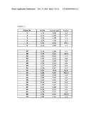 |  |
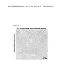 | 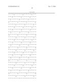 |
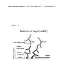 | 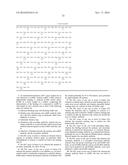 |
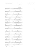 | 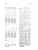 |
 | 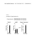 |
 |  |
 |  |
 |  |
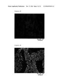 | 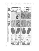 |
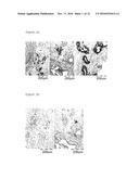 | 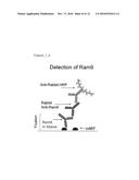 |
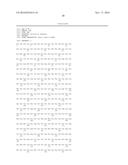 |  |
 |  |
 | 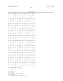 |
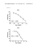 | 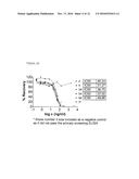 |
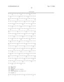 |  |
 |  |
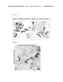 | 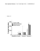 |
| New patent applications in this class: | |
| Date | Title |
|---|---|
| 2022-09-22 | Electronic device |
| 2022-09-22 | Front-facing proximity detection using capacitive sensor |
| 2022-09-22 | Touch-control panel and touch-control display apparatus |
| 2022-09-22 | Sensing circuit with signal compensation |
| 2022-09-22 | Reduced-size interfaces for managing alerts |
| New patent applications from these inventors: | |
| Date | Title |
|---|---|
| 2022-01-06 | Anti-oxmif/anti-cd3 antibody for cancer treatment |
| 2018-06-07 | Anti-mif antibodies in the treatment of cancers containing mutant tp53 and/or mutant ras |
| 2015-10-29 | Anti-mif antibody cell migration assay |