Patent application title: MONOSPECIFIC POLYPEPTIDE REAGENTS
Inventors:
Robert Poppe (Mainz, DE)
Assignees:
ORGENTEC DIAGNOSTIKA GMBH
IPC8 Class: AC07K1618FI
USPC Class:
424 937
Class name: Drug, bio-affecting and body treating compositions whole live micro-organism, cell, or virus containing animal or plant cell
Publication date: 2016-02-18
Patent application number: 20160046703
Abstract:
The present invention relates to a novel antigen-binding protein
construct or "modubody", which contains at least three functional single
domain modules of an antibody. The modubodies contain a domain from the
heavy chain variable region of an antibody (VH), a domain from the light
chain variable region of an antibody (VL) and bind monospecifically to an
antigen. The modubodies further contain a domain from the constant region
of antibodies. The modubodies can be used for diagnostic or therapeutic
purposes.Claims:
1. A nucleic acid coding for a monovalent fusion polypeptide in operative
linkage with an expression control sequence, wherein said monovalent
fusion polypeptide is in the form of a monomer under non-denaturing
conditions and comprises (i) a first domain comprising the heavy chain
variable region of an antibody (VH) or at least a section thereof that
mediates antigen binding, (ii) a second domain comprising the light chain
variable region of an antibody (VL) or at least a section thereof that
mediates antigen binding, and (iii) a third domain comprising a section
of a heavy chain constant region of an antibody (CHX), wherein domain
(iii) has a length of from 80 to 130 amino acid residues, wherein domains
(i), (ii) and (iii) are linked together via peptide linkers (L), wherein
the peptide linkers (L) each independently have a length of from 25 to 45
amino acid residues and consist of sequences which are heterologous to
the amino acid sequences of the first, second and third domains, wherein
the monovalent fusion polypeptide has antigen binding activity and over
90% of said antigen binding activity remains after 10 days incubation at
36.degree. C. or after drying and reconstitution or after freeze/thaw
cycles repeated 5 times and wherein the monovalent fusion polypeptide
does not form intermolecular disulphide bridges and is free of hinge
regions of antibodies.
2. The nucleic acid according to claim 1, wherein said nucleic acid is codon-optimized for expression in a chosen host cell.
3. A host cell containing a nucleic acid according to claim 1.
4. A process for the preparation of a monovalent fusion polypeptide, comprising cultivating a host cell containing a nucleic acid coding for said monovalent fusion polypeptide and obtaining the monovalent fusion polypeptide from the cell or from the culture supernatant, wherein said monovalent fusion polypeptide which is present in the form of a monomer under non-denaturing conditions, comprises (i) a first domain comprising the heavy chain variable region of an antibody (VH) or at least a section thereof that mediates antigen binding, (ii) a second domain comprising the light chain variable region of an antibody (VL) or at least a section thereof that mediates antigen binding, and (iii) a third domain comprising a section of a heavy chain constant region of an antibody (CHX), wherein domain (iii) has a length of from 80 to 130 amino acid residues, wherein domains (i), (ii) and (iii) are linked together via peptide linkers (L), wherein the peptide linkers (L) each independently have a length of from 25 to 45 amino acid residues and consist of sequences which are heterologous to the amino acid sequences of the first, second and third domains, wherein the monovalent fusion polypeptide has antigen binding activity and over 90% of said antigen binding activity remains after 10 days incubation at 36.degree. C. or after drying and reconstitution or after freeze/thaw cycles repeated 5 times and wherein the monovalent fusion polypeptide does not form intermolecular disulphide bridges and is free of hinge regions of antibodies.
5. A diagnostic or biochemical test comprising a monovalent fusion polypeptide as a reagent, wherein said monovalent fusion polypeptide is present in the form of a monomer under non-denaturing conditions, and comprises: (i) a first domain comprising the heavy chain variable region of an antibody (VH) or at least a section thereof that mediates antigen binding, (ii) a second domain comprising the light chain variable region of an antibody (VL) or at least a section thereof that mediates antigen binding, and (iii) a third domain comprising a section of a heavy chain constant region of an antibody (CHX), wherein domain (iii) has a length of from 80 to 130 amino acid residues, wherein domains (i), (ii) and (iii) are linked together via peptide linkers (L), wherein the peptide linkers (L) each independently have a length of from 25 to 45 amino acid residues and consist of sequences which are heterologous to the amino acid sequences of the first, second and third domains, wherein the monovalent fusion polypeptide has antigen binding activity and over 90% of said antigen binding activity remains after 10 days incubation at 36.degree. C. or after drying and reconstitution or after freeze/thaw cycles repeated 5 times and wherein the monovalent fusion polypeptide does not form intermolecular disulphide bridges and is free of hinge regions of antibodies.
6. The diagnostic or biochemical test according to claim 5, wherein said monovalent fusion polypeptide is in a carrier suitable for use in a control or calibrator reagent.
7. The diagnostic or biochemical test according to claim 5, wherein said monovalent fusion polypeptide is in a carrier suitable for use in a test reagent for determination of an analyte.
8. A pharmaceutical composition comprising a nucleic acid according to claim 1, together with a pharmaceutically suitable carrier.
9. A pharmaceutical composition comprising a host cell according to claim 3, together with a pharmaceutically suitable carrier.
10. The nucleic acid coding for a monovalent fusion polypeptide according to claim 1, wherein domains (i), (ii) and (iii) in said monovalent fusion polypeptide, each have a length of from 80 to 130 amino acid residues.
11. The nucleic acid according to claim 1, further comprising carrier substances suitable for use in human or veterinary medicine.
12. The host cell according to claim 3 further comprising carrier substances suitable for use in human or veterinary medicine.
13. The nucleic acid according to claim 2, which is codon-optimized for expression in E. coli.
Description:
[0001] This application is a divisional of U.S. Ser. No. 13/512,206, filed
on May 25, 2012, which is a 35 U.S.C. 371 National Phase Entry
Application from PCT/EP2010/068134, filed Nov. 24, 2010, which claims the
benefit of German Patent Application No. 10 2009 047 243.6 filed on Nov.
27, 2009, the disclosures of which are incorporated herein in their
entirety by reference.
[0002] The present invention relates to a novel antigen-binding protein construct or "modubody", which contains at least three functional single domain modules of an antibody. The modubodies contain a domain from the heavy chain variable region of an antibody (VH), a domain from the light chain variable region of an antibody (VL) and bind monospecifically to an antigen. The modubodies further contain a domain from the constant region of antibodies. The modubodies can be used for diagnostic or therapeutic purposes.
BACKGROUND OF THE INVENTION
[0003] Antibody Structure
[0004] Antibodies are plasma glycoproteins, which consist of a plurality of polypeptide chains connected by disulphide bridges. A standard antibody consists of two identical heavy immunoglobulin (Ig) chains and two identical light chains. Both antibody chains consist of different protein domains having a length of about 110 amino acid residues, which are composed of 6-sheets in the form of a characteristic immunoglobulin fold. The heavy chain consists of one variable (VH) domain and three or four constant domains (CH1, CH2, CH3, CH4). The light chain consists of one variable (VL) and one constant (CL) domain. The variable portions of the heavy and light chain, in particular the hypervariable complementarity determining regions (CDR), contribute to antigen specificity. The immune system provides a high diversity of antibodies against very different antigens. Antibodies can be assigned to different classes, for example IgM, IgA, IgG, IgE, IgD.
[0005] Antibody Fragments and Substructures
[0006] Antibody fragments can be obtained by enzymatic cleavage or by recombinant processes. A Fab fragment equipped with the antigen recognition function contains the VH-CH1 domains of the heavy chain and the VL-CL domains of the light chain, connected together via disulphide bridges, while the Fv fragment comprises only the heavy and light chain variable regions. However, these fragments consisting of a plurality of protein subunits can be prepared in biologically active form in an acceptable yield only by complex processes (Read et al. (2007), Appl. Environ. Microbiol. 73: 5088-5096).
[0007] A single-chain Fv fragment (scFv) is a small, approximately 28 kDa heavy antigen-binding substructure in the form of a covalent coupling of VH and VL domains via a peptide linker (Hu et al. (1996), Cancer Res 56: 3055-3061). However, the folding efficiency, the sometimes inadequate stability and the toxicity of these structures frequently limit the yield in the preparation of biologically active scFv in bacterial expression systems (Nieba et al. (1997), Protein Eng 10: 435-444).
[0008] The term minibody (mini-antibody) refers to an approximately 75 kDa heavy chimeric molecule of an scFv and a hinge region from the heavy chain fused with the CH3 domain which, assisted by the CH3 domain, is assembled to form a bivalent molecule covalently bonded via disulphide bridges of the hinge region (Wu, EP0627932B1). The CH3 domain serves as the dimerisation domain for the production of homodimers (cf. Dubel et al., Biol. Unserer Zeit, Vol. 34, No. 6, p. 372-379, 2004). However, on expression in E. coli, these molecules exhibit low expression rates and proteolytic degradation in the hinge region (Hu et al. (1996), Cancer Research 56: 3055-3061).
[0009] Like the minibody, the diabody has two antigen-binding sites. In the diabody, VL and VH domains are connected in the form of a single polypeptide chain to give a divalent and bispecific molecule (Hollinger et al. (1993), PNAS 90: 6444-6448).
[0010] Monobodies are chimeric antigen-binding polypeptides, which exhibit hypervariable CDR loops within a fibronectin type III scaffold (Koide, EP0985039B1). These molecules provide a valuable class of novel affinity reagents. However, when CDR loops are transplanted into a heterologous fibronectin scaffold, effector and detection functions of native antibodies are lost.
[0011] Nanobodies consist of single-chain antigen-binding VHH domains (variable domains of a heavy chain antibody) and are based on the observation that natural and functional antibodies that consist only of heavy chains are found in camels and llamas (Caserman and Harmers, EP 19930919098). The solubility of human VH domains (heavy chain variable domains) is, however, frequently limited on account of hydrophobic regions, which interact in the intact antibody with regions of the light chain (Barthelemy et al. (2007) J. Biol. Chem. 283: 3639-3654).
[0012] Antibodies in Immunodiagnosis
[0013] There are numerous immunoassay formats for determining the presence or concentration of a specific antibody, for example against a pathogen, an autoantigen or an allergen, in a biological sample. In general, such assays are directed to the detection of a specific antibody class or a combination of particular antibody classes and use specific internal controls or calibrators. Immunoassays which are suitable, for example, for determining human autoantibodies generally contain a positive control, a negative control and an index calibrator or a gradation of different calibrator concentrations (standard series) for producing a calibration curve, with which antibody concentrations in a sample can be interpolated. Such control and calibration reagents are conventionally prepared by diluting seropositive plasmas or serums in a suitable dilution medium.
[0014] For example, the calibrators and controls for the isotype-specific determination of β2-glycoprotein autoantibodies are prepared from the serum of human donors, which contain high concentrations of these autoantibodies of classes IgG, IgM and/or IgA. The use of human seropositive serum or plasma to prepare controls and calibrators is associated with numerous disadvantages, however, such as, for example, the difficulty of acquiring such reagents in large amounts and of suitable quality, differences in the binding characteristics in different batches, heterogeneous polyclonal specificity, heterogeneous isotype composition, presence of pathogens, costs, etc.
[0015] Because antibodies bind to antigens with high specificity and affinity, they are of central importance in immunodiagnosis. The size of the natural antibody molecules and their complex structure of a plurality of polypeptide chains with a large number of inter- and intra-domains, interconnections through disulphide bridges as well as glycosylation positions represent a considerable obstacle in the construction and the recombinant expression of specific antibodies.
[0016] Hackett et al. (EP1018019 B1) disclose a process for the preparation of reagents for use as calibrators and controls, wherein the reagent is a chimeric monoclonal antibody which comprises heavy and light chain variable regions from a first host species fused to heavy and light chain constant regions from a second host species, which corresponds to that of the antibody to be determined. However, the preparation of these species-chimeric monoclonal antibodies requires a high technical outlay, as is typical of the production of monoclonal antibodies. Although Hackett mentions the theoretical possibility of using as synthetic calibrators also polypeptides which bind specifically to a given ligand and are fused to an antibody region of the desired host species, Hackett does not provide a process by means of which single-chain synthetic polypeptide calibrators can be synthesised.
[0017] There is accordingly a continuing need for small, antibody-like molecules which specifically recognise an antigen, form complexes with antibodies against natural immunoglobulins, and can be prepared easily and in a large amount in bacterial expression systems.
SUMMARY OF THE INVENTION
[0018] The invention provides a monovalent fusion polypeptide comprising
[0019] (i) a first domain comprising the heavy chain variable region of an antibody (VH) or at least a section thereof that mediates antigen binding,
[0020] (ii) a second domain comprising the light chain variable region of an antibody (VL) or at least a section thereof that mediates antigen binding, and
[0021] (iii) a third domain comprising a section of a heavy chain constant region of an antibody (CHX), wherein domains (i), (ii) and (iii) are linked together via peptide linkers (L).
[0022] The invention further provides a nucleic acid which codes for a monovalent fusion polypeptide as described above.
[0023] The invention still further provides a host cell which contains a nucleic acid according to the invention.
[0024] The invention still further provides a process for the preparation of a monovalent fusion polypeptide by cultivating a host cell according to the invention and obtaining the fusion polypeptide from the cell or from the culture supernatant.
[0025] Yet further aspects of the present invention relate to the use of monovalent fusion polypeptides as reagents in diagnostic or biochemical tests, as well as to medical applications of the fusion polypeptides, nucleic acids and host cells.
[0026] The monovalent fusion polypeptides according to the invention are referred to as "modubodies".
[0027] Modubodies are constructs which consist of domains of the variable heavy (VH) and variable light (VL) chains of an antibody and one or more domains of the heavy chain constant region (CH1, CH2, CH3, CH4) of antibodies, these domains of an antibody being coupled together in the form of a linear sequence of independently structured function modules via suitable linker sequences. The individual domains have a length of, for example, from 80 to 130 amino acids. Modubodies consist of a single polypeptide chain and possess a single antigen-binding site, which is formed of the domains VH and VL. Modubodies are preferably unable to form intermolecular disulphide bridges, so that they are present in the form of monomers. Modubodies are free of hinge regions of antibodies. They are accordingly miniature versions of monovalent antibodies, which can be prepared simply and in large amounts in the form of a single protein chain in suitable expression systems, for example bacterial expression systems. Owing to their single-chain structure, their small size, the monoclonal composition, the fact that they can be prepared easily, and their stability, modubodies are ideal reagents for biochemical research, for use in diagnostic assays and as therapeutic agents.
[0028] Accordingly, a modubody is a chimeric molecule having a typical structure as in FIG. 1, consisting of separate function modules which are derived from domains of the variable heavy (VH) and variable light (VL) chains of an antibody and one or more domains of the heavy chain constant region (CHX, e.g. CH1, CH2, CH3 or CH4) of antibodies. The individual independently structured function modules are coupled together in a linear sequence via suitable linker sequences (L).
DESCRIPTION OF THE FIGURES
[0029] FIG. 1:
[0030] Schematic structure of an embodiment of a fusion polypeptide (modubody) according to the invention, wherein VL denotes a domain of the variable light chain, VH denotes a domain of the variable heavy chain, and CHX denotes a domain of the heavy chain constant region (CH1, CH2, CH3 or CH4) of antibodies. The individual domains are connected together in a linear sequence by heterologous, preferably flexible peptide linkers L.
[0031] FIG. 2:
[0032] Characterisation of the binding reaction of an scFV-CAD reaction module to the target β2-glycoprotein.
[0033] For the ELISA detection of the specific β2-glycoprotein-binding activity of the scFv-CAD reaction module, serial dilutions of scFv-CAD in the indicated concentration were applied to microtitre plates coated with β2-glycoprotein and, for control purposes, with bovine serum albumin (BSA). The binding was determined using a peroxidase-labelled RGS-6×-His antibody (Qiagen, Hilden) and a tetramethylbenzidine (TMB) colour reaction by measurement of the O.D. 450.
[0034] FIGS. 3A and 3B:
[0035] Characterisation of the stability of an scFV-CAD reaction module to temperature stress.
[0036] 3A. In order to investigate the influence of 50° C. temperature stress on the β2-glycoprotein-binding activity of the CAD-scFv, the purified protein module in a concentration of 5 μg/ml was exposed to a temperature of 50° C. for a period of 0-180 minutes. The binding of the scFv-CAD to β2-glycoprotein was determined in an ELISA using a peroxidase-labelled RGS-6×-His antibody and a TMB colour reaction by measurement of the O.D. 450.
[0037] 3B. In order to determine the inactivation temperature of the scFv-CAD, the purified protein module in a concentration of 5 μg/ml in calibrator dilution medium was exposed to a series of temperatures of from 20° C. to 90° C. for 10 minutes. The binding of the scFv-CAD to β2-glycoprotein was determined in an ELISA using a peroxidase-labelled RGS-6×-His antibody and a TMB colour reaction by measurement of the O.D. 450.
[0038] FIG. 4:
[0039] Characterisation of the calibrator function of a CAD-IgG-CH3 modubody by binding to the target β2-glycoprotein and detection with an antihuman IgG peroxidase secondary antibody. CAD-IgG-CH3 was applied in the indicated concentration to microtitre plates coated with β2-glycoprotein, and the binding was determined using a peroxidase-labelled antihuman IgG antibody (Jackson Immunoresearch) and a TMB colour reaction by measurement of the O.D. 450.
[0040] FIG. 5:
[0041] Characterisation of the stability of a CAD-IgG-CH3 modubody to temperature stress. The stability to heat stress was investigated as follows: dilutions of the CAD-IgG-CH3 modubody in the indicated concentrations were incubated for 60 minutes and 90 minutes at 50° C. and investigated in an anti-β2-glycoprotein immunoassay in comparison with a dilution series stored at room temperature. The binding of the CAD-IgG-CH3 modubody to β2-glycoprotein was determined using a peroxidase-labelled antihuman IgG antibody and a TMB colour reaction by measurement of the O.D. 450.
[0042] FIG. 6:
[0043] Characterisation of the stability of a CAD-IgG-CH3 modubody to storage at 36° C. The stability to elevated storage temperature was investigated as follows: dilutions of the CAD-IgG-CH3 modubody in the indicated concentrations were incubated for 1, 2, 4, 7, 10 days at 36° C. and investigated in an anti-β2-glycoprotein immunoassay in comparison with a dilution series stored at 4° C. The binding of the CAD-IgG-CH3 modubody to β2-glycoprotein was determined using a peroxidase-labelled antihuman IgG antibody and a TMB colour reaction by measurement of the O.D. 450.
[0044] FIG. 7:
[0045] Characterisation of the stability of a CAD-IgG-CH3 modubody to drying. The stability to drying was investigated as follows: dilutions of the CAD-IgG-CH3 modubody were dried in vacuo at 22° C. in a Speedvak device and then resolubilised to the indicated concentrations. The binding of the resolubilised CAD-IgG-CH3 modubody to β2-glycoprotein was then determined using a peroxidase-labelled antihuman IgG antibody and a TMB colour reaction by measurement of the O.D. 450.
[0046] FIG. 8:
[0047] Characterisation of the stability of a CAD-IgG-CH3 modubody to repeated freeze-thaw cycles. The stability to repeated freeze-thaw cycles was investigated as follows: dilutions of the CAD-IgG-CH3 modubody were frozen at -70° C. and thawed again at 37° C. in five repeated freeze-thaw cycles and then investigated in the indicated concentrations in an anti-β2-glycoprotein immunoassay. The binding of the CAD-IgG-CH3 modubody to β2-glycoprotein was determined using a peroxidase-labelled antihuman IgG antibody and a TMB colour reaction by measurement of the O.D. 450.
[0048] FIG. 9:
[0049] Characterisation of the calibrator function of the CAD-IgG-CH2 modubody by binding to the target β2-glycoprotein and detection with an antihuman IgG peroxidase secondary antibody. CAD-IgG-CH2 was applied in the indicated concentration to microtitre plates coated with β2-glycoprotein in complex with cardiolipin, and the binding was determined using a peroxidase-labelled antihuman IgG antibody (Jackson Immunoresearch) and a TMB colour reaction by measurement of the O.D. 450 nm.
[0050] FIG. 10:
[0051] Characterisation of the stability of the CAD-IgG-CH2 modubody to drying. The stability to drying was investigated as follows: dilutions of the CAD-IgG-CH2 modubody were dried in vacuo at 22° C. in a Speedvak device and then resolubilised to the indicated concentrations. The binding of the resolubilised CAD-IgG-CH2 modubody to β2-glycoprotein was then determined using a peroxidase-labelled antihuman IgG antibody and a TMB colour reaction by measurement of the O.D. 450 nm.
[0052] FIG. 11:
[0053] Characterisation of the stability of the CAD-IgG-CH2 modubody to repeated freeze-thaw cycles. The stability to repeated freeze-thaw cycles was investigated as follows: dilutions of the CAD-IgG-CH2 modubody were frozen at -70° C. and thawed again at 37° C. in five repeated freeze-thaw cycles and then investigated in the indicated concentrations on microtitre plates coated with β2-glycoprotein in complex with cardiolipin. The binding of the CAD-IgG-CH2 modubody to β2-glycoprotein was determined using a peroxidase-labelled antihuman IgG antibody and a TMB colour reaction by measurement of the O.D. 450 nm.
[0054] FIG. 12:
[0055] Characterisation of the calibrator function of the multifunctional CAD-IgM-IgA-IgG modubody by binding to the target β2-glycoprotein and separate detection with the isotype-specific antihuman IgM, antihuman IgA and antihuman IgG peroxidase secondary antibodies. CAD-IgM-IgA-IgG was applied in the indicated concentration to microtitre plates coated with β2-glycoprotein in complex with cardiolipin, and the binding was determined in separate determinations using isotype-specific peroxidase-labelled antihuman IgM, antihuman IgA and antihuman IgG antibodies (Jackson Immunoresearch) and a TMB colour reaction by measurement of the O.D. 450 nm.
[0056] FIG. 13:
[0057] Characterisation of the stability of the CAD-IgM-IgA-IgG modubody to drying. The stability to drying was investigated as follows: dilutions of the CAD-IgM-IgA-IgG modubody were dried in vacuo at 22° C. in a Speedvak device and then resolubilised to the indicated concentrations. The binding of the resolubilised CAD-IgM-IgA-IgG modubody to β2-glycoprotein was then determined in separate determinations using isotype-specific peroxidase-labelled antihuman IgM, antihuman IgA and antihuman IgG antibodies (Jackson Immunoresearch) and a TMB colour reaction by measurement of the O.D. 450 nm.
[0058] FIG. 14:
[0059] Characterisation of the stability of the CAD-IgM-IgA-IgG modubody to repeated freeze-thaw cycles. The stability to repeated freeze-thaw cycles was investigated as follows: dilutions of the CAD-IgM-IgA-IgG modubody were frozen at -70° C. and thawed again at 37° C. in five repeated freeze-thaw cycles and then investigated in the indicated concentrations on microtitre plates coated with β2-glycoprotein in complex with cardiolipin. The binding of the CAD-IgM-IgA-IgG modubody to β2-glycoprotein was then determined in separate determinations using isotype-specific peroxidase-labelled antihuman IgM, antihuman IgA and antihuman IgG antibodies (Jackson Immunoresearch) and a TMB colour reaction by measurement of the O.D. 450 nm.
[0060] FIG. 15:
[0061] Characterisation of the binding reaction of the CAD-IgG-CH3-Knob02 modubody to the target β2-glycoprotein. For the ELISA detection of the specific β2-glycoprotein-binding activity of the CAD-IgG-CH3-Knob02 modubody, serial dilutions of CAD-IgG-CH3-Knob02 in the indicated concentration were applied to microtitre plates coated with β2-glycoprotein and, for control purposes, with bovine serum albumin (BSA). The binding was determined using a peroxidase-labelled RGS-6×-His antibody (Qiagen, Hilden) and a tetramethylbenzidine (TMB) colour reaction by measurement of the O.D. 450 nm.
DETAILED DESCRIPTION OF THE INVENTION
[0062] Processes known hitherto for the preparation of antibodies, antibody fragments and small antibody-like molecules have numerous limitations as regards the possibility of preparing them inexpensively in biologically active form.
[0063] Processes known hitherto for the production of calibrators and standard materials for diagnostic assays have numerous disadvantages as regards availability, uniform binding and stability characteristics, ensuring the absence of pathogens, and production costs. These limitations and disadvantages are eliminated by the present invention. In addition, there is a continuing need for small antibody-like molecules which specifically recognise an antigen and form complexes with antibodies against natural immunoglobulins.
[0064] The present invention permits the preparation of monospecific polypeptide reagents--"modubodies"--for biochemical research, for use in diagnostic assays or as a constitutent of therapeutic agents.
[0065] A modubody is a chimeric single-chain fusion polypeptide which consists of at least three domains, namely a first domain from the heavy chain variable region of an antibody (VH), a second domain from the light chain variable region of an antibody (VL) and a third domain comprising a part-section of a heavy chain constant region of an antibody (CHX). The individual domains are linked together via suitable peptide linkers. The modubody is monovalent, that is to say it has a single antigen-binding site.
[0066] For example, the modubodies according to the invention have a structure (beginning at the N-terminus) as follows:
VH-L-VL-L-CHX or
VL-L-VH-L-CHX,
[0067] wherein VH denotes the heavy chain variable region of an antibody or at least a section thereof that mediates antigen binding, VL denotes the light chain variable region of an antibody or at least a section thereof that mediates antigen binding, L denotes a peptide linker, and CHX denotes a domain comprising a part-section of a heavy chain constant region of an antibody. CHX can be chosen, for example, from sections CH1, CH2, CH3 and CH4 from the heavy chain constant region of antibodies, preferably of antibodies of classes IgG, IgM, IgE and IgA, particularly preferably of human antibodies of the classes IgG, IgM, IgE and IgA. Preferred examples of the domain CHX are IgG-CH1, IgG-CH2, IgG-CH3, IgA-CH2, IgA-CH3, IgM-CH2, IgM-CH3 and IgM-CH4 or combinations thereof, in particular from the respective human antibodies.
[0068] The first domain (VH) and the second domain (VL) together form the antigen-binding site (binding module). They are preferably so chosen that they both originate from a single original antibody. The original antibody is any desired monoclonal antibody, for example a monoclonal antibody from a non-human mammalian species (e.g. rat, mouse or rabbit), a human antibody or a humanised antibody.
[0069] The modubody can be directed against any desired antigen, for example a diagnostically or therapeutically relevant antigen. Preferred examples of specific antigens are β2-glycoprotein, phosphatidylserine, vascular endothelial growth factor (VEGF-A), tumour necrosis factor (TNF-alpha), smoothened homolog (SMO), protein patched homolog (PTC1), B-lymphocyte antigen (CD20), cytotoxic T-lymphocyte protein 4 (CTLA4), amyloid beta A4 protein (APP), presenilin-1 (PS1), CC-chemokine receptor (CCR-5), telomere repeat-binding factor 1 (TRF1) and toll-like receptors (TLR1-10).
[0070] The first and second domains of the polypeptide according to the invention can contain the complete heavy or light chain variable regions of an antibody or at least a section thereof that mediates antigen binding. Preferably, the domains contain at least the regions CDR1, CDR2 and CDR3 of the heavy or light chain region in question in their entirety and at least parts of the corresponding framework regions FR1, FR2, FR3 and FR4. The length of the domains VH and VL is usually at least 80, at least 90 or at least 100 amino acid residues and up to 110 or up to 120 amino acid residues.
[0071] The fusion polypeptide according to the invention contains at least one domain comprising a section of a heavy chain constant region of an antibody (CHX). Preferably, the third domain or the third domains is/are arranged C-terminally to the VH and VL domains. A third domain can contain a complete section from a heavy chain constant region or a part-section thereof which imparts sufficient structural stability to the fusion polypeptide. The length of a third domain is usually at least 80, at least 90 or at least 100 amino acid residues and up to 110, up to 120 or up to 130 amino acid residues. The fusion polypeptide can optionally contain a plurality of third domains which can be identical or different, for example 2, 3 or 4. If a plurality of third domains is present, they are linked together via peptide linkers and are preferably arranged at the C-terminus of the fusion polypeptide. Examples of the structural configuration of fusion polypeptides according to the invention with 2 (or 3) third domains are as follows:
VL-L-VL-L-CHX1-L-CHX2 (-L-CHX3) or
[0072] VL-L-VH-L-CHX1-L-CHX2 (-L-CHX3), wherein VH, L and VL are as defined above and CHX1, CHX2 and CHX3 each denote a third domain CHX as defined above.
[0073] The fusion polypeptide according to the invention contains first, second and third domains which are linked together via peptide linkers (L). The peptide linkers consist of sequences which are heterologous to the amino acid sequences of the first, second and third domains, or of sequences which are not present in natural immunoglobulins. The peptide linkers which link the individual domains together can in each case be identical or different. Usually, the peptide linkers each independently have a length of from 10 to 50, preferably from 25 to 45 and particularly preferably from 30 to 40 amino acid residues. It is further preferred for the peptide linkers to be flexible linkers without a secondary structure. For example, suitable peptide linkers consist of at least 80% or at least 90%, preferably at least 95% or completely of glycine and/or serine residues. Particularly suitable are peptide linkers which contain a plurality of sequences SGGGG. A particularly preferred example of a peptide linker is shown in SEQ ID NO: 3. Linker sequences can optionally also be present at the N- and/or C-terminus of the fusion polypeptide.
[0074] In addition to domains (i), (ii) and (iii) and the peptide linkers arranged between the domains, the fusion polypeptide can optionally also contain further sequence sections, for example a signal peptide section which is arranged at the N- and/or C-terminus and facilitates the expression and/or secretion of the polypeptide. The fusion polypeptide can further contain one or more additional non-immunoglobulin domains, for example detection or recognition domains, that is to say peptide sequences suitable for the detection or recognition of the fusion polypeptide (tags), for example a FLAG epitope or a poly-His sequence. In addition, the fusion polypeptide can optionally also contain one or more non-immunoglobulin effector domains. The additional domains--where present--are preferably connected to the remainder of the fusion polypeptide via a peptide linker, for example a peptide linker as defined above.
[0075] Particularly preferred examples of fusion polypeptides according to the invention contain one or more domains VH, VL and/or CHX as defined above which have at least 90% identity, preferably at least 95% identity, at amino acid level with the corresponding domains according to SEQ ID NO: 1 (VL), SEQ ID NO: 2 (VH), SEQ ID NO: 4 (VH, VL), SEQ ID NO: 8 (IgG-CH1), SEQ ID NO: 10 (IgG-CH2), SEQ ID NO: 12 (IgG-CH3), SEQ ID NO: 14 (IgA-CH2), SEQ ID NO: 16 (IgA-CH3), SEQ ID NO: 18 (IgM-CH2), SEQ ID NO: 20 (IgM-CH3) or SEQ ID NO: 22 (IgM-CH4).
[0076] The fusion polypeptide according to the invention can optionally have one or more modifications with respect to the natural sequence of the VH, VL and CHX domains contained therein. For example, at least one asparagine residue at a glycosylation position, for example in a CHX domain, can be replaced by a different amino acid residue, preferably serine, alanine or glycine. Furthermore, at least one cysteine residue, for example in a CHX domain, which does not form a disulphide bridge with a second cysteine residue present in the domain in question and therefore potentially effect the formation of intermolecular disulphide bridges, can optionally be replaced by a different amino acid residue, preferably serine, alanine or glycine.
[0077] The modubodies according to the invention are artificial monospecific antibody-like molecules which, in the form of a single protein chain, can easily be prepared in large amounts in suitable expression systems, for example bacterial expression systems. As reagents for biochemical or diagnostic assays, modubodies can specifically bind an antigen via a binding module (VH+VL) and can specifically be detected via a reaction module (CHX) with species- and isotype-specific secondary antibodies. A sequence of a plurality of reaction modules (e.g. CHX1 and CHX2 or CHX and a non-immunoglobulin detection or recognition domain) permits detection with different reagents, for example with different species- or isotype-specific secondary antibodies. As reagents for therapeutic functions, modubodies can bind specifically to an antigen on a target structure via a binding module and exert specific effector functions via a reaction module (CHX) or a series of reaction modules (e.g. CHX1 and CHX2 or CHX and a non-immunoglobulin effector domain).
[0078] CHX domains which could potentially form dimers are preferably so modified that dimerisation can be ruled out and the modubodies are present in monovalent form.
[0079] For example, amino acid positions whose side chains exhibit interactions according to the concept of the "knobs-into-holes" arrangement (Crick, F. H. C. (1952), Nature, 170: 882-883) can be modified.
[0080] Accordingly, amino acid positions whose side chains form contacts at the interface of IgG1-CH3 dimers (Ridgway J. B. B. et al. (1996), Protein Engineering, 9: 617-621) can be so modified that bulky amino acid side chains are present at opposing contact sites in the IgG-CH3 dimer and dimerisation is sterically hindered by this "knob-knob" position. The interactions between the contacting amino acid side chains of two IgG1-CH3 domains can be blocked, for example, by replacing the amino acid positions threonine 366, threonine 394, phenylalanine 405 by tyrosine at positions 366, 394 and 405 (numbering scheme according to the Kabat EU Index (Kabat et al., (1991) Sequences of Proteins of Immunological Interest 5th Edition, NIH Publication 91-3242)).
[0081] The modubodies according to the invention are additionally distinguished by surprising stability, for example stability to temperature stress or stability to drying/reconstitution or freeze/thaw cycles. For example, in comparison with an untreated sample, over 90% of the antigen-binding activity remains, even in dilute solutions (5 μg/ml) of the modubodies, after 10 days' incubation at 36° C. or after drying and reconstitution or after freeze/thaw cycles repeated 5 times.
[0082] The de novo construction of a modubody preferably comprises the steps:
[0083] a) selection of spatially delimited domains from immunoglobulins of known or modelled spatial structure having regard to sequence and structure data bank information;
[0084] b) disulphide bridge optimisation of the selected domains, in which cysteine positions which, in the context of the intact immunoglobulin, form disulphide bridges outside the chosen domains are edited to a structurally neutral amino acid, preferably serine, alanine or glycine;
[0085] c) optional editing of asparagine-coupled glycosylation positions preferably to serine, alanine or glycine;
[0086] d) linking of a selection of the function modules resulting from steps a) to c) with flexible linker sequences which preferably consist predominantly of the amino acids glycine and/or serine and particularly preferably correspond to the linker sequence (SEQ ID NO: 3), wherein, in dependence on the position of the function module within the modubody and on the cloning strategy, a module sequence resulting from steps a) to c) can be linked with a flexible linker sequence and the linker sequence can form the N- or C-terminus of the function module, and wherein an amino acid sequence which comprises a plurality of function modules or an amino acid sequence forming the complete modubody can optionally be generated;
[0087] e) translation of the amino acid sequences resulting from steps a) to d) into corresponding DNA sequences, preferably having regard to a codon frequency table optimised for the intended expression system, wherein the resulting DNA sequences can optionally be provided with flanking restriction enzyme cleavage sites and conservative base substitutions in order to avoid undesirable restriction enzyme cleavage sites;
[0088] f) preparation of the sequences defined in step e) as synthetic DNA by gene synthesis and cloning of the genetic units which code for individual function modules or for a series of a plurality of identical or different function modules into suitable vectors, cloning of the complete modubody by assembly of the individual function modules or function module series in the intended function module sequence and number, wherein a genetic unit coding for the complete modubody can optionally also be cloned directly.
[0089] The aim of this construction method, for example the selection of spatially delimited domains, the avoidance of disulphide bridges which stabilise a dimer, and the linking of the individual modules by flexible glycine-serine linkers having a preferred length of from 30 to 40 amino acids, is that the individual modules can behave independently of one another.
[0090] The invention relates in addition to a nucleic acid, for example a DNA or RNA, which codes for a fusion polypeptide as described above. The nucleic acid can optionally be present in operative linkage with an expression control sequence, for example a promoter. The invention accordingly relates also to expression vectors which contain a nucleic acid sequence coding for a fusion polypeptide according to the invention and are suitable for the expression of that nucleic acid in a host cell. The host cell can be a prokaryotic host cell, such as a gram-negative bacterium such as E. coli, or a gram-positive bacterium such as B. subtilis, or a eukaryotic host cell, for example a yeast cell, a fungus cell, an insect cell or a mammalian cell. In order to improve the expression of the nucleic acid in the chosen host cell, it can be optimised in respect of the codon usage in the respective host cell. Corresponding processes are known to the person skilled in the art.
[0091] The invention further provides a host cell as indicated above which contains a nucleic acid according to the invention. The nucleic acid can be introduced into the corresponding host cell by known techniques, for example transformation or transfection. For the preparation of the fusion polypeptide according to the invention, the host cell can be cultivated and the fusion polypeptide can be obtained from the cell or from the culture supernatant by methods which are known in principle.
[0092] The fusion polypeptides according to the invention can be used, for example, as reagent in a diagnostic or biochemical test, in particular in a test based on immunological methods, for example as control or calibrator reagent or as test reagent for determining an analyte. Detection of the fusion polypeptides according to the invention can take place via the CHX domain(s), for example using isotype- or species-specific recognition reagents, for example secondary antibodies, and/or via non-immunoglobulin recognition domains using specific binding partners for those domains. Corresponding test formats are known to the person skilled in the art.
[0093] Furthermore, the fusion polypeptide, the nucleic acid or the host cell according to the present invention can also be used for medical purposes, for example in human or veterinary medicine. Fusion polypeptides can be used, for example, as immunotherapeutic agents, optionally coupled with non-immunoglobulin effector domains, for example radionuclides or toxins. The nucleic acids coding for the fusion polypeptide can be used, for example, as nucleic acid vaccines.
[0094] Accordingly, the invention still further provides a pharmaceutical composition which comprises the fusion polypeptide, the nucleic acid or the host cell together with pharmaceutically suitable carrier substances. The pharmaceutical composition can be administered by known methods, as are used, for example, for therapy with antibodies or for DNA vaccination, to a subject, for example a human patient, requiring corresponding therapeutic treatment.
[0095] The present invention is to be explained further by means of the following examples.
EXAMPLES
Example 1
Construction, Cloning and Characterisation of a β2-Glycoprotein-Binding Module (scFv-CAD)
[0096] 1.1 Construction of the scFv-CAD
[0097] The following example describes the construction of a β2-glycoprotein-specific binding module (scFv-CAD) in the form of a synthetic construct.
[0098] Portions of the light chain variable region VL (SEQ ID NO: 1) and of the heavy chain variable region VH (SEQ ID NO: 2) of the monoclonal antibody WBCAL-1, which comes from a mouse model of antiphospholipid syndrome, the F1 mouse from New Zealand white X BXSB (Ichikawa et al. (1999), Arthritis and Rheumatism 42:2461), were chosen as structural units for a β2-glycoprotein recognition module and joined with a flexible linker (SEQ ID NO: 3) and flanking sequences to form an artificial protein sequence scFv-RP-CAD-P (SEQ ID NO: 4). This protein sequence was translated, having regard to a codon frequency table, into a nucleic acid sequence optimised for expression in E. coli, provided with flanking cloning sequences and prepared as an artificial DNA sequence scFv-RP-CAD-N coding for a β2-glycoprotein recognition module (SEQ ID NO: 5) by gene synthesis. The VL domain extends from amino acid 1-118 in SEQ ID NO: 4, the VH domain extends from amino acid 159-272 in SEQ ID NO: 4.
[0099] 1.2 Cloning of the scFv-CAD
[0100] For the construction, the artificial DNA sequence scFv-RP-CAD-N(SEQ ID NO: 5) described under Example 1.1, which codes for a β2-glycoprotein detection domain, was amplified with the primers RP-CAD01 (SEQ ID NO: 6), RP-CAD02 (SEQ ID NO: 7) by polymerase chain reaction (PCR). The 856 base pair (bp) amplificate was isolated after agarose gel electrophoresis with the QiaExII kit (Qiagen, Hilden). The isolated fragment was first digested with the restriction enzyme HindIII and then subjected to a BglII partial digestion. The restriction fragments were ligated with the plasmid pQE-80L (Qiagen, Hilden), which was digested with the compatible enzymes BamHI and HindIII, and transformed into E. coli strain NovaBlue (Merck, Nottingham). The transformation batch was plated out on LB agar plates supplemented with 50 μg/m1 of carbenicillin and incubated overnight (o.n.) (16-20 h) at 36° C. Single colonies of the resulting E. coli strain CAD-pQE80-NovaBlue were propagated (36° C., o.n.) on a shaker at 180 revolutions per minute (rpm) in LB medium supplemented with 50 μg/m1 of carbenicillin (LB-Carb). Stock cultures for freezing were prepared from the single clone cultures. In each case 1 ml of the single clone cultures was used for plasmid preparation. The isolated plasmids were analysed by EcoRI/HindIII digestion. Clones with an expected 901 bp fragment were investigated further by induction analysis. To that end, the chosen single clones were grown in LB-Carb and, at an O.D. 500 of from 0.5 to 1.0, induced to expression of the protein coded for by the cloned DNA fragment by addition of one culture volume of LB-Carb, 1 mM isopropyl thiogalactoside (IPTG) and cultivated for 16 hours (h) at 36° C., 180 rpm. The induction cells were lysed in sodium dodecyl sulfate (SDS)-containing buffer, proteins were separated by SDS-polyacrylamide gel electrophoresis (PAGE). In the Western blot, the expression of an expected 30 kDa scFv-CAD protein was detected by detection with an anti-RGS-6×His peroxidase-coupled antibody (Qiagen, Hilden). The correct cloning was confirmed by sequencing.
[0101] 1.3 Expression of the scFv-CAD
[0102] E. coli strain CAD-pQE80-NovaBlue transformed under 1.2 with the expression construct was grown in LB-Carb medium at 36° C. to an O.D. 500 of from 0.5 to 1.0 and induced to synthesis of the scFv-CAD by addition of IPTG. The induced culture was cultivated at 36° C. for 4 hours to overnight. The induction cells were harvested and lysed after lysozyme treatment in an 8 M urea-containing Tris-HCl-sodium chloride buffer (TBS). Expressed scFv-CAD was purified to homogeneity by a combination of affinity and ion-exchange chromatography. The scFv-CAD protein is visible in the form of a 30 kDa band in the Coomassie blue-stained SDS-polyacrylamide gel. The enhancement of the β2-glycoprotein-binding activity is associated with the purification of the 30 kDa band.
[0103] 1.4 Characterisation of the scFv-CAD
[0104] The binding specificity of the scFv-CAD was tested in an enzyme-linked immunosorbent assay (ELISA) using microtitre plates coated with β2-glycoprotein or with bovine serum albumin (BSA). The stability of the scFv-CAD to heat stress was investigated on suitable dilutions of the scFv-CAD.
[0105] 1.4.1 Binding Specificity of the scFv-CAD
[0106] In order to determine whether scFv-CAD possesses a specific β2-glycoprotein-binding activity, the purified protein module was tested by immunoassays. To that end, serial dilutions of scFv-CAD were applied to microtitre plates of the anti-β2-glycoprotein assay coated with β2-glycoprotein (ORG 521, ORGENTEC Diagnostika GmbH, Mainz) and, for control of the binding specificity, to BSA-coated plates and incubated for 30 minutes at 20-25° C. The microtitre plates were washed and the binding of scFv-CAD was determined using a peroxidase-labelled RGS-6×-His antibody (Qiagen, Hilden) and a tetramethylbenzidine (TMB) colour reaction by measurement of the O.D. 450. As shown in FIG. 2, specific binding to β2-glycoprotein was detected at a concentration of the scFv-CAD of only 19 ng/ml. The comparison of the level of reaction of the scFv-CAD serial dilutions in the binding to β2-glycoprotein- and BSA-coated microtitre plates shows that no non-specific binding occurs in the concentration range 0.019-10 μg/ml scFv-CAD.
[0107] 1.4.2 Temperature Stability of the scFv-CAD
[0108] In order to investigate the influence of temperature stress on the specific β2-glycoprotein-binding activity of the scFv-CAD, the purified protein module in a concentration of 5 μg/ml was exposed to a temperature of 50° C. over a period of 0-180 minutes. Binding of the scFv-CAD to β2-glycoprotein was determined in an immunoassay as described under 1.4.1. As shown in FIG. 3A, the scFv-CAD module diluted in calibrator dilution medium was found to be resistant to a temperature of 50° C. for 3 hours.
[0109] In order to determine the inactivation temperature of the scFv-CAD, the purified protein module in a concentration of 5 μg/ml in calibrator dilution medium was exposed to a series of temperatures of from 20° C. to 90° C. for 10 minutes. The binding of the heat-treated scFv-CAD dilutions to β2-glycoprotein was determined in an immunoassay as described under 1.4.1. As shown in FIG. 3B, the scFv-CAD module was found to be stable up to a temperature of 50° C.; at a temperature of 60° C. and above, marked inactivation occurred.
Example 2
Construction of a Series of Reaction Modules for Modubodies
[0110] The following example describes the construction of a series of reaction modules which are derived from domains of the heavy chain constant region of human antibodies. To that end, structurally and functionally delimited domains were derived from protein sequences of the heavy chain of human antibodies of classes IgG, IgA, IgM of known spatial structure. The chosen protein sequences were checked for potential interdomain disulphide bridges. Cysteine positions which outside the chosen domains form disulphide bridges with regions of the intact immunoglobulin were edited from cysteine to serine. At individual glycosylation positions, asparagine was likewise edited to serine. The chosen immunoglobulin domain sequences were joined as structural units for reaction modules with a flexible linker (SEQ ID NO: 3) and flanking sequences to form artificial protein sequences. On the basis of a codon frequency table, these reaction module sequences were translated into a nucleic acid sequence optimised for expression in E. coli, provided with flanking cloning sequences and prepared as artificial DNA sequences by gene synthesis.
[0111] 2.1 Construction of a Human IgG-CH1 Reaction Module
[0112] From the Worldwide Protein Data Bank (pdb), (Berman et al. (2003), Nature Structural Biology 10: 980), sequence positions 125-219 were chosen as the representative IgG-CH1 domain from the crystal structure of the Fab fragment of the anti-factor Ix antibody 10c12 on the basis of protein structure data bank entry pdb 3D69H. The chosen IgG-CH1 immunoglobulin domain sequence was joined as structural unit for an IgG-CH1 reaction module with a flexible linker (SEQ ID NO: 3) to form an artificial protein sequence Sc-RP-CH1-G-P (SEQ ID NO: 8) and, having regard to a codon frequency table, translated into a nucleic acid sequence optimised for expression in E. coli, additionally provided with flanking cloning sequences (BamHI, HindIII) and prepared as an artificial DNA sequence Sc-RP-CH1-G-N(SEQ ID NO: 9) by gene synthesis. The IgG-CH1 domain extends from amino acid 37-131 in SEQ ID NO: 8.
[0113] 2.2 Construction of a Human IgG-CH2 Reaction Module
[0114] From the crystal structure of the mutated Adcc-strengthened Fc fragment, sequence positions 15-116 were chosen as the representative IgG-CH2 domain on the basis of protein structure data bank entry pdb 2QL1A. The chosen IgG-CH2 immunoglobulin domain sequence was joined as structural unit for an IgG-CH2 reaction module with a flexible linker (SEQ ID NO: 3) to form an artificial protein sequence Sc-RP-CH2-G-P (SEQ ID NO: 10) and, having regard to a codon frequency table, translated into a nucleic acid sequence optimised for expression in E. coli, additionally provided with flanking cloning sequences (BamHI/HindIII) and prepared as an artificial DNA sequence Sc-RP-CH2-G-N(SEQ ID NO: 11) by gene synthesis. The IgG-CH2 domain extends from amino acid 37-138 in SEQ ID NO: 10.
[0115] 2.3 Construction of a Human IgG-CH3 Reaction Module
[0116] From the crystal structure of the heavy chain of a human immunoglobulin with a hinge deletion, sequence positions 330-428 were chosen as the representative IgG-CH3 domain on the basis of protein structure data bank entry pdb 1MCO_H (GI: 494350). The chosen IgG-CH3 immunoglobulin domain sequence was joined as structural unit for an IgG-CH3 reaction module with a flexible linker (SEQ ID NO: 3) to form an artificial protein sequence Sc-RP-CH3-G-P (SEQ ID NO: 12) and, having regard to a codon frequency table, translated into a nucleic acid sequence optimised for expression in E. coli, additionally provided with flanking cloning sequences (BamHI/HindIII) and prepared as an artificial DNA sequence Sc-RP-CH3-G-N(SEQ ID NO: 13) by gene synthesis. The IgG-CH3 domain extends from amino acid 37-135 in SEQ ID NO: 12.
2.4 Construction of a Human IgA-CH2 Reaction Module
[0117] From a structural model of human IgA, which was derived on the basis of neutron scattering in solution and homology modelling (Boehm et al. (1999) J. Mol. Biol. 286: 1421-1447), sequence positions 126-222 (corresponding to UNIPROT P01876) were chosen as the representative IgA-CH2 domain from protein structure data bank entry pdb 1IGA on the basis of the spatial structure of the heavy chain of human IgA1. At positions 182, 192, cysteine was edited to serine. The chosen and edited IgA-CH2 immunoglobulin domain sequence was joined as structural unit for an IgA-CH2 reaction module with a flexible linker (SEQ ID NO: 3) to form an artificial protein sequence Sc-RP-CH2-A-P (SEQ ID NO: 14) and, having regard to a codon frequency table, translated into a nucleic acid sequence optimised for expression in E. coli, additionally provided with flanking cloning sequences (BamHI/HindIII) and prepared as an artificial DNA sequence Sc-RP-CH2-A-N(SEQ ID NO: 15) by gene synthesis. The IgA-CH2 domain extends from amino acid 37-133 in SEQ ID NO: 14.
[0118] 2.5 Construction of a Human IgA-CH3 Reaction Module
[0119] From protein structure data bank entry PDB 1IGA, sequence positions 227-331 (corresponding to UNIPROT P01876) were chosen as the representative IgA-CH3 domain on the basis of the spatial structure of the heavy chain of human IgA1. The chosen IgA-CH3 immunoglobulin domain sequence was joined as structural unit for an IgA-CH3 reaction module with a flexible linker (SEQ ID NO: 3) to form an artificial protein sequence sc-RP-CH3-A-P (SEQ ID NO: 16) and, having regard to a codon frequency table, translated into a nucleic acid sequence optimised for expression in E. coli, additionally provided with flanking cloning sequences (BamHI/HindIII) and prepared as an artificial DNA sequence sc-RP-CH3-A-N(SEQ ID NO: 17) by gene synthesis. The IgA-CH3 domain extends from amino acid 37-141 in SEQ ID NO: 16.
[0120] 2.6 Construction of a Human IgM-CH2 Reaction Module
[0121] From a structural model of human IgM which was derived on the basis of X-ray scattering in solution and modelling (Perkins et al. (1991) J. Mol. Biol. 221:1345-1366), sequence positions 106-217 (corresponding to UNIPROT P01871) were chosen as the representative IgM-CH2 domain from protein structure data bank entry pdb 2rcj on the basis of the spatial structure of the heavy chain of human IgM and were edited. At position 214, cysteine was edited to serine; at position 109, asparagine was edited to serine. The chosen and edited IgM-CH2 immunoglobulin domain sequence was joined as structural unit for an IgM-CH2 reaction module with a flexible linker (SEQ ID NO: 3) to form an artificial protein sequence Sc-RP-CH2-M-P (SEQ ID NO: 18) and, having regard to a codon frequency table, translated into a nucleic acid sequence optimised for expression in E. coli, additionally provided with flanking cloning sequences (BamHI/HindIII) and prepared as an artificial DNA sequence Sc-RP-CH2-M-N(SEQ ID NO: 19) by gene synthesis. The IgM-CH2 domain extends from amino acid 37-148 in SEQ ID NO: 18.
[0122] 2.7 Construction of a Human IgM-CH3 Reaction Module
[0123] From protein structure data bank entry pdb 2rcj, sequence positions 218-323 (corresponding to UNIPROT P01871) were chosen as the representative IgM-CH3 domain on the basis of the spatial structure of the heavy chain of human IgM and were edited. At position 291, cysteine was edited to serine; at positions 272 and 279, asparagine was edited to serine. The chosen and edited IgM-CH3 immunoglobulin domain sequence was joined as structural unit for an IgM-CH3 reaction module with a flexible linker (SEQ ID NO: 3) to form an artificial protein sequence Sc-RP-CH3-M-P (SEQ ID NO: 20) and, having regard to a codon frequency table, translated into a nucleic acid sequence optimised for expression in E. coli, additionally provided with flanking cloning sequences (BamHI/HindIII) and prepared as an artificial DNA sequence Sc-RP-CH3-M-N(SEQ ID NO: 21) by gene synthesis. The IgM-CH3 domain extends from amino acids 37-142 in SEQ ID NO: 20.
[0124] 2.8 Construction of a Human IgM-CH4 Reaction Module
[0125] From protein structure data bank entry pdb 2rcj, sequence positions 324-452 (corresponding to UNIPROT P01871) were chosen as the representative IgM-CH3 domain on the basis of the spatial structure of the heavy chain of human IgM and were edited. At position 451, cysteine was edited to serine; at position 439, asparagine was edited to serine. The chosen and edited IgM-CH3 immunoglobulin domain sequence was joined as structural unit for an IgM-CH3 reaction module with a flexible linker (SEQ ID NO: 3) to form an artificial protein sequence Sc-RP-CH4-M-P (SEQ ID NO: 22) and, having regard to a codon frequency table, translated into a nucleic acid sequence optimised for expression in E. coli, additionally provided with flanking cloning sequences (BamHI/HindIII) and prepared as an artificial DNA sequence Sc-RP-CH4-M-N(SEQ ID NO: 23) by gene synthesis. The IgM-CH4 domain extends from amino acid 37-165 in SEQ ID NO: 22.
[0126] 2.9 Construction of a Monovalent Human IgG-CH3-Knob02 Reaction Module
[0127] From the crystal structure of the heavy chain of a human immunoglobulin with a hinge deletion, sequence positions 330-428 were chosen as the representative IgG-CH3 domain on the basis of protein structure data bank entry pdb 1 MCO_H (GI:494350) and were edited. Positions of amino acid residues whose side chains form contacts at the interface of IgG1-CH3 dimers (Ridgway J. B. B. et al. (1996), Protein Engineering, 9: 617-621) were so modified that no dimerisation of the IgG-CH3 domain can take place. At positions 351 and 379, threonine was edited to tyrosine; at position 390, phenylalanine was edited to tyrosine. The chosen and edited IgG-CH3 immunoglobulin domain sequence was joined as structural unit for an IgG-CH3-Knob02 reaction module with a flexible linker (SEQ ID NO: 3) to form an artificial protein sequence Sc-RP-CH3-G-Knob02-P (SEQ ID NO: 32) and, having regard to a codon frequency table, translated into a nucleic acid sequence optimised for expression in E. coli, additionally provided with flanking cloning sequences (BamHI/HindIII) and prepared as an artificial DNA sequence Sc-RP-CH3-G-Knob02-N(SEQ ID NO: 33) by gene synthesis. The IgG-CH3-Knob02 domain extends from amino acid 37-135 in SEQ ID NO: 32.
Example 3
The Single-Chain CAD-IgG-CH3 Modubody
[0128] 3.1 Construction and Cloning of the Single-Chain CAD-IgG-CH3 Modubody
[0129] The following example describes the construction and cloning of the β2-glycoprotein-specific modubody CAD-IgG-CH3 provided with a human IgG-CH3 detection domain.
[0130] For the construction of the CAD-IgG-CH3 modubody, the modules scFv-CAD (Example 1) and IgG-CH3 (Example 2.3) were assembled by restriction digestion and ligation in the domain sequence VL-linker-VH-linker-IgG-CH3 to form a CAD-IgG-CH3 coding sequence (SEQ ID NO: 26). To that end, the synthetic IgG-CH3 reaction module Sc-RP-CH3-G-N corresponding to sequence SEQ ID NO: 13 and described under Example 2.3 was amplified with the primers CH05 (SEQ ID NO: 24) and CH04 (SEQ ID NO: 25) by PCR. The 443 bp amplificate was isolated after agarose gel electrophoresis with the QiaExII kit (Qiagen, Hilden). The isolated fragment was first digested with the restriction enzymes BclI and HindIII. The restriction fragments were ligated with the CAD-scFv-pQE80 vector construct described under Example 1, which was digested with the compatible enzymes BamHI and HindIII, and transformed into E. coli strain NovaBlue (Merck, Nottingham). The transformation batch was plated out on LB agar plates supplemented with carbenicillin (50 μg/ml) and incubated o.n. at 36° C. Single colonies of the resulting E. coli strain CAD-IgG-CH3-pQE80-NovaBlue were propagated (36° C., o.n., 180 rpm) in LB-Carb medium. Stock cultures for freezing were prepared from the single clone cultures, in each case 1 ml of the single clone cultures was used for plasmid preparation. The isolated plasmids were analysed by EcoRI/HindIII digestion. Clones with an expected 1312 bp fragment were investigated further by induction analysis. To that end, the chosen single clones were grown in LB-Carb and, at an O.D. 500 of from 0.5 to 1.0, induced to expression of the recombinant protein by addition of one culture volume of LB-Carb supplemented with 1 mM IPTG, and cultivated for 16 hours at 36° C., 180 rpm. The induction cells were lysed in SDS sample buffer, proteins were separated by SDS-PAGE. In the Western blot, the expression of the expected 44 kDa CAD-IgG-CH3 modubody was detected by detection with an anti-RGS-6×His peroxidase-coupled antibody (Qiagen, Hilden). The correct cloning was confirmed by sequencing.
[0131] 3.2 Expression and Purification of the Single-Chain CAD-IgG-CH3 Modubody
[0132] The following example describes the expression and purification of the β2-glycoprotein-specific modubody CAD-IgG-CH3 provided with a human IgG-CH3 detection domain.
[0133] E. coli strain CAD-IgG-CH3-pQE80-NovaBlue transformed under 3.1 with the expression construct was grown in LB-Carb medium at 36° C. to an O.D. 500 of from 0.5 to 1.0 and induced to synthesis of the CAD-IgG-CH3 modubody by addition of IPTG. The induced culture was cultivated at 36° C. for 4 hours to overnight. The induction cells were harvested and lysed after lysozyme treatment in an 8 M urea-containing TBS buffer. Expressed CAD-IgG-CH3 modubodies were purified to homogeneity by a combination of affinity and ion-exchange chromatography. A 44 kDa band is visible in the Coomassie blue-stained SDS-polyacrylamide gel. The enhancement of the β2-glycoprotein-binding activity and a reactivity with a peroxidase-coupled antihuman IgG secondary antibody (Jackson Immunoresearch) is associated with the purification of the 44 kDa band.
[0134] 3.3 Characteristics of the Single-Chain CAD-IgG-CH3 Modubody
[0135] The following example describes the characterisation of the β2-glycoprotein-specific modubody CAD-IgG-CH3 provided with a human IgG-CH3 detection domain in relation to specific antigen recognition, specific detectability via an antihuman IgG secondary antibody, and stability to elevated temperatures, drying and freeze-thaw cycles.
[0136] 3.3.1 Calibrator Function of the CAD-IgG-CH3 Modubody
[0137] In order to determine whether the CAD-IgG-CH3 modubody has retained the β2-glycoprotein-binding activity of the scFv-CAD and binding to the antigen can be detected specifically via an antihuman IgG secondary antibody, purified CAD-IgG-CH3 preparations were investigated in an anti-β2-glycoprotein immunoassay. The purified CAD-IgG-CH3 modubody was applied in serial dilutions with concentrations of 0 μg/ml, 0.31 μg/ml, 0.62 μg/ml, 1.25 μg/ml, 2.5 μg/ml, 5 μg/ml, 10 μg/ml, 20 μg/ml in calibrator dilution medium to microtitre plates coated with β2-glycoprotein. The antigen-binding activity was determined under the incubation conditions provided for the anti-β2-glycoprotein assay (ORG 521, ORGENTEC GmbH, Mainz) using an antihuman IgG peroxidase-labelled secondary antibody in a concentration of 80 ng/ml. FIG. 4 shows the variation in the OD 450 nm determined under the chosen reaction conditions in dependence on the concentration. Under the chosen reaction conditions, the CAD-IgG-CH3 modubody was detected at a concentration of 5 μg/ml with an O.D. 450 of 2.2.
[0138] 3.3.2 Stability of the CAD-IgG-CH3 Modubody
[0139] In order to characterise the robustness of the CAD-IgG-CH3 modubody, dilutions in calibrator dilution medium were exposed to the stress factors of 50° C. heat stress, elevated storage temperature at 36° C., dryness and repeated freeze-thaw cycles. The β2-glycoprotein-binding activity and the detectability with an anti-hu IgG peroxidase-labelled secondary antibody (Jackson Immunoresearch) were determined in comparison with untreated samples.
[0140] 3.3.2.1 Stability of the CAD-IgG-CH3 Modubody to Heat Stress
[0141] The stability to heat stress was investigated as follows: dilutions of the CAD-IgG-CH3 modubody in calibrator dilution medium in concentrations of 0 μg/ml, 0.31 μg/ml, 0.62 μg/ml, 1.25 μg/ml, 2.5 μg/ml, 5 μg/ml, 10 μg/ml, 20 μg/ml were incubated for 60 minutes and 90 minutes at 50° C. and investigated in an anti-β2-glycoprotein immunoassay in comparison with a dilution series stored at room temperature. The β2-glycoprotein-binding activity was determined under the incubation conditions provided for the anti-β2-glycoprotein assay (ORG 521, ORGENTEC GmbH, Mainz) using an antihuman IgG peroxidase-labelled secondary antibody in a concentration of 80 ng/ml. FIG. 5 shows the variation in the OD 450 nm determined under the chosen reaction conditions for the dilution series incubated for 60 and 90 minutes at 50° C. in comparison with the dilution series stored at room temperature. Under the chosen reaction conditions, the function of the CAD-IgG-CH3 modubody in dilutions in the concentration range from 0.31 μg/ml to 20 μg/ml was not impaired by 50° C. temperature stress for 60 minutes or 90 minutes.
[0142] 3.3.2.2 Stability of the CAD-IgG-CH3 Modubody to Storage at 36° C.
[0143] The stability to elevated storage temperature was investigated as follows: dilutions of the CAD-IgG-CH3 modubody in concentrations of 0 μg/ml, 0.31 μg/ml, 0.62 μg/ml, 1.25 μg/ml, 2.5 μg/ml, 5 μg/ml, 10 μg/ml, 20 μg/ml in calibrator dilution medium were incubated for 1, 2, 4, 7, 10 days at 36° C. and investigated in an anti-β2-glycoprotein immunoassay in comparison with a dilution series stored at 4° C. The β2-glycoprotein-binding activity was determined under the incubation conditions provided for the anti-β2-glycoprotein assay (ORG 521, ORGENTEC GmbH, Mainz) using an antihuman IgG peroxidase-labelled secondary antibody in a concentration of 80 ng/ml. FIG. 6 shows the variation in the OD 450 nm determined under the chosen reaction conditions for the dilution series stored for 1, 2, 4, 7, 10 days at 36° C. in comparison with the dilution series stored at 4° C. Under the chosen reaction conditions, the function of the CAD-IgG-CH3 modubody in dilutions in the concentration range from 0.31 μg/ml to 20 μg/ml was not impaired by storage at 36° C. over a period of ten days.
[0144] 3.3.2.3 Stability of the CAD-IgG-CH3 Modubody to Drying
[0145] The stability to drying was investigated as follows: dilutions of the CAD-IgG-CH3 modubody in calibrator dilution medium in 50 μl portions in concentrations of 0 μg/ml, 3.1 μg/ml, 6.21 μg/ml, 12.5 μg/ml, 25 μg/ml, 50 μg/ml, 100 μg/ml, 200 μg/ml were dried in vacuo at 22° C. in a Speedvak. The dried samples were resolubilised with 450 μl of calibrator dilution medium and 50 μl of water to give a dilution series with concentrations of 0 μg/ml, 0.31 μg/ml, 0.62 μg/ml, 1.25 μg/ml, 2.5 μg/ml, 5 μg/ml, 10 μg/ml, 20 μg/ml and were investigated in an anti-β2-glycoprotein-cardiolipin immunoassay in comparison with an untreated dilution series. The β2-glycoprotein-binding activity was determined under the incubation conditions provided for the anti-cardiolipin-β2-glycoprotein assay (ORG 515, ORGENTEC GmbH, Mainz) using an antihuman IgG peroxidase-labelled secondary antibody in a concentration of 80 ng/ml. FIG. 7 shows the variation in the OD 450 nm determined under the chosen reaction conditions for the dried samples in comparison with the untreated dilution series. Under the chosen reaction conditions, the function of the CAD-IgG-CH3 modubody in dilutions in the concentration range from 3.1 μg/ml to 200 μg/ml was not impaired by drying.
[0146] 3.3.2.4 Stability of the CAD-IgG-CH3 Modubody to Freeze-Thaw Cycles
[0147] The stability to repeated freeze-thaw cycles was investigated as follows: dilutions of the CAD-IgG-CH3 modubody in calibrator dilution medium in 50 μl portions in concentrations of 0 μg/ml, 3.1 μg/ml, 6.21 μg/ml, 12.5 μg/ml, 25 μg/ml, 50 μg/ml, 100 μg/ml, 200 μg/ml were frozen at -70° C. and thawed again at 37° C. in five repeated freeze-thaw cycles. The samples were then diluted with 450 μl of calibrator dilution medium to give a dilution series with concentrations of 0 μg/ml, 0.31 μg/ml, 0.62 μg/ml, 1.25 μg/ml, 2.5 μg/ml, 5 μg/ml, 10 μg/ml, 20 μg/ml and were investigated in an anti-β2-cardiolipin-glycoprotein immunoassay in comparison with an untreated CAD-IgG-CH3 dilution series. The β2-glycoprotein-binding activity was determined under the incubation conditions provided for the anti-cardiolipin-β2-glycoprotein assay (ORG 515, ORGENTEC GmbH, Mainz) using an antihuman IgG peroxidase-labelled secondary antibody in a concentration of 80 ng/ml. FIG. 8 shows the variation in the OD 450 nm determined under the chosen reaction conditions for the repeatedly frozen and thawed samples in comparison with the untreated dilution series. Under the chosen reaction conditions, the function of the CAD-IgG-CH3 modubody in the concentration range from 3.1 μg/ml to 200 μg/ml was not impaired by repeated freeze-thaw cycles.
Example 4
The Single-Chain CAD-IgG-CH2 Modubody
[0148] 4.1 Construction and Cloning of the Single-Chain CAD-IgG-CH2 Modubody
[0149] The following example describes the construction and cloning of the β2-glycoprotein-specific modubody CAD-IgG-CH2 provided with a monomeric human IgG-CH2 detection domain.
[0150] For the construction of the CAD-IgG-CH2 modubody, the modules scFv-CAD (Example 1) and IgG-CH2 (Example 2.2) were assembled by restriction digestion and ligation in the domain sequence VL-linker-VH-linker-IgG-CH2 to form a CAD-IgG-CH2 coding sequence (SEQ ID NO: 27). To that end, the synthetic IgG-CH2 reaction module Sc-RP-CH2-G-N corresponding to sequence SEQ ID NO: 11 and described under Example 2.2 was amplified with the primers CH03 (SEQ ID NO: 28) and CH04 (SEQ ID NO: 25) by PCR. The 443 bp amplificate was gel-isolated after agarose gel electrophoresis with the QiaExII kit (Qiagen, Hilden). The gel-isolated fragment was first digested with the restriction enzymes BclI and HindIII. The restriction fragments were ligated with the scFv-CAD-pQE80 vector construct described under Example 1, which was digested with the compatible enzymes BamHI and HindIII, and transformed into E. coli strain NovaBlue (Merck, Nottingham). The transformation batch was plated out on LB agar plates supplemented with carbenicillin (50 μg/ml) and incubated overnight at 36° C. Single colonies of the resulting E. coli strain CAD-IgG-CH2-pQE80-NovaBlue were propagated (36° C., overnight, 180 rpm) in LB medium supplemented with carbenicillin (50 μg/ml) (LB-Carb.). Stock cultures for freezing were prepared from the single clone cultures, in each case 1 ml of the single clone cultures was used for plasmid preparation. The isolated plasmids were analysed by EcoRI/HindIII digestion. Clones with an expected 1321 bp fragment were investigated further by induction analysis. To that end, the chosen single clones were grown in LB-Carb. and, at an O.D. 500 of from 0.5 to 1.0, induced to expression of the recombinant protein by addition of one culture volume of LB-Carb. supplemented with 1 mM IPTG, and cultivated for 16 hours at 36° C., 180 rpm. The induction cells were lysed in SDS sample buffer, proteins were separated by SDS-PAGE. In the Western blot, the expression of the expected 44 kDa CAD-IgG-CH2 modubody was detected by detection with an anti-RGS-6×His peroxidase-coupled antibody (Qiagen, Hilden). The correct cloning was confirmed by sequencing.
[0151] 4.2 Expression and Purification of the Single-Chain CAD-IgG-CH2 Modubody
[0152] The following example describes the expression and purification of the β2-glycoprotein-specific modubody CAD-IgG-CH2 provided with a monomeric human IgG-CH2 detection domain.
[0153] E. coli strain CAD-IgG-CH2-pQE80-NovaBlue transformed under 4.1 with the expression construct was grown in LB-Carb. medium at 36° C. to an O.D. 500 of from 0.5 to 1.0 and induced to synthesis of the CAD-IgG-CH2 modubody by addition of IPTG. The induced culture was cultivated at 36° C. for 4 hours to overnight. The induction cells were harvested and lysed after lysozyme treatment in an 8M urea-containing TBS buffer. Expressed CAD-IgG-CH2 modubodies were purified by Ni-NTA affinity chromatography. A 44 kDa band is visible in the Coomassie blue-stained SDS-polyacrylamide gel. The enhancement of the β2-glycoprotein-binding activity and a reactivity with a peroxidase-coupled antihuman IgG secondary antibody (Jackson Immunoresearch) is associated with the purification of the 44 kDa band.
[0154] 4.3 Characteristics of the Single-Chain CAD-IgG-CH2 Modubody
[0155] The following example describes the characterisation of the β2-glycoprotein-specific modubody CAD-IgG-CH2 provided with a monomeric human IgG-CH2 detection domain in relation to specific antigen recognition, specific detectability via an antihuman IgG secondary antibody, and stability to elevated temperatures, drying and freeze-thaw cycles.
[0156] 4.3.1 Calibrator Function of the CAD-IgG-CH2 Modubody
[0157] In order to determine whether the CAD-IgG-CH2 modubody has retained the β2-glycoprotein-binding activity of the scFv-CAD and binding to the antigen can be detected specifically via an antihuman IgG secondary antibody, purified CAD-IgG-CH2 preparations were investigated in an anti-cardiolipin/β2-glycoprotein immunoassay. The purified CAD-IgG-CH2 modubody was applied in a dilution series with concentrations of 0 μg/ml, 0.62 μg/ml, 1.25 μg/ml, 2.5 μg/ml, 5 μg/ml, 10 μg/ml, 20 μg/ml, 40 μg/ml in calibrator dilution medium to microtitre plates coated with β2-glycoprotein in complex with cardiolipin. The antigen-binding activity was determined under the incubation conditions provided for the anti-cardiolipin assay (ORG 515, ORGENTEC GmbH, Mainz) using an antihuman IgG peroxidase-labelled secondary antibody (Jackson Immunoresearch) in a concentration of 200 ng/ml. FIG. 9 shows the variation in the OD 450 nm determined under the chosen reaction conditions in dependence on the concentration. Under the chosen reaction conditions, the CAD-IgG-CH2 modubody was detected at a concentration of 20 μg/ml with an O.D. 450 nm of 1.7.
[0158] 4.3.2 Stability of the CAD-IgG-CH2 Modubody to Drying
[0159] The stability to drying was investigated as follows: dilutions of the CAD-IgG-CH2 modubody in calibrator dilution medium in 50 μl portions in concentrations of 0 μg/ml, 3.1 μg/ml, 6.21 μg/ml, 12.5 μg/ml, 25 μg/ml, 50 μg/ml, 100 μg/ml, 200 μg/ml were dried in vacuo at 22° C. in a Speedvak device. The dried samples were resolubilised with 450 μl of calibrator dilution medium and 50 μl of water to give a dilution series with concentrations of 0 μg/ml, 0.31 μg/ml, 0.62 μg/ml, 1.25 μg/ml, 2.5 μg/ml, 5 μg/ml, 10 μg/ml, 20 μg/ml and in comparison with an untreated dilution series were applied to microtitre plates coated with β2-glycoprotein in complex with cardiolipin. The antigen-binding activity was determined under the incubation conditions provided for the anti-cardiolipin assay (ORG 515, ORGENTEC GmbH, Mainz) using an antihuman IgG peroxidase-labelled secondary antibody (Jackson Immunoresearch) in a concentration of 200 ng/ml. FIG. 10 shows the variation in the OD 450 nm determined under the chosen reaction conditions for the dried samples in comparison with the untreated dilution series. Under the chosen reaction conditions, the function of the CAD-IgG-CH2 modubody in dilutions in the concentration range from 3.1 μg/ml to 200 μg/ml was not impaired by drying.
[0160] 4.3.3 Stability of the CAD-IgG-CH2 Modubody to Freeze-Thaw Cycles
[0161] The stability to repeated freeze-thaw cycles was investigated as follows: dilutions of the CAD-IgG-CH2 modubody in calibrator dilution medium in 50 μl portions in concentrations of 0 μg/ml, 3.1 μg/ml, 6.21 μg/ml, 12.5 μg/ml, 25 μg/ml, 50 μg/ml, 100 μg/ml, 200 μg/ml were frozen at -70° C. and thawed again at 37° C. in five repeated freeze-thaw cycles. The samples were then diluted with 450 μl of calibrator dilution medium to give a dilution series with concentrations of 0 μg/ml, 0.31 μg/ml, 0.62 μg/ml, 1.25 μg/ml, 2.5 μg/ml, 5 μg/ml, 10 μg/ml, 20 μg/ml and in comparison with an untreated CAD-IgG-CH2 dilution series were applied to microtitre plates coated with β2-glycoprotein in complex with cardiolipin. The antigen-binding activity was determined under the incubation conditions provided for the anti-cardiolipin assay (ORG 515, ORGENTEC GmbH, Mainz) using an antihuman IgG peroxidase-labelled secondary antibody (Jackson Immunoresearch) in a concentration of 200 ng/ml. FIG. 11 shows the variation in the OD 450 nm determined under the chosen reaction conditions for the repeatedly frozen and thawed samples in comparison with the untreated dilution series. Under the chosen reaction conditions, the function of the CAD-IgG-CH2 modubody in the concentration range from 3.1 μg/ml to 200 μg/ml was not impaired by repeated freeze-thaw cycles.
Example 5
The Single-Chain Multifunctional CAD-IgM-IgA-IgG Modubody
[0162] The single-chain multifunctional modubody CAD-IgM-IgA-IgG contains four function modules in a linear sequence. A β2-glycoprotein recognition domain and a plurality of reaction modules derived from CH3 domains of the heavy chain of human immunoglobulins IgM, IgA and IgG are linked via peptide linkers. Detection of the CAD-IgM-IgA-IgG modubody can thus take place via different isotype-specific secondary antibodies.
[0163] 5.1 Construction and Cloning of the Single-Chain CAD-IgM-IgA-IgG Modubody
[0164] The following example describes the construction and cloning of the β2-glycoprotein-specific modubody CAD-IgM-IgA-IgG provided with human IgM-, IgG- and IgA-CH3 detection domains.
[0165] For the construction of the CAD-IgM-IgA-IgG modubody, the modules scFv-CAD (Example 1), IgM-CH3 (Example 2.7), IgA-CH3 (Example 2.5) and IgG-CH3 (Example 2.3) were assembled by restriction digestion and ligation in the domain sequence VL-linker-VH-linker-IgM-CH3-linker-IgA-CH3-linker-IgG-CH3 to form a CAD-IgM-IgA-IgG coding sequence (SEQ ID NO: 29). To that end, the reaction modules IgM-CH3, IgA-CH3 and IgG-CH3 were inserted into the CAD-scFv-pQE80 vector construct described under Example 1 in an iterative process comprising restrictions of vector constructs and insert ligations. In the first construction step, the synthetic IgM-CH3 reaction module Sc-RP-CH3-M-N corresponding to sequence SEQ ID NO: 21 and described under Example 2.7 was amplified with the primers CH09 (SEQ ID NO: 30) and CH04 (SEQ ID NO: 25) by PCR. The 455 bp amplificate was gel-isolated after agarose gel electrophoresis with the QiaExII kit (Qiagen, Hilden). The gel-isolated fragment was digested with the restriction enzymes BclI and HindIII. The restriction fragments were ligated with the scFv-CAD-pQE80 vector construct described under Example 1, which was digested with the compatible enzymes BamHI and HindIII, and transformed into E. coli strain NovaBlue (Merck, Nottingham) and plated out on LB agar plates supplemented with carbenicillin (50 μg/ml) and incubated overnight at 36° C. Plasmid DNA was isolated from a single clone with correct insert ligation. In a second construction step, the synthetic IgA-CH3 reaction module Sc-RP-CH3-A-N corresponding to sequence SEQ ID NO: 17 and described under Example 2.5 was amplified with the primers CH07 (SEQ ID NO: 31) and CH04 (SEQ ID NO: 25) by PCR. The 452 bp amplificate was gel-isolated after agarose gel electrophoresis with the QiaExII kit (Qiagen, Hilden). The gel-isolated fragment was digested with the restriction enzymes BclI and HindIII. The restriction fragments were ligated with the vector construct isolated in the first construction step, which was digested with the compatible enzymes BamHI and HindIII, and transformed into E. coli strain NovaBlue (Merck, Nottingham) and plated out on LB agar plates supplemented with carbenicillin (50 μg/ml) and incubated overnight at 36° C. Plasmid DNA was isolated from a single clone with correct insert ligation. In a third construction step, the synthetic IgG-CH3 reaction module Sc-RP-CH3-G-N corresponding to sequence SEQ ID NO: 13 and described under Example 2.3 was amplified with the primers CH05 (SEQ ID NO: 24) and CH04 (SEQ ID NO: 25) by PCR. The 434 bp amplificate was gel-isolated after agarose gel electrophoresis with the QiaExII kit (Qiagen, Hilden). The gel-isolated fragment was digested with the restriction enzymes BclI and HindIII. The restriction fragments were ligated with the vector construct isolated in the second construction step, which was digested with the compatible enzymes BamHI and HindIII, and transformed into E. coli strain NovaBlue (Merck, Nottingham). The transformation batch was plated out on LB agar plates supplemented with carbenicillin (50 μg/ml) and incubated overnight at 36° C. Single colonies of the resulting E. coli strain CAD-IgM-IgA-IgG-pQE80-NovaBlue were propagated (36° C., overnight, 180 rpm) in LB medium supplemented with carbenicillin (50 μg/ml) (LB-Carb.). Stock cultures for freezing were prepared from the single clone cultures, in each case 1 ml of the single clone cultures was used for plasmid preparation. The isolated plasmids were analysed by EcoRI/HindIII digestion. Clones with an expected 2173 bp fragment were investigated further by induction analysis. To that end, the chosen single clones were grown in LB-Carb. and, at an O.D. 500 of from 0.5 to 1.0, induced to expression of the protein coded for by the cloned DNA construct by addition of one culture volume of LB-Carb. supplemented with 1 mM IPTG, and cultivated for 16 hours at 36° C., 180 rpm. The induction cells were lysed in SDS sample buffer, proteins were separated by SDS-PAGE. In the Western blot, the expression of the expected 72 kDa CAD-IgM-IgA-IgG modubody was detected by detection with an anti-RGS-6×His peroxidase-coupled antibody (Qiagen, Hilden). The correct cloning was confirmed by sequencing.
[0166] 5.2 Expression and Purification of the Single-Chain CAD-IgM-IgA-IgG Modubody
[0167] The following example describes the expression and purification of the β2-glycoprotein-specific modubody CAD-IgM-IgA-IgG provided with human IgM-CH3, IgA-CH3 and IgG-CH3 detection domains.
[0168] E. coli strain CAD-IgM-IgA-IgG-pQE80-NovaBlue transformed under 5.1 with the expression construct was grown in LB-Carb medium at 36° C. to an O.D. 500 of from 0.5 to 1.0 and induced to synthesis of the CAD-IgM-IgA-IgG modubody by addition of IPTG. The induced culture was cultivated at 36° C. for 4 hours to overnight. The induction cells were harvested and lysed after lysozyme treatment in an 8M urea-containing TBS buffer. Expressed CAD-IgM-IgA-IgG modubodies were purified by affinity chromatography. A 72 kDa band is visible in the Coomassie blue-stained SDS-polyacrylamide gel. The enhancement of the β2-glycoprotein-binding activity and a reactivity with peroxidase-coupled antihuman IgM, IgA and IgG secondary antibodies (Jackson Immunoresearch) is associated with the purification of the 72 kDa band.
[0169] 5.3 Characteristics of the Single-Chain CAD-IgM-IgA-IgG Modubody
[0170] The following example describes the characterisation of the β2-glycoprotein-specific modubody CAD-IgM-IgA-IgG provided with human IgM-CH3, IgA-CH3 and IgG-CH3 detection domains in relation to specific antigen recognition, specific detectability via antihuman IgM, IgA and IgG secondary antibodies, and stability to drying and freeze-thaw cycles.
[0171] 5.3.1 Calibrator Function of the CAD-IgM-IgA-IgG Modubody
[0172] In order to determine whether the CAD-IgM-IgA-IgG modubody has retained the β2-glycoprotein-binding activity of the scFv-CAD and binding to the antigen can be detected via different isotype-specific antihuman secondary antibodies, purified CAD-IgM-IgA-IgG preparations were investigated in an anti-β2-glycoprotein/cardiolipin immunoassay. The purified CAD-IgM-IgA-IgG modubody was applied in a dilution series with concentrations of 0 μg/ml, 0.31 μg/ml, 0.62 μg/ml, 1.25 μg/ml, 2.5 μg/ml, 5 μg/ml, 10 μg/ml, 20 μg/ml in calibrator dilution medium to microtitre plates coated with β2-glycoprotein in complex with cardiolipin. The antigen-binding activity was determined under the incubation conditions provided for the anti-cardiolipin assay (ORG 515, ORGENTEC GmbH, Mainz) in separate determinations using antihuman IgM, antihuman IgA and antihuman IgG peroxidase-labelled secondary antibodies in concentrations of 80 ng/ml. FIG. 12 shows the variation in the OD 450 nm determinations with the chosen detection antibodies in dependence on the concentration of the CAD-IgM-IgA-IgG modubody. Under the chosen reaction conditions, the CAD-IgM-IgA-IgG modubody was detected at a concentration of 5 μg/ml on detection with antihuman IgM, antihuman IgA and antihuman IgG secondary antibodies with O.D. 450 nm values of 2.8, 1.8 and 2.7.
[0173] 5.3.2 Stability of the CAD-IgM-IgA-IgG Modubody to Drying
[0174] The stability to drying was investigated as follows: dilutions of the CAD-IgM-IgA-IgG modubody in calibrator dilution medium in 50 μl portions in concentrations of 0 μg/ml, 3.1 μg/ml, 6.21 μg/ml, 12.5 μg/ml, 25 μg/ml, 50 μg/ml, 100 μg/ml, 200 μg/ml were dried in vacuo at 22° C. in a Speedvak device. The dried samples were resolubilised with 450 μl of calibrator dilution medium and 50 μl of water to give a dilution series with concentrations of 0 μg/ml, 0.31 μg/ml, 0.62 μg/ml, 1.25 μg/ml, 2.5 μg/ml, 5 μg/ml, 10 μg/ml, 20 μg/ml and in comparison with an untreated dilution series were applied to microtitre plates coated with β2-glycoprotein in complex with cardiolipin. The antigen-binding activity was determined under the incubation conditions provided for the anti-cardiolipin assay (ORG 515, ORGENTEC GmbH, Mainz) in separate determinations using antihuman IgM, antihuman IgA and antihuman IgG peroxidase-labelled secondary antibodies (Jackson Immunoresearch) in concentrations of 80 ng/ml. FIG. 13 shows the variation in the OD 450 nm determinations with the chosen detection antibodies in untreated sample dilution steps in comparison with dried sample dilution steps. Under the chosen reaction conditions, the function of the CAD-IgM-IgA-IgG modubody in dilutions in the concentration range from 3.1 μg/ml to 200 μg/ml was not impaired by drying.
[0175] 5.3.2.3 Stability of the CAD-IgM-IgA-IgG Modubody to Freeze-Thaw Cycles
[0176] The stability to repeated freeze-thaw cycles was investigated as follows: dilutions of the CAD-IgM-IgA-IgG modubody in calibrator dilution medium in 50 μl portions in concentrations of 0 μg/ml, 3.1 μg/ml, 6.21 μg/ml, 12.5 μg/ml, 25 μg/ml, 50 μg/ml, 100 μg/ml, 200 μg/ml were frozen at -70° C. and thawed again at 37° C. in five repeated freeze-thaw cycles. The samples were then diluted with 450 μl of calibrator dilution medium to give a dilution series with concentrations of 0 μg/ml, 0.31 μg/ml, 0.62 μg/ml, 1.25 μg/ml, 2.5 μg/ml, 5 μg/ml, 10 μg/ml, 20 μg/ml and in comparison with an untreated CAD-IgM-IgA-IgG dilution series were applied to microtitre plates coated with β2-glycoprotein in complex with cardiolipin. The antigen-binding activity was determined under the incubation conditions provided for the anti-cardiolipin assay (ORG 515, ORGENTEC GmbH, Mainz) in separate determinations using antihuman IgM, antihuman IgA and antihuman IgG peroxidase-labelled secondary antibodies (Jackson Immunoresearch) in concentrations of 80 ng/ml. FIG. 14 shows the variation in the OD 450 nm determinations with the chosen detection antibodies in the case of untreated sample dilution steps in comparison with the repeatedly frozen and thawed samples. Under the chosen reaction conditions, the function of the CAD-IgM-IgA-IgG modubody in dilutions in the concentration range from 3.1 μg/ml to 200 μg/ml was not impaired by repeated freeze-thaw cycles.
Example 6
The Single-Chain CAD-IgG-CH3-Knob02 Modubody
[0177] 6.1 Construction and Cloning of the Single-Chain CAD-IgG-CH3-Knob02 Modubody
[0178] The following example describes the construction and cloning of the β2-glycoprotein-specific modubody CAD-IgG-CH3-Knob02 provided with a monomeric modified human IgG-CH3-Knob02 detection domain
[0179] For the construction of the CAD-IgG-CH3-Knob02 modubody, the modules scFv-CAD (Example 1) and IgG-CH3-Knob02 (Example 2.9) were assembled by restriction digestion and ligation in the domain sequence VL-linker-VH-linker-IgG-CH3-Knob02 to form a CAD-IgG-CH3-Knob02 coding sequence (SEQ ID NO: 34). To that end, the synthetic IgG-CH3-Knob02 reaction module Sc-RP-CH3-Knob02-G-N corresponding to sequence SEQ ID NO: 33 and described under Example 2.9 was freed from the vector construct IgG-CH3-Knob02-pMA by digestion with the restriction enzymes BamHI and HindII.
[0180] The 417 bp restriction fragment was gel-isolated after agarose gel electrophoresis with the QiaExII kit (Qiagen, Hilden) and ligated with the CAD-scFv-pQE80 vector construct described under Example 1, which was digested with the compatible enzymes BamHI and HindIII. The ligation products were transformed into E. coli strain NovaBlue (Merck, Nottingham). The transformation batch was plated out on LB agar plates supplemented with carbenicillin (50 μg/ml) and incubated overnight at 36° C. Single colonies of the resulting E. coli strain CAD-IgG-CH3-Knob02-pQE80-NovaBlue were propagated (36° C., overnight, 180 rpm) in LB medium supplemented with carbenicillin (50 μg/ml) (LB-Carb.). Stock cultures for freezing were prepared from the single clone cultures, in each case 1 ml of the single clone cultures was used for plasmid preparation. The isolated plasmids were analysed by EcoRI/HindIII digestion. Clones with an expected 1308 bp fragment were investigated further by induction analysis. To that end, the chosen single clones were grown in LB-Carb. and, at an O.D. 500 of from 0.5 to 1.0, induced to expression of the protein coded for by the cloned DNA fragment by addition of one culture volume of LB-Carb. supplemented with 1 mM IPTG, and cultivated for 16 hours at 36° C., 180 rpm. The induction cells were lysed in SDS sample buffer, proteins were separated by SDS-PAGE. In the Western blot, the expression of the expected 44 kDa CAD-IgG-CH3-Knob02 modubody was detected by detection with an anti-RGS-6×His peroxidase-coupled antibody (Qiagen, Hilden). The correct cloning was confirmed by sequencing.
[0181] 6.2 Expression and Purification of the Single-Chain CAD-IgG-CH3-Knob02 Modubody
[0182] The following example describes the expression and purification of the β2-glycoprotein-specific modubody CAD-IgG-CH3-Knob02 provided with a modified human IgG-CH3-Knob02 detection domain.
[0183] E. coli strain CAD-IgG-CH3-Knob02-pQE80-NovaBlue transformed under 6.1 with the expression construct was grown in LB-Carb. medium at 36° C. to an O.D. 500 of from 0.5 to 1.0 and induced to synthesis of the CAD-IgG-CH3-Knob02 modubody by addition of IPTG. The induced culture was cultivated at 36° C. for 4 hours to overnight. The induction cells were harvested and lysed after lysozyme treatment in an 8M urea-containing TBS buffer. Expressed CAD-IgG-CH3-Knob02 modubodies were purified by Ni-NTA affinity chromatography. A 44 kDa band is visible in the Coomassie blue-stained SDS-polyacrylamide gel. The enhancement of the β2-glycoprotein-binding activity and a reactivity with a peroxidase-coupled antihuman IgG secondary antibody (Jackson Immunoresearch) is associated with the purification of the 44 kDa band.
[0184] 6.3 Binding of the CAD-IgG-CH3-Knob02 Modubody to β2-Glycoprotein
[0185] In order to determine whether the CAD-IgG-CH3-Knob02 modubody has retained the β2-glycoprotein-binding activity of the scFv-CAD, purified CAD-IgG-CH3-Knob02 preparations were tested by immunoassays. To that end, dilution series of the CAD-IgG-CH3-Knob02 modubody were applied to microtitre plates of the anti-β2-glycoprotein assay coated with β2-glycoprotein (ORG 521, ORGENTEC Diagnostika GmbH, Mainz) and, for control of the binding specificity, to BSA-coated plates, and incubated for 30 minutes at 20-25° C. The microtitre plates were washed and the binding of the CAD-IgG-CH3-Knob02 modubody was determined using a peroxidase-labelled RGS-6×-His antibody (Qiagen, Hilden) and a tetramethylbenzidine (TMB) colour reaction by measurement of the O.D. 450 nm. FIG. 15 shows the variation in the OD 450 nm determined under the chosen reaction conditions in dependence on the concentration. Specific binding to β2-glycoprotein was detected even at a concentration of the CAD-IgG-CH3-Knob02 modubody of 19 ng/ml. Comparison of the level of reaction of the CAD-IgG-CH3-Knob32 dilution steps in the case of binding to β2-glycoprotein- or BSA-coated microtitre plates shows that no non-specific binding occurs in the concentration range of 0.019-10 μg/ml CAD-IgG-CH3-Knob02.
Sequence CWU
1
1
341112PRTArtificialWBCAL-1 VL 1Asp Val Leu Met Thr Gln Thr Pro Leu Ser Leu
Pro Val Ser Leu Gly 1 5 10
15 Asp Gln Ala Ser Ile Ser Cys Arg Ser Ser Gln Ser Ile Val His Ser
20 25 30 Asn Gly
Asn Thr Tyr Leu Glu Trp Tyr Leu Gln Lys Pro Gly Gln Ser 35
40 45 Pro Lys Leu Leu Ile Tyr Lys
Val Ser Asn Arg Phe Ser Gly Val Pro 50 55
60 Asp Arg Phe Ser Gly Ser Gly Ser Gly Thr Asp Phe
Thr Leu Lys Ile 65 70 75
80 Ser Arg Val Glu Ala Glu Asp Leu Gly Val Tyr Tyr Cys Phe Gln Gly
85 90 95 Ser His Val
Pro Tyr Thr Phe Gly Gly Gly Thr Lys Leu Glu Ile Lys 100
105 110 2114PRTArtificialWBCAL-1 VH
2Glu Val Gln Leu Gln Gln Ser Gly Pro Glu Leu Val Lys Pro Gly Ala 1
5 10 15 Ser Val Lys Ile
Ser Cys Lys Ala Ser Gly Tyr Thr Phe Thr Asp Tyr 20
25 30 Tyr Met His Trp Val Arg Gln Arg Pro
Gly Gln Gly Leu Glu Trp Ile 35 40
45 Gly Glu Ile Tyr Pro Gly Ser Gly Asn Thr Ser Tyr Asn Glu
Lys Phe 50 55 60
Arg Gly Lys Ala Thr Leu Thr Ala Asp Lys Ser Ser Ser Thr Ala Tyr 65
70 75 80 Met Gln Leu Ser Ser
Leu Thr Ser Glu Asp Ser Ala Val Tyr Phe Cys 85
90 95 Ala Arg Gly Thr Leu Asp Tyr Thr Met Asp
Tyr Trp Gly Gln Gly Thr 100 105
110 Ser Val 336PRTArtificialpeptide linker 3Gly Ser Gly Gly Gly
Gly Ser Gly Gly Gly Gly Ser Gly Gly Gly Gly 1 5
10 15 Ser Ser Gly Gly Gly Gly Ser Gly Gly Gly
Gly Ser Gly Gly Gly Gly 20 25
30 Ser Gly Ser Gly 35
4276PRTArtificialscFv-RP-CAD-P 4Leu Val Pro Arg Gly Ser Asp Val Leu Met
Thr Gln Thr Pro Leu Ser 1 5 10
15 Leu Pro Val Ser Leu Gly Asp Gln Ala Ser Ile Ser Cys Arg Ser
Ser 20 25 30 Gln
Ser Ile Val His Ser Asn Gly Asn Thr Tyr Leu Glu Trp Tyr Leu 35
40 45 Gln Lys Pro Gly Gln Ser
Pro Lys Leu Leu Ile Tyr Lys Val Ser Asn 50 55
60 Arg Phe Ser Gly Val Pro Asp Arg Phe Ser Gly
Ser Gly Ser Gly Thr 65 70 75
80 Asp Phe Thr Leu Lys Ile Ser Arg Val Glu Ala Glu Asp Leu Gly Val
85 90 95 Tyr Tyr
Cys Phe Gln Gly Ser His Val Pro Tyr Thr Phe Gly Gly Gly 100
105 110 Thr Lys Leu Glu Ile Lys Arg
Thr Gly Ser Gly Ser Gly Gly Gly Gly 115 120
125 Ser Gly Gly Gly Gly Ser Gly Gly Gly Gly Ser Ser
Gly Gly Gly Gly 130 135 140
Ser Gly Gly Gly Gly Ser Gly Gly Gly Gly Ser Gly Ser Gly Glu Val 145
150 155 160 Gln Leu Gln
Gln Ser Gly Pro Glu Leu Val Lys Pro Gly Ala Ser Val 165
170 175 Lys Ile Ser Cys Lys Ala Ser Gly
Tyr Thr Phe Thr Asp Tyr Tyr Met 180 185
190 His Trp Val Arg Gln Arg Pro Gly Gln Gly Leu Glu Trp
Ile Gly Glu 195 200 205
Ile Tyr Pro Gly Ser Gly Asn Thr Ser Tyr Asn Glu Lys Phe Arg Gly 210
215 220 Lys Ala Thr Leu
Thr Ala Asp Lys Ser Ser Ser Thr Ala Tyr Met Gln 225 230
235 240 Leu Ser Ser Leu Thr Ser Glu Asp Ser
Ala Val Tyr Phe Cys Ala Arg 245 250
255 Gly Thr Leu Asp Tyr Thr Met Asp Tyr Trp Gly Gln Gly Thr
Ser Val 260 265 270
Thr Val Ser Ser 275 5862DNAArtificialscFv-RP-CAD-N
5ggtaccggat ccctggttcc gcgtggttct gatgttctga tgacccagac accgctgtct
60ctgccggtta gcctgggtga tcaggcaagc attagctgtc gtagcagcca gagcattgtt
120catagcaatg gcaataccta tctggaatgg tatctgcaga aaccgggtca gagcccgaaa
180ctgctgattt ataaagtgag caatcgcttt agcggtgttc cggatcgttt tagcggttca
240ggttctggca ccgattttac cctgaaaatt agccgtgttg aagcagaaga tctgggcgtt
300tattattgtt ttcagggtag ccatgttccg tatacctttg gtggtggcac caaactggaa
360attaaacgta ccggtagcgg tagtggaggt ggtggaagcg gtggtggcgg ttctggcggt
420ggaggttctt ctggtggcgg tggatcaggt ggaggtggct caggcggtgg cggtagcggc
480agcggtgaag ttcagctgca gcagagcggt ccggaactgg ttaaaccggg tgcaagcgtt
540aaaattagct gtaaagccag cggctatacc tttaccgatt attatatgca ttgggttcgt
600cagcgtccgg gtcagggtct ggaatggatt ggtgaaattt atccgggtag cggtaatacc
660agctataatg aaaaatttcg tggcaaagca accctgaccg cagataaaag cagcagcacc
720gcatatatgc agctgtctag cctgaccagc gaagatagcg cagtttactt ttgtgcacgt
780ggcaccctgg attataccat ggattattgg ggacagggca ccagcgttac cgttagcagc
840ggatcctaat aagcttgagc tc
862627DNAArtificialprimer RP-CAD01 6agtagatctc tggttccgcg tggttct
27718DNAArtificialprimer RP-CAD02
7ctcaagctta ttaggatc
188131PRTArtificialSc-RP-CH1-G-P 8Gly Ser Gly Gly Gly Gly Ser Gly Gly Gly
Gly Ser Gly Gly Gly Gly 1 5 10
15 Ser Ser Gly Gly Gly Gly Ser Gly Gly Gly Gly Ser Gly Gly Gly
Gly 20 25 30 Ser
Gly Ser Gly Gly Pro Ser Val Phe Pro Leu Ala Pro Ser Ser Lys 35
40 45 Ser Thr Ser Gly Gly Thr
Ala Ala Leu Gly Cys Leu Val Lys Asp Tyr 50 55
60 Phe Pro Glu Pro Val Thr Val Ser Trp Asn Ser
Gly Ala Leu Thr Ser 65 70 75
80 Gly Val His Thr Phe Pro Ala Val Leu Gln Ser Ser Gly Leu Tyr Ser
85 90 95 Leu Ser
Ser Val Val Thr Val Pro Ser Ser Ser Leu Gly Thr Gln Thr 100
105 110 Tyr Ile Cys Asn Val Asn His
Lys Pro Ser Asn Thr Lys Val Asp Lys 115 120
125 Lys Val Glu 130
9427DNAArtificialSc-RP-CH1-G-N 9gagctcggat ccggcagtgg tggaggtggc
tcaggcggag gtggaagcgg tggaggcggt 60tcttcaggtg gcggtggaag tggcggtgga
ggtagtggtg gcggaggctc tggatcaggt 120ggtccgagcg tttttccgct ggcaccgagc
agcaaaagca ccagcggtgg cacagcagca 180ctgggctgtc tggtgaaaga ttattttccg
gaaccggtta ccgttagctg gaatagcggt 240gcactgacca gcggtgttca tacctttccg
gcagttctgc agagcagcgg tctgtatagc 300ctgagcagcg ttgttaccgt tccgtctagc
agcctgggca cccagaccta tatttgcaat 360gtgaatcata aaccgagcaa tacgaaagtg
gataaaaaag tggaaggatc ctgataagct 420tggtacc
42710138PRTArtificialSc-RP-CH2-G-P
10Gly Ser Gly Gly Gly Gly Ser Gly Gly Gly Gly Ser Gly Gly Gly Gly 1
5 10 15 Ser Ser Gly Gly
Gly Gly Ser Gly Gly Gly Gly Ser Gly Gly Gly Gly 20
25 30 Ser Gly Ser Gly Gly Pro Asp Val Phe
Leu Phe Pro Pro Lys Pro Lys 35 40
45 Asp Thr Leu Met Ile Ser Arg Thr Pro Glu Val Thr Cys Val
Val Val 50 55 60
Asp Val Ser His Glu Asp Pro Glu Val Lys Phe Asn Trp Tyr Val Asp 65
70 75 80 Gly Val Glu Val His
Asn Ala Lys Thr Lys Pro Arg Glu Glu Gln Tyr 85
90 95 Asn Ser Thr Tyr Arg Val Val Ser Val Leu
Thr Val Leu His Gln Asp 100 105
110 Trp Leu Asn Gly Lys Glu Tyr Lys Cys Lys Val Ser Asn Lys Ala
Leu 115 120 125 Pro
Leu Pro Glu Glu Lys Thr Ile Ser Lys 130 135
11448DNAArtificialSc-RP-CH2-G-N 11gagctcggat ccggttctgg tgggggaggg
tctggcggag gtggctctgg cggaggcggt 60tcaagcggtg gcggaggtag tggcggtggt
ggtagtggag gcggaggctc tggttcaggt 120ggtccggatg tttttctgtt tccgcctaaa
ccgaaagata ccctgatgat tagccgtaca 180ccggaagtta cctgtgttgt tgttgatgtg
agccatgaag atccggaagt gaaatttaat 240tggtatgtgg atggtgtgga agttcataat
gccaaaacca aaccgcgtga agaacagtat 300aatagcacct atcgtgttgt ttctgttctg
accgttctgc atcaggattg gctgaatggc 360aaagaatata aatgcaaagt gtctaataaa
gcactgccgc tgccggaaga aaaaaccatt 420agcaaaggat cctaataagc ttggtacc
44812135PRTArtificialSc-RP-CH3-G-P
12Gly Ser Gly Gly Gly Gly Ser Gly Gly Gly Gly Ser Gly Gly Gly Gly 1
5 10 15 Ser Ser Gly Gly
Gly Gly Ser Gly Gly Gly Gly Ser Gly Gly Gly Gly 20
25 30 Ser Gly Ser Gly Glu Pro Gln Val Tyr
Thr Leu Pro Pro Ser Arg Glu 35 40
45 Glu Met Thr Lys Asn Gln Val Ser Leu Thr Cys Leu Val Lys
Gly Phe 50 55 60
Tyr Pro Ser Asp Ile Ala Val Glu Trp Glu Ser Asn Gly Gln Pro Glu 65
70 75 80 Asn Asn Tyr Lys Thr
Thr Pro Pro Val Leu Asp Ser Asp Gly Ser Phe 85
90 95 Phe Leu Tyr Ser Lys Leu Thr Val Asp Lys
Ser Arg Trp Gln Gln Gly 100 105
110 Asn Val Phe Ser Cys Ser Val Met His Glu Ala Leu His Asn His
Tyr 115 120 125 Thr
Gln Lys Ser Leu Ser Leu 130 135
13439DNAArtificialSc-RP-CH3-G-N 13gagctcggat ccggtagcgg tggtggtggt
tctggcggtg gtggcagcgg aggtggcggt 60agctcaggtg gcggaggtag tggcggtgga
ggcagtggtg gcggtggctc tggatctggt 120gaaccgcagg tttataccct gcctccgagc
cgtgaagaaa tgaccaaaaa tcaggttagc 180ctgacctgtc tggtgaaagg tttttatccg
agcgatattg cagttgaatg ggaaagcaat 240ggtcagccgg aaaataatta taaaaccaca
cctccggttc tggattctga tggtagcttt 300tttctgtata gcaaactgac cgttgataaa
agccgttggc agcagggtaa tgtttttagc 360tgtagcgtta tgcatgaagc cctgcataat
cattataccc agaaaagcct gagcctggga 420tcctaataag cttggtacc
43914133PRTArtificialSc-RP-CH2-A-P
14Gly Ser Gly Gly Gly Gly Ser Gly Gly Gly Gly Ser Gly Gly Gly Gly 1
5 10 15 Ser Ser Gly Gly
Gly Gly Ser Gly Gly Gly Gly Ser Gly Gly Gly Gly 20
25 30 Ser Gly Ser Gly Arg Leu Ser Leu His
Arg Pro Ala Leu Glu Asp Leu 35 40
45 Leu Leu Gly Ser Glu Ala Asn Leu Thr Cys Thr Leu Thr Gly
Leu Arg 50 55 60
Asp Ala Ser Gly Val Thr Phe Thr Trp Thr Pro Ser Ser Gly Lys Ser 65
70 75 80 Ala Val Gln Gly Pro
Pro Glu Arg Asp Leu Cys Gly Ser Tyr Ser Val 85
90 95 Ser Ser Val Leu Pro Gly Ser Ala Glu Pro
Trp Asn His Gly Lys Thr 100 105
110 Phe Thr Cys Thr Ala Ala Tyr Pro Glu Ser Lys Thr Pro Leu Thr
Ala 115 120 125 Thr
Leu Ser Lys Ser 130 15433DNAArtificialSc-RP-CH2-A-N
15gagctcggat ccggaagtgg cggaggcgga tcaggtggag gtggcagcgg aggtggcggt
60agctctggtg gcggtggtag tggcggtgga ggtagtggtg gcggaggctc tggttctggt
120cgtctgagcc tgcatcgtcc ggcactggaa gatctgctgc tgggtagcga agcaaatctg
180acctgtaccc tgaccggtct gcgtgatgca agcggtgtta catttacctg gaccccgagc
240agcggtaaaa gcgcagttca gggtccgcct gaacgtgatc tgtgtggtag ctatagcgtt
300agcagcgttc tgcctggtag cgcagaaccg tggaatcatg gtaaaacctt tacctgtacc
360gcagcatatc cggaaagcaa aacaccgctg accgcaaccc tgagcaaaag cggatcctaa
420taagcttggt acc
43316141PRTArtificialsc-RP-CH3-A-P 16Gly Ser Gly Gly Gly Gly Ser Gly Gly
Gly Gly Ser Gly Gly Gly Gly 1 5 10
15 Ser Ser Gly Gly Gly Gly Ser Gly Gly Gly Gly Ser Gly Gly
Gly Gly 20 25 30
Ser Gly Ser Gly Arg Pro Glu Val His Leu Leu Pro Pro Pro Ser Glu
35 40 45 Glu Leu Ala Leu
Asn Glu Leu Val Thr Leu Thr Cys Leu Ala Arg Gly 50
55 60 Phe Ser Pro Lys Asp Val Leu Val
Arg Trp Leu Gln Gly Ser Gln Glu 65 70
75 80 Leu Pro Arg Glu Lys Tyr Leu Thr Trp Ala Ser Arg
Gln Glu Pro Ser 85 90
95 Gln Gly Thr Thr Thr Phe Ala Val Thr Ser Ile Leu Arg Val Ala Ala
100 105 110 Glu Asp Trp
Lys Lys Gly Asp Thr Phe Ser Cys Met Val Gly His Glu 115
120 125 Ala Leu Pro Leu Ala Phe Thr Gln
Lys Thr Ile Asp Arg 130 135 140
17457DNAArtificialsc-RP-CH3-A-N 17gagctcggat ccggtagcgg aggtggcgga
agcggaggtg gaggcagtgg tggaggcggt 60agctcaggtg gcggtggaag tggtggaggt
ggctcagggg gtggaggttc tggttctggt 120cgtccggaag ttcatctgct gcctccgcct
agcgaagaac tggcactgaa tgaactggtt 180accctgacct gtctggcacg tggttttagc
ccgaaagatg ttctggttcg ttggctgcag 240ggtagccagg aactgcctcg cgaaaaatat
ctgacctggg catctcgcca ggaaccgagc 300cagggcacca ccacctttgc agttaccagc
attctgcgtg ttgcagcaga agattggaaa 360aaaggcgata cctttagctg tatggttggt
catgaagcac tgccgctggc atttacccag 420aaaaccatcg atcgcggatc ctaataagct
tggtacc 45718148PRTArtificialSc-RP-CH2-M-P
18Gly Ser Gly Gly Gly Gly Ser Gly Gly Gly Gly Ser Gly Gly Gly Gly 1
5 10 15 Ser Ser Gly Gly
Gly Gly Ser Gly Gly Gly Gly Ser Gly Gly Gly Gly 20
25 30 Ser Gly Ser Gly Ile Ala Glu Leu Pro
Pro Lys Val Ser Val Phe Val 35 40
45 Pro Pro Arg Asp Gly Phe Phe Gly Asn Pro Arg Lys Ser Lys
Leu Ile 50 55 60
Cys Gln Ala Thr Gly Phe Ser Pro Arg Gln Ile Gln Val Ser Trp Leu 65
70 75 80 Arg Glu Gly Lys Gln
Val Gly Ser Gly Val Thr Thr Asp Gln Val Gln 85
90 95 Ala Glu Ala Lys Glu Ser Gly Pro Thr Thr
Tyr Lys Val Thr Ser Thr 100 105
110 Leu Thr Ile Lys Glu Ser Asp Trp Leu Gly Gln Ser Met Phe Thr
Cys 115 120 125 Arg
Val Asp His Arg Gly Leu Thr Phe Gln Gln Ser Ala Ser Ser Met 130
135 140 Ser Val Pro Asp 145
19478DNAArtificialSc-RP-CH2-M-N 19gagctcggat ccggttcagg
tggtggtggt tctggtggtg gaggcagtgg aggtggcggt 60tctagcggtg gcggtggaag
tggcggaggt ggcagtggtg gcggtggctc tggttctggt 120attgcagaac tgcctccgaa
agttagcgtt tttgttcctc cgcgtgatgg tttttttggt 180aatccgcgta aaagcaaact
gatttgtcag gcaaccggtt tttctccgcg tcagattcag 240gttagctggc tgcgtgaagg
taaacaggtt ggtagcggtg ttaccaccga tcaggttcag 300gcagaagcaa aagaaagcgg
tccgaccacc tataaagtta ccagcaccct gaccattaaa 360gaaagcgatt ggctgggtca
gagcatgttt acctgtcgtg ttgatcatcg tggtctgacc 420tttcagcaga gcgcaagcag
catgagcgtt ccggatggat cctaataagc ttggtacc
47820142PRTArtificialSc-RP-CH3-M-P 20Gly Ser Gly Gly Gly Gly Ser Gly Gly
Gly Gly Ser Gly Gly Gly Gly 1 5 10
15 Ser Ser Gly Gly Gly Gly Ser Gly Gly Gly Gly Ser Gly Gly
Gly Gly 20 25 30
Ser Gly Ser Gly Gln Asp Thr Ala Ile Arg Val Phe Ala Ile Pro Pro
35 40 45 Ser Phe Ala Ser
Ile Phe Leu Thr Lys Ser Thr Lys Leu Thr Cys Leu 50
55 60 Val Thr Asp Leu Thr Thr Tyr Asp
Ser Val Thr Ile Ser Trp Thr Arg 65 70
75 80 Gln Asn Gly Glu Ala Val Lys Thr His Thr Ser Ile
Ser Glu Ser His 85 90
95 Pro Ser Ala Thr Phe Ser Ala Val Gly Glu Ala Ser Ile Ser Glu Asp
100 105 110 Asp Trp Asn
Ser Gly Glu Arg Phe Thr Cys Thr Val Thr His Thr Asp 115
120 125 Leu Pro Ser Pro Leu Lys Gln Thr
Ile Ser Arg Pro Lys Gly 130 135 140
21460DNAArtificialSc-RP-CH3-M-N 21gagctcggat ccggaagtgg tggaggtggc
agcggtggtg gtggctctgg tggaggcggt 60agctcaggtg gcggtggtag tggcggtgga
ggtagtggtg gcggaggttc tggatctggt 120caggataccg caattcgtgt ttttgcaatt
cctccgagct ttgcaagcat ttttctgacc 180aaaagcacca aactgacctg tctggttacc
gatctgacca cctatgatag cgttaccatt 240agctggaccc gtcagaatgg tgaagcagtt
aaaacccata ccagcattag cgaaagccat 300ccgagcgcaa cctttagcgc agttggtgaa
gcaagcattt ctgaagatga ttggaatagc 360ggtgaacgtt ttacctgtac cgttacccat
accgatctgc cgtcaccgct gaaacagacc 420attagccgtc cgaaaggtgg atcctaataa
gcttggtacc 46022165PRTArtificialSc-RP-CH4-M-P
22Gly Ser Gly Gly Gly Gly Ser Gly Gly Gly Gly Ser Gly Gly Gly Gly 1
5 10 15 Ser Ser Gly Gly
Gly Gly Ser Gly Gly Gly Gly Ser Gly Gly Gly Gly 20
25 30 Ser Gly Ser Gly Val Ala Leu His Arg
Pro Asp Val Tyr Leu Leu Pro 35 40
45 Pro Ala Arg Glu Gln Leu Asn Leu Arg Glu Ser Ala Thr Ile
Thr Cys 50 55 60
Leu Val Thr Gly Phe Ser Pro Ala Asp Val Phe Val Gln Trp Met Gln 65
70 75 80 Arg Gly Gln Pro Leu
Ser Pro Glu Lys Tyr Val Thr Ser Ala Pro Met 85
90 95 Pro Glu Pro Gln Ala Pro Gly Arg Tyr Phe
Ala His Ser Ile Leu Thr 100 105
110 Val Ser Glu Glu Glu Trp Asn Thr Gly Glu Thr Tyr Thr Cys Val
Ala 115 120 125 His
Glu Ala Leu Pro Asn Arg Val Thr Glu Arg Thr Val Asp Lys Ser 130
135 140 Thr Gly Lys Pro Thr Leu
Tyr Ser Val Ser Leu Val Met Ser Asp Thr 145 150
155 160 Ala Gly Thr Ser Tyr 165
23529DNAArtificialSc-RP-CH4-M-N 23gagctcggat ccggatctgg tggaggcgga
tctggcggtg gaggcagcgg aggtggcgga 60agctcaggtg gcggtggaag tggcggtggt
ggtagtggtg gcggtggctc tggttctggt 120gttgcactgc atcgtccgga tgtttatctg
ctgcctccgg cacgtgaaca gctgaatctg 180cgtgaaagcg caaccattac ctgtctggtt
accggttttt ctccggcaga tgtttttgtt 240cagtggatgc agcgtggtca gccgctgtct
ccggaaaaat atgttaccag cgcaccgatg 300ccggaaccgc aggcaccggg tcgttatttt
gcacatagca ttctgaccgt tagcgaagaa 360gaatggaata caggcgaaac ctatacctgt
gttgcacatg aagcactgcc gaatcgtgtt 420accgaacgta ccgttgataa aagcaccggt
aaaccgaccc tgtatagcgt tagcctggtt 480atgagcgata cagcaggcac aagctatgga
tcctaataag cttggtacc 5292429DNAArtificialprimer CH05
24agttgatcag gtagcggtgg tggtggttc
292519DNAArtificialprimer CH04 25taccaagctt attaggatc
19261284DNAArtificialCAD-IgG-CH3
26atgagaggat cgcatcacca tcaccatcac ggatctctgg ttccgcgtgg ttctgatgtt
60ctgatgaccc agacaccgct gtctctgccg gttagcctgg gtgatcaggc aagcattagc
120tgtcgtagca gccagagcat tgttcatagc aatggcaata cctatctgga atggtatctg
180cagaaaccgg gtcagagccc gaaactgctg atttataaag tgagcaatcg ctttagcggt
240gttccggatc gttttagcgg ttcaggttct ggcaccgatt ttaccctgaa aattagccgt
300gttgaagcag aagatctggg cgtttattat tgttttcagg gtagccatgt tccgtatacc
360tttggtggtg gcaccaaact ggaaattaaa cgtaccggta gcggtagtgg aggtggtgga
420agcggtggtg gcggttctgg cggtggaggt tcttctggtg gcggtggatc aggtggaggt
480ggctcaggcg gtggcggtag cggcagcggt gaagttcagc tgcagcagag cggtccggaa
540ctggttaaac cgggtgcaag cgttaaaatt agctgtaaag ccagcggcta tacctttacc
600gattattata tgcattgggt tcgtcagcgt ccgggtcagg gtctggaatg gattggtgaa
660atttatccgg gtagcggtaa taccagctat aatgaaaaat ttcgtggcaa agcaaccctg
720accgcagata aaagcagcag caccgcatat atgcagctgt ctagcctgac cagcgaagat
780agcgcagttt acttttgtgc acgtggcacc ctggattata ccatggatta ttggggacag
840ggcaccagcg ttaccgttag cagcggatca ggtagcggtg gtggtggttc tggcggtggt
900ggcagcggag gtggcggtag ctcaggtggc ggaggtagtg gcggtggagg cagtggtggc
960ggtggctctg gatctggtga accgcaggtt tataccctgc ctccgagccg tgaagaaatg
1020accaaaaatc aggttagcct gacctgtctg gtgaaaggtt tttatccgag cgatattgca
1080gttgaatggg aaagcaatgg tcagccggaa aataattata aaaccacacc tccggttctg
1140gattctgatg gtagcttttt tctgtatagc aaactgaccg ttgataaaag ccgttggcag
1200cagggtaatg tttttagctg tagcgttatg catgaagccc tgcataatca ttatacccag
1260aaaagcctga gcctgggatc ctaa
1284271293DNAArtificialCAD-IgG-CH2 27atgagaggat cgcatcacca tcaccatcac
ggatctctgg ttccgcgtgg ttctgatgtt 60ctgatgaccc agacaccgct gtctctgccg
gttagcctgg gtgatcaggc aagcattagc 120tgtcgtagca gccagagcat tgttcatagc
aatggcaata cctatctgga atggtatctg 180cagaaaccgg gtcagagccc gaaactgctg
atttataaag tgagcaatcg ctttagcggt 240gttccggatc gttttagcgg ttcaggttct
ggcaccgatt ttaccctgaa aattagccgt 300gttgaagcag aagatctggg cgtttattat
tgttttcagg gtagccatgt tccgtatacc 360tttggtggtg gcaccaaact ggaaattaaa
cgtaccggta gcggtagtgg aggtggtgga 420agcggtggtg gcggttctgg cggtggaggt
tcttctggtg gcggtggatc aggtggaggt 480ggctcaggcg gtggcggtag cggcagcggt
gaagttcagc tgcagcagag cggtccggaa 540ctggttaaac cgggtgcaag cgttaaaatt
agctgtaaag ccagcggcta tacctttacc 600gattattata tgcattgggt tcgtcagcgt
ccgggtcagg gtctggaatg gattggtgaa 660atttatccgg gtagcggtaa taccagctat
aatgaaaaat ttcgtggcaa agcaaccctg 720accgcagata aaagcagcag caccgcatat
atgcagctgt ctagcctgac cagcgaagat 780agcgcagttt acttttgtgc acgtggcacc
ctggattata ccatggatta ttggggacag 840ggcaccagcg ttaccgttag cagcggatca
ggttctggtg ggggagggtc tggcggaggt 900ggctctggcg gaggcggttc aagcggtggc
ggaggtagtg gcggtggtgg tagtggaggc 960ggaggctctg gttcaggtgg tccggatgtt
tttctgtttc cgcctaaacc gaaagatacc 1020ctgatgatta gccgtacacc ggaagttacc
tgtgttgttg ttgatgtgag ccatgaagat 1080ccggaagtga aatttaattg gtatgtggat
ggtgtggaag ttcataatgc caaaaccaaa 1140ccgcgtgaag aacagtataa tagcacctat
cgtgttgttt ctgttctgac cgttctgcat 1200caggattggc tgaatggcaa agaatataaa
tgcaaagtgt ctaataaagc actgccgctg 1260ccggaagaaa aaaccattag caaaggatcc
taa 12932830DNAArtificialprimer CH03
28agttgatcag gttctggtgg gggagggtct
30292145DNAArtificialCAD-IgM-IgA-IgG 29atgagaggat cgcatcacca tcaccatcac
ggatctctgg ttccgcgtgg ttctgatgtt 60ctgatgaccc agacaccgct gtctctgccg
gttagcctgg gtgatcaggc aagcattagc 120tgtcgtagca gccagagcat tgttcatagc
aatggcaata cctatctgga atggtatctg 180cagaaaccgg gtcagagccc gaaactgctg
atttataaag tgagcaatcg ctttagcggt 240gttccggatc gttttagcgg ttcaggttct
ggcaccgatt ttaccctgaa aattagccgt 300gttgaagcag aagatctggg cgtttattat
tgttttcagg gtagccatgt tccgtatacc 360tttggtggtg gcaccaaact ggaaattaaa
cgtaccggta gcggtagtgg aggtggtgga 420agcggtggtg gcggttctgg cggtggaggt
tcttctggtg gcggtggatc aggtggaggt 480ggctcaggcg gtggcggtag cggcagcggt
gaagttcagc tgcagcagag cggtccggaa 540ctggttaaac cgggtgcaag cgttaaaatt
agctgtaaag ccagcggcta tacctttacc 600gattattata tgcattgggt tcgtcagcgt
ccgggtcagg gtctggaatg gattggtgaa 660atttatccgg gtagcggtaa taccagctat
aatgaaaaat ttcgtggcaa agcaaccctg 720accgcagata aaagcagcag caccgcatat
atgcagctgt ctagcctgac cagcgaagat 780agcgcagttt acttttgtgc acgtggcacc
ctggattata ccatggatta ttggggacag 840ggcaccagcg ttaccgttag cagcggatca
ggaagtggtg gaggtggcag cggtggtggt 900ggctctggtg gaggcggtag ctcaggtggc
ggtggtagtg gcggtggagg tagtggtggc 960ggaggttctg gatctggtca ggataccgca
attcgtgttt ttgcaattcc tccgagcttt 1020gcaagcattt ttctgaccaa aagcaccaaa
ctgacctgtc tggttaccga tctgaccacc 1080tatgatagcg ttaccattag ctggacccgt
cagaatggtg aagcagttaa aacccatacc 1140agcattagcg aaagccatcc gagcgcaacc
tttagcgcag ttggtgaagc aagcatttct 1200gaagatgatt ggaatagcgg tgaacgtttt
acctgtaccg ttacccatac cgatctgccg 1260tcaccgctga aacagaccat tagccgtccg
aaaggtggat caggtagcgg aggtggcgga 1320agcggaggtg gaggcagtgg tggaggcggt
agctcaggtg gcggtggaag tggtggaggt 1380ggctcagggg gtggaggttc tggttctggt
cgtccggaag ttcatctgct gcctccgcct 1440agcgaagaac tggcactgaa tgaactggtt
accctgacct gtctggcacg tggttttagc 1500ccgaaagatg ttctggttcg ttggctgcag
ggtagccagg aactgcctcg cgaaaaatat 1560ctgacctggg catctcgcca ggaaccgagc
cagggcacca ccacctttgc agttaccagc 1620attctgcgtg ttgcagcaga agattggaaa
aaaggcgata cctttagctg tatggttggt 1680catgaagcac tgccgctggc atttacccag
aaaaccatcg atcgcggatc aggtagcggt 1740ggtggtggtt ctggcggtgg tggcagcgga
ggtggcggta gctcaggtgg cggaggtagt 1800ggcggtggag gcagtggtgg cggtggctct
ggatctggtg aaccgcaggt ttataccctg 1860cctccgagcc gtgaagaaat gaccaaaaat
caggttagcc tgacctgtct ggtgaaaggt 1920ttttatccga gcgatattgc agttgaatgg
gaaagcaatg gtcagccgga aaataattat 1980aaaaccacac ctccggttct ggattctgat
ggtagctttt ttctgtatag caaactgacc 2040gttgataaaa gccgttggca gcagggtaat
gtttttagct gtagcgttat gcatgaagcc 2100ctgcataatc attataccca gaaaagcctg
agcctgggat cctaa 21453028DNAArtificialprimer CH09
30agttgatcag gaagtggtgg aggtggca
283128DNAArtificialprimer CH07 31agttgatcag gtagcggagg tggcggaa
2832135PRTArtificialSc-RP-CH3-Knob02-P 32Gly
Ser Gly Gly Gly Gly Ser Gly Gly Gly Gly Ser Gly Gly Gly Gly 1
5 10 15 Ser Ser Gly Gly Gly Gly
Ser Gly Gly Gly Gly Ser Gly Gly Gly Gly 20
25 30 Ser Gly Ser Gly Glu Pro Gln Val Tyr Thr
Leu Pro Pro Ser Arg Glu 35 40
45 Glu Met Thr Lys Asn Gln Val Ser Leu Tyr Cys Leu Val Lys
Gly Phe 50 55 60
Tyr Pro Ser Asp Ile Ala Val Glu Trp Glu Ser Asn Gly Gln Pro Glu 65
70 75 80 Asn Asn Tyr Lys Thr
Tyr Pro Pro Val Leu Asp Ser Asp Gly Ser Phe 85
90 95 Tyr Leu Tyr Ser Lys Leu Thr Val Asp Lys
Ser Arg Trp Gln Gln Gly 100 105
110 Asn Val Phe Ser Cys Ser Val Met His Glu Ala Leu His Asn His
Tyr 115 120 125 Thr
Gln Lys Ser Leu Ser Leu 130 135
33423DNAArtificialSc-RP-CH3-Knob02-N 33ggatccggta gcggtggtgg tggtagtggt
ggcggtggtt caggtggtgg tggcagcagt 60ggtggtggcg gttctggcgg tggcggttca
ggtggcggag gtagcggtag cggtgaaccg 120caggtttata ccctgcctcc gagccgtgaa
gaaatgacca aaaatcaggt tagcctgtat 180tgcctggtga aaggttttta tccgagcgat
attgcagttg aatgggaaag caatggtcag 240ccggaaaata actataaaac ctatccgcct
gtgctggata gtgatggtag cttttatctg 300tatagcaaac tgaccgtgga taaaagccgt
tggcagcagg gtaatgtttt tagctgtagc 360gttatgcatg aagccctgca taatcactac
acccagaaaa gcctgagcct gtaataaaag 420ctt
423341281DNAArtificialCAD-IgG-CH3-Knob02 34atgagaggat cgcatcacca
tcaccatcac ggatctctgg ttccgcgtgg ttctgatgtt 60ctgatgaccc agacaccgct
gtctctgccg gttagcctgg gtgatcaggc aagcattagc 120tgtcgtagca gccagagcat
tgttcatagc aatggcaata cctatctgga atggtatctg 180cagaaaccgg gtcagagccc
gaaactgctg atttataaag tgagcaatcg ctttagcggt 240gttccggatc gttttagcgg
ttcaggttct ggcaccgatt ttaccctgaa aattagccgt 300gttgaagcag aagatctggg
cgtttattat tgttttcagg gtagccatgt tccgtatacc 360tttggtggtg gcaccaaact
ggaaattaaa cgtaccggta gcggtagtgg aggtggtgga 420agcggtggtg gcggttctgg
cggtggaggt tcttctggtg gcggtggatc aggtggaggt 480ggctcaggcg gtggcggtag
cggcagcggt gaagttcagc tgcagcagag cggtccggaa 540ctggttaaac cgggtgcaag
cgttaaaatt agctgtaaag ccagcggcta tacctttacc 600gattattata tgcattgggt
tcgtcagcgt ccgggtcagg gtctggaatg gattggtgaa 660atttatccgg gtagcggtaa
taccagctat aatgaaaaat ttcgtggcaa agcaaccctg 720accgcagata aaagcagcag
caccgcatat atgcagctgt ctagcctgac cagcgaagat 780agcgcagttt acttttgtgc
acgtggcacc ctggattata ccatggatta ttggggacag 840ggcaccagcg ttaccgttag
cagcggatcc ggtagcggtg gtggtggtag tggtggcggt 900ggttcaggtg gtggtggcag
cagtggtggt ggcggttctg gcggtggcgg ttcaggtggc 960ggaggtagcg gtagcggtga
accgcaggtt tataccctgc ctccgagccg tgaagaaatg 1020accaaaaatc aggttagcct
gtattgcctg gtgaaaggtt tttatccgag cgatattgca 1080gttgaatggg aaagcaatgg
tcagccggaa aataactata aaacctatcc gcctgtgctg 1140gatagtgatg gtagctttta
tctgtatagc aaactgaccg tggataaaag ccgttggcag 1200cagggtaatg tttttagctg
tagcgttatg catgaagccc tgcataatca ctacacccag 1260aaaagcctga gcctgtaata a
1281
User Contributions:
Comment about this patent or add new information about this topic:
 |  |
 | 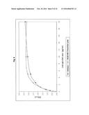 |
 |  |
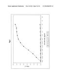 | 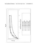 |
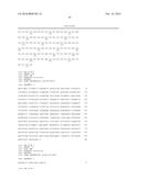 |  |
 | 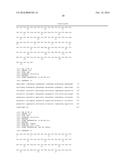 |
 |  |
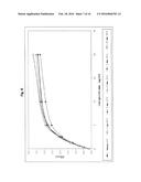 |  |
 |  |
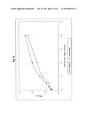 | 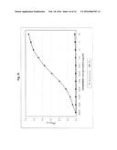 |
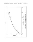 |  |
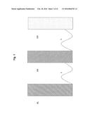 |  |
 | 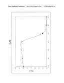 |
 |  |
 |  |
 | 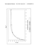 |
| Similar patent applications: | |
| Date | Title |
|---|---|
| 2016-04-28 | Enzymatic conjugation of polypeptides |
| 2016-05-12 | Docetaxel nano-polymer micelle lyophilized preparation and preparation method thereof |
| 2012-04-19 | Nitric oxide treatments |
| 2016-01-14 | Novel stip1 polypeptides and uses thereof |
| 2016-01-14 | Novel poly(adp-ribose) polymerase genes |
| New patent applications in this class: | |
| Date | Title |
|---|---|
| 2022-05-05 | Methods of using pdx1-positive pancreatic endoderm cells and endocrine precursor cells |
| 2022-05-05 | Amniotic fluid-derived extracellular vesicles and uses thereof for wound healing |
| 2022-05-05 | Pharmaceutical composition for treating amyotrophic lateral sclerosis |
| 2022-05-05 | Combination immune therapy and cytokine control therapy for cancer treatment |
| 2019-05-16 | Methods for identifying and modulating immune phenotypes |
| New patent applications from these inventors: | |
| Date | Title |
|---|---|
| 2016-02-18 | Method and reagent for determining vitamin d metabolites |
| 2012-11-01 | Monospecific polypeptide reagents |
| Top Inventors for class "Drug, bio-affecting and body treating compositions" | |
| Rank | Inventor's name |
|---|---|
| 1 | David M. Goldenberg |
| 2 | Hy Si Bui |
| 3 | Lowell L. Wood, Jr. |
| 4 | Roderick A. Hyde |
| 5 | Yat Sun Or |