Patent application title: PHARMACEUTICAL COMPOSITIONS COMPRISING INHIBITORS OF ZINC-ZIP8-MTF1 AS ACTIVE INGREDIENTS FOR PREVENTING OR TREATING A JOINT DISEASE
Inventors:
Jang-Soo Chun (Gwangju, KR)
Jin-Hong Kim (Gwangju, KR)
IPC8 Class: AG01N33564FI
USPC Class:
4241301
Class name: Drug, bio-affecting and body treating compositions immunoglobulin, antiserum, antibody, or antibody fragment, except conjugate or complex of the same with nonimmunoglobulin material
Publication date: 2015-11-12
Patent application number: 20150323528
Abstract:
The present invention relates to identification of Zinc-ZIP8-MTF1 axis
that plays an important role to OA (osteoarthritis) pathogenesis process
and a novel use thereof. According to the present invention, ZIP8 and
MTF1 of the present invention increase in the expression in joint disease
induced cells or cartilage tissue, and induce the expression of various
matrix-degrading enzymes (e.g., MMP-3, MMP-9, MMP-12, MMP-13 and ADAMTS-5
etc.). In addition, when the expression of ZIP8 or MTF1 is inhibited in
cells or tissues of animals (e.g., human, mouse), OA pathogenesis is
inhibited. Therefore, the ZIP8 and MTF1 of the present invention may be
applied to the diagnosis or prognosis of joint diseases, and may be used
for the development of therapeutics for joint diseases using these.Claims:
1. A method for screening a therapeutic agent for treating a joint
disease, comprising: (a) contacting a test substance of interest for
analysis to cells comprising (i) a ZIP8 protein or a MTF1
(metal-regulatory transcription factor-1) protein, or (ii) a nucleotide
sequence encoding the ZIP8 protein or the MTF1 protein; and (b) analyzing
the expression level of the ZIP8 gene, the MTF1 gene, the ZIP8 protein or
the MTF1 protein, or the activity of the ZIP8 protein or the MTF1
protein, wherein where the test substance inhibits the expression level
of the ZIP8 gene, the MTF1 gene, the ZIP8 protein or the MTF1 protein, or
the activity of the ZIP8 protein or the MTF1 protein, it is determined as
the therapeutic agent for treating joint disease.
2. A method for preventing or treating a joint disease, comprising administering to an subject in need thereof an inhibitor of the expression of the ZIP8 gene or the activity of the ZIP8 protein, or an inhibitor of the expression of the metal-regulatory transcription factor-1 (MTF1) gene or the activity of the MTF1 protein.
3. The method according to claim 2, wherein the joint disease is osteoarthritis, degenerative joint disease, osteochondritis dissecans, ligament injuries, meniscus injuries, malalignment of joint, osteonecrosis, arthroses, isolated chondral defect, chondromalacia patellae, tenosynovitis, bursitis, traumatic effusion, ligamentous deficiency arthroses, osteochondritis dissecans, patellar instability, rheumatoid arthritis, juvenile idiopathic arthritis, juvenile arthritis, trauma, inflammatory arthritis, septic arthritis caused by infection, lupus, scleroderma, tenosis, fibromylgia, fibromyositis or polymyositis.
4. The method according to claim 2, wherein the measurement of the expression level is performed by RT-PCR (reverse transcription-polymerase chain reaction) or an immunoassay.
5. The method according to claim 2, wherein the inhibitor as an active ingredient decreases the expression of matrix-degrading enzyme in mRNA level or protein level.
6. The method according to claim 5, wherein the cells is derived from a joint of an animal.
7. The method according to claim 5, wherein the joint disease is osteoarthritis, degenerative joint disease, osteochondritis dissecans, ligament injuries, meniscus injuries, malalignment of joint, osteonecrosis, arthroses, isolated chondral defect, chondromalacia patellae, tenosynovitis, bursitis, traumatic effusion, ligamentous deficiency arthroses, osteochondritis dissecans (OCD), patellar instability, rheumatoid arthritis, juvenile idiopathic arthritis, juvenile arthritis, trauma, inflammatory arthritis, septic arthritis caused by infection, lupus, scleroderma, tenosis, fibromylgia, fibromyositis or polymyositis.
8. The method according to claim 5, wherein the matrix-degrading enzyme is MMP (matrix metalloproteinase)-3, MMP-9, MMP-12, MMP-13 or ADAMTS (a disintegrin and metalloproteinase with thrombospondin motifs)-5.
9. A method for detecting a joint disease in a subject, comprising: (a) providing a biological sample from the subject; and (b) measuring the expression level of the ZIP8 gene, the MTF1 gene, the ZIP8 protein or the MTF1 protein, relative to the expression level of the ZIP8 gene, the MTF1 gene, the ZIP8 protein or the MTF1 protein in a control sample from a normal subject, wherein an increased level of the ZIP8 gene, the MTF1 gene, the ZIP8 protein or the MTF1 protein in the biological sample compared to the control sample indicates that the subject has the joint disease.
10. The method according to claim 9, wherein the biological sample is a chondrocyte or a cartilage tissue.
Description:
CROSS-REFERENCE TO RELATED APPLICATIONS
[0001] This application claims priority from Korean Patent Application No. 2014-0039540, filed on Apr. 2, 2014, in the Korean Intellectual Property Office, the disclosure of which is incorporated herein by reference.
FIELD OF THE INVENTION
[0002] The present invention relates to a novel use of ZIP8 or MTF1 as OA (osteoarthritis) pathogenesis factors.
DESCRIPTION OF THE RELATED ART
[0003] Osteoarthritis (OA) is the most common of all arthropathies and is a leading cause of disability with a large socioeconomic cost. To date, however, no effective disease-modifying therapies for OA have been developed. OA is primarily characterized by cartilage destruction, but also involves other pathological changes, including synovial inflammation, osteophyte formation, and subchondral bone sclerosis, in all tissues of joints (Bian et al., 2012; Loeser et al., 2012; Little and Hunter, 2013). OA pathogenesis is caused by an imbalance between anabolic and catabolic factors. A variety of etiologic risk factors and pathophysiological processes contribute to the progressive nature of the disease. Important among potential OA-causing mechanisms are mechanical stresses, including joint instability and injury, and factors that predispose toward OA, such as aging. These factors lead to the activation of biochemical pathways in chondrocytes that result in degradation of the extracellular matrix (ECM) by matrix metalloproteinases (MMPs) and aggrecanases (ADAMTSs). Among matrix-degrading enzymes, MMP3, MMP13, and ADAMTS5 are known to play crucial roles in OA cartilage destruction (Blom et al., 2007; Glasson et al., 2005; Little et al., 2009).
[0004] Matrix-degrading enzymes require zinc (Zn2+) as a structural component (Page-McCaw et al. 2007). Indeed, Zn2+ acts as an activator or co-activator of a variety of proteins by providing a structural scaffold, for example in the form of zinc fingers and zinc clusters (Prasad, 1995). Zn2+ may also be overtly toxic when accumulated in excess in cells. Therefore, normal cell functioning requires tight regulation of Zn2+ homeostasis. Zn2+ homeostasis is primarily regulated by membrane Zn2+ transporters of the Slc30a family (ZNT) of exporters and Slc39a family (ZIP) of importers (Cousins et al., 2006). The ZNT family, consisting of 10 members in mammals (ZNT1-ZNT10), mediates Zn2+ efflux from cells or influx into intracellular vesicles from the cytosol. There are 14 members of the ZIP family of Zn2+ importers in mammals (ZIP1-ZIP14) that promote Zn2+ influx from the extracellular fluid or intracellular vesicles into the cytoplasm. Zn2+ transporters exhibit tissue specific functions (Liuzzi et al., 2005; Kitamura et al., 2006), and abnormalities in the function of certain Zn2+ transporters are associated with human diseases, such as acrodermatitis enteropathica (Kury et al., 2002). Zn2+ influx modulates a number of transcription factors in various cell types. Among them, metal-regulatory transcription factor-1 (MTF1) regulates expression of a variety of target genes and thereby regulates cellular adaptation to various stress conditions, primarily exposure to heavy metals, but also hypoxia and oxidative stress (Laity and Andrews, 2007; Gunther et al., 2012). Zn2+ homeostasis is additionally regulated by metal-dependent transcriptional control of storage proteins. For instance, metallothioneins (MTs), whose genes are well-known targets of MTF1, act as Zn2+-storage proteins and thereby regulate cellular Zn2+ homeostasis. MTs also act as antioxidants and protect cells from oxidative stress (Laity and Andrews, 2007; Colvin et al., 2010; Gunther et al., 2012).
[0005] There has been a growing interest in the potential role of Zn2+ in the pathogenesis of OA. Clinical studies indicate highly elevated serum Zn2+ levels in OA patients (Ovesen et al., 2009) and specific accumulation of Zn2+ in the tidemark region of articular cartilage in aged populations (Roschger et al., 2013). The association of Zn2+ with OA pathogenesis is broadly appreciated in the context of its role as a structural component of matrix-degrading enzymes required for the maturation and activation of these enzymes. However, how Zn2+ homeostasis is regulated during the onset and progression of OA, and how it contributes to the pathological transition of articular chondrocytes remain unknown. Here, we investigated the roles of Zn2+ homeostasis, homeostasis-regulating Zn2+ transporters, and downstream transcription factors and their target genes in OA pathogenesis. We report here that the zinc-ZIPS-MTF1 axis regulates OA pathogenesis.
[0006] Throughout this application, several patents and publications are referenced and citations are provided in parentheses. The disclosure of these patents and publications is incorporated into this application in order to more fully describe this invention and the state of the art to which this invention pertains.
DETAILED DESCRIPTION OF THIS INVENTION
Technical Purposes of this Invention
[0007] The present inventors have made intensive studies to develop pathogenesis molecular factor and therapeutic target of osteoarthritis (OA), which is a representative example of joint disease, particularly degenerative arthritis. As a result, they found out that ZIP 8 playing critical role for homeostasis of cellular Zn2+ and MTF1 related to the ZIP 8 are closely relationship with development of OA, and joint disease may be prevented or treated through the inhibitory thereof in animal (e.g., human, mouse) cells or tissues.
[0008] Accordingly, it is an object of the present invention to provide a pharmaceutical composition for preventing or treating a joint disease.
[0009] It is another object of the present invention to provide a method for a method for preventing or treating a joint disease.
[0010] It is still another object of the present invention to provide a method for screening a therapeutic agent for treating a joint disease.
[0011] It is another object of the present invention to provide a method for detecting a joint disease in a subject.
[0012] It is still another object of the present invention to provide a non-human transgenic animal model for a joint disease.
[0013] Other objects and advantages of the present invention will become apparent from the detailed description to follow taken in conjugation with the appended claims and drawings.
Technical Solutions of this Invention
[0014] In one aspect of the present invention, there is provided a pharmaceutical composition for preventing or treating joint disease, comprising an inhibitor of the expression of a ZIP8 or MTF1 (metal-regulatory transcription factor-1) gene or protein, or an inhibitor of the activity of the ZIP8 or MTF1 protein as an active ingredient.
[0015] In another aspect of the present invention, there is provided a method for preventing or treating a joint disease, comprising administering to an subject in need thereof an inhibitor of the expression of the ZIP8 gene or the activity of the ZIP8 protein, or an inhibitor of the expression of the metal-regulatory transcription factor-1 (MTF1) gene or the activity of the MTF1 protein.
[0016] The present inventors have made intensive studies to develop pathogenesis molecular factor and therapeutic target of osteoarthritis (OA), which is a representative example of joint disease, particularly degenerative arthritis. As a result, they found out that ZIP 8 playing critical role for homeostasis of cellular Zn2+ and MTF1 related to the ZIP 8 are closely relationship with development of OA, and joint disease may be prevented or treated through the inhibitory thereof in animal (e.g., human, mouse) cells or tissues.
[0017] ZIP is one of membrane transporters that mediate metal ions, such as Zn2+ an influx from extracellular into cytosol as members of Slc39a (Solute-linked carrier 39a) family. There are 14 members of the ZIP family of Zn2+ importers in mammals. Zn2+ transporters exhibit tissue-specific functions. Abnormalities in the function of certain Zn2+ transporters are associated with human diseases. ZIP8 as a member of this ZIP family was observed that ZIP8 protein and mRNA levels were markedly elevated in OA cartilage of human and mouse models (see: FIG. 1f).
[0018] MTF1 (metal-regulatory transcription factor-1) is well-known that induces expression of metallothioneins and other genes associated with regulating metal homeostasis in response to heavy metals, such as calcium, zinc, copper or silver, etc., as a transcription factor. When cells were exposed to heavy metals, MTF1 is accumulated in nuclear and acts to bind to promoter comprising a metal-responsive element (MRE).
[0019] The present invention primarily identifies that a zinc-ZIP8-MTF1 axis is an important regulation factor consisting of Zn2+ influx, Zn2+ influx factor ZIP8 and Zn2+ dependent transcription factor MTF1, and elucidates that the joint diseases may be prevented and treated using it.
[0020] According to particular embodiment, the ZIP8 and MTF1 increases the expression of matrix-degrading enzymes in mRNA level or protein level, more specifically, increases mRNA level or protein level of MMP(matrix metalloproteinase)-3, MMP-9, MMP-12, MMP-13 and ADAMTS-5 (see: FIGS. 2a and f, FIGS. 5i and j).
[0021] According to particular embodiment, the inhibitor as an active ingredient decreases the expression of matrix-degrading enzyme in mRNA level or protein level, more particularly, the matrix-degrading enzyme is MMP (matrix metalloproteinase)-3, MMP-9, MMP-12, MMP-13 or ADAMTS (a disintegrin and metalloproteinase with thrombospondin motifs)-5.
[0022] Among the matrix-degrading enzymes, MMP-3, MMP-13 and ADAMTS-5 are known as crucial effectors of OA cartilage destruction, which exert a function by virtue of its role in degrading the extracellular matrix (ECM) of chondrocytes.
[0023] MMPs (matrix metalloproteinases) are a Zn2+-dependent endopeptidase. These may degrade all kinds of ECM, play a critical role in cell proliferation, differentiation, migration, angiogenesis and apoptosis.
[0024] MMP-3 functions degrading collagen type II, III, IV, IX and X, proteoglycans, fibronectin, laminin and elastin, and also activates other MMP, such as MMP-1, MMP-7 and MMP-9. Therefore, MMP-3 is considered as most main factor in reconstitution of connective tissues.
[0025] MMP-9 exerts a function by virtue of its role in degrading other ECM different from collagen type IV and V, and MMP-12 in degrading elastin.
[0026] MMP-13 as collagen-degrading enzyme known as collagenase-3 in human, highly expressed in skeleton mainly, due to requirement for reconstitution of collagen matrix in fetal development stage. It was observed that the MMP-13 is highly expressed in pathological condition, in which occur carcinoma, rheumatoid arthritis and osteoarthritis.
[0027] ADAMTS-5 has two C-terminal TS motifs as a member of ADAMTS (a disintegrin and metalloproteinase with thrombospondin motifs) protein family and acts as an aggrecanase degrading an aggrecan which is main proteoglycan in cartilage.
[0028] According to particular embodiment, the composition of the present invention may include siRNA (small interference RNA), shRNA (short hairpin RNA), miRNA (microRNA), ribozyme, DNAzyme, PNA (peptide nucleic acids), antisense oligonucleotides, peptides, antibodies, aptamers, extracts of natural sources and chemical substances. More preferably, the composition of the present invention may include a nucleotide sequence coding ZIP8 or MTF1 protein, a sequence complementary to the nucleotide sequence, or siRNA, shRNA, miRNA, ribozyme, DNAzyme or antisense oligonucleotides for a fragment of the nucleotide sequence as an active ingredient.
[0029] ZIP8-encoding nucleotide sequence and MTF1-encoding nucleotide sequence used in the present invention are illustrated in SEQ ID NO:1 (GenBank Accession NO. NM--001135149) and SEQ ID NO:3 (GenBank Accession NO. NM--008636), respectively, and the sequences of amino acid of proteins expressed from each nucleotide sequence are set forth in SEQ ID NO:2 (GenBank Accession NO. NP--001128622) and SEQ ID NO:4 (GenBank Accession NO. NP--032662).
[0030] The pharmaceutical composition of the present invention comprises siRNA having sequences complementary to the nucleotide sequences as set forth in SEQ ID NO:1 (ZIP8) and SEQ ID NO:3 (MTF1) as an active ingredient.
[0031] The term used herein "siRNA" refers to a nucleic acid molecule that enables to mediate RNA interference or gene silencing (see: WO 00/44895, WO 01/36646, WO 99/32619, WO 01/29058, WO 99/07409 and WO 00/44914). The siRNA to inhibit expression of a target gene provides effective gene knock-down method or gene therapy method. The siRNA was first discovered in plant, insect, fruit fly and parasite, but recently the siRNA was applied to the study of mammalian cells (Degot S, et al. 2002; Degot S, et al. 2004; Ballut L, et al. 2005).
[0032] The siRNA molecule of the present invention may has double strand structure that the sense strand (sequence corresponding to ZIP8 or MTF1 mRNA sequence) and the antisense strand (sequence corresponding to ZIP8 or MTF1 mRNA sequence) are located on the opposite side each other to form. In addition, according to another embodiment, the siRNA molecule of the present invention may have single strand structure in which has self-complementary sense strand and antisense strand.
[0033] The siRNA of this invention is not restricted to a RNA duplex of which two strands are completely paired and may comprise non-paired portion such as mismatched portion with non-complementary bases and bulge with no opposite bases. The overall length of the siRNA is 10-100 nucleotides, preferably 15-80 nucleotides, more preferably, 20-70 nucleotides and most preferably 20-30 nucleotides.
[0034] The siRNA may comprise either blunt or cohesive end so long as it enables to inhibit the ZIP8 or MTF1 gene expression via RNAi effect. The cohesive end may be prepared in 3'-end overhanging structure or 5'-end overhanging structure.
[0035] The siRNA molecule of the present invention may include the form that short nucleotide sequences (approximately 5-15 nt) is inserted between self-complementary sense and antisense strands. In this case, the siRNA molecule formed by the expression of the nucleotide sequence is formed hairpin structure by intramolecular hybridization, and overall stem-and-loop structure. The stem-and-loop structure is processed in vivo or in vitro to produce siRNA molecule of activity which may mediate RNAi.
[0036] The inhibition of ZIP8 or MTF1 protein in this invention, in particular the inhibitor used in inhibiting activity of the protein is preferably, an antibody or peptide binding specifically to ZIP8 or MTF1, chemicals or extracts of natural sources having small molecular weights.
[0037] The antibody inhibiting activity by binding specifically to ZIP8 or MTF1 protein used in the present invention is polyclonal or monoclonal antibody, preferably monoclonal antibody. Antibody against ZIP8 or MTF1 proteins may be prepared by a method widely known in the art, such as a hybridoma method (Kohler and Milstein, European Journal of Immunology, 6:511-519(1976)), a recombinant DNA methods (U.S. Pat. No. 4,816,56) or a phage antibody library technique (Clackson et al, Nature, 352:624-628(1991) and Marks et al, J. Mol. Biol., 222:58, 1-597(1991)). General process for antibody production is described in Harlow, E. and Lane, D., Using Antibodies: A Laboratory Manual, Cold Spring Harbor Press, New York, 1999; Zola, H., Monoclonal Antibodies: A Manual of Techniques, CRC Press, Inc., Boca Raton, Fla., 1984; Coligan, CURRENT PROTOCOLS IN IMMUNOLOGY, Wiley/Greene, NY, 1991, and the literatures are inserted as reference in the present invention. For example, the preparation of the hybridoma cells producing monoclonal antibody is accomplished by fusing immortalized cell line with antibody-producing lymphocytes. The technology required for this process is widely known in the art and may be easily carried out using techniques. Polyclonal antibodies may be produced by injecting the ZIP8 or MTF1 protein antigen into an appropriate animal and collecting blood samples from the animal to obtain sera containing antibodies using affinity technology known in the art.
[0038] Peptide that enables to inhibit activity of ZIP8 or MTF1 by specific binding to ZIP8 or MTF1 may be obtained by a method widely known in the art, such as a display method (Smith G P, "Filamentous fusion phage: novel expression vectors that display cloned antigens on the virion surface". Science 228 (4705):13151317(1985); Smith G P, Petrenko V A, "Phage display". Chem. Rev. 97(2):391410(1997)).
[0039] Chemicals of small molecular weights inhibiting activity of ZIP8 or MTF1 may be obtained easily through screening methods described hereinafter.
[0040] The pharmaceutical composition of the present disclosure may comprise a pharmaceutically acceptable carrier. The pharmaceutically acceptable carrier included in the pharmaceutical composition of the present disclosure is one commonly used in the preparation of formulations and includes lactose, dextrose, sucrose, sorbitol, mannitol, starch, gum acacia, calcium phosphate, alginate, gelatin, calcium silicate, microcrystalline cellulose, polyvinyl pyrrolidone, cellulose, water, syrup, methyl cellulose, methyl hydroxybenzoate, propyl hydroxybenzoate, talc, magnesium stearate, mineral oil, etc., but is not limited thereto. The pharmaceutical composition of the present disclosure may further include, in addition to the above-described components, a lubricant, a wetting agent, a sweetener, a fragrance, an emulsifier, a suspending agent, a preservative, or the like. Suitable pharmaceutically acceptable carriers and formulations are described in detail in Remington's Pharmaceutical Sciences (19th ed., 1995).
[0041] The composition of the present invention may be administered orally or parenterally. Preferably, it may be administered parenterally. When the composition of the present disclosure is administered parenterally, the pharmaceutical composition of the present disclosure may administer with intravenous injection, subcutaneous injection, local injection, intramuscular injection and intraosseous injection. More preferably, the pharmaceutical composition of the present disclosure may administer with cartilage injection.
[0042] An appropriate administration dosage of the pharmaceutical composition of the present disclosure may be determined variously depending on such factors as preparation method, administration method, age, body weight and gender of a patient, pathological condition, diet, administration time, administration route, excretion rate or response sensitivity. Specifically, a daily dosage of the pharmaceutical composition of the present disclosure may be 0.0001-100 mg/kg (weight).
[0043] The pharmaceutical composition of the present disclosure may be prepared into a unit dosage form or multiple dosage form along with a pharmaceutically acceptable carrier and/or excipient according to a method that can be easily employed by those skilled in the art. The formulation may be in the form of solution in oily or aqueous medium, suspension, syrup, emulsion, extract, dust, powder, granule, tablet or capsule, and may further include a dispersant or stabilizer.
[0044] A joint is the location at which bones connect. There are typically three classifications of joints: fibrous joint, cartilagenous joint and synoval joint. As joint disease, any of the diseases or injuries that affect mammal (e.g., human) joints, arthritis is no doubt the best-known joint disease, but there are also many others. Diseases of the joints may be variously short-lived or exceedingly chronic, agonizingly painful or merely nagging and uncomfortable; they may be confined to one joint or may affect many parts of the skeleton.
[0045] The term used herein "joint disease" refers to progressive deterioration or destruction of cartilage tissue surrounding joint. Arthritis is a form of joint disorder that involves inflammation of one or more joints. Particularly, osteoarthritis (OA) is the oldest and most common disease among arthritis, non-inflammatory disease showing chronic conditions characterized by destruction of joints cartilage, and also has known as degenerative joint disease, ostoarthrosis, hypertrophic arthritis or degenerative arthritis. Thus osteoarthritis referring herein may be used each other changing with other name of osteoarthritis the above-described.
[0046] Degenerative joint disease is a non-infectious progressive disorder of the weight bearing joints. The normal articular joint cartilage is smooth, white and translucent. It is composed of cartilage cells (chondrocyte) imbedded in a sponge-like middle, or matrix, made of collagen, protein polysaccharides and water. With early, primary degenerative arthritis, the cartilage becomes yellow and opaque with localized areas of softening and roughening of the surfaces. As the degeneration progresses, the soft areas become cracked and worn exposing bone under the cartilage, which begins to remodel and increase in density while any remaining cartilage begins to fray. Eventually, osteophytes (spurs of new bone), covered by cartilage, form at the edge of the joint. Also, as mechanical wear increases, the cartilage needs repairing. The cartilage cells are unable to produce enough of the sponge-like matrix and therefore the damaged cartilage cannot repair itself. In fact, it has no blood supply to enhance healing. The majority of degenerative joint disease is the result of mechanical instabilities or aging changes within the joint. This includes old age degenerative arthritis and, in youngers, may be the result of injuries, bruises, abnormal joint configuration, (i.e. hip dysplasia), or mechanical wear from anterior cruciate ligament rupture, patellar luxation, or osteochondritis dissecans. Degenerative joint disease may be occurred in any joint of body including knees, hips, shoulders, hands and a spine.
[0047] Cartilage is the part of the joint that cushions the ends of the bones and allows easy movement of joints. The breakdown of cartilage causes the bones to rub against each other, causing stiffness, pain and loss of movement in the joint. The frequency of arthritis is a high disease to be estimated that among the Koreans adults, nearly 2 million have clinical arthritis, among US adults, nearly 27-35 million (Helmick, C., et al., Estimates of the Prevalence of Arthritis and Other Rheumatic conditions in the United States. Arthritis & Rheumatism, 58(1): 15-25(2008)). Unfortunately, because the cause of arthritis is not yet known, there is also no therapy. In fact, because arthritis is caused by certain factors (e.g., aging, overweight, injury, repeated overuse of certain joints, heredity, etc.), the therapy is various according to condition of developing arthritis.
[0048] Osteoarthritis is divided into various stages as follows: (a) Cartilage loses elasticity and is more easily damaged by injury or use; (b) Wear of cartilage causes changes to underlying bone. The bone thickens and cysts may occur under the cartilage. Bony growths, called spurs or osteophytes, develop near the end of the bone at the affected joint. A pruritus and pain is caused; (c) Bits of bone or cartilage float loosely in the joint space; and (d) The joint lining, or the synovium, becomes inflamed due to cartilage breakdown causing cytokines and enzymes that damage cartilage further.
[0049] In another aspect of the present invention, there is provided a method for screening a therapeutic agent for treating a joint disease, comprising: (a) contacting a test substance of interest for analysis to cells comprising (i) a ZIP8 protein or a MTF1 (metal-regulatory transcription factor-1) protein, or (ii) a nucleotide sequence encoding the ZIP8 protein or the MTF1 protein; and (b) analyzing the expression level of the ZIP8 gene, the MTF1 gene, the ZIP8 protein or the MTF1 protein, or the activity of the ZIP8 protein or the MTF1 protein, wherein where the test substance inhibits the expression level of the ZIP8 gene, the MTF1 gene, the ZIP8 protein or the MTF1 protein, or the activity of the ZIP8 protein or the MTF1 protein, it is determined as the therapeutic agent for treating joint disease.
[0050] According to the method of the present invention, first the method comprise contacting a test substance of interest for analysis to cells comprising (i) a ZIP8 protein or a MTF1 (metal-regulatory transcription factor-1) protein, or (ii) a nucleotide sequence encoding the ZIP8 protein or the MTF1 protein.
[0051] The term used herein "treatment" refers to an administration process which is possible to analyze efficacy of test substance inducing contact the test substance with cells or tissues by administrating the test substance in cells or tissues, the term enables to be used to being compatible with "administration" or "contact".
[0052] Cells including nucleotide sequences of this invention are not limited specially, specifically comprise mammal cells, more specifically joint-derived cells, but are not limited thereto. According to particular embodiment, the cells that enable to be used in the present invention are joint-derived cells, more preferably articulating joints-derived cells. According to particular embodiment, the articulating joint tissue that enables to be used in the present invention includes wrists, elbows, shoulders, ankles, knees, hips, spine, temporomandibular and Carpometacarpal joints, but not limited to. More preferably, joint tissue is derived from femoral heads, femoral condyles, tibial plateaus, acetabulofemoral joint, acromioclavicular joint, femoropatellar joint, femorotibial joint, glenohumeral joint, humeroradial joint, humeroulnar joint, interphalangeal joint, metacarpal joint, radioulnar joint and talocrural joint, but is not limited to.
[0053] The method of the present invention further comprises (pre-a) causing joint diseases to the cells. For example, analysis of stage described below is enabled to perform more clearly by causing joint diseases condition to cells through mechanical stress like DMM surgery or injection of pro-inflammatory cytokine like IL-1 or virus. According to certain embodiment, the adenovirus of the present invention carries out through intraarticular (IA) injection or intraperitoneal (IP) injection.
[0054] The term "test substance" used herein in conjunction with the present screening method refers to a material tested in the present method for analyzing the influence on the activity of ZIP8 or MTF1 protein. The test substance includes chemical substances, siRNA (small interference RNA), shRNA (small hairpin RNA or short hairpin RNA), miRNA (microRNA), ribozyme, DNAzyme, PNA (peptide nucleic acids), antisense oligonucleotides, peptides, antibodies, aptamers and extracts of natural sources, but not limited to. The test material analyzed by the screening method of the present invention is a single compound or a mixture of compounds (e.g., a natural extract or a cell or tissue culture). The test material may be obtained from synthetic or natural compound libraries. These compound libraries are obtained by methods known in the art. The synthetic compound libraries are commercially available from Maybridge Chemical Co. (UK), Comgenex (USA), Brandon Associates (USA), Microsource (USA) and Sigma-Aldrich (USA), and the natural compound libraries are commercially available from Pan Laboratories (USA) and MycoSearch (USA). The test material may be obtained from various combinational library methods known in the art, for example, from a biological library method, a spatially addressable parallel solid phase or solution phase library method, a synthetic library method requiring deconvolution, a "one-bead one-compound" library method, and a synthetic library method using affinity chromatography selection. The synthetic methods of molecular libraries are disclosed in DeWitt et al., Proc. Natl. Acad. Sci. U.S.A. 90: 6909 (1993); Erb et al. Proc. Natl. Acad. Sci. U.S.A. 91: 11422 (1994); Zuckermann et al., J. Med. Chem. 37: 2678 (1994); Cho et al., Science 261: 1303 (1993); Carell et al., Angew. Chem. Int. Ed. Engl. 33: 2059 (1994); Carell et al., Angew. Chem. Int. Ed. Engl. 33: 2061; and Gallop et al., J. Med. Chem. 37: 1233 (1994).
[0055] Then, the measurement of the expression level of ZIP8 or MTF1 gene or protein, or activity of their proteins is conducted in cells treated the test substance. As a result, where the test substance down-regulates the expression of their gene or protein, or the activity of their proteins, it is determined as the therapeutic agent for preventing or treating joint disease.
[0056] According to particular embodiment, the substance for preventing or treating of joint disease discovered by the above described screening method may be used in treatment or prevention of ostarthritis, degenerative arthritis, osteochondritis dissecans, arthroses or arthritis after articular crescent meniscus injury, malalignment of joint, avascular necrosis, arthroses, isolated chondral defect, chondromalacia patellae, synovitis, bursitis, traumatic effusion, ligamentous deficiency arthroses, osteochondritis dissecans (OCD), patellar instability, rheumatoid arthritis, juvenile idiopathic arthritis, juvenile arthritis, post-traumatic arthritis, inflammatory arthritis, septic arthritis, lupus, scleroderma, tendinitis, fibrositis, fibromyositis or polymyositis, but not limited thereto.
[0057] In the case of performing the screening method of this invention by analyzing expression of ZIP or MTF1, the measurement of change in the expression level of ZIP8 or MTF1 afore-mentioned may be carried out through a variety of methods known in the art, for example, RT-PCR (Sambrook et al, Molecular Cloning. A Laboratory Manual, 3rd ed. Cold Spring Harbor Press (2001)), Northern blot (Peter B. Kaufma et al., Molecular and Cellular Methods in Biology and Medicine, 102-108, CRC press) or hybridization using cDNA microarray (Sambrook et al, Molecular Cloning. A Laboratory Manual, 3rd ed. Cold Spring Harbor Press (2001)).
[0058] According to RT-PCR protocol, total RNA is extracted from the test substance-treated cells, and first strand cDNA is prepared using oligo dT primer and reverse transcriptase. Then, PCR reaction is carried out using first strand cDNA as a template and a gene encoding the ZIP8 or MTF1 protein-specific primer set. The resulting products are separated by electrophoresis and the band patterns are analyzed to measure the expression level of the gene of ZIP8 or MTF1 protein.
[0059] The change of level of ZIP8 or MTF1 protein may be performed by a variety of quantitative or qualitative immunoassay protocols. The immunoassay format includes, but is not limited to, immunohistochemical staining, radioimmunoassay analysis, radioactive immunoprecipitation, western blotting, immunoprecipitation, ELISA (enzyme-linked immunosorbant assay), Capture-ELISA, sandwich assay, flow cytometry, immunofluorescence and immune affinity purified.
[0060] The immunoassay or the immuno staining method is described in Enzyme Immunoassay, E. T. Maggio, ed., CRC Press, Boca Raton, Fla., 1980; Gaastra, W., Enzyme-linked immunosorbent assay (ELISA), in Methods in Molecular Biology, Vol. 1, Walker, J. M. ed., Humana Press, NJ, 1984; and Ed Harlow and David Lane, Using Antibodies: A Laboratory Manual, Cold Spring Harbor Laboratory Press, 1999 and the literatures are inserted as reference in herein.
[0061] When the method of the present disclosure is performed using the ELISA, certain examples of the present invention comprise the steps of: (i) coating unknown cell cytolysate samples of interest for analysis on the surface of a solid substrate; (ii) contacting the cell cytolysate with antibody for the marker as the primary antibody; (iii) contacting the resultant of step (ii) with the secondary antibody conjugated enzyme; and (iv) detecting the enzyme activity.
[0062] The appropriate solid substrate is hydrocarbon polymers (e.g., polystyrene and polypropylene), glass, metal, or gel, and most preferably a micro-titer plate.
[0063] The appropriate secondary antibody conjugated enzyme includes, but is not limited to, color-developing reaction, fluorescent reaction, luminescent reaction or infrared reaction, for example, alkaline phosphatase, β-galactosidase, horseradish peroxidase, luciferase and cytochrome P450. Where alkaline phosphatase is used for the enzyme binding to the secondary antibody, bromochloroindolylphosphate (BCIP), nitro blue tetrazolium (NBT), naphthol-AS-B1-phosphate and ECF (enhanced chemifluorescence) may be used as a substrate for color-developing reactions. In the case of using horseradish peroxidase, chloronaphtol, aminoethylcarbazol, diaminobenzidine, D-luciferin, lucigenin (bis-N-methylacridinium nitrate), resorufin benzyl ether, luminol, Amplex Red reagent (10-acetyl-3,7-dihydroxyphenoxazine), HYR (p-phenylenediamine-HCl and pyrocatechol), TMB (3,3,5,5-tetramethylbenzidine), ABTS (2,2-Azine-di[3-ethylbenzthiazoline sulfonate]), o-phenylenediamine (OPD) and naphtol/pyronine may be used as a substrate; and in the case of using glucose oxidase, t-NBT (nitroblue tetrazolium) or m-PMS (phenzaine methosulfate) may be used as a substrate.
[0064] When the method of the present disclosure is performed using the Capture-ELISA, certain examples of the present invention comprise the steps of: (i) coating the antibody for the target (e.g., ZIP8 or MTF1 protein) of the present invention as capturing antibody on the surface of a solid substrate; (ii) contacting the cell sample with the capturing antibody; (iii) contacting the resultant of step (ii) with the detecting antibody which is combined with label generating signal and react specifically to the ZIP8 or MTF1 protein; and (iv) detecting the signal from the label. The detecting antibody has the label generating detectable a signal. The label includes, but is not limited to, chemical (e.g., biotin), enzyme (alkaline phosphatase, β-galactosidase, horseradish peroxidase and cytochrome P450), radioactive material (e.g., C14, I125, P32 and S35), fluorescent material (e.g., fluorescein), luminescent material, chemiluminescent material and FRET (fluorescence resonance energy transfer). A variety of labels and labeling methods are described in Ed. Harlow and David Lane, Using Antibodies: A Laboratory Manual, Cold Spring Harbor Laboratory Press, 1999.
[0065] The final measurement of enzyme activity or measurement of the signal in the ELISA and Capture-ELISA may be carried out in accordance with a variety of methods known in the art. The detection of this signal permits to qualitative or quantitative analysis of the target of the present invention. In the case of using biotin as label, the signal is easily detected using streptavidin. In the case of using luciferase as label, the signal is easily detected using luciferin.
[0066] When the screening method of the present invention is performed analyzing the activity of ZIP8 or MTF1, the activity of ZIP8 may be analyzed by measuring cellular Zn2+ level. According to a certain embodiment, when IL-1 is treated in chondrocyte, cellular Zn2+ level increases along with increasing of expression level of ZIP8 at the same time. However, when Zip-siRNA is treated in chondrocyte, increasing effect of Zn2+ level is blocked. Thus, if some test substance inhibits the activity of ZIP8 protein, it may be predicted to be unlikely to increase cellular Zn2+ level by being blocked cellular Zn2+ influx.
[0067] In addition, the activity of MTF1 protein may be analyzed by measuring a transcription activity of MTF1 using commercially available transcription factor activity analysis kit. According to a certain embodiment, when chondrocyte is infected with Ad-Zip8, it presents that transcription activity of MTF1 increase more than three fold.
[0068] In still another aspect of the present invention, there is provided a method for detecting a joint disease in a subject, comprising: (a) providing a biological sample from the subject; and (b) measuring the expression level of the ZIP8 gene, the MTF1 gene, the ZIP8 protein or the MTF1 protein, relative to the expression level of the ZIP8 gene, the MTF1 gene, the ZIP8 protein or the MTF1 protein in a control sample from a normal subject, wherein an increased level of the ZIP8 gene, the MTF1 gene, the ZIP8 protein or the MTF1 protein in the biological sample compared to the control sample indicates that the subject has the joint disease.
[0069] The molecular marker of this invention may be indicative of a joint disease, and also used in diagnosis of the joint disease development or progression, or prognosis.
[0070] The term used herein "Biosample or biological sample" is a human body or mammal-originated sample of material to be tested. The biosample refers to any cell or tissue from a cartilage (particularly, articular cartilage), urine, saliva, blood, plasma, or serum, but is not limited thereto. Preferably, ZIP8 or MTF1 of the present invention is comprised in a cartilage, particularly, articular cartilage. Accordingly, because ZIP8 or MTF1 of the present invention may be an indicator for pathogenesis or development of arthritis, ZIP8 or MTF1 can be used to diagnosis of pathogenesis or development of arthritis.
[0071] According to an embodiment, ZIP8 or MTF1 of the present invention may be used to prediction or diagnosis of osteoarthritis, degenerative joint disease, osteochondritis dissecans, ligamentinjuries, meniscus injuries, malalignment of joint, osteonecrosis, rheumatoid arthritis, juvenile idiopathicarthritis, trauma, inflammatory arthritis or septic arthritis by infection, more particulalry, osteoarthritis or degenerative joint disease, most preferably, very accurate prediction or diagnosis of osteoarthritis.
[0072] The term used herein "detecting a joint disease" includes the following matters: (a) to determine susceptibility of a subject to a particular disease or disorder; (b) to evaluate whether a subject has a particular disease or disorder; (c) to assess a prognosis of a subject suffering from a specific disease or disorder (e.g., identification of arthritis conditions, determination of arthritis stage, or investigation of arthritis response to treatment); or (d) therametrics (e.g., monitoring conditions of a subject to provide an information to treatment efficacy).
[0073] The term as used herein "prognosis" includes prediction in terms of the progression possibility process of the disease, in particular, the improvement of the disease, the regeneration of the disease and arthritis recurrence. Preferably, the prognosis of the present invention refers to completely cured possibility for the disease of arthritis patients.
[0074] Following preparation of biological samples, the ZIP8 gene, the MTF1 gene, the ZIP8 protein or the MTF1 protein in the biological sample is detected, relative to the expression level of the ZIP8 gene, the MTF1 gene, the ZIP8 protein or the MTF1 protein in a control sample from a normal subject, wherein an increased level of the ZIP8 gene, the MTF1 gene, the ZIP8 protein or the MTF1 protein in the biological sample compared to the control sample indicates that the subject has the joint disease.
[0075] A "control sample" refers to a sample of biological material representative of healthy, joint disease-free animals, and/or cells or tissues. The level of ZIP8 or MTF1 in a control sample is desirably typical of the general population of normal, joint disease-free animals or of a particular individual, or in a particular tissue. A control sample can also refer to an established level of ZIP8 or MTF1, representative of the joint disease-free population, that has been previously established based on measurements from normal, joint disease-free animals.
[0076] An "increased level of ZIP8 or MTF1" means a level of ZIP8 or MTF1 that, in comparison with a control level of ZIP8 or MTF1, is detectably higher. The method of comparison can be statistical, using quantified values for the level of ZIP8 or MTF1, or can be compared using non-statistical means, such as by visual assessment by a human.
[0077] The biomarkers of the present invention are biomolecules expressed highly in arthritis. The high expression of biomarkers may be measured at mRNA or protein level. The term "high expression" means that the nucleotide sequence of interest in a sample to be analyzed is much more highly expressed than that in the normal sample, for instance, a case analyzed as high expression according to analysis methods known to those skilled in the art, e.g., RT-PCR method or ELISA method (See, Sambrook, J., et al., Molecular Cloning. A Laboratory Manual, 3rd ed. Cold Spring Harbor Press (2001)). Using analysis methods as described above, where the biomarkers of the present invention are much more highly expressed at a range of 2-5 folds than in normal cells tissues, this case is determined as "high expression" and identified as development of arthritis in the present invention.
[0078] According to particular embodiment, the measurement of the expression level is performed by RT-PCR (reverse transcription-polymerase chain reaction) or an immunoassay.
[0079] Since the method of the present invention comprises the process of analyzing the expression levels of ZIP8 or MTF1 gene or protein described above, the common descriptions between the screening methods of the present invention described above are omitted in order to avoid undue redundancy leading to the complexity of this specification.
[0080] In the diagnosis or prognosis analysis using the present invention, when the expression of nucleotide sequence encoding ZIP8 or MTF1 protein is detected, probes or primers used in the present invention have a complementary sequence to the nucleotide sequence of ZIP8 or MTF1.
[0081] The nucleic acid sample to be analyzed may be prepared using mRNA from various biosamples. Preferably, the biosample is articular cartilage tissue cells.
[0082] The present kit for diagnosing joint disease may be used in accordance with hybridization. For such analysis, probes, which have a complementary sequence to the nucleotide sequence of the biomarkers of this invention as set forth, are used. Using probes hybridizable with the nucleotide sequence of the biomarkers of this invention, arthritis may be determined by hybridization-based assay.
[0083] Labels linking to the probes may generate a signal to detect hybridization and bound to oligonucleotide. Suitable labels include fluorophores (e.g., fluorescein, phycoerythrin, rhodamine, lissamine, Cy3 and Cy5 (Pharmacia)), chromophores, chemiluminescents, magnetic particles, radioisotopes (e.g., P32 and S35), mass labels, electron dense particles, enzymes (e.g., alkaline phosphatase or horseradish peroxidase), cofactors, substrates for enzymes, heavy metals (e.g., gold), and haptens having specific binding partners, e.g., an antibody, streptavidin, biotin, digoxigenin and chelating group, but not limited to. Labeling is performed according to various methods known in the art, such as nick translation, random priming (Multiprime DNA labeling systems booklet, "Amersham" (1989)) and kination (Maxam & Gilbert, Methods in Enzymology, 65: 499 (1986)). The labels generate signal detectable by fluorescence, radioactivity, measurement of color development, mass measurement, X-ray diffraction or absorption, magnetic force, enzymatic activity, mass analysis, binding affinity, high frequency hybridization or nanocrystal.
[0084] Probes are hybridized with cDNA molecules under stringent conditions. Suitable hybridization conditions may be routinely determined by optimization procedures. To establish a protocol for use of laboratory, these procedures may be carried out by various methods known to those ordinarily skilled in the art. Conditions such as temperature, concentration of components, hybridization and washing times, buffer components, and their pH and ionic strength may be varied depending on various factors, including the length and GC content of probes and target nucleotide sequence. The detailed conditions for hybridization can be found in Joseph Sambrook, et al., Molecular Cloning, A Laboratory Manual, Cold Spring Harbor Laboratory Press, Cold Spring Harbor, N.Y. (2001); and M. L. M. Anderson, Nucleic Acid Hybridization, Springer-Verlag New York Inc. N.Y. (1999). For example, the high stringent condition includes hybridization in 0.5 M NaHPO4, 7% SDS (sodium dodecyl sulfate) and 1 mM EDTA at 65° C. and washing in 0.1×SSC (standard saline citrate)/0.1% SDS at 68° C. Also, the high stringent condition includes washing in 6×SSC/0.05% sodium pyrophosphate at 48° C. The low stringent condition includes e.g., washing in 0.2×SSC/0.1% SDS at 42° C.
[0085] Following hybridization reactions, a hybridization signal indicative of the occurrence of hybridization is then measured. The hybridization signal may be analyzed by a variety of methods depending on labels. For example, where probes are labeled with enzymes, the occurrence of hybridization may be detected by reacting substrates for enzymes with hybridization resultants. The enzyme/substrate pair useful in this invention includes, but is not limited to, a pair of peroxidase (e.g., horseradish peroxidase) and chloronaphtol, aminoethylcarbazol, diaminobenzidine, D-luciferin, lucigenin (bis-N-methylacridinium nitrate), resorufin benzyl ether, luminol, Amplex Red reagent (10-acetyl-3,7-dihydroxyphenoxazine), HYR (p-phenylenediamine-HCl and pyrocatechol), TMB (3,3,5,5-tetramethylbenzidine), ABTS (2,2-Azine-di[3-ethylbenzthiazoline sulfonate]), o-phenylenediamine (OPD) and naphtol/pyronine; a pair of alkaline phosphatase and bromochloroindolylphosphate (BCIP), nitro blue tetrazolium (NBT), naphthol-AS-B1-phosphate and ECF substrate; and a pair of glucose oxidase and t-NBT (nitroblue tetrazolium) or m-PMS (phenzaine methosulfate). Where probes are labeled with gold particles, the occurrence of hybridization may be detected by silver staining method using silver nitrate. In these connections, where the present method for determining joint disease makers is carried out by hybridization, it comprises the steps of: (i) hybridizing a nucleic acid sample to a probe having a nucleotide sequence complementary to the nucleotide sequence of the biomarker of this invention as set forth; and (ii) detecting the occurrence of hybridization. The signal intensity from hybridization is indicative of joint disease. When the hybridization signal to the biomarker of this invention from a sample to be diagnosed is measured to be stronger than normal samples (e.g., normal tissues or cells from articular cartilage), the sample can be determined to have joint disease.
[0086] According to particular embodiment, the kit for diagnosing joint disease of human of this invention may be a kit for gene amplification. According to particular embodiment, the kit of the present invention is carried out using a real-time PCR. The real-time PCR is a technique which analyzes monitoring an increasing of PCR product in real time (Levak K J, et al., PCR Methods Appl., 4(6): 357-62(1995)).
[0087] In still another aspect of the present invention, there is provided a non-human transgenic animal model for joint disease comprising an expression construct comprising (a) a nucleotide sequence encoding ZIP8 or MTF1; and (b) a transcription-regulating sequence operatively linked to the nucleotide sequence.
[0088] According to the present invention, after a transformed fertilized egg obtains micro-injecting the recombinant expression vector of the present invention in a fertilized egg, the transformed fertilized egg implants in uterus of surrogate mother (e.g., mouse), followed by preparing a transformed animal through PCR genetic test obtaining a next generation animal (e.g., mouse).
[0089] The present invention prepares the transformed animal (specifically, mouse) using recombinant expression vector (expression construct) comprising ZIP8 or MTF1, which is a target factor for inducing of joint diseases, and the transformed animal is provided with an animal model system for studying of joint diseases.
[0090] The recombinant expression vector of the present invention comprises the nucleotide sequence as set forth in SEQ ID NO:1 (ZIP8) or SEQ ID NO:3 (MTF1). More detailed, the recombinant expression vector comprises (a) a construct comprising an expression substance of interest-encoding nucleotide sequence; (b) a promoter which is operatively linked to the construct and act in animal cells to form RNA molecule, more preferably (a) a construct comprising a nucleotide sequence encoding amino acid sequence as set forth in SEQ ID NO:2 (ZIP8) or SEQ ID NO:4 (MTF1); (b) a transcription-regulating sequence which is operatively linked to the construct, still more preferably (a) a construct comprising a nucleotide sequence encoding amino acid sequence as set forth in SEQ ID NO:2 (ZIP8) or SEQ ID NO:4 (MTF1); (b) a transcription-regulating sequence which is operatively linked to the construct; and (c) a poly A signal causing polyadenylation at the 3'-end of a RNA molecule which act in animal cells, most preferably (a) a construct comprising a nucleotide sequence encoding the nucleotide sequence as set forth in SEQ ID NO:1 (ZIP8) or SEQ ID NO:3 (MTF1); (b) a Co12a1 promoter and enhancer sequence which is operatively linked to the construct; and (c) a poly A signal causing polyadenylation at the 3'-end of a RNA molecule which act in animal cells.
[0091] The mRNA level or the protein level of the expression of the nucleotide sequence as set forth in SEQ ID NO:1 (ZIP8) or SEQ ID NO:3 (MTF1) which is included the recombinant vector of the present invention is increased in cells (e.g., chondrocytes) or tissues (e.g., the transgenic mouse cartilage tissue in the present invention) so that researches associated with arthritis have usability in animals (preferably mouse). In addition, it may become apparent to those skilled in this art that variants of nucleotide sequence encoding ZIP8 or MTF1 belong to the present invention. In other words, it may become apparent to those skilled in this art that variants fused with marker for detecting target protein belong to the present invention. For example, the protein which may be fused to target protein for detecting target protein includes, but not limited to, GFP (green fluorescent protein), RFP (red fluorescent protein), CFP (cyan fluorescent protein) and YFP (yellow fluorescent protein), BFP (bluefluorescent protein), luciferase or its variants (e.g., EGFP, ECFP, EYFP, ERFP, EBFP).
[0092] The term used herein "transcription-regulating sequence or promoter" means a DNA sequence that regulates the expression of a coding sequence or a functional RNA. In the recombinant expression vector of the present invention, the expression substance of interest-encoding nucleotide sequence is operatively linked to the promoter. The term "operatively linked" refers to functional linkage between a nucleic acid expression control sequence (such as a promoter, signal sequence, or array of transcription-regulating factor binding sites) and a second nucleic acid sequence, wherein the expression control sequence affects transcription and/or translation of the nucleic acid corresponding to the second sequence.
[0093] According to particular embodiment, the transcription-regulating sequence further comprises an enhancer.
[0094] According to the present invention, transcription-regulating sequence operatively linked to nucleotide sequence encoding ZIP8 or MTF1 gene is operable in, preferably, animal cells, more preferably, mammalian cells, to control transcription of the ZIP8 or MTF1 gene, including the promoters derived from the genome of mammalian cells or from mammalian viruses, for example, Col2a1 promoter, CMV (cytomegalovirus) promoter, the adenovirus late promoter, the vaccinia virus 7.5K promoter, SV40 promoter, HSV tk promoter, RSV promoter, EF1 alpha promoter, metallothionein promoter, beta-actin promoter, human IL-2 gene promoter, human IFN gene promoter, human IL-4 gene promoter, human lymphotoxin gene promoter, human GM-CSF gene promoter, tumor cell specific promoter (e.g., TERT promoter, PSA promoter, PSMA promoter, CEA promoter, E2F promoter and AFP promoter) and tissue specific promoter (e.g., albumin promoter). Most preferably, the promoter is Col2a1 promoter.
[0095] According to particular embodiment, the promoter of present invention is a chondrocyte-specific promoter and more preferably, the present invention further comprise the enhancer, most preferably the chondrocyte-specific enhancer of Col2a1.
[0096] According to particular embodiment, the expression construct of the present invention further comprises an intron sequence between the promoter and the nucleotide sequence encoding ZIP8 or MTF1. Preferably, the expression constructs used in the present invention comprises polyadenylated sequence (e.g., bovine growth hormone terminator and polyadenylated sequence derived from SV40).
[0097] According to particular embodiment, the construct comprising ZIP8 or MTF1-encoding nucleotide sequence used in the present invention has the structure of "transcription-regulating sequence-ZIP8 or MTF1-encoding nucleotide sequence-polyadenylated sequence".
Effects of this Invention
[0098] The features and advantages of the present invention will be summarized as follows:
[0099] (a) The present invention relates to identification of Zinc-ZIP8-MTF1 axis that plays an important role to OA (osteoarthritis) pathogenesis process and a novel use thereof.
[0100] (b) According to the present invention, ZIP8 and MTF1 of the present invention increase in the expression in joint disease induced cells or cartilage tissue, and induce the expression of various matrix-degrading enzymes (e.g., MMP-3, MMP-9, MMP-12, MMP-13 and ADAMTS-5 etc.).
[0101] (c) In addition, when the expression of ZIP8 or MTF1 is inhibited in cells or tissues of animals (e.g., human, mouse), OA pathogenesis is inhibited.
[0102] (d) Therefore, the ZIP8 and MTF1 of the present invention may be applied to the diagnosis or prognosis of joint diseases, and may be used for the development of therapeutics for joint diseases using these.
BRIEF DESCRIPTION OF THE DRAWINGS
[0103] FIG. 1a to if represent that the Zn2+ Influx Mediator ZIP8 is upregulated in Chondrocytes under Pathological Conditions and in OA Cartilage.
[0104] FIG. 1a represents mRNA levels of metal ion transporters determined by qRT-PCR in articular chondrocytes treated with IL1β (n=10). Insert is Western blotting of ZIP8 in chondrocytes treated with IL1β.
[0105] FIG. 1b shows that cellular levels of Zn2+, Fe2+/Fe3+, Mn2+, and Cd2+ were measured in chondrocytes infected with Ad-C or Ad-Zip8 following treatment with indicated concentrations of ZnCl2, FeCl2, MnCl2, or CdCl2.
[0106] FIG. 1c shows that cellular Zn2+ levels were imaged and quantified in chondrocytes treated with ZnCl2 or IL1β, with or without control or Zip8 siRNA, or in chondrocytes infected with Ad-Zip8, with or without the metal chelator, CaEDTA or TPEN (n=5-12).
[0107] FIG. 1d represents that cellular Zn2+ levels were quantified in chondrocytes infected with Ad-Zip8, with or without therapeutic ZIP8 antibodies.
[0108] FIGS. 1e and 1f represent staining of cartilage with alcian blue or safranin-O, imaging and quantification of Zn2+ levels with fluorophore, detection of ZIP8 by immunostaining, and quantification of ZIP8 mRNA levels by qRT-PCR in human OA cartilage (FIG. 1e) or mouse OA cartilage induced by DMM surgery (FIG. 1f) (n≧8).
[0109] Scale bar: 50 μm. Values are presented as means±SEM (*P<0.05, **P<0.01, ***P<0.001). NS, not significant.
[0110] FIGS. 2a to 2f represent that ZIP8-mediated Zn2+ influx induces upregulation of matrix-degrading enzymes in chondrocytes.
[0111] FIGS. 2a and 2b show that mRNA levels were quantified by qRT-PCR in chondrocytes infected with Ad-C or Ad-Zip8, with or without CaEDTA or TPEN (n≧6) (FIG. 2a) or treated with 100 μM of ZnCl2, FeCl2 or MnCl2 and 1 μM of CdCl2 (n=6) (FIG. 2b).
[0112] FIG. 2c represents that the indicated mRNAs were detected in chondrocytes infected with Ad-C or Ad-Zip8 in the absence or presence of the indicated concentrations of iron chelators, Zn/DFO, 2,2-bipyridyl, or Mn2+ chelator para-aminosalicylic acid (PAS) (n=4).
[0113] FIG. 2d shows mRNA levels of matrix-degrading enzymes in chondrocytes infected with Ad-C or Ad-Zip8 with or without TPEN (1 μM) or TPEN pre-incubated with 1 μM of the indicated metal ion (n=5).
[0114] FIG. 2e shows qRT-PCR analysis (n≧6) of mRNA levels of ZIP8 and matrix-degrading enzymes in chondrocytes treated with IL1β, with or without control or Zip8 siRNA, TPEN, or CaEDTA.
[0115] FIG. 2f represents expression of matrix-degrading enzymes, determined by Western blotting.
[0116] Values are presented as means±SEM (*P<0.05, **P<0.01, ***P<0.001).
[0117] FIGS. 3a to 3p represent that ZIP8 overexpression in cartilage tissue causes OA pathogenesis in mice.
[0118] FIG. 3a shows that mice were IA-injected with Ad-eGFP (1×10⁹ PFU, once per week for 3 weeks) and sacrificed 21 days after the first injection. GFP was visualized by fluorescence microscopy of the joint sections (left). The percentages of GFP-positive chondrocytes were quantified (n=5) (right).
[0119] FIG. 3b shows that after mice were IA-injected with Ad-C or Ad-Zip8 with or without TPEN, ZIP8, MMP3, and MMP13 in cartilage were detected by immunostaining, and Zn2+ was imaged using a fluorophore.
[0120] FIG. 3c represents that mice were IA-injected with Ad-C or Ad-Zip8 (1×10⁹ PFU, once per week for 3 weeks) and sacrificed 21 days after the first injection. ZIP8 protein in the meniscus, ligament, and synovium was detected by immunostaining.
[0121] FIGS. 3d and 3e represent that cartilage destruction was detected by safranin-O staining (FIG. 3d) and quantified by OARSI grade (n≧13) (FIG. 3e). FIG. 3f shows that mice were IA-injected with Ad-Zip8 (1×10⁹ PFU, once per week for 3 weeks) alone or coinjected with TPEN (0.1 mg/kg body weight). Mice were sacrificed 21 days after the first injection. Synovitis was determined by safranin-O/hematoxylin staining and quantified (n≧13).
[0122] FIG. 3g shows that primary cultures of mouse fibroblast-like synoviocytes were infected with Ad-C (800 MOI), Ad-Zip8 (at the indicated MOI) for 2 hours or were left untreated, and then were incubated for an additional 24 hours. The indicated mRNAs were detected by RT-PCR (n=4).
[0123] FIG. 3h represents that mice were IA-injected with Ad-Zip8(1×10⁹ PFU, once per week for 3 weeks) and sacrificed 8 weeks after the first injection. Cartilage destruction, subchondral bone sclerosis, and osteophyte maturity were determined by safranin-O staining and quantified (n=10).
[0124] FIG. 3i shows that cartilage sections from 12-week-old WT and Zip8 TG mice were immunostained for ZIP8, MMP3, and MMP13. Zn2+ was imaged using a fluorophore.
[0125] FIG. 3j shows that ZIP8 protein in the meniscus, ligament, and synovium of Col2a1-Zip8 TG mice and WT littermates was detected by immunostaining.
[0126] FIG. 3k shows that spontaneous cartilage destruction was determined by safranin-O staining in 12-month-old Col2a1-Zip8 TG mice and WT littermates. TG mice exhibited varying degrees of cartilage destruction from OARSI grade 1 to 6. None of the 12-month-old WT littermates (n=16) exhibited significant OA-associated phenotypes.
[0127] FIG. 3l represents spontaneous cartilage destruction in aged (12-month-old) WT and Zip8 TG mice (n=16).
[0128] FIGS. 3m and 3n represent subchondral bone sclerosis (FIG. 3m) and synovitis (FIG. 3n) in 12-month-old WT and Zip8 TG mice (n=16).
[0129] FIG. 3o shows cartilage destruction in 18- to 20-week-old DMM-operated WT and Zip8 TG mice (n=12)
[0130] FIG. 3p shows synovitis and subchondral bone sclerosis/osteophyte maturity in sham- and DMM-operated Col2a1-Zip8 TG mice and WT littermates (18- to 20-week-old) (n=12) were determined and quantified.
[0131] Scale bar: 50 μm. Values are presented as means±SEM (*P<0.05, **P<0.01, ***P<0.001).
[0132] FIGS. 4a to 4f represent that genetic deletion of Zip8 in mice inhibits OA pathogenesis.
[0133] FIG. 4a represents that cartilage sections from sham- and DMM-operated Zip8fl/fl and chondrocyte-specific CKO mice were stained with safranin-O. Cartilage destruction was quantified by OARSI grade (n=10).
[0134] FIG. 4b shows that CRE, ZIP8, MMP3, and MMP13 were detected by immunostaining, and Zn2+ was imaged in cartilage sections from Zip8fl/fl and Zip8-CKO mice after DMM surgery or sham operation.
[0135] FIGS. 4c and 4d show that primary cultured chondrocytes isolated from Zip8fl/fl and Zip8-CKO mice were treated with IL1β. Zn2+ was imaged and quantified using a fluorophore. mRNA levels of ZIP8 (FIG. 4c) and matrix-degrading enzymes (FIG. 4d) were determined by qRT-PCR (n≧4).
[0136] FIGS. 4e and 4f show synovitis (FIG. 4e) and subchondral bone sclerosis/osteophyte formation (FIG. 4f) in sham- and DMM-operated Zip8fl/fl and Zip8-CKO mice (n=10).
[0137] Scale bar: 50 μm. Values are presented as means±SEM (*P<0.01, **P<0.005, ***P<0.001)
[0138] FIGS. 5a to 5n show that ZIP8-mediated Zn2+ influx upregulates matrix-degrading enzymes through activation of MTF1.
[0139] FIG. 5a represents that primary cultured articular chondrocytes were infected with Ad-C or Ad-Zip8 at an MOI of 800 for 2 hours and incubated for an additional 24 hours. Transcriptional activities of the indicated transcription factors were determined using a transcription factor array kit (Cignal 45-Pathway Reporter Array).
[0140] FIG. 5b represents that chondrocytes were infected with Ad-C or Ad-Zip8 at an MOI of 800 for 2 hours and incubated for 24 hours in the absence or presence of the indicated concentrations of SC-514 to inhibit NFκB, pifithrin-α to inhibit p53, or tanshinone IIA to inhibit AP1.
[0141] FIG. 5c shows that chondrocytes, pretreated with 100 nM control siRNA (C-siRNA) or the indicated concentrations of siRNA targeting Nrf1, Nrf2, Cebpa or Cebpb, were infected with Ad-C or Ad-Zip8 at an MOI of 800 for 2 hours and incubated for 24 hours. The mRNA levels of indicated gene were detected by RT-PCR.
[0142] FIG. 5d shows mRNA levels determined in chondrocytes infected with Ad-C or Ad-Zip8 or treated with ZnCl2 in the absence or presence of control- or Mtf1-siRNA.
[0143] FIG. 5e shows that chondrocytes were infected with Ad-C or Ad-Zip8 at an MOI of 800 for 2 hours and incubated for 24 hours in the absence or presence of the indicated concentrations of mithramycin A to inhibit SP1. The indicated mRNAs were detected by RT-PCR and quantified by qRT-PCR (n≧6).
[0144] FIG. 5f represents immunostaining for MTF1 and quantification of cells with nuclear-localized MTF1.
[0145] FIG. 5g represents MTF1 transcriptional activity quantified by reporter gene assay in chondrocytes treated with ZnCl2, or infected with Ad-C, Ad-Zip8, or Ad-Mtf1 with or without TPEN (n=9).
[0146] FIG. 5h shows that chondrocytes were left untreated (None), or were infected with Ad-C at an MOI of 800 or with Ad-Mtf1 or Ad-Zip8 at the indicated MOI for 2 hours and incubated for additional 24 hours. Alternatively, chondrocytes were treated with the indicated concentrations of ZnCl2 or CdCl2 for 24 hours. MTF1 mRNA and protein levels were determined by RT-PCR and Western blotting, respectively.
[0147] FIG. 5i shows mRNA levels in chondrocytes infected with Ad-C or Ad-Mtf1(n≧5) in the absence or presence of CaEDTA or TPEN.
[0148] FIG. 5j represents protein levels of MTF1 and matrix-degrading enzymes.
[0149] FIG. 5k represents mRNA levels determined in chondrocytes infected with Ad-C or Ad-Zip8 or treated with ZnCl2 in the absence or presence of control- or Mtf1-siRNA.
[0150] FIG. 5l represents that chondrocytes were treated with the indicated metal ions for 24 hours. Nuclear localization of MTF1 protein was detected by immunofluorescence microscopy.
[0151] FIG. 5m shows that chondrocytes were treated with the indicated concentrations of metal ions for 24 hours, and MTF1 transcriptional activity was determined (n=4).
[0152] FIG. 5n shows that chondrocytes infected with 800 MOI of Ad-C or Ad-Zip8 were left untreated or treated with TPEN (1 μM) or TPEN pre-incubated with the indicated metal ion (1 μM). MTF1 transcriptional activity was determined by reporter gene assay (n=4).
[0153] Values are presented as means±SEM (*P<0.01, **P<0.001).
[0154] FIGS. 6a to 6n represent that MTF1 is a catabolic regulator of OA pathogenesis in mice.
[0155] FIG. 6a represents MTF1 protein and transcript levels in human OA cartilage determined by immunostaining and qRT-PCR, respectively (n=10).
[0156] FIGS. 6b and 6d represent that mice were IA-injected with Ad-C or Ad-Mtf1. MTF1, MMP3, and MMP13 immunostaining in cartilage sections (FIG. 6b). Safranin-O staining and scoring of cartilage destruction (n≧14) (FIGS. 6d).
[0157] FIGS. 6c and 6e show that mice were IA-injected with Ad-C or Ad-Mtf1 (1×10⁹ PFU, once per week for 3 weeks) and sacrificed 21 days after the first injection. ZIP8 protein in meniscus, ligament, and synovium was determined by immunostaining and MTF1 protein in cartilage, meniscus, ligament, and synovium was determined by immunostaining (FIG. 6c). Representative images of synovitis in the knee joints of Ad-C- or Ad-Mtf1-injected mice (n≧14) (FIG. 6e).
[0158] FIG. 6f represents that primary cultures of mouse fibroblast-like synoviocytes were infected with Ad-C at an MOI of 800 or the indicated MOI of Ad-Mtf1 for 2 hours and incubated for 24 hours. The indicated mRNAs were detected by RT-PCR (n=4).
[0159] FIG. 6g represents that mice were IA-injected with Ad-C or Ad-Mtf1 (1×10⁹ PFU, once per week for 3 weeks) and sacrificed 8 weeks after the first injection. Cartilage destruction, subchondral bone sclerosis, and osteophyte formation were determined in knee joints by safranin-O/hematoxylin staining (n=10).
[0160] FIGS. 6h and 6i represent that cartilage sections from sham- and DMM-operated and Mtf1-CKO mice were stained with safranin-O and cartilage destruction was scored (n=10) (FIG. 6h) and immunostained for CRE, MTF1, MMP3, and MMP13 (FIG. 6i).
[0161] FIG. 6j represents that subchondral bone sclerosis/osteophyte formation and synovitis in sham- and DMM-operated Mtf1fl/fl and Mtf1-CKO (n=10).
[0162] FIG. 6k shows that Mtf1fl/fl mice were IA-injected with Ad-C or Ad-Cre (1×10⁹ PFU). After 1 week, Ad-C injected mice were IA-injected with Ad-C or Ad-Zip8(1×10⁹ PFU, once per week for 3 weeks) and Ad-Cre-injected mice were co-injected with Ad-Zip8 (1×10⁹ PFU) or Ad-Cre (1×10⁹ PFU) once per week for three additional weeks. Mice were sacrificed 28 days after the first injection. Cartilage destruction and synovitis were determined by safranin-O/hematoxylin staining and quantified (n=10). CRE and MTF1 proteins were detected by immunostaining.
[0163] FIG. 6l shows that Zip8fl/fl mice were IA-injected with Ad-C or Ad-Cre. Ad-C-injected mice underwent three additional IA injections with Ad-C or Ad-Mtf1 (1×10⁹ PFU, once a week). Ad-Cre-injected mice were co-injected with Ad-Mtf1 (1×10⁹ PFU) or Ad-Cre (1×10⁹ PFU) for three additional weeks. Mice were sacrificed 28 days after the first injection. Cartilage destruction and synovitis were determined by safranin-O/hematoxylin staining and quantified (n=10). CRE and ZIP8 proteins were detected by immunostaining.
[0164] FIG. 6m represents that knee joint sections were prepared from sham- and DMM-operated mice that were fed low-Zn2+ (<0.5 mg zinc/kg), adequate-Zn2+ (30 mg zinc/kg), or high-Zn2+ (300 mg zinc/kg) diets. Cartilage destruction was determined by safranin-O/hematoxylin staining and quantified (n=10).
[0165] FIG. 6n represents that sham- and DMM-operated mice were intraperitoneally injected with PBS or ZnCl2 (5 mg/kg body weight) twice a week until they are sacrificed 6 weeks after surgery. Knee joint sections were stained with safranin-O/hematoxylin and cartilage destruction was determined (n=10).
[0166] Scale bar: 50 μm. Values are presented as means±SEM (*P<0.001).
[0167] FIGS. 7a to 7e represent that double knockout of Mt1 and Mt2 enhances OA pathogenesis in mice.
[0168] FIG. 7a shows mRNA levels of MT1 and MT2 in chondrocytes infected with Ad-Zip8 or Ad-Mtf1 or treated with ZnCl2 (n≧6).
[0169] FIG. 7b shows detection of MT1/MT2 proteins in chondrocytes treated with ZnCl2 or infected with Ad-Zip8, Ad-Mtf1, or Ad-Mt2.
[0170] FIGS. 7c-7e represent cartilage destruction (FIG. 7c), synovitis (FIG. 7d), osteophyte formation, and subchondral bone sclerosis (FIG. 7e) in sham- and DMM-operated WT and Mt1.sup.-/-; Mt2.sup.-/- double-KO mice (n=13).
[0171] Scale bar: 50 μm. Values are presented as means±SEM (*P<0.005, **P<0.001).
BEST MODE FOR CARRYING OUT THE INVENTION
[0172] The present invention will now be described in further detail by examples. It would be obvious to those skilled in the art that these examples are intended to be more concretely illustrative and the scope of the present invention as set forth in the appended claims is not limited to or by the examples.
Example
Experimental Procedures
Human OA Cartilage Tissue
[0173] International Cartilage Repair Society (ICRS) grade 4 human OA cartilage was sourced from individuals (age, 51 to 72 years) undergoing arthroplasty. The Institutional Review Board of the Wonkwang University Hospital approved the use of these materials, and all individuals provided full written informed consent before the operative procedure.
Mice and Experimental OA
[0174] Male mice (C57BL/6, Col2a1-Zip8 TG, Zip8.sup.+/-, Zip8fl/fl; Zip8fl/fl; Col2a1-Cre, Mtf1.sup.+/-, Mtf1fl/fl, Mtf1fl/fl; Col2a1-Cre, Mt1.sup.-/-; Mt2.sup.-/-) were used for experimental OA studies. Chondrocyte-specific Zip8 TG (Col2a1-Zip8) mice were generated using the Col2a1 promoter and enhancer. Zip8.sup.+/- mice were obtained from The European Mouse Mutant Archive. The Mtf1 mouse strain used for this research project was created from an ES cell clone generated by the Wellcome Trust Sanger Institute and developed into mice by the KOMP Repository and the Mouse Biology Program at the University of California, Davis. Zip8.sup.+/- and Mtf1.sup.+/- mice were backcrossed with Actb-Flp1 TG mice (The Jackson Laboratory) to generate Zip8fl/fl and Mtf1fl/fl mice, respectively. These mice were then backcrossed with Col2a1-Cre TG mice (The Jackson Laboratory) to generate chondrocyte-specific CKO mice (Zip8fl/fl; Col2a1-Cre and Mtf1fl/fl; Col2a1-Cre). Mt1.sup.-/-, Mt2.sup.-/- double-KO mice were obtained from The Jackson Laboratory. The inbred strain 129S1/SvImJ was used as a control for Mt1.sup.-/-; Mt2.sup.-/- double-KO mice. All mice used in this study showed normal skeletal development (data not shown). Animals were maintained under pathogen-free conditions. All experiments were approved by the Gwangju Institute of Science and Technology Animal Care and Use Committee. Experimental OA was induced by DMM surgery using 10- to 12-week-old male mice; sham-operated mice were used as controls (Glasson et al., 2007). Knee joints were processed for histological analysis 8 weeks after surgery. However, in studies involving Col2a1-Zip8 TG mice and Mt1.sup.-/-; Mt2.sup.-/- double-KO mice, the duration after surgery was adjusted to 6 weeks. Experimental OA was also induced by IA injection (once weekly for 3 weeks) of Ad-Zip8 or Ad-Mtf1 (1×10⁹ plaque forming units [PFUs] in a total volume of 10 μl) into 10- to 12-week-old male mice; IA injection of empty adenovirus (Ad-C) was used as a control. Mice were sacrificed 3 or 8 weeks after the first IA injection for histological and biochemical analyses. Where indicated, the mice were co-injected (IA) with 0.1 mg/kg body weight of TPEN.
Histology and Immunohistochemistry
[0175] Human OA cartilage was frozen, sectioned at 10-μm thickness, and fixed in paraformaldehyde. Sulfate proteoglycan was detected with alcian blue staining. Human ZIP8 was detected by immunostaining. Cartilage destruction in mice was examined using safranin-O staining. Briefly, knee joints were fixed in 4% paraformaldehyde, decalcified in 0.5 M EDTA, and embedded in paraffin. The paraffin blocks were sectioned at a thickness of 5 μm. Serial sections were obtained from the entire joint at 40-μm intervals. Sections were deparaffinized in xylene, hydrated with graded ethanol, and stained with safranin-O. Cartilage destruction was scored by two blinded observers using the OARSI grading system (Glasson et al., 2010). Synovitis was determined by safranin-O and hematoxylin staining, and synovial inflammation (grade 0-3) was scored as described previously (Yang et al., 2010). Osteophyte development was identified by safranin-O staining, and osteophyte maturity was quantified as described previously (Oh et al., 2012). Subchondral bone sclerosis was determined by measuring the thickness of the subchondral bone plate (Zhen et al., 2013). Antibodies used for immunostaining of cartilage sections are as follows: ZIP8 and MTF1 in human cartilage tissue sections were detected by antibodies obtained from Proteintech (20459-1-AP) and Novus (NBP1-86380), respectively. The following antibodies were used for immunostaining of mouse joint tissue sections: anti-CRE from Covance (MMS-106P), anti-MMP3 from Epitomics (1908-1), anti-MMP13 from Epitomics (1923-1), anti-MTF1 from Novus (NBP1-86380), anti-MT1/MT2 from Novus (NBP1-97493), and anti-ZIP8 from Santa Cruz Biotechnology (SC-133415). For Western blotting, antibodies were purchased from Thermo Scientific for ADAMTS5 (PA5-14350), BD Biosciences for ERK1 (610408), Epitomics for MMP3 (1908-1) and MMP13 (1923-1), Novus for MTF1 (NBP1-86380), and Santa Cruz Biotechnology for ZIP8 (SC-133415).
Primary Culture of Articular Chondrocytes
[0176] Chondrocytes were isolated from femoral condyles and tibial plateaus of mice, as described previously (Yang et al., 2010, Ryu et al., 2012, Oh et al., 2012). Chondrocytes were maintained as a monolayer in Dulbecco's Modified Eagle's Medium (DMEM) supplemented with 10% fetal bovine serum (FBS) and antibiotics, and cells at culture day 2 were treated as indicated in each experiment.
Adenoviruses, Infection of Chondrocytes and IA Injection
[0177] Adenoviruses expressing mouse Zip8 (Ad-Zip8) and Mtf1 (Ad-Mtf1) were purchased from Vector Biolabs. Mouse articular chondrocytes were cultured for 2 days, infected with empty virus (Ad-C), Ad-Zip8, or Ad-Mtf1 for 2 hours at the indicated multiplicity of infection (MOI), and cultured for 24 hours, followed by further analyses.
Zn2+ Imaging and Quantitation
[0178] Intracellular Zn2+ was detected using the Zn2+-selective fluorophore, FluoZin-3 AM (Invitrogen). Primary cultured articular chondrocytes and frozen sections of cartilage tissue were treated with 1 and 5 μM FluoZin-3 AM, respectively, in the presence of 0.02% and 0.1% Pluronic F-127(Invitrogen) for 30 minutes at 37° C. Cells were then washed with Ca2+- and Mg2+-free phosphate-buffered saline (PBS) and incubated for an additional 30 minutes at 37° C. Intracellular Zn2+ images were acquired with a fluorescence microscope, and population-wide fluorescence intensity was measured using a Spectramax Gemini microplate fluorescence reader (Molecular Devices) with excitation at 488 nm, cut-off at 515 nm, and emission at 530 nm.
MTF1 Activity Reporter Gene Assay and Immunostaining
[0179] The MTF1 reporter gene assay kit was obtained from SABiosciences. Chondrocytes were transfected for 6 hours with a metal response element (MRE) reporter construct and a constitutive Renilla luciferase construct using Lipofectamine 2000 (Invitrogen). Transfected cells were infected with Ad-Mtf1 or Ad-Zip8 for 2 hours or treated with metal ions. Cells were harvested 24 hours after treatment, and firefly luciferase and Renilla luciferase activities were measured using a Dual Luciferase Assay System (Promega). For immunostaining, cells were fixed in ice-cold 4% paraformaldehyde (pH 7.4), permeabilized with 0.2% Triton X-100 in PBS, and serially blocked with Image-iT FX Signal Enhancer (Invitrogen) and PBS containing 10% goat serum/0.1% bovine serum albumen. MTF1 was detected by immunofluorescence microscopy. Images were acquired using a fluorescence microscope, and at least 200 cells were examined in multiple fields per condition to quantify the percentage of nuclear MTF1-positive cells.
Reverse Transcription-Polymerase Chain Reaction (RT-PCR), Quantitative RT-PCR(qRT-PCR), siRNA and Therapeutic Antibodies Against ZIP8
[0180] Total RNA was extracted from primary cultured chondrocytes using the TRI reagent (Molecular Research Center Inc.). For the isolation of RNA from knee joints, cartilage tissues were scraped with a blade to remove cartilage, and RNA was isolated using the TRI reagent and Purelink RNA mini kit (Ambion). The RNA was reverse transcribed, and the resulting cDNA was amplified by PCR. PCR primers and experimental conditions are summarized in Table 1.
TABLE-US-00001 TABLE 1 PCR primers and conditions Size AT Gene Strand Primer sequences (bp) (° C.) Origin Adamts4 S 5-CATCCGAAACCCTGTCAACTTG-3 281 58 Mouse AS 5-GCCCATCATCTTCCACAATAGC-3 Adamts5 S 5-GCCATTGTAATAACCCTGCACC-3 292 58 Mouse AS 5-TCAGTCCCATCCGTAACCTTTG-3 Cre S 5-CAATGCTGTTTCACTGGTTATGCG-3 364 58 Entero- AS 5-AGCTACACCAGAGACGGAAATCCATC-3 bacetria Phage P1 GAPDH S 5-CGTCTTCACCACCATGGAGA-3 300 62 Human AS 5-CGGCCATCACGCCACAGTTT-3 Gapdh S 5-TCACTGCCACCCAGAAGAC-3 450 58 Mouse AS 5-TGTAGGCCATGAGGTCCAC-3 Mmp2 S 5-CCAACTACGATGATGAC-3 233 60 Mouse AS 5-ACCAGTGTCAGTATCAG-3 Mmp3 S 5-TCCTGATGTTGGTGGCTTCAG-3 102 58 Mouse AS 5-TGTCTTGGCAAATCCGGTGTA-3 Mmp9 S 5-ACCACATCGAACTTCGA-3 212 58 Mouse AS 5-CGACCATACAGATACTG-3 Mmp12 S 5-CCCAGAGGTCAAGATGGATG-3 482 60 Mouse AS 5-GGCTCCATAGAGGGACTGAA-3 Mmp13 S 5-TGATGGACCTTCTGGTCTTCTGG-3 473 58 Mouse AS 5-CATCCACATGGTTGGGAAGTTCT-3 Mmp14 S 5-GTGCCCTAGGCCTACATCCG-3 580 62 Mouse AS 5-TTGGGTATCCATCCATCACT-3 Mmp15 S 5-GAGAGATGTTTGTGTTCAAGGG-3 260 62 Mouse AS 5-TGTGTCAATGCGGTCATAGGG-3 Mt1 S 5-CAACTGCTCCTGCTCCACCG-3 132 58 Mouse AS 5-CCCTGGGCACATTTGGAGCAG-3 Mt2 S 5-AGCCCTGGGAGCACTTCGCAC-3 138 58 Mouse AS 5-ACCCCAACTGCTCCTGTGCCTC-3 MTF1 S 5-TCTGGAACTGTTTATGATAGG-3 446 58 Human AS 5-CTGAGGCTGTAAGAGGTAAGG-3 Mtf1 S 5-GTTTTAATGGTGATGCAGAGTCCGTC-3 461 62 Mouse AS 5-GGGATTATTAGTTAGGACAGAGTTGGC-3 Mtf1 S 5-CCAGTGCACCTTTGAGGGATG-3 189 62 Mouse AS 5-TCACACTCAAATGGCTTCTCCTT-3 (KO) Zip1 S 5-AGTAGGGCTGGAAGTGAAGC-3 484 62 Mouse AS 5-GAAGACCAGGACACAAGCAC-3 Zip2 S 5-ATGACTGCTGAAGCTCTGGA-3 436 58 Mouse AS 5-TGAACACCACAAGCCCCTTA-3 Zip3 S 5-TGGTGGGTTTCTTCCTCACT-3 566 62 Mouse AS 5-GGTGACAAACAGGAAGGTGC-3 Zip4 S 5-AAGATGGGCCTTGTAGCCAT-3 427 64 Mouse AS 5-ACTGCTAGAGCCACGTAGAG-3 Zips S 5-ATTGACAGCCGTGTTTGCAT-3 523 58 Mouse AS 5-GAGGGGCTAGAGATGGTGAG-3 Zip6 S 5-GTCACACGGTTGCTGGTAAA-3 361 62 Mouse AS 5-AAGCTCTTTCTGGGCTCACT-3 Zip7 S 5-CTTCGTGCTGTTCCTCATCC-3 576 58 Mouse AS 5-CCACGAAAGGAAGCACCAAT-3 ZIP8 S 5-ACGATTGCCTGGATGATAACGCTC-3 450 64 Human AS 5-GGTAATGAGTAGAATGGCTGTGAATCC-3 Zip8 S 5-GAACAATTGCCTGGATGATCACGC-3 430 62 Mouse AS 5-AAGCCGGTTAACATCCCTGCATTC-3 Zip8 S 5-CTAAGAAAGCACAACGCAAAGCC-3 167 62 Mouse AS 5-CCAATAGCGAGTCCCACGAAATAAG-3 (KO) Zip9 S 5-GTGTGTCCCTTGTATTGGGC-3 473 62 Mouse AS 5-CACTTCAGGGAGGACATGGA-3 Zip10 S 5-AGACCAGAGTGAAGACGACC-3 360 62 Mouse AS 5-AGTGCAACAAGGAACGTGAG-3 Zip11 S 5-CCTTCTTCACCTGGGCAATG-3 401 62 Mouse AS 5-AAGCCCAGTGCTACCTATCC-3 Zip12 S 5-TCATCGCTCTGTCACTCCAA-3 515 58 Mouse AS 5-CATGGAGCCCAAGGTTAGGA-3 Zip13 S 5-AACTGGGGCTATGGGTCATC-3 535 62 Mouse AS 5-GGTAACACATTCACCAGGGC-3 Zip14 S 5-ATTGTCAACTCCATGTCTGTGCAGG-3 464 64 Mouse AS 5-CTGTCGTTCTTCTCATCCTCCTGG-3 Znt1 S 5-TGATCGTGGTCGTGAATGCCTTG-3 486 61 Mouse AS 5-CGAATTCAGGCTGGATGGTGGTAG-3 Znt2 S 5-ATTCATGTGATTGGGGACCTTCTGC-3 403 64 Mouse AS 5-TCTCAATCTGGATGGTCATGGTGTG-3 Znt3 S 5-CTTCCTCCGCCTGCTTCATAGTG-3 498 58 Mouse AS 5-TAAGTAAGCGTCAGGGCCCACAG-3 Znt4 S 5-AAGCATGGTATCTAGTGGACACAAC-3 487 61 Mouse AS 5-CACTTCTTGTCTGTAACTCTGGAGC-3 Znt5 S 5-TCTCTCATCATGCCTTTCACCACAG-3 509 61 Mouse AS 5-ACGCCATAGAACAACTCCACAAAGG-3 Znt6 S 5-TGCTAGAAGTCCGAAATGAACAC-3 394 58 Mouse AS 5-CTATATGGCCTTGTCTGTGTGTTC-3 Znt7 S 5-TCATGATGGCAGAGGCAATTACAC-3 409 61 Mouse AS 5-TCAGAAGGAGTCGAGAGAGCATTG-3 Znt8 S 5-GATGTACAAGCTAATGCCAGTG-3 429 61 Mouse AS 5-CTGAATGGTAAGAGAGTGAAGATC-3 Znt9 S 5-GTTATTCTATTGGAGGACACTGCAG-3 414 61 Mouse AS 5-GGCTTCTAATTGTTCAGGAGTCTTC-3 Znt10 S 5-CCGAGAATGAACCAGAAGAGACGACG-3 435 64 Mouse AS 5-TGCTGGCATCCTGGTATTCCGTG-3 AT, annealing temperature; S, sense; AS, antisense.
[0181] Transcript levels were quantified by qRT-PCR. siRNAs targeting Zip8 and Mtf1 were obtained from Dharmacon and transfected using Lipofectamine 2000. Non-targeting (scrambled) siRNA was used as a negative control. Chondrocytes were transfected by incubating for 6 hours with siRNA and Lipofectamine 2000 (Invitrogen), and infected with adenoviruses as described above. Therapeutic antibodies for obstruction of ZIP8 function were purchased from Santa Cruz Biotechnology and MyBioSource Company, and treated in chondrocytes for 24 hours.
Western Blotting
[0182] Total cell lysates were prepared in lysis buffer (150 mM NaCl, 1% NP-40, 50 mM Tris, 5 mM NaF) and used to detect cellular proteins (ZIP8, MTF1, and ERK). Secreted proteins (MMP3, MMP13, and ADAMTS5) were detected after trichloroacetic acid (TCA) precipitation from 900 μl of serum-free conditioned medium. All lysis buffers contained a cocktail of protease inhibitors and phosphatase inhibitors (Roche). Source of antibodies are as follows: ZIP8 and MTF1 in human cartilage tissue sections were detected by antibodies obtained from Proteintech (20459-1-AP) and Novus (NBP1-86380), respectively. The following antibodies were used for immunostaining of mouse joint tissue sections: anti-CRE from Covance (MMS-106P), anti-MMP3 from Epitomics (1908-1), anti-MMP13 from Epitomics (1923-1), anti-MTF1 from Novus (NBP1-86380), anti-MT1/MT2 from Novus (NBP1-97493), and anti-ZIP8 from Santa Cruz Biotechnology (SC-133415). For Western blotting, antibodies were purchased from Thermo Scientific for ADAMTS5 (PA5-14350), BD Biosciences for ERK1 (610408), Epitomics for MMP3 (1908-1) and MMP13 (1923-1), Novus for MTF1 (NBP1-86380), and Santa Cruz Biotechnology for ZIP8 (SC-133415).
Statistical Analysis
[0183] Data quantified based on an ordinal grading system, such as OARSI grade, were analyzed using non-parametric statistical methods. For qRT-PCR data expressed as relative fold changes, Student's t-test and analysis of variance (ANOVA) with post hoc tests were used for pair-wise comparisons and multi-comparisons, respectively, after confirming a normal distribution using the Shapiro-Wilk test. Significance was accepted at the 0.05 level of probability (P<0.05).
Results
The Zn2+ Influx Mediator ZIP8 is Upregulated in Chondrocytes Under Pathological Conditions and in OA Cartilage
[0184] To elucidate the role of Zn2+ homeostasis and associated regulatory genes in OA pathogenesis, we initially examined expression levels of metal ion transporters--the Slc30a family (ZNT) of exporters and Slc39a family (ZIP) of importers--in primary cultures of mouse articular chondrocytes. Among the examined transporters, ZNT9 and ZIP7 mRNAs were expressed at relatively high levels in chondrocytes, whereas many of the remaining transporters were barely detectable (FIG. 1a). However, following treatment with interleukin (IL)1β, a proinflammatory cytokine that promotes catabolism in arthritic cartilage (Kapoor et al., 2010), ZIP8 expression levels were markedly increased, making it the most dominantly expressed transporter among the ZNT and ZIP family members (FIG. 1a). This led us to investigate the role of ZIP8 in OA pathogenesis. ZIP8 is known to transport Zn2+, non-transferrin bound Fe2+, Mn2+, and Cd2+ (Dalton et al., 2005; Wang et al., 2012). We therefore examined whether ZIP8 transports these metal ions in mouse articular chondrocytes. Overexpression of ZIP8 in chondrocytes caused significant Zn2+ influx (FIGS. 1b and 1c), whereas cellular levels of Fe2+, Mn2+, and Cd2+ were not affected by ZIP8 overexpression alone (FIG. 1b). However, in a certain range of each metal ion supplementation, ZIP8 effectively drove influx of these metal ions (FIG. 1b), suggesting that the transport efficiency of each metal ion by ZIP8 is determined by the cellular microenvironmental context, with Zn2+ evidently being the most efficiently transported by ZIP8 under our experiment conditions. Thus, we further characterized the regulation of Zn2+ influx by ZIP8. The ZIP8-mediated increase in Zn2+ influx was abolished by the cell-impermeable metal ion chelator, calcium-saturated EDTA (CaEDTA), or the cell-permeable chelator, TPEN [N,N,N',N'-tetrakis (2-pyridylmethyl)ethylenediamine] (FIG. 1c). IL1β, which increased both transcript and protein levels of ZIP8, caused an influx of Zn2+ in chondrocytes that was blocked by downregulating Zip8 with small interfering RNA (siRNA) or the metal ion chelators CaEDTA or TPEN (FIG. 1c). Also, cellular Zn2+ levels were quantified in chondrocytes infected with Ad-Zip8, with therapeutic ZIP8 antibodies. The results showed that the increase of Zn2+ influx by ZIP8 was suppressed (FIG. 1d).
[0185] The upregulation of ZIP8 and Zn2+ influx in chondrocytes under pathological conditions (i.e., exposure to IL1β) suggests the possible association of ZIP8-mediated Zn2+ influx with OA pathogenesis. This was evaluated by examining expression of ZIP8 and Zn2+ levels in OA cartilage of human and mouse models. Damage of OA-affected human cartilage was established based on alcian blue staining (FIG. 1e). Zn2+ and ZIP8 protein and mRNA levels were markedly elevated in OA-affected human cartilage, but were barely detectable in undamaged regions of arthritic cartilage (FIG. 1d). We also used destabilization of the medial meniscus (DMM) surgery as a mouse model of OA and compared it with normal cartilage in a shamoperated joint (Glasson et al., 2007). Similar to human OA cartilage, mouse OA cartilage showed a marked increase in Zn2+ and ZIP8 protein and mRNA levels (FIG. 1f).
ZIP8-Mediated Zn2+ Influx Induces Upregulation of Matrix-Degrading Enzymes in Chondrocytes
[0186] The increased expression of ZIP8 and elevated Zn2+ levels in OA cartilage suggests their possible involvement in OA pathogenesis. A hallmark of OA chondrocytes is their increased production of Zn2+-bound matrix-degrading enzymes such as MMPs and ADAMTSs. We therefore asked whether cellular Zn2+ influx driven by ZIP8 is sufficient to cause expression of matrix-degrading enzymes. Among the examined enzymes, MMP3, MMP9, MMP12, MMP13, and ADAMTS5 were significantly upregulated by infection of chondrocytes with an adenoviral Zip8 expression construct (Ad-Zip8); this effect was blocked by metal ion chelation with CaEDTA or TPEN (FIG. 2A). Consistent with this, treatment of chondrocytes with ZnCl2 up-regulated MMP3, MMP9, MMP12 and MMP13, whereas treatment with CdCl2 or MnCl2 did not cause a marked expression of matrix-degrading enzymes (FIG. 2B). FeCl2 treatment caused moderate expression of matrix-degrading enzymes. However, chelation of ferric or ferrous with Zn/DFO or 2,2-bipyridyl, respectively, did not affect Ad-Zip8-induced expression of matrix-degrading enzymes (FIG. 2C), consistent with the fact that ZIP8 did not cause Fe2+ influx without exogenous FeCl2 supplementation. Furthermore, pre-incubation of TPEN with ZnCl2 effectively abolished the inhibitory effects of TPEN on the ZIP8-mediated expression of matrix-degrading enzymes, whereas FeCl2, CdCl2, and MnCl2 showed no significant effects on TPEN inhibition of the ZIP8-mediated upregulation of matrix-degrading enzymes (FIG. 2D). These results collectively suggest that Zn2+, among the metal ions transported by ZIP8, plays a major role in ZIP8 regulation of the expression of matrix-degrading enzymes. Among the ZIP8-regulated matrix-degrading enzymes, MMP3, MMP13, and ADAMTS5 are crucial effectors of OA cartilage destruction (Blom et al., 2007; Glasson et al., 2005; Little et al., 2009). Indeed, upregulation of these enzymes by IL1β was abolished by knockdown of Zip8 with specific siRNA or by metal ion chelation with CaEDTA or TPEN (FIGS. 2E and F). Collectively, these results suggest ZIP8-mediated Zn2+ influx exerts a catabolic function in chondrocytes by virtue of its role in upregulating matrix-degrading enzymes.
ZIP8 Overexpression in Cartilage Tissue Causes OA Pathogenesis in Mice
[0187] The role of ZIP8-mediated Zn2+ influx in OA pathogenesis was directly examined by intraarticular (IA) injection of Ad-Zip8 into mouse knee joints. We have previously shown that the adenoviral system effectively delivers genes into mouse joint tissues (Yang et al., 2010, Ryu et al., 2011 and 2012; Oh et al., 2012), and further confirmed gene delivery via this system by injecting Ad-eGFP (FIG. 3a). Ad-Zip8 injection caused ZIP8 overexpression in cartilage, meniscus, ligament, and synovium (FIGS. 3b and 3c). Three weeks after Ad-Zip8 injection, chondrocytes of cartilage tissue exhibited markedly increased levels of Zn2+, MMP3, and MMP13 (FIG. 3b) with concomitant cartilage destruction (FIGS. 3d and 3e). IA injection of Ad-Zip8 also caused synovitis (FIG. 3f). Consistent with this, overexpression of ZIP8 in primary cultured mouse fibroblast-like synoviocytes caused upregulation of various catabolic factors, including matrix-degrading enzymes, cytokines, and chemokines (FIG. 3g). Additionally, Ad-Zip8-induced MMP expression, cartilage destruction, and synovitis were significantly blocked by co-injection of TPEN. Other OA manifestations, such as osteophyte development and subchondral bone sclerosis, were not observed at 3 weeks after Ad-Zip8 injection. However, 8 weeks after IA injection, joint tissues exhibited osteophyte development and subchondral bone sclerosis with more severe cartilage destruction (FIG. 3h). Collectively, these results demonstrate that ZIP8-mediated Zn2+ influx acts as a catabolic regulator of OA pathogenesis.
[0188] The in vivo role of ZIP8-mediated Zn2+ influx in OA pathogenesis was further validated using chondrocyte-specific Zip8-overexpressing transgenic (TG) mice (Col2a1-Zip8), in which Zip8 expression is driven by the promoter and enhancer region of mouse Col2a1 (Yang et al., 2010, Oh et al., 2012). Col2a1-Zip8 TG mice expressed markedly enhanced levels of ZIP8 in cartilage and meniscus, but not in the synovium or ligaments, of joint tissues (FIGS. 3i and 3j). Zip8 TG mice also showed increased levels of Zn2+, MMP3, and MMP13 in cartilage tissue (FIG. 3i). Consistent with this, aged (12-month-old) Zip8 TG mice exhibited spontaneous cartilage destruction (FIG. 3l), with OARSI grades ranging from 1 to 6 (FIG. 3k), and subchondral bone sclerosis (FIG. 3m) compared with age-matched wild-type (WT) mice; synovitis was not observed in Zip8 TG mice (FIG. 3n). Additionally, DMM surgery in young (10- to 12-weeks-old) Zip8 TG mice showed significantly enhanced OA cartilage destruction (FIG. 3o) and subchondral bone sclerosis, whereas synovitis and osteophyte development were not affected (FIG. 3p). These features of Zip8 TG mice clearly support the notion that ZIP overexpression in cartilage tissue is sufficient to cause OA pathogenesis.
Zip8 Knockout in Mice Inhibits Experimental OA Pathogenesis
[0189] We next confirmed the catabolic role of ZIP8 in OA pathogenesis using Zip8-knockout (KO) mice. Because homozygous deletion of Zip8 in mice (Zip8.sup.-/-) is embryonic lethal (Galvez Peralta et al., 2012), we evaluated the effect of Zip8 knockout on OA pathogenesis using chondrocyte-specific conditional KO (CKO) mice (Zip8fl/fl; Col2a1-Cre). Zip8-CKO mice showed a significant reduction in OA cartilage destruction following DMM surgery (FIG. 4a), with concomitant inhibition of ZIP8 expression, Zn2+ influx, and expression of MMP3 and MMP13 in cartilage tissue (FIG. 4b). Consistent with this, IL1β-induced Zn2+ influx and expression of MMP3, MMP13, and ADAMTS5 were reduced in primary cultured chondrocytes isolated from Zip8-CKO mice (FIGS. 4c and 4d). In addition to cartilage destruction, DMM-operated Zip8-CKO mice exhibited significant inhibition of subchondral bone sclerosis, whereas synovitis and osteophyte formation were not affected (FIGS. 4e and 4f). The results collectively suggest that genetic deletion of Zip8 inhibits experimental OA pathogenesis in mice. Similar to Zip8 CKO mice, Zip8.sup.+/- mice also exhibited reduced OA cartilage destruction and subchondral bone sclerosis following DMM surgery (data not shown).
ZIP8-Mediated Zn2+ Influx Upregulates Matrix-Degrading Enzymes by Enhancing MTF1 Transcriptional Activity
[0190] Next, we elucidated the regulatory mechanisms by which ZIP8-mediated Zn2+ influx mediates catabolic factor expression and OA pathogenesis. To do this, we first identified transcription factors activated by ZIP8-mediated Zn2+ influx in chondrocytes using a transcription factor array kit. Among the 37 transcription factors examined, MTF1, NRF1 and NRF2, NF-κB, SP1, p53, C/EBP, and AP1 showed more than a 3-fold increase in transcriptional activity following Ad-Zip8 infection (FIG. 5a). Pharmacological inhibition of p53, AP1 or NF-κB, or siRNA mediated knockdown of Nrf or Cebpb had no significant effect on Ad-Zip8-induced upregulation of matrix-degrading enzymes (FIGS. 5b and 5c). However, knockdown of Mtf1 with siRNA or inhibition of SP1 with mithramycin-A significantly attenuated ZIP8-induced upregulation of matrix-degrading enzymes (FIGS. 5d and 5e). Based on these observations and the fact that MTF1 is a master transcriptional regulator of cellular adaptation under conditions of exposure to heavy metals (Laity and Andrews, 2007; Gunther et al., 2012), we selected MTF1 for functional characterization in ZIP8-induced OA pathogenesis. Ad-Zip8 infection promoted the nuclear localization of MTF1 (FIG. 5f) and significantly increased its transcriptional activity (FIG. 5g), without modulating its expression (FIG. 5h). Notably, Ad-Mtf1 infection significantly upregulated expression of matrix-degrading enzymes (MMP3, MMP9, MMP12, MMP13, and ADAMTS5), an effect that was abolished by CaEDTA or TPEN (FIGS. 5i and 5j). Additionally, ZIP8 or Zn2+-induced upregulation of matrix-degrading enzymes was significantly reduced by siRNA-mediated knockdown of Mtf1 (FIGS. 5d and 5k). Among the ZIP8-regulated metal ions, Zn2+ was the most effective in regulating nuclear localization and transcriptional activation of MTF1, and Fe2+ and Mn2+ had no effect on MTF1 activity (FIGS. 5l and 5m). Although Cd2+ caused a moderate activation of MTF1 (FIG. 5m), it had no significant effect on the expression of matrix-degrading enzymes, except for a modest induction of MMP12 expression (FIG. 2b). This suggests that, unlike the effects of Zn2+-dependent activation of MTF1, Cd2+-induced MTF1 activation does not cause expression of matrix-degrading enzymes. This discrepancy may reflect the fact that MTF1 regulates target gene expression in a metal-specific manner (Gunther et al., 2012). Additionally, pre-incubation of TPEN with ZnCl2, but not FeCl2, CdCl2 or MnCl2, abolished the inhibitory effects of TPEN on MTF1 transcriptional activity (FIG. 5n). These results collectively suggest that ZIP8-mediated Zn2+ influx promotes MTF1 transcriptional activity, which in turn upregulates expression of matrix-degrading enzymes in chondrocytes.
MTF1 Mediates ZIP8-Induced OA Pathogenesis in Mice
[0191] We next examined the in vivo role of MTF1 in OA pathogenesis. In human cartilage tissues, both MTF1 mRNA and protein were detected at high levels in OA-affected cartilage tissue compared with undamaged regions of arthritic cartilage (FIG. 6a). The in vivo function of MTF1 was evaluated by IA injection of Ad-Mtf1 in mice, which caused MTF1 overexpression in the joint tissues (FIGS. 6b and 6c). Expression levels of MTF1, MMP3, and MMP13 in the chondrocytes of cartilage tissue were markedly enhanced at 3 weeks after Ad-Mtf1 injection (FIG. 6b) with concomitant cartilage destruction (FIG. 6d). IA injection of Ad-Mtf1 also caused synovial inflammation (FIG. 6e), and overexpression of MTF1 in fibroblast-like synoviocytes caused upregulation of various catabolic factors, including matrix-degrading enzymes, cytokines, and chemokines (FIG. 6f). Similar to the effects of Ad-Zip8 injection, Ad-Mtf1 injection in mice caused osteophyte formation and subchondral bone sclerosis at 8 weeks after IA injection, with more severe cartilage destruction (FIG. 6g).
[0192] We also confirmed MTF1 function in OA pathogenesis using Mtf1-KO mice. Chondrocyte-specific CKO mice (Mtf1fl/fl; Col2a1-Cre) exhibited significantly reduced OA cartilage destruction following DMM surgery (FIG. 6h), with concomitant reduction of the expression MTF1, MMP3, and MMP13 (FIG. 6i). DMM-operated Mtf1-CKO mice also exhibited inhibition of subchondral bone sclerosis without significant effects on synovitis or osteophyte formation (FIG. 6j). Additionally, DMM-operated Mtf1.sup.+/-•mice (homozygous deletion of Mtf1 is embryonic lethal; Wang et al., 2004) also exhibited reduced cartilage destruction, subchondral bone sclerosis, and expression of MMP3 and MMP13 (data not shown). To further elucidate MTF1 as the mediator of ZIP8 functions, we IA co-injected Mtf1fl/fl mice with Ad-Zip8 and Ad-Cre to locally delete Mtf1 in joint tissues. This local deletion of Mtf1 significantly inhibited Ad-Zip8-induced cartilage destruction and synovitis (FIG. 6k). In contrast, local deletion of Zip8 by Ad-Cre injection in Zip8fl/fl mice did not affect Ad-Mtf1-induced cartilage destruction or synovitis (FIG. 6l). This clearly supports the notion that MTF1 is a downstream mediator of ZIP8 in OA pathogenesis and ZIP8-activated MTF1 acts as a catabolic regulator of OA cartilage destruction by upregulating matrix-degrading enzymes in chondrocytes.
[0193] Finally, we explored the effects of dietary Zn2+ supplementation on surgically induced OA. Low-Zn2+ diet did not affect DMM-induced OA cartilage destruction, presumably due to trace amount of Zn2+ (<0.5 mg zinc/kg). However, mice fed a high-Zn2+ diet exhibited enhanced cartilage destruction following DMM surgery (FIG. 6m). Consistent with this, intraperitoneal administration of ZnCl2 augmented DMM-induced cartilage destruction (FIG. 6n). Taken together, these observations indicate that Zn2+ levels are positively correlated with OA pathogenesis, consistent with our findings that ZIP8-mediated Zn2+ influx and MTF1 activation favor OA development.
Knockout of MT Genes Enhances OA Cartilage Destruction in Mice
[0194] MTs, which are well-known targets of MTF1, regulate Zn2+ homeostasis by virtue of their function as Zn2+-storage proteins and they protect cells from oxidative stress by acting as antioxidants (Laity and Andrews, 2007; Colvin et al., 2010; Fukada et al., 2011). We therefore elucidated possible functions of MTs in OA pathogenesis. Expression of Mt1 and Mt2, which encode the closely related MT1 and MT2 proteins, was significantly increased in primary cultured chondrocytes infected with Ad-Zip8 or Ad-Mtf1 or treated with ZnCl2 (FIGS. 7a and 7b). The in vivo significance of MT1/MT2 in OA pathogenesis was evaluated using Mt1.sup.-/-; Mt2.sup.-/- double-KO mice. Compared with WT mice, DMM-operated Mt1.sup.-/-; Mt2.sup.-/- double-KO mice exhibited significantly enhanced OA cartilage destruction (FIG. 7c) and subchondral bone sclerosis without significant effects on synovitis and osteophyte formation (FIGS. 7d and 7e).
[0195] These results collectively suggest that MTF1 activation caused by ZIP8-mediated Zn2+ influx activates a catabolic cascade by upregulating matrix-degrading enzymes, whereas upregulation of MT1 and MT2 proteins by MTF1 forms a negative feedback loop that alleviates zinc cascade induced OA pathogenesis.
[0196] Having described a preferred embodiment of the present invention, it is to be understood that variants and modifications thereof falling within the spirit of the invention may become apparent to those skilled in this art, and the scope of this invention is to be determined by appended claims and their equivalents.
REFERENCES
[0197] Devireddy, L. R., Gazin, C., Zhu, X., and Green, M. R. (2005). A cell-surface receptor for lipocalin 24p3 selectively mediates apoptosis and iron uptake. Cell. 123, 1293-1305.
[0198] Gosset, M., Berenbaum F., Thirion S., and Jacques, C. (2008). Primary culture and phenotyping of murine chondrocytes, Nat. Protoc. 3, 1253-1260.
[0199] Honda, T., Segi-Nishida, E., Miyachi, Y., Narumiya, S. (2006). Prostacyclin-IP signaling and prostaglandin E2-EP2/EP4 signaling both mediate joint inflammation in mouse collageninduced arthritis. J. Exp. Med. 203, 325-335.
[0200] Kwakye, G. F., Li, D., and Bowman, A. B. (2011). Novel high-throughput assay to assess cellular manganese levels in a striatal cell line model of Huntingtons disease confirms a deficit in manganese accumulation. NeuroToxicol. 32, 630-639.
[0201] Prokunina-Olsson, L., Muchmore, B., Tang, W., Pfeiffer, R. M., Park, H., Dickensheets, H., Hergott, D., Porter-Gill, P., Mumy, A., Kohaas, I. et al. (2013). A variant upstream of IFNL3 (IL28B) creating a new interferon gene IFNL4 is associated with impaired clearance of hepatitis C virus. Nat. Genet. 45, 164-171.
[0202] Riemer, J., Hoepken, H. H., Czerwinska, H., Robinson, S. R., and Dringen, R. (2004). Colorimetric ferrozine-based assay for the quantitation of iron in cultured cells. Anal Biochem. 331, 370-375.
[0203] Romeo, A. M., Christen, L., Niles, E. G., and Kosman, D. J. (2001). Intracellular chelation of iron by bipyridyl inhibits DNA virus replication: ribonucleotide reductase maturation as a probe of intracellular iron pools. J. Biol. Chem. 276, 24301-24308.
[0204] Samuni, Y., Coffin, D., DeLuca, A. M., DeGraff, W. G., Venson, D. J., Ambudkar, I., Chevion, M., and Mitchell, J. B. (1999). The use of Zn-desferrioxamine for radioprotection in mice, tissue culture, and isolated DNA. Cancer Res. 59, 405-409.
[0205] Yamasaki, S., Sakata-Sogawa, K., Hasegawa, A., Suzuki, T., Kabu, K., Sato, E., Kurosaki, T., Yamashita, S., Tokuna, M., Nishida, K., et al. (2007). Zinc is a novel intracellular second messenger. J. Cell Biol. 177, 637-645.
Sequence CWU
1
1
413383DNAMus musculus 1agagtttctt agagaaagga gagagaaaga cagacagaga
gagagagaga gagagagaga 60gagagagaga gagagagaga gagagagaga gagagaagta
ttttgaatat atttgaacct 120caggggctga catagacagt agaggctccg caggaggctc
cgagaggcca cactgcctat 180cgcccttctg gctgttctac ctcttcaatg ccccgaaggt
ctgagactgc agagcggggg 240cacggagtgt ctcgctaact gggccaggca gtgacgcggg
accggcgcac cctcccaccc 300ccaccctaac ggcactgaca ccggggcacc ggcgcgggtc
cctgcatcag ctagctccgg 360agctgcgcgc tccacgatgg ctccgggccg ggcagtggcc
gggctgctgc tgctggcggc 420caccagcctg ggacacccct cggagggacc ggagctcgcc
ttcagtgagg atgtgctgag 480cgtgttcggc gccaaccgga gcctgtcggc cgcgcagctg
gggcgcctgc tggagcgtct 540gggggctgca tcccagcagg gagcgctgga cctgggccag
ctgcacttca accagtgttt 600gtcagcagaa gacatctttt ctcttcatgg tttctcaaat
gtcacccaga taaccagctc 660gaacttctct gccatctgcc ccgcgatctt acagcagctg
aacttccatc cctgcgagga 720tctaagaaag cacaacgcaa agcccagtct ttccgaagtc
tggggctatg gattcctgtc 780agtgacaatt atcaatctgg cctctctcct gggattgatt
ttaaccccct tgataaagaa 840gtcttatttc cccaagattt taacttattt cgtgggactc
gctattggga ctcttttctc 900aaacgccatt ttccagctta ttccagaggc atttggattt
aatcccaaaa ttgacaatta 960cgttgagaaa gcagttgctg tgtttggtgg attttacatg
cttttctttg tcgagagaac 1020acttaagatg ctactgaaaa catatggcca gaatgaccat
actcacttca ggaatgatga 1080ctttggttct aaagaaaaaa cccaccaacc caaaacgtta
ccattgcctg cagtcaacgg 1140tgtgacttgc tatgccaacc ccgctgtcac tgagcctaac
ggacacatcc acttcgacac 1200tgtcagcgtt gtatccctcc aggatggaaa aacagagcca
agctcatgta cctgtctgaa 1260ggggcccaaa ctgtcagaaa taggaacaat tgcctggatg
atcacgcttt gtgatgccct 1320ccacaatttc atcgacggct tggcgattgg ggcatcttgt
actttgtctc ttcttcaggg 1380gctcagtacg tccatagcga tcctgtgtga ggagtttcct
catgagttag gggactttgt 1440gatcttgctc aatgcaggaa tgagcacccg gcaagccttg
ctgttcaact tcctctccgc 1500gtgttcctgc tacgtgggac tagctttcgg cattttggtg
ggcaacaatt ttgctcccaa 1560tattatattt gcactcgctg gaggcatgtt cctctacatt
tctctggcag atatgtttcc 1620agagatgaac gacatgctga gagaaaaagt aactggaaga
caaacggatt tcaccttctt 1680catgatccag aatgcaggga tgttaaccgg cttcactgcc
atcctgctca tcaccttgta 1740cgcaggagac atcgaattgc agtagccgga agtggagtat
aacgtcaacg cgggaaggca 1800tttaataaca acacagaaac atctccatag ggatttttgt
ttttttaaaa gtatatccta 1860tttagttaaa agagattttt tttttattat tttcaactaa
aggctaagga atctaatgac 1920tggtttcaga tatgtagaat aggtgaaatt tgttgttaaa
atttttcctt aaaaggtttt 1980cggtttcaga ctgcaaaggc tggtgtatgg ggcctttggt
aaatacctgg ttttcaatat 2040tttatgcata ttagaaaatt atcatgaagc aaacacgtgc
attcacaagc agacatacag 2100atccagagaa aaacacaatc tgggtcatgt aaggtttcag
aacttgcttg ggtaaacaac 2160aacaacaaca acaacaacaa caacaacaac aaaaacctca
agcgttttca gagtggtttt 2220cttccaatta atgtgtttga ctgcttttaa aggcaggtgc
atcaaatgag gaagaaaatc 2280aagacagaca gctccagtgc atgcaggacc gatcccagtg
ccttaacgaa ccccagaaag 2340atagcgcaca gcgtttgttc agctttatgt ctcagttggc
gacctgtagg attgttttga 2400aagctaatac aaagtaacat gaattaggaa aggatgaaag
gcttgctagc ccagcaaggc 2460ttgggctcag ggcttggtcc ctgagttata agcttgaaca
caccctgcag aagaccagaa 2520ttgctttgct atgatttatg ctgtggttcc agccaacaga
aatgcctgag gagtatgcga 2580gactggttag aactcagtct tgccatgctc tgggattgct
ttgtcatggt ggagaatatg 2640ttgggtggga gagagaaatt gaatgtaatt gtgagcaatt
tactttttaa aagatgagca 2700taattattta gcagggggaa tttcaataaa tgcaaaaatc
acagctgggc tgggctgtgt 2760cagagactga tggtggagga ggcatgtgct cattaggggt
gattgacagt cagcagagac 2820ggcttctttt atgtgcagtg tgctttgttc ccatatggta
taccgttgga tggaaaacct 2880cagcatactc tttctacctt tcgtaaaaca cacagctcac
tgagtgtttg agcacgttgt 2940agacgatcag tctgaattga acatcctaat ttcaattcac
actcccaagt aacttaaaaa 3000aaagatgatt tgacagtgat gagaattagt aatagaagcc
aagttatctc aggaattatg 3060ttttcctata agcccaaaca cattttcatg taataacagt
gcagatttga taaactttaa 3120catatatgtt tatgtgtatt ctcactttat gactgacaat
taaaaaatat tatttgacca 3180aacagtaaaa gcttttgaaa ccatgtaccg tgtcagttat
ttttctctta atatgtcttt 3240attttgagtg acatcatgtg gctggtgtta tgagaagaga
ggcaagtaga gaactgatgt 3300tgtattattt taaaaatatc tagcaaaaaa atcctacaaa
gactaatttt tcatttcaat 3360aaacacaatc tatattagta taa
33832462PRTMus musculus 2Met Ala Pro Gly Arg Ala
Val Ala Gly Leu Leu Leu Leu Ala Ala Thr 1 5
10 15 Ser Leu Gly His Pro Ser Glu Gly Pro Glu Leu
Ala Phe Ser Glu Asp 20 25
30 Val Leu Ser Val Phe Gly Ala Asn Arg Ser Leu Ser Ala Ala Gln
Leu 35 40 45 Gly
Arg Leu Leu Glu Arg Leu Gly Ala Ala Ser Gln Gln Gly Ala Leu 50
55 60 Asp Leu Gly Gln Leu His
Phe Asn Gln Cys Leu Ser Ala Glu Asp Ile 65 70
75 80 Phe Ser Leu His Gly Phe Ser Asn Val Thr Gln
Ile Thr Ser Ser Asn 85 90
95 Phe Ser Ala Ile Cys Pro Ala Ile Leu Gln Gln Leu Asn Phe His Pro
100 105 110 Cys Glu
Asp Leu Arg Lys His Asn Ala Lys Pro Ser Leu Ser Glu Val 115
120 125 Trp Gly Tyr Gly Phe Leu Ser
Val Thr Ile Ile Asn Leu Ala Ser Leu 130 135
140 Leu Gly Leu Ile Leu Thr Pro Leu Ile Lys Lys Ser
Tyr Phe Pro Lys 145 150 155
160 Ile Leu Thr Tyr Phe Val Gly Leu Ala Ile Gly Thr Leu Phe Ser Asn
165 170 175 Ala Ile Phe
Gln Leu Ile Pro Glu Ala Phe Gly Phe Asn Pro Lys Ile 180
185 190 Asp Asn Tyr Val Glu Lys Ala Val
Ala Val Phe Gly Gly Phe Tyr Met 195 200
205 Leu Phe Phe Val Glu Arg Thr Leu Lys Met Leu Leu Lys
Thr Tyr Gly 210 215 220
Gln Asn Asp His Thr His Phe Arg Asn Asp Asp Phe Gly Ser Lys Glu 225
230 235 240 Lys Thr His Gln
Pro Lys Thr Leu Pro Leu Pro Ala Val Asn Gly Val 245
250 255 Thr Cys Tyr Ala Asn Pro Ala Val Thr
Glu Pro Asn Gly His Ile His 260 265
270 Phe Asp Thr Val Ser Val Val Ser Leu Gln Asp Gly Lys Thr
Glu Pro 275 280 285
Ser Ser Cys Thr Cys Leu Lys Gly Pro Lys Leu Ser Glu Ile Gly Thr 290
295 300 Ile Ala Trp Met Ile
Thr Leu Cys Asp Ala Leu His Asn Phe Ile Asp 305 310
315 320 Gly Leu Ala Ile Gly Ala Ser Cys Thr Leu
Ser Leu Leu Gln Gly Leu 325 330
335 Ser Thr Ser Ile Ala Ile Leu Cys Glu Glu Phe Pro His Glu Leu
Gly 340 345 350 Asp
Phe Val Ile Leu Leu Asn Ala Gly Met Ser Thr Arg Gln Ala Leu 355
360 365 Leu Phe Asn Phe Leu Ser
Ala Cys Ser Cys Tyr Val Gly Leu Ala Phe 370 375
380 Gly Ile Leu Val Gly Asn Asn Phe Ala Pro Asn
Ile Ile Phe Ala Leu 385 390 395
400 Ala Gly Gly Met Phe Leu Tyr Ile Ser Leu Ala Asp Met Phe Pro Glu
405 410 415 Met Asn
Asp Met Leu Arg Glu Lys Val Thr Gly Arg Gln Thr Asp Phe 420
425 430 Thr Phe Phe Met Ile Gln Asn
Ala Gly Met Leu Thr Gly Phe Thr Ala 435 440
445 Ile Leu Leu Ile Thr Leu Tyr Ala Gly Asp Ile Glu
Leu Gln 450 455 460
37631DNAMus musculus 3 gaagcggaag tgacgctagg gacaggtggg ggcaatcatg
gtgccgcttg ggaggggaga 60agctgctgct gccgccgttg ccgggagccg cggagaaagg
ccgttacctt tccatttctc 120acaattgggc tgagcacacg tgagccatgg gggaacacag
tccagacgac aatatcatct 180tctttaaggg agaggaggat gacctaaccc cgcatgacaa
gatgctcagg tttgtggatg 240acaacggact ggtgccttcc tcatctggaa ctgtttatga
taggaccact gttctcatag 300aacaggaccc tggcactttg gaggatgatg aagacgatgg
acagtgtgga gaacccttgc 360cttttctggt ggaaggtgaa gagggcttct taatagatca
ggaagcaatg tcccagggtt 420acgtacaaca cattatctca ccagatcaga ttcatttgac
tataaaccct ggttccacac 480ccatgccaag aaacattgaa ggtgcaactc ttactctgca
gtcggaatgt ccagaaacga 540aacggaaaga agtaaagcgg taccagtgca cctttgaggg
atgccctcgc acctacagca 600cagcaggcaa cctgcgcacc caccagaaga ctcaccgagg
agagtacacc ttcgtctgta 660atcaggaggg ctgtggcaag gccttcctca cctcctacag
cctcaggatc catgtgcgag 720tgcacacaaa ggagaagcca tttgagtgtg acgtgcaggg
ctgtgagaag gcgttcaaca 780cactgtacag gttgaaagca catcagaggc ttcacacagg
gaaaacgttt aactgtgaat 840ctcaaggctg cagcaaatac ttcaccacac tcagtgacct
gcggaagcac attcggaccc 900atacaggaga aaagccattt cgatgtgatc acgacggctg
tgggaaagcg tttgcagcga 960gccaccacct taaaactcac gtccggacgc acactggtga
aagacccttc ttctgcccca 1020gcaacggctg tgagaagacg tttagcactc agtacagtct
caaaagtcac atgaaaggtc 1080atgataacaa aggcactgca tacagtgcac ttccacagca
caatggatca gaggatacaa 1140atcactcact ttatctgagt gagctgggcc ttctgtccac
agattctgaa ctgcaagaaa 1200attccagttc gacacaggac caggacctca gcacaatttc
accagcaatc atctttgaat 1260caatgttcca gaattccgac gaccctggga ttcaggacga
ccctctgcag acagctgcct 1320tgattgacag ttttaatggt gatgcagagt ccgtcattga
tgtcccaccg cctgcaggaa 1380attcggcgtc tttatctctt ccactcgtac tgcagtctgg
catctccgag ccaccccagc 1440ctctgctacc agcaacagct ccgtccgctc ctccacctgc
tccctcccta ggacctggtt 1500cccagccagc tgcatttggc agcccccctg ctctcttaca
acctccagaa gtgcctgttc 1560cccacagcac acagtttgct gctaatcatc aagagtttct
tccgcacccc caggcgccac 1620cccagaccat cgtaccagga ctttctgttg ttgctggggc
tcctgcatca gcagcaacag 1680tggcgtcagc cgtggcagca ccagccccac cacaaagtac
tactgagccc ctgcctgcta 1740tggtccagac tctgcccctg ggtgccaact ctgtcctaac
taataatccc accataacca 1800tcaccccaac tcctaacacg gcaatcctgc agtccagcct
agtcatggga gaacagaact 1860tacagtggat attgaacggt gccaccagtt caccacaaaa
ccaagaacaa attcagcaag 1920catcgaaagt ggagcaggtg tacttcgcca ccgctgtacc
agtggccagt ggcacaggga 1980gctctgttca gcagattggc ctcagtgttc ctgtgatcat
catcaaacaa gaggaggcct 2040gtcagtgtca gtgcgcgtgc cgggactctg cgaaggagcg
ggcggccggc cggagaaagg 2100gatgttcttc cccaccccct ccagagccga acccccagcc
tcctgatggg cccagcctgc 2160agctgccacc ctagacttct ccctcatccc tgcccccccc
ccccccctcc gctggaccct 2220cttcctgtga gcacagcaga caggctgagg ctcctcagac
cctccgaaac gttaagtgcc 2280atggatgtgt cagagttcct gtccctccag agcctggaca
ccccgtccaa tccggttcac 2340agtgaagcac tactgcaggg gtagaggaaa gagcctggcc
ggcagcttct ccaggggaag 2400ggcctgtgcg cacacccctc caggaacagg gtgagcagga
agtaccggct gtgatgctgc 2460cgtcatgggg tcagaaattg gaaggatgaa gaaatctgcc
atctgaaagc tcacctttca 2520gacgtatttt ctttactcgt atcccaggaa catccatttt
aaggaactga tctttggagg 2580aaaaaacaaa aaacaaaaac aaaaaaagaa aaaaaaaagc
taagttataa gtgaactgtc 2640tggctgcact gtgtgtcact tttgcttatt gttatgtgaa
cttggaaact aaggttacgt 2700gtatgcataa aagttctaaa tgaaagggtg tggtttccat
cactttggta ctgcccatca 2760tttgcactgg ggtcactgtg gattgggcag gagaggccac
tgtgcctgcc gggtgttgct 2820tctcttctgt gtctgtttaa tccgaggcag tacctggagg
gccagaccac cgctctatga 2880aagcggggag tggcaagggc aggcgtgagt tagactgggt
gagttgcttt gttgttggca 2940cttggtttct gtggagcttg gggtagatgc agagggggct
gccctgtgtc ctgcttagtc 3000ctagcgggca gctgcaggcc tcctggccag agggaagatg
tggttctgca gggccccgag 3060gcagttgttg acagctctgt tgataatgtg ctagacccta
gagctatcta gcacagccac 3120agtcttgcct tcttggctct ttccatctct cagtgcttgt
tagtgcttga ggcttgaagt 3180tcatctctcc tacggaagcg ccctgtttta tcctaaagtg
aatgagaggc ttctggccag 3240tggctgccag ggtcttcctg ccttcgatga tgtttgcttc
ctcagagcag aagggctgct 3300tttccagaga aagtgcagag ctaaaggagt tagatggtga
ggcagcaggt ggtcggcagg 3360cattctggta cccagttgta gccaggcttt ggttatcctc
tcccagctgc atgtctcatg 3420tctcttagat ttgcatacag cctcatacag tgcaaaggaa
gatgtgatct ctaaacaaaa 3480aggaataaaa acctaaaatg atgatattct actcagggta
aaacaaacaa acaaacccct 3540tccaccaaaa agcctgaagc ttgcagtcag tttacctcat
ttggaatgtt ttgttgagtt 3600gtgttacagg aatttttttt tattagtgta taaaattata
cccatctcct tgcctttggc 3660tcctggttat tgcctcttca aacataagta caatctatag
gagaatgggg gtgtggctgt 3720ccttcggctg tttactgtag ccatcagtgc ttggcaaggt
cagaaggtag ctgggtctgg 3780gactggagga ggcgtctgtc tccagcaaag agcagggtct
ggtgccccaa caggacaacc 3840agagagctgt gtaggttgcc ctcctgcagt ctggaccttt
cctcagagcc agctaggatg 3900gtgacccttg tctttaggca aggtatttag ggacaattgg
ccaagagggt catggcagca 3960ctattgagaa atggcacagg tacaagcagc tgatctccca
ttccaggatt ggaccaggag 4020tatgaatttc tgatgttcta cacccccaca ccccacacac
acccaaaaaa ggttgatgga 4080tttgggcaac catagacctc agcaggaatc tgcacggtgt
gaggagcccc ctgttggcgc 4140cgtggttagt ccttctggga accagctgct tgggtgtaga
ttgtcctcag ctgtctctag 4200gacttgcact cacaggaagc tttgcactcg ggccctcact
cgtttctctt ctctgaggag 4260agcagtgaat gggaccccca aagtgagctt gtaacaaggg
gtagtaatgg ccctcccact 4320cactctgcct atgacaccct gggagctgac ataggaaaca
ggagcctagg gaggatgtat 4380ggaaggtttc aatggtttaa atatggcttc ttggtttgct
agggctgtgc tcttaatggt 4440ccattcctag gttattggct tttcaccctc cacagttggc
atggtgaaga aacatttgag 4500cagtgttgag gctgaagtgt tgctaggtga aggagctaag
tgattttaga acacaagaga 4560ctctcagaac ctaagaggtt ttggaaagtg aggactctgc
ttgcagttgg tggttgtgga 4620gagccaatgt gtgaaacagg gtagacagac gcttcggcca
tgcttgtacc agtggtcctt 4680cccccatctt tccacaagcc cagcctctaa tgctaaggac
cttggagaag agggaacctc 4740ctggctaaac tagaaccacg aagacccttc atggagtggt
ttcgccaggg aatgacctta 4800gggtaaggcc aagaagagtg aattctggca agtcgattaa
gcctttcctt ccacgacagc 4860catcaccact gtcaccctgt agacgagact tgggttacca
ctagcatctc cagtttagcc 4920tcttccaaga gactagttgg cagcagggct gatggaccta
ggaggtggtc aggtgatctt 4980ggtcccaccc aattgcctta taaatgtggt ccttcccagg
caggagggta aggctgagag 5040aagagagctc agtgcccttg ggcagcacta gtttggccac
aggagtactg tcctctgttg 5100actttggttc ataagatgat ggtacaatgc caaggagagg
cttggggtgt ggagagccct 5160ctaggtagaa ggcaatggga ctccccttcc ctttcagagt
cagtggatgg gaaaaggttg 5220ttttccggat caaggcagtg ggctgatggg gtaatggagg
tgcctgagtt ttgcctgagg 5280ctttgtatta tgctgaatgt gtccagaggg acaaatttgc
agaacctcat attgatattt 5340taaataataa aataaaaagc actttaggtt acttttatct
ttaacccaat tgctgcaatt 5400tctgttgtgt atgtatatat acatatatat actttcccca
aagttttatt ttttgctcag 5460aataaaagtt aagttgaggt gtaaaaagag cacttacttg
ggtgcaatat atgcgtagct 5520tgacagtcgc tatcccacgt ggccctggcc tggccttctg
ctccatcagc cctgtgctga 5580agctggccac agggaaccac tatcagcatc tcagcagctg
ctcaatctat gcaagccttc 5640ctgtgtgtcc ggggcgcccc cctcaggccc tccaaggaac
tgctgcagct gttttcttct 5700gactgttgag gccccttttc aactgcttct ccgccgtccc
ctgctctttc ccctcttccc 5760cagaacaaaa tgattcctga aaggaagggt cggttgttcc
taggtagaaa cctggcacct 5820ttagactttc atatttgtaa acacatccat tgaagggaag
cgtctccagt gtcactgaga 5880ctgttggtgt gtggaagtgc actactgtcg cgaatttaca
gctttttcac agccccaatc 5940agaaatatgt cggaaggctc tctgcaccac ctctgttggc
tcaaaagcag aatgacttcc 6000acgcttcatc aaatccattt aggaagccgt taatgacctc
cctcaccggc tcccacttac 6060tcttaaattc ccaactgtag aggcatttta aatatactcc
agccagtgca agattttcac 6120agatgaagtt tgcaagtggt tgtgctgtga cacagatggg
aaattgtcca tctctcctct 6180gaggcagaag gggatgagcc ataaaactgg atcatacata
tttctgattg gtctgtttct 6240tgcatattct tggatcttgt ttcttatgcc cctgctcttg
gggaggttgg tcattagccc 6300aggcatctct cactgttctg gcggcttgag ggcttctcag
tctgcgttga aaatgcaagt 6360ttaattggat ctgtcaaatt atattatagt acaacctttt
tctagaactg gcgtgtaaaa 6420accatacact gctccttggt ggtggctcac tgggacaaaa
ataggtgata atgtcatgtc 6480actttctcaa ttcaatgcaa tttctacttt tttttttttt
aaacaggtaa tgttttcata 6540aaactttttt ccaccaaaat ctggggtgtt tattgtagta
tccccgtgtc cctcgaggtg 6600ccaagtcagg ggcttgcccg cccttgccgc gggtgttctc
aggcttccca agggctgtgt 6660gagtcaacag cctgcatctt tggttgtgct ttggagaaaa
tgtcactgag gttggagtcc 6720caggtagcta gtcccttttc ctgggaattc cctgttgcag
tttggggaag gtggctggaa 6780aacgggcttt tctggttgcc tggctcagat ctagccagaa
aaggccacaa actcttttct 6840aacacaaact aatcattggc cagtggtctt ggtgacaagt
ttttaagtcc caaatagttt 6900tatttgaatt atgtaaaagt accaagttta ttttaaatgt
aaaacatggg aacaacggac 6960ttccactgag cgatatgaaa acgttacagg ttcagtactt
ccaaaggaag aaacctccaa 7020cccctaaaaa gaataaatac gaatttgtat ttttgaagaa
tgtgaaataa tagtgtttgc 7080ttaattgctc attttgtata aacttaatat tgtactttaa
aatatctgct atgaagtgaa 7140aatttaactt tttggaattg aaaaagcaat attaaatact
aatgaaatct taattaaatg 7200cttatttaaa tctggtagta catgtggcat tacttcccat
ccctgtccct ggttgacact 7260ctcttcccac tcccagccat caagtcttgg agggacagaa
aagaaaggtc agtcaccagg 7320gtctgcagat ttccttttaa tcaagactct gctcaagtgt
tttgtggggc tgagagcccc 7380caaagcatga aatgaacatg taataccacc tggaaccccc
aaagcaggcc agaccactct 7440ggccagcact gctggcttcc tgaatccgag tactcaagac
tggatgtttg ttggctccat 7500ttcaaagcac agtactgcct tcagccagga cgacgtggga
gtgaacccag ctgctagtag 7560agttgccatc ccaggctgag ggccaagtac cagcaactgc
ccgtgaagac tggccccttt 7620tagtgaaggg a
76314675PRTMus musculus 4Met Gly Glu His Ser Pro
Asp Asp Asn Ile Ile Phe Phe Lys Gly Glu 1 5
10 15 Glu Asp Asp Leu Thr Pro His Asp Lys Met Leu
Arg Phe Val Asp Asp 20 25
30 Asn Gly Leu Val Pro Ser Ser Ser Gly Thr Val Tyr Asp Arg Thr
Thr 35 40 45 Val
Leu Ile Glu Gln Asp Pro Gly Thr Leu Glu Asp Asp Glu Asp Asp 50
55 60 Gly Gln Cys Gly Glu Pro
Leu Pro Phe Leu Val Glu Gly Glu Glu Gly 65 70
75 80 Phe Leu Ile Asp Gln Glu Ala Met Ser Gln Gly
Tyr Val Gln His Ile 85 90
95 Ile Ser Pro Asp Gln Ile His Leu Thr Ile Asn Pro Gly Ser Thr Pro
100 105 110 Met Pro
Arg Asn Ile Glu Gly Ala Thr Leu Thr Leu Gln Ser Glu Cys 115
120 125 Pro Glu Thr Lys Arg Lys Glu
Val Lys Arg Tyr Gln Cys Thr Phe Glu 130 135
140 Gly Cys Pro Arg Thr Tyr Ser Thr Ala Gly Asn Leu
Arg Thr His Gln 145 150 155
160 Lys Thr His Arg Gly Glu Tyr Thr Phe Val Cys Asn Gln Glu Gly Cys
165 170 175 Gly Lys Ala
Phe Leu Thr Ser Tyr Ser Leu Arg Ile His Val Arg Val 180
185 190 His Thr Lys Glu Lys Pro Phe Glu
Cys Asp Val Gln Gly Cys Glu Lys 195 200
205 Ala Phe Asn Thr Leu Tyr Arg Leu Lys Ala His Gln Arg
Leu His Thr 210 215 220
Gly Lys Thr Phe Asn Cys Glu Ser Gln Gly Cys Ser Lys Tyr Phe Thr 225
230 235 240 Thr Leu Ser Asp
Leu Arg Lys His Ile Arg Thr His Thr Gly Glu Lys 245
250 255 Pro Phe Arg Cys Asp His Asp Gly Cys
Gly Lys Ala Phe Ala Ala Ser 260 265
270 His His Leu Lys Thr His Val Arg Thr His Thr Gly Glu Arg
Pro Phe 275 280 285
Phe Cys Pro Ser Asn Gly Cys Glu Lys Thr Phe Ser Thr Gln Tyr Ser 290
295 300 Leu Lys Ser His Met
Lys Gly His Asp Asn Lys Gly Thr Ala Tyr Ser 305 310
315 320 Ala Leu Pro Gln His Asn Gly Ser Glu Asp
Thr Asn His Ser Leu Tyr 325 330
335 Leu Ser Glu Leu Gly Leu Leu Ser Thr Asp Ser Glu Leu Gln Glu
Asn 340 345 350 Ser
Ser Ser Thr Gln Asp Gln Asp Leu Ser Thr Ile Ser Pro Ala Ile 355
360 365 Ile Phe Glu Ser Met Phe
Gln Asn Ser Asp Asp Pro Gly Ile Gln Asp 370 375
380 Asp Pro Leu Gln Thr Ala Ala Leu Ile Asp Ser
Phe Asn Gly Asp Ala 385 390 395
400 Glu Ser Val Ile Asp Val Pro Pro Pro Ala Gly Asn Ser Ala Ser Leu
405 410 415 Ser Leu
Pro Leu Val Leu Gln Ser Gly Ile Ser Glu Pro Pro Gln Pro 420
425 430 Leu Leu Pro Ala Thr Ala Pro
Ser Ala Pro Pro Pro Ala Pro Ser Leu 435 440
445 Gly Pro Gly Ser Gln Pro Ala Ala Phe Gly Ser Pro
Pro Ala Leu Leu 450 455 460
Gln Pro Pro Glu Val Pro Val Pro His Ser Thr Gln Phe Ala Ala Asn 465
470 475 480 His Gln Glu
Phe Leu Pro His Pro Gln Ala Pro Pro Gln Thr Ile Val 485
490 495 Pro Gly Leu Ser Val Val Ala Gly
Ala Pro Ala Ser Ala Ala Thr Val 500 505
510 Ala Ser Ala Val Ala Ala Pro Ala Pro Pro Gln Ser Thr
Thr Glu Pro 515 520 525
Leu Pro Ala Met Val Gln Thr Leu Pro Leu Gly Ala Asn Ser Val Leu 530
535 540 Thr Asn Asn Pro
Thr Ile Thr Ile Thr Pro Thr Pro Asn Thr Ala Ile 545 550
555 560 Leu Gln Ser Ser Leu Val Met Gly Glu
Gln Asn Leu Gln Trp Ile Leu 565 570
575 Asn Gly Ala Thr Ser Ser Pro Gln Asn Gln Glu Gln Ile Gln
Gln Ala 580 585 590
Ser Lys Val Glu Gln Val Tyr Phe Ala Thr Ala Val Pro Val Ala Ser
595 600 605 Gly Thr Gly Ser
Ser Val Gln Gln Ile Gly Leu Ser Val Pro Val Ile 610
615 620 Ile Ile Lys Gln Glu Glu Ala Cys
Gln Cys Gln Cys Ala Cys Arg Asp 625 630
635 640 Ser Ala Lys Glu Arg Ala Ala Gly Arg Arg Lys Gly
Cys Ser Ser Pro 645 650
655 Pro Pro Pro Glu Pro Asn Pro Gln Pro Pro Asp Gly Pro Ser Leu Gln
660 665 670 Leu Pro Pro
675
User Contributions:
Comment about this patent or add new information about this topic:
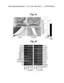 | 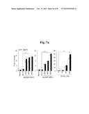 |
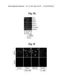 | 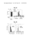 |
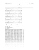 | 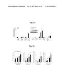 |
 | 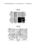 |
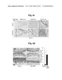 | 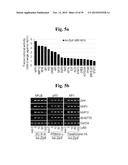 |
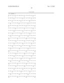 | 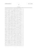 |
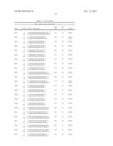 | 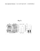 |
 | 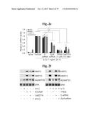 |
 | 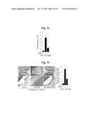 |
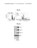 | 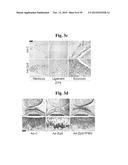 |
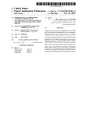 | 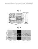 |
 | 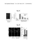 |
 | 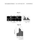 |
 | 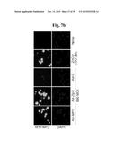 |
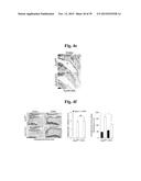 | 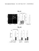 |
 |  |
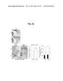 | 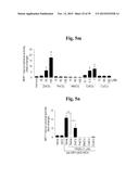 |
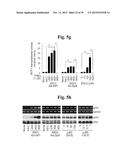 |
| Top Inventors for class "Drug, bio-affecting and body treating compositions" | |
| Rank | Inventor's name |
|---|---|
| 1 | David M. Goldenberg |
| 2 | Hy Si Bui |
| 3 | Lowell L. Wood, Jr. |
| 4 | Roderick A. Hyde |
| 5 | Yat Sun Or |