Patent application title: FUSION PROTEIN
Inventors:
Masayuki Homma (Narita-Shi, JP)
Takahisa Kogure (Narita-Shi, JP)
Kenji Nakajima (Izumisano-Shi, JP)
Assignees:
NIHON PHARMACEUTICAL CO., LTD.
IPC8 Class: AC07K1642FI
USPC Class:
4241341
Class name: Immunoglobulin, antiserum, antibody, or antibody fragment, except conjugate or complex of the same with nonimmunoglobulin material structurally-modified antibody, immunoglobulin, or fragment thereof (e.g., chimeric, humanized, cdr-grafted, mutated, etc.) antibody, immunoglobulin, or fragment thereof fused via peptide linkage to nonimmunoglobulin protein, polypeptide, or fragment thereof (i.e., antibody or immunoglobulin fusion protein or polypeptide)
Publication date: 2014-11-13
Patent application number: 20140335085
Abstract:
A new fusion protein which can specifically suppress the autoantibodies,
which can effectively prevent or treat the autoimmune disease of
autoantibody type, and which can be expressed in an amount sufficient for
industrial production. A fusion protein, characterized in that, a protein
(X) containing a site recognized by autoantibodies which are a cause of
the autoimmune disease of autoantibody type is connected to a protein (A)
containing a fragment of the antibody heavy chain constant region which
exhibits the antibody-dependent cellular cytotoxicity with a linker
peptide (L) consisting of one or more amino acid(s), wherein the protein
(X), the linker peptide (L) and the protein (A) are connected in this
order by means of peptide bond from N terminal to C terminal.Claims:
1. (canceled)
2. (canceled)
3. (canceled)
4. (canceled)
5. (canceled)
6. (canceled)
7. (canceled)
8. (canceled)
9. (canceled)
10. (canceled)
11. (canceled)
12. (canceled)
13. A fusion protein, characterized in that, a protein (X) containing a site recognized by autoantibodies which are a cause of the autoimmune disease of autoantibody type is connected to a protein (A) containing a fragment of the antibody heavy chain constant region which exhibits the antibody-dependent cellular cytotoxicity with a linker peptide (L) consisting of one or more amino acid(s), wherein the protein (X), the linker peptide (L) and the protein (A) are connected in this order by means of peptide bond from N terminal to C terminal, wherein the protein (X) is a nicotinic acetylcholine receptor (nAChR) α1 subunit or a part thereof, and wherein the linker peptide (L) contains an amino acid sequence represented by the formula (Gly-Gly-Gly-Gly-Ser)n (in the formula, n is an integer of 1 to 4), or the formula Pro-(Gly-Gly-Gly-Gly-Ser) n (in the formula, n is an integer of 1 to 4).
14. The fusion protein according to claim 13, wherein the protein (X) is isoform 1 consisting of amino acid sequence of SEQ ID No. 13 of human nAChRα1 subunit and/or isoform 2 consisting of amino acid sequence of SEQ ID No. 14 of human nAChRα1 subunit, or a part thereof, or consists of amino acid sequence wherein one or several amino acid(s) is/are deleted, added and/or substituted in any one of said amino acid sequences.
15. The fusion protein according to claim 13, wherein the protein (X) consists of amino acid sequence of N-terminal extracellular region of isoform 1 and/or isoform 2 of human nAChRα1 subunit, or consists of amino acid sequence wherein one or several amino acid(s) is/are deleted, added and/or substituted in said amino acid sequence.
16. The fusion protein according to claim 13, wherein the protein (A) contains a Fc region of the antibody heavy chain, an antibody heavy chain constant region, or a part thereof, or a combination thereof.
17. The fusion protein according to claim 13, wherein the protein (A) contains human antibody IgG heavy chain constant region, or a combination of parts of IgG subunit heavy chain constant region.
18. The fusion protein according to claim 13, wherein the protein (A) consists of amino acid sequence of SEQ ID No. 11 or SEQ ID No. 12, or consists of amino acid sequence wherein one or several amino acid(s) is/are deleted, added and/or substituted in said amino acid sequence.
19. The fusion protein according to claim 13, wherein the fusion protein consists of amino acid sequence of SEQ ID No. 10, or consists of amino acid sequence wherein one or several amino acid(s) is/are deleted, added and/or substituted in the amino acid sequence of SEQ ID No. 10.
20. A method for manufacturing the fusion protein of claim 13, characterized in that DNA encoding the fusion protein of claim 13 is inserted into a cellular expression vector and this vector is introduced into host cells to express the fusion protein.
21. A composition for prevention and treatment of autoimmune disease of autoantibody type, characterized in containing the fusion protein of claim 13 as an effective ingredient.
Description:
CROSS-REFERENCE TO RELATED APPLICATIONS
[0001] This application claims the benefit of Japanese Patent Application No. 2011-088762 filed on Apr. 13, 2011, which was patented as U.S. Pat. No. 4,857,396 on Nov. 4, 2011, and corresponding Patent Cooperation Treaty Application No. PCT/JP2012/058912 filed on Apr. 2, 2012.
REFERENCE TO SEQUENCE LISTING
[0002] This application includes as part of its subject matter a Sequence Listing electronically submitted via EFS-Web on Oct. 7, 2013, as a single text file named "Sequence Listing.txt". The Sequence Listing Text file was created on Sep. 13, 2013 and is 38 kb in size. The contents of the Sequence Listing are hereby incorporated by reference.
FIELD OF THE INVENTION
[0003] The present invention relates to a fusion protein which can effectively prevent and treat an autoimmune disease of autoantibody type such as myasthenia gravis by neutralizing autoantibodies and inhibiting the autoantibody production. More particularly, the present invention relates to a fusion protein having necessary and sufficient strong function for prevention and treatment and also being secreted to the outside of cells as a result of expression together with keeping its stable structure whereby being able to cope even with industrial production.
BACKGROUND OF THE INVENTION
[0004] Immune system inherently has a role of recognizing and eliminating a foreign body such as bacterium or virus which is different from the self but, sometimes, it excessively reacts with one's own normal cells and tissues and attacks them due to congenital or acquired abnormality. Autoimmune disease is a general name for the diseases resulted by such a state. Among them, a disease caused by the reaction of autoantibodies (antibodies which recognize one's own cells and tissues as an antigen) with autoantigen (one's own cells and tissues) is called "autoimmune disease of autoantibody type". Examples of the autoimmune disease of autoantibody type include myasthenia gravis, hemolytic anemia of autoimmune type, idiopathic thrombocytopenic purpura, neutropenia of autoimmune type, hyperthyroidism or Hashimoto disease caused by anti-TSH antibody, acute encephalitis of autoantibody type, and non-herpetic marginal encephalitis.
[0005] As to a treating method for autoimmune disease of autoantibody type, administration of steroidal agents or immunosuppressants has been conducted frequently. However, any of those drugs does not specifically suppress the autoantibodies which are fundamental cause of the disease but generally suppress the immunoreaction as a whole. Therefore, the drugs have no specificity and the methods are not a sufficiently effective treating method in terms of QOL (Quality of Life).
[0006] With regard to myasthenia gravis which is one of the representative examples of the autoimmune diseases of autoantibody type, there is no already-known treating agent for the fundamental treatment therefor as well but merely the above steroidal agents, the above immunosuppressants, cholinesterase inhibitors, plasma exchange therapy, immunoglobulin preparations for intravenous injection, and thymectomy have been mostly used (cf. Trends of clinical test studies for myasthenia gravis, Nippon Rinsho, Vol. 66, No. 6, pp. 1155-1157; High-dose therapy by immunoglobulin, Shinkei Chiryo, Vol. 25, No. 6, pp. 689-692; "Guideline for the Treatment of Myasthenia gravis (MG)", Report of 1995 by the Search and Study Team for Special Diseases and Immunological Neural Diseases, Health and Welfare Ministry; "Current Status of Treatment and Prognosis of Myasthenia Gravis in Japan", Memorial Lecture at the Fourth MG Forum).
[0007] Among the above, the use of choline esterase inhibitors is difficult for its dose setting. Also, it may be sometimes necessary that atropine sulfate is intravenously injected or airway is secured taking the case of side effect into consideration. Moreover, when high dose is administered for a long period, its effect lowers and, in some cases, cholinergic crisis may happen, which are regarded as problematic. This agent is not intended for the therapeutic treatment but is a mere symptomatic treatment. Fundamentally, the minimum dose by which the effect is achieved is to be used and a long-term administration is to be avoided if at all possible.
[0008] With regard to steroidal agents, their side effect is regarded as problematic and control of the side effect is very important. In addition, a continued administration of such agents for a long period is difficult and it is necessary to control together with the use of nonsteroidal immunosuppressants such as tacrolims or cyclosporine. However, as mentioned already, the above agents are for mere symptomatic treatment and are not fundamental therapeutic means.
[0009] With regard to thymectomy, although it shows some effect, there are problems of anxiety of patients to excising operation and also of cost. There is another problem that it is not applicable to small children whose immune function is still undeveloped and to patients suffering from immunodeficiency disease. In addition, although it exhibits some effects, long years of up to units of ten years are required until the effect is confirmed. It is unavoidable that other symptomatic treatments are jointly conducted until the effect is acknowledged. There is still another problem that the effect was confirmed for only less than 50% of the patients.
[0010] With regard to plasma exchange therapy, a cost of as high as not less than one million yen is needed for one treatment. A subsidy for the medical expenses of myasthenia gravis according to the system for Diagnosis Procedure Combination is only about six hundred thousand yen whereby the burden at the medical care site is big. Further, there is a problem that duration of the effect thereof is as short as only about one month.
[0011] As a treating method for myasthenia gravis, effectiveness of gamma-globulin preparations have been confirmed in recent years and some pharmaceutical manufacturers are now conducting clinical tests therefor. However, since gamma-globulin preparations are biological preparations derived from human plasma, there may be a risk of infection due to unknown virus, etc. In addition, dose of the gamma-globulin preparations is high (400 mg/kg, continued administration for 5 days) and, it is expected that burdens for patients and medical care sites will be considerably high. On the other hand, duration of the effect thereof has been said to be the same as plasma exchange therapy or merely a bit longer.
[0012] To sum up, the problem in the treatment of myasthenia gravis is that, as to the treatment using low-molecular drug, it is a mere temporary symptomatic treatment and, as to plasma exchange therapy, gamma-globulin preparations and thymectomy, the problems in terms of effect and cost are still left as well.
[0013] In view of the above problems, the present inventors thought that an effective effect will be expected in a small dose causing no burden to patients if an antibody reacting only to an anti-acetylcholine receptor autoantibodies which have been believed to be a cause of myasthenia gravis can be prepared in recombinant protein. Then the present inventors prepared fusion protein of nAChRα1 subunit N-terminal extracellular region with antibody heavy chain constant region as a substitute for antiidiotype antibody in order to neutralize the anti-acetylcholine receptor autoantibodies. Since this fusion protein has the activity of neutralizing the autoantibodies and also injuring the autoantibody production cells, it has been judged to be very effective to myasthenia gravis which is one of autoimmune diseases of autoantibody type. However, this fusion protein had a low expressing amount and its industrial production was under a difficult state (cf. Japanese Patent No. 4495776).
SUMMARY OF THE INVENTION
[0014] The present invention has been created in view of the current status of the prior art as such and an object of the present invention is to provide a new fusion protein which can specifically suppress the autoantibodies, which can effectively prevent or treat the autoimmune disease of autoantibody type, and which can be expressed in an amount sufficient for industrial production. Another object of the present invention is to provide a method for manufacturing the fusion protein.
[0015] The above fusion protein in Japanese Patent No. 4495776 can be expected for its effect as a treatment agent for myasthenia gravis in two points which are inhibition of production of autoantibodies and neutralization of the produced autoantibodies. However, in the fusion protein of receptor protein with antibody heavy chain constant region, small expressing amount which is seemingly caused by steric hindrance of the structure, purity of expressed protein, etc. are the problems.
[0016] Under such circumstances, the present inventors have conducted various investigations for enhancing the expressed amount of fusion protein and the purity of expressed protein and noted that, in the fusion protein of Japanese Patent No. 4495776, each of the receptor protein and the antibody heavy chain constant region is in a complicated structure whereby, due to their steric hindrance, incorrect disulfide bond is resulted during the expression of the fusion protein and, as a result, no sufficient purity and expressed amount are achieved. As a means for solving the above, the present inventors have conceived a fusion protein wherein a flexible linker peptide is inserted between the receptor protein and the antibody heavy chain constant region. Thus, the present inventors thought that each of structures of the receptor protein and the antibody heavy chain constant region keeps the inherent stable structure by insertion of the flexible linker peptide. The present inventors then thought that, as a result of formation of stable structure in each region, a secretive effect to the outside of the cells is promoted resulting in much more production of fusion protein and, in addition, stability of fusion protein itself is enhanced whereby the proportion of the decomposed product can be made significantly small and improvement in the purity is now possible. In view of the above, the present inventors have prepared a fusion protein into which this flexible linker peptide is inserted and found that, in this fusion protein, expressed amount is greatly enhanced and purity of the expressed protein is also significantly improved as compared with the conventional fused protein having no linker peptide. The present inventors have also found that, in the fusion protein into which a flexible linker peptide is inserted, neutralizing effect for autoantibodies is significantly enhanced and the effect of specifically suppressing the autoantibody production cells is also strong as compared with the conventional fused protein having no linker peptide. The present inventors have further found that cellular cytotoxicity is more strongly achieved when the antibody heavy chain constant region (A) is positioned at the C terminal side than at the N terminal side. The present inventors have achieved the present invention on the basis of those findings.
[0017] Thus, in accordance with the present invention, there is provided a fusion protein, characterized in that, a protein (X) containing a site recognized by autoantibodies which are a cause of the autoimmune disease of autoantibody type is connected to a protein (A) containing a fragment of the antibody heavy chain constant region which exhibits the antibody-dependent cellular cytotoxicity with a linker peptide (L) consisting of one or more amino acid(s), wherein the protein (X), the linker peptide (L) and the protein (A) are connected in this order by means of peptide bond from N terminal to C terminal.
[0018] Moreover, in accordance with the present invention, there is also provided a method for manufacturing the above fusion protein which is characterized in that DNA encoding the above fusion protein is inserted into a cellular expression vector and this vector is introduced into host cells to express the fusion protein. There is further provided a composition for prevention and treatment of autoimmune disease of autoantibody type which is characterized in containing the above fusion protein as an effective ingredient.
[0019] Among the advantages of the invention, the fusion protein of the present invention neutralizes the autoantibodies existing in the body of a patient suffering from autoimmune disease of autoantibody type and also inhibits the autoantibody production whereby it can specifically suppress the autoantibodies. In addition, the fusion protein of the present invention has high expressing amount and purity and can be provided as a drug in an actual production scale. Accordingly, when the fusion protein of the present invention is used, it is now possible to effectively prevent and treat various autoimmune diseases of autoantibody type such as myasthenia gravis.
BRIEF DESCRIPTION OF THE DRAWINGS
[0020] FIG. 1 is a schematic chart of region for expressing a fusion protein α1-Fc prepared in Examples.
[0021] FIG. 2 is a schematic chart of region for expressing a fusion protein α1-L-Fc prepared in Examples.
[0022] FIG. 3 is a schematic chart of region for expressing a fusion protein α1-L2-Fc prepared in Examples.
[0023] FIG. 4 is a schematic chart of region for expressing a fusion protein Fc-L2-α1 prepared in Examples.
[0024] FIG. 5 shows a silver-stained image (left side) and a western blotting image (right side) after SDS-PAGE of fusion protein α1-Fc and fusion protein α1-L-Fc in a reduced state purified after a transient expression.
[0025] FIG. 6 shows a binding ability of fusion protein α1-Fc and fusion protein α1-L-Fc to Protein A and α-bungarotoxin. .diamond-solid. shows the result for the fusion protein α1-Fc and A shows the result for the fusion protein α1-L-Fc.
[0026] FIG. 7 shows the binding ability of fusion protein α1-Fc and fusion protein α1-L-Fc to anti-nAChRα1 subunit autoantibodies. .diamond-solid. shows the result for the fusion protein α1-Fc and A shows the result for the fusion protein α1-L-Fc.
[0027] FIG. 8A shows a binding of fusion protein α1-Fe to hybridoma Mab35 cells. Added concentration of the fusion protein is shown in FIG. 8A.
[0028] FIG. 8B shows a binding of fusion protein α1-L-Fc to hybridoma Mab35 cells. Added concentration of the fusion protein is shown in FIG. 8B.
[0029] FIG. 9 shows a binding inhibitive activity of 100 μg/mL of fusion protein α1-Fc (left side) and a binding inhibitive activity of fusion protein α1-L-Fc in the same concentration (right side) to the binding of 1 μg/mL of autoantibody mAb35 to TE671 cells.
[0030] FIG. 10 shows an improving effect of fusion protein α1-Fc and fusion protein α1-L-Fc to myasthenia gravis-like symptom induced by autoantibody mAb35. The abscissa shows time and the ordinate shows score of myasthenia gravis-like symptom. shows a group administered with physiological saline, .diamond-solid. shows a group administered with a globulin preparation for intravenous injection, Δ and .tangle-solidup. each shows a group administered with fusion protein α1-Fc, and quadrature and .box-solid. each shows a group administered with fusion protein α1-L-Fc.
DETAILED DESCRIPTION OF THE INVENTION AND MODE FOR CARRYING OUT THE INVENTION
[0031] The fusion protein of the present invention has a structure wherein a protein (X) containing a site recognized by autoantibodies which are a cause of the autoimmune disease of autoantibody type is connected to a protein (A) containing a fragment which exhibits the antibody-dependent cellular cytotoxicity of the antibody heavy chain constant region with a linker peptide (L) consisting of one or more amino acid(s).
[0032] The protein (X) corresponds to an autoantigen to autoantibodies or a part thereof and plays a role of decoy binding to autoantibodies as a substitute for the autoantigen of a patient. Thus, when the fusion protein of the present invention is administered to a patient suffering from autoimmune disease of autoantibody type, the autoantibodies in the body of the patient recognize the protein (X) part in the fusion protein as autoantigen and bind to this part. Since the bound autoantibodies cannot bind to the autoantigen which is inherently present in the body of the patient any longer, the autoantibodies can be neutralized by this method and generation of symptom of the autoimmune disease by binding of the autoantibodies to the autoantigen of the patient can be suppressed. Although the autoantibodies are not one specific antibody but are composed of a group of various antibodies, any of the antibodies is common in such a view that it has a function of recognizing the autoantigen. Accordingly, when the fusion protein of the present invention which acts as a decoy of autoantigen is used, one fusion protein can neutralize a group of various antibodies, and thus there is no need to separately prepare each fusion protein for each of various antibodies.
[0033] "Antibody" defined here stands for all of the antibody in each class of IgA, IgD, IgE, IgG an IgM and in each subclass thereof. "Antibody constant region" stands for antibody in each class or antibody in each subclass and/or a combination of antibody heavy chain constant region thereof as well. There is no particular limitation for sugar chain structure being added the antibody heavy chain constant region.
[0034] The fusion protein of the present invention contains a protein (A) which contains a fragment of the antibody heavy chain constant region in addition to a protein (X) which acts as a decoy for an autoantigen. This protein (A) also plays a role of exhibiting the antibody-dependent cellular cytotoxicity (ADCC activity). Autoantibodies are produced by B cells in the blood. Antibodies of cell surface presenting type having the same antigen binding site as the autoantibodies exist on the surface of the B cells as a B cell receptor. Accordingly, when the fusion protein of the present invention is administered to a patient, a part of the fusion protein binds to the autoantibodies in the body of the patient as mentioned above while remainder binds to the antibodies on the surface of B cells (B cell receptor) which produce the autoantibodies. When the fusion protein of the present invention binds to the B cell receptor, effector cells such as NK cell bind to a protein (A) in the fusion protein via an Fc receptor of the effector cells, exhibit the antibody-dependent cellular cytotoxicity (ADCC activity), injure the B cells binding to the fusion protein, and suppress production of autoantibodies. As such, in accordance with the present invention, the outcome is not only that the autoantibodies existing in the body are neutralized to inhibit the binding of the autoantibodies to the autoantigen but also that the specific B cells which are a production source of the autoantibodies can be selectively injured. Accordingly, the fusion protein of the present invention can prevent or treat the autoimmune disease of autoantibody type by two ways which are inhibition of the autoantibody production and neutralization of the produced autoantibodies.
[0035] The fusion protein of the present invention includes a flexible linker peptide (L) consisting of one or more amino acid(s). As a result of insertion of such a linker peptide, structure of each of the receptor protein and the antibody heavy chain constant region becomes a stable structure whereby a fusion protein becomes stable as a whole.
[0036] The present invention is characterized in that receptor protein (X), linker peptide (L) and antibody heavy chain constant region (A) are sequenced in the order of (X)-(L)-(A) from N terminal to C terminal. Theoretically, the antibody heavy chain constant region (A) is positioned in any side of N terminal side and C terminal side but the present inventors have found that an antibody heavy chain constant region (A) should be positioned in the C terminal side wherein the steric hindrance is little in binding to a receptor, for a purpose of effective achievement of antibody-dependent cellular cytotoxicity (ADCC activity) of the antibody heavy chain constant region (A). It is also presumed that, when the receptor protein (X) is positioned at N terminal, its effect as a decoy can be also strongly achieved.
[0037] The protein (X) in the fusion protein of the present invention corresponds to an autoantigen, or a part thereof, to autoantibodies which are a cause of autoimmune disease of autoantibody type in the patient to be prevented or treated. The protein (X) is decided depending upon the autoimmune disease in the patient to be prevented or treated. For example, in the case of prevention and treatment of myasthenia gravis, since myasthenia gravis is a disease resulted by such a cause that anti-nicotinic acetylcholine receptor antibodies (autoantibodies) bind, in nerve-muscle junction, to a nicotinic acetylcholine receptor (autoantigen) which is a receiver in the side of muscle of neurotransmitter acetylcholine whereby nervous/muscular transmittance by acetylcholine is inhibited, the protein (X) can be a nicotinic acetylcholine receptor which is the autoantigen. Similarly, in the case of prevention and treatment of hemolytic anemia of autoimmune type, the protein (X) can be an erythrocyte surface marker; in the case of prevention and treatment of idiopathic thrombocytopenic purpura, the protein (X) can be a platelet surface marker; in the case of prevention and treatment of neutropenia of autoimmune type, the protein (X) can be a neutrophil surface marker; in the case of prevention and treatment of hyperthyroidism or primary hypothyroidism (Hashimoto disease) caused by anti-TSH antibody, the protein (X) can be TSH; and in the case of prevention of treatment of encephalitis and encephalopathy of autoantibody type, the protein (X) can be NMDA receptor, AMPA receptor, etc.
[0038] The protein (X) is not necessary to be the whole receptor or the whole marker but may be a part thereof as far as a recognition site of autoantibodies is contained therein. For example, in the case of myasthenia gravis, the above nicotinic acetylcholine receptor is a pentameric protein consisting of four kinds of subunits and, among them, the recognition site of the autoantibodies exists in the N-terminal extracellular region of isoform 1 (an isoform expressed only in skeletal muscle and shown by SEQ ID No. 13) and isoform 2 (an isoform expressed in skeletal muscle, brain, heart, kidney and lung, and shown by SEQ ID No. 14) of an α1 subunit. Accordingly, in the case of myasthenia gravis, the protein (X) may be a nicotinic acetylcholine receptor α1 (nAChRα1) subunit or a part thereof. To be more specific, it may be isoform 1 and/or isoform 2 of nAChRα1 subunit or a part thereof and, to be still more specific, it may consists of amino acid sequence of N-terminal extracellular region of isoform 1 and/or isoform 2 of nAChRα1 subunit.
[0039] In the amino acid sequences as such, one or several (such as 1 to 20, preferably 1 to 10, and more preferably 1 to 7) amino acid(s) may be deleted, added and/or substituted as far as the homology thereof is not impaired. With regard to the range thereof, an example is amino acid sequence having 70% or more, preferably 80% or more, and more preferably 90% or more sequence identity. Homology of the amino acid sequence can be calculated using a homology calculation algorithm NCBI BLAST (National Center for Biotechnology Information Basic Local Alignment Search Tool) under such a condition that expectation value is 10, gap is allowed, matrix is BLOSUM 62 and filtering is off To be more specific, the amino acid sequence wherein deletion, addition and/or substitution as such are/is introduced can be easily prepared by substituting the corresponding DNA sequence using a commercially available kit such as Site-Directed Mutagenesis Kit (manufactured by Takara Bio Inc.) or QuickChange Site-Directed Mutagenesis Kit (manufactured by STRATAGENE). It is also possible to directly prepare the above amino acid sequence by means of an artificial gene synthesis technique.
[0040] The protein (A) in the fusion protein of the present invention is a protein containing a fragment of an antibody heavy chain constant region and it may be, for example, a Fc region of the antibody heavy chain, antibody heavy chain constant region or a part thereof. "Antibody" includes all classes of IgA, IgD, IgE, IgG and IgM and also includes all of subclasses thereof "Antibody heavy chain constant region" is a part excluding the variable region of antibody heavy chain. For example, when the class is IgG, the antibody heavy chain constant region comprises a combination of CH1 region, hinge region, CH2 region and CH3 region. The antibody heavy chain constant region may also be a combination of above each class or each subclass or heavy chain constant region thereof. For example, when the class is IgG, the Fc region of the antibody heavy chain comprises a combination of hinge region, CH2 region and CH3 region. In case of human antibody IgG1, the amino acid sequence of SEQ ID No. 11 or SEQ ID No. 12 may be specifically exemplified. Both SEQ ID No. 11 and No. 12 are the sequences of human antibody IgG1 Fc region. SEQ ID No, 11 is said to be a type abundantly found in Asian people while SEQ ID No. 12 is said to be a type abundantly found in European and American people.
[0041] The peptide linker (L) in the fusion protein of the present invention consists of one or more amino acid(s), preferably 5 to 45, more preferably 10 to 20, and most preferably 16 amino acids. This peptide linker may contain Gly-Ser element or Ser-Gly.
[0042] Specific examples of the peptide linker (L) include that which contains an amino acid sequence represented by
[0043] the formula (Gly-Gly-Gly-Gly-Ser)n,
[0044] the formula Pro-(Gly-Gly-Gly-Gly-Ser) n,
[0045] the formula Gly-Ser(Gly-Gly-Gly-Gly-Ser)n,
[0046] the formula (Ser-Ser-Ser-Ser-Gly)n, or
[0047] the formula (Ser-Ser-Ser-Ser-Gly)n-Ser-Pro
[0048] (in the formulae, n is an integer of 1 to 8).
[0049] Among the above, the amino acid sequences represented by the first and second formulae are preferred. The repetition number (n) in the formulae is preferably an integer of 1 to 4 and, more preferably, 3.
[0050] Other specific examples of the peptide linker (L) include the peptide linkers containing a sequence having a structure based on amino acid Gly and/or amino acid Ser (such as that which contains a sequence having Gly-Gly-Ser-Ser-Arg-Gly-Gly, Gly-Gly-Ser-Ser-Arg-Ser-Ser-Ser-Ser-Gly-Gly-Gly-Gly-Ser-Gly-Gly-Gly-Gly, or Glu-Phe-Gly-Gly-Gly-Gly-Gly).
[0051] Still other specific examples of the peptide linker (L) include those having the following amino acid sequences or containing amino acid sequences wherein improvement is applied based on those sequences.
TABLE-US-00001 A) Asp-Ala-Ala-Ala-Lys-Glu-Ala-Ala-Ala-Lys-Asp- Ala-Ala-Ala-Arg-Glu-Ala-Ala-Ala-Arg-Asp-Ala-Ala- Ala-Lys B) Asn-Val-Asp-His-Lys-Pro-Ser-Asn-Thr-Lys-Val- Asp-Lys-Arg
[0052] A specific example of the fusion protein of the present invention is a protein consisting of amino acid sequence of SEQ ID No. 10. This fusion protein has a structure wherein the protein (X), the linker peptide (L) and the protein (A) are connected in this order by means of peptide bond from N terminal to C terminal. The protein (X) corresponds to an amino acid sequence consisting of amino acids of the 1st to 210th positions in the amino acid sequence of N-terminal extracellular region of isoform 1 of nAChRα1 subunit. The linker peptide (L) corresponds to Pro-(Gly-Gly-Gly-Gly-Ser)3. The protein (A) corresponds to amino acid sequence of SEQ ID No. 11. In this amino acid sequence of the fusion protein, as mentioned above, one or several (such as 1 to 20, preferably 1 to 10, more preferably 1 to 7, and most preferably 1 to 3) amino acid(s) may be deleted, added and/or substituted as far as the homology thereof is not impaired.
[0053] The fusion protein of the present invention can be manufactured according to the conventional publicly-known gene engineering technique. Thus, for example, each of DNA encoding the protein (X), DNA encoding the linker peptide (L) and DNA encoding the protein (A) is amplified if necessary, those DNAs are bound each other, the resulting DNA is inserted into a cellular expression vector and a host cell is transfected with the vector to express the fusion protein whereby fusion protein of the present invention can be manufactured. Amplification of DNA can be conducted by, for example, a PCR method. Binding of the amplified DNA can be conducted, for example, by an overlap extension PCR method. It is also possible to design an amino acid sequence of fusion protein to be expressed so as to directly prepare an artificial synthetic gene. It is preferred that the expression vector includes a promoter such as CMV or SV40 for enhancing the expression efficiency and a secretion signal sequence such as antibody heavy chain signal sequence or antibody κ chain signal sequence for easy recovery of the expressed fusion protein from culture supernatant. It is also preferred that kozak sequence is inserted into the upper stream of the transcription initiating codon for enhancing the expressed protein amount. In the case of the present fusion protein, nAChRα1 subunit which is a membrane protein exists in N terminal side and, in addition, the present membrane protein has an extracellular region in N terminal side and, accordingly, compatibility of expressed protein and signal sequence is good resulting in good secretion expression when an original signal sequence of nAChRα1 subunit is used. As to the expression host cell, mammalian cell, yeast, animal cell, insect cell, plant cell, bacterial cell (Escherichia coli etc.) etc. can be used. Among them, animal cell is preferred and CHO cell, HEK293 cell, etc. are particularly preferred. Further, when a nucleic acid sequence which expresses the fusion protein is infused into chromosomes, expression as a transgenic animal is also possible. The expressed fusion protein may be recovered by conventional means and may be purified by, for example, means of a Protein A column method.
[0054] Now, illustrations will be made to the composition for prevention and treatment of autoimmune disease of autoantibody type characterized by comprising the fusion protein of the present invention as an effective ingredient. Examples of the specific preparation form of such a composition are injection and mucosal absorber. In the case of injection, a stabilizer such as saccharide, polyol, albumin or surfactant, an isotonization agent such as salt, etc. are added to the above-prepared fusion protein of the present invention, the resulting product is freeze-dried for preservation and administered by dissolving in water for injection upon use. Although there is no particular limitation for the content of the fusion protein of the present invention in the freeze-dried product, it is 0.01 to 200 mg/g and preferably 0.1 to 100 mg/g, for example. Although there is no particular limitation for the content of the fusion protein in the dissolved injection, it is 0.01 to 200 mg/mL and preferably 0.1 to 100 mg/mL, for example. Examples of the administering method in the case of injection include intravenous administration, intramuscular administration and subcutaneous administration. In the case of mucosal absorber, the fusion protein of the present invention is made into a dosage form together, for example, with excipient and stabilizer so as to give a sustained-release mucosal absorber preparation and it may be administered via oral mucosa, nasal mucosa, eyelid, etc. Although there is no particular limitation for the content of the fusion protein of the present invention in the mucosal absorber, it is 0.1 to 300 mg/ml, and preferably 0.5 to 100 mg/mL, for example. Dose of the composition of the present invention varies depending upon the aimed therapeutic effect, administering method, therapeutic period, age, body weight, etc. and it is usually 10 μg/kg to 50 mg/kg per day for an adult.
EXAMPLES
[0055] The present invention will now be illustrated in more detail as hereunder by Examples, but the present invention is not limited to these Examples.
[0056] (1) Construction of Expression Vector of α1-Fc Fusion Protein
Comparative Example
[0057] Sequence of signal sequence and N-terminal extracellular region was extracted from nAChRα1 subunit based on isoform 1 protein sequence information (Accession No. P02708-2) of already-known nicotinic acetylcholine receptor (nAChR) α1 subunit. Sequence of Fc region was also extracted based on protein sequence information (Accession No. P01857) of human antibody IgG1 constant region. After that, protein sequence of 462 residues was designed wherein both sequences were fused.
[0058] In order to conduct the expression of fusion protein using Chinese hamster ovary cells (CHO cells), optimization to nucleic acid sequence suitable for CHO cells was conducted and a nucleic acid sequence was prepared wherein restriction enzyme recognition sequence and kozak sequence were added to the 5' side while termination codon and restriction enzyme recognition sequence were added to 3' side by means of an artificial gene synthetic technique (SEQ ID No. 1).
[0059] The resulting artificial synthetic gene was treated with restriction enzyme and inserted under the domination of hCMV-MIE promoter of pEE12.4 which was an expression vector for animal cells to construct a vector pEE12.4-A1Fc for secretion and expression of fusion protein α1-Fc (SEQ ID No. 2) comprising N-terminal extracellular region of isoform 1 of human nAChRα1 subunit and human antibody IgG1 heavy chain Fc region. Scheme of the protein expression region is shown in FIG. 1.
[0060] (2) Construction of Expression Vector of α1-L-Fc Fusion Protein
Example of the Present Invention
[0061] Nucleic acid amplification was conducted for nAChRα1 subunit region containing the kozak sequence of SEQ ID No. 1 by PCR method using the artificial synthetic gene prepared in (1) as a template; using a primer of SEQ ID No. 3 and a primer of SEQ ID No. 4 to which nucleic acid sequence encoding a flexible linker (L) (Pro-(Gly-Gly-Gly-Gly-Ser)3) were added as a primer set; and using "KOD-Plus-Neo" (catalog No.: KOD-401) of Toyobo as a DNA polymerase whereupon a nucleic acid sequence of SEQ ID No. 5 was prepared.
[0062] On the other hand, nucleic acid amplification was conducted for the antibody IgG1 Fc region by PCR method using the artificial synthetic gene of SEQ ID No. 1 as a template; using a primer of SEQ ID No. 6 to which a nucleic acid sequence encoding a flexible linker sequence was added and a primer of SEQ ID No. 7 as a primer set; and using KOD-Plus-Neo of Toyobo as a DNA polymerase whereupon a nucleic acid sequence of SEQ ID No. 8 was prepared.
[0063] Nucleic acid amplification was conducted by overlap extension PCR method using a mixed solutions of SEQ ID No. 5 and SEQ ID No. 8 as a template; using a primer of SEQ ID No. 3 and a primer of SEQ ID No. 7 as a primer set; and using KOD-Plus-Neo of Toyobo as a DNA polymerase whereupon a nucleic acid sequence of SEQ ID No. 9 was prepared.
[0064] The resulting amplified product of nucleic acid was treated with restriction enzyme and inserted under domination of hCMV-MIE promoter of pEE12.4 which was an expression vector for animal cells to construct a vector pEE12.4-A1LFc for secretion and expression of fusion protein α1-L-Fc (SEQ ID No. 10) wherein N-terminal extracellular region of isoform 1 of human nAChRα1 subunit and human antibody IgG1 heavy chain Fc region were connected by a flexible linker sequence (L). Scheme of the protein expression region is shown in FIG. 2.
[0065] (3) Confirmation of Transient Expression of Fusion Protein α1-Fc and Fusion Protein α1-L-Fc
[0066] HEK293 cells were transfected with fusion protein expression vectors pEE12.4-A1Fc and pEE12.4-A1LFc prepared in (1) and (2) using an expression system "Free Style MAX 293 Expression System" (catalog No. K9000-10) of Invitrogen to express the fusion protein α1-Fc and the fusion protein α1-L-Fc. Then they were purified using a purifying column "HiTrap Protein A HP Column" (catalog No. 17-0402-01) of GE Health Care to give the fusion protein α1-Fc and the fusion protein α1-L-Fc.
[0067] Confirmation of expression of each of the fusion protein was conducted by means of a silver staining and a western blotting after SDS-PAGE. For the western blotting, an HRP-labeled anti-human IgG antibody was used. The result is shown in FIG. 5. A band shown by an arrow in FIG. 5 corresponds to the fusion protein. From this result, it was confirmed that, when a flexible linker (L) was inserted, expressed amount of fusion protein significantly increased.
[0068] (4) Construction of Expression Vector of α1-L2-Fc Fusion Protein
Example of the Present Invention
[0069] There was prepared a vector for expressing a fusion protein α1-L2-Fc (SEQ ID No. 15) wherein a flexible liner (L) (Pro-(Gly-Gly-Gly-Gly-Ser)3) of α1-L-Fc prepared in (2) was modified to a flexible linker (L2) ((Gly-Gly-Gly-Gly-Ser)3).
[0070] Preparation of gene sequence encoding fusion protein (SEQ ID No. 18) was conducted by the same PCR method wherein an artificial synthetic gene (SEQ ID No. 1) was used as a template and the primers of SEQ ID Nos. 4 and 6 of (2) were substituted with SEQ ID Nos. 16 and 17.
[0071] The resulting amplified product of nucleic acid was treated with restriction enzyme and inserted under domination of hCMV-MIE promoter of pEE12.4 which was an expression vector for animal cells to construct a vector pEE12.4-A1L2Fc for secretion and expression of fusion protein α1-L2-Fc wherein N-terminal extracellular region of isoform 1 of human nAChRα1 subunit and human antibody IgG1 heavy chain Fc region were connected by a flexible linker sequence (L2). Scheme of the protein expression region is shown in FIG. 3.
[0072] (5) Construction of Expression Vector of Fc-L2-α1 Fusion Protein
Comparative Example
[0073] There was prepared a vector for expressing a fusion protein Fc-L2-α1 (SEQ ID No. 19) wherein nAChRα1 subunit of α1-L2-Fc prepared in (4) and antibody heavy chain constant region were fused in a reversed order to (4) sandwiching a flexible linker (L2).
[0074] Preparation of the vector was conducted by PCR method in the same manner as in (2) and (4) using artificial synthetic gene of SEQ ID No. 1 as a template.
[0075] The resulting amplified product of nucleic acid was treated with restriction enzyme and inserted under domination of hCMV-MIE promoter of pEE12.4 which was an expression vector for animal cells to construct a vector pEE12.4-FcL2A1 for secretion and expression of fusion protein Fc-L2-α1 wherein human antibody IgG1 heavy chain Fc region and N-terminal extracellular region of isoform 1 of human nAChRα1 subunit were connected by a flexible linker sequence (L2). Scheme of the protein expression region is shown in FIG. 4.
[0076] (6) Construction of Stable Expression Strains of Fusion Proteins α1-Fc, α1-L-Fc, α1-L2-Fc, and Fc-L-α1
[0077] Each of fusion protein expression vectors pEE12.4-A1Fc, pEE12.4-A1LFc, pEE12.4-A1L2Fc, and pEE12.4-FcL2A1 prepared in (1), (2), (4) and (5) was transferred into CHO-K1 cells by an electroporation method, incubated under methionine sulfoximine (MSX) selection and cloned to prepare a transformant. The resulting transformants (hereinafter, each of them will be abbreviated as "α1-Fc expression cell", "α1-L-Fc expression cell", "α1-L2-Fc expression cell", and "Fc-L2-α1 expression cell") was subjected to the following experiment.
[0078] (7) Expression Incubation of Fusion Proteins α1-Fc, α1-L-Fc, α1-L2-Fc, and Fc-L2-α1
[0079] Each of α1-Fc expression cells, α1-L-Fc expression cells, α1-L2-Fc expression cells, and Fc-L2-α1 expression cells prepared in (6) were incubated using 9 L of "CD-CHO medium" (catalog No. 12490-025) of Invitrogen as an incipient medium so as to produce each protein. The incubating condition was pH 7.1 at 37° C. and, during the fifth to the ninth days of the incubation, 50 mL/L/day of "CHO CD EfficientFeed B" (catalog No. A1024-01) of Invitrogen was added and incubation was conducted for 10 days. Supernatant liquid of the fusion protein expression culture was obtained by centrifuging the culture at 3500×G for 5 minutes to precipitate the incubated cells and recovering the supernatant liquid therefrom.
[0080] (8) Purification of Fusion Proteins α1-Fc, α1-L-Fc, α1-L2-Fc, and Fc-L2-α1 and Calculation of Expressed Amount
[0081] For each supernatant liquid of the fusion protein expression culture obtained in (7), a solution after a filtering treatment with a filter of 0.45 μm was loaded to "Mab select SuRe" (catalog No. 11-0026-01) of GE Healthcare, washed with PBS of 10-column volume and eluted with 2.5-column volume of 20 mM citric acid buffer (pH=3.0) and the eluate was immediately neutralized with 1M Tris-HCl (pH=9.0) in an amount of one-tenth of the eluate to purify the fusion protein. The resulting eluate was concentrated using "Amicon Ultra-15, Ultracel-50K" (catalog No. UFC905024) of Japan Millipore, substitution with PBS was conducted and a filtering treatment was done using an aseptic filter membrane of 0.22 μm followed by subjecting to the following experiment.
[0082] Absorbance at 280 nm (OD280) of the resulting eluate fraction was measured and expressed amount of fusion protein per 1 L of the incubated liquid for each expression strain was calculated using absorption coefficient (α1-Fc:0.57 mg/mL/OD280, α1-L-Fc:0.59 mg/mL/OD280, α1-L2-Fc:0.59 mg/mL/OD280, Fc-L2-α1:0.59 mg/mL/OD280) of each fusion protein, eluate amount and incubated liquid amount.
[0083] The result was that the expressed amount of fusion protein was 72 mg/L for α1-Fc expression cells, 1587 mg/L for α1-L-Fc expression cells, 1249 mg/L for α1-L2-Fc expression cells, and 356 mg/L for Fc-L2-α1 expression cells, and it was confirmed that the expressed amount was significantly enhanced by insertion of flexible linker peptide into the binding site of the fusion protein. Among them, a fusion protein expression strain using Pro-(Gly-Gly-Gly-Gly-Ser)3 as a flexible peptide linker was excellent.
[0084] (9) Confirmation of Binding Ability of Fusion Protein α1-Fc and Fusion Protein α1-L-Fc to α-BTX
[0085] With regard to the fusion protein α1-Fc and the fusion protein α1-L-Fc obtained in (8), the binding ability thereof to α-bungarotoxin (hereinafter, it will be abbreviated as "α-BTX") and to Protein A was confirmed with a method of ELISA. α-BTX is a substance having an action of inhibiting the neurotransmission by binding to α1 domain. Accordingly, as a result of confirmation of the binding ability of fusion protein to α-BTX, it is now possible to confirm whether the structure of α1 domain of the fusion protein is correctly formed. Further, Protein A fixes the fusion protein to a plate via Fe. Accordingly, although the main purpose of this experiment is to confirm the formation of α1 domain structure of fusion protein as mentioned above, it is also possible in this experiment to confirm whether the structure of fusion protein as an Fc is retained.
[0086] "Protein A" (catalog No. 987015) of ICN Biochemicals was immobilized at the concentration of 1 μg/mL in "C8 Maxisorp Nunc-Immuno Module" (catalog No. 445101) of Nalge Nunc and then blocking was conducted using PBS to which 1% of BSA was added, each of the fusion proteins prepared in (8) and subjected to 4-fold dilution series was added thereto as a sample followed by further washing, then "α-bungarotoxin, biotin-XX" (catalog No. B 1196) of Invitrogen was added at the concentration of 1 μg/mL and, finally, reaction was conducted with "Peroxidase-Avidin" (catalog No. 191370) of ICN Biochemicals. For the detection, "TMB solution" (catalog No. N301) of Funakoshi was made to react as a substrate and the reaction was stopped using 1% sulfuric acid solution. After that, absorbance at 450 nm wavelength was measured.
[0087] The result is shown in FIG. 6. For any of the fusion proteins, concentration-dependent reaction was noted. From this result, it was confirmed that each fusion protein binds to α-BTX and also binds to Protein A. Thus, it was confirmed that both of the fusion protein α1-Fc and the fusion protein α1-L-Fc retained the nAChRα1 subunit extracellular region and the structure as Fc. Incidentally, when both fusion proteins were compared, a binding curve to α-BTX was significantly shifted to a lower concentration side in the fusion protein α1-L-Fc whereby it was judged that, when flexible linker peptide is inserted, binding ability per protein concentration is enhanced to an extent of about 100-fold or more. This result suggests that, when flexible linker peptide is inserted, structural stability of the aimed fusion protein increases and purity becomes high.
[0088] (10) Confirmation of Binding Ability of Fusion Protein α1-Fe and Fusion Protein α1-L-Fc to Anti-nAChR Autoantibody
[0089] Mab35 (TIB-175) (hereinafter, it will be abbreviated as "Mab35 cells") which is rat anti-nAChR (α1 subunit) autoantibody production hybridoma obtained from ATCC was incubated in "Hybridoma-SFM" (catalog No. 12045-01) of Invitrogen and the supernatant liquid of the culture was treated with "HiTrap Protein G HP Column" (catalog No. 17-0405-01) of GE Healthcare whereupon a monoclonal antibody (hereinafter, it will be abbreviated as "mAb35") which is an anti-nAChR autoantibody was obtained.
[0090] The resulting mAb35 was immobilized at the concentration of 1 μg/mL in C8 Maxisorp Nunc-Immunomodule of Nalge Nunc and blocked using PBS to which 1% of BSA was added. After that, the fusion protein α1-Fe and the fusion protein α1-L-Fc prepared in (8) and subjected to 4-fold dilution series were added and, finally, reaction was conducted using HRP-labeled anti-human IgG1 Fc antibody. For the detection, "TMB solution" of Funakoshi was made to react as a substrate and the reaction was stopped using 1% sulfuric acid solution. After that, absorbance at 450 nm wavelength was measured.
[0091] The result is shown in FIG. 7. Both of the fusion protein α1-Fe and the fusion protein α1-L-Fc showed a concentration-dependent reaction. When both were compared, a binding curve to autoantibody was significantly shifted to lower concentration side in the fusion protein α1-L-Fc whereby it was judged that, when flexible linker peptide is inserted, binding ability (specific activity) per protein concentration is enhanced to an extent of about 100-fold or more. This result suggests that, when flexible linker peptide is internally inserted, not only the expressed amount of the aimed fusion protein but also the structural stability thereof increase and the reactivity with autoantibody is significantly enhanced.
[0092] (11) Confirmation of Binding Ability of Fusion Protein α1-Fc and Fusion Protein α1-L-Fc to Autoantibody Production Cells
[0093] In a living body, autoantibodies are produced in B cells. On the cell membrane of the cell surface of the autoantibody production B cells, the same antibodies as the autoantibodies are presented as a B cell receptor. Thus, on the cell membrane of the hybridoma Mab35 used in (10), it is likely that the hybridoma Mab35 also presents mAb35 antibody the same as in the case of B cells.
[0094] Now, 2×10⁵ Mab35 cells washed with HBSS/BSA were made to react with each of the fusion protein α1-Fc and the fusion protein α1-L-Fc prepared in (8) diluted to 10-fold within a range of 10 ng/mL to 1 mg/mL as samples. After that, PE-labeled anti-human IgG antibody was added as a detection reagent and the detection was conducted using "Cytomice FC500" of Beckman-Coulter.
[0095] The result is shown in FIG. 8A and FIG. 8B. Each of the fusion proteins was shifted to the right side in a concentration-dependent manner whereby it was confirmed that each of the fusion proteins binds to Mab35 cells in a concentration-dependent manner. Since the fusion protein α1-L-Fc was much more shifted, it was confirmed that, when flexible linker peptide is inserted, the binding ability to autoantibody production cells is enhanced to an extent of about 100-fold or more.
[0096] (12) Confirmation of Action of Fusion Protein α1-Fc and Fusion Protein α1-L-Fc as Decoy
[0097] nAChRα1 subunit similar to human muscle cells is present in the human neuroblastoma cells (TE-671) (hereinafter, it will be abbreviated as "TE671 cells") obtained from ATCC. Therefore, a binding-inhibitive activity of the fusion protein α1-Fc and the fusion protein α1-L-Fc prepared in (8) to the bond of the autoantibody mAb35 to TE671 cells was confirmed.
[0098] To 2×10⁵ TE671 cells washed with HBSS/BSA was added 100 μg/mL of the fusion protein α1-Fc or the fusion protein α1-L-Fc. As to a control, HBSS/BSA containing no fusion protein was added. After 1 μg/mL of mAb35 antibody was further added thereto, PE-labeled rat IgG antibody was added as a detection reagent and the detection was conducted using "Cytomics FC 500".
[0099] The result is shown in FIG. 9. When the fusion protein α1-Fc was used, only some inhibiting effect was noted at the concentration of 100 μg/mL while, when the fusion protein α1-L-Fc was used, sufficient inhibiting effect was noted at the concentration of 100 μg/mL. From this result, a significant enhancement of inhibiting activity by internal insertion of flexible linker peptide was confirmed.
[0100] (13) Confirmation of ADCC Activity of Fusion Protein α1-Fc, Fusion Protein α1-L-Fc, and Fusion Protein Fc-L2-α1
[0101] Mab35 cell (2×10⁵ cells) was washed with HBSS/BSA and incubated at 37° C. for 30 minutes in HBSS/BSA to which a fluorescent dye "Calcein-AM" (catalog No. C326) of Dojinsha was added to make the concentration 10 μM whereby Calcein-AM was incorporated into the cells. After that, those Mab35 cells were disseminated to a 96-well plate to make 10000 cells per well and then the fusion protein α1-L-Fc prepared in (8) or a control antibody (Avastin or Enbrel) and human natural killer NK92 cells (CRL-2407) obtained from ATCC (hereinafter, it will be abbreviated as "NK92 cells") were added thereto in various concentrations followed by incubating at 37° C. for 4 hours. After the incubation, centrifugal separation was conducted at 300×G for 5 minutes to precipitate the cells and fluorescence of each supernatant liquid was measured (Ex=485 nm, Em=540 nm). As a result, although a very weak cellular cytotoxicity (natural killing) was noted in a dependently manner on NK92 cell numbers even in a group wherein only NK92 cells were added (no antibody, etc. added), a strong cellular cytotoxicity of about 73% at the highest was noted in a group wherein the fusion protein α1-L-Fc was administered, and a strong cellular cytotoxicity was noted in a group wherein 25-fold amount of NK92 cells (i.e. the effector cells (E)) were added to Mab35 cells (i.e. the target cells (T)) (no data shown). From the result of the above preliminary experiments, cellular cytotoxicity was compared among the fusion protein α1-Fc, the fusion protein α1-L-Fc and the fusion protein Fc-L2-α1 under the condition wherein E/T ratio was 25.
[0102] As a result, cellular cytotoxicity values of 51.8%, 73.2% and 32.9% were noted in the fusion protein α1-Fc, in the fusion protein α1-L-Fc and in the fusion protein Fc-L2-α1, respectively. On the other hand, cellular cytotoxicity values of 17.9%, 12.5% and 6.9% were noted in a group wherein no antibody was added, in an Avastin-added group and in an Enbrel-added group, respectively.
[0103] From those results, it was confirmed that higher cytotoxicity was exhibited when the antibody heavy chain constant region was not positioned at N terminal side but was positioned at C terminal side. It was also confirmed that cellular cytotoxicity was further enhanced by internal insertion of flexible linker peptide even in the case wherein antibody heavy chain constant region was positioned at C terminal side.
[0104] (14) Confirmation of In Vivo Test to Myasthenia Gravis
[0105] For the experiment, 36 female Lewis rats of 11 weeks age (Nippon LSC) were prepared. As an animal model for myasthenia gravis, an autoantibody-inducing rat model was used. mAb35 which is an autoantiboy to the rat nAChR produced by hybridoma Mab35 was intraperitoneally administered to all rats in a dose of 1.25 mg/kg whereupon a morbid state was induced. Each of the following substances was intravenously administered after 4, 12, 24 and 32 hours from the administration of mAb35. To a control group, 1 mL of PBS was administered for each time (control group, 6 rats). α1-L-Fc prepared in (8) was administered at the dose of 2.5 mg/rat (α1-L-Fc 2.5, 6 rats) or 10 mg/rat (α1-L-Fc 10, 6 rats) for each time. Similarly, α1-Fc prepared in (8) was administered at the dose of 2.5 mg/rat (α1-Fc 2.5, 6 rats) or 10 mg/rat (α1-Fc 10, 6 rats) for each time. Further, donated Venoglobulin IH 5% for intravenous injection (manufactured by Tanabe Mitsubishi) which was a human immunoglobulin preparation for intravenous injection was administered at the dose of 80 mg/rat (IVIG, 6 rats) for each time.
[0106] During the period until 96 hours after induction of morbid state, muscle symptom score (MG Score) was evaluated. The muscle symptom score is as follows: point 0 for no abnormality; point 1 for lowering in grip of forelimbs; point 2 for disappearance of grip of forelimbs; point 3 for lowering of muscle strength of hind limbs and gait disorder in addition to disappearance of grip of forelimbs; and point 4 for paralysis of hind limbs in addition to disappearance of grip of forelimbs. Statistic analysis of the muscle symptom score was conducted in such a manner that comparison of the control group with each substance-administered group was done by Steel test (SAS Preclinical Package Version 5.00.010720, Windows (registered trade mark) version, SAS System Release 8.02 TS Level 02M0 (SAS Institute Japan)). The result is given in terms of (mean value)±(standard error) and the significance level of less than 5% (*) was judged as significant difference.
[0107] The result is shown in Table 1 and FIG. 10. FIG. 10 shows the effect of α1-L-Fc and α1-Fc in the mAb35-induced rat myasthenia gravis model. As shown in FIG. 10, such a change was observed in the control group that the muscle symptom score increased as from 24 hours after induction of morbid state, became highest after 56 hours and decreased thereafter. There was shown such a tendency that α1-L-Fc and α1-Fc suppressed the increase of the muscle symptom score and that the suppression as such is dependent on the dose. Table 1 shows a mean score after 56 hours from morbid state induction wherein the muscle symptom score of the control group became highest. It was shown that any of α1-L-Fc and α1-Fc significantly suppressed the muscle symptom by administration of 10 mg/rat for each time.
TABLE-US-00002 TABLE 1 muscle symptom score after 56 hours from morbid state induction muscle symptom score control 2.3 ± 0.6 α1-L-Fc 2.5 0.6 ± 0.3 α1-L-Fc 10 0.3 ± 0.2* α1-Fc 2.5 1.1 ± 0.4 α1-Fc 10 0.3 ± 0.2* IVIG 0.9 ± 0.2
[0108] In the above Examples, the effect was confirmed for the cases wherein a linker peptide having repetition number of 3 (flexible linker (L) (Pro-(Gly-Gly-Gly-Gly-Ser)3) or flexible linker (L2) ((Gly-Gly-Gly-Gly-Ser)3)) was used. From those results, persons skilled in the art can easily predict that even the cases wherein repeating number (n) of the linker peptide is integer of 1 to 8 or, particularly, integer of 1 to 4 will achieve the same excellent effect as the cases wherein the repetition number (n) of the linker peptide is 3. That is because the length of a linker peptide has been said to be usually from about 5 residues (in other words, the repetition number (n) in the above constitution unit is 1) to about 20 residues (in other words, the repetition number (n) in the above constitution unit is 4).
INDUSTRIAL APPLICABILITY
[0109] In accordance with the present invention, the fusion protein, which can prevent or treat the autoimmune disease of autoantibody type by two ways (i.e. inhibition of the autoantibody production and neutralization of the produced autoantibodies), can be provided as a drug in an actual production scale. Therefore, the fusion protein of the present invention can be widely used for effectively preventing and treating various autoimmune diseases of autoantibody type such as myasthenia gravis.
[0110] Sequence Listing Free Text
[0111] Sequence ID Nos. 3, 4, 6, 7, 16 and 17 are the sequences of the primers.
Sequence CWU
1
1
1911413DNAhomo sapiens 1aagcttgccg ccaccatgga gccctggcct ctcctcctgc
tctttagcct ttgctcagct 60ggcctcgtcc tgggctccga acatgagacc cgtctggtgg
caaagctatt taaagactac 120agcagcgtgg tgcggccagt ggaagaccac cgccaggtcg
tggaggtcac cgtgggcctg 180cagctgatac agctcatcaa tgtggatgaa gtaaatcaga
tcgtgacaac caatgtgcgt 240ctgaaacagc aatgggtgga ttacaaccta aaatggaatc
cagatgacta tggcggtgtg 300aaaaaaattc acattccttc agaaaagatc tggcgcccag
accttgttct ctataacaat 360gcagatggtg actttgctat tgtcaagttc accaaagtgc
tcctgcagta cactggccac 420atcacgtgga cacctccagc catctttaaa agctactgtg
agatcatcgt cacccacttt 480ccctttgatg aacagaactg cagcatgaag ctgggcacct
ggacctacga cggctctgtc 540gtggccatca acccggaaag cgaccagcca gacctgagca
acttcatgga gagcggggag 600tgggtgatca aggagtcccg gggctggaag cactccgtga
cctattcctg ctgccccgac 660accccctacc tggacatcac ctaccacttc gtcatgcagc
gcctggagcc caaatcttgt 720gacaaaactc acacatgccc accgtgccca gcacctgaac
tcctgggggg accgtcagtc 780ttcctcttcc ccccaaaacc caaggacacc ctcatgatct
cccggacccc tgaggtcaca 840tgcgtggtgg tggacgtgag ccacgaagac cctgaggtca
agttcaactg gtacgtggac 900ggcgtggagg tgcataatgc caagacaaag ccgcgggagg
agcagtacaa cagcacgtac 960cgtgtggtca gcgtcctcac cgtcctgcac caggactggc
tgaatggcaa ggagtacaag 1020tgcaaggtct ccaacaaagc cctcccagcc cccatcgaga
aaaccatctc caaagccaaa 1080gggcagcccc gagaaccaca ggtgtacacc ctgcccccat
cccgggatga gctgaccaag 1140aaccaggtca gcctgacctg cctggtcaaa ggcttctatc
ccagcgacat cgccgtggag 1200tgggagagca atgggcagcc ggagaacaac tacaagacca
cgcctcccgt gctggactcc 1260gacggctcct tcttcctcta cagcaagctc accgtggaca
agagcaggtg gcagcagggg 1320aacgtcttct catgctccgt gatgcatgag gctctgcaca
accactacac gcagaagagc 1380ctctccctgt ctccgggtaa atgataagaa ttc
14132442PRThomo sapiens 2Ser Glu His Glu Thr Arg
Leu Val Ala Lys Leu Phe Lys Asp Tyr Ser 1 5
10 15 Ser Val Val Arg Pro Val Glu Asp His Arg Gln
Val Val Glu Val Thr 20 25
30 Val Gly Leu Gln Leu Ile Gln Leu Ile Asn Val Asp Glu Val Asn
Gln 35 40 45 Ile
Val Thr Thr Asn Val Arg Leu Lys Gln Gln Trp Val Asp Tyr Asn 50
55 60 Leu Lys Trp Asn Pro Asp
Asp Tyr Gly Gly Val Lys Lys Ile His Ile 65 70
75 80 Pro Ser Glu Lys Ile Trp Arg Pro Asp Leu Val
Leu Tyr Asn Asn Ala 85 90
95 Asp Gly Asp Phe Ala Ile Val Lys Phe Thr Lys Val Leu Leu Gln Tyr
100 105 110 Thr Gly
His Ile Thr Trp Thr Pro Pro Ala Ile Phe Lys Ser Tyr Cys 115
120 125 Glu Ile Ile Val Thr His Phe
Pro Phe Asp Glu Gln Asn Cys Ser Met 130 135
140 Lys Leu Gly Thr Trp Thr Tyr Asp Gly Ser Val Val
Ala Ile Asn Pro 145 150 155
160 Glu Ser Asp Gln Pro Asp Leu Ser Asn Phe Met Glu Ser Gly Glu Trp
165 170 175 Val Ile Lys
Glu Ser Arg Gly Trp Lys His Ser Val Thr Tyr Ser Cys 180
185 190 Cys Pro Asp Thr Pro Tyr Leu Asp
Ile Thr Tyr His Phe Val Met Gln 195 200
205 Arg Leu Glu Pro Lys Ser Cys Asp Lys Thr His Thr Cys
Pro Pro Cys 210 215 220
Pro Ala Pro Glu Leu Leu Gly Gly Pro Ser Val Phe Leu Phe Pro Pro 225
230 235 240 Lys Pro Lys Asp
Thr Leu Met Ile Ser Arg Thr Pro Glu Val Thr Cys 245
250 255 Val Val Val Asp Val Ser His Glu Asp
Pro Glu Val Lys Phe Asn Trp 260 265
270 Tyr Val Asp Gly Val Glu Val His Asn Ala Lys Thr Lys Pro
Arg Glu 275 280 285
Glu Gln Tyr Asn Ser Thr Tyr Arg Val Val Ser Val Leu Thr Val Leu 290
295 300 His Gln Asp Trp Leu
Asn Gly Lys Glu Tyr Lys Cys Lys Val Ser Asn 305 310
315 320 Lys Ala Leu Pro Ala Pro Ile Glu Lys Thr
Ile Ser Lys Ala Lys Gly 325 330
335 Gln Pro Arg Glu Pro Gln Val Tyr Thr Leu Pro Pro Ser Arg Asp
Glu 340 345 350 Leu
Thr Lys Asn Gln Val Ser Leu Thr Cys Leu Val Lys Gly Phe Tyr 355
360 365 Pro Ser Asp Ile Ala Val
Glu Trp Glu Ser Asn Gly Gln Pro Glu Asn 370 375
380 Asn Tyr Lys Thr Thr Pro Pro Val Leu Asp Ser
Asp Gly Ser Phe Phe 385 390 395
400 Leu Tyr Ser Lys Leu Thr Val Asp Lys Ser Arg Trp Gln Gln Gly Asn
405 410 415 Val Phe
Ser Cys Ser Val Met His Glu Ala Leu His Asn His Tyr Thr 420
425 430 Gln Lys Ser Leu Ser Leu Ser
Pro Gly Lys 435 440 324DNAArtificial
SequencePrimer 3cccaagcttg ccgccaccat ggag
24481DNAArtificial SequencePrimer 4gctcttaggc tccgatccgc
caccgccaga gccacctccg cctgaaccgc ctccaccggg 60cagccgttgc atgacgaagt g
815768DNAhomo sapiens
5cccaagcttg ccgccaccat ggagccatgg cccctgctcc ttcttttcag cctgtgttca
60gctggcctcg tgctgggcag cgagcacgaa accaggttgg tcgctaaact tttcaaagat
120tactcctcag tagtgaggcc tgtagaggat catcggcagg tggtggaggt cactgtggga
180ctccagctca tccagttgat caatgtcgat gaggtcaacc aaatcgtcac cactaatgtc
240cgactgaagc agcagtgggt cgactacaac ctgaagtgga atcccgatga ctacggtggt
300gtgaaaaaaa tacatattcc cagtgagaag atctggcgtc cagatcttgt tctgtacaac
360aacgctgatg gagatttcgc tatcgtcaag ttcaccaaag tgctgctgca gtatacaggt
420catataactt ggactccccc agcaatcttt aagagttact gcgagatcat agtgacccat
480tttccctttg acgagcagaa ttgttccatg aagctgggca cttggaccta cgacgggtct
540gtcgtggcta ttaatccaga aagcgatcag cccgatcttt caaattttat ggagtccggt
600gagtgggtga tcaaagaatc aagggggtgg aaacattcag tgacctactc ttgctgtcct
660gatactccct acctcgacat tacctaccac ttcgtcatgc aacggctgcc cggtggaggc
720ggttcaggcg gaggtggctc tggcggtggc ggatcggagc ctaagagc
768682DNAArtificial SequencePrimer 6caacggctgc ccggtggagg cggttcaggc
ggaggtggct ctggcggtgg cggatcggag 60cctaagagct gcgataaaac ac
82721DNAArtificial SequencePrimer
7ggaattctta tcatttacct g
218766DNAhomo sapiens 8caacggctgc ccggtggagg cggttcaggc ggaggtggct
ctggcggtgg cggatcggag 60cctaagagct gcgataaaac acacacatgc cctccctgcc
ccgctccaga gctgttgggc 120ggaccaagcg ttttcctgtt cccaccaaag cccaaggaca
ctttgatgat ctctcggact 180cctgaagtga catgcgtcgt ggtagatgtc tctcatgaag
atccagaggt gaaatttaac 240tggtatgtag acggcgtgga ggtgcacaat gccaaaacca
agcctcgaga agaacagtac 300aatagtacat accgagtggt ttctgttttg accgtgcttc
accaggactg gctgaacgga 360aaggaataca aatgcaaggt ctcaaacaag gcattgccag
cccccatcga aaagacaatt 420tctaaagcca aaggacagcc cagagagcct caggtgtata
ccctcccacc atcacgagac 480gaactcacaa aaaaccaggt ttccctcacc tgtctggtga
aggggtttta cccatctgat 540atcgccgtcg aatgggagtc taacggacag cctgagaata
attataagac aactccacct 600gtcctggaca gtgatggatc tttctttctg tacagtaaac
tgaccgtgga taagtcacgc 660tggcaacaag gtaatgtgtt cagctgcagc gtcatgcacg
aggctctgca taaccattat 720acacagaagt cactctctct gtccccaggt aaatgataag
aattcc 76691465DNAhomo sapiens 9cccaagcttg ccgccaccat
ggagccatgg cccctgctcc ttcttttcag cctgtgttca 60gctggcctcg tgctgggcag
cgagcacgaa accaggttgg tcgctaaact tttcaaagat 120tactcctcag tagtgaggcc
tgtagaggat catcggcagg tggtggaggt cactgtggga 180ctccagctca tccagttgat
caatgtcgat gaggtcaacc aaatcgtcac cactaatgtc 240cgactgaagc agcagtgggt
cgactacaac ctgaagtgga atcccgatga ctacggtggt 300gtgaaaaaaa tacatattcc
cagtgagaag atctggcgtc cagatcttgt tctgtacaac 360aacgctgatg gagatttcgc
tatcgtcaag ttcaccaaag tgctgctgca gtatacaggt 420catataactt ggactccccc
agcaatcttt aagagttact gcgagatcat agtgacccat 480tttccctttg acgagcagaa
ttgttccatg aagctgggca cttggaccta cgacgggtct 540gtcgtggcta ttaatccaga
aagcgatcag cccgatcttt caaattttat ggagtccggt 600gagtgggtga tcaaagaatc
aagggggtgg aaacattcag tgacctactc ttgctgtcct 660gatactccct acctcgacat
tacctaccac ttcgtcatgc aacggctgcc cggtggaggc 720ggttcaggcg gaggtggctc
tggcggtggc ggatcggagc ctaagagctg cgataaaaca 780cacacatgcc ctccctgccc
cgctccagag ctgttgggcg gaccaagcgt tttcctgttc 840ccaccaaagc ccaaggacac
tttgatgatc tctcggactc ctgaagtgac atgcgtcgtg 900gtagatgtct ctcatgaaga
tccagaggtg aaatttaact ggtatgtaga cggcgtggag 960gtgcacaatg ccaaaaccaa
gcctcgagaa gaacagtaca atagtacata ccgagtggtt 1020tctgttttga ccgtgcttca
ccaggactgg ctgaacggaa aggaatacaa atgcaaggtc 1080tcaaacaagg cattgccagc
ccccatcgaa aagacaattt ctaaagccaa aggacagccc 1140agagagcctc aggtgtatac
cctcccacca tcacgagacg aactcacaaa aaaccaggtt 1200tccctcacct gtctggtgaa
ggggttttac ccatctgata tcgccgtcga atgggagtct 1260aacggacagc ctgagaataa
ttataagaca actccacctg tcctggacag tgatggatct 1320ttctttctgt acagtaaact
gaccgtggat aagtcacgct ggcaacaagg taatgtgttc 1380agctgcagcg tcatgcacga
ggctctgcat aaccattata cacagaagtc actctctctg 1440tccccaggta aatgataaga
attcc 146510458PRThomo sapiens
10Ser Glu His Glu Thr Arg Leu Val Ala Lys Leu Phe Lys Asp Tyr Ser 1
5 10 15 Ser Val Val Arg
Pro Val Glu Asp His Arg Gln Val Val Glu Val Thr 20
25 30 Val Gly Leu Gln Leu Ile Gln Leu Ile
Asn Val Asp Glu Val Asn Gln 35 40
45 Ile Val Thr Thr Asn Val Arg Leu Lys Gln Gln Trp Val Asp
Tyr Asn 50 55 60
Leu Lys Trp Asn Pro Asp Asp Tyr Gly Gly Val Lys Lys Ile His Ile 65
70 75 80 Pro Ser Glu Lys Ile
Trp Arg Pro Asp Leu Val Leu Tyr Asn Asn Ala 85
90 95 Asp Gly Asp Phe Ala Ile Val Lys Phe Thr
Lys Val Leu Leu Gln Tyr 100 105
110 Thr Gly His Ile Thr Trp Thr Pro Pro Ala Ile Phe Lys Ser Tyr
Cys 115 120 125 Glu
Ile Ile Val Thr His Phe Pro Phe Asp Glu Gln Asn Cys Ser Met 130
135 140 Lys Leu Gly Thr Trp Thr
Tyr Asp Gly Ser Val Val Ala Ile Asn Pro 145 150
155 160 Glu Ser Asp Gln Pro Asp Leu Ser Asn Phe Met
Glu Ser Gly Glu Trp 165 170
175 Val Ile Lys Glu Ser Arg Gly Trp Lys His Ser Val Thr Tyr Ser Cys
180 185 190 Cys Pro
Asp Thr Pro Tyr Leu Asp Ile Thr Tyr His Phe Val Met Gln 195
200 205 Arg Leu Pro Gly Gly Gly Gly
Ser Gly Gly Gly Gly Ser Gly Gly Gly 210 215
220 Gly Ser Glu Pro Lys Ser Cys Asp Lys Thr His Thr
Cys Pro Pro Cys 225 230 235
240 Pro Ala Pro Glu Leu Leu Gly Gly Pro Ser Val Phe Leu Phe Pro Pro
245 250 255 Lys Pro Lys
Asp Thr Leu Met Ile Ser Arg Thr Pro Glu Val Thr Cys 260
265 270 Val Val Val Asp Val Ser His Glu
Asp Pro Glu Val Lys Phe Asn Trp 275 280
285 Tyr Val Asp Gly Val Glu Val His Asn Ala Lys Thr Lys
Pro Arg Glu 290 295 300
Glu Gln Tyr Asn Ser Thr Tyr Arg Val Val Ser Val Leu Thr Val Leu 305
310 315 320 His Gln Asp Trp
Leu Asn Gly Lys Glu Tyr Lys Cys Lys Val Ser Asn 325
330 335 Lys Ala Leu Pro Ala Pro Ile Glu Lys
Thr Ile Ser Lys Ala Lys Gly 340 345
350 Gln Pro Arg Glu Pro Gln Val Tyr Thr Leu Pro Pro Ser Arg
Asp Glu 355 360 365
Leu Thr Lys Asn Gln Val Ser Leu Thr Cys Leu Val Lys Gly Phe Tyr 370
375 380 Pro Ser Asp Ile Ala
Val Glu Trp Glu Ser Asn Gly Gln Pro Glu Asn 385 390
395 400 Asn Tyr Lys Thr Thr Pro Pro Val Leu Asp
Ser Asp Gly Ser Phe Phe 405 410
415 Leu Tyr Ser Lys Leu Thr Val Asp Lys Ser Arg Trp Gln Gln Gly
Asn 420 425 430 Val
Phe Ser Cys Ser Val Met His Glu Ala Leu His Asn His Tyr Thr 435
440 445 Gln Lys Ser Leu Ser Leu
Ser Pro Gly Lys 450 455 11330PRThomo
sapiens 11Ala Ser Thr Lys Gly Pro Ser Val Phe Pro Leu Ala Pro Ser Ser Lys
1 5 10 15 Ser Thr
Ser Gly Gly Thr Ala Ala Leu Gly Cys Leu Val Lys Asp Tyr 20
25 30 Phe Pro Glu Pro Val Thr Val
Ser Trp Asn Ser Gly Ala Leu Thr Ser 35 40
45 Gly Val His Thr Phe Pro Ala Val Leu Gln Ser Ser
Gly Leu Tyr Ser 50 55 60
Leu Ser Ser Val Val Thr Val Pro Ser Ser Ser Leu Gly Thr Gln Thr 65
70 75 80 Tyr Ile Cys
Asn Val Asn His Lys Pro Ser Asn Thr Lys Val Asp Lys 85
90 95 Lys Val Glu Pro Lys Ser Cys Asp
Lys Thr His Thr Cys Pro Pro Cys 100 105
110 Pro Ala Pro Glu Leu Leu Gly Gly Pro Ser Val Phe Leu
Phe Pro Pro 115 120 125
Lys Pro Lys Asp Thr Leu Met Ile Ser Arg Thr Pro Glu Val Thr Cys 130
135 140 Val Val Val Asp
Val Ser His Glu Asp Pro Glu Val Lys Phe Asn Trp 145 150
155 160 Tyr Val Asp Gly Val Glu Val His Asn
Ala Lys Thr Lys Pro Arg Glu 165 170
175 Glu Gln Tyr Asn Ser Thr Tyr Arg Val Val Ser Val Leu Thr
Val Leu 180 185 190
His Gln Asp Trp Leu Asn Gly Lys Glu Tyr Lys Cys Lys Val Ser Asn
195 200 205 Lys Ala Leu Pro
Ala Pro Ile Glu Lys Thr Ile Ser Lys Ala Lys Gly 210
215 220 Gln Pro Arg Glu Pro Gln Val Tyr
Thr Leu Pro Pro Ser Arg Asp Glu 225 230
235 240 Leu Thr Lys Asn Gln Val Ser Leu Thr Cys Leu Val
Lys Gly Phe Tyr 245 250
255 Pro Ser Asp Ile Ala Val Glu Trp Glu Ser Asn Gly Gln Pro Glu Asn
260 265 270 Asn Tyr Lys
Thr Thr Pro Pro Val Leu Asp Ser Asp Gly Ser Phe Phe 275
280 285 Leu Tyr Ser Lys Leu Thr Val Asp
Lys Ser Arg Trp Gln Gln Gly Asn 290 295
300 Val Phe Ser Cys Ser Val Met His Glu Ala Leu His Asn
His Tyr Thr 305 310 315
320 Gln Lys Ser Leu Ser Leu Ser Pro Gly Lys 325
330 12330PRThomo sapiens 12Ala Ser Thr Lys Gly Pro Ser Val Phe Pro
Leu Ala Pro Ser Ser Lys 1 5 10
15 Ser Thr Ser Gly Gly Thr Ala Ala Leu Gly Cys Leu Val Lys Asp
Tyr 20 25 30 Phe
Pro Glu Pro Val Thr Val Ser Trp Asn Ser Gly Ala Leu Thr Ser 35
40 45 Gly Val His Thr Phe Pro
Ala Val Leu Gln Ser Ser Gly Leu Tyr Ser 50 55
60 Leu Ser Ser Val Val Thr Val Pro Ser Ser Ser
Leu Gly Thr Gln Thr 65 70 75
80 Tyr Ile Cys Asn Val Asn His Lys Pro Ser Asn Thr Lys Val Asp Lys
85 90 95 Arg Val
Glu Pro Lys Ser Cys Asp Lys Thr His Thr Cys Pro Pro Cys 100
105 110 Pro Ala Pro Glu Leu Leu Gly
Gly Pro Ser Val Phe Leu Phe Pro Pro 115 120
125 Lys Pro Lys Asp Thr Leu Met Ile Ser Arg Thr Pro
Glu Val Thr Cys 130 135 140
Val Val Val Asp Val Ser His Glu Asp Pro Glu Val Lys Phe Asn Trp 145
150 155 160 Tyr Val Asp
Gly Val Glu Val His Asn Ala Lys Thr Lys Pro Arg Glu 165
170 175 Glu Gln Tyr Asn Ser Thr Tyr Arg
Val Val Ser Val Leu Thr Val Leu 180 185
190 His Gln Asp Trp Leu Asn Gly Lys Glu Tyr Lys Cys Lys
Val Ser Asn 195 200 205
Lys Ala Leu Pro Ala Pro Ile Glu Lys Thr Ile Ser Lys Ala Lys Gly 210
215 220 Gln Pro Arg Glu
Pro Gln Val Tyr Thr Leu Pro Pro Ser Arg Glu Glu 225 230
235 240 Met Thr Lys Asn Gln Val Ser Leu Thr
Cys Leu Val Lys Gly Phe Tyr 245 250
255 Pro Ser Asp Ile Ala Val Glu Trp Glu Ser Asn Gly Gln Pro
Glu Asn 260 265 270
Asn Tyr Lys Thr Thr Pro Pro Val Leu Asp Ser Asp Gly Ser Phe Phe
275 280 285 Leu Tyr Ser Lys
Leu Thr Val Asp Lys Ser Arg Trp Gln Gln Gly Asn 290
295 300 Val Phe Ser Cys Ser Val Met His
Glu Ala Leu His Asn His Tyr Thr 305 310
315 320 Gln Lys Ser Leu Ser Leu Ser Pro Gly Lys
325 330 13437PRThomo sapiens 13Ser Glu His Glu Thr
Arg Leu Val Ala Lys Leu Phe Lys Asp Tyr Ser 1 5
10 15 Ser Val Val Arg Pro Val Glu Asp His Arg
Gln Val Val Glu Val Thr 20 25
30 Val Gly Leu Gln Leu Ile Gln Leu Ile Asn Val Asp Glu Val Asn
Gln 35 40 45 Ile
Val Thr Thr Asn Val Arg Leu Lys Gln Gln Trp Val Asp Tyr Asn 50
55 60 Leu Lys Trp Asn Pro Asp
Asp Tyr Gly Gly Val Lys Lys Ile His Ile 65 70
75 80 Pro Ser Glu Lys Ile Trp Arg Pro Asp Leu Val
Leu Tyr Asn Asn Ala 85 90
95 Asp Gly Asp Phe Ala Ile Val Lys Phe Thr Lys Val Leu Leu Gln Tyr
100 105 110 Thr Gly
His Ile Thr Trp Thr Pro Pro Ala Ile Phe Lys Ser Tyr Cys 115
120 125 Glu Ile Ile Val Thr His Phe
Pro Phe Asp Glu Gln Asn Cys Ser Met 130 135
140 Lys Leu Gly Thr Trp Thr Tyr Asp Gly Ser Val Val
Ala Ile Asn Pro 145 150 155
160 Glu Ser Asp Gln Pro Asp Leu Ser Asn Phe Met Glu Ser Gly Glu Trp
165 170 175 Val Ile Lys
Glu Ser Arg Gly Trp Lys His Ser Val Thr Tyr Ser Cys 180
185 190 Cys Pro Asp Thr Pro Tyr Leu Asp
Ile Thr Tyr His Phe Val Met Gln 195 200
205 Arg Leu Pro Leu Tyr Phe Ile Val Asn Val Ile Ile Pro
Cys Leu Leu 210 215 220
Phe Ser Phe Leu Thr Gly Leu Val Phe Tyr Leu Pro Thr Asp Ser Gly 225
230 235 240 Glu Lys Met Thr
Leu Ser Ile Ser Val Leu Leu Ser Leu Thr Val Phe 245
250 255 Leu Leu Val Ile Val Glu Leu Ile Pro
Ser Thr Ser Ser Ala Val Pro 260 265
270 Leu Ile Gly Lys Tyr Met Leu Phe Thr Met Val Phe Val Ile
Ala Ser 275 280 285
Ile Ile Ile Thr Val Ile Val Ile Asn Thr His His Arg Ser Pro Ser 290
295 300 Thr His Val Met Pro
Asn Trp Val Arg Lys Val Phe Ile Asp Thr Ile 305 310
315 320 Pro Asn Ile Met Phe Phe Ser Thr Met Lys
Arg Pro Ser Arg Glu Lys 325 330
335 Gln Asp Lys Lys Ile Phe Thr Glu Asp Ile Asp Ile Ser Asp Ile
Ser 340 345 350 Gly
Lys Pro Gly Pro Pro Pro Met Gly Phe His Ser Pro Leu Ile Lys 355
360 365 His Pro Glu Val Lys Ser
Ala Ile Glu Gly Ile Lys Tyr Ile Ala Glu 370 375
380 Thr Met Lys Ser Asp Gln Glu Ser Asn Asn Ala
Ala Ala Glu Trp Lys 385 390 395
400 Tyr Val Ala Met Val Met Asp His Ile Leu Leu Gly Val Phe Met Leu
405 410 415 Val Cys
Ile Ile Gly Thr Leu Ala Val Phe Ala Gly Arg Leu Ile Glu 420
425 430 Leu Asn Gln Gln Gly
435 14462PRThomo sapiens 14Ser Glu His Glu Thr Arg Leu Val Ala
Lys Leu Phe Lys Asp Tyr Ser 1 5 10
15 Ser Val Val Arg Pro Val Glu Asp His Arg Gln Val Val Glu
Val Thr 20 25 30
Val Gly Leu Gln Leu Ile Gln Leu Ile Asn Val Asp Glu Val Asn Gln
35 40 45 Ile Val Thr Thr
Asn Val Arg Leu Lys Gln Gly Asp Met Val Asp Leu 50
55 60 Pro Arg Pro Ser Cys Val Thr Leu
Gly Val Pro Leu Phe Ser His Leu 65 70
75 80 Gln Asn Glu Gln Trp Val Asp Tyr Asn Leu Lys Trp
Asn Pro Asp Asp 85 90
95 Tyr Gly Gly Val Lys Lys Ile His Ile Pro Ser Glu Lys Ile Trp Arg
100 105 110 Pro Asp Leu
Val Leu Tyr Asn Asn Ala Asp Gly Asp Phe Ala Ile Val 115
120 125 Lys Phe Thr Lys Val Leu Leu Gln
Tyr Thr Gly His Ile Thr Trp Thr 130 135
140 Pro Pro Ala Ile Phe Lys Ser Tyr Cys Glu Ile Ile Val
Thr His Phe 145 150 155
160 Pro Phe Asp Glu Gln Asn Cys Ser Met Lys Leu Gly Thr Trp Thr Tyr
165 170 175 Asp Gly Ser Val
Val Ala Ile Asn Pro Glu Ser Asp Gln Pro Asp Leu 180
185 190 Ser Asn Phe Met Glu Ser Gly Glu Trp
Val Ile Lys Glu Ser Arg Gly 195 200
205 Trp Lys His Ser Val Thr Tyr Ser Cys Cys Pro Asp Thr Pro
Tyr Leu 210 215 220
Asp Ile Thr Tyr His Phe Val Met Gln Arg Leu Pro Leu Tyr Phe Ile 225
230 235 240 Val Asn Val Ile Ile
Pro Cys Leu Leu Phe Ser Phe Leu Thr Gly Leu 245
250 255 Val Phe Tyr Leu Pro Thr Asp Ser Gly Glu
Lys Met Thr Leu Ser Ile 260 265
270 Ser Val Leu Leu Ser Leu Thr Val Phe Leu Leu Val Ile Val Glu
Leu 275 280 285 Ile
Pro Ser Thr Ser Ser Ala Val Pro Leu Ile Gly Lys Tyr Met Leu 290
295 300 Phe Thr Met Val Phe Val
Ile Ala Ser Ile Ile Ile Thr Val Ile Val 305 310
315 320 Ile Asn Thr His His Arg Ser Pro Ser Thr His
Val Met Pro Asn Trp 325 330
335 Val Arg Lys Val Phe Ile Asp Thr Ile Pro Asn Ile Met Phe Phe Ser
340 345 350 Thr Met
Lys Arg Pro Ser Arg Glu Lys Gln Asp Lys Lys Ile Phe Thr 355
360 365 Glu Asp Ile Asp Ile Ser Asp
Ile Ser Gly Lys Pro Gly Pro Pro Pro 370 375
380 Met Gly Phe His Ser Pro Leu Ile Lys His Pro Glu
Val Lys Ser Ala 385 390 395
400 Ile Glu Gly Ile Lys Tyr Ile Ala Glu Thr Met Lys Ser Asp Gln Glu
405 410 415 Ser Asn Asn
Ala Ala Ala Glu Trp Lys Tyr Val Ala Met Val Met Asp 420
425 430 His Ile Leu Leu Gly Val Phe Met
Leu Val Cys Ile Ile Gly Thr Leu 435 440
445 Ala Val Phe Ala Gly Arg Leu Ile Glu Leu Asn Gln Gln
Gly 450 455 460 15457PRThomo
sapiens 15Ser Glu His Glu Thr Arg Leu Val Ala Lys Leu Phe Lys Asp Tyr Ser
1 5 10 15 Ser Val
Val Arg Pro Val Glu Asp His Arg Gln Val Val Glu Val Thr 20
25 30 Val Gly Leu Gln Leu Ile Gln
Leu Ile Asn Val Asp Glu Val Asn Gln 35 40
45 Ile Val Thr Thr Asn Val Arg Leu Lys Gln Gln Trp
Val Asp Tyr Asn 50 55 60
Leu Lys Trp Asn Pro Asp Asp Tyr Gly Gly Val Lys Lys Ile His Ile 65
70 75 80 Pro Ser Glu
Lys Ile Trp Arg Pro Asp Leu Val Leu Tyr Asn Asn Ala 85
90 95 Asp Gly Asp Phe Ala Ile Val Lys
Phe Thr Lys Val Leu Leu Gln Tyr 100 105
110 Thr Gly His Ile Thr Trp Thr Pro Pro Ala Ile Phe Lys
Ser Tyr Cys 115 120 125
Glu Ile Ile Val Thr His Phe Pro Phe Asp Glu Gln Asn Cys Ser Met 130
135 140 Lys Leu Gly Thr
Trp Thr Tyr Asp Gly Ser Val Val Ala Ile Asn Pro 145 150
155 160 Glu Ser Asp Gln Pro Asp Leu Ser Asn
Phe Met Glu Ser Gly Glu Trp 165 170
175 Val Ile Lys Glu Ser Arg Gly Trp Lys His Ser Val Thr Tyr
Ser Cys 180 185 190
Cys Pro Asp Thr Pro Tyr Leu Asp Ile Thr Tyr His Phe Val Met Gln
195 200 205 Arg Leu Gly Gly
Gly Gly Ser Gly Gly Gly Gly Ser Gly Gly Gly Gly 210
215 220 Ser Glu Pro Lys Ser Cys Asp Lys
Thr His Thr Cys Pro Pro Cys Pro 225 230
235 240 Ala Pro Glu Leu Leu Gly Gly Pro Ser Val Phe Leu
Phe Pro Pro Lys 245 250
255 Pro Lys Asp Thr Leu Met Ile Ser Arg Thr Pro Glu Val Thr Cys Val
260 265 270 Val Val Asp
Val Ser His Glu Asp Pro Glu Val Lys Phe Asn Trp Tyr 275
280 285 Val Asp Gly Val Glu Val His Asn
Ala Lys Thr Lys Pro Arg Glu Glu 290 295
300 Gln Tyr Asn Ser Thr Tyr Arg Val Val Ser Val Leu Thr
Val Leu His 305 310 315
320 Gln Asp Trp Leu Asn Gly Lys Glu Tyr Lys Cys Lys Val Ser Asn Lys
325 330 335 Ala Leu Pro Ala
Pro Ile Glu Lys Thr Ile Ser Lys Ala Lys Gly Gln 340
345 350 Pro Arg Glu Pro Gln Val Tyr Thr Leu
Pro Pro Ser Arg Asp Glu Leu 355 360
365 Thr Lys Asn Gln Val Ser Leu Thr Cys Leu Val Lys Gly Phe
Tyr Pro 370 375 380
Ser Asp Ile Ala Val Glu Trp Glu Ser Asn Gly Gln Pro Glu Asn Asn 385
390 395 400 Tyr Lys Thr Thr Pro
Pro Val Leu Asp Ser Asp Gly Ser Phe Phe Leu 405
410 415 Tyr Ser Lys Leu Thr Val Asp Lys Ser Arg
Trp Gln Gln Gly Asn Val 420 425
430 Phe Ser Cys Ser Val Met His Glu Ala Leu His Asn His Tyr Thr
Gln 435 440 445 Lys
Ser Leu Ser Leu Ser Pro Gly Lys 450 455
1678DNAArtificial SequencePrimer 16gctcttaggc tccgatccgc caccgccaga
gccacctccg cctgaaccgc ctccacccag 60ccgttgcatg acgaagtg
781779DNAArtificial SequencePrimer
17caacggctgg gtggaggcgg ttcaggcgga ggtggctctg gcggtggcgg atcggagcct
60aagagctgcg ataaaacac
79181462DNAhomo sapiens 18cccaagcttg ccgccaccat ggagccatgg cccctgctcc
ttcttttcag cctgtgttca 60gctggcctcg tgctgggcag cgagcacgaa accaggttgg
tcgctaaact tttcaaagat 120tactcctcag tagtgaggcc tgtagaggat catcggcagg
tggtggaggt cactgtggga 180ctccagctca tccagttgat caatgtcgat gaggtcaacc
aaatcgtcac cactaatgtc 240cgactgaagc agcagtgggt cgactacaac ctgaagtgga
atcccgatga ctacggtggt 300gtgaaaaaaa tacatattcc cagtgagaag atctggcgtc
cagatcttgt tctgtacaac 360aacgctgatg gagatttcgc tatcgtcaag ttcaccaaag
tgctgctgca gtatacaggt 420catataactt ggactccccc agcaatcttt aagagttact
gcgagatcat agtgacccat 480tttccctttg acgagcagaa ttgttccatg aagctgggca
cttggaccta cgacgggtct 540gtcgtggcta ttaatccaga aagcgatcag cccgatcttt
caaattttat ggagtccggt 600gagtgggtga tcaaagaatc aagggggtgg aaacattcag
tgacctactc ttgctgtcct 660gatactccct acctcgacat tacctaccac ttcgtcatgc
aacggctggg tggaggcggt 720tcaggcggag gtggctctgg cggtggcgga tcggagccta
agagctgcga taaaacacac 780acatgccctc cctgccccgc tccagagctg ttgggcggac
caagcgtttt cctgttccca 840ccaaagccca aggacacttt gatgatctct cggactcctg
aagtgacatg cgtcgtggta 900gatgtctctc atgaagatcc agaggtgaaa tttaactggt
atgtagacgg cgtggaggtg 960cacaatgcca aaaccaagcc tcgagaagaa cagtacaata
gtacataccg agtggtttct 1020gttttgaccg tgcttcacca ggactggctg aacggaaagg
aatacaaatg caaggtctca 1080aacaaggcat tgccagcccc catcgaaaag acaatttcta
aagccaaagg acagcccaga 1140gagcctcagg tgtataccct cccaccatca cgagacgaac
tcacaaaaaa ccaggtttcc 1200ctcacctgtc tggtgaaggg gttttaccca tctgatatcg
ccgtcgaatg ggagtctaac 1260ggacagcctg agaataatta taagacaact ccacctgtcc
tggacagtga tggatctttc 1320tttctgtaca gtaaactgac cgtggataag tcacgctggc
aacaaggtaa tgtgttcagc 1380tgcagcgtca tgcacgaggc tctgcataac cattatacac
agaagtcact ctctctgtcc 1440ccaggtaaat gataagaatt cc
146219457PRThomo sapiens 19Glu Pro Lys Ser Cys Asp
Lys Thr His Thr Cys Pro Pro Cys Pro Ala 1 5
10 15 Pro Glu Leu Leu Gly Gly Pro Ser Val Phe Leu
Phe Pro Pro Lys Pro 20 25
30 Lys Asp Thr Leu Met Ile Ser Arg Thr Pro Glu Val Thr Cys Val
Val 35 40 45 Val
Asp Val Ser His Glu Asp Pro Glu Val Lys Phe Asn Trp Tyr Val 50
55 60 Asp Gly Val Glu Val His
Asn Ala Lys Thr Lys Pro Arg Glu Glu Gln 65 70
75 80 Tyr Asn Ser Thr Tyr Arg Val Val Ser Val Leu
Thr Val Leu His Gln 85 90
95 Asp Trp Leu Asn Gly Lys Glu Tyr Lys Cys Lys Val Ser Asn Lys Ala
100 105 110 Leu Pro
Ala Pro Ile Glu Lys Thr Ile Ser Lys Ala Lys Gly Gln Pro 115
120 125 Arg Glu Pro Gln Val Tyr Thr
Leu Pro Pro Ser Arg Asp Glu Leu Thr 130 135
140 Lys Asn Gln Val Ser Leu Thr Cys Leu Val Lys Gly
Phe Tyr Pro Ser 145 150 155
160 Asp Ile Ala Val Glu Trp Glu Ser Asn Gly Gln Pro Glu Asn Asn Tyr
165 170 175 Lys Thr Thr
Pro Pro Val Leu Asp Ser Asp Gly Ser Phe Phe Leu Tyr 180
185 190 Ser Lys Leu Thr Val Asp Lys Ser
Arg Trp Gln Gln Gly Asn Val Phe 195 200
205 Ser Cys Ser Val Met His Glu Ala Leu His Asn His Tyr
Thr Gln Lys 210 215 220
Ser Leu Ser Leu Ser Pro Gly Lys Gly Gly Gly Gly Ser Gly Gly Gly 225
230 235 240 Gly Ser Gly Gly
Gly Gly Ser Ser Glu His Glu Thr Arg Leu Val Ala 245
250 255 Lys Leu Phe Lys Asp Tyr Ser Ser Val
Val Arg Pro Val Glu Asp His 260 265
270 Arg Gln Val Val Glu Val Thr Val Gly Leu Gln Leu Ile Gln
Leu Ile 275 280 285
Asn Val Asp Glu Val Asn Gln Ile Val Thr Thr Asn Val Arg Leu Lys 290
295 300 Gln Gln Trp Val Asp
Tyr Asn Leu Lys Trp Asn Pro Asp Asp Tyr Gly 305 310
315 320 Gly Val Lys Lys Ile His Ile Pro Ser Glu
Lys Ile Trp Arg Pro Asp 325 330
335 Leu Val Leu Tyr Asn Asn Ala Asp Gly Asp Phe Ala Ile Val Lys
Phe 340 345 350 Thr
Lys Val Leu Leu Gln Tyr Thr Gly His Ile Thr Trp Thr Pro Pro 355
360 365 Ala Ile Phe Lys Ser Tyr
Cys Glu Ile Ile Val Thr His Phe Pro Phe 370 375
380 Asp Glu Gln Asn Cys Ser Met Lys Leu Gly Thr
Trp Thr Tyr Asp Gly 385 390 395
400 Ser Val Val Ala Ile Asn Pro Glu Ser Asp Gln Pro Asp Leu Ser Asn
405 410 415 Phe Met
Glu Ser Gly Glu Trp Val Ile Lys Glu Ser Arg Gly Trp Lys 420
425 430 His Ser Val Thr Tyr Ser Cys
Cys Pro Asp Thr Pro Tyr Leu Asp Ile 435 440
445 Thr Tyr His Phe Val Met Gln Arg Leu 450
455
User Contributions:
Comment about this patent or add new information about this topic:
 |  |
 |  |
 |  |
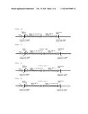 | 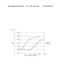 |
 | 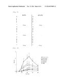 |
 |  |
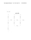 |  |
 |  |
 | 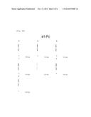 |
 |  |
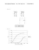 |  |
| Similar patent applications: | |
| Date | Title |
|---|---|
| 2011-02-03 | Fusion proteins |
| 2011-04-21 | Fusion proteins |
| 2012-03-15 | Fusion proteins |
| 2012-09-13 | Fusion proteins |
| 2013-06-27 | Fc fusion proteins |
| New patent applications in this class: | |
| Date | Title |
|---|---|
| 2022-05-05 | Dominant negative cd40l polypeptides |
| 2019-05-16 | Compositions and methods for antigen targeting to cd180 |
| 2019-05-16 | Synthetic membrane-receiver complexes |
| 2019-05-16 | Modular, controlled single chain variable fragment antibody switch |
| 2019-05-16 | Compositions comprising a p75 tumor necrosis factor receptor/ig fusion protein |
| New patent applications from these inventors: | |
| Date | Title |
|---|---|
| 2012-05-17 | Autoantibody production inhibitor |
| Top Inventors for class "Drug, bio-affecting and body treating compositions" | |
| Rank | Inventor's name |
|---|---|
| 1 | David M. Goldenberg |
| 2 | Hy Si Bui |
| 3 | Lowell L. Wood, Jr. |
| 4 | Roderick A. Hyde |
| 5 | Yat Sun Or |