Patent application title: COMPOSITIONS AND METHODS FOR THE TREATMENT OF CNS INJURIES
Inventors:
Elliott A. Gruskin (Killingworth, CT, US)
Elliott A. Gruskin (Killingworth, CT, US)
Anthony O. Caggiano (Larchmont, NY, US)
Michael P. Zimber (Mamroneck, NY, US)
Andrew R. Blight (Mahopac, NY, US)
Andrew R. Blight (Mahopac, NY, US)
Assignees:
ACORDA THERAPEUTICS, INC.
IPC8 Class: AA61K3851FI
USPC Class:
424 945
Class name: Drug, bio-affecting and body treating compositions enzyme or coenzyme containing transferases (2. ), lyase (4.), isomerase (5.), ligase (6.)
Publication date: 2014-09-04
Patent application number: 20140248253
Abstract:
The present invention is directed to a method of improving functional
recovery following a central nervous system contusion injury. The method
includes administering a therapeutically effective amount of
glycosaminoglycan degrading enzyme. The glycosaminoglycan degrading
enzyme may be dermatan sulfate or chondroitin sulfate degrading enzymes.
The central nervous system contusion injury may include a traumatic brain
injury or a spinal cord injury. The functional recovery may include
autonomic functions, sensory functions, motor functions or the like.Claims:
1. A method of improving autonomic function comprising administering
chondroitinase ABCType1 enzyme to a mammal following a contusion
injury that led to an impairment of autonomic function to the central
nervous system, wherein the autonomic function is improved.
2. The method of claim 1, wherein the chondroitinase ABCType1 enzyme administered to the mammal comprises a therapeutically effective amount.
3. The method of claim 2, wherein the therapeutically effective amount of chondroitinase ABCType1 enzyme comprises an amount sufficient to degrade chondroitin sulfate proteoglycans.
4. The method of claim 3, wherein the degradation of the chondroitin sulfate proteoglycans occurs at the site of the central nervous system.
5. (canceled)
6. The method of claim 2, wherein the therapeutically effective amount of chondroitinase ABCType1 enzyme comprises a maximum of about 100 mg/kg.
7-10. (canceled)
11. The method of claim 1, wherein the chondroitinase ABCType1 enzyme is administered locally.
12. The method of claim 11, wherein the local administration is selected from the group consisting of intrathecal and topical administration.
13. The method of claim 1, wherein the chondroitinase ABCType1 enzyme is in a sustained release formulation.
14-20. (canceled)
21. The method of claim 3, wherein the degradation of the chondroitin sulfate proteoglycans occurs outside the site of the contusion injury.
22-25. (canceled)
26. The method of claim 1, wherein the contusion injury comprises a spinal cord injury.
27. (canceled)
28. The method of claim 26, wherein the gross morphology of the spinal cord is maintained.
29. The method of claim 26, wherein the spinal cord injury comprises an injury resulting in a condition selected from the group consisting of monoplegia, diplegia, paraplegia, hemiplegia and quadriplegia.
30. The method of claim 1, wherein the contusion injury comprises torn or partially severed neurons.
31. The method of claim 1, wherein the contusion injury comprises crushed neurons.
32. The method of claim 1, wherein the contusion injury comprises compression of the central nervous system.
33. The method of claim 32, wherein the compression is caused by a traumatic force to the spinal cord.
34. (canceled)
Description:
CROSS REFERENCE TO RELATED APPLICATIONS
[0001] This application is a continuation application of pending U.S. patent application Ser. No. 10/847,540 filed May 17, 2004, which claims the benefit of U.S. Provisional Patent Application No. 60/417,236, filed May 16, 2003, each of which is incorporated herein by reference in its entirety.
BACKGROUND OF THE INVENTION
[0002] 1. Field of the Invention
[0003] The present invention generally relates to methods for promoting neurological functional recovery after central nervous system ("CNS") injury or disease. In particular, the present invention is directed to a method of utilizing chondroitinase to promote autonomic neurological functional recovery following injury in or to the spinal cord. Compositions useful in this method include acceptable formulations of chondroitinase, more particularly sustained release formulations of chondroitinase. The present invention is also directed to a method of promoting neurological functional recovery after a contusion injury to the spinal cord.
[0004] 2. Description of Related Art
[0005] The spinal cord is the largest nerve in the body and is made up of nerve fibers. These nerve fibers are responsible for the body's communication systems, which include sensory, motor, and autonomic functions. Sensory functions include the ability to feel sensations, like pain. Motor function include the ability to voluntarily move your body. Autonomic functions include involuntary body functions, for example the ability to sweat and breathe.
[0006] The central nervous system includes the brain and spinal cord. The spinal cord connects the peripheral nervous system ("PNS") to the brain. Sensory nerves, which enter the dorsal root of the spinal cord, transmit sensory information from the sensory receptors of the body to the brain. Different types of sensation are sent in different sensory pathways. For example, the spinothalamic tracts carry sensations of pain and temperature and dorsal column tracts carry sensations of position and touch. Motor nerves, which exit the ventral root nerves of the spinal cord, transmit voluntary motor information from the brain to the body.
[0007] The autonomic nervous system (ANS) influences the activities of involuntary muscles, include the heart muscle, and glands that release hormones. In particular, the ANS controls the cardiovascular, digestive and respiratory systems to maintain a stable environment within the body. The ANS includes sympathetic nerves, that cause constriction of the blood vessels and increased heart rate, and parasympathetic nerves, which act in an opposite manner to the sympathetic nerves by dilating blood vessels and decreasing heart rate.
[0008] Damage to the central nervous system, including the spinal cord, results in a loss of function. Depending upon the type of injury to the central nervous system, the loss of function may manifest itself in loss of sensory, motor or autonomic function or a combination thereof.
[0009] The most common types of spinal cord injuries (SCI) include contusions (bruising of the spinal cord) and compression injuries (caused by pressure on the spinal cord). In contusion injuries, the most common type of injury, a cavity or hole often forms in the center of the spinal cord. Unlike nerve cells, or neurons of the PNS, neurons of the CNS do not regenerate after injury. The inability of axons to regenerate may lead to loss of sensation, motor function and autonomic function, as well as permanent paralysis. One reason that neurons fail to regenerate may be their inability to traverse the glial scar that develops following a spinal cord injury. The injury-caused lesion will develop glial scarring, which contains extracellular matrix molecules including chondroitin sulfate proteoglycans (CSPGs). CSPGs inhibit nerve tissue growth in vitro and nerve tissue regeneration at CSPGs rich regions in vivo.
[0010] A number of molecules, and specified regions thereof, have been implicated in the ability to support the sprouting of neurites from a neuronal cell, a process also referred to as neurite outgrowth. The term neurite refers to both axon and dendrite structures. This process of spouting neurites is essential in neural development and regeneration, especially after physical injury or disease has damaged neuronal cells. Neurites elongate profusely during development both in the central and peripheral nervous systems of all animal species. This phenomenon pertains to both axons and dendrites.
[0011] Various polypeptides, especially cell adhesion molecules (CAMs), have been known to promote neural cell growth. While early efforts in this area of research concentrated on the adhesion-promoting extracellular matrix protein fibronectin (FN), other polypeptides have also been found to promote neural growth. For example, U.S. Pat. No. 5,792,743 discloses novel polypeptides and methods for promoting neural growth in the central nervous system of a mammal by administering a soluble neural CAM, a fragment thereof, or a Fc-fusion product thereof. U.S. Pat. No. 6,313,265 discloses synthetic polypeptides containing the pharmacologically active regions of CAMs that can be used in promoting nerve regeneration and repair in both peripheral nerve injuries as well as lesions in the central nervous system. While helpful, the use of regenerative proteins alone may not be sufficient to effect repair of a damaged nervous system.
[0012] During approximately the past two decades, the base knowledge of cell adhesion and migration in extracellular matrices (ECMs) at the molecular level has expanded rapidly. The action of enzymes and other polypeptides which degrade components of the extracellular matrix and basement membranes may facilitate the events of neural repair by a variety of mechanisms including the release of bound cytokines and by increasing the permeability of the matrix, thereby enhancing the mobility of mediator molecules, growth factors and chemotactic agents, as well as the cells involved in the healing process. For example, U.S. Pat. No. 5,997,863 discloses the use of glycosaminoglycans to manipulate cell proliferation and promote wound healing.
[0013] ECM molecules include the inhibitory CSPGs. Components of the CSPGs have been identified as the glycosaminoglycans, chondroitin sulfate (CS) and dermatan sulfate (DS). Removal of these inhibitory molecules would allow neurites to regenerate and reinnervate an area after physical injury or disease, as well as allow for recovery of sensory, motor and autonomic functions.
[0014] Previous studies have found that chondroitinases can lyse and degrade CSPGs including, CS and DS. One study found that chondroitinase ABC removed glycosaminoglycan (GAG) chains in and around lesioned areas of rat CNS in vivo. The degradation of GAGs promoted expression of a growth-associated protein, GAP-43, indicating increased regenerative propensity in treated cells. However, this growth-associated protein is associated with regeneration in peripheral, but not central, nerve injuries. Another study found that in vitro chondroitinase ABC treatment of rat spinal cord regenerated neurons on a tissue section substrata. This study observed that degradation of CSPGs may promote the neuro-stimulatory effects of laminin. (Zuo et al. Degradation of chondroitin sulfate proteoglycan enhances the neurite-promoting potential of spinal cord tissue, Exp. Neurol. 154(2): 654-62 (1998)). In a later study by the same primary researcher, it was reported that injection of chondroitinase ABC at the site of nerve damage degraded CSPGs and increased the ingress of axonal sprouts into the basal laminae of the distal nerve segment, which may be enabling more latitude in growth at the interface of coapted nerve. (Zuo et al. Regeneration of axons after nerve transaction repair is enhanced by degradation of chondroitin sulfate proteoglycan. Exp. Neurol. 176(1): 221-8 (2002)). The same group of researchers also found chondroitinase ABC treatments regenerated axons into acellular grafts at a much higher rate than the control grafts. (Krekoski et al., Axonal regeneration into acellular nerve grafts is enhanced by degradation of chondroitin sulfate proteoglycan. J. Neurosci. 15:21(16): 6206-13 (2001)). Applications of chondroitinase ABCTypeI to an injured corticospinal tract, in particular, an injury to the dorsal column, prevented axon refraction from the affected area and promoted more axon fiber growth than the control, with some axons arborizing into gray matter. Regenerated CST axons established functional connections. (Bradbury et al., Chondroitinase ABC promotes functional recovery after spinal cord injury, Nature 416: 636-640 (2002)).
[0015] However, chondroitinase-induced neurological functional recovery in a dorsal column transection lesion animal model has limited applicability or predictive power relative to recovery of autonomic function, and in particular, as a result of a contusion injury in the spinal cord. In the dorsal column transaction lesion described by Bradbury et al 2002, dorsal column fibers are lesioned by cutting the nerve tracts with a knife. This method produces a localized or "clean" lesion that severs the nerve fibers with minimal collateral damage to the remaining parenchyma of the spinal cord. Gray matter tissue and other white matter tracts sustain minimal damage, and thus this model is useful for studying the regenerative capacity of the dorsal column neurons, which carry sensory neurons.
SUMMARY OF THE INVENTION
[0016] The present invention is generally directed to a treatment of an injury to the CNS causing an increase in the extent of recovery of neurological autonomic function. The use of chondroitinase ABCTypeI, chondroitinase ABCTypeII, chondroitinase AC, chondroitinase B or mammalian enzymes with chondroitinase-like activity such as hyaluronidase I (Hyal 1), hyaluronidase 2 (Hyal 2), hyaluronidase 3 (Hyal 3), hyaluronidase 4 (Hyal 4), fragments thereof, or mixtures thereof promotes neurological functional recovery in mammals following injury to the CNS because these chondroitinases degrade components of the CNS extracellular matrix that are inhibitory to regeneration.
[0017] Various types of chondroitinases can be administered to a mammal afflicted with a CNS injury, whether the injury is immediate or long-standing. The chondroitinase is administered in an amount effective to degrade CSPGs and thereby promote the recovery of autonomic neurological function.
[0018] The chondroitinases optimally may be administered with a suitable pharmaceutical carrier. The administration can be local or systemic, including oral, parenteral, intraperitoneal, intrathecal or topical application. The release profiles of such formulations may be rapid release, immediate release, controlled release or sustained release. For example, the formulation may comprise a sustained release matrix and a therapeutically effective amount of a glycosamino-glycan degrading enzyme. Alternatively, chondroitinases can be secreted by genetically modified cells that are implanted, either free or in a capsule, at or near the site of CNS injury.
[0019] The administration of chondroitinases and the resulting promotion of neurological functional recovery in accordance with this disclosure restores motor, sensory and autonomic functions, to varying degrees, depending on the responsiveness of each individual following contusive or non-contusive injury to the central nervous system.
BRIEF DESCRIPTION OF THE DRAWINGS
[0020] For a fuller understanding of the nature and advantages of the present invention, reference should be had to the following detailed description taken in connection with the accompanying drawings, in which:
[0021] FIG. 1 is a graphical illustration of residual urine volumes following contusive spinal cord injury.
[0022] FIG. 2 is a graph of the release rate of chondroitinase from the sustained release matrices.
[0023] FIG. 3 is a graph of the BBB scores of rats following treatment with chondroitinase ABCType 1, penicillinase or control following contusive spinal cord injury.
[0024] FIG. 4 is a graph of the BBB scores of rats following treatment with chondroitinase ABCType 1or penicillinase following contusive spinal cord injury.
[0025] FIG. 5 is a graph of the mean changes in body weight following administration of varying doses of chondroitinase.
[0026] FIG. 6 is a graphical illustration of the mean weight change by dose in varying doses of Chondroitinase ABCType 1.
[0027] FIG. 7 is a graphical illustration of the mean temperature change by in varying doses of Chondroitinase ABCType 1.
[0028] FIG. 8 is a graphical illustration of the weight change in repeat and escalating doses of Chondroitinase ABCType 1.
[0029] FIG. 9 is a graphical illustration of the temperature change in repeat and escalating doses of Chondroitinase ABCType 1.
DETAILED DESCRIPTION
[0030] Before the present compositions and methods are described, it is to be understood that this invention is not limited to the particular processes, compositions, or methodologies described, as these may vary. It is also to be understood that the terminology used in the description is for the purpose of describing the particular versions or embodiments only, and is not intended to limit the scope of the present invention which will be limited only by the appended claims.
[0031] It must also be noted that as used herein and in the appended claims, the singular forms "a", "an", and "the" include plural reference unless the context clearly dictates otherwise. Thus, for example, reference to an "enzyme" is a reference to one or more enzymes and equivalents thereof known to those skilled in the art, and so forth. Unless defined otherwise, all technical and scientific terms used herein have the same meanings as commonly understood by one of ordinary skill in the art. Although any methods and materials similar or equivalent to those described herein can be used in the practice or testing of embodiments of the present invention, the preferred methods, devices, and materials are now described. All publications mentioned herein are incorporated by reference in their entirety. Nothing herein is to be construed as an admission that the invention is not entitled to antedate such disclosure by virtue of prior invention.
[0032] The present disclosure is directed to a method of treatment for mammalian CNS injuries, typically caused by trauma or disease. In particular, chondroitinase ARC chondroitinase ABCTypeII, chondroitinase AC, chondroitinase B, and mammalian enzymes with chondroitinase-like activity such as Hyal 1, Hyal 2, Hyal 3, and Hyal 4 individually and in combination, or fragments thereof, provide a therapeutic treatment for spinal cord injuries. The phrases "central nervous system injury" and "spinal cord injury," and "contusion injury" as used herein include disease and traumatic injuries that may result in tearing, bruising or crushing of neurons brought about by a traumatic injury, such as an auto accident, fall, or bullet wound, as well as other injuries, or a non-traumatic injury, or compression that may be caused from internal or external sources. Practice of the present methods will confer clinical benefits to the treated mammal, providing clinically relevant improvements in at least one of the subject's motor coordination functions, sensory perception, or autonomic functions. Clinically relevant improvements can range from a detectable improvement to a complete restoration of an impaired or lost function of the CNS.
[0033] In contrast to the "clean" lesions described by Bradbury, the contusion model of spinal cord injury produces an indiscriminate destruction of the tissue through a cascade of response mechanisms. A contusion injury is produced experimentally by applying a blunt force to the exposed spinal cord and more accurately mimics the typical human injury, providing a more suitable condition for the study of both primary and secondary pathophysiologic processes (Blight A R. 2000 Animal models of spinal cord injury. Top Spinal Cord Inj Rehabil 6(2):1-13; Kwon B K, Oxland T R, Tetzlaff W. (2002) Animal models used in spinal cord regeneration research. Spine 27(14):1504-10)). Following the immediate mechanical damage to the tissue, which can stretch and tear at axons in the white matter, a more profound and general destruction of the tissue ensues. The blood-brain barrier is compromised and the spinal cord undergoes a central gray matter hemorrhagic necrosis triggering inflammatory and biochemical cascades that result in extensive secondary cell and tissue damage. In a period of days to weeks a cavitated lesion forms at the injury site that may be filled with reactive glial cells, residual axons, neovasculature and a deposition of extracellular matrix molecules. Sensory, motor and autonomic function may be compromised. Compared to the relatively "clean" nature of a dorsal column transection lesion, a contusive injury produces a much more complicated injury that involves the repertoire of response and repair mechanisms of the spinal cord tissue and is appropriate for evaluating potential therapeutics that target both the acute and chronic phase of injury. Additionally, contusion injuries result in secondary effects resulting in tissue loss, scarring, cavity formation and the like. Contusion injury triggers a more robust immune response compared to transection injury, which may be of significant consequence for injury and repair (Hirschberg D L, Yoles E, Belkin M, Schwartz M. 1994).
[0034] The experimental paradigm in which to evaluate a potential therapeutic for SCI must be placed into context when transposing results onto other models of spinal cord injury. The contrast between the anatomy and pathophysiology of the dorsal column transection injury and the contusion model are extensive. Dorsal column transection injuries cause damage to neurons that are more prone to regenerate and have specific functions within the sensory tracts. Furthermore, transection injury leads to minimal secondary tissue damage. In contrast, contusion injury damages both sensory and motor nerve tracts, affective sensory, motor and autonomic function. The corticospinal tracts are far less regenerative than the neurons found in the dorsal columns. Contusion injury also leads to extensive secondary tissue damage that creates a lesion substantially larger than a dorsal column lesion.
[0035] After a spinal cord injury in the adult mammalian CNS, the inability of axons to regenerate may lead to permanent paralysis. The site of the CNS injury develops a lesion or glial scar by an increase in the deposition of extracellular matrix molecules by astrocytes and oligodendrocytes at the site of injury. These extracellular matrix molecules include CSPGs, which are highly expressed in scarring areas. CSPGs inhibit nerve tissue growth in vitro, and nerve tissue regeneration at CSPGs rich regions in vivo. Chondroitin sulfates A, B and C are the predominant forms found in mammals. These chondroitins may be involved in modulation of various biological activities including cell differentiation, adhesion, enzymatic pathways, and hormone interactions. The presence of chondroitin sulfate proteoglycans is elevated in the later stages of cell growth in response to tissue and vessel damage.
[0036] The glycosaminoglycans (GAGs), chondroitin sulfate (CS) and dermatan sulfate (DS), are important components of CSPG. They are inhibitory molecules that contribute to the lack of regeneration of the CNS in adult mammals, by hindering axonal and neuritic growth. However, CSPGs are important in neuronal guidance and patterning during development, rather than inhibition.
[0037] Glycosaminoglycans are unbranched polysaccharides consisting of alternating hexosamine and hexuronic residues which carry sulfate groups in different positions. The GAGs are typically divided into three families according to the composition of the disaccharide backbone. These are: heparin/heparan sulfate; chondroitin sulfate; and keratan sulfate. The chondroitin sulfate family includes seven sub-types designated unsulfated chondroitin sulfate, oversulfated chondroitin sulfate, and chondroitin sulfates A-E, which vary in the number and position of their sulfate functional groups. Chondroitin sulfate B is also referred to as dermatan sulfate, and it differs in that iduronic acid is the predominant residue in the alternative hexuronic acid position.
[0038] It has now been found that the CSPG-degrading enzymes such as chondroitinase ABCTypeI, chondroitinase ABCTypeII, chondroitinase AC, chondroitinase B or mammalian enzymes with chondroitinase-like activity such as a hyaluronoglucosaminidase, including Hyal 1, Hyal 2, Hyal 3, and Hyal 4 are useful in controlling and/or inhibiting the effects of chondroitin sulfates and in developing therapeutics for the treatment of disease states. The findings described herein are the first description of chondroitinase treatment of contusion injury causing an improvement in the recovery of neurological function, in particular autonomic function, following contusion injury of the spinal cord.
[0039] Chondroitinase AC and chondroitinase B are chondroitin lyase enzymes, which may be derived from various sources. Any chondroitinase AC or B may be used in the disclosure, including, but not limited to chondroitinase AC (derived from Flavobacterium heparinum; T. Yamagata, H. Saito, O. Habuchi, S. Suzuki, J. Biol. Chem., 243, 1523 (1968)); chondroitinase AC II (derived for Arthobacter aurescens; K. Hiyama, S. Okada, J. Biol. Chem., 250, 1824 (1975), K. Hiyama, S. Okada, J. Biochem. (Tokyo), 80, 1201 (1976)); chondroitinase AC III (derived from Flavobacterium sp. Hp102; H. Miyazono, H. Kikuchi, K. Yoshida, K. Morikawa, K. Tokuyasu, Seikagaku, 61, 1023 (1989)); chondroitinase B (derived from Flavobacterium heparinum; Y. M. Michelaaci, C. P. Dietrich, Biochem. Biophys. Res. Commun., 56, 973 (1974), V. M. Michelaaci, C. P. Dietrich, Biochem. J., 151, 121 (1975), K. Maeyama, A. Tawada, A. Ueno, K. Yoshida, Seikagaku, 57, 1189 (1985)); and chondroitinase B (derived from Flavobacterium sp. Hp102; H. Miyazono, H. Kikuchi, K. Yoshida, K. Morikawa, K. Tokuyasu, Seikagaku, 61, 1023 (1989)). Suitable chondroitinase AC and chondroitinase B are commercially available from Seikagaku America, Falmouth, Mass., USA. Additionally, the enzymes may be produced by the methods disclosed in U.S. Pat. No. 6,093,563 by Bennett et al., the disclosure of which is incorporated herein. Chondroitinase ABCTypeI and chondroitinase ABCTypeII are exo- and endo-lyases respectively which cleave both chondroitin and dermatan sulfates (Hamei et al 1997). Mammalian enzymes with chondroitinase-like activity have been identified. For example, certain hyaluronidases such as Hyal 1, Hyal 2, Hyal 3, and Hyal 4 also degrade CSPGs and can be used in the present invention.
[0040] Chondroitinase enzyme activity can be stabilized by the addition of excipients or by lyophilization. Stabilizers include carbohydrates, amino acids, fatty acids, and surfactants and are known to those skilled in the art. Examples include carbohydrate such as sucrose, lactose, mannitol, and dextran, proteins such as albumin and protamine, amino acids such as arginine, glycine, and threonine, surfactants such as TWEEN® and PLURONIC®, salts such as calcium chloride and sodium phosphate, and lipids such as fatty acids, phospholipids, and bile salts. The stabilizers are generally added to the protein in a ratio of 1:10 to 4:1, carbohydrate to protein, amino acids to protein, protein stabilizer to protein, and salts to protein; 1:1000 to 1:20, surfactant to protein; and 1:20 to 4:1, lipids to protein. Other stabilizers include high concentrations of ammonium sulfate, sodium acetate or sodium sulfate, based on comparative studies with heparinase activity. The stabilizing agents, preferably the ammonium sulfate or other similar salt, are added to the enzyme in a ratio of 0.1 to 4.0 mg ammonium sulfate/1 U enzyme.
[0041] Chondroitinase may be administered locally or systemically. Such administration includes oral, parenteral, enteral, intraperitoneal, intrathecal, inhalation, or topical administration. The preferred forms of administration include intravenous, subcutaneous, intrathecally, intradermal, intramuscular, internodal, intracutaneous, or percutaneous. Topical or local administration may preferable for greater control of application.
[0042] The chondroitinases, singularly or in combination, can be mixed with an appropriate pharmaceutical carrier prior to administration. Examples of generally used pharmaceutical carriers and additives are conventional diluents, binders, lubricants, coloring agents, disintegrating agents, buffer agents, isotonizing fatty acids, isotonizing agents, preservants, anesthetics, surfactants and the like, and are known to those skilled in the art. Specifically pharmaceutical carriers that may be used are dextran, sucrose, lactose, maltose, xylose, trehalose, mannitol, xylitol, sorbitol, inositol, serum albumin, gelatin, creatinine, polyethlene glycol, non-ionic surfactants (e.g. polyoxyethylene sorbitan fatty acid esters, polyoxyethylene hardened castor oil, sucrose fatty acid esters, polyoxyethylene polyoxypropylene glycol) and similar compounds. Pharmaceutical carriers may also be used in combination, such as polyethylene glycol and/or sucrose, or polyoxyethylene sorbitan fatty acid esters, polyoxyethylene sorbitan monooleate (20 E. 0.) is particularly preferred.
[0043] The release profiles of such formulations may be rapid release, immediate release, controlled release or sustained release. In particular, sustained release formulations of chondroitinase ABCTypeI, chondroitinase ABCTypeII, chondroitinase AC and chondroitinase B or mammalian enzymes with chondroitinase-like activity such as Hyal1, Hyal2, Hyal3, and Hyal4 may be used to improve or recover neurological function, including autonomic function. Such a formulation results in a controlled, sustained release of the enzyme into the system such that CSPGs are degraded. Degradation of CSPGs may occur at the site of injury, cavity or lesion or may occur at sites within the CNS upstream or downstream of the injury.
[0044] The treatment regimen according to the invention may be carried out by a means of administering chondroitinase ABCTypeI, chondroitinase ABCTypeII, chondroitinase AC and chondroitinase B or mammalian enzymes with chondroitinase-like activity such as Hyal1, Hyal2, Hyal3, and Hyal4, preferably to the CNS, and more preferably to the lesions of the injured area of the CNS. The mode of administration, the timing of administration and the dosage are carried out such that the functional recovery from impairment of the CNS is enhanced by the promotion of neurite outgrowth. The treatments of the present disclosure deliver an effective amount of chondroitinase ABCType1, chondroitinase ABCTypeII, chondroitinase AC and chondroitinase B or mammalian enzymes with chondroitinase-like activity such as Hyal 1, Hyal 2, Hyal 3, and Hyal 4 to the CNS or the injured site of the CNS. The term "effective amount" means an amount sufficient to degrade the CSPGs of the lesioned area of the spinal cord or within the CNS or an amount sufficient to restore, in whole or in part, motor, sensory or autonomic function of the mammal. For example, an effective amount of enzyme may be about 0.0001 mg/kg to about 100 mg/kg body weight of the patient. The effective amount of chondroitinase can be administered in a single dosage, two dosages or a plurality of dosages. Although it is to be understood that the dosage may be administered at any time, in one embodiment, the dosage is administered within 12 hours after injury, or as soon as is feasible. In another embodiment, the dosage is administered to an injured mammal in one, two or a plurality of dosages; such dosages would be dependant on the severity of the injury and the amount of CSPGs present in the glial scarring. Where a plurality of dosages is administered, they may be delivered on a daily, weekly, or bi-weekly basis. The delivery of the dosages may be by means of catheter or syringe. Alternatively, the treatment can be administered during surgery to allow direct application to the glial scar.
[0045] Once the chondroitinases are administered, the degradation of CSPGs removes the inhibitory molecules that block neurite outgrowth, and allow the regeneration of neurites into the affected area. Administration of chondroitinases may also degrade CSPGs in the CNS distant to the affected area, including upstream or downstream of the injury. The chondroitinase AC and chondroitinase B degrade CS and DS, respectively, resulting in unsaturated sulfated disaccharides. Chondroitinase ABCTypeI, chondroitinase ABCTypeII, are exo- and endo-lyases that cleave both CS and DS. The removal of CS and DS from the glial scar permits the regeneration of neurite outgrowths into the injured area.
[0046] The regeneration of the nerve cells in to the affected CNS area allows the return of motor, sensory and autonomic function. Clinically relevant improvement will range from a detectable improvement to a complete restoration of an impaired or lost nervous function, varying with the individual patients and injuries.
[0047] Practice of the invention, including additional preferred aspects and embodiments thereof, will be more fully understood from the following examples, which are presented for illustration only and should not be construed as limiting the invention in any way.
Example 1
Chondroitinase Improves Autonomic Functions Following Injury of the Spinal Cord in Rodents
[0048] A study of autonomic function following contusion injury of the spinal cord in rats with the use of Chondroitinase was completed in the Acorda Animal Modeling Facility. Animals (n=38) were subjected to an established model of SCI (Gruner et al. 1996). Beginning immediately following the SCI, 19 animals were treated with Chondroitinase ABC 1 (Seikagaku; Cat number 100332, lot number E02201) intrathecally (i.t.) at 0.06 Units per rat per dose in artificial cerebrospinal fluid, every other day for two weeks. The other 19 animals were treated with enzymatic protein (Penicillinase--Sigma; Cat number P4524) in vehicle.
[0049] The animals were induced and maintained in a state of surgical anesthesia with 1.5% isoflurane carried in medical grade air (95% oxygen, 5% CO2) mixture. An initial dose of Cefazolin (50 mg/kg, s.c.) was given preoperative. During surgery, the animal were placed on a heating pad to help maintain body temperature, and the pulse rate, SpO2 and temperature of the animal were monitored. A laminectomy of the T9 and T10 spinal vertebrae was performed. A partial laminectomy at the T13/L1 junction was made for the placement of an intrathecal catheter. An incision in the dura was made with a hypodermic needle. A 32 gauge catheter (ReCathCo., LLC, CS132G, lot number 20422) was inserted through the dural incision and fed rostally to lie immediately caudal to the T9/T10 laminectomy. The catheter was secured to bone and muscle with cyanoacrylate glue and sutures. The blades of modified coverslip forceps (4 mm wide×0.5 mm thick) were inserted into the spinal canal between the lateral aspects of the spinal cord and vertebrae at the rostral laminectomy. A SCI was induced by compressing the spinal cord between the blades of the forceps for a period of 15 seconds. The level of injury severity was induced by using forceps that compress the spinal cord to a distance of 0.3 mm (moderate injury). The dura over the injury remains intact. The forceps were removed, and the injury site flushed with saline. Overlaying muscle layers were sutured together and the skin wound was stapled closed. Post operatively, the animals were given an immediate 5 ml bolus of lactated Ringer's saline solution followed by a second administration of 5 mls lactated Ringer's saline solution after a few hours.
[0050] Immediately after surgery and then every other day for 2 weeks, animals were anesthetized with isoflurane and the experimental (Chondoitinase ABC I) or control agent (Penicillinase) as described below were injected into the intrathecal catheter. The volume injected was 3 microliters, followed by a 4 microliter wash of artificial cerebrospinal fluid.
[0051] Treatment Regimens:
[0052] 1. Chase ABC 1--Chondroitinase ABC 1 (Seikagaku), 0.06 U/dose, i.t. in 3 microliters aCSF.
[0053] 2. Penicillinase--Penicillinase (Sigma), 3 microliters, i.t. at 228 micrograms per milliliter.
[0054] Analysis and Results:
[0055] Urinary bladders were manually expressed at least twice daily and urine volumes recorded. Urine volumes in all groups were about 2.7 milliliters for the first 4 days following injury. Urine volumes in the penicillinase treated group reached peak volumes of 11 milliliters, returning to between 6 and 8 milliliters per day by about 3 weeks post injury. Urine volumes in the chondroitinase treated group reached maximal daily volumes of 6 milliliters, returning to about 2 milliliters by 3 weeks. See FIG. 1 for a plot of mean residual urine volumes from the moderate injury group from rats treated with chondroitinase and penicillinase. This system is well accepted in the field for the assessment of recovery of autonomic function following spinal cord injury in the rat.
[0056] These results demonstrate the potential use of a chondroitinase enzyme for the recovery of autonomic function following spinal cord injury. The spinal cord injury model and behavioral analysis system are well accepted in the field are believed to be the most relevant to most human spinal cord injuries.
Example 2
Sustained Release Formulations of Chondroitinase
[0057] The development of a sustained release chondroitinase enzyme delivery technology allows chondroitinase to be administered at any point following SCI, for a given duration. An ideal sustained release system for chondroitinase is one that not only affords prolonged release of the active agent, but one that is practical to use in the context of SCI. At a minimum, the design criteria includes biocompatibility of the device in the CNS, retention of chondroitinase catalytic activity and appropriate chondroitinase release kinetics. Preferably, the system is in the form of a thin film that is applied to the site of SCI or a polymerizing system that is applied to the site, polymerize on contact with the spinal cord and then stay in place throughout the course of the treatment period. The system is pliant so that introduction to the SCI does not lead to additional trauma in the form of compression.
[0058] Over the last few years advancements in biodegradable drug delivery systems have resulted in a variety of viable matrix candidates. The selection of an ideal system depends on an evaluation of the biochemical and practical attributed. In general, these systems capture the active agent in a matrix that is held together in a uniform phase through formation of covalent cross-linking, a crystalline matrix, an emulsion or a phase transition. The matrix material are either a biological system or a synthetic polymer. Examples of the candidate systems include biological matrices like fibrin glue, collagen and alginate and examples of synthetic matrices include polylactic/polyglycolic acid, pluronic and ethylene vinyl acetate. It is to be understood that sustained release formulations herein are not only suitable for use in the treatment of contusion injuries to the CNS, but also are suitable for use in the treatment of other CNS injuries and disorders.
[0059] Recombinant Chase enzymes developed may be loaded into a variety of SR matrices, including, but not limited to fibrin glue, collagen, alginate, polylactic/polyglycolic acid, pluronic and ethylene vinyl acetate. Both natural and synthetic polymers may be used. Synthetic polymers have the advantage of precise formulation and predictable release properties, while natural polymer preparations may have increased biocompatibility. Each type of polymer system has unique properties and the methods to generate Chase impregnated films or gels for each type is described below:
[0060] Collagen Gels: Collagen has been used as a biomaterial in medical devices for decades because of its ease of preparation and biocompatibility. Collagen is found in approved medical devices such as hemostats, tissue augmentation gels, bone graft substitutes, sealants and a variety of wound healing products. The collagen used for biomedical purposes is typically fibrilar type I collagen isolated from bovine tendons. It can be fashioned into a variety of forms including gels, films and fibers. Collagen gels may be formed from rodent type I collagen. The formation methods for collagen gels have been used routinely for many years. Collagen in dilute acid is combined with a solution of Chase in a buffer at physiological pH. When neutralized and warmed the type I collagen fibrils coalesce into disordered arrays to create a gel. The crosslinking density of the resulting gels may be controlled by varying the concentration of collagen.
[0061] Fibrin Glue: Fibrin glue is a blood-derived product that has been used clinically in Europe for many years as a hemostat or sealant. Fibrin glue consists of two components that are combined to form a gel. The first constituent is fibrinogen which is the major protein involved in clotting. Fibrinogen is combined with thrombin to recapitulate the final step in the clotting cascade. Thrombin is a serine protease that acts on fibrinogen to expose reactive termini that drive polymerization of fibrinogen into a fibrous network called fibrin. Chase may be combined with the fibrinogen solution and then polymerized by the addition of thrombin.
[0062] Alginate: Alginate is a large molecular weight carbohydrate isolated from marine kelp. It is most often used as an additive in foods and cosmetics. Its safety and biocompatibility have resulted in its use in wound healing products. It is also used in experimental preparations for the microencapsulation of cells or pancreatic islets. Alginate is particularly versatile because it can be formed into a variety of shapes and forms via ionic crosslinking with divalent cations such as calcium. Crosslinking density can be controlled by the concentration of the calcium and alginate and the resulting gels are stable for long periods of time in vivo. A solution of Chase and alginate may be used to form films upon the addition of calcium.
[0063] PLA/PGA: Polylactic and acid Polyglycolic polymers or copolymers are hydrolytically unstable polyesters used in sutures and other biodegradable implantable medical devices. PLA/PGA have also been used to create microcapsules for drug delivery. There are several methods available to create sustained release systems with these polymers. For example, a solvent exchange system may be used wherein PLA/PGA polymers are dissolved in n-methyl-pyrrolidone.
[0064] Pluronic: Pluronics are hydrophilic polymers that undergo a reverse phase transition. Pluronic solutions are viscous liquids at low temperatures and then undergo a liquid to solid phase transition when shifted to warmer temperatures. Pluronics have been used in a variety of biomedical applications where it is desirable to inject or spray a liquid on a tissue and then have the liquid solidify into a film. Pluronics may be dissolved in the aqueous buffers containing Chase. Films may be created by casting gels in tissue culture plates that are warmed in an incubator to promote the reverse phase transition and film formation.
[0065] Ethylene vinyl acetate: Ethylene vinyl acetate (EVA) is representative of polymers used to make nondegradable, biocompatible implants. EVA is soluble in certain organic solvents such as methylene chloride. Chase in an aqueous buffer may be added to the EVA solution under agitation to create an emulsion. The organic solvent may then be evaporated from the solution which drives the formation of a semi-crystalline polymer matrix impregnated with Chase. This method may be used to make a range of crosslinking densities.
Example 3
Sustained Release Formulations of Chondroitinase ABCTypeI
[0066] In one study, Chondroitinase ABCType I was formulated into three sustained release matrices: Duraseal® I (available from Confluent Surgical), Duraseal® II (available from Confluent Surgical) and Spray Gel. Duraseal® is an augmented hydrogel. The Spray Gel is a collagen based gel foam. Release was monitored by measuring chondroitinase activity released from the matrices over time. Results are illustrated in FIG. 2. The results demonstrate that chondroitinase is released from a sustained release matrix over time and may be formulated in a sustained release formulation.
Example 4
Chondroitinase ABCTYPE1 Improves Neurological Function Following Contusion Injury of the Spinal Cord in Rodents
[0067] A study of chondroitinase ABCType1 treatment of a contusion injury in the rat was completed in the Acorda Animal Modeling Facility. Animals (n=30) were subjected to an established model of SCI (Gruner et al. 1996). Beginning immediately following the SCI, ten animals were treated with chondroitinase ABC 1 (Seikagaku; Cat number 100332, lot number E02201) intrathecally (i.t.) at 0.06 Units per rat per dose in artificial cerebrospinal fluid, every day for one week and then on alternating days for one week. Another ten animals received enzymatic protein (Penicillinase--Sigma; Cat number P4524) and another ten animals received vehicle control (artificial cerebrospinal fluid--Harvard Apparatus; Cat number 59-7316). Animals were evaluated by open-field behavioral testing for a period of 12 to 16 weeks.
[0068] The animals were induced and maintained in a state of surgical anesthesia with 1.5% isoflorane carried in medical grade air (95% oxygen, 5% CO2) mixture. An initial dose of Baytril (25 mg/kg) was given preoperative. During surgery, the animal were placed on a heating pad to help maintain body temperature, and the pulse rate, SpO2 and temperature of the animal. A laminectomy of the T9 and T10 spinal vertebrae was performed. A partial laminectomy at the T13/L1 junction was made for the placement of an intrathecal catheter. An incision in the dura was made with a hypodermic needle. A catheter (Harvard Apparatus) was inserted through the dural incision and fed rostally to lye immediately caudal to the T9/T10 laminectomy. The catheter was secured to bone and muscle with cyanoacrylate glue and sutures. The blades of modified coverslip forceps (4 mm wide×0.5 mm thick) were inserted into the spinal canal between the lateral aspects of the spinal cord and vertebrae at the rostral laminectomy. A SCI was induced by compressing the spinal cord between the blades of the forceps for a period of 15 seconds. The forceps are designed to compress the spinal cord to a distance of 0.9 mm. The forceps were removed, and the injury site flushed with saline. Overlaying muscle layers were sutured together and the skin wound was stapled closed. Post operatively, the animals were given an immediate 5 ml bolus of lactated Ringer's saline solution followed by a second administration of 5 mls lactated Ringer's saline solution after a few hours.
[0069] Immediately after surgery and then every day for 1 week and then on alternating days for 1 week, animals were anesthetized with isoflurane and the experimental or control agents (chondroitinase ABC 1, penicillinase or aCSF) as described below was injected into the intrathecal catheter. The volume injected was 6 microliters, followed by a 6 microliter wash of artificial cerebrospinal fluid.
[0070] Treatment Regimens:
[0071] 1. Chase ABC 1--Chondroitinase ABC 1 (Seikagaku), 0.06 U/dose, i.t. in 6 microliters aCSF.
[0072] 2. Penicillinase--Penicillinase (Sigma), 6 microliters, i.t. at 124 micrograms per milliliter.
[0073] 3. aCSF--artificial cerebrospinal fluid (Harvard Apparatus), 6 microliters, i.t.
[0074] Behavioral Analysis:
[0075] At 48 hours and then weekly after injury, open field locomotor activity was observed and scored according to the Basso, Beattie an Bresnahan (BBB) scoring system (Basso et al., 1995). This system is well accepted in the field for the assessment of recovery of locomotor function following spinal cord injury in the rat.
[0076] Results: There was a mortality rate of 40% evenly distributed across all of the treatment groups that was due to the catheterization and adverse events associated with the severity of the injury. Animals that were treated with penicillinase or a CSF recovered function to an average BBB score of about 4 (n=11). Animals that were treated with Chondrointinase ABC 1 (n=7) recovered locomotor function to a BBB score of approximately 8. The chondroitinase ABC 1 treated group was significantly different from both other groups according to ANOVA and post-hoc Tukey analysis (p<0.01). See FIG. 3 illustrating BBB scores of rats following contusive spinal cord injury with administration of aCSF, penicillinase or chondroitinase ABC 1.
[0077] These results demonstrate the potential use of a chondroitinase enzyme for the treatment of a contusive spinal cord injury. The spinal cord injury model and behavioral analysis system are well accepted in the field are believed to be the most relevant to most human spinal cord injuries. These results could not have been anticipated from the published results of chondrointinase in vitro or from the results of transaction injury models of the spinal cord.
Example 5
Chondroitinase ABCType1 Improves Neurological Function Following Contusion Injury of the Spinal Cord in Rodents
[0078] A study of chondroitinase ABCType1 treatment of a contusion injury in the rat was completed in the Acorda Animal Modeling Facility. Animals (n=38) were subjected to an established model of SCI (Gruner et al. 1996). Beginning immediately following the SCI, 19 animals were treated with Chondroitinase ABC 1 (Seikagaku; Cat number 100332, lot number E02201) intrathecally (i.t.) at 0.06 Units per rat per dose in artificial cerebrospinal fluid, every other day for two weeks. The other 19 animals were treated with enzymatic protein (Penicillinase--Sigma; Cat number P4524) in vehicle. Animals were evaluated by open-field behavioral testing for a period of 10 weeks.
[0079] The animals were induced and maintained in a state of surgical anesthesia with 1.5% isoflurane carried in medical grade air (95% oxygen, 5% CO2) mixture. An initial dose of Cefazolin (50 mg/kg, s.c.) was given preoperative. During surgery, the animal were placed on a heating pad to help maintain body temperature, and the pulse rate, SpO2 and temperature of the animal were monitored. A laminectomy of the T9 and T10 spinal vertebrae was performed. A partial laminectomy at the T13/L1 junction was made for the placement of an intrathecal catheter. An incision in the dura was made with a hypodermic needle. A 32 gauge catheter (ReCathCo., LLC, CS132G, lot number 20422) was inserted through the dural incision and fed rostally to lie immediately caudal to the T9/T10 laminectomy. The catheter was secured to bone and muscle with cyanoacrylate glue and sutures. The blades of modified coverslip forceps (4 mm wide×0.5 mm thick) were inserted into the spinal canal between the lateral aspects of the spinal cord and vertebrae at the rostral laminectomy. A SCI was induced by compressing the spinal cord between the blades of the forceps for a period of 15 seconds. The level of injury severity was induced by using forceps that compress the spinal cord to a distance of 0.9 mm (moderate injury). The dura over the injury remains intact. The forceps were removed, and the injury site flushed with saline. Overlaying muscle layers were sutured together and the skin wound was stapled closed. Post operatively, the animals were given an immediate 5 ml bolus of lactated Ringer's saline solution followed by a second administration of 5 mls lactated Ringer's saline solution after a few hours.
[0080] Immediately after surgery and then every other day for 2 weeks, animals were anesthetized with isoflurane and the experimental (Chondoitinase ABC I) or control agent (Penicillinase) as described below were injected into the intrathecal catheter. The volume injected was 3 microliters, followed by a 4 microliter wash of artificial cerebrospinal fluid.
[0081] Treatment Regimens:
[0082] 1. Chase ABC 1--Chondroitinase ABC 1 (Seikagaku), 0.06 U/dose, i.t. in 3 microliters aCSF.
[0083] 2. Penicillinase--Penicillinase (Sigma), 3 microliters, i.t. at 228 micrograms per milliliter.
[0084] Behavioral Analysis: At 48 hours and then weekly after injury, open field locomotor activity was observed and scored according to the Basso, Beattie an Bresnahan (BBB) scoring system (Basso et al., 1995). This system is well accepted in the field for the assessment of recovery of locomotor function following spinal cord injury in the rat.
[0085] Results: Thirty-seven animals were enrolled: 19 per treatment group in the 0.9 mm forceps injury. Animal death was approximately equal among all treatment groups. Of the animals that died, more that 90% had urinary tract infections. One penicillinase treated 0.9 mm forceps injury animal was removed for scores that did not recover above a BBB of 3 or was extremely erratic. An additional 4 animals (2 penicillinase and 2 chondroitinase treated) were removed due to tremors and dyskinesias. Removal of these animals resulted in the number of animals in each 0.9 mm group to 12.
[0086] 0.9 mm forceps (moderate injury). Animals that were treated with penicillinase control recovered function to a BBB score of 7.1±0.36 (mean±SEM) (n=12) at ten weeks post injury. Mean BBB scores of animals treated with Chondrointinase ABC I (n=12) were significantly higher at 10 weeks with an average score of 9.1±0.64 (p<0.01). The scores of each group after reaching plateau at 4 weeks were also significantly different by ANOVA (p<0.001). See FIG. 4 illustrating BBB scores of Chondroitinase and control animals following moderate contusive SCI.
[0087] Chondroitinase has been shown to improve function and promote regeneration in dorsal hemisection and a severe forceps compression model of SCI. The present study confirms the result of the forceps compression study as well as demonstrates that chondroitinase is effective at improving locomotor function at more moderate injury levels. The moderate injury study showed significant improvement in open-field locomotor activity with chondroitinase treatment.
Example 6
Chondroitinase ABCType1 Acute Distribution and Toxicity
[0088] Female Long Evans rats from Charles River Laboratories, weighing approximately 210 grams were housed in the Acorda Animal Care Facility for 5 days prior to injection to ensure health and weight stability. Rats were anesthetized with isoflurane and injected i.v. via tail veins with Acorda chondroitinase ABC I (ABCI-batch 3). Animals were injected with either 0, 0.2, 0.775 or 7.775 mg/kg with solutions containing 0, 0.2, 0.775 and 7.775 mg/ml, respectively in Hank's balanced salt solution as shown in Table 1.
TABLE-US-00001 Ani- mg/ mal kg weight dose volume survival euth date 2 0 242 0 0.25 24 Wed Sep. 3, 200 9 0 243 0 0.25 24 Wed Sep. 3, 200 10 0 238 0 0.25 24 Wed Sep. 3, 200 12 0 252 0 0.25 7 D Tues Sep. 9, 200 14 0 247 0 0.25 7 D Tues Sep. 9, 200 18 0 244 0 0.25 7 D Tues Sep. 9, 200 5 0.2 237 0.0474 0.275581 24 Wed Sep. 3, 200 8 0.2 225 0.045 0.261628 24 Wed Sep. 3, 200 11 0.2 237 0.0474 0.275581 24 Wed Sep. 3, 200 13 0.2 235 0.047 0.273256 7 D Tues Sep. 9, 200 16 0.2 237 0.0474 0.275581 7 D Tues Sep. 9, 200 19 0.2 237 0.0474 0.275581 7 D Tues Sep. 9, 200 3 0.75 221 0.16575 0.221 24 Wed Sep. 3, 200 4 0.75 223 0.16725 0.223 24 Wed Sep. 3, 200 17 0.75 225 0.16875 0.225 24 Wed Sep. 3, 200 20 0.75 223 0.16725 0.223 7 D Tues Sep. 9, 200 21 0.75 225 0.16875 0.225 7 D Tues Sep. 9, 200 24 0.75 225 0.16875 0.225 7 D Tues Sep. 9, 200 1 7.75 219 1.69725 0.219 24 Wed Sep. 3, 200 6 7.75 218 1.6895 0.218 24 Wed Sep. 3, 200 7 7.75 203 1.57325 0.203 24 Wed Sep. 3, 200 15 7.75 212 1.643 0.212 7 D Tues Sep. 9, 200 22 7.75 216 1.674 0.216 7 D Tues Sep. 9, 200 23 7.75 218 1.6895 0.218 7 D Tues Sep. 9, 200 11.2546 total required indicates data missing or illegible when filed
Animals were returned to their home cages and monitored for pain and distress. Weights were acquired daily.
[0089] Half of the animals were sacrificed at 24 hours after injection. Brain, spinal cord, heart, liver, kidney and blood were removed and rapidly frozen in isopentane cooled to -40° C. for enzyme distribution assessment and histopathology. The remaining animals were observed for 7 days and then sacrificed and processed as with the 24 hour survival group.
[0090] Tissue was blocked and cryosectioned at 20 μm. Sections were washed briefly in 0.1 M phosphate buffered saline (PBS) and then fixed for 10 minutes in an ethanol, formalin acetic acid fixative (66, 4, 5% by volume, respectively). Tissues wash washed and blocked in a solution of 10% normal serum (containing 2% normal rat serum and 8% normal donkey serum) in 0.1 M PBS pH 7.4. Sections were incubated overnight in an anti-chondroitinase ABC I antibody (#8429). Tissue is assessed by immunohistochemistry and western blotting of tissue homogenates with an anti-chondroitinase and an antibody that recognized digested chondroitin sulfate proteoglycans.
[0091] Results: No overt reactions were observed during or immediately after injection. No swelling, inflammation, bruising or necrosis was noted at the injection site. No alterations in feeding, grooming or vocalizations were noted. Animals were assessed for motor behavior in an open pool. No abnormalities were noted by the animal care staff or behavioral specialists. Animals displayed to signs of joint tenderness or swelling.
[0092] Animals were weighed every day prior to and following injections. The mean changes in body weight are illustrated in FIG. 5. There were no significant differences in weight change between the treatment groups. All groups lost between 0 and 6.667 grams in the 24 hours following injection. The vehicle control group lost the most weight at 24 hours. All treatment groups gained weight on each successive day.
Example 7
Chondroitinase ABCType1 Single Dose Intrathecal Toxicity
[0093] Intrathecal catheters were placed in 16 normal, un-injured female rats at about the T13/L1 vertebral junction for delivery of chondroitinase. Catheters were fed rostrally to rest at the T9/T10 level to simulate previous chondroitinase studies. Twenty-four hours after intrathecal catheter placement animals were dosed with 0, 0.06, 0.6 or 6.0 Units of Acorda chondroitinase ABCI (100 Units/milligram) in 20 microliters of artificial cerebrospinal fluid over a 20 minute period. These doses were chosen from 0, 1, 10 and 100 times the dose used in Examples 1, 4, and 5. Animals were observed for 24 hours or 7 days and their weights and temperatures were followed.
[0094] No overt reactions were seen in any rats. As illustrated in FIGS. 6 and 7, no significant differences in weight or temperature were noted between groups, respectively. Chondroitinase appears safe in single intrathecal (I.T.) doses at up to 100 times the doses used in previous studies.
Example 8
Chondroitinase ABCType1 Repeat and Dose Escalation Intrathecal Toxicity Study
[0095] Intrathecal catheters were placed in five adult female Long Evans rats. Rats were anesthetized, muscles cleared from the vertebrae and a small laminectomy performed at approximately the T13/L1 junction. Intrathecal catheters of 1.4 mm length were placed so that their tip ends at about the T9/T10 level. Rats were anesthetized with isoflurane and dosed with either vehicle (aCSF), repeat or escalating doses of chondroitinase ABCI. Repeat doses were 0.6 Units or 10 times the efficacious dose from previous studies. Escalating doses were: 0.6, 1.2, 2.4, 4.8, 9.6 and 19.2 Units. Rats were monitored for weight and temperature changes, overt toxicity and behavioral changes.
[0096] Rats treated with either repeat or escalating doses of chondroitinase ABCI gained slightly more weight than with vehicle. Adverse effects were evenly distributed between vehicle, repeat and escalating dose rats. Adverse events included lethargy and tail drop and usually occurred on the day of anesthesia and dosing. Scatter plot of weight change is shown in FIG. 8 and temperature change is shown in FIG. 9 during dosing regimen. In both figures, twice daily weights are plotted relative to the first dose. Timing of doses is indicated with the arrows.
Example 9
Chondroitinase Improves Neurological Function in a Chronic Spinal Cord Contusion Injury
[0097] Individuals that have injuries to the CNS recovery some degree of neurological function and then enter into a chronic phase of injury wherein limited improvement occurs. With the exception of the Examples herein, all of the studies to date with chondroitinase have been conducted in animals immediately following transaction injuries of the spinal cord. In the example, chondroitinase ABCType1 chondroitinase ABCTypeII, chondrointinase AC and chondrointinase B or mammalian enzymes with chondroitinase-like activity such as Hyal 1, Hyal 2, Hyal 3, and Hyal 4 are used to treat mammals in the chronic phase of injury following a contusion injury of the CNS. In this example, rats are subjected to a contusion injury of the spinal cord and allowed to recover for at least 6 weeks. At this stage the animals have all reached a plateau value for open field locomotion as assessed by the BBB scoring method described in Example 1. The animals are treated with chondroitinase ABCType1, chondroitinase ABCTypeII, chondroitinase AC and chondroitinase B or mammalian enzymes with chondroitinase-like activity such as Hyal 1, Hyal 2, Hyal 3, and Hyal 4.
Example 10
Cloning of Chondroitinase AC from Flavobacterium heparinum
[0098] Flavobacterium heparinum (ATCC) was grown in LB (Luria broth) at 25° C. for 4 days. The bacteria were spun down by centrifugation and genomic DNA was isolated by DNeasy Tissue kit (Qiagen). PCR primers were synthesized with a Nde1 restriction site at the 5' end and a BamH1 site at the 3' end having sequences 5'-CATATGCAGCAGACCGGTACTGCA-3' (SEQ ID No: 7) and 5'-GGATTCTCAGTGCTCTTTATTTCT-3' (SEQ ID No: 8) respectively to synthesize the mature protein. One microgram of the genomic DNA was used in a 50 μl PCR reaction containing 10 mM of each dNTP (dATP, dTTP, dCTP and dGTP), 50 pmol each of forward and reverse primers, 1 mM of MgSO4, and 5 units of Tfl DNA polymerase (Promega). The hot start PCR reaction was initiated by denaturation at 95° C. for 2 min followed by another cycle of denaturation at 94° C. for 30 sec, annealing and extension at 58° C. for 8 min for 30 cycles. The final extension was carried on at 72° C. for 8 min before cooling at 4 C. The 2.0 kb PCR product was ligated into pCR 2.1 vector (TOPO cloning kit, Invitrogen) and transformed into OneShot competent cells (Invitrogen). Plasmid DNA was isolated from a number of clones screened by digestion with EcoR1 restriction enzyme and the positive clones were selected having the 2.0 kb insert. The integrity of the gene is confirmed by DNA sequencing and shows 100% identity with the published sequence (Genbank accession no. U27583). The nucleotide sequence of chondroitinase AC is SEQ ID No. 1.
Example 11
Cloning of Chondroitinase B from Flavobacterium heparinum
[0099] Chondroitinase B was cloned by a the same method as in Example 2 using primers with a Nde1 restriction site at the 5' end and BamH1 site at the 3' end having sequences 5'-CATATGCAGGTTGTTGCTTCAAAT-3' (SEQ ID No: 9) and 5'-GGATCCTCAGTGCTCTTTATT-TCT-3' (SEQ ID No: 10) respectively to synthesize the mature protein. One microgram of the genomic DNA was used in a 50 μl PCR reaction containing 10 mM of each dNTP (dATP, dTTP, dCTP and dGTP), 50 pmol each of forward and reverse primers, 1 mM of MgSO4, and 5 units of Tfl DNA polymerase (Promega). The hot start PCR reaction was initiated by denaturation at 95° C. for 2 min followed a second cycle of denaturation at 94° C. for 30 sec, annealing and extension at 58° C. for 5 min for 30 cycles. The final extension was carried on at 72° C. for 8 min before cooling at 4° C. The 1.6 kb PCR product was ligated into pCR 2.1 vector (TOPO cloning kit, which uses DNA topoisomerase I that functions both as a restriction enzyme and as a ligase, Invitrogen) and transformed into OneShot component cells (Invitrogen). Plasmid DNA was isolated from a number of clones screened by digestion with EcoRI restriction enzyme and the positive clones were selected having the 1.6 kb insert. The integrity of the gene is confirmed by DNA sequencing and shows 100% identify with the published sequence (accession no. U27584). The nucleotide sequence of chondroitinase B is SEQ ID No. 2.
Example 12
Cloning of Chondroitinase ABCTypeI
[0100] Genomic DNA was isolated from Proteus vulgaris using Dneasy Tissue kit (Qiagen). PCR primers were synthesized with an Nde1 restriction site at the 5' end and a BamH1 site at the 3' end having sequences 5'-CAT ATG GCC ACC AGC AAT CCT GCA TTT G-3' (SEQ ID No: 11) (F2) and 5'-GGA TCC TCA AGG GAG TGG CGA GAG-3' (SEQ ID No: 12) (R) respectively, to synthesize the mature protein (2). The 3.0 kb PCR products were ligated into pCR 2.1 vector (TOPO cloning kit, Invitrogen) and transformed into DH5α competent cells (Invitrogen). Plasmid DNA was isolated from a number of clones screened by digestion with EcoR1 restriction enzyme. The integrity of the gene was confirmed by repeated DNA sequencing and shows 99.7% and 99.5% identify at the nucleotide as well as amino acid level respectively, when compared with the published sequence. The nucleotide sequence for chondroitinase ABC1 is SEQ ID No. 3. The sequence identity at the amino acid level is SEQ ID No. 4.
Example 13
Cloning of Chondroitinase ABCTypeII
[0101] Chondroitinase ABC II has been cloned from genomic DNA of P. vulgaris. Forward and reverse primers were designed having the sequences 5'TTA CCC ACT CTG TCT CAT GAA GCT TTC 3' (SEQ ID No: 13) and 5'TTA CTT AAC TAA ATT AAT AAC AGT AGG 3' (SEQ ID No: 14) respectively. A single PCT product of mol.wt. of 3.0 kb was isolated after 30 cycles of amplification of the P. vulgaris genomic DNA. The PCR product has been cloned in pCR2.1 vector and confirmed by restriction digest. The integrity of the gene was confirmed by DNA sequencing and shows 99% identify with the published sequence. The nucleotide sequence of chondroitinase ABCII is SEQ ID No.5.
[0102] The above discrepancies at the nucleotide level resulted in 98.3% identity at the amino acid level. The amino acid sequence of chondroitinase ABCII is SEQ ID No.6.
Example 14
Chondroitinase AC and B Causes Neurite Outgrowth In Vitro
[0103] Chase AC and B were tested for the ability to promote neurite outgrowth over an inhibitory CSPG substrate. A CSPG mixture (0.5 mg/ml; Chemicon) was spotted onto poly-1-lysine coated tissue culture plastic. Recombinant Chase AC or B was added to the plate in serum-free media at 0.5 or 0.1 mg/ml (respectively) for 3 hours at 37° C. Cortical neurons fromembroyonic day 18 rat pups were plated onto the spots. Phase contact photomicrographs were acquired 48 hours after plating and the cells were assessed for neurite outgrowth. As seen in FIG. 2, both cloned Chase AC and B enzymes promoted neurite outgrowth over the CSPG substrate, when compared with non-treated controls. Neurite promotion was similar to commercially available Chase enzymes at equal molar concentrations.
Example 15
A Fusion Protein of Chondoitinase and TAT Cellular Transduction Peptide
[0104] The TAT fusion protein of chondroitinase suitable for use herein is described in commonly owned copending U.S. patent application Ser. No. [not yet assigned] filed concurrently herewith and incorporated herein by reference. The TAT protein form the human immunodeficiency virus (HIV) contains a protein transduction domain (PTD) that is involved in the transduction of HIV into cells. The PTD contains an 11 amino acid domain (TAT peptide) that is responsible for the activity of the PTD. The TAT peptide can be linked to proteins and facilitate the transduction o the proteins into cells. The mechanism of transduction is independent of the molecular weight or chemical properties of the proteins that are linked to the TAT Peptide. Therefore, the TAT Peptide provides a method to deliver any protein into the cell cytoplasm. In vivo studies show that if a fusion protein consisting of the TAT Peptide linked to the 120 kd enzyme, beta-galactosidease (β-Gal), is injected into mice, than a robust delivery of β-Gal into a wide variety of cells is observed. In particular, β-Gal was observed in the CNS. Without the TAT Peptide β-Gal was not observed in the CNS. Thus, TAT Peptide fusion proteins cross the blood brain barrier and are also transduced into cells. Transport across the blood brain barrier is normally restricted to certain hydrophobic small molecules and particular low molecular weight lipophilic peptides. Transport of proteins as large as β-Gal are not be possible without substantial disruption of the blood brain barrier, but the TAT Peptide facilitates transport while leaving the blood brain barrier intact.
[0105] The present invention is a chondroitinase enzyme functionally linked to the TAT Peptide (TAT-Chase). The first advantage is that TAT-Chase will cross the blood brain barrier and there TAT-Chase can be used systemically to treat spinal cord injury and related disorders of the CNS. The second advantage is that TAT-Chase will be transduced into cells and then degrade intracellular CSPGs stores. Therefore, TAT-Chase will degrade both extracellular and intracellular CSPGs.
Example 16
Encapsulated Cells that Release Chondroitinase
[0106] The genes encoding chondroitinase AC, chondroitinase B, chondroitinase ABCTypeI, chondroitinase ABCTypeII, or other enzymes with chondroitinase-like activity such as Hyal1, Hyal2, Hyal3, and Hyal4 are transfected into an appropriate mammalian cell such as a CHO line. Cells that express catalytically active chondroitinase are cloned and expanded. The chondroitinase-expressing cell line is encapsulated using polymeric systems such as polyacrylnitryl, polyvinyl chloride (PAN-PVC) that allow diffusion of nutrients and gasses, but prevents intrusion of host cells. When the capsule is implanted the chondroitinase-producing cell lines survive and continuously secrete chondroitinase, however the cells are not subjected to immune rejection because they are immunoisolated in the polymeric capsule. The advantage of this system is that instead of chondroitinase delivery through catheters or pumps, the chondroitinase is continually secreted. Furthermore, when treatment is no longer required the capsule can be retrieved to end the treatment interval. It is to be understood that the encapsulated cell based system herein is not only suitable for use in the treatment of contusion injuries to the CNS, but also are suitable for use in the treatment of other CNS injuries and disorders.
Example 17
Deletion Mutants of Chondroitinase that are Biologically Active
[0107] the deletion mutants of chondroitinase suitable for use herein is described in commonly owned co-pending U.S. patent application Ser. No. [not yet assigned] filed concurrently herewith and incorporated herein by reference. Recombinantly produced chondroitinases AC and B have shown efficacy in vitro by overcoming the barrier of an inhibitory substrate border, such as aggrecan and resulting in neurite extension for rate cortical neurons. However, in order to facilitate effective transport of the above enzymes to the injury site, a systematic deletion analysis based on the available crystal structures was carried out in order to determine the minimally sized polypeptides capable of degrading CSPGs. The cleavage activity of all these mutants have been screened in vitro by zymographic assay using aggrecan as substrate. To date, a truncated polypeptide of chondroitinase AC (nΔ50-cΔ275) lacking 50 and 275 amino acids from the amino and carboxy termini respectively having a molecular weight of 38 kDa compared to 75 kDa of the full length protein was found to be the minimal size that retained activity as tested by zymography assay. However, the deletion mutant of chondroitinase B (nΔ120-cΔ120) lacking 120 amino acids from each of the amino and carboxy termini, having a molecular weight of 26 kDa compared to 52 kDa of the full length protein has shown to retain activity as well in zymography assay. The homogenously purified truncated enzymes will be characterized in vitro and will further be tested in vivo in parallel with the full length molecules to evaluate the potency as therapeutics for spinal cord injury. It is to be understood that deletion mutants herein are not only suitable for use in the treatment of contusion injuries to the CNS, but also are suitable for use in the treatment of other CNS injuries and disorders.
[0108] What has been described and illustrated herein are embodiments of the invention along with some of their variations. The terms, descriptions and figures used herein are set forth by way of illustration only and are not meant as limitations. Those skilled in the art will recognize that many variations are possible within the spirit and scope of the invention, which is intended to be defined by the following claims and their equivalents in which all terms are meant in their broadest reasonable sense unless otherwise indicated.
Sequence CWU
1
1
1412037DNAFlavobacterium heparinum 1cagcagaccg gtactgcaga actgattatg
aagcgggtga tgctggacct taaaaagcct 60ttgcgcaata tggataaggt ggcggaaaag
aacctgaata cgctgcagcc tgacggtagc 120tggaaggatg tgccttataa agatgatgcc
atgaccaatt ggttgccaaa caaccacctg 180ctacaattgg aaactattat acaggcttat
attgaaaaag atagtcacta ttatggcgac 240gataaagtgt ttgaccagat ttccaaagct
tttaagtatt ggtatgacag cgacccgaaa 300agccgcaact ggtggcacaa tgaaattgcc
actccgcagg cccttggtga aatgctgatc 360ctgatgcgtt acggtaaaaa gccgcttgat
gaagcattgg tgcataaatt gaccgaaaga 420atgaagcggg gcgaaccgga gaagaaaacg
ggggccaaca aaacagatat cgccctgcat 480tacttttatc gtgctttgtt aacgtctgat
gaggctttgc tttccttcgc cgtaaaagaa 540ttgttttatc ccgtacagtt tgtacactat
gaggaaggcc tgcaatacga ttattcctac 600ctgcagcacg gtccgcaatt acagatatcg
agctacggtg ccgtatttat taccggggta 660ctgaaacttg ccaattacgt tagggatacc
ccttatgctt taagtaccga gaaactggct 720atattttcaa agtattaccg cgacagttat
ctgaaagcta tccgtggaag ttatatggat 780tttaacgtag aaggccgcgg agtaagccgg
ccagacattc taaataaaaa ggcagaaaaa 840aagaggttgc tggtggcgaa gatgatcgat
cttaagcata ctgaagaatg ggctgatgcg 900atagccagga cagatagcac agttgcggcc
ggctataaga ttgagcccta tcaccatcag 960ttctggaatg gtgattatgt gcaacattta
agacctgcct attcttttaa tgttcgtatg 1020gtgagtaagc ggacccgacg cagtgaatcc
ggcaataaag aaaacctgct gggcaggtat 1080ttatctgatg gggctactaa catacaattg
cgcggaccag aatactataa cattatgccg 1140gtatgggaat gggacaagat tcctggcata
accagccgtg attatttaac cgacagacct 1200ttgacgaagc tttggggaga gcaggggagc
aatgactttg caggaggggt gtctgatggt 1260gtatacgggg ccagtgccta cgcattggat
tacgatagct tacaggcaaa gaaagcctgg 1320ttcttttttg acaaagagat tgtatgtctt
ggtgccggta tcaacagcaa tgcccctgaa 1380aacattacca ctacccttaa ccagagctgg
ttaaatggcc cggttataag tactgcaggt 1440aaaaccggcc ggggtaaaat aacaacgttt
aaagcacagg gacagttctg gttgttgcac 1500gatgcgattg gttattactt tcctgaaggg
gccaacctta gtctgagtac ccagtcgcaa 1560aaaggcaatt ggttccacat caacaattca
cattcaaaag atgaagtttc tggtgatgta 1620tttaagcttt ggatcaacca tggtgccagg
ccagaaaatg cgcagtatgc ttatatcgtt 1680ttgccgggaa taaacaagcc ggaagaaatt
aaaaaatata atggaacggc accgaaagtc 1740cttgccaata ccaaccagct gcaggcagtt
tatcatcagc agttagatat ggtacaggct 1800atcttctata cagctggaaa attaagcgta
gcgggcgtag aaattgaaac agataagcca 1860tgtgcagtgc tgatcaagca catcaatggc
aagcaggtaa tttgggctgc cgatccattg 1920caaaaagaaa agactgcagt gttgagcatc
agggatttaa aaacaggaaa aacaaatcgg 1980gtaaaaattg attttccgca acaggaattt
gcaggtgcaa cggttgaact gaaatag 203721446DNAFlavobacterium heparinum
2caggttgttg cttcaaatga aactttatac caggttgtaa aggaggtaaa acccggtggt
60ctggtacaga ttgccgatgg gacttataaa gatgttcagc tgattgtcag caattcagga
120aaatctggtt tgcccatcac tattaaagcc ctgaacccgg gtaaggtttt ttttaccgga
180gatgctaaag tagagctgag gggcgagcac ctgatactgg aaggcatctg gtttaaagac
240gggaacagag ctattcaggc atggaaatca catggacccg gattggtggc tatatatggt
300agctataacc gcattaccgc atgtgtattt gattgttttg atgaagccaa ttctgcttac
360attactactt cgcttaccga agacggaaag gtacctcaac attgccgcat agaccattgc
420agttttaccg ataagatcac ttttgaccag gtaattaacc tgaacaatac agccagagct
480attaaagacg gttcggtggg aggaccgggg atgtaccatc gtgttgatca ctgttttttt
540tccaatccgc aaaaaccggg taatgccgga gggggaatca ggattggcta ttaccgtaat
600gatataggcc gttgtctggt agactctaac ctgtttatgc gtcaggattc ggaagcagag
660atcatcacca gcaaatcgca ggaaaatgtt tattatggta atacttacct gaattgccag
720ggcaccatga actttcgtca cggtgatcat caggtggcca ttaacaattt ttatataggc
780aatgaccagc gatttggata cgggggaatg tttgtttggg gaagcaggca tgtcatagcc
840tgtaattatt ttgagctgtc cgaaaccata aagtcgaggg ggaacgccgc attgtattta
900aaccccggtg ctatggcttc ggagcatgct cttgctttcg atatgttgat agccaacaac
960gctttcatca atgtaaatgg gtatgccatc cattttaatc cattggatga gcgcagaaaa
1020gaatattgtg cagccaatag gcttaagttc gaaaccccgc accagctaat gttaaaaggc
1080aatcttttct ttaaggataa accttatgtt tacccatttt ttaaagatga ttattttata
1140gcagggaaaa atagctggac tggtaatgta gccttaggtg tggaaaaggg aatccctgtt
1200aacatttcgg ccaataggtc tgcctataag ccggtaaaaa ttaaagatat ccagcccata
1260gaaggaatcg ctcttgatct caatgcgctg atcagcaaag gcattacagg aaagcccctt
1320agctgggatg aagtaaggcc ctactggtta aaagaaatgc ccgggacgta tgctttaacg
1380gccaggcttt ctgcagatag ggctgcaaag tttaaagccg taattaaaag aaataaagag
1440cactga
144632994DNAProteus vulgarismisc_feature(1086)..(1086)n is a, c, g, or t
3gccaccagca atcctgcatt tgatcctaaa aatctgatgc agtcagaaat ttaccatttt
60gcacaaaata acccattagc agacttctca tcagataaaa actcaatact aacgttatct
120gataaacgta gcattatggg aaaccaatct cttttatgga aatggaaagg tggtagtagc
180tttactttac ataaaaaact gattgtcccc accgataaag aagcatctaa agcatgggga
240cgctcatcca cccccgtttt ctcattttgg ctttacaatg aaaaaccgat tgatggttat
300cttactatcg atttcggaga aaaactcatt tcaaccagtg aggctcaggc aggctttaaa
360gtaaaattag atttcactgg ctggcgtact gtgggagtct ctttaaataa cgatcttgaa
420aatcgagaga tgaccttaaa tgcaaccaat acctcctctg atggtactca agacagcatt
480gggcgttctt taggtgctaa agtcgatagt attcgtttta aagcgccttc taatgtgagt
540cagggtgaaa tctatatcga ccgtattatg ttttctgtcg atgatgctcg ctaccaatgg
600tctgattatc aagtaaaaac tcgcttatca gaacctgaaa ttcaatttca caacgtaaag
660ccacaactac ctgtaacacc tgaaaattta gcggccattg atcttattcg ccaacgtcta
720attaatgaat ttgtcggagg tgaaaaagag acaaacctcg cattagaaga gaatatcagc
780aaattaaaaa gtgatttcga tgctcttaat actcacactt tagcaaatgg tggaacgcaa
840ggcagacatc tgatcactga taaacaaatc attatttatc aaccagagaa tcttaactct
900caagataaac aactatttga taattatgtt attttaggta attacacgac attaatgttt
960aatattagcc gtgcttatgt gctggaaaaa gatcccacac aaaaggcgca actaaagcag
1020atgtacttat taatgacaaa gcatttatta gatcaaggct ttgttaaagg gagtgcttta
1080gtgacnaccc atcactgggg atacagttct cgttggtggt atatttccac gttattaatg
1140tctgatgcac taaaagaagc gaacctacaa actcaagttt atgattcatt actgtggtat
1200tcacgtgagt ttaaaagtag ttttgatatg aaagtaagtg ctgatagctc tgatctagat
1260tatttcaata ccttatctcg ccaacattta gccttattac tactagagcc tgatgatcaa
1320aagcgtatca acttagttaa tactttcagc cattatatca ctggcgcatt aacgcaagtg
1380ccaccgggtg gtaaagatgg tttacgccct gatggtacag catggcgaca tgaaggcaac
1440tatccgggct actctttccc agcctttaaa aatgcctctc agcttattta tttattacgc
1500gatacaccat tttcagtggg tgaaagtggt tggaatagcc tgaaaaaagc gatggtttca
1560gcgtggatct acagtaatcc agaagttgga ttaccgcttg caggaagaca ccctcttaac
1620tcaccttcgt taaaatcagt cgctcaaggc tattactggc ttgccatgtc tgcaaaatca
1680tcgcctgata aaacacttgc atctatttat cttgcgatta gtgataaaac acaaaatgaa
1740tcaactgcta tttttggaga aactattaca ccagcgtctt tacctcaagg tttctatgcc
1800tttaatggcg gtgcttttgg tattcatcgt tggcaagata aaatggtgac actgaaagct
1860tataacacca atgtttggtc atctgaaatt tataacaaag ataaccgtta tggccgttac
1920caaagtcatg gtgtcgctca aatagtgagt aatggctcgc agctttcaca gggctatcag
1980caagaaggtt gggattggaa tagaatgcca ggggcaacca ctatccacct tcctcttaaa
2040gacttagaca gtcctaaacc tcatacctta atgcaacgtg gagagcgtgg atttagcgga
2100acatcatccc ttgaaggtca atatggcatg atggcattcg atcttattta tcccgccaat
2160cttgagcgtt ttgatcctaa tttcactgcg aaaaagagtg tattagccgc tgataatcac
2220ttaattttta ttggtagcaa tataaatagt agtgataaaa ataaaaatgt tgaaacgacc
2280ttattccaac atgccattac tccaacatta aatacccttt ggattaatgg acaaaagata
2340gaaaacatgc cttatcaaac aacacttcaa caaggtgatt ggttaattga tagcaatggc
2400aatggttact taattactca agcagaaaaa gtaaatgtaa gtcgccaaca tcaggtttca
2460gcggaaaata aaaatcgcca accgacagaa ggaaacttta gctcggcatg gatcgatcac
2520agcactcgcc ccaaagatgc cagttatgag tatatggtct ttttagatgc gacacctgaa
2580aaaatgggag agatggcaca aaaattccgt gaaaataatg ggttatatca ggttcttcgt
2640aaggataaag acgttcatat tattctcgat aaactcagca atgtaacggg atatgccttt
2700tatcagccag catcaattga agacaaatgg atcaaaaagg ttaataaacc tgcaattgtg
2760atgactcatc gacaaaaaga cactcttatt gtcagtgcag ttacacctga tttaaatatg
2820actcgccaaa aagcagcaac tcctgtcacc atcaatgtca cgattaatgg caaatggcaa
2880tctgctgata aaaatagtga agtgaaatat caggtttctg gtgataacac tgaactgacg
2940tttacgagtt actttggtat tccacaagaa atcaaactct cgccactccc ttga
29944997PRTProteus Vulgaris 4Ala Thr Ser Asn Pro Ala Phe Asp Pro Lys Asn
Leu Met Gln Ser Glu 1 5 10
15 Ile Tyr His Phe Ala Gln Asn Asn Pro Leu Ala Asp Phe Ser Ser Asp
20 25 30 Lys Asn
Ser Ile Leu Thr Leu Ser Asp Lys Arg Ser Ile Met Gly Asn 35
40 45 Gln Ser Leu Leu Trp Lys Trp
Lys Gly Gly Ser Ser Phe Thr Leu His 50 55
60 Lys Lys Leu Ile Val Pro Thr Asp Lys Glu Ala Ser
Lys Ala Trp Gly 65 70 75
80 Arg Ser Ser Thr Pro Val Phe Ser Phe Trp Leu Tyr Asn Glu Lys Pro
85 90 95 Ile Asp Gly
Tyr Leu Thr Ile Asp Phe Gly Glu Lys Leu Ile Ser Thr 100
105 110 Ser Glu Ala Gln Ala Gly Phe Lys
Val Lys Leu Asp Phe Thr Gly Trp 115 120
125 Arg Thr Val Gly Val Ser Leu Asn Asn Asp Leu Glu Asn
Arg Glu Met 130 135 140
Thr Leu Asn Ala Thr Asn Thr Ser Ser Asp Gly Thr Gln Asp Ser Ile 145
150 155 160 Gly Arg Ser Leu
Gly Ala Lys Val Asp Ser Ile Arg Phe Lys Ala Pro 165
170 175 Ser Asn Val Ser Gln Gly Glu Ile Tyr
Ile Asp Arg Ile Met Phe Ser 180 185
190 Val Asp Asp Ala Arg Tyr Gln Trp Ser Asp Tyr Gln Val Lys
Thr Arg 195 200 205
Leu Ser Glu Pro Glu Ile Gln Phe His Asn Val Lys Pro Gln Leu Pro 210
215 220 Val Thr Pro Glu Asn
Leu Ala Ala Ile Asp Leu Ile Arg Gln Arg Leu 225 230
235 240 Ile Asn Glu Phe Val Gly Gly Glu Lys Glu
Thr Asn Leu Ala Leu Glu 245 250
255 Glu Asn Ile Ser Lys Leu Lys Ser Asp Phe Asp Ala Leu Asn Thr
His 260 265 270 Thr
Leu Ala Asn Gly Gly Thr Gln Gly Arg His Leu Ile Thr Asp Lys 275
280 285 Gln Ile Ile Ile Tyr Gln
Pro Glu Asn Leu Asn Ser Gln Asp Lys Gln 290 295
300 Leu Phe Asp Asn Tyr Val Ile Leu Gly Asn Tyr
Thr Thr Leu Met Phe 305 310 315
320 Asn Ile Ser Arg Ala Tyr Val Leu Glu Lys Asp Pro Thr Gln Lys Ala
325 330 335 Gln Leu
Lys Gln Met Tyr Leu Leu Met Thr Lys His Leu Leu Asp Gln 340
345 350 Gly Phe Val Lys Gly Ser Ala
Leu Val Thr Thr His His Trp Gly Tyr 355 360
365 Ser Ser Arg Trp Trp Tyr Ile Ser Thr Leu Leu Met
Ser Asp Ala Leu 370 375 380
Lys Glu Ala Asn Leu Gln Thr Gln Val Tyr Asp Ser Leu Leu Trp Tyr 385
390 395 400 Ser Arg Glu
Phe Lys Ser Ser Phe Asp Met Lys Val Ser Ala Asp Ser 405
410 415 Ser Asp Leu Asp Tyr Phe Asn Thr
Leu Ser Arg Gln His Leu Ala Leu 420 425
430 Leu Leu Leu Glu Pro Asp Asp Gln Lys Arg Ile Asn Leu
Val Asn Thr 435 440 445
Phe Ser His Tyr Ile Thr Gly Ala Leu Thr Gln Val Pro Pro Gly Gly 450
455 460 Lys Asp Gly Leu
Arg Pro Asp Gly Thr Ala Trp Arg His Glu Gly Asn 465 470
475 480 Tyr Pro Gly Tyr Ser Phe Pro Ala Phe
Lys Asn Ala Ser Gln Leu Ile 485 490
495 Tyr Leu Leu Arg Asp Thr Pro Phe Ser Val Gly Glu Ser Gly
Trp Asn 500 505 510
Ser Leu Lys Lys Ala Met Val Ser Ala Trp Ile Tyr Ser Asn Pro Glu
515 520 525 Val Gly Leu Pro
Leu Ala Gly Arg His Pro Leu Asn Ser Pro Ser Leu 530
535 540 Lys Ser Val Ala Gln Gly Tyr Tyr
Trp Leu Ala Met Ser Ala Lys Ser 545 550
555 560 Ser Pro Asp Lys Thr Leu Ala Ser Ile Tyr Leu Ala
Ile Ser Asp Lys 565 570
575 Thr Gln Asn Glu Ser Thr Ala Ile Phe Gly Glu Thr Ile Thr Pro Ala
580 585 590 Ser Leu Pro
Gln Gly Phe Tyr Ala Phe Asn Gly Gly Ala Phe Gly Ile 595
600 605 His Arg Trp Gln Asp Lys Met Val
Thr Leu Lys Ala Tyr Asn Thr Asn 610 615
620 Val Trp Ser Ser Glu Ile Tyr Asn Lys Asp Asn Arg Tyr
Gly Arg Tyr 625 630 635
640 Gln Ser His Gly Val Ala Gln Ile Val Ser Asn Gly Ser Gln Leu Ser
645 650 655 Gln Gly Tyr Gln
Gln Glu Gly Trp Asp Trp Asn Arg Met Pro Gly Ala 660
665 670 Thr Thr Ile His Leu Pro Leu Lys Asp
Leu Asp Ser Pro Lys Pro His 675 680
685 Thr Leu Met Gln Arg Gly Glu Arg Gly Phe Ser Gly Thr Ser
Ser Leu 690 695 700
Glu Gly Gln Tyr Gly Met Met Ala Phe Asp Leu Ile Tyr Pro Ala Asn 705
710 715 720 Leu Glu Arg Phe Asp
Pro Asn Phe Thr Ala Lys Lys Ser Val Leu Ala 725
730 735 Ala Asp Asn His Leu Ile Phe Ile Gly Ser
Asn Ile Asn Ser Ser Asp 740 745
750 Lys Asn Lys Asn Val Glu Thr Thr Leu Phe Gln His Ala Ile Thr
Pro 755 760 765 Thr
Leu Asn Thr Leu Trp Ile Asn Gly Gln Lys Ile Glu Asn Met Pro 770
775 780 Tyr Gln Thr Thr Leu Gln
Gln Gly Asp Trp Leu Ile Asp Ser Asn Gly 785 790
795 800 Asn Gly Tyr Leu Ile Thr Gln Ala Glu Lys Val
Asn Val Ser Arg Gln 805 810
815 His Gln Val Ser Ala Glu Asn Lys Asn Arg Gln Pro Thr Glu Gly Asn
820 825 830 Phe Ser
Ser Ala Trp Ile Asp His Ser Thr Arg Pro Lys Asp Ala Ser 835
840 845 Tyr Glu Tyr Met Val Phe Leu
Asp Ala Thr Pro Glu Lys Met Gly Glu 850 855
860 Met Ala Gln Lys Phe Arg Glu Asn Asn Gly Leu Tyr
Gln Val Leu Arg 865 870 875
880 Lys Asp Lys Asp Val His Ile Ile Leu Asp Lys Leu Ser Asn Val Thr
885 890 895 Gly Tyr Ala
Phe Tyr Gln Pro Ala Ser Ile Glu Asp Lys Trp Ile Lys 900
905 910 Lys Val Asn Lys Pro Ala Ile Val
Met Thr His Arg Gln Lys Asp Thr 915 920
925 Leu Ile Val Ser Ala Val Thr Pro Asp Leu Asn Met Thr
Arg Gln Lys 930 935 940
Ala Ala Thr Pro Val Thr Ile Asn Val Thr Ile Asn Gly Lys Trp Gln 945
950 955 960 Ser Ala Asp Lys
Asn Ser Glu Val Lys Tyr Gln Val Ser Gly Asp Asn 965
970 975 Thr Glu Leu Thr Phe Thr Ser Tyr Phe
Gly Ile Pro Gln Glu Ile Lys 980 985
990 Leu Ser Pro Leu Pro 995 52973DNAProteus
vulgaris 5ttacccactc tgtctcatga agctttcggc gatatttatc tttttgaagg
cgaattaccc 60aatatcctta ccacttcaaa taataatcaa ttatcgctaa gcaaacagca
tgctaaagat 120ggtgaacaat cactcaaatg gcaatatcaa ccacaagcaa cattaacact
aaataatatt 180gttaattacc aagatgataa aaatacagcc acaccactca cttttatgat
gtggatttat 240aatgaaaaac ctcaatcttc cccattaacg ttagcattta aacaaaataa
taaaattgca 300ctaagtttta atgctgaact taattttacg gggtggcgag gtattgctgt
tccttttcgt 360gatatgcaag gctctgcgac aggtcaactt gatcaattag tgatcaccgc
tccaaaccaa 420gccggaacac tcttttttga tcaaatcatc atgagtgtac cgttagacaa
tcgttgggca 480gtacctgact atcaaacacc ttacgtaaat aacgcagtaa acacgatggt
tagtaaaaac 540tggagtgcat tattgatgta cgatcagatg tttcaagccc attaccctac
tttaaacttc 600gatactgaat ttcgcgatga ccaaacagaa atggcttcga tttatcagcg
ctttgaatat 660tatcaaggaa ttcgtagtga taaaaaaatt actccagata tgctagataa
acatttagcg 720ttatgggaaa aattggggtt aacacaacac gctgatggct caatcacagg
aaaagccctt 780gatcacccta accggcaaca ttttatgaaa gtcgaaggtg tatttagtga
ggggactcaa 840aaagcattac ttgatgccaa tatgctaaga gatgtgggca aaacgcttct
tcaaactgct 900atttacttgc gtagcgattc attatcagca actggtagaa aaaaattaga
agagcgctat 960ttattaggta ctcgttatgt ccttgaacaa ggttttacac gaggaagtgg
ttatcaaatt 1020attactcatg ttggttacca aaccagagaa ctttttgatg catggtttat
tggccgtcat 1080gttcttgcaa aaaataacct tttagccccc actcaacaag ctatgatgtg
gtacaacgcc 1140acaggacgta tttttgaaaa agataatgaa attgttgatg caaatgtcga
tattctcaat 1200actcaattgc aatggatgat aaaaagctta ttgatgctac cggattatca
acaacgtcaa 1260caagccttag cgcaactgca aagttggcta aataaaacca ttctaagctc
aaaaggtgtt 1320gctggcggtt tcaaatctga tggttctatt tttcaccatt cacaacatta
ccccgcttat 1380gctaaagatg catttggtgg tttagcaccc agtgtttatg cattaagtga
ttcacctttt 1440cgcttatcta cttcagcaca tgagcattta aaagatgttt tgttaaaaat
gcggatctac 1500accaaagaga cacaaattcc tgtggtatta agtggtcgtc atccaactgg
gttgcataaa 1560atagggatcg cgccatttaa atggatggca ttagcaggaa ccccagatgg
caaacaaaag 1620ttagatacca cattatccgc cgcttatgca aacttagaca acaaaacgca
ttttgaaggc 1680attaacgctg aaagtgagcc agtcggcgca tgggcaatga attatgcatc
aatggcaata 1740caacgaagag catcgaccca atcaccacaa caaagctggc tcgccatagc
gcgcggtttt 1800agccgttatc ttgttggtaa tgaaagctat gaaaataaca accgttatgg
tcgttattta 1860caatatggac aattggaaat tattccagct gatttaactc aatcagggtt
tagccatgct 1920ggatgggatt ggaatagata tccaggtaca acaactattc atcttcccta
taacgaactt 1980gaagcaaaac ttaatcaatt acctgctgca ggtattgaag aaatgttgct
ttcaacagaa 2040agttactctg gtgcaaatac ccttaataat aacagtatgt ttgccatgaa
attacacggt 2100cacagtaaat atcaacaaca aagcttaagg gcaaataaat cctatttctt
atttgataat 2160agagttattg ctttaggctc aggtattgaa aatgatgata aacaacatac
gaccgaaaca 2220acactattcc agtttgccgt ccctaaatta cagtcagtga tcattaatgg
caaaaaggta 2280aatcaattag atactcaatt aactttaaat aatgcagata cattaattga
tcctgccggc 2340aatttatata agctcactaa aggacaaact gtaaaattta gttatcaaaa
acaacattca 2400cttgatgata gaaattcaaa accaacagaa caattatttg caacagctgt
tatttctcat 2460ggtaaggcac cgagtaatga aaattatgaa tatgcaatag ctatcgaagc
acaaaataat 2520aaagctccca aatacacagt attacaacat aatgatcagc tccatgcggt
aaaagataaa 2580ataacccaag aagagggata tggttttttt gaagccacta agttaaaatc
agcggatgca 2640acattattat ccagtgatgc gccggttatg gtcatggcta aaatacaaaa
tcagcaatta 2700acattaagta ttgttaatcc tgatttaaat ttatatcaag gtagagaaaa
agatcaattt 2760gatgataaag gtaatcaaat cgaagttagt gtttattctc gtcattggct
tacagcagaa 2820tcgcaatcaa caaatagtac tattaccgta aaaggaatat ggaaattaac
gacacctcaa 2880cccggtgtta ttattaagca ccacaataac aacactctta ttacgacaac
aaccatacag 2940gcaacaccta ctgttattaa tttagttaag taa
29736990PRTProteus vulgaris 6Leu Pro Thr Leu Ser His Glu Ala
Phe Gly Asp Ile Tyr Leu Phe Glu 1 5 10
15 Gly Glu Leu Pro Asn Ile Leu Thr Thr Ser Asn Asn Asn
Gln Leu Ser 20 25 30
Leu Ser Lys Gln His Ala Lys Asp Gly Glu Gln Ser Leu Lys Trp Gln
35 40 45 Tyr Gln Pro Gln
Ala Thr Leu Thr Leu Asn Asn Ile Val Asn Tyr Gln 50
55 60 Asp Asp Lys Asn Thr Ala Thr Pro
Leu Thr Phe Met Met Trp Ile Tyr 65 70
75 80 Asn Glu Lys Pro Gln Ser Ser Pro Leu Thr Leu Ala
Phe Lys Gln Asn 85 90
95 Asn Lys Ile Ala Leu Ser Phe Asn Ala Glu Leu Asn Phe Thr Gly Trp
100 105 110 Arg Gly Ile
Ala Val Pro Phe Arg Asp Met Gln Gly Ser Ala Thr Gly 115
120 125 Gln Leu Asp Gln Leu Val Ile Thr
Ala Pro Asn Gln Ala Gly Thr Leu 130 135
140 Phe Phe Asp Gln Ile Ile Met Ser Val Pro Leu Asp Asn
Arg Trp Ala 145 150 155
160 Val Pro Asp Tyr Gln Thr Pro Tyr Val Asn Asn Ala Val Asn Thr Met
165 170 175 Val Ser Lys Asn
Trp Ser Ala Leu Leu Met Tyr Asp Gln Met Phe Gln 180
185 190 Ala His Tyr Pro Thr Leu Asn Phe Asp
Thr Glu Phe Arg Asp Asp Gln 195 200
205 Thr Glu Met Ala Ser Ile Tyr Gln Arg Phe Glu Tyr Tyr Gln
Gly Ile 210 215 220
Arg Ser Asp Lys Lys Ile Thr Pro Asp Met Leu Asp Lys His Leu Ala 225
230 235 240 Leu Trp Glu Lys Leu
Gly Leu Thr Gln His Ala Asp Gly Ser Ile Thr 245
250 255 Gly Lys Ala Leu Asp His Pro Asn Arg Gln
His Phe Met Lys Val Glu 260 265
270 Gly Val Phe Ser Glu Gly Thr Gln Lys Ala Leu Leu Asp Ala Asn
Met 275 280 285 Leu
Arg Asp Val Gly Lys Thr Leu Leu Gln Thr Ala Ile Tyr Leu Arg 290
295 300 Ser Asp Ser Leu Ser Ala
Thr Gly Arg Lys Lys Leu Glu Glu Arg Tyr 305 310
315 320 Leu Leu Gly Thr Arg Tyr Val Leu Glu Gln Gly
Phe Thr Arg Gly Ser 325 330
335 Gly Tyr Gln Ile Ile Thr His Val Gly Tyr Gln Thr Arg Glu Leu Phe
340 345 350 Asp Ala
Trp Phe Ile Gly Arg His Val Leu Ala Lys Asn Asn Leu Leu 355
360 365 Ala Pro Thr Gln Gln Ala Met
Met Trp Tyr Asn Ala Thr Gly Arg Ile 370 375
380 Phe Glu Lys Asp Asn Glu Ile Val Asp Ala Asn Val
Asp Ile Leu Asn 385 390 395
400 Thr Gln Leu Gln Trp Met Ile Lys Ser Leu Leu Met Leu Pro Asp Tyr
405 410 415 Gln Gln Arg
Gln Gln Ala Leu Ala Gln Leu Gln Ser Trp Leu Asn Lys 420
425 430 Thr Ile Leu Ser Ser Lys Gly Val
Ala Gly Gly Phe Lys Ser Asp Gly 435 440
445 Ser Ile Phe His His Ser Gln His Tyr Pro Ala Tyr Ala
Lys Asp Ala 450 455 460
Phe Gly Gly Leu Ala Pro Ser Val Tyr Ala Leu Ser Asp Ser Pro Phe 465
470 475 480 Arg Leu Ser Thr
Ser Ala His Glu His Leu Lys Asp Val Leu Leu Lys 485
490 495 Met Arg Ile Tyr Thr Lys Glu Thr Gln
Ile Pro Val Val Leu Ser Gly 500 505
510 Arg His Pro Thr Gly Leu His Lys Ile Gly Ile Ala Pro Phe
Lys Trp 515 520 525
Met Ala Leu Ala Gly Thr Pro Asp Gly Lys Gln Lys Leu Asp Thr Thr 530
535 540 Leu Ser Ala Ala Tyr
Ala Asn Leu Asp Asn Lys Thr His Phe Glu Gly 545 550
555 560 Ile Asn Ala Glu Ser Glu Pro Val Gly Ala
Trp Ala Met Asn Tyr Ala 565 570
575 Ser Met Ala Ile Gln Arg Arg Ala Ser Thr Gln Ser Pro Gln Gln
Ser 580 585 590 Trp
Leu Ala Ile Ala Arg Gly Phe Ser Arg Tyr Leu Val Gly Asn Glu 595
600 605 Ser Tyr Glu Asn Asn Asn
Arg Tyr Gly Arg Tyr Leu Gln Tyr Gly Gln 610 615
620 Leu Glu Ile Ile Pro Ala Asp Leu Thr Gln Ser
Gly Phe Ser His Ala 625 630 635
640 Gly Trp Asp Trp Asn Arg Tyr Pro Gly Thr Thr Thr Ile His Leu Pro
645 650 655 Tyr Asn
Glu Leu Glu Ala Lys Leu Asn Gln Leu Pro Ala Ala Gly Ile 660
665 670 Glu Glu Met Leu Leu Ser Thr
Glu Ser Tyr Ser Gly Ala Asn Thr Leu 675 680
685 Asn Asn Asn Ser Met Phe Ala Met Lys Leu His Gly
His Ser Lys Tyr 690 695 700
Gln Gln Gln Ser Leu Arg Ala Asn Lys Ser Tyr Phe Leu Phe Asp Asn 705
710 715 720 Arg Val Ile
Ala Leu Gly Ser Gly Ile Glu Asn Asp Asp Lys Gln His 725
730 735 Thr Thr Glu Thr Thr Leu Phe Gln
Phe Ala Val Pro Lys Leu Gln Ser 740 745
750 Val Ile Ile Asn Gly Lys Lys Val Asn Gln Leu Asp Thr
Gln Leu Thr 755 760 765
Leu Asn Asn Ala Asp Thr Leu Ile Asp Pro Ala Gly Asn Leu Tyr Lys 770
775 780 Leu Thr Lys Gly
Gln Thr Val Lys Phe Ser Tyr Gln Lys Gln His Ser 785 790
795 800 Leu Asp Asp Arg Asn Ser Lys Pro Thr
Glu Gln Leu Phe Ala Thr Ala 805 810
815 Val Ile Ser His Gly Lys Ala Pro Ser Asn Glu Asn Tyr Glu
Tyr Ala 820 825 830
Ile Ala Ile Glu Ala Gln Asn Asn Lys Ala Pro Lys Tyr Thr Val Leu
835 840 845 Gln His Asn Asp
Gln Leu His Ala Val Lys Asp Lys Ile Thr Gln Glu 850
855 860 Glu Gly Tyr Gly Phe Phe Glu Ala
Thr Lys Leu Lys Ser Ala Asp Ala 865 870
875 880 Thr Leu Leu Ser Ser Asp Ala Pro Val Met Val Met
Ala Lys Ile Gln 885 890
895 Asn Gln Gln Leu Thr Leu Ser Ile Val Asn Pro Asp Leu Asn Leu Tyr
900 905 910 Gln Gly Arg
Glu Lys Asp Gln Phe Asp Asp Lys Gly Asn Gln Ile Glu 915
920 925 Val Ser Val Tyr Ser Arg His Trp
Leu Thr Ala Glu Ser Gln Ser Thr 930 935
940 Asn Ser Thr Ile Thr Val Lys Gly Ile Trp Lys Leu Thr
Thr Pro Gln 945 950 955
960 Pro Gly Val Ile Ile Lys His His Asn Asn Asn Thr Leu Ile Thr Thr
965 970 975 Thr Thr Ile Gln
Ala Thr Pro Thr Val Ile Asn Leu Val Lys 980
985 990 724DNAArtificial Sequencesynthetic DNA
7catatgcagc agaccggtac tgca
24824DNAArtificial Sequencesynthetic DNA 8ggattctcag tgctctttat ttct
24924DNAArtificial
Sequencesynthetic DNA 9catatgcagg ttgttgcttc aaat
241024DNAArtificial Sequencesynthetic DNA 10ggatcctcag
tgctctttat ttct
241128DNAArtificial Sequencesynthetic DNA 11catatggcca ccagcaatcc
tgcatttg 281224DNAArtificial
Sequencesynthetic DNA 12ggatcctcaa gggagtggcg agag
241327DNAArtificial Sequencesynthetic DNA
13ttacccactc tgtctcatga agctttc
271427DNAArtificial Sequencesynthetic DNA 14ttacttaact aaattaataa cagtagg
27
User Contributions:
Comment about this patent or add new information about this topic:
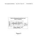 |  |
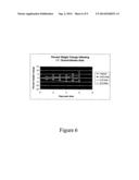 | 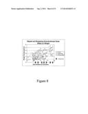 |
 |  |
 |  |
 |  |
 |  |
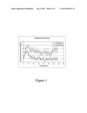 |  |
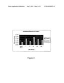 |  |
 | 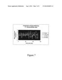 |
 |  |
 |  |
 | 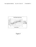 |
 |
| Similar patent applications: | |
| Date | Title |
|---|---|
| 2014-06-26 | Compositions and methods for treating celiac sprue disease |
| 2014-06-26 | Compositions and methods for targeting type 1 interferon producing cells |
| 2014-06-26 | Vegf-specific antagonists for adjuvant and neoadjuvant therapy and the treatment of early stage tumors |
| 2014-06-26 | Preselection of subjects for therapeutic treatment based on hypoxic status |
| New patent applications in this class: | |
| Date | Title |
|---|---|
| 2022-05-05 | Myc, cyclin t1 and/or cdk9 for use in the treatment of degenerative heart and cns disorders |
| 2022-05-05 | Crystallized oxalate decarboxylase and methods of use |
| 2019-05-16 | Method for mitigating metastasis |
| 2016-12-29 | Methods for analysis of somatic mobile elements, and uses thereof |
| 2016-09-01 | New methods to produce active tert |
| New patent applications from these inventors: | |
| Date | Title |
|---|---|
| 2020-04-16 | Durable treatment with 4-aminopyridine in patients with demyelination |
| 2020-04-16 | Use of 4-aminopyridine to improve neuro-cognitive and/or neuro-psychiatric impairment in patients with demyelinating and other nervous system conditions |
| 2019-01-03 | Durable treatment with 4-aminopyridine in patients with demyelination |
| 2018-06-07 | Methods of reducing extravasation of inflammatory cells |
| 2018-06-07 | Sustained release aminopyridine composition |
| Top Inventors for class "Drug, bio-affecting and body treating compositions" | |
| Rank | Inventor's name |
|---|---|
| 1 | David M. Goldenberg |
| 2 | Hy Si Bui |
| 3 | Lowell L. Wood, Jr. |
| 4 | Roderick A. Hyde |
| 5 | Yat Sun Or |