Patent application title: Anti-GLP-1R Antibodies and Their Uses
Inventors:
Karyn O'Neil (Radnor, PA, US)
Karyn O'Neil (Radnor, PA, US)
Kristen Picha (Radnor, PA, US)
IPC8 Class: AC07K1628FI
USPC Class:
530324
Class name: Chemistry: natural resins or derivatives; peptides or proteins; lignins or reaction products thereof peptides of 3 to 100 amino acid residues 25 or more amino acid residues in defined sequence
Publication date: 2011-04-28
Patent application number: 20110098443
Inventors list |
Agents list |
Assignees list |
List by place |
Classification tree browser |
Top 100 Inventors |
Top 100 Agents |
Top 100 Assignees |
Usenet FAQ Index |
Documents |
Other FAQs |
Patent application title: Anti-GLP-1R Antibodies and Their Uses
Inventors:
Karyn O'Neil
Kristen Picha
Agents:
Assignees:
Origin: ,
IPC8 Class: AC07K1628FI
USPC Class:
Publication date: 04/28/2011
Patent application number: 20110098443
Abstract:
The present invention relates to antibodies reactive with GLP-1R, and
methods of making and using them.Claims:
1. An isolated antibody reactive with glucagon-like peptide receptor 1
(GLP-1R) comprising the amino acid sequences of the light chain
complementarity determining regions (CDRs) 1, 2 and 3 as shown in SEQ ID
NOs: 3, 4 and 5, respectively and the amino acid sequences of the heavy
chain CDRs 1, 2 and 3 as shown in SEQ ID NOs: 6, 7 and 8, respectively.
2. An isolated antibody reactive with glucagon-like peptide receptor 1 (GLP-1R) comprising a light chain variable region (VL) having the amino acid sequence shown in SEQ ID NO: 9 and a heavy chain variable region (VH) having the amino acid sequence shown in SEQ ID NO: 10.
3. An isolated antibody reactive with glucagon-like peptide receptor 1 (GLP-1R) comprising a light chain (LC) having the amino acid sequence shown in SEQ ID NO: 11 and a heavy chain (HC) having the amino acid sequence shown in SEQ ID NO: 13.
4. An isolated antibody reactive with glucagon-like peptide receptor 1 (GLP-1R) comprising a light chain (LC) having the amino acid sequence shown in SEQ ID NO: 12 and a heavy chain (HC) having the amino acid sequence shown in SEQ ID NO: 14.
5. The isolated antibody of claim 1, 2, 3 or 4 wherein the antibody is an antagonist of GLP-1R.
6. The isolated antibody of claim 1, 2, 3 or 4 wherein the antibody comprises a Fab fragment.
7. The isolated antibody of claim 1, 2, 3 or 4 or 2 having an isotype selected from a group consisting of human IgG1 and mouse IgG2a.
8. An isolated antibody light chain comprising the amino acid sequence shown in SEQ ID NOS: 9, 11 or 12.
9. An isolated antibody heavy chain comprising the amino acid sequence shown in SEQ ID NO: 10, 13 or 14.
10. An isolated polynucleotide encoding an antibody light chain comprising the CDR amino acid sequences shown in SEQ ID NOs: 3, 4 and 5.
11. An isolated polynucleotide encoding an antibody heavy chain comprising the CDR amino acid sequences shown in SEQ ID NOs: 6, 7 and 8.
12. An isolated polynucleotide encoding an antibody light chain comprising the amino acid sequence shown in SEQ ID NO: 9, 11 and 12.
13. An isolated polynucleotide encoding an antibody heavy chain comprising the amino acid sequence shown in SEQ ID NO: 10, 13 and 14.
14. An isolated polynucleotide comprising a polynucleotide having a sequence shown in SEQ ID NOs: 15 and 16.
15. A vector comprising at least one polynucleotide of claims 10-14.
16. A host cell comprising the vector of claim 15.
17. A method of making an antibody reactive with GLP-1R comprising culturing the host cell of claim 16 and recovering the antibody produced by the host cell.
Description:
[0001] This application claims the benefit of U.S. Provisional Application
No. 61/255,532, filed 28 Oct. 2009, and U.S. Provisional Application No.
61/313,289, filed 12 Mar. 2010, the entire contents of which are
incorporated herein by reference.
FIELD OF THE INVENTION
[0002] The present invention relates to antibodies reactive with GLP-1R, and methods of making and using them.
BACKGROUND OF THE INVENTION
[0003] Diabetes is a growing epidemic that is estimated to affect over 300 million people by the year 2025, for which Type 2 diabetes accounts for 90-95% of all cases. Complications resulting from sustained elevated plasma glucose levels include cardiovascular disease, nephropathy, neuropathy, and retinopathy. In addition, the β-cells of the pancreas are destroyed and thus insulin secretion ceases during the later stages of the disease. Current treatments for diabetes can result in hypoglycemia and weight gain, and because patients may become resistant to treatment regimens over time, this may culminate in the need for insulin therapy at later stages of the disease (Narayan et al., Diabetes Research and Clinical Practice 50:S77-S84, 2000; Moller, Nature 414:821-827, 2001).
[0004] Glucagon like peptide-1 (GLP-1) is a 30-amino acid peptide (SEQ ID NO: 17) secreted from the L-cells of the intestine following an oral glucose challenge (Mojsov et al., J. Biol. Chem. 261:11880-11884, 1986; Orskov et al., Diabetes 43:535-539, 1994; Drucker et al., Proc. Natl. Acad. Sci. USA 84:3431-3438, 1987; Suzuki et al., Endocrinology 125:3109-3114, 1989). In response to glucose, GLP-1 binds to the GLP-1 receptor (GLP-1R) on the pancreas and induces insulin secretion (human GLP-1R amino acid sequence shown in SEQ ID NO: 1, mouse GLP-1R amino acid sequence shown in SEQ ID NO: 2). It has also been shown that GLP-1 reduces gastric emptying which decreases the bolus of glucose that is released into the circulation and may reduce food intake (Wettergren et al., Dig. Dis. Sci. 38:665-673, 1993). GLP-1 has also been shown to inhibit apoptosis and increase proliferation of the β-cells in the pancreas (Drucker et al., Mol. Endocrinology. 17:161-171, 2003; Perfetti et al., Endocrinology 141:4600-4605, 2000; Hui et al., Endocrinology 144:1444-1455, 2003; Farilla et al., Endocrinology 143:4397-4408, 2002). Thus, GLP-1 has attractive properties for a therapeutic to lower blood glucose and preserve the β-cells of the pancreas of diabetic patients.
[0005] It is likely that functional GLP-1 receptors localized in different types of tissues and cells contribute to the overall observed efficacy of the pleiotropic GLP-1 peptide. For example, it is unclear whether GLP-1 regulates food intake via a centrally or peripherally-mediated mechanism. Current GLP-1R tissue distribution studies have relied almost entirely upon mRNA levels rather than protein levels (Bullock et al., Endocrinology 137:2968-2978; Valverde et al., Endocrine 23:77-84, 2004). Further receptor biodistribution studies would benefit from the availability of specific GLP-1R antibodies that cross-react across species, thus facilitating measurement of protein levels for example in defined brain regions and peripheral tissues across multiple species. Antibodies that identify and neutralize the activity of the GLP-1R would facilitate phamacodynamic studies. For example, the antibody could be used as a tool to dissect the contributions of brain versus peripheral GLP-1 receptors that may be responsible for mediating GLP-1 effects.
[0006] Therefore, there is a need for antibodies against GLP-1R that facilitate biodistribution, pharmacodynamic, and mechanism of action studies of the GLP-1R and its ligands.
SUMMARY OF THE INVENTION
[0007] One aspect of the invention is an isolated antibody reactive with glucagon-like peptide receptor 1 (GLP-1R) comprising the amino acid sequences of the light chain complementarity determining regions (CDRs) 1, 2 and 3 as shown in SEQ ID NOs: 3, 4 and 5, respectively and the amino acid sequences of the heavy chain CDRs 1, 2 and 3 as shown in SEQ ID NOs: 6, 7 and 8, respectively.
[0008] Another aspect of the invention is an isolated antibody reactive with glucagon-like peptide receptor 1 (GLP-1R) comprising a light chain variable region (VL) having the amino acid sequence shown in SEQ ID NO: 9 and a heavy chain variable region (VH) having the amino acid sequence shown in SEQ ID NO: 10.
[0009] Another aspect of the invention is an isolated polynucleotide encoding an antibody light chain comprising the CDR amino acid sequences shown in SEQ ID NOs: 3, 4 and 5.
[0010] Another aspect of the invention is an isolated polynucleotide encoding an antibody heavy chain comprising the CDR amino acid sequences shown in SEQ ID NOs: 6, 7 and 8.
[0011] Another aspect of the invention is an isolated polynucleotide encoding an antibody light chain comprising the amino acid sequence shown in SEQ ID NOs: 9, 11 and 12.
[0012] Another aspect of the invention is an isolated polynucleotide encoding an antibody heavy chain comprising the amino acid sequence shown in SEQ ID NOs: 10, 13 and 14.
[0013] Another aspect of the invention is an isolated polynucleotide comprising a polynucleotide having a sequence shown in SEQ ID NOs: 15 and 16.
[0014] Another aspect of the invention is an isolated antibody light chain comprising the amino acid sequence shown in SEQ ID NOS: 9, 11 and 12.
[0015] Another aspect of the invention is an isolated antibody heavy chain comprising the CDR amino acid sequences shown in SEQ ID NOs: 10, 13 and 14.
[0016] Another aspect of the invention is a vector encoding the antibody heavy and light chains of the invention.
[0017] Another aspect of the invention is a host cell capable of expressing the isolated antibodies of the invention.
[0018] Another aspect of the invention is a method of making an antibody reactive with GLP-1R.
[0019] Another aspect of the invention is a hybridoma cell line expressing the isolated antibodies of the invention.
BRIEF DESCRIPTION OF THE FIGURES
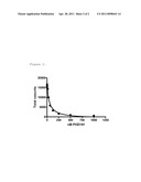
[0020] FIG. 1. PHD 191 inhibited GLP-1 peptide from binding to soluble human GLP-1R.
[0021] FIG. 2. PHD 191 inhibited 125I-GLP-1 binding to the human receptor.
[0022] Table 1 shows the description of the sequence listing.
TABLE-US-00001 SEQ ID NO: Description 1 human GLP-1R 2 mouse GLP-1R 3 PHD 191 LC-CDR1 4 PHD 191 LC-CDR2 5 PHD 191 LC-CDR3 6 PHD 191 HC-CDR1 7 PHD 191 HC-CDR2 8 PHD 191 HC-CDR3 9 PHD 191 LV 10 PHD191 HV 11 PHD 191 LC, human κ 12 PHD 191 LC, mouse κ 13 PHD 191 HC, human IgG1 14 PHD 191 HC, mouse IgG2a 15 DNA, PHD 191 LC, human κ 16 DNA, PHD 191 HC, human IgG1 17 GLP-1 (3-37)
DETAILED DESCRIPTION OF THE INVENTION
[0023] All publications, including but not limited to patents and patent applications, cited in this specification are herein incorporated by reference as though fully set forth.
[0024] The term "antagonist" as used herein means a molecule that partially or completely inhibits, by any mechanism, an effect of another molecule such as a receptor or a ligand. An antagonist is capable of, directly or indirectly, substantially counteracting, reducing or inhibiting GLP-1R biological activity. Antagonists can be antibodies, proteins, peptides, or the like. For example, an antibody antagonist can bind directly to GLP-1R and inhibit GLP-1R biological activity.
[0025] The term "reactive with" refers to antibody binding to a predetermined antigen with greater affinity than it has for other antigens or proteins. Typically, a reactive antibody binds with a dissociation constant (KD) of 10-7 M or less, and binds to the predetermined antigen with a KD that is at least tenfold less than its KD for a non-specific antigen (e.g., BSA, casein, or any other specified polypeptide). The phrases "an antibody recognizing an antigen" and "an antibody specific for an antigen" are used interchangeably herein with the term "an antibody reactive with an antigen" e.g., an antibody reactive with GLP-1R. The dissociation constant can be measured using standard procedures as described below.
[0026] The term "antibodies" as used herein is meant in a broad sense and includes immunoglobulin or antibody molecules including polyclonal antibodies, monoclonal antibodies including murine, human, human-adapted, humanized and chimeric monoclonal antibodies and antibody fragments.
[0027] In general, antibodies are proteins or peptide chains that exhibit binding specificity to a specific antigen. Intact antibodies are heterotetrameric glycoproteins, composed of two identical light chains and two identical heavy chains. Typically, each light chain is linked to a heavy chain by one covalent disulfide bond, while the number of disulfide linkages varies between the heavy chains of different immunoglobulin isotypes. Each heavy and light chain also has regularly spaced intrachain disulfide bridges. Each heavy chain has at one end a variable domain (VH) followed by a number of constant domains. Each light chain has a variable domain at one end (VL) and a constant domain at its other end; the constant domain of the light chain is aligned with the first constant domain of the heavy chain and the light chain variable domain is aligned with the variable domain of the heavy chain. Antibody light chains of any vertebrate species can be assigned to one of two clearly distinct types, namely kappa (κ) and lambda (λ), based on the amino acid sequences of their constant domains.
[0028] Immunoglobulins can be assigned to five major classes, namely IgA, IgD, IgE, IgG and IgM, depending on the heavy chain constant domain amino acid sequence. IgA and IgG are further sub-classified as the isotypes IgA1, IgA2, IgG1, IgG2, IgG3 and IgG4.
[0029] The term "antibody fragments" means a portion of an intact antibody, generally the antigen binding or variable region of the intact antibody. Examples of antibody fragments include Fab, Fab', F(ab')2 and Fv fragments, diabodies, single chain antibody molecules and multispecific antibodies formed from at least two intact antibodies.
[0030] An immunoglobulin light or heavy chain variable region consists of a "framework" region interrupted by three "antigen-binding sites". The antigen-binding sites are defined using various terms as follows: (i) Complementarity Determining Regions (CDRs) are based on sequence variability (Wu and Kabat, J. Exp. Med. 132:211-250, 1970). Generally, the antigen-binding site has six CDRs; three in the VH(HCDR1, HCDR2, HCDR3), and three in the VL (LCDR1, LCDR2, LCDR3) (Kabat et al., Sequences of Proteins of Immunological Interest, 5th Ed. Public Health Service, National Institutes of Health, Bethesda, Md., 1991). (ii) The term "hypervariable region", "HVR", or "HV" refers to the regions of an antibody variable domain which are hypervariable in structure as defined by Chothia and Lesk (Chothia and Lesk, Mol. Biol. 196:901-917, 1987). Generally, the antigen-binding site has six hypervariable regions, three in VH(H1, H2, H3) and three in VL (L1, L2, L3). Chothia and Lesk refer to structurally conserved HVs as "canonical structures". Numbering systems as well as annotation of CDRs and HVs have recently been revised by Abhinandan and Martin (Abhinandan and Martin, Mol. Immunol. 45:3832-3239, 2008). (iii) Another definition of the regions that form the antigen-binding site has been proposed by Lefranc (Lefranc et al., Dev. Comp. Immunol. 27:55-77, 2003) based on the comparison of V domains from immunoglobulins and T-cell receptors. The International ImMunoGeneTics (IMGT) database (http:_//www_imgt_org) provides a standardized numbering and definition of these regions. The correspondence between CDRs, HVs and IMGT delineations is described in Lefranc et al., Dev. Comp. Immunol. 27:55-77, 2003. (iv) The antigen-binding site can also be delineated based on Specificity Determining Residue Usage (SDRU), according to Almagro (Almagro, Mol. Recognit. 17:132-43, 2004), where Specificity Determining Residues (SDR), refers to amino acid residues of an immunoglobulin that are directly involved in antigen contact. SDRU as defined by Almagro is a precise measure of a number and distribution of SDR for different types of antigens as defined by analyses of crystal structures of antigen-antibody complexes.
[0031] The term "consensus region" as used herein means an antigen-binding site delineated to include all amino acid residues delineated individually by Kabat, Chothia or IMGT, or any other suitable antigen-binding site delineation.
[0032] "Framework" or "framework sequence" are the remaining sequences of a variable region minus the antigen-binding sites. Because the exact definition of an antigen-binding site can be determined by various delineations as described above, the meaning of a framework sequences is subject to correspondingly different interpretations. A framework sequence as used herein means those sequences within the variable region of an antibody other than those defined to be antigen-binding site sequences.
[0033] The term "monoclonal antibody" (mAb) as used herein means an antibody (or antibody fragment) obtained from a population of substantially homogeneous antibodies. Monoclonal antibodies are highly specific, typically being directed against a single antigenic determinant. The modifier "monoclonal" indicates the substantially homogeneous character of the antibody and does not require production of the antibody by any particular method.
[0034] The term "GLP-1R biological activity" as used herein refers to any activity occurring as a result of ligand binding, for example GLP-1(7-37) to GLP-1R. Exemplary GLP-1R activities are an intracellular accumulation of cyclic AMP, calcium release, insulin secretion, or kinase-mediated phosphorylation of target proteins. Assays measuring GLP-1R biological activity are well known in the art.
[0035] The term "GLP-1R" as used herein refers to human GLP-1R protein having an amino acid sequence as shown in GenBank accession number no: NP--002053 (SEQ ID NO: 1). The mouse GLP-1R has an amino acid sequence shown in GenBank accession number no: NP--067307 (SEQ ID NO: 2).
[0036] Conventional one and three-letter amino acid codes are used herein as follows:
TABLE-US-00002 Amino acid Three-letter code One-letter code Alanine ala A Arginine arg R Asparagine asn N Aspartate asp D Cysteine cys C Glutamate glu E Glutamine gln Q Glycine gly G Histidine his H Isoleucine ile I Leucine leu L Lysine lys K Methionine met M Phenylalanine phe F Proline pro P Serine ser S Threonine thr T Tryptophan trp W Tyrosine tyr Y Valine val V
Compositions of Matter
[0037] The present invention relates to antibodies that are reactive with human GLP-1R, and uses of such antibodies. Such GLP-1R antibodies may have the properties of binding a GLP-1R receptor and inhibiting GLP-1R receptor-mediated signaling. Exemplary mechanisms by which GLP-1R signaling may be inhibited by such antagonists include inhibition of ligand binding, or inhibition of downstream signaling pathways. Antibodies that are reactive with human GLP-1R may also be non-neutralizing antibodies which can be used to detect GLP-1R protein in biological samples, for example serum, tissue, cells, or fixed tissue or cells. The antibodies of the invention are useful for example as research reagents, diagnostic reagents, evaluation of biodistribution of GLP-1R, and in pharmacodynamic studies. For example, GLP-1R antibodies are unlikely to cross the blood brain barrier. Thus, antagonistic GLP-1R antibodies, when dosed to animals can be used to determine the effect of selectively blocking the peripheral but not central GLP-1 activities on for example food intake. Further, antibodies specific for GLP-1R are valuable in determining the receptor levels in various sections of the brain and in the vagus nerve, as expression of GLP1-R in either could contribute to the GLP-1 effect on food intake.
[0038] One embodiment of the invention is an isolated antibody reactive with GLP-1R having the light chain complementarity determining regions (CDR) amino acid sequences (LCDR1), (LCDR2) and (LCDR3) having amino acid sequences as shown in SEQ ID NO: 3, 4 and 5, respectively, and heavy chain complementarity determining regions (CDR) amino acid sequences (HCDR1), (HCDR2) and (HCDR3) having amino acid sequences as shown in SEQ ID NO: 6, 7 and 8, respectively.
[0039] In another aspect, the invention provides an isolated antibody reactive with GLP-1R comprising a light chain variable region having the amino acid sequence shown in SEQ ID NO: 9 and a heavy chain variable region having the amino acid sequence shown in SEQ ID NO: 10.
[0040] In another aspect, the invention provides isolated light chains having the amino acid sequences shown in SEQ ID NO: 11 and 12, and isolated heavy chains having the amino acid sequences shown in SEQ ID NO: 13 and 14.
[0041] Another embodiment of the invention is an isolated antagonistic GLP-1R antibody. To generate antagonistic antibodies to the GLP-1R, the GLP-1 binding domain in the receptor must be targeted. GLP-1R belongs to a subclass of seven transmembrane G-protein coupled receptors, and it has been challenging in the past to develop antibodies against this type of receptor (Michel at al., Naunyn-Schmied Arch Pharmacol. 379:385-388, 2009; Sangawa et. al., Hybridoma 27:331-335, 2008; Huang et. al., J. Mol. Recog. 18:327-333, 2005). The GLP-1R contains a relatively large extracellular N-terminal domain that has been demonstrated to be a major determinant in GLP-1 binding affinity (Wilmen et al., Feder. Europ. Biochem. Soc. 398:43-47, 1996). Therefore, antibodies raised against the N-terminal domain are likely to compete with GLP-1R agonists for binding and consequently inhibit the downstream effects.
[0042] Exemplary antibodies may be antibodies of the IgG, IgD, IgGA or IgM isotypes. Additionally, such antibodies can be post-translationally modified by processes such as glycosylation, isomerization, deglycosylation or non-naturally occurring covalent modification such as the addition of polyethylene glycol moieties (pegylation) and lipidation. Such modifications may occur in vivo or in vitro. For example, the antibodies of the invention can be conjugated to polyethylene glycol (PEGylated) to improve their pharmacokinetic profiles. Conjugation can be carried out by techniques known to those skilled in the art. Conjugation of therapeutic antibodies with PEG has been shown to enhance pharmacodynamics while not interfering with function (Deckert et al., Int. J. Cancer 87:382-390, 2000; Knight et al., Platelets 15:409-418, 2004; Leong et al., Cytokine 16:106-119, 2001; and Yang et al., Protein Eng. 16:761-770, 2003).
[0043] Pharmacokinetic properties of the antibodies of the invention could also be enhanced through Fc modifications by techniques known to those skilled in the art. For example, IgG4 isotype heavy chains contain a Cys-Pro-Ser-Cys (CPSC) motif in the hinge region capable of forming either inter- or intra-heavy chain disulfide bonds, i.e., the two Cys residues in the CPSC motif may disulfide bond with the corresponding Cys residues in the other heavy chain (inter) or the two Cys residues within a given CPSC motif may disulfide bond with each other (intra). It is believed that in vivo isomerase enzymes are capable of converting inter-heavy chain bonds of IgG4 molecules to intra-heavy chain bonds and vice versa (Aalberse and Schuurman, Immunology 105:9-19, 2002). Accordingly, since the heavy:light chain pairs in those IgG4 molecules with intra-heavy chain bonds in the hinge region are not covalently associated with each other, they may dissociate into HL monomers that then reassociate with HL monomers derived from other IgG4 molecules forming bispecific, heterodimeric IgG4 molecules. In a bispecific IgG antibody the two Fabs of the antibody molecule differ in the epitopes that they bind. Substituting the Ser residue in the hinge region CPSC motif of IgG4 with Pro results in "IgG1-like behavior," i.e., the molecules form stable disulfide bonds between heavy chains and therefore, are not susceptible to HL exchange with other IgG4 molecules. In one embodiment, the antibodies of the invention will comprise an IgG4 Fc domain with a S to P mutation in the CPSC motif. The location of the CPSC motif is typically found at residue 228 of a mature heavy chain but can change depending on CDR lengths.
[0044] Further, amino acid sequences can be changed or removed within the Fc domain that affect binding to Fc receptors other than an FcRn salvage receptor in the antibodies of the invention. For example, the antibody Fc regions involved in ADCC activity can be removed in the antibodies of the invention. For example, mutation of Leu234/Leu235 in the hinge region of IgG1 to L234A/L235A or Phe235/Leu236 in the hinge region of IgG4 to P235A/L236A minimizes FcR binding and reduces the ability of the immunoglobulin to mediate complement dependent cytotoxicity and ADCC. In one embodiment, the antibodies of the invention will comprise an IgG4 Fc domain with a P235A/L236A mutations. The location of these residues identified above is typical in a mature heavy chain but can change depending on CDR lengths.
[0045] The antibody antagonists of the invention may bind GLP-1R with a Kd less than or equal to about 10-7, 10-8, 10-9, 10-10, 10-11 or 10-12 M. The affinity of a given molecule for a GLP-1R can be determined experimentally using any suitable method. Such methods may utilize Biacore or KinExA instrumentation, ELISA or competitive binding assays known to those skilled in the art.
[0046] Antagonist antibody molecules binding to GLP-1R with a desired affinity can be selected from libraries of variants or fragments by techniques including antibody affinity maturation. Antagonist antibodies can be identified based on their inhibition of GLP-1R biological activity using any suitable method. Such methods may utilize reporter-gene assays or assays measuring intracellular cyclic AMP production known to those skilled in the art. Suitable antibodies for distribution and pharmacodynamic studies can be tested using routine methodology, such as immunohistochemistry or ELISA assays.
[0047] Antibodies of the present invention can be produced by a variety of techniques, for example by the hybridoma method of Kohler et al., Nature 256:495-497, 1975. Chimeric mAbs containing a light chain and heavy chain variable region derived from a donor antibody (typically murine) in association with light and heavy chain constant regions derived from an acceptor antibody (typically another mammalian species such as human) can be prepared by the method disclosed in U.S. Pat. No. 4,816,567. CDR-grafted mAbs having CDRs derived from a non-human donor immunoglobulin (typically murine) and the remaining immunoglobulin-derived parts of the molecule being derived from one or more human immunoglobulins can be prepared by techniques known to those skilled in the art such as that disclosed in U.S. Pat. No. 5,225,539. Human framework sequences useful for grafting can be selected from relevant databases by those skilled in the art. Optionally, CDR-grafted mAbs can be further humanized by incorporating altered framework support residues to preserve binding affinity by techniques such as those disclosed in Queen et al., Proc. Natl. Acad. Sci. (USA), 86:10029-10032, 1989 and Hodgson et al., Bio/Technology, 9:421, 1991. Fully human mAbs lacking any non-human sequences can be prepared from human immunoglobulin transgenic mice by techniques referenced in, e.g., Lonberg et al., Nature 368:856-859, 1994; Fishwild et al., Nature Biotechnology 14:845-851, 1996; and Mendez et al., Nature Genetics 15:146-156, 1997. Human mAbs can also be prepared and optimized from phage display libraries by techniques referenced in, e.g., Knappik et al., J. Mol. Biol. 296:57-86, 2000; and Krebs et al., J. Immunol. Meth. 254:67-84 2001.
[0048] Preparation of Immunogenic Antigens, and Monoclonal Antibody production can be performed using any suitable technique such as recombinant protein production. The immunogenic antigens can be administered to an animal in the form of purified protein, or protein mixtures including whole cells or cell or tissue extracts, or the antigen can be formed de novo in the animal's body from nucleic acids encoding said antigen or a portion thereof.
[0049] Another embodiment of the invention is an isolated polynucleotide encoding the antibodies of the invention or their complement. Exemplary polynucleotides are disclosed herein, however, other polynucleotides which, given the degeneracy of the genetic code or codon preferences in a given expression system, encode the antibody of the invention are also within the scope of the invention. Exemplary isolated polynucleotides comprise the polynucleotides having a sequence shown in SEQ ID NO: 15 or 16. The isolated nucleic acids of the present invention can be made using (a) recombinant methods, (b) synthetic techniques, (c) purification techniques, or combinations thereof, as well-known in the art. DNA encoding the monoclonal antibodies is readily isolated and sequenced using methods known in the art (e.g., by using oligonucleotide probes that are capable of binding specifically to genes encoding the heavy and light chains of murine antibodies). Where a hybridoma is produced, such cells can serve as a source of such DNA. Alternatively, using display techniques wherein the coding sequence and the translation product are linked, such as phage or ribosomal display libraries, the selection of the binder and the nucleic acid is simplified. After phage selection, the antibody coding regions from the phage can be isolated and used to generate whole antibodies, including human antibodies, or any other desired antigen binding fragment, and expressed in any desired host, including mammalian cells, insect cells, plant cells, yeast, and bacteria.
[0050] Another embodiment of the invention is a vector comprising at least one polynucleotide of the invention. Such vectors may be plasmid vectors, viral vectors, transposon based vectors or any other vector suitable for introduction of the polynucleotides of the invention into a given organism or genetic background by any means. An exemplary vector for expression polypeptides of the invention consists of a CMV promoter, T7 binding site, VH leader sequence, BGH polyA site, f1 origin of replication, ColE1 origin, CMV promoter, and beta-lactamase, and may include polynucleotides encoding an antibody constant region, for example a mouse gamma2a constant region.
[0051] Another embodiment of the invention is a host cell comprising any of the polynucleotides of the invention such as a polynucleotide encoding a polypeptide comprising an immunoglobulin light chain having the amino acid sequence shown in SEQ ID NO: 11 or an immunoglobulin heavy chain having the amino acid sequence shown in SEQ ID NO: 13. Such host cells may be eukaryotic cells, bacterial cells, plant cells or archeal cells. Exemplary eukaryotic cells may be of mammalian, insect, avian or other animal origins. Mammalian eukaryotic cells include immortalized cell lines such as hybridomas or myeloma cell lines such as SP2/0 (American Type Culture Collection (ATCC), Manassas, Va., CRL-1581), NS0 (European Collection of Cell Cultures (ECACC), Salisbury, Wiltshire, UK, ECACC No. 85110503), FO (ATCC CRL-1646) and Ag653 (ATCC CRL-1580) murine cell lines. An exemplary human myeloma cell line is U266 (ATTC CRL-TIB-196). Other useful cell lines include those derived from Chinese Hamster Ovary (CHO) cells such as CHO-K1SV (Lonza Biologics), CHO-K1 (ATCC CRL-61, Invitrogen) or DG44.
[0052] Another embodiment of the invention is a method of making an antibody reactive with GLP-1R comprising culturing a host cell of the invention and recovering the antibody produced by the host cell. Methods of making antibodies and purifying them are well known in the art.
[0053] Another embodiment of the invention is a hybridoma cell line that produces an antibody of the invention.
Example 1
Generation of Soluble GLP-1R Protein
[0054] cDNAs encoding soluble human and mouse GLP-1 receptors corresponding to amino acids 24-145 of human (SEQ ID NO: 1) and amino acids 21-143 of mouse (SEQ ID NO: 2) GLP-1 receptor were prepared using gene synthesis techniques (U.S. Pat. No. 6,670,127; U.S. Pat. No. 6,521,427). Plasmids for expression of the synthetic soluble receptors were prepared using standard molecular biology techniques. Each plasmid encoded a Kozak sequence, a human growth hormone signal sequence, the soluble region of human or murine GLP-1R, and a C-terminal 6×His tag for easy purification. Vectors were transiently transfected into HEK293T cells using standard methods and the secreted proteins were purified by IMAC using Talon resin.
[0055] The soluble human GLP-1R antigen (#3527) was labeled with biotin using EZ link Sulfo-NHS-LC-biotin according to the manufacturer's instructions (Pierce Chemical Company, St. Louis, Mo.). Activity of biotinylated 3527 was confirmed using CNT0736, a GLP-1 Mimetibody® construct (Picha et al., Diabetes 57:1926-34, 2008). A 96-well Maxisorp plate was coated (100 μl/well) with 5-μg/mL of either biotinylated or unlabeled 3527 and incubated at 4° C. overnight. Plates were washed with TBST and blocked with 300 μl/well of 1× Chemiblocker for 1 hour at RT. Plates were washed with TBST. Serial dilutions (1:3) of either CNT0736 or CNT01996 (negative control lacking the GLP-1 peptide) were made starting at 10 μg/ml, added to the appropriate wells in the plate (100 μl/well), and allowed to bind for 1 hr at RT. Plates were washed with TBST. Plates were treated for 1 hour at RT with 100 μl/well of goat anti-human IgG (H+L)-AP detection antibody diluted 1:5000 in TBST. AttoPhos substrate (1:5) detected AP activity. Biotinylated 3527 retained a comparable binding profile relative to unlabeled 3527 (data not shown).
Example 2
Identification of Anti-Human GLP-1R Antibodies
[0056] Solution panning of the Human Combinatorial Antibody Library GOLD (HuCAL GOLD) was performed using a biotinylated antigen-streptavidin magnetic bead capture method as described (Rothe et al., J. Mol. Biol. 376:1182-1200, 2008; Steidl et al., Mol. Immunol. 46: 135-144, 2008) in three subsequent rounds. Fabs recovered from the last round of selection were subcloned into the pMORPHx9_Mx_MH Fab expression vector containing a Myc and His6 tag (Rauchenberger et al., J. Biol. Chem. 278:38194-38205, 2003). E. coli TG1 cells (#200123, Stratagene, La Jolla, Calif.) were transformed via electroporation, and the Fabs produced by the bacteria.
[0057] Fabs were screened for binding to 3527 (Rauchenberger et al., J. Biol. Chem. 278:38194-38205, 2003). Fabs that bound 3527 greater than five times background were sequenced to determine the number of unique Fab clones. 39 Fabs were selected for sequencing. From the 39 clones sequenced, PHD191 was identified 3 times, or 8% of the total hits. The amino acid sequences of the CDRs and light and heavy chain variable regions of PHD191 are shown in SEQ ID NOs: 3-10.
Example 3
PHD 191 Fab Inhibits GLP-1 Binding to Soluble Human GLP-1R
[0058] 96-well Maxisorp plates were coated (100 μl/well) with 5 μg/ml of soluble human GLP-1R (shGLP1-R) in PBS and incubated at 4° C. overnight. Plates were washed with TBST and blocked with 300 μl/well of 1× Chemiblocker for 1 hour at RT. Plates were washed with TBST. A mixture was made of (1:2) serial dilutions of PHD 191 Fab starting at 25 μg/ml and biotinylated human GLP-1 peptide held constant at 75 ng/ml. This mixture was added (100 μl/well) to the plates and incubated at RT for 1 hour. Plates were washed with TBST. Plates were treated for 1 hr at RT with 100 μl/well of Strep-AP detection antibody diluted 1:2000 in TBST. AttoPhos substrate (1:5) detected AP activity. PHD 191 inhibited human GLP-1 peptide binding to soluble human GLP-1R (FIG. 1).
Example 4
Transfer of PHD 191 to Human IgG1/Human IgKappa and Murine IgG2a/Murine IgKappa Formats
[0059] PHD 191 was sub-cloned into full-length immunoglobulin expression vectors (Krebs et al., J. Immunol. Meth. 254:67-84, 2001). Similar constructs were created for the PHD 191 murine IgKappa, human IgG1, and human IgKappa constructs.
[0060] Full-length heavy and light chain vectors were transformed into DH10B cells according to the manufacturer's instructions. Individual colonies were screened for insertion of the correct variable region by PCR and sequence analysis. DNA was isolated for each of the correct clone and used for transfections using standard procedures, and the inserts were sequenced. The amino acid sequences of the resulting light and heavy chains are shown in SEQ ID NOs: 11-14.
[0061] The vectors encoding appropriate heavy and light chain partners were co-transfected into HEK293E cells. Supernatants containing the secreted antibodies were affinity purified using Protein A resin according to standard protocols.
Example 5
Characterization of PHD 191
[0062] Cross-Reactivity with Murine GLP-1R
[0063] The binding specificity of PHD 191 was characterized using two different ELISA assay formats. Both assay formats showed that PHD 191 binds human GLP-1R and also cross-reacts with murine GLP-1R. The first characterization assay was a neutravidin capture assay. 96-well Maxisorp plates were coated (100 μl/well) with 5 μg/ml of neutravidin in PBS and were incubated at 4° C. overnight. Coated plates were washed with TBST. 2 μg/ml of biotinylated soluble murine GLP-1R (3445), biotinylated soluble human GLP-1R (3527), or other biotinylated control proteins (mouse IL-23, mouse IL-12, mouse IL-18, rat transferrin) were added (100 μl/well) and allowed to bind to neutravidin for 1 hour at RT. Plates were washed with TBST and blocked with 300 μl/well of 1× Chemiblocker for 1 hour at RT. Plates were washed with TBST. Serial dilutions (1:2) of the PHD 191 mAb were made starting at 5 μg/ml, added to the plate (100 μl/well), and allowed to bind for 1 hr at RT. Plates were washed with TBST. Plates were treated for 1 hour at RT with 100 μl/well of goat anti-human IgG (H+L)-AP detection antibody diluted 1:5000 in TBST. AttoPhos substrate (1:5) detected AP activity. PHD 191 bound specifically to human soluble GLP-1R, cross-reacted with murine soluble GLP-1R and had no binding to unrelated biotinylated proteins (0.4 nM and 2.5 nM for human and murine soluble GLP-1R, respectively).
[0064] To confirm the cross-reactivity of PHD 191 to murine GLP-1R, a second characterization assay was performed using unlabeled receptor coated directly onto the plate. 96-well Maxisorp plates were coated (100 μl/well) with 5 μg/ml of unlabeled murine or human GLP-1R in PBS and were incubated at 4° C. overnight. Coated plates were washed with TBST and blocked with 300 μl/well of 1× Chemiblocker for 1 hour at RT. Plates were washed with TBST. Serial dilutions (1:3) of the PHD 191 Mab were made starting at 30 μg/ml, added to the plate (100 μl/well), and allowed to bind for 1 hr at RT. Plates were washed with TBST. Plates were treated for 1 hour at RT with 100 μl/well of goat anti-human IgG (H+L)-AP detection antibody diluted 1:5000 in TBST. AttoPhos substrate (1:5) detected AP activity. The direct coat assay confirmed that PHD 191 cross-reacted with soluble murine GLP-1R (0.8 nM and 11.9 nM, for human and murine soluble GLP-1R, respectively).
Inhibition of GLP-1 Binding to the Human GLP-1R
[0065] An 125I GLP-1 binding assay was used to determine whether PHD 191 could inhibit GLP-1 peptide binding to the human receptor expressed on cells. HEK293F cells over-expressing the human GLP-1 receptor were incubated overnight at 4° C. in the presence of 3 nM 125I GLP-1 along with a dose titration of PHD 191. Unbound 125I GLP-1 was washed away and cell associated counts were measured. The data were fit to a hyperbola to obtain an IC50 of 48.5 nM (FIG. 2). Assuming a Kd for GLP-1 binding to the human GLP-1R of 3 nM (determined previously) and competitive inhibition, this data indicates that the Ki for PHD 191 is approximately 20 nM.
Inhibition of Cyclic AMP Stimulation by GLP-1
[0066] Two assay formats were used to show that PHD 191 inhibited GLP-1 mediated cAMP accumulation. In the first format, a dose titration of PHD 191 in the presence of 0.3 nM GLP-1 peptide was added to HEK293F cells over-expressing the human GLP-1R receptor. In the second format, a dose response of GLP-1 peptide in the presence of 1 μM PHD 191 was added to the GLP-1R over-expressing cells. In both cases, total cAMP accumulation after seven minutes of stimulation was quantitated using the PerkinElmer LANCE cAMP Assay Kit. The dose titration of PHD 191 dose-dependently reduced the GLP-1 dependent cAMP accumulation, indicating a reduction in signaling of the GLP-1 peptide in the presence of the antibody. The data were fit to an equation describing a single binding event, providing an IC50 of 3.3 nM. PHD 191 also shifted the EC50 from 0.3 to 1.5 nM when GLP-1 peptide was tested in a cAMP assay.
Example 6
Tissue Distribution of Human GLP-1R
[0067] Protein expression of human GLP-1R was assessed in normal tissues by immunohistochemistry. GLP-1R protein was detected in pancreatic islets, CNS neurons and epithelial cells from gut.
[0068] The brain samples from normal donors were obtained from Analytical Biological Services, Inc. The brain dissections were made and qualified by a neurobiologist (Analytical Biological Services, Inc). Samples of other normal human tissues were obtained from the QualTek tissue bank, and qualified by a pathologist. Immunohistochemistry on human tissues was performed in the following manner. Four-micron tissue sections were cut from formalin fixed paraffin embedded blocks, placed on glass slides, and dried at 60° C. These were dewaxed through four 5-minute changes of xylene, followed by a graded alcohol series to distilled water. All tissue samples were tested for immunohistochemical reactivity to PHD 191 using the following protocol. Briefly, tissues were pretreated using sHIER 2 for 20 minutes and then digested for 10 minutes with Pro K at 1:15 dilution. Tissue samples were subsequently incubated with the primary antibody, PHD 191 for 63 hours at 4° C. at either 5 μg/ml or 7.5 μg/ml. For blocking experiments the tissues were incubated with PHD 191 in the presence of 20 molar excess of extracellular domain of GLP-1R. Mouse nonspecific isotype control antibody was used to confirm the specificity of staining. An ABC detection system using the MIPE protocol on the Techmate was used to label the bound primary antibody. After staining, slides were dehydrated through an alcohol series to absolute ethanol followed by xylene rinses. Slides were permanently coverslipped with glass coverslips and permount. Slides were examined under a microscope to assess staining. Positive staining is indicated by the presence of a brown chromogen (DAB-HRP) reaction product. Hematoxylin counterstain provided a blue nuclear stain to assess cell and tissue morphology. Representative images were obtained with an Olympus Microfire digital camera (M/N S97809) attached to an Olympus BX60 microscope and displayed on a Dell flat panel monitor with the DPI set at 96 and the screen resolution set at 1152 by 864 pixels. Images were brought into sharp focus using Koehler illumination. Some images taken with 40× or higher objective were taken as a series through the focal range of the field of view and combined with the Image-Pro Plus 5.1 (MediaCybernetics) extended depth of field function to obtain a composite image with sharp focus throughout the image field of view.
[0069] Pancreatic islets were identified in human pancreas section based on histological evaluation after a hematoxylin counterstain. IHC analysis of human pancreas showed the presence of human GLP-1R in islet cells. The specificity of staining was confirmed in blocking experiments where pre-incubation of PHD191 with extracellular domain of GLP-1R completely inhibited the staining. The GLP-1R was also detected in neurons in the amygdala, hypothalamus, arcuate nucleus, and area postrema. GLP-1R was not detected in two samples of brain cortex. No staining was detected in any brain sections stained with mouse nonspecific isotype control antibody.
[0070] In tissue samples containing epithelium, staining was occasionally seen in epithelial cells. In four of 14 lung samples some of the bronchial airways had epithelial cells with staining at the luminal surface and perinuclear staining in basal cells. In two of 4 colon samples, the luminal surface had strong staining. In one of 3 breast samples, perinuclear staining was seen in cells lining the ducts. No reactivity was seen in the negative controls for these samples.
[0071] In one of 3 samples of small intestine and in two of 4 colon samples, reactivity was seen in isolated crypt cells. The location and frequency of these stained crypt cells in the colon and small intestine is consistent with entero-endocrine cells.
[0072] Reactive immune cells were observed in a majority of the tissues. These were typically polymorphonuclear granulocytes within blood vessels or cells with macrophage or mast cell morphology. To confirm the specificity of staining, IHC was repeated with a subset of samples in the presence of an excess of antigen (N-terminal domain of GLP-1R). Under these conditions, PHD 191 reactivity was significantly inhibited in neurons, epithelial, and pancreatic islet cells. Only partial inhibition of PHD 191 reactivity in immune cells was observed, thus the specificity of PHD 191 staining in immune cells remains to be determined.
[0073] The data described here details the discovery and characterization of PHD 191. PHD 191 cross-reacted with murine GLP-1R and bound to human GLP-1R expressed on cells. PHD 191 was cloned into either a human IgG1 or mouse IgG2a format. PHD 191 inhibited GLP-1 binding to the human GLP-1 receptor and inhibited GLP-1 mediated cAMP accumulation. Antibodies with these properties are not available from a commercial source.
[0074] The present invention now being fully described, it will be apparent to one of ordinary skill in the art that many changes and modifications can be made thereto without departing from the spirit or scope of the appended claims.
Sequence CWU
1
171463PRTHomo sapiens 1Met Ala Gly Ala Pro Gly Pro Leu Arg Leu Ala Leu Leu
Leu Leu Gly1 5 10 15Met
Val Gly Arg Ala Gly Pro Arg Pro Gln Gly Ala Thr Val Ser Leu 20
25 30Trp Glu Thr Val Gln Lys Trp Arg
Glu Tyr Arg Arg Gln Cys Gln Arg 35 40
45Ser Leu Thr Glu Asp Pro Pro Pro Ala Thr Asp Leu Phe Cys Asn Arg
50 55 60Thr Phe Asp Glu Tyr Ala Cys Trp
Pro Asp Gly Glu Pro Gly Ser Phe65 70 75
80Val Asn Val Ser Cys Pro Trp Tyr Leu Pro Trp Ala Ser
Ser Val Pro 85 90 95Gln
Gly His Val Tyr Arg Phe Cys Thr Ala Glu Gly Leu Trp Leu Gln
100 105 110Lys Asp Asn Ser Ser Leu Pro
Trp Arg Asp Leu Ser Glu Cys Glu Glu 115 120
125Ser Lys Arg Gly Glu Arg Ser Ser Pro Glu Glu Gln Leu Leu Phe
Leu 130 135 140Tyr Ile Ile Tyr Thr Val
Gly Tyr Ala Leu Ser Phe Ser Ala Leu Val145 150
155 160Ile Ala Ser Ala Ile Leu Leu Gly Phe Arg His
Leu His Cys Thr Arg 165 170
175Asn Tyr Ile His Leu Asn Leu Phe Ala Ser Phe Ile Leu Arg Ala Leu
180 185 190Ser Val Phe Ile Lys Asp
Ala Ala Leu Lys Trp Met Tyr Ser Thr Ala 195 200
205Ala Gln Gln His Gln Trp Asp Gly Leu Leu Ser Tyr Gln Asp
Ser Leu 210 215 220Ser Cys Arg Leu Val
Phe Leu Leu Met Gln Tyr Cys Val Ala Ala Asn225 230
235 240Tyr Tyr Trp Leu Leu Val Glu Gly Val Tyr
Leu Tyr Thr Leu Leu Ala 245 250
255Phe Ser Val Leu Ser Glu Gln Trp Ile Phe Arg Leu Tyr Val Ser Ile
260 265 270Gly Trp Gly Val Pro
Leu Leu Phe Val Val Pro Trp Gly Ile Val Lys 275
280 285Tyr Leu Tyr Glu Asp Glu Gly Cys Trp Thr Arg Asn
Ser Asn Met Asn 290 295 300Tyr Trp Leu
Ile Ile Arg Leu Pro Ile Leu Phe Ala Ile Gly Val Asn305
310 315 320Phe Leu Ile Phe Val Arg Val
Ile Cys Ile Val Val Ser Lys Leu Lys 325
330 335Ala Asn Leu Met Cys Lys Thr Asp Ile Lys Cys Arg
Leu Ala Lys Ser 340 345 350Thr
Leu Thr Leu Ile Pro Leu Leu Gly Thr His Glu Val Ile Phe Ala 355
360 365Phe Val Met Asp Glu His Ala Arg Gly
Thr Leu Arg Phe Ile Lys Leu 370 375
380Phe Thr Glu Leu Ser Phe Thr Ser Phe Gln Gly Leu Met Val Ala Ile385
390 395 400Leu Tyr Cys Phe
Val Asn Asn Glu Val Gln Leu Glu Phe Arg Lys Ser 405
410 415Trp Glu Arg Trp Arg Leu Glu His Leu His
Ile Gln Arg Asp Ser Ser 420 425
430Met Lys Pro Leu Lys Cys Pro Thr Ser Ser Leu Ser Ser Gly Ala Thr
435 440 445Ala Gly Ser Ser Met Tyr Thr
Ala Thr Cys Gln Ala Ser Cys Ser 450 455
4602463PRTMus musculus 2Met Ala Ser Thr Pro Ser Leu Leu Arg Leu Ala Leu
Leu Leu Leu Gly1 5 10
15Ala Val Gly Arg Ala Gly Pro Arg Pro Gln Gly Thr Thr Val Ser Leu
20 25 30Ser Glu Thr Val Gln Lys Trp
Arg Glu Tyr Arg Arg Gln Cys Gln Arg 35 40
45Phe Leu Thr Glu Ala Pro Leu Leu Ala Thr Gly Leu Phe Cys Asn
Arg 50 55 60Thr Phe Asp Asp Tyr Ala
Cys Trp Pro Asp Gly Pro Pro Gly Ser Phe65 70
75 80Val Asn Val Ser Cys Pro Trp Tyr Leu Pro Trp
Ala Ser Ser Val Leu 85 90
95Gln Gly His Val Tyr Arg Phe Cys Thr Ala Glu Gly Leu Trp Leu His
100 105 110Lys Asp Asn Ser Ser Leu
Pro Trp Arg Asp Leu Ser Glu Cys Glu Glu 115 120
125Ser Lys Arg Gly Glu Arg Asn Phe Pro Glu Glu Gln Leu Leu
Ser Leu 130 135 140Tyr Ile Ile Tyr Thr
Val Gly Tyr Ala Leu Ser Phe Ser Ala Leu Val145 150
155 160Ile Ala Ser Ala Ile Leu Val Gly Phe Arg
His Leu His Cys Thr Arg 165 170
175Asn Tyr Ile His Leu Asn Leu Phe Ala Ser Phe Ile Leu Arg Ala Leu
180 185 190Ser Val Phe Ile Lys
Asp Ala Ala Leu Lys Trp Met Tyr Ser Thr Ala 195
200 205Ala Gln Gln His Gln Trp Asp Gly Leu Leu Ser Tyr
Gln Asp Ser Leu 210 215 220Gly Cys Arg
Leu Val Phe Leu Leu Met Gln Tyr Cys Val Ala Ala Asn225
230 235 240Tyr Tyr Trp Leu Leu Val Glu
Gly Val Tyr Leu Tyr Thr Leu Leu Ala 245
250 255Phe Ser Val Phe Ser Glu Gln Arg Ile Phe Lys Leu
Tyr Leu Ser Ile 260 265 270Gly
Trp Gly Val Pro Leu Leu Phe Val Ile Pro Trp Gly Ile Val Lys 275
280 285Tyr Leu Tyr Glu Asp Glu Gly Cys Trp
Thr Arg Asn Ser Asn Met Asn 290 295
300Tyr Trp Leu Ile Ile Arg Leu Pro Ile Leu Phe Ala Ile Gly Val Asn305
310 315 320Phe Leu Ile Phe
Ile Arg Val Ile Cys Ile Val Val Ser Lys Leu Lys 325
330 335Ala Asn Leu Met Cys Lys Thr Asp Ile Lys
Cys Arg Leu Ala Lys Ser 340 345
350Thr Leu Thr Leu Ile Pro Leu Leu Gly Thr His Glu Val Ile Phe Ala
355 360 365Phe Val Met Asp Glu His Ala
Arg Gly Thr Leu Arg Phe Ile Lys Leu 370 375
380Phe Thr Glu Leu Ser Phe Thr Ser Phe Gln Gly Leu Met Val Ala
Ile385 390 395 400Leu Tyr
Cys Phe Val Asn Asn Glu Val Gln Met Glu Phe Arg Lys Cys
405 410 415Trp Glu Arg Trp Arg Leu Glu
His Leu Asn Ile Gln Arg Asp Cys Ser 420 425
430Met Lys Pro Leu Lys Cys Pro Thr Ser Ser Val Ser Ser Gly
Ala Thr 435 440 445Val Gly Ser Ser
Val Tyr Ala Ala Thr Cys Gln Ser Ser Tyr Ser 450 455
460312PRTArtificial SequenceLight Chain CDR1 3Arg Ala Ser
Gln Tyr Gly Ser Ser Ser Tyr Leu Ala1 5
1047PRTArtificial SequenceLight Chain CDR2 4Asp Ala Ser Asn Arg Ala Thr1
559PRTArtificial SequenceLight Chain CDR3 5Gln Gln Tyr Ser
Asp Trp Pro Phe Thr1 563PRTArtificial SequenceHeavy Chain
CDR1 6Ala Ile His1717PRTArtificial SequenceHeavy Chain CDR2 7Gly Ile Ile
Pro Ile Phe Gly Met Ala Asp Tyr Ala Gln Lys Phe Gln1 5
10 15Gly810PRTArtificial SequenceHeavy
Chain CDR3 8Thr Asp Tyr Gly Phe Arg Ala Leu Asp Tyr1 5
109110PRTArtificial SequenceLight chain variable region 9Asp
Ile Val Leu Thr Gln Ser Pro Ala Thr Leu Ser Leu Ser Pro Gly1
5 10 15Glu Arg Ala Thr Leu Ser Cys
Arg Ala Ser Gln Tyr Gly Ser Ser Ser 20 25
30Tyr Leu Ala Trp Tyr Gln Gln Lys Pro Gly Gln Ala Pro Arg
Leu Leu 35 40 45Ile Tyr Asp Ala
Ser Asn Arg Ala Thr Gly Val Pro Ala Arg Phe Ser 50 55
60Gly Ser Gly Ser Gly Thr Asp Phe Thr Leu Thr Ile Ser
Ser Leu Glu65 70 75
80Pro Glu Asp Phe Ala Val Tyr Tyr Cys Gln Gln Tyr Ser Asp Trp Pro
85 90 95Phe Thr Phe Gly Gln Gly
Thr Lys Val Glu Ile Lys Arg Thr 100 105
11010117PRTArtificial SequenceHeavy chain variable region 10Gln
Val Glu Leu Val Gln Ser Gly Ala Glu Val Lys Lys Pro Gly Ser1
5 10 15Ser Val Lys Val Ser Cys Lys
Ala Ser Gly Gly Thr Phe Ser Ala Ile 20 25
30His Trp Val Arg Gln Ala Pro Gly Gln Gly Leu Glu Trp Met
Gly Gly 35 40 45Ile Ile Pro Ile
Phe Gly Met Ala Asp Tyr Ala Gln Lys Phe Gln Gly 50 55
60Arg Val Thr Ile Thr Ala Asp Glu Ser Thr Ser Thr Ala
Tyr Met Glu65 70 75
80Leu Ser Ser Leu Arg Ser Glu Asp Thr Ala Val Tyr Tyr Cys Ala Arg
85 90 95Thr Asp Tyr Gly Phe Arg
Ala Leu Asp Tyr Trp Gly Gln Gly Thr Leu 100
105 110Val Thr Val Ser Ser 11511215PRTArtificial
SequenceLight chain complete human kappa 11Asp Ile Val Leu Thr Gln Ser
Pro Ala Thr Leu Ser Leu Ser Pro Gly1 5 10
15Glu Arg Ala Thr Leu Ser Cys Arg Ala Ser Gln Tyr Gly
Ser Ser Ser 20 25 30Tyr Leu
Ala Trp Tyr Gln Gln Lys Pro Gly Gln Ala Pro Arg Leu Leu 35
40 45Ile Tyr Asp Ala Ser Asn Arg Ala Thr Gly
Val Pro Ala Arg Phe Ser 50 55 60Gly
Ser Gly Ser Gly Thr Asp Phe Thr Leu Thr Ile Ser Ser Leu Glu65
70 75 80Pro Glu Asp Phe Ala Val
Tyr Tyr Cys Gln Gln Tyr Ser Asp Trp Pro 85
90 95Phe Thr Phe Gly Gln Gly Thr Lys Val Glu Ile Lys
Arg Thr Val Ala 100 105 110Ala
Pro Ser Val Phe Ile Phe Pro Pro Ser Asn Glu Gln Leu Lys Ser 115
120 125Gly Thr Ala Ser Val Val Cys Leu Leu
Asn Asn Phe Tyr Pro Arg Glu 130 135
140Ala Lys Val Gln Trp Lys Val Asp Asn Ala Leu Gln Ser Gly Asn Ser145
150 155 160Gln Glu Ser Val
Thr Glu Gln Asp Ser Lys Asp Ser Thr Tyr Ser Leu 165
170 175Ser Ser Tyr Leu Thr Leu Ser Lys Ala Asp
Tyr Glu Lys His Lys Val 180 185
190Tyr Ala Cys Glu Val Thr His Gln Gly Leu Ser Ser Pro Val Thr Lys
195 200 205Ser Phe Asp Arg Gly Glu Cys
210 21512217PRTArtificial SequenceLight chain complete
mouse kappa 12Asp Ile Val Leu Thr Gln Ser Pro Ala Thr Leu Ser Leu Ser Pro
Gly1 5 10 15Glu Arg Ala
Thr Leu Ser Cys Arg Ala Ser Gln Tyr Gly Ser Ser Ser 20
25 30Tyr Leu Ala Trp Tyr Gln Gln Lys Pro Gly
Gln Ala Pro Arg Leu Leu 35 40
45Ile Tyr Asp Ala Ser Asn Arg Ala Thr Gly Val Pro Ala Arg Phe Ser 50
55 60Gly Ser Gly Ser Gly Thr Asp Phe Thr
Leu Thr Ile Ser Ser Leu Glu65 70 75
80Pro Glu Asp Phe Ala Val Tyr Tyr Cys Gln Gln Tyr Ser Asp
Trp Pro 85 90 95Phe Thr
Phe Gly Gln Gly Thr Lys Val Glu Ile Lys Arg Thr Arg Ala 100
105 110Asp Ala Ala Pro Thr Val Ser Ile Phe
Pro Pro Ser Ser Glu Gln Leu 115 120
125Thr Ser Gly Gly Ala Ser Val Val Cys Phe Leu Asn Asn Phe Tyr Pro
130 135 140Lys Asp Ile Asn Val Lys Trp
Lys Ile Asp Gly Ser Glu Arg Gln Asn145 150
155 160Gly Val Leu Asn Ser Trp Thr Asp Gln Asp Ser Lys
Asp Ser Thr Tyr 165 170
175Ser Met Ser Ser Thr Leu Thr Leu Thr Lys Asp Glu Tyr Glu Arg His
180 185 190Asn Ser Tyr Thr Cys Glu
Ala Thr His Lys Thr Ser Thr Ser Pro Ile 195 200
205Val Lys Ser Phe Asn Arg Asn Glu Cys 210
21513444PRTArtificial SequenceHeavy chain complete human IgG1 13Gln Val
Glu Leu Val Gln Ser Gly Ala Glu Val Lys Lys Pro Gly Ser1 5
10 15Ser Val Lys Val Ser Cys Lys Ala
Ser Gly Gly Thr Phe Ser Ala Ile 20 25
30His Trp Val Arg Gln Ala Pro Gly Gln Gly Leu Glu Trp Met Gly
Gly 35 40 45Ile Ile Pro Ile Phe
Gly Met Ala Asp Tyr Ala Gln Lys Phe Gln Gly 50 55
60Arg Val Thr Ile Thr Ala Asp Glu Ser Thr Ser Thr Ala Tyr
Met Glu65 70 75 80Leu
Ser Ser Leu Arg Ser Glu Asp Thr Ala Val Tyr Tyr Cys Ala Arg
85 90 95Thr Asp Tyr Gly Phe Arg Ala
Leu Asp Tyr Trp Gly Gln Gly Thr Leu 100 105
110Val Thr Val Ser Ser Ala Ser Thr Lys Gly Pro Ser Val Phe
Pro Leu 115 120 125Ala Pro Cys Ser
Arg Ser Thr Ser Glu Ser Thr Ala Ala Leu Gly Cys 130
135 140Leu Val Lys Asp Tyr Phe Pro Glu Pro Val Thr Val
Ser Trp Asn Ser145 150 155
160Gly Ala Leu Thr Ser Gly Val His Thr Phe Pro Ala Val Leu Gln Ser
165 170 175Ser Gly Leu Tyr Ser
Leu Ser Ser Val Val Thr Val Pro Ser Ser Ser 180
185 190Leu Gly Thr Lys Thr Tyr Thr Cys Asn Val Asp His
Lys Pro Ser Asn 195 200 205Thr Lys
Val Asp Lys Arg Val Glu Ser Lys Tyr Gly Pro Pro Cys Pro 210
215 220Ser Cys Pro Ala Pro Glu Phe Leu Gly Gly Pro
Ser Val Phe Leu Phe225 230 235
240Pro Pro Lys Pro Lys Asp Thr Leu Met Ile Ser Arg Thr Pro Glu Val
245 250 255Thr Cys Val Val
Val Asp Val Ser Gln Glu Asp Pro Glu Val Gln Phe 260
265 270Asn Trp Tyr Val Asp Gly Val Glu Val His Asn
Ala Lys Thr Lys Pro 275 280 285Arg
Glu Glu Gln Phe Asn Ser Thr Tyr Arg Val Val Ser Val Leu Thr 290
295 300Val Leu His Gln Asp Trp Leu Asn Gly Lys
Glu Tyr Lys Cys Lys Val305 310 315
320Ser Asn Lys Gly Leu Pro Ser Ser Ile Glu Lys Thr Ile Ser Lys
Ala 325 330 335Lys Gly Gln
Pro Arg Glu Pro Gln Val Tyr Thr Leu Pro Pro Ser Gln 340
345 350Glu Glu Met Thr Lys Asn Gln Val Ser Leu
Thr Cys Leu Val Lys Gly 355 360
365Phe Tyr Pro Ser Asp Ile Ala Val Glu Trp Glu Ser Asn Gly Gln Pro 370
375 380Glu Asn Asn Tyr Lys Thr Thr Pro
Pro Val Leu Asp Ser Asp Gly Ser385 390
395 400Phe Phe Leu Tyr Ser Arg Leu Thr Val Asp Lys Ser
Arg Trp Gln Glu 405 410
415Gly Asn Val Phe Ser Cys Ser Val Met His Glu Ala Leu His Asn His
420 425 430Tyr Thr Gln Lys Ser Leu
Ser Leu Ser Leu Gly Lys 435 44014447PRTArtificial
SequenceHeavy chain complete mouse IgG2a 14Gln Val Glu Leu Val Gln Ser
Gly Ala Glu Val Lys Lys Pro Gly Ser1 5 10
15Ser Val Lys Val Ser Cys Lys Ala Ser Gly Gly Thr Phe
Ser Ala Ile 20 25 30His Trp
Val Arg Gln Ala Pro Gly Gln Gly Leu Glu Trp Met Gly Gly 35
40 45Ile Ile Pro Ile Phe Gly Met Ala Asp Tyr
Ala Gln Lys Phe Gln Gly 50 55 60Arg
Val Thr Ile Thr Ala Asp Glu Ser Thr Ser Thr Ala Tyr Met Glu65
70 75 80Leu Ser Ser Leu Arg Ser
Glu Asp Thr Ala Val Tyr Tyr Cys Ala Arg 85
90 95Thr Asp Tyr Gly Phe Arg Ala Leu Asp Tyr Trp Gly
Gln Gly Thr Leu 100 105 110Val
Thr Val Ser Ser Ala Lys Thr Thr Ala Pro Ser Val Tyr Pro Leu 115
120 125Ala Pro Val Cys Gly Asp Thr Thr Gly
Ser Ser Val Thr Leu Gly Cys 130 135
140Leu Val Lys Gly Tyr Phe Pro Glu Pro Val Thr Leu Thr Trp Asn Ser145
150 155 160Gly Ser Leu Ser
Ser Gly Val His Thr Phe Pro Ala Val Leu Gln Ser 165
170 175Asp Leu Tyr Thr Leu Ser Ser Ser Val Thr
Val Thr Ser Ser Thr Trp 180 185
190Pro Ser Gln Ser Ile Thr Cys Asn Val Ala His Pro Ala Ser Ser Thr
195 200 205Lys Val Asp Lys Lys Ile Glu
Pro Arg Gly Pro Thr Ile Lys Pro Cys 210 215
220Pro Pro Cys Lys Cys Pro Ala Pro Asn Leu Leu Gly Gly Pro Ser
Val225 230 235 240Phe Ile
Phe Pro Pro Lys Ile Lys Asp Val Leu Met Ile Ser Leu Ser
245 250 255Pro Ile Val Thr Cys Val Val
Val Asp Val Ser Glu Asp Asp Pro Asp 260 265
270Val Gln Ile Ser Trp Phe Val Asn Asn Val Glu Val His Thr
Ala Gln 275 280 285Thr Gln Thr His
Arg Glu Asp Tyr Asn Ser Thr Leu Arg Val Val Ser 290
295 300Ala Leu Pro Ile Gln His Gln Asp Trp Met Ser Gly
Lys Glu Phe Lys305 310 315
320Cys Lys Val Asn Asn Lys Asp Leu Pro Ala Pro Ile Glu Arg Thr Ile
325 330 335Ser Lys Pro Lys Gly
Ser Val Arg Ala Pro Gln Val Tyr Val Leu Pro 340
345 350Pro Pro Glu Glu Glu Met Thr Lys Lys Gln Val Thr
Leu Thr Cys Met 355 360 365Val Thr
Asp Phe Met Pro Glu Asp Ile Tyr Val Glu Trp Thr Asn Asn 370
375 380Gly Lys Thr Glu Leu Asn Tyr Lys Asn Thr Glu
Pro Val Leu Asp Ser385 390 395
400Asp Gly Ser Tyr Phe Met Tyr Ser Lys Leu Arg Val Glu Lys Lys Asn
405 410 415Trp Val Glu Arg
Asn Ser Tyr Ser Cys Ser Val Val His Glu Gly Leu 420
425 430His Asn His His Thr Thr Lys Ser Phe Ser Arg
Thr Pro Gly Lys 435 440
44515654DNAArtificial SequenceLight chain complete human kappa
15gatatcgtgc tgacccagag cccggcgacc ctgagcctgt ctccgggcga acgtgcgacc
60ctgagctgca gagcgagcca gtatggttct tcttcttatc tggcttggta ccagcagaaa
120ccaggtcaag caccgcgtct attaatttat gatgcttcta atcgtgcaac tggggtcccg
180gcgcgtttta gcggctctgg atccggcacg gattttaccc tgaccattag cagcctggaa
240cctgaagact ttgcggttta ttattgccag cagtattctg attggccttt tacctttggc
300cagggtacga aagttgaaat taaacgtacg cgtacggtgg ctgcaccatc tgtcttcatc
360ttcccgccat ctgatgagca gttgaaatct ggaactgcct ctgttgtgtg cctgctgaat
420aacttctatc ccagagaggc caaagtacag tggaaggtgg ataacgccct ccaatcgggt
480aactcccagg agagtgtcac agagcaggac agcaaggaca gcacctacag cctcagcagc
540accctgacgc tgagcaaagc agactacgag aaacacaaag tctacgcctg cgaagtcacc
600catcagggcc tgagctcgcc cgtcacaaag agcttcaaca ggggagagtg ttag
654161344DNAArtificial SequenceHeavy chain complete human IgG1
16caggtggaat tggttcagtc tggcgcggaa gtgaaaaaac cgggcagcag cgtgaaagtg
60agctgcaaag cctccggagg cactttttct gctattcatt gggtgcgcca agcccctggg
120cagggtctcg agtggatggg cggtatcatt ccgatttttg gcatggcgga ttacgcgcag
180aagtttcagg gccgggtgac cattaccgcg gatgaaagca ccagcaccgc gtatatggaa
240ctgagcagcc tgcgtagcga agatacggcc gtgtattatt gcgcgcgtac tgattatggt
300tttcgtgctc ttgattattg gggccaaggc accctggtga cggttagctc agcctccacc
360aagggcccat cggtcttccc cctggcaccc tcctccaaga gcacctctgg gggcacagcg
420gccctgggct gcctggtcaa ggactacttc cccgaaccgg tgacggtgtc gtggaactca
480ggcgccctga ccagcggcgt gcacaccttc ccggctgtcc tacagtcctc aggactctac
540tccctcagca gcgtggtgac cgtgccctcc agcagcttgg gcacccagac ctacatctgc
600aacgtgaatc acaagcccag caacaccaag gtggacaaga aagttgagcc caaatcttgt
660gacaaaactc acacatgccc accgtgccca gcacctgaac tcctgggggg accgtcagtc
720ttcctcttcc ccccaaaacc caaggacacc ctcatgatct cccggacccc tgaggtcaca
780tgcgtggtgg tggacgtgag ccacgaagac cctgaggtca agttcaactg gtacgtggac
840ggcgtggagg tgcataatgc caagacaaag ccgcgggagg agcagtacaa cagcacgtac
900cgggtggtca gcgtcctcac cgtcctgcac caggactggc tgaatggcaa ggagtacaag
960tgcaaggtct ccaacaaagc cctcccagcc cccatcgaga aaaccatctc caaagccaaa
1020gggcagcccc gagaaccaca ggtgtacacc ctgcccccat cccgggatga gctgaccaag
1080aaccaggtca gcctgacctg cctggtcaaa ggcttctatc ccagcgacat cgccgtggag
1140tgggagagca atgggcagcc ggagaacaac tacaagacca cgcctcccgt gctggactcc
1200gacggctcct tcttcctcta cagcaagctc accgtggaca agagcaggtg gcagcagggg
1260aacgtcttct catgctccgt gatgcatgag gctctgcaca accactacac gcagaagagc
1320ctctccctgt ctccgggtaa atga
13441731PRTHomo sapiens 17His Ala Glu Gly Thr Phe Thr Ser Asp Val Ser Ser
Tyr Leu Glu Gly1 5 10
15Gln Ala Ala Lys Glu Phe Ile Ala Trp Leu Val Lys Gly Arg Gly
20 25 30
User Contributions:
comments("1"); ?> comment_form("1"); ?>Inventors list |
Agents list |
Assignees list |
List by place |
Classification tree browser |
Top 100 Inventors |
Top 100 Agents |
Top 100 Assignees |
Usenet FAQ Index |
Documents |
Other FAQs |
User Contributions:
Comment about this patent or add new information about this topic:
 |  |
 |  |
 |  |
 |  |
 |  |
 |  |
 |  |
| Similar patent applications: | |
| Date | Title |
|---|---|
| 2009-01-08 | Anti-tnfalpha antibodies and methods of use |
| 2010-08-19 | Anti-il-22ra antibodies and binding partners and methods of using in inflammation |
| 2010-12-16 | Anti-adam-15 antibodies and utilization of the same |
| 2011-06-16 | Interleukin 10 receptor (il-10r) antibodies and methods of use |
| 2009-01-08 | Humanized anti-ccr2 antibodies and methods of use therefor |
| New patent applications in this class: | |
| Date | Title |
|---|---|
| 2016-06-23 | Coupling method for peptide synthesis at elevated temperatures |
| 2016-06-23 | Stabilized calcium phosphate complexes |
| 2016-06-16 | Protein ligand for affinity isolation matrix |
| 2016-06-16 | Radiolabelling method |
| 2016-06-09 | Compositions and methods of use for recombinant human secretoglobins |
| New patent applications from these inventors: | |
| Date | Title |
|---|---|
| 2014-08-07 | Antibodies binding human collagen ii |
| 2014-06-12 | Anti-glp-1r antibodies and their uses |
| 2013-04-18 | Fibronectin type iii domain based scaffold compositions, methods and uses |
| 2012-11-01 | Il4/il13 binding repeat proteins and uses |
| Top Inventors for class "Chemistry: natural resins or derivatives; peptides or proteins; lignins or reaction products thereof" | |
| Rank | Inventor's name |
|---|---|
| 1 | Kevin I. Segall |
| 2 | Martin Schweizer |
| 3 | John R. Desjarlais |
| 4 | Brent E. Green |
| 5 | David M. Goldenberg |