Patent application title: ACTIVE ANTIANGIOGENIC THERAPY
Inventors:
Mónica Bequet Romero (Ciudad De La Habana, CU)
Mónica Bequet Romero (Ciudad De La Habana, CU)
Boris Ernesto Acevedo Castro (Ciudad De La Habana, CU)
Jorge Víctor Gavilondo Cowley (Ciudad De La Habana, CU)
Luis Enrique Fernández Molina (Ciudad De La Habana, CU)
Luis Enrique Fernández Molina (Ciudad De La Habana, CU)
Omar Lopez Ocejo (Ciudad De La Habana, CU)
Ricardo De La Caridad Silva Rodriguez (Ciudad De La Habana, CU)
Alexis Musachio Lasa (Mariel, CU)
Ernesto Galban Rodriguez (Ciudad De La Habana, CU)
Dania Marcia Vásquez Blomquist (Ciudad De La Habana, CU)
Dania Marcia Vásquez Blomquist (Ciudad De La Habana, CU)
Assignees:
CENTRO DE INGENIERIA GENETICA Y BIOTECNOLOGIA
IPC8 Class: AA61K3900FI
USPC Class:
4241851
Class name: Drug, bio-affecting and body treating compositions antigen, epitope, or other immunospecific immunoeffector (e.g., immunospecific vaccine, immunospecific stimulator of cell-mediated immunity, immunospecific tolerogen, immunospecific immunosuppressor, etc.) amino acid sequence disclosed in whole or in part; or conjugate, complex, or fusion protein or fusion polypeptide including the same
Publication date: 2010-02-25
Patent application number: 20100047265
Inventors list |
Agents list |
Assignees list |
List by place |
Classification tree browser |
Top 100 Inventors |
Top 100 Agents |
Top 100 Assignees |
Usenet FAQ Index |
Documents |
Other FAQs |
Patent application title: ACTIVE ANTIANGIOGENIC THERAPY
Inventors:
Monica Bequet Romero
Boris Ernesto Acevedo Castro
Jorge Victor Gavilondo Cowley
Luis Enrique Fernandez Molina
Omar Lopez Ocejo
Ricardo de la Caridad Silva Rodriguez
Ernesto Galban Rodriguez
Alexis Musachio Lasa
Dania Marcia Vasquez Blomquist
Agents:
HOFFMANN & BARON, LLP
Assignees:
CENTRO DE INGENIERIA GENETICA Y BIOTECNOLOGIA
Origin: SYOSSET, NY US
IPC8 Class: AA61K3900FI
USPC Class:
4241851
Patent application number: 20100047265
Abstract:
Application of oligonucleotide and polypeptide sequences of molecules of
the family of the vascular permeability factor (VPF), their receptors,
and co-receptors, as well as their modifications, in the active
immunotherapy of pathologic entities in which course is associated to the
increase of angiogenesis.
These procedures can be employed in the single or combined therapy for the
treatment of cancer and its metastasis, acute and chronic inflammatory
processes, infectious diseases, autoimmune diseases, diabetic and newborn
retinopathies, organ transplant rejection, macular degeneration,
neovascular glaucoma, hemangioma, and angiofibroma, among others.Claims:
1.-24. (canceled)
25. An immunogenic composition comprising VEGF polypeptides and/or its encoding oligonucleotides, administered in the presence of an adjuvant.
26. An immunogenic composition comprising at least two of the preparations described in: (i) an immunogenic composition comprising VEGFR1 polypeptides and fragments thereof, or its encoding oligonucleotides, administered in the presence or not of a pharmaceutically accepted adjuvant; (ii) an immunogenic composition comprising VEGFR1 polypeptides and fragments thereof, or its encoding oligonucleotides, administered associated covalently or not to Neisseria meningitides outer membrane derived VSSP preparations; (iii) an immunogenic composition comprising VEGFR1 polypeptides and fragments thereof, or its encoding oligonucleotides, administered associated covalently or not to the p64K protein; (iv) an immunogenic composition comprising VEGFR2 polypeptides and fragments thereof, or its encoding oligonucleotides, administered in the presence or not of a pharmaceutically accepted adjuvant; (v) an immunogenic composition comprising VEGFR2 polypeptides and fragments thereof, or its encoding oligonucleotides, administered associated covalently or not to Neisseria meningitides outer membrane derived VSSP preparations; (vi) an immunogenic composition comprising VEGFR2 polypeptides and fragments thereof, or its encoding oligonucleotides, administered associated covalently or not to the p64K protein; (vii) an immunogenic composition comprising VEGFR3, NRP-1 or NRP-2 polypeptides and fragments thereof, or their encoding oligonucleotides, administered in the presence or not of a pharmaceutically accepted adjuvant; (viii) an immunogenic composition comprising VEGFR3, NRP-1 or NRP-2 polypeptides and fragments thereof, or their encoding oligonucleotides, administered associated covalently or not to Neisseria meningitidi outer membrane derived VSSP preparations; and (ix) an immunogenic composition comprising VEGFR3, NRP-1 or NRP-2 polypeptides and fragments thereof, or their encoding oligonucleotides, administered associated covalently or not to the p64K protein.
27-79. (canceled)
Description:
BACKGROUND OF THE INVENTION
[0001]The present invention is related with the field of biotechnology and pharmaceutical industry, in particular with active immunization employing as targets molecules related with angiogenesis.
[0002]The process of formation of new blood vessels from pre-existent ones is called angiogenesis. This event is widely regulated through the equilibrium of pro- and anti-angiogenic factors. Among the diseases in which the course has been related with the induction of pro-angiogenic factors and the formation of new blood vessels in anomalous form are: (a) cancer (both primary tumors and their metastases), (b) acute and chronic inflammatory processes such as asthma, respiratory distress, endometriosis, atherosclerosis, and tissular edema, (c) diseases of infectious origin as the Hepatitis, and Kaposi sarcoma, (d) autoimmune diseases as diabetes, psoriasis, rheumatoid arthritis, thyroiditis, and (e) other diseases and states as the diabetic and newborn retinopathies, organ transplant rejection, macular degeneration, neovascular glaucoma, hemangioma, and angiofibroma (Carmelliet P. y Jain R K. Nature 407:249, 2000; Kuwano M, et al. Intern Med 40:565, 2001). A potentially attractive therapeutic procedure for many of these cases could be based on the inhibition of the activity of the pro-angiogenic factors, that stimulate the anomalous formation of blood vessels, via their neutralization, or that of their receptors, or by eliminating the sources that produces them.
[0003]Vascular endothelium growth factors are a family of molecules that induce the formation of new vessels specifically and directly (Leung Science 246:1306, 1989; Klagsburn M, Annual Rev Physiol 33:217, 1991). This family includes the vascular permeability factor, also known as vascular endothelium growth factor VPF/VEGF (now denominated VEGF-A), the placenta growth factor PIGF, the platelet derived growth factors PDGF-A and PDGF-B, and other four new molecules structurally and functionally related to VEGF-A designated VEGF-BNRF, VEGF-CNRP, VEGD-D/FIGF, and VEGF-E. (Olofsson B et al. PNAS USA 13:2576, 1996; Joukov V et al. EMBO J 15:290, 1996; Yamada Y et al. Genomics 42:483, 1997; Ogawa S et al. J Biol Chem 273:31273, 1998).
[0004]VEGF-A is a homodimeric glycoprotein formed by two 23-kDa subunits (Ferrara N, et al. Biochem Biophys Res Comun 165:198, 1989), of which five monomeric isoforms exist, derived from the differential splicing of the same RNA. These include two isoforms that remain attach to the cellular membrane (VEGF 189 and VEGF 206), and three of soluble nature (VEGF 121, VEGF 145, and VEGF 165). VEGF 165 is the most abundant isoform in mammal tissues, except for lung and heart, where VEGF 189 predominates (Neufeld G et al. Canc Met Rev 15:153, 1995), and in placenta, where VEGF 121 expression prevails (Shibuya M A et al. Adv Canc Res 67:281, 1995).
[0005]VEGF-A is the most studied and characterized protein of this family, and its alteration has been described in a larger number of diseases. Its over-expression is associated to tumors of different origin and localization, and their metastasis (Grunstein J et al. Cancer Res 59:1592, 1999), chronic inflammatory processes as ulcerative colitis and Crohn's disease (Kanazawa S, et al. Am J Gastroenterol 96:822, 2001), psoriasis (Detmar M, et al. J Exp Med 180:1141, 1994), respiratory distress (Thickett D R et al. Am J Respir Crit Care Med 164:1601, 2001), atherosclerosis (Celletti F L et al. Nat Med 7:425, 2001; Couffinhal T et al. Am J Pathol 150:1653, 1997), endometriosis (McLaren J. Hum Reprod Update 6:45, 200), asthma (Hoshino M, et al. J Allergy Clin Immunol 107:295, 2001), rheumatoid arthritis and osteoarthritis (Pufe T et al. J Rheumatol 28:1482, 2001), thyroiditis (Nagura S et al. Hum Pathol 32:10, 2001), diabetic and newborn retinopathies (Murata T et al. Lab Invest 74:819, 1996; Reynolds J D. Paediatr Drugs 3:263, 2001), macular degeneration and glaucoma (Wells J A et al. Br J Ophthalmol 80:363, 1996; Tripathi R C et al. Ophthalmology 105:232, 1998), tissular edema (Kaner R J et al Am J Respir Cell Mol Biol. 22:640 2000; Ferrara N Endocrinol Rev 13:18, 1992), obesity (Tonello C et al. FEBS Lett 442:167, 1999), hemangiomas (Wizigmann S y Plate K H Histol Histopathol 11:1049, 1996), in the synovial fluid of patients with inflammatory arthropathies (Bottomley M J et al Clin Exp Immunol 119:182, 2000), and associated to transplant rejection (Vasir B, et al. Transplantation 71:924, 2001). In the particular case of tumors, the cells expressing the three basic isoforms of VEGF-A: 121, 165, and 189, are the ones that grow faster in vivo; while in final stages most tumors limit expression to the VEGF 165 isoform, or, in its absence, to a combination of 121 and 189 that far from being additive, evidences a cooperation that strengthens the tumor vascular network (Grunstein J. Mol Cell Biol 20:7282, 2000).
[0006]PIGF, described in 1991, is not able to induce endothelial proliferation in its homodimeric form (Maglione D et al. Proc Natl Acad Sci USA 88:9267, 1991, DiSalvo J et al. J Biol Chem 270:7717, 1995). With PIGF up-regulation, and with it, of the signal transduced via VEGFR-1, the endothelial cells amplify their responses to VEGF during the change to the angiogenic phenotype associated to certain pathologies (Carmeliet P et al. Nat Med 7:575, 2001). PIGF expression has been related to the vascularization of human meningioma and glioma (Nomura M et al. J Neurooncol 40:123, 1998). This molecule forms heterodimers with VEGF 165, with pro-angiogenic activity, and their over-expression has been described in the conditioned media of some tumor cell lines (Cao Y et al. J Biol Chem 271:3154, 1996), and associated to the evolution of rheumatoid arthritis and to primary inflammatory arthropathies, in general (Bottomley M J et al. Clin Exp Immunol 119:182, 2000).
[0007]The over-expression of the rest of the members of the VEGF family, less studied, is also associated to a number of pathologies. VEGF-B has been related to breast, ovary, and kidney tumors, and to melanomas and fibrosarcomas (Sowter H M, et al. Lab. Invest. 77:607, 1997; Salven P Am. J. Pathol. 153:103, 1998, Gunningham S P et al. Cancer Res 61:3206, 2001). The differential expression of the VEGF-B 167 isoform in vitro has been reported in tumor cells of diverse origin (Li X, et al. Growth Factors 19:49, 2001). VEGF-C and VEGF-D are involved in the regulation of lymphatic vessels formation (Joukov V. et al EMBO J. 15: 290, 1996), and VEGF-C over-expression is associated to tissular edemas, to tumors of the breast, lung, head and neck, esophagus, and stomach, lymphomas, prostate cancer, and metastatic nodes (Kajita T, et al. Br J Cancer 85:255, 2001; Kitadi Y, et al Int J Cancer 93:662, 2001; Hashimoto I, et al. Br J Cancer 85:93, 2001; Kinoshita J, et al. Breast Cancer Res Treat 66:159, 2001; Ueda M, et al. Gynecol Oncol 82:162, 2001; Salven P Am. J. Pathol. 153:103, 1998; O-Charoenrat P et al. Cancer 92:556, 2001). In the case of VEGF-D, its over-expression by tumor cells is related to an in vivo increase of lymphatic vasculature in the tumors and the increase of metastasis in lymphatic nodes (Stacker S A, et al. Nat Med 7:186, 2001; Marconcini L et al. Proc Natl Acad Sci USA 96:9671, 1999).
[0008]The alterations on endothelial cell function induced by the molecules of the VEGF family are mediated by their binding to cell receptors of the type tyrosine kinase class 3, that so far include: VEGFR1 (Flt1), VEGFR2 (KDR/Flk1), and VEGFR3 (Flt4) (Kaipainen A J. Exp. Med. 178:2077, 1993). The N-terminal domain 2 has been identified as responsible of the binding to the ligands, favoring the phosphorilation of the cytoplasmatic domain and transduction of the signal (Davis-Smyth T et al EMBO 15:4919, 1996).
[0009]Ligands identified for VEGFR1 include VEGF-A, PIGF, and VEGF-B, in decreasing order of affinity (Shibuya M Int J Biochem Cell Biol 33: 409, 2001). In endothelial cells, this receptor captures the circulating VEGF (Gille H et al EMBO J. 19:4064, 2000). The binding of VEGF-A to the VEGFR1 expressed in cells of the hematopoyetic lineage affects significantly the activation of transcriptional factor NFκB in the precursors of dendritic cells, and in B and T lymphocytes. This last interaction is relevant in the in vivo establishment of an unfavorable immunologic balance, where dendritic cells maturation and the fraction of T lymphocytes are reduced, a phenomenon observed on immunosupressed patients and in particular, with cancer (Dikov M M et al Canc Res 61:2015, 2001; Gabrilovich D et al. Blood 92:4150, 1998). Over-expression of VEGFR1 has been related with psoriasis, endometrial cancer and hepatocellular carcinoma (Detmar M, et al. J Exp Med 180:1141, 1994; Ng I O Am J Clin Patol 116:838, 2001; Yokoyama Y et al Gynecol Oncol 77:413, 2000).
[0010]The VEGFR2 receptor (KDR/Flk1) mediates the biological effects of VEGF-A, and also binds VEGF-C and VEGF-D. This receptor is expressed differentially on activated endothelium and in some cell lines of tumor origin where it establishes autocrine pathways with the secreted VEGF. Apart from being involved in the already mentioned pathologies that are related with the over-expression of its ligands, the over-expression of VEGFR2 has been related with the progression of endometrial cancer (Giatromanolaki A et al, Cancer 92:2569, 2001), malignant mesothelioma (Strizzi L et al. J Pathol 193:468, 2001), astrocytic neoplasms (Carroll R S et al. Cancer 86:1335, 1999), primary breast cancer (Kranz A et al. Int J Cancer 84:293, 1999), intestinal type gastric cancer (Takahashi Y et al Clin Cancer Res 2:1679, 1996), multiform glioblastoma, anaplastic oligodendroglioma, and necrotic ependimoma (Chan A S et al. Am J Surg Pathol 22:816, 1998). Over-expression of KDR has also been associated to the autosomic disease VHL and to hemangioblastoma (Wizigmann-Voos S et al Cancer Res 55:1358, 1995), to the progression of diabetic retinopathy (Ishibashi T. Jpn J Ophthalmol 44:323. 2000) and, in combination with Flt-1 over-expression, to the delayed-type hypersensitivity reactions (Brown L F et al J Immunol 154:2801, 1995).
[0011]Lymphangiogenesis mediated by VEGF-C and VEGF-D results from their binding to the FLT4 receptor or VEGFR3, expressed in the lymphatic endothelium. In some cases, even when over-expression of the ligands is not present, the over-expression of the receptor has been related to an adverse prognosis in the course of a group of pathologic entities, including: diabetic retinopathy (Smith G. Br J Ophthalmol 1999 April;83(4):486-94), chronic inflammation and ulcers (Paavonen K et al, Am J Pathol 156:1499, 2000), the establishment of metastasis in lymphatic nodes and progression of breast cancer (Gunningham S P. Clin Cancer Res 6:4278, 2000 Valtola R et al. Am J Pathol 154:1381, 1999), associated to nasopharyngeal tumors and squamous oral carcinomas (Saaristo A et al. Am J Pathol 157:7, 2000; Moriyama M et al. Oral Oncol 33:369, 1997). Moreover, the over-expression of VEGFR3 is a sensitive marker of Kaposi sarcoma, type Dabska hemangioendothelioma and of cutaneous lymphangiomatosis (Folpe A L et al. Mod Pathol 13:180, 2000; Lymboussaki A et al. Am J Pathol 153:395, 1998).
[0012]Recently, two receptors where identified for VEGF named NRP1 and NRP2. These belong to the neurophilins family (NRP), and act as co-receptors for some specific isoforms of proteins of the VEGF family: VEGF-A145 VEGF-A165, VEGF-B167 and PIGF1, increasing their mitogenic capacity. The expression of NRP1 has become a marker of the aggressiveness of prostate cancer, has been related to the increase of angiogenesis in melanomas, and with apoptosis escape events in breast cancer (Latil A et al. Int J Cancer 89:167, 2000; Lacal P M J Invest Dermatol 115:1000, 2000; Bachelder R E Cancer Res 61:5736, 2001). The coordinate over-expression of NRP1, KDR, and VEGF-A165 have been related to the fibrovascular proliferation in diabetic retinopathy cases and rheumatoid arthritis (Ishida S. et al. Invest Ophthalmol Vis Sci 41: 1649, 2000; Ikeda M. Et al. J Pathol 191:426, 2000). NRP2 is over-expressed in osteosarcomas where it promotes angiogenesis and tumor growth (Handa A et al. Int J Oncol 17:291, 2000).
[0013]Most of the therapeutic strategies based on angiogenesis inhibition, especially in cancer treatment, are based in the blockade of molecules of the VEGF family and their receptors, with clinical trials in course using: (1) monoclonal antibodies blocking VEGF or the KDR receptor, (2) metalloproteinase inhibitors, as Neovastat and Prinomastat, (3) VEGF inhibitors as Thalidomide, Suramin, Troponin I, and IFN-α and Neovastat, (4) blockers of VEGF receptors as SU5416, FTK787 and SU6668, (5) inducers of tumor endothelium apoptosis, as Endostatin and CA4-P, and (6) ribozymes that decrease VEGF or VEGF receptors expression (Angiozyme). Due to the high homology between human VEGF and its receptors KDR and Fit-1 with their murine homologs (˜90%, 81%, and 89%, respectively), many animal models are used routinely to evaluate the preclinical effectiveness of antiangiogenic compounds directed to this system (Hicklin D J et al. DDT 6:517, 2001).
[0014]Passive administration of antibodies to VEGF or VEGFRs is successfully tested in different clinical phases in humans (Hicklin D J et al. DDT 6:517, 2001). The anti-VEGF humanized monoclonal antibody A.4.6.1 (Genentech, San Francisco, United States) is in phase III clinical trial for the treatment of colon, breast, kidney, and lung tumors (Kim, K J et al. Nature 362:841, 1993; Boersig C. R&D Directions October 7:44, 2001). In particular, for the case of the KDR receptor, a monoclonal antibody has been developed (IMC-1C11, ImClone) that recognizes the N-terminal extracellular domain of this receptor, and inhibits proliferation and migration of leukemic human cells, increasing survival of xenotransplanted mice. At present, its effect is being studied in patients with colon cancer metastasis (Dias S et al. J Clin Invest 106:511, 2000). In the aforementioned trials, the absence of concomitant adverse effects with the application of these monoclonal antibodies has been demonstrated.
[0015]Notwithstanding the previous, a therapeutic modality not yet employed for the blockade of neoangionegesis is specific active immunotherapy (SAI). In the SAI of cancer, antigens as peptides, proteins or DNA are employed, mixed with appropriate adjuvants. SAI procedures pursue the stimulation of an immune response, both of the humoral (activation of B-lymphocytes), and cellular types (activation of T helper, and cytotoxic lymphocytes, and natural killer cells), associated to dendritic cell function as professional presenting cells in the MCHI and MHC II contexts (Bystryn J C, Medscape Hematology-Oncology 4:1, 2001; Parker, K C et al., J. Immunol 152:163, 1994; Nestle F O et al., Nature Medicine 7:761, 2001; Timmerman J M, Annual Review Medicine 50:507, 1999).
[0016]SAI is a rapidly growing field of experimental and clinical research, with attractive applications, especially in oncology, where more than 60 undergoing clinical trials based in procedures of SAI are reported, which surpass at present the clinical trials based on the use of monoclonal antibodies. In the particular case of cancer, the antigens used as immunogens for SAI are selected because of their physiological relevance and difficulty of being substituted in the processes of tumor phenotypic drift (Bodey B et al., Anticancer Research 20: 2665, 2000), and because of their high specific association with the growth and evolution of tumor tissues.
[0017]The strategy of treating cancer using SAI also considers preferably the identification of antigens expressed in different tumor types, what could increase the number of indications for the same vaccine preparation. Examples of these are carcinoembryonic antigen (CEA), HER2-neu, human telomerase, and gangliosides (Greener M., Mol Med Today 6:257 2000; Rice J, et al. J Immunol 167:1558, 2001; Carr A et al, Melanoma Res 11:219, 2001; Murray J L, et al. Semin Oncol 27:71, 2000).
[0018]In human tumors, VEGF is over-express in the tumor compartment (Ferrara, N. Curr. Top. Microbiol. Immunol. 237:1, 1999), and high levels of VEGF and its receptors have been demonstrated in the tumor-associated vasculature (Brekken R A. J Control Release 74:173, 2001). The stromal cells also produce VEGF in response to the stimulus of transformed cells, with the result that when tumor cells are removed, VEGF levels persist in the patients. The presence of VEGF and its receptors have a practical value for the establishment of prognosis and staging in cases of prostate, cervix, and breast tumors (George D J et al. Clin Cancer Res 7:1932, 2001; Dobbs S P et al. Br J Cancer 76:1410, 1997; Callagy G et al. Appl Immunohistochem Mol Morphol 8:104, 2000). On the other hand, VEGF is also within the group of soluble factors that, together with other cytokines like IL-10, TNF-α and TGF-β, (Ohm J E y Carbone D P, Immunol Res 23:263, 2001), could be implicated in the immunosuppression that characterizes cancer patients (Staveley K, et al. Proc Natl Acad Sci USA 95:1178, 1998; Lee K H, et al. J Immunol 161:4183, 1998). This immunosuppressive effect seems to be related to its binding to the Flt1 receptor (Gabrilovich D et al. Blood 92:4150, 1998).
[0019]The present invention describes procedures of SAI in experimental tumors using molecules of the VEGF family and their receptors. The antitumoral effects obtained could be based in at least four different mechanisms, without discarding their possible combinations: (a) direct destruction of cancer and stromal cells producing VEGF, by cytotoxic lymphocytes, (b) damaging of endothelial cells of tumor-associated vessels due to the capture or neutralization of the circulating VEGF via antibodies, (c) direct destruction of endothelial cells that express VEGF receptors, by cytotoxic lymphocytes or complement fixing antibodies, (d) activation of a local immune response as a consequence of the capture or neutralization of circulating VEGF, and its consequent elimination of its immunosuppressive effects.
[0020]Ideally, these treatments could be used to diminish or avoid the appearance of metastasis, to reduce or eliminate primary tumors as a first or second line therapy, in combination or not with other anti-tumor agents.
[0021]Active immunization directed to VEGF family and their receptors could also be efficient in the single or combined therapy of acute and chronic inflammatory processes (asthma, respiratory distress, endometriosis, atherosclerosis, tissular edema), infectious diseases (Hepatitis, Kaposi sarcoma), autoimmune diseases (diabetes, psoriasis, rheumatoid arthritis, thyroiditis, synovitis), diabetic and newborn retinopathies, organ transplant rejection, macular degeneration, neovascular glaucoma, hemangioma, and angiofibroma, among others.
DETAIL DESCRIPTION OF THE INVENTION
[0022]According to the present invention, the in vivo administration of oligonucleotide sequences encoding for proteins of the VEGF family, their receptors, co-receptors or their fragments, as well as of their polypeptidic variants, induces a cellular and humoral immune response with antiangiogenic and antitumoral effect.
[0023]Immunogens of polypeptidic nature of interest for the present invention, as well as their fragments, can be isolated from their natural sources or obtained by synthesis or recombinant DNA technology. These polypeptides can also be produced fused to proteins with acknowledged adjuvant activity like p64K (R. Silva et al U.S. Pat. No. 5,286,484 y EP 0474313), or can be covalently bound to them after their individual obtainment. Other available strategy in these cases is the obtainment of the natural polypeptide, its mutated or modified variants, and their fragments, as a part of loops exposed or not in bacterial proteins like OMP1, which are part of immunostimulatory preparations, in this particular case VSSP (R. Perez et al U.S. Pat. Nos. 5,788,985 y 6,149,921). Furthermore, it is possible to obtain the polypeptidic immunogen exposed in the surface of a viral particle (HbsAg, VP2 of parvovirus, etc.), bound to specific peptides that target cells or organs specialized in the induction of the immune response (CTLA4, Fc segment of the Ig, etc.), or to proteins capable of increasing biodistribution like VP22.
[0024]The principal natural sources of the proteins of interest for the present invention are predominantly expressed in placenta, activated endothelial cells, and tumor cells. The mRNA of these cells or tissues is used to obtain complementary DNA (cDNA) by known methods. The extracted RNA is used as template for the amplification through the polymerase chain reaction (PCR) of the cDNA corresponding to the selected antigen. In each case, primers used are designed according to the characteristics of the vector where the cDNA is going to be inserted and to the previously reported sequences of the protein of interest. Alternatively, and preferably in the case of the receptors amplified by PCR, that are the largest size antigens that are used in the present invention, the coding regions are amplified in two or more overlapping fragments. These fragments include a common ligation site used to assemble the intact DNA, starting with its fragments.
[0025]An alternative for the cloning of the antigens of interest is the selection from commercially available DNA libraries derived from human endothelium, or from tumors of this same origin. In some cases, it might be desirable to mutate some of the antigens object of the present invention, in order to avoid, especially with the VEGF family, an angiogenesis induction event produced by vaccination. These mutations are made preferably in the receptor binding sites already described in the literature. For this, primers are designed that cover both ends of the desired molecule, and the PCR products are used as template to obtain the mutated molecule. These mutated variants lack biological activity but reproduce the immunogenic properties of the selected antigen.
[0026]The cDNA molecules obtained by the methods described earlier are administered in a proper vector, being this a virus, a plasmid, a bacterial artificial chromosome, or similar. The vector carries the elements needed for the adequate expression of the gene in target cells, as well as the rest of elements that allows it to be produced in the host cellular system according to its nature. DNA molecules of the present invention might contain one or more genes of interest, constituted by one or more nucleic acids (cDNA, gDNA, synthetic or semi-synthetic DNA, or similar) that when transcribed or translated (when appropriate) in target cells generates the products with therapeutic/vaccine value.
[0027]Generally, the gene of the vaccine therapeutic product according to the invention is under the control of a transcriptional promoter that is functional in the target cell or the organism (mammals), as well as of a 3' end region that contains the signals needed for termination and polyadenilation of the mRNA of the product of interest, allowing its expression. The promoter can be the natural promoter of the gene or a heterologous promoter transcriptionally active in the target cell. The promoter can be from eukaryotic or viral origin. Among eukaryotic promoters, it is possible to use any promoter or derived sequence that stimulates or represses the gene transcription, specifically or not, inducible or not, in a strong or weak manner. Additionally, the promoter region can be modified by the insertion of activators or inductor sequences, allowing the tissue-specific or predominant expression of the gene in question.
[0028]Besides, the gene of interest can contain a signal sequence for subcellular localization, in a way that its cellular localization or secretion could be modified in the cell where it is expressed, or elsewhere, once synthesized. It can also contain a sequence encoding for a region of specific binding to a ligand specific of immune tissue, being directed to the site where the response is generated, with the obtainment of the therapeutic/vaccine effect.
[0029]Additionally, the gene of interest can be preceded by the coding sequence for the mRNA replication machinery, in a way that mRNA is amplified in the target cell, increasing the expression of said gene, and with it, of the therapeutic/vaccine effect according to the invention. The replication machinery in question could of alphavirus origin (Schlesinger S., Expert Opin Biol Ther. 1:177, 2001), more specifically derived from the Sindbis or Semliki viruses, or similar. In this particular case, the gene of interest is under the transcriptional control of a subgenomic promoter that allows the amplification of its mRNA in target cells, once the molecules according to the present invention have been internalized. Besides, the DNA vector might contain sequences that permit the replication of the molecules object of the present invention in mammalian cells. This allows an increase in the expression levels and/or of the therapeutic/vaccine effect (Collings A., Vaccine 18: 4601, 1999)
[0030]The DNA vector can be purified using standard techniques for plasmid DNA purification. These techniques include the method of purification by cesium chloride density gradient, in the presence of ethidium bromide, or alternatively, the use of ionic exchange columns or any other exchanger or method to separate DNA molecules (Ferreira G N, et al, Trends Biotechnol. 18:380, 2000).
[0031]The present invention includes the use of plasmidic DNA vectors, preferably those of the PAEC family of compact vectors for DNA immunization and gene therapy in humans (Herrera et al, Biochem. Biophys. Res. Commu. 279: 548, 2000). This family comprises vectors pAEC-K6 (Access number AJ278712), pAEC-M7 (Access number AJ278713), pAEC-A2 (Access number AJ278714), pAEC-SPE (Access number AJ278715) and pAEC-SPT (Access number AJ278716). These vectors contain only the essential elements for the expression of the product of interest in mammalian cells, including human cells, and a replication unit in Escherichia coli. The transcriptional unit is formed by the immediate early promoter of human cytomegalovirus (CMV), a versatile multicloning site for the insertion of the product of interest, and the sequences for transcriptional termination and polyadenilation derived from simian virus 40 (SV40). In the replication unit, the vector contains the gene for kanamycin resistance (Tn903), and a pUC19 replication origin (ColE1), in order to guarantee a high copy number and the selection of the bacteria that bear the plasmid of interest.
[0032]Furthermore, the present invention includes the use of plasmidic DNA vectors, preferably those of the PMAE family of compact vectors for DNA immunization in humans. These contain the same functional elements in bacteria as PAEC series, as well as the CMV immediate early promoter and the multicloning site. Additionally, they bear a synthetic intron and a synthetic sequence for transcription termination and polyadenilation, derived from rabbit β-globin. It has been reported that with sequences similar to the latter it is possible to obtain higher expression levels of the cloned gene (Norman J A et al, Vaccine 15: 801, 1997). Moreover, the vectors of this series include consecutive repetitions of immunostimulatory sequences (CpG motives), that stimulate innate immune system in both mice and humans, with the consequent activation of a humoral and cellular response against the molecule of interest (Krieg A M, Vaccine 19:618, 2001).
[0033]The immunization with recombinant virus (adenovirus, adeno-associated, vaccinia, chickenpox virus, canarypox virus, among others) produces a potent cytotoxic cellular response in the hosts. To introduce the sequence of interest in the recombinant virus vectors that have integration sequences and promoters that are particular for each virus type, are used. This strategy is also included in the scope of the present invention, and chickenpox virus and the pFP67xgpt vector are preferably used. The pFP67xgpt vector is used to clone genes under a strong early/late promoter of synthetic nature between the open reading frames 6 and 7 of a fragment of 11.2 kB BamHI of the chickenpox virus FP9. This plasmid also contains the Ecogpt controlled by the vaccinia promoter p7.5K, which is used to identify recombinant virus.
[0034]Other alternative of the present invention consists of the immunization with proteins of the VEGF family and their receptors and/or co-receptors. cDNA molecules obtained as previously described are cloned in vectors for expression in virus, yeast, phage, plants, or superior cells, in order to obtain the protein variants of the antigens, after their sequence has been verified by the traditional methods of automatic sequencing. Several vectors for expression have been described and used for the obtainment of recombinant proteins. These vectors contain, at least, a sequence that controls the expression operatively linked to the sequence of the DNA or fragment to be expressed. Examples of sequences useful for the control of expression are: the systems lac, trp, tac, and trc, the promoter regions and the principal operator of lambda phage, the controller region of the surface protein fd, the glycolytic promoters of yeast (for example, the 3-phosphoglicerate kinase), the promoters of yeast acid phosphatase (for example, Pho5), the yeast promoters for the mating alpha factor, and the promoters derived from polyoma, adenovirus, retrovirus, simian virus (for example, the early/late promoters of SV40), and other known sequences that regulate the expression of genes in prokaryotic and eukaryotic cells, their viruses, and their combinations.
[0035]The hosts used for the replication of these vectors and the obtainment of the recombinant proteins object of the present invention include prokaryotic and eukaryotic cells. The prokaryotic comprise E. coli (DHI, MRCI, HB101, W3110, SG-936, X1776, X2282, DH5a), Pseudomonas, Bacillus subtilis, Streptomices, and others. The eukaryotic cells include yeast and fungi, insects, animal cells (for example, COS-7 and CHO), human, and plant cells, and tissue cultures, among others. After the expression in the system of choice in an adequate media, the polypeptides or peptides can be isolated by known procedures.
Use of Adjuvants
[0036]Even when vaccination with naked DNA or proteins has shown to be effective in certain animal models, the patients affected by tumors or autoimmune diseases present a challenge to the therapeutic strategy proposed by the present invention. To favor the immune response, the DNA or protein vaccines can be combined with immunopotentiators already described like:mineral salts (ex., Aluminum hydroxide, aluminum phosphate, calcium phosphate); immunoestimulators like: cytokines (ex., IL-2, IL-12, GM-CSF, IFN-α, IFN-γ, IL-18), molecules (ex., CD40, CD154, invariant chain of MHC type 1, LFA3); saponins (ex., QS21), MDP derivatives, CpG oligos, LPS, MPL and polyphosphazenes; lipidic particles like: emulsions (ex., Freund, SAF, MF59), liposomes, virosomes, iscoms, co-chelators; microparticular adjuvants like PLG microparticles, poloxamers, of viral type (ex., HBcAg, HCcAg, HBsAg), and of bacterial type (ie., VSSP, OPC); and mucosal adjuvants like heat-labile enterotoxin (LT), cholera toxin, and mutant toxins (ex., LTK63 y LTR72), microparticles and polymerized liposomes. In the case of DNA vaccination, the expression of the antigen of interest could be combined with some of the immunopotentiator molecules already mentioned, on a bi-cistronic vector.
[0037]The experimental situations detailed in the examples demonstrate that DNA can be coupled in a non-covalent manner to some of the mentioned particles and that the use of these mixtures reduce the optimal concentration to obtain an anti-tumor response, similar to those described for higher doses of naked DNA.
[0038]Administration to a Mammal
[0039]For the therapeutic applications, the vaccine preparations of the present invention are administered to a mammal, preferably a human, in a dose pharmaceutically acceptable, by the following routes: mucosal, subcutaneous, intramuscular, peritoneal, intra-lymphatic, topic, and by inhalation, among others. These could be administered on the tissue interstitial space, including: muscle, skin, brain, lung, liver, bone marrow, spleen, thymus, heart, lymph nodes, blood, bone, cartilage, pancreas, kidney, bladder, stomach, intestine, testicles, ovary, uterus, rectum, eye, glands, and connective tissue. In the case of vectors for oligonucleotide transfer, their expression is preferably directed to somatic differentiated cells, though they may be directed to non-differentiated or less differentiated cells like skin fibroblasts and blood pluripotent cells.
[0040]The doses of the immunogen could be administered in pharmaceutically accepted vehicles without toxicity or therapeutic effects. Examples of these vehicles include: ionic exchangers, alumina, aluminum esthearates, lecitine, seric proteins like albumin, buffer solutions, like phosphates, glycine, sorbic acid, potassium sorbate, partial glyceride mixtures of saturated fatty acids of plant origin, water, salts, or electrolites, like protamine sulphate, di-sodic hydrogen phosphate, sodium chloride, zinc salts, colloidal silica, magnesium trisilicate, polivynil pirrolidone, substances base on cellulose and polyethylene glycol. In the present invention, preferably phosphate buffers as vehicles of the vaccine preparations are used.
[0041]In the case of the use of proteins and peptides, these can be conjugated in covalent or non-covalent manner to molecules known as carriers that act like adjuvants. Among these molecules are: KLH, p64K, OPC (Musacchio A et al, Vaccine 19; 3692, 2001), and VSSP. The combination of naked DNA, viral vectors, and protein immunogens is an alternative also included within the scope of the present invention. In an advantageous manner, plasmid DNA administration allows the generation of formulations with one or more molecules of interest in the vaccine preparation. Thus, molecules according to the present invention can be administered in vaccine schedules through the combination of different types of vectors (variant of induction re-stimulation, with DNA, proteins, viral vectors)
[0042]DNA vectors could be directly administered to the patient, or host cells can be in vivo or ex vivo modified with these vectors. This last strategy can be combined with the insertion by site-specific recombination or the immunization by somatic transgenesis that directs the vector expression to specific cells. Furthermore, bacterial hosts of DNA vectors could be used as their vehicles of transfer in vivo.
[0043]In this way, the molecules carrying the genes according to the present invention could be used in the form of naked DNA, or in combination with different vectors: chemical/biochemical/biologic, natural/synthetic or recombinant. These molecules can be coupled or combined with cationic peptides, compacting molecules (ex. PEG, PEI), nuclear localization peptides (NLP), etc. These could be administered also together with cations capable of forming DNA precipitates, as a part of liposomal preparations to which the molecules have been added previously to the membrane fusion, and in synthetic vectors of lipid nature, or formed by cationic polymers (ex. DOGS or DOTMA). For the administration of the DNA vectors, chimeric proteins able to compact DNA and mediate the transport of the complex formed, and its selective endocytosis by specific cells, can also be used. DNA molecules carrying the therapeutic/vaccine genes according to the invention could be used for the genetic transfer to cells using physical methods of transfer, like particles bombardment, electroporation (in vitro, in vivo or ex vivo), or directly in vivo by topic application, inhalation by particulation, etc. The live vectors include adenoviral particles or the same hosts where the molecules according to the present invention have been generated.
[0044]The doses of polypeptides and/or oligonucleotides to be used can be established according to different parameters, in particular depending on the gene or protein administered as an immunogen, the route of administration, the pathology to be treated, the period of treatment, and in the case of using oligonucleotides, of the vector used for immunization. A change in dose schedule or administration route different to those described in the following examples, do not separate from the principle or precept of the present invention, being possible to achieve an optimization of the immunization schemes to obtain a better response.
[0045]Therapeutic Uses
[0046]The present invention has advantages over passive immunotherapy, which is in advanced phases of clinical trials using the same molecules as targets. In comparison with passive transfer of immunity through the administration of monoclonal antibodies (ex. Anti-VEGF), the immunization, be it with the protein or the oligonucleotide, has the advantage of inducing the endogen production of antibodies and in addition the proliferation and expansion of specific cytotoxic CD8+ lymphocytes.
[0047]The present invention has advantages over the therapeutic strategies directed to block VEGF-VEGFRs system, mainly because these strategies only diminish the levels of circulating VEGF or block KDR. The strategy proposed, apart from achieving the mentioned effects, also destroys the source of VEGF (that is, the tumor cells and associated stroma) and/or the cells expressing their receptors (tumor endothelium and some tumor cells). Previous work done in this area only describe a humoral response as a principal component of the observed effect. Without the intention of limiting the scope of the present invention to a particular mechanism, the examples show that, besides from the humoral specific response, the vaccine compositions are able to elicit a CD8+ cellular response that cooperates with the humoral response; and that in the tumor context, the combination of both are relevant to obtain an anti-tumor effect, the previous observed in example 9.
[0048]It is possible that the cytotoxic cellular response is mediated by the recognition of some of the peptides that appear in Tables 1 and 2. In these, some peptidic segments appear, that could be relevant in the cellular response directed to selected targets in VEGF family, its receptors and co-receptors. This information was obtained through computer analyses on public databases from NIH and Heidelberg Institute (http://bimas.dcrt.nih.gov/molbio/hla_bind, and www.bmi-heidelberg.com/scripts/MHCServer.dll/home.htm) using BIMAS and SYFPHEITI software, respectively. The peptides marked and other sequences derived from the antigens of interest could be used for the active immunotherapy of the already described pathologies, as a single or combined treatment, and as part or not of molecules with adjuvant capacities. These peptides can also be used in their oligonucleotide variants with vaccine purposes.
[0049]The methods to inhibit angiogenesis and the pathologic conditions associated to this event, comprise the administration of an effective amount of the DNA or protein of some of the molecules described in this invention, by any of the routes, and with the use of some of the previously described immunopotentiators or adjuvants, to a mammal. This mammal is preferably a human.
[0050]A non-reversible and unregulated increase of angiogenesis has been related to a wide group of diseases. The system that comprises the VEGF family, its receptors and co-receptors is over-expressed in many of these pathological conditions, as it has been described before. In this way, the therapeutic strategies proposed by the present invention result effective in the treatment of: (a) cancer (both primary tumors and their metastasis), (b) acute and chronic inflammatory processes like asthma, respiratory distress, endometriosis, atherosclerosis, and tissular edema, (c) diseases of infectious origin like Hepatitis and Kaposi sarcoma, (d) autoimmune diseases like diabetes, psoriasis, rheumatoid arthritis, thyroiditis, and (e) other diseases and states such as diabetic and newborn retinopathies, organ transplant rejection, macular degeneration, neovascular glaucoma, hemangioma and angiofibroma.
[0051]Particularly in the case of cancer, vaccination with the immunogens proposed by the present invention results effective in the treatment of carcinomas, sarcomas and vascularized tumors. Some examples of tumors that can be treated with the proposed strategies include epidermoid tumors, squamous tumors like those of the head and neck, and colorectal, prostate, breast, lung (including small and non-small cells), pancreas, thyroid, ovary, and liver tumors. These methods are also effective in the treatment of other types of tumors, like Kaposi sarcoma, central nervous system neoplasia (neuroblastoma, capillary hemangioma, meningioma and brain metastasis), melanomas, renal and gastrointestinal carcinomas, rhabdomyosarcoma, glioblastoma and leiomiosarcoma.
[0052]Specifically the use of VEGF-A and/or their receptors VEGFR-1 and VEGFR-2 as immunogen is useful for the treatment of: tumors of different origins and localizations and their metastasis, of hemangioma, of endometriosis, of tissue edemas, of chronic inflammatory processes like ulcerative colitis and Crohn's disease, of, atherosclerosis, of rheumatoid arthritis and osteoarthritis, of inflammatory arthropathies, psoriasis, respiratory distress, asthma, thyroiditis, of diabetic and newborn retinopathies, macular degeneration, and glaucoma, of the autosomic VHL disease, of obesity, and of the rejection of some organ transplants. On the other hand, a response vs PIGF is useful in cases of rheumatoid arthritis and in general for the treatment of primary inflammatory arthropathies.
[0053]In the case of VEGF-B, its use as immunogen results useful in cases of breast, ovary, and kidney tumors, and for melanoma and fibrosarcoma. The use of VEGF-C and its receptor VEGFR-3 results useful in the treatment of tissular edema, diabetic retinopathy, chronic inflammation, ulcers, and tumors of the breast, lung, head and neck, esophagus, stomach, lymphomas, and prostate, metastatic nodules and Kaposi sarcoma, Dabska type hemangioendothelioma and of the cutaneous lymphangiomatosis. Immunization with VEGF-D can be used specifically for the treatment of lymphatic node metastasis.
[0054]The use of NRP1 and NRP2 co-receptors in mammal immunization results useful for the treatment, in particular, of fibrovascular proliferation in prostate cancer, melanoma, osteosarcoma, breast cancer metastasis, diabetic retinopathy, and rheumatoid arthritis.
[0055]The studies based on the passive immunotherapy by administration of antibodies have shown that the combination of antibodies vs VEGF-A and KDR is more effective in models of syngeneic tumors. Thus, the use of two or more of the immunogens proposed in the present invention provides an especially efficient treatment for the inhibition of angiogenesis and tumor growth. These immunogens can be administered in an individual manner or by pairs using bi-cistronic vectors by the already mentioned pathways. Furthermore, vaccine compositions of the invention can be used together with, or in sequential manner, with drugs or chemotherapeutic agents, that offer a benefit to the condition under treatment.
[0056]The results described below demonstrate that the anti-angiogenic and anti-tumor responses are mediated by a cooperation of the humoral and cellular responses. In particular, VEGF and its receptor are involved in the process of maturation of dendritic cells and act on B and T lymphocytes precursors. Example 10 demonstrates that the proposed therapeutic strategy, apart from diminishing the levels of VEGF in sera also contributes to the normalization of the proportions of B and T lymphocytes, and of mature dendritic cells. This effect favors the presentation of tumor antigens within the MHC I context, improving the quality and intensity of the immune anti-tumor response directed not only to the immunogen, but also to the other tumor-associated, tumor-specific, and over-expressed antigens, in the tumor context.
TABLE-US-00001 TABLE 1 Estimation of the VEGF protein family MHCI associated peptides in the context of HLAA.0201 VEGF-A VEGF-B VEGF-C VEGF-D PIGF Sequence Kd Sequence Kd Sequence Kd Sequence Kd Sequence Kd A.-Using BIMAS software LLSWVHWSL 272 LLLAALLQL 309 YLSKTLFEI 640 FMMLYVQLV 1966 RLFPCFLQL 150 ALLLYLHHA 42 QLAPAQAPV 70 TLFEITVPL 324 KLWRCRLRL 620 VVSEYPSEV 42 WSLALLLYL 30 QLVPSCVTV 70 VLYPEYWKM 304 QLFEISVPL 324 VMRLFPCFL 42 FLQHNKCEC 23 LMGTVAKQL 26 CMNTSTSYL 85 YISKQLFEI 88 RALERLVDV 34 WVHWSLALL 20 LLAALLQLA 19 KLFPSQCGA 64 CMNTSTSYI 41 VELTFSQHV 32 FLLSWVHWS 16 LLQLAPAQA 8 LLGFFSVAC 32 VLQEENPLA 35 AVPPQQWAL 14 RQLELNERT 6 VVSWIDVYT 6 SLPATLPQC 11 WVVVNVFMM 27 LQLLAGLAL 14 NITMQIMRI 3 CVPTGQHQV 6 GLQCMNTST 7 VNVFMMLYV 10 RSGDRPSYV 10 YCHPIETLV 2 KQLVPSCVT 4 AAFESGLDL 4 SLICMNTST 7 LLAGLALPA 8 IEYIFKPSC 2 VVVPLTVEL 3 EQLRSVSSV 4 CVLQEENPL 7 CVPVETANV 6 VEGF-A VEGF-B VEGF-C VEGF-D PIGF Sequence Score Sequence Score Sequence Score Sequence Score Sequence Score B.-Using SYFPEITHI software LLSWVHWSL 24 LLLAALLQL 29 TLFEITVPL 27 FMMLYVQLV 25 ALERLVDVV 26 ALLLYLHHA 24 QLAPAQAPV 26 DLEEQLRSV 26 QLFEISVPL 25 RLFPCFLQL 24 WVHWSLALL 20 QLVPSCVTV 26 YLSKTLFEI 26 YISKQLFEI 24 RALERLVDV 24 SLALLLYLH 20 VVVPLTVEL 24 ALLPGPREA 24 KLWRCRLRL 23 LLAGLALPA 22 SYCHPIETL 19 LLRRLLLAA 23 CMNTSTSYL 21 RAASSLEEL 22 LAGLALPAV 22 NITMQIMRI 19 LLAALLQLA 23 DICGPNKEL 21 SLEELLRIT 22 VMRLFPCFL 20 FLLSWVHWS 18 FLRCQGRGL 22 AAAAFESGL 20 ATFYDIETL 22 CFLQLLAGL 20 WSLALLLYL 18 LTVELMGTV 21 AAFESGLDL 20 EISVPLTSV 22 QLLAGLALP 20 HPIETLVDI 18 LRRLLLAAL 20 VLYPEYWKM 20 SLICMNTST 20 SAGNGSSEV 20 CNDEGLECV 18 LMGTVAKQL 19 IIRRSLPAT 20 VPLTSVPEL 20 VVSEYPSEV 20 Note: Values in bold correspond to those peptides or their regions, which coincide in both predictions.
TABLE-US-00002 TABLE 2 Estimation of VEGF family receptors MHCI associated peptides in the context of HLAA.0201 VEGFR-1 VEGFR-2 VEGFR-3 NRP-1 NRP-2 Sequence Kd Sequence Kd Sequence Kd Sequence Kd Sequence Kd A.-Using BIMAS software FLYRDVTWI 1942 VLLWEIFSL 1792 VLLWEIFSL 1793 GLLRFVTAV 2249 WMYDHAKWL 5121 VLLWEIFSL 1792 SLQDQGDYV 769 RLLEEKSGV 1055 VLLGAVCGV 1006 ILQFLIFDL 484 KLLRGHTLV 901 VLLAVALWL 739 VLWPDGQEV 981 WMPENIRLV 436 YLQVDLRFL 247 GLLTCEATV 257 AMFFWLLLV 427 NLTDLLVNV 656 GILSMVFYT 278 ALYFSRHQV 223 TLFWLLLTL 182 VIAMFFWLL 270 KQAERGKWV 557 LLCAVLALV 272 NMLGMLSGL 131 ILLSENNVV 179 ILLSEKNVV 179 GVIAVFFWV 369 VLLHKSLKL 134 WLYTLDPIL 129 TLNLTIMNV 160 LLAVALWLC 146 KLVIQNANV 243 GMLGMVSGL 131 DIWDGIPHV 56 CVAATLFWL 137 KNLDTLWKL 128 ALWNSAAGL 177 FQLTGGTTV 120 KMEIILQFL 44 LLSIKQSNV 118 AVIAMFFWL 113 TLSLSIPRV 160 VLATEKPTV 118 VLNKLHAPL 36 SLQDSGTYA 112 LLLVIILRT 108 SQHDLGSYV 159 GPFLFIKFV 81 LLGATCAGL 36 VEGFR-1 VEGFR-2 VEGFR-3 NRP-1 NRP-2 Sequence Score Sequence Score Sequence Score Sequence Score Sequence Score B.-Using SYFPEITHI software TLFWLLLTL 29 VLLWEIFSL 29 VLLWEIFSL 29 VLLGAVCGV 30 NMLGMLSGL 27 VLLWEIFSL 29 LLVIILRTV 28 SIPGLNVTL 27 GLLRFVTAV 29 ILQFLIFDL 26 ILGPGSSTL 28 GLFCKTLTI 26 NLTDLLVNV 27 LLCAVLALV 28 DIWDGIPHV 26 LLCALLSCL 27 SIMYIVVVV 26 VLWPDGQEV 26 GMLGMVSGL 28 YLQVDLRFL 26 GLLTCEATV 27 IILVGTAVI 26 LLPRKSLEL 26 ALGVLLGAV 28 TLDPILITI 26 LLRGHTLVL 27 ALMSELKIL 26 ALWNSAAGL 26 VLLHKSLKL 27 ILAKPKMEI 25 ALMTELKIL 26 AASVGLPSV 25 IMDPGEVPL 26 VLATEKPTV 26 VLNKLHAPL 25 KLLRGHTLV 25 SISNLNVSL 25 RLWLCLGLL 25 QLTGGTTVL 25 LLGATCAGL 25 TLNLTIMNV 25 AMFFWLLLV 25 LIYFYVTTI 25 VLLGAVCGV 30 ALYFSRHQV 23 ILLSENNVV 25 ILLSEKNVV 25 LLEGQPVLL 25 GLLRFVTAV 29 GIGMRLEVL 23 Note: Values in bold correspond to those peptides or regions, which coincide in both predictions.
Examples
Example 1
[0057]Cloning and Transient Expression of Antigens.
[0058]Human VEGF, Its Isoforms and Functional Mutants
[0059]VEGF isoforms were cloned applying the polymerase chain reaction (PCR) using as template a cDNA obtained from a previous isolation of mRNA of CaSki cell line (ATCC CRL 1550), according to the manufacturer instructions (Perkin-Elmer), and utilizing primers SEQ ID1 and SEQ ID2. Bands corresponding to the amplification products of VEGF isoforms 121, 165 and 189 were extracted from 2% agarose gels. After band digestion with endonucleases BamHI and EcoRI, the cDNAs from the VEGF isoforms were purified and cloned independently in the PAECA2 vector (proprietary vector of CIGB). Resulting plasmids were sequenced and determined to have no mutations with respect to the aminoacid sequences reported by the EMBL (www.embl-heidelberg.de) for the cloned isoforms. The cDNA corresponding to VEGF isoforms were subsequently cloned KpnI/EcoRV on the pMAE5Δ5 vector, that among other characteristics differs from pAECΔ2 by the presence of 5 immunostimulatory CpG sites.
[0060]cDNA from a VEGF variant deficient for the binding to the KDR receptor (VEGFKDR(-)) was obtained by direct mutagenesis of the VEGF121 isoform previously cloned, as described by Siemeister G et al (Siemeister G et al. J Biol Chem 273:11115, 1998).
[0061]The mutated variant was generated by PCR using the following primers:
[0062](A) Amplification of the 5' terminal fragment (315 bp): using primers with sequences SEQ ID3 and SEQ ID4
[0063](B) Amplification of the 3' terminal fragment (93 bp): using primers with sequences SEQ ID5 and SEQ ID6.
[0064]The fragments thus amplified were purified as referred, and were used in equimolar concentrations as a template for a fusion PCR using the primers corresponding to sequences SEQ ID7 and SEQ ID8. Resultant cDNA containing the mutation was digested BamHI/EcoRI, and was purified, and cloned in pAECΔ2 vector. The mutations introduced were checked by sequencing, and the DNA corresponding to VEGFKDR(-) was subcloned KpnI/EcoRV in pMAE5Δ5 vector resulting in pMAE5Δ5 VEGFKDR(-).
[0065]Plasmids used both in transfection and in animal vaccination were purified in endotoxin-free conditions, as described by Whalen R. et al. (Whalen R G y Davis H L. Clin Immunol Immunopathol 75:1, 1995). Briefly, DNA was purified using QIAGEN Endo-free systems following the manufacturer instructions, and the DNA was furthermore submitted to a second precipitation. Finally, DNA was dissolved in endotoxin-free PBS (SIGMA, USA) to a final concentration of 4 mg/mL.
[0066]1.2 Human VEGF Receptor (KDR/Flk1)
[0067]The cDNAs coding for the extracellular domain of KDR receptor of VEGF (KDR1-3) and for the transmembrane and intracellular domains of this receptor (KDR TC), were obtained from an RT-PCR using mRNA of the endothelial cell line HUVEC (Clonetic, USA), treated with human VEGF (Sigma) and Heparin (Sigma).
[0068]In the case of the extracellular domains 1 to 3, the primers used correspond to sequences SEQ ID9 and SEQ ID10. After digestion of the amplified fragment (943 bp) with endonucleases BamHI and EcoRI, the cDNA coding for 1-3 domains of KDR was purified, and cloned in pAECΔ2 vector. Clones positive by restriction analysis were verified by sequencing of the corresponding DNA. The cDNA corresponding to KDR 1-3 was then subcloned KpnI/EcoRV in the already described pMAE5Δ5 vector (pMAE5Δ5 KDR1-3).
[0069]For the cloning of transmembrane and cytosolic regions of the receptor a two-step strategy was designed. For the insertion of the first segment, the primers corresponding to SEQ ID11 and SEQ ID12 were used. After the XbaI/BglII digestion of this 747 bp segment, the product was cloned in the pMAE5 vector, previously digested with the same enzymes, obtaining the plasmid PMAE5 KDR 747. This plasmid was digested BglII/NotI in order to insert the remaining carboxi-terminal fragment of 1091 bp that was amplified using the primers corresponding to sequences SEQ ID13 and SEQ ID14. Clones positive by restriction analysis were verified by DNA sequencing and denominated pMAE5 KDR C.
[0070]1.2.1 Cloning of the Transmembrane and Cytosolic Regions of KDR in a Viral Vector
[0071]For the cloning of transmembrane and cytosolic regions of VEGF receptor (KDR) on the chickenpox virus, the primers corresponding to sequences SEQ ID15 and SEQ ID16 were used. After digesting this 953 bp segment with StuI/SmaI enzymes, the product was cloned in the pFP67xgpt vector, previously digested with the same enzymes. In this same vector, digested SmaI/BamHI, the remaining 919 bp were inserted, that were amplified from the original cDNA using primers corresponding to sequences SEQ ID17 and SEQ ID18. Clones positive by restriction analysis were verified by DNA sequencing and denominated pFP67xgpt KDR C.
[0072]Chickenpox virus (FWPVs) were replicated in chicken embryo fibroblasts (CEF), in DMEM medium supplemented with 2% of fetal bovine serum (FBS). The pFP67xgpt KDR C was transfected using Lipofectin (Gibco BRL, Grand Island, USA) in CEF previously infected with the attenuated strain FP9. After 24 hours, fresh medium was added and cells were cultured for other 3 to 4 days. After this time, cells were frozen-thawed three times. Recombinant viruses expressing the gene coding for the Ecogpt enzyme were purified in selective media with mycophenolic acid (25 μg/mL), xantine (250 μg/mL), and hypoxantine (15 μg/mL) (MXH). The correct inclusion of the gene in recombinant viruses was checked by PCR. Recombinant viruses were denominated FPKDRgpt and non-recombinants used as negative control FP.
Example 2
[0073]In vivo Expression of Antigens.
[0074]In order to confirm the potential of the constructions made to express the proteins in vivo, these were injected in the femoral quadriceps muscle of C57BL6 mice (3 per group)
[0075]1. pMAE5Δ5-VEGF121 (10 and 50 μg/mouse) in PBS pH 7.2
[0076]2. pMAE5Δ5-VEGF165 (10 and 50 μg/mouse) in PBS pH 7.2
[0077]3. pMAE5Δ5-VEGF189 (10 and 50 μg/mouse) in PBS pH 7.2
[0078]4. pMAE5Δ5-VEGFKDR(-) (10 and 50 μg/mouse) in PBS pH 7.2
[0079]5. pMAE5Δ5-KDR 1-3 (10 and 50 μg/mouse) in PBS pH 7.2
[0080]6. pMAE5 KDR C (10 and 50 μg/mouse) in PBS pH 7.2
[0081]7. FPKDRgpt (2.5*107 cfu) in PBS pH 7.2
[0082]8. PBS pH 7.2 (negative control)5
[0083]48 hours after injection the animals were sacrificed and injected muscles extracted in one piece. A part of the muscular tissue was homogenized in presence of protease inhibitors and non-ionic detergents. Presence of VEGF in protein extracts was analyzed by Dot-Blot and by Western-Blot using a polyclonal antibody that recognizes all human VEGF isoforms (sc-152G), following described procedures. RNA was extracted from the remaining muscular tissue using TRI-Reagent (SIGMA). A total of 20 μg of RNA from each experimental situation were submitted to electrophoresis in 1% agarose gels containing formaldehyde. RNA was transferred to a nylon filter (HYBOND) and hybridized with the cDNA of VEGF 121 isoform labeled with ATP32, that recognizes all VEGF isoforms, or with the cDNA of KDR similarly labeled. In both cases, filters were re-hybridized with the cDNA corresponding to a constitutive gene: the gliceraldehyde 3-phosphate deshydrogenase (GAPDH). In all the analyzed constructions bands corresponding to human VEGF and the cloned fragments of the KDR receptor were identified.
Example 3
[0084]In vivo Protection Experiments Employing Vaccination with the Plasmid Containing the Gene Fragments of KDR, the VEGF Receptor.
[0085]Groups of 10 C57BL/6 mice were vaccinated or not with the following variants:
[0086]1. pMAE5Δ5-KDR 1-3 (1, 10, 50 and 100 μg/mouse) in PBS pH 7.2
[0087]2. pMAE5 KDR C (1, 10, 50 and 100 μg/mouse) in PBS pH 7.2
[0088]3. FPKDRgpt (2.5*107 cfu)
[0089]4. PBS pH 7.2 (negative control)
[0090]5. FP (2.5*107 cfu) (negative control group 3)
[0091]In every case, mice were immunized by intramuscular injection (im.) in the rear left foot with a total volume of 50 μl. All the animals were re-immunized 15 days later using the original immunization regime. The tumor challenge was developed thirty days after the last immunization, by a subcutaneous (sc.) injection of 104 cells of B16-F10 melanoma (ATCC, CRL-6475) in the right ventral zone of every animal. Tumor growth was monitored with three weekly measurements until animals started to die.
[0092]In mice immunized with the pMAE5Δ5-KDR 1-3 plasmid a reduction of tumor size was evidenced at doses of 50 and 100 μg of DNA/mouse, significantly lower with respect to the negative control (Table 3). A survival analysis at day 33 revealed a significant increment (with respect to the negative control) of this parameter for the animals immunized with the said DNA doses of 50 and 100 μg per mouse, with respect to the un-immunized mice (group PBS pH7.2). In the case of pMAE5Δ5-KDR C (Table 3) a significant reduction of tumor volume was observed at the four doses used, with an increment in survival for doses from 100 to 10 μg/animal. The use of viral vectors reduced the volume and increased survival in the condition used for the FPKDRgpt construction (Table 3), in comparison to the respective negative control (group of mice immunized with the vector without insert FPgpt).
TABLE-US-00003 TABLE 3 Tumor volume and survival in mice immunized with the fragments of the VEGF receptor (KDR) gene. Tumor Vol. Survival Group [DNA μg] (mm3) Day 24 (Day 43) pMAE5Δ5-KDR 1-3 100 424.0 ± 199.2 (***) (***) 50 756.32 ± 435.9 (***) (**) 10 1024.2 ± 397.1 (*) (ns) 1 1334.2 ± 620.7 (ns) (ns) pMAE5Δ5-KDR C 100 404.23 ± 200.0 (***) (***) 50 633.2 ± 365.2 (***) (***) 10 924.3 ± 437.1 (**) (*) 1 1114.2 ± 665.7 (*) (ns) FPKDRgpt 2.5 * 107 cfu 304.23 ± 152.0 (***) (***) FPgpt 2.5 * 107 cfu 1891.0 ± 726.0 (ns) (ns) PBS pH 7.2 -- 1785.0 ± 826.0 -- Note Tumor volume is reported as mean ± standard deviation (SD) of the measures performed on the animals of each group, statistical comparisons were performed using one-way ANOVA and a Bonferroni post-test. In the case of survival, the reported statistical significance was obtained using the log-rank test to compare each group with respect to the control group, in the indicated day. Statistical signification is indicated as ns, p ≦ 0.05 non-significant; *, p ≦ 0.05; **, p ≦ 0.01; and ***, p ≦ 0.001.
Example 4
[0093]In vivo Protection Experiments Using Vaccination with the Plasmids Containing the VEGF Isoforms, and the Mutated Variant.
[0094]Groups of 10 mice C57BL/6 were vaccinated or not with the following variants:
[0095]1. pAECΔ2-VEGF121 (1, 10, 50 and 100 μg/mouse) in PBS pH 7.2
[0096]2. pMAE5Δ5-VEGF121 (1, 10, 50 and 100 μg/mouse) in PBS pH 7.2
[0097]3. pMAE5Δ5-VEGF165 (1, 10, 50 and 100 μg/mouse) in PBS pH 7.2
[0098]4. pMAE5Δ5-VEGF189 (1, 10, 50 and 100 μg/mouse) in PBS pH 7.2
[0099]5. pMAE5Δ5 VEGFKDR(-) (1, 10, 50 and 100 μg/mouse) in PBS pH 7.2
[0100]6. PBS pH 7.2 (negative control)
[0101]In every case, mice were immunized by im. injection in the rear left foot with a total volume of 50 μl. All the animals were re-immunized 15 days later using the original immunization regime. The tumor challenge was developed thirty days after the last immunization, by a subcutaneous injection of 104 cells of B16-F10 melanoma (ATCC, CRL-6475) in the right ventral zone of every animal. Tumor growth was monitored with three weekly measurements until animals started to die.
[0102]For the naked DNA variant in pAEC series in the case of mice immunized with 100 μg/animal, a decrease in tumor growth with respect to the negative control was observed (Table 4). In the variants included in the vector of the pMAE5Δ5 series with 5 CpG sites, independently of the VEGF isoform, tumor size was significantly reduced as compared to the negative control in the groups of mice immunized with doses of 10, 50, or 100 μg of DNA. In the case where the mutated variant pMAE5Δ5 VEGFKDR(-) was used, a significant reduction of tumor size was obtained at similar doses as those employed with the pMAE5Δ5-VEGF121.
[0103]A survival analysis on day 43 evidenced a significant increase (with respect to the negative control) of the animals immunized with the variants pMAE5Δ5-VEGF121, pMAE5Δ5-VEGF165, pMAE5Δ5-VEGF189, and pMAE5Δ5 VEGFKDR(-), at doses of 50 and 100 μg per animal (Table 4).
TABLE-US-00004 TABLE 4 Tumor volume and survival in mice immunized with different variants of naked DNA containing the different isoforms of the VEGF gene and a mutated variant. Tumor Vol. (mm3) Survival Group [DNA μg] (Day 24) (Day 43) PAECΔ2-VEGF121 100 991.5 ± 354 (*) ns 50 1429.2 ± 396 (ns) ns 10 1506.6 ± 442 (ns) ns 1 1660.5 ± 456 (ns) ns PMAE5Δ5-VEGF121 100 645.0 ± 215 (***) *** 50 850.1 ± 463 (***) *** 10 992.1 ± 410 (*) ns 1 1560.3 ± 598 (ns) ns PMAE5Δ5-VEGF165 100 799.2 ± 335 (***) *** 50 916.6 ± 390 (**) ** 10 1000.5 ± 662 (*) ns 1 1845.3 ± 450(ns) ns PMAE5Δ5-VEGF189 100 790.1 ± 235 (***) *** 50 996.5 ± 255 (*) ** 10 1050.2 ± 362 (*) ns 1 1670.2 ± 408(ns) ns pMAE5Δ5 VEGFKDR (--) 100 550.1 ± 335 (***) *** 50 894.7 ± 408 (**) *** 10 991.8 ± 362 (*) ns 1 1489.3 ± 510 (ns) ns PBS pH 7.2 0 1673.9 ± 712 Note: Tumor volume is reported as mean ± standard deviation (SD) of the measures performed on the animals of each group, statistical comparisons were performed using one-way ANOVA and a Bonferroni post-test. In the case of survival, the reported statistical significance was obtained using the log-rank test to compare each group with respect to the control group, in the indicated day. Statistical signification is indicated as ns, p ≦ 0.05 non-significant; *, p ≦ 0.05; **, p ≦ 0.01 and ***, p ≦ 0.001.
Example 5
[0104]In vivo Protection Experiments Through Immunization with pMAE5Δ5-VEGF121 and pMAE5Δ5-KDR 1-3, in a Model of Collagen-Induced Arthritis,
[0105]Groups of 20 mice were vaccinated or not with the following variants:
[0106]1. pMAE5Δ5-VEGF121 (50 μg of DNA/mouse) in PBS pH 7.2
[0107]2. pMAE5Δ5-KDR 1-3 (50 μg of DNA/mouse) in PBS pH 7.2
[0108]3. PBS pH 7.2 (Negative control)
[0109]In all cases immunization (day 0) was by im. route in the rear left foot with a total volume of 50 μl. All the animals were re-immunized 15 days later using the original immunization regime.
[0110]On day 5 the induction of autoimmune arthritis began by immunization with chicken collagen type II (Sigma), a model previously described by Campbell et al. (Campbell I K et al Eur. J. Immunol. 30: 1568, 2000). This immunization was repeated on day 26. The four extremities of each mouse were evaluated on a daily basis according to the arthritis index that establishes punctuation from 0 to 3 for each limb due to the presence in the examination of signs of erythema (1), inflammation (2), or articular rigidity (3), with a maximal value of 12. Mice started to show clinical symptoms of arthritis development 23 days after the induction, with the higher incidences at 50 days. Table 5 shows the analysis of arthritis incidence in the animals of the different experimental groups. In days 40 and 55 a significant reduction on arthritis incidence was observed in vaccinated groups (1 and 2) as compared to control group.
TABLE-US-00005 TABLE 5 Incidence of arthritis on selected days (40 and 55). Group Incidence day 40 Incidence day 55 1 20/8 (40%) 20/9 (45%) 2 20/6 (30%) 20/12 (60%) 3 20/10 (50%) 20/14 (70%)
Example 6
[0111]In vivo Antiangiogenic Effect of Vaccination
[0112]Groups of 15 mice were vaccinated or not with the following variants:
[0113]1. pMAE5Δ5-VEGF121 (50 μg of DNA/mouse) in PBS pH 7.2
[0114]2. pMAE5Δ5-KDR 1-3 (50 μg of DNA/mouse) in PBS pH 7.2
[0115]3. pMAE5 KDR C (50 μg/mouse) in PBS pH 7.2
[0116]4. PBS pH 7.2 (Negative control)
[0117]In every case, C57BI/6 mice were immunized by im. injection in the rear left foot with a total volume of 50 μl. All the animals were re-immunized 15 days later using the original immunization regime. Thirty days after the last immunization, the in vivo angiogenesis was evaluated in the animals using matrigel as described by Coughlin M C et al. (Coughlin M C et al. J. Clin. Invest. 101:1441, 1998). The animals previously vaccinated were divided in groups of 5 and injected subcutaneously in the abdominal middle line with 500 μl of matrigel (Becton Dickinson and Co., Franklin Lakes, N.J., USA) containing:
[0118]1. VEGF 50 ng/mL, Heparin 50 U/mL
[0119]2. 105 cells of B16-F10 melanoma
[0120]3. PBS
[0121]Six days later the animals were sacrificed and the matrigel plug was extracted. Hemoglobin contents in the plugs were analyzed according to the manufacturer instructions (Drabkin's reagent kit; Sigma Diagnostics Co., St. Louis, Mo., USA). Vaccination with the plasmids coding for VEGF or its receptor KDR inhibit significantly (p<0.001) the VEGF induced vascularization, as well as that induced by systems that are more complex: tumor cells.
Example 7
[0122]Obtainment of an Immunogen Based in the Non-Covalent Binding of pMAE5Δ5-VEGF121 to Different Adjuvant Agents.
[0123]Different immunostimulatory agents, previously reported, were used, mixed with the pMAE5Δ5-VEGF121 construction following with the methodology described below. The Opc protein from the outer membrane of Neisseria meningitidis was purified according to the report of Musacchio et al. (Musacchio A et al. Vaccine, 67:751, 1997). 50 μg/mL of pMAE5Δ5-VEGF121 were added to 10 μg/mL of Opc with gentle shaking at acid pH. The resulting complex was extensively dialyzed overnight in endo-free PBS pH 7.2 (Sigma). The level of Opc protein-plasmid DNA association (Opc-pMAE5Δ5-VEGF121) was checked by DNA visualization using 1% agarose gel. More than 50% of the plasmid DNA was associated to the Opc protein.
[0124]Very small particles (VSSP) coming from complex of outer membrane proteins (OMPC) of Neisseria meningitides, supplied by the Center of Molecular Immunology (R. Perez et al. U.S. Pat. Nos. 5,788,985, and 6,149,921), were used for combination with the plasmid DNA of interest. VSSP (1 mg) were incubated with 5 mg of pMAE5Δ5-VEGF121 overnight with gentle agitation. The resulting material was extensively dialyzed in endo-free PBS pH 7.2 (Sigma). The level of VSSP-plasmid DNA association (VSSP-pMAE5Δ5-VEGF121) was checked by DNA visualization using 1% agarose gel. More than 50% of the plasmid DNA was associated to the VSSP particles.
[0125]The Hepatitis C and Hepatitis B core particulated antigens (HCcAg and HBcAg) were produced according to a previous report (Lorenzo L J et al., Biochem Biophys Res Commun 281:962, 2001). One mg of the antigens were mixed with 5 mg of the plasmid in an overnight incubation. The levels of HCcAg or HBcAg-plasmid DNA association (HCcAg-pMAE5Δ5-VEGF121 and HBcAg-pMAE5Δ5-VEGF121, respectively) were checked by DNA visualization using 1% agarose gel. More than 50% of the DNA was associated to the antigenic particle, in each case.
Example 8
[0126]Experiments of in vivo Protection with the pMAE5Δ5-VEGF121 Construction and Immune Response Adjuvants.
[0127]Groups of 10 C57BL/6 mice were vaccinated or not with the following variants:
[0128]1. pMAE5Δ5-VEGF121 (1, 10 and 50 μg of DNA/mouse) in PBS pH 7.2
[0129]2. Opc-pMAE5Δ5-VEGF121 (1, 10 and 50 μg of DNA/mouse)
[0130]3. VSSP-pMAE5Δ5-VEGF121 (1, 10 and 50 μg of DNA/mouse)
[0131]4. HBcAg-pMAE5Δ5-VEGF121 (1, 10 and 50 μg of DNA/mouse)
[0132]5. HCcAg-pMAE5Δ5-VEGF121 (1, 10 and 50 μg of DNA/mouse)
[0133]6. PBS pH 7.2 (Negative control for group 1)
[0134]7. Opc (Negative control for group 2)
[0135]8. VSSP (Negative control for group 3)
[0136]9. HBcAg (Negative control for group 4)
[0137]10. HCcAg (Negative control for group 5)
[0138]Immunization procedures, as well as tumor challenge and tumor volume measurements were similar to those described in the previous example. The vaccine variants with doses similar or higher to 10 μg of DNA/mouse decreased tumor growth in comparison to the respective negative controls (Table 6). A significant superior survival as compared to that of the respective control, was observed for the animals immunized with the VEGF gene, associated or not with Opc, VSSP, HCcAg and HBcAg, as immunopotentiator vehicles. All the variants with vehicle showed a significant superior survival versus the respective control, for doses starting with 10 μg/mouse, while the naked DNA variant with the vector pMAE5Δ5-VEGF121 resulted significantly different from the negative control at the dose of 50 μg/mouse (Table 6).
TABLE-US-00006 TABLE 6 Tumor volume and survival of mice immunized using different immunostimulatory agents. Tumor Vol. (mm3). Survival Group [DNA μg] (Day 24) (Day 43) pMAE5Δ5-VEGF 50 1050.9 ± 689 (**) ns 10 1229.0 ± 596 (*) ns 1 1895.3 ± 596 (ns) ns OpC-pMAE5Δ5-VEGF 50 960.6 ± 456 (**) ** 10 1100.5 ± 615 (**) * 1 1654.8 ± 663 (ns) ns VSSP-pMAE5Δ5-VEGF 50 884.6 ± 410 (***) ** 10 1002.3 ± 598 (**) * 1 1532.7 ± 745 (ns) ns HBcAg-pMAE5Δ5-VEGF 50 950.1 ± 570 (**) ** 10 1230.5 ± 662 (*) * 1 1867.2 ± 652 (ns) ns HCcAg-pMAE5Δ5-VEGF 50 950.1 ± 570 (**) ** 10 1230.5 ± 662 (*) * 1 1867.2 ± 652 (ns) ns OpC (5 μg/mouse/dose) 5 μg 2059.0 ± 687 (ns) ns VSSP 2156.0 ± 759 (ns) ns HBcAg (5 μg/mouse/dose) 1998.2 ± 798 (ns) ns HCcAg (5 μg/mouse/dose) 1897.0 ± 812 (ns) ns PBS pH 7.2 2073.0 ± 816 (ns) ns Note: Tumor volume is reported as mean ± standard deviation (SD) of the measures performed on the animals of each group, statistical comparisons were performed using one-way ANOVA and a Bonferroni post-test. In the case of survival, the reported statistical significance was obtained using the log-rank test to compare each group with respect to the control group, in the indicated day. Statistical signification is indicated as ns, p ≦ 0.05 non-significant; *, p ≦ 0.05; **, p ≦ 0.01; and ***, p ≦ 0.001.
Example 9
[0139]In vivo Protection Experiment Using VEGF in Its Protein Form.
[0140]Groups 10 C57BL/6 mice were vaccinated or not with the following variants:
[0141]VEGF165 (20 μg/mouse) with Complete and Incomplete Freund adjuvant
[0142]Complete and Incomplete Freund adjuvant (negative control)
[0143]VEGF165 antigen was obtained from a commercial source (SIGMA) with more than 97% purity. Mice were immunized by the intraperitoneal route using Complete Freund's adjuvant (Sigma) with re-immunizations in days 15 and 30 by the same route but using Incomplete Freund's adjuvant. Tumor challenge, and measurements of tumor volume were similar to those described in the previous example.
[0144]A significant reduction in tumor volume and increase survival were observed in the VEGF immunized group as compared to the control non-immunized group. The effect was similar to those found in previous experiments using VEGF DNA.
Example 10
[0145]In vivo Experiments of Immune Protection Transfer in C57BL/6 Mice with Severe Combined Immunodeficiency (SCID).
[0146]C57BL/6 mice were immunized or not with doses of 50 μg of pMAE5Δ5-VEGF121 DNA/mouse using the procedures described in the example 5. Mice were sacrificed at 45 days after first immunization. CD8+, CD4+ and B-lymphocytes of these mice were separated using magnetic beads (Dynabeads, USA), according to the manufacturer instructions.
[0147]Groups of 10 six-week old C57BL/6 SCID mice were reconstituted with the following combinations of the previously extracted lymphocytes.
[0148]Group 1: CDB+ T-lymphocytes and CD4+ T-lymphocytes from mice immunized with pMAE5Δ5-VEGF121 DNA. B-lymphocytes were not reconstituted.
[0149]Group 2: B-lymphocytes and CD4+ T-lymphocytes from immunized mice, and CD8+ T-lymphocytes from non-immunized mice.
[0150]Group 3: B-lymphocytes, CD8+ T-lymphocytes and CD4+ T-lymphocytes from immunized mice, as a positive control of the experiment.
[0151]Group 4: B-lymphocytes, CD8+ T-lymphocytes, and CD4+ T-lymphocytes from non-immunized mice, as a negative control of the experiment.
[0152]Reconstituted SCID mice were challenged sc. with 104 B16-F10 melanoma cells. Tumor growth was monitored by three weekly measurements until mice start to die.
[0153]Anti-VEGF antibody levels were analyzed through a laboratory ELISA. 96-well plates were incubated overnight with a 0.5 μg/ml solution of VEGF165 (Sigma). The wells were blocked with PBS-BSA 1% (BDH, UK) solution, and later incubated with serial dilutions of the animal sera. After washing with PBS-Tween 0.05%, a commercially available polyclonal anti mouse IgG (Sigma, A0168) was added. The signal was amplified in the presence of the commercial substrate ortho-phenilene-diamine (OPD, Sigma).
[0154]Table 7 reflects the results of tumor volume (Day 24) and survival (Day 40) of the groups of mice submitted to tumor challenge. Beginning on the day 15 after reconstitution, the animals of the groups 1 to 3 experienced a reduction in tumor size as compared to group 4, reconstituted with lymphocytes from non-immunized mice. Thus, the effect that provokes the immune system in the immunized mice, that allows the reduction in tumor size, is related to humoral and cellular responses, being the last one of the cytotoxic type (CTL), due to the absence of anti-VEGF antibodies in group 1. Nevertheless, in the experimental conditions used survival only increased in group 3 (B and T lymphocytes of immunized mice), as compared to the rest of the groups (Table 7). In the partially reconstituted animals where B or T of the CTL type responses were absent (groups 1 and 2, respectively) the survival was not different from the negative control. These results demonstrate that the combination of humoral and cellular responses (group 4), have a synergic effect that enables an effective response able to prolong the survival of mice submitted to the tumor challenge.
TABLE-US-00007 TABLE 7 Tumor volume and survival in SCID mice reconstituted with lymphocytes from pMAE5Δ5-VEGF121 immunized mice. Mice donating lymphocytes to the C57BL/6 SCID Tumor Vol. Survival Group B Lymph. CD4+ Lymph. CD8+ Lymph. (Day 24) (Day 40) 1 -- immunized immunized 1067.8 ± 689 (ns) ns 2 immunized immunized non immunized 1129.0 ± 596 (ns) ns 3 immunized immunized immunized 652.3 ± 396 (***) *** 4 Non immunized Non immunized Non immunized 1856.0 ± 756 -- Note: Donor mice were immunized or not with doses of 50 μg of pMAE5Δ5-VEGF DNA per mouse. Tumor volume is reported as mean ± standard deviation (SD) of the measures performed on the animals of each group, statistical comparisons were performed using one-way ANOVA and a Bonferroni post-test. In the case of survival, the reported statistical significance was obtained using the log-rank test to compare each group with respect to the control group, in the indicated day. Statistical signification is indicated as ns, p ≦ 0.05 non-significant; *, p ≦ 0.05; **, p ≦ 0.01; and ***, p ≦ 0.001.
Example 11
[0155]Demonstration of Immunological Restoration by Depletion of Circulant VEGF Through Immune Response.
[0156]Groups of 15 C57BL/6 female mice were injected by im. route with the following variants:
[0157]1. pMAE5Δ5-VEGF121 (50 μg/mouse) in PBS pH 7.2
[0158]2. PBS pH 7.2
[0159]In every case, mice were immunized by im. injection in the rear left foot with a total volume of 50 μl. All the animals were re-immunized 15 days later using the original immunization regime. Thirty days after the last immunization 5 randomly selected animals from each group were sacrificed to analyze the immunological state of the immunized and control animals, as well as the toxicity of vaccination on organs and tissues, through macroscopic and histological evaluations.
[0160]Remaining animals of each group received a sc injection of 104 melanoma B16-F10 cells in the right ventral zone. At 15 and 30 days after tumor cells injection, 5 mice per group were sacrificed and evaluated as previously described.
[0161]Toxic events were not evidenced at macroscopic level in any of the evaluated animals, and histopathological analysis reveal no damage in any of the organs examined 30 days after the last immunization. Immunological evaluation consisted of: (1) evaluation of murine VEGF levels in serum; (2) cellular content of T and B lymphocytes, as well as the degree of maturity of dendritic cells in spleen, and in brachial axillary and inguinal lymph nodes.
[0162]The analysis of the levels of murine VEGF (R&D kit for murine VEGF) in the sera of un-treated animals showed that with the increase of time of exposal to tumor, the VEGF levels increased in serum, in concordance with the increase of tumor size with time. In the group immunized against human VEGF a significant reduction (p<0.001 ANOVA, post-test Bonferroni) of murine VEGF levels was detected, that lasted past 30 days after the tumor challenge.
[0163]The status of the immune system of the animals sacrificed on each moment was analyzed through the study of the proportions of the cellular populations present on lymph nodes and spleen, according to the reports of Gabrilovich et al. (Gabrilovich D et al. Blood 92:4150, 1998). For theses studies, commercial monoclonal antibodies that recognize CD3, CD19, CD11c and CD86 (B7-2) molecules (Pharmingen) labeled with fluorescein isothiocyanate (FITC) and phycoerythrine (PE), were used, that allowed the visualization of the cellular populations using a flow cytometer (FACS). Results obtained are shown in table 8.
TABLE-US-00008 TABLE 8 Summary of the results of FACS analysis of cell populations according to surface markers. Fraction enriched with Total of Cells dendritic cells Lymph Nodes Spleen Lymph Nodes Spleen Group (day) CD-19 CD-3 CD-19 CD-3 CD-11c/B7-2 CD-11c/B7-2 A. Non immunized Non immunized 8% 86% 38.1% 40.8% 60% 62.4% (30 Days) After tumor 20.1% 60.5% 3.8 11.4% 32.8% 10.2% challenge (60 Days) B. Immunized Immunized 7.2% 87.3% 40% 39% 58.6% 60.3% (30 Days) After tumor 10.9% 80.1% 25.4 34% 53.5% 52.9% challenge (60 Days) Note: In every case, values indicate the percent of positive cells from the total of quantified cells.
[0164]The analyses of lymphoid cell populations and of the maturation of dendritic cells in the animals, 30 days after the immunization, indicate that the vaccination with the VEGF DNA does not induce any change in the immune status of the animal. Nevertheless, 30 days after the tumor implantation, the non-vaccinated animals show a decrease in the T-lymphocyte/B-lymphocyte ratio (CD3/CD19) both in lymph nodes and in spleen, with respect to the ratio before the tumor challenge. Furthermore, in particular in the spleen, there is a significant reduction in the number of lymphoid cells. A reduction in the number of mature dendritic cells both in lymph nodes and in spleen was also observed in these animals. In the group of mice vaccinated with the VEGF DNA a significant recovery in all parameters was evidenced, that could be correlated with the reduction of the VEGF levels in the sera observed in the animals of this group.
Sequence CWU
1
226121DNAArtificial Sequenceprimer 1tggatccatg aactttctgc t
21222DNAArtificial Sequenceprimer
2gaattcaccg cctcggcttg tc
22321DNAArtificial Sequenceprimer 3tggatccatg aactttctgc t
21430DNAArtificial Sequenceprimer
4ctggccttgt gcaggtgcga ttgccataat
30530DNAArtificial Sequenceprimer 5attatggcaa tcgcacctgc acaaggccag
30622DNAArtificial Sequenceprimer
6gaattcaccg cctcggcttg tc
22721DNAArtificial Sequenceprimer 7tggatccatg aactttctgc t
21822DNAArtificial Sequenceprimer
8gaattcaccg cctcggcttg tc
22925DNAArtificial Sequenceprimer 9tggatccatg gagagcaagg tgctg
251025DNAArtificial Sequenceprimer
10gaattcacat cagcccactg gatgc
251121DNAArtificial Sequenceprimer 11cctctagatg tgcaaaagtg g
211220DNAArtificial Sequenceprimer
12tgagatcttc gggagcttcc
201321DNAArtificial Sequenceprimer 13gaagatctgt ataaggactt c
211419DNAArtificial Sequenceprimer
14tagcggccgc ttaaacagg
191522DNAArtificial Sequenceprimer 15aggcctctac acctgccagg ca
221620DNAArtificial Sequenceprimer
16cctaggttaa acaggaggag
201721DNAArtificial Sequenceprimer 17cccgggatat ttataaagat c
211819DNAArtificial Sequenceprimer
18tagcggccgc ttaaacagg
1919147PRTHomo Sapiens 147Met Asn Phe Leu Leu Ser Trp Val His Trp Ser Leu
Ala Leu Leu Leu1 5 10
15Tyr Leu His His Ala Lys Trp Ser Gln Ala Ala Pro Met Ala Glu Gly
20 25 30Gly Gly Gln Asn His His Glu
Val Val Lys Phe Met Asp Val Tyr Gln 35 40
45Arg Ser Tyr Cys His Pro Ile Glu Thr Leu Val Asp Ile Phe Gln
Glu 50 55 60Tyr Pro Asp Glu Ile Glu
Tyr Ile Phe Lys Pro Ser Cys Val Pro Leu65 70
75 80Met Arg Cys Gly Gly Cys Cys Asn Asp Glu Gly
Leu Glu Cys Val Pro 85 90
95Thr Glu Glu Ser Asn Ile Thr Met Gln Ile Met Glu Ile Glu Pro Glu
100 105 110Gln Gly Gln His Ile Gly
Glu Met Ser Phe Leu Gln His Asn Lys Cys 115 120
125Glu Cys Arg Pro Lys Lys Asp Arg Ala Arg Gln Glu Lys Cys
Asp Lys 130 135 140Pro Arg
Arg14520444DNAHomo Sapiens 20atgaactttc tgctgtcttg ggtgcattgg agccttgcct
tgctgctcta cctccaccat 60gccaagtggt cccaggctgc acccatggca gaaggaggag
ggcagaatca tcacgaagtg 120gtgaagttca tggatgtcta tcagcgcagc tactgccatc
caatcgagac cctggtggac 180atcttccagg agtaccctga tgagatcgag tacatcttca
agccatcctg tgtgcccctg 240atgcgatgcg ggggctgctg caatgacgag ggcctggagt
gtgtgcccac tgaggagtcc 300aacatcacca tgcagattat gcggatcaaa cctcaccaag
gccagcacat aggagagatg 360agcttcctac agcacaacaa atgtgaatgc agaccaaaga
aagatagagc aagacaagaa 420aaatgtgaca agccgaggcg gtga
44421147PRTHomo Sapiens 147Met Asn Phe Leu Leu Ser
Trp Val His Trp Ser Leu Ala Leu Leu Leu1 5
10 15Tyr Leu His His Ala Lys Trp Ser Gln Ala Ala Pro
Met Ala Glu Gly 20 25 30Gly
Gly Gln Asn His His Glu Val Val Lys Phe Met Asp Val Tyr Gln 35
40 45Arg Ser Tyr Cys His Pro Ile Glu Thr
Leu Val Asp Ile Phe Gln Glu 50 55
60Tyr Pro Asp Glu Ile Glu Tyr Ile Phe Lys Pro Ser Cys Val Pro Leu65
70 75 80Met Arg Cys Gly Gly
Cys Cys Asn Asp Glu Gly Leu Glu Cys Val Pro 85
90 95Thr Glu Glu Ser Asn Ile Thr Met Gln Ile Met
Ala Ile Ala Pro Ala 100 105
110Gln Gly Gln His Ile Gly Glu Met Ser Phe Leu Gln His Asn Lys Cys
115 120 125Glu Cys Arg Pro Lys Lys Asp
Arg Ala Arg Gln Glu Lys Cys Asp Lys 130 135
140Pro Arg Arg14522444DNAHomo Sapiens 22atgaactttc tgctgtcttg
ggtgcattgg agccttgcct tgctgctcta cctccaccat 60gccaagtggt cccaggctgc
acccatggca gaaggaggag ggcagaatca tcacgaagtg 120gtgaagttca tggatgtcta
tcagcgcagc tactgccatc caatcgagac cctggtggac 180atcttccagg agtaccctga
tgagatcgag tacatcttca agccatcctg tgtgcccctg 240atgcgatgcg ggggctgctg
caatgacgag ggcctggagt gtgtgcccac tgaggagtcc 300aacatcacca tgcagattat
ggcaatcgca cctgcacaag gccagcacat aggagagatg 360agcttcctac agcacaacaa
atgtgaatgc agaccaaaga aagatagagc aagacaagaa 420aaatgtgaca agccgaggcg
gtaa 44423314PRTHomo Sapiens
314Met Glu Ser Lys Val Leu Leu Ala Val Ala Leu Trp Leu Cys Val Glu1
5 10 15Thr Arg Ala Ala Ser Val
Gly Leu Pro Ser Val Ser Leu Asp Leu Pro 20 25
30Arg Leu Ser Ile Gln Lys Asp Ile Leu Thr Ile Lys Ala
Asn Thr Thr 35 40 45Leu Gln Ile
Thr Cys Arg Gly Gln Arg Asp Leu Asp Trp Leu Trp Pro 50
55 60Asn Asn Gln Ser Gly Ser Glu Gln Arg Val Glu Val
Thr Glu Cys Ser65 70 75
80Asp Gly Leu Phe Cys Lys Thr Leu Thr Ile Pro Lys Val Ile Gly Asn
85 90 95Asp Thr Gly Ala Tyr Lys
Cys Phe Tyr Arg Glu Thr Asp Leu Ala Ser 100
105 110Val Ile Tyr Val Tyr Val Gln Asp Tyr Arg Ser Pro
Phe Ile Ala Ser 115 120 125Val Ser
Asp Gln His Gly Val Val Tyr Ile Thr Glu Asn Lys Asn Lys 130
135 140Thr Val Val Ile Pro Cys Leu Gly Ser Ile Ser
Asn Leu Asn Val Ser145 150 155
160Leu Cys Ala Arg Tyr Pro Glu Lys Arg Phe Val Pro Asp Gly Asn Arg
165 170 175Ile Ser Trp Asp
Ser Lys Lys Gly Phe Thr Ile Pro Ser Tyr Met Ile 180
185 190Ser Tyr Ala Gly Met Val Phe Cys Glu Ala Lys
Ile Asn Asp Glu Ser 195 200 205Tyr
Gln Ser Ile Met Tyr Ile Val Val Val Val Gly Tyr Arg Ile Tyr 210
215 220Asp Val Val Leu Ser Pro Ser His Gly Ile
Glu Leu Ser Val Gly Glu225 230 235
240Lys Leu Val Leu Asn Cys Thr Ala Arg Thr Glu Leu Asn Val Gly
Ile 245 250 255Asp Phe Asn
Trp Glu Tyr Pro Ser Ser Lys His Gln His Lys Lys Leu 260
265 270Val Asn Arg Asp Leu Lys Thr Gln Ser Gly
Ser Glu Met Lys Lys Phe 275 280
285Leu Ser Thr Leu Thr Ile Asp Gly Val Thr Arg Ser Asp Gln Gly Leu 290
295 300Tyr Thr Cys Ala Ala Ser Ser Gly
Leu Met305 31024943DNAHomo Sapiens 24atggagagca
aggtgctgct ggccgtcgcc ctgtggctct gcgtggagac ccgggccgcc 60tctgtgggtt
tgcctagtgt ttctcttgat ctgcccaggc tcagcataca aaaagacata 120cttacaatta
aggctaatac aactcttcaa attacttgca ggggacagag ggacttggac 180tggctttggc
ccaataatca gagtggcagt gagcaaaggg tggaggtgac tgagtgcagc 240gatggcctct
tctgtaagac actcacaatt ccaaaagtga tcggaaatga cactggagcc 300tacaagtgct
tctaccggga aactgacttg gcctcggtca tttatgtcta tgttcaagat 360tacagatctc
catttattgc ttctgttagt gaccaacatg gagtcgtgta cattactgag 420aacaaaaaca
aaactgtggt gattccatgt ctcgggtcca tttcaaatct caacgtgtca 480ctttgtgcaa
gatacccaga aaagagattt gttcctgatg gtaacagaat ttcctgggac 540agcaagaagg
gctttactat tcccagctac atgatcagct atgctggcat ggtcttctgt 600gaagcaaaaa
ttaatgatga aagttaccag tctattatgt acatagttgt cgttgtaggg 660tataggattt
atgatgtggt tctgagtccg tctcatggaa ttgaactatc tgttggagaa 720aagcttgtct
taaattgtac agcaagaact gaactaaatg tggggattga cttcaactgg 780gaataccctt
cttcgaagca tcagcataag aaacttgtaa accgagacct aaaaacccag 840tctgggagtg
agatgaagaa atttttgagc accttaacta tagatggtgt aacccggagt 900gaccaaggat
tgtacacctg tgcagcatcc agtgggctga tga 94325611PRTHomo
sapiens 611Ala Lys Val Glu Ala Phe Phe Ile Ile Glu Gly Ala Gln Glu Lys
Thr1 5 10 15Asn Leu Glu
Ile Ile Ile Leu Val Gly Thr Ala Val Ile Ala Met Phe 20
25 30Phe Trp Leu Leu Leu Val Ile Ile Leu Arg
Thr Val Lys Arg Ala Asn 35 40
45Gly Gly Glu Leu Lys Thr Gly Tyr Leu Ser Ile Val Met Asp Pro Asp 50
55 60Glu Leu Pro Leu Asp Glu His Cys Glu
Arg Leu Pro Tyr Asp Ala Ser65 70 75
80Lys Trp Glu Phe Pro Arg Asp Arg Leu Lys Leu Gly Lys Pro
Leu Gly 85 90 95Arg Gly
Ala Phe Gly Gln Val Ile Glu Ala Asp Ala Phe Gly Ile Asp 100
105 110Lys Thr Ala Thr Cys Arg Thr Val Ala
Val Lys Met Leu Lys Glu Gly 115 120
125Ala Thr His Ser Glu His Arg Ala Leu Met Ser Glu Leu Lys Ile Leu
130 135 140Ile His Ile Gly His His Leu
Asn Val Val Asn Leu Leu Gly Ala Cys145 150
155 160Thr Lys Pro Gly Gly Pro Leu Met Val Ile Val Glu
Phe Cys Lys Phe 165 170
175Gly Asn Leu Ser Thr Tyr Leu Arg Ser Lys Arg Asn Glu Phe Val Pro
180 185 190Tyr Lys Thr Lys Gly Ala
Arg Phe Arg Gln Gly Lys Asp Tyr Val Gly 195 200
205Ala Ile Pro Val Asp Leu Lys Arg Arg Leu Asp Ser Ile Thr
Ser Ser 210 215 220Gln Ser Ser Ala Ser
Ser Gly Phe Val Glu Glu Lys Ser Leu Ser Asp225 230
235 240Val Glu Glu Glu Glu Ala Pro Glu Asp Leu
Tyr Lys Asp Phe Leu Thr 245 250
255Leu Glu His Leu Ile Cys Tyr Ser Phe Gln Val Ala Lys Gly Met Glu
260 265 270Phe Leu Ala Ser Arg
Lys Cys Ile His Arg Asp Leu Ala Ala Arg Asn 275
280 285Ile Leu Leu Ser Glu Lys Asn Val Val Lys Ile Cys
Asp Phe Gly Leu 290 295 300Ala Arg Asp
Ile Tyr Lys Asp Pro Asp Tyr Val Arg Lys Gly Asp Ala305
310 315 320Arg Leu Pro Leu Lys Trp Met
Ala Pro Glu Thr Ile Phe Asp Arg Val 325
330 335Tyr Thr Ile Gln Ser Asp Val Trp Ser Phe Gly Val
Leu Leu Trp Glu 340 345 350Ile
Phe Ser Leu Gly Ala Ser Pro Tyr Pro Gly Val Lys Ile Asp Glu 355
360 365Glu Phe Cys Arg Arg Leu Lys Glu Gly
Thr Arg Met Arg Ala Pro Asp 370 375
380Tyr Thr Thr Pro Glu Met Tyr Gln Thr Met Leu Asp Cys Trp His Gly385
390 395 400Glu Pro Ser Gln
Arg Pro Thr Phe Ser Glu Leu Val Glu His Leu Gly 405
410 415Asn Leu Leu Gln Ala Asn Ala Gln Gln Asp
Gly Lys Asp Tyr Ile Val 420 425
430Leu Pro Ile Ser Glu Thr Leu Ser Met Glu Glu Asp Ser Gly Leu Ser
435 440 445Leu Pro Thr Ser Pro Val Ser
Cys Met Glu Glu Glu Glu Val Cys Asp 450 455
460Pro Lys Phe His Tyr Asp Asn Thr Ala Gly Ile Ser Gln Tyr Leu
Gln465 470 475 480Asn Ser
Lys Arg Lys Ser Arg Pro Val Ser Val Lys Thr Phe Glu Asp
485 490 495Ile Pro Leu Glu Glu Pro Glu
Val Lys Val Ile Pro Asp Asp Asn Gln 500 505
510Thr Asp Ser Gly Met Val Leu Ala Ser Glu Glu Leu Lys Thr
Leu Glu 515 520 525Asp Arg Thr Lys
Leu Ser Pro Ser Phe Gly Gly Met Val Pro Ser Lys 530
535 540Ser Arg Glu Ser Val Ala Ser Glu Gly Ser Asn Gln
Thr Ser Gly Tyr545 550 555
560Gln Ser Gly Tyr His Ser Asp Asp Thr Asp Thr Thr Val Tyr Ser Ser
565 570 575Glu Glu Ala Glu Leu
Leu Lys Leu Ile Glu Ile Gly Val Gln Thr Gly 580
585 590Ser Thr Ala Gln Ile Leu Gln Pro Asp Ser Gly Thr
Thr Leu Ser Ser 595 600 605Pro Pro
Val 610261836DNAHomo Sapiens 1836gcaaaagtgg aggcattttt cataatagaa
ggtgcccagg aaaagacgaa cttggaaatc 60attattctag taggcacggc ggtgattgcc
atgttcttct ggctacttct tgtcatcatc 120ctacggaccg ttaagcgggc caatggaggg
gaactgaaga caggctactt gtccatcgtc 180atggatccag atgaactccc attggatgaa
cattgtgaac gactgcctta tgatgccagc 240aaatgggaat tccccagaga ccggctgaag
ctaggtaagc ctcttggccg tggtgccttt 300ggccaagtga ttgaagcaga tgcctttgga
attgacaaga cagcaacttg caggacagta 360gcagtcaaaa tgttgaaaga aggagcaaca
cacagtgagc atcgagctct catgtctgaa 420ctcaagatcc tcattcatat tggtcaccat
ctcaatgtgg tcaaccttct aggtgcctgt 480accaagccag gagggccact catggtgatt
gtggaattct gcaaatttgg aaacctgtcc 540acttacctga ggagcaagag aaatgaattt
gtcccctaca agaccaaagg ggcacgattc 600cgtcaaggga aagactacgt tggagcaatc
cctgtggatc tgaaacggcg cttggacagc 660atcaccagta gccagagctc agccagctct
ggatttgtgg aggagaagtc cctcagtgat 720gtagaagaag aggaagctcc tgaagatctg
tataaggact tcctgacctt ggagcatctc 780atctgttaca gcttccaagt ggctaagggc
atggagttct tggcatcgcg aaagtgtatc 840cacagggacc tggcggcacg aaatatcctc
ttatcggaga agaacgtggt taaaatctgt 900gactttggct tggcccggga tatttataaa
gatccagatt atgtcagaaa aggagatgct 960cgcctccctt tgaaatggat ggccccagaa
acaatttttg acagagtgta cacaatccag 1020agtgacgtct ggtcttttgg tgttttgctg
tgggaaatat tttccttagg tgcttctcca 1080tatcctgggg taaagattga tgaagaattt
tgtaggcgat tgaaagaagg aactagaatg 1140agggcccctg attatactac accagaaatg
taccagacca tgctggactg ctggcacggg 1200gagcccagtc agagacccac gttttcagag
ttggtggaac atttgggaaa tctcttgcaa 1260gctaatgctc agcaggatgg caaagactac
attgttcttc cgatatcaga gactttgagc 1320atggaagagg attctggact ctctctgcct
acctcacctg tttcctgtat ggaggaggag 1380gaagtatgtg accccaaatt ccattatgac
aacacagcag gaatcagtca gtatctgcag 1440aacagtaagc gaaagagccg gcctgtgagt
gtaaaaacat ttgaagatat cccgttagaa 1500gaaccagaag taaaagtaat cccagatgac
aaccagacgg acagtggtat ggttcttgcc 1560tcagaagagc tgaaaacttt ggaagacaga
accaaattat ctccatcttt tggtggaatg 1620gtgcccagca aaagcaggga gtctgtggca
tctgaaggct caaaccagac aagcggctac 1680cagtccggat atcactccga tgacacagac
accaccgtgt actccagtga ggaagcagaa 1740cttttaaagc tgatagagat tggagtgcaa
accggtagca cagcccagat tctccagcct 1800gactcgggga ccacactgag ctctcctcct
gtttaa 1836279PRTArtificial SequenceVEGF-A
derived peptides 27Leu Leu Ser Trp Val His Trp Ser Leu1
5289PRTArtificial SequenceVEGF-A derived peptides 28Ala Leu Leu Leu Tyr
Leu His His Ala1 5299PRTArtificial SequenceVEGF-A derived
peptides 29Trp Ser Leu Ala Leu Leu Leu Tyr Leu1
5309PRTArtificial SequenceVEGF-A derived peptides 30Phe Leu Gln His Asn
Lys Cys Glu Cys1 5319PRTArtificial SequenceVEGF-A derived
peptides 31Trp Val His Trp Ser Leu Ala Leu Leu1
5329PRTArtificial SequenceVEGF-A derived peptides 32Phe Leu Leu Ser Trp
Val His Trp Ser1 5339PRTArtificial SequenceVEGF-A derived
peptides 33Arg Gln Leu Glu Leu Asn Glu Arg Thr1
5349PRTArtificial SequenceVEGF-A derived peptides 34Asn Ile Thr Met Gln
Ile Met Arg Ile1 5359PRTArtificial SequenceVEGF-A derived
peptides 35Tyr Cys His Pro Ile Glu Thr Leu Val1
5369PRTArtificial SequenceVEGF-A derived peptides 36Ile Glu Tyr Ile Phe
Lys Pro Ser Cys1 5379PRTArtificial SequenceVEGF-B derived
peptides 37Leu Leu Leu Ala Ala Leu Leu Gln Leu1
5389PRTArtificial SequenceVEGF-B derived peptides 38Gln Leu Ala Pro Ala
Gln Ala Pro Val1 5399PRTArtificial SequenceVEGF-B derived
peptides 39Gln Leu Val Pro Ser Cys Val Thr Val1
5409PRTArtificial SequenceVEGF-B derived peptides 40Leu Met Gly Thr Val
Ala Lys Gln Leu1 5419PRTArtificial SequenceVEGF-B derived
peptides 41Leu Leu Ala Ala Leu Leu Gln Leu Ala1
5429PRTArtificial SequenceVEGF-B derived peptides 42Leu Leu Gln Leu Ala
Pro Ala Gln Ala1 5438PRTArtificial SequenceVEGF-B derived
peptides 43Trp Ser Trp Ile Asp Val Tyr Thr1
5449PRTArtificial SequenceVEGF-B derived peptides 44Cys Val Pro Thr Gly
Gln His Gln Val1 5459PRTArtificial SequenceVEGF-B derived
peptides 45Lys Gln Leu Val Pro Ser Cys Val Thr1
5468PRTArtificial SequenceVEGF-B derived peptides 46Trp Val Pro Leu Thr
Val Glu Leu1 5479PRTArtificial SequenceVEGF-C derived
peptides 47Tyr Leu Ser Lys Thr Leu Phe Glu Ile1
5489PRTArtificial SequenceVEGF-C derived peptides 48Thr Leu Phe Glu Ile
Thr Val Pro Leu1 5499PRTArtificial SequenceVEGF-C derived
peptides 49Val Leu Tyr Pro Glu Tyr Trp Lys Met1
5509PRTArtificial SequenceVEGF-C derived peptides 50Cys Met Asn Thr Ser
Thr Ser Tyr Leu1 5519PRTArtificial SequenceVEGF-C derived
peptides 51Lys Leu Phe Pro Ser Gln Cys Gly Ala1
5529PRTArtificial SequenceVEGF-C derived peptides 52Leu Leu Gly Phe Phe
Ser Val Ala Cys1 5539PRTArtificial SequenceVEGF-C derived
peptides 53Ser Leu Pro Ala Thr Leu Pro Gln Cys1
5549PRTArtificial SequenceVEGF-C derived peptides 54Gly Leu Gln Cys Met
Asn Thr Ser Thr1 5559PRTArtificial SequenceVEGF-C derived
peptides 55Ala Ala Phe Glu Ser Gly Leu Asp Leu1
5569PRTArtificial SequenceVEGF-C derived peptides 56Glu Gln Leu Arg Ser
Val Ser Ser Val1 5579PRTArtificial SequenceVEGF-D derived
peptides 57Phe Met Met Leu Tyr Val Gln Leu Val1
5589PRTArtificial SequenceVEGF-D derived peptides 58Lys Leu Trp Arg Cys
Arg Leu Arg Leu1 5599PRTArtificial SequenceVEGF-D derived
peptides 59Gln Leu Phe Glu Ile Ser Val Pro Leu1
5609PRTArtificial SequenceVEGF-D derived peptides 60Tyr Ile Ser Lys Gln
Leu Phe Glu Ile1 5619PRTArtificial SequenceVEGF-D derived
peptides 61Cys Met Asn Thr Ser Thr Ser Tyr Ile1
5629PRTArtificial SequenceVEGF-D derived peptides 62Val Leu Gln Glu Glu
Asn Pro Leu Ala1 5639PRTArtificial SequenceVEGF-D derived
peptides 63Trp Val Val Val Asn Val Phe Met Met1
5649PRTArtificial SequenceVEGF-D derived peptides 64Val Asn Val Phe Met
Met Leu Tyr Val1 5659PRTArtificial SequenceVEGF-D derived
peptides 65Ser Leu Ile Cys Met Asn Thr Ser Thr1
5669PRTArtificial SequenceVEGF-D derived peptides 66Cys Val Leu Gln Glu
Glu Asn Pro Leu1 5679PRTArtificial SequencePLGF derived
peptides 67Arg Leu Phe Pro Cys Phe Leu Gln Leu1
5688PRTArtificial SequencePLGF derived peptides 68Trp Ser Glu Tyr Pro Ser
Glu Val1 5699PRTArtificial SequencePLGF derived peptides
69Val Met Arg Leu Phe Pro Cys Phe Leu1 5709PRTArtificial
SequencePLGF derived peptides 70Arg Ala Leu Glu Arg Leu Val Asp Val1
5719PRTArtificial SequencePLGF derived peptides 71Val Glu Leu
Thr Phe Ser Gln His Val1 5729PRTArtificial SequencePLGF
derived peptides 72Ala Val Pro Pro Gln Gln Trp Ala Leu1
5739PRTArtificial SequencePLGF derived peptides 73Leu Gln Leu Leu Ala Gly
Leu Ala Leu1 5749PRTArtificial SequencePLGF derived
peptides 74Arg Ser Gly Asp Arg Pro Ser Tyr Val1
5759PRTArtificial SequencePLGF derived peptides 75Leu Leu Ala Gly Leu Ala
Leu Pro Ala1 5769PRTArtificial SequencePLGF derived
peptides 76Cys Val Pro Val Glu Thr Ala Asn Val1
5779PRTArtificial SequenceVEGF-A derived peptides 77Leu Leu Ser Trp Val
His Trp Ser Leu1 5789PRTArtificial SequenceVEGF-A derived
peptides 78Ala Leu Leu Leu Tyr Leu His His Ala1
5799PRTArtificial SequenceVEGF-A derived peptides 79Trp Val His Trp Ser
Leu Ala Leu Leu1 5809PRTArtificial SequenceVEGF-A derived
peptides 80Ser Leu Ala Leu Leu Leu Tyr Leu His1
5819PRTArtificial SequenceVEGF-A derived peptides 81Ser Tyr Cys His Pro
Ile Glu Thr Leu1 5829PRTArtificial SequenceVEGF-A derived
peptides 82Asn Ile Thr Met Gln Ile Met Arg Ile1
5839PRTArtificial SequenceVEGF-A derived peptides 83Phe Leu Leu Ser Trp
Val His Trp Ser1 5849PRTArtificial SequenceVEGF-A derived
peptides 84Trp Ser Leu Ala Leu Leu Leu Tyr Leu1
5859PRTArtificial SequenceVEGF-A derived peptides 85His Pro Ile Glu Thr
Leu Val Asp Ile1 5869PRTArtificial SequenceVEGF-A derived
peptides 86Cys Asn Asp Glu Gly Leu Glu Cys Val1
5879PRTArtificial SequenceVEGF-B derived peptides 87Leu Leu Leu Ala Ala
Leu Leu Gln Leu1 5889PRTArtificial SequenceVEGF-B derived
peptides 88Gln Leu Ala Pro Ala Gln Ala Pro Val1
5899PRTArtificial SequenceVEGF-B derived peptides 89Gln Leu Val Pro Ser
Cys Val Thr Val1 5909PRTArtificial SequenceVEGF-B derived
peptides 90Val Val Val Pro Leu Thr Val Glu Leu1
5919PRTArtificial SequenceVEGF-B derived peptides 91Leu Leu Arg Arg Leu
Leu Leu Ala Ala1 5929PRTArtificial SequenceVEGF-B derived
peptides 92Leu Leu Ala Ala Leu Leu Gln Leu Ala1
5939PRTArtificial SequenceVEGF-B derived peptides 93Phe Leu Arg Cys Gln
Gly Arg Gly Leu1 5949PRTArtificial SequenceVEGF-B derived
peptides 94Leu Thr Val Glu Leu Met Gly Thr Val1
5959PRTArtificial SequenceVEGF-B derived peptides 95Leu Arg Arg Leu Leu
Leu Ala Ala Leu1 5969PRTArtificial SequenceVEGF-B derived
peptides 96Leu Met Gly Thr Val Ala Lys Gln Leu1
5979PRTArtificial SequenceVEGF-C derived peptides 97Thr Leu Phe Glu Ile
Thr Val Pro Leu1 5989PRTArtificial SequenceVEGF-C derived
peptides 98Asp Leu Glu Glu Gln Leu Arg Ser Val1
5999PRTArtificial SequenceVEGF-C derived peptides 99Tyr Leu Ser Lys Thr
Leu Phe Glu Ile1 51009PRTArtificial SequenceVEGF-C derived
peptides 100Ala Leu Leu Pro Gly Pro Arg Glu Ala1
51019PRTArtificial SequenceVEGF-C derived peptides 101Cys Met Asn Thr Ser
Thr Ser Tyr Leu1 51029PRTArtificial SequenceVEGF-C derived
peptides 102Asp Ile Cys Gly Pro Asn Lys Glu Leu1
51039PRTArtificial SequenceVEGF-C derived peptides 103Ala Ala Ala Ala Phe
Glu Ser Gly Leu1 51049PRTArtificial SequenceVEGF-C derived
peptides 104Ala Ala Phe Glu Ser Gly Leu Asp Leu1
51059PRTArtificial SequenceVEGF-C derived peptides 105Val Leu Tyr Pro Glu
Tyr Trp Lys Met1 51069PRTArtificial SequenceVEGF-C derived
peptides 106Ile Ile Arg Arg Ser Leu Pro Ala Thr1
51079PRTArtificial SequenceVEGF-D derived peptides 107Phe Met Met Leu Tyr
Val Gln Leu Val1 51089PRTArtificial SequenceVEGF-D derived
peptides 108Gln Leu Phe Glu Ile Ser Val Pro Leu1
51099PRTArtificial SequenceVEGF-D derived peptides 109Tyr Ile Ser Lys Gln
Leu Phe Glu Ile1 51109PRTArtificial SequenceVEGF-D derived
peptides 110Lys Leu Trp Arg Cys Arg Leu Arg Leu1
51119PRTArtificial SequenceVEGF-D derived peptides 111Arg Ala Ala Ser Ser
Leu Glu Glu Leu1 51129PRTArtificial SequenceVEGF-D derived
peptides 112Ser Leu Glu Glu Leu Leu Arg Ile Thr1
51139PRTArtificial SequenceVEGF-D derived peptides 113Ala Thr Phe Tyr Asp
Ile Glu Thr Leu1 51149PRTArtificial SequenceVEGF-D derived
peptides 114Glu Ile Ser Val Pro Leu Thr Ser Val1
51159PRTArtificial SequenceVEGF-D derived peptides 115Ser Leu Ile Cys Met
Asn Thr Ser Thr1 51169PRTArtificial SequenceVEGF-D derived
peptides 116Val Pro Leu Thr Ser Val Pro Glu Leu1
51178PRTArtificial SequencePLGF derived peptides 117Ala Leu Glu Arg Leu
Val Asp Trp1 51189PRTArtificial SequencePLGF derived
peptides 118Arg Leu Phe Pro Cys Phe Leu Gln Leu1
51199PRTArtificial SequencePLGF derived peptides 119Arg Ala Leu Glu Arg
Leu Val Asp Val1 51209PRTArtificial SequencePLGF derived
peptides 120Leu Leu Ala Gly Leu Ala Leu Pro Ala1
51219PRTArtificial SequencePLGF derived peptides 121Leu Ala Gly Leu Ala
Leu Pro Ala Val1 51229PRTArtificial SequencePLGF derived
peptides 122Val Met Arg Leu Phe Pro Cys Phe Leu1
51239PRTArtificial SequencePLGF derived peptides 123Cys Phe Leu Gln Leu
Leu Ala Gly Leu1 51249PRTArtificial SequencePLGF derived
peptides 124Gln Leu Leu Ala Gly Leu Ala Leu Pro1
51259PRTArtificial SequencePLGF derived peptides 125Ser Ala Gly Asn Gly
Ser Ser Glu Val1 51268PRTArtificial SequencePLGF derived
peptides 126Trp Ser Glu Tyr Pro Ser Glu Val1
51279PRTArtificial SequenceVEGFR-1 derived peptides 127Phe Leu Tyr Arg
Asp Val Thr Trp Ile1 51289PRTArtificial SequenceVEGFR-1
derived peptides 128Val Leu Leu Trp Glu Ile Phe Ser Leu1
51299PRTArtificial SequenceVEGFR-1 derived peptides 129Lys Leu Leu Arg
Gly His Thr Leu Val1 51309PRTArtificial SequenceVEGFR-1
derived peptides 130Gly Leu Leu Thr Cys Glu Ala Thr Val1
51319PRTArtificial SequenceVEGFR-1 derived peptides 131Thr Leu Phe Trp
Leu Leu Leu Thr Leu1 51329PRTArtificial SequenceVEGFR-1
derived peptides 132Ile Leu Leu Ser Glu Asn Asn Val Val1
51339PRTArtificial SequenceVEGFR-1 derived peptides 133Thr Leu Asn Leu
Thr Ile Met Asn Val1 51349PRTArtificial SequenceVEGFR-1
derived peptides 134Cys Val Ala Ala Thr Leu Phe Trp Leu1
51359PRTArtificial SequenceVEGFR-1 derived peptides 135Leu Leu Ser Ile
Lys Gln Ser Asn Val1 51369PRTArtificial SequenceVEGFR-1
derived peptides 136Ser Leu Gln Asp Ser Gly Thr Tyr Ala1
51379PRTArtificial SequenceVEGFR-2 derived peptides 137Val Leu Leu Trp
Glu Ile Phe Ser Leu1 51389PRTArtificial SequenceVEGFR-2
derived peptides 138Ser Leu Gln Asp Gln Gly Asp Tyr Val1
51399PRTArtificial SequenceVEGFR-2 derived peptides 139Val Leu Leu Ala
Val Ala Leu Trp Leu1 51409PRTArtificial SequenceVEGFR-2
derived peptides 140Ala Met Phe Phe Trp Leu Leu Leu Val1
51419PRTArtificial SequenceVEGFR-2 derived peptides 141Val Ile Ala Met
Phe Phe Trp Leu Leu1 51428PRTArtificial SequenceVEGFR-2
derived peptides 142Ile Leu Leu Ser Glu Lys Asn Trp1
51439PRTArtificial SequenceVEGFR-2 derived peptides 143Leu Leu Ala Val
Ala Leu Trp Leu Cys1 51449PRTArtificial SequenceVEGFR-2
derived peptides 144Lys Asn Leu Asp Thr Leu Trp Lys Leu1
51459PRTArtificial SequenceVEGFR-2 derived peptides 145Ala Val Ile Ala
Met Phe Phe Trp Leu1 51469PRTArtificial SequenceVEGFR-2
derived peptides 146Leu Leu Leu Val Ile Ile Leu Arg Thr1
51479PRTArtificial SequenceVEGFR-3 derived peptides 147Val Leu Leu Trp
Glu Ile Phe Ser Leu1 51489PRTArtificial SequenceVEGFR-3
derived peptides 148Arg Leu Leu Glu Glu Lys Ser Gly Val1
51499PRTArtificial SequenceVEGFR-3 derived peptides 149Val Leu Trp Pro
Asp Gly Gln Glu Val1 51509PRTArtificial SequenceVEGFR-3
derived peptides 150Asn Leu Thr Asp Leu Leu Val Asn Val1
51519PRTArtificial SequenceVEGFR-3 derived peptides 151Lys Gln Ala Glu
Arg Gly Lys Trp Val1 51529PRTArtificial SequenceVEGFR-3
derived peptides 152Gly Val Ile Ala Val Phe Phe Trp Val1
51539PRTArtificial SequenceVEGFR-3 derived peptides 153Lys Leu Val Ile
Gln Asn Ala Asn Val1 51549PRTArtificial SequenceVEGFR-3
derived peptides 154Ala Leu Trp Asn Ser Ala Ala Gly Leu1
51559PRTArtificial SequenceVEGFR-3 derived peptides 155Thr Leu Ser Leu
Ser Ile Pro Arg Val1 51569PRTArtificial SequenceVEGFR-3
derived peptides 156Ser Gln His Asp Leu Gly Ser Tyr Val1
51579PRTArtificial SequenceNRP-1 derived peptides 157Gly Leu Leu Arg Phe
Val Thr Ala Val1 51589PRTArtificial SequenceNRP-1 derived
peptides 158Val Leu Leu Gly Ala Val Cys Gly Val1
51599PRTArtificial SequenceNRP-1 derived peptides 159Trp Met Pro Glu Asn
Ile Arg Leu Val1 51609PRTArtificial SequenceNRP-1 derived
peptides 160Gly Ile Leu Ser Met Val Phe Tyr Thr1
51619PRTArtificial SequenceNRP-1 derived peptides 161Leu Leu Cys Ala Val
Leu Ala Leu Val1 51629PRTArtificial SequenceNRP-1 derived
peptides 162Val Leu Leu His Lys Ser Leu Lys Leu1
51639PRTArtificial SequenceNRP-1 derived peptides 163Gly Met Leu Gly Met
Val Ser Gly Leu1 51649PRTArtificial SequenceNRP-1 derived
peptides 164Phe Gln Leu Thr Gly Gly Thr Thr Val1
51659PRTArtificial SequenceNRP-1 derived peptides 165Val Leu Ala Thr Glu
Lys Pro Thr Val1 51669PRTArtificial SequenceNRP-1 derived
peptides 166Gly Pro Phe Leu Phe Ile Lys Phe Val1
51679PRTArtificial SequenceNRP-2 derived peptides 167Trp Met Tyr Asp His
Ala Lys Trp Leu1 51689PRTArtificial SequenceNRP-2 derived
peptides 168Ile Leu Gln Phe Leu Ile Phe Asp Leu1
51699PRTArtificial SequenceNRP-2 derived peptides 169Tyr Leu Gln Val Asp
Leu Arg Phe Leu1 51709PRTArtificial SequenceNRP-2 derived
peptides 170Ala Leu Tyr Phe Ser Arg His Gln Val1
51719PRTArtificial SequenceNRP-2 derived peptides 171Asn Met Leu Gly Met
Leu Ser Gly Leu1 51729PRTArtificial SequenceNRP-2 derived
peptides 172Trp Leu Tyr Thr Leu Asp Pro Ile Leu1
51739PRTArtificial SequenceNRP-2 derived peptides 173Asp Ile Trp Asp Gly
Ile Pro His Val1 51749PRTArtificial SequenceNRP-2 derived
peptides 174Lys Met Glu Ile Ile Leu Gln Phe Leu1
51759PRTArtificial SequenceNRP-2 derived peptides 175Val Leu Asn Lys Leu
His Ala Pro Leu1 51769PRTArtificial SequenceNRP-2 derived
peptides 176Leu Leu Gly Ala Thr Cys Ala Gly Leu1
51779PRTArtificial SequenceVEGFR-1 derived peptides 177Thr Leu Phe Trp
Leu Leu Leu Thr Leu1 51789PRTArtificial SequenceVEGFR-1
derived peptides 178Val Leu Leu Trp Glu Ile Phe Ser Leu1
51799PRTArtificial SequenceVEGFR-1 derived peptides 179Ile Leu Gly Pro
Gly Ser Ser Thr Leu1 51809PRTArtificial SequenceVEGFR-1
derived peptides 180Leu Leu Cys Ala Leu Leu Ser Cys Leu1
51819PRTArtificial SequenceVEGFR-1 derived peptides 181Gly Leu Leu Thr
Cys Glu Ala Thr Val1 51829PRTArtificial SequenceVEGFR-1
derived peptides 182Leu Leu Arg Gly His Thr Leu Val Leu1
51839PRTArtificial SequenceVEGFR-1 derived peptides 183Ala Leu Met Thr
Glu Leu Lys Ile Leu1 51849PRTArtificial SequenceVEGFR-1
derived peptides 184Lys Leu Leu Arg Gly His Thr Leu Val1
51859PRTArtificial SequenceVEGFR-1 derived peptides 185Thr Leu Asn Leu
Thr Ile Met Asn Val1 51869PRTArtificial SequenceVEGFR-1
derived peptides 186Ile Leu Leu Ser Glu Asn Asn Val Val1
51879PRTArtificial SequenceVEGFR-2 derived peptides 187Val Leu Leu Trp
Glu Ile Phe Ser Leu1 51889PRTArtificial SequenceVEGFR-2
derived peptides 188Leu Leu Val Ile Ile Leu Arg Thr Val1
51899PRTArtificial SequenceVEGFR-2 derived peptides 189Gly Leu Phe Cys
Lys Thr Leu Thr Ile1 51909PRTArtificial SequenceVEGFR-2
derived peptides 190Ser Ile Met Tyr Ile Val Val Val Val1
51919PRTArtificial SequenceVEGFR-2 derived peptides 191Ile Ile Leu Val
Gly Thr Ala Val Ile1 51929PRTArtificial SequenceVEGFR-2
derived peptides 192Ala Leu Met Ser Glu Leu Lys Ile Leu1
51939PRTArtificial SequenceVEGFR-2 derived peptides 193Ala Ala Ser Val
Gly Leu Pro Ser Val1 51949PRTArtificial SequenceVEGFR-2
derived peptides 194Ser Ile Ser Asn Leu Asn Val Ser Leu1
51959PRTArtificial SequenceVEGFR-2 derived peptides 195Ala Met Phe Phe
Trp Leu Leu Leu Val1 51968PRTArtificial SequenceVEGFR-2
derived peptides 196Ile Leu Leu Ser Glu Lys Asn Trp1
51979PRTArtificial SequenceVEGFR-3 derived peptides 197Val Leu Leu Trp
Glu Ile Phe Ser Leu1 51989PRTArtificial SequenceVEGFR-3
derived peptides 198Ser Ile Pro Gly Leu Asn Val Thr Leu1
51999PRTArtificial SequenceVEGFR-3 derived peptides 199Asn Leu Thr Asp
Leu Leu Val Asn Val1 52009PRTArtificial SequenceVEGFR-3
derived peptides 200Val Leu Trp Pro Asp Gly Gln Glu Val1
52019PRTArtificial SequenceVEGFR-3 derived peptides 201Leu Leu Pro Arg
Lys Ser Leu Glu Leu1 52029PRTArtificial SequenceVEGFR-3
derived peptides 202Ala Leu Trp Asn Ser Ala Ala Gly Leu1
52039PRTArtificial SequenceVEGFR-3 derived peptides 203Ile Met Asp Pro
Gly Glu Val Pro Leu1 52049PRTArtificial SequenceVEGFR-3
derived peptides 204Arg Leu Trp Leu Cys Leu Gly Leu Leu1
52059PRTArtificial SequenceVEGFR-3 derived peptides 205Leu Ile Tyr Phe
Tyr Val Thr Thr Ile1 52069PRTArtificial SequenceVEGFR-3
derived peptides 206Leu Leu Glu Gly Gln Pro Val Leu Leu1
52079PRTArtificial SequenceNRP-1 derived peptides 207Val Leu Leu Gly Ala
Val Cys Gly Val1 52089PRTArtificial SequenceNRP-1 derived
peptides 208Gly Leu Leu Arg Phe Val Thr Ala Val1
52099PRTArtificial SequenceNRP-1 derived peptides 209Leu Leu Cys Ala Val
Leu Ala Leu Val1 52109PRTArtificial SequenceNRP-1 derived
peptides 210Gly Met Leu Gly Met Val Ser Gly Leu1
52119PRTArtificial SequenceNRP-1 derived peptides 211Ala Leu Gly Val Leu
Leu Gly Ala Val1 52129PRTArtificial SequenceNRP-1 derived
peptides 212Val Leu Leu His Lys Ser Leu Lys Leu1
52139PRTArtificial SequenceNRP-1 derived peptides 213Val Leu Ala Thr Glu
Lys Pro Thr Val1 52149PRTArtificial SequenceNRP-1 derived
peptides 214Gln Leu Thr Gly Gly Thr Thr Val Leu1
52159PRTArtificial SequenceNRP-1 derived peptides 215Val Leu Leu Gly Ala
Val Cys Gly Val1 52169PRTArtificial SequenceNRP-1 derived
peptides 216Gly Leu Leu Arg Phe Val Thr Ala Val1
52179PRTArtificial SequenceNRP-2 derived peptides 217Asn Met Leu Gly Met
Leu Ser Gly Leu1 52189PRTArtificial SequenceNRP-2 derived
peptides 218Ile Leu Gln Phe Leu Ile Phe Asp Leu1
52199PRTArtificial SequenceNRP-2 derived peptides 219Asp Ile Trp Asp Gly
Ile Pro His Val1 52209PRTArtificial SequenceNRP-2 derived
peptides 220Tyr Leu Gln Val Asp Leu Arg Phe Leu1
52219PRTArtificial SequenceNRP-2 derived peptides 221Thr Leu Asp Pro Ile
Leu Ile Thr Ile1 52229PRTArtificial SequenceNRP-2 derived
peptides 222Ile Leu Ala Lys Pro Lys Met Glu Ile1
52239PRTArtificial SequenceNRP-2 derived peptides 223Val Leu Asn Lys Leu
His Ala Pro Leu1 52249PRTArtificial SequenceNRP-2 derived
peptides 224Leu Leu Gly Ala Thr Cys Ala Gly Leu1
52259PRTArtificial SequenceNRP-2 derived peptides 225Ala Leu Tyr Phe Ser
Arg His Gln Val1 52269PRTArtificial SequenceNRP-2 derived
peptides 226Gly Ile Gly Met Arg Leu Glu Val Leu1 5
User Contributions:
comments("1"); ?> comment_form("1"); ?>Inventors list |
Agents list |
Assignees list |
List by place |
Classification tree browser |
Top 100 Inventors |
Top 100 Agents |
Top 100 Assignees |
Usenet FAQ Index |
Documents |
Other FAQs |
User Contributions:
Comment about this patent or add new information about this topic:
 |  |
 | 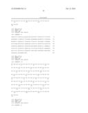 |
 |  |
 | 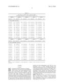 |
 |  |
 |  |
 |  |
 |  |
 |  |
 |  |
 |  |
 |  |
 |  |
 |  |
 |  |
 |  |
 |  |
 |  |
 |  |
 |  |
 |  |
 |  |
 |  |
 |  |
 |
| Similar patent applications: | |
| Date | Title |
|---|---|
| 2009-12-17 | Method for activating and stabilizing dissolved hydrogen in water |
| 2008-11-20 | Thombolysis and chronic anticoagulation therapy |
| 2010-02-18 | Formulations for nonsurgical exogenous crosslink therapy |
| 2010-02-18 | Formulations for nonsurgical exogenous crosslink therapy |
| 2009-07-09 | Albumin-fused anti-angiogenesis peptides |
| New patent applications in this class: | |
| Date | Title |
|---|---|
| 2019-05-16 | Plif multimeric peptides and uses thereof |
| 2019-05-16 | Cancer vaccines |
| 2018-01-25 | Methods and compositions for inducing an immune response to egfrviii |
| 2018-01-25 | Peptide mixture |
| 2017-08-17 | Cancer vaccine composition |
| New patent applications from these inventors: | |
| Date | Title |
|---|---|
| 2015-04-23 | Pharmaceutical combination for the treatment and/or chemosensibilization of refractory tumors to anticancer drugs |
| 2015-01-29 | Pharmaceutical combination for the treatment and/or chemosensibilization of refractory tumors to anticancer drugs |
| 2012-05-31 | Cell penetrating peptides and its use fused to biomolecules with therapeutic action |
| 2011-09-08 | Pharmaceutical composition containing polypeptide fragments of serralysins |
| Top Inventors for class "Drug, bio-affecting and body treating compositions" | |
| Rank | Inventor's name |
|---|---|
| 1 | David M. Goldenberg |
| 2 | Hy Si Bui |
| 3 | Lowell L. Wood, Jr. |
| 4 | Roderick A. Hyde |
| 5 | Yat Sun Or |