Patent application title: CLOSTRIDIAL TOXIN ACTIVITY ASSAYS
Inventors:
Ester Fernandez-Salas (Fullerton, CA, US)
Lance E. Steward (Irvine, CA, US)
Kei Roger Aoki (Coto De Caza, CA, US)
Assignees:
Allergan, Inc.
IPC8 Class: AC12Q102FI
USPC Class:
435 29
Class name: Chemistry: molecular biology and microbiology measuring or testing process involving enzymes or micro-organisms; composition or test strip therefore; processes of forming such composition or test strip involving viable micro-organism
Publication date: 2009-07-30
Patent application number: 20090191583
Inventors list |
Agents list |
Assignees list |
List by place |
Classification tree browser |
Top 100 Inventors |
Top 100 Agents |
Top 100 Assignees |
Usenet FAQ Index |
Documents |
Other FAQs |
Patent application title: CLOSTRIDIAL TOXIN ACTIVITY ASSAYS
Inventors:
Lance E. Steward
Kei Roger Aoki
Ester Fernandez-Salas
Agents:
ALLERGAN, INC.
Assignees:
ALLERGAN, INC.
Origin: IRVINE, CA US
IPC8 Class: AC12Q102FI
USPC Class:
435 29
Abstract:
Compositions useful for detecting Clostridial toxin activity comprising a
cell that contains an exogenous Clostridial toxin substrate comprises a
fluorescent member, a membrane targeting domain and a Clostridial toxin
recognition sequence comprising a cleavage site, where the cleavage site
intervenes between said fluorescent member and said membrane localization
domain and methods useful for determining Clostridial toxin activity
using such Clostridial toxin substrates.Claims:
104. A cell population comprising cells wherein greater than 50% of the
cells comprising the cell population comprise engineered cells
containing:a) an exogenous Clostridial toxin substrate comprisingi) a
fluorescent member;ii) a membrane targeting domain; andiii) a Clostridial
toxin recognition sequence comprising a Clostridial toxin recognition
cleavage site, wherein the cleavage site intervenes between the
fluorescent member and the membrane localization domain; andb) a receptor
capable of binding a Clostridial toxin;wherein the engineered cells are
capable of Clostridial toxin intoxication.
105. The cell population of claim 104, wherein the engineered cells transiently contains the exogenous Clostridial toxin substrate.
106. The cell population of claim 104, wherein the engineered cells stably contains the exogenous Clostridial toxin substrate.
107. The cell population of claim 104, wherein the Clostridial toxin substrate is expressed from a nucleic acid molecule.
108. The cell population of claim 104, wherein the cells comprising the cell population are neuronal cells.
109. The cell population of claim 104, wherein the cells comprising the cell population are non-neuronal cells.
110. The cell population of claim 104, wherein the fluorescent member is a fluorescent protein or a fluorophore binding protein.
111. The cell population according to claim 104, wherein the membrane targeting domain comprises a interhelical loop region of SNAP-25, an amino-terminal α-helix region of SNAP-25, or a carboxy-terminal α-helix region of SNAP-25.
112. The cell population according to claim 104, wherein the Clostridial toxin recognition sequence comprises a BoNT/A recognition sequence including a BoNT/A cleavage site, a BoNT/B recognition sequence including a BoNT/B cleavage site, a BoNT/C1 recognition sequence including a BoNT/C1 cleavage site, a BoNT/D recognition sequence including a BoNT/D cleavage site, a BoNT/E recognition sequence including a BoNT/E cleavage site, a BoNT/F recognition sequence including a BoNT/F cleavage site, a BoNT/G recognition sequence including a BoNT/G cleavage site, or a TeNT recognition sequence including a TeNT cleavage site.
113. The cell population according to claim 104, wherein the Clostridial toxin recognition sequence comprises at least six consecutive residues of SNAP-25, said six consecutive residues comprising Gln-Arg.
114. The cell population according to claim 104, wherein the Clostridial toxin recognition sequence comprises at least six consecutive residues of VAMP, said six consecutive residues comprising Gln-Phe.
115. The cell population according to claim 104, wherein the Clostridial toxin recognition sequence comprises at least six consecutive residues of SNAP-25, said six consecutive residues comprising Arg-Ala.
116. The cell population according to claim 104, wherein the Clostridial toxin recognition sequence comprises at least six consecutive residues of Syntaxin, said six consecutive residues comprising Lys-Ala.
117. The cell population according to claim 104, wherein the Clostridial toxin recognition sequence comprises at least six consecutive residues of VAMP, said six consecutive residues comprising Lys-Leu.
118. The cell population according to claim 104, wherein the Clostridial toxin recognition sequence comprises at least six consecutive residues of SNAP-25, said six consecutive residues comprising Arg-Ile.
119. The cell population according to claim 104, wherein the Clostridial toxin recognition sequence comprises at least six consecutive residues of VAMP, said six consecutive residues comprising Gln-Lys.
120. The cell population according to claim 104, wherein the Clostridial toxin recognition sequence comprises at least six consecutive residues of VAMP, said six consecutive residues comprising Ala-Ala.
121. The cell population of claim 104, wherein the receptor is an endogenous Clostridial toxin receptor.
122. The cell population of claim 104, wherein the receptor is an exogenous Clostridial toxin receptor.
123. A method of determining Clostridial toxin activity, said method comprising the steps of:a) contacting with a sample the cell population according to claim 1;b) exciting the fluorescent member;c) detecting fluorescence resonance energy transfer of the contacted cell population; andd) comparing the resonance energy transfer detected from the contacted cell population with the resonance energy transfer detected from a control cell population subjected to steps (b)-(c);wherein a difference in fluorescence of the contacted cell population as compared to the control cell population is indicative of Clostridial toxin activity.
124. The method of claim 123, wherein the sample is a crude cell lysate, a bulk Clostridial toxin, a partially purified Clostridial toxin, a purified Clostridial toxin, an isolated Clostridial toxin light chain, or a formulated Clostridial toxin product.
125. The method of claim 124, wherein the sample comprises a formulated Clostridial toxin product.
126. The method of claim 125, wherein the formulated Clostridial toxin product is a formulated BoNT/A product.
127. The method of claim 123, wherein the sample is a raw food, a partially cooked or processed food, a cooked or processed food, a beverage, an animal feed, a soil sample, a water sample, or a pond sediments.
128. The method according to claim 123, wherein determining the fluorescence of the contacted cell population relative to the control cell population comprises detecting an increased fluorescence of the cytoplasmic substrate from the contacted cell as indicative of Clostridial toxin activity.
129. The method according to claim 123, wherein determining the fluorescence of the contacted cell population relative to the control cell population comprises detecting a decrease fluorescence of the membrane-localized substrate from the contacted cell as indicative of Clostridial toxin activity.
130. The method according to claim 123, wherein determining the fluorescence of the contacted cell population relative to the control cell population comprises detecting a shift in fluorescence intensity of the membrane-localized substrate to the cytoplasmic substrate of said contact cell as indicative of Clostridial toxin activity.
131. The method according to claim 123, wherein determining the fluorescence of the contacted cell population relative to the control cell population comprises detecting a shift in fluorescence intensity of the membrane-localized substrate to the cytoplasmic substrate of said contact cell as indicative of Clostridial toxin activity.
Description:
[0001]This is a national stage application under 35 U.S.C. § 371 of
PCT patent application PCT/US2006/012825, filed on Apr. 4, 2006 which
claims the benefit of priority pursuant to 35 U.S.C. §119(e) to U.S.
provisional patent application Ser. No. 60/668,909 filed on Apr. 5, 2005,
each of which is hereby incorporated by reference in its entirety.
[0002]All of the publications cited in this application are hereby incorporated by reference herein in their entirety. All GeneBank sequence listings cited this application, as identified by their GenBank accession numbers, are available from the National Center for Biotechnological Information and are all hereby incorporated by reference in their entirety.
[0003]The myorelaxant properties of Clostridial toxins (CoNTs) are being exploited in a wide variety of therapeutic and cosmetic applications, see e.g., William J. Lipham, COSMETIC AND CLINICAL APPLICATIONS OF BOTULINUM TOXIN (Slack, Inc., 2004). For example, CoNTs therapies are proposed for treating dystonia, see e.g., Kei Roger Aoki, et al., Method for treating Dystonia with Botulinum Toxin C to G, U.S. Pat. No. 6,319,505 (Nov. 20, 2001); pain, see e.g., Kei Roger Aoki, et al., Method for Treating Pain by Peripheral Administration of a Neurotoxin, U.S. Pat. No. 6,464,986 (Oct. 15, 2002); muscle injuries, see e.g., Gregory F. Brooks, Methods for Treating Muscle Injuries, U.S. Pat. No. 6,423,319 (Jul. 23, 2002); cardiovascular diseases, see e.g., Gregory F. Brooks, Methods for Treating Cardiovascular Diseases with Botulinum Toxins, U.S. Patent Publication No. 2003/0185860 (Oct. 2, 2003); neuropsychiatric disorders, see e.g., Steven Donovan, Therapeutic Treatments for Neuropsychiatric Disorders, U.S. Patent Publication No. 2003/0211121 (Nov. 13, 2003); lower back pain, see e.g., Kei Roger Aoki, et al., Botulinum Toxin Therapy for Lower Back Pain, U.S. Patent Publication No. 2004/0037852 (Feb. 26, 2004); as well as other neuromuscular disorders, see e.g., Kei Roger Aoki, et al., Multiple Botulinum Toxins for Treating Neuromuscular Disorders and Conditions, U.S. Patent Publication No. 2001/0021695 (Sep. 13, 2001); Kei Roger Aoki, et al., Treatment of Neuromuscular Disorders and Conditions with Different Botulinum, U.S. Patent Publication No. 2002/0010138 (Jan. 24, 2002); Kei Roger Aoki, et al., Use of Botulinum Toxins for Treating Various Disorders and Conditions and Associated Pain, U.S. Patent Publication No. 2004/0013692 (Jan. 22, 2004) all of which are hereby incorporated by reference. Additional proposed uses of CoNTs as biopharmaceutical neuromodulators has expanded to cover a wide variety of treatments targeting certain disorders that lack a neuromuscular basis. For example, the effects on the autonomic nervous system has allowed the development of a Botulinum toxin serotype A (BoNT/A) therapy for treating axillary hyperhydrosis or sweating, and reports indicate BoNT/A may be an effective treatment for myofascial pain and tension, stroke, traumatic brain injury, cerebral palsy, gastrointestinal motility disorders, urinary incontinence cancer and migraine headaches. Lastly, cosmetic and other therapeutic applications are widely known. In fact, the expected use of CoNTs in both therapeutic and cosmetic treatments of humans is anticipated to expand to an ever widening range of diseases and aliments that can benefit from the myorelaxant properties of these toxins.
[0004]The growing clinical and therapeutic use of Clostridial toxins necessitates the pharmaceutical industry to use accurate assays for Clostridial toxin activity in order to, for example, ensure accurate pharmaceutical formulations and monitor established quality control standards. In addition, given the potential danger associated with small quantities of Clostridial toxins in foodstuffs, the food industry requires Clostridial toxin assays, for example, to validate new food packaging methods and to ensure food safety. The present invention provides novel Clostridial toxin assays for determining the presence or activity of a Clostridial toxin useful for various industries, such as, e.g. the pharmaceutical and food industries, and provides related advantages as well.
BRIEF DESCRIPTION OF THE DRAWINGS
[0005]FIG. 1 shows a schematic of the current paradigm of the intoxication mechanism for tetanus and botulinum toxin activity in central and peripheral neuron. This intoxication process can be described as comprising four steps: 1) receptor binding, where Clostridial toxin binds to a Clostridial receptor system initiates the intoxication process; 2) complex internalization, where after toxin binding, a vesicle containing a toxin/receptor system complex is endocytosised into the cell; 3) light chain translocation, where multiple events are thought to occur, including changes in the internal pH of the vesicle, formation of a channel pore comprising the HN domain of Clostridial toxin heavy chain, separation of the Clostridial toxin light chain from the heavy chain, enzymatic activation of the light chain; and release of the activated light chain and 4) enzymatic target modification, where the activated light chain of Clostridial toxin proteolytically cleaves its target SNARE substrates, such as, e.g., SNAP-25, VAMP or Syntaxin.
[0006]FIG. 2 shows a schematic of SNARE proteins. FIG. 1a shows the general domain organization of SNAP-25, VAMP and Syntaxin depicting approximate locations of the a-helical resions (white boxs), SNARE motifs (Hatched boxes with S1, S2, S3, S4, V1, V2, X1 or X2 designations) and the membrane anchoring domains (white boxes designated MA). FIG. 1b shoes the helical organization of a SNARE motif.
[0007]FIG. 3 shows a schematic of the subcellular localization and cleavage sites of SNAP-25, VAMP and Syntaxin. VAMP is localized to synaptic vesicle membrane, whereas SNAP-25 and Syntaxin are localized to the plasma membrane. BoNT/A and BoNT/E cleave SNAP-25 close to the carboxy-terminus, releasing nine or 26 residues, respectively. BoNT/B, BoNT/D, BoNT/F, BoNT/G and TeNT act on the conserved central portion of VAMP (white box) and release the amino-terminal cytosolic half of VAMP into the cytosol. BoNT/C1 cleaves SNAP-25 close to the carboxy-terminus as well as cleaving Syntaxin at a single site near the cytosolic membrane surface. The action of BoNT/C1 results in release of a large portion of the cytosolic domain of Syntaxin, while only a small portion of SNAP-25 is released by selective proteolysis of BoNT/C1.
[0008]FIG. 4 shows PC12 cells transfected with a plasmid encoding a green fluorescent protein alone (GFP, transfected with a plasmid encoding a Clostridial toxin substrate alone (GFP-SNAP25206), or co-transfected a plasmid encoding a Clostridial toxin substrate alone (GFP-SNAP25206) and a plasmid encoding the light chain of BoNT/A (BoNT/A-LC). Cells expressing green fluorescent protein alone (GFP) had fluorescence dispersed throughout the cell including the nuclei. Confocal pictures were taken with the plane in the middle of the cell. Cells expressing the Clostridial toxin substrate alone (GFP-SNAP25206) demonstrated fluorescence in the plasma membrane of the cell body and neurites. Cells co-expressing the Clostridial toxin substrate and the BoNT/A light chain (GFP-SNAP25206, BoNT/A-LC) exhibit a loss of plasma membrane localization of the GFP fluorescence. The GFP fluorescence instead accumulates in some areas of the cytoplasm.
[0009]FIG. 5 shows Western blot analysis identifying cells with high affinity uptake for a Clostridial toxin. FIG. 5a shows a Western blot analysis used to identify cells capable of BoNT/A uptake. The blot shows five cell lines treated with 1 nM of Pure BoNT/A overnight, with equal amounts of protein loaded per lane and probed with an antibody that detects the BoNT/A SNAP-25197 cleavage product. FIG. 5b shows Western blot analysis used to evaluate the time necessary for BoNT/A uptake. The blots show either Neuro-2A cells or SH-SY5Y cells treated with 1 nM of Pure BoNT/A for various lengths of time, with equal amounts of protein loaded per lane and probed with an antibody that detects the BoNT/A SNAP-25197 cleavage product. FIG. 5c shows a Western blot analysis used to evaluate the concentration range necessary of BoNT/A uptake. The blots show Neuro-2A cells treated with a range of Pure BoNT/A concentrations overnight, with equal amounts of protein loaded per lane and probed with an antibody that detects the BoNT/A SNAP-25197 cleavage product.
[0010]FIG. 6 shows Western blot analysis identifying cells with high affinity uptake for a Clostridial toxin. FIG. 6a shows a Western blot analysis used to identify cells capable of BoNT/E uptake. The top blot show Neuro-2A cells and SH-SY5Y cells treated with either 10 nM or 100 nM of BoNT/E di-chain overnight, with equal amounts of protein loaded per lane and probed with an antibody (SMI-81; Sternberger Monoclonals, Lutherville, Md.) that detects the uncleaved SNAP-25206 substrate and the BoNT/E SNAP-25180 cleavage product. The bottom blot show various cells treated with 20 nM of BoNT/E di-chain, with equal amounts of protein loaded per lane and probed with an antibody for the uncleaved SNAP-25206 substrate and the BoNT/E SNAP-25180 cleavage product. FIG. 6b shows Western blot analysis used to determine a time course for BoNT/E uptake. The blots show SH-SY5Y cells treated with either 5 nM or 20 nM of BoNT/E di-chain for either 4 hours or 8 hours, with equal amounts of protein loaded per lane and probed with an antibody (SMI-81; Sternberger Monoclonals, Lutherville, Md.) that detects the uncleaved SNAP-25206 substrate and the BoNT/E SNAP-25180 cleavage product. FIG. 6c shows a Western blot analysis used to evaluate the concentration range necessary of BoNT/E uptake. The blots show SK-N-DZ cells treated with a range of BoNT/E di-chain concentrations for approximately 6 hours, with equal amounts of protein loaded per lane and probed with an antibody (SMI-81; Sternberger Monoclonals, Lutherville, Md.) that detects the uncleaved SNAP-25206 substrate and the BoNT/E SNAP-25180 cleavage product.
[0011]FIG. 7 shows Western blot analysis evaluating the effects of treatments used to increase uptake of a Costridial toxin. FIG. 7a shows a Western blot analysis evaluating the effects of ganglioside treatment on the uptake of BoNT/A. The blot shows Neuro-2A cells treated without or with 25 μg/mL of GT1b (- or +) and exposed overnight to three different concentrations of BoNT/A (12.5 pM, 25 pM or 50 pM), with equal amounts of protein loaded per lane and probed with an antibody that detects the BoNT/A SNAP-25197 cleavage product. FIG. 7b shows Western blot analysis evaluating the effects of cell differentiation on the uptake of BoNT/A. The blots show either Neuro-2A cells or SH-SY5Y cells treated 2 nM of Pure BoNT/A overnight that where either grown in serum-free media or with various differentiation reagents (Ionomycin, db-cAMP, Retinoic acid, Neuraminidase or N2), with equal amounts of protein loaded per lane and probed with an antibody (SMI-81; Sternberger Monoclonals, Lutherville, Md.) that detects the uncleaved SNAP-25206 substrate and the BoNT/A SNAP-25197 cleavage product.
[0012]FIG. 8 shows Western blot analysis evaluating the effects of treatments used to increase uptake of a Costridial toxin. FIG. 8a shows a Western blot analysis evaluating the effects of ganglioside treatment on the uptake of BoNT/E. The blot shows Neuro-2A cells treated with either 25 μg/mL of GT1b, GQ1b, GD1a, GD1b or GD3 and exposed for approximately 5 hours to 14 nM of BoNT/E di-chain, with equal amounts of protein loaded per lane and probed with an antibody (SMI-81; Sternberger Monoclonals, Lutherville, Md.) that detects the uncleaved SNAP-25206 substrate and the BoNT/E SNAP-25180 cleavage product. FIG. 7b shows Western blot analysis evaluating the effects of cell differentiation on the uptake of BoNT/E. The blots show either N1E-115 cells, SH-SY5Y cells, SK-N-DZ cells or NG108-15 cells treated with either 0 nM, 2 nM or 20 nM of BoNT/E di-chain for approximately 6 hours that where grown in serum-free media, with equal amounts of protein loaded per lane and probed with an antibody (SMI-81; Sternberger Monoclonals, Lutherville, Md.) that detects the uncleaved SNAP-25206 substrate and the BoNT/E SNAP-25180 cleavage product.
[0013]FIG. 9 shows a schematic of a fluorescent-based Clostridial toxin activity assay which relies on cell lines containing a Clostridial toxin substrate localized to the cell membrane. FIG. 1a shows an assay scenario where the uncleaved Clostridial toxin substrate, comprising a fluorescent member (FM), a membrane targeting domain (MTD) and a Clostridial toxin recognition sequence comprising a Clostridial toxin cleavage site (RS), is detected. Upon excitation, the fluorescent member emits fluorescent light at a characteristic wavelength that is localized to the membrane. FIG. 1b shows an assay scenario where the cleaved Clostridial toxin substrate is detected. Upon excitation, the fluorescent member emits fluorescent light at a characteristic wavelength. However, because the cleavage product containing the fluorescent member is released into the cytoplasm, detection of fluorescence is in a different subcellular localization. Thus, a decrease in fluorescent member emissions in the membrane, or an increase in fluorescent member emissions in the cytoplasm is indicative of the presence of Clostridial toxin activity.
DETAILED DESCRIPTION OF THE INVENTION
[0014]The present invention provides novel assays for determining the presence or absence of an active Clostridial toxin in a sample and for determining the activity of a Clostridial toxin, including botulinum toxins of all serotypes and tetanus toxin. The novel assays of the invention rely on the cellular localization of the uncleaved Clostridial toxin substrate, the cellular localization of the cleaved Clostridial toxin substrate, or the cellular localization of both the uncleaved and cleaved Clostridial toxin substrate. The present invention further provides novel compositions, including cells and cell lines containing Clostridial toxin substrates, useful for the assays disclosed in the specification. The novel cells and assays of the invention reduce the need for animal toxicity studies, yet serve to analyze multiple toxin functions, namely, binding and cellular uptake of toxin, translocation into the cell cytosol, and protease activity. As discussed further below, the novel cells and methods of the invention can be used to analyze crude and bulk samples as well as highly purified dichain toxins and formulated toxin products and further are amenable to automated high throughput assay formats.
[0015]The assays disclosed in the present specification use cells which are capable of efficient Clostridial toxin uptake and which include a membrane localized Clostridial toxin substrate containing a fluorescent marker. As an example, a cell useful in the invention can express a SNAP25206-enhanced green fluorescent protein (EGFP) fusion protein (absorbance 484 nM, emission 510 nM), which localizes to the plasma membrane (FIG. 9). Upon BoNT/A treatment of this cell, cleavage of the membrane localized SNAP25206-EGFP substrate occurs, releasing the EGFP containing fragment into the cytoplasm. Upon excitation of the treated cell with a 484 nM laser, the EGFP is excited and emits light at 510 nM. However, because a portion of the EGFP is now cytoplasmic, a distribution change between the uncleaved, membrane localized SNAP25206-EGFP toxin substrate and the cleaved, cytoplasmic localized EGFP fragment can be observed in BoNT/A treated cells.
[0016]Aspects of the present invention provide for an exogenous Clostridial toxin substrate capable of being localized to the plasma membrane of a cell. These substrates comprise a fluorescent member, a membrane targeting domain and a Clostridial toxin recognition sequence comprising a cleavage site, where the cleavage site intervenes between the fluorescent member and the membrane localization domain.
[0017]Other aspects of the present invention provide compositions comprising a cell containing an exogenous Clostridial toxin substrate capable of being localized to the plasma membrane of the cell wherein the cell is capable of Clostridial toxin intoxication, and wherein the exogenous Clostridial toxin substrate comprises a fluorescent member, a membrane targeting domain and a Clostridial toxin recognition sequence comprising a cleavage site, where the cleavage site intervenes between the fluorescent member and the membrane localization domain. The exogenous Clostridial toxin substrate can be transiently or stably contained in the cell.
[0018]Other aspects of the present invention provide compositions comprising a cell population, said cell population comprising cells that contain an exogenous Clostridial toxin substrate capable of being localized to the plasma membrane of the cells wherein the cells are capable of Clostridial toxin intoxication, and wherein the exogenous Clostridial toxin substrate comprises a fluorescent member, a membrane targeting domain and a Clostridial toxin recognition sequence comprising a cleavage site, where the cleavage site intervenes between the fluorescent member and the membrane localization domain and wherein greater than 50% of the cell population comprise the cells containing the exogenous Clostridial toxin substrate.
[0019]Other aspects of the present invention provide compositions comprising a cell population, said cell population comprising cells that transiently contain an exogenous Clostridial toxin substrate capable of being localized to the plasma membrane of the cells wherein the cells are capable of Clostridial toxin intoxication, and wherein the exogenous Clostridial toxin substrate comprises a fluorescent member, a membrane targeting domain and a Clostridial toxin recognition sequence comprising a cleavage site, where the cleavage site intervenes between the fluorescent member and the membrane localization domain and wherein greater than 50% of the cell population comprise the cells containing the exogenous Clostridial toxin substrate.
[0020]Other aspects of the present invention provide compositions comprising a cell population, the cell population comprising cells that stably contain an exogenous Clostridial toxin substrate capable of being localized to the plasma membrane of the cells wherein the cells are capable of Clostridial toxin intoxication, and wherein the exogenous Clostridial toxin substrate comprises a fluorescent member, a membrane targeting domain and a Clostridial toxin recognition sequence comprising a cleavage site, where the cleavage site intervenes between the fluorescent member and the membrane localization domain.
[0021]Other aspects of the present invention provide methods of determining Clostridial toxin activity by contacting with a sample a cell population, the cell population comprising cells that contain an exogenous Clostridial toxin substrate capable of being localized to the plasma membrane of the cells wherein the cell are capable of Clostridial toxin intoxication, wherein the exogenous Clostridial toxin substrate comprises a fluorescent member, a membrane targeting domain and a Clostridial toxin recognition sequence comprising a cleavage site, where the cleavage site intervenes between the fluorescent member and the membrane localization domain and wherein greater than 50% of the cell population comprises the cells containing the exogenous Clostridial toxin substrate; exciting the fluorescent member; and determining the fluorescence of the contacted cell population relative to a control cell population, where a difference in fluorescence of the contacted cell population as compared to the control cell population is indicative of Clostridial toxin activity.
[0022]Other aspects of the present invention provide methods of determining Clostridial toxin activity by contacting with a sample a cell population, the cell population comprising cells that transiently contains an exogenous Clostridial toxin substrate capable of being localized to the plasma membrane of the cells wherein the cells are capable of Clostridial toxin intoxication, wherein the exogenous Clostridial toxin substrate comprises a fluorescent member, a membrane targeting domain and a Clostridial toxin recognition sequence comprising a cleavage site, where the cleavage site intervenes between the fluorescent member and the membrane localization domain and wherein greater than 50% of the cell population comprises the cells containing the exogenous Clostridial toxin substrate; exciting the fluorescent member; and determining the fluorescence of the contacted cell population relative to a control cell population, where a difference in fluorescence of the contacted cell population as compared to the control cell population is indicative of Clostridial toxin activity.
[0023]Other aspects of the present invention provide methods of determining Clostridial toxin activity by contacting with a sample a cell population, the cell population comprising cells that stably contain an exogenous Clostridial toxin substrate capable of being localized to the plasma membrane of the cells wherein the cells are capable of Clostridial toxin intoxication, wherein the exogenous Clostridial toxin substrate comprises a fluorescent member, a membrane targeting domain and a Clostridial toxin recognition sequence comprising a cleavage site, where the cleavage site intervenes between the fluorescent member and the membrane localization domain and wherein greater than 50% of the cell population comprises the cells containing the exogenous Clostridial toxin substrate; exciting the fluorescent member; and determining the fluorescence of the contacted cell population relative to a control cell population, where a difference in fluorescence of the contacted cell population as compared to the control cell population is indicative of Clostridial toxin activity.
[0024]Bacteria of the genus Clostridia are strictly anaerobic to aero-tolerant spore-forming bacilli found in soil, freshwater and saltwater sediments, household dust, the surface of foods, feces as well as in the normal intestinal flora of humans and animals. While the majority of isolates are gram-positive, a few gram-negative species exist. Members of this genus produce sophisticated exotoxins that are among the most potent toxins known in the world. Exposure to these toxins during the course of Clostridia infection is the primary cause underlying disease pathogenesis. Clostridia are a major threat to human and animal health, being responsible for many diseases including botulism, tetanus, gas gangrene, pseudomembranous colitis and food poisoning. For example, Clostridium argentinense, C. bifermentans, C. histolyticum, C. novyi, C. septicum, C. sporogenes and C. tertium are etiological agents for gas gangrene. C. perfringens is responsible for foodborne illness, enteritis necroticans where as C. difficile is responsible for pseudomembranous enterocolitis. Both C. baratii and C. butyricum are causative agents for a form of foodborne, intestinal and wound botulism. Interestingly, only a few species of these bacteria are pathogenic for humans, most are saprophytic. Thus, in most cases, Clostridia are opportunistic pathogens that infect a host whose health is compromised.
[0025]Of all Clostridia, Clostridium botulinum and Clostridium tetani produce the most potent biological toxins known and are the causative agents of the neuroparalytic syndromes botulism and tetanus. Seven antigenically-distinct types of Botulinum toxins (BoNTs) have been identified by investigating botulism outbreaks in man (BoNT/A, /B, /E and /F), animals (BoNT/C1 and /D), or isolated from soil (BoNT/G). BoNTs possess approximately 35% amino acid identity with each other and share the same functional domain organization and overall structural architecture. The amino acid sequences of eight Clostridial toxin serotypes have been derived from the corresponding genes (Niemann, "Molecular Biology of Clostridial Neurotoxins" in Sourcebook of Bacterial Protein Toxins Alouf and Freer (Eds.) pp. 303-348 London: Academic Press 1991). It is recognized by those of skill in the art that within each type of Clostridial toxin there can be various strains differing somewhat in their amino acid sequence, and also in the nucleic acids encoding these proteins. While all seven BoNT serotypes have similar structure and pharmacological properties, each also displays heterogeneous bacteriological characteristics. In contrast, tetanus toxin (TeNT) is produced by a uniform group of C. tetani. Two other species of clostridia, C. baratii and C. butyricum, also produce toxins similar to BoNT/F and BoNT/E, respectively.
[0026]Clostridia toxins (CoNTs) are each translated as a single chain polypeptide of approximately 150 kDa that is subsequently cleaved by proteolytic scission within a disulphide loop by bacterial or tissue proteases. This posttranslational processing yields a di-chain molecule comprising an approximately 50 kDa light chain (LC) and an approximately 100 kDa heavy chain (HC) held together by a single disulphide bond and noncovalent interactions. Each mature di-chain molecule comprises three functionally distinct domains: 1) an enzymatic domain located in the LC that includes a metalloprotease region containing a zinc-dependent endopeptidase activity which specifically targets core components of the neurotransmitter release apparatus; 2) a translocation domain contained within the amino-terminal half of the HC (HN) that facilitates release of the toxin from intracellular vesicles into the cytoplasm of the target cell; and 3) a binding domain found within the carboxy-terminal half of the HC (HC) that determines the binding activity and binding specificity of the toxin to the receptor complex located at the surface of the target cell.
[0027]The binding, translocation and enzymatic activity of these three functional domains are all necessary for toxicity. While all details of this process are not yet precisely known, the overall cellular intoxication mechanism whereby CoNTs enter a neuron and inhibit neurotransmitter release is similar, regardless of type. Although the applicants have no wish to be limited by the following description, the intoxication mechanism can be described as comprising four steps: 1) receptor binding, 2) complex internalization, 3) light chain translocation, and 4) enzymatic target modification (see FIG. 1). The process is initiated when the HC domain of a CoNT binds to CoNT-specific receptor complex located on the plasma membrane surface of a target cell. The binding specificity of a receptor complex is thought to be achieved, in part, by specific combinations of gangliosides and protein receptors that appear to distinctly comprise each Clostridial toxin receptor complex. Once bound, the CoNT/receptor complexes are internalized by endocytosis and the internalized vesicles are sorted to specific intracellular routes. The translocation step appears to be triggered by the acidification of the vesicle compartment. This process seems to initiate two important pH-dependent structural rearrangements that increase hydrophobicity and promote enzymatic activation of the toxin. Once activated, light chain endopeptidase of the toxin is released from the intracellular vesicle into the cytosol where it specifically targets one of three known core components of the neurotransmitter release apparatus. There of these core proteins, vesicle-associated membrane protein (VAMP)/synaptobrevin, synaptosomal-associated protein of 25 kDa (SNAP-25) and Syntaxin, are necessary for synaptic vesicle docking and fusion at the nerve terminal and constitute members of the soluble N-ethylmaleimide-sensitive factor-attachment protein-receptor (SNARE) family (see FIG. 2). The selective proteolysis of synaptic SNAREs accounts for the total block of neurotransmitter release caused by Clostridial toxins in vivo. The SNARE protein targets of Clostridial toxins are common to exocytosis in a variety of non-neuronal types; in these cells, as in neurons, light chain peptidase activity inhibits exocytosis, see, e.g., Yann Humeau et al., How Botulinum and Tetanus Neurotoxins Block Neurotransmitter Release, 82(5) Biochimie. 427-446 (2000); Kathryn Turton et al., Botulinum and Tetanus Neurotoxins: Structure, Function and Therapeutic Utility, 27(11) Trends Biochem. Sci. 552-558. (2002); M. Zouhair Atassi, Basic and Therapeutic Aspects of Botulinum and Tetanus Toxins, (Dirk W. Dressler & Joseph J. Jankovic eds., 2003); Giovanna Lalli et al., The Journey of Tetanus and Botulinum Neurotoxins in Neurons, 11(9) Trends Microbiol. 431-437, (2003) which are hereby incorporated by reference.
[0028]TeNT and BoNT/B, /D, /F, and /G specifically recognize VAMP (also known as synaptobrevin), an integral protein of the synaptic vesicle membrane. VAMP is cleaved at distinct bonds depending on the toxin. BoNT/A and /E recognize and specifically cleave SNAP-25, a protein of the presynaptic membrane, at two different sites in the carboxy-terminal portion of the protein. BoNT/C1 cleaves syntaxin, a protein of the nerve plasmalemma, in addition to SNAP-25. The three protein targets of the CoNTs are conserved from yeast to humans although cleavage sites and toxin susceptibility are not necessarily conserved, see below; see, also, e.g., Humeau, supra, (2000); Heiner Niemann et al., Clostridial neurotoxins: new tools for dissecting exocytosis, 4(5) Trends Cell Biol. 179-185 (1994); and Rossella Pellizzari et al., Tetanus and botulinum neurotoxins: mechanism of action and therapeutic uses, 354(1381) Philos. Trans. R. Soc. Lond. B Biol. Sci. 259-268 (1999).
[0029]The natural targets of the Clostridial toxins include VAMP, SNAP-25, and syntaxin. VAMP is associated with the synaptic vesicle membrane, whereas SNAP-25 and syntaxin are associated with the plasma membrane (see FIG. 3). BoNT/A and BoNT/E cleave SNAP-25 in the carboxy-terminal region, releasing a nine or twenty-six amino acid segment, respectively, and BoNT/C1 also cleaves SNAP-25 near the carboxy-terminus. The botulinum serotypes BoNT/B, BoNT/D, BoNT/F and BoNT/G, and tetanus toxin, act on the conserved central portion of VAMP, and release the amino-terminal portion of VAMP into the cytosol. BoNT/C1 cleaves syntaxin at a single site near the cytosolic membrane surface. Thus, BoNT/B, BoNT/C1, BoNT/D, BoNT/F, BoNT/G or TeNT proteolysis results in release of a large portion of the cytosolic domain of VAMP or syntaxin, while only a small portion of SNAP-25 is released by BoNT/A, BoNT/C1 or BoNT/E cleavage, see, e.g., Humeau et al., supra, (2000); Turton et al., supra, (2002); Lalli et al., supra (2003).
[0030]Naturally occurring SNAP-25, a protein of about 206 residues lacking a transmembrane segment, is associated with the cytosolic surface of the nerve plasmalemma (see FIG. 3). SNAP-25 is required for axonal growth during development and may be required for nerve terminal plasticity in the mature nervous system. SNAP-25 has been isolated from a variety of vertebrate and invertebrate species including, e.g., species belonging to the genera Homo, Macaca, Bos, Rattus, Mus, Gallus, Carassius, Danio, Torpedo, Xenopus, Strongylocentrotus, Drosophila, Hirudo, Loligo, Lymnaea and Caenorhabditis. In humans, at least two isoforms are differentially expressed during development; isoform a is constitutively expressed during fetal development, while isoform b appears at birth and predominates in adult life. SNAP-25 analogues such as SNAP-23 also are expressed outside the nervous system, for example, in pancreatic cells.
[0031]Naturally occurring VAMP is a protein of about 120 residues, with the exact length depending on the species and isoform. As shown in FIG. 3, VAMP contains a short carboxy-terminal segment inside the vesicle lumen while most of the molecule is exposed to the cytosol. The proline-rich amino-terminal thirty residues are divergent among species and isoforms while the central portion of VAMP (residues 30 to 96), which is rich in charged and hydrophilic residues and includes known cleavage sites, is highly conserved. VAMP colocalizes with synaptophysin on synaptic vesicle membranes. VAMP has been isolated from a variety of vertebrate and invertebrate species including, e.g., species belonging to the genera Homo, Macaca, Bos, Rattus, Mus, Gallus, Danio, Torpedo, Xenopus, Strongylocentrotus, Drosophila, Hirudo, Loligo, Lymnaea, Aplysia and Caenorhabditis. In addition, multiple isoforms of VAMP have been identified including VAMP-1, VAMP-2 and VAMP-3/cellubrevin, and forms insensitive to toxin cleavage have been identified in non-neuronal cells. VAMP appears to be present in all vertebrate tissues although the distribution of VAMP-1 and VAMP-2 varies in different cell types. Chicken and rat VAMP-1 are not cleaved by TeNT or BoNT/B. These VAMP-1 orthologs have a valine in place of the glutamine present in human and mouse VAMP-1 at the TeNT or BoNT/B cleavage site. The substitution does not affect BoNT/D, /F or /G, which cleave both VAMP-1 and VAMP-2 with similar rates.
[0032]Naturally occurring Syntaxin is located on the cytosolic surface of the nerve plasmalemma and is membrane-anchored via a carboxy-terminal segment, with most of the protein exposed to the cytosol (see FIG. 3). Syntaxin colocalizes with calcium channels at the active zones of the presynaptic membrane, where neurotransmitter release takes place. In addition, syntaxin interacts with synaptotagmin, a protein of the SSV membrane that forms a functional bridge between the plasmalemma and the vesicles. Syntaxin has been isolated from a variety of vertebrate and invertebrate species including, e.g., species belonging to the genera Homo, Bos, Rattus, Mus, Gallus, Danio, Strongylocentrotus, Drosophila, Hirudo, Loligo, Lymnaea and Aplysia. Three isoforms of slightly different length (285 and 288 residues) have been identified in nerve cells (isoforms 1A, 1B1 and 1B2), with isoforms 2, 3, 4 and 5 expressed in other tissues. The different isoforms have varying sensitivities to BoNT/C1, with the 1A, 1B, 1B2, 2 and 3 syntaxin isoforms cleaved by this toxin.
[0033]Aspects of the present invention provide for compositions comprising an exogenous Clostridial toxin substrate capable of being localized to the plasma membrane of a cell. These substrates are comprised of a fluorescent member, a membrane targeting domain and a Clostridial toxin recognition sequence comprising a cleavage site, where the cleavage site intervenes between the fluorescent member and the membrane localization domain.
[0034]The Clostridial toxin substrates disclosed in the present specification include, in part, a Clostridial toxin recognition sequence including a cleavage site. By definition, a Clostridial toxin substrate is susceptible to cleavage by at least one Clostridial toxin under conditions suitable for Clostridial toxin protease activity. A variety of Clostridial toxin substrates are discussed herein below. Additional Clostridial toxin substrates are described in, e.g., Lance E. Steward, et al., FRET Protease Assays for Clostridial Toxins, U.S. Patent Publication 2003/0143651 (Jul. 31, 2003); Lance E. Steward, et al., FRET Protease Assays for Botulinum Serotype A/E Toxins, U.S. Patent Publication 2003/0143650 (Jul. 31, 2003); and Ester Fernandez-Salas, et al., Cell-based Fluorescence Resonance Energy Transfer (FRET) Assays for Clostridial Toxins, U.S. Patent Publication 2004/0072270 (Apr. 15, 2004).
[0035]The Clostridial toxin substrates disclosed in the present specification comprise, in part, a Clostridial toxin recognition sequence including a cleavage site. As used herein, the term "Clostridial toxin recognition sequence" means a scissile bond together with adjacent or non-adjacent recognition elements, or both, sufficient for detectable proteolysis at the scissile bond by a Clostridial toxin under conditions suitable for Clostridial toxin protease activity. A variety of Clostridial toxin recognition sequences are discussed herein below.
[0036]Clostridial toxin substrates useful in aspects of the invention include peptides and peptidomimetics as well as derivatized forms thereof. As used herein, the term "peptidomimetic" is used broadly to mean a peptide-like molecule that is cleaved by the same Clostridial toxin as the peptide substrate upon which it is structurally based. Such peptidomimetics include chemically modified peptides, peptide-like molecules containing non-naturally occurring amino acids, and peptoids, which are peptide-like molecules resulting from oligomeric assembly of N-substituted glycines, and are cleaved by the same Clostridial toxin as the peptide substrate upon which the peptidomimetic is derived (see, for example, Goodman and Ro, Peptidomimetics for Drug Design, in "Burger's Medicinal Chemistry and Drug Discovery" Vol. 1 (ed. M. E. Wolff; John Wiley & Sons 1995), pages 803-861).
[0037]A variety of peptidomimetics are known in the art including, for example, peptide-like molecules which contain a constrained amino acid, a non-peptide component that mimics peptide secondary structure, or an amide bond isostere. A peptidomimetic that contains a constrained, non-naturally occurring amino acid can include, for example, an α-methylated amino acid; an α,α-dialkyl-glycine or α-aminocycloalkane carboxylic acid; an N.sup.α-C.sup.α cyclized amino acid; an N.sup.α-methylated amino acid; β- or γ-amino cycloalkane carboxylic acid; an α,β-unsaturated amino acid; a β,β-dimethyl or β-methyl amino acid; a β-substituted-2,3-methano amino acid; an NC.sup.δ or C.sup.α-C.sup.δ cyclized amino acid; or a substituted proline or another amino acid mimetic. In addition, a peptidomimetic which mimics peptide secondary structure can contain, for example, a nonpeptidic β-turn mimic; γ-turn mimic; mimic of β-sheet structure; or mimic of helical structure, each of which is well known in the art. A peptidomimetic also can be a peptide-like molecule which contains, for example, an amide bond isostere such as a retro-inverso modification; reduced amide bond; methylenethioether or methylenesulfoxide bond; methylene ether bond; ethylene bond; thioamide bond; trans-olefin or fluoroolefin bond; 1,5-disubstituted tetrazole ring; ketomethylene or fluoroketomethylene bond or another amide isostere. One skilled in the art understands that these and other peptidomimetics are encompassed within the meaning of the term "peptidomimetic" as used herein.
[0038]In other embodiments, a Clostridial toxin substrate useful in the invention is a peptide or peptidomimetic having a defined length. A Clostridial toxin substrate can be, for example, a peptide or peptidomimetic having at least 100, at least 150, at least 200, at least 250, at least 300, at least 350 or at least 500 residues. In other embodiments, a Clostridial toxin substrate has at most 20 residues, at most 30 residues, at most 40 residues, at most 50 residues, at most 100 residues, at most 150 residues, at most 200 residues, at most 250 residues, at most 300 residues, at most 350 residues or at most 400 residues.
[0039]A wide variety of Clostridial toxin recognition sequence are useful in aspects of the invention. Specific and distinct cleavage sites for different Clostridial toxins are well known in the art. BoNT/A cleaves a Gln-Arg bond; BoNT/B and TeNT cleave a Gln-Phe bond; BoNT/C1 cleaves a Lys-Ala or Arg-Ala bond; BoNT/D cleaves a Lys-Leu bond; BoNT/E cleaves an Arg-Ile bond; BoNT/F cleaves a Gln-Lys bond; and BoNT/G cleaves an Ala-Ala bond (see Table 1). In standard nomenclature, the sequence surrounding a Clostridial toxin cleavage site is denoted P5-P4-P3-P2-P1-P1'-P2'-P3'-P4'-P5' with P1-P1' representing the scissile bond. It is understood that a P1 or P1' site, or both, can be substituted with another amino acid or amino acid mimetic in place of the naturally occurring residue. As an example, BoNT/A substrates have been prepared in which the P1 position (Gln) is modified to be an alanine, 2-aminobutyric acid or asparagine residue; these substrates were hydrolyzed by BoNT/A at the P1-Arg bond, see, e.g., James J. Schmidt & Karen A Bostian, Endoproteinase activity of type A botulinum neurotoxin: substrate requirements and activation by serum albumin, 16(1) J. Protein Chem. 19-26 (1997). While it is recognized that substitutions can be introduced at the P1 position of the scissile bond, for example, a BoNT/A scissile bond, it is further recognized that conservation of the P1' residue can be advantageous, see, e.g., Vadakkanchery V. Vaidyanathan et al., Proteolysis of SNAP-25 isoforms by botulinum neurotoxin types A, C, and E: domains and amino acid residues controlling the formation of enzyme-substrate complexes and cleavage, 72(1) J Neurochem. 327-337 (1999).
TABLE-US-00001 TABLE 1 Bonds Cleaved in Human VAMP-2, SNAP-25 or Syntaxin-1 Toxin Target P4-P3-P2-P1--P1'-P2'-P3'-P4' SEQ ID NO: BoNT/A SNAP-25 Glu-Ala-Asn-Gln-Arg*-Ala-Thr-Lys 96 BoNT/B VAMP-2 Gly-Ala-Ser-Gln-Phe*-Glu-Thr-Ser 97 BoNT/C1 Syntaxin-1 Asp-Thr-Lys-Lys-Ala*-Val-Lys-Tyr 98 BoNT/C1 SNAP-25 Ala-Asn-Gln-Arg-Ala*-Thr-Lys-Met 99 BoNT/D VAMP-2 Arg-Asp-Gln-Lys-Leu*-Ser-Glu-Leu 100 BoNT/E SNAP-25 Gln-Ile-Asp-Arg-Ile*-Met-Glu-Lys 101 BoNT/F VAMP-2 Glu-Arg-Asp-Gln-Lys*-Leu-Ser-Glu 102 BoNT/G VAMP-2 Glu-Thr-Ser-Ala-Ala*-Lys-Leu-Lys 103 TeNT VAMP-2 Gly-Ala-Ser-Gln-Phe*-Glu-Thr-Ser 104 *Scissile bond shown in bold
[0040]Thus, an embodiment, is a composition comprising an exogenous Clostridial toxin substrate capable of being localized to the plasma membrane of a cell wherein said substrate comprises a fluorescent member, a membrane targeting domain and a Clostridial toxin recognition sequence comprising a cleavage site, where the cleavage site intervenes between said fluorescent member and said membrane localization domain. In an aspect of this embodiment, a Clostridial toxin substrate comprises a Clostridial toxin recognition sequence in which the P1' residue is not modified or substituted relative to the naturally occurring residue in a target protein cleaved by the Clostridial toxin. In another aspect of this embodiment, a Clostridial toxin substrate comprises a Clostridial toxin recognition sequence in which the P1 residue is modified or substituted relative to the naturally occurring residue in a target protein cleaved by the Clostridial toxin; such a Clostridial toxin substrate retains susceptibility to peptide bond cleavage between the P1 and P1' residues.
[0041]Any of a variety of Clostridial toxin recognition sequences are useful in the cells of the invention including, without limitation, botulinum toxin recognition sequences such as BoNT/A recognition sequences, BoNT/B recognition sequences, BoNT/C1 recognition sequences, BoNT/D recognition sequences, BoNT/E recognition sequences, BoNT/F recognition sequences, BoNT/G recognition sequences and TeNT recognition sequences.
[0042]A variety of BoNT/A recognition sequences are well known in the art and are useful in the invention, see, e.g., Mark A. Breidenbach & Axel T. Brunger, Substrate recognition strategy for botulinum neurotoxin serotype A, 432(7019) Nature 925-929 (2004). A BoNT/A recognition sequence can have, for example, residues 46-206, residues 134 to 206, residues 137 to 206 or 146-206 of human SNAP-25, see, e.g., Teresa A. Ekong et al., Recombinant SNAP-25 is an effective substrate for Clostridium botulinum type A toxin endopeptidase activity in vitro, 143 (Pt 10) Microbiology 3337-3347 (1997); Clifford C. Shone et al., Toxin Assays, U.S. Pat. No. 5,962,637 (Oct. 5, 1999); and Vaidyanathan et al., supra, (1999). A BoNT/A recognition sequence also can include, without limitation, the sequence Thr-Arg-Ile-Asp-Glu-Ala-Asn-Gln-Arg-Ala-Thr-Lys-Met (SEQ ID NO: 105) or a peptidomimetic thereof, which corresponds to residues 190 to 202 of human SNAP-25; Ser-Asn-Lys-Thr-Arg-Ile-Asp-Glu-Ala-Asn-Gln-Arg-Ala-Thr-Lys (SEQ ID NO: 106) or a peptidomimetic thereof, which corresponds to residues 187 to 201 of human SNAP-25; Ser-Asn-Lys-Thr-Arg-Ile-Asp-Glu-Ala-Asn-Gln-Arg-Ala-Thr-Lys-Met (SEQ ID NO: 107) or a peptidomimetic thereof, which corresponds to residues 187 to 202 of human SNAP-25; Ser-Asn-Lys-Thr-Arg-Ile-Asp-Glu-Ala-Asn-Gln-Arg-Ala-Thr-Lys-Met-Leu (SEQ ID NO: 108) or a peptidomimetic thereof, which corresponds to residues 187 to 203 of human SNAP-25; Asp-Ser-Asn-Lys-Thr-Arg-Ile-Asp-Glu-Ala-Asn-Gln-Arg-Ala-Thr-Lys-Met (SEQ ID NO: 109) or a peptidomimetic thereof, which corresponds to residues 186 to 202 of human SNAP-25; or Asp-Ser-Asn-Lys-Thr-Arg-Ile-Asp-Glu-Ala-Asn-Gln-Arg-Ala-Thr-Lys-Met-Leu (SEQ ID NO: 110) or a peptidomimetic thereof, which corresponds to residues 186 to 203 of human SNAP-25. See, for example, James J. Schmidt & Karen A Bostian, Proteolysis of synthetic peptides by type A botulinum neurotoxin, 14(8) J. Protein Chem. 703-708 (1995); Schmidt & Bostian, supra, (1997); James J. Schmidt et al., Type A botulinum neurotoxin proteolytic activity: development of competitive inhibitors and implications for substrate specificity at the S1' binding subsite, 435(1) FEBS Lett. 61-64 (1998); and James J. Schmidt & Karen A Bostian, Assay for the proteolytic activity of serotype a from clostridium botulinum, U.S. Pat. No. 5,965,699 (Oct. 12, 1999).
[0043]A BoNT/A recognition sequence useful in aspects of the invention can correspond to a segment of a protein that is sensitive to cleavage by botulinum toxin serotype A, or can be substantially similar to a segment of a BoNT/A-sensitive protein. As shown in Table 2, a variety of naturally occurring proteins sensitive to cleavage by BoNT/A are known in the art and include, for example, human, rat, mouse, Danio, Carassius, SNAP-25A and SNAP-25B; and Torpedo SNAP-25. Thus, a BoNT/A recognition sequence can correspond, for example, to a segment of human SNAP-25A or SNAP-25B; bovine SNAP-25A or SNAP-25B; rat SNAP-25A or SNAP-25B; mouse SNAP-25A or SNAP-25B; Xenopus SNAP-25A or SNAP-25B; Danio SNAP-25A or SNAP-25B; Carassius SNAP-25A or SNAP-25B; Torpedo SNAP-25; Strongylocentrotus SNAP-25; Loligo SNAP-25; Lymnaea SNAP-25; Aplysia SNAP-25, isoforms thereof, or another naturally occurring protein sensitive to cleavage by BoNT/A. Furthermore, comparison of native SNAP-25 amino acid sequences cleaved by BoNT/A reveals that such sequences are not absolutely conserved (see Table 2), indicating that a variety of amino acid substitutions and modifications relative to a naturally occurring BoNT/A-sensitive SNAP-25 sequence can be tolerated in a BoNT/A recognition sequence useful in the invention. It is understood that a similar BoNT/A recognition sequence can be prepared, if desired, from a corresponding (homologous) segment of another BoNT/A-sensitive SNAP-25 isoform, paralog or ortholog, such as, the BoNT/A recognition sequence contain in the SNAP-25 proteins identified in the organisms listed above and in Table 2.
TABLE-US-00002 TABLE 2 Cleavage of SNAP-25 and Related Proteinsa,b,c Cleavage Sites Organism Isoform BONT/E BoNT/A BoNT/C1 Cleaved Susceptibility Primate SNAP-25A MALDMGNEIDTQNRQIDR * IMEKADSNKTRIDEANQ * R * ATKMLGSG BoNT/A; BoNT/C1; BoNT/E SNAP-25B Primate SNAP-23A MALNIGNEIDAQN Q R -- ITDKADTNRDRIDIAN -- R -- AKKLIDS Noneb SNAP-23B Rodent SNAP-25A MALDMGNEIDTQNRQIDR * IMEKADSNKTRIDEANQ * R * ATKMLGSG BoNT/A; BoNT/C1; BoNT/E SNAP-25B Rodent SNAP-23 MALDMGNEIDAQNQQIQ * ITEKADTNKNRIDIAN -- R -- AKKLIDS BoNT/E Bird SNAP-25B MALDMGNEIDTQNRQIDR * IMEKADSNKTRIDEANQ -- R -- ATKMLGSG BoNT/E Amphibian SNAP-25A MALDMGNEIDTQNRQIDR ND IMEKADSNKARIDEAN ND ND ATKMLGSG ND SNAP-25B Amphibian SNAP-23 MAIDMGNELESHNQQIGR ND INEKAETNKTRIDEAN ND K ND AKKLIE ND Fish SNAP-25A MALDMGNEIDTQNRQIDR * IMEKADSNKTRIDEANQ * R * ATKMLGSG BoNT/A; BoNT/C1; BoNT/E SNAP-25B MALDMGNEIDTQNRQIDR * IMDMADSNKTRIDEANQ * R * ATKMLGSG Fish SNAP-23 LALDMGNEIDKQNKTIDR ND ITDKADMNKARIDEANQ ND R ND ANKLL ND Ray SNAP-25 MALDMSNEIGSQNAQIDR .sup. --c IV KGDMNKARIDEAN * ND ATKML BoNT/A Sea urchin SNAP-25 MAIDMQSEIGAQNSQVGR ND ITSKAESNEGRINSAD ND R ND AKNILRNK ND Insect SNAP-25 MALDMGSELENQNRQIDR -- INRKGESNEARIAVANQ -- R * AHQLLK BoNT/C1 Insect SNAP-24 MALDMGSELENQNKQVDR ND INAKGDANNIRMDGVN ND R ND ANNLLKS ND Segmented SNAP-25 MAVDMGSEIDSQNRQVDR ND INNKMTSNQLRISDAN -- R ND ASKLLKE ND worm Cephalopod SNAP-25 MAIDMGNEIGSQNRQVDR ND IQQKAESNESRIDEAN ND ND ATKLLKN ND Gastropod SNAP-25 MAVDMGNEIESQNKQLDR ND INQKGGSLNVRVDEAN ND R ND ANRILRKQ ND Round SNAP-25 MAIDMSTEVSNQNRQLDR * IHDKAQSNEVRVESAN -- R -- AKNLITK BoNT/E worm Proteolytic cleavage occurs at this site (*); Proteolytic cleavage not detected at this site (--); Proteolytic cleavage not determined at this site (ND) a= In vitro cleavage of SNAP-25 requires 1000-fold higher BoNT/C concentration than BoNT/A or /E. b= Substitution of P182R, or K185DD (boxes) induces susceptibility toward BoNT/E. c= Resistance to BoNT/E possibly due to D189 or E189 substitution by V189, see box.
[0044]Table 2--Cleavage of SNAP-25 and related proteins. Primate: Human SNAP-25A residues 163-206 of SEQ ID NO: 1; Human SNAP-25B residues 163-206 of SEQ ID NO: 2; Human SNAP-23A residues 169-211 of SEQ ID NO: 3; Human SNAP-23B residues 116-158 of SEQ ID NO: 4; Monkey SNAP-25B residues 163-206 of SEQ ID NO: 5; Rodent: Rat SNAP-25A residues 163-206 of SEQ ID NO: 6; Rat SNAP-25B residues 163-206 of SEQ ID NO: 7; Mouse SNAP-25B residues 163-206 of SEQ ID NO: 8; Rat SNAP-23 residues 168-210 of SEQ ID NO: 9; Mouse SNAP-23 residues 168-210 of SEQ ID NO: 10; Bird: Chicken SNAP-25B residues 163-206 of SEQ ID NO: 11; Fish: Goldfish SNAP-25A residues 161-204 of SEQ ID NO: 12; Goldfish SNAP-25B residues 160-203 of SEQ ID NO: 13; Zebrafish SNAP-25A residues 161-204 of SEQ ID NO: 14; Zebrafish SNAP-25B residues 160-203 of SEQ ID NO: 15; Zebrafish SNAP-23 residues 174-214 of SEQ ID NO: 16; Ray: marbled electric ray SNAP-25 residues 170-210 of SEQ ID NO: 17; Amphibian: Frog SNAP-25A residues 163-206 of SEQ ID NO: 18; Frog SNAP-25B residues 163-206 of SEQ ID NO: 19; Frog SNAP-23 residues 163-204 of SEQ ID NO: 20; Sea urchin SNAP-25 residues 169-212 of SEQ ID NO: 21; Insect: Fruit fly SNAP-25 residues 171-212 of SEQ ID NO: 22 212; Fruit fly SNAP-24 residues 170-212 of SEQ ID NO: 23; Segmented worm: Leech SNAP-25 residues 170-212 of SEQ ID NO: 24; Cephalopod: squid SNAP-25 residues 245-267 of SEQ ID NO: 25; Gastropod: Pond snail SNAP-25 residues 244-266 of SEQ ID NO: 26; Round worm: Nematode worm SNAP-25 residues 165-207 of SEQ ID NO: 27.
[0045]A Clostridial toxin substrate, such as a substrate containing a BoNT/A recognition sequence, can have one or multiple modifications as compared to a naturally occurring sequence that is cleaved by the corresponding Clostridial toxin. As an example, as compared to a 17-mer corresponding to residues 187 to 203 of human SNAP-25, substitution of Asp193 with Asn in the BoNT/A substrate resulted in a relative rate of proteolysis of 0.23; substitution of Glu194 with Gln resulted in a relative rate of 2.08; substitution of Ala195 with 2-aminobutyric acid resulted in a relative rate of 0.38; and substitution of Gln197 with Asn, 2-aminobutyric acid or Ala resulted in a relative rate of 0.66, 0.25, or 0.19, respectively (see Table 3). Furthermore, substitution of Ala199 with 2-aminobutyric acid resulted in a relative rate of 0.79; substitution of Thr200 with Ser or 2-aminobutyric acid resulted in a relative rate of 0.26 or 1.20, respectively; substitution of Lys201 with Ala resulted in a relative rate of 0.12; and substitution of Met202 with Ala or norleucine resulted in a relative rate of 0.38 or 1.20, respectively, see, e.g., Schmidt & Bostian, supra, (1997). These results indicate that a variety of residues can be substituted in a Clostridial toxin substrate as compared to a naturally occurring toxin-sensitive sequence. In the case of BoNT/A, these results indicate that residues including but not limited to Glu194, Ala195, Gln197, Ala199, Thr200 and Met202, Leu203, Gly204, Ser205, and Gly206, as well as residues more distal from the Gln-Arg scissile bond, can be substituted or conjugated to a fluorophore, bulking group, donor fluorophore or acceptor in a BoNT/A substrate useful in the invention. Such a BoNT/A substrate is detectably proteolyzed at the scissile bond by BoNT/A under conditions suitable for Clostridial toxin protease activity. Thus, a BoNT/A substrate can include, if desired, one or several amino acid substitutions, additions or deletions relative to a naturally occurring SNAP-25 sequence.
[0046]Thus, in an embodiment, a composition comprises an exogenous BoNT/A substrate capable of being localized to the plasma membrane of a cell wherein said substrate comprises a fluorescent member, a membrane targeting domain and a BoNT/A recognition sequence comprising a cleavage site, where the cleavage site intervenes between said fluorescent member and said membrane localization domain. As used herein, the term "botulinum toxin serotype A recognition sequence" is synonymous with "BoNT/A recognition sequence" and means a scissile bond together with adjacent or non-adjacent recognition elements, or both, sufficient for detectable proteolysis at the scissile bond by a BoNT/A under conditions suitable for Clostridial toxin protease activity. A scissile bond cleaved by BoNT/A can be, for example, Gln-Arg.
[0047]In an aspect of this embodiment, the Clostridial toxin substrate includes, in part, a BoNT/A recognition sequence comprising a BoNT/A recognition sequence containing at least six consecutive residues of SNAP-25 including Gln-Arg. In another aspect of this embodiment, the Clostridial toxin substrate includes, in part, a BoNT/A recognition sequence comprising the BoNT/A recognition sequence Glu-Ala-Asn-Gln-Arg-Ala-Thr-Lys (SEQ ID NO: 96). In other aspects of this embodiment, the Clostridial toxin substrate includes, in part, a BoNT/A recognition sequence comprising a portion of SNAP-25 such as, e.g., residues 1 to 206 of SEQ ID NO: 1; residues 46 to 206 of SEQ ID NO: 1; residues 134 to 206 of SEQ ID NO: 1; residues 137 to 206 of SEQ ID NO: 1; residues 146 to 206 of SEQ ID NO: 1, or a peptidomimetic thereof. In still other aspects of this embodiment, the Clostridial toxin substrate includes, in part, a BoNT/A recognition sequence comprising SEQ ID NO: 105, SEQ ID NO: 106, SEQ ID NO: 107, SEQ ID NO: 108, SEQ ID NO: 109, or SEQ ID NO: 110, or a peptidomimetic thereof.
TABLE-US-00003 TABLE 3 Kinetic Parameters of BoNT/A Synthetic Peptide Substrates Relative Peptide Sequencea SEQ ID NO: Rateb [1-15] SNKTRIDEANQRATK 106 0.03 [1-16] SNKTRIDEANQRATKM 107 1.17 [1-17] SNKTRIDEANQRATKML 108 1.00 M16A SNKTRIDEANQRATKAL 111 0.38 M16X SNKTRIDEANQRATKXL 112 1.20 K15A SNKTRIDEANQRATAML 113 0.12 T14S SNKTRIDEANQRASKML 114 0.26 T14B SNKTRIDEANQRABKML 115 1.20 A13B SNKTRIDEANQRBTKML 116 0.79 Q11A SNKTRIDEANARATKML 117 0.19 Q11B SNKTRIDEANBRATKML 118 0.25 Q11N SNKTRIDEANNRATKML 119 0.66 N10A SNKTRIDEAAQRATKML 120 0.06 A9B SNKTRIDEBNQRATKML 121 0.38 E8Q SNKTRIDQANQRATKML 122 2.08 D7N SNKTRINEANQRATKML 123 0.23 aNonstandard abbreviations: B, 2-aminobutyric acid; X, 2-aminohexanoic acid (norleucine) bInitial hydrolysis rates relative to peptide [1-17]. Peptide concentrations were 1.0 mM.
[0048]A variety of BoNT/B recognition sequences are well known in the art or can be defined by routine methods. Such BoNT/B recognition sequences can include, for example, a sequence corresponding to some or all of the hydrophilic core of a VAMP protein such as human VAMP-1 or human VAMP-2. A BoNT/B recognition sequence can include, without limitation, residues 33 to 94, residues 45 to 94, residues 55 to 94, residues 60 to 94, residues 65 to 94, residues 60 to 88 or residues 65 to 88 of human VAMP-2 (SEQ ID NO: 31), or residues 60 to 94 of human VAMP-1-1 (SEQ ID NO: 28), VAMP-1-2 (SEQ ID NO: 29) and VAMP-1-3 (SEQ ID NO: 30) see, e.g., Shone et al., Eur. J. Biochem. 217: 965-971 (1993); and Shone et al., supra, (Oct. 5, 1999). A BoNT/B recognition sequence also can include, without limitation, the sequence Leu-Ser-Glu-Leu-Asp-Asp-Arg-Ala-Asp-Ala-Leu-Gln-Ala-Gly-Ala-Ser-Gln-Phe-G- lu-Thr-Ser-Ala-Ala-Lys-Leu-Lys-Arg-Lys-Tyr-Trp-Trp-Lys-Asn-Leu-Lys (SEQ ID NO: 124) or a peptidomimetic thereof, which corresponds to residues 60 to 94 of human VAMP-2, see, e.g., James J. Schmidt & Robert G. Stafford, High Throughput Assays for the Proteolytic Activities of Clostridial Neurotoxins, U.S. Pat. No. 6,762,280 (Jul. 13, 2004) and the BoNT/B recognition sequence Leu-Ser-Glu-Leu-Asp-Asp-Arg-Ala-Asp-Ala-Leu-Gln-Ala-Gly-Ala-Ser-Gln-Phe-G- lu-Ser-Ser-Ala-Ala-Lys-Leu-Lys-Arg-Lys-Tyr-Trp-Trp-Lys-Asn-Cys-Lys (SEQ ID NO: 125) or a peptidomimetic thereof, which corresponds to residues 62 to 96 of human VAMP-1.
[0049]A BoNT/B recognition sequence useful in aspects of the invention can correspond to a segment of a protein that is sensitive to cleavage by botulinum toxin serotype B, or can be substantially similar to a segment of a BoNT/B-sensitive protein. As shown in Table 4, a variety of naturally occurring proteins sensitive to cleavage by BoNT/B are known in the art and include, for example, human and mouse VAMP-1, VAMP-2 and VAMP-3/cellubrevin; bovine VAMP-2; rat VAMP-2 and VAMP-3; chicken VAMP-2; Torpedo VAMP-1; Strongylocentrotus VAMP; Drosophila sybA, synB, synC, synD and synE; Hirudo VAMP; and Caenorhabditis SNB1-like. Thus, a BoNT/B recognition sequence can correspond, for example, to a segment of human VAMP-1, VAMP-2 or VAMP-3; bovine VAMP-2; rat VAMP-2 or VAMP-3; mouse VAMP-1, VAMP-2 or VAMP-3; chicken VAMP-1, VAMP-2 or VAMP-3; Xenopus VAMP-2 or VAMP-3; Danio VAMP-1 or VAMP-2; Torpedo VAMP-1; Strongylocentrotus VAMP; Drosophila sybA, synB, synC, synD or synE; Hirudo VAMP; Loligo VAMP; Lymnaea VAMP; Aplysia VAMP; Caenorhabditis SNB1, isoforms thereof, or another naturally occurring protein sensitive to cleavage by BoNT/B. Furthermore, as shown in Table 4, comparison of native VAMP amino acid sequences cleaved by BoNT/B reveals that such sequences are not absolutely conserved, indicating that a variety of amino acid substitutions and modifications relative to a naturally occurring VAMP sequence can be tolerated in a BoNT/B substrate of the invention. It is understood that a similar BoNT/B recognition sequence can be prepared, if desired, from a corresponding (homologous) segment of another BoNT/B-sensitive VAMP-1 or VAMP-2 isoform, paralog or ortholog, such as, the BoNT/B recognition sequence contain in the VAMP-1 and VAMP-2 proteins identified in the organisms listed above and in Table 4.
TABLE-US-00004 TABLE 4 Cleavage of VAMP and Related Proteins Cleavage Sites TeNT Organism Isoform BoNT/F BoNT/D BoNT/B BoNT/G Cleaved Susceptibility Primate VAMP1-1 RVNVDKVLERDQ * K * LSELDDRADALQAGASQ * FESSA * AKLKRKYWW BoNT/B; BoNT/D; BoNT/F; VAMP1-2 BoNT/G; TeNT VAMP1-3 Primate VAMP2 RVNVDKVLERDQ * K * LSELDDRADALQAGASQ * FETSA * AKLKRKYWW BoNT/B; BoNT/D; BoNT/F; BoNT/G; TeNT Primate VAMP3 RVNVDKVLERDQ * K * LSELDDRADALQAGASQ * FETSA * AKLKRKYWW BoNT/B; BoNT/D; BoNT/F; BoNT/G; TeNT Bovine VAMP2 RVNVDKVLERDQ * K * LSELDDRADALQAGASQ * FETSA * AKLKRKYWW BoNT/B; BoNT/D; BoNT/F; BoNT/G; TeNT Rodent VAMP1/1b RVNVDKVLERDQ * K * LSELDDRADALQAGAS .sup. --a FESSA * AKLKRKYWW BoNT/B; BoNT/D; BoNT/F; VAMP1 RVNVDKVLERDQ * K * LSELDDRADALQAGASQ * FESSA * AKLKRKYWW BoNT/G; TeNT Rodent VAMP2 RVNVDKVLERDQ * K * LSELDDRADALQAGASQ * FETSA * AKLKRKYWW BoNT/B; BoNT/D; BoNT/F; VAMP2-b BoNT/G; TeNT Rodent VAMP3 RVNVDKVLERDQ * K * LSELDDRADALQAGASQ * FETSA * AKLKRKYWW BoNT/B; BoNT/D; BoNT/F; BoNT/G; TeNT Bird VAMP1 RVNVDKVLERDQ * K * LSELDDRADALQAGAS * FESSA * AKLKRKYWW BoNT/D; BoNT/F; BoNT/G Bird VAMP2 RMNVDKVLERDQ * K * LSELDNRADALQAGASQ * FETSA * AKLKRKYWW BoNT/B; BoNT/D; BoNT/F; BoNT/G; TeNT Bird VAMP3 RVNVDKVLERDQ ND K ND LSELDDRADALQAGASQ ND FETSA ND AKLKRKYWW ND Amphibian VAMP2 RVNVDKVLERD ND K ND LSELDDRADALQAGASQ ND FETSA ND AKLKRKYWW ND Amphibian VAMP3 RVNVDKVLERDQ ND K ND LSELDDRADALQAGASQ ND FETSA ND AKLKRKYWW ND Fish VAMP1 RVNVDKVLERDQ ND K ND LSELDDRADALQAGASQ ND FESSA ND AKLKNKYWW ND Fish VAMP2 RVNVDKVLERDQ ND K ND LSELDDRADALQAGASQ ND FETSA ND AKLKNKYWW ND Fish VAMP-3 RVNVDKVLERDQ ND K ND LSELDDRADALQAGASQ ND FETSA ND AKLKRKYWW ND Ray VAMP1 RVNVDKVLERDQ * K * LSELDDRADALQAGASQ * FESSA * AKLKRKYWW BoNT/B; BoNT/D; BoNT/F; BoNT/G; TeNT Sea urchin VAMP RVNVDKVLERDQ -- -- LSVLDDRADALQQGASQ * FETNA -- KLKRKYWW BoNT/B; TeNT Insect Syn-A1 RVNVEKVLERDQ * K * LSELGERADQLEQGASQ * FEQQA -- KLKRKQWW BoNT/B; BoNT/D; BoNT/F; Syn-B1 TeNT Insect Syn-A2 RVNVEKVLERDQ * K * LSELGERADQLEQGASQ -- EQQA -- KLKRKQWW BoNT/D; BoNT/F Syn-B2 Insect Syn-C RTNVEKVLERD -- K * LSELDDRADALQQGASQ * FEQQA -- KLKRKFWL BoNT/B; BoNT/D; TeNT Syn-D Syn-E Segmented VAMP RVNVDKVLEKDQ * K * LAELDGRADALQAGASQ * FEASA -- KLKRKFWW BoNT/B; BoNT/D; BoNT/F; worm TeNT Cephalopod VAMP RVNVDKVLERD ND K ND SELDDRADALQAGASQ ND FEASA ND KLKRKFWW ND Gastropod VAMP RVNVEKVLDRDQ ND K ND SQLDDRAEALQAGASQ ND FEASA ND KLKRKYWW ND Round SNB1 KVNVEKVLERDQ ND K ND LSQLDDRADALQEGASQ ND FEKSA ND ATLKRKYWW BoNT/B; TeNT worm SNB-like RNNVNKVMERD -- -- LNSLDHRAEVLQNGASQ * FQQS -- TLRQKYWW Proteolytic cleavage occurs at this site (*); Proteolytic cleavage not detected at this site (--); Proteolytic cleavage not determined at this site (ND) a= Rat VAMP1 resistance to BoNT/B and TeNT possibly due to Q189V substitution, see box.
[0050]Table 4--Cleavage of VAMP and related proteins. Primate: Human VAMP-1-1 residues 49-92 of SEQ ID NO: 28; Human VAMP-1-2 residues 49-92 of SEQ ID NO: 29; Human VAMP-1-3 residues 49-92 of SEQ ID NO: 30; Human VAMP-2 residues 47-90 of SEQ ID NO: 31; Monkey VAMP-2 residues 47-90 of SEQ ID NO: 32; Human VAMP-3/cellubrevin residues 30-73 of SEQ ID NO: 33; Bovine: Cow VAMP-2 residues 47-90 of SEQ ID NO: 34; Rodent: Rat VAMP-1 residues 49-92 of SEQ ID NO: 35; Rat VAMP-1-b residues 49-92 of SEQ ID NO: 36; Mouse VAMP-1 residues 49-92 of SEQ ID NO: 37; Rat VAMP-2 residues 47-90 of SEQ ID NO: 38; Rat VAMP-2-b residues 47-90 of SEQ ID NO: 39; Mouse VAMP-2 residues 47-90 of SEQ ID NO: 40; Rat VAMP-3/cellubrevin residues 34-77 of SEQ ID NO: 41; Mouse VAMP-3/cellubrevin residues 34-77 of SEQ ID NO: 42; Bird: Chicken VAMP-1 residues 190-233 of SEQ ID NO: 43; Chicken VAMP-2 residues 47-88 of SEQ ID NO: 44; Chicken VAMP-3/cellubrevin residues 34-77 of SEQ ID NO: 45; Fish: Zebrafish VAMP-1 residues 50-93 of SEQ ID NO: 46; Zebrafish VAMP-2 residues 41-84 of SEQ ID NO: 47; Zebrafish VAMP-3 residues 33-60 of SEQ ID NO: 48; Ray: marbled electric ray VAMP-1 residues 51-94 of SEQ ID NO: 49; Amphibian: Frog VAMP-2 residues 45-88 of SEQ ID NO: 50; Frog VAMP-3 residues 32-75 of SEQ ID NO: 51; Sea urchin VAMP residues 31-74 of SEQ ID NO: 52; Insect: Fruit fly SynA1 residues 40-83 of SEQ ID NO: 53; Fruit fly SynA2 residues 63-106 of SEQ ID NO: 54; Fruit fly SynB1 residues 63-106 of SEQ ID NO: 55; Fruit fly SynB2 residues 63-106 of SEQ ID NO: 56; Fruit fly SynC residues 57-100 of SEQ ID NO: 57; Fruit fly SynD residues 66-109 of SEQ ID NO: 58; Fruit fly SynE residues 57-100 of SEQ ID NO: 59; Segmented worm: Leech VAMP residues 45-88 of SEQ ID NO: 60; Cephalopod: squid VAMP residues 56-99 of SEQ ID NO: 61; Gastropod: Pond snail VAMP residues 49-92 of SEQ ID NO: 62; sea hare VAMP residues 37-80 of SEQ ID NO: 63; Round worm: Nematode worm SNB1 residues 72-115 of SEQ ID NO: 64; Nematode worm SNB-like residues 82-115 of SEQ ID NO: 65.
[0051]Thus, in an embodiment, a composition comprises an exogenous BoNT/B substrate capable of being localized to the plasma membrane of a cell wherein said substrate comprises a fluorescent member, a membrane targeting domain and a BoNT/B recognition sequence comprising a cleavage site, where the cleavage site intervenes between said fluorescent member and said membrane localization domain. As used herein, the term "botulinum toxin serotype B recognition sequence" is synonymous with "BoNT/B recognition sequence" and means a scissile bond together with adjacent or non-adjacent recognition elements, or both, sufficient for detectable proteolysis at the scissile bond by a BoNT/B under appropriate conditions. A scissile bond cleaved by BoNT/B can be, for example, Gln-Phe.
[0052]In an aspect of this embodiment, the Clostridial toxin substrate includes, in part, a BoNT/B recognition sequence comprising a BoNT/B recognition sequence containing at least six consecutive residues of VAMP including Gln-Phe. In another aspect of this embodiment, the Clostridial toxin substrate includes, in part, a BoNT/B recognition sequence comprising the BoNT/B recognition sequence Gly-Ala-Ser-Gln-Phe-Glu-Thr-Ser (SEQ ID NO: 97). In other aspects of this embodiment, the Clostridial toxin substrate includes, in part, a BoNT/B recognition sequence comprising a portion of VAMP-1-1 such as, e.g., residues 1 to 118 of SEQ ID NO: 28; residues 62 to 96 of SEQ ID NO: 28, or a peptidomimetic thereof. In other aspects of this embodiment, the Clostridial toxin substrate includes, in part, a BoNT/B recognition sequence comprising a portion of VAMP-1-2 such as, e.g., residues 1 to 117 of SEQ ID NO: 29; residues 62 to 96 of SEQ ID NO: 29, or a peptidomimetic thereof. In other aspects of this embodiment, the Clostridial toxin substrate includes, in part, a BoNT/B recognition sequence comprising a portion of VAMP-1-3 such as, e.g., residues 1 to 116 of SEQ ID NO: 30; residues 62 to 96 of SEQ ID NO: 30, or a peptidomimetic thereof. In other aspects of this embodiment, the Clostridial toxin substrate includes, in part, a BoNT/B recognition sequence comprising a portion of VAMP-2 such as, e.g., residues 1 to 116 of SEQ ID NO: 31; residues 33 to 94 of SEQ ID NO: 31; residues 45 to 94 of SEQ ID NO: 31; residues 55 to 94 of SEQ ID NO: 31; residues 60 to 94 of SEQ ID NO: 31; residues 65 to 94 of SEQ ID NO: 31; residues 60 to 88 of SEQ ID NO: 31; residues 65 to 88 of SEQ ID NO: 31, or a peptidomimetic thereof.
[0053]It is understood that a BoNT/C1 recognition sequence can correspond to a segment of a protein that is sensitive to cleavage by botulinum toxin serotype C1, or can be substantially similar to a segment of a BoNT/C1-sensitive protein. As further shown in Table 5, a variety of naturally occurring proteins sensitive to cleavage by BoNT/C1 are known in the art and include, for example, human and mouse Syntaxin 1A, Syntaxin 1B1 and Syntaxin 1B2; bovine and rat Syntaxin 1A and Syntaxin 1B2; rat Syntaxin 2 and Rat syntaxin 3; Strongylocentrotus Syntaxin; Drosophila Syntaxin 1A; Hirudo Syntaxin 1A; Loligo Syntaxin 1A; Aplysia Syntaxin 1A. Thus, a BoNT/C1 recognition sequence can correspond, for example, to a segment of human Syntaxin 1A, Syntaxin 1B1, Syntaxin 1B2, Syntaxin 2-1, Syntaxin 2-2, Syntaxin 2-3 or Syntaxin 3A; bovine Syntaxin 1A, Syntaxin 1B1 or Syntaxin 1B2; rat Syntaxin 1A, Syntaxin 1B1, Syntaxin 1B2, Syntaxin 2 or Syntaxin 3A; mouse Syntaxin 1A, Syntaxin 1B1, Syntaxin 1B2, Syntaxin 2, Syntaxin 3A, Syntaxin 3B or Syntaxin 3C; chicken Syntaxin 1A or Syntaxin 2; Xenopus Syntaxin 1A or Syntaxin 1B; Danio Syntaxin 1A, Syntaxin 1B or Syntaxin 3; Torpedo Syntaxin 1A or Syntaxin 1B; Strongylocentrotus Syntaxin 1A or Syntaxin 1B; Drosophila Syntaxin 1A or Syntaxin 1B; Hirudo Syntaxin 1A or Syntaxin 1B; Loligo Syntaxin 1A or Syntaxin 1B; Lymnaea Syntaxin 1A or Syntaxin 1B, isoforms thereof, or another naturally occurring protein sensitive to cleavage by BoNT/C1. Furthermore, comparison of native syntaxin amino acid sequences cleaved by BoNT/C1 reveals that such sequences are not absolutely conserved (see Table 5), indicating that a variety of amino acid substitutions and modifications relative to a naturally occurring BoNT/C1-sensitive syntaxin sequence can be tolerated in a BoNT/C1 substrate useful in the invention. It is understood that a similar BoNT/C1 recognition sequence can be prepared, if desired, from a corresponding (homologous) segment of another BoNT/C1-sensitive syntaxin isoform, paralog or ortholog, such as, the BoNT/C1 recognition sequence contain in the syntaxin proteins identified in the organisms listed above and in Table 5.
[0054]Table 5--Cleavage of Syntaxin and related proteins. Primate: Human Syntaxin1A residues 242-264 of SEQ ID NO: 66; Human Syntaxin1B1 residues 241-263 of SEQ ID NO: 67; Human Syntaxin1B2 residues 241-263 of SEQ ID NO: 68; Human Syntaxin2-1 residues 241-263 of SEQ ID NO: 69; Human Syntaxin2-2 residues 241-263 of SEQ ID NO: 70; Human Syntaxin2-3 residues 241-263 of SEQ ID NO: 71; Human Syntaxin3 residues 241-263 of SEQ ID NO: 72; Bovine: Cow Syntaxin1A residues 242-264 of SEQ ID NO: 73; Cow Syntaxin1B2 residues 241-263 of SEQ ID NO: 74; Rodent: Rat Syntaxin1A residues 242-264 of SEQ ID NO: 75; Rat Syntaxin1B2 residues 241-263 of SEQ ID NO: 76; Mouse Syntaxin1A residues 242-264 of SEQ ID NO: 77; Mouse Syntaxin1B1 residues 241-263 of SEQ ID NO: 78; Mouse Syntaxin1B2 residues 241-263 of SEQ ID NO: 79; Rat Syntaxin2 residues 243-265 of SEQ ID NO: 80; Mouse Syntaxin2 residues 242-264 of SEQ ID NO: 81; Rat Syntaxin3A residues 241-263 of SEQ ID NO: 82; Mouse Syntaxin3A residues 241-263 of SEQ ID NO: 83; Mouse Syntaxin3B residues 241-263 of SEQ ID NO: 84; Mouse Syntaxin3C residues 223-245 of SEQ ID NO: 85; Bird: Chicken Syntaxin1B residues 235-257 of SEQ ID NO: 86; Chicken Syntaxin2 residues 240-262 of SEQ ID NO: 87; Fish: Zebrafish Syntaxin1B residues 241-263 of SEQ ID NO: 88; Zebrafish Syntaxin3 residues 239-261 of SEQ ID NO: 89; sea urchin Syntaxin1B residues 241-263 of SEQ ID NO: 90; Insect: Fruit fly Syntaxin1A residues 245-267 of SEQ ID NO: 91; Segmented worm: leech Syntaxin1A residues 248-270 of SEQ ID NO: 92; Cephalopod: squid Syntaxin1A residues 245-267 of SEQ ID NO: 93; Gastropod: Pond snail Syntaxin1A residues 244-266 of SEQ ID NO: 94; sea hare Syntaxin1A residues 244-266 of SEQ ID NO: 95.
TABLE-US-00005 TABLE 5 Cleavage of Syntaxin and Related Proteins Cleavage Site Cleaved Organism Isoform BoNT/C1 Susceptibility Primate Syntaxin1A DYVERAVSDTKK * AVKYQSKARRK BoNT/C1 Syntaxin1B1 Syntaxin1B2 Primate Syntaxin2-1 DYVEHAKEETKK ND AIKYQSKARRK ND Syntaxin2-2 Syntaxin2-3 Primate Syntaxin3A DHVEKARDESKK ND AVKYQSQARKK ND Bovine Syntaxin1A DYVERAVSDTKK * AVKYQSKARRK BoNT/C1 SyntaxinB2 Rodent Syntaxin1A DYVERAVSDTKK * AVKYQSKARRK BoNT/C1 Syntaxin1B1 Syntaxin1B2 Rodent Syntaxin2 DYVEHAKEETKK * AIKYQSKARRK BoNT/C1 Rodent Syntaxin3A DHVEKARDETK * AMKYQGQARKK BoNT/C1 Rodent Syntaxin3B GFVERAVADTKK ND AVKYQSEARRK ND Syntaxin3C Bird Syntaxin1B DYVEPWFVTK ND AVMYQCKSRRK ND Bird Syntaxin2 DYVEHAKEETKK ND AVKYQSKARRK ND Fish Syntaxin1B DYVERAVSDTKK * AVKYQSQARKK BoNT/C1 Fish Syntaxin3 DHVEAARDETKK ND AVRYQSKARKK ND Sea urchin Syntaxin1B DYVRRQNDTKK * AVKYQSKARRK BoNT/C1 Insect Syntaxin1A DYVQTATQDTKK * ALKYQSKARRK BoNT/C1 Segmented Syntaxin1A DYVETAAADTKK * AMKYQSAARKK BoNT/C1 worm Cephalopod Syntaxin1A DYIETAKVDTKK * AVKYQSKARQK BoNT/C1 Gastropod Syntaxin1A DYIETAKMDTKK * AVKYQSKARRK BoNT/C1 Proteolytic cleavage occurs at this site (*); Proteolytic cleavage not detected at this site (--); Proteolytic cleavage not determined at this site (ND)
[0055]As further shown in Table 2, a variety of naturally occurring proteins sensitive to cleavage by BoNT/C1 are known in the art and include, for example, human, rat, mouse, Danio, Carassius SNAP-25A and SNAP-25B; and Drosophila SNAP-25. Thus, a BoNT/C1 recognition sequence can correspond, for example, to a segment of human SNAP-25A or SNAP-25B; bovine SNAP-25A or SNAP-25B; rat SNAP-25A or SNAP-25B; mouse SNAP-25A or SNAP-25B; Xenopus SNAP-25A or SNAP-25B; Danio SNAP-25A or SNAP-25B; Carassius SNAP-25A or SNAP-25B; Torpedo SNAP-25; Strongylocentrotus SNAP-25; Drosophila SNAP-25 or SNAP-24; Hirudo SNAP-25; Loligo SNAP-25; Lymnaea SNAP-25, isoforms thereof, or another naturally occurring protein sensitive to cleavage by BoNT/C1. As discussed above in regard to variants of naturally occurring syntaxin sequences, comparison of native SNAP-25 amino acid sequences cleaved by BoNT/C1 reveals significant sequence variability (Table 2), indicating that a variety of amino acid substitutions and modifications relative to a naturally occurring BoNT/C1-sensitive SNAP-25 sequence can be tolerated in a BoNT/C1 substrate useful in the invention. It is understood that a similar BoNT/C1 recognition sequence can be prepared, if desired, from a corresponding (homologous) segment of another BoNT/C1-sensitive SNAP-25 isoform, paralog or ortholog, such as, the BoNT/A recognition sequence contain in the SNAP-25 proteins identified in the organisms listed above and in Table 2.
[0056]Thus, in an embodiment, a composition comprises an exogenous BoNT/C1 substrate capable of being localized to the plasma membrane of a cell wherein said substrate comprises a fluorescent member, a membrane targeting domain and a BoNT/C1 recognition sequence comprising a cleavage site, where the cleavage site intervenes between said fluorescent member and said membrane localization domain. As used herein, the term "botulinum toxin serotype C1 recognition sequence" is synonymous with "BoNT/C1 recognition sequence" and means a scissile bond together with adjacent or non-adjacent recognition elements, or both, sufficient for detectable proteolysis at the scissile bond by a BoNT/C1 under appropriate conditions. A scissile bond cleaved by BoNT/C1 can be, for example, Lys-Ala or Arg-Ala.
[0057]In an aspect of this embodiment, the encoded Clostridial toxin substrate includes, in part, a BoNT/C1 recognition sequence comprising a BoNT/C1 recognition sequence containing at least six consecutive residues of Syntaxin including Lys-Ala. In another aspect of this embodiment, the Clostridial toxin substrate includes, in part, a BoNT/C1 recognition sequence comprising the BoNT/C1 recognition sequence Asp-Thr-Lys-Lys-Ala-Val-Lys-Tyr (SEQ ID NO: 98). In another aspect of this embodiment, the Clostridial toxin substrate includes, in part, a BoNT/C1 recognition sequence comprising a BoNT/C1 recognition sequence containing at least six consecutive residues of SNAP-25 including Arg-Ala. In another aspect of this embodiment, the Clostridial toxin substrate includes, in part, a BoNT/C1 recognition sequence comprising the BoNT/C1 recognition sequence Ala-Asn-Gln-Arg-Ala-Thr-Lys-Met (SEQ ID NO: 99). In yet another aspect of this embodiment, the Clostridial toxin substrate includes, in part, a BoNT/C1 recognition sequence comprising a BoNT/C1 recognition sequence containing at least six consecutive residues of Syntaxin including Lys-Ala and a BoNT/C1 recognition sequence comprising a BoNT/C1 recognition sequence containing at least six consecutive residues of SNAP-25 including Arg-Ala.
[0058]In other aspects of this embodiment, the Clostridial toxin substrate includes, in part, a BoNT/C1 recognition sequence comprising a portion of Syntaxin-1A such as, e.g., residues 1 to 288 of SEQ ID NO: 66, or a peptidomimetic thereof. In other aspects of this embodiment, the Clostridial toxin substrate includes, in part, a BoNT/C1 recognition sequence comprising a portion of Syntaxin-1B1 such as, e.g., residues 1 to 288 of SEQ ID NO: 67, or a peptidomimetic thereof. In other aspects of this embodiment, the Clostridial toxin substrate includes, in part, a BoNT/C1 recognition sequence comprising a portion of Syntaxin-1B2 such as, e.g., residues 1 to 288 of SEQ ID NO: 68, or a peptidomimetic thereof. In other aspects of this embodiment, the Clostridial toxin substrate includes, in part, a BoNT/C1 recognition sequence comprising a portion of Syntaxin 2-1 such as, e.g., residues 1 to 287 of SEQ ID NO: 69, or a peptidomimetic thereof. In other aspects of this embodiment, the Clostridial toxin substrate includes, in part, a BoNT/C1 recognition sequence comprising a portion of Syntaxin-2-2 such as, e.g., residues 1 to 288 of SEQ ID NO: 70, or a peptidomimetic thereof. In other aspects of this embodiment, the Clostridial toxin substrate includes, in part, a BoNT/C1 recognition sequence comprising a portion of Syntaxin-2-3 such as, e.g., residues 1 to 289 of SEQ ID NO: 71, or a peptidomimetic thereof. In other aspects of this embodiment, the Clostridial toxin substrate includes, in part, a BoNT/C1 recognition sequence comprising a portion of Syntaxin-3A such as, e.g., residues 1 to 289 of SEQ ID NO: 83, or a peptidomimetic thereof. In other aspects of this embodiment, the Clostridial toxin substrate includes, in part, a BoNT/C1 recognition sequence comprising a portion of Syntaxin-3B such as, e.g., residues 1 to 283 of SEQ ID NO: 84, or a peptidomimetic thereof. In other aspects of this embodiment, the Clostridial toxin substrate includes, in part, a BoNT/C1 recognition sequence comprising a portion of Syntaxin-3C such as, e.g., residues 1 to 269 of SEQ ID NO: 85, or a peptidomimetic thereof.
[0059]In other aspects of this embodiment, the Clostridial toxin substrate includes, in part, a BoNT/C1 recognition sequence comprising a portion of SNAP-25 such as, e.g., residues 1 to 206 of SEQ ID NO: 1; residues 93 to 206 of SEQ ID NO: 1; residues 134 to 206 of SEQ ID NO: 1; residues 137 to 206 of SEQ ID NO: 1; residues 146 to 206 of SEQ ID NO: 1; residues 137 to 202 of SEQ ID NO: 1, or a peptidomimetic thereof. In still other aspects of this embodiment, the Clostridial toxin substrate includes, in part, a BoNT/C1 recognition sequence comprising SEQ ID NO: 105, SEQ ID NO: 106, SEQ ID NO: 107, SEQ ID NO: 108, SEQ ID NO: 109, or SEQ ID NO: 110, or a peptidomimetic thereof.
[0060]A variety of BoNT/D recognition sequences are well known in the art or can be defined by routine methods. A BoNT/D recognition sequence can include, for example, residues 27 to 116; residues 37 to 116; residues 1 to 86; residues 1 to 76; or residues 1 to 69 of rat VAMP-2, see, e.g., Shinji Yamasaki et al., Cleavage of members of the synaptobrevin/VAMP family by types D and F botulinum neurotoxins and tetanus toxin, 269(17) J. Biol. Chem. 12764-12772 (1994). Thus, a BoNT/D recognition sequence can include, for example, residues 27 to 69 or residues 37 to 69 of rat VAMP-2. A BoNT/D recognition sequence also can include, without limitation, the sequence Ala-Gln-Val-Asp-Glu-Val-Val-Asp-Ile-Met-Arg-Val-Asn-Val-Asp-Lys-Val-Leu-G- lu-Arg-Asp-Gln-Lys-Leu-Ser-Glu-Leu-Asp-Asp-Arg-Ala-Asp-Ala-Leu-Gln-Ala-Gly- -Ala-Ser (SEQ ID NO: 126) or a peptidomimetic thereof, which corresponds to residues 37 to 75 of human VAMP-2, see, e.g., Schmidt & Stafford, supra, (Jul. 13, 2004) and the BoNT/D recognition sequence Ala-Gln-Val-Glu-Glu-Val-Val-Asp-Ile-Ile-Arg-Val-Asn-Val-Asp-Lys-Val-Leu-G- lu-Arg-Asp-Gln-Lys-Leu-Ser-Glu-Leu-Asp-Asp-Arg-Ala-Asp-Ala-Leu-Gln-Ala-Gly- -Ala-Ser (SEQ ID NO: 127) or a peptidomimetic thereof, which corresponds to residues 39 to 77 of the human VAMP-1 isoforms, VAMP-1-1, VAMP-1-2 and VAMP-1-3.
[0061]A BoNT/D recognition sequence can correspond to a segment of a protein that is sensitive to cleavage by botulinum toxin serotype D, or can be substantially similar to a segment of a BoNT/D-sensitive protein. As shown in Table 4, a variety of naturally occurring proteins sensitive to cleavage by BoNT/D are known in the art and include, for example, human, rat and mouse VAMP-1, VAMP-2 and VAMP-3/cellubrevin; bovine VAMP-2; chicken VAMP-1, VAMP-2 and VAMP-3; Xenopus VAMP-2 or VAMP-3; Danio VAMP-1 or VAMP-2; Torpedo VAMP-1; Strongylocentrotus VAMP; Drosophila sybA, synB, synC, synD, synE; Hirudo VAMP; Loligo VAMP; Lymnaea VAMP; Aplysia VAMP; and Caenorhabditis SNB1. Thus, a BoNT/D recognition sequence can correspond, for example, to a segment of human VAMP-1, VAMP-2 or VAMP-3; bovine VAMP-2; rat VAMP-1, VAMP-2 or VAMP-3; mouse VAMP-1, VAMP-2 or VAMP-3; chicken VAMP-1, VAMP-2 or VAMP-3; Xenopus VAMP-2 or VAMP-3; Danio VAMP-1 or VAMP-2; Torpedo VAMP-1; Strongylocentrotus VAMP; Drosophila sybA, synB, synC, synD, synE; Hirudo VAMP; Loligo VAMP; Lymnaea VAMP; Aplysia VAMP; Caenorhabditis SNB1, isoforms thereof, or another naturally occurring protein sensitive to cleavage by BoNT/D. Furthermore, as shown in Table 4 above, comparison of native VAMP amino acid sequences cleaved by BoNT/D reveals significant sequence variability, indicating that a variety of amino acid substitutions and modifications relative to a naturally occurring BoNT/D-sensitive VAMP sequence can be tolerated in a BoNT/D substrate useful in the invention. It is understood that a similar BoNT/D recognition sequence can be prepared, if desired, from a corresponding (homologous) segment of another BoNT/D-sensitive VAMP-1 or VAMP-2 isoform, paralog or ortholog, such as, the BoNT/B recognition sequence contain in the VAMP-1 and VAMP-2 proteins identified in the organisms listed above and in Table 4.
[0062]Thus, in an embodiment, a composition comprises an exogenous BoNT/D substrate capable of being localized to the plasma membrane of a cell wherein said substrate comprises a fluorescent member, a membrane targeting domain and a BoNT/D recognition sequence comprising a cleavage site, where the cleavage site intervenes between said fluorescent member and said membrane localization domain. The term "botulinum toxin serotype D recognition sequence" is synonymous with "BoNT/D recognition sequence" and means a scissile bond together with adjacent or non-adjacent recognition elements, or both, sufficient for detectable proteolysis at the scissile bond by a BoNT/D under appropriate conditions. A scissile bond cleaved by BoNT/D can be, for example, Lys-Leu.
[0063]In an aspect of this embodiment, the Clostridial toxin substrate includes, in part, a BoNT/D recognition sequence comprising a BoNT/D recognition sequence containing at least six consecutive residues of VAMP including Lys-Leu. In another aspect of this embodiment, the Clostridial toxin substrate includes, in part, a BoNT/D recognition sequence comprising the BoNT/D recognition sequence Arg-Asp-Gln-Lys-Leu-Ser-Glu-Leu (SEQ ID NO: 100). In other aspects of this embodiment, the Clostridial toxin substrate includes, in part, a BoNT/D recognition sequence comprising a portion of VAMP-1-1 such as, e.g., residues 1 to 118 of SEQ ID NO: 28; residues 39 to 77 of SEQ ID NO: 28, or a peptidomimetic thereof. In other aspects of this embodiment, the Clostridial toxin substrate includes, in part, a BoNT/D recognition sequence comprising a portion of VAMP-1-2 such as, e.g., residues 1 to 117 of SEQ ID NO: 29; residues 39 to 77 of SEQ ID NO: 29, or a peptidomimetic thereof. In other aspects of this embodiment, the Clostridial toxin substrate includes, in part, a BoNT/D recognition sequence comprising a portion of VAMP-1-3 such as, e.g., residues 1 to 116 of SEQ ID NO: 30; residues 39 to 77 of SEQ ID NO: 30, or a peptidomimetic thereof. In other aspects of this embodiment, the Clostridial toxin substrate includes, in part, a BoNT/D recognition sequence comprising a portion of VAMP-2 such as, e.g., residues 1 to 116 of SEQ ID NO: 31; residues 1 to 86 of SEQ ID NO: 31; residues 1 to 76 of SEQ ID NO: 31; residues 1 to 69 of SEQ ID NO: 31; residues 27 to 116 of SEQ ID NO: 31; residues 37 to 116 of SEQ ID NO: 31; residues 27 to 68 of SEQ ID NO: 31; residues 37 to 69 of SEQ ID NO: 31, or a peptidomimetic thereof.
[0064]One skilled in the art appreciates that a BoNT/E recognition sequence can correspond to a segment of a protein that is sensitive to cleavage by botulinum toxin serotype E, or can be substantially similar to a segment of a BoNT/E-sensitive protein. A BoNT/E recognition sequence can have, for example, residues 46-206, residues 92 to 206, residues, residues 134 to 206, residues, 137 to 206; 146-206 or 156-206 of human SNAP-25, see, e.g., Vaidyanathan et al., supra, (1999); and Schmidt & Stafford, supra, (Jul. 13, 2004).
[0065]A BoNT/E recognition sequence useful in aspects of the invention can correspond to a segment of a protein that is sensitive to cleavage by botulinum toxin serotype E, or can be substantially similar to a segment of a BoNT/E-sensitive protein. As shown in Table 2, a variety of naturally occurring proteins sensitive to cleavage by BoNT/E are known in the art and include, for example, human, chicken, Danio, Carassius SNAP-25A and SNAP-25B; rat and mouse SNAP-25A, SNAP-25B and SNAP-23; and Caenorhabditis SNAP-25. Thus, a BoNT/E recognition sequence can correspond, for example, to a segment of human SNAP-25A or SNAP-25B; bovine SNAP-25A or SNAP-25B; rat SNAP-25A, SNAP-25B or SNAP-23; mouse SNAP-25A, SNAP-25B or SNAP-23; Xenopus SNAP-25A or SNAP-25B; Danio SNAP-25A or SNAP-25B; Carassius SNAP-25A or SNAP-25B; Strongylocentrotus SNAP-25; Drosophila SNAP-24; Hirudo SNAP-25; Loligo SNAP-25; Lymnaea SNAP-25; Caenorhabditis SNAP-25, isoforms thereof, or another naturally occurring protein sensitive to cleavage by BoNT/C1. Furthermore, as shown in Table 2, comparison of native SNAP-23 and SNAP-25 amino acid sequences cleaved by BoNT/E reveals that such sequences are not absolutely conserved, indicating that a variety of amino acid substitutions and modifications relative to a naturally occurring BoNT/E-sensitive SNAP-23 or SNAP-25 sequence can be tolerated in a BoNT/E substrate useful in the invention. It is understood that a similar BoNT/E recognition sequence can be prepared, if desired, from a corresponding (homologous) segment of another BoNT/E-sensitive SNAP-25 isoform, paralog or ortholog, such as, the BoNT/E recognition sequence contain in the SNAP-25 proteins identified in the organisms listed above and in Table 2.
[0066]Thus, in an embodiment, a composition comprises an exogenous BoNT/E substrate capable of being localized to the plasma membrane of a cell wherein said substrate comprises a fluorescent member, a membrane targeting domain and a BoNT/E recognition sequence comprising a cleavage site, where the cleavage site intervenes between said fluorescent member and said membrane localization domain. As used herein, the term "botulinum toxin serotype E recognition sequence" is synonymous with "BoNT/E recognition sequence" and means a scissile bond together with adjacent or non-adjacent recognition elements, or both, sufficient for detectable proteolysis at the scissile bond by a BoNT/E under appropriate conditions. A scissile bond cleaved by BoNT/E can be, for example, Arg-Ile.
[0067]In an aspect of this embodiment, the Clostridial toxin substrate includes, in part, a BoNT/E recognition sequence comprising a BoNT/E recognition sequence containing at least six consecutive residues of SNAP-25 including Arg-Ile. In another aspect of this embodiment, the Clostridial toxin substrate includes, in part, a BoNT/E recognition sequence comprising the BoNT/E recognition sequence Gln-Ile-Asp-Arg-Ile-Met-Glu-Lys (SEQ ID NO: 101). In other aspects of this embodiment, the Clostridial toxin substrate includes, in part, a BoNT/E recognition sequence comprising a portion of SNAP-25 such as, e.g., residues 1 to 206 of SEQ ID NO: 1; residues 46 to 206 of SEQ ID NO: 1; residues 92 to 206 of SEQ ID NO: 1; residues 134 to 206 of SEQ ID NO: 1; residues 137 to 206 of SEQ ID NO: 1, residues 146 to 206 of SEQ ID NO: 1; residues 156 to 206 of SEQ ID NO: 1, or a peptidomimetic thereof.
[0068]A variety of BoNT/F recognition sequences are well known in the art or can be defined by routine methods. A BoNT/F recognition sequence can include, for example, residues 27 to 116; residues 37 to 116; residues 1 to 86; residues 1 to 76; or residues 1 to 69 of rat VAMP-2, see, e.g., Yamasaki et al., supra, (1994). A BoNT/F recognition sequence also can include, for example, residues 27 to 69 or residues 37 to 69 of rat VAMP-2. It is understood that a similar BoNT/F recognition sequence can be prepared, if desired, from a corresponding (homologous) segment of another BoNT/F-sensitive VAMP isoform, paralog or ortholog, such as, e.g., human VAMP-1 or human VAMP-2. A BoNT/F recognition sequence also can include, without limitation, the sequence Ala-Gln-Val-Asp-Glu-Val-Val-Asp-Ile-Met-Arg-Val-Asn-Val-Asp-Lys-Val-Leu-G- lu-Arg-Asp-Gln-Lys-Leu-Ser-Glu-Leu-Asp-Asp-Arg-Ala-Asp-Ala-Leu-Gln-Ala-Gly- -Ala-Ser (SEQ ID NO: 126) or a peptidomimetic thereof, which corresponds to residues 37 to 75 of human VAMP-2, see, e.g., Schmidt & Stafford, supra, (Jul. 13, 2004) and the BoNT/F recognition sequence Ala-Gln-Val-Glu-Glu-Val-Val-Asp-Ile-Ile-Arg-Val-Asn-Val-Asp-Lys-Val-Leu-G- lu-Arg-Asp-Gln-Lys-Leu-Ser-Glu-Leu-Asp-Asp-Arg-Ala-Asp-Ala-Leu-Gln-Ala-Gly- -Ala-Ser (SEQ ID NO: 127) or a peptidomimetic thereof, which corresponds to residues 39 to 77 of human VAMP-1.
[0069]A BoNT/F recognition sequence can correspond to a segment of a protein that is sensitive to cleavage by botulinum toxin serotype F, or can be substantially similar to a segment of a BoNT/F-sensitive protein. As shown in Table 4, a variety of naturally occurring proteins sensitive to cleavage by BoNT/F are known in the art and include, for example, human, rat and mouse VAMP-1, VAMP-2 and VAMP-3/cellubrevin; bovine VAMP-2; chicken VAMP-1 and VAMP-2; Torpedo VAMP-1; and Drosophila sybA and synB. Thus, a BoNT/F recognition sequence can correspond, for example, to a segment of human VAMP-1, VAMP-2 or VAMP-3; bovine VAMP-2; rat VAMP-1, VAMP-2 or VAMP-3; mouse VAMP-1, VAMP-2 or VAMP-3; chicken VAMP-1, VAMP-2 or VAMP-3; Xenopus VAMP-2 or VAMP-3; Danio VAMP-1 or VAMP-2; Torpedo VAMP-1; Drosophila sybA and synB; Hirudo VAMP; Loligo VAMP; Lymnaea VAMP; Aplysia VAMP; Caenorhabditis SNB1, isoforms thereof, or another naturally occurring protein sensitive to cleavage by BoNT/F. Thus, a BoNT/F recognition sequence can correspond, for example, to a segment of human VAMP-1 or VAMP-2, mouse VAMP-1 or VAMP-2, bovine VAMP-1 or VAMP-2, rat VAMP-1 or VAMP-2, rat cellubrevin, chicken VAMP-1 or VAMP-2, Torpedo VAMP-1, Aplysia VAMP, Drosophila syb, leech VAMP, or another naturally occurring protein sensitive to cleavage by BoNT/F. Furthermore, as shown in Table 4 above, comparison of native VAMP amino acid sequences cleaved by BoNT/F reveals that such sequences are not absolutely conserved, indicating that a variety of amino acid substitutions and modifications relative to a naturally occurring BoNT/F-sensitive VAMP sequence can be tolerated in a BoNT/F substrate useful in the invention. It is understood that a similar BoNT/F recognition sequence can be prepared, if desired, from a corresponding (homologous) segment of another BoNT/F-sensitive VAMP-1 or VAMP-2 isoform, paralog or ortholog, such as, the BoNT/F recognition sequence contain in the VAMP-1 and VAMP-2 identified in the organisms listed above and in Table 4.
[0070]Thus, in an embodiment, a composition comprises an exogenous BoNT/F substrate capable of being localized to the plasma membrane of a cell wherein said substrate comprises a fluorescent member, a membrane targeting domain and a BoNT/F recognition sequence comprising a cleavage site, where the cleavage site intervenes between said fluorescent member and said membrane localization domain. The term "botulinum toxin serotype F recognition sequence," as used herein, is synonymous with "BoNT/F recognition sequence" and means a scissile bond together with adjacent or non-adjacent recognition elements, or both, sufficient for detectable proteolysis at the scissile bond by a BoNT/F under appropriate conditions. A scissile bond cleaved by BoNT/F can be, for example, Gln-Lys.
[0071]In an aspect of this embodiment, the Clostridial toxin substrate includes, in part, a BoNT/F recognition sequence comprising a BoNT/F recognition sequence containing at least six consecutive residues of VAMP including Gln-Lys. In another aspect of this embodiment, the Clostridial toxin substrate includes, in part, a BoNT/F recognition sequence comprising the BoNT/F recognition sequence Glu-Arg-Asp-Gln-Lys-Leu-Ser-Glu (SEQ ID NO: 102). In other aspects of this embodiment, the Clostridial toxin substrate includes, in part, a BoNT/F recognition sequence comprising a portion of VAMP-1-1 such as, e.g., residues 1 to 118 of SEQ ID NO: 28; residues 39 to 77 of SEQ ID NO: 28, or a peptidomimetic thereof. In other aspects of this embodiment, the Clostridial toxin substrate includes, in part, a BoNT/F recognition sequence comprising a portion of VAMP-1-2 such as, e.g., residues 1 to 117 of SEQ ID NO: 29; residues 39 to 77 of SEQ ID NO: 29, or a peptidomimetic thereof. In other aspects of this embodiment, the Clostridial toxin substrate includes, in part, a BoNT/F recognition sequence comprising a portion of VAMP-1-3 such as, e.g., residues 1 to 116 of SEQ ID NO: 30; residues 39 to 77 of SEQ ID NO: 30, or a peptidomimetic thereof. In other aspects of this embodiment, the Clostridial toxin substrate includes, in part, a BoNT/F recognition sequence comprising a portion of VAMP-2 such as, e.g., residues 1 to 116 of SEQ ID NO: 31; residues 1 to 86 of SEQ ID NO: 31; residues 1 to 76 of SEQ ID NO: 31; residues 1 to 69 of SEQ ID NO: 31; residues 27 to 116 of SEQ ID NO: 31; residues 37 to 116 of SEQ ID NO: 31; residues 27 to 68 of SEQ ID NO: 31; residues 37 to 75 of SEQ ID NO: 31; residues 37 to 69 of SEQ ID NO: 31, or a peptidomimetic thereof.
[0072]A BoNT/G recognition sequence can correspond to a segment of a protein that is sensitive to cleavage by botulinum toxin serotype G, or can be substantially similar to such a BoNT/G-sensitive segment. As shown in Table 4, a variety of naturally occurring proteins sensitive to cleavage by BoNT/G are known in the art and include, for example, human, rat and mouse VAMP-1, VAMP-2 and VAMP-3/cellubrevin; bovine VAMP-2; chicken VAMP-1, and VAMP-2; and Torpedo VAMP-1. Thus, a BoNT/G recognition sequence can correspond, for example, to a segment of human VAMP-1, VAMP-2 or VAMP-3; bovine VAMP-2; rat VAMP-1, VAMP-2 or VAMP-3; mouse VAMP-1, VAMP-2 or VAMP-3; chicken VAMP-1, VAMP-2 or VAMP-3; Xenopus VAMP-2 or VAMP-3; Danio VAMP-1 or VAMP-2; Torpedo VAMP-1; Caenorhabditis SNB1, isoforms thereof, or another naturally occurring protein sensitive to cleavage by BoNT/G. Furthermore, as shown in Table 4 above, comparison of native VAMP amino acid sequences cleaved by BoNT/G reveals that such sequences are not absolutely conserved, indicating that a variety of amino acid substitutions and modifications relative to a naturally occurring BoNT/G-sensitive VAMP sequence can be tolerated in a BoNT/G substrate useful in the invention. It is understood that a similar BoNT/G recognition sequence can be prepared, if desired, from a corresponding (homologous) segment of another BoNT/G-sensitive VAMP-1 or VAMP-2 isoform, paralog or ortholog, such as, the BoNT/G recognition sequence contain in the VAMP-1 and VAMP-2 identified in the organisms listed above and in Table 4.
[0073]Thus, in an embodiment, a composition comprises an exogenous BoNT/G substrate capable of being localized to the plasma membrane of a cell wherein said substrate comprises a fluorescent member, a membrane targeting domain and a BoNT/G recognition sequence comprising a cleavage site, where the cleavage site intervenes between said fluorescent member and said membrane localization domain. As used herein, the term "botulinum toxin serotype G recognition sequence" is synonymous with "BoNT/G recognition sequence" and means a scissile bond together with adjacent or non-adjacent recognition elements, or both, sufficient for detectable proteolysis at the scissile bond by a BoNT/G under appropriate conditions. A scissile bond cleaved by BoNT/G can be, for example, Ala-Ala.
[0074]In an aspect of this embodiment, the Clostridial toxin substrate includes, in part, a BoNT/G recognition sequence comprising a BoNT/G recognition sequence containing at least six consecutive residues of VAMP including Ala-Ala. In another aspect of this embodiment, the Clostridial toxin substrate includes, in part, a BoNT/G recognition sequence comprising the BoNT/G recognition sequence Glu-Thr-Ser-Ala-Ala-Lys-Leu-Lys (SEQ ID NO: 103). In other aspects of this embodiment, the Clostridial toxin substrate includes, in part, a BoNT/G recognition sequence comprising a portion of VAMP-1-1 such as, e.g., residues 1 to 118 of SEQ ID NO: 28, or a peptidomimetic thereof. In other aspects of this embodiment, the Clostridial toxin substrate includes, in part, a BoNT/G recognition sequence comprising a portion of VAMP-1-2 such as, e.g., residues 1 to 117 of SEQ ID NO: 29, or a peptidomimetic thereof. In other aspects of this embodiment, the Clostridial toxin substrate includes, in part, a BoNT/G recognition sequence comprising a portion of VAMP-1-3 such as, e.g., residues 1 to 116 of SEQ ID NO: 30, or a peptidomimetic thereof. In other aspects of this embodiment, the Clostridial toxin substrate includes, in part, a BoNT/G recognition sequence comprising a portion of VAMP-2 such as, e.g., residues 1 to 116 of SEQ ID NO: 31, or a peptidomimetic thereof.
[0075]A variety of TeNT recognition sequences are well known in the art or can be defined by routine methods and include sequences corresponding to some or all of the hydrophilic core of a VAMP protein such as human VAMP-1 or human VAMP-2. A TeNT recognition sequence can include, for example, residues 25 to 93 or residues 33 to 94 of human VAMP-2 (SEQ ID NO: 31; F. Cornille et al., Solid-phase synthesis, conformational analysis and in vitro cleavage of synthetic human synaptobrevin II 1-93 by tetanus toxin L chain, 222(1) Eur. J. Biochem. 173-181 (1994); Patrick Foran et al., Differences in the protease activities of tetanus and botulinum B toxins revealed by the cleavage of vesicle-associated membrane protein and various sized fragments, 33(51) Biochemistry 15365-15374 (1994); residues 51 to 93 or residues 1 to 86 of rat VAMP-2, see, e.g., Yamasaki et al., supra, (1994); or residues 33 to 94 of human VAMP-1-1 (SEQ ID NO: 28), residues 33 to 94 of human VAMP-1-2 (SEQ ID NO: 29) and residues 33 to 94 of human VAMP-1-3 (SEQ ID NO: 30). A TeNT recognition sequence also can include, for example, residues 25 to 86, residues 33 to 86 or residues 51 to 86 of human VAMP-2 (SEQ ID NO: 31) or rat VAMP-2 (SEQ ID NO: 38). It is understood that a similar TeNT recognition sequence can be prepared, if desired, from a corresponding (homologous) segment of another TeNT-sensitive VAMP isoform or species homolog such as human VAMP-1 or sea urchin or Aplysia VAMP.
[0076]Thus, a TeNT recognition sequence can correspond to a segment of a protein that is sensitive to cleavage by tetanus toxin, or can be substantially similar to a segment of a TeNT-sensitive protein. As shown in Table 4, a variety of naturally occurring proteins sensitive to cleavage by TeNT are known in the art and include, for example, human and mouse VAMP-1, VAMP-2 and VAMP-3/cellubrevin; bovine VAMP-2; rat VAMP-2 and VAMP-3; chicken VAMP-2; Torpedo VAMP-1; Strongylocentrotus VAMP; Drosophila sybA, synB, synC, synD and synE; Hirudo VAMP; and Caenorhabditis SNB1-like. Thus, a TeNT recognition sequence can correspond, for example, to a segment of human VAMP-1, VAMP-2 or VAMP-3; bovine VAMP-2; rat VAMP-2 or VAMP-3; mouse VAMP-1, VAMP-2 or VAMP-3; chicken VAMP-1, VAMP-2 or VAMP-3; Xenopus VAMP-2 or VAMP-3; Danio VAMP-1 or VAMP-2; Torpedo VAMP-1; Strongylocentrotus VAMP; Drosophila sybA, synB, synC, synD or synE; Hirudo VAMP; Loligo VAMP; Lymnaea VAMP; Aplysia VAMP; Caenorhabditis SNB1 and SNB-like, isoforms thereof, or another naturally occurring protein sensitive to cleavage by TeNT. Furthermore, comparison of native VAMP amino acid sequences cleaved by TeNT reveals that such sequences are not absolutely conserved (Table 4). This finding indicates that a variety of amino acid substitutions and modifications relative to a naturally occurring TeNT-sensitive VAMP sequence can be tolerated in a TeNT substrate useful in the invention. It is understood that a similar TeNT recognition sequence can be prepared, if desired, from a corresponding (homologous) segment of another TeNT-sensitive VAMP-1 or VAMP-2 isoform, paralog or ortholog, such as, the TeNT recognition sequence contain in the VAMP-1 and VAMP-2 identified in the organisms listed above and in Table 4.
[0077]Thus, in an embodiment, a composition comprises an exogenous TeNT substrate capable of being localized to the plasma membrane of a cell wherein said substrate comprises a fluorescent member, a membrane targeting domain and a TeNT recognition sequence comprising a cleavage site, where the cleavage site intervenes between said fluorescent member and said membrane localization domain. As used herein, the term "tetanus toxin recognition sequence" means a scissile bond together with adjacent or non-adjacent recognition elements, or both, sufficient for detectable proteolysis at the scissile bond by a tetanus toxin under appropriate conditions. A scissile bond cleaved by TeNT can be, for example, Gln-Phe.
[0078]In an aspect of this embodiment, the Clostridial toxin substrate includes, in part, a TeNT recognition sequence comprising a TeNT recognition sequence containing at least six consecutive residues of VAMP including Gln-Phe. In another aspect of this embodiment, the Clostridial toxin substrate includes, in part, a TeNT recognition sequence comprising the TeNT recognition sequence Gly-Ala-Ser-Gln-Phe-Glu-Thr-Ser (SEQ ID NO: 104). In other aspects of this embodiment, the Clostridial toxin substrate includes, in part, a TeNT recognition sequence comprising a portion of VAMP-1-1 such as, e.g., residues 1 to 118 of SEQ ID NO: 28 or residues 33 to 94 of SEQ ID NO: 28. In other aspects of this embodiment, the Clostridial toxin substrate includes, in part, a TeNT recognition sequence comprising a portion of VAMP-1-2 such as, e.g., residues 1 to 117 of SEQ ID NO: 29 or residues 33 to 94 of SEQ ID NO: 29. In other aspects of this embodiment, the Clostridial toxin substrate includes, in part, a TeNT recognition sequence comprising a portion of VAMP-1-3 such as, e.g., residues 1 to 116 of SEQ ID NO: 30 or residues 33 to 94 of SEQ ID NO: 30. In other aspects of this embodiment, the Clostridial toxin substrate includes, in part, a TeNT recognition sequence comprising a portion of VAMP-2 such as, e.g., residues 1 to 116 of SEQ ID NO: 31; residues 25 to 94 of SEQ ID NO: 31; residues 33 to 94 of SEQ ID NO: 31; residues 51 to 93 of SEQ ID NO: 31; residues 1 to 86 of SEQ ID NO: 31; residues 25 to 86 of SEQ ID NO: 31; residues 33 to 86 of SEQ ID NO: 31; residues 51 to 86 of SEQ ID NO: 31, or a peptidomimetic thereof.
[0079]SNAP-25, VAMP and Syntaxin share a short motif usually located within regions predicted to adopt an α-helical conformation called the SNARE motif. This motif usually comprises a nine amino acid motif with the general formula of H-Θ-Θ-X-H-Θ-X-H-P (see FIG. 2b), where H is a aliphatic residue, Θ is a carboxylate residue, P is a polar residue and X is any amino acid, see e.g., Ornella Rossetto et al., SNARE motif and neurotoxins, 372(6505) Nature 415-416 (1994); Rossella Pellizzari et al., Structural determinants of the specificity for synaptic vesicle-associated membrane protein/synaptobrevin of tetanus and botulinum type B and G neurotoxins, 271(34) J. Biol. Chem. 20353-20358 (1996); Rossella Pellizzari et al., The interaction of synaptic vesicle-associated membrane protein/synaptobrevin with botulinum neurotoxins D and F, 409(3) FEBS Lett. 339-342 (1997); and Philip Washbourne et al., Botulinum neurotoxin types A and E require the SNARE motif in SNAP-25 for proteolysis, 418(1-2) FEBS Lett. 1-5 (1997). This motif is present in SNAP-25, VAMP and syntaxin isoforms expressed in animals sensitive to the toxins. In contrast, Drosophila and yeast SNAP-25 proteins are resistant to these toxins. In addition, VAMP and syntaxin isoforms not involved in exocytosis contain sequence variations in these α-helical motif regions.
[0080]Multiple repetitions of the α-helical motif are present in proteins sensitive to cleavage by Clostridial toxins: Four copies are naturally present in SNAP-25, designated S1-S4; two copies are naturally present in VAMP, designated V1 and V2; and two copies are naturally present in syntaxin, designated X1 and X2, see, e.g., Humeau et al., supra, (2000). Furthermore, peptides corresponding to the specific sequence of the α-helical motifs can inhibit toxin activity in vitro and in vivo, and such peptides can cross-inhibit different toxins. In addition, antibodies raised against such peptides can cross-react among the three target proteins, indicating that this α-helical motif is exposed on the protein surface and adopts a similar configuration in each of the three target proteins. Consistent with these findings, SNAP-25-specific, VAMP-specific and syntaxin-specific toxins cross-inhibit each other by competing for the same binding site, although they do not cleave targets non-specifically. These results indicate that a Clostridial toxin recognition sequence can include, if desired, at least one α-helical motif. It is recognized that an α-helical motif is not required for cleavage by a Clostridial toxin, as evidenced by 16-mer and 17-mer substrates for BoNT/A known in the art, see, e.g., Schmidt & Bostian, supra, (1997); Schmidt & Bostian, supra, (Oct. 12, 1999); and Schmidt & Stafford, supra, (Jul. 13, 2004).
[0081]Although multiple α-helical motifs are found in the naturally occurring SNAP-25, VAMP and syntaxin target proteins, a Clostridial toxin recognition sequence useful in a Clostridial toxin substrate can have a single α-helical motif. In particular embodiments, a method of the invention relies on a Clostridial toxin recognition sequence including two or more α-helical motifs. A BoNT/A or BoNT/E recognition sequence can include, for example, the S4 α-helical motif, alone or combined with one or more additional α-helical motifs; a BoNT/B, BoNT/G or TeNT recognition sequence can include, for example, the V2 α-helical motif, alone or combined with one or more additional α-helical motifs; a BoNT/C1 recognition sequence can include, for example, the S4 α-helical motif, alone or combined with one or more additional α-helical motifs, or the X2 α-helical motif, alone or combined with one or more additional α-helical motifs; and a BoNT/D or BoNT/F recognition sequence can include, for example, the V1 α-helical motif, alone or combined with one or more additional α-helical motifs. Representative SNARE motifs are presented in Tables 6, 7 and 8.
TABLE-US-00006 TABLE 6 SNARE motifs of SNAP-25 and Related Proteins Motif Organism Isoform S1 S2 S3 S4 Primate SNAP-25A ADESLESTR VEESKDAGI LDEQGEQLD MDENLEQVS SNAP-25B LDEQGEQLE Primate SNAP-23A TDESLESTR AIESQDAGI LDEQKEQLN MEENLTQVG SNAP-23B Rodent SNAP-25A ADESLESTR VEESKDAGI LDEQGEQLD MDENLEQVS SNAP-25B LDEQGEQLE Rodent SNAP-23 TDESLESTR AIESQDAGI LDEQGEQLN MEENLTQVG Bird SNAP-25B ADESLESTR VEESKDAGI LDEQGEQLE MDENLEQVS Amphibian SNAP-25A ADESLESTR VEGSKDAGI LDEQGEQLD MDENLEQVG SNAP-25B LDEQGEQLE Amphibian SNAP-23 ADESLESTR ALESQDAGI LDEQGEQLD MDENLVQVG Fish SNAP-25A ADESLESTR VEESKDAGI LDEQGEQLE MDENLEQVG SNAP-25B GDESLESTR MDENLEQVG Fish SNAP-23 TDESLESTR AEESRETGV LDEQGEQLR MEENLDQVG Ray SNAP-25 TDESLESTR VEESKDAGI LDEQGEQLE MEENLDQVG Sea urchin SNAP-25 TDESLESTR AEESKEAGI LDEQGEQLD MDENLTQVS Insect SNAP-25 ADESLESTR CEESKEAGI LDDQGEQLD MEENMGQVN Insect SNAP-24 ADESLESTR MDESKEAGI LDDQGEQLD MDENLGQVN Segmented worm SNAP-25 TDDSLESTR CEESKDAGI LDEQGEQLD MEQNMGEVS Cephalopod SNAP-25 TDDSLESTR CEESKEAGI LDEQGEQLD MENNMKEVS Gastropod SNAP-25 TNESLESTR CEESKEAGI LDEQGEQLD MEQNIGEVA Round worm SNAP-25 TDDSLESTR CEESKEAGI LDDQGEQLE MDENVQQVS Proteolytic cleavage occurs at this site (*); Proteolytic cleavage not detected at this site (--); Proteolytic cleavage not determined at this site (ND)
[0082]Table 6--SNARE motifs of SNAP-25 and Related Proteins. Primate: Human SNAP-25A residues 22-30, 36-44, 50-58 and 146-154 of SEQ ID NO: 1; Human SNAP-25B residues 22-30, 36-44, 50-58 and 146-154 of SEQ ID NO: 2; Human SNAP-23A residues 17-25, 31-39, 45-53, and 152-160 of SEQ ID NO: 3; Human SNAP-23B residues 17-25, 31-39, 45-53 and 152-160 of SEQ ID NO: 4; Monkey SNAP-25B residues 22-30, 36-44, 50-58 and 146-154 of SEQ ID NO: 5; Rodent: Rat SNAP-25A residues 22-30, 36-44, 50-58 and 146-154 of SEQ ID NO: 6; Rat SNAP-25B residues 22-30, 36-44, 50-58 and 146-154 of SEQ ID NO: 7; Mouse SNAP-25B residues 22-30, 36-44, 50-58 and 146-154 of SEQ ID NO: 8; Rat SNAP-23 residues 17-25, 31-39, 45-53 and 151-159 of SEQ ID NO: 9; Mouse SNAP-23 residues 17-25, 31-39, 45-53 and 151-159 of SEQ ID NO: 10; Bird: Chicken SNAP-25B residues 22-30, 36-44, 50-58 and 146-154 of SEQ ID NO: 11; Fish: Goldfish SNAP-25A residues 22-30, 36-44, 50-58 and 144-152 of SEQ ID NO: 12; Goldfish SNAP-25B residues 22-30, 36-44, 50-58 and 143-151 of SEQ ID NO: 13; Zebrafish SNAP-25A residues 22-30, 36-44, 50-58 and 144-152 of SEQ ID NO: 14; Zebrafish SNAP-25B residues 22-30, 36-44, 50-58 and 143-151 of SEQ ID NO: 15; Zebrafish SNAP-23 residues 17-25, 31-39, 45-53 and 157-165 of SEQ ID NO: 16; Ray: marbled electric ray SNAP-25 residues 26-34, 40-48, 54-62 and 153-161 of SEQ ID NO: 17; Amphibian: Frog SNAP-25A residues 22-30, 36-44, 50-58 and 146-154 of SEQ ID NO: 18; Frog SNAP-25B residues 22-30, 36-44, 50-58 and 146-154 of SEQ ID NO: 19; Frog SNAP-23 residues 17-25, 31-39, 45-53 and 146-154 of SEQ ID NO: 20; Sea urchin SNAP-25 residues 24-32, 38-46, 52-60 and 152-160 of SEQ ID NO: 21; Insect: Fruit fly SNAP-25 residues 29-37, 43-51, 57-65 and 154-163 of SEQ ID NO: 22 212; Fruit fly SNAP-24 residues 24-32, 38-46, 52-60 and 153-162 of SEQ ID NO: 23; Segmented worm: Leech SNAP-25 residues 30-38, 44-52, 58-66 and 153-161 of SEQ ID NO: 24; Cephalopod: squid SNAP-25 residues 25-33, 39-47, 53-61 and 153-161 of SEQ ID NO: 25; Gastropod: Pond snail SNAP-25 residues 32-40, 46-54, 60-68 and 160-168 of SEQ ID NO: 26; Round worm: Nematode worm SNAP-25 residues 22-30, 36-44, 50-58 and 148-156 of SEQ ID NO: 27.
TABLE-US-00007 TABLE 7 SNARE motifs of VAMP and Related Proteins Motif Organism Isoform V1 V2 Primate VAMP1-1 VEEVVDIIR LDDRADALQ VAMP1-2 VAMP1-3 Primate VAMP2 VDEVVDIMR LDDRADALQ Primate VAMP3 VDEVVDIMR LDDRADALQ Bovine VAMP2 VDEVVDIMR LDDRADALQ Rodent VAMP1 VEEVVDIIR LDDRADALQ VAMP1/1b VEEVVDIMR Rodent VAMP2 VDEVVDIMR LDDRADALQ VAMP2-b Rodent VAMP3 VDEVVDIMR LDDRADALQ Bird VAMP1 VEEVVDIMR LDDRADALQ Bird VAMP2 VDEVVDIMR LDNRADALQ Bird VAMP3 VDEVVDIMR LDDRADALQ Amphibian VAMP2 VDEVVDIMR LDDRADALQ Amphibian VAMP3 VDEVVDIMR LDDRADALQ Fish VAMP1 VDEVVDIMR LDDRADALQ Fish VAMP2 VDEVVDIMR LDDRADALQ Fish VAMP-3 VDEVVDIMR LDDRADALQ Ray VAMP1 VEEVVDIIR LDDRADALQ Sea urchin VAMP VDEVVDIMR LDDRADALQ Insect Syn-A1 VDEVVGIMR LGERADQLE Syn-B1 Insect Syn-A2 VDEVVGIMR LGERADQLE Syn-B2 Insect Syn-C VDEVVDIMR LDDRADALQ Syn-D Syn-E Segmented worm VAMP VDEVVGMMR LDGRADALQ Cephalopod VAMP VEEVVGIMR LDDRADALQ Gastropod VAMP VDEVVGIMR LDDRAEALQ Round worm SNB1 VDEVVGIMK LDDRADALQ SNB-like VNEVIDVMR LDHRAEVLQ
[0083]Table 7--SNARE motifs of VAMP and Related Proteins. Primate: Human VAMP-1-1 residues 40-48 and 56-64 of SEQ ID NO: 28; Human VAMP-1-2 residues 40-48 and 56-64 of SEQ ID NO: 29; Human VAMP-1-3 residues 40-48 and 56-64 of SEQ ID NO: 30; Human VAMP-2 residues 39-47 and 63-71 of SEQ ID NO: 31; Monkey VAMP-2 residues 39-47 and 63-71 of SEQ ID NO: 32; Human VAMP-3/cellubrevin residues 22-30 and 46-54 of SEQ ID NO: 33; Bovine: Cow VAMP-2 residues 39-47 and 63-71 of SEQ ID NO: 34; Rodent: Rat VAMP-1 residues 40-48 and 56-64 of SEQ ID NO: 35; Rat VAMP-1-b residues 40-48 and 56-64 of SEQ ID NO: 36; Mouse VAMP-1 residues 40-48 and 56-64 of SEQ ID NO: 37; Rat VAMP-2 residues 39-47 and 63-71 of SEQ ID NO: 38; Rat VAMP-2-b residues 39-47 and 63-71 of SEQ ID NO: 39; Mouse VAMP-2 residues 39-47 and 63-71 of SEQ ID NO: 40; Rat VAMP-3/cellubrevin residues 26-34 and 50-58 of SEQ ID NO: 41; Mouse VAMP-3/cellubrevin residues 26-34 and 50-58 of SEQ ID NO: 42; Bird: Chicken VAMP-1 residues 182-190 and 198-206 of SEQ ID NO: 43; Chicken VAMP-2 residues 37-45 and 61-69 of SEQ ID NO: 44; Chicken VAMP-3/cellubrevin residues 26-34 and 50-58 of SEQ ID NO: 45; Fish: Zebrafish VAMP-1 residues 41-49 and 57-65 of SEQ ID NO: 46; Zebrafish VAMP-2 residues 33-41 and 57-65 of SEQ ID NO: 47; Zebrafish VAMP-3 residues 25-33 and 49-57 of SEQ ID NO: 48; Ray: marbled electric ray VAMP-1 residues 42-50 and 58-66 of SEQ ID NO: 49; Amphibian: Frog VAMP-2 residues 37-45 and 61-69 of SEQ ID NO: 50; Frog VAMP-3 residues 24-32 and 48-56 of SEQ ID NO: 51; Sea urchin VAMP residues 23-31 and 39-47 of SEQ ID NO: 52; Insect: Fruit fly SynA1 residues 31-39 and 47-55 of SEQ ID NO: 53; Fruit fly SynA2 residues 54-62 and 70-78 of SEQ ID NO: 54; Fruit fly SynB1 residues 54-62 and 70-78 of SEQ ID NO: 55; Fruit fly SynB2 residues 54-62 and 70-78 of SEQ ID NO: 56; Fruit fly SynC residues 48-56 and 64-72 of SEQ ID NO: 57; Fruit fly SynD residues 67-75 and 83-91 of SEQ ID NO: 58; Fruit fly SynE residues 67-75 and 83-91 of SEQ ID NO: 59; Segmented worm: Leech VAMP residues 37-45 and 53-61 of SEQ ID NO: 60; Cephalopod: squid VAMP residues 47-55 and 63-71 of SEQ ID NO: 61; Gastropod: Pond snail VAMP residues 40-48 and 56-64 of SEQ ID NO: 62; sea hare VAMP residues 30-38 and 46-54 of SEQ ID NO: 63; Round worm: Nematode worm SNB1 residues 34-42 and 50-58 of SEQ ID NO: 64; Nematode worm SNB-like residues 40-48 and 56-64 of SEQ ID NO: 65.
TABLE-US-00008 TABLE 8 SNARE motifs of Syntaxin and Related Proteins Motif Organism Isoform X1 X2 Primate Syntaxin1A MDEFFEQVE LEDMLESGN Syntaxin1B1 MDEFFEQEE LEDMLESGK Syntaxin1B2 MDEFFEQVE LEDMLESGK Primate Syntaxin2-1 MDDFFHQVE LEEMLESGK Syntaxin2-2 Syntaxin2-3 Primate Syntaxin3A MDEFFSEIE LEEMLESGN Bovine Syntaxin1A MDEFFEQVE LEDMLESGN Syntaxin1B2 LEDMLESGK Rodent Syntaxin1A MDEFFEQVE LEDMLESGN Syntaxin1B1 MAEFFEQVE LEDMLESGK Syntaxin1B2 MDEFFEQVE LEDMLESGK Rodent Syntaxin2 MDGFFHQVE LEEMLESGK Rodent Syntaxin3A MDEFFSEIE LEEMLESGN Syntaxin3B Rodent Syntaxin3C MDEFFSENF LEEMLESGN Bird Syntaxin1B MDEFFEQVE LEDMLESGK Bird Syntaxin2 MDDFFQQVE LEEMLESGN Fish Syntaxin1B MDEFFEQVE LEDMLESGK Fish Syntaxin3 MDEFFSQIE LEEMLEGGN Sea urchin Syntaxin1B MEEFFEQVE LEDMLESGN Insect Syntaxin1A MDDFFAQVE LEKMLEEGN Segmented worm Syntaxin1A MEEFFEQVN LEDMLESGN Cephalopod Syntaxin1A MEEFFEQVE LEDMLESGN Gastropod Syntaxin1A MEEFFEQVD LEDMIESGN
[0084]Table 8--SNARE motifs of Syntaxin and Related Proteins. Primate: Human Syntaxin1A residues 30-38 and 165-173 of SEQ ID NO: 66; Human Syntaxin1B1 residues 29-37 and 164-172 of SEQ ID NO: 67; Human Syntaxin1B2 residues 29-37 and 164-172 of SEQ ID NO: 68; Human Syntaxin2-1 residues 29-37 and 168-176 of SEQ ID NO: 69; Human Syntaxin2-2 residues 29-37 and 168-176 of SEQ ID NO: 70; Human Syntaxin2-3 residues 29-37 and 168-176 of SEQ ID NO: 71; Human Syntaxin3 residues 32-40 and 165-173 of SEQ ID NO: 72; Bovine: Cow Syntaxin1A residues 30-38 and 165-173 of SEQ ID NO: 73; Cow Syntaxin1B2 residues 29-37 and 164-172 of SEQ ID NO: 74; Rodent: Rat Syntaxin1A residues 30-38 and 165-173 of SEQ ID NO: 75; Rat Syntaxin1B2 residues 29-37 and 164-172 of SEQ ID NO: 76; Mouse Syntaxin1A residues 30-38 and 165-173 of SEQ ID NO: 77; Mouse Syntaxin1B1 residues 29-37 and 164-172 of SEQ ID NO: 78; Mouse Syntaxin1B2 residues 29-37 and 164-172 of SEQ ID NO: 79; Rat Syntaxin2 residues 31-39 and 170-178 of SEQ ID NO: 80; Mouse Syntaxin2 residues 30-38 and 169-177 of SEQ ID NO: 81; Rat Syntaxin3A residues 32-40 and 165-173 of SEQ ID NO: 82; Mouse Syntaxin3A residues 32-40 and 165-173 of SEQ ID NO: 83; Mouse Syntaxin3B residues 32-40 and 165-173 of SEQ ID NO: 84; Mouse Syntaxin3C residues 32-40 and 147-155 of SEQ ID NO: 85; Bird: Chicken Syntaxin1B residues 29-37 and 157-165 of SEQ ID NO: 86; Chicken Syntaxin2 residues 28-36 and 167-175 of SEQ ID NO: 87; Fish: Zebrafish Syntaxin1B residues 29-37 and 164-172 of SEQ ID NO: 88; Zebrafish Syntaxin3 residues 29-37 and 163-171 of SEQ ID NO: 89; sea urchin Syntaxin1B residues 29-37 and 164-172 of SEQ ID NO: 90; Insect: Fruit fly Syntaxin1A residues 33-41 and 168-176 of SEQ ID NO: 91; Segmented worm: leech Syntaxin1A residues 36-44 and 171-179 of SEQ ID NO: 92; Cephalopod: squid Syntaxin1A residues 33-41 and 168-176 of SEQ ID NO: 93; Gastropod: Pond snail Syntaxin1A residues 32-40 and 167-175 of SEQ ID NO: 94; sea hare Syntaxin1A residues 32-40 and 167-175 of SEQ ID NO: 95.
[0085]As discussed above, the SNARE complex is comprised of the t-SNARE SNAP-25 along with another t-SNARE, syntaxin 1 and a v-SNARE VAMP/synaptobrevin. Members of the SNAP-25 family of proteins can be divided into three structural domains and amino-terminal α-helix of approximately 84 residues, an approximately 36 amino acid interhelical loop and a carboxy-terminal α-helix of approximately 86 residues, depending on the individual member. As will be discussed below, all three of these regions may be used to target SNAP-25 to the plasma membrane either alone or in any combination of the three.
[0086]The interhelical loop of SNAP-25 appears to be important for conferring targeting specificity of this SNARE protein to the membrane. For example, in one study a membrane-targeting domain comprising residues 85-120 of SNAP-25 was shown to localize to the cell membrane Susana Gonzalo et al., SNAP-25 is targeted to the plasma membrane through a novel membrane-binding domain, 274(30) J. Biol. Chem. 21313-21318 (1999). This region represents two-thirds of the interhelical loop that connects the amino- and carboxy-terminal α-helices of SNAP-25. The function of this targeting domain appears to be independent of SNARE protein-protein interactions since remove of the SNAP-25 regions that associate with either syntaxin or synaptobrevin did not interfere with proper targeting of SNAP-25 to the membrane.
[0087]Alignment of SNAP-25 family members revealed two conserved motifs present within the interhelical loop region responsible for membrane targeting. The first is a cysteine-rich region present at the amino-terminal boundary of the membrane-targeting interhelical loop domain. One or more of the cysteines present in this motif is fatty acylated via a thioester linkage of palmitate. Palmitoylation of this cysteine-rich may be important for membrane insertion because elimination of these cysteine residues results in a loss of SNAP-25 membrane-targeting.
[0088]The second is a five-amino acid motif located at the carboxy-terminal boundary of the membrane-targeting interhelical loop domain (QPXR(V/I)). This motif is believed to play a role in membrane association, see, e.g., Gonzalo et al., supra, (1999); Philip Washbourne et al., Cysteine residues of SNAP-25 are required for SNARE disassembly and exocytosis, but not for membrane targeting, 357(Pt 3) Biochem. J. 625-634 (2001).
[0089]The α-helices of the various SNARE complex members seems to be involved in protein-protein interactions between members. For example, solution of the crystal structure of the SNARE complex reveals that SNAP-25, syntaxin and synaptobrevin appear to favor a heterotrimeric, parallel four-helix bundle association, see, e.g., R. Bryan Sutton et al., Crystal structure of a SNARE complex involved in synaptic exocytosis at 2.4 Å resolution, 395(6700) Nature 347-353 (1998). This analysis indicated an extensive intertwining of the α-helices with the amino-terminal region of the bundle comprising interactions between the amino-terminal α-helix of SNAP-25 with syntaxin, several central associations amongst all three members and an association between syntaxin and synaptobrevin at the carboxyl-terminal portion of the four-helix bundle.
[0090]Protein-protein interactions between the α-helices of SNARE complex members appear to be another way of localizing SNAP-25 to the membrane. For example, co-expression of SNAP-25 with syntaxin results in targeting SNAP-25 to the membrane in the absence of a functional interhelical loop suggesting that protein-protein interactions between these two t-SNAREs can target Clostridial toxin substrates to the membrane, see, e.g., Washbourne et al., supra, (2001).
[0091]Members of the Syntaxin family of proteins can be divided into several structural domains. In the amino-terminal half of the protein contains an Habc region comprising three α-helix domains located at amino acids 30-60, 69-104 and 110-154. The carboxy-terminal half of Syntaxin-1 contains an α-helix of approximately 52-69 residues, depending on the individual member and an approximately 23 amino acid membrane anchoring domain. As will be discussed below, regions comprising the membrane anchoring domain of Syntaxin may be used to target Clostridial toxin substrates to the plasma membrane.
[0092]The Clostridial toxin substrates disclosed in the present specification include, in part, a membrane targeting domain. As used herein, the term "membrane targeting domain" is synonymous with "MTD" and means a SNAP-25 or Syntaxin peptide which directs a Clostridial toxin substrate to the cell membrane. Any and all SNAP-25 or Syntaxin membrane targeting domains can be used in aspects of the present invention, with the proviso that the Clostridial toxin substrate maintains the property to be cleaved by a Clostridial toxin. Non-limiting examples include, without limitation, naturally occurring membrane targeting domains present in SNAP-25, naturally occurring SNAP-25 MTD variants, and non-naturally occurring SNAP-25 MTD variants, such as, e.g., genetically engineered SNAP-25 MTD variants, produced, e.g., by random mutagenesis or rational designed and SNAP-25 MTD peptidomimetics; and naturally occurring membrane targeting domains present in Syntaxin, naturally occurring Syntaxin MTD variants, and non-naturally occurring Syntaxin MTD variants, such as, e.g., genetically engineered Syntaxin MTD variants, produced, e.g., by random mutagenesis or rational designed and Syntaxin MTD peptidomimetics.
[0093]Thus, in an embodiment a Clostridial toxin substrate comprises, in part, the membrane targeting domain comprising a region from SNAP-25 sufficient to target a toxin substrate disclosed in the present specification to the membrane. In an aspect of this embodiment, the membrane targeting domain comprising a region from the interhelical region of SNAP-25 sufficient to target a toxin substrate disclosed in the present specification to the membrane. In an aspect of this embodiment the membrane targeting domain comprises the amino acids 85-120 of SEQ ID NO: 1. It is envisioned that an interhelical loop region from SNAP-25 of any and all lengths can comprise the membrane targeting domain with the proviso that the loop region is sufficient to target a toxin substrate disclosed in the present specification to the membrane. Thus, aspects of this embodiment may include an interhelical loop region comprising, e.g., at least 35 residues from amino acids 85-120 of SEQ ID NO: 1, at least 30 residues from amino acids 85-120 of SEQ ID NO: 1, at least 25 residues from amino acids 85-120 of SEQ ID NO: 1, at least 20 residues from amino acids 85-120 of SEQ ID NO: 1, at least 15 residues from amino acids 85-120 of SEQ ID NO: 1, at least 10 residues from amino acids 85-120 of SEQ ID NO: 1 or at least 5 residues from amino acids 85-120 of SEQ ID NO: 1. Further aspects of this embodiment may include an interhelical loop region comprising, e.g., at most 35 residues from amino acids 85-120 of SEQ ID NO: 1, at most 30 residues from amino acids 85-120 of SEQ ID NO: 1, at most 25 residues from amino acids 85-120 of SEQ ID NO: 1, at most 20 residues from amino acids 85-120 of SEQ ID NO: 1, at most 15 residues from amino acids 85-120 of SEQ ID NO: 1, at most 10 residues from amino acids 85-120 of SEQ ID NO: 1 or at most 5 residues from amino acids 85-120 of SEQ ID NO: 1.
[0094]In another aspect of this embodiment a Clostridial toxin substrate comprises, in part, the membrane targeting domain comprises amino acids CGLCVCPCNK (SEQ ID NO: 128). In another aspect of this embodiment the membrane targeting domain comprises amino acids CGLFICPCNK (SEQ ID NO: 129). In another aspect of this embodiment the membrane targeting domain comprises amino acids CGLCSCPCNK (SEQ ID NO: 130). In another aspect of this embodiment the membrane targeting domain comprises amino acids CGLCPCPCNK (SEQ ID NO: 131). In another aspect of this embodiment the membrane targeting domain comprises amino acids CGICVCPWKK (SEQ ID NO: 132). In another aspect of this embodiment the membrane targeting domain comprises amino acids CGICVLPCNK (SEQ ID NO: 133). In another aspect of this embodiment the membrane targeting domain comprises amino acids CGLCVLPWNK (SEQ ID NO: 134).
[0095]In another embodiment a Clostridial toxin substrate comprises, in part, the membrane targeting domain comprises the amino acids QPXRV (SEQ ID NO: 135), where X is any amino acid. In another aspect of this embodiment the membrane targeting domain comprises amino acids QPXR1 (SEQ ID NO: 136), where X is any amino acid. In another aspect of this embodiment the membrane targeting domain comprises amino acids QPARV (SEQ ID NO: 137). In another aspect of this embodiment the membrane targeting domain comprises amino acids QPQRV (SEQ ID NO: 138). In another aspect of this embodiment the membrane targeting domain comprises amino acids QPGRV (SEQ ID NO: 139). In another aspect of this embodiment the membrane targeting domain comprises amino acids QPSR1 (SEQ ID NO: 140). In another aspect of this embodiment the membrane targeting domain comprises amino acids QPMRM (SEQ ID NO: 141). In another aspect of this embodiment the membrane targeting domain comprises amino acids QPR1 (SEQ ID NO: 142).
[0096]In another embodiment a Clostridial toxin substrate comprises, in part, the membrane targeting domain comprises amino acids from the amino-terminal α-helix of SNAP-25 sufficient to target a toxin substrate disclosed in the present specification to the membrane. In an aspect of this embodiment the membrane targeting domain comprises the amino acids 1-84 of SEQ ID NO: 1. It is envisioned that an amino-terminal α-helix from SNAP-25 of any and all lengths can comprise the membrane targeting domain with the proviso that the loop region is sufficient to target a toxin substrate disclosed in the present specification to the membrane. Thus, aspects of this embodiment may include an amino-terminal α-helix region comprising, e.g., at least 80 residues from amino acids 1-84 of SEQ ID NO: 1, at least 75 residues from amino acids 1-84 of SEQ ID NO: 1, at least 70 residues from amino acids 1-84 of SEQ ID NO: 1, at least 65 residues from amino acids 1-84 of SEQ ID NO: 1, at least 60 residues from amino acids 1-84 of SEQ ID NO: 1, at least 55 residues from amino acids 1-84 of SEQ ID NO: 1, at least 50 residues from amino acids 1-84 of SEQ ID NO: 1, at least 45 residues from amino acids 1-84 of SEQ ID NO: 1, at least 40 residues from amino acids 1-84 of SEQ ID NO: 1, at least 35 residues from amino acids 1-84 of SEQ ID NO: 1, at least 30 residues from amino acids 1-84 of SEQ ID NO: 1, at least 25 residues from amino acids 1-84 of SEQ ID NO: 1, at least 20 residues from amino acids 1-84 of SEQ ID NO: 1, at least 15 residues from amino acids 1-84 of SEQ ID NO: 1, at least 10 residues from amino acids 1-84 of SEQ ID NO: 1 or at least 5 residues from amino acids 1-84 of SEQ ID NO: 1. Further aspects of this embodiment may include an amino-terminal α-helix region comprising, e.g., at most 80 residues from amino acids 1-84 of SEQ ID NO: 1, at most 75 residues from amino acids 1-84 of SEQ ID NO: 1, at most 70 residues from amino acids 1-84 of SEQ ID NO: 1, at most 65 residues from amino acids 1-84 of SEQ ID NO: 1, at most 60 residues from amino acids 1-84 of SEQ ID NO: 1, at most 55 residues from amino acids 1-84 of SEQ ID NO: 1, at most 50 residues from amino acids 1-84 of SEQ ID NO: 1, at most 45 residues from amino acids 1-84 of SEQ ID NO: 1, at most 40 residues from amino acids 1-84 of SEQ ID NO: 1, at most 35 residues from amino acids 1-84 of SEQ ID NO: 1, at most 30 residues from amino acids 1-84 of SEQ ID NO: 1, at most 25 residues from amino acids 1-84 of SEQ ID NO: 1, at most 20 residues from amino acids 1-84 of SEQ ID NO: 1, at most 15 residues from amino acids 1-84 of SEQ ID NO: 1, at most 10 residues from amino acids 1-84 of SEQ ID NO: 1 or at most 5 residues from amino acids 1-84 of SEQ ID NO: 1.
[0097]In yet another embodiment a Clostridial toxin substrate comprises, in part, the membrane targeting domain comprises amino acids from the carboxy-terminal α-helix of SNAP-25 sufficient to target a toxin substrate disclosed in the present specification to the membrane. In an aspect of this embodiment the membrane targeting domain comprises the amino acids 121-206 of SEQ ID NO: 1. It is envisioned that an carboxy-terminal α-helix from SNAP-25 of any and all lengths can comprise the membrane targeting domain with the proviso that the loop region is sufficient to target a toxin substrate disclosed in the present specification to the membrane. Thus, aspects of this embodiment may include an carboxy-terminal α-helix region comprising, e.g., at least 80 residues from amino acids 121-206 of SEQ ID NO: 1; at least 75 residues from amino acids 121-206 of SEQ ID NO: 1, at least 70 residues from amino acids 121-206 of SEQ ID NO: 1, at least 65 residues from amino acids 121-206 of SEQ ID NO: 1, at least 60 residues from amino acids 121-206 of SEQ ID NO: 1, at least 55 residues from amino acids 121-206 of SEQ ID NO: 1, at least 50 residues from amino acids 121-206 of SEQ ID NO: 1, at least 45 residues from amino acids 121-206 of SEQ ID NO: 1, at least 40 residues from amino acids 121-206 of SEQ ID NO: 1, at least 35 residues from amino acids 121-206 of SEQ ID NO: 1, at least 30 residues from amino acids 121-206 of SEQ ID NO: 1, at least 25 residues from amino acids 121-206 of SEQ ID NO: 1, at least 20 residues from amino acids 121-206 of SEQ ID NO: 1, at least 15 residues from amino acids 121-206 of SEQ ID NO: 1, at least 10 residues from amino acids 121-206 of SEQ ID NO: 1 or at least 5 residues from amino acids 121-206 of SEQ ID NO: 1. Further aspects of this embodiment may include an carboxy-terminal α-helix region comprising, e.g., at most 85 residues from amino acids 121-206 of SEQ ID NO: 1; at most 80 residues from amino acids 121-206 of SEQ ID NO: 1; at most 75 residues from amino acids 121-206 of SEQ ID NO: 1, at most 70 residues from amino acids 121-206 of SEQ ID NO: 1, at most 65 residues from amino acids 121-206 of SEQ ID NO: 1, at most 60 residues from amino acids 121-206 of SEQ ID NO: 1, at most 55 residues from amino acids 121-206 of SEQ ID NO: 1, at most 50 residues from amino acids 121-206 of SEQ ID NO: 1, at most 45 residues from amino acids 121-206 of SEQ ID NO: 1, at most 40 residues from amino acids 121-206 of SEQ ID NO: 1, at most 35 residues from amino acids 121-206 of SEQ ID NO: 1, at most 30 residues from amino acids 121-206 of SEQ ID NO: 1, at most 25 residues from amino acids 121-206 of SEQ ID NO: 1, at most 20 residues from amino acids 121-206 of SEQ ID NO: 1, at most 15 residues from amino acids 121-206 of SEQ ID NO: 1, at most 10 residues from amino acids 121-206 of SEQ ID NO: 1 or at most 5 residues from amino acids 121-206 of SEQ ID NO: 1.
[0098]In another embodiment a Clostridial toxin substrate comprises, in part, the membrane targeting domain comprising a region from Syntaxin sufficient to target a toxin substrate disclosed in the present specification to the membrane. In an aspect of this embodiment, the membrane targeting domain comprising a region from the membrane anchoring domain of Syntaxin sufficient to target a toxin substrate disclosed in the present specification to the membrane. In an aspect of this embodiment the membrane targeting domain comprises the amino acids 266-288 of SEQ ID NO: 66. It is envisioned that an membrane anchoring domain from Syntaxin of any and all lengths can comprise the membrane targeting domain with the proviso that the loop region is sufficient to target a toxin substrate disclosed in the present specification to the membrane. Thus, aspects of this embodiment may include an interhelical loop region comprising, e.g., at least 20 residues from amino acids 266-288 of SEQ ID NO: 66; at least 15 residues from amino acids 266-288 of SEQ ID NO: 66, Or at least 10 residues from amino acids 266-288 of SEQ ID NO: 66. Further aspects of this embodiment may include an membrane anchoring domain comprising, e.g., at most 20 residues from amino acids 266-288 of SEQ ID NO: 66; at most 15 residues from amino acids 266-288 of SEQ ID NO: 66 or at most 10 residues from amino acids 266-288 of SEQ ID NO: 66.
[0099]In an aspect of this embodiment, a Clostridial toxin substrate comprises, in part, the membrane targeting domain of human Syntaxin-1A of SEQ ID NO: 66. In another aspect of this embodiment, a Clostridial toxin substrate comprises, in part, the membrane targeting domain comprising amino acids IMIIICCVILGIVIASTVGGIFA, which corresponds to residues 266-288 of SEQ ID NO: 66. In another aspect of this embodiment, a Clostridial toxin substrate comprises, in part, the membrane targeting domain of human Syntaxin-1B1 of SEQ ID NO: 67. In another aspect of this embodiment, a Clostridial toxin substrate comprises, in part, the membrane targeting domain comprising amino acids IIIIICCWLGWLASSIGCTLGL, which corresponds to residues 265-288 of SEQ ID NO: 67. In another aspect of this embodiment, a Clostridial toxin substrate comprises, in part, the membrane targeting domain of human Syntaxin-1B2 of SEQ ID NO: 68. In another aspect of this embodiment, a Clostridial toxin substrate comprises, in part, the membrane targeting domain comprising amino acids IMIIICCWLGWLASSIGGTLGL, which corresponds to residues 265-288 of SEQ ID NO: 67. In another aspect of this embodiment, a Clostridial toxin substrate comprises, in part, the membrane targeting domain of human Syntaxin-2-1 of SEQ ID NO: 69. In another aspect of this embodiment, a Clostridial toxin substrate comprises, in part, the membrane targeting domain comprising amino acids LMFIIICVIVLLVILGIILATTLS, which corresponds to residues 264-287 of SEQ ID NO: 69. In another aspect of this embodiment, a Clostridial toxin substrate comprises, in part, the membrane targeting domain of human Syntaxin-2-2 of SEQ ID NO: 70. In another aspect of this embodiment, a Clostridial toxin substrate comprises, in part, the membrane targeting domain comprising amino acids WIIIAVSWLVVIIVLIIGLSVGK, which corresponds to residues 264-288 of SEQ ID NO: 70. In another aspect of this embodiment, a Clostridial toxin substrate comprises, in part, the membrane targeting domain of human Syntaxin-2-3 of SEQ ID NO: 71. In another aspect of this embodiment, a Clostridial toxin substrate comprises, in part, the membrane targeting domain comprising amino acids WIIIAVSWLVAIIALIIGLSVGK, which corresponds to residues 264-288 of SEQ ID NO: 71. In another aspect of this embodiment, a Clostridial toxin substrate comprises, in part, the membrane targeting domain of human Syntaxin-3 of SEQ ID NO: 72. In another aspect of this embodiment, a Clostridial toxin substrate comprises, in part, the membrane targeting domain comprising amino acids LIIIIVLWVLLGILALIIGISVGLN, which corresponds to residues 264-289 of SEQ ID NO: 72.
[0100]In another aspect of this embodiment, a Clostridial toxin substrate comprises, in part, the membrane targeting domain of cow Syntaxin-1A of SEQ ID NO: 73. In another aspect of this embodiment, a Clostridial toxin substrate comprises, in part, the membrane targeting domain comprising amino acids IMIVICCVVLGIVIASTFGGIFG, which corresponds to residues 266-288 of SEQ ID NO: 67.
[0101]In an aspect of this embodiment, a Clostridial toxin substrate comprises, in part, the membrane targeting domain of rat Syntaxin-1A of SEQ ID NO: 75. In another aspect of this embodiment, a Clostridial toxin substrate comprises, in part, the membrane targeting domain comprising amino acids IMIIICCVILGIIIASTIGGIFG, which corresponds to residues 266-288 of SEQ ID NO: 75. In an aspect of this embodiment, a Clostridial toxin substrate comprises, in part, the membrane targeting domain of rat Syntaxin-1B2 of SEQ ID NO: 76. In another aspect of this embodiment, a Clostridial toxin substrate comprises, in part, the membrane targeting domain comprising amino acids IMIIICCWLGWLASSIGGTLGL, which corresponds to residues 265-288 of SEQ ID NO: 76. In an aspect of this embodiment, a Clostridial toxin substrate comprises, in part, the membrane targeting domain of rat Syntaxin-2 of SEQ ID NO: 80. In another aspect of this embodiment, a Clostridial toxin substrate comprises, in part, the membrane targeting domain comprising amino acids WIIAVWAVIAVLALIIGLSVGK, which corresponds to residues 267-290 of SEQ ID NO: 80. In an aspect of this embodiment, a Clostridial toxin substrate comprises, in part, the membrane targeting domain of mouse Syntaxin-2 of SEQ ID NO: 81. In another aspect of this embodiment, a Clostridial toxin substrate comprises, in part, the membrane targeting domain comprising amino acids WIIAAVAVAVIAVLALIIGLSVGK, which corresponds to residues 266-289 of SEQ ID NO: 81. In an aspect of this embodiment, a Clostridial toxin substrate comprises, in part, the membrane targeting domain of rat Syntaxin-3A of SEQ ID NO: 82. In another aspect of this embodiment, a Clostridial toxin substrate comprises, in part, the membrane targeting domain comprising amino acids LIIIIVIVWLLGILALIIGISVGLK, which corresponds to residues 264-289 of SEQ ID NO: 82. In an aspect of this embodiment, a Clostridial toxin substrate comprises, in part, the membrane targeting domain of mouse Syntaxin-3A of SEQ ID NO: 83. In another aspect of this embodiment, a Clostridial toxin substrate comprises, in part, the membrane targeting domain comprising amino acids LIIIIVVVVVLLGILALIIGLSVGLK, which corresponds to residues 264-289 of SEQ ID NO: 83. In an aspect of this embodiment, a Clostridial toxin substrate comprises, in part, the membrane targeting domain of mouse Syntaxin-3B of SEQ ID NO: 84. In another aspect of this embodiment, a Clostridial toxin substrate comprises, in part, the membrane targeting domain comprising amino acids IMIMICClILAIILASTIG, which corresponds to residues 265-283 of SEQ ID NO: 84. In an aspect of this embodiment, a Clostridial toxin substrate comprises, in part, the membrane targeting domain of mouse Syntaxin-3C of SEQ ID NO: 85. In another aspect of this embodiment, a Clostridial toxin substrate comprises, in part, the membrane targeting domain comprising amino acids IMIMICClILAIILASTIGGIFA, which corresponds to residues 247-269 of SEQ ID NO: 85.
[0102]In an aspect of this embodiment, a Clostridial toxin substrate comprises, in part, the membrane targeting domain of chicken Syntaxin-1A of SEQ ID NO: 86. In another aspect of this embodiment, a Clostridial toxin substrate comprises, in part, the membrane targeting domain comprising amino acids IMIIIFWVLGWLSPVICGTLGL, which corresponds to residues 259-282 of SEQ ID NO: 86. In an aspect of this embodiment, a Clostridial toxin substrate comprises, in part, the membrane targeting domain of chicken Syntaxin-2 of SEQ ID NO: 87. In another aspect of this embodiment, a Clostridial toxin substrate comprises, in part, the membrane targeting domain comprising amino acids WIIIIVSLVLIAVIGIIIGLSVGIR, which corresponds to residues 263-288 of SEQ ID NO: 87.
[0103]In an aspect of this embodiment, a Clostridial toxin substrate comprises, in part, the membrane targeting domain of zebrafish Syntaxin-1A of SEQ ID NO: 88. In another aspect of this embodiment, a Clostridial toxin substrate comprises, in part, the membrane targeting domain comprising amino acids IMIIICCVILGVVLRSSIGGTLGF, which corresponds to residues 265-288 of SEQ ID NO: 88. In an aspect of this embodiment, a Clostridial toxin substrate comprises, in part, the membrane targeting domain of zebrafish Syntaxin-3 of SEQ ID NO: 89. In another aspect of this embodiment, a Clostridial toxin substrate comprises, in part, the membrane targeting domain comprising amino acids IIIIVSWLVILAIIALIVGISVGLKR, which corresponds to residues 262-288 of SEQ ID NO: 89.
[0104]In an aspect of this embodiment, a Clostridial toxin substrate comprises, in part, the membrane targeting domain of sea urchin Syntaxin-1A of SEQ ID NO: 90. In another aspect of this embodiment, a Clostridial toxin substrate comprises, in part, the membrane targeting domain comprising amino acids YIAICCGVALGILILVLIIVLA, which corresponds to residues 264-286 of SEQ ID NO: 90.
[0105]In an aspect of this embodiment, a Clostridial toxin substrate comprises, in part, the membrane targeting domain of fruit fly Syntaxin-1A of SEQ ID NO: 91. In another aspect of this embodiment, a Clostridial toxin substrate comprises, in part, the membrane targeting domain comprising amino acids IMILICLTVLGILAASYVSSYFM, which corresponds to residues 269-291 of SEQ ID NO: 91.
[0106]In an aspect of this embodiment, a Clostridial toxin substrate comprises, in part, the membrane targeting domain of leech Syntaxin-1A of SEQ ID NO: 92. In another aspect of this embodiment, a Clostridial toxin substrate comprises, in part, the membrane targeting domain comprising amino acids IIILICVSVLILIVGGSLLGIFIP, which corresponds to residues 272-295 of SEQ ID NO: 92.
[0107]In an aspect of this embodiment, a Clostridial toxin substrate comprises, in part, the membrane targeting domain of squid Syntaxin-1A of SEQ ID NO: 93. In another aspect of this embodiment, a Clostridial toxin substrate comprises, in part, the membrane targeting domain comprising amino acids IAILVCLVILVLVIVSTVGGVFGG, which corresponds to residues 269-292 of SEQ ID NO: 93.
[0108]In an aspect of this embodiment, a Clostridial toxin substrate comprises, in part, the membrane targeting domain of snail Syntaxin-1A of SEQ ID NO: 94. In another aspect of this embodiment, a Clostridial toxin substrate comprises, in part, the membrane targeting domain comprising amino acids IMIIICVCVLIIILVGILGGTFG, which corresponds to residues 268-290 of SEQ ID NO: 94.
[0109]In an aspect of this embodiment, a Clostridial toxin substrate comprises, in part, the membrane targeting domain of sea hare Syntaxin-1A of SEQ ID NO: 95. In another aspect of this embodiment, a Clostridial toxin substrate comprises, in part, the membrane targeting domain comprising amino acids IMILVCLAILIIILVGVIGGTLG, which corresponds to residues 268-290 of SEQ ID NO: 95.
[0110]Clostridial toxin substrates disclosed in the present specification include, in part, a fluorescent member. As used herein, the term "fluorescent member" means any peptide that can either inherently absorbs light energy of a certain wavelength and emits light energy of a different wavelength or is part of a complex that absorbs light energy of a certain wavelength and emits light energy of a different wavelength. Non-limiting examples of a fluorescent member include fluorescent proteins and fluorophore binding proteins.
[0111]Clostridial toxin substrates disclosed in the present specification include, in part, a fluorescent protein. As used herein, the term "fluorescent protein" means a peptide which absorbs light energy of a certain wavelength and emits light energy of a different wavelength and encompass those which emit in a variety of spectra, including violet, blue, cyan, green, yellow, orange and red, see Table 9. It is envisioned that fluorescent proteins derived from any of a variety of species can be useful in aspects of the present invention including, but not limited to, Aequorea fluorescent proteins, Anemonia fluorescent proteins, Anthozoa fluorescent proteins, Discosoma fluorescent proteins, Entacmeae fluorescent proteins, Heteractis fluorescent proteins, Montastrea fluorescent proteins, Renilla fluorescent proteins, Zoanthus fluorescent proteins, and fluorescent proteins from other organisms. Fluorescent proteins useful in the invention encompass, without limitation, wild type fluorescent proteins, naturally occurring variants, and genetically engineered variants, produced, e.g., by random mutagenesis or rational designed, and active peptide fragments derived from an organism. Fluorescent proteins useful in aspects of the invention include, e.g., those which have been genetically engineered for superior performance such as, without limitation, altered excitation or emission wavelengths; enhanced brightness, pH resistance, stability or speed of fluorescent protein formation; photoactivation; or reduced oligomerization or photobleaching, see, e.g., Brendan P. Cormack et al., FACS-optimized Mutants of the Green Fluorescent Protein (GFP), U.S. Pat. No. 5,804,387 (Sep. 8, 1998); Roger Y. Tsien & Roger Heim, Modified Green Fluorescent Proteins, U.S. Pat. No. 6,800,733 (Oct. 5, 2004); Roger Y. Tsien et al., Long Wavelength Engineered Fluorescent Proteins, U.S. Pat. No. 6,780,975 (Aug. 24, 2004); and Roger Y. Tsien et al., Fluorescent Protein Sensors For Measuring the pH of a Biological Sample, U.S. Pat. No. 6,627,449 (Sep. 30, 2003). It is understood that a fluorescent protein can be engineered for improved protein expression by converting wild type codons to other codons more efficiently utilized in the cells which serve to express the Clostridial toxin substrate, see, e.g., Brian Seed & Jurgen Haas, High Level Expression of Proteins, U.S. Pat. No. 5,795,737 (Aug. 18, 1998).
[0112]It is also envisioned that any of a variety of active protein fragments can be useful in aspects of the present invention with the proviso that these active fragments retain the ability to emit light energy in a range suitable for the proper operation of aspects of the present invention, such as, e.g. 420-460 nm for blue emitting fluorescent proteins, 460-500 nm for cyan emitting fluorescent proteins, 500-520 nm for green emitting fluorescent proteins, 520-550 nm for yellow emitting fluorescent proteins and for 550-740 nm for red emitting fluorescent proteins. Thus, aspects of this embodiment can include active fragments of fluorescent proteins that retain the ability to emit light energy in a range suitable for the proper operation of aspects of the present invention having a length of, e.g., at least 50 amino acids, at least 60 amino acids, at least 70 amino acids, at least 80 amino acids, at least 90 amino acids, at least 100 amino acids, at least 125 amino acids, at least 150 amino acids, at least 175 amino acids and at least 200 amino acids. Other aspects of this embodiment, can include active fragments of fluorescent proteins that retain the ability to emit light energy in a range suitable for the proper operation of aspects of the present invention having a length of, e.g., at most 50 amino acids, at most 60 amino acids, at most 70 amino acids, at most 80 amino acids, at most 90 amino acids, at most 100 amino acids, at most 125 amino acids, at most 150 amino acids, at most 175 amino acids and at most 200 amino acids.
[0113]Non-limiting examples of fluorescent proteins that may be operably-linked to a CoNT substrate disclosed in the specification include, e.g., photoproteins, such as, e.g., aequorin; obelin; Aequorea fluorescent proteins, such, e.g., green fluorescent proteins (GFP, EGFP, AcGFP1), cyan fluorescent proteins (CFP, ECFP), blue fluorescent proteins (BFP, EBFP), red fluorescent proteins (RFP), yellow fluorescent proteins (YFP, EYFP), ultraviolet fluorescent protein (GFPuv), their fluorescence-enhancement variants, their peptide destabilization variants, and the like; coral reef fluorescent proteins, such, e.g., Discosoma red fluorescent proteins (DsRed, DsRed1, DsRed2, and DsRed-Express), Anemonia red fluorescent proteins (AsRed and AsRed2), Heteractis far-red fluorescent proteins (HcRed, HcRed1), Anemonia cyan fluorescent proteins (AmCyan, AmCyan1), Zoanthus green fluorescent proteins (ZsGreen, ZsGreen1), Zoanthus yellow fluorescent proteins (ZsYellow, ZsYellow1), their fluorescence-enhancement variants, their peptide destabilization variants, and the like; Renilla reniformis green fluorescent protein (Vitality hrGFP), its fluorescence-enhancement variants, its peptide destabilization variants, and the like; and Great Star Coral fluorescent proteins, such, e.g., Montastrea cavernosa fluorescent protein (Monster Green® Fluorescent Protein), its fluorescence-enhancement variants, its peptide destabilization variants, and the like. One skilled in the art understands that these and a variety of other fluorescent proteins can be useful as a fluorescent protein in aspects of the invention, see, e.g., Jennifer Lippincott-Schwartz & George H. Patterson, Development and Use of Fluorescent Protein Markers in Living Cells, 300(5616) Science 87-91 (2003); and Jin Zhang et al., 3(12) Nat. Rev. Mol. Cell Biol. 906-918 (2002). One skilled in the art understands that these and many other fluorescent proteins, including species orthologs and paralogs of the above described naturally occurring fluorescent proteins as well as engineered fluorescent proteins can be useful as a fluorescent protein disclosed in aspects of the present specification. CoNT substrates disclosed in the present specification containing, in part, such fluorescent proteins can be prepared and expressed using standard methods see, e.g., Living Colorse User Manual PT2040-1 (PRI1Y691), BD Biosciences-Clontech, (Nov. 26 2001); BD Living Colors® User Manual Volume II: Reef Coral Fluorescent Proteins, PT3404-1 (PR37085), BD Biosciences-Clontech, (Jul. 17, 2003); Monster Green Florescent Protein pHMCFP Vector, TB320, Promega Corp., (May, 2004); and Vitality hrGFP Mammalian Expression Vectors, Instruction Manual (rev. 064007g), Stratagene, Inc. Expression vectors suitable for bacterial, mammalian and other expression of fluorescent proteins are available from a variety of commercial sources including BD Biosciences Clontech (Palo Alto, Calif.); Promega Corp. (Madison, Wis.) and Stratagene, Inc. (La Jolla, Calif.).
[0114]In an embodiment, the fluorescent protein is a green fluorescent protein. As used herein, the term "green fluorescent protein" is synonymous with "GFP" and means a protein which absorbs light of a certain wavelength and emits peak light energy of wavelengths in the range of 500-520 nm. Green fluorescent proteins useful in the invention include, without limitation, the AcGFP1 of SEQ ID NO: 143, genetically engineered AcGFP1 variants and active AcGFP1 fragments thereof that retain the ability to emit peak light energy in the range of 500-520 nm, the ZsGreen of SEQ ID NO: 144, genetically engineered ZsGreen variants and active ZsGreen fragments thereof that retain the ability to emit peak light energy in the range of 500-520 nm, the EGFP of SEQ ID NO: 145, genetically engineered ECFP variants and active ECFP fragments thereof that retain the ability to emit peak light energy in the range of 500-520 nm, the Monster Green Fluorescent Protein (MGFP) of SEQ ID NO: 146, genetically engineered MGFP variants and active MGFP fragments thereof that retain the ability to emit peak light energy in the range of 500-520 nm, the Vitalitye hrGFP of SEQ ID NO: 147, genetically engineered hrGFP variants and active hrGFP fragments thereof that retain the ability to emit peak light energy in the range of 500-520 nm, as well as, naturally-occurring GFPs, naturally occurring GFP variants, genetically engineered GFP variants and active GFP fragments thereof that retain the ability to emit peak light energy in the range of 500-520 nm. As non-limiting examples, Renilla-derived fluorescent proteins such as, e.g., the dimeric Renilla mulleri GFP, which has narrow excitation (498 nm) and emission (509 nm) peaks, see, e.g., Beau Peelle et al., Characterization and use of green fluorescent proteins from Renilla mulleri and Ptilosarcus guernyi for the human cell display of functional peptides, 20(6) J. Protein Chem. 507-519 (2001); and Aequorea-derived fluorescent proteins as described in, e.g., Roger Y. Tsien & Roger Heim, Modified Green Fluorescent Proteins, U.S. Pat. No. 5,625,048 (Apr. 29, 1997), U.S. Pat. No. 6,319,669 (Nov. 20, 2001), 6,066,476 (May 23, 2000) and U.S. Pat. No. 6,800,733 (Oct. 5, 2004).
[0115]Thus, in aspects of this embodiment, the fluorescent protein can be a GFP that emits peak s light in the range of 500-520 nm which has, e.g., at least 70% amino acid identity with the AcGFP1 of SEQ ID NO: 143, at least 75% amino acid identity with the AcGFP1 of SEQ ID NO: 143, at least 80% amino acid identity with the AcGFP1 of SEQ ID NO: 143, at least 85% amino acid identity with the AcGFP1 of SEQ ID NO: 143, at least 90% amino acid identity with the AcGFP1 of SEQ ID NO: 143 or at least 95% amino acid identity with the AcGFP1 of SEQ ID NO: 143. In other aspects of this embodiment, the fluorescent protein is a GFP that emits light in the range of 500-520 nm which has, e.g., at most one, two, three, four, five, six, seven, eight, nine, or ten amino acid substitutions relative to the AcGFP1 of SEQ ID NO: 143.
[0116]In other aspects of this embodiment, the fluorescent protein can be a GFP that emits light in the range of 500-520 nm which has, e.g., at least 70% amino acid identity with the ZsGreen of SEQ ID NO: 144, at least 75% amino acid identity with the ZsGreen of SEQ ID NO: 144, at least 80% amino acid identity with the ZsGreen of SEQ ID NO: 144, at least 85% amino acid identity with the ZsGreen of SEQ ID NO: 144, at least 90% amino acid identity with the ZsGreen of SEQ ID NO: 144 or at least 95% amino acid identity with the ZsGreen of SEQ ID NO: 144. In still other aspects of this embodiment, the fluorescent protein is a GFP that emits light in the range of 500-520 nm which has, e.g., at most one, two, three, four, five, six, seven, eight, nine, or ten amino acid substitutions relative to the ZsGreen of SEQ ID NO: 144.
[0117]In other aspects of this embodiment, the fluorescent protein can be a GFP that emits light in the range of 500-520 nm which has, e.g., at least 70% amino acid identity with the EGFP of SEQ ID NO: 145, at least 75% amino acid identity with the EGFP of SEQ ID NO: 145, at least 80% amino acid identity with the EGFP of SEQ ID NO: 145, at least 85% amino acid identity with the EGFP of SEQ ID NO: 145, at least 90% amino acid identity with the EGFP of SEQ ID NO: 145 or at least 95% amino acid identity with the EGFP of SEQ ID NO: 145. In still other aspects of this embodiment, the fluorescent protein is a GFP that emits light in the range of 500-520 nm which has, e.g., at most one, two, three, four, five, six, seven, eight, nine, or ten amino acid substitutions relative to the EGFP of SEQ ID NO: 145.
[0118]In other aspects of this embodiment, the fluorescent protein can be a GFP that emits light in the range of 500-520 nm which has, e.g., at least 70% amino acid identity with the MGFP of SEQ ID NO: 146, at least 75% amino acid identity with the MGFP of SEQ ID NO: 146, at least 80% amino acid identity with the MGFP of SEQ ID NO: 146, at least 85% amino acid identity with the MGFP of SEQ ID NO: 146, at least 90% amino acid identity with the MGFP of SEQ ID NO: 146 or at least 95% amino acid identity with the MGFP of SEQ ID NO: 146. In still other aspects of this embodiment, the fluorescent protein is a GFP that emits light in the range of 500-520 nm which has, e.g., at most one, two, three, four, five, six, seven, eight, nine, or ten amino acid substitutions relative to the MGFP of SEQ ID NO: 146.
[0119]In other aspects of this embodiment, the fluorescent protein can be a GFP that emits light in the range of 500-520 nm which has, e.g., at least 70% amino acid identity with the hrGFP of SEQ ID NO: 147, at least 75% amino acid identity with the hrGFP of SEQ ID NO: 147, at least 80% amino acid identity with the hrGFP of SEQ ID NO: 147, at least 85% amino acid identity with the hrGFP of SEQ ID NO: 147, at least 90% amino acid identity with the hrGFP of SEQ ID NO: 147 or at least 95% amino acid identity with the hrGFP of SEQ ID NO: 147. In still other aspects of this embodiment, the fluorescent protein is a GFP that emits light in the range of 500-520 nm which has, e.g., at most one, two, three, four, five, six, seven, eight, nine, or ten amino acid substitutions relative to the hrGFP of SEQ ID NO: 147.
[0120]In another embodiment, the fluorescent protein is a cyan fluorescent protein. As used herein, the term "cyan fluorescent protein" is synonymous with "CFP" and means a protein which absorbs light of a certain wavelength and emit peak light energy of wavelengths in the range of 460-500 nm. Cyan fluorescent proteins useful in the invention include, without limitation, the ECFP of SEQ ID NO: 148, genetically engineered ECFP variants and active ECFP fragments thereof that retain the ability to emit peak light energy in the range of 460-500 nm, the AmCyan of SEQ ID NO: 149, genetically engineered AmCyan variants and active AmCyan fragments thereof that retain the ability to emit peak light energy in the range of 460-500 nm, as well as, naturally-occurring cyan fluorescent proteins, naturally occurring CFP variants, genetically engineered CFP variants and active CFP fragments thereof that retain the ability to emit peak light energy in the range of 460-500 nm. As a non-limiting example, the CFP variant known as "CGFP" contains a Thr203Tyr substitution that changes the excitation and emission wavelengths of the ECFP of SEQ ID NO: 148 to a range between CFP and EGFP; and Aequorea-derived fluorescent proteins as described in, e.g., Roger Y. Tsien & Roger Heim, Modified Green Fluorescent Proteins, U.S. Pat. No. 5,625,048 (Apr. 29, 1997), U.S. Pat. No. 6,319,669 (Nov. 20, 2001), U.S. Pat. No. 6,066,476 (May 23, 2000) and U.S. Pat. No. 6,800,733 (Oct. 5, 2004).
[0121]Thus, in aspects of this embodiment, the fluorescent protein is a CFP that emits light in the range of 460-500 nm which has, e.g., at least 70% amino acid identity with the ECFP of SEQ ID NO: 148, at least 75% amino acid identity with the ECFP of SEQ ID NO: 148, at least 80% amino acid identity with the ECFP of SEQ ID NO: 148, at least 85% amino acid identity with the ECFP of SEQ ID NO: 148, at least 90% amino acid identity with the ECFP of SEQ ID NO: 148 or at least 95% amino acid identity with the ECFP of SEQ ID NO: 148. In other aspects of this embodiment, the fluorescent protein is a CFP that emits light in the range of 460-500 nm which has, e.g., at most one, two, three, four, five, six, seven, eight, nine, or ten amino acid substitutions relative to the ECFP of SEQ ID NO: 148.
[0122]In other aspects of this embodiment, the fluorescent protein is a CFP that emits light in the range of 460-500 nm which has, e.g., at least 70% amino acid identity with the AmCyan of SEQ ID NO: 149, at least 75% amino acid identity with the AmCyan of SEQ ID NO: 149, at least 80% amino acid identity with the AmCyan of SEQ ID NO: 149, at least 85% amino acid identity with the AmCyan of SEQ ID NO: 149, at least 90% amino acid identity with the AmCyan of SEQ ID NO: 149 or at least 95% amino acid identity with the AmCyan of SEQ ID NO: 149. In still other aspects of this embodiment, the fluorescent protein is a CFP that emits light in the range of 460-500 nm which has, e.g., at most one, two, three, four, five, six, seven, eight, nine, or ten amino acid substitutions relative to the AmCyan of SEQ ID NO: 149.
[0123]In yet another embodiment, the fluorescent protein is a blue fluorescent protein. As used herein, the term "blue fluorescent protein" is synonymous with "BFP" and means a protein which absorbs light of a certain wavelength and emit peak light energy of wavelengths in the range of 420-460 nm. Blue fluorescent proteins useful in the invention include, without limitation, the EBFP of SEQ ID NO: 150, genetically engineered EBFP variants and active EBFP fragments thereof that retain the ability to emit peak light energy in the range of 420-460 nm, as well as, naturally-occurring blue fluorescent proteins, naturally occurring BFP variants, genetically engineered BFP variants and active BFP fragments thereof that retain the ability to emit peak light energy in the range of 420-460 nm. As non-limiting examples, see Aequorea-derived fluorescent proteins as described in, e.g., Roger Y. Tsien & Roger Heim, Modified Green Fluorescent Proteins, U.S. Pat. No. 5,625,048 (Apr. 29, 1997), U.S. Pat. No. 6,319,669 (Nov. 20, 2001), U.S. Pat. No. 6,066,476 (May 23, 2000) and U.S. Pat. No. 6,800,733 (Oct. 5, 2004).
[0124]Thus, in aspects of this embodiment, the fluorescent protein is a BFP that emits light in the range of 420-460 nm which has, e.g., at least 70% amino acid identity with the EBFP of SEQ ID NO: 150, at least 75% amino acid identity with the EBFP of SEQ ID NO: 150, at least 80% amino acid identity with the EBFP of SEQ ID NO: 150, at least 85% amino acid identity with the EBFP of SEQ ID NO: 150, at least 90% amino acid identity with the EBFP of SEQ ID NO: 150 or at least 95% amino acid identity with the EBFP of SEQ ID NO: 150. In other aspects of this embodiment, the fluorescent protein is a BFP that emits light in the range of 420-460 nm which has, e.g., at most one, two, three, four, five, six, seven, eight, nine, or ten amino acid substitutions relative to the EBFP of SEQ ID NO: 150.
[0125]In yet another embodiment, the fluorescent protein is a yellow fluorescent protein. As used herein, the term "yellow fluorescent protein" is synonymous with "YFP" and means a protein which absorbs light of a certain wavelength and emit peak light energy of wavelengths in the range of 520-550 nm. Yellow fluorescent proteins useful in the invention include, without limitation, the EYFP of SEQ ID NO: 151, genetically engineered EYFP variants and active EYFP fragments thereof that retain the ability to emit peak light energy in the range of 520-550 nm, the ZsYellow of SEQ ID NO: 152, genetically engineered ZsYellow variants and active ZsYellow fragments thereof that retain the ability to emit peak light energy in the range of 520-550 nm, as well as, naturally-occurring YFPs, naturally occurring YFP variants, genetically engineered YFP variants and active YFP fragments thereof that retain the ability to emit peak light energy in the range of 520-550 nm. As non-limiting examples, the YFP variants "Citrine," which contain Val68Leu and Gln69Met substitutions in the YFP of SEQ ID NO: 151, and "Venus," which contain Phe46Leu, Met153Thr, Val163Ala and Ser175Gly substitutions in the YFP of SEQ ID NO: 151, are extremely bright and fast-maturing YFPs, see, e.g., Oliver Griesbeck et al., Reducing the environmental sensitivity of yellow fluorescent protein. Mechanism and applications, 276(31) J. Biol. Chem. 29188-29194 (2001); and Takeharu Nagai et al., A variant of yellow fluorescent protein with fast and efficient maturation for cell-biological applications, 20(1) Nat. Biotechnol. 87-90 (2002); and Aequorea-derived fluorescent proteins as described in, e.g., Roger Y. Tsien et al., Long Wavelength Engineered Fluorescent Proteins, U.S. Pat. No. 6,124,128 (Sep. 26, 2000), U.S. Pat. No. 6,054,321 (Apr. 25, 2000), U.S. Pat. No. 6,077,707 (Jun. 20, 2000), U.S. Pat. No. 6,403,374 (Jun. 11, 2002) and U.S. Pat. No. 6,780,975 (Aug. 24, 2004).
[0126]Thus, in aspects of this embodiment, the fluorescent protein is a YFP that emits light in the range of 520-550 nm which has, e.g., at least 70% amino acid identity with the YFP of SEQ ID NO: 151, at least 75% amino acid identity with the YFP of SEQ ID NO: 151, at least 80% amino acid identity with the YFP of SEQ ID NO: 151, at least 85% amino acid identity with the YFP of SEQ ID NO: 151, at least 90% amino acid identity with the YFP of SEQ ID NO: 151 or at least 95% amino acid identity with the YFP of SEQ ID NO: 151. In other aspects of this embodiment, the fluorescent protein is a YFP that emits light in the range of 520-550 nm which has, e.g., at most one, two, three, four, five, six, seven, eight, nine, or ten amino acid substitutions relative to the YFP of SEQ ID NO: 151.
[0127]In other aspects of this embodiment, the fluorescent protein is a YFP that emits light in the range of 520-550 nm which has, e.g., at least 70% amino acid identity with the ZsYellow of SEQ ID NO: 152, at least 75% amino acid identity with the ZsYellow of SEQ ID NO: 152, at least 80% amino acid identity with the ZsYellow of SEQ ID NO: 152, at least 85% amino acid identity with the ZsYellow of SEQ ID NO: 152, at least 90% amino acid identity with the ZsYellow of SEQ ID NO: 152 or at least 95% amino acid identity with the ZsYellow of SEQ ID NO: 152. In still other aspects of this embodiment, the fluorescent protein is a YFP that emits light in the range of 520-550 nm which has, e.g., at most one, two, three, four, five, six, seven, eight, nine, or ten amino acid substitutions relative to the ZsYellow of SEQ ID NO: 152.
[0128]In yet embodiment, the fluorescent protein is a red fluorescent protein. As used herein, the term "red fluorescent protein" is synonymous with "RFP" and means a protein which absorbs light of a certain wavelength and emit peak light energy of wavelengths in the range of 550-740 nm. Red fluorescent proteins useful in the invention include, without limitation, the Discosoma striate RFP DsRed of SEQ ID NO: 153, DsRed1 of SEQ ID NO: 154, DsRed2 of SEQ ID NO: 155 and DsRed Express of SEQ ID NO: 156, genetically engineered DsRed, DsRed1, DsRed2 and DsRed Express variants and active DsRed, DsRed1, DsRed2 and DsRed Express fragments thereof that retain the ability to emit peak light energy in the range of 550-740 nm; the Heteractis crispa RFP HcRed of SEQ ID NO: 157, genetically engineered HcRed variants and active HcRed fragments thereof that retain the ability to emit peak light energy in the range of 550-740 nm; the Anemonia sulcata RFP AsRed of SEQ ID NO: 158, genetically engineered AsRed variants and active AsRed fragments thereof that retain the ability to emit peak light energy in the range of 550-740 nm, as well as, naturally-occurring RFPs, naturally occurring RFP variants, genetically engineered RFP variants and active RFP fragments thereof that retain the ability to emit peak light energy in the range of 550-740 nm. As a non-limiting example, Entacmeae quadricolor fluorescent proteins including red fluorescent proteins such as, e.g., eqFP611, see, e.g., Jorg Wiedenmann et al., A far-red fluorescent protein with fast maturation and reduced oligomerization tendency from Entacmaea quadricolor (Anthozoa, Actinaria), 99(18) Proc. Natl. Acad. Sci. U.S.A. 11646-11651 (2002).
[0129]Thus, in aspects of this embodiment, the fluorescent protein can be a RFP that emits light in the range of 550-740 nm which has, e.g., at least 70% amino acid identity with the DsRed of SEQ ID NO: 153, at least 75% amino acid identity with the DsRed of SEQ ID NO: 153, at least 80% amino acid identity with the DsRed of SEQ ID NO: 153, at least 85% amino acid identity with the DsRed of SEQ ID NO: 153, at least 90% amino acid identity with the DsRed of SEQ ID NO: 153 or at least 95% amino acid identity with the DsRed of SEQ ID NO: 153. In other aspects of this embodiment, the fluorescent protein is a RFP that emits light in the range of 550-740 nm which has, e.g., at most one, two, three, four, five, six, seven, eight, nine, or ten amino acid substitutions relative to the DsRed of SEQ ID NO: 153.
[0130]In other aspects of this embodiment, the fluorescent protein can be a RFP that emits light in the range of 550-740 nm which has, e.g., at least 70% amino acid identity with the DsRed1 of SEQ ID NO: 154, at least 75% amino acid identity with the DsRed1 of SEQ ID NO: 154, at least 80% amino acid identity with the DsRed1 of SEQ ID NO: 154, at least 85% amino acid identity with the DsRed1 of SEQ ID NO: 154, at least 90% amino acid identity with the DsRed1 of SEQ ID NO: 154 or at least 95% amino acid identity with the DsRed1 of SEQ ID NO: 154. In still other aspects of this embodiment, the fluorescent protein is a RFP that emits light in the range of 550-740 nm which has, e.g., at most one, two, three, four, five, six, seven, eight, nine, or ten amino acid substitutions relative to the DsRed1 of SEQ ID NO: 154.
[0131]In other aspects of this embodiment, the fluorescent protein can be a RFP that emits light in the range of 550-740 nm which has, e.g., at least 70% amino acid identity with the DsRed2 of SEQ ID NO: 155, at least 75% amino acid identity with the DsRed2 of SEQ ID NO: 155, at least 80% amino acid identity with the DsRed2 of SEQ ID NO: 155, at least 85% amino acid identity with the DsRed2 of SEQ ID NO: 155, at least 90% amino acid identity with the DsRed2 of SEQ ID NO: 155 or at least 95% amino acid identity with the DsRed2 of SEQ ID NO: 155. In still other aspects of this embodiment, the fluorescent protein is a RFP that emits light in the range of 550-740 nm which has, e.g., at most one, two, three, four, five, six, seven, eight, nine, or ten amino acid substitutions relative to the DsRed2 of SEQ ID NO: 155.
[0132]In other aspects of this embodiment, the fluorescent protein can be a RFP that emits light in the range of 550-740 nm which has, e.g., at least 70% amino acid identity with the DsRed2 of SEQ ID NO: 156, at least 75% amino acid identity with the DsRed Express of SEQ ID NO: 156, at least 80% amino acid identity with the DsRed Express of SEQ ID NO: 156, at least 85% amino acid identity with the DsRed Express of SEQ ID NO: 156, at least 90% amino acid identity with the DsRed Express of SEQ ID NO: 156 or at least 95% amino acid identity with the DsRed Express of SEQ ID NO: 156. In still other aspects of this embodiment, the fluorescent protein is a RFP that emits light in the range of 550-740 nm which has, e.g., at most one, two, three, four, five, six, seven, eight, nine, or ten amino acid substitutions relative to the DsRed Express of SEQ ID NO: 156.
[0133]In other aspects of this embodiment, the fluorescent protein can be a RFP that emits light in the range of 550-740 nm which has, e.g., at least 70% amino acid identity with the AsRed of SEQ ID NO: 158, at least 75% amino acid identity with the AsRed of SEQ ID NO: 158, at least 80% amino acid identity with the AsRed of SEQ ID NO: 158, at least 85% amino acid identity with the AsRed of SEQ ID NO: 158, at least 90% amino acid identity with the AsRed of SEQ ID NO: 158 or at least 95% amino acid identity with the AsRed of SEQ ID NO: 158. In still other aspects of this embodiment, the fluorescent protein is a RFP that emits light in the range of 550-740 nm which has, e.g., at most one, two, three, four, five, six, seven, eight, nine, or ten amino acid substitutions relative to the AsRed of SEQ ID NO: 158.
[0134]In other aspects of this embodiment, the fluorescent protein can be a RFP that emits light in the range of 550-740 nm which has, e.g., at least 70% amino acid identity with the HcRed of SEQ ID NO: 157, at least 75% amino acid identity with the HcRed of SEQ ID NO: 157, at least 80% amino acid identity with the HcRed of SEQ ID NO: 157, at least 85% amino acid identity with the HcRed of SEQ ID NO: 157, at least 90% amino acid identity with the HcRed of SEQ ID NO: 157 or at least 95% amino acid identity with the HcRed of SEQ ID NO: 157. In still other aspects of this embodiment, the fluorescent protein is a RFP that emits light in the range of 550-740 nm which has, e.g., at most one, two, three, four, five, six, seven, eight, nine, or ten amino acid substitutions relative to the HcRed of SEQ ID NO: 157.
TABLE-US-00009 TABLE 9 Excitation and Emission Maxima of Exemplary Fluorescent Proteins Fluorescent protein Excitation maxima (nm) Emission maxima (nm) EBFP 380 440 ECFP 439 476 AmCyan 458 489 AcGFP 475 505 ZsGreen 493 505 Vitality ® hrGFP 500 506 EGFP 484 510 Monster Green ® 505 515 EYFP 512 529 ZsYellow 529 539 DsRed-Express 557 579 DsRed2 563 582 DsRed 558 583 AsRed2 576 592 HcRed1 588 618
[0135]Clostridial toxin substrates disclosed in the present specification include, in part, a fluorophore binding protein. As used herein, the term "fluorophore binding protein" means a peptide that can covalently or non-covalently associate with a fluorophore. A fluorophore binding protein can establish a covalent bond, or strong non-covalent interaction, with a fluorophore in a selective chemical or biochemical reaction. Nonlimiting examples of such fluorophore binding proteins and corresponding fluorophores include the bis-arsenical tetracysteine system, see, e.g., B. Albert Griffin et al., Specific covalent labeling of recombinant protein molecules inside live cells, 281(5374) Science 269-272 (1998); and B. Albert Griffin et al., Fluorescent labeling of recombinant proteins in living cells with FlAsH, 327 Methods Enzymol. 565-578 (2000); the alkylguanine-DNA-alkyltransferase (AGT) system, see, e.g., Antje Keppler et al, A General Method for the Covalant Labeling of Fusion proteins with Small Molecules in vivo, 21(1) Nat. Biotech 86-89 (2003); Antje Keppler et al, Labeling of fusion proteins of O6-alkylguanine-DNA alkyltransferase with small molecules in vivo and in vitro, 32(4) Methods 437-444 (2004); and Antje Keppler et al, Labeling of Fusion Proteins with Synthetic Fluorophores in Live Cells, 101(27) Proc. Natl. Acad. Sci. USA 9955-9959 (2004); and the dehalogenase system. In addition, non-limiting examples of fluorophore binding proteins and corresponding fluorophores, as well as well-characterized reagents, conditions and protocols are readily available from commercial vendors that include, without limitation, TC-FlAsH® TC-ReAsH® In-Cell Tetracysteine Tag Detection Kit (Invitrogin Corp., Carlsbad, Calif.); SNAP-tag® multi-purpose protein tag system (Covalys Biosciences AG, Switzerland); and HaloTag® Interchangeable Labeling Technology (Promega Corp., Madison Wis.). These protocols are routine procedures well within the scope of one skilled in the art and from the teaching herein.
TABLE-US-00010 TABLE 10 Excitation and Emission Maxima of Exemplary Fluorophores for Fluorophore Binding Proteins Name Dye Excitation maxima (nm) Emission maxima (nm) bis-Arsenical Tetracysteine System FlAsH fluorescein arsenical hairpin binding dye 508 528 ReAsH resorufin arsenical hairpin binding dye 593 608 AGT/SNAP-Tag System BG-430 para-benzyl guanine diethylaminocoumarin 421 444 and 484 BG-DAF para-benzyl guanine diacetylfluorescein 500 524 BG-505 para-benzyl guanine dyomic DY-505-05 504 532 BG-488 para-benzyl guanine ATTO 488 506 526 BG-532 para-benzyl guanine ATTO 532 536 554 BG-547 para-benzyl guanine dyomic DY-547 554 568 TMR-Star para-benzyl guanine tetramethylrhodamine 554 580 BG-600 para-benzyl guanine ATTO 600 606 626 BG-632 para-benzyl guanine dyomic DY-632 636 656 BG-647 para-benzyl guanine dyomic DY-647 660 673 BG-732 para-benzyl guanine dyomic DY-732 732 747 BG-747 para-benzyl guanine dyomic DY-747 752 763 Dehalogenase/HaloTag ® System HaloTag Coumarian derivative 353 434 Coumarian HaloTag nonfluorescent diacetyl fluorescein 494 526 diAcFAM derivative HaloTag TMR tetramethyl rhodamine derivative 555 585
[0136]The bis-arsenical tetracysteine system comprises a fusion protein including the protein of interest and a tetracysteine hexapeptide comprising the amino acid sequence C-C-X-X-C-C (SEQ ID NO: 182) and a bis-arsenical fluorophore complexed with two dithiol residues. In the labeling reaction, the tetracysteine peptide displaces the dithiols from the arsenic residues of the fluorophore. This interaction strongly couples the fluorophore with the fluorophore binding protein and significantly increases the signal by reducing the quenching of the fluorophore. Nonlimiting examples of bis-arsenical fluorophores include nonfluorescent biarsenical derivitives of fluorescein, such as, e.g., FlAsH and nonfluorescent biarsenical derivitives of resorufin, such as, e.g., ReAsH.
[0137]The AGT system comprises a fusion protein including the protein of interest and a modified AGT 22 kDa polypeptide (SEQ ID NO: 183) and a benzyl guanine modified in the para-position by a fluorescent label. In the labeling reaction, the 06-position of the para-substituted benzyl guanine irreversibly binds to a reactive csyteine in the active center of AGT. Nonlimiting examples of modified benzylguanine fluorophores listed in Table 10.
[0138]The dehalogenase system comprises a fusion protein including the protein of interest and a modified dehalogenase and a modified fluorophore comprising an alkyl residue. In the labeling reaction, the modified fluorophore strongly interacts with the active site of the modified dehalogenase. The modified dehalogenase is a 33 kDa polypeptide (SEQ ID NO: 184) comprising a mutation in the active center that significantly slows the catalytic activity of the enzyme, effectively creating an irreversible interaction. Nonlimiting examples of modified benzylguanine fluorophores listed in Table 10.
[0139]Thus, in an embodiment, a fluorophore binding protein is a tetracysteine peptide which strongly interacts with a fluorophore. In an aspect of this embodiment, the fluorophore binding protein is a tetracysteine peptide comprises SEQ ID NO: 182 which strongly interacts with a fluorophore. In another aspect of this embodiment, the fluorophore binding protein is a tetracysteine peptide that strongly interacts with a nonfluorescent biarsenical derivitives of fluorescein. In another aspect of this embodiment, the fluorophore binding protein is a tetracysteine peptide that strongly interacts with a nonfluorescent biarsenical derivitives of resorufin.
[0140]Thus, in an embodiment, a fluorophore binding protein is an AGT polypeptide which strongly interacts with a fluorophore. In an aspect of this embodiment, the fluorophore binding protein is an AGT which strongly interacts with a fluorophore comprises SEQ ID NO: 183. In other aspects of this embodiment, the fluorophore binding protein can be a AGT which strongly interacts with a fluorophore that has, e.g., at least 70% amino acid identity with the AGT of SEQ ID NO: 183, at least 75% amino acid identity with the AGT of SEQ ID NO: 183, at least 80% amino acid identity with the AGT of SEQ ID NO: 183, at least 85% amino acid identity with the AGT of SEQ ID NO: 183, at least 90% amino acid identity with the AGT of SEQ ID NO: 183 or at least 95% amino acid identity with the AGT of SEQ ID NO: 183. In still other aspects of this embodiment, the fluorophore binding protein is a AGT which strongly interacts with a fluorophore that has, e.g., at most one, two, three, four, five, six, seven, eight, nine, or ten amino acid substitutions relative to the AGT of SEQ ID NO: 183. In other aspects of this embodiment, the fluorophore binding protein is an AGT that strongly interacts with a para-substituted benzyl guanine derivitive comprising a diethylaminocoumarin, a diacetylfluorescein, a dyomic DY-505-05, an ATTO 488, an ATTO 532, a DY-547, a tetramethylrhodamine, an ATTO 600, a dyomic DY-632, a dyomic DY-647, a dyomic DY-732 or a dyomic DY-747.
[0141]Thus, in an embodiment, a fluorophore binding protein is a dehalogenase polypeptide which strongly interacts with a fluorophore. In an aspect of this embodiment, the fluorophore binding protein is a dehalogenase which strongly interacts with a fluorophore comprises SEQ ID NO: 184. In other aspects of this embodiment, the fluorophore binding protein can be a dehalogenase which strongly interacts with a fluorophore that has, e.g., at least 70% amino acid identity with the dehalogenase of SEQ ID NO: 184, at least 75% amino acid identity with the dehalogenase of SEQ ID NO: 184, at least 80% amino acid identity with the dehalogenase of SEQ ID NO: 184, at least 85% amino acid identity with the dehalogenase of SEQ ID NO: 184, at least 90% amino acid identity with the dehalogenase of SEQ ID NO: 184 or at least 95% amino acid identity with the dehalogenase of SEQ ID NO: 184. In still other aspects of this embodiment, the fluorophore binding protein is a dehalogenase which strongly interacts with a fluorophore that has, e.g., at most one, two, three, four, five, six, seven, eight, nine, or ten amino acid substitutions relative to the dehalogenase of SEQ ID NO: 184. In other aspects of this embodiment, the fluorophore binding protein is an dehalogenase that strongly interacts with a coumarian derivitive such as HaloTag Coumarian, a fluorescein derivitive such as HaloTag diAcFAM or a tetramethyl rhodamine derivitive such as HaloTag TMR.
[0142]It is understood that a Clostridial toxin substrate useful in the invention optionally can include one or more additional components. As a non-limiting example, a flexible spacer sequence such as GGGGS (SEQ ID NO: 159) can be included in a Clostridial toxin substrate useful in the invention. A useful Clostridial toxin substrate further can include, without limitation, one or more of the following: epitope-binding tags, such as. e.g., FLAG, Express®, human Influenza virus hemagluttinin (HA), human p62c-Myc protein (c-MYC), Vesicular Stomatitis Virus Glycoprotein (VSV-G), glycoprotein-D precursor of Herpes simplex virus (HSV), V5, and AU1; affinity-binding, such as. e.g., polyhistidine (HIS), streptavidin binding peptide (strep), and biotin or a biotinylation sequence; peptide-binding regions, such as. e.g., the glutathione binding domain of glutathione-S-transferase, the calmodulin binding domain of the calmodulin binding protein, and the maltose binding domain of the maltose binding protein; immunoglobulin hinge region; an N-hydroxysuccinimide linker; a peptide or peptidomimetic hairpin turn; or a hydrophilic sequence or another component or sequence that, for example, promotes the solubility or stability of the Clostridial toxin substrate. Non-limiting examples of specific protocols for selecting, making and using an appropriate binding peptide are described in, e.g., Epitope Tagging, pp. 17.90-17.93 (Sambrook and Russell, eds., Molecular Cloning A Laboratory Manual, Vol. 3, 3rd ed. 2001); Antibodies: A Laboratory Manual (Edward Harlow & David Lane, eds., Cold Spring Harbor Laboratory Press, 2nd ed. 1998); and Using Antibodies: A Laboratory Manual: Portable Protocol No. I (Edward Harlow & David Lane, Cold Spring Harbor Laboratory Press, 1998), which are hereby incorporated by reference. In addition, non-limiting examples of binding peptides as well as well-characterized reagents, conditions and protocols are readily available from commercial vendors that include, without limitation, BD Biosciences-Clontech, Palo Alto, Calif.; BD Biosciences Pharmingen, San Diego, Calif.; Invitrogen, Inc, Carlsbad, Calif.; QIAGEN, Inc., Valencia, Calif.; and Stratagene, La Jolla, Calif. These protocols are routine procedures well within the scope of one skilled in the art and from the teaching herein.
[0143]Aspects of the present invention provide compositions comprising a cell that contains an exogenous Clostridial toxin substrate capable of being localized to the plasma membrane of said cell wherein said cell is capable of Clostridial toxin intoxication, and wherein said substrate comprises a fluorescent member, a membrane targeting domain and a Clostridial toxin recognition sequence comprising a cleavage site, where the cleavage site intervenes between said fluorescent member and said membrane localization domain.
[0144]Other aspects of the present invention provide compositions comprising a cell that transiently contains an exogenous Clostridial toxin substrate capable of being localized to the plasma membrane of said cell wherein said cell is capable of Clostridial toxin intoxication, wherein said substrate comprises a fluorescent member, a membrane targeting domain and a Clostridial toxin recognition sequence comprising a cleavage site, where the cleavage site intervenes between said fluorescent member and said membrane localization domain.
[0145]Other aspects of the present invention provide compositions comprising a cell that stably contains an exogenous Clostridial toxin substrate capable of being localized to the plasma membrane of said cell wherein said cell is capable of Clostridial toxin intoxication, and wherein said substrate comprises a fluorescent member, a membrane targeting domain and a Clostridial toxin recognition sequence comprising a cleavage site, where the cleavage site intervenes between said fluorescent member and said membrane localization domain.
[0146]Yet other aspects of the present invention provide compositions comprising a cell population, the cell population comprising cells that contain an exogenous Clostridial toxin substrate capable of being localized to the plasma membrane of said cells wherein said cells are capable of Clostridial toxin intoxication, and wherein said substrate comprises a fluorescent member, a membrane targeting domain and a Clostridial toxin recognition sequence comprising a cleavage site, where the cleavage site intervenes between said fluorescent member and said membrane localization domain and wherein greater than 50% of said cell population comprises said cells containing said exogenous Clostridial toxin substrate.
[0147]Yet other aspects of the present invention provide compositions comprising a cell population, the cell population comprising cells that transiently contain an exogenous Clostridial toxin substrate capable of being localized to the plasma membrane of said cells wherein said cells are capable of Clostridial toxin intoxication, wherein said substrate comprises a fluorescent member, a membrane targeting domain and a Clostridial toxin recognition sequence comprising a cleavage site, where the cleavage site intervenes between said fluorescent member and said membrane localization domain and wherein greater than 50% of said cell population comprises said cells containing said exogenous Clostridial toxin substrate.
[0148]Yet other aspects of the present invention provide compositions comprising a cell population, said cell population comprising cells that stably contain an exogenous Clostridial toxin substrate capable of being localized to the plasma membrane of said cells wherein said cells are capable of Clostridial toxin intoxication, and wherein said substrate comprises a fluorescent member, a membrane targeting domain and a Clostridial toxin recognition sequence comprising a cleavage site, where the cleavage site intervenes between said fluorescent member and said membrane localization domain.
[0149]The cell compositions disclosed in the present specification include, in part, a cell capable of Clostridial toxin intoxication. As used herein, the term "cell," means any eukaryotic cell that expresses, or can be engineered to express, at least one receptor that binds a Clostridial toxin. The term cell encompasses cells from a variety of organisms, such as, e.g., murine, rat, porcine, bovine, equine, primate and human cells; from a variety of cell types such as, e.g., neural and non-neural; and can be isolated from or part of a heterogeneous cell population, tissue or organism. It is understood that cells useful in aspects of the invention can included, without limitation, primary cells; cultured cells; established cells; normal cells; transformed cells; tumor cells; infected cells; proliferating and terminally differentiated cells; and stably or transiently transfected cells, including stably and transiently transfected cells. It is further understood that cells useful in aspects of the invention can be in any state such as proliferating or quiescent; intact or permeabilized such as through chemical-mediated transfection such as, e.g., calcium phosphate-mediated, diethyl-laminoethyl (DEAE) dextran-mediated, lipid-mediated, polyethyleneimine (PEI)-mediated and polybrene-mediated; physical-mediated transfection, such as, e.g., biolistic particle delivery, microinjection and electroporation; and viral-mediated transfection, such as, e.g., retroviral-mediated transfection. It is further understood that cells useful in aspects of the invention may include those which express a Clostridial toxin substrate under control of a constitutive, tissue-specific, cell-specific or inducible promoter element, enhancer element or both. It further is understood that cells useful in aspects of the invention may or may not express one or more endogenous Clostridial toxin target proteins such as, e.g., SNAP-25, VAMP and syntaxin.
[0150]As used herein, the term "cell capable of Clostridial toxin intoxication" means a cell that can enable the overall cellular mechanism whereby a Clostridial toxin proteolytically cleaves a substrate and encompasses the binding of a Clostridial toxin to a low or high affinity receptor, the internalization of the toxin/receptor complex, the translocation of the Clostridial toxin light chain into the cytoplasm and the enzymatic target modification of a Clostridial toxin substrate. By definition, a cell capable of Clostridial toxin intoxication must express one or more endogenous low or high affinity Clostridial toxin receptors for one or more Clostridial toxin serotypes; express one or more exogenous low or high affinity Clostridial toxin receptors for one or more Clostridial toxin serotypes; or express a combination of endogenous low or high affinity Clostridial toxin receptors and exogenous low or high affinity Clostridial toxin receptors for one or more Clostridial toxin serotypes.
[0151]Thus, in an embodiment, a cell capable of Clostridial toxin intoxication can be a cell expressing a Clostridial toxin receptor. In aspects of this embodiment, the Clostridial toxin receptor can be a low affinity Clostridial toxin receptor, a high affinity Clostridial toxin receptor, an endogenous Clostridial toxin receptor, an exogenous Clostridial toxin receptor, or any combination thereof. In other aspects of this embodiment, the Clostridial toxin receptor can be a BoNT/A receptor, a BoNT/B receptor, a BoNT/C1 receptor, a BoNT/D receptor, a BoNT/E receptor, a BoNT/F receptor, a BoNT/G receptor or a TeNT receptor.
[0152]In another embodiment, a cell capable of Clostridial toxin intoxication can be a cell expressing a plurality of Clostridial toxin receptors. In aspects of this embodiment, a plurality of Clostridial toxin receptor can comprise low affinity Clostridial toxin receptors, high affinity Clostridial toxin receptors, endogenous Clostridial toxin receptors, exogenous Clostridial toxin receptors, or any combination thereof. In aspects of this embodiment, a plurality of Clostridial toxin receptor can comprise, e.g., two or more Clostridial toxin receptors, three or more Clostridial toxin receptors, four or more Clostridial toxin receptors, five or more Clostridial toxin receptors, six or more Clostridial toxin receptors, seven or more Clostridial toxin receptors and eight or more Clostridial toxin receptors. In other aspects of this embodiment, cell capable of Clostridial toxin intoxication can express two or more of the following receptors a BoNT/A receptor, a BoNT/B receptor, a BoNT/C1 receptor, a BoNT/D receptor, a BoNT/E receptor, a BoNT/F receptor, a BoNT/G receptor or a TeNT receptor. In other aspects of this embodiment, cell capable of Clostridial toxin intoxication can express three or more of the following receptors a BoNT/A receptor, a BoNT/B receptor, a BoNT/C1 receptor, a BoNT/D receptor, a BoNT/E receptor, a BoNT/F receptor, a BoNT/G receptor or a TeNT receptor. In other aspects of this embodiment, cell capable of Clostridial toxin intoxication can express four or more of the following receptors a BoNT/A receptor, a BoNT/B receptor, a BoNT/C1 receptor, a BoNT/D receptor, a BoNT/E receptor, a BoNT/F receptor, a BoNT/G receptor or a TeNT receptor. In other aspects of this embodiment, cell capable of Clostridial toxin intoxication can express five or more of the following receptors a BoNT/A receptor, a BoNT/B receptor, a BoNT/C1 receptor, a BoNT/D receptor, a BoNT/E receptor, a BoNT/F receptor, a BoNT/G receptor or a TeNT receptor. In other aspects of this embodiment, cell capable of Clostridial toxin intoxication can express six or more of the following receptors a BoNT/A receptor, a BoNT/B receptor, a BoNT/C1 receptor, a BoNT/D receptor, a BoNT/E receptor, a BoNT/F receptor, a BoNT/G receptor or a TeNT receptor. In other aspects of this embodiment, cell capable of Clostridial toxin intoxication can express seven or more of the following receptors a BoNT/A receptor, a BoNT/B receptor, a BoNT/C1 receptor, a BoNT/D receptor, a BoNT/E receptor, a BoNT/F receptor, a BoNT/G receptor or a TeNT receptor.
[0153]Cells that express one or more endogenous or exogenous Clostridial toxin receptors can be identified by routine methods including direct and indirect assays for toxin uptake. Assays that determine Clostridial toxin binding or uptake properties can be used to assess whether a cell is expressing a Clostridial toxin receptor. Such assays include, without limitation, cross-linking assays using labeled Clostridial toxins, such as, e.g., [125I] BoNT/A, [125I] BoNT/B, [125I] BoNT/C1, [125I] BoNT/D, [125I] BoNT/E, [125I] BoNT/F, [125I] BoNT/G and [125I] TeNT, see, e.g., Noriko Yokosawa et al., Binding of Clostridium botulinum type C neurotoxin to different neuroblastoma cell lines, 57(1) Infect. Immun. 272-277 (1989); Noriko Yokosawa et al., Binding of botulinum type Cl, D and E neurotoxins to neuronal cell lines and synaptosomes, 29(2) Toxicon 261-264 (1991); and Tei-ichi Nishiki et al., Identification of protein receptor for Clostridium botulinum type B neurotoxin in rat brain synaptosomes, 269(14) J. Biol. Chem. 10498-10503 (1994). Other non-limiting assays include immunocytochemical assays that detect toxin binding using labeled or unlabeled antibodies, see, e.g., Atsushi Nishikawa et al., The receptor and transporter for internalization of Clostridium botulinum type C progenitor toxin into HT-29 cells, 319(2) Biochem. Biophys. Res. Commun. 327-333 (2004) and immunoprecipitation assays, see, e.g., Yukako Fujinaga et al., Molecular characterization of binding subcomponents of Clostridium botulinum type C progenitor toxin for intestinal epithelial cells and erythrocytes, 150 (Pt 5) Microbiology 1529-1538 (2004), that detect bound toxin using labeled or unlabeled antibodies. Antibodies useful for these assays include, without limitation, antibodies selected against a Clostridial toxin, such as, e.g., BoNT/A, BoNT/B, BoNT/C1, BoNT/D, BoNT/E, BoNT/F, BoNT/G or TeNT, antibodies selected against a CoNT receptor, such as, e.g., FGFR3 or synaptotagmin, and/or antibodies selected against a ganglioside, such as, e.g., GD1a, GD1b, GD3, GQ1b, or GT1b. If the antibody is labeled, the binding of the molecule can be detected by various means, including Western blotting, direct microscopic observation of the cellular location of the antibody, measurement of cell or substrate-bound antibody following a wash step, or electrophoresis, employing techniques well-known to those of skill in the art. If the antibody is unlabeled, one may employ a labeled secondary antibody for indirect detection of the bound molecule, and detection can proceed as for a labeled antibody. It is understood that these and similar assays that determine Clostridial toxin uptake properties or characteristics can be useful in selecting a neuron or other cells useful in aspects of the invention.
[0154]Assays that monitor the release of a molecule after exposure to a Clostridial toxin can also be used to assess whether a cell is expressing a Clostridial toxin receptor. In these assays, inhibition of the molecule's release would occur in cells expressing a Clostridial toxin receptor after Clostridial toxin treatment. Well known assays include methods that measure inhibition of radio-labeled catecholamine release from neurons, such as, e.g., [3H] noradrenaline or [3H] dopamine release, see e.g., A Fassio et al., Evidence for calcium-dependent vesicular transmitter release insensitive to tetanus toxin and botulinum toxin type F, 90(3) Neuroscience 893-902 (1999); and Sara Stigliani et al., The sensitivity of catecholamine release to botulinum toxin C1 and E suggests selective targeting of vesicles set into the readily releasable pool, 85(2) J. Neurochem. 409-421 (2003), or measures catecholamine release using a fluorometric procedure, see, e.g., Anton de Paiva et al., A role for the interchain disulfide or its participating thiols in the internalization of botulinum neurotoxin A revealed by a toxin derivative that binds to ecto-acceptors and inhibits transmitter release intracellularly, 268(28) J. Biol. Chem. 20838-20844 (1993); Gary W. Lawrence et al., Distinct exocytotic responses of intact and permeabilised chromaffin cells after cleavage of the 25-kDa synaptosomal-associated protein (SNAP-25) or synaptobrevin by botulinum toxin A or B, 236(3) Eur. J. Biochem. 877-886 (1996); and Patrick Foran et al., Botulinum neurotoxin C1 cleaves both syntaxin and SNAP-25 in intact and permeabilized chromaffin cells: correlation with its blockade of catecholamine release, 35(8) Biochemistry 2630-2636 (1996). Other non-limiting examples include assays that measure inhibition of hormone release from endocrine cells, such as, e.g., anterior pituitary cells or ovarian cells. It is understood that these and similar assays for molecule release can be useful in selecting a neuron or other cells useful in aspects of the invention.
[0155]Assays that detect the cleavage of a Clostridial toxin substrate after exposure to a Clostridial toxin can also be used to assess whether a cell is expressing a Clostridial toxin receptor. In these assays, generation of a Clostridial toxin substrate cleavage-product would be detected in cells expressing a Clostridial toxin receptor after Clostridial toxin treatment. Non-limiting examples of specific Western blotting procedures, as well as well-characterized reagents, conditions and protocols are readily available from commercial vendors that include, without limitation, Amersham Biosciences, Piscataway, N.J.; Bio-Rad Laboratories, Hercules, Calif.; Pierce Biotechnology, Inc., Rockford, Ill.; Promega Corporation, Madison, Wis., and Stratagene, Inc., La Jolla, Calif. It is understood that these and similar assays for Clostridial toxin substrate cleavage can be useful in selecting a neuron or other cells useful in aspects of the invention.
[0156]As non-limiting examples, western blot analysis using an antibody that specifically recognizes BoNT/A SNAP-25-cleaved product can be used to assay for uptake of BoNT/A; western blot analysis using an antibody that specifically recognizes BoNT/C1 SNAP-25-cleaved product can be used to assay for uptake of BoNT/C1; and western blot analysis using an antibody that specifically recognizes a BoNT/E SNAP-25-cleaved product can be used to assay for uptake of BoNT/E. Examples of anti-SNAP-25 antibodies useful for these assays include, without limitation, rabbit polyclonal anti-SNAP25197 antiserum pAb anti-SNAP25197 #1 (Allergan, Inc., Irvine, Calif.), mouse monoclonal anti-SNAP-25 antibody SMI-81 (Sternberger Monoclonals, Lutherville, Md.), mouse monoclonal anti-SNAP-25 antibody CI 71.1 (Synaptic Systems, Goettingen, Germany), mouse monoclonal anti-SNAP-25 antibody CI 71.2 (Synaptic Systems, Goettingen, Germany), mouse monoclonal anti-SNAP-25 antibody SP12 (Abcam, Cambridge, Mass.), rabbit polyclonal anti-SNAP-25 antiserum (Synaptic Systems, Goettingen, Germany), and rabbit polyclonal anti-SNAP-25 antiserum (Abcam, Cambridge, Mass.).
[0157]As additional non-limiting examples, western blot analysis using an antibody that specifically recognizes a BoNT/B VAMP-cleaved product can be used to assay for uptake of BoNT/B; western blot analysis using an antibody that specifically recognizes BoNT/D VAMP-cleaved product can be used to assay for uptake of BoNT/D; western blot analysis using an antibody that specifically recognizes BoNT/F VAMP-cleaved product can be used to assay for uptake of BoNT/F; western blot analysis using an antibody that specifically recognizes BoNT/G VAMP-cleaved product can be used to assay for uptake of BoNT/G; and western blot analysis using an antibody that specifically recognizes TeNT. Examples of anti-VAMP antibodies useful for these assays include, without limitation, mouse monoclonal anti-VAMP-1 antibody CI 10.1 (Synaptic Systems, Goettingen, Germany), mouse monoclonal anti-VAMP-1 antibody SP10 (Abcam, Cambridge, Mass.), mouse monoclonal anti-VAMP-1 antibody SP11 (Abcam, Cambridge, Mass.), rabbit polyclonal anti-VAMP-1 antiserum (Synaptic Systems, Goettingen, Germany), rabbit polyclonal anti-VAMP-1 antiserum (Abcam, Cambridge, Mass.), mouse monoclonal anti-VAMP-2 antibody CI 69.1 (Synaptic Systems, Goettingen, Germany), rabbit polyclonal anti-VAMP-2 antiserum (Synaptic Systems, Goettingen, Germany), rabbit polyclonal anti-VAMP-2 antiserum (Abcam, Cambridge, Mass.), mouse monoclonal anti-VAMP-3 antibody CI 10.1 (Synaptic Systems, Goettingen, Germany), rabbit polyclonal anti-VAMP-3 antiserum (Synaptic Systems, Goettingen, Germany) and rabbit polyclonal anti-VAMP-3 antiserum (Abcam, Cambridge, Mass.),
[0158]As another non-limiting example, western blot analysis using an antibody that specifically recognizes BoNT/C1 Syntaxin-cleaved product can be used to assay for uptake of BoNT/C1. Examples of anti-Syntaxin antibodies useful for these assays include, without limitation, mouse monoclonal anti-Syntaxin-1 antibody CI 78.2 (Synaptic Systems, Goettingen, Germany), mouse monoclonal anti-Syntaxin-1A antibody CI 78.3 (Synaptic Systems, Goettingen, Germany), rabbit polyclonal anti-Syntaxin-1A antiserum (Synaptic Systems, Goettingen, Germany), rabbit polyclonal anti-Syntaxin-1B antiserum (Synaptic Systems, Goettingen, Germany), rabbit polyclonal anti-Syntaxin antiserum (Abcam, Cambridge, Mass.), rabbit polyclonal anti-Syntaxin-2 antiserum (Abcam, Cambridge, Mass.) and rabbit polyclonal anti-Syntaxin-3 antiserum (Abcam, Cambridge, Mass.),
[0159]It is envisioned that an exogenous Clostridial toxin receptor can include, without limitation, a nucleic acid molecule, such as, e.g., DNA and RNA, that encodes a Clostridial toxin receptor disclosed in the present specification and peptide molecule or peptidomimetic comprising a Clostridial toxin receptor disclosed in the present specification. In is also envisioned that an exogenous Clostridial toxin receptor can be transiently or stably expressed in a cell useful in aspects of the invention. Thus, aspects of this embodiment include a cell that transiently contains a nucleic acid molecule, such as, e.g., DNA and RNA, that encode a Clostridial toxin receptor disclosed in the present specification and a cell that transiently contains a peptide molecule or peptidomimetic comprising Clostridial toxin receptor disclosed in the present specification. Other aspects of this embodiment include a cell that stably contains a nucleic acid molecule, such as, e.g., DNA and RNA, that encode a Clostridial toxin substrate disclosed in the present specification and a cell that stably contains a peptide molecule or peptidomimetic comprising Clostridial toxin substrate disclosed in the present specification. Stably-maintained nucleic acid molecules may be extra-chromosomal and replicate autonomously, or they may be integrated into the chromosomal material of the cell and replicate non-autonomously.
[0160]It is understood that the selection of a cell depends, in part, on which Clostridial toxin is to be assayed. As a non-limiting example, to assay for BoNT/A activity, one selects a cell that expresses or can be engineered to express a low or high affinity receptor for BoNT/A. As a further example, to assay for BoNT/B activity, one selects a cell that expresses or can be engineered to express a low or high affinity receptor for BoNT/B. As a still further example, to assay for BoNT/C1 activity, one selects a cell that expresses or can be engineered to express a low or high affinity receptor for BoNT/C1. As a still further example, to assay for BoNT/D activity, one selects a cell that expresses or can be engineered to express a low or high affinity receptor for BoNT/D. As a still further example, to assay for BoNT/E activity, one selects a cell that expresses or can be engineered to express a low or high affinity receptor for BoNT/E. As a still further example, to assay for BoNT/F activity, one selects a cell that expresses or can be engineered to express a low or high affinity receptor for BoNT/F. As a still further example, to assay for BoNT/G activity, one selects a cell that expresses or can be engineered to express a low or high affinity receptor for BoNT/G. As a still further example, to assay for TeNT activity, one selects a cell that expresses or can be engineered to express a low or high affinity receptor for TeNT.
[0161]As discussed above, it is understood that a cell useful in the invention expresses endogenous or exogenous low or high affinity Clostridial toxin receptors for one or more Clostridial toxins. Such a cell also generally exhibits inhibition of exocytosis upon exposure to Clostridial toxin with, for example, an IC50 of less than 500 nM, less than 100 mM, less than 50 nM, less than 5 nM, less than 0.5 nM, less than 0.05 nM, less than 0.005 nM, less than 0.0005 nM, less than 0.00005 nM or less than 0.000005 nM. In particular embodiments, the invention provides a neuron containing a BoNT/A substrate which exhibits inhibition of exocytosis with an IC50 of less than 500 nM, less than 100 mM, less than 50 nM, less than 5 nM, less than 0.5 nM, less than 0.05 nM, less than 0.005 nM, less than 0.0005 nM, less than 0.00005 nM or less than 0.000005 nM upon exposure to BoNT/A. In further embodiments, the invention provides a neuron containing a BoNT/B substrate which exhibits inhibition of exocytosis with an IC50 of less than 500 nM, less than 100 mM, less than 50 nM, less than 5 nM, less than 0.5 nM, less than 0.05 nM, less than 0.005 nM, less than 0.0005 nM, less than 0.00005 nM or less than 0.000005 nM upon exposure to BoNT/B. In other embodiments, the invention provides a neuron containing a BoNT/C1 substrate which exhibits inhibition of exocytosis with an IC50 of less than 500 nM, less than 100 mM, less than 50 nM, less than 5 nM, less than 0.5 nM, less than 0.05 nM, less than 0.005 nM, less than 0.0005 nM, less than 0.00005 nM or less than 0.000005 nM upon exposure to BoNT/C1. In still further embodiments, the invention provides a neuron containing a BoNT/D substrate which exhibits inhibition of exocytosis with an IC50 of less than 500 nM, less than 100 mM, less than 50 nM, less than 5 nM, less than 0.5 nM, less than 0.05 nM, less than 0.005 nM, less than 0.0005 nM, less than 0.00005 nM or less than 0.000005 nM upon exposure to BoNT/D. In additional embodiments, the invention provides a neuron containing a BoNT/E substrate which exhibits inhibition of exocytosis with an IC50 of less than 500 nM, less than 100 mM, less than 50 nM, less than 5 nM, less than 0.5 nM, less than 0.05 nM, less than 0.005 nM, less than 0.0005 nM, less than 0.00005 nM or less than 0.000005 nM upon exposure to BoNT/E. In yet further embodiments, the invention provides a neuron containing a BoNT/F substrate which exhibits inhibition of exocytosis with an IC50 of less than 500 nM, less than 100 mM, less than 50 nM, less than 5 nM, less than 0.5 nM, less than 0.05 nM, less than 0.005 nM, less than 0.0005 nM, less than 0.00005 nM or less than 0.000005 nM upon exposure to BoNT/F. In further embodiments, the invention provides a neuron containing a BoNT/G substrate which exhibits inhibition of exocytosis with an IC50 of less than 500 nM, less than 100 mM, less than 50 nM, less than 5 nM, less than 0.5 nM, less than 0.05 nM, less than 0.005 nM, less than 0.0005 nM, less than 0.00005 nM or less than 0.000005 nM upon exposure to BoNT/G. In still further embodiments, the invention provides a neuron containing a TeNT substrate which exhibits inhibition of exocytosis with an IC50 of less than 500 nM, less than 100 mM, less than 50 nM, less than 5 nM, less than 0.5 nM, less than 0.05 nM, less than 0.005 nM, less than 0.0005 nM, less than 0.00005 nM or less than 0.000005 nM upon exposure to TeNT. It is understood that the same neuron can express two or more receptors for different Clostridial toxin serotypes, with the same or a different IC50 for each individual toxin serotype.
[0162]Cells useful in aspects of the invention include both neuronal and non-neuronal cells. Neuronal cells useful in aspects of the invention include, without limitation, primary neuronal cells; immortalized or established neuronal cells; transformed neuronal cells; neuronal tumor cells; stably and transiently transfected neuronal cells and further include, yet are not limited to, mammalian, murine, rat, primate and human neuronal cells. Non-limiting examples of neuronal cells useful in aspects of the invention include, e.g., peripheral neuronal cells, such as, e.g., motor neurons and sensory neurons; and CNS neuronal cells, such as, e.g., spinal cord neurons like embryonic spinal cord neurons, dorsal root ganglia (DRG) neurons, cerebral cortex neurons, cerebellar neurons, hippocampal neurons and motor neurons. Neuronal cells useful in the invention include, without limitation, those described herein below or tabulated in Table 12. Such neuronal cells can be, for example, central nervous system (CNS) neurons; neuroblastoma cells; motor neurons, hippocampal neurons or cerebellar neurons and further can be, without limitation, Neuro-2A, SH-SY5Y, NG108-15, N1E-115 or SK-N-DZ cells. The skilled person understands that these and additional primary and established neurons can be useful in the cells and methods of the invention.
[0163]Neurons useful in aspects of the invention include, without limitation, primary cultures such as primary cultures of embryonic dorsal root ganglion (DRG) neurons. As one example, primary cultures of embryonic rat DRG neurons are described in Mary J. Welch et al., Sensitivity of embryonic rat dorsal root ganglia neurons to Clostridium botulinum neurotoxins, 38(2) Toxicon 245 258 (2000); and primary cultures of fetal spinal cord neurons, for example, primary cultures of murine fetal spinal cord neurons are described in Elaine A. Neale et al., Botulinum neurotoxin A blocks synaptic vesicle exocytosis but not endocytosis at the nerve terminal, 147(6) J. Cell Biol. 1249-1260 (1999), and John A. Chaddock et al., Inhibition of vesicular secretion in both neuronal and non-neuronal cells by a retargeted endopeptidase derivative of Clostridium botulinum neurotoxin type A, 68(5) Infect. Immun. 2587-2593 (2000). Thus, in an embodiment, a cell capable of Clostridial toxin intoxication can be a neuron. In aspects of this embodiment, a neuron can be a neuron from, e.g., a primary culture, an embryonic dorsal root ganglion primary culture or a fetal spinal cord primary culture. As non-limiting examples, cells useful for determining Clostridial toxin activity according to a method disclosed in the present specification can include, a primary neuronal cell, such as, e.g., rat embryonic dorsal root ganglion (DRG) neurons or murine fetal spinal cord neurons, that include a Clostridial toxin substrate comprising a SNAP-25 recognition sequence; such as, e.g., a BoNT/A recognition sequence or a BoNT/E recognition sequence; a primary neuronal cell, such as, e.g., rat embryonic dorsal root ganglion (DRG) neurons or murine fetal spinal cord neurons, that include a Clostridial toxin substrate comprising a VAMP recognition sequence; such as, e.g., a BoNT/B recognition sequence or a TeNT recognition sequence; and a primary neuronal cell, such as, e.g., rat embryonic dorsal root ganglion (DRG) neurons or murine fetal spinal cord neurons, that include a Clostridial toxin substrate comprising a Syntaxin recognition sequence; such as, e.g., a BoNT/C1 recognition sequence.
[0164]Neuronal cell lines useful in aspects of the invention include, without limitation, neuroblastoma cell lines, neuronal hybrid cell lines, spinal cord cell lines, central nervous system cell lines, cerebral cortex cell lines, dorsal root ganglion cell lines, hippocampal cell lines and pheochromocytoma cell lines.
[0165]Neuroblastoma cell lines, such as, e.g., murine, rat, primate or human neuroblastoma cell lines can be useful in aspects of the invention. Neuroblastoma cell lines useful in aspects of the invention include, without limitation, BE(2)-C (ATCC CRL-2268; ECACC 95011817), BE(2)-M17 (ATCC CRL-2267; ECACC 95011816), C1300 (ECACC 93120817), CHP-212 (ATCC CRL-2273), CHP-126 (DSMZ ACC 304), IMR 32 (ATCC CRL-127; ECACC 86041809; DSMZ ACC 165), KELLY (ECACC 92110411; DSMZ ACC 355), LA-N-2, see, e.g., Robert C. Seeger et al., Morphology, growth, chromosomal pattern and fibrinolytic activity of two new human neuroblastoma cell lines, 37(5) Cancer Res. 1364-1371 (1977); and G. J. West et al., Adrenergic, cholinergic, and inactive human neuroblastoma cell lines with the action-potential Na+ ionophore, 37(5) Cancer Res. 1372-1376 (1977), MC-IXC (ATCC CRL-2270), MHH-NB-11 (DSMZ ACC 157), N18Tg2 (DSMZ ACC 103), N1E-115 (ATCC CCL-2263; ECACC 88112303), N4TG3 (DSMZ ACC 101), Neuro-2A (ATCC CCL-131; ECACC 89121404; DSMZ ACC 148), NB41A3 (ATCC CCL-147; ECACC 89121405), NS20Y (DSMZ ACC 94), SH-SY5Y (ATCC CRL-2266; ECACC 94030304; DSMZ ACC 209), SIMA (DSMZ ACC 164), SK-N-DZ (ATCC CRL-2149; ECACC 94092305), SK-N-F1 (ATCC CRL-2142, ECACC 94092304), SK-N-MC (ATCC HTB-10, DSMZ ACC 203) and SK-N-SH (ATCC HTB-11, ECACC 86012802). Thus, in an embodiment, a cell capable of Clostridial toxin intoxication can be a neuroblastoma cell. In aspects of this embodiment, a neuroblastoma cell can be, e.g., BE(2)-C, BE(2)-M17, C1300, CHP-212, CHP-126, IMR 32, KELLY, LA-N-2, MC-IXC, MHH-NB-11, N18Tg2, N1E-115, N4TG3, Neuro-2A, NB41A3, NS20Y, SH-SY5Y, SIMA, SK-N-DZ, SK-N-F1, SK-N-MC and SK-N-SH. As non-limiting examples, cells useful for determining Clostridial toxin activity according to a method disclosed in the present specification can include, a neuroblastoma cell, such as, e.g., SH-SY5Y cells, that include a Clostridial toxin substrate comprising a SNAP-25 recognition sequence; such as, e.g., a BoNT/A recognition sequence or a BoNT/E recognition sequence; Neuro-2a cells, that include a Clostridial toxin substrate comprising a SNAP-25 recognition sequence; such as, e.g., a BoNT/A recognition sequence; and N1E-115 cells or SK-N-DZ cells, that include a Clostridial toxin substrate comprising a SNAP-25 recognition sequence; such as, e.g., a BoNT/E recognition sequence.
[0166]Neuronal hybrid cell lines, such as, e.g., murine, rat, primate and human hybrid neuronal cell lines can be useful in aspects of the invention. Such hybrid cell lines include neuroblastoma/glioma hybrids, such as, e.g., N18 (ECACC 88112301), NG108-15 (ATCC HB-12317, ECACC 88112302) and NG115-401 L (ECACC 87032003); neuroblastoma/motor neuron hybrids, such as, e.g., NSC-19 and NSC-34, which express motor neuron characteristics, display a multipolar neuron-like phenotype, express high levels of choline acetyltransferase (CHAT), generate action potentials, express neurofilament triplet proteins and synthesize, store and release acetylcholine, see, e.g., N. R. Cashman et al., Neuroblastoma x spinal cord (NSC) hybrid cell lines resemble developing motor neurons, 194(3) Dev. Dyn. 209-221 (1992); and Christopher J. Eggett et al., Development and characterisation of a glutamate-sensitive motor neuronal cell line, 74(5) J. Neurochem. 1895-1902 (2000); neuroblastoma/dorsal root ganglion neuron hybrids, such as, e.g., F11, see, e.g., Doros Platika et al., Neuronal traits of clonal cell lines derived by fusion of dorsal root ganglia neurons with neuroblastoma cells, 82(10) Proc. Natl. Acad. Sci. U.S.A. 3499-3503 (1985), ND-C (ECACC 92090913), ND-E (ECACC 92090915), ND-U1 (ECACC 92090916), ND3 (ECACC 92090901), ND7/23 (ECACC 92090903), ND8/34 (ECACC 92090904), ND8/47, ND15 (ECACC 92090907), ND27 (ECACC 92090912); neuroblastoma/hippocampal neuron hybrids, such as, e.g., HN-33, see, e.g., Henry J. Lee et al., Neuronal properties and trophic activities of immortalized hippocampal cells from embryonic and young adult mice. 10(6) J. Neurosci. 1779-1787 (1990). Thus, in an embodiment, a cell capable of Clostridial toxin intoxication can be a hybrid neuron. In aspects of this embodiment, a hybrid neuron can be, e.g., a neuroblastoma/glioma hybrid, a neuroblastoma/motor neuron hybrid, a neuroblastoma/root ganglion neuron hybrid and a neuroblastoma/hippocampal neuron hybrid. In further aspects of this embodiment, a neuroblastoma/glioma hybrid can be, e.g., N18, NG108-15 and NG115-401 L. In further aspects of this embodiment, a neuroblastoma/motor neuron hybrid can be, e.g., NSC-19 and NSC-32. In further aspects of this embodiment, a neuroblastoma/dorsal root ganglion neuron hybrid can be, e.g., ND8-47. In further aspects of this embodiment, a neuroblastoma/root ganglion neuron hybrid can be, e.g., F11, ND-C, ND-E, ND-U1, ND3, ND7/23, ND8/34, ND8/47, ND15 and ND27. In further aspects of this embodiment, a neuroblastoma/hippocampal neuron hybrid can be, e.g., HN-33.
[0167]The NG108-15 cell line is a hybrid of mouse neuroblastoma and rat glioma cells that binds BoNT/C1 at subnanomolar concentrations with an IC50 of 0.2 nM (0.18 ng of complex per microliter), reaching saturation at 6 nM, see, e.g., Noriko Yokosawa et al., Binding of Clostridium botulinum type C neurotoxin to different neuroblastoma cell lines, 57(1) Infect. Immun. 272-277 (1989); and Noriko Yokosawa et al., Binding of botulinum type Cl, D and E neurotoxins to neuronal cell lines and synaptosomes, 29(2) Toxicon 261-264 (1991). Based on binding data, the NG108-15 cell line may contain both low and high affinity receptors for BoNT/C1. As non-limiting examples, cells useful for determining Clostridial toxin activity according to a method disclosed in the present specification can include, a neuronal hybrid cell, such as, e.g., NG108-15 cells, that include a Clostridial toxin substrate comprising a SNAP-25 recognition sequence; such as, e.g., a BoNT/A recognition sequence, a BoNT/C1 recognition sequence or a BoNT/E recognition sequence; and NG108-15 cells, that include a Clostridial toxin substrate comprising a Syntaxin recognition sequence; such as, e.g., a BoNT/C1 recognition sequence.
[0168]Spinal cord cell lines, such as, e.g., murine, rat, primate or human spinal cord cell lines can be useful in aspects of the invention and include, without limitation, TE 189.T (ATCC CRL-7947) and M4b, see, e.g., Ana M. Cardenas et al., Establishment and characterization of immortalized neuronal cell lines derived from the spinal cord of normal and trisomy 16 fetal mice, an animal model of Down syndrome, 68(1) J. Neurosci. Res. 46-58 (2002). As an example, a human spinal cord cell line can be generated from precursors of human embryonic spinal cord cells (first trimester embryos) that are immortalized with a tetracycline repressible v-myc oncogene as described in Ronghao Li et al., Motoneuron differentiation of immortalized human spinal cord cell lines, 59(3) J. Neurosci. Res. 342-352 (2000). Such cells can be expanded indefinitely in proliferative growth conditions before rapid differentiation (4-7 days) into functional neurons that express neuronal phenotypic markers such as choline acetyltransferase. As another example, a murine spinal cord cell line can be prepared by immortalizing an embryonic spinal cord culture using transforming media. Such a spinal cord cell line can be, for example, the murine M4b line and can express neuronal markers such as NSE, synaptophysin, MAP 2 and choline acetyltransferase, and can release acetylcholine upon appropriate stimulation, see, e.g., Cardenas et al., supra, (2002). Thus, in an embodiment, a cell capable of Clostridial toxin intoxication can be a spinal cord cell. In aspects of this embodiment, a spinal cord cell can be, e.g., TE 189.T and M4b.
[0169]Central nervous system (CNS) cell lines, such as, e.g., murine, rat, primate and human CNS cell lines, can be useful in aspects of the invention. A useful CNS cell line can be, for example, a human CNS cell line immortalized with a tetracycline repressible v-myc oncogene as described in Dinah W. Sah et al., Bipotent progenitor cell lines from the human CNS, 15(6) Nat. Biotechnol. 574-580 (1997). Upon repression of the oncogene, the cells differentiate into neurons. Thus, in an embodiment, a cell capable of Clostridial toxin intoxication can be a CNS cell.
[0170]Cerebral cortex cell lines, such as, e.g., murine, rat, primate and human cerebral cortex cell lines, can be useful in aspects of the invention and include, without limitation, CNh, see, e.g., Ana M. Cardenas et al., Calcium signals in cell lines derived from the cerebral cortex of normal and trisomy 16 mice, 10(2) Neuroreport 363-369 (1999), HCN-1a (ATCC CRL-10442) and HCN-2 (ATCC CRL-10742). As an example, murine cortex primary cultures from 12-16 days embryos can be immortalized, for example, by culturing the cells in conditioned media from a rat thyroid cell line that induces transformation in vitro. The immortalized cells can be differentiated into neurons expressing neuronal markers using the appropriate media; these differentiated cells express choline acetyltransferase and secrete acetylcholine and glutamate in response to depolarization and nicotine stimulation, see, e.g., David D. Allen et al., Impaired cholinergic function in cell lines derived from the cerebral cortex of normal and trisomy 16 mice, 12(9) Eur. J. Neurosci. 3259-3264 (2000). Thus, in an embodiment, a cell capable of Clostridial toxin intoxication can be a cerebral cortex cell. In aspects of this embodiment, a cerebral cortex cell can be, e.g., CNh, HCN-1a and HCN-2.
[0171]Dorsal root ganglia cell lines, such as, e.g., murine, rat, primate and human dorsal root ganglia cell lines, can be useful in aspects of the invention and include, without limitation, G4b, see, e.g., David D. Allen et al., A dorsal root ganglia cell line derived from trisomy 16 fetal mice, a model for Down syndrome, 13(4) Neuroreport 491-496 (2002). Embryonic dorsal root ganglia primary cultures can be immortalized with transforming conditioned media as described above. Upon differentiation, the cell line exhibits neuronal traits and lacks glial markers by immunohistochemistry. Release of neurotransmitters such as acetylcholine can be induced in response to potassium and nicotine, see, e.g., Allen et al., supra, (2002). Thus, in an embodiment, a cell capable of Clostridial toxin intoxication can be a dorsal root ganglia cell. In aspects of this embodiment, a dorsal root ganglia cell can be, e.g., G4b.
[0172]Hippocampal cell lines, such as, e.g., murine, rat, primate and human hippocampal lines can be useful in aspects of the invention and include, without limitation, HT-4, see, e.g., K. Frederiksen et al., Immortalization of precursor cells from the mammalian CNS, 1(6) Neuron 439-448 (1988) and HT-22, see, e.g., John B. Davis and Pamela Maher, Protein kinase C activation inhibits glutamate-induced cytotoxicity in a neuronal cell line, 652(1) Brain Res. 169-173 (1994). As a non-limiting example, the murine hippocampal cell line HT-22 can be useful in the invention. As a further non-limiting example, the immortalized HN33 hippocampal cell line can be useful in the invention. This hippocampal cell line was derived from the fusion of primary neurons from the hippocampus of postnatal day 21 mice with the N18TG2 neuroblastoma cell line, and, when differentiated, shares membrane properties with adult hippocampal neurons in primary culture, see, e.g., Henry J. Lee et al., Neuronal Properties and Trophic Activities of Immortalized Hippocampal Cells from Embryonic and Young Adult Mice, 19(6) J. Neurosci. 1779-1787 (1990); and Henry J. Lee et al., Immortalized young adult neurons from the septal region: generation and characterization, 52(1-2) Brain Res. Dev Brain Res. 219-228 (1990). Thus, in an embodiment, a cell capable of Clostridial toxin intoxication can be a hippocampal cell. In aspects of this embodiment, a hippocampal cell can be, e.g., HT-4, HT-22 and HN33.
[0173]A variety of non-neuronal cells are useful in aspects of the invention. Non-neuronal cells useful in aspects of the invention include, without limitation, primary non-neuronal cells; immortalized or established non-neuronal cells; transformed non-neuronal cells; non-neuronal tumor cells; stably and transiently transfected non-neuronal cells and further include, yet are not limited to, mammalian, murine, rat, primate and human non-neuronal cells. Non-neuronal cells useful in aspects of the invention further include, without limitation, any of the following primary or established cells: anterior pituitary cells; adrenal cells, such as. e.g., chromaffin cells of the adrenal medulla; pancreatic cells, such as. e.g., pancreatic acinar cells, pancreatic islet β cells and insulinoma HIT or INS-1 cells; ovarian cells, such as. e.g., steroid-producing ovarian cells; kidney cells, such as. e.g., inner medullary collecting duct (IMCD) cells; stomach cells, such as, e.g., enterochromaffin cells; blood cells, such as. e.g., eurythrocytes, leucocytes, platelets, neutrophils, eosinophils, mast cells; epithelial cells, such as. e.g., those of the apical plasma membrane; fibroblasts; thyroid cells; chondrocytes; muscle cells; hepatocytes; glandular cells such as, e.g., pituitary cells, adrenal cells, chromaffin cells; and cells involved in glucose transporter (GLUT4) translocation. Thus, in an embodiment, a cell capable of Clostridial toxin intoxication can be a non-neuronal cell. In aspects of this embodiment, a non-neuronal cell can be from a primary or established non-neuronal cell line from the, e.g., anterior pituitary cells, adrenal cells, pancreatic cells, ovarian cells, kidney cells, stomach cells, blood cells, epithelial cells, fibroblasts, thyroid cells, chondrocytes, muscle cells, hepatocytes and glandular cells.
[0174]As non-limiting examples, cells useful for determining Clostridial toxin activity according to a method disclosed in the present specification can include, a primary or established non-neuronal cell, such as, e.g., chromaffin cells or pancreatic acinar cells, that include a Clostridial toxin substrate comprising a SNAP-25 recognition sequence; such as, e.g., a BoNT/A recognition sequence or a BoNT/E recognition sequence; a primary neuronal cell, such as, e.g., chromaffin cells or pancreatic acinar cells, that include a Clostridial toxin substrate comprising a VAMP recognition sequence; such as, e.g., a BoNT/B recognition sequence or a TeNT recognition sequence; and a primary neuronal cell, such as, e.g., chromaffin cells or pancreatic acinar cells, that include a Clostridial toxin substrate comprising a Syntaxin recognition sequence; such as, e.g., a BoNT/C1 recognition sequence.
[0175]As discussed above, cells useful in the invention include neuronal and non-neuronal cells that express low or undetectable levels of endogenous receptor but which have been transfected with, or otherwise engineered to express, one or more exogenous nucleic acid molecules encoding one or more Clostridial toxin receptors. The selection of the Clostridial toxin receptor depends on which Clostridial toxin is to be assayed. As a non-limiting example, a neuronal or non-neuronal cell can be transiently or stably engineered to express an exogenous nucleic acid molecule encoding the fibroblast growth factor 3 receptor (FGFR3), which serves as a BoNT/A receptor, see, e.g., PCT Patent Application No. 2005/006421. As another non-limiting example, a neuronal or non-neuronal cell can be transiently or stably engineered to express an exogenous nucleic acid molecule encoding a synaptic vesicle glycoprotein 2 (SV2) isoform, which serves as a BoNT/A receptor, see, e.g., Min Dong et al., SV2 Is the Protein Receptor for Botulinum Neurotoxin A, Science (2006); S. Mahrhold et al, The Synaptic Vesicle Protein 2C Mediates the Uptake of Botulinum Neurotoxin A into Phrenic Nerves, 580(8) FEBS Lett. 2011-2014 (2006). Additionally, a neuronal or non-neuronal cell can be transiently or stably engineered to express multiple exogenous nucleic acid molecules encoding FGFR3 and an SV2 isoform. As another non-limiting example, a neuronal or non-neuronal cell can be transiently or stably engineered to express an exogenous nucleic acid molecule encoding the synaptotagmin I, which serves as a BoNT/B receptor and as a BoNT/G receptor, see, e.g., Min Dong et al., Synaptotagmins I and II mediate entry of botulinum neurotoxin B into cells, 162(7) J. Cell Biol. 1293-1303 (2003); and Andreas Rummel et al., Synaptotagmins I and II act as nerve cell receptors for botulinum neurotoxin G, 279(29) J. Biol. Chem. 30865-30870 (2004). As another non-limiting example, a neuronal or non-neuronal cell can be transiently or stably engineered to express an exogenous nucleic acid molecule encoding the synaptotagmin II, which serves as a BoNT/B receptor and as a BoNT/G receptor, see, e.g., Min Dong et al., supra, (2003); and Andreas Rummel et al., supra, (2004).
[0176]Thus in an embodiment, a neuronal or non-neuronal cell is transiently or stably engineered to express an exogenous nucleic acid molecule encoding a FGFR3. In aspects of this embodiment, a neuronal or non-neuronal cell is transiently or stably engineered to express an exogenous nucleic acid molecule encoding the FGFR3 of SEQ ID NO: 173, the FGFR3 of SEQ ID NO: 174 or the FGFR3 of SEQ ID NO: 175.
[0177]In another embodiment, a neuronal or non-neuronal cell is transiently or stably engineered to express an exogenous nucleic acid molecule encoding a SV2. In aspects of this embodiment, a neuronal or non-neuronal cell is transiently or stably engineered to express an exogenous nucleic acid molecule encoding the SV2 of SEQ ID NO: 176, the SV2 of SEQ ID NO: 177, the SV2 of SEQ ID NO: 178 or the SV2 of SEQ ID NO: 179.
[0178]In another embodiment, a neuronal or non-neuronal cell is transiently or stably engineered to express an exogenous nucleic acid molecule encoding a FGFR3 and an exogenous nucleic acid molecule encoding a SV2. In aspects of this embodiment, a neuronal or non-neuronal cell is transiently or stably engineered to express an exogenous nucleic acid molecule encoding the FGFR3 of SEQ ID NO: 173, the FGFR3 of SEQ ID NO: 174 or the FGFR3 of SEQ ID NO: 175 and an exogenous nucleic acid molecule encoding the SV2 of SEQ ID NO: 176, the SV2 of SEQ ID NO: 177, the SV2 of SEQ ID NO: 178 or the SV2 of SEQ ID NO: 179.
[0179]In another embodiment, a neuronal or non-neuronal cell is transiently or stably engineered to express an exogenous nucleic acid molecule encoding a Synaptotagmin I. In aspects of this embodiment, a neuronal or non-neuronal cell is transiently or stably engineered to express an exogenous nucleic acid molecule encoding the Synaptotagmin of SEQ ID NO: 180.
[0180]In another embodiment, a neuronal or non-neuronal cell is transiently or stably engineered to express an exogenous nucleic acid molecule encoding a Synaptotagmin II. In aspects of this embodiment, a neuronal or non-neuronal cell is transiently or stably engineered to express an exogenous nucleic acid molecule encoding the Synaptotagmin of SEQ ID NO: 181.
[0181]Cells useful in aspects of the present invention further include, without limitation, transformed, tumor or other cells which over-express one or more endogenous Clostridial toxin receptors or which express one or more endogenous Clostridial toxin receptors. It is understood that the over-expressed receptor can be a wild type form of the receptor or can include one or more amino acid modifications as compared to the wild type receptor, with the proviso that the process of Clostridial toxin intoxication can still occur. As a non-limiting example, cells useful for determining BoNT/A activity encompass those which express or over-express a form of the fibroblast growth factor 3 receptor (FGFR3). As another non-limiting example, cells useful for determining BoNT/B activity encompass those which express or over-express a form of synaptotagmin 1. As another non-limiting example, cells useful for determining BoNT/B activity encompass those which express or over-express a form of synaptotagmin II. As another non-limiting example, cells useful for determining BoNT/G activity encompass those which express or over-express a form of synaptotagmin 1. As another non-limiting example, cells useful for determining BoNT/G activity encompass those which express or over-express a form of synaptotagmin II.
[0182]Cells which express or over-express a form of the fibroblast growth factor 3 receptor include, yet are not limited to, naturally occurring and genetically modified as well as primary and established myeloma cells, bladder carcinoma cells, prostate carcinoma cells, thyroid carcinoma cells and cervical carcinoma cells. Such cells useful in aspects of the invention further encompass, without limitation, human myeloma cell lines including H929 (ATCC CRL-9068; ECACC 95050415; DSMZ ACC 163), JIM-3, see, e.g., H. Barker et al., pp. 155-158 (J. Radl & B. van Camp eds., EURAGE Monoclonal Gammopathies III: Clinical Significance and Basic Mechanisms, 1991), KMS-11, see, e.g., Masayoshi Namba et al., Establishment of five human myeloma cell lines, 25(8) In Vitro Cell Dev. Biol. 723-729 (1989), KMS-18, see, e.g., Naozo Nakazawa et al., Interphase detection of t(4;14)(p16.3;q32.3) by in situ hybridization and FGFR3 over-expression in plasma cell malignancies, 117(2) Cancer Genet. Cytogenet. 89-96 (2000), LB278, see, e.g., D. Ronchetti et al., Characterization of the t(4;14)(p16.3;q32) in the KMS-18 multiple myeloma cell line, 15(5) Leukemia 864-865 (2001), LB375, see, e.g., Ronchetti et al., supra, (2001), LB1017, see, e.g., Ronchetti et al., supra, (2001), LB2100, see, e.g., Ronchetti et al., supra, (2001), LP-1 (DSMZ ACC 41), OPM-2 (DSMZ ACC 50), PCL1, see, e.g., Ronchetti et al., supra, (2001), UTMC-2, see, e.g., Shuji Ozaki et al., Characterization of a novel interleukin-6 autocrine-dependent human plasma cell line, 8(12) Leukemia 2207-2213 (1994), which over-express FGFR3 due to chromosomal translocation t(4;14)(q16.3;q32.3) and other multiple myeloma cells with a t(4:14) translocation; leukemia cells including chronic myeloid leukemia (CML) cells such as CD34+ BCR-ABL+ cells; and bladder carcinoma cells including primary and other urothelial carcinoma cells. One skilled in the art understands that these and other cells which over-express or express a form of the fibroblast growth factor 3 receptor can be useful in determining BoNT/A activity according to a method of the invention.
[0183]Thus, in an embodiment, a cell capable of Clostridial toxin intoxication can be a cell expressing an endogenous Clostridial toxin receptor. In aspects of this embodiment, an endogenous Clostridial toxin receptor expressed by a cell is a receptor for, e.g., BoNT/A, BoNT/B, BoNT/C1, BoNT/D, BoNT/E, BoNT/F, BoNT/G and TeNT. In further aspects of this embodiment, an endogenous Clostridial toxin receptor is, e.g., FGFR3, synaptotagmin I or synaptotagmin II. In another aspect of this embodiment, a cell expressing an endogenous Clostridial toxin receptor can be from, e.g., a primary myeloma cell line, an established myeloma cell line, a primary bladder carcinoma cell line, an established bladder carcinoma cell line, a primary cervical carcinoma cell line and an established cervical carcinoma cell line. In another embodiment, an FGFR3 expressing cell can be, e.g., a cell containing a t(4;14)(q16.3;q32.3) chromosomal translocation. In further aspects of this embodiment, an FGFR3 expressing cell can be, e.g., H929, JIM-3, KMS-11, KMS-18, LB278, LB375, LB1017, LB2100, LP-1, OPM-2, PCL1 and UTMC-2. In further aspects of this embodiment, an FGFR3 expressing cell can be, e.g., H929, JIM-3, KMS-11, KMS-18, LB278, LB375, LB1017, LB2100, LP-1, OPM-2, PCL1 and UTMC-2. As non-limiting examples, cells useful for determining Clostridial toxin activity according to a method disclosed in the present specification can include, an established myeloma cell, such as, e.g., KMS-11 or H929, that include a Clostridial toxin substrate comprising a SNAP-25 recognition sequence; such as, e.g., a BoNT/A recognition sequence; a primary or established bladder carcinoma cell that includes a Clostridial toxin substrate comprising a SNAP-25 recognition sequence; such as, e.g., a BoNT/A recognition sequence; and a primary or established cervical carcinoma cell that includes a Clostridial toxin substrate comprising a SNAP-25 recognition sequence; such as, e.g., a BoNT/A recognition sequence.
[0184]Further such cells useful in aspects of the invention further encompass, without limitation, stably transfected cell lines expressing a Clostridial toxin receptor. including, e.g., B9, see, e.g., Elizabeth E. Plowright et al., Ectopic expression of fibroblast growth factor receptor 3 promotes myeloma cell proliferation and prevents apoptosis, 95(3) Blood 992-998 (2000); TC, see, e.g., Hiroyuki Onose et al., Over-expression of fibroblast growth factor receptor 3 in a human thyroid carcinoma cell line results in overgrowth of the confluent cultures, 140(2) Eur. J. Endocrinol. 169-173 (1999); L6, see, e.g., M. Kana et al., Signal transduction pathway of human fibroblast growth factor receptor 3. Identification of a novel 66-kDa phosphoprotein, 272(10) J. Biol. Chem. 6621-6628 (1997); and CFK2, see, e.g., Janet E. Henderson et al., Expression of FGFR3 with the G380R achondroplasia mutation inhibits proliferation and maturation of CFK2 chondrocytic cells, 15(1) J. Bone Miner. Res. 155-165 (2000). One skilled in the art understands that these and other cells which over-express or express an activated form of the fibroblast growth factor 3 receptor can be useful in determining BoNT/A activity according to a method of the invention. Thus, in an embodiment, a cell capable of Clostridial toxin intoxication can be a cell stably expressing an exogenous Clostridial toxin receptor. In aspects of this embodiment, an exogenous Clostridial toxin receptor stably expressed by a cell is a receptor for, e.g., BoNT/A, BoNT/B, BoNT/C1, BoNT/D, BoNT/E, BoNT/F, BoNT/G and TeNT. In further aspects of this embodiment, an exogenous Clostridial toxin receptor is, e.g., FGFR3. In further aspects of this embodiment, an FGFR3 expressing cell can be, e.g., B9, TC, L6 and CFK2. As non-limiting examples, cells useful for determining Clostridial toxin activity according to a method disclosed in the present specification can include a B9 cell which stably express a nucleic acid molecule encoding a Clostridial toxin substrate, such as, e.g., a BoNT/A substrate; a B9 cell which stably contains a Clostridial toxin substrate, such as, e.g., a BoNT/A substrate; a TC cell which stably express a nucleic acid molecule encoding a Clostridial toxin substrate, such as, e.g., a BoNT/A substrate; a TC cell which stably contains a Clostridial toxin substrate, such as, e.g., a BoNT/A substrate; a L6 cell which stably express a nucleic acid molecule encoding a Clostridial toxin substrate, such as, e.g., a BoNT/A substrate; a L6 cell which stably contains a Clostridial toxin substrate, such as, e.g., a BoNT/A substrate; a CFK2 cell which stably express a nucleic acid molecule encoding a Clostridial toxin substrate, such as, e.g., a BoNT/A substrate; and a CFK2 cell which stably contains a Clostridial toxin substrate, such as, e.g., a BoNT/A substrate.
[0185]The cell compositions disclosed in the present specification include, in part, a Clostridial toxin substrate. In is envisioned that any and all Clostridial toxin substrate disclosed in the present specification can be used. Thus, aspects of this embodiment include nucleic acid molecules, such as, e.g., DNA and RNA, that encode a Clostridial toxin substrate disclosed in the present specification and peptide molecule or peptidomimetic comprising a Clostridial toxin substrate disclosed in the present specification. Other aspects of this embodiment include, in part, a Clostridial toxin recognition sequence including, without limitation, a BoNT/A toxin recognition sequence, a BoNT/B toxin recognition sequence, a BoNT/C1 toxin recognition sequence, a BoNT/D toxin recognition sequence, a BoNT/E toxin recognition sequence, a BoNT/F toxin recognition sequence, a BoNT/G toxin recognition sequence and a TeNT toxin recognition sequence. Other aspects of this embodiment include, in part, a membrane targeting domain including, without limitation, naturally occurring membrane targeting domains present in SNAP-25, naturally occurring SNAP-25 MTD variants, and non-naturally occurring SNAP-25 MTD variants, and SNAP-25 MTD peptidomimetics; and naturally occurring membrane targeting domains present in syntaxin, naturally occurring syntaxin MTD variants, and non-naturally occurring syntaxin MTD variants and syntaxin MTD peptidomimetics. Other aspects of this embodiment include, in part, a fluorescent protein including, without limitation, wild-type fluorescent proteins, naturally occurring variants, genetically engineered variants, active peptide fragments derived from Aequorea fluorescent proteins, Anemonia fluorescent proteins, Anthozoa fluorescent proteins, Discosoma fluorescent proteins, Entacmeae fluorescent proteins, Heteractis fluorescent proteins, Montastrea fluorescent proteins, Renilla fluorescent proteins, Zoanthus fluorescent proteins and fluorescent binding proteins. Non-limiting examples of fluorescent proteins include, e.g., EBFP, ECFP, AmCyan, AcGFP, ZsGreen, Vitality® hrGFP, EGFP, Monster Green® hMGFP, EYFP, ZsYellow, DsRed-Express, DsRed2, DsRed, AsRed2 and HcRed1. Non-limiting examples of fluorescent binding proteins include, e.g., a tetracysteine peptide, an AGT and a dehalogenase.
[0186]The cell compositions disclosed in the present specification include, in part, a cell that transiently contains a Clostridial toxin substrate. As used herein, the term "transiently containing" means a Clostridial toxin substrate that is temporarily introduced into a cell in order to perform the assays disclosed in the present specification. By definition, in order to perform the assays disclosed in the present specification at least 50% of the cells comprising a cell population must contain an exogenous Clostridial toxin substrate. As used herein, the term "cell population" means the total number of cells used in a method that transiently introduces a Clostridial toxin substrate for a given assay. As a non-limiting example, given a cell population comprising 1.5×10⁵ cells, at least 7.5×10⁴ cells must contain a non-naturally occurring Clostridal toxin substrate after transduction using, e.g., an adenoviral method or a lentiviral method. As another non-limiting example, given a cell population comprising 1.5×10⁵ cells, at least 7.5×10⁴ cells must contain an exogenous Clostridal toxin substrate after transfection using, e.g., a protein transfection method. Thus, aspects of a cell transiently containing a Clostridial toxin substrate disclosed in the specification may include a cell that contains a substrate for, e.g., at most about one day, at most about two days, at most about three days, at most about four days, at most about five days, and at most about six days, at most about seven days, at most about eight days, at most about nine days and at most about ten days and wherein the cell population containing a Clostridial toxin substrate comprises, e.g., at least 50% of the cells within the cell population, at least 60% of the cells within the cell population, at least 70% of the cells within the cell population, at least 80% of the cells within the cell population, and at least 90% of the cells within the cell population.
[0187]Thus, in an embodiment, a composition comprises a cell that transiently contains a nucleic acid molecule that encodes an exogenous Clostridial toxin substrate capable of being localized to the plasma membrane of said cell wherein said cell is capable of Clostridial toxin intoxication, wherein said substrate comprises a fluorescent member, a membrane targeting domain and a Clostridial toxin recognition sequence comprising a cleavage site, where the cleavage site intervenes between said fluorescent member and said membrane localization domain.
[0188]In another embodiment, a composition comprises a cell population, said cell population comprising cells that transiently contains a nucleic acid molecule that encodes an exogenous Clostridial toxin substrate capable of being localized to the plasma membrane of said cells wherein said cells are capable of Clostridial toxin intoxication, wherein said substrate comprises a fluorescent member, a membrane targeting domain and a Clostridial toxin recognition sequence comprising a cleavage site, where the cleavage site intervenes between said fluorescent member and said membrane localization domain and wherein greater than 50% of said cell population comprises said cells containing said exogenous Clostridial toxin substrate. In aspects of this embodiment, the Clostridial toxin substrate capable of being localized to the plasma membrane encoded by the nucleic acid molecule can be, e.g., a BoNT/A substrate, a BoNT/B substrate, a BoNT/C1 substrate, a BoNT/D substrate, a BoNT/E substrate, a BoNT/F substrate, a BoNT/G substrate or a TeNT substrate. As non-limiting examples, cells useful for determining Clostridial toxin activity according to a method disclosed in the present specification can include SH-SY5Y cells such as, e.g., differentiated SH-SY5Y cells and SH-SY5Y cells which transiently express a nucleic acid molecule encoding a Clostridial toxin substrate, such as, e.g., a BoNT/A substrate or a BoNT/E substrate; NG108-15 cells such as, e.g., differentiated NG108-15 cells and NG108-15 cells which transiently express a nucleic acid molecule encoding a Clostridial toxin substrate, such as, e.g., a BoNT/A substrate or a BoNT/E substrate; Neuro-2A cells such as, e.g., differentiated Neuro-2A cells and Neuro-2A cells which transiently express a nucleic acid molecule encoding a Clostridial toxin substrate, such as, e.g., a BoNT/A substrate; N1E-115 cells such as, e.g., differentiated N1E-115 cells and N1E-115 cells which transiently express a nucleic acid molecule encoding a Clostridial toxin substrate, such as, e.g., a BoNT/E substrate; and SK-N-DZ cells such as, e.g., differentiated SK-N-DZ cells and SK-N-DZ cells which transiently express a nucleic acid molecule encoding a Clostridial toxin substrate, such as, e.g., a BoNT/E substrate.
[0189]In another embodiment, a composition comprises a cell that transiently contains an exogenous Clostridial toxin substrate capable of being localized to the plasma membrane of said cell wherein said cell is capable of Clostridial toxin intoxication, wherein said substrate comprises a fluorescent member, a membrane targeting domain and a Clostridial toxin recognition sequence comprising a cleavage site, where the cleavage site intervenes between said fluorescent member and said membrane localization domain.
[0190]In another embodiment, a composition comprises a cell population, said cell population comprising cells that transiently contains an exogenous Clostridial toxin substrate capable of being localized to the plasma membrane of said cells wherein said cells are capable of Clostridial toxin intoxication, wherein said substrate comprises a fluorescent member, a membrane targeting domain and a Clostridial toxin recognition sequence comprising a cleavage site, where the cleavage site intervenes between said fluorescent member and said membrane localization domain and wherein greater than 50% of said cell population comprises said cells containing said exogenous Clostridial toxin substrate. In aspects of this embodiment, the Clostridial toxin substrate capable of being localized to the plasma membrane can be, e.g., a BoNT/A substrate, a BoNT/B substrate, a BoNT/C1 substrate, a BoNT/D substrate, a BoNT/E substrate, a BoNT/F substrate, a BoNT/G substrate or a TeNT substrate. As non-limiting examples, cells useful for determining Clostridial toxin activity according to a method disclosed in the present specification can include SH-SY5Y cells such as, e.g., differentiated SH-SY5Y cells and SH-SY5Y cells which transiently contain a Clostridial toxin substrate, such as, e.g., a BoNT/A substrate or a BoNT/E substrate; NG108-15 cells such as, e.g., differentiated NG108-15 cells and NG108-15 cells which transiently contain a Clostridial toxin substrate, such as, e.g., a BoNT/A substrate or a BoNT/E substrate; Neuro-2A cells such as, e.g., differentiated Neuro-2A cells and Neuro-2A cells which transiently contain a Clostridial toxin substrate, such as, e.g., a BoNT/A substrate; N1E-115 cells such as, e.g., differentiated N1E-115 cells and N1E-115 cells which transiently contain a Clostridial toxin substrate, such as, e.g., a BoNT/E substrate; and SK-N-DZ cells such as, e.g., differentiated SK-N-DZ cells and SK-N-DZ cells which transiently contain a Clostridial toxin substrate, such as, e.g., a BoNT/E substrate.
[0191]The cell compositions disclosed in the present specification include, in part, a cell that stably contains a Clostridial toxin substrate. As used herein, the term "stably containing" means a Clostridial toxin substrate that is introduced into a cell and maintained for long periods of time in order to perform the fluorescence assays of the present invention. Stably-maintained nucleic acid molecules encompass stably-maintained nucleic acid molecules that are extra-chromosomal and replicate autonomously and stably-maintained nucleic acid molecules that are integrated into the chromosomal material of the cell and replicate non-autonomously. Thus aspects of a cell stably containing a Clostridial toxin substrate disclosed in the specification may include a cell that contains a substrate for, e.g., at least ten days, at least 20 two days, at least 30 days, at least forty days, at least 50 days, and at least 60 days, at least 70 days, at least 80 days, at least 90 days and at least 100 days. Other aspects of a cell stably containing a Clostridial toxin substrate disclosed in the specification may include a cell that contains a substrate for, e.g., at least 100 days, at least 200 days, at least 300 days, at least 400 days, and at least 500 days. Still other aspects of a cell stably containing a Clostridial toxin substrate disclosed in the specification may include a cell that permanently contains a Clostridial toxin substrate.
[0192]Thus, in an embodiment, a composition comprises a cell stably containing a nucleic acid molecule that encodes an exogenous Clostridial toxin substrate capable of being localized to the plasma membrane of said cell wherein said cell is capable of Clostridial toxin intoxication, and wherein said substrate comprises a fluorescent member, a membrane targeting domain and a Clostridial toxin recognition sequence comprising a cleavage site, where the cleavage site intervenes between said fluorescent member and said membrane localization domain.
[0193]In another embodiment, a composition comprises a cell population, said cell population comprising cells stably containing a nucleic acid molecule that encodes an exogenous Clostridial toxin substrate capable of being localized to the plasma membrane of said cells wherein said cells are capable of Clostridial toxin intoxication, and wherein said substrate comprises a fluorescent member, a membrane targeting domain and a Clostridial toxin recognition sequence comprising a cleavage site, where the cleavage site intervenes between said fluorescent member and said membrane localization domain. In aspects of this embodiment, the Clostridial toxin substrate capable of being localized to the plasma membrane encoded by the nucleic acid molecule can be, e.g., a BoNT/A substrate, a BoNT/B substrate, a BoNT/C1 substrate, a BoNT/D substrate, a BoNT/E substrate, a BoNT/F substrate, a BoNT/G substrate or a TeNT substrate. As non-limiting examples, cells useful for determining Clostridial toxin activity according to a method disclosed in the present specification can include SH-SY5Y cells such as, e.g., differentiated SH-SY5Y cells and SH-SY5Y cells which stably express a nucleic acid molecule encoding a Clostridial toxin substrate, such as, e.g., a BoNT/A substrate or a BoNT/E substrate; NG108-15 cells such as, e.g., differentiated NG108-15 cells and NG108-15 cells which stably express a nucleic acid molecule encoding a Clostridial toxin substrate, such as, e.g., a BoNT/A substrate or a BoNT/E substrate; Neuro-2A cells such as, e.g., differentiated Neuro-2A cells and Neuro-2A cells which stably express a nucleic acid molecule encoding a Clostridial toxin substrate, such as, e.g., a BoNT/A substrate; KMS-11 cells such as, e.g., differentiated KMS-11 cells and KMS-11 cells which stably express a nucleic acid molecule encoding a Clostridial toxin substrate, such as, e.g., a BoNT/A substrate; N1E-115 cells such as, e.g., differentiated N1E-115 cells and N1E-115 cells which stably express a nucleic acid molecule encoding a Clostridial toxin substrate, such as, e.g., a BoNT/E substrate; and SK-N-DZ cells such as, e.g., differentiated SK-N-DZ cells and SK-N-DZ cells which stably express a nucleic acid molecule encoding a Clostridial toxin substrate, such as, e.g., a BoNT/E substrate.
[0194]In another embodiment, a composition comprises a cell stably containing an exogenous Clostridial toxin substrate capable of being localized to the plasma membrane of said cell wherein said cell is capable of Clostridial toxin intoxication, and wherein said substrate comprises a fluorescent protein, a membrane targeting domain and a Clostridial toxin recognition sequence comprising a cleavage site, where the cleavage site intervenes between said fluorescent protein and said membrane localization domain.
[0195]In another embodiment, a composition comprises a cell population, said cell population comprising cells stably containing an exogenous Clostridial toxin substrate capable of being localized to the plasma membrane of said cells wherein said cells are capable of Clostridial toxin intoxication, and wherein said substrate comprises a fluorescent member, a membrane targeting domain and a Clostridial toxin recognition sequence comprising a cleavage site, where the cleavage site intervenes between said fluorescent member and said membrane localization domain. In aspects of this embodiment, the Clostridial toxin substrate capable of being localized to the plasma membrane can be, e.g., a BoNT/A substrate, a BoNT/B substrate, a BoNT/C1 substrate, a BoNT/D substrate, a BoNT/E substrate, a BoNT/F substrate, a BoNT/G substrate or a TeNT substrate. As non-limiting examples, cells useful for determining Clostridial toxin activity according to a method disclosed in the present specification can include SH-SY5Y cells such as, e.g., differentiated SH-SY5Y cells and SH-SY5Y cells which stably contain a Clostridial toxin substrate, such as, e.g., a BoNT/A substrate or a BoNT/E substrate; NG108-15 cells such as, e.g., differentiated NG108-15 cells and NG108-15 cells which stably contain a Clostridial toxin substrate, such as, e.g., a BoNT/A substrate or a BoNT/E substrate; Neuro-2A cells such as, e.g., differentiated Neuro-2A cells and Neuro-2A cells which stably contain a Clostridial toxin substrate, such as, e.g., a BoNT/A substrate; KMS-11 cells such as, e.g., differentiated KMS-11 cells and KMS-11 cells which stably contain a Clostridial toxin substrate, such as, e.g., a BoNT/A substrate; N1E-115 cells such as, e.g., differentiated N1E-115 cells and N1E-115 cells which stably contain a Clostridial toxin substrate, such as, e.g., a BoNT/E substrate; and SK-N-DZ cells such as, e.g., differentiated SK-N-DZ cells and SK-N-DZ cells which stably contain a Clostridial toxin substrate, such as, e.g., a BoNT/E substrate.
[0196]As mentioned above, a nucleic acid molecule can be used to express a Clostridial toxin substrate disclosed in the present specification. It is envisioned that any and all methods for introducing a nucleic acid molecule into a cell can be used. Methods useful for introducing a nucleic acid molecule into a cell including, without limitation, calcium phosphate-mediated, DEAE dextran-mediated, lipid-mediated, polybrene-mediated, polylysine-mediated, viral-mediated, microinjection, protoplast fusion, biolistic, electroporation and conjugation to an antibody, gramacidin S, artificial viral envelope or other intracellular carrier such as TAT, see, e.g., Introducing Cloned Genes into Cultured Mammalian Cells, pp. 16.1-16.62 (Sambrook & Russell, eds., Molecular Cloning A Laboratory Manual, Vol. 3, 3rd ed. 2001); Alessia Colosimo et al., Transfer and expression of foreign genes in mammalian cells, 29(2) Biotechniques 314-318, 320-322, 324 (2000); Philip Washbourne & A. Kimberley McAllister, Techniques for gene transfer into neurons, 12(5) Curr. Opin. Neurobiol. 566-573 (2002); and Current Protocols in Molecular Biology, John Wiley and Sons, pp 9.16.4-9.16.11 (2000). One skilled in the art understands that selection of a specific method to introduce a nucleic acid molecule into a cell will depend, in part, on whether the cell will transiently contain the Clostridial toxin substrate or whether the cell will stably contain the Clostridial toxin substrate.
[0197]In an aspect of this embodiment, a chemical-mediated method, termed transfection, is used to introduce a nucleic acid molecule expressing a Clostridial toxin substrate into a cell. In chemical-mediated methods of transfection the chemical reagent forms a complex with the nucleic acid that facilitates its uptake into the cells. Such chemical reagents include, without limitation, calcium phosphate-mediated, see, e.g., Martin Jordan & Florian Worm, Transfection of adherent and suspended cells by calcium phosphate, 33(2) Methods 136-143 (2004); diethyl-laminoethyl (DEAE) dextran-mediated, lipid-mediated, cationic polymer-mediated like polyethyleneimine (PEI)-mediated and polylysine-mediated and polybrene-mediated, see, e.g., Chun Zhang et al., Polyethylenimine strategies for plasmid delivery to brain-derived cells, 33(2) Methods 144-150 (2004). Such chemical-mediated delivery systems can be prepared by standard methods and are commercially available, see, e.g., CellPhect Transfection Kit (Amersham Biosciences, Piscataway, N.J.); Mammalian Transfection Kit, Calcium phosphate and DEAE Dextran, (Stratagene, Inc., La Jolla, Calif.); Lipofectamine® Transfection Reagent (Invitrogen, Inc., Carlsbad, Calif.); ExGen 500 Transfection kit (Fermentas, Inc., Hanover, Md.), and SuperFect and Effectene Transfection Kits (Qiagen, Inc., Valencia, Calif.).
[0198]In another aspect of this embodiment, a physical-mediated method is used to introduce a nucleic acid molecule expressing a Clostridial toxin substrate into a cell. Physical reagents include, without limitation, electroporation, biolistic and microinjection. Biolistics and microinjection techniques perforate the cell wall in order to introduce the nucleic acid molecule into the cell, see, e.g., Jeike E. Biewenga et al., Plasmid-mediated gene transfer in neurons using the biolistics technique, 71(1) J. Neurosci. Methods. 67-75 (1997); and John O'Brien & Sarah C. R. Lummis, Biolistic and diolistic transfection: using the gene gun to deliver DNA and lipophilic dyes into mammalian cells, 33(2) Methods 121-125 (2004). Electroporation, also termed electropermeabilization, uses brief, high-voltage, electrical pulses to create transient pores in the membrane through which the nucleic acid molecules enter and be used effectively for stable and transient transfections of all cell types, see, e.g., M. Golzio et al., In vitro and in vivo electric field-mediated permeabilization, gene transfer, and expression, 33(2) Methods 126-135 (2004); and Oliver Greschet al., New non-viral method for gene transfer into primary cells, 33(2) Methods 151-163 (2004).
[0199]In another aspect of this embodiment, a viral-mediated method, termed transduction, is used to introduce a nucleic acid molecule expressing a Clostridial toxin substrate into a cell. In viral-mediated methods of transient transduction, the process by which viral particles infect and replicate in a host cell has been manipulated in order to use this mechanism to introduce a nucleic acid molecule into the cell. Viral-mediated methods have been developed from a wide variety of viruses including, without limitation, retroviruses, adenoviruses, adeno-associated viruses, herpes simplex viruses, picornaviruses, alphaviruses and baculoviruses, see, e.g., Armin Blesch, Lentiviral and MLV based retroviral vectors for ex vivo and in vivo gene transfer, 33(2) Methods 164-172 (2004); and Maurizio Federico, From lentiviruses to lentivirus vectors, 229 Methods Mol. Biol. 3-15 (2003); E. M. Poeschla, Non-primate lentiviral vectors, 5(5) Curr. Opin. Mol. Ther. 529-540 (2003); Karim Benihoud et al, Adenovirus vectors for gene delivery, 10(5) Curr. Opin. Biotechnol. 440-447 (1999); H. Bueler, Adeno-associated viral vectors for gene transfer and gene therapy, 380(6) Biol. Chem. 613-622 (1999); Chooi M. Lai et al., Adenovirus and adeno-associated virus vectors, 21(12) DNA Cell Biol. 895-913 (2002); Edward A. Burton et al., Gene delivery using herpes simplex virus vectors, 21(12) DNA Cell Biol. 915-936 (2002); Paola Grandi et al., Targeting HSV amplicon vectors, 33(2) Methods 179-186 (2004); Ilya Frolov et al., Alphavirus-based expression vectors: strategies and applications, 93(21) Proc. Natl. Acad. Sci. U.S.A. 11371-11377 (1996); Markus U. Ehrengruber, Alphaviral gene transfer in neurobiology, 59(1) Brain Res. Bull. 13-22 (2002); Thomas A. Kost & J. Patrick Condreay, Recombinant baculoviruses as mammalian cell gene-delivery vectors, 20(4) Trends Biotechnol. 173-180 (2002); and A. Huser & C. Hofmann, Baculovirus vectors: novel mammalian cell gene-delivery vehicles and their applications, 3(1) Am. J. Pharmacogenomics 53-63 (2003).
[0200]Adenoviruses, which are non-enveloped, double-stranded DNA viruses, are often selected for mammalian cell transduction because adenoviruses handle relatively large nucleic acid molecules of about 36 kd, are produced at high titer, and can efficiently infect a wide variety of both dividing and non-dividing cells, see, e.g., Wim T. J. M. C. Hermens et al., Transient gene transfer to neurons and glia: analysis of adenoviral vector performance in the CNS and PNS, 71(1) J. Neurosci. Methods 85-98 (1997); and Hiroyuki Mizuguchi et al., Approaches for generating recombinant adenovirus vectors, 52(3) Adv. Drug Deliv. Rev. 165-176 (2001). Transduction using adenoviral-based system do not support prolonged protein expression because the nucleic acid molecule is carried from an episome in the cell nucleus, rather than being integrated into the host cell chromosome. Adenovirual vector systems and specific protocols for how to use such vectors are disclosed in, e.g., ViraPower® Adenoviral Expression System (Invitrogen, Inc., Carlsbad, Calif.) and ViraPower® Adenoviral Expression System Instruction Manual 25-0543 version A, Invitrogen, Inc., (Jul. 15, 2002); and AdEasy® Adenoviral Vector System (Stratagene, Inc., La Jolla, Calif.) and AdEasy® Adenoviral Vector System Instruction Manual 064004f, Stratagene, Inc.
[0201]Nucleic acid molecule delivery can also use single-stranded RNA retroviruses viruses, such as, e.g., oncoretroviruses and lentiviruses. Retroviral-mediated transduction often produce transduction efficiencies close to 100%, can easily control the proviral copy number by varying the multiplicity of infection (MOI), and can be used to either transiently or stably transduce cells, see, e.g., Tiziana Tonini et al., Transient production of retroviral- and lentiviral-based vectors for the transduction of Mammalian cells, 285 Methods Mol. Biol. 141-148 (2004); Armin Blesch, Lentiviral and MLV based retroviral vectors for ex vivo and in vivo gene transfer, 33(2) Methods 164-172 (2004); Felix Recillas-Targa, Gene transfer and expression in mammalian cell lines and transgenic animals, 267 Methods Mol. Biol. 417-433 (2004); and Roland Wolkowicz et al., Lentiviral vectors for the delivery of DNA into mammalian cells, 246 Methods Mol. Biol. 391-411 (2004). Retroviral particles consist of an RNA genome packaged in a protein capsid, surrounded by a lipid envelope. The retrovirus infects a host cell by injecting its RNA into the cytoplasm along with the reverse transcriptase enzyme. The RNA template is then reverse transcribed into a linear, double stranded cDNA that replicates itself by integrating into the host cell genome. Viral particles are spread both vertically (from parent cell to daughter cells via the provirus) as well as horizontally (from cell to cell via virions). This replication strategy enables long-term persist expression since the nucleic acid molecules of interest are stably integrated into a chromosome of the host cell, thereby enabling long-term expression of the protein. For instance, animal studies have shown that lentiviral vectors injected into a variety of tissues produced sustained protein expression for more than 1 year, see, e.g., Luigi Naldini et al., In vivo gene delivery and stable transduction of non-dividing cells by a lentiviral vector, 272(5259) Science 263-267 (1996). The Oncoretroviruses-derived vector systems, such as, e.g., Moloney murine leukemia virus (MoMLV), are widely used and infect many different non-dividing cells. Lentiviruses can also infect many different cell types, including dividing and non-dividing cells and possess complex envelope proteins, which allows for highly specific cellular targeting.
[0202]Retroviral vector systems and specific protocols for how to use such vectors are disclosed in, e.g., U.S. patent Nos. Manfred Gossen & Hermann Bujard, Tight control of gene expression in eukaryotic cells by tetracycline-responsive promoters, U.S. Pat. No. 5,464,758 (Nov. 7, 1995) and Hermann Bujard & Manfred Gossen, Methods for regulating gene expression, U.S. Pat. No. 5,814,618 (Sep. 29, 1998) David S. Hogness, Polynucleotides encoding insect steroid hormone receptor polypeptides and cells transformed with same, U.S. Pat. No. 5,514,578 (May 7, 1996) and David S. Hogness, Polynucleotide encoding insect ecdysone receptor, U.S. Pat. No. 6,245,531 (Jun. 12, 2001); Elisabetta Vegeto et al., Progesterone receptor having C. terminal hormone binding domain truncations, U.S. Pat. No. 5,364,791 (Nov. 15, 1994), Elisabetta Vegeto et al., Mutated steroid hormone receptors, methods for their use and molecular switch for gene therapy, U.S. Pat. No. 5,874,534 (Feb. 23, 1999) and Elisabetta Vegeto et al., Mutated steroid hormone receptors, methods for their use and molecular switch for gene therapy, U.S. Pat. No. 5,935,934 (Aug. 10, 1999). Furthermore, such viral delivery systems can be prepared by standard methods and are commercially available, see, e.g., BD® Tet-Off and Tet-On Gene Expression Systems (BD Biosciences-Clonetech, Palo Alto, Calif.) and BD® Tet-Off and Tet-On Gene Expression Systems User Manual, PT3001-1, BD Biosciences Clonetech, (Mar. 14, 2003), GeneSwitch® System (Invitrogen, Inc., Carlsbad, Calif.) and GeneSwitch® System A Mifepristone-Regulated Expression System for Mammalian Cells version D, 25-0313, Invitrogen, Inc., (Nov. 4, 2002); ViraPower® Lentiviral Expression System (Invitrogen, Inc., Carlsbad, Calif.) and ViraPower® Lentiviral Expression System Instruction Manual 25-0501 version E, Invitrogen, Inc., (Dec. 8, 2003); and Complete Control® Retroviral Inducible Mammalian Expression System (Stratagene, La Jolla, Calif.) and Complete Control® Retroviral Inducible Mammalian Expression System Instruction Manual, 064005e.
[0203]As mentioned above, a Clostridial toxin substrate disclosed in the present specification can be introduced into a cell. It is envisioned that any and all methods using a delivery agent to introduce a Clostridial toxin substrate into a cell population can be used. As used herein, the term "delivery agent" means any molecule that enables or enhances internalization of a covalently-linked, non-covalently-linked or in any other manner associated with a Clostridial toxin substrate into a cell. Thus, the term "delivery agent" encompasses, without limitation, proteins, peptides, peptidomimetics, small molecules, nucleic acid molecules, liposomes, lipids, viruses, retroviruses and cells that, without limitation, transport a covalently or non-covalently linked substrate to the cell membrane, cell cytoplasm or nucleus. It further is understood that the term "delivery agent" encompasses molecules that are internalized by any mechanism, including delivery agents which function via receptor mediated endocytosis and those which are independent of receptor mediated endocytosis.
[0204]It is also envisioned that any and all methods useful for introducing a Clostridial toxin substrate with a delivery agent into a cell population can be useful with the proviso that this method introduce a Clostridial toxin substrate disclosed in the present specification in at least 50% of the cells within a given cell population. Thus, aspects of this embodiment can include a cell population in which, e.g., at least 90% of the given cell population contains a Clostridial toxin substrate, at least 80% of the given cell population contains a Clostridial toxin substrate, at least 70% of the given cell population contains a Clostridial toxin substrate, at least 60% of the given cell population contains a Clostridial toxin substrate, at least 50% of the given cell population contains a Clostridial toxin substrate.
[0205]It is also envisioned that any and all methods useful for introducing a Clostridial toxin substrate disclosed in the present specification linked to a delivery agent can be useful, including methods that covalently link the delivery agent to the substrate and methods that non-covalently link the delivery agent to the substrate. Covalent linking methods that attach a delivery agent to a Clostridial toxin substrate can include chemical conjugation and genetically produced fusion proteins. In one non-limiting method, a polynucleotide, such as, e.g., a plasmid or oligonucleotide, is attached to a Clostridial toxin substrate by conjugation chemistry and introduced into the cell using a method useful for introducing a nucleic acid molecule into a cell population as described in the present specification. In another non-limiting method, a lipid, such as, e.g., a cationic liposome, is attached to a Clostridial toxin substrate by conjugation chemistry and introduced into the cell using a method useful for introducing a nucleic acid molecule into a cell population as described in the present specification. In yet another non-limiting method, a peptide, is attached to a Clostridial toxin substrate by conjugation chemistry and introduced into the cell using a protein delivery method described below. In yet another non-limiting method, a peptide is attached to a Clostridial toxin substrate by producing a nucleic acid molecule that encodes the peptide delivery agent and substrate as an operationally linked fusion protein and this fusion protein is introduced into the cell using a protein delivery method described below.
[0206]In an aspect of the present invention, a Clostridial toxin substrate disclosed in the present specification can be introduced into a cell using a peptide delivery agent to produce a cell transiently containing a Clostridial toxin substrate capable of being localized to the plasma. It is envisioned that a variety of peptide delivery agents can be covalently linked to a Clostridial toxin substrate, including, without limitation, the active fragment of protein transduction peptides; the active fragment of cell permeant peptides; phosphopeptides; the active fragment of membrane-translocating peptides; the active fragment of secreted proteins; the active fragment of nuclear localization signal peptides; predominantly hydrophobic peptides; predominantly α-helical peptides, such as, e.g., amphipathic-helical peptide; predominantly basic peptides, such as, e.g., basic amphipathic peptides; peptides containing D-amino acids; short peptides, such as, e.g., KDEL; denatured peptides linked to a denatured or folded Clostridial toxin substrate as described in, e.g., Steven F. Dowdy, Methods for Transducing Fusion Molecules, PCT Publication No. WO99/55899 (Nov. 11, 1999); and Steven F. Dowdy, Novel Transduction Molecules and Methods for Using the Same, PCT Publication No. WO0/62067 (Oct. 19, 2000); and, and any other denatured or folded, modified or unmodified, naturally occurring or synthetic peptide, peptidomimetics or analogs thereof.
[0207]It is envisioned that any and all peptide lengths of a delivery agent can be useful in aspects of the present invention. In aspects of this embodiment, therefore, a delivery agent useful for introducing a Clostridial toxin substrate disclosed in the present specification in a cell can be a peptide or peptidomimetic having a length of less than 10 residues, a length of less than 20 residues, a length of less than 30 residues, a length of less than 40 residues, or a length of less than 50 residues.
[0208]As non-limiting examples, delivery agents suitable for introducing a Clostridial toxin substrate disclosed in the present specification into a cell include polylysine; ciliary neurotrophic factor (CNTF) or an active fragment thereof; caveolin or an active fragment thereof; interleukin-1 (IL-1) or an active fragment thereof; thioredoxin or an active fragment thereof; homeodomain-derived peptides or an active fragment thereof, such as, e.g., Antennapedia (Antp) or an active fragment thereof, like penetratin-1 (SEQ ID NO: 160), Engrailed 1 (En1) or an active fragment thereof, Engrailed 2 (En2) or an active fragment thereof, Hoxb-4 or an active fragment thereof, Hoxa-5 or an active fragment thereof, Hoxc-8 or an active fragment thereof, and Knotted-1 (KN1) or an active fragment thereof; fibroblast growth factor-1 (FGF-1) or an active fragment thereof; Kaposi fibroblast growth factor (kFGF) or an active fragment thereof, such as, e.g., AAVALLPAVLLALLAP (SEQ ID NO: 169); human 3 integrin or an active fragment thereof such as, e.g., a hydrophobic signal sequence; a nuclear localization sequence (NLS) or an active fragment thereof, such as, e.g., TPPKKKRKVEDP (SEQ ID NO: 170); FGF-2 or an active fragment thereof; transportan or an active fragment thereof, such as, e.g., GWTLNSAGYLLGKINLKALAALAKKIL (SEQ ID NO: 171); lactoferrin or an active fragment thereof; VP22 or an active fragment thereof; HIV type I transactivator (HIV TAT) or an active fragment thereof, such as, e.g., (YGRKKRRQRRR; SEQ ID NO: 168); or a heat shock protein such as HSP70 or an active fragment thereof. These and additional delivery agents are well known in the art as described in, e.g., Steven R. Schwarze and Steven F. Dowdy, In vivo protein transduction: intracellular delivery of biologically active proteins, compounds and DNA, 21(2) Trends Pharmacol. Sci. 45-48 (2000); Dara J. Dunican and Patrick Doherty, Designing cell-permeant phosphopeptides to modulate intracellular signaling pathways, 60(1) Biopolymers 45-60 (2001); K. G.
[0209]Ford et al., Protein transduction: an alternative to genetic intervention? 8(1) Gene Ther. 1-4 (2001); Alain Prochiantz, Messenger proteins: homeoproteins, TAT and others, 12(4) Curr. Opin. Cell Biol. 400-406 (2000); J. J. Schwartz and S. Zhang, Peptide-mediated cellular delivery, 2(2) Curr. Opin. Mol. Ther. 162-167 (2000); and Steven F. Dowdy, Protein Transduction System and Methods of Use Thereof, PCT Publication No. WO00/34308 (Jun. 15, 2000). In an embodiment, a delivery agent can be a homeoprotein or an active fragment thereof, such as, e.g., a homeodomain or an active fragment thereof. Homeoproteins are helix-turn-helix proteins that contain a DNA-binding domain of about 60 residues, denoted the homeodomain. A variety of homeoproteins, homeodomains and active fragments thereof can be delivery agents useful in the invention including, without limitation, Antennapedia, Engrailed1 (En1), Engrailed2 (En2), Hoxa-5, Hoxc-8, Hoxb-4 and Knotted-1 (KN1). As an example, En1 and En1 have been expressed in COS-7 cells, where they are first secreted and then internalized by other cells, see, e.g., Prochiantz, supra, (2000). Delivery agents using peptides derived from homeodomains and methods of using such agents are described in, e.g., Gerard Chassaing & Alain Prochiantz, Peptides which can be Used as Vectors for the Intracellular Addresing of Active Molecules, U.S. Pat. No. 6,080,724 (Jun. 27, 2000).
[0210]In an aspect of this embodiment, a substrate composition of the invention includes a delivery agent which is the homeodomain protein, Antennapedia, or an active fragment thereof. Antennapedia is a member of a family of developmentally important Drosophila homeoproteins which translocate across neuronal membranes. The third helix of the Antennapedia homeodomain, the 16 residue peptide "penetratin-1" (SEQ ID NO: 160), is internalized into live cells. The internalization occurs both at 37° C. and 4° C., indicating that delivery is neither receptor-mediated nor energy-dependent. Additional delivery agents include peptides and peptidomimetics related in sequence to Penetratin-1 such as, without limitation, one of the peptides shown below in Table 11 or another penetratin-derived peptide or peptidomimetic, including a retroinverse or all D-amino acid peptide or peptidomimetic, or a related but non-α-helical peptide or peptidomimetic, see, e.g., Chassaing & Prochiantz, supra, (2000). In one embodiment, such a penetratin-derived peptide retains the tryptophan, phenylalanine and glutamine residues of penetratin-1 (SEQ ID NO: 160).
TABLE-US-00011 TABLE 11 Penetratin-Derived Peptides Useful As Delivery Agents Peptide Sequence SEQ ID NO: 43-58 RQIKIWFQNRRMKWKK 160 58-43 KKWKMRRNQFWIKIQR 161 43-58 RQIKIWFQNRRMKWKK 162 Pro50 RQIKIWFPNRRMKWKK 163 3Pro RQPKIWFPNRRMPWKK 164 Met-Arg RQIKIWFQNMRRKWKK 165 7Arg RQIRIWFQNRRMRWRR 166 W/R RRWRRWWRRWWRRWRR 167
[0211]In another embodiment, a substrate composition of the invention includes a delivery agent which is a HIV trans-activator (TAT) protein or an active fragment thereof. Such a delivery agent can include, for example, a sequence identical or similar to residues 47-57 or 47-59 of HIV TAT, see, e.g., Alan Frankel et al., Fusion Protein Comprising TAT-derived Transport Moiert, U.S. Pat. No. 5,674,980 (Oct. 7, 1995); Alan Frankel et al., TAT-derived Transport Polypeptide Conjugates, U.S. Pat. No. 5,747,641 (May 5, 1998); and Alan Frankel et al., TAT-derived Transport Polypeptides and Fusion Proteins, U.S. Pat. No. 5,804,604 (Sep. 8, 1998). As an example, fusion proteins including residues 47-57 of HIV TAT (YGRKKRRQRRR; SEQ ID NO: 168) cross the plasma membrane of, for example, human and murine cells in vitro and in vivo, see, e.g., Schwartz and Zhang, supra, (2000); a variety of proteins from 15 to 120 KDa have been shown to retain biological activity when fused to a HIV TAT delivery agent. An HIV TAT delivery agent can be positively charged and can function, for example, in an energy-, receptor-, transporter- and endocytosis-independent manner to deliver a covalently linked Clostridial toxin substrate to, for example, 90-100% of target cells. Delivery agents using peptides derived from TAT and methods of using such agents are described in, e.g., Frankel et al., supra, (1995); Frankel et al., supra, (1998); and Frankel et al., supra, (1998).
[0212]In another embodiment, a substrate composition of the invention also can include as a delivery agent a herpes simplex virus VP22 protein or active fragment thereof. In an aspect of this embodiment, a substrate composition of the invention includes an HSV type 1 (HSV-1) VP22 protein or active fragment thereof. HSV VP22, a nuclear transcription factor, can cross the plasma membrane through non-classical endocytosis and can enter cells independent of GAP junctions and physical contacts. As a fusion with a variety of different proteins, HSV VP22 results in uptake into cells of different types including terminally differentiated cells and can function to deliver a linked Clostridial toxin substrate to, for example, 90-100% of cultured cells. Delivery agents using peptides derived from TAT and methods of using such agents are described in, e.g., Peter F. J. O'Hare & Gillian D. Elliott, Transport Proteins and Their Uses, PCT Patent Publication No. WO97/05265 (Feb. 13, 1997); Peter F. J. O'Hare & Gillian D. Elliott, Fusion Proteins for Intracellular and Intercellular Transport and Their Uses, PCT Patent Publication No. WO98/32866 (Jul. 30, 1998); Peter F. J. O'Hare et al., Use of Transport Proteins, U.S. Pat. No. 6,734,167 (May 11, 2004).
[0213]In another embodiment, a delivery agent useful in the invention corresponds to or is derived from a hydrophobic signal sequence. Such a delivery agent can be, for example, the Kaposi fibroblast growth factor (kFGF) or an active fragment thereof such as AAVALLPAVLLALLAP (SEQ ID NO: 169); human β3 integrin or an active fragment thereof; or another hydrophobic delivery agent such as one of those described in, e.g., Dunican & Doherty, supra, (2001). Delivery agents using peptides derived from hydrophobic signal sequences and methods of using such agents are described in, e.g., Yao-Zhong Lin & Jack J. Hawiger, Method for importing biologically active molecules into cells, U.S. Pat. No. 5,807,746 (Sep. 15, 1998); Yao-Zhong Lin & Jack J. Hawiger, Method for importing biologically active molecules into cells, U.S. Pat. No. 6,043,339 (Mar. 28, 2000); Yao-Zhong Lin et al., Sequence and Method for Genetic Engineering of Proteins with Cell Membrane Translocating Activity, U.S. Pat. No. 6,248,558 (Jun. 19, 2001); Yao-Zhong Lin et al., Sequence and Method for Genetic Engineering of Proteins with Cell Membrane Translocating Activity, U.S. Pat. No. 6,432,680 (Aug. 13, 2002); Jack J. Hawiger et al., Method for importing biologically active molecules into cells, U.S. Pat. No. 6,495,518 (Dec. 17, 2002); and Yao-Zhong Lin et al., Sequence and Method for Genetic Engineering of Proteins with Cell Membrane Translocating Activity, U.S. Pat. No. 6,780,843 (Aug. 24, 2004).
[0214]In another embodiment, a delivery agent useful in the invention also can be a synthetic sequence that shares one or more characteristics of a naturally occurring delivery agent such as, e.g., a protein transduction domain (PTD). Such delivery agents include, but are not limited to, L- and D-arginine oligomers, for example, 9-mers of L- or D-arginine and related peptoids, see, e.g., Jonathan B. Rothbard & Paul A Wender, Method and Composition for Enhancing Transport Across Biological Membranes, U.S. Pat. No. 6,306,993 (Oct. 23, 2001); and Jonathan B. Rothbard & Paul A Wender, Method and Composition for Enhancing Transport Across Biological Membranes, U.S. Pat. No. 6,495,663 (Dec. 17, 2002). Such delivery agents further include basic peptides and peptidomimetics; basic α-helical peptides and peptidomimetics; and peptides and peptidomimetics with optimized arginine alignment or optimized α-helical character as compared to a naturally occurring protein transduction domain such as residues 47-57 of HIV TAT, see, e.g., Rothbard & Wender, supra, (2001); and Rothbard & Wender, supra, (2002). The skilled person understands that these and other naturally occurring and synthetic delivery agents can be useful in the substrate compositions of the invention.
[0215]In another embodiment, a protein conjugate consisting of antibody directed at a receptor on the plasma membrane and a Clostridial toxin substrate disclosed in the present specification can be introduced into a cell. Delivery agents using antibodies and methods of using such agents are described in, e.g., Pamela B. Davis et al., Fusion proteins for protein delivery, U.S. Pat. No. 6,287,817 (Sep. 11, 2001).
[0216]A delivery agent useful in the invention also can be an agent that enables or enhances cellular uptake of a non-covalently associated Clostridial toxin substrate. In one embodiment, such a delivery agent is peptide containing two independent domains: a hydrophobic domain and a hydrophilic domain.
[0217]In another embodiment, such a delivery agent is an MPG peptide, which is a peptide derived from both the nuclear localization sequence (NLS) of SV40 large T antigen and the fusion peptide domain of HIV-1 gp41, see, e.g., Virginie Escriou et al., NLS bioconjugates for targeting therapeutic genes to the nucleus, 55(2) Adv. Drug Deliv. Rev. 295-306 (2003). In a further embodiment, such a delivery agent is an MPG peptide having the amino acid sequence GALFLGFLGAAGSTMGAWSQPKSKRKV (SEQ ID NO: 172). In yet a further embodiment, such a delivery agent is an amphipathic peptide such as Pep-1. These and related delivery agents that function in the absence of covalent linkage and methods of using such agents are described in, e.g., Gilles Divita et al, Peptide-mediated Transfection Agents and Methods of Use, U.S. Pat. No. 6,841,535 (Jan. 11, 2005); Philip L Felgner and Olivier Zelphati, Intracellular Protein Delivery Compositions and Methods of Use, U.S. Patent Publication No. 2003/0008813); and Michael Karas Intracellular Delivery of Small Molecules, Proteins and Nucleic Acids, U.S. Patent Publication 2004/0209797 (Oct. 21, 2004). Such peptide delivery agents can be prepared and used by standard methods and are commercially available, see, e.g. the Chariot® Reagent (Active Motif, Carlsbad, Calif.); BioPORTER® Reagent (Gene Therapy Systems, Inc., San Diego, Calif.), BioTrek® Protein Delivery Reagent (Stratagene, La Jolla, Calif.), and Pro-Ject® Protein Transfection Reagent (Pierce Biotechnology Inc., Rockford, Ill.).
[0218]Another aspect of the present invention provides expression construct that allow for expression of a nucleic acid molecule encoding a Clostridial toxin substrate disclosed in the present specification. These expression constructs comprise an open reading frame encoding a Clostridial toxin substrate disclosed in the present specification, operably-linked to control sequences from an expression vector useful for expressing the Clostridial toxin substrate in a cell. The term "operably linked" as used herein, refers to any of a variety of cloning methods that can ligate a nucleic acid molecule disclosed in the present specification into an expression vector such that a peptide encoded by the composition is expressed when introduced into a cell. Well-established molecular biology techniques that may be necessary to make an expression construct disclosed in the present specification including, but not limited to, procedures involving polymerase chain reaction (PCR) amplification restriction enzyme reactions, agarose gel electrophoresis, nucleic acid ligation, bacterial transformation, nucleic acid purification, nucleic acid sequencing are routine procedures well within the scope of one skilled in the art and from the teaching herein. Non-limiting examples of specific protocols necessary to make an expression construct are described in e.g., MOLECULAR CLONING A LABORATORY MANUAL, supra, (2001); and CURRENT PROTOCOLS IN MOLECULAR BIOLOGY (Frederick M. Ausubel et al., eds. John Wiley & Sons, 2004), which are hereby incorporated by reference. These protocols are routine procedures well within the scope of one skilled in the art and from the teaching herein.
[0219]A wide variety of expression vectors can be employed for expressing an open reading frame encoding an Clostridial toxin substrate and include without limitation, viral expression vectors, prokaryotic expression vectors and eukaryotic expression vectors including yeast, insect and mammalian expression vectors and generally are equivalent to the expression vectors disclosed herein in Examples 4-6 and 8-14. Non-limiting examples of expression vectors, along with well-established reagents and conditions for making and using an expression construct from such expression vectors are readily available from commercial vendors that include, without limitation, BD Biosciences-Clontech, Palo Alto, Calif.; BD Biosciences Pharmingen, San Diego, Calif.; Invitrogen, Inc, Carlsbad, Calif.; EMD Biosciences-Novagen, Madison, Wis.; QIAGEN, Inc., Valencia, Calif.; and Stratagene, La Jolla, Calif. The selection, making and use of an appropriate expression vector are routine procedures well within the scope of one skilled in the art and from the teachings herein.
[0220]It is envisioned that any of a variety of expression systems may be useful for expressing construct compositions disclosed in the present specification. An expression system encompasses both cell-based systems and cell-free expression systems. Cell-based systems include, without limited, viral expression systems, prokaryotic expression systems, yeast expression systems, baculoviral expression systems, insect expression systems and mammalian expression systems. Cell-free systems include, without limitation, wheat germ extracts, rabbit reticulocyte extracts and E. coli extracts. Expression using an expression system can include any of a variety of characteristics including, without limitation, inducible expression, non-inducible expression, constitutive expression, viral-mediated expression, stably-integrated expression, and transient expression. Expression systems that include well-characterized vectors, reagents, conditions and cells are well-established and are readily available from commercial vendors that include, without limitation, Ambion, Inc. Austin, Tex.; BD Biosciences-Clontech, Palo Alto, Calif.; BD Biosciences Pharmingen, San Diego, Calif.; Invitrogen, Inc, Carlsbad, Calif.; QIAGEN, Inc., Valencia, Calif.; Roche Applied Science, Indianapolis, Ind.; and Stratagene, La Jolla, Calif. Non-limiting examples on the selection and use of appropriate heterologous expression systems are described in e.g., PROTEIN EXPRESSION. A PRACTICAL APPROACH(S. J. Higgins and B. David Hames eds., Oxford University Press, 1999); Joseph M. Fernandez & James P. Hoeffler, GENE EXPRESSION SYSTEMS. USING NATURE FOR THE ART OF EXPRESSION (Academic Press, 1999); and Meena Rai & Harish Padh, Expression Systems for Production of Heterologous Proteins, 80(9) CURRENT SCIENCE 1121-1128, (2001), which are hereby incorporated by reference. These protocols are routine procedures well within the scope of one skilled in the art and from the teaching herein.
[0221]An expression construct comprising a nucleic acid molecule encoding a Clostridial toxin substrate disclosed in the present specification can be operationally-linked to a variety of regulatory elements that can positively or negatively modulate, either directly or indirectly, the expression of a nucleic acid molecule, such as, e.g., constitutive, tissue-specific, inducible or synthetic promoters and enhancers. Non-limiting examples of constitutive regulatory elements include, e.g., the cytomegalovirus (CMV), herpes simplex virus thymidine kinase (HSV TK), simian virus 40 (SV40) early, 5' long terminal repeat (LTR), elongation factor-1α (EF-1α) and polybiquitin (UbC) regulatory elements. Non-limiting examples of inducible regulatory elements useful in aspects of the present invention include, e.g., chemical-inducible regulatory elements such as, without limitation, alcohol-regulated, tetracycline-regulated, steroid-regulated, metal-regulated and pathogenesis-related; and physical-inducible regulatory elements such as, without limitation, temperature-regulated and light-regulated. Such inducible regulatory elements can be prepared and used by standard methods and are commercially available, including, without limitation, tetracycline-inducible and tetracycline-repressible elements such as, e.g., Tet-On® and Tet-Off® (BD Biosciences-Clontech, Palo Alto, Calif.) and the T-REx® (Tetracycline-Regulated Expression) and Flp-In® T-REx® systems (Invitrogen, Inc., Carlsbad, Calif.); ecdysone-inducible regulatory elements such as, e.g., the Complete Control® Inducible Mammalian Expression System (Stratagene, Inc., La Jolla, Calif.); isopropyl β-D-galactopyranoside (IPTG)-inducible regulatory elements such as, e.g., the LacSwitch® II Inducible Mammalian Expression System (Stratagene, Inc., La Jolla, Calif.); and steroid-inducible regulatory elements such as, e.g., the chimeric progesterone receptor inducible system, GeneSwitch® (Invitrogen, Inc., Carlsbad, Calif.). The skilled person understands that these and a variety of other constitutive and inducible regulatory systems are commercially available or well known in the art and can be useful in the invention for controlling expression of a nucleic acid molecule which encodes a Clostridial toxin substrate.
[0222]In an embodiment, a nucleic acid molecule encoding the Clostridial toxin substrate can optionally be linked to a regulatory element such as a constitutive regulatory element.
[0223]In another embodiment, a nucleic acid molecule encoding the Clostridial toxin substrate can optionally be linked to a regulatory element such as an inducible regulatory element. In an aspect of this embodiment, expression of the nucleic acid molecule is induced using, e.g., tetracycline-inducible, ecdysone-inducible or steroid-inducible.
[0224]Aspects of the present invention provide methods of determining Clostridial toxin activity by contacting with a sample a cell population, the cell population comprising cells that contain an exogenous Clostridial toxin substrate capable of being localized to the plasma membrane of said cells wherein said contacted cell population is capable of Clostridial toxin intoxication, wherein said substrate comprises a fluorescent member, a membrane targeting domain and a Clostridial toxin recognition sequence comprising a cleavage site, where the cleavage site intervenes between said fluorescent member and said membrane localization domain and wherein greater than 50% of said cell population comprises said cells containing said exogenous Clostridial toxin substrate; exciting said fluorescent member; and determining the fluorescence of said contacted cell population relative to a control cell population, where a difference in fluorescence of said contacted cell population as compared to said control cell population is indicative of Clostridial toxin activity.
[0225]Other aspects of the present invention provide methods of determining Clostridial toxin activity by contacting with a sample a cell population, the cell population comprising cells that transiently contain an exogenous Clostridial toxin substrate capable of being localized to the plasma membrane of said cells wherein said contacted cell population is capable of Clostridial toxin intoxication, and wherein said substrate comprises a fluorescent member, a membrane targeting domain and a Clostridial toxin recognition sequence comprising a cleavage site, where the cleavage site intervenes between said fluorescent member and said membrane localization domain, wherein greater than 50% of said cell population comprises said cells containing said exogenous Clostridial toxin substrate; exciting said fluorescent member; and determining the fluorescence of said contacted cell population relative to a control cell population, where a difference in fluorescence of said contacted cell population as compared to said control cell population is indicative of Clostridial toxin activity.
[0226]Aspects of the present invention provide methods of determining Clostridial toxin activity by contacting with a sample a cell population, the cell population comprising cells that stably contain an exogenous Clostridial toxin substrate capable of being localized to the plasma membrane of said cells wherein said contacted cell population is capable of Clostridial toxin intoxication, and wherein said substrate comprises a fluorescent member, a membrane targeting domain and a Clostridial toxin recognition sequence comprising a cleavage site, where the cleavage site intervenes between said fluorescent member and said membrane localization domain; exciting said fluorescent member; and determining the fluorescence of said contacted cell population relative to a control cell population, where a difference in fluorescence of said contacted cell population as compared to said control cell population is indicative of Clostridial toxin activity.
[0227]In an embodiment, a method of determining Clostridial toxin activity comprises the steps of contacting with a sample a cell population, the cell population comprising cells containing an exogenous Clostridial toxin substrate capable of being localized to the plasma membrane of said cells wherein said contacted cell population is capable of Clostridial toxin intoxication, and wherein said substrate comprises a fluorescent member, a membrane targeting domain and a Clostridial toxin recognition sequence comprising a cleavage site, where the cleavage site intervenes between said fluorescent member and said membrane localization domain; exciting said fluorescent member; and determining the fluorescence of said contacted cell population relative to a control cell population, where a difference in fluorescence of said contacted cell population as compared to said control cell population is indicative of Clostridial toxin activity. In aspects of this embodiment, the Clostridial toxin substrate capable of being localized to the plasma membrane can be, e.g., a BoNT/A substrate, a BoNT/B substrate, a BoNT/C1 substrate, a BoNT/D substrate, a BoNT/E substrate, a BoNT/F substrate, a BoNT/G substrate or a TeNT substrate.
[0228]The methods disclosed in the present specification include, in part, a Clostridial toxin substrate. In is envisioned that any and all Clostridial toxin substrate disclosed in the present specification can be used to practice the present methods. Thus, aspects of this embodiment include nucleic acid molecules, such as, e.g., DNA and RNA, that encode a Clostridial toxin substrate disclosed in the present specification and peptide molecule or peptidomimetic comprising a Clostridial toxin substrate disclosed in the present specification. Other aspects of this embodiment include, in part, a Clostridial toxin recognition sequence including, without limitation, a BoNT/A toxin recognition sequence, a BoNT/B toxin recognition sequence, a BoNT/C1 toxin recognition sequence, a BoNT/D toxin recognition sequence, a BoNT/E toxin recognition sequence, a BoNT/F toxin recognition sequence, a BoNT/G toxin recognition sequence and a TeNT toxin recognition sequence. Other aspects of this embodiment include, in part, a membrane targeting domain including, without limitation, naturally occurring membrane targeting domains present in SNAP-25, naturally occurring SNAP-25 MTD variants, and non-naturally occurring SNAP-25 MTD variants, and SNAP-25 MTD peptidomimetics; and naturally occurring membrane targeting domains present in syntaxin, naturally occurring syntaxin MTD variants, and non-naturally occurring syntaxin MTD variants and syntaxin MTD peptidomimetics. Other aspects of this embodiment include, in part, a fluorescent protein including, without limitation, wild type fluorescent proteins, naturally occurring variants, genetically engineered variants, active peptide fragments derived from Aequorea fluorescent proteins, Anemonia fluorescent proteins, Anthozoa fluorescent proteins, Discosoma fluorescent proteins, Entacmeae fluorescent proteins, Heteractis fluorescent proteins, Montastrea fluorescent proteins, Renilla fluorescent proteins, Zoanthus fluorescent proteins and fluorophore binding proteins. Non-limiting examples of fluorescent proteins include, e.g., EBFP, ECFP, AmCyan, AcGFP, ZsGreen, Vitality® hrGFP, EGFP, Monster Green® hMGFP, EYFP, ZsYellow, DsRed-Express, DsRed2, DsRed, AsRed2 and HcRed1. Non-limiting examples of fluorescent binding proteins include, e.g., a tetracysteine peptide, an AGT and a dehalogenase.
[0229]The methods disclosed in the present specification include, in part, a cell capable of Clostridial toxin intoxication. In is envisioned that any and all cells disclosed in the present specification can be used to practice the present methods. Thus, aspects of this embodiment include cells, such as, e.g., cells expressing one or more Clostridial toxin receptors including, without limitation, a low affinity Clostridial toxin receptor, a high affinity Clostridial toxin receptor, an endogenous Clostridial toxin receptor, an exogenous Clostridial toxin receptors, a BoNT/A receptor, a BoNT/B receptor, a BoNT/C1 receptor, a BoNT/D receptor, a BoNT/E receptor, a BoNT/F receptor, a BoNT/G receptor and a TeNT receptor. Other aspects of this embodiment include cells, such as, e.g., neuronal cells including, without limitation, primary neuronal cells; immortalized or established neuronal cells; transformed neuronal cells; neuronal tumor cells; stably and transiently transfected neuronal cells expressing a Clostridial toxin receptor, and further include, yet are not limited to, mammalian, murine, rat, primate and human neuronal cells. Other aspects of this embodiment include cells from, such as, e.g., neuronal cell lines including, without limitation, neuroblastoma cell lines, neuronal hybrid cell lines, spinal cord cell lines, central nervous system cell lines, cerebral cortex cell lines, dorsal root ganglion cell lines, hippocampal cell lines and pheochromocytoma cell lines. Non-limiting examples of neuronal cell lines include, e.g., neuroblastoma cell lines BE(2)-C, BE(2)-M17, C1300, CHP-212, CHP-126, IMR 32, KELLY, LA-N-2, MC-IXC, MHH-NB-11, N18Tg2, N1E-115, N4TG3, Neuro-2A, NB41A3, NS20Y, SH-SY5Y, SIMA, SK-N-DZ, SK-N-F1, SK-N-MC and SK-N-SH; neuroblastoma/glioma hybrid cell lines N18, NG108-15 and NG115-401L; neuroblastoma/motor neuron hybrid cell lines NSC-19 and NSC-32; neuroblastoma/root ganglion neuron hybrid cell lines F11, ND-C, ND-E, ND-U1, ND3, ND7/23, ND8/34, ND8/47, ND15 and ND27; the neuroblastoma/hippocampal neuron hybrid cell line HN-33; spinal cord cell lines TE 189.T and M4b; cerebral cortex cell lines CNh, HCN-1a and HCN-2; dorsal root ganglia cell line G4b; hippocampal cell lines HT-4, HT-22 and HN33; FGFR3 expressing cell lines H929, JIM-3, KMS-11, KMS-18, LB278, LB375, LB1017, LB2100, LP-1, OPM-2, PCL1 and UTMC-2. In further aspects of this embodiment, an FGFR3 expressing cell can be, e.g., H929, JIM-3, KMS-11, KMS-18, LB278, LB375, LB1017, LB2100, LP-1, OPM-2, PCL1 UTMC-2, B9, TC, L6 and CFK2. Other aspects of this embodiment include cells, such as, e.g., non-neuronal cells including, without limitation, primary non-neuronal cells; immortalized or established non-neuronal cells; transformed non-neuronal cells; non-neuronal tumor cells; stably and transiently transfected non-neuronal cells expressing a Clostridial toxin receptor, and further include, yet are not limited to, mammalian, murine, rat, primate and human non-neuronal cells. Other aspects of this embodiment include cells, such as, e.g., non-neuronal cells useful in aspects of the invention further include, without limitation, anterior pituitary cells; adrenal cells, pancreatic cells, ovarian cells, kidney cells, such as, e.g., HEK293, stomach cell, blood cells, epithelial cells, fibroblasts, thyroid cells, chondrocytes, muscle cells, hepatocytes, glandular cells and cells involved in glucose transporter (GLUT4) translocation.
[0230]The methods disclosed in the present specification include, in part, a sample. As used herein, the term "sample" means any biological matter that contains or potentially contains an active Clostridial toxin. A variety of samples can be assayed according to a method disclosed in the present specification including, without limitation, purified, partially purified, or unpurified Clostridial toxin; recombinant single chain or di-chain toxin with a naturally or non-naturally occurring sequence; recombinant Clostridial toxin with a modified protease specificity; recombinant Clostridial toxin with an altered cell specificity; chimeric toxin containing structural elements from multiple Clostridial toxin species or subtypes; bulk Clostridial toxin; formulated Clostridial toxin product, including, e.g., formulated BoNT/A and BoNT/E products; and foods; cells or crude, fractionated or partially purified cell lysates, for example, engineered to include a recombinant nucleic acid encoding a Clostridial toxin; bacterial, baculoviral and yeast lysates; raw, cooked, partially cooked or processed foods; beverages; animal feed; soil samples; water samples; pond sediments; lotions; cosmetics; and clinical formulations. It is understood that the term sample encompasses tissue samples, including, without limitation, mammalian tissue samples, livestock tissue samples such as sheep, cow and pig tissue samples; primate tissue samples; and human tissue samples. Such samples encompass, without limitation, intestinal samples such as infant intestinal samples, and tissue samples obtained from a wound. As non-limiting examples, a method of the invention can be useful for determining the presence or activity of a Clostridial toxin in a food or beverage sample; to assay a sample from a human or animal, for example, exposed to a Clostridial toxin or having one or more symptoms of a Clostridial toxin; to follow activity during production and purification of Clostridial toxin; or to assay formulated Clostridial toxin products such as pharmaceuticals or cosmetics.
[0231]The methods disclosed in the present specification include, in part, determining the Clostridial toxin activity from a sample by detecting the fluorescence intensity of a Clostridial toxin substrate from a cell contacted population with the sample. As used herein, with regard to detecting fluorescence intensity the term "contacted cell population" means both detecting the fluorescence intensity of the contacted cell population in the aggregate or detecting the fluorescence intensity of individual cells comprising the contacted cell population and analysis the obtained data individually or in the aggregate. A variety of means can be useful for determining the fluorescence intensity of a Clostridial toxin substrate contained in a cell contacted population with sample, including, without limitation, detecting the localization of fluorescence intensity within the contacted cell population, detecting fluorescence intensity in the contacted cell population over time, detecting fluorescence intensity in the contacted cell population over time and comparing fluorescence intensity detected in the contacted cell population relative to the fluorescence intensity detected in a control cell population. As used herein, the term "control cell population" means a cell population of the same or similar type as the contacted cell population and grown under the same conditions but which is not contacted with any sample or is contacted with a defined negative sample or a defined positive sample. As used herein, with regard to detecting fluorescence intensity the term "control cell population" means both detecting the fluorescence intensity of the control cell population in the aggregate or detecting the fluorescence intensity of individual cells comprising the control cell population and analysis the obtained data individually or in the aggregate. One skilled in the art understands that a variety of control cell populations are useful in the methods of the invention and that a control cell population can be a positive control cell population or a negative control cell population. A control cell population can be, for example, a negative control cell population such as a similar or identical cell population comprising cells containing the same or similar Clostridial toxin substrate that is contacted with a similar, defined negative sample, which is known to lack active Clostridial toxin, or that is not contacted with any sample. A control cell population also can be, for example, a positive control cell population such as a cell population comprising cells containing one or both cleavage products that result from proteolysis of the Clostridial toxin substrate at the cleavage site or a cell population comprising cells containing the same or similar substrate contacted with a defined positive sample, which is known to include active Clostridial toxin.
[0232]Fluorescence intensity and, hence, Clostridial toxin activity, can be detected by a variety of means, for example, by detecting increased fluorescence intensity of the cytoplasmic substrate from the contacted cell population; decreased fluorescence intensity of the membrane-localized substrate from the contacted cell population; a shift in fluorescence intensity of the membrane-localized substrate to the cytoplasmic substrate; a decreased fluorescence intensity found in a certain sub-cellular area of cells comprising the contacted cell population; an increased fluorescence intensity found in a certain sub-cellular area of cells comprising the contacted cell population. In aspects of this embodiment, a decreased fluorescence intensity can be, e.g., at least two-fold, at least three-fold, at least four-fold, at least five-fold, at least ten-fold, at least twenty-fold or more relative to fluorescence intensity at the same wavelength of the same cell population detected at a different time point, or relative to fluorescence intensity at the same wavelength of a similar cell population not contacted with a sample, such as, e.g., a control cell population. In other aspects of this embodiment, a decreased fluorescence intensity can be, e.g., at most two-fold, at most three-fold, at most four-fold, at most five-fold, at most ten-fold, at most twenty-fold relative to fluorescence intensity at the same wavelength of the same cell population detected at a different time point, or relative to fluorescence intensity at the same wavelength of a similar cell population not contacted with a sample, such as, e.g., a control cell population. In yet other aspects of this embodiment, an increased fluorescence intensity can be, e.g., at least two-fold, at least three-fold, at least four-fold, at least five-fold, at least ten-fold, at least twenty-fold or more relative to fluorescence intensity at the same wavelength of the same cell population detected at a different time point, or relative to fluorescence intensity at the same wavelength of a similar cell population not contacted with a sample, such as, e.g., a control cell. In yet other aspects of this embodiment, an increased fluorescence intensity can be, e.g., at most two-fold, at most three-fold, at most four-fold, at most five-fold, at most ten-fold, at most twenty-fold relative to fluorescence intensity at the same wavelength of the same cell population detected at a different time point, or relative to fluorescence intensity at the same wavelength of a similar cell population not contacted with a sample, such as, e.g., a control cell.
[0233]It is understood that the relevant fluorescence intensities are detected at the appropriate wavelength or range of wavelengths. As an example, where the fluorescence intensity of the membrane-localized substrate is detected, the appropriate wavelength is at or near the emission maxima of the fluorescent member comprising the membrane-localized substrate, or is a range of wavelengths encompassing or near to the emission maxima of the fluorescent member comprising the membrane-localized substrate. It is recognized that changes in the absolute amount of Clostridial toxin substrate in the cell, fluorescence intensity, and turbidity or other background absorbance at the excitation wavelength effects the fluorescence intensities of the fluorescent member comprising the Clostridial toxin substrate roughly in parallel. Thus, it is understood that a ratio of emission fluorescence intensities is independent of the absolute amount of substrate, fluorescence intensity, and turbidity or other background absorbance, and can be a useful indicator of Clostridial toxin activity.
[0234]In one embodiment, Clostridial toxin activity from a sample is determined by detecting the fluorescence intensity of the membrane-localized Clostridial toxin substrate from the contacted cell population, where a decreased in fluorescence intensity over time of the membrane-localized substrate from the contacted cell population is indicative of Clostridial toxin activity. Detection of fluorescence intensity can be practiced as "fixed-time" assays or as continuous-time assays and comparisons can be made using different time points taken from the same contacted cell population or relative to a control cell population. Thus, aspect of this embodiment include detecting the fluorescence intensity of the membrane-localized Clostridial toxin substrate in, e.g., at least two different time points, at least three different time points, at least four different time points, at least five different time points, at least ten different time points and at least 20 different time points. Other aspects of this embodiment include detecting the fluorescence intensity of the membrane-localized Clostridial toxin substrate over time intervals that are, e.g., no more than 1 minute apart, no more than 5 minutes apart, no more than 10 minutes apart, no more than 15 minutes apart, no more than 30 minutes apart and no more than 30 minutes apart. Other aspects of this embodiment include detecting the fluorescence intensity of the membrane-localized Clostridial toxin substrate over time intervals that are, e.g., no less than 15 minutes apart, no less than 30 minutes apart, no less than 45 minutes apart, no less than 60 minutes apart, no less than 90 minutes apart and no less than 120 minutes apart. Still other aspects of this embodiment include detecting the fluorescence intensity of the membrane-localized Clostridial toxin substrate continuously over time for, e.g., at most about 5 minutes, at most about 10 minutes, at most about 15 minutes, at most about 30 minutes, at most about 45 minutes, at most about 60 minutes, at most about 90 minutes and at most about 120 minutes. Still other aspects of this embodiment include detecting the fluorescence intensity of the membrane-localized Clostridial toxin substrate continuously over time for, e.g., at least about 15 minutes, at least about 30 minutes, at least about 45 minutes, at least about 60 minutes, at least about 90 minutes and at least about 120 minutes.
[0235]In another embodiment, Clostridial toxin activity from a sample is determined by detecting the fluorescence intensity of the cytoplasmic Clostridial toxin substrate from the contacted cell population, where increased fluorescence intensity over time of the cytoplasmic substrate from the contacted cell population is indicative of Clostridial toxin activity. Detection of fluorescence intensity can be practiced as a "fixed-time" assay or as a continuous-time assay and comparisons can be made using different time points taken from the same contacted cell population or relative to a control cell population . . . . Thus, aspect of this embodiment include detecting the fluorescence intensity of the cytoplasmic Clostridial toxin substrate in, e.g., at least two different time points, at least three different time points, at least four different time points, at least five different time points, at least ten different time points and at least 20 different time points. Other aspects of this embodiment include detecting the fluorescence intensity of the cytoplasmic Clostridial toxin substrate over time intervals that are, e.g., no more than 1 minute apart, no more than 5 minutes apart, no more than 10 minutes apart, no more than 15 minutes apart, no more than 30 minutes apart and no more than 30 minutes apart. Other aspects of this embodiment include detecting the fluorescence intensity of the cytoplasmic Clostridial toxin substrate over time intervals that are, e.g., no less than 15 minutes apart, no less than 30 minutes apart, no less than 45 minutes apart, no less than 60 minutes apart, no less than 90 minutes apart and no less than 120 minutes apart. Still other aspects of this embodiment include detecting the fluorescence intensity of the cytoplasmic Clostridial toxin substrate continuously over time for, e.g., at most about 5 minutes, at most about 10 minutes, at most about 15 minutes, at most about 30 minutes, at most about 45 minutes, at most about 60 minutes, at most about 90 minutes and at most about 120 minutes. Still other aspects of this embodiment include detecting the fluorescence intensity of the cytoplasmic Clostridial toxin substrate continuously over time for, e.g., at least about 15 minutes, at least about 30 minutes, at least about 45 minutes, at least about 60 minutes, at least about 90 minutes and at least about 120 minutes.
[0236]In another embodiment, Clostridial toxin activity from a sample is determined by detecting the fluorescence intensity of the membrane-localized Clostridial toxin substrate from the contacted cell population, where a decreased in fluorescence intensity of the membrane-localized substrate from the contacted cell population, as compared to the control cell population, is indicative of Clostridial toxin activity. It is understood that fluorescence intensity can be detected from a single time point or a plurality of time points. It is envisioned that comparison of the fluorescence intensity detected from the contacted cell population to the fluorescence intensity detected from the control cell population can be made using the values obtained from the same, or similar time point or from different time points. Thus, aspect of this embodiment include detecting the fluorescence intensity of the membrane-localized Clostridial toxin substrate from the contacted cell population and control cell population in, e.g., at least one different time point, at least two different time points, at least three different time points, at least four different time points, at least five different time points, at least ten different time points and at least 20 different time points. Other aspects of this embodiment can include comparison of the fluorescence intensity detected from the contacted cell population obtained from a single time point to the fluorescence intensity detected from the control cell population obtained, e.g., at the same time point, at a similar time point, at a time point later than the time point obtained from the contact cell population, at a time point earlier than the time point obtained from the contact cell population, at a plurality time points later than the time point obtained from the contact cell population, at a plurality time points earlier than the time point obtained from the contact cell population and at a plurality time point both later than and earlier than the time point obtained from the contact cell population, Other aspects of this embodiment can include comparison of the fluorescence intensity detected from the contacted cell population obtained from a plurality of time points to the fluorescence intensity detected from the control cell population obtained, e.g., from a single time point, at the same time points, at a similar time points, at a time point later than the time points obtained from the contact cell population, at a time point earlier than the time points obtained from the contact cell population, at a plurality time points later than the time points obtained from the contact cell population, at a plurality time points earlier than the time points obtained from the contact cell population and at a plurality time point both later than and earlier than the time points obtained from the contact cell population,
[0237]In another embodiment, Clostridial toxin activity from a sample is determined by detecting the fluorescence intensity of the cytoplasmic Clostridial toxin substrate from the contacted cell population, where an increased in fluorescence intensity of the cytoplasmic substrate from the contacted cell population, as compared to the control cell population, is indicative of Clostridial toxin activity. It is understood that fluorescence intensity can be detected from a single time point or a plurality of time points. It is envisioned that comparison of the fluorescence intensity detected from the contacted cell population to the fluorescence intensity detected from the control cell population can be made using the values obtained from the same, or similar time point or from different time points. Thus, aspect of this embodiment include detecting the fluorescence intensity of the cytoplasmic Clostridial toxin substrate from the contacted cell population and control cell population in, e.g., at least one different time point, at least two different time points, at least three different time points, at least four different time points, at least five different time points, at least ten different time points and at least 20 different time points. Other aspects of this embodiment can include comparison of the fluorescence intensity detected from the contacted cell population obtained from a single time point to the fluorescence intensity detected from the control cell population obtained, e.g., at the same time point, at a similar time point, at a time point later than the time point obtained from the contact cell, population at a time point earlier than the time point obtained from the contact cell population, at a plurality time points later than the time point obtained from the contact cell population, at a plurality time points earlier than the time point obtained from the contact cell population and at a plurality time point both later than and earlier than the time point obtained from the contact cell population, Other aspects of this embodiment can include comparison of the fluorescence intensity detected from the contacted cell population obtained from a plurality of time points to the fluorescence intensity detected from the control cell population obtained, e.g., from a single time point, at the same time points, at a similar time points, at a time point later than the time points obtained from the contact cell population, at a time point earlier than the time points obtained from the contact cell population, at a plurality time points later than the time points obtained from the contact cell population, at a plurality time points earlier than the time points obtained from the contact cell population and at a plurality time point both later than and earlier than the time points obtained from the contact cell population,
[0238]In another embodiment, Clostridial toxin activity from a sample is determined by detecting the fluorescence intensity of the Clostridial toxin substrate found in a certain sub-cellular area of cells comprising the contacted cell population. Aspects of this embodiment can include detecting a decreased in fluorescence intensity of the Clostridial toxin substrate found in a certain sub-cellular area of the cells comprising the contacted cell population, detecting an increase in fluorescence intensity of the Clostridial toxin substrate found in a certain sub-cellular area of the cells comprising the contacted cell population, or both. In other aspects of this embodiment, the sub-cellular region can be, e.g., the cell membrane, the cytoplasm, the endoplasmic reticulum, an organelle and a vesicle.
[0239]In a method of the invention, fluorescence of a contacted cell typically is determined using a fluorimeter. In general, excitation radiation from an excitation source having a first wavelength passes through excitation optics. The excitation optics cause the excitation radiation to excite the substrate in the cell. In response, the fluorescent member in the substrate emit radiation which has a wavelength that is different from the excitation wavelength. Collection optics then collect the emission; if desired, the device includes a temperature controller to maintain the cell at a specific temperature while being scanned. If desired, a multi axis translation stage moves a microtiter plate containing a plurality of samples in order to position different wells to be exposed. It is understood that the multi-axis translation stage, temperature controller, auto-focusing feature, and electronics associated with imaging and data collection can be managed by the appropriate digital computer.
[0240]It is further understood that the methods of the invention can be automated and can be configured in a high throughput or ultra high-throughput format using, without limitation, 96-well, 384-well or 1536-well plates. As one non-limiting example, fluorescence emission can be detected using the SpectraMax M5 microplate reader (Molecular Devices, Sunnyvale, Calif.), a dual-monochromator, multi-detection microplate reader with a wavelength range of 250-850 nm and a 6-384 microplate reading capability. As another non-limiting example, fluorescence emission can be detected using the Typhoon® 9410 system (Amersham Biosciences, Piscataway, N.J.). Designed for microplate assays, this system utilizes is capable of excitation fluorescence at 488 nm, 532 nm or 633 nm and has a semiconfocal optimal system with a charge coupled device (CCD) camera to illuminate and image the entire plate. The FPM-2 96-well plate reader (Folley Consulting and Research, Round Lake, Ill.) also can be useful in detecting fluorescence emission in the methods of the invention. One skilled in the art understands that these and other automated systems with the appropriate spectroscopic compatibility such as the ECLIPSE cuvette reader (Varian-Cary; Walnut Creek, Calif.) and the FLIPR® and Gemini XPS spectrofluorimeter systems (Molecular Devices, Sunnyvale, Calif.).
[0241]It is envisioned that a variety of conditions suitable for determining Clostridial toxin activity in a sample can be useful according to the methods disclosed in the present specification. In aspects of this embodiment, conditions suitable for determining Clostridial toxin activity can be provided such that, e.g., at least 10% of the substrate is cleaved, at least 20% of the substrate is cleaved, at least 30% of the substrate is cleaved, at least 40% of the substrate is cleaved, at least 50% of the substrate is cleaved, at least 60% of the substrate is cleaved, at least 70% of the substrate is cleaved, at least 80% of the substrate is cleaved or at least 90% of the substrate is cleaved. In other aspects of this embodiment, conditions suitable for determining Clostridial toxin activity can be provided such that, e.g., at most 10% of the substrate is cleaved, at most 20% of the substrate is cleaved, at most 30% of the substrate is cleaved, at most 40% of the substrate is cleaved, at most 50% of the substrate is cleaved, at most 60% of the substrate is cleaved, at most 70% of the substrate is cleaved, at most 80% of the substrate is cleaved or at most 90% of the substrate is cleaved. In another aspect of this embodiment, conditions suitable for determining Clostridial toxin activity can be provided such that 100% of the substrate is cleaved. In another aspect of this embodiment, the conditions suitable for determining Clostridial toxin activity are provided such that the assay is linear. In another aspect of this embodiment, the conditions suitable for determining Clostridial toxin activity are provided such that the assay is non-linear.
[0242]Clostridial toxins are zinc metalloproteases, and a source of zinc, such as zinc chloride or zinc acetate, typically in the range of 1 to 500 μM, for example, 5 to 10 μM can be included, if desired, as part of the conditions suitable for determining Clostridial toxin activity. One skilled in the art understands that zinc chelators such as EDTA generally are excluded from a buffer for determining the presence or activity of a Clostridial toxin.
[0243]The concentration of purified or partially purified Clostridial toxin to be assayed in a method of the invention generally is in the range of about 0.0001 ng/ml to 500 μg/ml toxin, for example, about 0.0001 ng/ml to 50 μg/ml toxin, 0.001 ng/ml to 500 μg/ml toxin, 0.001 ng/ml to 50 μg/ml toxin, 0.0001 to 5000 ng/ml toxin, 0.001 ng/ml to 5000 ng/ml, 0.01 ng/ml to 5000 ng/ml, 0.1 ng/ml to 5000 ng/ml, 0.1 ng/ml to 500 ng/ml, 0.1 ng/ml to 50 ng/ml, 1 ng/ml to 5000 ng/ml, 1 ng/ml to 500 ng/ml, 1 ng/ml to 50 ng/ml, 10 ng/ml to 5000 ng/ml, 10 ng/ml to 500 ng/ml, 50 ng/ml to 5000 ng/ml, 50 ng/ml to 500 ng/ml or 100 ng/ml to 5000 ng/ml toxin, which can be, for example, purified recombinant dichain or single chain toxin or formulated Clostridial toxin product containing human serum albumin and excipients. In aspects of this embodiment, the concentration of purified or partially purified Clostridial toxin assayed results in cleavage of, e.g., at least 10% of the total substrate present, at least 20% of the total substrate present, at least 30% of the total substrate present, at least 40% of the total substrate present, at least 50% of the total substrate present, at least 60% of the total substrate present, at least 70% of the total substrate present, at least 80% of the total substrate present or at least 90% of the total substrate present. In further aspects of this embodiment, the concentration of purified or partially purified Clostridial toxin assayed results in cleavage of, e.g., at most 10% of the total substrate present, at most 20% of the total substrate present, at most 30% of the total substrate present, at most 40% of the total substrate present, at most 50% of the total substrate present, at most 60% of the total substrate present, at most 70% of the total substrate present, at most 80% of the total substrate present or at most 90% of the total substrate present. In another aspect of this embodiment, the concentration of purified or partially purified Clostridial toxin assayed results in cleavage of 100% of the total substrate present.
[0244]The concentration of purified or partially purified Clostridial toxin assayed in a method of the invention can be, for example, in the range of about 0.1 pM to 500 pM, 0.1 pM to 100 pM, 0.1 pM to 10 pM, 0.1 pM to 1 pM, 0.1 pM to 500 nM, 0.1 pM to 100 nM, 0.1 pM to 10 nM, 0.1 pM to 1 nM, 0.1 pM to 500 pM, 0.1 pM to 100 pM, 0.1 pM to 50 pM, 0.1 pM to 10 pM, 1 pM to 500 pM, 1 pM to 100 pM, 1 pM to 10 pM, 1 pM to 1 pM, 1 pM to 500 nM, 1 pM to 100 nM, 1 pM to 10 nM, 1 pM to 1 nM, 1 pM to 500 pM, 1 pM to 100 pM, 1 pM to 50 pM, 1 pM to 10 pM, 10 pM to 500 pM, 10 pM to 100 pM, 10 pM to 10 pM, 10 pM to 10 pM, 10 pM to 500 nM, 10 pM to 100 nM, 10 pM to 10 nM, 10 pM to 1 nM, 10 pM to 500 pM, 10 pM to 100 pM, 10 pM to 50 pM, 100 pM to 500 pM, 100 pM to 100 pM, 100 pM to 10 pM, 100 pM to 1 pM, 100 pM to 500 nM, 100 pM to 100 nM, 100 pM to 10 nM, 100 pM to 1 nM, 100 pM to 500 pM 1 nM to 500 pM, 1 nM to 100 pM, 1 nM to 10 pM, 1 nM to 1 pM, 1 nM to 500 nM, 1 nM to 100 nM, 1 nM to 50 nM, 1 nM to 10 nM, 3 nM to 100 nM toxin, which can be, for example, purified native or recombinant light chain or dichain toxin or formulated Clostridial toxin product containing human serum albumin and excipients. One skilled in the art understands that the concentration of purified or partially purified Clostridial toxin will depend on the serotype of the toxin assayed, as well as the purity or recombinant sequence of the toxin, the presence of inhibitory components, and the assay conditions. It is additionally understood that purified, partially purified or crude samples can be diluted to within a convenient range for assaying for Clostridial toxin activity against a standard curve. Similarly, it is understood that a sample can be diluted, if desired, such that the assay is linear. In aspects of this embodiment, the concentration of purified or partially purified Clostridial toxin assayed results in cleavage of, e.g., at least 10% of the total substrate present, at least 20% of the total substrate present, at least 30% of the total substrate present, at least 40% of the total substrate present, at least 50% of the total substrate present, at least 60% of the total substrate present, at least 70% of the total substrate present, at least 80% of the total substrate present at least 90% of the total substrate present. In further aspects of this embodiment, the concentration of purified or partially purified Clostridial toxin assayed results in cleavage of, e.g., at most 10% of the total substrate present, at most 20% of the total substrate present, at most 30% of the total substrate present, at most 40% of the total substrate present, at most 50% of the total substrate present, at most 60% of the total substrate present, at most 70% of the total substrate present, at most 80% of the total substrate present at most 90% of the total substrate present. In another aspect of this embodiment, the concentration of purified or partially purified Clostridial toxin assayed results in cleavage of 100% of the total substrate present.
[0245]In still another embodiment, it is envisioned that any and all temperatures that allow the function of a Clostridial activity assay can be used in methods disclosed in the present specification. Assay temperatures can be varied as appropriate by one skilled in the art and generally depend, in part, on the concentration, purity and activity of the Clostridial toxin, the sample to be assayed, the assay time or the convience of the artisan. Thus, an assay temperature should not be as low as to cause the solution to freeze and should not be as high as to denature the Clostridial toxin, the Clostridial toxin substrate disclosed in the present specification. In an aspect of this embodiment, the assay is performed within a temperature range above 0° C., but below 40° C. In another aspect of this embodiment, the assay is performed within a temperature range of about 4° C. to about 37° C. In yet another aspect of this embodiment, the assay is performed within a temperature range of about 2° C. to 10° C. In yet another aspect of this embodiment, the assay is performed at about 4° C. In still another aspect of this embodiment, the assay is performed within a temperature range of about 10° C. to about 18° C. In still another aspect of this embodiment, the assay is performed at about 16° C. In yet another aspect of this embodiment, the assay is performed within a temperature range of about 18° C. to about 32° C. In yet another aspect of this embodiment, the assay is performed at about 20° C. In another aspect of this embodiment, the assay is performed within a temperature range of about 32° C. to about 40° C. In another aspect of this embodiment, the assay is performed at about 37° C. In aspects of this embodiment, the amount of Clostridial toxin substrate cleaved within a temperature range is, e.g., at least 10% of the total substrate present, at least 20% of the total substrate present, at least 30% of the total substrate present, at least 40% of the total substrate present, at least 50% of the total substrate present, at least 60% of the total substrate present, at least 70% of the total substrate present, at least 80% of the total substrate present or at least 90% of the total substrate present. In further aspects of this embodiment, the amount of Clostridial toxin substrate cleaved within a temperature range is, e.g., at most 10% of the total substrate present, at most 20% of the total substrate present, at most 30% of the total substrate present, at most 40% of the total substrate present, at most 50% of the total substrate present, at most 60% of the total substrate present, at most 70% of the total substrate present, at most 80% of the total substrate present or at most 90% of the total substrate present. In another aspect of this embodiment, the amount of Clostridial toxin substrate cleaved within a temperature range is 100%.
[0246]In still another embodiment, it is foreseen that any and all times sufficient for the detection of the presence of Clostridial toxin substrate cleavage products can be used in methods disclosed in the present specification. Assay times can be varied as appropriate by the skilled artisan and generally depend, in part, on the concentration, purity and activity of the Clostridial toxin, the sample to be assayed, incubation temperature or the convience of the artisan. Assay times generally vary, without limitation, in the range of about 15 minutes to about 4 hours, 30 minutes to 8 hours, 1 hours to 12 overs, 2 hours to 24 hours, 4 hours to 48 hours, 6 hours to 72 hours. In aspects of this embodiment, the amount of Clostridial toxin substrate cleaved during an assay time is, e.g., at least 10% of the total substrate present, at least 20% of the total substrate present, at least 30% of the total substrate present, at least 40% of the total substrate present, at least 50% of the total substrate present, at least 60% of the total substrate present, at least 70% of the total substrate present, at least 80% of the total substrate present or at least 90% of the total substrate present. In further aspects of this embodiment, the amount of Clostridial toxin substrate cleaved during an assay time is, e.g., at most 10% of the total substrate present, at most 20% of the total substrate present, at most 30% of the total substrate present, at most 40% of the total substrate present, at most 50% of the total substrate present, at most 60% of the total substrate present, at most 70% of the total substrate present, at most 80% of the total substrate present or at most 90% of the total substrate present. In another aspect of this embodiment, the amount of Clostridial toxin substrate cleaved during an assay time is 100%. It is understood that assays can be terminated, if desired, prior to exciting the fluorescent member.
[0247]Aspects of the present invention can also be describes as follows: [0248]1. A composition comprising a cell population, said population comprising cells containing an exogenous Clostridial toxin substrate capable of being localized to the plasma membrane of said cells; wherein said cells are capable of Clostridial toxin intoxication; wherein said exogenous Clostridial toxin substrate comprises a fluorescent member, a membrane targeting domain and a Clostridial toxin recognition sequence comprising a cleavage site, where the cleavage site intervenes between said fluorescent member and said membrane localization domain; and wherein greater than 50% of said cell population comprises said cells containing said exogenous Clostridial toxin substrate. [0249]2. The composition according to 1, wherein said cells transiently contains said exogenous Clostridial toxin substrate. [0250]3. The composition according to 1, wherein said cells stably contains said exogenous Clostridial toxin substrate. [0251]4. The composition according to 1, wherein said substrate is expressed from a nucleic acid molecule. [0252]5. The composition according to 4, wherein said nucleic acid molecule comprises a viral expression construct encoding a Clostridial toxin substrate. [0253]6. The composition according to 1, wherein said toxin substrate is introduced into said cell using a protein delivery method. [0254]7. The composition according to 1, wherein said cell is a neuronal cell. [0255]8. The composition according to 7, wherein said neuronal cell is selected from the group consisting of a primary neuronal cell, an immortalized neuronal cell and a transformed neuronal cell. [0256]9. The composition according to 7, wherein said neuronal cell is selected from the group consisting of a neuroblastoma cell, a neuronal hybrid cell, a spinal cord cell, a central nervous system cell, a cerebral cortex cell, a dorsal root ganglion cell, a hippocampal cell and a pheochromocytoma cell. [0257]10. The composition according to 7, wherein said neuronal cell is selected from the group consisting of Neuro-2a, SH-SY5Y, NG108-C15, N1E-115, ND8/34 and SK-N-DZ. [0258]11. The composition according to 1, wherein said cell is a non-neuronal cell. [0259]12. The composition according to 11, wherein said non-neuronal cell is selected from the group consisting of a primary non-neuronal cell, an immortalized non-neuronal cell and a transformed non-neuronal cell. [0260]13. The composition according to 11, wherein said non-neuronal cell is selected from the group consisting of an anterior pituitary cell, an adrenal cell, a pancreatic cell, an ovarian cell, a kidney cell, a stomach cell, a blood cell, an epithelial cell, a fibroblast, a thyroid cell, a chondrocyte, a muscle cell, a hepatocyte, a glandular cell. [0261]14. The composition according to 11, wherein said kidney cell is HEK-293. [0262]15. The composition according to 1, wherein said cell is sensitive to said intoxication at Clostridial toxin concentrations of 2 nM and below. [0263]16. The composition according to 1, wherein said cell is sensitive to said intoxication at Clostridial toxin concentrations of 0.1 nM and below. [0264]17. The composition according to 1, wherein said cell is sensitive to said intoxication at Clostridial toxin concentrations of 0.02 nM and below. [0265]18. The composition according to 1, wherein said fluorescent member is a fluorescent protein. [0266]19. The composition according to 18, wherein said fluorescent protein is selected from the group consisting of a green fluorescent protein, a blue fluorescent protein, a cyan fluorescent protein, a yellow fluorescent protein and a red fluorescent protein. [0267]20. The composition according to 1, wherein said fluorescent protein is a fluorophore binding protein. [0268]21. The composition according to 20, wherein said fluorophore binding protein is selected from the group consisting of a tetracysteine peptide, an AGT and a dehalogenase. [0269]22. The composition according to 21, wherein said tetracysteine peptide binds to a fluorophore selected from the group consisting of a nonfluorescent biarsenical derivitive of fluorescein and a nonfluorescent biarsenical derivitive of resorufin. [0270]23. The composition according to 21, wherein said AGT binds to a fluorophore selected from the group consisting of a para-benzyl guanine diethylaminocoumarin, a para-benzyl guanine diacetylfluorescein, a para-benzyl guanine dyomic DY-505-05, a para-benzyl guanine ATTO 488, a para-benzyl guanine ATTO 532, a para-benzyl guanine dyomic DY-547, a para-benzyl guanine tetramethylrhodamine, a para-benzyl guanine ATTO 600, a para-benzyl guanine dyomic DY-632, a para-benzyl guanine dyomic DY-647, a para-benzyl guanine dyomic DY-732 and a para-benzyl guanine dyomic DY-747. [0271]24. The composition according to 21, wherein said dehalogenase binds to a fluorophore selected from the group consisting of a HaloTag Coumarian, a HaloTag diAcFAM and a HaloTag TMR. [0272]25. The composition according to 1, wherein said membrane targeting domain comprises a interhelical loop region of SNAP-25. [0273]26. The composition according to 25, wherein said interhelical loop region of SNAP-25 comprises at least 5 residues from amino acids 85-120 of SEQ ID NO: 1. [0274]27. The composition according to 25, wherein said interhelical loop region of SNAP-25 comprises at most 35 residues from amino acids 85-120 of SEQ ID NO: 1. [0275]28. The composition according to 25, wherein said interhelical loop region of SNAP-25 comprises an amino acid sequence selected from the group consisting SEQ ID NO: 128, SEQ ID NO: 129, SEQ ID NO: 130, SEQ ID NO: 131, SEQ ID NO: 132, SEQ ID NO: 133 and SEQ ID NO: 134. [0276]29. The composition according to 1, wherein said membrane targeting domain comprises the amino acid motif QPXRV, where X is any amino acid. [0277]30. The composition according to 29, wherein said motif comprises an amino acid sequence selected from the group consisting SEQ ID NO: 135, SEQ ID NO: 136, SEQ ID NO: 137, SEQ ID NO: 138, SEQ ID NO: 139, SEQ ID NO: 140, SEQ ID NO: 141 and SEQ ID NO: 142. [0278]31. The composition according to 1, wherein said membrane targeting domain comprises the amino-terminal α-helix region of SNAP-25. [0279]32. The composition according to 31, wherein said amino-terminal α-helix region of SNAP-25 comprises at least 5 residues from amino acids 1-84 of SEQ ID NO: 1 [0280]33. The composition according to 31, wherein said amino-terminal α-helix region of SNAP-25 comprises at most 80 residues from amino acids 1-84 of SEQ ID NO: 1 [0281]34. The composition according to 1, wherein said membrane targeting domain comprises the carboxy-terminal α-helix region of SNAP-25. [0282]35. The composition according to 34, wherein said carboxy-terminal α-helix region of SNAP-25 comprises at least 5 residues from amino acids 121-206 of SEQ ID NO: 1. [0283]36. The composition according to 34, wherein said carboxy-terminal α-helix region of SNAP-25 comprises at most 85 residues from amino acids 121-206 of SEQ ID NO: 1. [0284]37. The composition according to 1, wherein said substrate comprises a Clostridial toxin recognition sequence selected from the group consisting of a BoNT/A recognition sequence including a cleavage site, a BoNT/B recognition sequence including a cleavage site, a BoNT/C1 recognition sequence including a cleavage site, a BoNT/D recognition sequence including a cleavage site, a BoNT/E recognition sequence including a cleavage site, a BoNT/F recognition sequence including a cleavage site, a BoNT/G recognition sequence including a cleavage site and a TeNT recognition sequence including a cleavage site. [0285]38. The composition according to 1, wherein said Clostridial toxin recognition sequence comprises at least six consecutive residues of SNAP-25, said six consecutive residues comprising Gln-Arg. [0286]39. The composition according to 1, wherein said Clostridial toxin recognition sequence comprises at least six consecutive residues of VAMP, said six consecutive residues comprising Gln-Phe. [0287]40. The composition according to 1, wherein said Clostridial toxin recognition sequence comprises at least six consecutive residues of SNAP-25, said six consecutive residues comprising Arg-Ala.
[0288]41. The composition according to 1, wherein said Clostridial toxin recognition sequence comprises at least six consecutive residues of Syntaxin, said six consecutive residues comprising Lys-Ala. [0289]42. The composition according to 1, wherein said Clostridial toxin recognition sequence comprises at least six consecutive residues of VAMP, said six consecutive residues comprising Lys-Leu. [0290]43. The composition according to 1, wherein said Clostridial toxin recognition sequence comprises at least six consecutive residues of SNAP-25, said six consecutive residues comprising Arg-Ile. [0291]44. The composition according to 1, wherein said Clostridial toxin recognition sequence comprises at least six consecutive residues of VAMP, said six consecutive residues comprising Gln-Lys. [0292]45. The composition according to 1, wherein said Clostridial toxin recognition sequence comprises at least six consecutive residues of VAMP, said six consecutive residues comprising Ala-Ala. [0293]46. A composition comprising a cell population, said population comprising cells containing an exogenous BoNT/A substrate capable of being localized to the plasma membrane of said cells; wherein said cells are capable of BoNT/A intoxication; wherein said exogenous BoNT/A substrate is comprised of a fluorescent member, a membrane targeting domain and a BoNT/A recognition sequence comprising a cleavage site, where the cleavage site intervenes between said fluorescent member and said membrane localization domain; and wherein greater than 50% of said cell population comprises said cells containing said exogenous BoNT/A substrate. [0294]47. A composition comprising a cell population, said population comprising cells containing an exogenous BoNT/E substrate capable of being localized to the plasma membrane of said cells; wherein said cells are capable of BoNT/E intoxication; wherein said exogenous BoNT/E substrate is comprised of a fluorescent member, a membrane targeting domain and a BoNT/E recognition sequence comprising a cleavage site, where the cleavage site intervenes between said fluorescent member and said membrane localization domain; and wherein greater than 50% of said cell population comprises said cells containing said exogenous BoNT/A substrate. [0295]48. A method of determining Clostridial toxin activity, said method comprising the steps of: a. contacting with a sample a cell population, said population comprising cells containing an exogenous Clostridial toxin substrate capable of being localized to the plasma membrane of said cells wherein said cells are capable of Clostridial toxin intoxication; wherein said exogenous Clostridial toxin substrate comprises a fluorescent member, a membrane targeting domain and a Clostridial toxin recognition sequence comprising a cleavage site, where the cleavage site intervenes between said fluorescent member and said membrane localization domain; and wherein greater than 50% of said cell population comprises said cells containing said exogenous Clostridial toxin substrate; b. exciting said fluorescent member; and c. determining the fluorescence of said contacted cell population relative to a control cell population, where a difference in fluorescence of said contacted cell population as compared to said control cell population is indicative of Clostridial toxin activity. [0296]49. The method according to 48, wherein said sample is selected from the group consisting of a purified Clostridial toxin, a partially purified Clostridial toxin or unpurified Clostridial toxin. [0297]50. The method according to 48, wherein said sample is selected from the group consisting of a purified Clostridial toxin light chain, a partially purified Clostridial toxin light chain or unpurified Clostridial toxin light chain. [0298]51. The method according to 48, wherein said sample is selected from the group consisting of a bulk Clostridial toxin, a formulated Clostridial toxin, a cosmetics Clostridial toxin formulation or a clinical Clostridial toxin formulation. [0299]52. The method according to 48, wherein said sample is selected from the group consisting of a recombinant Clostridial toxin and a recombinant Clostridial toxin light chain. [0300]53. The method according to 48, wherein said sample is selected from the group consisting of a raw food, a cooked food, a partially cooked food or a processed food. [0301]54. The method according to 48, wherein said sample is a sample taken from a mammal. [0302]55. The method according to 54, wherein said mammalian sample is selected from the group consisting of a tissue, a saliva, an excretion or a feces. [0303]56. The method according to 48, wherein said cell transiently contains said exogenous Clostridial toxin substrate. [0304]57. The method according to 48, wherein said cell stably contains said exogenous Clostridial toxin substrate. [0305]58. The method according to 48, wherein said substrate is expressed from a nucleic acid molecule. [0306]59. The method according to 58, wherein said nucleic acid molecule comprises a viral expression construct encoding a Clostridial toxin substrate. [0307]60. The method according to 48, wherein said toxin substrate is introduced into said cell using a protein delivery method. [0308]61. The method according to 48, wherein said cell is a neuronal cell. [0309]62. The method according to 61, wherein said neuronal cell is selected from the group consisting of a primary neuronal cell, an immortalized neuronal cell and a transformed neuronal cell. [0310]63. The method according to 61, wherein said neuronal cell is selected from the group consisting of a neuroblastoma cell, a neuronal hybrid cell, a spinal cord cell, a central nervous system cell, a cerebral cortex cell, a dorsal root ganglion cell, a hippocampal cell and a pheochromocytoma cell. [0311]64. The method according to 48, wherein said cell is a non-neuronal cell. [0312]65. The method according to 64, wherein said non-neuronal cell is selected from the group consisting of a primary neuronal cell, an immortalized neuronal cell and a transformed neuronal cell. [0313]66. The method according to 64, wherein said non-neuronal cell is selected from the group consisting of an anterior pituitary cell, an adrenal cell, a pancreatic cell, an ovarian cell, a kidney cell, a stomach cell, a blood cell, an epithelial cell, a fibroblast, a thyroid cell, a chondrocyte, a muscle cell, a hepatocyte, a glandular cell. [0314]67. The method according to 48, wherein said cell is sensitive to said intoxication at Clostridial toxin concentrations of 2 nM and below. [0315]68. The method according to 48, wherein said cell is sensitive to said intoxication at Clostridial toxin concentrations of 0.1 nM and below. [0316]69. The method according to 48, wherein said cell is sensitive to said intoxication at Clostridial toxin concentrations of 0.02 nM and below. [0317]70. The composition according to 48, wherein said fluorescent member is a fluorescent protein. [0318]71. The composition according to 70, wherein said fluorescent protein is selected from the group consisting of a green fluorescent protein, a blue fluorescent protein, a cyan fluorescent protein, a yellow fluorescent protein and a red fluorescent protein. [0319]72. The composition according to 48, wherein said fluorescent protein is a fluorophore binding protein. [0320]73. The composition according to 72, wherein said fluorophore binding protein is selected from the group consisting of a tetracysteine peptide, an AGT and a dehalogenase. [0321]74. The composition according to 73, wherein said tetracysteine peptide binds to a fluorophore selected from the group consisting of a nonfluorescent biarsenical derivitive of fluorescein and a nonfluorescent biarsenical derivitive of resorufin. [0322]75. The composition according to 73, wherein said AGT binds to a fluorophore selected from the group consisting of a para-benzyl guanine diethylaminocoumarin, a para-benzyl guanine diacetylfluorescein, a para-benzyl guanine dyomic DY-505-05, a para-benzyl guanine ATTO 488, a para-benzyl guanine ATTO 532, a para-benzyl guanine dyomic DY-547, a para-benzyl guanine tetramethylrhodamine, a para-benzyl guanine ATTO 600, a para-benzyl guanine dyomic DY-632, a para-benzyl guanine dyomic DY-647, a para-benzyl guanine dyomic DY-732 and a para-benzyl guanine dyomic DY-747. [0323]76. The composition according to 73, wherein said dehalogenase binds to a fluorophore selected from the group consisting of a HaloTag Coumarian, a HaloTag diAcFAM and a HaloTag TMR. [0324]77. The composition according to 48, wherein said membrane targeting domain comprises a interhelical loop region of SNAP-25. [0325]78. The composition according to 77, wherein said interhelical loop region of SNAP-25 comprises at least 5 residues from amino acids 85-120 of SEQ ID NO: 1. [0326]79. The composition according to 77, wherein said interhelical loop region of SNAP-25 comprises at most 35 residues from amino acids 85-120 of SEQ ID NO: 1. [0327]80. The composition according to 77, wherein said interhelical loop region of SNAP-25 comprises an amino acid sequence selected from the group consisting SEQ ID NO: 128, SEQ ID NO: 129, SEQ ID NO: 130, SEQ ID NO: 131, SEQ ID NO: 132, SEQ ID NO: 133 and SEQ ID NO: 134. [0328]81. The composition according to 48, wherein said membrane targeting domain comprises the amino acid motif QPXRV, where X is any amino acid. [0329]82. The composition according to 81, wherein said motif comprises an amino acid sequence selected from the group consisting SEQ ID NO: 135, SEQ ID NO: 136, SEQ ID NO: 137, SEQ ID NO: 138, SEQ ID NO: 139, SEQ ID NO: 140, SEQ ID NO: 141 and SEQ ID NO: 142. [0330]83. The composition according to 48, wherein said membrane targeting domain comprises the amino-terminal α-helix region of SNAP-25. [0331]84. The composition according to 83, wherein said amino-terminal α-helix region of SNAP-25 comprises at least 5 residues from amino acids 1-84 of SEQ ID NO: 1 [0332]85. The composition according to 83, wherein said amino-terminal α-helix region of SNAP-25 comprises at most 80 residues from amino acids 1-84 of SEQ ID NO: 1 [0333]86. The composition according to 48, wherein said membrane targeting domain comprises the carboxy-terminal α-helix region of SNAP-25. [0334]87. The composition according to 86, wherein said carboxy-terminal α-helix region of SNAP-25 comprises at least 5 residues from amino acids 121-206 of SEQ ID NO: 1. [0335]88. The composition according to 86, wherein said carboxy-terminal α-helix region of SNAP-25 comprises at most 85 residues from amino acids 121-206 of SEQ ID NO: 1. [0336]89. The composition according to 48, wherein said substrate comprises a Clostridial toxin recognition sequence selected from the group consisting of a BoNT/A recognition sequence including a cleavage site, a BoNT/B recognition sequence including a cleavage site, a BoNT/C1 recognition sequence including a cleavage site, a BoNT/D recognition sequence including a cleavage site, a BoNT/E recognition sequence including a cleavage site, a BoNT/F recognition sequence including a cleavage site, a BoNT/G recognition sequence including a cleavage site and a TeNT recognition sequence including a cleavage site. [0337]90. The composition according to 48, wherein said Clostridial toxin recognition sequence comprises at least six consecutive residues of SNAP-25, said six consecutive residues comprising Gln-Arg. [0338]91. The composition according to 48, wherein said Clostridial toxin recognition sequence comprises at least six consecutive residues of VAMP, said six consecutive residues comprising Gln-Phe. [0339]92. The composition according to 48, wherein said Clostridial toxin recognition sequence comprises at least six consecutive residues of SNAP-25, said six consecutive residues comprising Arg-Ala. [0340]93. The composition according to 48, wherein said Clostridial toxin recognition sequence comprises at least six consecutive residues of Syntaxin, said six consecutive residues comprising Lys-Ala. [0341]94. The composition according to 48, wherein said Clostridial toxin recognition sequence comprises at least six consecutive residues of VAMP, said six consecutive residues comprising Lys-Leu. [0342]95. The composition according to 48, wherein said Clostridial toxin recognition sequence comprises at least six consecutive residues of SNAP-25, said six consecutive residues comprising Arg-Ile. [0343]96. The composition according to 48, wherein said Clostridial toxin recognition sequence comprises at least six consecutive residues of VAMP, said six consecutive residues comprising Gln-Lys. [0344]97. The composition according to 48, wherein said Clostridial toxin recognition sequence comprises at least six consecutive residues of VAMP, said six consecutive residues comprising Ala-Ala. [0345]98. The method according to 48, wherein determining the fluorescence of said contacted cell relative to a control cell comprises detecting an increased fluorescence of the cytoplasmic substrate from the contacted cell as indicative of Clostridial toxin activity. [0346]99. The method according to 48, wherein determining the fluorescence of said contacted cell relative to a control cell comprises detecting a decrease fluorescence of the membrane-localized substrate from the contacted cell as indicative of Clostridial toxin activity. [0347]100. The method according to 48, wherein determining the fluorescence of said contacted cell relative to a control cell comprises detecting a shift in fluorescence intensity of the membrane-localized substrate to the cytoplasmic substrate of said contact cell as indicative of Clostridial toxin activity. [0348]101. The method according to 48, wherein determining the fluorescence of said contacted cell relative to a control cell comprises detecting a shift in fluorescence intensity of the membrane-localized substrate to the cytoplasmic substrate of said contact cell as indicative of Clostridial toxin activity. [0349]102. A method of determining BoNT/A activity, said method comprising the steps of: a. contacting with a sample a cell population, said population comprising cells containing an exogenous BoNT/A substrate capable of being localized to the plasma membrane of said cells; wherein said cells are capable of BoNT/A intoxication; wherein said exogenous BoNT/A substrate comprises a fluorescent member, a membrane targeting domain and a BoNT/A recognition sequence comprising a cleavage site, where the cleavage site intervenes between said fluorescent member and said membrane localization domain; and wherein greater than 50% of said cell population comprises said cells containing said exogenous BoNT/A substrate; b. exciting said fluorescent member; and c. determining the fluorescence of said contacted cell population relative to a control cell population, where a difference in fluorescence of said contacted cell population as compared to said control cell population is indicative of BoNT/E activity.
[0350]103. A method of determining BoNT/E activity, said method comprising the steps of: a. contacting with a sample a cell population, said population comprising cells containing an exogenous BoNT/E substrate capable of being localized to the plasma membrane of said cells; wherein said cells are capable of BoNT/E intoxication; wherein said exogenous BoNT/E substrate comprises a fluorescent member, a membrane targeting domain and a BoNT/E recognition sequence comprising a cleavage site, where the cleavage site intervenes between said fluorescent member and said membrane localization domain; and wherein greater than 50% of said cell population comprises said cells containing said exogenous BoNT/E substrate; b. exciting said fluorescent member; and c. determining the fluorescence of said contacted cell population relative to a control cell population, where a difference in fluorescence of said contacted cell population as compared to said control cell population is indicative of BoNT/E activity.
[0351]The following examples are intended to illustrate but not limit aspects of the present invention.
EXAMPLES
[0352]The following non-limiting examples are provided for illustrative purposes only in order to facilitate a more complete understanding of disclosed embodiments and are in no way intended to limit any of the embodiments disclosed in the present invention.
Example I
Construction of Clostridial Toxin Substrates
1. Construction of BoNT/A, BoNT/C1 and BoNT/E SNAP-25 Substrates
[0353]1a. Construction of pQBI25/SNAP-25206-GFP
[0354]To construct pQBI-25/GFP-SNAP-25206, a pGEX/SNAP-25206 construct was digested with BamHI and EcoRI to excise a fragment containing the entire open reading frame of SNAP-25206. The resulting restriction fragment was purified by the QIAquick Gel Extraction Kit (QIAGEN, Inc., Valencia, Calif.), and subcloned using a T4 DNA ligase procedure into a pQBI-25C3 vector (Qbiogene, Inc., Irvine, Calif.), digested BamHI and EcoRI, to yield pQBI-25/GFP-SNAP-25206. The ligation mixture was transformed into chemically competent E. coli TOP10 cells (Invitrogen, Inc, Carlsbad, Calif.) using a heat shock method, plated on 1.5% Luria-Bertani agar plates (pH 7.0) containing 100 μg/mL of Ampicillin, and placed in a 37° C. incubator for overnight growth. Ampicillin-resistant colonies were analyzed using an alkaline lysis plasmid mini-preparation procedure and candidate expression constructs were screened by restriction endonuclease mapping to determine the presence and orientation of the correct insert fragment. Cultures containing the desired expression construct were used to inoculate 1 L baffled flasks containing 200 mL of Luria-Bertani media containing 100 μg/mL of Ampicillin and placed in a 37° C. incubator, shaking at 250 rpm, for overnight growth. Purified plasmid DNA corresponding to an expression construct was isolated using the QIAGEN Maxi-prep method (QIAGEN, Inc., Valencia, Calif.) and sequenced to verify that the correct expression construct was made (service contract with Sequetech Corp., Mountain View, Calif.). This cloning strategy yielded a pQBI-25 expression construct encoding a GFP-SNAP-25206.
[0355]To construct pQBI-25/SNAP-25206-GFP, a nucleic acid fragment encoding the amino acid region comprising SNAP-25206 is amplified from pQBI-25/GFP-SNAP25206 DNA using a polymerase chain reaction method and subcloned into a pCR2.1 vector using the TOPO® TA cloning method (Invitrogen, Inc, Carlsbad, Calif.). The resulting pCR2.1/SNAP-25206 construct is digested with BamHI and EcoRI to excise a fragment containing the entire open reading frame of SNAP-25206. The resulting restriction fragment was purified by the QIAquick Gel Extraction Kit (QIAGEN, Inc., Valencia, Calif.), and subcloned using a T4 DNA ligase procedure into a pQBI-25A2 vector (Qbiogene, Inc., Irvine, Calif.), digested BamHI and EcoRI, to yield pQBI-25/SNAP-25206-GFP. The ligation mixture was transformed into chemically competent E. coli TOP10 cells (Invitrogen, Inc, Carlsbad, Calif.) using a heat shock method, plated on 1.5% Luria-Bertani agar plates (pH 7.0) containing 100 μg/mL of Ampicillin, and placed in a 37° C. incubator for overnight growth. Ampicillin-resistant colonies were analyzed using an alkaline lysis plasmid mini-preparation procedure and candidate expression constructs were screened by restriction endonuclease mapping to determine the presence and orientation of the correct insert fragment. Cultures containing the desired expression construct were used to inoculate 1 L baffled flasks containing 200 mL of Luria-Bertani media containing 100 μg/mL of Ampicillin and placed in a 37° C. incubator, shaking at 250 rpm, for overnight growth. Purified plasmid DNA corresponding to an expression construct was isolated using the QIAGEN Maxi-prep method (QIAGEN, Inc., Valencia, Calif.) and sequenced to verify that the correct expression construct was made (service contract with Sequetech Corp., Mountain View, Calif.). This cloning strategy yielded a pQBI-25 expression construct encoding a SNAP-25206-GFP.
1b. Construction of pQBI25/SNAP-25134-GFP
[0356]To construct pQBI-25/SNAP-25206-GFP, a nucleic acid fragment encoding the amino acid region comprising SNAP-25206 is amplified from pQBI-25/GFP-SNAP25206 DNA using a polymerase chain reaction method and subcloned into a pCR2.1 vector using the TOPO® TA cloning method (Invitrogen, Inc, Carlsbad, Calif.). The resulting pCR2.1/SNAP-25206 construct is digested with BamHI and EcoRI to excise a fragment containing the entire open reading frame of SNAP-25206. The resulting restriction fragment was purified by the QIAquick Gel Extraction Kit (QIAGEN, Inc., Valencia, Calif.), and subcloned using a T4 DNA ligase procedure into a pQBI-25A2 vector (Qbiogene, Inc., Irvine, Calif.), digested BamHI and EcoRI, to yield pQBI-25/SNAP-25206-GFP. The ligation mixture was transformed into chemically competent E. coli TOP10 cells (Invitrogen, Inc, Carlsbad, Calif.) using a heat shock method, plated on 1.5% Luria-Bertani agar plates (pH 7.0) containing 100 μg/mL of Ampicillin, and placed in a 37° C. incubator for overnight growth. Ampicillin-resistant colonies were analyzed using an alkaline lysis plasmid mini-preparation procedure and candidate expression constructs were screened by restriction endonuclease mapping to determine the presence and orientation of the correct insert fragment. Cultures containing the desired expression construct were used to inoculate 1 L baffled flasks containing 200 mL of Luria-Bertani media containing 100 μg/mL of Ampicillin and placed in a 37° C. incubator, shaking at 250 rpm, for overnight growth. Purified plasmid DNA corresponding to an expression construct was isolated using the QIAGEN Maxi-prep method (QIAGEN, Inc., Valencia, Calif.) and sequenced to verify that the correct expression construct was made (service contract with Sequetech Corp., Mountain View, Calif.). This cloning strategy yielded a pQBI-25 expression construct encoding a SNAP-25206-GFP.
1c. Subcellular Localization of SNAP-25-GFP Substrate and Cleavage Products
[0357]In order to determine whether a BoNT/A substrate can localize to the cell membrane, we assessed whether a cell expressing a plasmid encoding a SNAP-25-GFP substrate was able to localized the substrate to the cell membrane. To transiently express a SNAP-25-GFP substrate in a cell line, a suitable density of PC12 cells was plated into the wells of 6-well, poly-D-lysine/Laminin coated, tissue culture plates containing 3 mL of a suitable medium (see Table 12), and grown in a 37° C. incubator under 5% carbon dioxide until cells reach the desired density (see Table 12). A 500 μL transfection solution is prepared by adding 250 μL of OPTI-MEM Reduced Serum Medium containing 15 μL of LipofectAmine 2000 (Invitrogen, Carlsbad, Calif.) incubated at room temperature for 5 minutes to 250 μL of OPTI-MEM Reduced Serum Medium containing 10 μg of a pQBI-25/GFP-SNAP-25206 construct or 10 μg of a QBI-25/SNAP-25206-GFP construct. This transfection was incubated at room temperature for approximately 20 minutes. The media was replaced with fresh unsupplemented media and the 500 μL transfection solution was added to the cells. The cells were then incubated in a 37° C. incubator under 5% carbon dioxide for approximately 6 to 18 hours. Transfection media was replaced with 3 mL of fresh media and incubate cells in a 37° C. incubator under 5% carbon dioxide. After 48 hours, cells were fixed with paraformaldehyde and imaged in a confocal microscope as described in, e.g., Ester Fernandez-Salas et al., Plasma membrane localization signals in the light chain of botulinum neurotoxin, 101(9) Proc. Natl. Acad. Sci. U.S.A. 3208-3213 (2004). Both the GFP-SNAP25206 and SNAP25206-GFP substrates localized to the plasma membrane (see FIG. 4).
[0358]In order to determine whether a cell membrane-localized BoNT/A substrate can be susceptible to BoNT/A cleavage, we assessed whether BoNT/A exposure to a cell containing a membrane-localized SNAP-25-GFP substrate resulted in the cleavage of the substrate. PC12 cells were transiently transfected with pQBI-25-SNAP25206-GFP a plasmid construct that encodes a SNAP25206-GFP substrate, and pcDNA3.1-LC/A, a plasmid construct that encodes the light chain of BoNT/A as described above in Example I, 1b. Observation of living cells 24 to 48 hours post-transfection using a fluorescence inverted microscope indicated that PC12 cells co-expressing SNAP25206-GFP and the light chain of BoNT/A resulted in a change in fluorescent intensity relative to control cells expressing only the SNAP25206-GFP substrate. The change in fluorescent intensity resulted from a reduced level of fluorescence emitted from the cells, as well as a change in subcellular localization of the GFP fluorescence; fluorescence in the cells co-expressing both substrate and enzyme was observed in the cytoplasm with accumulation in some areas or cytoplasmic structures (see FIG. 4). This cytoplasmic accumulation appears to represent the cleavage product containing the 9 amino acid cleavage fragment from SNAP-25 fused to GFP. These results indicate that there is a distinct fluorescence pattern as well as a different degree of fluorescence in cells containing the uncleaved SNAP25206-GFP substrate as compared to cells containing the cleavage products of this substrate (SNAP25197 and the remaining 9 residue fragment of SNAP-25 fused to GFP).
1d. Construction of pQBI67/SNAP-25206-GFP
[0359]To construct pQBI-67/SNAP-25206-GFP, a nucleic acid fragment encoding the amino acid region comprising SNAP-25206-GFP substrate is amplified from pQBI-25/SNAP25206-GFP DNA using a polymerase chain reaction method and subcloned into a pCR2.1 vector using the TOPO® TA cloning method (Invitrogen, Inc, Carlsbad, Calif.). The forward and reverse oligonucleotide primers used for this reaction are designed to include unique restriction enzyme sites useful for subsequent subcloning steps. The resulting pCR2.1/SNAP-25206-GFP construct is digested with restriction enzymes that 1) excise the insert containing the entire open reading frame encoding the SNAP-25206-GFP peptide; and 2) enable this insert to be operably-linked to a pQBI-67 vector (Qbiogene, Inc., Irvine, Calif.). This insert is subcloned using a T4 DNA ligase procedure into a pQBI-67 vector that is digested with appropriate restriction endonucleases to yield pQBI-67/SNAP-25206-GFP. The ligation mixture is transformed into chemically competent E. coli BL21 (DE3) cells (Invitrogen, Inc, Carlsbad, Calif.) using a heat shock method, plated on 1.5% Luria-Bertani agar plates (pH 7.0) containing 100 μg/mL of Ampicillin, and placed in a 37° C. incubator for overnight growth. Bacteria containing expression constructs are identified as Ampicillin resistant colonies. Candidate constructs are isolated using an alkaline lysis plasmid mini-preparation procedure and analyzed by restriction endonuclease digest mapping to determine the presence and orientation of the inset. This cloning strategy yields a mammalian expression construct encoding the SNAP-25206-GFP operably-linked to the expression elements of the pQBI-67 vector.
2. Construction of BoNT/B, BoNT/D, BoNT/F, BoNT/G and TeNT VAMP Substrates
[0360]2a. Construction of pQBI25/VAMP-1-GFP
[0361]To make a VAMP-1 substrate suitable for methods disclosed in the present specification, a pQBI-25/VAMP-1-GFP construct will be made using a Splicing by Overlapping ends polymerase chain reaction (SOE-PCR) procedure, see, e.g., R. M. Horton et al., Engineering hybrid genes without the use of restriction enzymes: gene splicing by overlapping extension, 77(1) Gene 61-68 (1989); and R. M. Horton, PCR-mediated recombination and mutagenesis. SOEing together tailor-made genes, 3(2) Mol. Biotechnol. 93-99 (1995). A nucleic acid fragment comprising a region encoding amino acids 85 to 120 of SNAP-25 (SEQ ID NO: 1) will be operably-linked by SOE-PCR to a VAMP-1 sequence comprising a region encoding amino acids 49-92 of SEQ ID NO: 28 and subcloned into a pCR2.1 vector using the TOPO® TA cloning method (Invitrogen, Inc, Carlsbad, Calif.). The forward and reverse oligonucleotide primers used for these reaction are designed to include unique restriction enzyme sites useful for subsequent subcloning steps. The resulting pCR2./VAMP-1 construct is digested with restriction enzymes that 1) excise the insert containing the entire open reading frame encoding amino acids 85-120 of SNAP-25 (SEQ ID NO: 1) and amino acids 49-92 of VAMP-1 (SEQ ID NO: 28); and 2) enable this insert to be operably-linked to a pQBI-25A vector (Qbiogene, Inc., Carlsbad, Calif.). The resulting restriction fragment will be purified by the QIAquick Gel Extraction Kit (QIAGEN, Inc., Valencia, Calif.), and will be subcloned using a T4 DNA ligase procedure into a pQBI-25A vector (Qbiogene, Inc., Irvine, Calif.) to yield pQBI-25/VAMP-1-GFP. This cloning strategy yielded a pQBI-25 expression construct encoding a SNAP-25 membrane targeting domain comprising amino acids 85-120 of SNAP-25 (SEQ ID NO: 1), a Clostridial toxin recognition sequence comprising amino acids 49-92 of VAMP-1 (SEQ ID NO: 28) and a GFP all operably-linked and suitable to detect activity from BoNT/B, BoNT/D, BoNT/F, BoNT/G or TeNT
[0362]The subcellular localization of VAMP-1-GFP substrates and their cleavage products will be analyzed using the procedures essentially as described above in Example I, 1c, with the exception that the pQBI-25/VAMP-1-GFP construct described above in Example I, 2a will be used instead of expression constructs encoding SNAP-25206-GFP. In addition, a suitable expression construct encoding the light chain of an appropriate Clostridial toxin, such as, e.g., the light chain BoNT/B, BoNT/D, BoNT/F, BoNT/G or TeNT will be used instead of the pcDNA3.1-LC/A construct.
2b. Construction of pQBI25/VAMP-2-GFP
[0363]To make a VAMP-2 substrate suitable for methods disclosed in the present specification, a pQBI-25/VAMP-2-GFP construct will be made using a SOE-PCR procedure. A nucleic acid fragment comprising a region encoding amino acids 85 to 120 of SNAP-25 (SEQ ID NO: 1) will be operably-linked by SOE-PCR to a VAMP-2 sequence comprising a region encoding amino acids 47-90 of SEQ ID NO: 31 and subcloned into a pCR2.1 vector using the TOPO® TA cloning method (Invitrogen, Inc, Carlsbad, Calif.). The forward and reverse oligonucleotide primers used for these reaction are designed to include unique restriction enzyme sites useful for subsequent subcloning steps. The resulting pCR2.1/VAMP-2 construct is digested with restriction enzymes that 1) excise the insert containing the entire open reading frame encoding amino acids 85-120 of SNAP-25 (SEQ ID NO: 1) and amino acids 47-90 of VAMP-2 (SEQ ID NO: 31); and 2) enable this insert to be operably-linked to a pQBI-25A vector (Qbiogene, Inc., Carlsbad, Calif.). The resulting restriction fragment will be purified by the QIAquick Gel Extraction Kit (QIAGEN, Inc., Valencia, Calif.), and will be subcloned using a T4 DNA ligase procedure into a pQBI-25A vector (Qbiogene, Inc., Irvine, Calif.) to yield pQBI-25/VAMP-2-GFP. This cloning strategy yielded a pQBI-25 expression construct encoding a SNAP-25 membrane targeting domain comprising amino acids 85-120 of SNAP-25 (SEQ ID NO: 1), a Clostridial toxin recognition sequence comprising amino acids 47-90 of VAMP-2 (SEQ ID NO: 31) and a GFP all operably-linked and suitable to detect activity from BoNT/B, BoNT/D, BoNT/F, BoNT/G or TeNT
[0364]The subcellular localization of VAMP-2-GFP substrates and their cleavage products will be analyzed using the procedures essentially as described above in Example I, 1c, with the exception that the pQBI-25/VAMP-2-GFP construct described above in Example I, 2b will be used instead of expression constructs encoding SNAP-25206-GFP. In addition, a suitable expression construct encoding the light chain of an appropriate Clostridial toxin, such as, e.g., the light chain BoNT/B, BoNT/D, BoNT/F, BoNT/G or TeNT will be used instead of the pcDNA3.1-LC/A construct.
2c. Construction of pQBI25/VAMP-3-GFP
[0365]To make a VAMP-3 substrate suitable for methods disclosed in the present specification, a pQBI-25/VAMP-3-GFP construct will be made using a SOE-PCR procedure. A nucleic acid fragment comprising a region encoding amino acids 85 to 120 of SNAP-25 (SEQ ID NO: 1) will be operably-linked by SOE-PCR to a VAMP-3 sequence comprising a region encoding amino acids 34-77 of SEQ ID NO: 33 and subcloned into a pCR2.1 vector using the TOPO® TA cloning method (Invitrogen, Inc, Carlsbad, Calif.). The forward and reverse oligonucleotide primers used for these reaction are designed to include unique restriction enzyme sites useful for subsequent subcloning steps. The resulting pCR2.1/VAMP-3 construct is digested with restriction enzymes that 1) excise the insert containing the entire open reading frame encoding amino acids 85-120 of SNAP-25 (SEQ ID NO: 1) and amino acids 34-77 of VAMP-3 (SEQ ID NO: 33); and 2) enable this insert to be operably-linked to a pQBI-25A vector (Qbiogene, Inc., Carlsbad, Calif.). The resulting restriction fragment will be purified by the QIAquick Gel Extraction Kit (QIAGEN, Inc., Valencia, Calif.), and will be subcloned using a T4 DNA ligase procedure into a pQBI-25A vector (Qbiogene, Inc., Irvine, Calif.) to yield pQBI-25/VAMP-3-GFP. This cloning strategy yielded a pQBI-25 expression construct encoding a SNAP-25 membrane targeting domain comprising amino acids 85-120 of SNAP-25 (SEQ ID NO: 1), a Clostridial toxin recognition sequence comprising amino acids 34-77 of VAMP-3 (SEQ ID NO: 33) and a GFP all operably-linked and suitable to detect activity from BoNT/B, BoNT/D, BoNT/F, BoNT/G or TeNT
[0366]The subcellular localization of VAMP-3-GFP substrates and their cleavage products will be analyzed using the procedures essentially as described above in Example I, 1c, with the exception that the pQBI-25/VAMP-3-GFP construct described above in Example I, 2c will be used instead of expression constructs encoding SNAP-25206-GFP. In addition, a suitable expression construct encoding the light chain of an appropriate Clostridial toxin, such as, e.g., the light chain BoNT/B, BoNT/D, BoNT/F, BoNT/G or TeNT will be used instead of the pcDNA3.1-LC/A construct.
2d. Construction of pQBI67/VAMP-1-GFP
[0367]To construct pQBI-67/VAMP-1-GFP, a nucleic acid fragment encoding the amino acid region comprising the VAMP-1-GFP substrate as described above in Example I, 2a, is amplified from pQBI-25/VAMP-1-GFP DNA using a polymerase chain reaction method and subcloned into a pCR2.1 vector using the TOPO® TA cloning method (Invitrogen, Inc, Carlsbad, Calif.). The forward and reverse oligonucleotide primers used for this reaction are designed to include unique restriction enzyme sites useful for subsequent subcloning steps. The resulting pCR2.1/VAMP-1-GFP construct is digested with restriction enzymes that 1) excise the insert containing the entire open reading frame encoding the VAMP-1-GFP peptide; and 2) enable this insert to be operably-linked to a pQBI-67 vector (Qbiogene, Inc., Irvine, Calif.). This insert is subcloned using a T4 DNA ligase procedure into a pQBI-67 vector that is digested with appropriate restriction endonucleases to yield pQBI-67/VAMP-1-GFP. The ligation mixture is transformed into chemically competent E. coli BL21 (DE3) cells (Invitrogen, Inc, Carlsbad, Calif.) using a heat shock method, plated on 1.5% Luria-Bertani agar plates (pH 7.0) containing 100 μg/mL of Ampicillin, and placed in a 37° C. incubator for overnight growth. Bacteria containing expression constructs are identified as Ampicillin resistant colonies. Candidate constructs are isolated using an alkaline lysis plasmid mini-preparation procedure and analyzed by restriction endonuclease digest mapping to determine the presence and orientation of the inset. This cloning strategy yields a mammalian expression construct encoding the VAMP-1-GFP operably-linked to the expression elements of the pQBI-67 vector.
2e. Construction of pQBI67/VAMP-2-GFP
[0368]To construct pQBI-67/VAMP-2-GFP, a nucleic acid fragment encoding the amino acid region comprising the VAMP-2-GFP substrate as described above in Example I, 2b, is amplified from pQBI-25/VAMP-2-GFP DNA using a polymerase chain reaction method and subcloned into a pCR2.1 vector using the TOPO® TA cloning method (Invitrogen, Inc, Carlsbad, Calif.). The forward and reverse oligonucleotide primers used for this reaction are designed to include unique restriction enzyme sites useful for subsequent subcloning steps. The resulting pCR2.1/VAMP-2-GFP construct is digested with restriction enzymes that 1) excise the insert containing the entire open reading frame encoding the VAMP-2-GFP peptide; and 2) enable this insert to be operably-linked to a pQBI-67 vector (Qbiogene, Inc., Irvine, Calif.). This insert is subcloned using a T4 DNA ligase procedure into a pQBI-67 vector that is digested with appropriate restriction endonucleases to yield pQBI-67/VAMP-2-GFP. The ligation mixture is transformed into chemically competent E. coli BL21 (DE3) cells (Invitrogen, Inc, Carlsbad, Calif.) using a heat shock method, plated on 1.5% Luria-Bertani agar plates (pH 7.0) containing 100 μg/mL of Ampicillin, and placed in a 37° C. incubator for overnight growth. Bacteria containing expression constructs are identified as Ampicillin resistant colonies. Candidate constructs are isolated using an alkaline lysis plasmid mini-preparation procedure and analyzed by restriction endonuclease digest mapping to determine the presence and orientation of the inset. This cloning strategy yields a mammalian expression construct encoding the VAMP-2-GFP operably-linked to the expression elements of the pQBI-67 vector.
2f. Construction of pQBI67/VAMP-3-GFP
[0369]To construct pQBI-67/VAMP-3-GFP, a nucleic acid fragment encoding the amino acid region comprising the VAMP-3-GFP substrate as described above in Example I, 2c, is amplified from pQBI-25/VAMP-3-GFP DNA using a polymerase chain reaction method and subcloned into a pCR2.1 vector using the TOPO® TA cloning method (Invitrogen, Inc, Carlsbad, Calif.). The forward and reverse oligonucleotide primers used for this reaction are designed to include unique restriction enzyme sites useful for subsequent subcloning steps. The resulting pCR2.1/VAMP-3-GFP construct is digested with restriction enzymes that 1) excise the insert containing the entire open reading frame encoding the VAMP-3-GFP peptide; and 2) enable this insert to be operably-linked to a pQBI-67 vector (Qbiogene, Inc., Irvine, Calif.). This insert is subcloned using a T4 DNA ligase procedure into a pQBI-67 vector that is digested with appropriate restriction endonucleases to yield pQBI-67/VAMP-3-GFP. The ligation mixture is transformed into chemically competent E. coli BL21 (DE3) cells (Invitrogen, Inc, Carlsbad, Calif.) using a heat shock method, plated on 1.5% Luria-Bertani agar plates (pH 7.0) containing 100 μg/mL of Ampicillin, and placed in a 37° C. incubator for overnight growth. Bacteria containing expression constructs are identified as Ampicillin resistant colonies. Candidate constructs are isolated using an alkaline lysis plasmid mini-preparation procedure and analyzed by restriction endonuclease digest mapping to determine the presence and orientation of the inset. This cloning strategy yields a mammalian expression construct encoding the VAMP-3-GFP operably-linked to the expression elements of the pQBI-67 vector.
3. Construction of BoNT/C1 Syntaxin Substrates
[0370]3a. Construction of pQBI25/Syntaxin-1-GFP
[0371]To make a Syntaxin-1 substrate suitable for methods disclosed in the present specification, a pQBI-25/Syntaxin-1-GFP construct will be made using a SOE-PCR procedure. A nucleic acid fragment comprising a region encoding amino acids 85 to 120 of SNAP-25 (SEQ ID NO: 1) will be operably-linked by SOE-PCR to a Syntaxin-1 sequence comprising a region encoding amino acids 242-264 of SEQ ID NO: 66 and subcloned into a pCR2.1 vector using the TOPO® TA cloning method (Invitrogen, Inc, Carlsbad, Calif.). The forward and reverse oligonucleotide primers used for these reaction are designed to include unique restriction enzyme sites useful for subsequent subcloning steps. The resulting pCR2.1/Syntaxin-1 construct is digested with restriction enzymes that 1) excise the insert containing the entire open reading frame encoding amino acids 85-120 of SNAP-25 (SEQ ID NO: 1) and amino acids 242-264 of Syntaxin-1 (SEQ ID NO: 66); and 2) enable this insert to be operably-linked to a pQBI-25A vector (Qbiogene, Inc., Carlsbad, Calif.). The resulting restriction fragment will be purified by the QIAquick Gel Extraction Kit (QIAGEN, Inc., Valencia, Calif.), and will be subcloned using a T4 DNA ligase procedure into a pQBI-25A vector (Qbiogene, Inc., Irvine, Calif.) to yield pQBI-25/Syntaxin-1-GFP. This cloning strategy yielded a pQBI-25 expression construct encoding a SNAP-25 membrane targeting domain comprising amino acids 85-120 of SNAP-25 (SEQ ID NO: 1), a Clostridial toxin recognition sequence comprising amino acids 242-264 of Syntaxin-1 (SEQ ID NO: 66) and a GFP all operably-linked and suitable to detect activity from BoNT/C1
[0372]The subcellular localization of Syntaxin-1-GFP substrates and their cleavage products will be analyzed using the procedures essentially as described above in Example I, 1c, with the exception that the pQBI-25/Syntaxin-1-GFP construct described above in Example I, 2c will be used instead of expression constructs encoding SNAP-25206-GFP. In addition, a suitable expression construct encoding the light chain of an appropriate Clostridial toxin, such as, e.g., the light chain BoNT/C1 will be used instead of the pcDNA3.1-LC/A construct.
3b. Construction of pQBI67/Syntaxin-1-GFP
[0373]To construct pQBI-67/Syntaxin-1-GFP, a nucleic acid fragment encoding the amino acid region comprising the Syntaxin-1-GFP substrate as described above in Example I, 3a, is amplified from pQBI-25/Syntaxin-1-GFP DNA using a polymerase chain reaction method and subcloned into a pCR2.1 vector using the TOPO® TA cloning method (Invitrogen, Inc, Carlsbad, Calif.). The forward and reverse oligonucleotide primers used for this reaction are designed to include unique restriction enzyme sites useful for subsequent subcloning steps. The resulting pCR2.1/Syntaxin-1-GFP construct is digested with restriction enzymes that 1) excise the insert containing the entire open reading frame encoding the Syntaxin-1-GFP peptide; and 2) enable this insert to be operably-linked to a pQBI-67 vector (Qbiogene, Inc., Irvine, Calif.). This insert is subcloned using a T4 DNA ligase procedure into a pQBI-67 vector that is digested with appropriate restriction endonucleases to yield pQBI-67/Syntaxin-1-GFP. The ligation mixture is transformed into chemically competent E. coli BL21 (DE3) cells (Invitrogen, Inc, Carlsbad, Calif.) using a heat shock method, plated on 1.5% Luria-Bertani agar plates (pH 7.0) containing 100 μg/mL of Ampicillin, and placed in a 37° C. incubator for overnight growth. Bacteria containing expression constructs are identified as Ampicillin resistant colonies. Candidate constructs are isolated using an alkaline lysis plasmid mini-preparation procedure and analyzed by restriction endonuclease digest mapping to determine the presence and orientation of the inset. This cloning strategy yields a mammalian expression construct encoding the Syntaxin-1-GFP operably-linked to the expression elements of the pQBI-67 vector.
Example II
Identification of Cell Lines with High Affinity Uptake for CoNTs
[0374]Distinct sensitivities to each of the CoNT serotypes might be expected based on the individual receptor systems for each different toxin and toxin serotype and their differing expression in different cell lines. The presence of a high affinity receptor system in a cell for CoNT can be characterized by two attributes: a rapid uptake of the neurotoxin by the cell, and a low neurotoxin concentration needed for cell intoxication. To identify a cell line having a high affinity receptor system for a CoNT, we tested cell lines using one of two different in vitro cleavage assay, one to determine the amount of toxin required for intoxication, the other to determine the length of time necessary for the cell to uptake the neurotoxin.
1. Identification of Cell Lines with High Affinity Uptake for BoNT/A1a. Assay to Determine the BoNT/A Concentration Necessary for Cell Intoxication
[0375]In order to assess the amount of BoNT/A needed to intoxicate a cell, a panel of mammalian cell lines of neuronal origin was screened to determine the concentration of toxin necessary to cleave endogenously expressed SNAP-25 (see Table 12). A suitable seed density of cells from each line was plated into individual wells of 6-well, poly-D-lysine/Laminin coated, tissue culture plates containing 3 mL of a suitable medium (see Table 12), and grown in a 37° C. incubator under 5% carbon dioxide for approximately 24 hours. BoNT/A (Metabiologics, Inc., Madison, Wis.) was added at different concentrations (0 nM, 1 nM, 5 nM, 12.5 nM, 25 nM, 50 nM) in the culture medium containing the cells for approximately 8 or approximately 16 hours. Cells were collected in 15 ml tubes, washed once with 1 ml of phosphate-buffered saline, pH 7.4, and then transferred to 1.5 ml microcentrifuge tubes. Cells were lysed in 0.5 ml of lysis buffer containing 50 mM N-(2-hydroxyethyl) piperazine-N'-(2-ethanesulfonic acid) (HEPES), pH 6.8, 150 mM sodium chloride, 1.5 mM magnesium chloride, 1 mM ethylene glycol bis(β-aminoethyl ether) N,N,N',N'-tetraacetic acid (EGTA), 10% glycerol and 1% (v/v) Triton-X® 100 (4-octylphenol polyethoxylate), with rotation for 1 hour at 4° C. Lysed cells were centrifuged at 5000 rpm for 10 min at 4° C. to eliminate debris and the supernatants were transferred to fresh siliconized tubes. Protein concentrations were measured by Bradford's method and resuspended in 1×SDS sample buffer at 1 mg/ml or higher concentration.
TABLE-US-00012 TABLE 12 Culture Conditions for Cell Lines Cell Line Complete Culture Media Passage Conditions Seed Density (cells/mm2) SK-N-DZ 90% DMEM, A Trypsin/EDTA treatment, 1:4 dilution split every 2-3 day 4.25 × 10³ SK-N-F1 90% DMEM, A Trypsin/EDTA treatment, 1:4 dilution spilt twice a week 4.25 × 10³ SK-N-SH Ham's F12, DMEM or EMEM, B Trypsin/EDTA treatment, 1:20 dilution split every 4-7 day 4.25 × 10³ SH-SY5Y EMEM and Ham's F12 1:1, C Trypsin/EDTA treatment, 1:6 dilution split every 2-3 day 4.25 × 10³ SK-N-BE(2) EMEM and Ham's F12 1:1, D Trypsin/EDTA treatment, 1:6 dilution split every 3 day 4.25 × 10³ BE(2)-C EMEM and Ham's F12 1:1, D Trypsin/EDTA treatment, 1:4 dilution split every 2-3 day 4.25 × 10³ BE(2)-M17 EMEM and Ham's F12 1:1, D Trypsin/EDTA treatment, 1:20 dilution split every 4-7 day 4.25 × 10³ Neuro 2a EMEM, E Trypsin/EDTA treatment, 1:3 dilution split every 3 day 4.25 × 10³ C1300 RPMI 1640, B Trypsin/EDTA treatment, 1:3 dilution split every 3 day 4.25 × 10³ NB4 1A3 Ham's F10, F Trypsin/EDTA treatment, 1:3 dilution split every 3 day 4.25 × 10³ N1E-115 DMEM, G Trypsin/EDTA treatment, 1:3 dilution split every 3 day 4.25 × 10³ NG108-15 DMEM, B 1:4 dilution split every 1-2 days 4.25 × 10³ HCN-1A DMEM, H Trypsin/EDTA treatment, 1:3 dilution split every 3 day 4.25 × 10³ HCN-2 DMEM, H Trypsin/EDTA treatment, 1:3 dilution split every 3 day 4.25 × 10³ TE 189.T DMEM, H Trypsin/EDTA treatment, 1:3 dilution split every 3 day 4.25 × 10³ ND8/34 DMEM, B Trypsin/EDTA treatment, 1:3 dilution split every 3 day 4.25 × 10³ A contains 1.5 g/L sodium bicarbonate, 0.1 mM Non-essential amino acids (NEAA), 4 mM Glutamine & 10% Fetal Calf serum (FCS) B contains 2 mM Glutamine & 10% FCS C contains 1.5 g/L sodium bicarbonate, 0.1 mM NEAA, 4 mM Glutamine, 1% sodium pyruvate, 1% penicillin/streptomycin (P/S) & 10% FCS D contains 0.1 mM NEAA, 4 mM Glutamine, & 10% FCS E contains 1.5 g/L sodium bicarbonate, 0.1 mM NEAA, 2 mM Glutamine, 1 mM sodium pyruvate & 10% FCS F contains 2 mM Glutamine, 15% Horse Serum & 2.5% FCS G contains 4.5 g/L glucose & 10% FCS H contains 4 mM glucose & 10% FCS Freeze medium comprises 95% culture medium and 5% DMSO
[0376]To detect for the presence of a cleaved BoNT/A substrate, samples were boiled for 5 min, and 40 μl aliquots were separated by MOPS polyacrylamide gel electrophoresis using NuPAGE® Novex 4-12% Bis-Tris precast polyacrylamide gels (Invitrogen, Inc, Carlsbad, Calif.) under denaturing, reducing conditions. Separated peptides were transferred from the gel onto polyvinylidene fluoride (PVDF) membranes (Invitrogen, Inc, Carlsbad, Calif.) by Western blotting using a Trans-Blot® SD semi-dry electrophoretic transfer cell apparatus (Bio-Rad Laboratories, Hercules, Calif.). PVDF membranes were blocked by incubating at room temperature for 2 hours in a solution containing 25 mM Tris-Buffered Saline (25 mM 2-amino-2-hydroxymethyl-1,3-propanediol hydrochloric acid (Tris-HCl) (pH 7.4), 137 mM sodium chloride, 2.7 mM potassium chloride), 0.1% TWEEN-20®, polyoxyethylene (20) sorbitan monolaureate, 2% bovine serum albumin, 5% nonfat dry milk. Blocked membranes were incubated at 4° C. for overnight in Tris-Buffered Saline TWEEN-20® (25 mM Tris-Buffered Saline, 0.1% TWEEN-20®, polyoxyethylene (20) sorbitan monolaureate) containing a 1:5,000 dilution of rabbit polyclonal anti-SNAP25 antiserum pAb anti-SNAP25197 #1, a polyclonal antibody which is specific for the SNAP25197-cleavage product and does not cross-react with full-length SNAP25206, (Allergan, Inc., generated under contract with Zymed Laboratories Inc., South San Francisco, Calif.). Primary antibody probed blots were washed three times for 15 minutes each time in Tris-Buffered Saline TWEEN-20®. Washed membranes were incubated at room temperature for 2 hours in Tris-Buffered Saline TWEEN-20, containing a 1:20,000 dilution of goat polyclonal anti-rabbit immunoglobulin G, heavy and light chains (IgG, H+L) antibody conjugated to horseradish peroxidase (HRP; Pierce Biotechnology, Inc., Rockford, Ill.) as a secondary antibody. Secondary antibody-probed blots were washed three times for 15 minutes each time in Tris-Buffered Saline TWEEN-20®. Signal detection of the labeled BoNT/A SNAP25197-cleavage product was visualized using the ECL Plus® Western Blot Detection System (Amersham Biosciences, Piscataway, N.J.) and the membrane was imaged and cleavage product quantitated with a Typhoon 9410 Variable Mode Imager and Imager Analysis software (Amersham Biosciences, Piscataway, N.J.). The choice of pixel size (100 to 200 pixels) and PMT voltage settings (350 to 600, normally 400) depended on the individual blot. A BoNT/A SNAP25197-cleavage product was detected in the cell lines SH-SY5Y, NG108-15, N1E-115, Neuro-2A and SK-N-BE(2) after at least an 8 hour incubation with at least 5 nM BoNT/A, thereby indicating the ability of BoNT/A to intoxicate these cell lines (see FIG. 5a).
[0377]The mouse neuroblastoma cell line Neuro-2A was further analyzed with lower concentrations of BoNT/A to determine the concentration of neurotoxin necessary to cleave endogenously expressed SNAP-25. Cells were grown in poly-D-lysine/Laminin coated 6-well plates as described above in Example II, 1a. BoNT/A (Metabiologics, Inc., Madison, Wis.) was added at different concentrations (0 nM, 0.05 nM, 0.1 nM, 0.2 nM, 0.5 nM, 1 nM, 5 nM and 20 nM) in the culture medium containing cells for either approximately 8 or approximately 16 hours. Toxin treated cells were harvested and lysed as described above in Example II, 1a. The presence of a BoNT/A SNAP25197-cleavage product was determined by Western blot analysis as described above in Example II, 1a. A BoNT/A SNAP25197-cleavage product was detected in the cell line Neuro-2A after at least a 8 hour incubation with at least 0.5 nM BoNT/A, thereby indicating the ability of BoNT/A to intoxicate these cell lines (see FIG. 5c).
1b. Assay to Determine the Time Required by a Cell to Uptake BoNT/A
[0378]In order to assess the amount of time needed by a cell line to uptake BoNT/A, a panel of mammalian cell lines of neuronal origin was screened to determine the length of toxin exposure necessary to cleave endogenously expressed SNAP-25. Cells from each line were grown in poly-D-lysine/Laminin coated 6-well plates as described above in Example II, 1a. Approximately 1 nM BoNT/A (Metabiologics, Inc., Madison, Wis.) was added to the culture medium for 10 min, 20 min, 30 min, 60 min 2 hours, 4 hours, 6 hours, 8 hours or 16 hours. Toxin treated cells were collected and lysed as described above in Example II, 1a. The presence of a BoNT/A SNAP25197-cleavage product was determined by Western blot analysis as described above in Example II, 1a. A BoNT/A SNAP25197-cleavage product was detected in the cell lines Neuro-2A, SH-SY5Y, and NG108-15 after at least an 8 hour incubation with 1 nM BoNT/A, thereby indicating the ability of these cell lines to rapidly uptake BoNT/A (see FIG. 5b).
2. Identification of Cell Lines with High Affinity Uptake for BoNT/B2a. Assay to Determine the BoNT/B Concentration Necessary for Cell Intoxication
[0379]In order to assess the amount of BoNT/B needed to intoxicate a cell line, a panel of mammalian cell lines of neuronal origin will be screened to determine the concentration of neurotoxin necessary to cleave endogenously expressed VAMP (see Table 12). Cells will be grown in poly-D-lysine/Laminin coated 6-well plates as described above in Example II, 1a. BoNT/B (Metabiologics, Inc., Madison, Wis.) will be added at different concentrations (0 nM, 1 nM, 2 nM, 5 nM, 10 nM, 20 nM and 100 nM) in the culture medium containing cells for either approximately 8 or approximately 16 hours. Cells will be harvested and lysed as described above in Example II, 1a.
[0380]To detect for the presence of a cleaved BoNT/B substrate, western blot analysis will be conducted as described above in Example II, 1a, with the exception that blocked PVDF membranes will be incubated in a primary antibody solution containing one of the following antibodies in order to detect a BoNT/B VAMP-cleavage product rather than the rabbit polyclonal anti-SNAP25 antiserum pAb anti-SNAP25197 #1:1) 1:1000 dilution of mouse monoclonal anti-VAMP-1 antibody clone CI 10.1 (Synaptic Systems, Goettingen, Germany); 2) 1:20,000 dilution of mouse monoclonal anti-VAMP-2 antibody clone CI 69.1 (Synaptic Systems, Goettingen, Germany); or 3) 1:1000 dilution of mouse monoclonal anti-VAMP-3 antibody clone CI 10.1 (Synaptic Systems, Goettingen, Germany). In addition, a secondary antibody solution containing a 1:20,000 dilution of goat polyclonal anti-mouse immunoglobulin G, heavy and light chains (IgG, H+L) antibody conjugated to horseradish peroxidase (HRP; Pierce Biotechnology, Inc., Rockford, Ill.) will be used rather than the goat polyclonal anti-rabbit IgG-HRP antibody. Detection of a BoNT/B VAMP-cleavage product in a cell line after at least an 8 hours incubation with at least 20 nM BoNT/B will indicate the ability of BoNT/B to intoxicate these cell lines.
2b. Assay to Determine the Time Required by a Cell to Uptake BoNT/B
[0381]In order to assess the amount of time needed by a cell line to uptake BoNT/B, a panel of mammalian cell lines of neuronal origin will be screened to determine the length of toxin exposure necessary to cleave endogenously expressed VAMP. Cells will be grown in poly-D-lysine/Laminin coated 6-well plates as described above in Example II, 2a. Approximately 1 nM BoNT/B (Metabiologics, Inc., Madison, Wis.) will be added to the culture medium for 10 min, 20 min, 30 min, 60 min 2 hours, 4 hours, 6 hours, 8 hours or 16 hours. Toxin treated cells will be harvested and lysed as described above in Example II, 2a. The presence of a BoNT/B VAMP-cleavage product will be determined by Western blot analysis as described above in Example II, 2a. Detection of a BoNT/B VAMP-cleavage product in a cell line after at least an 8 hour incubation with 1 nM BoNT/B will indicate a cell line that can rapidly uptake BoNT/B.
3. Identification of Cell Lines with High Affinity Uptake for BoNT/C13a. Assay to Determine the BoNT/C1 Concentration Necessary for Cell Intoxication
[0382]In order to assess the amount of BoNT/C1 needed to intoxicate a cell line, a panel of mammalian cell lines of neuronal origin will be screened to determine the concentration of neurotoxin necessary to cleave endogenously expressed SNAP-25 or endogenously expressed Syntaxin (see Table 12). Cells will be grown in poly-D-lysine/Laminin coated 6-well plates as described above in Example II, 1a. BoNT/C1 (Metabiologics, Inc., Madison, Wis.) will be added at different concentrations (0 nM, 1 nM, 2 nM, 5 nM, 10 nM, 20 nM and 100 nM) in the culture medium containing cells for either approximately 8 or approximately 16 hours. Cells will be harvested and lysed as described above in Example II, 1a.
[0383]To detect for the presence of a cleaved BoNT/C1 substrate, western blot analysis will be conducted as described above in Example II, 1a, with the exception: 1) blocked PVDF membranes will be incubated in a primary antibody solution containing a 1:50,000 dilution of mouse monoclonal anti-SNAP-25 antibody (SMI-81; Sternberger Monoclonals, Lutherville, Md.) rather than the rabbit polyclonal anti-SNAP25 antiserum pAb anti-SNAP25197 #1 and a secondary antibody solution containing a 1:20,000 dilution of goat polyclonal anti-mouse immunoglobulin G, heavy and light chains (IgG, H+L) antibody conjugated to horseradish peroxidase (HRP; Pierce Biotechnology, Inc., Rockford, Ill.) rather than the goat polyclonal anti-rabbit IgG-HRP antibody in order to detect a BoNT/C1 SNAP25198-cleavage product; 2) blocked PVDF membranes will be incubated in a primary antibody solution containing a 1:5000 dilution of mouse monoclonal anti-Syntaxin-1 antibody clone CI 78.2 (Synaptic Systems, Goettingen, Germany) rather than the rabbit polyclonal anti-SNAP25 antiserum pAb anti-SNAP25197 #1 and a secondary antibody solution containing a 1:20,000 dilution of goat polyclonal anti-mouse immunoglobulin G, heavy and light chains (IgG, H+L) antibody conjugated to horseradish peroxidase (HRP; Pierce Biotechnology, Inc., Rockford, Ill.) rather than the goat polyclonal anti-rabbit IgG-HRP antibody in order to detect a BoNT/C1 Syntaxin-cleavage product. Detection of a SNAP25198-cleavage product in a cell line after at least an 8 hours incubation with at least 20 nM BoNT/C1 will indicate the ability of BoNT/C1 to intoxicate these cell lines. Detection of a Syntaxin-cleavage product in a cell line after at least an 8 hours incubation with at least 20 nM BoNT/C1 will indicate the ability of BoNT/C1 to intoxicate these cell lines.
3b. Assay to Determine the Time Required by a Cell to Uptake BoNT/C1
[0384]In order to assess the amount of time needed by a cell line to uptake BoNT/C1, a panel of mammalian cell lines of neuronal origin will be screened to determine the length of toxin exposure necessary to cleave endogenously expressed SNAP-25 or endogenously expressed Syntaxin. Cells will be grown in poly-D-lysine/Laminin coated 6-well plates as described above in Example II, 3a. Approximately 1 nM BoNT/C1 (Metabiologics, Inc., Madison, Wis.) will be added to the culture medium for 10 min, 20 min, 30 min, 60 min 2 hours, 4 hours, 6 hours, 8 hours or 16 hours. Toxin treated cells will be harvested and lysed as described above in Example II, 3a. The presence of a BoNT/C1 SNAP25198-cleavage product and BoNT/C1 Syntaxin-cleavage product will be determined by Western blot analysis as described above in Example II, 3a. Detection of a BoNT/C1 SNAP25198-cleavage product in a cell line after at least an 8 hour incubation with 1 nM BoNT/C1 will indicate a cell line that can rapidly uptake BoNT/C1. Detection of a BoNT/C1 Syntaxin-cleavage product in a cell line after at least an 8 hour incubation with 1 nM BoNT/C1 will indicate a cell line that can rapidly uptake BoNT/C1 .
4. Identification of Cell Lines with High Affinity Uptake for BoNT/D4a. Assay to Determine the BoNT/D Concentration Necessary for Cell Intoxication
[0385]In order to assess the amount of BoNT/D needed to intoxicate a cell line, a panel of mammalian cell lines of neuronal origin will be screened to determine the concentration of neurotoxin necessary to cleave endogenously expressed VAMP (see Table 12). Cells will be grown in poly-D-lysine/Laminin coated 6-well plates as described above in Example II, 1a. BoNT/D (Metabiologics, Inc., Madison, Wis.) will be added at different concentrations (0 nM, 1 nM, 2 nM, 5 nM, 10 nM, 20 nM and 100 nM) in the culture medium containing cells for either approximately 8 or approximately 16 hours. Cells will be harvested and lysed as described above in Example II, 1a.
[0386]To detect for the presence of a cleaved BoNT/D substrate, western blot analysis will be conducted as described above in Example II, 1a, with the exception that blocked PVDF membranes will be incubated in a primary antibody solution containing one of the following antibodies in order to detect a BoNT/D VAMP-cleavage product rather than the rabbit polyclonal anti-SNAP25 antiserum pAb anti-SNAP25197 #1:1) 1:1000 dilution of mouse monoclonal anti-VAMP-1 antibody clone CI 10.1 (Synaptic Systems, Goettingen, Germany); 2) 1:20,000 dilution of mouse monoclonal anti-VAMP-2 antibody clone CI 69.1 (Synaptic Systems, Goettingen, Germany); or 3) 1:1000 dilution of mouse monoclonal anti-VAMP-3 antibody clone CI 10.1 (Synaptic Systems, Goettingen, Germany). In addition, a secondary antibody solution containing a 1:20,000 dilution of goat polyclonal anti-mouse immunoglobulin G, heavy and light chains (IgG, H+L) antibody conjugated to horseradish peroxidase (HRP; Pierce Biotechnology, Inc., Rockford, Ill.) will be used rather than the goat polyclonal anti-rabbit IgG-HRP antibody. Detection of a BoNT/D VAMP-cleavage product in a cell line after at least an 8 hours incubation with at least 20 nM BoNT/D will indicate the ability of BoNT/D to intoxicate these cell lines.
4b. Assay to Determine the Time Required by a Cell to Uptake BoNT/D
[0387]In order to assess the amount of time needed by a cell line to uptake BoNT/D, a panel of mammalian cell lines of neuronal origin will be screened to determine the length of toxin exposure necessary to cleave endogenously expressed VAMP. Cells will be grown in poly-D-lysine/Laminin coated 6-well plates as described above in Example II, 4a. Approximately 1 nM BoNT/D (Metabiologics, Inc., Madison, Wis.) will be added to the culture medium for 10 min, 20 min, 30 min, 60 min 2 hours, 4 hours, 6 hours, 8 hours or 16 hours. Toxin treated cells will be harvested and lysed as described above in Example II, 4a. The presence of a BoNT/D VAMP-cleavage product will be determined by Western blot analysis as described above in Example II, 4a. Detection of a BoNT/D VAMP-cleavage product in a cell line after at least an 8 hour incubation with 1 nM BoNT/D will indicate a cell line that can rapidly uptake BoNT/D.
5. Identification of Cell Lines with High Affinity Uptake for BoNT/E5a. Assay to Determine the BoNT/E Concentration Necessary for Cell Intoxication
[0388]In order to assess the amount of BoNT/E needed to intoxicate a cell line, a panel of mammalian cell lines of neuronal origin was screened to determine the concentration of neurotoxin necessary to cleave endogenously expressed SNAP-25 (see Table 12). A suitable density of cells from each line was plated into individual wells of 6-well, poly-D-lysine/Laminin coated, tissue culture plates containing 3 mL of a suitable medium (see Table 12), and grown in a 37° C. incubator under 5% carbon dioxide for approximately 24 hours. BoNT/E (Metabiologics, Inc., Madison, Wis.) was added at different concentrations (0 nM, 2 nM or 20 nM) in the culture medium containing cells for either approximately 6 or approximately 16 hours. Cells were collected in 15 ml tubes, washed once with 1 ml of phosphate-buffered saline, pH 7.4, and then transferred to 1.5 ml microcentrifuge tubes. Cells were lysed in 0.5 ml of lysis buffer containing 50 mM N-(2-hydroxyethyl) piperazine-N'-(2-ethanesulfonic acid) (HEPES), pH 6.8, 150 mM sodium chloride, 1.5 mM magnesium chloride, 1 mM ethylene glycol bis(β-aminoethyl ether) N,N,N',N'-tetraacetic acid (EGTA), 10% glycerol and 1% (v/v) Triton-X® 100 (4-octylphenol polyethoxylate), with rotation for 1 hour at 4° C. Lysed cells were centrifuged at 5000 rpm for 10 min at 4° C. to eliminate debris and the supernatants were transferred to fresh siliconized tubes. Protein concentrations were measured by Bradford's method and resuspended in 1×SDS sample buffer at 1 mg/ml or higher concentration.
[0389]To detect for the presence of a cleaved BoNT/E substrate, western blot analysis was conducted as described above in Example II, 1a, with the exception that blocked PVDF membranes were incubated in a primary antibody solution containing a 1:50,000 dilution of mouse monoclonal anti-SNAP-25 antibody (SMI-81; Sternberger Monoclonals, Lutherville, Md.) rather than the rabbit polyclonal anti-SNAP25 antiserum pAb anti-SNAP25197 #1 and a secondary antibody solution containing a 1:20,000 dilution of goat polyclonal anti-mouse immunoglobulin G, heavy and light chains (IgG, H+L) antibody conjugated to horseradish peroxidase (HRP; Pierce Biotechnology, Inc., Rockford, Ill.) rather than the goat polyclonal anti-rabbit IgG-HRP antibody in order to detect a BoNT/E SNAP25180-cleavage product. A BoNT/E SNAP25180-cleavage product was detected in the cell lines Neuro-2A, SH-SY5Y, N1E-115, SK-N-BE(2), NG108-15, SK-N-DZ and BE(2)-C after at least a 6 hour incubation with at least 20 nM BoNT/E, thereby indicating the ability of BoNT/E to intoxicate these cell lines (see FIG. 6a).
[0390]The human neuroblastoma cell line SK-N-DZ was further analyzed with lower concentrations of BoNT/E to determine the concentration of neurotoxin necessary to cleave endogenously expressed SNAP-25. Cells were grown in poly-D-lysine/Laminin coated 6-well plates as described above in Example II, 5a. BoNT/E (Metabiologics, Inc., Madison, Wis.) was added at different concentrations (0 nM, 0.05 nM, 0.1 nM, 0.2 nM, 0.5 nM, 1 nM, 2 nM and 5 nM) in the culture medium containing cells for approximately 6 hours. Toxin treated cells were harvested and lysed as described above in Example II,
[0391]5a. The presence of a BoNT/E SNAP25180-cleavage product was determined by Western blot analysis as described above in Example II, 5a. A BoNT/E SNAP25180-cleavage product was detected in the cell line SK-N-DZ after at least a 6 hour incubation with at least 0.1 nM BoNT/E, thereby indicating the ability of BoNT/E to intoxicate these cell lines (see FIG. 6c).
5b. Assay to Determine the Time Required by a Cell to Uptake BoNT/E
[0392]In order to assess the amount of time needed by a cell line to uptake BoNT/E, a panel of mammalian cell lines of neuronal origin was screened to determine the length of toxin exposure necessary to cleave endogenously expressed SNAP-25 (see Table 12). Cells were grown in poly-D-lysine/Laminin coated 6-well plates as described above in Example II, 5a. Approximately 1 nM BoNT/E (Metabiologics, Inc., Madison, Wis.) was added to the culture medium for 10 min, 20 min, 30 min, 60 min 2 hours, 4 hours, 6 hours, 8 hours or 16 hours. Toxin treated cells were harvested and lysed as described above in Example II, 5a. The presence of a BoNT/E SNAP25180-cleavage product was determined by Western blot analysis as described above in Example II, 5a. A BoNT/E SNAP25180-cleavage product was detected in the cell lines Neuro-2A, SH-SY5Y, and NG108-15 after at least an 6 hour incubation with 1 nM BoNT/E, thereby indicating the ability of these cell lines to rapidly uptake BoNT/E (see FIG. 6b).
6. Identification of Cell Lines with High Affinity Uptake for BoNT/F6a. Assay to Determine the BoNT/F Concentration Necessary for cell Intoxication
[0393]In order to assess the amount of BoNT/F needed to intoxicate a cell line, a panel of mammalian cell lines of neuronal origin will be screened to determine the concentration of neurotoxin necessary to cleave endogenously expressed VAMP (see Table 12). Cells will be grown in poly-D-lysine/Laminin coated 6-well plates as described above in Example II, 1a. BoNT/F (Metabiologics, Inc., Madison, Wis.) will be added at different concentrations (0 nM, 1 nM, 2 nM, 5 nM, 10 nM, 20 nM and 100 nM) in the culture medium containing cells for either approximately 8 or approximately 16 hours. Cells will be harvested and lysed as described above in Example II, 1a.
[0394]To detect for the presence of a cleaved BoNT/F substrate, western blot analysis will be conducted as described above in Example II, 1a, with the exception that blocked PVDF membranes will be incubated in a primary antibody solution containing one of the following antibodies in order to detect a BoNT/F VAMP-cleavage product rather than the rabbit polyclonal anti-SNAP25 antiserum pAb anti-SNAP25197 #1:1) 1:1000 dilution of mouse monoclonal anti-VAMP-1 antibody clone CI 10.1 (Synaptic Systems, Goettingen, Germany); 2) 1:20,000 dilution of mouse monoclonal anti-VAMP-2 antibody clone CI 69.1 (Synaptic Systems, Goettingen, Germany); or 3) 1:1000 dilution of mouse monoclonal anti-VAMP-3 antibody clone CI 10.1 (Synaptic Systems, Goettingen, Germany). In addition, a secondary antibody solution containing a 1:20,000 dilution of goat polyclonal anti-mouse immunoglobulin G, heavy and light chains (IgG, H+L) antibody conjugated to horseradish peroxidase (HRP; Pierce Biotechnology, Inc., Rockford, Ill.) will be used rather than the goat polyclonal anti-rabbit IgG-HRP antibody. Detection of a BoNT/F VAMP-cleavage product in a cell line after at least an 8 hours incubation with at least 20 nM BoNT/F will indicate the ability of BoNT/F to intoxicate these cell lines.
6b. Assay to Determine the Time Required by a Cell to Uptake BoNT/F
[0395]In order to assess the amount of time needed by a cell line to uptake BoNT/F, a panel of mammalian cell lines of neuronal origin will be screened to determine the length of toxin exposure necessary to cleave endogenously expressed VAMP. Cells will be grown in poly-D-lysine/Laminin coated 6-well plates as described above in Example II, 6a. Approximately 1 nM BoNT/F (Metabiologics, Inc., Madison, Wis.) will be added to the culture medium for 10 min, 20 min, 30 min, 60 min 2 hours, 4 hours, 6 hours, 8 hours or 16 hours. Toxin treated cells will be harvested and lysed as described above in Example II, 6a. The presence of a BoNT/F VAMP-cleavage product will be determined by Western blot analysis as described above in Example II, 6a. Detection of a BoNT/F VAMP-cleavage product in a cell line after at least an 8 hour incubation with 1 nM BoNT/F will indicate a cell line that can rapidly uptake BoNT/F.
7. Identification of Cell Lines with High Affinity Uptake for BoNT/G7a. Assay to Determine the BoNT/G Concentration Necessary for Cell Intoxication
[0396]In order to assess the amount of BoNT/G needed to intoxicate a cell line, a panel of mammalian cell lines of neuronal origin will be screened to determine the concentration of neurotoxin necessary to cleave endogenously expressed VAMP (see Table 12). Cells will be grown in poly-D-lysine/Laminin coated 6-well plates as described above in Example II, 1a. BoNT/G (Metabiologics, Inc., Madison, Wis.) will be added at different concentrations (0 nM, 1 nM, 2 nM, 5 nM, 10 nM, 20 nM and 100 nM) in the culture medium containing cells for either approximately 8 or approximately 16 hours. Cells will be harvested and lysed as described above in Example II, 1a.
[0397]To detect for the presence of a cleaved BoNT/G substrate, western blot analysis will be conducted as described above in Example II, 1a, with the exception that blocked PVDF membranes will be incubated in a primary antibody solution containing one of the following antibodies in order to detect a BoNT/G VAMP-cleavage product rather than the rabbit polyclonal anti-SNAP25 antiserum pAb anti-SNAP25197 #1:1) 1:1000 dilution of mouse monoclonal anti-VAMP-1 antibody clone CI 10.1 (Synaptic Systems, Goettingen, Germany); 2) 1:20,000 dilution of mouse monoclonal anti-VAMP-2 antibody clone CI 69.1 (Synaptic Systems, Goettingen, Germany); or 3) 1:1000 dilution of mouse monoclonal anti-VAMP-3 antibody clone CI 10.1 (Synaptic Systems, Goettingen, Germany). In addition, a secondary antibody solution containing a 1:20,000 dilution of goat polyclonal anti-mouse immunoglobulin G, heavy and light chains (IgG, H+L) antibody conjugated to horseradish peroxidase (HRP; Pierce Biotechnology, Inc., Rockford, Ill.) will be used rather than the goat polyclonal anti-rabbit IgG-HRP antibody. Detection of a BoNT/G VAMP-cleavage product in a cell line after at least an 8 hours incubation with at least 20 nM BoNT/G will indicate the ability of BoNT/G to intoxicate these cell lines.
7b. Assay to Determine the Time Required by a Cell to Uptake BoNT/G
[0398]In order to assess the amount of time needed by a cell line to uptake BoNT/G, a panel of mammalian cell lines of neuronal origin will be screened to determine the length of toxin exposure necessary to cleave endogenously expressed VAMP. Cells will be grown in poly-D-lysine/Laminin coated 6-well plates as described above in Example II, 7a. Approximately 1 nM BoNT/G (Metabiologics, Inc., Madison, Wis.) will be added to the culture medium for 10 min, 20 min, 30 min, 60 min 2 hours, 4 hours, 6 hours, 8 hours or 16 hours. Toxin treated cells will be harvested and lysed as described above in Example II, 7a. The presence of a BoNT/G VAMP-cleavage product will be determined by Western blot analysis as described above in Example II, 7a. Detection of a BoNT/G VAMP-cleavage product in a cell line after at least an 8 hour incubation with 1 nM BoNT/G will indicate a cell line that can rapidly uptake BoNT/G.
8. Identification of Cell Lines with High Affinity Uptake for TeNT8a. Assay to Determine the TeNT Concentration Necessary for Cell Intoxication
[0399]In order to assess the amount of TeNT needed to intoxicate a cell line, a panel of mammalian cell lines of neuronal origin will be screened to determine the concentration of neurotoxin necessary to cleave endogenously expressed VAMP (see Table 12). Cells will be grown in poly-D-lysine/Laminin coated 6-well plates as described above in Example II, 1a. TeNT (Metabiologics, Inc., Madison, Wis.) will be added at different concentrations (0 nM, 1 nM, 2 nM, 5 nM, 10 nM, 20 nM and 100 nM) in the culture medium containing cells for either approximately 8 or approximately 16 hours. Cells will be harvested and lysed as described above in Example II, 1a.
[0400]To detect for the presence of a cleaved TeNT substrate, western blot analysis will be conducted as described above in Example II, 1a, with the exception that blocked PVDF membranes will be incubated in a primary antibody solution containing one of the following antibodies in order to detect a TeNT VAMP-cleavage product rather than the rabbit polyclonal anti-SNAP25 antiserum pAb anti-SNAP25197 #1:1) 1:1000 dilution of mouse monoclonal anti-VAMP-1 antibody clone CI 10.1 (Synaptic Systems, Goettingen, Germany); 2) 1:20,000 dilution of mouse monoclonal anti-VAMP-2 antibody clone CI 69.1 (Synaptic Systems, Goettingen, Germany); or 3) 1:1000 dilution of mouse monoclonal anti-VAMP-3 antibody clone CI 10.1 (Synaptic Systems, Goettingen, Germany). In addition, a secondary antibody solution containing a 1:20,000 dilution of goat polyclonal anti-mouse immunoglobulin G, heavy and light chains (IgG, H+L) antibody conjugated to horseradish peroxidase (HRP; Pierce Biotechnology, Inc., Rockford, Ill.) will be used rather than the goat polyclonal anti-rabbit IgG-HRP antibody. Detection of a TeNT VAMP-cleavage product in a cell line after at least an 8 hours incubation with at least 20 nM TeNT will indicate the ability of TeNT to intoxicate these cell lines.
8b. Assay to Determine the Time Required by a Cell to Uptake TeNT
[0401]In order to assess the amount of time needed by a cell line to uptake TeNT, a panel of mammalian cell lines of neuronal origin will be screened to determine the length of toxin exposure necessary to cleave endogenously expressed VAMP. Cells will be grown in poly-D-lysine/Laminin coated 6-well plates as described above in Example II, 8a. Approximately 1 nM TeNT (Metabiologics, Inc., Madison, Wis.) will be added to the culture medium for 10 min, 20 min, 30 min, 60 min 2 hours, 4 hours, 6 hours, 8 hours or 16 hours. Toxin treated cells will be harvested and lysed as described above in Example II, 8a. The presence of a TeNT VAMP-cleavage product will be determined by Western blot analysis as described above in Example II, 8a. Detection of a TeNT VAMP-cleavage product in a cell line after at least an 8 hour incubation with 1 nM TeNT will indicate a cell line that can rapidly uptake TeNT.
Example III
Treatments to Increase Uptake of a Cell for a Clostridial Toxin
[0402]Cell surface gangliosides are part of the receptor system for Clostridial toxins and appear to participate in binding of a toxin to its receptor system. Although toxin binding is not strictly dependent on the presence of gangliosides, the presence of specific gangliosides appears to be required for high affinity binding. In particular, CoNTs have been observed to interact in vitro and in vivo with polysialogangliosides, especially those of the G1b series (GD1a, GD1b, GD3, GQ1b, or GT1b), see, e.g., Jane L. Halpern & Elaine A. Neale, Neurospecific binding, internalization, and retrograde axonal transport, 195 Curr. Top. Microbiol. Immunol. 221-241 (1995). Likewise, the differentiated state of a cell could influence the expression, or level of expression of important components of a Clostridial toxin receptor system, such as, e.g., a cell-surface receptor. For example, Neuro-2A and SH-SY5Y cells can be differentiated to acquire a neuronal-like phenotype that may facilitate toxin uptake. To determine whether we could increase the uptake of a Clostridial toxin by a particular cell, we tested 1) whether a treatment that increased the ganglioside content of the cell membrane increased uptake of a Clostridial toxin by a cell; and 2) whether changing the state of differentiation of a cell could increase uptake of a Clostridial toxin by a cell.
1. Identification of Treatments that Increased Uptake of BoNT/A by a Cell1a. Ganglioside Treatment to Increase High Affinity Uptake of BoNT/A by a Cell
[0403]In order to assess the effect of ganglioside treatment on the ability of BoNT/A to intoxicate a cell, a Neuro-2A cell line was pre-treated with different gangliosides to determine whether these sugar moieties could increase the uptake of BoNT/A by these cells. Neuro-2A cells were plated at a suitable density into individual wells of 6-well, poly-D-lysine/Laminin coated, tissue culture plates containing 3 mL of a suitable medium (see Table 12), and grown in a 37° C. incubator under 5% carbon dioxide. After approximately 24 hours, the medium was replaced by a serum-free media and 25 μg/mL of one of the following gangliosides was added to individual wells: GD1a, GD1b, GD3, GQ1b, or GT1b (AXXORA, LLC, San Diego, Calif.). After an overnight 37° C. incubation period, the ganglioside-treated cells were washed three times with 1 ml of phosphate-buffered saline, pH 7.4 and then incubated at 37° C. with 1% serum media containing different concentrations (0 nM, 12.5 nM, 25 nM, 50 nM) of BoNT/A (Metabiologics, Inc., Madison, Wis.) for approximately 8 or approximately 16 hours. Cells were collected in 15 ml tubes, washed once with 1 ml of phosphate-buffered saline, pH 7.4, and then transferred to 1.5 ml microcentrifuge tubes. Cells were lysed in 0.5 ml of lysis buffer containing 50 mM N-(2-hydroxyethyl) piperazine-N'-(2-ethanesulfonic acid) (HEPES), pH 6.8, 150 mM sodium chloride, 1.5 mM magnesium chloride, 1 mM ethylene glycol bis(β-aminoethyl ether) N,N,N',N'-tetraacetic acid (EGTA), 10% glycerol and 1% (v/v) Triton-X® 100 (4-octylphenol polyethoxylate), with rotation for 1 hour at 4° C. Lysed cells were centrifuged at 5000 rpm for 10 min at 4° C. to eliminate debris and the supernatants were transferred to fresh siliconized tubes. Protein concentrations were measured by Bradford's method and resuspended in 1×SDS sample buffer at 1 mg/ml or higher concentration. The presence of a BoNT/A SNAP25197-cleavage product was determined by Western blot analysis as described above in Example II, 1a. An increase in BoNT/A SNAP25197-cleavage product was detected in the Neuro-2A cell line treated with the ganglioside GT1b, thereby indicating that GT1b-treatment can increase the uptake of BoNT/A by Neuro-2A cells (see FIG. 7a).
1b. Differentiation Reagent Treatment to Increase High Affinity Uptake of BoNT/A by a Cell
[0404]In order to assess the effect of cellular differentiation on the ability of BoNT/A to intoxicate a cell, Neuro-2A and SH-SY5Y cells were treated with different growth conditions or differentiation reagents to determine whether differentiation of these cells could result in an increased uptake of BoNT/A by these cells. Cells were plated at a suitable density into individual wells of 6-well, poly-D-lysine/Laminin coated, tissue culture plates containing 3 mL of a suitable medium (see Table 12), and grown in a 37° C. incubator under 5% carbon dioxide. After approximately 24 hours, the medium was replaced with either a serum-free culture media or a 10% serum media and one of the following differentiating reagents was added to individual wells: 0.2 units Neuraminidase Type V (Sigma-Aldrich, St. Louis, Mo.), in water containing 0.2% ALBUMAX II (Invitrogen, Inc., Carlsbad, Calif.); 20 μM All Trans-Retinoic acid (Sigma-Aldrich, St. Louis, Mo.) in DMSO (Sigma-Aldrich, St. Louis, Mo.); 1 mM N6,2'-O-Dibutyryladenosine 3':5'-cyclic monophosphate sodium salt (db-cAMP) (Sigma-Aldrich, St. Louis, Mo.); 1 μM Ionomycin, calcium salt (Molecular Probes, Eugene, Oreg.) in DMSO (Sigma-Aldrich, St. Louis, Mo.); or 1×N-2 Supplement (Invitrogen, Inc., 17502-048, Carlsbad, Calif.). After a three day 37° C. incubation period, the serum-free media cells and the reagent-treated cells were washed three times with 1 ml of phosphate-buffered saline, pH 7.4 and then incubated at 37° C. with either serum-free media containing 2 nM Pure A (BTX-540) toxin (Metabiologics, Inc., Madison, Wis.) for approximately 18 hours (the growth condition experiments), or 10% serum media containing 2 nM Pure A (BTX-540) toxin (Metabiologics, Inc., Madison, Wis.) for approximately 18 hours (the differentiation reagent experiments). Cells were harvested by trypsin treatment, collected in 15 ml tubes, washed once with 1 ml of phosphate-buffered saline, pH 7.4, and then transferred to 1.5 ml microcentrifuge tubes. Cells were lysed in 0.5 ml of lysis buffer containing 50 mM N-(2-hydroxyethyl) piperazine-N'-(2-ethanesulfonic acid) (HEPES), pH 6.8, 150 mM sodium chloride, 1.5 mM magnesium chloride, 1 mM ethylene glycol bis(β-aminoethyl ether) N,N,N',N'-tetraacetic acid (EGTA), 10% glycerol and 1% (v/v) Triton-X® 100 (4-octylphenol polyethoxylate), with rotation for 1 to 2 hours at 4° C. Lysed cells were centrifuged at 5000 rpm for 10 min at 4° C. to eliminate debris and the supernatants were transferred to fresh 1.5 mL siliconized tubes. Protein concentrations were measured by Bradford's method and resuspended in 1×SDS sample buffer at 1 mg/ml or higher concentration. The presence of a BoNT/A SNAP25197-cleavage product was determined by Western blot analysis as described above in Example II, 1a, with the exception that blocked PVDF membranes were incubated in a primary antibody solution containing a 1:50,000 dilution of mouse monoclonal anti-SNAP-25 antibody (SMI-81; Sternberger Monoclonals, Lutherville, Md.) rather than the rabbit polyclonal anti-SNAP25 antiserum pAb anti-SNAP25197 #1 and a secondary antibody solution containing a 1:20,000 dilution of goat polyclonal anti-mouse immunoglobulin G, heavy and light chains (IgG, H+L) antibody conjugated to horseradish peroxidase (HRP; Pierce Biotechnology, Inc., Rockford, Ill.) rather than the goat polyclonal anti-rabbit IgG-HRP antibody in order to detect both the uncleaved SNAP-25 and the BoNT/A SNAP25197-cleavage product. An increase in BoNT/A SNAP25197-cleavage product was detected in Neuro-2A and SH-SY5Y cells differentiated in serum-free conditions as compared to 10% serum media, thereby indicating that serum-free media conditions can increase the uptake of BoNT/A by Neuro-2A and SH-SY5Y cells (see FIG. 7b). Likewise, an increase in BoNT/A SNAP25197-cleavage product was detected in Neuro-2A cells treated with all trans retinoic acid, thereby indicating that retinoic-induced differentiation of Neuro-2A can increase the uptake of BoNT/A by these cells (see FIG. 7b).
2. Identification of Treatments that Increased Uptake of BoNT/B by a Cell2a. Ganglioside Treatment to Increase High Affinity Uptake of BoNT/B by a Cell
[0405]In order to assess the effect of ganglioside treatment on the ability of BoNT/B to intoxicate a cell, suitable mammalian cells will be pre-treated with different gangliosides to determine whether these sugar moieties can increase the uptake of BoNT/B by these cells. Cells will be grown in poly-D-lysine/Laminin coated 6-well plates and treated with gangliosides as described above in Example III, 1a. The ganglioside-treated cells will be incubated with BoNT/B (Metabiologics, Inc., Madison, Wis.) at different concentrations (0 nM, 12.5 nM, 25 nM, 50 nM) in 1% serum media for either approximately 8 or approximately 16 hours. Toxin treated cells will be harvested and lysed as described above in Example III, 1a. The presence of a BoNT/B VAMP-cleavage product will be determined by Western blot analysis as described above in Example II, 2a. An increase in BoNT/B VAMP-cleavage product detected in the cell line treated with a ganglioside will indicate that treatment with that ganglioside can increase the uptake of BoNT/B by these cells.
2b. Differentiation Reagent Treatment to Increase High Affinity Uptake of BoNT/B by a Cell
[0406]In order to assess the effect of cellular differentiation on the ability of BoNT/B to intoxicate a cell, suitable mammalian cells will be treated with different growth conditions or differentiation reagents to determine whether differentiation of these cells can result in an increased uptake of BoNT/B by these cells. Cells will be grown in poly-D-lysine/Laminin coated 6-well plates using either serum-free or 10% serum media treated with differentiation reagents as described above in Example III, 1b. After a three day 37° C. incubation period, the serum-free media cells and the reagent-treated cells will be washed three times with 1 ml of phosphate-buffered saline, pH 7.4 and then will be incubated at 37° C. with either serum-free media containing BoNT/B (Metabiologics, Inc., Madison, Wis.) for approximately 18 hours (the growth condition experiments), or 10% serum media containing BoNT/B (Metabiologics, Inc., Madison, Wis.) for approximately 18 hours (the differentiation reagent experiments). Cells were harvested, collected and lysed as described above in Example III, 1a. The presence of a BoNT/B VAMP-cleavage product will be determined by Western blot analysis as described above in Example II, 2a. An increase in a BoNT/B VAMP-cleavage product detected in cells grown in serum-free media will indicate that treatment with that reagent can increase the uptake of BoNT/B by these cells. An increase in a BoNT/B VAMP-cleavage product detected in cells treated with a differentiation reagent will indicate that treatment with that reagent can increase the uptake of BoNT/B by these cells.
3. Identification of Treatments that Increased Uptake of BoNT/C1 by a Cell3a. Ganglioside Treatment to Increase High Affinity Uptake of BoNT/C1 by a Cell
[0407]In order to assess the effect of ganglioside treatment on the ability of BoNT/C1 to intoxicate a cell, suitable mammalian cells will be pre-treated with different gangliosides to determine whether these sugar moieties can increase the uptake of BoNT/C1 by these cells. Cells will be grown in poly-D-lysine/Laminin coated 6-well plates and treated with gangliosides as described above in Example III, 1a. The ganglioside-treated cells will be incubated with BoNT/C1 (Metabiologics, Inc., Madison, Wis.) at different concentrations (0 nM, 12.5 nM, 25 nM, 50 nM) in 1% serum media for either approximately 8 or approximately 16 hours. Toxin treated cells will be harvested and lysed as described above in Example III, 1a. The presence of a BoNT/C1 SNAP25180-cleavage product will be determined by Western blot analysis as described above in Example II, 3a. The presence of a BoNT/C1 Syntaxin-cleavage product will be determined by Western blot analysis as described above in Example II, 3a. An increase in BoNT/C1 SNAP25180-cleavage product detected in the cell line treated with a ganglioside will indicate that treatment with that ganglioside can increase the uptake of BoNT/C1 by these cells. An increase in BoNT/C1 Syntaxin-cleavage product detected in the cell line treated with a ganglioside will indicate that treatment with that ganglioside can increase the uptake of BoNT/C1 by these cells.
3b. Differentiation Reagent Treatment to Increase High Affinity Uptake of BoNT/C1 by a Cell
[0408]In order to assess the effect of cellular differentiation on the ability of BoNT/C1 to intoxicate a cell, suitable mammalian cells will be treated with different growth conditions or differentiation reagents to determine whether differentiation of these cells can result in an increased uptake of BoNT/C1 by these cells. Cells will be grown in poly-D-lysine/Laminin coated 6-well plates using either serum-free or 10% serum media treated with differentiation reagents as described above in Example III, 1b. After a three day 37° C. incubation period, the serum-free media cells and the reagent-treated cells will be washed three times with 1 ml of phosphate-buffered saline, pH 7.4 and then will be incubated at 37° C. with either serum-free media containing BoNT/C1 (Metabiologics, Inc., Madison, Wis.) for approximately 18 hours (the growth condition experiments), or 10% serum media containing BoNT/C1 (Metabiologics, Inc., Madison, Wis.) for approximately 18 hours (the differentiation reagent experiments). Cells were harvested, collected and lysed as described above in Example III, 1a. The presence of a BoNT/C1 SNAP25180-cleavage product will be determined by Western blot analysis as described above in Example II, 3a. The presence of a BoNT/C1 Syntaxin-cleavage product will be determined by Western blot analysis as described above in Example II, 3a. An increase in a BoNT/C1 SNAP25180-cleavage product detected in cells grown in serum-free media will indicate that treatment with that reagent can increase the uptake of BoNT/C1 by these cells. An increase in a BoNT/C1 SNAP25180-cleavage product detected in cells treated with a differentiation reagent will indicate that treatment with that reagent can increase the uptake of BoNT/C1 by these cells. An increase in a BoNT/C1 Syntaxin-cleavage product detected in cells grown in serum-free media will indicate that treatment with that reagent can increase the uptake of BoNT/C1 by these cells. An increase in a BoNT/C1 Syntaxin-cleavage product detected in cells treated with a differentiation reagent will indicate that treatment with that reagent can increase the uptake of BoNT/C1 by these cells.
4. Identification of Treatments that Increased Uptake of BoNT/D by a Cell4a. Ganglioside Treatment to Increase High Affinity Uptake of BoNT/D by a Cell
[0409]In order to assess the effect of ganglioside treatment on the ability of BoNT/D to intoxicate a cell, suitable mammalian cells will be pre-treated with different gangliosides to determine whether these sugar moieties can increase the uptake of BoNT/D by these cells. Cells will be grown in poly-D-lysine/Laminin coated 6-well plates and treated with gangliosides as described above in Example III, 1a. The ganglioside-treated cells will be incubated with BoNT/D (Metabiologics, Inc., Madison, Wis.) at different concentrations (0 nM, 12.5 nM, 25 nM, 50 nM) in 1% serum media for either approximately 8 or approximately 16 hours. Toxin treated cells will be harvested and lysed as described above in Example III, 1a. The presence of a BoNT/D VAMP-cleavage product will be determined by Western blot analysis as described above in Example II, 4a. An increase in BoNT/D VAMP-cleavage product detected in the cell line treated with a ganglioside will indicate that treatment with that ganglioside can increase the uptake of BoNT/D by these cells.
4b. Differentiation Reagent Treatment to Increase High Affinity Uptake of BoNT/D by a Cell
[0410]In order to assess the effect of cellular differentiation on the ability of BoNT/D to intoxicate a cell, suitable mammalian cells will be treated with different growth conditions or differentiation reagents to determine whether differentiation of these cells can result in an increased uptake of BoNT/D by these cells. Cells will be grown in poly-D-lysine/Laminin coated 6-well plates using either serum-free or 10% serum media treated with differentiation reagents as described above in Example III, 1b. After a three day 37° C. incubation period, the serum-free media cells and the reagent-treated cells will be washed three times with 1 ml of phosphate-buffered saline, pH 7.4 and then will be incubated at 37° C. with either serum-free media containing BoNT/D (Metabiologics, Inc., Madison, Wis.) for approximately 18 hours (the growth condition experiments), or 10% serum media containing BoNT/D (Metabiologics, Inc., Madison, Wis.) for approximately 18 hours (the differentiation reagent experiments). Cells were harvested, collected and lysed as described above in Example III, 1a. The presence of a BoNT/D VAMP-cleavage product will be determined by Western blot analysis as described above in Example II, 4a. An increase in a BoNT/D VAMP-cleavage product detected in cells grown in serum-free media will indicate that treatment with that reagent can increase the uptake of BoNT/D by these cells. An increase in a BoNT/D VAMP-cleavage product detected in cells treated with a differentiation reagent will indicate that treatment with that reagent can increase the uptake of BoNT/D by these cells.
5. Identification of Treatments that Increased Uptake of BoNT/E by a Cell5a. Ganglioside Treatment to Increase High Affinity Uptake of BoNT/E by a Cell
[0411]In order to assess the effect of ganglioside treatment on the ability of BoNT/E to intoxicate a cell, a Neuro-2A cell line was pre-treated with different gangliosides to determine whether these sugar moieties could increase the uptake of BoNT/E by these cells. Neuro-2A cells were grown in poly-D-lysine/Laminin coated 6-well plates and treated with gangliosides as described above in Example III, 1a. The ganglioside-treated cells were incubated with BoNT/E (Metabiologics, Inc., Madison, Wis.) at different concentrations (0 nM, 12.5 nM, 25 nM, 50 nM) in 1% serum media for either approximately 6 or approximately 16 hours. Toxin treated cells were harvested and lysed as described above in Example II,
[0412]5a. The presence of a BoNT/E SNAP25180-cleavage product was determined by Western blot analysis as described above in Example II, 5a. An increase in BoNT/E SNAP25180-cleavage product was detected in the Neuro-2A cell lines treated with the gangliosides GD3, GD1b and GD1a, thereby indicating that GD3-treatment, GD1b-treatment or GD1a-treatment can increase the uptake of BoNT/E by Neuro-2A cells (see FIG. 8a).
5b. Differentiation Reagent Treatment to Increase High Affinity Uptake of BoNT/E by a Cell
[0413]In order to assess the effect of cellular differentiation on the ability of BoNT/E to intoxicate a cell, SH-SY5Y cells were treated with different growth conditions to determine whether differentiation of these cells could result in an increased uptake of BoNT/E by these cells. SH-SY5Y cells were grown in poly-D-lysine/Laminin coated 6-well plates using serum-free as described above in Example III, 1b. The serum-free media cells were incubated with BoNT/E (Metabiologics, Inc., Madison, Wis.) at concentrations of 5 nM and 20 nM for approximately 30 minutes, approximately 1 hour, approximately 2 hours, approximately 4 hours, approximately 8 hours and approximately 16 hours. Toxin treated cells were harvested, collected and lysed as described above in Example III, 1b. The presence of a BoNT/E SNAP25180-cleavage product was determined by Western blot analysis as described above in Example II, 5a. An increase in BoNT/E SNAP25180-cleavage product was detected in SH-SY5Y cells differentiated in serum-free conditions as early as 4 hours following exposure to toxin, with a maximal signal evident at least at 8 hours after BoNT/E-treatment, as compared to 10% serum media, thereby indicating that serum-free media conditions can increase the uptake of BoNT/E by SH-SY5Y cells (see FIG. 8b).
6. Identification of Treatments that Increased Uptake of BoNT/F by a Cell6a. Ganglioside Treatment to Increase High Affinity Uptake of BoNT/F by a Cell
[0414]In order to assess the effect of ganglioside treatment on the ability of BoNT/F to intoxicate a cell, suitable mammalian cells will be pre-treated with different gangliosides to determine whether these sugar moieties can increase the uptake of BoNT/F by these cells. Cells will be grown in poly-D-lysine/Laminin coated 6-well plates and treated with gangliosides as described above in Example III, 1a. The ganglioside-treated cells will be incubated with BoNT/F (Metabiologics, Inc., Madison, Wis.) at different concentrations (0 nM, 12.5 nM, 25 nM, 50 nM) in 1% serum media for either approximately 8 or approximately 16 hours. Toxin treated cells will be harvested and lysed as described above in Example III, 1a. The presence of a BoNT/F VAMP-cleavage product will be determined by Western blot analysis as described above in Example II, 6a. An increase in BoNT/F VAMP-cleavage product detected in the cell line treated with a ganglioside will indicate that treatment with that ganglioside can increase the uptake of BoNT/F by these cells.
6b. Differentiation Reagent Treatment to Increase High Affinity Uptake of BoNT/F by a Cell
[0415]In order to assess the effect of cellular differentiation on the ability of BoNT/F to intoxicate a cell, suitable mammalian cells will be treated with different growth conditions or differentiation reagents to determine whether differentiation of these cells can result in an increased uptake of BoNT/F by these cells. Cells will be grown in poly-D-lysine/Laminin coated 6-well plates using either serum-free or 10% serum media treated with differentiation reagents as described above in Example III, 1b. After a three day 37° C. incubation period, the serum-free media cells and the reagent-treated cells will be washed three times with 1 ml of phosphate-buffered saline, pH 7.4 and then will be incubated at 37° C. with either serum-free media containing BoNT/F (Metabiologics, Inc., Madison, Wis.) for approximately 18 hours (the growth condition experiments), or 10% serum media containing BoNT/F (Metabiologics, Inc., Madison, Wis.) for approximately 18 hours (the differentiation reagent experiments). Cells were harvested, collected and lysed as described above in Example III, 1a. The presence of a BoNT/F VAMP-cleavage product will be determined by Western blot analysis as described above in Example II, 6a. An increase in a BoNT/F VAMP-cleavage product detected in cells grown in serum-free media will indicate that treatment with that reagent can increase the uptake of BoNT/F by these cells. An increase in a BoNT/F VAMP-cleavage product detected in cells treated with a differentiation reagent will indicate that treatment with that reagent can increase the uptake of BoNT/F by these cells.
7. Identification of Treatments that Increased Uptake of BoNT/G by a Cell7a. Ganglioside Treatment to Increase High Affinity Uptake of BoNT/G by a Cell
[0416]In order to assess the effect of ganglioside treatment on the ability of BoNT/G to intoxicate a cell, suitable mammalian cells will be pre-treated with different gangliosides to determine whether these sugar moieties can increase the uptake of BoNT/G by these cells. Cells will be grown in poly-D-lysine/Laminin coated 6-well plates and treated with gangliosides as described above in Example III, 1a. The ganglioside-treated cells will be incubated with BoNT/G (Metabiologics, Inc., Madison, Wis.) at different concentrations (0 nM, 12.5 nM, 25 nM, 50 nM) in 1% serum media for either approximately 8 or approximately 16 hours. Toxin treated cells will be harvested and lysed as described above in Example III, 1a. The presence of a BoNT/G VAMP-cleavage product will be determined by Western blot analysis as described above in Example II, 7a. An increase in BoNT/G VAMP-cleavage product detected in the cell line treated with a ganglioside will indicate that treatment with that ganglioside can increase the uptake of BoNT/G by these cells.
7b. Differentiation Reagent Treatment to Increase High Affinity Uptake of BoNT/G by a Cell
[0417]In order to assess the effect of cellular differentiation on the ability of BoNT/G to intoxicate a cell, suitable mammalian cells will be treated with different growth conditions or differentiation reagents to determine whether differentiation of these cells can result in an increased uptake of BoNT/G by these cells. Cells will be grown in poly-D-lysine/Laminin coated 6-well plates using either serum-free or 10% serum media treated with differentiation reagents as described above in Example III, 1b. After a three day 37° C. incubation period, the serum-free media cells and the reagent-treated cells will be washed three times with 1 ml of phosphate-buffered saline, pH 7.4 and then will be incubated at 37° C. with either serum-free media containing BoNT/G (Metabiologics, Inc., Madison, Wis.) for approximately 18 hours (the growth condition experiments), or 10% serum media containing BoNT/G (Metabiologics, Inc., Madison, Wis.) for approximately 18 hours (the differentiation reagent experiments). Cells were harvested, collected and lysed as described above in Example III, 1a. The presence of a BoNT/G VAMP-cleavage product will be determined by Western blot analysis as described above in Example II, 7a. An increase in a BoNT/G VAMP-cleavage product detected in cells grown in serum-free media will indicate that treatment with that reagent can increase the uptake of BoNT/G by these cells. An increase in a BoNT/G VAMP-cleavage product detected in cells treated with a differentiation reagent will indicate that treatment with that reagent can increase the uptake of BoNT/G by these cells.
8. Identification of Treatments that Increased Uptake of TeNT by a Cell8a. Ganglioside Treatment to Increase High Affinity Uptake of TeNT by a Cell
[0418]In order to assess the effect of ganglioside treatment on the ability of TeNT to intoxicate a cell, suitable mammalian cells will be pre-treated with different gangliosides to determine whether these sugar moieties can increase the uptake of TeNT by these cells. Cells will be grown in poly-D-lysine/Laminin coated 6-well plates and treated with gangliosides as described above in Example III, 1a. The ganglioside-treated cells will be incubated with TeNT (Metabiologics, Inc., Madison, Wis.) at different concentrations (0 nM, 12.5 nM, 25 nM, 50 nM) in 1% serum media for either approximately 8 or approximately 16 hours. Toxin treated cells will be harvested and lysed as described above in Example III, 1a. The presence of a TeNT VAMP-cleavage product will be determined by Western blot analysis as described above in Example II, 8a. An increase in TeNT VAMP-cleavage product detected in the cell line treated with a ganglioside will indicate that treatment with that ganglioside can increase the uptake of TeNT by these cells.
8b. Differentiation Reagent Treatment to Increase High Affinity Uptake of TeNT by a Cell
[0419]In order to assess the effect of cellular differentiation on the ability of TeNT to intoxicate a cell, suitable mammalian cells will be treated with different growth conditions or differentiation reagents to determine whether differentiation of these cells can result in an increased uptake of TeNT by these cells. Cells will be grown in poly-D-lysine/Laminin coated 6-well plates using either serum-free or 10% serum media treated with differentiation reagents as described above in Example III, 1b. After a three day 37° C. incubation period, the serum-free media cells and the reagent-treated cells will be washed three times with 1 ml of phosphate-buffered saline, pH 7.4 and then will be incubated at 37° C. with either serum-free media containing TeNT (Metabiologics, Inc., Madison, Wis.) for approximately 18 hours (the growth condition experiments), or 10% serum media containing TeNT (Metabiologics, Inc., Madison, Wis.) for approximately 18 hours (the differentiation reagent experiments). Cells were harvested, collected and lysed as described above in Example III, 1a. The presence of a TeNT VAMP-cleavage product will be determined by Western blot analysis as described above in Example II, 8a. An increase in a TeNT VAMP-cleavage product detected in cells grown in serum-free media will indicate that treatment with that reagent can increase the uptake of TeNT by these cells. An increase in a TeNT VAMP-cleavage product detected in cells treated with a differentiation reagent will indicate that treatment with that reagent can increase the uptake of TeNT by these cells.
Example IV
Generation of Cells Transiently Containing a Clostridial Toxin Substrate
1. Generation of Cells Containing a BoNT/A, BoNT/C1 or BoNT/E SNAP-25 Substrate by Adenorviral Transduction
[0420]1a. Construction of pAd-DEST/SNAP-25206-GFP
[0421]To make a pAd-DEST/SNAP-25206-GFP construct, a nucleic acid fragment encoding the amino acid region comprising SNAP-25206-GFP of is amplified from pQBI-25/SNAP25206-GFP DNA (see Example I, 1a) using a polymerase chain reaction method and subcloned into a pCR2.1 vector using the TOPO® TA cloning method (Invitrogen, Inc, Carlsbad, Calif.). The forward and reverse oligonucleotide primers used for this reaction are designed to include unique restriction enzyme sites useful for subsequent subcloning steps. The resulting pCR2.1/SNAP-25206-GFP construct is digested with restriction enzymes that 1) excise the insert containing the entire open reading frame encoding the SNAP-25206-GFP peptide; and 2) enable this insert to be operably-linked to a pAd-DEST vector (Invitrogen, Inc., Carlsbad, Calif.). This insert is subcloned using a T4 DNA ligase procedure into a pAd-DEST vector that is digested with appropriate restriction endonucleases to yield pAd-DEST/SNAP-25206-GFP. The ligation mixture is transformed into chemically competent E. coli BL21 (DE3) cells (Invitrogen, Inc, Carlsbad, Calif.) using a heat shock method, plated on 1.5% Luria-Bertani agar plates (pH 7.0) containing 100 μg/mL of Ampicillin, and placed in a 37° C. incubator for overnight growth. Bacteria containing expression constructs are identified as Ampicillin resistant colonies. Candidate constructs are isolated using an alkaline lysis plasmid mini-preparation procedure and analyzed by restriction endonuclease digest mapping to determine the presence and orientation of the inset. This cloning strategy yields a mammalian expression construct encoding the SNAP-25206-GFP operably-linked to the expression elements of the pAd-DEST vector.
1b. Production of an Adenoviral Stock Containing pAd-DEST/SNAP-25-GFP
[0422]To produce an adenoviral stock containing an expression construct encoding a BoNT/A, BoNT/C1 or BoNT/E SNAP-25-GFP substrate, such as, e.g., pAd-DEST/SNAP-25206-GFP, about 5×10⁵ 293A cells are plated in a 35 mm tissue culture dish containing 3 mL of complete Dulbecco's Modified Eagle Media (DMEM), supplemented with 10% fetal bovine serum (FBS), 1× penicillin/streptomycin solution (Invitrogen, Inc, Carlsbad, Calif.) and 1×MEM non-essential amino acids solution (Invitrogen, Inc, Carlsbad, Calif.), and grown in a 37° C. incubator under 5% carbon dioxide until the cells reach a density of about 5×10⁵ cells/ml (6-16 hours). One the day of transfection, replace complete, supplemented DMEM media with 2 mL of OPTI-MEM Reduced Serum Medium. A 500 μL transfection solution is prepared by adding 250 μL of OPTI-MEM Reduced Serum Medium containing 15 μL of LipofectAmine 2000 (Invitrogen, Carlsbad, Calif.) incubated at room temperature for 5 minutes to 250 μL of OPTI-MEM Reduced Serum Medium containing 5 μg of the linearized expression construct encoding a BoNT/A, BoNT/C1 or BoNT/E SNAP-25-GFP substrate, such as, e.g., pAd-DEST/SNAP-25206-GFP. To linearized a pAd-DEST/SNAP-25-GFP construct, 5 μg of a pAd-DEST/SNAP-25-GFP construct is digested with PacI (New England Biolabs, Beverly, Mass.). The linearized plasmid is purified using QIAquick kit procedure (QIAGEN, Inc., Valencia, Calif.) and is resuspended in TE Buffer. This transfection is incubated at room temperature for approximately 20 minutes. The 500 μL transfection solution is then added to the 293A cells and the cells are incubated in a 37° C. incubator under 5% carbon dioxide for approximately 16 hours. Transfection media is replaced with 3 mL of fresh complete, supplemented DMEM and cells are incubated in a 37° C. incubator under 5% carbon dioxide for approximately 24 hours. The cells are trypsinized and the contents of each well are transferred to a sterile 10 cm tissue culture plate containing 10 mL of complete, supplemented DMEM. Replace the old media with fresh complete, supplemented DMEM every 2 or 3 days until visible regions of cytopathic effect are observed (typically 7-10 days). Replenish the old culture media with fresh complete, supplemented DMEM and allow the infections to proceed until approximately 80% cytopathic effect is observed (typically 10-13 days post transfection). The adenovirus-containing cells are harvested by detaching the cells using the culture media and scraping cells from the culture plate. Detached cells and media are transferred to a 15 mL tube. The harvested cells are lysed using one freeze-thaw round consisting of -80° C. for 30 minutes then 37° C. for 15 minutes. The cell lysate is centrifuged (5,000×g at 20° C. for 15 minutes) to pellet the cellular debris. The clarified supernatant containing the adenoviral particles is transferred to 2 mL cryovials in 1 mL aliquots and should contain approximately 1×10⁷ to 108 pfu of adenoviral particles. Aliquots can be stored at -80° C. until needed.
1c. Amplification of an Adenoviral Stock Containing pAd-DEST/SNAP-25-GFP
[0423]To amplified to the adenoviral stock containing an expression construct encoding a BoNT/A, BoNT/C1 or BoNT/E SNAP-25-GFP substrate, such as, e.g., pAd-DEST/SNAP-25206-GFP, about 3×10⁶ 293A cells are plated in a 100 mm culture dish containing in 10 mL of complete Dulbecco's Modified Eagle Media (DMEM), supplemented with 10% fetal bovine serum (FBS), 1× penicillin/streptomycin solution (Invitrogen, Inc, Carlsbad, Calif.) and 1×MEM non-essential amino acids solution (Invitrogen, Inc, Carlsbad, Calif.), and grown in a 37° C. incubator under 5% carbon dioxide until the cells reach a density of about 80-90% confluency (6-16 hours). The cells are inoculated cells with 100 μL of adenoviral stock and incubated for approximately 48-72 hours in a 37° C. incubator under 5% carbon dioxide until the cells round up and are floating or lightly attached to the culture plate. The adenovirus-containing cells are harvested by detaching the cells using the culture media and scraping cells from the culture plate. Detached cells and media are transferred to a 15 mL tube. The harvested cells are lysed using three freeze-thaw round consisting of -80° C. for 30 minutes then 37° C. for 15 minutes. The cell lysate is centrifuged (5,000×g at 20° C. for 15 minutes) to pellet the cellular debris. The clarified supernatant containing the adenoviral particles is transferred to 2 mL cryovials in 1 mL aliquots and should contain approximately 1×10⁸ to 109 pfu of adenoviral particles. Aliquots can be stored at -80° C. until needed.
1d. Transduction of Cells with an Adenoviral Stock Containing pAd-DEST/SNAP-25-GFP
[0424]To transduce cells with the adenoviral stock containing an expression construct encoding a BoNT/A, BoNT/C1 or BoNT/E SNAP-25-GFP substrate, such as, e.g., pAd-DEST/SNAP-25206-GFP, about 1.5×10⁵ SH-SY5Y cells are plated in a 6-well tissue culture dish containing 3 mL of complete 1:1 EMEM and Ham's F12 Media (EMEM:F12), supplemented with 10% fetal bovine serum (FBS), 4 mM glutamine (Invitrogen, Inc, Carlsbad, Calif.), 1% sodium pyruvate (Invitrogen, Inc, Carlsbad, Calif.), 1.5 g/L sodium bicarbonate, 1× penicillin/streptomycin solution (Invitrogen, Inc, Carlsbad, Calif.) and 1×MEM non-essential amino acids solution (Invitrogen, Inc, Carlsbad, Calif.), and grown in a 37° C. incubator under 5% carbon dioxide until the cells reach a density of about 5×10⁵ cells/ml (6-16 hours). Cells are inoculated with approximately 4 μL of the adenoviral stock (approximately 5×10⁸ pfu/ml) and are incubated for approximately 24 hours in a 37° C. incubator under 5% carbon dioxide. The tranduction media is replaced with 3 mL of fresh complete, supplemented EMEM:F12 and cells are incubated in a 37° C. incubator under 5% carbon dioxide for approximately 24 hours. The transduced cells can be used to conduct a BoNT/A, BoNT/C1 or BoNT/E activity assay using a SNAP-25-GFP substrate. For greater details on procedures described in this example, see ViraPower® Adenoviral Expression System Instruction Manual 25-0543 version A, Invitrogen, Inc., (Jul. 15, 2002).
2. Generation of Cells Containing a BoNT/A, BoNT/C1 or BoNT/E Substrate by Lentiviral Transduction
[0425]2a. Construction of pLenti6Ubc/V5-SNAP-25206-GFP
[0426]To make a pLenti6Ubc/V5-SNAP-25206-GFP construct, a nucleic acid fragment encoding the amino acid region comprising a BoNT/A, BoNT/C1 or BoNT/E SNAP-25-GFP substrate is amplified from, e.g., pQBI-25/SNAP25206-GFP DNA (see Example I, 1a) using a polymerase chain reaction method and subcloned into a pCR2.1 vector using the TOPO® TA cloning method (Invitrogen, Inc, Carlsbad, Calif.). The forward and reverse oligonucleotide primers used for this reaction are designed to include unique restriction enzyme sites useful for subsequent subcloning steps. The resulting pCR2.1/SNAP-25206-GFP construct is digested with restriction enzymes that 1) excise the insert containing the entire open reading frame encoding the SNAP-25206-GFP peptide; and 2) enable this insert to be operably-linked to a pLenti6Ubc/V5 vector (Invitrogen, Inc., Carlsbad, Calif.). This insert is subcloned using a T4 DNA ligase procedure into a pLenti6Ubc/V5 vector that is digested with appropriate restriction endonucleases to yield pLenti6Ubc/V5-SNAP-25206-GFP. The ligation mixture is transformed into chemically competent E. coli BL21 (DE3) cells (Invitrogen, Inc, Carlsbad, Calif.) using a heat shock method, plated on 1.5% Luria-Bertani agar plates (pH 7.0) containing 100 μg/mL of Ampicillin, and placed in a 37° C. incubator for overnight growth. Bacteria containing expression constructs are identified as Ampicillin resistant colonies. Candidate constructs are isolated using an alkaline lysis plasmid mini-preparation procedure and analyzed by restriction endonuclease digest mapping to determine the presence and orientation of the inset. This cloning strategy yields a mammalian expression construct encoding the SNAP-25206-GFP operably-linked to the expression elements of the pLenti6Ubc/V5 vector an amino-terminal V5 peptide.
2b. Production of a Lentiviral Stock Containing pLenti6Ubc/V5-SNAP-25-GFP
[0427]To produce a lentiviral stock containing pLenti6Ubc/V5-SNAP-25-GFP, a 3.0 mL transfection solution is prepared by adding 1.5 mL of OPTI-MEM Reduced Serum Medium containing 36 μL of LipofectAmine 2000 (Invitrogen, Carlsbad, Calif.) incubated at room temperature for 5 minutes to 1.5 mL of OPTI-MEM Reduced Serum Medium containing 3 μg of an expression construct encoding a BoNT/A, BoNT/C1 or BoNT/E SNAP-25 substrate, such as, e.g., pLenti6Ubc/V5-SNAP-25206-GFP and 9 μg of ViraPower® Pacakaging Mix. After an approximately 20 minute incubation at room temperature, the DNA-lipid complexes are added to a 10 cm tissue culture plate containing 5 mL OPTI-MEM Reduced Serum Medium. A 5 mL cell suspension containing approximately 6×10⁶ 293A cells are then added to DNA-lipid complex media and grown in a 37° C. incubator under 5% carbon dioxide overnight. Transfection media is replaced with 10 mL of complete Dulbecco's Modified Eagle Media (DMEM), supplemented with 10% fetal bovine serum (FBS), 2 mM glutamine (Invitrogen, Inc, Carlsbad, Calif.), 1 mM sodium pyruvate (Invitrogen, Inc, Carlsbad, Calif.), 1× penicillin/streptomycin solution (Invitrogen, Inc, Carlsbad, Calif.) and 1×MEM non-essential amino acids solution (Invitrogen, Inc, Carlsbad, Calif.), and grown in a 37° C. incubator under 5% carbon dioxide for approximately 24-48 hours. The lentiovirus-containing cells are harvested by detaching the cells using the culture media and scraping cells from the culture plate. Detached cells and media are transferred to a 15 mL tube and centrifuged (5,000×g at 20° C. for 15 minutes) to pellet the cellular debris. The clarified supernatant containing the lentiviral particles is transferred to 2 mL cryovials in 1 mL aliquots and should contain approximately 5×10⁵ to 2×10⁷ tu/mL of lentiviral particles. Aliquots can be stored at -80° C. until needed.
2c. Transduction of Cells with an Lentiviral Stock Containing a pLenti6Ubc/V5-SNAP-25-GFP
[0428]To transduce cells with the lentiviral stock containing pLenti6Ubc/V5-SNAP-25-GFP, about 1.5×10⁵ SH-SY5Y cells are plated in a 6-well tissue culture dish containing 3 mL of complete 1:1 EMEM and Ham's F12 Media (EMEM:F12), supplemented with 10% fetal bovine serum (FBS), 4 mM glutamine
[0429](Invitrogen, Inc, Carlsbad, Calif.), 1% sodium pyruvate (Invitrogen, Inc, Carlsbad, Calif.), 1.5 g/L sodium bicarbonate, 1× penicillin/streptomycin solution (Invitrogen, Inc, Carlsbad, Calif.) and 1×MEM non-essential amino acids solution (Invitrogen, Inc, Carlsbad, Calif.), and grown in a 37° C. incubator under 5% carbon dioxide until the cells reach a density of about 5×10⁵ cells/ml (6-16 hours). Cells are inoculated with the lentiviral stock containing an expression construct encoding a BoNT/A, BoNT/C1 or BoNT/E SNAP-25 substrate, such as, e.g., pLenti6Ubc/V5-SNAP-25206-GFP (see Example I, 1), using a suitable multiplicity of infection and are incubated for approximately 16-24 hours in a 37° C. incubator under 5% carbon dioxide. The tranduction media is replaced with 3 mL of fresh complete, supplemented EMEM:F12 and cells are incubated in a 37° C. incubator under 5% carbon dioxide for approximately 24-48 hours. The transduced cells can be used to conduct a BoNT/A, BoNT/C1 or BoNT/E activity assay using a SNAP-25-GFP substrate. For greater details on procedures described in this example, see ViraPower® Lentiviral Expression System Instruction Manual 25-0501 version E, Invitrogen, Inc., (Dec. 8, 2003).
3. Generation of Cells Containing a BoNT/A, BoNT/C1 or BoNT/E Substrate by Protein Transformation
[0430]3a. Expression of a SNAP-25-GFP Substrate Using a Bacterial Cell Line
[0431]To express a BoNT/A, BoNT/C1 or BoNT/E SNAP-25-GFP substrate using bacteria, an expression construct encoding a BoNT/A, BoNT/C1 or BoNT/E SNAP-25-GFP substrate, such as, e.g., pQBI-67/SNAP-25206-GFP as described above in Example I, 1d, is introduced into chemically competent E. coli BL21 (DE3) cells (Invitrogen, Inc, Carlsbad, Calif.) using a heat-shock transformation protocol. The heat-shock reaction is then plated onto 1.5% Luria-Bertani agar plates (pH 7.0) containing 100 μg/mL of Ampicillin and placed in a 37° C. incubator for overnight growth. A single Ampicillin-resistant colony of transformed E. coli containing pQBI-67/SNAP-25206-GFP is used to inoculate a 15 mL test tube containing 3.0 mL Luria-Bertani media, (pH 7.0) containing 100 μg/mL of Ampicillin which is then placed in a 37° C. incubator, shaking at 250 rpm, for overnight growth. The resulting overnight starter culture is used to inoculate a 1.0 L baffled flask containing 100 mL Luria-Bertani media, (pH 7.0) containing 100 μg/mL of Ampicillin at a dilution of 1:1000. This culture is grown in a 32° C. incubator shaking at 250 rpm for approximately 6.5 hours until mid-log phase is reached (OD600 of about 0.6-0.8). Protein expression is then induced by adding 1 mM isopropyl-β-D-thiogalactopyranoside (IPTG) and the culture is placed in a 32° C. incubator shaking at 250 rpm for overnight expression. Cells are harvested by centrifugation (4,000 rpm at 4° C. for 20-30 minutes) to pellet the cells. The supernatant is discarded and the cell pellet is used immediately for subsequent steps, or the pellet is stored at -80° C. until needed.
3b. Expression of a SNAP-25-GFP Substrate Using a Mammalian Cell Line
[0432]To express a BoNT/A, BoNT/C1 or BoNT/E SNAP-25-GFP substrate using a mammalian cell line, about 1.5×10⁵ SH-SY5Y cells are plated in a 35 mm tissue culture dish containing 3 mL of complete 1:1 EMEM and Ham's F12 Media (EMEM:F12), supplemented with 10% fetal bovine serum (FBS), 4 mM glutamine (Invitrogen, Inc, Carlsbad, Calif.), 1% sodium pyruvate (Invitrogen, Inc, Carlsbad, Calif.), 1.5 g/L sodium bicarbonate, 1× penicillin/streptomycin solution (Invitrogen, Inc, Carlsbad, Calif.) and 1×MEM non-essential amino acids solution (Invitrogen, Inc, Carlsbad, Calif.), and grown in a 37° C. incubator under 5% carbon dioxide until the cells reach a density of about 5×10⁵ cells/ml (6-16 hours). A 500 μL transfection solution is prepared by adding 250 μL of OPTI-MEM Reduced Serum Medium containing 15 μL of LipofectAmine 2000 (Invitrogen, Carlsbad, Calif.) incubated at room temperature for 5 minutes to 250 μL of OPTI-MEM Reduced Serum Medium containing 5 μg of an expression construct encoding a BoNT/A, BoNT/C1 or BoNT/E SNAP-25-GFP substrate, such as, e.g., pQBI-25/SNAP25206-GFP (see Example I, 1a). This transfection is incubated at room temperature for approximately 20 minutes. The complete, supplemented EMEM:F12 media is replaced with 2 mL of OPTI-MEM Reduced Serum Medium and the 500 μL transfection solution is added to the SH-SY5Y cells and the cells are incubated in a 37° C. incubator under 5% carbon dioxide for approximately 16 hours. Transfection media is replaced with 3 mL of fresh complete, supplemented EMEM:F12 and cells are incubated in a 37° C. incubator under 5% carbon dioxide for approximately 48 hours. Cells are harvest by rinsing cells once with 3.0 mL of 100 mM phosphate-buffered saline, pH 7.4 and detaching rinsed cells by adding 500 μl of 100 mM phosphate-buffered saline, pH 7.4 and scraping cells from the culture plate. Detached cells are transferred to a 1.5 mL test tube and are pelleted by microcentrifugation (10,000×g at 4° C. for 5 minutes). The supernatant is discarded and the cell pellet is used immediately for subsequent steps, or the pellet is stored at -80° C. until needed.
3c. Purification of a SNAP-25-GFP Substrate
[0433]To purify a SNAP-25-GFP substrate, a cell pellet expressing an expression construct encoding a BoNT/A, BoNT/C1 or BoNT/E SNAP-25 substrate, such as, e.g., either a pQBI-67/SNAP-25206-GFP or a pQBI-25/SNAP-25206-GFP construct, is resuspended in 10 mL of Tris-EDTA Buffer (10 mM Tris-HCl, pH 8.0; 1 mM EDTA, pH 8.0), containing 1 mg/mL of lysozyme and the cells are lysed using three freeze-thaw rounds consisting of -80° C. for 5 minutes then 37° C. for 5 minutes. The cell lysate is centrifuged (5,000×g at 4° C. for 15 minutes) to pellet the cellular debris and the supernatant is transferred to a new tube containing an equal volume of Column Binding Buffer (4 M ammonium sulfate). A hydrophobic interaction chromatography (HIC) column is prepared using a 20 mL Econo-Pac column support (Bio-Rad Laboratories, Hercules, Calif.) that is packed with 2.5-5.0 mL of methyl HIC resin (Bio-Rad Laboratories, Hercules, Calif.), which is then equilibrated by rinsing with 5 column volumes of Column Equilibration Buffer (2 M ammonium sulfate). The clarified lysate is applied slowly to the equilibrated column by gravity flow (approximately 0.25-0.3 mL/minute). The column is then washed with 5 column volumes of Column Wash Buffer (1.3 M ammonium sulfate). The SNAP-25206-GFP substrate is eluted with 20-30 mL of Column Elution Buffer (10 mM TE Buffer) and is collected in approximately twelve 1 mL fractions. The progress of the SNAP-25206-GFP substrate sample through the column as well as which elution fractions contain the sample is monitored using an ultraviolet light from a hand-held transilluminator. The amount of SNAP-25206-GFP substrate contained in each elution fraction is determined by a Bradford dye assay. In this procedure, 20 μL aliquot from each 1.0 mL fraction is combined with 200 μL of Bio-Rad Protein Reagent (Bio-Rad Laboratories, Hercules, Calif.), diluted 1 to 4 with deionized, distilled water, and then the intensity of the colorimetric signal is measured using a spectrophotometer. The five fractions with the strongest signal are considered to comprise the elution peak and are pooled. Total protein yield are determined by estimating the total protein concentration of the pooled peak elution fractions using bovine gamma globulin as a standard (Bio-Rad Laboratories, Hercules, Calif.). The amount of SNAP-25206-GFP substrate is adjusted to a protein concentration of approximately 100 ng/mL.
3d. Protein Transformation of a SNAP-25-GFP Substrate
[0434]To transform a SNAP-25-GFP substrate into a mammalian cell line, about 1.5×10⁵ SH-SY5Y cells are plated in a 35 mm tissue culture dish containing 3 mL of complete 1:1 EMEM and Ham's F12 Media (EMEM:F12), supplemented with 10% fetal bovine serum (FBS), 4 mM glutamine (Invitrogen, Inc, Carlsbad, Calif.), 1% sodium pyruvate (Invitrogen, Inc, Carlsbad, Calif.), 1.5 g/L sodium bicarbonate, 1× penicillin/streptomycin solution (Invitrogen, Inc, Carlsbad, Calif.) and 1×MEM non-essential amino acids solution (Invitrogen, Inc, Carlsbad, Calif.), and grown in a 37° C. incubator under 5% carbon dioxide until the cells reach a density of about 5×10⁵ cells/ml (6-16 hours). A 200 μL protein transfection solution is prepared by adding 100 μL of distilled water containing 6 μL of Chariot® protein delivery agent (Active Motif, Carlsbad, Calif.) to 100 μL of 100 mM phosphate-buffered saline, pH 7.4 containing 1 μg of a SNAP25-GFP substrate, such as, e.g., SNAP25206-GFP, and this solution is incubated at room temperature for approximately 30 minutes. After incubation, the cells are washed once by rinsing cells with 3.0 mL of 100 mM phosphate-buffered saline, pH 7.4. The 200 μL protein transfection solution is added to the washed cells, followed by 400 μL of OPTI-MEM Reduced Serum Medium and the cells are incubated in a 37° C. incubator under 5% carbon dioxide for approximately 1 hour. Add 1 mL of fresh complete, supplemented EMEM:F12 to the cells and incubate in a 37° C. incubator under 5% carbon dioxide. After 1-2 hours, the transformed cells can be used to conduct a BoNT/A, BoNT/C1 or BoNT/E activity assay.
4. Generation of Cells Containing a BoNT/B, BoNT/D, BoNT/F, BoNT/G or TeNT VAMP Substrate by Adenorviral Transduction
[0435]4a. Construction of pAd-DEST/VAMP-GFP
[0436]To make a pAd-DEST/VAMP-GFP construct encoding a BoNT/B, BoNT/D, BoNT/F, BoNT/G or TeNT VAMP-GFP substrate, a nucleic acid fragment encoding the amino acid region comprising VAMP-GFP of is amplified from, e.g., pQBI-25/VAMP-1-GFP DNA, pQBI-25/VAMP-2-GFP DNA or pQBI-25/VAMP-3-GFP DNA (see Examples I, 2a; I, 2b; or I, 2c) using a polymerase chain reaction method and subcloned into a pCR2.1 vector using the TOPO® TA cloning method (Invitrogen, Inc, Carlsbad, Calif.). The forward and reverse oligonucleotide primers used for this reaction are designed to include unique restriction enzyme sites useful for subsequent subcloning steps. The resulting pCR2.1/VAMP-GFP construct is digested with restriction enzymes that 1) excise the insert containing the entire open reading frame encoding the VAMP-GFP peptide; and 2) enable this insert to be operably-linked to a pAd-DEST vector (Invitrogen, Inc., Carlsbad, Calif.). This insert is subcloned using a T4 DNA ligase procedure into a pAd-DEST vector that is digested with appropriate restriction endonucleases to yield pAd-DEST/VAMP-GFP. The ligation mixture is transformed into chemically competent E. coli BL21 (DE3) cells (Invitrogen, Inc, Carlsbad, Calif.) using a heat shock method, plated on 1.5% Luria-Bertani agar plates (pH 7.0) containing 100 μg/mL of Ampicillin, and placed in a 37° C. incubator for overnight growth. Bacteria containing expression constructs are identified as Ampicillin resistant colonies. Candidate constructs are isolated using an alkaline lysis plasmid mini-preparation procedure and analyzed by restriction endonuclease digest mapping to determine the presence and orientation of the inset. This cloning strategy yields a mammalian expression construct encoding the VAMP-GFP operably-linked to the expression elements of the pAd-DEST vector.
4b. Production of an Adenoviral Stock Containing pAd-DEST/VAMP-GFP
[0437]To produce an adenoviral stock containing an expression construct encoding a BoNT/B, BoNT/D, BoNT/F, BoNT/G or TeNT VAMP-GFP substrate, such as, e.g., pAd-DEST/VAMP-GFP, about 5×10⁵ 293A cells are plated in a 35 mm tissue culture dish containing 3 mL of complete Dulbecco's Modified Eagle Media (DMEM), supplemented with 10% fetal bovine serum (FBS), 1× penicillin/streptomycin solution (Invitrogen, Inc, Carlsbad, Calif.) and 1×MEM non-essential amino acids solution (Invitrogen, Inc, Carlsbad, Calif.), and grown in a 37° C. incubator under 5% carbon dioxide until the cells reach a density of about 5×10⁵ cells/ml (6-16 hours). One the day of transfection, replace complete, supplemented DMEM media with 2 mL of OPTI-MEM Reduced Serum Medium. A 500 μL transfection solution is prepared by adding 250 μL of OPTI-MEM Reduced Serum Medium containing 15 μL of LipofectAmine 2000 (Invitrogen, Carlsbad, Calif.) incubated at room temperature for 5 minutes to 250 μL of OPTI-MEM Reduced Serum Medium containing 5 μg of the linearized expression construct encoding a BoNT/B, BoNT/D, BoNT/F, BoNT/G or TeNT VAMP-GFP substrate, such as, e.g., pAd-DEST/VAMP-GFP. To linearized a pAd-DEST/VAMP-GFP construct, 5 μg of a pAd-DEST/VAMP-GFP construct is digested with PacI (New England Biolabs, Beverly, Mass.). The linearized plasmid is purified using QIAquick kit procedure (QIAGEN, Inc., Valencia, Calif.) and is resuspended in TE Buffer. This transfection is incubated at room temperature for approximately 20 minutes. The 500 μL transfection solution is then added to the 293A cells and the cells are incubated in a 37° C. incubator under 5% carbon dioxide for approximately 16 hours. Transfection media is replaced with 3 mL of fresh complete, supplemented DMEM and cells are incubated in a 37° C. incubator under 5% carbon dioxide for approximately 24 hours. Trypsinize the cells and transfer the contains of each well to a sterile 10 cm tissue culture plate containing 10 mL of complete, supplemented DMEM. Replace the old media with fresh complete, supplemented DMEM every 2 or 3 days until visible regions of cytopathic effect are observed (typically 7-10 days). Replenish the old culture media with fresh complete, supplemented DMEM and allow the infections to proceed until approximately 80% cytopathic effect is observed (typically 10-13 days post transfection). The adenovirus-containing cells are harvested by detaching the cells using the culture media and scraping cells from the culture plate. Detached cells and media are transferred to a 15 mL tube. The harvested cells are lysed using one freeze-thaw round consisting of -80° C. for 30 minutes then 37° C. for 15 minutes. The cell lysate is centrifuged (5,000×g at 20° C. for 15 minutes) to pellet the cellular debris. The clarified supernatant containing the adenoviral particles is transferred to 2 mL cryovials in 1 mL aliquots and should contain approximately 1×10⁷ to 108 pfu of adenoviral particles. Aliquots can be stored at -80° C. until needed.
4c. Amplification of an Adenoviral Stock Containing pAd-DEST/VAMP-GFP
[0438]To amplified to the adenoviral stock containing an expression construct encoding a BoNT/B, BoNT/D, BoNT/F, BoNT/G or TeNT VAMP-GFP substrate, such as, e.g., pAd-DEST/VAMP-GFP, about 3×10⁶ 293A cells are plated in a 100 mm culture dish containing in 10 mL of complete Dulbecco's Modified Eagle Media (DMEM), supplemented with 10% fetal bovine serum (FBS), 1× penicillin/streptomycin solution (Invitrogen, Inc, Carlsbad, Calif.) and 1×MEM non-essential amino acids solution (Invitrogen, Inc, Carlsbad, Calif.), and grown in a 37° C. incubator under 5% carbon dioxide until the cells reach a density of about 80-90% confluency (6-16 hours). The cells are inoculated cells with 100 μL of adenoviral stock and incubated for approximately 48-72 hours in a 37° C. incubator under 5% carbon dioxide until the cells round up and are floating or lightly attached to the culture plate. The adenovirus-containing cells are harvested by detaching the cells using the culture media and scraping cells from the culture plate. Detached cells and media are transferred to a 15 mL tube. The harvested cells are lysed using three freeze-thaw round consisting of -80° C. for 30 minutes then 37° C. for 15 minutes. The cell lysate is centrifuged (5,000×g at 20° C. for 15 minutes) to pellet the cellular debris. The clarified supernatant containing the adenoviral particles is transferred to 2 mL cryovials in 1 mL aliquots and should contain approximately 1×10⁸ to 109 pfu of adenoviral particles. Aliquots can be stored at -80° C. until needed.
4d. Transduction of Cells with an Adenoviral Stock Containing pAd-DEST/VAMP-GFP
[0439]To transduce cells with the adenoviral stock containing an expression construct encoding a BoNT/B, BoNT/D, BoNT/F, BoNT/G or TeNT VAMP-GFP substrate, such as, e.g., pAd-DEST/VAMP-GFP, a suitable density (1×10⁵ to 1×10⁶) of appropriate cells are plated in a 6-well tissue culture dish containing 3 mL of complete, supplemented culture media and are grown in a 37° C. incubator under 5% carbon dioxide until the cells reach a density appropriate for transduction. Cells are inoculated with approximately 4 μL of the adenoviral stock (approximately 5×10⁸ pfu/ml) and are incubated for approximately 24 hours in a 37° C. incubator under 5% carbon dioxide. The tranduction media is replaced with 3 mL of fresh complete, supplemented media and cells are incubated in a 37° C. incubator under 5% carbon dioxide for approximately 24 hours. The transduced cells can be used to conduct a BoNT/B, BoNT/D, BoNT/F, BoNT/G or TeNT activity assay using a VAMP-GFP substrate. For greater details on procedures described in this example, see ViraPower® Adenoviral Expression System Instruction Manual 25-0543 version A, Invitrogen, Inc., (Jul. 15, 2002).
5. Generation of Cells Containing a BoNT/B, BoNT/D, BoNT/F, BoNT/G or TeNT Substrate by Lentiviral Transduction
[0440]5a. Construction of pLenti6Ubc/V5-VAMP-GFP
[0441]To make a pLenti6Ubc/V5-VAMP-GFP construct encoding a BoNT/B, BoNT/D, BoNT/F, BoNT/G or TeNT VAMP-GFP substrate, a nucleic acid fragment encoding the amino acid region comprising a BoNT/B, BoNT/D, BoNT/F, BoNT/G or TeNT VAMP-GFP substrate is amplified from, e.g., e.g., pQBI-25/VAMP-1-GFP DNA, pQBI-25/VAMP-2-GFP DNA or pQBI-25/VAMP-3-GFP DNA (see Examples I, 2a; I, 2b; or I, 2c), using a polymerase chain reaction method and subcloned into a pCR2.1 vector using the TOPO® TA cloning method (Invitrogen, Inc, Carlsbad, Calif.). The forward and reverse oligonucleotide primers used for this reaction are designed to include unique restriction enzyme sites useful for subsequent subcloning steps. The resulting pCR2.1/VAMP-GFP construct is digested with restriction enzymes that 1) excise the insert containing the entire open reading frame encoding the VAMP-GFP peptide; and 2) enable this insert to be operably-linked to a pLenti6Ubc/V5 vector (Invitrogen, Inc., Carlsbad, Calif.). This insert is subcloned using a T4 DNA ligase procedure into a pLenti6Ubc/V5 vector that is digested with appropriate restriction endonucleases to yield pLenti6Ubc/V5-VAMP-GFP. The ligation mixture is transformed into chemically competent E. coli BL21 (DE3) cells (Invitrogen, Inc, Carlsbad, Calif.) using a heat shock method, plated on 1.5% Luria-Bertani agar plates (pH 7.0) containing 100 μg/mL of Ampicillin, and placed in a 37° C. incubator for overnight growth. Bacteria containing expression constructs are identified as Ampicillin resistant colonies. Candidate constructs are isolated using an alkaline lysis plasmid mini-preparation procedure and analyzed by restriction endonuclease digest mapping to determine the presence and orientation of the inset. This cloning strategy yields a mammalian expression construct encoding the VAMP-GFP operably-linked to the expression elements of the pLenti6Ubc/V5 vector an amino-terminal V5 peptide.
5b. Production of a Lentiviral Stock Containing pLenti6Ubc/V5-VAMP-GFP
[0442]To produce a lentiviral stock containing an expression construct encoding a BoNT/B, BoNT/D, BoNT/F, BoNT/G or TeNT VAMP-GFP substrate, a 3.0 mL transfection solution is prepared by adding 1.5 mL of OPTI-MEM Reduced Serum Medium containing 36 μL of LipofectAmine 2000 (Invitrogen, Carlsbad, Calif.) incubated at room temperature for 5 minutes to 1.5 mL of OPTI-MEM Reduced Serum Medium containing 3 μg of an expression construct encoding a BoNT/B, BoNT/D, BoNT/F, BoNT/G or TeNT VAMP-GFP substrate, such as, e.g., pLenti6Ubc/V5-VAMP-GFP and 9 μg of ViraPower® Pacakaging Mix. After an approximately 20 minute incubation at room temperature, the DNA-lipid complexes are added to a 10 cm tissue culture plate containing 5 mL OPTI-MEM Reduced Serum Medium. A 5 mL cell suspension containing approximately 6×10⁶ 293A cells are then added to DNA-lipid complex media and grown in a 37° C. incubator under 5% carbon dioxide overnight. Transfection media is replaced with 10 mL of complete Dulbecco's Modified Eagle Media (DMEM), supplemented with 10% fetal bovine serum (FBS), 2 mM glutamine (Invitrogen, Inc, Carlsbad, Calif.), 1 mM sodium pyruvate (Invitrogen, Inc, Carlsbad, Calif.), 1× penicillin/streptomycin solution (Invitrogen, Inc, Carlsbad, Calif.) and 1×MEM non-essential amino acids solution (Invitrogen, Inc, Carlsbad, Calif.), and grown in a 37° C. incubator under 5% carbon dioxide for approximately 24-48 hours. The lentiovirus-containing cells are harvested by detaching the cells using the culture media and scraping cells from the culture plate. Detached cells and media are transferred to a 15 mL tube and centrifuged (5,000×g at 20° C. for 15 minutes) to pellet the cellular debris. The clarified supernatant containing the lentiviral particles is transferred to 2 mL cryovials in 1 mL aliquots and should contain approximately 5×10⁵ to 2×10⁷ tu/mL of lentiviral particles. Aliquots can be stored at -80° C. until needed.
5c. Transduction of Cells with an Lentiviral Stock Containing a pLenti6Ubc/V5-VAMP-GFP
[0443]To transduce cells with the lentiviral stock containing an expression construct encoding a BoNT/B, BoNT/D, BoNT/F, BoNT/G or TeNT VAMP-GFP substrate, a suitable density (1.5×10⁵ to 1.5×10⁶) of appropriate cells is plated in a 6-well tissue culture dish containing 3 mL of complete, supplemented culture media and the cells are grown in a 37° C. incubator under 5% carbon dioxide until the cells reach a density of about 5×10⁵ cells/ml (6-16 hours). Cells are inoculated with the lentiviral stock containing an expression construct encoding a BoNT/B, BoNT/D, BoNT/F, BoNT/G or TeNT VAMP-GFP substrate, such as, e.g., pLenti6Ubc/V5-VAMP-GFP, using a suitable multiplicity of infection and are incubated for approximately 16-24 hours in a 37° C. incubator under 5% carbon dioxide. The tranduction media is replaced with 3 mL of fresh complete, supplemented media and cells are incubated in a 37° C. incubator under 5% carbon dioxide for approximately 24-48 hours. The transduced cells can be used to conduct a BoNT/B, BoNT/D, BoNT/F, BoNT/G or TeNT activity assay using a VAMP-GFP substrate. For greater details on procedures described in this example, see ViraPower® Lentiviral Expression System Instruction Manual 25-0501 version E, Invitrogen, Inc., (Dec. 8, 2003).
6. Generation of Cells Containing a BoNT/B, BoNT/D, BoNT/F, BoNT/G or TeNT Substrate by Protein Transformation
[0444]6a. Expression of a VAMP-GFP Substrate Using a Bacterial Cell Line
[0445]To express a BoNT/B, BoNT/D, BoNT/F, BoNT/G or TeNT VAMP-GFP substrate using bacteria, an expression construct encoding a BoNT/B, BoNT/D, BoNT/F, BoNT/G or TeNT VAMP-GFP substrate, such as, e.g., pQBI-67/VAMP-1-GFP, pQBI-67/VAMP-2-GFP or pQBI-67/VAMP-3-GFP as described above in Examples I, 2-d; I, 2e; or I, 2f, is introduced into chemically competent E. coli BL21 (DE3) cells (Invitrogen, Inc, Carlsbad, Calif.) using a heat-shock transformation protocol. The heat-shock reaction is then plated onto 1.5% Luria-Bertani agar plates (pH 7.0) containing 100 μg/mL of Ampicillin and placed in a 37° C. incubator for overnight growth. A single Ampicillin-resistant colony of transformed E. coli containing pQBI-67/VAMP-GFP is used to inoculate a 15 mL test tube containing 3.0 mL Luria-Bertani media, (pH 7.0) containing 100 μg/mL of Ampicillin which is then placed in a 37° C. incubator, shaking at 250 rpm, for overnight growth. The resulting overnight starter culture is used to inoculate a 1.0 L baffled flask containing 100 mL Luria-Bertani media, (pH 7.0) containing 100 μg/mL of Ampicillin at a dilution of 1:1000. This culture is grown in a 32° C. incubator shaking at 250 rpm for approximately 6.5 hours until mid-log phase is reached (OD600 of about 0.6-0.8). Protein expression is then induced by adding 1 mM isopropyl-β-D-thiogalactopyranoside (IPTG) and the culture is placed in a 32° C. incubator shaking at 250 rpm for overnight expression. Cells are harvested by centrifugation (4,000 rpm at 4° C. for 20-30 minutes) to pellet the cells. The supernatant is discarded and the cell pellet is used immediately for subsequent steps, or the pellet is stored at -80° C. until needed.
6b. Expression of a VAMP-GFP Substrate Using a Mammalian Cell Line
[0446]To express a BoNT/B, BoNT/D, BoNT/F, BoNT/G or TeNT VAMP-GFP substrate using a mammalian cell line, a suitable density (1.0×10⁵ to 1.0×10⁶) of appropriate cells is plated in a 35 mm tissue culture dish containing 3 mL of complete, supplemented culture media and the cells are grown in a 37° C. incubator under 5% carbon dioxide until the cells reach a density of about 5×10⁵ cells/ml (6-16 hours). A 500 μL transfection solution is prepared by adding 250 μL of OPTI-MEM Reduced Serum Medium containing 15 μL of LipofectAmine 2000 (Invitrogen, Carlsbad, Calif.) incubated at room temperature for 5 minutes to 250 μL of OPTI-MEM Reduced Serum Medium containing 5 μg of an expression construct encoding a BoNT/B, BoNT/D, BoNT/F, BoNT/G or TeNT VAMP-GFP substrate, such as, e.g., pQBI-25/VAMP-1-GFP, pQBI-25/VAMP-2-GFP or pQBI-25/VAMP-3-GFP (see Examples I, 2a; I, 2b; or I, 2c). This transfection is incubated at room temperature for approximately 20 minutes. The complete, supplemented culture media is replaced with 2 mL of OPTI-MEM Reduced Serum Medium and the 500 μL transfection solution is added to the cells and the cells are incubated in a 37° C. incubator under 5% carbon dioxide for approximately 16 hours. Transfection media is replaced with 3 mL of fresh complete, supplemented EMEM:F12 and cells are incubated in a 37° C. incubator under 5% carbon dioxide for approximately 48 hours. Cells are harvest by rinsing cells once with 3.0 mL of 100 mM phosphate-buffered saline, pH 7.4 and detaching rinsed cells by adding 500 μl of 100 mM phosphate-buffered saline, pH 7.4 and scraping cells from the culture plate. Detached cells are transferred to a 1.5 mL test tube and are pelleted by microcentrifugation (10,000×g at 4° C. for 5 minutes). The supernatant is discarded and the cell pellet is used immediately for subsequent steps, or the pellet is stored at -80° C. until needed.
6c. Purification of a VAMP-GFP Substrate
[0447]To purify a VAMP-GFP substrate, a cell pellet expressing an expression construct encoding a BoNT/B, BoNT/D, BoNT/F, BoNT/G or TeNT VAMP-GFP substrate, such as, e.g., either a pQBI-67/VAMP-1-GFP or a pQBI-25/VAMP-1-GFP construct, is resuspended in 10 mL of Tris-EDTA Buffer (10 mM Tris-HCl, pH 8.0; 1 mM EDTA, pH 8.0), containing 1 mg/mL of lysozyme and the cells are lysed using three freeze-thaw rounds consisting of -80° C. for 5 minutes then 37° C. for 5 minutes. The cell lysate is centrifuged (5,000×g at 4° C. for 15 minutes) to pellet the cellular debris and the supernatant is transferred to a new tube containing an equal volume of Column Binding Buffer (4 M ammonium sulfate). A hydrophobic interaction chromatography (HIC) column is prepared using a 20 mL Econo-Pac column support (Bio-Rad Laboratories, Hercules, Calif.) that is packed with 2.5-5.0 mL of methyl HIC resin (Bio-Rad Laboratories, Hercules, Calif.), which is then equilibrated by rinsing with 5 column volumes of Column Equilibration Buffer (2 M ammonium sulfate). The clarified lysate is applied slowly to the equilibrated column by gravity flow (approximately 0.25-0.3 mL/minute). The column is then washed with 5 column volumes of Column Wash Buffer (1.3 M ammonium sulfate). The VAMP-GFP substrate is eluted with 20-30 mL of Column Elution Buffer (10 mM TE Buffer) and is collected in approximately twelve 1 mL fractions. The progress of the VAMP-GFP substrate sample through the column as well as which elution fractions contain the sample is monitored using an ultraviolet light from a hand-held transilluminator. The amount of VAMP-GFP substrate contained in each elution fraction is determined by a Bradford dye assay. In this procedure, 20 μL aliquot from each 1.0 mL fraction is combined with 200 μL of Bio-Rad Protein Reagent (Bio-Rad Laboratories, Hercules, Calif.), diluted 1 to 4 with deionized, distilled water, and then the intensity of the colorimetric signal is measured using a spectrophotometer. The five fractions with the strongest signal are considered to comprise the elution peak and are pooled. Total protein yield are determined by estimating the total protein concentration of the pooled peak elution fractions using bovine gamma globulin as a standard (Bio-Rad Laboratories, Hercules, Calif.). The amount of VAMP-GFP substrate is adjusted to a protein concentration of approximately 100 ng/mL.
6d. Protein Transformation of a VAMP-GFP Substrate
[0448]To transform a VAMP-GFP substrate into a mammalian cell line, a suitable density (1.0×10⁵ to 1.0×10⁶) of appropriate cells is plated in a 35 mm tissue culture dish containing 3 mL of complete, supplemented culture media and the cells are grown in a 37° C. incubator under 5% carbon dioxide until the cells reach a density of about 5×10⁵ cells/ml (6-16 hours). A 200 μL protein transfection solution is prepared by adding 100 μL of distilled water containing 6 μL of Chariot® protein delivery agent (Active Motif, Carlsbad, Calif.) to 100 μL of 100 mM phosphate-buffered saline, pH 7.4 containing 1 μg of a VAMP-GFP substrate and this solution is incubated at room temperature for approximately 30 minutes. After incubation, the cells are washed once by rinsing cells with 3.0 mL of 100 mM phosphate-buffered saline, pH 7.4. The 200 μL protein transfection solution is added to the washed cells, followed by 400 μL of OPTI-MEM Reduced Serum Medium and the cells are incubated in a 37° C. incubator under 5% carbon dioxide for approximately 1 hour. Add 1 mL of fresh complete, supplemented culture media to the cells and incubate in a 37° C. incubator under 5% carbon dioxide. After 1-2 hours, the transformed cells can be used to conduct a BoNT/B, BoNT/D, BoNT/F, BoNT/G or TeNT activity assay using a VAMP-GFP substrate.
7. Generation of Cells Containing a BoNT/C1 Syntaxin Substrate by Adenorviral Transduction
[0449]7a. Construction of pAd-DEST/Syntaxin-GFP
[0450]To make a pAd-DEST/Syntaxin-GFP construct encoding a BoNT/C1 Syntaxin-GFP substrate, a nucleic acid fragment encoding the amino acid region comprising Syntaxin-GFP of is amplified an expression construct encoding a BoNT/C1-GFP substrate, such as, e.g., pQBI-25/Syntaxin-1-GFP DNA (see Example I, 3a) using a polymerase chain reaction method and subcloned into a pCR2.1 vector using the TOPO® TA cloning method (Invitrogen, Inc, Carlsbad, Calif.). The forward and reverse oligonucleotide primers used for this reaction are designed to include unique restriction enzyme sites useful for subsequent subcloning steps. The resulting pCR2.1/Syntaxin-GFP construct is digested with restriction enzymes that 1) excise the insert containing the entire open reading frame encoding the Syntaxin-GFP peptide; and 2) enable this insert to be operably-linked to a pAd-DEST vector (Invitrogen, Inc., Carlsbad, Calif.). This insert is subcloned using a T4 DNA ligase procedure into a pAd-DEST vector that is digested with appropriate restriction endonucleases to yield pAd-DEST/Syntaxin-GFP. The ligation mixture is transformed into chemically competent E. coli BL21 (DE3) cells (Invitrogen, Inc, Carlsbad, Calif.) using a heat shock method, plated on 1.5% Luria-Bertani agar plates (pH 7.0) containing 100 μg/mL of Ampicillin, and placed in a 37° C. incubator for overnight growth. Bacteria containing expression constructs are identified as Ampicillin resistant colonies. Candidate constructs are isolated using an alkaline lysis plasmid mini-preparation procedure and analyzed by restriction endonuclease digest mapping to determine the presence and orientation of the inset. This cloning strategy yields a mammalian expression construct encoding the Syntaxin-GFP operably-linked to the expression elements of the pAd-DEST vector.
7b. Production of an Adenoviral Stock Containing pAd-DEST/Syntaxin-GFP
[0451]To produce an adenoviral stock containing an expression construct encoding a BoNT/C1 Syntaxin-GFP substrate, about 5×10⁵ 293A cells are plated in a 35 mm tissue culture dish containing 3 mL of complete Dulbecco's Modified Eagle Media (DMEM), supplemented with 10% fetal bovine serum (FBS), 1× penicillin/streptomycin solution (Invitrogen, Inc, Carlsbad, Calif.) and 1×MEM non-essential amino acids solution (Invitrogen, Inc, Carlsbad, Calif.), and grown in a 37° C. incubator under 5% carbon dioxide until the cells reach a density of about 5×10⁵ cells/ml (6-16 hours). One the day of transfection, replace complete, supplemented DMEM media with 2 mL of OPTI-MEM Reduced Serum Medium. A 500 μL transfection solution is prepared by adding 250 μL of OPTI-MEM Reduced Serum Medium containing 15 μL of LipofectAmine 2000 (Invitrogen, Carlsbad, Calif.) incubated at room temperature for 5 minutes to 250 μL of OPTI-MEM Reduced Serum Medium containing 5 μg of the linearized expression construct encoding a BoNT/C1 Syntaxin-GFP substrate, such as, e.g., pAd-DEST/Syntaxin-GFP. To linearized a pAd-DEST/Syntaxin-GFP construct, 5 μg of a pAd-DEST/Syntaxin-GFP construct is digested with PacI (New England Biolabs, Beverly, Mass.). The linearized plasmid is purified using QIAquick kit procedure (QIAGEN, Inc., Valencia, Calif.) and is resuspended in TE Buffer. This transfection is incubated at room temperature for approximately 20 minutes. The 500 μL transfection solution is then added to the 293A cells and the cells are incubated in a 37° C. incubator under 5% carbon dioxide for approximately 16 hours. Transfection media is replaced with 3 mL of fresh complete, supplemented DMEM and cells are incubated in a 37° C. incubator under 5% carbon dioxide for approximately 24 hours. Trypsinize the cells and transfer the contains of each well to a sterile 10 cm tissue culture plate containing 10 mL of complete, supplemented DMEM. Replace the old media with fresh complete, supplemented DMEM every 2 or 3 days until visible regions of cytopathic effect are observed (typically 7-10 days). Replenish the old culture media with fresh complete, supplemented DMEM and allow the infections to proceed until approximately 80% cytopathic effect is observed (typically 10-13 days post transfection). The adenovirus-containing cells are harvested by detaching the cells using the culture media and scraping cells from the culture plate. Detached cells and media are transferred to a 15 mL tube. The harvested cells are lysed using one freeze-thaw round consisting of -80° C. for 30 minutes then 37° C. for 15 minutes. The cell lysate is centrifuged (5,000×g at 20° C. for 15 minutes) to pellet the cellular debris. The clarified supernatant containing the adenoviral particles is transferred to 2 mL cryovials in 1 mL aliquots and should contain approximately 1×10⁷ to 108 pfu of adenoviral particles. Aliquots can be stored at -80° C. until needed.
7c. Amplification of an Adenoviral Stock Containing pAd-DEST/Syntaxin-GFP
[0452]To amplified to the adenoviral stock containing an expression construct encoding a BoNT/C1 Syntaxin-GFP substrate, such as, e.g., pAd-DEST/Syntaxin-GFP, about 3×10⁶ 293A cells are plated in a 100 mm culture dish containing in 10 mL of complete Dulbecco's Modified Eagle Media (DMEM), supplemented with 10% fetal bovine serum (FBS), 1× penicillin/streptomycin solution (Invitrogen, Inc, Carlsbad, Calif.) and 1×MEM non-essential amino acids solution (Invitrogen, Inc, Carlsbad, Calif.), and grown in a 37° C. incubator under 5% carbon dioxide until the cells reach a density of about 80-90% confluency (6-16 hours). The cells are inoculated cells with 100 μL of adenoviral stock and incubated for approximately 48-72 hours in a 37° C. incubator under 5% carbon dioxide until the cells round up and are floating or lightly attached to the culture plate. The adenovirus-containing cells are harvested by detaching the cells using the culture media and scraping cells from the culture plate. Detached cells and media are transferred to a 15 mL tube. The harvested cells are lysed using three freeze-thaw round consisting of -80° C. for 30 minutes then 37° C. for 15 minutes. The cell lysate is centrifuged (5,000×g at 20° C. for 15 minutes) to pellet the cellular debris. The clarified supernatant containing the adenoviral particles is transferred to 2 mL cryovials in 1 mL aliquots and should contain approximately 1×10⁸ to 109 pfu of adenoviral particles. Aliquots can be stored at -80° C. until needed.
7d. Transduction of Cells with an Adenoviral Stock Containing pAd-DEST/Syntaxin-GFP
[0453]To transduce cells with the adenoviral stock containing an expression construct encoding a BoNT/C1 Syntaxin-GFP substrate, such as, e.g., pAd-DEST/Syntaxin-GFP, a suitable density (1×10⁵ to 1×10⁶) of appropriate cells are plated in a 6-well tissue culture dish containing 3 mL of complete, supplemented culture media and are grown in a 37° C. incubator under 5% carbon dioxide until the cells reach a density appropriate for transduction. Cells are inoculated with approximately 4 μL of the adenoviral stock (approximately 5×10⁸ pfu/ml) and are incubated for approximately 24 hours in a 37° C. incubator under 5% carbon dioxide. The tranduction media is replaced with 3 mL of fresh complete, supplemented media and cells are incubated in a 37° C. incubator under 5% carbon dioxide for approximately 24 hours. The transduced cells can be used to conduct a BoNT/C1 activity assay using a Syntaxin-GFP substrate. For greater details on procedures described in this example, see ViraPower® Adenoviral Expression System Instruction Manual 25-0543 version A, Invitrogen, Inc., (Jul. 15, 2002).
8. Generation of Cells Containing a BoNT/C1 Substrate by Lentiviral Transduction
[0454]8a. Construction of pLenti6Ubc/V5-Syntaxin-GFP
[0455]To make a pLenti6Ubc/V5-Syntaxin-GFP construct encoding a BoNT/C1 Syntaxin-GFP substrate, a nucleic acid fragment encoding the amino acid region comprising a BoNT/C1 Syntaxin-GFP substrate is amplified from, e.g., pQBI-25/Syntaxin-1-GFP DNA (see Example I, 3a) using a polymerase chain reaction method and subcloned into a pCR2.1 vector using the TOPO® TA cloning method (Invitrogen, Inc, Carlsbad, Calif.). The forward and reverse oligonucleotide primers used for this reaction are designed to include unique restriction enzyme sites useful for subsequent subcloning steps. The resulting pCR2.1/Syntaxin-GFP construct is digested with restriction enzymes that 1) excise the insert containing the entire open reading frame encoding the Syntaxin-GFP peptide; and 2) enable this insert to be operably-linked to a pLenti6Ubc/V5 vector (Invitrogen, Inc., Carlsbad, Calif.). This insert is subcloned using a T4 DNA ligase procedure into a pLenti6Ubc/V5 vector that is digested with appropriate restriction endonucleases to yield pLenti6Ubc/V5-Syntaxin-GFP. The ligation mixture is transformed into chemically competent E. coli BL21 (DE3) cells (Invitrogen, Inc, Carlsbad, Calif.) using a heat shock method, plated on 1.5% Luria-Bertani agar plates (pH 7.0) containing 100 μg/mL of Ampicillin, and placed in a 37° C. incubator for overnight growth. Bacteria containing expression constructs are identified as Ampicillin resistant colonies. Candidate constructs are isolated using an alkaline lysis plasmid mini-preparation procedure and analyzed by restriction endonuclease digest mapping to determine the presence and orientation of the inset. This cloning strategy yields a mammalian expression construct encoding the Syntaxin-GFP operably-linked to the expression elements of the pLenti6Ubc/V5 vector an amino-terminal V5 peptide.
8b. Production of a Lentiviral Stock Containing pLenti6Ubc/V5-Syntaxin-GFP
[0456]To produce a lentiviral stock containing an expression construct encoding a BoNT/C1 Syntaxin-GFP substrate, a 3.0 mL transfection solution is prepared by adding 1.5 mL of OPTI-MEM Reduced Serum Medium containing 36 μL of LipofectAmine 2000 (Invitrogen, Carlsbad, Calif.) incubated at room temperature for 5 minutes to 1.5 mL of OPTI-MEM Reduced Serum Medium containing 3 μg of an expression construct encoding a BoNT/C1 Syntaxin-GFP substrate, such as, e.g., pLenti6Ubc/V5-Syntaxin-GFP and 9 μg of ViraPower® Pacakaging Mix. After an approximately 20 minute incubation at room temperature, the DNA-lipid complexes are added to a 10 cm tissue culture plate containing 5 mL OPTI-MEM Reduced Serum Medium. A 5 mL cell suspension containing approximately 6×10⁶ 293A cells are then added to DNA-lipid complex media and grown in a 37° C. incubator under 5% carbon dioxide overnight. Transfection media is replaced with 10 mL of complete Dulbecco's Modified Eagle Media (DMEM), supplemented with 10% fetal bovine serum (FBS), 2 mM glutamine (Invitrogen, Inc, Carlsbad, Calif.), 1 mM sodium pyruvate (Invitrogen, Inc, Carlsbad, Calif.), 1× penicillin/streptomycin solution (Invitrogen, Inc, Carlsbad, Calif.) and 1×MEM non-essential amino acids solution (Invitrogen, Inc, Carlsbad, Calif.), and grown in a 37° C. incubator under 5% carbon dioxide for approximately 24-48 hours. The lentiovirus-containing cells are harvested by detaching the cells using the culture media and scraping cells from the culture plate. Detached cells and media are transferred to a 15 mL tube and centrifuged (5,000×g at 20° C. for 15 minutes) to pellet the cellular debris. The clarified supernatant containing the lentiviral particles is transferred to 2 mL cryovials in 1 mL aliquots and should contain approximately 5×10⁵ to 2×10⁷ tu/mL of lentiviral particles. Aliquots can be stored at -80° C. until needed.
8c. Transduction of Cells with an Lentiviral Stock Containing a pLenti6Ubc/V5-Syntaxin-GFP
[0457]To transduce cells with the lentiviral stock containing an expression construct encoding a BoNT/C1 Syntaxin-GFP substrate, a suitable density (1.5×10⁵ to 1.5×10⁶) of appropriate cells is plated in a 6-well tissue culture dish containing 3 mL of complete, supplemented culture media and the cells are grown in a 37° C. incubator under 5% carbon dioxide until the cells reach a density of about 5×10⁵ cells/ml (6-16 hours). Cells are inoculated with the lentiviral stock containing an expression construct encoding a BoNT/C1 Syntaxin-GFP substrate, such as, e.g., pLenti6Ubc/V5-Syntaxin-GFP, using a suitable multiplicity of infection and are incubated for approximately 16-24 hours in a 37° C. incubator under 5% carbon dioxide. The tranduction media is replaced with 3 mL of fresh complete, supplemented media and cells are incubated in a 37° C. incubator under 5% carbon dioxide for approximately 24-48 hours. The transduced cells can be used to conduct a BoNT/C1 activity assay using a Syntaxin-GFP substrate. For greater details on procedures described in this example, see ViraPower® Lentiviral Expression System Instruction Manual 25-0501 version E, Invitrogen, Inc., (Dec. 8, 2003).
9. Generation of Cells Containing a BoNT/C1 Substrate by Protein Transformation
[0458]9a. Expression of a Syntaxin-GFP Substrate Using a Bacterial Cell Line
[0459]To express a BoNT/C1 Syntaxin-GFP substrate using bacteria, an expression construct encoding a BoNT/C1 Syntaxin-GFP substrate, such as, e.g., pQBI-67/Syntaxin-1-GFP as described above in Example I, 3b, is introduced into chemically competent E. coli BL21 (DE3) cells (Invitrogen, Inc, Carlsbad, Calif.) using a heat-shock transformation protocol. The heat-shock reaction is then plated onto 1.5% Luria-Bertani agar plates (pH 7.0) containing 100 μg/mL of Ampicillin and placed in a 37° C. incubator for overnight growth. A single Ampicillin-resistant colony of transformed E. coli containing pQBI-67/Syntaxin-GFP is used to inoculate a 15 mL test tube containing 3.0 mL Luria-Bertani media, (pH 7.0) containing 100 μg/mL of Ampicillin which is then placed in a 37° C. incubator, shaking at 250 rpm, for overnight growth. The resulting overnight starter culture is used to inoculate a 1.0 L baffled flask containing 100 mL Luria-Bertani media, (pH 7.0) containing 100 μg/mL of Ampicillin at a dilution of 1:1000. This culture is grown in a 32° C. incubator shaking at 250 rpm for approximately 6.5 hours until mid-log phase is reached (OD600 of about 0.6-0.8). Protein expression is then induced by adding 1 mM isopropyl-β-D-thiogalactopyranoside (IPTG) and the culture is placed in a 32° C. incubator shaking at 250 rpm for overnight expression. Cells are harvested by centrifugation (4,000 rpm at 4° C. for 20-30 minutes) to pellet the cells. The supernatant is discarded and the cell pellet is used immediately for subsequent steps, or the pellet is stored at -80° C. until needed.
9b. Expression of a Syntaxin-GFP Substrate Using a Mammalian Cell Line
[0460]To express a BoNT/C1 Syntaxin-GFP substrate using a mammalian cell line, a suitable density (1.0×10⁵ to 1.0×10⁶) of appropriate cells is plated in a 35 mm tissue culture dish containing 3 mL of complete, supplemented culture media and the cells are grown in a 37° C. incubator under 5% carbon dioxide until the cells reach a density of about 5×10⁵ cells/ml (6-16 hours). A 500 μL transfection solution is prepared by adding 250 μL of OPTI-MEM Reduced Serum Medium containing 15 μL of LipofectAmine 2000 (Invitrogen, Carlsbad, Calif.) incubated at room temperature for 5 minutes to 250 μL of OPTI-MEM Reduced Serum Medium containing 5 μg of an expression construct encoding a BoNT/C1 Syntaxin-GFP substrate, such as, e.g., pQBI-25/Syntaxin-1-GFP (see Example I, 3a). This transfection is incubated at room temperature for approximately 20 minutes. The complete, supplemented culture media is replaced with 2 mL of OPTI-MEM Reduced Serum Medium and the 500 μL transfection solution is added to the cells and the cells are incubated in a 37° C. incubator under 5% carbon dioxide for approximately 16 hours. Transfection media is replaced with 3 mL of fresh complete, supplemented culture media and cells are incubated in a 37° C. incubator under 5% carbon dioxide for approximately 48 hours. Cells are harvest by rinsing cells once with 3.0 mL of 100 mM phosphate-buffered saline, pH 7.4 and detaching rinsed cells by adding 500 μl of 100 mM phosphate-buffered saline, pH 7.4 and scraping cells from the culture plate. Detached cells are transferred to a 1.5 mL test tube and are pelleted by microcentrifugation (10,000×g at 4° C. for 5 minutes). The supernatant is discarded and the cell pellet is used immediately for subsequent steps, or the pellet is stored at -80° C. until needed.
9c. Purification of a Syntaxin-GFP Substrate
[0461]To purify a Syntaxin-GFP substrate, a cell pellet expressing an expression construct encoding a BoNT/C1 Syntaxin-GFP substrate, such as, e.g., either a pQBI-67/Syntaxin-1-GFP or a pQBI-25/Syntaxin-1-GFP construct, is resuspended in 10 mL of Tris-EDTA Buffer (10 mM Tris-HCl, pH 8.0; 1 mM EDTA, pH 8.0), containing 1 mg/mL of lysozyme and the cells are lysed using three freeze-thaw rounds consisting of -80° C. for 5 minutes then 37° C. for 5 minutes. The cell lysate is centrifuged (5,000×g at 4° C. for 15 minutes) to pellet the cellular debris and the supernatant is transferred to a new tube containing an equal volume of Column Binding Buffer (4 M ammonium sulfate). A hydrophobic interaction chromatography (HIC) column is prepared using a 20 mL Econo-Pac column support (Bio-Rad Laboratories, Hercules, Calif.) that is packed with 2.5-5.0 mL of methyl HIC resin (Bio-Rad Laboratories, Hercules, Calif.), which is then equilibrated by rinsing with 5 column volumes of Column Equilibration Buffer (2 M ammonium sulfate). The clarified lysate is applied slowly to the equilibrated column by gravity flow (approximately 0.25-0.3 mL/minute). The column is then washed with 5 column volumes of Column Wash Buffer (1.3 M ammonium sulfate). The Syntaxin-GFP substrate is eluted with 20-30 mL of Column Elution Buffer (10 mM TE Buffer) and is collected in approximately twelve 1 mL fractions. The progress of the Syntaxin-GFP substrate sample through the column as well as which elution fractions contain the sample is monitored using an ultraviolet light from a hand-held transilluminator. The amount of Syntaxin-GFP substrate contained in each elution fraction is determined by a Bradford dye assay. In this procedure, 20 μL aliquot from each 1.0 mL fraction is combined with 200 μL of Bio-Rad Protein Reagent (Bio-Rad Laboratories, Hercules, Calif.), diluted 1 to 4 with deionized, distilled water, and then the intensity of the colorimetric signal is measured using a spectrophotometer. The five fractions with the strongest signal are considered to comprise the elution peak and are pooled. Total protein yield are determined by estimating the total protein concentration of the pooled peak elution fractions using bovine gamma globulin as a standard (Bio-Rad Laboratories, Hercules, Calif.). The amount of Syntaxin-GFP substrate is adjusted to a protein concentration of approximately 100 ng/mL.
9d. Protein Transformation of a Syntaxin-GFP Substrate
[0462]To transform a Syntaxin-GFP substrate into a mammalian cell line, a suitable density (1.0×10⁵ to 1.0×10⁶) of appropriate cells is plated in a 35 mm tissue culture dish containing 3 mL of complete, supplemented culture media and the cells are grown in a 37° C. incubator under 5% carbon dioxide until the cells reach a density of about 5×10⁵ cells/ml (6-16 hours). A 200 μL protein transfection solution is prepared by adding 100 μL of distilled water containing 6 μL of Chariot® protein delivery agent (Active Motif, Carlsbad, Calif.) to 100 μL of 100 mM phosphate-buffered saline, pH 7.4 containing 1 μg of a Syntaxin-GFP substrate and this solution is incubated at room temperature for approximately 30 minutes. After incubation, the cells are washed once by rinsing cells with 3.0 mL of 100 mM phosphate-buffered saline, pH 7.4. The 200 μL protein transfection solution is added to the washed cells, followed by 400 μL of OPTI-MEM Reduced Serum Medium and the cells are incubated in a 37° C. incubator under 5% carbon dioxide for approximately 1 hour. Add 1 mL of fresh complete, supplemented culture media to the cells and incubate in a 37° C. incubator under 5% carbon dioxide. After 1-2 hours, the transformed cells can be used to conduct a BoNT/C1 activity assay using a Syntaxin-GFP substrate.
Example V
Generation of Cells Stably Containing a Clostridial Toxin Substrate
1. Generation of Cells Stably Containing a BoNT/A, BoNT/C1 or BoNT/E Substrate
[0463]1a. Stably Transformed Neuro-2A Cells Using a Recombinant Crossing-Over Procedure
[0464]To generate a stably-integrated cell line expressing a BoNT/A, BoNT/C1 or BoNT/E SNAP-25 substrate using a crossing over procedure, approximately 1.5×10⁵ Neuro-2A cells were plated in a 35 mm tissue culture dish containing 3 mL of complete EMEM, supplemented with 10% FBS, 2 mM glutamine
[0465](Invitrogen, Inc, Carlsbad, Calif.), 1 mM sodium pyruvate (Invitrogen, Inc, Carlsbad, Calif.), 1.5 g/L sodium bicarbonate and 1×MEM non-essential amino acids solution (Invitrogen, Inc, Carlsbad, Calif.), and grown in a 37° C. incubator under 5% carbon dioxide until the cells reached a density of about 5×10⁵ cells/ml. A 500 μL transfection solution was prepared by adding 250 μL of OPTI-MEM Reduced Serum Medium containing 15 μL of LipofectAmine 2000 (Invitrogen, Carlsbad, Calif.) incubated at room temperature for 5 minutes to 250 μL of OPTI-MEM Reduced Serum Medium containing 5 μg of an expression construct encoding a BoNT/A, BoNT/C1 or BoNT/E, substrate, such as, e.g., pQBI-25/SNAP25206-GFP (see EXAMPLE I, 1). This transfection was incubated at room temperature for approximately 20 minutes. The complete, supplemented EMEM media was replaced with 2 mL of OPTI-MEM Reduced Serum Medium and the 500 μL transfection solution was added to the Neuro-2A cells and the cells incubated in a 37° C. incubator under 5% carbon dioxide for approximately 16 hours. Transfection media was replaced with 3 mL of fresh complete, supplemented EMEM and cells were incubated in a 37° C. incubator under 5% carbon dioxide for approximately 48 hours. Media was replaced with 3 mL of fresh complete EMEM, containing approximately 5 μg/mL of G418, 10% FBS, 2 mM glutamine (Invitrogen, Inc, Carlsbad, Calif.), 1 mM sodium pyruvate (Invitrogen, Inc, Carlsbad, Calif.). 1.5 g/L sodium bicarbonate and 1×MEM non-essential amino acids solution (Invitrogen, Inc, Carlsbad, Calif.). Cells were incubated in a 37° C. incubator under 5% carbon dioxide for approximately 4 weeks, with old media being replaced with fresh G418-selective, complete, supplemented EMEM every 4 to 5 days. Once G418-resistant Neuro-2A colonies were established, resistant clones were replated to new 35 mm culture plates containing fresh G418-selective, complete, supplemented EMEM, until these cells reached a density of 6 to 20×105 cells/mL.
[0466]To test for expression of SNAP-25206-GFP from isolated Neuro-2A cell lines that stably-integrated expression construct encoding a BoNT/A, BoNT/C1 or BoNT/E, substrate, such as, e.g., pQBI-25/SNAP25206-GFP (see Example I, 1a), approximately 1.5×10⁵ Neuro-2A cells from each cell line were plated in a 35 mm tissue culture dish containing 3 mL of G418-selective, complete, supplemented EMEM and grown in a 37° C. incubator under 5% carbon dioxide until cells reached a density of about 5×10⁵ cells/ml (6-16 hours). Media was replaced with 3 mL of fresh G418-selective, complete, supplemented EMEM and cells were incubated in a 37° C. incubator under 5% carbon dioxide. After 48 hours, the cells were harvested by rinsing the cells once with 3.0 mL of 100 mM phosphate-buffered saline, pH 7.4 and lysed with a buffer containing 62.6 mM 2-amino-2-hydroxymethyl-1,3-propanediol hydrochloric acid (Tris-HCl), pH 6.8 and 2% sodium lauryl sulfate (SDS). Lysed cells were centrifuged at 5000 rpm for 10 min at 4° C. to eliminate debris and the supernatants were transferred to fresh siliconized tubes. Protein concentrations were measured by Bradford's method and resuspended in 1×SDS sample buffer at 1 mg/ml or higher concentration.
[0467]To detect for the presence of the SNAP-25-GFP substrate, samples were boiled for 5 min, and 40 μl aliquots were separated by MOPS polyacrylamide gel electrophoresis using NuPAGE® Novex 4-12% Bis-Tris precast polyacrylamide gels (Invitrogen, Inc, Carlsbad, Calif.) under denaturing, reducing conditions. Separated peptides were transferred from the gel onto polyvinylidene fluoride (PVDF) membranes (Invitrogen, Inc, Carlsbad, Calif.) by Western blotting using a Trans-Blot® SD semi-dry electrophoretic transfer cell apparatus (Bio-Rad Laboratories, Hercules, Calif.). PVDF membranes were blocked by incubating at room temperature for 2 hours in a solution containing 25 mM Tris-Buffered Saline (25 mM 2-amino-2-hydroxymethyl-1,3-propanediol hydrochloric acid (Tris-HCl) (pH 7.4), 137 mM sodium chloride, 2.7 mM potassium chloride), 0.1% TWEEN-20®, polyoxyethylene (20) sorbitan monolaureate, 2% bovine serum albumin, 5% nonfat dry milk. Blocked membranes were incubated at 4° C. for overnight in Tris-Buffered Saline TWEEN-20® (25 mM Tris-Buffered Saline, 0.1% TWEEN-20', polyoxyethylene (20) sorbitan monolaureate) containing a 1:50,000 dilution of mouse monoclonal anti-SNAP-25 antibody (SMI-81; Sternberger Monoclonals, Lutherville, Md.). Primary antibody probed blots were washed three times for 15 minutes each time in Tris-Buffered Saline TWEEN-20®. Washed membranes were incubated at room temperature for 2 hours in Tris-Buffered Saline TWEEN-20® containing a 1:20,000 dilution of goat polyclonal anti-mouse immunoglobulin G, heavy and light chains (IgG, H+L) antibody conjugated to horseradish peroxidase (HRP; Pierce Biotechnology, Inc., Rockford, Ill.) as a secondary antibody. Secondary antibody-probed blots were washed three times for 15 minutes each time in Tris-Buffered Saline TWEEN-20®. Signal detection of the labeled SNAP-25206-GFP substrate was visualized using the ECL Plus® Western Blot Detection System (Amersham Biosciences, Piscataway, N.J.) and the membrane was imaged and substrate quantitated with a Typhoon 9410 Variable Mode Imager and Imager Analysis software (Amersham Biosciences, Piscataway, N.J.). The choice of pixel size (100 to 200 pixels) and PMT voltage settings (350 to 600, normally 400) depended on the individual blot. X isolated Neuro-2A cell lines were identified that stably integrated and expressed the SNAP-25206-GFP substrate of SEQ ID NO: 1.
[0468]To determine the subcellular localization of the SNAP-25-GFP substrate from isolated Neuro-2A cell lines that stably-integrated an expression construct encoding a BoNT/A, BoNT/C1 or BoNT/E, substrate, such as, e.g., pQBI-25/SNAP25206-GFP (see Example I, 1a), approximately 1.5×10⁵ Neuro-2A cells from each cell line were plated in a 35 mm tissue culture dish containing 3 mL of G418-selective, complete, supplemented EMEM and grown in a 37° C. incubator under 5% carbon dioxide until cells reached a density of about 5×10⁵ cells/ml (6-16 hours). Media was replaced with 3 mL of fresh G418-selective, complete, supplemented EMEM and cells were incubated in a 37° C. incubator under 5% carbon dioxide. After 24-48 hours, living cells were observation using a fluorescence inverted microscope. X isolated Neuro-2A cell lines were detected to have the localization of the GFP fluorescence in the cell membrane indicating that the expressed SNAP25206-GFP in these isolated Neuro-2A cell lines was correctly targeted to the cell membrane. Stably transduced cells can be used to conduct a BoNT/A, BoNT/C1 or BoNT/E activity assay.
1b. Stably Transduced SH-SY5Y Cells Using a Lentiviral Procedure
[0469]To generate a stably-integrated cell line expressing a BoNT/A, BoNT/C1 or BoNT/E SNAP-25 substrate using a lentioviral procedure, about 1.5×10⁵ SH-SY5Y cells are plated in a 6-well tissue culture dish containing 3 mL of complete 1:1 EMEM and Ham's F12 Media (EMEM:F12), supplemented with 10% fetal bovine serum (FBS), 4 mM glutamine (Invitrogen, Inc, Carlsbad, Calif.), 1% sodium pyruvate (Invitrogen, Inc, Carlsbad, Calif.), 1.5 g/L sodium bicarbonate, 1× penicillin/streptomycin solution (Invitrogen, Inc, Carlsbad, Calif.) and 1×MEM non-essential amino acids solution (Invitrogen, Inc, Carlsbad, Calif.), and grown in a 37° C. incubator under 5% carbon dioxide until the cells reach a density of about 5×10⁵ cells/ml (6-16 hours). Cells are inoculated with the lentioviral stock containing an expression construct encoding a BoNT/A, BoNT/C1 or BoNT/E, substrate, such as, e.g., pLenti6Ubc/V5-SNAP-25206-GFP, as described above in Example IV, 2b, using a suitable multiplicity of infection and are incubated for approximately 16-24 hours in a 37° C. incubator under 5% carbon dioxide. The tranduction media is replaced with 3 mL of fresh complete, supplemented EMEM:F12 containing an appropriate amount of Blasticidin. Cells are incubated in a 37° C. incubator under 5% carbon dioxide for approximately 2 weeks, with old media being replaced with fresh Blasticidin-selective, complete, supplemented EMEM:F12 every 3 to 4 days. Once Blasticidin-resistant SH-SY5Y colonies are established, resistant clones are replated to new 35 mm culture plates containing fresh Blasticidin-selective, complete, supplemented EMEM:F12, until these cells reached a density of 6 to 20×105 cells/mL. For greater details on procedures described in this example, see ViraPower® Lentiviral Expression System Instruction Manual 25-0501 version E, Invitrogen, Inc., (Dec. 8, 2003).
[0470]The presence of the SNAP-25-GFP substrate in isolated cell lines will be determined by Western blot analysis as describes above in Example V, 1a. The subcellular localization of the SNAP-25206-GFP substrate in isolated cell lines will be determined by fluorescence microscopy as describes above in Example V, 1a. Stably transduced cells can be used to conduct a BoNT/A, BoNT/C1 or BoNT/E activity assay.
1c. Stably Transduced SK-N-DZ Cells Using a Recombinant Crossing-Over Procedure
[0471]To generate a stably-integrated cell line expressing a BoNT/A, BoNT/C1 or BoNT/E SNAP-25 substrate using a crossing over procedure, approximately 1.5×10⁵ SK-N-DZ cells were plated in a 35 mm tissue culture dish containing 3 mL of complete DMEM, supplemented with 10% FBS, 4 mM glutamine
[0472](Invitrogen, Inc, Carlsbad, Calif.) and 1×MEM non-essential amino acids solution (Invitrogen, Inc, Carlsbad, Calif.), and grown in a 37° C. incubator under 5% carbon dioxide until the cells reached a density of about 5×10⁵ cells/ml. A 500 μL transfection solution was prepared by adding 250 μL of OPTI-MEM Reduced Serum Medium containing 15 μL of LipofectAmine 2000 (Invitrogen, Carlsbad, Calif.) incubated at room temperature for 5 minutes to 250 μL of OPTI-MEM Reduced Serum Medium containing 5 μg of an expression construct encoding a BoNT/A, BoNT/C1 or BoNT/E, substrate, such as, e.g., pQBI-25/SNAP25206-GFP (see Example I, 1a). This transfection was incubated at room temperature for approximately 20 minutes. The complete, supplemented DMEM media was replaced with 2 mL of OPTI-MEM Reduced Serum Medium and the 500 μL transfection solution was added to the SK-N-DZ cells and the cells incubated in a 37° C. incubator under 5% carbon dioxide for approximately 16 hours. Transfection media was replaced with 3 mL of fresh complete, supplemented DMEM and cells were incubated in a 37° C. incubator under 5% carbon dioxide for approximately 48 hours. Media was replaced with 3 mL of fresh complete DMEM, containing approximately 5 μg/mL of G418, 10% FBS, 1× penicillin/streptomycin solution (Invitrogen, Inc, Carlsbad, Calif.) and 1×MEM non-essential amino acids solution (Invitrogen, Inc, Carlsbad, Calif.). Cells were incubated in a 37° C. incubator under 5% carbon dioxide for approximately 4 weeks, with old media being replaced with fresh G418 selective, complete, supplemented DMEM every 4 to 5 days. Once G418-resistant SK-N-DZ colonies were established, resistant clones were replated to new 35 mm culture plates containing fresh complete DMEM, supplemented with approximately 5 μg/mL of G418, 10% FBS, 1× penicillin/streptomycin solution (Invitrogen, Inc, Carlsbad, Calif.) and 1×MEM non-essential amino acids solution (Invitrogen, Inc, Carlsbad, Calif.), until these cells reached a density of 6 to 20×105 cells/mL.
[0473]To test for expression of SNAP-25-GFP from isolated SK-N-DZ cell lines that stably-integrated an expression construct encoding a BoNT/A, BoNT/C1 or BoNT/E, substrate, such as, e.g., pQBI-25/SNAP25206-GFP (see Example I, 1a), approximately 1.5×10⁵ SK-N-DZ cells from each cell line were plated in a 35 mm tissue culture dish containing 3 mL of G418-selective, complete, supplemented DMEM and grown in a 37° C. incubator under 5% carbon dioxide until cells reached a density of about 5×10⁵ cells/ml (6-16 hours). Media was replaced with 3 mL of fresh G418-selective, complete, supplemented DMEM and cells were incubated in a 37° C. incubator under 5% carbon dioxide. After 48 hours, the cells were harvested by rinsing the cells once with 3.0 mL of 100 mM phosphate-buffered saline, pH 7.4 and lysed with a buffer containing 62.6 mM 2-amino-2-hydroxymethyl-1,3-propanediol hydrochloric acid (Tris-HCl), pH 6.8 and 2% sodium lauryl sulfate (SDS). Lysed cells were centrifuged at 5000 rpm for 10 min at 4° C. to eliminate debris and the supernatants were transferred to fresh siliconized tubes. Protein concentrations were measured by Bradford's method and resuspended in 1×SDS sample buffer at 1 mg/ml or higher concentration.
[0474]To detect for the presence of the SNAP-25206-GFP substrate, samples were boiled for 5 min, and 40 μl aliquots were separated by MOPS polyacrylamide gel electrophoresis using NuPAGE® Novex 4-12% Bis-Tris precast polyacrylamide gels (Invitrogen, Inc, Carlsbad, Calif.) under denaturing, reducing conditions. Separated peptides were transferred from the gel onto polyvinylidene fluoride (PVDF) membranes (Invitrogen, Inc, Carlsbad, Calif.) by Western blotting using a Trans-Blot® SD semi-dry electrophoretic transfer cell apparatus (Bio-Rad Laboratories, Hercules, Calif.). PVDF membranes were blocked by incubating at room temperature for 2 hours in a solution containing 25 mM Tris-Buffered Saline (25 mM 2-amino-2-hydroxymethyl-1,3-propanediol hydrochloric acid (Tris-HCl) (pH 7.4), 137 mM sodium chloride, 2.7 mM potassium chloride), 0.1% TWEEN-20®, polyoxyethylene (20) sorbitan monolaureate, 2% bovine serum albumin, 5% nonfat dry milk. Blocked membranes were incubated at 4° C. for overnight in Tris-Buffered Saline TWEEN-20° (25 mM Tris-Buffered Saline, 0.1% TWEEN-20®, polyoxyethylene (20) sorbitan monolaureate) containing a 1:50,000 dilution of mouse monoclonal anti-SNAP-25 antibody (SMI-81; Sternberger Monoclonals, Lutherville, Md.). Primary antibody probed blots were washed three times for 15 minutes each time in Tris-Buffered Saline TWEEN-20®. Washed membranes were incubated at room temperature for 2 hours in Tris-Buffered Saline TWEEN-20® containing a 1:20,000 dilution of goat polyclonal anti-mouse immunoglobulin G, heavy and light chains (IgG, H+L) antibody conjugated to horseradish peroxidase (HRP; Pierce Biotechnology, Inc., Rockford, Ill.) as a secondary antibody. Secondary antibody-probed blots were washed three times for 15 minutes each time in Tris-Buffered Saline TWEEN-20®. Signal detection of the labeled SNAP-25206-GFP substrate was visualized using the ECL Plus® Western Blot Detection System (Amersham Biosciences, Piscataway, N.J.) and the membrane was imaged and substrate quantitated with a Typhoon 9410 Variable Mode Imager and Imager Analysis software (Amersham Biosciences, Piscataway, N.J.). The choice of pixel size (100 to 200 pixels) and PMT voltage settings (350 to 600, normally 400) depended on the individual blot. X isolated SK-N-DZ cell lines were identified that stably integrated and expressed the SNAP-25206-GFP substrate of SEQ ID NO: 1.
[0475]To determine the subcellular localization of the SNAP-25206-GFP substrate from isolated SK-N-DZ cell lines that stably-integrated an expression construct encoding a BoNT/A, BoNT/C1 or BoNT/E, substrate, such as, e.g., pQBI-25/SNAP25206-GFP (see Example I, 1a), approximately 1.5×10⁵ SK-N-DZ cells from each cell line were plated in a 35 mm tissue culture dish containing 3 mL of G418-selective, complete, supplemented DMEM and grown in a 37° C. incubator under 5% carbon dioxide until cells reached a density of about 5×10⁵ cells/ml (6-16 hours). Media was replaced with 3 mL of fresh G418-selective, complete, supplemented DMEM and cells were incubated in a 37° C. incubator under 5% carbon dioxide. After 24-48 hours, living cells were observation using a fluorescence inverted microscope. X isolated SK-N-DZ cell lines were detected to have the localization of the GFP fluorescence in the cell membrane indicating that the expressed SNAP25206-GFP in these isolated SK-N-DZ cell lines was correctly targeted to the cell membrane. Stably transduced cells can be used to conduct a BoNT/A, BoNT/C1 or BoNT/E activity assay using a SNAP-25-GFP substrate.
1d. Stably Transduced SK-N-DZ Cells Using a Lentiviral Procedure
[0476]To generate a stably-integrated cell line expressing a BoNT/A, BoNT/C1 or BoNT/E SNAP-25 substrate using a lentioviral procedure, approximately 1.5×10⁵ SK-N-DZ cells are plated in a 6-well tissue culture dish containing 3 mL of complete DMEM, supplemented with 10% FBS, 4 mM glutamine (Invitrogen, Inc, Carlsbad, Calif.) and 1×MEM non-essential amino acids solution (Invitrogen, Inc, Carlsbad, Calif.), and grown in a 37° C. incubator under 5% carbon dioxide until the cells reach a density of about 5×10⁵ cells/ml (6-16 hours). Cells are inoculated with the lentiviral stock containing an expression construct encoding a BoNT/A, BoNT/C1 or BoNT/E substrate, such as, e.g., pLenti6Ubc/V5-SNAP-25206-GFP, as described above in Example IV, 2b, using a suitable multiplicity of infection and are incubated for approximately 16-24 hours in a 37° C. incubator under 5% carbon dioxide. The tranduction media is replaced with 3 mL of fresh complete, supplemented EMEM:F12 containing an appropriate amount of Blasticidin. Cells are incubated in a 37° C. incubator under 5% carbon dioxide for approximately 2 weeks, with old media being replaced with fresh Blasticidin-selective, complete, supplemented EMEM:F12 every 3 to 4 days. Once Blasticidin-resistant SH-SY5Y colonies are established, resistant clones are replated to new 35 mm culture plates containing fresh Blasticidin-selective, complete, supplemented EMEM:F12, until these cells reached a density of 6 to 20×105 cells/mL. For greater details on procedures described in this example, see ViraPower® Lentiviral Expression System Instruction Manual 25-0501 version E, Invitrogen, Inc., (Dec. 8, 2003).
[0477]The presence of the SNAP-25206-GFP substrate in isolated cell lines will be determined by Western blot analysis as describes above in Example V, 1a. The subcellular localization of the SNAP-25206-GFP substrate in isolated cell lines will be determined by fluorescence microscopy as describes above in Example V, 1a. Stably transduced cells can be used to conduct a BoNT/A, BoNT/C1 or BoNT/E activity assay using a SNAP-25-GFP substrate.
2. Generation of Cells Stably Containing a BoNT/B, BoNT/D, BoNT/F, BoNT/G or TeNT Substrate
[0478]2a. Stably Transformed Cells Using a Recombinant Crossing-Over Procedure
[0479]To generate a stably-integrated cell line expressing a BoNT/B, BoNT/D, BoNT/F, BoNT/G or TeNT VAMP substrate using a crossing over procedure, a suitable density (1×10⁵ to 1×1066 cells) of appropriate cells are plated in a 35 mm tissue culture dish containing 3 mL of complete, supplemented culture media and grown in a 37° C. incubator under 5% carbon dioxide until the cells reached a density appropriate for transfection. A 500 μL transfection solution is prepared by adding 250 μL of OPTI-MEM Reduced Serum Medium containing 15 μL of LipofectAmine 2000 (Invitrogen, Carlsbad, Calif.) incubated at room temperature for 5 minutes to 250 μL of OPTI-MEM Reduced Serum Medium containing 5 μg of expression construct encoding a BoNT/B, BoNT/D, BoNT/F, BoNT/G or TeNT VAMP substrate, such as, e.g., pQBI-25/VAMP-1-GFP, pQBI-25/VAMP-2-GFP or pQBI-25/VAMP-3-GFP (see Examples I, 2a; I, 2b; or I, 2c). This transfection was incubated at room temperature for approximately 20 minutes. The complete, supplemented media is replaced with 2 mL of OPTI-MEM Reduced Serum Medium and the 500 μL transfection solution is added to the cells and the cells are incubated in a 37° C. incubator under 5% carbon dioxide for approximately 16 hours. Transfection media is replaced with 3 mL of fresh complete, supplemented culture media and the cells are incubated in a 37° C. incubator under 5% carbon dioxide for approximately 48 hours. Media is replaced with 3 mL of fresh complete, supplemented culture media, containing approximately 5 μg/mL of G418. Cells are incubated in a 37° C. incubator under 5% carbon dioxide for approximately 4 weeks, with old media being replaced with fresh G418 selective, complete, supplemented media every 4 to 5 days. Once G418-resistant colonies are established, resistant clones are replated to new 35 mm culture plates containing fresh complete culture media, supplemented with approximately 5 μg/mL of G418 until these cells reached a density of 6 to 20×105 cells/mL.
[0480]To test for expression of a BoNT/B, BoNT/D, BoNT/F, BoNT/G or TeNT VAMP-GFP from isolated cell lines that stably-integrated an expression construct encoding a BoNT/B, BoNT/D, BoNT/F, BoNT/G or TeNT VAMP-GFP substrate, such as, e.g., pQBI-25/VAMP-1-GFP, pQBI-25/VAMP-2-GFP or pQBI-25/VAMP-3-GFP (see Examples I, 2a; I, 2b; or I, 2c), approximately 1.5×10⁵ cells from each cell line are plated in a 35 mm tissue culture dish containing 3 mL of G418-selective, complete, supplemented DMEM and are grown in a 37° C. incubator under 5% carbon dioxide until cells reached a density of about 5×10⁵ cells/ml (6-16 hours). Media is replaced with 3 mL of fresh G418-selective, complete, supplemented culture media and cells are incubated in a 37° C. incubator under 5% carbon dioxide. After 48 hours, the cells are harvested by rinsing the cells once with 3.0 mL of 100 mM phosphate-buffered saline, pH 7.4 and are lysed with a buffer containing 62.6 mM 2-amino-2-hydroxymethyl-1,3-propanediol hydrochloric acid (Tris-HCl), pH 6.8 and 2% sodium lauryl sulfate (SDS). Lysed cells are centrifuged at 5000 rpm for 10 min at 4° C. to eliminate debris and the supernatants are transferred to fresh siliconized tubes. Protein concentrations are measured by Bradford's method and are resuspended in 1×SDS sample buffer at 1 mg/ml or higher concentration.
[0481]To detect for the presence of the VAMP-GFP substrate, samples are separated by MOPS polyacrylamide gel electrophoresis and analyzed by Western blotting procedures as described above in Examples II, 2a; II, 4a; II, 6a; II, 7a; and II, 8a, in order to identify cell lines that have stably integrated and express the VAMP-GFP substrate.
[0482]To determine the subcellular localization of the VAMP-GFP substrate from isolated cell lines that stably-integrated an expression construct encoding a BoNT/B, BoNT/D, BoNT/F, BoNT/G or TeNT VAMP-GFP substrate, such as, e.g., pQBI-25/VAMP-1-GFP, pQBI-25/VAMP-2-GFP or pQBI-25/VAMP-3-GFP (see Examples I, 2a; I, 2b; or I, 2c), approximately 1.5×10⁵ cells from each cell line are plated in a 35 mm tissue culture dish containing 3 mL of G418-selective, complete, supplemented culture media and are grown in a 37° C. incubator under 5% carbon dioxide until cells reached a density of about 5×10⁵ cells/ml (6-16 hours). Media is replaced with 3 mL of fresh G418-selective, complete, supplemented culture media and cells are incubated in a 37° C. incubator under 5% carbon dioxide. After 24-48 hours, living cells are observation using a fluorescence inverted microscope in order to identify isolated cell lines that exhibit GFP fluorescence localized to the cell membrane, thereby indicating that the expressed VAMP-GFP in these isolated cell lines is correctly targeted to the cell membrane. Stably transduced cells can be used to conduct a BoNT/B, BoNT/D, BoNT/F, BoNT/G or TeNT activity assay using a VAMP-GFP substrate.
2b. Stably Transduced Cells Using a Lentiviral Procedure
[0483]To generate a stably-integrated cell line expressing a BoNT/B, BoNT/D, BoNT/F, BoNT/G or TeNT VAMP substrate using a lentiviral procedure, a suitable density (1×10⁵ to 1×1066 cells) of appropriate cells are plated in a 6-well tissue culture dish containing 3 mL of complete, supplemented culture media and are grown in a 37° C. incubator under 5% carbon dioxide until the cells reach a density appropriate for transduction. Cells are inoculated with the lentiviral stock, as described above in Example IV, 5b, using a suitable multiplicity of infection and are incubated for approximately 16-24 hours in a 37° C. incubator under 5% carbon dioxide. The tranduction media is replaced with 3 mL of fresh complete, supplemented media containing an appropriate amount of Blasticidin. Cells are incubated in a 37° C. incubator under 5% carbon dioxide for approximately 2 weeks, with old media being replaced with fresh Blasticidin-selective, complete, supplemented media every 3 to 4 days. Once Blasticidin-resistant colonies are established, resistant clones are replated to new 35 mm culture plates containing fresh Blasticidin-selective, complete, supplemented media, until these cells reached a density of 6 to 20×105 cells/mL. For greater details on procedures described in this example, see ViraPower® Lentiviral Expression System Instruction Manual 25-0501 version E, Invitrogen, Inc., (Dec. 8, 2003).
[0484]The presence of the VAMP-GFP substrate in isolated cell lines will be determined by Western blot analysis as describes above in Example V, 2a. The subcellular localization of the VAMP-GFP substrate in isolated cell lines will be determined by fluorescence microscopy as describes above in Example V, 2a. Stably transduced cells can be used to conduct a BoNT/B, BoNT/D, BoNT/F, BoNT/G or TeNT activity assay using a VAMP-GFP substrate.
3. Generation of Cells Stably Containing a BoNT/C1 Syntaxin Substrate
[0485]3a. Stably Transformed Cells Using a Recombinant Crossing-Over Procedure
[0486]To generate a stably-integrated cell line expressing a BoNT/C1 Syntaxin substrate using a crossing over procedure, a suitable density (1×10⁵ to 1×1066 cells) of appropriate cells are plated in a 35 mm tissue culture dish containing 3 mL of complete, supplemented culture media and grown in a 37° C. incubator under 5% carbon dioxide until the cells reached a density appropriate for transfection. A 500 μL transfection solution is prepared by adding 250 μL of OPTI-MEM Reduced Serum Medium containing 15 μL of LipofectAmine 2000 (Invitrogen, Carlsbad, Calif.) incubated at room temperature for 5 minutes to 250 μL of OPTI-MEM Reduced Serum Medium containing 5 μg of expression construct encoding a BoNT/C1 Syntaxin substrate, such as, e.g., pQBI-25/Syntaxin-1-GFP (see Example I, 3a). This transfection was incubated at room temperature for approximately 20 minutes. The complete, supplemented media is replaced with 2 mL of OPTI-MEM Reduced Serum Medium and the 500 μL transfection solution is added to the cells and the cells are incubated in a 37° C. incubator under 5% carbon dioxide for approximately 16 hours. Transfection media is replaced with 3 mL of fresh complete, supplemented culture media and the cells are incubated in a 37° C. incubator under 5% carbon dioxide for approximately 48 hours. Media is replaced with 3 mL of fresh complete, supplemented culture media, containing approximately 5 μg/mL of G418. Cells are incubated in a 37° C. incubator under 5% carbon dioxide for approximately 4 weeks, with old media being replaced with fresh G418 selective, complete, supplemented media every 4 to 5 days. Once G418-resistant colonies are established, resistant clones are replated to new 35 mm culture plates containing fresh complete culture media, supplemented with approximately 5 μg/mL of G418 until these cells reached a density of 6 to 20×105 cells/mL.
[0487]To test for expression of a BoNT/C1 Synataxin-GFP from isolated cell lines that stably-integrated an expression construct encoding a BoNT/C1 Syntaxin-GFP substrate, such as, e.g., pQBI-25/Syntaxin-1-GFP (see Example I, 3a), approximately 1.5×10⁵ cells from each cell line are plated in a 35 mm tissue culture dish containing 3 mL of G418-selective, complete, supplemented culture media and are grown in a 37° C. incubator under 5% carbon dioxide until cells reached a density of about 5×10⁵ cells/ml (6-16 hours). Media is replaced with 3 mL of fresh G418-selective, complete, supplemented culture media and cells are incubated in a 37° C. incubator under 5% carbon dioxide. After 48 hours, the cells are harvested by rinsing the cells once with 3.0 mL of 100 mM phosphate-buffered saline, pH 7.4 and are lysed with a buffer containing 62.6 mM 2-amino-2-hydroxymethyl-1,3-propanediol hydrochloric acid (Tris-HCl), pH 6.8 and 2% sodium lauryl sulfate (SDS). Lysed cells are centrifuged at 5000 rpm for 10 min at 4° C. to eliminate debris and the supernatants are transferred to fresh siliconized tubes. Protein concentrations are measured by Bradford's method and are resuspended in 1×SDS sample buffer at 1 mg/ml or higher concentration.
[0488]To detect for the presence of the Syntaxin-GFP substrate, samples are separated by MOPS polyacrylamide gel electrophoresis and analyzed by Western blotting procedures as described above in Examples II, 3a, in order to identify cell lines that have stably integrated and express the Syntaxin-GFP substrate.
[0489]To determine the subcellular localization of the Syntaxin-GFP substrate from isolated cell lines that stably-integrated an expression construct encoding a BoNT/C1 Syntaxin-GFP substrate, such as, e.g., pQBI-25/Syntaxin-1-GFP (see Example I, 3a), approximately 1.5×10⁵ cells from each cell line are plated in a 35 mm tissue culture dish containing 3 mL of G418-selective, complete, supplemented culture media and are grown in a 37° C. incubator under 5% carbon dioxide until cells reached a density of about 5×10⁵ cells/ml (6-16 hours). Media is replaced with 3 mL of fresh G418-selective, complete, supplemented culture media and cells are incubated in a 37° C. incubator under 5% carbon dioxide. After 24-48 hours, living cells are observation using a fluorescence inverted microscope in order to identify isolated cell lines that exhibit GFP fluorescence localized to the cell membrane, thereby indicating that the expressed Syntaxin-GFP in these isolated cell lines is correctly targeted to the cell membrane. Stably transduced cells can be used to conduct a BoNT/C1 activity assay using a Syntaxin-GFP substrate.
3b. Stably Transduced Cells Using a Lentiviral Procedure
[0490]To generate a stably-integrated cell line expressing a BoNT/C1 Syntaxin substrate using a lentiviral procedure, a suitable density (1×10⁵ to 1×1066 cells) of appropriate cells are plated in a 6-well tissue culture dish containing 3 mL of complete, supplemented culture media and are grown in a 37° C. incubator under 5% carbon dioxide until the cells reach a density appropriate for transduction. Cells are inoculated with the lentiviral stock, as described above in Example IV, 8b, using a suitable multiplicity of infection and are incubated for approximately 16-24 hours in a 37° C. incubator under 5% carbon dioxide. The tranduction media is replaced with 3 mL of fresh complete, supplemented media containing an appropriate amount of Blasticidin. Cells are incubated in a 37° C. incubator under 5% carbon dioxide for approximately 2 weeks, with old media being replaced with fresh Blasticidin-selective, complete, supplemented media every 3 to 4 days. Once Blasticidin-resistant colonies are established, resistant clones are replated to new 35 mm culture plates containing fresh Blasticidin-selective, complete, supplemented media, until these cells reached a density of 6 to 20×105 cells/mL. For greater details on procedures described in this example, see ViraPower® Lentiviral Expression System Instruction Manual 25-0501 version E, Invitrogen, Inc., (Dec. 8, 2003).
[0491]The presence of the Syntaxin-GFP substrate in isolated cell lines will be determined by Western blot analysis as describes above in Example V, 3a. The subcellular localization of the Syntaxin-GFP substrate in isolated cell lines will be determined by fluorescence microscopy as describes above in Example V, 3a. Stably transduced cells can be used to conduct a BoNT/C1 activity assay using a Syntaxin-GFP substrate.
Example VI
Clostridial Toxin Activity Assays
1. BoNT/A Activity Assays
[0492]1a. Assay of BoNT/A Activity in a BOTOX® Product
[0493]To conduct a BoNT/A activity assay using a formulated botulinum neurotoxin product such as, e.g., a BOTOX® product, differentiated Neuro-2A cells expressing a BoNT/A SNAP25206-GFP substrate will be prepared, for example, using one of the methods as described above in Examples IV, 1 and V, 1. A standard curve will be obtained by treating Neuro-2A cells with 0.001 nM, 0.002 nM, 0.005 nM, 0.01 nM, 0.02 nM or 0.05 nM of Pure A (BTX-540; Metabiologics, Inc., Madison, Wis.), with each of the concentrations run in triplicates in a 24 well plate. Simultaneously, separate wells in the same plate will be treated with BOTOX® dissolved in 1 ml of complete EMEM media to a final concentration of approximately 0.0055 nM. Cells in three replicate wells will be treated with the contents of each resuspended BOTOX® vial. After six hours, cells will be analyzed using the Typhoon 9140 Imager with excitation at 484 nm and emission at 510 nm. The emissions at each concentration of Pure A and each BOTOX® sample will be calculated as a percentage of the untreated control (fluorescence measured at 510 nm of non-toxin treated cells). The experimentally-derived concentration of BOTOX® for each vial will be extrapolated from the Pure A concentration curve using the value calculated from an average of three replicate wells.
1b. Assay of BoNT/A Activity in a Food Sample
[0494]To conduct a BoNT/A activity assay using a food sample such as, e.g., a processed food sample, differentiated Neuro-2A cells expressing a BoNT/A SNAP25206-GFP substrate will be prepared, for example, using one of the methods as described above in Examples IV, 1 and V, 1. A standard curve will be obtained by treating Neuro-2A cells with 0.001, 0.002, 0.005, 0.01, 0.02 or 0.05 nM of Pure A (BTX-540; Metabiologics, Inc., Madison, Wis.), with each of the concentrations run in triplicates in a 24 well plate. Simultaneously, separate wells in the same plate will be treated with a processed food sample from vials of BOTOX® diluted in 1 ml of complete EMEM media. Cells in three replicate wells will be treated with the contents of each diluted sample. After six hours, cells will be analyzed using the Typhoon 9140 Imager with excitation at 484 nm and emission at 510 nm. The emissions at each concentration of Pure A and each processed food sample will be calculated as a percentage of the untreated control (fluorescence measured at 510 nm of non-toxin treated cells). The experimentally derived concentration of BoNT/A present in the processed food sample will be extrapolated from the Pure A concentration curve using the value calculated from an average of three replicate wells.
2. BoNT/B Activity Assays
[0495]2a. Assay of BoNT/B Activity in a Formulated BoNT/B Product
[0496]To conduct a BoNT/B activity assay using a formulated botulinum neurotoxin product such as, e.g., a formulated BoNT/B product, cells expressing a BoNT/B VAMP-GFP substrate will be prepared, for example, using one of the methods as described above in Examples IV, 2 and V, 2. A standard curve will be obtained by treating cells with 0.001 nM, 0.002 nM, 0.005 nM, 0.01 nM, 0.02 nM or 0.05 nM of BoNT/B (Metabiologics, Inc., Madison, Wis.), with each of the concentrations run in triplicates in a 24 well plate. Simultaneously, separate wells in the same plate will be treated with formulated BoNT/B dissolved in 1 ml of complete culture media to a final concentration of approximately 0.0055 nM. Cells in three replicate wells will be treated with the contents of each resuspended formulated BoNT/B vial. After six hours, cells will be analyzed using the Typhoon 9140 Imager with excitation at 484 nm and emission at 510 nm. The emissions at each concentration of BoNT/B and each formulated BoNT/B sample will be calculated as a percentage of the untreated control (fluorescence measured at 510 nm of non-toxin treated cells). The experimentally-derived concentration of formulated BoNT/B for each vial will be extrapolated from the BoNT/B concentration curve using the value calculated from an average of three replicate wells.
2b. Assay of BoNT/B Activity in a Food Sample
[0497]To conduct a BoNT/B activity assay using a food sample such as, e.g., a processed food sample, cells expressing a BoNT/B VAMP-GFP substrate will be prepared, for example, using one of the methods as described above in Examples IV, 2 and V, 2. A standard curve will be obtained by treating cells with 0.001, 0.002, 0.005, 0.01, 0.02 or 0.05 nM of BoNT/B (Metabiologics, Inc., Madison, Wis.), with each of the concentrations run in triplicates in a 24 well plate. Simultaneously, separate wells in the same plate will be treated with a processed food sample from vials of formulated BoNT/B diluted in 1 ml of complete culture media. Cells in three replicate wells will be treated with the contents of each diluted sample. After six hours, cells will be analyzed using the Typhoon 9140 Imager with excitation at 484 nm and emission at 510 nm. The emissions at each concentration of BoNT/B and each processed food sample will be calculated as a percentage of the untreated control (fluorescence measured at 510 nm of non-toxin treated cells). The experimentally derived concentration of BoNT/B present in the processed food sample will be extrapolated from the BoNT/B concentration curve using the value calculated from an average of three replicate wells.
3. BoNT/C1 Activity Assays
[0498]3a. Assay of BoNT/C1 Activity in a Formulated BoNT/C1 Product
[0499]To conduct a BoNT/C1 activity assay using a formulated botulinum neurotoxin product such as, e.g., a formulated BoNT/C1 product, cells expressing a BoNT/C1 SNAP-25206-GFP substrate or a BoNT/C1 Syntaxin-GFP substrate will be prepared, for example, using one of the methods as described above in Examples IV, 3 and V, 3. A standard curve will be obtained by treating cells with 0.001 nM, 0.002 nM, 0.005 nM, 0.01 nM, 0.02 nM or 0.05 nM of BoNT/B (Metabiologics, Inc., Madison, Wis.), with each of the concentrations run in triplicates in a 24 well plate. Simultaneously, separate wells in the same plate will be treated with formulated BoNT/B dissolved in 1 ml of complete culture media to a final concentration of approximately 0.0055 nM. Cells in three replicate wells will be treated with the contents of each resuspended formulated BoNT/C1 vial. After six hours, cells will be analyzed using the Typhoon 9140 Imager with excitation at 484 nm and emission at 510 nm. The emissions at each concentration of BoNT/C1 and each formulated BoNT/C1 sample will be calculated as a percentage of the untreated control (fluorescence measured at 510 nm of non-toxin treated cells). The experimentally-derived concentration of formulated BoNT/C1 for each vial will be extrapolated from the BoNT/C1 concentration curve using the value calculated from an average of three replicate wells.
3b. Assay of BoNT/C1 Activity in a Food Sample
[0500]To conduct a BoNT/C1 activity assay using a food sample such as, e.g., a processed food sample, cells expressing a BoNT/C1 Syntaxin-GFP substrate will be prepared, for example, using one of the methods as described above in Examples IV, 3 and V, 3. A standard curve will be obtained by treating cells with 0.001, 0.002, 0.005, 0.01, 0.02 or 0.05 nM of BoNT/C1 (Metabiologics, Inc., Madison, Wis.), with each of the concentrations run in triplicates in a 24 well plate. Simultaneously, separate wells in the same plate will be treated with a processed food sample from vials of formulated BoNT/C1 diluted in 1 ml of complete culture media. Cells in three replicate wells will be treated with the contents of each diluted sample. After six hours, cells will be analyzed using the Typhoon 9140 Imager with excitation at 484 nm and emission at 510 nm. The emissions at each concentration of BoNT/C1 and each processed food sample will be calculated as a percentage of the untreated control (fluorescence measured at 510 nm of non-toxin treated cells). The experimentally derived concentration of BoNT/C1 present in the processed food sample will be extrapolated from the BoNT/C1 concentration curve using the value calculated from an average of three replicate wells.
4. BoNT/B Activity Assays
[0501]4a. Assay of BoNT/D Activity in a Formulated BoNT/D Product
[0502]To conduct a BoNT/D activity assay using a formulated botulinum neurotoxin product such as, e.g., a formulated BoNT/D product, cells expressing a BoNT/D VAMP-GFP substrate will be prepared, for example, using one of the methods as described above in Examples IV, 4 and V, 4. A standard curve will be obtained by treating cells with 0.001 nM, 0.002 nM, 0.005 nM, 0.01 nM, 0.02 nM or 0.05 nM of BoNT/D (Metabiologics, Inc., Madison, Wis.), with each of the concentrations run in triplicates in a 24 well plate. Simultaneously, separate wells in the same plate will be treated with formulated BoNT/D dissolved in 1 ml of complete culture media to a final concentration of approximately 0.0055 nM. Cells in three replicate wells will be treated with the contents of each resuspended formulated BoNT/D vial. After six hours, cells will be analyzed using the Typhoon 9140 Imager with excitation at 484 nm and emission at 510 nm. The emissions at each concentration of BoNT/D and each formulated BoNT/D sample will be calculated as a percentage of the untreated control (fluorescence measured at 510 nm of non-toxin treated cells). The experimentally-derived concentration of formulated BoNT/D for each vial will be extrapolated from the BoNT/D concentration curve using the value calculated from an average of three replicate wells.
4b. Assay of BoNT/D Activity in a Food Sample
[0503]To conduct a BoNT/D activity assay using a food sample such as, e.g., a processed food sample, cells expressing a BoNT/D VAMP-GFP substrate will be prepared, for example, using one of the methods as described above in Examples IV, 4 and V, 4. A standard curve will be obtained by treating cells with 0.001, 0.002, 0.005, 0.01, 0.02 or 0.05 nM of BoNT/D (Metabiologics, Inc., Madison, Wis.), with each of the concentrations run in triplicates in a 24 well plate. Simultaneously, separate wells in the same plate will be treated with a processed food sample from vials of formulated BoNT/D diluted in 1 ml of complete culture media. Cells in three replicate wells will be treated with the contents of each diluted sample. After six hours, cells will be analyzed using the Typhoon 9140 Imager with excitation at 484 nm and emission at 510 nm. The emissions at each concentration of BoNT/D and each processed food sample will be calculated as a percentage of the untreated control (fluorescence measured at 510 nm of non-toxin treated cells). The experimentally derived concentration of BoNT/D present in the processed food sample will be extrapolated from the BoNT/D concentration curve using the value calculated from an average of three replicate wells.
5. BoNT/E Activity Assays
[0504]5a. Assay of BoNT/E Activity in a Formulated BoNT/E Product
[0505]To conduct a BoNT/E activity assay using a formulated botulinum neurotoxin product such as, e.g., a formulated BoNT/E product, differentiated SK-N-DZ cells expressing a BoNT/E SNAP25206-GFP substrate will be prepared, for example, using one of the methods as described above in Examples IV, 5 and V, 5. A standard curve will be obtained by treating SK-N-DZ cells with 0.001 nM, 0.002 nM, 0.005 nM, 0.01 nM, 0.02 nM or 0.05 nM of BoNT/E (Metabiologics, Inc., Madison, Wis.), with each of the concentrations run in triplicates in a 24 well plate. Simultaneously, separate wells in the same plate will be treated with formulated BoNT/E dissolved in 1 ml of complete DMEM media to a final concentration of approximately 0.0055 nM. Cells in three replicate wells will be treated with the contents of each resuspended formulated BoNT/E vial. After six hours, cells will be analyzed using the Typhoon 9140 Imager with excitation at 484 nm and emission at 510 nm. The emissions at each concentration of BoNT/E and each formulated BoNT/E sample will be calculated as a percentage of the untreated control (fluorescence measured at 510 nm of non-toxin treated cells). The experimentally-derived concentration of formulated BoNT/E for each vial will be extrapolated from the BoNT/E concentration curve using the value calculated from an average of three replicate wells.
5b. Assay of BoNT/E Activity in a Food Sample
[0506]To conduct a BoNT/E activity assay using a food sample such as, e.g., a processed food sample, differentiated SK-N-DZ cells expressing a BoNT/E SNAP25206-GFP substrate will be prepared, for example, using one of the methods as described above in Examples IV, 5 and V, 5. A standard curve will be obtained by treating SK-N-DZ cells with 0.001, 0.002, 0.005, 0.01, 0.02 or 0.05 nM of BoNT/E (Metabiologics, Inc., Madison, Wis.), with each of the concentrations run in triplicates in a 24 well plate. Simultaneously, separate wells in the same plate will be treated with a processed food sample from vials of formulated BoNT/E diluted in 1 ml of complete DMEM media. Cells in three replicate wells will be treated with the contents of each diluted sample. After six hours, cells will be analyzed using the Typhoon 9140 Imager with excitation at 484 nm and emission at 510 nm. The emissions at each concentration of BoNT/E and each processed food sample will be calculated as a percentage of the untreated control (fluorescence measured at 510 nm of non-toxin treated cells). The experimentally derived concentration of BoNT/E present in the processed food sample will be extrapolated from the BoNT/E concentration curve using the value calculated from an average of three replicate wells.
6. BoNT/F Activity Assays
[0507]6a. Assay of BoNT/F Activity in a Formulated BoNT/F Product
[0508]To conduct a BoNT/F activity assay using a formulated botulinum neurotoxin product such as, e.g., a formulated BoNT/F product, cells expressing a BoNT/F VAMP-GFP substrate will be prepared, for example, using one of the methods as described above in Examples IV, 6 and V, 6. A standard curve will be obtained by treating cells with 0.001 nM, 0.002 nM, 0.005 nM, 0.01 nM, 0.02 nM or 0.05 nM of BoNT/F (Metabiologics, Inc., Madison, Wis.), with each of the concentrations run in triplicates in a 24 well plate. Simultaneously, separate wells in the same plate will be treated with formulated BoNT/F dissolved in 1 ml of complete culture media to a final concentration of approximately 0.0055 nM. Cells in three replicate wells will be treated with the contents of each resuspended formulated BoNT/F vial. After six hours, cells will be analyzed using the Typhoon 9140 Imager with excitation at 484 nm and emission at 510 nm. The emissions at each concentration of BoNT/F and each formulated BoNT/F sample will be calculated as a percentage of the untreated control (fluorescence measured at 510 nm of non-toxin treated cells). The experimentally-derived concentration of formulated BoNT/F for each vial will be extrapolated from the BoNT/F concentration curve using the value calculated from an average of three replicate wells.
6b. Assay of BoNT/F Activity in a Food Sample
[0509]To conduct a BoNT/F activity assay using a food sample such as, e.g., a processed food sample, cells expressing a BoNT/F VAMP-GFP substrate will be prepared, for example, using one of the methods as described above in Examples IV, 6 and V, 6. A standard curve will be obtained by treating cells with 0.001, 0.002, 0.005, 0.01, 0.02 or 0.05 nM of BoNT/F (Metabiologics, Inc., Madison, Wis.), with each of the concentrations run in triplicates in a 24 well plate. Simultaneously, separate wells in the same plate will be treated with a processed food sample from vials of formulated BoNT/F diluted in 1 ml of complete culture media. Cells in three replicate wells will be treated with the contents of each diluted sample. After six hours, cells will be analyzed using the Typhoon 9140 Imager with excitation at 484 nm and emission at 510 nm. The emissions at each concentration of BoNT/F and each processed food sample will be calculated as a percentage of the untreated control (fluorescence measured at 510 nm of non-toxin treated cells). The experimentally derived concentration of BoNT/F present in the processed food sample will be extrapolated from the BoNT/F concentration curve using the value calculated from an average of three replicate wells.
7. BoNT/G Activity Assays
[0510]7a. Assay of BoNT/G Activity in a Formulated BoNT/G Product
[0511]To conduct a BoNT/G activity assay using a formulated botulinum neurotoxin product such as, e.g., a formulated BoNT/G product, cells expressing a BoNT/G VAMP-GFP substrate will be prepared, for example, using one of the methods as described above in Examples IV, 7 and V, 7. A standard curve will be obtained by treating cells with 0.001 nM, 0.002 nM, 0.005 nM, 0.01 nM, 0.02 nM or 0.05 nM of BoNT/B (Metabiologics, Inc., Madison, Wis.), with each of the concentrations run in triplicates in a 24 well plate. Simultaneously, separate wells in the same plate will be treated with formulated BoNT/G dissolved in 1 ml of complete culture media to a final concentration of approximately 0.0055 nM. Cells in three replicate wells will be treated with the contents of each resuspended formulated BoNT/G vial. After six hours, cells will be analyzed using the Typhoon 9140 Imager with excitation at 484 nm and emission at 510 nm. The emissions at each concentration of BoNT/G and each formulated BoNT/G sample will be calculated as a percentage of the untreated control (fluorescence measured at 510 nm of non-toxin treated cells). The experimentally-derived concentration of formulated BoNT/G for each vial will be extrapolated from the BoNT/G concentration curve using the value calculated from an average of three replicate wells.
7b. Assay of BoNT/G Activity in a Food Sample
[0512]To conduct a BoNT/G activity assay using a food sample such as, e.g., a processed food sample, cells expressing a BoNT/G VAMP-GFP substrate will be prepared, for example, using one of the methods as described above in Examples IV, 7 and V, 7. A standard curve will be obtained by treating cells with 0.001, 0.002, 0.005, 0.01, 0.02 or 0.05 nM of BoNT/G (Metabiologics, Inc., Madison, Wis.), with each of the concentrations run in triplicates in a 24 well plate. Simultaneously, separate wells in the same plate will be treated with a processed food sample from vials of formulated BoNT/G diluted in 1 ml of complete culture media. Cells in three replicate wells will be treated with the contents of each diluted sample. After six hours, cells will be analyzed using the Typhoon 9140 Imager with excitation at 484 nm and emission at 510 nm. The emissions at each concentration of BoNT/G and each processed food sample will be calculated as a percentage of the untreated control (fluorescence measured at 510 nm of non-toxin treated cells). The experimentally derived concentration of BoNT/G present in the processed food sample will be extrapolated from the BoNT/G concentration curve using the value calculated from an average of three replicate wells.
8. TeNT Activity Assays
[0513]8a. Assay of TeNT Activity in a Formulated TeNT Product
[0514]To conduct a TeNT activity assay using a formulated botulinum neurotoxin product such as, e.g., a formulated TeNT product, cells expressing a TeNT VAMP-GFP substrate will be prepared, for example, using one of the methods as described above in Examples IV, 8 and V, 8. A standard curve will be obtained by treating cells with 0.001 nM, 0.002 nM, 0.005 nM, 0.01 nM, 0.02 nM or 0.05 nM of TeNT (Metabiologics, Inc., Madison, Wis.), with each of the concentrations run in triplicates in a 24 well plate. Simultaneously, separate wells in the same plate will be treated with formulated TeNT dissolved in 1 ml of complete culture media to a final concentration of approximately 0.0055 nM. Cells in three replicate wells will be treated with the contents of each resuspended formulated TeNT vial. After six hours, cells will be analyzed using the Typhoon 9140 Imager with excitation at 484 nm and emission at 510 nm. The emissions at each concentration of TeNT and each formulated TeNT sample will be calculated as a percentage of the untreated control (fluorescence measured at 510 nm of non-toxin treated cells). The experimentally-derived concentration of formulated TeNT for each vial will be extrapolated from the TeNT concentration curve using the value calculated from an average of three replicate wells.
8b. Assay of TeNT Activity in a Food Sample
[0515]To conduct a TeNT activity assay using a food sample such as, e.g., a processed food sample, cells expressing a TeNT VAMP-GFP substrate will be prepared, for example, using one of the methods as described above in Examples IV, 8 and V, 8. A standard curve will be obtained by treating cells with 0.001, 0.002, 0.005, 0.01, 0.02 or 0.05 nM of TeNT (Metabiologics, Inc., Madison, Wis.), with each of the concentrations run in triplicates in a 24 well plate. Simultaneously, separate wells in the same plate will be treated with a processed food sample from vials of formulated TeNT diluted in 1 ml of complete culture media. Cells in three replicate wells will be treated with the contents of each diluted sample. After six hours, cells will be analyzed using the Typhoon 9140 Imager with excitation at 484 nm and emission at 510 nm. The emissions at each concentration of TeNT and each processed food sample will be calculated as a percentage of the untreated control (fluorescence measured at 510 nm of non-toxin treated cells). The experimentally derived concentration of TeNT present in the processed food sample will be extrapolated from the TeNT concentration curve using the value calculated from an average of three replicate wells.
[0516]Although the invention has been described with reference to the examples provided above, it should be understood that various modifications can be made without departing from the spirit of the invention. Accordingly, the invention is limited only by the following claims.
Sequence CWU
1
1841206PRTHomo sapiens SNAP-25A (Human) 1Met Ala Glu Asp Ala Asp Met Arg
Asn Glu Leu Glu Glu Met Gln Arg1 5 10
15Arg Ala Asp Gln Leu Ala Asp Glu Ser Leu Glu Ser Thr Arg Arg
Met 20 25 30Leu Gln Leu Val
Glu Glu Ser Lys Asp Ala Gly Ile Arg Thr Leu Val 35
40 45Met Leu Asp Glu Gln Gly Glu Gln Leu Asp Arg Val Glu
Glu Gly Met 50 55 60Asn His Ile Asn
Gln Asp Met Lys Glu Ala Glu Lys Asn Leu Lys Asp65 70
75 80Leu Gly Lys Cys Cys Gly Leu Phe Ile
Cys Pro Cys Asn Lys Leu Lys 85 90
95Ser Ser Asp Ala Tyr Lys Lys Ala Trp Gly Asn Asn Gln Asp Gly Val
100 105 110Val Ala Ser Gln Pro
Ala Arg Val Val Asp Glu Arg Glu Gln Met Ala 115 120
125Ile Ser Gly Gly Phe Ile Arg Arg Val Thr Asn Asp Ala
Arg Glu Asn 130 135 140Glu Met Asp Glu
Asn Leu Glu Gln Val Ser Gly Ile Ile Gly Asn Leu145 150
155 160Arg His Met Ala Leu Asp Met Gly Asn
Glu Ile Asp Thr Gln Asn Arg 165 170
175Gln Ile Asp Arg Ile Met Glu Lys Ala Asp Ser Asn Lys Thr Arg
Ile 180 185 190Asp Glu Ala Asn
Gln Arg Ala Thr Lys Met Leu Gly Ser Gly 195 200
2052206PRTHomo sapiens SNAP-25B (Human) 2Met Ala Glu Asp Ala
Asp Met Arg Asn Glu Leu Glu Glu Met Gln Arg1 5
10 15Arg Ala Asp Gln Leu Ala Asp Glu Ser Leu Glu Ser
Thr Arg Arg Met 20 25 30Leu
Gln Leu Val Glu Glu Ser Lys Asp Ala Gly Ile Arg Thr Leu Val 35
40 45Met Leu Asp Glu Gln Gly Glu Gln Leu Glu
Arg Ile Glu Glu Gly Met 50 55 60Asp
Gln Ile Asn Lys Asp Met Lys Glu Ala Glu Lys Asn Leu Thr Asp65
70 75 80Leu Gly Lys Phe Cys Gly
Leu Cys Val Cys Pro Cys Asn Lys Leu Lys 85
90 95Ser Ser Asp Ala Tyr Lys Lys Ala Trp Gly Asn Asn Gln
Asp Gly Val 100 105 110Val Ala
Ser Gln Pro Ala Arg Val Val Asp Glu Arg Glu Gln Met Ala 115
120 125Ile Ser Gly Gly Phe Ile Arg Arg Val Thr
Asn Asp Ala Arg Glu Asn 130 135 140Glu
Met Asp Glu Asn Leu Glu Gln Val Ser Gly Ile Ile Gly Asn Leu145
150 155 160Arg His Met Ala Leu Asp
Met Gly Asn Glu Ile Asp Thr Gln Asn Arg 165
170 175Gln Ile Asp Arg Ile Met Glu Lys Ala Asp Ser Asn
Lys Thr Arg Ile 180 185 190Asp
Glu Ala Asn Gln Arg Ala Thr Lys Met Leu Gly Ser Gly 195
200 2053211PRTHomo sapiens SNAP-23A (Human) 3Met Asp
Asn Leu Ser Ser Glu Glu Ile Gln Gln Arg Ala His Gln Ile1 5
10 15Thr Asp Glu Ser Leu Glu Ser Thr Arg
Arg Ile Leu Gly Leu Ala Ile 20 25
30Glu Ser Gln Asp Ala Gly Ile Lys Thr Ile Thr Met Leu Asp Glu Gln
35 40 45Lys Glu Gln Leu Asn Arg Ile
Glu Glu Gly Leu Asp Gln Ile Asn Lys 50 55
60Asp Met Arg Glu Thr Glu Lys Thr Leu Thr Glu Leu Asn Lys Cys Cys65
70 75 80Gly Leu Cys Val
Cys Pro Cys Asn Arg Thr Lys Asn Phe Glu Ser Gly 85
90 95Lys Ala Tyr Lys Thr Thr Trp Gly Asp Gly Gly
Glu Asn Ser Pro Cys 100 105
110Asn Val Val Ser Lys Gln Pro Gly Pro Val Thr Asn Gly Gln Leu Gln
115 120 125Gln Pro Thr Thr Gly Ala Ala
Ser Gly Gly Tyr Ile Lys Arg Ile Thr 130 135
140Asn Asp Ala Arg Glu Asp Glu Met Glu Glu Asn Leu Thr Gln Val Gly145
150 155 160Ser Ile Leu
Gly Asn Leu Lys Asp Met Ala Leu Asn Ile Gly Asn Glu 165
170 175Ile Asp Ala Gln Asn Pro Gln Ile Lys
Arg Ile Thr Asp Lys Ala Asp 180 185
190Thr Asn Arg Asp Arg Ile Asp Ile Ala Asn Ala Arg Ala Lys Lys Leu
195 200 205Ile Asp Ser
2104158PRTHomo sapiens SNAP-23B (Human) 4Met Asp Asn Leu Ser Ser Glu Glu
Ile Gln Gln Arg Ala His Gln Ile1 5 10
15Thr Asp Glu Ser Leu Glu Ser Thr Arg Arg Ile Leu Gly Leu Ala
Ile 20 25 30Glu Ser Gln Asp
Ala Gly Ile Lys Thr Ile Thr Met Leu Asp Glu Gln 35
40 45Lys Glu Gln Leu Asn Arg Ile Glu Glu Gly Leu Asp Gln
Ile Asn Lys 50 55 60Asp Met Arg Glu
Thr Glu Lys Thr Leu Thr Glu Leu Asn Lys Cys Cys65 70
75 80Gly Leu Cys Val Cys Pro Cys Asn Ser
Ile Thr Asn Asp Ala Arg Glu 85 90
95Asp Glu Met Glu Glu Asn Leu Thr Gln Val Gly Ser Ile Leu Gly Asn
100 105 110Leu Lys Asp Met Ala
Leu Asn Ile Gly Asn Glu Ile Asp Ala Gln Asn 115 120
125Pro Gln Ile Lys Arg Ile Thr Asp Lys Ala Asp Thr Asn
Arg Asp Arg 130 135 140Ile Asp Ile Ala
Asn Ala Arg Ala Lys Lys Leu Ile Asp Ser145 150
1555206PRTMacaca mulatta SNAP-25B (Rhesus monkey) 5Met Ala Glu Asp
Ala Asp Met Arg Asn Glu Leu Glu Glu Met Gln Arg1 5
10 15Arg Ala Asp Gln Leu Ala Asp Glu Ser Leu Glu
Ser Thr Arg Arg Met 20 25
30Leu Gln Leu Val Glu Glu Ser Lys Asp Ala Gly Ile Arg Thr Leu Val
35 40 45Met Leu Asp Glu Gln Gly Glu Gln
Leu Glu Arg Ile Glu Glu Gly Met 50 55
60Asp Gln Ile Asn Lys Asp Met Lys Glu Ala Glu Lys Asn Leu Thr Asp65
70 75 80Leu Gly Lys Phe Cys
Gly Leu Cys Val Cys Pro Cys Asn Lys Leu Lys 85
90 95Ser Ser Asp Ala Tyr Lys Lys Ala Trp Gly Asn Asn
Gln Asp Gly Val 100 105 110Val
Ala Ser Gln Pro Ala Arg Val Val Asp Glu Arg Glu Gln Met Ala 115
120 125Ile Ser Gly Gly Phe Ile Arg Arg Val
Thr Asn Asp Ala Arg Glu Asn 130 135
140Glu Met Asp Glu Asn Leu Glu Gln Val Ser Gly Ile Ile Gly Asn Leu145
150 155 160Arg His Met Ala
Leu Asp Met Gly Asn Glu Ile Asp Thr Gln Asn Arg 165
170 175Gln Ile Asp Arg Ile Met Glu Lys Ala Asp
Ser Asn Lys Thr Arg Ile 180 185
190Asp Glu Ala Asn Gln Arg Ala Thr Lys Met Leu Gly Ser Gly 195
200 2056206PRTRattus norvegicus SNAP-25A (Rat)
6Met Ala Glu Asp Ala Asp Met Arg Asn Glu Leu Glu Glu Met Gln Arg1
5 10 15Arg Ala Asp Gln Leu Ala
Asp Glu Ser Leu Glu Ser Thr Arg Arg Met 20 25
30Leu Gln Leu Val Glu Glu Ser Lys Asp Ala Gly Ile Arg Thr
Leu Val 35 40 45Met Leu Asp Glu
Gln Gly Glu Gln Leu Asp Arg Val Glu Glu Gly Met 50 55
60Asn His Ile Asn Gln Asp Met Lys Glu Ala Glu Lys Asn Leu
Lys Asp65 70 75 80Leu
Gly Lys Cys Cys Gly Leu Phe Ile Cys Pro Cys Asn Lys Leu Lys
85 90 95Ser Ser Asp Ala Tyr Lys Lys Ala
Trp Gly Asn Asn Gln Asp Gly Val 100 105
110Val Ala Ser Gln Pro Ala Arg Val Val Asp Glu Arg Glu Gln Met
Ala 115 120 125Ile Ser Gly Gly Phe
Ile Arg Arg Val Thr Asn Asp Ala Arg Glu Asn 130 135
140Glu Met Asp Glu Asn Leu Glu Gln Val Ser Gly Ile Ile Gly
Asn Leu145 150 155 160Arg
His Met Ala Leu Asp Met Gly Asn Glu Ile Asp Thr Gln Asn Arg
165 170 175Gln Ile Asp Arg Ile Met Glu
Lys Ala Asp Ser Asn Lys Thr Arg Ile 180 185
190Asp Glu Ala Asn Gln Arg Ala Thr Lys Met Leu Gly Ser Gly
195 200 2057206PRTRattus norvegicus
SNAP-25B (Rat) 7Met Ala Glu Asp Ala Asp Met Arg Asn Glu Leu Glu Glu Met
Gln Arg1 5 10 15Arg Ala
Asp Gln Leu Ala Asp Glu Ser Leu Glu Ser Thr Arg Arg Met 20
25 30Leu Gln Leu Val Glu Glu Ser Lys Asp Ala
Gly Ile Arg Thr Leu Val 35 40
45Met Leu Asp Glu Gln Gly Glu Gln Leu Glu Arg Ile Glu Glu Gly Met 50
55 60Asp Gln Ile Asn Lys Asp Met Lys Glu
Ala Glu Lys Asn Leu Thr Asp65 70 75
80Leu Gly Lys Phe Cys Gly Leu Cys Val Cys Pro Cys Asn Lys
Leu Lys 85 90 95Ser Ser
Asp Ala Tyr Lys Lys Ala Trp Gly Asn Asn Gln Asp Gly Val 100
105 110Val Ala Ser Gln Pro Ala Arg Val Val
Asp Glu Arg Glu Gln Met Ala 115 120
125Ile Ser Gly Gly Phe Ile Arg Arg Val Thr Asn Asp Ala Arg Glu Asn 130
135 140Glu Met Asp Glu Asn Leu Glu Gln Val
Ser Gly Ile Ile Gly Asn Leu145 150 155
160Arg His Met Ala Leu Asp Met Gly Asn Glu Ile Asp Thr Gln
Asn Arg 165 170 175Gln Ile
Asp Arg Ile Met Glu Lys Ala Asp Ser Asn Lys Thr Arg Ile 180
185 190Asp Glu Ala Asn Gln Arg Ala Thr Lys
Met Leu Gly Ser Gly 195 200
2058206PRTMus musculus SNAP-25B (Mouse) 8Met Ala Glu Asp Ala Asp Met Arg
Asn Glu Leu Glu Glu Met Gln Arg1 5 10
15Arg Ala Asp Gln Leu Ala Asp Glu Ser Leu Glu Ser Thr Arg Arg
Met 20 25 30Leu Gln Leu Val
Glu Glu Ser Lys Asp Ala Gly Ile Arg Thr Leu Val 35
40 45Met Leu Asp Glu Gln Gly Glu Gln Leu Glu Arg Ile Glu
Glu Gly Met 50 55 60Asp Gln Ile Asn
Lys Asp Met Lys Glu Ala Glu Lys Asn Leu Thr Asp65 70
75 80Leu Gly Lys Phe Cys Gly Leu Cys Val
Cys Pro Cys Asn Lys Leu Lys 85 90
95Ser Ser Asp Ala Tyr Lys Lys Ala Trp Gly Asn Asn Gln Asp Gly Val
100 105 110Val Ala Ser Gln Pro
Ala Arg Val Val Asp Glu Arg Glu Gln Met Ala 115 120
125Ile Ser Gly Gly Phe Ile Arg Arg Val Thr Asn Asp Ala
Arg Glu Asn 130 135 140Glu Met Asp Glu
Asn Leu Glu Gln Val Ser Gly Ile Ile Gly Asn Leu145 150
155 160Arg His Met Ala Leu Asp Met Gly Asn
Glu Ile Asp Thr Gln Asn Arg 165 170
175Gln Ile Asp Arg Ile Met Glu Lys Ala Asp Ser Asn Lys Thr Arg
Ile 180 185 190Asp Glu Ala Asn
Gln Arg Ala Thr Lys Met Leu Gly Ser Gly 195 200
2059210PRTRattus norvegicus SNAP-23 (Rat) 9Met Asp Asp Leu
Ser Pro Glu Glu Ile Gln Leu Arg Ala His Gln Val1 5
10 15Thr Asp Glu Ser Leu Glu Ser Thr Arg Arg Ile
Leu Gly Leu Ala Ile 20 25
30Glu Ser Gln Asp Ala Gly Ile Lys Thr Ile Thr Met Leu Asp Glu Gln
35 40 45Gly Glu Gln Leu Asn Arg Ile Glu
Glu Gly Met Asp Gln Ile Asn Lys 50 55
60Asp Met Arg Glu Ala Glu Lys Thr Leu Thr Glu Leu Asn Lys Cys Cys65
70 75 80Gly Leu Cys Val Cys
Pro Cys Asn Arg Thr Lys Asn Phe Glu Ser Gly 85
90 95Lys Asn Tyr Lys Ala Thr Trp Gly Asp Gly Gly Asp
Ser Ser Pro Ser 100 105 110Asn
Val Val Ser Lys Gln Pro Ser Arg Ile Thr Asn Gly Gln Pro Gln 115
120 125Gln Thr Thr Gly Ala Ala Ser Gly Gly
Tyr Ile Lys Arg Ile Thr Asn 130 135
140Asp Ala Arg Glu Asp Glu Met Glu Glu Asn Leu Thr Gln Val Gly Ser145
150 155 160Ile Leu Gly Asn
Leu Lys Asn Met Ala Leu Asp Met Gly Asn Glu Ile 165
170 175Asp Ala Gln Asn Gln Gln Ile Gln Lys Ile
Thr Glu Lys Ala Asp Thr 180 185
190Asn Lys Asn Arg Ile Asp Ile Ala Asn Thr Arg Ala Lys Lys Leu Ile
195 200 205Asp Ser 21010210PRTMus
musculus SNAP-23 (Mouse) 10Met Asp Asn Leu Ser Pro Glu Glu Val Gln Leu
Arg Ala His Gln Val1 5 10
15Thr Asp Glu Ser Leu Glu Ser Thr Arg Arg Ile Leu Gly Leu Ala Ile
20 25 30Glu Ser Gln Asp Ala Gly Ile
Lys Thr Ile Thr Met Leu Asp Glu Gln 35 40
45Gly Glu Gln Leu Asn Arg Ile Glu Glu Gly Met Asp Gln Ile Asn Lys
50 55 60Asp Met Arg Glu Ala Glu Lys
Thr Leu Thr Glu Leu Asn Lys Cys Cys65 70
75 80Gly Leu Cys Ile Cys Pro Cys Asn Arg Thr Lys Asn
Phe Glu Ser Gly 85 90
95Lys Asn Tyr Lys Ala Thr Trp Gly Asp Gly Gly Asp Asn Ser Pro Ser
100 105 110Asn Val Val Ser Lys Gln Pro
Ser Arg Ile Thr Asn Gly Gln Pro Gln 115 120
125Gln Thr Thr Gly Ala Ala Ser Gly Gly Tyr Ile Lys Arg Ile Thr
Asn 130 135 140Asp Ala Arg Glu Asp Glu
Met Glu Glu Asn Leu Thr Gln Val Gly Ser145 150
155 160Ile Leu Gly Asn Leu Lys Asn Met Ala Leu Asp
Met Gly Asn Glu Ile 165 170
175Asp Ala Gln Asn Gln Gln Ile Gln Lys Ile Thr Glu Lys Ala Asp Thr
180 185 190Asn Lys Asn Arg Ile Asp Ile
Ala Asn Thr Arg Ala Lys Lys Leu Ile 195 200
205Asp Ser 21011206PRTGallus gallus SNAP-25B (Chicken) 11Met
Ala Glu Asp Ala Asp Met Arg Asn Glu Leu Glu Glu Met Gln Arg1
5 10 15Arg Ala Asp Gln Leu Ala Asp Glu
Ser Leu Glu Ser Thr Arg Arg Met 20 25
30Leu Gln Leu Val Glu Glu Ser Lys Asp Ala Gly Ile Arg Thr Leu Val
35 40 45Met Leu Asp Glu Gln Gly
Glu Gln Leu Glu Arg Ile Glu Glu Gly Met 50 55
60Asp Gln Ile Asn Lys Asp Met Lys Glu Ala Glu Lys Asn Leu Thr
Asp65 70 75 80Leu Gly
Lys Phe Cys Gly Leu Cys Val Cys Pro Cys Asn Lys Leu Lys 85
90 95Ser Ser Asp Ala Tyr Lys Lys Ala Trp
Gly Asn Asn Gln Asp Gly Val 100 105
110Val Ala Ser Gln Pro Ala Arg Val Val Asp Glu Arg Glu Gln Met Ala
115 120 125Ile Ser Gly Gly Phe Ile Arg
Arg Val Thr Asn Asp Ala Arg Glu Asn 130 135
140Glu Met Asp Glu Asn Leu Glu Gln Val Ser Gly Ile Ile Gly Asn Leu145
150 155 160Arg His Met
Ala Leu Asp Met Gly Asn Glu Ile Asp Thr Gln Asn Arg 165
170 175Gln Ile Asp Arg Ile Met Glu Lys Ala
Asp Ser Asn Lys Thr Arg Ile 180 185
190Asp Glu Ala Asn Gln Arg Ala Thr Lys Met Leu Gly Ser Gly 195
200 20512204PRTCarassius auratus SNAP-25A
(Goldfish) 12Met Ala Glu Asp Ala Asp Met Arg Asn Glu Leu Ser Asp Met Gln
Gln1 5 10 15Arg Ala Asp
Gln Leu Ala Asp Glu Ser Leu Glu Ser Thr Arg Arg Met 20
25 30Leu Gln Leu Val Glu Glu Ser Lys Asp Ala Gly
Ile Arg Thr Leu Val 35 40 45Met
Leu Asp Glu Gln Gly Glu Gln Leu Glu Arg Ile Glu Glu Gly Met 50
55 60Asp Gln Ile Asn Lys Asp Met Lys Asp Ala Glu
Lys Asn Leu Asn Asp65 70 75
80Leu Gly Lys Phe Cys Gly Leu Cys Ser Cys Pro Cys Asn Lys Met Lys
85 90 95Ser Gly Gly Ser Lys
Ala Trp Gly Asn Asn Gln Asp Gly Val Val Ala 100
105 110Ser Gln Pro Ala Arg Val Val Asp Glu Arg Glu Gln
Met Ala Ile Ser 115 120 125Gly Gly
Phe Ile Arg Arg Val Thr Asp Asp Ala Arg Glu Asn Glu Met 130
135 140Asp Glu Asn Leu Glu Gln Val Gly Gly Ile Ile
Gly Asn Leu Arg His145 150 155
160Met Ala Leu Asp Met Gly Asn Glu Ile Asp Thr Gln Asn Arg Gln Ile
165 170 175Asp Arg Ile Met
Glu Lys Ala Asp Ser Asn Lys Thr Arg Ile Asp Glu 180
185 190Ala Asn Gln Arg Ala Thr Lys Met Leu Gly Ser
Gly 195 20013203PRTCarassius auratus SNAP-25B
(Goldfish) 13Met Ala Asp Glu Ala Asp Met Arg Asn Glu Leu Thr Asp Met Gln
Ala1 5 10 15Arg Ala Asp
Gln Leu Gly Asp Glu Ser Leu Glu Ser Thr Arg Arg Met 20
25 30Leu Gln Leu Val Glu Glu Ser Lys Asp Ala Gly
Ile Arg Thr Leu Val 35 40 45Met
Leu Asp Glu Gln Gly Glu Gln Leu Glu Arg Ile Glu Glu Gly Met 50
55 60Asp Gln Ile Asn Lys Asp Met Lys Glu Ala Glu
Lys Asn Leu Thr Asp65 70 75
80Leu Gly Asn Leu Cys Gly Leu Cys Pro Cys Pro Cys Asn Lys Leu Lys
85 90 95Gly Gly Gly Gln Ser
Trp Gly Asn Asn Gln Asp Gly Val Val Ser Ser 100
105 110Gln Pro Ala Arg Val Val Asp Glu Arg Glu Gln Met
Ala Ile Ser Gly 115 120 125Gly Phe
Ile Arg Arg Val Thr Asn Asp Ala Arg Glu Asn Glu Met Asp 130
135 140Glu Asn Leu Glu Gln Val Gly Ser Ile Ile Gly
Asn Leu Arg His Met145 150 155
160Ala Leu Asp Met Gly Asn Glu Ile Asp Thr Gln Asn Arg Gln Ile Asp
165 170 175Arg Ile Met Asp
Met Ala Asp Ser Asn Lys Thr Arg Ile Asp Glu Ala 180
185 190Asn Gln Arg Ala Thr Lys Met Leu Gly Ser Gly
195 20014204PRTDanio rerio SNAP-25A (Zebrafish) 14Met
Ala Glu Asp Ser Asp Met Arg Asn Glu Leu Ala Asp Met Gln Gln1
5 10 15Arg Ala Asp Gln Leu Ala Asp Glu
Ser Leu Glu Ser Thr Arg Arg Met 20 25
30Leu Gln Leu Val Glu Glu Ser Lys Asp Ala Gly Ile Arg Thr Leu Val
35 40 45Met Leu Asp Glu Gln Gly
Glu Gln Leu Glu Arg Ile Glu Glu Gly Met 50 55
60Asp Gln Ile Asn Lys Asp Met Lys Asp Ala Glu Lys Asn Leu Asn
Asp65 70 75 80Leu Gly
Lys Phe Cys Gly Leu Cys Ser Cys Pro Cys Asn Lys Met Lys 85
90 95Ser Gly Ala Ser Lys Ala Trp Gly Asn
Asn Gln Asp Gly Val Val Ala 100 105
110Ser Gln Pro Ala Arg Val Val Asp Glu Arg Glu Gln Met Ala Ile Ser
115 120 125Gly Gly Phe Ile Arg Arg Val
Thr Asp Asp Ala Arg Glu Asn Glu Met 130 135
140Asp Glu Asn Leu Glu Gln Val Gly Gly Ile Ile Gly Asn Leu Arg His145
150 155 160Met Ala Leu
Asp Met Gly Asn Glu Ile Asp Thr Gln Asn Arg Gln Ile 165
170 175Asp Arg Ile Met Glu Lys Ala Asp Ser
Asn Lys Thr Arg Ile Asp Glu 180 185
190Ala Asn Gln Arg Ala Thr Lys Met Leu Gly Ser Gly 195
20015203PRTDanio rerio SNAP-25B (Zebrafish) 15Met Ala Asp Glu Ser
Asp Met Arg Asn Glu Leu Asn Asp Met Gln Ala1 5
10 15Arg Ala Asp Gln Leu Gly Asp Glu Ser Leu Glu Ser
Thr Arg Arg Met 20 25 30Leu
Gln Leu Val Glu Glu Ser Lys Asp Ala Gly Ile Arg Thr Leu Val 35
40 45Met Leu Asp Glu Gln Gly Glu Gln Leu Glu
Arg Ile Glu Glu Gly Met 50 55 60Asp
Gln Ile Asn Lys Asp Met Lys Glu Ala Glu Lys Asn Leu Thr Asp65
70 75 80Leu Gly Asn Leu Cys Gly
Leu Cys Pro Cys Pro Cys Asn Lys Leu Lys 85
90 95Gly Gly Gly Gln Ser Trp Gly Asn Asn Gln Asp Gly Val
Val Ser Ser 100 105 110Gln Pro
Ala Arg Val Val Asp Glu Arg Glu Gln Met Ala Ile Ser Gly 115
120 125Gly Phe Ile Arg Arg Val Thr Asn Asp Ala
Arg Glu Asn Glu Met Asp 130 135 140Glu
Asn Leu Glu Gln Val Gly Ser Ile Ile Gly Asn Leu Arg His Met145
150 155 160Ala Leu Asp Met Gly Asn
Glu Ile Asp Thr Gln Asn Arg Gln Ile Asp 165
170 175Arg Ile Met Asp Met Ala Asp Ser Asn Lys Thr Arg
Ile Asp Glu Ala 180 185 190Asn
Gln Arg Ala Thr Lys Met Leu Gly Ser Gly 195
20016214PRTDanio rerio SNAP-23 (Zebrafish) 16Met Ala Asp Met Thr Val Glu
Asp Ile Thr Met Arg Ala Asn Gln Val1 5 10
15Thr Asp Glu Ser Leu Glu Ser Thr Arg Arg Met Leu Gln Met
Ala Glu 20 25 30Glu Ser Arg
Glu Thr Gly Val Lys Thr Met Thr Met Leu Asp Glu Gln 35
40 45Gly Glu Gln Leu Arg Arg Val Asp Gln Gly Met Asp
Gln Ile Asn Gln 50 55 60Asp Met Arg
Gln Ala Glu Lys Asn Leu Thr Asp Leu Ser Lys Cys Cys65 70
75 80Gly Leu Cys Val Cys Pro Cys Glu
Arg Val Thr Ser Ile Glu His Asp 85 90
95Gly Arg Tyr Lys Arg Thr Trp Gly Thr Gly Ser Asp Asn Ser Ser
Thr 100 105 110Glu Gly Lys Glu
Gly Gly Val Val Ser Ser Gln Pro Thr Ala Val Arg 115
120 125Asn Gly Gln Ala Val Ser Gly Gly Ser Ser Gly Ala
Ser Gly Pro Tyr 130 135 140Ile Lys Arg
Ile Thr Asn Asp Ala Arg Glu Asp Glu Met Glu Glu Asn145
150 155 160Leu Asp Gln Val Gly Ser Ile
Ile Gly Asn Leu Lys Asn Leu Ala Leu 165 170
175Asp Met Gly Asn Glu Ile Asp Lys Gln Asn Lys Thr Ile
Asp Arg Ile 180 185 190Thr Asp
Lys Ala Asp Met Asn Lys Ala Arg Ile Asp Glu Ala Asn Gln 195
200 205Arg Ala Asn Lys Leu Leu
21017210PRTTorpedo marmorata SNAP-25 (Marbled electric ray) 17Met Glu Asn
Ser Val Glu Asn Ser Met Asp Pro Arg Ser Glu Gln Glu1 5
10 15Glu Met Gln Arg Cys Ala Asp Gln Ile Thr
Asp Glu Ser Leu Glu Ser 20 25
30Thr Arg Arg Met Leu Gln Leu Val Glu Glu Ser Lys Asp Ala Gly Ile
35 40 45Arg Thr Leu Val Met Leu Asp Glu
Gln Gly Glu Gln Leu Glu Arg Ile 50 55
60Glu Glu Gly Met Asp Gln Ile Asn Lys Asp Met Lys Glu Ala Glu Lys65
70 75 80Asn Leu Ser Asp Leu
Gly Lys Cys Cys Gly Leu Cys Ser Cys Pro Cys 85
90 95Asn Lys Leu Lys Asn Phe Glu Ala Gly Gly Ala Tyr
Lys Lys Val Trp 100 105 110Gly
Asn Asn Gln Asp Gly Val Val Ala Ser Gln Pro Ala Arg Val Met 115
120 125Asp Asp Arg Glu Gln Met Ala Met Ser
Gly Gly Tyr Ile Arg Arg Ile 130 135
140Thr Asp Asp Ala Arg Glu Asn Glu Met Glu Glu Asn Leu Asp Gln Val145
150 155 160Gly Ser Ile Ile
Gly Asn Leu Arg His Met Ala Leu Asp Met Ser Asn 165
170 175Glu Ile Gly Ser Gln Asn Ala Gln Ile Asp
Arg Ile Val Val Lys Gly 180 185
190Asp Met Asn Lys Ala Arg Ile Asp Glu Ala Asn Lys His Ala Thr Lys
195 200 205Met Leu 21018206PRTXenopus
laevis SNAP-25A (African clawed frog) 18Met Ala Asp Asp Ala Asp Met Arg
Asn Glu Leu Glu Glu Met Gln Arg1 5 10
15Arg Ala Asp Gln Leu Ala Asp Glu Ser Leu Glu Ser Thr Arg Arg
Met 20 25 30Leu Gln Tyr Val
Glu Gly Ser Lys Asp Ala Gly Ile Arg Thr Leu Val 35
40 45Met Leu Asp Glu Gln Gly Glu Gln Leu Asp Arg Val Glu
Glu Gly Met 50 55 60Asn His Ile Asn
Gln Asp Met Lys Glu Ala Glu Lys Asn Leu Lys Asp65 70
75 80Leu Gly Lys Cys Cys Gly Leu Phe Ile
Cys Pro Cys Asn Lys Leu Lys 85 90
95Ser Ser Gly Ala Tyr Asn Lys Ala Trp Gly Asn Asn Gln Asp Gly Val
100 105 110Val Ala Ser Gln Pro
Ala Arg Val Val Asp Glu Arg Glu Gln Met Ala 115 120
125Ile Ser Gly Gly Phe Val Arg Arg Val Thr Asn Asp Ala
Arg Glu Thr 130 135 140Glu Met Asp Glu
Asn Leu Glu Gln Val Gly Gly Ile Ile Gly Asn Leu145 150
155 160Arg His Met Ala Leu Asp Met Gly Asn
Glu Ile Asp Thr Gln Asn Arg 165 170
175Gln Ile Asp Arg Ile Met Glu Lys Ala Asp Ser Asn Lys Ala Arg
Ile 180 185 190Asp Glu Ala Asn
Lys His Ala Thr Lys Met Leu Gly Ser Gly 195 200
20519206PRTXenopus laevis SNAP-25B (African clawed frog)
19Met Ala Asp Asp Ala Asp Met Arg Asn Glu Leu Glu Glu Met Gln Arg1
5 10 15Arg Ala Asp Gln Leu Ala
Asp Glu Ser Leu Glu Ser Thr Arg Arg Met 20 25
30Leu Gln Tyr Val Glu Gly Ser Lys Asp Ala Gly Ile Arg Thr
Leu Val 35 40 45Met Leu Asp Glu
Gln Gly Glu Gln Leu Glu Arg Ile Glu Glu Gly Met 50 55
60Glu Gln Ile Asn Lys Asp Met Lys Glu Ala Glu Lys Asn Leu
Thr Asp65 70 75 80Leu
Gly Lys Phe Cys Gly Leu Cys Val Cys Pro Cys Asn Lys Leu Lys
85 90 95Ser Ser Asp Ala Tyr Lys Lys Ala
Trp Gly Asn Asn Gln Asp Gly Val 100 105
110Val Ala Ser Gln Pro Ala Arg Val Val Asp Glu Arg Glu Gln Met
Ala 115 120 125Ile Ser Gly Gly Phe
Val Arg Arg Val Thr Asn Asp Ala Arg Glu Thr 130 135
140Glu Met Asp Glu Asn Leu Glu Gln Val Gly Gly Ile Ile Gly
Asn Leu145 150 155 160Arg
His Met Ala Leu Asp Met Gly Asn Glu Ile Asp Thr Gln Asn Arg
165 170 175Gln Ile Asp Arg Ile Met Glu
Lys Ala Asp Ser Asn Lys Ala Arg Ile 180 185
190Asp Glu Ala Asn Lys His Ala Thr Lys Met Leu Gly Ser Gly
195 200 20520204PRTXenopus laevis
SNAP-23 (African clawed frog) 20Met Asp Asp Met Thr Ala Glu Glu Ile Gln
Leu Lys Ala Asn Gln Val1 5 10
15Ala Asp Glu Ser Leu Glu Ser Thr Arg Arg Met Leu Asn Leu Ala Leu
20 25 30Glu Ser Gln Asp Ala Gly
Ile Lys Thr Ile Thr Met Leu Asp Glu Gln 35 40
45Gly Glu Gln Leu Asp Arg Ile Glu Glu Gly Met Asp Gln Ile Asn
Lys 50 55 60Asp Met Arg Glu Ala Glu
Lys Asn Leu Thr Glu Leu Asn Lys Cys Cys65 70
75 80Gly Leu Cys Val Cys Pro Gly Lys Arg Ser Lys
Asp Phe Glu Thr Gly 85 90
95Glu Asn Tyr Lys Lys Ala Trp Gly Ser Lys Asp Asn Asp Ser Asp Val
100 105 110Val Ser Lys Gln Pro Gly Gln
Thr Asn Gly Gln Leu Ser Gly Ala Gly 115 120
125Gln Ser Gly Pro Tyr Ile Lys Arg Ile Thr Asn Asp Asp Arg Glu
Asp 130 135 140Glu Met Asp Glu Asn Leu
Val Gln Val Gly Ser Ile Leu Gly Asn Leu145 150
155 160Lys Asn Met Ala Ile Asp Met Gly Asn Glu Leu
Glu Ser His Asn Gln 165 170
175Gln Ile Gly Arg Ile Asn Glu Lys Ala Glu Thr Asn Lys Thr Arg Ile
180 185 190Asp Glu Ala Asn Thr Lys Ala
Lys Lys Leu Ile Glu 195
20021212PRTStrongylocentrotus purpuratus SNAP-25 (Sea urchin) 21Met Glu
Asp Gln Asn Asp Met Asn Met Arg Ser Glu Leu Glu Glu Ile1 5
10 15Gln Met Gln Ser Asn Met Gln Thr Asp
Glu Ser Leu Glu Ser Thr Arg 20 25
30Arg Met Leu Gln Met Ala Glu Glu Ser Gln Asp Met Gly Ile Lys Thr
35 40 45Leu Val Met Leu Asp Glu Gln
Gly Glu Gln Leu Asp Arg Ile Glu Glu 50 55
60Gly Met Asp Gln Ile Asn Thr Asp Met Arg Glu Ala Glu Lys Asn Leu65
70 75 80Thr Gly Leu Glu
Lys Cys Cys Gly Ile Cys Val Cys Pro Trp Lys Lys 85
90 95Leu Gly Asn Phe Glu Lys Gly Asp Asp Tyr Lys
Lys Thr Trp Lys Gly 100 105
110Asn Asp Asp Gly Lys Val Asn Ser His Gln Pro Met Arg Met Glu Asp
115 120 125Asp Arg Asp Gly Cys Gly Gly
Asn Ala Ser Met Ile Thr Arg Ile Thr 130 135
140Asn Asp Ala Arg Glu Asp Glu Met Asp Glu Asn Leu Thr Gln Val Ser145
150 155 160Ser Ile Val
Gly Asn Leu Arg His Met Ala Ile Asp Met Gln Ser Glu 165
170 175Ile Gly Ala Gln Asn Ser Gln Val Gly
Arg Ile Thr Ser Lys Ala Glu 180 185
190Ser Asn Glu Gly Arg Ile Asn Ser Ala Asp Lys Arg Ala Lys Asn Ile
195 200 205Leu Arg Asn Lys
21022212PRTDrosophila melanogaster SNAP-25 (Fruit fly) 22Met Pro Ala Asp
Pro Ser Glu Glu Val Ala Pro Gln Val Pro Lys Thr1 5
10 15Glu Leu Glu Glu Leu Gln Ile Asn Ala Gln Gly
Val Ala Asp Glu Ser 20 25
30Leu Glu Ser Thr Arg Arg Met Leu Ala Leu Cys Glu Glu Ser Lys Glu
35 40 45Ala Gly Ile Arg Thr Leu Val Ala
Leu Asp Asp Gln Gly Glu Gln Leu 50 55
60Asp Arg Ile Glu Glu Gly Met Asp Gln Ile Asn Ala Asp Met Arg Glu65
70 75 80Ala Glu Lys Asn Leu
Ser Gly Met Glu Lys Cys Cys Gly Ile Cys Val 85
90 95Leu Pro Cys Asn Lys Ser Gln Ser Phe Lys Glu Asp
Asp Gly Thr Trp 100 105 110Lys
Gly Asn Asp Asp Gly Lys Val Val Asn Asn Gln Pro Gln Arg Val 115
120 125Met Asp Asp Arg Asn Gly Met Met Ala
Gln Ala Gly Tyr Ile Gly Arg 130 135
140Ile Thr Asn Asp Ala Arg Glu Asp Glu Met Glu Glu Asn Met Gly Gln145
150 155 160Val Asn Thr Met
Ile Gly Asn Leu Arg Asn Met Ala Leu Asp Met Gly 165
170 175Ser Glu Leu Glu Asn Gln Asn Arg Gln Ile
Asp Arg Ile Asn Arg Lys 180 185
190Gly Glu Ser Asn Glu Ala Arg Ile Ala Val Ala Asn Gln Arg Ala His
195 200 205Gln Leu Leu Lys
21023212PRTDrosophila melanogaster SNAP-24 (Fruit fly) 23Met Ala Ala Val
Glu Asn Ala Glu Pro Arg Thr Glu Leu Gln Glu Leu1 5
10 15Gln Phe Lys Ser Gly Gln Val Ala Asp Glu Ser
Leu Glu Ser Thr Arg 20 25
30Arg Met Leu Ala Leu Met Asp Glu Ser Lys Glu Ala Gly Ile Arg Thr
35 40 45Leu Val Ala Leu Asp Asp Gln Gly
Glu Gln Leu Asp Arg Ile Glu Glu 50 55
60Gly Met Asp Arg Ile Asn Ala Asp Met Arg Glu Ala Glu Lys Asn Leu65
70 75 80Ser Gly Met Glu Lys
Cys Cys Gly Ile Cys Val Leu Pro Trp Lys Lys 85
90 95Val Asn Ile Lys Asp Asp Gly Glu Ser Ala Trp Lys
Ala Asn Asp Asp 100 105 110Gly
Lys Ile Val Ala Ser Gln Pro Gln Arg Val Ile Asp Glu Arg Glu 115
120 125Arg Gly Gly Met Gly Ala Pro Pro Gln
Ser Gly Tyr Val Ala Arg Ile 130 135
140Thr Asn Asp Ala Arg Glu Asp Glu Met Asp Glu Asn Leu Gly Gln Val145
150 155 160Asn Ser Met Leu
Gly Asn Leu Arg Asn Met Ala Leu Asp Met Gly Ser 165
170 175Glu Leu Glu Asn Gln Asn Lys Gln Val Asp
Arg Ile Asn Ala Lys Gly 180 185
190Asp Ala Asn Asn Ile Arg Met Asp Gly Val Asn Lys Arg Ala Asn Asn
195 200 205Leu Leu Lys Ser
21024212PRTHirudo medicinalis SNAP-25 (Leech) 24Met Ala Lys Asp Ile Lys
Pro Lys Pro Ala Asn Gly Arg Asp Ser Pro1 5
10 15Thr Asp Leu Gln Glu Ile Gln Leu Gln Met Asn Ala Ile
Thr Asp Asp 20 25 30Ser Leu
Glu Ser Thr Arg Arg Met Leu Ala Met Cys Glu Glu Ser Lys 35
40 45Asp Ala Gly Ile Arg Thr Leu Val Met Leu Asp
Glu Gln Gly Glu Gln 50 55 60Leu Asp
Arg Ile Glu Glu Gly Met Asp Gln Ile Asn Gln Asp Met Arg65
70 75 80Asp Ala Glu Lys Asn Leu Glu
Gly Met Glu Lys Cys Cys Gly Leu Cys 85 90
95Ile Leu Pro Trp Lys Arg Thr Lys Asn Phe Asp Lys Gly Ala
Glu Trp 100 105 110Asn Lys Gly
Asp Glu Gly Lys Val Asn Thr Asp Gly Pro Arg Leu Val 115
120 125Val Gly Asp Gly Asn Met Gly Pro Ser Gly Gly
Phe Ile Thr Lys Ile 130 135 140Thr Asn
Asp Ala Arg Glu Glu Glu Met Glu Gln Asn Met Gly Glu Val145
150 155 160Ser Asn Met Ile Ser Asn Leu
Arg Asn Met Ala Val Asp Met Gly Ser 165 170
175Glu Ile Asp Ser Gln Asn Arg Gln Val Asp Arg Ile Asn
Asn Lys Met 180 185 190Thr Ser
Asn Gln Leu Arg Ile Ser Asp Ala Asn Lys Arg Ala Ser Lys 195
200 205Leu Leu Lys Glu 21025212PRTLoligo pealei
SNAP-25 (Longfin squid) 25Met Ser Ala Asn Gly Glu Val Glu Val Pro Lys Thr
Glu Leu Glu Glu1 5 10
15Ile Gln Gln Gln Cys Asn Gln Val Thr Asp Asp Ser Leu Glu Ser Thr
20 25 30Arg Arg Met Leu Asn Met Cys
Glu Glu Ser Lys Glu Ala Gly Ile Arg 35 40
45Thr Leu Val Met Leu Asp Glu Gln Gly Glu Gln Leu Asp Arg Ile Glu
50 55 60Glu Gly Leu Asp Gln Ile Asn
Gln Asp Met Lys Asp Ala Glu Lys Asn65 70
75 80Leu Glu Gly Met Glu Lys Cys Cys Gly Leu Cys Val
Leu Pro Trp Lys 85 90
95Arg Gly Lys Ser Phe Glu Lys Ser Gly Asp Tyr Ala Asn Thr Trp Lys
100 105 110Lys Asp Asp Asp Gly Pro Thr
Asn Thr Asn Gly Pro Arg Val Thr Val 115 120
125Gly Asp Gln Asn Gly Met Gly Pro Ser Ser Gly Tyr Val Thr Arg
Ile 130 135 140Thr Asn Asp Ala Arg Glu
Asp Asp Met Glu Asn Asn Met Lys Glu Val145 150
155 160Ser Ser Met Ile Gly Asn Leu Arg Asn Met Ala
Ile Asp Met Gly Asn 165 170
175Glu Ile Gly Ser Gln Asn Arg Gln Val Asp Arg Ile Gln Gln Lys Ala
180 185 190Glu Ser Asn Glu Ser Arg Ile
Asp Glu Ala Asn Lys Lys Ala Thr Lys 195 200
205Leu Leu Lys Asn 21026220PRTLymnaea stagnalis SNAP-25 (Great
pond snail) 26Met Thr Thr Asn Gly Glu Ile Leu Pro Val Gly Glu Glu Glu Glu
Glu1 5 10 15Glu Leu Gly
Glu Asp Ala Leu Leu Arg Lys Gln Ile Asp Cys Asn Thr 20
25 30Asn Glu Ser Leu Glu Ser Thr Arg Arg Met Leu
Ser Leu Cys Glu Glu 35 40 45Ser
Lys Glu Ala Gly Ile Lys Thr Leu Val Met Leu Asp Glu Gln Gly 50
55 60Glu Gln Leu Asp Arg Ile Glu Glu Gly Met Gly
Gln Ile Asn Gln Asp65 70 75
80Met Arg Asp Ala Glu Lys Asn Leu Glu Gly Leu Glu Lys Cys Cys Gly
85 90 95Leu Cys Val Leu Pro
Trp Lys Arg Ser Lys Asn Phe Glu Lys Gly Ser 100
105 110Asp Tyr Asn Lys Thr Trp Lys Ala Ser Glu Asp Gly
Lys Ile Asn Thr 115 120 125Asn Gly
Pro Arg Leu Val Val Asp Gln Gly Asn Gly Ser Gly Pro Thr 130
135 140Gly Gly Tyr Ile Thr Arg Ile Thr Asn Asp Ala
Arg Glu Asp Glu Met145 150 155
160Glu Gln Asn Ile Gly Glu Val Ala Gly Met Val Ser Asn Leu Arg Asn
165 170 175Met Ala Val Asp
Met Gly Asn Glu Ile Glu Ser Gln Asn Lys Gln Leu 180
185 190Asp Arg Ile Asn Gln Lys Gly Gly Ser Leu Asn
Val Arg Val Asp Glu 195 200 205Ala
Asn Lys Arg Ala Asn Arg Ile Leu Arg Lys Gln 210 215
22027207PRTCaenorhabditis elegans SNAP-25 (Round worm) 27Met
Ser Gly Asp Asp Asp Ile Pro Glu Gly Leu Glu Ala Ile Asn Leu1
5 10 15Lys Met Asn Ala Thr Thr Asp Asp
Ser Leu Glu Ser Thr Arg Arg Met 20 25
30Leu Ala Leu Cys Glu Glu Ser Lys Glu Ala Gly Ile Lys Thr Leu Val
35 40 45Met Leu Asp Asp Gln Gly
Glu Gln Leu Glu Arg Cys Glu Gly Ala Leu 50 55
60Asp Thr Ile Asn Gln Asp Met Lys Glu Ala Glu Asp His Leu Lys
Gly65 70 75 80Met Glu
Lys Cys Cys Gly Leu Cys Val Leu Pro Trp Asn Lys Thr Asp 85
90 95Asp Phe Glu Lys Thr Glu Phe Ala Lys
Ala Trp Lys Lys Asp Asp Asp 100 105
110Gly Gly Val Ile Ser Asp Gln Pro Arg Ile Thr Val Gly Asp Ser Ser
115 120 125Met Gly Pro Gln Gly Gly Tyr
Ile Thr Lys Ile Thr Asn Asp Ala Arg 130 135
140Glu Asp Glu Met Asp Glu Asn Val Gln Gln Val Ser Thr Met Val Gly145
150 155 160Asn Leu Arg
Asn Met Ala Ile Asp Met Ser Thr Glu Val Ser Asn Gln 165
170 175Asn Arg Gln Leu Asp Arg Ile His Asp
Lys Ala Gln Ser Asn Glu Val 180 185
190Arg Val Glu Ser Ala Asn Lys Arg Ala Lys Asn Leu Ile Thr Lys
195 200 20528118PRTHomo sapiens VAMP-1-1
(Human) 28Met Ser Ala Pro Ala Gln Pro Pro Ala Glu Gly Thr Glu Gly Thr
Ala1 5 10 15Pro Gly Gly
Gly Pro Pro Gly Pro Pro Pro Asn Met Thr Ser Asn Arg 20
25 30Arg Leu Gln Gln Thr Gln Ala Gln Val Glu Glu
Val Val Asp Ile Ile 35 40 45Arg
Val Asn Val Asp Lys Val Leu Glu Arg Asp Gln Lys Leu Ser Glu 50
55 60Leu Asp Asp Arg Ala Asp Ala Leu Gln Ala Gly
Ala Ser Gln Phe Glu65 70 75
80Ser Ser Ala Ala Lys Leu Lys Arg Lys Tyr Trp Trp Lys Asn Cys Lys
85 90 95Met Met Ile Met Leu
Gly Ala Ile Cys Ala Ile Ile Val Val Val Ile 100
105 110Val Ile Tyr Phe Phe Thr 11529117PRTHomo
sapiens VAMP-1-2 (Human) 29Met Ser Ala Pro Ala Gln Pro Pro Ala Glu Gly
Thr Glu Gly Thr Ala1 5 10
15Pro Gly Gly Gly Pro Pro Gly Pro Pro Pro Asn Met Thr Ser Asn Arg
20 25 30Arg Leu Gln Gln Thr Gln Ala
Gln Val Glu Glu Val Val Asp Ile Ile 35 40
45Arg Val Asn Val Asp Lys Val Leu Glu Arg Asp Gln Lys Leu Ser Glu
50 55 60Leu Asp Asp Arg Ala Asp Ala
Leu Gln Ala Gly Ala Ser Gln Phe Glu65 70
75 80Ser Ser Ala Ala Lys Leu Lys Arg Lys Tyr Trp Trp
Lys Asn Cys Lys 85 90
95Met Met Ile Met Leu Gly Ala Ile Cys Ala Ile Ile Val Val Val Ile
100 105 110Val Ser Lys Tyr Arg
11530116PRTHomo sapiens VAMP-1-3 (Human) 30Met Ser Ala Pro Ala Gln Pro
Pro Ala Glu Gly Thr Glu Gly Thr Ala1 5 10
15Pro Gly Gly Gly Pro Pro Gly Pro Pro Pro Asn Met Thr Ser
Asn Arg 20 25 30Arg Leu Gln
Gln Thr Gln Ala Gln Val Glu Glu Val Val Asp Ile Ile 35
40 45Arg Val Asn Val Asp Lys Val Leu Glu Arg Asp Gln
Lys Leu Ser Glu 50 55 60Leu Asp Asp
Arg Ala Asp Ala Leu Gln Ala Gly Ala Ser Gln Phe Glu65 70
75 80Ser Ser Ala Ala Lys Leu Lys Arg
Lys Tyr Trp Trp Lys Asn Cys Lys 85 90
95Met Met Ile Met Leu Gly Ala Ile Cys Ala Ile Ile Val Val Val
Ile 100 105 110Val Arg Arg Asp
11531116PRTHomo sapiens VAMP-2 (Human) 31Met Ser Ala Thr Ala Ala Thr
Ala Pro Pro Ala Ala Pro Ala Gly Glu1 5 10
15Gly Gly Pro Pro Ala Pro Pro Pro Asn Leu Thr Ser Asn Arg
Arg Leu 20 25 30Gln Gln Thr
Gln Ala Gln Val Asp Glu Val Val Asp Ile Met Arg Val 35
40 45Asn Val Asp Lys Val Leu Glu Arg Asp Gln Lys Leu
Ser Glu Leu Asp 50 55 60Asp Arg Ala
Asp Ala Leu Gln Ala Gly Ala Ser Gln Phe Glu Thr Ser65 70
75 80Ala Ala Lys Leu Lys Arg Lys Tyr
Trp Trp Lys Asn Leu Lys Met Met 85 90
95Ile Ile Leu Gly Val Ile Cys Ala Ile Ile Leu Ile Ile Ile Ile
Val 100 105 110Tyr Phe Ser Thr
11532116PRTMacaca mulatta VAMP-2 (Rhesus monkey) 32Met Ser Ala Thr
Ala Ala Thr Ala Pro Pro Ala Ala Pro Ala Gly Glu1 5
10 15Gly Gly Pro Pro Ala Pro Pro Pro Asn Leu Thr
Ser Asn Arg Arg Leu 20 25
30Gln Gln Thr Gln Ala Gln Val Asp Glu Val Val Asp Ile Met Arg Val
35 40 45Asn Val Asp Lys Val Leu Glu Arg
Asp Gln Lys Leu Ser Glu Leu Asp 50 55
60Asp Arg Ala Asp Ala Leu Gln Ala Gly Ala Ser Gln Phe Glu Thr Ser65
70 75 80Ala Ala Lys Leu Lys
Arg Lys Tyr Trp Trp Lys Asn Leu Lys Met Met 85
90 95Ile Ile Leu Gly Val Ile Cys Ala Ile Ile Leu Ile
Ile Ile Ile Val 100 105 110Tyr
Phe Ser Thr 11533100PRTHomo sapiens VAMP-3 (Human) 33Met Ser Thr
Gly Pro Thr Ala Ala Thr Gly Ser Asn Arg Arg Leu Gln1 5
10 15Gln Thr Gln Asn Gln Val Asp Glu Val Val
Asp Ile Met Arg Val Asn 20 25
30Val Asp Lys Val Leu Glu Arg Asp Gln Lys Leu Ser Glu Leu Asp Asp
35 40 45Arg Ala Asp Ala Leu Gln Ala Gly
Ala Ser Gln Phe Glu Thr Ser Ala 50 55
60Ala Lys Leu Lys Arg Lys Tyr Trp Trp Lys Asn Cys Lys Met Trp Ala65
70 75 80Ile Gly Ile Thr Val
Leu Val Ile Phe Ile Ile Ile Ile Ile Val Trp 85
90 95Val Val Ser Ser 10034118PRTRattus
norvegicus VAMP-1 (Rat) 34Met Ser Ala Pro Ala Gln Pro Pro Ala Glu Gly Thr
Glu Gly Ala Ala1 5 10
15Pro Gly Gly Gly Pro Pro Gly Pro Pro Pro Asn Thr Thr Ser Asn Arg
20 25 30Arg Leu Gln Gln Thr Gln Ala
Gln Val Glu Glu Val Val Asp Ile Ile 35 40
45Arg Val Asn Val Asp Lys Val Leu Glu Arg Asp Gln Lys Leu Ser Glu
50 55 60Leu Asp Asp Arg Ala Asp Ala
Leu Gln Ala Gly Ala Ser Val Phe Glu65 70
75 80Ser Ser Ala Ala Lys Leu Lys Arg Lys Tyr Trp Trp
Lys Asn Cys Lys 85 90
95Met Met Ile Met Leu Gly Ala Ile Cys Ala Ile Ile Val Val Val Ile
100 105 110Val Ile Tyr Ile Phe Thr
11535116PRTBos taurus VAMP-2 (Cow) 35Met Ser Ala Thr Ala Ala Thr Ala Pro
Pro Ala Ala Pro Ala Gly Glu1 5 10
15Gly Gly Pro Pro Ala Pro Pro Pro Asn Leu Thr Ser Asn Arg Arg Leu
20 25 30Gln Gln Thr Gln Ala
Gln Val Asp Glu Val Val Asp Ile Met Arg Val 35 40
45Asn Val Asp Lys Val Leu Glu Arg Asp Gln Lys Leu Ser Glu
Leu Asp 50 55 60Asp Arg Ala Asp Ala
Leu Gln Ala Gly Ala Ser Gln Phe Glu Thr Ser65 70
75 80Ala Ala Lys Leu Lys Arg Lys Tyr Trp Trp
Lys Asn Leu Lys Met Met 85 90
95Ile Ile Leu Gly Val Ile Cys Ala Ile Ile Leu Ile Ile Ile Ile Val
100 105 110Tyr Phe Ser Ser
11536117PRTRattus norvegicus VAMP-1b (Rat) 36Met Ser Ala Pro Ala Gln Pro
Pro Ala Glu Gly Thr Glu Gly Ala Ala1 5 10
15Pro Gly Gly Gly Pro Pro Gly Pro Pro Pro Asn Thr Thr Ser
Asn Arg 20 25 30Arg Leu Gln
Gln Thr Gln Ala Gln Val Glu Glu Val Val Asp Ile Met 35
40 45Arg Val Asn Val Asp Lys Val Leu Glu Arg Asp Gln
Lys Leu Ser Glu 50 55 60Leu Asp Asp
Arg Ala Asp Ala Leu Gln Ala Gly Ala Ser Val Phe Glu65 70
75 80Ser Ser Ala Ala Lys Leu Lys Arg
Lys Tyr Trp Trp Lys Asn Cys Lys 85 90
95Met Met Ile Met Leu Gly Ala Ile Cys Ala Ile Ile Val Val Val
Ile 100 105 110Val Ser Lys Tyr
Arg 11537118PRTMus musculus VAMP-1 (Mouse) 37Met Ser Ala Pro Ala
Gln Pro Pro Ala Glu Gly Thr Glu Gly Ala Ala1 5
10 15Pro Gly Gly Gly Pro Pro Gly Pro Pro Pro Asn Met
Thr Ser Asn Arg 20 25 30Arg
Leu Gln Gln Thr Gln Ala Gln Val Glu Glu Val Val Asp Ile Met 35
40 45Arg Val Asn Val Asp Lys Val Leu Glu Arg
Asp Gln Lys Leu Ser Glu 50 55 60Leu
Asp Asp Arg Ala Asp Ala Leu Gln Ala Gly Ala Ser Gln Phe Glu65
70 75 80Ser Ser Ala Ala Lys Leu
Lys Arg Lys Tyr Trp Trp Lys Asn Cys Lys 85
90 95Met Met Ile Met Leu Gly Ala Ile Cys Ala Ile Ile Val
Val Val Ile 100 105 110Val Ile
Tyr Phe Phe Thr 11538116PRTRattus norvegicus VAMP-2 (Rat) 38Met Ser
Ala Thr Ala Ala Thr Val Pro Pro Ala Ala Pro Ala Gly Glu1 5
10 15Gly Gly Pro Pro Ala Pro Pro Pro Asn
Leu Thr Ser Asn Arg Arg Leu 20 25
30Gln Gln Thr Gln Ala Gln Val Asp Glu Val Val Asp Ile Met Arg Val
35 40 45Asn Val Asp Lys Val Leu Glu
Arg Asp Gln Lys Leu Ser Glu Leu Asp 50 55
60Asp Arg Ala Asp Ala Leu Gln Ala Gly Ala Ser Gln Phe Glu Thr Ser65
70 75 80Ala Ala Lys Leu
Lys Arg Lys Tyr Trp Trp Lys Asn Leu Lys Met Met 85
90 95Ile Ile Leu Gly Val Ile Cys Ala Ile Ile Leu
Ile Ile Ile Ile Val 100 105
110Tyr Phe Ser Thr 11539135PRTRattus norvegicus VAMP-2b (Rat) 39Met
Ser Ala Thr Ala Ala Thr Val Pro Pro Ala Ala Pro Ala Gly Glu1
5 10 15Gly Gly Pro Pro Ala Pro Pro Pro
Asn Leu Thr Ser Asn Arg Arg Leu 20 25
30Gln Gln Thr Gln Ala Gln Val Asp Glu Val Val Asp Ile Met Arg Val
35 40 45Asn Val Asp Lys Val Leu
Glu Arg Asp Gln Lys Leu Ser Glu Leu Asp 50 55
60Asp Arg Ala Asp Ala Leu Gln Ala Gly Ala Ser Gln Phe Glu Thr
Ser65 70 75 80Ala Ala
Lys Leu Lys Arg Lys Tyr Trp Trp Lys Asn Leu Lys Met Met 85
90 95Ile Ile Leu Gly Val Ile Cys Ala Ile
Ile Leu Ile Ile Ile Ile Gly 100 105
110Glu Trp Ser Arg Ser Gly Gln Gly Pro Phe Pro Gly Glu Val Glu Gly
115 120 125Phe Pro Val Gly Ser Gly Leu
130 13540116PRTMus musculus VAMP-2 (Mouse) 40Met Ser
Ala Thr Ala Ala Thr Val Pro Pro Ala Ala Pro Ala Gly Glu1 5
10 15Gly Gly Pro Pro Ala Pro Pro Pro Asn
Leu Thr Ser Asn Arg Arg Leu 20 25
30Gln Gln Thr Gln Ala Gln Val Asp Glu Val Val Asp Ile Met Arg Val
35 40 45Asn Val Asp Lys Val Leu Glu
Arg Asp Gln Lys Leu Ser Glu Leu Asp 50 55
60Asp Arg Ala Asp Ala Leu Gln Ala Gly Ala Ser Gln Phe Glu Thr Ser65
70 75 80Ala Ala Lys Leu
Lys Arg Lys Tyr Trp Trp Lys Asn Leu Lys Met Met 85
90 95Ile Ile Leu Gly Val Ile Cys Ala Ile Ile Leu
Ile Ile Ile Ile Val 100 105
110Tyr Phe Ser Thr 11541103PRTRattus norvegicus VAMP-3 (Rat) 41Met
Ser Thr Gly Val Pro Ser Gly Ser Ser Ala Ala Thr Gly Ser Asn1
5 10 15Arg Arg Leu Gln Gln Thr Gln Asn
Gln Val Asp Glu Val Val Asp Ile 20 25
30Met Arg Val Asn Val Asp Lys Val Leu Glu Arg Asp Gln Lys Leu Ser
35 40 45Glu Leu Asp Asp Arg Ala
Asp Ala Leu Gln Ala Gly Ala Ser Gln Phe 50 55
60Glu Thr Ser Ala Ala Lys Leu Lys Arg Lys Tyr Trp Trp Lys Asn
Cys65 70 75 80Lys Met
Trp Ala Ile Gly Ile Ser Val Leu Val Ile Ile Val Ile Ile 85
90 95Ile Ile Val Trp Cys Val Ser
10042103PRTMus musculus VAMP-3 (Mouse) 42Met Ser Thr Gly Val Pro Ser
Gly Ser Ser Ala Ala Thr Gly Ser Asn1 5 10
15Arg Arg Leu Gln Gln Thr Gln Asn Gln Val Asp Glu Val Val
Asp Ile 20 25 30Met Arg Val
Asn Val Asp Lys Val Leu Glu Arg Asp Gln Lys Leu Ser 35
40 45Glu Leu Asp Asp Arg Ala Asp Ala Leu Gln Ala Gly
Ala Ser Gln Phe 50 55 60Glu Thr Ser
Ala Ala Lys Leu Lys Arg Lys Tyr Trp Trp Lys Asn Cys65 70
75 80Lys Met Trp Ala Ile Gly Ile Ser
Val Leu Val Ile Ile Val Ile Ile 85 90
95Ile Ile Val Trp Cys Val Ser 10043259PRTGallus
gallus VAMP-1 (Chicken) 43Met His Gln Glu Asn Gln Thr Lys Gln Val Gln Gln
Val Ser Pro Ser1 5 10
15Val Asn Ala Ala Trp Lys Leu Leu Val Pro Val Phe Leu Pro Gly Gly
20 25 30Ser Thr Pro Ala Ala Pro Tyr
Pro Asp Cys Cys Ser Thr Arg Ala Gln 35 40
45Arg Thr Leu Ala Ala Leu Ser Pro Ala Leu Ile Gly Arg Cys Gln Ala
50 55 60Gly Thr Gly Leu Asn Pro Gly
Glu Ser Gly Gly Gln Arg Glu Ala Gly65 70
75 80Leu Arg Glu Gly Ala Leu Phe Thr Gly Ala Ser Leu
Arg Pro Ser Arg 85 90
95Gly Ala Leu Ile Gly Phe Gly Glu Gly Glu Gly Gly Ala Asp Ser Arg
100 105 110Val Ser Ala Arg Pro Ser Cys
Asp Tyr Phe Ser Leu Ala Ala Gly Pro 115 120
125Cys Gly Ala Gly Leu Phe Val Cys Ala Gly Trp Gly Met Ser Glu
Pro 130 135 140Ala Gln Gln Pro Ala Pro
Gly Ala Pro Glu Gly Gly Ala Pro Ala Gly145 150
155 160Gly Pro Pro Gly Pro Pro Pro Asn Leu Ser Ser
Asn Arg Arg Leu Gln 165 170
175Gln Thr Gln Ala Gln Val Glu Glu Val Val Asp Ile Met Arg Val Asn
180 185 190Val Asp Lys Val Leu Glu Arg
Asp Gln Lys Leu Ser Glu Leu Asp Asp 195 200
205Arg Ala Asp Ala Leu Gln Ala Gly Ala Ser Val Phe Glu Ser Ser
Ala 210 215 220Ala Lys Leu Lys Arg Lys
Tyr Trp Trp Lys Asn Cys Lys Met Met Ile225 230
235 240Met Met Gly Val Ile Cys Ala Ile Val Val Val
Val Ile Val Ile Tyr 245 250
255Phe Phe Thr44114PRTGallus gallus VAMP-2 (Chicken) 44Met Ser Ala Pro
Ala Pro Thr Gln Gly Pro Thr Ser Thr Gly Ala Ala1 5
10 15Gly Pro Pro Pro Ala Thr Asn Val Ser Ser Asn
Lys Arg Leu Gln Gln 20 25
30Thr Gln Ala Gln Val Asp Glu Val Val Asp Ile Met Arg Met Asn Val
35 40 45Asp Lys Val Leu Glu Arg Asp Gln
Lys Leu Ser Glu Leu Asp Asn Arg 50 55
60Ala Asp Ala Leu Gln Ala Gly Ala Ser Gln Phe Glu Thr Ser Ala Ala65
70 75 80Lys Leu Lys Arg Lys
Tyr Trp Trp Lys Asn Cys Lys Met Met Ile Ile 85
90 95Leu Gly Val Val Cys Thr Val Ile Leu Ile Ile Ile
Ile Ile Tyr Phe 100 105 110Ser
Thr45118PRTGallus gallus VAMP-3 (Chicken) 45Met Ser Ala Asn Val Pro Gly
Asn Thr Asn Val Pro Ala Gly Ser Asn1 5 10
15Arg Arg Leu Gln Gln Thr Gln His Gln Val Asp Glu Val Val
Asp Ile 20 25 30Met Arg Val
Asn Val Asp Lys Val Leu Glu Arg Asp Gln Lys Leu Ser 35
40 45Glu Leu Asp Asp Arg Ala Asp Ala Leu Gln Ala Gly
Ala Ser Gln Phe 50 55 60Glu Thr Ser
Ala Ala Lys Leu Lys Arg Lys Tyr Trp Trp Lys Asn Cys65 70
75 80Lys Met Trp Ala Ile Leu Ile Ala
Val Val Val Ile Ile Ile Ile Ile 85 90
95Ile Ile Val Val Ser Val Ser Ala Ala Leu Ser Ala Arg Leu Leu
Leu 100 105 110Phe Lys Ala Lys
Leu Phe 11546119PRTDanio rerio VAMP-1 (Zebrafish) 46Met Ser Ala Pro
Asp Ala Ala Ala Ser Pro Gly Ala Pro Gly Ala Pro1 5
10 15Glu Gly Glu Gly Gly Ala Pro Ala Gln Pro Pro
Asn Leu Thr Ser Asn 20 25
30Arg Arg Leu Gln Gln Thr Gln Ala Gln Val Asp Glu Val Val Asp Ile
35 40 45Met Arg Val Asn Val Asp Lys Val
Leu Glu Arg Asp Gln Lys Leu Ser 50 55
60Glu Leu Asp Asp Arg Ala Asp Ala Leu Gln Ala Gly Ala Ser Gln Phe65
70 75 80Glu Ser Ser Ala Ala
Lys Leu Lys Asn Lys Tyr Trp Trp Lys Asn Met 85
90 95Lys Met Met Ile Ile Met Gly Ile Met Gly Ile Ile
Leu Leu Gly Ile 100 105 110Ala
Phe Met Tyr Phe Tyr Tyr 11547110PRTDanio rerio VAMP-2 (Zebrafish)
47Met Ser Ala Pro Ala Gly Ala Pro Ala Pro Glu Gly Gly Asn Gln Ala1
5 10 15Pro Pro Asn Leu Thr Ser
Asn Arg Arg Leu Gln Gln Thr Gln Ala Gln 20 25
30Val Asp Glu Val Val Asp Ile Met Arg Val Asn Val Asp Lys
Val Leu 35 40 45Glu Arg Asp Gln
Lys Leu Ser Glu Leu Asp Asp Arg Ala Asp Ala Leu 50 55
60Gln Ala Gly Ala Ser Gln Phe Glu Thr Ser Ala Ala Lys Leu
Lys Asn65 70 75 80Lys
Tyr Trp Trp Lys Asn Ala Lys Met Met Ile Ile Leu Gly Val Ile
85 90 95Cys Val Ile Val Leu Ile Ile Ile
Ile Val Tyr Phe Ser Thr 100 105
11048102PRTDanio rerio VAMP-3 (Zebrafish) 48Met Ser Ala Pro Gly Ala Asp
Ala Ser Gly Ser Ser Gly Ser Asn Arg1 5 10
15Arg Leu Gln Gln Thr Gln Ala Gln Val Asp Glu Val Val Asp
Ile Met 20 25 30Arg Val Asn
Val Asp Lys Val Leu Glu Arg Asp Gln Lys Leu Ser Glu 35
40 45Leu Asp Asp Arg Ala Asp Ala Leu Gln Ala Gly Ala
Ser Gln Phe Glu 50 55 60Thr Ser Ala
Ala Lys Leu Lys Arg Lys Phe Trp Trp Lys Asn Val Lys65 70
75 80Met Trp Ala Ile Leu Ile Ala Val
Val Val Ile Ile Ile Ile Ile Ile 85 90
95Val Ile Trp Ser Gln Ser 10049120PRTTorpedo
marmorata VAMP-1 (Marbled electric ray) 49Met Ser Ala Pro Pro Ser Gly Pro
Ala Pro Asp Ala Gln Gly Gly Ala1 5 10
15Pro Gly Gln Pro Thr Gly Pro Pro Gly Ala Pro Pro Asn Thr Thr
Ser 20 25 30Asn Arg Arg Leu
Gln Gln Thr Gln Ala Gln Val Glu Glu Val Val Asp 35
40 45Ile Ile Arg Val Asn Val Asp Lys Val Leu Glu Arg Asp
Gln Lys Leu 50 55 60Ser Glu Leu Asp
Asp Arg Ala Asp Ala Leu Gln Ala Gly Ala Ser Gln65 70
75 80Phe Glu Ser Ser Ala Ala Lys Leu Lys
Arg Lys Tyr Trp Trp Lys Asn 85 90
95Cys Lys Met Met Ile Met Leu Gly Gly Ile Gly Ala Ile Ile Val Ile
100 105 110Val Ile Ile Ile Tyr
Phe Phe Thr 115 12050114PRTXenopus laevis VAMP-2
(African clawed frog) 50Met Ser Ala Pro Ala Ala Gly Pro Pro Ala Ala Ala
Pro Gly Asp Gly1 5 10
15Ala Pro Gln Gly Pro Pro Asn Leu Thr Ser Asn Arg Arg Leu Gln Gln
20 25 30Thr Gln Ala Gln Val Asp Glu
Val Val Asp Ile Met Arg Val Asn Val 35 40
45Asp Lys Val Leu Glu Arg Asp Thr Lys Leu Ser Glu Leu Asp Asp Arg
50 55 60Ala Asp Ala Leu Gln Ala Gly
Ala Ser Gln Phe Glu Thr Ser Ala Ala65 70
75 80Lys Leu Lys Arg Lys Tyr Trp Trp Lys Asn Met Lys
Met Met Ile Ile 85 90
95Met Gly Val Ile Cys Ala Ile Ile Leu Ile Ile Ile Ile Val Tyr Phe
100 105 110Ser Thr51101PRTXenopus
laevis VAMP-3 (African clawed frog) 51Met Ser Thr Pro Gly Thr Ser Ala Thr
Gly Asp Pro Gly Asn Arg Arg1 5 10
15Leu Gln Gln Thr Gln Ala Gln Val Asp Glu Val Val Asp Ile Met Arg
20 25 30Val Asn Val Asp Lys
Val Leu Glu Arg Asp Gln Lys Leu Ser Glu Leu 35 40
45Asp Asp Arg Ala Asp Ala Leu Gln Ala Gly Ala Ser Gln Phe
Glu Thr 50 55 60Ser Ala Ala Lys Leu
Lys Arg Lys Tyr Trp Trp Lys Asn Cys Lys Met65 70
75 80Trp Ala Ile Leu Ile Ala Val Val Leu Val
Ile Ile Ile Ile Ile Ile 85 90
95Val Trp Ser Val Ser 10052104PRTStrongylocentrotus
purpuratus VAMP (Sea urchin) 52Met Ala Ala Pro Pro Pro Pro Gln Pro Ala
Pro Ser Asn Lys Arg Leu1 5 10
15Gln Gln Thr Gln Ala Gln Val Asp Glu Val Val Asp Ile Met Arg Val
20 25 30Asn Val Asp Lys Val Leu
Glu Arg Asp Gln Ala Leu Ser Val Leu Asp 35 40
45Asp Arg Ala Asp Ala Leu Gln Gln Gly Ala Ser Gln Phe Glu Thr
Asn 50 55 60Ala Gly Lys Leu Lys Arg
Lys Tyr Trp Trp Lys Asn Cys Lys Met Met65 70
75 80Ile Ile Leu Ala Ile Ile Ile Ile Val Ile Leu
Ile Ile Ile Ile Val 85 90
95Ala Ile Val Gln Ser Gln Lys Lys 10053129PRTDrosophila
melanogaster SynA1 (Fruit fly) 53Met Glu Asn Asn Glu Ala Pro Ser Pro Ser
Gly Ser Asn Asn Asn Glu1 5 10
15Asn Asn Asn Ala Ala Gln Lys Lys Leu Gln Gln Thr Gln Ala Lys Val
20 25 30Asp Glu Val Val Gly Ile
Met Arg Val Asn Val Glu Lys Val Leu Glu 35 40
45Arg Asp Gln Lys Leu Ser Glu Leu Gly Glu Arg Ala Asp Gln Leu
Glu 50 55 60Gln Gly Ala Ser Gln Phe
Glu Gln Gln Ala Gly Lys Leu Lys Arg Lys65 70
75 80Gln Trp Trp Ala Asn Met Lys Met Met Ile Ile
Leu Gly Val Ile Ala 85 90
95Val Val Leu Leu Ile Ile Val Leu Val Ser Val Trp Pro Ser Ser Ser
100 105 110Asp Ser Gly Ser Gly Gly Gly
Asn Lys Ala Ile Thr Gln Ala Pro Pro 115 120
125His54152PRTDrosophila melanogaster SynA2 (Fruit fly) 54Met Glu
Asn Asn Glu Ala Pro Ser Pro Ser Gly Ser Asn Asn Asn Asp1 5
10 15Phe Pro Ile Leu Pro Pro Pro Pro Asn
Ala Asn Asp Asn Tyr Asn Gln 20 25
30Phe Gly Asp His Gln Ile Arg Asn Asn Asn Ala Ala Gln Lys Lys Leu
35 40 45Gln Gln Thr Gln Ala Lys Val
Asp Glu Val Val Gly Ile Met Arg Val 50 55
60Asn Val Glu Lys Val Leu Glu Arg Asp Gln Lys Leu Ser Glu Leu Gly65
70 75 80Glu Arg Ala Asp
Gln Leu Glu Gln Gly Ala Ser Gln Ser Glu Gln Gln 85
90 95Ala Gly Lys Leu Lys Arg Lys Gln Trp Trp Ala
Asn Met Lys Met Met 100 105
110Ile Ile Leu Gly Val Ile Ala Val Val Leu Leu Ile Ile Val Leu Val
115 120 125Ser Val Trp Pro Ser Ser Ser
Asp Ser Gly Ser Gly Gly Gly Asn Lys 130 135
140Ala Ile Thr Gln Ala Pro Pro His145
15055132PRTDrosophila melanogaster SynB1 (Fruit fly) 55Met Glu Asn Asn
Glu Ala Pro Ser Pro Ser Gly Ser Asn Asn Asn Asp1 5
10 15Phe Pro Ile Leu Pro Pro Pro Pro Asn Ala Asn
Asp Asn Tyr Asn Gln 20 25
30Phe Gly Asp His Gln Ile Arg Asn Asn Asn Ala Ala Gln Lys Lys Leu
35 40 45Gln Gln Thr Gln Ala Lys Val Asp
Glu Val Val Gly Ile Met Arg Val 50 55
60Asn Val Glu Lys Val Leu Glu Arg Asp Gln Lys Leu Ser Glu Leu Gly65
70 75 80Glu Arg Ala Asp Gln
Leu Glu Gln Gly Ala Ser Gln Phe Glu Gln Gln 85
90 95Ala Gly Lys Leu Lys Arg Lys Gln Trp Trp Ala Asn
Met Lys Met Met 100 105 110Ile
Ile Leu Gly Val Ile Ala Val Val Leu Leu Ile Ile Val Leu Val 115
120 125Ser Leu Phe Asn
13056132PRTDrosophila melanogaster SynB2 (Fruit fly) 56Met Glu Asn Asn
Glu Ala Pro Ser Pro Ser Gly Ser Asn Asn Asn Asp1 5
10 15Phe Pro Ile Leu Pro Pro Pro Pro Asn Ala Asn
Asp Asn Tyr Asn Gln 20 25
30Phe Gly Asp His Gln Ile Arg Asn Asn Asn Ala Ala Gln Lys Lys Leu
35 40 45Gln Gln Thr Gln Ala Lys Val Asp
Glu Val Val Gly Ile Met Arg Val 50 55
60Asn Val Glu Lys Val Leu Glu Arg Asp Gln Lys Leu Ser Glu Leu Gly65
70 75 80Glu Arg Ala Asp Gln
Leu Glu Gln Gly Ala Ser Gln Ser Glu Gln Gln 85
90 95Ala Gly Lys Leu Lys Arg Lys Gln Trp Trp Ala Asn
Met Lys Met Met 100 105 110Ile
Ile Leu Gly Val Ile Ala Val Val Leu Leu Ile Ile Val Leu Val 115
120 125Ser Leu Phe Asn
13057183PRTDrosophila melanogaster SynC (Fruit fly) 57Met Ala Asp Ala Ala
Pro Ala Gly Asp Ala Pro Pro Asn Ala Gly Ala1 5
10 15Pro Ala Gly Glu Gly Gly Asp Gly Glu Ile Val Gly
Gly Pro His Asn 20 25 30Pro
Gln Gln Ile Ala Ala Gln Lys Arg Leu Gln Gln Thr Gln Ala Gln 35
40 45Val Asp Glu Val Val Asp Ile Met Arg Thr
Asn Val Glu Lys Val Leu 50 55 60Glu
Arg Asp Ser Lys Leu Ser Glu Leu Asp Asp Arg Ala Asp Ala Leu65
70 75 80Gln Gln Gly Ala Ser Gln
Phe Glu Gln Gln Ala Gly Lys Leu Lys Arg 85
90 95Lys Phe Trp Leu Gln Asn Leu Lys Met Met Ile Ile Met
Gly Val Ile 100 105 110Gly Leu
Val Val Val Gly Ile Ile Ala Asn Lys Leu Gly Leu Ile Gly 115
120 125Gly Glu Gln Pro Pro Gln Tyr Gln Tyr Pro
Pro Gln Tyr Met Gln Pro 130 135 140Pro
Pro Pro Pro Pro Gln Gln Pro Ala Gly Gly Gln Ser Ser Leu Val145
150 155 160Asp Ala Ala Gly Ala Gly
Asp Gly Ala Gly Ala Gly Gly Ser Ala Gly 165
170 175Ala Gly Asp His Gly Gly Val
18058221PRTDrosophila melanogaster SynD (Fruit fly) 58Met Gly Lys Lys Asp
Lys Asn Lys Glu Gln Ala Asp Ala Ala Pro Ala1 5
10 15Gly Asp Ala Pro Pro Asn Ala Gly Ala Pro Ala Gly
Glu Gly Gly Asp 20 25 30Gly
Glu Ile Val Gly Gly Pro His Asn Pro Gln Gln Ile Ala Ala Gln 35
40 45Lys Arg Leu Gln Gln Thr Gln Ala Gln Val
Asp Glu Val Val Asp Ile 50 55 60Met
Arg Thr Asn Val Glu Lys Val Leu Glu Arg Asp Ser Lys Leu Ser65
70 75 80Glu Leu Asp Asp Arg Ala
Asp Ala Leu Gln Gln Gly Ala Ser Gln Phe 85
90 95Glu Gln Gln Ala Gly Lys Leu Lys Arg Lys Phe Trp Leu
Gln Asn Leu 100 105 110Lys Met
Met Ile Ile Met Gly Val Ile Gly Leu Val Val Val Gly Ile 115
120 125Ile Ala Lys Arg Arg Arg Ile Ile Thr Gln
Lys Ala Ser Ala Leu Tyr 130 135 140Asn
Phe Ile Asn His Lys Gln Ile Asn Leu Pro Asn Ile Thr Leu Tyr145
150 155 160Lys Leu Gly Leu Ile Gly
Gly Glu Gln Pro Pro Gln Tyr Gln Tyr Pro 165
170 175Pro Gln Tyr Met Gln Pro Pro Pro Pro Pro Pro Gln
Gln Pro Ala Gly 180 185 190Gly
Gln Ser Ser Leu Val Asp Ala Ala Gly Ala Gly Asp Gly Ala Gly 195
200 205Ala Gly Gly Ser Ala Gly Ala Gly Asp
His Gly Gly Val 210 215
22059192PRTDrosophila melanogaster SynE (Fruit fly) 59Met Gly Lys Lys Asp
Lys Asn Lys Glu Gln Ala Asp Ala Ala Pro Ala1 5
10 15Gly Asp Ala Pro Pro Asn Ala Gly Ala Pro Ala Gly
Glu Gly Gly Asp 20 25 30Gly
Glu Ile Val Gly Gly Pro His Asn Pro Gln Gln Ile Ala Ala Gln 35
40 45Lys Arg Leu Gln Gln Thr Gln Ala Gln Val
Asp Glu Val Val Asp Ile 50 55 60Met
Arg Thr Asn Val Glu Lys Val Leu Glu Arg Asp Ser Lys Leu Ser65
70 75 80Glu Leu Asp Asp Arg Ala
Asp Ala Leu Gln Gln Gly Ala Ser Gln Phe 85
90 95Glu Gln Gln Ala Gly Lys Leu Lys Arg Lys Phe Trp Leu
Gln Asn Leu 100 105 110Lys Met
Met Ile Ile Met Gly Val Ile Gly Leu Val Val Val Gly Ile 115
120 125Ile Ala Asn Lys Leu Gly Leu Ile Gly Gly
Glu Gln Pro Pro Gln Tyr 130 135 140Gln
Tyr Pro Pro Gln Tyr Met Gln Pro Pro Pro Pro Pro Pro Gln Gln145
150 155 160Pro Ala Gly Gly Gln Ser
Ser Leu Val Asp Ala Ala Gly Ala Gly Asp 165
170 175Gly Ala Gly Ala Gly Gly Ser Ala Gly Ala Gly Asp
His Gly Gly Val 180 185
19060169PRTHirudo medicinalis VAMP (Leech) 60Met Ala Gln Pro Pro Pro Lys
Pro Ser Thr Gly Pro Gly Gly Leu Pro1 5 10
15Ala Pro Gly Ala Pro Pro Gln Pro Ala Pro Gln Ser Lys Arg
Leu Gln 20 25 30Gln Ala Gln
Ala Gln Val Asp Glu Val Val Asp Met Met Arg Val Asn 35
40 45Val Asp Lys Val Leu Glu Lys Asp Gln Lys Leu Ala
Glu Leu Asp Gly 50 55 60Arg Ala Asp
Ala Leu Gln Ala Gly Ala Ser Gln Phe Glu Ala Ser Ala65 70
75 80Gly Lys Leu Lys Arg Lys Phe Trp
Trp Lys Asn Met Lys Met Met Leu 85 90
95Ile Met Gly Ala Val Val Ala Val Val Val Val Ile Phe Gly Ala
Trp 100 105 110Ile Tyr Asn Lys
Phe Ser Gly Thr Ser Ser Val Pro Gln Glu Gly Thr 115
120 125Pro Val Leu Gln Ser Pro Met Ala Gln Gln Pro Gln
Ser Leu Pro Glu 130 135 140Asn Ile Pro
Pro Ala Ser Pro Val Gly Gly Gly Gly Gly Gly Lys Lys145
150 155 160Gly Lys Asn Lys Gln Pro His
Ser Ser 16561125PRTLoligo pealei VAMP (Longfin squid) 61Met
Ser Gly Pro Gln Asn Pro Gln Ala Gly Pro Gly Gly Pro Pro Ser1
5 10 15Gly Pro Pro Gln Pro Gly Gly Pro
Pro Gly Pro Pro Gln Gly Pro Pro 20 25
30Gln Pro Val Gln Gln Ser Lys Arg Leu Gln Gln Thr Gln Ala Gln Val
35 40 45Glu Glu Val Val Asp Ile
Met Arg Val Asn Val Asp Lys Val Leu Glu 50 55
60Arg Asp Ser Lys Ile Ser Glu Leu Asp Asp Arg Ala Asp Ala Leu
Gln65 70 75 80Ala Gly
Ala Ser Gln Phe Glu Ala Ser Ala Gly Lys Leu Lys Arg Lys 85
90 95Phe Trp Trp Lys Asn Cys Lys Met Met
Ile Ile Leu Gly Gly Ile Val 100 105
110Ala Val Ile Val Thr Val Ile Ile Val Trp Ala Ala Thr 115
120 12562145PRTLymnaea stagnalis VAMp (Great
pond snail) 62Met Ala Ala Ser Gln Asn Pro Gln Ala Gly Pro Gly Gly Pro Pro
Ser1 5 10 15Ala Gly Pro
Gly Gly Pro Gly Met Gln Pro Pro Arg Glu Gln Ser Lys 20
25 30Arg Leu Gln Gln Thr Gln Ala Gln Val Asp Glu
Val Val Asp Ile Met 35 40 45Arg
Val Asn Val Glu Lys Val Leu Asp Arg Asp Gln Lys Ile Ser Gln 50
55 60Leu Asp Asp Arg Ala Glu Ala Leu Gln Ala Gly
Ala Ser Gln Phe Glu65 70 75
80Ala Ser Ala Gly Lys Leu Lys Arg Lys Tyr Trp Trp Lys Asn Cys Lys
85 90 95Met Met Leu Ile Leu
Gly Ala Ile Ile Gly Ile Ile Cys Ile Ile Ile 100
105 110Ile Val Trp Val Val Thr Ser Thr Lys Gly Gly Asp
Asp Lys Pro Thr 115 120 125Pro Gln
Pro Ala Ile Ser Ser Thr Thr Gly Thr Pro Ser Pro Lys Thr 130
135 140Thr14563180PRTAplysia californica VAMP
(California sea hare) 63Met Ser Ala Gly Pro Gly Gly Pro Gln Gly Gly Met
Gln Pro Pro Arg1 5 10
15Glu Gln Ser Lys Arg Leu Gln Gln Thr Gln Ala Gln Val Asp Glu Val
20 25 30Val Asp Ile Met Arg Val Asn
Val Glu Lys Val Leu Asp Arg Asp Gln 35 40
45Lys Ile Ser Gln Leu Asp Asp Arg Ala Glu Ala Leu Gln Ala Gly Ala
50 55 60Ser Gln Phe Glu Ala Ser Ala
Gly Lys Leu Lys Arg Lys Tyr Trp Trp65 70
75 80Lys Asn Cys Lys Met Met Leu Ile Leu Gly Ala Ile
Ile Gly Val Ile 85 90
95Val Ile Ile Ile Ile Val Trp Val Val Thr Ser Gln Asp Ser Gly Gly
100 105 110Asp Asp Ser Gly Ser Lys Thr
Pro Ala Thr Ala Gly Thr Ser Pro Lys 115 120
125Pro Val Glu Ser Gly Val Gln Gly Gly Gly Gly Arg Gln Gln Arg
Pro 130 135 140His Ser Gln Leu Val Glu
Arg Arg Asn Val Leu Arg Arg Thr Glu Asp145 150
155 160His Ile Gly Cys Arg Pro His Ile His Ser Phe
Ile His Ile Phe Met 165 170
175Ile Cys Leu Val 18064109PRTCaenorhabditis elegans SNB1
(Round worm) 64Met Asp Ala Gln Gly Asp Ala Gly Ala Gln Gly Gly Ser Gln
Gly Gly1 5 10 15Pro Arg
Pro Ser Asn Lys Arg Leu Gln Gln Thr Gln Ala Gln Val Asp 20
25 30Glu Val Val Gly Ile Met Lys Val Asn Val
Glu Lys Val Leu Glu Arg 35 40
45Asp Gln Lys Leu Ser Gln Leu Asp Asp Arg Ala Asp Ala Leu Gln Glu 50
55 60Gly Ala Ser Gln Phe Glu Lys Ser Ala
Ala Thr Leu Lys Arg Lys Tyr65 70 75
80Trp Trp Lys Asn Ile Lys Met Met Ile Ile Met Cys Ala Ile
Val Val 85 90 95Ile Leu
Ile Ile Ile Ile Val Leu Trp Ala Gly Gly Lys 100
10565118PRTCaenorhabditis elegans SNB1-like (Round worm) 65Met Phe Ser
Arg Met Ser Ala Asn Asn Glu Ala Asn Lys Asp Leu Glu1 5
10 15Ala Gly Asn Gly Glu Ala Gln Pro Pro Thr
Gly Thr Tyr Asn Thr Lys 20 25
30Arg Met Gln Met Ala Gln Ala Gln Val Asn Glu Val Ile Asp Val Met
35 40 45Arg Asn Asn Val Asn Lys Val Met
Glu Arg Asp Val Gln Leu Asn Ser 50 55
60Leu Asp His Arg Ala Glu Val Leu Gln Asn Gly Ala Ser Gln Phe Gln65
70 75 80Gln Ser Ser Arg Thr
Leu Arg Gln Lys Tyr Trp Trp Gln Asn Ile Arg 85
90 95Met Met Ile Ile Ile Gly Leu Ile Ala Phe Leu Val
Ile Gly Ile Phe 100 105 110Leu
Ile Trp Ile Phe Asn 11566288PRTHomo sapiens Syntaxin-1A (Human)
66Met Lys Asp Arg Thr Gln Glu Leu Arg Thr Ala Lys Asp Ser Asp Asp1
5 10 15Asp Asp Asp Val Ala Val
Thr Val Asp Arg Asp Arg Phe Met Asp Glu 20 25
30Phe Phe Glu Gln Val Glu Glu Ile Arg Gly Phe Ile Asp Lys
Ile Ala 35 40 45Glu Asn Val Glu
Glu Val Lys Arg Lys His Ser Ala Ile Leu Ala Ser 50 55
60Pro Asn Pro Asp Glu Lys Thr Lys Glu Glu Leu Glu Glu Leu
Met Ser65 70 75 80Asp
Ile Lys Lys Thr Ala Asn Lys Val Arg Ser Lys Leu Lys Ser Ile
85 90 95Glu Gln Ser Ile Glu Gln Glu Glu
Gly Leu Asn Arg Ser Ser Ala Asp 100 105
110Leu Arg Ile Arg Lys Thr Gln His Ser Thr Leu Ser Arg Lys Phe
Val 115 120 125Glu Val Met Ser Glu
Tyr Asn Ala Thr Gln Ser Asp Tyr Arg Glu Arg 130 135
140Cys Lys Gly Arg Ile Gln Arg Gln Leu Glu Ile Thr Gly Arg
Thr Thr145 150 155 160Thr
Ser Glu Glu Leu Glu Asp Met Leu Glu Ser Gly Asn Pro Ala Ile
165 170 175Phe Ala Ser Gly Ile Ile Met
Asp Ser Ser Ile Ser Lys Gln Ala Leu 180 185
190Ser Glu Ile Glu Thr Arg His Ser Glu Ile Ile Lys Leu Glu
Asn Ser 195 200 205Ile Arg Glu Leu
His Asp Met Phe Met Asp Met Ala Met Leu Val Glu 210 215
220Ser Gln Gly Glu Met Ile Asp Arg Ile Glu Tyr Asn Val
Glu His Ala225 230 235
240Val Asp Tyr Val Glu Arg Ala Val Ser Asp Thr Lys Lys Ala Val Lys
245 250 255Tyr Gln Ser Lys Ala Arg
Arg Lys Lys Ile Met Ile Ile Ile Cys Cys 260 265
270Val Ile Leu Gly Ile Val Ile Ala Ser Thr Val Gly Gly
Ile Phe Ala 275 280 28567288PRTHomo
sapiens Syntaxin-1B1 (Human) 67Met Lys Asp Arg Thr Gln Val Leu Arg Thr
Arg Arg Asn Ser Asp Asp1 5 10
15Lys Glu Glu Val Val His Val Asp Arg Asp His Phe Met Asp Glu Phe
20 25 30Phe Glu Gln Glu Glu Glu
Ile Arg Gly Cys Ile Glu Lys Leu Ser Glu 35 40
45Asp Val Glu Gln Val Lys Lys Gln His Ser Ala Ile Leu Ala Ala
Pro 50 55 60Asn Pro Asp Glu Arg Thr
Lys Gln Glu Leu Glu Asp Leu Thr Ala Asp65 70
75 80Ile Lys Lys Thr Ala Asn Lys Val Arg Ser Lys
Leu Lys Ala Ile Glu 85 90
95Gln Ser Ile Glu Gln Glu Glu Gly Ser Thr Ala Pro Arg Pro Ile Leu
100 105 110Arg Ile Arg Lys Thr Gln His
Ser Thr Leu Ser Arg Lys Phe Val Glu 115 120
125Val Met Thr Glu Tyr Asn Ala Thr Gln Ser Lys Tyr Arg Asp Arg
Cys 130 135 140Lys Asp Arg Ile Gln Arg
Gln Leu Glu Ile Thr Gly Arg Thr Thr Thr145 150
155 160Asn Glu Glu Leu Glu Asp Met Leu Glu Ser Gly
Lys Leu Pro Ile Phe 165 170
175Thr Asp Asp Ile Lys Met Asp Ser Gln Met Thr Lys Gln Ala Leu Asn
180 185 190Glu Ile Glu Thr Arg His Asn
Glu Ile Ile Lys Leu Glu Thr Ser Ile 195 200
205Arg Glu Leu His Asp Met Phe Val Asp Met Ala Met Leu Val Glu
Ser 210 215 220Gln Gly Glu Met Ile Asp
Arg Ile Glu Tyr Asn Val Glu His Ser Val225 230
235 240Asp Tyr Val Glu Arg Ala Val Ser Asp Thr Lys
Lys Ala Val Lys Tyr 245 250
255Gln Ser Lys Ala Arg Arg Lys Lys Ile Ile Ile Ile Ile Cys Cys Val
260 265 270Val Leu Gly Val Val Leu Ala
Ser Ser Ile Gly Cys Thr Leu Gly Leu 275 280
28568288PRTHomo sapiens Syntaxin-1B2 (Human) 68Met Lys Asp Arg
Thr Gln Glu Leu Arg Ser Ala Lys Asp Ser Asp Asp1 5
10 15Glu Glu Glu Val Val His Val Asp Arg Asp His
Phe Met Asp Glu Phe 20 25
30Phe Glu Gln Val Glu Glu Ile Arg Gly Cys Ile Glu Lys Leu Ser Glu
35 40 45Asp Val Glu Gln Val Lys Lys Gln
His Ser Ala Ile Leu Ala Ala Pro 50 55
60Asn Pro Asp Glu Lys Thr Lys Gln Glu Leu Glu Asp Leu Thr Ala Asp65
70 75 80Ile Lys Lys Thr Ala
Asn Lys Val Arg Ser Lys Leu Lys Ala Ile Glu 85
90 95Gln Ser Ile Glu Gln Glu Glu Gly Leu Asn Arg Ser
Ser Ala Asp Leu 100 105 110Arg
Ile Arg Lys Thr Gln His Ser Thr Leu Ser Arg Lys Phe Val Glu 115
120 125Val Met Thr Glu Tyr Asn Ala Thr Gln
Ser Lys Tyr Arg Asp Arg Cys 130 135
140Lys Asp Arg Ile Gln Arg Gln Leu Glu Ile Thr Gly Arg Thr Thr Thr145
150 155 160Asn Glu Glu Leu
Glu Asp Met Leu Glu Ser Gly Lys Leu Ala Ile Phe 165
170 175Thr Asp Asp Ile Lys Met Asp Ser Gln Met
Thr Lys Gln Ala Leu Asn 180 185
190Glu Ile Glu Thr Arg His Asn Glu Ile Ile Lys Leu Glu Thr Ser Ile
195 200 205Arg Glu Leu His Asp Met Phe
Val Asp Met Ala Met Leu Val Glu Ser 210 215
220Gln Gly Glu Met Ile Asp Arg Ile Glu Tyr Asn Val Glu His Ser Val225
230 235 240Asp Tyr Val
Glu Arg Ala Val Ser Asp Thr Lys Lys Ala Val Lys Tyr 245
250 255Gln Ser Lys Ala Arg Arg Lys Lys Ile
Met Ile Ile Ile Cys Cys Val 260 265
270Val Leu Gly Val Val Leu Ala Ser Ser Ile Gly Gly Thr Leu Gly Leu
275 280 28569287PRTHomo sapiens
Syntaxin-2-1 (Human) 69Met Arg Asp Arg Leu Pro Asp Leu Thr Ala Cys Arg
Lys Asn Asp Asp1 5 10
15Gly Asp Thr Val Val Val Val Glu Lys Asp His Phe Met Asp Asp Phe
20 25 30Phe His Gln Val Glu Glu Ile
Arg Asn Ser Ile Asp Lys Ile Thr Gln 35 40
45Tyr Val Glu Glu Val Lys Lys Asn His Ser Ile Ile Leu Ser Ala Pro
50 55 60Asn Pro Glu Gly Lys Ile Lys
Glu Glu Leu Glu Asp Leu Asn Lys Glu65 70
75 80Ile Lys Lys Thr Ala Asn Lys Ile Arg Ala Lys Leu
Lys Ala Ile Glu 85 90
95Gln Ser Phe Asp Gln Asp Glu Ser Gly Asn Arg Thr Ser Val Asp Leu
100 105 110Arg Ile Arg Arg Thr Gln His
Ser Val Leu Ser Arg Lys Phe Val Glu 115 120
125Ala Met Ala Glu Tyr Asn Glu Ala Gln Thr Leu Phe Arg Glu Arg
Ser 130 135 140Lys Gly Arg Ile Gln Arg
Gln Leu Glu Ile Thr Gly Arg Thr Thr Thr145 150
155 160Asp Asp Glu Leu Glu Glu Met Leu Glu Ser Gly
Lys Pro Ser Ile Phe 165 170
175Thr Ser Asp Ile Ile Ser Asp Ser Gln Ile Thr Arg Gln Ala Leu Asn
180 185 190Glu Ile Glu Ser Arg His Lys
Asp Ile Met Lys Leu Glu Thr Ser Ile 195 200
205Arg Glu Leu His Glu Met Phe Met Asp Met Ala Met Phe Val Glu
Thr 210 215 220Gln Gly Glu Met Ile Asn
Asn Ile Glu Arg Asn Val Met Asn Ala Thr225 230
235 240Asp Tyr Val Glu His Ala Lys Glu Glu Thr Lys
Lys Ala Ile Lys Tyr 245 250
255Gln Ser Lys Ala Arg Arg Lys Leu Met Phe Ile Ile Ile Cys Val Ile
260 265 270Val Leu Leu Val Ile Leu Gly
Ile Ile Leu Ala Thr Thr Leu Ser 275 280
28570288PRTHomo sapiens Syntaxin-2-2 (Human) 70Met Arg Asp Arg Leu
Pro Asp Leu Thr Ala Cys Arg Lys Asn Asp Asp1 5
10 15Gly Asp Thr Val Val Val Val Glu Lys Asp His Phe
Met Asp Asp Phe 20 25 30Phe
His Gln Val Glu Glu Ile Arg Asn Ser Ile Asp Lys Ile Thr Gln 35
40 45Tyr Val Glu Glu Val Lys Lys Asn His Ser
Ile Ile Leu Ser Ala Pro 50 55 60Asn
Pro Glu Gly Lys Ile Lys Glu Glu Leu Glu Asp Leu Asn Lys Glu65
70 75 80Ile Lys Lys Thr Ala Asn
Lys Ile Arg Ala Lys Leu Lys Ala Ile Glu 85
90 95Gln Ser Phe Asp Gln Asp Glu Ser Gly Asn Arg Thr Ser
Val Asp Leu 100 105 110Arg Ile
Arg Arg Thr Gln His Ser Val Leu Ser Arg Lys Phe Val Glu 115
120 125Ala Met Ala Glu Tyr Asn Glu Ala Gln Thr
Leu Phe Arg Glu Arg Ser 130 135 140Lys
Gly Arg Ile Gln Arg Gln Leu Glu Ile Thr Gly Arg Thr Thr Thr145
150 155 160Asp Asp Glu Leu Glu Glu
Met Leu Glu Ser Gly Lys Pro Ser Ile Phe 165
170 175Thr Ser Asp Ile Ile Ser Asp Ser Gln Ile Thr Arg
Gln Ala Leu Asn 180 185 190Glu
Ile Glu Ser Arg His Lys Asp Ile Met Lys Leu Glu Thr Ser Ile 195
200 205Arg Glu Leu His Glu Met Phe Met Asp
Met Ala Met Phe Val Glu Thr 210 215
220Gln Gly Glu Met Ile Asn Asn Ile Glu Arg Asn Val Met Asn Ala Thr225
230 235 240Asp Tyr Val Glu
His Ala Lys Glu Glu Thr Lys Lys Ala Ile Lys Tyr 245
250 255Gln Ser Lys Ala Arg Arg Lys Lys Trp Ile
Ile Ile Ala Val Ser Val 260 265
270Val Leu Val Ala Ile Ile Ala Leu Ile Ile Gly Leu Ser Val Gly Lys
275 280 28571299PRTHomo sapiens
Syntaxin-2-3 (Human) 71Met Arg Asp Arg Leu Pro Asp Leu Thr Ala Cys Arg
Lys Asn Asp Asp1 5 10
15Gly Asp Thr Val Val Val Val Glu Lys Asp His Phe Met Asp Asp Phe
20 25 30Phe His Gln Val Glu Glu Ile
Arg Asn Ser Ile Asp Lys Ile Thr Gln 35 40
45Tyr Val Glu Glu Val Lys Lys Asn His Ser Ile Ile Leu Ser Ala Pro
50 55 60Asn Pro Glu Gly Lys Ile Lys
Glu Glu Leu Glu Asp Leu Asn Lys Glu65 70
75 80Ile Lys Lys Thr Ala Asn Lys Ile Ala Ala Lys Leu
Lys Ala Ile Glu 85 90
95Gln Ser Phe Asp Gln Asp Glu Ser Gly Asn Arg Thr Ser Val Asp Leu
100 105 110Arg Ile Arg Arg Thr Gln His
Ser Val Leu Ser Arg Lys Phe Val Glu 115 120
125Ala Met Ala Glu Tyr Asn Glu Ala Gln Thr Leu Phe Arg Glu Arg
Ser 130 135 140Lys Gly Arg Ile Gln Arg
Gln Leu Glu Ile Thr Gly Arg Thr Thr Thr145 150
155 160Asp Asp Glu Leu Glu Glu Met Leu Glu Ser Gly
Lys Pro Ser Ile Phe 165 170
175Thr Ser Asp Ile Ile Ser Asp Ser Gln Ile Thr Arg Gln Ala Leu Asn
180 185 190Glu Ile Glu Ser Arg His Lys
Asp Ile Met Lys Leu Glu Thr Ser Ile 195 200
205Arg Glu Leu His Glu Met Phe Met Asp Met Ala Met Phe Val Glu
Thr 210 215 220Gln Gly Glu Met Ile Asn
Asn Ile Glu Arg Asn Val Met Asn Ala Thr225 230
235 240Asp Tyr Val Glu His Ala Lys Glu Glu Thr Lys
Lys Ala Ile Lys Tyr 245 250
255Gln Ser Lys Ala Arg Arg Lys Lys Trp Ile Ile Ile Ala Val Ser Val
260 265 270Val Leu Val Val Tyr Arg Leu
Phe Gly Leu Ser Leu Glu Tyr Val Val 275 280
285Arg Ser Ala Ala Ser Leu Pro Gly Trp Gly Asn 290
29572289PRTHomo sapiens Syntaxin-3 (Human) 72Met Lys Asp Arg Leu Glu
Gln Leu Lys Ala Lys Gln Leu Thr Gln Asp1 5
10 15Asp Asp Thr Asp Ala Val Glu Ile Ala Ile Asp Asn Thr
Ala Phe Met 20 25 30Asp Glu
Phe Phe Ser Glu Ile Glu Glu Thr Arg Leu Asn Ile Asp Lys 35
40 45Ile Ser Glu His Val Glu Glu Ala Lys Lys Leu
Tyr Ser Ile Ile Leu 50 55 60Ser Ala
Pro Ile Pro Glu Pro Lys Thr Lys Asp Asp Leu Glu Gln Leu65
70 75 80Thr Thr Glu Ile Lys Lys Arg
Ala Asn Asn Val Arg Asn Lys Leu Lys 85 90
95Ser Met Glu Lys His Ile Glu Glu Asp Glu Val Arg Ser Ser
Ala Asp 100 105 110Leu Arg Ile
Arg Lys Ser Gln His Ser Val Leu Ser Arg Lys Phe Val 115
120 125Glu Val Met Thr Lys Tyr Asn Glu Ala Gln Val
Asp Phe Arg Glu Arg 130 135 140Ser Lys
Gly Arg Ile Gln Arg Gln Leu Glu Ile Thr Gly Lys Lys Thr145
150 155 160Thr Asp Glu Glu Leu Glu Glu
Met Leu Glu Ser Gly Asn Pro Ala Ile 165 170
175Phe Thr Ser Gly Ile Ile Asp Ser Gln Ile Ser Lys Gln
Ala Leu Ser 180 185 190Glu Ile
Glu Gly Arg His Lys Asp Ile Val Arg Leu Glu Ser Ser Ile 195
200 205Lys Glu Leu His Asp Met Phe Met Asp Ile
Ala Met Leu Val Glu Asn 210 215 220Gln
Gly Glu Met Leu Asp Asn Ile Glu Leu Asn Val Met His Thr Val225
230 235 240Asp His Val Glu Lys Ala
Arg Asp Glu Ser Lys Lys Ala Val Lys Tyr 245
250 255Gln Ser Gln Ala Arg Lys Lys Leu Ile Ile Ile Ile
Val Leu Val Val 260 265 270Val
Leu Leu Gly Ile Leu Ala Leu Ile Ile Gly Leu Ser Val Gly Leu 275
280 285Asn73288PRTBos taurus Syntaxin-1A
(Cow) 73Met Lys Asp Arg Thr Gln Glu Leu Arg Thr Ala Lys Asp Ser Asp Asp1
5 10 15Asp Asp Asp Val Thr
Val Thr Val Asp Arg Asp Arg Phe Met Asp Glu 20
25 30Phe Phe Glu Gln Val Glu Glu Ile Arg Gly Phe Ile Asp
Lys Ile Ser 35 40 45Glu Asn Val
Glu Glu Val Lys Arg Lys His Ser Ala Ile Leu Ala Ser 50
55 60Pro Asn Pro Asp Glu Lys Thr Lys Glu Glu Leu Glu Glu
Leu Met Ser65 70 75
80Asp Ile Lys Lys Thr Ala Asn Lys Val Arg Ser Lys Leu Lys Ser Ile
85 90 95Glu Gln Ser Ile Glu Gln
Glu Glu Gly Leu Asn Arg Ser Ser Ala Asp 100 105
110Leu Arg Ile Arg Lys Thr Gln His Ser Thr Leu Ser Arg
Lys Phe Val 115 120 125Glu Val Met
Ser Glu Tyr Asn Ala Thr Gln Ser Asp Tyr Arg Glu Arg 130
135 140Cys Lys Gly Arg Ile Gln Arg Gln Leu Glu Ile Thr
Gly Arg Thr Thr145 150 155
160Thr Ser Glu Glu Leu Glu Asp Met Leu Glu Ser Gly Asn Pro Ala Ile
165 170 175Phe Ala Ser Gly Ile
Ile Met Asp Ser Ser Ile Ser Lys Gln Ala Leu 180
185 190Ser Glu Ile Glu Thr Arg His Ser Glu Ile Ile Lys
Leu Glu Asn Ser 195 200 205Ile Arg
Glu Leu His Asp Met Phe Met Asp Met Ala Met Leu Val Glu 210
215 220Ser Gln Gly Glu Met Ile Asp Arg Ile Glu Tyr
Asn Val Glu His Ser225 230 235
240Val Asp Tyr Val Glu Arg Ala Val Ser Asp Thr Lys Lys Ala Val Lys
245 250 255Tyr Gln Ser Lys
Ala Arg Arg Lys Lys Ile Met Ile Val Ile Cys Cys 260
265 270Val Val Leu Gly Ile Val Ile Ala Ser Thr Phe
Gly Gly Ile Phe Gly 275 280
28574288PRTBos taurus Syntaxin-1B2 (Cow) 74Met Lys Asp Arg Thr Gln Glu
Leu Arg Ser Ala Lys Asp Ser Asp Asp1 5 10
15Glu Glu Glu Val Val His Val Asp Arg Asp His Phe Met Asp
Glu Phe 20 25 30Phe Glu Gln
Val Glu Glu Ile Arg Gly Cys Ile Glu Lys Leu Ser Glu 35
40 45Asp Val Glu Gln Val Lys Lys Gln His Ser Ala Ile
Leu Ala Ala Pro 50 55 60Asn Pro Asp
Glu Lys Thr Lys Gln Glu Leu Glu Asp Leu Thr Thr Asp65 70
75 80Ile Lys Lys Thr Ala Asn Lys Val
Arg Ser Lys Leu Lys Ala Ile Glu 85 90
95Gln Ser Ile Glu Gln Glu Glu Gly Leu Asn Arg Ser Ser Ala Asp
Leu 100 105 110Arg Ile Arg Lys
Thr Gln His Ser Thr Leu Ser Arg Lys Phe Val Glu 115
120 125Val Met Thr Glu Tyr Asn Ala Thr Gln Ser Lys Tyr
Arg Asp Arg Cys 130 135 140Lys Asp Arg
Ile Gln Arg Gln Leu Glu Ile Thr Gly Arg Thr Thr Thr145
150 155 160Asn Glu Glu Leu Glu Asp Met
Leu Glu Ser Gly Lys Leu Ala Ile Phe 165 170
175Thr Asp Asp Ile Lys Met Asp Ser Gln Met Thr Lys Gln
Ala Leu Asn 180 185 190Glu Ile
Glu Thr Arg His Asn Glu Ile Ile Lys Leu Glu Thr Ser Ile 195
200 205Arg Glu Leu His Asp Met Phe Val Asp Met
Ala Met Leu Val Glu Ser 210 215 220Gln
Gly Glu Met Ile Asp Arg Ile Glu Tyr Asn Val Glu His Ser Val225
230 235 240Asp Tyr Val Glu Arg Ala
Val Ser Asp Thr Lys Lys Ala Val Lys Tyr 245
250 255Gln Ser Lys Ala Arg Arg Lys Lys Ile Met Ile Ile
Ile Cys Cys Val 260 265 270Val
Leu Gly Val Val Leu Ala Ser Ser Ile Gly Gly Thr Leu Gly Leu 275
280 28575288PRTRattus norvegicus Syntaxin-1A
(Rat) 75Met Lys Asp Arg Thr Gln Glu Leu Arg Thr Ala Lys Asp Ser Asp Asp1
5 10 15Asp Asp Asp Val Thr
Val Thr Val Asp Arg Asp Arg Phe Met Asp Glu 20
25 30Phe Phe Glu Gln Val Glu Glu Ile Arg Gly Phe Ile Asp
Lys Ile Ala 35 40 45Glu Asn Val
Glu Glu Val Lys Arg Lys His Ser Ala Ile Leu Ala Ser 50
55 60Pro Asn Pro Asp Glu Lys Thr Lys Glu Glu Leu Glu Glu
Leu Met Ser65 70 75
80Asp Ile Lys Lys Thr Ala Asn Lys Val Arg Ser Lys Leu Lys Ser Ile
85 90 95Glu Gln Ser Ile Glu Gln
Glu Glu Gly Leu Asn Arg Ser Ser Ala Asp 100 105
110Leu Arg Ile Arg Lys Thr Gln His Ser Thr Leu Ser Arg
Lys Phe Val 115 120 125Glu Val Met
Ser Glu Tyr Asn Ala Thr Gln Ser Asp Tyr Arg Glu Arg 130
135 140Cys Lys Gly Arg Ile Gln Arg Gln Leu Glu Ile Thr
Gly Arg Thr Thr145 150 155
160Thr Ser Glu Glu Leu Glu Asp Met Leu Glu Ser Gly Asn Pro Ala Ile
165 170 175Phe Ala Ser Gly Ile
Ile Met Asp Ser Ser Ile Ser Lys Gln Ala Leu 180
185 190Ser Glu Ile Glu Thr Arg His Ser Glu Ile Ile Lys
Leu Glu Asn Ser 195 200 205Ile Arg
Glu Leu His Asp Met Phe Met Asp Met Ala Met Leu Val Glu 210
215 220Ser Gln Gly Glu Met Ile Asp Arg Ile Glu Tyr
Asn Val Glu His Ala225 230 235
240Val Asp Tyr Val Glu Arg Ala Val Ser Asp Thr Lys Lys Ala Val Lys
245 250 255Tyr Gln Ser Lys
Ala Arg Arg Lys Lys Ile Met Ile Ile Ile Cys Cys 260
265 270Val Ile Leu Gly Ile Ile Ile Ala Ser Thr Ile
Gly Gly Ile Phe Gly 275 280
28576288PRTRattus norvegicus Syntaxin-1B2 (Rat) 76Met Lys Asp Arg Thr Gln
Glu Leu Arg Ser Ala Lys Asp Ser Asp Asp1 5
10 15Glu Glu Glu Val Val His Val Asp Arg Asp His Phe Met
Asp Glu Phe 20 25 30Phe Glu
Gln Val Glu Glu Ile Arg Gly Cys Ile Glu Lys Leu Ser Glu 35
40 45Asp Val Glu Gln Val Lys Lys Gln His Ser Ala
Ile Leu Ala Ala Pro 50 55 60Asn Pro
Asp Glu Lys Thr Lys Gln Glu Leu Glu Asp Leu Thr Ala Asp65
70 75 80Ile Lys Lys Thr Ala Asn Lys
Val Arg Ser Lys Leu Lys Ala Ile Glu 85 90
95Gln Ser Ile Glu Gln Glu Glu Gly Leu Asn Arg Ser Ser Ala
Asp Leu 100 105 110Arg Ile Arg
Lys Thr Gln His Ser Thr Leu Ser Arg Lys Phe Val Glu 115
120 125Val Met Thr Glu Tyr Asn Ala Thr Gln Ser Lys
Tyr Arg Asp Arg Cys 130 135 140Lys Asp
Arg Ile Gln Arg Gln Leu Glu Ile Thr Gly Arg Thr Thr Thr145
150 155 160Asn Glu Glu Leu Glu Asp Met
Leu Glu Ser Gly Lys Leu Ala Ile Phe 165 170
175Thr Asp Asp Ile Lys Met Asp Ser Gln Met Thr Lys Gln
Ala Leu Asn 180 185 190Glu Ile
Glu Thr Arg His Asn Glu Ile Ile Lys Leu Glu Thr Ser Ile 195
200 205Arg Glu Leu His Asp Met Phe Val Asp Met
Ala Met Leu Val Glu Ser 210 215 220Gln
Gly Glu Met Ile Asp Arg Ile Glu Tyr Asn Val Glu His Ser Val225
230 235 240Asp Tyr Val Glu Arg Ala
Val Ser Asp Thr Lys Lys Ala Val Lys Tyr 245
250 255Gln Ser Lys Ala Arg Arg Lys Lys Ile Met Ile Ile
Ile Cys Cys Val 260 265 270Val
Leu Gly Val Val Leu Ala Ser Ser Ile Gly Gly Thr Leu Gly Leu 275
280 28577288PRTMus musculus Syntaxin-1A
(Mouse) 77Met Lys Asp Arg Thr Gln Glu Leu Arg Thr Ala Lys Asp Ser Asp
Asp1 5 10 15Asp Asp Asp
Val Thr Val Thr Val Asp Arg Asp Arg Phe Met Asp Glu 20
25 30Phe Phe Glu Gln Val Glu Glu Ile Arg Gly Phe
Ile Asp Lys Ile Ala 35 40 45Glu
Asn Val Glu Glu Val Lys Arg Lys His Ser Ala Ile Leu Ala Ser 50
55 60Pro Asn Pro Asp Glu Lys Thr Lys Glu Glu Leu
Glu Glu Leu Met Ser65 70 75
80Asp Ile Lys Lys Thr Ala Asn Lys Val Arg Ser Lys Leu Lys Ser Ile
85 90 95Glu Gln Ser Ile Glu
Gln Glu Glu Gly Leu Asn Arg Ser Ser Ala Asp 100
105 110Leu Arg Ile Arg Lys Thr Gln His Ser Thr Leu Ser
Arg Lys Phe Val 115 120 125Glu Val
Met Ser Glu Tyr Asn Ala Thr Gln Ser Asp Tyr Arg Glu Arg 130
135 140Cys Lys Gly Arg Ile Gln Arg Gln Leu Glu Ile
Thr Gly Arg Thr Thr145 150 155
160Thr Ser Glu Glu Leu Glu Asp Met Leu Glu Ser Gly Asn Pro Ala Ile
165 170 175Phe Ala Ser Gly
Ile Ile Met Asp Ser Ser Ile Ser Lys Gln Ala Leu 180
185 190Ser Glu Ile Glu Thr Arg His Ser Glu Ile Ile
Lys Leu Glu Thr Ser 195 200 205Ile
Arg Glu Leu His Asp Met Phe Met Asp Met Ala Met Leu Val Glu 210
215 220Ser Gln Gly Glu Met Ile Asp Arg Ile Glu
Tyr Asn Val Glu His Ala225 230 235
240Val Asp Tyr Val Glu Arg Ala Val Ser Asp Thr Lys Lys Ala Val
Lys 245 250 255Tyr Gln Ser
Lys Ala Arg Arg Lys Lys Ile Met Ile Ile Ile Cys Cys 260
265 270Val Ile Leu Gly Ile Ile Ile Ala Ser Thr
Ile Gly Gly Ile Phe Gly 275 280
28578288PRTMus musculus Syntaxin-1B1 (Mouse) 78Met Lys Glu Trp Thr Gln
Glu Arg Arg Ser Ala Lys Asp Ser Asp Asp1 5
10 15Glu Glu Glu Val Val His Val Asp Arg Ala His Phe Met
Ala Glu Phe 20 25 30Phe Glu
Gln Val Glu Glu Ile Arg Gly Cys Ile Glu Lys Leu Ser Glu 35
40 45Asp Val Gly Arg Val Gly Gly Gln His Ser Ala
Ile Leu Ala Ala Pro 50 55 60Lys Pro
Asp Glu Lys Thr Lys Gln Glu Leu Glu Asp Leu Thr Ala Asp65
70 75 80Ile Lys Lys Thr Ala Asn Lys
Val Arg Ser Lys Leu Lys Ala Ile Glu 85 90
95Gln Gly Ile Glu Gln Glu Glu Gly Leu Asn Arg Ser Ser Ala
Asp Leu 100 105 110Arg Tyr Arg
Thr Thr Gln His Ser Thr Val Ser Arg Asn Phe Val Glu 115
120 125Val Met Thr Glu Tyr Asn Ala Thr Lys Ser Lys
Tyr Arg Asp Arg Cys 130 135 140Lys Asp
Arg Leu Gln Arg Gln Leu Glu Ile Thr Gly Arg Thr Thr Thr145
150 155 160Asn Glu Glu Leu Glu Asp Met
Leu Glu Ser Gly Lys Leu Ala Ile Phe 165 170
175Thr Asp Asp Ile Lys Met Asp Ser Gln Met Thr Lys Gln
Ala Arg Asn 180 185 190Glu Ile
Glu Thr Arg His Asn Glu Ile Ile Lys Leu Glu Thr Ser Ile 195
200 205Arg Glu Leu His Asp Met Phe Val Asp Met
Ala Met Leu Val Glu Ser 210 215 220Gln
Gly Glu Met Ile Asp Arg Ile Glu Tyr Asn Val Glu His Ser Val225
230 235 240Asp Tyr Val Glu Arg Ala
Val Ser Asp Thr Lys Lys Ala Val Lys Tyr 245
250 255Gln Ser Lys Ala Arg Arg Lys Lys Ile Met Ile Ile
Ile Cys Cys Val 260 265 270Val
Leu Gly Val Val Leu Ala Ser Ser Ile Gly Gly Thr Leu Gly Leu 275
280 28579288PRTMus musculus Syntaxin-1B2
(Mouse) 79Met Lys Asp Arg Thr Gln Glu Leu Arg Ser Ala Lys Asp Ser Asp
Asp1 5 10 15Glu Glu Glu
Val Val His Val Asp Arg Asp His Phe Met Asp Glu Phe 20
25 30Phe Glu Gln Val Glu Glu Ile Arg Gly Cys Ile
Glu Lys Leu Ser Glu 35 40 45Asp
Val Glu Gln Val Lys Lys Gln His Ser Ala Ile Leu Ala Ala Pro 50
55 60Asn Pro Asp Glu Lys Thr Lys Gln Glu Leu Glu
Asp Leu Thr Ala Asp65 70 75
80Ile Lys Lys Thr Ala Asn Lys Val Arg Ser Lys Leu Lys Ala Ile Glu
85 90 95Gln Ser Ile Glu Gln
Glu Glu Gly Leu Asn Arg Ser Ser Ala Asp Leu 100
105 110Arg Ile Arg Lys Thr Gln His Ser Thr Leu Ser Arg
Lys Phe Val Glu 115 120 125Val Met
Thr Glu Tyr Asn Ala Thr Gln Ser Lys Tyr Arg Asp Arg Cys 130
135 140Lys Asp Arg Ile Gln Arg Gln Leu Glu Ile Thr
Gly Arg Thr Thr Thr145 150 155
160Asn Glu Glu Leu Glu Asp Met Leu Glu Ser Gly Lys Leu Ala Ile Phe
165 170 175Thr Asp Asp Ile
Lys Met Asp Ser Gln Met Thr Lys Gln Ala Leu Asn 180
185 190Glu Ile Glu Thr Arg His Asn Glu Ile Ile Lys
Leu Glu Thr Ser Ile 195 200 205Arg
Glu Leu His Asp Met Phe Val Asp Met Ala Met Leu Val Glu Ser 210
215 220Gln Gly Glu Met Ile Asp Arg Ile Glu Tyr
Asn Val Glu His Ser Val225 230 235
240Asp Tyr Val Glu Arg Ala Val Ser Asp Thr Lys Lys Ala Val Lys
Tyr 245 250 255Gln Ser Lys
Ala Arg Arg Lys Lys Ile Met Ile Ile Ile Cys Cys Val 260
265 270Val Leu Gly Val Val Leu Ala Ser Ser Ile
Gly Gly Thr Leu Gly Leu 275 280
28580290PRTRattus norvegicus Syntaxin-2 (Rat) 80Met Arg Asp Arg Leu Pro
Asp Leu Thr Ala Cys Arg Lys Ser Asp Asp1 5
10 15Gly Asp Asn Ala Val Ile Ile Thr Val Glu Lys Asp His
Phe Met Asp 20 25 30Ala Phe
Phe His Gln Val Glu Glu Ile Arg Ser Ser Ile Ala Arg Ile 35
40 45Ala Gln His Val Glu Asp Val Lys Lys Asn His
Ser Ile Ile Leu Ser 50 55 60Ala Pro
Asn Pro Glu Gly Lys Ile Lys Glu Glu Leu Glu Asp Leu Asn65
70 75 80Lys Glu Ile Lys Lys Thr Ala
Asn Arg Ile Arg Gly Lys Leu Lys Ala 85 90
95Ile Glu Gln Ser Cys Asp Gln Asp Glu Asn Gly Asn Arg Thr
Ser Val 100 105 110Asp Leu Arg
Ile Arg Arg Thr Gln His Ser Val Leu Ser Arg Lys Phe 115
120 125Val Asp Val Met Thr Glu Tyr Asn Glu Ala Gln
Ile Leu Phe Arg Glu 130 135 140Arg Ser
Lys Gly Arg Ile Gln Arg Gln Leu Glu Ile Thr Gly Arg Thr145
150 155 160Thr Thr Asp Glu Glu Leu Glu
Glu Met Leu Glu Ser Gly Lys Pro Ser 165 170
175Ile Phe Ile Ser Asp Ile Ile Ser Asp Ser Gln Ile Thr
Arg Gln Ala 180 185 190Leu Asn
Glu Ile Glu Ser Arg His Lys Asp Ile Met Lys Leu Glu Thr 195
200 205Ser Ile Arg Glu Leu His Glu Met Phe Met
Asp Met Ala Met Phe Val 210 215 220Glu
Thr Gln Gly Glu Met Val Asn Asn Ile Glu Arg Asn Val Val Asn225
230 235 240Ser Val Asp Tyr Val Glu
His Ala Lys Glu Glu Thr Lys Lys Ala Ile 245
250 255Lys Tyr Gln Ser Lys Ala Arg Arg Lys Lys Trp Ile
Ile Ala Ala Val 260 265 270Val
Val Ala Val Ile Ala Val Leu Ala Leu Ile Ile Gly Leu Ser Val 275
280 285Gly Lys 29081289PRTMus musculus
Syntaxin-2 (Mouse) 81Met Arg Asp Arg Leu Pro Asp Leu Thr Ala Cys Arg Thr
Asn Asp Asp1 5 10 15Gly
Asp Thr Ala Val Val Ile Val Glu Lys Asp His Phe Met Asp Gly 20
25 30Phe Phe His Gln Val Glu Glu Ile Arg
Ser Ser Ile Ala Arg Ile Ala 35 40
45Gln His Val Glu Asp Val Lys Lys Asn His Ser Ile Ile Leu Ser Ala 50
55 60Pro Asn Pro Glu Gly Lys Ile Lys Glu
Glu Leu Glu Asp Leu Asn Lys65 70 75
80Glu Ile Lys Lys Thr Ala Asn Arg Ile Arg Gly Lys Leu Lys
Ser Ile 85 90 95Glu Gln
Ser Cys Asp Gln Asp Glu Asn Gly Asn Arg Thr Ser Val Asp 100
105 110Leu Arg Ile Arg Arg Thr Gln His Ser
Val Leu Ser Arg Lys Phe Val 115 120
125Asp Val Met Thr Glu Tyr Asn Glu Ala Gln Ile Leu Phe Arg Glu Arg 130
135 140Ser Lys Gly Arg Ile Gln Arg Gln Leu
Glu Ile Thr Gly Arg Thr Thr145 150 155
160Thr Asp Asp Glu Leu Glu Glu Met Leu Glu Ser Gly Lys Pro
Ser Ile 165 170 175Phe Ile
Ser Asp Ile Ile Ser Asp Ser Gln Ile Thr Arg Gln Ala Leu 180
185 190Asn Glu Ile Glu Ser Arg His Lys Asp
Ile Met Lys Leu Glu Thr Ser 195 200
205Ile Arg Glu Leu His Glu Met Phe Met Asp Met Ala Met Phe Val Glu 210
215 220Thr Gln Gly Glu Met Val Asn Asn Ile
Glu Arg Asn Val Val Asn Ser225 230 235
240Val Asp Tyr Val Glu His Ala Lys Glu Glu Thr Lys Lys Ala
Ile Lys 245 250 255Tyr Gln
Ser Lys Ala Arg Arg Lys Lys Trp Ile Ile Ala Ala Val Ala 260
265 270Val Ala Val Ile Ala Val Leu Ala Leu
Ile Ile Gly Leu Ser Val Gly 275 280
285Lys82289PRTRattus norvegicus Syntaxin-3A (Rat) 82Met Lys Asp Arg Leu
Glu Gln Leu Lys Ala Lys Gln Leu Thr Gln Asp1 5
10 15Asp Asp Thr Asp Glu Val Glu Ile Ala Ile Asp Asn
Thr Ala Phe Met 20 25 30Asp
Glu Phe Phe Ser Glu Ile Glu Glu Thr Arg Leu Asn Ile Asp Lys 35
40 45Ile Ser Glu His Val Glu Glu Ala Lys Lys
Leu Tyr Ser Ile Ile Leu 50 55 60Ser
Ala Pro Ile Pro Glu Pro Lys Thr Lys Asp Asp Leu Glu Gln Leu65
70 75 80Thr Thr Glu Ile Lys Lys
Arg Ala Asn Asn Val Arg Asn Lys Leu Lys 85
90 95Ser Met Glu Lys His Ile Glu Glu Asp Glu Val Arg Ser
Ser Ala Asp 100 105 110Leu Arg
Ile Arg Lys Ser Gln His Ser Val Leu Ser Arg Lys Phe Val 115
120 125Glu Val Met Thr Lys Tyr Asn Glu Ala Gln
Val Asp Phe Arg Glu Arg 130 135 140Ser
Lys Gly Arg Ile Gln Arg Gln Leu Glu Ile Thr Gly Lys Lys Thr145
150 155 160Thr Asp Glu Glu Leu Glu
Glu Met Leu Glu Ser Gly Asn Pro Ala Ile 165
170 175Phe Thr Ser Gly Ile Ile Asp Ser Gln Ile Ser Lys
Gln Ala Leu Ser 180 185 190Glu
Ile Glu Gly Arg His Lys Asp Ile Val Arg Leu Glu Ser Ser Ile 195
200 205Lys Glu Leu His Asp Met Phe Met Asp
Ile Ala Met Leu Val Glu Asn 210 215
220Gln Gly Glu Met Leu Asp Asn Ile Glu Leu Asn Val Met His Thr Val225
230 235 240Asp His Val Glu
Lys Ala Arg Asp Glu Thr Lys Arg Ala Met Lys Tyr 245
250 255Gln Gly Gln Ala Arg Lys Lys Leu Ile Ile
Ile Ile Val Ile Val Val 260 265
270Val Leu Leu Gly Ile Leu Ala Leu Ile Ile Gly Leu Ser Val Gly Leu
275 280 285Lys83289PRTMus musculus
Syntaxin-3A (Mouse) 83Met Lys Asp Arg Leu Glu Gln Leu Lys Ala Lys Gln Leu
Thr Gln Asp1 5 10 15Asp
Asp Thr Asp Glu Val Glu Ile Ala Ile Asp Asn Thr Ala Phe Met 20
25 30Asp Glu Phe Phe Ser Glu Ile Glu Glu
Thr Arg Leu Asn Ile Asp Lys 35 40
45Ile Ser Glu His Val Glu Glu Ala Lys Lys Leu Tyr Ser Ile Ile Leu 50
55 60Ser Ala Pro Ile Pro Glu Pro Lys Thr
Lys Asp Asp Leu Glu Gln Leu65 70 75
80Thr Thr Glu Ile Lys Lys Arg Ala Asn Asn Val Arg Asn Lys
Leu Lys 85 90 95Ser Met
Glu Lys His Ile Glu Glu Asp Glu Val Arg Ser Ser Ala Asp 100
105 110Leu Arg Ile Arg Lys Ser Gln His Ser
Val Leu Ser Arg Lys Phe Val 115 120
125Glu Val Met Thr Lys Tyr Asn Glu Ala Gln Val Asp Phe Arg Glu Arg 130
135 140Ser Lys Gly Arg Ile Gln Arg Gln Leu
Glu Ile Thr Gly Lys Lys Thr145 150 155
160Thr Asp Glu Glu Leu Glu Glu Met Leu Glu Ser Gly Asn Pro
Ala Ile 165 170 175Phe Thr
Ser Gly Ile Ile Asp Ser Gln Ile Ser Lys Gln Ala Leu Ser 180
185 190Glu Ile Glu Gly Arg His Lys Asp Ile
Val Arg Leu Glu Ser Ser Ile 195 200
205Lys Glu Leu His Asp Met Phe Met Asp Ile Ala Met Leu Val Glu Asn 210
215 220Gln Gly Glu Met Leu Asp Asn Ile Glu
Leu Asn Val Met His Thr Val225 230 235
240Asp His Val Glu Lys Ala Arg Asp Glu Thr Lys Arg Ala Met
Lys Tyr 245 250 255Gln Gly
Gln Ala Arg Lys Lys Leu Ile Ile Ile Ile Val Val Val Val 260
265 270Val Leu Leu Gly Ile Leu Ala Leu Ile
Ile Gly Leu Ser Val Gly Leu 275 280
285Lys84283PRTMus musculus Syntaxin-3B (Mouse) 84Met Lys Asp Arg Leu Glu
Gln Leu Lys Ala Lys Gln Leu Thr Gln Asp1 5
10 15Asp Asp Thr Asp Glu Val Glu Ile Ala Ile Asp Asn Thr
Ala Phe Met 20 25 30Asp Glu
Phe Phe Ser Glu Ile Glu Glu Thr Arg Leu Asn Ile Asp Lys 35
40 45Ile Ser Glu His Val Glu Glu Ala Lys Lys Leu
Tyr Ser Ile Ile Leu 50 55 60Ser Ala
Pro Ile Pro Glu Pro Lys Thr Lys Asp Asp Leu Glu Gln Leu65
70 75 80Thr Thr Glu Ile Lys Lys Arg
Ala Asn Asn Val Arg Asn Lys Leu Lys 85 90
95Ser Met Glu Lys His Ile Glu Glu Asp Glu Val Arg Ser Ser
Ala Asp 100 105 110Leu Arg Ile
Arg Lys Ser Gln His Ser Val Leu Ser Arg Lys Phe Val 115
120 125Glu Val Met Thr Lys Tyr Asn Glu Ala Gln Val
Asp Phe Arg Glu Arg 130 135 140Ser Lys
Gly Arg Ile Gln Arg Gln Leu Glu Ile Thr Gly Lys Lys Thr145
150 155 160Thr Asp Glu Glu Leu Glu Glu
Met Leu Glu Ser Gly Asn Pro Ala Ile 165 170
175Phe Thr Ser Gly Ile Ile Asp Ser Gln Ile Ser Lys Gln
Ala Leu Ser 180 185 190Glu Ile
Glu Gly Arg His Lys Asp Ile Val Arg Leu Glu Ser Ser Ile 195
200 205Lys Glu Leu His Asp Met Phe Met Asp Ile
Ala Met Leu Val Glu Asn 210 215 220Gln
Gly Ala Met Ile Asp Arg Ile Glu Asn Asn Met Asp Gln Ser Val225
230 235 240Gly Phe Val Glu Arg Ala
Val Ala Asp Thr Lys Lys Ala Val Lys Tyr 245
250 255Gln Ser Glu Ala Arg Arg Lys Lys Ile Met Ile Met
Ile Cys Cys Ile 260 265 270Ile
Leu Ala Ile Ile Leu Ala Ser Thr Ile Gly 275
28085269PRTMus musculus Syntaxin-3C (Mouse) 85Met Lys Asp Arg Leu Glu Gln
Leu Lys Ala Lys Gln Leu Thr Gln Asp1 5 10
15Asp Asp Thr Asp Glu Val Glu Ile Ala Ile Asp Asn Thr Ala
Phe Met 20 25 30Asp Glu Phe
Phe Ser Glu Asn Phe His Gly Ile Leu Ser Tyr Leu Leu 35
40 45Arg Leu Ser Ser His Glu Thr Lys Asp Asp Leu Glu
Gln Leu Thr Thr 50 55 60Glu Ile Lys
Lys Arg Ala Asn Asn Val Arg Asn Lys Leu Lys Ser Met65 70
75 80Glu Lys His Ile Glu Glu Asp Glu
Val Arg Ser Ser Ala Asp Leu Arg 85 90
95Ile Arg Lys Ser Gln His Ser Val Leu Ser Arg Lys Phe Val Glu
Val 100 105 110Met Thr Lys Tyr
Asn Glu Ala Gln Val Asp Phe Arg Glu Arg Ser Lys 115
120 125Gly Arg Ile Gln Arg Gln Leu Glu Ile Thr Gly Lys
Lys Thr Thr Asp 130 135 140Glu Glu Leu
Glu Glu Met Leu Glu Ser Gly Asn Pro Ala Ile Phe Thr145
150 155 160Ser Gly Ile Ile Asp Ser Gln
Ile Ser Lys Gln Ala Leu Ser Glu Ile 165 170
175Glu Gly Arg His Lys Asp Ile Val Arg Leu Glu Ser Ser
Ile Lys Glu 180 185 190Leu His
Asp Met Phe Met Asp Ile Ala Met Leu Val Glu Asn Gln Gly 195
200 205Ala Met Ile Asp Arg Ile Glu Asn Asn Met
Asp Gln Ser Val Gly Phe 210 215 220Val
Glu Arg Ala Val Ala Asp Thr Lys Lys Ala Val Lys Tyr Gln Ser225
230 235 240Glu Ala Arg Arg Lys Lys
Ile Met Ile Met Ile Cys Cys Ile Ile Leu 245
250 255Ala Ile Ile Leu Ala Ser Thr Ile Gly Gly Ile Phe
Ala 260 26586282PRTGallus gallus Syntaxin-1B
(Chicken) 86Met Lys Asp Arg Thr Gln Glu Leu Arg His Ala Lys Asp Ser Asp
Asp1 5 10 15Glu Glu Glu
Val Val His Val Asp Arg Asn His Phe Met Asp Glu Phe 20
25 30Phe Glu Gln Val Glu Glu Ile Arg Gly Cys Ile
Glu Lys Leu Phe Glu 35 40 45Asp
Val Glu Gln Val Lys Lys Gln His Ser Ala Ile Leu Ala Ala Pro 50
55 60Asn Pro Asp Glu Arg Thr Lys Gln Glu Leu Glu
Asp Leu Thr Ala Asp65 70 75
80Ile Lys Lys Thr Ala Asn Lys Val Arg Ser Lys Leu Lys Ala Ile Glu
85 90 95Gln Ser Ile Ala Asp
Glu Glu Gly Leu Asn Arg Ser Ser Ala Asp Leu 100
105 110Arg Ile Arg Lys Thr His Val Arg Glu Val Met Thr
Glu Tyr Asn Ala 115 120 125Thr Gln
Ser Lys Tyr Arg Asp Arg Cys Lys Asp Arg Ile Gln Arg Leu 130
135 140Leu Glu Ile Thr Gly Arg Thr Thr Thr Asn Glu
Glu Leu Glu Asp Met145 150 155
160Leu Glu Ser Gly Lys Leu Ala Val Phe Asn Asp Asp Ile Lys Ile Asp
165 170 175Ser Gln Met Thr
Lys Gln Ala Leu Asn Glu Ile Glu Thr Arg His Asn 180
185 190Glu Ile Ile Tyr Leu Glu Thr Ser Ile Arg Glu
Leu His Asp Met Phe 195 200 205Val
Asp Met Ala Met Leu Val Glu Ser His Gly Glu Ser Ile Arg Pro 210
215 220Ala Ser Ser Thr Thr Cys Val His Thr Val
Asp Tyr Val Glu Pro Val225 230 235
240Val Phe Val Thr Lys Ser Ala Val Met Tyr Gln Cys Lys Ser Arg
Arg 245 250 255Lys Lys Ile
Met Ile Ile Ile Phe Val Val Val Leu Gly Val Val Leu 260
265 270Ser Pro Val Ile Cys Gly Thr Leu Gly Leu
275 28087288PRTGallus gallus Syntaxin-2 (Chicken)
87Met Lys Asp Arg Leu Ala Asp Leu Ala Glu Cys Lys Gly Asn Glu Asp1
5 10 15Gly Glu Thr Val Ile Val
Glu Lys Asp His Phe Met Asp Asp Phe Phe 20 25
30Gln Gln Val Glu Glu Ile Arg Asn Asn Ile Thr Lys Ile Ala
Gln Asn 35 40 45Val Glu Glu Val
Lys Lys Gln His Ser Ile Ile Leu Ser Ala Pro Asn 50 55
60Pro Glu Gly Arg Thr Lys Glu Glu Leu Glu Glu Leu Asn Glu
Glu Ile65 70 75 80Lys
Lys Thr Ala Asn Lys Ile Arg Ala Arg Leu Lys Ala Ile Glu Gln
85 90 95Ser Val Asp Gln Ser Glu Asn Ala
Asn Arg Thr Ser Val Asn Val Arg 100 105
110Ile Arg Lys Thr Gln His Ser Val Leu Ala His Lys Phe Val Glu
Val 115 120 125Met Thr Glu Tyr Asn
Glu Thr Gln Thr Leu Phe Arg Glu Arg Ser Lys 130 135
140Gly Arg Ile Gln Arg Gln Leu Glu Ile Thr Gly Lys Thr Thr
Thr Asp145 150 155 160Glu
Glu Leu Glu Glu Met Leu Glu Ser Gly Asn Pro Ser Ile Phe Thr
165 170 175Ser Asp Ile Ile Ser Asp Ser
Gln Ile Thr Arg Gln Ala Leu Asn Glu 180 185
190Ile Glu Ser Arg His Lys Asp Ile Met Lys Leu Glu Ser Ser
Ile Arg 195 200 205Glu Leu His Glu
Met Phe Met Asp Met Ala Met Phe Val Glu Thr Gln 210 215
220Gly Glu Met Ile Asn Asn Ile Glu Lys Asn Val Met Asn
Ala Thr Asp225 230 235
240Tyr Val Glu His Ala Lys Glu Glu Thr Lys Lys Ala Val Lys Tyr Gln
245 250 255Ser Lys Ala Arg Arg Lys
Met Trp Ile Ile Ile Ile Val Ser Leu Val 260 265
270Leu Ile Ala Val Ile Gly Ile Ile Ile Gly Leu Ser Val
Gly Ile Arg 275 280
28588288PRTDanio rerio Syntaxin-1B (Zebrafish) 88Met Lys Asp Arg Thr Gln
Glu Leu Arg Ser Ala Lys Asp Ser Asp Asp1 5
10 15Asp Glu Glu Val Val His Val Asp Arg Asp His Phe Met
Asp Glu Phe 20 25 30Phe Glu
Gln Val Glu Glu Ile Arg Gly Cys Ile Glu Lys Leu Ser Glu 35
40 45Asp Val Glu Gln Val Lys Lys Gln His Ser Ala
Ile Leu Ala Ala Pro 50 55 60Asn Pro
Asp Glu Lys Thr Lys Gln Glu Leu Glu Asp Leu Thr Ala Asp65
70 75 80Ile Lys Lys Thr Ala Asn Lys
Val Arg Ser Lys Leu Lys Ala Ile Glu 85 90
95Gln Ser Ile Glu Gln Glu Glu Gly Leu Asn Arg Ser Ser Ala
Asp Leu 100 105 110Arg Ile Arg
Lys Thr Gln His Ser Thr Leu Ser Arg Lys Phe Val Glu 115
120 125Val Met Thr Glu Tyr Asn Thr Thr Gln Ser Lys
Tyr Arg Asp Arg Cys 130 135 140Lys Asp
Arg Ile Gln Arg Gln Leu Glu Ile Thr Gly Arg Thr Thr Thr145
150 155 160Asn Glu Glu Leu Glu Asp Met
Leu Glu Ser Gly Lys Leu Ala Ile Phe 165 170
175Thr Asp Asp Ile Lys Met Asp Ser Gln Met Thr Lys Gln
Ala Leu Asn 180 185 190Glu Ile
Glu Thr Arg His Thr Glu Ile Ile Lys Leu Glu Asn Ser Ile 195
200 205Arg Glu Leu His Asp Met Phe Val Asp Met
Ala Met Leu Val Glu Ser 210 215 220Gln
Gly Glu Met Ile Asp Arg Ile Glu Tyr Asn Val Glu His Ser Val225
230 235 240Asp Tyr Val Glu Arg Ala
Val Ser Asp Thr Lys Lys Ala Val Lys Tyr 245
250 255Gln Ser Gln Ala Arg Lys Lys Lys Ile Met Ile Ile
Ile Cys Cys Val 260 265 270Ile
Leu Gly Val Val Leu Arg Ser Ser Ile Gly Gly Thr Leu Gly Phe 275
280 28589288PRTDanio rerio Syntaxin-3
(Zebrafish) 89Met Lys Asp Arg Leu Glu Gln Leu Lys Ala Thr Cys Asp His Asp
Asp1 5 10 15Glu Asp Val
Glu Ile Ala Val Asp Asn Ala Ala Phe Met Asp Glu Phe 20
25 30Phe Ser Gln Ile Glu Asp Ile Arg Asn Ser Ile
Asp Lys Ile Asp Glu 35 40 45Asn
Val Ala Glu Val Lys Lys Leu Tyr Ser Val Ile Leu Ser Ala Pro 50
55 60Thr Ser Asp Gln Lys Thr Gln Asp Asp Leu Glu
Ala Leu Thr Asn Asp65 70 75
80Ile Lys Lys Met Ala Asn Asn Ala Arg Asn Lys Leu Lys Thr Ile Glu
85 90 95Arg Asn Leu Glu Thr
Glu Glu Val Glu Arg Val Ser Ala Asp Met Arg 100
105 110Ile Arg Lys Ser Gln His Ala Val Leu Ser Arg Lys
Phe Val Asp Val 115 120 125Met Thr
Lys Tyr Asn Glu Ala Gln Val Asp Phe Arg Glu Lys Ser Lys 130
135 140Gly Arg Ile Gln Arg Gln Leu Glu Ile Thr Gly
Lys Ala Thr Thr Asp145 150 155
160Glu Glu Leu Glu Glu Met Leu Glu Gly Gly Asn Ala Ala Val Phe Thr
165 170 175Ala Gly Ile Val
Asp Ser Gly Ile Ser Lys Gln Ala Leu Ser Glu Ile 180
185 190Glu Ala Arg His Lys Asp Ile Val Arg Leu Glu
Ser Ser Ile Lys Glu 195 200 205Leu
His Asp Met Phe Val Asp Ile Ala Met Leu Val Glu Ser Gln Gly 210
215 220Asn Met Val Asp Asn Ile Glu Val Asn Val
Gly Lys Ala Val Asp His225 230 235
240Val Glu Ala Ala Arg Asp Glu Thr Lys Lys Ala Val Arg Tyr Gln
Ser 245 250 255Lys Ala Arg
Lys Lys Ile Ile Ile Ile Val Ser Val Val Leu Val Ile 260
265 270Leu Ala Ile Ile Ala Leu Ile Val Gly Leu
Ser Val Gly Leu Lys Arg 275 280
28590286PRTStrongylocentrotus purpuratus Syntaxin-1B (urchin) 90Met Arg
Asp Arg Leu Gly Ser Leu Lys Arg Asn Glu Glu Asp Asp Val1 5
10 15Gly Gln Ser Arg Gly His Val Glu Ser
Glu Lys Phe Met Glu Glu Phe 20 25
30Phe Glu Gln Val Glu Glu Val Arg Asn Asn Ile Asp Lys Ile Ser Lys
35 40 45Asn Val Asp Glu Val Lys Lys
Lys His Ser Asp Ile Leu Ser Ala Pro 50 55
60Gln Ala Asp Glu Lys Val Lys Asp Glu Leu Glu Glu Leu Met Ser Asp65
70 75 80Ile Lys Lys Thr
Ala Asn Lys Val Arg Val Lys Leu Lys Met Met Tyr 85
90 95Glu Ser Ile Glu Arg Arg Arg Val Leu Arg Arg
Thr Gln Thr Asp Val 100 105
110Arg Ile Arg Lys Thr Gln His Ser Thr Leu Ser Arg Lys Phe Val Glu
115 120 125Val Met Thr Asp Tyr Asn Ser
Thr Gln Thr Asp Tyr Arg Glu Arg Cys 130 135
140Lys Gly Arg Ile Gln Arg Gln Leu Glu Ile Thr Gly Lys Ser Thr Thr145
150 155 160Asp Ala Glu
Leu Glu Asp Met Leu Glu Ser Gly Asn Pro Ala Ile Phe 165
170 175Thr Ser Gly Ile Ile Met Asp Thr Gln
Gln Ala Lys Gln Thr Leu Arg 180 185
190Asp Ile Glu Ala Arg His Asn Asp Ile Ile Lys Leu Glu Ser Ser Ile
195 200 205Arg Glu Leu His Asp Met Phe
Met Asp Met Ala Met Leu Val Glu Ser 210 215
220Gln Gly Glu Met Ile Asp Arg Ile Glu Tyr Asn Val Glu Gln Ser Val225
230 235 240Asp Tyr Val
Arg Arg Gln Asn Asp Thr Lys Lys Ala Val Lys Tyr Gln 245
250 255Ser Lys Ala Arg Arg Lys Lys Phe Tyr
Ile Ala Ile Cys Cys Gly Val 260 265
270Ala Leu Gly Ile Leu Ile Leu Val Leu Ile Ile Val Leu Ala 275
280 28591291PRTDrosophila melanogaster
Syntaxin-1A (Fruit fly) 91Met Thr Lys Asp Arg Leu Ala Ala Leu His Ala Ala
Gln Ser Asp Asp1 5 10
15Glu Glu Glu Thr Glu Val Ala Val Asn Val Asp Gly His Asp Ser Tyr
20 25 30Met Asp Asp Phe Phe Ala Gln
Val Glu Glu Ile Arg Gly Met Ile Asp 35 40
45Lys Val Gln Asp Asn Val Glu Glu Val Lys Lys Lys His Ser Ala Ile
50 55 60Leu Ser Ala Pro Gln Thr Asp
Glu Lys Thr Lys Gln Glu Leu Glu Asp65 70
75 80Leu Met Ala Asp Ile Lys Lys Asn Ala Asn Arg Val
Arg Gly Lys Leu 85 90
95Lys Gly Ile Glu Gln Asn Ile Glu Gln Glu Glu Gln Gln Asn Lys Ser
100 105 110Ser Ala Asp Leu Arg Ile Arg
Lys Thr Gln His Ser Thr Leu Ser Arg 115 120
125Lys Phe Val Glu Val Met Thr Glu Tyr Asn Arg Thr Gln Thr Asp
Tyr 130 135 140Arg Glu Arg Cys Lys Gly
Arg Ile Gln Arg Gln Leu Glu Ile Thr Gly145 150
155 160Arg Pro Thr Asn Asp Asp Glu Leu Glu Lys Met
Leu Glu Glu Gly Asn 165 170
175Ser Ser Val Phe Thr Gln Gly Ile Ile Met Glu Thr Gln Gln Ala Lys
180 185 190Gln Thr Leu Ala Asp Ile Glu
Ala Arg His Gln Asp Ile Met Lys Leu 195 200
205Glu Thr Ser Ile Lys Glu Leu His Asp Met Phe Met Asp Met Ala
Met 210 215 220Leu Val Glu Ser Gln Gly
Glu Met Ile Asp Arg Ile Glu Tyr His Val225 230
235 240Glu His Ala Met Asp Tyr Val Gln Thr Ala Thr
Gln Asp Thr Lys Lys 245 250
255Ala Leu Lys Tyr Gln Ser Lys Ala Arg Arg Lys Lys Ile Met Ile Leu
260 265 270Ile Cys Leu Thr Val Leu Gly
Ile Leu Ala Ala Ser Tyr Val Ser Ser 275 280
285Tyr Phe Met 29092295PRTHirudo medicinalis Syntaxin-1A
(Leech) 92Met Thr Lys Asp Arg Leu Ala Ala Leu Lys Ala Ala Gln Ser Asp
Asp1 5 10 15Asp Asp Glu
Pro Gly Glu His Met Pro Met Thr Met Asn Val Asp Gly 20
25 30Gly Lys Phe Met Glu Glu Phe Phe Glu Gln Val
Asn Glu Ile Arg Glu 35 40 45Met
Ile Asp Lys Ile Ala Val Asp Val Asp Glu Val Lys Lys Lys His 50
55 60Ser Ala Ile Leu Ser Ala Pro Gln Thr Asp Asp
Lys Thr Lys Glu Glu65 70 75
80Leu Glu Asp Leu Met Ala Glu Ile Lys Lys Thr Ala Asn Lys Val Arg
85 90 95Gly Lys Leu Lys Val
Leu Glu Gln Lys Ile Glu Gln Glu Glu Glu Thr 100
105 110Asn Lys Ser Ser Ala Asp Leu Arg Ile Arg Lys Thr
Gln His Ser Thr 115 120 125Ile Leu
Arg Lys Phe Ile Glu Val Met Asn Gln Tyr Asn Ala Ala Gln 130
135 140Val Asp Tyr Arg Asp Gly Cys Lys Lys Arg Leu
Gln Arg Gln Met Glu145 150 155
160Ile Thr Gly Arg Ala Thr Thr Asn Glu Glu Leu Glu Asp Met Leu Glu
165 170 175Ser Gly Asn Pro
Ala Ile Phe Thr Gln Gly Ile Ile Thr Asp Thr Gln 180
185 190Gln Ala Lys Gln Ser Leu Met Asp Ile Glu Ala
Arg His Asn Asp Ile 195 200 205Met
Lys Leu Glu Gln Ser Ile Lys Glu Leu His Asp Met Phe Met Asp 210
215 220Met Ala Met Leu Val Glu Ser Gln Gly Glu
Met Ile Asp Arg Ile Glu225 230 235
240His Asn Val Glu Lys Ala Val Asp Tyr Val Glu Thr Ala Ala Ala
Asp 245 250 255Thr Lys Lys
Ala Met Lys Tyr Gln Ser Ala Ala Arg Lys Lys Lys Ile 260
265 270Ile Ile Leu Ile Cys Val Ser Val Leu Ile
Leu Ile Val Gly Gly Ser 275 280
285Leu Leu Gly Ile Phe Ile Pro 290 29593292PRTLoligo
pealei Syntaxin-1A (Longfin squid) 93Met Thr Lys Asp Arg Leu Ala Ala Leu
Lys Ala Ala Val Ser Asp Glu1 5 10
15Glu Asp Val Glu Glu Val Ala Val Gln Val Asp Ser Gly Gly Gly Phe
20 25 30Met Glu Glu Phe Phe
Glu Gln Val Glu Glu Ile Arg Ala Met Ile Asp 35 40
45Lys Ile Ser Asp Asn Val Asp Ala Val Lys Lys Lys His Ser
Asp Ile 50 55 60Leu Ser Ala Pro Gln
Thr Asp Asp Gln Met Lys Glu Glu Leu Glu Glu65 70
75 80Leu Met Thr Asp Ile Lys Arg Thr Ala Asn
Lys Val Arg Gly Lys Leu 85 90
95Lys Thr Ile Glu Leu Asn Ile Glu Gln Glu Glu His Ser Asn Lys Ser
100 105 110Ser Ala Asp Leu Arg Ile
Arg Lys Thr Gln Tyr Ser Thr Ile Ser Arg 115 120
125Lys Phe Val Glu Val Met Ser Asp Tyr Asn Thr Thr Gln Ile
Asp Tyr 130 135 140Arg Asp Arg Cys Lys
Ala Arg Ile Lys Arg Gln Met Glu Ile Thr Gly145 150
155 160Arg Thr Thr Thr Asn Glu Glu Leu Glu Asp
Met Leu Glu Ser Gly Asn 165 170
175Pro Ala Ile Phe Thr Gln Gly Ile Ile Met Glu Thr Gln Gln Ala Lys
180 185 190Gln Thr Leu Ala Asp Ile
Glu Ala Arg His Ala Asp Ile Met Lys Leu 195 200
205Glu Thr Ser Ile Arg Glu Leu His Asp Met Phe Met Asp Met
Ala Met 210 215 220Leu Val Glu Ser Gln
Gly Glu Met Ile Asp Arg Ile Glu Tyr Asn Val225 230
235 240Glu Ala Ala Val Asp Tyr Ile Glu Thr Ala
Lys Val Asp Thr Lys Lys 245 250
255Ala Val Lys Tyr Gln Ser Lys Ala Arg Gln Lys Lys Ile Ala Ile Leu
260 265 270Val Cys Leu Val Ile Leu
Val Leu Val Ile Val Ser Thr Val Gly Gly 275 280
285Val Phe Gly Gly 29094290PRTLymnaea stagnalis Syntaxin-1A
(Great pond snail) 94Met Thr Lys Asp Arg Leu Ala Ala Leu Lys Ala Ala Gln
Ser Asp Asp1 5 10 15Asp
Glu Asn Asp Asp Val Ala Val Thr Val Asp Ser Ser Gly Phe Met 20
25 30Glu Glu Phe Phe Glu Gln Val Asp Glu
Ile Arg Glu Met Ile Asp Lys 35 40
45Ile Ala Ser Asn Val Asp Glu Val Lys Lys Lys His Ser Ala Ile Leu 50
55 60Ser Ala Pro Gln Thr Asp Asp Lys Met
Lys Glu Glu Leu Glu Glu Leu65 70 75
80Met Ser Glu Ile Lys Lys Asn Ala Asn Lys Val Arg Ala Lys
Leu Lys 85 90 95Val Ile
Glu Gln Asn Ile Glu Gln Glu Glu His Thr Asn Lys Ser Ser 100
105 110Ala Asp Leu Arg Ile Arg Lys Thr Gln
His Ala Thr Leu Ser Arg Lys 115 120
125Phe Val Glu Val Met Asn Asp Tyr Asn Ala Cys Gln Ile Asp Tyr Arg 130
135 140Glu Arg Cys Lys Gly Arg Ile Lys Arg
Gln Leu Ala Ile Thr Gly Lys145 150 155
160Thr Thr Thr Asn Glu Glu Leu Glu Asp Met Ile Glu Ser Gly
Asn Pro 165 170 175Ala Ile
Phe Thr Gln Gly Ile Ile Met Glu Thr Gln Gln Ala Lys Gln 180
185 190Thr Leu Ala Asp Ile Glu Ala Arg His
Asn Asp Ile Met Lys Leu Glu 195 200
205Thr Ser Ile Arg Asp Leu His Asp Met Phe Met Asp Met Ala Met Leu 210
215 220Val Glu Ser Gln Gly Glu Met Ile Asp
Arg Ile Glu Tyr Asn Val Glu225 230 235
240Gln Ala Val Asp Tyr Ile Glu Thr Ala Lys Met Asp Thr Lys
Lys Ala 245 250 255Val Lys
Tyr Gln Ser Lys Ala Arg Arg Lys Lys Ile Met Ile Ile Ile 260
265 270Cys Val Cys Val Leu Ile Ile Ile Leu
Val Gly Ile Leu Gly Gly Thr 275 280
285Phe Gly 29095290PRTAplysia californica Syntaxin-1A (sea hare) 95Met
Thr Lys Asp Arg Leu Ala Ala Leu Lys Ala Ala Gln Ser Asp Asp1
5 10 15Asp Asp Asn Asp Asp Val Ala Val
Thr Val Asp Ser Ser Gly Phe Met 20 25
30Glu Glu Phe Phe Glu Gln Val Asp Glu Ile Arg Glu Met Ile Asp Lys
35 40 45Ile Ala Ser Asn Val Asp
Glu Val Lys Lys Lys His Ser Ala Ile Leu 50 55
60Ser Ala Pro Gln Thr Asp Asp Lys Met Lys Glu Glu Leu Glu Glu
Leu65 70 75 80Met Ser
Glu Ile Lys Lys Asn Ala Asn Lys Val Arg Ala Lys Leu Lys 85
90 95Val Ile Glu Gln Asn Ile Glu Gln Glu
Glu His Thr Asn Lys Ser Ser 100 105
110Ala Asp Leu Arg Ile Arg Lys Thr Gln His Ala Thr Leu Ser Arg Lys
115 120 125Phe Val Glu Val Met Asn Asp
Tyr Asn Ala Cys Gln Ile Asp Tyr Arg 130 135
140Glu Arg Cys Lys Gly Arg Ile Lys Arg Gln Leu Ala Ile Thr Gly Lys145
150 155 160Thr Thr Thr
Asn Glu Glu Leu Glu Asp Met Ile Glu Ser Gly Asn Pro 165
170 175Ala Ile Phe Thr Gln Gly Ile Ile Met
Glu Thr Gln Gln Ala Asn Glu 180 185
190Thr Leu Ala Asp Ile Glu Ala Arg His Asn Asp Ile Met Lys Leu Glu
195 200 205Thr Ser Ile Arg Asp Leu His
Asp Met Phe Met Asp Met Ala Met Leu 210 215
220Val Glu Ser Gln Gly Glu Met Ile Asp Arg Ile Glu Tyr Asn Val Glu225
230 235 240Gln Ala Val
Asp Tyr Ile Glu Thr Ala Lys Met Asp Thr Lys Lys Ala 245
250 255Val Lys Tyr Gln Ser Lys Ala Arg Arg
Lys Lys Ile Met Ile Leu Val 260 265
270Cys Leu Ala Ile Leu Ile Ile Ile Leu Val Gly Val Ile Gly Gly Thr
275 280 285Leu Gly
290968PRTArtificial SequenceBoNT/A SNAP-25 cleavage bond 96Glu Ala Asn
Gln Arg Ala Thr Lys1 5978PRTArtificial SequenceBoNT/B
VAMP-2 cleavage bond 97Gly Ala Ser Gln Phe Glu Thr Ser1
5988PRTArtificial SequenceBoNT/C1 Syntaxin-1 cleavage bond 98Asp Thr Lys
Lys Ala Val Lys Tyr1 5998PRTArtificial SequenceBoNT/C1
SNAP-25 cleavage bond 99Ala Asn Gln Arg Ala Thr Lys Met1
51008PRTArtificial SequenceBoNT/D VAMP-2 cleavage bond 100Arg Asp Gln Lys
Leu Ser Glu Leu1 51018PRTArtificial SequenceBoNT/E SNAP-25
cleavage bond 101Gln Ile Asp Arg Ile Met Glu Lys1
51028PRTArtificial SequenceBoNT/F VAMP-2 cleavage bond 102Glu Arg Asp Gln
Lys Leu Ser Glu1 51038PRTArtificial SequenceBoNT/G VAMP-2
cleavage bond 103Glu Thr Ser Ala Ala Lys Leu Lys1
51048PRTArtificial SequenceTeNT VAMP-2 cleavage bond 104Gly Ala Ser Gln
Phe Glu Thr Ser1 510513PRTArtificial SequenceBoNT/A SNAP-25
recognition site 105Thr Arg Ile Asp Glu Ala Asn Gln Arg Ala Thr Lys Met1
5 1010615PRTArtificial SequenceBoNT/A
SNAP-25 recognition site 106Ser Asn Lys Thr Arg Ile Asp Glu Ala Asn Gln
Arg Ala Thr Lys1 5 10
1510716PRTArtificial SequenceBoNT/A SNAP-25 recognition site 107Ser Asn
Lys Thr Arg Ile Asp Glu Ala Asn Gln Arg Ala Thr Lys Met1 5
10 1510817PRTArtificial SequenceBoNT/A
SNAP-25 recognition site 108Ser Asn Lys Thr Arg Ile Asp Glu Ala Asn Gln
Arg Ala Thr Lys Met1 5 10
15Leu10917PRTArtificial SequenceBoNT/A SNAP-25 recognition site 109Asp
Ser Asn Lys Thr Arg Ile Asp Glu Ala Asn Gln Arg Ala Thr Lys1
5 10 15Met11018PRTArtificial
SequenceBoNT/A SNAP-25 recognition site 110Asp Ser Asn Lys Thr Arg Ile
Asp Glu Ala Asn Gln Arg Ala Thr Lys1 5 10
15Met Leu11117PRTArtificial SequenceBoNT/A SNAP-25
recognition site 111Ser Asn Lys Thr Arg Ile Asp Glu Ala Asn Gln Arg Ala
Thr Lys Ala1 5 10
15Leu11217PRTArtificial SequenceBoNT/A SNAP-25 recognition site 112Ser
Asn Lys Thr Arg Ile Asp Glu Ala Asn Gln Arg Ala Thr Lys Xaa1
5 10 15Leu11317PRTArtificial
SequenceBoNT/A SNAP-25 recognition site 113Ser Asn Lys Thr Arg Ile Asp
Glu Ala Asn Gln Arg Ala Thr Ala Met1 5 10
15Leu11417PRTArtificial SequenceBoNT/A SNAP-25
recognition site 114Ser Asn Lys Thr Arg Ile Asp Glu Ala Asn Gln Arg Ala
Ser Lys Met1 5 10
15Leu11517PRTArtificial SequenceBoNT/A SNAP-25 recognition site 115Ser
Asn Lys Thr Arg Ile Asp Glu Ala Asn Gln Arg Ala Xaa Lys Met1
5 10 15Leu11617PRTArtificial
SequenceBoNT/A SNAP-25 recognition site 116Ser Asn Lys Thr Arg Ile Asp
Glu Ala Asn Gln Arg Xaa Thr Lys Met1 5 10
15Leu11717PRTArtificial SequenceBoNT/A SNAP-25
recognition site 117Ser Asn Lys Thr Arg Ile Asp Glu Ala Asn Ala Arg Ala
Thr Lys Met1 5 10
15Leu11817PRTArtificial SequenceBoNT/A SNAP-25 recognition site 118Ser
Asn Lys Thr Arg Ile Asp Glu Ala Asn Xaa Arg Ala Thr Lys Met1
5 10 15Leu11917PRTArtificial
SequenceBoNT/A SNAP-25 recognition site 119Ser Asn Lys Thr Arg Ile Asp
Glu Ala Asn Asn Arg Ala Thr Lys Met1 5 10
15Leu12017PRTArtificial SequenceBoNT/A SNAP-25
recognition site 120Ser Asn Lys Thr Arg Ile Asp Glu Ala Ala Gln Arg Ala
Thr Lys Met1 5 10
15Leu12117PRTArtificial SequenceBoNT/A SNAP-25 recognition site 121Ser
Asn Lys Thr Arg Ile Asp Glu Xaa Asn Gln Arg Ala Thr Lys Met1
5 10 15Leu12217PRTArtificial
SequenceBoNT/A SNAP-25 recognition site 122Ser Asn Lys Thr Arg Ile Asp
Gln Ala Asn Gln Arg Ala Thr Lys Met1 5 10
15Leu12317PRTArtificial SequenceBoNT/A SNAP-25
recognition site 123Ser Asn Lys Thr Arg Ile Asn Glu Ala Asn Gln Arg Ala
Thr Lys Met1 5 10
15Leu12435PRTArtificial SequenceBoNT/B VAMP-2 recognition site 124Leu Ser
Glu Leu Asp Asp Arg Ala Asp Ala Leu Gln Ala Gly Ala Ser1 5
10 15Gln Phe Glu Thr Ser Ala Ala Lys Leu
Lys Arg Lys Tyr Trp Trp Lys 20 25
30Asn Leu Lys 3512535PRTArtificial SequenceBoNT/B VAMP-1
recognition site 125Leu Ser Glu Leu Asp Asp Arg Ala Asp Ala Leu Gln Ala
Gly Ala Ser1 5 10 15Gln
Phe Glu Ser Ser Ala Ala Lys Leu Lys Arg Lys Tyr Trp Trp Lys 20
25 30Asn Cys Lys
3512639PRTArtificial SequenceBoNT/D and BoNT/F VAMP-2 recognition site
126Ala Gln Val Asp Glu Val Val Asp Ile Met Arg Val Asn Val Asp Lys1
5 10 15Val Leu Glu Arg Asp Gln
Lys Leu Ser Glu Leu Asp Asp Arg Ala Asp 20 25
30Ala Leu Gln Ala Gly Ala Ser 3512739PRTArtificial
SequenceBoNT/D and BoNT/F VAMP-1 recognition site 127Ala Gln Val Glu Glu
Val Val Asp Ile Ile Arg Val Asn Val Asp Lys1 5
10 15Val Leu Glu Arg Asp Gln Lys Leu Ser Glu Leu Asp
Asp Arg Ala Asp 20 25 30Ala
Leu Gln Ala Gly Ala Ser 3512810PRTArtificial SequenceSNAP-25
membrane targeting domain fragment 128Cys Gly Leu Cys Val Cys Pro Cys Asn
Lys1 5 1012910PRTArtificial
SequenceSNAP-25 Membrane Targeting Domain fragment 129Cys Gly Leu Phe Ile
Cys Pro Cys Asn Lys1 5
1013010PRTArtificial SequenceSNAP-25 Membrane Targeting Domain fragment
130Cys Gly Leu Cys Ser Cys Pro Cys Asn Lys1 5
1013110PRTArtificial SequenceSNAP-25 Membrane Targeting Domain
fragment 131Cys Gly Leu Cys Pro Cys Pro Cys Asn Lys1 5
1013210PRTArtificial SequenceSNAP-25 Membrane Targeting
Domain fragment 132Cys Gly Ile Cys Val Cys Pro Trp Lys Lys1
5 1013310PRTArtificial SequenceSNAP-25 Membrane
Targeting Domain fragment 133Cys Gly Ile Cys Val Leu Pro Cys Asn Lys1
5 1013410PRTArtificial SequenceSNAP-25
Membrane Targeting Domain fragment 134Cys Gly Leu Cys Val Leu Pro Trp Asn
Lys1 5 101355PRTArtificial SequenceSNAP-25
Membrane Targeting Domain fragment 135Gln Pro Xaa Arg Val1
51365PRTArtificial SequenceSNAP-25 Membrane Targeting Domain fragment
136Gln Pro Xaa Arg Ile1 51375PRTArtificial SequenceSNAP-25
Membrane Targeting Domain fragment 137Gln Pro Ala Arg Val1
51385PRTArtificial SequenceSNAP-25 Membrane Targeting Domain fragment
138Gln Pro Gln Arg Val1 51395PRTArtificial SequenceSNAP-25
Membrane Targeting Domain fragment 139Gln Pro Gly Arg Val1
51405PRTArtificial SequenceSNAP-25 Membrane Targeting Domain fragment
140Gln Pro Ser Arg Ile1 51415PRTArtificial SequenceSNAP-25
Membrane Targeting Domain fragment 141Gln Pro Met Arg Met1
51424PRTArtificial SequenceSNAP-25 Membrane Targeting Domain fragment
142Gln Pro Arg Ile1143239PRTAequorea coerulescens Green Fluorescent
Protein 143Met Val Ser Lys Gly Ala Glu Leu Phe Thr Gly Ile Val Pro Ile
Leu1 5 10 15Ile Glu Leu
Asn Gly Asp Val Asn Gly His Lys Phe Ser Val Ser Gly 20
25 30Glu Gly Glu Gly Asp Ala Thr Tyr Gly Lys Leu
Thr Leu Lys Phe Ile 35 40 45Cys
Thr Thr Gly Lys Leu Pro Val Pro Trp Pro Thr Leu Val Thr Thr 50
55 60Leu Ser Tyr Gly Val Gln Cys Phe Ser Arg Tyr
Pro Asp His Met Lys65 70 75
80Gln His Asp Phe Phe Lys Ser Ala Met Pro Glu Gly Tyr Ile Gln Glu
85 90 95Arg Thr Ile Phe Phe
Glu Asp Asp Gly Asn Tyr Lys Ser Arg Ala Glu 100
105 110Val Lys Phe Glu Gly Asp Thr Leu Val Asn Arg Ile
Glu Leu Thr Gly 115 120 125Thr Asp
Phe Lys Glu Asp Gly Asn Ile Leu Gly Asn Lys Met Glu Tyr 130
135 140Asn Tyr Asn Ala His Asn Val Tyr Ile Met Thr
Asp Lys Ala Lys Asn145 150 155
160Gly Ile Lys Val Asn Phe Lys Ile Arg His Asn Ile Glu Asp Gly Ser
165 170 175Val Gln Leu Ala
Asp His Tyr Gln Gln Asn Thr Pro Ile Gly Asp Gly 180
185 190Pro Val Leu Leu Pro Asp Asn His Tyr Leu Ser
Thr Gln Ser Ala Leu 195 200 205Ser
Lys Asp Pro Asn Glu Lys Arg Asp His Met Ile Tyr Phe Gly Phe 210
215 220Val Thr Ala Ala Ala Ile Thr His Gly Met
Asp Glu Leu Tyr Lys225 230
235144231PRTZoanthus Green Fluorescent Protein 144Met Ala Gln Ser Lys His
Gly Leu Thr Lys Glu Met Thr Met Lys Tyr1 5
10 15Arg Met Glu Gly Cys Val Asp Gly His Lys Phe Val Ile
Thr Gly Glu 20 25 30Gly Ile
Gly Tyr Pro Phe Lys Gly Lys Gln Ala Ile Asn Leu Cys Val 35
40 45Val Glu Gly Gly Pro Leu Pro Phe Ala Glu Asp
Ile Leu Ser Ala Ala 50 55 60Phe Met
Tyr Gly Asn Arg Val Phe Thr Glu Tyr Pro Gln Asp Ile Ala65
70 75 80Asp Tyr Phe Lys Asn Ser Cys
Pro Ala Gly Tyr Thr Trp Asp Arg Ser 85 90
95Phe Leu Phe Glu Asp Gly Ala Val Cys Ile Cys Asn Ala Asp
Ile Thr 100 105 110Val Ser Val
Glu Glu Asn Cys Met Tyr His Glu Ser Lys Phe Tyr Gly 115
120 125Val Asn Phe Pro Ala Asp Gly Pro Val Met Lys
Lys Met Thr Asp Asn 130 135 140Trp Glu
Pro Ser Cys Glu Lys Ile Ile Pro Val Pro Lys Gln Gly Ile145
150 155 160Leu Lys Gly Asp Val Ser Met
Tyr Leu Leu Leu Lys Asp Gly Gly Arg 165 170
175Leu Arg Cys Gln Phe Asp Thr Val Tyr Lys Ala Lys Ser
Val Pro Arg 180 185 190Lys Met
Pro Asp Trp His Phe Ile Gln His Lys Leu Thr Arg Glu Asp 195
200 205Arg Ser Asp Ala Lys Asn Gln Lys Trp His
Leu Thr Glu His Ala Ile 210 215 220Ala
Ser Gly Ser Ala Leu Pro225 230145239PRTAequorea victoria
Green Fluorescent Protein 145Met Val Ser Lys Gly Glu Glu Leu Phe Thr Gly
Val Val Pro Ile Leu1 5 10
15Val Glu Leu Asp Gly Asp Val Asn Gly His Lys Phe Ser Val Ser Gly
20 25 30Glu Gly Glu Gly Asp Ala Thr
Tyr Gly Lys Leu Thr Leu Lys Phe Ile 35 40
45Cys Thr Thr Gly Lys Leu Pro Val Pro Trp Pro Thr Leu Val Thr Thr
50 55 60Leu Thr Tyr Gly Val Gln Cys
Phe Ser Arg Tyr Pro Asp His Met Lys65 70
75 80Gln His Asp Phe Phe Lys Ser Ala Met Pro Glu Gly
Tyr Val Gln Glu 85 90
95Arg Thr Ile Phe Phe Lys Asp Asp Gly Asn Tyr Lys Thr Arg Ala Glu
100 105 110Val Lys Phe Glu Gly Asp Thr
Leu Val Asn Arg Ile Glu Leu Lys Gly 115 120
125Ile Asp Phe Lys Glu Asp Gly Asn Ile Leu Gly His Lys Leu Glu
Tyr 130 135 140Asn Tyr Asn Ser His Asn
Val Tyr Ile Met Ala Asp Lys Gln Lys Asn145 150
155 160Gly Ile Lys Val Asn Phe Lys Ile Arg His Asn
Ile Glu Asp Gly Ser 165 170
175Val Gln Leu Ala Asp His Tyr Gln Gln Asn Thr Pro Ile Gly Asp Gly
180 185 190Pro Val Leu Leu Pro Asp Asn
His Tyr Leu Ser Thr Gln Ser Ala Leu 195 200
205Ser Lys Asp Pro Asn Glu Lys Arg Asp His Met Val Leu Leu Glu
Phe 210 215 220Val Thr Ala Ala Gly Ile
Thr Leu Gly Met Asp Glu Leu Tyr Lys225 230
235146227PRTMontastrea cavernosa Monster Green 146Met Gly Val Ile Lys
Pro Asp Met Lys Ile Lys Leu Arg Met Glu Gly1 5
10 15Ala Val Asn Gly His Lys Phe Val Ile Glu Gly Asp
Gly Lys Gly Lys 20 25 30Pro
Phe Glu Gly Lys Gln Thr Met Asp Leu Thr Val Ile Glu Gly Ala 35
40 45Pro Leu Pro Phe Ala Tyr Asp Ile Leu Thr
Thr Val Phe Asp Tyr Gly 50 55 60Asn
Arg Val Phe Ala Lys Tyr Pro Lys Asp Ile Pro Asp Tyr Phe Lys65
70 75 80Gln Thr Phe Pro Glu Gly
Tyr Ser Trp Glu Arg Ser Met Thr Tyr Glu 85
90 95Asp Gln Gly Ile Cys Ile Ala Thr Asn Asp Ile Thr Met
Met Lys Gly 100 105 110Val Asp
Asp Cys Phe Val Tyr Lys Ile Arg Phe Asp Gly Val Asn Phe 115
120 125Pro Ala Asn Gly Pro Val Met Gln Arg Lys
Thr Leu Lys Trp Glu Pro 130 135 140Ser
Thr Glu Lys Met Tyr Val Arg Asp Gly Val Leu Lys Gly Asp Val145
150 155 160Asn Met Ala Leu Leu Leu
Glu Gly Gly Gly His Tyr Arg Cys Asp Phe 165
170 175Lys Thr Thr Tyr Lys Ala Lys Lys Val Val Gln Leu
Pro Asp Tyr His 180 185 190Phe
Val Asp His Arg Ile Glu Ile Val Ser His Asp Lys Asp Tyr Asn 195
200 205Lys Val Lys Leu Tyr Glu His Ala Glu
Ala His Ser Gly Leu Pro Arg 210 215
220Gln Ala Gly225147233PRTRenilla reniformis green fluorescent protein
147Met Asp Leu Ala Lys Leu Gly Leu Lys Glu Val Met Pro Thr Lys Ile1
5 10 15Asn Leu Glu Gly Leu Val
Gly Asp His Ala Phe Ser Met Glu Gly Val 20 25
30Gly Glu Gly Asn Ile Leu Glu Gly Thr Gln Glu Val Lys Ile
Ser Val 35 40 45Thr Lys Gly Ala
Pro Leu Pro Phe Ala Phe Asp Ile Val Ser Val Ala 50 55
60Phe Ser Tyr Gly Asn Arg Ala Tyr Thr Gly Tyr Pro Glu Glu
Ile Ser65 70 75 80Asp
Tyr Phe Leu Gln Ser Phe Pro Glu Gly Phe Thr Tyr Glu Arg Asn
85 90 95Ile Arg Tyr Gln Asp Gly Gly Thr
Ala Ile Val Lys Ser Asp Ile Ser 100 105
110Leu Glu Asp Gly Lys Phe Ile Val Asn Val Asp Phe Lys Ala Lys
Asp 115 120 125Leu Arg Arg Met Gly
Pro Val Met Gln Gln Asp Ile Val Gly Met Gln 130 135
140Pro Ser Tyr Glu Ser Met Tyr Thr Asn Val Thr Ser Val Ile
Gly Glu145 150 155 160Cys
Ile Ile Ala Phe Lys Leu Gln Thr Gly Lys His Phe Thr Tyr His
165 170 175Met Arg Thr Val Tyr Lys Ser
Lys Lys Pro Val Glu Thr Met Pro Leu 180 185
190Tyr His Phe Ile Gln His Arg Leu Val Lys Thr Asn Val Asp
Thr Ala 195 200 205Ser Gly Tyr Val
Val Gln His Glu Thr Ala Ile Ala Ala His Ser Thr 210 215
220Ile Lys Lys Ile Glu Gly Ser Leu Pro225
230148239PRTAequorea victoria Cyan Fluorescent Protein 148Met Val Ser
Lys Gly Glu Glu Leu Phe Thr Gly Val Val Pro Ile Leu1 5
10 15Val Glu Leu Asp Gly Asp Val Asn Gly His
Lys Phe Ser Val Ser Gly 20 25
30Glu Gly Glu Gly Asp Ala Thr Tyr Gly Lys Leu Thr Leu Lys Phe Ile
35 40 45Cys Thr Thr Gly Lys Leu Pro Val
Pro Trp Pro Thr Leu Val Thr Thr 50 55
60Leu Thr Trp Gly Val Gln Cys Phe Ser Arg Tyr Pro Asp His Met Lys65
70 75 80Gln His Asp Phe Phe
Lys Ser Ala Met Pro Glu Gly Tyr Val Gln Glu 85
90 95Arg Thr Ile Phe Phe Lys Asp Asp Gly Asn Tyr Lys
Thr Arg Ala Glu 100 105 110Val
Lys Phe Glu Gly Asp Thr Leu Val Asn Arg Ile Glu Leu Lys Gly 115
120 125Ile Asp Phe Lys Glu Asp Gly Asn Ile
Leu Gly His Lys Leu Glu Tyr 130 135
140Asn Tyr Ile Ser His Asn Val Tyr Ile Thr Ala Asp Lys Gln Lys Asn145
150 155 160Gly Ile Lys Ala
Asn Phe Lys Ile Arg His Asn Ile Glu Asp Gly Ser 165
170 175Val Gln Leu Ala Asp His Tyr Gln Gln Asn
Thr Pro Ile Gly Asp Gly 180 185
190Pro Val Leu Leu Pro Asp Asn His Tyr Leu Ser Thr Gln Ser Ala Leu
195 200 205Ser Lys Asp Pro Asn Glu Lys
Arg Asp His Met Val Leu Leu Glu Phe 210 215
220Val Thr Ala Ala Gly Ile Thr Leu Gly Met Asp Glu Leu Tyr Lys225
230 235149229PRTAnemonia majano cyan
fluorescent protein 149Met Ala Leu Ser Asn Lys Phe Ile Gly Asp Asp Met
Lys Met Thr Tyr1 5 10
15His Met Asp Gly Cys Val Asn Gly His Tyr Phe Thr Val Lys Gly Glu
20 25 30Gly Ser Gly Lys Pro Tyr Glu
Gly Thr Gln Thr Ser Thr Phe Lys Val 35 40
45Thr Met Ala Asn Gly Gly Pro Leu Ala Phe Ser Phe Asp Ile Leu Ser
50 55 60Thr Val Phe Met Tyr Gly Asn
Arg Cys Phe Thr Ala Tyr Pro Thr Ser65 70
75 80Met Pro Asp Tyr Phe Lys Gln Ala Phe Pro Asp Gly
Met Ser Tyr Glu 85 90
95Arg Thr Phe Thr Tyr Glu Asp Gly Gly Val Ala Thr Ala Ser Trp Glu
100 105 110Ile Ser Leu Lys Gly Asn Cys
Phe Glu His Lys Ser Thr Phe His Gly 115 120
125Val Asn Phe Pro Ala Asp Gly Pro Val Met Ala Lys Met Thr Thr
Gly 130 135 140Trp Asp Pro Ser Phe Glu
Lys Met Thr Val Cys Asp Gly Ile Leu Lys145 150
155 160Gly Asp Val Thr Ala Phe Leu Met Leu Gln Gly
Gly Gly Asn Tyr Arg 165 170
175Cys Gln Phe His Thr Ser Tyr Lys Thr Lys Lys Pro Val Thr Met Pro
180 185 190Pro Asn His Ala Val Glu His
Arg Ile Ala Arg Thr Asp Leu Asp Lys 195 200
205Gly Gly Asn Ser Val Gln Leu Thr Glu His Ala Val Ala His Ile
Thr 210 215 220Ser Val Val Pro
Phe225150239PRTAequorea victoria Blue Fluorescent Protein 150Met Val Ser
Lys Gly Glu Glu Leu Phe Thr Gly Val Val Pro Ile Leu1 5
10 15Val Glu Leu Asp Gly Asp Val Asn Gly His
Lys Phe Ser Val Ser Gly 20 25
30Glu Gly Glu Gly Asp Ala Thr Tyr Gly Lys Leu Thr Leu Lys Phe Ile
35 40 45Cys Thr Thr Gly Lys Leu Pro Val
Pro Trp Pro Thr Leu Val Thr Thr 50 55
60Leu Thr His Gly Val Gln Cys Phe Ser Arg Tyr Pro Asp His Met Lys65
70 75 80Gln His Asp Phe Phe
Lys Ser Ala Met Pro Glu Gly Tyr Val Gln Glu 85
90 95Arg Thr Ile Phe Phe Lys Asp Asp Gly Asn Tyr Lys
Thr Arg Ala Glu 100 105 110Val
Lys Phe Glu Gly Asp Thr Leu Val Asn Arg Ile Glu Leu Lys Gly 115
120 125Ile Asp Phe Lys Glu Asp Gly Asn Ile
Leu Gly His Lys Leu Glu Tyr 130 135
140Asn Phe Asn Ser His Asn Val Tyr Ile Met Ala Asp Lys Gln Lys Asn145
150 155 160Gly Ile Lys Val
Asn Phe Lys Ile Arg His Asn Ile Glu Asp Gly Ser 165
170 175Val Gln Leu Ala Asp His Tyr Gln Gln Asn
Thr Pro Ile Gly Asp Gly 180 185
190Pro Val Leu Leu Pro Asp Asn His Tyr Leu Ser Thr Gln Ser Ala Leu
195 200 205Ser Lys Asp Pro Asn Glu Lys
Arg Asp His Met Val Leu Leu Glu Phe 210 215
220Val Thr Ala Ala Gly Ile Thr Leu Gly Met Asp Glu Leu Tyr Lys225
230 235151239PRTAequorea victoria Yellow
Fluorescent Protein 151Met Val Ser Lys Gly Glu Glu Leu Phe Thr Gly Val
Val Pro Ile Leu1 5 10
15Val Glu Leu Asp Gly Asp Val Asn Gly His Lys Phe Ser Val Ser Gly
20 25 30Glu Gly Glu Gly Asp Ala Thr
Tyr Gly Lys Leu Thr Leu Lys Phe Ile 35 40
45Cys Thr Thr Gly Lys Leu Pro Val Pro Trp Pro Thr Leu Val Thr Thr
50 55 60Phe Gly Tyr Gly Leu Gln Cys
Phe Ala Arg Tyr Pro Asp His Met Lys65 70
75 80Gln His Asp Phe Phe Lys Ser Ala Met Pro Glu Gly
Tyr Val Gln Glu 85 90
95Arg Thr Ile Phe Phe Lys Asp Asp Gly Asn Tyr Lys Thr Arg Ala Glu
100 105 110Val Lys Phe Glu Gly Asp Thr
Leu Val Asn Arg Ile Glu Leu Lys Gly 115 120
125Ile Asp Phe Lys Glu Asp Gly Asn Ile Leu Gly His Lys Leu Glu
Tyr 130 135 140Asn Tyr Asn Ser His Asn
Val Tyr Ile Met Ala Asp Lys Gln Lys Asn145 150
155 160Gly Ile Lys Val Asn Phe Lys Ile Arg His Asn
Ile Glu Asp Gly Ser 165 170
175Val Gln Leu Ala Asp His Tyr Gln Gln Asn Thr Pro Ile Gly Asp Gly
180 185 190Pro Val Leu Leu Pro Asp Asn
His Tyr Leu Ser Tyr Gln Ser Ala Leu 195 200
205Ser Lys Asp Pro Asn Glu Lys Arg Asp His Met Val Leu Leu Glu
Phe 210 215 220Val Thr Ala Ala Gly Ile
Thr Leu Gly Met Asp Glu Leu Tyr Lys225 230
235152231PRTZoanthus Yellow Fluorescent Protein 152Met Ala His Ser Lys
His Gly Leu Lys Glu Glu Met Thr Met Lys Tyr1 5
10 15His Met Glu Gly Cys Val Asn Gly His Lys Phe Val
Ile Thr Gly Glu 20 25 30Gly
Ile Gly Tyr Pro Phe Lys Gly Lys Gln Thr Ile Asn Leu Cys Val 35
40 45Ile Glu Gly Gly Pro Leu Pro Phe Ser Glu
Asp Ile Leu Ser Ala Gly 50 55 60Phe
Lys Tyr Gly Asp Arg Ile Phe Thr Glu Tyr Pro Gln Asp Ile Val65
70 75 80Asp Tyr Phe Lys Asn Ser
Cys Pro Ala Gly Tyr Thr Trp Gly Arg Ser 85
90 95Phe Leu Phe Glu Asp Gly Ala Val Cys Ile Cys Asn Val
Asp Ile Thr 100 105 110Val Ser
Val Lys Glu Asn Cys Ile Tyr His Lys Ser Ile Phe Asn Gly 115
120 125Val Asn Phe Pro Ala Asp Gly Pro Val Met
Lys Lys Met Thr Thr Asn 130 135 140Trp
Glu Ala Ser Cys Glu Lys Ile Met Pro Val Pro Lys Gln Gly Ile145
150 155 160Leu Lys Gly Asp Val Ser
Met Tyr Leu Leu Leu Lys Asp Gly Gly Arg 165
170 175Tyr Arg Cys Gln Phe Asp Thr Val Tyr Lys Ala Lys
Ser Val Pro Ser 180 185 190Lys
Met Pro Glu Trp His Phe Ile Gln His Lys Leu Leu Arg Glu Asp 195
200 205Arg Ser Asp Ala Lys Asn Gln Lys Trp
Gln Leu Thr Glu His Ala Ile 210 215
220Ala Phe Pro Ser Ala Leu Ala225 230153225PRTDiscosoma
striata Red Fluorescent Protein 153Met Arg Ser Ser Lys Asn Val Ile Lys
Glu Phe Met Arg Phe Lys Val1 5 10
15Arg Met Glu Gly Thr Val Asn Gly His Glu Phe Glu Ile Glu Gly Glu
20 25 30Gly Glu Gly Arg Pro
Tyr Glu Gly His Asn Thr Val Lys Leu Lys Val 35 40
45Thr Lys Gly Gly Pro Leu Pro Phe Ala Trp Asp Ile Leu Ser
Pro Gln 50 55 60Phe Gln Tyr Gly Ser
Lys Val Tyr Val Lys His Pro Ala Asp Ile Pro65 70
75 80Asp Tyr Lys Lys Leu Ser Phe Pro Glu Gly
Phe Lys Trp Glu Arg Val 85 90
95Met Asn Phe Glu Asp Gly Gly Val Val Thr Val Thr Gln Asp Ser Ser
100 105 110Leu Gln Asp Gly Cys Phe
Ile Tyr Lys Val Lys Phe Ile Gly Val Asn 115 120
125Phe Pro Ser Asp Gly Pro Val Met Gln Lys Lys Thr Met Gly
Trp Glu 130 135 140Ala Ser Thr Glu Arg
Leu Tyr Pro Arg Asp Gly Val Leu Lys Gly Glu145 150
155 160Ile His Lys Ala Leu Lys Leu Lys Asp Gly
Gly His Tyr Leu Val Glu 165 170
175Phe Lys Ser Ile Tyr Met Ala Lys Lys Pro Val Gln Leu Pro Gly Tyr
180 185 190Tyr Tyr Val Asp Ser Lys
Leu Asp Ile Thr Ser His Asn Glu Asp Tyr 195 200
205Thr Ile Val Glu Gln Tyr Glu Arg Thr Glu Gly Arg His His
Leu Phe 210 215
220Leu225154226PRTDiscosoma striata Red Fluorescent Protein 1 154Met Val
Arg Ser Ser Lys Asn Val Ile Lys Glu Phe Met Arg Phe Lys1 5
10 15Val Arg Met Glu Gly Thr Val Asn Gly
His Glu Phe Glu Ile Glu Gly 20 25
30Glu Gly Glu Gly Arg Pro Tyr Glu Gly His Asn Thr Val Lys Leu Lys
35 40 45Val Thr Lys Gly Gly Pro Leu
Pro Phe Ala Trp Asp Ile Leu Ser Pro 50 55
60Gln Phe Gln Tyr Gly Ser Lys Val Tyr Val Lys His Pro Ala Asp Ile65
70 75 80Pro Asp Tyr Lys
Lys Leu Ser Phe Pro Glu Gly Phe Lys Trp Glu Arg 85
90 95Val Met Asn Phe Glu Asp Gly Gly Val Val Thr
Val Thr Gln Asp Ser 100 105
110Ser Leu Gln Asp Gly Cys Phe Ile Tyr Lys Val Lys Phe Ile Gly Val
115 120 125Asn Phe Pro Ser Asp Gly Pro
Val Met Gln Lys Lys Thr Met Gly Trp 130 135
140Glu Ala Ser Thr Glu Arg Leu Tyr Pro Arg Asp Gly Val Leu Lys Gly145
150 155 160Glu Ile His
Lys Ala Leu Lys Leu Lys Asp Gly Gly His Tyr Leu Val 165
170 175Glu Phe Lys Ser Ile Tyr Met Ala Lys
Lys Pro Val Gln Leu Pro Gly 180 185
190Tyr Tyr Tyr Val Asp Ser Lys Leu Asp Ile Thr Ser His Asn Glu Asp
195 200 205Tyr Thr Ile Val Glu Gln Tyr
Glu Arg Thr Glu Gly Arg His His Leu 210 215
220Phe Leu225155225PRTDiscosoma striata Red Fluorescent Protein 2
155Met Ala Ser Ser Glu Asn Val Ile Thr Glu Phe Met Arg Phe Lys Val1
5 10 15Arg Met Glu Gly Thr Val
Asn Gly His Glu Phe Glu Ile Glu Gly Glu 20 25
30Gly Glu Gly Arg Pro Tyr Glu Gly His Asn Thr Val Lys Leu
Lys Val 35 40 45Thr Lys Gly Gly
Pro Leu Pro Phe Ala Trp Asp Ile Leu Ser Pro Gln 50 55
60Phe Gln Tyr Gly Ser Lys Val Tyr Val Lys His Pro Ala Asp
Ile Pro65 70 75 80Asp
Tyr Lys Lys Leu Ser Phe Pro Glu Gly Phe Lys Trp Glu Arg Val
85 90 95Met Asn Phe Glu Asp Gly Gly Val
Ala Thr Val Thr Gln Asp Ser Ser 100 105
110Leu Gln Asp Gly Cys Phe Ile Tyr Lys Val Lys Phe Ile Gly Val
Asn 115 120 125Phe Pro Ser Asp Gly
Pro Val Met Gln Lys Lys Thr Met Gly Trp Glu 130 135
140Ala Ser Thr Glu Arg Leu Tyr Pro Arg Asp Gly Val Leu Lys
Gly Glu145 150 155 160Thr
His Lys Ala Leu Lys Leu Lys Asp Gly Gly His Tyr Leu Val Glu
165 170 175Phe Lys Ser Ile Tyr Met Ala
Lys Lys Pro Val Gln Leu Pro Gly Tyr 180 185
190Tyr Tyr Val Asp Ala Lys Leu Asp Ile Thr Ser His Asn Glu
Asp Tyr 195 200 205Thr Ile Val Glu
Gln Tyr Glu Arg Thr Glu Gly Arg His His Leu Phe 210 215
220Leu225156244PRTDiscosoma striata Red Fluorescent
Protein Express 156Met Ala Ser Ser Glu Asp Val Ile Lys Glu Phe Met Arg
Phe Lys Val1 5 10 15Arg
Met Glu Gly Ser Val Asn Gly His Glu Phe Glu Ile Glu Gly Glu 20
25 30Gly Glu Gly Arg Pro Tyr Glu Gly Thr
Gln Thr Ala Lys Leu Lys Val 35 40
45Thr Lys Gly Gly Pro Leu Pro Phe Ala Trp Asp Ile Leu Ser Pro Gln 50
55 60Phe Gln Tyr Gly Ser Lys Val Tyr Val
Lys His Pro Ala Asp Ile Pro65 70 75
80Asp Tyr Lys Lys Leu Ser Phe Pro Glu Gly Phe Lys Trp Glu
Arg Val 85 90 95Met Asn
Phe Glu Asp Gly Gly Val Val Thr Val Thr Gln Asp Ser Ser 100
105 110Leu Gln Asp Gly Ser Phe Ile Tyr Lys
Val Lys Phe Ile Gly Val Asn 115 120
125Phe Pro Ser Asp Gly Pro Val Met Gln Lys Lys Thr Met Gly Trp Glu 130
135 140Ala Ser Thr Glu Arg Leu Tyr Pro Arg
Asp Gly Val Leu Lys Gly Glu145 150 155
160Ile His Lys Ala Leu Lys Leu Lys Asp Gly Gly His Tyr Leu
Val Glu 165 170 175Phe Lys
Ser Ile Tyr Met Ala Lys Lys Pro Val Gln Leu Pro Gly Tyr 180
185 190Tyr Tyr Val Asp Ser Lys Leu Asp Ile
Thr Ser His Asn Glu Asp Tyr 195 200
205Thr Ile Val Glu Gln Tyr Glu Arg Ala Glu Gly Arg His His Leu Phe 210
215 220Leu Arg Pro Arg Leu Ile Ile Ile Ser
His Thr Thr Phe Val Glu Val225 230 235
240Leu Leu Ala Leu157227PRTHeteractis crispa Red Fluorescent
Protein 157Met Ala Gly Leu Leu Lys Glu Ser Met Arg Ile Lys Met Tyr Met
Glu1 5 10 15Gly Thr Val
Asn Gly His Tyr Phe Lys Cys Glu Gly Glu Gly Asp Gly 20
25 30Asn Pro Phe Thr Gly Thr Gln Ser Met Arg Ile
His Val Thr Glu Gly 35 40 45Ala
Pro Leu Pro Phe Ala Phe Asp Ile Leu Ala Pro Cys Cys Glu Tyr 50
55 60Gly Ser Arg Thr Phe Val His His Thr Ala Glu
Ile Pro Asp Phe Phe65 70 75
80Lys Gln Ser Phe Pro Glu Gly Phe Thr Trp Glu Arg Thr Thr Thr Tyr
85 90 95Glu Asp Gly Gly Ile
Leu Thr Ala His Gln Asp Thr Ser Leu Glu Gly 100
105 110Asn Cys Leu Ile Tyr Lys Val Lys Val Leu Gly Thr
Asn Phe Pro Ala 115 120 125Asp Gly
Pro Val Met Lys Asn Lys Ser Gly Gly Trp Glu Pro Cys Thr 130
135 140Glu Val Val Tyr Pro Glu Asn Gly Val Leu Cys
Gly Arg Asn Val Met145 150 155
160Ala Leu Lys Val Gly Asp Arg Arg Leu Ile Cys His Leu Tyr Thr Ser
165 170 175Tyr Arg Ser Lys
Lys Ala Val Arg Ala Leu Thr Met Pro Gly Phe His 180
185 190Phe Thr Asp Ile Arg Leu Gln Met Pro Arg Lys
Lys Lys Asp Glu Tyr 195 200 205Phe
Glu Leu Tyr Glu Ala Ser Val Ala Arg Tyr Ser Asp Leu Pro Glu 210
215 220Lys Ala Asn225158232PRTAnemonia sulcata
Red Fluorescent Protein 158Met Ala Ser Leu Leu Lys Lys Thr Met Pro Phe
Arg Thr Thr Ile Glu1 5 10
15Gly Thr Val Asn Gly His Tyr Phe Lys Cys Thr Gly Lys Gly Glu Gly
20 25 30Asn Pro Leu Glu Gly Thr Gln
Glu Met Lys Ile Glu Val Ile Glu Gly 35 40
45Gly Pro Leu Pro Phe Ala Phe His Ile Leu Ser Thr Ser Cys Met Tyr
50 55 60Gly Ser Lys Ala Phe Ile Lys
Tyr Val Ser Gly Ile Pro Asp Tyr Phe65 70
75 80Lys Gln Ser Leu Pro Glu Gly Phe Thr Trp Glu Arg
Thr Thr Thr Tyr 85 90
95Glu Asp Gly Gly Phe Leu Thr Ala His Gln Asp Thr Ser Leu Asp Gly
100 105 110Asp Cys Leu Val Tyr Lys Val
Lys Ile Leu Gly Asn Asn Phe Pro Ala 115 120
125Asp Gly Pro Val Met Gln Asn Lys Ala Gly Arg Trp Glu Pro Ser
Thr 130 135 140Glu Ile Val Tyr Glu Val
Asp Gly Val Leu Arg Gly Gln Ser Leu Met145 150
155 160Ala Leu Glu Cys Pro Gly Gly Arg His Leu Thr
Cys His Leu His Thr 165 170
175Thr Tyr Arg Ser Lys Lys Pro Ala Ser Ala Leu Lys Met Pro Gly Phe
180 185 190His Phe Glu Asp His Arg Ile
Glu Ile Leu Glu Glu Val Glu Lys Gly 195 200
205Lys Cys Tyr Lys Gln Tyr Glu Ala Ala Val Gly Arg Tyr Cys Asp
Ala 210 215 220Ala Pro Ser Lys Leu Gly
His Asn225 2301595PRTArtificial Sequenceflexible spacer
sequence 159Gly Gly Gly Gly Ser1 516016PRTArtificial
SequencePenetratin fragment 160Arg Gln Ile Lys Ile Trp Phe Gln Asn Arg
Arg Met Lys Trp Lys Lys1 5 10
1516116PRTArtificial SequencePenetratin fragment 161Lys Lys Trp Lys
Met Arg Arg Asn Gln Phe Trp Ile Lys Ile Gln Arg1 5
10 1516216PRTArtificial SequencePenetratin
fragment 162Arg Gln Ile Lys Ile Trp Phe Gln Asn Arg Arg Met Lys Trp Lys
Lys1 5 10
1516316PRTArtificial SequencePenetratin fragment 163Arg Gln Ile Lys Ile
Trp Phe Pro Asn Arg Arg Met Lys Trp Lys Lys1 5
10 1516416PRTArtificial SequencePenetratin fragment
164Arg Gln Pro Lys Ile Trp Phe Pro Asn Arg Arg Met Pro Trp Lys Lys1
5 10 1516516PRTArtificial
SequencePenetratin fragment 165Arg Gln Ile Lys Ile Trp Phe Gln Asn Met
Arg Arg Lys Trp Lys Lys1 5 10
1516616PRTArtificial SequencePenetratin fragment 166Arg Gln Ile Arg
Ile Trp Phe Gln Asn Arg Arg Met Arg Trp Arg Arg1 5
10 1516716PRTArtificial SequencePenetratin
fragment 167Arg Arg Trp Arg Arg Trp Trp Arg Arg Trp Trp Arg Arg Trp Arg
Arg1 5 10
1516811PRTArtificial SequenceHIV TAT fragment 168Tyr Gly Arg Lys Lys Arg
Arg Gln Arg Arg Arg1 5
1016916PRTArtificial SequenceKaposi fibroblast growth factor fragment
169Ala Ala Val Ala Leu Leu Pro Ala Val Leu Leu Ala Leu Leu Ala Pro1
5 10 1517012PRTArtificial
SequenceNuclear localization signal fragment 170Thr Pro Pro Lys Lys Lys
Arg Lys Val Glu Asp Pro1 5
1017127PRTArtificial Sequencetransportan fragment 171Gly Trp Thr Leu Asn
Ser Ala Gly Tyr Leu Leu Gly Lys Ile Asn Leu1 5
10 15Lys Ala Leu Ala Ala Leu Ala Lys Lys Ile Leu
20 2517227PRTArtificial SequenceMPG fragment
172Gly Ala Leu Phe Leu Gly Phe Leu Gly Ala Ala Gly Ser Thr Met Gly1
5 10 15Ala Trp Ser Gln Pro Lys
Ser Lys Arg Lys Val 20 25173808PRTHomo sapiens
173Met Gly Ala Pro Ala Cys Ala Leu Ala Leu Cys Val Ala Val Ala Ile1
5 10 15Val Ala Gly Ala Ser Ser
Glu Ser Leu Gly Thr Glu Gln Arg Val Val 20 25
30Gly Arg Ala Ala Glu Val Pro Gly Pro Glu Pro Gly Gln Gln
Glu Gln 35 40 45Leu Val Phe Gly
Ser Gly Asp Ala Val Glu Leu Ser Cys Pro Pro Pro 50 55
60Gly Gly Gly Pro Met Gly Pro Thr Val Trp Val Lys Asp Gly
Thr Gly65 70 75 80Leu
Val Pro Ser Glu Arg Val Leu Val Gly Pro Gln Arg Leu Gln Val
85 90 95Leu Asn Ala Ser His Glu Asp Ser
Gly Ala Tyr Ser Cys Arg Gln Arg 100 105
110Leu Thr Gln Arg Val Leu Cys His Phe Ser Val Arg Val Thr Asp
Ala 115 120 125Pro Ser Ser Gly Asp
Asp Glu Asp Gly Glu Asp Glu Ala Glu Asp Thr 130 135
140Gly Val Asp Thr Gly Ala Pro Tyr Trp Thr Arg Pro Glu Arg
Met Asp145 150 155 160Lys
Lys Leu Leu Ala Val Pro Ala Ala Asn Thr Val Arg Phe Arg Cys
165 170 175Pro Ala Ala Gly Asn Pro Thr
Pro Ser Ile Ser Trp Leu Lys Asn Gly 180 185
190Arg Glu Phe Arg Gly Glu His Arg Ile Gly Gly Ile Lys Leu
Arg His 195 200 205Gln Gln Trp Ser
Leu Val Met Glu Ser Val Val Pro Ser Asp Arg Gly 210 215
220Asn Tyr Thr Cys Val Val Glu Asn Lys Phe Gly Ser Ile
Arg Gln Thr225 230 235
240Tyr Thr Leu Asp Val Leu Glu Arg Ser Pro His Arg Pro Ile Leu Gln
245 250 255Ala Gly Leu Pro Ala Asn
Gln Thr Ala Val Leu Gly Ser Asp Val Glu 260 265
270Phe His Cys Lys Val Tyr Ser Asp Ala Gln Pro His Ile
Gln Trp Leu 275 280 285Lys His Val
Glu Val Asn Gly Ser Lys Val Gly Pro Asp Gly Thr Pro 290
295 300Tyr Val Thr Val Leu Lys Ser Trp Ile Ser Glu Ser
Val Glu Ala Asp305 310 315
320Val Arg Leu Arg Leu Ala Asn Val Ser Glu Arg Asp Gly Gly Glu Tyr
325 330 335Leu Cys Arg Ala Thr
Asn Phe Ile Gly Val Ala Glu Lys Ala Phe Trp 340
345 350Leu Ser Val His Gly Pro Arg Ala Ala Glu Glu Glu
Leu Val Glu Ala 355 360 365Asp Glu
Ala Gly Ser Val Tyr Ala Gly Ile Leu Ser Tyr Gly Val Gly 370
375 380Phe Phe Leu Phe Ile Leu Val Val Ala Ala Val
Thr Leu Cys Arg Leu385 390 395
400Arg Ser Pro Pro Lys Lys Gly Leu Gly Ser Pro Thr Val His Lys Ile
405 410 415Ser Arg Phe Pro
Leu Lys Arg Gln Val Ser Leu Glu Ser Asn Ala Ser 420
425 430Met Ser Ser Asn Thr Pro Leu Val Arg Ile Ala
Arg Leu Ser Ser Gly 435 440 445Glu
Gly Pro Thr Leu Ala Asn Val Ser Glu Leu Glu Leu Pro Ala Asp 450
455 460Pro Lys Trp Glu Leu Ser Arg Ala Arg Leu
Thr Leu Gly Lys Pro Leu465 470 475
480Gly Glu Gly Cys Phe Gly Gln Val Val Met Ala Glu Ala Ile Gly
Ile 485 490 495Asp Lys Asp
Arg Ala Ala Lys Pro Val Thr Val Ala Val Lys Met Leu 500
505 510Lys Asp Asp Ala Thr Asp Lys Asp Leu Ser
Asp Leu Val Ser Glu Met 515 520
525Glu Met Met Lys Met Ile Gly Lys His Lys Asn Ile Ile Asn Leu Leu 530
535 540Gly Ala Cys Thr Gln Gly Gly Pro Leu
Tyr Val Leu Val Glu Tyr Ala545 550 555
560Ala Lys Gly Asn Leu Arg Glu Phe Leu Arg Ala Arg Arg Pro
Pro Gly 565 570 575Leu Asp
Tyr Ser Phe Asp Thr Cys Lys Pro Pro Glu Glu Gln Leu Thr 580
585 590Phe Lys Asp Leu Val Ser Cys Ala Tyr
Gln Val Ala Arg Gly Met Glu 595 600
605Tyr Leu Ala Ser Gln Lys Cys Ile His Arg Asp Leu Ala Ala Arg Asn 610
615 620Val Leu Val Thr Glu Asp Asn Val Met
Lys Ile Ala Asp Phe Gly Leu625 630 635
640Ala Arg Asp Val His Asn Leu Asp Tyr Tyr Lys Lys Thr Thr
Asn Gly 645 650 655Arg Leu
Pro Val Lys Trp Met Ala Pro Glu Ala Leu Phe Asp Arg Val 660
665 670Tyr Thr His Gln Ser Asp Val Trp Ser
Phe Gly Val Leu Leu Trp Glu 675 680
685Ile Phe Thr Leu Gly Gly Ser Pro Tyr Pro Gly Ile Pro Val Glu Glu 690
695 700Leu Phe Lys Leu Leu Lys Glu Gly His
Arg Met Asp Lys Pro Ala Asn705 710 715
720Cys Thr His Asp Leu Tyr Met Ile Met Arg Glu Cys Trp His
Ala Ala 725 730 735Pro Ser
Gln Arg Pro Thr Phe Lys Gln Leu Val Glu Asp Leu Asp Arg 740
745 750Val Leu Thr Val Thr Ser Thr Asp Glu
Tyr Leu Asp Leu Ser Ala Pro 755 760
765Phe Glu Gln Tyr Ser Pro Gly Gly Gln Asp Thr Pro Ser Ser Ser Ser 770
775 780Ser Gly Asp Asp Ser Val Phe Ala His
Asp Leu Leu Pro Pro Ala Pro785 790 795
800Pro Ser Ser Gly Gly Ser Arg Thr
805174806PRTHomo sapiens 174Met Gly Ala Pro Ala Cys Ala Leu Ala Leu Cys
Val Ala Val Ala Ile1 5 10
15Val Ala Gly Ala Ser Ser Glu Ser Leu Gly Thr Glu Gln Arg Val Val
20 25 30Gly Arg Ala Ala Glu Val Pro
Gly Pro Glu Pro Gly Gln Gln Glu Gln 35 40
45Leu Val Phe Gly Ser Gly Asp Ala Val Glu Leu Ser Cys Pro Pro Pro
50 55 60Gly Gly Gly Pro Met Gly Pro
Thr Val Trp Val Lys Asp Gly Thr Gly65 70
75 80Leu Val Pro Ser Glu Arg Val Leu Val Gly Pro Gln
Arg Leu Gln Val 85 90
95Leu Asn Ala Ser His Glu Asp Ser Gly Ala Tyr Ser Cys Arg Gln Arg
100 105 110Leu Thr Gln Arg Val Leu Cys
His Phe Ser Val Arg Val Thr Asp Ala 115 120
125Pro Ser Ser Gly Asp Asp Glu Asp Gly Glu Asp Glu Ala Glu Asp
Thr 130 135 140Gly Val Asp Thr Gly Ala
Pro Tyr Trp Thr Arg Pro Glu Arg Met Asp145 150
155 160Lys Lys Leu Leu Ala Val Pro Ala Ala Asn Thr
Val Arg Phe Arg Cys 165 170
175Pro Ala Ala Gly Asn Pro Thr Pro Ser Ile Ser Trp Leu Lys Asn Gly
180 185 190Arg Glu Phe Arg Gly Glu His
Arg Ile Gly Gly Ile Lys Leu Arg His 195 200
205Gln Gln Trp Ser Leu Val Met Glu Ser Val Val Pro Ser Asp Arg
Gly 210 215 220Asn Tyr Thr Cys Val Val
Glu Asn Lys Phe Gly Ser Ile Arg Gln Thr225 230
235 240Tyr Thr Leu Asp Val Leu Glu Arg Ser Pro His
Arg Pro Ile Leu Gln 245 250
255Ala Gly Leu Pro Ala Asn Gln Thr Ala Val Leu Gly Ser Asp Val Glu
260 265 270Phe His Cys Lys Val Tyr Ser
Asp Ala Gln Pro His Ile Gln Trp Leu 275 280
285Lys His Val Glu Val Asn Gly Ser Lys Val Gly Pro Asp Gly Thr
Pro 290 295 300Tyr Val Thr Val Leu Lys
Thr Ala Gly Ala Asn Thr Thr Asp Lys Glu305 310
315 320Leu Glu Val Leu Ser Leu His Asn Val Thr Phe
Glu Asp Ala Gly Glu 325 330
335Tyr Thr Cys Leu Ala Gly Asn Ser Ile Gly Phe Ser His His Ser Ala
340 345 350Trp Leu Val Val Leu Pro Ala
Glu Glu Glu Leu Val Glu Ala Asp Glu 355 360
365Ala Gly Ser Val Tyr Ala Gly Ile Leu Ser Tyr Gly Val Gly Phe
Phe 370 375 380Leu Phe Ile Leu Val Val
Ala Ala Val Thr Leu Cys Arg Leu Arg Ser385 390
395 400Pro Pro Lys Lys Gly Leu Gly Ser Pro Thr Val
His Lys Ile Ser Arg 405 410
415Phe Pro Leu Lys Arg Gln Val Ser Leu Glu Ser Asn Ala Ser Met Ser
420 425 430Ser Asn Thr Pro Leu Val Arg
Ile Ala Arg Leu Ser Ser Gly Glu Gly 435 440
445Pro Thr Leu Ala Asn Val Ser Glu Leu Glu Leu Pro Ala Asp Pro
Lys 450 455 460Trp Glu Leu Ser Arg Ala
Arg Leu Thr Leu Gly Lys Pro Leu Gly Glu465 470
475 480Gly Cys Phe Gly Gln Val Val Met Ala Glu Ala
Ile Gly Ile Asp Lys 485 490
495Asp Arg Ala Ala Lys Pro Val Thr Val Ala Val Lys Met Leu Lys Asp
500 505 510Asp Ala Thr Asp Lys Asp Leu
Ser Asp Leu Val Ser Glu Met Glu Met 515 520
525Met Lys Met Ile Gly Lys His Lys Asn Ile Ile Asn Leu Leu Gly
Ala 530 535 540Cys Thr Gln Gly Gly Pro
Leu Tyr Val Leu Val Glu Tyr Ala Ala Lys545 550
555 560Gly Asn Leu Arg Glu Phe Leu Arg Ala Arg Arg
Pro Pro Gly Leu Asp 565 570
575Tyr Ser Phe Asp Thr Cys Lys Pro Pro Glu Glu Gln Leu Thr Phe Lys
580 585 590Asp Leu Val Ser Cys Ala Tyr
Gln Val Ala Arg Gly Met Glu Tyr Leu 595 600
605Ala Ser Gln Lys Cys Ile His Arg Asp Leu Ala Ala Arg Asn Val
Leu 610 615 620Val Thr Glu Asp Asn Val
Met Lys Ile Ala Asp Phe Gly Leu Ala Arg625 630
635 640Asp Val His Asn Leu Asp Tyr Tyr Lys Lys Thr
Thr Asn Gly Arg Leu 645 650
655Pro Val Lys Trp Met Ala Pro Glu Ala Leu Phe Asp Arg Val Tyr Thr
660 665 670His Gln Ser Asp Val Trp Ser
Phe Gly Val Leu Leu Trp Glu Ile Phe 675 680
685Thr Leu Gly Gly Ser Pro Tyr Pro Gly Ile Pro Val Glu Glu Leu
Phe 690 695 700Lys Leu Leu Lys Glu Gly
His Arg Met Asp Lys Pro Ala Asn Cys Thr705 710
715 720His Asp Leu Tyr Met Ile Met Arg Glu Cys Trp
His Ala Ala Pro Ser 725 730
735Gln Arg Pro Thr Phe Lys Gln Leu Val Glu Asp Leu Asp Arg Val Leu
740 745 750Thr Val Thr Ser Thr Asp Glu
Tyr Leu Asp Leu Ser Ala Pro Phe Glu 755 760
765Gln Tyr Ser Pro Gly Gly Gln Asp Thr Pro Ser Ser Ser Ser Ser
Gly 770 775 780Asp Asp Ser Val Phe Ala
His Asp Leu Leu Pro Pro Ala Pro Pro Ser785 790
795 800Ser Gly Gly Ser Arg Thr
805175694PRTHomo sapiens 175Met Gly Ala Pro Ala Cys Ala Leu Ala Leu Cys
Val Ala Val Ala Ile1 5 10
15Val Ala Gly Ala Ser Ser Glu Ser Leu Gly Thr Glu Gln Arg Val Val
20 25 30Gly Arg Ala Ala Glu Val Pro
Gly Pro Glu Pro Gly Gln Gln Glu Gln 35 40
45Leu Val Phe Gly Ser Gly Asp Ala Val Glu Leu Ser Cys Pro Pro Pro
50 55 60Gly Gly Gly Pro Met Gly Pro
Thr Val Trp Val Lys Asp Gly Thr Gly65 70
75 80Leu Val Pro Ser Glu Arg Val Leu Val Gly Pro Gln
Arg Leu Gln Val 85 90
95Leu Asn Ala Ser His Glu Asp Ser Gly Ala Tyr Ser Cys Arg Gln Arg
100 105 110Leu Thr Gln Arg Val Leu Cys
His Phe Ser Val Arg Val Thr Asp Ala 115 120
125Pro Ser Ser Gly Asp Asp Glu Asp Gly Glu Asp Glu Ala Glu Asp
Thr 130 135 140Gly Val Asp Thr Gly Ala
Pro Tyr Trp Thr Arg Pro Glu Arg Met Asp145 150
155 160Lys Lys Leu Leu Ala Val Pro Ala Ala Asn Thr
Val Arg Phe Arg Cys 165 170
175Pro Ala Ala Gly Asn Pro Thr Pro Ser Ile Ser Trp Leu Lys Asn Gly
180 185 190Arg Glu Phe Arg Gly Glu His
Arg Ile Gly Gly Ile Lys Leu Arg His 195 200
205Gln Gln Trp Ser Leu Val Met Glu Ser Val Val Pro Ser Asp Arg
Gly 210 215 220Asn Tyr Thr Cys Val Val
Glu Asn Lys Phe Gly Ser Ile Arg Gln Thr225 230
235 240Tyr Thr Leu Asp Val Leu Glu Arg Ser Pro His
Arg Pro Ile Leu Gln 245 250
255Ala Gly Leu Pro Ala Asn Gln Thr Ala Val Leu Gly Ser Asp Val Glu
260 265 270Phe His Cys Lys Val Tyr Ser
Asp Ala Gln Pro His Ile Gln Trp Leu 275 280
285Lys His Val Glu Val Asn Gly Ser Lys Val Gly Pro Asp Gly Thr
Pro 290 295 300Tyr Val Thr Val Leu Lys
Val Ser Leu Glu Ser Asn Ala Ser Met Ser305 310
315 320Ser Asn Thr Pro Leu Val Arg Ile Ala Arg Leu
Ser Ser Gly Glu Gly 325 330
335Pro Thr Leu Ala Asn Val Ser Glu Leu Glu Leu Pro Ala Asp Pro Lys
340 345 350Trp Glu Leu Ser Arg Ala Arg
Leu Thr Leu Gly Lys Pro Leu Gly Glu 355 360
365Gly Cys Phe Gly Gln Val Val Met Ala Glu Ala Ile Gly Ile Asp
Lys 370 375 380Asp Arg Ala Ala Lys Pro
Val Thr Val Ala Val Lys Met Leu Lys Asp385 390
395 400Asp Ala Thr Asp Lys Asp Leu Ser Asp Leu Val
Ser Glu Met Glu Met 405 410
415Met Lys Met Ile Gly Lys His Lys Asn Ile Ile Asn Leu Leu Gly Ala
420 425 430Cys Thr Gln Gly Gly Pro Leu
Tyr Val Leu Val Glu Tyr Ala Ala Lys 435 440
445Gly Asn Leu Arg Glu Phe Leu Arg Ala Arg Arg Pro Pro Gly Leu
Asp 450 455 460Tyr Ser Phe Asp Thr Cys
Lys Pro Pro Glu Glu Gln Leu Thr Phe Lys465 470
475 480Asp Leu Val Ser Cys Ala Tyr Gln Val Ala Arg
Gly Met Glu Tyr Leu 485 490
495Ala Ser Gln Lys Cys Ile His Arg Asp Leu Ala Ala Arg Asn Val Leu
500 505 510Val Thr Glu Asp Asn Val Met
Lys Ile Ala Asp Phe Gly Leu Ala Arg 515 520
525Asp Val His Asn Leu Asp Tyr Tyr Lys Lys Thr Thr Asn Gly Arg
Leu 530 535 540Pro Val Lys Trp Met Ala
Pro Glu Ala Leu Phe Asp Arg Val Tyr Thr545 550
555 560His Gln Ser Asp Val Trp Ser Phe Gly Val Leu
Leu Trp Glu Ile Phe 565 570
575Thr Leu Gly Gly Ser Pro Tyr Pro Gly Ile Pro Val Glu Glu Leu Phe
580 585 590Lys Leu Leu Lys Glu Gly His
Arg Met Asp Lys Pro Ala Asn Cys Thr 595 600
605His Asp Leu Tyr Met Ile Met Arg Glu Cys Trp His Ala Ala Pro
Ser 610 615 620Gln Arg Pro Thr Phe Lys
Gln Leu Val Glu Asp Leu Asp Arg Val Leu625 630
635 640Thr Val Thr Ser Thr Asp Glu Tyr Leu Asp Leu
Ser Ala Pro Phe Glu 645 650
655Gln Tyr Ser Pro Gly Gly Gln Asp Thr Pro Ser Ser Ser Ser Ser Gly
660 665 670Asp Asp Ser Val Phe Ala His
Asp Leu Leu Pro Pro Ala Pro Pro Ser 675 680
685Ser Gly Gly Ser Arg Thr 690176604PRTHomo sapiens 176Ala Gln
Arg Arg Lys Glu Arg Glu Glu Leu Ala Gln Gln Tyr Glu Ala1 5
10 15Ile Leu Arg Glu Cys Gly His Gly Arg
Phe Gln Trp Thr Leu Tyr Phe 20 25
30Val Leu Gly Leu Ala Leu Met Ala Asp Gly Val Glu Val Phe Val Val
35 40 45Gly Phe Val Leu Pro Ser Ala
Glu Lys Asp Met Cys Leu Ser Asp Ser 50 55
60Asn Lys Gly Met Leu Gly Leu Ile Val Tyr Leu Gly Met Met Val Gly65
70 75 80Ala Phe Leu Trp
Gly Gly Leu Ala Asp Arg Leu Gly Arg Arg Gln Cys 85
90 95Leu Leu Ile Ser Leu Ser Val Asn Ser Val Phe
Ala Phe Phe Ser Ser 100 105
110Phe Val Gln Gly Tyr Gly Thr Phe Leu Phe Cys Arg Leu Leu Ser Gly
115 120 125Val Gly Ile Gly Gly Ser Ile
Pro Ile Val Phe Ser Tyr Phe Ser Glu 130 135
140Phe Leu Ala Gln Glu Lys Arg Gly Glu His Leu Ser Trp Leu Cys Met145
150 155 160Phe Trp Met
Ile Gly Gly Val Tyr Ala Ala Ala Met Ala Trp Ala Ile 165
170 175Ile Pro His Tyr Gly Trp Ser Phe Gln
Met Gly Ser Ala Tyr Gln Phe 180 185
190His Ser Trp Arg Val Phe Val Leu Val Cys Ala Phe Pro Ser Val Phe
195 200 205Ala Ile Gly Ala Leu Thr Thr
Gln Pro Glu Ser Pro Arg Phe Phe Leu 210 215
220Glu Asn Gly Lys His Asp Glu Ala Trp Met Val Leu Lys Gln Val His225
230 235 240Asp Thr Asn
Met Arg Ala Lys Gly His Pro Glu Arg Val Phe Ser Val 245
250 255Thr His Ile Lys Thr Ile His Gln Glu
Asp Glu Leu Ile Glu Ile Gln 260 265
270Ser Asp Thr Gly Thr Trp Tyr Gln Arg Trp Gly Val Arg Ala Leu Ser
275 280 285Leu Gly Gly Gln Val Trp Gly
Asn Phe Leu Ser Cys Phe Gly Pro Glu 290 295
300Tyr Arg Arg Ile Thr Leu Met Met Met Gly Val Trp Phe Thr Met Ser305
310 315 320Phe Ser Tyr
Tyr Gly Leu Thr Val Trp Phe Pro Asp Met Ile Arg His 325
330 335Leu Gln Ala Val Asp Tyr Ala Ser Arg
Thr Lys Val Phe Pro Gly Glu 340 345
350Arg Val Glu His Val Thr Phe Asn Phe Thr Leu Glu Asn Gln Ile His
355 360 365Arg Gly Gly Gln Tyr Phe Asn
Asp Lys Phe Ile Gly Leu Arg Leu Lys 370 375
380Ser Val Ser Phe Glu Asp Ser Leu Phe Glu Glu Cys Tyr Phe Glu Asp385
390 395 400Val Thr Ser
Ser Asn Thr Phe Phe Arg Asn Cys Thr Phe Ile Asn Thr 405
410 415Val Phe Tyr Asn Thr Asp Leu Phe Glu
Tyr Lys Phe Val Asn Ser Arg 420 425
430Leu Ile Asn Ser Thr Phe Leu His Asn Lys Glu Gly Cys Pro Leu Asp
435 440 445Val Thr Gly Thr Gly Glu Gly
Ala Tyr Met Val Tyr Phe Val Ser Phe 450 455
460Leu Gly Thr Leu Ala Val Leu Pro Gly Asn Ile Val Ser Ala Leu Leu465
470 475 480Met Asp Lys
Ile Gly Arg Leu Arg Met Leu Ala Gly Ser Ser Val Met 485
490 495Ser Cys Val Ser Cys Phe Phe Leu Ser
Phe Gly Asn Ser Glu Ser Ala 500 505
510Met Ile Ala Leu Leu Cys Leu Phe Gly Gly Val Ser Ile Ala Ser Trp
515 520 525Asn Ala Leu Asp Val Leu Thr
Val Glu Leu Tyr Pro Ser Asp Lys Arg 530 535
540Thr Thr Ala Phe Gly Phe Leu Asn Ala Leu Cys Lys Leu Ala Ala Val545
550 555 560Leu Gly Ile
Ser Ile Phe Thr Ser Phe Val Gly Ile Thr Lys Ala Ala 565
570 575Pro Ile Leu Phe Ala Ser Ala Ala Leu
Ala Leu Gly Ser Ser Leu Ala 580 585
590Leu Lys Leu Pro Glu Thr Arg Gly Gln Val Leu Gln 595
600177683PRTHomo sapiens 177Met Asp Asp Tyr Lys Tyr Gln Asp Asn
Tyr Gly Gly Tyr Ala Pro Ser1 5 10
15Asp Gly Tyr Tyr Arg Gly Asn Glu Ser Asn Pro Glu Glu Asp Ala Gln
20 25 30Ser Asp Val Thr Glu
Gly His Asp Glu Glu Asp Glu Ile Tyr Glu Gly 35 40
45Glu Tyr Gln Gly Ile Pro His Pro Asp Asp Val Lys Ala Lys
Gln Ala 50 55 60Lys Met Ala Pro Ser
Arg Met Asp Ser Leu Arg Gly Gln Thr Asp Leu65 70
75 80Met Ala Glu Arg Leu Glu Asp Glu Glu Gln
Leu Ala His Gln Tyr Glu 85 90
95Thr Ile Met Asp Glu Cys Gly His Gly Arg Phe Gln Trp Ile Leu Phe
100 105 110Phe Val Leu Gly Leu Ala
Leu Met Ala Asp Gly Val Glu Val Phe Val 115 120
125Val Ser Phe Ala Leu Pro Ser Ala Glu Lys Asp Met Cys Leu
Ser Ser 130 135 140Ser Lys Lys Gly Met
Leu Gly Met Ile Val Tyr Leu Gly Met Met Ala145 150
155 160Gly Ala Phe Ile Leu Gly Gly Leu Ala Asp
Lys Leu Gly Arg Lys Arg 165 170
175Val Leu Ser Met Ser Leu Ala Val Asn Ala Ser Phe Ala Ser Leu Ser
180 185 190Ser Phe Val Gln Gly Tyr
Gly Ala Phe Leu Phe Cys Arg Leu Ile Ser 195 200
205Gly Ile Gly Ile Gly Gly Ala Leu Pro Ile Val Phe Ala Tyr
Phe Ser 210 215 220Glu Phe Leu Ser Arg
Glu Lys Arg Gly Glu His Leu Ser Trp Leu Gly225 230
235 240Ile Phe Trp Met Thr Gly Gly Leu Tyr Ala
Ser Ala Met Ala Trp Ser 245 250
255Ile Ile Pro His Tyr Gly Trp Gly Phe Ser Met Gly Thr Asn Tyr His
260 265 270Phe His Ser Trp Arg Val
Phe Val Ile Val Cys Ala Leu Pro Cys Thr 275 280
285Val Ser Met Val Ala Leu Lys Phe Met Pro Glu Ser Pro Arg
Phe Leu 290 295 300Leu Glu Met Gly Lys
His Asp Glu Ala Trp Met Ile Leu Lys Gln Val305 310
315 320His Asp Thr Asn Met Arg Ala Lys Gly Thr
Pro Glu Lys Val Phe Thr 325 330
335Val Ser Asn Ile Lys Thr Pro Lys Gln Met Asp Glu Phe Ile Glu Ile
340 345 350Gln Ser Ser Thr Gly Thr
Trp Tyr Gln Arg Trp Leu Val Arg Phe Lys 355 360
365Thr Ile Phe Lys Gln Val Trp Asp Asn Ala Leu Tyr Cys Val
Met Gly 370 375 380Pro Tyr Arg Met Asn
Thr Leu Ile Leu Ala Val Val Trp Phe Ala Met385 390
395 400Ala Phe Ser Tyr Tyr Gly Leu Thr Val Trp
Phe Pro Asp Met Ile Arg 405 410
415Tyr Phe Gln Asp Glu Glu Tyr Lys Ser Lys Met Lys Val Phe Phe Gly
420 425 430Glu His Val Tyr Gly Ala
Thr Ile Asn Phe Thr Met Glu Asn Gln Ile 435 440
445His Gln His Gly Lys Leu Val Asn Asp Lys Phe Thr Arg Met
Tyr Phe 450 455 460Lys His Val Leu Phe
Glu Asp Thr Phe Phe Asp Glu Cys Tyr Phe Glu465 470
475 480Asp Val Thr Ser Thr Asp Thr Tyr Phe Lys
Asn Cys Thr Ile Glu Ser 485 490
495Thr Ile Phe Tyr Asn Thr Asp Leu Tyr Glu His Lys Phe Ile Asn Cys
500 505 510Arg Phe Ile Asn Ser Thr
Phe Leu Glu Gln Lys Glu Gly Cys His Met 515 520
525Asp Leu Glu Gln Asp Asn Asp Phe Leu Ile Tyr Leu Val Ser
Phe Leu 530 535 540Gly Ser Leu Ser Val
Leu Pro Gly Asn Ile Ile Ser Ala Leu Leu Met545 550
555 560Asp Arg Ile Gly Arg Leu Lys Met Ile Gly
Gly Ser Met Leu Ile Ser 565 570
575Ala Val Cys Cys Phe Phe Leu Phe Phe Gly Asn Ser Glu Ser Ala Met
580 585 590Ile Gly Trp Gln Cys Leu
Phe Cys Gly Thr Ser Ile Ala Ala Trp Asn 595 600
605Ala Leu Asp Val Ile Thr Val Glu Leu Tyr Pro Thr Asn Gln
Arg Ala 610 615 620Thr Ala Phe Gly Ile
Leu Asn Gly Leu Cys Lys Phe Gly Ala Ile Leu625 630
635 640Gly Asn Thr Ile Phe Ala Ser Phe Val Gly
Ile Thr Lys Val Val Pro 645 650
655Ile Leu Leu Ala Ala Ala Ser Leu Val Gly Gly Gly Leu Ile Ala Leu
660 665 670Arg Leu Pro Glu Thr Arg
Glu Gln Val Leu Ile 675 680178727PRTHomo sapiens
178Met Glu Asp Ser Tyr Lys Asp Arg Thr Ser Leu Met Lys Gly Ala Lys1
5 10 15Asp Ile Ala Arg Glu Val
Lys Lys Gln Thr Val Lys Lys Val Asn Gln 20 25
30Ala Val Asp Arg Ala Gln Asp Glu Tyr Thr Gln Arg Ser Tyr
Ser Arg 35 40 45Phe Gln Asp Glu
Glu Asp Asp Asp Asp Tyr Tyr Pro Ala Gly Glu Thr 50 55
60Tyr Asn Gly Glu Ala Asn Asp Asp Glu Gly Ser Ser Glu Ala
Thr Glu65 70 75 80Gly
His Asp Glu Asp Asp Glu Ile Tyr Glu Gly Glu Tyr Gln Gly Ile
85 90 95Pro Ser Met Asn Gln Ala Lys Asp
Ser Ile Val Ser Val Gly Gln Pro 100 105
110Lys Gly Asp Glu Tyr Lys Asp Arg Arg Glu Leu Glu Ser Glu Arg
Arg 115 120 125Ala Asp Glu Glu Glu
Leu Ala Gln Gln Tyr Glu Leu Ile Ile Gln Glu 130 135
140Cys Gly His Gly Arg Phe Gln Trp Ala Leu Phe Phe Val Leu
Gly Met145 150 155 160Ala
Leu Met Ala Asp Gly Val Glu Val Phe Val Val Gly Phe Val Leu
165 170 175Pro Ser Ala Glu Thr Asp Leu
Cys Ile Pro Asn Ser Gly Ser Gly Trp 180 185
190Leu Gly Ser Ile Val Tyr Leu Gly Met Met Val Gly Ala Phe
Phe Trp 195 200 205Gly Gly Leu Ala
Asp Lys Val Gly Arg Lys Gln Ser Leu Leu Ile Cys 210 215
220Met Ser Val Asn Gly Phe Phe Ala Phe Leu Ser Ser Phe
Val Gln Gly225 230 235
240Tyr Gly Phe Phe Leu Phe Cys Arg Leu Leu Ser Gly Phe Gly Ile Gly
245 250 255Gly Ala Ile Pro Thr Val
Phe Ser Tyr Phe Ala Glu Val Leu Ala Arg 260 265
270Glu Lys Arg Gly Glu His Leu Ser Trp Leu Cys Met Phe
Trp Met Ile 275 280 285Gly Gly Ile
Tyr Ala Ser Ala Met Ala Trp Ala Ile Ile Pro His Tyr 290
295 300Gly Trp Ser Phe Ser Met Gly Ser Ala Tyr Gln Phe
His Ser Trp Arg305 310 315
320Val Phe Val Ile Val Cys Ala Leu Pro Cys Val Ser Ser Val Val Ala
325 330 335Leu Thr Phe Met Pro
Glu Ser Pro Arg Phe Leu Leu Glu Val Gly Lys 340
345 350His Asp Glu Ala Trp Met Ile Leu Lys Leu Ile His
Asp Thr Asn Met 355 360 365Arg Ala
Arg Gly Gln Pro Glu Lys Val Phe Thr Val Asn Lys Ile Lys 370
375 380Thr Pro Lys Gln Ile Asp Glu Leu Ile Glu Ile
Glu Ser Asp Thr Gly385 390 395
400Thr Trp Tyr Arg Arg Cys Phe Val Arg Ile Arg Thr Glu Leu Tyr Gly
405 410 415Ile Trp Leu Thr
Phe Met Arg Cys Phe Asn Tyr Pro Val Arg Asp Asn 420
425 430Thr Ile Lys Leu Thr Ile Val Trp Phe Thr Leu
Ser Phe Gly Tyr Tyr 435 440 445Gly
Leu Ser Val Trp Phe Pro Asp Val Ile Lys Pro Leu Gln Ser Asp 450
455 460Glu Tyr Ala Leu Leu Thr Arg Asn Val Glu
Arg Asp Lys Tyr Ala Asn465 470 475
480Phe Thr Ile Asn Phe Thr Met Glu Asn Gln Ile His Thr Gly Met
Glu 485 490 495Tyr Asp Asn
Gly Arg Phe Ile Gly Val Lys Phe Lys Ser Val Thr Phe 500
505 510Lys Asp Ser Val Phe Lys Ser Cys Thr Phe
Glu Asp Val Thr Ser Val 515 520
525Asn Thr Tyr Phe Lys Asn Cys Thr Phe Ile Asp Thr Val Phe Asp Asn 530
535 540Thr Asp Phe Glu Pro Tyr Lys Phe Ile
Asp Ser Glu Phe Lys Asn Cys545 550 555
560Ser Phe Phe His Asn Lys Thr Gly Cys Gln Ile Thr Phe Asp
Asp Asp 565 570 575Tyr Ser
Ala Tyr Trp Ile Tyr Phe Val Asn Phe Leu Gly Thr Leu Ala 580
585 590Val Leu Pro Gly Asn Ile Val Ser Ala
Leu Leu Met Asp Arg Ile Gly 595 600
605Arg Leu Thr Met Leu Gly Gly Ser Met Val Leu Ser Gly Ile Ser Cys 610
615 620Phe Phe Leu Trp Phe Gly Thr Ser Glu
Ser Met Met Ile Gly Met Leu625 630 635
640Cys Leu Tyr Asn Gly Leu Thr Ile Ser Ala Trp Asn Ser Leu
Asp Val 645 650 655Val Thr
Val Glu Leu Tyr Pro Thr Asp Arg Arg Ala Thr Gly Phe Gly 660
665 670Phe Leu Asn Ala Leu Cys Lys Ala Ala
Ala Val Leu Gly Asn Leu Ile 675 680
685Phe Gly Ser Leu Val Ser Ile Thr Lys Ser Ile Pro Ile Leu Leu Ala 690
695 700Ser Thr Val Leu Val Cys Gly Gly Leu
Val Gly Leu Cys Leu Pro Asp705 710 715
720Thr Arg Thr Gln Val Leu Met
725179742PRTHomo sapiens 179Met Glu Glu Gly Phe Arg Asp Arg Ala Ala Phe
Ile Arg Gly Ala Lys1 5 10
15Asp Ile Ala Lys Glu Val Lys Lys His Ala Ala Lys Lys Val Val Lys
20 25 30Gly Leu Asp Arg Val Gln Asp
Glu Tyr Ser Arg Arg Ser Tyr Ser Arg 35 40
45Phe Glu Glu Glu Asp Asp Asp Asp Asp Phe Pro Ala Pro Ser Asp Gly
50 55 60Tyr Tyr Arg Gly Glu Gly Thr
Gln Asp Glu Glu Glu Gly Gly Ala Ser65 70
75 80Ser Asp Ala Thr Glu Gly His Asp Glu Asp Asp Glu
Ile Tyr Glu Gly 85 90
95Glu Tyr Gln Asp Ile Pro Arg Ala Glu Ser Gly Gly Lys Gly Glu Arg
100 105 110Met Ala Asp Gly Ala Pro Leu
Ala Gly Val Arg Gly Gly Leu Ser Asp 115 120
125Gly Glu Gly Pro Pro Gly Gly Arg Gly Glu Ala Gln Arg Arg Lys
Glu 130 135 140Arg Glu Glu Leu Ala Gln
Gln Tyr Glu Ala Ile Leu Arg Glu Cys Gly145 150
155 160His Gly Arg Phe Gln Trp Thr Leu Tyr Phe Val
Leu Gly Leu Ala Leu 165 170
175Met Ala Asp Gly Val Glu Val Phe Val Val Gly Phe Val Leu Pro Ser
180 185 190Ala Glu Lys Asp Met Cys Leu
Ser Asp Ser Asn Lys Gly Met Leu Gly 195 200
205Leu Ile Val Tyr Leu Gly Met Met Val Gly Ala Phe Leu Trp Gly
Gly 210 215 220Leu Ala Asp Arg Leu Gly
Arg Arg Gln Cys Leu Leu Ile Ser Leu Ser225 230
235 240Val Asn Ser Val Phe Ala Phe Phe Ser Ser Phe
Val Gln Gly Tyr Gly 245 250
255Thr Phe Leu Phe Cys Arg Leu Leu Ser Gly Val Gly Ile Gly Gly Ser
260 265 270Ile Pro Ile Val Phe Ser Tyr
Phe Ser Glu Phe Leu Ala Gln Glu Lys 275 280
285Arg Gly Glu His Leu Ser Trp Leu Cys Met Phe Trp Met Ile Gly
Gly 290 295 300Val Tyr Ala Ala Ala Met
Ala Trp Ala Ile Ile Pro His Tyr Gly Trp305 310
315 320Ser Phe Gln Met Gly Ser Ala Tyr Gln Phe His
Ser Trp Arg Val Phe 325 330
335Val Leu Val Cys Ala Phe Pro Ser Val Phe Ala Ile Gly Ala Leu Thr
340 345 350Thr Gln Pro Glu Ser Pro Arg
Phe Phe Leu Glu Asn Gly Lys His Asp 355 360
365Glu Ala Trp Met Val Leu Lys Gln Val His Asp Thr Asn Met Arg
Ala 370 375 380Lys Gly His Pro Glu Arg
Val Phe Ser Val Thr His Ile Lys Thr Ile385 390
395 400His Gln Glu Asp Glu Leu Ile Glu Ile Gln Ser
Asp Thr Gly Thr Trp 405 410
415Tyr Gln Arg Trp Gly Val Arg Ala Leu Ser Leu Gly Gly Gln Val Trp
420 425 430Gly Asn Phe Leu Ser Cys Phe
Gly Pro Glu Tyr Arg Arg Ile Thr Leu 435 440
445Met Met Met Gly Val Trp Phe Thr Met Ser Phe Ser Tyr Tyr Gly
Leu 450 455 460Thr Val Trp Phe Pro Asp
Met Ile Arg His Leu Gln Ala Val Asp Tyr465 470
475 480Ala Ser Arg Thr Lys Val Phe Pro Gly Glu Arg
Val Gly His Val Thr 485 490
495Phe Asn Phe Thr Leu Glu Asn Gln Ile His Arg Gly Gly Gln Tyr Phe
500 505 510Asn Asp Lys Phe Ile Gly Leu
Arg Leu Lys Ser Val Ser Phe Glu Asp 515 520
525Ser Leu Phe Glu Glu Cys Tyr Phe Glu Asp Val Thr Ser Ser Asn
Thr 530 535 540Phe Phe Arg Asn Cys Thr
Phe Ile Asn Thr Val Phe Tyr Asn Thr Asp545 550
555 560Leu Phe Glu Tyr Lys Phe Val Asn Ser Arg Leu
Ile Asn Ser Thr Phe 565 570
575Leu His Asn Lys Glu Gly Cys Pro Leu Asp Val Thr Gly Thr Gly Glu
580 585 590Gly Ala Tyr Met Val Tyr Phe
Val Ser Phe Leu Gly Thr Leu Ala Val 595 600
605Leu Pro Gly Asn Ile Val Ser Ala Leu Leu Met Asp Lys Ile Gly
Arg 610 615 620Leu Arg Met Leu Ala Gly
Ser Ser Val Met Ser Cys Val Ser Cys Phe625 630
635 640Phe Leu Ser Phe Gly Asn Ser Glu Ser Ala Met
Ile Ala Leu Leu Cys 645 650
655Leu Phe Gly Gly Val Ser Ile Ala Ser Trp Asn Ala Leu Asp Val Leu
660 665 670Thr Val Glu Leu Tyr Pro Ser
Asp Lys Arg Thr Thr Ala Phe Gly Phe 675 680
685Leu Asn Ala Leu Cys Lys Leu Ala Ala Val Leu Gly Ile Ser Ile
Phe 690 695 700Thr Ser Phe Val Gly Ile
Thr Lys Ala Ala Pro Ile Leu Phe Ala Ser705 710
715 720Ala Ala Leu Ala Leu Gly Ser Ser Leu Ala Leu
Lys Leu Pro Glu Thr 725 730
735Arg Gly Gln Val Leu Gln 740180422PRTHomo sapiens 180Met Val
Ser Glu Ser His His Glu Ala Leu Ala Ala Pro Pro Val Thr1 5
10 15Thr Val Ala Thr Val Leu Pro Ser Asn
Ala Thr Glu Pro Ala Ser Pro 20 25
30Gly Glu Gly Lys Glu Asp Ala Phe Ser Lys Leu Lys Glu Lys Phe Met
35 40 45Asn Glu Leu His Lys Ile Pro
Leu Pro Pro Trp Ala Leu Ile Ala Ile 50 55
60Ala Ile Val Ala Val Leu Leu Val Leu Thr Cys Cys Phe Cys Ile Cys65
70 75 80Lys Lys Cys Leu
Phe Lys Lys Lys Asn Lys Lys Lys Gly Lys Glu Lys 85
90 95Gly Gly Lys Asn Ala Ile Asn Met Lys Asp Val
Lys Asp Leu Gly Lys 100 105
110Thr Met Lys Asp Gln Ala Leu Lys Asp Asp Asp Ala Glu Thr Gly Leu
115 120 125Thr Asp Gly Glu Glu Lys Glu
Glu Pro Lys Glu Glu Glu Lys Leu Gly 130 135
140Lys Leu Gln Tyr Ser Leu Asp Tyr Asp Phe Gln Asn Asn Gln Leu Leu145
150 155 160Val Gly Ile
Ile Gln Ala Ala Glu Leu Pro Ala Leu Asp Met Gly Gly 165
170 175Thr Ser Asp Pro Tyr Val Lys Val Phe
Leu Leu Pro Asp Lys Lys Lys 180 185
190Lys Phe Glu Thr Lys Val His Arg Lys Thr Leu Asn Pro Val Phe Asn
195 200 205Glu Gln Phe Thr Phe Lys Val
Pro Tyr Ser Glu Leu Gly Gly Lys Thr 210 215
220Leu Val Met Ala Val Tyr Asp Phe Asp Arg Phe Ser Lys His Asp Ile225
230 235 240Ile Gly Glu
Phe Lys Val Pro Met Asn Thr Val Asp Phe Gly His Val 245
250 255Thr Glu Glu Trp Arg Asp Leu Gln Ser
Ala Glu Lys Glu Glu Gln Glu 260 265
270Lys Leu Gly Asp Ile Cys Phe Ser Leu Arg Tyr Val Pro Thr Ala Gly
275 280 285Lys Leu Thr Val Val Ile Leu
Glu Ala Lys Asn Leu Lys Lys Met Asp 290 295
300Val Gly Gly Leu Ser Asp Pro Tyr Val Lys Ile His Leu Met Gln Asn305
310 315 320Gly Lys Arg
Leu Lys Lys Lys Lys Thr Thr Ile Lys Lys Asn Thr Leu 325
330 335Asn Pro Tyr Tyr Asn Glu Ser Phe Ser
Phe Glu Val Pro Phe Glu Gln 340 345
350Ile Gln Lys Val Gln Val Val Val Thr Val Leu Asp Tyr Asp Lys Ile
355 360 365Gly Lys Asn Asp Ala Ile Gly
Lys Val Phe Val Gly Tyr Asn Ser Thr 370 375
380Gly Ala Glu Leu Arg His Trp Ser Asp Met Leu Ala Asn Pro Arg Arg385
390 395 400Pro Ile Ala
Gln Trp His Thr Leu Gln Val Glu Glu Glu Val Asp Ala 405
410 415Met Leu Ala Val Lys Lys
420181419PRTHomo sapiens 181Met Arg Asn Ile Phe Lys Arg Asn Gln Glu Pro
Ile Val Ala Pro Ala1 5 10
15Thr Thr Thr Ala Thr Met Pro Ile Gly Pro Val Asp Asn Ser Thr Glu
20 25 30Ser Gly Gly Ala Gly Glu Ser
Gln Glu Asp Met Phe Ala Lys Leu Lys 35 40
45Glu Lys Leu Phe Asn Glu Ile Asn Lys Ile Pro Leu Pro Pro Trp Ala
50 55 60Leu Ile Ala Ile Ala Val Val
Ala Gly Leu Leu Leu Leu Thr Cys Cys65 70
75 80Phe Cys Ile Cys Lys Lys Cys Cys Cys Lys Lys Lys
Lys Asn Lys Lys 85 90
95Glu Lys Gly Lys Gly Met Lys Asn Ala Met Asn Met Lys Asp Met Lys
100 105 110Gly Gly Gln Asp Asp Asp Asp
Ala Glu Thr Gly Leu Thr Glu Gly Glu 115 120
125Gly Glu Gly Glu Glu Glu Lys Glu Pro Glu Asn Leu Gly Lys Leu
Gln 130 135 140Phe Ser Leu Asp Tyr Asp
Phe Gln Ala Asn Gln Leu Thr Val Gly Val145 150
155 160Leu Gln Ala Ala Glu Leu Pro Ala Leu Asp Met
Gly Gly Thr Ser Asp 165 170
175Pro Tyr Val Lys Val Phe Leu Leu Pro Asp Lys Lys Lys Lys Tyr Glu
180 185 190Thr Lys Val His Arg Lys Thr
Leu Asn Pro Ala Phe Asn Glu Thr Phe 195 200
205Thr Phe Lys Val Pro Tyr Gln Glu Leu Gly Gly Lys Thr Leu Val
Met 210 215 220Ala Ile Tyr Asp Phe Asp
Arg Phe Ser Lys His Asp Ile Ile Gly Glu225 230
235 240Val Lys Val Pro Met Asn Thr Val Asp Leu Gly
Gln Pro Ile Glu Glu 245 250
255Trp Arg Asp Leu Gln Gly Gly Glu Lys Glu Glu Pro Glu Lys Leu Gly
260 265 270Asp Ile Cys Thr Ser Leu Arg
Tyr Val Pro Thr Ala Gly Lys Leu Thr 275 280
285Val Cys Ile Leu Glu Ala Lys Asn Leu Lys Lys Met Asp Val Gly
Gly 290 295 300Leu Ser Asp Pro Tyr Gly
Lys Ile His Leu Met Gln Asn Gly Lys Arg305 310
315 320Leu Lys Lys Lys Lys Thr Thr Val Lys Lys Lys
Thr Leu Asn Pro Tyr 325 330
335Phe Asn Glu Ser Phe Ser Phe Glu Ile Pro Phe Glu Gln Ile Gln Lys
340 345 350Val Gln Val Val Val Thr Val
Leu Asp Tyr Asp Lys Leu Gly Lys Asn 355 360
365Glu Ala Ile Gly Lys Ile Phe Val Gly Ser Asn Ala Thr Gly Thr
Glu 370 375 380Leu Arg His Trp Ser Asp
Met Leu Ala Asn Pro Arg Arg Pro Ile Ala385 390
395 400Gln Trp His Ser Leu Lys Pro Glu Glu Glu Val
Asp Ala Leu Leu Gly 405 410
415Lys Asn Lys1826PRTArtificial SequencePEPTIDE(1)...(6)tetracysteine
hexapeptide 182Cys Cys Xaa Xaa Cys Cys1 5183182PRTHomo
sapiens 183Met Asp Lys Asp Cys Glu Met Lys Arg Thr Thr Leu Asp Ser Pro
Leu1 5 10 15Gly Lys Leu
Glu Leu Ser Gly Cys Glu Gln Gly Leu His Glu Ile Lys 20
25 30Leu Leu Gly Lys Gly Thr Ser Ala Ala Asp Ala
Val Glu Val Pro Ala 35 40 45Pro
Ala Ala Val Leu Gly Gly Pro Glu Pro Leu Met Gln Ala Thr Ala 50
55 60Trp Leu Asn Ala Tyr Phe His Gln Pro Glu Ala
Ile Glu Glu Phe Pro65 70 75
80Val Pro Ala Leu His His Pro Val Phe Gln Gln Glu Ser Phe Thr Arg
85 90 95Gln Val Leu Trp Lys
Leu Leu Lys Val Val Lys Phe Gly Glu Val Ile 100
105 110Ser Tyr Gln Gln Leu Ala Ala Leu Ala Gly Asn Pro
Ala Ala Thr Ala 115 120 125Ala Val
Lys Thr Ala Leu Ser Gly Asn Pro Val Pro Ile Leu Ile Pro 130
135 140Cys His Arg Val Val Ser Ser Ser Gly Ala Val
Gly Gly Tyr Glu Gly145 150 155
160Gly Leu Ala Val Lys Glu Trp Leu Leu Ala His Glu Gly His Arg Leu
165 170 175Gly Lys Pro Gly
Leu Gly 180184296PRTRhodococcus rhodochrous 184Met Gly Ser Glu
Ile Gly Thr Gly Phe Pro Phe Asp Pro His Tyr Val1 5
10 15Glu Val Leu Gly Glu Arg Met His Tyr Val Asp
Val Gly Pro Arg Asp 20 25
30Gly Thr Pro Val Leu Phe Leu His Gly Asn Pro Thr Ser Ser Tyr Leu
35 40 45Trp Arg Asn Ile Ile Pro His Val
Ala Pro Ser His Arg Cys Ile Ala 50 55
60Pro Asp Leu Ile Gly Met Gly Lys Ser Asp Lys Pro Asp Leu Asp Tyr65
70 75 80Phe Phe Asp Asp His
Val Arg Tyr Leu Asp Ala Phe Ile Glu Ala Leu 85
90 95Gly Leu Glu Glu Val Val Leu Val Ile His Asp Trp
Gly Ser Ala Leu 100 105 110Gly
Phe His Trp Ala Lys Arg Asn Pro Glu Arg Val Lys Gly Ile Ala 115
120 125Cys Met Glu Phe Ile Arg Pro Ile Pro
Thr Trp Asp Glu Trp Pro Glu 130 135
140Phe Ala Arg Glu Thr Phe Gln Ala Phe Arg Thr Ala Asp Val Gly Arg145
150 155 160Glu Leu Ile Ile
Asp Gln Asn Ala Phe Ile Glu Gly Ala Leu Pro Met 165
170 175Gly Val Val Arg Pro Leu Thr Glu Val Glu
Met Asp His Tyr Arg Glu 180 185
190Pro Phe Leu Lys Pro Val Asp Arg Glu Pro Leu Trp Arg Phe Pro Asn
195 200 205Glu Leu Pro Ile Ala Gly Glu
Pro Ala Asn Ile Val Ala Leu Val Glu 210 215
220Ala Tyr Met Asn Trp Leu His Gln Ser Pro Val Pro Lys Leu Leu Phe225
230 235 240Trp Gly Thr
Pro Gly Val Leu Ile Pro Pro Ala Glu Ala Ala Arg Leu 245
250 255Ala Glu Ser Leu Pro Asn Cys Lys Thr
Val Asp Ile Gly Pro Gly Leu 260 265
270Phe Leu Leu Gln Glu Asp Asn Pro Asp Leu Ile Gly Ser Glu Ile Ala
275 280 285Arg Trp Leu Pro Gly Leu Ala
Gly 290 295
User Contributions:
comments("1"); ?> comment_form("1"); ?>Inventors list |
Agents list |
Assignees list |
List by place |
Classification tree browser |
Top 100 Inventors |
Top 100 Agents |
Top 100 Assignees |
Usenet FAQ Index |
Documents |
Other FAQs |
User Contributions:
Comment about this patent or add new information about this topic:
 | 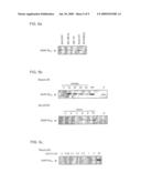 |
 |  |
 |  |
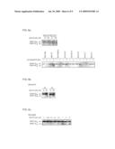 |  |
 |  |
 |  |
 |  |
 |  |
 |  |
 |  |
 |  |
 |  |
 |  |
 |  |
 |  |
 |  |
 |  |
 | 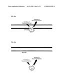 |
 |  |
 |  |
 |  |
 |  |
 |  |
 |  |
 | 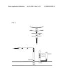 |
 |  |
 |  |
 |  |
 |  |
 |  |
 |  |
 |  |
 |  |
 |  |
 |  |
 |  |
 |  |
 |  |
 |  |
 |  |
 |  |
 |  |
 |  |
 |  |
 |  |
 |  |
 |  |
 |  |
 |  |
 |  |
 |  |
 |  |
 |  |
 |  |
 |  |
 |  |
 |  |
 |
| New patent applications in this class: | |
| Date | Title |
|---|---|
| 2019-05-16 | Non-invasive method for assessing the presence and severity of esophageal varices |
| 2019-05-16 | Method for evaluating protrusion-forming ability of cell spheroids |
| 2019-05-16 | Product demonstration device |
| 2019-05-16 | In vitro model for blood-brain barrier and method for producing in vitro model for blood-brain barrier |
| 2019-05-16 | Microbeads for cell culture and method of monitoring cell culture using the same |
| New patent applications from these inventors: | |
| Date | Title |
|---|---|
| 2022-01-13 | Microneedle array with active ingredient |
| 2021-11-04 | Immuno-based botulinum toxin serotype a activity assays |
| 2015-10-22 | Immuno-based retargeted endopeptidase activity assays |
| 2015-08-20 | Methods of activating clostridial toxins |
| 2014-09-04 | Highly sensitive cell-based assay to detect the presence of active botulinum neurotoxin serotype-a |
| Top Inventors for class "Chemistry: molecular biology and microbiology" | |
| Rank | Inventor's name |
|---|---|
| 1 | Marshall Medoff |
| 2 | Anthony P. Burgard |
| 3 | Mark J. Burk |
| 4 | Robin E. Osterhout |
| 5 | Rangarajan Sampath |