Patent application title: Human Cancer Suppressor Gene, Protein Encoded Therein, Expression Vector Containing The Same, And Cell Transformed By The Vector
Inventors:
Hyun-Kee Kim (Seoul, KR)
Jin Woo Kim (Seoul, KR)
IPC8 Class: AC12N500FI
USPC Class:
435325
Class name: Chemistry: molecular biology and microbiology animal cell, per se (e.g., cell lines, etc.); composition thereof; process of propagating, maintaining or preserving an animal cell or composition thereof; process of isolating or separating an animal cell or composition thereof; process of preparing a composition containing an animal cell; culture media therefore
Publication date: 2009-06-18
Patent application number: 20090155896
Inventors list |
Agents list |
Assignees list |
List by place |
Classification tree browser |
Top 100 Inventors |
Top 100 Agents |
Top 100 Assignees |
Usenet FAQ Index |
Documents |
Other FAQs |
Patent application title: Human Cancer Suppressor Gene, Protein Encoded Therein, Expression Vector Containing The Same, And Cell Transformed By The Vector
Inventors:
Hyun-Kee Kim
Jin-Woo Kim
Agents:
HAMRE, SCHUMANN, MUELLER & LARSON, P.C.
Assignees:
Origin: MINNEAPOLIS, MN US
IPC8 Class: AC12N500FI
USPC Class:
435325
Abstract:
Disclosed are a human cancer suppressor gene, a protein encoded therein,
an expression vector containing the same, and a cell transformed by the
vector. The gene of the present invention can be used for diagnosing,
preventing and treating the human cancers.Claims:
1. A human cancer suppressor protein having an amino acid sequence
selected from the group consisting of SEQ ID NO: 2, SEQ ID NO: 6, SEQ ID
NO: 10, SEQ ID NO: 14, SEQ ID NO: 18, SEQ ID NO: 22, SEQ ID NO: 26 and
SEQ ID NO: 30.
2. The human cancer suppressor protein according to claim 1, wherein the cancer is a cancer of the normal tissue selected from the group consisting of breast, lungs, thymus, liver, skeletal muscles, kidney, spleen, heart, placenta and peripheral blood.
3. A human cancer suppressor gene having a DNA sequence selected from the group consisting of SEQ ID NO: 1, SEQ ID NO: 5, SEQ ID NO, 9, SEQ ID NO: 13, SEQ ID NO: 17, SEQ ID NO: 21, SEQ ID NO: 25 and SEQ ID NO: 29, which encode the corresponding proteins.
4. The human cancer suppressor genes according to claim 3, wherein the cancer is a cancer of the normal tissue selected from the group consisting of breast, lungs, thymus, liver, skeletal muscles, kidney, spleen, heart, placenta and peripheral blood.
5. An expression vector containing each of the genes as defined in claim 3.
6. A cell transformed by each of the expression vectors as defined in claim 5.
7. The cell according to claim 6, wherein it is microorganisms or animal cells.
8. The cell according to claim 7, wherein the cell is selected from the group consisting of Escherichia coli DH5.alpha./GIG12/pcDNA3.1 (Accession No. KCTC 10642BP), E. coli DH5.alpha./GIG17/pcDNA3.1 (Accession No. KCTC 10655BP), E. coli DH5.alpha./GIG19/pcDNA3.1 (Accession No. KCTC 10656BP), E. coli DH5.alpha./GIG20/pcDNA3.1 (Accession No. KCTC 10657BP), E. coli DH5.alpha./GIG22/pcDNA3.1 (Accession No. KCTC 10658BP), E. coli DH5.alpha./GIG25/pcDNA3.1 (Accession No. KCTC 10659BP), E. coli DH5.alpha./GIG36/pcDNA3.1 (Accession No: KCTC 10643BP) and E. coli DH5.alpha./GIG2/pcDNA3.1 (Accession No. KCTC 10641 BP).
Description:
TECHNICAL FIELD
[0001]The present invention relates to a human cancer suppressor gene, a protein encoded therein, an expression vector containing the same, and a cell transformed by the vector.
BACKGROUND ART
[0002]Tumor suppressor gene products function to suppress normal cells from being transformed into certain cancer cells, and therefore loss of this function of the tumor suppressor gene products allows the normal cells to become malignant transformants (Klein, G., FASEB J., 7, 821-825 (1993)). In order to allow cancer cells to grow into a cancer, the cells should lose a function to control the normal copy number of a tumor suppressor gene. It was found that modification in a coding sequence of a p53 tumor suppressor gene is one of the most general genetic changes in the human cancers (Bishop, J. M., Cell, 64, 235-248 (1991); and Weinberg, R. A., Science, 254, 1138-1146 (1991)).
[0003]However, it was estimated that only some of breast cancer tissues exhibited a p53 mutation because the reported p53 mutation was in a range of 30% in the breast cancer (Keen, J. C. & Davidson, N. E., Cancer, 97, 825-833 (2003)) and Borresen-Dale, A-L., Human Mutation, 21, 292-300 (2003)).
[0004]The p53 mutation accounts for at least 50% of liver cancer especially in the region exposed to aflatoxin B1 or having a high frequency of infection by hepatitis B virus, and it is mainly characterized by a missense mutation at a codon 249 in the p53 tumor gene (Montesano, R. et al., J. Natl. Cancer Inst., 89, 1844-1851 (1997); Szymanska, K. & Hainaut, P. Acta Biochimica Polonica, 50, 231-238 (2003)). However, the p53 mutation was nothing but a range of 30% of breast cancer in U.S. and Western Europe, and there is no hot spot in which such mutation occurs more frequently (Szymanska, K. & Hainaut, P. Acta Biochimica Polonica, 50, 231-238 (2003)).
[0005]Accordingly, the present inventors have ardently attempted to separate a novel tumor suppressor gene from normal breast tissues using an mRNA differential display (DD) method for effectively displaying genes differentially expressed between a normal breast tissue and a breast cancer, or between a normal liver tissue and a liver cancer (Liang, P. and Pardee, A. B., Science, 257, 967-971 (1992); and Liang, P. et al., Cancer Res., 52, 6966-6968 (1993)).
DISCLOSURE OF INVENTION
Technical Problem
[0006]Accordingly, the present invention is designed to solve the problems of the prior art, and therefore it is an object of the present invention to provide a novel human cancer suppressor gene.
[0007]It is another object of the present invention to provide a cancer suppressor protein coded by the gene.
[0008]It is still another object of the present invention to provide an expression vector containing the gene.
[0009]It is yet another object of the present invention to provide a cell transformed by the expression vector.
Technical Solution
[0010]In order to accomplish the above object, the present invention provides a human cancer suppressor gene (growth-inhibiting gene 12; also referred to as GIG12) having a DNA sequence set forth in SEQ ID NO: 1.
[0011]In order to accomplish the other object, the present invention provides a human cancer suppressor protein having an amino acid sequence set forth in SEQ ID NO: 2, which is encoded by the GIG12 gene.
[0012]The present invention also provides a human cancer suppressor gene (growth-inhibiting gene 17; also referred to as GIG17) having a DNA sequence set forth in SEQ ID NO: 5.
[0013]The present invention provides a human cancer suppressor protein having an amino acid sequence set forth in SEQ ID NO: 6, which is encoded by the GIG17 gene.
[0014]The present invention also provides a human cancer suppressor gene (growth-inhibiting gene 19; also referred to as GIG19) having a DNA sequence set forth in SEQ ID NO: 9.
[0015]The present invention provides a human cancer suppressor protein having an amino acid sequence set forth in SEQ ID NO: 10, which is encoded by the GIG19 gene.
[0016]The present invention also provides a human cancer suppressor gene (growth-inhibiting gene 20; also referred to as GIG20) having a DNA sequence set forth in SEQ ID NO: 13.
[0017]The present invention provides a human cancer suppressor protein having an amino acid sequence set forth in SEQ ID NO: 14, which is encoded by the GIG20 gene.
[0018]The present invention also provides a human cancer suppressor gene (growth-inhibiting gene 22; also referred to as GIG22) having a DNA sequence set forth in SEQ ID NO: 17.
[0019]The present invention provides a human cancer suppressor protein having an amino acid sequence set forth in SEQ ID NO: 18, which is encoded by the GIG22 gene.
[0020]The present invention also provides a human cancer suppressor gene (growth-inhibiting gene 25; also referred to as GIG25) having a DNA sequence set forth in SEQ ID NO: 21.
[0021]The present invention provides a human cancer suppressor protein having an amino acid sequence set forth in SEQ ID NO: 22, which is encoded by the GIG25 gene.
[0022]The present invention also provides a human cancer suppressor gene (growth-inhibiting gene 36; also referred to as GIG36) having a DNA sequence set forth in SEQ ID NO: 25.
[0023]The present invention provides a human cancer suppressor protein having an amino acid sequence set forth in SEQ ID NO: 26, which is encoded by the GIG36 gene.
[0024]The present invention also provides a human cancer suppressor gene (growth-inhibiting gene 2; also referred to as GIG2) having a DNA sequence set forth in SEQ ID NO: 29.
[0025]The present invention provides a human cancer suppressor protein having an amino acid sequence set forth in SEQ ID NO: 30, which is encoded by the GIG2 gene.
[0026]According to still another object, the present invention provides an expression vector containing each of the genes.
[0027]According to yet another object, the present invention provides a cell transformed by each of the expression vectors.
BRIEF DESCRIPTION OF THE DRAWINGS
[0028]These and other features, aspects, and advantages of preferred embodiments of the present invention will be more fully described in the following detailed description, taken accompanying drawings. In the drawings:
[0029]FIG. 1 is a gel diagram showing a PCR result using a 5'-13-mer random primer H-AP32 of SEQ ID NO: 3 and an anchored oligo-dT primer of SEQ ID NO: 4;
[0030]FIG. 2 is a gel diagram showing a PCR result using a 5'-13mer random primer H-AP7 of SEQ ID NO: 7 and an anchored oligo-dT primer of SEQ ID NO: 8;
[0031]FIG. 3 is a gel diagram showing a PCR result using a 5'-13mer random primer H-AP45 of SEQ ID NO: 11 and an anchored oligo-dT primer of SEQ ID NO: 12;
[0032]FIG. 4 is a gel diagram showing a PCR result using a 5'-13mer random primer H-AP40 of SEQ ID NO: 15 and an anchored oligo-dT primer of SEQ ID NO: 16;
[0033]FIG. 5 is a gel diagram showing a PCR result using a 5'-13mer random primer H-AP30 of SEQ ID NO: 19 and an anchored oligo-dT primer of SEQ ID NO: 20;
[0034]FIG. 6 is a gel diagram showing a PCR result using a 5'-13mer random primer H-AP40 of SEQ ID NO: 23 and an anchored oligo-dT primer of SEQ ID NO: 24;
[0035]FIG. 7 is a gel diagram showing a PCR result using a 5'-13mer random primer H-AP29 of SEQ ID NO: 27 and an anchored oligo-dT primer of SEQ ID NO: 28;
[0036]FIG. 8 is a gel diagram showing a PCR result using a 5'-13mer random primer H-AP32 of SEQ ID NO: 31 and an anchored oligo-dT primer of SEQ ID NO: 32;
[0037]FIG. 9 is a diagram showing a result that a gene product of the GIG12 is analyzed on SDS-PAGE;
[0038]FIG. 10 is a diagram showing a result that a gene product of the GIG17 is analyzed on SDS-PAGE;
[0039]FIG. 11 is a diagram showing a result that a gene product of the GIG19 is analyzed on SDS-PAGE;
[0040]FIG. 12 is a diagram showing a result that a gene product of the GIG20 is analyzed on SDS-PAGE;
[0041]FIG. 13 is a diagram showing a result that a gene product of the GIG22 is analyzed on SDS-PAGE;
[0042]FIG. 14 is a diagram showing a result that a gene product of the GIG25 is analyzed on SDS-PAGE;
[0043]FIG. 15 is a diagram showing a result that a gene product of the GIG-36 is analyzed on SDS-PAGE;
[0044]FIG. 16 is a diagram showing a result that a gene product of the GIG2 is analyzed on SDS-PAGE;
[0045]FIG. 17(a) is a diagram showing a northern blotting result that the GIG12 gene is differentially expressed in a normal breast tissue, a primary breast cancer tissue and a breast cancer cell line, and FIG. 17(b) is a diagram showing a northern blotting result obtained by hybridizing the same blot with β-actin probe;
[0046]FIG. 18(a) is a diagram showing a northern blotting result that the GIG17 gene is differentially expressed in a normal breast tissue, a primary breast cancer tissue and a breast cancer cell line, and FIG. 18(b) is a diagram showing a northern blotting result obtained by hybridizing the same blot with β-actin probe;
[0047]FIG. 19(a) is a diagram showing a northern blotting result that the GIG19 gene is differentially expressed in a normal breast tissue, a primary breast cancer tissue and a breast cancer cell line, and FIG. 19(b) is a diagram showing a northern blotting result obtained by hybridizing the same blot with β-actin probe;
[0048]FIG. 20(a) is a diagram showing a northern blotting result that the GIG20 gene is differentially expressed in a normal breast tissue, a primary breast cancer tissue and a breast cancer cell line, and FIG. 20(b) is a diagram showing a northern blotting result obtained by hybridizing the same blot with β-actin probe;
[0049]FIG. 21(a) is a diagram showing a northern blotting result that the GIG22 gene is differentially expressed in a normal breast tissue, a primary breast cancer tissue and a breast cancer cell line, and FIG. 21(b) is a diagram showing a northern blotting result obtained by hybridizing the same blot with β-actin probe;
[0050]FIG. 22(a) is a diagram showing a northern blotting result that the GIG25 gene is differentially expressed in a normal breast tissue, a primary breast cancer tissue and a breast cancer cell line, and FIG. 22(b) is a diagram showing a northern blotting result obtained by hybridizing the same blot with β-actin probe;
[0051]FIG. 23(a) is a diagram showing a northern blotting result that the GIG36 gene is differentially expressed in a normal breast tissue, a primary breast cancer tissue and a breast cancer cell line, and FIG. 23(b) is a diagram showing a northern blotting result obtained by hybridizing the same blot with β-actin probe;
[0052]FIG. 24(a) is a diagram showing a northern blotting result that the GIG2 gene is differentially expressed in a normal lung tissue, a primary lung cancer tissue, a metastatic lung cancer tissue and a lung cancer cell line, and FIG. 24(b) is a diagram showing a northern blotting result obtained by hybridizing the same blot with β-actin probe;
[0053]FIG. 25(a) is a diagram showing a northern blotting result that the GIG12 gene is differentially expressed in various normal tissues, and FIG. 25(b) is a diagram showing a northern blotting result obtained by hybridizing the same blot with β-actin probe;
[0054]FIG. 26(a) is a diagram showing a northern blotting result that the GIG17 gene is differentially expressed in various normal tissues, and FIG. 26(b) is a diagram showing a northern blotting result obtained by hybridizing the same blot with β-actin probe;
[0055]FIG. 27(a) is a diagram showing a northern blotting result that the GIG19 gene is differentially expressed in various normal tissues, and FIG. 27(b) is a diagram showing a northern blotting result obtained by hybridizing the same blot with β-actin probe;
[0056]FIG. 28(a) is a diagram showing a northern blotting result that the GIG20 gene is differentially expressed in various normal tissues, and FIG. 28(b) is a diagram showing a northern blotting result obtained by hybridizing the same blot with β-actin probe;
[0057]FIG. 29(a) is a diagram showing a northern blotting result that the GIG22 gene is differentially expressed in various normal tissues, and FIG. 29(b) is a diagram showing a northern blotting result obtained by hybridizing the same blot with β-actin probe;
[0058]FIG. 30(a) is a diagram showing a northern blotting result that the GIG25 gene is differentially expressed in various normal tissues, and FIG. 30(b) is a diagram showing a northern blotting result obtained by hybridizing the same blot with β-actin probe;
[0059]FIG. 31(a) is a diagram showing a northern blotting result that the GIG36 gene is differentially expressed in various normal tissues, and FIG. 31(b) is a diagram showing a northern blotting result obtained by hybridizing the same blot with β-actin probe;
[0060]FIG. 32(a) is a diagram showing a northern blotting result that the GIG2 gene is differentially expressed in various normal tissues, and FIG. 32(b) is a diagram showing a northern blotting result obtained by hybridizing the same blot with β-actin probe;
[0061]FIG. 33(a) is a diagram showing a northern blotting result that the GIG12 gene is differentially expressed in various cancer cell lines, and FIG. 33(b) is a diagram showing a northern blotting result obtained by hybridizing the same blot with β-actin probe;
[0062]FIG. 34(a) is a diagram showing a northern blotting result that the GIG17 gene is differentially expressed in various cancer cell lines, and FIG. 34(b) is a diagram showing a northern blotting result obtained by hybridizing the same blot with β-actin probe;
[0063]FIG. 35(a) is a diagram showing a northern blotting result that the GIG19 gene is differentially expressed in various cancer cell lines, and FIG. 35(b) is a diagram showing a northern blotting result obtained by hybridizing the same blot with β-actin probe;
[0064]FIG. 36(a) is a diagram showing a northern blotting result that the GIG20 gene is differentially expressed in various cancer cell lines, and FIG. 36(b) is a diagram showing a northern blotting result obtained by hybridizing the same blot with β-actin probe;
[0065]FIG. 37(a) is a diagram showing a northern blotting result that the GIG22 gene is differentially expressed in various cancer cell lines, and FIG. 37(b) is a diagram showing a northern blotting result obtained by hybridizing the same blot with β-actin probe;
[0066]FIG. 38(a) is a diagram showing a northern blotting result that the GIG25 gene is differentially expressed in various cancer cell lines, and FIG. 38(b) is a diagram showing a northern blotting result obtained by hybridizing the same blot with β-actin probe;
[0067]FIG. 39(a) is a diagram showing a northern blotting result that the GIG36 gene is differentially expressed in various cancer cell lines, and FIG. 39(b) is a diagram showing a northern blotting result obtained by hybridizing the same blot with β-actin probe;
[0068]FIG. 40(a) is a diagram showing a northern blotting result that the GIG2 gene is differentially expressed in various cancer cell lines, and FIG. 40(b) is a diagram showing a northern blotting result obtained by hybridizing the same blot with β-actin probe;
[0069]FIG. 41 is a graph showing growth curves of the wild-type MCF-7 cell, the MCF-7 breast cancer cell transfected by the GIG12 gene, and the MCF-7 cell transfected by the expression vector pcDNA3.1;
[0070]FIG. 42 is a graph showing growth curves of the wild-type HepG2 liver cancer cell line, the HepG2 liver cancer cell transfected by the GIG17 gene, and the HepG2 cell transfected by the expression vector pcDNA3.1;
[0071]FIG. 43 is a graph showing growth curves of the wild-type HepG2 liver cancer cell line, the HepG2 liver cancer cell transfected by the GIG19 gene, and the HepG2 cell transfected by the expression vector pcDNA3.1;
[0072]FIG. 44 is a graph showing growth curves of the wild-type HepG2 liver cancer cell line, the HepG2 liver cancer cell transfected by the GIG20 gene, and the HepG2 cell transfected by the expression vector pcDNA3.1;
[0073]FIG. 45 is a graph showing growth curves of the wild-type HepG2 liver cancer cell line, the HepG2 liver cancer cell transfected by the GIG22 gene, and the HepG2 cell transfected by the expression vector pcDNA3.1;
[0074]FIG. 46 is a graph showing growth curves of the wild-type HepG2 liver cancer cell line, the HepG2 liver cancer cell transfected by the GIG25 gene, and the HepG2 cell transfected by the expression vector pcDNA3.1;
[0075]FIG. 47 is a graph showing growth curves of the wild-type HepG2 liver cancer cell line, the HepG2 liver cancer cell transfected by the GIG36 gene, and the HepG2 cell transfected by the expression vector pcDNA3.1; and
[0076]FIG. 48 is a graph showing growth curves h of the wild-type A549 lung cancer cell line, the A549 lung cancer cell transfected by the GIG2 gene, and the A549 cell transfected by the expression vector pcDNA3.1.
BEST MODE
[0077]Hereinafter, preferred embodiments of the present invention will be described in detail referring to the accompanying drawings.
[0078]1. GIG12
[0079]The gene of the present invention is a human cancer suppressor gene 36 (GIG36) showing a DNA sequence of SEQ ID NO: 1, which has been deposited with Accession No. AY493417 into the GenBank database of U.S. National Institutes of Health (NIH) (Publication Date: Mar. 1, 2005), and some DNA sequence of the deposited gene is identical with that of the lactotransferrin deposited with Accession No. NM--002343 into the database. The lactotransferrin is abundantly distributed mainly in milk and serum, and its function has been known as only a carrier of ferric ions (Kanyshkova, G. T., et al., Biochemistry (Moscow), 66, 1-7 (2001)). At the same time, it was found that the lactotransferrin has only a strong antibacterial activity (Oppenheimer, J. S. J. Nutr., 131, 6165-6335 (2001); Shugars, C. D., et al., Gerontology, 47, 246-253 (2001)).
[0080]Contrary to the functions as reported previously, it was however found from this study result that the lactotransferrin is closely associated with various carcinogenesis. It was also found that the GIG12 tumor suppressor gene was not at all expressed in various human tumors including the breast cancer, while its expression was increased in various normal tissues.
[0081]The DNA sequence of SEQ ID NO: 1 has one open reading frame (ORF) corresponding to base positions from 111 to 2246 of the DNA sequence (Base positions from 2244 to 2246 represent a stop codon). However, because of degeneracy of codons, or considering preference of codons for living organisms to express the genes, the genes of the present invention may be variously modified in coding regions without changing an amino acid sequence of the protein expressed from the coding region, and also be variously modified or changed in a region except the coding region within a range that does not affect the gene expression. Such a modified gene is also included in the scope of the present invention. Accordingly, the present invention also includes a polynucleotide having substantially the same DNA sequence as the gene; and fragments of the gene. The term "substantially the same polynucleotide" means a polynucleotide having DNA sequence homology of at least 80%, preferably at least 90%, and the most preferably at least 95%.
[0082]The protein expressed from the gene of the present invention consists of 711 amino acid residues, and has an amino acid sequence of SEQ ID NO: 2 and a molecular weight of approximately 78 kDa. However, one or more amino acids may be also substituted, added or deleted in the amino acid sequence of the protein within a range that does not affect functions of the protein, and only some portion of the protein may be used depending on its usage. Such a modified amino acid sequence is also included in the scope of the present invention. Accordingly, the present invention also includes a polypeptide having substantially the same amino acid sequence as the protein; and fragments of the protein. The term "substantially the same polypeptide" means a polypeptide having sequence homology of at least 80%, preferably at least 90%, and the most preferably at least 95%.
[0083]The gene and the protein of the present invention may be separated from human tissues, or be synthesized according to the known methods for synthesizing DNA or peptide. For example, the gene of the present invention may be screened and cloned according to the conventional methods on the basis of the information on the DNA sequence set forth in SEQ ID NO: 1. As another example, a 680-bp cDNA fragment, which is not expressed in the cancer tissue or the cancer cell line but differentially expressed only in the normal tissue, may be obtained by carrying out a reverse transcription polymerase chain reaction (RT-PCR) on the total RNAs extracted from a normal tissue, and a cancer tissue or a cancer cell line using a random primer H-AP32 of SEQ ID NO: 3 (5'-AAGCTTCCTGCAA-3') and an anchored oligo-dT primer of SEQ ID NO: 4 (5'-AAGCTTTTTTTTTTTC-3'), and the resultant fragment, which is used as the probe, may be plaque-hybridized with a cDNA library to obtain a full-length cDNA clone.
[0084]The gene prepared thus may be inserted into a vector for expression in microorganisms or animal cells, already known in the art, to obtain an expression vector, and then cDNA of the gene may be replicated in a large quantity or its protein may be produced in a commercial quantity by introducing the expression vector into suitable host cells, for example Escherichia coli, a MCF-7 cell line, etc. Upon constructing the expression vector, DNA regulatory sequences such as a promoter and a terminator, autonomously replicating sequences, secretion signals, etc. may be suitably selected and combined depending on kinds of the host cells that are engineered to produce the gene or the protein.
[0085]The present inventors inserted the full-length GIG12 cDNA into an expression vector pcDNA3.1 (Invitrogen, U.S.), and then transformed Escherichia coli DH5α with the resultant expression vector to obtain a transformant, which was then named E. coli DH5α/GIG12/pcDNA3.1, and deposited with Accession No. KCTC 10642BP into Korean Collection for Type Cultures on May 24, 2004.
[0086]It is regarded that the gene of the present invention is overexpressed in normal tissues, preferably breast, lungs, thymus, liver, skeletal muscles, kidney, spleen, heart, placenta, and peripheral bloods, to suppress carcinogenesis. The gene of the present invention is mainly overexpressed in these tissues as an mRNA transcript having a size of approximately 2.4 kb. Especially, the gene of the present invention is differentially expressed only in the normal tissues. For example, the gene of the present invention is not expressed in the cancer tissues and the cancer cells such as the breast cancer tissue, the breast cancer cell line MCF-7, etc., but differentially expressed only in the normal tissues.
[0087]The cancer cell line into which the genes of the present invention were introduced showed a high mortality, and therefore the gene of the present invention may be effectively used for treatment and prevention of the cancer.
[0088]2. GIG17
[0089]The gene of the present invention is a human cancer suppressor gene 17 (GIG17) having a DNA sequence of SEQ ID NO: 5, which has been deposited with Accession No. AY544122 into the GenBank database of U.S. National Institutes of Health (NIH) (Publication Date: Dec. 31, 2005), and the deposited gene has polymorphism that its 3 base pairs are different to a DNA sequence of the human fructose 1,6-bisphosphatase deposited with Accession No. M19922 into the database.
[0090]One of the most important reactions in a glucose metabolism is to hydrolyze fructose 1,6-bisphosphate into fructose-6-phosphate (Marcus, F. et al., Arch. Biol. Med. Exp., 20, 371-378 (1987); Okar, D, A. & Lange, A. J. Biofactors, 10, 1-14 (1999)). An enzyme that catalyzes the metabolism is the human fructose 1,6-bisphosphatase, which is present in all living organisms.
[0091]Contrary to the glucose metabolism as reported previously, it was however found from this study result that the GIG17 tumor suppressor gene was not at all expressed in various human tumors including the liver cancer, while its expression was significantly increased in various normal tissues.
[0092]The DNA sequence of SEQ ID NO: 5 has one open reading frame (ORF) corresponding to base positions from 88 to 1104 of the DNA sequence (Base positions from 1102 to 1104 represent a stop codon). However, because of degeneracy of codons, or considering preference of codons for living organisms to express the genes, the genes of the present invention may be variously modified in coding regions without changing an amino acid sequence of the protein expressed from the coding region, and also be variously modified or changed in a region except the coding region within a range that does not affect the gene expression. Such a modified gene is also included in the scope of the present invention. Accordingly, the present invention also includes a polynucleotide having substantially the same DNA sequence as the gene; and fragments of the gene. The term "substantially the same polynucleotide" means a polynucleotide having DNA sequence homology of at least 80%, preferably at least 90%, and the most preferably at least 95%.
[0093]The protein expressed from the gene of the present invention consists of 338 amino acid residues, and has an amino acid sequence of SEQ ID NO: 6 and a molecular weight of approximately 37 kDa. However, one or more amino acids may be also substituted, added or deleted in the amino acid sequence of the protein within a range that does not affect functions of the protein, and only some portion of the protein may be used depending on its usage. Such a modified amino acid sequence is also included in the scope of the present invention. Accordingly, the present invention also includes a polypeptide having substantially the same amino acid sequence as the protein; and fragments of the protein. The term "substantially the same polypeptide" means a polypeptide having sequence homology of at least 80%, preferably at least 90%, and the most preferably at least 95%.
[0094]The gene and protein of the present invention may be separated from human tissues, or be synthesized according to the known methods for synthesizing DNA or peptide. For example, the gene of the present invention may be screened and cloned according to the conventional methods on the basis of the information on the DNA sequence set forth in SEQ ID NO: 5. As another example, a 250-bp cDNA fragment, which is not expressed in the cancer tissue or the cancer cell line but differentially expressed only in the normal tissue, may be obtained by carrying out a reverse transcription-polymerase chain reaction (RT-PCR) on the total RNAs extracted from a normal tissue, and a cancer tissue or a cancer cell line using a random primer H-AP7 of SEQ ID NO: 7 (5'-AAGCTTAACGAGG-3') and an anchored oligo-dT primer of SEQ ID NO: 8 (5'-AAGCTTTTTTTTTTTC-3'), and the resultant fragment, which is used as the probe, may be plaque-hybridized with a cDNA library to obtain a full-length cDNA clone.
[0095]The gene prepared thus may be inserted into a vector for expression in microorganisms or animal cells, already known in the art, to obtain an expression vector, and then cDNA of the gene may be replicated in a large quantity or its protein may be produced in a commercial quantity by introducing the expression vector into suitable host cells, for example Escherichia coli, a HepG2 cell line, etc. Upon constructing the expression vector, DNA regulatory sequences such as a promoter and a terminator, autonomously replicating sequences, secretion signals, etc. may be suitably selected and combined depending on kinds of the host cells that are engineered to produce the gene or the protein.
[0096]The present inventors inserted the full-length GIG17 cDNA into an expression vector pcDNA3.1 (Invitrogen, U.S.), and then transformed Escherichia coli DH5α with the resultant expression vector to obtain a transformant, which was then named E. coli DH5α/GIG17/pcDNA3.1, and deposited with Accession No. KCTC 10655BP into Korean Collection for Type Cultures on Jun. 14, 2004.
[0097]It is regarded that the gene of the present invention is overexpressed in normal tissues, preferably liver, kidney, spleen and lungs, to suppress carcinogenesis. It is also regarded that the gene of the present invention is suppressed even in leukemia, uterine cancer, malignant lymphoma, colon cancer and skin cancer to induce carcinogenesis. The gene of the present invention is mainly overexpressed in these tissues as an mRNA transcript having a size of approximately 1.3 kb. Especially, the gene of the present invention is differentially expressed only in the normal tissues. For example, the gene of the present invention is not expressed in the cancer tissues and the cancer cells such as the liver cancer tissue, the liver cancer cell line HepG2, etc., but differentially expressed only in the normal liver tissue.
[0098]The cancer cell line into which the genes of the present invention were introduced showed a high mortality, and therefore the gene of the present invention may be effectively used for treatment and prevention of the cancer.
[0099]3. GIG19
[0100]The gene of the present invention is a human cancer suppressor gene 19 (GIG19) having a DNA sequence of SEQ ID NO: 9, which has been deposited with Accession No. AY544123 into the GenBank database of U.S. National Institutes of Health (NIH) (Publication Date: Dec. 31, 2005), and the DNA sequence of the deposited gene is identical with those of the Homo sapiens alpha-1-microglobulin/bikunin precursor and the human mRNA for protein HC (alpha-1-microglobulin), deposited with Accession No. BC041593 and X04225 into the existing database, respectively. The alpha-1-microglobulin, also referred to as an HC protein, is a lipoprotein having an immunosuppressive effect (Akerstrom, B. et al., Biochimica Biophysica Acta, 1482, 172-184 (2002); Xu, S. & Venge, P., Biochimica Biophysica Acta, 1482, 298-307 (2002)). Contrary to the functions of the tumor suppressor gene as reported previously, it was however found from this study result that the GIG19 tumor suppressor gene was not at all expressed in the liver cancer, while its expression was significantly increased in various normal liver tissues.
[0101]The DNA sequence of SEQ ID NO: 9 has one open reading frame (ORF) corresponding to base positions from 61 to 1119 of the DNA sequence (Base positions from 59 to 61 represent a stop codon). However, because of degeneracy of codons, or considering preference of codons for living organisms to express the genes, the genes of the present invention may be variously modified in coding regions without changing an amino acid sequence of the protein expressed from the coding region, and also be variously modified or changed in a region except the coding region within a range that does not affect the gene expression. Such a modified gene is also included in the scope of the present invention. Accordingly, the present invention also includes a polynucleotide having substantially the same DNA sequence as the gene; and fragments of the gene. The term "substantially the same polynucleotide" means a polynucleotide having DNA sequence homology of at least 80%; preferably at least 90%; and the most preferably at least 95%.
[0102]The protein expressed from the gene of the present invention consists of 352 amino acid residues, and has an amino acid sequence of SEQ ID NO: 10 and a molecular weight of approximately 39 kDa. However, one or more amino acids may be also substituted, added or deleted in the amino acid sequence of the protein within a range that does not affect functions of the protein, and only some portion of the protein may be used depending on its usage. Such a modified amino acid sequence is also included in the scope of the present invention. Accordingly, the present invention also includes a polypeptide having substantially the same amino acid sequence as the protein; and fragments of the protein. The term "substantially the same polypeptide" means a polypeptide having sequence homology of at least 80%, preferably at least 90%, and the most preferably at least 95%.
[0103]The gene and protein of the present invention may be separated from human tissues, or be synthesized according to the known methods for synthesizing DNA or peptide. For example, the gene of the present invention may be screened and cloned according to the conventional methods on the basis of the information on the DNA sequence set forth in SEQ ID NO: 9. As another example, a 281-bp cDNA fragment, which is not expressed in the cancer tissue or the cancer cell line but differentially expressed only in the normal tissue, may be obtained by carrying out a reverse transcription-polymerase chain reaction (RT-PCR) on the total RNAs extracted from a normal tissue, and a cancer tissue or a cancer cell line using a random primer H-AP40 of SEQ ID NO: 11 (5'-AAGCTTGTCAGCC-3') and an anchored oligo-dT primer of SEQ ID NO: 12 (5'-AAGCTTTTTTTTTTTC-3'); and the resultant fragment, which is used as the probe, may be plaque-hybridized with a cDNA library to obtain a full-length cDNA clone.
[0104]The gene prepared thus may be inserted into a vector for expression in microorganisms or animal cells, already known in the art, to obtain an expression vector, and then cDNA of the gene may be replicated in a large quantity or its protein may be produced in a commercial quantity by introducing the expression vector into suitable host cells, for example Escherichia coli, a HepG2 cell line, etc. Upon constructing the expression vector, DNA regulatory sequences such as a promoter and a terminator, autonomously replicating sequences, secretion signals, etc. may be suitably selected and combined depending on kinds of the host cells that are engineered to produce the gene or the protein.
[0105]The present inventors inserted the full-length GIG19 cDNA into an expression vector pcDNA3.1 (Invitrogen, U.S.), and then transformed Escherichia coli DH5α with the resultant expression vector to obtain a transformant, which was then named E. coli DH5α/GIG19/pcDNA3.1, and deposited with Accession No. KCTC 10656BP into Korean Collection for Type Cultures on Jun. 14, 2004.
[0106]It is regarded that the gene of the present invention is overexpressed in normal tissues, preferably liver tissues, to suppress carcinogenesis. The gene of the present invention is mainly overexpressed in these tissues as an mRNA transcript having a size of approximately 1.2 kb. Especially, the gene of the present invention is differentially expressed in the normal tissues. For example, the gene of the present invention is not expressed in the cancer tissues and the cancer cells such as the liver cancer tissue, the liver cancer cell line HepG2, etc., but differentially expressed only in the normal liver tissue.
[0107]The liver cancer cell line into which the genes of the present invention were introduced showed a high mortality, and therefore the gene of the present invention may be effectively used for treatment and prevention of the cancer.
[0108]4. GIG20
[0109]The gene of the present invention is a human cancer suppressor gene 20 (GIG20) having a DNA sequence of SEQ ID NO: 13, which has been deposited with Accession No. AY544124 into the GenBank database of U.S. National Institutes of Health (NIH) (Publication Date: Dec. 31, 2005), and the DNA sequence of the deposited gene is identical with that of the Homo sapiens albumin deposited with Accession No. BC041789 into the existing database. It has been known that the albumin was a protein that takes role in supplying nutrients (Grant, J. P., Ann. Surg., 220, 610-616 (1994)). Contrary to the functions of the tumor suppressor gene as reported previously, it was however found from this study result that the GIG20 tumor suppressor gene was not at all expressed in the liver cancer, while its expression was significantly increased in various normal liver tissues. The fact that the gene of the present invention is a tumor suppressor gene is based on that the protein "albumin" is produced in the normal liver cell because albumin genes within nucleuses are present in the liver cell. This is why that the normal liver cell is a cell in which the albumin gene normally works, but if a level of albumin is lower than the normal level in the liver cell, then the albumin gene does not normally works in the liver cell, or the number of the normal albumin gene is reduced, which may appear in the case of liver cancer
[0110]The DNA sequence of SEQ ID NO: 13 has one open reading frame (ORF) corresponding to base positions from 8 to 1261 of the DNA sequence (Base positions from 1259 to 1261 represent a stop codon). However, because of degeneracy of codons, or considering preference of codons for living organisms to express the genes, the genes of the present invention may be variously modified in coding regions without changing an amino acid sequence of the protein expressed from the coding region, and also be variously modified or changed in a region except the coding region within a range that does not affect the gene expression. Such a modified gene is also included in the scope of the present invention. Accordingly, the present invention also includes a polynucleotide having substantially the same DNA sequence as the gene; and fragments of the gene. The term "substantially the same polynucleotide" means a polynucleotide having DNA sequence homology of at least 80%, preferably at least 90%, and the most preferably at least 95%.
[0111]The protein expressed from the gene of the present invention consists of 417 amino acid residues, and has an amino acid sequence of SEQ ID NO: 14 and a molecular weight of approximately 47 kDa. However, one or more amino acids may be also substituted, added or deleted in the amino acid sequence of the protein within a range that does not affect functions of the protein, and only some portion of the protein may be used depending on its usage. Such a modified amino acid sequence is also included in the scope of the present invention. Accordingly, the present invention also includes a polypeptide having substantially the same amino acid sequence as the protein; and fragments of the protein. The term "substantially the same polypeptide" means a polypeptide having sequence homology of at least 80%, preferably at least 90%, and the most preferably at least 95%.
[0112]The gene and protein of the present invention may be separated from human tissues, or be synthesized according to the known methods for synthesizing DNA or peptide. For example, the gene of the present invention may be screened and cloned according to the conventional methods on the basis of the information on the DNA sequence set forth in SEQ ID NO: 13. As another example, a 256-bp cDNA fragment, which is not expressed in the cancer tissue or the cancer cell line but differentially expressed only in the normal tissue, may be obtained by carrying out a reverse transcription-polymerase chain reaction (RT-PCR) on the total RNAs extracted from a normal tissue, and a cancer tissue or a cancer cell line using a random primer H-AP40 of SEQ ID NO: 15 (5'-AAGCTTGTCAGCC-3') and an anchored oligo-dT primer of SEQ ID NO: 16 (5'-AAGCTTTTTTTTTTTC-3'), and the resultant fragment, which is used as the probe, may be plaque-hybridized with a cDNA library to obtain a full-length cDNA clone.
[0113]The gene prepared thus may be inserted into a vector for expression in microorganisms or animal cells, already known in the art, to obtain an expression vector, and then cDNA of the gene may be replicated in a large quantity or its protein may be produced in a commercial quantity by introducing the expression vector into suitable host cells, for example Escherichia coli, a HepG2 cell line, etc. Upon constructing the expression vector, DNA regulatory sequences such as a promoter and a terminator, autonomously replicating sequences, secretion signals, etc. may be suitably selected and combined depending on kinds of the host cells that are engineered to produce the gene or the protein.
[0114]The present inventors inserted the full-length GIG20-cDNA into an expression vector pcDNA3.1 (Invitrogen, U.S.), and then transformed Escherichia coli DH5α with the resultant expression vector to obtain a transformant, which was then named E. coli DH5α/GIG20/pcDNA3.1, and deposited with Accession No. KCTC 10657BP into Korean Collection for Type Cultures on Jun. 14, 2004.
[0115]It is regarded that the gene of the present invention is overexpressed in normal tissues, preferably liver tissues, to suppress carcinogenesis. The gene of the present invention is mainly overexpressed in these tissues as an mRNA transcript having a size of approximately 2.4 kb. Especially, the gene of the present invention is differentially expressed only in the normal tissues. For example, the gene of the present invention is not expressed in the cancer tissues and the cancer cells such as the liver cancer tissue, the liver cancer cell line HepG2, etc., but differentially expressed only in the normal liver tissue.
[0116]The liver cancer cell line into which the genes of the present invention were introduced showed a high mortality, and therefore the gene of the present invention may be effectively used for treatment and prevention of the cancer.
[0117]5. GIG22
[0118]The gene of the present invention is a human cancer suppressor gene 22 (GIG22) having a DNA sequence of SEQ ID NO: 17, which has been deposited with Accession No. AY512565 into the GenBank database of U.S. National Institutes of Health (NIH) (Publication Date: May 31, 2005), and some DNA sequence of the deposited gene is different to a DNA sequence of the Homo sapiens DNAJ domain-containing protein MCJ gene deposited with Accession No. AF126743 into the database and its expressed amino acid sequence is also different to that of the Homo sapiens DNAJ domain-containing protein MCJ. It was reported that expression of the MCJ gene was reduced in the case of ovarian cancer (Shridhar, V. et al., Cancer Res., 61, 4258-4265 (2001)), but it was found from this study result that the GIG22 tumor suppressor gene was not at all expressed in various human tumors including liver cancer, while its expression was significantly increased in various normal tissues.
[0119]The DNA sequence of SEQ ID NO: 17 has one open reading frame (ORF) corresponding to base positions from 95 to 547 of the DNA sequence (Base positions from 545 to 547 represent a stop codon). However, because of degeneracy of codons, or considering preference of codons for living organisms to express the genes, the genes of the present invention may be variously modified in coding regions without changing an amino acid sequence of the protein expressed from the coding region, and also be variously modified or changed in a region except the coding region within a range that does not affect the gene expression. Such a modified gene is also included in the scope of the present invention. Accordingly, the present invention also includes a polynucleotide having substantially the same DNA sequence as the gene; and fragments of the gene. The term "substantially the same polynucleotide" means a polynucleotide having DNA sequence homology of at least 80%, preferably at least 90%, and the most preferably at least 95%.
[0120]The protein expressed from the gene of the present invention consists of 150 amino acid residues, and has an amino acid sequence of SEQ ID NO: 18 and a molecular weight of approximately 16 kDa. However, one or more amino acids may be also substituted, added or deleted in the amino acid sequence of the protein within a range that does not affect functions of the protein, and only some portion of the protein may be used depending on its usage. Such a modified amino acid sequence is also included in the scope of the present invention. Accordingly, the present invention also includes a polypeptide having substantially the same amino acid sequence as the protein; and fragments of the protein. The term "substantially the same polypeptide" means a polypeptide having sequence homology of at least 80%, preferably at least 90%, and the most preferably at least 95%.
[0121]The gene and protein of the present invention may be separated from human tissues, or be synthesized according to the known methods for synthesizing DNA or peptide. For example, the gene of the present invention may be screened and cloned according to the conventional methods on the basis of the information on the DNA sequence set forth in SEQ ID NO: 17. As another example, a 281-bp cDNA fragment, which is not expressed in the cancer tissue or the cancer cell line but differentially expressed only in the normal tissue, may be obtained by carrying out a reverse transcription-polymerase chain reaction (RT-PCR) on the total RNAs extracted from a normal tissue, and a cancer tissue or a cancer cell line using a random primer H-AP30 of SEQ ID NO: 19 (5'-AAGCTTCGTACGT-3') and an anchored oligo-dT primer of SEQ ID NO: 20 (5'-AAGCTTTTTTTTTTTC-3'), and the resultant fragment, which is used as the probe, may be plaque-hybridized with a cDNA library to obtain a full-length cDNA clone.
[0122]The gene prepared thus may be inserted into a vector for expression in microorganisms or animal cells, already known in the art, to obtain an expression vector, and then cDNA of the gene may be replicated in a large quantity or its protein may be produced in a commercial quantity by introducing the expression vector into suitable host cells, for example Escherichia coli, a HepG2 cell line, etc. Upon constructing the expression vector, DNA regulatory sequences such as a promoter and a terminator, autonomously replicating sequences, secretion signals, etc. may be suitably selected and combined depending on kinds of the host cells that are engineered to produce the gene or the protein.
[0123]The present inventors inserted the full-length GIG22 cDNA into an expression vector pcDNA3.1 (Invitrogen, U.S.), and then transformed Escherichia coli DH5α with the resultant expression vector to obtain a transformant, which was then named E. coli DH5α/GIG22/pcDNA3.1, and deposited with Accession No. KCTC 10658BP into Korean Collection for Type Cultures on Jun. 14, 2004.
[0124]It is regarded that the gene of the present invention is overexpressed in normal tissues, preferably heart, muscles, liver, kidney, placenta, spleen, lungs, small and large intestines, spleen, thymus and leucocyte, to suppress carcinogenesis. It is regarded that the gene of the present invention is suppressed in leukemia, uterine cancer, malignant lymphoma, colon cancer, lung cancer and skin cancer to induce carcinogenesis. The gene of the present invention is mainly overexpressed in these tissues as an mRNA transcript having a size of approximately 0.6 kb. Especially, the gene of the present invention is differentially expressed only in the normal tissues. For example, the gene of the present invention is not expressed in the cancer tissues and the cancer cells such as the liver cancer tissue, the liver cancer cell line HepG2, etc., but differentially expressed only in the normal liver tissue.
[0125]The cancer cell line into which the genes of the present invention were introduced showed a high mortality, and therefore the gene of the present invention may be effectively used for treatment and prevention of the cancer.
[0126]6. GIG25
[0127]The gene of the present invention is a human cancer suppressor gene 25 (GIG25) having a DNA sequence of SEQ ID NO: 21, which has been deposited with Accession No. AY513276 into the GenBank database of U.S. National Institutes of Health (NIH) (Publication Date: Dec. 31, 2005), and some DNA sequence of the deposited gene is different to that of the Homo sapiens serine (or cysteine) proteinase inhibitor, clade A (alpha-1 antiproteinase, antitrypsin), member 3 gene deposited with Accession No. BC0110530 into the existing database. The alpha-1 antitrypsin is a typical member of serine (or cysteine) proteinase inhibitors, and it has been known that the alpha-1 antitrypsin was an acute-phase protein and its expression level was increased three to four times upon inflammatory reaction (Morgan, K., & Kalsherker, N. A., Int. J. Biochem. Cell Biol., 29, 1501-1511 (1997)). Contrary to the functions of the tumor suppressor gene as reported previously, it was however found from this study result that the GIG25 tumor suppressor gene was not at all expressed in the liver cancer, while its expression was significantly increased in various normal liver tissues.
[0128]The DNA sequence of SEQ ID NO: 21 has one open reading frame (ORF) corresponding to base positions from 436 to 1299 of the DNA sequence (Base positions from 434 to 436 represent a stop codon). However, because of degeneracy of codons, or considering preference of codons for living organisms to express the genes, the genes of the present invention may be variously modified in coding regions without changing an amino acid sequence of the protein expressed from the coding region, and also be variously modified or changed in a region except the coding region within a range that does not affect the gene expression. Such a modified gene is also included in the scope of the present invention. Accordingly, the present invention also includes a polynucleotide having substantially the same DNA sequence as the gene; and fragments of the gene. The term "substantially the same polynucleotide" means a polynucleotide having DNA sequence homology of at least 80%, preferably at least 90%, and the most preferably at least 95%.
[0129]The protein expressed from the gene of the present invention consists of 287 amino acid residues, and has an amino acid sequence of SEQ ID NO: 22 and a molecular weight of approximately 33 kDa. However, one or more amino acids may be also substituted, added or deleted in the amino acid sequence of the protein within a range that does not affect functions of the protein, and only some portion of the protein may be used depending on its usage. Such a modified amino acid sequence is also included in the scope of the present invention. Accordingly, the present invention also includes a polypeptide having substantially the same amino acid sequence as the protein; and fragments of the protein. The term "substantially the same polypeptide" means a polypeptide having sequence homology of at least 80%, preferably at least 90%, and the most preferably at least 95%.
[0130]The gene and protein of the present invention may be separated from human tissues, or be synthesized according to the known methods for synthesizing DNA or peptide. For example, the gene of the present invention may be screened and cloned according to the conventional methods on the basis of the information on the DNA sequence set forth in SEQ ID NO: 21. As another example, a 250-bp cDNA fragment, which is not expressed in the cancer tissue or the cancer cell line but differentially expressed only in the normal tissue, may be obtained by carrying out a reverse transcription-polymerase chain reaction (RT-PCR) on the total RNAs extracted from a normal tissue, and a cancer tissue or a cancer cell line using a random primer H-AP40 of SEQ ID NO: 23 (5'-AAGCTTGTCAGCC-3') and an anchored oligo-dT primer of SEQ ID NO: 24 (5'-AAGCTTTTTTTTTTTC-3'), and the resultant fragment, which is used as the probe, may be plaque-hybridized with a cDNA library to obtain a full-length cDNA clone.
[0131]The gene prepared thus may be inserted into a vector for expression in microorganisms or animal cells, already known in the art, to obtain an expression vector, and then cDNA of the gene may be replicated in a large quantity or its protein may be produced in a commercial quantity by introducing the expression vector into suitable host cells, for example Escherichia coli, a HepG2 cell line, etc. Upon constructing the expression vector, DNA regulatory sequences such as a promoter and a terminator, autonomously replicating sequences, secretion signals, etc. may be suitably selected and combined depending on kinds of the host cells that are engineered to produce the gene or the protein.
[0132]The present inventors inserted the full-length GIG25 cDNA into an expression vector pcDNA3.1 (Invitrogen, U.S.), and then transformed Escherichia coli DH5α with the resultant expression vector to obtain a transformant, which was then named E. coli DH5α/GIG25/pcDNA3.1, and deposited with Accession No. KCTC 10659BP into Korean Collection for Type Cultures on Jun. 14, 2004.
[0133]It is regarded that the gene of the present invention is overexpressed in normal tissues, preferably liver tissues, to suppress carcinogenesis. The genes of the present invention is mainly overexpressed in these tissues as an mRNA transcript having a size of approximately 1.5 kb. Especially, the gene of the present invention is differentially expressed only in the normal tissues. For example, the gene of the present invention is not expressed in the cancer tissues and the cancer cells such as the liver cancer tissue, the liver cancer cell line HepG2, etc., but differentially expressed only in the normal liver tissue.
[0134]The liver cancer cell line into which the genes of the present invention were introduced showed a high mortality, and therefore the gene of the present invention may be effectively used for treatment and prevention of the cancer.
[0135]7. GIG36
[0136]The gene of the present invention is a human cancer suppressor gene 36 (GIG36) having a DNA sequence of SEQ ID NO: 25, which has been deposited with Accession No. AY542304 into the GenBank database of U.S. National Institutes of Health (NIH) (Publication Date: Dec. 31, 2005), and the DNA sequence of the deposited gene is identical with that of the matrix Gla protein deposited with Accession No. M58549 into the existing database, and only one base pair of its DNA sequence is different to that of the matrix Gla protein deposited with Accession No. BC005272. It was reported that the matrix Gla protein was mainly secreted in vascular smooth muscle cells (Shanahan, C. M., et al., Crit. Rev. Eukaryot Gene Express., 8, 357-375 (1998)) and chondrocytes (Hale, J. E., et al., J. Biol. Chem., 263, 5820-5824 (1988)), and its function was to suppress mineralization (Luo, G., et al., Nature, 386, 78-81 (1997); Price, P. A., et al., Arterioscler. Thromb. Vasc. Biol., 18, 1400-1407 (1998); Price, P. A., et al., Arterioscler. Thromb. Vasc. Biol., 20, 317-327 (2000). It was also reported that expression of the matrix Gla gene was increased in some of cancers including ovarian cancer (Colleen, D., et al., Cancer Res., 61, 3869-3876 (2001)) and breast cancer (Chen, L., et al., Oncogene, 5, 1391-1395 (1990)). Contrary to the studies as reported previously, it was however found from this study result that the GIG36 tumor suppressor gene was not at all expressed in various human tumors including the liver cancer, while its expression was significantly increased in various normal tissues.
[0137]The DNA sequence of SEQ ID NO: 25 has one open reading frame (ORF) corresponding to base positions from 12 to 323 of the DNA sequence (Base positions from 321 to 323 represent a stop codon). However, because of degeneracy of codons, or considering preference of codons for living organisms to express the genes, the genes of the present invention may be variously modified in coding regions without changing an amino acid sequence of the protein expressed from the coding region, and also be variously modified or changed in a region except the coding region within a range that does not affect the gene expression. Such a modified gene is also included in the scope of the present invention. Accordingly, the present invention also includes a polynucleotide having substantially the same DNA sequence as the gene; and fragments of the gene. The term "substantially the same polynucleotide" means a polynucleotide having DNA sequence homology of at least 80%, preferably at least 90%, and the most preferably at least 95%.
[0138]The protein expressed from the gene of the present invention consists of 103 amino acid residues, and has an amino acid sequence of SEQ ID NO: 26 and a molecular weight of approximately 12 kDa. However, one or more amino acids may be also substituted, added or deleted in the amino acid sequence of the protein within a range that does not affect functions of the protein, and only some portion of the protein may be used depending on its usage. Such a modified amino acid sequence is also included in the scope of the present invention. Accordingly, the present invention also includes a polypeptide having substantially the same amino acid sequence as the protein; and fragments of the protein. The term "substantially the same polypeptide" means a polypeptide having sequence homology of at least 80%, preferably at least 90%, and the most preferably at least 95%.
[0139]The gene and protein of the present invention may be separated from human tissues, or be synthesized according to the known methods for synthesizing DNA or peptide. For example, the gene of the present invention may be screened and cloned according to the conventional methods on the basis of the information on the DNA sequence set forth in SEQ ID NO: 25. As another example, a 182-bp cDNA fragment, which is not expressed in the cancer tissue or the cancer cell line but differentially expressed only in the normal tissue, may be obtained by carrying out a reverse transcription-polymerase chain reaction (RT-PCR) on the total RNAs extracted from a normal tissue, and a cancer tissue or a cancer cell line using a random primer H-AP29 of SEQ ID NO: 27 (5'-AAGCTTAGCAGCA-3') and an anchored oligo-dT primer of SEQ ID NO: 28 (5'-AAGCTTTTTTTTTTTC-3'), and the resultant fragment, which is used as the probe, may be plaque-hybridized with a cDNA library to obtain a full-length cDNA clone.
[0140]The gene prepared thus may be inserted into a vector for expression in microorganisms or animal cells, already known in the art, to obtain an expression vector, and then cDNA of the gene may be replicated in a large quantity or its protein may be produced in a commercial quantity by introducing the expression vector into suitable host cells, for example Escherichia coli, an MCF-7 cell line, etc. Upon constructing the expression vector, DNA regulatory sequences such as a promoter and a terminator, autonomously replicating sequences, secretion signals, etc. may be suitably selected and combined depending on kinds of the host cells that are engineered to produce the gene or the protein.
[0141]The present inventors inserted the full-length GIG36 cDNA into an expression vector pcDNA3.1 (Invitrogen, U.S.), and then transformed Escherichia coli DH5α with the resultant expression vector to obtain a transformant, which was then named E. coli DH5α/GIG36/pcDNA3.1, and deposited with Accession No. KCTC 10643BP into Korean Collection for Type Cultures on May 24, 2004.
[0142]It is regarded that the gene of the present invention is overexpressed in normal tissues, preferably liver, kidney, spleen and lungs, to suppress carcinogenesis. It is also regarded that the gene of the present invention is suppressed even in leukemia, uterine cancer, malignant lymphoma, colon cancer and skin cancer to induce carcinogenesis. The gene of the present invention is mainly overexpressed in these tissues as an mRNA transcript having a size of approximately 1.3 kb. Especially, the gene of the present invention is differentially expressed only in the normal tissues. For example, the gene of the present invention is not expressed in the cancer tissues and the cancer cells such as the liver cancer tissue, the liver cancer cell line HepG2, etc., but differentially expressed only in the normal liver tissue.
[0143]The cancer cell line into which the genes of the present invention were introduced showed a high mortality, and therefore the gene of the present invention may be effectively used for treatment and prevention of the cancer.
[0144]8. GIG2
[0145]The gene of the present invention is a human cancer suppressor gene 2 (GIG2) having a DNA sequence of SEQ ID NO: 29, which has been deposited with Accession No. AY423720 into the GenBank database of U.S. National Institutes of Health (NIH) (Publication Date: Dec. 31, 2004), and the DNA sequence of the deposited gene is identical with those of the Homo sapiens mRNA for motility-related protein (MRP-1) gene and the Homo sapiens CD9 antigen (p24) (CD9) gene, deposited with Accession No. X60111 and NM--001769 into the database, respectively. That is to say, it was reported that the Homo sapiens mRNA for motility-related protein (MRP-1) gene deposited with Accession No. X60111 was associated with cell migration (Miyake, M., et al., J. Exp. Med., 174, 1347-1354 (1991)). It was also reported that the Homo sapiens CD9 antigen (p24) (CD9) gene deposited with Accession No. NM--001769 was associated with cell migration and an invasive ability in the breast cancer (Sauer, G. et al., Oncol. Rep., 10, 405-410 (2003)) and the lung cancer (Funakoshi, T. et al., Oncogene, 22, 674-687 (2003)).
[0146]Contrary to the cell migration and the invasive ability as reported previously, it was however found from this study result that the GIG2 tumor suppressor gene was very slightly expressed or not at all expressed in various human tumors including the lung cancer, while its expression was significantly increased in various normal tissues.
[0147]The DNA sequence of SEQ ID NO: 29 has one open reading frame (ORF) corresponding to base positions from 18 to 704 of the DNA sequence (Base positions from 702 to 704 represent a stop codon). However, because of degeneracy of codons, or considering preference of codons for living organisms to express the genes, the genes of the present invention may be variously modified in coding regions without changing an amino acid sequence of the protein expressed from the coding region, and also be variously modified or changed in a region except the coding region within a range that does not affect the gene expression. Such a modified gene is also included in the scope of the present invention. Accordingly, the present invention also includes a polynucleotide having substantially the same DNA sequence as the gene; and fragments of the gene. The term "substantially the same polynucleotide" means a polynucleotide having DNA sequence homology of at least 80%, preferably at least 90%, and the most preferably at least 95%.
[0148]The protein expressed from the gene of the present invention consists of 228 amino acid residues, and has an amino acid sequence of SEQ ID NO: 30 and a molecular weight of approximately 25 kDa. However, one or more amino acids may be also substituted, added or deleted in the amino acid sequence of the protein within a range that does not affect functions of the protein, and only some portion of the protein may be used depending on its usage. Such a modified amino acid sequence is also included in the scope of the present invention. Accordingly, the present invention also includes a polypeptide having substantially the same amino acid sequence as the protein; and fragments of the protein. The term "substantially the same polypeptide" means a polypeptide having sequence homology of at least 80%, preferably at least 90%, and the most preferably at least 95%.
[0149]The gene and protein of the present invention may be separated from human tissues, or be synthesized according to the known methods for synthesizing DNA or peptide. For example, the gene of the present invention may be screened and cloned according to the conventional methods on the basis of the information on the DNA sequence set forth in SEQ ID NO: 29. As another example, a 240-bp cDNA fragment, which is not expressed in the cancer tissue or the cancer cell line but differentially expressed only in the normal tissue, may be obtained by carrying out a reverse transcription-polymerase chain reaction (RT-PCR) on the total RNAs extracted from a normal tissue, and a cancer tissue or a cancer cell line using a random primer H-AP32 of SEQ ID NO: 31 (5'-AAGCTTCTTGCAA-3') and an anchored oligo-dT primer of SEQ ID NO: 32 (5'-AAGCTTTTTTTTTTTC-3'), and the resultant fragment, which is used as the probe, may be plaque-hybridized with a cDNA library to obtain a full-length cDNA clone.
[0150]The gene prepared thus may be inserted into a vector for expression in microorganisms or animal cells, already known in the art, to obtain an expression vector, and then cDNA of the gene may be replicated in a large quantity or its protein may be produced in a commercial quantity by introducing the expression vector into suitable host cells, for example Escherichia coli, an A549 cell line, etc. Upon constructing the expression vector, DNA regulatory sequences such as a promoter and a terminator, autonomously replicating sequences, secretion signals, etc. may be suitably selected and combined depending on kinds of the host cells that are engineered to produce the gene or the protein.
[0151]The present inventors inserted the full-length GIG2 cDNA into an expression vector pcDNA3.1 (Invitrogen, U.S.), and then transformed Escherichia coli DH5α with the resultant expression vector to obtain a transformant. which was then named E. coli DH5α/GIG2/pcDNA3.1, and deposited with Accession No. KCTC 10641BP into Korean Collection for Type Cultures on May 31, 2004.
[0152]It is regarded that the gene of the present invention is overexpressed in normal tissues, preferably brain, heart, muscles, large intestines, thymus, spleen, kidney, liver, small intestines, placenta, lungs and leucocyte, to suppress carcinogenesis. It is also regarded that the gene of the present invention is not at all expressed in acute leukemia (HL-60 cell line) and malignant lymphoma (the RaJi cancer cell line) to induce the cancer, and also slightly expressed in the uterine cancer, the chronic leukemia, the colon cancer, the lung cancer and the skin cancer to induce carcinogenesis. The gene of the present invention is mainly overexpressed in these tissues as an mRNA transcript having a size of approximately 1.3 kb. Especially, the gene of the present invention is differentially expressed only in the normal tissues. For example, the gene of the present invention is not expressed in the cancer tissues and the cancer cells such as the lung cancer tissue, the metastatic lung cancer tissue, the lung cancer cell line (A549 and NCI-H358), etc., but differentially expressed only in the normal lung tissue.
[0153]The cancer cell line into which the genes of the present invention were introduced showed a high mortality, and therefore the gene of the present invention may be effectively used for treatment and prevention of the cancer.
MODE FOR INVENTION
[0154]Hereinafter, the present invention will be described in detail referring to preferred examples. Therefore, the description proposed herein is just a preferable example for the purpose of illustrations only, not intended to limit the scope of the invention.
Reference Example
Separation of Total RNA
[0155]The total RNA samples were separated from fresh tissues or cultured cells using the RNeasy total RNA kit (Qiagen Inc., Germany), and then the contaminated DNA was removed from the RNA samples using the message clean kit (GenHunter Corp., MA, U.S.).
Example 1
Separation of Total RNA and mRNA Differential Display
[0156]1-1. GIG12
[0157]A differential expression pattern of the gene of interest was measured in a normal breast tissue, a primary breast cancer tissue and a breast cancer cell line, as follows.
[0158]A normal breast tissue sample was obtained from a breast cancer patient during mastectomy surgery, and a primary breast cancer tissue sample was obtained during radical mastectomy surgery from a patient who did not undergo radiation or anti-cancer therapy before surgical treatment. MCF-7 (American Type Culture Collection; ATCC Number HTB-22) was used as the human breast cancer cell line. This experiment was repeated in the same manner as in the reference example to separate the total RNAs from these tissues and cells, respectively.
[0159]An RT-PCR reaction was carried out using each of the total RNA samples separated from the tissues and the cells according to a modified method as described in the disclosure (Liang, P. and Pardee, A. B., Science, 257, 967-971 (1992); and Liang, P. et al., Cancer Res., 52, 6966-6968 (1993)), as follows. 0.2 μg of the total RNA was reverse-transcribed with an anchored oligo-dT primer of SEQ ID NO: 4 using a kit (a RNAimage kit, GenHunter), and then a PCR reaction was carried out in the presence of 0.5 mM [α-35S]-labeled dATP (1,200 Ci/mmol) using the same anchored oligo-dT primer and a 5'13-mer random primer H-AP32 (RNAimage primer set 1, GenHunter Corporation, U.S.) of SEQ ID NO: 3. The PCR reaction was conducted under the following conditions: the total 40 amplification cycles consisting of a denaturation step at 95° C. for 40 seconds, an annealing step at 40° C. for 2 minutes and an extension step at 72° C. for 40 seconds, and followed by one extension step at 72° C. for 5 minutes. The amplified fragments were electrophoresized in a 6% polyacrylamide gel for DNA sequencing, and then autoradiographed.
[0160]FIG. 1 shows a PCR result using a 5'13-mer random primer H-AP32 of SEQ ID NO: 3 and an anchored oligo-dT primer of SEQ ID NO: 4. In FIG. 1, Lanes 1, 2 and 3 represent the normal breast tissues; Lanes 4, 5 and 6 represent the breast cancer tissues; and Lane 7 represents the breast cancer cell line MCF-7. As seen in FIG. 1, it was confirmed that a 680-bp cDNA fragment was not expressed in the breast cancer tissue and the breast cancer cell line, but differentially expressed only in the normal breast tissue (Base positions from 1614 to 2283 of the full-length GIG12 gene sequence). The cDNA fragment was named FC26.
[0161]A 680-bp band, FC5 fragment, was removed from the dried gell, boiled for 15 minutes to elute cDNA, and a PCR reaction was then carried out under the same said condition using the same said primer set to re-amplify the cDNA, except that the [α-35S]-labeled dATP and the 20 μM dNTP were not used herein. The re-amplified cDNA fragment FC26 was cloned into an expression vector pGEM-T Easy using the TA cloning system (Promega), and then sequenced using the Sequenase Version 2.0 DNA Sequencing System (United States Biochemical Co.). This DNA sequence was searched in the GenBank database of U.S. National Institutes of Health (NIH) using the BLAST and FASTA program. As a result, its DNA sequence was identical with that of the matrix Gla protein deposited with Accession No. M58549 and BC005272 into the database.
[0162]1-2. GIG17
[0163]A differential expression pattern of the gene of interest was measured in a normal liver tissue, a primary liver cancer tissue and a liver cancer cell line, as follows.
[0164]Samples of a normal liver tissue and a liver cancer tissue were obtained from a liver cancer patient during tissue biopsy, and HepG2 (American Type Culture Collection; ATCC Number HB-8065) was used as the human liver cancer cell line. This experiment was repeated in the same manner as in the reference example to separate the total RNAs from these tissues and cells, respectively.
[0165]An RT-PCR reaction was carried out using each of the total RNA samples separated from the tissues and the cells according to a modified method as described in the disclosure (Liang, P. and Pardee, A. B., Science, 257, 967-971 (1992); and Liang, P. et al., Cancer Res., 52, 6966-6968 (1993)), as follows. 0.2 μg of the total RNA was reverse-transcribed with an anchored oligo-dT primer of SEQ ID NO: 8 using a kit (a RNAimage kit, GenHunter), and then a PCR reaction was carried out in the presence of 0.5 mM [α-35S]-labeled dATP (1,200 Ci/mmol) using the same anchored oligo-dT primer and a 5'13-mer random primer H-AP7 (RNAimage primer set 1, GenHunter Corporation, U.S.) of SEQ ID NO: 7. The PCR reaction was conducted under the following conditions: the total 40 amplification cycles consisting of a denaturation step at 95° C. for 40 seconds, an annealing step at 40° C. for 2 minutes and an extension step at 72° C. for 40 seconds, and followed by one extension step at 72° C. for 5 minutes. The amplified fragments were electrophoresized in a 6% polyacrylamide gel for DNA sequencing, and then autoradiographed.
[0166]FIG. 2 shows a PCR result using a 5'13-mer random primer H-AP7 of SEQ ID NO: 7 and an anchored oligo-dT primer of SEQ ID NO: 8. In FIG. 1, Lanes 1, 2 and 3 represent the normal liver tissues; Lanes 4, 5 and 6 represent the liver cancer tissues; and Lane 7 represents the liver cancer cell line HepG2. As seen in FIG. 1, it was confirmed that a 250-bp cDNA fragment was very slightly expressed in the liver cancer tissue, not expressed in the liver cancer cell line, and differentially expressed only in the normal liver tissue (Base positions from 721 to 970 of the full-length GIG17 gene sequence). The cDNA fragment was named HP24.
[0167]A 250-bp band, HP24 fragment, was removed from the dried gell, boiled for 15 minutes to elute cDNA, and a PCR reaction was then carried out under the same said condition using the same said primer set to re-amplify the cDNA, except that the [α-35S]-labeled dATP and the 20 μM dNTP were not used herein. The re-amplified cDNA fragment FC5 was cloned into an expression vector pGEM-T Easy using the TA cloning system (Promega), and then sequenced using the Sequenase Version 2.0 DNA Sequencing System (United States Biochemical Co.). This DNA sequence was searched in the GenBank database of U.S. National Institutes of Health (NIH) using the BLAST and FASTA program. As a result, its DNA sequence was identical with that of the human fructose 1,6-bisphosphatase deposited with Accession No. M19922 into the database.
[0168]1-3. GIG19
[0169]A differential expression pattern of the gene of interest was measured in a normal liver tissue, a primary liver cancer tissue and a liver cancer cell line, as follows.
[0170]Samples of a normal liver tissue and a liver cancer tissue were obtained from a liver cancer patient during tissue biopsy, and HepG2 (American Type Culture Collection; ATCC Number HB-8065) was used as the human liver cancer cell line. This experiment was repeated in the same manner as in the reference example to separate the total RNAs from these tissues and cells, respectively.
[0171]An RT-PCR reaction was carried out using each of the total RNA samples separated from the tissue and the cell according to a modified method as described in the disclosure (Liang, P. and Pardee, A. B., Science, 257, 967-971 (1992); and Liang, P. et al., Cancer Res., 52, 6966-6968 (1993)), as follows. 0.2 μg of the total RNA was reverse-transcribed with an anchored oligo-dT primer of SEQ ID NO: 12 using a kit (a RNAimage kit, GenHunter), and then a PCR reaction was carried out in the presence of 0.5 mM [α-35S]-labeled dATP (1,200 Ci/mmol) using the same anchored oligo-dT primer and a 5'13-mer random primer H-AP40 (RNAimage primer set 1, GenHunter Corporation, U.S.) of SEQ ID NO: 11. The PCR reaction was conducted under the following conditions: the total 40 amplification cycles consisting of a denaturation step at 95° C. for 40 seconds, an annealing step at 40° C. for 2 minutes and an extension step at 72° C. for 40 seconds, and followed by one extension step at 72° C. for 5 minutes. The amplified fragments were electrophoresized in a 6% polyacrylamide gel for DNA sequencing, and then autoradiographed.
[0172]FIG. 3 shows a PCR result using a 5'13-mer random primer H-AP40 of SEQ ID NO: 11 and an anchored oligo-dT primer of SEQ ID NO: 12. In FIG. 3, Lanes 1, 2 and 3 represent the normal liver tissues; Lanes 4, 5 and 6 represent the liver cancer tissues; and Lane 7 represents the liver cancer cell line HepG2. As seen in FIG. 3, it was confirmed that a 281-bp cDNA fragment was not expressed in the liver cancer tissue and the liver cancer cell line, but differentially expressed only in the normal liver tissue (Base positions from 781 to 1061 of the full-length GIG19 gene sequence). The cDNA fragment was named HP48.
[0173]A 281-bp band, HP48 fragment, was removed from the dried gell, boiled for 15 minutes to elute cDNA, and a PCR reaction was then carried out under the same said condition using the same said primer set to re-amplify the cDNA, except that the [α-35S]-labeled dATP and the 20 μM dNTP were not used herein. The re-amplified cDNA fragment FC5 was cloned into an expression vector pGEM-T Easy using the TA cloning system (Promega), and then sequenced using the Sequenase Version 2.0 DNA Sequencing System (United States Biochemical Co.). This DNA sequence was searched in the GenBank database of U.S. National Institutes of Health (NIH) using the BLAST and FASTA program. As a result, its DNA sequence was identical with those of the Homo sapiens alpha-1-microglobulin/bikunin precursor and the human mRNA for protein HC (alpha-1-microglobulin), deposited with Accession No. BC041593 and X04225 into the database, respectively.
[0174]1-4. GIG20
[0175]A differential expression pattern of the gene of interest was measured in a normal liver tissue, a primary liver cancer tissue and a liver cancer cell line, as follows.
[0176]Samples of a normal liver tissue and a liver, cancer tissue were obtained from a liver cancer patient during tissue biopsy, and HepG2 (American Type Culture Collection; ATCC Number HB-8065) was used as the human liver cancer cell line. This experiment was repeated in the same manner as in the reference example to separate the total RNAs from these tissues and cells, respectively.
[0177]An RT-PCR reaction was carried out using each of the total RNA samples separated from the tissues and the cells according to a modified method as described in the disclosure (Liang, P. and Pardee, A. B., Science, 257, 967-971 (1992); and Liang, P. et al., Cancer Res., 52, 6966-6968 (1993)), as follows. 0.2 μg of the total RNA was reverse-transcribed with an anchored oligo-dT primer of SEQ ID NO: 16 using a kit (a RNAimage kit, GenHunter), and then a PCR reaction was carried out in the presence of 0.5 mM [α-35S]-labeled dATP (1,200 Ci/mmol) using the same anchored oligo-dT primer and a 5'13-mer random primer H-AP40 (RNAimage primer set 1, GenHunter Corporation, U.S.) of SEQ ID NO: 15. The PCR reaction was conducted under the following conditions: the total 40 amplification cycles consisting of a denaturation step at 95° C. for 40 seconds, an annealing step at 40° C. for 2 minutes and an extension step at 72° C. for 40 seconds, and followed by one extension step at 72° C. for 5 minutes. The amplified fragments were electrophoresized in a 6% polyacrylamide gel for DNA sequencing, and then autoradiographed.
[0178]FIG. 4 shows a PCR result using a 5'13-mer random primer H-AP40 of SEQ ID NO: 15 and an anchored oligo-dT primer of SEQ ID NO: 16. In FIG. 4, Lanes 1, 2 and 3 represent the normal liver tissues; Lanes 4, 5 and 6 represent the liver cancer tissues; and Lane 7 represents the liver cancer cell line HepG2. As seen in FIG. 4, it was confirmed that a 256-bp cDNA fragment was not expressed in the liver cancer tissue and the liver cancer cell line, but differentially expressed only in the normal liver tissue (Base positions from 776 to 1031 of the full-length GIG19 gene sequence). The cDNA fragment was named HP50.
[0179]A 256-bp band, HP50 fragment, was removed from the dried gell, boiled for 15 minutes to elute cDNA, and a PCR reaction was then carried out under the same said condition using the same said primer set to re-amplify the cDNA, except that the [α-35S]-labeled dATP and the 20 μM dNTP were not used herein. The re-amplified cDNA fragment HP50 was cloned into an expression vector pGEM-T Easy using the TA cloning system (Promega), and then sequenced using the Sequenase Version 2.0 DNA Sequencing System (United States Biochemical Co.). This DNA sequence was searched in the GenBank database of U.S. National Institutes of Health (NIH) using the BLAST and FASTA program. As a result, its DNA sequence was identical with that of the Homo sapiens albumin deposited with Accession No. BC041789 into the database.
[0180]1-5. GIG22
[0181]A differential expression pattern of the gene of interest was measured in a normal liver tissue, a primary liver cancer tissue and a liver cancer cell line, as follows.
[0182]Samples of a normal liver tissue and a liver cancer tissue were obtained from a liver cancer patient during tissue biopsy, and HepG2 (American Type Culture Collection; ATCC Number HB-8065) was used as the human liver cancer cell line. This experiment was repeated in the same manner as in the reference example to separate the total RNAs from these tissues and cells, respectively.
[0183]An RT-PCR reaction was carried out using each of the total RNA samples separated from the tissues and the cells according to a modified method as described in the disclosure (Liang, P. and Pardee, A. B., Science, 257, 967-971 (1992); and Liang, P. et al., Cancer Res., 52, 6966-6968 (1993)), as follows. 0.2 μg of the total RNA was reverse-transcribed with an anchored oligo-dT primer of SEQ ID NO: 20 using a kit (a RNAimage kit, GenHunter), and then a PCR reaction was carried out in the presence of 0.5 mM [α-35S]-labeled dATP (1,200 Ci/mmol) using the same anchored oligo-dT primer and a 5'13-mer random primer H-AP30 (RNAimage primer set 1, GenHunter Corporation, U.S.) of SEQ ID NO: 19. The PCR reaction was conducted under the following conditions: the total 40 amplification cycles consisting of a denaturation step at 95° C. for 40 seconds, an annealing step at 40° C. for 2 minutes and an extension step at 72° C. for 40 seconds, and followed by one extension step at 72° C. for 5 minutes. The amplified fragments were electrophoresized in a 6% polyacrylamide gel for DNA sequencing, and then autoradiographed.
[0184]FIG. 5 shows a PCR result using a 5'13-mer random primer H-AP30 of SEQ ID NO: 19 and an anchored oligo-dT primer of SEQ ID NO: 20. In FIG. 5, Lanes 1, 2 and 3 represent the normal liver tissues; Lanes 4, 5 and 6 represent the liver cancer tissues; and Lane 7 represents the liver cancer cell line HepG2. As seen in FIG. 5, it was confirmed that a 281-bp cDNA fragment was not expressed in the liver cancer tissue and the liver cancer cell line, but differentially expressed only in the normal liver tissue (Base positions from 262 to 542 of the full-length GIG22 gene sequence). The cDNA fragment was named HP59.
[0185]A 281-bp band, HP59 fragment, was removed from the dried gell, boiled for 15 minutes to elute cDNA, and a PCR reaction was then carried out under the same said condition using the same said primer set to re-amplify the cDNA, except that the [α-35S]-labeled dATP and the 20 μM dNTP were not used herein. The re-amplified cDNA fragment HP59 was cloned into an expression vector pGEM-T Easy using the TA cloning system (Promega), and then sequenced using the Sequenase Version 2.0 DNA Sequencing System (United States Biochemical Co.).
[0186]1-6. GIG25
[0187]A differential expression pattern of the gene of interest was measured in a normal liver tissue, a primary liver cancer tissue and a liver cancer cell line, as follows.
[0188]Samples of a normal liver tissue and a liver cancer tissue were obtained from a liver cancer patient during tissue biopsy, and HepG2 (American Type Culture Collection; ATCC Number HB-8065) was used as the human liver cancer cell line. This experiment was repeated in the same manner as in the reference example to separate the total RNAs from these tissues and cells, respectively.
[0189]An RT-PCR reaction was carried out using each of the total RNA samples separated from the tissues and the cells according to a modified method as described in the disclosure (Liang, P. and Pardee, A. B., Science, 257, 967-971 (1992); and Liang, P. et al., Cancer Res., 52, 6966-6968 (1993)), as follows. 0.2 μg of the total RNA was reverse-transcribed with an anchored oligo-dT primer of SEQ ID NO: 24 using a kit (a RNAimage kit, GenHunter), and then a PCR reaction was carried out in the presence of 0.5 mM [α-35S]-labeled dATP (1,200 Ci/mmol) using the same anchored oligo-dT primer and a 5'13-mer random primer H-AP40 (RNAimage primer set 1, GenHunter Corporation, U.S.) of SEQ ID NO: 23. The PCR reaction was conducted under the following conditions: the total 40 amplification cycles consisting of a denaturation step at 95° C. for 40 seconds, an annealing step at 40° C. for 2 minutes and an extension step at 72° C. for 40 seconds, and followed by one extension step at 72° C. for 5 minutes. The amplified fragments were electrophoresized in a 6% polyacrylamide gel for DNA sequencing, and then autoradiographed.
[0190]FIG. 6 shows a PCR result using a 5'13-mer random primer H-AP40 of SEQ ID NO: 23 and an anchored oligo-dT primer of SEQ ID NO: 24. In FIG. 1, Lanes 1, 2 and 3 represent the normal liver tissues; Lanes 4, 5 and 6 represent the liver cancer tissues; and Lane 7 represents the liver cancer cell line HepG2. As seen in FIG. 6, it was confirmed that a 250-bp cDNA fragment was not expressed in the liver cancer tissue and the liver cancer cell line, but differentially expressed only in the normal liver tissue (Base positions from 1201 to 1450 of the full-length GIG25 gene sequence). The cDNA fragment was named HP74.
[0191]A 250-bp band, HP74 fragment, was remove from the dried gell, boiled for 15 minutes to elute cDNA, and a PCR reaction was then carried out under the same said condition using the same said primer set to re-amplify the cDNA, except that the [α-35S]-labeled dATP and the 20 μM dNTP were not used herein. The re-amplified cDNA fragment HP74 was cloned into an expression vector pGEM-T Easy using the TA cloning system (Promega), and then sequenced using the Sequenase Version 2.0 DNA Sequencing System (United States Biochemical Co.). This DNA sequence was searched in the GenBank database of U.S. National Institutes of Health (NIH) using the BLAST and FASTA program. As a result, some of its DNA sequence was different to that of the Homo sapiens serine (or cysteine) proteinase inhibitor, clade A (alpha-1 antiproteinase, antitrypsin), member 3 deposited with Accession No. BC0110530 into the database.
[0192]1-7. GIG36
[0193]A differential expression pattern of the gene of interest was measured in a normal breast tissue, a primary breast cancer tissue and a breast cancer cell line, as follows.
[0194]A normal breast tissue sample was obtained from a breast cancer patient during mastectomy surgery, and a primary breast cancer tissue sample was obtained during radical mastectomy from a patient who did not undergo radiation or anti-cancer therapy before surgical treatment. MCF-7 (American Type Culture Collection; ATCC Number HTB-22) was used as the human breast cancer cell line. This experiment was repeated in the same manner as in the reference example to separate the total RNAs from these tissues and cells, respectively.
[0195]An RT-PCR reaction was carried out using each of the total RNA samples separated from the tissue and the cell according to a modified method as described in the disclosure (Liang, P. and Pardee, A. B., Science, 257, 967-971 (1992); and Liang, P. et al., Cancer Res., 52, 6966-6968 (1993)), as follows. 0.2 μg of the total RNA was reverse-transcribed with an anchored oligo-dT primer of SEQ ID NO: 28 using a kit (a RNAimage kit, GenHunter), and then a PCR reaction was carried out in the presence of 0.5 mM [(α-35S]-labeled dATP (1,200 Ci/mmol) using the same anchored oligo-dT primer and a 5'13-mer random primer H-AP29 (RNAimage primer set 1, GenHunter Corporation, U.S.) of SEQ ID NO: 27. The PCR reaction was conducted under the following conditions: the total 40 amplification cycles consisting of a denaturation step at 95° C. for 40 seconds, an annealing step at 40° C. for 2 minutes and an extension step at 72° C. for 40 seconds, and followed by one extension step at 72° C. for 5 minutes. The amplified fragments were electrophoresized in a 6% polyacrylamide gel for DNA sequencing, and then autoradiographed.
[0196]FIG. 7 shows a PCR result using a 5'13-mer random primer H-AP29 of SEQ ID NO: 27 and an anchored oligo-dT primer of SEQ ID NO: 28. In FIG. 1, Lanes 1, 2 and 3 represent the normal breast tissues; Lanes 4, 5 and 6 represent the breast cancer tissues; and Lane 7 represents the breast cancer cell line MCF-7. As seen in FIG. 7, it was confirmed that a 182-bp cDNA fragment was not expressed in the breast cancer tissue and the breast cancer cell line, but differentially expressed only in the normal breast tissue (Base positions from 183 to 364 of the full-length GIG36 gene sequence). The cDNA fragment was named FC5.
[0197]A 182-bp band, FC5 fragment, was removed from the dried gell, boiled for 15 minutes to elute cDNA, and a PCR reaction was then carried out under the same said condition using the same said primer set to re-amplify the cDNA, except that the [α-35S]-labeled dATP and the 20 μM dNTP were not used herein. The re-amplified cDNA fragment FC5 was cloned into an expression vector pGEM-T Easy using the TA cloning system (Promega), and then sequenced using the Sequenase Version 2.0 DNA Sequencing System (United States Biochemical Co.). This DNA sequence was searched in the GenBank database of U.S. National Institutes of Health (NIH) using the BLAST and FASTA program. As a result, its DNA sequence was identical with those of the matrix Gla proteins, deposited with Accession No. M58549 and BC005272 into the database, respectively.
[0198]1-8. GIG2
[0199]A differential expression pattern of the gene of interest was measured in a normal lung tissue, a primary lung cancer tissue, a metastatic lung cancer tissue and a lung cancer cell line, as follows. Samples of a normal lung tissue, a lung cancer tissue and a metastatic lung cancer tissue were obtained from a lung cancer patient during surgery, and A549 (American Type Culture Collection; ATCC Number CCL-185) and NCI-H358 (American Type Culture Collection; ATCC Number CRL-5807) were used as the human lung cancer cell line. This experiment was repeated in the same manner as in the reference example to separate the total RNAs from these tissues and cells, respectively.
[0200]An RT-PCR reaction was carried out using each of the total RNA samples separated from the tissues and the cells according to a modified method as described in the disclosure (Liang, P. and Pardee, A. B., Science, 257, 967-971 (1992); and Liang, P. et al., Cancer Res., 52, 6966-6968 (1993)), as follows. 0.2 μg of the total RNA was reverse-transcribed with an anchored oligo-dT primer of SEQ ID NO: 32 using a kit (a RNAimage kit, GenHunter), and then a PCR reaction was carried out in the presence of 0.5 mM [α-35S]-labeled dATP (1,200 Ci/mmol) using the same anchored oligo-dT primer and a 5'13-mer random primer H-A32 (RNAimage primer set 1, GenHunter Corporation, U.S.) of SEQ ID NO: 31. The PCR reaction was conducted under the following conditions: the total 40 amplification cycles consisting of a denaturation step at 95° C. for 40 seconds, an annealing step at 40° C. for 2 minutes and an extension step at 72° C. for 40 seconds, and followed by one extension step at 72° C. for 5 minutes. The amplified fragments were electrophoresized in a 6% polyacrylamide gel for DNA sequencing, and then autoradiographed.
[0201]FIG. 8 shows a PCR result using a 5'13-mer random primer H-AP32 of SEQ ID NO: 31 and an anchored oligo-dT primer of SEQ ID NO: 32. In FIG. 8, Lanes 1, 2 and 3 represent the normal lung tissues; Lanes 4, 5 and 6 represent the lung cancer tissues; and Lane 7 represents the lung cancer cell line NCI-H358. As seen in FIG. 8, it was confirmed that a 240-bp cDNA fragment was not expressed in the lung cancer tissue, the metastatic lung cancer tissue and the lung cancer cell line, but differentially expressed only in the normal lung tissue (Base positions from 371 to 610 of the full-length GIG2 gene sequence). The cDNA fragment was named L933.
[0202]A 240-bp band, L933 fragment, was remove from the dried gell, boiled for 15 minutes to elute cDNA, and a PCR reaction was then carried out under the same said condition using the same said primer set to re-amplify the cDNA, except that the [α-35S]-labeled dATP and the 20 μM dNTP were not used herein. The re-amplified cDNA fragment L933 was cloned into an expression vector pGEM-T Easy using the TA cloning system (Promega), and then sequenced using the Sequenase Version 2.0 DNA Sequencing System (United States Biochemical Co.).
Example 2
cDNA Library Screening
[0203]The cDNA fragments FC26 obtained in Example 1-1, HP24 obtained in Example 1-2, HP48 obtained in Example 1-3, HP50 obtained in Example 1-4, HP59 obtained in Example 1-5, HP74 obtained in Example 1-6, FC5 obtained in Example 1-7 and L933 obtained in Example 1-8 were labeled according to the method of the disclosure (Feinberg, A. P. and Vogelstein, B., Anal. Biochem., 132, 6-13 (1983)), respectively, to obtain 32P-labeled cDNA probes, and the resultant 32P-labeled cDNA probes was plaque-hybridized with bacteriophage λgt11 human lung embryonic fibroblast cDNA library (Miki, T. et al., Gene, 83, 137-146 (1989)) according to the method as described in the disclosure (Sambrook, J. et al., Molecular Cloning: A Laboratory manual, New York: Cold Spring Harbor Laboratory (1989)) to obtain full-length cDNA clones of the human cancer suppressor genes GIG12, GIG17, GIG19, GIG20, GIG22, GIG25, GIG36 and GIG2, respectively.
[0204]The full-length cDNAs were sequenced, and therefore their DNA sequences were identical with SEQ ID NO: 1 (GIG12), SEQ ID NO: 5 (GIG17), SEQ ID NO: 9 (GIG19), SEQ ID NO: 13 (GIG20), SEQ ID NO: 17 (GIG22), SEQ ID NO: 21 (GIG25), SEQ ID NO: 25 (GIG36) and SEQ ID NO: 29 (GIG2), respectively.
[0205]The GIG12 sequence has an open reading frame encoding 711 amino acid residues, and the amino acid sequence derived from the open reading frame was identical with SEQ ID NO: 2. The derived protein also had a molecular weight of approximately 78 kDa.
[0206]The GIG17 sequence also has an open reading frame encoding 388 amino acid residues, and the amino acid sequence derived from the open reading frame was identical with SEQ ID NO: 6. The derived protein also had a molecular weight of approximately 37 kDa.
[0207]The GIG19 sequence also has an open reading frame encoding 352 amino acid residues, and the amino acid sequence derived from the open reading frame was identical with SEQ ID NO: 10. The derived protein also had a molecular weight of approximately 39 kDa.
[0208]The GIG20 sequence also has an open reading frame encoding 417 amino acid residues, and the amino acid sequence derived from the open reading frame was identical with SEQ ID NO: 14. The derived protein also had a molecular weight of approximately 47 kDa.
[0209]The GIG22 sequence also has an open reading frame encoding 150 amino acid residues, and the amino acid sequence derived from the open reading frame was identical with SEQ ID NO: 18. The derived protein also had a molecular weight of approximately 16 kDa.
[0210]The GIG25 sequence also has an open reading frame encoding 287 amino acid residues, and the amino acid sequence derived from the open reading frame was identical with SEQ ID NO: 22. The derived protein also had a molecular weight of approximately 33 kDa.
[0211]The GIG36 sequence also has an open reading frame encoding 103 amino acid residues, and the amino acid sequence derived from the open reading frame was identical with SEQ ID NO: 26. The derived protein also had a molecular weight of approximately 12 kDa.
[0212]The GIG2 sequence also has an open reading frame encoding 228 amino acid residues, and the amino acid sequence derived from the open reading frame was identical with SEQ ID NO: 30. The derived protein also had a molecular weight of approximately 25 kDa.
[0213]Each of the resultant full-length GIG cDNA clones was inserted into a eukaryotic expression vector pcDNA3.1 (Invitrogen, U.S.) to obtain eukaryotic expression vectors, and Escherichia coli DH5α was then transformed with the resultant eukaryotic expression vectors to obtain transformants. Each of the transformants was named E. coli DH5α/GIG12/pcDNA3.1, and then deposited Accession No. KCTC 10642BP (GIG12) into Korean Collection for Type Cultures on May 24, 2004; the resultant transformant was named E. coli DH5α/GIG17/pcDNA3.1, and then deposited with Accession No. KCTC 10655BP (GIG17) into Korean Collection for Type Cultures on Jun. 14, 2004; the resultant transformant was named E. coli DH5α/GIG19/pcDNA3.1, and then deposited with Accession No. KCTC 10656BP (GIG19) into Korean Collection for Type Cultures on Jun. 14, 2004; the resultant transformant was named E. coli DH5α/GIG20/pcDNA3.1, and then deposited with Accession No. KCTC 10657BP (GIG20) into Korean Collection for Type Cultures on Jun. 14, 2004; the resultant transformant was named E. coli DH5α/GIG22/pcDNA3.1, and then deposited with Accession No. KCTC 10658BP (GIG22) into Korean Collection for Type Cultures on Jun. 14, 2004; the resultant transformant was named E. coli DH5α/GIG25/pcDNA3.1, and then deposited with Accession No. KCTC 10659BP (GIG25) into Korean Collection for Type Cultures on Jun. 14, 2004; the resultant transformant was named E. coli DH5α/GIG36/pcDNA3.1, and then deposited with Accession No. KCTC 10643BP (GIG36) into Korean Collection for Type Cultures on May 24, 2004; and the full-length GIG2 cDNA clone was inserted into a eukaryotic expression vector pcDNA3.1 (Invitrogen, U.S.), and then Escherichia coli DH5α was transformed by the resultant eukaryotic expression vectors, and the resultant transformant was named E. coli DH5α/GIG2/pcDNA3.1, and then deposited with Accession No. KCTC 10641BP (GIG2) into Korean Collection for Type Cultures on May 31, 2004.
[0214]The transformed E. coli strain was culture in LB broth, and 0.2 M L-arabinose (Sigma, U.S.) was added to the culture media, and then reacted at 37° C. for 3 hours to express the GIG36 gene. Protein samples was obtained from the resultant culture media, and then SDS-PAGE was conducted with the resultant protein samples according to the method as described in the disclosure (Sambrook, J. et al., Molecular Cloning: A Laboratory manual, New York: Cold Spring Harbor Laboratory (1989)).
[0215]FIGS. 9, 10, 11, 12, 13, 14, 15 and 16 show results that the gene products of GIG12, GIG17, GIG19, GIG20, GIG22, GIG25, GIG36 and GIG2 are analyzed on SDS-PAGE, respectively. In FIG. 2, Lane 1 represents the protein sample before IPTG induction, and Lane 2 represents the protein sample after expression of the GIG gene is induced by IPTG, respectively. As shown in FIG. 2, the expressed GIG12 protein has a molecular weight of approximately 78 kDa, which corresponds to the molecular weight derived from its DNA sequence; the expressed GIG17 protein has a molecular weight of approximately 37 kDa, which corresponds to the molecular weight derived from its DNA sequence; the expressed GIG19 protein has a molecular weight of approximately 39 kDa, which corresponds to the molecular weight derived from its DNA sequence; the expressed GIG20 protein has a molecular weight of approximately 47 kDa, which corresponds to the molecular weight derived from its DNA sequence; the expressed GIG22 protein has a molecular weight of approximately 16 kDa, which corresponds to the molecular weight derived from its DNA sequence; the expressed GIG25 protein has a molecular weight of approximately 33 kDa, which corresponds to the molecular weight derived from its DNA sequence; the expressed GIG36 protein has a molecular weight of approximately 12 kDa, which corresponds to the molecular weight derived from its DNA sequence; and the expressed GIG2 protein has a molecular weight of approximately 25 kDa, which corresponds to the molecular weight derived from its DNA sequence.
Example 3
Northern Blotting of GIG Gene
[0216]3-1. Northern Blotting of GIG12 Gene
[0217]In order to assess an expression level of the GIG12 gene, the northern blotting was carried out, as follows.
[0218]20 μg of each of the total RNA samples obtained from the three normal breast tissues, the three primary breast cancer tissues and the breast cancer cell line MCF-7 as described in Example 1 was denatured and electrophoresized in a 1% formaldehyde agarose gel, and then the resultant agarose gel was transferred to a nylon membrane (Boehringer-Mannheim, Germany). The nylon membrane was then hybridized at 42° C. overnight with the 32P-labeled random prime probe prepared from a partial sequence FC26 cDNA of the full-length GIG12 cDNA using the Rediprime II random prime labelling system (Amersham, United Kingdom). The northern blotting procedure was repeated twice; one was quantified using the densitometer and the other was hybridized with the β-actin probe to determine the total mRNA.
[0219]FIG. 17(a) shows the northern blotting result that the GIG12 gene is differentially expressed in a normal breast tissue, a primary breast cancer tissue and a breast cancer cell line, and FIG. 17(b) shows the northern blotting result obtained by hybridizing the same blot with β-actin probe. As shown in FIGS. 17(a) and (b), it was revealed that the expression level of the GIG12 gene was highly detected all in the three samples of the normal breast tissue, but its expression level was significantly lower in the three samples of the breast cancer tissue than the normal tissue, and not detected in the one sample of the breast cancer cell line.
[0220]The northern blotting was carried out on the normal human multiple tissue (Clontech) and the human cancer cell line (Clontech). That is to say, the northern blotting was carried out by hybridizing blots, into which each of the total RNA samples extracted from the normal tissues and the cancer cell lines was transferred, in the same manner as described above, wherein the blots were commercially available from the company Clontech (U.S.), and the normal tissue is, for example, selected from the group consisting of brain, heart, skeletal muscles, colon, thymus, spleen, kidney, liver, small intestines, placenta, lungs and peripheral blood leukocyte, and the cancer cell line is, for example, selected from the group consisting of promyelocytic leukemia HL-60, HeLa cervical cancer cell, a chronic myelocytic leukemia cell line K-562, lymphoblastoid leukemia MOLT-4, Burkitt's lymphoma (RaJi), SW480 colon cancer cell, A549 lung cancer cell and G361 melanoma cell.
[0221]FIG. 25(a) shows a northern blotting result that the GIG12 gene is differentially expressed in various normal tissues, and FIG. 25(b) shows a northern blotting result obtained by hybridizing the same blot with β-actin probe. As shown in FIG. 25(a), dominant GIG12 mRNA transcript having a size of approximately 2.4 kb was overexpressed in the normal tissues such as lungs, thymus, liver, skeletal muscles, kidney, spleen, heart, placenta and peripheral blood.
[0222]FIG. 33(a) shows a northern blotting result that the GIG12 gene is differentially expressed in various cancer cell lines, and FIG. 33(b) shows a northern blotting result obtained by hybridizing the same blot with β-actin probe. As shown in FIG. 33(a), the GIG12 gene was not expressed in the tissues such as promyelocytic leukemia HL-60, HeLa cervical cancer cell, a chronic myelocytic leukemia cell line K-562, lymphoblastoid leukemia MOLT-4, Burkitt's lymphoma (Raji), SW480 colon cancer cell, A549 lung cancer cell and G361 melanoma cell. As a result, it was revealed that the GIG12 gene of the present invention had the tumor suppresser function in the normal tissues such as breast, lungs, thymus, liver, skeletal muscles, kidney, spleen, heart, placenta, and peripheral blood.
[0223]3-2. Northern Blotting of GIG17 Gene
[0224]In order to assess an expression level of the GIG17 gene, the northern blotting was carried out, as follows.
[0225]20 μg of each of the total RNA samples obtained from the three normal liver tissues, the three primary liver cancer tissues and the liver cancer cell line HepG2 as described in Example 1 was denatured and electrophoresized in a 1% formaldehyde agarose gel, and then the resultant agarose gel was transferred to a nylon membrane (Boehringer-Mannheim, Germany). The nylon membrane was then hybridized at 42° C. overnight with the 32P-labeled random prime probe prepared from the full-length GIG17 cDNA using the Rediprime II random prime labelling system (Amersham, United Kingdom). The northern blotting procedure was repeated twice; one was quantified using the densitometer and the other was hybridized with the β-actin probe to determine the total mRNA.
[0226]FIG. 18(a) shows the northern blotting result that the GIG17 gene is differentially expressed in a normal liver tissue, a primary liver cancer tissue and a liver cancer cell line, and FIG. 18(b) shows the northern blotting result obtained by hybridizing the same blot with β-actin probe. As shown in FIGS. 18(a) and (b), it was revealed that the expression level of the GIG17 gene was highly detected all in the three samples of the normal liver tissue, but its expression level was significantly lower in the three samples of the liver cancer tissue than the normal tissue, and not detected in the one sample of the liver cancer cell line.
[0227]The northern blotting was carried out on the normal human multiple tissue (Clontech) and the human cancer cell line (Clontech). That is to say, the northern blotting was carried out by hybridizing blots, into which each of the total RNA samples extracted from the normal tissues and the cancer cell lines were transferred, in the same manner as described above, wherein the blots were commercially available from the company Clontech (U.S.), and the normal tissue is, for example, selected from the group consisting of brain, heart, skeletal muscles, colon, thymus, spleen, kidney, liver, small intestines, placenta, lungs and peripheral blood leukocyte, and the cancer cell line is, for example, selected from the group consisting of promyelocytic leukemia HL-60, HeLa cervical cancer cell, a chronic myelocytic leukemia cell line K-562, lymphoblastoid leukemia MOLT-4, Burkitt's lymphoma (Raji), SW480 colon cancer cell, A549 lung cancer cell and G361 melanoma cell.
[0228]FIG. 26(a) shows a northern blotting result that the GIG17 gene is differentially expressed in various normal tissues, and FIG. 26(b) shows a northern blotting result obtained by hybridizing the same blot with β-actin probe. As shown in FIG. 26(a), dominant GIG17 mRNA transcript having a size of approximately 1.7 kb was overexpressed in the normal tissues such as liver, kidney, spleen and lungs.
[0229]FIG. 34(a) shows a northern blotting result that the GIG17 gene is differentially expressed in various cancer cell lines, and FIG. 34(b) shows a northern blotting result obtained by hybridizing the same blot with β-actin probe. As shown in FIG. 34(a), the GIG17 gene was not expressed in the tissues such as promyelocytic leukemia HL-60, HeLa cervical cancer cell, a chronic myelocytic leukemia cell line K-562, lymphoblastoid leukemia MOLT-4, Burkitt's lymphoma (Raji), SW480 colon cancer cell, A549 lung cancer cell and G361 melanoma cell. As a result, it was revealed that the GIG17 gene of the present invention had the tumor suppresser function in the normal tissues such as liver, kidney, spleen and lungs. Also, it was revealed that the GIG17 gene of the present invention had the tumor suppresser function, considering that its expression was suppressed in the tissues such as leukemia, uterine cancer, malignant lymphoma, colon cancer, skin cancer, etc. to induce carcinogenesis.
[0230]3-3. Northern Blotting of GIG19 Gene
[0231]In order to assess an expression level of the GIG19 gene, the northern blotting was carried out, as follows.
[0232]20 μg of each of the total RNA samples obtained from the three normal liver tissues, the three primary liver cancer tissues and the liver cancer cell line HepG2 as described in Example 1 was denatured and electrophoresized in a 1% formaldehyde agarose gel, and then the resultant agarose gel was transferred to a nylon membrane (Boehringer-Mannheim, Germany). The nylon membrane was then hybridized at 42° C. overnight with the 32P-labeled random prime probe prepared from the HP48 cDNA using the Rediprime II random prime labelling system (Amersham, United Kingdom). The northern blotting procedure was repeated twice; one was quantified using the densitometer and the other was hybridized with the β-actin probe to determine the total mRNA.
[0233]FIG. 19(a) shows the northern blotting result that the GIG19 gene is differentially expressed in a normal liver tissue, a primary liver cancer tissue and a liver cancer cell line, and FIG. 19(b) shows the northern blotting result obtained by hybridizing the same blot with β-actin probe. As shown in FIGS. 19(a) and (b), it was revealed that the expression level of the GIG19 gene was highly detected all in the three samples of the normal liver tissue, but its expression level was not detected in the three samples of the liver cancer tissue and the one sample of the liver cancer cell line.
[0234]The northern blotting was carried out on the normal human multiple tissue (Clontech) and the human cancer cell line (Clontech). That is to say, the northern blotting was carried out by hybridizing blots, into which each of the total RNA samples extracted from the normal tissues and the cancer cell lines was transferred, in the same manner as described above, wherein the blots were commercially available from the company Clontech (U.S.), and the normal tissue is, for example, selected from the group consisting of brain, heart, skeletal muscles, colon, thymus, spleen, kidney, liver, small intestines, placenta, lungs and peripheral blood leukocyte, and the cancer cell line is, for example, selected from the group consisting of promyelocytic leukemia HL-60, HeLa cervical cancer cell, a chronic myelocytic leukemia cell line K-562, lymphoblastoid leukemia MOLT-4, Burkitt's lymphoma (Raji), SW480 colon cancer cell, A549 lung cancer cell and G361 melanoma cell.
[0235]FIG. 27(a) shows a northern blotting result that the GIG19 gene is differentially expressed in various normal tissues, and FIG. 27(b) shows a northern blotting result obtained by hybridizing the same blot with β-actin probe. As shown in FIG. 27(a), dominant GIG19 mRNA transcript having a size of approximately 1.2 kb was overexpressed only in the normal liver tissue.
[0236]FIG. 35(a) shows a northern blotting result that the GIG19 gene is differentially expressed in various cancer cell lines, and FIG. 35(b) shows a northern blotting result obtained by hybridizing the same blot with β-actin probe. As shown in FIG. 35(a), the GIG19 gene was not at all expressed in the tissues such as promyelocytic leukemia HL-60, HeLa cervical cancer cell, a chronic myelocytic leukemia cell line K-562, lymphoblastoid leukemia MOLT-4, Burkitt's lymphoma (Raji), SW480 colon cancer cell, A549 lung cancer cell and G361 melanoma cell. As a result, it was revealed that the GIG19 gene of the present invention had the tumor suppresser function in the normal liver tissue.
[0237]3-4. Northern Blotting of GIG20 Gene
[0238]In order to assess an expression level of the GIG20 gene, the northern blotting was carried out, as follows.
[0239]20 μg of each of the total RNA samples obtained from the three normal liver tissues, the three primary liver cancer tissues and the liver cancer cell line HepG2 as described in Example 1 was denatured and electrophoresized in a 1% formaldehyde agarose gel, and then the resultant agarose gel was transferred to a nylon membrane (Boehringer-Mannheim, Germany). The nylon membrane was then hybridized at 42° C. overnight with the 32P-labeled random prime probe prepared from the HP50 cDNA using the Rediprime II random prime labelling system (Amersham, United Kingdom). The northern blotting procedure was repeated twice; one was quantified using the densitometer and the other was hybridized with the β-actin probe to determine the total mRNA.
[0240]FIG. 20(a) shows the northern blotting result that the GIG20 gene is differentially expressed in a normal liver tissue, a primary liver cancer tissue and a liver cancer cell line, and FIG. 20(b) shows the northern blotting result obtained by hybridizing the same blot with β-actin probe. As shown in FIGS. 20(a) and (b), it was revealed that the expression level of the GIG20 gene was highly detected all in the samples of the three normal liver tissue, but its expression level was not detected in the three samples of the liver cancer tissue and the one sample of the liver cancer cell line.
[0241]The northern blotting was carried out on the normal human multiple tissue (Clontech) and the human cancer cell line (Clontech). That is to say, the northern blotting was carried out by hybridizing blots, into which each of the total RNA samples extracted from the normal tissues and the cancer cell lines was transferred, in the same manner as described above, wherein the blots were commercially available from the company Clontech (U.S.), and the normal tissue is, for example, selected from the group consisting of brain, heart, skeletal muscles, colon, thymus, spleen, kidney, liver, small intestines, placenta, lungs and peripheral blood leukocyte, and the cancer cell line is, for example, selected from the group consisting of promyelocytic leukemia HL-60, HeLa cervical cancer cell, a chronic myelocytic leukemia cell line K-562, lymphoblastoid leukemia MOLT-4, Burkitt's lymphoma (Raji), SW480 colon cancer cell, A549 lung cancer cell and G361 melanoma cell.
[0242]FIG. 28(a) shows a northern blotting result that the GIG20 gene is differentially expressed in various normal tissues, and FIG. 28(b) shows a northern blotting result obtained by hybridizing the same blot with β-actin probe. As shown in FIG. 28(a), dominant GIG20 mRNA transcript having a size of approximately 2.4 kb was overexpressed only in the normal liver tissue.
[0243]FIG. 36(a) shows a northern blotting result that the GIG20 gene is differentially expressed in various cancer cell lines, and FIG. 36(b) shows a northern blotting result obtained by hybridizing the same blot with β-actin probe. As shown in FIG. 36(a), the GIG20 gene was not at all expressed in the tissues such as promyelocytic leukemia HL-60, HeLa cervical cancer cell, a chronic myelocytic leukemia cell line K-562, lymphoblastoid leukemia MOLT-4, Burkitt's lymphoma (Raji), SW480 colon cancer cell, A549 lung cancer cell and G361 melanoma cell. As a result, it was revealed that the GIG20 gene of the present invention had the tumor suppresser function in the normal liver tissue.
[0244]3-5. Northern Blotting of GIG22 Gene
[0245]In order to assess an expression level of the GIG22 gene, the northern blotting was carried out, as follows.
[0246]20 μg of each of the total RNA samples obtained from the three normal liver tissues, the three primary liver cancer tissues and the liver cancer cell line HepG2 as described in Example 1 was denatured and electrophoresized in a 1% formaldehyde agarose gel, and then the resultant agarose gel was transferred to a nylon membrane (Boehringer-Mannheim, Germany). The nylon membrane was then hybridized at 42° C. overnight with the 32P-labeled random prime probe prepared from the full-length GIG22 cDNA using the Rediprime II random prime labelling system (Amersham, United Kingdom). The northern blotting procedure was repeated twice; one was quantified using the densitometer and the other was hybridized with the β-actin probe to determine the total mRNA.
[0247]FIG. 21(a) shows the northern blotting result that the GIG22 gene is differentially expressed in a normal liver tissue, a primary liver cancer tissue and a liver cancer cell line, and FIG. 21(b) shows the northern blotting result obtained by hybridizing the same blot with β-actin probe. As shown in FIGS. 21(a) and (b), it was revealed that the expression level of the GIG22 gene was highly detected all in the three samples of the normal liver tissue, but its expression level was significantly lower in the three samples of the liver cancer tissue than the normal tissue, and not detected in the one sample of the liver cancer cell line.
[0248]The northern blotting was carried out on the normal human multiple tissue (Clontech) and the human cancer cell line (Clontech). That is to say, the northern blotting was carried out by hybridizing blots, into which each of the total RNA samples extracted from the normal tissues and the cancer cell lines was transferred, in the same manner as described above, wherein the blots were commercially available from the company Clontech (U.S.), and the normal tissue is, for example, selected from the group consisting of brain, heart, skeletal muscles, colon, thymus, spleen, kidney, liver, small intestines, placenta, lungs and peripheral blood leukocyte, and the cancer cell line is, for example, selected from the group consisting of promyelocytic leukemia HL-60, HeLa cervical cancer cell, a chronic myelocytic leukemia cell line K-562, lymphoblastoid leukemia MOLT-4, Burkitt's lymphoma (Raji), SW480 colon cancer cell, A549 lung cancer cell and G361 melanoma cell.
[0249]FIG. 29(a) shows a northern blotting result that the GIG22 gene is differentially expressed in various normal tissues, and FIG. 29(b) shows a northern blotting result obtained by hybridizing the same blot with β-actin probe. As shown in FIG. 29(a), dominant GIG22 mRNA transcript having a size of approximately 0.6 kb was overexpressed in the normal tissues such as heart, muscles, liver, kidney, placenta, spleen, lungs, small and large intestines, spleen, thymus and leukocyte.
[0250]FIG. 37(a) shows a northern blotting result that the GIG22 gene is differentially expressed in various cancer cell lines, and FIG. 37(b) shows a northern blotting result obtained by hybridizing the same blot with β-actin probe. As shown in FIG. 37(a), the GIG22 gene was not expressed in the tissues such as promyelocytic leukemia HL-60, HeLa cervical cancer cell, a chronic myelocytic leukemia cell line K-562, lymphoblastoid leukemia MOLT-4, Burkitt's lymphoma (Raji), SW480 colon cancer cell, A549 lung cancer cell and G361 melanoma cell. As a result, it was revealed that the GIG22 gene of the present invention had the tumor suppresser function in the normal tissues such as heart, muscles, liver, kidney, placenta, spleen, lungs, small and large intestines, spleen, thymus and leukocyte. Also, it was revealed that the GIG22 gene of the present invention had the tumor suppresser function, considering that its expression was suppressed in the tissues such as leukemia, uterine cancer, malignant lymphoma, colon cancer, lung cancer, skin cancer, etc. to induce carcinogenesis.
[0251]3-6. Northern Blotting of GIG25 Gene
[0252]In order to assess an expression level of the GIG25 gene, the northern blotting was carried out, as follows.
[0253]20 μg of each of the total RNA samples obtained from the three normal liver tissues, the three primary liver cancer tissues and the liver cancer cell line HepG2 as described in Example 1 was denatured and electrophoresized in a 1% formaldehyde agarose gel, and then the resultant agarose gel was transferred to a nylon membrane (Boehringer-Mannheim, Germany). The nylon membrane was then hybridized at 42° C. overnight with the 32P-labeled random prime probe prepared from the HP74 cDNA using the Rediprime II random prime labelling system (Amersham, United Kingdom). The northern blotting procedure was repeated twice; one was quantified using the densitometer and the other was hybridized with the β-actin probe to determine the total mRNA.
[0254]FIG. 22(a) shows the northern blotting result that the GIG25 gene is differentially expressed in a normal liver tissue, a primary liver cancer tissue and a liver cancer cell line, and FIG. 22(b) shows the northern blotting result obtained by hybridizing the same blot with β-actin probe. As shown in FIGS. 22(a) and (b), it was revealed that the expression level of the GIG25 gene was highly detected all in the three samples of the normal liver tissue, but its expression level was not detected in the three samples of the liver cancer tissue and the one sample of the liver cancer cell line.
[0255]The northern blotting was carried out on the normal human multiple tissue (Clontech) and the human cancer cell line (Clontech). That is to say, the northern blotting was carried out by hybridizing blots, into which each of the total RNA samples extracted from the normal tissues and the cancer cell lines was transferred, in the same manner as described above, wherein the blots were commercially available from the company Clontech (U.S.), and the normal tissue is, for example, selected from the group consisting of brain, heart, skeletal muscles, colon, thymus, spleen, kidney, liver, small intestines, placenta, lungs and peripheral blood leukocyte, and the cancer cell line is, for example, selected from the group consisting of promyelocytic leukemia HL-60, HeLa cervical cancer cell, a chronic myelocytic leukemia cell line K-562, lymphoblastoid leukemia MOLT-4, Burkitt's lymphoma (Raji), SW480 colon cancer cell, A549 lung cancer cell and G361 melanoma cell.
[0256]FIG. 30(a) shows a northern blotting result that the GIG25 gene is differentially expressed in various normal tissues, and FIG. 30(b) shows a northern blotting result obtained by hybridizing the same blot with β-actin probe. As shown in FIG. 30(a), dominant GIG25 mRNA transcript having a size of approximately 1.5 kb was overexpressed only in the normal liver tissue.
[0257]FIG. 38(a) shows a northern blotting result that the GIG25 gene is differentially expressed in various cancer cell lines, and FIG. 38(b) shows a northern blotting result obtained by hybridizing the same blot with β-actin probe. As shown in FIG. 38(a), the GIG25 gene was not at all expressed in the tissues such as promyelocytic leukemia HL-60, HeLa cervical cancer cell, a chronic myelocytic leukemia cell line K-562, lymphoblastoid leukemia MOLT-4, Burkitt's lymphoma (Raji), SW480 colon cancer cell, A549 lung cancer cell and G361 melanoma cell. As a result, it was revealed that the GIG25 gene of the present invention had the tumor suppresser function in the normal liver tissue.
[0258]3-7. Northern Blotting of GIG36 Gene
[0259]In order to assess an expression level of the GIG36 gene, the northern blotting was carried out, as follows.
[0260]20 μg of each of the total RNA samples obtained from the three normal breast tissues, the three primary breast cancer tissues and the breast cancer cell line MCF-7 as described in Example 1 was denatured and electrophoresized in a 1% formaldehyde agarose gel, and then the resultant agarose gel was transferred to a nylon membrane (Boehringer-Mannheim, Germany). The nylon membrane was then hybridized at 42° C. overnight with the 32P-labeled random prime probe prepared from the full-length GIG36 cDNA using the Rediprime II random prime labelling system (Amersham, United Kingdom). The northern blotting procedure was repeated twice; one was quantified using the densitometer and the other was hybridized with the β-actin probe to determine the total mRNA.
[0261]FIG. 23(a) shows the northern blotting result that the GIG36 gene is differentially expressed in a normal breast tissue, a primary breast cancer tissue and a breast cancer cell line, and FIG. 23(b) shows the northern blotting result obtained by hybridizing the same blot with β-actin probe. As shown in FIGS. 23(a) and (b), it was revealed that the expression level of the GIG36 gene was highly detected all in the three samples of the normal breast tissue, but its expression level was significantly lower in the three samples of the breast cancer tissue than the normal tissue, and not detected in the one sample of the breast cancer cell line.
[0262]The northern blotting was carried out on the normal human multiple tissue (Clontech) and the human cancer cell line (Clontech). That is to say, the northern blotting was carried out by hybridizing blots, into which each of the total RNA samples extracted from the normal tissues and the cancer cell lines was transferred, in the same manner as described above, wherein the blots were commercially available from the company Clontech (U.S.), and the normal tissue is, for example, selected from the group consisting of brain, heart, skeletal muscles, colon, thymus, spleen, kidney, liver, small intestines, placenta, lungs and peripheral blood leukocyte, and the cancer cell line is, for example, selected from the group consisting of promyelocytic leukemia HL-60, HeLa cervical cancer cell, a chronic myelocytic leukemia cell line K-562, lymphoblastoid leukemia MOLT-4, Burkitt's lymphoma (Raji), SW480 colon cancer cell, A549 lung cancer cell and G361 melanoma cell.
[0263]FIG. 31(a) shows a northern blotting result that the GIG36 gene is differentially expressed in various normal tissues, and FIG. 31(b) shows a northern blotting result obtained by hybridizing the same blot with β-actin probe. As shown in FIG. 31(a), dominant GIG36 mRNA transcript having a size of approximately 0.4 kb was overexpressed in the normal tissues such as heart, skeletal muscles, kidney, lungs, small and large intestines, liver, placenta, thymus and spleen.
[0264]FIG. 39(a) shows a northern blotting result that the GIG36 gene is differentially expressed in various cancer cell lines, and FIG. 39(b) shows a northern blotting result obtained by hybridizing the same blot with β-actin probe. As shown in FIG. 39(a), the GIG36 gene was not expressed in the tissues such as promyelocytic leukemia HL-60, HeLa cervical cancer cell, a chronic myelocytic leukemia cell line K-562, lymphoblastoid leukemia MOLT-4, Burkitt's lymphoma (Raji), SW480 colon cancer cell, A549 lung cancer cell and G361 melanoma cell. As a result, it was revealed that the GIG36 gene of the present invention had the tumor suppresser function in the normal tissues such as breast, heart, skeletal muscles, kidney, lungs, small and large intestines, liver, placenta, thymus and spleen.
[0265]3-8. Northern Blotting of GIG2 Gene
[0266]In order to assess an expression level of the GIG2 gene, the northern blotting was carried out, as follows.
[0267]20 μg of each of the total RNA samples obtained from the three normal lung tissues, the two primary lung cancer tissues, the two metastatic lung cancer tissues and the lung cancer cell line (A549 and NCI-H358) as described in Example 1-8 was denatured and electrophoresized in a 1% formaldehyde agarose gel, and then the resultant agarose gel was transferred to a nylon membrane (Boehringer-Mannheim, Germany). The nylon membrane was then hybridized at 42° C. overnight with the 32P-labeled random prime probe prepared from the full-length GIG2 cDNA using the Rediprime II random prime labelling system (Amersham, United Kingdom). The northern blotting procedure was repeated twice; one was quantified using the densitometer and the other was hybridized with the β-actin probe to determine the total mRNA.
[0268]FIG. 24(a) shows the northern blotting result that the GIG2 gene is differentially expressed in a normal lung tissue, a primary lung cancer tissue, a metastatic lung cancer tissue and a lung cancer cell line, and FIG. 24(b) shows the northern blotting result obtained by hybridizing the same blot with β-actin probe. As shown in FIGS. 24(a) and (b), it was revealed that the expression level of the GIG2 gene was highly detected all in the three samples of the normal lung tissue, but its expression level was not detected in the two samples of the primary lung cancer tissue, the two samples of the metastatic lung cancer tissue and the two samples of the lung cancer cell line.
[0269]The northern blotting was carried out on the normal human multiple tissue (Clontech) and the human cancer cell line (Clontech). That is to say, the northern blotting was carried out by hybridizing blots, into which each of the total RNA samples extracted from the normal tissues and the cancer cell lines was transferred, in the same manner as described above, wherein the blots were commercially available from the company Clontech (U.S.), and the normal tissue is, for example, selected from the group consisting of brain, heart, skeletal muscles, colon, thymus, spleen, kidney, liver, small intestines, placenta, lungs and peripheral blood leukocyte, and the cancer cell line is, for example, selected from the group consisting of promyelocytic leukemia HL-60, HeLa cervical cancer cell, a chronic myelocytic leukemia cell line K-562, lymphoblastoid leukemia MOLT-4, Burkitt's lymphoma (Raji), SW480 colon cancer cell, A549 lung cancer cell and G361 melanoma cell.
[0270]FIG. 32(a) shows a northern blotting result that the GIG2 gene is differentially expressed in various normal tissues, and FIG. 32(b) shows a northern blotting result obtained by hybridizing the same blot with β-actin probe. As shown in FIG. 32(a), dominant GIG2 mRNA transcript having a size of approximately 1.3 kb was very highly overexpressed in the normal tissues such as brain, heart, skeletal muscles, colon, thymus, spleen, kidney, liver, small intestines, placenta, lungs and peripheral blood leukocyte.
[0271]FIG. 40(a) shows a northern blotting result that the GIG2 gene is differentially expressed in various cancer cell lines, and FIG. 40(b) shows a northern blotting result obtained by hybridizing the same blot with β-actin probe. As shown in FIG. 40(a), the GIG2 gene was not expressed in the tissues such as promyelocytic leukemia HL-60 and Burkitt's lymphoma (Raji) cell line. As a result, it was revealed that the GIG2 gene of the present invention had the tumor suppresser function in the normal tissues such as brain, heart, skeletal muscles, colon, thymus, spleen, kidney, liver, small intestines, placenta, lungs and peripheral blood leukocyte. Also, it was revealed that the GIG2 gene of the present invention had the tumor suppresser function, considering that its expression was suppressed in the tissues such as uterine cancer, chronic leukemia, colon cancer, lung cancer, skin cancer, etc. to induce carcinogenesis.
Example 4
Construction and Transfection of Expression Vector
[0272]4-1. GIG12 and GIG36
[0273]An expression vector containing a coding region of either GIG12 or GIG36 gene was constructed, as follows. At first, the full-length GIG12 or GIG36 cDNA clone prepared in Example 2 was inserted into a eukaryotic expression vector pcDNA3.1 (Invitrogen, U.S.) to obtain an expression vector pcDNA3.1/GIG12 and an expression vector pcDNA3.1/GIG36, respectively. Each of the expression vectors was transfected into an MCF-7 breast cancer cell line using lipofectamine (Gibco BRL), and then cultured in a DMEM medium including 0.6 mg/ml of G418 (Gibco) to select transfected cells. At this time, MCF-7 cell transfected by the expression vector pcDNA3.1 devoid of the GIG12 cDNA was used as the control group.
[0274]4-2. Group of GIG17, GIG19, GIG20, GIG22 and GIG25 Genes
[0275]Expression vectors containing each of coding regions of the GIG genes except the GIG12 and the GIG36 genes out of a GIG gene group as described above were constructed, as follows. At first, each of the full-length GIG cDNA clones prepared in Example 2 was inserted into a eukaryotic expression vector pcDNA3.1 (Invitrogen, U.S.) to obtain expression vectors pcDNA3.1/GIG17, pcDNA3.1/GIG19, pcDNA3.1/GIG20, pcDNA3.1/GIG22 and pcDNA3.1/GIG25, respectively. Each of the expression vectors was transfected into an HepG2 liver cancer cell line using lipofectamine (Gibco BRL), and then cultured in a DMEM medium including 0.6 mg/ml of G418 (Gibco) to select transfected cells. At this time, HepG2 cell transfected by the expression vector pcDNA3.1 devoid of the GIG cDNAs was used as the control group.
[0276]4-3. GIG2
[0277]An expression vector containing a coding region of GIG2 gene was constructed, as follows. At first, the full-length GIG2 cDNA clone prepared in Example 2 was inserted into a eukaryotic expression vector pcDNA3.1 (Invitrogen, U.S.) to obtain an expression vector pcDNA3.1/GIG2. The expression vector was transfected into an A549 lung cancer cell line using lipofectamine (Gibco BRL), and then cultured in a DMEM medium including 0.6 mg/ml of G418 (Gibco) to select transfected cells. At this time, A549 cell transfected by the expression vector pcDNA3.1 devoid of the GIG2 cDNA was used as the control group.
Example 5
5-1. Growth Curve of Breast Cancer Cell Transfected by GIG12 Gene
[0278]In order to determine an effect of the GIG12 gene on growth of the breast cancer cell, the wild-type MCF-7 cell, the MCF-7 breast cancer cell transfected by the vector pcDNA3.1/GIG12 prepared in Example 4, and the MCF-7 cell transfected only by the vector pcDNA3.1 were cultured at a cell density of 1×10⁵ cells/ml in a DMEM medium for 9 days, respectively. The cells were isolated from the flask they attach to in each of the culture solutions by treatment with trypsin (Sigma), and then the survived cells were counted on days 1, 3, 5, 7 and 9 according to a trypan blue dye exclusion (Freshney, I. R., Culture of Animal Cells, 2nd Ed. A. R. Liss, New York (1987)).
[0279]FIG. 41 shows growth curves of the wild-type MCF-7 cell, the MCF-7 breast cancer cell transfected by the vector pcDNA3.1/GIG12 prepared in Example 4, and the MCF-7 cell transfected only by the expression vector pcDNA3.1. As shown in FIG. 41, it was revealed that the MCF-7 breast cancer cell transfected by the vector pcDNA3.1/GIG12 exhibited a higher mortality, compared to those of the MCF-7 cell transfected by the expression vector pcDNA3.1 and the wild-type MCF-7 cell. After 9 days of incubation, only 50% of the MCF-7 breast cancer cell transfected by the vector pcDNA3.1/GIG12 was survived when compared to the wild-type MCF-7 cell. From such a result, it might be seen that the GIG12 gene suppressed growth of the breast cancer cell.
[0280]5-2. Growth Curve of Liver Cancer Cell Transfected by GIG17 Gene
[0281]In order to determine an effect of the GIG17 gene on growth of the liver cancer cell, the wild-type HepG2 cell, the HepG2 liver cancer cell transfected by the vector cells were counted on days 1, 3, 5, 7 and 9 according to a trypan blue dye exclusion (Freshney, I. R., Culture of Animal Cells, 2nd Ed. A. R. Liss, New York (1987)).
[0282]FIG. 43 shows growth curves of the wild-type HepG2 cell, the HepG2 liver cancer cell transfected by the vector pcDNA3.1/GIG19 prepared in Example 4, and the HepG2 cell transfected only by the vector pcDNA3.1. As shown in FIG. 43, it was revealed that the HepG2 liver cancer cell transfected by the vector pcDNA3.1/GIG19 exhibited a higher mortality, compared to those of the HepG2 cell transfected by the expression vector pcDNA3.1 and the wild-type HepG2 cell. After 9 days of incubation, only about 40% of the HepG2 liver cancer cell transfected by the vector pcDNA3.1/GIG19 was survived when compared to the wild-type HepG2 cell. From such a result, it might be seen that the GIG19 gene suppressed growth of the liver cancer cell.
[0283]5-4. Growth Curve of Liver Cancer Cell Transfected by GIG20 Gene
[0284]In order to determine an effect of the GIG20 gene on growth of the liver cancer cell, the wild-type HepG2 cell, the HepG2 liver cancer cell transfected by the vector pcDNA3.1/GIG20 prepared in Example 4, and the HepG2 cell transfected only by the vector pcDNA3.1 were cultured at a cell density of 1×10⁵ cells/ml in a DMEM medium for 9 days, respectively. The cells were isolated from the flask they attach to in each of the culture solutions by treatment with trypsin (Sigma), and then the survived cells were counted on days 1, 3, 5, 7 and 9 according to a trypan blue dye exclusion (Freshney, I. R., Culture of Animal Cells, 2nd Ed. A. R. Liss, New York (1987)).
[0285]FIG. 44 shows growth curves of the wild-type HepG2 cell, the HepG2 liver cancer cell transfected by the vector pcDNA3.1/GIG20 prepared in Example 4, and the pcDNA3.1/GIG17 prepared in Example 4, and the HepG2 cell transfected only by the vector pcDNA3.1 were cultured at a cell density of 1×10⁵ cells/ml in a DMEM medium for 9 days, respectively. The cells were isolated from the flask they attach to in each of the culture solutions by treatment with trypsin (Sigma), and then the survived cells were counted on days 1, 3, 5, 7 and 9 according to a trypan blue dye exclusion (Freshney, I. R., Culture of Animal Cells, 2nd Ed. A. R. Liss, New York (1987)).
[0286]FIG. 42 shows growth curves of the wild-type HepG2 cell, the HepG2 liver cancer cell transfected by the vector pcDNA3.1/GIG17 prepared in Example 4, and the HepG2 cell transfected only by the vector pcDNA3.1. As shown in FIG. 42, it was revealed that the HepG2 liver cancer cell transfected by the vector pcDNA3.1/GIG17 exhibited a higher mortality, compared to those of the HepG2 cell transfected by the expression vector pcDNA3.1 and the wild-type HepG2 cell. After 9 days of incubation, only about 45% of the HepG2 liver cancer cell transfected by the vector pcDNA3.1/GIG17 was survived when compared to the wild-type HepG2 cell. From such a result, it might be seen that the GIG17 gene suppressed growth of the liver cancer cell.
[0287]5-3. Growth Curve of Liver Cancer Cell Transfected by GIG19 Gene
[0288]In order to determine an effect of the GIG19 gene on growth of the liver cancer cell, the wild-type HepG2 cell, the HepG2 liver cancer cell transfected by the vector pcDNA3.1/GIG19 prepared in Example 4, and the HepG2 cell transfected only by the vector pcDNA3.1 were cultured at a cell density of 1×10⁵ cells/ml in a DMEM medium for 9 days, respectively. The cells were isolated from the flask they attach to in each of the culture solutions by treatment with trypsin (Sigma), and then the survived HepG2 cell transfected only by the vector pcDNA3.1. As shown in FIG. 44, it was revealed that the HepG2 liver cancer cell transfected by the vector pcDNA3.1/GIG20 exhibited a higher mortality, compared to those of the HepG2 cell transfected by the expression vector pcDNA3.1 and the wild-type HepG2 cell. After 9 days of incubation, only about 35% of the HepG2 liver cancer cell transfected by the vector pcDNA3.1/GIG20 was survived when compared to the wild-type HepG2 cell. From such a result, it might be seen that the GIG20 gene suppressed growth of the liver cancer cell.
[0289]5-5. Growth Curve of Liver Cancer Cell Transfected by GIG22 Gene
[0290]In order to determine an effect of the GIG22 gene on growth of the liver cancer cell, the wild-type HepG2 cell, the HepG2 liver cancer cell transfected by the vector pcDNA3.1/GIG22 prepared in Example 4, and the HepG2 cell transfected only by the vector pcDNA3.1 were cultured at a cell density of 1×10⁵ cells/ml in a DMEM medium for 9 days, respectively. The cells were isolated from the flask they attach to in each of the culture solutions by treatment with trypsin (Sigma), and then the survived cells were counted on days 1, 3, 5, 7 and 9 according to a trypan blue dye exclusion (Freshney, I. R., Culture of Animal Cells, 2nd Ed. A. R. Liss, New York (1987)).
[0291]FIG. 45 shows growth curves of the wild-type HepG2 cell, the HepG2 liver cancer cell transfected by the vector pcDNA3.1/GIG22 prepared in Example 4, and the HepG2 cell transfected only by the vector pcDNA3.1. As shown in FIG. 45, it was revealed that the HepG2 liver cancer cell transfected by the vector pcDNA3.1/GIG22 exhibited a higher mortality, compared to those of the HepG2 cell transfected by the expression vector pcDNA3.1 and the wild-type HepG2 cell. After 9 days of incubation, only about 40% of the HepG2 liver cancer cell transfected by the vector pcDNA3.1/GIG22 was survived when compared to the wild-type HepG2 cell. From such a result, it might be seen that the GIG22 gene suppressed growth of the liver cancer cell.
[0292]5-6. Growth Curve of Liver Cancer Cell Transfected by GIG25 Gene
[0293]In order to determine an effect of the GIG25 gene on growth of the liver cancer cell, the wild-type HepG2 cell, the HepG2 liver cancer cell transfected by the vector pcDNA3.1/GIG25 prepared in Example 4, and the HepG2 cell transfected only by the vector pcDNA3.1 were cultured at a cell density of 1×10⁵ cells/ml in a DMEM medium for 9 days, respectively. The cells were isolated from the flask they attach to in each of the culture solutions by treatment with trypsin (Sigma), and then the survived cells were counted on days 1, 3, 5, 7 and 9 according to a trypan blue dye exclusion (Freshney, I. R., Culture of Animal Cells, 2nd Ed. A. R. Liss, New York (1987)).
[0294]FIG. 46 shows growth curves of the wild-type HepG2 cell, the HepG2 liver cancer cell transfected by the vector pcDNA3.1/GIG25 prepared in Example 4, it was HepG2 cell transfected only by the vector pcDNA3.1. As shown in FIG. 46, it was revealed that the HepG2 liver cancer cell transfected by the vector pcDNA3.1/GIG25 exhibited a higher mortality, compared to those of the HepG2 cell transfected by the expression vector pcDNA3.1 and the wild-type HepG2 cell. After 9 days of incubation, only about 35% of the HepG2 liver cancer cell transfected by the vector pcDNA3.1/GIG25 was survived when compared to the wild-type HepG2 cell. From such a result, it might be seen that the GIG25 gene suppressed growth of the liver cancer cell.
[0295]5-7. Growth Curve of Breast Cancer Cell Transfected by GIG36 Gene
[0296]In order to determine an effect of the GIG36 gene on growth of the breast cancer cell, the wild-type MCF-7 cell, the MCF-7 breast cancer cell transfected by the vector pcDNA3.1/GIG36 prepared in Example 4, and the MCF-7 cell transfected only by the vector pcDNA3.1 were cultured at a cell density of 1×10⁵ cells/ml in a DMEM medium for 9 days, respectively. The cells were isolated from the flask they attach to in each of the culture solutions by treatment with trypsin (Sigma), and then the survived cells were counted on days 1, 3, 5, 7 and 9 according to a trypan blue dye exclusion (Freshney, I. R., Culture of Animal Cells, 2nd Ed. A. R. Liss, New York (1987)).
[0297]FIG. 47 shows growth curves of the wild-type MCF-7 cell, the MCF-7 breast cancer cell transfected by the vector pcDNA3-1/GIG36 prepared in Example 4, and the MCF-7 cell transfected only by the expression vector pcDNA3.1. As shown in FIG. 47, it was revealed that the MCF-7 breast cancer cell transfected by the vector pcDNA3.1/GIG36 exhibited a higher mortality, compared to those of the MCF-7 cell transfected by the expression vector pcDNA3.1 and the wild-type MCF-7 cell. After 9 days of incubation, only 50% of the MCF-7 breast cancer cell transfected by the vector pcDNA3.1/GIG36 was survived when compared to the wild-type MCF-7 cell. From such a result, it might be seen that the GIG36 gene suppressed growth of the breast cancer cell.
[0298]5-8: Growth Curve of Lung Cancer Cell Transfected by GIG2 Gene
[0299]In order to determine an effect of the GIG2 gene on growth of the lung cancer cell, the wild-type A549 cell, the A549 lung cancer cell transfected by the vector pcDNA3.1/GIG2 prepared in Example 4, and the A549 cell transfected only by the vector pcDNA3.1 were cultured at a cell density of 1×10⁵ cells/ml in a DMEM medium for 9 days, respectively. The cells were isolated from the flask they attach to in each of the culture solutions by treatment with trypsin (Sigma), and then the survived cells were counted on days 1, 3, 5, 7 and 9 according to a trypan blue dye exclusion (Freshney, I. R., Culture of Animal Cells, 2nd Ed. A. R. Liss, New York (1987)).
[0300]FIG. 48 shows growth curves of the wild-type A549 cell, the A549 lung cancer cell transfected by the vector pcDNA3.1/GIG2 prepared in Example 4-3, and the A549 cell transfected only by the expression vector pcDNA3.1. As shown in FIG. 48, it was revealed that the A549 lung cancer cell transfected by the vector pcDNA3.1/GIG2 exhibited a higher mortality, compared to those of the A549 cell transfected by the vector pcDNA3.1 and the wild-type A549 cell. After 9 days of incubation, only about 40% of the A549 lung cancer cell transfected by the vector pcDNA3.1/GIG2 was survived when compared to the wild-type A549 cell. From such a result, it might be seen that the GIG2 gene suppressed growth of the lung cancer cell.
INDUSTRIAL APPLICABILITY
[0301]As described above, the GIG genes of the present invention can be effectively used for diagnosing, preventing and treating the human cancers.
Sequence CWU
1
3212291DNAHomo sapiens 1agggaagggg tgtctattgg gcaacagggc ggcaaagccc
tgaataaagg ggcgcagggc 60aggcgcaagt gcagagcctt cgtttgccaa gtcgcctcca
gaccgcagac atgaaacttg 120tcttcctcgt cctgctgttc ctcggggccc tcggactgtg
tctggctggc cgtaggagaa 180ggagtgttca gtggtgcgcc gtatcccaac ccgaggccac
aaaatgcttc caatggcaaa 240ggaatatgag aaaagtgcgt ggccctcctg tcagctgcat
aaagagagac tcccccatcc 300agtgtatcca ggccattgcg gaaaacaggg ccgatgctgt
gacccttgat ggtggtttca 360tatacgaggc aggcctggcc ccctacaaac tgcgacctgt
agcggcggaa gtctacggga 420ccgaaagaca gccacgaact cactattatg ccgtggctgt
ggtgaagaag ggcggcagct 480ttcagctgaa cgaactgcaa ggtctgaagt cctgccacac
aggtcttcgc aggaccgctg 540gatggaatgt ccctataggg acacttcgtc cattcttgaa
ttggacgggt ccacctgagc 600ccattgaggc agctgtggcc aggttcttct cagccagctg
tgttcccggt gcagataaag 660gacagttccc caacctgtgt cgcctgtgtg cggggacagg
ggaaaacaaa tgtgccttct 720cctcccagga accgtacttc agctactctg gtgccttcaa
gtgtctgaga gacggggctg 780gagacgtggc ttttatcaga gagagcacag tgtttgagga
cctgtcagac gaggctgaaa 840gggacgagta tgagttactc tgcccagaca acactcggaa
gccagtggac aagttcaaag 900actgccatct ggcccgggtc ccttctcatg ccgttgtggc
acgaagtgtg aatggcaagg 960aggatgccat ctggaatctt ctccgccagg cacaggaaaa
gtttggaaag gacaagtcac 1020cgaaattcca gctctttggc tcccctagtg ggcagaaaga
tctgctgttc aaggactctg 1080ccattgggtt ttcgagggtg cccccgagga tagattctgg
gctgtacctt ggctccggct 1140acttcactgc catccagaac ttgaggaaaa gtgaggagga
agtggctgcc cggcgtgcgc 1200gggtcgtgtg gtgtgcggtg ggcgagcagg agctgcgcaa
gtgtaaccag tggagtggct 1260tgagcgaagg cagcgtgacc tgctcctcgg cctccaccac
agaggactgc atcgccctgg 1320tgctgaaagg agaagctgat gccatgagtt tggatggagg
atatgtgtac actgcaggca 1380aatgtggttt ggtgcctgtc ctggcagaga actacaaatc
ccaacaaagc agtgaccctg 1440atcctaactg tgtggataga cctgtggaag gatatcttgc
tgtggcggtg gttaggagat 1500cagacactag ccttacctgg aactctgtga aaggcaagaa
gtcctgccac accgccgtgg 1560acaggactgc aggctggaat atccccatgg gcctgctctt
caaccagacg ggctcctgca 1620aatttgatga atatttcagt caaagctgtg cccctgggtc
tgacccgaga tctaatctct 1680gtgctctgtg tattggcgac gagcagggtg agaataagtg
cgtgcccaac agcaacgaga 1740gatactacgg ctacactggg gctttccggt gcctggctga
gaatgctgga gacgttgcat 1800ttgtgaaaga tgtcactgtc ttgcagaaca ctgatggaaa
taacaatgac gcatgggcta 1860aggatttgaa gctggcagac tttgcgctgc tgtgcctcga
tggcaaacgg aagcctgtga 1920ctgaggctag aagctgccat cttgccatgg ccccgaatca
tgccgtggtg tctcggatgg 1980ataaggtgga acgcctgaaa caggtgttgc tccaccaaca
ggctaaattt gggagaaatg 2040gatctgactg cccggacaag ttttgcttat tccagtctga
aaccaaaaac cttctgttca 2100atgacaacac tgagtgtctg gccagactcc atggcaaaac
aacatatgaa aaatatttgg 2160gaccacagta tgtcgcaggc attactaatc tgaaaaagtg
ctcaacctcc cccctcctgg 2220aagcctgtga attcctcagg aagtaaaacc gaagaagatg
gcccagctcc ccaagaaagc 2280ctcagccatt c
22912711PRTHomo sapiens 2Met Lys Leu Val Phe Leu
Val Leu Leu Phe Leu Gly Ala Leu Gly Leu1 5
10 15Cys Leu Ala Gly Arg Arg Arg Arg Ser Val Gln Trp
Cys Ala Val Ser20 25 30Gln Pro Glu Ala
Thr Lys Cys Phe Gln Trp Gln Arg Asn Met Arg Lys35 40
45Val Arg Gly Pro Pro Val Ser Cys Ile Lys Arg Asp Ser Pro
Ile Gln50 55 60Cys Ile Gln Ala Ile Ala
Glu Asn Arg Ala Asp Ala Val Thr Leu Asp65 70
75 80Gly Gly Phe Ile Tyr Glu Ala Gly Leu Ala Pro
Tyr Lys Leu Arg Pro85 90 95Val Ala Ala
Glu Val Tyr Gly Thr Glu Arg Gln Pro Arg Thr His Tyr100
105 110Tyr Ala Val Ala Val Val Lys Lys Gly Gly Ser Phe
Gln Leu Asn Glu115 120 125Leu Gln Gly Leu
Lys Ser Cys His Thr Gly Leu Arg Arg Thr Ala Gly130 135
140Trp Asn Val Pro Ile Gly Thr Leu Arg Pro Phe Leu Asn Trp
Thr Gly145 150 155 160Pro
Pro Glu Pro Ile Glu Ala Ala Val Ala Arg Phe Phe Ser Ala Ser165
170 175Cys Val Pro Gly Ala Asp Lys Gly Gln Phe Pro
Asn Leu Cys Arg Leu180 185 190Cys Ala Gly
Thr Gly Glu Asn Lys Cys Ala Phe Ser Ser Gln Glu Pro195
200 205Tyr Phe Ser Tyr Ser Gly Ala Phe Lys Cys Leu Arg
Asp Gly Ala Gly210 215 220Asp Val Ala Phe
Ile Arg Glu Ser Thr Val Phe Glu Asp Leu Ser Asp225 230
235 240Glu Ala Glu Arg Asp Glu Tyr Glu Leu
Leu Cys Pro Asp Asn Thr Arg245 250 255Lys
Pro Val Asp Lys Phe Lys Asp Cys His Leu Ala Arg Val Pro Ser260
265 270His Ala Val Val Ala Arg Ser Val Asn Gly Lys
Glu Asp Ala Ile Trp275 280 285Asn Leu Leu
Arg Gln Ala Gln Glu Lys Phe Gly Lys Asp Lys Ser Pro290
295 300Lys Phe Gln Leu Phe Gly Ser Pro Ser Gly Gln Lys
Asp Leu Leu Phe305 310 315
320Lys Asp Ser Ala Ile Gly Phe Ser Arg Val Pro Pro Arg Ile Asp Ser325
330 335Gly Leu Tyr Leu Gly Ser Gly Tyr Phe
Thr Ala Ile Gln Asn Leu Arg340 345 350Lys
Ser Glu Glu Glu Val Ala Ala Arg Arg Ala Arg Val Val Trp Cys355
360 365Ala Val Gly Glu Gln Glu Leu Arg Lys Cys Asn
Gln Trp Ser Gly Leu370 375 380Ser Glu Gly
Ser Val Thr Cys Ser Ser Ala Ser Thr Thr Glu Asp Cys385
390 395 400Ile Ala Leu Val Leu Lys Gly
Glu Ala Asp Ala Met Ser Leu Asp Gly405 410
415Gly Tyr Val Tyr Thr Ala Gly Lys Cys Gly Leu Val Pro Val Leu Ala420
425 430Glu Asn Tyr Lys Ser Gln Gln Ser Ser
Asp Pro Asp Pro Asn Cys Val435 440 445Asp
Arg Pro Val Glu Gly Tyr Leu Ala Val Ala Val Val Arg Arg Ser450
455 460Asp Thr Ser Leu Thr Trp Asn Ser Val Lys Gly
Lys Lys Ser Cys His465 470 475
480Thr Ala Val Asp Arg Thr Ala Gly Trp Asn Ile Pro Met Gly Leu
Leu485 490 495Phe Asn Gln Thr Gly Ser Cys
Lys Phe Asp Glu Tyr Phe Ser Gln Ser500 505
510Cys Ala Pro Gly Ser Asp Pro Arg Ser Asn Leu Cys Ala Leu Cys Ile515
520 525Gly Asp Glu Gln Gly Glu Asn Lys Cys
Val Pro Asn Ser Asn Glu Arg530 535 540Tyr
Tyr Gly Tyr Thr Gly Ala Phe Arg Cys Leu Ala Glu Asn Ala Gly545
550 555 560Asp Val Ala Phe Val Lys
Asp Val Thr Val Leu Gln Asn Thr Asp Gly565 570
575Asn Asn Asn Asp Ala Trp Ala Lys Asp Leu Lys Leu Ala Asp Phe
Ala580 585 590Leu Leu Cys Leu Asp Gly Lys
Arg Lys Pro Val Thr Glu Ala Arg Ser595 600
605Cys His Leu Ala Met Ala Pro Asn His Ala Val Val Ser Arg Met Asp610
615 620Lys Val Glu Arg Leu Lys Gln Val Leu
Leu His Gln Gln Ala Lys Phe625 630 635
640Gly Arg Asn Gly Ser Asp Cys Pro Asp Lys Phe Cys Leu Phe
Gln Ser645 650 655Glu Thr Lys Asn Leu Leu
Phe Asn Asp Asn Thr Glu Cys Leu Ala Arg660 665
670Leu His Gly Lys Thr Thr Tyr Glu Lys Tyr Leu Gly Pro Gln Tyr
Val675 680 685Ala Gly Ile Thr Asn Leu Lys
Lys Cys Ser Thr Ser Pro Leu Leu Glu690 695
700Ala Cys Glu Phe Leu Arg Lys705 710313DNAArtificial
SequencePrimer H-AP32 3aagcttcctg caa
13416DNAArtificial Sequenceanchored oligo-dT primer
4aagctttttt tttttc
1651145DNAHomo sapiens 5gtgcctactg ccctctcttg ccgcccgcac ctgcagcccc
gcacctgccg cttgcacctg 60ctctacccgg ttcaagcatg gctgaccagg cgcccttcga
cacggacgtc aacaccctga 120cccgcttcgt catggaggag ggcaggaagg cccgcggcac
gggcgagttg acccagctgc 180tcaactcgct ctgcacagca gtcaaagcca tctcttcggc
ggtgcgcaag gcgggcatcg 240cgcacctcta tggcattgct ggttctacca acgtgacagg
tgatcaagtt aagaagctgg 300acgtcctctc caacgacctg gttatgaaca tgttaaagtc
atcctttgcc acgtgtgttc 360tcgtgtcaga agaagataaa cacgccatca tagtggaacc
ggagaaaagg ggtaaatatg 420tggtctgttt tgatcccctt gatggatctt ccaacatcga
ttgccttgtg cagccccgcg 480tccgttggaa ccatttttgg catctataga aagaaatcaa
ctgatgagcc ttctgagaag 540gatgctctgc aaccaggccg gaacctggtg gcagccggct
acgcactgta tggcagtgcc 600accatgctgg tccttgccat ggactgtggg gtcaactgct
tcatgctgga cccggccatc 660ggggagttca ttttggtgga caaggatgtg aagataaaaa
agaaaggtaa aatctacagc 720cttaacgagg gctacgccaa ggactttgac cctgccgtca
ctgagtacat ccagaggaag 780aagttccccc cagataattc agctccttat ggggcccggt
atgtgggctc catggtggct 840gatgttcatc gcactctggt ctacggaggg atatttctgt
accccgctaa caagaagagc 900cccaatggaa agctgagact gctgtacgaa tgcaacccca
tggcctacgt catggagaag 960gctgggggaa tggccaccac tgggaaggag gccgtgttag
acgtcattcc cacagacatt 1020caccagaggg cgccggtgat cttggggtcc cccgacgacg
tgctcgagtt cctgaaggtg 1080tatgagaagc actctgccca gtgagcacct gccctgcctg
catctggaga attgcctcta 1140cctgg
11456338PRTHomo sapiens 6Met Ala Asp Gln Ala Pro
Phe Asp Thr Asp Val Asn Thr Leu Thr Arg1 5
10 15Phe Val Met Glu Glu Gly Arg Lys Ala Arg Gly Thr
Gly Glu Leu Thr20 25 30Gln Leu Leu Asn
Ser Leu Cys Thr Ala Val Lys Ala Ile Ser Ser Ala35 40
45Val Arg Lys Ala Gly Ile Ala His Leu Tyr Gly Ile Ala Gly
Ser Thr50 55 60Asn Val Thr Gly Asp Gln
Val Lys Lys Leu Asp Val Leu Ser Asn Asp65 70
75 80Leu Val Met Asn Met Leu Lys Ser Ser Phe Ala
Thr Cys Val Leu Val85 90 95Ser Glu Glu
Asp Lys His Ala Ile Ile Val Glu Pro Glu Lys Arg Gly100
105 110Lys Tyr Val Val Cys Phe Asp Pro Leu Asp Gly Ser
Ser Asn Ile Asp115 120 125Cys Leu Val Ser
Val Gly Thr Ile Phe Gly Ile Tyr Arg Lys Lys Ser130 135
140Thr Asp Glu Pro Ser Glu Lys Asp Ala Leu Gln Pro Gly Arg
Asn Leu145 150 155 160Val
Ala Ala Gly Tyr Ala Leu Tyr Gly Ser Ala Thr Met Leu Val Leu165
170 175Ala Met Asp Cys Gly Val Asn Cys Phe Met Leu
Asp Pro Ala Ile Gly180 185 190Glu Phe Ile
Leu Val Asp Lys Asp Val Lys Ile Lys Lys Lys Gly Lys195
200 205Ile Tyr Ser Leu Asn Glu Gly Tyr Ala Lys Asp Phe
Asp Pro Ala Val210 215 220Thr Glu Tyr Ile
Gln Arg Lys Lys Phe Pro Pro Asp Asn Ser Ala Pro225 230
235 240Tyr Gly Ala Arg Tyr Val Gly Ser Met
Val Ala Asp Val His Arg Thr245 250 255Leu
Val Tyr Gly Gly Ile Phe Leu Tyr Pro Ala Asn Lys Lys Ser Pro260
265 270Asn Gly Lys Leu Arg Leu Leu Tyr Glu Cys Asn
Pro Met Ala Tyr Val275 280 285Met Glu Lys
Ala Gly Gly Met Ala Thr Thr Gly Lys Glu Ala Val Leu290
295 300Asp Val Ile Pro Thr Asp Ile His Gln Arg Ala Pro
Val Ile Leu Gly305 310 315
320Ser Pro Asp Asp Val Leu Glu Phe Leu Lys Val Tyr Glu Lys His Ser325
330 335Ala Gln713DNAArtificial
SequencePrimer H-AP7 7aagcttaacg agg
13816DNAArtificial Sequenceanchored oligo-dT primer
8aagctttttt tttttg
1691155DNAHomo sapiens 9tggcccttct gttgctagac cgagcctgtg ggatatacca
aggcagagga gcccatagcc 60atgaggagcc tcggggccct gctcttgctg ctgagcgcct
gcctggcggt gagcgctggc 120cctgtgccaa cgccgcccga caacatccaa gtgcaggaaa
acttcaatat ctctcggatc 180tatgggaagt ggtacaacct ggccatcggt tccacctgcc
cctggctgaa gaagatcatg 240gacaggatga cagtgagcac gctggtgctg ggagagggcg
ctacagaggc ggagatcagc 300atgaccagca ctcgttggcg gaaaggtgtc tgtgaggaga
cgtctggagc ttatgagaaa 360acagatactg atgggaagtt tctctatcac aaatccaaat
ggaacataac catggagtcc 420tatgtggtcc acaccaacta tgatgagtat gccattttcc
tgaccaagaa attcagccgc 480catcatggac ccaccattac tgccaagctc tacgggcggg
cgccgcagct gagggaaact 540ctcctgcagg acttcagagt ggttgcccag ggtgtgggca
tccctgagga ctccatcttc 600accatggctg accgaggtga atgtgtccct ggggagcagg
aaccagagcc catcttaatc 660ccgagagtcc ggagggctgt gctaccccaa gaagaggaag
gatcaggggg tgggcaactg 720gtaactgaag tcaccaagaa agaagattcc tgccagctgg
gctactcggc cggtccctgc 780atgggaatga ccagcaggta tttctataat ggtacatcca
tggcctgtga gactttccag 840tacggcggct gcatgggcaa cggtaacaac ttcgtcacag
aaaaggagtg tctgcagacc 900tgccgaactg tggcggcctg caatctcccc atagtccggg
gcccctgccg agccttcatc 960cagctctggg catttgatgc tgtcaagggg aagtgcgtcc
tcttccccta cgggggctgc 1020cagggcaacg ggaacaagtt ctactcagag aaggagtgca
gagagtactg cggtgtccct 1080ggtgatggtg atgaggagct gctgcgcttc tccaactgac
aactggccgg tctgcaagtc 1140agaggatggc cagtg
115510352PRTHomo sapiens 10Met Arg Ser Leu Gly Ala
Leu Leu Leu Leu Leu Ser Ala Cys Leu Ala1 5
10 15Val Ser Ala Gly Pro Val Pro Thr Pro Pro Asp Asn
Ile Gln Val Gln20 25 30Glu Asn Phe Asn
Ile Ser Arg Ile Tyr Gly Lys Trp Tyr Asn Leu Ala35 40
45Ile Gly Ser Thr Cys Pro Trp Leu Lys Lys Ile Met Asp Arg
Met Thr50 55 60Val Ser Thr Leu Val Leu
Gly Glu Gly Ala Thr Glu Ala Glu Ile Ser65 70
75 80Met Thr Ser Thr Arg Trp Arg Lys Gly Val Cys
Glu Glu Thr Ser Gly85 90 95Ala Tyr Glu
Lys Thr Asp Thr Asp Gly Lys Phe Leu Tyr His Lys Ser100
105 110Lys Trp Asn Ile Thr Met Glu Ser Tyr Val Val His
Thr Asn Tyr Asp115 120 125Glu Tyr Ala Ile
Phe Leu Thr Lys Lys Phe Ser Arg His His Gly Pro130 135
140Thr Ile Thr Ala Lys Leu Tyr Gly Arg Ala Pro Gln Leu Arg
Glu Thr145 150 155 160Leu
Leu Gln Asp Phe Arg Val Val Ala Gln Gly Val Gly Ile Pro Glu165
170 175Asp Ser Ile Phe Thr Met Ala Asp Arg Gly Glu
Cys Val Pro Gly Glu180 185 190Gln Glu Pro
Glu Pro Ile Leu Ile Pro Arg Val Arg Arg Ala Val Leu195
200 205Pro Gln Glu Glu Glu Gly Ser Gly Gly Gly Gln Leu
Val Thr Glu Val210 215 220Thr Lys Lys Glu
Asp Ser Cys Gln Leu Gly Tyr Ser Ala Gly Pro Cys225 230
235 240Met Gly Met Thr Ser Arg Tyr Phe Tyr
Asn Gly Thr Ser Met Ala Cys245 250 255Glu
Thr Phe Gln Tyr Gly Gly Cys Met Gly Asn Gly Asn Asn Phe Val260
265 270Thr Glu Lys Glu Cys Leu Gln Thr Cys Arg Thr
Val Ala Ala Cys Asn275 280 285Leu Pro Ile
Val Arg Gly Pro Cys Arg Ala Phe Ile Gln Leu Trp Ala290
295 300Phe Asp Ala Val Lys Gly Lys Cys Val Leu Phe Pro
Tyr Gly Gly Cys305 310 315
320Gln Gly Asn Gly Asn Lys Phe Tyr Ser Glu Lys Glu Cys Arg Glu Tyr325
330 335Cys Gly Val Pro Gly Asp Gly Asp Glu
Glu Leu Leu Arg Phe Ser Asn340 345
3501113DNAArtificial SequencePrimer H-AP40 11aagcttgtca gcc
131216DNAArtificial
Sequenceanchored oligo-dT primer 12aagctttttt tttttc
16131363DNAHomo sapiens 13tggcacaatg
aagtgggtaa cctttatttc ccttcttttt ctctttagct cggcttattc 60caggggtgtg
tttcgtcgag atgcacacaa gagtgaggtt gctcatcggt ttaaagattt 120gggagaagaa
aatttcaaag catgggcagt agctcgcctg agccagagat ttcccaaagc 180tgagtttgca
gaagtttcca agttagtgac agatcttacc aaagtccaca cggaatgctg 240ccatggagat
ctgcttgaat gtgctgatga cagggcggac cttgccaagt atatctgtga 300aaatcaagat
tcgatctcca gtaaactgaa ggaatgctgt gaaaaacctc tgttggaaaa 360atcccactgc
attgccgaag tggaaaatga tgagatgcct gctgacttgc cttcattagc 420tgctgatttt
gttgaaagta aggatgtttg caaaaactat gctgaggcaa aggatgtctt 480cctgggcatg
tttttgtatg aatatgcaag aaggcatcct gattactctg tcgtgctgct 540gctgagactt
gccaagacat atgaaaccac tctagagaag tgctgtgccg ctgcggatcc 600tcatgaatgc
tatgccaaag tgttcgatga atttaaacct cttgtggaag agcctcagaa 660tttaatcaaa
caaaattgtg agctttttga gcagcttgga gagtacaaat tccagaatgc 720gctattagtt
cgttacacca agaaagtacc ccaagtgtca actccaactc ttgtagaggt 780ctcaagaaac
ctaggaaaag tgggcagcaa atgttgtaaa catcctgaag caaaaagaat 840gccctgtgca
gaagactatc tatccgtggt cctgaaccag ttatgtgtgt tgcatgagaa 900aacgccagta
agtgacagag tcaccaaatg ctgcacagaa tccttggtga acaggcgacc 960atgcttttca
gctctggaag tcgatgaaac atacgttccc aaagagttta atgctgaaac 1020attcaccttc
catgcagata tatgcacact ttctgagaag gagagacaaa tcaagaaaca 1080aactgcactt
gttgagcttg tgaaacacaa gcccaaggca acaaaagagc aactgaaagc 1140tgttatggat
gatttcgcag cttttgtaga gaagtgctgc aaggctgacg ataaggagac 1200ctgctttgcc
gaggagggta aaaaacttgt tgctgcaagt caagctgcct taggcttata 1260acatcacatt
taaaagcatc tcagcctacc atgagaataa gagaaagaaa atgaagatca 1320aaagcttatt
catctgtttt tctttttcgt tggtgtaaag cca 136314417PRTHomo
sapiens 14Met Lys Trp Val Thr Phe Ile Ser Leu Leu Phe Leu Phe Ser Ser
Ala1 5 10 15Tyr Ser Arg
Gly Val Phe Arg Arg Asp Ala His Lys Ser Glu Val Ala20 25
30His Arg Phe Lys Asp Leu Gly Glu Glu Asn Phe Lys Ala
Trp Ala Val35 40 45Ala Arg Leu Ser Gln
Arg Phe Pro Lys Ala Glu Phe Ala Glu Val Ser50 55
60Lys Leu Val Thr Asp Leu Thr Lys Val His Thr Glu Cys Cys His
Gly65 70 75 80Asp Leu
Leu Glu Cys Ala Asp Asp Arg Ala Asp Leu Ala Lys Tyr Ile85
90 95Cys Glu Asn Gln Asp Ser Ile Ser Ser Lys Leu Lys
Glu Cys Cys Glu100 105 110Lys Pro Leu Leu
Glu Lys Ser His Cys Ile Ala Glu Val Glu Asn Asp115 120
125Glu Met Pro Ala Asp Leu Pro Ser Leu Ala Ala Asp Phe Val
Glu Ser130 135 140Lys Asp Val Cys Lys Asn
Tyr Ala Glu Ala Lys Asp Val Phe Leu Gly145 150
155 160Met Phe Leu Tyr Glu Tyr Ala Arg Arg His Pro
Asp Tyr Ser Val Val165 170 175Leu Leu Leu
Arg Leu Ala Lys Thr Tyr Glu Thr Thr Leu Glu Lys Cys180
185 190Cys Ala Ala Ala Asp Pro His Glu Cys Tyr Ala Lys
Val Phe Asp Glu195 200 205Phe Lys Pro Leu
Val Glu Glu Pro Gln Asn Leu Ile Lys Gln Asn Cys210 215
220Glu Leu Phe Glu Gln Leu Gly Glu Tyr Lys Phe Gln Asn Ala
Leu Leu225 230 235 240Val
Arg Tyr Thr Lys Lys Val Pro Gln Val Ser Thr Pro Thr Leu Val245
250 255Glu Val Ser Arg Asn Leu Gly Lys Val Gly Ser
Lys Cys Cys Lys His260 265 270Pro Glu Ala
Lys Arg Met Pro Cys Ala Glu Asp Tyr Leu Ser Val Val275
280 285Leu Asn Gln Leu Cys Val Leu His Glu Lys Thr Pro
Val Ser Asp Arg290 295 300Val Thr Lys Cys
Cys Thr Glu Ser Leu Val Asn Arg Arg Pro Cys Phe305 310
315 320Ser Ala Leu Glu Val Asp Glu Thr Tyr
Val Pro Lys Glu Phe Asn Ala325 330 335Glu
Thr Phe Thr Phe His Ala Asp Ile Cys Thr Leu Ser Glu Lys Glu340
345 350Arg Gln Ile Lys Lys Gln Thr Ala Leu Val Glu
Leu Val Lys His Lys355 360 365Pro Lys Ala
Thr Lys Glu Gln Leu Lys Ala Val Met Asp Asp Phe Ala370
375 380Ala Phe Val Glu Lys Cys Cys Lys Ala Asp Asp Lys
Glu Thr Cys Phe385 390 395
400Ala Glu Glu Gly Lys Lys Leu Val Ala Ala Ser Gln Ala Ala Leu Gly405
410 415Leu1513DNAArtificial SequencePrimer
H-AP40 15aagcttgtca gcc
131616DNAArtificial Sequenceanchored oligo-dT primer 16aagctttttt
tttttc 1617550DNAHomo
sapiens 17tggatctgcg agaagaaacc gcgctaacta gtttgtccct acggccgcct
cgtagtcact 60gccgcggcgc cttgagtctc cgggccgcct tgccatggct gcccgtggtg
tcatcgctcc 120agttggcgag agtttgcgct acgctgagta cttgcagccc tcggccaaac
ggccagacgc 180cgacgtcgac cagcagagac tggtaagaag tttgatagct gtaggcctgg
gtgttgcagc 240tcttgcattt gcaggtcgct acgcatttcg gatctggaaa cctctagaac
aagttatcac 300agaaactgca aagaagattt caactcctag cttttcatcc tactataaag
gaggatttga 360acagaaaatg agtaggcgag aagctggtct tattttaggt gtaagcccat
ctgctggcaa 420ggctaagatt agaacagctc ataggagagt catgattttg aatcacccag
ataaaggtgg 480atctccttac gtagcagcca aaataaatga agcaaaagac ttgctagaaa
caaccaccaa 540acattgatgc
55018150PRTHomo sapiens 18Met Ala Ala Arg Gly Val Ile Ala Pro
Val Gly Glu Ser Leu Arg Tyr1 5 10
15Ala Glu Tyr Leu Gln Pro Ser Ala Lys Arg Pro Asp Ala Asp Val
Asp20 25 30Gln Gln Arg Leu Val Arg Ser
Leu Ile Ala Val Gly Leu Gly Val Ala35 40
45Ala Leu Ala Phe Ala Gly Arg Tyr Ala Phe Arg Ile Trp Lys Pro Leu50
55 60Glu Gln Val Ile Thr Glu Thr Ala Lys Lys
Ile Ser Thr Pro Ser Phe65 70 75
80Ser Ser Tyr Tyr Lys Gly Gly Phe Glu Gln Lys Met Ser Arg Arg
Glu85 90 95Ala Gly Leu Ile Leu Gly Val
Ser Pro Ser Ala Gly Lys Ala Lys Ile100 105
110Arg Thr Ala His Arg Arg Val Met Ile Leu Asn His Pro Asp Lys Gly115
120 125Gly Ser Pro Tyr Val Ala Ala Lys Ile
Asn Glu Ala Lys Asp Leu Leu130 135 140Glu
Thr Thr Thr Lys His145 1501913DNAArtificial
SequencePrimer H-AP30 19aagcttcgta cgt
132016DNAArtificial Sequenceanchored oligo-dT primer
20aagctttttt tttttc
16211474DNAHomo sapiens 21gctacatcca gctccctgag agttgagaat ggagagaatg
tacctctcct gactctgggg 60ctcttggcgg ctgggttctg ccctgctgtc ctctgccacc
ctaacagccc acttgacgag 120gagaatctga cccaggagaa ccaagaccga gggacacacg
tggacctcgg attagcctcc 180gccaacgtgg acttcgcttt cagcctgtac aagcagttag
tcctgaaggc ccctgataag 240aatgtcatct tctccccact gagcatctcc accgccttgg
ccttcctgtc tctgggggcc 300cataatacca ccctgacaga gattctcaaa ggcctcaagt
tcaacctcac ggagacttct 360gaggcagaaa ttcaccagag cttccagcac ctcctgcgca
ccctcaatca gtccagcgat 420gagctgcagc tgagtatggg aaatgccatg tttgtcaaag
agcaactcag tctgctggac 480aggttcacgg aggatgccaa gaggctgtat ggctccgagg
cctttgccac tgactttcag 540gactcagctg cagctaagaa gctcatcaac gactacgtga
agaatggaac tagggggaaa 600atcacagatc tgatcaagaa ccttgactcg cagacaatga
tggtcctggt gaattacatc 660ttctttaaag ccaaatggga gatgcccttt gacccccaag
atactcatca gtcaaggttc 720tacttgaaca agaaaaagtg ggtaatggta cccatgatga
gtttgcatca cctgactata 780ccttacttcc gggacgagga gctgtcctgc accgtggtgg
agctgaagta cacaggcaat 840gccagcgcac tcttcatcct ccctgatcaa gacaagatgg
aggaagtgga agccatgctg 900ctcccagaga ccctgaagcg gtggagagac tctctggagt
tcagagagat aggtgagctc 960tacctgccaa agttttccat ctcgagggac tataacctga
acgacatact tctccagctg 1020ggcattgagg aagccttcac cagcaaggct gacctgtcag
ggatcacagg ggccaggaac 1080ctagcagtct cccaggtggt ccataaggct gtgcttgatg
tatttgagga gggcacagaa 1140gcatctgctg ccacagcagt caaaatcacc ctcctttctg
cattagtgga gacaaggacc 1200attgtgcgtt tcaacaggcc cttcctgatg atcattgtcc
ctacagacac ccagaacatc 1260ttcttcatga gcaaagtcac caatcccaag caagcctaga
gcttgccatc aagcagtggg 1320gctctcagta aggaacttgg aatgcaagct ggatgcctgg
gtctctgggc acagcctggc 1380ccctgtgcac cgagtggcca tggcatgtgt ggccctgtct
gcttatcctt ggaaggtgac 1440agcgattccc tgtgtagctc tcacatgcac aggg
147422287PRTHomo sapiens 22Met Gly Asn Ala Met Phe
Val Lys Glu Gln Leu Ser Leu Leu Asp Arg1 5
10 15Phe Thr Glu Asp Ala Lys Arg Leu Tyr Gly Ser Glu
Ala Phe Ala Thr20 25 30Asp Phe Gln Asp
Ser Ala Ala Ala Lys Lys Leu Ile Asn Asp Tyr Val35 40
45Lys Asn Gly Thr Arg Gly Lys Ile Thr Asp Leu Ile Lys Asn
Leu Asp50 55 60Ser Gln Thr Met Met Val
Leu Val Asn Tyr Ile Phe Phe Lys Ala Lys65 70
75 80Trp Glu Met Pro Phe Asp Pro Gln Asp Thr His
Gln Ser Arg Phe Tyr85 90 95Leu Asn Lys
Lys Lys Trp Val Met Val Pro Met Met Ser Leu His His100
105 110Leu Thr Ile Pro Tyr Phe Arg Asp Glu Glu Leu Ser
Cys Thr Val Val115 120 125Glu Leu Lys Tyr
Thr Gly Asn Ala Ser Ala Leu Phe Ile Leu Pro Asp130 135
140Gln Asp Lys Met Glu Glu Val Glu Ala Met Leu Leu Pro Glu
Thr Leu145 150 155 160Lys
Arg Trp Arg Asp Ser Leu Glu Phe Arg Glu Ile Gly Glu Leu Tyr165
170 175Leu Pro Lys Phe Ser Ile Ser Arg Asp Tyr Asn
Leu Asn Asp Ile Leu180 185 190Leu Gln Leu
Gly Ile Glu Glu Ala Phe Thr Ser Lys Ala Asp Leu Ser195
200 205Gly Ile Thr Gly Ala Arg Asn Leu Ala Val Ser Gln
Val Val His Lys210 215 220Ala Val Leu Asp
Val Phe Glu Glu Gly Thr Glu Ala Ser Ala Ala Thr225 230
235 240Ala Val Lys Ile Thr Leu Leu Ser Ala
Leu Val Glu Thr Arg Thr Ile245 250 255Val
Arg Phe Asn Arg Pro Phe Leu Met Ile Ile Val Pro Thr Asp Thr260
265 270Gln Asn Ile Phe Phe Met Ser Lys Val Thr Asn
Pro Lys Gln Ala275 280
2852313DNAArtificial SequencePrimer H-AP40 23aagcttgtca gcc
132416DNAArtificial
Sequenceanchored oligo-dT primer 24aagctttttt tttttc
1625394DNAHomo sapiens 25aggacgaaac
catgaagagc ctgatccttc ttgccatcct ggccgcctta gcggtagtaa 60ctttgtgtta
tgaatcacat gaaagcatgg aatcttatga acttaatccc ttcattaaca 120ggagaaatgc
aaataccttc atatcccctc agcagagatg gagagctaaa gtccaagaga 180ggatccgaga
acgctctaag cctgtccacg agctcaatag ggaagcctgt gatgactaca 240gactttgcga
acgctacgcc atggtttatg gatacaatgc tgcctataat cgctacttca 300ggaagcgccg
agggaccaaa tgagactgag ggaagaaaaa aaatctcttt ttttctggag 360gctggcacct
gattttgtat ccccctgtag cagc 39426103PRTHomo
sapiens 26Met Lys Ser Leu Ile Leu Leu Ala Ile Leu Ala Ala Leu Ala Val
Val1 5 10 15Thr Leu Cys
Tyr Glu Ser His Glu Ser Met Glu Ser Tyr Glu Leu Asn20 25
30Pro Phe Ile Asn Arg Arg Asn Ala Asn Thr Phe Ile Ser
Pro Gln Gln35 40 45Arg Trp Arg Ala Lys
Val Gln Glu Arg Ile Arg Glu Arg Ser Lys Pro50 55
60Val His Glu Leu Asn Arg Glu Ala Cys Asp Asp Tyr Arg Leu Cys
Glu65 70 75 80Arg Tyr
Ala Met Val Tyr Gly Tyr Asn Ala Ala Tyr Asn Arg Tyr Phe85
90 95Arg Lys Arg Arg Gly Thr Lys1002713DNAArtificial
SequencePrimer H-AP29 27aagcttagca gca
132816DNAArtificial Sequenceanchored oligo-dT primer
28aagctttttt tttttc
1629705DNAHomo sapiens 29ctaagttagc cctcaccatg ccggtcaaag gaggcaccaa
gtgcatcaaa tacctgctgt 60tcggatttaa cttcatcttc tggcttgccg ggattgctgt
ccttgccatt ggactatggc 120tccgattcga ctctcagacc aagagcatct tcgagcaaga
aactaataat aataattcca 180gcttctacac aggagtctat attctgatcg gagccggcgc
cctcatgatg ctggtgggct 240tcctgggctg ctgcggggct gtgcaggagt cccagtgcat
gctgggactg ttcttcggct 300tcctcttggt gatattcgcc attgaaatag ctgcggccat
ctggggatat tcccacaagg 360atgaggtgat taaggaagtc caggagtttt acaaggacac
ctacaacaag ctgaaaacca 420aggatgagcc ccagcgggaa acgctgaaag ccatccacta
tgcgttgaac tgctgtggtt 480tggctggggg cgtggaacag tttatctcag acatctgccc
caagaaggac gtactcgaaa 540ccttcaccgt gaagtcctgt cctgatgcca tcaaagaggt
cttcgacaat aaattccaca 600tcatcggcgc agtgggcatc ggcattgccg tggtcatgat
atttggcatg atcttcagta 660tgatcttgtg ctgtgctatc cgcaggaacc gcgagatggt
ctaga 70530228PRTHomo sapiens 30Met Pro Val Lys Gly
Gly Thr Lys Cys Ile Lys Tyr Leu Leu Phe Gly1 5
10 15Phe Asn Phe Ile Phe Trp Leu Ala Gly Ile Ala
Val Leu Ala Ile Gly20 25 30Leu Trp Leu
Arg Phe Asp Ser Gln Thr Lys Ser Ile Phe Glu Gln Glu35 40
45Thr Asn Asn Asn Asn Ser Ser Phe Tyr Thr Gly Val Tyr
Ile Leu Ile50 55 60Gly Ala Gly Ala Leu
Met Met Leu Val Gly Phe Leu Gly Cys Cys Gly65 70
75 80Ala Val Gln Glu Ser Gln Cys Met Leu Gly
Leu Phe Phe Gly Phe Leu85 90 95Leu Val
Ile Phe Ala Ile Glu Ile Ala Ala Ala Ile Trp Gly Tyr Ser100
105 110His Lys Asp Glu Val Ile Lys Glu Val Gln Glu Phe
Tyr Lys Asp Thr115 120 125Tyr Asn Lys Leu
Lys Thr Lys Asp Glu Pro Gln Arg Glu Thr Leu Lys130 135
140Ala Ile His Tyr Ala Leu Asn Cys Cys Gly Leu Ala Gly Gly
Val Glu145 150 155 160Gln
Phe Ile Ser Asp Ile Cys Pro Lys Lys Asp Val Leu Glu Thr Phe165
170 175Thr Val Lys Ser Cys Pro Asp Ala Ile Lys Glu
Val Phe Asp Asn Lys180 185 190Phe His Ile
Ile Gly Ala Val Gly Ile Gly Ile Ala Val Val Met Ile195
200 205Phe Gly Met Ile Phe Ser Met Ile Leu Cys Cys Ala
Ile Arg Arg Asn210 215 220Arg Glu Met
Val2253113DNAArtificial SequencePrimer H-AP32 31aagcttcttg caa
133216DNAArtificial
Sequenceanchored oligo-dT primer 32aagctttttt tttttg
16
User Contributions:
comments("1"); ?> comment_form("1"); ?>Inventors list |
Agents list |
Assignees list |
List by place |
Classification tree browser |
Top 100 Inventors |
Top 100 Agents |
Top 100 Assignees |
Usenet FAQ Index |
Documents |
Other FAQs |
User Contributions:
Comment about this patent or add new information about this topic:
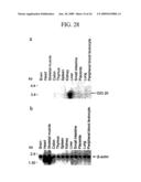 | 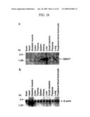 |
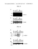 | 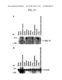 |
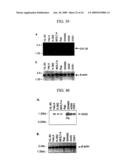 | 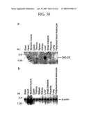 |
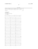 |  |
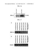 | 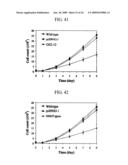 |
 |  |
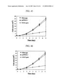 | 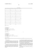 |
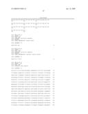 |  |
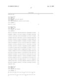 | 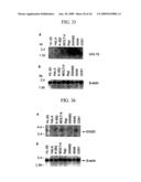 |
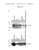 | 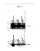 |
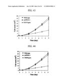 | 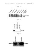 |
 | 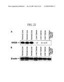 |
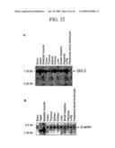 |  |
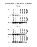 |  |
 | 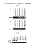 |
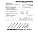 | 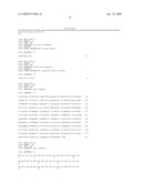 |
 |  |
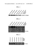 | 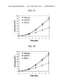 |
 |  |
 |  |
 |
| Similar patent applications: | |
| Date | Title |
|---|---|
| 2011-07-21 | Designer calvin-cycle-channeled production of butanol and related higher alcohols |
| 2011-07-28 | Method and kit for use in the differentiation of ibd and ibs and further distinction between disease types of ibd |
| 2011-07-14 | Microrna expression profile associated with pancreatic cancer |
| 2011-07-14 | Targeted gene deletions for polysaccharide slime formers |
| 2011-04-28 | Mammalian cell expression vectors and utilization |
| New patent applications in this class: | |
| Date | Title |
|---|---|
| 2022-05-05 | Method for producing kidney structure having dendritically branched collecting duct from pluripotent stem cells |
| 2017-08-17 | Method for priming hollow-fiber membrane module |
| 2016-12-29 | Methods for purifying biological cells |
| 2016-09-01 | System and method for processing of biological tissue samples |
| 2016-09-01 | Three-dimensional tissue and production method therefor |
| New patent applications from these inventors: | |
| Date | Title |
|---|---|
| 2022-03-31 | Memory allocation device and method thereof |
| 2021-12-09 | Transmitting system and method of processing digital broadcast signal in transmitting system, receiving system and method of receiving digital broadcast signal in receiving system |
| 2019-01-03 | Dtv receiving system and method of processing dtv signal |
| 2017-07-13 | Digital broadcasting system and data processing method in digital broadcasting system |
| 2017-01-26 | Digital broadcasting system and data processing method |
| Top Inventors for class "Chemistry: molecular biology and microbiology" | |
| Rank | Inventor's name |
|---|---|
| 1 | Marshall Medoff |
| 2 | Anthony P. Burgard |
| 3 | Mark J. Burk |
| 4 | Robin E. Osterhout |
| 5 | Rangarajan Sampath |