Patent application title: NOVEL USE OF LIGANDS SPECIFIC TO FEX-2 POLYPEPTIDE
Inventors:
In San Kim (Daegu, KR)
Seung-Yeon Park (Gyeongbuk, KR)
Mi-Yeon Jung (Daegu, KR)
Ha-Jeong Kim (Daegu, KR)
Assignees:
Kyungpook National University Industry Academic Cooperation Foundation
IPC8 Class: AA61K39395FI
USPC Class:
4241451
Class name: Immunoglobulin, antiserum, antibody, or antibody fragment, except conjugate or complex of the same with nonimmunoglobulin material monoclonal antibody or fragment thereof (i.e., produced by any cloning technology) binds hormone or other secreted growth regulatory factor, differentiation factor, or intercellular mediator (e.g., cytokine, etc.); or binds serum protein, plasma protein (e.g., tpa, etc.), or fibrin
Publication date: 2009-01-29
Patent application number: 20090028871
Inventors list |
Agents list |
Assignees list |
List by place |
Classification tree browser |
Top 100 Inventors |
Top 100 Agents |
Top 100 Assignees |
Usenet FAQ Index |
Documents |
Other FAQs |
Patent application title: NOVEL USE OF LIGANDS SPECIFIC TO FEX-2 POLYPEPTIDE
Inventors:
In-San Kim
Seung-Yeon Park
Mi-Yeon Jung
Ha-Jeong Kim
Agents:
BUCHANAN, INGERSOLL & ROONEY PC
Assignees:
KYUNGPOOK NATIONAL UNIVERSITY INDUSTRY-ACADEMIC COOPERATION FOUNDATION
Origin: ALEXANDRIA, VA US
IPC8 Class: AA61K39395FI
USPC Class:
4241451
Abstract:
The present invention relates to novel use of a ligand specific to a FEX-2
polypeptide. More specifically, the present invention relates to methods
for modulating the secretion of an inflammation-associated cytokine and
for treating or preventing an inflammatory disease using the
FEX-2-specific ligand. The ligand specific to the FEX-2 polypeptide can
bind to FEX-2 expressed on the surface of phagocytes so that it can
stimulate the secretion of anti-inflammatory cytokine and inhibit the
secretion of inflammatory cytokine, so as to treat or prevent
inflammatory diseases.Claims:
1-30. (canceled)
31. A method for modulating the secretion of an inflammation-associated cytokine in a subject, the method comprising administering to a subject in need thereof an effective amount of a FEX-2 polypeptide specific ligand except phosphatidylserine.
32. The method of claim 31, wherein the cytokine is secreted from phagocytes.
33. The method of claim 31, wherein modulating the secretion of the cytokine is stimulating the secretion of anti-inflammatory cytokine and/or inhibiting the secretion of inflammatory cytokine.
34. The method of claim 33, wherein the anti-inflammatory cytokine is selected from the group consisting of TGF-.beta., IL-10, IL-4 and IL-13.
35. The method of claim 33, wherein the inflammatory cytokine is selected from the group consisting of TNF-.alpha., IL-1, IL-6, IL-8, IL-18 and MIP.sub.2.
36. The method of claim 31, wherein the FEX-2 polypeptide is derived from a mammal.
37. The method of claim 31, wherein the FEX-2 polypeptide comprises the amino acid sequence set forth in SEQ ID NO. 1.
38. The method of claim 31, wherein the FEX-2 polypeptide is expressed on the surface of phagocytes.
39. The method of claim 31, wherein the ligand is selected from the group consisting of phosphatidylserine derivatives and anti-FEX-2 antibody.
40. The method of claim 39, wherein the phosphatidylserine derivative is phospho-L-serine.
41. The method of claim 39, wherein the anti-FEX-2 antibody is a polyclonal or monoclonal antibody.
42. The method of claim 41, wherein the monoclonal antibody is produced by a hybridoma (accession number: KCTC 10639BP).
43. A method for treating or preventing an inflammatory disease, the method comprising administering to a subject in need thereof an effective amount of a FEX-2 polypeptide specific ligand except phosphatidylserine.
44. The method of claim 43, wherein the FEX-polypeptide is derived from a mammal.
45. The method of claim 43, wherein the FEX-2 polypeptide comprises the amino acid sequence set forth in SEQ ID NO: 1.
46. The method of claim 43, wherein the FEX-2 polypeptide is expressed on the surface of phagocytes.
47. The method of claim 43, wherein the ligand is selected from the group consisting of phosphatidylserine derivatives and anti-FEX-2 antibody.
48. The method of claim 47, wherein the phosphatidylserine derivative is phospho-L-serine.
49. The method of claim 47, wherein the anti-FEX-2 antibody is a polyclonal or monoclonal antibody.
50. The method of claim 49, wherein the monoclonal antibody is produced by the hybridoma (accession number: KCTC 10639BP).
51. The method of claim 43, wherein the inflammatory disease is selected from the group consisting of inflammation, inflammatory bowl disease, diabetic ocular disease, peritonitis, osteomyelitis, cellulitis, meningitis, encephalitis, pancreatitis, trauma causing shock, bronchial asthma, rhinitis, sinusitis, otitis media, pneumonia, gastritis, enteritis, cystic fibrosis, apoplexy, bronchitis, bronchiolitis, hepatitis, nephritis, arthritis, gout, spondylitis, Reiter's syndrome, polyarteritis nodosa, hypersensitivity vasculitis, Wegener's granulomatosis, polymyalgia rheumatica, giant cell arteritis, calcium crystal deposition arthropathy, pseudogout, nonarticular rheumatism, bursitis, tenosynovitis, epicondylitis (Tennis elbow), neuropathic joint disease (Charcot's joint), hemarthrosis, Henoch-Schonlein Purpura, hypertrophic osteoarthropathy, multicentric reticulohistiocytoma, scoliosis, hemochromoatosis, sickle cell disease and other hemoglobinopathies, hyperlipoproteinemia, hypogammaglobulinemia, hyperparathyroidism, acromegaly, familial mediterranean fever, Behcet's disease, systemic lupus erythematosus, relapsing fever, psoriasis, multiple sclerosis, septicemia, septic shock, acute respiratory distress syndrome, multiple organ failure, chronic obstructive pulmonary disease, acute lung injury and broncho-pulmonary dysplasia.
52. A method for screening an agent for modulating the secretion of an inflammation-associated cytokine, the method comprising the steps of:(a) culturing a phagocyte expressing FEX-2 with a test agent; and(b) measuring change in the level of cytokine secreted from the phagocyte cultured with the test agent relative to the level of cytokine secreted from the phagocyte cultured without the test agent.
53. The method of claim 52, wherein the cytokine secreted from the phagocyte in the step (b) is TGF-.beta. or TNF-.alpha..
54. A method for identifying an agent for treating or preventing an inflammatory disease, the method comprising the steps of:(a) culturing a phagocyte expressing FEX-2 with a test agent;(b) measuring change in the level of cytokine secreted from the phagocyte cultured with the test agent relative to the level of cytokine secreted from phagocyte cultured without the test agent;(c) administering the test agent selected in the step (b), which modulates the level of the cytokine selected from the phagocyte, to an animal suffering from an inflammatory disease, and determining whether the test agent shows a therapeutic effect in the animal.
55. The method of claim 54, wherein the cytokine secreted from the phagocyte in the step (b) is TGF-.beta. or TNF-.alpha..
56. The method of claim 54, wherein the test agent used in the step (c) has the activity to stimulate the secretion of anti-inflammatory cytokine and/or inhibit the secretion of inflammatory cytokine.
57. Use of a FEX-2 polypeptide specific ligand except phosphatidylserine, for preparing a pharmaceutical composition for modulating the secretion of an inflammation-associated cytokine.
58. Use of a FEX-2 polypeptide specific ligand except phosphatidylserine, for preparing a pharmaceutical composition for treating or preventing an inflammatory disease.
59. A pharmaceutical composition for modulating the secretion of an inflammation-associated cytokine, the composition comprising a FEX-2 polypeptide specific ligand except phosphatidylserine, as an active ingredient.
60. A pharmaceutical composition for treating or preventing an inflammatory disease, the composition comprising a FEX-2 polypeptide specific ligand except phosphatidylserine, as an active ingredient.
Description:
[0001]This application claims priority to Korean Patent Application No.
10-2004-0081499, filed on Oct. 12, 2004, the contents of which are hereby
incorporated by reference.
FIELD OF THE INVENTION
[0002]The present invention relates to novel use of a ligand specific to a FEX-2 polypeptide.
BACKGROUND OF THE INVENTION
[0003]Inflammation is a local or systemic protective response to the injury or infection of cells or tissues. Inflammation is typically caused by a series of biological reactions occurring due to the direct reaction of a large number of humoral immune mediators that consist of immune system or the stimulation of the local or systemic effector system by the mediators. The mediators involved in such inflammatory reactions include immune cells, such as polymorphonuclear leukocytes (PMN), cytotoxic T lymphocyte (CTL), natural killer (NK) cells and macrophages etc, and cytokines. Inflammatory diseases are caused particularly by an imbalance of inflammatory cytokines and by the interaction between effector cells. Main inflammatory diseases include: rhinitis and sinusitis such as infectious rhinitis, allergic rhinitis, chronic rhinitis, acute sinusitis and chronic sinusitis; otitis media such as acute purulent otitis media and chronic purulent otitis media; pneumonia such as bacterial pneumonia, bronchial pneumonia, lobar pneumonia, Legionella pneumonia and viral pneumonia; acute or chronic gastritis; enteritis such as infectious enterocolitis, Crohn's disease, idiopathic ulcerative colitis and pseudomembranous colitis; arthritis such as septic arthritis, tuberculous arthritis, degenerative arthritis and rheumatoid arthritis; and diabetic ocular disease.
[0004]Main inflammatory cytokines known up to now include tumor necrosis factor-α (TNF-α), IL-1, IL-6 and IL-18, which are produced by monocytes and macrophages. Among them, TNF-α performs suitable homeostatic and protective actions at low concentrations, but aggravates inflammatory reactions at high concentrations by interaction with other kinds of inflammatory cytokines, such as IL-1, in systemic tissue or a specific tissue.
[0005]Meanwhile, TGF-β is a cytokine having the activity of stimulating or inhibiting a certain cell, and has activities, such as an antagonistic effect on lymphocyte response, and the inhibition of macrophage activation. For this reason, TGF-β is known as a typical anti-inflammatory cytokine.
[0006]To treat inflammatory diseases caused by an imbalance of inflammatory cytokines, various studies to inhibit the inflammatory cytokines and stimulate the production of anti-inflammatory cytokines have been conducted.
[0007]Methods of inhibiting proinflammatory cytokine include a production inhibitory method for inhibiting the secretion of cytokines from being secreted by external stimulation, and a functional inhibitory method for modulating the stimulation of target cells caused by cytokines. Those known as inhibitors of cytokine production include SK&F 86002, which is inhibitors of IL-1 and TNF-α production, tetradrine and WIN 67694 and those developed as inhibitors of cytokine function include IL-1 receptor antagonist IL-1Ra, soluble TNF-α receptors and cytokine antibodies.
DETAILED DESCRIPTION OF THE INVENTION
Technical Problem
[0008]Therefore, it is an object of the present invention to provide novel use of a ligand specific to FEX-2.
Technical Solution
[0009]Accordingly, the present inventors have conducted many studies to develop a novel therapeutic agent for inflammatory diseases and as a result, found that a ligand specific to FEX-2 can bind to FEX-2 expressed on the surface of phagocytes to stimulate the secretion of anti-inflammatory cytokine and to inhibit the secretion of inflammatory cytokine, thereby treating or preventing inflammatory diseases.
[0010]To achieve the above object, in one aspect, the present invention provides a method for modulating the secretion of an inflammation-related cytokine in a subject and a method for treating or preventing an inflammatory disease, the methods comprising administering to FEX-2 the subject in need thereof an effective amount of a ligand specific to FEX-2.
[0011]In another aspect, the present invention provides a method for screening an agent for modulating an inflammation-associated cytokine, the method comprising the steps of: (a) culturing a test agent with phagocyte expressing FEX-2; and (b), measuring change in the level of cytokine secreted from the phagocyte cultured with the test agent relative to the level of cytokine secreted from the phagocyte cultured without the test agent.
[0012]In still another aspect, the present invention provides a method for identifying an agent for treating or preventing of an inflammatory disease, the method further comprising administering the test agent selected in step (b), which modulates the level of cytokine secreted from phagocyte, to an animal having inflammatory diseases, and determining whether the test agent shows a therapeutic effect in the animal, in addition to the steps of the screening method.
[0013]In yet another aspect, the present invention provides the uses of a ligand specific to a FEX-2 polypeptide, for preparing a pharmaceutical composition for modulating the secretion of an inflammation-associated cytokine and a pharmaceutical composition for the treatment or prevention of an inflammatory disease.
[0014]In still yet another aspect, the present invention provides a pharmaceutical composition for modulating an inflammation-associated cytokine and a pharmaceutical composition for the treatment or prevention of an inflammatory disease comprising, as an active ingredient, a ligand specific to a FEX-2 polypeptide.
DEFINITIONS
[0015]Unless defined otherwise, all technical and scientific terms used herein have the same meanings as commonly understood by those ordinary skill in the art to the present invention pertains. The following references provide one of skill with a general definition of many of the terms used in the present invention: Singleton et al., DICTIONARY OF MICROBIOLOGY AND MOLECULAR BIOLOGY (2nd ed. 1994); THE CAMBRIDGE DICTIONARY OF SCIENCE AND TECHNOLOGY (Walker ed., 1988); and Hale & Marham, THE HARPER COLLINS DICTIONARY OF BIOLOGY. In addition, the following definitions are provided to assist the reader in the practice of the present invention.
[0016]As used herein, the term "polypeptide" is used interchangeably with the terms "polypeptides" and "protein(s)", and refers to, a polymer of amino acid residues, e.g., as typically found in proteins in nature.
[0017]The term "FEX-2 polypeptide" may be derived from mammals, and may preferably be derived from any one selected from the group consisting of human beings, rats and mice. More preferably, it may be human FEX-2 comprising the amino acid sequence shown in SEQ ID NO: 1. Most preferably, the FEX-2 may be one expressed in mammalian phagocytes.
[0018]The term "ligand specific to FEX-2" is meant to include all kinds of peptides, polypeptides, peptide imitations, compounds and biological preparations, which bind to FEX-2. Preferably, the FEX-2-specific ligand refers to one having an activity capable of binding to FEX-2 expressed on the surface of phagocytes so as to modulate the secretion of cytokine from the activated phagocytes. More preferably, the FEX-2-specific ligand may be one having activities capable of stimulating the secretion of anti-inflammatory cytokines from phagocytes and inhibiting the secretion of inflammatory cytokine from phagocytes. Examples of the phagocytes include not only macrophages which are professional phagocytes, but also non-professional phagocytes, such as epithelial cells and fibroblasts. Most preferably, the FEX-2-specific ligand may be phosphatidylserine, an anti-FEX-2 antibody, or derivatives thereof. As used herein, the term "derivatives" refers to those obtained by modifying a part of the chemical structure of the phosphatidylserine or anti-FEX-2 antibody while maintaining the fundamental framework and physiological activity thereof.
[0019]The phosphatidylserine is a kind of naturally occurring lipid and consists of a serine, phosphate, glycerol and two fatty acids. After the phosphatidylserine was first isolated by Folch in 1948, it has been reported to have various effects, such as the treatment of dementia, the improvement of brain function, the treatment of epilepsy, and stress resistance by a great many research studies. The phosphatidylserine used in the present invention is not specifically limited and may be derived from nature or be synthesized or be commercially available. Also, the phosphatidylserine derivative may be preferably phospho-L-serine. The phosphatidylserine or derivatives thereof may be in the form of powder, granule, paste, liquid or salt. As the salt, any salt may be used if it is pharmaceutically-acceptable. Examples of the pharmaceutically acceptable salt may include sodium salt, potassium salt, magnesium salt, ammonium salt, phosphate salt, chloride salt and sulfate salt.
[0020]Also, the anti-FEX-2 antibody, as used herein, may be a polyclonal or monoclonal antibody. The antibody used in the present invention may be prepared using a FEX-2 protein as an antigen by any conventional method widely known in the immunology field.
[0021]The polyclonal antibody may be prepared from warm-blooded animals, such as horses, cattle, goats, sheep, dogs, fowls, turkeys, rabbits, mice or rats, according to any conventional technology known in the art. Namely, animals are immunized by the intraperitoneal, intramuscular, intraocular or subcutaneous injection of the antigen. Immunity to the antigen can be increased by the use of an adjuvant, such as Freund's complete or incomplete adjuvant. After booster immunization, a small sample of serum is obtained and tested for reactivity to the desired antigen. Once the titer of the animal has reached a plateau in views of its reactivity to the antigen, large amounts of polyclonal immune serum may be readily obtained either by weekly bleeding or by exsanguinating the animal.
[0022]The monoclonal antibody may also be readily generated using well-known techniques (see Kennet®, McKearn, and Bechtol (eds.), Monoclonal Antibodies, Hybridomas; A New Dimension in Biological Analyses, Plenum Press, 1980). Specifically, the monoclonal antibody may be prepared by immunizing an animal using a protein as an immunogen; fusing the spleen cells of the immunized animal with myeloma cells to produce hybridomas; selecting from the produced hybridomas a hybridoma selectively recognizing the FEX-2 protein; culturing the selected hybridoma; and isolating the desired antibody from the hybridoma culture medium. Alternatively, the monoclonal antibody used in the present invention may also be prepared by injecting into an animal the hybridoma producing the anti-FEX2 antibody, which selectively recognizes the FEX-2 protein; and isolating the antibody from the collected abdominal dropsy of the animal at a given period after the injection. The hybridoma 5G3 producing the human FEX-2 monoclonal antibody, prepared in one example of the present invention, was deposited under accession No. KCTC-10639BP on May 21, 2004 with the Korean Collection for Type Cultures (KCTC), Korean Research Institute of Bioscience and Biotechnology, (52, Oun-dong, Yusong-ku, Taejon, Korea), which is an International Depository Authority under the Budapest Treaty. The deposit shall be maintained in viable condition at the KCTC during the entire term of the issued patent and shall be made available to any person or entity for non-commercial use without restriction, but in accordance with the provisions of the law governing the deposit.
[0023]Derivatives of the anti-FEX-2-antibody according to the present invention may include those obtained by modifying the structure of the antibody to alter the stability, storage stability, volatility or solubility of the antibody. In some cases; they may be those obtained by modifying the antibody by phosphorylation, sulfation, acrylation, glycosylation, methylation, farnesylation, etc.
[0024]As used herein, the term "subject" may be an animal, and is preferably a mammal. It may also be cells, tissues, organs, etc., derived from the animals.
[0025]As used herein, the term "effective amount" refers to an amount that exhibits the effect of modulating the secretion of an inflammation-associated cytokine in a subject, and preferably, the effect of modulating the secretion of cytokine from phagocytes, and most preferably, the effect of stimulating the secretion of anti-inflammatory cytokine and/or inhibiting the secretion of inflammatory cytokine, or thus, the effect of treating or preventing an inflammatory disease.
[0026]As used herein, the term "agent" or "test agent" includes any substance, molecule, element, compound, entity, or a combination thereof. It includes, but is not limited to e.g., protein, polypeptide, small organic molecule, polysaccharide and polynucleotide. Moreover, it may also be a natural product, a synthetic compound, a chemical compound, or a combination of two or more substances. Unless otherwise specified, the terms "agent", "substance" and "compound" can be used interchangeably.
[0027]Hereinafter, the present invention will be described in detail.
[0028]The present inventors searched for partial human cDNA sequences comprising fas-1 domain in the known nucleotide database. Among the searched sequences, cDNA sequences whose characteristics are not yet identified were selected. On the basis of the cDNA sequences, primers were designed, and then, RT-PCR and 5' RACE PCR were performed using total RNA extracted from the human spleen as a template, thus cloning a new human gene comprising the fas-1 domain (see Example 1).
[0029]The gene synthesized by the present inventors had seven fas-1 domains, twenty-three EGF-like domains, one X-link domain and one transmembrane domain (see FIG. 1). On the basis of the domain structure as described above, the gene was named "FEX-2" and its gene sequence was registered in GeneBank under accession number AY311388. The full amino acid sequence of the human FEX-2 gene is set forth in SEQ ID NO: 1.
[0030]The present inventors conducted various tests using fibroblasts transformed with the FEX-2 gene to examine the function of the FEX-2 gene. As a result, we have found that the FEX-2 specifically recognizes phosphatidylserine expressed on the surface of aged cells and apoptotic cells, thereby mediating the binding and phagocytosis of the cells. Also, the present inventors have found that the FEX-2 gene is expressed on the surface of macrophages involved in the phagocytosis of aged cells and apoptotic cells, and the phagocytosis by macrophages is mediated by the FEX-2 gene. From this fact, the present inventors confirmed that the FEX-2 gene is a novel receptor for phosphatidylserine.
[0031]Furthermore, the present inventors have found that, when macrophages (activated phagocytes) or fibroblasts transformed with FEX-2 was treated with a ligand specific to FEX-2, the secretion of anti-inflammatory cytokines would be stimulated and the secretion of inflammatory cytokines would be inhibited. This suggests that the FEX-2-specific ligand can be used as an agent for modulating the secretion of an inflammation-associated cytokines in a subject and an agent for the prevention or treatment of an inflammatory disease.
[0032]More specifically, in one embodiment of the present invention, a recombinant vector comprising the FEX-2 gene was prepared and transfected into L cells (mouse fibroblasts), thus preparing L/FEX-2 cells (see Example 1). Then, an examination was made to check whether or not the prepared cells are involved in the binding and phagocytosis of aged cells and apoptotic cells (see Example 4). As a result, it could be seen that the L/FEX-2 cells selectively recognized, and bind to the aged red blood cells, thereby phagocytising them (see FIGS. 3 and 4). This suggests that the binding and phagocytosis of the aged red blood cells by L/FEX-2 cells is mediated by FEX-2.
[0033]Furthermore, in another embodiment of the present invention, the recognition sites of FEX-2 in the binding of aged cells to FEX-2 were identified (see Example 5).
[0034]As a result, it could be found that, as aging progressed, the expression of phosphatidylserine on the surface of red blood cells increased (see FIG. 5), and the binding of red blood cells to L/FEX-2 cells also increased (see FIG. 6). In addition, it could be found that the FEX-2 gene selectively recognized only the phosphatidylserine on the surface of red blood cells (see FIGS. 7 to 9), and the FEX-2 gene did not recognize phosphatidylserine through negative charge, but rather specifically recognized the structure of phosphatidylserine (see FIG. 10).
[0035]Also, in view of the fact that phosphatidylserine is expressed on the surface of not only aged cells but also apoptotic cells, the present inventors examined whether apoptotic cells are phagocytised by the L/FEX-2 cells (see Example 6). As a result, it could be found that L/FEX-2 cells recognized phosphatidylserine expressed on the surface of not only aged cells but also apoptotic cells thereby mediating the binding and phagocytosis of these cells (see FIGS. 12 and 13).
[0036]Meanwhile, the present inventors treated the L/FEX-2 cells with the FEX-2 monoclonal antibody and measured the secretion of anti-inflammatory cytokine from the cells (see Example 6). As a result, it could be found that the treatment of the L/FEX-2 cells with the FEX-2 monoclonal antibody resulted in an increase in the secretion of the anti-inflammatory cytokine (see FIG. 14).
[0037]Also, since the phagocytosis of aged cells and apoptotic cells is mainly undertaken by macrophages, the present inventors examined whether the FEX-2 is expressed on the surface of macrophages and whether FEX-2 is involved in the phagocytosis by macrophages (see Example 7). As a result, it could be found that the FEX-2 was expressed on macrophages (see FIG. 15), and the phagocytosis by macrophages was made as a result of a process where the FEX-2 existing on the surface of macrophages specifically recognized phosphatidylserine expressed on apoptotic cells (see FIG. 16). This indicates that the FEX-2 is a novel receptor for phosphatidylserine.
[0038]It is known that the phosphatidylserine-dependent phagocytosis of apoptotic cells stimulates the secretion of anti-inflammatory cytokine TGF-β and inhibits the secretion of inflammatory cytokine TNF-α. In view of this fact, the present inventors examined whether a ligand specific to the FEX-2 existing on the surface of macrophages can transfer to activated macrophages the same signal as a signal generated during the phagocytosis of macrophages thereby stimulate the secretion of anti-inflammatory cytokine (see Example <7-3>). As a result, it could be found that macrophages activated by treatment with the phosphatidylserine or the monoclonal antibody to FEX-2 stimulated the secretion of anti-inflammatory cytokine and inhibited the secretion of inflammatory cytokine, similarly to the case where macrophages phagocytise the aged cells (see FIG. 17).
[0039]Therefore, the present invention provides a method for modulating the secretion of an inflammation-associated cytokine in a subject and a method for treating or preventing an inflammatory disease, the methods comprising administering to a subject in need thereof an effective amount of a ligand specific to the FEX-2 polypeptide. In the method for modulating the secretion of an inflammation-associated cytokine, the cytokine may be secreted from phagocytes. The cytokine includes all anti-inflammatory cytokines and inflammatory cytokines. For example, the anti-inflammatory cytokine may be selected from the group consisting of TGF-β, IL-10, IL-4 and IL-13, but not limited thereto (Watanabe, M. et al., Adeno-associated virus-mediated human IL-10 gene transfer suppresses the development of experimental autoimmune orchitis. Gene Therapy 2005, 12:1126-1132; Ghoreschi; K. et al., Interleukin-4 therapy of psoriasis induces Th2 responses and improves human autoimmune disease. Nature Medicine 2003, 9:40-46; Kluth, D. C. et al., New approaches to modify glomerular inflammation. J. Nephrol. 1999, 12:66-75), and may preferably be TGF-β. Also, the inflammatory cytokine may be selected from the group consisting of TNF-α, IL-1, IL-6, IL-8, IL-18 and MIP2, but not limited to (see McDonald, P. P. et al., Transcriptional and translational regulation of inflammatory mediator production by endogenous TGF-13 in macrophages that have ingested apoptotic cells. J. immunol. 1999, 1, 63:6164-6172; Dinarello, C. A. et al., Blocking IL-1 in systemic inflammation. J Exp Med. 2005, 201:1355-9; de Boer, W. I. et al., Perspectives for cytokine antagonist therapy in COPD. Drug Discov Today. 2005, 10:93-106), and may preferably be TNF-α.
[0040]In the above method, modulating the secretion of cytokines may preferably be stimulating the secretion of anti-inflammatory cytokine and/or inhibiting the secretion of inflammatory cytokine. For example, stimulating the secretion of TGF-β and inhibiting the secretion of TNF-α may be employed.
[0041]The ligand specific to the FEX-2 polypeptide, which is used in the inventive method, can be provided in the form of a pure substance or a suitable pharmaceutical composition. The pharmaceutical composition is characterized by comprising the ligand specific to FEX-2, as an active ingredient. The pharmaceutical composition may be prepared by formulating the inventive FEX-2-specific ligand into a suitable form together with a pharmaceutically acceptable carrier. As used herein, the term "pharmaceutically acceptable carrier" refers to a composition which is physiologically acceptable and, when administered to human beings, generally does not cause allergic reactions such as gastrointestinal disorder and dizziness, or similar reactions thereto. Example of the pharmaceutically acceptable carrier includes oral carrier such as lactose, starch, cellulose derivative, magnesium stearate, stearic acid, etc; and parenteral carrier such as water, suitable oil, saline solution, aqueous glucose, glycol etc. The composition may further comprise a stabilizer and a preservative. Suitable stabilizer includes an antioxidant, such as sodium bisulphite, sodium sulphite or ascorbic acid. Suitable preservative includes benzalkonium chloride, methylparaben, propylparaben and chlorobutanol. Other pharmaceutically acceptable carriers can be found in the following literature: Remington's Pharmaceutical Sciences, 19th ed., Mack Publishing Company, Easton, Pa., 1995.
[0042]The inventive pharmaceutical composition can be prepared in the form of various parenteral or oral forms according to a conventional method. The parenteral formulation typically includes an injection formulation, and preferably an isotonic solution or a suspension. The injection formulation may be prepared using a suitable dispersing agent, wetting agent or suspending agent according to any method known in the art. For example, the injection formulation can be prepared by dissolving necessary components in a saline or buffer solution. Also, the oral formulation includes, but are not limited to, powder, granule, tablet, pill and capsule.
[0043]The pharmaceutical composition formulated as described above may be administered in an effective amount by various routes, including oral, transdermal, subcutaneous, intravenous and intramuscular routes.
[0044]The dose of either the inventive ligand specific to the FEX-2 polypeptide or the pharmaceutical composition comprising the ligand as an active ingredient may be suitably selected depending on an administration route, subject and its age, body weight, sex, disease severity. Although the dose of the active ingredient may vary depending on disease severity, the active ingredient may preferably be generally administered several times a day at a one time effective dose of 10 μg-10 mg for adult.
[0045]The inventive pharmaceutical composition prepared as described above can be effectively used for modulating the secretion of an inflammation-associated cytokine in a subject and for preventing and treating an inflammatory disease. As used herein, the term "inflammatory disease" is meant to include simple inflammation itself and all diseases caused by the inflammatory reaction. Examples of the inflammatory diseases include, but are not limited to, inflammation, inflammatory bowl disease, diabetic ocular-disease, peritonitis, osteomyelitis, cellulitis, meningitis, encephalitis, pancreatitis, trauma causing shock, bronchial asthma, rhinitis, sinusitis, otitis media, pneumonia, gastritis, enteritis, cystic fibrosis, apoplexy, bronchitis, bronchiolitis, hepatitis, nephritis, arthritis, gout, spondylitis, Reiter's syndrome, polyarteritis nodosa, hypersensitivity vasculitis, Wegener's granulomatosis, polymyalgia rheumatica, giant cell arteritis, calcium crystal deposition arthropathy, pseudogout, nonarticular rheumatism, bursitis, tenosynovitis, epicondylitis (Tennis elbow), neuropathic joint disease (Charcot's joint), hemarthrosis, Henoch-Schonlein Purpura, hypertrophic osteoarthropathy, multicentric reticulohistiocytoma, scoliosis, hemochromoatosis, sickle cell disease and other hemoglobinopathies, hyperlipoproteinemia, hypogammaglobulinemia, hyperparathyroidism, acromegaly, familial mediterranean fever, Behcet's disease, systemic lupus erythematosus, relapsing fever, psoriasis, multiple sclerosis, septicemia, septic shock, acute respiratory distress syndrome, multiple organ failure, chronic obstructive pulmonary disease, acute lung injury and broncho-pulmonary dysplasia.
[0046]Also, the present invention provides methods for screening an agent for modulating the secretion of an inflammation-associated cytokine in phagocyte and an agent for treating and preventing an inflammatory disease.
[0047]Specifically, the screening methods comprise:
[0048](a) culturing a phagocyte expressing FEX-2 with a test agent; and
[0049](b) measuring change in the level of cytokine secreted from the phagocyte cultured with the test agent relative to the level of cytokine secreted from phagocytes cultured without the test agent.
[0050]In the screening method, the phagocyte expressing FEX-2 in the step (a) is preferably macrophage or mouse fibroblast L cell transformed with FEX-2. Also, the step (b) can be performed by measuring the production of the anti-inflammatory cytokine (e.g., TGF-β) and/or inflammatory cytokine (e.g., TNF-α) using an ELISA method known in the art.
[0051]Furthermore, the method for screening the agent for treating or preventing an inflammatory disease may comprises, in addition to the steps (a) and (b), the step of: (c) administering the test agent selected in step (b), which modulates the level of cytokine secreted from phagocyte, to an animal suffering from inflammatory disease, and determining whether the test agent shows a therapeutic effect in the animal.
[0052]The term "animal" used in the step (c) is preferably a non-human animal. Also, the term "therapeutic effect" used in the step (c) means the effect of alleviating or improving an inflammatory disease and its symptoms and the effect of inhibiting the progression of the inflammatory disease.
[0053]Agents which can be screened according to the present invention may preferably be either agents for stimulating the secretion of anti-inflammatory cytokine and/or inhibiting the secretion of inflammatory cytokine, or agents showing the effect of treating or treating the inflammatory disease through these actions.
[0054]Test agents which can be screened according to the inventive methods include polypeptides, beta-turn mimetics, polysaccharides, phospholipids, hormones, prostaglandins, steroids, aromatic compounds, heterocyclic compounds, benzodiazepines, oligomeric N-substituted glycines, oligocarbamates, saccharides, fatty acids, purins, pyrimidines, derivatives, structural analogs or combinations thereof. Some test agents may be synthetic molecules, and others natural molecules. The test agents can be obtained from a wide variety of source, including libraries of synthetic or natural compounds. Combinatorial libraries can be produced for many types of compounds that can be synthesized in a step-by-step fashion. Large combinatorial libraries of compounds can be constructed by the encoded synthetic libraries (ESL) method described in WO 95/12608, WO 93/06121, WO 94/08051, WO 95/395503 and WO 95/30642. Libraries of natural compounds in the form of bacterial, fungal, plant and animal extracts can be obtained from commercial sources or collected in the field. Additionally, known pharmacological agents can be subject to directed or random chemical modifications, such as acylation, alkylation, esterification, amidification, etc. to produce structural analogs. The test agents may be naturally occurring proteins or their fragments. Such test agents can be obtained from natural sources, for example, a cells or tissue lysate. Libraries of polypeptides agents can also be prepared, e.g., from a cDNA library commercially available or generated with routine methods. The test agents can also be peptides, e.g., peptides having about 5-30 amino acids, preferably about 5-20 amino acids, and more preferably about 7-15 amino acids. The peptides can be digests of naturally occurring proteins, random peptides, or "biased" random peptides.
[0055]The test agents can also be nucleic acids. Nucleic acid test agents can be naturally occurring nucleic acids, random nucleic acids, or "biased" random nucleic acids. For example, digests of prokaryotic or eukaryotic genomes can be similarly used as described above for proteins.
[0056]The test agents may be small molecules (e.g., molecules with a molecular weight of not more than about 1,000). Preferably, high throughput assays are adapted and used to screen for such small molecules. In some cases, combinatorial libraries of small molecule test agents as described above can be readily employed in the inventive screening method (Shultz, Bioorg. Med. Chem. Lett., 8:2409-2414, 1998; Weller, Mol. Drivers., 3:61-70, 1997; Fernandes, Curr. Opin. Chem. Biol., 2:597-603, 1998; and Sittampalam, Curr. Opin. Chem. Biol., 1:384-91, 1997).
[0057]Libraries of test agents, to be screened with the inventive methods, can also be generated based on structural studies of the FEX-2 polypeptide or a ligand specific thereto, or fragments or analogues thereof. The term "analogue" as used herein refers to a molecule that structurally resembles a reference molecule but which has been modified in a targeted and controlled manner, by replacing a specific substituent of the reference molecule with an alternate substituent. Compared to the reference molecule, an analogue would be expected, by one skilled in the art, to exhibit the same, similar, or improved utility. Synthesis and screening of analogs, to identify variants of known compounds having improved traits (such as higher binding affinity for a target molecule) is an approach that is well known in pharmaceutical chemistry.
[0058]Such structural studies allow the identification of test agents that are likely to bind to the FEX-2 polypeptide. The three-dimensional structure of the FEX-2 polypeptide can be studied in a number of ways, e.g., crystal structure and molecular modeling. Methods of studying protein structures using X-ray crystallography are well known in the following literatures: Physical Bio-chemistry, Van Holde, K. E. (Prentice-Hall, New Jersey 1971), pp. 221-239, and Physical Chemistry with Applications to the Life Sciences, D. Eisenberg & D. C. Crothers (Benjamin Cummings, Menlo Park 1979). Computer modeling of the structure of FEX-2 provides another means for designing test agents for screening an agent for modulating the secretion of an inflammation-associated cytokine. Methods of molecular modeling are described in the following literature: U.S. Pat. No. 5,612,894 and U.S. Pat. No. 5,583,973. In addition, protein structures can also be determined by neutron diffraction and nuclear magnetic resonance (NMR). See Physical Chemistry, 4th Ed. Moore, W. J. (Prentice-Hall, New Jersey 1972) and NMR of Proteins and Nucleic Acids, K. Wuthrich (Wiley-Interscience, New York 1986).
BRIEF DESCRIPTION OF THE DRAWINGS
[0059]FIG. 1 is a schematic diagram showing each domain of a human FEX-2 protein.
[0060]FIG. 2 shows the results of flow cytometry analysis using a FEX-2 monoclonal antibody, conducted in order to examine whether FEX-2 is expressed on the surface of L/FEX-2 cells. Control group: mouse immunoglobulin.
[0061]FIG. 3 shows the binding and phagocytosis of aged cells and normal red blood cells by L/FEX-2 cells, compared to those for normal red blood cells and L/Mock cells.
[0062]FIG. 4 shows the inhibitory effect of the FEX-2 monoclonal antibody on the binding and phagocytosis of aged red blood cells,
[0063]None: untreated L/FEX-2 cells
[0064]5G3: L/FEX-2 cells pre-cultured with the FEX-2 monoclonal antibody
[0065]IgG1: L/FEX-2 cells pre-cultured with mouse immunoglobulin
[0066]FIG. 5 shows the results of flow cytometric measurement using an annexin V antibody, conducted to measure the expression of phosphatidylserine according to the aging of red blood cells.
[0067]FIG. 6 shows measurement results for the binding of red blood cells to L/FEX-2 cells according to the aging of red blood cells.
[0068]FIG. 7 is a graphic diagram showing the binding of various phospholipids to L/FEX-2 cells.
[0069]PI: phosphatidylionsitol;
[0070]PE: phosphatidylethanolamine;
[0071]PS: phosphatidylserine; PC: phosphatidylcholine;
[0072]PA: phosphatidic acid; and
[0073]PG: phosphatidylglycerol.
[0074]FIG. 8 is a graphic diagram showing the inhibition of the binding and phagocytosis of aged red bloods by L/FEX-2 cells according to the treatment of various phospholipids.
[0075]None: no addition of phospholipids;
[0076]PI: phosphatidylionsitol;
[0077]PE: phosphatidylethanolamine;
[0078]PS: phosphatidylserine; PC: phosphatidylcholine;
[0079]PA: phosphatidic acid; and
[0080]PG: phosphatidylglycerol.
[0081]FIG. 9 is a graphic diagram showing the binding of aged red blood cells to L/FEX-2 cells according to the concentration of phosphatidylserine or phosphatidylcholine.
[0082]FIG. 10 shows the inhibitory effect of structural analogues of phosphatidylserin on the binding and phagocytosis of aged red cells by L/FEX-2 cells.
[0083]None: no addition of phospholipids;
[0084]PS: phosphatidylserine;
[0085]PDS: phospho-D-serine; and
[0086]PLS: phospho-L-serine.
[0087]FIG. 11 shows the results of flow cytometry analysis for apoptosis in Jurkat T cells treated with an anti-Fas-antibody and U937 cells treated with ectoposide.
[0088]FIG. 12 illustrates photographs showing the phagocytosis of apoptotic cells by L/FEX-2 cells and L/Mock cells.
[0089]a: L/Mock cells; and
[0090]b: L/FEX-2 cells.
[0091]FIG. 13 is a graphic diagram showing the inhibitory effect of phosphatidylserine or a FEX-2 monoclonal antibody on the phagocytosis of apoptotic cells by L/FEX-2 cells.
[0092]None: untreated;
[0093]PS: phosphatidylserine; PC: phosphatidylcholine;
[0094]5G3: FEX-2 monoclonal antibody; and
[0095]IgG1: mouse immunoglobulin.
[0096]FIG. 14 shows measurement results for the secretion of anti-inflammatory cytokine by the treatment of L/FEX-2 cells with the FEX-2 monoclonal antibody.
[0097]Con: untreated control group;
[0098]IgG: treated with mouse immunoglobulin (IgG1); and
[0099]5G3: treated with the FEX-2 monoclonal antibody.
[0100]FIG. 15 shows the results of flow cytometric analysis using the FEX-2 monoclonal antibody, conducted to examine whether FEX-2 is expressed on the surface of macrophages. Mouse immunoglobulin (IgG1) was used as a control group.
[0101]FIG. 16 is a graphic diagram showing the inhibitory effect of phosphatidylserine or FEX-2 monoclonal antibody on the apoptosis and phagocytosis by macrophages.
[0102]PS: phosphatidylserine;
[0103]IgG: mouse immunoglobulin; and
[0104]5G3: FEX-2 monoclonal antibody.
[0105]FIG. 17 shows measurement results for the secretion of anti-inflammatory cytokine and inflammatory cytokine by the treatment of an LPS-stimulated J774 macrophage cell line with various ligands.
[0106]Control group: untreated;
[0107]LPS: stimulated with LPS;
[0108]LPS+AR: stimulated with LPS and then treated with aged red blood cells;
[0109]LPS+PC: stimulated with LPS and then treated with phosphatidylcholine liposome;
[0110]LPS+PS: stimulated with LPS and then treated with phosphatidylserine liposome
[0111]LPS+IgG: stimulated with LPS and then treated with mouse immunoglobulin; and
[0112]LPS+5G3: stimulated with LPS and then treated with FEX-2 monoclonal antibody.
BEST MODE FOR CARRYING OUT THE INVENTION
[0113]Hereinafter, the present invention will be described in detail by example. It is to be understood, however, that these examples are given for illustrative purpose only and are not construed to limit the scope of the present invention.
Example 1
Cloning of Human FEX-2 cDNA
[0114]<1-1> Preparation of Expression Vector
[0115]To identify a novel cell adhesion molecule comprising the fas-1 domains, sequences comprising the fas-1 domains were searched in the nucleotide database of Genbank and Celera genomics. As a result, human partial cDNA clones (FLJ00112, DZKZp434E0321, and CD44-like precursor FELL), which are not yet characterized, were selected. The full-length cDNA sequences were prepared from human spleen cells by RT-PCR and 5' RACE PCR.
[0116]For this purpose, based on the above three kinds of searched partial cDNA clones, which comprise the fas-1 domains, two pairs of primers (SEQ ID NOS: 2-5) were first designed (see Table 1). Using 2 μg of RNA extracted from the human spleen, as a template, and each of the prior pairs of primers designed as described above, reverse transcriptase polymerase chain reaction (RT-PCR) was performed to obtain two protein partial cDNAs comprising the fas-1 domains. The PCR reaction was performed in the Expand high fidelity PCR system (Roche) under the following condition: 2 min at 95° C.; and 30 cycles of 30 sec at 94° C., 30 sec at 60° C. and 30 sec at 72° C.
[0117]As a result, 3.6 kb and 2.0 kb amplification products were obtained. Among the amplification products, the 3.6 kb cDNA was digested with restriction enzymes ClaI and SacI (TaKaRa) and then inserted into the same restriction enzyme sites of a pBluescript-KS(+) vector (Stratagene) using T4 ligase (Invitrogen), thus preparing recombinant vector "pBS-FEX23". Then, among the amplification products, the 2.0 kb cDNA was digested with restriction enzymes SacI and HindIII (TaKaRa) and then inserted into the same restriction enzyme sites of a pET-29b vector (Novagen) using T4 ligase (Invitrogen), thus preparing recombinant vector "pET-Fex45". Then, to combine the two cDNA sequences with each other, a fragment obtained by digesting pET-Fex45 with EcoRI was treated with a klenow enzyme to make a blunt fragment. The blunt fragment was digested with SacI and cloned into the same restriction enzyme site of the recombinant vector PET-Fex23, thus preparing recombinant vector "pET-Fex2345".
[0118]The 5'-terminus of the cDNA clone comprising the fas-1 domain was prepared in the following manner. RNA extracted from the human spleen was amplified using the primer (SEQ ID NO: 6) shown in Table 1 below to prepare cDNA. The cDNA template was subjected to 5' RACE PCR using the primers (SEQ ID NOS: 7 and 8) shown in Table 1 and an adaptor primer provided in the 5' RACE system (5' RACE system for Rapid Amplification of cDNA Ends version 2.0; Invitrogen), according to the manufacturer's instruction, thus obtaining an amplification product. Then, the amplification product was analyzed in a base sequencer (Applied Biosystems AB13700) to identify the base sequence of the 5'-terminus of FEX-2. Based on the base sequence of the 5'-terminus, primers were constructed. Using 2 μg of an RNA template extracted from the human spleen and the primer pair (SEQ ID NO: 9 and SEQ ID NO: 10) designed based on the 5'-terminus, a reverse transcriptase polymerase chain reaction (RT-PCR) was performed to obtain a 2.5 kb 5'-terminal cDNA product. The amplification product was digested with restriction enzymes EcoRI and ClaI (TaKaRa) and inserted into the same restriction enzyme sites of a pBluescript-KS(+) vector (Stratagene), thus preparing recombinant vector "pBS-Fex1". In order to clone a complete base sequence into an expression vector, the above-prepared pET-Fex2345 was first digested with KpnI and HindIII the same restriction enzyme sites of a pcDNA3.1(-)/Myc-His vector (Invitrogen), thus preparing "pcDNA-Fex45". Also, the pET-Fex2345 was digested with restriction enzymes BamHI and KpnI and inserted into the same restriction enzyme sites of the pcDNA-Fex45, thus preparing "pcDNA-Fex2345". Then, the pBS-Fex1 was digested with EcoRI and ClaI and inserted into the same restriction enzyme sites of the pcDNA-Fex2345, thus cloning a complete base sequence.
[0119]The above-prepared plasmid was analyzed in a base sequencer (Applied Biosystems AB13700), and the identified base sequence was registered in Genebank® under accession number AY311388. The above-prepared expression vector comprising the complete base sequence of FEX-2 was named "pcDNA-Fex2".
[0120]The amino acid sequence deduced from the result of the base sequence analysis was shown to be substantially identical to scavenger receptor FEEL-2 (Adachi H. et al., J. Biol. Chem. 277:34264-34270, 2002) and encocytic hyaluronan receptor stabilin-2 (Politz O. et al., Biochem J. 362:155-164, 2002) and had seven fas-1 domains, twenty-three EGF-like domains, one X-link domain and one transmembrane domain (see FIG. 1).
TABLE-US-00001 TABLE 1 Primers used in cloning of Fex-2 expression vector SEQ ID Primers Sequences NO Fex23 5'-tgc ccc atc gat gtg atg aaa 2 sense c-3' Fex23 5'-caa aca aga gct ccc gct gca 3 antisense caa t-3' Fex45 5'-gca gcg gga gct ctt tga cct 4 sense g-3' Fex45 5'-ccc gtc caa gct tgc aca gtg 5 antisense tcc t-3' RACE-GSP-1 5'-tcc cag Ctt act cag tgg cca 6 antisense ggc-3' RACE-GSP-2 5'-cag gcc cat cat att tgc aca 7 antisense ctg tag ac-3' RACE-GSP-3 5'-agt taa ttt ggc agg ggt cca 8 antisense cag gc-3' Fex1 sense 5'-aag gca ggt ctc acc tat ctc 9 ctg g-3' Fex1 5'-tca caa atg cat gtc ccg ttg 10 antisense c-3'
[0121]<1-2> Transformation of L Cells
[0122]Mouse fibroblast L-cells (ATCC CCL-1; provided by Dr. M. Takeichi, Department of Biophysics, Kyoto University, Japan) were transformed with the expression vector pcDNA-Fex2 prepared in Example <1-1>. The L cells were cultured in Dulbecco's modified Eagle's medium (DMEM) supplemented with 10% heat-inactivated FBS, penicillin G and streptomycin.
[0123]To transform the above-selected L cells with the recombinant vector pcDNA-Fex2, lipofectamine (Invitrogen) was used according to the manufacturer's instruction. 48 hours after the transformation, the cells were treated with G418 (400 μg/ml) and cultured for 10 to 12 days, while each colony showing resistance was isolated. The cells transformed as described above were named "L/FEX-2". As a negative control group (L/Mock), cells transformed with pcDNA3.1 (-)/Myc-His, a vector comprising no FEX-2 gene were used.
Example 2
Preparation of Monoclonal Antibody to Human FEX-2
[0124]In order to prepare a monoclonal antibody to human FEX-2, cDNA comprising a base sequence corresponding to amino acid residues 1173-1727 of the entire amino acid sequence of human FEX-2 was prepared. The cDNA was obtained by performing PCR using the pcDNA-Fex2 DNA prepared in Example <1-1> as a template and the primers (SEQ ID NO: 11 and SEQ ID NO: 12) shown in Table 2. The PCR reaction was performed under the following conditions: 2 min at 95° C.; and 25 cycles of, 30 sec at 94° C., 30 sec at 60° C. and 30 sec at 72° C. The amplification product was digested with restriction enzymes BamHI and XhoI (TaKaRa) and inserted into the same restriction enzyme sites of a pET43.1a vector (Novagen).
TABLE-US-00002 TABLE 2 Primers used in preparation of recombinant protein as antigen SEQ ID Primers Sequences NO hFex-2-4E5 5'-aaa aag gat cca cag tgt ttg 11 sense ctc c-3' hFex-2-4E5 5'-ttt tac tcg aga ctt ttg gga 12 antisense gat agc-3'
[0125]The above-prepared expression vector was named "pET-4E5", and the expression and isolation of a protein were performed in the same manner as in Example <2-1>. The preparation of a monoclonal antibody was performed in Dinona Inc. (Seoul, Korea). Specifically, 20 μg of the above-prepared recombinant protein was immunized into six rats at 2-week intervals to obtain a positive hybridoma clone (clone 5G3). The hybridoma clone was injected into the abdominal cavity of rats, and the abdominal dropsy of the rats was obtained, thus obtaining the monoclonal antibody. The isotype of the monoclonal antibody was determined using the IsoStrip mouse monoclonal antibody isotyping kit (Roche). As a result, the isotype of the inventive monoclonal antibody was determined to be an IgG1 with lambda chain.
[0126]The hybridoma 5G3 that produces the inventive human FEX-2 monoclonal antibody was deposited under accession No. KCTC-10639BP on May 21, 2004 with the Korean Collection for Type Cultures (KCTC), which is an International Depository Authority under the Budapest Treaty.
Example 3
Determination of Expression or Non-Expression of FEX-2 on Surface of L/FEX-2 Cells
[0127]Whether or not FEX-2 is expressed on the surface of the L/FEX-2 cells prepared in Example <1-2> was analyzed by flow cytometry using the FEX-2 monoclonal antibody (5G3) prepared in Example <2-3>.
[0128]For this purpose, a confluent plates, in which L/FEX-2 cells were first cultured comprising Dulbecco's modified Eagle's medium (DMEM), after which the plate was treated with a PBS buffer solution comprising 0.25% trypsin and 0.05% EDTA so as to separate the cells from the plate surface. The cells were washed with PBS buffer solution twice and then re-suspended in PBS buffer solution. To the cell suspension, the FEX-2 monoclonal antibody (5G3) was added and cultured at 4° C. for one hour. To the reaction mixture, 10 μg/ml of FITC-conjugated secondary rabbit anti-mouse immunoglobulin G (Santa Cruz Biotechnology, Inc., CA) was added and the cells were further cultured at 4° C. for one hour. Then, the reaction mixture was analyzed at 488 nm by using a flow cytometer (Becton Dickinson, San Jose, Calif.) equipped with a 5-watt laser. As a control group, mouse immunoglobulin (IgG1) in place of the FEX-2 monoclonal antibody was used.
[0129]The test results showed that FEX-2 was expressed on the surface of the L/FEX-2 cells. However, FEX-2 was not expressed on the surface of the L/Mock cells. Also, even in the use of the mouse immunoglobulin in place of the FEX-2 monoclonal antibody, the expression of FEX-2 was not detected (see FIG. 2).
Example 4
Binding and Phagocytosis of Aged Red Blood Cells by FEX-2
[0130]<4-1> Binding and Phagocytosis of Aged Red Blood Cells by L/FEX-2 Cells
[0131]Whether or not aged red blood cells are bound and phagocytosed by the L/FEX-2 cells prepared in Example <1-2> and L/Mock cells as a control group was examined. For this purpose, normal red blood cells obtained from normal adult volunteers were first diluted with PBS to a hematocrit concentration of 20% and cultured at 37° C. for 4 days, thus preparing aged red blood cells. Then, each of L/FEX-2 cells, L/Mock cells and normal L cells was cultured to a confluent 6-well plate comprising Dulbecco's modified Eagle's medium (DMEM). To the culture media, aged red blood cells and normal red blood cells were added to a hematocrit concentration of 1% and cultured at 37° C. for one hour. Then, the cells were washed five times with PBS, and observed for the binding of the aged red blood cells and normal red blood cells using an optical microscope. The number of red blood cells bound to 100 randomly selected L/FEX-2 cells was counted and averaged. To analyze phagocytosis, the cells were washed with PBS and treated with water for 10 seconds to completely dissolve undigested cells. Then, the L/FEX-2 cells were stained using the Diff quick staining kit (IMEM Ins., San Marcos, Calif., USA), and five sites from the stained cells were randomly selected and counted for the number of cells which have phagocytosed red blood cells phagocytosed. Binding and phagocytosis indexes (%) were expressed as the percentages of cells having red blood cells bound and phagocytosed thereby, relative to the entire L/FEX-2 cells.
[0132]The test results showed that the aged red blood cells were bound and phagocytosed by the L/FEX-2 cells. However, the aged red blood cells were little bound and phagocytosed by the normal L cells and L/Mock cells as control groups. Also, the normal red blood cells were little bound and phagocytosed by the L/FEX-2 cells (see FIG. 3).
[0133]These results suggest that FEX-2 mediates the selective binding and phagocytosis of aged cells.
[0134]<4-2> Inhibition of Binding and Phagocytosis of Aged Red Blood Cells by Anti-FEX-2 Monoclonal Antibody.
[0135]In order to confirm if the binding and phagocytosis of aged red blood cells is attributable to FEX-2, an examination was made to check whether or not the monoclonal antibody (5G3) to FEX-2 inhibits the binding and phagocytosis of aged red blood cells.
[0136]To the L/FEX-2 cells prepared in Example <1-2>, 100 μg/ml of the human FEX-2 monoclonal antibody (5G3) was added and the mixture was pre-cultured at 37° C. for one hour. As a control group, mouse immunoglobulin (IgG1) in place of the FEX-2 monoclonal antibody was used. To the culture medium, the aged red blood cells prepared in Example <4-1> were added to a hematocrit concentration of 1%, and the cell mixture was additionally cultured at 37° C. for one hour. After completion of the culture, the binding and phagocytosis of the aged red blood cells were analyzed in the same manner as in Example <4-1>. In this regard, the binding and phagocytosis of the aged red blood cells were expressed as inhibition percentage relative to the case of L/FEX-2 cells, which have not been pre-cultured with the antibody.
[0137]The test results showed that the pre-culture of the L/FEX-2 cells with the FEX-2 monoclonal antibody (5G3) resulted in the significant inhibition of the binding and phagocytosis of aged red blood cells. On the other hand, the pre-culture of the L/FEX-2 cells with the mouse immunoglobulin had no significant effect on the binding and phagocytosis of aged red blood cells (see FIG. 4).
[0138]The above test results suggest that the binding and phagocytosis of aged red blood cells are mediated by FEX-2 and inhibited by the anti-FEX-2 antibody.
Example 5
Identification of Recognition Sites of FEX-2 in Binding of Aged Red Blood Cells to FEX-2
[0139]<5-1> Measurement of Expression of Phosphatidylserine on Cell Surface According to the Aging of Red Blood Cells
[0140]Apoptotic cells exhibit: an "eat-me" signal on the cell surface so as to be phagocytosed by macrophages. A typical example of the signal is phosphatidylserine. Thus, it was assumed that, in the binding and phagocytosis of aged red blood cells by FEX-2, phosphatidylserine expressed on the surface of aged red blood cells would be specifically recognized by FEX-2. To confirm this assumption, the expression of phosphatidylserine according to the degree of aging was first measured. Annexin V is a protein which binds to phospholipid in a calcium-dependent manner and is known to have a strong affinity for phosphatidylserine. In view of this fact, the amount of phosphatidylserine expressed on the cell surface during the aging of red blood cells was measured using the annexin V antibody by flow cytometry. Namely, normal red blood cells obtained from normal adult volunteers were cultured in PBS at 37° C. for each of 0, 2 and 4 days, and then, the cells were measured for the expression of phosphatidylserine by flow cytometry using the annexin V apoptosis detection kit according to the manufacturer's instruction.
[0141]The test results showed that the expression of phosphatidylserine on the surface of red blood cells was increased with the progression of aging (see FIG. 5).
[0142]<5-2> Examination of Binding of Red Blood Cells by FEX-2 According to Aging of Red Blood Cells
[0143]The L/FEX-2 cells were cultured with aged red blood cells in the same manner as in Example <4-1>. In this regard, the aged red blood cells were used after culture for each of 0, 1, 2, 3, 4 and 5 days. After completion of the culture, the binding of the aged red blood cells to the L/FEX-2 cells was analyzed in the same manner as in Example <4-1>.
[0144]The test results showed that, as the culture time of the red blood cells became longer (i.e., the aging of the red blood cells progressed), the binding of the red blood cells to the L/FEX-2 cells increased (see FIG. 6).
[0145]<5-3> Activity of Binding of L/FEX-2 Cells to Various Phospholipids
[0146]In order to confirm the fact that phosphatidylserine on the cell surface is increased with the progression of aging and recognized by FEX-2, the activity of the L/FEX-2 binding to various phospholipids was examined. Liposomes comprising phospholipids, such as phosphatidylcholine (PC), phosphatidylserine (PS), phospatidylinositol (PI), phsophatidylethanolamine (PE), phosphatidic acid (PA) and phosphatidylgycerol (PG), were prepared using a previously reported method (Oka, K. et al., Proc. Natl. Acad. Sci. USA. 95:9535-9540, 1998; Fadok, V. A. et al., Nature 405:85-90, 2000). Specifically, PS:PC (PS liposome), PI:PC (PI liposome), PG:PC (PG liposome), PE:PC (PE liposome), and PA:PC (PA liposome), were mixed with each other at a molar ratio of 50:50 to prepare liposomes, and a liposome comprising only PC (PC liposome) was prepared. Each of the liposome mixtures was mixed with chloroform and dried under nitrogen gas. Then, each of the mixtures was re-suspended in PBS to a final concentration of 5 mM and sonicated on ice for 10 minutes. Also, during the preparation of the liposomes, N-(lissamin rhodamine B sulfonyl)-L-α-phosphatidylethanolamine (Avanti Polar Lipids) was added at a concentration of 1% relative to the total phospholipid so as to fluorescence-labeled phospholipid liposomes. The L/FEX-2 cells prepared in Example <1-2> were cultured to confluence in DMEM medium, to which 100 μM of each of the above-prepared phospholipid liposomes was added and followed by culturing at 37° C. for 1 hour. After completion of the culture, the cells were sufficiently washed with PBS and analyzed for the binding of liposomes to the L/FEX-2 cells in the same manner as in Example <4-1>. Also, to measure the phagocytosis of the phospholipid liposomes, the L/FEX-2 cells were added and dissolved in cell lysis buffer (1% Triton X-100). Then, the amount of phagocytosed liposomes was measured with the fluorescent microplate reader (Biolumin 960, Molecular Dynamics). In this regard, the excitation wavelength was 549 nm, and the emission wavelength was 565 nm. As a control group, L/Mock cells were used.
[0147]The test results showed that the binding of the phospholipid liposomes to the L/FEX-2 cells was the highest for the PS-liposome, and similar to the control group for the other phospholipid liposomes (see FIG. 7). Also, the phagocytosis of the phospholipid liposomes by the L/FEX-2 cells was much higher for the PS liposome than those of the other phospholipid liposomes and the control group (data not shown).
[0148]<5-4> Inhibitory Effects of Various Phospholipid Liposomes on Binding and Phagocytosis of Aged Red Blood Cells by L/FEX-2 Cells
[0149]Whether the binding and phagocytosis of red blood cells by the L/FEX-2 cells are inhibited, it was examined by pre-culturing the L/FEX-2 cells with each of the PS liposome, PI liposome, PG liposome, PE liposome, PA liposome and PC liposome prepared in Example <5-3> and then additionally culturing the pre-cultured cells with aged red blood cells. Specifically, the L/FEX-2 cells were cultured in the same manner as in Example 4-1, in which each 100 μM of the PS liposome, the PI liposome, the PG liposome, the PE liposome, the PA liposome and the PC liposome was added and cultured at 37° C. for 30 minutes. To a control group, the phospholipid liposome was not added. The L/FEX-2 cells were treated and cultured with aged red blood cells in the same manner as in Example <4-2>. After completion of the culture, the binding and phagocytosis of the aged red blood cells were analyzed in the same manner as in. Example <4-1>. The binding and phagocytosis of aged red blood cells were expressed as values relative to the control group taken as 100%.
[0150]The test results showed that, in the case where the L/FEX-2 cells were pre-cultured with the PS liposome, the binding and phagocytosis of aged red blood cells were highly inhibited. On the other hand, in the case where the L/FEX-2 cells were pre-cultured with each of the liposomes other than the PS liposome, the binding of aged red blood cells was not substantially inhibited and the phagocytosis of aged red blood cells was only slightly reduced (see FIG. 8).
[0151]<5-5> Inhibition of Binding of Aged Red Blood Cells to L/FEX-2 Cells According to Addition Concentration of PS Liposome
[0152]The binding and phagocytosis of red blood cells were examined by pre-culturing the L/FEX-2 cells with varying concentrations of the PS liposome prepared in Example <5-3> and then culturing the pre-cultured cells with aged red blood cells. To a control group, the PC liposome in place of the PS liposome was added. Specifically, the test was performed in the same manner as in Example <5-4> except that the PS liposome was added at each of concentrations of 0, 0.1, 1, 10 and 100 μM. After completion of the culture, the binding of the aged red blood cells to the L/FEX-2 cells was measured in the same manner as in Example <4-1>.
[0153]The test results showed that the pre-culture of the L/FEX-2 cells with the PS liposome resulted in the inhibition of the binding of the aged red blood cells to the L/FEX-2 cells in a manner dependent on the addition concentration of the PS liposome. On the other hand, the pre-culture of the L/FEX-2 cells with the PC liposome had no effect on the binding of the aged blood cells to the L/FEX-2 cells (see FIG. 9).
[0154]The test results suggest that FEX-2 specifically recognizes phosphatidylserine expressed on the surface of aged red blood cells so as to bind and phagocytize the aged cells.
[0155]<5-6> Inhibitory Effect of Structural Analogues of Phosphatidylserine on Binding and Phagocytosis of Aged Red Blood Cells by L/FEX-2 Cells
[0156]In order to further confirm whether FEX-2 specifically recognizes the PS liposome, phospho-L-serine (PLS) and phospho-D-serine (PDS), which are structural analogue of phosphatidylserine, were used to examine the inhibition of the binding and phagocytosis of aged red blood cells by the L/FEX-2 cells.
[0157]The test was performed in the same manner as in Example <5-4> except that each of PS liposome, phospho-L-serine and phospho-D-serine (Sigma) was used as the phospholipid liposome. To a control group, the phospholipid liposome was not added. The binding and phagocytosis of the aged red blood cells were expressed as values relative to the control group taken as 100%.
[0158]The test results showed that the pre-culture of the L/FEX-2 cells with the PDS had no significant effect on the binding and phagocytosis of the aged red blood cells. On the other hand, the pre-culture of the L/FEX-2 cells with the PLS liposome resulted in the significant inhibition of the binding and phagocytosis of the aged red blood cells, which was similar to the case of PS liposome. (see FIG. 10)
[0159]These test results suggest that FEX-2 does not recognize phosphatidylserine through negative charges, but rather specifically recognizes the structure of phosphatidylserine.
Example 6
Binding and Phagocytosis of Apoptotic Cells by FEX-2
[0160]<6-1> Induction of Cell Apoptosis
[0161]The expression of phosphatidylerine on the cell surface is widely known as an "eat-me" signal not only for aged cells, but also for apoptotic cells. On the basis of this fact, in order to confirm whether FEX-2 also mediates the binding and phagocytosis of apoptotic cells, the apoptosis of cells was induced to prepare apoptotic cells.
[0162]For this purpose, the apoptosis of human leukemic Jurkat T cells (Korean cell line bank) and premyelomonocytic leukemic U937 cells (Korean cell line bank) was induced. The cells were cultured in an RPMI 1640 medium comprising 10% fetal bovine serum. The induction of apoptosis of the Jurkat T cells was performed according to a known method by inoculating the Jurkat T cells in a medium comprising 100 ng/ml of an anti-Fas antibody (CH-11) and culturing the inoculated cells for 6 hours (Oka, K. et al., Proc Natl Acad Sci USA, 95:9535-40, 1998). Also, the induction of apoptosis of the U937 cells was performed according to a known method by inoculating the U937 cells in a medium comprising 100 ng/ml of ectoposide and culturing the inoculated cells for 4 hours (Bave, U., et. al., J Immunol 165:3519-26, 2000). The degree of apoptosis of the cells was determined by measuring the amount of phosphatidylserine expressed on the cell surface, by flow cytometry in the same manner as in Example <5-1> using the annexin V apoptosis detection kit (Santacruz) according to the manufacturer's instruction.
[0163]The test results showed that the apoptosis of the cells was induced in both the Jurkat T cells treated with the anti-Fas antibody and the U937 cells treated with ectoposide (see FIG. 11).
[0164]<6-2> Phagocytosis of Apoptotic Cells by L/FEX-2 Cells
[0165]The apoptosis-induced Jurkat T cells and U937 cells prepared in Example <6-1> were cultured with the L/FEX-2 cells prepared in Example <1-2> and then analyzed for phagocytosis. Specifically, the L/FEX-2 cells were cultured to confluence in a 6-well plate, after which 1×10⁶ of the apoptotic Jurkat T cells or U937 cells were added thereto and cultured at 37° C. for one hour. After completion of the culture, the cells were washed five times with PBS, and stained in the same manner as in Example <4-1> using the Diff quick staining kit (IMEM Ins., San Marcos, Calif., USA). Then, five sites of the stained cells were randomly selected and counted for the number of cells which have phagocytized the apoptotic cells.
[0166]The test results showed that, in the control group L/Mock cells, the apoptosis-induced Jurkat T cells were little phagocytized (see FIG. 12a), but in the L/FEX-2 cells, a large number of the apoptotic Jurkat T cells were phagocytized (see FIG. 12b). Also, the test using the U937 cells provided results similar to the above results (data not shown). The L/FEX-2 cells did not phagocytize the Jurkat T cells and U937 cells whose apoptosis had not been induced.
[0167]<6-3> Inhibitory Effects of PS Liposome and FEX-2 Monoclonal Antibody on Phagocytosis of Apoptotic Cells
[0168]In order to confirm if the phagocytosis of apoptotic cells by the L/FEX-2 cell in Example <6-2> occurs because FEX-2 recognizes phosphatidylserine expressed on the surface of apoptotic cells, whether the PS liposome and the FEX-2 monoclonal antibody inhibit the phagocytosis of apoptotic cells by the L/FEX-2 cells was examined.
[0169]For this purpose, the L/FEX-2 cells prepared in Example <1-2> were cultured to confluence in a 6-well plate comprising DMEM medium. Then, the cells were pre-cultured with 100 μg/ml of the FEX-2 monoclonal antibody (5G3) prepared in Example <2-3>, 100 μg/ml of mouse immunoglobulin (IgG1) and 100 μM of the PS liposome and PC liposome prepared in Example <6-3>. To the culture media, 1×10⁶ of the apoptosis-induced Jurkat T cells or U937 cells prepared in Example <6-1> were added, and the cell mixture was additionally cultured at 37° C. for one hour. After completion of the culture, the cells were washed five times with PBS and stained in the same manner as in Example <4-> using the Diff quick staining kit (IMEM Ins., San Marcos, Calif., USA). Then, five sites from the stained cells were randomly selected and counted for the number of cells which have phagocytized the apoptotic cells. The counted number of the L/FEX-2 cells which phagocytosed apoptotic cells was expressed as inhibition percentage of phagocytosis relative to the case where the L/FEX-2 cells were cultured only with apoptotic cells.
[0170]The test results showed that the phagocytosis of apoptotic cells by the L/FEX-2 cells was almost completely inhibited by phosphatidylserine and the FEX-2 monoclonal antibody. On the other hand, the inhibition of phagocytosis by the PC liposome and mouse immunoglobulin was insignificant (see FIG. 13).
[0171]These results suggest that FEX-2 recognizes phosphatidylserine expressed on the surface of not only aged cells but also apoptotic cells and mediates the phagocytosis of them.
[0172]<6-4> Analysis of Anti-Inflammatory Cytokine Secreted from L/FEX-2 Cells by Treatment with FEX-2 Monoclonal Antibody
[0173]The L/FEX-2 cells prepared in Example <1-2> were cultured to confluence in a 6-well plate comprising DMEM medium, and then, each of 100 μg/ml of the FEX-2 monoclonal antibody (5G3) prepared in Example <2-3> and 100 μg/ml of mouse immunoglobulin was added thereto, and the cells were additionally cultured. After completion of the culture, the culture media were recovered and measured for the production of TGF-β by ELISA (enzyme-linked immunosorbent assay).
[0174]The test results showed that the addition of the inventive FEX-2 monoclonal antibody stimulated the production of anti-inflammatory cytokine TGF-β (see FIG. 14).
Example 7
Phagocytosis of Apoptotic Cells by FEX-2 Expressed on Surface of Macrophages
[0175]<7-1> Examination of Expression or Non-Expression of FEX-2 on Macrophages
[0176]Whether FEX-2 is expressed on macrophages involved in the phagocytosis of apoptotic cells and aged cells was examined. For this purpose, blood from healthy adult volunteers was first obtained, and monocytes isolated from the blood were cultured in an X-Vivo 10 medium (BioWhitaker) comprising 10% human serum for about 7 days so as to differentiate into macrophages. The macrophages were activated by treatment with β-glucan, thus preparing human monocyte-derived macrophages (HMDM). Meanwhile, the THP-1 human monocyte cell line (Korean cell line bank) was treated with PMA to a concentration of 10 ng/ml and cultured for 72 hours so as to differentiate into macrophages. To examine if FEX-2 is expressed on the surface of the above two kinds of macrophages and the mouse macrophage cell line P388D1 (Korean cell line bank), flow cytometry analysis was carried out using the FEX-2 monoclonal antibody 5G3 prepared in Example 2.
[0177]The test results showed that FEX-2 was expressed in all the HMDM, THP-1 and P388D1 macrophages (see FIG. 15).
[0178]<7-2> Inhibitory Effects of PS Liposome and FEX-2 Monoclonal Antibody on Phagocytosis of Apoptotic Cells by Macrophages
[0179]To examine if the phagocytosis of apoptotic cells by the above three kinds of macrophages prepared in Example <7-1> is inhibited by the PS liposome and the FEX-2 monoclonal antibody, the following test was carried out.
[0180]The above three kinds of macrophages prepared in Example <7-1> were first pre-cultured for 30 minutes with the PS liposome prepared in Example <5-3> or the FEX-2 monoclonal antibody prepared in Example <2-3>. To the culture media, 1×10⁶ of the apoptosis-induced Jurkat T cells or U937 cells prepared in Example <6-1> were added, and the cell mixture was additionally cultured at 37° C. for one hour.
[0181]As a control group, mouse immunoglobulin (IgG1) in place of the FEX-2 monoclonal antibody was used. The inhibition of phagocytosis of the macrophages was measured in the same manner as in Example <6-3>.
[0182]The test results showed that the phagocytosis by all the activated HMDM, THP-1 and P388D1 macrophages was inhibited by the PS liposome and the FEX-2 monoclonal antibody 5G3. On the other hand, it was not substantially inhibited by the mouse immunoglobulin (see FIG. 16).
[0183]The test results suggest that PEX-2 expressed on the surface of macrophages specifically recognizes phosphatidylserine of apoptotic cells to mediate binding and phagocytosis of apoptotic cells by macrophages.
[0184]<7-3> Analysis of Anti-Inflammatory Cytokine Secreted from Macrophages by Treatment with FEX-2 Ligand
[0185]It is known that the phosphatidylserine-dependent phagocytosis of apoptotic cells stimulates the secretion of anti-inflammatory cytokine TGF-β and inhibits the secretion of inflammatory cytokine TNF-α (Fadok V. A. et al., J. Clin. Invest., 101:890-898, 1998; McDonald P. P. et al., J. Immunol. 163:6164-6172, 2000). In view of this fact, activated macrophages were treated with each of various ligands specific to FEX-2 expressed on macrophages, and the secretion of anti-inflammatory cytokine or inflammatory cytokine from the macrophages by treatment with the ligands was measured. For this purpose, the mouse macrophage cell line J774 (Korean cell line bank) was first stimulated by treatment with LPS (10 ng/ml), after which 100 μM of the PS liposome or the PC liposome, aged red blood cells, 100 μg/ml of each of the FEX-2 monoclonal antibody 5G3 and the mouse immunoglobulin IgG were added thereto. The mixture was cultured at 37° C. for 18 hours. After completion of the culture, the culture media were recovered and were measured for the amounts of TGF-β and TNF-α using ELISA (enzyme-linked immunosorbent assay). A control group was untreated.
[0186]The test results showed that, in the macrophages stimulated by treatment with LPS alone, the secretion of inflammatory cytokine TNF-α increased. Meanwhile, in the case where the macrophages were treated with LPS and then the aged red blood cells, the PS liposome and the FEX-2 monoclonal antibody 5G3, the secretion of anti-inflammatory cytokine TGF-β in the macrophages was promoted and the secretion of inflammatory cytokine TNF-α was inhibited. On the other hand, the macrophages treated with the PC liposome and the mouse immunoglobulin IgG showed essentially the same aspect as the macrophages stimulated with LPS (see FIG. 17).
[0187]These results suggest that the PS liposome and FEX-2 monoclonal antibody acting as ligands specific to the FEX-2 protein expressed on the surface of macrophages allow the same signal to be transferred as in the case of the phagocytosis of aged cells by macrophages and thus stimulates the secretion of anti-inflammatory cytokine from macrophages, thereby inhibiting inflammation.
INDUSTRIAL APPLICABILITY
[0188]As can be seen from the foregoing, the inventive ligand, specific to the FEX-2 polypeptide, can bind to FEX-2 expressed on the surface of phagocytes so that it can inhibit the secretion of inflammatory cytokines from the phagocytes and stimulate the secretion of anti-inflammatory cytokines, so as to treat or prevent inflammatory diseases.
Sequence CWU
1
1212551PRTHomo sapiensPEPTIDE(1)..(2251)human FEX-2 1Met Met Leu Gln His
Leu Val Ile Phe Cys Leu Gly Leu Val Val Gln 1 5
10 15Asn Phe Cys Ser Pro Ala Glu Thr Thr Gly Gln
Ala Arg Arg Cys Asp 20 25
30Arg Lys Ser Leu Leu Thr Ile Arg Thr Glu Cys Arg Ser Cys Ala Leu
35 40 45Asn Leu Gly Val Lys Cys Pro Asp
Gly Tyr Thr Met Ile Thr Ser Gly 50 55
60Ser Val Gly Val Arg Asp Cys Arg Tyr Thr Phe Glu Val Arg Thr Tyr 65
70 75 80Ser Leu Ser Leu
Pro Gly Cys Arg His Ile Cys Arg Lys Asp Tyr Leu 85
90 95Gln Pro Arg Cys Cys Pro Gly Arg Trp Gly
Pro Asp Cys Ile Glu Cys 100 105
110Pro Gly Gly Ala Gly Ser Pro Cys Asn Gly Arg Gly Ser Cys Ala Glu
115 120 125Gly Met Glu Gly Asn Gly Thr
Cys Ser Cys Gln Glu Gly Phe Gly Gly 130 135
140Thr Ala Cys Glu Thr Cys Ala Asp Asp Asn Leu Phe Gly Pro Ser
Cys145 150 155 160Ser Ser
Val Cys Asn Cys Val His Gly Val Cys Asn Ser Gly Leu Asp
165 170 175Gly Asp Gly Thr Cys Glu Cys
Tyr Ser Ala Tyr Thr Gly Pro Lys Cys 180 185
190Asp Lys Pro Ile Pro Glu Cys Ala Ala Leu Leu Cys Pro Glu
Asn Ser 195 200 205Arg Cys Ser Pro
Ser Thr Glu Asp Glu Asn Lys Leu Glu Cys Lys Cys 210
215 220Leu Pro Asn Tyr Arg Gly Asp Gly Lys Tyr Cys Asp
Pro Ile Asn Pro225 230 235
240Cys Leu Arg Lys Ile Cys His Pro His Ala His Cys Thr Tyr Leu Gly
245 250 255Pro Asn Arg His Ser
Cys Thr Cys Gln Glu Gly Tyr Arg Gly Asp Gly 260
265 270Gln Val Cys Leu Pro Val Asp Pro Cys Gln Ile Asn
Phe Gly Asn Cys 275 280 285Pro Thr
Lys Ser Thr Val Cys Lys Tyr Asp Gly Pro Gly Gln Ser His 290
295 300Cys Glu Cys Lys Glu His Tyr Gln Asn Phe Val
Pro Gly Val Gly Cys305 310 315
320Ser Met Thr Asp Ile Cys Lys Ser Asp Asn Pro Cys His Arg Asn Ala
325 330 335Asn Cys Thr Thr
Val Ala Pro Gly Arg Thr Glu Cys Ile Cys Gln Lys 340
345 350Gly Tyr Val Gly Asp Gly Leu Thr Cys Tyr Gly
Asn Ile Met Glu Arg 355 360 365Leu
Arg Glu Leu Asn Thr Glu Pro Arg Gly Lys Trp Gln Gly Arg Leu 370
375 380Thr Ser Phe Ile Ser Leu Leu Asp Lys Ala
Tyr Ala Trp Pro Leu Ser385 390 395
400Lys Leu Gly Pro Phe Thr Val Leu Leu Pro Thr Asp Lys Gly Leu
Lys 405 410 415Gly Phe Asn
Val Asn Glu Leu Leu Val Asp Asn Lys Ala Ala Gln Tyr 420
425 430Phe Val Lys Leu His Ile Ile Ala Gly Gln
Met Asn Ile Glu Tyr Met 435 440
445Asn Asn Thr Asp Met Phe Tyr Thr Leu Thr Gly Lys Ser Gly Glu Ile 450
455 460Phe Asn Ser Asp Lys Asp Asn Gln
Ile Lys Leu Lys Leu His Gly Gly465 470
475 480Lys Lys Lys Val Lys Ile Ile Gln Gly Asp Ile Ile
Ala Ser Asn Gly 485 490
495Leu Leu His Ile Leu Asp Arg Ala Met Asp Lys Leu Glu Pro Thr Phe
500 505 510Glu Ser Asn Asn Glu Gln
Thr Ile Met Thr Met Leu Gln Pro Arg Tyr 515 520
525Ser Lys Phe Arg Ser Leu Leu Glu Glu Thr Asn Leu Gly His
Ala Leu 530 535 540Asp Glu Asp Gly Val
Gly Gly Pro Tyr Thr Ile Phe Val Pro Asn Asn545 550
555 560Glu Ala Leu Asn Asn Met Lys Asp Gly Thr
Leu Asp Tyr Leu Leu Ser 565 570
575Pro Glu Gly Ser Arg Lys Leu Leu Glu Leu Val Arg Tyr His Ile Val
580 585 590Pro Phe Thr Gln Leu
Glu Val Ala Thr Leu Ile Ser Thr Pro His Ile 595
600 605Arg Ser Met Ala Asn Gln Leu Ile Gln Phe Asn Thr
Thr Asp Asn Gly 610 615 620Gln Ile Leu
Ala Asn Asp Val Ala Met Glu Glu Ile Glu Ile Thr Ala625
630 635 640Lys Asn Gly Arg Ile Tyr Thr
Leu Thr Gly Val Leu Ile Pro Pro Ser 645
650 655Ile Val Pro Ile Leu Pro His Arg Cys Asp Glu Thr
Lys Arg Glu Met 660 665 670Lys
Leu Gly Thr Cys Val Ser Cys Ser Leu Val Tyr Trp Ser Arg Cys 675
680 685Pro Ala Asn Ser Glu Pro Thr Ala Leu
Phe Thr His Arg Cys Val Tyr 690 695
700Ser Gly Arg Phe Gly Ser Leu Lys Ser Gly Cys Ala Arg Tyr Cys Asn705
710 715 720Ala Thr Val Lys
Ile Pro Lys Cys Cys Lys Gly Phe Tyr Gly Pro Asp 725
730 735Cys Asn Gln Cys Pro Gly Gly Phe Ser Asn
Pro Cys Ser Gly Asn Gly 740 745
750Gln Cys Ala Asp Ser Leu Gly Gly Asn Gly Thr Cys Ile Cys Glu Glu
755 760 765Gly Phe Gln Gly Ser Gln Cys
Gln Phe Cys Ser Asp Pro Asn Lys Tyr 770 775
780Gly Pro Arg Cys Asn Lys Lys Cys Leu Cys Val His Gly Thr Cys
Asn785 790 795 800Asn Arg
Ile Asp Ser Asp Gly Ala Cys Leu Thr Gly Thr Cys Arg Asp
805 810 815Gly Ser Ala Gly Arg Leu Cys
Asp Lys Gln Thr Ser Ala Cys Gly Pro 820 825
830Tyr Val Gln Phe Cys His Ile His Ala Thr Cys Glu Tyr Ser
Asn Gly 835 840 845Thr Ala Ser Cys
Ile Cys Lys Ala Gly Tyr Glu Gly Asp Gly Thr Leu 850
855 860Cys Ser Glu Met Asp Pro Cys Thr Gly Leu Thr Pro
Gly Gly Cys Ser865 870 875
880Arg Asn Ala Glu Cys Ile Lys Thr Gly Thr Gly Thr His Thr Cys Val
885 890 895Cys Gln Gln Gly Trp
Thr Gly Asn Gly Arg Asp Cys Ser Glu Ile Asn 900
905 910Asn Cys Leu Leu Pro Ser Ala Gly Gly Cys His Asp
Asn Ala Ser Cys 915 920 925Leu Tyr
Val Gly Pro Gly Gln Asn Glu Cys Glu Cys Lys Lys Gly Phe 930
935 940Arg Gly Asn Gly Ile Asp Cys Glu Pro Ile Thr
Ser Cys Leu Glu Gln945 950 955
960Thr Gly Lys Cys His Pro Leu Ala Ser Cys Gln Ser Thr Ser Ser Gly
965 970 975Val Trp Ser Cys
Val Cys Gln Glu Gly Tyr Glu Gly Asp Gly Phe Leu 980
985 990Cys Tyr Gly Asn Ala Ala Val Glu Leu Ser Phe
Leu Ser Glu Ala Ala 995 1000 1005Ile
Phe Asn Arg Trp Ile Asn Asn Ala Ser Leu Gln Pro Thr Leu Ser 1010
1015 1020Ala Thr Ser Asn Leu Thr Val Leu Val Pro
Ser Gln Gln Ala Thr Glu1025 1030 1035
1040Asp Met Asp Gln Asp Glu Lys Ser Phe Trp Leu Ser Gln Ser Asn
Ile 1045 1050 1055Pro Ala Leu
Ile Lys Tyr His Met Leu Leu Gly Thr Tyr Arg Val Ala 1060
1065 1070Asp Leu Gln Thr Leu Ser Ser Ser Asp Met
Leu Ala Thr Ser Leu Gln 1075 1080
1085Gly Asn Phe Leu His Leu Ala Lys Val Asp Gly Asn Ile Thr Ile Glu
1090 1095 1100Gly Ala Ser Ile Val Asp Gly
Asp Asn Ala Ala Thr Asn Gly Val Ile1105 1110
1115 1120His Ile Ile Asn Lys Val Leu Val Pro Gln Arg Arg
Leu Thr Gly Ser 1125 1130
1135Leu Pro Asn Leu Leu Met Arg Leu Glu Gln Met Pro Asp Tyr Ser Ile
1140 1145 1150Phe Arg Gly Tyr Ile Ile
Gln Tyr Asn Leu Ala Asn Ala Ile Glu Ala 1155 1160
1165Ala Asp Ala Tyr Thr Val Phe Ala Pro Asn Asn Asn Ala Ile
Glu Asn 1170 1175 1180Tyr Ile Arg Glu Lys
Lys Val Leu Ser Leu Glu Glu Asp Val Leu Arg1185 1190
1195 1200Tyr His Val Val Leu Glu Glu Lys Leu Leu
Lys Asn Asp Leu His Asn 1205 1210
1215Gly Met His Arg Glu Thr Met Leu Gly Phe Ser Tyr Phe Leu Ser Phe
1220 1225 1230Phe Leu His Asn Asp
Gln Leu Tyr Val Asn Glu Ala Pro Ile Asn Tyr 1235
1240 1245Thr Asn Val Ala Thr Asp Lys Gly Val Ile His Gly
Leu Gly Lys Val 1250 1255 1260Leu Glu Ile
Gln Lys Asn Arg Cys Asp Asn Asn Asp Thr Thr Ile Ile1265
1270 1275 1280Arg Gly Arg Cys Arg Thr Cys
Ser Ser Glu Leu Thr Cys Pro Phe Gly 1285
1290 1295Thr Lys Ser Leu Gly Asn Glu Lys Arg Arg Cys Ile
Tyr Thr Ser Tyr 1300 1305 1310Phe
Met Gly Arg Arg Thr Leu Phe Ile Gly Cys Gln Pro Lys Cys Val 1315
1320 1325Arg Thr Val Ile Thr Arg Glu Cys Cys
Ala Gly Phe Phe Gly Pro Gln 1330 1335
1340Cys Gln Pro Cys Pro Gly Asn Ala Gln Asn Val Cys Phe Gly Asn Gly1345
1350 1355 1360Ile Cys Leu Asp
Gly Val Asn Gly Thr Gly Val Cys Glu Cys Gly Glu 1365
1370 1375Gly Phe Ser Gly Thr Ala Cys Glu Thr Cys
Thr Glu Gly Lys Tyr Gly 1380 1385
1390Ile His Cys Asp Gln Ala Cys Ser Cys Val His Gly Arg Cys Asn Gln
1395 1400 1405Gly Pro Leu Gly Asp Gly Ser
Cys Asp Cys Asp Val Gly Trp Arg Gly 1410 1415
1420Val His Cys Asp Asn Ala Thr Thr Glu Asp Asn Cys Asn Gly Thr
Cys1425 1430 1435 1440His Thr
Ser Ala Asn Cys Leu Thr Asn Ser Asp Gly Thr Ala Ser Cys
1445 1450 1455Lys Cys Ala Ala Gly Phe Gln
Gly Asn Gly Thr Ile Cys Thr Ala Ile 1460 1465
1470Asn Ala Cys Glu Ile Ser Asn Gly Gly Cys Ser Ala Lys Ala
Asp Cys 1475 1480 1485Lys Arg Thr Thr
Pro Gly Arg Arg Val Cys Thr Cys Lys Ala Gly Tyr 1490
1495 1500Thr Gly Asp Gly Ile Val Cys Leu Glu Ile Asn Pro
Cys Leu Glu Asn1505 1510 1515
1520His Gly Gly Cys Asp Lys Asn Ala Glu Cys Thr Gln Thr Gly Pro Asn
1525 1530 1535Gln Ala Ala Cys Asn
Cys Leu Pro Ala Tyr Thr Gly Asp Gly Lys Val 1540
1545 1550Cys Thr Leu Ile Asn Val Cys Leu Thr Lys Asn Gly
Gly Cys Ser Glu 1555 1560 1565Phe Ala
Ile Cys Asn His Thr Gly Gln Val Glu Arg Thr Cys Thr Cys 1570
1575 1580Lys Pro Asn Tyr Ile Gly Asp Gly Phe Thr Cys
Arg Gly Ser Ile Tyr1585 1590 1595
1600Gln Glu Leu Pro Lys Asn Pro Lys Thr Ser Gln Tyr Phe Phe Gln Leu
1605 1610 1615Gln Glu His Phe
Val Lys Asp Leu Val Gly Pro Gly Pro Phe Thr Val 1620
1625 1630Phe Ala Pro Leu Ser Ala Ala Phe Asp Glu Glu
Ala Arg Val Lys Asp 1635 1640 1645Trp
Asp Lys Tyr Gly Leu Met Pro Gln Val Leu Arg Tyr His Val Val 1650
1655 1660Ala Cys His Gln Leu Leu Leu Glu Asn Leu
Lys Leu Ile Ser Asn Ala1665 1670 1675
1680Thr Ser Leu Gln Gly Glu Pro Ile Val Ile Ser Val Ser Gln Ser
Thr 1685 1690 1695Val Tyr Ile
Asn Asn Lys Ala Lys Ile Ile Ser Ser Asp Ile Ile Ser 1700
1705 1710Thr Asn Gly Ile Val His Ile Ile Asp Lys
Leu Leu Ser Pro Lys Asn 1715 1720
1725Leu Leu Ile Thr Pro Lys Asp Asn Ser Gly Arg Ile Leu Gln Asn Leu
1730 1735 1740Thr Thr Leu Ala Thr Asn Asn
Gly Tyr Ile Lys Phe Ser Asn Leu Ile1745 1750
1755 1760Gln Asp Ser Gly Leu Leu Ser Val Ile Thr Asp Pro
Ile His Thr Pro 1765 1770
1775Val Thr Leu Phe Trp Pro Thr Asp Gln Ala Leu His Ala Leu Pro Ala
1780 1785 1790Glu Gln Gln Asp Phe Leu
Phe Asn Gln Asp Asn Lys Asp Lys Leu Lys 1795 1800
1805Glu Tyr Leu Lys Phe His Val Ile Arg Asp Ala Lys Val Leu
Ala Val 1810 1815 1820Asp Leu Pro Thr Ser
Thr Ala Trp Lys Thr Leu Gln Gly Ser Glu Leu1825 1830
1835 1840Ser Val Lys Cys Gly Ala Gly Arg Asp Ile
Gly Asp Leu Phe Leu Asn 1845 1850
1855Gly Gln Thr Cys Arg Ile Val Gln Arg Glu Leu Leu Phe Asp Leu Gly
1860 1865 1870Val Ala Tyr Gly Ile
Asp Cys Leu Leu Ile Asp Pro Thr Leu Gly Gly 1875
1880 1885Arg Cys Asp Thr Phe Thr Thr Phe Asp Ala Ser Gly
Glu Cys Gly Ser 1890 1895 1900Cys Val Asn
Thr Pro Ser Cys Pro Arg Trp Ser Lys Pro Lys Gly Val1905
1910 1915 1920Lys Gln Lys Cys Leu Tyr Asn
Leu Pro Phe Lys Arg Asn Leu Glu Gly 1925
1930 1935Cys Arg Glu Arg Cys Ser Leu Val Ile Gln Ile Pro
Arg Cys Cys Lys 1940 1945 1950Gly
Tyr Phe Gly Arg Asp Cys Gln Ala Cys Pro Gly Gly Pro Asp Ala 1955
1960 1965Pro Cys Asn Asn Arg Gly Val Cys Leu
Asp Gln Tyr Ser Ala Thr Gly 1970 1975
1980Glu Cys Lys Cys Asn Thr Gly Phe Asn Gly Thr Ala Cys Glu Met Cys1985
1990 1995 2000Trp Pro Gly Arg
Phe Gly Pro Asp Cys Leu Pro Cys Gly Cys Ser Asp 2005
2010 2015His Gly Gln Cys Asp Asp Gly Ile Thr Gly
Ser Gly Gln Cys Leu Cys 2020 2025
2030Glu Thr Gly Trp Thr Gly Pro Ser Cys Asp Thr Gln Ala Val Leu Pro
2035 2040 2045Ala Val Cys Thr Pro Pro Cys
Ser Ala His Ala Thr Cys Lys Glu Asn 2050 2055
2060Asn Thr Cys Glu Cys Asn Leu Asp Tyr Glu Gly Asp Gly Ile Thr
Cys2065 2070 2075 2080Thr Val
Val Asp Phe Cys Lys Gln Asp Asn Gly Gly Cys Ala Lys Val
2085 2090 2095Ala Arg Cys Ser Gln Lys Gly
Thr Lys Val Ser Cys Ser Cys Gln Lys 2100 2105
2110Gly Tyr Lys Gly Asp Gly His Ser Cys Thr Glu Ile Asp Pro
Cys Ala 2115 2120 2125Asp Gly Leu Asn
Gly Gly Cys His Glu His Ala Thr Cys Lys Met Thr 2130
2135 2140Gly Pro Gly Lys His Lys Cys Glu Cys Lys Ser His
Tyr Val Gly Asp2145 2150 2155
2160Gly Leu Asn Cys Glu Pro Glu Gln Leu Pro Ile Asp Arg Cys Leu Gln
2165 2170 2175Asp Asn Gly Gln Cys
His Ala Asp Ala Lys Cys Val Asp Leu His Phe 2180
2185 2190Gln Asp Thr Thr Val Gly Val Phe His Leu Arg Ser
Pro Leu Gly Gln 2195 2200 2205Tyr Lys
Leu Thr Phe Asp Lys Ala Arg Glu Ala Cys Ala Asn Glu Ala 2210
2215 2220Ala Thr Met Ala Thr Tyr Asn Gln Leu Ser Tyr
Ala Gln Lys Ala Lys2225 2230 2235
2240Tyr His Leu Cys Ser Ala Gly Trp Leu Glu Thr Gly Arg Val Ala Tyr
2245 2250 2255Pro Thr Ala Phe
Ala Ser Gln Asn Cys Gly Ser Gly Val Val Gly Ile 2260
2265 2270Val Asp Tyr Gly Pro Arg Pro Asn Lys Ser Glu
Met Trp Asp Val Phe 2275 2280 2285Cys
Tyr Arg Met Lys Asp Val Asn Cys Thr Cys Lys Val Gly Tyr Val 2290
2295 2300Gly Asp Gly Phe Ser Cys Ser Gly Asn Leu
Leu Gln Val Leu Met Ser2305 2310 2315
2320Phe Pro Ser Leu Thr Asn Phe Leu Thr Glu Val Leu Ala Tyr Ser
Asn 2325 2330 2335Ser Ser Ala
Arg Gly Arg Ala Phe Leu Glu His Leu Thr Asp Leu Ser 2340
2345 2350Ile Arg Gly Thr Leu Phe Val Pro Gln Asn
Ser Gly Leu Gly Glu Asn 2355 2360
2365Glu Thr Leu Ser Gly Arg Asp Ile Glu His His Leu Ala Asn Val Ser
2370 2375 2380Met Phe Phe Tyr Asn Asp Leu
Val Asn Gly Thr Thr Leu Gln Thr Arg2385 2390
2395 2400Leu Gly Ser Lys Leu Leu Ile Thr Ala Ser Gln Asp
Pro Leu Gln Pro 2405 2410
2415Thr Glu Thr Arg Phe Val Asp Gly Arg Ala Ile Leu Gln Trp Asp Ile
2420 2425 2430Phe Ala Ser Asn Gly Ile
Ile His Val Ile Ser Arg Pro Leu Lys Ala 2435 2440
2445Pro Pro Ala Pro Val Thr Leu Thr His Thr Gly Leu Gly Ala
Gly Ile 2450 2455 2460Phe Phe Ala Ile Ile
Leu Val Thr Gly Ala Val Ala Leu Ala Ala Tyr2465 2470
2475 2480Ser Tyr Phe Arg Ile Asn Arg Arg Thr Ile
Gly Phe Gln His Phe Glu 2485 2490
2495Ser Glu Glu Asp Ile Asn Val Ala Ala Leu Gly Lys Gln Gln Pro Glu
2500 2505 2510Asn Ile Ser Asn Pro
Leu Tyr Glu Ser Thr Thr Ser Ala Pro Pro Glu 2515
2520 2525Pro Ser Tyr Asp Pro Phe Thr Asp Ser Glu Glu Arg
Gln Leu Glu Gly 2530 2535 2540Asn Asp Pro
Leu Arg Thr Leu2545 2550222DNAArtificial SequenceFex23
sense primer 2tgccccatcg atgtgatgaa ac
22325DNAArtificial SequenceFex23 antisense primer 3caaacaagag
ctcccgctgc acaat
25425DNAArtificial SequenceFex45 sense primer 4gcagcgggag ctcttgtttg
acctg 25525DNAArtificial
SequenceFex45 antisense primer 5cccgtccaag cttgcacagt gtcct
25624DNAArtificial SequenceRACE-GSP-1 sense
primer 6tcccagctta ctcagtggcc aggc
24729DNAArtificial SequenceRACE-GSP-2 antisense primer 7caggcccatc
atatttgcac actgtagac
29826DNAArtificial SequenceRACE-GSP-3 antisense primer 8agttaatttg
gcaggggtcc acaggc
26925DNAArtificial SequenceFEX-1 sense primer 9aaggcaggtc tcacctatct
cctgg 251022DNAArtificial
SequenceFEX-1 antisense primer 10tcacaaatgc atgtcccgtt gc
221125DNAArtificial SequencehFex2-4E5 sense
primer 11aaaaaggatc cacagtgttt gctcc
251227DNAArtificial SequencehFex2-4E5 antisene primer 12ttttactcga
gacttttggg agatagc 27
User Contributions:
comments("1"); ?> comment_form("1"); ?>Inventors list |
Agents list |
Assignees list |
List by place |
Classification tree browser |
Top 100 Inventors |
Top 100 Agents |
Top 100 Assignees |
Usenet FAQ Index |
Documents |
Other FAQs |
User Contributions:
Comment about this patent or add new information about this topic:
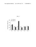 | 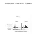 |
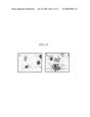 | 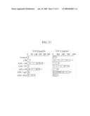 |
 | 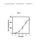 |
 | 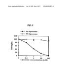 |
 | 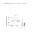 |
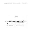 | 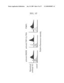 |
 | 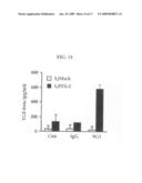 |
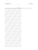 | 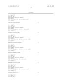 |
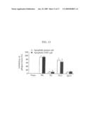 | 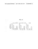 |
 | 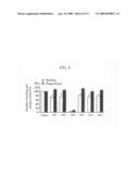 |
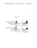 |  |
 | 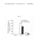 |
 |  |
| Similar patent applications: | |
| Date | Title |
|---|---|
| 2010-12-30 | Use of bispecific antibodies to regulate immune responses |
| 2010-12-23 | Novel uses of hibiscus, in particular pharmaceutical uses |
| 2008-10-16 | Novel uses of calcium hydroxide |
| 2010-12-02 | Conserved and specific streptococcal genomes |
| 2010-12-09 | Vector system for site-specific integration |
| New patent applications in this class: | |
| Date | Title |
|---|---|
| 2017-08-17 | Treatment of myocardial infarction using tgf beta antagonists |
| 2016-12-29 | Methods and compositions for treating obesity |
| 2016-07-14 | Method of treatment |
| 2016-06-09 | Treatment of cancer with anti-il-1 alpha antibodies |
| 2016-06-09 | Compositions and methods for treating conditions related to elevated levels of eosinophils and/or basophils |
| New patent applications from these inventors: | |
| Date | Title |
|---|---|
| 2017-09-14 | Methods of inhibiting pathological angiogenesis with doppel-targeting molecules |
| 2016-03-03 | Human ferritin-derived fusion polypeptide |
| 2015-02-19 | Drug delivery conjugate capable of controlled release, and use thereof |
| 2014-09-25 | Drug-fluorophore complex for specific detection of tumor cells |
| 2014-05-29 | Fusion peptide comprising dhfas-1 domain and mmp substrate and use thereof for preventing and treating rheumatoid arthritis |
| Top Inventors for class "Drug, bio-affecting and body treating compositions" | |
| Rank | Inventor's name |
|---|---|
| 1 | David M. Goldenberg |
| 2 | Hy Si Bui |
| 3 | Lowell L. Wood, Jr. |
| 4 | Roderick A. Hyde |
| 5 | Yat Sun Or |