Patent application title: URINE MRNA PROFILE AND ACUTE DYSFUNCTION OF KIDNEY ALLOGRAFT
Inventors:
Thangamani Muthukumar (New York, NY, US)
Manikkam Suthanthiran (Scarsdale, NY, US)
Ruchuang Ding (Beechurst, NY, US)
IPC8 Class: AC12Q168FI
USPC Class:
Class name:
Publication date: 2015-07-09
Patent application number: 20150191787
Abstract:
Non-invasive methods for detecting, predicting, and/or monitoring
differential diagnosis of kidney transplant dysfunction in kidney
transplant patients are described.Claims:
1. A method of detecting, predicting, or monitoring acute rejection of a
kidney transplant comprising (a) measuring urinary RNA expression levels
of the following genes: CD3.epsilon., CD105, TLR4, CD14, complement
factor B, and vimentin in a test urinary cell sample from a subject with
a kidney transplant; and (b) identifying increased expression of the
CD3.epsilon. mRNA CD105 mRNA, TLR4 mRNA, CD14 mRNA, complement factor B
mRNA, and vimentin mRNA compared to a baseline to thereby detect,
predict, or monitor acute rejection of a kidney transplant in the
subject.
2. The method of claim 1, wherein the method identifies acute cellular rejection in the sample, and distinguishes patients with acute rejection from patients with acute tubular injury.
3. The method of claim 1, wherein the baseline is an average or median amount of expression for the corresponding gene in urinary cells from a group of healthy patients or from a group of patients with a known kidney problem.
4. The method of claim 1, wherein measuring urinary RNA expression levels comprises: reverse transcription of RNA isolated from the test urinary cell sample of the subject; hybridization and/or primer extension of at least one probe or primer that selectively hybridizes to CD3.epsilon., CD105, TLR4, CD14, complement factor B, vimentin, CD46, or 18S rRNA; preamplification of urinary RNA from the sample; quantitative polymerase chain reaction of at least six of the following RNAs, or cDNAs generated from at least six of the following RNAs: CD3.epsilon., CD105, TLR4, CD14, complement factor B, vimentin, CD46, and 18S rRNA; quantifying amounts of at least six of the following RNAs: CD3.epsilon., CD105, TLR4, CD14, CD46, complement factor B, vimentin, and 18S rRNA, each as copy number of RNA per microgram of total RNA; or a combination thereof.
5. The method of claim 1, comprising identifying increased expression of each of the following mRNAs: CD3.epsilon., CD105, TLR4, CD14, complement factor B, and vimentin using the following six-gene diagnostic signature that distinguishes acute rejection from acute tubular injury: (0.52*lnCD3.epsilon.)+(1.02*lnCD105)+(0.81*lnTLR4)+(-1.16*lnCD14)+(0.28*l- nComplement Factor B)+(-0.79*lnVimentin); wherein a patient whose test urinary cell sample has a six-gene diagnostic signature of greater than about -0.24 has a transplanted kidney that is undergoing acute rejection, or will develop acute rejection.
6. The method of claim 5, further comprising treatment of subject for acute rejection of a kidney transplant when the six-gene diagnostic signature is greater than about -0.24.
7. The method of claim 1, further comprising identifying expression of each of the following mRNAs: CD3.epsilon., CD105, TLR4, CD14, complement factor B, and vimentin using the following six-gene diagnostic signature that distinguishes acute rejection from acute tubular injury: (0.52*lnCD3.epsilon.)+(1.02*lnCD105)+(0.81*lnTLR4)+(-1.16*lnCD14)+(0.28*l- nComplement Factor B)+(-0.79*lnVimentin); wherein a patient whose test urinary cell sample has a six-gene diagnostic signature of less than about -0.24 has a transplanted kidney that is undergoing acute tubular injury, or will develop acute tubular injury.
8. The method of claim 7, further comprising treatment of a subject for acute tubular injury when the six-gene diagnostic signature is less than about -0.25, or less than about -0.3.
9. The method of claim 1, further comprising measuring urinary RNA expression levels of CD46, and 18S rRNA and identifying increased expression CD46, and 18S rRNA compared to a baseline, to thereby distinguish acute cellular rejection (ACR) from antibody-mediated rejection (AMR) of a kidney transplant in a subject.
10. The method of claim 9, comprising identifying increased expression of each of the following mRNAs CD3.epsilon., CD105, CD14, CD46 and 18S rRNA using the following five-gene diagnostic signature that distinguishes acute cellular rejection (ACR) from antibody-mediated rejection (AMR): (0.67*lnCD3.epsilon.)+(-1.18*lnCD105)+(1.30*lnCD14)+(-0.83*lnCD46)+(0.45*- ln18S) wherein a patient whose test urinary cell sample has a five-gene diagnostic signature of greater than about 9.24 has a transplanted kidney that is undergoing acute cellular rejection, or will develop acute cellular rejection, rather than antibody-mediated rejection.
11. The method of claim 10, further comprising treatment of subject for acute cellular rejection when the five-gene diagnostic signature is greater than about 9.24.
12. The method of claim 1, comprising identifying expression of each of the following mRNAs CD3.epsilon., CD105, CD14, CD46 and 18S rRNA using the following five-gene diagnostic signature that distinguishes acute cellular rejection (ACR) from antibody-mediated rejection (AMR): (0.67*lnCD3.epsilon.)+(-1.18*lnCD105)+(1.30*lnCD14)+(-0.83*lnCD46)+(0.45*- ln18S) wherein a patient whose test urinary cell sample has a five-gene diagnostic signature of less than about 9.2 has a transplanted kidney that is undergoing antibody-mediated rejection, or will develop antibody-mediated rejection, rather than acute cellular rejection.
13. The method of claim 12, further comprising treatment of subject for antibody-mediated rejection when the five-gene diagnostic signature is less than about 9.2, or less than about 9.3.
14. A method of detecting lack of acute kidney rejection comprising measuring urinary RNA expression levels of the following: CD3.epsilon., CD105, TLR4, CD14, complement factor B, and vimentin expression levels in a test urinary cell sample from a subject with a kidney transplant, identifying no increased expression of the following RNAs: CD3.epsilon., CD105, TLR4, CD14, CD46, complement factor B, vimentin, and 18S rRNA in the test urinary cell sample to thereby detect lack of acute kidney rejection in a subject.
15. A kit comprising instructions for detecting acute rejection of a kidney transplant, and probes or primers for selective hybridization to at least five mRNAs selected from the group: CD3.epsilon., CD105, TLR4, CD14, complement factor B, vimentin, CD46, and 18S rRNA.
16. The kit of claim 15, comprising at least one probe or primer for each of the following mRNAs: CD3.epsilon., CD105, TLR4, CD14, complement factor B, and vimentin.
17. The kit of claim 15, wherein the instructions comprise the following signature for distinguishing acute rejection of a kidney transplant from acute tubular injury to the kidney transplant: (0.52*lnCD3.epsilon.)+(1.02*lnCD105)+(0.81*lnTLR4)+(-1.16*lnCD14)+(0.28*l- nComplement Factor B)+(-0.79*lnVimentin); wherein a patient whose test urinary cell sample has a six-gene diagnostic signature of greater than about -0.24 has a transplanted kidney that is undergoing acute rejection, or will develop acute rejection.
18. The kit of claim 15, comprising at least one probe or primer for each of the following mRNAs: CD46, and 18S rRNA.
19. The kit of claim 15, wherein the instructions comprise the following method for distinguishing acute cellular rejection (ACR) from antibody-mediated rejection (AMR): (0.67*lnCD3.epsilon.)+(-1.18*lnCD105)+(1.30*lnCD14)+(-0.83*lnCD46)+(0.45*- ln18S) wherein a patient whose test urinary cell sample has a five-gene diagnostic signature of greater than about 9.24 has a transplanted kidney that is undergoing acute cellular rejection, or will develop acute cellular rejection, rather than antibody-mediated rejection.
20. The kit of claim 15, wherein the instructions comprise instructions for treatment of acute rejection, acute tubular injury, acute cellular rejection, and/or antibody-mediated rejection of a transplanted kidney.
Description:
[0001] This application claims benefit of priority to the filing date of
U.S. Provisional Application Ser. No. 61/924,543, filed Jan. 7, 2014, the
contents of which are specifically incorporated by reference herein in
their entity.
BACKGROUND
[0003] The healthiness of a kidney transplant is conventionally assessed by measuring creatinine levels in the blood. An increase in creatinine is called allograft dysfunction. Two types of acute rejection are the more common cause of allograft dysfunction: acute cellular and acute antibody mediated. Accurate diagnosis is important for providing that will provide treatment that is optimally therapeutic.
[0004] When the creatinine levels increase, patients typically undergo an invasive needle biopsy of the transplanted kidney to confirm acute rejection. However an increase in creatinine is not a specific test for acute rejection and a sizable proportion of patients with allograft dysfunction do not have acute rejection on biopsy. Moreover, invasive needle biopsy is not only associated with complications but is costly as well. Noninvasive tests to identify acute rejection would help obviate the need for biopsies in sizable proportion of patients with allograft dysfunction.
SUMMARY
[0005] The invention relates to methods of detecting acute kidney rejection in a subject, and discriminating between types of rejection, by detecting urinary RNA expression levels in a test urinary sample from the subject. For example, the methods described herein can be used to distinguish various types of kidney conditions such as acute rejection, acute tubular injury, acute cellular rejection, and/or antibody-mediated rejection. The methods can also be used to identify whether a subject has a kidney condition that may need treatment. Probes, primers, and methods for detecting RNA expression levels in urinary test samples and for performing the methods are also described herein.
[0006] For example, a method of detecting, predicting, or monitoring acute kidney rejection and distinguishing it from other types of kidney problems is described herein that includes:
[0007] (a) measuring urinary RNA expression levels of the following genes: CD3ε, CD105, TLR4, CD14, complement factor B, and vimentin in a test urinary cell sample from a subject with a kidney transplant; and
[0008] (b) identifying increased expression of CD3ε, CD105, TLR4, CD14, complement factor B, and vimentin to thereby detect, predict, or monitor acute kidney rejection in the subject.
[0009] A six-gene diagnostic signature can be used to distinguish acute cellular rejection from acute tubular injury:
(0.52*lnCD3ε)+(1.02*lnCD105)+(0.81*lnTLR4)+(-1.16*lnCD14)+(0.28*- lnComplement Factor B)+(-0.79*lnVimentin);
[0010] wherein a patient with test urinary cell sample that has a six-gene diagnostic signature of greater than about -0.24 has a transplanted kidney that is undergoing acute rejection, or will develop acute rejection. The method can also include treatment of subject for acute rejection when the six-gene diagnostic signature is greater than about -0.24. When the six-gene diagnostic signature of a sample is less than about -0.24 the patient from whom the sample was obtained has a transplanted kidney that is undergoing acute tubular injury, or will develop acute tubular injury. The method can also include treatment of subject for acute tubular injury when the six-gene diagnostic signature is less than about -0.24.
[0011] In another example, the method can include measuring urinary RNA expression levels of the following RNAs: CD3ε, CD105, CD14, CD46, and 18S rRNA in a test urinary cell sample from a subject with a kidney transplant. A five-gene diagnostic signature can be used to distinguish acute cellular rejection (ACR) from antibody-mediated rejection (AMR):
(0.67*lnCD3ε)+(-1.18*lnCD105)+(1.30*lnCD14)+(-0.83*lnCD46)+(0.45- *ln18S)
[0012] wherein a patient with a test urinary cell sample that has a five-gene diagnostic signature of greater than about 9.1 has a transplanted kidney that is undergoing acute cellular rejection, or will develop acute cellular rejection, rather than antibody-mediated rejection. The method can also include treatment of subject for acute cellular rejection when the five-gene diagnostic signature is greater than about 9.1. When the five-gene diagnostic signature of a sample is less than about 9.1 the patient from whom the sample was obtained has a transplanted kidney that is undergoing antibody-mediated rejection, or will develop antibody-mediated rejection. The method can also include treatment of subject for antibody-mediated rejection when the five-gene diagnostic signature is less than about 9.1.
[0013] Another method of detecting lack of acute kidney rejection involves measuring urinary RNA expression levels of one or more of the following: CD3ε, CD105, TLR4, CD14, CD46, complement factor B, vimentin, and 18S rRNA expression levels in a test urinary cell sample from a subject with a kidney transplant, identifying no increased expression of one or more of the CD3ε, CD105, TLR4, CD14, CD46, complement factor B, vimentin, and 18S rRNA to thereby detect lack of acute kidney rejection in a subject.
[0014] A kit is also described herein that includes instructions for detecting acute rejection of a kidney transplant, and probes or primers for selective hybridization to at least five mRNAs selected from the group: CD3ε, CD105, TLR4, CD14, CD46, complement factor B, vimentin, and 18S rRNA.
DESCRIPTION OF THE FIGURES
[0015] FIG. 1A-1B illustrates the methods and characteristics of the patient samples used to develop the methods. FIG. 1A is a schematic showing a flowchart for discovery and validation of urinary cell diagnostic signatures. Urinary cells were obtained from 84 kidney transplant recipients with acute allograft dysfunction. Transcript levels were measured in RNA from the urinary cells by pre-amplification enhanced real-time quantitative polymerase chain reaction (PCR) assays using a customized amplicon for construction of a standard curve of quantified mRNA abundance as copies per microgram of total RNA obtained from urinary cells. Individual transcripts were used as variables to construct statistical models using discriminant analysis. In each model the linear combination of variables yielded a discriminant score that constituted the diagnostic signature. A two-step approach was used to develop the diagnostic signatures. In the first step, acute rejection (both types, N=52) was differentiated from acute tubular injury (ATI, N=32). In the second step, acute cellular rejection (ACR, N=26) was differentiated from acute antibody mediated rejection (AMR, N=26) with the use of same PCR assay results. Ten-fold cross validation was used to validate both the models. FIG. 1B graphically illustrates the RNA quantity and purity, as well as the 18S rRNA levels in urinary cells from male or female patients, as a function of time post-kidney transplantation. The scatter plot shows the relation between time from kidney transplantation to the collection of urine samples (X-axis) and the quantity of total RNA isolated in urinary cells (Y-axis top), the purity of RNA as assessed by the ratio of the optical density (OD) at 260 nm to the optical density at 280 nm (Y-axis bottom) and the levels of endogenous control 18S ribosomal RNA. Total RNA was reverse transcribed to cDNA. The 18S rRNA level in urinary cells was quantified using gene specific primers and probes by real-time PCR assay and expressed as natural log-transformed copies per one microgram of total RNA. Dark closed circles represent samples from women and lighter open circles represent samples from men. By Spearman rank order correlation, there was no statistically significant association (P>0.5) between time from transplant to urine collection and each of the variables represented on the Y-axis. The OD260/OD280 ratio of pure RNA is about 2.
[0016] FIG. 2A-2ZA shows box plots illustrating the quantity of the twenty-six mRNAs and the 18S rRNA measured in the urinary cells of kidney transplant recipients at the time of for-cause (diagnostic) kidney biopsies. The X-axis of each box plot shows the expression levels of the indicated RNA in three groups of biopsy types; acute T-cell mediated rejection (ACR, left, N=26), acute antibody mediated rejection (AMR, center, N=26) and acute tubular injury (ATI, right, N=32). FIG. 2A shows CD3 expression levels. FIG. 2B shows Granzyme B expression levels. FIG. 2C shows perforin expression levels. FIG. 2D shows FoxP3 expression levels. FIG. 2E shows OX40 expression levels. FIG. 2F shows CD105 expression levels. FIG. 2G shows CD146 expression levels. FIG. 2H shows Von Willebrand factor expression levels. FIG. 2I shows Immunoglobulin J expression levels. FIG. 2J shows PSMB10 expression levels. FIG. 2K shows TRIM1 expression levels. FIG. 2L shows TRL-4 expression levels. FIG. 2M shows CD14 expression levels. FIG. 2N shows C3 expression levels. FIG. 2O shows C5 expression levels. FIG. 2P shows properdin expression levels. FIG. 2Q shows complement factor B expression levels. FIG. 2R shows CD55 expression levels. FIG. 2S shows CD46 expression levels. FIG. 2T shows vimentin expression levels. FIG. 2U shows NKCC2 expression levels. FIG. 2V shows E-cadherin expression levels. FIG. 2W shows IL-6 expression levels. FIG. 2X shows CXCL13 expression levels. FIG. 2Y shows CD20 expression levels. FIG. 2ZA shows TGFβ1 expression levels. FIG. 2Zb shows 18S rRNA expression levels. The 26 mRNAs and 18S rRNA were quantified using gene specific primers and probes by real-time PCR assay and expressed as copies per microgram of total RNA. The horizontal line within each box represents the median, the bottom and top of each box represent the 25th and 75th percentile values, and the whiskers represent the 10th and 90th percentile values. P values are based on the Kruskal-Wallis test. Individual groups were compared by Dunn's test, and if significant (P<0.05) are represented by an asterisk.
[0017] FIG. 3A-3B graphically illustrate differentiation of acute rejection from acute tubular injury. FIG. 3A shows a box plot of predicted probability of acute rejection from the cross validation. The horizontal line within each box represents the median and the plus symbol the mean. The bottom and top of each box represent 1.5 times the interquartile range. The values beyond this are shown as dots. The discrimination slope is the difference between the means of the predicted probabilities of the two groups. FIG. 3B shows the ROC curve of the predicted probability for each patient from the cross validation to diagnose AR. The sensitivity (true positive fraction), specificity (false positive fraction), likelihood ratio of a positive test (LR+, sensitivity/1-specificity) and likelihood ratio of a negative test (LR-, 1-sensitivity/specificity) for various cut-points of predicted risks are shown beneath the X-axis. The AUC is the estimate of the expected value in an independent sample not used for deriving the diagnostic signature. The absolute levels of the 26 mRNAs and the 18S rRNA in the urinary cells from 84 kidney graft recipients were measured. Quadratic discriminant function analysis was used to derive linear combination of mRNAs to better differentiate the 52 AR (acute rejection) biopsies (ACR and AMR, N=52 patients) from 32 ATI biopsies (N=32 patients) than any single mRNA measure. A linear combination of six mRNAs (CD3ε, CD105, TLR-4, CD14, Complement Factor B, and Vimentin) emerged as the parsimonious model and yielded a discriminant score that constituted the diagnostic signature. Ten-fold cross validation was performed to internally validate the 6-gene diagnostic signature. The entire study cohort of 84 patients was randomly divided into ten equal groups. Within each of the ten groups, the proportion of samples (AR vs. ATI) was similar to the undivided cohort. At the first run, group 1 (10% of samples) was excluded and a signature was derived from the remaining 9 groups (90% of samples) including both variables selection and model fitting. Next, this newly derived signature was applied to samples of group 1 to predict their diagnostic outcome. In the second run, group 2 was excluded and a signature was derived from the remaining 9 groups (90% of samples) including both variables selection and model fitting. This newly derived signature was applied to samples of group 2 (10% of samples) to predict their diagnostic outcome. This iteration was done for all the 10 groups. Thus, all observations were used for both deriving and validating a model and each observation was used for validation exactly once. Accordingly, the predicted probability for an individual patient was derived from a model that did not include any data from that patient. The predicted probability for each patient from the cross validation was used to construct a ROC curve. This is the estimate of the expected value of the AUC in an independent sample not used for deriving the diagnostic signature.
[0018] FIG. 4A-4B illustrate decision curve analysis to assess the clinical benefit of the 6-gene urinary cell diagnostic signature. The predicted probability for each patient from the cross validation in decision curve analysis was used to quantify the clinical benefit of the diagnostic signature in terms of the number of unnecessary biopsies that can be avoided in the diagnosis of AR. FIG. 4A shows the net benefit [(true positive count/n)-(false positive count/n)*[pt/(1-pt)] in the Y axis, where true positive count=the number of patients with AR, false positive count=the number of patients with ATI, n=the total number of patients and pt=threshold probability. Here, pt/(1-pt) is the ratio of the harms of false positive to false negative results. Of the 84 patients studied, 52 (62%) had AR. This proportion of acute rejection (AR) is a reasonable approximation of the expected incidence of AR in consecutive for-cause (diagnostic) biopsies done to identify the cause of acute graft dysfunction. The uppermost line is the net benefit of the urinary cell diagnostic signature. This strategy is compared with the `biopsy all patients` strategy (middle line), which is essentially the current approach. The lowest line, which represents no net benefit, is the `biopsy none` strategy. The decision curve plot depicts that among patients who present with acute graft dysfunction, within a reasonable physician/patient threshold probability for doing a biopsy to diagnose AR, the use of urinary cell diagnostic signature is beneficial compared with the current `biopsy all patients` strategy. FIG. 4B shows the threshold probability on the X-axis, for the corresponding value on the Y-axis where the Y-axis represents the net reduction in avoidable biopsies per 100 patients, when using the diagnostic signature.
[0019] FIG. 5A-5B graphically illustrates the differentiation of acute T-cell mediated rejection from acute antibody-mediated rejection. After differentiation of acute rejection from acute tubular injury in step 1 (see, FIG. 1A), in step 2 and among patients diagnosed with acute rejection biopsies, another urinary cell diagnostic signature was derived to better differentiate ACR biopsies (N=26 patients) from AMR biopsies (N=26 patients) (see, FIG. 1A) than any single mRNA measure. By quadratic discriminant function analysis, a linear combination of four mRNAs (CD3ε, CD105, CD14, and CD46) and 18S rRNA emerged as the parsimonious model and yielded a discriminant score that constituted the diagnostic signature. Ten-fold cross validation was performed to internally validate the 5-gene diagnostic signature. FIG. 5A shows the box plot of predicted probability of ACR biopsies from the cross-validation. FIG. 5B shows the ROC curve of the 5-gene urinary cell diagnostic signature to diagnose ACR. The AUC is the estimate of the expected value in an independent sample not used for deriving the diagnostic signature.
[0020] FIG. 6A-6B illustrate the relationship between the urinary cell diagnostic signature score and the time from transplantation to biopsy/urine sample collection. FIG. 6A shows the 6-gene signature. FIG. 6B shows the 5-gene signature. The diagnostic signature score is represented on the Y-axis for both the 6-gene signature (FIG. 6A) and the 5-gene signature (FIG. 6B) and time from transplantation to biopsy/urine sample collection, in logarithmic scale, is represented on the X-axis. Induction immunosuppression therapy with lymphocyte depleting-Thymoglobulin® (including one patient with alemtuzumab) is shown as closed symbols while induction with lymphocyte non-depleting interleukin-2 receptor antibody or no induction therapy is shown as open symbols. Within each diagnostic category, analysis involving Spearman rank order correlation showed that there was no significant association (P>0.5) between the score of the 6 or the 5-gene diagnostic signature and the time from transplantation to biopsy in patients with biopsies showing ACR, AMR or ATI and induced with depleting or non-depleting antibodies. There was also no association between the scores of the signatures and either serum creatinine levels (6-gene signature-ACR: rs=-0.39, P=0.06; AMR: rs=-0.19, P=0.3; ATI: rs=-0.002, P=0.9 and 5-gene signature-ACR: rs=-0.14, P=0.5; AMR: rs=-0.07, P=0.7) or tacrolimus trough levels (6-gene signature-ACR: rs=0.14, P=0.5; AMR: rs=-0.14, P=0.5; ATI: rs=-0.02, P=0.9 and 5-gene signature-ACR: rs=-0.12, P=0.6; AMR: rs=-0.02, P=0.9) (not shown).
DETAILED DESCRIPTION
[0021] Noninvasive tests to differentiate the basis for acute dysfunction of the kidney allograft are preferable to invasive allograft biopsies. As described herein, cells obtained from urine samples of patients with kidney transplants can be used to detect whether the patient has, or will develop transplant tissue rejection.
[0022] For example, the urinary cell expression levels of mRNAs for CD3ε, CD105, TLR4, CD14, complement factor B, and vimentin distinguish acute rejection (AR) from acute tubular injury (ATI) in a six-gene signature. The method for distinguishing acute rejection (AR) from acute tubular injury (ATI) involved natural log (ln) transformation of measured mRNA values of CD3ε, CD105, TLR4, CD14, Complement factor B, and Vimentin where the unit of measurement in the PCR assay is copies/μg of total RNA. The six-gene diagnostic signature that distinguished AR from ATI is as follows:
(0.52*lnCD3ε)+(1.02*lnCD105)+(0.81*lnTLR4)+(-1.16*lnCD14)+(0.28*- lnComplement Factor B)+(-0.79*lnVimentin).
This diagnostic signature better differentiated AR from ATI than any single mRNA measure (e.g. vs. CD3ε [AUC: 0.88], likelihood ratio test X2=40.6, P<0.0001). The diagnostic signature also outperformed other variables; time from transplantation to biopsy (AUC: 0.65), serum creatinine (AUC: 0.59) or tacrolimus trough levels (AUC: 0.77).
[0023] A six-gene diagnostic signature of greater than about -0.24 indicates that the transplanted kidney in the patient from whom the tested sample was obtained can be acutely rejecting the transplanted kidney, or will develop acute rejection of the transplanted kidney. For example, a six-gene diagnostic signature that greater than about -0.2, or greater than about -0.1, or greater than about 0, or greater than about 0.1, or greater than about 0.2, or greater than about 0.3, or greater than about 0.4 indicates that the transplanted kidney in the patient from whom the tested sample was obtained can be acutely rejecting the transplanted kidney, or will develop acute rejection of the transplanted kidney.
[0024] In general, a six-gene diagnostic signature of less than about -0.25 indicates that the transplanted kidney in the patient from whom the tested sample was obtained is not acutely rejecting the transplanted kidney. However, when a sample has a six-gene diagnostic signature of less than about -0.25, the patient from whom the sample was obtained can have a kidney with acute tubular injury (ATI). For example, samples with a six-gene diagnostic signature of less than about -0.3, or less than about -0.35, or less than about -0.4, or less than about -0.45, or less than about -0.5, or less than about -0.6 can mean that the patient from whom the sample was obtained has a kidney with acute tubular injury (ATI).
[0025] In addition, mRNAs for CD3ε, CD105, CD14, CD46 and 18S rRNA can distinguish acute cellular rejection (ACR) from antibody-mediated rejection (AMR) in a five-gene signature. The five-gene model involved natural log (ln) transformation of measured mRNA values of CD3ε, CD105, CD14, CD46 and 18S rRNA and the following algorithm:
(0.67*lnCD3ε)+(-1.18*lnCD105)+(1.30*lnCD14)+(-0.83*lnCD46)+(0.45- *ln18S).
This diagnostic signature better differentiated ACR from AMR than any other single mRNA measure (e.g. vs. CD3ε [AUC: 0.87], likelihood ratio test X2=30.4, P<0.0001). Ten-fold cross validation of this 5-gene model yielded an AUC of 0.81 (95% CI 0.68 to 0.93, P<0.001, FIG. 4).
[0026] A five-gene diagnostic signature of greater than about 9.24 indicates that the transplanted kidney in the patient from whom the tested sample was obtained is undergoing acute cellular rejection, or will develop acute cellular rejection. For example, a five-gene diagnostic signature of greater than about 9.24, or greater than about 9.3, or greater than about 9.4, or greater than about 9.5, or greater than about 9.6, or greater than about 9.7, or greater than about 9.8, or greater than about 9.9 indicates that the transplanted kidney in the patient from whom the tested sample was obtained is undergoing acute cellular rejection, or will develop acute cellular rejection.
[0027] In general, a five-gene diagnostic signature of less than about 9.24 indicates that the transplanted kidney in the patient from whom the tested sample was obtained is undergoing antibody-mediated rejection rather than acute cellular rejection. For example, a five-gene diagnostic signature of less than about 9.2, or less than about 9.1, or less than about 9.0, or less than about 8.7, or less than about 8.5, or less than about 8.0, or less than about 7.0, or less than about 6.0, or less than about 5.0 indicates that the transplanted kidney in the patient from whom the tested sample was obtained is undergoing antibody-mediated rejection rather than acute cellular rejection.
[0028] The inventors measured absolute levels of a panel of 26 pre-specified mRNAs in 84 urine samples collected from 84 kidney graft recipients at the time of a for-cause biopsy for acute allograft dysfunction, and investigated whether differential diagnosis of acute graft dysfunction is feasible using urinary cell mRNA profiles. Fifty-two urine samples from 52 patients with acute rejection biopsies (26 with acute T-cell mediated rejection [ACR] and 26 with acute antibody-mediated rejection [AMR]) and 32 urine samples from 32 patients with acute tubular injury and without acute rejection changes (ATI) were profiled. A stepwise quadratic discriminant analysis of mRNA measurements identified a linear combination of mRNAs for CD3ε, CD105, TLR4, CD14, complement factor B, and vimentin that distinguishes acute rejection from acute tubular injury (ATI). Ten-fold cross validation of the 6-gene signature yielded an estimate of the area under the curve (AUC) of 0.92 (95% CI, 0.86-0.98). In a decision analysis, the 6-gene signature yielded the highest net benefit across a range of reasonable threshold probabilities for biopsy. Next, among patients diagnosed with acute rejection biopsies, a similar statistical approach identified a linear combination of mRNAs for CD3ε, CD105, CD14, CD46 and 18S rRNA that distinguishes ACR from AMR, with a cross-validated estimate of the AUC of 0.81 (95% CI, 0.68-0.93). The incorporation of these urinary cell mRNA signatures in clinical decision making may help avoid substantial number of biopsies in patients with acute dysfunction of the kidney allograft.
Organ Transplant
[0029] Organ transplantation or the transfer of an organ from one human to another continues to rise throughout the world as the treatment of choice when an organ is irreversibly damaged or organ function is severely impaired. Organ transplantation is not without complications, not only from the transplant surgery itself, but also from the transplant recipient's own immune system and this process, if it happens suddenly, is called acute rejection.
[0030] For example, when acute rejection of a kidney transplant occurs, it manifests itself by a sudden deterioration in kidney transplant function. About 30 percent of transplant recipients experience an episode of acute rejection. Acute rejection can be associated with reduction in the one-year survival rate of kidney grafts from a deceased donor of about 20 percent, and the projected half-life is about four years shorter in patients who have had an episode of acute rejection compared to patients who have not had an episode of acute rejection.
[0031] Sometimes, acute rejection can result from the activation of recipient's T cells and/or B cells. The rejection primarily due to T cells is classified as T cell mediated acute rejection or acute cellular rejection (ACR) and the rejection in which B cells are primarily responsible is classified as antibody mediated acute rejection (AMR). Often times, acute rejection of either type can result in the complete loss of transplant function and transplant failure.
[0032] An increase in the level of serum creatinine, a clinically used measure of kidney function, is often the first clinical indicator of acute rejection, and is currently the best surrogate marker of acute rejection of either type. However, this biomarker lacks sensitivity and specificity because graft dysfunction can occur due to non-immunological causes.
[0033] Two of the commonly used drugs prescribed to transplant recipients to prevent rejection, cyclosporine and tacrolimus, can cause kidney toxicity, and this complication is not readily identified solely on the basis of blood concentrations of cyclosporine/tacrolimus. In kidney transplant patients, the clinical importance of distinguishing acute rejection from cyclosporine/tacrolimus toxicity cannot be overemphasized because the treatment approaches are diametrically opposite. In one instance, continued administration of cyclosporine/tacrolimus for rejection is critical whereas, in the other instance, a reduction in dosage or discontinuation of cyclosporine/tacrolimus is indicated to prevent further kidney toxicity. Furthermore, deterioration in kidney function is not always available as a clinical clue to diagnose rejection because many of the kidney transplants suffer from acute (reversible) renal failure in the immediate post-transplantation period due to injury from organ procurement and the ex-vivo preservation procedures involved.
[0034] Currently, acute rejection is diagnosed by performing an invasive core needle biopsy procedure, which obtains a biopsy of the kidney graft. The histological features in the allograft biopsy tissues are then observed. However, this invasive biopsy procedure is associated with complications such as bleeding, arteriovenous fistula, graft loss, and, in severe cases, even death.
[0035] Development of a noninvasive test either to anticipate an episode of acute rejection or to diagnose acute rejection without performing the transplant biopsy procedure is a major and an unmet goal in organ transplantation.
Measurement of mRNA and 18S rRNA Quantities in Urinary Cells
[0036] Any procedure available to those of skill in the art can be employed to determine the expression levels of CD3ε mRNA, CD105 mRNA, TLR4 mRNA, CD14 mRNA, complement factor B mRNA, vimentin mRNA, CD46 mRNA, 18S rRNA, or a combination thereof. For example, probes, primers, and/or antibodies can be employed in quantitative nucleic acid amplification reactions (e.g., quantitative polymerase chain reaction (PCR)), primer extension, Northern blot, immunoassay, immunosorbant assay (ELISA), radioimmunoassay (RIA), immunofluorimetry, immunoprecipitation, equilibrium dialysis, immunodiffusion, immunoblotting, mass spectrometry and other techniques available to the skilled artisan.
[0037] In some embodiments, the expression levels CD3ε mRNA, CD105 mRNA, TLR4 mRNA, CD14 mRNA, complement factor B mRNA, vimentin mRNA, CD46 mRNA, 18S rRNA, or a combination thereof are determined using respective probes or primers that can hybridize to the CD3ε mRNA, CD105 mRNA, TLR4 mRNA, CD14 mRNA, complement factor B mRNA, vimentin mRNA, CD46 mRNA, and 18S rRNA.
[0038] Sequences for CD3ε mRNA, CD105 mRNA, TLR4 mRNA, CD14 mRNA, complement factor B mRNA, vimentin mRNA, CD46 mRNA, and 18S rRNA are readily available and can be used to make such probes and primers.
[0039] For example, the following cDNA sequence for a human 18S rRNA is available from the National Center for Biotechnology Information database (see website at ncbi.nlm.nih.gov) as accession number K03432 (SEQ ID NO:1).
TABLE-US-00001 1 CGCTGCTCCT CCCGTCGCCG TCCGGGCCCG TCCGTCCGTC 41 CGTCCGTCGT CCTCCTCGCT NNNNCGGGGC GCCGGGCCCG 61 TCCTCACNGG CCCCCGNNNN NGTCCNGGCC CGTCGGGGCC 121 TCGCCGCGCT CTACCTTACC TACCTGGTTG ATCCTGCCAG 161 TAGCATATGC TTGTCTCAAA GATTAAGCCA TGCATGTCTA 201 AGTACGCACG GCCGGTACAG TGAAACTGCG AATGGCTCAT 241 TAAATCAGTT ATGGTTCCTT TGGTCGCTCG CTCCTCTCCT 281 ACTTGGATAA CTGTGGTAAT TCTAGAGCTA ATACATGCCG 321 ACGGGCGCTG ACCCCCTTCG CGGGGGGGAT GCGTGCATTT 361 ATCAGATCAA AACCAACCCG GTCAGCCCCT CTCCGGCCCC 401 GGCCGGGGGG CGGGCGCCGG CGGCTTTGGT GACTCTAGAT 441 AACCTCGGGC CGATCGCACG CCCCCCGTGG CGGCGACGAC 481 CCATTCGAAC GTCTGCCCTA TCAACTTTCG ATGGTAGTCG 521 CCGTGCCTAC CATGGTGACC ACGGGTGACG GGGAATCAGG 561 GTTCGATTCC GGAGAGGGAG CCTGAGAAAC GGCTACCACA 601 TCCAAGGAAG GCAGCAGGCG CGCAAATTAC CCACTCCCGA 641 CCCGGGGAGG TAGTGACGAA AAATAACAAT ACAGGACTCT 681 TTCGAGGCCC TGTAATTGGA ATGAGTCCAC TTTAAATCCT 721 TTAACGAGGA TCCATTGGAG GGCAAGTCTG GTGCCAGCAG 761 CCGCGGTAAT TCCAGCTCCA ATAGCGTATA TTAAAGTTGC 801 TGCAGTTAAA AAGCTCGTAG TTGGATCTTG GGAGCGGGCG 841 GGCGGTCCGC CGCGAGGCGA GCCACCGCCC GTCCCCGCCC 881 CTTGCCTCTC GGCGCCCCCT CGATGCTCTT AGCTGAGTGT 921 CCCGCGGGGC CCGAAGCGTT TACTTTGAAA AAATTAGAGT 961 GTTCAAAGCA GGCCCGAGCC GCCTGGATAC CGCAGCTAGG 1001 AATAATGGAA TAGGACCGCG GTTCTATTTT GTTGGTTTTC 1041 GGAACTGAGG CCATGATTAA GAGGGACGGC CGGGGGCATT 1081 CGTATTGCGC CGCTAGAGGT GAAATTCCTT GGACCGGCGC 1121 AAGACGGACC AGAGCGAAAG CATTTGCCAA GAATGTTTTC 1161 ATTAATCAAG AACGAAAGTC GGAGGTTCGA AGACGATCAG 1201 ATACCGTCGT AGTTCCGACC ATAAACGATG CCGACCGGCG 1241 ATGCGGCGGC GTTATTCCCA TGACCCGCCG GGCAGCTTCC 1281 GGGAAACCAA AGTCTTTGGG TTCCGGGGGG AGTATGGTTG 1321 CAAAGCTGAA ACTTAAAGGA ATTGACGGAA GGGCACCACC 1361 AGGAGTGGAG CCTGCGGCTT AATTTGACTC AACACGGGAA 1401 ACCTCACCCG GCCCGGACAC GGACAGGATT GACAGATTGA 1441 TAGCTCTTTC TCGATTCCGT GGGTGGTGGT GCATGGCCGT 1481 TCTTAGTTGG TGGAGCGATT TGTCTGGTTA ATTCCGATAA 1521 CGAACGAGAC TCTGGCATGC TAACTAGTTA CGCGACCCCC 1561 GAGCGGTCGG CGTCCCCCAA CTTCTTAGAG GGACAAGTGG 1601 CGTTCAGCCA CCCGAGATTG AGCAATAACA GGTCTGTGAT 1641 GCCCTTAGAT GTCCGGGGCT GCACGCGCGC TACACTGACT 1681 GGCTCAGCGT GTGCCTACCC TACGCCGGCA GGCGCGGGTA 1721 ACCCGTTGAA CCCCATTCGT GATGGGGATC GGGGATTGCA 1761 ATTATTCCCC ATGAACGAGG AATTCCCAGT AAGTGCGGGT 1801 CATAAGCTTG CGTTGATTAA GTCCCTGCCC TTTGTACACA 1841 CCGCCCGTCG CTACTACCGA TTGGATGGTT TAGTGAGGCC 1881 CTCGGATCGG CCCCGCCGGG GTCGGCCCAC GGCCCTGGCG 1921 GAGCGCTGAG AAGACGGTCG AACTTGACTA TCTAGAGGAA 1961 GTAAAAGTCG TAACAAGGTT TCCGTAGGTG AACCTGCGGA 2001 AGGATCATTA ACGGAGCCCG GACGGCGGCC CGCGGCGGCG 2041 CCGCGCCGCG CTTCCCTCCG CACACCCACC CCCCCACCGC 2081 GACGGCGCGT GCGGGCGGGG CCGTGCCCGT TCGTTCGCTC 2121 GCTCGTTCGT TCGCCGCCCG GCCCGGCCGC GAGAGCCGAG 2161 AACTCGGGAG GGAGACGGGG GAGAGAGAGA GAGAGAGAGA 2201 GAGAGAGAGA GAGAGAGAGA GAAAGAAGGG CGTGT
[0040] A cDNA sequence for a human CD3ε is also available from the National Center for Biotechnology Information database as accession number NM--000733 (SEQ ID NO:2).
TABLE-US-00002 1 TATTGTCAGA GTCCTCTTGT TTGGCCTTCT AGGAAGGCTG 41 TGGGACCCAG CTTTCTTCAA CCAGTCCAGG TGGAGGCCTC 81 TGCCTTGAAC GTTTCCAAGT GAGGTAAAAC CCGCAGGCCC 121 AGAGGCCTCT CTACTTCCTG TGTGGGGTTC AGAAACCCTC 161 CTCCCCTCCC AGCCTCAGGT GCCTGCTTCA GAAAATGAAG 201 TAGTAAGTCT GCTGGCCTCC GCCATCTTAG TAAAGTAACA 241 GTCCCATGAA ACAAAGATGC AGTCGGGCAC TCACTGGAGA 281 GTTCTGGGCC TCTGCCTCTT ATCAGTTGGC GTTTGGGGGC 321 AAGATGGTAA TGAAGAAATG GGTGGTATTA CACAGACACC 361 ATATAAAGTC TCCATCTCTG GAACCACAGT AATATTGACA 401 TGCCCTCAGT ATCCTGGATC TGAAATACTA TGGCAACACA 441 ATGATAAAAA CATAGGCGGT GATGAGGATG ATAAAAACAT 481 AGGCAGTGAT GAGGATCACC TGTCACTGAA GGAATTTTCA 521 GAATTGGAGC AAAGTGGTTA TTATGTCTGC TACCCCAGAG 561 GAAGCAAACC AGAAGATGCG AACTTTTATC TCTACCTGAG 601 GGCAAGAGTG TGTGAGAACT GCATGGAGAT GGATGTGATG 641 TCGGTGGCCA CAATTGTCAT AGTGGACATC TGCATCACTG 681 GGGGCTTGCT GCTGCTGGTT TACTACTGGA GCAAGAATAG 721 AAAGGCCAAG GCCAAGCCTG TGACACGAGG AGCGGGTGCT 761 GGCGGCAGGC AAAGGGGACA AAACAAGGAG AGGCCACCAC 801 CTGTTCCCAA CCCAGACTAT GAGCCCATCC GGAAAGGCCA 841 GCGGGACCTG TATTCTGGCC TGAATCAGAG ACGCATCTGA 881 CCCTCTGGAG AACACTGCCT CCCGCTGGCC CAGGTCTCCT 921 CTCCAGTCCC CCTGCGACTC CCTGTTTCCT GGGCTAGTCT 961 TGGACCCCAC GAGAGAGAAT CGTTCCTCAG CCTCATGGTG 1001 AACTCGCGCC CTCCAGCCTG ATCCCCCGCT CCCTCCTCCC 1041 TGCCTTCTCT GCTGGTACCC AGTCCTAAAA TATTGCTGCT 1081 TCCTCTTCCT TTGAAGCATC ATCAGTAGTC ACACCCTCAC 1121 AGCTGGCCTG CCCTCTTGCC AGGATATTTA TTTGTGCTAT 1161 TCACTCCCTT CCCTTTGGAT GTAACTTCTC CGTTCAGTTC 1201 CCTCCTTTTC TTGCATGTAA GTTGTCCCCC ATCCCAAAGT 1241 ATTCCATCTA CTTTTCTATC GCCGTCCCCT TTTGCAGCCC 1281 TCTCTGGGGA TGGACTGGGT AAATGTTGAC AGAGGCCCTG 1321 CCCCGTTCAC AGATCCTGGC CCTGAGCCAG CCCTGTGCTC 1361 CTCCCTCCCC CAACACTCCC TACCAACCCC CTAATCCCCT 1401 ACTCCCTCCA CCCCCCCTCC ACTGTAGGCC ACTGGATGGT 1441 CATTTGCATC TCCGTAAATG TGCTCTGCTC CTCAGCTGAG 1481 AGAGAAAAAA ATAAACTGTA TTTGGCTGCA AGAAAAAAAA 1521 AAAAAAAAAA AAAA
[0041] A cDNA sequence for human CD105 (also called endoglin) is available from the National Center for Biotechnology Information database as accession number BC014271.2 (GI:33871100; SEQ ID NO:3).
TABLE-US-00003 1 CCACCCCAGA AGGCTGGAGC AGGGACGCCG TCGCTCCGGC 41 CGCCTGCTCC CCTCGGGTCC CCGTGCGAGC CCACGCCGGC 81 CCCGGTGCCC GCCCGCAGCC CTGCCACTGG ACACAGGATA 121 AGGCCCAGCG CACAGGCCCC CACGTGGACA GCATGGACCG 161 CGGCACGCTC CCTCTGGCTG TTGCCCTGCT GCTGGCCAGC 201 TGCAGCCTCA GCCCCACAAG TCTTGCAGAA ACAGTCCATT 241 GTGACCTTCA GCCTGTGGGC CCCGAGAGGG ACGAGGTGAC 281 ATATACCACT AGCCAGGTCT CGAAGGGCTG CGTGGCTCAG 321 GCCCCCAATG CCATCCTTGA AGTCCATGTC CTCTTCCTGG 361 AGTTCCCAAC GGGCCCGTCA CAGCTGGAGC TGACTCTCCA 401 GGCATCCAAG CAAAATGGCA CCTGGCCCCG AGAGGTGCTT 441 CTGGTCCTCA GTGTAAACAG CAGTGTCTTC CTGCATCTCC 481 AGGCCCTGGG AATCCCACTG CACTTGGCCT ACAATTCCAG 521 CCTGGTCACC TTCCAAGAGC CCCCGGGGGT CAACACCACA 561 GAGCTGCCAT CCTTCCCCAA GACCCAGATC CTTGAGTGGG 601 CAGCTGAGAG GGGCCCCATC ACCTCTGCTG CTGAGCTGAA 641 TGACCCCCAG AGCATCCTCC TCCGACTGGG CCAAGCCCAG 681 GGGTCACTGT CCTTCTGCAT GCTGGAAGCC AGCCAGGACA 721 TGGGCCGCAC GCTCGAGTGG CGGCCGCGTA CTCCAGCCTT 761 GGTCCGGGGC TGCCACTTGG AAGGCGTGGC CGGCCACAAG 801 GAGGCGCACA TCCTGAGGGT CCTGCCGGGC CACTCGGCCG 841 GGCCCCGGAC GGTGACGGTG AAGGTGGAAC TGAGCTGCGC 881 ACCCGGGGAT CTCGATGCCG TCCTCATCCT GCAGGGTCCC 921 CCCTACGTGT CCTGGCTCAT CGACGCCAAC CACAACATGC 961 AGATCTGGAC CACTGGAGAA TACTCCTTCA AGATCTTTCC 1001 AGAGAAAAAC ATTCGTGGCT TCAAGCTCCC AGACACACCT 1041 CAAGGCCTCC TGGGGGAGGC CCGGATGCTC AATGCCAGCA 1081 TTGTGGCATC CTTCGTGGAG CTACCGCTGG CCAGCATTGT 1121 CTCACTTCAT GCCTCCAGCT GCGGTGGTAG GCTGCAGACC 1161 TCACCCGCAC CGATCCAGAC CACTCCTCCC AAGGACACTT 1201 GTAGCCCGGA GCTGCTCATG TCCTTGATCC AGACAAAGTG 1241 TGCCGACGAC GCCATGACCC TGGTACTAAA GAAAGAGCTT 1281 GTTGCGCATT TGAAGTGCAC CATCACGGGC CTGACCTTCT 1321 GGGACCCCAG CTGTGAGGCA GAGGACAGGG GTGACAAGTT 1361 TGTCTTGCGC AGTGCTTACT CCAGCTGTGG CATGCAGGTG 1401 TCAGCAAGTA TGATCAGCAA TGAGGCGGTG GTCAATATCC 1441 TGTCGAGCTC ATCACCACAG CGGAAAAAGG TGCACTGCCT 1481 CAACATGGAC AGCCTCTCTT TCCAGCTGGG CCTCTACCTC 1521 AGCCCACACT TCCTCCAGGC CTCCAACACC ATCGAGCCGG 1561 GGCAGCAGAG CTTTGTGCAG GTCAGAGTGT CCCCATCCGT 1601 CTCCGAGTTC CTGCTCCAGT TAGACAGCTG CCACCTGGAC 1641 TTGGGGCCTG AGGGAGGCAC CGTGGAACTC ATCCAGGGCC 1681 GGGCGGCCAA GGGCAACTGT GTGAGCCTGC TGTCCCCAAG 1721 CCCCGAGGGT GACCCGCGCT TCAGCTTCCT CCTCCACTTC 1761 TACACAGTAC CCATACCCAA AACCGGCACC CTCAGCTGCA 1801 CGGTAGCCCT GCGTCCCAAG ACCGGGTCTC AAGACCAGGA 1841 AGTCCATAGG ACTGTCTTCA TGCGCTTGAA CATCATCAGC 1881 CCTGACCTGT CTGGTTGCAC AAGCAAAGGC CTCGTCCTGC 1921 CCGCCGTGCT GGGCATCACC TTTGGTGCCT TCCTCATCGG 1961 GGCCCTGCTC ACTGCTGCAC TCTGGTACAT CTACTCGCAC 2001 ACGCGTTCCC CCAGCAAGCG GGAGCCCGTG GTGGCGGTGG 2041 CTGCCCCGGC CTCCTCGGAG AGCAGCAGCA CCAACCACAG 2081 CATCGGGAGC ACCCAGAGCA CCCCCTGCTC CACCAGCAGC 2121 ATGGCATAGC CCCGGCCCCC CGCGCTCGCC CAGCAGGAGA 2161 GACTGAGCAG CCGCCAGCTG GGAGCACTGG TGTGAACTCA 2201 CCCTGGGAGC CAGTCCTCCA CTCGACCCAG AATGGAGCCT 2241 GCTCTCCGCG CCTACCCTTC CCGCCTCCCT CTCAGAGGCC 2281 TGCTGCCAGT GCAGCCACTG GCTTGGAACA CCTTGGGGTC 2321 CCTCCACCCC ACAGAACCTT CAACCCAGTG GGTCTGGGAT 2361 ATGGCTGCCC AGGAGACAGA CCACTTGCCA CGCTGTTGTA 2401 AAAACCCAAG TCCCTGTCAT TTGAACCTGG ATCCAGCACT 2441 GGTGAACTGA GCTGGGCAGG AAGGGAGAAC TTGAAACAGA 2481 TTCAGGCCAG CCCAGCCAGG CCAACAGCAC CTCCCCGCTG 2521 GGAAGAGAAG AGGGCCCAGC CCAGAGCCAC CTGGATCTAT 2561 CCCTGCGGCC TCCACACCTG AACTTGCCTA ACTAACTGGC 2601 AGGGGAGACA GGAGCCTAGC GGAGCCCAGC CTGGGAGCCC 2641 AGAGGGTGGC AAGAACAGTG GGCGTTGGGA GCCTAGCTCC 2681 TGCCACATGG AGCCCCCTCT GCCGGTCGGG CAGCCAGCAG 2721 AGGGGGAGTA GCCAAGCTGC TTGTCCTGGG CCTGCCCCTG 2761 TGTATTCACC ACCAATAAAT CAGACCATGA AACCAAAAAA 2801 AAAAAAAAAA AAAAAAAAAA AAAAAAAAAA
[0042] Another cDNA sequence for human CD105 is available from the National Center for Biotechnology Information database as accession number NM--000118.2 (GI:168693645; SEQ ID NO:4).
TABLE-US-00004 1 CTCTACCCGG TTGGCAGGCG GCCTGGCCCA GCCCCTTCTC 41 TAAGGAAGCG CATTTCCTGC CTCCCTGGGC CGGCCGGGCT 81 GGATGAGCCG GGAGCTCCCT GCTGCCGGTC ATACCACAGC 121 CTTCATCTGC GCCCTGGGGC CAGGACTGCT GCTGTCACTG 161 CCATCCATTG GAGCCCAGCA CCCCCTCCCC GCCCATCCTT 201 CGGACAGCAA CTCCAGCCCA GCCCCGCGTC CCTGTGTCCA 241 CTTCTCCTGA CCCCTCGGCC GCCACCCCAG AAGGCTGGAG 281 CAGGGACGCC GTCGCTCCGG CCGCCTGCTC CCCTCGGGTC 321 CCCGTGCGAG CCCACGCCGG CCCCGGTGCC CGCCCGCAGC 361 CCTGCCACTG GACACAGGAT AAGGCCCAGC GCACAGGCCC 401 CCACGTGGAC AGCATGGACC GCGGCACGCT CCCTCTGGCT 441 GTTGCCCTGC TGCTGGCCAG CTGCAGCCTC AGCCCCACAA 481 GTCTTGCAGA AACAGTCCAT TGTGACCTTC AGCCTGTGGG 521 CCCCGAGAGG GGCGAGGTGA CATATACCAC TAGCCAGGTC 561 TCGAAGGGCT GCGTGGCTCA GGCCCCCAAT GCCATCCTTG 601 AAGTCCATGT CCTCTTCCTG GAGTTCCCAA CGGGCCCGTC 641 ACAGCTGGAG CTGACTCTCC AGGCATCCAA GCAAAATGGC 681 ACCTGGCCCC GAGAGGTGCT TCTGGTCCTC AGTGTAAACA 721 GCAGTGTCTT CCTGCATCTC CAGGCCCTGG GAATCCCACT 761 GCACTTGGCC TACAATTCCA GCCTGGTCAC CTTCCAAGAG 801 CCCCCGGGGG TCAACACCAC AGAGCTGCCA TCCTTCCCCA 841 AGACCCAGAT CCTTGAGTGG GCAGCTGAGA GGGGCCCCAT 881 CACCTCTGCT GCTGAGCTGA ATGACCCCCA GAGCATCCTC 921 CTCCGACTGG GCCAAGCCCA GGGGTCACTG TCCTTCTGCA 961 TGCTGGAAGC CAGCCAGGAC ATGGGCCGCA CGCTCGAGTG 1001 GCGGCCGCGT ACTCCAGCCT TGGTCCGGGG CTGCCACTTG 1041 GAAGGCGTGG CCGGCCACAA GGAGGCGCAC ATCCTGAGGG 1081 TCCTGCCGGG CCACTCGGCC GGGCCCCGGA CGGTGACGGT 1121 GAAGGTGGAA CTGAGCTGCG CACCCGGGGA TCTCGATGCC 1161 GTCCTCATCC TGCAGGGTCC CCCCTACGTG TCCTGGCTCA 1201 TCGACGCCAA CCACAACATG CAGATCTGGA CCACTGGAGA 1241 ATACTCCTTC AAGATCTTTC CAGAGAAAAA CATTCGTGGC 1281 TTCAAGCTCC CAGACACACC TCAAGGCCTC CTGGGGGAGG 1321 CCCGGATGCT CAATGCCAGC ATTGTGGCAT CCTTCGTGGA 1361 GCTACCGCTG GCCAGCATTG TCTCACTTCA TGCCTCCAGC 1401 TGCGGTGGTA GGCTGCAGAC CTCACCCGCA CCGATCCAGA 1441 CCACTCCTCC CAAGGACACT TGTAGCCCGG AGCTGCTCAT 1481 GTCCTTGATC CAGACAAAGT GTGCCGACGA CGCCATGACC 1521 CTGGTACTAA AGAAAGAGCT TGTTGCGCAT TTGAAGTGCA 1561 CCATCACGGG CCTGACCTTC TGGGACCCCA GCTGTGAGGC 1601 AGAGGACAGG GGTGACAAGT TTGTCTTGCG CAGTGCTTAC 1641 TCCAGCTGTG GCATGCAGGT GTCAGCAAGT ATGATCAGCA 1681 ATGAGGCGGT GGTCAATATC CTGTCGAGCT CATCACCACA 1721 GCGGAAAAAG GTGCACTGCC TCAACATGGA CAGCCTCTCT 1761 TTCCAGCTGG GCCTCTACCT CAGCCCACAC TTCCTCCAGG 1801 CCTCCAACAC CATCGAGCCG GGGCAGCAGA GCTTTGTGCA 1841 GGTCAGAGTG TCCCCATCCG TCTCCGAGTT CCTGCTCCAG 1881 TTAGACAGCT GCCACCTGGA CTTGGGGCCT GAGGGAGGCA 1921 CCGTGGAACT CATCCAGGGC CGGGCGGCCA AGGGCAACTG 1961 TGTGAGCCTG CTGTCCCCAA GCCCCGAGGG TGACCCGCGC 2001 TTCAGCTTCC TCCTCCACTT CTACACAGTA CCCATACCCA 2041 AAACCGGCAC CCTCAGCTGC ACGGTAGCCC TGCGTCCCAA 2081 GACCGGGTCT CAAGACCAGG AAGTCCATAG GACTGTCTTC 2121 ATGCGCTTGA ACATCATCAG CCCTGACCTG TCTGGTTGCA 2161 CAAGCAAAGG CCTCGTCCTG CCCGCCGTGC TGGGCATCAC 2201 CTTTGGTGCC TTCCTCATCG GGGCCCTGCT CACTGCTGCA 2241 CTCTGGTACA TCTACTCGCA CACGCGTGAG TACCCCAGGC 2281 CCCCACAGTG AGCATGCCGG GCCCCTCCAT CCACCCGGGG 2321 GAGCCCAGTG AAGCCTCTGA GGGATTGAGG GGCCCTGGCC 2361 AGGACCCTGA CCTCCGCCCC TGCCCCCGCT CCCGCTCCCA 2401 GGTTCCCCCA GCAAGCGGGA GCCCGTGGTG GCGGTGGCTG 2441 CCCCGGCCTC CTCGGAGAGC AGCAGCACCA ACCACAGCAT 2481 CGGGAGCACC CAGAGCACCC CCTGCTCCAC CAGCAGCATG 2521 GCATAGCCCC GGCCCCCCGC GCTCGCCCAG CAGGAGAGAC 2561 TGAGCAGCCG CCAGCTGGGA GCACTGGTGT GAACTCACCC 2601 TGGGAGCCAG TCCTCCACTC GACCCAGAAT GGAGCCTGCT 2641 CTCCGCGCCT ACCCTTCCCG CCTCCCTCTC AGAGGCCTGC 2681 TGCCAGTGCA GCCACTGGCT TGGAACACCT TGGGGTCCCT 2721 CCACCCCACA GAACCTTCAA CCCAGTGGGT CTGGGATATG 2761 GCTGCCCAGG AGACAGACCA CTTGCCACGC TGTTGTAAAA 2801 ACCCAAGTCC CTGTCATTTG AACCTGGATC CAGCACTGGT 2841 GAACTGAGCT GGGCAGGAAG GGAGAACTTG AAACAGATTC 2881 AGGCCAGCCC AGCCAGGCCA ACAGCACCTC CCCGCTGGGA 2921 AGAGAAGAGG GCCCAGCCCA GAGCCACCTG GATCTATCCC 2961 TGCGGCCTCC ACACCTGAAC TTGCCTAACT AACTGGCAGG 3001 GGAGACAGGA GCCTAGCGGA GCCCAGCCTG GGAGCCCAGA 3041 GGGTGGCAAG AACAGTGGGC GTTGGGAGCC TAGCTCCTGC 3081 CACATGGAGC CCCCTCTGCC GGTCGGGCAG CCAGCAGAGG 3121 GGGAGTAGCC AAGCTGCTTG TCCTGGGCCT GCCCCTGTGT 3161 ATTCACCACC AATAAATCAG ACCATGAAAC CAGTGA
[0043] A cDNA sequence for human Toll-like receptor 4 (TLR4) is available from the National Center for Biotechnology Information database as accession number NM--138554.1 (GI:19924148; SEQ ID NO:5).
TABLE-US-00005 1 CCTCTCACCC TTTAGCCCAG AACTGCTTTG AATACACCAA 41 TTGCTGTGGG GCGGCTCGAG GAAGAGAAGA CACCAGTGCC 81 TCAGAAACTG CTCGGTCAGA CGGTGATAGC GAGCCACGCA 121 TTCACAGGGC CACTGCTGCT CACAGAAGCA GTGAGGATGA 161 TGCCAGGATG ATGTCTGCCT CGCGCCTGGC TGGGACTCTG 201 ATCCCAGCCA TGGCCTTCCT CTCCTGCGTG AGACCAGAAA 241 GCTGGGAGCC CTGCGTGGAG GTGGTTCCTA ATATTACTTA 281 TCAATGCATG GAGCTGAATT TCTACAAAAT CCCCGACAAC 321 CTCCCCTTCT CAACCAAGAA CCTGGACCTG AGCTTTAATC 361 CCCTGAGGCA TTTAGGCAGC TATAGCTTCT TCAGTTTCCC 401 AGAACTGCAG GTGCTGGATT TATCCAGGTG TGAAATCCAG 441 ACAATTGAAG ATGGGGCATA TCAGAGCCTA AGCCACCTCT 481 CTACCTTAAT ATTGACAGGA AACCCCATCC AGAGTTTAGC 521 CCTGGGAGCC TTTTCTGGAC TATCAAGTTT ACAGAAGCTG 561 GTGGCTGTGG AGACAAATCT AGCATCTCTA GAGAACTTCC 601 CCATTGGACA TCTCAAAACT TTGAAAGAAC TTAATGTGGC 641 TCACAATCTT ATCCAATCTT TCAAATTACC TGAGTATTTT 681 TCTAATCTGA CCAATCTAGA GCACTTGGAC CTTTCCAGCA 721 ACAAGATTCA AAGTATTTAT TGCACAGACT TGCGGGTTCT 761 ACATCAAATG CCCCTACTCA ATCTCTCTTT AGACCTGTCC 801 CTGAACCCTA TGAACTTTAT CCAACCAGGT GCATTTAAAG 841 AAATTAGGCT TCATAAGCTG ACTTTAAGAA ATAATTTTGA 881 TAGTTTAAAT GTAATGAAAA CTTGTATTCA AGGTCTGGCT 921 GGTTTAGAAG TCCATCGTTT GGTTCTGGGA GAATTTAGAA 961 ATGAAGGAAA CTTGGAAAAG TTTGACAAAT CTGCTCTAGA 1001 GGGCCTGTGC AATTTGACCA TTGAAGAATT CCGATTAGCA 1041 TACTTAGACT ACTACCTCGA TGATATTATT GACTTATTTA 1081 ATTGTTTGAC AAATGTTTCT TCATTTTCCC TGGTGAGTGT 1141 GACTATTGAA AGGGTAAAAG ACTTTTCTTA TAATTTCGGA 1181 TGGCAACATT TAGAATTAGT TAACTGTAAA TTTGACAAAT 1201 TTCCCACATT GAAACTCAAA TCTCTCAAAA GGCTTACTTT 1241 CACTTCCAAC AAAGGTGGGA ATGCTTTTTC AGAAGTTGAT 1281 CTACCAAGCC TTGAGTTTCT AGATCTCAGT AGAAATGGCT 1321 TGAGTTTCAA AGGTTGCTGT TCTCAAAGTG ATTTTGGGAC 1361 AACCAGCCTA AAGTATTTAG ATCTGAGCTT CAATGGTGTT 1401 ATTACCATGA GTTCAAACTT CTTGGGCTTA GAACAACTAG 1441 AACATCTGGA TTTCCAGCAT TCCAATTTGA AACAAATGAG 1481 TGAGTTTTCA GTATTCCTAT CACTCAGAAA CCTCATTTAC 1521 CTTGACATTT CTCATACTCA CACCAGAGTT GCTTTCAATG 1561 GCATCTTCAA TGGCTTGTCC AGTCTCGAAG TCTTGAAAAT 1601 GGCTGGCAAT TCTTTCCAGG AAAACTTCCT TCCAGATATC 1641 TTCACAGAGC TGAGAAACTT GACCTTCCTG GACCTCTCTC 1681 AGTGTCAACT GGAGCAGTTG TCTCCAACAG CATTTAACTC 1721 ACTCTCCAGT CTTCAGGTAC TAAATATGAG CCACAACAAC 1761 TTCTTTTCAT TGGATACGTT TCCTTATAAG TGTCTGAACT 1801 CCCTCCAGGT TCTTGATTAC AGTCTCAATC ACATAATGAC 1841 TTCCAAAAAA CAGGAACTAC AGCATTTTCC AAGTAGTCTA 1881 GCTTTCTTAA ATCTTACTCA GAATGACTTT GCTTGTACTT 1921 GTGAACACCA GAGTTTCCTG CAATGGATCA AGGACCAGAG 1961 GCAGCTCTTG GTGGAAGTTG AACGAATGGA ATGTGCAACA 2001 CCTTCAGATA AGCAGGGCAT GCCTGTGCTG AGTTTGAATA 2041 TCACCTGTCA GATGAATAAG ACCATCATTG GTGTGTCGGT 2081 CCTCAGTGTG CTTGTAGTAT CTGTTGTAGC AGTTCTGGTC 2121 TATAAGTTCT ATTTTCACCT GATGCTTCTT GCTGGCTGCA 2161 TAAAGTATGG TAGAGGTGAA AACATCTATG ATGCCTTTGT 2201 TATCTACTCA AGCCAGGATG AGGACTGGGT AAGGAATGAG 2241 CTAGTAAAGA ATTTAGAAGA AGGGGTGCCT CCATTTCAGC 2281 TCTGCCTTCA CTACAGAGAC TTTATTCCCG GTGTGGCCAT 2321 TGCTGCCAAC ATCATCCATG AAGGTTTCCA TAAAAGCCGA 2361 AAGGTGATTG TTGTGGTGTC CCAGCACTTC ATCCAGAGCC 2401 GCTGGTGTAT CTTTGAATAT GAGATTGCTC AGACCTGGCA 2441 GTTTCTGAGC AGTCGTGCTG GTATCATCTT CATTGTCCTG 2481 CAGAAGGTGG AGAAGACCCT GCTCAGGCAG CAGGTGGAGC 2521 TGTACCGCCT TCTCAGCAGG AACACTTACC TGGAGTGGGA 2561 GGACAGTGTC CTGGGGCGGC ACATCTTCTG GAGACGACTC 2601 AGAAAAGCCC TGCTGGATGG TAAATCATGG AATCCAGAAG 2641 GAACAGTGGG TACAGGATGC AATTGGCAGG AAGCAACATC 2681 TATCTGAAGA GGAAAAATAA AAACCTCCTG AGGCATTTCT 2721 TGCCCAGCTG GGTCCAACAC TTGTTCAGTT AATAAGTATT 2761 AAATGCTGCC ACATGTCAGG CCTTATGCTA AGGGTGAGTA 2801 ATTCCATGGT GCACTAGATA TGCAGGGCTG CTAATCTCAA 2841 GGAGCTTCCA GTGCAGAGGG AATAAATGCT AGACTAAAAT 2881 ACAGAGTCTT CCAGGTGGGC ATTTCAACCA ACTCAGTCAA 2921 GGAACCCATG ACAAAGAAAG TCATTTCAAC TCTTACCTCA 2961 TCAAGTTGAA TAAAGACAGA GAAAACAGAA AGAGACATTG 3001 TTCTTTTCCT GAGTCTTTTG AATGGAAATT GTATTATGTT 3041 ATAGCCATCA TAAAACCATT TTGGTAGTTT TGACTGAACT 3081 GGGTGTTCAC TTTTTCCTTT TTGATTGAAT ACAATTTAAA 3121 TTCTACTTGA TGACTGCAGT CGTCAAGGGG CTCCTGATGC 3161 AAGATGCCCC TTCCATTTTA AGTCTGTCTC CTTACAGAGG 3201 TTAAAGTCTA GTGGCTAATT CCTAAGGAAA CCTGATTAAC 3241 ACATGCTCAC AACCATCCTG GTCATTCTCG AGCATGTTCT 3281 ATTTTTTAAC TAATCACCCC TGATATATTT TTATTTTTAT 3321 ATATCCAGTT TTCATTTTTT TACGTCTTGC CTATAAGCTA 3361 ATATCATAAA TAAGGTTGTT TAAGACGTGC TTCAAATATC 3401 CATATTAACC ACTATTTTTC AAGGAAGTAT GGAAAAGTAC 3441 ACTCTGTCAC TTTGTCACTC GATGTCATTC CAAAGTTATT 3481 GCCTACTAAG TAATGACTGT CATGAAAGCA GCATTGAAAT 3521 AATTTGTTTA AAGGGGGCAC TCTTTTAAAC GGGAAGAAAA 3561 TTTCCGCTTC CTGGTCTTAT CATGGACAAT TTGGGCTAGA 3601 GGCAGGAAGG AAGTGGGATG ACCTCAGGAG GTCACCTTTT 3641 CTTGATTCCA GAAACATATG GGCTGATAAA CCCGGGGTGA 3681 CCTCATGAAA TGAGTTGCAG CAGAAGTTTA TTTTTTTCAG 3721 AACAAGTGAT GTTTGATGGA CCTCTGAATC TCTTTAGGGA 3761 GACACAGATG GCTGGGATCC CTCCCCTGTA CCCTTCTCAC 3801 TGCCAGGAGA ACTA
[0044] A cDNA sequence for human CD14 is available from the National Center for Biotechnology Information database as accession number NM--000591.3 (GI:291575160; SEQ ID NO:6).
TABLE-US-00006 1 CAGAGAAGGC TTAGGCTCCC GAGTCAACAG GGCATTCACC 41 GCCTGGGGCG CCTGAGTCAT CAGGACACTG CCAGGAGACA 81 CAGAACCCTA GATGCCCTGC AGAATCCTTC CTGTTACGGT 121 CCCCCTCCCT GAAACATCCT TCATTGCAAT ATTTCCAGGA 161 AAGGAAGGGG GCTGGCTCGG AGGAAGAGAG GTGGGGAGGT 201 GATCAGGGTT CACAGAGGAG GGAACTGAAT GACATCCCAG 241 GATTACATAA ACTGTCAGAG GCAGCCGAAG AGTTCACAAG 281 TGTGAAGCCT GGAAGCCGGC GGGTGCCGCT GTGTAGGAAA 321 GAAGCTAAAG CACTTCCAGA GCCTGTCCGG AGCTCAGAGG 361 TTCGGAAGAC TTATCGACCA TGGAGCGCGC GTCCTGCTTG 401 TTGCTGCTGC TGCTGCCGCT GGTGCACGTC TCTGCGACCA 441 CGCCAGAACC TTGTGAGCTG GACGATGAAG ATTTCCGCTG 481 CGTCTGCAAC TTCTCCGAAC CTCAGCCCGA CTGGTCCGAA 521 GCCTTCCAGT GTGTGTCTGC AGTAGAGGTG GAGATCCATG 561 CCGGCGGTCT CAACCTAGAG CCGTTTCTAA AGCGCGTCGA 601 TGCGGACGCC GACCCGCGGC AGTATGCTGA CACGGTCAAG 641 GCTCTCCGCG TGCGGCGGCT CACAGTGGGA GCCGCACAGG 681 TTCCTGCTCA GCTACTGGTA GGCGCCCTGC GTGTGCTAGC 721 GTACTCCCGC CTCAAGGAAC TGACGCTCGA GGACCTAAAG 761 ATAACCGGCA CCATGCCTCC GCTGCCTCTG GAAGCCACAG 801 GACTTGCACT TTCCAGCTTG CGCCTACGCA ACGTGTCGTG 841 GGCGACAGGG CGTTCTTGGC TCGCCGAGCT GCAGCAGTGG 881 CTCAAGCCAG GCCTCAAGGT ACTGAGCATT GCCCAAGCAC 921 ACTCGCCTGC CTTTTCCTGC GAACAGGTTC GCGCCTTCCC 961 GGCCCTTACC AGCCTAGACC TGTCTGACAA TCCTGGACTG 001 GGCGAACGCG GACTGATGGC GGCTCTCTGT CCCCACAAGT 1041 TCCCGGCCAT CCAGAATCTA GCGCTGCGCA ACACAGGAAT 1081 GGAGACGCCC ACAGGCGTGT GCGCCGCACT GGCGGCGGCA 1121 GGTGTGCAGC CCCACAGCCT AGACCTCAGC CACAACTCGC 1161 TGCGCGCCAC CGTAAACCCT AGCGCTCCGA GATGCATGTG 1201 GTCCAGCGCC CTGAACTCCC TCAATCTGTC GTTCGCTGGG 1241 CTGGAACAGG TGCCTAAAGG ACTGCCAGCC AAGCTCAGAG 1281 TGCTCGATCT CAGCTGCAAC AGACTGAACA GGGCGCCGCA 1321 GCCTGACGAG CTGCCCGAGG TGGATAACCT GACACTGGAC 1361 GGGAATCCCT TCCTGGTCCC TGGAACTGCC CTCCCCCACG 1401 AGGGCTCAAT GAACTCCGGC GTGGTCCCAG CCTGTGCACG 1441 TTCGACCCTG TCGGTGGGGG TGTCGGGAAC CCTGGTGCTG 1481 CTCCAAGGGG CCCGGGGCTT TGCCTAAGAT CCAAGACAGA 1521 ATAATGAATG GACTCAAACT GCCTTGGCTT CAGGGGAGTC 1561 CCGTCAGGAC GTTGAGGACT TTTCGACCAA TTCAACCCTT 1601 TGCCCCACCT TTATTAAAAT CTTAAACAAC GGGTCAAAAA 1641 AAAAAAAA
[0045] A cDNA sequence for human CD46 is available from the National Center for Biotechnology Information database as accession number NM--002389.3 (GI:27502401; SEQ ID NO:7).
TABLE-US-00007 1 GCTCGGGCCA CGCCCACCTG TCCTGCAGCA CTGGATGCTT 41 TGTGAGTTGG GGATTGTTGC GTCCCATATC TGGACCCAGA 81 AGGGACTTCC CTGCTCGGCT GGCTCTCGGT TTCTCTGCTT 121 TCCTCCGGAG AAATAACAGC GTCTTCCGCG CCGCGCATGG 161 AGCCTCCCGG CCGCCGCGAG TGTCCCTTTC CTTCCTGGCG 201 CTTTCCTGGG TTGCTTCTGG CGGCCATGGT GTTGCTGCTG 241 TACTCCTTCT CCGATGCCTG TGAGGAGCCA CCAACATTTG 281 AAGCTATGGA GCTCATTGGT AAACCAAAAC CCTACTATGA 321 GATTGGTGAA CGAGTAGATT ATAAGTGTAA AAAAGGATAC 361 TTCTATATAC CTCCTCTTGC CACCCATACT ATTTGTGATC 401 GGAATCATAC ATGGCTACCT GTCTCAGATG ACGCCTGTTA 441 TAGAGAAACA TGTCCATATA TACGGGATCC TTTAAATGGC 481 CAAGCAGTCC CTGCAAATGG GACTTACGAG TTTGGTTATC 521 AGATGCACTT TATTTGTAAT GAGGGTTATT ACTTAATTGG 561 TGAAGAAATT CTATATTGTG AACTTAAAGG ATCAGTAGCA 601 ATTTGGAGCG GTAAGCCCCC AATATGTGAA AAGGTTTTGT 641 GTACACCACC TCCAAAAATA AAAAATGGAA AACACACCTT 681 TAGTGAAGTA GAAGTATTTG AGTATCTTGA TGCAGTAACT 721 TATAGTTGTG ATCCTGCACC TGGACCAGAT CCATTTTCAC 761 TTATTGGAGA GAGCACGATT TATTGTGGTG ACAATTCAGT 801 GTGGAGTCGT GCTGCTCCAG AGTGTAAAGT GGTCAAATGT 841 CGATTTCCAG TAGTCGAAAA TGGAAAACAG ATATCAGGAT 881 TTGGAAAAAA ATTTTACTAC AAAGCAACAG TTATGTTTGA 921 ATGCGATAAG GGTTTTTACC TCGATGGCAG CGACACAATT 961 GTCTGTGACA GTAACAGTAC TTGGGATCCC CCAGTTCCAA 1001 AGTGTCTTAA AGTGCTGCCT CCATCTAGTA CAAAACCTCC 1041 AGCTTTGAGT CATTCAGTGT CGACTTCTTC CACTACAAAA 1081 TCTCCAGCGT CCAGTGCCTC AGGTCCTAGG CCTACTTACA 1121 AGCCTCCAGT CTCAAATTAT CCAGGATATC CTAAACCTGA 1161 GGAAGGAATA CTTGACAGTT TGGATGTTTG GGTCATTGCT 1201 GTGATTGTTA TTGCCATAGT TGTTGGAGTT GCAGTAATTT 1241 GTGTTGTCCC GTACAGATAT CTTCAAAGGA GGAAGAAGAA 1281 AGGCACATAC CTAACTGATG AGACCCACAG AGAAGTAAAA 1321 TTTACTTCTC TCTGAGAAGG AGAGATGAGA GAAAGGTTTG 1361 CTTTTATCAT TAAAAGGAAA GCAGATGGTG GAGCTGAATA 1401 TGCCACTTAC CAGACTAAAT CAACCACTCC AGCAGAGCAG 1441 AGAGGCTGAA TAGATTCCAC AACCTGGTTT GCCAGTTCAT 1481 CTTTTGACTC TATTAAAATC TTCAATAGTT GTTATTCTGT 1621 AGTTTCACTC TCATGAGTGC AACTGTGGCT TAGCTAATAT 1561 TGCAATGTGG CTTGAATGTA GGTAGCATCC TTTGATGCTT 1601 CTTTGAAACT TGTATGAATT TGGGTATGAA CAGATTGCCT 1641 GCTTTCCCTT AAATAACACT TAGATTTATT GGACCAGTCA 1681 GCACAGCATG CCTGGTTGTA TTAAAGCAGG GATATGCTGT 1721 ATTTTATAAA ATTGGCAAAA TTAGAGAAAT ATAGTTCACA 1761 ATGAAATTAT ATTTTCTTTG TAAAGAAAGT GGCTTGAAAT 1801 CTTTTTTGTT CAAAGATTAA TGCCAACTCT TAAGATTATT 1841 CTTTCACCAA CTATAGAATG TATTTTATAT ATCGTTCATT 1881 GTAAAAAGCC CTTAAAAATA TGTGTATACT ACTTTGGCTC 1921 TTGTGCATAA AAACAAGAAC ACTGAAAATT GGGAATATGC 1961 ACAAACTTGG CTTCTTTAAC CAAGAATATT ATTGGAAAAT 2001 TCTCTAAAAG TTAATAGGGT AAATTCTCTA TTTTTTGTAA 2041 TGTGTTCGGT GATTTCAGAA AGCTAGAAAG TGTATGTGTG 2061 GCATTTGTTT TCACTTTTTA AAACATCCCT AACTGATCGA 2121 ATATATCAGT AATTTCAGAA TCAGATGCAT CCTTTCATAA 2161 GAAGTGAGAG GACTCTGACA GCCATAACAG GAGTGCCACT 2201 TCATGGTGCG AAGTGAACAC TGTAGTCTTG TTGTTTTCCC 2241 AAAGAGAACT CCGTATGTTC TCTTAGGTTG AGTAACCCAC 2281 TCTGAATTCT GGTTACATGT GTTTTTCTCT CCCTCCTTAA 2321 ATAAAGAGAG GGGTTAAACA TGCCCTCTAA AAGTAGGTGG 2361 TTTTGAAGAG AATAAATTCA TCAGATAACC TCAAGTCACA 2401 TGAGAATCTT AGTCCATTTA CATTGCCTTG GCTAGTAAAA 2441 GCCATCTATG TATATGTCTT ACCTCATCTC CTAAAAGGCA 2481 GAGTACAAAG TAAGCCATGT ATCTCAGGAA GGTAACTTCA 2521 TTTTGTCTAT TTGCTGTTGA TTGTACCAAG GGATGGAAGA 2561 AGTAAATATA GCTCAGGTAG CACTTTATAC TCAGGCAGAT 2601 CTCAGCCCTC TACTGAGTCC CTTAGCCAAG CAGTTTCTTT 2641 CAAAGAAGCC AGCAGGCGAA AAGCAGGGAC TGCCACTGCA 2681 TTTCATATCA CACTGTTAAA AGTTGTGTTT TGAAATTTTA 2721 TGTTTAGTTG CACAAATTGG GCCAAAGAAA CATTGCCTTG 2761 AGGAAGATAT GATTGGAAAA TCAAGAGTGT AGAAGAATAA 2801 ATACTGTTTT ACTGTCCAAA GACATGTTTA TAGTGCTCTG 2841 TAAATGTTCC TTTCCTTTGT AGTCTCTGGC AAGATGCTTT 2881 AGGAAGATAA AAGTTTGAGG AGAACAAACA GGAATTCTGA 2921 ATTAAGCACA GAGTTGAAGT TTATACCCGT TTCACATGCT 2961 TTTCAAGAAT GTCGCAATTA CTAAGAAGCA GATAATGGTG 3001 TTTTTTAGAA ACCTAATTGA AGTATATTCA ACCAAATACT 3041 TTAATGTATA AAATAAATAT TATACAATAT ACTTGTATAG 3081 CAGTTTCTGC TTCACATTTG ATTTTTTCAA ATTTAATATT 3121 TATATTAGAG ATCTATATAT GTATAAATAT GTATTTTGTC 3161 AAATTTGTTA CTTAAATATA TAGAGACCAG TTTTCTCTGG 3201 AAGTTTGTTT AAATGACAGA AGCGTATATG AATTCAAGAA 3241 AATTTAAGCT GCAAAAATGT ATTTGCTATA AAATGAGAAG 3281 TCTCACTGAT AGAGGTTCTT TATTGCTCAT TTTTTAAAAA 3321 ATGGACTCTT GAAATCTGTT AAAATAAAAT TGTACATTTG 3361 GAGATGTTTC A
[0046] A cDNA sequence for human complement factor B is available from the National Center for Biotechnology Information database as accession number NM--001710.5 (GI:189181756; SEQ ID NO:8).
TABLE-US-00008 1 GACTTCTGCA GTTTCTGTTT CCTTGACTGG CAGCTCAGCG 41 GGGCCCTCCC GCTTGGATGT TCCGGGAAAG TGATGTGGGT 81 AGGACAGGCG GGGCGAGCCG CAGGTGCCAG AACACAGATT 121 GTATAAAAGG CTGGGGGCTG GTGGGGAGCA GGGGAAGGGA 161 ATGTGACCAG GTCTAGGTCT GGAGTTTCAG CTTGGACACT 201 GAGCCAAGCA GACAAGCAAA GCAAGCCAGG ACACACCATC 241 CTGCCCCAGG CCCAGCTTCT CTCCTGCCTT CCAACGCCAT 281 GGGGAGCAAT CTCAGCCCCC AACTCTGCCT GATGCCCTTT 321 ATCTTGGGCC TCTTGTCTGG AGGTGTGACC ACCACTCCAT 361 GGTCTTTGGC CCGGCCCCAG GGATCCTGCT CTCTGGAGGG 401 GGTAGAGATC AAAGGCGGCT CCTTCCGACT TCTCCAAGAG 441 GGCCAGGCAC TGGAGTACGT GTGTCCTTCT GGCTTCTACC 481 CGTACCCTGT GCAGACACGT ACCTGCAGAT CTACGGGGTC 521 CTGGAGCACC CTGAAGACTC AAGACCAAAA GACTGTCAGG 561 AAGGCAGAGT GCAGAGCAAT CCACTGTCCA AGACCACACG 601 ACTTCGAGAA CGGGGAATAC TGGCCCCGGT CTCCCTACTA 641 CAATGTGAGT GATGAGATCT CTTTCCACTG CTATGACGGT 681 TACACTCTCC GGGGCTCTGC CAATCGCACC TGCCAAGTGA 721 ATGGCCGATG GAGTGGGCAG ACAGCGATCT GTGACAACGG 761 AGCGGGGTAC TGCTCCAACC CGGGCATCCC CATTGGCACA 801 AGGAAGGTGG GCAGCCAGTA CCGCCTTGAA GACAGCGTCA 841 CCTACCACTG CAGCCGGGGG CTTACCCTGC GTGGCTCCCA 881 GCGGCGAACG TGTCAGGAAG GTGGCTCTTG GAGCGGGACG 921 GAGCCTTCCT GCCAAGACTC CTTCATGTAC GACACCCCTC 961 AAGAGGTGGC CGAAGCTTTC CTGTCTTCCC TGACAGAGAC 1001 CATAGAAGGA GTCGATGCTG AGGATGGGCA CGGCCCAGGG 1041 GAACAACAGA AGCGGAAGAT CGTCCTGGAC CCTTCAGGCT 1081 CCATGAACAT CTACCTGGTG CTAGATGGAT CAGACAGCAT 1121 TGGGGCCAGC AACTTCACAG GAGCCAAAAA GTGTCTAGTC 1161 AACTTAATTG AGAAGGTGGC AAGTTATGGT GTGAAGCCAA 1201 GATATGGTCT AGTGACATAT GCCACATACC CCAAAATTTG 1241 GGTCAAAGTG TCTGAAGCAG ACAGCAGTAA TGCAGACTGG 1281 GTCACGAAGC AGCTCAATGA AATCAATTAT GAAGACCACA 1321 AGTTGAAGTC AGGGACTAAC ACCAAGAAGG CCCTCCAGGC 1361 AGTGTACAGC ATGATGAGCT GGCCAGATGA CGTCCCTCCT 1401 GAAGGCTGGA ACCGCACCCG CCATGTCATC ATCCTCATGA 1441 CTGATGGATT GCACAACATG GGCGGGGACC CAATTACTGT 1481 CATTGATGAG ATCCGGGACT TGCTATACAT TGGCAAGGAT 1521 CGCAAAAACC CAAGGGAGGA TTATCTGGAT GTCTATGTGT 1561 TTGGGGTCGG GCCTTTGGTG AACCAAGTGA ACATCAATGC 1601 TTTGGCTTCC AAGAAAGACA ATGAGCAACA TGTGTTCAAA 1641 GTCAAGGATA TGGAAAACCT GGAAGATGTT TTCTACCAAA 1681 TGATCGATGA AAGCCAGTCT CTGAGTCTCT GTGGCATGGT 1721 TTGGGAACAC AGGAAGGGTA CCGATTACCA CAAGCAACCA 1761 TGGCAGGCCA AGATCTCAGT CATTCGCCCT TCAAAGGGAC 1801 ACGAGAGCTG TATGGGGGCT GTGGTGTCTG AGTACTTTGT 1841 GCTGACAGCA GCACATTGTT TCACTGTGGA TGACAAGGAA 1881 CACTCAATCA AGGTCAGCGT AGGAGGGGAG AAGCGGGACC 1921 TGGAGATAGA AGTAGTCCTA TTTCACCCCA ACTACAACAT 1961 TAATGGGAAA AAAGAAGCAG GAATTCCTGA ATTTTATGAC 2001 TATGACGTTG CCCTGATCAA GCTCAAGAAT AAGCTGAAAT 2041 ATGGCCAGAC TATCAGGCCC ATTTGTCTCC CCTGCACCGA 2081 GGGAACAACT CGAGCTTTGA GGCTTCCTCC AACTACCACT 2121 TGCCAGCAAC AAAAGGAAGA GCTGCTCCCT GCACAGGATA 2161 TCAAAGCTCT GTTTGTGTCT GAGGAGGAGA AAAAGCTGAC 2201 TCGGAAGGAG GTCTACATCA AGAATGGGGA TAAGAAAGGC 2241 AGCTGTGAGA GAGATGCTCA ATATGCCCCA GGCTATGACA 2281 AAGTCAAGGA CATCTCAGAG GTGGTCACCC CTCGGTTCCT 2321 TTGTACTGGA GGAGTGAGTC CCTATGCTGA CCCCAATACT 2361 TGCAGAGGTG ATTCTGGCGG CCCCTTGATA GTTCACAAGA 2401 GAAGTCGTTT CATTCAAGTT GGTGTAATCA GCTGGGGAGT 2441 AGTGGATGTC TGCAAAAACC AGAAGCGGCA AAAGCAGGTA 2481 CCTGCTCACG CCCGAGACTT TCACATCAAC CTCTTTCAAG 2521 TGCTGCCCTG GCTGAAGGAG AAACTCCAAG ATGAGGATTT 2561 GGGTTTTCTA TAAGGGGTTT CCTGCTGGAC AGGGGCGTGG 2601 GATTGAATTA AAACAGCTGC GACAACAAAA AAAAAAAAAA 2641 AAAAAA
[0047] A cDNA sequence for human vimentin is available from the National Center for Biotechnology Information database as accession number NM--003380.2 (GI:62414288; SEQ ID NO:9).
TABLE-US-00009 1 GTCCCCGCGC CAGAGACGCA GCCGCGCTCC CACCACCCAC 41 ACCCACCGCG CCCTCGTTCG CCTCTTCTCC GGGAGCCAGT 81 CCGCGCCACC GCCGCCGCCC AGGCCATCGC CACCCTCCGC 121 AGCCATGTCC ACCAGGTCCG TGTCCTCGTC CTCCTACCGC 161 AGGATGTTCG GCGGCCCGGG CACCGCGAGC CGGCCGAGCT 201 CCAGCCGGAG CTACGTGACT ACGTCCACCC GCACCTACAG 241 CCTGGGCAGC GCGCTGCGCC CCAGCACCAG CCGCAGCCTC 281 TACGCCTCGT CCCCGGGCGG CGTGTATGCC ACGCGCTCCT 321 CTGCCGTGCG CCTGCGGAGC AGCGTGCCCG GGGTGCGGCT 361 CCTGCAGGAC TCGGTGGACT TCTCGCTGGC CGACGCCATC 401 AACACCGAGT TCAAGAACAC CCGCACCAAC GAGAAGGTGG 441 AGCTGCAGGA GCTGAATGAC CGCTTCGCCA ACTACATCGA 481 CAAGGTGCGC TTCCTGGAGC AGCAGAATAA GATCCTGCTG 521 GCCGAGCTCG AGCAGCTCAA GGGCCAAGGC AAGTCGCGCC 561 TGGGGGACCT CTACGAGGAG GAGATGCGGG AGCTGCGCCG 601 GCAGGTGGAC CAGCTAACCA ACGACAAAGC CCGCGTCGAG 641 GTGGAGCGCG ACAACCTGGC CGAGGACATC ATGCGCCTCC 681 GGGAGAAATT GCAGGAGGAG ATGCTTCAGA GAGAGGAAGC 721 CGAAAACACC CTGCAATCTT TCAGACAGGA TGTTGACAAT 761 GCGTCTCTGG CACGTCTTGA CCTTGAACGC AAAGTGGAAT 801 CTTTGCAAGA AGAGATTGCC TTTTTGAAGA AACTCCACGA 841 AGAGGAAATC CAGGAGCTGC AGGCTCAGAT TCAGGAACAG 881 CATGTCCAAA TCGATGTGGA TGTTTCCAAG CCTGACCTCA 921 CGGCTGCCCT GCGTGACGTA CGTCAGCAAT ATGAAAGTGT 961 GGCTGCCAAG AACCTGCAGG AGGCAGAAGA ATGGTACAAA 1001 TCCAAGTTTG CTGACCTCTC TGAGGCTGCC AACCGGAACA 1041 ATGACGCCCT GCGCCAGGCA AAGCAGGAGT CCACTGAGTA 1081 CCGGAGACAG GTGCAGTCCC TCACCTGTGA AGTGGATGCC 1121 CTTAAAGGAA CCAATGAGTC CCTGGAACGC CAGATGCGTG 1161 AAATGGAAGA GAACTTTGCC GTTGAAGCTG CTAACTACCA 1201 AGACACTATT GGCCGCCTGC AGGAIGAGAT TCAGAATATG 1241 AAGGAGGAAA TGGCTCGTCA CCTTCGTGAA TACCAAGACC 1281 TGCTCAATGT TAAGATGGCC CTTGACATTG AGATTGCCAC 1321 CTACAGGAAG CTGCTGGAAG GCGAGGAGAG CAGGATTTCT 1361 CTGCCTCTTC CAAACTTTTC CTCCCTGAAC CTGAGGGAAA 1401 CTAATCTGGA TTCACTCCCT CTGGTTGATA CCCACTCAAA 1441 AAGGACACTT CTGATTAAGA CGGTTGAAAC TAGAGATGGA 1481 CAGGTTATCA ACGAAACTTC TCAGCATCAC GATGACCTTG 1521 AATAAAAATT GCACACACTC AGTGCAGCAA TATATTACCA 1561 GCAAGAATAA AAAAGAAATC CATATCTTAA AGAAACAGCT 1601 TTCAAGTGCC TTTCTGCAGT TTTTCAGGAG CGCAAGATAG 1641 ATTTGGAATA GGAATAAGCT CTAGTTCTTA ACAACCGACA 1681 CTCCTACAAG ATTTAGAAAA AAGTTTACAA CATAATCTAG 1721 TTTACAGAAA AATCTTGTGC TAGAATACTT TTTAAAAGGT 1761 ATTTTGAATA CCATTAAAAC TGCTTTTTTT TTTCCAGCAA 1801 GTATCCAACC AACTTGGTTC TGCTTCAATA AATCTTTGGA 1841 AAAACTC
[0048] The CD3ε mRNA, CD105 mRNA, TLR4 mRNA, CD14 mRNA, complement factor B mRNA, vimentin mRNA, CD46 mRNA, and 18S rRNA quantified using the methods described herein have RNA sequences that are the same or complementary to those recited above, except that these RNAs have uracil-containing nucleotides instead of the thymine-containing nucleotides recited in the sequences described herein.
[0049] The CD3ε mRNA, CD105 mRNA, TLR4 mRNA, CD14 mRNA, complement factor B mRNA, vimentin mRNA, CD46 mRNA, and 18S rRNA quantified using the methods described herein, and the probes and primers described herein can exhibit some variation of sequence from those recited herein. For example, the CD3ε mRNA, CD105 mRNA, TLR4 mRNA, CD14 mRNA, complement factor B mRNA, vimentin mRNA, CD46 mRNA, and 18S rRNA quantified using the methods described herein, and the probes and primers described herein, can have at least 70% sequence identity, or at least 80% sequence identity, or at least 90%, or at least 91% sequence identity, or at least 93% sequence identity, or at least 95% sequence identity, or at least 96%, or at least 97% sequence identity, or at least 98% sequence identity, or at least 99% sequence identity, or at least 99.5% sequence identity to the sequences described herein.
[0050] The level of expression is determined for one or more of the foregoing genes in sample obtained from a subject. For example, the quantity of expression of at least two of the foregoing genes, or at least three of the foregoing genes, or at least four of the foregoing genes, or at least five of the foregoing genes, or at least six of the foregoing genes is determined. In some instances, the quantity of expression of at least five or six of the foregoing genes is determined.
[0051] The sample can be a fluid sample such as a blood sample, a peripheral blood mononuclear cell (PBMC) sample, a urine sample, a sample of broncho-alveolar lavage fluid, a sample of bile, pleural fluid or peritoneal fluid, any other fluid secreted or excreted by a normally or abnormally functioning allograft, or any other fluid resulting from exudation or transudation through an allograft or in anatomic proximity to an allograft, or any fluid in fluid communication with the allograft. One convenient example of a sample for determination of the level of gene expression is a urine sample, for example, cells present or obtained from a urine sample.
[0052] RNA can be isolated from the samples by procedures available in the art. Commercially available kits can be employed for such isolation. Alternatively, the urine sample can be treated to lyse any cells therein and the RNA expression levels can be determined with little or no RNA purification step.
[0053] For example, the quantity of CD3ε mRNA, CD105 mRNA, TLR4 mRNA, CD14 mRNA, complement factor B mRNA, vimentin mRNA, CD46 mRNA, 18S rRNA, or a combination thereof can be determined from a urinary cell sample from the recipient of an organ transplant. Any method known to those in the art can be employed for determining the level of CD3ε mRNA, CD105 mRNA, TLR4 mRNA, CD14 mRNA, complement factor B mRNA, vimentin mRNA, CD46 mRNA, 18S rRNA or a combination thereof. For example, total RNA, which includes mRNA, tRNA, and rRNA, can be isolated from the sample by use of a commercial kit, such as the TRI Reagent® commercially available from Molecular Research Center, Inc. (Cincinnati, Ohio), can be used to isolate RNA.
[0054] Any method known to those in the art can be employed for determining the level of gene expression. For example, one method for measuring gene expression is by real-time RT-PCR. Classical TaqMan® Gene Expression Assays or TaqMan® Low Density Array microfluidic cards (Applied Biosystems) can be employed. Such methods provide quantitative measurements of RNA levels.
[0055] In another example, a microarray can be used. Microarrays are known in the art and consist of a surface to which probes that correspond in sequence to gene products (e.g. mRNAs, rRNAs, polypeptides, fragments thereof etc.) can be specifically hybridized or bound to a known position. Hybridization intensity data detected by the scanner are automatically acquired and processed by the Affymetrix Microarray Suite (MASS) software. Raw data is normalized to expression levels using a target intensity of 150.
[0056] The transcriptional state of a cell may be measured by other gene expression technologies known in the art. For example, the RNA can be reverse transcribed into cDNA and then quantity of cDNA can be measured. Technologies can be used that produce pools of restriction fragments of limited complexity for electrophoretic analysis, such as methods combining double restriction enzyme digestion with phasing primers (e.g. EP-A1-0534858), or methods selecting restriction fragments with sites closest to a defined RNA end (e.g. Prashar et al; Proc. Nat. Acad. Sci., 93, 659-663, 1996). Other methods statistically sample cDNA pools, such as by sequencing sufficient bases (e.g. 20-50 bases) in each multiple cDNAs to identify each cDNA, or by sequencing short tags (e.g. 9-10 bases) which are generated at known positions relative to a defined RNA end (e.g. Velculescu, Science, 270, 484-487, 1995) pathway pattern.
[0057] The quantification of CD3ε mRNA, CD105 mRNA, TLR4 mRNA, CD14 mRNA, complement factor B mRNA, vimentin mRNA, CD46 mRNA, 18S rRNA, or a combination thereof from the total RNA of a sample can be performed by any method known to those in the art. For example, kinetic, quantitative PCR involves reverse transcribing CD3ε mRNA, CD105 mRNA, TLR4 mRNA, CD14 mRNA, complement factor B mRNA, vimentin mRNA, CD46 mRNA, 18S rRNA, or a combination thereof by using reverse-transcriptase polymerase chain reaction (RT-PCR) to obtain cDNA copies of CD3ε mRNA, CD105 mRNA, TLR4 mRNA, CD14 mRNA, complement factor B mRNA, vimentin mRNA, CD46 mRNA, 18S rRNA, or a combination thereof. The cDNA can then, for example, be amplified by PCR followed by quantification using a suitable detection apparatus. Determination of CD3ε mRNA, CD105 mRNA, TLR4 mRNA, CD14 mRNA, complement factor B mRNA, vimentin mRNA, CD46 mRNA, 18S rRNA, or a combination thereof of expression levels can involve a preamplification step followed by an amplification process. See Examples 1 and 3 for exemplary methods for quantification of CD3ε mRNA, CD105 mRNA, TLR4 mRNA, CD14 mRNA, complement factor B mRNA, vimentin mRNA, CD46 mRNA, 18S rRNA, or a combination thereof by kinetic, quantitative PCR.
[0058] Amplification systems utilizing, for example, PCR or RT-PCR methodologies are available to those skilled in the art. For a general overview of amplification technology, see, for example, Dieffenbach et al., PCR Primer: A Laboratory Manual, Cold Spring Harbor Laboratory Press, New York (1995).
[0059] An alternative method for determining the level of CD3ε mRNA, CD105 mRNA, TLR4 mRNA, CD14 mRNA, complement factor B mRNA, vimentin mRNA, CD46 mRNA, 18S rRNA, or a combination thereof includes the use of molecular beacons and other labeled probes useful in, for example multiplex PCR. In a multiplex PCR assay, the PCR mixture contains primers and probes directed to the CD3ε mRNA, CD105 mRNA, TLR4 mRNA, CD14 mRNA, complement factor B mRNA, vimentin mRNA, CD46 mRNA, 18S rRNA, or a combination thereof. Typically, a single fluorophore is used in the assay. The molecular beacon or probe is detected to determine the level of CD3ε mRNA, CD105 mRNA, TLR4 mRNA, CD14 mRNA, complement factor B mRNA, vimentin mRNA, CD46 mRNA, 18S rRNA, or a combination thereof. Molecular beacons are described, for example, by Tyagi and Kramer (Nature Biotechnology 14, 303-308, (1996)) and by Andrus and Nichols in U.S. Patent Application Publication No. 20040053284.
[0060] Another method includes, for instance, quantifying cDNA (obtained by reverse transcribing the CD3ε mRNA, CD105 mRNA, TLR4 mRNA, CD14 mRNA, complement factor B mRNA, vimentin mRNA, CD46 mRNA, 18S rRNA, or a combination thereof using a fluorescence based real-time detection method, such as the ABI PRISM 7500, 7700, or 7900 Sequence Detection System (TaqMan®) commercially available from Applied Biosystems, Foster City, Calif., or similar system as described by Heid et al., (Genome Res. 1996; 6:986-994) and Gibson et al. (Genome Res. 1996; 6:995-1001).
[0061] The quantities of RNA expression are conveniently expressed as RNA copies per microgram of total RNA. A standard curve of RNA copy numbers in the selected RNA measurement (e.g., PCR) assays can range, for example, from 25 to 5 million copies, 25 to 3 million copies, or from 25 to 2.5 million copies. When mRNA copy numbers are measured as less than 25 can be scored as 12.5 copies per microgram of total RNA. Measurements of 18S rRNA that are greater than 5×10⁷ copies/microgram total RNA can be used as a measure of transcript adequacy in that sample specimen.
[0062] Generally, the level of CD3ε mRNA, CD105 mRNA, TLR4 mRNA, CD14 mRNA, complement factor B mRNA, vimentin mRNA, CD46 mRNA, 18S rRNA, or a combination thereof in a sample is upregulated if the quantity of expression of CD3ε mRNA, CD105 mRNA, TLR4 mRNA, CD14 mRNA, complement factor B mRNA, vimentin mRNA, CD46 mRNA, 18S rRNA, or a combination thereof is increased beyond a baseline level. In some embodiments, upregulation includes increases above a baseline level of 10%, 15%, 20%, 25%, 30%, 35%, 40%, 45%, 50%, 55%, 60%, 65%, 70%, 75%, 80%, 85%, 90%, 95%, 100%, 200%, 300%, 400%, 500%, 600%, 700%, 800%, 900%, 1000% or higher. In some instances, the "increased expression" means detection of expression that is greater than a baseline level by 2-fold, 3-fold, 5-fold, 7-fold, 8-fold, 9-fold, 10-fold, or more. "Increased expression" can also mean detection of expression with a six-signature of greater than about -0.24, or greater than about -0.2, or greater than about -0.1, or greater than about 0.0, or greater than about 0.1, or greater than about 0.2, or greater than about 0.3, or greater than about 0.4. "Increased expression" can also mean detection of expression with a five-gene signature that is greater than about A five-gene diagnostic signature of greater than about 9.24, or greater than about 9.3, or greater than about 9.4, or greater than about 9.5, or greater than about 9.6, or greater than about 9.7, or greater than about 9.8, or greater than about 9.9.
[0063] The level of CD3ε mRNA, CD105 mRNA, TLR4 mRNA, CD14 mRNA, complement factor B mRNA, vimentin mRNA, CD46 mRNA, 18S rRNA, or a combination thereof in a sample is down-regulated if the quantity of expression of CD3ε mRNA, CD105 mRNA, TLR4 mRNA, CD14 mRNA, complement factor B mRNA, vimentin mRNA, CD46 mRNA, 18S rRNA, or a combination thereof is decreased below a baseline. For example, down-regulation can include decreases below a baseline level by 10%, 15%, 20%, 25%, 30%, 35%, 40%, 45%, 50%, 55%, 60%, 65%, 70%, 75%, 80%, 85%, 90%, 95%, 100% or more below the baseline.
[0064] The level of CD3ε mRNA, CD105 mRNA, TLR4 mRNA, CD14 mRNA, complement factor B mRNA, vimentin mRNA, CD46 mRNA, 18S rRNA, or a combination thereof in a sample is generally unchanged if the quantity of expression of CD3ε mRNA, CD105 mRNA, TLR4 mRNA, CD14 mRNA, complement factor B mRNA, vimentin mRNA, CD46 mRNA, 18S rRNA, or a combination thereof does not vary significantly from a baseline. In such instances a transplanted tissue in patient with a sample having unchanged level of measured expression of CD3ε mRNA, CD105 mRNA, TLR4 mRNA, CD14 mRNA, complement factor B mRNA, vimentin mRNA, CD46 mRNA, 18S rRNA, or a combination thereof likely is not being rejected (e.g., no acute rejection), and will likely not be rejected. For example, variance from a baseline level of 0.1%, 0.2%, 0.5%, 1%, 2%, 3%, 4% or less is not sufficiently significant and the level of CD3ε mRNA, CD105 mRNA, TLR4 mRNA, CD14 mRNA, complement factor B mRNA, vimentin mRNA, CD46 mRNA, 18S rRNA, or a combination thereof in a sample is generally unchanged if such variance is measured.
[0065] As used herein the "level" of expression means the amount of expression. The level or amount can be described as RNA copy number per microgram of total RNA in a sample.
[0066] The baseline level of expression can be a level of expression of a gene in a healthy or control group of patients. For example, the baseline level of expression can be a level of expression that distinguishes one type of kidney problem from another. In such a situation, the control group can be the average or median level of expression for the gene in urinary cells from a group of patients with a known kidney problem. For example, for distinguishing acute rejection from acute tubular injury, the baseline level can be the average or median level of expression for the gene in urinary cells from acute tubular injury patients. For distinguishing acute cellular rejection from antibody mediated rejection, the baseline level can be the average or median level of expression for the gene in urinary cells from antibody mediated rejection patients.
[0067] A discriminatory level of upregulated gene expression of CD3ε mRNA, CD105 mRNA, TLR4 mRNA, CD14 mRNA, complement factor B mRNA, vimentin mRNA, CD46 mRNA, 18S rRNA, or a combination thereof, or a combination thereof, includes the mean±95% confidence interval of a group of values observed in transplant recipients that is above the baseline group level or the control group level. Upregulation of CD3ε mRNA, CD105 mRNA, TLR4 mRNA, CD14 mRNA, complement factor B mRNA, vimentin mRNA, CD46 mRNA, 18S rRNA, or a combination thereof expression is considered to be significantly greater if the value is greater than the mean±95% confidence interval of a group of values observed for the baseline group level or the control group level. Similarly, the level of CD3ε mRNA, CD105 mRNA, TLR4 mRNA, CD14 mRNA, complement factor B mRNA, vimentin mRNA, CD46 mRNA, 18S rRNA, or a combination thereof in the sample is considered to be significantly lower if the amount of CD3ε mRNA, CD105 mRNA, TLR4 mRNA, CD14 mRNA, complement factor B mRNA, vimentin mRNA, CD46 mRNA, 18S rRNA, or a combination thereof detected is lower than the mean±95% confidence interval of the amount detected for the baseline group level or the control group level.
[0068] In some embodiments, the expression level is determined using natural log-transformed levels of CD3ε mRNA, CD105 mRNA, TLR4 mRNA, CD14 mRNA, complement factor B mRNA, vimentin mRNA, CD46 mRNA, 18S rRNA, or a combination thereof in a urine cell sample from the patient. The natural log transformation or RNA levels can reduce the positive skew in the data.
[0069] In some embodiments, the level of gene expression is determined using natural log-transformed RNA levels determined by normalizing RNA levels to a normalizer using a logistic regression model of CD3ε mRNA, CD105 mRNA, TLR4 mRNA, CD14 mRNA, complement factor B mRNA, vimentin mRNA, CD46 mRNA, 18S rRNA, or a combination thereof or a weighted combination of log transformed, normalized RNA levels of CD3ε mRNA, CD105 mRNA, TLR4 mRNA, CD14 mRNA, complement factor B mRNA, vimentin mRNA, CD46 mRNA, 18S rRNA, or a combination thereof based on a logistic regression model. Logistic regression models are used for prediction of the probability of occurrence of acute rejection by fitting data to a logistic curve. It is a generalized linear model used for binomial regression.
[0070] In some embodiments, for interpretation of quantitative gene expression measurements, a normalizer may be used to correct expression data for differences in cellular input, RNA quality, and RT efficiency between samples. In some embodiments, to accurately assess whether measured RNA levels are significant, the RNA expression can be normalized to accurately compare levels of expression between samples, for example, between a baseline level and an expression level detected in a test sample. Reverse Transcriptase-PCR (RT-PCR) normalization can be performed using normalizers such as one or more housekeeping RNAs as references against the expression level of a gene under investigation. Normalization includes rendering the measured values of RNA expression from different arrays or PCR or in particular RT-PCR experiments comparable by reducing or removing the technical variability. Within these experiments there exists a multiplicity of sources capable of falsifying the measurements. Possible technical sources of interference are: different efficiency in reverse transcription, labeling or hybridization reactions, as well as problems with the arrays, batch effects in reagents, or lab-specific conditions. A more robust or accurate measurement of RNA expression may occur when normalization is employed.
[0071] Normalization can involve use of a "housekeeping RNA" as a normalizer. Such a housekeeping RNA can be utilized as a reference, internal control or reference value in the quantification of RNA expression. The housekeeping RNA allows an identification and quantitative analysis of a RNA expression whose expression is regulated differentially in different pathological conditions. A housekeeping RNA exhibits minimum change of expression and transcription across different RNA samples and thus serves as a control, or reference, for the measurement of variable RNA expression levels across different samples. Housekeeping RNAs for mRNA detection include those RNAs expressed from genes such as 2-Microglobulin (β2M), Glucose-6-phosphate dehydrogenase (G6PDH), 5-aminolevulinate synthase (ALAS or ALAS 1) Hypoxanthinephophoribosyltransferase (HPRT), Porphobilinogen deaminase (PBGD), 18S rRNA, or the like. Various housekeeping RNAs and normalization reagents are available from many sources including Applied Biosystems, (Foster City, Calif.), and geNorm® kits Hoffmann-La Roche (Nutley, N.J.).
Development of Molecular Signatures to Identify Tissue Rejection
[0072] The inventors' goal was to develop noninvasive molecular signatures of urinary RNA expression levels that differentiate common causes of acute kidney allograft dysfunction-a condition where an increase in serum creatinine suggests acute rejection and triggers a for-cause biopsy. Physicians generally do not predict the histology of acute graft dysfunction well (Al-Awwa et al., Am J Kidney Dis 31: S15-18 (1998); Pascual et al., Transplantation 67: 737-741 (1999)). A sizable proportion of biopsies performed to confirm acute rejection are in fact not acute rejection, and thus could have been avoided (Pascual et al., Transplantation 67: 737-741 (1999)).
[0073] As described herein, the inventors have discovered and cross-validated urinary cell RNA signatures for the noninvasive diagnosis of acute allograft dysfunction. The molecular signatures were better predictors of the status of a transplanted tissue than any individual mRNA or clinical parameter conventionally employed by the medical community, including those based on the time of biopsy, serum creatinine levels, tacrolimus trough concentration measured at the time of a for-cause biopsy, or a combination thereof.
[0074] The data described herein show that among patients who had for-cause kidney allograft biopsies for acute allograft dysfunction, a 6-gene signature differentiates acute rejection from acute tubular injury (ATI). This signature is not only accurate but its clinical implementation would be beneficial. The data also show that among patients with acute rejection, a 5-gene signature differentiates acute cellular rejection (ACR) from antibody mediated rejection (AMR).
[0075] Several features the experiments described herein have contributed to the development of robust noninvasive signatures. First, the three groups that were studied were well characterized with no overlap in histological features (Table 2). Second, the refinement of the standard RT-PCR assays allowed for absolute quantification of levels of mRNAs of interest. Third, a mechanistically informative mRNA panel was used. Fourth, a two-step sequential approach was used to differentiate the three diagnostic categories of ACR, AMR and ATI. The relatively large number of patients with AMR was also a positive aspect of the study.
[0076] An important attribute of the signatures described herein is that the heterogeneity in patient and transplant-related characteristics did not undermine the ability of the signatures to differentiate acute rejection from ATI, and ACR from AMR. Also only the cross validated results of the signatures are reported, potentially minimizing the upward bias of the estimate due to model overfit. The cross-validated AUC of 0.92 for the 6-gene signature distinguishing acute rejection from ATI and the cross-validated AUC of 0.81 for the 5-gene signature distinguishing ACR from AMR shows very good discrimination. These AUCs are the expected values in an independent sample that has not been used for deriving the diagnostic signatures.
[0077] A newly developed test can be accurate, but, in patient management, may or may not be useful compared to existing strategies (Vickers, Am Stat 62: 314-320 (2008)). From a clinical perspective, the 6-gene signature differentiating acute rejection from ATI is may have more significant clinical benefits than the 5-gene signature distinguishing ACR from AMR. The clinical benefit of the 6-gene signature was evaluated using decision curve analysis (Vickers et al., Med Decis Making 26: 565-574 (2006); Steyerberg et al., Med Decis Making 28: 146-149 (2008)). The advantage of this approach is that it provides a quantitative estimate of the benefit of a new test compared to the existing strategy. The proportion of samples with acute rejection (62%) and ATI (38%) in the studies described herein is a reasonable approximation that can be expected in consecutive biopsies done for acute allograft dysfunction (Kon et al., Transplantation, 63: 547-550 (1997)). Thus approximately 35-40% of biopsies done to confirm acute rejection is in fact not acute rejection and can potentially be avoided. Instead of the current strategy of `biopsy all` to confirm acute rejection, if the physician uses the 6-gene signature, then, a substantial number of biopsies can be avoided without an undue number of patients with acute rejection experiencing delayed diagnosis. This benefit is present across a range of reasonable physician threshold probabilities to do a biopsy.
[0078] Moreover, cost of the described PCR assay is about $300, and at the inventors' institution, the Medicare reimbursement for a kidney biopsy is about $3000. Thus, the use of 6-gene signature for clinical decision results in substantial cost savings. Among patients thus identified as acute rejection, incorporating the 5-gene signature in the decision process, for example, by treating acute cellular rejection based on the signature with high dose intravenous corticosteroids and restricting biopsies only for antibody mediated rejection (AMR), a condition that requires complex treatment decisions, or for patients with acute cellular rejection who do not respond to corticosteroids, will further reduce the need for invasive biopsies.
[0079] A pre-amplification protocol for the PCR assay allows for measurement of several RNAs in small quantity of cDNA. The turnaround time for the PCR assay is about six hours; the same time needed for a provisional read on biopsies, but PCR assays incur only a fraction of the cost of biopsies. This is especially important in the current health care cost-conscious environment.
[0080] Other biomarkers have been evaluated for the diagnosis of acute allograft dysfunction. In a study of 182 consecutive kidney transplant recipients, urinary neutrophil gelatinase-associated lipocalin (NGAL) protein levels were higher in 9 patients with biopsy proven acute rejection compared to the 35 patients with other causes of acute kidney injury. However, clinical criteria rather than biopsy were used to define acute kidney injury. Moreover creatinine levels were also different between patients with acute rejection and acute kidney injury (Heyne et al., Transplantation 93: 1252-1257 (2012)). In a recent study, peripheral blood mononuclear cell levels of IL-6 protein differentiated 29 patients with rejection (12 ACR, 7 AMR and 10 borderline) from the 35 with no rejection (6 ATI, 20 chronic damage, 9 others) with an AUC 0.79 in a training cohort and 0.85 in the validation cohort. However there were very few ATI, an important masquerade of acute rejection. Moreover IL-6 levels did not differentiate ACR from AMR (De Serres et al., Clin J Am Soc Nephrol 7: 1018-1025 (2012)). In a study of 21 ACR and 8 AMR, urinary protein levels of endothelial protein c receptor differentiated ACR from AMR with an AUC of 0.875. This study however did not include patients with ATI (Lattenist et al., PLoS ONE 8: e64994 (2013)).
[0081] An important consideration in evaluating the clinical utility of the signature developed in the studies described herein is the impact of infection on the diagnostic accuracy of the signatures. The findings from the recent Clinical Trials of Transplantation-04 (CTOT-04) study revealed that while bacterial urinary tract infection, blood infection and CMV infection do not impact the diagnostic accuracy of the signature, BK virus infection does impact the signature (Suthanthiran et al., N Engl J Med 369: 20-31 (2013)). Therefore the clinical decision to biopsy or not, could be made independent of the presence of UTI, blood infection and CMV but the signature would not necessarily obviate a biopsy in the presence of BK virus infection.
[0082] Urinary cell mRNA profiles have been recently validated in a multicenter trial (CTOT-04 study) as robust biomarkers of ACR (Suthanthiran et al., N Engl J Med 369: 20-31 (2013)). In that study, a three-gene signature of 18S rRNA normalized measures of CD3ε, IP-10 and 18S rRNA distinguished biopsies showing ACR from biopsies not showing rejection, and the cross-validated estimate of the AUC was 0.83 by bootstrap resampling. However there were only nine AMR-biopsy matched urine samples in the multicenter trial that precluded an analysis of the utility of the 3-gene signature in diagnosing AMR. The IP-10 mRNA levels were not measure in the current study as the diagnostic accuracy of the 3-gene signature was not known to the inventors when the experiments were designed for using the 26-member RNA panel. Hence, in the studies described herein, there is no comparison of the performance of the signatures developed in this study with the signature developed in the multicenter trial. Two transcripts measured in both studies--CD3ε mRNA and 18S rRNA- are significantly associated with ACR biopsy diagnosis in both studies.
[0083] Thus, the inventors have discovered and validated urinary cell mRNA based signatures for the differential diagnosis of acute dysfunction of kidney allografts. The signatures can be incorporated in clinical decisions for managing kidney transplant recipients with acute allograft dysfunction, potentially avoiding substantial number of biopsies.
[0084] The method can detect or predict kidney dysfunction (e.g., acute cellular rejection) a number of days prior to acute rejection. For example, the method can detect or predict kidney dysfunction (e.g., acute cellular rejection) 90 to 3 days before rejection is detectable by biopsy, or 80 to 5 days before confirmation by biopsy, or 70 to 10 days before confirmation by biopsy. Kidney transplant dysfunction such as acute cellular rejection can be predicted about 3 months to about two weeks before it happens.
[0085] If an increase or decrease in of CD3ε mRNA, CD105 mRNA, TLR4 mRNA, CD14 mRNA, complement factor B mRNA, vimentin mRNA, CD46 mRNA, 18S rRNA, or a combination thereof expression levels is determined, the patient can be informed that there is increased risk of developing transplant rejection. The increased risk varies in different patients, and the organ transplanted. Generally, the increased risk for developing acute rejection is at least about 25%, at least about 50%, at least about 75%, or at least about 90%, or at least about 99% or at least about 100%.
[0086] The method can further comprise determining the patient's serum creatinine protein level. The determination of the level of serum creatinine can be made by any method known to those skilled in the art. The next step in this embodiment can include correlating the level of serum creatinine in peripheral blood with predicting acute rejection and eventual loss of the transplanted organ. A significantly greater level of serum creatinine in peripheral blood and increased levels of CD3ε mRNA, CD105 mRNA, TLR4 mRNA, CD14 mRNA, complement factor B mRNA, vimentin mRNA, CD46 mRNA, 18S rRNA, or a combination thereof in urinary cells correlates with acute rejection and may also increase risk of loss of the transplanted kidney.
[0087] Generally, the level of serum creatinine in peripheral blood is considered to be significantly greater if the level is at least about 25% greater than the level of creatinine in a control sample. Commercial kits can be utilized to test creatinine. An example of a commercial kit for determining creatinine level is the QuantiChrom® Creatinine Assay Kit from BioAssay Systems (Hayward, Calif.).
[0088] A control or baseline level of serum creatinine can be the level of serum creatinine in peripheral blood of a healthy person or a person with a well-functioning (e.g., stable) transplant. For example, the normal level of serum creatinine in a healthy person or a person with a well-functioning transplant is generally about 0.8-1.6 milligrams/deciliter. In either case, the person may be the patient or a person different from the patient.
[0089] It is not necessary to determine the level of creatinine in a control sample every time the method is conducted. For example, the serum creatinine level from the patient can be compared to that of one or more previously determined control samples or to a level recognized by the physician or clinician conducting the method, or by a consensus of medical and/or clinical practitioners.
Diagnostic Signature
[0090] Diagnostic signature algorithms are provided herein that can be employed in a method for detecting, monitoring and diagnosing kidney function from a urinary cell samples obtained from a subject. A six-gene signature is useful for detecting or predicting the development of acute rejection of a transplanted organ such as a kidney. A five-gene signature is useful for distinguishing acute cellular rejection (ACR) from antibody-mediated rejection (AMR).
[0091] A method for detecting developing or existing acute rejection of a kidney transplant in a subject from a urine sample obtained from the subject can involve:
[0092] (a) measuring an amount of CD3ε mRNA, CD105 mRNA, TLR4 mRNA, CD14 mRNA, complement factor B mRNA, vimentin mRNA, or a combination thereof copy number per microgram of total RNA in a urine cell sample;
[0093] (b) ascertaining a diagnostic signature of developing or existing acute rejection of a kidney transplant in the subject with the following algorithm:
[0093] (0.52*lnCD3ε)+(1.02*lnCD105)+(0.81*lnTLR4)+(-1.16*lnCD14)- +(0.28*lnComplement Factor B)+(-0.79*lnVimentin); and
[0094] (c) identifying whether the diagnostic signature is greater than about -0.24.
[0095] A signature greater than about -0.24 indicates that the transplanted kidney in the patient from whom the tested sample was obtained can be acutely rejecting the transplanted kidney, or will develop acute rejection of the transplanted kidney. For example, a six-gene diagnostic signature of -0.2, or greater than -0.1, or greater than 0, or greater than 0.1, or greater than 0.2, or greater than 0.3, or greater than 0.4 indicates that the transplanted kidney in the patient from whom the tested sample was obtained can be acutely rejecting the transplanted kidney, or will develop acute rejection of the transplanted kidney.
[0096] In general, a six-gene diagnostic signature of less than about -0.25 indicates that the transplanted kidney in the patient from whom the tested sample was obtained is not acutely rejecting the transplanted kidney. However, when a sample has a six-gene diagnostic signature of less than about -0.25, the patient from whom the sample was obtained can have a kidney with acute tubular injury (ATI). For example, samples with a six-gene diagnostic signature of less than about -0.3, or less than about -0.35, or less than about -0.4, or less than about -0.45, or less than about -0.5, or less than about -0.6 can mean that the patient from whom the sample was obtained has a kidney with acute tubular injury (ATI).
[0097] A method for distinguishing acute cellular rejection (ACR) from antibody-mediated rejection (AMR) of a kidney transplant in a subject from a urine sample obtained from the subject can involve:
[0098] (a) measuring an amount of CD3ε, CD105, CD14, CD46, 18S rRNA or a combination thereof copy number per microgram of total RNA in a urine cell sample;
[0099] (b) ascertaining a diagnostic signature of The five-gene model involved natural log (ln) transformation of acute cellular rejection (ACR) versus antibody-mediated rejection (AMR) using the following algorithm:
[0099] (0.67*lnCD3ε)+(-1.18*lnCD105)+(1.30*lnCD14)+(-0.83*lnCD46- )+(0.45*ln18S); and
[0100] (c) identifying whether the diagnostic signature is greater than 9 to distinguish or identify acute rejection of the kidney transplant.
[0101] A five-gene diagnostic signature of greater than about 9.24 indicates that the transplanted kidney in the patient from whom the tested sample was obtained is undergoing acute cellular rejection, or will develop acute cellular rejection (rather than from antibody-mediated rejection). For example, a five-gene diagnostic signature of greater than about 9.3, or greater than about 9.4, or greater than about 9.5, or greater than about 9.6, or greater than about 9.7, or greater than about 9.8, or greater than about 9.9 indicates that the transplanted kidney in the patient from whom the tested sample was obtained is undergoing acute cellular rejection, or will develop acute cellular rejection.
[0102] In general, a five-gene diagnostic signature of less than about 9.24 indicates that the transplanted kidney in the patient from whom the tested sample was obtained is undergoing antibody-mediated rejection rather than acute cellular rejection. For example, a five-gene diagnostic signature of than about 9.1, or less than about 9.0, or less than 8.0, or less than about 7.0, or less than 6.0, or less than 5.0 indicates that the transplanted kidney in the patient from whom the tested sample was obtained is undergoing antibody-mediated rejection rather than acute cellular rejection.
Treatment
[0103] The methods of assaying for kidney rejection can further include informing medical personnel or the patient about the test results. Information about whether the patient will have acute rejection can also be communicated. If the patient is likely to develop kidney dysfunction, the patient can be prescribed and/or administered a treatment to delay rejection of the transplanted organ.
[0104] The methods of assaying for kidney rejection can further include treatment of kidney conditions such as kidney transplant rejection, acute tubular injury, acute cellular rejection (ACR), or antibody-mediated rejection (AMR).
[0105] Such treatment can include increased or decreased dose of an anti-rejection agent or an anti-rejection agent can be added. Anti-rejection agents, include for example, azathioprine, cyclosporine, FK506, tacrolimus, mycophenolate mofetil, anti-CD25 antibodies, anti-thymocyte globulin, rapamycin, ACE inhibitors, perillyl alcohol, anti-CTLA4 antibodies, anti-CD40L antibodies, anti-thrombin III, tissue plasminogen activator, antioxidants, anti-CD154 antibodies, anti-CD3 antibodies, lymphocyte-depleting antibodies, thymoglobin, OKT3, cortico steroids, or a combination thereof.
[0106] For example, if acute rejection is predicted, a steroid pulse therapy can be started and may include the administration for three to six days of a high dose corticosteroid (e.g., greater than 100 mg). A maintenance regimen of prednisone doses can be used if the patient is not receiving steroid treatment. One or more antibody preparations can be added for treatment of acute rejection (e.g., acute cellular rejection). Examples antibody therapy that can be used for treatment of acute rejection include the administration for seven to fourteen days of a lymphocyte-depleting antibody, a polyclonal antibody against Thymoglobin, or the monoclonal antibody OT3 (an anti-CD3 antibody).
[0107] Another example of a treatment that can be administered, for example for antibody-mediated rejection, is plasmapheresis. Plasmapheresis is a process in which the fluid part of the blood (i.e., plasma) is removed from blood cells. Typically, the plasma is removed by a device known as a cell separator. The cells are generally returned to the person undergoing treatment, while the plasma, which contains antibodies, is discarded. Other examples, of treatments for antibody mediated acute rejection include intravenous immunoglobulin, and/or anti-CD20 antibodies.
[0108] Drugs that can be employed for treatment of patients having a rejection episode include mycophenolate and/or azathioprine.
Kits
[0109] The methods can also be performed by use of kits that are described herein. In general, kits can include a detection reagent that is suitable for detecting the presence of one or more RNA of interest.
[0110] The kits can include a panel of probe and/or primer sets. Such probe and/or primer sets are designed to detect expression of one or more genes and provide information about the rejection of a transplant organ. Probe sets can include probes or primers that are labeled (e.g., fluorescer, quencher, etc.). Unlabeled probes or primers can also be provided in the kits.
[0111] The probes and primers are useful for detection of CD3ε mRNA, CD105 mRNA, TLR4 mRNA, CD14 mRNA, complement factor B mRNA, vimentin mRNA, CD46 mRNA, 18S rRNA, or a combination thereof. The probe and/or primer sets are targeted at the detection of RNA transcripts and/or structural RNAs that are informative about acute rejection. Probe and/or primer sets may also comprise a large or small number of probes or primers that detect gene transcripts that are not informative about transplant rejection. Such probes and primers are useful as controls and for normalization. Probe and/or primer sets can be provided in the kits as a dry material or dissolved in solution. In some embodiments, probe and/or primer sets can be affixed to a solid substrate to form an array of probes. Probe and/or primer sets can be configured for multiplex PCR. The probes and/or primers can be nucleic acids (e.g., DNA, RNA, chemically modified forms of DNA and RNA), LNA, or PNA, or any other polymeric compound capable of specifically hybridizing with the desired nucleic acid sequences.
[0112] The kits can include components for isolating and/or detecting RNA in essentially any sample (e.g., urine, blood, etc.), and a wide variety of reagents and methods are, in view of this specification, known in the art. Hence, the kits can include vials, swabs, needles, syringes, labels, pens, pencils, or combinations thereof.
[0113] Commercially available components can also be included in the kits.
[0114] For example, the kit can include components from QIAGEN, which manufactures a number of components for RNA isolation, including RNEASY, a Total RNA System (involving binding total RNA to a silica-gel-based membrane and spinning the RNA); OLIGOTEX® for isolation of RNA utilizing spherical latex particles; and QIAGEN total RNA kit for In Vitro Transcripts and RNA clean-up.
[0115] The kits can include components for fluorescence based real-time detection methods. For example, the kits can include primers for generating cDNA and/or for amplification of mRNA and rRNA. The kits can include components for 5' nuclease assays employ oligonucleotide probes labeled with at least one fluorescer and at least one quencher. Prior to cleavage of the probe, the fluorescer excites the quencher(s) rather than producing a detectable fluorescence emission. The oligonucleotide probe hybridizes to a target oligonucleotide sequence for amplification in PCR. The nuclease activity of the polymerase used to catalyze the amplification of the primers of the target sequence serves to cleave the probe, thereby causing at least one fluorescer to be spatially separated from the quencher so that the signal from the fluorescer is no longer quenched. A change in fluorescence of the fluorescer and/or a change in fluorescence of the quencher due to the oligonucleotide probe being digested can be used to indicate the amplification of the target oligonucleotide sequence. Although some primers and probes are described in Table 1, other suitable primers and probes can be employed. Probes and primers can be designed using techniques available to those of skill in the art.
[0116] The kits can also include any of the following components: materials for obtaining a sample, enzymes, buffers, probes, primers for generating cDNA, primers for amplifying RNA or cDNA, materials for labeling nucleic acids, microarrays, one or more microarray reader, competitor nucleic acids, probes and/or primers for a housekeeping gene for normalization, control nucleic acids, and antibodies.
[0117] In further embodiments, kits can include a urine collection system. Urine collection systems can include essentially any material useful for obtaining and/or holding a urine sample. Urine collection systems may include, for example, tubing, a beaker, a flask, a vial, a test tube, a container, and/or a lid for a vial, test tube or container (e.g., a plastic container with a snap-on or screw top lid).
[0118] In certain embodiments, kits can also include sample test system. A sample test system can include essentially any material that is useful for containing the sample and contacting the sample with the appropriate detection reagents. In some instances, the sample test system can include purification chambers and purification reagents. A sample test system can include, for example, a sample well, which may be part of a multi-well plate, a petri dish, a filter (e.g., paper, nylon, nitrocellulose, PVDF, cellulose, silica, phosphocellulose, or other solid or fibrous surface), a microchannel (which may be part of a microchannel array or a microfluidics device), a small tube such as a thin-walled PCR tube or a 1.5 ml plastic tube, a microarray to which urine, urinary cells, or material obtained from urine may be applied, a capillary tube or a flat or curved surface with detection reagent adhered thereto, or a flat or curved surface with material that adheres to proteins or nucleic acids present in the urine sample or in the urinary cells.
[0119] Kits can include probes that may be affixed to a solid surface to form a customized array. The probes can be any probe that can hybridize to any of the nucleic acids described herein. In some instances, the probes hybridize under medium to high stringency conditions.
[0120] Kits may also include a sample preparation system. A sample preparation system comprises, generally, any materials or substances that are useful in preparing the urine sample to be contacted with the detection reagents. For example, a sample preparation system may include materials for separating urine sediments from the fluids, such as centrifuge tube, a microcentrifuge, or a filter (optionally fitted to a tube designed to permit a pressure gradient to be established across the filter). One example of a filter that can be used is a filter within a syringe, such as those available from Zymo Research (see website at zymoresearch.com/columns-plastics/column-filter-assemblies/zrc-gf-filter; e.g., ZRC-GF Filter®). Other components that can be included in the kit include buffers, precipitating agents for precipitating either wanted or unwanted materials, chelators, cell lysis reagents, RNase inhibitors etc.
[0121] Collection, presentation and preparation systems can accomplished in various ways. For example, a filter can be used to separate urine sediments (e.g., cells) from the urinary fluids, and the filter may be coated with antibodies suitable for specifically detecting the desired proteins. One of skill in the art would, in view of this specification, readily understand many combinations of components that a kit of the invention may comprise.
DEFINITIONS
[0122] An "anti-rejection agent" is any substance administered to a subject for the purpose of preventing or ameliorating a rejection state. Anti-rejection agents include, but are not limited to, azathioprine, cyclosporine, FK506, tacrolimus, mycophenolate mofetil, anti-CD25 antibody, antithymocyte globulin, rapamycin, ACE inhibitors, perillyl alcohol, anti-CTLA4 antibody, anti-CD40L antibody, anti-thrombin III, tissue plasminogen activator, antioxidants, anti-CD 154, anti-CD3 antibody, thymoglobin, OKT3, corticosteroid, or a combination thereof.
[0123] "Baseline therapeutic regimen" is understood to include those anti-rejection agents being administered at a baseline time, subsequent to the transplant. The baseline therapeutic regimen may be modified by the temporary or long-term addition of other anti-rejection agents, or by a temporary or long-term increase or decrease in the dose of one, or all, of the baseline anti-rejection agents.
[0124] The term "biopsy" refers to a specimen obtained by removing tissue from living patients for diagnostic examination. The term includes aspiration biopsies, brush biopsies, chorionic villus biopsies, endoscopic biopsies, excision biopsies, needle biopsies (specimens obtained by removal by aspiration through an appropriate needle or trocar that pierces the skin, or the external surface of an organ, and into the underlying tissue to be examined), open biopsies, punch biopsies (trephine), shave biopsies, sponge biopsies, and wedge biopsies. Biopsies also include a fine needle aspiration biopsy, a minicore needle biopsy, and/or a conventional percutaneous core needle biopsy.
[0125] A "sample" includes fluid samples obtained from a subject. A sample contains cells, proteins, nucleic acids or other cellular matter. A sample may also be the liquid phase of a body fluid from which sedimentary materials have been substantially removed. Exemplary samples include, but are not limited to, blood samples containing peripheral blood mononuclear cells (PBMCs), urine samples containing urinary cells, urine "supernatant" that is substantially free of cells, a sample of bronchoalveolar lavage fluid, a sample of bile, pleural fluid or peritoneal fluid, or any other fluid secreted or excreted by a normally or abnormally functioning allograft, or any other fluid resulting from exudation or transudation through an allograft or in anatomic proximity to an allograft, or any fluid in fluid communication with the allograft. A sample may also be obtained from essentially any body fluid including: blood (including peripheral blood), lymphatic fluid, sweat, peritoneal fluid, pleural fluid, bronchoalveolar lavage fluid, pericardial fluid, gastrointestinal juice, bile, urine, feces, tissue fluid or swelling fluid, joint fluid, cerebrospinal fluid, or any other named or unnamed fluid gathered from the anatomic area in proximity to the allograft or gathered from a fluid conduit in fluid communication with the allograft. For example, the sample can be a urinary cell sample. A "post-transplantation sample" refers to a sample obtained from a subject after the transplantation has been performed.
[0126] "Baseline level of gene expression level" includes the particular gene expression level of a healthy subject or a subject with a well-functioning transplant. The baseline level of gene expression includes the gene expression level of a subject without acute rejection. The baseline level of gene expression can be a number on paper or the baseline level of gene expression from a control sample of a healthy subject or a subject with a well-functioning transplant.
[0127] The term "determining" is used herein to mean testing, assaying, and/or physically manipulating a sample to ascertain what the sample contains. In some cases, "determining" can also include quantifying a component of a sample.
[0128] As used herein, "identifying increased expression of an RNA" includes measurement of a signal from a assay that quantifies the amount of RNA in a sample subjected to the assay.
[0129] The term "up-regulation," "up-regulated," "increased expression," "higher expression," and "higher levels of expression" are used interchangeably herein and refer to the increase or elevation in the amount of a target RNA. "Up-regulation," "up-regulated," "increased expression," "higher expression," and "increased levels of expression mean detection of expression that is greater than a baseline level (e.g., a control, or reference) of 10%, 15%, 20%, 25%, 30%, 35%, 40%, 45%, 50%, 55%, 60%, 65%, 70%, 75%, 80%, 85%, 90%, 95%, 100%, 200%, 300%, 400%, 500%, 600%, 700%, 800%, 900%, 1000% or higher. In some instances, the "up-regulation," "up-regulated," "increased expression," "higher expression," and "higher levels of expression" mean detection of expression that is greater than a baseline level by 2-fold, 3-fold, 5-fold, 7-fold, 8-fold, 9-fold, 10-fold, or more. "Increased expression," "up-regulation," "up-regulated," "higher expression," and "higher levels of expression" can also mean detection of expression with a six-signature of greater than about 0.3, or greater than about 0.4, or greater than about 0.5, or greater than about 0.6, or greater than about 0.7, or greater than about 0.8, or greater than about 0.9, or greater than 1.0.
[0130] The term "diagnosis" is used herein to refer to the identification or classification of a molecular or pathological state, disease or condition. For example, "diagnosis" may refer to identification of a particular type of acute rejection, e.g., acute cellular rejection.
[0131] The term "aiding diagnosis" is used herein to refer to methods that assist in making a clinical determination regarding the presence, degree or other nature, of a particular type of symptom or condition of acute rejection.
[0132] The term "prediction" or "predicting" is used herein to refer to the likelihood that a patient will develop acute rejection. Thus, prediction also includes the time period without acute rejection.
[0133] A "probe or primer" as used herein refers to a group of nucleic acids where one or more of the nucleic acids can be used to detect one or more genes (e.g., CD3ε mRNA, CD105 mRNA, TLR4 mRNA, CD14 mRNA, complement factor B mRNA, vimentin mRNA, CD46 mRNA, or 18S rRNA). Detection may be, for example, through amplification as in PCR, QPCR, RT-PCR, or primer extension. Detection can also be through hybridization, or through selective destruction and protection, as in assays based on the selective enzymatic degradation of single or double stranded nucleic acids, or by detecting RNA affixed to a solid surface (e.g., a blot). Probes and/or primers may be labeled with one or more fluorescent labels, radioactive labels, fluorescent quenchers, enzymatic labels, or other detectable moieties. Probes may be any size so long as the probe is sufficiently large to selectively detect the desired nucleic acid or to serve as a primer for amplification.
[0134] Primers can be polynucleotides or oligonucleotides capable of being extended in a primer extension reaction at their 3' end. In order for an oligonucleotide to serve as a primer, it typically needs only be sufficiently complementary in sequence to be capable of forming a double-stranded structure with the template, or target, under the conditions employed. Establishing such conditions typically involves selection of solvent and salt concentration, incubation temperatures, incubation times, assay reagents and stabilization factors known to those in the art. The term primer or primer oligonucleotide refers to an oligonucleotide as defined herein, which is capable of acting as a point of initiation of synthesis when employed under conditions in which synthesis of a primer extension product that is complementary to a nucleic acid strand is induced, as, for example, in a DNA replication reaction such as a PCR reaction. Like non-primer oligonucleotides, primer oligonucleotides may be labeled according to any technique known in the art, such as with radioactive atoms, fluorescent labels, enzymatic labels, proteins, haptens, antibodies, sequence tags, mass label or the like. Such labels may be employed by associating them, for example, with the 5' terminus of a primer by a plurality of techniques known in the art. Such labels may also act as capture moieties. A probe or primer may be in solution, as would be typical for multiplex PCR, or a probe or primer may be adhered to a solid surface, as in an array or microarray. It is well known that compounds such as PNAs may be used instead of nucleic acids to hybridize to genes. In addition, probes may contain rare or unnatural nucleic acids such as inosine.
[0135] As used herein, the term polynucleotide or nucleic acid includes nucleotide polymers of any number. The term polynucleotide can, for example, have less than about 200 nucleotides. However, other polynucleotides can have more than 200 nucleotides. Probes and primers are polynucleotides. Primers can, for example, have between 5 and 100 nucleotides, or have about 15 to 100 nucleotides. Probes can have the same or longer lengths. For example, probes can have about 16 nucleotides to about 10,000 nucleotides. The exact length of a particular polynucleotide depends on many factors, which in turn depend on its ultimate function or use. Some factors affecting the length of a polynucleotide are, for example, the sequence of the polynucleotide, the assay conditions in terms of such variables as salt concentrations and temperatures used during the assay, and whether or not the polynucleotide is modified at the 5' terminus to include additional bases for the purposes of modifying the mass: charge ratio of the polynucleotide, or providing a tag capture sequence which may be used to geographically separate a polynucleotide to a specific hybridization location on a DNA chip, for example.
[0136] As used herein, the term "transplantation" refers to the process of taking a cell, tissue, or organ, called a "transplant" or "graft" from one individual and placing it or them into a (usually) different individual. The individual who provides the transplant is called the "donor" and the individual who received the transplant is called the "recipient" (or "host"). An organ, or graft, transplanted between two genetically different individuals of the same species is called an "allograft." A graft transplanted between individuals of different species can be referred to as a "xenograft."
[0137] As used herein, "transplant rejection" refers to a functional and structural deterioration of the organ due to an active immune response expressed by the recipient, and independent of non-immunologic causes of organ dysfunction. Acute transplant rejection can result from the activation of recipient's T cells and/or B cells; the rejection primarily due to T cells is classified as T cell mediated acute rejection or acute cellular rejection (ACR) and the rejection in which B cells are primarily responsible is classified as antibody mediated acute rejection (AMR). In some embodiments, the methods and compositions provided can detect and/or predict acute cellular rejection.
[0138] As used herein, "subject" means a mammal. "Mammals" means any member of the class of Mammalia including, but not limited to, humans, non-human primates such as chimpanzees and other apes and monkey species; farm animals such as cattle, horses, sheep, goats, and swine; domestic animals such as rabbits, dogs, and cats; laboratory animals including rodents, such as rats, mice, and guinea pigs; or the like. The term "subject" does not denote a particular age or sex. Preferably the subject is a human patient. In some instances, the subject is a human who has received an organ transplant.
[0139] The term "up-regulation," "up-regulated," "increased expression," "higher expression," and "higher levels of expression" are used interchangeably herein and refer to the increase or elevation in the amount of a target RNA. "Up-regulation," "up-regulated," "increased expression," "higher expression," and "increased levels of expression include increases above a baseline (e.g., a control, or reference) level of 10%, 15%, 20%, 25%, 30%, 35%, 40%, 45%, 50%, 55%, 60%, 65%, 70%, 75%, 80%, 85%, 90%, 95%, 100% or higher. "Increased expression" can also mean detection of expression with a six-signature of greater than about -0.24, or greater than about -0.2, or greater than about -0.1, or greater than about 0.0, or greater than about 0.1, or greater than about 0.2, or greater than about 0.3, or greater than about 0.4.
[0140] The term "hybridization" includes a reaction in which one or more nucleic acids or polynucleotides react to form a complex that is stabilized via hydrogen bonding between the bases of the nucleotide residues. The hydrogen bonding may occur by Watson-Crick base pairing, Hoogstein binding, or in any other sequence-specific manner. The complex may comprise two single strands forming a duplex structure, three or more strands forming a multi-stranded complex, a single self-hybridizing strand, or any combination of these. A hybridization reaction may constitute a step in a more extensive process, such as the initiation of a PCR reaction, primer extension reaction, or the enzymatic cleavage of a polynucleotide by a ribozyme.
[0141] As used herein, the terms "hybridize" and "hybridization" refer to the annealing of a complementary sequence to the target nucleic acid, i.e., the ability of two polymers of nucleic acid (polynucleotides) containing complementary sequences to anneal through base pairing. The terms "annealed" and "hybridized" are used interchangeably throughout, and are intended to encompass any specific and reproducible interaction between a complementary sequence and a target nucleic acid, including binding of regions having only partial complementarity. Certain bases not commonly found in natural nucleic acids may be included in the nucleic acids of the present invention and include, for example, inosine and 7-deazaguanine. Those skilled in the art of nucleic acid technology can determine duplex stability empirically considering a number of variables including, for example, the length of the complementary sequence, base composition and sequence of the oligonucleotide, ionic strength and incidence of mismatched base pairs. The stability of a nucleic acid duplex is measured by the melting temperature, or "Tm". The Tm of a particular nucleic acid duplex under specified conditions is the temperature at which on average half of the base pairs have disassociated.
[0142] Hybridization reactions can be performed under conditions of different "stringency". The stringency of a hybridization reaction includes the difficulty with which any two nucleic acid molecules will hybridize to one another. Under stringent conditions, nucleic acid molecules at least 60%, 65%, 70%, 75% identical to each other remain hybridized to each other, whereas molecules with low percent identity cannot remain hybridized. A preferred, non-limiting example of highly stringent hybridization conditions are hybridization in 6×sodium chloride/sodium citrate (SSC) at about 45° C., followed by one or more washes in 0.2×SSC, 0.1% SDS at 50° C., preferably at 55° C., more preferably at 60° C., and even more preferably at 65° C. When hybridization occurs in an antiparallel configuration between two single-stranded polynucleotides, the reaction is called "annealing" and those polynucleotides are described as "complementary". A double-stranded polynucleotide can be "complementary" or "homologous" to another polynucleotide if hybridization can occur between one of the strands of the first polynucleotide and the second polynucleotide. "Complementarity" or "homology" is quantifiable in terms of the proportion of bases in opposing strands that are expected to hydrogen bond with each other, according to generally accepted base-pairing rules.
[0143] The term "stringency" is used in reference to the conditions of temperature, ionic strength, and the presence of other compounds, under which nucleic acid hybridizations are conducted. With "high stringency" conditions, nucleic acid base pairing will occur only between nucleic acid fragments that have a high frequency of complementary base sequences. Thus, conditions of "medium" or "low" stringency are often required when it is desired that nucleic acids which are not completely complementary to one another be hybridized or annealed together. The art knows well that numerous equivalent conditions can be employed to comprise medium or low stringency conditions. The choice of hybridization conditions is generally evident to one skilled in the art and is usually guided by the purpose of the hybridization, the type of hybridization (DNA-DNA or DNA-RNA), and the level of desired relatedness between the sequences (e.g., Sambrook et al. (1989); Nucleic Acid Hybridization, A Practical Approach, IRL Press, Washington D.C. 1985, for a general discussion of the methods).
[0144] The stability of nucleic acid duplexes is known to decrease with an increased number of mismatched bases, and further to be decreased to a greater or lesser degree depending on the relative positions of mismatches in the hybrid duplexes. Thus, the stringency of hybridization can be used to maximize or minimize stability of such duplexes. Hybridization stringency can be altered by: adjusting the temperature of hybridization; adjusting the percentage of helix destabilizing agents, such as formamide, in the hybridization mix; and adjusting the temperature and/or salt concentration of the wash solutions. For filter hybridizations, the final stringency of hybridizations often is determined by the salt concentration and/or temperature used for the post-hybridization washes.
[0145] "High stringency conditions" when used in reference to nucleic acid hybridization include conditions equivalent to binding or hybridization at 42° C. in a solution consisting of 5×SSPE (43.8 g/l NaCl, 6.9 g/l NaH2PO4 H2O and 1.85 g/1 EDTA, pH adjusted to 7.4 with NaOH), 0.5% SDS, 5×Denhardt's reagent and 100 μg/ml denatured salmon sperm DNA followed by washing in a solution comprising 0.1×SSPE, 1.0% SDS at 42° C. when a probe of about 500 nucleotides in length is employed. In general, the stringency of hybridization is determined by the wash step. Hence, a wash step involving 0.1×SSPE, 1.0% SDS at a temperature of at least 42° C. can yield a high stringency hybridization product. In some instances the high stringency hybridization conditions include a wash in 1×SSPE, 1.0% SDS at a temperature of at least 50° C., or at about 65° C.
[0146] "Medium stringency conditions" when used in reference to nucleic acid hybridization include conditions equivalent to binding or hybridization at 42quadrature° C. in a solution consisting of 5×SSPE (43.8 g/l NaCl, 6.9 g/l NaH2PO4 H2O and 1.85 g/l EDTA, pH adjusted to 7.4 with NaOH), 0.5% SDS, 5×Denhardt's reagent and 100 μg/ml denatured salmon sperm DNA followed by washing in a solution comprising 1.0×SSPE, 1.0% SDS at 42° C. when a probe of about 500 nucleotides in length is employed. Hence, a wash step involving 1.0×SSPE, 1.0% SDS at a temperature of 42° C. can yield a medium stringency hybridization product.
[0147] "Low stringency conditions" include conditions equivalent to binding or hybridization at 42° C. in a solution consisting of 5×SSPE (43.8 g/l NaCl, 6.9 g/1 NaH2PO4 H2O and 1.85 g/l EDTA, pH adjusted to 7.4 with NaOH), 0.1% SDS, 5×Denhardt's reagent [50×Denhardt's contains per 500 ml: 5 g Ficoll (Type 400, Pharmacia), 5 g BSA (Fraction V; Sigma)] and 100 g/ml denatured salmon sperm DNA followed by washing in a solution comprising 5×SSPE, 0.1% SDS at 42° C. when a probe of about 500 nucleotides in length is employed. Hence, a wash step involving 5×SSPE, 1.0% SDS at a temperature of 42° C. can yield low stringency hybridization product.
[0148] A "gene product" includes a peptide, polypeptide, or structural RNA generated when a gene is transcribed and/or translated. While an mRNA encoding a peptide or polypeptide can be translated to generate the peptide or polypeptide, a structural RNA (e.g., an rRNA) is not translated. In some embodiments, the target gene expresses CD3ε mRNA, CD105 mRNA, TLR4 mRNA, CD14 mRNA, complement factor B mRNA, vimentin mRNA, CD46 mRNA, or 18S rRNA.
[0149] The term "level of gene expression" as used herein refers to quantifying gene expression. In some embodiments, to accurately assess whether increased mRNA or rRNA is significant, the measured expression is "normalized" against a selected normalizer. Normalization of gene expression can allow more accurate comparison of levels of expression between samples. Quantification of gene expression can be accomplished by methods known in the art, such as, for example, reverse transcription polymerase chain reaction (RT-PCR), TAQMAN® assays or the like. Gene expression can also be quantified by detecting a protein, peptide or structural RNA gene product directly, in a variety of assay formats known to those of ordinary skill in the art. For example, proteins and peptides can be detected by an assay such as an enzyme linked immunosorbant assay (ELISA), radioimmunoassay (RIA), immunofluorimetry, immunoprecipitation, equilibrium dialysis, immunodiffusion, immunoblotting, mass spectrometry and other techniques. See, e.g., Harlow and Lane, Antibodies: A Laboratory Manual, Cold Spring Harbor Laboratory, 1 88; Weir, D. M., Handbook of Experimental Immunology, 1986, Blackwell Scientific, Boston.
[0150] As used herein, the term "biomarker" includes a polynucleotide or polypeptide molecule which is present or increased in quantity or activity in subjects having acute rejection or where the acute rejection is anticipated.
[0151] As used herein, the term "panel of biomarkers" includes a group of markers, the quantity or activity of each member of which is correlated with subjects having acute rejection or where the acute rejection is anticipated. In certain embodiments, a panel of markers may include only those markers which are either increased in quantity or activity in those subjects. In some embodiments, the panel of markers include one, two, three, four, five, six, seven, or eight, of CD3ε mRNA, CD105 mRNA, TLR4 mRNA, CD14 mRNA, complement factor B mRNA, vimentin mRNA, CD46 mRNA, and 18S rRNA. For example, the panel can include RNAs from the following genes: CD3ε, CD105, TLR4, CD14, complement factor B, and vimentin (e.g., to detect acute rejection of a transplant). A panel can also include RNAs from the following genes: CD3ε, CD105, CD14, CD46 and 18S rRNA (e.g., to distinguish acute cellular rejection (ACR) from antibody-mediated rejection (AMR)).
[0152] The present description is further illustrated by the following examples, which should not be construed as limiting in any way.
Example 1
Materials and Methods
[0153] This Example describes some of the materials and methods employed in developing the invention.
Study Cohorts
[0154] Absolute levels of 26 mRNAs and 18S rRNA in 84 urine samples from 84 kidney transplant recipients were measured. All recipients provided written informed consent to participate in the study and the Institutional Review Board approved the study. The clinical and research activities that reported are consistent with the Principles of the Declaration of Istanbul as outlined in the "Declaration of Istanbul on Organ Trafficking and Transplant Tourism." A single pathologist with no prior information about the urinary cell gene expression results evaluated the biopsy specimens and categorized them using the Banff '07 update of the Banff '97 classification (Solez et al., Am J Transplant 8: 753-760 (2008).
[0155] There were 26 ACR biopsies (interstitial inflammation and tubulitis with minimal microcirculatory inflammation and absence of peritubular capillary C4d staining) from 26 patients, 26 AMR biopsies (microcirculatory inflammation and presence of C4d staining with minimal interstitial inflammation and tubulitis) from 26 patients, and 32 ATI biopsies (attenuation or loss of brush border or necrosis and sloughing of tubular epithelium with or without isometric vacuolization and no interstitial inflammation, tubulitis, or microcirculatory inflammation and absence of C4d staining) from 32 patients.
[0156] Among the 26 patients with AMR, 22 had results available, and were positive, for circulating anti-HLA donor specific antibodies. The remaining 4 patients did not have results available for circulating anti-HLA donor specific antibodies and hence should be categorized as suspicious for AMR based on Banff classification (Solez et al., Am J Transplant 8: 753-760 (2008).
Quantification of mRNAs
[0157] Approximately 50 ml of urine was obtained at the time of allograft biopsy. Urine was centrifuged at 1250 g for 30 minutes at room temperature within 4 hours of collection. Total RNA was isolated from urinary cells using the RNeasy mini kit (Qiagen, Valencia, Calif.). The quantity (absorbance at 260 nm) and purity (ratio of the absorbance at 260 nm and 280 nm) of the RNA isolated from the urine cell pellet was measured using the NanoDrop® ND-1000 spectrophotometer (Thermo Scientific). The RNA was reverse transcribed to complementary DNA using TaqMan Reverse Transcription Reagents (Applied Biosystems) at a final concentration of 1.0 μg of total RNA in 100 μl volume. Gene-specific oligonucleotide primers and flurogenic probes were designed using Primer Express software (Applied Biosystems, Foster City, Calif.), for the measurement of 26 mRNAs and a housekeeping/reference gene, 18S rRNA. Sequences for some of the designed primers and probes are listed in Table 1.
TABLE-US-00010 TABLE 1 Oligonucleotide Primers and Probes SEQ ID mRNA Accession No. Sequence Location NO: CD3ε NM_000733 Sense: 5'-AAGAAATGGGTGGTA 131-156 10 TTACACAGACA-3' Antisense: 5'-TGCCATAGTATTTCA 233-209 11 GATCCAGGAT-3' Probe: 5'-FAM-CCATCTCTGGAAC 170-202 12 CACAGTAATATTGACATGCC- TAMRA-3' Granzyme J04071 Sense: 5'-GCGAATCTGACTTACGCC 534-557 13 B ATTATT-3' Antisense: 5'-CAAGAGGGCCTCCAG 638-619 14 AGTCC-3' Probe: 5'-FAM-CCCACGCACAACTC 559-585 15 AATGGTACTGTCG-TAMRA-3' Perforin M28393 Sense: 5'-GGACCAGTACAGCTTCA 492-514 16 GCACTG-3' Antisense: 5'-GCCCTCTTGAAGTCA 587-568 17 GGGTG-3' Probe: 5'-FAM-TGCCGCTTCTACAG 526-555 18 TTTCCATGTGGTACAC-TAMRA-3' FoxP3 NM_014009 Sense 5'-GAGAAGCTGAGTGCC 939-958 19 ATGCA-3' Antisense 5-GGAGCCCTTGTCGG 1025-1007 20 ATGAT-3' Probe 5'-FAM-TGCCATTTTCCCAG 962-983 21 CCAGGTGG-TAMRA-3' OX40 NM_003327.2 Sense: 5'-ACGACGTGGTCAG 223-241 22 CTCCAA-3' Antisense: 5'-TCCGCTCACTC 291-272 23 CCACTTCTG-3' Probe: 5'-FAM-AAGCCCTGC 249-263 24 ACGTGG-MGB-3' CD105 NM_000118.2 Sense: 5'-CAGCCTCAGCCCC 464-482 25 ACAAGT-3' Antisense: 5-GGCCCACAGGC 522-505 26 TGAAGGT-3' Probe: 5'-FAM-TTGCAGAAA 484-501 27 CAGTCCATT-MGB-3' CD146 NM_006500.2 Sense: 5'-CCTGGA CTTGGACA 995-1015 28 CCATGAT-3' Antisense: 5'-ACTCGGACGTCA 1049-1072 29 GACACATAGTTC 3' Probe: 5'-FAM-TGCTGAGTGA 1021-1038 30 ACCACAGG-MGB 3' von X04385.1 Sense: 5'-CCTCAAAGGCGGT 5474-5492 31 Willebrand GGTCAT-3' Factor Antisense: 5'-AGCGATCTCCA 5590-5569 32 ATTCCAATAGG-3' Probe: 5'-FAM-CCAACAGAG 5549-5566 33 TGACAGTGT-MGB-3' Iummuno- NM_144646.3 Sense: 5'-TGGAGAGAAACAT 308-331 34 globulin J CCGAATTATTG- 3' Antisense: 5'-TGGTGAGGTGG 375-354 35 GATCAGAGATA-3' Probe: 5'-FAM-TCCTCTGAAC 333-350 36 AACAGGGA-MGB-3' PSMB10 NM_002801.3 Sense: 5'-AGAGCTGCGAGAA 331-352 37 GATCCACTT-3' Antisense: 5'-CTCCAGCCCCA 388-369 38 CAGCAGTA-3' Probe: 5'-FAM-ATCGCCCCC 354-367 39 AAAAT-MGB-3' TRIBS-1 BC063292.1 Sense: 5'-GGGCGCTGTGCA 902-917 40 TCCA-3' Antisense: 5-AAGGCCTGATT 982-961 41 TTGTCCTGGTA-3' Probe: 5'-FAM-CGCTGCAAG 934-948 42 GTGTTT-MGB-3' TLR-4 NM_138554.1 Sense: 5'-CATGGCCTTCCTCT 209-227 43 CCTGC-3' Antisense: 5'-GAAATTCAGCT 302-281 44 CCATGCATTGA-3' Probe: 5'-FAM-AGGAACCACCT 269-247 45 CCACGCAGGGCT-TAMRA-3' CD14 NM_000591 Sense: 5'-GCTGTGTAGAAAGAA 51-77 46 GCTAAAGCACTT-3' Antisense: 5'-TGGCGTGGTCGCA 185-169 47 GAGA-3' Probe: 5'-FAM-CTTATCGACCAT 110-132 48 GGAGCGCGCGT TAMRA 3' C3 NM_000064.2 Sense: 5'-CAGCACCGGAAACAG 4168-4189 49 AAAAGAG-3' Antisense: 5'-CCCCGGTACCTG 4243-4143 50 GTACAGATC-3' Probe: 5'-FAM-AAGAACACTA 4203-4221 51 TGATCCTTG-MGB-3' C5 NM_001735.2 Sense: 5'-TTCCTTGGGAGGCC 4027-4046 52 AGTAGA-3' Antisense: 5'-AGCCAAGCCAC 4101-4082 53 TGCCAAA-3' Probe: 5'-FAM-ACCTCATTGTC 4084 54 AGTACAGG-MGB-3' 4064- Properdin NM_002621.2 Sense: 5'-GATGTGCCGGGCAA 1318-1334 55 CAG-3' Antisense: 5'-CACTCTGACCAT 1396-1378 56 GATCCTTTCAAG-3' Probe: 5'-FAM-TATCCGGCAC 1340-1356 57 TGCTACA-MGB-3' Comple- NM_001710.5 Sense: 5'-TGGCGGCCCCTTGA 2375-2392 58 ment TAGT-3' Factor B Antisense: 5'-CCCAGCTGATT 2436-2415 59 ACACCAACTTG-3' Probe: 5'-FAM-CACAAGAGA 2394-2312 60 AGTCGTTTCA-MGB-3' CD55 NM_001114752.1 Sense: 5'-CACCACCTGAATG 1130-1150 61 CAGAGGAA-3' Antisense: 5'-GAACATTTACTGT 1207-1180 62 GGTAGGTTTCTGAAC-3' Probe: 5'-FAM-CTAACTTCCAA 1156-1172 63 GGTCCC-MGB-3' CD46 NM_ Sense: 5'-GATCGGAATCATAC 397-420 64 002389.3 ATGGCTACCT-3' Antisense: 5'-GGCCATTTAAA 481-459 65 GGATCCCGTATA-3' Probe: 5'-FAM-CTCAGATGAC 423-456 66 GCCTGTTATAGAGAAACATG TCCA TAMRA -3' Vimentin NM_003380.2 Sense: 5'-TCAGAGAGAGGAAG 706-727 67 CCGAAAAC-3' Antisense: 5'-CCAGAGACGCA 770-749 68 TTGTCAACATC-3' Probe: 5'-FAM-CCCTGCAATC 729-746 69 TTTCAGAC-MGB-3' NKCC2 BC040138.2 Sense: 5'-TCACGAGCAACTCG 588-607 70 CAAAGA-3' Antisense: 5'-TCCCATCACCG 658-638 71 TTAGCAACTC-3' Probe: 5'-FAM-TGTGGCAGTCA 609-632 72 CCCCAAGTTCAGC-TAMRA-3' E-cadherin XM_007840 Sense: 5'-TGAGTGTCCCCCGGT 2469-2489 73 ATCTTC-3' Antisense: 5'-CAGCCGCTTTCA 2549-2529 74 GATTTTCAT-3' Probe: 5'-FAM-CCTGCCAATCC 2495-2521 75 CGATGAAATTGGAAAT-TAMRA- 3' IL-6 NM_000600 Sense: 5'-CCAGGAGCCCAGCT 49-68 76 ATGAAC-3' Antisense:5'-CCCAGGGAGAA 112-94 77 GGCAACTG-3' Probe: 5'-FAM-CCTTCTCCAC 70-92 78 AAGCGCCTTCGGT TAMRA-3' CD20 NM_021950 Sense: 5'-AACTCCCCATCTA 616-640 79 CCCAATACTGTT-3' Antisense: 5'-AGAAGGCAAA 697-675 80 GATCAGCATCACT-3' Probe: 5'-FAM-CAGCATACAATCT 642-672 81 CTGTTCTTGGGCATTTTG- TAMRA-3' CXCL13 NM_006419.2 Sense: 5'-CCCGTGGGAATGG 242-259 82 TTGTC-3' Antisense: 5'-GGGTCCACACA 314-293 83 CACAATTGACT-3' Probe: 5'-FAM-ATCATAGTCTG 271-290 84 GAAGAAGAA-MGB-3' TGFβ1 NM_000660 Sense: 5-'GCGTGCTAATGGTG 1170-1189 85 GAAACC-3' Antisense: 5-'CGGAGCTCTGA 1263-1242 86 TGTGTTGAAGA-3' Probe: 5'-FAM-ACAACGAAATC 1191-1227 87 TATGACAAGTTCAAGCAGAGT ACACA-TAMRA-3' 18S rRNA K03432 Sense: 5-'GCCCGAAGCGTTTAC 929-948 88 TTTGA-3' Antisense: 5-'TCCATTATTCCTAG 1009-986 89 CTGCGGTATC-3' Probe: 5-FAM-AAAGCAGGCCC 965-983 90 GAGCCGCC-TAMRA-3'
[0158] The mechanistically informative panel of 26 mRNAs was designed based on the inventors' single center experience and as informed from the literature. The probes were labeled with 6-carboxy-fluorescein (FAM) at the 5' end and 6-carboxy-tetramethylrodamine (TAMRA) or dihydrocyclopyrroloindole tripeptide minor groove binder (MGB) at the 3' end. FAM functioned as the reporter dye and TAMRA or MGB as the quencher dye. A two-step PCR assay was performed with a preamplification step (Muthukumar et al., N Engl J Med 353: 2342-2351 (2005)) followed by measurement of the absolute levels of mRNAs, using the inventors' previously described standard curve method (Suthanthiran et al., N Engl J Med 369: 20-31 (2013)) in an ABI Prism 7500HT Fast detection system. The values of mRNAs and the 18S rRNA were expressed as copies per microgram of total RNA. The standard curve copy numbers in our PCR assays ranged from 25 to 2.5 million copies, and for data analysis, mRNA copy numbers greater than 25 were scored as 12.5 copies per microgram of total RNA. In each of the 84 specimens, an 18S rRNA value of greater than 5×10⁷ copies/microgram total RNA and a TGFβ1 mRNA value of greater than 1×10² copies/ug total RNA were used as a measure of transcript adequacy in that specimen (Suthanthiran et al., N Engl J Med 369: 20-31 (2013)).
[0159] Three patients (2 in the AMR biopsies group and 1 in the ATI biopsy group) had BK virus replication in the urine, defined as greater than or equal to 106 copies of BK virus VP1 mRNA per microgram of total RNA from urinary cells (Dadhania et al., Transplantation 86: 521-528 (2008)), collected at the time of allograft biopsy. These three patients however did not have BK virus nephropathy as defined by negative immunohistochemistry for renal tubular epithelial nuclear SV40 large T antigen.
Statistical Analysis
[0160] The levels of urinary cell transcripts were natural logarithm (ln) transformed to reduce the deviation from normality. The levels of transcripts in the three diagnostic categories; ACR, AMR and ATI were compared, using the Kruskal-Wallis test followed by Dunn's post-test.
[0161] A two-step approach was used to develop the diagnostic signatures. In both steps, the AUC was first calculated for each mRNA quantity measured to differentiate the two diagnostic categories, AR vs. ATI and ACR vs. AMR. Quadratic discriminant function analysis was then used to develop a linear combination of variables that best predicted the diagnostic category (Hair et al. (Eds.) Multivariate Analysis, Upper Saddle river, New Jersey, Prentice hall (1998). Twenty-five of the 26 mRNAs measured and 18S rRNA were used as independent variables. Discriminant analysis measures the distance from each point in the data to each group's multivariate mean and calculates a posterior probability of group membership. The analysis also takes into account the prior probability of group membership for calculating the posterior probability.
[0162] To mimic the approximate prevalence of AR and ATI in consecutive biopsies done for acute allograft dysfunction, for step-1, a prior probability of 0.6 for AR and 0.4 for ATI was assigned. For the same reason, in step-2, a prior probability of 0.65 for ACR and 0.35 for AMR was assigned. A step-wise backward estimation was used; all 25 mRNAs and 18SrRNA were entered in the model and were removed one at a time on the basis of their discriminating power. At P<0.05, no further variables were removed and the existing variables were considered as the final parsimonious model. The linear combination of variables yielded a discriminant score that constituted the diagnostic signature.
[0163] The inventors tested whether the signature better predicted the diagnostic outcome than individual mRNAs using the likelihood ratio test. A 10-fold cross validation was used to internally validate the diagnostic signatures. The entire cohort was randomly divided into ten equal groups. Within each of the ten groups, the proportion of samples was similar to the undivided cohort. At the first run, group 1 was excluded and the signature was derived from the remaining 9 groups (90% of samples) including both variables selection and model fitting. Next, this newly derived signature was applied to samples of group 1 (10% of samples) to predict their diagnostic outcome. In the second run, group 2 was excluded and the signature was derived from the remaining 9 groups (90% of samples) including both variables selection and model fitting. This newly derived signature was applied to samples of group 2 (10% of samples) to predict their diagnostic outcome. This iteration was done for all the 10 groups. Thus, all observations are used for both deriving and validating a model and each observation is used for validation exactly once. Accordingly, the predicted probability for an individual patient was derived from a model that does not include any data from that patient. The predicted probability for each patient from the cross validation was then used to calculate discrimination statistics and in decision curve analysis, which quantifies the clinical benefit of the diagnostic signature in terms of the number of unnecessary biopsies that can be avoided in the diagnosis of AR. Decision curve analysis is a widely used method for evaluating predictions (Steyerberg et al., Epidemiology 21: 128-138 (2010); Vickers et al., Med Decis Making 26: 565-574 (2006); Steyerberg & Vickers, Med Decis Making 28: 146-149 (2008); Vickers, Am Stat 62: 314-320 (2008). It is a weighted sum of true and false positives, with the latter weighted by the odds at the threshold probability for biopsy, a value that can be modified to reflect different preferences about the harms of unnecessary biopsies compared to that of delayed diagnosis of acute rejection. JMP 10.0.2 software (SAS Institute Inc., Cary, N.C.) was used for discriminant analysis and Stata 11.2 software (StataCorp, College Station, Tex.) for decision curve analysis.
Example 2
Patient Characteristics
[0164] This Example describes the patient characteristics.
[0165] Absolute levels of mRNAs were measured in 84 urine samples from 84-kidney transplant recipients who had undergone a clinically indicated (for-cause) kidney allograft biopsy at our institution to determine the cause of their acute allograft dysfunction (FIG. 1A). All 84 patients had either an elevation in the level of serum creatinine from baseline or had persistently elevated serum creatinine levels that did not decrease as expected that prompted the treating physician to do a biopsy. Urine samples were collected at the time of a for-cause biopsy and prior to the initiation of any specific treatment. Among the 84 biopsy matched urine samples, 52 were from patients with biopsies showing acute rejection (26 ACR and 26 AMR), and 32 were from patients with biopsies showing ATI without acute rejection (AR; Table 1). Biopsy-matched urine specimens were selected at a ratio of 3:2 for AR:ATI and 1:1 for ACR:AMR. The 3:2 ratio is an approximation of the expected proportions of the biopsy diagnosis in consecutive biopsies performed for suspected acute rejection. A 1:1 ratio of ACR to AMR was used to develop robust biomarkers for distinguishing these two major subtypes of AR. Urine from patients with less frequent findings such as BK virus nephropathy or allergic interstitial nephritis were not included. As illustrated in FIG. 1A, a two-step approach was used to develop the diagnostic signatures for the differential diagnosis of acute graft dysfunction. First, AR (both types, N=52) was differentiated from ATI (N=32) with the use of urinary cell mRNA levels. Thereafter, ACR (N=26) was differentiated from AMR (N=26) using the same assay results.
[0166] Urine volume and the quantity and purity of total RNA isolated from the urinary cells did not vary across the three diagnostic categories (Table 2).
TABLE-US-00011 TABLE 2 Characteristics of Kidney Allograft Recipients Acute Acute T-cell Antibody- Acute Mediated Mediated Tubular Pa Variables Rejection Rejection Injury value Kidney allograft recipients, N 26 26 32 -- Urine specimens, N 26 26 32 -- At the time of transplant Age, years, mean (SD) 49 (14) 47 (13) 51 (15) 0.5 Gender, female, N (%) 8 (31) 11 (42) 13 (41) 0.6 Racial categories, black/other 11/15 (42/58) 8/18 (31/69) 9/23 (28/72) 0.5 categories, N (%) Cause of end-stage kidney disease, N (%) Diabetes mellitus 7 (27) 4 (15) 6 (19) 0.9 Hypertension 6 (23) 7 (27) 8 (24) Glomerulonephritis 3 (12) 5 (20) 6 (19) Others 6 (23) 4 (15) 6 (19) Unknown 4 (15) 6 (23) 6 (19) Donor information Age, years, mean (SD) 43 (21) 45 (13) 53 (11) 0.1 Gender, female, N (%) 15 (68) 10 (45) 12 (44) 0.2 Racial categories, 4/22 (15/85) 8/18 (31/69) 5/27 (16/84) 0.5 black/other categories, N (%) Deceased donor organ, N (%) 10 (38) 14 (54) 19 (59) 0.3 Human leukocyte antigen 4 (1.2) .sup. 5 (1.6) .sup. 4 (1.8) 0.1 mismatches, mean (SD) Cold ischemia time (deceased .sup. 21 (12-29) .sup. 24 (12-26) .sup. 28 (18-33) 0.5 donors), hours, median (interquartile range) Induction therapy Lymphocyte depleting- 16 (62) 16 (62) 27b (84).sup. 0.02c Thymoglobulin ®, N (%) Lymphocyte non-depleting- 4 (15) 8 (30) 5 (16) 0.01d Interleukin-2 receptor antibody, N (%) None, N (%) 6 (23) 2 (8) 0 (0) After transplant and prior to the index allograft biopsy Delayed graft function, N (%) 8 (31) 5 (19) 19 (59) 0.01 Maintenance immunosuppression Tacrolimus, N (%) 26 (100) 26 (100) 32 (100) -- Mycophenolate, N (%) 26 (100) 26 (100) 32 (100) -- Corticosteroids, N (%) 16 (62) 17 (65) 7 (22) 0.001 Acute rejection, N (%) 2 (8) 2 (8) 0 (0) -- Bacterial urinary tract 9 (35) 8 (31) 5 (16) 0.2 infectionse, N (%) BK virus nephropathy, N (%) 0 (0) 1 (4) 0 (0) -- Cytomegalovirus disease, N (%) 0 (0) 0 (0) 0 (0) -- At the time of the index allograft biopsyf Time from transplantation to .sup. 4.4 (0.6-10.5) .sup. 16.2 (0.5-34.4) .sup. 1.2 (0.6-2.8) 0.03g biopsy, months, median (interquartile range) Biopsy within 1 month of 10 (39) 9 (35) 12 (38) <0.001 transplantation, N (%) Biopsy between 1 month and 10 (39) 3 (12) 18 (56) 12 months of transplantation, N (%) Biopsy beyond 12 months of 6 (23) 14 (54) 2 (6) transplantation, N (%) Indication for biopsy, 25/1 (96/4) 23/3 (88/12) 24/8 (75/25) 0.1 creatinine increase/delayed graft function, N (%) Serum creatinine, mg/dl, .sup. 3.20 (1.90-4.33) .sup. 2.62 (2.01-4.29) .sup. 3.10 (2.46-5.12) 0.3 median (interquartile range) Urine protein to creatinine ratio, .sup. 0.56 (0.3-2.4) .sup. 1.03 (0.4-2.9) .sup. 0.31 (0-2-0.9) 0.1 median (inter quartile range) Serum tacrolimus trough, .sup. 5.2 (4.6-8.7) .sup. 6.5 (4.4-8.9) .sup. 9.3 (7.8-10.4) <0.0001h ng/ml, median (interquartile range) Biopsy information Acute antibody-mediated -- 1/25/0 -- -- rejection, I/II/IIIi Acute T-cell-mediated 5/15/4/2/0 -- -- -- rejection, IA/IB/IIA/IIB/IIIi Acute tubular injury, -- -- 32 -- focal necrosis/isometric 9/20 (28/63) vacuolization, N (%) Positive staining for 0 (0) 26 (100) 0 (0) -- complement split product C4d, N (%) Concomitant interstitial 3 (12) 7 (27) 1 (3) 0.1 fibrosis/tubular atrophy (moderate-severe), N (%) Antibodies to one or more 13 (50) 22 (85) 25 (78) donor specific Human leukocyte antigens - Data available, N (%) Mean fluorescent intensity 3 (23) 0 (0) 17 (68) <0.0001 (MFI) of the highest rank donor specific bead <1000, N (%) MFI 1000-3000, N (%) 6 (46) 2 (9) 4 (16) MFI 3000- 10000, N (%) 3 (23) 10 (46) 3 (12) MFI >10000, N (%) 1 (8) 10 (46) 1 (4) Urine specimens Collected on the day of 18 (69) 18 (69) 21 (66) 0.7 biopsy, N (%) Collected 1-day prior/1- 3/5 (12/19) 4/4 (15/15) 2/9 (6/28) day after biopsy, N (%) Urine volume, ml, median .sup. 45 (28-70) .sup. 43 (28-55) .sup. 35 (25-45) 0.1 (inter quartile range) Urinary cell total RNA .sup. 2.1 (1.2-3.4) .sup. 1.0 (0.6-2.5) .sup. 1.2 (0.5-2.3) 0.1 quantity, μg, Median (inter quartile range) Urinary cell total RNA .sup. 1.97 (1.93-2.02) .sup. 1.96 (1.91-1.99) .sup. 1.93 (1.84-1.99) 0.1 purity, OD260/OD280 ratioj, median (inter quartile range) aP value derived by Chi-square test for categorical variables or Kruskal-Wallis for continuous variables bIncludes one patient with Alemtuzumab (Campath-1H) induction cP value based on Chi-square test of independence for 3 rows (lymphocyte depleting induction immunosuppression, lymphocyte non-depleting induction and no induction) and 3 columns (ACR, AMR and ATI). dP value based on Chi-square test of independence for 2 rows (induction immunosuppression and no induction) and 3 columns (ACR, AMR and ATI). eDefined as the presence of ≧105 colony forming units per milliliter of urine fThree patients (2 AMR and 1 ATI) had BK virus replication (≧106 copies of BK virus VP1 mRNA/microgram of total RNA from urinary cells) in the urine collected at the time of biopsy. None of the three had BK virus nephropathy (negative for SV40 staining) gP < 0.05 by Dunn's test for AMR vs. ATI hP < 0.05 by Dunn's test for ACR vs. ATI and AMR vs. ATI iBased on the Banff '09 update of the Banff 97 diagnostic categories for renal allograft biopsies jRatio of optical density (absorbance of ultraviolet light) at 260 nm and 280 nm. Pure RNA has a ratio of ~2.
Example 3
Patient RNA Properties
[0167] This Example describes the properties of the RNA obtained from biopsies and urine samples. The quantity and purity of total RNA and the absolute levels of housekeeping/reference gene 18S rRNA were not related to the time from transplantation to biopsy/urine collection (FIG. 1B).
[0168] Gene specific oligonucleotide primers and TaqMan probes were designed (Table 1) and used to measure absolute levels of 26 pre-specified mRNAs and 18S rRNA in urinary cells using pre-amplification enhanced real time-quantitative PCR assays (Suthanthiran et al., N Engl J Med 369: 20-31 (2013)). The 26-member mRNA panel was designed to be mechanistically informative and to include mRNAs encoding proteins implicated in innate as well as adaptive immunity.
[0169] Table 3 shows the median (interquartile range) absolute copy number per microgram of total RNA of all 26 mRNAs measured and the levels of 18S rRNA in the urinary cells from all 84 patients. Box plots of the levels are illustrated in FIG. 2A-2ZB.
TABLE-US-00012 TABLE 3 Levels of mRNA in Urinary Cells P value P Value (Dunn's Test) (Kruskal- ACR ACR AMR ACR AMR ATI Wallis vs. vs. vs. (N = 26) (N = 26) (N = 32) Test) AMR ATI ATI 27350 2690 382 <0.0001 <0.05 <0.05 <0.05 (15525, 96400) (875, 8540) (108, 1415) 17400 2240 444 <0.0001 <0.05 <0.05 >0.05 (4105, 33950) (708, 5610) (190, 2105) 12950 1920 21 <0.0001 <0.05 <0.05 <0.05 (4745, 48125) (407, 6443) (84, 1313) 541 119 12.5 <0.0001 <0.5 <0.5 <0.5 (371, 1080) (13, 288) (12.5, 12.5) 4885 422 235 <0.0001 <0.05 <0.05 >0.05 (1007, 11450) (127, 2033) (12.5, 385) 32550 6890 2950 <0.0001 <0.05 <0.05 >0.05 (13750, 55750) (1785, 16450) (663, 8398) 983 573 316 <0.01 >0.05 <0.05 >0.05 (630, 2923) (126, 1323) (56, 750) 1245 718 316 <0.01 >0.05 <0.05 >0.05 (523, 2680) (121, 1960) (88, 1096) 1210 523 72 <0.0001 >0.05 <0.05 <0.05 (571, 4525) (149, 3660) (12.5, 192) 214500 28900 28550 <0.0001 <0.05 <0.05 >0.05 (66525, 484500) (9345, 58975) (6253, 44150) 227000 62450 86200 <0.01 <0.05 <0.05 >0.05 (145250, 452750) (18900, 176500) (18750, 242500) 25200 6800 4140 <0.01 <0.05 <0.05 >0.05 (5363, 67525) (975, 19725) (444, 13775) 290500 21500 55950 <0.0001 <0.05 <0.05 >0.05 (117025, 699250) (9548, 88500) (3083, 182250) 16700 6445 2120 <0.001 <0.05 <0.05 >0.05 (6028, 43550) (1233, 13950) (745, 8318) 806 260 97 <0.0001 <0.05 <0.05 >0.05 (374, 1733) (46, 535) (12.5, 390) 68500 8960 21450 <0.01 <0.05 <0.05 >0.05 (19600, 208500) (3165, 47425) (1858, 59400) 31900 27150 15250 0.01 >0.05 <0.05 >0.05 (14200, 58925) (14150, 51475) (6085, 26575) 571000 175500 173500 <0.01 <0.05 <0.05 >0.05 (321500, 1403000) (63075, 530000) (44475, 891750) 749500 173500 272000 <0.0001 <0.05 <0.05 >0.05 (328000, 1438000) (83100, 411750) (60075, 526500) 1430000 390000 295500 <0.001 <0.05 <0.05 >0.05 (556000, 3810000) (91100, 800500) (53350, 844250) 3130 1031 1110 <0.01 <0.05 <0.05 >0.05 (967, 7133) (214, 3480) (191, 2863) 19200 7160 5310 <0.01 <0.05 <0.05 >0.05 (7248, 41700) (3443, 15675) (1246, 14675) 1307 328 151 <0.01 <0.05 <0.05 >0.05 (576, 10433) (47, 2598) (12.5, 1935) 7920 437 78 <0.0001 <0.05 <0.05 >0.05 (3428, 50600) (12.5, 3865) (12.5, 459) 3360 449 89 <0.0001 <0.05 <0.05 <0.05 (644, 6805) (133, 2088) (12.5, 403) 117000 27950 18050 <0.0001 <0.05 <0.05 >0.05 (44350, 207250) (5410, 68750) (2978, 46675) .sup. 1.12 × 10¹⁰ 4.15 × 10⁹ 4.28 × 10⁹ <0.01 <0.05 <0.05 >0.05 (6.68 × 10⁹, (1.97 × 10⁹, (1.40 × 10⁹, .sup. 3.62 × 10¹⁰) 9.45 × 10⁹) .sup. 1.19 × 10¹⁰) The 26 mRNAs and 18S rRNA levels shown in Table 3 were quantified using gene specific primers and probes by real-time PCR assay. Median absolute copy number per microgram of total RNA (interquartile range) of each mRNA measure and 18S rRNA are shown. P values were calculated using Kruskal-Wallis test of no differences among the Acute T-cell mediated rejection (ACR) biopsy group, Acute antibody mediated rejection (AMR) biopsy group and Acute tubular injury (ATI) biopsy group. After Kruskal-Wallis test, pair-wise comparisons among the three groups were performed using the Dunn's test. P-values <0.05 were considered statistically significant.
[0170] The levels of all 26 mRNAs and the levels of 18S rRNA were significantly different (P<0.05) in urinary cells from the patients with biopsies showing ACR, AMR or ATI by Kruskal-Wallis test. Pair-wise comparisons using Dunn's test showed that urinary cell levels of mRNA for CD3ε, perforin, FoxP3, and CD20 were significantly different between ACR and AMR, ACR and ATI and AMR and ATI. Pair-wise comparisons also showed that the levels of 18 rRNA were significantly different between ACR vs. AMR and ACR vs. ATI but not AMR vs. ATI (Table 3).
Example 4
Development of a 6-Gene Urinary Cell Diagnostic Signature to Differentiate Acute Rejection from Acute Tubular Injury (ATI)
[0171] This Example describes the development of a diagnostic signature to distinguish acute rejection (AR) from acute tubular injury (ATI).
[0172] Receiver-operating-characteristic (ROC) curve analyses of the urinary cell mRNA measures to differentiate AR (both types) from ATI are shown in Table 4.
TABLE-US-00013 TABLE 4 Receiver Operating Characteristic Curve Analysis of Urinary Cell mRNAs to Differentiate Acute Rejection (N = 52) from Acute Tubular Injury (N = 32) Urinary Cell Area Under the mRNA Curve 95% Confidence Interval CD3ε 0.8786 0.8078 0.9493 FoxP3 0.8705 0.8030 0.9379 Perforin 0.8395 0.7538 0.9252 Immunoglobulin J 0.8338 0.7395 0.9281 Granzyme B 0.8242 0.7356 0.9128 CD20 0.8131 0.7175 0.9086 OX40 0.7930 0.6974 0.8885 CXCL13 0.7683 0.6601 0.8765 CD105 0.7479 0.6405 0.8552 PSMB10 0.7121 0.6019 0.8223 TGFβ1 0.7112 0.5991 0.8234 C3 0.7046 0.5867 0.8224 C5 0.6989 0.5806 0.8171 Complement Factor 0.6869 0.5669 0.8068 B CD146 0.6812 0.5595 0.8028 Toll Like Receptor 0.6572 0.5341 0.7801 Von Willebrand 0.6665 0.5439 0.7892 Factor E cadherin 0.6656 0.5431 0.7879 Vimentin 0.6502 0.5265 0.7739 CD46 0.6487 0.5270 0.7704 Interleukin 6 0.6469 0.5201 0.7737 CD14 0.6352 0.5112 0.7591 NKCC2 0.6247 0.5019 0.7474 18S rRNA 0.6130 0.4860 0.7399 TRIBS1 0.5980 0.4676 0.7282 CD55 0.5965 0.4653 0.7276 Complement Factor 0.5895 0.4605 0.7185 Properdin Receiver-operating-characteristic (ROC) curve analyses of the urinary cell mRNA measures to differentiate AR (both types) from ATI are shown. Urinary cell levels of CD3ε mRNA differentiated the two groups best. To determine if combination of mRNAs better differentiated AR from ATI, we then used stepwise quadratic discriminant analysis to develop a linear combination of variables that best predicted the diagnostic groups. A six-gene model of natural logarithm (ln)-transformed mRNA values of CD3ε, CD105, TLR4, CD14, Complement factor B, and Vimentin emerged as the parsimonious model yielding a diagnostic signature that distinguished AR from ATI. This diagnostic signature better differentiated AR from ATI than CD3ε mRNA value alone (likelihood ratio test χ2 = 40.6, P < 0.0001).
[0173] Stepwise quadratic discriminant analysis was used to develop a linear combination of variables that best predicted the diagnostic groups (Dessing et al., Nephrol Dial Transplant 25: 4087-4092 (2010)). Because several patients in the ATI group had zero copies of FoxP3 mRNA, this mRNA was not included as an independent variable but all other measures of the twenty-five mRNAs and the 18S rRNA were used as independent variables in the analysis.
[0174] A six-gene model of natural logarithm (ln)-transformed mRNA values of CD3ε, CD105, TLR4, CD14, Complement factor B, and Vimentin emerged as the parsimonious model yielding the diagnostic signature that distinguished AR from ATI:
(0.52*lnCD3ε)+(1.02*lnCD105)+(0.81*lnTLR4)+(-1.16*lnCD14)+(0.28*- lnComplement Factor B)+(-0.79*lnVimentin)
[0175] where the unit of measurement in the PCR assay is copies/μg of total RNA. This diagnostic signature better differentiated AR from ATI than any single mRNA measure (e.g. vs. CD3ε [AUC: 0.88], likelihood ratio test X2=40.6, P<0.0001). The diagnostic signature also outperformed other variables; time from transplantation to biopsy (AUC: 0.65), serum creatinine (AUC: 0.59) or tacrolimus trough levels (AUC: 0.77).
Example 5
Internal Validation of the 6-Gene Urinary Cell Diagnostic Signature
[0176] A 10-fold cross validation was performed to internally validate the 6-gene diagnostic signature (FIG. 3). The predicted probability for each patient from the cross validation was used to construct a ROC curve. The 6-gene model yielded a cross-validated estimate of the AUC of 0.92 (95% CI 0.86 to 0.98, P<0.0001). This is the estimate of the expected value of the AUC in an independent sample not used for deriving the diagnostic signature.
Example 6
Clinical Benefit of the 6-Gene Urinary Cell Diagnostic Signature
[0177] Decision curve analysis (FIG. 4A) was used to assess whether the signature was clinically beneficial (Vickers & Elkin, Med Decis Making 26: 565-574 (2006)). This analysis depicts the `net benefit` of the signature at various threshold probabilities (pt), the minimum expected probability of AR at which the physician in consultation with the patient opts for a biopsy to diagnose AR (e.g., if a physician will do a biopsy when the probability of AR in a patient is 20%, but will not do a biopsy if the probability is 19%, then the pt is 20%, but this pt may vary among physicians). The analysis showed that across a range of reasonable threshold probabilities from 10% to 50%, the highest net benefit was for the diagnostic signature. The net reduction in avoidable biopsies per 100 patients, when using the diagnostic signature, is shown in FIG. 4B.
Example 7
Development and Validation of a 5-Gene Urinary Cell Diagnostic Signature to Differentiate Acute Cellular Rejection from Acute Antibody Mediated Rejection
[0178] After noninvasively distinguishing acute rejection from acute tubular injury using the 6-gene diagnostic signature, the inventors next determined if the two types of acute rejections (acute cellular rejection (ACR) and acute antibody mediated rejection (AMR) could be differentiated without the need for an invasive biopsy (FIG. 1).
[0179] Table 5 shows the diagnostic value of individual mRNAs to differentiate ACR from AMR, as ascertained using the ROC curve analysis.
TABLE-US-00014 TABLE 5 Receiver Operating Characteristic Curve Analysis of Urinary Cell mRNAs to Differentiate Acute T-Cell Mediated Rejection (N = 26) from Acute Antibody Mediated Rejection (N = 26) Urinary Cell Area Under the mRNA Curve 95% Confidence Interval CD3ε 0.8735 0.7758 0.9711 FoxP3 0.8735 0.7741 0.9729 PSMB10 0.8462 0.7415 0.9507 CD14 0.8351 0.7256 0.9445 CXCL13 0.8299 0.7207 0.9390 Granzyme B 0.8262 0.7129 0.9394 Perforin 0.8062 0.6864 0.9259 OX40 0.8129 0.6968 0.9289 TGFβ1 0.8047 0.6871 0.9223 CD46 0.8010 0.6782 0.9238 Vimentin 0.7966 0.6725 0.9206 Complement Factor 0.7840 0.6604 0.9075 Properdin CD105 0.7751 0.6427 0.9075 TRIBS1 0.7714 0.6430 0.8999 C5 0.7700 0.6401 0.8998 18S rRNA 0.7663 0.6378 0.8948 CD55 0.7611 0.6319 0.8902 Interleukin 6 0.7382 0.6033 0.8730 Toll Like Receptor 4 0.7352 0.5997 0.8706 E cadherin 0.7241 0.5847 0.8634 CD20 0.7204 0.5694 0.8714 C3 0.7145 0.5714 0.8575 NKCC2 0.7034 0.5602 0.8465 Immunoglobulin J 0.6746 0.5202 0.8288 CD146 0.6686 0.5192 0.8180 Von Willebrand 0.6546 0.5029 0.8062 Factor Complement Factor 0.5673 0.4088 0.7258 B
[0180] The best determinant of ACR was the urinary cell level of CD3ε mRNA--CD3ε mRNA levels differentiated ACR from AMR the best. To determine if a combination of mRNAs better differentiated ACR from AMR, the inventors then used stepwise quadratic discriminant analysis to develop a linear combination of variables that best predicted the diagnostic groups. A five-gene model of ln-transformed mRNA values of CD3ε, CD105, CD14 and CD46 as well as ln-transformed 18S rRNA emerged as the parsimonious model yielding a diagnostic signature that distinguished ACR from AMR. This diagnostic signature better differentiated ACR from AMR than CD3ε mRNA value alone (likelihood ratio test X2=30.4, P<0.0001).
[0181] A five-gene model of ln-transformed mRNA values of CD3ε, CD105, CD14, CD46 and 18S rRNA emerged as the parsimonious model yielding the following diagnostic signature:
(0.67*lnCD3ε)+(-1.18*lnCD105)+(1.30*lnCD14)+(-0.83*lnCD46)+(0.45- *ln18S).
This diagnostic signature better differentiated ACR from AMR than any other single mRNA measure (e.g. vs. CD3ε [AUC: 0.87], likelihood ratio test X2=30.4, P<0.0001). Ten-fold cross validation of this 5-gene model yielded an AUC of 0.81 (95% CI 0.68 to 0.93, P<0.001, FIG. 5).
Example 8
Other Attributes of the Urinary Cell Diagnostic Signatures
[0182] The signatures were examined to determine whether they were diagnostic in patients induced with different types of induction therapy. The 6-gene signature distinguishes AR from ATI in those induced with lymphocyte depleting antibodies (P<0.0001) and in those induced with anti-interleukin-2 receptor antibodies or no induction (P<0.01) (Table 3). The inventors analysis also showed that the 5-gene signature distinguishes ACR from AMR in those induced with lymphocyte depleting antibodies (P<0.0001) and in those induced with anti-interleukin-2 receptor antibodies or no induction (P<0.0001) (Table 6).
TABLE-US-00015 TABLE 6 Urinary Cell Diagnostic Signature Score: Subgroup Analysis 6-gene Signature Score 5-gene Signature Score (AR vs. ATI) (ACR vs. AMR) Median valuea Median valuea AR ATI ACR AMR Variables N (N = 52) (N = 32) P value N (N = 26) (N = 26) P value Biopsy within 31 1.15 -1.47 <0.0001 19 10.18 7.87 <0.0001 1 month of transplantation Biopsy beyond 53 0.77 -1.41 <0.0001 33 10.21 8.10 <0.0001 1 month of transplantation P Value 0.7 0.6 0.7 0.3 Male 52 0.98 -1.45 <0.0001 33 10.29 7.95 <0.0001 Female 32 0.77 -1.46 <0.0001 19 10.11 7.88 <0.01 P Value 0.7 0.7 0.7 0.6 Black 28 1.08 -1.36 <0.0001 19 10.34 8.05 <0.001 Others 56 0.77 -1.46 <0.0001 33 10.08 7.90 <0.0001 P Value 0.7 0.8 0.5 0.6 Black donor 17 1.61 -1.87 <0.01 12 10.29 8.38 <0.01 Other donor 67 0.77 -1.45 <0.0001 40 10.18 7.87 <0.0001 P Value 0.6 0.3 0.9 0.2 Living donor 41 1.11 -1.45 <0.0001 27 10.33 8.70 <0.0001 Deceased donor 43 0.47 -1.46 <0.0001 25 10.11 7.87 <0.0001 P Value 0.1 0.7 0.5 0.04 History of delayed 26 1.30 -1.34 <0.0001 13 10.17 7.95 <0.01 graft function No delayed 58 0.77 -1.46 <0.0001 39 10.22 7.92 <0.0001 graft function P Value 0.9 0.6 0.9 0.7 Induction 76 1.01 -1.46 <0.0001 44 10.11 7.98 <0.0001 immunosuppression No induction 8 0.77 .sup. --b -- 8 11.06 7.40c -- immunosuppression P Value 0.9 -- 0.2 -- Lymphocyte 59 1.11 -1.50 <0.0001 32 10.18 8.31 <0.0001 depleting induction immunosuppression (Thymo- globulin ®)d Lymphocyte 25 0.62 -1.11 <0.01 20 10.20 7.90 <0.0001 non-depleting induction (Interleukin-2 receptor antibody) or no induction P Value 0.5 0.1 0.7 0.7 Maintenance 40 0.77 -1.45 <0.0001 33 10.61 7.87 <0.0001 immunosup- pression with corticosteroids Maintenance 44 1.30 -1.46 <0.0001 19 10.09 8.65 <0.001 immunosup- pression without corticosteroids P Value 0.4 0.8 0.4 0.1 History of 22 0.77 -1.27 <0.01 17 10.0 8.0 <0.0001 bacterial urinary tract infection No bacterial 62 1.01 -1.46 <0.0001 35 10.3 7.9 <0.0001 urinary tract infection P Value 0.8 0.5 0.4 0.6 Acute T-cell 20 1.36 -- -- 20 10.09 -- -- mediated rejection, Banff 4-Ia/Ib Acute T-cell 6 0.49 -- -- 6 11.14 -- -- mediated rejection, Banff 4-IIa/IIb P Value 0.3 -- -- 0.1 Concomitant 73 0.77 -1.46 <0.0001 42 10.25 7.87 <0.0001 interstitial fibrosis/tubular atrophy, nil-mild Concomitant 11 1.36 -0.55c -- 10 9.66 8.65 0.02 interstitial fibrosis/tubular atrophy, moderate-severe P Value 0.9 -- 0.8 0.3 aMedian values of the two diagnostic signature scores are shown for different subgroups of patient variables. P values are derived using the Mann-Whiney test. bThere were no patients in the "ATI and No induction immunosuppression" group cN ≦ 2. Hence no statistical comparisons were done dIncludes one patient with Alemtuzumab (Campath ®) induction
[0183] Tacrolimus and mycophenolate were used as maintenance immunosuppressive therapy, with or without additional corticosteroids (Table 2). The signature discriminated AR from ATI in patients managed with or without corticosteroids maintenance therapy (P<0.0001, for both groups). The signature also distinguished ACR from AMR in patients managed with (P<0.0001) or without (P<0.001) corticosteroids maintenance therapy (Table 6).
[0184] The inventors examined whether the two diagnostic signatures are associated with time from transplantation to biopsy/urine collection. Such evaluation showed that there was no significant relationship between the signatures and the time from transplantation to biopsy in patients induced with depleting or non-depleting antibodies (FIG. 6A-6B, Spearman's correlation, P>0.05).
REFERENCES
[0185] Solez, K, Colvin, R B, Racusen, L C, Haas, M, Sis, B, Mengel, M, Halloran, P F, Baldwin, W, Banfi, G, Collins, A B, Cosio, F, David, D S, Drachenberg, C, Einecke, G, Fogo, A B, Gibson, I W, Glotz, D, Iskandar, S S, Kraus, E, Lerut, E, Mannon, R B, Mihatsch, M, Nankivell, B J, Nickeleit, V, Papadimitriou, J C, Randhawa, P, Regele, H, Renaudin, K, Roberts, I, Seron, D, Smith, R N, Valente, M: Banff 07 classification of renal allograft pathology: updates and future directions. Am J Transplant 8: 753-760, 2008
[0186] Muthukumar, T, Dadhania, D, Ding, R, Snopkowski, C, Naqvi, R, Lee, J B, Hartono, C, Li, B, Sharma, V K, Seshan, S V, Kapur, S, Hancock, W W, Schwartz, J E, Suthanthiran, M: Messenger RNA for FOXP3 in the urine of renal-allograft recipients. N Engl J Med 353: 2342-2351, 2005
[0187] Suthanthiran, M, Schwartz, J E, Ding, R, Abecassis, M, Dadhania, D, Samstein, B, Knechtle, S J, Friedewald, J, Becker, Y T, Sharma, V K, Williams, N M, Chang, C S, Hoang, C, Muthukumar, T, August, P, Keslar, K S, Fairchild, R L, Hricik, D E, Heeger, P S, Han, L, Liu, J, Riggs, M, Ikle, D N, Bridges, N D, Shaked, A: Urinary-cell mRNA profile and acute cellular rejection in kidney allografts. N Engl J Med 369: 20-31, 2013
[0188] Dadhania D, Snopkowski C, Ding R, Muthukumar T, Chang C, Aull M, Lee J, Sharma V K, Kapur S, Suthanthiran M. Epidemiology of BK virus in renal allograft recipients: independent risk factors for BK virus replication. Transplantation 86: 521-528, 2008
[0189] Li, B, Hartono, C, Ding, R, Sharma, V K, Ramaswamy, R, Qian, B, Serur, D, Mouradian, J, Schwartz, J E, Suthanthiran, M: Noninvasive diagnosis of renal-allograft rejection by measurement of messenger RNA for perforin and granzyme B in urine. N Engl J Med 344: 947-954, 2001
[0190] Afaneh, C, Muthukumar, T, Lubetzky, M, Ding, R, Snopkowski, C, Sharma, V K, Seshan, S, Dadhania, D, Schwartz, J E, Suthanthiran, M: Urinary cell levels of mRNA for OX40, OX40L, PD-1, PD-L1, or PD-L2 and acute rejection of human renal allografts. Transplantation 90: 1381-1387, 2010
[0191] Woywodt, A, Schroeder, M, Gwinner, W, Mengel, M, Jaeger, M, Schwarz, A, Haller, H, Haubitz, M: Elevated numbers of circulating endothelial cells in renal transplant recipients. Transplantation 76: 1-4, 2003
[0192] Perco, P, Kainz, A, Wilflingseder, J, Soleiman, A, Mayer, B, Oberbauer, R: Histogenomics: association of gene expression patterns with histological parameters in kidney biopsies. Transplantation 87: 290-295, 2009
[0193] Ashton-Chess, J, Mai, H L, Jovanovic, V, Renaudin, K, Foucher, Y, Giral, M, Moreau, A, Dugast, E, Mengel, M, Racape, M, Danger, R, Usal, C, Smit, H, Guillet, M, Gwinner, W, Le Berre, L, Dantal, J, Soulillou, J P, Brouard, S: Immunoproteasome beta subunit 10 is increased in chronic antibody-mediated rejection. Kidney Int 77: 880-890, 2010
[0194] Ashton-Chess, J, Giral, M, Mengel, M, Renaudin, K, Foucher, Y, Gwinner, W, Braud, C, Dugast, E, Quillard, T, Thebault, P, Chiffoleau, E, Braudeau, C, Charreau, B, Soulillou, J P, Brouard, S: Tribbles-1 as a novel biomarker of chronic antibody-mediated rejection. J Am Soc Nephrol 19: 1116-1127, 2008
[0195] Dessing, M C, Bemelman, F J, Claessen, N, Ten Berge, I J, Florquin, S, Leemans, J C: Intragraft Toll-like receptor profiling in acute renal allograft rejection. Nephrol Dial Transplant 25: 4087-4092, 2010
[0196] Bogman, M J, Dooper, I M, van de Winkel, J G, Tax, W J, Hoitsma, A J, Assmann, K J, Ruiter, D J, Koene, R A: Diagnosis of renal allograft rejection by macrophage immunostaining with a CD14 monoclonal antibody, WT14. Lancet 2: 235-238, 1989
[0197] Stegall, M D, Chedid, M F, Cornell, L D: The role of complement in antibody-mediated rejection in kidney transplantation. Nat Rev Nephrol 8: 670-678, 2012
[0198] Anglicheau, D, Muthukumar, T, Hummel, A, Ding, R, Sharma, V K, Dadhania, D, Seshan, S V, Schwartz, J E, Suthanthiran, M: Discovery and validation of a molecular signature for the noninvasive diagnosis of human renal allograft fibrosis. Transplantation 93: 1136-1146, 2012
[0199] De Serres, S A, Mfarrej, B G, Grafals, M, Riella, L V, Magee, C N, Yeung, M Y, Dyer, C, Ahmad, U, Chandraker, A, Najafian, N: Derivation and validation of a cytokine-based assay to screen for acute rejection in renal transplant recipients. Clin J Am Soc Nephrol 7: 1018-1025, 2012
[0200] Steinmetz, O M, Panzer, U, Kneissler, U, Harendza, S, Lipp, M, Helmchen, U, Stahl, R A: BCA-1/CXCL13 expression is associated with CXCR5-positive B-cell cluster formation in acute renal transplant rejection. Kidney Int 67: 1616-1621, 2005
[0201] Sarwal, M, Chua, M S, Kambham, N, Hsieh, S C, Satterwhite, T, Masek, M, Salvatierra, O, Jr.: Molecular heterogeneity in acute renal allograft rejection identified by DNA microarray profiling. N Engl J Med 349: 125-138, 2003
[0202] Hair, J F, Anderson, R E, Tatham, R L, Black, W C: (Eds.) Multivariate Analysis, Upper Saddle river, New Jersey, Prentice hall, 1998
[0203] Steyerberg, E W, Vickers, A J, Cook, N R, Gerds, T, Gonen, M, Obuchowski, N, Pencina, M J, Kattan, M W: Assessing the performance of prediction models: a framework for traditional and novel measures. Epidemiology 21: 128-138, 2010
[0204] Vickers, A J, Elkin, E B: Decision curve analysis: a novel method for evaluating prediction models. Med Decis Making 26: 565-574, 2006
[0205] Steyerberg, E W, Vickers, A J: Decision curve analysis: a discussion. Med Decis Making 28: 146-149, 2008
[0206] Vickers, A J: Decision analysis for the evaluation of diagnostic tests, prediction models and molecular markers. Am Stat 62: 314-320, 2008.
[0207] Knechtle, S J, Pirsch, J D: Early Course of the patient with a Kidney Transplant. In: Kidney Transplantation: Principles and Practice. 6 ed. edited by Morris, P.
[0208] J., Knechtle, S. J., Philadelphia, Saunders, 2008, pp 210-219 Wilkinson, A: The "First Quarter". The First Three Months After Transplant. In: Handbook of Kidney Transplantation. 5 ed. edited by Danovitch, G. M., Philadelphia, Lippincott Willimas & Wilkins, 2010, pp 198-216
[0209] Al-Awwa, I A, Hariharan, S, First, M R: Importance of allograft biopsy in renal transplant recipients: correlation between clinical and histological diagnosis. Am J Kidney Dis 31: S15-18, 1998
[0210] Pascual, M, Vallhonrat, H, Cosimi, A B, Tolkoff-Rubin, N, Colvin, R B, Delmonico, F L, Ko, D S, Schoenfeld, D A, Williams, W W, Jr.: The clinical usefulness of the renal allograft biopsy in the cyclosporine era: a prospective study. Transplantation 67: 737-741, 1999
[0211] Furness, P N, Taub, N: International variation in the interpretation of renal transplant biopsies: report of the CERTPAP Project. Kidney Int 60: 1998-2012, 2001.
[0212] Williams, W W, Taheri, D, Tolkoff-Rubin, N, Colvin, R B: Clinical role of the renal transplant biopsy. Nat Rev Nephrol 8: 110-121, 2012
[0213] Li, B, Hartono, C, Ding, R, Sharma, V K, Ramaswamy, R, Qian, B, Serur, D, Mouradian, J, Schwartz, J E, Suthanthiran, M: Noninvasive diagnosis of renal-allograft rejection by measurement of messenger RNA for perforin and granzyme B in urine. N Engl J Med 344: 947-954, 2001
[0214] Muthukumar, T, Dadhania, D, Ding, R, Snopkowski, C, Naqvi, R, Lee, J B, Hartono, C, Li, B, Sharma, V K, Seshan, S V, Kapur, S, Hancock, W W, Schwartz, J E, Suthanthiran, M: Messenger RNA for FOXP3 in the urine of renal-allograft recipients. N Engl J Med 353: 2342-2351, 2005
[0215] Anglicheau, D, Muthukumar, T, Hummel, A, Ding, R, Sharma, V K, Dadhania, D, Seshan, S V, Schwartz, J E, Suthanthiran, M: Discovery and validation of a molecular signature for the noninvasive diagnosis of human renal allograft fibrosis. Transplantation 93: 1136-1146, 2012
[0216] Suthanthiran, M, Schwartz, J E, Ding, R, Abecassis, M, Dadhania, D, Samstein, B, Knechtle, S J, Friedewald, J, Becker, Y T, Sharma, V K, Williams, N M, Chang, C S, Hoang, C, Muthukumar, T, August, P, Keslar, K S, Fairchild, R L, Hricik, D E, Heeger, P S, Han, L, Liu, J, Riggs, M, Ikle, D N, Bridges, N D, Shaked, A: Urinary-cell mRNA profile and acute cellular rejection in kidney allografts. N Engl J Med 369: 20-31, 2013
[0217] Hair, J F, Anderson, R E, Tatham, R L, Black, W C: (Eds.) Multivariate Analysis, Upper Saddle river, New Jersey, Prentice hall, 1998
[0218] Vickers, A J, Elkin, E B: Decision curve analysis: a novel method for evaluating prediction models. Med Decis Making 26: 565-574, 2006.
[0219] Vickers, A J: Decision analysis for the evaluation of diagnostic tests, prediction models and molecular markers. Am Stat 62: 314-320, 2008
[0220] Steyerberg, E W, Vickers, A J: Decision curve analysis: a discussion. Med Decis Making 28: 146-149, 2008
[0221] Kon, S P, Templar, J, Dodd, S M, Rudge, C J, Raftery, M J: Diagnostic contribution of renal allograft biopsies at various intervals after transplantation. Transplantation, 63: 547-550, 1997
[0222] Heyne, N, Kemmner, S, Schneider, C, Nadalin, S, Konigsrainer, A, Haring, H U: Urinary neutrophil gelatinase-associated lipocalin accurately detects acute allograft rejection among other causes of acute kidney injury in renal allograft recipients. Transplantation 93: 1252-1257, 2012
[0223] De Serres, S A, Mfarrej, B G, Grafals, M, Riella, L V, Magee, C N, Yeung, M Y, Dyer, C, Ahmad, U, Chandraker, A, Najafian, N: Derivation and validation of a cytokine-based assay to screen for acute rejection in renal transplant recipients. Clin J Am Soc Nephrol 7: 1018-1025, 2012
[0224] Lattenist, L, Kers, J, Claessen, N, ten Berge, I J, Bemelman, F J, Florquin, S, Roelofs, J J: Renal and urinary levels of Endothelial Protein C Receptor correlate with acute renal allograft rejection. PLoS ONE 8: e64994, 2013
[0225] Solez, K, Colvin, R B, Racusen, L C, Haas, M, Sis, B, Mengel, M, Halloran, P F, Baldwin, W, Banfi, G, Collins, A B, Cosio, F, David, D S, Drachenberg, C, Einecke, G, Fogo, A B, Gibson, I W, Glotz, D, Iskandar, S S, Kraus, E, Lerut, E, Mannon, R B, Mihatsch, M, Nankivell, B J, Nickeleit, V, Papadimitriou, J C, Randhawa, P, Regele, H, Renaudin, K, Roberts, I, Seron, D, Smith, R N, Valente, M: Banff 07 classification of renal allograft pathology: updates and future directions. Am J Transplant 8: 753-760, 2008
[0226] All patents and publications referenced or mentioned herein are indicative of the levels of skill of those skilled in the art to which the invention pertains, and each such referenced patent or publication is hereby incorporated by reference to the same extent as if it had been incorporated by reference in its entirety individually or set forth herein in its entirety. Applicants reserve the right to physically incorporate into this specification any and all materials and information from any such cited patents or publications.
[0227] The following statements summarize aspects of the invention.
Statements:
[0228] 1. A method of detecting acute kidney rejection in a subject comprising detecting urinary RNA expression levels in a test urinary sample from the subject.
[0229] 2. The method of statement 1, wherein detecting urinary RNA expression levels comprises detection of one or more of the following: CD3ε, CD105, TLR4, CD14, CD46, complement factor B, vimentin, and 18S rRNA in the test sample.
[0230] 3. The method of statement 1 or 2, wherein detecting urinary RNA expression levels comprises detection of two or more (at least two) of the following: CD3ε, CD105, TLR4, CD14, CD46, complement factor B, vimentin, and 18S rRNA expression levels in the test sample.
[0231] 4. The method of any of statements 1-3, wherein detecting urinary RNA expression levels comprises detection of three or more (at least three) of the following: CD3ε, CD105, TLR4, CD14, CD46, complement factor B, vimentin, and 18S rRNA expression levels in the test sample.
[0232] 5. The method of any of statements 1-4, wherein detecting urinary RNA expression levels comprises detection of four or more (at least four) of the following: CD3ε, CD105, TLR4, CD14, CD46, complement factor B, vimentin, and 18S rRNA expression levels in the test sample.
[0233] 6. The method of any of statements 1-5, wherein detecting urinary RNA expression levels comprises detection of five or more (at least five) of the following: CD3ε, CD105, TLR4, CD14, CD46, complement factor B, vimentin, and 18S rRNA expression levels in the test sample.
[0234] 7. The method of any of statements 1-6, wherein detecting urinary RNA expression levels comprises detection of six or more (at least six) of the following: CD3ε, CD105, TLR4, CD14, CD46, complement factor B, vimentin, and 18S rRNA expression levels in the test sample.
[0235] 8. The method of any of statements 1-7, further comprising distinguishing acute kidney rejection from acute tubular injury (without acute rejection) comprising detecting urinary RNA expression levels in a test urinary sample from the subject.
[0236] 9. The method of statement 8, wherein distinguishing acute kidney rejection from acute tubular injury (without acute rejection) comprises detection of RNA expression levels in the test urinary sample for one or more RNAs for CD3ε, CD105, TLR4, CD14, complement factor B, vimentin, or any combination thereof.
[0237] 10. The method of statement 8 or 9, wherein distinguishing acute kidney rejection from acute tubular injury (without acute rejection) comprises detection of RNA expression levels in the test urinary sample for two or more RNAs for CD3ε, CD105, TLR4, CD14, complement factor B, or vimentin.
[0238] 11. The method of any of statements 8-10, wherein distinguishing acute kidney rejection from acute tubular injury (without acute rejection) comprises detection of RNA expression levels in the test urinary sample for three or more RNAs for CD3ε, CD105, TLR4, CD14, complement factor B, or vimentin.
[0239] 12. The method of any of statements 8-11, wherein distinguishing acute kidney rejection from acute tubular injury (without acute rejection) comprises detection of RNA expression levels in the test urinary sample for four or more RNAs for CD3ε, CD105, TLR4, CD14, complement factor B, or vimentin.
[0240] 13. The method of any of statements 8-12, wherein distinguishing acute kidney rejection from acute tubular injury (without acute rejection) comprises detection of RNA expression levels in the test urinary sample for five or more RNAs for CD3ε, CD105, TLR4, CD14, complement factor B, or vimentin.
[0241] 14. The method of any of statements 8-13, wherein distinguishing acute kidney rejection from acute tubular injury (without acute rejection) comprises detection of RNA expression levels in the test urinary sample for CD3ε, CD105, TLR4, CD14, complement factor B, and vimentin.
[0242] 15. The method of any of statements 1-14, further comprising distinguishing T-cell mediated rejection (ACR) from antibody-mediated rejection (AMR) comprising detecting urinary RNA expression levels in a test urinary sample from the subject.
[0243] 16. The method of statement 15, wherein distinguishing T-cell mediated rejection (ACR) from antibody-mediated rejection (AMR) comprises detection of RNA expression levels in the test urinary sample for one or more RNAs for CD3ε, CD105, CD14, CD46, 18S rRNA, or any combination thereof.
[0244] 17. The method of statement 15 or 16, wherein distinguishing T-cell mediated rejection (ACR) from antibody-mediated rejection (AMR) comprises detection of RNA expression levels in the test urinary sample for two or more RNAs for CD3ε, CD105, CD14, CD46, or 18S rRNA.
[0245] 18. The method of any of statements 15-17, wherein distinguishing T-cell mediated rejection (ACR) from antibody-mediated rejection (AMR) comprises detection of RNA expression levels in the test urinary sample for three or more RNAs for CD3ε, CD105, CD14, CD46, or 18S rRNA.
[0246] 19. The method of any of statements 15-18, wherein distinguishing T-cell mediated rejection (ACR) from antibody-mediated rejection (AMR) comprises detection of RNA expression levels in the test urinary sample for four or more RNAs for CD3ε, CD105, CD14, CD46, or 18S rRNA.
[0247] 20. The method of any of statements 15-19, wherein distinguishing T-cell mediated rejection (ACR) from antibody-mediated rejection (AMR) comprises detection of RNA expression levels in the test urinary sample for RNAs for CD3ε, CD105, CD14, CD46, and 18S rRNA.
[0248] 21. The method of any of statements 1-20, wherein measuring urinary RNA expression levels comprises reverse transcription of RNA isolated from the test urinary cell sample of the subject.
[0249] 22. The method of any of statements 1-21, wherein measuring urinary RNA expression levels comprises hybridization and/or primer extension of at least one probe or primer that selectively hybridizes to CD3ε, CD105, TLR4, CD14, CD46, complement factor B, vimentin, or 18S rRNA.
[0250] 23. The method of any of statements 1-22, wherein measuring urinary RNA expression levels comprises hybridization and/or primer extension of at least one probe or primer selected from primers or probes listed in Table 1.
[0251] 24. The method of any of statements 1-23, wherein measuring urinary RNA expression levels comprises preamplification of urinary RNA from the sample.
[0252] 25. The method of any of statements 1-24, wherein measuring urinary RNA expression levels comprises quantitative polymerase chain reaction of one or more of the following RNAs, or cDNAs generated from one or more of the following RNAs: CD3ε, CD105, TLR4, CD14, CD46, complement factor B, vimentin, and 18S rRNA.
[0253] 26. The method of any of statements 1-25, wherein measuring urinary RNA expression levels comprises quantifying amounts of one or more of the following RNAs: CD3ε, CD105, TLR4, CD14, CD46, complement factor B, vimentin, and 18S rRNA as copies of RNA per microgram of total RNA.
[0254] 27. The method of any of statements 1-26, further comprising identifying increased expression of one or more of the CD3ε, CD105, TLR4, CD14, CD46, complement factor B, vimentin, and 18S rRNA to thereby detect, predict, or monitor acute kidney rejection in a subject.
[0255] 28. The method of any of statements 1-27, further comprising identifying increased expression of CD3ε, CD105, TLR4, CD14, complement factor B, and vimentin.
[0256] 29. The method of any of statements 1-28, further comprising identifying increased expression of each of the following mRNAs CD3ε, CD105, TLR4, CD14, complement factor B, and vimentin.
[0257] 30. The method of any of statements 1-29, comprising identifying increased expression of each of the following mRNAs CD3ε, CD105, TLR4, CD14, complement factor B, and vimentin using the following six-gene diagnostic signature that distinguishes acute rejection from acute tubular injury:
[0257] (0.52*lnCD3ε)+(1.02*lnCD105)+(0.81*lnTLR4)+(-1.16*lnCD14)- +(0.28*lnComplement Factor B)+(-0.79*lnVimentin);
wherein a six-gene diagnostic signature of greater than about -0.24, or greater than about -0.2, or greater than -0.1, or greater than 0, or greater than 0.1, or greater than 0.2, or greater than 0.3, or greater than 0.4 indicates that the transplanted kidney in the patient from whom the tested sample was obtained is acutely rejecting the transplanted kidney, or will develop acute rejection of the transplanted kidney.
[0258] 31. The method of any of statements 1-30, wherein measuring urinary RNA expression levels comprises measuring amounts of each of the following mRNAs: CD3ε, CD105, CD14, CD46 and 18S rRNA.
[0259] 32. The method of any of statements 1-31, wherein measuring urinary RNA expression levels comprises quantifying amounts of one or more of the following RNAs: CD3ε, CD105, CD14, CD46 and 18S rRNA.
[0260] 33. The method of any of statements 1-32, further comprising identifying increased expression of one or more of the following RNAs: CD3ε, CD105, CD14, CD46 and 18S rRNA.
[0261] 34. The method of any of statements 1-33, comprising identifying increased expression of each of the following mRNAs CD3ε, CD105, CD14, CD46 and 18S rRNA using the following six-gene diagnostic signature that distinguishes acute cellular rejection (ACR) from antibody-mediated rejection (AMR):
[0261] (0.67*lnCD3ε)+(-1.18*lnCD105)+(1.30*lnCD14)+(-0.83*lnCD46- )+(0.45*ln18S)
[0262] wherein a five-gene diagnostic signature of greater than about 9.24, or greater than about 9.3, or greater than about 9.4, or greater than about 9.5, or greater than about 9.6, or greater than about 9.7, or greater than about 9.8, or greater than about 9.9 indicates that the transplanted kidney in the patient from whom the tested sample was obtained is undergoing acute cellular rejection, or will develop acute cellular rejection, rather than antibody-mediated rejection.
[0263] 35. The method of any of statements 1-34, further comprising treatment of subject in need thereof.
[0264] 36. The method of any of statements 1-35, further comprising treatment of subject when one or RNA expression levels differs from a control or baseline RNA expression level of a control RNA selected from the group consisting of CD3ε, CD105, TLR4, CD14, CD46, complement factor B, vimentin, 18S rRNA expression levels, or any combination thereof.
[0265] 37. The method of statement 36, wherein the control or baseline RNA expression level is that of a healthy subject.
[0266] 38. A method of detecting lack of acute kidney rejection comprising measuring urinary RNA expression levels of one or more of the following: CD3ε, CD105, TLR4, CD14, CD46, complement factor B, vimentin, and 18S rRNA expression levels in a test urinary cell sample from a subject with a kidney transplant, identifying no increased expression of one or more of the CD3ε, CD105, TLR4, CD14, CD46, complement factor B, vimentin, and 18S rRNA to thereby detect lack of acute kidney rejection in a subject.
[0267] 39. A method of detecting, predicting, or monitoring acute rejection of a kidney transplant comprising
[0268] (a) measuring urinary RNA expression levels of the following genes: CD3ε, CD105, TLR4, CD14, complement factor B, and vimentin in a test urinary cell sample from a subject with a kidney transplant; and
[0269] (b) identifying increased expression of the CD3ε mRNA CD105 mRNA, TLR4 mRNA, CD14 mRNA, complement factor B mRNA, and vimentin mRNA compared to a baseline to thereby detect, predict, or monitor acute rejection of a kidney transplant in the subject.
[0270] 40. The method of statement 39, wherein the method identifies acute cellular rejection in the sample, and distinguishes patients with acute rejection from patients with acute tubular injury.
[0271] 41. The method of statement 39 or 40, wherein the baseline is an average or median amount of expression for the corresponding gene in urinary cells from a group of healthy patients or from a group of patients with a known kidney problem.
[0272] 42. The method of any of statements 39-41, wherein measuring urinary RNA expression levels comprises:
[0273] reverse transcription of RNA isolated from the test urinary cell sample of the subject;
[0274] hybridization and/or primer extension of at least one probe or primer that selectively hybridizes to CD3ε, CD105, TLR4, CD14, complement factor B, vimentin, CD46, or 18S rRNA;
[0275] preamplification of urinary RNA from the sample;
[0276] quantitative polymerase chain reaction of at least six of the following RNAs, or cDNAs generated from at least six of the following RNAs: CD3ε, CD105, TLR4, CD14, complement factor B, vimentin, CD46, and 18S rRNA;
[0277] quantifying amounts of at least six of the following RNAs: CD3ε, CD105, TLR4, CD14, CD46, complement factor B, vimentin, and 18S rRNA, each as copy number of RNA per microgram of total RNA; or a combination thereof.
[0278] 43. The method of any of statements 39-42, comprising identifying increased expression of each of the following mRNAs: CD3ε, CD105, TLR4, CD14, complement factor B, and vimentin using the following six-gene diagnostic signature that distinguishes acute rejection from acute tubular injury:
[0278] (0.52*lnCD3ε)+(1.02*lnCD105)+(0.81*lnTLR4)+(-1.16*lnCD14)- +(0.28*lnComplement Factor B)+(-0.79*lnVimentin);
[0279] wherein a patient whose test urinary cell sample has a six-gene diagnostic signature of greater than about -0.24 has a transplanted kidney that is undergoing acute rejection, or will develop acute rejection.
[0280] 44. The method of statement 43, further comprising treatment of subject for acute rejection of a kidney transplant when the six-gene diagnostic signature is greater than about -0.24.
[0281] 45. The method of any of statements 39-44, further comprising identifying expression of each of the following mRNAs: CD3ε, CD105, TLR4, CD14, complement factor B, and vimentin using the following six-gene diagnostic signature that distinguishes acute rejection from acute tubular injury:
[0281] (0.52*lnCD3ε)+(1.02*lnCD105)+(0.81*lnTLR4)+(-1.16*lnCD14)- +(0.28*lnComplement Factor B)+(-0.79*lnVimentin);
[0282] wherein a patient whose test urinary cell sample has a six-gene diagnostic signature of less than about -0.24 has a transplanted kidney that is undergoing acute tubular injury, or will develop acute tubular injury.
[0283] 46. The method of statement 45, further comprising treatment of a subject for acute tubular injury when the six-gene diagnostic signature is less than about -0.25, or less than about -0.3.
[0284] 47. The method of any of statements 39-46, further comprising measuring urinary RNA expression levels of CD46, and 18S rRNA and identifying increased expression CD46, and 18S rRNA compared to a baseline, to thereby distinguish acute cellular rejection (ACR) from antibody-mediated rejection (AMR) of a kidney transplant in a subject.
[0285] 48. A method of detecting, predicting, or monitoring acute cellular rejection of a kidney transplant comprising
[0286] (a) measuring urinary RNA expression levels of the following genes: CD3ε, CD105, CD14, CD46 and 18S rRNA in a test urinary cell sample from a subject with a kidney transplant; and
[0287] (b) identifying increased expression of the CD3ε mRNA, CD105 mRNA, CD14 mRNA, CD46 mRNA, and 18S rRNA compared to a baseline to thereby detect, predict, or monitor acute cellular rejection of a kidney transplant in the subject.
[0288] 49. A method of statement 48, wherein identifying increased expression of the CD3ε mRNA, CD105 mRNA, CD14 mRNA, CD46 mRNA, and 18S rRNA comprises using the following five-gene diagnostic signature that distinguishes acute cellular rejection (ACR) from antibody-mediated rejection (AMR):
[0288] (0.67*lnCD3ε)+(-1.18*lnCD105)+(1.30*lnCD14)+(-0.83*lnCD46- )+(0.45*ln18S)
[0289] wherein a patient whose test urinary cell sample has a five-gene diagnostic signature of greater than about 9.24 has a transplanted kidney that is undergoing acute cellular rejection, or will develop acute cellular rejection, rather than antibody-mediated rejection.
[0290] 50. The method of statement 48 or 49, further comprising treatment of subject for acute cellular rejection when the five-gene diagnostic signature is greater than about 9.24.
[0291] 51. A method of detecting, predicting, or monitoring antibody-mediated rejection of a kidney transplant comprising
[0292] (a) measuring urinary RNA expression levels of the following genes: CD3ε, CD105, CD14, CD46 and 18S rRNA in a test urinary cell sample from a subject with a kidney transplant; and
[0293] (b) identifying reduced expression of the CD3ε mRNA, CD105 mRNA, CD14 mRNA, CD46 mRNA, and 18S rRNA compared to a baseline to thereby detect, predict, or monitor antibody-mediated rejection of a kidney transplant in the subject.
[0294] 52. The method of statement 51, wherein identifying reduced expression of each of the following mRNAs CD3ε, CD105, CD14, CD46 and 18S rRNA comprises using the following five-gene diagnostic signature that distinguishes acute cellular rejection (ACR) from antibody-mediated rejection (AMR):
[0294] (0.67*lnCD3ε)+(-1.18*lnCD105)+(1.30*lnCD14)+(-0.83*lnCD46- )+(0.45*ln18S)
[0295] wherein a patient whose test urinary cell sample has a five-gene diagnostic signature of less than about 9.2 has a transplanted kidney that is undergoing antibody-mediated rejection, or will develop antibody-mediated rejection, rather than acute cellular rejection.
[0296] 53. The method of statement 52, further comprising treatment of subject for antibody-mediated rejection when the five-gene diagnostic signature is less than about 9.2, or less than about 9.3.
[0297] 54. A method of detecting lack of acute kidney rejection comprising measuring urinary RNA expression levels of the following: CD3ε, CD105, TLR4, CD14, complement factor B, and vimentin expression levels in a test urinary cell sample from a subject with a kidney transplant, identifying no increased expression of the following RNAs: CD3ε, CD105, TLR4, CD14, CD46, complement factor B, vimentin, and 18S rRNA in the test urinary cell sample to thereby detect lack of acute kidney rejection in a subject.
[0298] 55. A kit comprising instructions for detecting acute rejection of a kidney transplant, and probes or primers for selective hybridization to at least five mRNAs selected from the group: CD3ε, CD105, TLR4, CD14, complement factor B, vimentin, CD46, and 18S rRNA.
[0299] 56. The kit of statement 55, comprising at least one probe or primer for each of the following mRNAs: CD3ε, CD105, TLR4, CD14, complement factor B, and vimentin.
[0300] 57. The kit of statement 55 or 56, wherein the instructions comprise the following signature for distinguishing acute rejection of a kidney transplant from acute tubular injury to the kidney transplant:
[0300] (0.52*lnCD3ε)+(1.02*lnCD105)+(0.81*lnTLR4)+(-1.16*lnCD14)- +(0.28*lnComplement Factor B)+(-0.79*lnVimentin);
[0301] wherein a patient whose test urinary cell sample has a six-gene diagnostic signature of greater than about -0.24 has a transplanted kidney that is undergoing acute rejection, or will develop acute rejection.
[0302] 58. The kit of any of statements 55-57, comprising at least one probe or primer for each of the following mRNAs: CD46, and 18S rRNA.
[0303] 59. The kit of any of statements 55-58, wherein the instructions comprise the following method for distinguishing acute cellular rejection (ACR) from antibody-mediated rejection (AMR):
[0303] (0.67*lnCD3ε)+(-1.18*lnCD105)+(1.30*lnCD14)+(-0.83*lnCD46- )+(0.45*ln18S)
[0304] wherein a patient whose test urinary cell sample has a five-gene diagnostic signature of greater than about 9.24 has a transplanted kidney that is undergoing acute cellular rejection, or will develop acute cellular rejection, rather than antibody-mediated rejection.
[0305] 60. Use of at least one oligonucleotide probe or primer that is complementary to an RNA selected from the group consisting of CD3ε, CD105, TLR4, CD14, CD46, complement factor B, vimentin, 18S rRNA expression levels, or any combination thereof, for detection of acute kidney rejection in a urinary test sample from a subject.
[0306] 61. The use of statement 38, wherein at least one probe or primer is selected from primers or probes listed in Table 1.
[0307] The specific methods, compositions, kits, and devices described herein are representative of preferred embodiments and are exemplary and not intended as limitations on the scope of the invention. Other objects, aspects, and embodiments will occur to those skilled in the art upon consideration of this specification, and are encompassed within the spirit of the invention as defined by the scope of the claims. It will be readily apparent to one skilled in the art that varying substitutions and modifications may be made to the invention disclosed herein without departing from the scope and spirit of the invention. The invention illustratively described herein suitably may be practiced in the absence of any element or elements, or limitation or limitations, which are not specifically disclosed herein as essential. The methods and processes illustratively described herein suitably may be practiced in differing orders of steps, and the methods and processes are not necessarily restricted to the orders of steps indicated herein or in the claims.
[0308] As used herein and in the appended claims, the singular forms "a," "an," and "the" include plural reference unless the context clearly dictates otherwise. Thus, for example, a reference to "a nucleic acid" or "a polypeptide" includes a plurality of such nucleic acids or polypeptides (for example, a solution of nucleic acids or polypeptides or a series of nucleic acids or polypeptide preparations), and so forth. Under no circumstances may the patent be interpreted to be limited to the specific examples or embodiments or methods specifically disclosed herein. Under no circumstances may the patent be interpreted to be limited by any statement made by any Examiner or any other official or employee of the Patent and Trademark Office unless such statement is specifically and without qualification or reservation expressly adopted in a responsive writing by Applicants.
[0309] The terms and expressions that have been employed are used as terms of description and not of limitation, and there is no intent in the use of such terms and expressions to exclude any equivalent of the features shown and described or portions thereof, but it is recognized that various modifications are possible within the scope of the invention as claimed. Thus, it will be understood that although the present invention has been specifically disclosed by preferred embodiments and optional features, modification and variation of the concepts herein disclosed may be resorted to by those skilled in the art, and that such modifications and variations are considered to be within the scope of this invention as defined by the appended claims and statements of the invention.
[0310] The Abstract is provided to allow the reader to quickly ascertain the nature and gist of the technical disclosure. The Abstract is submitted with the understanding that it will not be used to interpret or limit the scope or meaning of the claims.
Sequence CWU
1
1
9012235DNAHomo sapiensA synthetic oligonucleotide 1cgctgctcct cccgtcgccg
tccgggcccg tccgtccgtc cgtccgtcgt cctcctcgct 60nnnncggggc gccgggcccg
tcctcacngg cccccgnnnn ngtccnggcc cgtcggggcc 120tcgccgcgct ctaccttacc
tacctggttg atcctgccag tagcatatgc ttgtctcaaa 180gattaagcca tgcatgtcta
agtacgcacg gccggtacag tgaaactgcg aatggctcat 240taaatcagtt atggttcctt
tggtcgctcg ctcctctcct acttggataa ctgtggtaat 300tctagagcta atacatgccg
acgggcgctg acccccttcg cgggggggat gcgtgcattt 360atcagatcaa aaccaacccg
gtcagcccct ctccggcccc ggccgggggg cgggcgccgg 420cggctttggt gactctagat
aacctcgggc cgatcgcacg ccccccgtgg cggcgacgac 480ccattcgaac gtctgcccta
tcaactttcg atggtagtcg ccgtgcctac catggtgacc 540acgggtgacg gggaatcagg
gttcgattcc ggagagggag cctgagaaac ggctaccaca 600tccaaggaag gcagcaggcg
cgcaaattac ccactcccga cccggggagg tagtgacgaa 660aaataacaat acaggactct
ttcgaggccc tgtaattgga atgagtccac tttaaatcct 720ttaacgagga tccattggag
ggcaagtctg gtgccagcag ccgcggtaat tccagctcca 780atagcgtata ttaaagttgc
tgcagttaaa aagctcgtag ttggatcttg ggagcgggcg 840ggcggtccgc cgcgaggcga
gccaccgccc gtccccgccc cttgcctctc ggcgccccct 900cgatgctctt agctgagtgt
cccgcggggc ccgaagcgtt tactttgaaa aaattagagt 960gttcaaagca ggcccgagcc
gcctggatac cgcagctagg aataatggaa taggaccgcg 1020gttctatttt gttggttttc
ggaactgagg ccatgattaa gagggacggc cgggggcatt 1080cgtattgcgc cgctagaggt
gaaattcctt ggaccggcgc aagacggacc agagcgaaag 1140catttgccaa gaatgttttc
attaatcaag aacgaaagtc ggaggttcga agacgatcag 1200ataccgtcgt agttccgacc
ataaacgatg ccgaccggcg atgcggcggc gttattccca 1260tgacccgccg ggcagcttcc
gggaaaccaa agtctttggg ttccgggggg agtatggttg 1320caaagctgaa acttaaagga
attgacggaa gggcaccacc aggagtggag cctgcggctt 1380aatttgactc aacacgggaa
acctcacccg gcccggacac ggacaggatt gacagattga 1440tagctctttc tcgattccgt
gggtggtggt gcatggccgt tcttagttgg tggagcgatt 1500tgtctggtta attccgataa
cgaacgagac tctggcatgc taactagtta cgcgaccccc 1560gagcggtcgg cgtcccccaa
cttcttagag ggacaagtgg cgttcagcca cccgagattg 1620agcaataaca ggtctgtgat
gcccttagat gtccggggct gcacgcgcgc tacactgact 1680ggctcagcgt gtgcctaccc
tacgccggca ggcgcgggta acccgttgaa ccccattcgt 1740gatggggatc ggggattgca
attattcccc atgaacgagg aattcccagt aagtgcgggt 1800cataagcttg cgttgattaa
gtccctgccc tttgtacaca ccgcccgtcg ctactaccga 1860ttggatggtt tagtgaggcc
ctcggatcgg ccccgccggg gtcggcccac ggccctggcg 1920gagcgctgag aagacggtcg
aacttgacta tctagaggaa gtaaaagtcg taacaaggtt 1980tccgtaggtg aacctgcgga
aggatcatta acggagcccg gacggcggcc cgcggcggcg 2040ccgcgccgcg cttccctccg
cacacccacc cccccaccgc gacggcgcgt gcgggcgggg 2100ccgtgcccgt tcgttcgctc
gctcgttcgt tcgccgcccg gcccggccgc gagagccgag 2160aactcgggag ggagacgggg
gagagagaga gagagagaga gagagagaga gagagagaga 2220gaaagaaggg cgtgt
223521534DNAHomo sapiens
2tattgtcaga gtcctcttgt ttggccttct aggaaggctg tgggacccag ctttcttcaa
60ccagtccagg tggaggcctc tgccttgaac gtttccaagt gaggtaaaac ccgcaggccc
120agaggcctct ctacttcctg tgtggggttc agaaaccctc ctcccctccc agcctcaggt
180gcctgcttca gaaaatgaag tagtaagtct gctggcctcc gccatcttag taaagtaaca
240gtcccatgaa acaaagatgc agtcgggcac tcactggaga gttctgggcc tctgcctctt
300atcagttggc gtttgggggc aagatggtaa tgaagaaatg ggtggtatta cacagacacc
360atataaagtc tccatctctg gaaccacagt aatattgaca tgccctcagt atcctggatc
420tgaaatacta tggcaacaca atgataaaaa cataggcggt gatgaggatg ataaaaacat
480aggcagtgat gaggatcacc tgtcactgaa ggaattttca gaattggagc aaagtggtta
540ttatgtctgc taccccagag gaagcaaacc agaagatgcg aacttttatc tctacctgag
600ggcaagagtg tgtgagaact gcatggagat ggatgtgatg tcggtggcca caattgtcat
660agtggacatc tgcatcactg ggggcttgct gctgctggtt tactactgga gcaagaatag
720aaaggccaag gccaagcctg tgacacgagg agcgggtgct ggcggcaggc aaaggggaca
780aaacaaggag aggccaccac ctgttcccaa cccagactat gagcccatcc ggaaaggcca
840gcgggacctg tattctggcc tgaatcagag acgcatctga ccctctggag aacactgcct
900cccgctggcc caggtctcct ctccagtccc cctgcgactc cctgtttcct gggctagtct
960tggaccccac gagagagaat cgttcctcag cctcatggtg aactcgcgcc ctccagcctg
1020atcccccgct ccctcctccc tgccttctct gctggtaccc agtcctaaaa tattgctgct
1080tcctcttcct ttgaagcatc atcagtagtc acaccctcac agctggcctg ccctcttgcc
1140aggatattta tttgtgctat tcactccctt ccctttggat gtaacttctc cgttcagttc
1200cctccttttc ttgcatgtaa gttgtccccc atcccaaagt attccatcta cttttctatc
1260gccgtcccct tttgcagccc tctctgggga tggactgggt aaatgttgac agaggccctg
1320ccccgttcac agatcctggc cctgagccag ccctgtgctc ctccctcccc caacactccc
1380taccaacccc ctaatcccct actccctcca ccccccctcc actgtaggcc actggatggt
1440catttgcatc tccgtaaatg tgctctgctc ctcagctgag agagaaaaaa ataaactgta
1500tttggctgca agaaaaaaaa aaaaaaaaaa aaaa
153432830DNAHomo sapiens 3ccaccccaga aggctggagc agggacgccg tcgctccggc
cgcctgctcc cctcgggtcc 60ccgtgcgagc ccacgccggc cccggtgccc gcccgcagcc
ctgccactgg acacaggata 120aggcccagcg cacaggcccc cacgtggaca gcatggaccg
cggcacgctc cctctggctg 180ttgccctgct gctggccagc tgcagcctca gccccacaag
tcttgcagaa acagtccatt 240gtgaccttca gcctgtgggc cccgagaggg acgaggtgac
atataccact agccaggtct 300cgaagggctg cgtggctcag gcccccaatg ccatccttga
agtccatgtc ctcttcctgg 360agttcccaac gggcccgtca cagctggagc tgactctcca
ggcatccaag caaaatggca 420cctggccccg agaggtgctt ctggtcctca gtgtaaacag
cagtgtcttc ctgcatctcc 480aggccctggg aatcccactg cacttggcct acaattccag
cctggtcacc ttccaagagc 540ccccgggggt caacaccaca gagctgccat ccttccccaa
gacccagatc cttgagtggg 600cagctgagag gggccccatc acctctgctg ctgagctgaa
tgacccccag agcatcctcc 660tccgactggg ccaagcccag gggtcactgt ccttctgcat
gctggaagcc agccaggaca 720tgggccgcac gctcgagtgg cggccgcgta ctccagcctt
ggtccggggc tgccacttgg 780aaggcgtggc cggccacaag gaggcgcaca tcctgagggt
cctgccgggc cactcggccg 840ggccccggac ggtgacggtg aaggtggaac tgagctgcgc
acccggggat ctcgatgccg 900tcctcatcct gcagggtccc ccctacgtgt cctggctcat
cgacgccaac cacaacatgc 960agatctggac cactggagaa tactccttca agatctttcc
agagaaaaac attcgtggct 1020tcaagctccc agacacacct caaggcctcc tgggggaggc
ccggatgctc aatgccagca 1080ttgtggcatc cttcgtggag ctaccgctgg ccagcattgt
ctcacttcat gcctccagct 1140gcggtggtag gctgcagacc tcacccgcac cgatccagac
cactcctccc aaggacactt 1200gtagcccgga gctgctcatg tccttgatcc agacaaagtg
tgccgacgac gccatgaccc 1260tggtactaaa gaaagagctt gttgcgcatt tgaagtgcac
catcacgggc ctgaccttct 1320gggaccccag ctgtgaggca gaggacaggg gtgacaagtt
tgtcttgcgc agtgcttact 1380ccagctgtgg catgcaggtg tcagcaagta tgatcagcaa
tgaggcggtg gtcaatatcc 1440tgtcgagctc atcaccacag cggaaaaagg tgcactgcct
caacatggac agcctctctt 1500tccagctggg cctctacctc agcccacact tcctccaggc
ctccaacacc atcgagccgg 1560ggcagcagag ctttgtgcag gtcagagtgt ccccatccgt
ctccgagttc ctgctccagt 1620tagacagctg ccacctggac ttggggcctg agggaggcac
cgtggaactc atccagggcc 1680gggcggccaa gggcaactgt gtgagcctgc tgtccccaag
ccccgagggt gacccgcgct 1740tcagcttcct cctccacttc tacacagtac ccatacccaa
aaccggcacc ctcagctgca 1800cggtagccct gcgtcccaag accgggtctc aagaccagga
agtccatagg actgtcttca 1860tgcgcttgaa catcatcagc cctgacctgt ctggttgcac
aagcaaaggc ctcgtcctgc 1920ccgccgtgct gggcatcacc tttggtgcct tcctcatcgg
ggccctgctc actgctgcac 1980tctggtacat ctactcgcac acgcgttccc ccagcaagcg
ggagcccgtg gtggcggtgg 2040ctgccccggc ctcctcggag agcagcagca ccaaccacag
catcgggagc acccagagca 2100ccccctgctc caccagcagc atggcatagc cccggccccc
cgcgctcgcc cagcaggaga 2160gactgagcag ccgccagctg ggagcactgg tgtgaactca
ccctgggagc cagtcctcca 2220ctcgacccag aatggagcct gctctccgcg cctacccttc
ccgcctccct ctcagaggcc 2280tgctgccagt gcagccactg gcttggaaca ccttggggtc
cctccacccc acagaacctt 2340caacccagtg ggtctgggat atggctgccc aggagacaga
ccacttgcca cgctgttgta 2400aaaacccaag tccctgtcat ttgaacctgg atccagcact
ggtgaactga gctgggcagg 2460aagggagaac ttgaaacaga ttcaggccag cccagccagg
ccaacagcac ctccccgctg 2520ggaagagaag agggcccagc ccagagccac ctggatctat
ccctgcggcc tccacacctg 2580aacttgccta actaactggc aggggagaca ggagcctagc
ggagcccagc ctgggagccc 2640agagggtggc aagaacagtg ggcgttggga gcctagctcc
tgccacatgg agccccctct 2700gccggtcggg cagccagcag agggggagta gccaagctgc
ttgtcctggg cctgcccctg 2760tgtattcacc accaataaat cagaccatga aaccaaaaaa
aaaaaaaaaa aaaaaaaaaa 2820aaaaaaaaaa
283043196DNAHomo sapiens 4ctctacccgg ttggcaggcg
gcctggccca gccccttctc taaggaagcg catttcctgc 60ctccctgggc cggccgggct
ggatgagccg ggagctccct gctgccggtc ataccacagc 120cttcatctgc gccctggggc
caggactgct gctgtcactg ccatccattg gagcccagca 180ccccctcccc gcccatcctt
cggacagcaa ctccagccca gccccgcgtc cctgtgtcca 240cttctcctga cccctcggcc
gccaccccag aaggctggag cagggacgcc gtcgctccgg 300ccgcctgctc ccctcgggtc
cccgtgcgag cccacgccgg ccccggtgcc cgcccgcagc 360cctgccactg gacacaggat
aaggcccagc gcacaggccc ccacgtggac agcatggacc 420gcggcacgct ccctctggct
gttgccctgc tgctggccag ctgcagcctc agccccacaa 480gtcttgcaga aacagtccat
tgtgaccttc agcctgtggg ccccgagagg ggcgaggtga 540catataccac tagccaggtc
tcgaagggct gcgtggctca ggcccccaat gccatccttg 600aagtccatgt cctcttcctg
gagttcccaa cgggcccgtc acagctggag ctgactctcc 660aggcatccaa gcaaaatggc
acctggcccc gagaggtgct tctggtcctc agtgtaaaca 720gcagtgtctt cctgcatctc
caggccctgg gaatcccact gcacttggcc tacaattcca 780gcctggtcac cttccaagag
cccccggggg tcaacaccac agagctgcca tccttcccca 840agacccagat ccttgagtgg
gcagctgaga ggggccccat cacctctgct gctgagctga 900atgaccccca gagcatcctc
ctccgactgg gccaagccca ggggtcactg tccttctgca 960tgctggaagc cagccaggac
atgggccgca cgctcgagtg gcggccgcgt actccagcct 1020tggtccgggg ctgccacttg
gaaggcgtgg ccggccacaa ggaggcgcac atcctgaggg 1080tcctgccggg ccactcggcc
gggccccgga cggtgacggt gaaggtggaa ctgagctgcg 1140cacccgggga tctcgatgcc
gtcctcatcc tgcagggtcc cccctacgtg tcctggctca 1200tcgacgccaa ccacaacatg
cagatctgga ccactggaga atactccttc aagatctttc 1260cagagaaaaa cattcgtggc
ttcaagctcc cagacacacc tcaaggcctc ctgggggagg 1320cccggatgct caatgccagc
attgtggcat ccttcgtgga gctaccgctg gccagcattg 1380tctcacttca tgcctccagc
tgcggtggta ggctgcagac ctcacccgca ccgatccaga 1440ccactcctcc caaggacact
tgtagcccgg agctgctcat gtccttgatc cagacaaagt 1500gtgccgacga cgccatgacc
ctggtactaa agaaagagct tgttgcgcat ttgaagtgca 1560ccatcacggg cctgaccttc
tgggacccca gctgtgaggc agaggacagg ggtgacaagt 1620ttgtcttgcg cagtgcttac
tccagctgtg gcatgcaggt gtcagcaagt atgatcagca 1680atgaggcggt ggtcaatatc
ctgtcgagct catcaccaca gcggaaaaag gtgcactgcc 1740tcaacatgga cagcctctct
ttccagctgg gcctctacct cagcccacac ttcctccagg 1800cctccaacac catcgagccg
gggcagcaga gctttgtgca ggtcagagtg tccccatccg 1860tctccgagtt cctgctccag
ttagacagct gccacctgga cttggggcct gagggaggca 1920ccgtggaact catccagggc
cgggcggcca agggcaactg tgtgagcctg ctgtccccaa 1980gccccgaggg tgacccgcgc
ttcagcttcc tcctccactt ctacacagta cccataccca 2040aaaccggcac cctcagctgc
acggtagccc tgcgtcccaa gaccgggtct caagaccagg 2100aagtccatag gactgtcttc
atgcgcttga acatcatcag ccctgacctg tctggttgca 2160caagcaaagg cctcgtcctg
cccgccgtgc tgggcatcac ctttggtgcc ttcctcatcg 2220gggccctgct cactgctgca
ctctggtaca tctactcgca cacgcgtgag taccccaggc 2280ccccacagtg agcatgccgg
gcccctccat ccacccgggg gagcccagtg aagcctctga 2340gggattgagg ggccctggcc
aggaccctga cctccgcccc tgcccccgct cccgctccca 2400ggttccccca gcaagcggga
gcccgtggtg gcggtggctg ccccggcctc ctcggagagc 2460agcagcacca accacagcat
cgggagcacc cagagcaccc cctgctccac cagcagcatg 2520gcatagcccc ggccccccgc
gctcgcccag caggagagac tgagcagccg ccagctggga 2580gcactggtgt gaactcaccc
tgggagccag tcctccactc gacccagaat ggagcctgct 2640ctccgcgcct acccttcccg
cctccctctc agaggcctgc tgccagtgca gccactggct 2700tggaacacct tggggtccct
ccaccccaca gaaccttcaa cccagtgggt ctgggatatg 2760gctgcccagg agacagacca
cttgccacgc tgttgtaaaa acccaagtcc ctgtcatttg 2820aacctggatc cagcactggt
gaactgagct gggcaggaag ggagaacttg aaacagattc 2880aggccagccc agccaggcca
acagcacctc cccgctggga agagaagagg gcccagccca 2940gagccacctg gatctatccc
tgcggcctcc acacctgaac ttgcctaact aactggcagg 3000ggagacagga gcctagcgga
gcccagcctg ggagcccaga gggtggcaag aacagtgggc 3060gttgggagcc tagctcctgc
cacatggagc cccctctgcc ggtcgggcag ccagcagagg 3120gggagtagcc aagctgcttg
tcctgggcct gcccctgtgt attcaccacc aataaatcag 3180accatgaaac cagtga
319653814DNAHomo sapiens
5cctctcaccc tttagcccag aactgctttg aatacaccaa ttgctgtggg gcggctcgag
60gaagagaaga caccagtgcc tcagaaactg ctcggtcaga cggtgatagc gagccacgca
120ttcacagggc cactgctgct cacagaagca gtgaggatga tgccaggatg atgtctgcct
180cgcgcctggc tgggactctg atcccagcca tggccttcct ctcctgcgtg agaccagaaa
240gctgggagcc ctgcgtggag gtggttccta atattactta tcaatgcatg gagctgaatt
300tctacaaaat ccccgacaac ctccccttct caaccaagaa cctggacctg agctttaatc
360ccctgaggca tttaggcagc tatagcttct tcagtttccc agaactgcag gtgctggatt
420tatccaggtg tgaaatccag acaattgaag atggggcata tcagagccta agccacctct
480ctaccttaat attgacagga aaccccatcc agagtttagc cctgggagcc ttttctggac
540tatcaagttt acagaagctg gtggctgtgg agacaaatct agcatctcta gagaacttcc
600ccattggaca tctcaaaact ttgaaagaac ttaatgtggc tcacaatctt atccaatctt
660tcaaattacc tgagtatttt tctaatctga ccaatctaga gcacttggac ctttccagca
720acaagattca aagtatttat tgcacagact tgcgggttct acatcaaatg cccctactca
780atctctcttt agacctgtcc ctgaacccta tgaactttat ccaaccaggt gcatttaaag
840aaattaggct tcataagctg actttaagaa ataattttga tagtttaaat gtaatgaaaa
900cttgtattca aggtctggct ggtttagaag tccatcgttt ggttctggga gaatttagaa
960atgaaggaaa cttggaaaag tttgacaaat ctgctctaga gggcctgtgc aatttgacca
1020ttgaagaatt ccgattagca tacttagact actacctcga tgatattatt gacttattta
1080attgtttgac aaatgtttct tcattttccc tggtgagtgt gactattgaa agggtaaaag
1140acttttctta taatttcgga tggcaacatt tagaattagt taactgtaaa tttggacagt
1200ttcccacatt gaaactcaaa tctctcaaaa ggcttacttt cacttccaac aaaggtggga
1260atgctttttc agaagttgat ctaccaagcc ttgagtttct agatctcagt agaaatggct
1320tgagtttcaa aggttgctgt tctcaaagtg attttgggac aaccagccta aagtatttag
1380atctgagctt caatggtgtt attaccatga gttcaaactt cttgggctta gaacaactag
1440aacatctgga tttccagcat tccaatttga aacaaatgag tgagttttca gtattcctat
1500cactcagaaa cctcatttac cttgacattt ctcatactca caccagagtt gctttcaatg
1560gcatcttcaa tggcttgtcc agtctcgaag tcttgaaaat ggctggcaat tctttccagg
1620aaaacttcct tccagatatc ttcacagagc tgagaaactt gaccttcctg gacctctctc
1680agtgtcaact ggagcagttg tctccaacag catttaactc actctccagt cttcaggtac
1740taaatatgag ccacaacaac ttcttttcat tggatacgtt tccttataag tgtctgaact
1800ccctccaggt tcttgattac agtctcaatc acataatgac ttccaaaaaa caggaactac
1860agcattttcc aagtagtcta gctttcttaa atcttactca gaatgacttt gcttgtactt
1920gtgaacacca gagtttcctg caatggatca aggaccagag gcagctcttg gtggaagttg
1980aacgaatgga atgtgcaaca ccttcagata agcagggcat gcctgtgctg agtttgaata
2040tcacctgtca gatgaataag accatcattg gtgtgtcggt cctcagtgtg cttgtagtat
2100ctgttgtagc agttctggtc tataagttct attttcacct gatgcttctt gctggctgca
2160taaagtatgg tagaggtgaa aacatctatg atgcctttgt tatctactca agccaggatg
2220aggactgggt aaggaatgag ctagtaaaga atttagaaga aggggtgcct ccatttcagc
2280tctgccttca ctacagagac tttattcccg gtgtggccat tgctgccaac atcatccatg
2340aaggtttcca taaaagccga aaggtgattg ttgtggtgtc ccagcacttc atccagagcc
2400gctggtgtat ctttgaatat gagattgctc agacctggca gtttctgagc agtcgtgctg
2460gtatcatctt cattgtcctg cagaaggtgg agaagaccct gctcaggcag caggtggagc
2520tgtaccgcct tctcagcagg aacacttacc tggagtggga ggacagtgtc ctggggcggc
2580acatcttctg gagacgactc agaaaagccc tgctggatgg taaatcatgg aatccagaag
2640gaacagtggg tacaggatgc aattggcagg aagcaacatc tatctgaaga ggaaaaataa
2700aaacctcctg aggcatttct tgcccagctg ggtccaacac ttgttcagtt aataagtatt
2760aaatgctgcc acatgtcagg ccttatgcta agggtgagta attccatggt gcactagata
2820tgcagggctg ctaatctcaa ggagcttcca gtgcagaggg aataaatgct agactaaaat
2880acagagtctt ccaggtgggc atttcaacca actcagtcaa ggaacccatg acaaagaaag
2940tcatttcaac tcttacctca tcaagttgaa taaagacaga gaaaacagaa agagacattg
3000ttcttttcct gagtcttttg aatggaaatt gtattatgtt atagccatca taaaaccatt
3060ttggtagttt tgactgaact gggtgttcac tttttccttt ttgattgaat acaatttaaa
3120ttctacttga tgactgcagt cgtcaagggg ctcctgatgc aagatgcccc ttccatttta
3180agtctgtctc cttacagagg ttaaagtcta gtggctaatt cctaaggaaa cctgattaac
3240acatgctcac aaccatcctg gtcattctcg agcatgttct attttttaac taatcacccc
3300tgatatattt ttatttttat atatccagtt ttcatttttt tacgtcttgc ctataagcta
3360atatcataaa taaggttgtt taagacgtgc ttcaaatatc catattaacc actatttttc
3420aaggaagtat ggaaaagtac actctgtcac tttgtcactc gatgtcattc caaagttatt
3480gcctactaag taatgactgt catgaaagca gcattgaaat aatttgttta aagggggcac
3540tcttttaaac gggaagaaaa tttccgcttc ctggtcttat catggacaat ttgggctaga
3600ggcaggaagg aagtgggatg acctcaggag gtcacctttt cttgattcca gaaacatatg
3660ggctgataaa cccggggtga cctcatgaaa tgagttgcag cagaagttta tttttttcag
3720aacaagtgat gtttgatgga cctctgaatc tctttaggga gacacagatg gctgggatcc
3780ctcccctgta cccttctcac tgccaggaga acta
381461648DNAHomo sapiens 6cagagaaggc ttaggctccc gagtcaacag ggcattcacc
gcctggggcg cctgagtcat 60caggacactg ccaggagaca cagaacccta gatgccctgc
agaatccttc ctgttacggt 120ccccctccct gaaacatcct tcattgcaat atttccagga
aaggaagggg gctggctcgg 180aggaagagag gtggggaggt gatcagggtt cacagaggag
ggaactgaat gacatcccag 240gattacataa actgtcagag gcagccgaag agttcacaag
tgtgaagcct ggaagccggc 300gggtgccgct gtgtaggaaa gaagctaaag cacttccaga
gcctgtccgg agctcagagg 360ttcggaagac ttatcgacca tggagcgcgc gtcctgcttg
ttgctgctgc tgctgccgct 420ggtgcacgtc tctgcgacca cgccagaacc ttgtgagctg
gacgatgaag atttccgctg 480cgtctgcaac ttctccgaac ctcagcccga ctggtccgaa
gccttccagt gtgtgtctgc 540agtagaggtg gagatccatg ccggcggtct caacctagag
ccgtttctaa agcgcgtcga 600tgcggacgcc gacccgcggc agtatgctga cacggtcaag
gctctccgcg tgcggcggct 660cacagtggga gccgcacagg ttcctgctca gctactggta
ggcgccctgc gtgtgctagc 720gtactcccgc ctcaaggaac tgacgctcga ggacctaaag
ataaccggca ccatgcctcc 780gctgcctctg gaagccacag gacttgcact ttccagcttg
cgcctacgca acgtgtcgtg 840ggcgacaggg cgttcttggc tcgccgagct gcagcagtgg
ctcaagccag gcctcaaggt 900actgagcatt gcccaagcac actcgcctgc cttttcctgc
gaacaggttc gcgccttccc 960ggcccttacc agcctagacc tgtctgacaa tcctggactg
ggcgaacgcg gactgatggc 1020ggctctctgt ccccacaagt tcccggccat ccagaatcta
gcgctgcgca acacaggaat 1080ggagacgccc acaggcgtgt gcgccgcact ggcggcggca
ggtgtgcagc cccacagcct 1140agacctcagc cacaactcgc tgcgcgccac cgtaaaccct
agcgctccga gatgcatgtg 1200gtccagcgcc ctgaactccc tcaatctgtc gttcgctggg
ctggaacagg tgcctaaagg 1260actgccagcc aagctcagag tgctcgatct cagctgcaac
agactgaaca gggcgccgca 1320gcctgacgag ctgcccgagg tggataacct gacactggac
gggaatccct tcctggtccc 1380tggaactgcc ctcccccacg agggctcaat gaactccggc
gtggtcccag cctgtgcacg 1440ttcgaccctg tcggtggggg tgtcgggaac cctggtgctg
ctccaagggg cccggggctt 1500tgcctaagat ccaagacaga ataatgaatg gactcaaact
gccttggctt caggggagtc 1560ccgtcaggac gttgaggact tttcgaccaa ttcaaccctt
tgccccacct ttattaaaat 1620cttaaacaac gggtcaaaaa aaaaaaaa
164873371DNAHomo sapiens 7gctcgggcca cgcccacctg
tcctgcagca ctggatgctt tgtgagttgg ggattgttgc 60gtcccatatc tggacccaga
agggacttcc ctgctcggct ggctctcggt ttctctgctt 120tcctccggag aaataacagc
gtcttccgcg ccgcgcatgg agcctcccgg ccgccgcgag 180tgtccctttc cttcctggcg
ctttcctggg ttgcttctgg cggccatggt gttgctgctg 240tactccttct ccgatgcctg
tgaggagcca ccaacatttg aagctatgga gctcattggt 300aaaccaaaac cctactatga
gattggtgaa cgagtagatt ataagtgtaa aaaaggatac 360ttctatatac ctcctcttgc
cacccatact atttgtgatc ggaatcatac atggctacct 420gtctcagatg acgcctgtta
tagagaaaca tgtccatata tacgggatcc tttaaatggc 480caagcagtcc ctgcaaatgg
gacttacgag tttggttatc agatgcactt tatttgtaat 540gagggttatt acttaattgg
tgaagaaatt ctatattgtg aacttaaagg atcagtagca 600atttggagcg gtaagccccc
aatatgtgaa aaggttttgt gtacaccacc tccaaaaata 660aaaaatggaa aacacacctt
tagtgaagta gaagtatttg agtatcttga tgcagtaact 720tatagttgtg atcctgcacc
tggaccagat ccattttcac ttattggaga gagcacgatt 780tattgtggtg acaattcagt
gtggagtcgt gctgctccag agtgtaaagt ggtcaaatgt 840cgatttccag tagtcgaaaa
tggaaaacag atatcaggat ttggaaaaaa attttactac 900aaagcaacag ttatgtttga
atgcgataag ggtttttacc tcgatggcag cgacacaatt 960gtctgtgaca gtaacagtac
ttgggatccc ccagttccaa agtgtcttaa agtgctgcct 1020ccatctagta caaaacctcc
agctttgagt cattcagtgt cgacttcttc cactacaaaa 1080tctccagcgt ccagtgcctc
aggtcctagg cctacttaca agcctccagt ctcaaattat 1140ccaggatatc ctaaacctga
ggaaggaata cttgacagtt tggatgtttg ggtcattgct 1200gtgattgtta ttgccatagt
tgttggagtt gcagtaattt gtgttgtccc gtacagatat 1260cttcaaagga ggaagaagaa
aggcacatac ctaactgatg agacccacag agaagtaaaa 1320tttacttctc tctgagaagg
agagatgaga gaaaggtttg cttttatcat taaaaggaaa 1380gcagatggtg gagctgaata
tgccacttac cagactaaat caaccactcc agcagagcag 1440agaggctgaa tagattccac
aacctggttt gccagttcat cttttgactc tattaaaatc 1500ttcaatagtt gttattctgt
agtttcactc tcatgagtgc aactgtggct tagctaatat 1560tgcaatgtgg cttgaatgta
ggtagcatcc tttgatgctt ctttgaaact tgtatgaatt 1620tgggtatgaa cagattgcct
gctttccctt aaataacact tagatttatt ggaccagtca 1680gcacagcatg cctggttgta
ttaaagcagg gatatgctgt attttataaa attggcaaaa 1740ttagagaaat atagttcaca
atgaaattat attttctttg taaagaaagt ggcttgaaat 1800cttttttgtt caaagattaa
tgccaactct taagattatt ctttcaccaa ctatagaatg 1860tattttatat atcgttcatt
gtaaaaagcc cttaaaaata tgtgtatact actttggctc 1920ttgtgcataa aaacaagaac
actgaaaatt gggaatatgc acaaacttgg cttctttaac 1980caagaatatt attggaaaat
tctctaaaag ttaatagggt aaattctcta ttttttgtaa 2040tgtgttcggt gatttcagaa
agctagaaag tgtatgtgtg gcatttgttt tcacttttta 2100aaacatccct aactgatcga
atatatcagt aatttcagaa tcagatgcat cctttcataa 2160gaagtgagag gactctgaca
gccataacag gagtgccact tcatggtgcg aagtgaacac 2220tgtagtcttg ttgttttccc
aaagagaact ccgtatgttc tcttaggttg agtaacccac 2280tctgaattct ggttacatgt
gtttttctct ccctccttaa ataaagagag gggttaaaca 2340tgccctctaa aagtaggtgg
ttttgaagag aataaattca tcagataacc tcaagtcaca 2400tgagaatctt agtccattta
cattgccttg gctagtaaaa gccatctatg tatatgtctt 2460acctcatctc ctaaaaggca
gagtacaaag taagccatgt atctcaggaa ggtaacttca 2520ttttgtctat ttgctgttga
ttgtaccaag ggatggaaga agtaaatata gctcaggtag 2580cactttatac tcaggcagat
ctcagccctc tactgagtcc cttagccaag cagtttcttt 2640caaagaagcc agcaggcgaa
aagcagggac tgccactgca tttcatatca cactgttaaa 2700agttgtgttt tgaaatttta
tgtttagttg cacaaattgg gccaaagaaa cattgccttg 2760aggaagatat gattggaaaa
tcaagagtgt agaagaataa atactgtttt actgtccaaa 2820gacatgttta tagtgctctg
taaatgttcc tttcctttgt agtctctggc aagatgcttt 2880aggaagataa aagtttgagg
agaacaaaca ggaattctga attaagcaca gagttgaagt 2940ttatacccgt ttcacatgct
tttcaagaat gtcgcaatta ctaagaagca gataatggtg 3000ttttttagaa acctaattga
agtatattca accaaatact ttaatgtata aaataaatat 3060tatacaatat acttgtatag
cagtttctgc ttcacatttg attttttcaa atttaatatt 3120tatattagag atctatatat
gtataaatat gtattttgtc aaatttgtta cttaaatata 3180tagagaccag ttttctctgg
aagtttgttt aaatgacaga agcgtatatg aattcaagaa 3240aatttaagct gcaaaaatgt
atttgctata aaatgagaag tctcactgat agaggttctt 3300tattgctcat tttttaaaaa
atggactctt gaaatctgtt aaaataaaat tgtacatttg 3360gagatgtttc a
337182646DNAHomo sapiens
8gacttctgca gtttctgttt ccttgactgg cagctcagcg gggccctccc gcttggatgt
60tccgggaaag tgatgtgggt aggacaggcg gggcgagccg caggtgccag aacacagatt
120gtataaaagg ctgggggctg gtggggagca ggggaaggga atgtgaccag gtctaggtct
180ggagtttcag cttggacact gagccaagca gacaagcaaa gcaagccagg acacaccatc
240ctgccccagg cccagcttct ctcctgcctt ccaacgccat ggggagcaat ctcagccccc
300aactctgcct gatgcccttt atcttgggcc tcttgtctgg aggtgtgacc accactccat
360ggtctttggc ccggccccag ggatcctgct ctctggaggg ggtagagatc aaaggcggct
420ccttccgact tctccaagag ggccaggcac tggagtacgt gtgtccttct ggcttctacc
480cgtaccctgt gcagacacgt acctgcagat ctacggggtc ctggagcacc ctgaagactc
540aagaccaaaa gactgtcagg aaggcagagt gcagagcaat ccactgtcca agaccacacg
600acttcgagaa cggggaatac tggccccggt ctccctacta caatgtgagt gatgagatct
660ctttccactg ctatgacggt tacactctcc ggggctctgc caatcgcacc tgccaagtga
720atggccgatg gagtgggcag acagcgatct gtgacaacgg agcggggtac tgctccaacc
780cgggcatccc cattggcaca aggaaggtgg gcagccagta ccgccttgaa gacagcgtca
840cctaccactg cagccggggg cttaccctgc gtggctccca gcggcgaacg tgtcaggaag
900gtggctcttg gagcgggacg gagccttcct gccaagactc cttcatgtac gacacccctc
960aagaggtggc cgaagctttc ctgtcttccc tgacagagac catagaagga gtcgatgctg
1020aggatgggca cggcccaggg gaacaacaga agcggaagat cgtcctggac ccttcaggct
1080ccatgaacat ctacctggtg ctagatggat cagacagcat tggggccagc aacttcacag
1140gagccaaaaa gtgtctagtc aacttaattg agaaggtggc aagttatggt gtgaagccaa
1200gatatggtct agtgacatat gccacatacc ccaaaatttg ggtcaaagtg tctgaagcag
1260acagcagtaa tgcagactgg gtcacgaagc agctcaatga aatcaattat gaagaccaca
1320agttgaagtc agggactaac accaagaagg ccctccaggc agtgtacagc atgatgagct
1380ggccagatga cgtccctcct gaaggctgga accgcacccg ccatgtcatc atcctcatga
1440ctgatggatt gcacaacatg ggcggggacc caattactgt cattgatgag atccgggact
1500tgctatacat tggcaaggat cgcaaaaacc caagggagga ttatctggat gtctatgtgt
1560ttggggtcgg gcctttggtg aaccaagtga acatcaatgc tttggcttcc aagaaagaca
1620atgagcaaca tgtgttcaaa gtcaaggata tggaaaacct ggaagatgtt ttctaccaaa
1680tgatcgatga aagccagtct ctgagtctct gtggcatggt ttgggaacac aggaagggta
1740ccgattacca caagcaacca tggcaggcca agatctcagt cattcgccct tcaaagggac
1800acgagagctg tatgggggct gtggtgtctg agtactttgt gctgacagca gcacattgtt
1860tcactgtgga tgacaaggaa cactcaatca aggtcagcgt aggaggggag aagcgggacc
1920tggagataga agtagtccta tttcacccca actacaacat taatgggaaa aaagaagcag
1980gaattcctga attttatgac tatgacgttg ccctgatcaa gctcaagaat aagctgaaat
2040atggccagac tatcaggccc atttgtctcc cctgcaccga gggaacaact cgagctttga
2100ggcttcctcc aactaccact tgccagcaac aaaaggaaga gctgctccct gcacaggata
2160tcaaagctct gtttgtgtct gaggaggaga aaaagctgac tcggaaggag gtctacatca
2220agaatgggga taagaaaggc agctgtgaga gagatgctca atatgcccca ggctatgaca
2280aagtcaagga catctcagag gtggtcaccc ctcggttcct ttgtactgga ggagtgagtc
2340cctatgctga ccccaatact tgcagaggtg attctggcgg ccccttgata gttcacaaga
2400gaagtcgttt cattcaagtt ggtgtaatca gctggggagt agtggatgtc tgcaaaaacc
2460agaagcggca aaagcaggta cctgctcacg cccgagactt tcacatcaac ctctttcaag
2520tgctgccctg gctgaaggag aaactccaag atgaggattt gggttttcta taaggggttt
2580cctgctggac aggggcgtgg gattgaatta aaacagctgc gacaacaaaa aaaaaaaaaa
2640aaaaaa
264691847DNAHomo sapiens 9gtccccgcgc cagagacgca gccgcgctcc caccacccac
acccaccgcg ccctcgttcg 60cctcttctcc gggagccagt ccgcgccacc gccgccgccc
aggccatcgc caccctccgc 120agccatgtcc accaggtccg tgtcctcgtc ctcctaccgc
aggatgttcg gcggcccggg 180caccgcgagc cggccgagct ccagccggag ctacgtgact
acgtccaccc gcacctacag 240cctgggcagc gcgctgcgcc ccagcaccag ccgcagcctc
tacgcctcgt ccccgggcgg 300cgtgtatgcc acgcgctcct ctgccgtgcg cctgcggagc
agcgtgcccg gggtgcggct 360cctgcaggac tcggtggact tctcgctggc cgacgccatc
aacaccgagt tcaagaacac 420ccgcaccaac gagaaggtgg agctgcagga gctgaatgac
cgcttcgcca actacatcga 480caaggtgcgc ttcctggagc agcagaataa gatcctgctg
gccgagctcg agcagctcaa 540gggccaaggc aagtcgcgcc tgggggacct ctacgaggag
gagatgcggg agctgcgccg 600gcaggtggac cagctaacca acgacaaagc ccgcgtcgag
gtggagcgcg acaacctggc 660cgaggacatc atgcgcctcc gggagaaatt gcaggaggag
atgcttcaga gagaggaagc 720cgaaaacacc ctgcaatctt tcagacagga tgttgacaat
gcgtctctgg cacgtcttga 780ccttgaacgc aaagtggaat ctttgcaaga agagattgcc
tttttgaaga aactccacga 840agaggaaatc caggagctgc aggctcagat tcaggaacag
catgtccaaa tcgatgtgga 900tgtttccaag cctgacctca cggctgccct gcgtgacgta
cgtcagcaat atgaaagtgt 960ggctgccaag aacctgcagg aggcagaaga atggtacaaa
tccaagtttg ctgacctctc 1020tgaggctgcc aaccggaaca atgacgccct gcgccaggca
aagcaggagt ccactgagta 1080ccggagacag gtgcagtccc tcacctgtga agtggatgcc
cttaaaggaa ccaatgagtc 1140cctggaacgc cagatgcgtg aaatggaaga gaactttgcc
gttgaagctg ctaactacca 1200agacactatt ggccgcctgc aggatgagat tcagaatatg
aaggaggaaa tggctcgtca 1260ccttcgtgaa taccaagacc tgctcaatgt taagatggcc
cttgacattg agattgccac 1320ctacaggaag ctgctggaag gcgaggagag caggatttct
ctgcctcttc caaacttttc 1380ctccctgaac ctgagggaaa ctaatctgga ttcactccct
ctggttgata cccactcaaa 1440aaggacactt ctgattaaga cggttgaaac tagagatgga
caggttatca acgaaacttc 1500tcagcatcac gatgaccttg aataaaaatt gcacacactc
agtgcagcaa tatattacca 1560gcaagaataa aaaagaaatc catatcttaa agaaacagct
ttcaagtgcc tttctgcagt 1620ttttcaggag cgcaagatag atttggaata ggaataagct
ctagttctta acaaccgaca 1680ctcctacaag atttagaaaa aagtttacaa cataatctag
tttacagaaa aatcttgtgc 1740tagaatactt tttaaaaggt attttgaata ccattaaaac
tgcttttttt tttccagcaa 1800gtatccaacc aacttggttc tgcttcaata aatctttgga
aaaactc 18471026DNAArtificial SequenceA synthetic
oligonucleotide 10aagaaatggg tggtattaca cagaca
261125DNAArtificial SequenceA synthetic oligonucleotide
11tgccatagta tttcagatcc aggat
251233DNAArtificial SequenceA synthetic oligonucleotide 12ccatctctgg
aaccacagta atattgacat gcc
331324DNAArtificial SequenceA synthetic oligonucleotide 13gcgaatctga
cttacgccat tatt
241420DNAArtificial SequenceA synthetic oligonucleotide 14caagagggcc
tccagagtcc
201527DNAArtificial SequenceA synthetic oligonucleotide 15cccacgcaca
actcaatggt actgtcg
271623DNAArtificial SequenceA synthetic oligonucleotide 16ggaccagtac
agcttcagca ctg
231720DNAArtificial SequenceA synthetic oligonucleotide 17gccctcttga
agtcagggtg
201830DNAArtificial SequenceA synthetic oligonucleotide 18tgccgcttct
acagtttcca tgtggtacac
301920DNAArtificial SequenceA synthetic oligonucleotide 19gagaagctga
gtgccatgca
202019DNAArtificial SequenceA synthetic oligonucleotide 20ggagcccttg
tcggatgat
192122DNAArtificial SequenceA synthetic oligonucleotide 21tgccattttc
ccagccaggt gg
222219DNAArtificial SequenceA synthetic oligonucleotide 22acgacgtggt
cagctccaa
192320DNAArtificial SequenceA synthetic oligonucleotide 23tccgctcact
cccacttctg
202415DNAArtificial SequenceA synthetic oligonucleotide 24aagccctgca
cgtgg
152519DNAArtificial SequenceA synthetic oligonucleotide 25cagcctcagc
cccacaagt
192618DNAArtificial SequenceA synthetic oligonucleotide 26ggcccacagg
ctgaaggt
182718DNAArtificial SequenceA synthetic oligonucleotide 27ttgcagaaac
agtccatt
182821DNAArtificial SequenceA synthetic oligonucleotide 28cctggacttg
gacaccatga t
212924DNAArtificial SequenceA synthetic oligonucleotide 29actcggacgt
cagacacata gttc
243018DNAArtificial SequenceA synthetic oligonucleotide 30tgctgagtga
accacagg
183119DNAArtificial SequenceA synthetic oligonucleotide 31cctcaaaggc
ggtggtcat
193222DNAArtificial SequenceA synthetic oligonucleotide 32agcgatctcc
aattccaata gg
223318DNAArtificial SequenceA synthetic oligonucleotide 33ccaacagagt
gacagtgt
183424DNAArtificial SequenceA synthetic oligonucleotide 34tggagagaaa
catccgaatt attg
243522DNAArtificial SequenceA synthetic oligonucleotide 35tggtgaggtg
ggatcagaga ta
223618DNAArtificial SequenceA synthetic oligonucleotide 36tcctctgaac
aacaggga
183722DNAArtificial SequenceA synthetic oligonucleotide 37agagctgcga
gaagatccac tt
223819DNAArtificial SequenceA synthetic oligonucleotide 38ctccagcccc
acagcagta
193914DNAArtificial SequenceA synthetic oligonucleotide 39atcgccccca aaat
144016DNAArtificial
SequenceA synthetic oligonucleotide 40gggcgctgtg catcca
164122DNAArtificial SequenceA synthetic
olilgonucleotide 41aaggcctgat tttgtcctgg ta
224215DNAArtificial SequenceA synthetic olilgonucleotide
42cgctgcaagg tgttt
154319DNAArtificial SequenceA synthetic olilgonucleotide 43catggccttc
ctctcctgc
194422DNAArtificial SequenceA synthetic olilgonucleotide 44gaaattcagc
tccatgcatt ga
224523DNAArtificial SequenceA synthetic olilgonucleotide 45aggaaccacc
tccacgcagg gct
234627DNAArtificial SequenceA synthetic olilgonucleotide 46gctgtgtaga
aagaagctaa agcactt
274717DNAArtificial SequenceA synthetic olilgonucleotide 47tggcgtggtc
gcagaga
174823DNAArtificial SequenceA synthetic olilgonucleotide 48cttatcgacc
atggagcgcg cgt
234922DNAArtificial SequenceA synthetic olilgonucleotide 49cagcaccgga
aacagaaaag ag
225021DNAArtificial SequenceA synthetic olilgonucleotide 50ccccggtacc
tggtacagat c
215119DNAArtificial SequenceA synthetic olilgonucleotide 51aagaacacta
tgatccttg
195220DNAArtificial SequenceA synthetic olilgonucleotide 52ttccttggga
ggccagtaga
205318DNAArtificial SequenceA synthetic olilgonucleotide 53agccaagcca
ctgccaaa
185419DNAArtificial SequenceA synthetic olilgonucleotide 54acctcattgt
cagtacagg
195517DNAArtificial SequenceA synthetic olilgonucleotide 55gatgtgccgg
gcaacag
175624DNAArtificial SequenceA synthetic olilgonucleotide 56cactctgacc
atgatccttt caag
245717DNAArtificial SequenceA synthetic olilgonucleotide 57tatccggcac
tgctaca
175818DNAArtificial SequenceA synthetic olilgonucleotide 58tggcggcccc
ttgatagt
185922DNAArtificial SequenceA synthetic olilgonucleotide 59cccagctgat
tacaccaact tg
226019DNAArtificial SequenceA synthetic olilgonucleotide 60cacaagagaa
gtcgtttca
196121DNAArtificial SequenceA synthetic olilgonucleotide 61caccacctga
atgcagagga a
216228DNAArtificial SequenceA synthetic olilgonucleotide 62gaacatttac
tgtggtaggt ttctgaac
286317DNAArtificial SequenceA synthetic olilgonucleotide 63ctaacttcca
aggtccc
176424DNAArtificial SequenceA synthetic olilgonucleotide 64gatcggaatc
atacatggct acct
246523DNAArtificial SequenceA synthetic olilgonucleotide 65ggccatttaa
aggatcccgt ata
236634DNAArtificial SequenceA synthetic olilgonucleotide 66ctcagatgac
gcctgttata gagaaacatg tcca
346722DNAArtificial SequenceA synthetic olilgonucleotide 67tcagagagag
gaagccgaaa ac
226822DNAArtificial SequenceA synthetic olilgonucleotide 68ccagagacgc
attgtcaaca tc
226918DNAArtificial SequenceA synthetic olilgonucleotide 69ccctgcaatc
tttcagac
187020DNAArtificial SequenceA synthetic olilgonucleotide 70tcacgagcaa
ctcgcaaaga
207121DNAArtificial SequenceA synthetic olilgonucleotide 71tcccatcacc
gttagcaact c
217224DNAArtificial SequenceA synthetic olilgonucleotide 72tgtggcagtc
accccaagtt cagc
247321DNAArtificial SequenceA synthetic olilgonucleotide 73tgagtgtccc
ccggtatctt c
217421DNAArtificial SequenceA synthetic olilgonucleotide 74cagccgcttt
cagattttca t
217527DNAArtificial SequenceA synthetic olilgonucleotide 75cctgccaatc
ccgatgaaat tggaaat
277620DNAArtificial SequenceA synthetic olilgonucleotide 76ccaggagccc
agctatgaac
207719DNAArtificial SequenceA synthetic olilgonucleotide 77cccagggaga
aggcaactg
197823DNAArtificial SequenceA synthetic olilgonucleotide 78ccttctccac
aagcgccttc ggt
237925DNAArtificial SequenceA synthetic olilgonucleotide 79aactccccat
ctacccaata ctgtt
258023DNAArtificial SequenceA synthetic olilgonucleotide 80agaaggcaaa
gatcagcatc act
238131DNAArtificial SequenceA synthetic olilgonucleotide 81cagcatacaa
tctctgttct tgggcatttt g
318218DNAArtificial SequenceA synthetic olilgonucleotide 82cccgtgggaa
tggttgtc
188322DNAArtificial SequenceA synthetic olilgonucleotide 83gggtccacac
acacaattga ct
228420DNAArtificial SequenceA synthetic olilgonucleotide 84atcatagtct
ggaagaagaa
208520DNAArtificial SequenceA synthetic olilgonucleotide 85gcgtgctaat
ggtggaaacc
208622DNAArtificial SequenceA synthetic olilgonucleotide 86cggagctctg
atgtgttgaa ga
228737DNAArtificial SequenceA synthetic olilgonucleotide 87acaacgaaat
ctatgacaag ttcaagcaga gtacaca
378820DNAArtificial SequenceA synthetic olilgonucleotide 88gcccgaagcg
tttactttga
208924DNAArtificial SequenceA synthetic olilgonucleotide 89tccattattc
ctagctgcgg tatc
249019DNAArtificial SequenceA synthetic olilgonucleotide 90aaagcaggcc
cgagccgcc 19
User Contributions:
Comment about this patent or add new information about this topic:
 | 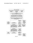 |
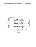 |  |
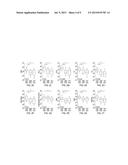 |  |
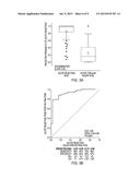 | 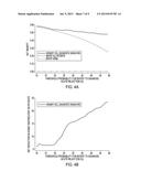 |
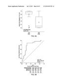 | 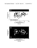 |
 |  |
 |  |
 |  |
 |  |
 |  |
 |  |
 |  |
 |  |
 |  |
 |  |
 |  |
 |  |
 |  |
 |  |
 |  |
 |  |
 |  |
 |  |
 |  |
 |  |
 |
| New patent applications in this class: | |
| Date | Title |
|---|---|
| 2022-09-08 | Shrub rose plant named 'vlr003' |
| 2022-08-25 | Cherry tree named 'v84031' |
| 2022-08-25 | Miniature rose plant named 'poulty026' |
| 2022-08-25 | Information processing system and information processing method |
| 2022-08-25 | Data reassembly method and apparatus |
| New patent applications from these inventors: | |
| Date | Title |
|---|---|
| 2015-08-27 | Non-invasive method of diagnosing renal fibrosis |
| 2015-03-26 | Methods of predicting acute rejection outcomes |
| 2014-07-31 | Methods to detect, treat and prevent acute cellular rejection in kidney allografts |