Patent application title: DIAGNOSIS METHOD OF ACTIVE TUBERCULOSIS
Inventors:
Edouard Tuaillon (Castelnau-Le-Lez, FR)
Pierre-Alain Rubbo (Montpellier, FR)
Stephane Canaan (Marseille, FR)
Laurent Kremer (Montpellier, FR)
Nicolas Nagot (Prades Le Lez, FR)
Philippe Van De Perre (Saint Vincent De Barbeyrargues, FR)
Jean-Pierre Vendrell (Castelnau-Le-Lez, FR)
Assignees:
UNIVERSITE MONTPELLIER 2 SCIENCES ET TECHNIQUES
CENTRE NATIONAL DE LA RECHERCHE SCIENTIFIQUE
CENTRE HOSPTALER UNIVERSITAIRE DE MONTPELLIER
Universite Montpellier 1
IPC8 Class: AG01N33569FI
USPC Class:
435 724
Class name: Involving a micro-organism or cell membrane bound antigen or cell membrane bound receptor or cell membrane bound antibody or microbial lysate animal cell leukocyte (e.g., lymphocyte, granulocyte, monocyte, etc.)
Publication date: 2014-08-07
Patent application number: 20140220599
Abstract:
The present invention relates to a method for the in vitro diagnosis of
active tuberculosis, comprising a step of contacting lymphocytes of a
patient suspected to have active tuberculosis with at least one protein
of mycobacteria, said protein being an enzyme having a lipolytic
activity, and a step of detecting the presence of specific activated
lymphocytes.Claims:
1-10. (canceled)
11. A method for the diagnosis of active tuberculosis, comprising: a step of contacting a sample of purified B lymphocytes of a patient suspected to have an active tuberculosis with at least one protein of mycobacteria, said protein being an enzyme having a lipolytic activity and being able to hydrolyse lipids having a carbohydrate chain comprising at least 12 carbons, said sample being devoid of serum or of any component liable to contain soluble antibodies, a step of incubation during a determined time to allow the purified B lymphocytes to secrete specific antibodies directed against said enzyme having a lipolytic activity, and a step of detecting the presence of an immune complex comprising said antibodies and said enzyme having a lipolytic activity.
12. The method for diagnosis of active tuberculosis according to claim 11, wherein said enzyme having a lipolytic activity is chosen among the group consisting of: the monoacylglycerol hydrolase Rv0183 as set forth in SEQ ID NO: 4, a phospholipase, the lipase Rv1984c as set forth in SEQ ID NO: 2, and a triacylglycerol hydrolase, preferably LipY.
13. The method for diagnosis of active tuberculosis according to claim 12, wherein said phospholipase belongs to the group consisting of Rv3452 as set forth in SEQ ID NO: 1, Rv3802c, PlcA as set forth in SEQ ID NO: 5, PlcB as set forth in SEQ ID NO: 6, PlcC as set forth in SEQ ID NO: 8 and PlcD as set forth in SEQ 10 NO: 8 or 9.
14. The method for diagnosis of active tuberculosis according to claim 12, wherein said triacylglycerol hydrolase is LipY as set forth in SEQ ID NO: 3.
15. The method for the diagnosis of active tuberculosis according to claim 11, wherein B lymphocytes are chosen among plasmablasts and plasmocytes.
16. The method for the diagnosis of active tuberculosis according to claim 11, wherein the step of detecting the presence of activated B lymphocytes is carried out by an immunological detection.
17. The method for the diagnosis of active tuberculosis according to claim 11, wherein said determined time is from 1 hour to 72 hours, preferably from 6 hours to 48 hours, more preferably from 16 h to 30 hours.
18. A kit for the detection of active tuberculosis comprising: at least one protein of mycobacteria immobilised on a support, said protein being an enzyme having a lipolytic activity, antibodies, possibly labelled, directed against constant chain of immunoglobulins, and means for specifically purifying B lymphocytes.
19. The kit according to claim 18, wherein said enzyme having a lipolityc activity are able to hydrolyse lipids having a carbohydrate chain comprising at least 12 carbons.
20. The kit according claim 18, wherein said enzyme having a lipolytic activity is chosen among the group consisting of: a monoacylglycerol hydrolase, Rv0183, a phospholipase, preferably Rv3452, Rv3802c, PlcA, PlcB, PlcC or PlcD, a lipase, preferably Rv1984c, and a triacylglycerol hydrolase, preferably LipY.
21. The kit according to claim 20, wherein said phospholipase belongs to the group consisting of Rv3452 as set forth in SEQ ID NO: 1, Rv3802c, PlcA as set forth in SEQ ID NO: 5, PlcB as set forth in SEQ ID NO: 6, PlcC as set forth in SEQ ID NO: 8 and PlcD as set forth in SEQ ID NO: 8 or 9.
22. The kit according to claim 20, wherein said triacylglycerol hydrolase is LipY as set forth in SEQ ID NO: 3.
23. The kit according to claim 18, wherein said means specifically purifying B-lymphocytes are chosen among specific B cell specific antibodies, or density gradients, or a combination of the above.
Description:
CROSS REFERENCE TO RELATED APPLICATIONS
[0001] This application is a 371 application of International Application No. PCT/EP2012/080437, filed Jun. 1, 2012, which claims priority to European Patent Application EP11168643.2 filed Jun. 3, 2011, all of which are incorporated by reference in its entirety herein.
DESCRIPTION
[0002] The inventions relates to a diagnosis method of active tuberculosis.
[0003] Human tuberculosis (TB) is a highly contagious bacterial infection that is passed from person to person through the air. It is usually spread through contact with an infected person who is actively coughing or talking. TB disease is caused when the bacteria multiply inside the body, causing tissue and organ damage. Without treatment, half of those with TB disease will die.
[0004] Most people know TB as a disease of the lungs. However, not all TB disease is in the lungs. Around 40% of TB disease occurs in another part of the body. This happens when the bacteria spread outside of the lungs. In these cases, extrapulmonary TB is more difficult to diagnose since the patient does not have normal signs and symptoms associated with pulmonary TB. TB disease can also occur in the lymph glands, brain, spine, bones, kidneys, or other organs.
[0005] There are two forms of TB infection: active TB, also known as TB disease, and latent TB infection (LTBI).
[0006] A person who is TB-infected may not necessarily develop the disease. Some are able to control the infection, but unable to completely remove it from their bodies. In these cases, the infection remains, lying in an inactive state within granulomas. This is often described as LTBI. People with LTBI do not usually show any signs of TB symptoms so it can go unnoticed. LTBI may develop into active disease someday, often when the person's immune system becomes weakened. In fact, healthy people who are infected with latent TB have about 10% risk of converting to active disease each year over their lifetime. Thus, active TB is generally considered as the reactivation of mycobacteria from a latent form. Since this latent infection can become active at any time, it is usually monitored but not necessarily treated. By contrast, active TB that is the critical form of the disease and easily spread to other individuals, must be rapidly treated with up to four antibiotics during several months. Therefore, distinction between active and latent TB remains essential for the therapeutic care of infected patients.
Vaccination
[0007] An effective TB vaccine would be a major advance in the control of TB, but to date no such vaccine exists. The BCG vaccine is used in some countries and while this is thought to offer some protection against TB infection in children it is considered to have limited benefits in adults (around 50% protection). Additionally, BCG-vacinated people and individuals infected with nontuberculous mycobacteria (mycobacteria other than M. tuberculosis) may produce a positive tuberculin skin test (TST) result, even if they are not infected. These are the two most common causes of false positive TST results and can lead to unnecessary treatment for LTBI.
[0008] So there is a need to provide a diagnosis method with an optimal sensitivity, and well differentiates patient having an effective active TB.
Diagnosing Active TB
[0009] 1. Active TB can be difficult to diagnose, especially in children, those who have weakened immune systems or those developing extrapulmonary TB. Beside medical examinations, the following tests used to determine if a patient has active TB:
[0010] Tuberculin Skin Test (TST),
[0011] Chest Radiograph (X-ray),
[0012] Sputum Smear Microscopy (SSM),
[0013] Culture, and
[0014] PGR (GeneXpert®).
[0015] Tuberculin Skin Test (TST)
[0016] The TST has been in existence for over 100 years. A suspension containing TB proteins is injected into the derma in the lower part of the arm. The injection site is examined by a trained healthcare professional 2-3 days later. If the person has TB, the body recognizes the proteins that were injected and responds by forming a lump where the TB proteins were injected.
[0017] The accuracy of the TST varies and can be affected by a previous TB vaccination (BCG), infection with nontuberculous bacteria, a weakened immune system and by other illnesses or medical treatments,
[0018] Chest X-rays and Tomodensitometry
[0019] Chest X-rays are used to check for lung abnormalities in people who have signs and symptoms of TB disease in the lungs. Although chest X-rays may suggest that TB disease is present, a chest X-ray alone cannot definitely diagnose a TB infection in the lungs or anywhere else in the body.
[0020] Sputum Smear Microscopy (SSM)
[0021] This is a simple laboratory test that examines sputum for bacteria using a microscope. This test also identifies non-TB bacteria, so it cannot always distinguish between TB and other infections. It is commonly used to diagnose TB disease because it can quickly determine if a person is infected. However, it sometimes gives a negative result even in people with TB disease so a negative result cannot be relied upon.
[0022] Culture
[0023] Culture techniques can be used to grow live TB bacteria in a laboratory. This is the standard reference method for detecting active TB as long as a suitable sample containing the TB bacteria can be obtained. TB can be cultured from a variety of specimens. This test can also provide information on which antibiotics would be effective in treating the infection. A major drawback of this test is the length of time it takes to get the results back (2-12 weeks) and the need to repeat the experiment from similar specimens of the same patient in order to obtain a positive result (i.e. poor sensitivity).
The GeneXpert® System
[0024] The GeneXpert® System is a closed and automated diagnostic platform that allow molecular identification of M. tuberculosis as well as resistance to rifampicin. It provides results from unprocessed sputum samples with limited technical training required for operators. This test has several limitations such as limited shelf-life of the diagnostic cartridges, some operating temperature and humidity restrictions, requirement for electricity supply, the need for annual servicing and calibration of each machine, the cost of the machine and each TB test as well as the need to multiply tests for increasing sensitivity.
[0025] Other tests have been developed using blood samples in order to diagnose active TB. As illustration, the following documents can be cited:
[0026] The international application WO 2009/129521 discloses the use of specific epitopes of mycobacterial proteins in order to specifically identify patient afflicted by active TB. Three proteins have been identified as harboring efficient epitopes: PTRP, PE-PGRS51 and LipC.
[0027] Zhang et al., Clin Microbiol Infect 2007; 13: 139-145, disclose that the comparative immunoreactivity of Rv3425, ESAT-6 and CFP-10 clearly distinguished between healthy subjects and TB patients. Moreover, Zhang et al. disclose that negligible antibody response obtained in the BCG-vaccinated healthy control group suggests that Rv3425, ESAT-6 and CFP-10 can be used for diagnosis of M. tuberculosis infection, even in geographical areas in which BCG vaccination is used routinely.
[0028] However, none of the methods disclosed in the art allows the practitioner to determine rapidly after the reactivation of the bacteria, the active TB in a patient, with a low amount of false-positive results.
[0029] Therefore, the invention proposes to overcome the above inconvenient.
[0030] One aim of the invention is to provide a new efficient method to precociously detect reactivation of mycobacteria.
[0031] Another aim of the invention is to provide an efficient method to distinguish active TB from LTBI.
[0032] Still another aim of the invention is to provide kits to carry out the method according to the invention.
[0033] The invention relates to a method for the in vitro diagnosis of active TB, comprising
[0034] a step of contacting lymphocytes of a patient suspected to have active TB with at least one mycobacterial protein, said protein being an enzyme having a lipolytic activity, and
[0035] a step of defecting the presence of specific activated lymphocytes.
[0036] The invention also relates to a Method for the in vitro diagnosis of active tuberculosis, comprising:
[0037] a step of contacting a sample of purified B lymphocytes of a patient suspected to have an active tuberculosis with at least one protein of mycobacteria, said protein being an enzyme having a lipolytic activity and being able to hydrolyse lipids having a carbohydrate chain comprising at least 12 carbons,
[0038] said sample being devoid of serum or of any component liable to contain soluble antibodies,
[0039] a step of incubation during a determined time to allow the purified B lymphocytes to secrete specific antibodies directed against said enzyme having a lipolytic activity, and
[0040] a step of detecting the presence of an immune complex comprising said antibodies and said enzyme having a lipolytic activity.
[0041] The invention is based on the unexpected observation made by the Inventors that mycobacterial enzymes expressing a lipolytic activity are powerful candidates for the enablement of a specific method allowing to discriminate active tuberculosis from latent tuberculosis.
[0042] The diagnosis proposed by the invention can be carried out in human but also in animals.
[0043] Hereafter, tuberculosis will be uniformly written "tuberculosis" or "TB".
[0044] In the invention, the term "lipase" is used to designate enzyme having lipolytic activity and include, phospholipases A, B, C or D, and lipases including triglyceride lipases, diglyceride lipases, or monoglyceride lipases.
[0045] During infection, Mycobacterium tuberculosis accumulates intracellular lipid-loaded inclusion bodies whose lipids probably originate from the host cell membrane degradation. There is now strong evidence supporting that fatty acids also represent a source of carbon during dormancy. Mycobacterium tuberculosis stores fatty acids in the form of triacylglycerol (TAG) as it enters in the nonreplicating persistence stage (Latent stage). Moreover, granulomas have been found to contain foamy macrophages that are cells bearing large amounts of neutral lipids surrounded by phospholipids in their cytoplasm. These lipid bodies are induced by the internalisation of bacteria and therefor providing a caron source for the survival and the reactivation of the pathogen. Overall, these findings support the view that enzymes involved in lipid degradation may fulfil important physiological functions and may participate in the extraordinary capacity of survival and reactivation of Mycobacterium tuberculosis from the infected host. Degradation of host lipis by Mycobacterium tuberculosis is likely to be performed by lipolytic enzymes, such lipases and phospholipases, including the cutinase family enzymes.
[0046] Lipases are water-soluble proteins having lipolytic activity belonging to esterase group and catalyzing the hydrolysis of insoluble substrates in water like triacylglycerol and phospholipids ester bonds [Alberghina, et al. (1991) Lipases; Structure, Mechanism and Genetic Engineering, Vol. 16, VCH, Weinheim, Fed Rep. Ger.; Wooley P., and Petersen, S. B. (1994) Lipases; their structure, biochemistry and applications, Cambridge Univerity Press, Cambridge; Brockerhoff, H., and Jensen, R. G. (1974) Lipolytic Enzymes, Academic Press, New York; Borgstrom, B., and Erlanson, C. (1984) Lipases, Elsevier, Amsterdam]. In this context, the catalytic reaction of lipolysis involves various interfacial processes and depends strongly on the structure of the lipid substrates present in oil-in-water emulsions, membrane bilayers, monolayers, micelles, and vesicles [Aloulou, et al. (2006) Biochim. Biophys. Acta-Molecular and Cell Biology of Lipids 1761, 995-1013]. The catalytic process can be described as a reversible lipase adsorption/desorption step occurring at the oil/water interface, followed by the formation of an interfacial enzyme substrate complex and the release of lipolysis products [Verger, R., et al. (1973) J. Biol. Chem. 248, 4023-4034; Panaitov, I., and Verger, R. (2000) Enzymatic reactions at inerfaces: Interfacial and temporal organization of enzymatic lipolysis, in Physical Chemistry of Biological Interfaces (Baszkin, A, and Norde; W., Eds.), pp 359-400, Marcel Dekker. Inc, New York, Base].
[0047] Among the M. tuberculosis identified lipases, 24 have been classified in a family of enzymes called the "Lip family". However, this classification is only based on the presence of the consensus sequence GXSXG, which is characteristic of esterases and members of the α/β hydrolase fold family. This classification does not allow the distinction between lipolytic enzymes (lipase) and non lipolytic enzymes (esterase).
[0048] For instance, LipH [Canaan, S. et al. (2004) Eur. J. Biochem. 271, 3953-3961], LipF [Zhang, M. et al. (2005) Protein Expr Purif 42, 59-66] and LipC (see example 1) were cloned and characterized but no long-chain triglycerides (TG) hydrolysis was detected.
[0049] On the contrary, the Inventors have demonstrated that "real" lipases, i.e. proteins having a long-chain hydrolysis activity, are useful for specifically diagnosing active TB, without cross-reaction with Latent TB samples. This includes the real lipase belonging to the Lip family, and any other real lipases that do not belong to the Lip family [Cotes K. et al. (2007) Biochem J. 408(3), 417-427].
[0050] The Inventors have hypothesized that lipolytic activity is high during the reactivation of the bacteria, thus leading to the method according to the invention which allows to specifically identify the reactivation only, i.e. the active TB.
[0051] On the contrary, in healthy individuals, or patient/individuals having LTBI, the immune system has not been in presence of the lipases according to the invention. Consequently, these healthy individuals or patient/individuals having LTBI would never been detected with the method according to the invention.
[0052] Therefore, the above mentioned lipases are used to carry out a method of diagnosis, preferably in vitro.
[0053] The method according to the invention is based on the detection of activated lymphocytes. "Activated lymphocytes" are single lymphocytes that are not naive, i.e. lymphocytes that have been previously stimulated by specific antigens. This activation is accompanied by morphologic changes known as lymphocyte transformation, and the ability to secrete active molecules such as antibodies, cytokines, growth or death factors. These mechanisms of lymphocytes activation are well known in the art.
[0054] In a first step of the method according to the invention, activated lymphocytes of an individual, or a patient, suspected to have an active TB, are purified and contacted with at least one lipase of mycobacteria, preferably from Mycobacterium tuberculosis, said lipase having, as mentioned above, a lipolytic activity, or a biochemically identified lipolytic activity.
[0055] As mentioned above, LipC, LipH and LipF, which have not such lipase activity, are excluded de facto from the method according to the invention. Moreover, proteins from Mycobacterium, in particular from Mycobacterium tuberculosis, having no lipolytic activity are also excluded.
[0056] In a second step of the method according to the invention, the lipase-specific lymphocytes are detected, by means described hereafter.
[0057] It is clear for one having ordinary skill that the "activated lymphocytes" according to the invention are specific of the protein having a lipolytic activity. In other word, lymphocytes that have been activated by another protein devoid of lipolytic activity, would never been detected by the method according to the invention.
[0058] Thus, following the contacting of the lymphocytes of said patient/individual suspected to have active TB, and the detection of the active lymphocytes, the pathologist is able to determine if said patient/individual has active TB.
[0059] For instance, if no active lymphocytes are detected, it is possible to conclude that said patient is not afflicted by active TB. On the contrary, if activated lymphocytes are detected, the conclusion is that said patient/individual has active TB.
[0060] In the last case, it may also be possible, by quantifying the amount of active lymphocytes, to determine approximately the period from which bacteria have been reactivated.
[0061] On the contrary to the immunological standard method used in the art, which are based on the detection of the presence of antibodies in serum of patient/individual suspected to be afflicted by active TB, the method according to the invention allows a most precocious detection.
[0062] This precocity is due to the fact that activated lymphocytes are the first to be present in blood, prior to produce detectable compounds, such as antibodies, that can be detected by immunological techniques. In addition, detection of those activated lymphocytes specific for lipases involved in the reactivation of bacteria clearly permit the characterization of the active state of the disease. By contrast, detection of antibodies, such as in ELISA, does not necessarily reflect the active disease occurring at the time of detection but only a past exposition without indication of time since secreted antibodies after immunization could have a long half-life. Detection of such a low-level serological response to TB is caused by a previous exposure to M. tuberculosis sometime in the past and is called "serological scar". Therefore, the short half-life of these enzymes-specific activated lymphocytes circulating in the blood only during active TB, by contrast to antibodies, combined to the preferential production of some of the lipases used in the invention allow the precocious diagnostic of active TB.
[0063] In one advantageous embodiment, the invention relates to the method for the in vitro diagnosis of active TB as defined above, wherein said lymphocytes are B lymphocytes or T lymphocytes.
[0064] In the method according to the invention, the lymphocytes, and the activated lymphocytes are preferably B lymphocytes, secreting antibodies, and effector memory T lymphocytes, in particular T CD4+ and CD8+.
[0065] Another advantageous embodiment of the invention relates to the method for the in vitro diagnosis of active TB previously defined, wherein said enzyme having a lipolytic are able to hydrolyse lipids having a carbohydrate chain comprising at least 12 carbons.
[0066] The lipase according to the invention are the refore able to hydrolyse lipids having a carbohydrate chain comprising at least 12 carbons, preferably at least 14 carbons, in particular at least 16 carbons. At most, the carbohydrate chain has about 30 carbons.
[0067] The lipids defined in the invention are chosen among fatty acyls, glycerolipids, glycerophospholipids, sphingolipids, . . .
[0068] These carbohydrates chains contained in the lipids are well known in the art, and can be mono or polysaturated, or not.
[0069] In one another advantageous embodiment, the invention relates to the method for the in vitro diagnosis of active TB as defined above, wherein said enzyme having a lipolytic activity is chosen among the group consisting of
[0070] a phospholipase, preferably Rv3452, Rv3802c, PlcA, PlcB, PlcC or PlcD.
[0071] a lipase, preferably the lipase Rv1984c,
[0072] a triacylglycerol hydrolase, preferably LipY, and
[0073] a monoacylglycerol hydrolase, Rv0183.
[0074] The most advantageous lipases according to the invention are:
[0075] the protein having a phospholipase activity Rv3452, represented by the amino acid sequence SEQ ID NO: 1,
[0076] the protein having a lipase activity Rv1984c, represented by the amino acid sequence SEQ ID NO: 2,
[0077] the triacylglycerol hydrolase LipY (Rv3097c), represented by the amino acid sequence SEQ ID NO: 3,
[0078] the monoacylglycerol hydrolase Rv0183 represented by the amino acid sequence SEQ ID NO; 4,
[0079] the protein having a phospholipase activity PlcA (also named Rv2351c), represented by the amino acid sequence SEQ ID NO: 5.
[0080] the protein having a phospholipase activity PlcB (also named Rv2350c), represented by the amino acid sequence SEQ ID NO: 6,
[0081] the protein having a phospholipase activity PlcC (also named Rv2349c), represented by the amino acid sequence SEQ ID NO: 7, and
[0082] the protein having a phospholipase activity PlcD, represented by the amino acid sequence SEQ ID NO: 9 or the amino acid sequence SEQ ID NO: 8.
[0083] The above sequences correspond to the protein of Mycobacterium tuberculosis. However, the invention also encompasses peptides from other species of Mycobacterium involved in tuberculosis, such as Mycobacterium kansasii, Mycobacterium marinum, Mycobacterium ulcerans, Mycobacterium avium, Mycobacterium avium subsp. avium, Mycobacterium avium subsp. paratuberculosis, Mycobacterium parascrofulaceum, Mycobacterium intracellulare, Mycobacterium leprae and Mycobacterium bovis.
[0084] In one other embodiment of the invention, fragments of the above peptides harbouring immunogenic epitopes can be used in the method described above and hereafter.
[0085] The invention also encompasses variants of the above proteins, having a sequence identity of at least 75%, preferably at least 80%, more preferably at least 90%.
[0086] One another advantageous embodiment of the invention relates to the method for the in vitro diagnosis of active TB previously defined, wherein said lymphocytes are B lymphocytes, in particular B lymphocytes chosen among circulating plasmablasts and plasmocytes, said B lymphocytes secreting antibodies specifically directed against said enzyme having a lipolytic activity.
[0087] In this advantageous embodiment of the method according to the invention, B lymphocytes are detected.
[0088] This advantageous embodiment corresponds to an ELISpot (B-ELISpot, or ELISPOT B) technique wherein B cells expressing antibodies specifically directed against the lipase are detected.
[0089] The details of the method are disclosed in the Examples.
[0090] Briefly, the method consists to contact the lipase according to the invention with purified B lymphocytes from the patient/individual suspected to have active TB.
[0091] The purification of B lymphocytes can be achieved by any techniques well known in the art, for instance, by using the protocol disclosed in Greaves and Brown [Greaves and Brown. 1974, The Journal of Immunology, vol. 112 no. 1 420-42], or any adaptations thereof. Another protocol is disclosed in the Example section.
[0092] Preferably, the invention relates to the method for the in vitro diagnosis of active TB as defined above, wherein the step of detecting the presence of in vivo activated lymphocytes consists of detecting the secretion of said antibodies specifically directed against said enzyme having a lipolytic activity.
[0093] Advantageously, the method according to the invention allows the detection of activated B lymphocytes, by measuring their ability to secrete specific antibodies.
[0094] In order to limit interferences, or false positive, the B lymphocytes used in the method according to the invention are purified, and isolated from blood.
[0095] By "purified" it is meant in the invention that the B lymphocytes are isolated from the other cells contained in blood. The methods allowing the purification of B lymphocytes are well known in the art, and include immunological positive and/or negative selections (positive selection with specific marker expressed on B cells; negative selection by eliminating non B-Lymphocytes ceils . . . ).
[0096] A very important step during the isolation of the B lymphocytes is to eliminate any residual traces of serum, or fluid that is liable to contain antibodies. Repetitions of washing steps using ad hoc buffer, or separation on a sucrose gradient, or a combination of both, can be used.
[0097] The rosette method as described in Example 4 is particularly appropriated.
[0098] As mentioned above, the lymphocytes are obtained from blood of the patient/individual. In blood, it is possible to distinguish some B lymphocytes:
[0099] the naive B lymphocytes (that have never been activated by any antigen),
[0100] the memory B lymphocytes (that have been activated by one antigen during an ancient infection),
[0101] the transient B lymphocytes (incompletely differentiated naive cells), and
[0102] the plasmablasts and the plasmocytes (that secrete antibodies), plasmablasts being detectable precociously during an infectious episode.
[0103] In vivo, if the patient/individual has a reactivation of the mycobacteria, lipase according to the invention will be expressed and accessible to the immune system. Then, B cells will be activated in order to produce specific antibodies directed against the lipase which have activated them.
[0104] The purified lymphocytes are then contacted with at least one lipase according to the invention for a determined period.
[0105] More advantageously, the invention relates to the method for the in vitro diagnosis of active TB as defined above, wherein said secretion is detected by measuring the formation of an immune complex between said antibodies and said enzyme having a lipolytic activity.
[0106] As mentioned above, the purified B lymphocytes are contacted with at least one lipase according to the invention. Thus, all the B lymphocytes able to produce antibodies will secrete these antibodies in the medium. Only antibodies that are specific to the lipase will form an immune complex (lipase/antibody immune complex), the other antibodies present being not able to form such immune complex.
[0107] A step of the method consists to remove the B lymphocytes, and the medium comprising them and the secreted antibodies. Then, only the lipase/antibody complex will be present. An specific immune complex is formed and can be defected.
[0108] Therefore, by common techniques allowing the detection of an immune complex, it would be possible to determine, and to quantify, the presence of such complex.
[0109] In the case of a bacterial reactivation, i.e. active TB, the method according to the invention would detect such immune complex. On the contrary, in the case of a healthy individual, or an individual having latent tuberculosis, no complex would be detected.
[0110] To summarize, the above method can be carry out as follows:
[0111] 1. lipases according to the Invention are immobilized on a support.
[0112] 2. purified B lymphocytes are contacted with said immobilized lipases to allow the formation of a binary lipase/anti lipase antibody complex.
[0113] 3. after an appropriate incubation (from 1 h to 72 hours), lymphocytes are removed and the immobilized lipases are washed at least one time.
[0114] 4. labelled antibodies directed against human Fc fragments are contacted with the immobilized lipases.
[0115] 5. The ternary lipase/anti-lipase antibody/anti-Fc antibody complex is detected by appropriate means corresponding to the labelling molecule fixed on the anti-Fc antibody.
[0116] The step 3 mentioned above, the B lymphocytes are incubated with the immobilized enzyme having a lipolytic activity from about 1 hour to about 72 hours, preferably from about 24 hours to about 48 hours, in particular from about 16 hours to about 30 hours.
[0117] The above mentioned incubation times should no be too longer in order to limit the detection of memory B lymphocytes, which are present in a very low amount, but remain liable to secrete antibodies.
[0118] In still another advantageous embodiment the invention relates to the method for the in vitro diagnosis of active TB previously defined, wherein said lymphocytes are T lymphocytes, said lymphocytes secreting biomarkers such as cytokines, chemokines or growth factors after their activation.
[0119] In this advantageous embodiment of the method according to the invention, T lymphocytes are detected.
[0120] This advantageous embodiment corresponds to an ELISpot (T-ELISpot or ELISpot T) technique wherein T cells activated by the presentation of antigens corresponding to the lipases according to the invention are detected.
[0121] The details of the method are disclosed in the Examples.
[0122] Briefly, the method consists to contact the lipase according to the invention with purified peripheral blood mononuclear cells(PBMC) from the patient/individual suspected to have active TB.
[0123] The purification of PBMC can be achieved by any techniques well known in the art, for instance, by using gradient such as Ficoll® or Histopack® or any adaptations thereof. A protocol is disclosed in the Example section.
[0124] Advantageously, the invention relates to the method for the in vitro diagnosis of active TB above-described, wherein the step of detecting the presence of activated lymphocytes consists of detecting the secretion of at least one biomarker such as cytokine, chemokine or growth factor, said lymphocytes being activated by said enzyme having a lipolytic activity.
[0125] Following T cell development, matured, naive T cells express the T ceil receptor (TCR)-CD3 complex. The TCR has an affinity for Major Histo Compatibility Complex (MHC) molecules.
[0126] Class II MHC proteins are generally found on the surface of specialised antigen-presenting cells (APCs), said Class II MHC molecules expressing parts of the peptides originating from exogenous bacteria or viruses. Class I MHC proteins are generally found on every nucleated cell of the body, said Class I MHC molecules expressing part of the peptides originating from degradation of cytosolic foreign proteins.
[0127] During an immune response, APCs cells express at their surface MHC molecules containing antigens of bacteria. Naive T lymphocytes having a TCR complementary to the class MHC molecule-presenting antigen are then activated. This is the first step of the activation.
[0128] T cells having received the first signal have to be activated by a second signal implying surface co-stimulatory proteins involved in the MHC-TCR complex.
[0129] When the second signal is received, the T lymphocytes are then activated which allows itself to proliferate. Activated T ceils become effector or auxiliary lymphocytes secreting informative transmitters (cytokines, interferons, growth factors, chemokines . . . ).
[0130] When purifying PBMC, the antigen-experienced effector T lymphocytes are the predominant source of the early production of molecules (cytokines, interferon's, growth factors, chemokines, . . . ) following ex vivo antigen re-stimulation in the invention.
[0131] When purifying PBMC, the T lymphocytes having received the first signal are present, and can be stimulated in vitro by the antigen.
[0132] Thus, if the lipases according to the invention are contacted to the purified PBMC, only sensitized T lymphocytes that have been previously activated and differentiated Into effector cells can therefore produce the biomarkers corresponding to informative transmitters (cytokines, interferons, growth factors, chemokines . . . ).
[0133] Thus, if specific biomarkers are detected, that means that the PBMC stimulated in vitro by the lipases according to the invention contain active effector T lymphocytes, said T lymphocytes being activated and sensitized in vivo by APC cells expressing in their MHC molecules parts of said lipases. Measuring the presence of such specific effector T cells therefore diagnoses an ongoing infection, i.e. the bacteria have been reactivated.
[0134] On the contrary, if no specific biomarker is detected, that means that the PBMC do not contain lipase-specific effector T lymphocytes, i.e. no reactivation of the bacteria has occurred in vivo.
[0135] In a particularly advantageous embodiment, the invention relates to the method for the in vitro diagnosis of active TB defined above, wherein said secretion is detected by measuring the formation of an immune complex between at least one biomarker such as cytokine, chemokine or growth factor secreted by said activated T lymphocytes and antibodies specifically directed against said at least one biomarker such as cytokine, chemokine or growth factor.
[0136] Proteins such as IFN-γ, IP-10 (also known as CXCL10), IL-2, MCP-1 and IL-1RA are released in response to Mycobacterium tuberculosis antigen stimulation.
[0137] Therefore, the above biomarkers, and any other markers known in the art, can be detected with the method according to the invention as described above.
[0138] in one another advantageous embodiment, the invention relates to the method defined above, wherein said step of detecting the presence of activated lymphocytes is carried out by an immunological detection.
[0139] As mentioned above, activated T lymphocytes are able to produce/secrete biomarkers in the medium. Only antibodies that are specific to the said biomarkers will form an immune complex (biomarker/antibody complex), the other biomarkers present being not able to form such immune complex.
[0140] A step of the method consists to remove the T lymphocytes, and the medium comprising them and the secreted biomarkers. Then, only the biomarker/antibody complex will be present.
[0141] Therefore, by common techniques allowing the detection of an immune complex, it would be possible to determine, and to quantify, the presence of such complex.
[0142] In the case of active TB, the method according to the invention would defect such immune complex. On the contrary, in the case of a healthy individual, or an individual having LTBI, no complex would be detected.
[0143] To summarize, the above method can be carry out as follows:
[0144] 1. Antibodies directed against specific biomarkers are immobilized on a support.
[0145] 2. Purified PBMC are contacted with said immobilized antibodies directed against specific biomarkers to allow the formation of a binary biomarker/antibodies complex directed against specific biomarkers that will be secreted after lipase-speoific stimulation.
[0146] 3. Lipase antigens according to the invention is added to PBMC.
[0147] 4. After an appropriate incubation (from about 24 hours to about 6 days), PBMC are removed and the immobilized antibodies directed against specific biomarkers are washed at least one time.
[0148] 5. Labelled antibodies directed against said biomarker are contacted with the immobilized antibodies directed against specific biomarkers.
[0149] 6. The ternary complex antibodies directed against specific biomarkers-biomarker-labelled antibody anti-biomarker is detected by appropriate means corresponding to the labelling molecule fixed on the antibody anti-biomarker.
[0150] In both ELISpot disclosed above, the skilled person knows how to choose the appropriate labelling molecule to detect the ternary complex.
[0151] The invention also relates to a kit for the in vitro detection of active TB comprising
[0152] 1. at least one protein of mycobacteria immobilised on a support, said protein being an enzyme having a lipolytic activity, and
[0153] 2. antibodies, possibly labelled, directed against constant chain of immunoglobulins.
[0154] Advantageously, the invention relates to a kit for the in vitro detection of active tuberculosis comprising:
[0155] at least one protein of mycobacteria immobilized on a support, said protein being an enzyme having a lipolytic activity, antibodies, possibly labelled, directed against constant chain of immunoglobulins, and
[0156] means for specifically purifying B lymphocytes.
[0157] The kit may also further comprise positive and/or negative controls.
[0158] Support according to the kit described above and hereafter are for instance micro-titration plates, Elisa plates, plastic, Polyvinylidene Fluoride (PVDF) membrane. . . . The skilled person can easily determine the appropriate support.
[0159] For the detection procedure, antibodies used for the detection are usually labelled with a marker. Markers used for the labeling of the antibodies are chosen among markers commonly used by the skilled man in the art, and in particular are chosen among radio-isotopic marker, enzymes, fluorescent agents, luminescent agents, magnetic particles. . . .
[0160] The invention also relates to a kit for the in vitro detection of active TB comprising
[0161] 1. at least one protein of mycobacteria said protein being an enzyme having a lipolytic activity, and
[0162] 2. antibodies, immobilized on a support, directed against at least one blomarker secreted by activated T lymphocytes,
[0163] Advantageously, the invention relates to a kit as defined above, wherein said enzyme having a lipolytic activity are able to hydrolyse lipids having a carbohydrate chain comprising at least 12 carbons.
[0164] More advantageously, the invention relates to a kit above defined, wherein said enzyme having a lipolytic activity is chosen among the group consisting of:
[0165] a monoacylglycerol hydrolase, Rv0183,
[0166] a phospholipase, preferably Rv3452, Rv3802c, PlcA, PlcB, PlcC or PlcD,
[0167] a lipase, preferably Rv1984c, and
[0168] a triacylglycerol hydrolase, preferably LipY.
[0169] Another advantageous embodiment of the invention related to a kit according as defined above, wherein said means specifically purifying B-lymphocytes are chosen among specific B cell specific antibodies, or density gradients, or a combination of the above.
[0170] The kit may also further comprise positive and/or negative controls, such as cells activated by polyclonal activator compounds or cultivated in cell culture media.
[0171] Advantageously, the invention relates to the kits above-defined, wherein said enzyme having a lipolytic activity are able to hydrolyse lipids having a carbohydrate chain comprising at least 12 carbons.
[0172] In particular, the invention relates to the kits previously defined, wherein said enzyme having a lipolytic activity is chosen among the group consisting of
[0173] a phospholipase. preferably Rv3452, Rv3802c, PlcA, PlcB, PlcC or PlcD,
[0174] a lipase, preferably Rv1984c,
[0175] a triacylglycerol hydrolase, preferably LipY, and
[0176] a monoacylglycerol hydrolase, Rv0183.
[0177] The invention will be better explained by the following examples and following FIGS. 1 to 3.
LEGEND TO THE FIGURES
[0178] FIGS. 1A and B represent the map of the pfasmid pSD24 obtained from pSD26.
[0179] FIG. 1A represents the map of the plasmid pSD24 wherein restriction site are indicated.
[0180] FIG. 1B represents the multicloning site (MCS) of the pSD26 vector.
[0181] FIG. 2 represents a curve demonstrating the lipase activity of LipY. The graph represents the degradation of para-Nitrophenyl state with different amounts of protein. X-axis represents the amount of proteins (μg of cell wall); Y-axis represents the absorbance at 405 nm. Triangles: control mycobacteria. Circles: mycobacteria overexpression LipY
[0182] FIG. 3 represents the results obtained with an ELISpot-B using LipY antigen.
[0183] 1. represents the patients with a latent tuberculosis (IGRA positive) and 2, the patients with active TB. Y-axis represents the antibodies secreting cells/106 B lymphocytes.
EXAMPLES
Example 1
LipC Is Not a Lipase per se, Whereas LipY is
[0184] Comparison of the lipase activity of Rv0220 (LIPC) and a cutinase from F. solani on paranitrophenyl ester family.
[0185] The activity of rLipC or cutinases was measured using p-nitrophenyl esters family (Sigma) with carbon chain lengths ranging from C2 to C14. Release of p-NP was monitored at 410 nm using a 96-well plates spectrophotomer and quantified using a calibration curve of pNP (ε.sub.(λ=410 nm)=30 mM-1). Enzymatic reactions were performed in a 2.5 mM Tris buffer pH 8.0 containing 300 mM NaCl and 4 mM NaTDC at 357° C. over a period of 15 min in a final volume of 300 μL, containing various amount of enzyme and 1 mM substrate. Results are expressed as specific activity in international unit (U/mg) corresponding to 1 μmole of pNP released per minute and per mg of enzyme.
TABLE-US-00001 TABLE 1 Specific Activity (U mg-1) Substrate Rv0220 Cutinase F. solani p-Nitrophenyl esters pNP-butyrate (C4) 133 × 10-3 382 ± 22 pNP-valerate (C5) 111 × 10-3 78 ± 0.5 pNP-caprylate (C8) 22 × 10-3 14 ± 3 pNP-caprate (C10) 2 × 10⁻³ 9 ± 1 pNP-laurate (C12) 0 1 ± 0.4 pNP-myristate (C14) 0 1 ± 0.2
[0186] The above table 1 demonstrates that LipC is not able to hydrolyse esters function associated with a fatty acyl chain having more than 10 carbons.
[0187] rLipC activity was also investigated using pH-stat, fluorescent and spectrophotometric assays for various lipids, as well as phospholipids, to detect lipase or phospholipase activity, respectively.
[0188] In contrast to cutinase or well known lipases, Rv0220 does not show any activity using pH-state, fluorescent and spectrophotometric assays using various lipids like triglycerides, diglycerides or monoglycerides as substrate, whatever the carbon chain length.
[0189] All these biochemical data clearly demonstrate that Rv0220 is not a lipolytic enzyme.
[0190] LipY
[0191] Lipolytic activity was assayed using cell wall preparations from M. smegmatis strains carrying the various lipase-expressing derivatives following induction with acetamide. Cell wall fraction from M. smegmatis harboring empty pSD26 was used as an internal control, so that the activity observed can be attributed only to the overexpressed lipases. para-Nitrophenyl stearate (Sigma) was used as the substrate, as it is specifically hydrolyzed by lipases and not by carboxyl esterases, which can hydrolyze substrates with short-chain acyl groups only (Zhang, M. et al. 2005. Protein Expr. Purif. 42:59-66). Assays were performed in 96-well plates (in a 100-l reaction mixture) containing increasing cell wall concentrations and a final concentration of 0.5 mM p-nitrophenylstearate (by diluting a 20 mM stock solution in acetonitrile with 100 mM Tris-HCl, ph 8.0). The mixture was incubated at 35° C., and the release of p-nitrophenol was measured spectrophotometrically at 405 nm after 40 min of reaction. The lipase activity was expressed as the difference between absorbance at 40 min and that at 0 min. Reactions were done in triplicate.
[0192] Results for LipY are shown in FIG. 2.
Example 2
Production of M. tuberulosis antigens: Rv0183, Rv1984c, Rv3452 and Rv3097c (LipY)
[0193] Bacterial Strains
[0194] Escherichia coli Rosetta pLysS was used for the expression of recombinant Rv0183 and Rv1984c, whereas C41(DE3) was used to overproduce Rv3452 [Cotes et al. (2007) Biochem J 408, 417-427; Schue et al. (2010) Faseb J 24, 1893-1903.]. LipY was expressed from recombinant M. smegmatis mc2155 as reported [Mishra, et al. (2008) Infect Immun 76, 127-140] and the purification is described below.
M. tuberculosis Lipolytic Enzymes Expression Constructs
[0195] The Rv0183, Rv1984c, Rv3452 were amplified from cosmids MTCl28.23, MTCY39, MTCY13E12 and Rv3097c from BAC 48, respectively (obtained from the Pasteur Institute). Rv0183, Rv1984c and Rv3452 were cloned into pDest14 (Invitrogen) using the gateway technology, giving rise to pDest14-His-Rv0183, pDest14-His-RV 1984c and pDest14-His-Rv3452, respectively [Cotes et al. (2007) Biochem J 408, 417-427; Schue et al. (2010) Faseb J 24, 19893-1903]. The Rv3097c gene was coned into the acetamide-inducible pSD26 (FIG. 1) [Daugelat et al. (2003) Microbes Infect 5, 1082-1095]as described previously [Mishra, et al. (2008) Infect Immun 76, 127-140].
Expression and Purification of Rv0183, Rv1984c and Rv3452
[0196] E. coli strains carrying pDest14-His-Rv0183, pDest14-His-Rv 1984c or pDest14-His-Rv3452 were grown overnight in LB medium supplemented with appropriate antibiotics at 37° C. Cultures were then diluted 20 times with Terrific Broth and protein expression was induced with 1 mM isopropyl β-D-thiogalactoside (IPTG). Temperature was decreased to 25° C. and cultures were grown for another 16 hrs. Bacteria were then collected and resuspended in ice-cold lysis supplemented with DNAsel and MgSO4. For purification of Rv0183, the supernatant obtained after cenirifugation was loaded onto a Ni2+-Agarose column using a FPLC chromatography system (Amersham Biosciences). After washing with 10% of buffer B (consisting of Tris 10 mM pH 8.0, NaCl 150 mM with 500 mM imidazole), rRv0183 was eluted with 100% of buffer B. Eluted fractions containing the purified protein were pooled and further purified by size exclusion chromatograph (Superdex 200) in Tris 10 mM pH 8.0, NaCl 150 mM (buffer A).
[0197] Recombinant Rv1984c and Rv3452 were expressed only in an insoluble form and were refolded as already described [Schue et al. (2010) Faseb J 24, 1893-1903], Around 10-15 mg can be purified from 1 liter of culture. Enzymes were finally concentrated to 0.5-1 mg/ml and stored at -80° C. until further use.
Expression and Purification of Rv3097c
[0198] --Expression
[0199] Expression of recombinant Rv3097c was performed using M. smegmatis mc2 155 strain as previously reported [Mishra, et al. (2008) Infect Immun 76, 127-140] with some minor modifications. Briefly, a single transformed colony of M. smegmatis carrying the pSD26_LipY was used to inoculate 4 ml of 7H9 complete medium containing 50 μg/ml hygromycin B and used to inoculate 400 ml of culture medium for large-scale production. Cells were grown at 37° C. with shaking (220 rpm) until an OD.sub.600nm value of approximately 3 was reached. Expression of recombinant proteins was induced for by adding 0.2% acetamide for another 16 hrs.
[0200] --Purification
[0201] Bacteria were harvested and resuspended in ice-cold buffer consisting of 10 mM Tris/HCl buffer (pH 8.0) containing 150 mM NaCl and 1% N-lauroyl-sarcosine (sarcosyl) subsequently broken using a French press. The supernatant was recovered whereas the pellet was resuspended again and sonicated thrice during 30 seconds with 30 s breaks between each cycle and stirred overnight at 4° C. Both supernatants were then pooled and loaded onto a Ni2+-NTA resin equilibrated with 10 mM Tris-HCl buffer (pH 8.0) containing 150 mM NaCl and 1% sarcosyl. The column was subsequently washed with buffer without detergent prior elution with increasing concentrations of imidazole. Fractions containing pure LipY were pooled and dialysed overnight against 10 mM Tris-HCl buffer (pH 8.0) containing 150 mM NaCl and further concentrated by ultrafiltration, generally leading to a final concentration of 0.5 mg/ml.
Example 3
Isolation and Purification of PBMC and B lymphocytes
[0202] Approximately, 20 ml of peripheral blood are collected in ethylenediamine tetraacetic acid (EDTA) tubes and peripheral blood mononuclear cells (PBMC) are cryoconserved before B cells are isolated and assayed.
[0203] PBMC are obtained from fresh blood samples by histopaque® density centrifugation at 1,200 g for 20 min. PBMC are recovered from the ficoll-plasma interface and centrifuged three times in PBS-2% FCS before being resuspended in 1 ml complete culture medium. Once obtained, PBMC are mixed with an equal volume of FCS containing 20% DMSO at 4° C., stored for 24 h at -80° C. and then in liquid nitrogen. Upon removal from liquid nitrogen, cryovials were transferred to a 37° C. water bath for rapid thawing and then to a 50 ml centrifuge tube containing 5 ml of warm complete culture medium (RPMI 1640 supplemented with 10% FCS, 2 mM L-glutamine, 100 U/ml penicillin and 100 μg/ml streptomycin, all reagents from Eurobio, Les Ullis, France). Cells were centrifuged for 5 minutes at 1,200 g and resuspended in 1 ml of warm complete culture medium.
[0204] Meanwhile, 5 ml of fresh blood are centrifuged for 10 min at 50 g. Plasma and the layer of lymphocytes are removed before the red blood cell pellet is washed with PBS-2% FCS. The last pellet is resuspended in 1 ml of PBS-2% FCS.
[0205] Then, 30 ml of red blood cells previously purified and 10 ml of the Rosette Sep® B cells enrichment cocktail are added in defrozen PBMC. The mixture is incubated for 20 min at room temperature with gentle shaking. Isolation of purified B lymphocytes is obtained by histopaque® density centrifugation at 1,200 g for 20 min. Purified B cells are recovered from the ficoll-plasma interface and centrifuged three times in PBS-2% FCS before being resuspended in 1 ml complete culture medium. Finally, B cells are seeded into wells on ELISPOT plates.
Example 4
Enumeration of Spontaneous Plasmocytes and Plasmablastes Secreting Mycobacterium tuberculosis-Specific Antibodies (ELISPOT B)
[0206] The ability of B lymphocytes to spontaneously secrete specific antibodies directed against Rv0183, Rv1984c, Rv3452 and Rv3097c (LipY) is evaluated ex vivo using an ELISpot assay. Immobiion-P membrane 96-well plates (MAIPN 4550, Millipore, Molsheim, France) are activated with 50 μl of ethanol 70% for 10 min, washed three times with PBS and coated 18 h at 4° C. with about 3 μg/well of Rv0183, Rv1984c, R3452, Rv309c PlcA-D or Rv3802c in PBS. After three washes in PBS, each well is saturated with 100 μl of complete culture medium for 2 h at 37° C. All cells isolated after ficoll-hypaque separation were seeded at 2.5×10⁵ cells/well in complete culture medium and incubated for about 24 hours at 37° C. in a 5% CO2-humidifed atmosphere. After cell cultures are recovered, the plate is washed with PBS, PBS-0.05% Tween 20 and PBS again. Both biotinylated anti-IgM and phosphatase alkaline anti-IgG or both biotinylated anti-IgA and phosphatase alkaline anti-IgG monoclonal antibodies are added at 1:1,000 dilution in PBS and the plate is incubated 18 h at 4° C. or 6 h at 37° C. After washing with PBS, a solution of alkaline phosphatase-labeled streptavidin diluted at 1:1,000 in PBS was added and incubated 45 min at 37° C., The plates were washed three times with PBS and developed with the alkaline phosphatase conjugate substrate kit BCIP/NBT (a mixture of 5-bromo-4-chloro-3-indolyl phosphate and nitroblue tetrazolium) according to manufacturer's instructions (Bio-Rad). Once purple spots appeared, the reaction is stopped with three washes in PBS. 100 ml of AEC substrate (3-amino-9Ethylcarbazole) of peroxydase is added to each well for 20 min at room temperature. Reaction is stopped with 3 washes of distilled water. Each purple spot corresponds to a single cell capable of secreting anti-Rv0183, Rv1984c, Rv3452 or Rv3097c IgG antibodies while red spots corresponds to a single cell capable of secreting anti-Rv0183, Rv1984c, Rv3452 or Rv3097c IgM or IgA antibodies. Spots were counted by video camera imaging and computer-assisted analysis (KS ELISPOT; Carl Zeiss, Jena, Germany).
Results with LipY Are Shown in FIG. 3
[0207] Theses results demonstrate that the ELISPOT 6 using LipY allows to discriminate patients with active TB (2.; n=5) from subjects with a latent TB (1.; n=10), at least for IgM and IgG (P<0.05).
[0208] Similar results are obtained for the other lipases Rv3452, Rv1984c, Rv0183 Rv3452, Rv3802c, PlcA, PlcB, PlcC and PlcD.
[0209] Material and Method of Example 4
[0210] Day 1:
[0211] Membrane activation: ethanol 70% addition, 50 82 l per well, 5-10 minutes
[0212] 3 washes, PBS (machine)
[0213] Add, per well, 100 μl antigen (tuberculin or lipases) at the following concentrations:
[0214] Tuberculin=4 μg/well (Cm=1 mg/ml) in PBS
[0215] LipY=5 μg/well (Cm=660 μg/ml) in PBS
[0216] Rv3452=6 μg/well (Cm=450 μg/ml) in carbonate buffer 100 mM, pH 9.6
[0217] Rv0183=4 μg/well (Cm=430 μg/ml) in PBS
[0218] Rv1984c=5 μg/well (Cm=600 μg/ml) in PBS
[0219] IgG/IgA: 10 μl of each in 3.2 ml PBS (100 μl/well)
[0220] Cover with adhesive and incubate over-night at 4° C. (72 h maximum or 6 h at 37° C.
[0221] Day 2:
[0222] 3 washes, PBS
[0223] Membrane saturation with 100 μl enriched RPMI (decomplemented 10% FCS, glutamine and penicillin/streptomycin) for 2 h at 37° C.
[0224] Meanwhile, rosetteSep isolation of B lymphocytes from blood: In a 50 ml
[0225] Falcon tube, pour whole blood (maximum 15 ml).
[0226] Add equivalent volume of Rosettesep (GE Healthcare, for B cells: 50 μl per ml of whole blood).
[0227] Mix by reversing
[0228] Incubate for 20 minutes minimum.
[0229] B cell separation
[0230] Add volume 1/1 of PBS+2% FCS and mix by aspiration/overflow On a same volume of Ficoll (same as whole blood) add carefully blood/PBS mix by using a 10 mL pipette
[0231] Centrifuge 21 min, 1200 g
[0232] Note the ring corresponding to B cells
[0233] Remove and discard carefully supernatant with a pipette With a 5 ml pipette, remove by rotation the B cells ring and place in a 50 ml Falcon tube
[0234] Add 30 ml PBS, 2% FCS
[0235] Centrifuge: 6 min, 1200 g
[0236] Remove and carefully discard supernatant and wash the pellet with 30 ml PBS. 2% FCS
[0237] Wash 2 times.
[0238] Resuspend in 1 ml enriched RPMI
[0239] Count the cells
[0240] 10 μl cells+10 μl acetic acid (erythrocytes lysis), mix, place on a counting device and count using a microscope.
[0241] Place 5000 B cells for control+in enriched RPMI and about 100,000 elsewhere
[0242] Cover plate with adhesive and incubate for 18 h-24 h at 37° C., CO2 5%
[0243] Be careful not to move the cells during this incubation in order to avoid smears.
[0244] Day 3:
[0245] Wash: 3×PBS, 3×PBS/Tween and 3×PBS (plate washer)
[0246] Revelation by adding 100 μl per well of mix PBS+antibodies 1/1000
[0247] Anti-IgG AP (alkaline phosphatase) conjugated
[0248] Anti-IgA hiotin-conjugated
[0249] Cover with adhesive and over-night at 4° C. or 6 h at 37° C.
[0250] Day 4
[0251] 3 washes in PBS
[0252] For biotin-conjugated antibodies, add streptavidin-peroxidase, 1/1000 in PBS and incubate 45 min at 37° C.
[0253] Wash 3 times in PBS
[0254] Add AP substrate: NTB-BCIP (Biorad): 10 μl+10 μl B per ml of PBS (100 μl/well) during 20 min at room temperature (wash when spots appear)
[0255] Stop by 3 washes in PBS
[0256] Add peroxydase substrata AEC (100 μl/well) during 20 min at room temperature (4 ml distilled H2O+2 drops of acetate buffer+1 drop of AEC chromogen+1 drop of 3% hydrogen peroxide)
[0257] Stop reaction by 3 washes in distillated water
[0258] Dry membrane at 37° C.
[0259] Punch membrane and glue spots on a transparent plastic adhesive
[0260] Read results (i.e. count spots).
[0261] Media
[0262] Enriched RPMI
[0263] Add to 500 ml bottles of RPMI 1640 medium
[0264] 10% of decomplemented Foetal Calf Serum or FCS (56° C., 30 min)
[0265] 1 ml of Streptomycin/Penicillin
[0266] 5 ml of L-Glutamine
[0267] Enriched PBS
[0268] Add to 500 ml bottles of Phosphate Buffer Saline or PBS:
[0269] 2% of FCS
[0270] 1 ml of Streptomycin/Penicillin
Example 5
Enumeration of Spontaneous Mycobacterium tuberculosis-Specific Interferon (IFN)-γ-Producing T lymphocytes (ELISpot T)
[0271] T cells stimulation by Rv0183, Rv1984c, Rv3452 or Rv3097c are measured by IFN-γ production using an IFN-γ-ELISpot kit (Diaclone, Besancon, France) according to manufacturer's instructions. PBMC are purified using ficoll-hipaque density gradient and 105 PBMC are stimulated with individual Mycobacterium tuberculosis-specific antigens (2-8 μg/ml) or with 1 μg/ml phytohemagglutinin (PHA) for 24 h at 37° C. with 5% CO2 on ELISpot plates (Millipore, Molsheim, France) that were pre-coated with anti-human IFN-γ capture antibodies, PBMC in culture medium without antigens were used as negative controls. At day 1, PBMC are removed, plate is incubated at 4° C. for 10 min with 100 ml of PBS-0.05% Tween 20 and 100 μl of detection antibody is added to each well after three more washes in 100 μl of PBS-0.05% Tween 20. Plate Is incubated at room temperature for 1 h 30 min and washed three more times with 100 μl of PBS-0.05% Tween 20. Then, 100 μl of streptavidin-alkaline phosphatase conjugate is added to every well and plate is incubated for 1 h at room temperature. The plates were washed three times with 100 μl of PBS-0.05% Tween 20 and developed with the alkaline phosphatase conjugate substrate kit BCIP/NBT according to manufacturer's instructions (Bio-Rad). Once purple spots appeared, the reaction is stopped with three washes in distilled water. Spots were counted by video camera imaging and computer-assisted analysis (KS ELISPOT: Carl Zeiss, Jena, Germany). The number of IFN-γ-secreting cells (ISC) is normalized per 106 PBMC. The response to each antigen is considered as positive if the number of ISO was greater than twice the response vvithont antigen stimulation, after deduction of the background level.
[0272] Results are similar for LipY and the other lipases as those obtained with the method according to Example 4.
Sequence CWU
1
1
101194PRTMycobacterium tuberculosis 1Met His His His His His His Glu Asn
Leu Tyr Phe Gln Gly Cys Pro 1 5 10
15 Asp Ala Glu Val Val Phe Ala Arg Gly Thr Gly Glu Pro Pro
Gly Leu 20 25 30
Gly Arg Val Gly Gln Ala Phe Val Ser Ser Leu Arg Gln Gln Thr Asn
35 40 45 Lys Ser Ile Gly
Thr Tyr Gly Val Asn Tyr Pro Ala Asn Gly Asp Phe 50
55 60 Leu Ala Ala Ala Asp Gly Ala Asn
Asp Ala Ser Asp His Ile Gln Gln 65 70
75 80 Met Ala Ser Ala Cys Arg Ala Thr Arg Leu Val Leu
Gly Gly Tyr Ser 85 90
95 Gln Gly Ala Ala Val Ile Asp Ile Val Thr Ala Ala Pro Leu Pro Gly
100 105 110 Leu Gly Phe
Thr Gln Pro Leu Pro Pro Ala Ala Asp Asp His Ile Ala 115
120 125 Ala Ile Ala Leu Phe Gly Asn Pro
Ser Gly Arg Ala Gly Gly Leu Met 130 135
140 Ser Ala Leu Thr Pro Gln Phe Gly Ser Lys Thr Ile Asn
Leu Cys Asn 145 150 155
160 Asn Gly Asp Pro Ile Cys Ser Asp Gly Asn Arg Trp Arg Ala His Leu
165 170 175 Gly Tyr Val Pro
Gly Met Thr Asn Gln Ala Ala Arg Phe Val Ala Ser 180
185 190 Arg Ile 2199PRTMycobacterium
tuberculosis 2Met His His His His His His Glu Asn Leu Tyr Phe Gln Gly Asp
Pro 1 5 10 15 Cys
Ser Asp Ile Ala Val Val Phe Ala Arg Gly Thr His Gln Ala Ser
20 25 30 Gly Leu Gly Asp Val
Gly Glu Ala Phe Val Asp Ser Leu Thr Ser Gln 35
40 45 Val Gly Gly Arg Ser Ile Gly Val Tyr
Ala Val Asn Tyr Pro Ala Ser 50 55
60 Asp Asp Tyr Arg Ala Ser Ala Ser Asn Gly Ser Asp Asp
Ala Ser Ala 65 70 75
80 His Ile Gln Arg Thr Val Ala Ser Cys Pro Asn Thr Arg Ile Val Leu
85 90 95 Gly Gly Tyr Ser
Gln Gly Ala Thr Val Ile Asp Leu Ser Thr Ser Ala 100
105 110 Met Pro Pro Ala Val Ala Asp His Val
Ala Ala Val Ala Leu Phe Gly 115 120
125 Glu Pro Ser Ser Gly Phe Ser Ser Met Leu Trp Gly Gly Gly
Ser Leu 130 135 140
Pro Thr Ile Gly Pro Leu Tyr Ser Ser Lys Thr Ile Asn Leu Cys Ala 145
150 155 160 Pro Asp Asp Pro Ile
Cys Thr Gly Gly Gly Asn Ile Met Ala His Val 165
170 175 Ser Tyr Val Gln Ser Gly Met Thr Ser Gln
Ala Ala Thr Phe Ala Ala 180 185
190 Asn Arg Leu Asp His Ala Gly 195
3455PRTMycobacterium tuberculosis 3Pro Glu Val Val Phe Ile His Gly Ser
Val Val Ser Tyr Val Val Ala 1 5 10
15 Leu Pro Glu Val Met Ser Ala Ala Ala Thr Asp Val Ala Ser
Ile Gly 20 25 30
Ser Val Val Ala Thr Ala Ser Gln Gly Val Ala Gly Ala Thr Thr Thr
35 40 45 Val Leu Ala Ala
Ala Glu Asp Glu Val Ser Ala Ala Ile Ala Ala Leu 50
55 60 Phe Ser Gly His Gly Gln Asp Tyr
Gln Ala Leu Ser Ala Gln Leu Ala 65 70
75 80 Val Phe His Glu Arg Phe Val Gln Ala Leu Thr Gly
Ala Ala Lys Gly 85 90
95 Tyr Ala Ala Ala Glu Leu Ala Asn Ala Ser Leu Leu Gln Ser Glu Phe
100 105 110 Ala Ser Gly
Ile Gly Asn Gly Phe Ala Thr Ile His Gln Glu Ile Gln 115
120 125 Arg Ala Pro Thr Ala Leu Ala Ala
Gly Phe Thr Gln Val Pro Pro Phe 130 135
140 Ala Ala Ala Gln Ala Gly Ile Phe Thr Gly Thr Pro Ser
Gly Ala Ala 145 150 155
160 Gly Phe Asp Ile Ala Ser Leu Trp Pro Val Lys Pro Leu Leu Ser Leu
165 170 175 Ser Ala Leu Glu
Thr His Phe Ala Ile Pro Asn Asn Pro Leu Leu Ala 180
185 190 Leu Ile Ala Ser Asp Ile Pro Pro Leu
Ser Trp Phe Leu Gly Asn Ser 195 200
205 Pro Pro Pro Leu Leu Asn Ser Leu Leu Gly Gln Thr Val Gln
Tyr Thr 210 215 220
Thr Tyr Asp Gly Met Ser Val Val Gln Ile Thr Pro Ala His Pro Thr 225
230 235 240 Gly Glu Tyr Val Val
Ala Ile His Gly Gly Ala Phe Ile Leu Pro Pro 245
250 255 Ser Ile Phe His Trp Leu Asn Tyr Ser Val
Thr Ala Tyr Gln Thr Gly 260 265
270 Ala Thr Val Gln Val Pro Ile Tyr Pro Leu Val Gln Glu Gly Gly
Thr 275 280 285 Ala
Gly Thr Val Val Pro Ala Met Ala Gly Leu Ile Ser Thr Gln Ile 290
295 300 Ala Gln His Gly Val Ser
Asn Val Ser Val Val Gly Asp Ser Ala Gly 305 310
315 320 Gly Asn Leu Ala Leu Ala Ala Ala Gln Tyr Met
Val Ser Gln Gly Asn 325 330
335 Pro Val Pro Ser Ser Met Val Leu Leu Ser Pro Trp Leu Asp Val Gly
340 345 350 Thr Trp
Gln Ile Ser Gln Ala Trp Ala Gly Asn Leu Ala Val Asn Asp 355
360 365 Pro Leu Val Ser Pro Leu Tyr
Gly Ser Leu Asn Gly Leu Pro Pro Thr 370 375
380 Tyr Val Tyr Ser Gly Ser Leu Asp Pro Leu Ala Gln
Gln Ala Val Val 385 390 395
400 Leu Glu His Thr Ala Val Val Gln Gly Ala Pro Phe Ser Phe Val Leu
405 410 415 Ala Pro Trp
Gln Ile His Asp Trp Ile Leu Leu Thr Pro Trp Gly Leu 420
425 430 Leu Ser Trp Pro Gln Ile Asn Gln
Gln Leu Gly Ile Ala Ala Ser Asp 435 440
445 Ile His His His His His His 450
455 4279PRTMycobacterium tuberculosis 4Gly Thr Thr Thr Arg Thr Glu Arg
Asn Phe Ala Gly Ile Gly Asp Val 1 5 10
15 Arg Ile Val Tyr Asp Val Trp Thr Pro Asp Thr Ala Pro
Gln Ala Val 20 25 30
Val Val Leu Ala His Gly Leu Gly Glu His Ala Arg Arg Tyr Asp His
35 40 45 Val Ala Gln Arg
Leu Gly Ala Ala Gly Leu Val Thr Tyr Ala Leu Asp 50
55 60 His Arg Gly His Gly Arg Ser Gly
Gly Lys Arg Val Leu Val Arg Asp 65 70
75 80 Ile Ser Glu Tyr Thr Ala Asp Phe Asp Thr Leu Val
Gly Ile Ala Thr 85 90
95 Arg Glu Tyr Pro Gly Cys Lys Arg Ile Val Leu Gly His Ser Met Gly
100 105 110 Gly Gly Ile
Val Phe Ala Tyr Gly Val Glu Arg Pro Asp Asn Tyr Asp 115
120 125 Leu Met Val Leu Ser Ala Pro Ala
Val Ala Ala Gln Asp Leu Val Ser 130 135
140 Pro Val Val Ala Val Ala Ala Lys Leu Leu Gly Val Val
Val Pro Gly 145 150 155
160 Leu Pro Val Gln Glu Leu Asp Phe Thr Ala Ile Ser Arg Asp Pro Glu
165 170 175 Val Val Gln Ala
Tyr Asn Thr Asp Pro Leu Val His His Gly Arg Val 180
185 190 Pro Ala Gly Ile Gly Arg Ala Leu Leu
Gln Val Gly Glu Thr Met Pro 195 200
205 Arg Arg Ala Pro Ala Leu Thr Ala Pro Leu Leu Val Leu His
Gly Thr 210 215 220
Asp Asp Arg Leu Ile Pro Ile Glu Gly Ser Arg Arg Leu Val Glu Cys 225
230 235 240 Val Gly Ser Ala Asp
Val Gln Leu Lys Glu Tyr Pro Gly Leu Tyr His 245
250 255 Glu Val Phe Asn Glu Pro Glu Arg Asn Gln
Val Leu Asp Asp Val Val 260 265
270 Ala Trp Leu Thr Glu Arg Leu 275
5512PRTMycobacterium tuberculosis 5Met Ser Arg Arg Glu Phe Leu Thr Lys
Leu Thr Gly Ala Gly Ala Ala 1 5 10
15 Ala Phe Leu Met Asp Trp Ala Ala Pro Val Ile Glu Lys Ala
Tyr Gly 20 25 30
Ala Gly Pro Cys Pro Gly His Leu Thr Asp Ile Glu His Ile Val Leu
35 40 45 Leu Met Gln Glu
Asn Arg Ser Phe Asp His Tyr Phe Gly Thr Leu Ser 50
55 60 Ser Thr Asn Gly Phe Asn Ala Ala
Ser Pro Ala Phe Gln Gln Met Gly 65 70
75 80 Trp Asn Pro Met Thr Gln Ala Leu Asp Pro Ala Gly
Val Thr Ile Pro 85 90
95 Phe Arg Leu Asp Thr Thr Arg Gly Pro Phe Leu Asp Gly Glu Cys Val
100 105 110 Asn Asp Pro
Glu His Gln Trp Val Gly Met His Leu Ala Trp Asn Gly 115
120 125 Gly Ala Asn Asp Asn Trp Leu Pro
Ala Gln Ala Thr Thr Arg Ala Gly 130 135
140 Pro Tyr Val Pro Leu Thr Met Gly Tyr Tyr Thr Arg Gln
Asp Ile Pro 145 150 155
160 Ile His Tyr Leu Leu Ala Asp Thr Phe Thr Ile Cys Asp Gly Tyr His
165 170 175 Cys Ser Leu Leu
Thr Gly Thr Leu Pro Asn Arg Leu Tyr Trp Leu Ser 180
185 190 Ala Asn Ile Asp Pro Ala Gly Thr Asp
Gly Gly Pro Gln Leu Val Glu 195 200
205 Pro Gly Phe Leu Pro Leu Gln Gln Phe Ser Trp Arg Ile Met
Pro Glu 210 215 220
Asn Leu Glu Asp Ala Gly Val Ser Trp Lys Val Tyr Gln Asn Lys Gly 225
230 235 240 Leu Gly Arg Phe Ile
Asn Thr Pro Ile Ser Asn Asn Gly Leu Val Gln 245
250 255 Ala Phe Arg Gln Ala Ala Asp Pro Arg Ser
Asn Leu Ala Arg Tyr Gly 260 265
270 Ile Ala Pro Thr Tyr Pro Gly Asp Phe Ala Ala Asp Val Arg Ala
Asn 275 280 285 Arg
Leu Pro Lys Val Ser Trp Leu Val Pro Asn Ile Leu Gln Ser Glu 290
295 300 His Pro Ala Leu Pro Val
Ala Leu Gly Ala Val Ser Met Val Thr Ala 305 310
315 320 Leu Arg Ile Leu Leu Ser Asn Pro Ala Val Trp
Glu Lys Thr Ala Leu 325 330
335 Ile Val Ser Tyr Asp Glu Asn Gly Gly Phe Phe Asp His Val Thr Pro
340 345 350 Pro Thr
Ala Pro Pro Gly Thr Pro Gly Glu Phe Val Thr Val Pro Asn 355
360 365 Ile Asp Ala Val Pro Gly Ser
Gly Gly Ile Arg Gly Pro Leu Gly Leu 370 375
380 Gly Phe Arg Val Pro Cys Ile Val Ile Ser Pro Tyr
Ser Arg Gly Pro 385 390 395
400 Leu Met Val Ser Asp Thr Phe Asp His Thr Ser Gln Leu Lys Leu Ile
405 410 415 Arg Ala Arg
Phe Gly Val Pro Val Pro Asn Met Thr Ala Trp Arg Asp 420
425 430 Gly Val Val Gly Asp Met Thr Ser
Ala Phe Asn Phe Ala Thr Pro Pro 435 440
445 Asn Ser Thr Arg Pro Asn Leu Ser His Pro Leu Leu Gly
Ala Leu Pro 450 455 460
Lys Leu Pro Gln Cys Ile Pro Asn Val Val Leu Gly Thr Thr Asp Gly 465
470 475 480 Ala Leu Pro Ser
Ile Pro Tyr Arg Val Pro Tyr Pro Gln Val Met Pro 485
490 495 Thr Gln Glu Thr Thr Pro Val Arg Gly
Thr Pro Ser Gly Leu Cys Ser 500 505
510 6512PRTMycobacterium tuberculosis 6Met Thr Arg Arg Gln
Phe Phe Ala Lys Ala Ala Ala Ala Thr Thr Ala 1 5
10 15 Gly Ala Phe Met Ser Leu Ala Gly Pro Ile
Ile Glu Lys Ala Tyr Gly 20 25
30 Ala Gly Pro Cys Pro Gly His Leu Thr Asp Ile Glu His Ile Val
Leu 35 40 45 Leu
Met Gln Glu Asn Arg Ser Phe Asp His Tyr Phe Gly Thr Leu Ser 50
55 60 Asp Thr Arg Gly Phe Asp
Asp Thr Thr Pro Pro Val Val Phe Ala Gln 65 70
75 80 Ser Gly Trp Asn Pro Met Thr Gln Ala Val Asp
Pro Ala Gly Val Thr 85 90
95 Leu Pro Tyr Arg Phe Asp Thr Thr Arg Gly Pro Leu Val Ala Gly Glu
100 105 110 Cys Val
Asn Asp Pro Asp His Ser Trp Ile Gly Met His Asn Ser Trp 115
120 125 Asn Gly Gly Ala Asn Asp Asn
Trp Leu Pro Ala Gln Val Pro Phe Ser 130 135
140 Pro Leu Gln Gly Asn Val Pro Val Thr Met Gly Phe
Tyr Thr Arg Arg 145 150 155
160 Asp Leu Pro Ile His Tyr Leu Leu Ala Asp Thr Phe Thr Val Cys Asp
165 170 175 Gly Tyr Phe
Cys Ser Leu Leu Gly Gly Thr Thr Pro Asn Arg Leu Tyr 180
185 190 Trp Met Ser Ala Trp Ile Asp Pro
Asp Gly Thr Asp Gly Gly Pro Val 195 200
205 Leu Ile Glu Pro Asn Ile Gln Pro Leu Gln His Tyr Ser
Trp Arg Ile 210 215 220
Met Pro Glu Asn Leu Glu Asp Ala Gly Val Ser Trp Lys Val Tyr Gln 225
230 235 240 Asn Lys Leu Leu
Gly Ala Leu Asn Asn Thr Val Val Gly Tyr Asn Gly 245
250 255 Leu Val Asn Asp Phe Lys Gln Ala Ala
Asp Pro Arg Ser Asn Leu Ala 260 265
270 Arg Phe Gly Ile Ser Pro Thr Tyr Pro Leu Asp Phe Ala Ala
Asp Val 275 280 285
Arg Asn Asn Arg Leu Pro Lys Val Ser Trp Val Leu Pro Gly Phe Leu 290
295 300 Leu Ser Glu His Pro
Ala Phe Pro Val Asn Val Gly Ala Val Ala Ile 305 310
315 320 Val Asp Ala Leu Arg Ile Leu Leu Ser Asn
Pro Ala Val Trp Glu Lys 325 330
335 Thr Ala Leu Ile Val Asn Tyr Asp Glu Asn Gly Gly Phe Phe Asp
His 340 345 350 Val
Val Pro Pro Thr Pro Pro Pro Gly Thr Pro Gly Glu Phe Val Thr 355
360 365 Val Pro Asp Ile Asp Ser
Val Pro Gly Ser Gly Gly Ile Arg Gly Pro 370 375
380 Ile Gly Leu Gly Phe Arg Val Pro Cys Leu Val
Ile Ser Pro Tyr Ser 385 390 395
400 Arg Gly Pro Leu Met Val His Asp Thr Phe Asp His Thr Ser Thr Leu
405 410 415 Lys Leu
Ile Arg Ala Arg Phe Gly Val Pro Val Pro Asn Leu Thr Ala 420
425 430 Trp Arg Asp Ala Thr Val Gly
Asp Met Thr Ser Thr Phe Asn Phe Ala 435 440
445 Ala Pro Pro Asn Pro Ser Lys Pro Asn Leu Asp His
Pro Arg Leu Asn 450 455 460
Ala Leu Pro Lys Leu Pro Gln Cys Val Pro Asn Ala Val Leu Gly Thr 465
470 475 480 Val Thr Lys
Thr Ala Ile Pro Tyr Arg Val Pro Phe Pro Gln Ser Met 485
490 495 Pro Thr Gln Glu Thr Ala Pro Thr
Arg Gly Ile Pro Ser Gly Leu Cys 500 505
510 7508PRTMycobacterium tuberculosis 7Met Ser Arg Arg
Ala Phe Leu Ala Lys Ala Ala Gly Ala Gly Ala Ala 1 5
10 15 Ala Val Leu Thr Asp Trp Ala Ala Pro
Val Ile Glu Lys Ala Tyr Gly 20 25
30 Ala Gly Pro Cys Ser Gly His Leu Thr Asp Ile Glu His Ile
Val Leu 35 40 45
Cys Leu Gln Glu Asn Arg Ser Phe Asp His Tyr Phe Gly Thr Leu Ser 50
55 60 Ala Val Asp Gly Phe
Asp Thr Pro Thr Pro Leu Phe Gln Gln Lys Gly 65 70
75 80 Trp Asn Pro Glu Thr Gln Ala Leu Asp Pro
Thr Gly Ile Thr Leu Pro 85 90
95 Tyr Arg Ile Asn Thr Thr Gly Gly Pro Asn Gly Val Gly Glu Cys
Val 100 105 110 Asn
Asp Pro Asp His Gln Trp Ile Ala Ala His Leu Ser Trp Asn Gly 115
120 125 Gly Ala Asn Asp Gly Trp
Leu Pro Ala Gln Ala Arg Thr Arg Ser Val 130 135
140 Ala Asn Thr Pro Val Val Met Gly Tyr Tyr Ala
Arg Pro Asp Ile Pro 145 150 155
160 Ile His Tyr Leu Leu Ala Asp Thr Phe Thr Ile Cys Asp Gln Tyr Phe
165 170 175 Ser Ser
Leu Leu Gly Gly Thr Met Pro Asn Arg Leu Tyr Trp Ile Ser 180
185 190 Ala Thr Val Asn Pro Asp Gly
Asp Gln Gly Gly Pro Gln Ile Val Glu 195 200
205 Pro Ala Ile Gln Pro Lys Leu Thr Phe Thr Trp Arg
Ile Met Pro Gln 210 215 220
Asn Leu Ser Asp Ala Gly Ile Ser Trp Lys Val Tyr Asn Ser Lys Leu 225
230 235 240 Leu Gly Gly
Leu Asn Asp Thr Ser Leu Ser Arg Asn Gly Tyr Val Gly 245
250 255 Ser Phe Lys Gln Ala Ala Asp Pro
Arg Ser Asp Leu Ala Arg Tyr Gly 260 265
270 Ile Ala Pro Ala Tyr Pro Trp Asp Phe Ile Arg Asp Val
Ile Asn Asn 275 280 285
Thr Leu Pro Gln Val Ser Trp Val Val Pro Leu Thr Val Glu Ser Glu 290
295 300 His Pro Ser Phe
Pro Val Ala Val Gly Ala Val Thr Ile Val Asn Leu 305 310
315 320 Ile Arg Val Leu Leu Arg Asn Pro Ala
Val Trp Glu Lys Thr Ala Leu 325 330
335 Ile Ile Ala Tyr Asp Glu His Gly Gly Phe Phe Asp His Val
Thr Pro 340 345 350
Leu Thr Ala Pro Glu Gly Thr Pro Gly Glu Trp Ile Pro Asn Ser Val
355 360 365 Asp Ile Asp Lys
Val Asp Gly Ser Gly Gly Ile Arg Gly Pro Ile Gly 370
375 380 Leu Gly Phe Arg Val Pro Cys Phe
Val Ile Ser Pro Tyr Ser Arg Gly 385 390
395 400 Gly Leu Met Val His Asp Arg Phe Asp His Thr Ser
Gln Leu Gln Leu 405 410
415 Ile Gly Lys Arg Phe Gly Val Pro Val Pro Asn Leu Thr Pro Trp Arg
420 425 430 Ala Ser Val
Thr Gly Asp Met Thr Ser Ala Phe Asn Phe Ala Ala Pro 435
440 445 Pro Asp Pro Ser Pro Pro Asn Leu
Asp His Pro Val Arg Gln Leu Pro 450 455
460 Lys Val Ala Lys Cys Val Pro Asn Val Val Leu Gly Phe
Leu Asn Glu 465 470 475
480 Gly Leu Pro Tyr Arg Val Pro Tyr Pro Gln Thr Thr Pro Val Gln Glu
485 490 495 Ser Gly Pro Ala
Arg Pro Ile Pro Ser Gly Ile Cys 500 505
8280PRTMycobacterium tuberculosis 8Asp Ala Gly Val Ser Trp Lys Val
Tyr Arg Asn Lys Thr Leu Gly Pro 1 5 10
15 Ile Ser Ser Val Leu Thr Tyr Gly Ser Leu Val Thr Ser
Phe Lys Gln 20 25 30
Ser Ala Asp Pro Arg Ser Asp Leu Val Arg Phe Gly Val Ala Pro Ser
35 40 45 Tyr Pro Ala Ser
Phe Ala Ala Asp Val Leu Ala Asn Arg Leu Pro Arg 50
55 60 Val Ser Trp Val Ile Pro Asn Val
Leu Glu Ser Glu His Pro Ala Val 65 70
75 80 Pro Ala Ala Ala Gly Ala Phe Ala Ile Val Asn Ile
Leu Arg Ile Leu 85 90
95 Leu Ala Asn Pro Ala Val Trp Glu Lys Thr Ala Leu Ile Val Ser Tyr
100 105 110 Asp Glu Asn
Gly Gly Phe Phe Asp His Val Val Pro Ala Thr Ala Pro 115
120 125 Ala Gly Thr Pro Gly Glu Tyr Val
Thr Val Pro Asp Ile Asp Gln Val 130 135
140 Pro Gly Ser Gly Gly Ile Arg Gly Pro Ile Gly Leu Gly
Phe Arg Val 145 150 155
160 Pro Cys Phe Val Ile Ser Pro Tyr Ser Arg Gly Pro Gln Met Val His
165 170 175 Asp Thr Phe Asp
His Thr Ser Gln Leu Arg Leu Leu Glu Thr Arg Phe 180
185 190 Gly Val Pro Val Pro Asn Leu Thr Ala
Trp Arg Arg Ser Val Thr Gly 195 200
205 Asp Met Thr Ser Thr Phe Asn Phe Ala Val Pro Pro Asn Ser
Ser Trp 210 215 220
Pro Asn Leu Asp Tyr Pro Gly Leu His Ala Leu Ser Thr Val Pro Gln 225
230 235 240 Cys Val Pro Asn Ala
Ala Leu Gly Thr Ile Asn Arg Gly Ile Pro Tyr 245
250 255 Arg Val Pro Asp Pro Gln Ile Met Pro Thr
Gln Glu Thr Thr Pro Thr 260 265
270 Arg Gly Ile Pro Ser Gly Pro Cys 275
280 9514PRTMycobacterium tuberculosis 9Met Ser Gln Ser His Ile Gly Gly
Val Ser Arg Arg Glu Phe Leu Ala 1 5 10
15 Lys Val Ala Ala Gly Gly Ala Gly Ala Leu Met Ser Phe
Ala Gly Pro 20 25 30
Val Ile Glu Lys Ala Tyr Gly Ala Gly Pro Cys Ser Gly His Leu Thr
35 40 45 Asp Ile Glu His
Phe Val Phe Phe Met Gln Glu Asn Arg Ser Phe Asp 50
55 60 His Tyr Phe Gly Thr Leu Ser Gly
Thr Asp Gly Phe Asn Thr Val Ser 65 70
75 80 Pro Leu Phe Gln Gln Lys Gly Trp Asn Pro Met Thr
Gln Ala Leu Asp 85 90
95 Ala Thr Gly Val Thr Met Pro Tyr Arg Phe Asp Thr Thr Arg Gly Pro
100 105 110 Phe Leu Asp
Gly Ala Cys Val Asn Asp Pro Asp His Ser Trp Val Ala 115
120 125 Met His Glu Ser Trp Asn Gly Gly
Val Asn Asp Asn Trp Leu Pro Ala 130 135
140 Gln Ala Lys Thr Arg Ser Ala Ala His Thr Pro Thr Val
Met Gly Tyr 145 150 155
160 Tyr Thr Arg Gln Asp Ile Pro Ile His Tyr Leu Leu Ala Asp Ala Phe
165 170 175 Thr Val Cys Asp
Arg Tyr Phe Cys Ser Val Leu Gly Pro Thr Leu Pro 180
185 190 Asn Arg Leu Tyr Trp Leu Ser Ala Thr
Ile Asp Pro Asp Gly Gln Asn 195 200
205 Gly Gly Pro Glu Leu Gln Ser Pro Thr Phe Gln Pro Val Arg
Arg Phe 210 215 220
Gly Trp Arg Ile Met Pro Gln Asn Leu Ser Asp Ala Gly Val Ser Trp 225
230 235 240 Lys Val Tyr Arg Asn
Lys Thr Leu Gly Pro Ile Ser Ser Val Leu Thr 245
250 255 Tyr Gly Ser Leu Val Thr Ser Phe Lys Gln
Ser Ala Asp Pro Arg Ser 260 265
270 Asp Leu Val Arg Phe Gly Val Ala Pro Ser Tyr Pro Ala Ser Phe
Ala 275 280 285 Ala
Asp Val Leu Ala Asn Arg Leu Pro Arg Val Ser Trp Val Ile Pro 290
295 300 Asn Val Leu Glu Ser Glu
His Pro Ala Val Pro Ala Ala Ala Gly Ala 305 310
315 320 Phe Ala Ile Val Asn Ile Leu Arg Ile Leu Leu
Ala Asn Pro Ala Val 325 330
335 Trp Glu Lys Thr Ala Leu Ile Val Ser Tyr Asp Glu Asn Gly Gly Phe
340 345 350 Phe Asp
His Val Val Pro Ala Thr Ala Pro Ala Gly Thr Pro Gly Glu 355
360 365 Tyr Val Thr Val Pro Asp Ile
Asp Gln Val Pro Gly Ser Gly Gly Ile 370 375
380 Arg Gly Pro Ile Gly Leu Gly Phe Arg Val Pro Cys
Phe Val Ile Ser 385 390 395
400 Pro Tyr Ser Arg Gly Pro Gln Met Val His Asp Thr Phe Asp His Thr
405 410 415 Ser Gln Leu
Arg Leu Leu Glu Thr Arg Phe Gly Val Pro Val Pro Asn 420
425 430 Leu Thr Ala Trp Arg Arg Ser Val
Thr Gly Asp Met Thr Ser Thr Phe 435 440
445 Asn Phe Ala Val Pro Pro Asn Ser Ser Trp Pro Asn Leu
Asp Tyr Pro 450 455 460
Gly Leu His Ala Leu Ser Thr Val Pro Gln Cys Val Pro Asn Ala Ala 465
470 475 480 Leu Gly Thr Ile
Asn Arg Gly Ile Pro Tyr Arg Val Pro Asp Pro Gln 485
490 495 Ile Met Pro Thr Gln Glu Thr Thr Pro
Thr Arg Gly Ile Pro Ser Gly 500 505
510 Pro Cys 10336PRTMycobacterium tuberculosis 10Met Ala
Lys Asn Ser Arg Arg Lys Arg His Arg Ile Leu Ala Trp Ile 1 5
10 15 Ala Ala Gly Ala Met Ala Ser
Val Val Ala Leu Val Ile Val Ala Val 20 25
30 Val Ile Met Leu Arg Gly Ala Glu Ser Pro Pro Ser
Ala Val Pro Pro 35 40 45
Gly Val Leu Pro Pro Gly Pro Thr Pro Ala His Pro His Lys Pro Arg
50 55 60 Pro Ala Phe
Gln Asp Ala Ser Cys Pro Asp Val Gln Met Ile Ser Val 65
70 75 80 Pro Gly Thr Trp Glu Ser Ser
Pro Gln Gln Asn Pro Leu Asn Pro Val 85
90 95 Gln Phe Pro Lys Ala Leu Leu Leu Lys Val Thr
Gly Pro Ile Ala Gln 100 105
110 Gln Phe Ala Pro Ala Arg Val Gln Thr Tyr Thr Val Ala Tyr Thr
Ala 115 120 125 Gln
Phe His Asn Pro Leu Thr Thr Asp Asn Gln Met Ser Tyr Asn Asp 130
135 140 Ser Arg Ala Glu Gly Thr
Arg Ala Met Val Ala Ala Met Thr Asp Met 145 150
155 160 Asn Asn Arg Cys Pro Leu Thr Ser Tyr Val Leu
Ile Gly Phe Ser Gln 165 170
175 Gly Ala Val Ile Ala Gly Asp Val Ala Ser Asp Ile Gly Asn Gly Arg
180 185 190 Gly Pro
Val Asp Glu Asp Leu Val Leu Gly Val Thr Leu Ile Ala Asp 195
200 205 Gly Arg Arg Gln Gln Gly Val
Gly Asn Gln Val Pro Pro Ser Pro Arg 210 215
220 Gly Glu Gly Ala Glu Ile Thr Leu His Glu Val Pro
Val Leu Ser Gly 225 230 235
240 Leu Gly Leu Thr Met Thr Gly Pro Arg Pro Gly Gly Phe Gly Ala Leu
245 250 255 Asp Gly Arg
Thr Asn Glu Ile Cys Ala Gln Gly Asp Leu Ile Cys Ala 260
265 270 Ala Pro Ala Gln Ala Phe Ser Pro
Ala Asn Leu Pro Thr Thr Leu Asn 275 280
285 Thr Leu Ala Gly Gly Ala Gly Gln Pro Val His Ala Met
Tyr Ala Thr 290 295 300
Pro Glu Phe Trp Asn Ser Asp Gly Glu Pro Ala Thr Glu Trp Thr Leu 305
310 315 320 Asn Trp Ala His
Gln Leu Ile Glu Asn Ala Pro His Pro Lys His Arg 325
330 335
User Contributions:
Comment about this patent or add new information about this topic:
 | 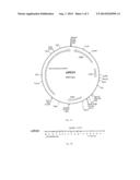 |
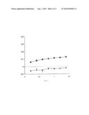 | 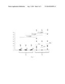 |
 |  |
 |  |
 |  |
 |  |
 |  |
 |  |
| Similar patent applications: | |
| Date | Title |
|---|---|
| 2012-06-14 | Diagnosis of autism |
| 2014-05-22 | Diagnosis of sepsis |
| 2014-08-07 | System and method for diagnosing lymphoma in cats |
| 2012-08-09 | Diagnostic methods |
| 2011-05-26 | Diagnostic method |
| New patent applications in this class: | |
| Date | Title |
|---|---|
| 2019-05-16 | Method to identify antigen-specific immune cells |
| 2016-09-01 | Devices and methods for manipulating components in a fluid sample |
| 2016-09-01 | Single b-cell cultivation method |
| 2016-07-07 | Method for analysis of nkt cell function |
| 2016-06-30 | Microfluidic system and method for isolating and quantifying at least one sub-population of cells from a population of cells |
| New patent applications from these inventors: | |
| Date | Title |
|---|---|
| 2021-07-01 | Immunogenic peptides and use thereof |
| 2015-12-17 | Process for identifying rare events |
| 2014-11-20 | Methods and kits for diagnosing latent tuberculosis infection |
| 2010-07-22 | Released cytokeratins as markers for epithelial cells |
| Top Inventors for class "Chemistry: molecular biology and microbiology" | |
| Rank | Inventor's name |
|---|---|
| 1 | Marshall Medoff |
| 2 | Anthony P. Burgard |
| 3 | Mark J. Burk |
| 4 | Robin E. Osterhout |
| 5 | Rangarajan Sampath |