Patent application title: METHODS FOR DIAGNOSING, TREATING, AND MONITORING CHRONIC INFLAMMATORY RESPONSE SYNDROME
Inventors:
Ritchie Shoemaker (Pocomoke City, MD, US)
Jimmy Ryan (Vero Beach, FL, US)
IPC8 Class: AA61B500FI
USPC Class:
600301
Class name: Surgery diagnostic testing via monitoring a plurality of physiological data, e.g., pulse and blood pressure
Publication date: 2014-02-13
Patent application number: 20140046143
Abstract:
The present invention relates generally to the diagnosis, treatment, and
monitoring of Chronic Inflammatory Response Syndrome (CIRS), kits for use
in the methods, and pharmaceutical compositions for use in the methods of
treatment. The invention specifically relates to the diagnosis, treatment
and monitoring of CIRS through a comprehensive approach comprising an
assessment of a subject for case definition parameters and a
proteogenomic analysis. The proteogenomic analysis for the diagnosis,
treatment, and monitoring of CIRS is based on identifying proteins and/or
genes that are differentially expressed in subjects suffering from CIRS
compared to healthy subjects.Claims:
1. A method for diagnosing chronic inflammatory response syndrome (CIRS)
in a subject suspected of having CIRS, comprising: a) obtaining case
definition parameters from said subject, selected from the group
consisting of: i) exhibiting eight or more symptom clusters selected from
the group consisting of 1) a fatigue cluster, 2) a weakness, ache,
headache, light sensitivity, and/or decreased assimilation cluster, 3) a
memory and/or word recall cluster, 4) a focus/concentration cluster, 5) a
cramp, joint pain, and/or morning stiffness cluster, 6) a tingling
cluster, 7) a sinus and/or shortness of breath cluster, 8) a cough,
confusion, and/or thirst cluster, 9) an appetite, temperature regulation,
and/or increased urination cluster, 10) an unusual pain, ice pick pain,
red eyes, blurred vision, mood swings, and sweats cluster, 11) abdominal
pain, diarrhea, and/or numbness cluster, 12) a tearing, disorientation,
and/or metallic taste cluster, and 13) a static shocks and/or vertigo
cluster; ii) visual contrast sensitivity deficit; iii) alpha melanocyte
stimulating hormone (MSH) deficiency; iv) vasoactive intestinal
polypeptide (VIP) deficiency; iv) elevated split product of complement
component 4 (C4a); v) elevated matrix metalloproteinase 9 (MMP9); vi)
elevated transforming growth factor beta-1 (TGF-.beta.1); vii)
dysregulation of adrenocorticotropic hormone (ACTH) and cortisol levels;
viii) dysregulation of antidiuretic hormone (ADH) and osmolality; and ix)
attaining a confidence level of greater than 90% using a support vector
machine classification algorithm to predict whether a sample from said
subject exhibits a gene, mRNA, and/or microRNA expression profile
consistent with CIRS; and b) diagnosing the subject as having CIRS if the
subject shows the presence of at least five of the case definition
parameters.
2. The method according to claim 1, wherein the step of obtaining case definition parameters for the subject comprises, i) obtaining medical history of the subject, ii) optionally performing a visual contrast sensitivity testing on the subject, iii) determining levels of one or more protein markers selected from the group consisting of C4a, VIP, MSH, TGF-.beta.1, MMP9, ACTH/Cortisol, ADH/Osmolality, Von Willebrand's profile, and HLA-DR in a biological sample obtained from the subject, iv) determining levels of one or more mRNA markers selected from Appendices A, B and/or C in the biological sample, and v) determining levels of one or more micro-RNA markers listed in Appendix D in the biological sample.
3. The method according to claim 1, wherein detected levels of the one or more protein markers set forth as case definition parameters iii)-viii) in a biological sample obtained from the subject are statistically significantly different from reference levels for the one or more protein markers.
4. A method according to claim 1, wherein detected levels of one or more mRNA markers set forth in Appendices A, B and/or C in a biological sample obtained from the subject are statistically significantly different from reference levels for the one or more mRNA markers.
5. A method according to claim 1, wherein detected levels of one or more micro-RNA markers listed in Appendix D in a biological sample obtained from the subject are statistically significantly different from reference levels for the one or more micro-RNA markers.
6. A method of treating chronic inflammatory response syndrome (CIRS) in a subject, comprising: a) obtaining case definition parameters from said subject, selected from the group consisting of: i) exhibiting eight or more symptom clusters selected from the group consisting of 1) a fatigue cluster, 2) a weakness, ache, headache, light sensitivity, and/or decreased assimilation cluster, 3) a memory and/or word recall cluster, 4) a focus/concentration cluster, 5) a cramp, joint pain, and/or morning stiffness cluster, 6) a tingling cluster, 7) a sinus and/or shortness of breath cluster, 8) a cough, confusion, and/or thirst cluster, 9) an appetite, temperature regulation, and/or increased urination cluster, 10) an unusual pain, ice pick pain, red eyes, blurred vision, mood swings, and sweats cluster, 11) abdominal pain, diarrhea, and/or numbness cluster, 12) a tearing, disorientation, and/or metallic taste cluster, and 13) a static shocks and/or vertigo cluster; ii) visual contrast sensitivity deficit; iii) alpha melanocyte stimulating hormone (MSH) deficiency; iv) vasoactive intestinal polypeptide (VIP) deficiency; iv) elevated split product of complement component 4 (C4a); v) elevated matrix metalloproteinase 9 (MMP9); vi) elevated transforming growth factor beta-1 (TGF-.beta.1); vii) dysregulation of adrenocorticotropic hormone (ACTH) and cortisol levels; viii) dysregulation of antidiuretic hormone (ADH) and osmolality; and ix) attaining a confidence level of greater than 90% using a support vector machine classification algorithm to predict whether a sample from said subject exhibits a gene, mRNA, and/or microRNA expression profile consistent with CIRS; b) diagnosing the subject as having CIRS if the subject shows the presence of at least five of the case definition parameters; and c) administering cholestyramine to the subject.
7. A method of treating chronic inflammatory response syndrome (CIRS) in a subject, comprising: a) obtaining case definition parameters from said subject, selected from the group consisting of: i) exhibiting eight or more symptom clusters selected from the group consisting of 1) a fatigue cluster, 2) a weakness, ache, headache, light sensitivity, and/or decreased assimilation cluster, 3) a memory and/or word recall cluster, 4) a focus/concentration cluster, 5) a cramp, joint pain, and/or morning stiffness cluster, 6) a tingling cluster, 7) a sinus and/or shortness of breath cluster, 8) a cough, confusion, and/or thirst cluster, 9) an appetite, temperature regulation, and/or increased urination cluster, 10) an unusual pain, ice pick pain, red eyes, blurred vision, mood swings, and sweats cluster, 11) abdominal pain, diarrhea, and/or numbness cluster, 12) a tearing, disorientation, and/or metallic taste cluster, and 13) a static shocks and/or vertigo cluster; ii) visual contrast sensitivity deficit; iii) alpha melanocyte stimulating hormone (MSH) deficiency; iv) vasoactive intestinal polypeptide (VIP) deficiency; iv) elevated split product of complement component 4 (C4a); v) elevated matrix metalloproteinase 9 (MMP9); vi) elevated transforming growth factor beta-1 (TGF-.beta.1); vii) dysregulation of adrenocorticotropic hormone (ACTH) and cortisol levels; viii) dysregulation of antidiuretic hormone (ADH) and osmolality; and ix) attaining a confidence level of greater than 90% using a support vector machine classification algorithm to predict whether a sample from said subject exhibits a gene, mRNA, and/or microRNA expression profile consistent with CIRS; b) diagnosing the subject as having CIRS if the subject shows the presence of at least five of the case definition parameters; and c) administering vasoactive intestinal peptide (VIP) to the subject.
8. The method according to claim 6, further comprising: d) treating the subject to eliminate MARCoNS infection if the subject has MARCoNS infection; e) correcting or restoring levels of antigliadin, androgens, ADH/osmolality, MMP9, VEGF, C3a, C4a, TGFβ-1; and f) administering vasoactive intestinal peptide to the subject.
9. The method according to claim 6, further comprising: d) treating the subject to eliminate MARCoNS infection if the subject has MARCoNS infection; e) correcting or restoring levels of antigliadin, androgens, ADH/osmolality, MMP9, VEGF, C3a, C4a, TGFβ-1, f) administering a therapeutically effective dose of VIP to the subject; g) obtaining case definition parameters for the subject at a second time for monitoring the status of CIRS in the subject, wherein the second time is later than the first time; h) diagnosing the subject as showing an improvement of CIRS if, at the second time, the subject shows the presence of fewer health symptoms or the presence of less than five case definition parameters, or diagnosing the subject as not showing an improvement if the subject shows the presence of at least five of the case definition parameters listed in Table 1; and i) repeating steps d) to g), until resolution of CIRS is obtained.
Description:
RELATED APPLICATION DATA
[0001] This application claims priority to Provisional Application No. 61/680,613, filed on Aug. 7, 2012, the disclosure of which is hereby incorporated by reference herein in its entirety.
FIELD OF THE INVENTION
[0002] The present invention relates generally to the diagnosis, treatment, and monitoring of Chronic Inflammatory Response Syndrome (CIRS), kits for use in the methods, and pharmaceutical compositions for use in the methods of treatment. The invention specifically relates to the diagnosis, treatment and monitoring of CIRS through a comprehensive approach comprising an assessment of a subject for case definition parameters and a proteogenomic analysis. The proteogenomic analysis for the diagnosis, treatment, and monitoring of CIRS is based on identifying proteins and/or genes that are differentially expressed in subjects suffering from CIRS compared to healthy subjects.
BACKGROUND OF THE INVENTION
[0003] CIRS is a form of Systemic Inflammatory Response Syndrome (SIRS) and is characterized by (1) lack of regulation of host inflammatory response as evidenced by deficiency of alpha melanocyte stimulating hormone (MSH) and/or vasoactive intestinal polypeptide (VIP); (2) presence of more than one of Th1 responses (pro-inflammatory); Th2 responses (anti-inflammatory); Th17 responses (tied to transforming growth factor beta-1 (TGF-β1)); coagulation abnormalities, especially abnormalities in von Willebrand's profile; activation of complement split products; activation of elements under regulation of hypoxia inducible factor including vascular endothelial growth factor (VEGF) and erythropoietin; abnormal regulation of ACTH responses to cortisol and ADH responses to osmolality. CIRS may be acquired through different mechanisms, for example, an exposure to toxins or inflammagens that may include, but are not limited to, environmental biotoxins, and chronic illness from Lyme disease present even after treatment with antibiotics. The exposure to environmental sources of biotoxins includes a chronic exposure to the interior environment of water-damaged buildings (WDB), or ingestion of fish contaminated with the toxins of marine dinoflagellates, such as ciguatoxins. Other environmental sources of biotoxins that can lead to CIRS include certain compounds made by dinoflagellates, cyanobacteria, fungi, actinomycetes, bacteria, mycobacteria, etc. When CIRS is acquired because of an exposure to a WDB, it is termed CIRS-WDB (Expert Treating Physicians Consensus, 2010).
[0004] According to a report released by the World Health Organization in 2009, in a WDB, people are chronically exposed to different microbes and/or compounds of microbial or other origin that are present in the indoor air of a WDB. These compounds initiate an innate immune inflammatory response in the human host. These microbes and compounds include but are not limited to fungi, bacteria, actinomycetes, and mycobacteria and their toxins; as well as inflammagens from fragments of fungal structures; and beta glucans, mannans, hemolysins, proteinases, spirocyclic drimanes and volatile organic compounds (VOCs). An ongoing exposure to the above microbes and/or compounds can result in a recurrent activation of immune responses, leading to exaggerated immune responses and prolonged production of inflammatory mediators, especially in the absence of regulation of inflammation by neuropeptides MSH or VIP.
[0005] Some of the organisms that make biotoxins that can cause CIRS include dinoflagellates (Pfiesteria, Gambierdiscus (ciguatera), Karenia (and other species that produce brevetoxins) cyanobacteria (Microcystis, Cylindrospermopsis, Lyngbya wollei); fungi (Wallemia, Stachybotrys, Chaetomium, Trichoderma, Aspergillus versicolor, Aspergillus versicolor and others); actinomycetes (Streptomyces and others); apicomplexans (Babesia, Sarcocystis, Eimeria), and spirochetes (Borrelia spp burgdorferi and (likely) B. lonestari). Organisms such as commensal multiple-antibiotic resistant coagulase negative staphylococci (MARCoNS), including methicillin resistant Staphylococcus epidermidis, may also contribute to CIRS.
[0006] Two examples of inflammagens that may cause CIRS are beta-glucans and mannans made by fungi that activate specific C-type lectin receptors, namely dectin-1 and dectin-2 receptors.
[0007] Patients with CIRS are often misdiagnosed as having depression, stress, allergy, fibromyalgia, post traumatic stress disorder, Chronic Fatigue Syndrome or somatization, etc., and are treated with various therapies, some of them being potentially toxic, which have not yet been shown to be effective, and are often costly. One reason that CIRS may be misdiagnosed is because there are no biomarkers that have been identified yet for CIRS or for those commonly-misdiagnosed illnesses, which would allow for a confirmatory diagnosis. Treating CIRS patients for the above conditions does not improve their symptoms of CIRS. With proper detection, diagnosis, and documentation of the objective basis of illness pathophysiology, CIRS may be treated effectively to improve symptoms and decrease the recurrence of uncontrolled inflammatory responses. Therefore, there exists a need for accurate diagnosing of CIRS in order to effectively treat patients with CIRS.
SUMMARY OF THE INVENTION
[0008] The present invention relates to the diagnosis, prognosis, prevention and/or treatment of CIRS. In particular, the invention is directed to the differential diagnosis of CIRS in a subject suspected of having CIRS, including differential diagnosis based on a detailed medical history and optional visual contrast sensitivity testing (VCS), and also identifying a panel of proteogenomic markers that are differentially expressed in subjects with CIRS relative to reference levels of expression of these markers either in disease-free healthy subjects or in subjects with CIRS prior to receiving the treatment for CIRS.
[0009] The term "a subject suspected of having CIRS" refers to, but is not limited to, individuals who are exposed to CIRS etiological factors such as environmental sources of toxins, including WDB, ciguatoxins, or other toxic components originating from dinoflagellates, cyanobacteria, fungi, actinomycetes, bacteria, mycobacteria, etc., or chronic illness from Lyme disease.
[0010] The term "differential diagnosis" as used herein refers to obtaining medical history and objective parameters from a subject, determining whether the subject has been exposed to any potential CIRS etiological factors, optionally performing visual contrast sensitivity (VCS) testing on the subject, and making a determination as to whether the subject is suspected of having CIRS or not having CIRS based on the medical history, laboratory testing, and the results of VCS testing, if performed.
[0011] The term "proteogenomic markers" as used herein refers to a combination of one or more genomic markers and/or one or more proteomic markers that are used in order to make a determination of whether a subject is suspected of having CIRS or not having CIRS. The proteogenomic markers may include any one of the markers set forth in Tables 3-4 and Appendices A-D, any combination of markers set forth in Tables 3-4 and Appendices A-D, or all of the markers set forth in Tables 3-4 and Appendices A-D.
[0012] The present invention also includes methods for testing trigger mechanisms of CIRS, such as an Environmental Relative Moldiness Index (ERMI) and its derivative, Health Roster of Type Specific formers of Mycotoxins and Inflammagens, second iteration (HERTSMI-2); testing for the presence of fungi in the built environment; testing for Lyme borreliosis; or an assessment of intoxication with ciguatoxins.
[0013] The treatment methods of the invention may also include removing the subject from the source of the toxins and/or inflammagens, and/or removing the toxins and/or inflammagens from the subject or the environment surrounding the subject. Such toxins or inflammagens may include, but are not limited to, environmental biotoxins, such as those found in the interior environment of water-damaged buildings (WDB), in fish contaminated with the toxins of marine dinoflagellates (e.g., ciguatoxins), as well as certain compounds produced by dinoflagellates, cyanobacteria, fungi, actinomycetes, bacteria, mycobacteria, etc. The toxins may also be present in the subject as a result of chronic illness from Lyme disease present even after treatment with antibiotics, for example.
[0014] In one embodiment, the method of diagnosing Chronic Inflammatory Response Syndrome (CIRS) in a subject suspected of having CIRS, comprises a) obtaining at least the case definition parameters listed in Table 1 for the subject; and b) diagnosing the subject as having CIRS if the subject shows the presence of one or more, two or more, three or more, four or more, five or more, six or more, seven or more eight or more, nine or more, or all ten of the case definition parameters listed in Table 1. Preferably, the presence of at least three of these parameters is detected; more preferably, the presence of at least four parameters is detected; most preferably, the presence of at least five parameters is detected. The step of obtaining case definition parameters may comprise i) obtaining medical history of the subject; ii) optionally performing a visual contrast sensitivity testing on the subject; iii) determining levels of one or more protein markers listed in Tables 3 and 4 in a biological sample obtained from the subject; iv) determining levels of one or more mRNA markers selected from Appendices A, B and/or C in the biological sample; v) determining levels of one or more micro-RNA markers listed in Appendix D in the biological sample.
[0015] In one embodiment, the step of obtaining medical history is followed by differential diagnosis to diagnose the subject as suspected of having CIRS or not having CIRS. The differential diagnosis is then confirmed by determining the levels of one or more proteogenomic markers.
[0016] In another embodiment, the method of diagnosing CIRS comprises diagnosing CIRS based on the expression levels of one or more protein markers. According to this embodiment, the method of diagnosing chronic inflammatory response syndrome (CIRS) in a subject suspected of having CIRS comprises a) determining levels of one or more protein markers listed in Table 3 in a biological sample obtained from the subject; b) diagnosing the subject as having CIRS if the levels of the one or more protein markers are statistically significantly different from reference levels for the one or more protein markers.
[0017] In yet another embodiment, the method of diagnosing CIRS comprises diagnosing CIRS based on the expression levels of one or more mRNA markers. According to this embodiment, the method of diagnosing chronic inflammatory response syndrome (CIRS) in a subject suspected of having CIRS comprises a) determining levels of one or more mRNA markers selected from Appendices A, B and/or C in a biological sample obtained from the subject; b) diagnosing the subject as having CIRS if the levels of the one or more mRNA markers are statistically significantly different from reference levels for the one or more mRNA markers.
[0018] In yet another embodiment, the method of diagnosing CIRS comprises diagnosing CIRS based on the expression levels of one or more micro-RNA markers. According to this embodiment, the method of diagnosing chronic inflammatory response syndrome (CIRS) in a subject suspected of having CIRS comprises a) determining levels of one or more micro-RNA markers listed in Appendix D in a biological sample obtained from the subject; b) diagnosing the subject as having CIRS if the levels of the one or more micro-RNA markers are statistically significantly different from reference levels for the one or more micro-RNA markers.
[0019] According to the invention, it has been found that subjects with CIRS may also exhibit brain abnormalities. Specifically, it has been found that subjects with CIRS may exhibit structural brain volumes abnormalities including, but not limited to, an increase in forebrain parenchyma, an increase in cortical gray area, an increase in the volume of the hippocampus, a decrease in the volume of caudate, and/or an increase in the volume of pallidum. These structural brain abnormalities have been identified, for example, in subjects suffering from CIRS-WDB. Accordingly, in another embodiment, the method of diagnosing CIRS may further comprise detecting brain abnormalities in a subject suspected of having CIRS using known or as-yet undeveloped methods for detecting brain abnormalities. The methods of monitoring treatment of CIRS may also further comprise detecting brain abnormalities in a subject being treated for CIRS at a first time period prior to treatment, and comparing the abnormalities with brain abnormalities detected at a second time period after treatment has been initiated, and determining whether improvement in brain abnormalities is observed. Methods known in the art for detecting brain abnormalities may include, but are not limited to, CT scans, MRI scans, PET scans, brain volume measurement (i.e., FreeSurfer, Martinos Center for Biomedical Imaging, Charlestown, Mass.), and NeuroQuant® (CorTechs Labs, La Jolla, Calif.).
[0020] The present invention also provides methods of treating chronic inflammatory response syndrome (CIRS). In one embodiment, a method of treating CIRS in a subject, comprises: a) obtaining case definition parameters listed in Table 1 for the subject; b) diagnosing the subject as having CIRS if the subject shows the presence of at least five of the case definition parameters listed in Table 1; c) administering cholestyramine to the subject. The methods may also optionally include removing or reducing the amount of any toxins and/or inflammagens that are suspected of causing CIRS in the subject. When cholestyramine is administered to the subject, dosage amounts and schedules are within the ability of the skilled practitioner to determine, and may be, for example, in the range of 1 to 40 grams per day, preferably 8 to 36 grams per day, more preferably 16 to 24 grams per day. The doses may be provided from 1 to 6 times per day, preferably 2 to 4 times per day. The doses are preferably administered orally. Cholestyramine may be administered for any period of time necessary to achieve relief from CIRS. However, it should be noted that the present invention is not limited by the dosage amount, administration route, or length of administration.
[0021] In another embodiment, a method of treating CIRS in a subject comprises: a) obtaining case definition parameters listed in Table 1 for the subject; b) diagnosing the subject as having CIRS if the subject shows the presence of at least five of the case definition parameters listed in Table 1; c) administering vasoactive intestinal peptide (VIP) to the subject. The methods may also optionally include removing or reducing the amount of any toxins and/or inflammagens that are suspected of causing CIRS in the subject. When VIP (aviptadil) is administered to the subject, dosage amounts and schedules are within the ability of the skilled practitioner to determine, and may be, for example, in the range of 5 to 200 micrograms, preferably 10 to 100 micrograms, more preferably 25 to 75 micrograms. According to one embodiment, a dose of 50 micrograms is administered. The doses may be provided from 1 to 4 times per day, preferably 1 time per day. The doses may be administered nasally or injected, although nasal instillation is a preferred method of administration. VIP may be administered for any period of time necessary to achieve relief from CIRS. However, it should be noted that the present invention is not limited by the dosage amount, administration route, or length of administration.
[0022] In yet another embodiment, a method of treating CIRS in a subject comprises sequential therapeutic intervention shown in FIG. 2. According to this embodiment, a method of treating CIRS in a subject comprises: a) obtaining case definition parameters listed in Table 1 for the subject including performing differential diagnosis; b) diagnosing the subject as having CIRS if the subject shows the presence of at least five of the case definition parameters listed in Table 1; c) administering a therapeutically effective dose of cholestyramine to the subject; d) treating the subject to eliminate MARCoNS infection if the subject has MARCoNS infection; e) correcting or restoring levels of antigliadin, androgens, ADH/osmolality, MMP9, VEGF, C3a, C4a, TGF-β1; and f) administering a therapeutically effective dose of VIP to the subject.
[0023] In one embodiment, the method of treating chronic inflammatory response syndrome (CIRS) in a subject comprises a) determining levels of: one or more protein markers listed in Table 3 and/or one or more mRNA markers selected from Appendices A, B and/or C and/or one or more micro-RNA markers listed in Appendix D in a biological sample obtained from the subject; b) diagnosing the subject as having CIRS if the levels of the one or more protein markers and/or the one or more mRNA markers and/or the one or more micro-RNA markers are statistically significantly different from reference levels for corresponding markers; c) administering a therapeutically effective dose of cholestyramine to the subject.
[0024] In another embodiment, the method of treating chronic inflammatory response syndrome (CIRS) in a subject comprises a) obtaining case definition parameters listed in Table 1 for the subject, including performing differential diagnosis, at a first time; b) diagnosing the subject as having CIRS if the subject shows the presence of at least five of the case definition parameters listed in Table 1; c) administering a therapeutically effective dose of cholestyramine to the subject; d) treating the subject to eliminate MARCoNS infection if the subject has MARCoNS infection; e) correcting or restoring levels of antigliadin, androgens, ADH/osmolality, MMP9, VEGF, C3a, C4a, TGF-β1; f) administering a therapeutically effective dose of VIP to the subject; g) obtaining case definition parameters listed in Table 1 for the subject at a second time for monitoring the status of CIRS in the subject, wherein the second time is later than the first time; h) diagnosing the subject as showing an improvement of CIRS if, at the second time, the subject shows the presence of fewer health symptoms or the presence of less than five case definition parameters listed in Table 1 or diagnosing the subject as not showing an improvement if the subject shows the presence of at least five of the case definition parameters listed in Table 1; i) repeating steps d) to g), until resolution of CIRS is obtained.
[0025] The present invention also provides a method for monitoring CIRS in a subject comprising: a) obtaining case definition parameters listed in Table 1 for the subject at a first time; b) diagnosing the subject as having CIRS if the subject shows the presence of at least five of the case definition parameters listed in Table 1; c) administering a dose of cholestyramine to the subject; d) obtaining case definition parameters listed in Table 1 for the subject at a second time for monitoring the status of CIRS in the subject, wherein the second time is later than the first time; e) diagnosing the subject as showing an improvement of CIRS if the subject shows the presence of less than five case definition parameters listed in Table 1 or diagnosing the subject as not showing an improvement if the subject shows the presence of at least five of the case definition parameters listed in Table 1; f) adjusting the dose of cholestyramine, if required based on the diagnosis in step e); g) administering the adjusted or the same dose of cholestyramine to the subject; h) repeating steps d) to g) until resolution of CIRS is obtained.
[0026] In one aspect, the method for diagnosing or monitoring CIRS in a subject according to the invention further comprises a step of detecting, in a biological sample of the subject, a level of regulatory T lymphocytes (Treg cells) as an indirect indicator of the status of tissue-based inflammation in the subject.
[0027] In another aspect, the method for diagnosing or monitoring CIRS in a subject according to the invention further comprises determining the presence of unusual staphylococci such as coagulase negative staphylococci, staphylococci which are resistant in Kirby-Bauer susceptibility testing to at least two classes of antibiotics (MARCoNS), and methicillin resistant staphylococci (MRCoNS) in the subject by obtaining an aerobic culture using the API-STAPH or similar technique from deep nasal spaces from the subject. These organisms invariably form biofilm, providing a mechanism to continue to colonize deep nasal spaces without causing overt invasive symptoms such as headache or sinus congestion. In one aspect, the method for diagnosing or monitoring CIRS in a subject according to the invention comprises determining diagnostic parameters that are not altered in CIRS compared to other diseases or disorders the symptoms of which may overlap with CIRS. Such diagnostic parameters include, but are not limited to, erythrocyte sedimentation rate (ESR), C-reactive protein (CRP), etc.
[0028] The methods of diagnosing, treating, and monitoring CIRS according to the invention may comprise various permutations and combinations of the methods disclosed herein.
[0029] According to the invention, it has been found that subjects with CIRS may show a distinct and specific pattern or profile of expression for a set of genes (for example, set X) compared to healthy subjects or subjects with illnesses that have symptoms similar to CIRS. For example, in one aspect of the invention, subjects with CIRS may show a distinct and specific pattern of expression for one or more mRNA markers selected from Appendices A, B and/or C and/or one or more micro RNA markers selected from Appendix D. It has also been found that the expression profile of a set of genes (for example, set Y) may vary between the subjects with CIRS based on the etiology of CIRS. For example, a subject that has acquired CIRS because of an exposure to WDB may show a gene expression pattern different from the gene expression pattern of a subject that has acquired CIRS because of chronic illness from Lyme disease. According to one aspect of the invention, set Y is a subset of set X. Accordingly, the present invention is also directed to predicting the causative agent of CIRS.
[0030] In one aspect of the invention, the diagnosis of CIRS is carried out by detecting levels of one or more genes that are differentially expressed between the subjects with CIRS and disease-free healthy subjects. The differential expression of CIRS genes may be measured at the gene level, transcript level and/or polypeptide level. The term "transcript" as used herein encompasses all RNA products produced from the genome, including sense and antisense products, coding RNAs such as messenger RNAs (mRNAs) as well as non-coding RNAs such as Long Intergenic Non-Coding (linc) RNAs as well as regulatory RNAs such as micro-RNAs. Micro-RNAs are a class of small, non-coding RNAs that control gene expression by hybridizing to and triggering either translational repression or, more frequently, degradation of a messenger RNA (mRNA) target. linc RNAs are in general considered as non-protein coding transcripts longer than 200 nucleotides.
[0031] The term "differential expression" as used herein means either an over-expression or under-expression of one or more CIRS genes, transcripts or proteins relative to reference levels of expression of corresponding genes, transcripts or proteins. That is, in an embodiment, certain genes, transcripts, and/or proteins are down-regulated while certain other genes, transcripts, and/or proteins are up-regulated compared to reference levels of corresponding genes, transcripts, and/or proteins. In one example, a gene, transcript, and/or protein for vasoactive intestinal peptide (VIP) may be under-expressed while a gene, transcript, and/or protein for anaphylatoxin C4a may be over-expressed. The term CIRS gene, transcript or protein means a gene, transcript or protein that is differentially expressed in CIRS subjects compared to healthy subjects.
[0032] In one embodiment, reference levels of expression are the expression levels of genes, transcripts or proteins in a healthy subject. In another embodiment, reference levels of expression are the expression levels of genes, transcripts or proteins in a subject with CIRS prior to the treatment of CIRS. In another embodiment, reference levels of expression are the expression levels of genes, transcripts or proteins in a subject with CIRS at various stages during the course of treatment for CIRS.
[0033] The term "statistically significantly different" means the p-value of the differential expression for each selected CIRS gene, transcript or protein is no more than 0.05. In one embodiment, the difference in the expression levels of CIRS genes, transcripts or proteins compared to the reference levels of expression of corresponding genes, transcripts or proteins is at least 1.3-, 2-, 3-, 4-, 5-, 10-, 20-fold or more. In another embodiment, the expression level is indicative of CIRS if it is found in at least 70%, at least 75%, at least 80%, at least 85%, at least 90%, at least 95%, at least 97%, at least 98%, at least 99%, or more in subjects with CIRS and is found in less than 20%, less than 10%, less than 8%, less than 5%, less than 2.5%, or less than 1% of subjects who do not have CIRS.
[0034] In yet another embodiment, the differentially expressed CIRS genes, transcripts, and proteins are selected from Tables 3-4 and Appendices A-D. In still another embodiment, the CIRS genes include innate immune response genes such as genes encoding complement factors, neuropeptides, hormones, transcription factors, cytokines, etc.
[0035] In one aspect, the differential expression of CIRS genes is diagnosed by measuring the levels of expression of one or more polypeptides, mRNA and/or micro RNA in a biological sample obtained from a subject with CIRS and a biological sample obtained from a healthy individual. The biological sample may be blood, nasal swab, a bodily waste sample or any other body fluid or tissue that can provide a meaningful diagnosis of differentially expressed CIRS genes.
[0036] In one aspect, levels of protein markers listed in Table 3, mRNA markers selected from Appendices A, B and/or C, and micro-RNA markers listed in Appendix D in a biological sample may be detected by methods known in the art. In one example, levels of protein markers listed in Table 3 may be determined using antibodies or fragments of antibodies specific for these markers. In another example, levels of protein markers may be detected using methods such as enzyme-linked immunosorbent assays (ELISA), radio-immuno assay (RIA), or western blot analysis. In another example, levels of protein markers may be detected using direct methods such as mass spectrometry. In yet another example, levels of protein markers listed in Table 3 may be determined using commercially available kits such as those available from Laboratory Corporation of America (LabCorp) or Quest Diagnostics, Inc. Although ELISA is one preferred technique for detecting the protein markers used in the invention, alternative techniques such as immunoblotting nuclease protection assays, in situ hybridization, microarrays, immunohistochemistry, are also envisioned, and appropriate apparatus, and reagents for use in carrying out these well-known techniques are envisioned for use in the methods and included as components of the kits.
[0037] In an embodiment, levels of mRNA markers selected from Appendices A, B and/or C and levels of micro-RNA markers listed in Appendix D in a biological sample may be determined by extracting total RNA from the biological sample and hybridizing the extracted RNA to labeled nucleic acid probes complementary to mRNA and micro-RNA markers using any state of the art techniques. Techniques for detecting mRNAs and micro-RNAs using labeled nucleic acid probes include microarray analysis, northern blot analysis, and nuclease protection assays. Alternatively, mRNA and micro-RNA markers can be detected using reverse transcription-polymerase chain reaction (RT-PCR) with or without labeled nucleic acid probes. Additionally, newer techniques that do not rely on complementary target-probe hybridization, such as Next Generation Sequencing (NGS), may be used to determine levels of transcripts.
[0038] The present invention also provides pharmaceutical compositions useful for treating or preventing CIRS. In one embodiment, the pharmaceutical compositions of the present invention include a pharmaceutically acceptable carrier and at least one polynucleotide encoded by a CIRS gene that is over-expressed or under-expressed in CIRS subjects relative to healthy subjects. The pharmaceutical compositions can also include a variant or an allele of the polynucleotide. In one example, the pharmaceutical compositions are compositions comprising at least one micro-RNA listed in Appendix D.
[0039] In another embodiment, the pharmaceutical compositions of the present invention include a pharmaceutically acceptable carrier and at least one active component selected from (i) agents capable of modulating the expression of one or more CIRS genes which are over-expressed or under-expressed in CIRS subjects relative to healthy subjects, (ii) agents capable of binding to, or modulating the biological activity of, the polypeptide(s) encoded by the CIRS gene, (iii) agents capable of modulating Treg cells, or (iv) Treg cells. Exemplary modulations include, but are not limited to, up-regulation, induction, stimulation, inhibition, down-regulation, and suppression.
[0040] In one example, the active component is a polynucleotide comprising or encoding an RNA that is capable of inhibiting or decreasing expression of the CIRS gene by RNA interference or an antisense mechanism. In another example, the active component is an agonist or antagonist of a protein encoded by the CIRS gene. In yet another example, the active component is a monoclonal antibody or a fragment thereof. Proteins encoded by CIRS genes can be, for example, innate immune response proteins such as complement factors, neuropeptides, hormones, transcription factors, cytokines, etc. These proteins and genes are potential targets for drug action and development.
[0041] In still yet another aspect, the present invention provides methods for screening anti-CIRS agents based on their effects on the expression or function of CIRS genes.
[0042] In a further aspect, the present invention provides kits useful for diagnosing CIRS. Each kit can include at least one of the following: (a) one or more polynucleotide probes capable of hybridizing under reduced stringent, stringent, or highly stringent conditions to one or more CIRS genes (or a complement thereof), or (ii) one or more antibodies capable of specifically binding to one or more polypeptides encoded by CIRS genes (e.g., antibodies to the polypeptides). The kits may include a carrier where the polynucleotide probes or antibodies are immobilized and one or more reagents or detection agents for detecting a reaction between the one or more probes or antibodies and target nucleic acids or polypeptides. The kit may also include one or more software and/or instructions to analyze the data generated by using the kit.
[0043] In another embodiment, the invention is directed to an antibody or antibodies that specifically bind to proteins or protein fragments that are differentially expressed in subjects with CIRS.
[0044] It is further intended that the inventions not be limited only to the specific structure, material or acts that are described in the preferred embodiments, but in addition, include any and all structures, materials or acts that are capable of performing the claimed function, along with any and all known or later-developed equivalent structures, materials or acts capable of performing the claimed function.
BRIEF DESCRIPTION OF THE DRAWINGS
[0045] FIG. 1 shows that patients with CIRS exhibit defects in visual contrast sensitivity as compared to control healthy individuals.
[0046] FIG. 2 shows a sequential therapeutic intervention for treating subjects with CIRS according to one embodiment of the invention.
[0047] FIG. 3 shows mean values of various laboratory parameters with significant differences between baseline and controls.
DETAILED DESCRIPTION OF THE INVENTION
[0048] The present invention provides methods of diagnosing, treating and monitoring CIRS, as well as test kits for use in the methods. In one aspect, the method of diagnosing CIRS in a subject suspected of having CIRS comprises a) obtaining case definition parameters listed in Table 1 for the subject, comprising: i) obtaining medical history of the subject, ii) performing differential diagnosis; iii) optionally performing visual contrast sensitivity testing on the subject, iv) determining levels of one or more protein markers listed in Table 3 in a biological sample obtained from the subject, v) determining levels of one or more mRNA markers selected from Appendices A, B and/or C in the biological sample, vi) optionally performing brain scanning on the subject, vii) determining levels of one or more micro-RNA markers listed in Appendix D in the biological sample; and b) diagnosing the subject as having CIRS if the subject shows the presence of three or more, preferably four or more, and most preferably five or more of the case definition parameters listed in Table 1.
TABLE-US-00001 TABLE 1 Case Definition Parameters Parameters Symptoms score > 8 VCS deficit MSH < 25 pg/mL VIP < 23 pg/mL C4a > 2830 ng/mL TGF-β1 > 2380 pg/mL MMP9 > 332 ng/mL ADH/osmolality dysregulation ACTH/cortisol dysregulation Genomic SVM confidence > 0.9
[0049] The case definition parameters listed in Table 1 are explained in more detail below.
[0050] Symptoms score: Symptoms score is obtained by obtaining a detailed medical history of a subject. Medical history evaluates both subjective patient complaints and causative environmental parameters for potential exposure to biotoxins in the home, school or workplace. Medical history includes a detailed analysis of past medical conditions and current symptoms experienced by the subject. A symptom roster (Table 2) may be used to determine the symptoms experienced by the subject. The symptom roster comprises a list of symptoms grouped by symptom clusters which are discussed in the following references: Shoemaker et al., "Sick Building Syndrome in water-damaged buildings: Generalization of the chronic biotoxin-associated illness paradigm to indoor toxigenic fungi," Bioaerosols, Fungi, Bacteria, Mycotoxins and Human Health, Johanning, E., editor, 2005, pages 66-77; Shoemaker et al., "A time-series of sick building syndrome; chronic, biotoxin-associated illness from exposure to water-damaged buildings," Neurotoxicology and Teratology, 2005, 27(1) 29-46; Shoemaker et al., "SBS and exposure to water damaged buildings: time series study, clinical trial and mechanisms," Neurotoxicology and Teratology, 2006, 28: 573-588; Shoemaker et al., "Innate immunity, MR spectroscopy, HLA DR, TGF beta-1, VIP and capillary hypoperfusion define acute and chronic human illness acquired following exposure to water-damaged buildings," Healthy Buildings, Syracuse, N.Y., 2009; Shoemaker, "Innate immune responses define pediatric CFS," International Association for CFS/ME Conference, 2009, Reno, Nev.; Shoemaker, "Exposure to water-damaged buildings causes a readily identifiable chronic inflammatory response syndrome that is successfully treated by a sequential intervention protocol," 9th International Mycology Congress, Edinburgh, Scotland, 2010; Shoemaker, "Vasoactive Intestinal Polypeptide (VIP): a final step in correction of the inflammatory illness acquired following exposure to water-damaged buildings," "T regulatory cells in chronic inflammatory response syndrome from water-damaged buildings (CIRS-WDB)," and "HERTSMI-2 Simplifying analysis of safety of WDB," 6th International Scientific Conference on Bioaerosols, Fungi, Bacteria, Mycotoxins in Indoor and Outdoor Environments and Human Health, Saratoga Springs, N.Y., September 2011. Each of these references is hereby incorporated by reference herein in its entirety.
[0051] Each symptom cluster may comprise one or more symptoms. A symptoms score is the number of symptom clusters experienced by the subject. For example, a symptoms score of 5 means the symptoms experienced by the subject belong to 5 symptom clusters. A symptoms score of 8 or more indicates that a subject may be suffering from CIRS.
TABLE-US-00002 TABLE 2 Symptom Roster Score Symptom/Cluster Patient Fatigue/1 Weak/2 Ache/2 Cramp/5 Unusual pain/10 Ice pick pain/10 Headache/2 Light sensitivity/2 Red eyes/10 Blurred vision/10 Tearing/12 Sinus/7 Cough/8 Shortness of breath/7 Abdominal pain/11 Diarrhea/11 Joint pain/5 Morning stiffness/5 Memory/3 Focus/concentration/4 Word recall/3 Decreased assimilation/2 Confusion/8 Disorientation/12 Skin sensitivity/6 Mood swings/10 Appetite/9 Sweats/10 Temp regulation/9 Thirst/8 Increased urination/9 Static shocks/13 Numbness/11 Tingling/6 Vertigo/13 Metallic taste/12
[0052] 2. Visual Contrast Sensitivity (VCS) Testing:
[0053] VCS testing measures the neurologic function called contrast that permits an eye to resolve patterns. This test is regarded as the best current test of functional vision. VCS deficiencies due to an exposure to CIRS causative agents such as biotoxins have been shown to correlate with capillary hypoperfusion in the retina and neural rim of the optic nerve head as measured by a dual laser device, Heidelberg Retinal Flowmeter (Hudnell, EPA 2003). Since excessive optical-refraction errors can cause abnormal VCS readings, a visual acuity as measured by Snellen Distance Equivalent Score of better than 20:50 is required for inclusion of this element in the diagnosis of CIRS. Apart from CIRS, VCS deficiencies may occur in certain neurologic conditions such as neurologic conditions developed due to an exposure to mercury, organic solvents, hydrocarbons, or Parkinson's disease (see, e.g., Residential and Recreational Acquisition of Possible Estuarine Associated Syndrome: A New approach to Successful Diagnosis and Therapy, Environmental Health Perspectives, Special CDC Pfiesteria Supplement, 2001; 109S5; 791-796). However, according to the present invention, a combination of symptoms score and VCS can differentially diagnose CIRS subjects from those experiencing non-CIRS illnesses such as the neurologic conditions described above (Table 5). Differences in VCS between subjects exposed to CIRS causative agents and control subjects are shown in FIG. 1.
[0054] 3. Protein Markers:
[0055] The present invention uses a panel of protein markers (Table 3) that are differentially expressed in CIRS subjects compared to healthy individuals. These protein markers are indicative of the physiologic status of inflammatory responses in CIRS subjects. Methods for detecting levels of protein markers listed in Table 3 are known in the art, and include commercially-available enzyme linked immunosorbent assays (ELISAs), radioimmunoassays, and other immunoassay technologies. Additional detection methods may also be employed in order to identify biomarkers, including PCR and related technologies. Various techniques for the detection of the biomarkers may be used in accordance with the invention, such as those described in Ausubel et al., Current Protocols In Molecular Biology, John Wiley & Sons, New York, N.Y., 2002, or Sambrook et al., Molecular Cloning: A Laboratory Manual, Cold Spring Harbor Press, Cold Spring Harbor, N.Y., 1989. In one embodiment, levels of protein markers listed in Table 3 in a biological sample may be determined using commercially available test kits such as those available from LabCorp or Quest Diagnostics, Inc.
TABLE-US-00003 TABLE 3 Protein markers Protein marker C4a VIP MSH TGF-β1 MMP9 ACTH/Cortisol ADH/Osmolality Von Willebrand's profile HLA-DR
[0056] The protein markers that may be measured in accordance with the invention can be obtained from any sample of a biological material that is suspected of containing the protein markers as an analyte of interest. Examples of biological materials include, but are not limited to, stool, whole blood, serum, plasma, red blood cells, platelets, interstitial fluid, saliva, ocular lens fluid, cerebral spinal fluid, sweat, urine, ascites fluid, mucous, nasal fluid, sputum, synovial fluid, peritoneal fluid, vaginal fluid, menses, amniotic fluid, semen, etc. Preferably, the test sample is a blood, serum, or urine sample. The test sample may be used directly as obtained or following a pretreatment to modify the sample. For example, pretreatment may include preparing plasma or serum from blood. Methods of pretreatment may also involve filtration, precipitation, dilution, distillation, mixing, concentration, inactivation of interfering components, the addition of reagents, lysing, etc. If such methods of pretreatment are employed with respect to the test sample, such pretreatment methods are such that the analyte of interest remains in the test sample at a concentration proportional to that in an untreated test sample (e.g., a test sample that is not subjected to any such pretreatment method(s)).
[0057] 3.1. C4a
[0058] is a split product of complement component 4. The complement system is an important component of both innate and adaptive immune responses. CIRS patients show a statistically significant difference from normal levels of C4a. Interestingly, CIRS patients, mainly those with CIRS-WDB or CIRS-ciguatera, are not likely to show an increase in the levels of C3a, a product immediately downstream of C4 activation. Complement activation through both the classical pathway and mannose binding lectin system generates increased levels of C4a.
[0059] 3.2. And 3.3. Vasoactive Intestinal Peptide (VIP) and Alpha Melanocyte stimulating hormone (MSH):
[0060] VIP and MSH are neuropeptides critical for the regulation of inflammation. CIRS patients consistently exhibit low levels of VIP and MSH, suggesting lack of regulation of inflammation in the development and persistence of CIRS. These two neuropeptides have been shown to have profound anti-inflammatory effects both in vivo and in vitro and could be used for treating inflammatory diseases.
[0061] VIP and/or MSH deficiencies can be acquired either acutely or at an advanced stage of CIRS, as well as through diverse mechanisms such as acute brain injuries (S. Magnoni, et al., Alpha-melanocyte-stimulating hormone is decreased in plasma of patients with acute brain injury, J Neurotrauma 20 (2003) 251-60) or persistent viral infection (Y.-R. Tan, et al., Pulmonary peptidergic innervation remodeling and development of airway hyperresponsiveness induced by RSV persistent infection, Peptides 29 (2008) 47-56). It has been shown that VIP agonists effectively protect against Alzheimer-related learning impairment in rats (I. Gozes, et al., Neuropeptides and neuronal survival: neuroprotective strategy for Alzheimer's disease, Annals of the New York Academy of Sciences 814 (1997) 161-6) while deficiency of VIP has been shown to cause cognitive defects in mice (D. Chaudhury, et al., Select cognitive deficits in vasoactive intestinal peptide deficient mice, BMC Neurosci 9 (2008) 63), common symptoms observed in CIRS subjects. One important role of these two neuropeptides is the induction of tolerogenic dendritic cells and generation of T regulatory cells (Tregs), which suppress autoreactive T cells and autoimmune progression (M. Delgado and D. Ganea, Anti-inflammatory neuropeptides: A new class of endogenous immunoregulatory agents, Brain, Behavior, and Immunity 22 (2008) 1146-1151). Even in healthy individuals autoreactive T cells can escape clonal deletion and must be policed in the periphery by Tregs to prevent pathologic autoimmunity (N. A. Danke, et al., Autoreactive T Cells in Healthy Individuals, J Immunol 172 (2004) 5967-5972). Decreased levels of Treg cells due to lower levels of VIP and MSH could be the reason that both pediatric and adult CIRS patients show certain features of autoimmune disease such as markedly elevated levels of anti-gliadin and anti-cardiolipin antibodies compared to healthy individuals.
[0062] 3.4. And 3.5. Matrix Metalloproteinase 9 (MMP9) and Transforming Growth Factor Beta-1 (TGF-β1):
[0063] CIRS patients often show high levels of MMP9 and TGF-β1. These two proteins have been demonstrated to have wide ranging effects on the immune system, including important roles in autoimmune and inflammatory diseases. The timing, duration and target tissues are important aspects for protection or pathological activity of these proteins.
[0064] Similar to VIP and MSH, TGF-β1 has been shown to regulate T-cell differentiation pathways and is considered to have an anti-inflammatory action (M. A. Kriegel, et al., Transforming growth factor-beta: recent advances on its role in immune tolerance, Curr Rheumatol Rep 8 (2006) 138-44). However, in the presence of low levels of Treg cells in blood and increased levels of inflammatory cytokines in tissues, TGF-β1 may act in a pro-inflammatory manner. TGF-β1 requires a proteolytic cleavage for its transformation from a latent to a mature complex. Interestingly, MMP9 can drive this transformation. MMP9 expression is also up regulated by TGF-β1 and can influence disease progression by both tissue destruction and cytokine processing (P. Van Lint and C. Libert, Chemokine and cytokine processing by matrix metalloproteinases and its effect on leukocyte migration and inflammation, J Leukoc Biol 82 (2007) 1375-1381). Elevated levels of both MMP9 and TGF-β1 have been reported in systemic sclerosis, a generalized disorder of the microvascular characterized by excessive fibrosis (M. Ram, et al., Matrix Metalloproteinase-9 and Autoimmune Diseases, Journal of Clinical Immunology 26 (2006) 299-307). Further, the role of TGF-β1 as a stimulant to pro-fibrotic effects in lung parenchyma, including epithelial to mesenchymal transformation, may support an explanation of restrictive pulmonary function seen in some CIRS cases.
[0065] 3.6. and 3.7. ACTH/Cortisol and ADH/Osmolality:
[0066] Levels of ACTH and cortisol as well as levels of ADH along with osmolality values are important diagnostic parameters in the differential diagnosis of CIRS. Antidiuretic hormone (ADH), also known as vasopressin, is an important hormone in regulating body osmolality or salt balance. All cells in the body rely on maintenance of voltage potential between the intracellular and extracellular compartments, which is maintained by an appropriate concentration of salts in each compartment. Absence of adequate ADH can result in intravascular and systemic dehydration.
[0067] Abnormal osmolality values can result in widespread cellular dysfunction. Abnormalities in ADH/osmolality are recorded as "absolute" if ADH is less than 1.3 pg/ml or more than 13.3 pg/ml or if osmolality is more than 295 mOsm/kg or less than 275 mOsm/kg. Abnormalities in ADH/osmolality are recorded as "relative or dysregulated" if simultaneous readings for ADH and osmolality are ADH less than 2.3 pg/ml and osmolality of 292-295 mOsm/kg or ADH more than 4.0 pg/ml and osmolality of 275-278 mOsm/kg. Symptoms associated with dysregulation of ADH include migraine-like headaches, dehydration, frequent urination, excessive thirst and sensitivity to static electrical shocks. Elevated levels of ADH can cause edema and rapid weight gain due to fluid retention.
[0068] ACTH/cortisol: Abnormalities in ACTH/cortisol are recorded as "absolute" if morning cortisol levels are more than 18 μg/ml or less than 5 μg/ml; or if morning ACTH levels are more than 45 pg/ml or less than 6 pg/ml. Abnormalities in ACTH/cortisol are recorded as "relative or dysregulated" if simultaneous readings for cortisol and adrenocorticotropic hormone (ACTH), also known as corticotropin, are greater than 16 μg/ml and ACTH greater 20 pg/ml or cortisol less than 12 μg/ml and ACTH less than 10 pg/ml. At an early stage of CIRS, levels of ACTH may remain high; however, at later stages, ACTH levels begin to fall. Simultaneously high levels of cortisol and ACTH could be interpreted to indicate the presence of ACTH secreting tumors. However, in contrast to ACTH secreting tumors, in CIRS, high levels of cortisol and ACTH are usually corrected with treatment.
[0069] 3.8 von Willebrand's Profile:
[0070] von Willebrand's profile consists of measuring the levels of clotting factor Factor VIII as well as von Willebrand's antigen and ristocetin-associated cofactor. More than 66% of CIRS patients show a lack of normal levels of one or more of these components. Combined low levels of ristocetin-associated co-factor and multimers of von Willebrand's factor are called acquired von Willebrand's disease, a rare entity except in CIRS-WDB.
[0071] 3.9 HLA-DR Typing:
[0072] HLA-DR typing is offered by LabCorp as a standard typing assay of 10 alleles using a PCR technique. HLA-DR typing using PCR provides greater specificity in distinguishing individual allele polymorphisms compared to serologic assays for HLA-DR genotypes. CIRS patients show a strong linkage disequilibrium with multiple associations to inflammatory and autoimmune disease. HLA-DR typing by itself may not conclusively indicate the presence or absence of CIRS, but it helps in assessing the susceptibility of a subject to develop CIRS. The role of HLA allele types in determining subjects at risk of chronic illnesses has been investigated, for example, in U.S. Published Application No. 2003/0219400, the contents of which are incorporated herein by reference.
[0073] 4. Aerobic Culture:
[0074] An aerobic culture may be used to detect the presence of biofilm-forming coagulase-negative staphylococci in CIRS patients. API-STAPH nasal culture test can identify slow-growing, commensal biofilm-forming, multiple-antibiotic resistant coagulase-negative staphylococci. These staphylococci differ significantly from non-pathogenic staphylococci and alter levels of MSH, C4a, and TGF-β1. CIRS patients with an ongoing colonization of multiple-antibiotic resistant coagulase-negative staphylococci may not show a significant improvement in their symptoms until the organism is eradicated. If detected, these infections may be treated using medications and treatment regimens known to those skilled in the art.
[0075] 5. CD4.sup.+ CD25.sup.+ Treg Cells:
[0076] CD4.sup.+ CD25.sup.+ Treg cells in CIRS patients can be measured by flow cytometry. CD4.sup.+ CD25.sup.+ Treg cells mature in response to an inflammatory stimulation, and in particular, in response to increasing levels of TGF-β1. Matured CD4.sup.+ CD25.sup.+ Treg cells migrate to tissues to suppress inflammation and autoimmunity. However, if there is a pre-existing inflammatory response ongoing in tissues, CD4.sup.+ CD25.sup.+ Treg cells may turn into autoreactive T cells. Therefore, the determination of CD4.sup.+ CD25.sup.+ Treg cells in blood as well as in tissues may be helpful in assessing the status of inflammation in CIRS patients.
[0077] 6. Brain Abnormalities:
[0078] It has been found that subjects with CIRS may exhibit brain abnormalities. Specifically, it has been found that subjects with CIRS may exhibit structural brain volumes abnormalities including, but not limited to, an increase in forebrain parenchyma, an increase in cortical gray area, an increase in the volume of the hippocampus, a decrease in the volume of caudate, and/or an increase in the volume of pallidum. These structural brain abnormalities have been identified, for example, in subjects suffering from CIRS-WDB. Accordingly, methods for diagnosing, treating, and monitoring CIRS may further include detecting one or more brain abnormalities using presently known or as-yet undeveloped techniques. Methods known in the art for detecting brain abnormalities may include, but are not limited to, CT scans, MRI scans, PET scans, brain volume measurement (i.e., FreeSurfer, Martinos Center for Biomedical Imaging, Charlestown, Mass.), and NeuroQuant® (CorTechs Labs, La Jolla, Calif.).
[0079] 7. Genomic SVM Confidence>0.9:
[0080] The present invention uses gene expression microarrays and micro-RNA profiling to identify changes in expression profiles of genes, mRNAs, and/or micro-RNAs in subjects suspected of having CIRS compared to healthy subjects. The data obtained in whole genome microarray experiments and micro-RNA profiling experiments may be analyzed, for example, by using a Support Vector Machine (SVM) classification algorithm to predict whether a given sample is from a CIRS patient or a healthy individual. If the algorithm indicates that it has greater than 90% confidence (confidence>0.9) in its prediction, then the subject suspected of having CIRS is considered to be positive for this parameter.
[0081] 7A. mRNA Markers:
[0082] In an embodiment, one or more of mRNA markers, listed in Appendices A, B and/or C, are differentially expressed between CIRS patients and healthy controls
[0083] 7B. Micro-RNA Markers:
[0084] In an embodiment, one or more of micro-RNA markers, provided in Appendix D, is differentially expressed between CIRS patients and healthy controls. Preferably, one or more of the subset of micro-RNA markers provided in Appendix E is differentially expressed in CIRS patients as compared to healthy controls.
[0085] In one embodiment, a physician's order sheet, such as the sample shown in Table 4, may be used to obtain case definition parameters described above.
TABLE-US-00004 TABLE 4 Sample Physician's Order Sheet. Test Lab to Use Spec Code # DX Codes HLA DR by PCR Lab Corp Yellow, refrig 012542 279.10, 377.34, 279.8 VIP Quest Lav - freeze-Trasylol 10397 279.8, 286.5, 710.0 MSH Lab Corp Lav - freeze-Trasylol 010421 253.2 Leptin Quest SST-freeze 84657N 253.2 ADH Quest SST refrig LAV freeze 31260P 253.5 Osmo Quest SST - refrig 677X 253.5 ACTH LabCorp Lav - freeze 004440 255.41 Cortisol Quest SST refrig 11281X 255.41 DHEAS Quest SST - freeze 21915R M 257.2 F 256.39 Testosterone Quest SST - freeze 29868W M 257.2 F 256.39 Androstenedione Quest SST - freeze 17182X M 607.84 F 256.39 CRP Lab Corp SST - refrig 006627 378.54 ESR Here Lav -- TGF-B1 Quest Lav freeze 99895 platelet poor plasma TGF-B1 Lab Corp Lav freeze 905036 platelet poor plasma MMP-9 Quest SST- freeze 41865 340 PAI-1 Lab Corp Blue freeze 146787 437.6 Lipid with Phenotype Lab Corp SST - refrig 033886 272.0 CBC Quest Lav - refrig 6399X 285.0 CMP Quest SST - refrig 10231X 780.79 GGT Quest SST - refrig 23242E 250.00 Nasal Culture API-STAPH Rm temp DLM 478.21 VEGF Quest Lav - freeze 14512X 416.9, 253.2, 710.0 Erythropoietin Quest Red - freeze 22376R 285.9 Anticardiolipins Quest SST - refrig 36333X 710.0 Antigliadin, IgA, IgG Quest SST - refrig 3517N 579.0 B-Type Natriuretic Peptide LabCorp Plastic Tube Lav-Freeze 140889 428.0 C3a RIA Quest Lav - freeze 42003 279.8, 286.5 C4a RIA Quest Lav - freeze 42658 279.8 IgE Quest SST - refrig 002170 493.01 Lyme WB Lab Corp SST freeze 163600 088.81 TSH Quest SST - refrig 30163E 244.8 von Willebrands profile Quest Blue freeze 15540X 279.8, 286.5 710.0 Fe Quest SST-refrig 24984R 280.1 IBC Quest SST-refrig 7573X 720.0 Ferritin Quest SST-refrig 457X HgB A1C Quest Lav-refrig 496X 250.00 PAXgene Research whole blood Two aliquots
[0086] In another embodiment, blood drawn from CIRS patients may be used to determine the levels of one or more protein markers, mRNA markers, and/or micro-RNA markers listed in Tables 3-4, Appendices A, B and/or C, and Appendix D, respectively. The testing of protein markers listed in Table 3 is preferably conducted at CLIA-approved facilities and handled according to the facility's standard operating protocol (SOP). In certain aspects of the invention, one of the tubes used for blood draw is a specialized tube, PAXgene® Blood DNA tube (Qiagen, Venlo, Netherlands), for stabilizing RNA, and is processed according to a Proteogenomics, LLC SOP. Briefly, PAXgene® tubes are incubated at room temperature for 5 hours and frozen until they are shipped overnight to a processing facility. The tubes may be processed using a Qiagen PAXgene® RNA blood processing kit, preferably according to an FDA-approved manufacturer's protocol. Extracted RNA may be quantified, for example, using a UV-Visible spectrophotometer, and the quality of the extracted RNA may be analyzed, for example, using Agilent 2100 Bioanalyzer (Agilent Technologies, Inc., Santa Clara, Calif.).
[0087] The extracted RNA may be subjected to an nCounter digital gene expression detection assay (nanoString Technologies, Seattle, Wash.), for example, in order to concurrently measuring mRNAs and micro-RNAs to determine differential expression of genes in CIRS patients.
[0088] Once diagnosed, subjects having CIRS may be treated in accordance with the methods of the invention. Treatment may include removing the subject from the source of the agent (toxins, biotoxins, inflammagens, etc.) responsible for causing CIRS, and/or removing or reducing the agent or its metabolites or byproducts from the subject.
[0089] In accordance with the methods of the invention, treating CIRS in a subject who has been diagnosed as having CIRS may include administering cholestyramine and/or VIP to the subject. When cholestyramine is administered to the subject, dosage amounts and schedules are within the ability of the skilled practitioner to determine, and may be, for example, in the range of 1 to 40 grams per day, preferably 8 to 36 grams per day, more preferably 16 to 24 grams per day. The doses may be provided from 1 to 6 times per day, preferably 2 to 4 times per day. The doses of cholestyramine are preferably administered orally. When VIP (aviptadil) is administered to the subject, dosage amounts and schedules are within the ability of the skilled practitioner to determine, and may be, for example, in the range of 5 to 200 micrograms, preferably 10 to 100 micrograms, more preferably 25 to 75 micrograms. According to one embodiment, a dose of 50 micrograms is administered. The doses may be provided from 1 to 4 times per day, preferably 1 time per day. The doses of VIP may be administered nasally or injected, although nasal instillation is a preferred method of administration. Cholestyramine and/or VIP may be administered for any period of time necessary to achieve relief from CIRS. However, it should be noted that the present invention is not limited by the dosage amount, administration route, or length of administration.
[0090] The methods of treating CIRS in a subject may further include sequential therapeutic intervention, such as those shown in FIG. 2. According to this embodiment, a method of treating CIRS in a subject includes: administering a therapeutically effective dose of cholestyramine to the subject; treating the subject to eliminate MARCoNS infection if the subject has MARCoNS infection; correcting or restoring levels of antigliadin, androgens, ADH/osmolality, MMP9, VEGF, C3a, C4a, TGF-β1; and administering a therapeutically effective dose of VIP to the subject.
[0091] The treatment may be monitored in accordance with further methods of the invention in which testing of one or more of the parameters used to diagnose a subject with CIRS is repeated during the course of treatment, and compared with the results obtained for that parameter prior to treatment. Improvement in the selected parameter or parameters over time may be used as an indication that the treatment is effective.
[0092] The present invention is illustrated by the following examples, which are set forth to illustrate certain embodiments of the present invention and are not to be construed as limiting.
EXAMPLES
Example 1
Subjects, VCS Testing, Blood Analysis and Statistical Analysis
[0093] 1A. Subjects: For all subjects, medical history was obtained concerning possible biotoxin exposure from dinoflagellates, fungi, actinomycetes, mycobacteria, endotoxin-producing bacteria, cyanobacteria, apicomplexans and spirochetes, as well as undiagnosed neurologic disease, alcoholism, occupational exposure to solvents, petroleum products, known neurotoxicants and metal fumes. Symptoms roster and VCS testing was used to determine whether or not a cause of illness other than WDB-CIRS or ciguatera could be identified. Differential diagnosis was performed. Subjects were included as patients with CIRS (n>1650) if they were considered to have symptoms that persisted beyond three months, had a non-exclusionary differential diagnosis and had abnormalities in laboratory parameters listed in Table 4. Patients coming to the clinic for well-physicals as well as volunteers were included as controls (n>150) if they had (i) no illness of any kind requiring acute intervention during the office visit; (ii) no history of acute multi-system, multi-symptom illness following exposure to environmentally produced biotoxins as described above; (iii) any untreated chronic illness. Patients meeting inclusion criteria received a physical examination and blood analyses. Pregnant or nursing patients were excluded from study participation. All participants signed a HIPAA waiver permitting use of their clinical data. Internal review board's (IRB) approval for retrospective analysis was obtained from the Copernicus Group IRB, Cary, N.C. Participants were not remunerated for study participation.
[0094] 1B. VCS testing: Visual contrast sensitivity (VCS) testing measures the eye's ability to resolve patterns and was performed by an experienced physician using a previously published protocol (R. C. Shoemaker, Residential and recreational acquisition of possible estuary-associated syndrome: a new approach to successful diagnosis and treatment, Environmental Health Perspectives 109 Suppl 5 (2001) 791-6). Visual acuity and VCS testing were administered monocularly, with patients wearing any necessary corrective lenses, under a "daylight" illuminator (exceeding 70 foot lamberts) in a clinical unit with normal background lighting. A test card holder was used to position the acuity and VCS test cards at a constant, standardized distance (acuity--36 cm, contrast sensitivity--46 cm).
[0095] Visual acuity using Snellen score (e.g. 20/20) was determined for each eye using the acuity test card (MIS Pocket Vision Guide, ©1997 MIS, Inc.). To avoid inaccurate VCS results, a visual acuity of 20:50 or better was required for each eye to be included in analysis. All participants had at least one eye included in analysis. Two-tailed Student t-tests were performed, using the mean score±S.E.M. of each participant's two eyes, to determine if acuity scores differed significantly (p≦0.05) between cohorts.
[0096] The contrast sensitivity test card (Functional Acuity Contrast Test, (FACT), Stereo Optical Co., Chicago, Ill.) contained a matrix (5×9) of circles filled with sinusoidal gratings (dark and light bars) with spatial frequencies of 1.5, 3, 6, 12 and 18 cycles/degree of visual arc. The grating bars were oriented either vertically, or tilted 15 degrees to the left or right. Subjects identified the orientation of the grating by saying either: vertical, left, right or blank. The contrast sensitivity score for each row (spatial frequency) was recorded as the contrast of the last circle correctly identified on that row following verification by repeated testing of that circle. The procedure was repeated for each row in descending order. The units of analysis for the VCS test were the mean scores±S.E.M. of the participant's two eyes at each spatial frequency.
[0097] It was found that using a combination of symptoms roster, VCS test results, and results of laboratory parameters listed in Table 4, 98.2% patients were accurately diagnosed as having CIRS (Table 5). The statistical significance of the rate of accuracy in case detection is shown in Table 6.
TABLE-US-00005 TABLE 5 Predicted Predicted Controls Cases Row Total Observed Controls 37 2 39 Observed Cases 3 243 246 Column Total 40 245 285 Percent Agreement 98.2
Agreement Odds: 280/285
Standard Deviation: 0.78%
[0098] 95% confidence interval: (96.72%, 96.72%) or (1 to 9 disparities)
TABLE-US-00006 TABLE 6 Test of Agreement Test Chi-sq P-value Likelihood 183.01 <.0001 Pearson 244.71 <.0001
[0099] 1C. Blood tests: Laboratory measurements were performed by CLIA licensed facilities, LabCorp, Quest Diagnostics, National Jewish Center, Cambridge Biomedical, and Diagnostic Laboratory Medicine. Testing included HLA-DR by PCR, alpha melanocyte stimulating hormone (MSH), vasoactive intestinal peptide (VIP), leptin, matrix metalloproteinase 9 (MMP9), split product of complement component 3 (C3a) and component 4 (C4a), transforming growth factor beta-1 (TGF-β1), IgG for gliadin (AGA), and IgM for cardiolipin (ACLA), vascular endothelial growth factor (VEGF), plasminogen activator inhibitor (PAI-1), cortisol, erythrocyte sedimentation rate, C reactive protein (CRP), lipid profile, complete blood count (CBC), comprehensive metabolic panel (CMP), gamma-glutamyl transpeptidase (GGTP), thyroid stimulating hormone (TSH), lipid profile, and von Willebrand's profile.
[0100] Patients were classified abnormal for von Willebrand's antigen if the values were less than 50 IU or greater than 150 IU. Dysregulation of simultaneously measured ACTH/cortisol and ADH/osmolality was determined by adding (i) the number of cases with absolute high (ACTH>45 or cortisol>21; ADH>13 or osmolality>300) or low (ACTH<5 or cortisol<4; ADH<1.3 or osmolality<275) values for the two paired tests; to the cases (ii) in which ACTH was below 10 when cortisol was below 7; or ADH was below 2.2 when osmolality was 292-300; to the cases (iii) in which ACTH was >15 when cortisol was >16; and ADH>4.0 when osmolality was 275-278. The results of prior in-house analysis showed that such dysregulation was highly associated with low MSH and was essentially not found in normal-MSH patients.
[0101] 1D. Statistical analysis of symptoms, results of blood test and VCS testing: To determine the most accurate indicators of illness, we tested 37 symptoms and 22 blood parameters measured in this study for a total of 59 variables not including VCS. Because of the presence of multiple variables, the Bonferroni correction was applied to symptom and blood variables which resulted in a single variable p-value being considered statistically significant if p<0.001 (0.05/59 rounded) in order to have an experiment wise p<0.05. The units of analysis for the VCS test were the mean scores of the participant's two eyes at each spatial frequency. The VCS data were analyzed using multivariate analyses of variance (MANOVA, with the Wilks' lambda statistic) procedures suitable for repeated measures with an α=0.05. The factors in this model were group, spatial frequency, age and their interaction terms. A factor for gender was not included as no gender differences in VCS have been reported. Results further showed that a significant group-by-spatial-frequency interaction were further analyzed in step down, two-tailed Student's t-tests (α=0.05), the equivalent of a univariate ANOVA, to determine which spatial frequencies accounted for the overall effect.
[0102] Symptoms:
[0103] The prevalence of each symptom in the illness and control groups was compared for statistical significance (p<0.001) using Fisher's exact test.
[0104] Blood Testing Parameters:
[0105] For each blood parameter, the difference between the two groups was tested for statistical significance (p<0.001) using the two-tailed two-sample Student t-test.
[0106] VCS:
[0107] The VCS data were analyzed using multivariate analyses of variance procedures suitable for repeated measures. The factors in the model were group, spatial frequency, and their interaction. A significant (p<0.05) overall group by spatial frequency interaction was further analyzed by a two-tailed Student t-test at each spatial frequency to determine which frequencies accounted for the effect.
[0108] HLA Haplotype Relative Risk:
[0109] Differences in relative risk were assessed using incidence in cases to incidence in an established control population. Results were considered significant if the ratio exceeded 2.0. Such relative risk ratios were observed in the following HLA-DR types: 4-3-53, 7-2/3-53, 11-3-52B, 12-3-52B, 13-6-52ABC, 17-2-52A.
Example 2
RNA Collection, Microarray Testing for mRNA and Micro-RNA Markers, and Statistical Analysis of Microarray Data
[0110] 2A. RNA collection: Blood samples were collected from willing participants after written consent using PAXgene tubes under manufacturers recommended protocol (Qiagen, Valencia, Calif.). The patient samples were all drawn at the Center for Research on Biotoxin Associated Illness (CRBAI, Pocomoke Md.). Illness status was determined using a combination of medical history, physical findings, VCS testing, pulmonary functions, laboratory findings and response to clinical treatment to avoid possible mis-diagnosis. Reference samples from 80 healthy subjects were drawn at 3 separate locations, CRBAI, Washington D.C. metro area, and Charleston, S.C. All samples were first incubated at room temperature, as recommended by manufacturer, and then frozen at -40 or -80° C. for storage. All blood samples were extracted according to manufacturer's protocol. Total RNA was quantified by UV-Visible spectroscopy and the quality was assessed using Agilent 2100 Bioanalyzer. Only samples with a RIN score of 7.5 or better were used for analysis.
[0111] Of the 280 blood samples collected, 200 were chosen for analysis due to quality and quantity of RNA or age and gender to balance the study. Of these 200 samples, 60 samples were considered clinically healthy since their participation was not based on seeking medical attention and their presentation was not that of any illness. RNA from blood was extracted using the PAXGene Blood RNA Kit according to manufacturer (Qiagen, Valencia, Calif.). RNA was then quantified using a NanoDrop N. Dak.-1000 (Wilmington, Del.), qualified on an Agilent 2100 Bioanalyzer (Foster City, Calif.) and stored at -80° C. until needed for gene expression profiling.
[0112] 2B. Microarray labeling and hybridization: All RNA labeling and microarray hybridizations were performed according to the manufacturer's instructions in the Agilent One-Color Microarray-Based Gene Expression Analysis manual (Agilent Technologies, Santa Clara, Calif.) with one exception. The amount of recommended labeled material for hybridization is 1.65 micrograms, this study used 2.45 micrograms. This amount of material was rigorously tested and found to give highly reproducible results before adopting the experimental protocol described here. Total RNA was amplified and labeled with Cy3 labeled CTP with the Agilent Quick Amp labeling kit (Agilent Technologies, Santa Clara, Calif.). The amplification product was measured for quantity and dye incorporation using the NanoDrop 1000. 2.45 micrograms of amplified, fluorescently labeled RNA was hybridized to an Agilent human whole genome microarray at and incubated at 65° C. in a rotating oven. After 17 hours, the hybridization arrays were washed consecutively in solutions of 6×SSPE with 0.005% N-lauroylsarcosine and 0.06×SSPE with 0.005% N-lauroylsarcosine for 1 min each at room temperature. This was followed by a final 15 sec wash in acetonitrile.
[0113] 2C. Microarray Data Analysis: Microarrays were imaged on an Agilent microarray scanner, extracted with Agilent Feature Extraction software version A8.5.3, and data analyzed with both Rosetta Resolver 7.0 gene expression analysis system (Rosetta Informatics, Seattle, Wash.) as well as GeneSpring 11.5.1 (Agilent Technologies, Santa Clara, Calif.).
[0114] One color gene expression arrays were normalized by removing control and flagged data, applying a trimming function to remove the top and bottom 5% of intensity data, then scaling to the mean intensity. Samples, cases, and controls were segregated by sex. The arrays from each treatment/gender group (Patient and Control and Male and Female) were then combined and feature intensities underwent averaging and ratios were built between patient and control groups. These ratio data were initially filtered using a p<0.05 cutoff and 1.3 fold change to feed into an SVM classification algorithm. This gene set was then used for unsupervised clustering using K-means with both Euclidean and Pearson metrics, and principal component analysis using z-score values.
[0115] 2D. Micro RNA Data Analysis: Micro RNA populations from the same total RNA samples used for gene expression microarrays were elucidated using the nCounter® Human v1 miRNA Expression Assay Kit, with two probes, mir191 and mir451 removed because of signal saturation. The assays were run according to manufacturer's recommendations and read on the nCounter array reader. Micro RNA data was imported into GeneSpring v12 as single color generic assays. The data were normalized with a 75 percentile shift and using the median of all samples as baseline.
[0116] Samples, cases and controls, were segregated and underwent class prediction using a support vector machine classification algorithm. The results obtained using an SVM to stratify patients and controls are shown in Appendix F. As shown in Appendix F, the total successful prediction rate was approximately 90% using an n-fold validation. Subjects were segregated by gender before classification was attempted.
Example 3
Treatment of CIRS Subjects with VIP
[0117] 3A. Treatment: Human research approval for this study was provided by Copernicus Group IRB, Cary, N.C. After providing informed consent, 20 patients (11 Caucasian females, mean age 51.1; and 9 Caucasian males, mean age 48.2) with clinically-confirmed CIRS-WDB refractory to all prior treatment modalities (steps 1-10 (removal from exposure to correcting C4a), as described in FIG. 2) were enrolled in this trial. Specifically, prior to the treatment with VIP, patients were treated sequentially in 30-day steps by 1) removal from exposure; 2) treatment with cholestyramine for at least one month and ongoing; 3) eradication biofilm-forming coagulase negative staphylococci; 4) discontinuance of consumption of gluten if they had a positive antigliadin antibody titer (three months minimum required); 5) correction of abnormalities in androgens; 6) correction of abnormalities in regulation of salt and water with synthetic desmopressin as shown by simultaneous ADH and osmolality; 7) normalizing MMP9 with pioglitazone 45 mg daily; 8) normalizing VEGF with high dose omega-3 fish oils (4.2 grams daily); 9) correction of C3a with high dose statins; 10) attempting to correct C4a with erythropoietin but only if entry criteria met; 11) attempting to correct TGF beta-1 with losartan 25 mg daily, monitoring blood pressure carefully. Further, certain patients were excluded from the trial for the following reason. Specifically, initial use of low dose VIP by nasal instillation was shown to be safe in human volunteers; its use provided prompt reduction in symptoms and blunted accentuated pulmonary artery responses to exercise. However, benefit of replacement VIP in earlier trials was not observed to be universal. Specifically, the presence of any one of the following three parameters was associated with reduced efficacy: 1) depressed visual contrast sensitivity (VCS); 2) measurement of fungal DNA in settled dust using QPCR that resulted in an Environmental Relative Mold Index (ERMI)>2; and 3) presence of multiply antibiotic resistant biofilm-forming coagulase negative staphylococci (MAR-CONS) in deep nasal aerobic spaces. Patients with these findings were excluded from the formal replacement trial.
[0118] All patients enrolled in the trial were confirmed to be exposed to WDB by observation of illness acquisition solely following water intrusion followed by either (1) visible microbial growth; (2) speciation of molds by QPCR DNA testing; or (3) musty smells. All patients met the case criteria established by the GAO (US GAO 2008). Patients were known to the clinic with a 36-month mean duration of prior treatment following exposure to WDB. No patients had been treated with VIP previously.
[0119] Entry criteria also included a rise in pulmonary artery systolic pressure (PASP) in exercise as measured by stress echocardiography that exceeded 8 mm Hg as compared to resting PASP. Standard Bruce protocols were employed during stress echocardiography testing. Target heart rate of 85% of maximum was obtained after baseline recording showed no evidence of PASP greater than 30 mm Hg or left ventricular ejection fraction of less than 50%. All patients achieved target heart rate; none developed ischemic changes on EKG or chest pain during stress testing. Each had a repeat measurement of the tricuspid regurgitation jet (TR) immediately after exercise with recordings of TR performed within 30 seconds of cessation of exercise.
[0120] For the experimental protocol, subjects self-administered 50 mcg VIP (Aviptadil; Bachem AG, Switzerland) four times a day via nasal aerosol and returned to the clinic to review symptoms and clinical course, and undergo interval physical exam and laboratory exam at scheduled intervals. Clinical data were collected before any treatments, or baseline (BASE); after all other treatments and before VIP (AC2); after 12 months of VIP (12M); and after 18 months of VIP (18M). Data collection included physician recorded symptoms in a medical history (Table 2); levels of VIP, MSH, C4a, TGF beta-1, VEGF, MMP9, estradiol, testosterone, 25-0H vitamin D, lipase, CBC and CMP. Measurement of CD4+CD25+T regulatory (Treg) cells was not available at BASE; Treg testing was performed at AC2 and 18 month time periods. Laboratory testing for all analytes was performed by high complexity CLIA-certified labs including LabCorp and Quest Diagnostics. All data were compared to either established normative values or values from prior testing of healthy control populations (N=850) performed at this clinic. Data were analyzed by two-sample T-tests for each of two study time points and one-sample T-tests that compared the specified data to the corresponding historical control values. A Bonferroni correction to the p-values was done to control the experiment-wise error rate. A p value of <0.001 was chosen as the determinant of significance for the study.
[0121] 3B. Results: Patients tolerated the drug well and there were no dropouts due to adverse effects over the course of the 18 month study. Use of replacement doses of the regulatory neuropeptide VIP in a nasal spray (i) safely reduced refractory symptoms to control levels; (ii) corrected inflammatory parameters to be not significantly different from controls; (iii) raised levels of VIP and MSH; (iv) returned PASP during exercise to normal; and (v) enhanced quality of life in 20 patients in an open-label trial. Follow-up as long as 18 months after initiation of the study showed durable salutary responses without significant adverse effects. Elevated PASP in stress exercise testing was reduced to <8 mm Hg in all subjects within two months. Symptom reduction to equal those of controls occurred in all treated patients. Laboratory results showed a marked improvement over both baseline values and following all previously employed therapies (Table 7 and FIG. 3) although MSH and VIP continued to be significantly depressed compared to controls. In FIG. 3, values from Table 7 were log transformed and normalized to baseline.
TABLE-US-00007 TABLE 7 LAB Control Base AC2 12 mon 18 mon SX 2.9 29.8 17.8 5.5 3.4 VIP 28.9 9.3 14.4 12.3 16.6 MSH 37.2 9.4 16.8 17.3 18.2 C4a 2830 8346 4662 5679 3306 TGF 2380 12579 13015 5359 4040 VEGF 65 61 95.3 73.4 66.2 MMP-9 332 628 358 302 290 Estra-F 29.8 28 26.6 27.8 28.9 Estra-M 23.1 36 30.3 27.7 22.8 Test-F 282 20 18.9 22.6 25.2 Test-M 415 298.8 293.5 546.7 511.5 VIT D 38.4 19.4 24.1 38.3 39.3 Lipase 30.1 24.2 30.8 30.7 27.9 Symptoms and laboratory parameters mean values for patients and controls. Sx = symptoms, Estra-F = estradiol in females, Estra-M = estradiol in males, Test-F = testosterone in females, Test-M = testosterone in males.
[0122] Cases were similar at baseline to known cases of CIRS-WDB in large cohorts previously published (Shoemaker et al. "Innate immunity, MR spectroscopy, HLA DR, TGF beta-1, VIP and capillary hypoperfusion define acute and chronic human illness acquired following exposure to water-damaged buildings," Healthy Buildings, Syracuse, N.Y., 2009; and Shoemaker "Exposure to water-damaged buildings causes a readily identifiable chronic inflammatory response syndrome that is successfully treated by a sequential intervention protocol," 9th International Mycology Congress, Edinburgh, Scotland, August 2010) with excessive numbers of symptoms in cases compared to controls and multiple laboratory abnormalities (Tables 8 and 9) in cases compared to controls. This cohort may be affected by an innate immune inflammatory process with nearly 23 health symptoms at baseline; marked reduction in VIP and MSH; significant elevation of C4a, MMP9 and TGF beta-1; evidence of increased activity of aromatase with reduction of testosterone and elevated estradiol in males but not females; and reduced Vitamin D3 levels. Lipase levels did not rise in this cohort during the trial.
TABLE-US-00008 TABLE 8 2007 2009 Control Case Control Case N Mean N Mean P value N Mean N Mean P value Age 37 51.1 202 46.1 0.027 132 50.3 815 Symptoms 37 2.5 202 22.9 <0.0001 132 2.9 815 22.6 <.001 VIP 132 36.6 815 12.8 <.001 MSH 37 34.7 201 14.1 <0.0001 132 41.2 815 13.1 <.001 Leptin 36 14.5 197 27.9 0.546 132 17.9 815 ADH 34 3.9 192 3.3 0.709 132 3.7 815 Osmo 34 238 200 298 <0.0001 132 288 815 ADH/osmo 132 5 815 69 <.001 ACTH 35 23.2 200 15.8 0.084 132 22 815 Cortisol 36 16.8 201 15.4 0.811 132 21.7 815 >.5 ACTH/cortisol 132 3 815 58 <.001 MMP-9 37 284 201 376 0.04 132 269 815 419 <.001 PAI-1 37 6.63 182 5.72 0.561 132 6.3 815 21 <.001 VEGF 30 67.2 198 64.7 0.888 132 67.9 815 C3A 24 242 185 929 0.24 132 253 815 C4A 22 1852 190 8818 <0.001 132 2303 815 9924 <.001 TGF Beta-1 132 2365 815 4602 <.001 GGTP 132 16 815 <.5 CRP 35 2.6 187 5.12 2.4 132 2.4 815 1.9 0.424 ESR 37 0.08 187 27.9 <0.0001 132 9 815 >.5 IgE 36 49 191 82.5 0.732 132 34 815 >.5 CBC 132 nl 815 5 0.999 CMP 132 nl 815 5 0.999 Lipids 132 nl 815 >.5 VwF 132 2 815 71 <.001 ACLA 132 7 815 23 <.001 AGA 8 815 17 <.001 HLA DR RR >2 6 0 815 4-3-53 1 0 7-2-53 1 0 11-3-52B 1 0 13-6-52A 1 0 14-5-52B 1 0 17-2-52A 1 0
TABLE-US-00009 TABLE 9 2010 Control Case N Mean N Mean P value Age 132 50.3 812 47.8 0.0378 Symptoms 132 2.9 812 22.7 <.0001 VIP 132 36.6 812 12.8 <.0001 MSH 132 41.2 799 13.1 <.0001 Leptin 132 17.9 697 19.4 0.816 ADH 132 3.7 692 3.1 0.544 Osmo 132 288 709 302 <.0001 ADH/osmo 132 ACTH 132 22 687 16.8 0.0007 Cortisol 132 21.7 689 13 0.0009 ACTH/cortisol 132 MMP-9 132 269 812 420 <.0001 PAI-1 132 6.3 636 7 0.6605 VEGF 132 67.9 656 62.2 0.4084 C3A 132 253 808 427 <.0001 C4A 132 2303 812 9661 <.0001 TGF Beta-1 132 2365 812 6412 <.0001 GGTP 132 16 788 21 Not signif CRP 132 1.2 812 1.8 Not signif ESR 132 9 792 6 Not signif IgE 132 34 802 39 Not signif CBC 132 nl 812 nl Not signif CMP 132 nl 812 nl Not signif Lipids 132 nl 812 IV* VwF 132 5 704 67 ACLA 132 1 812 24 AGA 2 812 32 HLA DR RR > 2 0 812 6 4-3-53 0 1 7-2-53 0 1 11-3-526 0 1 13-6-52A 0 1 14-5-52B 0 1 17-2-52A 0 1
[0123] While significant differences (p<0.01) were identified between controls and baseline lab values in patients for symptoms, MSH, VIP, MMP9, C4a, TGF-β1, 25-OH Vitamin D, and testosterone (in males), by the end of the trial patient values were not statistically different from controls, except for VIP and MSH. Flow cytometry showed a significant increase of CD4+CD25+Treg cells from a mean of 8.9 to 22.5. No changes in CBC and CMP were noted. One patient had a transient elevated level of lipase without abdominal pain that resolved without cessation of use of VIP. After six months patients titrated use of VIP to these symptoms with verification of benefit obtained by clinical exam and blood testing at three month intervals. Eight patients consistently used VIP at three or four times a day; four patients used VIP once or twice a day. Three patients used VIP before strenuous activity only; five patients stopped using VIP at various points during the trial as they had correction of symptoms and no evidence of relapse without protocol medications. For these patients, only data while using VIP was collected for statistical calculation.
[0124] This study confirms the hypotheses that the use of VIP in VIP-deficient patients is both durably safe and effective for up to 18 months. When used by nasal instillation, VIP is well-tolerated, has few side effects and is unlikely to result in an over-dosage. Treatment with VIP restored clinical functioning in a cohort of CIRS-WDB patients with severe illness characterized by profound, refractory abnormalities in innate immune inflammatory responses (see BASE on Table 7). Symptom improvement was remarkable, including correction of chronic fatigue, shortness of breath and asthma-like conditions, executive cognitive deficits, neurologic symptoms and chronic joint pain. The known effects of VIP in the normalization of Treg levels and controlling cytokine responses were also confirmed by this study. Use of VIP corrected abnormal PASP responses during exercise; reduced total number of symptoms to equal controls; and down-regulated inflammatory responses as measured by MMP9, TGF beta-1 and C4a levels, with positive effects occurring over time. The benefit of restoration of androgen levels to equal control levels suggests a down-regulating effect of VIP on the enzyme aromatase. The normalization of Vitamin D3 levels was unexpected, suggesting additional but as yet undefined upstream aspects of VIP as a pluripotent immunoregulatory hormone.
[0125] It will, of course, be appreciated that the above description has been given by way of example only and that modifications in detail may be made within the scope of the present invention.
[0126] Throughout this application, various patents and publications have been cited. The disclosures of these patents and publications in their entireties are hereby incorporated by reference into this application, in order to more fully describe the state of the art to which this invention pertains.
[0127] The invention is capable of considerable modification, alteration, and equivalents in form and function, as will occur to those ordinarily skilled in the pertinent arts having the benefit of this disclosure.
[0128] While the present invention has been described for what are presently considered the preferred embodiments, the invention is not so limited. To the contrary, the invention is intended to cover various modifications and equivalent arrangements included within the spirit and scope of the detailed description provided above.
TABLE-US-00010 Lengthy table referenced here US20140046143A1-20140213-T00001 Please refer to the end of the specification for access instructions.
TABLE-US-00011 Lengthy table referenced here US20140046143A1-20140213-T00002 Please refer to the end of the specification for access instructions.
TABLE-US-00012 Lengthy table referenced here US20140046143A1-20140213-T00003 Please refer to the end of the specification for access instructions.
TABLE-US-00013 Lengthy table referenced here US20140046143A1-20140213-T00004 Please refer to the end of the specification for access instructions.
TABLE-US-00014 Lengthy table referenced here US20140046143A1-20140213-T00005 Please refer to the end of the specification for access instructions.
TABLE-US-00015 Lengthy table referenced here US20140046143A1-20140213-T00006 Please refer to the end of the specification for access instructions.
TABLE-US-LTS-00001 LENGTHY TABLES The patent application contains a lengthy table section. A copy of the table is available in electronic form from the USPTO web site (http://seqdata.uspto.gov/?pageRequest=docDetail&DocID=US20140046143A1). An electronic copy of the table will also be available from the USPTO upon request and payment of the fee set forth in 37 CFR 1.19(b)(3).
User Contributions:
Comment about this patent or add new information about this topic:
Images included with this patent application:
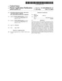 | 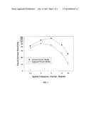 |
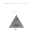 |  |
 | 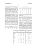 |
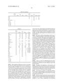 |  |
| Similar patent applications: | |
| Date | Title |
|---|---|
| 2013-07-25 | Magnetostrictive auditory system |
| 2013-11-21 | 360 degree panning stereo endoscope |
| 2014-03-13 | Mobile cardiac health monitoring |
| 2009-03-26 | Method of reducing stress |
| 2009-10-22 | Behavior modification recommender |
| Top Inventors for class "Surgery" | |
| Rank | Inventor's name |
|---|---|
| 1 | Roderick A. Hyde |
| 2 | Lowell L. Wood, Jr. |
| 3 | Eric C. Leuthardt |
| 4 | Adam Heller |
| 5 | Phillip John Plante |