Patent application title: METHODS AND COMPOSITIONS FOR MODULATING P300/CBP ACTIVITY
Inventors:
Ronen Marmorstein (Swarthmore, PA, US)
Xin Liu (Menlo Park, CA, US)
Philip A. Cole (Baltimore, MD, US)
Ling Wang (Seattle, WA, US)
Erin M. Bowers (Baltimore, MD, US)
David J. Meyers (Towson, MD, US)
Chandrani Mukherjee (Baltimore, MD, US)
IPC8 Class: AC07D40506FI
USPC Class:
514369
Class name: Five-membered hetero ring containing at least one nitrogen ring atom (e.g., 1,2,3-triazoles, etc.) 1,3-thiazoles (including hydrogenated) chalcogen bonded directly to ring carbon of the thiazole ring
Publication date: 2013-12-26
Patent application number: 20130345267
Abstract:
The present invention relates to a method for identifying compounds that
modulate the activity of p300/CBP. Compounds of the invention are
identified by designing or screening for a compound which binds to at
least one amino acid residue of the newly identified lysine-CoA inhibitor
binding site, L1 loop, electronegative pocket, or electronegative groove
of the HAT domain of p300/CBP and testing the compound for its ability to
modulate the activity of p300/CBP. Compositions and methods for
preventing or treating diseases or disorders associated with p300/CBP are
also provided as is a method for producing a semi-synthetic HAT domain.Claims:
1. A method for modulating the activity of p300/CBP comprising contacting
a p300/CBP protein with a p300/CBP inhibitor having the structure of
Formula II: ##STR00025## wherein the dotted lines independently
represent optional bonds, R1 is H, NO2, or substituted or
unsubstituted C1-4 alkyl; R2 and R3 are independently H,
CH3, or NO2; and R4 and R5 are independently H,
CH3, NO2, SO3H, halogen or substituted or unsubstituted
C1-4 alkyl, thereby modulating the activity of p300/CBP.
2. The method of claim 1, wherein the p300/CBP inhibitor is selected from the group set forth in Table 2.
3. A method for preventing or treating cancer, obesity, or diabetes comprising administering to a subject in need of treatment an effective amount of p300/CBP inhibitor having the structure of Formula II: ##STR00026## wherein the dotted lines independently represent optional bonds, R1 is H, NO2, or substituted or unsubstituted C1-4 alkyl; R2 and R3 are independently H, CH3, or NO2; and R4 and R5 are independently H, CH3, NO2, SO3H, halogen or substituted or unsubstituted C1-4 alkyl, thereby preventing or treating cancer, obesity, or diabetes in the subject.
4. The method of claim 3, wherein the p300/CBP inhibitor is selected from the group set forth in Table 2.
5. A method for identifying a compound which modulates the activity of p300/CBP comprising (a) designing or screening for a compound which binds to at least one amino acid residue of the lysine-CoA inhibitor binding site, L1 loop, electronegative pocket, or electronegative groove of the histone acetyltransferase (HAT) domain of p300/CBP; and (b) testing the compound designed or screened for in (a) for its ability to modulate the activity of p300/CBP, thereby identifying a compound that modulates the activity of p300/CBP.
6. The method of claim 5, wherein the compound binds to the substrate binding site and inhibits the activity of p300/CBP.
7. The method of claim 5, wherein step (a) is carried out in silico.
8. The method of claim 5, wherein step (a) is carried out in vitro.
9. A compound identified by the method of claim 5.
10. A pharmaceutical composition comprising a compound identified by the method of claim 5 in admixture with a pharmaceutically acceptable carrier.
11. A method for making an inhibitor of p300/CBP comprising screening for a compound which interacts with amino acid residues Arg1410, Thr1411, Trp1466, and Tyr1467 of SEQ ID NO:1 or amino acid residues Arg1446, Thr1447, Trp1502 and Tyr1503 of SEQ ID NO:2 thereby making an inhibitor of p300/CBP.
12. A method for modulating the activity of p300/CBP comprising contacting a p300/CBP protein with a compound of claim 8 thereby modulating the activity of p300/CBP.
13. A method for preventing or treating cancer, obesity, or diabetes comprising administering to a subject in need of treatment the pharmaceutical composition of claim 10, thereby preventing or treating cancer, obesity, or diabetes in the subject.
14. A method for producing a heterodimeric histone acetyltransferase (HAT) complex comprising subjecting a HAT domain and N-Cys peptide of p300/CBP to expressed protein ligation to produce a semi-synthetic HAT domain; and subjecting the semi-synthetic HAT domain to proteolysis in the presence of an inhibitor thereby producing a heterodimeric HAT complex.
Description:
INTRODUCTION
[0001] This application is a continuation of U.S. Serial application Ser. No. 12/665,751 filed May 5, 2010, which is the U.S. National Phase of PCT/US2008/067477 filed Jun. 19, 2008, which claims benefit of priority to U.S. Provisional Patent Application Ser. No. 60/945,404, filed Jun. 21, 2007, the teachings of which are incorporated herein by reference in their entireties.
BACKGROUND OF THE INVENTION
[0003] The CBP and p300 paralogs were identified by their roles in regulating cyclic AMP-related gene activation and binding to adenoviral protein E1A, respectively (Chrivia, et al. (1993) Nature 365:855-859; Eckner, et al. (1994) Genes Dev. 8:869-884). One or more copies of the p300/CBP transcriptional coactivator is encoded in organisms from worm to man, and they have been intensively studied because of their diverse and important roles in complex biological processes (Goodman & Smolik (2000) Genes Dev. 14:1553-1577). The p300/CBP protein is ˜250 kDa and contains a number of well-defined domains, many of which are crucial for its recruitment by a wide range of transcription factors (Legge, et al. (2004) J. Mol. Biol. 343:1081-1093; Mujtaba, et al. (2004) Mol. Cell. 13:251-263; Radhakrishnan, et al. (1997) Cell 91:741-752). p300/CBP has been shown to possess intrinsic histone acetyltransferase (HAT) activity which has led to a wide array of insights into its biological activities (Bannister & Kouzarides (1996) Nature 384:641-643; Ogryzko, et al. (1996) Cell 87:953-959). For example, p300/CBP HAT activity is important for its ability to act as a coactivator for a variety of transcription factors, e.g., p53, NFκB, STAT3, GATA-1, MyoD, TCF, androgen receptor (AR), and HIV Tat; thereby indicating that its HAT activity is important for a variety of pathways including cancer (Iyer, et al. (2004b) Oncogene 23:4225-4231), HIV (Kaehlcke, et al. (2003) Mol. Cell. 12:167-176) and HTLV-1 (Georges, et al. (2003) Mol. Cell. Biol. 23:3392-3404) pathogenesis; as well as, cardiac remodeling (Gusterson, et al. (2003) J. Biol. Chem. 278:6838-6847), glucose regulation (van der Heide & Smidt (2005) Trends Biochem. Sci. 30:81-86), oxygen sensing (Roe, et al. (2006) Mol. Cell 22:395-405), and steroid hormone signaling (Korzus, et al. (1998) Science 279:703-707). In addition to catalyzing the acetylation of multiple lysines on all four core histones, p300/CBP has been shown to acetylate a wide array of transcription factors and other proteins as part of its functions. Some of these p300/CBP-acetylated substrates include p53, p73, NFκB, STAT3, GATA-1, MyoD, TCF, E2F1, HMG14, HMGI(Y), androgen receptor (AR), Tat, and c-Myb (Chen, et al. (2001) Science 293:1653-1657; Costanzo, et al. (2002) Mol. Cell. 9:175-186; Thompson, et al. (2001) J. Biol. Chem. 276:33721-33729; Yuan, et al. (2005) Science 307:269-273). Although there are no precise consensus sequences for p300/CBP-mediated acetylation, there is a clear preference for nearby positively charged residues influencing targeted lysine acetylation (Thompson, et al. (2001) supra).
[0004] As noted above, p300/CBP plays a key role in regulating the transcription of a large subset of eukaryotic genes. Consistent with this important role is the fact that mutations, altered expression, and gene rearrangements of p300/CBP have been observed in a variety of diseases including cancer (Iyer, et al. (2004b) supra). For example, patients with Rubinstein-Taybi Syndrome, which involves a heterozygous mutation in one CBP allele (Murata, et al. (2001) Hum. Mol. Genet. 10:1071-1076), have an increased incidence of tumors. Additionally, point mutations thought to interfere with the catalytic activity of p300/CBP have been observed in pancreatic cancer (Gayther, et al. (2000) Nat. Genet. 24:300-303), colon cancer (Muraoka, et al. (1996) Oncogene 12:1565-1569), and lung cancer (Kishimoto, et al. (2005) Clin. Cancer Res. 11:512-519). Gene fusion events involving the CBP HAT domain have been detected in a number of acute leukemias (Borrow, et al. (1996) Nat. Genet. 14:33-41). Finally, overexpression of p300/CBP has been detected in a variety of cancers including colon (Pena, et al. (2006) Int. J. Cancer 119:2098-2104), gastric (Kim, et al. (2007) Am. J. Physiol. Cell Physiol. 292:C857-866), and thyroid (Fluge, et al. (2006) Thyroid 16:161-175) carcinoma.
[0005] The coactivator activity of p300/CBP is controlled at multiple levels and its regulation has been studied (Goodman & Smolik (2000) supra). For example, p300/CBP is known to be phosphorylated, methylated, ubiquitinated, sumoylated, and acetylated and these modifications exert a myriad of effects on p300/CBP coactivator activity by modulating its protein levels, its interactions with other proteins, and its HAT activity (Goodman & Smolik (2000) supra; Thompson, et al. (2004) Nat. Struct. Mol. Biol. 11:308-315). Regarding acetylation, there are a dense cluster of lysines in a flexible loop region of the p300/CBP HAT domain that are sites of autoacetylation (Thompson, et al. (2004) supra). Intermolecular autoacetylation of these lysines appears to upregulate p300/CBP HAT activity (Thompson, et al. (2004) supra) and also modulate protein-protein interactions with APC (Turnell, et al. (2005) Nature 438:690-695), PIC (Black, et al. (2006) Mol. Cell. 23:809-818), and ATF-2 among possibly others. Partial deletion of this p300/CBP autoacetylated loop can upregulate HAT activity and modulate transcriptional activation (Thompson, et al. (2004) supra).
[0006] Studies investigating the structure, mechanism, and inhibition of different HATs have been conducted. The most well-understood HATs are the paralogs PCAF and GCN5, and these enzymes appear to be classical members of the GNAT superfamily (Neuwald & Landsman (1997) Trends Biochem. Sci. 22:154-155) based on structure and catalytic mechanism (Vetting, et al. (2005) Arch. Biochem. Biophys. 433:212-226). The GNAT superfamily is composed of weakly conserved acetyltransferases with ˜200 residue catalytic domains that show a similar core protein fold and include enzymes involved in antibiotic resistance, melatonin biosynthesis, and polyamine metabolism (Vetting, et al. (2005) supra). The catalytic mechanism of PCAF/GCN5 and most other GNATs usually involves a ternary complex mechanism with ordered binding of the acetyl-CoA substrate prior to the amine substrate (Vetting, et al. (2005) supra). Upon ternary complex formation, there is direct transfer of the acetyl group from acetyl-CoA to the substrate amino group. The α-β fold for acetyl-CoA binding is quite conserved and many of these enzymes appear to have a catalytic base assisting in amine substrate deprotonation (Vetting, et al. (2005) supra). Bisubstrate analog inhibitors in which the amine substrate are linked to coenzyme A via an acetyl bridge have proved to be powerful inhibitors for these enzymes and have been extensively used in biochemical and structural studies (Vetting, et al. (2005) supra).
[0007] Sequence alignments and enzymology experiments on p300/CBP have led to somewhat confusing and contradictory results regarding the mechanism of p300/CBP and its structural relationship to PCAF/GCN5. For example, sequence alignments of p300/CBP and PCAF/GCN5 (Martinez-Balbas, et al. (1998) EMBO J. 17:2886-2893; Yuan & Giordano (2002) Oncogene 21:2253-2260) have shown limited homology that appears to be inconsistent with the PCAF/GCN5 structure (Poux, et al. (2002) Proc. Natl. Acad. Sci. USA 99:14065-14070). Additionally, a two substrate kinetic analysis showed a parallel line pattern suggestive of a ping-pong kinetic mechanism with a covalent enzyme intermediate (Thompson, et al. (2001) supra), potentially similar to the mechanism employed by Esa1 (Berndsen, et al. (2007) Biochemistry 46:623-629) and different from PCAF/Gcn5 (Tanner, et al. (1999) J. Biol. Chem. 274:18157-18160; Trievel, et al. (1999) Proc. Natl. Acad. Sci. USA 96:8931-8936; Vetting, et al. (2005) supra). However, experiments with acetyl-CoA-based affinity labeling agents failed to identify a key active site nucleophilic residue that would play a role in forming a covalent intermediate. Interestingly, the nominal bisubstrate analog Lys-CoA in which a derivatized lysine is bridged to coenzyme A via an acetyl linker is a powerful and selective inhibitor of p300/CBP (Lau, et al. (2000) Mol. Cell. 5:589-595). Paradoxically, longer peptide-CoA conjugates, based on better peptide substrates of p300 HAT, are weaker p300 HAT inhibitors (Lau, et al. (2000) supra). This pattern is reversed for PCAF/GCN5 where longer rather than shorter peptide-CoA conjugates are better HAT PCA Compound F/GCN5 inhibitors (Lau, et al. (2000) supra), consistent with their substrate behaviors and ternary complex mechanisms. Interestingly, deletion of the 3'-phosphate from Lys-CoA results in a 30-fold reduction in p300 HAT inhibitory potency (Cebrat, et al. (2003) Bioorg. Med. Chem. 11:3307-3313). In contrast, for a GNAT superfamily member serotonin N-acetyltransferase bisubstrate analog the 3'-phosphate is essentially completely dispensable (Khalil, et al. (1999) Proc. Natl. Acad. Sci. USA 96:12418-12423). Taken together, these studies provide little information of the nature of p300/CBP HAT mechanism and structure.
SUMMARY OF THE INVENTION
[0008] The present invention is a method for identifying a compound which modulates the activity of p300/CBP. The method of this invention involves, (a) designing or screening for a compound which binds to at least one amino acid residue of the lysine-CoA inhibitor binding site, L1 loop, electronegative pocket, or electronegative groove of the HAT domain of p300/CBP; and (b) testing the compound designed or screened for in (a) for its ability to modulate the activity of p300/CBP, thereby identifying a compound that modulates the activity of p300/CBP. In one embodiment, the compound binds to the substrate binding site and inhibits the activity of p300/CBP. In other embodiments, step (a) is carried out in silico or in vitro. Compounds identified by the present invention and pharmaceutical compositions containing the same are also provided.
[0009] The present invention also provides a method for making an inhibitor of p300/CBP. This method involves screening for a compound which interacts with amino acid residues Arg1410, Thr1411, Trp1466, and Tyr1467 of SEQ ID NO:1 or amino acid residues Arg1446, Thr1447, Trp1502 and Tyr1503 of SEQ ID NO:2 thereby making an inhibitor of p300/CBP.
[0010] Methods for inhibiting the activity of p300/CBP and preventing or treating cancer, diabetes, or obesity using one or more p300/CBP inhibitors of the invention are also provided.
[0011] The present invention is also a method for producing a semi-synthetic HAT domain. The method involves subjecting a HAT domain and N-Cys peptide of p300/CBP to expressed protein ligation to produce a semi-synthetic HAT domain; and subjecting the semi-synthetic HAT domain to proteolysis in the presence of an inhibitor thereby producing a heterodimeric HAT complex.
BRIEF DESCRIPTION OF THE DRAWINGS
[0012] FIGS. 1A and 1B show the structure of p300 and preparation and structure of p300 HAT domain. FIG. 1A is a schematic representation of p300 domain structure with selected interacting proteins. FIG. 1B is a scheme of expressed protein ligation strategy for semi-synthetic p300 HAT domain preparation.
[0013] FIG. 2 shows the relative activity of several mutants around the p300 HAT domain active site as inferred from the structure. The activity was measured using 50 nM enzyme, 20 μM AcCoA and 200 μM H4-15 for most of the mutants. W1436A, H1434A activity was measured using 50 nM enzyme, 200 μM AcCoA and 200 μM H4-15. Sub-saturating concentrations of AcCoA and H4-15 were used to more easily detect the difference between wild-type p300 and mutants resulting from Km changes. Wild-type (W.T.) p300 activity was measured under each condition to normalize the relative activity of the mutants.
[0014] FIG. 3 is a schematic view of p300 HAT domain interactions with the Lys-CoA inhibitor. Hydrophobic interactions and hydrogen bonds are indicated with solid and dotted arrows, respectively.
[0015] FIG. 4 shows the sequence alignments of histone (H2A, SEQ ID NO:3; H2B, SEQ ID NO:4; H3, SEQ ID NO:5; and H4, SEQ ID NO:6) and non-histone (p53, SEQ ID NO:7; p73, SEQ ID NO:8; HMG14, SEQ ID NO:9; E2F1, SEQ ID NO:10; dTCF, SEQ ID NO:11; HMGI(Y), SEQ ID NO:12; ATF-2, SEQ ID NO:13; c-Myb, SEQ ID NO:14; NFκB, SEQ ID NO:15; STAT3, SEQ ID NO:16; mGATA-1, SEQ ID NO:17; MyoD, SEQ ID NO:18; AR, SEQ ID NO:19; Tat, SEQ ID NO:20; p300, SEQ ID NO:21) p300 substrates with all positively charged residues are underlined. The preferred acetylation sites are indicated by the box and the proximal lysine or arginine residues are marked with a star.
[0016] FIG. 5 depicts the results of a mutagenesis study showing the relative activity of several putative substrate binding mutants. The activity was measure using 50 nM enzyme, 200 μM AcCoA and 200 μM H4-15; and is plotted as activity relative to wild-type (W.T.) p300.
[0017] FIGS. 6A and 6B show the anticipated catalytic mechanism of p300 HAT. FIG. 6A is a schematic of the catalytic mechanism of p300/CBP. FIG. 6B is a pH-rate profile of wild-type p300. The pka is at 8.36±0.14. The pH-rate profiles of Y1467F, Y1394F and H1434A were also measured and were found to not be significantly different from that of wild-type p300.
[0018] FIG. 7 depicts the results of a mutagenesis study showing the relative activity of cancer associated mutants. p300 wt and disease mutants for these studies do not contain the internal deletion of residues 1523-1554 used for the structural studies; however, they were allowed to activate by pre-incubation with acetyl-CoA. V/K is the Vmax/Km (H4-15). D1399Y, R1342P steady state assays were carried out using a radioactive assay because of their low activity with 20 μM AcCoA. All the other mutants were evaluated using a nonradioactive HAT assay with 2 mM AcCoA.
[0019] FIG. 8 shows the amino acid sequences of human p300 and human CBP. An overall structure and amino acid sequence comparison of the human p300 (SEQ ID NO:22) and human CBP (SEQ ID NO:23) HAT domain are shown.
[0020] FIG. 9 shows H4-CoA-20 linker variants for structural analysis.
[0021] FIGS. 10A and 10B show the pattern of inhibition of compound 7 versus acetyl-CoA (FIG. 10A) or H4-15 (FIG. 10B).
[0022] FIG. 11 shows a docking model of compound 7 bound to p300 HAT. Inhibitor 7 is shown as a stick drawing and enzyme residues interacting with inhibitor 7 are indicated.
DETAILED DESCRIPTION OF THE INVENTION
[0023] The transcriptional coactivator p300/CBP is a histone acetyltransferase (HAT) that regulates gene expression by acetylating histones and other transcription factors and the dysregulation of p300/CBP HAT activity contributes to various diseases including cancer. The p300/CBP proteins contain a number of well-defined domains (FIG. 1A), many of which are crucial for recruitment by a wide range of transcription factors. The high-resolution crystal structure of the human p300 HAT domain in complex with a specific bisubstrate analog inhibitor, Lys-CoA, has now been determined. The structure reveals similarity with other HAT domains in the CoA-binding region, despite the lack of sequence conservation within this region, and a novel, and presumably flexible, cofactor-binding loop within the CoA-binding core region that makes additional interactions with cofactor. HAT regions flanking the core CoA binding regions show significant structural divergence with other HATs and appears to help form a unique substrate recognition region. Structure-guided mutagenesis indicates that p300/CBP uses a different catalytic mechanism than other HATs. Mapping of p300 tumor-derived mutations onto the HAT domain structure also highlights the key role of HAT domain integrity and cofactor binding in particular in maintaining p300 tumor suppressor activity.
[0024] Because of the involvement of p300/CBP in a growing number of cellular processes, p300/CBP is a therapeutic drug target for the development of small molecule effectors. The term "effector" refers to any agonist, antagonist, ligand or other agent that affects the activity of p300/CBP used in the assays of the present invention. Effectors can be, but are not limited to, peptides, carbohydrates, nucleic acids, lipids, fatty acids, hormones, organic compounds, and inorganic compounds. The information obtained from the inhibitor-bound p300 complex crystal structures of the present invention reveal detailed information which is useful in the design, isolation, screening and determination of potential compounds which modulate the activity of p300 proteins. Compounds that bind the L1 loop, electronegative pocket, or electronegative groove, and either sterically block substrate binding, lysine-CoA inhibitor binding, or the acetylation reaction may act as effective p300/CBP-specific inhibitors. Accordingly, the present invention provides methods for identifying a compound which modulates the activity of p300/CBP. Broadly, the methods involve designing or screening for a compound which binds to at least one amino acid residue of the L1 loop, electronegative pocket, or electronegative groove of the HAT domain of p300; and testing the compound designed or screened for its ability to modulate the activity of p300/CBP. In certain embodiments, the method of the present invention is carried out using various in silico, in vitro and/or in vivo assays based on detecting interactions between the HAT domain of p300/CBP and a test compound.
[0025] In the context of the present invention, p300/CBP refers to a family member of the p300/CBP family of co-activators which have histone acetyltransferase activity. p300 is described, e.g., by Eckner, et al. ((1994) Genes Dev. 8:869-884 and is provided in GENBANK Accession Nos. NP--001420 (human) and NP--808489 (mouse). The amino acid sequence of human p300 is set forth herein as SEQ ID NO:1. p300 is related by sequence to CBP (CREB-binding protein [CREB, cyclic-AMP responsive element binding protein]), and like CBP can stimulate transcription through activation of CREB. p300 has also been identified as a co-activator of HIF1A (hypoxia-inducible factor 1 alpha), and thus plays a role in the stimulation of hypoxia-induced genes such as VEGF. CBP is also known in the art and described, e.g., by Bannister & Kouzarides ((1996) Nature 384:641-643) and provided in GENBANK Accession Nos. NP--004371 (human), NP--596872 (rat), and NP--001020603 (mouse). The amino acid sequence of human CBP is set forth herein as SEQ ID NO:2. For the purposes of the present invention, reference to p300 or CBP refers to human allelic and synthetic variants of p300 or CBP, and to other mammalian variants and allelic and synthetic variants thereof, as well as fragments of said human and mammalian forms of p300 or CBP. Synthetic variants include those which have at least 80%, preferably at least 90%, homology to p300 or CBP. More preferably such variants correspond to the sequence of p300 or CBP but have one or more, e.g., from 1 to 10, such as from 1 to 5, substitutions, deletions or insertions of amino acids. Fragments of p300 or CBP and variants thereof are preferably at least 20, more preferably at least 50 and most preferably at least 200 amino acids in size.
[0026] Compounds designed or screened for in accordance with the present invention can interact with at least one of the amino acid residues of the Lys-CoA inhibitor binding site, L1 loop, electronegative pocket, electronegative groove or substrate binding site of the HAT domain of p300/CBP via various heterogeneous interactions including, but not limited to van der Waals contacts, hydrogen bonding, ionic interactions, polar contacts, or combinations thereof.
[0027] As depicted in FIG. 1A, the HAT domain is composed of amino acid residues 1195-1673 of p300 (SEQ ID NO:1), which correspond to amino acid residues 1231-1710 of CBP (SEQ ID NO:2). As identified herein, the L1 loop of the HAT domain, flanked by the β5-strand and α4-helix, is involved in the binding of both the acetyl-CoA and lysine moieties of the Lys-CoA inhibitor. Accordingly, in one embodiment of the present invention, a compound of the invention binds to or interacts with at least one amino acid residue of the L1 loop. In the context of the present invention, the L1 loop is composed of amino acid residues 1436-1459 of p300 (SEQ ID NO:1) or the corresponding residues of CBP, i.e., amino acid residues 1472-1495. See FIG. 8.
[0028] Structure analyses disclosed herein further indicate that the edges of the α3-helix/β4-strand and α4-helix/β5-strand flank the other sides of the Lys-CoA inhibitor to form the Lys-CoA inhibitor binding site. In particular, FIG. 3 depicts amino acid residues of the p300 HAT domain which interact with the Lys-CoA inhibitor. As depicted, the CoA moiety of the Lys-CoA inhibitor interacts with at least Arg1410, Thr1411, Trp1466, Arg1462 and Ile1457 of p300, whereas the lysine moiety of the Lys-CoA inhibitor interacts with at least Trp1436, Tyr1397, Tyr1446, and Cys1438 of p300. Accordingly, particular embodiments of the present invention embrace a compound which binds to or interacts with at least one amino acid residue of Lys-CoA inhibitor binding site. In the context of the present invention, the Lys-CoA inhibitor binding site is composed of amino acid residues 1395-1467 of p300 (SEQ ID NO:1) or amino acid residues 1431-1503 of CBP (SEQ ID NO:2). In particular embodiments, the Lys-CoA inhibitor binding site includes one or more of amino acid residue 1395, 1397, 1398, 1399, 1400, 1410, 1411, 1414, 1435, 1436, 1437, 1438, 1439, 1440, 1446, 1456, 1457, 1458, 1462, 1463, 1466 and 1467 of p300 (SEQ ID NO:1), or one or more of amino acid residue 1431, 1433, 1434, 1435, 1436, 1446, 1447, 1450, 1471, 1472, 1473, 1474, 1475, 1476, 1482, 1492, 1493, 1494, 1498, 1499, 1502 and 1503 of CBP (SEQ ID NO:2).
[0029] In addition to the pocket that accommodates the lysine moiety of Lys-CoA, a second pronounced and highly electronegative pocket was identified. This second pocket, referred to herein as the "electronegative pocket", is composed of at least amino acid residues Thr1357, Glu1505, Asp1625, and Asp1628 of p300 (SEQ ID NO:1), i.e., amino acid residues Thr1393, Glu1541, Asp1662, and Asp1665 of CBP (SEQ ID NO:2). In addition, a narrow, shallow and electronegative groove connecting the two above-referenced pockets was identified. This groove, referred to herein as the "electronegative groove," is composed of at least amino acid residues Ser1396 and Tyr1397 of p300 (SEQ ID NO:1), or amino acid residues Ser1432 and Tyr1433 of CBP (SEQ ID NO:2). Based upon the structural and mutational analysis disclosed herein, the electronegative pocket and electronegative groove in conjunction with the pocket that accommodates the lysine moiety of Lys-CoA form the substrate binding site. Accordingly, in some embodiments, the present invention embraces a compound which binds to or interacts with at least one amino acid residue of the electronegative pocket or electronegative groove. In other embodiments, the present invention embraces a compound which binds to or interacts with at least one amino acid residue of the substrate binding site.
[0030] In general, it is desirable that a compound of the invention interacts with 2, 3, 4, 5, 6 or up to 25 amino acid residues of the L1 loop, electronegative pocket, electronegative groove, Lys-CoA inhibitor binding site or substrate binding site of the HAT domain of p300/CBP to enhance the specificity of the compound for p300/CBP.
[0031] In accordance with the present invention, molecular design techniques or in silico techniques can be employed to design, identify and synthesize chemical entities and compounds, including inhibitory and stimulatory compounds, capable of binding to the HAT domain. In accordance with designing compounds which modulate the activity of the HAT domain, any suitable method for determining the crystal structure of the HAT domain can be employed. However, because the production of recombinant p300/CBP HAT domain protein in quantities necessary for crystallization is difficult using conventional methods, particular embodiments of the present invention embrace a method for producing an heterodimeric HAT complex suitable for crystallization. As exemplified herein, a heterodimeric HAT complex can be produced by expressed protein ligation of the HAT domain with the N-Cys peptide of p300/CBP, wherein limited proteolysis in the presence of an inhibitor yields an active and minimally acetylated semi-synthetic HAT domain useful for subsequent crystallization. Expressed protein ligation is well-known in the art (see, e.g., Thompson, et al. (2004) supra) and depicted in FIG. 1B. In general, expressed protein ligation involves fusing the HAT domain (e.g., amino acid residues 1287-1652 of p300 or residues 1323-1689 of CBP which lack the N-Cys peptide) to a VMA intein-chitin binding domain, purifying the fusion protein with chitin resin, and converting the fusion protein to a C-terminal thioester, e.g., by treatment with MESNA. The purified fusion protein is then chemically ligated to the N-Cys peptide of the HAT domain (residues 1653-1666 of p300 or 1690-1703 of CBP) to produce an active and minimally acetylated semi-synthetic HAT domain. In particular embodiments, amino acid residues 1523-1554 of the p300 HAT domain or residues 1559-1590 of the CBP HAT domain are deleted and replaced with a potent lysine autoacetylation site as described herein to generate a loop-deleted semi-synthetic p300 HAT. Proteolysis (e.g., sequential lysis with trypsin and carboxypeptidase A and B) of the resulting semi-synthetic HAT domain in the presence of an inhibitor, e.g., Lys-CoA or other inhibitor (e.g., identified in a screening assay) then provides a heterodimeric HAT complex suitable for crystallization in accordance with conventional techniques.
[0032] The structure of the HAT domain can be used in conjunction with computer modeling using a docking program such as GRAM, DOCK, HOOK or AUTODOCK (Dunbrack, et al. (1997) Folding & Design 2:27-42) to identify potential modulators of p300/CBP. This procedure can include computer fitting of compounds to the L1 loop, electronegative pocket, electronegative groove, Lys-CoA inhibitor binding site or substrate binding site of the HAT domain to ascertain how well the shape and the chemical structure of the compound will complement these sites or to compare the compound with the binding of Lys-CoA. Computer programs can also be-employed to estimate the attraction, repulsion and stearic hindrance of the HAT domain of p300/CBP and effector compound. Generally, the tighter the fit, the lower the stearic hindrances, the greater the attractive forces, and the greater the specificity which are important features for a specific effector compound which is more likely to interact with p300/CBP rather than other classes of proteins.
[0033] Alternatively, a chemical-probe approach can be employed in the design of p300/CBP modulators or effectors. For example, Goodford ((1985) J. Med. Chem. 28:849) describes several commercial software packages, such as GRID (Molecular Discovery Ltd., Oxford, UK), which probe the L1 loop, electronegative pocket, electronegative groove, Lys-CoA inhibitor binding site or substrate binding site of the HAT domain with different chemical probes, e.g., water, a methyl group, an amine nitrogen, a carboxyl oxygen, and a hydroxyl. Favored sites for interaction between these regions or sites of the HAT domain and each probe are thus determined, and from the resulting three-dimensional pattern of such regions or sites a putative complementary molecule can be generated.
[0034] The compounds of the present invention can also be designed by visually inspecting the three-dimensional structure of the HAT domain of p300 to determine more effective inhibitors or activators. This type of modeling is generally referred to as "manual" drug design. Manual drug design can employ visual inspection and analysis using a graphics visualization program such as "O" (Jones, et al. (1991) Acta Crystallographica Section A A47:110-119).
[0035] Initially effector compounds can be selected for their structural similarity to the X, Y and Z constituents of, e.g., Lys-CoA by manual drug design. The structural analog thus designed can then be modified by computer modeling programs to better define the most likely effective candidates. Reduction of the number of potential candidates is useful as it may not be possible to synthesize and screen a countless number of compound variations that may have some similarity to known inhibitory molecules. Such analysis has been shown effective in the development of HIV protease inhibitors (Lam, et al. (1994) Science 263:380-384; Wlodawer, et al. (1993) Ann. Rev. Biochem. 62:543-585; Appelt (1993) Perspectives in Drug Discovery and Design 1:23-48; Erickson (1993) Perspectives in Drug Discovery and Design 1:109-128). Alternatively, random screening of a small molecule library could lead to modulators whose activity may then be analyzed by computer modeling as described above to better determine their effectiveness as inhibitors or activators.
[0036] Programs suitable for searching three-dimensional databases include MACCS-3D and ISIS/3D (Molecular Design Ltd, San Leandro, Calif.), ChemDBS-3D (Chemical Design Ltd., Oxford, UK), and Sybyl/3 DB Unity (Tripos Associates, St Louis, Mo.). Programs suitable for compound selection and design include, e.g., DISCO (Abbott Laboratories, Abbott Park, Ill.), Catalyst (Bio-CAD Corp., Mountain View, Calif.), and ChemDBS-3D (Chemical Design Ltd., Oxford, UK).
[0037] The compounds designed using the information of the present invention can bind to all or a portion of the HAT domain of p300/CBP and may be more potent, more specific, less toxic and more effective than known inhibitors of p300/CBP. The designed compounds can also be less potent but have a longer half-life in vivo and/or in vitro and therefore be more effective at modulating p300/CBP activity in vivo and/or in vitro for prolonged periods of time. Such designed modulators are useful to inhibit or activate p300/CBP activity to, e.g., alter cyclic AMP-related gene activation; histone acetyltransferase activity; or co-activation of p53, NFκB, STAT3, GATA-1, MyoD, TCF, androgen receptor, and HIV Tat.
[0038] The present invention also provides the use of molecular design techniques to computationally screen small molecule databases for chemical entities or compounds that can bind to p300/CBP in a manner analogous to the Lys-CoA inhibitor as defined by the structure of the present invention. Such computational screening can identify various groups which interact with one or more amino acid residues of the Lys-CoA inhibitor binding site of the HAT domain of p300 and can be employed to synthesize modulators of the present invention.
[0039] In vitro (i.e., in solution) screening assays are also embraced by the present invention. Such assays include combining p300/CBP, the p300/CBP HAT domain (e.g., as disclosed herein), or portions of the p300/CBP HAT domain (e.g., sites, or fragments disclosed herein) with acetyl-CoA and a substrate (e.g., H4-15 peptide) in solution and determining whether a test compound can sterically block the subsequent acetylation reaction.
[0040] Compounds which can be screened in accordance with the method of the present invention are generally derived from libraries of agents or compounds. Such libraries can contain either collections of pure agents or collections of agent mixtures. Examples of pure agents include, but are not limited to, proteins, polypeptides, peptides, nucleic acids, oligonucleotides, carbohydrates, lipids, synthetic or semi-synthetic chemicals, and purified natural products. Examples of agent mixtures include, but are not limited to, extracts of prokaryotic or eukaryotic cells and tissues, as well as fermentation broths and cell or tissue culture supernates. Databases of chemical structures are also available from a number of sources including Cambridge Crystallographic Data Centre (Cambridge, UK) and Chemical Abstracts Service (Columbus, Ohio). De novo design programs include Ludi (Biosym Technologies Inc., San Diego, Calif.), Sybyl (Tripos Associates) and Aladdin (Daylight Chemical Information Systems, Irvine, Calif.).
[0041] Library screening can be performed using any conventional method and can be performed in any format that allows rapid preparation and processing of multiple reactions. For in vitro screening assays, stock solutions of the test compounds as well as assay components can be prepared manually and all subsequent pipeting, diluting, mixing, washing, incubating, sample readout and data collecting carried out using commercially available robotic pipeting equipment, automated work stations, and analytical instruments for detecting the signal generated by the assay. Examples of such detectors include, but are not limited to, luminometers, spectrophotometers, and fluorimeters, and devices that measure the decay of radioisotopes.
[0042] After designing or screening for a compound which binds to at least one amino acid residue of the L1 loop, electronegative pocket, electronegative groove, Lys-CoA inhibitor binding site or substrate binding site of the HAT domain or p300/CBP, the compound is subsequently tested for its ability to modulate the activity of p300/CBP. Such testing can be based upon whether the compound modulates the HAT activity of p300/CPB (e.g., in a HAT assay), co-activation activity, or based on binding activity. To measure binding constants (e.g., Kd), any suitable method known to those in the art can be employed including, e.g., BIACORE analysis, isothermal titration calorimetry, ELISA with a known drug on the plate to show competitive binding, or by a HAT activity assay. Alternatively, the compound can be co-crystallized with p300/CBP to determine the binding characteristics through X-ray crystallography techniques. See, for example, U.S. Pat. No. 7,149,280 which discloses a method for identifying a ligand of a target macromolecule by obtaining an X-ray crystal diffraction pattern of a compound bound to the macromolecule crystal.
[0043] In accordance with the methods of the present invention, compounds were screened to identify modulators of p300/CBP HAT activity. Virtual library screening identified compound 7 as a potent inhibitor of p300/CBP HAT. Based upon the structure of compound 7, several other known compounds with benzoate moieties linked to aryl groups were analyzed for inhibitory activity. These compounds and their percent inhibition of p300/CBP activity in a coupled spectrophotometric assay as well as a direct radioactive assay are listed in Table 1.
TABLE-US-00001 TABLE 1 Com- % Inhibition pound Structure Coupled Direct 7 ##STR00001## 85.9% 17.2 ##STR00002## 17.3 ##STR00003## 6328730 ##STR00004## 90.5% 67.9% 40174 ##STR00005## ND 79.5% 6c ##STR00006## 38.1% 8.3% Pyrazolones 7, 6328730 and 40174 were obtained commercially from Chembridge, and Interbioscreen, respectively.
[0044] Novel analogs of compound 7 were also designed, synthesized, and screened for p300/CBP inhibitory activity. These compounds and their percent inhibition of p300/CBP activity in a coupled spectrophotometric assay as well as a direct radioactive assay are listed in Table 2.
TABLE-US-00002 TABLE 2 % Inhibition Com- Cou- pound Structure pled Direct 6a ##STR00007## 33.7% 71.5% 6b ##STR00008## 35.6% 33.8% 6d ##STR00009## 0% ND 6e ##STR00010## 63.4% 96.4% 6f ##STR00011## 57.4% 97.2% 6g ##STR00012## 4.5% ND 6h ##STR00013## 40.6% 94.7% 6i ##STR00014## 13.9% ND CM-26 ##STR00015## 16.3% ND 9 ##STR00016## 92.6% 55.8%
[0045] In accordance with particular embodiments of this invention, the compounds set forth in Table 1 and/or Table are employed as inhibitors of p300/CBP activity. Moreover, analogs, derivatives and salts of the compounds set forth in Tables 1 and 2 are also embraced by this invention. In this regard, the present invention further provides a method for making an inhibitor of p300/CBP. This method involves screening for a compound which interacts with amino acid residues Arg1410, Thr1411, Trp1466, and Tyr1467 of SEQ ID NO:1 or amino acid residues Arg1446, Thr1447, Trp1502 and Tyr1503 of SEQ ID NO:2. As depicted in FIG. 11, the compounds listed in Tables 1 and 2 appear to overlap with acetyl-CoA binding and can thus be used as competitive inhibitors of acetyl-CoA binding. Accordingly, some embodiments embrace compounds of Formula I
##STR00017##
wherein
[0046] A is COOH, CONH2, SO3H, COCH2NH2, COOK, wherein R is CH3, C2H5 or alkyl;
[0047] B is CH3, CF3, or halogen;
[0048] D is H or CH3;
[0049] E is any functional group or halogen;
[0050] F, G, J, K, L and M are independently any combination of C, O, S, or N;
[0051] X is (CH2)n, wherein n=0 to 6 and said chain can contain heteroatoms (e.g., O, S, or N), or X is CH2--CH═CH--(CH2)m, wherein m is 0 to 2 and the double bond is cis or trans; and
[0052] Y is C═O or (CH2)q wherein q=1 to 3.
[0053] In certain embodiments of the present invention, an inhibitor of p300/CBP is set forth in Formula II
##STR00018##
wherein the dotted lines represent optional bonds,
[0054] R1 is H, NO2, or substituted or unsubstituted C1-4 alkyl;
[0055] R2 and R3 are independently H, CH3, or NO2;
[0056] R4 and R5 are independently H, CH3, NO2, SO3H, halogen
[0057] or substituted or unsubstituted C1-4 alkyl; with the provisio that Formula I does not include a compound wherein R1 is NO2, R2 and R3 are CH3, R4 is H, and R5 COOH; a compound wherein R1 is NO2, R2 is H, R3 are CH3, R4 is H, and R5 COOH; or a compound wherein R1 is COOEt, R2 and R3 are H, R4 is H, and R5 COOH.
[0058] As used herein, alkyl groups may be straight or branched chain groups of desirably 1 to 4 carbon atoms. Methyl, ethyl, and propyl including isopropyl are particular suitable alkyl groups in the compounds of the present invention. The alkyl groups of the compounds of the present invention can be substituted by one or more different groups including H, OH, CH3, halogen, and amino groups. The number of substitutions on the alkyl group is restricted only by the number of substitutable positions and by steric constraints.
[0059] Halogen atoms in the compounds of the present invention are desirably fluorine, chlorine, bromine or iodine.
[0060] To further evaluate the efficacy of a compound identified using the method of the invention, one of skill will appreciate that a model system of any particular disease or disorder involving p300/CBP can be utilized to evaluate the adsorption, distribution, metabolism and excretion of a compound as well as its potential toxicity in acute, sub-chronic and chronic studies.
[0061] By way of illustration, Example 12 describes a cell-based assay and animal model systems which can be used to assess the inhibition of tumor cell growth by one or more compounds of the invention. Another useful method for assessing anticancer activities of compounds of the invention involves the multiple-human cancer cell line screening assays run by the National Cancer Institute (see, e.g., Boyd (1989) in Cancer: Principles and Practice of Oncology Updates, DeVita et al., eds, pp. 1-12). This screening panel, which involves approximately 60 different human cancer cell lines, is a useful indicator of in vivo antitumor activity for a broad variety of tumor types (Greyer, et al. (1992) Seminars Oncol. 19:622; Monks, et al. (1991) Natl. Cancer Inst. 83:757-766), such as leukemia, non-small cell lung, colon, melanoma, ovarian, renal, prostate, and breast cancers. Antitumor activities can be expressed in terms of ED50 (or GI50), where ED50 is the molar concentration of compound effective to reduce cell growth by 50%. Compounds with lower ED50 values tend to have greater anticancer activities than compounds with higher ED50 values.
[0062] Upon the confirmation of a compound's potential activity in one or more in vitro assays, further evaluation is typically conducted in vivo in laboratory animals, for example, measuring reduction of lung nodule metastases in mice with B16 melanoma (e.g., Schuchter, et al. (1991) Cancer Res. 51:682-687). The efficacy of a compound of the invention either alone or as a drug combination chemotherapy can also be evaluated, for example, using the human B-CLL xenograft model in mice (e.g., Mohammad, et al. (1996) Leukemia 10:130-137). Such assays typically involve injecting primary tumor cells or a tumor cell line into immune compromised mice (e.g., a SCID mouse or other suitable animal) and allowing the tumor to grow. Mice carrying the tumors are then treated with a compound of the invention and tumor size is measured to follow the effect of the treatment. Alternatively, a compound of the invention is administered prior to injection of tumor cells to evaluate tumor prevention. Ultimately, the safety and efficacy of compounds of the invention are evaluated in human clinical trials.
[0063] Compounds which bind to at least one amino acid residue of the L1 loop, electronegative pocket, electronegative groove, Lys-CoA inhibitor binding site or substrate binding site of the HAT domain or p300/CBP can be used in a method for modulating (i.e., blocking or inhibiting, or enhancing or activating) a p300/CBP. Such a method involves contacting a p300/CBP either in vitro or in vivo with an effective amount of a compound that interacts with at least one amino acid residue of the L1 loop, electronegative pocket, electronegative groove, Lys-CoA inhibitor binding site or substrate binding site of the HAT domain or p300/CBP so that the activity of p300/CBP is modulated. An effective amount of an effector or modulatory compound is an amount which reduces or increases the activity of the p300/CBP by 10%, 20%, 30%, 40%, 50%, 60%, 70%, 80%, 90% or 100%. Such activity can be monitored using the methods disclosed herein, by enzymatic assays detecting activity of the p300/CBP, or by monitoring the expression or activity of proteins which are known to be regulated by p300/CBP protein (e.g., p53, NFκB, STAT3, GATA-1, MyoD, TCF, androgen receptor, and HIV Tat).
[0064] Given the therapeutic potential of p300/CBP inhibitors in cancer (Iyer, et al. (2004) Proc. Natl. Acad. Sci. USA 101:7386-7391; Stimson, et al. (2005) Mol. Cancer. Ther. 4:1521-1532; Zheng, et al. (2004) Methods Enzymol. 376:188-199), cardiac disease (Davidson, et al. (2005) Chembiochem. 6:162-170), diabetes mellitus (Zhou, et al. (2004) Nat. Med. 10:633-637), and HIV (Varier & Kundu (2006) Curr. Pharm. Des. 12:1975-1993), the structure disclosed herein is useful for designing and screening for more specific compounds based on, e.g., the Lys-CoA scaffold. Of equal importance would be compounds that complement disease-associated mutants (Iyer, et al. (2004) supra) that are associated with p300/CBP HAT inactivation (Qiao, et al. (2006) Science 311:1293-1297).
[0065] In this regard, one of skill in the art can appreciate that modulating the activity of p300/CBP can be useful in selectively analyzing p300/CBP signaling events in model systems as well as in preventing or treating diseases and disorders involving p300/CBP. The selection of the compound for use in preventing or treating a particular disease or disorder will be dependent upon the particular disease or disorder. For example, a compound which inhibits the activity of p300/CBP will be useful in the prevention or treatment of cancer, cardiac disease, diabetes mellitus, obesity and HIV.
[0066] Prevention or treatment typically involves administering to a subject in need of treatment a pharmaceutical composition containing an effective dose of a compound identified in the screening method of the invention. In most cases this will be a human being, but treatment of agricultural animals, e.g., livestock and poultry, and companion animals, e.g., dogs, cats and horses, is expressly covered herein. The selection of the dosage or effective amount of a compound is that which has the desired outcome of preventing, reducing or reversing at least one sign or symptom of the disease or disorder being treated. For example, a subject with cancer (including, e.g., carcinomas, melanomas, sarcomas, lymphomas and leukaemias) can experience unexplained weight loss, fatigue, fever, pain, skin changes, sores that do not heal, thickening or lump in breast or other parts of the body, or a nagging cough or hoarseness, wherein treatment with a compound of the invention can prevent, reduce, or reverse one or more of these symptoms.
[0067] Pharmaceutical compositions can be in the form of pharmaceutically acceptable salts and complexes and can be provided in a pharmaceutically acceptable carrier and at an appropriate dose. Such pharmaceutical compositions can be prepared by methods and contain carriers which are well-known in the art. A generally recognized compendium of such methods and ingredients is Remington: The Science and Practice of Pharmacy, Alfonso R. Gennaro, editor, 20th ed. Lippincott Williams & Wilkins: Philadelphia, Pa., 2000. A pharmaceutically-acceptable carrier, composition or vehicle, such as a liquid or solid filler, diluent, excipient, or solvent encapsulating material, is involved in carrying or transporting the subject compound from one organ, or portion of the body, to another organ, or portion of the body. Each carrier must be acceptable in the sense of being compatible with the other ingredients of the formulation and not injurious to the subject being treated.
[0068] Examples of materials which can serve as pharmaceutically acceptable carriers include sugars, such as lactose, glucose and sucrose; starches, such as corn starch and potato starch; cellulose, and its derivatives, such as sodium carboxymethyl cellulose, ethyl cellulose and cellulose acetate; powdered tragacanth; malt; gelatin; talc; excipients, such as cocoa butter and suppository waxes; oils, such as peanut oil, cottonseed oil, safflower oil, sesame oil, olive oil, corn oil and soybean oil; glycols, such as propylene glycol; polyols, such as glycerin, sorbitol, mannitol and polyethylene glycol; esters, such as ethyl oleate and ethyl laurate; agar; buffering agents, such as magnesium hydroxide and aluminum hydroxide; alginic acid; pyrogen-free water; isotonic saline; Ringer's solution; ethyl alcohol; pH buffered solutions; polyesters, polycarbonates and/or polyanhydrides; and other non-toxic compatible substances employed in pharmaceutical formulations. Wetting agents, emulsifiers and lubricants, such as sodium lauryl sulfate and magnesium stearate, as well as coloring agents, release agents, coating agents, sweetening, flavoring and perfuming agents, preservatives and antioxidants can also be present in the compositions.
[0069] The compositions of the present invention can be administered parenterally (for example, by intravenous, intraperitoneal, subcutaneous or intramuscular injection), topically (including buccal and sublingual), orally, intranasally, intravaginally, or rectally according to standard medical practices.
[0070] The selected dosage level will depend upon a variety of factors including the activity of the particular compound of the present invention employed, the route of administration, the time of administration, the rate of excretion or metabolism of the particular compound being employed, the duration of the treatment, other drugs, compounds and/or materials used in combination with the particular compound employed, the age, sex, weight, condition, general health and prior medical history of the patient being treated, and like factors well known in the medical arts.
[0071] A physician or veterinarian having ordinary skill in the art can readily determine and prescribe the effective amount of the pharmaceutical composition required. For example, the physician or veterinarian could start doses of a compound at levels lower than that required in order to achieve the desired therapeutic effect and gradually increase the dosage until the desired effect is achieved. This is considered to be within the skill of the artisan and one can review the existing literature on a specific compound or similar compounds to determine optimal dosing.
[0072] The invention is described in greater detail by the following non-limiting examples.
Example 1
Materials and Methods
[0073] Peptide Synthesis.
[0074] Peptides CMLVELHTQSQDRF (SEQ ID NO:24) for expressed protein ligation and H4-15 (GRGKGGKGLGKGGAK; SEQ ID NO:25) for acetyltransferase assays were prepared using the solid phase peptide synthesis Fmoc strategy. The N-terminal amino group was acetylated for the H4-15 substrate peptide. Peptides were purified (>95% homogeneity) by reversed phase (C-18) high performance liquid chromatography as described previously using a gradient of water-acetontitrile (0.05% trifluoroacetic acid) (Thompson, et al. (2001) supra). Electrospray mass spectrometry of peptides confirmed the correct masses.
[0075] Constructs and Semisynthetic p300 HAT.
[0076] Semisynthetic proteins were prepared and purified following known procedures (Thompson, et al. (2004) supra). Briefly, the truncated p300 HAT domain (amino acid residues 1287-1652) or (amino acid residues 1287-1652 with an internal deletion of amino acid residues 1523-1554) containing an M1652G mutation was inserted into the pTYB2 expression plasmid (New England Biolabs, Ipswich, Mass.). Different point mutations were introduced into the plasmid by site-directed mutagenesis. For protein preparation, the truncated p300 HAT fused to VMA intein-chitin binding domain was expressed in E. coli BL21(DE3)-RIL cells at 16° C. for 16 hours induced by IPTG (0.5 mM). The cells were harvested and lysed by French press in intein lysis buffer (25 mM HEPES (pH 7.9), 500 mM NaCl, 10% glycerol, 1 mM MgSO4, and 2 mM PMSF). After centrifugation, the supernatant was applied to a chitin column that was extensively washed to remove unbound proteins. The immobilized fusion protein was treated with 200 mM MESNA to generate the thioester and ligated to a synthetic peptide corresponding to amino acid residues 1653-1666 (CMLVELHTQSQDRF; SEQ ID NO:24) over 16 hours at room temperature. The semi-synthetic p300 HAT was then applied to a MONO-S HR5/5 cation exchange column (Amersham Biosciences) for further purification. Following concentration, 10% glycerol was added before flash freezing in liquid N2 and samples were stored at -80° C. Semi-synthetic proteins showed the correct mass as determined by MALDI mass spectrometry.
[0077] Crystallization and Structure Determination.
[0078] To facilitate protein crystallization, the purified p300 HAT was subjected to a 2-step protease treatment. Briefly, concentrated p300 HAT was diluted to 1 mg/ml in a buffer containing phosphate-buffered saline, pH 7.4 and 5 mM dithiothreitol. Fifty μM Lys-CoA was added to the diluted p300 HAT and incubated for 30 minutes. Ten μg/ml trypsin was then added to the protein inhibitor complex at room temperature for 16 hours, and followed by protease treatment with 10 μg/ml carboxypeptidase A and B for another 16 hours. Following protease treatments, two bands corresponding to two protease-resistant p300 N- and C-subdomains were observed on SDS-PAGE gel and were further purified by anion exchange and gel filtration chromatography with MONOQ and SUPERDEX 200a, respectively. The molecular weights of the N- and C-subdomains were assessed to be ˜28 kDa and ˜11 kDa, respectively, by MALDI-TOF and accurate N-terminal sequences of these two subdomains were obtained by Edman degradation.
[0079] The copurified N- and C-subdomains of the p300 HAT domain were concentrated to 12 mg/ml in a buffer containing mM HEPES, pH 7.4, 150 mM sodium chloride and 5 mM dithiothreitol by ultrafiltration for crystallization using hanging-drop vapor-diffusion at room temperature. Initial crystals were obtained by mixing 0.7 μl protein with 0.7 μl reservoir containing 0.1 M HEPES. pH 7.5, 20% w/v polyethylene glycol 4,000 and 10% v/v 2-propanol (Hampton Research, Aliso Viejo, Calif.). Crystals typically appeared in two days but were not easily reproduced and did not grow large enough for diffraction studies. Streak seeding with these initial crystals was necessary to obtain crystals with typical size of 50 μm×50 μm×30 μm. Selenium-derived protein was crystallized under the same conditions as the native protein. Both native and selenium-derived crystals were cryo-protected by transferring them stepwise into a reservoir solution supplemented with 5%, 10% and 15% v/v glycerol respectively, with 500 mM sodium chloride also added into the final cryo-protection solution prior to flash freezing the crystals into liquid propane cooled by liquid nitrogen for data collection. To prepare bromide-derived crystals, native crystals were cryo-protected and frozen as described above, except that sodium chloride was replaced by sodium bromide in the final cryoprotection step and crystals were only soaked for 30-60 seconds prior to freezing them (Dauter, et al. (2000) Acta Crystallogr. D Biol. Crystallogr. 56:232-237).
[0080] Crystallographic data from both native and derivatized crystals were collected at beamline X6A at the National Synchrotron Light Source (NSLS, Brookhaven National Laboratories). Multiple-wavelength anomalous (MAD) diffraction (Hendrickson & Ogata (1997) Methods Enzymol. 276:494-523) datasets were collected for selenium and bromide-derived crystals and the data was processed and scaled with the HKL2000 suite of programs (Otwinowski & Minor (1997) Methods Enzymol. 276:307-326). Due to crystal decay of the bromide-derivatized crystals, only a dataset at the peak wavelength for bromine was useful for phase calculation in single-wavelength anomalous diffraction (SAD). Initial phases was calculated independently for Se-MAD and Br-SAD using the programs SOLVE (Terwilliger & Berendzen (1999) Acta Crystallogr. D Biol. Crystallogr. 55:849-861) and SHELX followed by using the AUTOBUILD function in RESOLVE (Terwilliger (2000) Acta Crystallogr. D Biol. Crystallogr. 56:965-972; Terwilliger (2003) Acta Crystallogr. D Biol. Crystallogr. 59:38-44) and ARP/wARP (Perrakis, et al. (1999) Nat. Struct. Biol. 6:458-463) respectively, which resulted in nearly identical models representing 90% of the input protein sequence. A partial model refinement in CNS (Brunger, et al. (1998) Acta Crystallogr. D Biol. Crystallogr. 54:905-921) with simulated annealing permitted manual building with the program 0 (Jones, et al. (1991) Acta Crystallogr. A 47(Pt 2):110-119) of additional protein residues that were not present in the initial model. Inspection of Fo-Fc difference Fourier maps at this stage facilitated the unambiguous placement of the Lys-CoA inhibitor employing the HI-CUp server (Kleywegt (2007) Acta Crystallogr. D Biol. Crystallogr. 63:94-100). The complete model was further refined using translation, liberation and screw-rotation (TLS) and restrained refinement in REFMAC (inn, et al. (2001) Acta Crystallogr. D Biol. Crystallogr. 57:122-13) implemented in CCP4i to a final resolution of 1.7 Å. The final model was checked by PROCHECK (Laskowski, et al. (1993) J. Appl. Cryst. 26:283-291) revealing good stereochemical parameters with no residues outside of the allowed regions of the Ramachandran plot.
[0081] HAT Assays.
[0082] HAT activity was determined by either a rapid and nonradioactive HAT assay that measures the production of CoASH by its facile reaction with DTNB, or by a radioactive assay (Lau, et al. (2000) supra; Thompson, et al. (2000) supra). For the DTNB assay, fixed concentrations of acetyl-CoA (2 mM) and the H4-15 peptide (400 μM) were used to measure the kcat and Km parameters for peptides and acetyl-CoA, respectively. For the radioactive assay, the concentration of [14C]acetyl-CoA was fixed at 20 μM when measuring the steady-state kinetic parameters for peptide substrates.
Example 2
Preparation and Crystallization of Semi-Synthetic p300 HAT Domain
[0083] Production of recombinant p300/CBP HAT domain protein in quantities necessary for crystallization is difficult using conventional methods because of toxicity due to aberrant acetylation of host proteins as well as heterogeneous p300 HAT autoacetylation. To overcome these challenges, a semi-synthetic human p300 HAT domain was generated using expressed protein ligation (Thompson, et al. (2004) supra) (FIG. 1B). In this procedure, p300 amino acid residues 1287-1652 were generated, fused to a VMA intein-chitin binding domain, which can be purified on chitin resin, and then converted to a C-terminal thioester by treatment with MESNA. This p300 fragment was soluble but catalytically inactive, and its activity could be recovered by native chemical ligation to N-Cys peptide (residues 1653-1666), which produces active and minimally acetylated semi-synthetic p300 HAT. To further aid in crystallization efforts, 32 residues (1523-1554) of a proteolytically sensitive region were genetically deleted and replaced with a potent lysine autoacetylation site at 1637 with an arginine residue (Thompson, et al. (2004) supra). Although well-behaved by gel filtration, this loop-deleted semi-synthetic p300 HAT failed to crystallize with various ligands tested.
[0084] It was contemplated that a residual internal flexible loop was inhibiting crystallization. Therefore, the semi-synthetic p300 HAT was treated with various ratios of trypsin. Two relatively well-defined p300 HAT fragments (about 28 kDa and 11 kDa) were generated when limited proteolysis was performed in the presence of Lys-CoA, whereas protease treatment gave a more complex mixture in the absence of p300 HAT inhibitor. By mass spectrometry and N-terminal sequencing, it was shown that the 28 kDa band corresponded to an N-terminal subdomain (N-subdomain) and that the 11 kDa band corresponded to a C-terminal subdomain (C-subdomain) and that approximately 12 amino acids were removed in addition to the 32 amino acids already genetically deleted (total removal ca. amino acids 1523-1567). The heterodimeric p300 HAT complex was prepared in the presence of Lys-CoA by carrying out an additional proteolysis of the complex with carboxypeptidase A and B and using ion exchange and gel filtration chromatography, which preserved the integrity of the complex, suggesting a stable association and structure.
[0085] Purified heterodimeric semi-synthetic p300 HAT-Lys-CoA was crystallized using hanging drop vapor diffusion and streak seeding to obtain crystals of sufficient size (50 μm×50 μm×30 μm) for X-ray data collection. Crystals of the p300 HAT-inhibitor complex formed in spacegroup P43 with one complex per asymmetric unit cell and the structure was determined by a combination of MAD and SAD using selenomethione and bromine derivatized protein (Table 3).
TABLE-US-00003 TABLE 3 Se-MAD Br-SAD Data set Peak Edge Remote Peak Native Space group P43 P43 P43 Unit cell dimensions (Å) a 61.3 61.4 61.5 b 61.3 61.4 61.5 c 101.3 101.0 101.2 Wavelength (Å) 0.9786 0.9789 0.9287 0.9193 0.9253 Resolution (Å) 50-2.0 50-2.0 50-2.0 50-1.8 50-1.7 Unique reflections 25310 25331 25271 34709 41358 Completeness (%)a 99.9 99.9 99.9 100.0 99.9 (100.0) (100.0) (100.0) (100.0) (100.0) Multiplicity 6.3 6.3 6.3 6.4 5.6 I/σ 18.4 18.8 21.3 29.2 33.5 (2.4) (2.4) (2.2) (3.5) (2.6) Rmerge (%)b 8.6 8.4 8.3 5.6 4.4 (71.9) (76.8) (81.2) (50.9) (65.8) Number of sites 6 28 FOM/CC 0.51 0.33 Resolution range 50-1.7 Rcryst (%)c 22.1 Rfree (%)d 18.2 Number of atoms Protein 2596 Lys-CoA 64 Water 299 Average B-factors (Å2) Protein 25.3 Lys-CoA 22.5 Water 35.1 Root mean square deviations Bond length (Å) 0.011 Bond angle (°) 1.466 aValues in parentheses are from the highest resolution shell. bRmerge = Σ|I - <I>|/Σ<I> cRcryst = Σ||Fo| - |Fc||/Σ|Fo| dRfree = ΣT||Fo| - |Fc||/ΣT|Fo| (where T is a test data set of 5% of the total reflections randomly chosen and set aside before refinement).
Example 3
Overall Structure of the p300 HAT-Inhibitor Complex
[0086] The overall fold of the p300 HAT domain includes a central β-sheet composed of 7 β-strands surrounded by 9 α-helices and several loops, with the last 3 α-helices and the last β-strand coming from the C-subdomain (FIG. 8). The smaller C-subdomain spans the entire structure by capping opposite ends of the larger N-subdomain with secondary structural elements that are connected by a long loop (L2) that tracks along the "bottom" side of the N-subdomain. This intimate association of the N- and C-subdomains is consistent with their protease resistance and tight heteromeric association during purification and crystallization. Within the central β-sheet the β7-strand from the C-subdomain is intercalated between the β5 and β6 strands of the N-subdomain, contributing to the integral association between the two subdomains. On the "top" surface and at one end of the β-sheet, the α2, α5 and α6 helices from the N-subdomain and the α7 helix and a loop region from the C-subdomain form a local hydrophobic core that appears to provide a structurally stable scaffold for the cleaved autoacetylation loop that connects the α6 and α7 helices of the N- and C-subdomains, respectively, of the native protein. In the presence of the Lys-CoA inhibitor, this autoacetylation loop is presumably flexible and susceptible to protease cleavage. Helices α8 and α9 from the C-subdomain also sit on the "top" of the central β-sheet and cap the other end of the β-sheet. Helices α1, α3 and α4 of the N-subdomain line the "bottom" surface of the central β-sheet and make extensive interactions with the sheet. There are two unusually long loops in the structure that are referred to herein as L1 and L2. The L2 loop from the C-subdomain connects the β7 strand and α8 helix of the C-subdomain and spans the underside of the central β-sheet while the L1 loop is intimately associated with Lys-CoA inhibitor binding. Specifically, the Lys-CoA inhibitor binds to one edge of the β-sheet with the α3 helix and β4-strand on one side and the α4 helix and β5-strand on the opposite side. The L1 loop covers what would otherwise be the solvent exposed surface of the Lys-CoA inhibitor.
[0087] As described, deletion of residues 1653-1666 leads to loss of catalytic activity. The semi-synthetic ligation junction occurs in the middle of the C-terminal α9 helix and loss of this helix would presumably have ramifications for the integrity of the overall structure. It is however, noteworthy, that circular dichroism studies suggest that the catalytically defective, C-terminally truncated p300 HAT domain does not contain a significantly perturbed HAT domain fold (Karanam, et al. (2006) J. Biol. Chem. 281:40292-40301).
Example 4
Comparison of p300 with Other HATs
[0088] A comparison of the p300 HAT domain with other HAT structures (Marmorstein (2001) J. Mol. Biol. 311:433-444) shows several differences and some similarities, despite the absence of detectable sequence conservation with other HATs. Specifically, an overlay of the p300 HAT domain with the HAT domains from yeast Gcn5, a member of the GCN5/PCAF family of HATs, and from yeast Esa1, a member of the MYST family of HATs, shows structural conservation within the central core region associated with acetyl-CoA cofactor binding. This structural homology corresponds to the A, B and D sequence motifs of the GNAT (Gcn5-related histone N-acetyltransferases) homology reported by Neuwald and Landsman (Neuwald and Landsman (1997) supra). Specifically, β-strands β1-β4, and a helices α3 and α4, show significant structural alignment within all three proteins, while β-strands β5 and β7 show additional structural alignment with Gcn5. In addition, like the Gcn5 and Esa1 HAT domains, p300 contains secondary structural elements that flank the central core acetyl-CoA binding region and appear to form the substrate binding cleft, as has been shown to be the case for the Gcn5 HAT, however, these regions structurally diverge among the three HAT domains.
[0089] Other aspects of the p300 HAT domain are very different from other HATs. In particular, the unusually long L1 loop that connects β5 and α4 and that appears to encapsulate the Lys-CoA inhibitor is a unique feature of the p300 HAT domain. Indeed, the L1 loop contributes about 30% of the total buried solvent accessible surface of 1225 Å2 of the CoA portion of Lys-CoA. The tip of the L1 loop also appears to be in position to influence protein substrate binding in a way that is distinct from other HATs. Indeed, the L1 loop buries 266 Å2 of the lysine portion of the Lys-CoA inhibitor. In addition, comparison of the electrostatic surface potential of the substrate binding surfaces of the HAT domains of Gcn5, Esa1 and p300 shows significant divergence. While Gcn5 and Esa1 show deeper and more apolar substrate binding pockets, the p300 HAT domain reveals a shallow and highly acidic site consistent with the different substrate binding properties of p300 relative to other HATs.
Example 5
Structure-Guided Mutagenesis and Functional Characterization
[0090] The p300 HAT/Lys-CoA complex structure indicates a variety of residues that may be involved in substrate binding or catalysis. Catalytic residues include four tyrosines: Tyr1394, Tyr1397, Tyr1446, and Tyr1467 within about 3-8 Å of the Lys-CoA bridging acetyl carbonyl group. His1434, Ser1396 and Trp1436 are also proximal to the active site. Interestingly, among them, the Tyr1394 side-chain phenol makes a hydrogen bond with Asp1507, and Tyr1467 makes a hydrogen bond with the Lys-CoA sulfur atom. Residues that appear particularly critical for substrate binding include Arg1410 and Trp1466 that mediate CoA interactions in the structure. A shallow pocket proximal to the lysine moiety of the Lys-CoA inhibitor also indicates that the polar residue Thr1357 and the acidic residues Glu1505, Asp1625 and Asp1628 may play a role in binding to the basic protein substrates of p300. In addition, several cancer-associated amino-acid substitution mutants including Ser1650, Asp1399, and Arg1342 were analyzed. In general, amino acid substitutions of putative catalytic residues involved conservative changes while other residues were substituted with alanines or charge reversal mutants. FIG. 2 shows a comparison of the catalytic activity of several amino acid substitution mutants of putative catalytic and CoA-binding residues. A more comprehensive analysis of amino acid substitution mutants including those mentioned above are described in the subsequent sections.
[0091] The L1 Substrate Binding Loop and Inhibitor Recognition.
[0092] The L1 loop is involved in the binding of both the acetyl-CoA and lysine moieties of the Lys-CoA inhibitor (FIG. 3). This region of the p300 HAT domain is therefore also referred to herein as the L1 substrate binding loop. This substrate binding loop L1 adopts an ordered conformation without secondary structure, largely due to the presence of six prolines out of the 25 residues within this loop. While the extended regions of the L1 loop sits over the CoA portion of the Lys-CoA inhibitor, hydrophobic residues within the tip, or turn region, sit within a local hydrophobic pocket that is formed by apolar residues from both N- and C-subdomains. In particular, Ile1447 and Phe1448 from the L1 loop interact with residues from the α8, α9 and central β-sheet residues: Phe1361, Val1401, Phe1630, Phe1641 and to a lesser extent with non-polar regions of His1377, Tyr1397 and Arg1627. In addition, the side chains of Asp1445, His1451 and main chains of Ile1447, Phe1448 from the L1 loop form a specific hydrogen bonding network with the side chain of Asp1399 and the main chain of Ser1400 from 134. Together the L1 substrate binding loop is intimately associated with both the rest of the protein and the Lys-CoA inhibitor consistent with its resistance to proteolysis.
[0093] The Lys-CoA inhibitor binds against one edge of the central β-sheet and is flanked on the opposite site by the L1 loop. The edges of the α3 helix/β4-strand and α4 helix/β5-strand flank the two other sides that together with the central β-sheet and L1 loop surround the inhibitor within a tunnel.
[0094] The CoA portion of the Lys-CoA makes extensive interactions with the p300 HAT domain (FIG. 3). Specifically, the middle portion of the pantetheine arm of the Lys-CoA inhibitor is almost completely buried by predominantly van der Waals interactions with residues from the L1 loop, α4 and β4, and the pantetheine phosphate and 3' phosphate oxygens make several direct and water-mediated hydrogen bond interactions from residues in the β4 strand and with the guanidinium side chain of Arg1410 from α3. As discussed, Lys-CoA is unusual in requiring its 3'-phosphate for high affinity interaction with p300 (Cebrat, et al. (2003) supra). In this regard, Arg1410 appears to make three hydrogen bonds with phosphates of the Lys-CoA molecule, with one involving a water-mediated and a direct hydrogen bond to the 3'-phosphate and the other being a direct hydrogen bond to the pantetheine phosphate. The importance of Arg1410 was examined by replacing it with Lys and Ala. As shown in FIG. 2 and Table 4, R1410A leads to a 15-fold increase in the K. of acetyl-CoA, but has essentially the same K. for peptide substrate compared with the wild-type p300 HAT. Unexpectedly, R1410K p300 HAT behaves much more similarly to wild-type p300 HAT, providing a strong evidence for a critical electrostatic contribution of Arg1410. In addition, the pantetheine phosphate group of Lys-CoA makes hydrogen bonds with the hydroxyl group side chain of Thr1411 and the indole ring of the Trp1466 side chain of α3 and α4, respectively, and Trp1466 also contributes hydrophobic contacts to the pantetheine arm of the CoA moiety. The modest effect on catalysis of the W1466F mutant reveals that the Trp1466-mediated hydrogen bond is unlikely to be very important for catalysis (FIG. 2); however the hydrophobic effect mediated by the Trp1466 sidechain may be nicely captured by a Phe replacement. The adenosine ring of the CoA moiety is also stabilized by a noncanonical cation-π interaction with Arg1462 from α4 and by a conventional hydrogen bond with the main chain oxygen of Ile1457 from the C-terminal end of the L1 loop.
TABLE-US-00004 TABLE 4 Km (μM) Km (μM) Structural basis for for for the mutant Enzyme H4-15 AcCoA kcat (s-1) V/K (M-1s-1).sup.† residue W.T. 164 ± 10 40 ± 6 4.1 ± 0.1 25,000 ± 1643 Y1467F 520 ± 30 17 ± 2 0.030 ± 0.001 58 ± 4 H-bond with Sulfur and may protonate AcCoA leaving group Y1394F 136 ± 30 47 ± 9 0.47 ± 0.04 3456 ± 830 Proton relay W1436A 394 ± 40 20 ± 3 0.19 ± 0.01 482 ± 60 Hydrophobic interaction for Lys-CoA H1434A 151 ± 10 88 ± 4 0.59 ± 0.02 3910 ± 290 Proton replay R1410A 190 ± 40 657 ± 90 1.2 ± 0.1 6263 ± 1300 H-bond with 3' R1410K 156 ± 40 74 ± 6 1.4 ± 0.1 8718 ± 2200 and pantetheine phosphate D1625R 640 ± 70 NM.sup..dagger-dbl. 3.6 ± 0.2 5668 ± 690 Contribute to the D1628R >1000 NM.sup..dagger-dbl. UD* ~2100** ± 90 negative surface E1505R >1000 NM.sup..dagger-dbl. UD* ~2000** ± 40 of the peptide binding site T1357R 294 ± 50 NM.sup..dagger-dbl. 3.9 ± 0.3 13163 ± 2491 Located at the T1357L 111 ± 20 NM.sup..dagger-dbl. 2.1 ± 0.1 18739 ± 3550 peptide binding site D1625R/D1628R >1000 40 ± 7 UD* ~1162** ± 60 Double mutant E1625R/D1625R/ >1000 461 ± 40 UD* ~232** ± 10 Triple mutant D1628R *UD: Undetectable. .sup..dagger-dbl.NM: Not measured. .sup.†V/K was calculated using peptide Km. **V/K was calculated using a linear fit.
[0095] The lysine component of the Lys-CoA inhibitor also makes extensive interactions with the p300 HAT domain. Specifically, the ε nitrogen atom makes a hydrogen bond with the main chain oxygen of Trp1436. The amine group that caps the carboxyl end of the lysine backbone, presumably mimicking the backbone position of the Lys-1 amino acid, forms a hydrogen bond with the hydroxyl group of Tyr1397. Three hydrophobic residues, Tyr1397 from β4, and Trp1436 and Tyr1446 from the L1 loop together with Cys1438 also from the L1 loop form a hydrophobic subtunnel that interacts with the aliphatic portion of the lysine side chain.
[0096] Substrate Binding.
[0097] In addition to the pocket that accommodates the lysine moiety of Lys-CoA, presumably mimicking the lysine side chain from native protein substrates, a second pronounced and highly electronegative pocket is present about 10 Å away from the substrate lysine. Because p300 substrates show a strong preference for the presence of a Lys or Arg side-chain two to three residues downstream or upstream of the primary acetylation site (Thompson, et al. (2001) supra) (FIG. 4), the possibility that this region might be important for the binding of protein substrates was considered. Consistent with this possibility is the presence of a narrow, shallow and electronegative groove connecting the two pockets. The wall of one side of the groove is composed of Ser1396 and Tyr1397. The negative potential of the second pocket is largely formed by the side chain atoms of residues Thr1357, Glu1505, Asp1625, and Asp1628. To examine if these regions are important for substrate binding, single, double, triple and quadruple amino acid mutagenesis experiments were carried out on the mentioned residues. These mutations were designed to (1) either occlude or change the charge state of the groove or the first pocket, (2) fill the cavity or reverse the negative charge of the second pocket.
[0098] For the first strategy, since it was shown that S1396A does not decrease activity (FIG. 2), S1396R/Y1397R and S1396R/Y1397R double mutants of p300 HAT were generated to perturb the groove. Both mutants showed severely decreased p300 HAT activity (>50-fold), indicating the importance of the groove for peptide substrate binding (FIG. 5). For the second strategy, the T1357L replacements lead to rather modest V/K effects (Table 4), indicating that the second pocket may not be deeply engaged by peptide substrate residues. For charge reversal mutagenesis analysis, the single mutant D1625R showed an increased peptide substrate K. and a V/K decrease of about 5-fold, while D1628R and E1505R mutant proteins displayed more pronounced effects; V/K was decreased by 12-fold and the K. for the H4-15 peptide substrate was too high to measure (FIG. 5 and Table 4). The double, triple, and quadruple mutations showed partially additive effects (FIG. 5 and Table 4). The D1625R/D1628R double mutant protein showed a 20-fold drop in V/K whereas the K. of acetyl-CoA was unchanged compared to that of wild-type p300 HAT. The E1505R/D1625R/D1628R triple mutant displayed a more dramatic V/K reduction; however, the acetyl-CoA K. for this triple mutant was also elevated, indicating slight structural perturbation (FIG. 5 and Table 4). Thus, the D1625R/D1628R p300 HAT mutant was selected for further analysis of substrate residue preferences.
[0099] To test the influence of electrostatic interactions on substrate sequence selection, the steady state kinetic parameters for several synthetic H4 tail peptide analogs was determined (listed in Table 5). In H4-15, there are two other basic residues (Lys5 and Lys12) near the target Lys8. It has previously been shown that either one of the nearby Lys (or Arg replacements) is sufficient to permit efficient acetylation by full-length p300 (Thompson, et al. (2001) supra). Here, the effects of replacing Lys5 and Lys12 with Ala, the neutral residue hydrogen bonding citrulline, or Asp were compared for wild-type and mutant p300 HAT as indicated in Table 5. Analysis of the single lysine substrate, analogous to the Lys-CoA component, which contains both amino and carboxyl groups derivatized as amides (Table 5), was also conducted. K. of these substrates were generally too high to be measured (>1000 μM) under the experimental conditions, highlighting the important role of the nearby positively charged residues for substrate interaction. As shown Table 5, the 22-fold V/K difference between wild-type p300 HAT protein and the D1625R/D1628R mutant for H4-15 substrate dropped to 6-fold when using Ac-Lys-NH2 as substrate. The smaller V/K drop in activity with Ac-Lys-NH2 substrate indicates that a key role of Asp1625 and Asp1628 is to assist in recruiting the basic residues of H4-15 by electrostatic interaction. Consistent with this idea, H4-12.sup.K5A/K12A or H4-12.sup.K5Cit/K12A which lack proximal basic residues but contain neutral hydrogen bonding residues show kinetic behavior similar to that of Ac-Lys-NH2 (Table 5). However, with H4-15.sup.K5D/K12D as substrate, wild-type and D1625R/D1628R p300 HAT displayed almost equal (within 2-fold) acetyltransferase activity. Taken together, these data indicate that the second highly electronegative pocket binds to the proximal basic residue (Lys or Arg) and facilitates the acetyltransferase reaction. However, the loss of 6-fold in V/K for neutral H4 substrates H4-12.sup.K5A/K12A or H4-12.sup.K5Cit/K12A or Ac-Lys-NH2 indicates that the double mutant may also interfere with the overall surface charge of the nearby first pocket as well.
TABLE-US-00005 TABLE 5 V/K(M-1s-1) SEQ V/K(M-1s-1) for Peptide Peptide Sequence ID NO: for W.T. D1625R/D1628R H4-15 GRGKGGKGLGKGGAK 25 25000 ± 1643 1162 ± 60 H4-15(K5D/K12D) GRGDGGKGLGDGGAK 26 636 ± 0 422 ± 20 H4-12(K5A/K12A) RGAGGKGLGAGA 27 3824 ± 180 515 ± 15 H4-12(K5X/K12A) RGXGGKGLGAGA* 28 5247 ± 60 738 ± 20 Ac-Lys-NH2 CH3CO-NH-Lys-CONH2 -- 1843 ± 50 326 ± 10 *X = citrulline.
[0100] Autoinhibition.
[0101] Although the lysine-rich autoacetylation loop of the p300 HAT domain is not present in the structure disclosed herein, it is contemplated that it may fold back in cis onto the highly electronegative surface of the p300 HAT domain that is proximal to the lysine binding site. Such a nonspecific electrostatic interaction is probably strong enough to prevent the binding of other substrates until intermolecular autoacetylation of the loop occurs. Autoacetylation of multiple sites within the loop, which is known to occur, may function to neutralize the lysine-rich loop such that protein substrates may compete with it for binding to p300. Though most of the acetylated lysine residues were genetically or proteolytically removed, there were some acetylated lysines (Lys1336, Lys1499, Lys1473, Lys1637) that remained (Thompson, et al. (2004) supra). These lysine residues are all located at the surface, which supports an intermolecular autoacetylation model (Karanam, et al. (2006) supra).
[0102] Catalytic Mechanism.
[0103] As discussed, studies on the p300 histone acetyltransferase reaction show conflicting evidence regarding the potential of an acetylated covalent intermediate. The catalytic relevance of the heterodimeric structure here is indicated by the heterodimeric stability through two chromatographic separations, its similarity in CoA binding to other acetyltransferases, and by site-directed mutagenesis of key active site residues contributing to turnover. The X-ray structure shows a number of potential nucleophilic residues (Ser/His/Tyr) in proximity to the predicted position of the acetyl-CoA carbonyl carbon but mutagenesis of any of these residues fails to fully abolish catalytic activity, as would be predicted for an obligate catalytic intermediate. Moreover, it has been demonstrated with acetyl-coenzyme A affinity labeling-based reagents that these do not target any critical residue for catalysis. Given the high affinity of the partial bisubstrate analog Lys-CoA for p300 HAT, it is contemplated that the protein conformation captured in the structure corresponds to a catalytic state indicating direct passage of the acetyl group from acetyl-CoA to the substrate lysine.
[0104] It is unclear why bisubstrate analogs with longer peptide moieties are weaker binders. For example, H4-CoA-20, a bisubstrate analog composed of 20 residues of the histone H4 tail, inhibits p300 more than 20-fold more weakly than Lys-CoA (Lau, et al. (2000) supra) In contrast, based on studies herein and previously (Thompson, et al. (2001) supra), the histone H4 tail peptide H4-20 is indeed processed at least 15-fold faster than Ac-Lys-NH2 (Table 5). If a classical ternary complex mechanism were operative, then more authentic bisubstrate analogs would be expected to bind to p300 more tightly than the primitive Lys-CoA analog, as occurs with PCAF/GCN5. It is believed that a "hit-and-run" or Theorell-Chance (T.-C.) acetyl transfer mechanism (Segel (1975) Enzyme Kinetics) could account for such behavior. In the T.-C. mechanism, there is no stable ternary complex as formed in standard sequential mechanisms (FIG. 6A). Rather, after the acetyl-CoA binds, the peptide substrate associates weakly with the p300 surface allowing the lysyl residue to snake through the p300 tunnel and react with the acetyl carbon. It is contemplated that a conformational change is associated with acetyl transfer and that this post-reaction state is captured in the structure disclosed herein. This post-reaction conformation further weakens peptide-enzyme interactions, somewhat analogous to, but more extreme than a loop displacement model suggested for PCAF/GCN5 (Poux, et al. (2002) supra; Zheng, et al. (2005b) Biochemistry 44:10501-10509). Thus, the "partial" bisubstrate analog Lys-CoA rather than the apparently more complete structure H4-CoA-20 is a more faithful mimic of the T.-C. reaction coordinate of p300, and the X-ray structure captures this reactive state. It is noted, however, that the parallel line pattern previously observed in a steady-state two substrate kinetic analysis of p300 (Thompson, et al. (2001) supra) is not inconsistent with a T.-C. mechanism since the acyl transfer from a thioester to an amine is effectively irreversible thermodynamically.
[0105] The precise role of various residues in facilitating the chemistry of acetyl transfer is indicated by mutagenesis and the observed structure. Steady-state kinetic analysis of Y1394F p300 HAT showed that the loss in acetyltransferase activity for this mutant was due to a decrease in kcat with insignificant K. effects. His1434A showed a 7-fold lower kcat compared with that of wild-type p300 HAT. W1436A was 50-fold slower and Y1467F was more than 400-fold slower than wild-type p300 HAT. Most of the reduction was due to a drop in kcat, indicating an important effect on the chemical step in acetyl transfer. The most critical residue is Tyr1467 (Table 4). Mutation of Tyr1467 results in a 430-fold reduction in V/K. This residue forms a direct hydrogen bond to the sulfur atom of Lys-CoA and is predicted to play a key orienting role as well as general acid function in protonating the leaving group. It is noteworthy that Y1467F showed a mildly depressed acetyl-CoA K. relative to wild-type. This indicates that ground state hydrogen bonding energy between the Tyr phenol group and the acetyl-CoA sulfur was modest and a source of strain, characteristic of a form of enzymatic rate enhancement known as ground-state destabilization (Scheibner, et al. (2002) J. Biol. Chem. 277:18118-18126). A related role for a Tyr has not been observed in other HATs but has been noted in serotonin N-acetyltransferase (Scheibner, et al. (2002) supra).
[0106] A second key residue is Trp1436. Mutation of Trp1436 into Ala reduced kcat/Km by 50-fold (Table 4). The Trp1436 indole side-chain appears to play a key role in guiding the Lys side chain into attacking acetyl-CoA by van der Waals contact. By contributing to the hydrophobic environment (formed by Tyr1397, Ty1446, Cys1438 and Trp1436) in the active site, it also likely helps reduce the Lys pKa below its native level of 10. This would serve to enhance catalysis by biasing the substrate Lys to the neutral amine which is necessary for attack. Although the backbone carbonyl of Trp1436 makes an H-bond with the ε-amino hydrogen of Lys-CoA, there is no obvious catalytic base in contrast to other HATs which are believed to require acidic residues playing this role. However, as discussed with serotonin N-acetyltransferase, the spontaneous reaction between a neutral amine and thioester is predicted to be very fast once the enzyme can template their association in correct orientation (Scheibner, et al. (2002) supra; Zheng, et al. (2004) Methods Enzymol. 376:188-199). Roles for polar residues for guiding the protons out of the active site are indicated for His1434 and Tyr1394 and/or Asp1507 which contribute modestly to catalysis (Table 4). An analysis of pH-rate profiles in the wild-type reaction (FIG. 6B) and selected mutants (data not shown) fails to account for the important pKa of 8.5 which we speculate may be the substrate Lys epsilon-amine group. Based on the discussion above, a catalytic mechanism is illustrated in FIG. 6A.
[0107] Disease Mutations.
[0108] The structure of p300 HAT permits a more detailed understanding of the enzymologic mutations that have been observed in a number of human disease states. There are now several reports of various point mutations in p300/CBP HAT domain being observed in human cancers and Rubinstein-Taybi syndrome but these have not been characterized with enzyme kinetic studies. Such p300/CBP mutations include R1378P in CBP (R1342P in p300) in Rubinstein-Taybi syndrome (Murata, et al. (2001) Hum. Mol. Genet. 10:1071-1076), D1399Y in p300 in primary colon cancer (Muraoka, et al. (1996) supra), S1650Y in p300 in pancreatic cancer (Gayther, et al. (2000) supra), and R1446C(R1410 in p300), W1472C (W1436 in p300), G1411E (G1375 in p300) in CBP in human lung cancer (Kishimoto, et al. (2005) supra). The importance of Arg1410 and Trp1436 has been discussed above, and it is likely that replacement of these critical residues by Cys will severely decrease p300/CBP HAT activity as was observed with Ala substitutions. Arg1342 is in the middle of the α2 helix, and replacement by Pro will interrupt the helix, and it is expected that this helical destabilization would disrupt the β1-β2 interaction of the core domain. Indeed, enzymatic analysis shows that a R1342P mutation results in at least a 50-fold reduction in acetyltransferase activity compared with wild-type p300 HAT (FIG. 7). In contrast, R1342K and R1342A p300 HAT mutants both show only modest 3-4-fold rate reductions, and these Arg replacements would be less likely to disrupt helical secondary structure (FIG. 7). Asp1399 is a key residue of the hydrogen bonding network between the substrate binding loop L1 and the β4 core domain where it forms a direct interaction with His1451. Mutagenesis analysis shows that the D1399Y, D1399N, and D1399A p300 HAT mutants all have very low activity (<1% wt p300 HAT activity) (FIG. 7), indicating that the L1 loop interaction with the core domain requires the negatively charged side chain of Asp1399 to form a strong hydrogen bond with His1451. Ser1650 is located at the upper middle portion of α9. It is expected that the bulkier side-chain found in S1650Y will not be accommodated by the limited space available around the tight turn region between α8 and α9, thereby interfering with the conformation of the final two α-helices, which themselves appear crucial because of their interactions with the L1 loop (the C-terminal truncated protein has very low activity probably for the same reason). As expected, S1650Y p300 HAT was greatly impaired catalytically (50-fold rate reduction), whereas the smaller replacement found in S1650A led to very similar catalytic activity compared to wild-type p300 HAT (FIG. 7). Although G1375E p300 HAT was not prepared, it is clear from the structure that a Glu substitution here would disrupt the appropriate positioning of the L1 loop for substrate binding (FIG. 7).
Example 6
p300 L1 Loop
[0109] The L1 loop (between β5 and α6, amino acid residues 1436-1459 SEQ ID NO:1) is a specialized feature of p300 HAT which serves as a lid for Lys-CoA. Based upon structural analysis, a related loop was identified within the yeast HAT RTT109 which is overall a rather close structural homolog of p300/CBP (despite very limited sequence homology). Based on the structure (FIG. 3), the L1 loop appears to be relevant for either substrate positioning, acetyl transfer, and/or product release. Thus, in addition to Trp4136Ala and Tyr1446Phe point mutations, a series of p300 HAT point mutants within the L1 loop are prepared and the kinetics (kcat, Km measurements, viscosity effects) and binding properties using acetyltransferase assays and fluorescence binding assays with CoA analogs (CoASH, acetonyl-CoA, Lys-CoA) are carried out. L1 residues that appear to be directly interactive with Lys-CoA (Ala1437, Cys1438, Pro1439, P1440, Lys1456, Pro1458) as well as residues that may be important for loop flexibility (Pro1439, Pro1440, Pro1458) are selected for Ala scanning (or Gly for Ala1437). To investigate regulation, it is determined how loop autoacetylation, which stimulates the wild-type enzyme, affects the kinetics of particular mutants. Outcomes of interest for these experiments would be the identification of key residues that affect the rate of acetyl transfer (kcat drop or change in viscosity effect), the affinity for CoA or peptide, selective effects on Lys-CoA binding or sensitivity to autoacetylation. Pro to Ala (or Gly) mutants are expected to alter the loop flexibility and provide insights into the backbone effects of these residues on loop conformation. Complementary experiments involve obtaining X-ray structures of p300 HAT in complex with CoASH and acetonyl-CoA to determine how the L1 loop and P1 pocket are influenced by the Lys-side chain. Taken together, these studies allow a more complete understanding of the relationship between structure and mechanism of p300/CBP and how autoacetylation exerts its effect on catalysis. These experiments are also relevant in the comparative biochemistry of yeast RTT10999 and p300/CBP, which as mentioned are unique in containing the L1 loop. A practical result from these L1 mutant studies is the identification of mutants that are less potently inhibited by Lys-CoA (and other inhibitors) but still retain robust acetyltransferase activity. The application of such mutants in cellular studies in "knock-in" type experiments allows for the discrimination between the biological functions of p300 and CBP HATs by permitting selective enzyme inhibition.
Example 7
p300 HAT in Protein-Protein Interactions
[0110] The p300 HAT structure shows a negatively charged pocket (P2) near the Lys moiety of Lys-CoA and has led to the plausible notion that basic substrates interact in this pocket. It is contemplated that the b-ZIP domain of ATF2 which shows affinity for the loop-deleted or autoacetylated p300 HAT and is an in vitro p300 substrate may interact with the p300 P2 pocket. It has been shown that two Asp residues (Asp1625, Asp1628) may be central to the preference for basic peptide substrates based on mutagenesis studies with the Asp-Asp/Arg-Arg variant. Accordingly, related experiments can be carried out with the ATF2-b-ZIP domain to determine whether mutation of these residues affects p300 HAT binding or acetylation of this transcription factor. Pull-down experiments as well as acetyltransferase assays are carried out on the Asp-Asp/Arg-Arg p300 HAT to see if GST-ATF2-bZIP shows differential interaction with the P2 mutant, as expected. Wherein the Asp-Asp/Arg-Arg mutant does not show reduced interaction with ATF-2-b-ZIP, this indicates an alternate p300 region of interaction. In either case, co-crystallization of p300 HAT with ATF2-b-ZIP is performed to determine the high resolution structural basis of interaction.
[0111] Regarding histone peptide substrate recognition by p300, studies with bisubstrate analogs, structure, and steady-state kinetics indicate that co-crystallization with H4 peptide or an H4-CoA-20 analog are unlikely to reveal relevant interactions because they are weak ligands. To circumvent this, H4-CoA-20 variants with extended linkers between peptide and CoA (L, X, Z; see FIG. 9) are employed. A compound with a longer and flexible linker between the CoA and the H4 tail peptide is expected to place the H4 peptide moiety near the histone substrate's initial encounter surface on p300, permitting capture by X-ray crystallography. After synthesizing the compounds shown in FIG. 9, their potency is evaluated as p300 HAT inhibitors. The most potent compounds could indicate that dual occupancy of the p300 HAT `Lys-CoA tunnel` as well as the adjacent 2-Asp pocket P2 is possible. This is determined by making H4 variants that replace the basic residues Lys5 and Lys12 as well as testing the p300 Asp-Asp/Arg-Arg mutant enzyme. Crystallographic analysis with the novel H4-CoA conjugates can also be performed.
[0112] Using cell co-transfection and in vitro enzymatic experiments, it has been found that a transcription factor called MAML1, critical in Notch signaling can activate full-length p300 HAT and also induce its hyper-autoacetylation. Like many transcription factors, it is known that MAML1 can bind to the p300/CBP C/H3 domain. It is contemplated that engaging the C/H3 domain with MAML1 relieves an intramolecular autoinhibitory interaction between the p300 HAT domain and the C/H3 domain (FIG. 1A). To demonstrate this, a trans interaction between p300 HAT and recombinant C/H3 domain is shown by examining the ability of the C/H3 domain to modulate p300 HAT domain activity and testing for affinity using pull-downs.
Example 8
Analysis of the p300 Autoacetylation Loop
[0113] To interrogate the regulatory autoacetylation loop in p300 HAT, a circularly permuted protein domain is employed. Such a circular permutation involves linking the natural N- and C-termini (residues 1287 and 1666) of the HAT domain and creating new termini at 1565 (N-terminus) and 1520 (C-terminus). This construct is then fused to an intein through its new C-terminus and the thioester generated is ligated to synthetic peptides containing the key autoacetylated lysines. This approach is particular useful because of the close proximity between the natural N- and C-termini; the p300 HAT crystal structure indicates that these termini are a mere 15 Å apart. Moreover, it has been demonstrated that the unlinked N- and C-subdomains of p300 HAT form a very stable complex, which also supports the use of circular permutation. In generating this circularly permuted p300 HAT, several linker lengths (6-10 residues, Gly rich linkers) can be tested to optimize expression. Because p300 HAT activity is toxic to E. coli, the production of the p300 Tyr1467Phe catalytically impaired mutant can be analyzed first. While this mutant will have reduced utility in acetyltransferase studies, it can be employed to assess the influence of the ligated regulatory loop (amino acid residues 1520-1565) on protein-protein interactions in binding studies. Ligation with various mono, di, and triacetylated regulatory segment peptides can be used to explore site-specific modifications on interaction. After completion of experiments on inactive circularly permuted p300 HAT, investigations with the non-catalytically impaired HAT are carried out. To limit toxicity from host protein p300 acetylation, the catalytically active HAT is coexpressed with Sir2 deacetylase. While autoacetylation of several residues in the circularly permuted p300 HAT (outside the regulatory loop which is installed in vitro) is likely, as demonstrated herein, these sites are not very influential on p300 HAT activity. With the semi-synthetic circularly permuted p300 HAT proteins in hand, kinetic parameters of the various site-specifically acetylated forms are measured. These studies generate unique and important new information on how p300/CBP is regulated. If functional effects conferred by the unilaterally attached regulatory segment are not observed, the free (C-terminal end) can be linked by native chemical ligation to the new N-terminus of the circularly permuted protein. This can be achieved by generating a C-terminal thioester synthetic segment and an N-Cys on the N-terminus using Factor Xa cleavage or by a trans-splicing strategy.
[0114] As an alternative approach to generating a circularly permuted protein domain, a heterodimeric p300 HAT is generated by producing separate non-attached segments, which are co-expressed in E. coli. The N-subdomain is fused to an intein to allow ligation to regulatory segment peptides. The heterodimers thus produced would then be used in binding and catalytic studies as described for the circularly permuted protein.
Example 9
Virtual Library Screening of p300/CBP
[0115] Regulatory molecules of p300/CBP HAT were identified by using the coordinates of the p300 HAT-Lys-CoA crystal structure and systematically docking into the active site (in the Lys-CoA binding region) the structures of -2 million commercially available compounds (from the Chembridge library) in silico via ICM software algorithm. The top 200 compounds were selected, which also had favorable drug-like properties (MW<500, log P=1-6), and screened against p300 HAT using a spectrophotometric coupled assay that measures CoASH production in real-time. After secondary and tertiary screens (including a direct, radioactive assay that measures 14C-incorporation from acetyl-CoA into H4 tail peptide), three compounds (7, 17.2 and 17.3) were identified, which had IC50s less than 20 μM, e.g., the IC50 of 7 was 3.09±0.62 μM. Moreover, 7 was shown to exhibit competitive inhibition against acetyl-CoA (FIG. 10A) and mixed-type inhibition with H4-15 substrate (FIG. 10B).
##STR00019##
[0116] The chemical structures and purity of these three compounds were analyzed by NMR and mass spectrometry. It is noteworthy that these compounds contain a benzoate moiety linked to a series of aryl groups, which based on the computational docking (FIG. 11) overlaps with CoA binding.
Example 10
Mechanistic Analysis of p300/CBP HAT Inhibitors
[0117] The three small molecule p300 HAT inhibitors (7, 17.2 and 17.3) identified using virtual library screening provided new leads for further development. Based upon the docking model of binding of compound 7 (FIG. 11) and pattern of competitive inhibition of compound 7 versus acetyl-CoA (FIG. 10A), it is expected that analogs of compound 7 will compete for binding with acetyl-CoA. Similarly, based upon the pattern of mixed-type inhibition of compound 7 versus peptide substrate (FIG. 10B), it is expected that analogs of compound 7 exhibit non-competitive binding. Compounds that are found not to be linear competitive inhibitors vs. acetyl-CoA (studies indicate that 7 is competitive with Ki=400 nM) are likely binding differently from the proposed docking model. From the docking model of 7 to p300 HAT (FIG. 11), important hydrogen bonding roles are indicated for the side-chains of Arg1410, Thr1411, Trp1466 (all interacting with the inhibitor carboxylate) as well as the side-chain of Tyr1467 (hydrogen bonding to the nitro group). While Arg1410 and Tyr1467 are critical for CoA binding and/or catalysis, mutation of Trp1466 is well-tolerated. To explore the specificity of the small molecule HAT inhibitors, these compounds are tested against other acetyltransferases (PCAF, AANAT, RTT109, EsaI), which assists in the prioritization of candidates for pharmacologic applications.
[0118] Indeed, it was demonstrated that compound 7 was specific for p300 HAT (85.9% inhibition) compared to AANAT (4.8% inhibition) and PCAF (0% inhibition).
Example 11
Synthetic Derivation of p300/CBP HAT Inhibitors
[0119] Synthesis of congeners of 7 involved the use of Knoevenagel condensation of furanyl-aldehyde derivatives with a set of pyrazolone building blocks.
General Procedure for Synthesis of 1-Aryl-3-Methyl-Pyrazol-5-One (3)
[0120] 1-Aryl-3-methyl-pyrazol-5-one 3 was synthesized following a modified literature procedure (Scheme 1). (Kim & Lee (1991) Bull. Korean Chem. Soc. 12:376). A suspension of ethyl acetoacetate (2.8 mmol) and corresponding arylhydrazine (2, 3.3 mmol) in glacial acetic acid (30 ml) was stirred at reflux for 24 hours. The reaction mixture was concentrated and the solid precipitated out was filtered and washed with dichloromethane to remove traces of acetic acid. Yield 65-73%. 3b was synthesized by esterification of 3a in ethanol and purified by column chromatography using dichloromethane as eluent.
##STR00020##
wherein R1 is 4-COOH (3a), 4-COOEt (3b), 4-CONH2 (3c), 3-COOH (3d), 4-SO3H (3e), 4-Cl (3f).
[0121] 1-Bromo-4,5-dimethyl-2-nitrobenzene 4a was synthesized in 81% yield using Sandmeyer reaction on 4,5-dimethyl-2-nitroaniline following a known procedure (Scheme 2) (Langner, et al. (2005) Chem. Eur. J. 11:6254). Ethyl 2-iodobenzoate 4d was prepared from esterification of corresponding halo-acid by refluxing in ethanol for 24 h. Pure product was obtained in 85% yield by column chromatography using dichloromethane as eluent.
##STR00021##
General Procedures for Synthesis of 5-Aryl-2-Furaldehydes (5)
[0122] 5-Aryl-2-furaldehyde 5 was synthesized from Suzuki coupling of 5-formyl-2-furanboronic acid and corresponding aryl halides 4 in 72-75% yields following a literature procedure (Scheme 3) (Hosoya, et al. (2003) Bioorg. Med. Chem. 11:663). The crude solid products were purified by column chromatography using dichloromethane as eluent.
##STR00022##
wherein R2 is 2-NO2, 3,4-diMe (4a, 5a); H (4b, 5b); 4-NO2 (4c, 5c); 2-COOEt (4d, 5d).
General Procedure for Synthesis of 1-Aryl-3-Methyl-4-[[5-Aryl-2-Furanyl]Methylene]-Pyrazol-5-One (6)
[0123] The final compounds were synthesized following a modified literature procedure (Scheme 4) (2005) Russ. J. Org. Chem. 41:742). An equimolar solution of 1-aryl-3-methyl-pyrazol-5-one, 5-aryl-2-furaldehyde and diethylamine was stirred in ethanol at 50° C. for 1-3 hours. On cooling the precipitated red solid was filtered and further purified by column chromatography using 2-10% MeOH/DCM as eluent. The structures of all the compounds were confirmed from 1H NMR, ESI-MS and HRMS analysis.
##STR00023##
wherein for 3 R1 is 4-COOH (3a), 4-COOEt (3b), 4-CONH2 (3c), 3-COOH (3d), 4-SO3H (3e), 4-Cl (3f); for 5 R2 is 2-NO2, 3,4-diMe (5a), H (5b), 4-NO2 (5c), 2-COOEt (5d); and for 6a R1 is 4-COOH and R2 is H, 6b R1 is 4-COOH and R2 is 4-NO2, 6c R1 is 4-COOH and R2 is 2-COOEt, 6d R1 is 4-COOEt and R2 is 2-NO2, 3,4-diMe, 6e R1 is 3-COOH and R2 is 2-NO2, 3,4-diMe, 6f R1 is 4-SO3H and R2 is 2-NO2, 3,4-diMe, 6g R1 is 4-COOH and R2 is 2-NO2, 3,4-diMe, 6h R1 is 4-CONH2 and R2 is 2-NO2, 3,4-diMe, and 6i R1 is 4-C1 and R2 is 2-NO2, 3,4-diMe.
[0124] Compound 6a was obtained as a red solid in 50% yield, mp 306-309° C. 1H NMR (400 MHz, DMSO-d6) δ: 2.35 (3H, s, CH3), 7.46-7.55 (4H, m, ArH), 7.75 (1H, s, CH), 7.96-8.02 (4H, m, ArH), 8.08 (2H, m, ArH), 8.66 (1H, br s, ArH), 12.84 (1H, br s, COOH); HRMS: Calculated [M]+ for C22H16N2O4, 372.1110; observed [M+H]+373.1189, observed [M+H]+ from ESI-MS: 373.3.
[0125] Compound 6b was obtained as a red solid in 60% yield, mp 338-341° C. 1H NMR (400 MHz, DMSO-d6) δ: 2.35 (3H, s, CH3), 7.73 (2H, d, J=4.0 Hz, ArH), 7.77 (1H, s, ArH), 7.99 (2H, d, J=8.8 Hz, ArH), 8.07 (2H, d, J=8.8 Hz, ArH), 8.17 (2H, d, J=9.2 Hz, ArH), 8.36 (2H, d, J=8.8 Hz, ArH), 12.84 (1H, br s, COOH); HRMS: Calculated [M]+ for C22H15N3O6, 417.0961; observed [M+H]+418.1040.
[0126] Compound 6c was obtained as a red solid in 41% yield, mp 249-251° C. 1H NMR (400 MHz, DMSO-d6) δ: 1.16 (3H, t, J=7.6 Hz, COOCH2CH3), 2.33 (3H, s, CH3), 4.27 (2H, q, J=7.0 Hz, COOCH2CH3), 7.22 (1H, d, J=4.0 Hz, ArH), 7.60 (1H, d, J=6.0 Hz, ArH), 7.63 (1H, m, ArH), 7.68-7.81 (2H, m, ArH), 7.87 (1H, d, J=7.2 Hz, ArH), 8.00-8.02 (2H, m, ArH), 8.08 (2H, d, J=8.4 Hz, ArH), 8.70 (1H, d, J=3.2 Hz, ArH), (1H, br s, COOH); HRMS: Calculated [M]+ for C25H20N2O6, 444.1321; observed [M+H]+445.1396, observed [M+H]+ from ESI-MS: 445.2. See WO 2006/129583 and WO 2006/129587.
[0127] Compound 6d was obtained as a red solid in 70% yield, mp 192-194° C. 1H NMR (400 MHz, CDCl3) δ: 1.40 (3H, t, J=7.2 Hz, COOCH2CH3), 2.34 (3H, s, CH3), 2.38 and 2.39 (6H, 2s, 2×CH3), 4.37 (2H, q, J=7.2 Hz, COOCH2CH3), 6.92 (1H, d, J=4.0 Hz, ArH), 7.28 (1H, s, CH), 7.53 (1H, s, ArH), 7.65 (1H, s, ArH), 8.08-8.14 (4H, m, ArH), 8.82 (1H, d, J=4.0 Hz, ArH); HRMS: Calculated [M]+ for C26H23N3O6, 473.1587; observed [M+H]+474.1660, observed [M+H]+ from ESI-MS: 474.3.
[0128] Compound 6e was obtained as a red solid in 64% yield, mp 260-262° C. 1H NMR (400 MHz, DMSO-d6) δ: 2.33, 2.35 and 2.36 (9H, 3s, 3×CH3), 7.18 and 7.23 (1H, dd, J=4.0 and 18.8 Hz, ArH), 7.53-7.58 (2H, m, ArH), 7.70-7.78 (2H, m, ArH), 7.85 (1H, s, CH), 8.15 (1H, d, J=7.0 Hz, ArH), 8.55 (1H, d, J=1.2 Hz, ArH), 8.71 (1H, d, J=3.2 Hz, ArH), 13.12 (1H, br s, COOH); HRMS: Calculated [M]+ for C24H19N3O6, 445.1274; observed [M+H]+446.1349, observed [M+H]+ from ESI-MS: 446.2.
[0129] Compound 6f was obtained as a red solid in 58% yield, mp 210-213° C. 1H NMR (400 MHz, DMSO-d6) δ: 2.31, 2.33 and 2.35 (9H, 3s, 3×CH3), 6.90-6.96 (1H, m, ArH), 7.20 (1H, m, ArH), 7.45-7.55 (1H, m, ArH), 7.63-7.72 (2H, m, ArH), 7.76 (1H, s, CH), 7.84 (1H, s, ArH), 7.90 (1H, d, J=8.4 Hz, ArH), 8.23 (1H, br s, ArH), 10.58 (1H, s, SO3H); HRMS: Calculated [M]+ for C23H19N3O7S, 481.0944; observed [M+H]+ 482.1021, observed [M+H]+ from ESI-MS: 482.3.
[0130] Compound 6g was obtained as a red solid in 72% yield. Calculated [M]+ for C24H19N3O6, 445.1274; observed [M+H]+ from ESI-MS: 446.1. The synthesized 6g was compared with compound 7 obtained commercially on TLC and found of same Rf (4% MeOH/DCM).
[0131] Compound 6h was obtained as a red solid in 40% yield, mp 273-275° C. 1H NMR (400 MHz, DMSO-d6) δ: 2.32, 2.33 and 2.35 (9H, 3s, 3×CH3), 7.22 (1H, d, J=3.2 Hz, ArH), 7.33 (1H, s, ArH), 7.56 (1H, s, ArH), 7.75 (1H, s, 1H), 7.84 (1H, s, ArH), 7.92-8.01 (3H, m, 3ArH, CONH2), 8.68 (1H, d, J=2.4 Hz, ArH); HRMS: Calculated [M]+ for C24H20N4O5, 444.1434; observed [M+H]+445.1507, observed [M+H]+ from ESI-MS: 445.4.
[0132] Compound 6i was obtained as a red solid in 75% yield, mp 220-223° C. 1H NMR (400 MHz, CDCl3-d6) δ: 2.32 (3H, s, CH3), 2.37 and 2.38 (6H, 2s, 2×CH3), 6.90 (1H, d, J=4.0 Hz, ArH), 7.26 (1H, s, CH), 7.36 (2H, d, J=8.4 Hz, ArH), 7.51 (1H, s, ArH), 7.63 (1H, s, ArH), 7.94 (2H, d, J=8.8 Hz, ArH), 8.80 (1H, d, J=4.0 Hz, ArH); HRMS: Calculated [M]+ for C23H18N3O4Cl, 435.0986; observed [M+H]+436.1061, observed [M+H]+ from ESI-MS: 436.1.
General Procedure for Synthesis of 7
[0133] To a solution of 6g (2.24 mmol) in methanol (20 ml), sodium borohydride (2.24 mmol) was added portion-wise, effervescence was evolved and the color of the solution changed from dark red to light yellow. The resulting solution was stirred at room temperature for 30 minutes, solvent was removed and pH of the solution was made 7.0 on adding dilute HCl drop-wise. The resulting mixture was extracted with EtOAc (2×50 ml) and concentrated to give an orange solid in 80.0% yield, mp 160-163° C. 1H NMR (400 MHz, DMSO-d6) δ: 2.02 (3H, s, CH3), 2.27 and 2.28 (6H, 2s, 2×CH3), 3.56 (2H, s, CH2), 6.05 (1H, br s, ArH), 6.61 (1H, br s, ArH), 7.50 (1H, s, ArH), 7.61 (1H, s, ArH), 7.85 (2H, d, J=8.4 Hz, ArH), 8.04 (2H, d, J=8.4 Hz, ArH), 12.80 (1H, br s, COOH); HRMS: Calculated [M]+ for C24H21N3O6, 447.1430; observed [M+H]+448.1507, observed [M+H]+ from ESI-MS: 448.3.
General Procedure for Synthesis of 9
[0134] 3-Methyl-pyrazol-5-one was synthesized in 60% yield on stirring an equimolar mixture of hydrazine monohydrate and ethyl acetoacetate in ethanol. The exothermic reaction was then cooled in ice-bath to precipitate out a solid white product. The structure of the product was confirmed from ESI-MS and comparing the observed melting point (214-216° C.) with the literature melting point (218° C.; Singh, et al. (2005) Eur. J. Pharm. Sci. 25:255-262). Compound 8 was obtained in on alkylation of 3-methylpyrazol-5-one with equimolar 1-bromovaleric acid by refluxing in 1,4-dioxane for 48 hours following conventional procedures (Belmar, et al. (2005) J. Braz. Chem. Soc. 16:179). See Scheme 5. The structure of compound 8 was confirmed from ESI-MS and used for next step without further purification.
##STR00024##
[0135] Compound 9 was obtained in 15% yield from pyrazole 8 and 5-(2-nitro-4,5-dimethylphenyl)-2-furaldehyde 5a following the procedure described for the synthesis of compound 6 in Scheme 4. The product was purified by column chromatography using 2-3% MeOH/DCM as eluent. Compound 9 was obtained as a red solid in 15% yield, mp 240-243° C. 1H NMR (400 MHz, CDCl3) δ: 1.58-1.74 (2H, m, CH2), 2.23 (5H, s and m, CH3 and CH2), 2.37 and 2.39 (10H, 2s and 2m, 2×CH3 and 2×CH2), 6.87 (1H, d, J=4.0 Hz, ArH), 7.21 (1H, s, CH), 7.52 (1H, s, ArH), 7.63 (1H, s, ArH), 8.71 (1H, br s, COOH), 8.75 (1H, d, J=4.0 Hz, ArH); HRMS: Calculated [M]+ for C22H23N3O6, 425.1587; observed [M+H]+ NA.
Example 12
Activity of p300/CBP HAT Inhibitors
[0136] Novel HAT inhibitors of the instant invention can be analyzed in a variety of systems with implications for endocrine, cancer, neuropsychiatric, and immunologic therapeutics. The compounds can be assessed in defined well-known model systems used to assess cellular permeability, toxicity, and pharmacodynamic effects. These assays include western blot analysis of histone acetylation as well as HAT-responsive transcriptional reporters that are specific for p300/CBP-mediated acetylation reactions. Regarding endocrine disorders, it has been found that p300/CBP HAT inhibition by Lys-CoA-Tat blocks CREB signaling and glucagon-induced glucose elevation. These data indicate that novel p300/CBP inhibitors could play a role in the treatment of diabetes mellitus. In addition, it has been shown that Lys-CoA-Tat can inhibit melanoma growth in three different cell lines. New HAT inhibitor scaffolds would thus be useful in treating cancers such as melanoma.
[0137] Indeed, the data disclosed herein indicates that inhibitors with a benzoate moiety linked to various aryl groups can inhibit p300/CBP HAT activity (see Tables 1 and 2) and find use in diseases mediated by p300/CBP HAT. Both cell-based assays and animal model studies are performed to further demonstrate efficacy in the treatment of diseases such as cancer.
[0138] Cell-Based Assay.
[0139] Cells from a P388 cell line (CellGate, Inc., Sunnyvale, Calif.) or human malignant melanoma cell line SK-MEL-2 are grown in RPMI 1640 cell medium containing fetal calf serum (10%), L-glutamine, penicillin, streptomycin and are split twice weekly. All compounds are first diluted with DMSO. Later serial dilutions are done with a phosphate-buffered saline solution. All dilutions are done in glass vials and the final DMSO concentration is generally below 0.5% by volume. Final two-fold dilutions are done in a 96-well plate using cell media so that each well contains 50 μL. All compounds are assayed over multiple concentrations. Cell concentration is measured using a hemacytometer and the final cell concentration is adjusted to about 1×10⁴ cells/mL with cell medium. The resulting solution of cells (50 μL) is then added to each well and the plates are incubated for 5 days in a 37° C., 5% CO2, humidified incubator. MTT solution (3-[4,5-dimethylthiazol-2-yl]-2,5-diphenyltetrazolium bromide, 10 μL) is then added to each well and the plates are re-incubated under identical conditions for 2 hours. To each well is then added acidified isopropanol (150 μL of i-PrOH solution containing 0.05 N HCl) and mixed thoroughly. The plates are then scanned at 595 nm and the absorbances are read (Wallac Victor 1420 Multilabel Counter). The resulting data is then analyzed to determine an ED50 value. Compounds that kill cancer cells, but fail to kill normal cells, find application in the prevention or treatment of cancer.
[0140] Mouse Ovarian Carcinoma Zenograft Model.
[0141] Compounds of the invention are evaluated in an ovarian carcinoma xenograft model of cancer, based on that described by Davis, et al. ((1993) Cancer Research 53:2087-2091). This model, in brief, involves inoculating female nu/nu mice with 1×10⁹ OVCAR3-icr cells into the peritoneal cavity. One or more test compounds are administered, e.g., prior to or after tumor cell injection, by the oral route as a suspension in 1% methyl cellulose or intraperitoneally as a suspension in phosphate-buffered saline in 0.01% TWEEN-20. At the conclusion of the experiment (4-5 weeks) the number of peritoneal cells are counted and any solid tumor deposits weighed. In some experiments tumor development is monitored by measurement of tumor specific antigens.
[0142] Rat Mammary Carcinoma Model.
[0143] Compounds of the invention are evaluated in a HOSP.1 rat mammary carcinoma model of cancer (Eccles, et al. (1995) Cancer Res. 56:2815-2822). This model involves the intravenous inoculation of 2×10⁴ tumor cells into the jugular vein of female CBH/cbi rats. One or more test compounds are administered, e.g., prior to or after tumor cell injection, by the oral route as a suspension in 1% methyl cellulose or intraperitoneally as a suspension in phosphate-buffered saline and 0.01% TWEEN-20. At the conclusion of the experiment (4-5 weeks) the animals are killed, the lungs are removed and individual tumors counted after 20 hours fixation in Methacarn.
[0144] Mouse B16 Melanoma Model.
[0145] The anti-metastatic potential of compounds of the invention is evaluated in a B16 melanoma model in C57BL/6. Mice are injected intravenously with 2×10⁵ B16/F10 murine tumor cells harvested from in vitro cultures. Inhibitors are administered by the oral route as a suspension in 1% methyl cellulose or intraperitoneally as a suspension in phosphate-buffered saline pH 7.2 and 0.01% TWEEN-20. Mice are killed 14 days after cell inoculation and the lungs removed and weighed prior to fixing in Bouin's solution. The number of colonies present on the surface of each set of lungs is then counted.
Sequence CWU
1
1
3012414PRTHomo sapiens 1Met Ala Glu Asn Val Val Glu Pro Gly Pro Pro Ser
Ala Lys Arg Pro 1 5 10
15 Lys Leu Ser Ser Pro Ala Leu Ser Ala Ser Ala Ser Asp Gly Thr Asp
20 25 30 Phe Gly Ser
Leu Phe Asp Leu Glu His Asp Leu Pro Asp Glu Leu Ile 35
40 45 Asn Ser Thr Glu Leu Gly Leu Thr
Asn Gly Gly Asp Ile Asn Gln Leu 50 55
60 Gln Thr Ser Leu Gly Met Val Gln Asp Ala Ala Ser Lys
His Lys Gln 65 70 75
80 Leu Ser Glu Leu Leu Arg Ser Gly Ser Ser Pro Asn Leu Asn Met Gly
85 90 95 Val Gly Gly Pro
Gly Gln Val Met Ala Ser Gln Ala Gln Gln Ser Ser 100
105 110 Pro Gly Leu Gly Leu Ile Asn Ser Met
Val Lys Ser Pro Met Thr Gln 115 120
125 Ala Gly Leu Thr Ser Pro Asn Met Gly Met Gly Thr Ser Gly
Pro Asn 130 135 140
Gln Gly Pro Thr Gln Ser Thr Gly Met Met Asn Ser Pro Val Asn Gln 145
150 155 160 Pro Ala Met Gly Met
Asn Thr Gly Met Asn Ala Gly Met Asn Pro Gly 165
170 175 Met Leu Ala Ala Gly Asn Gly Gln Gly Ile
Met Pro Asn Gln Val Met 180 185
190 Asn Gly Ser Ile Gly Ala Gly Arg Gly Arg Gln Asn Met Gln Tyr
Pro 195 200 205 Asn
Pro Gly Met Gly Ser Ala Gly Asn Leu Leu Thr Glu Pro Leu Gln 210
215 220 Gln Gly Ser Pro Gln Met
Gly Gly Gln Thr Gly Leu Arg Gly Pro Gln 225 230
235 240 Pro Leu Lys Met Gly Met Met Asn Asn Pro Asn
Pro Tyr Gly Ser Pro 245 250
255 Tyr Thr Gln Asn Pro Gly Gln Gln Ile Gly Ala Ser Gly Leu Gly Leu
260 265 270 Gln Ile
Gln Thr Lys Thr Val Leu Ser Asn Asn Leu Ser Pro Phe Ala 275
280 285 Met Asp Lys Lys Ala Val Pro
Gly Gly Gly Met Pro Asn Met Gly Gln 290 295
300 Gln Pro Ala Pro Gln Val Gln Gln Pro Gly Leu Val
Thr Pro Val Ala 305 310 315
320 Gln Gly Met Gly Ser Gly Ala His Thr Ala Asp Pro Glu Lys Arg Lys
325 330 335 Leu Ile Gln
Gln Gln Leu Val Leu Leu Leu His Ala His Lys Cys Gln 340
345 350 Arg Arg Glu Gln Ala Asn Gly Glu
Val Arg Gln Cys Asn Leu Pro His 355 360
365 Cys Arg Thr Met Lys Asn Val Leu Asn His Met Thr His
Cys Gln Ser 370 375 380
Gly Lys Ser Cys Gln Val Ala His Cys Ala Ser Ser Arg Gln Ile Ile 385
390 395 400 Ser His Trp Lys
Asn Cys Thr Arg His Asp Cys Pro Val Cys Leu Pro 405
410 415 Leu Lys Asn Ala Gly Asp Lys Arg Asn
Gln Gln Pro Ile Leu Thr Gly 420 425
430 Ala Pro Val Gly Leu Gly Asn Pro Ser Ser Leu Gly Val Gly
Gln Gln 435 440 445
Ser Ala Pro Asn Leu Ser Thr Val Ser Gln Ile Asp Pro Ser Ser Ile 450
455 460 Glu Arg Ala Tyr Ala
Ala Leu Gly Leu Pro Tyr Gln Val Asn Gln Met 465 470
475 480 Pro Thr Gln Pro Gln Val Gln Ala Lys Asn
Gln Gln Asn Gln Gln Pro 485 490
495 Gly Gln Ser Pro Gln Gly Met Arg Pro Met Ser Asn Met Ser Ala
Ser 500 505 510 Pro
Met Gly Val Asn Gly Gly Val Gly Val Gln Thr Pro Ser Leu Leu 515
520 525 Ser Asp Ser Met Leu His
Ser Ala Ile Asn Ser Gln Asn Pro Met Met 530 535
540 Ser Glu Asn Ala Ser Val Pro Ser Leu Gly Pro
Met Pro Thr Ala Ala 545 550 555
560 Gln Pro Ser Thr Thr Gly Ile Arg Lys Gln Trp His Glu Asp Ile Thr
565 570 575 Gln Asp
Leu Arg Asn His Leu Val His Lys Leu Val Gln Ala Ile Phe 580
585 590 Pro Thr Pro Asp Pro Ala Ala
Leu Lys Asp Arg Arg Met Glu Asn Leu 595 600
605 Val Ala Tyr Ala Arg Lys Val Glu Gly Asp Met Tyr
Glu Ser Ala Asn 610 615 620
Asn Arg Ala Glu Tyr Tyr His Leu Leu Ala Glu Lys Ile Tyr Lys Ile 625
630 635 640 Gln Lys Glu
Leu Glu Glu Lys Arg Arg Thr Arg Leu Gln Lys Gln Asn 645
650 655 Met Leu Pro Asn Ala Ala Gly Met
Val Pro Val Ser Met Asn Pro Gly 660 665
670 Pro Asn Met Gly Gln Pro Gln Pro Gly Met Thr Ser Asn
Gly Pro Leu 675 680 685
Pro Asp Pro Ser Met Ile Arg Gly Ser Val Pro Asn Gln Met Met Pro 690
695 700 Arg Ile Thr Pro
Gln Ser Gly Leu Asn Gln Phe Gly Gln Met Ser Met 705 710
715 720 Ala Gln Pro Pro Ile Val Pro Arg Gln
Thr Pro Pro Leu Gln His His 725 730
735 Gly Gln Leu Ala Gln Pro Gly Ala Leu Asn Pro Pro Met Gly
Tyr Gly 740 745 750
Pro Arg Met Gln Gln Pro Ser Asn Gln Gly Gln Phe Leu Pro Gln Thr
755 760 765 Gln Phe Pro Ser
Gln Gly Met Asn Val Thr Asn Ile Pro Leu Ala Pro 770
775 780 Ser Ser Gly Gln Ala Pro Val Ser
Gln Ala Gln Met Ser Ser Ser Ser 785 790
795 800 Cys Pro Val Asn Ser Pro Ile Met Pro Pro Gly Ser
Gln Gly Ser His 805 810
815 Ile His Cys Pro Gln Leu Pro Gln Pro Ala Leu His Gln Asn Ser Pro
820 825 830 Ser Pro Val
Pro Ser Arg Thr Pro Thr Pro His His Thr Pro Pro Ser 835
840 845 Ile Gly Ala Gln Gln Pro Pro Ala
Thr Thr Ile Pro Ala Pro Val Pro 850 855
860 Thr Pro Pro Ala Met Pro Pro Gly Pro Gln Ser Gln Ala
Leu His Pro 865 870 875
880 Pro Pro Arg Gln Thr Pro Thr Pro Pro Thr Thr Gln Leu Pro Gln Gln
885 890 895 Val Gln Pro Ser
Leu Pro Ala Ala Pro Ser Ala Asp Gln Pro Gln Gln 900
905 910 Gln Pro Arg Ser Gln Gln Ser Thr Ala
Ala Ser Val Pro Thr Pro Thr 915 920
925 Ala Pro Leu Leu Pro Pro Gln Pro Ala Thr Pro Leu Ser Gln
Pro Ala 930 935 940
Val Ser Ile Glu Gly Gln Val Ser Asn Pro Pro Ser Thr Ser Ser Thr 945
950 955 960 Glu Val Asn Ser Gln
Ala Ile Ala Glu Lys Gln Pro Ser Gln Glu Val 965
970 975 Lys Met Glu Ala Lys Met Glu Val Asp Gln
Pro Glu Pro Ala Asp Thr 980 985
990 Gln Pro Glu Asp Ile Ser Glu Ser Lys Val Glu Asp Cys Lys
Met Glu 995 1000 1005
Ser Thr Glu Thr Glu Glu Arg Ser Thr Glu Leu Lys Thr Glu Ile 1010
1015 1020 Lys Glu Glu Glu Asp
Gln Pro Ser Thr Ser Ala Thr Gln Ser Ser 1025 1030
1035 Pro Ala Pro Gly Gln Ser Lys Lys Lys Ile
Phe Lys Pro Glu Glu 1040 1045 1050
Leu Arg Gln Ala Leu Met Pro Thr Leu Glu Ala Leu Tyr Arg Gln
1055 1060 1065 Asp Pro
Glu Ser Leu Pro Phe Arg Gln Pro Val Asp Pro Gln Leu 1070
1075 1080 Leu Gly Ile Pro Asp Tyr Phe
Asp Ile Val Lys Ser Pro Met Asp 1085 1090
1095 Leu Ser Thr Ile Lys Arg Lys Leu Asp Thr Gly Gln
Tyr Gln Glu 1100 1105 1110
Pro Trp Gln Tyr Val Asp Asp Ile Trp Leu Met Phe Asn Asn Ala 1115
1120 1125 Trp Leu Tyr Asn
Arg Lys Thr Ser Arg Val Tyr Lys Tyr Cys Ser 1130
1135 1140 Lys Leu Ser Glu Val Phe Glu Gln
Glu Ile Asp Pro Val Met Gln 1145 1150
1155 Ser Leu Gly Tyr Cys Cys Gly Arg Lys Leu Glu Phe Ser
Pro Gln 1160 1165 1170
Thr Leu Cys Cys Tyr Gly Lys Gln Leu Cys Thr Ile Pro Arg Asp 1175
1180 1185 Ala Thr Tyr Tyr Ser
Tyr Gln Asn Arg Tyr His Phe Cys Glu Lys 1190 1195
1200 Cys Phe Asn Glu Ile Gln Gly Glu Ser Val
Ser Leu Gly Asp Asp 1205 1210 1215
Pro Ser Gln Pro Gln Thr Thr Ile Asn Lys Glu Gln Phe Ser Lys
1220 1225 1230 Arg Lys
Asn Asp Thr Leu Asp Pro Glu Leu Phe Val Glu Cys Thr 1235
1240 1245 Glu Cys Gly Arg Lys Met His
Gln Ile Cys Val Leu His His Glu 1250 1255
1260 Ile Ile Trp Pro Ala Gly Phe Val Cys Asp Gly Cys
Leu Lys Lys 1265 1270 1275
Ser Ala Arg Thr Arg Lys Glu Asn Lys Phe Ser Ala Lys Arg Leu 1280
1285 1290 Pro Ser Thr Arg Leu
Gly Thr Phe Leu Glu Asn Arg Val Asn Asp 1295 1300
1305 Phe Leu Arg Arg Gln Asn His Pro Glu Ser
Gly Glu Val Thr Val 1310 1315 1320
Arg Val Val His Ala Ser Asp Lys Thr Val Glu Val Lys Pro Gly
1325 1330 1335 Met Lys
Ala Arg Phe Val Asp Ser Gly Glu Met Ala Glu Ser Phe 1340
1345 1350 Pro Tyr Arg Thr Lys Ala Leu
Phe Ala Phe Glu Glu Ile Asp Gly 1355 1360
1365 Val Asp Leu Cys Phe Phe Gly Met His Val Gln Glu
Tyr Gly Ser 1370 1375 1380
Asp Cys Pro Pro Pro Asn Gln Arg Arg Val Tyr Ile Ser Tyr Leu 1385
1390 1395 Asp Ser Val His Phe
Phe Arg Pro Lys Cys Leu Arg Thr Ala Val 1400 1405
1410 Tyr His Glu Ile Leu Ile Gly Tyr Leu Glu
Tyr Val Lys Lys Leu 1415 1420 1425
Gly Tyr Thr Thr Gly His Ile Trp Ala Cys Pro Pro Ser Glu Gly
1430 1435 1440 Asp Asp
Tyr Ile Phe His Cys His Pro Pro Asp Gln Lys Ile Pro 1445
1450 1455 Lys Pro Lys Arg Leu Gln Glu
Trp Tyr Lys Lys Met Leu Asp Lys 1460 1465
1470 Ala Val Ser Glu Arg Ile Val His Asp Tyr Lys Asp
Ile Phe Lys 1475 1480 1485
Gln Ala Thr Glu Asp Arg Leu Thr Ser Ala Lys Glu Leu Pro Tyr 1490
1495 1500 Phe Glu Gly Asp Phe
Trp Pro Asn Val Leu Glu Glu Ser Ile Lys 1505 1510
1515 Glu Leu Glu Gln Glu Glu Glu Glu Arg Lys
Arg Glu Glu Asn Thr 1520 1525 1530
Ser Asn Glu Ser Thr Asp Val Thr Lys Gly Asp Ser Lys Asn Ala
1535 1540 1545 Lys Lys
Lys Asn Asn Lys Lys Thr Ser Lys Asn Lys Ser Ser Leu 1550
1555 1560 Ser Arg Gly Asn Lys Lys Lys
Pro Gly Met Pro Asn Val Ser Asn 1565 1570
1575 Asp Leu Ser Gln Lys Leu Tyr Ala Thr Met Glu Lys
His Lys Glu 1580 1585 1590
Val Phe Phe Val Ile Arg Leu Ile Ala Gly Pro Ala Ala Asn Ser 1595
1600 1605 Leu Pro Pro Ile Val
Asp Pro Asp Pro Leu Ile Pro Cys Asp Leu 1610 1615
1620 Met Asp Gly Arg Asp Ala Phe Leu Thr Leu
Ala Arg Asp Lys His 1625 1630 1635
Leu Glu Phe Ser Ser Leu Arg Arg Ala Gln Trp Ser Thr Met Cys
1640 1645 1650 Met Leu
Val Glu Leu His Thr Gln Ser Gln Asp Arg Phe Val Tyr 1655
1660 1665 Thr Cys Asn Glu Cys Lys His
His Val Glu Thr Arg Trp His Cys 1670 1675
1680 Thr Val Cys Glu Asp Tyr Asp Leu Cys Ile Thr Cys
Tyr Asn Thr 1685 1690 1695
Lys Asn His Asp His Lys Met Glu Lys Leu Gly Leu Gly Leu Asp 1700
1705 1710 Asp Glu Ser Asn Asn
Gln Gln Ala Ala Ala Thr Gln Ser Pro Gly 1715 1720
1725 Asp Ser Arg Arg Leu Ser Ile Gln Arg Cys
Ile Gln Ser Leu Val 1730 1735 1740
His Ala Cys Gln Cys Arg Asn Ala Asn Cys Ser Leu Pro Ser Cys
1745 1750 1755 Gln Lys
Met Lys Arg Val Val Gln His Thr Lys Gly Cys Lys Arg 1760
1765 1770 Lys Thr Asn Gly Gly Cys Pro
Ile Cys Lys Gln Leu Ile Ala Leu 1775 1780
1785 Cys Cys Tyr His Ala Lys His Cys Gln Glu Asn Lys
Cys Pro Val 1790 1795 1800
Pro Phe Cys Leu Asn Ile Lys Gln Lys Leu Arg Gln Gln Gln Leu 1805
1810 1815 Gln His Arg Leu Gln
Gln Ala Gln Met Leu Arg Arg Arg Met Ala 1820 1825
1830 Ser Met Gln Arg Thr Gly Val Val Gly Gln
Gln Gln Gly Leu Pro 1835 1840 1845
Ser Pro Thr Pro Ala Thr Pro Thr Thr Pro Thr Gly Gln Gln Pro
1850 1855 1860 Thr Thr
Pro Gln Thr Pro Gln Pro Thr Ser Gln Pro Gln Pro Thr 1865
1870 1875 Pro Pro Asn Ser Met Pro Pro
Tyr Leu Pro Arg Thr Gln Ala Ala 1880 1885
1890 Gly Pro Val Ser Gln Gly Lys Ala Ala Gly Gln Val
Thr Pro Pro 1895 1900 1905
Thr Pro Pro Gln Thr Ala Gln Pro Pro Leu Pro Gly Pro Pro Pro 1910
1915 1920 Ala Ala Val Glu Met
Ala Met Gln Ile Gln Arg Ala Ala Glu Thr 1925 1930
1935 Gln Arg Gln Met Ala His Val Gln Ile Phe
Gln Arg Pro Ile Gln 1940 1945 1950
His Gln Met Pro Pro Met Thr Pro Met Ala Pro Met Gly Met Asn
1955 1960 1965 Pro Pro
Pro Met Thr Arg Gly Pro Ser Gly His Leu Glu Pro Gly 1970
1975 1980 Met Gly Pro Thr Gly Met Gln
Gln Gln Pro Pro Trp Ser Gln Gly 1985 1990
1995 Gly Leu Pro Gln Pro Gln Gln Leu Gln Ser Gly Met
Pro Arg Pro 2000 2005 2010
Ala Met Met Ser Val Ala Gln His Gly Gln Pro Leu Asn Met Ala 2015
2020 2025 Pro Gln Pro Gly Leu
Gly Gln Val Gly Ile Ser Pro Leu Lys Pro 2030 2035
2040 Gly Thr Val Ser Gln Gln Ala Leu Gln Asn
Leu Leu Arg Thr Leu 2045 2050 2055
Arg Ser Pro Ser Ser Pro Leu Gln Gln Gln Gln Val Leu Ser Ile
2060 2065 2070 Leu His
Ala Asn Pro Gln Leu Leu Ala Ala Phe Ile Lys Gln Arg 2075
2080 2085 Ala Ala Lys Tyr Ala Asn Ser
Asn Pro Gln Pro Ile Pro Gly Gln 2090 2095
2100 Pro Gly Met Pro Gln Gly Gln Pro Gly Leu Gln Pro
Pro Thr Met 2105 2110 2115
Pro Gly Gln Gln Gly Val His Ser Asn Pro Ala Met Gln Asn Met 2120
2125 2130 Asn Pro Met Gln
Ala Gly Val Gln Arg Ala Gly Leu Pro Gln Gln 2135
2140 2145 Gln Pro Gln Gln Gln Leu Gln Pro
Pro Met Gly Gly Met Ser Pro 2150 2155
2160 Gln Ala Gln Gln Met Asn Met Asn His Asn Thr Met Pro
Ser Gln 2165 2170 2175
Phe Arg Asp Ile Leu Arg Arg Gln Gln Met Met Gln Gln Gln Gln 2180
2185 2190 Gln Gln Gly Ala Gly
Pro Gly Ile Gly Pro Gly Met Ala Asn His 2195 2200
2205 Asn Gln Phe Gln Gln Pro Gln Gly Val Gly
Tyr Pro Pro Gln Gln 2210 2215 2220
Gln Gln Arg Met Gln His His Met Gln Gln Met Gln Gln Gly Asn
2225 2230 2235 Met Gly
Gln Ile Gly Gln Leu Pro Gln Ala Leu Gly Ala Glu Ala 2240
2245 2250 Gly Ala Ser Leu Gln Ala Tyr
Gln Gln Arg Leu Leu Gln Gln Gln 2255 2260
2265 Met Gly Ser Pro Val Gln Pro Asn Pro Met Ser Pro
Gln Gln His 2270 2275 2280
Met Leu Pro Asn Gln Ala Gln Ser Pro His Leu Gln Gly Gln Gln 2285
2290 2295 Ile Pro Asn Ser Leu
Ser Asn Gln Val Arg Ser Pro Gln Pro Val 2300 2305
2310 Pro Ser Pro Arg Pro Gln Ser Gln Pro Pro
His Ser Ser Pro Ser 2315 2320 2325
Pro Arg Met Gln Pro Gln Pro Ser Pro His His Val Ser Pro Gln
2330 2335 2340 Thr Ser
Ser Pro His Pro Gly Leu Val Ala Ala Gln Ala Asn Pro 2345
2350 2355 Met Glu Gln Gly His Phe Ala
Ser Pro Asp Gln Asn Ser Met Leu 2360 2365
2370 Ser Gln Leu Ala Ser Asn Pro Gly Met Ala Asn Leu
His Gly Ala 2375 2380 2385
Ser Ala Thr Asp Leu Gly Leu Ser Thr Asp Asn Ser Asp Leu Asn 2390
2395 2400 Ser Asn Leu Ser Gln
Ser Thr Leu Asp Ile His 2405 2410
22442PRTHomo sapiens 2Met Ala Glu Asn Leu Leu Asp Gly Pro Pro Asn Pro Lys
Arg Ala Lys 1 5 10 15
Leu Ser Ser Pro Gly Phe Ser Ala Asn Asp Ser Thr Asp Phe Gly Ser
20 25 30 Leu Phe Asp Leu
Glu Asn Asp Leu Pro Asp Glu Leu Ile Pro Asn Gly 35
40 45 Gly Glu Leu Gly Leu Leu Asn Ser Gly
Asn Leu Val Pro Asp Ala Ala 50 55
60 Ser Lys His Lys Gln Leu Ser Glu Leu Leu Arg Gly Gly
Ser Gly Ser 65 70 75
80 Ser Ile Asn Pro Gly Ile Gly Asn Val Ser Ala Ser Ser Pro Val Gln
85 90 95 Gln Gly Leu Gly
Gly Gln Ala Gln Gly Gln Pro Asn Ser Ala Asn Met 100
105 110 Ala Ser Leu Ser Ala Met Gly Lys Ser
Pro Leu Ser Gln Gly Asp Ser 115 120
125 Ser Ala Pro Ser Leu Pro Lys Gln Ala Ala Ser Thr Ser Gly
Pro Thr 130 135 140
Pro Ala Ala Ser Gln Ala Leu Asn Pro Gln Ala Gln Lys Gln Val Gly 145
150 155 160 Leu Ala Thr Ser Ser
Pro Ala Thr Ser Gln Thr Gly Pro Gly Ile Cys 165
170 175 Met Asn Ala Asn Phe Asn Gln Thr His Pro
Gly Leu Leu Asn Ser Asn 180 185
190 Ser Gly His Ser Leu Ile Asn Gln Ala Ser Gln Gly Gln Ala Gln
Val 195 200 205 Met
Asn Gly Ser Leu Gly Ala Ala Gly Arg Gly Arg Gly Ala Gly Met 210
215 220 Pro Tyr Pro Thr Pro Ala
Met Gln Gly Ala Ser Ser Ser Val Leu Ala 225 230
235 240 Glu Thr Leu Thr Gln Val Ser Pro Gln Met Thr
Gly His Ala Gly Leu 245 250
255 Asn Thr Ala Gln Ala Gly Gly Met Ala Lys Met Gly Ile Thr Gly Asn
260 265 270 Thr Ser
Pro Phe Gly Gln Pro Phe Ser Gln Ala Gly Gly Gln Pro Met 275
280 285 Gly Ala Thr Gly Val Asn Pro
Gln Leu Ala Ser Lys Gln Ser Met Val 290 295
300 Asn Ser Leu Pro Thr Phe Pro Thr Asp Ile Lys Asn
Thr Ser Val Thr 305 310 315
320 Asn Val Pro Asn Met Ser Gln Met Gln Thr Ser Val Gly Ile Val Pro
325 330 335 Thr Gln Ala
Ile Ala Thr Gly Pro Thr Ala Asp Pro Glu Lys Arg Lys 340
345 350 Leu Ile Gln Gln Gln Leu Val Leu
Leu Leu His Ala His Lys Cys Gln 355 360
365 Arg Arg Glu Gln Ala Asn Gly Glu Val Arg Ala Cys Ser
Leu Pro His 370 375 380
Cys Arg Thr Met Lys Asn Val Leu Asn His Met Thr His Cys Gln Ala 385
390 395 400 Gly Lys Ala Cys
Gln Val Ala His Cys Ala Ser Ser Arg Gln Ile Ile 405
410 415 Ser His Trp Lys Asn Cys Thr Arg His
Asp Cys Pro Val Cys Leu Pro 420 425
430 Leu Lys Asn Ala Ser Asp Lys Arg Asn Gln Gln Thr Ile Leu
Gly Ser 435 440 445
Pro Ala Ser Gly Ile Gln Asn Thr Ile Gly Ser Val Gly Thr Gly Gln 450
455 460 Gln Asn Ala Thr Ser
Leu Ser Asn Pro Asn Pro Ile Asp Pro Ser Ser 465 470
475 480 Met Gln Arg Ala Tyr Ala Ala Leu Gly Leu
Pro Tyr Met Asn Gln Pro 485 490
495 Gln Thr Gln Leu Gln Pro Gln Val Pro Gly Gln Gln Pro Ala Gln
Pro 500 505 510 Gln
Thr His Gln Gln Met Arg Thr Leu Asn Pro Leu Gly Asn Asn Pro 515
520 525 Met Asn Ile Pro Ala Gly
Gly Ile Thr Thr Asp Gln Gln Pro Pro Asn 530 535
540 Leu Ile Ser Glu Ser Ala Leu Pro Thr Ser Leu
Gly Ala Thr Asn Pro 545 550 555
560 Leu Met Asn Asp Gly Ser Asn Ser Gly Asn Ile Gly Thr Leu Ser Thr
565 570 575 Ile Pro
Thr Ala Ala Pro Pro Ser Ser Thr Gly Val Arg Lys Gly Trp 580
585 590 His Glu His Val Thr Gln Asp
Leu Arg Ser His Leu Val His Lys Leu 595 600
605 Val Gln Ala Ile Phe Pro Thr Pro Asp Pro Ala Ala
Leu Lys Asp Arg 610 615 620
Arg Met Glu Asn Leu Val Ala Tyr Ala Lys Lys Val Glu Gly Asp Met 625
630 635 640 Tyr Glu Ser
Ala Asn Ser Arg Asp Glu Tyr Tyr His Leu Leu Ala Glu 645
650 655 Lys Ile Tyr Lys Ile Gln Lys Glu
Leu Glu Glu Lys Arg Arg Ser Arg 660 665
670 Leu His Lys Gln Gly Ile Leu Gly Asn Gln Pro Ala Leu
Pro Ala Pro 675 680 685
Gly Ala Gln Pro Pro Val Ile Pro Gln Ala Gln Pro Val Arg Pro Pro 690
695 700 Asn Gly Pro Leu
Ser Leu Pro Val Asn Arg Met Gln Val Ser Gln Gly 705 710
715 720 Met Asn Ser Phe Asn Pro Met Ser Leu
Gly Asn Val Gln Leu Pro Gln 725 730
735 Ala Pro Met Gly Pro Arg Ala Ala Ser Pro Met Asn His Ser
Val Gln 740 745 750
Met Asn Ser Met Gly Ser Val Pro Gly Met Ala Ile Ser Pro Ser Arg
755 760 765 Met Pro Gln Pro
Pro Asn Met Met Gly Ala His Thr Asn Asn Met Met 770
775 780 Ala Gln Ala Pro Ala Gln Ser Gln
Phe Leu Pro Gln Asn Gln Phe Pro 785 790
795 800 Ser Ser Ser Gly Ala Met Ser Val Gly Met Gly Gln
Pro Pro Ala Gln 805 810
815 Thr Gly Val Ser Gln Gly Gln Val Pro Gly Ala Ala Leu Pro Asn Pro
820 825 830 Leu Asn Met
Leu Gly Pro Gln Ala Ser Gln Leu Pro Cys Pro Pro Val 835
840 845 Thr Gln Ser Pro Leu His Pro Thr
Pro Pro Pro Ala Ser Thr Ala Ala 850 855
860 Gly Met Pro Ser Leu Gln His Thr Thr Pro Pro Gly Met
Thr Pro Pro 865 870 875
880 Gln Pro Ala Ala Pro Thr Gln Pro Ser Thr Pro Val Ser Ser Ser Gly
885 890 895 Gln Thr Pro Thr
Pro Thr Pro Gly Ser Val Pro Ser Ala Thr Gln Thr 900
905 910 Gln Ser Thr Pro Thr Val Gln Ala Ala
Ala Gln Ala Gln Val Thr Pro 915 920
925 Gln Pro Gln Thr Pro Val Gln Pro Pro Ser Val Ala Thr Pro
Gln Ser 930 935 940
Ser Gln Gln Gln Pro Thr Pro Val His Ala Gln Pro Pro Gly Thr Pro 945
950 955 960 Leu Ser Gln Ala Ala
Ala Ser Ile Asp Asn Arg Val Pro Thr Pro Ser 965
970 975 Ser Val Ala Ser Ala Glu Thr Asn Ser Gln
Gln Pro Gly Pro Asp Val 980 985
990 Pro Val Leu Glu Met Lys Thr Glu Thr Gln Ala Glu Asp Thr
Glu Pro 995 1000 1005
Asp Pro Gly Glu Ser Lys Gly Glu Pro Arg Ser Glu Met Met Glu 1010
1015 1020 Glu Asp Leu Gln Gly
Ala Ser Gln Val Lys Glu Glu Thr Asp Ile 1025 1030
1035 Ala Glu Gln Lys Ser Glu Pro Met Glu Val
Asp Glu Lys Lys Pro 1040 1045 1050
Glu Val Lys Val Glu Val Lys Glu Glu Glu Glu Ser Ser Ser Asn
1055 1060 1065 Gly Thr
Ala Ser Gln Ser Thr Ser Pro Ser Gln Pro Arg Lys Lys 1070
1075 1080 Ile Phe Lys Pro Glu Glu Leu
Arg Gln Ala Leu Met Pro Thr Leu 1085 1090
1095 Glu Ala Leu Tyr Arg Gln Asp Pro Glu Ser Leu Pro
Phe Arg Gln 1100 1105 1110
Pro Val Asp Pro Gln Leu Leu Gly Ile Pro Asp Tyr Phe Asp Ile 1115
1120 1125 Val Lys Asn Pro Met
Asp Leu Ser Thr Ile Lys Arg Lys Leu Asp 1130 1135
1140 Thr Gly Gln Tyr Gln Glu Pro Trp Gln Tyr
Val Asp Asp Val Trp 1145 1150 1155
Leu Met Phe Asn Asn Ala Trp Leu Tyr Asn Arg Lys Thr Ser Arg
1160 1165 1170 Val Tyr
Lys Phe Cys Ser Lys Leu Ala Glu Val Phe Glu Gln Glu 1175
1180 1185 Ile Asp Pro Val Met Gln Ser
Leu Gly Tyr Cys Cys Gly Arg Lys 1190 1195
1200 Tyr Glu Phe Ser Pro Gln Thr Leu Cys Cys Tyr Gly
Lys Gln Leu 1205 1210 1215
Cys Thr Ile Pro Arg Asp Ala Ala Tyr Tyr Ser Tyr Gln Asn Arg 1220
1225 1230 Tyr His Phe Cys Glu
Lys Cys Phe Thr Glu Ile Gln Gly Glu Asn 1235 1240
1245 Val Thr Leu Gly Asp Asp Pro Ser Gln Pro
Gln Thr Thr Ile Ser 1250 1255 1260
Lys Asp Gln Phe Glu Lys Lys Lys Asn Asp Thr Leu Asp Pro Glu
1265 1270 1275 Pro Phe
Val Asp Cys Lys Glu Cys Gly Arg Lys Met His Gln Ile 1280
1285 1290 Cys Val Leu His Tyr Asp Ile
Ile Trp Pro Ser Gly Phe Val Cys 1295 1300
1305 Asp Asn Cys Leu Lys Lys Thr Gly Arg Pro Arg Lys
Glu Asn Lys 1310 1315 1320
Phe Ser Ala Lys Arg Leu Gln Thr Thr Arg Leu Gly Asn His Leu 1325
1330 1335 Glu Asp Arg Val Asn
Lys Phe Leu Arg Arg Gln Asn His Pro Glu 1340 1345
1350 Ala Gly Glu Val Phe Val Arg Val Val Ala
Ser Ser Asp Lys Thr 1355 1360 1365
Val Glu Val Lys Pro Gly Met Lys Ser Arg Phe Val Asp Ser Gly
1370 1375 1380 Glu Met
Ser Glu Ser Phe Pro Tyr Arg Thr Lys Ala Leu Phe Ala 1385
1390 1395 Phe Glu Glu Ile Asp Gly Val
Asp Val Cys Phe Phe Gly Met His 1400 1405
1410 Val Gln Glu Tyr Gly Ser Asp Cys Pro Pro Pro Asn
Thr Arg Arg 1415 1420 1425
Val Tyr Ile Ser Tyr Leu Asp Ser Ile His Phe Phe Arg Pro Arg 1430
1435 1440 Cys Leu Arg Thr Ala
Val Tyr His Glu Ile Leu Ile Gly Tyr Leu 1445 1450
1455 Glu Tyr Val Lys Lys Leu Gly Tyr Val Thr
Gly His Ile Trp Ala 1460 1465 1470
Cys Pro Pro Ser Glu Gly Asp Asp Tyr Ile Phe His Cys His Pro
1475 1480 1485 Pro Asp
Gln Lys Ile Pro Lys Pro Lys Arg Leu Gln Glu Trp Tyr 1490
1495 1500 Lys Lys Met Leu Asp Lys Ala
Phe Ala Glu Arg Ile Ile His Asp 1505 1510
1515 Tyr Lys Asp Ile Phe Lys Gln Ala Thr Glu Asp Arg
Leu Thr Ser 1520 1525 1530
Ala Lys Glu Leu Pro Tyr Phe Glu Gly Asp Phe Trp Pro Asn Val 1535
1540 1545 Leu Glu Glu Ser Ile
Lys Glu Leu Glu Gln Glu Glu Glu Glu Arg 1550 1555
1560 Lys Lys Glu Glu Ser Thr Ala Ala Ser Glu
Thr Thr Glu Gly Ser 1565 1570 1575
Gln Gly Asp Ser Lys Asn Ala Lys Lys Lys Asn Asn Lys Lys Thr
1580 1585 1590 Asn Lys
Asn Lys Ser Ser Ile Ser Arg Ala Asn Lys Lys Lys Pro 1595
1600 1605 Ser Met Pro Asn Val Ser Asn
Asp Leu Ser Gln Lys Leu Tyr Ala 1610 1615
1620 Thr Met Glu Lys His Lys Glu Val Phe Phe Val Ile
His Leu His 1625 1630 1635
Ala Gly Pro Val Ile Asn Thr Leu Pro Pro Ile Val Asp Pro Asp 1640
1645 1650 Pro Leu Leu Ser Cys
Asp Leu Met Asp Gly Arg Asp Ala Phe Leu 1655 1660
1665 Thr Leu Ala Arg Asp Lys His Trp Glu Phe
Ser Ser Leu Arg Arg 1670 1675 1680
Ser Lys Trp Ser Thr Leu Cys Met Leu Val Glu Leu His Thr Gln
1685 1690 1695 Gly Gln
Asp Arg Phe Val Tyr Thr Cys Asn Glu Cys Lys His His 1700
1705 1710 Val Glu Thr Arg Trp His Cys
Thr Val Cys Glu Asp Tyr Asp Leu 1715 1720
1725 Cys Ile Asn Cys Tyr Asn Thr Lys Ser His Ala His
Lys Met Val 1730 1735 1740
Lys Trp Gly Leu Gly Leu Asp Asp Glu Gly Ser Ser Gln Gly Glu 1745
1750 1755 Pro Gln Ser Lys Ser
Pro Gln Glu Ser Arg Arg Leu Ser Ile Gln 1760 1765
1770 Arg Cys Ile Gln Ser Leu Val His Ala Cys
Gln Cys Arg Asn Ala 1775 1780 1785
Asn Cys Ser Leu Pro Ser Cys Gln Lys Met Lys Arg Val Val Gln
1790 1795 1800 His Thr
Lys Gly Cys Lys Arg Lys Thr Asn Gly Gly Cys Pro Val 1805
1810 1815 Cys Lys Gln Leu Ile Ala Leu
Cys Cys Tyr His Ala Lys His Cys 1820 1825
1830 Gln Glu Asn Lys Cys Pro Val Pro Phe Cys Leu Asn
Ile Lys His 1835 1840 1845
Lys Leu Arg Gln Gln Gln Ile Gln His Arg Leu Gln Gln Ala Gln 1850
1855 1860 Leu Met Arg Arg Arg
Met Ala Thr Met Asn Thr Arg Asn Val Pro 1865 1870
1875 Gln Gln Ser Leu Pro Ser Pro Thr Ser Ala
Pro Pro Gly Thr Pro 1880 1885 1890
Thr Gln Gln Pro Ser Thr Pro Gln Thr Pro Gln Pro Pro Ala Gln
1895 1900 1905 Pro Gln
Pro Ser Pro Val Ser Met Ser Pro Ala Gly Phe Pro Ser 1910
1915 1920 Val Ala Arg Thr Gln Pro Pro
Thr Thr Val Ser Thr Gly Lys Pro 1925 1930
1935 Thr Ser Gln Val Pro Ala Pro Pro Pro Pro Ala Gln
Pro Pro Pro 1940 1945 1950
Ala Ala Val Glu Ala Ala Arg Gln Ile Glu Arg Glu Ala Gln Gln 1955
1960 1965 Gln Gln His Leu Tyr
Arg Val Asn Ile Asn Asn Ser Met Pro Pro 1970 1975
1980 Gly Arg Thr Gly Met Gly Thr Pro Gly Ser
Gln Met Ala Pro Val 1985 1990 1995
Ser Leu Asn Val Pro Arg Pro Asn Gln Val Ser Gly Pro Val Met
2000 2005 2010 Pro Ser
Met Pro Pro Gly Gln Trp Gln Gln Ala Pro Leu Pro Gln 2015
2020 2025 Gln Gln Pro Met Pro Gly Leu
Pro Arg Pro Val Ile Ser Met Gln 2030 2035
2040 Ala Gln Ala Ala Val Ala Gly Pro Arg Met Pro Ser
Val Gln Pro 2045 2050 2055
Pro Arg Ser Ile Ser Pro Ser Ala Leu Gln Asp Leu Leu Arg Thr 2060
2065 2070 Leu Lys Ser Pro Ser
Ser Pro Gln Gln Gln Gln Gln Val Leu Asn 2075 2080
2085 Ile Leu Lys Ser Asn Pro Gln Leu Met Ala
Ala Phe Ile Lys Gln 2090 2095 2100
Arg Thr Ala Lys Tyr Val Ala Asn Gln Pro Gly Met Gln Pro Gln
2105 2110 2115 Pro Gly
Leu Gln Ser Gln Pro Gly Met Gln Pro Gln Pro Gly Met 2120
2125 2130 His Gln Gln Pro Ser Leu Gln
Asn Leu Asn Ala Met Gln Ala Gly 2135 2140
2145 Val Pro Arg Pro Gly Val Pro Pro Gln Gln Gln Ala
Met Gly Gly 2150 2155 2160
Leu Asn Pro Gln Gly Gln Ala Leu Asn Ile Met Asn Pro Gly His 2165
2170 2175 Asn Pro Asn Met Ala
Ser Met Asn Pro Gln Tyr Arg Glu Met Leu 2180 2185
2190 Arg Arg Gln Leu Leu Gln Gln Gln Gln Gln
Gln Gln Gln Gln Gln 2195 2200 2205
Gln Gln Gln Gln Gln Gln Gln Gln Gly Ser Ala Gly Met Ala Gly
2210 2215 2220 Gly Met
Ala Gly His Gly Gln Phe Gln Gln Pro Gln Gly Pro Gly 2225
2230 2235 Gly Tyr Pro Pro Ala Met Gln
Gln Gln Gln Arg Met Gln Gln His 2240 2245
2250 Leu Pro Leu Gln Gly Ser Ser Met Gly Gln Met Ala
Ala Gln Met 2255 2260 2265
Gly Gln Leu Gly Gln Met Gly Gln Pro Gly Leu Gly Ala Asp Ser 2270
2275 2280 Thr Pro Asn Ile Gln
Gln Ala Leu Gln Gln Arg Ile Leu Gln Gln 2285 2290
2295 Gln Gln Met Lys Gln Gln Ile Gly Ser Pro
Gly Gln Pro Asn Pro 2300 2305 2310
Met Ser Pro Gln Gln His Met Leu Ser Gly Gln Pro Gln Ala Ser
2315 2320 2325 His Leu
Pro Gly Gln Gln Ile Ala Thr Ser Leu Ser Asn Gln Val 2330
2335 2340 Arg Ser Pro Ala Pro Val Gln
Ser Pro Arg Pro Gln Ser Gln Pro 2345 2350
2355 Pro His Ser Ser Pro Ser Pro Arg Ile Gln Pro Gln
Pro Ser Pro 2360 2365 2370
His His Val Ser Pro Gln Thr Gly Ser Pro His Pro Gly Leu Ala 2375
2380 2385 Val Thr Met Ala Ser
Ser Ile Asp Gln Gly His Leu Gly Asn Pro 2390 2395
2400 Glu Gln Ser Ala Met Leu Pro Gln Leu Asn
Thr Pro Ser Arg Ser 2405 2410 2415
Ala Leu Ser Ser Glu Leu Ser Leu Val Gly Asp Thr Thr Gly Asp
2420 2425 2430 Thr Leu
Glu Lys Phe Val Glu Gly Leu 2435 2440
319PRTArtificial sequenceSynthetic peptide 3Ser Gly Arg Gly Lys Gln Gly
Gly Lys Ala Arg Ala Lys Ala Lys Ser 1 5
10 15 Arg Ser Ser 419PRTArtificial
sequenceSynthetic peptide 4Pro Ala Pro Lys Lys Gly Ser Lys Lys Ala Val
Thr Lys Ala Gln Lys 1 5 10
15 Lys Asp Gly 519PRTArtificial sequenceSynthetic peptide 5Ser Thr
Gly Gly Lys Ala Pro Arg Lys Gln Leu Ala Thr Lys Ala Ala 1 5
10 15 Arg Lys Ser
619PRTArtificial sequenceSynthetic peptide 6Gly Lys Gly Gly Lys Gly Leu
Gly Lys Gly Gly Ala Lys Arg His Arg 1 5
10 15 Lys Val Leu 719PRTArtificial
sequenceSynthetic peptide 7Ala His Ser Ser His Leu Lys Ser Lys Lys Gly
Gln Ser Thr Ser Arg 1 5 10
15 His Lys Lys 819PRTArtificial sequenceSynthetic peptide 8Asn Gly
Ala Ala Ser Lys Arg Ala Phe Lys Gln Ser Pro Pro Ala Val 1 5
10 15 Pro Ala Leu
919PRTArtificial sequenceSynthetic peptide 9Val Gln Thr Lys Gly Lys Arg
Gly Ala Lys Gly Lys Gln Ala Glu Tyr 1 5
10 15 Ala Tyr Gln 1019PRTArtificial
sequenceSynthetic peptide 10Arg Gly Arg His Pro Gly Lys Gly Val Lys Ser
Pro Gly Glu Lys Ser 1 5 10
15 Arg Tyr Glu 1119PRTArtificial sequenceSynthetic peptide 11Cys
Ser Thr Asp Glu Val Lys Ile Phe Lys Asp Glu Gly Asp Arg Glu 1
5 10 15 Asp Glu Lys
1219PRTArtificial sequenceSynthetic peptide 12Arg Pro Arg Gly Arg Pro Lys
Gly Ser Lys Asn Lys Gly Ala Ala Lys 1 5
10 15 Thr Arg Lys 1319PRTArtificial
sequenceSynthetic peptide 13Asn Glu Asp Pro Asp Glu Lys Arg Arg Lys Phe
Leu Glu Arg Asn Arg 1 5 10
15 Ala Ala Ala 1419PRTArtificial sequenceSynthetic peptide 14Ala
Gln Glu Ile Lys Tyr Gly Pro Leu Lys Met Leu Pro Gln Thr Pro 1
5 10 15 Ser His Leu
1519PRTArtificial sequenceSynthetic peptide 15Lys Arg Lys Arg Thr Tyr Glu
Thr Phe Lys Ser Ile Met Lys Lys Ser 1 5
10 15 Pro Phe Ser 1619PRTArtificial
sequenceSynthetic peptide 16Asp Ile Pro Lys Glu Glu Ala Phe Gly Lys Tyr
Cys Arg Pro Glu Ser 1 5 10
15 Gln Glu His 1719PRTArtificial sequenceSynthetic peptide 17Gln
Thr Arg Asn Arg Lys Ala Ser Gly Lys Gly Lys Lys Lys Arg Gly 1
5 10 15 Ser Asn Leu
1819PRTArtificial sequenceSynthetic peptide 18Ala Gly Arg Cys Leu Leu Trp
Ala Cys Lys Ala Cys Lys Arg Lys Thr 1 5
10 15 Thr Asn Ala 1919PRTArtificial
sequenceSynthetic peptide 19Glu Ala Gly Met Thr Leu Gly Ala Arg Lys Leu
Lys Lys Leu Gly Asn 1 5 10
15 Leu Lys Leu 2019PRTArtificial sequenceSynthetic peptide 20Lys
Ala Leu Gly Ile Ser Tyr Gly Arg Lys Lys Arg Arg Gln Arg Arg 1
5 10 15 Arg Ala His
2119PRTArtificial sequenceSynthetic peptide 21Lys Gly Asp Ser Lys Asn Ala
Lys Lys Lys Asn Asn Lys Lys Thr Ser 1 5
10 15 Lys Asn Lys 22380PRTHomo sapiens 22Lys Phe
Ser Ala Lys Arg Leu Pro Ser Thr Arg Leu Gly Thr Phe Leu 1 5
10 15 Glu Asn Arg Val Asn Asp Phe
Leu Arg Arg Gln Asn His Pro Glu Ser 20 25
30 Gly Glu Val Thr Val Arg Val Val His Ala Ser Asp
Lys Thr Val Glu 35 40 45
Val Lys Pro Gly Met Lys Ala Arg Phe Val Asp Ser Gly Glu Met Ala
50 55 60 Glu Ser Phe
Pro Tyr Arg Thr Lys Ala Leu Phe Ala Phe Glu Glu Ile 65
70 75 80 Asp Gly Val Asp Leu Cys Phe
Phe Gly Met His Val Gln Glu Tyr Gly 85
90 95 Ser Asp Cys Pro Pro Pro Asn Gln Arg Arg Val
Tyr Ile Ser Tyr Leu 100 105
110 Asp Ser Val His Phe Phe Arg Pro Lys Cys Leu Arg Thr Ala Val
Tyr 115 120 125 His
Glu Ile Leu Ile Gly Tyr Leu Glu Tyr Val Lys Lys Leu Gly Tyr 130
135 140 Thr Thr Gly His Ile Trp
Ala Cys Pro Pro Ser Glu Gly Asp Asp Tyr 145 150
155 160 Ile Phe His Cys His Pro Pro Asp Gln Lys Ile
Pro Lys Pro Lys Arg 165 170
175 Leu Gln Glu Trp Tyr Lys Lys Met Leu Asp Lys Ala Val Ser Glu Arg
180 185 190 Ile Val
His Asp Tyr Lys Asp Ile Phe Lys Gln Ala Thr Glu Asp Arg 195
200 205 Leu Thr Ser Ala Lys Glu Leu
Pro Tyr Phe Glu Gly Asp Phe Trp Pro 210 215
220 Asn Val Leu Glu Glu Ser Ile Lys Glu Leu Glu Gln
Glu Glu Glu Glu 225 230 235
240 Arg Lys Arg Glu Glu Asn Thr Ser Asn Glu Ser Thr Asp Val Thr Lys
245 250 255 Gly Asp Ser
Lys Asn Ala Lys Lys Lys Asn Asn Lys Lys Thr Ser Lys 260
265 270 Asn Lys Ser Ser Leu Ser Arg Gly
Asn Lys Lys Lys Pro Gly Met Pro 275 280
285 Asn Val Ser Asn Asp Leu Ser Gln Lys Leu Tyr Ala Thr
Met Glu Lys 290 295 300
His Lys Glu Val Phe Phe Val Ile Arg Leu Ile Ala Gly Pro Ala Ala 305
310 315 320 Asn Ser Leu Pro
Pro Ile Val Asp Pro Asp Pro Leu Ile Pro Cys Asp 325
330 335 Leu Met Asp Gly Arg Asp Ala Phe Leu
Thr Leu Ala Arg Asp Lys His 340 345
350 Leu Glu Phe Ser Ser Leu Arg Arg Ala Gln Trp Ser Thr Met
Cys Met 355 360 365
Leu Val Glu Leu His Thr Gln Ser Gln Asp Arg Phe 370
375 380 23381PRTHomo sapiens 23Lys Phe Ser Ala Lys Arg
Leu Gln Thr Thr Arg Leu Gly Asn His Leu 1 5
10 15 Glu Asp Arg Val Asn Lys Phe Leu Arg Arg Gln
Asn His Pro Glu Ala 20 25
30 Gly Glu Val Phe Val Arg Val Val Ala Ser Ser Asp Lys Thr Val
Glu 35 40 45 Val
Lys Pro Gly Met Lys Ser Arg Phe Val Asp Ser Gly Glu Met Ser 50
55 60 Glu Ser Phe Pro Tyr Arg
Thr Lys Ala Leu Phe Ala Phe Glu Glu Ile 65 70
75 80 Asp Gly Val Asp Val Cys Phe Phe Gly Met His
Val Gln Glu Tyr Gly 85 90
95 Ser Asp Cys Pro Pro Pro Asn Thr Arg Arg Val Tyr Ile Ser Tyr Leu
100 105 110 Asp Ser
Ile His Phe Phe Arg Pro Arg Cys Leu Arg Thr Ala Val Tyr 115
120 125 His Glu Ile Leu Ile Gly Tyr
Leu Glu Tyr Val Lys Lys Leu Gly Tyr 130 135
140 Val Thr Gly His Ile Trp Ala Cys Pro Pro Ser Glu
Gly Asp Asp Tyr 145 150 155
160 Ile Phe His Cys His Pro Pro Asp Gln Lys Ile Pro Lys Pro Lys Arg
165 170 175 Leu Gln Glu
Trp Tyr Lys Lys Met Leu Asp Lys Ala Phe Ala Glu Arg 180
185 190 Ile Ile His Asp Tyr Lys Asp Ile
Phe Lys Gln Ala Thr Glu Asp Arg 195 200
205 Leu Thr Ser Ala Lys Glu Leu Pro Tyr Phe Glu Gly Asp
Phe Trp Pro 210 215 220
Asn Val Leu Glu Glu Ser Ile Lys Glu Leu Glu Gln Glu Glu Glu Glu 225
230 235 240 Arg Lys Lys Glu
Glu Ser Thr Ala Ala Ser Glu Thr Thr Glu Gly Ser 245
250 255 Gln Gly Asp Ser Lys Asn Ala Lys Lys
Lys Asn Asn Lys Lys Thr Asn 260 265
270 Lys Asn Lys Ser Ser Ile Ser Arg Ala Asn Lys Lys Lys Pro
Ser Met 275 280 285
Pro Asn Val Ser Asn Asp Leu Ser Gln Lys Leu Tyr Ala Thr Met Glu 290
295 300 Lys His Lys Glu Val
Phe Phe Val Ile His Leu His Ala Gly Pro Val 305 310
315 320 Ile Asn Thr Leu Pro Pro Ile Val Asp Pro
Asp Pro Leu Leu Ser Cys 325 330
335 Asp Leu Met Asp Gly Arg Asp Ala Phe Leu Thr Leu Ala Arg Asp
Lys 340 345 350 His
Trp Glu Phe Ser Ser Leu Arg Arg Ser Lys Trp Ser Thr Leu Cys 355
360 365 Met Leu Val Glu Leu His
Thr Gln Gly Gln Asp Arg Phe 370 375
380 2414PRTArtificial sequenceSynthetic peptide 24Cys Met Leu Val Glu
Leu His Thr Gln Ser Gln Asp Arg Phe 1 5
10 2515PRTArtificial sequenceSynthetic peptide 25Gly Arg
Gly Lys Gly Gly Lys Gly Leu Gly Lys Gly Gly Ala Lys 1 5
10 15 2615PRTArtificial sequenceSynthetic
peptide 26Gly Arg Gly Asp Gly Gly Lys Gly Leu Gly Asp Gly Gly Ala Lys 1
5 10 15 2712PRTArtificial
sequenceSynthetic peptide 27Arg Gly Ala Gly Gly Lys Gly Leu Gly Ala Gly
Ala 1 5 10 2812PRTArtificial
sequenceSynthetic peptide 28Arg Gly Xaa Gly Gly Lys Gly Leu Gly Ala Gly
Ala 1 5 10 297PRTArtificial
sequenceSynthetic peptide 29Gly Gly Lys Gly Arg Gly Ser 1 5
3012PRTArtificial sequenceSynthetic peptide 30Gly Leu Gly Lys
Gly Gly Ala Lys Arg Asn Arg Ala 1 5 10
User Contributions:
Comment about this patent or add new information about this topic:
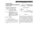 | 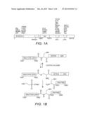 |
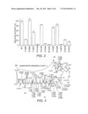 | 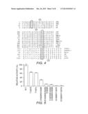 |
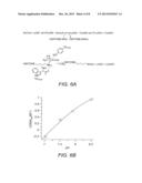 | 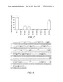 |
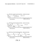 | 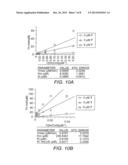 |
 | 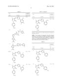 |
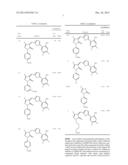 |  |
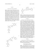 | 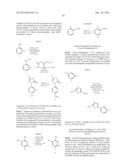 |
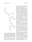 | 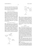 |
 |  |
 |  |
 |  |
 |  |
 |  |
 |  |
 |  |
 |  |
 |  |
 |  |
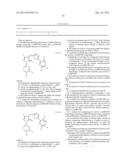 |
| New patent applications from these inventors: | |
| Date | Title |
|---|---|
| 2014-10-30 | Small molecule modulators of prb inactivation |
| 2013-12-05 | Compositions and methods for treating foxp3+ treg related diseases |
| 2013-06-06 | Use of histone acetyltransferase inhibitors as novel anti-cancer therapies |
| 2011-10-20 | Methods for synthesis and uses of inhibitors of ghrelin o-acyltransferase as potential therapeutic agents for obesity and diabetes |
| Top Inventors for class "Drug, bio-affecting and body treating compositions" | |
| Rank | Inventor's name |
|---|---|
| 1 | Anthony W. Czarnik |
| 2 | Ulrike Wachendorff-Neumann |
| 3 | Ken Chow |
| 4 | John E. Donello |
| 5 | Rajinder Singh |