Patent application title: TRRAP AND GRIN2A MUTATIONS AND USE THEREOF FOR THE DIAGNOSIS AND TREATMENT OF MELANOMA
Inventors:
Yardena R. Samuels (Potomac, MD, US)
Wei Xiaomu (Ithaca, NY, US)
IPC8 Class: AC12Q168FI
USPC Class:
4241741
Class name: Immunoglobulin, antiserum, antibody, or antibody fragment, except conjugate or complex of the same with nonimmunoglobulin material binds eukaryotic cell or component thereof or substance produced by said eukaryotic cell (e.g., honey, etc.) cancer cell
Publication date: 2013-11-21
Patent application number: 20130309254
Abstract:
Described herein is the identification of 68 genes with an elevated
frequency of somatic mutations in melanoma. Nine genes were identified
that exhibited recurring mutations in melanoma. The TRRAP gene was
mutated at nucleotide 2165 (C2165T) in six different melanoma tumor
samples. In addition, 16 genes were identified that were highly mutated
in melanoma samples. The most highly mutated gene identified was GRIN2A,
which was mutated in 34% of melanoma tumor samples. The study disclosed
herein identified 34 different nonsynonymous somatic mutations in GRIN2A
among 36 melanoma tumor samples. Provided is a method of diagnosing a
subject as having melanoma or susceptible to developing melanoma by
detecting one or more mutations in the TRRAP or GRIN2A genes. Further
provided is a method of selecting an appropriate therapy for a subject
diagnosed with melanoma by detecting the presence or absence of a
mutation in TRRAP or GRIN2A.Claims:
1. A method of diagnosing a subject as having melanoma, or susceptible to
developing melanoma, comprising detecting the presence or absence of at
least one mutation in the glutamate receptor, ionotropic, N-methyl
D-aspartate 2A (GRIN2A) gene or the transformation/transcription
domain-associated protein (TRRAP) gene in a sample obtained from the
subject, wherein the at least one mutation comprises: (i) a mutation in a
portion of the GRIN2A gene that encodes the PBP1_iGluR_NMDA_NR2 domain
(nucleotides 94-1176 of SEQ ID NO: 3); (ii) a mutation in a portion of
the GRIN2A gene that encodes the NMDAR2_C domain (nucleotides 2515-4392
of SEQ ID NO: 3); (iii) a mutation in a portion of the GRIN2A gene that
encodes the Lig_chan domain (nucleotides 1660-2484 of SEQ ID NO: 3); (iv)
G20A in the GRIN2A gene (SEQ ID NO: 3); (v) G1346A in the GRIN2A gene
(SEQ ID NO: 3); (vi) T1376C in the GRIN2A gene (SEQ ID NO: 3); or (vii)
C2165T in the TRRAP gene (SEQ ID NO: 1), wherein the presence of the at
least one mutation indicates the subject has melanoma or is susceptible
to developing melanoma.
2. The method of claim 1, wherein the mutation in the portion of the GRIN2A gene that encodes the PBP1_iGluR_NMDA_NR2 domain is C170T, T547A, G754A, C833T, G1028A, G1111A, G1116A or G1117A (SEQ ID NO: 3).
3. The method of claim 2, wherein the mutation in the portion of the GRIN2A gene that encodes the PBP1_iGluR_NMDA_NR2 domain is C833T or G1111A (SEQ ID NO: 3).
4. The method of claim 1, wherein the mutation in the portion of the GRIN2A gene that encodes the NMDAR2_C domain is G2666A, C2671T, G2759A, C2786T, G2884A, G3217A, C3221T, G3457A, G3523A, G3812A, C3827G, G3854A, C3952T, C4097T, G4261A, C4274T, G4276A or C4385G (SEQ ID NO: 3).
5. The method of claim 4, wherein the mutation in the portion of the GRIN2A gene that encodes the NMDAR2_C domain is G3523A or G3812A (SEQ ID NO: 3).
6. The method of claim 1, wherein the mutation in the portion of the GRIN2A gene that encodes the Lig_chan domain is A1784G, C1793T, G1959A, G2135A or G2218A (SEQ ID NO: 3).
7. The method of claim 1, wherein the mutation is C2165T in the TRRAP gene (SEQ ID NO: 1).
8. (canceled)
9. The method of claim 1, further comprising providing a test output to a user, wherein the test output comprises the presence or absence of the at least one mutation, a diagnosis, a treatment recommendation, or any combination thereof.
10. (canceled)
11. The method of claim 1, further comprising providing an appropriate therapy to the subject, wherein the appropriate therapy comprises surgical removal of tumor tissue, radiation therapy, chemotherapy, administration of a TRRAP inhibitor, administration of a GRIN2A inhibitor, administration of a glutamate antagonist, or any combination of two or more thereof.
12. (canceled)
13. A method of selecting a therapy for a subject diagnosed with melanoma, comprising detecting the presence or absence of at least one mutation in the GRIN2A gene in a sample obtained from the subject, wherein the at least one mutation comprises: (i) a mutation in a portion of the GRIN2A gene that encodes the PBP1_iGluR_NMDA_NR2 domain (nucleotides 94-1176 of SEQ ID NO: 3); (ii) a mutation in a portion of the GRIN2A gene that encodes the NMDAR2_C domain (nucleotides 2515-4392 of SEQ ID NO: 3); (iii) a mutation in a portion of the GRIN2A gene that encodes the Lig_chan domain (nucleotides 1660-2484 of SEQ ID NO: 3); (iv) G20A in the GRIN2A gene (SEQ ID NO: 3); (v) G1346A in the GRIN2A gene (SEQ ID NO: 3); or (vi) T1376C in the GRIN2A gene (SEQ ID NO: 3), wherein an inhibitor of GRIN2A or a glutamate antagonist is selected for therapy if the at least one mutation in GRIN2A is present.
14. The method of claim 13, wherein the mutation in the portion of the GRIN2A gene that encodes the PBP1_iGluR_NMDA_NR2 domain is C170T, T547A, G754A, C833T, G1028A, G1111A, G1116A or G1117A (SEQ ID NO: 3).
15. The method of claim 13, wherein the mutation in the portion of the GRIN2A gene that encodes the NMDAR2_C domain is G2666A, C2671T, G2759A, C2786T, G2884A, G3217A, C3221T, G3457A, G3523A, G3812A, C3827G, G3854A, C3952T, C4097T, G4261A, C4274T, G4276A or C4385G (SEQ ID NO: 3).
16. The method of claim 13, wherein the mutation in the portion of the GRIN2A gene that encodes the Lig_chan domain is A1784G, C1793T, G1959A, G2135A or G2218A (SEQ ID NO: 3).
17. A method of selecting a therapy for a subject diagnosed with melanoma, comprising detecting the presence or absence of a C2165T mutation in the TRRAP gene (SEQ ID NO: 1), wherein an inhibitor of TRRAP is selected if the C2165T mutation in the TRRAP gene is present.
18. The method of claim 13, further comprising administering the selected therapy.
19. (canceled)
20. An oligonucleotide that specifically hybridizes with a nucleic acid molecule encoding GRIN2A or TRRAP, wherein the nucleic acid molecule comprises: (i) G20A, C170T, T547A, G754A, C833T, G1028A, G1111A, G1116A, G1117A, G1346A, T1376C, A1784G, C1793T, G1959A, G2135A, G2218A, G2666A, C2671T, G2759A, C2786T, G2884A, G3217A, C3221T, G3457A, G3523A, G3812A, C3827G, G3854A, C3952T, C4097T, G4261A, C4274T, G4276A and C4385G in GRIN2A (SEQ ID NO: 3); or (ii) C2165T in TRRAP (SEQ ID NO: 1), and wherein the oligonucleotide comprises a label.
21. The oligonucleotide of claim 20, wherein the oligonucleotide is about 15 to about 40 nucleotides in length.
22. (canceled)
23. The oligonucleotide of claim 20, wherein the label is a fluorescent label, an enzymatic label or a radioisotope.
24. An array comprising one or more of the oligonucleotides of claim 20.
25. The array of claim 24, comprising at least one oligonucleotide that specifically hybridizes with each of the mutations selected from: (i) G20A, C170T, T547A, G754A, C833T, G1028A, G1111A, G1116A, G1117A, G1346A, T1376C, A1784G, C1793T, G1959A, G2135A, G2218A, G2666A, C2671T, G2759A, C2786T, G2884A, G3217A, C3221T, G3457A, G3523A, G3812A, C3827G, G3854A, C3952T, C4097T, G4261A, C4274T, G4276A and C4385G in GRIN2A (SEQ ID NO: 3); and (ii) C2165T in TRRAP (SEQ ID NO: 1).
26. (canceled)
27. The method of claim 17, further comprising administering the selected therapy.
Description:
CROSS REFERENCE TO RELATED APPLICATIONS
[0001] This application claims the benefit of U.S. Provisional Application No. 61/462,471, filed Feb. 2, 2011, which is herein incorporated by reference in its entirety.
FIELD
[0002] This disclosure concerns diagnostic markers for melanoma. In particular, this disclosure concerns identification of mutations in the transformation/transcription domain-associated protein (TRRAP) and glutamate receptor, ionotropic, N-methyl D-aspartate 2A (GRIN2A) genes, and their use for the diagnosis and treatment of melanoma.
BACKGROUND
[0003] Melanoma is the most deadly form of skin cancer. Despite years of research, metastatic melanoma disease has a dismal prognosis and is often fatal (Jemal et al., CA Cancer J Clin 59:225-249, 2009). There are few therapeutic options for melanoma patients, demonstrating a need for new clinically relevant targets. Although candidate gene analyses have been powerful in identifying melanoma driver mutations (Davies et al., Nature 417:949-954, 2002; Curtin et al., J Clin Oncol 24:4340-4346, 2006; Prickett et al., Nat Genet. 41:1127-1132, 2009), no comprehensive analysis of this tumor type has yet been performed.
[0004] Glutamate antagonists have previously been shown to inhibit proliferation of human tumor cells (Rzeski et al., Proc Natl Acad Sci USA 98(11):6372-6377, 2001). Glutamate is known to activate two different types of receptors--ionotropic glutamate receptors (iGluRs) and metabotropic glutamate receptors (mGlus). iGluRs are ligand-gated ion channels that allow cations, such as calcium and potassium, to pass through the plasma membrane of the cell after being bound by glutamate. iGluRs are subdivided into three receptor types according to agonist response, one of which is N-methyl-D-aspartate (NMDA) (Hollmann and Heinemann, Annu Rev Neurosci 17:31-108, 1994). The glutamate receptor, ionotropic, N-methyl D-aspartate 2A (GRIN2A) gene encodes a subunit of NMDA receptors; the GRIN2A subunit contains the agonist binding site for glutamate (Johnson and Ascher, Nature 325:529-531, 1987).
[0005] Prior studies have suggested that transformation/transcription domain-associated protein (TRRAP) may function as an oncogene in pancreatic cancer (Loukopoulos et al., Cancer Sci 98(3):392-400, 2007; Bashyam et al., Neoplasia 7(6):556-562, 2005). TRRAP is an adaptor protein found in various multiprotein chromatin complexes with histone acetyltransferase activity, which in turn is responsible for epigenetic transcription activation. TRRAP plays a central role in the transcriptional activity of p53, c-Myc, E2F1 and other transcription factors (McMahon et al., Cell 94:363-374, 1998; Barley et al., Mol Cell 8:1243-1254, 2001). TRRAP knockout mice are embryonic lethal suggesting that TRRAP is essential for cell survival (Herceg et al., Nat Genet. 29:206-211, 2001).
SUMMARY
[0006] Disclosed herein are 68 human genes with an elevated mutation frequency in melanoma. This disclosure specifically describes the identification of nine genes with recurring mutations in melanoma and 16 genes that are highly mutated in melanoma. In particular, disclosed herein is the identification of a recurrent mutation in human TRRAP (C2165T) found in six different melanoma samples. Also disclosed is the identification of 34 different nonsynonymous somatic mutations of human GRIN2A in melanoma samples.
[0007] Provided herein is a method of diagnosing a subject as having melanoma, or having a greater susceptibility to developing melanoma, by detecting at least one mutation in the TRRAP gene or the GRIN2A gene. The presence of the at least one mutation indicates the subject has melanoma or is susceptible to developing melanoma. In some embodiments, the TRRAP mutation is C2165T (numbered with reference to SEQ ID NO: 1). In some embodiments, the GRIN2A mutation is a mutation that occurs in the PBP1_iGluR_NMDA_NR2 domain, the Lig_chan domain or the NMDAR2_C domain of GRIN2A. In particular examples, the GRIN2A mutation is selected from one or more of G20A, C170T, T547A, G754A, C833T, G1028A, G1111A, G1116A, G1117A, G1346A, T1376C, A1784G, C1793T, G1959A, G2135A, G2218A, G2666A, C2671T, G2759A, C2786T, G2884A, G3217A, C3221T, G3457A, G3523A, G3812A, C3827G, G3854A, C3952T, C4097T, G4261A, C4274T, G4276A and C4385G (numbered with reference to SEQ ID NO: 3). In some embodiments, the subject who has the detected mutation is further treated with an anti-melanoma therapy.
[0008] Further provided is a method of selecting a therapy for a subject diagnosed with melanoma by detecting the presence or absence of a C2165T mutation in the TRRAP gene (SEQ ID NO: 1). An inhibitor of TRRAP is selected if the C2165T mutation in the TRRAP gene is present. Also provided is a method of selecting a therapy for a subject diagnosed with melanoma, comprising detecting the presence or absence of at least one mutation in the GRIN2A gene disclosed herein. An inhibitor of GRIN2A or a glutamate antagonist is selected for therapy if at least one mutation in GRIN2A is identified.
[0009] The present disclosure further provides synthetic oligonucleotides as probes that specifically hybridize with a nucleic acid molecule encoding human TRRAP or human GRIN2A comprising at least one of the mutations disclosed herein. Also provided are arrays comprising a substrate to which are bound one or more of the oligonucleotides specific for mutant TRRAP or mutant GRIN2A. The oligonucleotides may be located at addressable locations on the array.
[0010] The foregoing and other objects, features, and advantages of the invention will become more apparent from the following detailed description, which proceeds with reference to the accompanying figures.
BRIEF DESCRIPTION OF THE FIGURES
[0011] FIG. 1 is a series of schematic illustrations showing the distribution of novel nonsynonymous recurrent mutations in melanoma in TRRAP, DCC, PLCH1, NOS1, LRRN3, ZNF831 and SLC17A5. Seven novel nonsynonymous recurrent mutations identified in the study disclosed herein are presented on relevant protein schematics. Black arrows indicate locations of recurrent mutations and conserved protein functional domains are indicated as boxes (1: Immunoglobulin I-set domain; 2: Fibronectin type III domain; 3: Neogenin C-terminus; 4: Phosphoinositide-specific phospholipase C, efhand-like; 5: Phosphatidylinositol-specific phospholipase C, X domain; 6: Phosphatidylinositol-specific phospholipase C, Y domain; 7: C2 domain; 8: PDZ domain; 9: Nitric oxide synthase, oxygenase domain; 10: Flavodoxin; 11: FAD binding domain; 12: Oxidoreductase NAD-binding domain; 13: LRRNT: Leucine rich repeat N-terminal domain; 14: Leucine Rich Repeat; 15: Immunoglobulin I-set domain; 16: Fibronectin type III domain; 17: Major Facilitator Superfamily).
[0012] FIGS. 2A-2J are a series of graphs and immunoblots showing the effect of mutant TRRAP on cell growth. FIG. 2A is a table showing mutant TRRAP induces cell transformation in NIH 3T3 cells. The table shows focus formation of NIH 3T3 cells transfected with the indicated constructs or empty vector control. KRas.sup.G12V was included as a positive control for cell transformation. FIG. 2B is an immunoblot showing detection of TRRAP and KRas.sup.G12V protein expression in lysates of transiently transfected NIH 3T3 cells. FIG. 2C shows an immunoblot of cell lysates from HEK 293T cells transiently transfected with either control vector or shRNAs that target TRRAP. For normalization, lysates were analyzed in parallel by anti-α-tubulin immunoblotting. FIG. 2D is an anti-TRRAP immunoblot of melanoma cells transduced with shRNA targeting TRRAP. For normalization, lysates were analyzed in parallel by anti-α-tubulin immunoblotting. FIGS. 2E-2H are graphs showing TRRAP mutation confers resistance to apoptosis. The graphs show apoptosis quantification of melanoma cell lines transduced with shRNA control or shRNAs targeting TRRAP. Cells were grown in medium containing 2.5% serum for the indicated times. Apoptosis was assessed by fluorescence microscopy of Hoechst 33258-stained cells. FIGS. 2I and 2J are immunoblots of representative melanoma lines presented in E-H using the indicated antibodies to assess PARP cleavage. WT=wild-type.
[0013] FIG. 3A is a schematic illustration showing the locations of somatic mutations in GRIN2A. Human GRIN2A protein is presented with conserved functional domains indicated (SP: Signal peptide; PBP1_iGluR_NMDA_NR2: N-terminal leucine/isoleucine/valine-binding protein LIVBP-like domain of the NR2 subunit of NMDA receptor family; Lig_chan: Ligand-gated ion channel; NMDAR2_C: N-methyl D-aspartate receptor 2B3 C-terminus). Somatic mutations are indicated with arrows and the amino acid changes are listed. Recurrent mutations in GRIN2A are S278F, E371K and E1175K. Nonsense mutations are indicated by black boxes.
[0014] FIG. 3B is a schematic illustration showing glutamate signaling pathway mutations in melanoma. The genes that function in glutamate signaling are specified. Circled genes have somatic mutations. The number of mutations in each gene is indicated by the number adjacent to the circle (PSD complex: post synaptic density complex; NMDAR: N-methyl-D-aspartic acid (NMDA) receptor; RTK: receptor tyrosine kinase).
[0015] FIG. 4 is a graph showing mutation spectra of single base pair substitutions in melanoma whole exome sequencing. The number of each of the six classes of base substitutions resulting in nonsynonymous changes in the whole exome screen is shown.
[0016] FIG. 5 is a series of chromatograms showing a recurrent hotspot mutation in TRRAP in representative melanoma tumor samples and the commercially available A375 cell line. In each case, the top sequence chromatogram was obtained from normal tissue and the lower sequence chromatogram from the indicated tumors. Arrows indicate the location of the missense mutations. The nucleotide and amino acid alterations are indicated below the chromatograms. The sequences shown in the chromatograms are TGTCTCCCTCT (SEQ ID NO: 5) for normal tissue; TGTCTYCCTCT (SEQ ID NO: 6) for tumor samples; and TGTCTYYCTCT (SEQ ID NO: 7) for the A375 cell line.
[0017] FIG. 6 is a sequence comparison of conserved serine-722 of human TRRAP with its orthologs. The human TRRAP orthologs for 12 species were compared using the indicated NCBI accession numbers by COBALT algorithm (available online at www.ncbi.nlm.nih.gov/tools/cobalt/colbat.cgi). The human TRRAP sequence (amino acids 697-747 of SEQ ID NO: 2) is compared with sequences from Canis familiaris (SEQ ID NO: 8), Pan troglodytes (SEQ ID NO: 9), Bos Taurus (SEQ ID NO: 10), Rattus norvegicus (SEQ ID NO: 11), Gallus gallus (SEQ ID NO: 12), Danio rerio (SEQ ID NO: 13), Mus musculus (SEQ ID NO: 14), D. melanogaster (SEQ ID NO: 15), Anopheles gamiae (SEQ ID NO: 16), C. elegans (SEQ ID NO: 17) and S. cerevisiae (SEQ ID NO: 18). The conserved serine at amino acid 722 in humans is underlined and aligned with other species. This residue is not conserved in C. elegans or S. cerevisiae.
[0018] FIG. 7 is a graph showing mutation spectra of single base pair substitutions in GRIN2A. The number of each of the six classes of base substitutions resulting in nonsynonymous changes in GRIN2A is shown.
[0019] FIG. 8 is a schematic of the whole exome capture and sequencing analysis of 14 melanoma samples.
[0020] FIG. 9 is a table showing score cutoff for determination of melanoma somatic mutations.
[0021] FIGS. 10A-10D are a series of graphs showing the effect of mutant TRRAP on cell growth. Apoptosis was quantified for melanoma cell lines transduced with shRNA control or shRNAs targeting TRRAP. Cells were grown in medium containing 10% serum for the indicated times. Apoptosis was assessed by fluorescence microscopy of Hoechst 33258-stained cells.
[0022] FIGS. 11A-11B demonstrate that melanoma cells expressing mutant forms of GRIN2A have increased ability for anchorage-independent growth. Melanoma cells (A375 cells) stably expressing GRIN1 and GRIN2A mutants, or empty vector, were seeded into soft agar in 10% serum and grown for 14 days before staining and counting (A). Also shown is a quantitative graph of colony formation of A375 cells in soft agar (B). Error bars are representative of n=3 (s.d.).
[0023] FIGS. 12A-12B are a set of graphs showing GRIN2A functions as a tumor suppressor in melanoma cells. Endogenous GRIN2A was stably depleted in melanoma cells using shRNA specific for human GRIN2A. (A) Proliferation assay of mutant expressing GRIN2A cell lines depleted of endogenous GRIN2A. Knock-down using GRIN2A-specific shRNA (#1, #2 and #3) resulted in little to no change in proliferation for 125T or 501Mel melanoma cell lines. (B) Proliferation assay of wild-type expressing GRIN2A cell lines depleted of GRIN2A. Knock-down resulted in increased proliferation for both 31T and 39T compared to empty vector control (pLKO.1).
[0024] FIGS. 13A-13B demonstrate that somatic mutations in GRIN2A have adverse effects on receptor function and formation. (A) Influx of calcium upon NMDA stimulation of transiently transfected HEK293T cells shows decreased calcium permeability in cells expressing mutant forms of GRIN2A. (B) Mutant forms of GRIN2A bind GRIN1 with reduced affinity, thus causing decreased NMDAR complex formation. HEK293T cells were transiently transfected with WT GRIN1 and GRIN2A (WT or mutants) or empty vectors as control and immunoprecipitated with anti-GRIN1. Immunoprecipitates were probed with anti-GRIN2A and anti-GRIN1 to confirm binding. Lysates were probed with anti-GRIN2A and anti-GAPDH as a loading control.
SEQUENCE LISTING
[0025] The nucleic and amino acid sequences listed in the accompanying sequence listing are shown using standard letter abbreviations for nucleotide bases, and three letter code for amino acids, as defined in 37 C.F.R. 1.822. Only one strand of each nucleic acid sequence is shown, but the complementary strand is understood as included by any reference to the displayed strand. The Sequence Listing is submitted as an ASCII text file, created on Jan. 18, 2012, 82.6 KB, which is incorporated by reference herein. In the accompanying sequence listing:
[0026] SEQ ID NO: 1 is the nucleotide sequence of human TRRAP (CCDS ID 5659.1).
[0027] SEQ ID NO: 2 is the amino acid sequence of human TRRAP (CCDS ID 5659.1).
[0028] SEQ ID NO: 3 is the nucleotide sequence of human GRIN2A (CCDS ID 10539.1).
[0029] SEQ ID NO: 4 is the amino acid sequence of human GRIN2A (CCDS ID 10539.1).
[0030] SEQ ID NOs: 5-7 are nucleotide sequences of the region surrounding the TRRAP hotspot mutation from a control sample, a tumor sample and A375 cells, respectively.
[0031] SEQ ID NOs: 8-18 are amino acid sequences of a segment of the TRRAP protein from several different species.
[0032] SEQ ID NOs: 19-24 are nucleotide sequences of TRRAP primers used for recurrent mutation confirmation.
[0033] SEQ ID NOs: 25-62 are nucleotide sequences of GRIN2A primers used for PCR and sequencing.
[0034] SEQ ID NOs: 63-66 are nucleotide sequences of TRRAP mutation primers.
[0035] SEQ ID NOs: 67-72 are TRRAP-specific shRNA sequences.
DETAILED DESCRIPTION
I. Abbreviations
[0036] FBS fetal bovine serum
[0037] GRIN2A glutamate receptor, ionotropic, N-methyl D-aspartate 2A
[0038] H&E hematoxylin and eosin
[0039] iGluR ionotropic glutamate receptor
[0040] mGlu metabotropic glutamate receptor
[0041] NMDA N-methyl-D-aspartate
[0042] PARP poly-ADP ribose polymerase
[0043] PCR polymerase chain reaction
[0044] RT-PCR reverse transcriptase polymerase chain reaction
[0045] shRNA short hairpin RNA
[0046] TRRAP transformation/transcription domain-associated protein
[0047] WT wild-type
II. Terms and Methods
[0048] Unless otherwise noted, technical terms are used according to conventional usage. Definitions of common terms in molecular biology may be found in Benjamin Lewin, Genes V, published by Oxford University Press, 1994 (ISBN 0-19-854287-9); Kendrew et al. (eds.), The Encyclopedia of Molecular Biology, published by Blackwell Science Ltd., 1994 (ISBN 0-632-02182-9); and Robert A. Meyers (ed.), Molecular Biology and Biotechnology: a Comprehensive Desk Reference, published by VCH Publishers, Inc., 1995 (ISBN 1-56081-569-8).
[0049] In order to facilitate review of the various embodiments of the disclosure, the following explanations of specific terms are provided:
[0050] Administration: The introduction of a composition into a subject by a chosen route. For example, if the chosen route is intravenous, the composition is administered by introducing the composition into a vein of the subject.
[0051] Antagonist: A molecule or compound that blocks or inhibits the activity of another molecule or compound (such as a drug that inhibits the function of a protein).
[0052] Array: An arrangement of molecules, such as biological macromolecules (such as peptides or nucleic acid molecules) or biological samples (such as tissue sections), in addressable locations on or in a substrate. A "microarray" is an array that is miniaturized so as to require or be aided by microscopic examination for evaluation or analysis. Arrays are sometimes called DNA chips or biochips.
[0053] The array of molecules ("features") makes it possible to carry out a very large number of analyses on a sample at one time. In certain example arrays, one or more molecules (such as an oligonucleotide probe) will occur on the array a plurality of times (such as twice), for instance to provide internal controls. The number of addressable locations on the array can vary, for example from at least two, at least four, at least six, to at least 9, at least 10, at least 14, at least 15, at least 20, at least 30, at least 50, at least 75, at least 100, at least 150, at least 200, at least 300, at least 500, least 550, at least 600, at least 800, at least 1000, or more. In a particular example, an array includes 2-100 addressable locations, such as 4-20 addressable locations. In particular examples, an array consists essentially of oligonucleotide probes specific for the somatic mutations in TRRAP and/or GRIN2A disclosed herein.
[0054] In particular examples, an array includes nucleic acid molecules, such as oligonucleotide sequences that are at least 15 nucleotides in length, such as about 15-40 nucleotides in length.
[0055] Within an array, each arrayed sample is addressable, in that its location can be reliably and consistently determined within at least two dimensions of the array. The feature application location on an array can assume different shapes. For example, the array can be regular (such as arranged in uniform rows and columns) or irregular. Thus, in ordered arrays the location of each sample is assigned to the sample at the time when it is applied to the array, and a key may be provided in order to correlate each location with the appropriate target or feature position. Often, ordered arrays are arranged in a symmetrical grid pattern, but samples could be arranged in other patterns (such as in radially distributed lines, spiral lines, or ordered clusters). Addressable arrays usually are computer readable, in that a computer can be programmed to correlate a particular address on the array with information about the sample at that position (such as hybridization or binding data, including for instance signal intensity). In some examples of computer readable formats, the individual features in the array are arranged regularly, for instance in a Cartesian grid pattern, which can be correlated to address information by a computer.
[0056] Clinical outcome: Refers to the health status of a patient following treatment for a disease or disorder (such as melanoma), or in the absence of treatment. Clinical outcomes include, but are not limited to, an increase in the length of time until death, a decrease in the length of time until death, an increase in the chance of survival, an increase in the risk of death, survival, disease-free survival, chronic disease, metastasis, advanced or aggressive disease, disease recurrence, death, and favorable or poor response to therapy.
[0057] Decrease in survival: As used herein, "decrease in survival" refers to a decrease in the length of time before death of a patient, or an increase in the risk of death for the patient. A decrease in survival also can refer to a decrease in the average time to death in a group, such as a group of patients diagnosed with melanoma.
[0058] Diagnosing: Refers to the process of identifying the nature or cause of a disease or disorder.
[0059] Glutamate antagonist: Refers to any compound that blocks glutamate function, such as glutamate function mediated through ionotropic glutamate receptors. Thus, suitable glutamate antagonists include, for example, compounds that bind ionotropic glutamate receptors (iGluRs), including NMDA receptors (e.g., GRIN2A). Compounds that bind ionotropic glutamate receptors include compounds which bind α-amino-3-hydroxy-5-methyl-4-isoxazole-propionate (AMPA), NMDA, or kainate receptors in a competitive manner or interact with ionotropic glutamate receptor mediated signals in a non-competitive manner. Other suitable glutamate antagonists include, for example, glutathione promoting agents, GABA stimulating agents and certain neurotransmitters. Glutamate antagonists are known in the art (see, for example, U.S. Patent Application Publication Nos. 2007/0135413 and 2007/0248690). In particular examples, the glutamate antagonist is dizocilpine, GYKI52466, MK-801, AP-5 or CGS 19755 (selfotel).
[0060] GRIN2A (glutamate receptor, ionotropic, N-methyl D-aspartate 2A): A subunit of N-methyl-D-aspartate (NMDA) receptors. NMDA receptors are a class of ionotropic glutamate-gated ion channels. These receptors have been shown to be involved in long-term potentiation, an activity-dependent increase in the efficiency of synaptic transmission thought to underlie certain kinds of memory and learning. NMDA receptor channels are heteromeric complexes composed of the key receptor subunit NMDAR1 (GRIN 1) and 1 or more of the 4 NMDAR2 subunits: NMDAR2A (GRIN2A), NMDAR2B (GRIN2B), NMDAR2C (GRIN2C) and NMDAR2D (GRIN2D). GRIN2A, which is also known as NR2A and NMDAR2A, contains the antagonist binding site for glutamate.
[0061] GRIN2A sequences are publically available. For example, GenBank Accession Nos. NM--000833 and NP--000824 are the nucleotide and amino acid sequences, respectively, of human GRIN2A transcript variant 2. GenBank Accession Nos. NM--001134407 and NP--001127879 are the nucleotide and amino acid sequences, respectively, of human GRIN2A transcript variant 1. The NCBI CCDS database also provides nucleotide and amino acid sequences for GRIN2A under CCDS ID 10539.1. The GenBank Accession numbers and CCDS ID numbers listed above and disclosed herein are incorporated by reference as they appear in the database as of Dec. 3, 2010.
[0062] Disclosed herein are somatic mutations in GRIN2A identified in melanoma tumor samples. The disclosed mutations are referred to by the location of the GRIN2A mutation with reference to SEQ ID NO: 3 (nucleotide) and SEQ ID NO: 4 (amino acid). For example, the C833T mutation refers to a cytosine to thymidine substitution at nucleotide 833 (SEQ ID NO: 3), which results in a serine to phenylalanine change at amino acid 278 (S278F; SEQ ID NO: 4). Some somatic mutations result in the introduction of a stop codon (a nonsense mutation), for example, the G20A GRIN2A mutation. The corresponding amino acid change for the G20A mutation (which occurs at position 7 of SEQ ID NO: 4) is therefore designated W7*.
[0063] In some embodiments of the methods disclosed herein, the GRIN2A mutation occurs in a portion of the GRIN2A gene that encodes the PBP1_iGluR_NMDA_NR2 domain; a portion of the GRIN2A gene that encodes the Lig_chan domain; or a portion of the GRIN2A gene that encodes the NMDAR2_C domain. As defined herein, the PBP1_iGluR_NMDA_NR2 domain is encoded by nucleotides 94-1176 of SEQ ID NO: 3 and the amino acid sequence of the PBP1_iGluR_NMDA_NR2 domain corresponds to residues 32-392 of SEQ ID NO: 4; the Lig_chan domain is encoded by nucleotides 1660-2484 of SEQ ID NO: 3 and the amino acid sequence of the Lig_chan domain corresponds to residues 554-828 of SEQ ID NO: 4; and the NMDAR2_C domain is encoded by nucleotides 2515-4392 of SEQ ID NO: 3 and the amino acid sequence of the NMDAR2_C domain corresponds to residues 839-1464 of SEQ ID NO: 4. In some embodiments, the GRIN2A mutation is selected from G20A, C170T, T547A, G754A, C833T, G1028A, G1111A, G1116A, G1117A, G1346A, T1376C, A1784G, C1793T, G1959A, G2135A, G2218A, G2666A, C2671T, G2759A, C2786T, G2884A, G3217A, C3221T, G3457A, G3523A, G3812A, C3827G, G3854A, C3952T, C4097T, G4261A, C4274T, G4276A and C4385G (SEQ ID NO: 3).
[0064] Inhibitor: As used herein, an "inhibitor" refers to any compound that is capable of reducing or altering the expression or activity of a target molecule (such as a nucleic acid molecule or a protein). In some embodiments, the inhibitor is an inhibitor of TRRAP or GRIN2A.
[0065] Label: An agent capable of detection, for example by enzyme-linked immunosorbent assay (ELISA), spectrophotometry, flow cytometry or microscopy. For example, a label can be attached to a nucleic acid molecule or protein, thereby permitting detection of the nucleic acid molecule or protein. Examples of labels include, but are not limited to, radioactive isotopes, enzyme substrates, co-factors, ligands, chemiluminescent agents, fluorophores, haptens, enzymes, and combinations thereof. Methods for labeling and guidance in the choice of labels appropriate for various purposes are discussed for example in Sambrook et al. (Molecular Cloning: A Laboratory Manual, Cold Spring Harbor, N.Y., 1989) and Ausubel et al. (In Current Protocols in Molecular Biology, John Wiley & Sons, New York, 1998).
[0066] In some embodiments, the label is a fluorophore ("fluorescent label"). Fluorophores are chemical compounds, which when excited by exposure to a particular wavelength of light, emits light (i.e., fluoresces), for example at a different wavelength. Fluorophores can be described in terms of their emission profile, or "color." Green fluorophores, for example Cy3, FITC, and Oregon Green®, are characterized by their emission at wavelengths generally in the range of 515-540λ. Red fluorophores, for example Texas Red®, Cy5 and tetramethylrhodamine, are characterized by their emission at wavelengths generally in the range of 590-690λ.
[0067] Examples of fluorophores that may be used are provided in U.S. Pat. No. 5,866,366 to Nazarenko et al., and include for instance: 4-acetamido-4'-isothiocyanatostilbene-2,2'disulfonic acid, acridine and derivatives such as acridine and acridine isothiocyanate, 5-(2'-aminoethyl)aminonaphthalene-1-sulfonic acid (EDANS),4-amino-N-[3-vinylsulfonyl)phenyl]naphthalimide-3,5 disulfonate (Lucifer Yellow VS), N-(4-anilino-1-naphthyl)maleimide, anthranilamide, Brilliant Yellow, coumarin and derivatives such as coumarin, 7-amino-4-methylcoumarin (AMC, Coumarin 120), 7-amino-4-trifluoromethylcouluarin (Coumarin 151); cyanosine; 4',6-diaminidino-2-phenylindole (DAPI); 5',5''-dibromopyrogallol-sulfonephthalein (Bromopyrogallol Red); 7-diethylamino-3-(4'-isothiocyanatophenyl)-4-methylcoumarin; diethylenetriamine pentaacetate; 4,4'-diisothiocyanatodihydro-stilbene-2,2'-disulfonic acid; 4,4'-diisothiocyanatostilbene-2,2'-disulfonic acid; 5-[dimethylamino]naphthalene-1-sulfonyl chloride (DNS, dansyl chloride); 4-(4'-dimethylaminophenylazo)benzoic acid (DABCYL); 4-dimethylaminophenylazophenyl-4'-isothiocyanate (DABITC); eosin and derivatives such as eosin and eosin isothiocyanate; erythrosin and derivatives such as erythrosin B and erythrosin isothiocyanate; ethidium; fluorescein and derivatives such as 5-carboxyfluorescein (FAM), 5-(4,6-dichlorotriazin-2-yl)aminofluorescein (DTAF), 2'7'-dimethoxy-4'5'-dichloro-6-carboxyfluorescein (JOE), fluorescein, fluorescein isothiocyanate (FITC), and QFITC (XRITC); fluorescamine; IR144; IR1446; Malachite Green isothiocyanate; 4-methylumbelliferone; ortho cresolphthalein; nitrotyrosine; pararosaniline; Phenol Red; B-phycoerythrin; o-phthaldialdehyde; pyrene and derivatives such as pyrene, pyrene butyrate and succinimidyl 1-pyrene butyrate; Reactive Red 4 (Cibacron® Brilliant Red 3B-A); rhodamine and derivatives such as 6-carboxy-X-rhodamine (ROX), 6-carboxyrhodamine (R6G), lissamine rhodamine B sulfonyl chloride, rhodamine (Rhod), rhodamine B, rhodamine 123, rhodamine X isothiocyanate, sulforhodamine B, sulforhodamine 101 and sulfonyl chloride derivative of sulforhodamine 101 (Texas Red); N,N,N',N'-tetramethyl-6-carboxyrhodamine (TAMRA); tetramethyl rhodamine; tetramethyl rhodamine isothiocyanate (TRITC); riboflavin; rosolic acid and terbium chelate derivatives.
[0068] Other contemplated fluorophores include GFP (green fluorescent protein), Lissamine®, diethylaminocoumarin, fluorescein chlorotriazinyl, naphthofluorescein, 4,7-dichlororhodamine and xanthene and derivatives thereof. Other fluorophores known to those skilled in the art may also be used.
[0069] Melanoma: A form of cancer that originates in melanocytes (cells that make the pigment melanin). Melanocytes are found primarily in the skin, but are also present in the bowel and eye. As used herein, "melanoma" refers to any stage of melanoma, or any subtype of melanoma, such as superficial spreading melanoma, nodular melanoma, acral lentiginous melanoma, lentigo maligna, melanoma-in-situ, mucosal melanoma and uveal melanoma.
[0070] Metastasis: Refers to the spread of cancer cells from the original tumor to other sites in the body.
[0071] Mutation: Any change of the DNA sequence within a gene or chromosome. In some instances, a mutation will alter a characteristic or trait (phenotype), but this is not always the case. Types of mutations include base substitution point mutations (e.g., transitions or transversions), deletions and insertions. Missense mutations are those that introduce a different amino acid into the sequence of the encoded protein; nonsense mutations are those that introduce a new stop codon. In the case of insertions or deletions, mutations can be in-frame (not changing the frame of the overall sequence) or frame shift mutations, which may result in the misreading of a large number of codons (and often leads to abnormal termination of the encoded product due to the presence of a stop codon in the alternative frame).
[0072] This term specifically encompasses variations that arise through somatic mutation, for instance those that are found only in disease cells (such as cancer cells), but not constitutionally, in a given individual. Examples of such somatically-acquired variations include the point mutations that frequently result in altered function of various genes that are involved in development of cancers. This term also encompasses DNA alterations that are present constitutionally, that alter the function of the encoded protein in a readily demonstrable manner, and that can be inherited by the children of an affected individual. In this respect, the term overlaps with "polymorphism," as discussed below, but generally refers to the subset of constitutional alterations that have arisen within the past few generations in a kindred and that are not widely disseminated in a population group.
[0073] In some embodiments, a mutation in TRRAP refers to a nucleotide substitution in the TRRAP gene or cDNA, or an amino acid substitution in the TRRAP protein. In some embodiments, a mutation in GRIN2A refers to a nucleotide substitution in the GRIN2A gene or cDNA, or an amino acid substitution in the GRIN2A protein.
[0074] Oligonucleotide: A linear polynucleotide sequence of up to about 100 nucleotide bases in length. In some embodiments, the oligonucleotide is 15-40 nucleotides in length.
[0075] Patient: As used herein, the term "patient" includes human and non-human animals. The preferred patient for treatment is a human.
[0076] Pharmaceutically acceptable vehicles: The pharmaceutically acceptable carriers (vehicles) useful in this disclosure are conventional. Remington's Pharmaceutical Sciences, by E. W. Martin, Mack Publishing Co., Easton, Pa., 15th Edition (1975), describes compositions and formulations suitable for pharmaceutical delivery of one or more therapeutic compounds, molecules or agents.
[0077] In general, the nature of the carrier will depend on the particular mode of administration being employed. For instance, parenteral formulations usually comprise injectable fluids that include pharmaceutically and physiologically acceptable fluids such as water, physiological saline, balanced salt solutions, aqueous dextrose, glycerol or the like as a vehicle. For solid compositions (for example, powder, pill, tablet, or capsule forms), conventional non-toxic solid carriers can include, for example, pharmaceutical grades of mannitol, lactose, starch, or magnesium stearate. In addition to biologically-neutral carriers, pharmaceutical compositions to be administered can contain minor amounts of non-toxic auxiliary substances, such as wetting or emulsifying agents, preservatives, and pH buffering agents and the like, for example sodium acetate or sorbitan monolaurate.
[0078] Polymorphism: Variant in a sequence of a gene, or any genomic sequence, usually carried from one generation to another in a population. Polymorphisms can be those variations (nucleotide sequence differences) that, while having a different nucleotide sequence, produce functionally equivalent gene products, such as those variations generally found between individuals, different ethnic groups, and geographic locations. The term polymorphism also encompasses variations that produce gene products with altered function, i.e., variants in the gene sequence that lead to gene products that are not functionally equivalent. This term also encompasses variations that produce no gene product, an inactive gene product, a truncated gene product, or increased or increased activity gene product.
[0079] Polymorphisms can be referred to, for instance, by the nucleotide position at which the variation exists, by the change in amino acid sequence caused by the nucleotide variation, or by a change in some other characteristic of the nucleic acid molecule or protein that is linked to the variation (e.g., an alteration of a secondary structure such as a stem-loop, or an alteration of the binding affinity of the nucleic acid for associated molecules, such as polymerases, RNAses, a change in the availability of a site for cleavage by a restriction endonuclease, either the formation of a new site, or lose of a site, and so forth).
[0080] Polypeptide: A polymer in which the monomers are amino acid residues which are joined together through amide bonds. When the amino acids are alpha-amino acids, either the L-optical isomer or the D-optical isomer can be used. The terms "polypeptide" or "protein" as used herein are intended to encompass any amino acid sequence and include modified sequences such as glycoproteins. The term "polypeptide" is specifically intended to cover naturally occurring proteins, as well as those which are recombinantly or synthetically produced.
[0081] The term "residue" or "amino acid residue" includes reference to an amino acid that is incorporated into a protein, polypeptide, or peptide.
[0082] Conservative amino acid substitutions are those substitutions that, when made, least interfere with the properties of the original protein, that is, the structure and especially the function of the protein is conserved and not significantly changed by such substitutions. Examples of conservative substitutions are shown in the following table.
TABLE-US-00001 Original Residue Conservative Substitutions Ala Ser Arg Lys Asn Gln, His Asp Glu Cys Ser Gln Asn Glu Asp His Asn; Gln Ile Leu, Val Leu Ile; Val Lys Arg; Gln; Glu Met Leu; Ile Phe Met; Leu; Tyr Ser Thr Thr Ser Trp Tyr Tyr Trp; Phe Val Ile; Leu
[0083] Conservative substitutions generally maintain (a) the structure of the polypeptide backbone in the area of the substitution, for example, as a sheet or helical conformation, (b) the charge or hydrophobicity of the molecule at the target site, or (c) the bulk of the side chain.
[0084] The substitutions which in general are expected to produce the greatest changes in protein properties will be non-conservative, for instance changes in which (a) a hydrophilic residue, for example, seryl or threonyl, is substituted for (or by) a hydrophobic residue, for example, leucyl, isoleucyl, phenylalanyl, valyl or alanyl; (b) a cysteine or proline is substituted for (or by) any other residue; (c) a residue having an electropositive side chain, for example, lysyl, arginyl, or histadyl, is substituted for (or by) an electronegative residue, for example, glutamyl or aspartyl; or (d) a residue having a bulky side chain, for example, phenylalanine, is substituted for (or by) one not having a side chain, for example, glycine.
[0085] Preventing, treating or ameliorating a disease: "Preventing" or "inhibiting" a disease (such as melanoma) refers to inhibiting the full development of a disease. "Treating" refers to a therapeutic intervention that ameliorates a sign or symptom of a disease or pathological condition after it has begun to develop. "Ameliorating" refers to the reduction in the number or severity of signs or symptoms of a disease.
[0086] Probes and primers: A probe comprises an isolated nucleic acid capable of hybridizing to a target nucleic acid. A detectable label or reporter molecule can be attached to a probe or primer. Typical labels include radioactive isotopes, enzyme substrates, co-factors, ligands, chemiluminescent or fluorescent agents, haptens, and enzymes. Methods for labeling and guidance in the choice of labels appropriate for various purposes are discussed, for example in Sambrook et al. (In Molecular Cloning: A Laboratory Manual, CSHL, New York, 1989) and Ausubel et al. (In Current Protocols in Molecular Biology, John Wiley & Sons, New York, 1998). In some embodiments, an "oligonucleotide" is a probe or primer.
[0087] In a particular example, a probe includes at least one fluorophore, such as an acceptor fluorophore or donor fluorophore. For example, a fluorophore can be attached at the 5'- or 3'-end of the probe. In specific examples, the fluorophore is attached to the base at the 5'-end of the probe, the base at its 3'-end, the phosphate group at its 5'-end or a modified base, such as a T internal to the probe.
[0088] Probes are generally at least 15 nucleotides in length, such as at least 15, at least 16, at least 17, at least 18, at least 19, least 20, at least 21, at least 22, at least 23, at least 24, at least 25, at least 26, at least 27, at least 28, at least 29, at least 30, at least 31, at least 32, at least 33, at least 34, at least 35, at least 36, at least 37, at least 38, at least 39, at least 40, at least 41, at least 42, at least 43, at least 44, at least 45, at least 46, at least 47, at least 48, at least 49, at least 50 at least 51, at least 52, at least 53, at least 54, at least 55, at least 56, at least 57, at least 58, at least 59, at least 60, at least 61, at least 62, at least 63, at least 64, at least 65, at least 66, at least 67, at least 68, at least 69, at least 70, or more contiguous nucleotides complementary to the target nucleic acid molecule, such as 15-70 nucleotides, 15-60 nucleotides, 15-50 nucleotides, 15-40 nucleotides, or 15-30 nucleotides.
[0089] Primers are short nucleic acid molecules, for instance DNA oligonucleotides 15 nucleotides or more in length, which can be annealed to a complementary target nucleic acid molecule by nucleic acid hybridization to form a hybrid between the primer and the target nucleic acid strand. A primer can be extended along the target nucleic acid molecule by a polymerase enzyme. Therefore, primers can be used to amplify a target nucleic acid molecule.
[0090] The specificity of a primer increases with its length. Thus, for example, a primer that includes 30 consecutive nucleotides will anneal to a target sequence with a higher specificity than a corresponding primer of only 15 nucleotides. Thus, to obtain greater specificity, probes and primers can be selected that include at least 15, 20, 25, 30, 35, 40, 45, 50, 55, 60, 65, 70 or more consecutive nucleotides. In particular examples, a primer is at least 15 nucleotides in length, such as at least 15 contiguous nucleotides complementary to a target nucleic acid molecule. Particular lengths of primers that can be used to practice the methods of the present disclosure include primers having at least 15, at least 16, at least 17, at least 18, at least 19, at least 20, at least 21, at least 22, at least 23, at least 24, at least 25, at least 26, at least 27, at least 28, at least 29, at least 30, at least 31, at least 32, at least 33, at least 34, at least 35, at least 36, at least 37, at least 38, at least 39, at least 40, at least 45, at least 50, at least 55, at least 60, at least 65, at least 70, or more contiguous nucleotides complementary to the target nucleic acid molecule to be amplified, such as a primer of 15-70 nucleotides, 15-60 nucleotides, 15-50 nucleotides, 15-40 nucleotides or 15-30 nucleotides.
[0091] Primer pairs can be used for amplification of a nucleic acid sequence, for example, by PCR, real-time PCR, or other nucleic-acid amplification methods known in the art. An "upstream" or "forward" primer is a primer 5' to a reference point on a nucleic acid sequence. A "downstream" or "reverse" primer is a primer 3' to a reference point on a nucleic acid sequence. In general, at least one forward and one reverse primer are included in an amplification reaction.
[0092] Nucleic acid probes and primers can be readily prepared based on the nucleic acid molecules provided herein. It is also appropriate to generate probes and primers based on fragments or portions of these disclosed nucleic acid molecules, for instance regions that encompass the identified mutations of interest. PCR primer pairs can be derived from a known sequence by using computer programs intended for that purpose such as Primer (Version 0.5, © 1991, Whitehead Institute for Biomedical Research, Cambridge, Mass.) or PRIMER EXPRESS® Software (Applied Biosystems, AB, Foster City, Calif.).
[0093] Prognosis: The likelihood of the clinical outcome for a subject afflicted with a specific disease or disorder. With regard to cancer, the prognosis is a representation of the likelihood (probability) that the subject will survive (such as for one, two, three, four or five years) and/or respond to anti-melanoma treatment, and/or the likelihood (probability) that the tumor will metastasize. A "poor prognosis" indicates a greater than 50% chance that the subject will not survive to a specified time point (such as one, two, three, four or five years), and/or a greater than 50% chance that the tumor will metastasize. In several examples, a poor prognosis indicates that there is a greater than 60%, 70%, 80%, or 90% chance that the subject will not survive and/or a greater than 60%, 70%, 80% or 90% chance that the tumor will metastasize. Conversely, a "good prognosis" indicates a greater than 50% chance that the subject will survive to a specified time point (such as one, two, three, four or five years), and/or a greater than 50% chance that the tumor will not metastasize. In several examples, a good prognosis indicates that there is a greater than 60%, 70%, 80%, or 90% chance that the subject will survive and/or a greater than 60%, 70%, 80% or 90% chance that the tumor will not metastasize.
[0094] Sample: A biological specimen containing genomic DNA, RNA, protein, or combinations thereof, obtained from a subject. Examples include, but are not limited to, peripheral blood, urine, saliva, tissue biopsy (such as skin tissue), surgical specimen, and autopsy material. In one example, a sample includes a biopsy of a melanoma tumor or a sample of normal tissue, such as skin tissue (from a subject not afflicted with a known disease or disorder, such as a cancer-free subject).
[0095] Specific hybridization: Specific hybridization refers to the binding, duplexing, or hybridizing of a molecule only or substantially only to a particular nucleotide sequence when that sequence is present in a complex mixture (e.g. total cellular DNA or RNA). Specific hybridization may also occur under conditions of varying stringency.
[0096] Hybridization conditions resulting in particular degrees of stringency will vary depending upon the nature of the hybridization method of choice and the composition and length of the hybridizing DNA used. Generally, the temperature of hybridization and the ionic strength (especially the Na.sup.+ concentration) of the hybridization buffer will determine the stringency of hybridization. Calculations regarding hybridization conditions required for attaining particular degrees of stringency are discussed by Sambrook et al. (In: Molecular Cloning: A Laboratory Manual, Cold Spring Harbor, N.Y., 1989 ch. 9 and 11). By way of illustration only, a hybridization experiment may be performed by hybridization of a DNA molecule to a target DNA molecule which has been electrophoresed in an agarose gel and transferred to a nitrocellulose membrane by Southern blotting (Southern, J. Mol. Biol. 98:503, 1975), a technique well known in the art and described in Sambrook et al. (Molecular Cloning: A Laboratory Manual, Cold Spring Harbor, N.Y., 1989).
[0097] Stringent conditions may be defined as those under which DNA molecules with more than 25%, 15%, 10%, 6% or 2% sequence variation (also termed "mismatch") will not hybridize. Stringent conditions are sequence dependent and are different in different circumstances. Longer sequences hybridize specifically at higher temperatures. Generally, stringent conditions are selected to be about 5° C. lower than the thermal melting point Tm for the specific sequence at a defined ionic strength and pH. An example of stringent conditions is a salt concentration of at least about 0.01 to 1.0 M Na ion concentration (or other salts) at pH 7.0 to 8.3 and a temperature of at least about 30° C. for short probes (e.g. 10 to 50 nucleotides). Stringent conditions can also be achieved with the addition of destabilizing agents such as formamide. For example, conditions of 5×SSPE (750 mM NaCl, 50 mM Na Phosphate, 5 mM EDTA, pH 7.4) and a temperature of 25-30° C. are suitable for allele-specific probe hybridizations.
[0098] The following is an exemplary set of hybridization conditions and is not meant to be limiting:
[0099] Very High Stringency (Detects Sequences that Share 90% Identity)
[0100] Hybridization: 5×SSC at 65° C. for 16 hours
[0101] Wash twice: 2×SSC at room temperature (RT) for 15 minutes each
[0102] Wash twice: 0.5×SSC at 65° C. for 20 minutes each
[0103] High Stringency (Detects Sequences that Share 80% Identity or Greater)
[0104] Hybridization: 5×-6×SSC at 65° C.-70° C. for 16-20 hours
[0105] Wash twice: 2×SSC at RT for 5-20 minutes each
[0106] Wash twice: 1×SSC at 55° C.-70° C. for 30 minutes each
[0107] Low Stringency (Detects Sequences that Share Greater than 50% Identity)
[0108] Hybridization: 6×SSC at RT to 55° C. for 16-20 hours
[0109] Wash at least twice: 2×-3×SSC at RT to 55° C. for 20-30 minutes each.
[0110] A perfectly matched probe has a sequence perfectly complementary to a particular target sequence. The test probe is typically perfectly complementary to a portion (subsequence) of the target sequence. The term "mismatch probe" refers to probes whose sequence is deliberately selected not to be perfectly complementary to a particular target sequence.
[0111] Somatic mutation: An acquired mutation that occurs in a somatic cell (as opposed to a germ cell).
[0112] Subject: Living multi-cellular vertebrate organisms, a category that includes both human and non-human mammals. In some embodiments, the subject is a human subject.
[0113] Therapeutic agent: A chemical compound, small molecule, or other composition, such as an antisense compound, antibody, peptide or nucleic acid molecule capable of inducing a desired therapeutic or prophylactic effect when properly administered to a subject. For example, therapeutic agents for melanoma include agents that prevent or inhibit development or metastasis of melanoma. In some embodiments, the therapeutic agent is an inhibitor of TRRAP, an inhibitor of GRIN2A or a glutamate antagonist.
[0114] Therapeutically effective amount: A quantity of a specific substance sufficient to achieve a desired effect in a subject being treated. For example, a therapeutically effective amount of a therapeutic agent to treat melanoma can refer to the amount necessary to inhibit tumor growth, decrease tumor volume, inhibit tumor metastasis, or prolong survival.
[0115] Therapy: The mode of treatment or care of a patient. In some cases, therapy refers to administration of a therapeutic agent. In some embodiments herein, therapy includes administration of an inhibitor of TRRAP, an inhibitor of GRIN2A or a glutamate antagonist. In other examples, therapy includes surgery, such as surgical resection of a melanoma tumor, chemotherapy, radiation therapy, administration of a second therapeutic agent, or any combination thereof.
[0116] TRRAP (transformation/transcription domain-associated protein): An adaptor protein found in various multiprotein chromatin complexes with histone acetyltransferase activity, which in turn is responsible for epigenetic transcription activation. TRRAP plays a central role in the transcriptional activity of p53, c-Myc, E2F1 and other transcription factors (McMahon et al., Cell 94:363-74, 1998; Barley et al., Mol Cell 8:1243-1254, 2001). TRRAP knockout mice are embryonic lethal suggesting that TRRAP is essential for cell survival (Herceg et al., Nat Genet. 29:206-11, 2001). Prior studies have suggested that TRRAP may function as an oncogene in pancreatic cancer (Loukopoulos et al., Cancer Sci 98(3):392-400, 2007; Bashyam et al., Neoplasia 7(6):556-562, 2005).
[0117] TRRAP sequences are publically available. For example, GenBank Accession Nos. NM--003496 and NP--003487 are the nucleotide and amino acid sequences, respectively, of human TRRAP. The NCBI CCDS database also provides nucleotide and amino acid sequences for human TRRAP under CCDS ID 5659.1. The GenBank Accession numbers and CCDS ID numbers listed above and disclosed herein are incorporated by reference as they appear in the database as of Dec. 3, 2010.
[0118] Disclosed herein is a recurrent somatic mutation in TRRAP identified in melanoma tumor samples. The disclosed mutation is referred to by the location of the TRRAP mutation with reference to SEQ ID NO: 1 (nucleotide) and SEQ ID NO: 2 (amino acid). Thus, the C2165T mutation refers to a cytosine to thymidine substitution at nucleotide 2165 (SEQ ID NO: 1), which results in a serine to phenylalanine change at amino acid 722 (S722F; SEQ ID NO: 2).
[0119] Tumor, neoplasia, malignancy or cancer: A neoplasm is an abnormal growth of tissue or cells that results from excessive cell division. Neoplastic growth can produce a tumor. The amount of a tumor in an individual is the "tumor burden" which can be measured as the number, volume, or weight of the tumor. A tumor that does not metastasize is referred to as "benign." A tumor that invades the surrounding tissue and/or can metastasize is referred to as "malignant." A "non-cancerous tissue" is a tissue from the same organ wherein the malignant neoplasm formed, but does not have the characteristic pathology of the neoplasm. Generally, noncancerous tissue appears histologically normal. A "normal tissue" is tissue from an organ, wherein the organ is not affected by cancer or another disease or disorder of that organ. A "cancer-free" subject has not been diagnosed with a cancer of that organ and does not have detectable cancer.
[0120] Unless otherwise explained, all technical and scientific terms used herein have the same meaning as commonly understood by one of ordinary skill in the art to which this disclosure belongs. The singular terms "a," "an," and "the" include plural referents unless context clearly indicates otherwise. Similarly, the word "or" is intended to include "and" unless the context clearly indicates otherwise. Hence "comprising A or B" means including A, or B, or A and B. It is further to be understood that all base sizes or amino acid sizes, and all molecular weight or molecular mass values, given for nucleic acids or polypeptides are approximate, and are provided for description. Although methods and materials similar or equivalent to those described herein can be used in the practice or testing of the present disclosure, suitable methods and materials are described below. All publications, patent applications, patents, GenBank Accession numbers and other references mentioned herein are incorporated by reference in their entirety. In case of conflict, the present specification, including explanations of terms, will control. In addition, the materials, methods, and examples are illustrative only and not intended to be limiting.
III. Overview of Several Embodiments
[0121] Described herein is the identification of 68 human genes with an elevated frequency of somatic mutations in melanoma. Nine genes were identified that exhibited recurring mutations in melanoma. In particular, the TRRAP gene was mutated at nucleotide 2165 (C2165T) in six different melanoma tumor samples. In addition, 16 genes were identified that were highly mutated in melanoma samples. The most highly mutated gene identified was GRIN2A, which was mutated in 34% of melanoma tumor samples. The study disclosed herein identified 34 different nonsynonymous somatic mutations in GRIN2A among 36 melanoma tumor samples.
[0122] Provided herein is a method of diagnosing a subject as having melanoma, or susceptible to developing melanoma, by detecting at least one mutation in the TRRAP gene or the GRIN2A gene. The presence of the at least one mutation indicates the subject has melanoma or is susceptible to developing melanoma.
[0123] In some embodiments, the at least one mutation is in the human TRRAP gene (SEQ ID NO: 1). In particular examples, the mutation is the C2165T mutation in human TRRAP.
[0124] In some embodiments, the at least one mutation is in the human GRIN2A gene (SEQ ID NO: 3). In some examples, the mutation occurs in a portion of the GRIN2A gene that encodes the PBP1_iGluR_NMDA_NR2 domain, the Lig_chan domain or the NMDAR2_C domain of human GRIN2A. In other examples, the at least one mutation is G20A, G1346A or T1376C in the GRIN2A gene (SEQ ID NO: 3). In particular examples, the mutation in the portion of the GRIN2A gene that encodes the PBP1_iGluR_NMDA_NR2 domain is C170T, T547A, G754A, C833T, G1028A, G1111A, G1116A or G1117A (SEQ ID NO: 3). In other particular examples, the mutation in the portion of the GRIN2A gene that encodes the Lig_chan domain is A1784G, C1793T, G1959A, G2135A or G2218A (SEQ ID NO: 3). In yet other examples, the mutation in the portion of the GRIN2A gene that encodes the NMDAR2_C domain is G2666A, C2671T, G2759A, C2786T, G2884A, G3217A, C3221T, G3457A, G3523A, G3812A, C3827G, G3854A, C3952T, C4097T, G4261A, C4274T, G4276A or C4385G (SEQ ID NO: 3).
[0125] In some embodiments, the at least one mutation is selected from G20A, C170T, T547A, G754A, C833T, G1028A, G1111A, G1116A, G1117A, G1346A, T1376C, A1784G, C1793T, G1959A, G2135A, G2218A, G2666A, C2671T, G2759A, C2786T, G2884A, G3217A, C3221T, G3457A, G3523A, G3812A, C3827G, G3854A, C3952T, C4097T, G4261A, C4274T, G4276A and C4385G in human GRIN2A (SEQ ID NO: 3). In specific examples, the mutation is a recurring mutation in GRIN2A identified herein, such as C833T, G1111A, G3523A or G3812A (SEQ ID NO: 3).
[0126] In some embodiments of the diagnosis methods, detecting the presence or absence of the at least one mutation comprises detecting the presence or absence of the mutation in a skin sample obtained from the subject.
[0127] In some embodiments, the method further includes providing a test output (i.e., the result of the test to detect mutations in TRRAP or GRIN2A) to a user (such as a physician or health care worker, the patient or laboratory personnel). In particular examples, the output includes the presence or absence of the at least one mutation, a diagnosis, a treatment recommendation, or any combination thereof. Examples of such output include a printout or display screen that reports the output by displaying it to a clinician to technician. Other examples are electronic medical record reports or other records that include the output in a form discernible to the clinician or technician.
[0128] In some embodiments, the subject is further treated with an appropriate therapy, such as with a TRRAP inhibitor (if a mutation in TRRAP is identified), or a GRIN2A inhibitor or glutamate antagonist (if a mutation in GRIN2A is identified). Alternatively or in addition, the appropriate therapy can include administration of a second therapeutic agent known to be effective for the treatment of melanoma, removal of tumor tissue, radiation therapy, chemotherapy, or any combination thereof.
[0129] Further provided is a method of selecting a therapy for a subject diagnosed with melanoma by detecting the presence or absence of a C2165T mutation in the TRRAP gene (SEQ ID NO: 1). An inhibitor of TRRAP is selected if the C2165T mutation in the TRRAP gene is present.
[0130] Also provided is a method of selecting a therapy for a subject diagnosed with melanoma by detecting the presence or absence of at least one mutation in the GRIN2A gene. An inhibitor of GRIN2A or a glutamate antagonist is selected for therapy if the at least one mutation in GRIN2A is present. In some embodiments, the at least one mutation is (i) a mutation in a portion of the GRIN2A gene that encodes the PBP1_iGluR_NMDA_NR2 domain (nucleotides 94-1176 of SEQ ID NO: 3); (ii) a mutation in a portion of the GRIN2A gene that encodes the Lig_chan domain (nucleotides 1660-2484 of SEQ ID NO: 3); (iii) a mutation in a portion of the GRIN2A gene that encodes the NMDAR2_C domain (nucleotides 2515-4392 of SEQ ID NO: 3); (iv) G20A in the GRIN2A gene (SEQ ID NO: 3); (v) G1346A in the GRIN2A gene (SEQ ID NO: 3); or (vi) T1376C in the GRIN2A gene (SEQ ID NO: 3). In some examples, the mutation in the portion of the GRIN2A gene that encodes the PBP1_iGluR_NMDA_NR2 domain is C170T, T547A, G754A, C833T, G1028A, G1111A, G1116A or G1117A (SEQ ID NO: 3). In some examples, the mutation in the portion of the GRIN2A gene that encodes the Lig_chan domain is A1784G, C1793T, G1959A, G2135A or G2218A (SEQ ID NO: 3). In some examples, the mutation in the portion of the GRIN2A gene that encodes the NMDAR2_C domain is G2666A, C2671T, G2759A, C2786T, G2884A, G3217A, C3221T, G3457A, G3523A, G3812A, C3827G, G3854A, C3952T, C4097T, G4261A, C4274T, G4276A or C4385G (SEQ ID NO: 3).
[0131] In some embodiments, the method further includes administering the selected therapy. In some instances, the subject is further treated by administration of a second therapeutic agent, surgical removal of tumor tissue, radiation therapy, chemotherapy, or any combination of thereof.
[0132] Also provided herein is a method of predicting the prognosis of a subject diagnosed with melanoma by detecting the presence or absence of at least one mutation in the TRRAP gene or the GRIN2A gene. The presence of the at least one mutation indicates the subject has a poor prognosis. In some embodiments, the at least one mutation is selected from C2165T in human TRRAP (SEQ ID NO: 1), or G20A, C170T, T547A, G754A, C833T, G1028A, G1111A, G1116A, G1117A, G1346A, T1376C, A1784G, C1793T, G1959A, G2135A, G2218A, G2666A, C2671T, G2759A, C2786T, G2884A, G3217A, C3221T, G3457A, G3523A, G3812A, C3827G, G3854A, C3952T, C4097T, G4261A, C4274T, G4276A and C4385G in human GRIN2A (SEQ ID NO: 3). A poor prognosis refers to any negative clinical outcome. For example, in some embodiments, a poor prognosis is an increase in the likelihood of death. In some embodiments, a poor prognosis is an increase in the likelihood of metastasis of the melanoma. In other embodiments, a poor prognosis refers to failure to respond to therapy. The sample can be any appropriate sample from the patient, such as a tissue sample or bodily fluid sample. In particular examples, the sample is a melanoma tumor sample from the subject.
[0133] Although mutations in TRRAP and GRIN2A for the diagnosis, prognosis and treatment of melanoma are exemplified herein, the present disclosure contemplates the use of mutations in any of the genes identified herein as mutated in melanoma (see Tables 1-4, 6 and 7 for a list of genes). Thus, in some embodiments of the methods disclosed herein, the method includes detecting at least one mutation selected from any one of the mutations listed in Table 1 or Table 3.
[0134] For detection of TRRAP or GRIN2A mutations (or mutations in any other gene mutated in melanoma), nucleic acid (such as DNA or RNA) can be isolated from a biological sample according to well-known methods. In some embodiments, the biological sample is a tissue sample, such as a skin sample or a tumor tissue sample. In other embodiments, the biological sample is a fluid sample, such as blood. For example, nucleic acid can be isolated from cells obtained from a blood sample. In some embodiments, the biological sample is obtained from a patient diagnosed with melanoma or at risk for developing melanoma. In some embodiments, the biological sample is obtained from a control subject.
[0135] Methods of detecting mutations in a gene are well known in the art. Detection of one or more mutations in the TRRAP or GRIN2A gene (or any other gene mutated in melanoma) can be accomplished using any suitable technique, such as those described in detail below. For example, TRRAP- or GRIN2A-specific primers can be used to amplify nucleic acid from a biological sample (such as a skin sample, tumor tissue sample or blood sample). The amplified molecule can then be sequenced and compared to a reference TRRAP sequence (such as SEQ ID NO: 1) or a reference GRIN2A sequence (such as SEQ ID NO: 3). Alternatively, the sequence of the amplified molecule can be compared with TRRAP or GRIN2A from a control sample such as a non-cancerous tissue sample. TRRAP and GRIN2A amplification primers and sequencing primers can be designed according to well-known methods. Examples of TRRAP and GRIN2A primers are shown in Table 6 and Table 7. Other suitable primers can be designed using publically available TRRAP or GRIN2A nucleic acid sequences, according to well-known procedures.
[0136] Mutations in TRRAP or GRIN2A can also be detected using oligonucleotides that specifically hybridize with a particular mutation. Hybridization of such oligonucleotides can be detected by labeling the oligonucleotide with a detectable marker, such as a fluorescent marker, enzymatic marker or radioisotope. Appropriate output devices for obtaining nucleic acid sequence information or for detecting the presence of a fluorescent or radioactive signal are well known in the art.
[0137] Thus, provided herein is an oligonucleotide that specifically hybridizes with a mutant TRRAP nucleic acid molecule or a mutant GRIN2A nucleic acid molecule. In some embodiments, the nucleic acid molecule comprises a mutation selected from (i) C2165T in human TRRAP (SEQ ID NO: 1); and (ii) G20A, C170T, T547A, G754A, C833T, G1028A, G1111A, G1116A, G1117A, G1346A, T1376C, A1784G, C1793T, G1959A, G2135A, G2218A, G2666A, C2671T, G2759A, C2786T, G2884A, G3217A, C3221T, G3457A, G3523A, G3812A, C3827G, G3854A, C3952T, C4097T, G4261A, C4274T, G4276A and C4385G in human GRIN2A (SEQ ID NO: 3).
[0138] The oligonucleotide can be any suitable length to allow for specific hybridization to a target nucleic acid molecule. In some embodiments, the oligonucleotide is about 12 to about 50, about 15 to about 40, about 18 to about 30 or about 20 to about 25 nucleotides in length. In particular examples, the oligonucleotide is about 15 to about 40 nucleotides in length. In some embodiments, the oligonucleotide includes a label, such as a fluorescent label, an enzymatic label or a radioisotope.
[0139] Further provided is an addressable array including an oligonucleotide that specifically hybridizes with a mutant TRRAP nucleic acid molecule disclosed herein or a mutant GRIN2A nucleic acid molecule disclosed herein. In some embodiments, the array includes two or more oligonucleotides that specifically hybridize with a TRRAP nucleic acid comprising a C2165T mutation or with a GRIN2A nucleic acid molecule comprising a mutation selected from G20A, C170T, T547A, G754A, C833T, G1028A, G1111A, G1116A, G1117A, G1346A, T1376C, A1784G, C1793T, G1959A, G2135A, G2218A, G2666A, C2671T, G2759A, C2786T, G2884A, G3217A, C3221T, G3457A, G3523A, G3812A, C3827G, G3854A, C3952T, C4097T, G4261A, C4274T, G4276A and C4385G in human GRIN2A (SEQ ID NO: 3). In particular examples, the array comprises a plurality of oligonucleotides such that the array comprises at least one oligonucleotide that specifically hybridizes to each of the above listed mutations in TRRAP and GRIN2A. In some examples, the array is a microarray.
IV. Methods of Detecting GRIN2A and TRRAP Mutations
[0140] Methods of detecting mutations in genes of interest are known in the art and exemplary methods are described below. Although detection of mutations in the GRIN2A and TRRAP genes is exemplified herein, the techniques described can be applied to other genes and proteins, including other genes identified as mutated in melanoma and disclosed herein (such as those listed in Tables 1-4, 6 and 7).
[0141] Detecting mutations in GRIN2A and TRRAP can be accomplished using any technique known in the art. For example, the presence or absence of a GRIN2A or TRRAP mutation can be determined by conventional methods such as gene or RNA detection methods (for example, DNA sequencing, oligonucleotide hybridization, polymerase chain reaction (PCR) amplification with primers specific to the mutation), or protein detection methods (for example, immunoassays or biochemical assays to identify a mutated GRIN2A or TRRAP protein. Generally, the nucleic acid sequence of the GRIN2A or TRRAP gene or RNA in a sample can be detected by any suitable method or technique of detecting gene sequence. Such methods include, but are not limited to, PCR, reverse transcriptase-PCR (RT-PCR), in situ PCR, in situ hybridization, Southern blot, Northern blot, sequence analysis, microarray analysis, or other DNA/RNA hybridization platforms.
[0142] Detection of point mutations in target nucleic acids can be accomplished by molecular cloning of the target nucleic acid molecules and sequencing the nucleic acid molecules using techniques well known in the art. Alternatively, amplification techniques such as PCR can be used to amplify target nucleic acid sequences directly from a genomic DNA preparation from a tumor tissue or cell sample. The nucleic acid sequence of the amplified molecules can then be determined to identify mutations. Representative primer pairs that can be used to amplify GRIN2A or TRRAP nucleic acid from a biological sample are listed in Tables 6 and 7. However, design and selection of appropriate primers is well within the abilities of one of ordinary skill in the art.
[0143] Ligase chain reaction (Wu et al., Genomics 4:560-569, 1989) and allele-specific PCR (Ruano and Kidd, Nucleic Acids Res. 17:8392, 1989) can also be used to amplify target nucleic acid sequences. Amplification by allele-specific PCR uses primers that hybridize at their 3' ends to a particular target nucleic acid mutation. If the particular mutation is not present, an amplification product is not observed. Amplification Refractory Mutation System can also be used to detect mutations in nucleic acid sequences (U.S. Pat. No. 5,595,890; Newton et al., Nucleic Acids Res. 17:2503-2516, 1989). Insertions and deletions of genes can also be detected by cloning, sequencing and amplification. In addition, restriction fragment length polymorphism probes for the gene or surrounding marker genes can be used to score alteration of an allele or an insertion in a polymorphic fragment. Single stranded conformation polymorphism analysis can also be used to detect base change variants of an allele (Orita et al., Proc. Natl. Acad. Sci. USA 86:2766-2770, 1989). Other known techniques for detecting insertions and deletions can also be used with the claimed methods.
[0144] Mismatch detection can be used to detect point mutations in a target nucleic acid molecule, such as GRIN2A or TRRAP. Mismatches are hybridized nucleic acid duplexes which are not 100% complementary. The lack of total complementarity can be due to deletions, insertions, inversions, substitutions or frameshift mutations. An example of a mismatch cleavage technique is the RNase protection method, which is described in detail in Winter et al. (Proc. Natl. Acad. Sci. USA 82:7575-7579, 1985) and Myers et al. (Science 230:1242-1246, 1985). For example, detection of mutations in GRIN2A or TRRAP can involve the use of a labeled riboprobe that is complementary to wild-type GRIN2A or TRRAP. The riboprobe and nucleic acid molecule to be tested (for example, obtained from a tumor sample) are annealed (hybridized) together and subsequently digested with the enzyme RNase A, which is able to detect mismatches in a duplex RNA structure. If a mismatch is detected by RNase A, it cleaves at the site of the mismatch. Thus, when the annealed RNA preparation is separated on an electrophoretic gel matrix, if a mismatch has been detected and cleaved by RNase A, an RNA product will be seen which is smaller than the full-length duplex RNA for the riboprobe and the mRNA or DNA. The riboprobe need not be the full length of the target nucleic acid mRNA or gene, but can a portion of the target nucleic acid, provided it encompasses the position suspected of being mutated. If the riboprobe comprises only a segment of the target nucleic acid mRNA or gene, it may be desirable to use a number of these probes to screen the whole target nucleic acid sequence for mismatches if desired.
[0145] In a similar manner, DNA probes can be used to detect mismatches, for example through enzymatic or chemical cleavage (Cotton et al., Proc. Natl. Acad. Sci. USA 85: 4397, 1988; Shenk et al., Proc. Natl. Acad. Sci. USA 72:989, 1975). Alternatively, mismatches can be detected by shifts in the electrophoretic mobility of mismatched duplexes relative to matched duplexes (Cariello, Human Genetics 42:726, 1988). With riboprobes or DNA probes, the target nucleic acid mRNA or DNA which may contain a mutation can be amplified before hybridization. Changes in target nucleic acid DNA can also be detected using Southern hybridization, especially if the changes are gross rearrangements, such as deletions and insertions.
[0146] Amplified nucleic acid sequences can also be screened using allele-specific probes. These probes are nucleic acid oligomers, each of which contains a region of the target nucleic acid gene harboring a known mutation. For example, one oligomer may be about 30 nucleotides in length, corresponding to a portion of the target gene sequence. By use of a battery of such allele-specific probes, target nucleic acid amplification products can be screened to identify the presence of a previously identified mutation in the target gene. Hybridization of allele-specific probes with amplified target nucleic acid sequences can be performed, for example, on a nylon filter. Hybridization to a particular probe under stringent hybridization conditions indicates the presence of the same mutation in the tumor tissue as in the allele-specific probe.
[0147] Gene-specific primers are useful for determination of the nucleotide sequence of a target nucleic acid molecule using nucleic acid amplification techniques such as the polymerase chain reaction. Pairs of single stranded DNA primers can be annealed to sequences within or surrounding the target nucleic acid sequence in order to prime amplification of the target sequence. Allele-specific primers can also be used. Such primers anneal only to particular mutant target sequence, and thus will only amplify a product in the presence of the mutant target sequence as a template. In order to facilitate subsequent cloning of amplified sequences, primers may have restriction enzyme site sequences appended to their ends. Such enzymes and sites are well known in the art. The primers themselves can be synthesized using techniques which are well known in the art. Generally, the primers can be made using oligonucleotide synthesizing machines which are commercially available. Design of particular primers is well within the skill of the art. In addition, exemplary GRIN2A and TRRAP primers are provided in Tables 6 and 7.
[0148] Nucleic acid probes that hybridize with a GRIN2A or TRRAP nucleic acid molecule, such as a wild-type GRIN2A or TRRAP nucleic acid molecule or a mutant GRIN2A or TRRAP nucleic acid molecule described herein, are useful for a number of purposes. They can be used in Southern hybridization to genomic DNA and in RNase protection assays for detecting point mutations. The probes can also be used to detect target nucleic acid amplification products. GRIN2A or TRRAP probes can also be used to detect mismatches with the wild type gene or mRNA using other techniques. Mismatches can be detected using either enzymes (e.g., S1 nuclease), chemicals (e.g., hydroxylamine or osmium tetroxide and piperidine), or changes in electrophoretic mobility of mismatched hybrids as compared to totally matched hybrids (Novack et al., Proc. Natl. Acad. Sci. USA 83:586, 1986).
[0149] Mutations in nucleic acid molecules can also be detected by screening for alterations of the corresponding protein. For example, monoclonal antibodies immunoreactive with a target gene product can be used to screen a tissue, for example an antibody that is known to bind to a particular mutated position of the gene product (protein). For example, a suitable antibody may be one that binds to a deleted exon or that binds to a conformational epitope comprising a deleted portion of the target protein. Lack of cognate antigen would indicate a mutation. Such immunological assays can be accomplished using any convenient format known in the art, such as Western blot, immunohistochemical assay and ELISA. In some embodiments, the GRIN2A amino acid mutation is selected from W7*, P57L, F1831, D252N, S278F, W343*, E371K, W372*, E373K, G449E, F459S, H595R, S598F, M653I, G712E, G740W, G889E, Q891*, R920K, S929F, E962K, E1073K, P1074L, D1153N, E1175K, W1271*, A1276G, R1285K, R1318W, P1366L, D1421N, S1425L, E1426K and S1462C (SEQ ID NO: 4). In some embodiments, the TRRAP amino acid mutation is S722F (SEQ ID NO: 2).
[0150] Mutations in a gene or encoded protein can be evaluated using any technique described above, or any other method known in the art. For example, mutations in a gene or corresponding mRNA can be detecting by direct sequencing of a nucleic acid molecule, detection of an amplification product, microarray analysis or any other DNA/RNA hybridization platform. For detection of mutant proteins, an immunoassay, biochemical assay or microarray can be used.
[0151] Any suitable output device or format can be used to transmit the information obtained from the technique used to detect gene or protein mutations. For example, the output device can be a visual output device, such as a computer screen, a printed piece of paper or a written piece of paper. In other examples, the output device can be an auditory output device, such as a speaker. In other examples, the output device is a printer. In some cases, the data is recorded in a patient's electronic medical record. In some embodiments, the results of the test used to identify a mutation are provided to a user (such as a clinician or other health care worker, laboratory personnel, or patient) in a perceivable output that provides information about the results of the test. In some examples, the output is communicated to the user, for example by providing an output via physical, audible or electronic means (for example, by mail, telephone, facsimile transmission, e-mail or communication to an electronic medical record).
[0152] In some examples, the output is accompanied by guidelines for interpreting the data, for example, an indication of the likelihood of diagnosis of melanoma. The guidelines need not specify whether melanoma is present or absent, although it may include such a diagnosis. In other examples, the output can provide a recommended therapeutic regimen. For instance, based on the presence of a mutation in the GRIN2A gene, the output can recommend treatment with a GRIN2A inhibitor, or a glutamate antagonist, alone or in combination with other standard cancer treatments, such as surgery, radiation therapy, chemotherapy, or any combination thereof. Similarly, based on the presence of the C2165T mutation in TRRAP, the output can recommend treatment with a TRRAP inhibitor, alone or in combination with any other type of treatment. In some examples, the test may include determination of other clinical information (such as determining the presence or absence of mutations in other genes).
V. Oligonucleotides and Arrays
[0153] Provided herein are oligonucleotides that specifically hybridize to a nucleic acid molecule encoding TRRAP or GRIN2A having one or more of the melanoma-associated mutations disclosed herein. Oligonucleotides that specifically hybridize with a TRRAP or GRIN2A nucleic acid comprising a mutation do not hybridize to wild-type (WT) TRRAP or GRIN2A, or hybridization of the oligonucleotide to WT TRRAP or GRIN2A is significantly weaker than hybridization to the mutant TRRAP or GRIN2A. The oligonucleotides described herein can be used, for example, as probes to identify the presence of a mutation in TRRAP or GRIN2A in a sample obtained from a subject suspected of having melanoma. The oligonucleotide probes can further include one or more detectable labels, to permit detection of hybridization signals between the probe and target sequence (such as one of the mutant GRIN2A nucleic acid molecules).
[0154] In particular embodiments provided herein, arrays comprising the mutation-specific oligonucleotides as described herein can be used to evaluate the presence or absence of mutations in GRIN2A or TRRAP (or other genes mutated in melanoma).
[0155] In some examples, the array comprises an oligonucleotide that specifically hybridizes with a TRRAP nucleic acid molecule comprising the C2165T mutation (SEQ ID NO: 1). In some examples, the array comprises an oligonucleotide that specifically hybridizes with a GRIN2A nucleic acid molecule comprising a mutation selected from G20A, C170T, T547A, G754A, C833T, G1028A, G1111A, G1116A, G1117A, G1346A, T1376C, A1784G, C1793T, G1959A, G2135A, G2218A, G2666A, C2671T, G2759A, C2786T, G2884A, G3217A, C3221T, G3457A, G3523A, G3812A, C3827G, G3854A, C3952T, C4097T, G4261A, C4274T, G4276A and C4385G in GRIN2A (SEQ ID NO: 3). In particular examples, the array comprises (or further comprises) oligonucleotides that specifically hybridize with additional mutations in GRIN2A or TRRAP. In further examples, the array comprises oligonucleotides that specifically hybridize with mutations in other genes, including those listed in Tables 1-4, 6 and 7. In particular examples, the array contains oligonucleotides that specifically hybridize with each mutation in GRIN2A (G20A, C170T, T547A, G754A, C833T, G1028A, G1111A, G1116A, G1117A, G1346A, T1376C, A1784G, C1793T, G1959A, G2135A, G2218A, G2666A, C2671T, G2759A, C2786T, G2884A, G3217A, C3221T, G3457A, G3523A, G3812A, C3827G, G3854A, C3952T, C4097T, G4261A, C4274T, G4276A and C4385G), and may further contain at least one oligonucleotide that specifically hybridizes with the C2165T mutation in TRRAP.
[0156] In yet further examples, the array comprises one or more oligonucleotides that specifically hybridize with a mutation in TRRAP and/or GRIN2A and further comprises at least one oligonucleotide that specifically hybridizes with another melanoma-associated gene identified herein, such as one or more of the genes listed in Tables 1-4, 6 and 7. In particular, the array may include one or more oligonucleotides to specifically hybridize to one or more of the mutations identified in Table 1 or Table 3.
[0157] In some embodiments of the arrays disclosed herein, the array further comprises other oligonucleotides, such as control oligonucleotides or oligonucleotides that specifically hybridize with WT TRRAP or WT GRIN2A. Exemplary control oligonucleotide probes include GAPDH, actin, and YWHAZ, or any probes specific for housekeeping genes.
Array Substrates
[0158] The solid support of the array can be formed from an organic polymer. Suitable materials for the solid support include, but are not limited to: polypropylene, polyethylene, polybutylene, polyisobutylene, polybutadiene, polyisoprene, polyvinylpyrrolidine, polytetrafluoroethylene, polyvinylidene difluoroide, polyfluoroethylene-propylene, polyethylenevinyl alcohol, polymethylpentene, polycholorotrifluoroethylene, polysulfornes, hydroxylated biaxially oriented polypropylene, aminated biaxially oriented polypropylene, thiolated biaxially oriented polypropylene, ethyleneacrylic acid, thylene methacrylic acid, and blends of copolymers thereof (see U.S. Pat. No. 5,985,567).
[0159] In general, suitable characteristics of the material that can be used to form the solid support surface include: being amenable to surface activation such that upon activation, the surface of the support is capable of covalently attaching a biomolecule such as an oligonucleotide thereto; amenability to "in situ" synthesis of biomolecules; being chemically inert such that the areas on the support not occupied by the oligonucleotides are not amenable to non-specific binding, or when non-specific binding occurs, such materials can be readily removed from the surface without removing the oligonucleotides.
[0160] In one example, the solid support surface is polypropylene. Polypropylene is chemically inert and hydrophobic. Non-specific binding is generally avoidable, and detection sensitivity is improved. Polypropylene has good chemical resistance to a variety of organic acids (such as formic acid), organic agents (such as acetone or ethanol), bases (such as sodium hydroxide), salts (such as sodium chloride), oxidizing agents (such as peracetic acid), and mineral acids (such as hydrochloric acid). Polypropylene also provides a low fluorescence background, which minimizes background interference and increases the sensitivity of the signal of interest.
[0161] In another example, a surface activated organic polymer is used as the solid support surface. One example of a surface activated organic polymer is a polypropylene material aminated via radio frequency plasma discharge. Such materials are easily utilized for the attachment of nucleotide molecules. The amine groups on the activated organic polymers are reactive with nucleotide molecules such that the nucleotide molecules can be bound to the polymers. Other reactive groups can also be used, such as carboxylated, hydroxylated, thiolated, or active ester groups.
Array Formats
[0162] A wide variety of array formats can be employed in accordance with the present disclosure. One example includes a linear array of oligonucleotide bands, generally referred to in the art as a dipstick. Another suitable format includes a two-dimensional pattern of discrete cells (such as 4096 squares in a 64 by 64 array). As is appreciated by those skilled in the art, other array formats including, but not limited to slot (rectangular) and circular arrays are equally suitable for use (see U.S. Pat. No. 5,981,185). In some examples, the array is a multi-well plate. In one example, the array is formed on a polymer medium, which is a thread, membrane or film. An example of an organic polymer medium is a polypropylene sheet having a thickness on the order of about 1 mil. (0.001 inch) to about 20 mil., although the thickness of the film is not critical and can be varied over a fairly broad range. The array can include biaxially oriented polypropylene (BOPP) films, which in addition to their durability, exhibit a low background fluorescence.
[0163] The array formats of the present disclosure can be included in a variety of different types of formats. A "format" includes any format to which the solid support can be affixed, such as microtiter plates (e.g. multi-well plates), test tubes, inorganic sheets, dipsticks, and the like. For example, when the solid support is a polypropylene thread, one or more polypropylene threads can be affixed to a plastic dipstick-type device; polypropylene membranes can be affixed to glass slides. The particular format is, in and of itself, unimportant. All that is necessary is that the solid support can be affixed thereto without affecting the functional behavior of the solid support or any biopolymer absorbed thereon, and that the format (such as the dipstick or slide) is stable to any materials into which the device is introduced (such as clinical samples and hybridization solutions).
[0164] The arrays of the present disclosure can be prepared by a variety of approaches. In one example, oligonucleotide sequences are synthesized separately and then attached to a solid support (see U.S. Pat. No. 6,013,789). In another example, sequences are synthesized directly onto the support to provide the desired array (see U.S. Pat. No. 5,554,501). Suitable methods for covalently coupling oligonucleotides to a solid support and for directly synthesizing the oligonucleotides onto the support are known to those working in the field; a summary of suitable methods can be found in Matson et al. (Anal. Biochem. 217:306-10, 1994). In one example, the oligonucleotides are synthesized onto the support using conventional chemical techniques for preparing oligonucleotides on solid supports (such as see PCT publications WO 85/01051 and WO 89/10977, or U.S. Pat. No. 5,554,501).
[0165] A suitable array can be produced using automated means to synthesize oligonucleotides in the cells of the array by laying down the precursors for the four bases in a predetermined pattern. Briefly, a multiple-channel automated chemical delivery system is employed to create oligonucleotide probe populations in parallel rows (corresponding in number to the number of channels in the delivery system) across the substrate. Following completion of oligonucleotide synthesis in a first direction, the substrate can then be rotated by 90° to permit synthesis to proceed within a second(2°) set of rows that are now perpendicular to the first set. This process creates a multiple-channel array whose intersection generates a plurality of discrete cells.
[0166] The oligonucleotides can be bound to the polypropylene support by either the 3' end of the oligonucleotide or by the 5' end of the oligonucleotide. In one example, the oligonucleotides are bound to the solid support by the 3' end. However, one of skill in the art can determine whether the use of the 3' end or the 5' end of the oligonucleotide is suitable for bonding to the solid support. In general, the internal complementarity of an oligonucleotide probe in the region of the 3' end and the 5' end determines binding to the support.
[0167] In particular examples, the oligonucleotide probes on the array include one or more labels, that permit detection of oligonucleotide probe:target sequence hybridization complexes.
VI. Use of GRIN2A and TRRAP for Diagnosis and Treatment of Melanoma
[0168] Using whole exome sequencing of melanoma tumor samples and matched controls, 68 genes were identified with an elevated frequency of somatic mutations in melanoma. In particular, nine genes were identified that exhibited recurring mutations in melanoma (see Table 1). For example, the TRRAP gene was mutated at nucleotide 2165 (C2165T) in six different melanoma tumor samples. In another aspect of the study disclosed herein, 16 genes were identified that were highly mutated in melanoma samples (see Table 3). The most highly mutated gene identified was GRIN2A, which was mutated in 34% of melanoma tumor samples. In total, 34 different nonsynonymous somatic mutations in GRIN2A were identified (FIG. 3A) among 36 melanoma tumor samples.
[0169] In light of these findings, disclosed herein is a method of diagnosing melanoma in a subject (or diagnosing a subject as susceptible to developing melanoma) by detecting one or more mutations in the TRRAP gene or the GRIN2A gene. In some embodiments, the at least one mutation is selected from: (i) C2165T in the TRRAP gene (SEQ ID NO: 1); (ii) a mutation in a portion of the GRIN2A gene that encodes the PBP1_iGluR_NMDA_NR2 domain (nucleotides 94-1176 of SEQ ID NO: 3); (iii) a mutation in a portion of the GRIN2A gene that encodes the Lig_chan domain (nucleotides 1660-2484 of SEQ ID NO: 3); (iv) a mutation in a portion of the GRIN2A gene that encodes the NMDAR2_C domain (nucleotides 2515-4392 of SEQ ID NO: 3); (v) G20A in the GRIN2A gene (SEQ ID NO: 3); (vi) G1346A in the GRIN2A gene (SEQ ID NO: 3); and (vii) T1376C in the GRIN2A gene (SEQ ID NO: 3). The presence of the at least one mutation indicates the subject has melanoma or has increased susceptibility to developing melanoma as compared to the general population.
[0170] In some examples, the mutation in the portion of the GRIN2A gene that encodes the PBP1_iGluR_NMDA_NR2 domain is C170T, T547A, G754A, C833T, G1028A, G1111A, G1116A or G1117A (SEQ ID NO: 3). In particular examples, the mutation is a recurring mutation identified herein, such as C833T or G1111A (SEQ ID NO: 3).
[0171] In other examples, the mutation in the portion of the GRIN2A gene that encodes the Lig_chan domain is A1784G, C1793T, G1959A, G2135A or G2218A (SEQ ID NO: 3).
[0172] In yet other examples, the mutation in the portion of the GRIN2A gene that encodes the NMDAR2_C domain is G2666A, C2671T, G2759A, C2786T, G2884A, G3217A, C3221T, G3457A, G3523A, G3812A, C3827G, G3854A, C3952T, C4097T, G4261A, C4274T, G4276A or C4385G (SEQ ID NO: 3). In particular examples, the mutation is a recurring mutation identified herein, such as G3523A or G3812A (SEQ ID NO: 3).
[0173] In some embodiments, the subject is further treated with an appropriate therapy, such as with a TRRAP inhibitor (if a mutation in TRRAP is identified), or a GRIN2A inhibitor or glutamate antagonist (if a mutation in GRIN2A is identified).
[0174] Also provided are a method of treating a subject with melanoma harboring a mutation in the TRRAP gene by administering an inhibitor of TRRAP, and a method of treating a subject with melanoma harboring a mutation in the GRIN2A gene by administering an inhibitor of GRIN2A or a glutamate antagonist. Further provided is a method of selecting a patient with melanoma as a candidate for treatment with a TRRAP inhibitor by detecting the C2165T mutation in the patient. Also provided is a method of selecting a patient with melanoma as a candidate for treatment with a GRIN2A inhibitor or glutamate antagonist by detecting the G20A, C170T, T547A, G754A, C833T, G1028A, G1111A, G1116A, G1117A, G1346A, T1376C, A1784G, C1793T, G1959A, G2135A, G2218A, G2666A, C2671T, G2759A, C2786T, G2884A, G3217A, C3221T, G3457A, G3523A, G3812A, C3827G, G3854A, C3952T, C4097T, G4261A, C4274T, G4276A or C4385G GRIN2A mutation in the patient. A method of predicting the prognosis of a subject with melanoma by detecting one or more mutations in GRIN2A or TRRAP is also provided.
[0175] In some embodiments disclosed herein, the detection of the C2165T TRRAP mutation and/or one or more GRIN2A mutations selected from G20A, C170T, T547A, G754A, C833T, G1028A, G1111A, G1116A, G1117A, G1346A, T1376C, A1784G, C1793T, G1959A, G2135A, G2218A, G2666A, C2671T, G2759A, C2786T, G2884A, G3217A, C3221T, G3457A, G3523A, G3812A, C3827G, G3854A, C3952T, C4097T, G4261A, C4274T, G4276A and C4385G (SEQ ID NO: 3) can be used as a clinical tool to diagnose a patient that has already developed melanoma, or who has an increased risk of developing melanoma. Detection of one or more of the above-listed mutations can also be used to determine the prognosis of a patient previously diagnosed with melanoma. The presence of one or more of the disclosed mutations indicates a subject has already developed melanoma or is susceptible to developing melanoma.
[0176] Detection of one or more of the disclosed mutations in TRRAP and GRIN2A can also be used as a tool for determining an appropriate therapy for a subject with melanoma. The presence of one or mutations in GRIN2A indicates the subject is a candidate for treatment with a GRIN2A inhibitor or a glutamate antagonist. The presence of the C2165T mutation in TRRAP indicates the subject is a candidate for treatment with a TRRAP inhibitor.
[0177] Subjects diagnosed with melanoma and treated with a TRRAP inhibitor, a GRIN2A inhibitor or glutamate antagonist can further be treated with a second therapeutic agent known to be effective for the treatment of melanoma. Alternatively, or in addition, the subject with melanoma can be further treated by surgical removal of tumor tissue, chemotherapy, radiation therapy, or any combination thereof.
[0178] The following examples are provided to illustrate certain particular features and/or embodiments. These examples should not be construed to limit the disclosure to the particular features or embodiments described.
EXAMPLES
Example 1
Materials and Methods
[0179] This example describes the experimental procedures for the studies described in Example 2.
Tumor Tissues
[0180] The tissue and melanoma cell lines used for the Discovery and Prevalence Screen in this study were described previously (Palavalli et al., Nat Genet. 41:518-520, 2009). Tissues used for Validation set 1 were fresh frozen melanoma tumors obtained from the University of Colorado Denver Skin Cancer Biorepository, Division of Medical Oncology. Tissue was collected at University of Colorado Hospital, Anschutz Medical Campus, under Institutional Review Board protocols. Macrodissection was conducted using a dissection microscope. Isolation of DNA from tumor enriched isolates has been previously described online at http:www.riedlab.nci.nih.gov. Tissue processing and storage were previously described by Morente et al. (Eur J Cancer 42:2684-2691, 2006). Tissues used for Validation set 2 were obtained from Optimum Cutting Temperature (OCT)--embedded frozen clinical specimens from the Melanoma Informatics, Tissue Resource, and Pathology Core (MelCore) at The University of Texas MD Anderson Cancer Center under Institutional Review Board-approved protocols. H&E-guided dissection and isolation of DNA from the tumor-enriched isolates has been described previously (Davies et al., Clin Cancer Res 15:7538-7546, 2009). The clinical information associated with the melanoma tumors used in this study is provided in Table 5.
Exome Capture
[0181] Exome capture was performed using the SureSelect® Human All Exon System (Agilent Technologies, Santa Clara, Calif.). The manufacturer's protocol for SureSelect® Human All Exon System (Illumina Paired-End Sequencing Library Prep) version 1.0.1 was used, with the modifications listed below. Bioanalyzer steps were either performed using agarose gel or omitted. In the sample preparation step 9, samples were purified using AMPure® XP beads (Agencourt/Beckman Coulter Genomics, Danvers, Mass.) according to the manufacturer's protocols. In step 12, samples were purified with the QIAquick® MinElute® kit (Qiagen Inc., Valencia, Calif.). One column was used for each sample--the four 250 μL post-amplification aliquots were pooled, and passed over the column in several spin steps. Samples were eluted in 12 μL buffer EB, and quantitated using the QUBIT® dsDNA BR Assay kit (Invitrogen Corp, Carlsbad, Calif.). In the post-hybridization amplification step 2, samples were purified with AMPure® XP beads as described above. Samples were then eluted in 30 μL buffer EB.
Illumina Sequencing
[0182] Sequencing was performed on the Illumina GAIIx platform according to the manufacturer's instructions. 76 base paired-end reads were generated.
Read Mapping and Variant Analysis
[0183] Reads were initially aligned using ELAND (Illumina Inc, San Diego, Calif.). ELAND alignments were used to place reads in bins of about 5 million base pairs. Unmapped reads were placed in the bin of the mate pair if the mate was mapped. Cross_match (a general purpose program for comparing any two DNA sequence sets; see www.phrap.org) was utilized to align the reads assigned to each bin to the corresponding ˜5 Mb of genomic sequence. Cross_match alignments were converted to the SamTools bam format, and then genotypes were called using bam2 mpg (Teer et al., Genome Res 20(10):1420-1431, 2010; see website research.nhgri.nih.gov/software/bam2 mpg). Bam2 mpg was used to implement the Most Probable Genotype (MPG) algorithm, a Bayesian based method to determine the probability of each genotype given the data observed at that position. The quality score represents the difference of the log likelihoods of the most and second most probable genotype. The MPG was divided by the coverage at each position to calculate the MPG/coverage ratio.
[0184] To eliminate common germline mutations from consideration, alterations observed in dbSNP130 or in a high quality set of common variants from the 1000 genomes 11--2010 data release project were removed. To perform the 1000 genomes project filtering, low coverage genome data from 629 individuals was obtained from the November 2010 data release of the 1000 genomes project (ftp://ftp trace.ncbi.nih.gov/1000genomes/ftp/release/2010--11/ALL.2 of 4intersection.20100804.sites.vcf.gz). From this list of variants, those positions called by at least 3 of the 4 analysis methods used by the project were included. The list was further limited to those variants above 5% minor allele frequency. Polymorphisms were further removed by examination of the sequence of the gene in genomic DNA from matched normal tissue. Genotypes were annotated as described in Biesecker et al. (Genome Res 19:1665-1674, 2009). "Type of Mutation" definitions are as follows: synonymous: in a protein coding region, but does not change the amino acid; non-synonymous: in a protein coding region; missense variant: changes amino acid; nonsense variant, stop: introduces a stop codon; DIV-c: an in-frame deletion/insertion variant in a coding region; and DIV-fs: a frameshifting deletion/insertion variant in a coding region.
Statistical Analysis of Mutated Genes
[0185] To determine which of the genes in the Discovery Screen were more likely to be drivers, two metrics were calculated to assess the frequency and probability of mutations. The first method considered the frequency of mutations in the Discovery Screen, which was calculated by dividing the total number of nonsynonymous mutations observed by the total number of base pairs sequenced. The second method fit a binomial distribution based on the number of base pairs sequenced for the gene of interest, the number of mutations observed, and the background rate of mutation. For the background rate, the observed rate in the exome screen was used, 11.2 mutations per megabase. Based on these methods, 16 genes were selected for validation.
PCR, Sequencing and Mutational Analysis of Melanoma Samples
[0186] Genes identified to harbor recurrent mutations were confirmed and further screened using two primer sets listed in Table 6 in an additional 153 melanoma samples. A subset of 16 genes which were mutated in the Discovery Screen was selected for analysis in the Prevalence Screen using criteria described herein. These genes were amplified and sequenced in an additional 38 melanoma samples using the primers listed in Table 6 and Table 7. BRAF was only evaluated for the V600E alteration. Mutational analysis, confirmation and determination of somatic status were carried out as previously described (Prickett et al., Nat Genet. 41:1127-1132, 2009; Palavalli et al., Nat Genet. 41:518-520, 2009; Viloria et al., Cancer Res 69:4926-4934, 2009). Sequence traces of the Validation Screen were analyzed using the Mutation Surveyor software package and all genes had 93% coverage or above (SoftGenetics, State College, Pa.).
Plasmid Cloning
[0187] A TRRAP expressing construct in CβSBS vector was used. Mutant TRRAP was generated using an overlapping two-fragment PCR mediated strategy using Phusion® High-Fidelity polymerase (New England Biolabs, Ipswich, Mass.). Forward and reverse CDS mutation-containing oligonucleotides of 21-26 bp in length were designed with the corresponding mutation centrally located within the oligonucleotide. These primer pairs were used in two independent PCR reactions with wild-type TRRAP plasmid as a template as follows:
TABLE-US-00002 (1) TRRAP-F(3+)- (SEQ ID NO: 63) CAGATGGTGAAAGGAATGCTC Mutation-R- (SEQ ID NO: 64) GCAAAGAGGAAGACAGAGCCAAAG (2) Mutation-F- (SEQ ID NO: 65) CTTTGGCTCTGTCTTCCTCTTTGC TRRAP-R(7-)- (SEQ ID NO: 66) GTTCATAACAACATGCACACAG
[0188] PCR products purified by gel purification were used as a template for PCR using TRRAP-F and--R primer pairs to generate a TRRAP mutation bearing product. The TRRAP mutation bearing PCR product was cut with BbvC1 and Kas1 restriction enzymes and was inserted in the TRRAP-CβSBS plasmid. Sequence verified mutant TRRAP was subsequently used in various experiments.
Cell Culture and Transient Expression
[0189] Metastatic melanoma tumor lines were maintained as previously described. HEK 293T cells were purchased from ATCC (Manassas, Va.) and maintained in complete Dulbecco's Modified Eagles Medium supplemented with 10% fetal bovine serum (FBS), 1× non-essential amino acids, 2 mM L-glutamine, and 0.75% sodium bicarbonate. Sk-Mel-28 and A375 cells were purchased from the National Cancer Institute, Division of Cancer Treatment, Developmental Therapeutics Program (Frederick, Md.) and maintained in RPMI-1640 and supplemented with 10% FBS.
Western Blotting
[0190] Transfected cells or stable pooled clones were gently washed 3× in PBS and then lysed using 0.5-1.0 ml 1% NP-40 lysis buffer (1% NP-40, 50 mM Tris-HCl pH 7.5, 150 mM NaCl, complete protease inhibitor tablet, EDTA-free (Roche, Indianapolis, Ind.), 1 μM sodium orthovanadate, 1 mM sodium fluoride, and 0.1% β-mercaptoethanol) per T-75 flask for 20 minutes on ice. Lysed cells were scraped and transferred into a 1.5 mL microcentrifuge tube followed by brief sonication. Whole cell extracts lysed with 2×SDS sample buffer were subjected to SDS-PAGE and western blotted for TRRAP expression. Primary antibodies used in the analysis were anti-TRRAP antibody (Cell Signaling, Cat. No. 3966S), anti-PARP antibody (Cell Signaling, Cat. No. 9542) and anti-alpha-tubulin antibody (Calbiochem-EMD Biosciences, Cat. No. 555627).
Lentiviral shRNA
[0191] Constructs for stable depletion of TRRAP (Catalog Number RHS4533, Construct Numbers TRCN0000005361, TRCN0000005362) were obtained from Open Biosystems (Huntsville, Ala.) and were confirmed to efficiently knockdown TRRAP at the protein level. Lentiviral stocks were prepared as previously described (Prickett et al., Nat Genet. 41:1127-1132, 2009). Melanoma cell lines (63T, 17T, A375, and Sk-Mel-28) were infected with shRNA lentiviruses for each condition (vector and a TRRAP specific shRNA). Selection and growth were performed as described above. Hairpin sequence for TRCN0000005361 was CCGGCGTGTAAGAAAGGGAGAATATCTCGAGATATTCTCCCTTTCTTACA CGTTTTT (SEQ ID NO: 67), mature sense for TRCN0000005361 was CGTGTAAGAAAGGGAGAATAT (SEQ ID NO: 68), mature antisense for TRCN0000005361 was ATATTCTCCCTTTCTTACACG (SEQ ID NO: 69); hairpin sequence for TRCN0000005362 was CCGGGCCCTGTTCTTTCGCTTTGTACTCGAGTACAAAGCGAAAGAACAG GGCTTTTT (SEQ ID NO: 70), mature sense for TRCN0000005362 was GCCCTGTTCTTTCGCTTTGTA (SEQ ID NO: 71), mature antisense for TRCN0000005362 was TACAAAGCGAAAGAACAGGGC (SEQ ID NO: 72).
Apoptosis Measurement
[0192] Cells were collected by incubation in trypsin/EDTA followed by centrifugation and were fixed in a solution containing 3.7% formaldehyde, 0.5% NP40, and 10 μg/ml Hoechst 33258 in PBS. Apoptotic indices were determined by visual scoring of at least 300 nuclei.
NIH 3T3 Transformation Assay
[0193] For each plasmid (CβSBS, CβSBS-TRRAP (wt), CβSBS-TRRAP (S722F), and Ras.sup.G12V), 3 μg of plasmid was transfected using the calcium phosphate precipitation method into NIH 3T3 cells cultured in T25 flasks. Twenty-four hours after transfection, 1-5% of transfected cells were seeded into T25 flasks and cultured in normal growth medium containing 2.5% FBS for 14-20 days. The flasks were scored by visually scoring under a microscope for foci formation.
Pathway Analysis
[0194] In order to explore the combined effects of groups of genes that were mutated in the melanoma exome analysis, group and pathways analysis was performed on different sets of genes. Three sources of gene sets were selected, including Gene Ontology (www.geneontology.org), Kyoto Encyclopedia of Genes and Genomes (www.genome.jp/kegg), and MSigDB (www.broadinstitute.org/gsea/msigdb), containing 10147, 214, and 1892 groups respectively, when these studies were performed. The sets included groups based on molecular function, cellular localization, biological processes, and signaling pathways. For each of the group of genes, the respective genes in the exome analysis were examined and it was assessed whether mutations were observed. Several different statistics were then calculated to accentuate different aspects of the analysis. First, the number of genes that were successfully sequenced in the study was compared to the number that was mutated. This proportion was then compared to the background rate of 3308 genes mutated in the total of 16,768 genes using a hypergeometric distribution. As the purpose was for ranking, no multiple comparison correction was implemented. Second, the average number of nonsynonymous mutations observed for the mutated genes was examined by dividing the total number of nonsynonymous mutations into the total number of genes observed to be mutated. Third, a binomial calculation was used. For each group of genes the total number of mutations observed and the number of base pairs that were successfully sequenced was determined. The P-value was calculated as the probability of a group having at least as many mutations as were observed, given the numbers of base pairs sequenced and the background passenger frequencies, using the binomial distribution in R (www.r-project.org). The background passenger frequencies were conservatively estimated as the total numbers of mutations observed the whole exome study divided by the total number of base pairs sequenced in the study (i.e., assuming that all of the mutations observed were passengers).
Example 2
Whole Exome Sequencing Identifies TRRAP Hotspot Mutations and GRIN2A as Frequently Mutated in Melanoma
[0195] An exome re-sequencing was conducted of 14 matched normal and metastatic tumor DNAs from untreated melanoma patients. Exonic sequences were enriched with Agilent's SureSelect® technology for targeted exon capture (Gnirke et al., Nat Biotechnol 27:182-189, 2009), targeting 37 Mb of sequence from exons and flanking regions in nearly 20,000 genes. Sequencing was performed with the Illumina GAii platform, and reads were aligned by using ELAND (Illumina, Inc. San Diego Calif.) followed by cross_match (www.phrap.org) to the reference human genome (Build 36.1). On average, 12 Gb of sequence was generated per sample to a mean depth of 180× or greater to achieve exome builds with at least 90% of the exons covered by high quality genotype calls. To eliminate common germline mutations from consideration, any potential somatic mutation that was observed in dbSNP130, or in a high quality set of common variants from the 1000 genomes project, was removed. To determine which of these alterations were somatic (i.e. tumor-specific), the particular gene in the whole exome sequence derived from genomic DNA from the matched normal tissue was examined. From these putative alterations, 5161 potential somatic mutations in 3568 different genes were identified in the fourteen samples sequenced.
[0196] A major challenge of such studies is discriminating true mutations from the large number of possible sequence alterations identified. Several bioinformatic and experimental steps were used to discriminate between these possibilities. A previously described Bayesian genotype caller, MPG, was used to call genotypes (available online at research.nhgri.nih.gov/software/bam2 mpg) (Teer et al., Genome Res 20(10):1420-1431, 2010). Empirical testing against Sanger-based sequencing was employed to set a score cutoff above which the alterations could be trusted. A total of 91 regions were amplified by PCR from the relevant melanoma genomic DNA sample using specific primers and directly sequenced with dye-terminator chemistry. It was determined whether a mutation was somatic by examining the sequence of the gene in genomic DNA from matched normal tissue. From the 91 sequences obtained, 47 were confirmed and 44 were not. Looking for differences between matched tissue samples will select for both somatic variants, and sequencing errors, resulting in a higher non-confirmation rate.
[0197] The analysis was further refined by examining the relationship between the MPG quality score and depth of coverage at each position. The quality score should rise with increasing amounts of sequence covering a given position. When the score does not rise with increasing depth of coverage, this suggests a false genotype call. As indicated in FIG. 9, all the confirmed alterations correlated with a MPG/coverage ratio of 0.5 or above in both the tumor and matched normal samples, with only one exception. In contrast, 91% of the alterations that were not confirmed by Sanger sequencing, had a MPG/coverage ratio below 0.5 in either the tumor or the normal or both. Thus, of the 91 assessed alterations, using 0.5 as a cutoff, 46 somatic mutations out of 47 were identified by conventional sequencing, indicating a 97.9% coverage rate and a 2.4% false negative rate. All of the future analyses therefore relate to alterations that obtained a MPG/coverage ratio of 0.5 or above in both the tumor and normal samples. Using the 0.5 ratio cutoff, of 47 somatic substitutions that were discovered prior to this study in these samples, 38 were present in the whole exome study, giving a sensitivity of 81%.
[0198] The analysis tools described above removed ˜18% of the alterations, leaving 4228 somatic base substitutions for further scrutiny. A total of 3872 were heterozygous alterations and 356 changes were loss of heterozygosity. Of these alterations, 2813 caused amino acid changes (nonsynonymous), including 2589 that were missense, 175 nonsense and 49 occurred at splice sites. There were 1387 silent (synonymous) substitutions. A total of 19 small deletions and 9 insertions were observed.
[0199] The observed somatic mutations could either be `driver` mutations that have a role in melanoma neoplasia or functionally inert `passenger` changes. In this whole exome screen, 2813 nonsynonymous and 1387 synonymous mutations were identified, yielding a ratio of nonsynonymous to synonymous changes (N/S ratio) of 2.0:1; which is not higher than the N/S ratio of 2.5:1 predicted for nonselected passenger mutations (Sjoblom et al., Science 314:268-274, 2006), suggesting that most of these alterations are likely to be `passenger` mutations. The number of C>T mutations was significantly greater than the numbers of other nucleotide substitutions, resulting in a high prevalence of C>T/G>A transitions (P<0.001; FIG. 9). Finally, a total of 116 dinucleotide substitutions were observed, of these, 59 were CC>TT/GG>AA changes, all consistent with the previously documented ultraviolet light exposure signature (Greenman et al., Nature 446:153-158, 2007).
[0200] To search for novel recurrent mutations, alterations that occurred in two or more of the fourteen samples subjected to whole exome sequencing were examined. From this analysis, as expected, the BRAF V600E alteration was captured in 7 out of the fourteen samples. In addition to BRAF, nine more genes were found to harbor a recurrent mutation. Seven of the novel recurrent mutations were nonsynonymous and two were synonymous (Table 1). Further screening of the novel hotspot mutations in an additional 153 melanomas identified DCC and ZNF831 to have a third recurring mutation each. Strikingly, TRRAP, encoding the transformation/transcription domain-associated protein, contained four additional cases that harbored the recurrent mutation (FIG. 1 and FIG. 5), one of which was found in the commercially available cell line, A375. All 6 nonsynonymous alterations in TRRAP had the same point mutation, a change of a cytosine to a thymine at position 2165 of the TRRAP transcript (uc003upp.1), leading to the replacement of a serine with a phenylalanine at amino acid residue 722 of the protein (S722F).
[0201] Subsequent sequence analysis of all coding exons of TRRAP in 25 additional melanomas revealed no additional nonsynonymous alterations. The positions of the mutations in TRRAP imply that they are likely to be oncogenic as no truncating mutations were observed and 100% of the alterations occurred in one location. In addition, the affected residue is highly conserved evolutionarily (FIG. 6) and SIFT (sorting intolerant from tolerant) analysis (Ng and Henikoff, Nucleic Acids Res 31:3812-3814, 2003) of the TRRAP mutation gave it a score of 0.00 (median information content 2.71), predicting that the mutation alters protein function. The clustering of somatic missense mutations is similar to that observed for activating mutations, such as RAS (Bos et al., Nature 327:293-297, 1987), BRAF (Davies et al., Nature 417:949-954, 2002), and PIK3CA (Samuels et al., Science 304:554, 2004), suggesting that TRRAP may be a novel oncogene.
[0202] TRRAP functions as part of a multiprotein co-activator complex possessing histone acetyltranferase activity that is central for the transcriptional activity of p53, c-Myc, E2F1 and other transcription factors (McMahon et al., Cell 94:363-374, 1998; Barley et al., Mol Cell 8:1243-1254, 2001). TRRAP knockout mice are embryonic lethal indicating that TRRAP is essential for cell survival (Herceg et al., Nat Genet. 29:206-211, 2001).
[0203] To asses if melanoma cells harboring endogenous TRRAP mutations are dependent on TRRAP for survival, short hairpin RNA (shRNA) was used to stably knock-down TRRAP protein levels in melanoma cells harboring either wild-type TRRAP(SK-Mel28 and 17T) or mutant TRRAP (63T and A375). Specific targeting of TRRAP was confirmed by TRRAP immunoblotting of transiently transfected HEK293T cells as well as one of the melanoma cell lines (FIGS. 2C and 2D). The various cell clones were grown in 10% or 2.5% serum to test cell viability. The unique shRNA constructs targeting TRRAP had minimal effect on the survival of cells expressing wild-type TRRAP but substantially increased apoptosis rates of melanoma lines carrying mutant TRRAP, particularly when the clones were grown in low serum conditions (FIGS. 2E-2H and FIG. 10). These results were further corroborated by western blot analysis of cell lysates from the analyzed cells showing increased levels of cleaved PARP specifically in mutant cells that have been knocked down for TRRAP compared to vector control as well as TRRAP wild-type cells (FIGS. 2I-2J). Thus, mutant TRRAP is essential for melanoma cell survival which is consistent with previous results showing that TRRAP has a role in cell viability (Herceg et al., Nat Genet. 29:206-211, 2001; Ard et al., Mol Cell Biol 22:5650-5661, 2002).
[0204] To further demonstrate the prevalence and spectrum of somatic mutations in genes identified in the whole exome Discovery Screen, genes were selected that had more mutations than expected using the observed mutation rate (binomial p-value<0.05) and were mutated in more than two Discovery Screen samples. Sixteen such genes were identified and analyzed for mutations in an additional 38 melanoma samples using PCR amplification and Sanger sequencing. Gene coverage in the Prevalence Screen was 93% and above. This effort involved 9165 PCR and sequencing reactions encompassing 5.4 Mb of tumor DNA. A total of 65 putative changes were identified in the Prevalence Screen (Table 2).
[0205] This list of sixteen genes, which, except for BRAF, had not previously been identified as playing a role in melanoma, was found to scale up and harbor an increasing number of somatic mutations in the Prevalence Screen which correlated with the initial frequency identified in the Discovery Screen (Table 2 and Table 3).
[0206] The most highly mutated genes out the 16 validated genes were GRIN2A (33%), TMEM132B (17.3%), ZNF831 (17.3%) and PLCB4 (15.4%). The transmembrane protein TMEM132B, as well as the Zinc Finger protein ZNF831 whose functions have as yet not been determined, contained only heterozygous mutations and harbored recurrent mutations G822E and S 1474F, respectively. PLCB4, which encodes phosphoinositide phospholipase C-beta-4 that catalyzes the formation of inositol 1,4,5-trisphosphate and diacylglycerol from phosphatidylinositol 4,5-bisphosphate using calcium as a cofactor, is known to play an important role in the intracellular transduction of multiple extracellular signals in response to activation of G protein-coupled receptors (GPCRs) (Rebecchi and Pentyala, Physiol Rev 80:1291-1335, 2000). SIFT analysis of the different missense mutations identified in the novel highly mutated genes, predicts that a large fraction of the alterations would affect protein function (Table 4).
[0207] The most frequently mutated of the validated fourteen genes, GRIN2A, on chromosome 16p13.2, encodes a glutamate [N-methyl-(D)-aspartic acid (NMDA)] receptor subunit epsilon-1 that is part of the class of ionotropic glutamate receptors. The NMDA receptor is a heterotrimeric channel composed of three different subunits, GRIN1 (NR1), GRIN2A or GRIN2B (NR2) and GRIN3 (NR3). GRIN2A bears the agonist binding site for glutamate (Johnson and Ascher, Nature 325:529-531, 1987). The ligand-gated channel is permeable to cations including Ca2+, and at resting membrane potential, NMDA receptors are inactive due to a voltage-dependent blockade of the channel pore by Mg2+. NMDA receptor activation, which requires binding of glutamate and glycine, leads to an influx of Ca2+ and increased cAMP production.
[0208] GRIN2A, which was found to be mutated in 6 of the 14 melanomas in the Discovery Screen, was found to harbor an additional 11 somatic mutations in the Prevalence Screen. In addition to the Discovery and Prevalence Screen samples, GRIN2A somatic mutations were searched for in two independent additional validation panel sets from untreated melanoma patients. Validation panel set 1, which included 39 melanomas, revealed 11 tumors with somatic mutations affecting 28.2% of the tumors examined and validation panel set 2, which included 32 melanomas, identified 7 GRIN2A somatic mutations affecting 15.6% of cases. Lastly, GRIN2A sequencing in 12 commercially available cell lines revealed a mutation in 501Mel. In total, 34 distinct GRIN2A mutations were identified in 135 samples affecting 25.2% of melanoma cases (Table 3). The number of C>T mutations identified in GRIN2A was significantly greater than the numbers of other nucleotide substitutions, resulting in a high prevalence of C>T/G>A transitions (P<1×0.001; FIG. 7), reproducing previously reported mutation signatures in melanoma (Greenman et al., Nature 446:153-158, 2007). FIG. 8 depicts the various genetic and analysis stages used in this whole exome study.
[0209] The location of the discovered GRIN2A mutations is summarized in FIG. 3A. Clustering of somatic mutations is seen in regions of GRIN2A encoding various of its functional domains, and two particular clusters can be observed surrounding amino acids 371, 372, and 373 in the PBP1 iGluR NMDA NR2 domain and amino acids 1073, 1074 and 1076 in the NMDAR2C domain. In addition, three recurrent alterations were observed (S278F, E371K and E1175K). One of these recurrent mutations (E1175K) occurred in the commercially available cell line 501Mel. The affected residues within these clusters are highly conserved evolutionarily and SIFT analysis of the different missense mutations on GRIN2A predicts that over 51% of the missense alterations would affect protein function (Table 4). In addition to these missense alterations, five nonsense mutations were identified, which would cause polypeptide truncation.
[0210] The nature of somatic mutations in tumors may aid in classifying the identified genes to oncogenes or tumor suppressor genes (Vogelstein and Kinzler, Nat Med 10:789-799, 2004). Generally, oncogenes harbor adjacent recurrent mutations in different tumors; the mutations are nearly always missense and the mutations commonly affect only one allele. Tumor suppressor genes are generally mutated throughout the coding region of the gene, many of the mutations truncate the protein and the mutations generally affect both alleles. Based on this classification the mechanism by which somatic mutations in GRIN2A have a tumorigenic effect is unclear; however, the frequency and nature of the discovered alterations strongly suggest that mutations in GRIN2A play a major role in melanoma tumorigenesis.
[0211] The comprehensive nature of the acquired data in this study provided an opportunity to determine whether any particular novel gene pathways and functional groups have a significant role in melanoma. Using pathways from Gene Ontology (www.geneontology.org), Kyoto Encyclopedia of Genes and Genomes (www.genome.jp/kegg), and MSigDB (www.broadinstitute.org/gsea/msigdb), different statistical metrics were recalculated based on the total number of mutations from all genes within each group, the number of different genes mutated, the combined sizes of the genes in each group, and the total number of tumors examined (see methods in Example 1). From this analysis, one particular pathway was highly significant--the glutamate signaling pathway.
[0212] Glutamate is known to activate two different types of receptors: ionotropic glutamate receptors (iGluRs) and metabotropic glutamate receptors (mGlus). iGluRs are ligand-gated ion channels that allow cations such as calcium and potassium to pass through the plasma membrane of the cell after binding of glutamate to the receptors. iGluRs are subdivided into three receptor types according to agonists response, one of which is N-methyl-D-aspartate (NMDA) (Hollmann and Heinemann, Annu Rev Neurosci 17:31-108, 1994). Thus, GRIN2A, the most highly mutated gene in the screen encodes a glutamate receptor subunit which binds NMDA. In addition, PLCB4, which is a down-stream protein involved in metabotropic glutamate receptor-related signal transduction which leads to inositol phosphate production and protein kinase C (PKC) activation (Pin et al., Biochem Soc Trans 23:91-96, 1995) was also highly mutated in the screen.
[0213] Genetic analysis of the G protein coupled receptor family in melanoma determined that GRM3, which belongs to the metabotropic glutamate receptors and is also activated by glutamate, is mutated in 16% of melanoma cases. In this study as well, the GRM-signaling downstream-effector, PLCB4 is activated and hydrolyses phosphatidylinositol 4,5-bisphosphate and generates inositol 1,4,5-trisphosphate. Finally, a recent survey of genetic alteration of the tyrosine kinome in melanoma has pointed to ERBB4, Pyk2 and the Ephrin receptors to be highly mutated in melanoma (Prickett et al., Nat Genet. 41:1127-1132, 2009). ERBB4 and its ligand NRG1, Pyk2 as well as the Ephrins, have been shown to play a crucial role in modulation of NMDA receptor signaling (Anton et al., Nat Neurosci 7:1319-328, 2004; Rieff et al., J Neurosci 19:10757-10766, 1999; Ozaki et al., Nature 390:691-694, 1997; Dalva et al., Cell 103:945-956, 2000). Thus, molecular and genetic studies have implicated cross-talk between NRG1-ERBB4, GRM3, Ephrin signaling and glutamate receptor functions. Interestingly, a link between the glutamate pathway and tumor genesis has been seen in neuronal tumors, where glioma cells releasing an excess of glutamate, showed more aggressive growth than parental glioma cells (Takano et al., Nat Med 7:1010-1015, 2001). Furthermore, previous reports showing that ectopic expression of the metabotropic glutamate receptor GRM1 results in melanocytes transformation in mice (Pollock et al., Nat Genet. 34:108-112, 2003) and that expression of GRM1 is sufficient to induce full transformation of immortalized melanocytes (Shin et al., Pigment Cell Melanoma Res 21:368-378, 2008) also implicate glutamate signaling in melanoma. A pathway schematic summarizing these various components in the glutamate pathway is depicted in FIG. 3B.
[0214] The study disclosed herein presents the most complete demonstration of melanoma exome alterations to date and provides a number of important genetic insights into melanoma. The comprehensive nature of the study allows one to: (1) identify TRRAP as an unexpected target of recurring genetic alterations, (2) reveal genes that were not previously connected with melanoma, one of which, GRIN2A is one of the most highly mutated in melanoma to date and (3) demonstrate that a majority of melanoma tumors had alterations in genes encoding members of the glutamate pathway.
Example 3
Mutations in GRIN2A Increase Anchorage-Independent Growth and Adversely Affect Receptor Function
[0215] This example describes studies to evaluate the functional effect of somatic mutations in GRIN2A.
[0216] Melanoma cell lines (2359 and A375 cell lines) stably expressing both GRIN1 and a mutant GRIN2A (E371K, W372X, E373K, G889E, W1271X, Q891X, R9020K or E1175K) were generated. Expression of GRIN1 and mutant GRIN2A protein was confirmed by Western blot. To test the effect of stable expression of mutant GRIN2A on anchorage-independent growth, melanoma cell lines 2359 and A375 stably expressing GRIN1 and a mutant GRIN2A were seeded into soft agar in 10% serum and grown for 14 days before staining and counting. A375 colonies are shown in FIG. 11A. As shown in FIG. 11B, colony formation significantly increased in A375 melanoma cells expressing mutant GRIN2A, compared with cells expressing empty vector. Similar results were obtained for 2359 melanoma cells.
[0217] To test if melanoma cell lines are dependent on GRIN2A for survival, endogenous GRIN2A message was stably depleted using several different specific shRNA for human GRIN2A. Depletion of message was confirmed by qRT-PCR. Lentiviral stocks were prepared as previously described (Prickett et al., Nat Genet. 41:1127-1132, 2009). Melanoma cell lines (501Mel and 125T) were infected with control (pLKO.1) and GRIN2A specific shRNA lentiviruses (#1, #2 and #3). Selection and growth were carried out as previously described (Palavalli et al., Nat Genet. 41:518-520, 2009). Knock-down of endogenous GRIN2A in cells expressing mutant forms of GRIN2A had little to no effect on proliferation (FIG. 12A). However, depletion of GRIN2A in cells expressing endogenous wild-type GRIN2A resulted in increased cell proliferation (FIG. 12B). These results demonstrate that GRIN2A functions as a tumor suppressor in melanoma cells.
[0218] The effect of mutant GRIN2A on receptor function was also evaluated. As shown in FIG. 13A, influx of calcium upon NMDA stimulation of transiently transfected HEK293T cells decreased calcium permeability in cells expressing mutant forms of GRIN2A. It was also demonstrated that mutant forms of GRIN2A bind GRIN1 with reduced affinity, thus causing decreased NMDAR complex formation (FIG. 13B). Thus, somatic mutations in GRIN2A have adverse effects on receptor function and formation.
TABLE-US-00003 TABLE 1 Recurrent mutations identified in melanoma whole exome sequencing # of Tumors Nucleotide Amino Acid Synonymous or Gene Name Affected Change Change Non_Synonymous Tumor Name Tumor Panel BRAF 7 T1799A V600E Nonsynonymous 1T Exome Capture 5T Exome Capture 9T Exome Capture 22T Exome Capture 35T Exome Capture 51T Exome Capture 91T Exome Capture CPT1A 2 C1638T F546F Synonymous 5T Exome Capture 43T Exome Capture DCC 3 G164A G55E Nonsynonymous 12T Exome Capture 18T Exome Capture MB1160_T Validation set 1 FCRL1 3 C741T I247I Synonymous 91T Exome Capture 96T Exome Capture 63T Prevalence screen LRRN3 2 G1084A E362K Nonsynonymous 12T Exome Capture 24T Exome Capture NOS1 2 C2312T S771L Nonsynonymous 24T Exome Capture 60T Exome Capture PLCH1 2 C907T Q303X Nonsynonymous 1T Exome Capture 24T Exome Capture SLC17A5 2 C1090T R364C Nonsynonymous 12T Exome Capture 18T Exome Capture TRRAP 6 C2165T S722F Nonsynonymous 63T Exome Capture 91T Exome Capture 96T Prevalence screen 106T Prevalence screen 119T Prevalence screen A375 Commercial cell line ZNF831 3 C4421T S1474F Nonsynonymous 43T Exome Capture 91T Exome Capture MB1160_T Validation set 1 Samples used in exome capture and prevalance screen were obtained from The Surgery Branch, National Cancer Institute. Validation set 1 samples were obtained from The Division of Medical Oncology, University of Colorado Denver School of Medicine. A375 is a commercially available melanoma cell line.
TABLE-US-00004 TABLE 2 Sixteen highly mutated genes in melanoma Combined Exome Capture and Exome Capture Prevalence Screen Prevalence Screens No. of % of No. of % of No. of % of No. of tumors tumors No. of tumors tumors No. of tumors tumors Gene name ucsc ID P value mutations affected affected mutations affected affected mutations affected affected BRAF uc003vwc.2 4.80E-05 7 7 50.0% 27 27 69.2% 34 34 64.2% GRIN2A uc002czq.1 6.36E-03 6 6 42.9% 12 12 30.8% 18 18 34.0% CCDC63 uc001trv.1 3.34E-03 4 4 28.6% 2 2 5.1% 6 6 11.3% TMEM132B uc001uhe.1 7.59E-03 5 4 28.6% 5 5 12.8% 10 9 17.0% ZNF831 uc002yan.1 1.29E-02 5 4 28.6% 5 5 12.8% 10 9 17.0% PLCB4 uc010gbx.1 4.39E-02 5 4 28.6% 4 4 10.3% 9 8 15.1% AKR1B10 uc003vrr.1 5.21E-03 3 3 21.4% 1 1 2.6% 4 4 7.5% TAS2R60 uc003wdb.1 5.46E-03 4 3 21.4% 2 2 5.1% 6 5 9.4% KHDRBS2 uc003peg.2 7.26E-03 3 3 21.4% 2 2 5.1% 5 5 9.4% PTPRO uc001rda.1 9.09E-03 3 3 21.4% 1 1 2.6% 4 4 7.5% SYT4 uc002law.1 1.23E-02 3 3 21.4% 1 1 2.6% 4 4 7.5% UGT2B10 uc003hee.1 2.13E-02 3 3 21.4% 1 1 2.6% 4 4 7.5% SLC6A11 uc003bvz.1 2.84E-02 3 3 21.4% 0 0 0.0% 3 3 5.7% SLC17A5 uc003phn.2 7.91E-03 4 3 21.4% 0 0 0.0% 4 3 5.7% C12orf63 uc001tet.1 4.46E-02 4 3 21.4% 2 2 5.1% 6 5 9.4% PCDHB8 uc003liu.1 4.80E-02 3 3 21.4% 1 1 2.6% 4 4 7.5%
TABLE-US-00005 TABLE 3 Somatic mutations identified in the Discovery, Prevalence and Validation Screens No. of mutations (% Amino of tumors Tumor Nucleotide Acid Gene Name CCDS number affected) Name Change Change Type of Mutation Hete/Homo Sample Panel GRIN2A CCDS10539.1 18 (34%){circumflex over ( )} B12 G20A W7* Stop Heterozygous Validation set 2 12 (28.2%)~ 17T C170T P57L Nonsynonymous Heterozygous Prevalence screen 7 (15.6%).sup.† MB1227 T547A F183I Nonsynonymous Heterozygous Validation set 1 83T G754A D252N Nonsynonymous Heterozygous Prevalence screen MD_13 C833T S278F Nonsynonymous Heterozygous Validation set 2 96T C833T S278F Nonsynonymous Heterozygous Exome Capture 9T G1028A W343* Stop Heterozygous Exome Capture MB487 G1111A E371K Nonsynonymous Heterozygous Validation set 1 18T G1111A E371K Nonsynonymous Heterozygous Exome Capture B01x G1116A W372* Stop Heterozygous Validation set 2 123T G1117A E373K Nonsynonymous Heterozygous Prevalence screen 24T G1346A G449E Nonsynonymous Heterozygous Exome Capture 110T T1376C F459S Nonsynonymous Heterozygous Prevalence screen MD_09 A1784G H595R Nonsynonymous Heterozygous Validation set 2 114T C1793T S598F Nonsynonymous Heterozygous Prevalence screen MB490 G1959A M653I Nonsynonymous Heterozygous Validation set l 91T G2135A G712E Nonsynonymous Heterozygous Exome Capture MB532 G2218A G740W Nonsynonymous Heterozygous Validation set 1 MB669 G2666A G889E Nonsynonymous Heterozygous Validation set 1 MB1337 C2671T Q891* Stop Heterozygous Validation set 1 MD_13 G2759A R920K Nonsynonymous Heterozygous Validation set 2 MB1113 C2786T S929F Nonsynonymous Heterozygous Validation set 1 119T G2884A E962K Nonsynonymous Heterozygous Prevalence screen MB490 G3217A E1073K Nonsynonymous Heterozygous Validation set 1 MB1160 C3221T P1074L Nonsynonymous Heterozygous Validation set 1 88T G3457A D1153N Nonsynonymous Heterozygous Prevalence screen 501_Mel G3523A E1175K Nonsynonymous Heterozygous Commercial cell line 125T G3523A E1175K Nonsynonymous Heterozygous Prevalence screen 14T G3812A W1271* Stop Heterozygous Prevalence screen 98T G3812A W1271* Stop Heterozygous Prevalence screen MB706 C3827G A1276G Nonsynonymous Heterozygous Validation set 1 MB929 G3854A R1285K Nonsynonymous Heterozygous Validation set 1 95T C3952T R1318W Nonsynonymous Heterozygous Prevalence screen 86T C4097T P1366L Nonsynonymous Heterozygous Prevalence screen B12 G4261A D1421N Nonsynonymous Heterozygous Validation set 2 MB1335 C4274T S1425L Nonsynonymous Heterozygous Validation set l 43T G4276A E1426K Nonsynonymous Heterozygous Exome Capture D03x C4385G S1462C Nonsynonymous Heterozygous Validation set 2 CCDC63 CCDS9151.1 6 (11.3%){circumflex over ( )} 91T C140T S47F Nonsynonymous Heterozygous Exome Capture 93T G443A R148Q Nonsynonymous Heterozygous Exome Capture 29T G850A E284K Nonsynonymous Heterozygous Prevalence screen 81T G1068A R356Q Nonsynonymous Heterozygous Prevalence screen 43T G1078A E360K Nonsynonymous Homozygous Exome Capture 01T G1366A D456N Nonsynonymous Heterozygous Exome Capture TMEM132B CCDS41859.1 10 (17%){circumflex over ( )} 60T C854T S285L Nonsynonymous Heterozygous Exome Capture 05T A1207G M403V Nonsynonymous Heterozygous Exome Capture 119T G1597A G533S Nonsyncnymous Heterozygous Prevalence screen 20T C1907T S636L Nonsynonymous Heterozygous Prevalence screen 51T A1976T E659V Nonsynonymous Heterozygous Exome Capture 05T G2254A G752S Nonsynonymous Heterozygous Exome Capture 83T C2450T S817F Nonsynonymous Heterozygous Prevalence screen 116T G2465A G822E Nonsynonymous Heterozygous Prevalence screen 12T G2465A G822E Nonsynonymous Heterozygous Exome Capture 108T C2890T P964S Nonsynonymous Heterozygous Prevalence screen ZNF831 CCDS42894.1 10 (17%){circumflex over ( )} 91T C1357T H453Y Nonsynonymous Heterozygous Exome Capture 96T C1424T T475M Nonsynonymous Heterozygous Exome Capture 86T C2138G T713R Nonsynonymous Heterozygous Prevalence screen 64T C3229T R1077C Nonsynonymous Heterozygous Prevalence screen 90T G3956A S1319N Nonsynonymous Heterozygous Prevalence screen 81T C4070T S1357L Nonsynonymous Heterozygous Prevalence screen 43T C4421T S1474F Nonsynonymous Heterozygous Exome Capture 91T C4421T S1474F Nonsynonymous Heterozygous Exome Capture 119T C4514T A1505V Nonsynonymous Heterozygous Prevalence screen 05T G4687A E1563K Nonsynonymous Heterozyqous Exome Capture PLCB4 CCDS13104.1 8 (15.1%){circumflex over ( )} 12T T248C L83P Nonsynonymous Heterozygous Exome Capture 91T C494T P165L Nonsynonymous Heterozygous Exome Capture 83T G752A R251Q Nonsynonymous Heterozygous Prevalence screen 119T C1025T S342L Nonsynonymous Heterozygous Prevalence screen 84T G1486A D496N Nonsynonymous Heterozygous Prevalence screen 8T G2209A E737K Nonsynonymous Heterozygous Prevalence screen 01T C2314T R772W Nonsynonymous Heterozygous Exome Capture 9T G3292A E1098K Nonsynonymous Heterozyqous Exome Capture No. of mutations (% Amino of tumors Tumor Nucleotide Acid Gene Name CCDS number affected) Name Change Change Syn/NonSyn Hete/Homo Sample Panel AKR1B10 CCDS5832.1 4 (7.5%){circumflex over ( )} 91T G400A G134R Nonsynonymous Homozygous Exome Capture 01T G691A D231N Nonsynonymous Heterozygous Exome Capture 24T A730C T244P Nonsynonymous Heterozygous Exome Capture 7T Exon 2 (-8) C/T N/A splice site Heterozygous Prevalence screen TAS2R60 CCDS5885.1 5 (9.4%){circumflex over ( )} 92T C272T P91L Nonsynonymous Homozygous Prevalence screen 60T G429A W143* Stop Heterozygous Exome Capture 9T G449A G150E Nonsynonymous Heterozygous Exome Capture 17T G639A M213I Nonsynonymous Heterozygous Prevalence screen 43T G639A M213I Nonsynonymous Heterozygous Exome Capture KHDRBS2 CCDS4963.1 5 (9.4%){circumflex over ( )} 119T T82A L28M Nonsynonymous Heterozygous Prevalence screen 60T C178T L60F Nonsynonymous Heterozygous Exome Capture 91T C556T R186C Nonsynonymous Heterozygous Exome Capture 05T C299T R224W Nonsynonymous Homozygous Exome Capture 109T C733T P245S Nonsynonymous Heterozygous Prevalence screen PTPRO CCDS8675.1 4 (7.5%){circumflex over ( )} 12T G127A E43K Nonsynonymous Heterozygous Exome Capture 24T T297G F99L Nonsynonymous Heterozygous Exome Capture 95T C838A S280Y Nonsynonymous Heterozygous Prevalence screen 60T C847T R283* Stop Heterozygous Exome Capture SYT4 CCDS11922.1 4 (7.5%){circumflex over ( )} 43T G37A E13K Nonsynonymous Heterozygous Exome Capture 22T G119A R40K Nonsynonymous Heterozygous Exome Capture 12T G388A E130K Nonsynonymous Homozygous Exome Capture 119T G1151A R384Q Nonsynonymous Heterozygous Prevalence screen UGT2B10 N/A 4 (7.5%){circumflex over ( )} 12T C1177T P393S Nonsynonymous Heterozygous Exome Capture 96T G1412A G471E Nonsynonymous Heterozygous Exome Capture 60T A1586G *529W Nonsynonymous Heterozygous Exome Capture 116T Exon 4 (-1) N/A Splice site Heterozygous Prevalence screen SLC6A11 CCDS2602.1 3 (5.7%){circumflex over ( )} 05T C660A H220Q Nonsynonymous Heterozygous Exome Capture 12T G1084A E362K Nonsynonymous Heterozygous Exome Capture 51T G1435A V479M Nonsynonymous Homozygous Exome Capture SLC17A5 CCDS4981.1 4 (5.7%){circumflex over ( )} 24T G776A S259F Nonsynonymous Heterozygous Exome Capture 24T G862A S288P Nonsynonymous Heterozygous Exome Capture 12T C1090T R364C Nonsynonymous Heterozygous Exome Capture 18T C1090T R364C Nonsynonymous Heterozygous Exome Capture C12orf63 CCDS9062.1 6 (9.4%){circumflex over ( )} 32T C62T S21F Nonsynonymous Heterozygous Prevalence screen 91T C700T P234S Nonsynonymous Heterozygous Exome Capture 12T G848A R283Q Nonsynonymous Heterozygous Exome Capture 125T G2857K E953K Nonsynonymous Homozygous Prevalence screen 93T T3104C L1035P Nonsynonymous Heterozygous Exome Capture 93T A3110G Q1037R Nonsynonymous Heterozygous Exome Capture PCDHB8 CCDS4250.1 4 (7.5%){circumflex over ( )} 12T G385A D129N Nonsynonymous Heterozygous Exome Capture 18T A713G D238G Nonsynonymous Heterozygous Exome Capture 56T G931A E311K Nonsynonymous Heterozygous Prevalence screen 22T G931A E311K Nonsynonymous Homozygous Exome Capture *Number of nonsynonymous, stop and splice sile mutations observed and percent of tumors affected for each of the 16 genes in the panel of 53 untreated melanoma samples. ~†Number of nonsynonymous, stop and splice site mutations observed and percent of tumors affected for each of the 16 genes in validation set 1 and validation set 2 respectively. *refers to stop codon. Samples used in exome capture and prevalance screen were obtained from The Surgery Branch, National Cancer Institute. Validation set 1 samples were obtained from The Division of Medical Oncology, University of Colorado Denver School of Medicine. Validation set 2 samples were obtained from The University of Texas MD Anderson Cancer Center. 501_Mel is a commercially available melanoma cell line.
TABLE-US-00006 TABLE 4 SIFT analysis of somatic mutations identified in highly mutated genes Amino Tumor Acid Sample SIFT SIFT Gene Name Name Change Syn/NonSyn Hete/Homo Panel Functional Domain change score median BRAF N/A V600E Nonsynonymous Heterozygous N/A Pkinase V600E 0 2.81 GRIN2A B12 W7* Stop Heterozygous Validation Signal Peptide Stop N/A N/A set 2 GRIN2A 17T P57L Nonsynonymous Heterozygous Prevalence PBP1_iGluR_NMDA_NR2 P57L 0.26 3.14 screen GRIN2A MB1227 F183I Nonsynonymous Heterozygous Validation PBP1_iGluR_NMDA_NR2 F183I 0.11 2.78 set 1 GRIN2A 83T D252N Nonsynonymous Heterozygous Prevalence PBP1_iGluR_NMDA_NR2 D252N 0.31 2.78 screen GRIN2A MD_13 S278F Nonsynonymous Heterozygous Validation PBP1_iGluR_NMDA_NR2 S278F 0.01 2.85 set 2 GRIN2A 96T S278F Nonsynonymous Heterozygous Exome PBP1_iGluR_NMDA_NR2 S278F 0 01 2.85 Capture GRIN2A 9T W343* Stop Heterozygous Exome PBP1_iGluR_NMDA_NR2 Stop N/A N/A Capture GRIN2A MB487 E371K Nonsynonymous Heterozygous Validation PBP1_iGluR_NMDA_NR2 E371K 1 2.8 set 1 GRIN2A 18T E371K Nonsynonymous Heterozygous Exome PBP1_iGluR_NMDA_NR2 E371K 1 2.8 Capture GRIN2A B01x W372* Stop Heterozygous Validation PBP1_iGluR_NMDA_NR2 Stop N/A N/A set 2 GRIN2A 123T E373K Nonsynonymous Heterozygous Prevalence PBP1_iGluR_NMDA_NR2 E373K 0.05 2.79 screen GRIN2A 24T G449E Nonsynonymous Heterozygous Exome N/A G449E 0.3 2.79 Capture GRIN2A 110T F459S Nonsynonymous Heterozygous Prevalence PBPb F459S 0 2.79 screen GRIN2A MD_09 H595R Nonsynonymous Heterozygous Validation Lig_chan H595R 0.61 2.79 set 2 GRIN2A 114T S598F Nonsynonymous Heterozygous Prevalence Lig_chan S598F 0.01 2.79 screen GRIN2A MB490 M653I Nonsynonymous Heterozygous Validation Lig_chan M653I 0.11 2.79 set 1 GRIN2A 91T G712E Nonsynonymous Heterozygous Exome Lig_chan/PBPb G712E 0.01 2.79 Capture GRIN2A MB532 G740W Nonsynonymous Heterozygous Validation Lig_chan/PBPb G740W 0 2.88 set 1 GRIN2A MB669 G889E Nonsynonymous Heterozygous Validation NMDAR2_C G889E 0.26 2.81 set 1 GRIN2A MB1337 Q891* Stop Heterozygous Validation NMDAR2_C Stop N/A N/A set 1 GRIN2A MD_13 R920K Nonsynonymous Heterozygous Validation NMDAR2_C R920K 0.54 2.98 set 2 GRIN2A MB1113 S929F Nonsynonymous Heterozygous Validation NMDAR2_C S929F 0.01 3.37 set 1 GRIN2A 119T E962K Nonsynonymous Heterozygous Prevalence NMDAR2_C E962K 0 01 3.42 screen GRIN2A MB490 E1073K Nonsynonymous Heterozygous Validation NMDAR2_C E1073K 0.05 3.37 set 1 GRIN2A MB1160 P1074L Nonsynonymous Heterozygous Validation NMDAR2_C P1074L 1 3.37 set 1 GRIN2A 88T D1153N Nonsynonymous Heterozygous Prevalence NMDAR2_C D1153N 0.32 3.37 screen GRIN2A 501Mel E1175K Nonsynonymous Heterozygous Commercial NMDAR2_C E1175K 1 3.4 cell line GRIN2A 125T E1175K Nonsynonymous Heterozygous Prevalence NMDAR2_C E1175K 1 3.4 screen GRIN2A 98T W1271* Stop Heterozygous Prevalence NMDAR2_C Stop N/A N/A screen GRIN2A MB706 A1276G Nonsynonymous Heterozygous Validation NMDAR2_C A1276G 0.34 3.37 set 1 GRIN2A MB929 R1285K Nonsynonymous Heterozygous Validation NMDAR2_C R1285K 0.19 3.37 set 1 GRIN2A 95T R1318W Nonsynonymous Heterozygous Prevalence NMDAR2_C R1318W 0 3.37 screen GRIN2A 86T P1366L Nonsynonymous Heterozygous Prevalence NMDAR2_C P1366L 0 3.37 screen GRIN2A B12 D1421N Nonsynonymous Heterozygous Validation NMDAR2_C D1421N 0.15 3.37 set 2 GRIN2A MB1335 S1425L Nonsynonymous Heterozygous Validation NMDAR2_C S1425L 0 04 3.36 set 1 GRIN2A 43T E1426K Nonsynonymous Heterozygous Exome NMDAR2_C E1426K 0.26 3.36 Capture GRIN2A D03x S1462C Nonsynonymous Heterozygous Validation NMDAR2_C S1462C 0 3.37 set 2 CCDC63 91T S47F Nonsynonymous Heterozygous Exome No domain altered S47F 0.01 2.8 Capture CCDC63 93T R148Q Nonsynonymous Heterozygous Exome No domain altered R148Q 0.36 2.8 Capture CCDC63 29T E284K Nonsynonymous Heterozygous Prevalence Filament E284K 0.24 2.8 screen CCDC63 81T R356Q Nonsynonymous Heterozygous Prevalence Filament R356Q 0.46 2.8 screen CCDC63 43T E360K Nonsynonymous Homozygous Exome Filament E360K 0.12 2.8 Capture CCDC63 01T D456N Nonsynonymous Heterozygous Exome Filament D456N 0.03 2.93 Capture TMEM132B 60T S285L Nonsynonymous Heterozygous Exome DUF59 S285L 0.46 2.85 Capture TMEM132B 05T M403V Nonsynonymous Heterozygous Exome No domain altered M403V 0.36 2.8 Capture TMEM132B 119T G533S Nonsynonymous Heterozygous Prevalence No domain altered G533S 0 2.78 screen TMEM132B 20T S636L Nonsynonymous Heterozygous Prevalence No domain altered S636L 0 2.79 screen TM EM132B 51T E659V Nonsynonymous Heterozygous Exome No domain altered E659V 0.03 2.79 Capture TMEM132B 05T G752S Nonsynonymous Heterozygous Exome No domain altered G752S 0.31 2.78 Capture TMEM132B 83T S817F Nonsynonymous Heterozygous Prevalence No domain altered S817F 0.05 2.94 screen TMEM132B 116T G822E Nonsynonymous Heterozygous Prevalence No domain altered G822E 1 2.97 screen TMEM132B 12T G822E Nonsynonymous Heterozygous Exome No domain altered G822E 1 2.97 Capture TMEM132B 108T P964S Nonsynonymous Heterozygous Prevalence No domain altered P964S 0.45 2.96 screen ZNF831 91T H453Y Nonsynonymous Heterozygous Exome No domain altered H453Y 0.04 2.3 Capture ZNF831 96T T475M Nonsynonymous Heterozygous Exome No domain altered T475M 0 2.19 Capture ZNF831 86T T713R Nonsynonymous Heterozygous Prevalence No domain altered T713R 0.61 2.04 screen ZNF831 64T R1077C Nonsynonymous Heterozygous Prevalence No domain altered R1077C 0.22 2.12 screen ZNF831 90T S1319N Nonsynonymous Heterozygous Prevalence No domain altered S1319N 0.36 2.35 screen ZNF831 81T S1357L Nonsynonymous Heterozygous Prevalence No domain altered S1357L 0.17 2.53 screen ZNF831 43T S1474F Nonsynonymous Heterozygous Exome No domain altered S1474F 0.74 2.48 Capture ZNF831 91T S1474F Nonsynonymous Heterozygous Exome No domain altered S1474F 0.74 2.48 Capture ZNF831 119T A1505V Nonsynonymous Heterozygous Prevalence No domain altered A1505V 0.2 2.33 screen ZNF831 05T E1563K Nonsynonymous Heterozygous Exome No domain altered E1563K 0.87 2.42 Capture PLCB4 12T L83P Nonsynonymous Heterozygous Exome No domain altered L83P 0.02 2.79 Capture PLCB4 91T P165L Nonsynonymous Heterozygous Exome No domain altered P165L 0 2.79 Capture PLCB4 83T R251Q Nonsynonymous Heterozygous Prevalence No domain altered R251Q 0 2.79 screen PLCB4 119T S342L Nonsynonymous Heterozygous Prevalence Pl-PLC-X S342L 0 2.79 screen PLCB4 84T D496N Nonsynonymous Heterozygous Prevalence No domain altered D496N 0.28 2.77 screen PLCB4 8T E737K Nonsynonymous Heterozygous Prevalence C2 E737K 0.85 2.77 screen PLCB4 01T R772W Nonsynonymous Heterozygous Exome No domain altered R772W 0.09 2.12 Capture PLCB4 9T E1098K Nonsynonymous Heterozygous Exome No domain altered E1098K 0.33 2.4 Capture AKR1B10 91T G134R Nonsynonymous Homozygous Exome Aldo_ket_red G134R 0.01 2.84 Capture AKR1B10 01T D231 N Nonsynonymous Heterozygous Exome Aldo_ket_red D231N 0.08 2.84 Capture AKR1B10 24T T244P Nonsynonymous Heterozygous Exome Aldo_ket_red T244P 0 2.84 Capture AKR1B10 7T splice site Heterozygous Prevalence No domain altered splice site N/A N/A screen TAS2R60 92T P91L Nonsynonymous Homozygous Prevalence 7tm_1 P91L 1 2.89 screen TAS2R60 60T Stop Heterozygous Exome No domain altered Stop N/A N/A Capture TAS2R60 9T G150E Nonsynonymous Heterozygous Exome 7tm_1 G150E 0.02 2.86 Capture TAS2R60 17T M213I Nonsynonymous Heterozygous Prevalence 7tm_1 M213I 1 2.86 screen TAS2R60 43T M213I Nonsynonymous Heterozygous Exome 7tm_1 M213I 1 2.86 Capture KHDRBS2 119T L28M Nonsynonymous Heterozygous Prevalence No domain altered L28M 0.01 2.8 screen KHDRBS2 60T L60F Nonsynonymous Heterozygous Exome No domain altered L60F 0.01 2.78 Capture KHDRBS2 91T R186C Nonsynonymous Heterozygous Exome No domain altered R186C 0.12 2.85 Capture KHDRBS2 05T R224W Nonsynonymous Homozygous Exome No domain altered R224W 0.01 2.91
Capture KHDRBS2 109T P245S Nonsynonymous Heterozygous Prevalence No domain altered P245S 0.03 2.91 screen PTPRO 12T E854K Nonsynonymous Heterozygous Exome No domain altered E854K 0.57 2.27 Capture PTPRO 24T F938L Nonsynonymous Heterozygous Exome No domain altered F938L 0.1 1.96 Capture PTPRO 95T S280Y Nonsynonymous Heterozygous Prevalence No domain altered S280Y 0 3.32 screen PTPRO 60T Stop Heterozygous Exome No domain altered Stop N/A N/A Capture SYT4 43T E13K Nonsynonymous Heterozygous Exome No domain altered E13K 0.05 3.37 Capture SYT4 22T R40K Nonsynonymous Heterozygous Exome No domain altered R40K 0.04 3.11 Capture SYT4 12T E130K Nonsynonymous Homozygous Exome No domain altered E130K 0.3 2.84 Capture SYT4 119T R384Q Nonsynonymous Heterozygous Prevalence C2 R384Q 0.02 2.78 screen UGT2B10 12T P393S Nonsynonymous Heterozygous Exome UDPGT P393S 0 2.73 Capture UGT2B10 96T G471E Nonsynonymous Heterozygous Exome UDPGT G471E 0 2.73 Capture UGT2B10 60T *529W Nonsynonymous Heterozygous Exome No domain altered N/A N/A Capture UGT2B10 116T Splice site Heterozygous Prevalence No domain altered Splice site N/A N/A screen SLC6A11 05T H220Q Nonsynonymous Heterozygous Exome SNF H220Q 0.16 2.74 Capture SLC6A11 12T E362K Nonsynonymous Heterozygous Exome SNF E362K 0.35 2.74 Capture SLC6A11 51T V479M Nonsynonymous Homozygous Exome SNF V479M 0.16 2.74 Capture C12orf63 32T S21F Nonsynonymous Heterozygous Prevalence No domain altered S21F 0.07 3.09 screen C12orf63 91T P234S Nonsynonymous Heterozygous Exome No domain altered P234S 0.8 3.36 Capture C12orf63 12T R283Q Nonsynonymous Heterozygous Exome No domain altered R283Q 0.29 3.12 Capture C12orf63 125T E953K Nonsynonymous Homozygous Prevalence No domain altered E953K 0.13 2.72 screen C12orf63 93T L1035P Nonsynonymous Heterozygous Exome No domain altered L1035P 0 4.32 Capture C12orf63 93T Q1037R Nonsynonymous Heterozygous Exome No domain altered Q1037R 0.33 3.12 Capture PCDHB8 12T D129N Nonsynonymous Heterozygous Exome No domain altered D129N 0 2.83 Capture PCDHB8 18T D238G Nonsynonymous Heterozygous Exome No domain altered D238G 0 2.83 Capture PCDHB8 56T E311K Nonsynonymous Heterozygous Prevalence Cadherin E311K 0 2.83 screen PCDHB8 22T E311K Nonsynonymous Homozygous Exome Cadherin E311K 0 2.83 Capture SLC17A5 24T S259F Nonsynonymous Heterozygous Exome MFS_1 S259F 0 2.76 Capture SLC17A5 24T S288P Nonsynonymous Heterozygous Exome Sugar_tr S288P 0 2.77 Capture SLC17A5 12T R364C Nonsynonymous Heterozygous Exome MFS_1 R364C 0 2.76 Capture SLC17A5 18T R364C Nonsynonymous Heterozygous Exome MFS_1 R364C 0 2.76 Capture Alterations with a SIFT score highlighted in red are predicted to be deleterious to protein function. N/A, not applicable
TABLE-US-00007 TABLE 5 Characteristics of melanoma patient samples used in this study Patient Age Patient Matched normal Sample (years)* Gender MetastaticTumor Site source 1T.sup.† 29 F Lung Blood 2T 30 M Pectoral muscle Blood 3T 18 M Forehead, subcutaneous Blood 4T 33 F Supraclavicular, soft tissue Blood 4T 34 F Lung Blood 5T 47 M Iliac Blood 6T 42 M Neck, soft tissue Blood 7T.sup.† 53 M Stomach Blood 8T 61 M Inguinal Blood 9T 62 M Back, subcutaneous Blood 10T 55 M Axilla Blood 12T 53 M Upper arm, subcutaneous Blood 13T.sup.† 49 M Chest wall, subcutaneous Blood 14T 58 F Small bowel Blood 15T 39 M Thigh, subcutaneous Blood 16T.sup.† 62 M Lung Blood 17T.sup.† 33 M Shoulder, subcutaneous Blood 18T 55 M Clavicle, soft tissue Blood 19T.sup.† 49 M Scapula, subcutaneous Blood 20T 58 F Axilla Blood 21T 59 M Omentum Blood 22T 51 M Chest wall, subcutaneous Blood 23T 44 M Lung Blood 24T 49 M Axilla Blood 26T 48 F Lung Blood 28T 28 F Iliac Blood 29T 51 M Inguinal Blood 30T 53 F Lung Blood 31T 49 F Thigh, subcutaneous Blood 32T 58 M Omentum Blood 33T 33 M Chest wall, subcutaneous, & pleura ** Blood 34T.sup.† 31 M Shoulder, subcutaneous Blood 35T 23 F Thigh, subcutaneous Blood 36T.sup.† 25 M Thigh, subcutaneous Blood 37T 38 F Omentum Blood 38T 27 M Skull versus Dura Blood 39T 56 M Mesentery Blood 41T 45 M Neck, soft tissue Blood 43T 19 F Popliteal soft tissue Blood 44T 56 M Lung Blood 45T 48 M Mediastinum Blood 47T 42 F Abdomen, subcutaneous Blood 48T 28 M Back, soft tissue Blood 49T 43 M Thigh, subcutaneous Blood 50T 49 F Inguinal Blood 51T 50 F Adnexa Blood 52T 39 F Lung Blood 53T 48 F Breast Blood 55T 60 M Lung Blood 56T 52 M Lung Blood 58T 46 F Hip, subcutaneous Blood 59T 64 F Abdomen, subcutaneous Blood 60T 46 M Flank, subcutaneous Blood 62T 58 F Thigh, subcutaneous Blood 63T 30 M Small Bowel Blood 64T.sup.† 32 F Ovary Blood 67T 0 M Back, subcutaneous Blood 68T 49 M Lung Blood 69T 36 M Axilla Blood 71T 67 M Lung Blood 72T 53 M Liver Blood 73T 45 F Breast Blood 74T 40 F Lower extremity, subcutaneous Blood 75T 0 F Upper arm, subcutaneous Blood 76T 40 M Neck, soft tissue Blood 77T 39 M Lung Blood 78T 27 F Lung Blood 79T 0 M Supraclavicular, soft tissue Blood 80T 36 M Popliteal Blood 81T 60 F Upper arm, subcutaneous Blood 82T 0 M Axilla Blood 83T 33 F Back, subcutaneous Blood 84T 60 F Thigh, subcutaneous Blood 85T.sup.† 44 M Chest wall, subcutaneous Blood 86T 42 F Liver Blood 87T 0 M Small bowel & mesentary ** Blood 88T 37 F Chest wall, subcutaneous Blood 90T 19 M Neck, soft tissue Blood 91T 55 F Subcostal soft tissue Blood 92T 37 F Femur Blood 93T 0 F Axilla Blood 94T 44 M Adrenal gland Blood 95T.sup.† 58 F Inguinal Blood 96T 49 M Inguinal Blood 98T 58 F Small Bowel Blood 99T 57 M Liver Blood 100T 28 M Chest wall, soft tissue Blood 101T 0 M Omentum Blood 103T 35 F Axilla Blood 104T 56 M Thigh, subcutaneous Blood 105T 28 M Neck, soft tissue Blood 106T 41 F Lung Blood 107T 0 F Liver Blood 108T 0 F Thigh, subcutaneous Blood 109T 0 M Scrotum Blood 110T 0 M Axilla Blood 111T 0 M Axilla Blood 112T 0 M Inguinal Blood 113T 0 M Axilla Blood 114T 0 M Adrenal gland Blood 115T 0 M Brain Blood 116T 0 M Thigh, subcutaneous Blood 117T 0 M Chest wall, subcutaneous Blood 119T 0 M Axilla Blood 120T 0 M Lung Blood 122T 0 F Lung Blood 123T 0 M Anticubital Blood 124T 0 M Inguinal Blood 125T 0 M Axilla Blood 127T 0 F Infraclavicular, soft tissue Blood 128T 0 M Lung Blood A11 57 M LN None B01x 49 M LN Blood B03 62 F LN None B05 47 F LN None B06 34 M LN None B07 77 F LN None B09 55 M Soft Tissue None B12 72 M LN None B19 64 M LN/Soft Tissue None C14 65 M LN met None C16 69 M LN met None C18 61 F Soft Tissue/Skin None C19 56 M Soft Tissue/Skin None C22 56 M LN None C29 65 F Soft Tissue/Skin None C30x 51 M Soft Tissue/Skin None C31x 83 F LN None D03x 68 F LN None D13 70 F LN None D16 68 M LN Blood D22 77 M LN None D23 69 M LN None MD_04 61 F LN None MD_09 27 M Soft Tissue None MD_13 62 F Small Intestine None MD_14 91 F LN None MD_15 28 F LN Blood MD_16 45 F Lung None MD_22 58 M Brain Blood MD_35 38 F Liver None MD_37 53 F Liver None MD_40 74 F Liver None MB532 44 F Lymph Node Blood MB669 59 M Bowel Small Blood MB930-T 64 M Bone Spine Blood MB1089 56 F Liver Blood MB1160 61 F Lymph Node Blood MB1245 41 M Bowel Small Blood MB1287 83 M Subcutaneous Neck Blood MB1320 57 F Lymph Node Blood MB706 63 M Subcutaneous Back Blood MB929 69 F Subcutaneous Arm Blood MB947-LN 67 F Lymph Node Blood MB1029 55 M Primary Skin Blood MB1046-LN 70 M Lymph Node Blood MB1067 47 F Lymph Node Blood MB1082 61 F Subcutaneous Groin Blood MB1110 61 F Subcutaneous Arm Blood MB1113 63 F Muscle Back Blood MB1157 71 F Primary Skin Blood MB1222 25 M Lymph Node Blood MB87 53 F Brain Blood MB104 65 M Subcutaneous Back Blood MB106 67 M Bone Spine Blood MB107 47 M Brain Blood MB161 72 F Primary Vulvar Vag Blood MB298 53 F Brain Blood MB327 71 F Primary Vulvar Vag Blood MB363 69 M Subcutaneous Back Blood MB393 68 M Brain Blood MB402 52 M Lymph Node Blood MB404 67 M Muscle Gluteal Blood MB463 52 M Primary Skin Blood MB464 37 F Primary Skin Blood MB487 73 M Lymph Node Blood MB490 65 F Brain Blood MB505 75 F Subcutaneous Thigh Blood MB522 52 M Bowel Small Blood MB1227 41 M Subcutaneous Trunk Blood MB1335 61 M Primary Skin Blood MB1337 41 M Subcutaneous Thigh Blood *Patient's age when tumor was surgically removed. ** Cell line generated from 2 metastatic melanoma tumors mixed after resection during the same operation.
TABLE-US-00008 TABLE 6 TRRAP primers used for recurrent mutation confirmation Primer Hotspot SEQ ID Primer set # Type Change Sequence NO: TRRAP #1 Forward C2165T GTAAAACGACGGCCAGTTAAAA 19 CTGCTTTGGGGAAGG TRRAP #1 Reverse C2165T GAGTGCCTATAGTCCCAAAAA 20 TRRAP #1 Sequencing C2165T GTAAAACGACGGCCAGT 21 TRRAP #2 Forward C2165T GTAAAACGACGGCCAGTTTGCTA 22 CGATTCTGGTGGAA TRRAP #2 Reverse C2165T CGTGAGGCCCTGTCTCTAAC 23 TRRAP #2 Sequencing C2165T GTAAAACGACGGCCAGT 24
TABLE-US-00009 TABLE 7 GRIN2A primers used for PCR and sequencing Primer SEQ ID Name Coverage Sequence NO: GRIN2A_1 Exon 1 GTAAAACGACGGCCAGTCCTATCCTGCTGCT 25 Forward GAGTTCC GRIN2A_1 Exon 1 AGTTTCCGGCCTTACCTTGTC 26 Reverse GRIN2A_2 Exon 1 GTAAAACGACGGCCAGTAGAGTGGGCTATTG 27 Forward GACCCTG GRIN2A_2 Exon 1 GAGGCAAGACCTGGTTCTCAC 28 Reverse GRIN2A_3 Exon 2 GTAAAACGACGGCCAGTCTAGGACGCAGTTT 29 Forward GTGCTTC GRIN2A_3 Exon 2 GAACAGCCTCGTCTTTGGAAC 30 Reverse GRIN2A_4 Exon 2 GTAAAACGACGGCCAGTGGCTACAGGGAAT 31 Forward TCATCAGC GRIN2A_4 Exon 2 TCAGTGCGTATTTCCAACAATG 32 Reverse GRIN2A_5 Exon 3 GTAAAACGACGGCCAGTGCAGAGAGGCTTCT 33 Forward TGTGATG GRIN2A_5 Exon 3 AGAAAGAAGCACTGTGAGCCC 34 Reverse GRIN2A_6 Exon 4 GTAAAACGACGGCCAGTGGAAAGGATTTGC 35 Forward CTCTCCAG GRIN2A_6 Exon 4 GCAAGTGTGGCACATCTCTAGG 36 Reverse GRIN2A_7 Exon 5 GTAAAACGACGGCCAGTGTCCTTGGGAAAGC 37 Forward CACTTC GRIN2A_7 Exon 5 CGTTGATAGACCACCTGGATG 38 Reverse GRIN2A_8 Exon 7 GTAAAACGACGGCCAGTATGTCTGGGCTTCC 39 Forward TGCTG GRIN2A_8 Exon 7 TCCTGACCTCATGATCCACC 40 Reverse GRIN2A_9 Exon 8 GTAAAACGACGGCCAGTTTCCATCTTCTGGC 41 Forward AACCTTC GRIN2A_9 Exon 8 TCAATGAGAGGCACCTGAATC 42 Reverse GRIN2A_10 Exon 9 GTAAAACGACGGCCAGTTTGTCATCCTGCCC 43 Forward TAATGC GRIN2A_10 Exon 9 CATGCCGAGAGTCAATTTCTG 44 Reverse GRIN2A_11 Exon 10 GTAAAACGACGGCCAGTAAAGTGTGGGATG 45 Forward CTTTCAGG GRIN2A_11 Exon 10 ATGCAAAGATCCACTGGGAAG 46 Reverse GRIN2A_12 Exon 11 GTAAAACGACGGCCAGTGTCTGTTCCAAACC 47 Forward CAGAAGC GRIN2A_12 Exon 11 TGAGACATCAAGAACCCAAGC 48 Reverse GRIN2A_13 Exon 12 GTAAAACGACGGCCAGTCCCTATGCTTTGCA 49 Forward ACTTGTC GRIN2A_13 Exon 12 GAAACCATGTCCATGATGAGG 50 Reverse GRIN2A_14 Exon 12 GTAAAACGACGGCCAGTCAGGCATCTACAGC 51 Forward TGCATTC GRIN2A_14 Exon 12 GGAGTGGGTCCTATTCTCTGC 52 Reverse GRIN2A_15 Exon 12 GTAAAACGACGGCCAGTAGACAACAGGTCC 53 Forward TTTCAGGG GRIN2A_15 Exon 12 TGTTGTCAGGTTCCCTGTGG 54 Reverse GRIN2A_16 Exon 12 GTAAAACGACGGCCAGTATTCCATACGCCAG 55 Forward GATTCAC GRIN2A_16 Exon 12 GTATCGCTCGCTGGTCTCAC 56 Reverse GRIN2A_17 Exon 12 GTAAAACGACGGCCAGTAACGTGGACTTCCC 57 Forward GGAC GRIN2A_17 Exon 12 GCCCAGTCCTGCTGGTAGAC 58 Reverse GRIN2A_18 Exon 12 GTAAAACGACGGCCAGTGTGAGACCAGCGA 59 Forward GCGATAC GRIN2A_18 Exon 12 GAAAGGGTTATCGGAGGTGTG 60 Reverse GRIN2A_19 Exon 12 GTAAAACGACGGCCAGTGCTAGACCTTAGCA 61 Forward GGCCCTC GRIN2A_19 Exon 12 TTTACCCTCCAGAACATTGGC 62 Reverse
[0219] In view of the many possible embodiments to which the principles of the disclosed invention may be applied, it should be recognized that the illustrated embodiments are only preferred examples of the invention and should not be taken as limiting the scope of the invention. Rather, the scope of the invention is defined by the following claims. We therefore claim as our invention all that comes within the scope and spirit of these claims.
Sequence CWU
1
1
72111493DNAHomo sapiens 1atggcgtttg ttgcaacaca gggggccacg gtggttgacc
agaccacttt gatgaaaaag 60taccttcagt ttgtggcagc tctcacagat gtgaatacac
ctgatgaaac aaagttgaaa 120atgatgcaag aagttagtga aaattttgag aatgtcacgt
catctcctca gtattctaca 180ttcctagaac atatcatccc tcgattcctt acatttctcc
aagatggaga agttcagttt 240cttcaggaga aaccagcaca gcaactgcgg aagctcgtac
ttgaaataat tcatagaata 300ccaaccaacg aacatcttcg tcctcacaca aaaaatgttt
tgtctgtgat gtttcgcttt 360ttagagacgg aaaatgaaga aaatgttctt atttgtctaa
gaataattat tgagctacac 420aaacagttca ggccaccgat cacacaagaa attcatcatt
ttctggattt tgtgaaacag 480atttacaagg agcttccaaa agtagtgaac cgctactttg
agaaccctca agtgatcccc 540gagaacacag tgcctccccc agaaatggtt ggtatgataa
caacgattgc tgtgaaagtc 600aacccggagc gtgaggacag tgagactcga acacattcca
tcattccgag gggatcactt 660tctctgaaag tgttggcaga attgcccatt attgttgttt
taatgtatca gctctacaaa 720ctgaacatcc acaatgttgt tgctgagttt gtgcccttga
tcatgaacac cattgccatt 780caggtgtctg cacaagcgag gcaacataag ctttacaaca
aggagttgta tgctgacttc 840attgctgctc agattaaaac attgtcattt ttagcttaca
ttatcaggat ttaccaggag 900ttggtgacta agtattctca gcagatggtg aaaggaatgc
tccagttact ttcaaattgt 960ccagcagaga ctgcacacct cagaaaggag cttctgattg
ctgccaaaca catcctcacc 1020acagagctga gaaaccagtt cattccttgc atggacaagc
tgtttgatga atccatacta 1080attggctcag gatatactgc cagagagact ctaaggcccc
tcgcctacag cacgctggcc 1140gacctcgtgc accatgtccg ccagcacctg cccctcagcg
acctctccct cgccgtccag 1200ctcttcgcca agaacatcga cgatgagtcc ctgcccagca
gcatccagac catgtcctgc 1260aagctcctgc tgaacctggt ggactgcatc cgttccaaga
gcgagcagga gagtggcaat 1320gggagagacg tcctgatgcg gatgctggag gttttcgttc
tcaaattcca cacaattgct 1380cggtaccagc tctctgccat ttttaagaag tgtaagcctc
agtcagaact tggagccgtg 1440gaagcagctc tgcctggggt gcccactgcc cctgcagctc
ctggccctgc tccctcccca 1500gcccctgtcc ctgccccacc tccacccccg cccccacccc
cacctgccac ccctgtgacc 1560ccggcccccg tgcctccctt cgagaagcaa ggagaaaagg
acaaggaaga caagcagaca 1620ttccaagtca cagactgtcg aagtttggtc aaaaccttgg
tgtgtggtgt caagacaatc 1680acgtggggca taacatcatg caaagcacct ggtgaagctc
agttcattcc caacaagcag 1740ttacaaccca aagagacaca gatttacatc aaacttgtga
aatatgcaat gcaagcttta 1800gatatttatc aggtccagat agcaggaaat ggacagacat
acatccgtgt ggccaactgc 1860cagactgtga gaatgaaaga ggagaaggag gtattggagc
atttcgctgg tgtgttcaca 1920atgatgaacc ccttaacgtt caaagaaatc ttccaaacta
cggtccctta tatggtggag 1980agaatctcaa aaaattatgc tcttcagatt gttgccaatt
ccttcttggc aaatcctact 2040acctctgctc tgtttgctac gattctggtg gaatatctcc
ttgatcgcct gccagaaatg 2100ggctccaacg tggagctctc caacctgtac ctcaagctgt
tcaagctggt ctttggctct 2160gtctccctct ttgcagctga aaatgaacaa atgctgaagc
ctcacttgca caagattgtg 2220aacagctcta tggagctcgc gcagactgcc aaggaaccct
acaactactt cttgctgcta 2280cgggcgctgt ttcgctctat tggtggaggt agccacgatc
tcttgtatca ggagttcttg 2340cctctccttc caaacctcct gcaagggctg aacatgcttc
agagtggcct gcacaagcag 2400cacatgaagg acctctttgt ggagctgtgt ctcaccgtcc
ctgtgcggct gagctcgctt 2460ttgccgtacc tgcccatgct tatggatccc ttggtgtctg
cactcaatgg gtctcagaca 2520ttggtcagcc aaggcctcag gacgctggag ctgtgtgtgg
acaacctgca gcccgacttc 2580ctctacgacc acatccagcc ggtgcgcgca gagctcatgc
aggctctgtg gcgcacctta 2640cgcaaccctg ctgacagcat ctcccacgtg gcctaccgtg
tgctcggtaa gtttggcggc 2700agtaacagga agatgctgaa ggagtcgcag aagctgcact
acgttgtgac cgaggttcag 2760ggccccagca tcactgtgga gttttccgac tgcaaagctt
ctctccagct ccccatggag 2820aaggccattg aaactgctct ggactgcctg aaaagcgcca
acactgagcc ctactaccgg 2880aggcaggcgt gggaagtgat caaatgcttc ctggtggcca
tgatgagcct ggaggacaac 2940aagcacgcac tctaccagct cctggcacac cccaacttta
cagaaaagac catccccaat 3000gttatcatct cacatcgcta caaagcccag gacactccag
cccggaagac ttttgagcag 3060gccctgacag gcgccttcat gtctgctgtc attaaggacc
tgcggcccag cgccctgccc 3120tttgtcgcca gcttgatccg ccactatacg atggtggcag
tcgcccagca gtgtggccct 3180ttcttgctgc cttgctacca ggtgggcagc cagcccagca
cagccatgtt tcacagtgaa 3240gaaaatggct cgaaaggaat ggatcctttg gttctcattg
atgcaattgc tatttgtatg 3300gcatatgaag aaaaggagct ttgcaaaatc ggggaggtgg
ccctagctgt gatatttgat 3360gttgcaagta tcatcctggg ctccaaggag agggcctgcc
agctgcccct gttttcttac 3420atcgtggagc gcctgtgtgc atgttgttat gaacaggcgt
ggtatgcaaa gctggggggt 3480gtggtgtcta ttaagtttct catggagcgg ctgcctctca
cttgggttct ccagaaccag 3540cagacattcc tgaaagcact tctctttgtc atgatggact
taactggaga ggtttccaat 3600ggggcagtcg ctatggcaaa gaccacgctg gagcagcttc
tgatgcggtg cgcaacgcct 3660ttaaaagacg aggagagagc cgaagagatc gtggccgccc
aggaaaagtc tttccaccat 3720gtgacacacg acttggttcg agaagtcacc tctccaaact
ccactgtgag gaagcaggcc 3780atgcattcgc tgcaggtgtt ggcccaggtc actgggaaga
gtgtcacggt gatcatggaa 3840ccccacaaag aggtcctgca ggatatggtc ccccctaaga
agcacctgct ccgacaccag 3900cctgccaacg cacagattgg cctgatggag gggaacacgt
tctgtaccac gttgcagccc 3960aggctcttca caatggacct taacgtggtg gagcataagg
tgttctacac agagctgttg 4020aatttgtgtg aggctgaaga ttcagcttta acaaagctgc
cctgttataa aagccttccg 4080tcactcgtac ctttacgaat tgcggcatta aatgcacttg
ctgcctgcaa ttaccttcct 4140cagtccaggg agaaaatcat cgctgcactc ttcaaagccc
tgaattccac caatagtgag 4200ctccaagagg ccggagaagc ctgtatgaga aagtttttag
aaggtgctac catagaagtc 4260gatcaaatcc acacacatat gcgacctttg ctgatgatgc
tgggagatta ccggagcttg 4320acgctgaatg ttgtgaatcg cctgacttcg gtcacgaggc
tcttcccaaa ttccttcaat 4380gataaatttt gtgatcagat gatgcaacat ctgcgcaagt
ggatggaagt ggtggtgatc 4440acccacaaag ggggccagag gagcgacgga aacgaaatga
agatttgctc agcaattata 4500aacctttttc atctgatccc ggctgctcct cagacactgg
tgaagccttt gctagaggtt 4560gtcatgaaaa cggagcgggc gatgctgatc gaggcgggga
gtccattccg agagcccctg 4620atcaagttcc tgactcgaca tccctcgcag acagtggagc
tgttcatgat ggaagccaca 4680ctgaacgatc cccagtggag cagaatgttt atgagttttt
taaaacacaa agacgccaga 4740cctctgcggg atgtgctggc tgccaacccc aacaggttca
tcaccctgct gctgccgggg 4800ggtgcccaga cggctgtgcg ccccggttcg cccagcacca
gcaccatgcg cctggacctc 4860cagttccagg ccatcaagat cataagcatt atagtgaaaa
acgatgactc ctggctggcc 4920agccagcact ctctggtgag ccagttgcga cgtgtgtggg
tgagtgagaa cttccaagag 4980aggcaccgca aggagaacat ggcagccacc aactggaagg
agcccaagct gctggcctac 5040tgcctgctga actactgcaa aaggaattac ggagatatag
aattgctgtt ccagctgctc 5100cgagccttta ctggtcgttt tctctgcaac atgacattct
taaaagagta tatggaggaa 5160gagattccca aaaattacag catcgctcag aaacgtgccc
tgttctttcg ctttgtagac 5220ttcaacgacc ccaacttcgg agatgaatta aaagctaaag
ttctgcagca tatcttgaat 5280cctgctttct tgtacagctt tgagaagggg gaaggagagc
agctcttggg acctcccaat 5340ccagaaggag ataacccaga aagcatcacc agtgtgttta
ttaccaaggt cctggacccc 5400gagaagcagg cggacatgct ggactcgctg cggatctacc
tgctgcagta cgccacgctg 5460ctggtggagc acgcccccca ccacatccat gacaacaaca
agaaccgcaa cagcaagctg 5520cgccgcctca tgaccttcgc ctggccctgc ctgctctcca
aggcctgcgt ggacccagcc 5580tgcaagtaca gcggacactt gctcctggcg cacattatcg
ccaaattcgc catacacaag 5640aagatcgtcc tgcaggtttt tcatagtctc ctcaaggctc
acgcaatgga agctcgagcg 5700atcgtcagac aggcgatggc cattctgacc ccggcggtgc
cggccaggat ggaggacggg 5760caccagatgc tgacccactg gacccggaag atcattgtgg
aggaggggca caccgtcccg 5820cagctggtcc acattctgca cctgatagtg caacacttca
aggtgtacta cccggtacgg 5880caccacttgg tgcagcacat ggtgagcgcc atgcagaggc
tgggcttcac gcccagtgtc 5940accatcgagc agaggcggct ggccgtggac ctgtctgaag
tcgtcatcaa gtgggagctg 6000cagaggatca aggaccagca gccggattca gatatggacc
caaattccag tggagaagga 6060gtcaattctg tctcatcctc cattaagaga ggcctgtccg
tggattctgc ccaggaagtg 6120aaacgcttta ggacggccac cggagccatc agtgcagtct
ttgggaggag ccagtcgcta 6180cctggagcag actctctcct cgccaagccc attgacaagc
agcacacaga cactgtggtg 6240aacttcctta tccgcgtggc ctgtcaggtt aatgacaaca
ccaacacagc ggggtcccct 6300ggggaggtgc tctctcgccg gtgtgtgaac cttctgaaga
ctgcgttgcg gccagacatg 6360tggcccaagt ccgaactcaa gctgcagtgg ttcgacaagc
tgctgatgac tgtggagcag 6420ccaaaccaag tgaactatgg gaatatctgc acgggcctag
aagtgctgag cttcctgcta 6480actgtcctcc agtccccagc catcctcagt agcttcaaac
ctctgcagcg tggaattgcc 6540gcctgcatga catgtggaaa caccaaggtg ttgcgagccg
tccacagcct tctctcgcgc 6600ctgatgagca ttttcccaac agagccgagt acttccagtg
tggcctccaa atatgaagag 6660ctggagtgcc tctacgcagc cgtcggaaag gtcatctatg
aagggctcac caactacgag 6720aaggccacca atgccaatcc ctcccagctc ttcgggaccc
ttatgatcct caagtctgcc 6780tgcagcaaca accccagcta catagacagg ctgatctccg
tctttatgcg ctccctgcag 6840aagatggtcc gggagcattt aaaccctcag gcagcgtcag
gaagcaccga agccacctca 6900ggtacaagcg agctggtgat gctgagtctg gagctggtga
agacgcgcct ggcagtgatg 6960agcatggaga tgcggaagaa cttcatccag gccatcctga
catccctcat cgaaaaatca 7020ccagatgcca aaatcctccg ggctgtggtc aaaatcgtgg
aagaatgggt caagaataac 7080tccccaatgg cagccaatca gacacctaca ctccgggaga
agtccatttt gcttgtgaag 7140atgatgactt acatagaaaa acgctttccg gaagaccttg
aattaaatgc ccagttttta 7200gatcttgtta actatgtcta cagggatgag accctctctg
gcagcgagct gacggcgaaa 7260cttgagcctg cctttctctc tgggctgcgc tgtgcccagc
cactcatcag ggcaaagttt 7320ttcgaggttt ttgacaactc catgaaacgt cgtgtctacg
agcgcttgct ctatgtgacc 7380tgttcgcaga actgggaagc catggggaac cacttctgga
tcaagcagtg cattgagctg 7440cttctggccg tgtgtgagaa gagcaccccc attggcacca
gctgccaagg agccatgctc 7500ccgtccatca ccaacgtcat caacctggcc gatagccacg
accgtgccgc cttcgccatg 7560gtcacacatg tcaagcagga gccccgggag cgggagaaca
gcgagtccaa agaggaggat 7620gtagagatag acatcgaact agctcctggg gatcagacca
gcacgcccaa aaccaaagaa 7680ctttcagaaa aggacattgg aaaccagctg cacatgctaa
ccaacaggca cgacaagttt 7740ctggacactc tccgagaggt gaagactgga gcgctgctca
gcgctttcgt tcagctgtgc 7800cacatttcca cgacgctggc agagaagacg tgggtccagc
ttttccccag attgtggaag 7860atcctctctg acagacagca gcatgcactc gcgggtgaga
taagtccatt tctgtgcagc 7920ggcagtcacc aggtgcagcg ggactgccag cccagcgcgc
tgaactgctt tgtggaagcc 7980atgtcccagt gcgtgccgcc aatccccatc cgaccctgcg
tcctgaagta cctggggaag 8040acacacaacc tctggttccg gtccacgctg atgttggagc
accaggcttt tgaaaagggt 8100ctgagtcttc agattaagcc gaagcaaaca acggagtttt
atgagcagga gagcatcacc 8160ccgccgcagc aggagatact ggattccctt gcggagcttt
actccctgtt acaagaggaa 8220gatatgtggg ctggtctgtg gcagaagcgg tgcaagtact
cggagacagc gactgcgatt 8280gcttacgagc agcacgggtt ctttgagcag gcacaagaat
cctatgaaaa ggcaatggat 8340aaagccaaaa aagaacatga gaggagtaac gcctcccctg
ctattttccc tgaataccag 8400ctctgggaag accactggat tcgatgctcc aaggaattga
accagtggga agccctgacg 8460gagtacggtc agtccaaagg ccacatcaac ccctacctcg
tcctggagtg cgcctggcgg 8520gtgtccaact ggactgccat gaaggaggcg ctggtgcagg
tggaagtgag ctgtccgaag 8580gagatggcct ggaaggtgaa catgtaccgc ggatacctgg
ccatctgcca ccccgaggag 8640cagcagctca gcttcatcga gcgcctggtg gagatggcca
gcagcctggc catccgcgag 8700tggcggcggc tgccccacgt agtgtcccac gtgcacacgc
ctctcctaca ggcagcccag 8760caaatcatcg aactccagga agctgcacaa atcaacgcag
gcttacagcc aaccaacctg 8820ggaaggaaca acagcctgca cgacatgaag acggtggtga
agacctggag gaaccgactg 8880cccatcgtgt ctgacgactt gtcccactgg agcagcatct
tcatgtggag gcagcatcat 8940taccaggcga ttgtaactgc ctatgagaat agctctcagc
atgatcccag ttcaaataac 9000gctatgcttg gggttcatgc atcagcttca gcgatcatcc
agtatggaaa aatcgcccgg 9060aaacaaggac tggtcaatgt agctctggat atattaagtc
ggattcatac tattccaact 9120gttcctatcg tggattgctt ccagaagatt cgacagcaag
ttaaatgcta cctccagctg 9180gcaggcgtca tgggcaaaaa cgagtgcatg cagggccttg
aagttattga atctacaaat 9240ttaaaatact tcacaaaaga gatgacagcc gaattttatg
cactgaaggg aatgttcttg 9300gctcagatca acaagtccga ggaggcaaac aaagccttct
ctgcagctgt gcagatgcac 9360gatgtgctgg tgaaagcctg ggccatgtgg ggcgactacc
tggagaacat ctttgtgaag 9420gagcggcagc tgcacctggg cgtgtctgcc atcacctgct
acctgcacgc ctgccggcat 9480cagaacgaga gcaaatcgag gaaatactta gccaaggtgc
tgtggctttt gagttttgat 9540gatgacaaaa acactttggc agatgccgtc gacaagtact
gcattggtgt gccacccatc 9600cagtggctgg cctggatccc acagctgctc acctgcctgg
ttggctcgga gggaaagctg 9660ctcttgaacc tcattagcca ggttggacgc gtgtatcccc
aagcggtcta ctttcccatc 9720cggaccctgt acctgaccct gaaaatagaa cagcgggaac
gctacaagag cgatccaggg 9780cccataagag caacagcacc catgtggcgc tgcagccgaa
tcatgcacat gcagcgagag 9840ctccacccca cccttctgtc ttccctggaa ggcatcgtcg
atcagatggt ctggttcaga 9900gaaaattggc atgaagaggt tctcaggcag ctccaacagg
gcctggcgaa atgttactcc 9960gtggcgtttg agaaaagtgg agcggtgtcc gatgctaaaa
tcacccccca cactctcaat 10020tttgtgaaga agttggtgag cacgtttggg gtgggcctgg
agaatgtgtc caacgtctcg 10080accatgttct ccagcgcagc ctctgagtct ctggcccggc
gggcgcaggc cactgcacaa 10140gaccctgtct ttcagaagct gaaaggccag ttcacgacgg
attttgactt cagcgttcca 10200ggatccatga agcttcataa tcttatttct aagttgaaaa
agtggatcaa aatcttggag 10260gccaagacca agcaactccc caaattcttc ctcatagagg
aaaagtgccg gttcttgagc 10320aatttctcgg cacagacagc tgaagtggaa attcctgggg
agtttctgat gccaaagcca 10380acgcattatt acatcaagat tgcacggttc atgccccggg
tagagattgt gcagaagcac 10440aacaccgcag cccggcggct gtacatccgg ggacacaatg
gcaagatcta cccatacctc 10500gtcatgaacg acgcctgcct cacagagtca cggcgagagg
agcgtgtgtt gcagctgctg 10560cgtctgctga acccctgttt ggagaagaga aaggagacca
ccaagaggca cttgtttttc 10620acagtgcccc gggttgtggc agtttcccca cagatgcgcc
tcgtggagga caacccctct 10680tcactttccc ttgtggagat ctacaagcag cgctgcgcca
agaagggcat cgagcatgac 10740aaccccatct cccgttacta tgaccggctg gctacggtgc
aggcgcgggg aacccaagcc 10800agccaccagg tcctccgcga catcctcaag gaggttcaga
gtaacatggt gccgcgcagc 10860atgctcaagg agtgggcgct gcacaccttc cccaatgcca
cggactactg gacgttccgg 10920aagatgttca ccatccagct ggctctgata ggcttcgcgg
aattcgtcct gcatttaaat 10980agactcaacc ccgagatgtt acagatcgct caggacactg
gcaaactgaa tgttgcctac 11040tttcgatttg acataaacga cgcgactgga gacctggatg
ccaaccgtcc tgtcccattt 11100cgactcacgc ccaacatttc tgagtttctg accaccatcg
gggtctccgg cccgttgaca 11160gcgtccatga ttgcggtcgc ccggtgcttc gcccagccaa
actttaaggt ggatggcatt 11220ctgaaaacgg ttctccggga cgagatcatt gcttggcaca
aaaaaacaca agaggacacg 11280tcctctcctc tctcggccgc cgggcagcca gagaacatgg
acagccagca actggtgtcc 11340ctggttcaga aagccgtcac cgccatcatg acccgcctgc
acaacctcgc ccagttcgaa 11400ggcggggaaa gcaaggtgaa caccctggtg gccgcggcaa
acagcctgga caatctgtgc 11460cgcatggacc ccgcctggca cccctggctg tga
1149323830PRTHomo sapiens 2Met Ala Phe Val Ala Thr
Gln Gly Ala Thr Val Val Asp Gln Thr Thr 1 5
10 15 Leu Met Lys Lys Tyr Leu Gln Phe Val Ala Ala
Leu Thr Asp Val Asn 20 25
30 Thr Pro Asp Glu Thr Lys Leu Lys Met Met Gln Glu Val Ser Glu
Asn 35 40 45 Phe
Glu Asn Val Thr Ser Ser Pro Gln Tyr Ser Thr Phe Leu Glu His 50
55 60 Ile Ile Pro Arg Phe Leu
Thr Phe Leu Gln Asp Gly Glu Val Gln Phe 65 70
75 80 Leu Gln Glu Lys Pro Ala Gln Gln Leu Arg Lys
Leu Val Leu Glu Ile 85 90
95 Ile His Arg Ile Pro Thr Asn Glu His Leu Arg Pro His Thr Lys Asn
100 105 110 Val Leu
Ser Val Met Phe Arg Phe Leu Glu Thr Glu Asn Glu Glu Asn 115
120 125 Val Leu Ile Cys Leu Arg Ile
Ile Ile Glu Leu His Lys Gln Phe Arg 130 135
140 Pro Pro Ile Thr Gln Glu Ile His His Phe Leu Asp
Phe Val Lys Gln 145 150 155
160 Ile Tyr Lys Glu Leu Pro Lys Val Val Asn Arg Tyr Phe Glu Asn Pro
165 170 175 Gln Val Ile
Pro Glu Asn Thr Val Pro Pro Pro Glu Met Val Gly Met 180
185 190 Ile Thr Thr Ile Ala Val Lys Val
Asn Pro Glu Arg Glu Asp Ser Glu 195 200
205 Thr Arg Thr His Ser Ile Ile Pro Arg Gly Ser Leu Ser
Leu Lys Val 210 215 220
Leu Ala Glu Leu Pro Ile Ile Val Val Leu Met Tyr Gln Leu Tyr Lys 225
230 235 240 Leu Asn Ile His
Asn Val Val Ala Glu Phe Val Pro Leu Ile Met Asn 245
250 255 Thr Ile Ala Ile Gln Val Ser Ala Gln
Ala Arg Gln His Lys Leu Tyr 260 265
270 Asn Lys Glu Leu Tyr Ala Asp Phe Ile Ala Ala Gln Ile Lys
Thr Leu 275 280 285
Ser Phe Leu Ala Tyr Ile Ile Arg Ile Tyr Gln Glu Leu Val Thr Lys 290
295 300 Tyr Ser Gln Gln Met
Val Lys Gly Met Leu Gln Leu Leu Ser Asn Cys 305 310
315 320 Pro Ala Glu Thr Ala His Leu Arg Lys Glu
Leu Leu Ile Ala Ala Lys 325 330
335 His Ile Leu Thr Thr Glu Leu Arg Asn Gln Phe Ile Pro Cys Met
Asp 340 345 350 Lys
Leu Phe Asp Glu Ser Ile Leu Ile Gly Ser Gly Tyr Thr Ala Arg 355
360 365 Glu Thr Leu Arg Pro Leu
Ala Tyr Ser Thr Leu Ala Asp Leu Val His 370 375
380 His Val Arg Gln His Leu Pro Leu Ser Asp Leu
Ser Leu Ala Val Gln 385 390 395
400 Leu Phe Ala Lys Asn Ile Asp Asp Glu Ser Leu Pro Ser Ser Ile Gln
405 410 415 Thr Met
Ser Cys Lys Leu Leu Leu Asn Leu Val Asp Cys Ile Arg Ser 420
425 430 Lys Ser Glu Gln Glu Ser Gly
Asn Gly Arg Asp Val Leu Met Arg Met 435 440
445 Leu Glu Val Phe Val Leu Lys Phe His Thr Ile Ala
Arg Tyr Gln Leu 450 455 460
Ser Ala Ile Phe Lys Lys Cys Lys Pro Gln Ser Glu Leu Gly Ala Val 465
470 475 480 Glu Ala Ala
Leu Pro Gly Val Pro Thr Ala Pro Ala Ala Pro Gly Pro 485
490 495 Ala Pro Ser Pro Ala Pro Val Pro
Ala Pro Pro Pro Pro Pro Pro Pro 500 505
510 Pro Pro Pro Ala Thr Pro Val Thr Pro Ala Pro Val Pro
Pro Phe Glu 515 520 525
Lys Gln Gly Glu Lys Asp Lys Glu Asp Lys Gln Thr Phe Gln Val Thr 530
535 540 Asp Cys Arg Ser
Leu Val Lys Thr Leu Val Cys Gly Val Lys Thr Ile 545 550
555 560 Thr Trp Gly Ile Thr Ser Cys Lys Ala
Pro Gly Glu Ala Gln Phe Ile 565 570
575 Pro Asn Lys Gln Leu Gln Pro Lys Glu Thr Gln Ile Tyr Ile
Lys Leu 580 585 590
Val Lys Tyr Ala Met Gln Ala Leu Asp Ile Tyr Gln Val Gln Ile Ala
595 600 605 Gly Asn Gly Gln
Thr Tyr Ile Arg Val Ala Asn Cys Gln Thr Val Arg 610
615 620 Met Lys Glu Glu Lys Glu Val Leu
Glu His Phe Ala Gly Val Phe Thr 625 630
635 640 Met Met Asn Pro Leu Thr Phe Lys Glu Ile Phe Gln
Thr Thr Val Pro 645 650
655 Tyr Met Val Glu Arg Ile Ser Lys Asn Tyr Ala Leu Gln Ile Val Ala
660 665 670 Asn Ser Phe
Leu Ala Asn Pro Thr Thr Ser Ala Leu Phe Ala Thr Ile 675
680 685 Leu Val Glu Tyr Leu Leu Asp Arg
Leu Pro Glu Met Gly Ser Asn Val 690 695
700 Glu Leu Ser Asn Leu Tyr Leu Lys Leu Phe Lys Leu Val
Phe Gly Ser 705 710 715
720 Val Ser Leu Phe Ala Ala Glu Asn Glu Gln Met Leu Lys Pro His Leu
725 730 735 His Lys Ile Val
Asn Ser Ser Met Glu Leu Ala Gln Thr Ala Lys Glu 740
745 750 Pro Tyr Asn Tyr Phe Leu Leu Leu Arg
Ala Leu Phe Arg Ser Ile Gly 755 760
765 Gly Gly Ser His Asp Leu Leu Tyr Gln Glu Phe Leu Pro Leu
Leu Pro 770 775 780
Asn Leu Leu Gln Gly Leu Asn Met Leu Gln Ser Gly Leu His Lys Gln 785
790 795 800 His Met Lys Asp Leu
Phe Val Glu Leu Cys Leu Thr Val Pro Val Arg 805
810 815 Leu Ser Ser Leu Leu Pro Tyr Leu Pro Met
Leu Met Asp Pro Leu Val 820 825
830 Ser Ala Leu Asn Gly Ser Gln Thr Leu Val Ser Gln Gly Leu Arg
Thr 835 840 845 Leu
Glu Leu Cys Val Asp Asn Leu Gln Pro Asp Phe Leu Tyr Asp His 850
855 860 Ile Gln Pro Val Arg Ala
Glu Leu Met Gln Ala Leu Trp Arg Thr Leu 865 870
875 880 Arg Asn Pro Ala Asp Ser Ile Ser His Val Ala
Tyr Arg Val Leu Gly 885 890
895 Lys Phe Gly Gly Ser Asn Arg Lys Met Leu Lys Glu Ser Gln Lys Leu
900 905 910 His Tyr
Val Val Thr Glu Val Gln Gly Pro Ser Ile Thr Val Glu Phe 915
920 925 Ser Asp Cys Lys Ala Ser Leu
Gln Leu Pro Met Glu Lys Ala Ile Glu 930 935
940 Thr Ala Leu Asp Cys Leu Lys Ser Ala Asn Thr Glu
Pro Tyr Tyr Arg 945 950 955
960 Arg Gln Ala Trp Glu Val Ile Lys Cys Phe Leu Val Ala Met Met Ser
965 970 975 Leu Glu Asp
Asn Lys His Ala Leu Tyr Gln Leu Leu Ala His Pro Asn 980
985 990 Phe Thr Glu Lys Thr Ile Pro Asn
Val Ile Ile Ser His Arg Tyr Lys 995 1000
1005 Ala Gln Asp Thr Pro Ala Arg Lys Thr Phe Glu
Gln Ala Leu Thr 1010 1015 1020
Gly Ala Phe Met Ser Ala Val Ile Lys Asp Leu Arg Pro Ser Ala
1025 1030 1035 Leu Pro Phe
Val Ala Ser Leu Ile Arg His Tyr Thr Met Val Ala 1040
1045 1050 Val Ala Gln Gln Cys Gly Pro Phe
Leu Leu Pro Cys Tyr Gln Val 1055 1060
1065 Gly Ser Gln Pro Ser Thr Ala Met Phe His Ser Glu Glu
Asn Gly 1070 1075 1080
Ser Lys Gly Met Asp Pro Leu Val Leu Ile Asp Ala Ile Ala Ile 1085
1090 1095 Cys Met Ala Tyr Glu
Glu Lys Glu Leu Cys Lys Ile Gly Glu Val 1100 1105
1110 Ala Leu Ala Val Ile Phe Asp Val Ala Ser
Ile Ile Leu Gly Ser 1115 1120 1125
Lys Glu Arg Ala Cys Gln Leu Pro Leu Phe Ser Tyr Ile Val Glu
1130 1135 1140 Arg Leu
Cys Ala Cys Cys Tyr Glu Gln Ala Trp Tyr Ala Lys Leu 1145
1150 1155 Gly Gly Val Val Ser Ile Lys
Phe Leu Met Glu Arg Leu Pro Leu 1160 1165
1170 Thr Trp Val Leu Gln Asn Gln Gln Thr Phe Leu Lys
Ala Leu Leu 1175 1180 1185
Phe Val Met Met Asp Leu Thr Gly Glu Val Ser Asn Gly Ala Val 1190
1195 1200 Ala Met Ala Lys Thr
Thr Leu Glu Gln Leu Leu Met Arg Cys Ala 1205 1210
1215 Thr Pro Leu Lys Asp Glu Glu Arg Ala Glu
Glu Ile Val Ala Ala 1220 1225 1230
Gln Glu Lys Ser Phe His His Val Thr His Asp Leu Val Arg Glu
1235 1240 1245 Val Thr
Ser Pro Asn Ser Thr Val Arg Lys Gln Ala Met His Ser 1250
1255 1260 Leu Gln Val Leu Ala Gln Val
Thr Gly Lys Ser Val Thr Val Ile 1265 1270
1275 Met Glu Pro His Lys Glu Val Leu Gln Asp Met Val
Pro Pro Lys 1280 1285 1290
Lys His Leu Leu Arg His Gln Pro Ala Asn Ala Gln Ile Gly Leu 1295
1300 1305 Met Glu Gly Asn Thr
Phe Cys Thr Thr Leu Gln Pro Arg Leu Phe 1310 1315
1320 Thr Met Asp Leu Asn Val Val Glu His Lys
Val Phe Tyr Thr Glu 1325 1330 1335
Leu Leu Asn Leu Cys Glu Ala Glu Asp Ser Ala Leu Thr Lys Leu
1340 1345 1350 Pro Cys
Tyr Lys Ser Leu Pro Ser Leu Val Pro Leu Arg Ile Ala 1355
1360 1365 Ala Leu Asn Ala Leu Ala Ala
Cys Asn Tyr Leu Pro Gln Ser Arg 1370 1375
1380 Glu Lys Ile Ile Ala Ala Leu Phe Lys Ala Leu Asn
Ser Thr Asn 1385 1390 1395
Ser Glu Leu Gln Glu Ala Gly Glu Ala Cys Met Arg Lys Phe Leu 1400
1405 1410 Glu Gly Ala Thr Ile
Glu Val Asp Gln Ile His Thr His Met Arg 1415 1420
1425 Pro Leu Leu Met Met Leu Gly Asp Tyr Arg
Ser Leu Thr Leu Asn 1430 1435 1440
Val Val Asn Arg Leu Thr Ser Val Thr Arg Leu Phe Pro Asn Ser
1445 1450 1455 Phe Asn
Asp Lys Phe Cys Asp Gln Met Met Gln His Leu Arg Lys 1460
1465 1470 Trp Met Glu Val Val Val Ile
Thr His Lys Gly Gly Gln Arg Ser 1475 1480
1485 Asp Gly Asn Glu Met Lys Ile Cys Ser Ala Ile Ile
Asn Leu Phe 1490 1495 1500
His Leu Ile Pro Ala Ala Pro Gln Thr Leu Val Lys Pro Leu Leu 1505
1510 1515 Glu Val Val Met Lys
Thr Glu Arg Ala Met Leu Ile Glu Ala Gly 1520 1525
1530 Ser Pro Phe Arg Glu Pro Leu Ile Lys Phe
Leu Thr Arg His Pro 1535 1540 1545
Ser Gln Thr Val Glu Leu Phe Met Met Glu Ala Thr Leu Asn Asp
1550 1555 1560 Pro Gln
Trp Ser Arg Met Phe Met Ser Phe Leu Lys His Lys Asp 1565
1570 1575 Ala Arg Pro Leu Arg Asp Val
Leu Ala Ala Asn Pro Asn Arg Phe 1580 1585
1590 Ile Thr Leu Leu Leu Pro Gly Gly Ala Gln Thr Ala
Val Arg Pro 1595 1600 1605
Gly Ser Pro Ser Thr Ser Thr Met Arg Leu Asp Leu Gln Phe Gln 1610
1615 1620 Ala Ile Lys Ile Ile
Ser Ile Ile Val Lys Asn Asp Asp Ser Trp 1625 1630
1635 Leu Ala Ser Gln His Ser Leu Val Ser Gln
Leu Arg Arg Val Trp 1640 1645 1650
Val Ser Glu Asn Phe Gln Glu Arg His Arg Lys Glu Asn Met Ala
1655 1660 1665 Ala Thr
Asn Trp Lys Glu Pro Lys Leu Leu Ala Tyr Cys Leu Leu 1670
1675 1680 Asn Tyr Cys Lys Arg Asn Tyr
Gly Asp Ile Glu Leu Leu Phe Gln 1685 1690
1695 Leu Leu Arg Ala Phe Thr Gly Arg Phe Leu Cys Asn
Met Thr Phe 1700 1705 1710
Leu Lys Glu Tyr Met Glu Glu Glu Ile Pro Lys Asn Tyr Ser Ile 1715
1720 1725 Ala Gln Lys Arg Ala
Leu Phe Phe Arg Phe Val Asp Phe Asn Asp 1730 1735
1740 Pro Asn Phe Gly Asp Glu Leu Lys Ala Lys
Val Leu Gln His Ile 1745 1750 1755
Leu Asn Pro Ala Phe Leu Tyr Ser Phe Glu Lys Gly Glu Gly Glu
1760 1765 1770 Gln Leu
Leu Gly Pro Pro Asn Pro Glu Gly Asp Asn Pro Glu Ser 1775
1780 1785 Ile Thr Ser Val Phe Ile Thr
Lys Val Leu Asp Pro Glu Lys Gln 1790 1795
1800 Ala Asp Met Leu Asp Ser Leu Arg Ile Tyr Leu Leu
Gln Tyr Ala 1805 1810 1815
Thr Leu Leu Val Glu His Ala Pro His His Ile His Asp Asn Asn 1820
1825 1830 Lys Asn Arg Asn Ser
Lys Leu Arg Arg Leu Met Thr Phe Ala Trp 1835 1840
1845 Pro Cys Leu Leu Ser Lys Ala Cys Val Asp
Pro Ala Cys Lys Tyr 1850 1855 1860
Ser Gly His Leu Leu Leu Ala His Ile Ile Ala Lys Phe Ala Ile
1865 1870 1875 His Lys
Lys Ile Val Leu Gln Val Phe His Ser Leu Leu Lys Ala 1880
1885 1890 His Ala Met Glu Ala Arg Ala
Ile Val Arg Gln Ala Met Ala Ile 1895 1900
1905 Leu Thr Pro Ala Val Pro Ala Arg Met Glu Asp Gly
His Gln Met 1910 1915 1920
Leu Thr His Trp Thr Arg Lys Ile Ile Val Glu Glu Gly His Thr 1925
1930 1935 Val Pro Gln Leu Val
His Ile Leu His Leu Ile Val Gln His Phe 1940 1945
1950 Lys Val Tyr Tyr Pro Val Arg His His Leu
Val Gln His Met Val 1955 1960 1965
Ser Ala Met Gln Arg Leu Gly Phe Thr Pro Ser Val Thr Ile Glu
1970 1975 1980 Gln Arg
Arg Leu Ala Val Asp Leu Ser Glu Val Val Ile Lys Trp 1985
1990 1995 Glu Leu Gln Arg Ile Lys Asp
Gln Gln Pro Asp Ser Asp Met Asp 2000 2005
2010 Pro Asn Ser Ser Gly Glu Gly Val Asn Ser Val Ser
Ser Ser Ile 2015 2020 2025
Lys Arg Gly Leu Ser Val Asp Ser Ala Gln Glu Val Lys Arg Phe 2030
2035 2040 Arg Thr Ala Thr Gly
Ala Ile Ser Ala Val Phe Gly Arg Ser Gln 2045 2050
2055 Ser Leu Pro Gly Ala Asp Ser Leu Leu Ala
Lys Pro Ile Asp Lys 2060 2065 2070
Gln His Thr Asp Thr Val Val Asn Phe Leu Ile Arg Val Ala Cys
2075 2080 2085 Gln Val
Asn Asp Asn Thr Asn Thr Ala Gly Ser Pro Gly Glu Val 2090
2095 2100 Leu Ser Arg Arg Cys Val Asn
Leu Leu Lys Thr Ala Leu Arg Pro 2105 2110
2115 Asp Met Trp Pro Lys Ser Glu Leu Lys Leu Gln Trp
Phe Asp Lys 2120 2125 2130
Leu Leu Met Thr Val Glu Gln Pro Asn Gln Val Asn Tyr Gly Asn 2135
2140 2145 Ile Cys Thr Gly Leu
Glu Val Leu Ser Phe Leu Leu Thr Val Leu 2150 2155
2160 Gln Ser Pro Ala Ile Leu Ser Ser Phe Lys
Pro Leu Gln Arg Gly 2165 2170 2175
Ile Ala Ala Cys Met Thr Cys Gly Asn Thr Lys Val Leu Arg Ala
2180 2185 2190 Val His
Ser Leu Leu Ser Arg Leu Met Ser Ile Phe Pro Thr Glu 2195
2200 2205 Pro Ser Thr Ser Ser Val Ala
Ser Lys Tyr Glu Glu Leu Glu Cys 2210 2215
2220 Leu Tyr Ala Ala Val Gly Lys Val Ile Tyr Glu Gly
Leu Thr Asn 2225 2230 2235
Tyr Glu Lys Ala Thr Asn Ala Asn Pro Ser Gln Leu Phe Gly Thr 2240
2245 2250 Leu Met Ile Leu Lys
Ser Ala Cys Ser Asn Asn Pro Ser Tyr Ile 2255 2260
2265 Asp Arg Leu Ile Ser Val Phe Met Arg Ser
Leu Gln Lys Met Val 2270 2275 2280
Arg Glu His Leu Asn Pro Gln Ala Ala Ser Gly Ser Thr Glu Ala
2285 2290 2295 Thr Ser
Gly Thr Ser Glu Leu Val Met Leu Ser Leu Glu Leu Val 2300
2305 2310 Lys Thr Arg Leu Ala Val Met
Ser Met Glu Met Arg Lys Asn Phe 2315 2320
2325 Ile Gln Ala Ile Leu Thr Ser Leu Ile Glu Lys Ser
Pro Asp Ala 2330 2335 2340
Lys Ile Leu Arg Ala Val Val Lys Ile Val Glu Glu Trp Val Lys 2345
2350 2355 Asn Asn Ser Pro Met
Ala Ala Asn Gln Thr Pro Thr Leu Arg Glu 2360 2365
2370 Lys Ser Ile Leu Leu Val Lys Met Met Thr
Tyr Ile Glu Lys Arg 2375 2380 2385
Phe Pro Glu Asp Leu Glu Leu Asn Ala Gln Phe Leu Asp Leu Val
2390 2395 2400 Asn Tyr
Val Tyr Arg Asp Glu Thr Leu Ser Gly Ser Glu Leu Thr 2405
2410 2415 Ala Lys Leu Glu Pro Ala Phe
Leu Ser Gly Leu Arg Cys Ala Gln 2420 2425
2430 Pro Leu Ile Arg Ala Lys Phe Phe Glu Val Phe Asp
Asn Ser Met 2435 2440 2445
Lys Arg Arg Val Tyr Glu Arg Leu Leu Tyr Val Thr Cys Ser Gln 2450
2455 2460 Asn Trp Glu Ala Met
Gly Asn His Phe Trp Ile Lys Gln Cys Ile 2465 2470
2475 Glu Leu Leu Leu Ala Val Cys Glu Lys Ser
Thr Pro Ile Gly Thr 2480 2485 2490
Ser Cys Gln Gly Ala Met Leu Pro Ser Ile Thr Asn Val Ile Asn
2495 2500 2505 Leu Ala
Asp Ser His Asp Arg Ala Ala Phe Ala Met Val Thr His 2510
2515 2520 Val Lys Gln Glu Pro Arg Glu
Arg Glu Asn Ser Glu Ser Lys Glu 2525 2530
2535 Glu Asp Val Glu Ile Asp Ile Glu Leu Ala Pro Gly
Asp Gln Thr 2540 2545 2550
Ser Thr Pro Lys Thr Lys Glu Leu Ser Glu Lys Asp Ile Gly Asn 2555
2560 2565 Gln Leu His Met Leu
Thr Asn Arg His Asp Lys Phe Leu Asp Thr 2570 2575
2580 Leu Arg Glu Val Lys Thr Gly Ala Leu Leu
Ser Ala Phe Val Gln 2585 2590 2595
Leu Cys His Ile Ser Thr Thr Leu Ala Glu Lys Thr Trp Val Gln
2600 2605 2610 Leu Phe
Pro Arg Leu Trp Lys Ile Leu Ser Asp Arg Gln Gln His 2615
2620 2625 Ala Leu Ala Gly Glu Ile Ser
Pro Phe Leu Cys Ser Gly Ser His 2630 2635
2640 Gln Val Gln Arg Asp Cys Gln Pro Ser Ala Leu Asn
Cys Phe Val 2645 2650 2655
Glu Ala Met Ser Gln Cys Val Pro Pro Ile Pro Ile Arg Pro Cys 2660
2665 2670 Val Leu Lys Tyr Leu
Gly Lys Thr His Asn Leu Trp Phe Arg Ser 2675 2680
2685 Thr Leu Met Leu Glu His Gln Ala Phe Glu
Lys Gly Leu Ser Leu 2690 2695 2700
Gln Ile Lys Pro Lys Gln Thr Thr Glu Phe Tyr Glu Gln Glu Ser
2705 2710 2715 Ile Thr
Pro Pro Gln Gln Glu Ile Leu Asp Ser Leu Ala Glu Leu 2720
2725 2730 Tyr Ser Leu Leu Gln Glu Glu
Asp Met Trp Ala Gly Leu Trp Gln 2735 2740
2745 Lys Arg Cys Lys Tyr Ser Glu Thr Ala Thr Ala Ile
Ala Tyr Glu 2750 2755 2760
Gln His Gly Phe Phe Glu Gln Ala Gln Glu Ser Tyr Glu Lys Ala 2765
2770 2775 Met Asp Lys Ala Lys
Lys Glu His Glu Arg Ser Asn Ala Ser Pro 2780 2785
2790 Ala Ile Phe Pro Glu Tyr Gln Leu Trp Glu
Asp His Trp Ile Arg 2795 2800 2805
Cys Ser Lys Glu Leu Asn Gln Trp Glu Ala Leu Thr Glu Tyr Gly
2810 2815 2820 Gln Ser
Lys Gly His Ile Asn Pro Tyr Leu Val Leu Glu Cys Ala 2825
2830 2835 Trp Arg Val Ser Asn Trp Thr
Ala Met Lys Glu Ala Leu Val Gln 2840 2845
2850 Val Glu Val Ser Cys Pro Lys Glu Met Ala Trp Lys
Val Asn Met 2855 2860 2865
Tyr Arg Gly Tyr Leu Ala Ile Cys His Pro Glu Glu Gln Gln Leu 2870
2875 2880 Ser Phe Ile Glu Arg
Leu Val Glu Met Ala Ser Ser Leu Ala Ile 2885 2890
2895 Arg Glu Trp Arg Arg Leu Pro His Val Val
Ser His Val His Thr 2900 2905 2910
Pro Leu Leu Gln Ala Ala Gln Gln Ile Ile Glu Leu Gln Glu Ala
2915 2920 2925 Ala Gln
Ile Asn Ala Gly Leu Gln Pro Thr Asn Leu Gly Arg Asn 2930
2935 2940 Asn Ser Leu His Asp Met Lys
Thr Val Val Lys Thr Trp Arg Asn 2945 2950
2955 Arg Leu Pro Ile Val Ser Asp Asp Leu Ser His Trp
Ser Ser Ile 2960 2965 2970
Phe Met Trp Arg Gln His His Tyr Gln Ala Ile Val Thr Ala Tyr 2975
2980 2985 Glu Asn Ser Ser Gln
His Asp Pro Ser Ser Asn Asn Ala Met Leu 2990 2995
3000 Gly Val His Ala Ser Ala Ser Ala Ile Ile
Gln Tyr Gly Lys Ile 3005 3010 3015
Ala Arg Lys Gln Gly Leu Val Asn Val Ala Leu Asp Ile Leu Ser
3020 3025 3030 Arg Ile
His Thr Ile Pro Thr Val Pro Ile Val Asp Cys Phe Gln 3035
3040 3045 Lys Ile Arg Gln Gln Val Lys
Cys Tyr Leu Gln Leu Ala Gly Val 3050 3055
3060 Met Gly Lys Asn Glu Cys Met Gln Gly Leu Glu Val
Ile Glu Ser 3065 3070 3075
Thr Asn Leu Lys Tyr Phe Thr Lys Glu Met Thr Ala Glu Phe Tyr 3080
3085 3090 Ala Leu Lys Gly Met
Phe Leu Ala Gln Ile Asn Lys Ser Glu Glu 3095 3100
3105 Ala Asn Lys Ala Phe Ser Ala Ala Val Gln
Met His Asp Val Leu 3110 3115 3120
Val Lys Ala Trp Ala Met Trp Gly Asp Tyr Leu Glu Asn Ile Phe
3125 3130 3135 Val Lys
Glu Arg Gln Leu His Leu Gly Val Ser Ala Ile Thr Cys 3140
3145 3150 Tyr Leu His Ala Cys Arg His
Gln Asn Glu Ser Lys Ser Arg Lys 3155 3160
3165 Tyr Leu Ala Lys Val Leu Trp Leu Leu Ser Phe Asp
Asp Asp Lys 3170 3175 3180
Asn Thr Leu Ala Asp Ala Val Asp Lys Tyr Cys Ile Gly Val Pro 3185
3190 3195 Pro Ile Gln Trp Leu
Ala Trp Ile Pro Gln Leu Leu Thr Cys Leu 3200 3205
3210 Val Gly Ser Glu Gly Lys Leu Leu Leu Asn
Leu Ile Ser Gln Val 3215 3220 3225
Gly Arg Val Tyr Pro Gln Ala Val Tyr Phe Pro Ile Arg Thr Leu
3230 3235 3240 Tyr Leu
Thr Leu Lys Ile Glu Gln Arg Glu Arg Tyr Lys Ser Asp 3245
3250 3255 Pro Gly Pro Ile Arg Ala Thr
Ala Pro Met Trp Arg Cys Ser Arg 3260 3265
3270 Ile Met His Met Gln Arg Glu Leu His Pro Thr Leu
Leu Ser Ser 3275 3280 3285
Leu Glu Gly Ile Val Asp Gln Met Val Trp Phe Arg Glu Asn Trp 3290
3295 3300 His Glu Glu Val Leu
Arg Gln Leu Gln Gln Gly Leu Ala Lys Cys 3305 3310
3315 Tyr Ser Val Ala Phe Glu Lys Ser Gly Ala
Val Ser Asp Ala Lys 3320 3325 3330
Ile Thr Pro His Thr Leu Asn Phe Val Lys Lys Leu Val Ser Thr
3335 3340 3345 Phe Gly
Val Gly Leu Glu Asn Val Ser Asn Val Ser Thr Met Phe 3350
3355 3360 Ser Ser Ala Ala Ser Glu Ser
Leu Ala Arg Arg Ala Gln Ala Thr 3365 3370
3375 Ala Gln Asp Pro Val Phe Gln Lys Leu Lys Gly Gln
Phe Thr Thr 3380 3385 3390
Asp Phe Asp Phe Ser Val Pro Gly Ser Met Lys Leu His Asn Leu 3395
3400 3405 Ile Ser Lys Leu Lys
Lys Trp Ile Lys Ile Leu Glu Ala Lys Thr 3410 3415
3420 Lys Gln Leu Pro Lys Phe Phe Leu Ile Glu
Glu Lys Cys Arg Phe 3425 3430 3435
Leu Ser Asn Phe Ser Ala Gln Thr Ala Glu Val Glu Ile Pro Gly
3440 3445 3450 Glu Phe
Leu Met Pro Lys Pro Thr His Tyr Tyr Ile Lys Ile Ala 3455
3460 3465 Arg Phe Met Pro Arg Val Glu
Ile Val Gln Lys His Asn Thr Ala 3470 3475
3480 Ala Arg Arg Leu Tyr Ile Arg Gly His Asn Gly Lys
Ile Tyr Pro 3485 3490 3495
Tyr Leu Val Met Asn Asp Ala Cys Leu Thr Glu Ser Arg Arg Glu 3500
3505 3510 Glu Arg Val Leu Gln
Leu Leu Arg Leu Leu Asn Pro Cys Leu Glu 3515 3520
3525 Lys Arg Lys Glu Thr Thr Lys Arg His Leu
Phe Phe Thr Val Pro 3530 3535 3540
Arg Val Val Ala Val Ser Pro Gln Met Arg Leu Val Glu Asp Asn
3545 3550 3555 Pro Ser
Ser Leu Ser Leu Val Glu Ile Tyr Lys Gln Arg Cys Ala 3560
3565 3570 Lys Lys Gly Ile Glu His Asp
Asn Pro Ile Ser Arg Tyr Tyr Asp 3575 3580
3585 Arg Leu Ala Thr Val Gln Ala Arg Gly Thr Gln Ala
Ser His Gln 3590 3595 3600
Val Leu Arg Asp Ile Leu Lys Glu Val Gln Ser Asn Met Val Pro 3605
3610 3615 Arg Ser Met Leu Lys
Glu Trp Ala Leu His Thr Phe Pro Asn Ala 3620 3625
3630 Thr Asp Tyr Trp Thr Phe Arg Lys Met Phe
Thr Ile Gln Leu Ala 3635 3640 3645
Leu Ile Gly Phe Ala Glu Phe Val Leu His Leu Asn Arg Leu Asn
3650 3655 3660 Pro Glu
Met Leu Gln Ile Ala Gln Asp Thr Gly Lys Leu Asn Val 3665
3670 3675 Ala Tyr Phe Arg Phe Asp Ile
Asn Asp Ala Thr Gly Asp Leu Asp 3680 3685
3690 Ala Asn Arg Pro Val Pro Phe Arg Leu Thr Pro Asn
Ile Ser Glu 3695 3700 3705
Phe Leu Thr Thr Ile Gly Val Ser Gly Pro Leu Thr Ala Ser Met 3710
3715 3720 Ile Ala Val Ala Arg
Cys Phe Ala Gln Pro Asn Phe Lys Val Asp 3725 3730
3735 Gly Ile Leu Lys Thr Val Leu Arg Asp Glu
Ile Ile Ala Trp His 3740 3745 3750
Lys Lys Thr Gln Glu Asp Thr Ser Ser Pro Leu Ser Ala Ala Gly
3755 3760 3765 Gln Pro
Glu Asn Met Asp Ser Gln Gln Leu Val Ser Leu Val Gln 3770
3775 3780 Lys Ala Val Thr Ala Ile Met
Thr Arg Leu His Asn Leu Ala Gln 3785 3790
3795 Phe Glu Gly Gly Glu Ser Lys Val Asn Thr Leu Val
Ala Ala Ala 3800 3805 3810
Asn Ser Leu Asp Asn Leu Cys Arg Met Asp Pro Ala Trp His Pro 3815
3820 3825 Trp Leu 3830
34395DNAHomo sapiens 3atgggcagag tgggctattg gaccctgctg gtgctgccgg
cccttctggt ctggcgcggt 60ccggcgccga gcgcggcggc ggagaagggt ccccccgcgc
taaatattgc ggtgatgctg 120ggtcacagcc acgacgtgac agagcgcgaa cttcgaacac
tgtggggccc cgagcaggcg 180gcggggctgc ccctggacgt gaacgtggta gctctgctga
tgaaccgcac cgaccccaag 240agcctcatca cgcacgtgtg cgacctcatg tccggggcac
gcatccacgg cctcgtgttt 300ggggacgaca cggaccagga ggccgtagcc cagatgctgg
attttatctc ctcccacacc 360ttcgtcccca tcttgggcat tcatgggggc gcatctatga
tcatggctga caaggatccg 420acgtctacct tcttccagtt tggagcgtcc atccagcagc
aagccacggt catgctgaag 480atcatgcagg attatgactg gcatgtcttc tccctggtga
ccactatctt ccctggctac 540agggaattca tcagcttcgt caagaccaca gtggacaaca
gctttgtggg ctgggacatg 600cagaatgtga tcacactgga cacttccttt gaggatgcaa
agacacaagt ccagctgaag 660aagatccact cttctgtcat cttgctctac tgttccaaag
acgaggctgt tctcattctg 720agtgaggccc gctcccttgg cctcaccggg tatgatttct
tctggattgt ccccagcttg 780gtctctggga acacggagct catcccaaaa gagtttccat
cgggactcat ttctgtctcc 840tacgatgact gggactacag cctggaggcg agagtgaggg
acggcattgg catcctaacc 900accgctgcat cttctatgct ggagaagttc tcctacatcc
ccgaggccaa ggccagctgc 960tacgggcaga tggagaggcc agaggtcccg atgcacacct
tgcacccatt tatggtcaat 1020gttacatggg atggcaaaga cttatccttc actgaggaag
gctaccaggt gcaccccagg 1080ctggtggtga ttgtgctgaa caaagaccgg gaatgggaaa
aggtgggcaa gtgggagaac 1140catacgctga gcctgaggca cgccgtgtgg cccaggtaca
agtccttctc cgactgtgag 1200ccggatgaca accatctcag catcgtcacc ctggaggagg
ccccattcgt catcgtggaa 1260gacatagacc ccctgaccga gacgtgtgtg aggaacaccg
tgccatgtcg gaagttcgtc 1320aaaatcaaca attcaaccaa tgaggggatg aatgtgaaga
aatgctgcaa ggggttctgc 1380attgatattc tgaagaagct ttccagaact gtgaagttta
cttacgacct ctatctggtg 1440accaatggga agcatggcaa gaaagttaac aatgtgtgga
atggaatgat cggtgaagtg 1500gtctatcaac gggcagtcat ggcagttggc tcgctcacca
tcaatgagga acgttctgaa 1560gtggtggact tctctgtgcc ctttgtggaa acgggaatca
gtgtcatggt ttcaagaagt 1620aatggcaccg tctcaccttc tgcttttcta gaaccattca
gcgcctctgt ctgggtgatg 1680atgtttgtga tgctgctcat tgtttctgcc atagctgttt
ttgtctttga atacttcagc 1740cctgttggat acaacagaaa cttagccaaa gggaaagcac
cccatgggcc ttcttttaca 1800attggaaaag ctatatggct tctttggggc ctggtgttca
ataactccgt gcctgtccag 1860aatcctaaag ggaccaccag caagatcatg gtatctgtat
gggccttctt cgctgtcata 1920ttcctggcta gctacacagc caatctggct gccttcatga
tccaagagga atttgtggac 1980caagtgaccg gcctcagtga caaaaagttt cagagacctc
atgactattc cccacctttt 2040cgatttggga cagtgcctaa tggaagcacg gagagaaaca
ttcggaataa ctatccctac 2100atgcatcagt acatgaccaa atttaatcag aaaggagtag
aggacgcctt ggtcagcctg 2160aaaacgggga agctggacgc tttcatctac gatgccgcag
tcttgaatta caaggctggg 2220agggatgaag gctgcaagct ggtgaccatc gggagtgggt
acatctttgc caccaccggt 2280tatggaattg cccttcagaa aggctctcct tggaagaggc
agatcgacct ggccttgctt 2340cagtttgtgg gtgatggtga gatggaggag ctggagaccc
tgtggctcac tgggatctgc 2400cacaacgaga agaacgaggt gatgagcagc cagctggaca
ttgacaacat ggcgggcgta 2460ttctacatgc tggctgccgc catggccctt agcctcatca
ccttcatctg ggagcacctc 2520ttctactgga agctgcgctt ctgtttcacg ggcgtgtgct
ccgaccggcc tgggttgctc 2580ttctccatca gcaggggcat ctacagctgc attcatggag
tgcacattga agaaaagaag 2640aagtctccag acttcaatct gacgggatcc cagagcaaca
tgttaaaact cctccggtca 2700gccaaaaaca tttccagcat gtccaacatg aactcctcaa
gaatggactc acccaaaaga 2760gctgctgact tcatccaaag aggttccctc atcatggaca
tggtttcaga taaggggaat 2820ttgatgtact cagacaacag gtcctttcag gggaaagaga
gcatttttgg agacaacatg 2880aacgaactcc aaacatttgt ggccaaccgg cagaaggata
acctcaataa ctatgtattc 2940cagggacaac atcctcttac tctcaatgag tccaacccta
acacggtgga ggtggccgtg 3000agcacagaat ccaaagcgaa ctctagaccc cggcagctgt
ggaagaaatc cgtggattcc 3060atacgccagg attcactatc ccagaatcca gtctcccaga
gggatgaggc aacagcagag 3120aataggaccc actccctaaa gagccctagg tatcttccag
aagagatggc ccactctgac 3180atttcagaaa cgtcaaatcg ggccacgtgc cacagggaac
ctgacaacag taagaaccac 3240aaaaccaagg acaactttaa aaggtcagtg gcctccaaat
accccaagga ctgtagtgag 3300gtcgagcgca cctacctgaa aaccaaatca agctccccta
gagacaagat ctacactata 3360gatggtgaga aggagcctgg tttccactta gatccacccc
agtttgttga aaatgtgacc 3420ctgcccgaga acgtggactt cccggacccc taccaggatc
ccagtgaaaa cttccgcaag 3480ggggactcca cgctgccaat gaaccggaac cccttgcata
atgaagaggg gctttccaac 3540aacgaccagt ataaactcta ctccaagcac ttcaccttga
aagacaaggg ttccccgcac 3600agtgagacca gcgagcgata ccggcagaac tccacgcact
gcagaagctg cctttccaac 3660atgcccacct attcaggcca cttcaccatg aggtccccct
tcaagtgcga tgcctgcctg 3720cggatgggga acctctatga catcgatgaa gaccagatgc
ttcaggagac aggtaaccca 3780gccaccgggg agcaggtcta ccagcaggac tgggcacaga
acaatgccct tcaattacaa 3840aagaacaagc taaggattag ccgtcagcat tcctacgata
acattgtcga caaacctagg 3900gagctagacc ttagcaggcc ctcccggagc ataagcctca
aggacaggga acggcttctg 3960gagggaaatt tttacggcag cctgtttagt gtcccctcaa
gcaaactctc ggggaaaaaa 4020agctcccttt tcccccaagg tctggaggac agcaagagga
gcaagtctct cttgccagac 4080cacacctccg ataacccttt cctccactcc cacagggatg
accaacgctt ggttattggg 4140agatgcccct cggaccctta caaacactcg ttgccatccc
aggcggtgaa tgacagctat 4200cttcggtcgt ccttgaggtc aacggcatcg tactgttcca
gggacagtcg gggccacaat 4260gatgtgtata tttcggagca tgttatgcct tatgctgcaa
ataagaataa tatgtactct 4320acccccaggg ttttaaattc ctgcagcaat agacgcgtgt
acaagaaaat gcctagtatc 4380gaatctgatg tttaa
439541464PRTHomo sapiens 4Met Gly Arg Val Gly Tyr
Trp Thr Leu Leu Val Leu Pro Ala Leu Leu 1 5
10 15 Val Trp Arg Gly Pro Ala Pro Ser Ala Ala Ala
Glu Lys Gly Pro Pro 20 25
30 Ala Leu Asn Ile Ala Val Met Leu Gly His Ser His Asp Val Thr
Glu 35 40 45 Arg
Glu Leu Arg Thr Leu Trp Gly Pro Glu Gln Ala Ala Gly Leu Pro 50
55 60 Leu Asp Val Asn Val Val
Ala Leu Leu Met Asn Arg Thr Asp Pro Lys 65 70
75 80 Ser Leu Ile Thr His Val Cys Asp Leu Met Ser
Gly Ala Arg Ile His 85 90
95 Gly Leu Val Phe Gly Asp Asp Thr Asp Gln Glu Ala Val Ala Gln Met
100 105 110 Leu Asp
Phe Ile Ser Ser His Thr Phe Val Pro Ile Leu Gly Ile His 115
120 125 Gly Gly Ala Ser Met Ile Met
Ala Asp Lys Asp Pro Thr Ser Thr Phe 130 135
140 Phe Gln Phe Gly Ala Ser Ile Gln Gln Gln Ala Thr
Val Met Leu Lys 145 150 155
160 Ile Met Gln Asp Tyr Asp Trp His Val Phe Ser Leu Val Thr Thr Ile
165 170 175 Phe Pro Gly
Tyr Arg Glu Phe Ile Ser Phe Val Lys Thr Thr Val Asp 180
185 190 Asn Ser Phe Val Gly Trp Asp Met
Gln Asn Val Ile Thr Leu Asp Thr 195 200
205 Ser Phe Glu Asp Ala Lys Thr Gln Val Gln Leu Lys Lys
Ile His Ser 210 215 220
Ser Val Ile Leu Leu Tyr Cys Ser Lys Asp Glu Ala Val Leu Ile Leu 225
230 235 240 Ser Glu Ala Arg
Ser Leu Gly Leu Thr Gly Tyr Asp Phe Phe Trp Ile 245
250 255 Val Pro Ser Leu Val Ser Gly Asn Thr
Glu Leu Ile Pro Lys Glu Phe 260 265
270 Pro Ser Gly Leu Ile Ser Val Ser Tyr Asp Asp Trp Asp Tyr
Ser Leu 275 280 285
Glu Ala Arg Val Arg Asp Gly Ile Gly Ile Leu Thr Thr Ala Ala Ser 290
295 300 Ser Met Leu Glu Lys
Phe Ser Tyr Ile Pro Glu Ala Lys Ala Ser Cys 305 310
315 320 Tyr Gly Gln Met Glu Arg Pro Glu Val Pro
Met His Thr Leu His Pro 325 330
335 Phe Met Val Asn Val Thr Trp Asp Gly Lys Asp Leu Ser Phe Thr
Glu 340 345 350 Glu
Gly Tyr Gln Val His Pro Arg Leu Val Val Ile Val Leu Asn Lys 355
360 365 Asp Arg Glu Trp Glu Lys
Val Gly Lys Trp Glu Asn His Thr Leu Ser 370 375
380 Leu Arg His Ala Val Trp Pro Arg Tyr Lys Ser
Phe Ser Asp Cys Glu 385 390 395
400 Pro Asp Asp Asn His Leu Ser Ile Val Thr Leu Glu Glu Ala Pro Phe
405 410 415 Val Ile
Val Glu Asp Ile Asp Pro Leu Thr Glu Thr Cys Val Arg Asn 420
425 430 Thr Val Pro Cys Arg Lys Phe
Val Lys Ile Asn Asn Ser Thr Asn Glu 435 440
445 Gly Met Asn Val Lys Lys Cys Cys Lys Gly Phe Cys
Ile Asp Ile Leu 450 455 460
Lys Lys Leu Ser Arg Thr Val Lys Phe Thr Tyr Asp Leu Tyr Leu Val 465
470 475 480 Thr Asn Gly
Lys His Gly Lys Lys Val Asn Asn Val Trp Asn Gly Met 485
490 495 Ile Gly Glu Val Val Tyr Gln Arg
Ala Val Met Ala Val Gly Ser Leu 500 505
510 Thr Ile Asn Glu Glu Arg Ser Glu Val Val Asp Phe Ser
Val Pro Phe 515 520 525
Val Glu Thr Gly Ile Ser Val Met Val Ser Arg Ser Asn Gly Thr Val 530
535 540 Ser Pro Ser Ala
Phe Leu Glu Pro Phe Ser Ala Ser Val Trp Val Met 545 550
555 560 Met Phe Val Met Leu Leu Ile Val Ser
Ala Ile Ala Val Phe Val Phe 565 570
575 Glu Tyr Phe Ser Pro Val Gly Tyr Asn Arg Asn Leu Ala Lys
Gly Lys 580 585 590
Ala Pro His Gly Pro Ser Phe Thr Ile Gly Lys Ala Ile Trp Leu Leu
595 600 605 Trp Gly Leu Val
Phe Asn Asn Ser Val Pro Val Gln Asn Pro Lys Gly 610
615 620 Thr Thr Ser Lys Ile Met Val Ser
Val Trp Ala Phe Phe Ala Val Ile 625 630
635 640 Phe Leu Ala Ser Tyr Thr Ala Asn Leu Ala Ala Phe
Met Ile Gln Glu 645 650
655 Glu Phe Val Asp Gln Val Thr Gly Leu Ser Asp Lys Lys Phe Gln Arg
660 665 670 Pro His Asp
Tyr Ser Pro Pro Phe Arg Phe Gly Thr Val Pro Asn Gly 675
680 685 Ser Thr Glu Arg Asn Ile Arg Asn
Asn Tyr Pro Tyr Met His Gln Tyr 690 695
700 Met Thr Lys Phe Asn Gln Lys Gly Val Glu Asp Ala Leu
Val Ser Leu 705 710 715
720 Lys Thr Gly Lys Leu Asp Ala Phe Ile Tyr Asp Ala Ala Val Leu Asn
725 730 735 Tyr Lys Ala Gly
Arg Asp Glu Gly Cys Lys Leu Val Thr Ile Gly Ser 740
745 750 Gly Tyr Ile Phe Ala Thr Thr Gly Tyr
Gly Ile Ala Leu Gln Lys Gly 755 760
765 Ser Pro Trp Lys Arg Gln Ile Asp Leu Ala Leu Leu Gln Phe
Val Gly 770 775 780
Asp Gly Glu Met Glu Glu Leu Glu Thr Leu Trp Leu Thr Gly Ile Cys 785
790 795 800 His Asn Glu Lys Asn
Glu Val Met Ser Ser Gln Leu Asp Ile Asp Asn 805
810 815 Met Ala Gly Val Phe Tyr Met Leu Ala Ala
Ala Met Ala Leu Ser Leu 820 825
830 Ile Thr Phe Ile Trp Glu His Leu Phe Tyr Trp Lys Leu Arg Phe
Cys 835 840 845 Phe
Thr Gly Val Cys Ser Asp Arg Pro Gly Leu Leu Phe Ser Ile Ser 850
855 860 Arg Gly Ile Tyr Ser Cys
Ile His Gly Val His Ile Glu Glu Lys Lys 865 870
875 880 Lys Ser Pro Asp Phe Asn Leu Thr Gly Ser Gln
Ser Asn Met Leu Lys 885 890
895 Leu Leu Arg Ser Ala Lys Asn Ile Ser Ser Met Ser Asn Met Asn Ser
900 905 910 Ser Arg
Met Asp Ser Pro Lys Arg Ala Ala Asp Phe Ile Gln Arg Gly 915
920 925 Ser Leu Ile Met Asp Met Val
Ser Asp Lys Gly Asn Leu Met Tyr Ser 930 935
940 Asp Asn Arg Ser Phe Gln Gly Lys Glu Ser Ile Phe
Gly Asp Asn Met 945 950 955
960 Asn Glu Leu Gln Thr Phe Val Ala Asn Arg Gln Lys Asp Asn Leu Asn
965 970 975 Asn Tyr Val
Phe Gln Gly Gln His Pro Leu Thr Leu Asn Glu Ser Asn 980
985 990 Pro Asn Thr Val Glu Val Ala Val
Ser Thr Glu Ser Lys Ala Asn Ser 995 1000
1005 Arg Pro Arg Gln Leu Trp Lys Lys Ser Val Asp
Ser Ile Arg Gln 1010 1015 1020
Asp Ser Leu Ser Gln Asn Pro Val Ser Gln Arg Asp Glu Ala Thr
1025 1030 1035 Ala Glu Asn
Arg Thr His Ser Leu Lys Ser Pro Arg Tyr Leu Pro 1040
1045 1050 Glu Glu Met Ala His Ser Asp Ile
Ser Glu Thr Ser Asn Arg Ala 1055 1060
1065 Thr Cys His Arg Glu Pro Asp Asn Ser Lys Asn His Lys
Thr Lys 1070 1075 1080
Asp Asn Phe Lys Arg Ser Val Ala Ser Lys Tyr Pro Lys Asp Cys 1085
1090 1095 Ser Glu Val Glu Arg
Thr Tyr Leu Lys Thr Lys Ser Ser Ser Pro 1100 1105
1110 Arg Asp Lys Ile Tyr Thr Ile Asp Gly Glu
Lys Glu Pro Gly Phe 1115 1120 1125
His Leu Asp Pro Pro Gln Phe Val Glu Asn Val Thr Leu Pro Glu
1130 1135 1140 Asn Val
Asp Phe Pro Asp Pro Tyr Gln Asp Pro Ser Glu Asn Phe 1145
1150 1155 Arg Lys Gly Asp Ser Thr Leu
Pro Met Asn Arg Asn Pro Leu His 1160 1165
1170 Asn Glu Glu Gly Leu Ser Asn Asn Asp Gln Tyr Lys
Leu Tyr Ser 1175 1180 1185
Lys His Phe Thr Leu Lys Asp Lys Gly Ser Pro His Ser Glu Thr 1190
1195 1200 Ser Glu Arg Tyr Arg
Gln Asn Ser Thr His Cys Arg Ser Cys Leu 1205 1210
1215 Ser Asn Met Pro Thr Tyr Ser Gly His Phe
Thr Met Arg Ser Pro 1220 1225 1230
Phe Lys Cys Asp Ala Cys Leu Arg Met Gly Asn Leu Tyr Asp Ile
1235 1240 1245 Asp Glu
Asp Gln Met Leu Gln Glu Thr Gly Asn Pro Ala Thr Gly 1250
1255 1260 Glu Gln Val Tyr Gln Gln Asp
Trp Ala Gln Asn Asn Ala Leu Gln 1265 1270
1275 Leu Gln Lys Asn Lys Leu Arg Ile Ser Arg Gln His
Ser Tyr Asp 1280 1285 1290
Asn Ile Val Asp Lys Pro Arg Glu Leu Asp Leu Ser Arg Pro Ser 1295
1300 1305 Arg Ser Ile Ser Leu
Lys Asp Arg Glu Arg Leu Leu Glu Gly Asn 1310 1315
1320 Phe Tyr Gly Ser Leu Phe Ser Val Pro Ser
Ser Lys Leu Ser Gly 1325 1330 1335
Lys Lys Ser Ser Leu Phe Pro Gln Gly Leu Glu Asp Ser Lys Arg
1340 1345 1350 Ser Lys
Ser Leu Leu Pro Asp His Thr Ser Asp Asn Pro Phe Leu 1355
1360 1365 His Ser His Arg Asp Asp Gln
Arg Leu Val Ile Gly Arg Cys Pro 1370 1375
1380 Ser Asp Pro Tyr Lys His Ser Leu Pro Ser Gln Ala
Val Asn Asp 1385 1390 1395
Ser Tyr Leu Arg Ser Ser Leu Arg Ser Thr Ala Ser Tyr Cys Ser 1400
1405 1410 Arg Asp Ser Arg Gly
His Asn Asp Val Tyr Ile Ser Glu His Val 1415 1420
1425 Met Pro Tyr Ala Ala Asn Lys Asn Asn Met
Tyr Ser Thr Pro Arg 1430 1435 1440
Val Leu Asn Ser Cys Ser Asn Arg Arg Val Tyr Lys Lys Met Pro
1445 1450 1455 Ser Ile
Glu Ser Asp Val 1460 511DNAHomo sapiens 5tgtctccctc t
11611DNAHomo
sapiens 6tgtctycctc t
11711DNAHomo sapiens 7tgtctyyctc t
11851PRTCanis familiaris 8Leu Pro Glu Met Gly Ser
His Val Glu Leu Ser Asn Leu Tyr Leu Lys 1 5
10 15 Leu Phe Lys Leu Val Phe Gly Ser Val Ser Leu
Phe Ala Ala Glu Asn 20 25
30 Glu Gln Met Leu Lys Pro His Leu His Lys Ile Val Asn Ser Ser
Met 35 40 45 Glu
Leu Ala 50 951PRTPan troglodytes 9Leu Pro Glu Met Gly Ser Asn
Val Glu Leu Ser Asn Leu Tyr Leu Lys 1 5
10 15 Leu Phe Lys Leu Val Phe Gly Ser Val Ser Leu
Phe Ala Ala Glu Asn 20 25
30 Glu Gln Met Leu Lys Pro His Leu His Lys Ile Val Asn Ser Ser
Met 35 40 45 Glu
Leu Ala 50 1051PRTBos taurus 10Leu Pro Glu Met Gly Ser Asn Val
Glu Leu Ser Asn Leu Tyr Leu Lys 1 5 10
15 Leu Phe Lys Leu Val Phe Gly Ser Val Ser Leu Phe Ala
Ala Glu Asn 20 25 30
Glu Gln Met Leu Lys Pro His Leu His Lys Ile Val Asn Ser Ser Met
35 40 45 Glu Leu Ala
50 1151PRTRattus norvegicus 11Leu Pro Glu Met Gly Ser Asn Val Glu
Leu Ser Asn Leu Tyr Leu Lys 1 5 10
15 Leu Phe Lys Leu Val Phe Gly Ser Val Ser Leu Phe Ala Ala
Glu Asn 20 25 30
Glu Gln Met Leu Lys Pro His Leu His Lys Ile Val Asn Ser Ser Met
35 40 45 Glu Leu Ala
50 1251PRTGallus gallus 12Leu Pro Glu Met Gly Ser Asn Val Glu Leu
Ser Asn Leu Tyr Leu Lys 1 5 10
15 Leu Phe Lys Leu Val Phe Gly Ser Val Ser Leu Phe Ala Ala Glu
Asn 20 25 30 Glu
Gln Met Leu Lys Pro His Leu His Lys Ile Val Asn Ser Ser Met 35
40 45 Glu Leu Ala 50
1351PRTDanio rerio 13Leu Pro Glu Met Gly Ser Asn Val Glu Leu Ser Asn Leu
Tyr Leu Lys 1 5 10 15
Leu Phe Lys Leu Val Phe Gly Ser Val Ser Leu Phe Ala Ala Glu Asn
20 25 30 Glu Gln Met Leu
Lys Pro His Leu His Lys Ile Val Asn Ser Ser Met 35
40 45 Glu Leu Ala 50 1451PRTMus
musculus 14Leu Pro Glu Met Gly Ser Asn Val Glu Leu Ser Asn Leu Tyr Leu
Lys 1 5 10 15 Leu
Phe Lys Leu Val Phe Gly Ser Val Ser Leu Phe Ala Ala Glu Asn
20 25 30 Glu Gln Met Leu Lys
Pro His Leu His Lys Ile Val Asn Ser Ser Met 35
40 45 Glu Leu Ala 50
1551PRTDrosophila melanogaster 15Met Glu Glu Met Gly Ser Asn Leu Glu Arg
Ser Asn Leu Tyr Leu Arg 1 5 10
15 Leu Phe Lys Leu Val Phe Gly Ser Val Ser Leu Phe Pro Val Glu
Asn 20 25 30 Glu
Gln Met Leu Arg Pro His Leu His Lys Ile Val Asn Arg Ser Met 35
40 45 Glu Leu Ala 50
1651PRTAnopheles gambiae 16Met Asp Glu Met Gly Ser Asn Ile Glu Arg Ser
Asn Leu Tyr Leu Arg 1 5 10
15 Leu Phe Lys Leu Val Phe Gly Ser Val Ser Leu Phe Ala Ala Glu Asn
20 25 30 Glu His
Met Leu Arg Pro His Leu His Asn Ile Val Asn Arg Ser Met 35
40 45 Glu Leu Ala 50
1753PRTCaenorhabditis elegans 17Met Lys Leu Leu Glu Val Ser Asn Asp Lys
Thr Met Leu Tyr Val Lys 1 5 10
15 Leu Phe Lys Ile Ile Phe Ser Ala Ile Gly Ala Asn Gly Ser Gly
Leu 20 25 30 His
Gly Asp Lys Met Leu Thr Ser Tyr Leu Pro Glu Ile Leu Lys Gln 35
40 45 Ser Thr Val Leu Ala
50 1852PRTSaccharomyces cerevisiae 18Leu Lys Asp Leu Gly Asn
Val Asp Phe Asn Thr Ser Asn Val Leu Ile 1 5
10 15 Arg Leu Phe Lys Leu Ser Phe Met Ser Val Asn
Leu Phe Pro Asn Ile 20 25
30 Asn Glu Val Val Leu Leu Pro His Leu Asn Asp Leu Ile Leu Asn
Ser 35 40 45 Leu
Lys Tyr Ser 50 1937DNAArtificial SequenceSynthetic
polynucleotide 19gtaaaacgac ggccagttaa aactgctttg gggaagg
372021DNAArtificial SequenceSynthetic polynucleotide
20gagtgcctat agtcccaaaa a
212117DNAArtificial SequenceSynthetic polynucleotide 21gtaaaacgac ggccagt
172237DNAArtificial
SequenceSynthetic polynucleotide 22gtaaaacgac ggccagtttg ctacgattct
ggtggaa 372320DNAArtificial
SequenceSynthetic polynucleotide 23cgtgaggccc tgtctctaac
202417DNAArtificial SequenceSynthetic
polynucleotide 24gtaaaacgac ggccagt
172538DNAArtificial SequenceSynthetic polynucleotide
25gtaaaacgac ggccagtcct atcctgctgc tgagttcc
382621DNAArtificial SequenceSynthetic polynucleotide 26agtttccggc
cttaccttgt c
212738DNAArtificial SequenceSynthetic polynucleotide 27gtaaaacgac
ggccagtaga gtgggctatt ggaccctg
382821DNAArtificial SequenceSynthetic polynucleotide 28gaggcaagac
ctggttctca c
212938DNAArtificial SequenceSynthetic polynucleotide 29gtaaaacgac
ggccagtcta ggacgcagtt tgtgcttc
383021DNAArtificial SequenceSynthetic polynucleotide 30gaacagcctc
gtctttggaa c
213138DNAArtificial SequenceSynthetic polynucleotide 31gtaaaacgac
ggccagtggc tacagggaat tcatcagc
383222DNAArtificial SequenceSynthetic polynucleotide 32tcagtgcgta
tttccaacaa tg
223338DNAArtificial SequenceSynthetic polynucleotide 33gtaaaacgac
ggccagtgca gagaggcttc ttgtgatg
383421DNAArtificial SequenceSynthetic polynucleotide 34agaaagaagc
actgtgagcc c
213538DNAArtificial SequenceSynthetic polynucleotide 35gtaaaacgac
ggccagtgga aaggatttgc ctctccag
383622DNAArtificial SequenceSynthetic polynucleotide 36gcaagtgtgg
cacatctcta gg
223737DNAArtificial SequenceSynthetic polynucleotide 37gtaaaacgac
ggccagtgtc cttgggaaag ccacttc
373821DNAArtificial SequenceSynthetic polynucleotide 38cgttgataga
ccacctggat g
213936DNAArtificial SequenceSynthetic polynucleotide 39gtaaaacgac
ggccagtatg tctgggcttc ctgctg
364020DNAArtificial SequenceSynthetic polynucleotide 40tcctgacctc
atgatccacc
204138DNAArtificial SequenceSynthetic polynucleotide 41gtaaaacgac
ggccagtttc catcttctgg caaccttc
384221DNAArtificial SequenceSynthetic polynucleotide 42tcaatgagag
gcacctgaat c
214337DNAArtificial SequenceSynthetic polynucleotide 43gtaaaacgac
ggccagtttg tcatcctgcc ctaatgc
374421DNAArtificial SequenceSynthetic polynucleotide 44catgccgaga
gtcaatttct g
214538DNAArtificial SequenceSynthetic polynucleotide 45gtaaaacgac
ggccagtaaa gtgtgggatg ctttcagg
384621DNAArtificial SequenceSynthetic polynucleotide 46atgcaaagat
ccactgggaa g
214738DNAArtificial SequenceSynthetic polynucleotide 47gtaaaacgac
ggccagtgtc tgttccaaac ccagaagc
384821DNAArtificial SequenceSynthetic polynucleotide 48tgagacatca
agaacccaag c
214938DNAArtificial SequenceSynthetic polynucleotide 49gtaaaacgac
ggccagtccc tatgctttgc aacttgtc
385021DNAArtificial SequenceSynthetic polynucleotide 50gaaaccatgt
ccatgatgag g
215138DNAArtificial SequenceSynthetic polynucleotide 51gtaaaacgac
ggccagtcag gcatctacag ctgcattc
385221DNAArtificial SequenceSynthetic polynucleotide 52ggagtgggtc
ctattctctg c
215338DNAArtificial SequenceSynthetic polynucleotide 53gtaaaacgac
ggccagtaga caacaggtcc tttcaggg
385420DNAArtificial SequenceSynthetic polynucleotide 54tgttgtcagg
ttccctgtgg
205538DNAArtificial SequenceSynthetic polynucleotide 55gtaaaacgac
ggccagtatt ccatacgcca ggattcac
385620DNAArtificial SequenceSynthetic polynucleotide 56gtatcgctcg
ctggtctcac
205735DNAArtificial SequenceSynthetic polynucleotide 57gtaaaacgac
ggccagtaac gtggacttcc cggac
355820DNAArtificial SequenceSynthetic polynucleotide 58gcccagtcct
gctggtagac
205937DNAArtificial SequenceSynthetic polynucleotide 59gtaaaacgac
ggccagtgtg agaccagcga gcgatac
376021DNAArtificial SequenceSynthetic polynucleotide 60gaaagggtta
tcggaggtgt g
216138DNAArtificial SequenceSynthetic polynucleotide 61gtaaaacgac
ggccagtgct agaccttagc aggccctc
386221DNAArtificial SequenceSynthetic polynucleotide 62tttaccctcc
agaacattgg c
216321DNAArtificial SequenceSynthetic polynucleotide 63cagatggtga
aaggaatgct c
216424DNAArtificial SequenceSynthetic polynucleotide 64gcaaagagga
agacagagcc aaag
246524DNAArtificial SequenceSynthetic polynucleotide 65ctttggctct
gtcttcctct ttgc
246622DNAArtificial SequenceSynthetic polynucleotide 66gttcataaca
acatgcacac ag
226757DNAArtificial SequenceSynthetic polynucleotide 67ccggcgtgta
agaaagggag aatatctcga gatattctcc ctttcttaca cgttttt
576821DNAArtificial SequenceSynthetic polynucleotide 68cgtgtaagaa
agggagaata t
216921DNAArtificial SequenceSynthetic polynucleotide 69atattctccc
tttcttacac g
217057DNAArtificial SequenceSynthetic polynucleotide 70ccgggccctg
ttctttcgct ttgtactcga gtacaaagcg aaagaacagg gcttttt
577121DNAArtificial SequenceSynthetic polynucleotide 71gccctgttct
ttcgctttgt a
217221DNAArtificial SequenceSynthetic polynucleotide 72tacaaagcga
aagaacaggg c 21
User Contributions:
Comment about this patent or add new information about this topic:
| People who visited this patent also read: | |
| Patent application number | Title |
|---|---|
| 20170014371 | SARMS AND METHOD OF USE THEREOF |
| 20170014370 | SARMS AND METHOD OF USE THEREOF |
| 20170014369 | COMPOSITIONS COMPRISING POLYUNSATURATED FATTY ACID MONOGLYCERIDES OR DERIVATIVES THEREOF AND USES THEREOF |
| 20170014368 | RETINAL DERIVATIVES AND METHODS FOR THE USE THEREOF FOR THE TREATMENT OF VISUAL DISORDERS |
| 20170014367 | CONTROLLED RELEASE PHARMACEUTICAL COMPOSITIONS COMPRISING A FUMARIC ACID ESTER |
 | 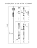 |
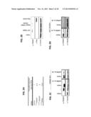 | 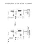 |
 | 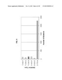 |
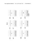 |  |
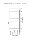 | 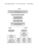 |
 |  |
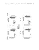 | 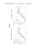 |
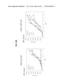 |  |
 |  |
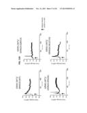 | 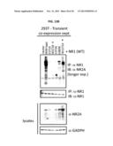 |
 | 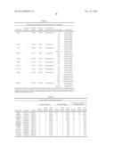 |
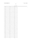 | 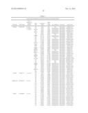 |
 | 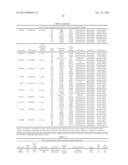 |
 |  |
 |  |
 |  |
 |  |
 |  |
 |  |
 |  |
 |  |
 |  |
 |  |
 |  |
 |  |
 |  |
 |  |
 |  |
 |  |
 |  |
 |  |
| Similar patent applications: | |
| Date | Title |
|---|---|
| 2014-02-27 | Combinations of medicaments for the treatment of respiratory diseases |
| 2014-02-27 | Transport agents for crossing the blood-brain and into brain cancer cells and methods of use thereof |
| 2014-02-27 | Photodynamic therapy or diagnostic agent, using infrared-spectrum light |
| 2014-02-06 | Radioimaging methods using teboroxime and thallium |
| New patent applications in this class: | |
| Date | Title |
|---|---|
| 2019-05-16 | Antibodies, uses thereof and conjugates thereof |
| 2019-05-16 | Monoclonal antibodies that specifically recognize canine dla-dr antigen and their uses |
| 2018-01-25 | Anti-ceacam5 antibodies and uses thereof |
| 2018-01-25 | Tnfrsf-binding agents and uses thereof |
| 2018-01-25 | Antiproliferative compounds and methods of use thereof |
| New patent applications from these inventors: | |
| Date | Title |
|---|---|
| 2013-07-25 | Grm3 mutations and use thereof for the diagnosis and treatment of melanoma |
| 2011-10-27 | Use of erbb4 as a prognostic and therapeutic marker for melanoma |
| Top Inventors for class "Drug, bio-affecting and body treating compositions" | |
| Rank | Inventor's name |
|---|---|
| 1 | David M. Goldenberg |
| 2 | Hy Si Bui |
| 3 | Lowell L. Wood, Jr. |
| 4 | Roderick A. Hyde |
| 5 | Yat Sun Or |