Patent application title: ASSESSMENT OF SUBCHONDRAL BONE REMODELLING BY MEASURING CATHEPSIN K FRAGMENTS OF COLLAGEN TYPE II
Inventors:
Diana J. Leeming (Copenhagen, DK)
Inger Byrjalsen (Hoersholm, DK)
Per Qvist (Klampenborg, DK)
Morten A. Karsdal (Copenhagen, DK)
IPC8 Class: AG01N33566FI
USPC Class:
435 79
Class name: Measuring or testing process involving enzymes or micro-organisms; composition or test strip therefore; processes of forming such composition or test strip involving antigen-antibody binding, specific binding protein assay or specific ligand-receptor binding assay assay in which an enzyme present is a label
Publication date: 2011-10-06
Patent application number: 20110244482
Abstract:
A method of assay to determine the extent of collagen type II resorption
activity comprising measuring the level of fragments of collagen type II
that contain a cathepsin K generated neo-epitope not shared by collagen
type I by binding the neo-epitope with an antibody specific for the
neo-epitope and detecting the level of binding of said binding partner.Claims:
1. A method of assay to determine the extent of collagen type II
resorption activity in a subject, comprising measuring in a biological
sample from said subject fragments of collagen type II that contain a
cathepsin K generated neo-epitope not shared by collagen type I by
binding the neo-epitope with an immunological binding partner specific
for the presence of said neo-epitope and detecting the level of binding
of said binding partner.
2. A method as claimed in claim 1, wherein said extent of collagen type II resorption in said subject is evaluated by comparing the level of binding measured in said assay with levels previously established in healthy subjects and or in subjects having pathological collagen type II remodelling activity.
3. A method as claimed in claim 2, wherein said pathological collagen type II resorption activity is arthritis.
4. A method as claimed in claim 1 for determining the extent of subchondral bone remodelling activity.
5. A method as claimed in claim 1, wherein said extent of subchondral bone remodelling in said subject is evaluated by comparing the level of binding measured in said assay with levels previously established in healthy subjects and or in subjects having pathological subchondral bone remodelling activity.
6. A method as claimed in claim 1, wherein the immunological binding partner is specific for an epitope defined by one of the following amino acid sequences: . . . GQPGPA SEQ ID NO:53; . . . EPGGVG SEQ ID NO:54; DQGVPG . . . SEQ ID NO:55; . . . . PKGARG SEQ ID NO:56; and REGSPG . . . SEQ ID NO:57, wherein the symbol indicates the end of the peptide chain generated by cathepsin K cleavage.
7. A method as claimed in claim 6, wherein said immunological binding partner does not specifically bind a sequence as defined in claim 4 if continued past the indicated cleavage site.
8. A method as claimed in claim 1, conducted as a sandwich immunoassay using a second immunological binding partner which is specifically immunoreactive with a collagen type II amino acid sequence containing an isomerisation.
9. An immunological binding partner against a C-terminal or N-terminal neo-epitope formed by proteinase cleavage of type II collagen.
10. An immunological binding partner having specific binding affinity for a peptide having the N-terminal sequence REGSPG . . . SEQ ID NO:57 or DQGVPG . . . SEQ ID NO:55.
11. An immunological binding partner having specific binding affinity for a peptide having the C-terminal sequence . . . GQPGPA SEO ID NO:53; . . . EPGGVG SEQ ID NO:54; or . . . PKGARG SEQ ID NO:56.
12. An immunoassay kit comprising an immunological binding partner as claimed in claim 9 together with at least one of calibration standards immunoreactive with said binding partner, a wash reagent, a buffer, a secondary immunological binding partner for revealing binding between said immunological binding partner as claimed in claim 6 and components of a sample, an enzyme label, an enzyme label substrate, a stopping reagent, or instructions for conducting an assay using said kit.
Description:
[0001] The present invention relates to methods of immunoassay of peptide
fragments generated by proteolytic cleavage of type II collagen by
Cathepsin K which are excellent biomarkers of subchondral bone
remodelling.
[0002] Osteoarthritis is one of the leading causes of disability in the world, with more than 10% of the elderly population having symptomatic disease (Woolf & Pfleger, 2003). The incidence increases with age, and by age 65, 80% has radiographic evidence of OA (Lawrence et al., 1998). Therefore, osteoarthritis is both prevalent and a serious burden to the patient and the society. However, at present there is little to offer the affected individuals for prevention of the disease or treatment in the early stages. For many patients, hip or knee replacement is eventually the only treatment option.
[0003] Although the pathogenicity of osteoarthritis is not fully understood at present, it is evident that a central hallmark in this slow, chronic disease is progressive destruction of the articular joints, which consists of bone, cartilage and the synovium. In particular the cartilage has attracted much attention, and the different grades and stages of OA cartilage histopathology have recently been detailed described by a working group under OARSI (Pritzker et al., 2006). This system encompasses 7 grades (or severity levels) with involvement of the deeper cartilage layers in more advanced OA disease, and when combined with the extent of cartilage involvement expressed in 5 stages this leads to a semi-quantitative scoring system of 0 to 24.
[0004] However, the lack of sensitive, specific and fast analytical techniques to assess important metabolic processes in articular cartilage and their effects on the structure of the tissue is a major barrier to effective drug development in OA.
[0005] In particular, major efforts have been allocated to the development of new and better biochemical markers of cartilage turnover.
[0006] Cartilage, including articular cartilage, is for the most part composed of collagen type II (60%-70% of dry weight) and proteoglycans (10% of dry weight). Cartilage degradation is mainly mediated by the MMPs and the closely related ADAM-TS (a disintegrin and metalloproteinase with thrombospondin motifs), but collagen type II is most sensitive to MMP activity (Dean et al., 1989; Reboul et al., 1996; Hui et al., 2003). The action of these proteases results in the release of various extracellular fragments which could be candidates as biomarkers of cartilage degradation.
[0007] Since type II collagen is the most abundant protein in cartilage, several different degradation fragments of collagen type II have been indicated as useful for monitoring degenerative diseases of the cartilage (Schaller et al., 2005; Sumer et al., 2006; Birmingham et al., 2006). A fragment of the C-telopeptide of type II collagen, i.e. CTX-II, is generated by MMP-activity (Christgau et al., 2001; Mouritzen et al., 2003) and measurement of CTX-II has been reported for monitoring degradation of type II collagen in experimental setups assessing cartilage degradation (Schaller et al., 2005) as well as in humans (Reijman et al., 2004).
[0008] However, the first report of using antibodies for detection of collagen type II fragments came from Billinghurst and co-workers (Billinghurst et al., 1997), who described detection of an amino-terminal neoepitope on the shorter fragment of type II collagen after cleavage by collagenase.
[0009] More well-described in the literature is the C2C neoepitope at the C-terminus of the 3/4 length fragment (Fraser et al., 2003; Poole et al., 2004). This test is dependant of the binding of a monoclonal antibody to the amino acid sequence EGPP(OH)GPQG SEQ ID NO:1 (Poole et al., 2004).
[0010] Other tests for collagen type II fragments include the C1,2C, which, however, is based on the amino acid sequence GPP(OH)GPQG SEQ ID NO:2 found in both type I and type II (Billinghurst et al., 1997). Also, another fragment generated by the action of collagenase is the TIINE fragment (Otterness et al., 1997) and can be detected using monoclonal antibody 9A4 recognising the neoepitope Gly-Pro-Pro-Gly-Pro-Gln-Gly-COOH SEQ ID NO:3. Combined with monoclonal antibody 5109 (Downs et al., 2001) as a capture antibody, the sandwich test is claimed to be specific for type II collagen.
[0011] Apart from the involvement of articular cartilage in OA, an increasing body of evidence demonstrates that skeletal structural integrity is a prerequisite for preventing progression of joint disease.
[0012] Examinations of peri-articular bone in knees and hips of patients with OA have confirmed that the subchondral bone is abnormal in OA joints, with altered trabecular structure and sclerosis of the subchondral plate (Hunter et al., 2003A; Hunter et al., 2003B). Bone scintigraphy has revealed localization of the nuclide to sites of increased subchondral bone turnover (Dieppe et al., 1993). Cross-sectional studies have also established that women with advanced knee or hip OA have higher bone mineral densities (BMD) near, or at the site of joint OA (Arden et al., 2006). In fact, subchondral bone turnover has been shown to be as much as 20 fold increased compared to that of normal bone turnover (Bailey et al., 2004).
[0013] Subchondral bone is separated from the articular cartilage only by a layer of calcified cartilage (Burr et al., 2003). This allows for several possibilities of transmitting signals from one compartment to the other. Among these are increased vascularisation, and the development of microcracks occurring in the bone matrix, both phenomena which have been strongly implicated in initiation of bone remodelling, as well as increased degradation of the calcified cartilage (Burr et al., 2003; Lajeunesse et al., 2003).
[0014] Despite the fact that subchondral bone abnormality could be an important risk factor for the development of OA, the metabolic activity in this compartment cannot be studied with biochemical markers, as markers with sufficient specificity has not been identified.
[0015] However, we have discovered that the quantification of fragments of type II collagen carrying neo-epitopes generated by the proteolytic cleavage of the protein by cathepsin K are both sensitive and specific markers of subchondral bone turnover. Furthermore we have discovered that elevated levels of these fragments in body fluids is associated with a high risk for progression of OA and therefore could be incorporated into the overall risk assessment of this disease. It is conjectured that such fragments are produced when calcified cartilage is exposed to cathepsin K produced by osteoclasts.
[0016] Cathepsin K cleavage of type II collagen has been reported in a few studies. The first study to associate cathepsin K with cleavage of type II collagen was reported by Kafienah et al. (1998). Kafienah and coworkers described helical cleavage site(s) of collagen type II by Cathepsin K, and provided the amino acid sequence for one cleavage site, i.e.
TABLE-US-00001 PGDDGEAGKPG KSGERGPPG SEQ ID NO: 4 (bovine sequence)
[0017] Ten years later, Dejica et al. (2008) reported the development of an enzyme-linked immunosorbent assay based on polyclonal antibodies against the C-terminal neoepitope (C2K) of the cathepsin K cleavage site reported by Kefienah, i.e.
TABLE-US-00002 PGDDGEAGKPG KAGERGPPG SEQ ID NO: 5
[0018] As the seven C-terminal amino acids of this epitope, i.e. GEAGKPG SEQ ID NO:6 can be found in type I collagen as well, this test will not distinguish type I and type II collagen fragments generated by cathepsin K activity. In contrast, according to the present invention such sequences are de-selected to increase specificity for type II collagen fragments. The measure in Dejica is not taught to provide information regarding subchondral bone resorption.
[0019] U.S. Pat. No. 6,642,007 (Saltarelli) disclose methods for monitoring urine for type II collagen fragment using a combination of a capture antibody and a detection antibody, such that type II collagen is distinguished from other collagen fragments.
[0020] U.S. Pat. No. 6,030,792 (Otterness) discloses antibodies for detecting collagen type II fragments resulting from collagenase cleavage. In particular, the following sequences are disclosed;
TABLE-US-00003 GPPGPQG SEQ ID NO: 7 GEPGDDGPSG SEQ ID NO: 8 APGEDGRPGPPGP SEQ ID NO: 9 GKVGPSGAPGEDGRPG SEQ ID NO: 10 AEGPPGPQG SEQ ID NO: 11 GPPGPQGLAG SEQ ID NO: 12 GEPGDDGPS SEQ ID NO: 13 GEPGDDGPSGAEGPPG SEQ ID NO: 14 EKGEPGDDAPSGAEGPPGPQG SEQ ID NO: 15 GPPGPPGKPGDDGEAGKPGKA SEQ ID NO: 16 GPPGPRGRSGETGPAGPPGNP SEQ ID NO: 17 GAPGPQGFQGNPGEPGEPGVSY SEQ ID NO: 18 GEPGDDAGPSGAEGPPGPQG SEQ ID NO: 19
[0021] A series of patents (U.S. Pat. Nos. 6,602,980; 6,566,492; 6,348,320; 6,255,056; 6,153,732; 6,143,511; 6,100,379; 5,919,634; 5,702,909; 5,688,652; 5,641,837; 5,641,687; 5,532,169; and 5,455,179) (Eyre) relates to peptides and methods for cartilage resorption assays employing antibodies binding to epitopes in the telopeptides of type II collagen.
[0022] U.S. Pat. No. 5,283,197 (Robins) describes methods of detecting collagen fragments cross-linked with lysyl pyridinoline or hydroxylysyl pyridinoline.
[0023] U.S. Pat. No. 7,410,770 (Reginster) disclosed methods for detection of collagenase-generated fragments of collagen type II using antibody binding to epitope in the amino acid sequence HRGYPGLDG SEQ ID NO:20 located in the helical region of collagen type II.
[0024] U.S. Pat. No. 6,132,976 (Poole) discloses methods for detecting cartilage degradation using antibodies which does not bind to unwound (native) type II collagen fragments but only to fragments being generated by collagenase cleavage. In particular the following amino acid sequences originating from type II collagen are included;
TABLE-US-00004 CGKVGPSGAPGEDGRPGPPGPQY SEQ ID NO: 21 APGEDGRPGPPGP SEQ ID NO: 22 GQPG SEQ ID NO: 23 GPPGPQG SEQ ID NO: 7 CGGEGPPGPQG SEQ ID NO: 24 GAEGPPGPQGLAGQRGIVG SEQ ID NO: 25 GAPGTPGPQGIAGQRGVVG SEQ ID NO: 26 GPPGTPGPQGLLGAPGILG SEQ ID NO: 27 GPPGAPGPLGIAGITGARG SEQ ID NO: 28 CGGEGPPGPQGL SEQ ID NO: 29 CGGEGPPGPQGLA SEQ ID NO: 30 CGGEGPPGPQ SEQ ID NO: 31 CGGEGPPGP SEQ ID NO: 32 CGPPGPQG SEQ ID NO: 33
[0025] U.S. Pat. No. 7,115,378 (Welsch) describes the use of mass spectrometry for identifying and quantifying peptides resulting from enzyme cleavage of collagen type II. The technique is used for identification and quantification of the peptides in a biological sample to assess activity of proteolytic enzymes in osteoarthritis and rheumatoid arthritis. In particular, the following sequences originating from humans are disclosed;
TABLE-US-00005 SEQ ID NO: 7 GPPGPQG SEQ ID NO: 34 LQGPAGPPGEKGEPGDDGPSGAEGPPGPQG PQG SEQ ID NO: 35 PGPQG SEQ ID NO: 36 PPGPQG SEQ ID NO: 37 VLQGPAGPPGEKGEPGDDGPSGAEGPPGPQG SEQ ID NO: 38 KGARGDSGPPGRAGEPGLQGPAGPPGEKGEPGDDGPSGAEGPPGPQG SEQ ID NO: 39 ARGDSGPPGRAGEPGLQGPAGPPGEKGEPGDDGPSGAEGPPGPQG SEQ ID NO: 40 GPAGPPGEKGEPGDDGPSGAEGPPGPQG SEQ ID NO: 41 GPIGPPGERGAPGNRGFPGQDGLAGPKGAPGERGPSGLAGPKGANGD PGRPGEPGLPGARGLTGRPGDAGPQGKVGPSGAPGEDGRPGPPGPQG ARGQPGVMGFPGPKGANGEPGKAGEKGLPGAPGLGLPGKDGETGAEG PPPA . . .
[0026] U.S. Pat. No. 6,706,490 (Cook) describes the detection of antibodies to collagen using CB peptides, in particular CB10, of mammalian type II collagen. Cyanogen bromide cleaves the carboxyl terminal of methionine residues thereby producing the CB peptides. The CB10 peptide has the sequence:
TABLE-US-00006 SEQ ID NO: 42 MPGERGAAGIAGPKGDRGDVGEKGPEGAPGKDGGRGLTGPIGPPGPAG ANGEKGEVGPPGPAGSAGARGAPGERGETGPPGTSGIAGPPGADGQPG AKGEQGEAGQKGDAGAPGPQGPSGAPGPQGPTGVTGPKGARGAQGPPG ATGFPGAAGRVGPPGSNGNPGPPGPPGPSGKDGPKGARGDSGPPGRAG EPGLQGPAGPPGEKGEPGDDGPSGAEGPPGPQGLAGQRGIVGLPGQRG ERGFPGLPGPSGEPGQQGAPGASGDRGPPGPVGPPGLTGPAGEPGREG SPGADGPPGRDGAAGVKGDRGETGAVGAPGAPGPPGSPGPAGPTGKQG DRGEAGAQGPM
[0027] U.S. Pat. No. 7,195,883 (Rosenquist) disclose sandwich immunoassays in which a single antibody specific for the amino acid sequence EKGPDP SEQ ID NO:43 is used to detect telopeptide fragments fo type II collagen.
[0028] U.S. Pat. Nos. 6,420,125 and 6,107,047 (Fledelius) disclose methods of measuring the rate of degradation of collagen using antibodies binding to an amino acid sequence of type II collagen containing an isoaspartic acid residue. Also, the use of synthetic peptides having an amino acid sequence of type II collagen that contains an isoaspartic acid residue is described. In particular, the following sequences from type II collagen are described;
TABLE-US-00007 GDIK*DIV SEQ ID NO: 44 EKGP*D, SEQ ID NO: 45
where (*) denotes an isomerised peptide bond.
[0029] U.S. Pat. No. 6,300,083 (Fledelius) describes the determination of the amount of a D-amino acid containing fragment of the protein in a body fluid using an antibody capable of discriminating between the D-amino acid containing fragment and its L-amino acid containing analogue. In particular, the application includes the following peptide sequences from type II collagen;
TABLE-US-00008 GDIKDIV SEQ ID NO: 46 EKGPD SEQ ID NO: 43
[0030] U.S. Pat. Nos. 6,372,442 and 6,210,902 (Bonde) describes methods of characterizing the degradation of type II collagen. At least two distinct immunological assays should be used, each using a different immunological binding partner, and a numerical index is formed representing the difference in the results of the assays. In particular, the following type II collagen sequences are disclosed;
TABLE-US-00009 EKGPDP SEQ ID NO: 47 EKGPD SEQ ID NO: 43 GVK PGVKG SEQ ID NO: 48 PGPKGE SEQ ID NO: 49 GQKGEP SEQ ID NO: 50 GDIKDIV SEQ ID NO: 46
[0031] U.S. Pat. Nos. 6,355,442; 6,342,361; 6,323,314 and 6,110,689 (Qvist) describe the use of antibodies recognizing synthetic peptides for detection of collagen fragments. In particular, the following amino acid sequences are included;
TABLE-US-00010 PGPKGE SEQ ID NO: 49 GQKGEP SEQ ID NO: 50 GDIKDIV SEQ ID NO: 46 EKGPD SEQ ID NO: 43 GVK PGVKG SEQ ID NO: 48
[0032] U.S. Pat. No. 6,010,863 (Te Koppele) discloses the use of a sandwich immunoassay for the detection of collagen degradation using a first antibody directed at an epitope present on a collagen molecule at a distance of up to 165 amino acids from a collagen telopeptide crosslink site, and a second antibody directed at another epitope of the crosslinked collagen molecule.
[0033] U.S. Pat. No. 5,541,295 (Barrach) discloses monoclonal antibodies which bind specifically to Type II collagen, but not to its peptides, or vice versa. In particular, the following type II collagen sequences are included;
TABLE-US-00011 GFQGL-Xaa-G-Xaa-Xaa-G-Xaa-Xaa-G SEQ ID NO: 51 GLQGL-Xaa-G-Xaa-Xaa-G-Xaa-SG SEQ ID NO: 52
[0034] None of the above mentioned patents make reference to the cleavage of type II collagen by cathepsin K. Moreover, none of the patents disclose the usefulness of a quantitative measure of such fragments as biomarkers of subchondral bone remodelling.
[0035] References to collagen type II herein include specifically reference to human type II collagen.
[0036] The present invention now provides a method of assay to determine the extent of collagen type II resorption activity, or of degradation of mineralised cartilage, in a subject, comprising measuring in a biological sample from said subject fragments of collagen type II that contain a cathepsin K generated neo-epitope not shared by collagen type I by binding the neo-epitope with an immunological binding partner specific for the presence of said neo-epitope and detecting the level of binding of said binding partner.
[0037] Preferably, said extent of collagen type II resorption in said subject is evaluated by comparing the level of binding measured in said assay with levels previously established in healthy subjects and or in subjects having pathological collagen type II resorption activity, e.g. arthritis. Moreover the assay can provide an indication of the extent of subchondral bone remodelling activity and the comparator level used may be that seen in patients having a pathological level of subchondral bone remodelling.
[0038] The immunological binding partner is preferably specific for an epitope defined by one of the following amino acid sequences: . . . GQPGPA SEQ ID NO:53; . . . EPGGVG SEQ ID NO:54; DQGVPG . . . SEQ ID NO:55; . . . PKGARG SEQ ID NO:56; and REGSPG . . . SEQ ID NO:57, wherein the symbol indicates the end of the peptide chain generated by cathepsin K cleavage.
[0039] Preferably therefore, said immunological binding partner does not specifically bind a sequence as defined above if continued past the indicated cleavage site and does not bind intact collagen type II.
[0040] The assay may be conducted as a sandwich immunoassay using a second immunological binding partner which is specifically immunoreactive with a collagen type II amino acid sequence containing an isomerisation.
[0041] The invention includes an immunological binding partner against a C-terminal or N-terminal neo-epitope formed by proteinase cleavage of type II collagen which is not found in collagen type I.
[0042] Such an immunological binding partner preferably has specific binding affinity for a peptide having the N-terminal sequence REGSPG . . . SEQ ID NO:57 or DQGVPG . . . SEQ ID NO:55 or has specific binding affinity for a peptide having the C-terminal sequence . . . GQPGPA SEQ ID NO:53; . . . EPGGVG SEQ ID NO:54; or . . . PKGARG SEQ ID NO:56.
[0043] The invention includes an immunoassay kit comprising an immunological binding partner as described together with at least one of calibration standards immunoreactive with said binding partner, a wash reagent, a buffer, a secondary immunological binding partner for revealing binding between said immunological binding partner of the invention and components of a sample, an enzyme label, an enzyme label substrate, a stopping reagent, or instructions for conducting an assay using said kit.
[0044] The biological sample may in particular be a body fluid sample and may be blood, serum, plasma, or urine.
[0045] As explained in WO02/095415, isomerisation of certain amino acids occurs naturally over time in proteins of the body, particularly at aspartic acid, asparagines, glutamic acid and glutamine residues, according to the illustrative reaction scheme:
##STR00001##
[0046] This may give rise to either or both of optical or structural isomerism at the affected residue and either can be recognised in a context specific manner (i.e. dependant on the presence of the appropriate flanking amino acid sequences) or context independent manner (i.e. not dependant on the presence of the appropriate flanking amino acid sequences) by a suitably selected immunological binding partner. In assays according to the invention a first immunological binding partner specifically binding a cathepsin K generated neo-epitope of collagen II may be used in combination with an immunological binding partner specifically binding an epitope containing a said isomerisation in a sandwich assay.
[0047] Assay formats useful in accordance with the present invention include both heterogeneous and homogeneous sandwich assay formats.
[0048] Homogeneous formats include the use of two different immunological binding partners bound to respective beads wherein the beads incorporate a detectable proximity signal activated when the beads are brought into proximity by their respective binding partners both binding to sites on a single fragment molecule.
[0049] Heterogeneous assay formats include those in which one of said immunological binding partners is immobilised to a solid support, said fragments are bound to said immobilised antibody and the binding of the other of said immunological binding partners to said fragments is detected.
[0050] Immunological binding partners for use in the present invention include whole antibodies, especially monoclonal antibodies, and antibody fragments with specific binding affinity. These include binding fragments such as Fab or F(ab')2.
[0051] The following procedure was used for identification of type II collagen sequences carrying Cathepsin K neoepitopes.
[0052] Human collagen type II (BIOCOL BC-3001) was dissolved in 10 mM acetic acid (400 μl added to 1 mg of collagen type II). Ten μg of procathepsin K (Calbiochem 342001) was activated by addition of 200 μl of 100 mM sodium acetate containing 10 mM DTT and 5 mM EDTA, pH 3.9 for 40 minutes at room temperature. Ten μg of MMP9 (Calbiochem 444231) was activated by addition of 200 μl of 1 mM APMA in DMSO for 2 hours at 37° C. For the Cathepsin K cleavage, 60 μl of collagen type II was added 120 μl of 50 mM sodium acetate, pH 5.5 containing 20 mM L-cysteine and 24 μl of activated cathepsin K for 4 hours at 37° C. For the MMP9 cleavage, 60 μl of collagen type II was added 120 μl of 100 mM Tris-HCl, 100 mM sodium chloride, 10 mM calcium chloride, 2 mM zinc chloride, pH 8.0 and 20 μl of MMP9 for 3 days at 37° C. The resulting proteolytic cleavage fragments were characterized by high performance liquid chromatography (HPLC)-tandem mass spectrometry (MS/MS) analysis. The MS/MS spectra were searched against protein databases using Sequest and X! Tandem database search algorithms. The following sequence hits of fragments were found for the Cathepsin K cleaved collagen type II:
TABLE-US-00012 AQGPPGATGFPGAAGR SEQ ID NO: 58 ASGDRGPPGPV SEQ ID NO: 59 ASGDRGPPGPVGPPG SEQ ID NO: 60 GANGEKGEVGPPGPA SEQ ID NO: 61 GAPGEDGRPGPPGPQ SEQ ID NO: 62 GARGAPGERGETGPPGPA SEQ ID NO: 63 GDRGPPGPV SEQ ID N0: 64 GERGFPG SEQ ID N0: 65 GERGFPGER SEQ ID NO: 66 GESGSPGENGSPGPM SEQ ID N0: 67 GLPGPPGPPGEGGKPG SEQ ID NO: 68 GPIGPPGPA SEQ ID NO: 69 GPPGPPGKPGDDGEAGKPG SEQ ID NO: 70 GPPGPV SEQ ID NO: 71 GPPGPVGPA SEQ ID NO: 72 LPGPPGPPGEGGKPG SEQ ID NO: 73 NPGPPGPPGPPGPG SEQ ID NO: 74 PIGPP SEQ ID NO: 75 REGSPGADGPPGRDGAAGVK SEQ ID NO: 76 SNGNPGPPGPPGPS SEQ ID NO: 77
[0053] Identified fragments were aligned with the sequence for human collagen type II (sp|P02458|CO2A1_HUAN Collagen alpha-1(II) chain), and cleavage sites were localized as indicated by the arrows.
TABLE-US-00013 SEQ ID NO: 78 QMAGGFDEKAGGAQLGVMQGPMGPMGPRGPPGPAGAPGPQGFQGNPGEPG EPGVSGPMGPR GPPGPPGKPGDDGEAGKPG KAGERGPPGPQGARG FPGTPGLPGVKGHRGYPGLDGAKGEAGAPGVK GESGSPGENGSPGPM GPRGLPGERGRTGPAGAAGARGNDGQPGPA GPPGPV GPA GGP GFPGAPGAKGEAGPTGARGPEGAQGPRGEPGTPGSPGPAGASGNPGTD GIPGAKGSAGAPGIAGAPGFPGPRGPPGPQGATGPLGPKGQTGEPGIAG FKGEQGPKGEPGPAGPQGAPGPAGEEGKRGARGEPGGVG PIGPP G ERGAPGNRGFPGQDGLAGPKGAPGERGPSGLAGPKGANGDPGRPGEPGL PGARGLTGRPGDAGPQGKVGPS GAPGEDGRPGPPGPQ GARGQPGV MGFPGPKGANGEPGKAGEKGLPGAPGLRGLPGKDGETGAAGPPGPAGPA GERGEQGAPGPSGFQ G LPGPPGPPGEGGKPG DQGVPGEAGAPG LVGPR GERGFPG ER GSPGAQGLQGPRGLPGTPGTDGPKGASGP AGPPGAQGPPGLQGMPGERGAAGIAGPKGDRGDVGEKGPEGAPGKDGGRG LT GPIGPPGPA GANGEKGEVGPPGPA GSA GARGAPGERGET GPPGPA GFAGPPGADGQPGAKGEQGEAGQKGDAGAPGPQGPSGAPGP QGPTGVTGPKGARG AQGPPGATGFPGAAGR VGPPG SNGNPGPP GPPGPS GKDGPKGARGDSGPPGRAGEPGLQGPAGPPGEKGEPGDDGP SGAEGPPGPQGLAGQRGIVGLPGQRGERGFPGLPGPSGEPGKQGAPG AS GDRGPPGPV GPPG LTGPAGEPG REGSPGADGPPGRDGAA GVK GDRGETGAVGAPGAPGPPGSPGPAGPTGKQGDRGEAGAQGPMGP SGPAGARGIQGPQGPRGDKGEAGEPGERGLKGHRGFTGLQGLPGPPGPS GDQGASGPAGPSGPRGPPGPVGPSGKDGANGIPGPIGPPGPRGRSGETG PAGPPG NPGPPGPPGPPGPG IDMSAFAGLGPREKGPDPLQYMRA
[0054] Preferred biomarker neoepitopes were selected based on protease and protein specificity. First protease specificity was assessed by comparison of Cathepsin K cleavage sites with cleavage sites of other proteases, e.g. MMP9 and MMP13, and cleavage sites that were in common were deselected. Next protein specificity of the remaining cleavage sites were assessed by identity search of 6 amino-terminal or 6 carboxy-terminal residues on either site of the cleavage site. The public available programme "Pattinprot" was used in the search of the UNIPROT/SWISSPROT databank. The preferred biomarker neoepitopes are indicated and listed below.
Collagen α1 Chain Type II Collagen (1060 Residues)
TABLE-US-00014 [0055] SEQ ID NO: 78 QMAGGFDEKAGGAQLGVMQGPMGPMGPRGPPGPAGAPGPQGFQGNP GEPGEPGVSGPMGPRGPPGPPGKPGDDGEAGKPGKAGERGPPGPQG ARGFPGTPGLPGVKGHRGYPGLDGAKGEAGAPGVKGESGSPGENGS PGPMGPRGLPGERGRTGPAGAAGARGNDGQPGPA GPPGPVGPAGG PGFPGAPGAKGEAGPTGARGPEGAQGPRGEPGTPGSPGPAGASGNP GTDGIPGAKGSAGAPGIAGAPGFPGPRGPPGPQGATGPLGPKGQTG EPGIAGFKGEQGPKGEPGPAGPQGAPGPAGEEGKRGARGEPGGVG PIGPPGERGAPGNRGFPGQDGLAGPKGAPGERGPSGLAGPKGANGD PGRPGEPGLPGARGLTGRPGDAGPQGKVGPSGAPGEDGRPGPPGPQ GARGQPGVMGFPGPKGANGEPGKAGEKGLPGAPGLRGLPGKDGETG AAGPPGPAGPAGERGEQGAPGPSGFQGLPGPPGPPGEGGKPG DQG VPGEAGAPGLVGPRGERGFPGERGSPGAQGLQGPRGLPGTPGTDGP KGASGPAGPPGAQGPPGLQGMPGERGAAGIAGPKGDRGDVGEKGPE GAPGKDGGRGLTGPIGPPGPAGANGEKGEVGPPGPAGSAGARGAPG ERGETGPPGPAGFAGPPGADGQPGAKGEQGEAGQKGDAGAPGPQGP SGAPGPQGPTGVTGPKGARG AQGPPGATGFPGAAGRVGPPGSNGN PGPPGPPGPSGKDGPKGARGDSGPPGRAGEPGLQGPAGPPGEKGEP GDDGPSGAEGPPGPQGLAGQRGIVGLPGQRGERGFPGLPGPSGEPG KQGAPGASGDRGPPGPVGPPGLTGPAGEPG REGSPGADGPPGRDG AAGVKGDRGETGAVGAPGAPGPPGSPGPAGPTGKQGDRGEAGAQGP MGPSGPAGARGIQGPQGPRGDKGEAGEPGERGLKGHRGFTGLQGLP GPPGPSGDQGASGPAGPSGPRGPPGPVGPSGKDGANGIPGPIGPPG PRGRSGETGPAGPPGNPGPPGPPGPPGPGIDMSAFAGLGPREKGPD PLQYMRA
[0056] Preferred biomarker neoepitopes are marked in the above sequence and are extracted below.
List of Preferred Biomarker Neoepitopes
TABLE-US-00015 [0057] 1 . . . GQPGPA SEQ ID NO: 53 2 . . . EPGGVG SEQ ID NO: 54 3 DQGVPG . . . SEQ ID NO: 55 4 . . . PKGARG SEQ ID NO: 56 5 REGSPG . . . SEQ ID NO: 57
[0058] For the sequences of 1, 2, and 4 the immunological binding partner should preferably have specific binding affinity for peptide fragments comprising the C-terminal neoepitope.
[0059] For the sequences of 3 and 5 the immunological binding partner should preferably have specific binding affinity for peptide fragments comprising the N-terminal neoepitope.
[0060] The invention will be further described and illustrated by the following examples making reference to the accompanying drawings, in which:
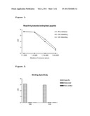
[0061] FIG. 1 shows antibody binding in sera studied in Example 1;
[0062] FIG. 2 shows monoclonal antibody binding observed in Example 1;
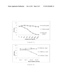
[0063] FIG. 3 shows monoclonal antibody binding observed in Example 3; and
[0064] FIG. 4 shows further monoclonal antibody binding observed in Example 3.
EXAMPLE 1
Generation of Monoclonal Antibodies Recognising a N-terminal Neoepitope
[0065] The following demonstrates that monoclonal antibodies can be generated against collagen II neo-epitopes. Synthetic peptides were prepared by standard techniques. To increase immunogenicity, the peptide (LTGPAGGGGC SEQ ID NO:78) was coupled at the C-terminus to the carrier protein KLH using site-directed coupling technology via the cysteine. Before immunisation, the immunogen was mixed 1:1 with Freund's Incomplete Adjuvant and the mixture was injected s.c. in Balb/c mice. The immunisation was repeated every 2 weeks for two months (four immunisations) and then continued with 4 weeks between each immunisation. Blood was obtained from the mice before immunisation initiated and one week after each immunization. The immune response was evaluated by testing the binding reactivity of mouse immune sera towards the C-terminal biotinylated synthetic peptide (LTGPAGEPGK-Biotin SEQ ID NO:79). The test for binding reactivity of mouse immune sera was based on binding of the immune serum to the biotinylated synthetic peptide that was bound to the surface of a streptavidin-coated microtitre plate. After incubation and washing the bound antibody was demonstrated by incubation with anti-mouse IgG conjugated to horseradish peroxidase, washing and addition of the chromogen TMB (FIG. 1).
[0066] Subsequent to attaining sufficient immune sera titers in the above mentioned screening test, the selected mice were rested for at least 4 weeks, and boosted i.p. withl immunogen without adjuvants. Three days later, the spleen was removed and used for fusion with myeloma cells using standard techniques. Antibodies from growing hybridomas were evaluated by their binding reactivity to the biotinylated synthetic peptide in the assay as described above. Additionally the cleavage specificity of the antibodies was demonstrated by minimum binding reactivity towards a one-residue extended coater (GLTGPAGEPGK-Biotin SEQ ID NO:80) as well as no binding towards a non-similar coater (Biotin-KGATGPLGPK SEQ ID NO:81) (FIG. 2).
EXAMPLE 2
Generation of Monoclonal Antibodies Recognising N- or C-terminal Neoepitopes
[0067] The following further demonstrates that monoclonal antibodies can be generated against cathepsin K mediated collagen type II neo-epitopes. To increase immunogenicity, the peptides EAGKPG SEQ ID NO:82 (NB76), GQPGPA SEQ ID NO:53 (NB77), EPGGVG SEQ ID NO:54(NB78), DQGVPG SEQ ID NO:55 (NB79), PKGARG SEQ ID NO:56 (NB80) and REGSPG SEQ ID NO:57 (NB81) were coupled at the N- or C-terminus as appropriate (i.e. were coupled at the end opposite the marked cleavage site) to the carrier protein KLH using site-directed coupling technology via a cysteine, which is added to the synthetic peptide.
[0068] Before immunisation, the immunogen was mixed 1:1 with Freund's Incomplete Adjuvant and the mixture was injected s.c. in Balb/c mice. The immunisation was repeated every 2 weeks for two months (four immunisations) and then continued with 4 weeks between each immunisation. Blood was obtained from the mice before immunisation was initiated and one week after each immunization. The immune response was evaluated by testing the binding reactivity of mouse immune sera towards the C-terminal biotinylated synthetic peptide corresponding to each of the selected sequences (e.g. REGSPGGADAP-Biotin SEQ ID NO:83). The test for binding reactivity of mouse immune sera was based on binding of the immune serum to the biotinylated synthetic peptide that was bound to the surface of a streptavidin-coated micro-titre plate. After incubation and washing the bound antibody was demonstrated by incubation with anti-mouse IgG conjugated to horseradish peroxidase, washing and addition of the chromogen TMB. Subsequent to attaining sufficient immune sera titers in the above mentioned screening test, the selected mice were rested for at least 4 weeks, and boosted i.p. with immunogen without adjuvants. Three days later, the spleen was removed and used for fusion with myeloma cells using standard techniques. Antibodies from growing hybridomas were evaluated by their binding reactivity to the biotinylated synthetic peptide in the assay as described above.
EXAMPLE 3
Testing Native Reactivity of a New Neoepitope Antibody
[0069] Several hybridomas and subsequent clones were selected for testing. The cleavage specificity of a typical one of these monoclonal antibodies (NB81) was demonstrated by showing minimal binding reactivity towards a one-residue extended peptide (GREGSPGGADAP SEQ ID NO:84) as well as no binding towards a non-similar peptide (KGATGPLGPK SEQ ID NO:85) in competition against a coater peptide REGSPGGADAP-Biotin SEQ ID NO:83 (FIG. 3). The signal was only displaced by the specific peptide (REGSPGGADAP SEQ ID NO:83) and it can therefore be concluded that the antibody is specific for the neoepitope.
[0070] Next the native reactivity of NB81 was tested using supernatants from bovine cartilage explants. Results are shown in FIG. 4. The release of fragments giving rise to assay signal could be inhibited by supernatants from cartilage when treated with catabolic cytokines for more than 7 days CAT-bovine). However this was neither observed with the non-catabolic control (CON-Bovine) nor a metabolically inactive control (MI-Bovine). (FIG. 4). This indicates that the fragment is related to cartilage turnover and degradation and it will therefore be a good marker for catK induced cartilage degradation.
[0071] In this specification, unless expressly otherwise indicated, the word `or` is used in the sense of an operator that returns a true value when either or both of the stated conditions is met, as opposed to the operator `exclusive or` which requires that only one of the conditions is met. The word `comprising` is used in the sense of `including` rather than in to mean `consisting of`. All prior teachings acknowledged above are hereby incorporated by reference. No acknowledgement of any prior published document herein should be taken to be an admission or representation that the teaching thereof was common general knowledge in Australia or elsewhere at the date hereof.
REFERENCES
[0072] Kafienah W, Bromme D, Buttle D J, Croucher L J, Hollander A P. Human cathepsin K cleaves native type I and II collagens at the N-terminal end of the triple helix. Biochem J. 1998 May 1; 331 (Pt 3):727-32 [0073] Dejica V M, Mort J S, Layerty S, Percival M D, Antoniou J, Zukor D J, Poole A R. Cleavage of type II collagen by cathepsin K in human osteoarthritic cartilage. Am J. Pathol. 2008 July; 173(1):161-9. Epub 2008 May 29 [0074] Dean, D. D., Martel-Pelletier, J., Pelletier, J. P., Howell, D. S., and Woessner, J. F., Jr. 1989. Evidence for metalloproteinase and metalloproteinase inhibitor imbalance in human osteoarthritic cartilage. J Clin Invest 84:678-685. [0075] Reboul, P., Pelletier, J. P., Tardif, G., Cloutier, J. M., and Martel-Pelletier, J. 1996. The new collagenase, collagenase-3, is expressed and synthesized by human chondrocytes but not by synoviocytes. A role in osteoarthritis. J Clin Invest 97:2011-2019. [0076] Hui, W., Rowan, A. D., Richards, C. D., and Cawston, T. E. 2003. Oncostatin M in combination with tumor necrosis factor alpha induces cartilage damage and matrix metalloproteinase expression in vitro and in vivo. Arthritis Rheum 48:3404-3418. [0077] Schaller, S., Henriksen, K., Hoegh-Andersen, P., Sondergaard, B. C., Sumer, E. U., Tanko, L. B., Qvist, P., and Karsdal, M. A. 2005. In vitro, ex vivo, and in vivo methodological approaches for studying therapeutic targets of osteoporosis and degenerative joint diseases: how biomarkers can assist? Assay. Drug Dev. Technol. 3:553-580. [0078] Sumer, E. U., Schaller, S., Sondergaard, B. C., Tanko, L. B., and Qvist, P. 2006. Application of biomarkers in the clinical development of new drugs for chondroprotection in destructive joint diseases: a review. Biomarkers 11:485-506. [0079] Christgau, S., Garnero, P., Fledelius, C., Moniz, C., Ensig, M., Gineyts, E., Rosenquist, C., and Qvist, P. 2001. Collagen type II C-telopeptide fragments as an index of cartilage degradation. Bone 29:209-215. [0080] Mouritzen, U., Christgau, S., Lehmann, H. J., Tanko, L. B., and Christiansen, C. 2003. Cartilage turnover assessed with a newly developed assay measuring collagen type II degradation products: influence of age, sex, menopause, hormone replacement therapy, and body mass index. Ann. Rheum Dis 62:332-336. [0081] Reijman, M., Hazes, J. M., Bierma-Zeinstra, S. M., Koes, B. W., Christgau, S., Christiansen, C., Uitterlinden, A. G., and Pols, H. A. 2004. A new marker for osteoarthritis: cross-sectional and longitudinal approach. Arthritis Rheum 50:2471-2478. [0082] Hunter D J, Hart D, Snieder H, Bettica P, Swaminathan R, Spector T D. Evidence of altered bone turnover, vitamin D and calcium regulation with knee osteoarthritis in female twins. Rheumatology (Oxford). 2003A; 42:1311-16. [0083] Hunter D J, Spector T D. The role of bone metabolism in osteoarthritis. Curr Rheumatol Rep. 2003B; 5:15-19. [0084] Dieppe P, Cushnaghan J, Young P, Kirwan J. Prediction of the progression of joint space narrowing in osteoarthritis of the knee by bone scintigraphy. Ann Rheum Dis. 1993; 52:557-63. [0085] Arden N, Nevitt M C. Osteoarthritis: epidemiology. Best Pract Res Clin Rheumatol. 2006; 20:3-25. [0086] Burr D B, Radin E L. Microfractures and microcracks in subchondral bone: are they relevant to osteoarthrosis? Rheum Dis Clin North Am. 2003; 29:675-85. [0087] Lajeunesse D, Reboul P. Subchondral bone in osteoarthritis: a biologic link with articular cartilage leading to abnormal remodeling. Curr Opin Rheumatol. 2003; 15:628-33. [0088] Bailey A J, Mansell J P, Sims T J, Banse X. Biochemical and mechanical properties of subchondral bone in osteoarthritis. Biorheology. 2004; 41:349-58. [0089] Woolf A D, Pfleger B: Burden of major musculoskeletal conditions. Bull World Health Organ 2003; 81:646-56 [0090] Lawrence R C, Helmick C G, Arnett F C et al. Estimates of the prevalence of arthritis nad selected musculoskeletal disorders in the United States. Arthritis Rheum 1998; 41:778-99. [0091] Pritzker K P, Gay S, Jimenez S A, Ostergaard K, Pelletier J P, Revell P A, Salter D, van den Berg W B. Osteoarthritis cartilage histopathology: grading and staging. Osteoarthritis Cartilage 2006; 14:13-29. [0092] Birmingham J D, Vilim V, Kraus V B. Collagen biomarkers for arthritis applications. Biomarker Insights 2006; 2: 61-76. [0093] Fraser A, Fearon U, Billinghurst R C, Ionescu M, Reece R, Barwick T, Emery P, Poole A R, Veale D J. Turnover of type II collagen and aggrecan in cartilage matrix at the onset of inflammatory arthritis in humans: relationship to mediators of systemic and local inflammation. Arthritis Rheum. 2003 November; 48(11):3085-95. [0094] Billinghurst R C, Dahlberg L, Ionescu M, Reiner A, Bourne R, Rorabeck C, Mitchell P, Hambor J, Diekmann O, Tschesche H, Chen J, Van Wart H, Poole A R. Enhanced cleavage of type II collagen by collagenases in osteoarthritic articular cartilage. J Clin Invest. 1997 Apr. 1; 99(7):1534-45. [0095] Poole A R, Ionescu M, Fitzcharles M A, Billinghurst R C. The assessment of cartilage degradation in vivo: development of an immunoassay for the measurement in body fluids of type II collagen cleaved by collagenases. J Immunol Methods. 2004 November; 294(1-2):145-53. [0096] Otterness I G, Downs J T, Lane C, Bliven M L, Stukenbrok H, Scampoli D N, Milici A J, Mezes P S. Detection of collagenase-induced damage of collagen by 9A4, a monoclonal C-terminal neoepitope antibody. Matrix Biol. 1999 August; 18(4):331-41. [0097] Downs J T, Lane C L, Nestor N B, McLellan T J, Kelly M A, Karam G A, Mezes P S, Pelletier J P, Otterness I G. Analysis of collagenase-cleavage of type II collagen using a neoepitope ELISA. J Immunol Methods. 2001 Jan. 1; 247(1-2):25-34.
Sequence CWU
1
8518PRTHomo sapiensMISC_FEATURE(4)..(4)Hydroxyproline 1Glu Gly Pro Pro Gly
Pro Gln Gly1 527PRTHomo
sapiensMISC_FEATURE(3)..(3)Hydroxyproline 2Gly Pro Pro Gly Pro Gln Gly1
537PRTHomo sapiens 3Gly Pro Pro Gly Pro Gln Gly1
5420PRTBos taurus 4Pro Gly Asp Asp Gly Glu Ala Gly Lys Pro Gly Lys Ser
Gly Glu Arg1 5 10 15Gly
Pro Pro Gly 20520PRTHomo sapiens 5Pro Gly Asp Asp Gly Glu Ala
Gly Lys Pro Gly Lys Ala Gly Glu Arg1 5 10
15Gly Pro Pro Gly 2067PRTHomo sapiens 6Gly
Glu Ala Gly Lys Pro Gly1 577PRTHomo sapiens 7Gly Pro Pro
Gly Pro Gln Gly1 5810PRTHomo sapiens 8Gly Glu Pro Gly Asp
Asp Gly Pro Ser Gly1 5 10913PRTHomo
sapiens 9Ala Pro Gly Glu Asp Gly Arg Pro Gly Pro Pro Gly Pro1
5 101016PRTHomo sapiens 10Gly Lys Val Gly Pro Ser Gly
Ala Pro Gly Glu Asp Gly Arg Pro Gly1 5 10
15119PRTHomo sapiens 11Ala Glu Gly Pro Pro Gly Pro Gln
Gly1 51210PRTHomo sapiens 12Gly Pro Pro Gly Pro Gln Gly Leu
Ala Gly1 5 10139PRTHomo sapiens 13Gly Glu
Pro Gly Asp Asp Gly Pro Ser1 51416PRTHomo sapiens 14Gly Glu
Pro Gly Asp Asp Gly Pro Ser Gly Ala Glu Gly Pro Pro Gly1 5
10 151521PRTHomo sapiens 15Glu Lys Gly
Glu Pro Gly Asp Asp Ala Pro Ser Gly Ala Glu Gly Pro1 5
10 15Pro Gly Pro Gln Gly
201621PRTHomo sapiens 16Gly Pro Pro Gly Pro Pro Gly Lys Pro Gly Asp Asp
Gly Glu Ala Gly1 5 10
15Lys Pro Gly Lys Ala 201721PRTHomo sapiens 17Gly Pro Pro Gly
Pro Arg Gly Arg Ser Gly Glu Thr Gly Pro Ala Gly1 5
10 15Pro Pro Gly Asn Pro
201822PRTHomo sapiens 18Gly Ala Pro Gly Pro Gln Gly Phe Gln Gly Asn Pro
Gly Glu Pro Gly1 5 10
15Glu Pro Gly Val Ser Tyr 201920PRTHomo sapiens 19Gly Glu Pro
Gly Asp Asp Ala Gly Pro Ser Gly Ala Glu Gly Pro Pro1 5
10 15Gly Pro Gln Gly
20209PRTHomo sapiens 20His Arg Gly Tyr Pro Gly Leu Asp Gly1
52123PRTHomo sapiens 21Cys Gly Lys Val Gly Pro Ser Gly Ala Pro Gly Glu
Asp Gly Arg Pro1 5 10
15Gly Pro Pro Gly Pro Gln Tyr 202213PRTHomo sapiens 22Ala Pro
Gly Glu Asp Gly Arg Pro Gly Pro Pro Gly Pro1 5
10234PRTHomo sapiens 23Gly Gln Pro Gly12411PRTHomo sapiens 24Cys Gly
Gly Glu Gly Pro Pro Gly Pro Gln Gly1 5
102519PRTHomo sapiens 25Gly Ala Glu Gly Pro Pro Gly Pro Gln Gly Leu Ala
Gly Gln Arg Gly1 5 10
15Ile Val Gly2619PRTHomo sapiens 26Gly Ala Pro Gly Thr Pro Gly Pro Gln
Gly Ile Ala Gly Gln Arg Gly1 5 10
15Val Val Gly2719PRTHomo sapiens 27Gly Pro Pro Gly Thr Pro Gly
Pro Gln Gly Leu Leu Gly Ala Pro Gly1 5 10
15Ile Leu Gly2819PRTHomo sapiens 28Gly Pro Pro Gly Ala
Pro Gly Pro Leu Gly Ile Ala Gly Ile Thr Gly1 5
10 15Ala Arg Gly2912PRTHomo sapiens 29Cys Gly Gly
Glu Gly Pro Pro Gly Pro Gln Gly Leu1 5
103013PRTHomo sapiens 30Cys Gly Gly Glu Gly Pro Pro Gly Pro Gln Gly Leu
Ala1 5 103110PRTHomo sapiens 31Cys Gly
Gly Glu Gly Pro Pro Gly Pro Gln1 5
10329PRTHomo sapiens 32Cys Gly Gly Glu Gly Pro Pro Gly Pro1
5338PRTHomo sapiens 33Cys Gly Pro Pro Gly Pro Gln Gly1
53430PRTHomo sapiens 34Leu Gln Gly Pro Ala Gly Pro Pro Gly Glu Lys Gly
Glu Pro Gly Asp1 5 10
15Asp Gly Pro Ser Gly Ala Glu Gly Pro Pro Gly Pro Gln Gly 20
25 30355PRTHomo sapiens 35Pro Gly Pro Gln
Gly1 5366PRTHomo sapiens 36Pro Pro Gly Pro Gln Gly1
53731PRTHomo sapiens 37Val Leu Gln Gly Pro Ala Gly Pro Pro Gly Glu
Lys Gly Glu Pro Gly1 5 10
15Asp Asp Gly Pro Ser Gly Ala Glu Gly Pro Pro Gly Pro Gln Gly
20 25 303847PRTHomo sapiens 38Lys Gly
Ala Arg Gly Asp Ser Gly Pro Pro Gly Arg Ala Gly Glu Pro1 5
10 15Gly Leu Gln Gly Pro Ala Gly Pro
Pro Gly Glu Lys Gly Glu Pro Gly 20 25
30Asp Asp Gly Pro Ser Gly Ala Glu Gly Pro Pro Gly Pro Gln Gly
35 40 453945PRTHomo sapiens 39Ala
Arg Gly Asp Ser Gly Pro Pro Gly Arg Ala Gly Glu Pro Gly Leu1
5 10 15Gln Gly Pro Ala Gly Pro Pro
Gly Glu Lys Gly Glu Pro Gly Asp Asp 20 25
30Gly Pro Ser Gly Ala Glu Gly Pro Pro Gly Pro Gln Gly
35 40 454028PRTHomo sapiens 40Gly Pro
Ala Gly Pro Pro Gly Glu Lys Gly Glu Pro Gly Asp Asp Gly1 5
10 15Pro Ser Gly Ala Glu Gly Pro Pro
Gly Pro Gln Gly 20 2541145PRTHomo sapiens
41Gly Pro Ile Gly Pro Pro Gly Glu Arg Gly Ala Pro Gly Asn Arg Gly1
5 10 15Phe Pro Gly Gln Asp Gly
Leu Ala Gly Pro Lys Gly Ala Pro Gly Glu 20 25
30Arg Gly Pro Ser Gly Leu Ala Gly Pro Lys Gly Ala Asn
Gly Asp Pro 35 40 45Gly Arg Pro
Gly Glu Pro Gly Leu Pro Gly Ala Arg Gly Leu Thr Gly 50
55 60Arg Pro Gly Asp Ala Gly Pro Gln Gly Lys Val Gly
Pro Ser Gly Ala65 70 75
80Pro Gly Glu Asp Gly Arg Pro Gly Pro Pro Gly Pro Gln Gly Ala Arg
85 90 95Gly Gln Pro Gly Val Met
Gly Phe Pro Gly Pro Lys Gly Ala Asn Gly 100
105 110Glu Pro Gly Lys Ala Gly Glu Lys Gly Leu Pro Gly
Ala Pro Gly Leu 115 120 125Gly Leu
Pro Gly Lys Asp Gly Glu Thr Gly Ala Glu Gly Pro Pro Pro 130
135 140Ala14542347PRTHomo sapiens 42Met Pro Gly Glu
Arg Gly Ala Ala Gly Ile Ala Gly Pro Lys Gly Asp1 5
10 15Arg Gly Asp Val Gly Glu Lys Gly Pro Glu
Gly Ala Pro Gly Lys Asp 20 25
30Gly Gly Arg Gly Leu Thr Gly Pro Ile Gly Pro Pro Gly Pro Ala Gly
35 40 45Ala Asn Gly Glu Lys Gly Glu Val
Gly Pro Pro Gly Pro Ala Gly Ser 50 55
60Ala Gly Ala Arg Gly Ala Pro Gly Glu Arg Gly Glu Thr Gly Pro Pro65
70 75 80Gly Thr Ser Gly Ile
Ala Gly Pro Pro Gly Ala Asp Gly Gln Pro Gly 85
90 95Ala Lys Gly Glu Gln Gly Glu Ala Gly Gln Lys
Gly Asp Ala Gly Ala 100 105
110Pro Gly Pro Gln Gly Pro Ser Gly Ala Pro Gly Pro Gln Gly Pro Thr
115 120 125Gly Val Thr Gly Pro Lys Gly
Ala Arg Gly Ala Gln Gly Pro Pro Gly 130 135
140Ala Thr Gly Phe Pro Gly Ala Ala Gly Arg Val Gly Pro Pro Gly
Ser145 150 155 160Asn Gly
Asn Pro Gly Pro Pro Gly Pro Pro Gly Pro Ser Gly Lys Asp
165 170 175Gly Pro Lys Gly Ala Arg Gly
Asp Ser Gly Pro Pro Gly Arg Ala Gly 180 185
190Glu Pro Gly Leu Gln Gly Pro Ala Gly Pro Pro Gly Glu Lys
Gly Glu 195 200 205Pro Gly Asp Asp
Gly Pro Ser Gly Ala Glu Gly Pro Pro Gly Pro Gln 210
215 220Gly Leu Ala Gly Gln Arg Gly Ile Val Gly Leu Pro
Gly Gln Arg Gly225 230 235
240Glu Arg Gly Phe Pro Gly Leu Pro Gly Pro Ser Gly Glu Pro Gly Gln
245 250 255Gln Gly Ala Pro Gly
Ala Ser Gly Asp Arg Gly Pro Pro Gly Pro Val 260
265 270Gly Pro Pro Gly Leu Thr Gly Pro Ala Gly Glu Pro
Gly Arg Glu Gly 275 280 285Ser Pro
Gly Ala Asp Gly Pro Pro Gly Arg Asp Gly Ala Ala Gly Val 290
295 300Lys Gly Asp Arg Gly Glu Thr Gly Ala Val Gly
Ala Pro Gly Ala Pro305 310 315
320Gly Pro Pro Gly Ser Pro Gly Pro Ala Gly Pro Thr Gly Lys Gln Gly
325 330 335Asp Arg Gly Glu
Ala Gly Ala Gln Gly Pro Met 340 345436PRTHomo
sapiens 43Glu Lys Gly Pro Asp Pro1 5447PRTHomo
sapiensMISC_FEATURE(4)..(5)Isomerised peptide bond 44Gly Asp Ile Lys Asp
Ile Val1 5455PRTHomo sapiensMISC_FEATURE(4)..(5)Isomerised
peptide bond 45Glu Lys Gly Pro Asp1 5467PRTHomo sapiens
46Gly Asp Ile Lys Asp Ile Val1 5476PRTHomo sapiens 47Glu
Lys Gly Pro Asp Pro1 5485PRTHomo sapiens 48Pro Gly Pro Lys
Gly1 5496PRTHomo sapiens 49Pro Gly Val Lys Gly Glu1
5506PRTHomo sapiens 50Gly Gln Lys Gly Glu Pro1
55113PRTHomo sapiensMISC_FEATURE(6)..(6)Unknown amino acid 51Gly Phe Gln
Gly Leu Xaa Gly Xaa Xaa Gly Xaa Xaa Gly1 5
105213PRTHomo sapiensMISC_FEATURE(6)..(6)Unspecified amino acid 52Gly
Leu Gln Gly Leu Xaa Gly Xaa Xaa Gly Xaa Ser Gly1 5
10536PRTHomo sapiens 53Gly Gln Pro Gly Pro Ala1
5546PRTHomo sapiens 54Glu Pro Gly Gly Val Gly1 5556PRTHomo
sapiens 55Asp Gln Gly Val Pro Gly1 5566PRTHomo sapiens
56Pro Lys Gly Ala Arg Gly1 5576PRTHomo sapiens 57Arg Glu
Gly Ser Pro Gly1 55816PRTHomo sapiens 58Ala Gln Gly Pro Pro
Gly Ala Thr Gly Phe Pro Gly Ala Ala Gly Arg1 5
10 155911PRTHomo sapiens 59Ala Ser Gly Asp Arg Gly
Pro Pro Gly Pro Val1 5 106015PRTHomo
sapiens 60Ala Ser Gly Asp Arg Gly Pro Pro Gly Pro Val Gly Pro Pro Gly1
5 10 156115PRTHomo sapiens
61Gly Ala Asn Gly Glu Lys Gly Glu Val Gly Pro Pro Gly Pro Ala1
5 10 156215PRTHomo sapiens 62Gly
Ala Pro Gly Glu Asp Gly Arg Pro Gly Pro Pro Gly Pro Gln1 5
10 156318PRTHomo sapiens 63Gly Ala Arg
Gly Ala Pro Gly Glu Arg Gly Glu Thr Gly Pro Pro Gly1 5
10 15Pro Ala649PRTHomo sapiens 64Gly Asp
Arg Gly Pro Pro Gly Pro Val1 5657PRTHomo sapiens 65Gly Glu
Arg Gly Phe Pro Gly1 5669PRTHomo sapiens 66Gly Glu Arg Gly
Phe Pro Gly Glu Arg1 56715PRTHomo sapiens 67Gly Glu Ser Gly
Ser Pro Gly Glu Asn Gly Ser Pro Gly Pro Met1 5
10 156816PRTHomo sapiens 68Gly Leu Pro Gly Pro Pro
Gly Pro Pro Gly Glu Gly Gly Lys Pro Gly1 5
10 15699PRTHomo sapiens 69Gly Pro Ile Gly Pro Pro Gly
Pro Ala1 57019PRTHomo sapiens 70Gly Pro Pro Gly Pro Pro Gly
Lys Pro Gly Asp Asp Gly Glu Ala Gly1 5 10
15Lys Pro Gly716PRTHomo sapiens 71Gly Pro Pro Gly Pro
Val1 5729PRTHomo sapiens 72Gly Pro Pro Gly Pro Val Gly Pro
Ala1 57315PRTHomo sapiens 73Leu Pro Gly Pro Pro Gly Pro Pro
Gly Glu Gly Gly Lys Pro Gly1 5 10
157414PRTHomo sapiens 74Asn Pro Gly Pro Pro Gly Pro Pro Gly Pro
Pro Gly Pro Gly1 5 10755PRTHomo sapiens
75Pro Ile Gly Pro Pro1 57620PRTHomo sapiens 76Arg Glu Gly
Ser Pro Gly Ala Asp Gly Pro Pro Gly Arg Asp Gly Ala1 5
10 15Ala Gly Val Lys
207714PRTHomo sapiens 77Ser Asn Gly Asn Pro Gly Pro Pro Gly Pro Pro Gly
Pro Ser1 5 107810PRTHomo sapiens 78Leu
Thr Gly Pro Ala Gly Gly Gly Gly Cys1 5
107910PRTHomo sapiens 79Leu Thr Gly Pro Ala Gly Glu Pro Gly Lys1
5 108011PRTHomo sapiens 80Gly Leu Thr Gly Pro Ala
Gly Glu Pro Gly Lys1 5 108110PRTHomo
sapiens 81Lys Gly Ala Thr Gly Pro Leu Gly Pro Lys1 5
10826PRTHomo sapiens 82Glu Ala Gly Lys Pro Gly1
58311PRTHomo sapiens 83Arg Glu Gly Ser Pro Gly Gly Ala Asp Ala Pro1
5 108412PRTHomo sapiens 84Gly Arg Glu Gly Ser
Pro Gly Gly Ala Asp Ala Pro1 5
108510PRTHomo sapiens 85Lys Gly Ala Thr Gly Pro Leu Gly Pro Lys1
5 10
User Contributions:
Comment about this patent or add new information about this topic:
 |  |
| New patent applications in this class: | |
| Date | Title |
|---|---|
| 2019-05-16 | Compounds and methods for use in detecting gabapentin |
| 2019-05-16 | Multisignal reagents for labeling analytes |
| 2016-06-23 | Immunoassay product and process |
| 2016-05-19 | Optically-detectable enzyme substrates and their method of use |
| 2016-04-28 | Calibration of fluidic devices |
| New patent applications from these inventors: | |
| Date | Title |
|---|---|
| 2013-03-28 | Treatment of cartilage resorption |
| 2012-02-23 | Treatment of diabetes and metabolic syndrome |
| 2011-10-20 | Assessment of protein degradation by measurement of isomerised neo-epitope containing fragments |
| Top Inventors for class "Chemistry: molecular biology and microbiology" | |
| Rank | Inventor's name |
|---|---|
| 1 | Marshall Medoff |
| 2 | Anthony P. Burgard |
| 3 | Mark J. Burk |
| 4 | Robin E. Osterhout |
| 5 | Rangarajan Sampath |