Patent application title: Generation of Genetically Corrected Disease-free Induced Pluripotent Stem Cells
Inventors:
Angel Raya (Barcelona, ES)
Juan Antonio Bueren (Madrid, ES)
Juan Carlos Izpisua Belmonte (La Jolla, CA, US)
Juan Carlos Izpisua Belmonte (La Jolla, CA, US)
Assignees:
THE SALK INSTITUTE FOR BIOLOGICAL STUDIES
THE CENTER OF REGENERATIVE MEDICINE
IPC8 Class: AA61K3512FI
USPC Class:
424 9321
Class name: Whole live micro-organism, cell, or virus containing genetically modified micro-organism, cell, or virus (e.g., transformed, fused, hybrid, etc.) eukaryotic cell
Publication date: 2010-12-02
Patent application number: 20100303775
Claims:
1. A method for preparing a genetically corrected induced pluripotent stem
cell comprising:(i) transfecting a genetically diseased non-pluripotent
cell with a nucleic acid encoding a disease-correcting gene to form a
genetically corrected non-pluripotent cell;(ii) transfecting said
genetically corrected non-pluripotent cell with a nucleic acid encoding
an OCT4 protein, a nucleic acid encoding a SOX2 protein, a nucleic acid
encoding a KLF4 protein and a nucleic acid encoding a cMYC protein to
form a genetically corrected transfected non-pluripotent cell; and(iii)
allowing said genetically corrected transfected non-pluripotent cell to
divide thereby forming said genetically corrected induced pluripotent
stem cell.
2. The method of claim 1, wherein said genetically diseased non-pluripotent cell is a human cell.
3. The method of claim 1, wherein said genetically diseased non-pluripotent cell is a mouse cell.
4. The method of claim 1, wherein said disease-correcting gene encodes a FANCA protein.
5. The method of claim 1, wherein said disease-correcting gene encodes a FANCD2 protein.
6. The method of claim 1, wherein said method further comprises introducing to said genetically corrected transfected non-pluripotent cell of step (iii) at least one kinase inhibitor.
7. The method of claim 1, wherein said method further comprises introducing to said genetically corrected transfected non-pluripotent cell of step (iii) a MEK1 and a GSK3 kinase inhibitor.
8. A method for preparing a genetically corrected induced pluripotent stem cell comprising:(i) transfecting a genetically diseased non-pluripotent cell with a nucleic acid encoding an OCT4 protein, a nucleic acid encoding a SOX2 protein, a nucleic acid encoding a KLF4 protein and a nucleic acid encoding a cMYC protein to form a transfected genetically diseased non-pluripotent cell;(ii) allowing said transfected genetically diseased non-pluripotent cell to divide thereby forming a genetically diseased induced pluripotent stem cell; and(iii) transfecting said genetically diseased induced pluripotent stem cell with a nucleic acid encoding a disease-correcting gene to form said genetically corrected induced pluripotent stem cell.
9. The method of claim 8, wherein said method further comprises introducing to said transfected genetically diseased non-pluripotent cell of step (ii) at least one kinase inhibitor.
10. The method of claim 8, wherein said method further comprises introducing to said transfected genetically diseased non-pluripotent cell of step (ii) a MEK1 and a GSK3 kinase inhibitor.
11. A genetically corrected induced pluripotent stem cell prepared in accordance with the method of either of claim 1 or 8.
12. A method for producing a genetically corrected somatic cell from a genetically diseased mammal comprising:(a) contacting a genetically corrected induced pluripotent stem cell with cellular growth factors; and(b) allowing said genetically corrected induced pluripotent stem cell to divide, thereby forming said genetically corrected somatic cell.
13. The method of claim 12, wherein said genetically corrected induced pluripotent stem cell is prepared in accordance with a method comprising:(i) transfecting a genetically diseased non-pluripotent cell with a nucleic acid encoding a disease-correcting gene to form a genetically corrected non-pluripotent cell;(ii) transfecting said genetically corrected non-pluripotent cell with a nucleic acid encoding an OCT4 protein, a nucleic acid encoding a SOX2 protein, a nucleic acid encoding a KLF4 protein and a nucleic acid encoding a cMYC protein to form a genetically corrected transfected non-pluripotent cell; and(iii) allowing said genetically corrected transfected non-pluripotent cell to divide thereby forming said genetically corrected induced pluripotent stem cell.
14. The method of claim 13, wherein said method further comprises introducing to said genetically corrected transfected non-pluripotent cell of step (iii) a kinase inhibitor.
15. The method of claim 13, wherein said method further comprises introducing to said genetically corrected transfected non-pluripotent cell of step (iii) a MEK1 and a GSK3 kinase inhibitor.
16. The method of claim 12, wherein said genetically corrected induced pluripotent stem cell is prepared in accordance with a method comprising:(i) transfecting a genetically diseased non-pluripotent cell with a nucleic acid encoding an OCT4 protein, a nucleic acid encoding a SOX2 protein, a nucleic acid encoding a KLF4 protein and a nucleic acid encoding a cMYC protein to form a transfected genetically diseased non-pluripotent cell;(ii) allowing said transfected genetically diseased non-pluripotent cell to divide thereby forming a genetically diseased induced pluripotent stem cell; and(iii) transfecting said genetically diseased induced pluripotent stem cell with a nucleic acid encoding a disease-correcting gene to form said genetically corrected induced pluripotent stem cell.
17. The method of claim 16, wherein said method further comprises introducing to said transfected genetically diseased non-pluripotent cell of step (ii) at least one kinase inhibitor.
18. The method of claim 16, wherein said method further comprises introducing to said transfected genetically diseased non-pluripotent cell of step (ii) a MEK1 and a GSK3 kinase inhibitor.
19. A method of treating a mammal in need of tissue repair comprising:(i) administering a genetically corrected induced pluripotent stem cell to said mammal,(ii) allowing said genetically corrected induced pluripotent stem cell to divide and differentiate into somatic cells in said mammal, thereby providing tissue repair in said mammal.
20. The method of claim 19, wherein said genetically corrected induced pluripotent stem cell is prepared in accordance with a method comprising:(i) transfecting a genetically diseased non-pluripotent cell with a nucleic acid encoding a disease-correcting gene to form a genetically corrected non-pluripotent cell;(ii) transfecting said genetically corrected non-pluripotent cell with a nucleic acid encoding an OCT4 protein, a nucleic acid encoding a SOX2 protein, a nucleic acid encoding a KLF4 protein and a nucleic acid encoding a cMYC protein to form a genetically corrected transfected non-pluripotent cell; and(iii) allowing said genetically corrected transfected non-pluripotent cell to divide thereby forming said genetically corrected induced pluripotent stem cell.
21. The method of claim 20, wherein said method further comprises introducing to said genetically corrected transfected non-pluripotent cell of step (iii) a kinase inhibitor.
22. The method of claim 20, wherein said method further comprises introducing to said transfected genetically corrected non-pluripotent cell of step (iii) a MEK1 and a GSK3 kinase inhibitor.
23. The method of claim 19, wherein said genetically corrected induced pluripotent stem cell is prepared in accordance with a method comprising:(i) transfecting a genetically diseased non-pluripotent cell with a nucleic acid encoding an OCT4 protein, a nucleic acid encoding a SOX2 protein, a nucleic acid encoding a KLF4 protein and a nucleic acid encoding a cMYC protein to form a transfected genetically diseased non-pluripotent cell;(ii) allowing said transfected genetically diseased non-pluripotent cell to divide thereby forming a genetically diseased induced pluripotent stem cell; and(iii) transfecting said genetically diseased induced pluripotent stem cell with a nucleic acid encoding a disease-correcting gene to form said genetically corrected induced pluripotent stem cell.
24. The method of claim 23, wherein said method further comprises introducing to said transfected genetically diseased non-pluripotent cell of step (ii) at least one kinase inhibitor.
25. The method of claim 23, wherein said method further comprises introducing to said transfected genetically diseased non-pluripotent cell of step (ii) a MEK1 and a GSK3 kinase inhibitor.
26. A genetically diseased non-pluripotent cell comprising a nucleic acid encoding a disease-correcting gene, a nucleic acid encoding an OCT4 protein, a nucleic acid encoding a SOX2 protein, a nucleic acid encoding a KLF4 protein and a nucleic acid encoding a cMYC protein.
27. The genetically diseased non-pluripotent cell of claim 26, further comprising at least one kinase inhibitor.
28. The genetically diseased non-pluripotent cell of claim 26, further comprising a Mek1 and a GSK3 inhibitor.
29. The genetically diseased non-pluripotent cell of claim 26, wherein said disease-correcting gene is encoding a FANCA protein.
30. The genetically diseased non-pluripotent cell of claim 26, wherein said disease-correcting gene is encoding a FANC2D protein.
Description:
CROSS-REFERENCES TO RELATED APPLICATIONS
[0001]This application claims the benefit of U.S. Provisional Application No. 61/181,287, filed May 27, 2009, the content of which is incorporated herein by reference in its entirety and for all purposes.
BACKGROUND OF THE INVENTION
[0002]The possibility of reprogramming mature somatic cells to generate iPS cells1-5 has opened new perspectives in regenerative medicine. The generation of iPS cells may have a wide range of applications in cell and gene therapy, and could be particularly relevant for the treatment of inherited bone marrow failure (BMF) syndromes, where the progressive decline in hematopoietic stem cell numbers limits the production of peripheral blood cells. In these cases, the generation of disease-free hematopoietic progenitor cells from genetically corrected reprogrammed cells from other tissues may open new therapeutic options not previously considered. Among the different inherited BMF syndromes, Fanconi anemia is the most common9. FA is a rare recessive, autosomal or X-linked, chromosomal instability disorder caused by mutations in any of the 13 genes so far identified in the FA/BRCA pathway10. Cells from these patients display typical chromosomal instability and hypersensitivity to DNA cross-linking agents, characteristics that are used to make the diagnosis of FA11. Most FA patients develop BMF, being the cumulative incidence of 90% by 40 years of age12. Additionally, FA patients are prone to develop malignancies, principally acute myeloid leukemia and squamous cell carcinomas12. Currently, the therapy of choice for FA patients is transplantation of hematopoietic grafts from HLA-identical siblings, since the output of transplants from non-related donors is poor13,14. Although the genetic correction of autologous HSCs with integrative vectors may constitute a good therapeutic option for FA patients, gene therapy trials conducted so far have not been clinically successful15,16. The paucity of hematopoietic stem cells in the bone marrow of FA patients16-18 not only accounts for the BMF occurring in FA patients12, but also constitutes one of the main factors limiting the efficacy of FA gene therapy15,16. The generation of genetically corrected FA-specific iPS cells by the reprogramming of non-hematopoietic somatic cells would result in the production of large numbers of autologous hematopoietic stem cells that may be used to restore the hematopoietic function in these patients. It is shown herein that somatic cells from Fanconi anemia (FA) patients, upon correction of the genetic defect, can be reprogrammed to pluripotency to generate patient-specific iPS cells. These cell lines appear indistinguishable from human embryonic stem cells and iPS cells from healthy individuals in colony morphology, growth properties, expression of pluripotency-associated transcription factors and surface markers, and differentiation potential in vitro and in vivo. Most importantly, it is demonstrated that corrected FA-specific iPS cells can give rise to hematopoietic progenitors of the myeloid and erythroid lineages that are phenotypically normal, i.e. disease-free. These data offer proof-of-concept that iPS cell technology can be used for the generation of disease-corrected, patient-specific cells with potential value for cell therapy applications.
BRIEF SUMMARY OF THE INVENTION
[0003]Provided herein are, inter alia, highly efficient methods and compositions for making and using genetically corrected induced pluripotent stem cells. The genetically corrected induced pluripotent stem cells may be generated through genetic correction and reprogramming of a non-pluripotent genetically diseased cell.
[0004]In one aspect, a method for preparing a genetically corrected induced pluripotent stem cell is provided. The method includes transfecting a genetically diseased non-pluripotent cell with a nucleic acid encoding a disease-correcting gene to form a genetically corrected non-pluripotent cell. The genetically corrected non-pluripotent cell is transfected with a nucleic acid encoding an OCT4 protein, a nucleic acid encoding a SOX2 protein, a nucleic acid encoding a KLF4 protein and a nucleic acid encoding a cMYC protein to form a genetically corrected transfected non-pluripotent cell. The genetically corrected transfected non-pluripotent cell is allowed to divide thereby forming the genetically corrected induced pluripotent stem cell.
[0005]In another aspect, a method for preparing a genetically corrected induced pluripotent stem cell is provided. The method includes transfecting a genetically diseased non-pluripotent cell with a nucleic acid encoding an OCT4 protein, a nucleic acid encoding a SOX2 protein, a nucleic acid encoding a KLF4 protein and a nucleic acid encoding a cMYC protein to form a transfected genetically diseased non-pluripotent cell. The transfected genetically diseased non-pluripotent cell is allowed to divide thereby forming a genetically diseased induced pluripotent stem cell. And the genetically diseased induced pluripotent stem cell is transfected with a nucleic acid encoding a disease-correcting gene to form the genetically corrected induced pluripotent stem cell.
[0006]In another aspect, a genetically corrected induced pluripotent stem cell is prepared according to the methods provided herein.
[0007]In another aspect, a method for producing a genetically corrected somatic cell from a genetically diseased mammal is provided. The method includes contacting a genetically corrected induced pluripotent stem cell with cellular growth factors and allowing the genetically corrected induced pluripotent stem cell to divide, thereby forming the genetically corrected somatic cell.
[0008]In another aspect, a method of treating a mammal in need of tissue repair is provided. The method includes administering a genetically corrected induced pluripotent stem cell to the mammal and allowing the genetically corrected induced pluripotent stem cell to divide and differentiate into somatic cells in the mammal, thereby providing tissue repair in the mammal.
[0009]In one aspect, a genetically diseased non-pluripotent cell in including a nucleic acid encoding a disease-correcting gene, a nucleic acid encoding an OCT4 protein, a nucleic acid encoding a SOX2 protein, a nucleic acid encoding a KLF4 protein and a nucleic acid encoding a cMYC protein is provided.
BRIEF DESCRIPTION OF THE DRAWINGS
[0010]FIG. 1: Derivation of patient-specific induced pluripotent stem cells from Fanconi anemia patients. FIGS. 1a-1f: Successful reprogramming of genetically corrected primary dermal fibroblasts (FIG. 1a) derived from patient FA90. FIG. 1b: Colony of iPS cells from the cFA90-44-14 line grown on Matrigel-coated plated showing hES cell-like morphology. FIGS. 1c-1f: The same iPS cell line shows strong AP staining (FIG. 1c) and expression of the transcription factors OCT4 (FIG. 1d), SOX2 (FIG. 1e) and NANOG (FIG. 10 and the surface markers SSEA3 (FIGS. 1d-e) and SSEA4 (FIG. 10. FIG. 1g: Genetically corrected fibroblasts from patient FA404. FIG. 1h: Colony of iPS cells from the cFA404-FiPS4F1 line grown on feeder cells displaying typical hES cell morphology. FIGS. 1i-1l: The same iPS cell line shows strong AP staining (FIG. 1i) and expression of the pluripotency-associated transcription factors OCT4 (FIG. 1j), SOX2 (FIG. 1k) and NANOG (FIG. 1l) and surface markers SSEA3 (FIG. 1j), SSEA4 (FIG. 1k) and TRA1-80 (FIG. 1l). Cell nuclei were counterstained with DAPI in FIGS. 1d-1f and 1j-1l. Scale bar, 100 μm (FIGS. 1a, 1c-1g, 1i-1l) and 250 μm (FIGS. 1b, 1h).
[0011]FIG. 2: Molecular characterization of FA patient-specific iPS cell lines. FIG. 2a: PCR of genomic DNA to detect integration of the indicated retroviral transgenes in the patient-specific iPS cell lines cFA90-44-14 and cFA404-FiPS4F1. Genetically corrected fibroblasts (Fibr.) from patient FA404 prior to reprogramming were used as negative control. FIGS. 2b-2c: Quantitative RT-PCR analyses of the expression levels of retroviral-derived reprogramming factors (FIG. 2b) and of total expression levels of reprogramming factors and pluripotency-associated transcription factors (FIG. 2c) in the indicated patients' fibroblasts (fibr.) and patient-specific iPS cell lines. hES cells (ES[4]) and partially-silenced iPS cells (KiPS4F3) are included as controls Transcript expression levels are plotted relative to GAPDH expression. FIGS. 2d-2g: Colony of cFA90-44-14 iPS cells showing high levels of endogenous NANOG expression (FIGS. 2e, 2d) and absence of FLAG immunoreactivity (FIGS. 2f, 2d). Cell nuclei were counterstained with DAPI (FIGS. 2g, 2d). FIG. 2h: Bisulfite genomic sequencing of the OCT4 and NANOG promoters showing demethylation in the patient-specific iPS cell lines cFA90-44-14 and cFA404-KiPS4F3, compared to patient's fibroblasts. Open and closed circles represent unmethylated and methylated CpGs, respectively, at the indicated promoter positions. Scale bar, 100 μm. Histograms in FIGS. 2b-2c depict data in the order: cFA90 fibr., cFA90-44-1, cFA90-44-11, cFA90-44-14, cFA90-44-21, cFA404 fibr., cFA404-KIPS4F1, cFA404-KIPS4F3, cFA404-KIPS4F6, cFA404-FIPS4F1, cFA404-FiPS4F2, ES(4) and KIPS4F3.
[0012]FIG. 3: Pluripotency of FA patient-specific iPS cells. FIGS. 3a-3c: In vitro differentiation experiments of cFA404-FiPS4F2 iPS cells reveal their potential to generate cell derivatives of all three primary germ cell layers. Immunofluorescence analyses show expression of markers of FIG. 3a, endoderm (α-fetoprotein; FoxA2), FIG. 3b, neuroectoderm (TuJ1; GFAP), and mesoderm (α-actinin) FIGS. 3d-3f: Injection of cFA90-44-14 iPS cells under the skin of immunocompromised mice results in the formation of teratomas containing structures that represent the 3 main embryonic germ layers. Endoderm derivatives (FIGS. 3d-3e) include glandular structures that stain positive for endoderm markers (α-fetoprotein); ectoderm derivatives (FIG. 3e) include structures that stain positive for neuroectoderm markers (TuJ1); mesoderm derivatives (FIG. 3f) include structures that stain positive for muscle markers (α-actinin). All images are from the same tumor. Scale bar, 100 μm (a, b, d, e) and 25 μm (c, f).
[0013]FIG. 4: Functional FA pathway in patient-specific iPS cell lines. FIG. 4a: Western blot analysis of FANCA in protein extracts from the indicated cell lines, showing expression of FANCA in FA patient-specific iPS cells. The expression of vinculin was used as loading control. FIG. 4b: FANCD2 fails to relocate to UVC radiation-induced stalled replication forks, visualized by immunofluorescence with antibodies against cyclobutane pyrimidine dimers (CPD), in fibroblasts from patient FA404, while it shows normal accumulation to damaged sites in wild-type fibroblasts (control), corrected fibroblasts (cFA404) or FA-iPS-derived cells (cFA404-FiPS4F2). FIG. 4c: Western blot analysis of FANCA in protein extracts from untransduced cFA404-KiPS4F3 cells or 6 days after transduction with lentiviruses expressing scramble shRNA (Control) or the indicated FANCA-shRNAs. The expression of vinculin was used as loading control. Values at the bottom represent FANCA expression levels measured by densitometry quantification normalized by vinculin expression and referred to untransduced cFA404-KiPS4F3 cells. FIG. 4d: Alkaline phosphatase staining of cFA404-KiPS4F3 cells 1 passage after being transduced with lentiviruses expressing scramble shRNA (Control) or the indicated FANCA-shRNAs, 1 week after seeding. FIG. 4e: Mitotic index values in cFA404-FiPS4F2-derived cells transfected with scramble (Control) or FANCA siRNAs and incubated in the absence or in the presence of diepoxybutane (DEB). The inset shows FANCA depletion induced by FANCA siRNAs in these experiments, as visualized by Western blot using vinculin as loading control.
[0014]FIG. 5: Generation of disease-free hematopoietic progenitors from patient-specific iPS cell lines. FIG. 5a: Expression of CD34 and CD45 markers in iPS cells subjected to hematopoietic differentiation. FIGS. 5b-5c: Representative erythroid (BFU-E) and myeloid (CFU-GM) colonies generated 14 days after the incubation of iPS-derived CD34.sup.+ cells in semisolid cultures. FIG. 5d: The myeloid nature of CFU-GM colonies was confirmed by the co-expression of the CD33 and CD45 markers in CFU-GM colonies. FIG. 5e: Total number of colony-forming cells (CFC) generated in the absence and the presence of 10 nM mitomycin C (MMC) from CD34.sup.+ cells derived from the indicated FA-iPS cell lines. For comparison, clonogenic assays were also performed using hematopoietic progenitors from healthy donors (purified CD34.sup.+ cord blood cells from 2 independent donors, CB CD34.sup.+; and mononuclear bone marrow cells, BM MNC), from a FA patient, and from CD34.sup.+ cells derived from control human pluripotent stem cells, including ES[2] cells (hES) and KiPS4F1 cells (KiPS). FIG. 5f: Immunofluorescence analysis showing FANCD2 foci in mitomycin C-treated CD34.sup.+ cells derived from FA-iPS cells (line cFA90-44-14).
[0015]FIG. 6: Derivation of self-renewing cells from human fibroblasts. Control human fibroblasts were infected with retroviruses encoding OCT4, SOX2, KLF4, and c-MYC and selected for growth in hES cell medium in the presence of inhibitors PD0325901 and CT99021. FIGS. 6a-6b: Defined colonies of tightly packed cells appearing after 20 d (FIG. 6a) and 30d (FIG. 6b). FIG. 6c: Cells of line T1-4F#14 at passage 10 grown on feeders, displaying mouse ES cell-like colony morphology. FIG. 6d: Injection of T1-4F#14 cells into the testis of immunocompromised mice gave rise to homogeneous tumors composed of undifferentiated cells, not resembling teratomas (FIG. 6d' is a magnification of the area boxed in FIG. 6d). FIG. 6e: PCR on genomic DNA of T1-4F#14 cells only detected integration of the cMYC transgene.
[0016]FIG. 7: Normal karyotype of FA patient specific iPS cells. G-banding karyotype analyses of cFA90-44-14 cells at passage 43 and cFA404-KiPS4F3 cells at passage 24 reveal normal karyotype of FA patient-specific iPS cells.
[0017]FIG. 8: Characterization of additional iPS cell lines derived from patient FA90. Immunofluorescence analyses of the expression of the pluripotency-associated transcription factors OCT4, SOX2, and NANOG and surface markers SSEA3, SSEA4, and TRA1-60 in colonies of clonal iPS cell lines derived from corrected fibroblasts of patient FA90.
[0018]FIG. 9: Characterization of additional iPS cell lines derived from patient FA404. AP staining (top row) and immunofluorescence analyses of the expression of the pluripotency-associated transcription factors OCT4, SOX2, and NANOG and surface markers SSEA3, SSEA4, and TRA1-60 in colonies of clonal iPS cell lines derived from corrected fibroblasts of patient FA404.
[0019]FIG. 10: Characterization of iPS cell lines derived from patient FA431. FIG. 10a: Genetically corrected fibroblasts from patient FA431. FIGS. 10b-10f: iPS cells generated by reprogramming fibroblasts from patient FA431 transduced with FANCD2-expressing lentiviruses (line cFA431-44-1) grow as hES-like colonies (FIG. 10b), stain positive for AP activity (FIG. 10c), and express the pluripotency-associated transcription factors OCT4 (FIG. 10d), SOX2 (FIG. 10e), and NANOG (FIG. 10f) and surface markers SSEA3 (FIG. 10d), TRA1-81 (FIG. 10e), and TRA1-60 (FIG. 10f). FIG. 10g: AP staining of iPS-like colonies of lines generated from unmodified (FA431-44-1) or genetically corrected (cFA431-44-1) fibroblasts 5 days after passage 2 (top images) and 15 (bottom left) or 7 (bottom right) days after passage 3.
[0020]FIG. 11: Retroviral integrations in iPS cell lines generated from corrected FA fibroblasts. PCR on genomic DNA from the indicated iPS cell lines showing integration of all 4 retroviruses.
[0021]FIG. 12: In vitro differentiation ability of additional FA patient specific iPS cell lines. Immunofluorescence analyses of differentiation markers representing the 3 main embryonic germ layers, endoderm (α-fetoprotein; FoxA2), ectoderm (TuJ1; tyroxine hydroxilase, TH; Glial fibrillary acidic protein, GFAP), and mesoderm (vimentin, α-actinin), in in vitro differentiation assays of the indicated iPS cell lines.
[0022]FIG. 13: Teratoma formation of an additional FA patient specific iPS cell line. Injection of cFA404-KiPS4F1 cells into the testis of immunocompromised mice induced the formation of complex teratomas comprising structures derived from the 3 main embryonic germ layers. Endoderm derivatives (top row) included columnar epithelium and structures that stained positive for endoderm markers (α-fetoprotein and FoxA2); ectoderm derivatives (middle row) included pigmented epithelium, neural rosettes and structures that stained positive for neuroectoderm markers (TuJ1 and GFAP); mesoderm derivatives (bottom row) included cartilage and structures that stained positive with muscle markers (α-actinin). All images are from the same tumor. Left and middle columns are hematoxylin and eosin staining, right column are immunofluorescence analyses with the indicated antibodies.
[0023]FIG. 14: Phenotypic modification of patient FA404 fibroblasts after transduction with lentiviral vectors encoding FANCA. FIG. 14a: Copy number of lentiviruses expressing FANCA-IRES-EGFP integrated in the genome of the indicated cell lines. *: Represents the average number of lentiviral integrations in non-clonal transduced fibroblasts. **: Copy number value was slightly lower than 2 because of contamination with feeder cells. FIG. 14b: Prior to reprogramming, FA fibroblasts were transduced with FANCA-IRES-EGFP LVs. The analysis of EGFP expression by flow cytometry (histogram panel of FIG. 14b) indicated that 35-50% of the transduced cells were EGFP-positive.
[0024]FIG. 15: Functional FA pathway in FAiPS derived cells. FANCD2 fails to relocate to hydroxyurea-induced stalled and broken replication forks (marked by γ-H2AX foci) in FANCA deficient fibroblasts from patient FA404, while it forms normal co-localizing foci in wild type fibroblasts (control), corrected FA fibroblasts (cFA404) or FA-iPS-derived fibroblast-like cells (cFA404-FiPS4F2).
[0025]FIG. 16: Derivation of FA patient specific iPS cells without cMYC. FIGS. 16a-16d: Successful reprogramming in the absence of c-MYC retroviruses of genetically-corrected primary epidermal keratinocytes derived from patient FA404. cFA404-KiPS3F1 cells show expression of the transcription factors OCT4 (FIG. 16a), SOX2 (FIG. 16b) and NANOG (FIG. 16c) and the surface markers SSEA3 (FIG. 16a), SSEA4 (FIG. 16b), and TRA1-60 (FIG. 16c), strong AP staining (FIG. 16d). FIGS. 16e-16g: In vitro differentiation of cFA404-KiPS3F1 cells toward endoderm (FIG. 16e, α-fetoprotein; FoxA2) and ectoderm (FIG. 16f, TuJ1) derivatives. Hematopoietic progenitor cells (mesoderm derivatives) at day 10 of differentiation (FIG. 16g).
[0026]FIG. 17: Retroviral integrations of reprogramming factors in FA patient-specific iPS cells. Southern blotting to analyze the number of retroviral integrations in the genome of the indicated FA patient-specific iPS cell lines. Genomic DNA digested with the indicated restriction enzymes was blotted and hybridized with probes specific to the reprogramming factors. Genetically corrected fibroblasts from patient FA404 (cFA404 fibr.) were used as control for endogenous bands, marked by asterisks on the left of the blot. Retroviral integrations are indicated by arrowheads. Note the absence of c-MYC integrations in cFA404-KiPS3F1 cells.
DETAILED DESCRIPTION OF THE INVENTION
I. Definitions
[0027]The following definitions are provided to facilitate understanding of certain terms used frequently herein and are not meant to limit the scope of the present disclosure.
[0028]"Nucleic acid" refers to deoxyribonucleotides or ribonucleotides and polymers thereof in either single- or double-stranded form, and complements thereof.
[0029]The words "complementary" or "complementarity" refer to the ability of a nucleic acid in a polynucleotide to form a base pair with another nucleic acid in a second polynucleotide. For example, the sequence A-G-T is complementary to the sequence T-C-A. Complementarity may be partial, in which only some of the nucleic acids match according to base pairing, or complete, where all the nucleic acids match according to base pairing.
[0030]The terms "identical" or percent "identity," in the context of two or more nucleic acids, refer to two or more sequences or subsequences that are the same or have a specified percentage of nucleotides that are the same (i.e., about 60% identity, preferably 65%, 70%, 75%, 80%, 85%, 90%, 91%, 92%, 93%, 94%, 95%, 96%, 97%, 98%, 99%, or higher identity over a specified region, when compared and aligned for maximum correspondence over a comparison window or designated region) as measured using a BLAST or BLAST 2.0 sequence comparison algorithms with default parameters described below, or by manual alignment and visual inspection (see, e.g., NCBI web site or the like). Such sequences are then said to be "substantially identical." This definition also refers to, or may be applied to, the compliment of a test sequence. The definition also includes sequences that have deletions and/or additions, as well as those that have substitutions. As described below, the preferred algorithms can account for gaps and the like. Preferably, identity exists over a region that is at least about 25 amino acids or nucleotides in length, or more preferably over a region that is 50-100 amino acids or nucleotides in length.
[0031]The phrase "stringent hybridization conditions" refers to conditions under which a probe will hybridize to its target sequence, typically in a complex mixture of nucleic acids, but to not other sequences. Stringent conditions are sequence-dependent and will be different in different circumstances. Longer sequences hybridize specifically at higher temperatures. An extensive guide to the hybridization of nucleic acids is found in Tijssen, TECHNIQUES IN BIOCHEMISTRY AND MOLECULAR BIOLOGY--HYBRIDIZATION WITH NUCLEIC PROBES, "Overview of principles of hybridization and the strategy of nucleic acid assays" (1993). Generally, stringent conditions are selected to be about 5-10° C. lower than the thermal melting point (Tm) for the specific sequence at a defined ionic strength pH. The Tm is the temperature (under defined ionic strength, pH, and nucleic concentration) at which 50% of the probes complementary to the target hybridize to the target sequence at equilibrium (as the target sequences are present in excess, at Tm, 50% of the probes are occupied at equilibrium). Stringent conditions may also be achieved with the addition of destabilizing agents such as formamide. For selective or specific hybridization, a positive signal is at least two times background, preferably 10 times background hybridization. Exemplary stringent hybridization conditions can be as following: 50% formamide, 5×SSC, and 1% SDS, incubating at 42° C., or, 5×SSC, 1% SDS, incubating at 65° C., with wash in 0.2×SSC, and 0.1% SDS at 65° C.
[0032]A variety of methods of specific DNA and RNA measurement that use nucleic acid hybridization techniques are known to those of skill in the art (see, Sambrook, supra). Some methods involve electrophoretic separation (e.g., Southern blot for detecting DNA, and Northern blot for detecting RNA), but measurement of DNA and RNA can also be carried out in the absence of electrophoretic separation (e.g., by dot blot).
[0033]The sensitivity of the hybridization assays may be enhanced through use of a nucleic acid amplification system that multiplies the target nucleic acid being detected. Examples of such systems include the polymerase chain reaction (PCR) system and the ligase chain reaction (LCR) system. Other methods recently described in the art are the nucleic acid sequence based amplification (NASBA, Cangene, Mississauga, Ontario) and Q Beta Replicase systems. These systems can be used to directly identify mutants where the PCR or LCR primers are designed to be extended or ligated only when a selected sequence is present. Alternatively, the selected sequences can be generally amplified using, for example, nonspecific PCR primers and the amplified target region later probed for a specific sequence indicative of a mutation. It is understood that various detection probes, including Taqman® and molecular beacon probes can be used to monitor amplification reaction products, e.g., in real time.
[0034]The word "polynucleotide" refers to a linear sequence of nucleotides. The nucleotides can be ribonucleotides, deoxyribonucleotides, or a mixture of both. Examples of polynucleotides contemplated herein include single and double stranded DNA, single and double stranded RNA (including miRNA), and hybrid molecules having mixtures of single and double stranded DNA and RNA.
[0035]The words "protein", "peptide", and "polypeptide" are used interchangeably to denote an amino acid polymer or a set of two or more interacting or bound amino acid polymers.
[0036]The term "gene" refers to the segment of DNA involved in producing a protein; it includes regions preceding and following the coding region (leader and trailer) as well as intervening sequences (introns) between individual coding segments (exons). The leader, the trailer as well as the introns include regulatory elements that are necessary during the transcription and the translation of a gene. Further, a "protein gene product" is a protein expressed from a particular gene.
[0037]A "deletion" is defined as a change in either nucleotide or amino acid sequence in which one or more nucleotides or amino acid residues, respectively, are absent.
[0038]An "insertion" or "addition" as used herein, is a change in a nucleotide or amino acid sequence which has resulted in the addition of one or more nucleotides or amino acid residues, respectively, as compared to naturally occurring sequences.
[0039]A "substitution" results from the replacement of one or more nucleotides or amino acids by different nucleotides or amino acids, respectively.
[0040]A "variant" in regard to amino acid sequences is used herein to indicate an amino acid sequence that differs by one or more amino acids from another, usually related amino acid. The variant may have "conservative" changes, wherein a substituted amino acid has similar structural or chemical properties (e.g. replacement of leucine with isoleucine). A variant may have "non-conservative" changes, e.g., replacement of a glycine with a tryptophan. Similar minor variations may also include amino acid deletions or insertions (i.e. additions), or both.
[0041]A "locus" as used herein is a fixed position on a chromosome that may be occupied by one or more genes. The locus of a gene on a chromosome is determined by its linear order relative to the other genes on that chromosome. A variant of the DNA sequence at a given locus is called "allele".
[0042]A "viral vector" is a viral-derived nucleic acid that is capable of transporting another nucleic acid into a cell. A viral vector is capable of directing expression of a protein or proteins encoded by one or more genes carried by the vector when it is present in the appropriate environment. Examples for viral vectors include, but are not limited to retroviral, adenoviral, lentiviral and adeno-associated viral vectors.
[0043]The term "transfection" or "transfecting" is defined as a process of introducing nucleic acid molecules to a cell by non-viral and viral-based methods. For non-viral methods of transfection any appropriate transfection method that does not use viral DNA or viral particles as a delivery system to introduce the nucleic acid molecule into the cell is useful in the methods described herein. Exemplary transfection methods include calcium phosphate transfection, liposomal transfection, nucleofection, sonoporation, transfection through heat shock, magnetifection and electroporation. In some embodiments, the nucleic acid molecules are introduced into a cell using electroporation following standard procedures well known in the art. For viral based methods of transfection any useful viral vector may be used in the methods described herein. Examples for viral vectors include, but are not limited to retroviral, adenoviral, lentiviral and adeno-associated viral vectors.
[0044]The word "expression" or "expressed" as used herein in reference to a gene means the transcriptional and/or translational product of that gene. The level of expression of a DNA molecule in a cell may be determined on the basis of either the amount of corresponding mRNA that is present within the cell or the amount of protein encoded by that DNA produced by the cell (Sambrook et al., 1989 Molecular Cloning: A Laboratory Manual, 18.1-18.88).
[0045]Expression of a transfected gene can occur transiently or stably in a cell. During "transient expression" the transfected gene is not transferred to the daughter cell during cell division. Since its expression is restricted to the transfected cell, expression of the gene is lost over time. In contrast, stable expression of a transfected gene can occur when the gene is co-transfected with another gene that confers a selection advantage to the transfected cell. Such a selection advantage may be a resistance towards a certain toxin that is presented to the cell. Expression of a transfected gene can further be accomplished by transposon-mediated insertion into to the host genome. During transposon-mediated insertion the gene is positioned between two transposon linker sequences that allow insertion into the host genome as well as subsequent excision.
[0046]The term "plasmid" refers to a nucleic acid molecule that encodes for genes and/or regulatory elements necessary for the expression of genes. Expression of a gene from a plasmid can occur in cis or in trans. If a gene is expressed in cis, gene and regulatory elements are encoded by the same plasmid. Expression in trans refers to the instance where the gene and the regulatory elements are encoded by separate plasmids.
[0047]The term "episomal" refers to the extra-chromosomal state of a plasmid in a cell. Episomal plasmids are nucleic acid molecules that are not part of the chromosomal DNA and replicate independently thereof.
[0048]A "cell culture" is a population of cells residing outside of an organism. These cells are optionally primary cells isolated from a cell bank, animal, or blood bank, or secondary cells that are derived from one of these sources and have been immortalized for long-lived in vitro cultures.
[0049]A "stem cell" is a cell characterized by the ability of self-renewal through mitotic cell division and the potential to differentiate into a tissue or an organ. Among mammalian stem cells, embryonic and somatic stem cells can be distinguished. Embryonic stem cells reside in the blastocyst and give rise to embryonic tissues, whereas somatic stem cells reside in adult tissues for the purpose of tissue regeneration and repair.
[0050]The term "pluripotent" or "pluripotency" refers to cells with the ability to give rise to progeny that can undergo differentiation, under appropriate conditions, into cell types that collectively exhibit characteristics associated with cell lineages from the three germ layers (endoderm, mesoderm, and ectoderm). Pluripotent stem cells can contribute to tissues of a prenatal, postnatal or adult organism. A standard art-accepted test, such as the ability to form a teratoma in 8-12 week old SCID mice, can be used to establish the pluripotency of a cell population. However, identification of various pluripotent stem cell characteristics can also be used to identify pluripotent cells.
[0051]"Pluripotent stem cell characteristics" refer to characteristics of a cell that distinguish pluripotent stem cells from other cells. Expression or non-expression of certain combinations of molecular markers are examples of characteristics of pluripotent stem cells. More specifically, human pluripotent stem cells may express at least some, and optionally all, of the markers from the following non-limiting list: SSEA-3, SSEA-4, TRA-1-60, TRA-1-81, TRA-2-49/6E, ALP, Sox2, E-cadherin, UTF-1, Oct4, Lin28, Rex1, and Nanog. Cell morphologies associated with pluripotent stem cells are also pluripotent stem cell characteristics.
[0052]An "induced pluripotent stem cell" refers to a pluripotent stem cell artificially derived from a non-pluripotent cell. A non-pluripotent cell can be a cell of lesser potency to self-renew and differentiate than a pluripotent stem cell. Cells of lesser potency can be, but are not limited to, somatic stem cells, tissue specific progenitor cells, primary or secondary cells. Without limitation, a somatic stem cell can be a hematopoietic stem cell, a mesenchymal stem cell, an epithelial stem cell, a skin stem cell or a neural stem cell. A tissue specific progenitor refers to a cell devoid of self-renewal potential that is committed to differentiate into a specific organ or tissue. A primary cell includes any cell of an adult or fetal organism apart from egg cells, sperm cells and stem cells. Examples of useful primary cells include, but are not limited to, skin cells, bone cells, blood cells, cells of internal organs and cells of connective tissue. A secondary cell is derived from a primary cell and has been immortalized for long-lived in vitro cell culture.
[0053]The term "reprogramming" refers to the process of dedifferentiating a non-pluripotent cell into a cell exhibiting pluripotent stem cell characteristics.
[0054]The term "treating" means ameliorating, suppressing, eradicating, and/or delaying the onset of the disease being treated.
II. Methods of Preparing Genetically Corrected Induced Pluripotent Stem Cells
[0055]In one aspect, a method for preparing a genetically corrected induced pluripotent stem cell is provided. The method includes transfecting a genetically diseased non-pluripotent cell with a nucleic acid encoding a disease-correcting gene to form a genetically corrected non-pluripotent cell. The genetically corrected non-pluripotent cell is transfected with a nucleic acid encoding an OCT4 protein, a nucleic acid encoding a SOX2 protein, a nucleic acid encoding a KLF4 protein and a nucleic acid encoding a cMYC protein to form a genetically corrected transfected non-pluripotent cell. The genetically corrected transfected non-pluripotent cell is allowed to divide thereby forming the genetically corrected induced pluripotent stem cell.
[0056]A "genetically corrected induced pluripotent stem cell" refers to an induced pluripotent stem cell that originates from a genetically diseased non-pluripotent cell and has been corrected for a genetic defect. The genetically diseased non-pluripotent cell includes a genetic defect of a single gene or allele. Through correction of the genetic defect before reprogramming of the non-pluripotent cell a genetically corrected induced pluripotent stem cell is generated. The genetic defect may form the basis for a monogenic disease and includes, but is not limited to base pair deletions, insertions or mutations in a gene. Monogenic diseases include disorders that result from defects in a single gene and can be dominant, recessive or x-linked. Recessive monogenic diseases are characterized by a defect of both copies of a gene. Dominant monogenic diseases involve defects in only one gene copy. X-linked monogenic diseases are disorders that are linked to defective genes on the X chromosome. Examples for monogenic disease are severe combined immunodeficiency disease, thalassaemia, sickle cell anemia, Fanconi anaemia, haemophilia A, haemophilia B, cystic fibrosis, α1-antitrypsin deficiency, Canavan disease, muscular dystrophy, adenosine deaminase deficiency, Tay Sachs disease, Fragile X chromosome, Huntington's disease, Gaucher's disease, Hurler's disease, von Recklinghausen's disease, familial hypercholesterolemia, von Willebrand disease, Congenital leptin deficiency, Congenital neurogenic diabetes insipidus, Fabry disease, and Pompe disease.
[0057]A genetically diseased non-pluripotent cell may be corrected by introducing a disease-correcting gene. A disease-correcting gene is a non-defective version of the defective gene causing the disease. The disease correcting gene may be introduced to the genetically diseased non-pluripotent cell according to the transfection methods described herein. The expression of the disease-correcting gene generates a non-diseased cell thereby forming a genetically corrected non-pluripotent cell.
[0058]An "OCT4 protein" as referred to herein includes any of the naturally-occurring forms of the Octomer 4 transcription factor, or variants thereof that maintain Oct4 transcription factor activity (e.g. within at least 50%, 80%, 90%, 95%, 96%, 97%, 98%, 99% or 100% activity compared to Oct4). In some embodiments, variants have at least 90%, 95%, 96%, 97%, 98%, 99% or 100% amino acid sequence identity across the whole sequence or a portion of the sequence (e.g. a 50, 100, 150 or 200 continuous amino acid portion) compared to a naturally occurring Oct4 polypeptide (e.g. SEQ ID NO:1, SEQ ID NO:2 or SEQ ID NO:3). In other embodiments, the Oct4 protein is the protein as identified by the NCBI reference gi:42560248 corresponding to isoform 1 (SEQ ID NO:1), and gi:116235491 and gi:291167755 corresponding to isoform 2 (SEQ ID NO:2 and SEQ ID NO:3).
[0059]A "SOX2 protein" as referred to herein includes any of the naturally-occurring forms of the Sox2 transcription factor, or variants thereof that maintain Sox2 transcription factor activity (e.g. within at least 50%, 80%, 90%, 95%, 96%, 97%, 98%, 99% or 100% activity compared to Sox2). In some embodiments, variants have at least 90%, 95%, 96%, 97%, 98%, 99% or 100% amino acid sequence identity across the whole sequence or a portion of the sequence (e.g. a 50, 100, 150 or 200 continuous amino acid portion) compared to a naturally occurring Sox2 polypeptide (e.g. SEQ ID NO:4). In other embodiments, the Sox2 protein is the protein as identified by the NCBI reference gi:28195386 (SEQ ID NO:4).
[0060]A "KLF4 protein" as referred to herein includes any of the naturally-occurring forms of the KLF4 transcription factor, or variants thereof that maintain KLF4 transcription factor activity (e.g. within at least 50%, 80%, 90%, 95%, 96%, 97%, 98%, 99% or 100% activity compared to KLF4). In some embodiments, variants have at least 90%, 95%, 96%, 97%, 98%, 99% or 100% amino acid sequence identity across the whole sequence or a portion of the sequence (e.g. a 50, 100, 150 or 200 continuous amino acid portion) compared to a naturally occurring KLF4 polypeptide (e.g. SEQ ID NO:5). In other embodiments, the KLF4 protein is the protein as identified by the NCBI reference gi:194248077 (SEQ ID NO:5).
[0061]A "cMYC protein" as referred to herein includes any of the naturally-occurring forms of the cMyc transcription factor, or variants thereof that maintain cMyc transcription factor activity (e.g. within at least 50%, 80%, 90%, 95%, 96%, 97%, 98%, 99% or 100% activity compared to cMyc). In some embodiments, variants have at least 90%, 95%, 96%, 97%, 98%, 99% or 100% amino acid sequence identity across the whole sequence or a portion of the sequence (e.g. a 50, 100, 150 or 200 continuous amino acid portion) compared to a naturally occurring cMyc polypeptide (e.g. SEQ ID NO:6). In other embodiments, the cMyc protein is the protein as identified by the NCBI reference gi:71774083 (SEQ ID NO:6).
[0062]Allowing the genetically corrected transfected non-pluripotent cell to divide and thereby forming the genetically corrected induced pluripotent stem cell may include expansion of the genetically corrected transfected non-pluripotent cell after transfection, optional selection for transfected cells and identification of pluripotent stem cells. Expansion as used herein includes the production of progeny cells by a genetically corrected transfected non-pluripotent cell in containers and under conditions well know in the art. Expansion may occur in the presence of suitable media and cellular growth factors. Cellular growth factors are agents which cause cells to migrate, differentiate, transform or mature and divide. They are polypeptides which can usually be isolated from various normal and malignant mammalian cell types. Some growth factors can also be produced by genetically engineered microorganisms, such as bacteria (E. coli) and yeasts. Cellular growth factors may be supplemented to the media and/or may be provided through co-culture with irradiated embryonic fibroblast that secrete such cellular growth factors. Examples of cellular growth factors include, but are not limited to, FGF, bFGF2, and EGF.
[0063]Where appropriate the expanding of the genetically corrected transfected non-pluripotent cell may be subjected to a process of selection. A process of selection may include a selection marker introduced into a neural stem cell upon transfection. A selection marker may be a gene encoding for a polypeptide with enzymatic activity. The enzymatic activity includes, but is not limited to, the activity of an acetyltransferase and a phosphotransferase. In some embodiments, the enzymatic activity of the selection marker is the activity of a phosphotransferase. The enzymatic activity of a selection marker may confer to a transfected neural stem cell the ability to expand in the presence of a toxin. Such a toxin typically inhibits cell expansion and/or causes cell death. Examples of such toxins include, but are not limited to, hygromycin, neomycin, puromycin and gentamycin. In some embodiments, the toxin is hygromycin. Through the enzymatic activity of a selection maker a toxin may be converted to a non-toxin which no longer inhibits expansion and causes cell death of a genetically corrected transfected non-pluripotent cell. Upon exposure to a toxin a cell lacking a selection marker may be eliminated and thereby precluded from expansion.
[0064]Identification of the genetically corrected induced pluripotent stem cell may include, but is not limited to the evaluation of the afore mentioned pluripotent stem cell characteristics. Such pluripotent stem cell characteristics include without further limitation, the expression or non-expression of certain combinations of molecular markers. Further, cell morphologies associated with pluripotent stem cells are also pluripotent stem cell characteristics.
[0065]The genetically diseased non-pluripotent cell may be a mammalian cell. In some embodiments, the genetically diseased non-pluripotent cell is a human cell. In other embodiments, the genetically diseased non-pluripotent cell is a mouse cell.
[0066]The disease-correcting gene may encode a polypeptide which upon expression may compensate for the gene defect and restore the status of a non-diseased cell. In some embodiments, the disease-correcting gene encodes a FANCA protein. A "FANCA protein" as referred to herein stands for Fanconi anemia complementation group A and includes any of the naturally-occurring forms of the FANCA protein, or variants thereof that maintain FANCA protein activity (e.g. within at least 50%, 80%, 90%, 95%, 96%, 97%, 98%, 99% or 100% activity compared to FANCA). In some embodiments, variants have at least 90%, 95%, 96%, 97%, 98%, 99% or 100% amino acid sequence identity across the whole sequence or a portion of the sequence (e.g. a 50, 100, 150 or 200 continuous amino acid portion) compared to a naturally occurring FANCA polypeptide (e.g. SEQ ID NO:7). In other embodiments, the FANCA protein is the protein as identified by the NCBI reference gi: 66880553 (SEQ ID NO:7). In other embodiments, the disease-correcting gene encodes a FANCD2 protein. A "FANCD2 protein" as referred to herein stands for Fanconi anemia complementation group D2 and includes any of the naturally-occurring forms of the FANCD2 protein, or variants thereof that maintain FANCD2 protein activity (e.g. within at least 50%, 80%, 90%, 95%, 96%, 97%, 98%, 99% or 100% activity compared to FANCD2). In some embodiments, variants have at least 90%, 95%, 96%, 97%, 98%, 99% or 100% amino acid sequence identity across the whole sequence or a portion of the sequence (e.g. a 50, 100, 150 or 200 continuous amino acid portion) compared to a naturally occurring FANCD2 polypeptide (e.g. SEQ ID NO:8). In other embodiments, the FANCD2 protein is the protein as identified by the NCBI reference gi: 21361861 (SEQ ID NO:8).
[0067]The methods described herein may include the introduction of a kinase inhibitor when the genetically corrected transfected non-pluripotent cell is allowed to divide and thereby forms the genetically corrected pluripotent stem cell. A kinase inhibitor is an enzyme inhibitor that specifically blocks the action of one or more protein kinases. Depending on the amino acid being phosphorylated the kinases can be subdivided into serine and threonine kinases, tyrosine kinases and histidine kinases. A kinase inhibitor prevents phosphorylation of such amino acids. Examples of a kinase inhibitor include, but are not limited to monoclonal antibodies, small molecules and organic compounds. The kinase inhibitor may be added to the genetically corrected non-pluripotent cell upon transfection with the nucleic acids encoding an OCT4 protein, a SOX2 protein, a KLF4 protein and a cMYC protein. The kinase inhibitor may be added to the genetically corrected non-pluripotent cell after transfection with the nucleic acids encoding an OCT4 protein, a SOX2 protein, a KLF4 protein and a cMYC protein. In some embodiments, at least one kinase inhibitor is introduced to the genetically corrected transfected non-pluripotent cell of step (iii). In other embodiments, a MEK1 and a GSK3 kinase inhibitor is introduced to the genetically corrected transfected non-pluripotent cell of step (iii).
[0068]In another aspect, a method for preparing a genetically corrected induced pluripotent stem cell is provided. The method includes transfecting a genetically diseased non-pluripotent cell with a nucleic acid encoding an OCT4 protein, a nucleic acid encoding a SOX2 protein, a nucleic acid encoding a KLF4 protein and a nucleic acid encoding a cMYC protein to form a transfected genetically diseased non-pluripotent cell. The transfected genetically diseased non-pluripotent cell is allowed to divide thereby forming a genetically diseased induced pluripotent stem cell. And the genetically diseased induced pluripotent stem cell is transfected with a nucleic acid encoding a disease-correcting gene to form the genetically corrected induced pluripotent stem cell.
[0069]Allowing the transfected genetically diseased non-pluripotent cell to divide and thereby forming the genetically diseased induced pluripotent stem cell may include expansion of the transfected genetically non-pluripotent cell after transfection, optional selection for transfected cells and identification of pluripotent stem cells. Expansion as used herein includes the production of progeny cells by a genetically corrected transfected non-pluripotent cell in containers and under conditions well know in the art. Expansion may occur in the presence of suitable media and cellular growth factors. Cellular growth factors are agents which cause cells to migrate, differentiate, transform or mature and divide. They are polypeptides which can usually be isolated from various normal and malignant mammalian cell types. Some growth factors can also be produced by genetically engineered microorganisms, such as bacteria (E. coli) and yeasts. Cellular growth factors may be supplemented to the media and/or may be provided through co-culture with irradiated embryonic fibroblast that secrete such cellular growth factors. Examples of cellular growth factors include, but are not limited to, FGF, bFGF2, and EGF.
[0070]Where appropriate the expanding of the transfected genetically diseased non-pluripotent cell may be subjected to a process of selection. A process of selection may include a selection marker introduced into a neural stem cell upon transfection. A selection marker may be a gene encoding for a polypeptide with enzymatic activity. The enzymatic activity includes, but is not limited to, the activity of an acetyltransferase and a phosphotransferase. In some embodiments, the enzymatic activity of the selection marker is the activity of a phosphotransferase. The enzymatic activity of a selection marker may confer to a transfected neural stem cell the ability to expand in the presence of a toxin. Such a toxin typically inhibits cell expansion and/or causes cell death. Examples of such toxins include, but are not limited to, hygromycin, neomycin, puromycin and gentamycin. In some embodiments, the toxin is hygromycin. Through the enzymatic activity of a selection maker a toxin may be converted to a non-toxin which no longer inhibits expansion and causes cell death of a genetically corrected transfected non-pluripotent cell. Upon exposure to a toxin a cell lacking a selection marker may be eliminated and thereby precluded from expansion.
[0071]Identification of the genetically diseased induced pluripotent stem cell may include, but is not limited to the evaluation of the afore mentioned pluripotent stem cell characteristics. Such pluripotent stem cell characteristics include without further limitation, the expression or non-expression of certain combinations of molecular markers. Further, cell morphologies associated with pluripotent stem cells are also pluripotent stem cell characteristics.
[0072]The genetically diseased non-pluripotent cell may be a mammalian cell. In some embodiments, the genetically diseased non-pluripotent cell is a human cell. In other embodiments, the genetically diseased non-pluripotent cell is a mouse cell.
[0073]The disease-correcting gene may encode a polypeptide which upon expression may compensate for the gene defect and restore the status of a non-diseased cell. In some embodiments, the disease-correcting gene encodes a FANCA protein. In other embodiments, the FANCA protein is the protein as identified by the NCBI reference gi: 66880553. In some embodiments, the disease-correcting gene encodes a FANCD2 protein. In other embodiments, the FANCD2 protein is the protein as identified by the NCBI reference gi: 21361861.
[0074]The methods described herein may include the introduction of a kinase inhibitor when the transfected genetically diseased non-pluripotent cell is allowed to divide and thereby forms the genetically diseased pluripotent stem cell. The kinase inhibitor may be added to the genetically diseased non-pluripotent cell upon transfection with the nucleic acids encoding an OCT4 protein, a SOX2 protein, a KLF4 protein and a cMYC protein. The kinase inhibitor may be added to the genetically diseased non-pluripotent cell after transfection with the nucleic acids encoding an OCT4 protein, a SOX2 protein, a KLF4 protein and a cMYC protein. In some embodiments, at least one kinase inhibitor is introduced to the genetically diseased non-pluripotent cell of step (ii). In other embodiments, a MEK1 and a GSK3 kinase inhibitor is introduced to the genetically diseased non-pluripotent cell of step (ii).
[0075]The disease correcting gene may be introduced to the genetically diseased pluripotent stem cell according to the transfection methods described herein. The expression of the disease-correcting gene generates the status of a non-diseased cell thereby forming a genetically corrected pluripotent stem cell.
III. A Genetically Corrected Induced Pluripotent Stem Cell
[0076]In one aspect, a genetically corrected induced pluripotent stem cell is prepared according to the methods provided herein.
IV. Methods for Producing Human Somatic Cells from Genetically Corrected Induced Pluripotent Stem Cells
[0077]In another aspect, a method for producing a genetically corrected somatic cell from a genetically diseased mammal is provided. The method includes contacting a genetically corrected induced pluripotent stem cell with cellular growth factors and allowing the genetically corrected induced pluripotent stem cell to divide, thereby forming the genetically corrected somatic cell. Examples for cellular growth factors include, but are not limited to, SCF, GMCSF, FGF, TNF, IFN, EGF, IGF and members of the interleukin family. The genetically corrected induced pluripotent stem cell is prepared in accordance with the methods provided by the present invention. In some embodiments, a genetically diseased non-pluripotent cell is transfected with a nucleic acid encoding a disease-correcting gene to form a genetically corrected non-pluripotent cell. The genetically corrected non-pluripotent cell is transfected with a nucleic acid encoding an OCT4 protein, a nucleic acid encoding a SOX2 protein, a nucleic acid encoding a KLF4 protein and a nucleic acid encoding a cMYC protein to form a genetically corrected transfected non-pluripotent cell. The genetically corrected transfected non-pluripotent cell is allowed to divide thereby forming the genetically corrected induced pluripotent stem cell. In some embodiments, at least one kinase inhibitor is introduced to the genetically corrected transfected non-pluripotent cell of step (iii). In other embodiments, a MEK1 and a GSK3 kinase inhibitor is introduced to the genetically corrected transfected non-pluripotent cell of step (iii).
[0078]In other embodiments, a genetically diseased non-pluripotent cell is transfected with a nucleic acid encoding an OCT4 protein, a nucleic acid encoding a SOX2 protein, a nucleic acid encoding a KLF4 protein and a nucleic acid encoding a cMYC protein to form a transfected genetically diseased non-pluripotent cell. The transfected genetically diseased non-pluripotent cell is allowed to divide thereby forming a genetically diseased induced pluripotent stem cell. The genetically diseased induced pluripotent stem cell is transfected with a nucleic acid encoding a disease-correcting gene to form the genetically corrected induced pluripotent stem cell. In some embodiments, at least one kinase inhibitor is introduced to the genetically diseased non-pluripotent cell of step (ii). In other embodiments, a MEK1 and a GSK3 kinase inhibitor is introduced to the genetically diseased non-pluripotent cell of step (ii).
[0079]In another aspect, a method of treating a mammal in need of tissue repair is provided. The method includes administering a genetically corrected induced pluripotent stem cell to the mammal and allowing the genetically corrected induced pluripotent stem cell to divide and differentiate into somatic cells in the mammal, thereby providing tissue repair in the mammal. The genetically corrected induced pluripotent stem cell is prepared in accordance with the methods provided by the present invention. In some embodiments, a genetically diseased non-pluripotent cell is transfected with a nucleic acid encoding a disease-correcting gene to form a genetically corrected non-pluripotent cell. The genetically corrected non-pluripotent cell is transfected with a nucleic acid encoding an OCT4 protein, a nucleic acid encoding a SOX2 protein, a nucleic acid encoding a KLF4 protein and a nucleic acid encoding a cMYC protein to form a genetically corrected transfected non-pluripotent cell. The genetically corrected transfected non-pluripotent cell is allowed to divide thereby forming the genetically corrected induced pluripotent stem cell. In some embodiments, at least one kinase inhibitor is introduced to the genetically corrected transfected non-pluripotent cell of step (iii). In other embodiments, a MEK1 and a GSK3 kinase inhibitor is introduced to the genetically corrected transfected non-pluripotent cell of step (iii).
[0080]In other embodiments, a genetically diseased non-pluripotent cell is transfected with a nucleic acid encoding an OCT4 protein, a nucleic acid encoding a SOX2 protein, a nucleic acid encoding a KLF4 protein and a nucleic acid encoding a cMYC protein to form a transfected genetically diseased non-pluripotent cell. The transfected genetically diseased non-pluripotent cell is allowed to divide thereby forming a genetically diseased induced pluripotent stem cell. The genetically diseased induced pluripotent stem cell is transfected with a nucleic acid encoding a disease-correcting gene to form the genetically corrected induced pluripotent stem cell. In some embodiments, at least one kinase inhibitor is introduced to the genetically diseased non-pluripotent cell of step (ii). In other embodiments, a MEK1 and a GSK3 kinase inhibitor is introduced to the genetically diseased non-pluripotent cell of step (ii).
V. Non-Pluripotent Cells
[0081]Provided herein are genetically diseased non-pluripotent cells useful as intermediates in making genetically corrected induced pluripotent stem cells.
[0082]In one aspect, a genetically diseased non-pluripotent cell in including a nucleic acid encoding a disease-correcting gene, a nucleic acid encoding an OCT4 protein, a nucleic acid encoding a SOX2 protein, a nucleic acid encoding a KLF4 protein and a nucleic acid encoding a cMYC protein is provided. In some embodiments, the genetically diseased non-pluripotent cell includes at least one kinase inhibitor. In other embodiments, the genetically diseased non-pluripotent cell includes a MEK1 and a GSK3 kinase inhibitor. In some embodiments, the disease-correcting gene is encoding a FANCA protein. In other embodiments, the disease-correcting gene is encoding a FANCD2 protein.
EXAMPLES
[0083]In this study, samples from 6 FA patients were obtained, 4 of which are from the FA-A complementation group (patients FA5, FA90, FA153, and FA404) and 2 from the FA-D2 complementation group (FA430 and FA431). Samples from patients FA5, FA90, FA153, FA430, and FA431 were cryopreserved primary dermal fibroblasts that had undergone an undetermined number of passages. From patient FA404 a skin biopsy was obtained, from which primary cultures of dermal fibroblasts and epidermal keratinocytes were established. Current protocols of induced reprogramming are highly inefficient for human fibroblasts, especially adult human fibroblasts. Successful reprogramming of human adult fibroblasts with retroviruses encoding OCT4, SOX2, KLF4 and c-MYC has been achieved by prior lentiviral transduction with the mouse receptor for retroviruses, co-transduction with hTERT and SV40 large T4 or by using VSVg-pseudotyped retroviruses6,7. Even under those conditions, the reprogramming efficiency of human adult fibroblasts is as low as 0.01-0.02%. Similarly, lentiviral delivery of OCT4, SOX2, NANOG, and LIN28 has been reported to reprogram human adult fibroblasts, although at even lower efficiencies (0.001%, ref 8). For this reason, it was first attempted to optimize the reprogramming protocol using primary dermal fibroblasts from a foreskin biopsy of a healthy donor. See FIG. 6. The improved reprogramming protocol consisted of 2 rounds of infection with murine stem cell virus-(MSCV) based retroviruses encoding N-terminal FLAG-tagged versions of OCT4, SOX2, KLF4 and c-MYC, performed 6 days apart. Transduced fibroblasts were passaged after 5 days onto a feeder layer of mitotically-inactivated primary human fibroblasts and then switched to human embryonic stem (hES) cell medium the next day. Also included was a selection step based on the combined inhibition of MEK1 and GSK3 with inhibitors PD0325901 and CT99021 (a combination termed 21 that enhances derivation and growth of mouse ES cells19) for 1 week, starting 1 week after plating onto feeders.
[0084]Because of the genetic instability and apoptotic predisposition of FA cells20, skin cells from FA-A and FA-D2 patients were reprogrammed, either directly or after genetic correction with lentiviral vectors encoding FANCA or FANCD2, respectively. It has been previously shown that genetic complementation of human and mouse FA cells with these vectors efficiently corrects the FA phenotype21. iPS-like colonies from fibroblasts of patients FAS, FA153 or FA430, either unmodified or corrected, after at least 5 reprogramming attempts were not obtained. Without wishing to be bound by any theory, it is believed that this result is probably owing to the cells having accumulated too many passages and/or karyotypic abnormalities. See Table 1 following. However, from patient FA90 iPS-like colonies were readily obtained when using genetically corrected fibroblasts (FIG. 1a). Overall, 10-15 iPS-like colonies were obtained in each of 3 independent experiments. Of these, 10 colonies were randomly picked, all of which could successfully be expanded on feeder layers or Matrigel-coated plates and grew as flat colonies of tightly packed cells with a high nucleus-to-cytoplasm ratio (FIG. 1b), morphologically indistinguishable from hES cells, and stained strongly positive for alkaline phosphatase (AP) activity (FIG. 1c). Five of these lines (cFA90-44-1, -11, -14, -20, and -21) were selected for further characterization. All of them displayed a normal karyotype (46 XX) at passages 12-16 and could be maintained in culture for, at least, 20 passages. Indeed, cFA90-44-14 had undergone 43 passages without signs of replicative crisis, while maintaining a normal karyotype (FIG. 7).
TABLE-US-00001 TABLE 1 Overview of derivation attempts of FA patient specific iPS cell lines. Patient FA Somatic iPS-like Lines Lines characterized ID group cell Attempts colonies generated Markers In vitro Terat. FA5 A Fibr. 5 0 0 NA NA NA FA5 A cFibr. 5 0 0 NA NA NA FA90 A Fibr. 3 0 0 NA NA NA FA90 A cFibr. 3 ~37 10 5 5 3 cFA90-44-X FA153 A Fibr. 5 0 0 NA NA NA FA153 A cFibr. 5 0 0 NA NA NA FA404 A Fibr. 3 0 0 NA NA NA FA404 A cFibr. 3 ~30 2 2 2 2 cFA404-FiPS4FX FA404 A Kerat. 3 0 0 NA NA NA FA404 A cKerat 3 ~30 3 3 3 3 cFA404-KiPS4FX FA430 D2 Fibr. 6 0 0 NA NA NA FA430 D2 cFibr. 6 0 0 NA NA NA FA431 D2 Fibr. 3 ~10 2* 2* NA NA FA431 D2 cFibr. 3 ~10 2 2 2 NT cFA431-44-X
[0085]Immunofluorescence analyses of the 5 lines revealed expression of high levels of transcription factors (OCT4, SOX2, NANOG) and surface markers (SSEA3, SSEA4, TRA1-60, TRA1-81) characteristic of pluripotent cells (FIGS. 1d-1f and FIG. 8). These results indicated the successful generation of patient-specific iPS cell lines from fibroblasts of a FA-A patient. In another series of experiments, it was attempted to reprogram somatic cells from another FA-A patient, patient FA404, and obtained similar results. Fibroblasts that had been transduced with lentiviruses encoding FANCA (FIG. 1g) were readily reprogrammed to generate iPS-like cells (FIG. 1h). Two cell lines (cFA404-FiPS4F1 and cFA404-FiPS4F2) were established which were expanded and analyzed in detail. These cell lines displayed typical hES-like morphology and growth characteristics, stained positive for AP activity, and expressed all the pluripotency-associated markers tested (FIGS. 1i-1l and FIG. 9). From patient FA404 primary epidermal keratinocytes, which were attempted to be reprogrammed using an efficient protocol recently set up in the laboratory22 were also derived. iPS cells from keratinocytes transduced with FANCA-expressing lentiviruses were successfully generated, but not from uncorrected keratinocytes. The overall reprogramming efficiency of keratinocytes from patient FA404 was much lower (-20-fold) than that of early-passage primary juvenile keratinocytes from healthy donors22. Nevertheless, the 3 iPS cell lines that were established from these experiments (cFA404-KiPS4F1, -KiPS4F3, and -KiPS4F6) displayed all the main characteristics of bona fide iPS cells and hES cells (FIG. 9) and a normal 46 XY karyotype (FIG. 7).
[0086]Reprogramming fibroblasts from patient FA431, a FA-D2 patient, was also performed successfully (FIG. 10a). In this case, iPS-like colonies appeared in roughly equal numbers from either unmodified or genetically-corrected fibroblasts (see Table 1). Two iPS-like colonies were picked from either condition, which grew as iPS-like colonies after passaging and stained positive for AP activity (FIGS. 10c, 10g). However, whereas those derived from corrected fibroblasts (cFA431-44-1 and cFA431-44-2) could be maintained in culture for extended periods of time (e.g., at least 18 passages) and showed expression of pluripotency-associated transcription factors and surface markers (FIGS. 10d-10f), those derived from unmodified fibroblasts experienced a progressive growth delay and could not be maintained over the third passage (FIG. 10g). The observation that uncorrected FA-D2 fibroblasts from patient FA431 could be reprogrammed, while iPS cells were only obtained from FANCA-complemented fibroblasts from patients FA90 or FA404, could be explained by the fact that FA-D2 patients, in particular FA431, carry hypomorphic mutations compatible with the expression of residual FANCD2 protein23.
[0087]Of the 19 patient-specific iPS cell lines generated in these studies, 10 were selected for a more thorough characterization (see Table 1). The presence of the reprogramming transgenes integrated in their genome was confirmed by PCR of genomic DNA (FIG. 2a and FIG. 11), as well as the origin of the iPS cell lines by comparing their HLA type and DNA fingerprint with those of patients' somatic cells. See Table 2 following. Next, it was analyzed whether the expression of retroviral reprogramming transgenes had been silenced by quantitative RT-PCR analyses using transgene-specific primers. In all lines tested, transgenic expression of the 4 factors was reduced to low or undetectable levels, compared to an iPS cell line (KiPS4F3) that was previously shown not to have silenced the retroviral expression of OCT4 and c-MYC22 (FIG. 2b). Furthermore, all the patient-specific iPS cell lines tested showed re-activation of endogenous OCT4 and SOX2 expression, as well as that of other pluripotency-associated transcription factors such as NANOG, REX-1, and CRIPTO (FIG. 2c). Taking advantage of the fact that the retroviral transgenes used in the studies were FLAG-tagged, it was confirmed, by immunofluorescence, that iPS cells displayed negligible anti-FLAG immunoreactivity (FIGS. 2d-2g). Finally, the promoters of the pluripotency-associated transcription factors OCT4 and NANOG, heavily methylated in patients' fibroblasts, were demethylated in FA-specific iPS cells (FIG. 2h), indicating epigenetic reprogramming to pluripotency.
TABLE-US-00002 TABLE 2 Molecular typing of FA fibroblasts and iPS cell lines. cFA90 cFA404 iPS cells iPS cells Fibr. 44-11 44-14 Fibr. FiPS4F1 FiPS4F2 KiPS4F1 KiPS4F3 KiPS4F6 HLA A* 02, 29 02, 29 02, 29 02, 24 02, 24 02, 24 02, 24 02, 24 02, 24 B* 44, 49 44, 49 44, 49 35, 51 35, 51 35, 51 35, 51 35, 51 35, 51 Cw* 07, 16 07, 16 07, 16 02, 04 02, 04 02, 04 02, 04 02, 04 02, 04 DRB1* 11, 15 11, 15 11, 15 03, 04 03, 04 03, 04 03, 04 03, 04 03, 04 DRQ1* 03, 06 03, 06 03, 06 02, 03 02, 03 02, 03 02, 03 02, 03 02, 03 DNA fingerprint Amelogenin X X X X, Y X, Y X, Y X, Y X, Y X, Y D3S1358 16, 17 16, 17 16, 17 15, 16 15, 16 15, 16 15, 16 15, 16 15, 16 vWA 14, 17 14, 17 14, 17 14, 18 14, 18 14, 18 14, 18 14, 18 14, 18 FGA 21, 23 21, 23 21, 23 19, 25 19, 25 19, 25 19, 25 19, 25 19, 25 D8S1179 13 13 13 14, 15 14, 15 14, 15 14, 15 14, 15 14, 15 D21S11 28, 30 28, 30 28, 30 27, 29 27, 29 27, 29 27, 29 27, 29 27, 29 D18S51 15 15 15 14, 15 14, 15 14, 15 14, 15 14, 15 14, 15 D5S818 12, 13 12, 13 12, 13 11, 13 11, 13 11, 13 11, 13 11, 13 11, 13 D13S317 11, 12 11, 12 11, 12 12, 13 12, 13 12, 13 12, 13 12, 13 12, 13 D7S820 9, 11 9, 11 9, 11 10, 13 10, 13 10, 13 10, 13 10, 13 10, 13
[0088]Next the ability of patient-specific iPS cells to differentiate into cell derivatives of all three embryonic germ layers was analyzed. In vitro, iPS-derived embryoid bodies readily differentiated into endoderm, ectoderm and mesoderm derivatives as judged by cell morphology and specific immunostaining with α-fetoprotein/FoxA2, TuJ1/GFAP, and α-actinin, respectively (FIGS. 3a-3c, and FIG. 12). Following specific in vitro differentiation protocols, iPS cells gave rise to specialized mesoderm-derived cell types such as rhythmically beating cardiomyocytes and hematopoietic progenitor cells (see below). The patient-specific iPS cells were also subjected to the most stringent test available to assess pluripotency of human cells, the formation of bona fide teratomas24. For this purpose, cells from 8 different lines were injected into the testes of immunocompromised mice. In all cases, teratomas could be recovered after 8-10 weeks that were composed of complex structures representing the three main embryonic germ layers, including glandular formations that stained positive for definitive endoderm markers, neural structures that expressed neuroectodermal markers, and mesoderm derivatives such as muscle and cartilage (FIGS. 3d-3f, FIG. 13). Using comparable assays, the in vitro differentiation and teratoma induction abilities of a variety of normal human pluripotent stem cell lines, including hES cells25 and iPS cells generated from healthy donors22 has been recently characterized. Overall, no differences were detected in the capacity of FA patient-specific iPS cell lines to differentiate into any of the cell lineages tested, when compared to that of either hES cells or normal iPS cells.
[0089]The generation of indefinitely self-renewing iPS cells from patients with monogenic diseases provides a unique opportunity for controlled ex vivo gene therapy. The FA patient-specific iPS cell lines were generated from somatic cells that had been previously transduced with FA-correcting lentiviruses. Indeed, the presence of integrated copies of the gene therapy vectors could be detect by quantitative PCR of genomic DNA in all FA-iPS cell lines tested (FIG. 14a). A concern with gene therapy strategies is the silencing of the correcting transgene. Even though lentiviral transgenes are particularly resistant to silencing in hES cells26, this appears to be promoter-dependent27 and nearly complete silencing has been recently observed in the context of induced reprogramming3,8. In these experiments, a partial degree of silencing of the correcting transgene occurred in FA-iPS cells. The FANCA-expressing lentiviruses used to infect patients' cells carried an EGFP reporter after an IRES element that conferred a weak, but detectable, fluorescence to transduced cells (FIG. 14b). However, EGFP expression was no longer detectable in FA-iPS or FA-iPS-derived cells (data not shown), indicating that the transgene had been, at least, partially silenced. To check the extent of transgene silencing, the expression of FANCA was analyzed, absent in uncorrected fibroblasts from patients FA90 or FA404 (FIG. 4a). All the FA-iPS cell lines analyzed expressed lentiviral-derived FANCA, showing that none of them had completely silenced the transgene (FIG. 4a). While the majority of FA-iPS cell lines expressed FANCA at levels comparable to those of hES cells, the expression in some lines was much lower. In this respect, it has been recently shown that weak expression of FANCA is enough to restore the FA pathway in FA-A cells21.
[0090]To confirm the disease-free phenotype of FA-iPS cells a battery of functional tests was performed. When the FA pathways is functional, FANCD2 is activated and subsequently relocated to stalled replication forks in a process that depends on FANCA10. Subnuclear accumulation of stalled replication forks was induced by high-energy local UV-irradiation across a filter with 5 μm pores and checked whether FANCD2 relocated to the locally-damaged subnuclear areas, visualized by immunofluorescence with antibodies against cyclobutane pyrimidine dimers (CPD)28. In those experiments, FANCD2 relocated to stalled replication forks in normal or complemented FA fibroblasts, as well as in fibroblast-like cells derived from FA-iPS cells, but not in uncorrected FA fibroblasts (FIG. 4b). In addition, replication fork collapse was induced by treating FA-iPS-derived cells with the DNA replication inhibitor hydroxyurea (HU). Stalled and broken replication forks were detected by phosphorylated histone H2AX (γ-H2AX) immunoreactivity. Also in this case, it was found that FANCD2 normally relocated to HU-induced stalled replication forks in normal fibroblasts, genetically complemented FA fibroblasts, or FA-iPS-derived cells, but failed to do so in uncorrected FA fibroblasts (FIG. 15). These results, together with the persistent FANCA expression in FA-iPS cells, clearly show that iPS cells generated from genetically corrected FA somatic cells maintain a fully-functional FA pathway and are, thus, phenotypically disease free.
[0091]The findings that successful reprogramming of FA cells only occurred in those that had been transduced with FANCA-expressing lentiviruses (in spite of only 35-50% of cells being actually transduced with the correcting lentiviruses; see FIG. 14b), and that lentiviral transgenes were not completely silenced in FA-iPS cells, suggest that a functional FA pathway confers a strong selection advantage for iPS cell generation and/or maintenance. This would be consistent with the marked proliferation advantage observed in hematopoietic stem cells from mosaic FA patients that have spontaneously reverted a pathogenic mutation in one of the affected alleles29-31 or from FA mice genetically treated with therapeutic lentiviruses32. To directly address this possibility, the transgenic expression of FANCA in FA-iPS cells was knocked down by lentiviral delivery of FANCA-shRNAs. Of the 5 different shRNAs tested, 3 achieved greater than 70% downregulation of FANCA expression in cFA404-KiPS4F3 cells (FIG. 4c). Notably, iPS cells with the lowest FANCA levels failed to proliferate after 1 passage (FIG. 4d). These experiments were repeated with cFA90-44-14 cells and obtained very similar results (data not shown). As a complementary approach, transient downregulation of FANCA expression in FA-iPS-derived cells was induced by siRNA transfection, which led to a marked decrease in cell proliferation (˜7 fold) compared to scramble siRNA-transfected cells (FIG. 4e). The decrease in cell proliferation induced by FANCA siRNA was even more pronounced (>15 fold) in response to diepoxybutane-induced DNA damage (FIG. 4e). These results provide further evidence for the FA disease-free status of the FA patient-specific iPS cells and, importantly, unveil a previously unsuspected role of the FA pathway as a critical player in the maintenance of pluripotent stem cell self-renewal. It is conceivable that the FANCA requirement for normal iPS cell proliferation may have played an important part in ensuring that FA patient-specific iPS cells are maintained disease-free; for instance, by positively selecting iPS cells that have not completely silenced the correcting transgene and express FANCA above threshold levels.
[0092]The most prominent feature of FA is BMF arising from the progressive decline in the numbers of functional hematopoietic stem cells16-18. Therefore, it was tested whether patient-specific iPS cells could be used as a source of hematopoietic cells for potential cell therapy applications. Embryoid bodies from 6 different patient-specific iPS cell lines (cFA90-44-11 and -44-14, cFA404-FiPS4F2, -KiPS4F1, -KiPS4F3, and -KiPS4F6) were used in differentiation experiments based on co-culture with OP9 stromal cells33 in the presence of hematopoietic cytokines In all cases, CD34.sup.+ cells could be detected by flow cytometry starting at day 5 and peaking at day 12 (7.23±2.57%, n=7). CD45.sup.+ cells could also be detected in those cultures from day 10, which reached 0.95±0.38% (n=6) by day 12 (FIG. 5a). The timing of appearance and frequency of hematopoietic progenitors obtained from patient-specific iPS cells were similar to those obtained using iPS cells from healthy individuals (7.24±3.43% of CD34.sup.+ cells at day 12, n=5 from 2 independent iPS cell lines) and hES cells (6.62±1.03% of CD34.sup.+ cells at day 12, n=5 from 2 independent hES cell lines; see also ref 34). These results show that FA patient-specific iPS cells display normal potential to undergo early hematopoiesis in vitro.
[0093]FA-iPS-derived CD34.sup.+ cells were purified at day 12 of the differentiation protocol by 2 rounds of magnetic activated cell sorting (MACS) to test their hematopoietic differentiation ability in clonogenic progenitor assays. It could be observed that FA-iPS cell-derived CD34.sup.+ cells generated erythroid (burst forming unit-erythroid [BFU-E]) and myeloid (colony forming unit-granulocytic, monocytic [CFU-GM]) colonies after 14 days in methylcellulose culture (FIGS. 5b-5c). The myeloid nature of CFU-GM colonies was confirmed by the expression of the CD33 and CD45 markers in these colonies (FIG. 5d). The hematopoietic potential of FA-iPS cell-derived CD34.sup.+ cells was robust and the numbers of colony-forming cells (CFCs) obtained in clonogenic assays were comparable to those obtained from CD34.sup.+ cells derived from hES cells or control iPS cells (FIG. 5e, solid bars). These results indicate that patient-specific iPS cells successfully differentiated into hematopoietic progenitors of the erythroid and myeloid lineages. In some experiments, iPS-derived CD34.sup.+ cells were maintained with hematopoietic growth factors for 7 days. In these cases, the number of CFCs increased very significantly (about 60 fold), suggesting a progressive hematopoietic differentiation in these cultures. It was also attempted to generate blood cells in NOD/SCID mice transplanted with CD34.sup.+ cells derived from genetically corrected FA-iPS cells, but no engraftments of human hematopoietic cells were observed in those animals, in agreement with previous data showing current technical limitations to repopulate immunodeficient mice with in vitro-differentiated hES cells35.
[0094]To test whether hematopoietic progenitors derived from genetically corrected and reprogrammed FA-A cells maintained the disease-free phenotype of FA-iPS cells, hematopoietic colonies were incubated in the presence of mitomycin C, since hypersensitivity to DNA cross-linking agents is a hallmark of FA cells11. The proportion of mitomycin C-resistant colonies obtained from FA-iPS-derived CD34.sup.+ cells was similar to that obtained from mononuclear bone marrow cells from a healthy donor, or from CD34.sup.+ cells derived from either hES cells or iPS cells generated from somatic cells of a healthy donor, and contrasted sharply with the hypersensitivity to mitomycin C shown by FA mononuclear bone marrow cells (FIG. 5e, white bars). Moreover, FA-iPS cell-derived CD34+ cells were able to localize FANCD2 to foci of mitomycin C-induced DNA damage (FIG. 5f), demonstrating a functional FA pathway. The maintenance of the disease-free phenotype in FA-iPS-derived hematopoietic progenitors is further supported by the sustained expression of the lentiviral FANCA transgene during the process of in vitro hematopoietic differentiation (data not shown).
[0095]The results demonstrate that iPS cell technology can be used for the generation of patient-specific, disease-corrected cells with potential value for cell therapy applications. Retroviral transduction of adult somatic cells with OCT4, SOX2, KLF4, and c-MYC, while currently the most efficient method for generating human iPS cells, results in permanent undesirable transgene integrations. Although the retroviral transgenes become silenced during reprogramming, their re-activation during cell differentiation (particularly that of the oncogene c-Myc) has been associated with tumor formation36. Human iPS cells can be generated without c-MYC, but reprogramming efficiency in this case is drastically reduced22,37. To ascertain whether FA patient-specific iPS cells could be generated without retroviral transduction with c-MYC, primary keratinocytes from patient FA404 were used. After 3 reprogramming attempts, 1 iPS cell line (cFA404-KiPS3F1) was generated, which expanded robustly and showed all the characteristics and differentiation ability of iPS cells generated with 4 factors, and gave rise to hematopoietic progenitors in vitro (FIG. 16). As expected, the genome of cFA404-KiPS3F1 cells did not contain integrations of the c-MYC retrovirus, as revealed by Southern hybridization with probes specific for the reprogramming factors (FIG. 17) and PCR of genomic DNA (data not shown). The availability of patient-specific iPS cells could overcome the main limitation of current gene therapy strategies, due to risks of insertional oncogenesis40, as genetically corrected iPS cells lend themselves to the screening of safe integration sites of the therapeutic transgenes. In addition, the generation of iPS cells from patients with genetic diseases offers the possibility of correcting these cells using gene targeting approaches based on homologous recombination41. The studies, thus, may represent a step forward in the potential application of iPS technology for regenerative medicine.
VI. Materials and Methods
Patients
[0096]Studies were approved by the authors' Institutional Review Board and conducted under the Declaration of Helsinki Patients were encoded to protect their confidentiality, and written informed consent obtained. The generation of human iPS cells was done following a protocol approved by the Spanish competent authorities (Comision de Seguimiento y Control de la Donacion de Celulas y Tejidos Humanos del Instituto de Salud Carlos III). Fanconi anemia patients were diagnosed on the basis of clinical symptoms and chromosome breakage tests of peripheral blood cells using a DNA cross-linker drug. Patients FA5, FA90 and FA153 have been previously described42; patients FA430 and FA431 correspond to patients #2 and #10, respectively, in ref 23. Patient FA404 was subtyped by analyzing the G2-phase arrest of dermal fibroblasts transduced with EGFP and FANCA retroviral vectors and then exposed to mitomycin C, as previously described42.
Cell Lines
[0097]293T and HT1080 cells (ATCC CRL-12103) were used for the production and titration of lentiviruses, respectively. These cell lines were grown in Dulbecco's modified medium (DMEM, Invitrogen) supplemented with 10% fetal bovine serum (FBS; Biowhitaker®). The ES[2] and ES[4] lines of hES cells were maintained as originally described25. The control iPS cell lines KiPS4F1 and KiPS3F1 and the partially-silenced KiPS4F3 cell line were cultured as reported22.
Generation of iPS Cells
[0098]Fibroblasts were cultured in DMEM supplemented with 10% FBS (all from Invitrogen) at 37° C., 5% CO2, 5% O2 and used between 2-6 passages. For reprogramming experiments, about 50,000 fibroblasts were seeded per well of a 6-well plate and infected with a 1:1:1:1 mix of retroviral supernatants of FLAG-tagged OCT4, SOX2, KLF4, and c-MYC.sup.T58A (ref 22) in the presence of 1 μg/ml polybrene. Infection consisted of a 45-min spinfection at 750×g after which supernatants were left in contact with the cells for 24 h at 37° C., 5% CO2. One or two rounds of 3 infections on consecutive days were performed at the times indicated in Supplementary Text. Five days after beginning the last round of infection, fibroblasts were trypsinized and seeded onto feeder layers of irradiated human foreskin fibroblasts in the same culture medium. After 24 h, the medium was changed to hES cell medium, consisting on KO-DMEM (Invitrogen) supplemented with 10% KO-Serum Replacement (Invitrogen), 0.5% human albumin (Grifols, Barcelona, Spain), 2 mM Glutamax (Invitrogen), 50 μM 2-mercaptoethanol (Invitrogen), non-essential amino acids (Cambrex), and 10 ng/ml bFGF (Peprotech). Cultures were maintained at 37° C., 5% CO2, with media changes every other day. Starting 1 week after plating onto feeders, medium was supplemented with 1 μM PD0325901 and 1 μM CT99021 (both from Stem Cell Sciences) for 1 week. Colonies were picked based on morphology 45-60 d after the initial infection and plated onto fresh feeders. Lines of patient-specific iPS cells were maintained by mechanical dissociation of colonies and splitting 1:3 onto feeder cells in hES cell medium or by limited trypsin digestion and passaging onto Matrigel-coated plates with hES cell medium pre-conditioned by mouse embryonic fibroblasts (MEFs). Other inhibitors were used as indicated in Supplementary Text, at the following concentrations: 10 μM U0126 (Calbiochem), 25 μM PD098059 (Calbiochem), 5 μM BIO (Sigma), 10 μM Y27632 (Calbiochem). Generation of patient-specific KiPS cells was essentially as previously reported22, except that primary epidermal keratinocytes were derived from small biopsy explants in the presence of irradiated fibroblasts in DMEM/Hams-F12 (3:1) supplemented with 10% FBS, 1 μg/mlEGF (BioNova), 0.4 μg/ml hydrocortisone, 5 μg/mlTransferrin, 5 μg/ml Insulin, 2×10-11 M Liothyronine (all from Sigma), and 10-10 M cholera toxin (Quimigen).
Quantitative RT-PCR, Transgene Integration, and Promoter Methylation Analyses
[0099]Expression of retroviral transgenes and endogenous pluripotency-associated transcription factors, integration of retroviral transgenes by genomic PCR or Southern blot, and methylation status of OCT4 and NANOG promoters were assessed as previously reported22.
HLA Typing and DNA Fingerprinting
[0100]Molecular typing of cell lines was performed by Banc de Sang i Teixits (Barcelona, Spain). HLA typing hES cell lines used sequence-based typification (SBT) with the AlleleSEQR® HLA Sequencing Kit (Atria Genetics). Microsatellite DNA fingerprinting was performed using multiplex polymerase chain reaction of 9 microsatellites/short tandem repeats (STRs) plus Amelogenin gene using AmplF1STR® Profiler Plus Kit (Applied Biosystems).
Analysis of Proviral Copy Number and Transgene Expression
[0101]Quantification of proviral copy number per cell was analyzed by qPCR in a Rotor Gene® RG-3000 (Corbett Research Products) using primers against FANCA transgene: hFANCA-F: 5'-GCTCAAGGGTCAGGGCAAC-3' (SEQ ID NO:9) and hFANCA-R: 5'-TGTGAGAAGCTCTTTTTCGGG-3' (SEQ ID NO:10) and detected with the Taqman® probe FANCA-P: 5'-FAM-CGTCTTTTTCTGCTGCAGTTAATACCTCGGT-BHQ1-3' (SEQ ID NO:11). To quantify the number of cells, actin primers were used: DNA-RNA-β Actin-F: 5'-ATTGGCAATGAGCGGTTC C-3' (SEQ ID NO:12) and DNA-β Actin-R: 5''-ACAGTCTCCACTCACCCAGGA-3' (SEQ ID NO:13) and detected with the probe DNA-RNA-β Actin-P: 5'-Texas Red-CCCTGAGGCACTCTTCCAGCCTTCC-BHQ1-3' (SEQ ID NO:14). To measure the average proviral DNA per transduced cell a standard curve of LV: (FANCA-IRES-EGFP) and β Actin DNA amplification was made. Next, the average proviral number per cell was estimated by interpolation of the hFANCA β Actin ratio from each DNA sample in the standard curve. The expression of the human FANCA transgene was analyzed by real-time quantitative reverse transcriptase-polymerase chain reaction (qRT-PCR) on cDNA obtained from total RNA. Samples from a healthy donor and a FA patient were used as controls. To distinguish between endogenous expression of hFANCA and the expression due to the transgene, total hFANCA expression was analyzed using hFANCA primers and probe and the endogenous expression was analyzed using h3'FANCA-F: TCTTCTGACGGGACCTGCC (SEQ ID NO:15) and h3'FANCA-R: AAGAGCTCCATGTTATGCTTGTAATAAAT (SEQ ID NO:16) and detected with Taqman® probe: h3'FANCA-P: 5'-FAM-CACACCAGCCCAGCTCCCGTGTAA-BHQ1-3' (SEQ ID NO:17). For housekeeping control expression β Actin was analyzed using DNA-RNA-β Actin-F primer, RNA-β Actin-R primer: 5''-CACAGGACTCCATGCCCA-3' (SEQ ID NO:18) and Taqman® probe DNA-RNA-β Actin-P. Differences between the expression obtained with the hFANCA and the h3'FANCA indicate the expression of the integrated provirus.
Western Blot
[0102]Cell extracts were prepared using standard RIPA buffer. Briefly, harvested cells were washed three times with PBS and then resuspended in RIPA buffer. The total protein concentration in the supernatant was then measured using the Bio-Rad Protein Assay (Biorad, Hercules, Calif., USA) according to the manufacturer's instructions. 40 μg of total proteins were then loaded on a 6% SDS-PAGE and subjected to standard Western blot procedure followed with immunodetection with an anti-human FANCA antibody kindly provided by the Fanconi Anemia Research Fund, Eugene, Portland, USA. Vinculin (Abcam, Cat. No. ab18058; 1:5000) was used as internal loading control.
Functional Studies of the FA Pathway in iPS-Derived Cells
[0103]Subnuclear accumulation of stalled replication forks was induced by local UVC irradiation essentially as described28 with some minor modifications. Briefly, cells (primary fibroblasts or iPS-derived cells) were seeded on 22×22 mm sterile coverslips. Prior to irradiation, the medium was aspirated and the cells were washed with PBS. Cells were then covered with an Isopore® polycarbonate filter with pores of 5 μm diameter (Millipore, Badford, Mass., USA) and exposed to 60 J/m2 UVC from above with a Philips 15 W UV-C lamp G15-T8. Subsequently, the filter was removed and fresh pre-warmed medium added back to the cells, which were returned to culture conditions and processed for immunofluorescence 6 h later. In parallel experiments, primary fibroblast and iPS-derived cells were exposed to hydroxyurea (HU, 2 mM) for 24 h and then fixed and processed for immunofluorescence as described below.
[0104]For FANCD2 detection at UV-induced stalled replication forks, cells were fixed with PBS containing 4% formaldehyde (Sigma-Aldrich, St. Louis, Mo., USA) for 15 min at room temperature (RT), washed with PBS and incubated with PBS, 0.5% Triton (Sigma-Aldrich) for 10 min at RT. Next, cells were washed with PBS and subsequently rinsed with a washing buffer (WB) consisting of 5% bovine albumin (Sigma-Aldrich) and 0.05% Tween-20 (Sigma-Aldrich) in PBS. Cells were then treated with 1M HCl for 5 min at 37° C. and incubated for 1 h at 37° C. with a primary rabbit antibody against FANCD2 (Abcam, Cambridge, UK; 1:1000) mixed with an anti CPD antibody (Kamiya Biomed, MC-062; 1:500). Cells were then washed for 15 min in WB with gentle agitation and incubated with secondary antibodies anti-mouse Alexa Fluor® 488 (Molecular Probes, Eugene, Oreg., USA) and anti-rabbit Alexa Fluor® 555 (Molecular Probes) diluted in WB for 30 min at 37° C. followed by a 15 min washing step in WB with gentle agitation, rinsed in distilled water, air dried and mounted in anti-fading medium containing 4'-6'-diamidino-2-phenylindole (DAPI, Sigma). In the HU experiments, immunodetection was identical with the exception that the HCl washing step and a primary mouse antibody against anti-γH2AX (Upstate; 1:3000) was used instead of an anti-CPD to visualize nuclei foci representing stalled and broken replication forks. Using this color combination, nuclei were visualized in blue color, the site of stalled replication forks (UV irradiation spot or HU-induced foci) in green color, and FANCD2 in red color. Microscopic analysis and image capturing were performed in identical optical and exposure conditions for all cell types using a Zeiss Axio Observer Al epifluorescence microscope equipped with a AxioCam MRc 5 camera and the AxioVision®, Rel. 4.6 software.
Immunofluorescence and AP Analyses
[0105]Patient-specific iPS cells were grown on plastic coverslide chambers and fixed with 4% paraformaldehyde (PFA). The following antibodies were used: Tra-1-60 (MAB4360, 1:100), Tra-1-81 (MAB4381, 1:100), and Sox2 (AB5603, 1:500) from Chemicon, SSEA-4 (MC-8,3-70, 1:2) and SSEA-3 (MC-631, 1:2) from the Developmental Studies Hybridoma Bank at the University of Iowa, Tujl (1:500; Covance), TH (1:1000; Sigma), α-fetoprotein (1:400; Dako), α-actinin (1:100; Sigma), Oct-3/4 (C-10, SantaCruz, 1:100), Nanog (Everest Biotech; 1:100), GFAP (1:1000; Dako), Vimentin (1:500, Chemicon), FoxA2 (1:100; R&D Biosystems). Secondary antibodies used were all the Alexa Fluor® Series from Invitrogen (all 1:500). Images were taken using Leica SP5 confocal microscope. Direct AP activity was analyzed using an Alkaline Phosphatase Blue/Red Membrane Substrate solution kit (Sigma) according to the manufacturer guidelines. For FANCD2 immunofluorescence assays, cells were grown in plastic coverslide chambers and treated with 30 nM mitomycin C. After 16 h cells were fixed with 3.7% PFA in PBS for 15 minutes followed by permeabilization with 0.5% Triton X-100 in PBS for 5 min. After blocking for 30 minutes in blocking buffer (10% FBS, 0.1% NP-40 in PBS), cells were incubated with polyclonal rabbit anti-FANCD2 antibody (Novus Biologicals, NB 100-182, 1/250). Anti-rabbit Texas red conjugated antibody (Jackson Immunoresearch Laboratories) was used as secondary antibody (1:500). Slides were analyzed with a fluorescence microscope Axioplan2 (Carl Zeiss, Gottingen, Germany) using a 100×/1.45 Oil working distance 0.17 mm objective.
In Vitro Differentiation
[0106]Differentiation towards endoderm, cardiogenic mesoderm, and neuroectoderm was carried out essentially as described25. Differentiation towards fibroblast-like cells was accomplished by plating embryoid bodies (EBs) onto gelatin-coated plates in 90% DMEM, 10% FBS and repeated passaging of differentiated cells with fibroblast-like morphology. For hematopoietic differentiation, EBs were produced by scraping of confluent iPS wells and cultured in suspension in EB medium (90% DMEM, 10% FBS) for 24-48 hrs. EBs were then placed over a feeder layer of confluent OP9 stromal cells and allowed to attach. The medium used for the first 48 h of differentiation was 50% EB medium and 50% hematopoietic differentiation medium. The hematopoietic differentiation medium was StemSpan® Serum Free Medium (StemCell Technologies) supplemented with cytokines BMP4 (10 ng/ml), VEGF (10 ng/ml), SCF (25 ng/ml), FGF (10 ng/ml), TPO (20 ng/ml), and Flt ligand (10 ng/ml). After 48 h, cells were cultured with hematopoietic differentiation medium, with medium changes every 48 h until the end of the differentiation protocol, day 13 after EB plating. At day 13, OP9 and EBs were collected by trypsinization (0.25% trypsin), washed and labeled with anti CD34-beads conjugated antibody (Miltenyi Biotec) according to manufacturer's specification. The CD34.sup.+ fraction was purified by MACS, and fraction purity was increased by a second round of MACS. Final purity of the collected cells for CD34 was checked on a fraction of the MACS eluate by flow cytometry. The remaining CD34.sup.+ cells were frozen in medium IMDM containing 10% DMSO and 20% FBS and stored in liquid nitrogen until further use. For the assessment of colony forming cells (CFCs), samples were cultured in triplicates, in Methocult® H4434 (Stem Cell Technologies) at 37° C., in 5% CO2, 5% O2 and 95% humidified air. Colonies were scored after two weeks in culture. To analyze the mitomycin C-resistance of the hematopoietic progenitors, CFC cultures were treated with 10 nM mitomycin C (Sigma). In some experiments, iPS-derived CD34.sup.+ cells were cultured for 7 days in StemSpan® Serum Free Medium (StemCell Technologies) supplemented with hematopoietic growth factors SCF (Amgen, 300 ng/ml), TPO(R&D Systems, 100 ng/ml), and Flt ligand (BioSource, 100 ng/ml).
Flow Cytometry Analyses
[0107]For surface phenotyping the following fluorochrome (phycoerythrin [PE], or allophycocyanin [APC])--labeled monoclonal antibodies were used (all from Becton Dickinson Biosciences): anti-CD34 PE (581/CD34), anti-CD45 APC (HI30). Gating was done with matched isotype control mAbs. Hoechst 33528 (H258) was included at 1 μg/mL in the final wash to exclude dead cells. All analyses were performed on a MoFlo® cell sorter (DakoCytomation) running Summit software. To analyze the phenotype of hematopoietic progenitors, CFU-GM colonies were picked and washed with PBS. Cells were stained with antihuman CD45-PECy5 mAb (Clone J33, Immunotech) in combination with antihuman CD33-PE mAb (D3HL60.251, Immunotech). Cells were then washed in PBA (phosphate-buffered salt solution with 0.1% BSA and 0.01% sodium azide), resuspended in PBA+2 μg/mL propidium iodide, and analyzed using an EPICS ELITE-ESP cytometer (Coulter). Off-line analysis was done with CXP Analysis 2.1 flow-cytometry software (Beckman Coulter Inc).
Teratoma Formation
[0108]Severe combined immunodeficient (SCID) beige mice (Charles River Laboratories) were used to test the teratoma induction capacity of patient-specific iPS cells essentially as described22. All animal experiments were conducted following experimental protocols previously approved by the Institutional Ethics Committee on Experimental Animals, in full compliance with Spanish and European laws and regulations.
Genetic Correction of FA Cells with Lentiviral Vectors
[0109]Lentiviral (LV) vectors carrying the hFANCA-IRES-EGFP cassette under the control of the internal spleen focus forming virus (SFFV) U3 promoter (FANCA-LV; ref. 21) were used to transduce fibroblasts and keratinocytes from FA-A patients. Fibroblasts from the FA-D2 patient were transduced with a LV carrying the FANCD2 cDNA under the control of the vav promoter (FANCD2-LV, ref 21). Lentiviral vectors carrying either of these promoters were equally efficient to correct the phenotype of human FA cells21. Vector stocks of VSV-G pseudotyped LVs were prepared by four-plasmid calcium phosphate-mediated transfection in 293T cells, essentially as described43. Supernatants were recovered 24 h and 48 h after transfection and filtered through 0.45 μm. Functional titers of infective LVs were determined in HT1080 cells, plated at 3.5×10⁴ cells per well in 24 well-plates and infected overnight with different dilutions of either LV-supernatant. Cells were washed and incubated with fresh medium, and the proportion of EGFP+ cells was determined 5 days later by flow cytometry, or after 8 days by qPCR.
Knockdown of FANCA
[0110]Lentiviral vectors expressing scramble shRNA and 5 different FANCA-shRNAs (Sigma, MISSION shRNA NCBI accession gi:NM--000135) were used to generate viral particles according to the manufacturer's instructions. For infection, FA patient-specific iPS cells were incubated with viral supernatants in 6-well plates for 24 hours. Puromycin selection (2 μg/ml) was applied for 24 hours 3 days after lentiviral infection and cells were allowed to recover for 3 days before splitting. Transient RNA interference experiments with siRNA were performed as previously described44. In brief, cells were grown in OPTI-MEM® medium (Gibco, Cat. No. 31985) with 10% FCS without antibiotics and transfected with 10 nM FANCA siRNA (ref. 45) or Luciferase siRNA as a control (5'CGUACGCGGAAUACUUCGA[dT][dT]3') (SEQ ID NO:19), with Lipofectamin® RNAiMAX transfection reagent (Invitrogen, Cat. No. 13778-075) twice over a period of 24 h. 24 h after the second transfection, cells were left untreated or were treated with diepoxybutane (DEB) at 0.02 μg/ml for 3 days and subsequently harvested for protein lysates or processed following standard cytogenetic methods. Mitotic indexes were calculated by counting the number of mitotic cells in 500-6000 cells per point in duplicate. The Luciferase siRNA (SEQ ID NO:19) is a combined DNA/RNA molecule having deoxythymidine at positions 20-21.
Optimization of the Fibroblast Reprogramming Protocol
[0111]Primary dermal fibroblasts from a foreskin biopsy of a healthy donor (HD) were first used to optimize the reprogramming protocol. For this purpose, about 50,000 fibroblasts were transduced at days 0, 1, and 2 with murine stem cell virus-(MSCV) based retroviruses encoding N-terminal FLAG-tagged versions of OCT4, SOX2, KLF4 and c-MYC. Transduced HD fibroblasts were passaged on day 5 onto a feeder layer of mitotically-inactivated primary human fibroblasts and switched to human embryonic stem (hES) cell medium on day 6. Under these conditions, hundreds of "granulated" colonies' appeared starting around day 13 and 3-4 iPS-like colonies were apparent at day 30 (data not shown, see also ref 47). However, iPS-like colonies from Fanconi anemia (FA) fibroblasts using this protocol could not be obtained, even though "granulated" colonies, albeit at reduced numbers, appeared at comparable times. Next it was attempted to increase the efficiency of fibroblast reprogramming by experimental manipulations reported to improve ES cell derivation and/or maintenance, such as inhibition of MEK/ERK signaling48,49, glycogen synthase kinase-3 (GSK3) activity50,51 or Rho-associated kinase (ROCK) activity52. Treatment with the MEK inhibitors U0126 or PD098059, the GSK3 inhibitor BIO, or the ROCK inhibitor Y27632 during days 6-20 or 13-20 did not increase the numbers of granulated or iPSlike colonies obtained from HD fibroblasts (data not shown). In contrast, combined inhibition of MEK1 and GSK3 with inhibitors PD0325901 and CT99021 (a combination termed 21 that enhances derivation and growth of mouse ES cells53) during days 13-20 of the reprogramming protocol resulted in few small "granulated" colonies that disappeared over the following week, whereas ˜20-30 compact and well defined colonies appeared starting around day 20 (FIGS. 6a, 6b). These colonies could be readily expanded and grew as small, round compact cell colonies, highly reminiscent of mouse ES cells, although they did not express detectable levels of endogenous pluripotency-associated transcription factors or surface markers, nor were they able to undergo in vitro differentiation or to induce teratoma formation upon injection into immunocompromised mice (FIGS. 6c, 6d). Analysis of retroviral integration by PCR on genomic DNA only detected the presence of OCT4- and/or c-MYC-encoding retroviruses in those cell lines (FIG. 6e), indicating that fibroblasts had been immortalized, rather than reprogrammed, but that combined MEK1/GSK3 inhibition efficiently selected cells that had acquired self-renewing ability.
[0112]Based on these results, the reprogramming protocol was modified as to include a second round of retroviral infection with the four factors at days 5-7, while maintaining the 21-selection step at days 17-24. Under these conditions, HD fibroblasts reprogrammed to pluripotency and dozens of iPS-like colonies appeared from day 30 to 60 (42±17 AP+colonies of hES-like morphology, n=3).
VII. References
[0113]1. Takahashi, K. & Yamanaka, S. Induction of pluripotent stem cells from mouse embryonic and adult fibroblast cultures by defined factors. Cell 126, 663-676 (2006). [0114]2. Takahashi, K. et al. Induction of pluripotent stem cells from adult human fibroblasts by defined factors. Cell 131, 861-872 (2007). [0115]3. Yu, J. et al. Induced pluripotent stem cell lines derived from human somatic cells. Science 318, 1917-1920 (2007). [0116]4. Park, I. H. et al. Reprogramming of human somatic cells to pluripotency with defined factors. Nature 451, 141-146 (2008). [0117]5. Lowry, W. E. et al. Generation of human induced pluripotent stem cells from dermal fibroblasts. Proc Natl Acad Sci USA 105, 2883-2888 (2008). [0118]6. Park, I. H. et al. Disease-specific induced pluripotent stem cells. Cell 134, 877-886 (2008). [0119]7. Dimos, J. T. et al. Induced pluripotent stem cells generated from patients with ALS can be differentiated into motor neurons. Science 321, 1218-1221 (2008). [0120]8. Ebert, A. D. et al. Induced pluripotent stem cells from a spinal muscular atrophy patient. Nature (2008). [0121]9. Tischkowitz, M. D. & Hodgson, S. V. Fanconi anaemia. J Med Genet. 40, 1-10 (2003). [0122]10. Wang, W. Emergence of a DNA-damage response network consisting of Fanconi anaemia and BRCA proteins. Nat Rev Genet. 8, 735-748 (2007). [0123]11. Auerbach, A. D. & Wolman, S. R. Susceptibility of Fanconi's anaemia fibroblasts to chromosome damage by carcinogens. Nature 261, 494-496 (1976). [0124]12. Kutler, D. I. et al. A 20-year perspective on the International Fanconi Anemia Registry (IFAR). Blood 101, 1249-1256 (2003). [0125]13. Guardiola, P. et al. Outcome of 69 allogeneic stem cell transplantations for Fanconi anemia using HLA-matched unrelated donors: a study on behalf of the European Group for Blood and Marrow Transplantation. Blood 95, 422-429 (2000). [0126]14. Wagner, J. E. et al. Unrelated donor bone marrow transplantation for the treatment of Fanconi anemia. Blood 109, 2256-2262 (2007). [0127]15. Liu, J. M. et al. Engraftment of hematopoietic progenitor cells transduced with the Fanconi anemia group C gene (FANCC). Hum Gene Ther 10, 2337-2346 (1999). [0128]16. Kelly, P. F. et al. Stem cell collection and gene transfer in Fanconi anemia. Mol Ther 15, 211-219 (2007). [0129]17. Larghero, J. et al. Hematopoietic progenitor cel harvest and functionality in Fanconi anemia patients. Blood 100, 3051 (2002). [0130]18. Jacome, A. et al. A simplified approach to improve the efficiency and safety of ex vivo hematopoietic gene therapy in fanconi anemia patients. Hum Gene Ther 17, 245-250 (2006). [0131]19. Ying, Q. L. et al. The ground state of embryonic stem cell self-renewal. Nature 453, 519-523 (2008). [0132]20. Taniguchi, T. & D'Andrea, A. D. Molecular pathogenesis of Fanconi anemia: recent progress. Blood 107, 4223-4233 (2006). [0133]21. Almarza, E. et al. Characteristics of lentiviral vectors harboring the proximal promoter of the vav proto-oncogene: a weak and efficient promoter for gene therapy. Mol Ther 15, 1487-1494 (2007). [0134]22. Aasen, T. et al. Efficient and rapid generation of induced pluripotent stem cells from human keratinocytes. Nat Biotechnol 26, 1276-1284 (2008). [0135]23. Kalb, R. et al. Hypomorphic mutations in the gene encoding a key Fanconi anemia protein, FANCD2, sustain a significant group of FA-D2 patients with severe phenotype. Am J Hum Genet. 80, 895-910 (2007). [0136]24. Brivanlou, A. H. et al. Stem cells. Setting standards for human embryonic stem cells. Science 300, 913-916 (2003). [0137]25. Raya, A. et al. Generation of cardiomyocytes from new human embryonic stem cell lines derived from poor-quality blastocysts. Cold Spring Harb Symp Quant Biol 73, In press (2008). [0138]26. Pfeifer, A., Ikawa, M., Dayn, Y. & Verma, I. M. Transgenesis by lentiviral vectors: lack of gene silencing in mammalian embryonic stem cells and preimplantation embryos. Proc Natl Acad Sci USA 99, 2140-2145 (2002). [0139]27. Xia, X., Zhang, Y., Zieth, C. R. & Zhang, S. C. Transgenes delivered by lentiviral vector are suppressed in human embryonic stem cells in a promoter-dependent manner. Stem Cells Dev 16, 167-176 (2007). [0140]28. Bogliolo, M. et al. Histone H2AX and Fanconi anemia FANCD2 function in the same pathway to maintain chromosome stability. Embo J26, 1340-1351 (2007). [0141]29. Waisfisz, Q. et al. Spontaneous functional correction of homozygous fanconi anaemia alleles reveals novel mechanistic basis for reverse mosaicism. Nat Genet. 22, 379-383 (1999). [0142]30. Gregory, J. J., Jr. et al. Somatic mosaicism in Fanconi anemia: evidence of genotypic reversion in lymphohematopoietic stem cells. Proc Natl Acad Sci US A 98, 2532-2537 (2001). [0143]31. Gross, M. et al. Reverse mosaicism in Fanconi anemia: natural gene therapy via molecular self-correction. Cytogenet Genome Res 98, 126-135 (2002). [0144]32. Rio, P. et al. In vivo proliferation advantage of genetically corrected hematopoietic stem cells in a mouse model of Fanconi anemia FA-D1. Blood 112, 4853-4861 (2008). [0145]33. Nakano, T., Kodama, H. & Honjo, T. Generation of lymphohematopoietic cells from embryonic stem cells in culture. Science 265, 1098-1101 (1994). [0146]34. Vodyanik, M. A., Bork, J. A., Thomson, J. A. & Slukvin, II. Human embryonic stem cell-derived CD34+ cells: efficient production in the coculture with OP9 stromal cells and analysis of lymphohematopoietic potential. Blood 105, 617-626 (2005). [0147]35. Ji, J., Vijayaragavan, K., Bosse, M., Weisel, K. & Bhatia, M. OP9 stroma augments survival of hematopoietic precursors and progenitors during hematopoietic differentiation from human embryonic stem cells. Stem Cells 26, 2485-2495 (2008). [0148]36. Okita, K., Ichisaka, T. & Yamanaka, S. Generation of germline-competent induced pluripotent stem cells. Nature 448, 313-317 (2007). [0149]37. Nakagawa, M. et al. Generation of induced pluripotent stem cells without Myc from mouse and human fibroblasts. Nat Biotechnol 26, 101-106 (2008). [0150]38. Okita, K., Nakagawa, M., Hyenjong, H., Ichisaka, T. & Yamanaka, S. Generation of mouse induced pluripotent stem cells without viral vectors. Science 322, 949-953 (2008). [0151]39. Stadtfeld, M., Nagaya, M., Utikal, J., Weir, G. & Hochedlinger, K. Induced pluripotent stem cells generated without viral integration. Science 322, 945-949 (2008). [0152]40. Hacein-Bey-Abina, S. et al. Insertional oncogenesis in 4 patients after retrovirus-mediated gene therapy of SCID-X1. J Clin Invest 118, 3132-3142 (2008). [0153]41. Zwaka, T. P. & Thomson, J. A. Homologous recombination in human embryonic stem cells. Nat Biotechnol 21, 319-321 (2003). [0154]42. Casado, J. A. et al. A comprehensive strategy for the subtyping of patients with Fanconi anaemia: conclusions from the Spanish Fanconi Anemia Research Network. J Med Genet. 44, 241-249 (2007). [0155]43. Gonzalez-Murillo, A., Lozano, M. L., Montini, E., Bueren, J. A. & Guenechea, G. Unaltered repopulation properties of mouse hematopoietic stem cells transduced with lentiviral vectors. Blood 112, 3138-3147 (2008). [0156]44. Nijman, S. M. et al. The deubiquitinating enzyme USP1 regulates the Fanconi anemia pathway. Mol Cell 17, 331-339 (2005). [0157]45. Bruun, D. et al. siRNA depletion of BRCA1, but not BRCA2, causes increased genome instability in Fanconi anemia cells. DNA Repair (Amst) 2, 1007-1013 (2003). [0158]46. Takahashi, K. et al. Induction of pluripotent stem cells from adult human fibroblasts by defined factors. Cell 131, 861-872 (2007). [0159]47. Aasen, T. et al. Efficient and rapid generation of induced pluripotent stem cells from human keratinocytes. Nat Biotechnol 26, 1276-1284 (2008). [0160]48. Burdon, T., Stracey, C., Chambers, I., Nichols, J. & Smith, A. Suppression of SHP-2 and ERK signalling promotes self-renewal of mouse embryonic stem cells. Dev Biol 210, 30-43 (1999). [0161]49. Lodge, P., McWhir, J., Gallagher, E. & Sang, H. Increased gp130 signaling in combination with inhibition of the MEK/ERK pathway facilitates embryonic stem cell isolation from normally refractory murine CBA blastocysts. Cloning Stem Cells 7, 2-7 (2005). [0162]50. Sato, N., Meijer, L., Skaltsounis, L., Greengard, P. & Brivanlou, A. H. Maintenance of pluripotency in human and mouse embryonic stem cells through activation of Wnt signaling by a pharmacological GSK-3-specific inhibitor. Nat Med 10, 55-63 (2004). [0163]51. Umehara, H. et al. Efficient derivation of embryonic stem cells by inhibition of glycogen synthase kinase-3. Stem Cells 25, 2705-2711 (2007). [0164]52. Watanabe, K. et al. A ROCK inhibitor permits survival of dissociated human embryonic stem cells. Nat Biotechnol 25, 681-686 (2007). [0165]53. Ying, Q. L. et al. The ground state of embryonic stem cell self-renewal. Nature 453, 519-523 (2008).
Sequence CWU
1
191360PRThomo sapiens 1Met Ala Gly His Leu Ala Ser Asp Phe Ala Phe Ser Pro
Pro Pro Gly1 5 10 15Gly
Gly Gly Asp Gly Pro Gly Gly Pro Glu Pro Gly Trp Val Asp Pro 20
25 30Arg Thr Trp Leu Ser Phe Gln Gly
Pro Pro Gly Gly Pro Gly Ile Gly 35 40
45Pro Gly Val Gly Pro Gly Ser Glu Val Trp Gly Ile Pro Pro Cys Pro
50 55 60Pro Pro Tyr Glu Phe Cys Gly Gly
Met Ala Tyr Cys Gly Pro Gln Val65 70 75
80Gly Val Gly Leu Val Pro Gln Gly Gly Leu Glu Thr Ser
Gln Pro Glu 85 90 95Gly
Glu Ala Gly Val Gly Val Glu Ser Asn Ser Asp Gly Ala Ser Pro
100 105 110Glu Pro Cys Thr Val Thr Pro
Gly Ala Val Lys Leu Glu Lys Glu Lys 115 120
125Leu Glu Gln Asn Pro Glu Glu Ser Gln Asp Ile Lys Ala Leu Gln
Lys 130 135 140Glu Leu Glu Gln Phe Ala
Lys Leu Leu Lys Gln Lys Arg Ile Thr Leu145 150
155 160Gly Tyr Thr Gln Ala Asp Val Gly Leu Thr Leu
Gly Val Leu Phe Gly 165 170
175Lys Val Phe Ser Gln Thr Thr Ile Cys Arg Phe Glu Ala Leu Gln Leu
180 185 190Ser Phe Lys Asn Met Cys
Lys Leu Arg Pro Leu Leu Gln Lys Trp Val 195 200
205Glu Glu Ala Asp Asn Asn Glu Asn Leu Gln Glu Ile Cys Lys
Ala Glu 210 215 220Thr Leu Val Gln Ala
Arg Lys Arg Lys Arg Thr Ser Ile Glu Asn Arg225 230
235 240Val Arg Gly Asn Leu Glu Asn Leu Phe Leu
Gln Cys Pro Lys Pro Thr 245 250
255Leu Gln Gln Ile Ser His Ile Ala Gln Gln Leu Gly Leu Glu Lys Asp
260 265 270Val Val Arg Val Trp
Phe Cys Asn Arg Arg Gln Lys Gly Lys Arg Ser 275
280 285Ser Ser Asp Tyr Ala Gln Arg Glu Asp Phe Glu Ala
Ala Gly Ser Pro 290 295 300Phe Ser Gly
Gly Pro Val Ser Phe Pro Leu Ala Pro Gly Pro His Phe305
310 315 320Gly Thr Pro Gly Tyr Gly Ser
Pro His Phe Thr Ala Leu Tyr Ser Ser 325
330 335Val Pro Phe Pro Glu Gly Glu Ala Phe Pro Pro Val
Ser Val Thr Thr 340 345 350Leu
Gly Ser Pro Met His Ser Asn 355 3602265PRThomo
sapiens 2Met His Phe Tyr Arg Leu Phe Leu Gly Ala Thr Arg Arg Phe Leu Asn1
5 10 15Pro Glu Trp Lys
Gly Glu Ile Asp Asn Trp Cys Val Tyr Val Leu Thr 20
25 30Ser Leu Leu Pro Phe Lys Ile Gln Ser Gln Asp
Ile Lys Ala Leu Gln 35 40 45Lys
Glu Leu Glu Gln Phe Ala Lys Leu Leu Lys Gln Lys Arg Ile Thr 50
55 60Leu Gly Tyr Thr Gln Ala Asp Val Gly Leu
Thr Leu Gly Val Leu Phe65 70 75
80Gly Lys Val Phe Ser Gln Thr Thr Ile Cys Arg Phe Glu Ala Leu
Gln 85 90 95Leu Ser Phe
Lys Asn Met Cys Lys Leu Arg Pro Leu Leu Gln Lys Trp 100
105 110Val Glu Glu Ala Asp Asn Asn Glu Asn Leu
Gln Glu Ile Cys Lys Ala 115 120
125Glu Thr Leu Val Gln Ala Arg Lys Arg Lys Arg Thr Ser Ile Glu Asn 130
135 140Arg Val Arg Gly Asn Leu Glu Asn
Leu Phe Leu Gln Cys Pro Lys Pro145 150
155 160Thr Leu Gln Gln Ile Ser His Ile Ala Gln Gln Leu
Gly Leu Glu Lys 165 170
175Asp Val Val Arg Val Trp Phe Cys Asn Arg Arg Gln Lys Gly Lys Arg
180 185 190Ser Ser Ser Asp Tyr Ala
Gln Arg Glu Asp Phe Glu Ala Ala Gly Ser 195 200
205Pro Phe Ser Gly Gly Pro Val Ser Phe Pro Leu Ala Pro Gly
Pro His 210 215 220Phe Gly Thr Pro Gly
Tyr Gly Ser Pro His Phe Thr Ala Leu Tyr Ser225 230
235 240Ser Val Pro Phe Pro Glu Gly Glu Ala Phe
Pro Pro Val Ser Val Thr 245 250
255Thr Leu Gly Ser Pro Met His Ser Asn 260
2653190PRThomo sapiens 3Met Gly Val Leu Phe Gly Lys Val Phe Ser Gln Thr
Thr Ile Cys Arg1 5 10
15Phe Glu Ala Leu Gln Leu Ser Phe Lys Asn Met Cys Lys Leu Arg Pro
20 25 30Leu Leu Gln Lys Trp Val Glu
Glu Ala Asp Asn Asn Glu Asn Leu Gln 35 40
45Glu Ile Cys Lys Ala Glu Thr Leu Val Gln Ala Arg Lys Arg Lys
Arg 50 55 60Thr Ser Ile Glu Asn Arg
Val Arg Gly Asn Leu Glu Asn Leu Phe Leu65 70
75 80Gln Cys Pro Lys Pro Thr Leu Gln Gln Ile Ser
His Ile Ala Gln Gln 85 90
95Leu Gly Leu Glu Lys Asp Val Val Arg Val Trp Phe Cys Asn Arg Arg
100 105 110Gln Lys Gly Lys Arg Ser
Ser Ser Asp Tyr Ala Gln Arg Glu Asp Phe 115 120
125Glu Ala Ala Gly Ser Pro Phe Ser Gly Gly Pro Val Ser Phe
Pro Leu 130 135 140Ala Pro Gly Pro His
Phe Gly Thr Pro Gly Tyr Gly Ser Pro His Phe145 150
155 160Thr Ala Leu Tyr Ser Ser Val Pro Phe Pro
Glu Gly Glu Ala Phe Pro 165 170
175Pro Val Ser Val Thr Thr Leu Gly Ser Pro Met His Ser Asn
180 185 1904317PRThomo sapiens 4Met Tyr
Asn Met Met Glu Thr Glu Leu Lys Pro Pro Gly Pro Gln Gln1 5
10 15Thr Ser Gly Gly Gly Gly Gly Asn
Ser Thr Ala Ala Ala Ala Gly Gly 20 25
30Asn Gln Lys Asn Ser Pro Asp Arg Val Lys Arg Pro Met Asn Ala
Phe 35 40 45Met Val Trp Ser Arg
Gly Gln Arg Arg Lys Met Ala Gln Glu Asn Pro 50 55
60Lys Met His Asn Ser Glu Ile Ser Lys Arg Leu Gly Ala Glu
Trp Lys65 70 75 80Leu
Leu Ser Glu Thr Glu Lys Arg Pro Phe Ile Asp Glu Ala Lys Arg
85 90 95Leu Arg Ala Leu His Met Lys
Glu His Pro Asp Tyr Lys Tyr Arg Pro 100 105
110Arg Arg Lys Thr Lys Thr Leu Met Lys Lys Asp Lys Tyr Thr
Leu Pro 115 120 125Gly Gly Leu Leu
Ala Pro Gly Gly Asn Ser Met Ala Ser Gly Val Gly 130
135 140Val Gly Ala Gly Leu Gly Ala Gly Val Asn Gln Arg
Met Asp Ser Tyr145 150 155
160Ala His Met Asn Gly Trp Ser Asn Gly Ser Tyr Ser Met Met Gln Asp
165 170 175Gln Leu Gly Tyr Pro
Gln His Pro Gly Leu Asn Ala His Gly Ala Ala 180
185 190Gln Met Gln Pro Met His Arg Tyr Asp Val Ser Ala
Leu Gln Tyr Asn 195 200 205Ser Met
Thr Ser Ser Gln Thr Tyr Met Asn Gly Ser Pro Thr Tyr Ser 210
215 220Met Ser Tyr Ser Gln Gln Gly Thr Pro Gly Met
Ala Leu Gly Ser Met225 230 235
240Gly Ser Val Val Lys Ser Glu Ala Ser Ser Ser Pro Pro Val Val Thr
245 250 255Ser Ser Ser His
Ser Arg Ala Pro Cys Gln Ala Gly Asp Leu Arg Asp 260
265 270Met Ile Ser Met Tyr Leu Pro Gly Ala Glu Val
Pro Glu Pro Ala Ala 275 280 285Pro
Ser Arg Leu His Met Ser Gln His Tyr Gln Ser Gly Pro Val Pro 290
295 300Gly Thr Ala Ile Asn Gly Thr Leu Pro Leu
Ser His Met305 310 3155479PRThomo sapiens
5Met Arg Gln Pro Pro Gly Glu Ser Asp Met Ala Val Ser Asp Ala Leu1
5 10 15Leu Pro Ser Phe Ser Thr
Phe Ala Ser Gly Pro Ala Gly Arg Glu Lys 20 25
30Thr Leu Arg Gln Ala Gly Ala Pro Asn Asn Arg Trp Arg
Glu Glu Leu 35 40 45Ser His Met
Lys Arg Leu Pro Pro Val Leu Pro Gly Arg Pro Tyr Asp 50
55 60Leu Ala Ala Ala Thr Val Ala Thr Asp Leu Glu Ser
Gly Gly Ala Gly65 70 75
80Ala Ala Cys Gly Gly Ser Asn Leu Ala Pro Leu Pro Arg Arg Glu Thr
85 90 95Glu Glu Phe Asn Asp Leu
Leu Asp Leu Asp Phe Ile Leu Ser Asn Ser 100
105 110Leu Thr His Pro Pro Glu Ser Val Ala Ala Thr Val
Ser Ser Ser Ala 115 120 125Ser Ala
Ser Ser Ser Ser Ser Pro Ser Ser Ser Gly Pro Ala Ser Ala 130
135 140Pro Ser Thr Cys Ser Phe Thr Tyr Pro Ile Arg
Ala Gly Asn Asp Pro145 150 155
160Gly Val Ala Pro Gly Gly Thr Gly Gly Gly Leu Leu Tyr Gly Arg Glu
165 170 175Ser Ala Pro Pro
Pro Thr Ala Pro Phe Asn Leu Ala Asp Ile Asn Asp 180
185 190Val Ser Pro Ser Gly Gly Phe Val Ala Glu Leu
Leu Arg Pro Glu Leu 195 200 205Asp
Pro Val Tyr Ile Pro Pro Gln Gln Pro Gln Pro Pro Gly Gly Gly 210
215 220Leu Met Gly Lys Phe Val Leu Lys Ala Ser
Leu Ser Ala Pro Gly Ser225 230 235
240Glu Tyr Gly Ser Pro Ser Val Ile Ser Val Ser Lys Gly Ser Pro
Asp 245 250 255Gly Ser His
Pro Val Val Val Ala Pro Tyr Asn Gly Gly Pro Pro Arg 260
265 270Thr Cys Pro Lys Ile Lys Gln Glu Ala Val
Ser Ser Cys Thr His Leu 275 280
285Gly Ala Gly Pro Pro Leu Ser Asn Gly His Arg Pro Ala Ala His Asp 290
295 300Phe Pro Leu Gly Arg Gln Leu Pro
Ser Arg Thr Thr Pro Thr Leu Gly305 310
315 320Leu Glu Glu Val Leu Ser Ser Arg Asp Cys His Pro
Ala Leu Pro Leu 325 330
335Pro Pro Gly Phe His Pro His Pro Gly Pro Asn Tyr Pro Ser Phe Leu
340 345 350Pro Asp Gln Met Gln Pro
Gln Val Pro Pro Leu His Tyr Gln Glu Leu 355 360
365Met Pro Pro Gly Ser Cys Met Pro Glu Glu Pro Lys Pro Lys
Arg Gly 370 375 380Arg Arg Ser Trp Pro
Arg Lys Arg Thr Ala Thr His Thr Cys Asp Tyr385 390
395 400Ala Gly Cys Gly Lys Thr Tyr Thr Lys Ser
Ser His Leu Lys Ala His 405 410
415Leu Arg Thr His Thr Gly Glu Lys Pro Tyr His Cys Asp Trp Asp Gly
420 425 430Cys Gly Trp Lys Phe
Ala Arg Ser Asp Glu Leu Thr Arg His Tyr Arg 435
440 445Lys His Thr Gly His Arg Pro Phe Gln Cys Gln Lys
Cys Asp Arg Ala 450 455 460Phe Ser Arg
Ser Asp His Leu Ala Leu His Met Lys Arg His Phe465 470
4756454PRThomo sapiens 6Met Asp Phe Phe Arg Val Val Glu Asn
Gln Gln Pro Pro Ala Thr Met1 5 10
15Pro Leu Asn Val Ser Phe Thr Asn Arg Asn Tyr Asp Leu Asp Tyr
Asp 20 25 30Ser Val Gln Pro
Tyr Phe Tyr Cys Asp Glu Glu Glu Asn Phe Tyr Gln 35
40 45Gln Gln Gln Gln Ser Glu Leu Gln Pro Pro Ala Pro
Ser Glu Asp Ile 50 55 60Trp Lys Lys
Phe Glu Leu Leu Pro Thr Pro Pro Leu Ser Pro Ser Arg65 70
75 80Arg Ser Gly Leu Cys Ser Pro Ser
Tyr Val Ala Val Thr Pro Phe Ser 85 90
95Leu Arg Gly Asp Asn Asp Gly Gly Gly Gly Ser Phe Ser Thr
Ala Asp 100 105 110Gln Leu Glu
Met Val Thr Glu Leu Leu Gly Gly Asp Met Val Asn Gln 115
120 125Ser Phe Ile Cys Asp Pro Asp Asp Glu Thr Phe
Ile Lys Asn Ile Ile 130 135 140Ile Gln
Asp Cys Met Trp Ser Gly Phe Ser Ala Ala Ala Lys Leu Val145
150 155 160Ser Glu Lys Leu Ala Ser Tyr
Gln Ala Ala Arg Lys Asp Ser Gly Ser 165
170 175Pro Asn Pro Ala Arg Gly His Ser Val Cys Ser Thr
Ser Ser Leu Tyr 180 185 190Leu
Gln Asp Leu Ser Ala Ala Ala Ser Glu Cys Ile Asp Pro Ser Val 195
200 205Val Phe Pro Tyr Pro Leu Asn Asp Ser
Ser Ser Pro Lys Ser Cys Ala 210 215
220Ser Gln Asp Ser Ser Ala Phe Ser Pro Ser Ser Asp Ser Leu Leu Ser225
230 235 240Ser Thr Glu Ser
Ser Pro Gln Gly Ser Pro Glu Pro Leu Val Leu His 245
250 255Glu Glu Thr Pro Pro Thr Thr Ser Ser Asp
Ser Glu Glu Glu Gln Glu 260 265
270Asp Glu Glu Glu Ile Asp Val Val Ser Val Glu Lys Arg Gln Ala Pro
275 280 285Gly Lys Arg Ser Glu Ser Gly
Ser Pro Ser Ala Gly Gly His Ser Lys 290 295
300Pro Pro His Ser Pro Leu Val Leu Lys Arg Cys His Val Ser Thr
His305 310 315 320Gln His
Asn Tyr Ala Ala Pro Pro Ser Thr Arg Lys Asp Tyr Pro Ala
325 330 335Ala Lys Arg Val Lys Leu Asp
Ser Val Arg Val Leu Arg Gln Ile Ser 340 345
350Asn Asn Arg Lys Cys Thr Ser Pro Arg Ser Ser Asp Thr Glu
Glu Asn 355 360 365Val Lys Arg Arg
Thr His Asn Val Leu Glu Arg Gln Arg Arg Asn Glu 370
375 380Leu Lys Arg Ser Phe Phe Ala Leu Arg Asp Gln Ile
Pro Glu Leu Glu385 390 395
400Asn Asn Glu Lys Ala Pro Lys Val Val Ile Leu Lys Lys Ala Thr Ala
405 410 415Tyr Ile Leu Ser Val
Gln Ala Glu Glu Gln Lys Leu Ile Ser Glu Glu 420
425 430Asp Leu Leu Arg Lys Arg Arg Glu Gln Leu Lys His
Lys Leu Glu Gln 435 440 445Leu Arg
Asn Ser Cys Ala 45071455PRThomo sapiens 7Met Ser Asp Ser Trp Val Pro
Asn Ser Ala Ser Gly Gln Asp Pro Gly1 5 10
15Gly Arg Arg Arg Ala Trp Ala Glu Leu Leu Ala Gly Arg
Val Lys Arg 20 25 30Glu Lys
Tyr Asn Pro Glu Arg Ala Gln Lys Leu Lys Glu Ser Ala Val 35
40 45Arg Leu Leu Arg Ser His Gln Asp Leu Asn
Ala Leu Leu Leu Glu Val 50 55 60Glu
Gly Pro Leu Cys Lys Lys Leu Ser Leu Ser Lys Val Ile Asp Cys65
70 75 80Asp Ser Ser Glu Ala Tyr
Ala Asn His Ser Ser Ser Phe Ile Gly Ser 85
90 95Ala Leu Gln Asp Gln Ala Ser Arg Leu Gly Val Pro
Val Gly Ile Leu 100 105 110Ser
Ala Gly Met Val Ala Ser Ser Val Gly Gln Ile Cys Thr Ala Pro 115
120 125Ala Glu Thr Ser His Pro Val Leu Leu
Thr Val Glu Gln Arg Lys Lys 130 135
140Leu Ser Ser Leu Leu Glu Phe Ala Gln Tyr Leu Leu Ala His Ser Met145
150 155 160Phe Ser Arg Leu
Ser Phe Cys Gln Glu Leu Trp Lys Ile Gln Ser Ser 165
170 175Leu Leu Leu Glu Ala Val Trp His Leu His
Val Gln Gly Ile Val Ser 180 185
190Leu Gln Glu Leu Leu Glu Ser His Pro Asp Met His Ala Val Gly Ser
195 200 205Trp Leu Phe Arg Asn Leu Cys
Cys Leu Cys Glu Gln Met Glu Ala Ser 210 215
220Cys Gln His Ala Asp Val Ala Arg Ala Met Leu Ser Asp Phe Val
Gln225 230 235 240Met Phe
Val Leu Arg Gly Phe Gln Lys Asn Ser Asp Leu Arg Arg Thr
245 250 255Val Glu Pro Glu Lys Met Pro
Gln Val Thr Val Asp Val Leu Gln Arg 260 265
270Met Leu Ile Phe Ala Leu Asp Ala Leu Ala Ala Gly Val Gln
Glu Glu 275 280 285Ser Ser Thr His
Lys Ile Val Arg Cys Trp Phe Gly Val Phe Ser Gly 290
295 300His Thr Leu Gly Ser Val Ile Ser Thr Asp Pro Leu
Lys Arg Phe Phe305 310 315
320Ser His Thr Leu Thr Gln Ile Leu Thr His Ser Pro Val Leu Lys Ala
325 330 335Ser Asp Ala Val Gln
Met Gln Arg Glu Trp Ser Phe Ala Arg Thr His 340
345 350Pro Leu Leu Thr Ser Leu Tyr Arg Arg Leu Phe Val
Met Leu Ser Ala 355 360 365Glu Glu
Leu Val Gly His Leu Gln Glu Val Leu Glu Thr Gln Glu Val 370
375 380His Trp Gln Arg Val Leu Ser Phe Val Ser Ala
Leu Val Val Cys Phe385 390 395
400Pro Glu Ala Gln Gln Leu Leu Glu Asp Trp Val Ala Arg Leu Met Ala
405 410 415Gln Ala Phe Glu
Ser Cys Gln Leu Asp Ser Met Val Thr Ala Phe Leu 420
425 430Val Val Arg Gln Ala Ala Leu Glu Gly Pro Ser
Ala Phe Leu Ser Tyr 435 440 445Ala
Asp Trp Phe Lys Ala Ser Phe Gly Ser Thr Arg Gly Tyr His Gly 450
455 460Cys Ser Lys Lys Ala Leu Val Phe Leu Phe
Thr Phe Leu Ser Glu Leu465 470 475
480Val Pro Phe Glu Ser Pro Arg Tyr Leu Gln Val His Ile Leu His
Pro 485 490 495Pro Leu Val
Pro Gly Lys Tyr Arg Ser Leu Leu Thr Asp Tyr Ile Ser 500
505 510Leu Ala Lys Thr Arg Leu Ala Asp Leu Lys
Val Ser Ile Glu Asn Met 515 520
525Gly Leu Tyr Glu Asp Leu Ser Ser Ala Gly Asp Ile Thr Glu Pro His 530
535 540Ser Gln Ala Leu Gln Asp Val Glu
Lys Ala Ile Met Val Phe Glu His545 550
555 560Thr Gly Asn Ile Pro Val Thr Val Met Glu Ala Ser
Ile Phe Arg Arg 565 570
575Pro Tyr Tyr Val Ser His Phe Leu Pro Ala Leu Leu Thr Pro Arg Val
580 585 590Leu Pro Lys Val Pro Asp
Ser Arg Val Ala Phe Ile Glu Ser Leu Lys 595 600
605Arg Ala Asp Lys Ile Pro Pro Ser Leu Tyr Ser Thr Tyr Cys
Gln Ala 610 615 620Cys Ser Ala Ala Glu
Glu Lys Pro Glu Asp Ala Ala Leu Gly Val Arg625 630
635 640Ala Glu Pro Asn Ser Ala Glu Glu Pro Leu
Gly Gln Leu Thr Ala Ala 645 650
655Leu Gly Glu Leu Arg Ala Ser Met Thr Asp Pro Ser Gln Arg Asp Val
660 665 670Ile Ser Ala Gln Val
Ala Val Ile Ser Glu Arg Leu Arg Ala Val Leu 675
680 685Gly His Asn Glu Asp Asp Ser Ser Val Glu Ile Ser
Lys Ile Gln Leu 690 695 700Ser Ile Asn
Thr Pro Arg Leu Glu Pro Arg Glu His Met Ala Val Asp705
710 715 720Leu Leu Leu Thr Ser Phe Cys
Gln Asn Leu Met Ala Ala Ser Ser Val 725
730 735Ala Pro Pro Glu Arg Gln Gly Pro Trp Ala Ala Leu
Phe Val Arg Thr 740 745 750Met
Cys Gly Arg Val Leu Pro Ala Val Leu Thr Arg Leu Cys Gln Leu 755
760 765Leu Arg His Gln Gly Pro Ser Leu Ser
Ala Pro His Val Leu Gly Leu 770 775
780Ala Ala Leu Ala Val His Leu Gly Glu Ser Arg Ser Ala Leu Pro Glu785
790 795 800Val Asp Val Gly
Pro Pro Ala Pro Gly Ala Gly Leu Pro Val Pro Ala 805
810 815Leu Phe Asp Ser Leu Leu Thr Cys Arg Thr
Arg Asp Ser Leu Phe Phe 820 825
830Cys Leu Lys Phe Cys Thr Ala Ala Ile Ser Tyr Ser Leu Cys Lys Phe
835 840 845Ser Ser Gln Ser Arg Asp Thr
Leu Cys Ser Cys Leu Ser Pro Gly Leu 850 855
860Ile Lys Lys Phe Gln Phe Leu Met Phe Arg Leu Phe Ser Glu Ala
Arg865 870 875 880Gln Pro
Leu Ser Glu Glu Asp Val Ala Ser Leu Ser Trp Arg Pro Leu
885 890 895His Leu Pro Ser Ala Asp Trp
Gln Arg Ala Ala Leu Ser Leu Trp Thr 900 905
910His Arg Thr Phe Arg Glu Val Leu Lys Glu Glu Asp Val His
Leu Thr 915 920 925Tyr Gln Asp Trp
Leu His Leu Glu Leu Glu Ile Gln Pro Glu Ala Asp 930
935 940Ala Leu Ser Asp Thr Glu Arg Gln Asp Phe His Gln
Trp Ala Ile His945 950 955
960Glu His Phe Leu Pro Glu Ser Ser Ala Ser Gly Gly Cys Asp Gly Asp
965 970 975Leu Gln Ala Ala Cys
Thr Ile Leu Val Asn Ala Leu Met Asp Phe His 980
985 990Gln Ser Ser Arg Ser Tyr Asp His Ser Glu Asn Ser
Asp Leu Val Phe 995 1000 1005Gly
Gly Arg Thr Gly Asn Glu Asp Ile Ile Ser Arg Leu Gln Glu 1010
1015 1020Met Val Ala Asp Leu Glu Leu Gln Gln
Asp Leu Ile Val Pro Leu 1025 1030
1035Gly His Thr Pro Ser Gln Glu His Phe Leu Phe Glu Ile Phe Arg
1040 1045 1050Arg Arg Leu Gln Ala Leu
Thr Ser Gly Trp Ser Val Ala Ala Ser 1055 1060
1065Leu Gln Arg Gln Arg Glu Leu Leu Met Tyr Lys Arg Ile Leu
Leu 1070 1075 1080Arg Leu Pro Ser Ser
Val Leu Cys Gly Ser Ser Phe Gln Ala Glu 1085 1090
1095Gln Pro Ile Thr Ala Arg Cys Glu Gln Phe Phe His Leu
Val Asn 1100 1105 1110Ser Glu Met Arg
Asn Phe Cys Ser His Gly Gly Ala Leu Thr Gln 1115
1120 1125Asp Ile Thr Ala His Phe Phe Arg Gly Leu Leu
Asn Ala Cys Leu 1130 1135 1140Arg Ser
Arg Asp Pro Ser Leu Met Val Asp Phe Ile Leu Ala Lys 1145
1150 1155Cys Gln Thr Lys Cys Pro Leu Ile Leu Thr
Ser Ala Leu Val Trp 1160 1165 1170Trp
Pro Ser Leu Glu Pro Val Leu Leu Cys Arg Trp Arg Arg His 1175
1180 1185Cys Gln Ser Pro Leu Pro Arg Glu Leu
Gln Lys Leu Gln Glu Gly 1190 1195
1200Arg Gln Phe Ala Ser Asp Phe Leu Ser Pro Glu Ala Ala Ser Pro
1205 1210 1215Ala Pro Asn Pro Asp Trp
Leu Ser Ala Ala Ala Leu His Phe Ala 1220 1225
1230Ile Gln Gln Val Arg Glu Glu Asn Ile Arg Lys Gln Leu Lys
Lys 1235 1240 1245Leu Asp Cys Glu Arg
Glu Glu Leu Leu Val Phe Leu Phe Phe Phe 1250 1255
1260Ser Leu Met Gly Leu Leu Ser Ser His Leu Thr Ser Asn
Ser Thr 1265 1270 1275Thr Asp Leu Pro
Lys Ala Phe His Val Cys Ala Ala Ile Leu Glu 1280
1285 1290Cys Leu Glu Lys Arg Lys Ile Ser Trp Leu Ala
Leu Phe Gln Leu 1295 1300 1305Thr Glu
Ser Asp Leu Arg Leu Gly Arg Leu Leu Leu Arg Val Ala 1310
1315 1320Pro Asp Gln His Thr Arg Leu Leu Pro Phe
Ala Phe Tyr Ser Leu 1325 1330 1335Leu
Ser Tyr Phe His Glu Asp Ala Ala Ile Arg Glu Glu Ala Phe 1340
1345 1350Leu His Val Ala Val Asp Met Tyr Leu
Lys Leu Val Gln Leu Phe 1355 1360
1365Val Ala Gly Asp Thr Ser Thr Val Ser Pro Pro Ala Gly Arg Ser
1370 1375 1380Leu Glu Leu Lys Gly Gln
Gly Asn Pro Val Glu Leu Ile Thr Lys 1385 1390
1395Ala Arg Leu Phe Leu Leu Gln Leu Ile Pro Arg Cys Pro Lys
Lys 1400 1405 1410Ser Phe Ser His Val
Ala Glu Leu Leu Ala Asp Arg Gly Asp Cys 1415 1420
1425Asp Pro Glu Val Ser Ala Ala Leu Gln Ser Arg Gln Gln
Ala Ala 1430 1435 1440Pro Asp Ala Asp
Leu Ser Gln Glu Pro His Leu Phe 1445 1450
145581471PRThomo sapiens 8Met Val Ser Lys Arg Arg Leu Ser Lys Ser Glu
Asp Lys Glu Ser Leu1 5 10
15Thr Glu Asp Ala Ser Lys Thr Arg Lys Gln Pro Leu Ser Lys Lys Thr
20 25 30Lys Lys Ser His Ile Ala Asn
Glu Val Glu Glu Asn Asp Ser Ile Phe 35 40
45Val Lys Leu Leu Lys Ile Ser Gly Ile Ile Leu Lys Thr Gly Glu
Ser 50 55 60Gln Asn Gln Leu Ala Val
Asp Gln Ile Ala Phe Gln Lys Lys Leu Phe65 70
75 80Gln Thr Leu Arg Arg His Pro Ser Tyr Pro Lys
Ile Ile Glu Glu Phe 85 90
95Val Ser Gly Leu Glu Ser Tyr Ile Glu Asp Glu Asp Ser Phe Arg Asn
100 105 110Cys Leu Leu Ser Cys Glu
Arg Leu Gln Asp Glu Glu Ala Ser Met Gly 115 120
125Ala Ser Tyr Ser Lys Ser Leu Ile Lys Leu Leu Leu Gly Ile
Asp Ile 130 135 140Leu Gln Pro Ala Ile
Ile Lys Thr Leu Phe Glu Lys Leu Pro Glu Tyr145 150
155 160Phe Phe Glu Asn Lys Asn Ser Asp Glu Ile
Asn Ile Pro Arg Leu Ile 165 170
175Val Ser Gln Leu Lys Trp Leu Asp Arg Val Val Asp Gly Lys Asp Leu
180 185 190Thr Thr Lys Ile Met
Gln Leu Ile Ser Ile Ala Pro Glu Asn Leu Gln 195
200 205His Asp Ile Ile Thr Ser Leu Pro Glu Ile Leu Gly
Asp Ser Gln His 210 215 220Ala Asp Val
Gly Lys Glu Leu Ser Asp Leu Leu Ile Glu Asn Thr Ser225
230 235 240Leu Thr Val Pro Ile Leu Asp
Val Leu Ser Ser Leu Arg Leu Asp Pro 245
250 255Asn Phe Leu Leu Lys Val Arg Gln Leu Val Met Asp
Lys Leu Ser Ser 260 265 270Ile
Arg Leu Glu Asp Leu Pro Val Ile Ile Lys Phe Ile Leu His Ser 275
280 285Val Thr Ala Met Asp Thr Leu Glu Val
Ile Ser Glu Leu Arg Glu Lys 290 295
300Leu Asp Leu Gln His Cys Val Leu Pro Ser Arg Leu Gln Ala Ser Gln305
310 315 320Val Lys Leu Lys
Ser Lys Gly Arg Ala Ser Ser Ser Gly Asn Gln Glu 325
330 335Ser Ser Gly Gln Ser Cys Ile Ile Leu Leu
Phe Asp Val Ile Lys Ser 340 345
350Ala Ile Arg Tyr Glu Lys Thr Ile Ser Glu Ala Trp Ile Lys Ala Ile
355 360 365Glu Asn Thr Ala Ser Val Ser
Glu His Lys Val Phe Asp Leu Val Met 370 375
380Leu Phe Ile Ile Tyr Ser Thr Asn Thr Gln Thr Lys Lys Tyr Ile
Asp385 390 395 400Arg Val
Leu Arg Asn Lys Ile Arg Ser Gly Cys Ile Gln Glu Gln Leu
405 410 415Leu Gln Ser Thr Phe Ser Val
His Tyr Leu Val Leu Lys Asp Met Cys 420 425
430Ser Ser Ile Leu Ser Leu Ala Gln Ser Leu Leu His Ser Leu
Asp Gln 435 440 445Ser Ile Ile Ser
Phe Gly Ser Leu Leu Tyr Lys Tyr Ala Phe Lys Phe 450
455 460Phe Asp Thr Tyr Cys Gln Gln Glu Val Val Gly Ala
Leu Val Thr His465 470 475
480Ile Cys Ser Gly Asn Glu Ala Glu Val Asp Thr Ala Leu Asp Val Leu
485 490 495Leu Glu Leu Val Val
Leu Asn Pro Ser Ala Met Met Met Asn Ala Val 500
505 510Phe Val Lys Gly Ile Leu Asp Tyr Leu Asp Asn Ile
Ser Pro Gln Gln 515 520 525Ile Arg
Lys Leu Phe Tyr Val Leu Ser Thr Leu Ala Phe Ser Lys Gln 530
535 540Asn Glu Ala Ser Ser His Ile Gln Asp Asp Met
His Leu Val Ile Arg545 550 555
560Lys Gln Leu Ser Ser Thr Val Phe Lys Tyr Lys Leu Ile Gly Ile Ile
565 570 575Gly Ala Val Thr
Met Ala Gly Ile Met Ala Ala Asp Arg Ser Glu Ser 580
585 590Pro Ser Leu Thr Gln Glu Arg Ala Asn Leu Ser
Asp Glu Gln Cys Thr 595 600 605Gln
Val Thr Ser Leu Leu Gln Leu Val His Ser Cys Ser Glu Gln Ser 610
615 620Pro Gln Ala Ser Ala Leu Tyr Tyr Asp Glu
Phe Ala Asn Leu Ile Gln625 630 635
640His Glu Lys Leu Asp Pro Lys Ala Leu Glu Trp Val Gly His Thr
Ile 645 650 655Cys Asn Asp
Phe Gln Asp Ala Phe Val Val Asp Ser Cys Val Val Pro 660
665 670Glu Gly Asp Phe Pro Phe Pro Val Lys Ala
Leu Tyr Gly Leu Glu Glu 675 680
685Tyr Asp Thr Gln Asp Gly Ile Ala Ile Asn Leu Leu Pro Leu Leu Phe 690
695 700Ser Gln Asp Phe Ala Lys Asp Gly
Gly Pro Val Thr Ser Gln Glu Ser705 710
715 720Gly Gln Lys Leu Val Ser Pro Leu Cys Leu Ala Pro
Tyr Phe Arg Leu 725 730
735Leu Arg Leu Cys Val Glu Arg Gln His Asn Gly Asn Leu Glu Glu Ile
740 745 750Asp Gly Leu Leu Asp Cys
Pro Ile Phe Leu Thr Asp Leu Glu Pro Gly 755 760
765Glu Lys Leu Glu Ser Met Ser Ala Lys Glu Arg Ser Phe Met
Cys Ser 770 775 780Leu Ile Phe Leu Thr
Leu Asn Trp Phe Arg Glu Ile Val Asn Ala Phe785 790
795 800Cys Gln Glu Thr Ser Pro Glu Met Lys Gly
Lys Val Leu Thr Arg Leu 805 810
815Lys His Ile Val Glu Leu Gln Ile Ile Leu Glu Lys Tyr Leu Ala Val
820 825 830Thr Pro Asp Tyr Val
Pro Pro Leu Gly Asn Phe Asp Val Glu Thr Leu 835
840 845Asp Ile Thr Pro His Thr Val Thr Ala Ile Ser Ala
Lys Ile Arg Lys 850 855 860Lys Gly Lys
Ile Glu Arg Lys Gln Lys Thr Asp Gly Ser Lys Thr Ser865
870 875 880Ser Ser Asp Thr Leu Ser Glu
Glu Lys Asn Ser Glu Cys Asp Pro Thr 885
890 895Pro Ser His Arg Gly Gln Leu Asn Lys Glu Phe Thr
Gly Lys Glu Glu 900 905 910Lys
Thr Ser Leu Leu Leu His Asn Ser His Ala Phe Phe Arg Glu Leu 915
920 925Asp Ile Glu Val Phe Ser Ile Leu His
Cys Gly Leu Val Thr Lys Phe 930 935
940Ile Leu Asp Thr Glu Met His Thr Glu Ala Thr Glu Val Val Gln Leu945
950 955 960Gly Pro Pro Glu
Leu Leu Phe Leu Leu Glu Asp Leu Ser Gln Lys Leu 965
970 975Glu Ser Met Leu Thr Pro Pro Ile Ala Arg
Arg Val Pro Phe Leu Lys 980 985
990Asn Lys Gly Ser Arg Asn Ile Gly Phe Ser His Leu Gln Gln Arg Ser
995 1000 1005Ala Gln Glu Ile Val His
Cys Val Phe Gln Leu Leu Thr Pro Met 1010 1015
1020Cys Asn His Leu Glu Asn Ile His Asn Tyr Phe Gln Cys Leu
Ala 1025 1030 1035Ala Glu Asn His Gly
Val Val Asp Gly Pro Gly Val Lys Val Gln 1040 1045
1050Glu Tyr His Ile Met Ser Ser Cys Tyr Gln Arg Leu Leu
Gln Ile 1055 1060 1065Phe His Gly Leu
Phe Ala Trp Ser Gly Phe Ser Gln Pro Glu Asn 1070
1075 1080Gln Asn Leu Leu Tyr Ser Ala Leu His Val Leu
Ser Ser Arg Leu 1085 1090 1095Lys Gln
Gly Glu His Ser Gln Pro Leu Glu Glu Leu Leu Ser Gln 1100
1105 1110Ser Val His Tyr Leu Gln Asn Phe His Gln
Ser Ile Pro Ser Phe 1115 1120 1125Gln
Cys Ala Leu Tyr Leu Ile Arg Leu Leu Met Val Ile Leu Glu 1130
1135 1140Lys Ser Thr Ala Ser Ala Gln Asn Lys
Glu Lys Ile Ala Ser Leu 1145 1150
1155Ala Arg Gln Phe Leu Cys Arg Val Trp Pro Ser Gly Asp Lys Glu
1160 1165 1170Lys Ser Asn Ile Ser Asn
Asp Gln Leu His Ala Leu Leu Cys Ile 1175 1180
1185Tyr Leu Glu His Thr Glu Ser Ile Leu Lys Ala Ile Glu Glu
Ile 1190 1195 1200Ala Gly Val Gly Val
Pro Glu Leu Ile Asn Ser Pro Lys Asp Ala 1205 1210
1215Ser Ser Ser Thr Phe Pro Thr Leu Thr Arg His Thr Phe
Val Val 1220 1225 1230Phe Phe Arg Val
Met Met Ala Glu Leu Glu Lys Thr Val Lys Lys 1235
1240 1245Ile Glu Pro Gly Thr Ala Ala Asp Ser Gln Gln
Ile His Glu Glu 1250 1255 1260Lys Leu
Leu Tyr Trp Asn Met Ala Val Arg Asp Phe Ser Ile Leu 1265
1270 1275Ile Asn Leu Ile Lys Val Phe Asp Ser His
Pro Val Leu His Val 1280 1285 1290Cys
Leu Lys Tyr Gly Arg Leu Phe Val Glu Ala Phe Leu Lys Gln 1295
1300 1305Cys Met Pro Leu Leu Asp Phe Ser Phe
Arg Lys His Arg Glu Asp 1310 1315
1320Val Leu Ser Leu Leu Glu Thr Phe Gln Leu Asp Thr Arg Leu Leu
1325 1330 1335His His Leu Cys Gly His
Ser Lys Ile His Gln Asp Thr Arg Leu 1340 1345
1350Thr Gln His Val Pro Leu Leu Lys Lys Thr Leu Glu Leu Leu
Val 1355 1360 1365Cys Arg Val Lys Ala
Met Leu Thr Leu Asn Asn Cys Arg Glu Ala 1370 1375
1380Phe Trp Leu Gly Asn Leu Lys Asn Arg Asp Leu Gln Gly
Glu Glu 1385 1390 1395Ile Lys Ser Gln
Asn Ser Gln Glu Ser Thr Ala Asp Glu Ser Glu 1400
1405 1410Asp Asp Met Ser Ser Gln Ala Ser Lys Ser Lys
Ala Thr Glu Val 1415 1420 1425Ser Leu
Gln Asn Pro Pro Glu Ser Gly Thr Asp Gly Cys Ile Leu 1430
1435 1440Leu Ile Val Leu Ser Trp Trp Ser Arg Thr
Leu Pro Thr Tyr Val 1445 1450 1455Tyr
Cys Gln Met Leu Leu Cys Pro Phe Pro Phe Pro Pro 1460
1465 1470919DNAArtificialSynthetic DNA primer for FANCA
transgene. 9gctcaagggt cagggcaac
191021DNAArtificialSynthetic DNA primer for FNACA transgene.
10tgtgagaagc tctttttcgg g
211131DNAArtificialSynthetic DNA probe to quantify proviral copy
number. 11cgtctttttc tgctgcagtt aatacctcgg t
311219DNAArtificialSynthetic DNA primer to quantify number of
cells. 12attggcaatg agcggttcc
191321DNAArtificialSynthetic DNA primer. 13acagtctcca ctcacccagg a
211425DNAArtificialSynthetic
DNA probe. 14ccctgaggca ctcttccagc cttcc
251519DNAArtificialSynthetic DNA primer. 15tcttctgacg ggacctgcc
191629DNAArtificialSynthetic DNA primer 16aagagctcca tgttatgctt gtaataaat
291724DNAArtificialSynthetic DNA
probe. 17cacaccagcc cagctcccgt gtaa
241818DNAArtificialSynthetic DNA primer. 18cacaggactc catgccca
181921DNAArtificialSynthetic
Luciferase siRNA having deoxythymidine at positions 20-21.
19cguacgcgga auacuucgat t
21
User Contributions:
Comment about this patent or add new information about this topic:
| People who visited this patent also read: | |
| Patent application number | Title |
|---|---|
| 20160302923 | DEVICES, SYSTEMS AND METHODS FOR ACCURATE POSITIONING OF A PROSTHETIC VALVE |
| 20160302922 | LOW-PROFILE PROSTHETIC HEART VALVE FOR REPLACING A MITRAL VALVE |
| 20160302921 | TRANSCATHETER PROSTHETIC HEART VALVE DELIVERY SYSTEM AND METHOD |
| 20160302920 | METHODS, DEVICES AND SYSTEMS FOR TRANSCATHETER MITRAL VALVE REPLACEMENT IN A DOUBLE-ORIFICE MITRAL VALVE |
| 20160302919 | EXPANDABLE STENT-VALVE AND METHOD FOR MANUFACTURING A STENT |
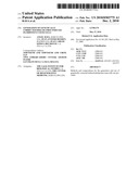 | 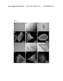 |
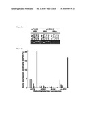 | 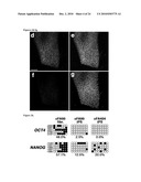 |
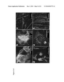 | 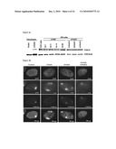 |
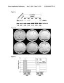 | 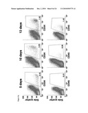 |
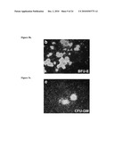 | 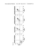 |
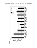 | 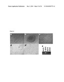 |
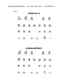 | 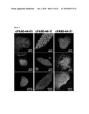 |
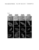 | 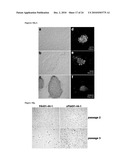 |
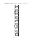 | 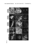 |
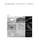 | 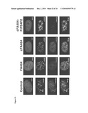 |
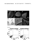 | 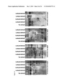 |
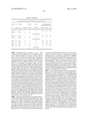 | 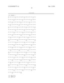 |
 |  |
 |  |
 |  |
 |  |
 |  |
 |  |
 |  |
 |
| Similar patent applications: | |
| Date | Title |
|---|---|
| 2011-07-14 | Markers of induced pluripotent stem cells |
| 2011-08-04 | Uses and isolation of very small embryonic-like (vsel) stem cells |
| 2011-08-04 | Combination of hgf inhibitor and pten agonist to treat cancer |
| 2011-08-04 | Proteolytically cleavable fusion proteins with high molar specific activity |
| 2011-07-21 | Cytokine induction of selectin ligands on cells |
| New patent applications in this class: | |
| Date | Title |
|---|---|
| 2022-05-05 | Compositions and methods for treating neurocognitive disorders |
| 2022-05-05 | Administration of tumor infiltrating lymphocytes with membrane bound interleukin 15 to treat cancer |
| 2019-05-16 | Crispr/cas9 complex for genomic editing |
| 2019-05-16 | Chimeric antigen receptor with single domain antibody |
| 2019-05-16 | Chimeric antigen receptors targeting epidermal growth factor receptor variant iii |
| New patent applications from these inventors: | |
| Date | Title |
|---|---|
| 2016-03-10 | Methods for heart regeneration |
| 2014-08-07 | Methods for heart regeneration |
| 2014-06-12 | Generation of vascular progenitor cells |
| 2014-02-13 | Robust and efficient differentiation of human pluripotent stem cells to multipotent vascular progenitors |
| 2013-11-21 | Formation of hematopoietic progenitor cells from mesenchymal stem cells |
| Top Inventors for class "Drug, bio-affecting and body treating compositions" | |
| Rank | Inventor's name |
|---|---|
| 1 | David M. Goldenberg |
| 2 | Hy Si Bui |
| 3 | Lowell L. Wood, Jr. |
| 4 | Roderick A. Hyde |
| 5 | Yat Sun Or |