Patent application title: Novel ubiquitin lifases as therapeutic tragets
Inventors:
Dah Shiam Chiaur (New York, NY, US)
Michele Pagano (New York, NY, US)
Esther Latres (New York, NY, US)
Esther Latres (New York, NY, US)
IPC8 Class: AA01K6700FI
USPC Class:
800 13
Class name: Multicellular living organisms and unmodified parts thereof and related processes nonhuman animal transgenic nonhuman animal (e.g., mollusks, etc.)
Publication date: 2010-08-19
Patent application number: 20100212033
Claims:
1. An isolated nucleic acid molecule comprising a nucleotide sequence
which encodes a protein comprising the amino acid sequence of SEQ ID NO:
2, 4, 6, 8, 10, 12, 14, 24, 26, 28, 30, 32, 34, 36, 38, 40, 42, 44, 46,
48, 50, 52, 54, 56, 58, or 60.
2. An isolated nucleic acid molecule which encodes an F-box protein, or a fragment thereof, having a nucleotide sequence that:a) hybridizes under highly stringent conditions to the nucleotide sequence of SEQ ID NO: 1, 3, 5, 7, 9, 11 or 13; andb) does not encompass the nucleotide sequences which encode the following known F-box proteins: Cdc4, Grr1, Met30, Skp2, Cyclin F, Elongin A or mouse Md6.
3. An isolated nucleic acid sequence derived from a mammalian genome that:a) hybridizes under highly stringent conditions to the nucleotide sequence of SEQ ID NO: 1, 3, 5, 7, 9, 11 or 13; andb) encodes a gene product which contains an F-box motif and binds to Skp1.
4. An isolated nucleic acid molecule which encodes an F-box protein, said nucleic acid molecule having a nucleotide sequence of SEQ ID NO: 23, 25, 27, 29, 31, 33, 35, 37, 39, 41, 43, 45, 47, 49, 51, 53, 55, 57, or 59.
5. A nucleotide vector containing the nucleotide sequence of claim 1, 2, 3, or 4.
6. An expression vector containing the nucleotide sequence of claim 1, 2, 3, or 4 in operative association with a nucleotide regulatory sequence that controls expression of the nucleotide sequence in a host cell.
7. A genetically engineered host cell that contains the nucleotide sequence of claim 1, 2, 3, or 4 in operative association with a nucleotide regulatory sequence that controls expression of the nucleotide sequence in the host cell.
8. A transgenic animal having cells which harbor a transgene comprising the nucleic acid of claim 1, 2, 3, or 4.
9. An animal inactivated in the loci comprising the nucleotide sequence of claim 1, 2, 3, or 4.
10. An isolated F-box protein having the amino acid sequence of SEQ ID NO: 2, 4, 6, 8, 10, 12, 14, 24, 26, 28, 30, 32, 34, 36, 38, 40, 42, 44, 46, 48, 50, 52, 54, 56, 58, or 60.
11. An antibody that immunospecifically binds the polypeptide of claim 10.
12. A method of diagnosing proliferative and differentiative related disorders comprising measuring FBP gene expression in a patient sample.
13. A method for screening compounds useful for the treatment of proliferative and differentiative disorders comprising contacting a compound with a cell expressing an F-box protein having the amino acid sequence of SEQ ID NO: 2, 4, 6, 8, 10, 12, 14, 24, 26, 28, 30, 32, 34, 36, 38, 40, 42, 44, 46, 48, 50, 52, 54, 56, 58, or 60, or a fragment thereof, and its substrate, and detecting a change in the F-box protein activity.
14. The method of claim 13 wherein the change in the F-box protein activity is detected by detecting a change in the interaction of the F-box protein with one or more proteins.
15. The method of claim 14 in which one of the one or more proteins is the substrate of the F-box protein.
16. The method of claim 13 in which at least one of the one or more proteins is a component of the ubiquitin pathway.
17. The method of claim 13 in which one of the one or more proteins is Skp1.
18. The method of claim 13 in which the F-box protein is Fbp1 and the substrate is β-catenin or IKBα.
19. The method of claim 13 wherein the change in the F-box protein activity is detected by detecting a change in the ubiquitination or degradation of the substrate.
20. A method for screening compounds useful for the treatment of proliferative and differentiative disorders comprising contacting a compound with a cell or a cell extract expressing Skp2 and one or both of p27 and E2F, and detecting a change in the activity of Skp2.
21. The method of claim 20 wherein the change in the activity of Skp2 is detected by detecting a change in the interaction of Skp2 with either p27 or E2F-1.
22. The method of claim 20 wherein the change in the activity of Skp2 is detected by detecting a change in the ubiquitination or degradation of p27 or E2F-1.
23. A method for treating a proliferative or differentiative disorder in a mammal comprising administering to the mammal a compound to the mammal that modulates the synthesis, expression or activity of an FBP gene or gene product so that symptoms of the disorder are ameliorated.
24. The method of claim 23 in which the disorder is breast cancer.
25. The method of claim 23 in which the disorder is ovarian cancer.
26. The method of claim 23 in which the disorder is prostate cancer.
27. The method of claim 23 in which the disorder is small cell lung carcinoma.
Description:
[0001]This application claims priority under 35 U.S.C. §119(e) to
U.S. Application No. 60/098,355, filed Aug. 28, 1998, Application No.
60/118,568, filed Feb. 3, 1999, and Application No. 60/124,449 filed Mar.
15, 1999, the contents of which are incorporated herein by reference in
their entirety.
1. INTRODUCTION
[0002]The present invention relates to the discovery, identification and characterization of nucleotide sequences that encode novel substrate-targeting subunits of ubiquitin ligases. The invention encompasses nucleic acid molecules comprising nucleotide sequences encoding novel substrate-targeting subunits of ubiquitin ligases: FBP1, FBP2, FBP3a, FBP3b, FBP4, FBP5, FBP6, FBP7, FBP8, FBP11, FBP12, FBP13, FBP14, FBP15, FBP17, FBP18, FBP20, FBP21, FBP22, FBP23, AND FBP25, transgenic mice, knock-out mice, host cell expression systems and proteins encoded by the nucleotides of the present invention. The present invention relates to screening assays to identify potential therapeutic agents such as small molecules, compounds or derivatives and analogues of the novel ubiquitin ligases which modulate activity of the novel ubiquitin ligases for the treatment of proliferative and differentiative disorders, such as cancer, major opportunistic infections, immune disorders, certain cardiovascular diseases, and inflammatory disorders. The invention further encompasses therapeutic protocols and pharmaceutical compositions designed to target ubiquitin ligases and their substrates for the treatment of proliferative disorders.
2. BACKGROUND OF THE INVENTION
2.1 Cell Cycle Regulatory Proteins
[0003]The eukaryotic cell cycle is regulated by a family of serine/threonine protein kinases called cyclin dependent kinases (Cdks) because their activity requires the association with regulatory subunits named Cyclins (Hunter & Pines, 1994, Cell 79:573). Cdks also associate with Cdk inhibitors (Ckis) which mediate cell cycle arrest in response to various antiproliferative signals. So far, based on their sequence homology, two families of Ckis have been identified in mammalian cells: the Cip/Kip family, which includes p21, p27 and p57; and the Ink family, which includes p15, p16, p18, and p20 (Sherr & Roberts, 1999, Genes & Dev. 13: 1501).
2.2 The Ubiquitin Pathway
[0004]Ubiquitin-mediated proteolysis is an important pathway of non-lysosomal protein degradation which controls the timed destruction of many cellular regulatory proteins including, p27, p53, p300, cyclins, E2F, STAT-1, c-Myc, c-Jun, EGF receptor, IkBα, NFkB and β-catenin (reviewed in Pagano, 1997, FASEB J. 11:1067). Ubiquitin is an evolutionary highly conserved 76-amino acid polypeptide which is abundantly present in all eukaryotic cells. The ubiquitin pathway leads to the covalent attachment of a poly-ubiquitin chain to target substrates which are then degraded by the multi-catalytic proteasome complex (see Pagano, supra, for a recent review). Many of the steps regulating protein ubiquitination are known. Initially the ubiquitin activating enzyme (E1), forms a high energy thioester with ubiquitin which is, in turn, transferred to a reactive cysteine residue of one of many ubiquitin conjugating enzymes (Ubcs or E2s). The final transfer of ubiquitin to an e-amino group of a reactive lysine residue in the target protein occurs in a reaction that is may or may not require an ubiquitin ligase (E3) protein. The large number of ubiquitin ligases ensures the high level of substrate specificity.
2.3 The Ubiquitin Pathway and the Regulation of the G1 Phase by F Box Proteins
[0005]Genetic and biochemical studies in several organisms have shown that the G1 phase of the cell cycle is regulated by the ubiquitin pathway. Proteolysis of cyclins, Ckis and other G1 regulatory proteins is controlled in yeast by the ubiquitin conjugating enzyme Ubc3 (also called Cdc34) and by an E3 ubiquitin ligase formed by three subunits: Cdc53, Skp1 and one of many F box proteins (reviewed in E. Patton et al., 1998, TIG. 14:6). The F box proteins (FBPs) are so called because they contain a motif, the F box, that was first identified in Cyclin F, and that is necessary for FBP interaction with Skp1 (Bai, et al., 1996, Cell 86:263). In addition, F box proteins also contain either WD-40 domains or Leucine-Rich Repeats (LRR) protein-protein interaction domains. Cdc53 (also called Cul A) and Skp1 appear to participate in the formation of at least three distinct E3, each containing a different F box protein. Because these ligases are similar protein modules composed of Skp1, Cul A, and an F box protein, they have been named SCF. The interaction of the ligase with its substrates occurs via the F box subunit. The three SCFs identified so far in S. cerevisiae are: SCF.sup.Cdc4 (which recruits the Ckis Sic1 and Fat1, the replication factor Cdc6, and the transcriptional activator Gcn4, as substrates through the F box protein Cdc4), SCF.sup.Gn1 (which recruits the G1 cyclins Cln1 and Cln2 as substrates through the F box protein GRR1), and SCF.sup.Met30 (which recruits the G1 cyclin Cln3 as a substrate throughout the F box protein MET30; see Pagano and Patton, supra, for recent reviews).
[0006]The intracellular level of the human Cki p27 is mainly regulated by degradation and it is known that the ubiquitin system controls p27 degradation (Pagano et al., 1995, Science 269:682). Similarly, degradation of other G1 human regulatory proteins (Cyclin E, Cyclin D1, p21, E2F, β-catenin) is controlled by the ubiquitin-pathway (reviewed in M. Pagano, supra). Yet, the specific enzymes involved in the degradation of G1 regulatory proteins have not been identified.
[0007]A family of 6 genes (CUL1, 2, 3, 4a, 4b, and 5) homologous to S. cerevisiae cul A have been identified by searching the EST database (Kipreos, et al., 1996, Cell 85:829). Human Skp1 and the F box protein Skp2 (that contains five LRRs) were identified as two proteins associated in vivo with Cyclin A and thus designated as S-phase kinase-associated protein 1 and 2 (Zhang, et al., 1995, Cell 82:915).
2.4 Deregulation of the Ubiquitin Pathway in Cancer and Other Proliferative Disorders
[0008]Cancer develops when cells multiply too quickly. Cell proliferation is determined by the net balance of positive and negative signals. When positive signals overcome or when negative signals are absent, the cells multiply too quickly and cancer develops.
[0009]Ordinarily cells precisely control the amount of any given protein and eliminate the excess or any unwanted protein. To do so, the cell specifically tags the undesired protein with a long chain of molecules called ubiquitin. These molecules are then recognized and destroyed by a complex named proteasome. However, all this mechanism goes awry in tumors leading to the excessive accumulation of positive signals (oncogenic proteins), or resulting in the abnormal degradation of negative regulators (tumor suppressor proteins). Thus, without tumor suppressor proteins or in the presence of too much oncogenic proteins, cells multiply ceaselessly, forming tumors (reviewed by Ciechanover, 1998, EMBO J. 17: 7151; Spataro, 1998, Br. J. Cancer 77: 448). For example, abnormal ubiquitin-mediated degradation of the p53 tumor suppressor (reviewed by J. Brown and M. Pagano, 1997, Biochim. Biophys. Acta1332: 1), the putative oncogene β-catenin (reviewed by Peifer, 1997, Science 275:1752) and the Cki p27 (reviewed in Ciechanover, supra; Spataro, supra; Lloyd, 1999, Am. J. Pathol. 154: 313) have been correlated with tumorigenesis, opening to the hypothesis that some genes encoding ubiquitinating enzymes may be mutated in tumors.
[0010]Initial evidence indicates that human F-box proteins play a role in the ubiquitination of G1 regulatory proteins as their homologs do in yeast (see below). Unchecked degradation of cell cycle regulatory proteins has been observed in certain tumors and it is possible that deregulated ubiquitin ligase play a role in the altered degradation of cell cycle regulators. A well understood example is that of Mdm2, a ubiquitin ligase whose overexpression induces low levels of its substrate, the tumor suppressor p53.
3. SUMMARY OF THE INVENTION
[0011]The present invention relates to novel F box proteins and therapeutic protocols and pharmaceutical compositions designed to target the novel F box proteins and their interactions with substrates for the treatment of proliferative and differentiative disorders. The present invention also relates to screening assays to identify substrates of the novel F box proteins and to identify agents which modulate or target the novel ubiquitin ligases and interactions with their substrates. The invention further relates to screening assays based on the identification of novel substrates of known F box proteins, such as the two novel substrates of the known F box protein Skp2, E2F and p27. The screening assays of the present invention may be used to identify potential therapeutic agents for the treatment of proliferative or differentiative disorders and other disorders that related to levels of expression or enzymatic activity of F box proteins.
[0012]The invention is based in part, on the Applicants' discovery, identification and characterization of nucleic acids comprising nucleotide sequences that encode novel ubiquitin ligases with F box motifs. These twenty-six novel substrate-targeting subunits of ubiquitin ligase complexes, FBP1, FBP2, FBP3a, FBP3b, FBP4, FBP5, FBP6, FBP7, FBP8, FBP9, FBP10, FBP11, FBP12, FBP13, FBP14, FBP15, FBP16, FBP17, FBP18, FBP19, FBP20, FBP21, FBP22, FBP23, FBP24, and FBP25, described herein, were first identified based on their interaction with components of the ubiquitin ligase complex (FBP1, FBP2, FBP3a, FBP4, FBP5, FBP6 and FBP7) or by sequence comparison of these proteins with nucleotide sequences present in DNA databases (FBP3b, FBP8, FBP9, FBP10, FBP11, FBP12, FBP13, FBP14, FBP15, FBP16, FBP17, FBP18, FBP19, FBP20, FBP21, FBP22, FBP23, FBP24, and FBP25). These novel substrate-targeting subunits of ubiquitin ligase complexes each contain an F box motif through which they interact with the other components of the ubiquitin ligase complex. In addition, some of these FBPs contain WD-40 domains and LRRs (which appear to be involved in their interaction with substrates), while other FBPs contain potential protein-protein interaction modules not yet identified in FBPs, such as leucine zippers, ring fingers, helix-loop-helix motifs, proline rich motifs and SH2 domains. The invention is also based, in part, on the Applicants' discovery and identification of FBP specific substrates p27 and β-catenin and on methods to identify novel FBP substrates. Some of the genes encoding the novel F box proteins were also mapped to chromosome sites frequently altered in breast, prostate and ovarian cancer, nasopharyngeal and small cell lung carcinomas, gastric hepatocarcinomas, Burkitt's lymphoma and parathyroid adenomas. Finally, the invention is also based, in part, on the Applicants' generation of transgenic mice expressing wild type or dominant negative versions of FBP proteins and on the generation of FBP knock-out mice.
[0013]The invention encompasses the following nucleotide sequences, host cells expressing such nucleotide sequences, and the expression products of such nucleotide sequences: (a) nucleotide sequences that encode mammalian FBP1, FBP2, FBP3a, FBP3b, FBP4, FBP5, FBP6, FBP7, FBP8, FBP11, FBP12, FBP13, FBP14, FBP15, FBP17, FBP18, FBP20, FBP21, FBP22, FBP23, and FBP25, including the human nucleotides, and their gene products; (b) nucleotides that encode portions of the novel substrate-targeting subunits of ubiquitin ligase complexes, and the polypeptide products specified by such nucleotide sequences, including but not limited to F box motifs, the substrate binding domains; WD-40 domains; and leucine rich repeats, etc.; (c) nucleotides that encode mutants of the novel ubiquitin ligases in which all or part of the domain is deleted or altered, and the polypeptide products specified by such nucleotide sequences; (d) nucleotides that encode fusion proteins containing the novel ubiquitin ligases or one of its domains fused to another polypeptide.
[0014]The invention further encompasses agonists and antagonists of the novel substrate-targeting subunits of ubiquitin ligase complexes, including small molecules, large molecules, mutants that compete with native F box binding proteins, and antibodies as well as nucleotide sequences that can be used to inhibit ubiquitin ligase gene expression (e.g., antisense and ribozyme molecules, and gene regulatory or replacement constructs) or to enhance ubiquitin ligase gene expression (e.g., expression constructs that place the ubiquitin ligase gene under the control of a strong promoter system), and transgenic animals that express a ubiquitin ligase transgene or knock-outs that do not express the novel ubiquitin ligases.
[0015]Further, the present invention also relates to methods for the use of the genes and/or gene products of novel substrate-targeting subunits of ubiquitin ligase complexes for the identification of compounds which modulate, i.e., act as agonists or antagonists, of ubiquitin ligase activity. Such compounds can be used as agents to control proliferative or differentiative disorders, e.g. cancer. In particular, the present invention encompasses methods to inhibit the interaction between β-catenin and FBP1 or p27 and Skp2. In fact, agents able to block these interactions can be used to modulate cell proliferation and/or growth.
[0016]Still further, the invention encompasses screening methods to identify derivatives and analogues of the novel substrate-targeting subunits of ubiquitin ligase complexes which modulate the activity of the novel ligases as potential therapeutics for proliferative or differentiative disorders. The invention provides methods of screening for proteins that interact with novel components of the ubiquitin ligase complex, including FBP1, FBP2, FBP3a, FBP3b, FBP4, FBP5, FBP6, FBP7, FBP8, FBP9, FBP10, FBP11, FBP12, FBP13, FBP14, FBP15, FBP16, FBP17, FBP18, FBP19, FBP20, FBP21, FBP22, FBP23, FBP24, and FBP25 or derivatives, fragments or domains thereof, such as the F box motif. In accordance with the invention, the screening methods may utilize known assays to identify protein-protein interactions including phage display assays or the yeast two-hybrid assay system or variations thereof.
[0017]In addition, the present invention is directed to methods that utilize FBP gene sequences and/or FBP gene product sequences for the diagnostic evaluation, genetic testing and/or prognosis of an FBP-related disorder, such as a proliferative disorder. For example, the invention relates to methods for diagnosing FBP-related disorders, e.g., proliferative disorders, wherein such methods can comprise measuring FBP gene expression in a patient sample, or detecting an FBP mutation that correlates with the presence or development of such a disorder, in the genome of a mammal suspected of exhibiting such a disorder. In particular, the invention encompasses methods for determining if a subject (e.g., a human patient) is a risk for a disorder characterized by one or more of: (i) a mutation of an FBP gene encoding a protein represented in part A of FIGS. 3-28, or a homolog thereof; (ii) the mis-expression of an FBP gene; (iii) the mis-expression of an FBP protein.
[0018]The invention is illustrated by way of working examples which demonstrate the identification and characterization of the novel substrate-targeting subunits of ubiquitin ligase complexes. The working examples of the present invention further demonstrate the identification of the specific interaction of (i) FBP1 with β-catenin and (ii) the known FBP, Skp2, with the cell-cycle regulatory proteins E2F and p27. These interactions suggest that β-catenin is a specific substrate of FBP1, while E2F and p27 are substrates of Skp2. In fact, the working examples of the present invention further demonstrate that β-catenin is a specific substrate of FBP1, while p27 is substrates of Skp2. The identification of proteins interacting with the novel FBPs will be possible using the methods described herein or with a different approach.
3.1 Definitions
[0019]As used herein, the term "F-box motif" refers to a stretch of approximately amino acid that was identified as being necessary for the interaction of F-box containing proteins with Skp1. The consensus sequence of an F-box motif is described in Bai et al., 1996, Cell 86:263-274, incorporated herein by reference in its entirety.
[0020]As used herein the term "F-box protein" (FBP) refers to peptide, polypeptide or protein which contains an F-box motif.
[0021]Although, FBPs are substrate-targeting subunits of ubiquitin ligase complexes, as used herein the term "ubiquitin ligase" refers to a peptide, polypeptide or protein that contains an F-box motif and interacts with Skp1.
[0022]As used herein, the term "functionally equivalent to an FBP gene product" refers to a gene product that exhibits at least one of the biological activities of the endogenous FBP gene product. For example, a functionally equivalent FBP gene product is one that is capable of interacting with Skp1 so as to become associated with a ubiquitin ligase complex. Such a ubiquitin ligase complex may be capable of ubiquitinating a specific cell-cycle regulatory protein, such as a cyclin or cki protein.
[0023]As used herein, the term "to target" means to inhibit, block or prevent gene expression, enzymatic activity, or interaction with other cellular factors.
[0024]As used herein, the term "therapeutic agent" refers to any molecule, compound or treatment that alleviates of assists in the treatment of a proliferative disorder or related disorder.
[0025]As used herein, the terms "WD-40 domain", "Leucine Rich Repeat", "Leucine Zipper", "Ring finger", "Helix-loop-helix motif", "Proline rich motif", and "SH2 domain" refer to domains potentially involved in mediating protein-protein interactions. The "WD-40 domain" refers to a consensus sequence of forty amino acid repeats which is rich in tryptophan and aspartic acid residues and is commonly found in the beta subunits of trimeric G proteins (see Neer et al., 1994 Nature 371:297-300 and references therein, which are incorporated herein by reference in their entirety). An "LRR" or a "Leucine Rich Repeat" is a leucine rich sequence also known to be involved in mediating protein-protein interactions (see Kobe & Deisenhofer, 1994, Trends. Biochem. Sci. 19:415-421 which are incorporated herein by reference in their entirety). A "leucine zipper" domain refers to a domain comprising a stretch of amino acids with a leucine residue in every seventh position which is present in a large family of transcription factors (see Landshultz et al., 1988, Science 240:1759-64; see also Sudol et al., 1996, Trends Biochem. 21:1-3, and Koch et al., 1991, Science 252:668-74).
4. BRIEF DESCRIPTION OF THE FIGURES
[0026]FIG. 1. Alignment of the conserved F-box motif amino acid residues in the human F-box proteins FBP1 (SEQ ID NO:15), FBP2 (SEQ ID NO:16), FBP3a (SEQ ID NO:17), FBP3b (SEQ ID NO:78), FBP4 (SEQ ID NO:18), FBP5 (SEQ ID NO:19), FBP6 (SEQ ID NO:20), FBP7 (SEQ ID NO:21), Skp2 (SEQ ID NO:22), FBP8 (SEQ ID NO:61) FBP9 (SEQ ID NO:62), FBP10 (SEQ ID NO:63), FBP11 (SEQ ID NO:64), FBP12 (SEQ ID NO:65), FBP13 (SEQ ID NO:79); FBP14 (SEQ ID NO:66); FBP15 (SEQ ID NO:67), FBP16 (SEQ ID NO:68), FBP17 (SEQ ID NO:69), FBP18 (SEQ ID NO:70), FBP19 (SEQ ID NO:71), FBP20 (SEQ ID NO:72), FBP21 (SEQ ID NO:73), FBP22 (SEQ ID NO:74), FBP23 (SEQ ID NO:75), FBP24 (SEQ ID NO:76), FBP25 (SEQ ID NO:77). Alignment of the F-boxes of a previously known FBP, Skp2, with the F-boxes of FBPs identified through a two-hybrid screen (designated by the pound symbol) or BLAST searches (designated by a cross) was performed using the Clustal W method (MacVector®) followed by manual re-adjustment. Identical residues in at least 15 F-boxes are shaded in dark gray, while similar residues are shaded in light gray. One asterisk indicates the presence in the cDNA of a STOP codon followed by a polyA tail, while potential full length clones are designated with two asterisks. The asterisks on the bottom of the figure indicate the amino acid residues mutated in FBP3a (see FIG. 29).
[0027]FIG. 2. Schematic representation of FBPs. Putative protein-protein interaction domains in human FBPs are represented (see key-box for explanation). FBPs identified by a two-hybrid screen are designated by the pound symbol, FBPs identified through BLAST searches by a cross. The double slash indicates that the corresponding cDNAs are incomplete at the 5' end; the asterisks indicate the presence in the cDNA of a STOP codon followed by a polyA tail.
[0028]FIG. 3 A-B. A. Amino acid sequence of human F-box protein FBP1 (SEQ ID NO:2). B. Corresponding cDNA (SEQ ID NO:1).
[0029]FIG. 4 A-B. A. Amino acid sequence of human F-box protein FBP2 (SEQ ID NO:4). B. Corresponding cDNA (SEQ ID NO:3).
[0030]FIG. 5 A-B. A. Amino acid sequence of human F-box protein FBP3a (SEQ ID NO:6). B. Corresponding cDNA (SEQ ID NO:5).
[0031]FIG. 6 A-B. A. Amino acid sequence of human F-box protein FBP3b (SEQ ID NO:24). B. Corresponding cDNA (SEQ ID NO:23).
[0032]FIG. 7 A-B. A. Amino acid sequence of human F-box protein FBP4 (SEQ ID NO:8). B. Corresponding cDNA (SEQ ID NO:7).
[0033]FIG. 8 A-B. A. Amino acid sequence of human F-box protein FBP5 (SEQ ID NO:10). B. Corresponding cDNA (SEQ ID NO:9).
[0034]FIG. 9 A-B. A. Amino acid sequence of human F-box protein FBP6 (SEQ ID NO:12). B. Corresponding cDNA (SEQ ID NO:11).
[0035]FIG. 10 A-B. A. Amino acid sequence of human F-box protein FBP7 (SEQ ID NO:14). B. Corresponding cDNA (SEQ ID NO:13).
[0036]FIG. 11A-B. A. Amino acid sequence of human F-box protein FBP8 (SEQ ID NO:26). B. Corresponding cDNA (SEQ ID NO:25).
[0037]FIG. 12 A-B. A. Amino acid sequence of human F-box protein FBP9 (SEQ ID NO:28). B. Corresponding cDNA (SEQ ID NO:27).
[0038]FIG. 13 A-B. A. Amino acid sequence of human F-box protein FBP10 (SEQ ID NO:30). B. Corresponding cDNA (SEQ ID NO:29).
[0039]FIG. 14 A-B. A. Amino acid sequence of human F-box protein FBP11 (SEQ ID NO:32). B. Corresponding cDNA (SEQ ID NO:31).
[0040]FIG. 15 A-B. A. Amino acid sequence of human F-box protein FBP12 (SEQ ID NO:34). B. Corresponding cDNA (SEQ ID NO:33).
[0041]FIG. 16 A-B. A. Amino acid sequence of human F-box protein FBP13 (SEQ ID NO:36). B. Corresponding cDNA (SEQ ED NO:35).
[0042]FIG. 17 A-B. A. Amino acid sequence of human F-box protein FBP14 (SEQ ID NO:38). B. Corresponding cDNA (SEQ ID NO:37).
[0043]FIG. 18 A-B. A. Amino acid sequence of human F-box protein FBP15 (SEQ ID NO:40). B. Corresponding cDNA (SEQ ID NO:39).
[0044]FIG. 19 A-B. A. Amino acid sequence of human F-box protein FBP16 (SEQ ID NO:42). B. Corresponding cDNA (SEQ ID NO:41).
[0045]FIG. 20 A-B. A. Amino acid sequence of human F-box protein FBP17 (SEQ ID NO:44). B. Corresponding cDNA (SEQ ID NO:43).
[0046]FIG. 21A-B. A. Amino acid sequence of human F-box protein FBP18 (SEQ ID NO:46). B. Corresponding cDNA (SEQ ID NO:45).
[0047]FIG. 22 A-B. A. Amino acid sequence of human F-box protein FBP19 (SEQ ID NO:48). B. Corresponding cDNA (SEQ ID NO:47).
[0048]FIG. 23 A-B. A. Amino acid sequence of human F-box protein FBP20 (SEQ ID NO:50). B. Corresponding cDNA (SEQ ID NO:49).
[0049]FIG. 24 A-B. A. Amino acid sequence of human F-box protein FBP21 (SEQ ID NO:52). B. Corresponding cDNA (SEQ ID NO:51).
[0050]FIG. 25 A-B. A. Amino acid sequence of human F-box protein FBP22 (SEQ ID NO:54). B. Corresponding cDNA (SEQ ID NO:53).
[0051]FIG. 26 A-B. A. Amino acid sequence of human F-box protein FBP23 (SEQ ID NO:56). B. Corresponding cDNA (SEQ ID NO:55).
[0052]FIG. 27 A-B. A. Amino acid sequence of human F-box protein FBP24 (SEQ ID NO:58). B. Corresponding cDNA (SEQ ID NO:57).
[0053]FIG. 28A-B. A. Amino acid sequence of human F-box protein FBP25 (SEQ ID NO:60). B. Corresponding cDNA (SEQ ID NO:59).
[0054]FIG. 29. FBPs interact specifically with Skp1 through their F-box. The cDNAs of FBPs (wild type and mutants) were transcribed and translated in vitro (IVT) in the presence of 35S-methionine. Similar amounts of IVT proteins (indicated at the top of each lane) were subjected to a histidine-tagged pull-down assay using Nickel-agarose beads to which either His-tagged-Skp1 (lanes 1, 3, 4, 6-10, 12, 15, 17, 19 and 21), His-tagged-Elongin C (lanes 2, 5, 11, 14, 16, 18, 19 and 22), or His-tagged p27 (lane 12) were pre-bound. Bound IVT proteins were analyzed by SDS-PAGE and autoradiography. The arrows on the left side of the panels point to the indicated FBPs. The apparent molecular weights of the protein standards are indicated on the right side of the panels.
[0055]FIG. 30. FBP1, FBP2, FBP3a, FBP4 and FBP7 form novel SCFs with endogenous Skp1 and Cul1 in vivo. HeLa cells were transfected with mammalian expression plasmids encoding Flag-tagged versions of FBP1 (lane 1), (ΔF)FBP1 (lane 2), FBP4 (lane 3), FBP7 (lane 5), FBP2 (lane 7), (ΔF)FBP2 (lane 8), FBP3a (lane 9), (ΔF)FBP3a (lane 10), or with an empty vector (lanes 4 and 6). Cells were lysed and extracts were subjected to immunoprecipitation with a rabbit anti-Flag antibody (lanes 1-8). Immunoprecipitates were then immunoblotted with a mouse anti-Cul1 monoclonal antibody, a rabbit anti-Skp1 polyclonal antibody or a rabbit anti-Cul2 polyclonal antibody, as indicated. The last lane contains 25 μg of extracts from non-transfected HeLa cells; lane 9 contains recombinant Cul1, Skp1, or Cul2 proteins used as markers. The slower migrating bands detected with the antibodies to Cul1 and Cul2 are likely generated by the covalent attachment of a ubiquitin-like molecule to these two cullins, as already described for the yeast cullin Cdc53 and mammalian Cul4a.
[0056]FIG. 31. FBP1, FBP2, FBP3a, FBP4 and FBP7 associate with a ubiquitin ligase activity. HeLa cells were transfected with mammalian expression plasmids encoding human Skp1, Cul1 and Flag-tagged versions of FBP1 (lane 3), (ΔF)FBP1 (lane 4), FBP2 (lanes 2 and 5), (ΔF)FBP2 (lane 6), FBP7 (lane 7), FBP3a (lanes 8 and 13), (ΔF)FBP3a (lane 9), a non relevant Flag-tagged protein (Irf3, lane 10), FBP4 (lanes 11 and 12) or with an empty vector (lane 1). Cells were lysed and extracts were subjected to immunoprecipitation with a rabbit anti-Flag antibody. Immunoprecipitates were incubated in the presence of purified recombinant E1 and Ubc4 (lanes 1-11) or Ubc2 (lanes (12 and 13) and a reaction mix containing biotynilated ubiquitin. Reaction in lane 2 contained also NEM. Ubiquitinated proteins were visualized by blotting with HRP-streptavidin. The bracket on the left side of the panels marks a smear of ubiquitinated proteins produced in the reaction, the asterisk indicates ubiquitin conjugated with E1 that were resistant to boiling.
[0057]FIG. 32. Subcellular localization of FBPs. HeLa cells were transfected with mammalian expression plasmids encoding Flag-tagged versions of FBP1 (a-b), FBP2 (c-d), FBP3a (e-f), FBP4 (g-h), (DF)FBP2 (i-j), or (ΔF)FBP3a (k-l). After 24 hours, cells were subjected to immunofluorescence with a rabbit anti-Flag antibody (a, c, e, g, i, k) to stain FBPs and bisbenzimide (b, d, f, h, j, 1) to stain nuclei.
[0058]FIG. 33. Abundance of FBP transcripts in human tissues. Membranes containing electrophoretically fractionated poly(A)+ mRNA from different human tissues were hybridized with specific probes prepared form FBP1, FBP2, FBP3a, FBP4, SKP2, and β-ACTIN cDNAs. The arrows on the left side of the figure point to the major transcripts as described in the text.
[0059]FIG. 34 A-E. FISH localization of FBP genes. Purified phage DNA containing a genomic probe was labeled with digoxygenin dUTP and detected with Cy3 conjugated antibodies. The signals corresponding to the locus of the genomic probe (red) are seen against the DAPI-Actimomycin D stained normal human chromosomes (blue-white). Panel A shows localization of FBP1 to 10q24, B shows localization of FBP2 to 9q34, C shows localization of FBP3a to 13q22, D shows localization of FBP4 to 5p12, and E shows localization of FBP5 to 6q25-26. Arrows point to FBP-specific FISH signals.
[0060]FIG. 35A-C. FBP1 associates with β-catenin. A. Extracts from baculovirus-infected insect cells expressing either β-catenin alone (lane 1) or in combination with Flag-tagged FBP1 (lane 2) were immunoprecipitated (IP) with a rabbit anti-Flag antibody (rα-Flag), followed by immunoblotting with anti-Flag (mα-Flag) and anti-β-catenin mouse antibodies, as indicated. Lanes 3 and 4 contain 25 μg of extracts from infected insect cells immunoblotted with the same antibodies. B. Extracts from baculovirus-infected insect cells expressing cyclin D1, Flag-FBP1 in the absence (lanes 1-3) or in the presence of Skp1 (lanes 4-6) were immunoprecipitated with normal rabbit IgG (r-IgG, lanes 1 and 4), rabbit anti-Flag antibody (r α-Flag, lanes 2 and 5), or rabbit anti-cyclin D1 antibody (r α-D1, lanes 3 and 6). Immunoprecipitates were then immunoblotted with anti-Flag (mα-Flag) and cyclin D1 (m α-D1) mouse antibodies, as indicated. The last lane contains 25 μg of a representative extract from infected insect cells immunoblotted with the same antibodies. C. 293 cells were transfected with mammalian expression plasmids encoding HA-tagged β-catenin alone or in combination with either Flag-tagged FBP1 or Flag-tagged (ΔF)FBP1. Cells were lysed and extracts were subjected to immunoprecipitation with a rabbit anti-Flag antibody (r α-Flag, lanes 4-6) and immunoblotted with rat anti-HA (α-HA) and mouse anti-Flag (m α-Flag) antibodies, as indicated. The first three lanes contain 25 μg of extracts from transfected 293 cells immunoblotted with the same antibodies. Transfecting high levels of β-catenin expression vector, the associations of β-catenin with FBP1 and (ΔF)FBP1 could be determined independently of β-catenin levels.
[0061]FIG. 36 A-B. Stabilization of β-catenin by a dominant negative (ΔF)FBP1 mutant. A. Human 293 cells were transfected with mammalian expression plasmids encoding HA-tagged β-catenin alone or in combination with either Flag-tagged (ΔF)FBP1 or Flag-tagged (ΔF)FBP2. Cells were lysed and extracts were subjected to immunoblotting with rat anti-HA and rabbit anti-Flag (r α-Flag) antibody, as indicated. B. Pulse chase analysis of β-catenin turnover rate. HA-tagged β-catenin in combination with either an empty vector, FBP1, or (ΔF)FBP1 was co-transfected in 293 cells. 24 hours later cells were labeled with 35S-methionine for 30 minutes and chased with medium for the indicated times. Extracts were then subjected to immunoprecipitation with a rat anti-HA antibody.
[0062]FIG. 37A-C. Binding of phosphorylated p27 to Skp2. A. A panel of in vitro translated [35S]FBPs were used in binding reactions with beads coupled to the phospho-peptide NAGSVEQT*PKKPGLRRRQT, corresponding to the carboxy terminus of the human p27 with a phosphothreonine at position 187 (T*). Beads were washed with RIPA buffer and bound proteins were eluted and subjected to electrophoresis and autoradiography (Upper Panel). Bottom Panel: 10% of the in vitro translated [35S]FBP inputs. B. HeLa cell extracts were incubated with beads coupled to the phospho-p27 peptide (lane 2), an identical except unphosphorylated p27 peptide (lane 1) or the control phospho-peptide AEIGVGAY*GTVYKARDPHS, corresponding to an amino terminal peptide of human Cdk4 with a phosphotyrosine at position 17 (Y*) (lane 3). Beads were washed with RIPA buffer and bound proteins were immunoblotted with antibodies to the proteins indicated on the left of each panel. A portion of the HeLa extract (25 μg) was used as a control (lane 4). The slower migrating band in Cul1 is likely generated by the covalent attachment of a ubiquitin-like molecule, as already described for other cullins 48. C. One μl of in vitro translated [35S] wild type p27 (WT, lanes 1-4) or p27(T187A) mutant (T187A, lanes 5-6) were incubated for 30 minutes at 301/4 C in 10 μl of kinase buffer. Where indicated, ˜2.5 pmole of recombinant purified cyclin E/Cdk2 or ˜1 pmole Skp2 (in Skp1/Skp2 complex) were added. Samples were then incubated with 6 μl of Protein-A beads to which antibodies to Skp2 had been covalently linked. Beads were washed with RIPA buffer and bound proteins subjected to electrophoresis and autoradiography. Lanes 1-6: Skp2-bound proteins; Lanes 7 and 8: 7.5% of the in vitro translated [35S] protein inputs.
[0063]FIG. 38. In vivo binding of Skp2 to p27. Extracts from HeLa cells (lanes 1-2 and 5-6) or EMR90 fibroblasts (lanes 9-10) were immunoprecipitated with different affinity purified (AP) antibodies to Skp2 or with purified control IgG fractions. Lane 1: extract immunoprecipitated with a goat IgG (G-IgG); lane 2: with an AP goat antibody to an N-terminal Skp2 peptide (G-α-Skp2); lanes 5 and 9: with a rabbit IgG (R-IgG); lanes 6 and 10: with an AP rabbit antibody to Skp2 (R-α-Skp2). Immunoprecipitates were immunoblotted with antibodies to the proteins indicated on the left of each panel. Lanes 1-4 in the bottom panel were immunoblotted with a phospho-site p27 specific antibody. Lanes 3, 7, and 11 contain 25 μg of cell extracts; Lanes 4, 8, and 12 contain the relevant recombinant proteins used as markers. The altered migration of some markers is due to the presence of tags on the recombinant proteins.
[0064]FIG. 39 A-B. Skp2 and cyclin E/Cdk2 complex are rate-limiting for p27 ubiquitination in G1 extracts. A. In vitro ubiquitin ligation (lanes 1-12 and 17-20) and degradation (lanes 13-16) of p27 were carried out with extracts from asynchronously growing (Asyn. ext., lanes 2-3) or G1-arrested (G1 ext., lanes 4-20) HeLa cells. Lane 1 contains no extract. Recombinant purified proteins were supplemented as indicated. Reactions were performed using wild-type p27 (lanes 1-18) or p27(T187A) mutant (T187A, lanes 19-20). Lanes 1-8, 9-12, and 17-20 are from three separate experiments. The bracket on the left side of the panels marks a ladder of bands >27,000 corresponding to polyubiquitinated p27. The asterisk indicates a non-specific band present in most samples. B. Immunoblot analysis of levels of Skp2 and p27 in extracts from asynchronous (lane 1) or G1-arrested (lane 2) HeLa cells.
[0065]FIG. 40 A-C. Skp2 is required for p27-ubiquitin ligation activity. A. Immunodepletion. Extracts from asynchronous HeLa cells were untreated (lane 2) or immunodepleted with pre-immune serum (lane 3), anti-Skp2 antibody pre-incubated with 2 of purified GST (lane 4), or anti-Skp2 antibody pre-incubated with 2 μg of purified GST-Skp2 (lane 5). Lane 1 contains no extract. Samples (30 μg of protein) were assayed for p27 ubiquitination in the presence of cyclin E/Cdk2. The bracket on the left side of the panels marks a ladder of bands >27,000 corresponding to polyubiquitinated p27. The asterisk indicates a non-specific band present in all samples. B. Reconstitution. The restoration of p27 ubiquitination activity in Skp2-immunodepleted extracts was tested by the addition of the indicated purified proteins. All samples contained 30 μg of Skp2-depleted extract (Skp2-depl. ext.) and cyclin E/Cdk2. C. Immunopurification. Extracts from asynchronous HeLa cells were immunoprecipitated with a rabbit anti-Skp2 antibody (lanes 3 and 5) or pre-immune serum (PI, lanes 2 and 4). Total extract (lane 1) and immuno-beads (lanes 2-5) were added with p27, recombinant purified cyclin E/Cdk2 and ubiquitination reaction mix. Samples in lanes 4 and 5 were supplemented with recombinant purified E1 and Ubc3. All samples were then assayed for p27 ubiquitination.
[0066]FIG. 41A-B. In vivo role of Skp2 in p27 degradation. A. Stabilization of p27 by a dominant negative (ΔF)Skp2 mutant in vivo. NIH-3T3 cells were transfected with mammalian expression vectors encoding human p27 alone (lane 2), p27 in combination with either (ΔF)Skp2 (lane 3), or (ΔF)FBP1 (lane 4). Lane 1: untransfected cells. Cells were lysed and extracts were subjected to immunoblotting with antibodies to p27, Skp2 or Flag [to detect Flag-tagged (ΔF)FBP1]. Exogenous human p27 protein migrates more slowly than the endogenous murine p27. B. Pulse chase analysis of p27 turnover rate. Human p27 in combination with either an empty vector, or (ΔF)Skp2 was transfected in NIH-3T3 cells. Twenty-four hours later, cells were labeled with [35S]-methionine for 20 minutes and chased with medium for the indicated times. Extracts were then subjected to immunoprecipitation with a mouse anti-p27 antibody.
[0067]FIG. 42. Stabilization of cellular p27 by antisense oligonucleotides targeting SKP2 mRNA. HeLa cells were treated for 16-18 hours with two different anti-sense oligodeoxynucleotides (AS) targeting two different regions of SKP2 mRNA. Lanes 2, 6, 12 and 16: AS targeting the N-terminal SKP2 region (NT); Lanes 4 and 8: AS targeting the C-terminal SKP2 region (CT); Lanes 1, 3, 5, 7 11 and 15: control oligodeoxynucleotides pairs (Ctrl). Lanes 1-4, and 5-8 are from two separate experiments. Lanes 11-12 and 15-16: HeLa cells were blocked in G1/S with either Hydroxyurea or Aphidicolin treatment respectively, for 24 hours. Cells were then transfected with oligodeoxynucleotides, lysed after 12 hours (before cells had re-entered G1) and immunoblotted with antibodies to Skp2 (top panels) and p27 (bottom panels). Lanes 9 and 13: Untransfected HeLa cells; Lanes 10 and 14: Untransfected HeLa cells treated with drugs as transfected cells.
[0068]FIG. 43 A-C. Timing of Skp2 action in the process of p27 degradation. A. IMR90 fibroblasts were synchronized in G0/G1 by serum deprivation, reactivated with serum, and sampled at the indicated intervals. Protein extracts were analyzed by immunoblot with the antibodies to the indicated proteins. The Skp2 doublet was likely generated by phosphorylation since was consistently observed using a 12.5% gel only when cell lysis was performed in the presence of okadaic acid. B. HeLa cells blocked in mitosis with nocodazole were shaken off, released in fresh medium and sampled at the indicated intervals. Protein extracts were analyzed by immunoblotting with the antibodies to the indicated proteins. C. Extracts from G1 (3 hours after release from nocodazole block) (lane 1) and S-phase (12 hours after release from the nocodazole block) (lane 2) HeLa cells were either immunoprecipitated with an anti-p27 antibody (top two panels) or with an anti-Skp2 antibody (bottom three panels) and then immunoblotted with the antibodies to the indicated proteins.
[0069]FIG. 44 A-C. Western blot analysis of Skp2/E2F interaction assay. Details of the Western Blot experiments are given in the Example in Section 9.
5. DETAILED DESCRIPTION OF THE INVENTION
[0070]The present invention relates to novel F-box proteins and to novel substrates of F-box proteins. The present invention relates to screening assays designed to identify substrates of the novel F-box proteins and to identify small molecules and compounds which modulate the interaction and/or activity of the F-box proteins and their substrates.
[0071]The present invention relates to screening assays to identify substrates of the novel F-box proteins and to identify potential therapeutic agents. The present invention further relates to screening assays based on the identification of novel substrates of both novel and known F-box proteins. The screening assays of the present invention may be used to identify potential therapeutic agents which may be used in protocols and as pharmaceutical compositions designed to target the novel ubiquitin ligases and interactions with their substrates for the treatment of proliferative disorders. In one particular embodiment the present invention relates to screening assays and potential therapeutic agents which target the interaction of FBP with novel substrates β-catenin, p27 and E2F as identified by Applicants.
[0072]The invention further encompasses the use of nucleotides encoding the novel F-box proteins, proteins and peptides, as well as antibodies to the novel ubiquitin ligases (which can, for example, act as agonists or antagonists), antagonists that inhibit ubiquitin ligase activity or expression, or agonists that activate ubiquitin ligase activity or increase its expression. In addition, nucleotides encoding the novel ubiquitin ligases and proteins are useful for the identification of compounds which regulate or mimic their activity and therefore are potentially effective in the treatment of cancer and tumorigenesis.
[0073]In particular, the invention described in the subsections below encompasses FBP1, FBP2, FBP3a, FBP3b, FBP4, FBP5, FBP6, FBP7, FBP5, FBP9, FBP10, FBP11, FBP12, FBP13, FBP14, FBP15, FBP16, FBP17, FBP18, FBP19, FBP20, FBP21, FBP22, FBP23, FBP24, and FBP25 polypeptides or peptides corresponding to functional domains of the novel ubiquitin ligases (e.g., the F-box motif, the substrate binding domain, and leucine-rich repeats), mutated, truncated or deleted (e.g. with one or more functional domains or portions thereof deleted), ubiquitin ligase fusion proteins, nucleotide sequences encoding such products, and host cell expression systems that can produce such ubiquitin ligase products.
[0074]The present invention provides methods of screening for peptides and proteins that interact with novel components of the ubiquitin ligase complex, including FBP1, FBP2, FBP3a, FBP3b, FBP4, FBP5, FBP6, FBP7, FBP8, FBP9, FBP10, FBP11, FBP12, FBP13, FBP14, FBP15, FBP16, FBP17, FBP18, FBP19, FBP20, FBP21, FBP22, FBP23, FBP24, and FBP25 or derivatives, fragments or analogs thereof. Preferably, the method of screening is a yeast two-hybrid assay system or a variation thereof, as further described below. Derivatives (e.g., fragments) and analogs of a protein can be assayed for binding to a binding partner by any method known in the art, for example, the modified yeast two-hybrid assay system described below, immunoprecipitation with an antibody that binds to the protein in a complex followed by analysis by size fractionation of the immunoprecipitated proteins (e.g., by denaturing or nondenaturing polyacrylamide gel electrophoresis), Western analysis, non-denaturing gel electrophoresis, etc.
[0075]The present invention relates to screening assays to identify agents which modulate the activity of the novel ubiquitin ligases. The invention encompasses both in vivo and in vitro assays to screen small molecules, compounds, recombinant proteins, peptides, nucleic acids, antibodies etc. which modulate the activity of the novel ubiquitin ligases and thus, identify potential therapeutic agents for the treatment of proliferative or differentiative disorders. In one embodiment, the present invention provides methods of screening for proteins that interact with the novel ubiquitin ligases.
[0076]The invention also encompasses antibodies and anti-idiotypic antibodies, antagonists and agonists, as well as compounds or nucleotide constructs that inhibit expression of the ubiquitin ligase gene (transcription factor inhibitors, antisense and ribozyme molecules, or gene or regulatory sequence replacement constructs), or promote expression of the ubiquitin ligase (e.g., expression constructs in which ubiquitin ligase coding sequences are operatively associated with expression control elements such as promoters, promoter/enhancers, etc.). The invention also relates to host cells and animals genetically engineered to express the human (or mutants thereof) or to inhibit or "knock-out" expression of the animal's endogenous ubiquitin ligase.
[0077]Finally, the ubiquitin ligase protein products and fusion protein products, (i.e., fusions of the proteins or a domain of the protein, e.g., F-box motif), antibodies and anti-idiotypic antibodies (including Fab fragments), antagonists or agonists (including compounds that modulate the ubiquitization pathway can be used for therapy of proliferative or differentiative diseases. Thus, the invention also encompasses pharmaceutical formulations and methods for treating cancer and tumorigenesis.
[0078]Various aspects of the invention are described in greater detail in the subsections below.
5.1 FBP Genes
[0079]The invention provides nucleic acid molecules comprising seven novel nucleotide sequences, and fragments thereof, FBP1, FBP2, FBP3a, FBP4, FBP5, FBP6, and FBP7, nucleic acids which are novel genes identified by the interaction of their gene products with Skp1, a component of the ubiquitin ligase complex. The invention further provides fourteen novel nucleic acid molecules comprising the nucleotide sequences of FBP1, FBP2, FBP3a, FBP3b, FBP4, FBP5, FBP6, FBP7, FBP5, FBP11, FBP12, FBP13, FBP14, FBP15, FBP17, FBP18, FBP20, FBP21, FBP22, FBP23, FBP24, and FBP25, which Nucleic acid sequences of the identified FBP genes are described herein.
[0080]As used herein, "an FBP gene" refers to: [0081](a) a nucleic acid molecule containing the DNA sequences of FBP1, shown in FIG. 3 (SEQ ID NO:1), the DNA sequences of FBP2, shown in FIG. 4 (SEQ ID NO:3), the DNA sequences of FBP3a, shown in FIG. 5 (SEQ ID NO:5), the DNA sequences of FBP3b, shown in FIG. 6 (SEQ ID NO:23), the DNA sequences of FBP4, shown in FIG. 7 (SEQ ID NO:7), the DNA sequences of FBP5, shown in FIG. 8 (SEQ ID NO:9), the DNA sequences of FBP6, shown in FIG. 9 (SEQ ID NO:11), the DNA sequences of FBP7, shown in FIG. 10 (SEQ ID NO:13), the DNA sequences of FBP8, shown in FIG. 11 (SEQ ID NO:25), the DNA sequences of FBP9, shown in FIG. 12 (SEQ ID NO:27), the DNA sequences of FBP10, shown in FIG. 13 (SEQ ID NO:29), the DNA sequences of FBP11, shown in FIG. 14 (SEQ ID NO:31), the DNA sequences of FBP12, shown in FIG. 15 (SEQ ID NO:33), the DNA sequences of FBP13, shown in FIG. 16 (SEQ ID NO:35), the DNA sequences of FBP14, shown in FIG. 17 (SEQ ID NO:37), the DNA sequences of FBP15, shown in FIG. 18 (SEQ ID NO:39), the DNA sequences of FBP16, shown in FIG. 19 (SEQ ID NO:41), the DNA sequences of FBP17, shown in FIG. 20 (SEQ ID NO:43), the DNA sequences of FBP18, shown in FIG. 21 (SEQ ID NO:45), the DNA sequences of FBP19, shown in FIG. 22 (SEQ ID NO:47), the DNA sequences of FBP20, shown in FIG. 23 (SEQ ID NO:49), the DNA sequences of FBP21, shown in FIG. 24 (SEQ ID NO:51), the DNA sequences of FBP22, shown in FIG. 25 (SEQ ID NO:53), the DNA sequences of FBP23, shown in FIG. 26 (SEQ ID NO:55), the DNA sequences of FBP24, shown in FIG. 27 (SEQ ID NO:57), the DNA sequences of FBP25, shown in FIG. 28 (SEQ ID NO:59). [0082](b) any DNA sequence that encodes a polypeptide containing: the amino acid sequence of FBP1 shown in FIG. 3A (SEQ ID NO:2), the amino acid sequence of FBP2, shown in FIG. 4A (SEQ ID NO:4), the amino acid sequence of FBP3a shown in FIG. 5A (SEQ ID NO:6), the amino acid sequence of FBP3b shown in FIG. 6A (SEQ ID NO:24), the amino acid sequence of FBP4 shown in FIG. 7A (SEQ ID NO:8), the amino acid sequence of FBP5 shown in FIG. 8A (SEQ ID NO:10), or the amino acid sequence of FBP6 shown in FIG. 9A (SEQ ID NO:12), the amino acid sequences of FBP7, shown in FIG. 10 (SEQ ID NO:14), the amino acid sequences of FBP8, shown in FIG. 11 (SEQ ID NO:26), the amino acid sequences of FBP9, shown in FIG. 12 (SEQ ID NO:28), the amino acid sequences of FBP10, shown in FIG. 13 (SEQ ID NO:30), the amino acid sequences of FBP11, shown in FIG. 14 (SEQ ID NO:32), the amino acid sequences of FBP12, shown in FIG. 15 (SEQ ID NO:34), the amino acid sequences of FBP13, shown in FIG. 16 (SEQ ID NO:36), the amino acid sequences of FBP14, shown in FIG. 17 (SEQ ID NO:38), the amino acid sequences of FBP15, shown in FIG. 18 (SEQ ID NO:40), the amino acid sequences of FBP16, shown in FIG. 19 (SEQ ID NO:42), the amino acid sequences of FBP17, shown in FIG. 20 (SEQ ID NO:44), the amino acid sequences of FBP18, shown in FIG. 21 (SEQ ID NO:46), the amino acid sequences of FBP19, shown in FIG. 22 (SEQ ID NO:48), the amino acid sequences of FBP20, shown in FIG. 23 (SEQ ID NO:50), the amino acid sequences of FBP21, shown in FIG. 24 (SEQ ID NO:52), the amino acid sequences of FBP22, shown in FIG. 25 (SEQ ID NO:54), the amino acid sequences of FBP23, shown in FIG. 26 (SEQ ID NO:56), the amino acid sequences of FBP24, shown in FIG. 27 (SEQ ID NO:58), the amino acid sequences of FBP25, shown in FIG. 28 (SEQ ID NO:60). [0083](c) any DNA sequence that hybridizes to the complement of the DNA sequences that encode any of the amino acid sequences of (SEQ ID NO: 2, 4, 6, 8, 10, 12 or 14) or FIG. 15 under highly stringent conditions, e.g., hybridization to filter-bound DNA in 0.5 M NaHPO4, 7% sodium dodecyl sulfate (SDS), 1 mM EDTA at 65 C, and washing in 0.1×SSC/0.1% SDS at 68 C (Ausubel F. M. et al., eds., 1989, Current Protocols in Molecular Biology, Vol. I, Green Publishing Associates, Inc., and John Wiley & sons, Inc., New York, at p. 2.10.3); and/or [0084](d) any DNA sequence that hybridizes to the complement of the DNA sequences that encode any of the amino acid sequences in (SEQ ID NO: 2, 4, 6, 8, 10, 12 or 14) or FIG. 15, under less stringent conditions, such as moderately stringent conditions, e.g., washing in 0.2×SSC/0.1% SDS at 42 C (Ausubel et al., 1989, supra), and encodes a gene product functionally equivalent to an FBP gene product.
[0085]It is understood that the FBP gene sequences of the present invention do not encompass the previously described genes encoding other mammalian F-box proteins, Skp2, Elongin A, Cyclin F, mouse Md6, (see Pagano, 1997, supra; Zhang et al., 1995, supra; Bai et al., 1996, supra; Skowyra et al., 1997, supra). It is further understood that the nucleic acid molecules of the invention do not include nucleic acid molecules that consist solely of the nucleotide sequence in GenBank Accession Nos. AC002428, AI457595, AI105408, H66467, T47217, H38755, THC274684, AI750732, AA976979, AI571815, T57296, Z44228, Z45230, N42405, AA018063, AI751015, AI400663, T74432, AA402-415, AI826000, AI590138, AF174602, Z45775, AF174599, THC288870, AI017603, AF174598, THC260994, AI475671, AA768343, AF174595, THC240016, N70417, T10511, AF174603, EST04915, AA147429, AI192344, AF174594, AI147207, AI279712, AA593015, AA644633, AA335703, N26196, AF174604, AF053356, AF174606, AA836036, AA853045, AI479142, AA772788, AA039454, AA397652, AA463756, AA007384, AA749085, AI640599, THC253263, AB020647, THC295423, AA434109, AA370939, AA215393, THC271423, AF052097, THC288182, AL049953, CAB37981, AL022395, AL031178, THC197682, and THC205131.
[0086]FBP sequences of the present invention are derived from a eukaryotic genome, preferably a mammalian genome, and more preferably a human or murine genome. Thus, the nucleotide sequences of the present invention do not encompass those derived from yeast genomes. In a specific embodiment, the nucleotides of the present invention encompass any DNA sequence derived from a mammalian genome which hybridizes under highly stringent conditions to SEQ ID NO: 1, 3, 5, 7, 9, 11 or 13, or to DNA sequence shown in FIG. 14, encodes a gene product which contains an F-box motif and binds to Skp1. In a specific embodiment, the nucleotides of the present invention encompass any DNA sequence derived from a mammalian genome which hybridize under highly stringent conditions to SEQ ID NO: 1, 3, 5, 7, 9, 11 or 13 encodes a gene product which contains an F-box motif and another domain selected from the group comprising WD-40, leucine rich region, leucine zipper motif, or other protein-protein interaction domain, and binds to Skp-1 and is at least 300 or 400 nucleotides in length.
[0087]FBP sequences can include, for example, either eukaryotic genomic DNA (cDNA) or cDNA sequences. When referring to a nucleic acid which encodes a given amino acid sequence; therefore, it is to be understood that the nucleic acid need not only be a cDNA molecule, but can also, for example, refer to a cDNA sequence from which an mRNA species is transcribed that is processed to encode the given amino acid sequence.
[0088]As used herein, an FBP gene may also refer to degenerate variants of DNA sequences (a) through (d).
[0089]The invention also includes nucleic acid molecules derived from mammalian nucleic acids, preferably DNA molecules, that hybridize to, and are therefore the complements of, the DNA sequences (a) through (d), in the preceding paragraph. Such hybridization conditions may be highly stringent or less highly stringent, as described above. In instances wherein the nucleic acid molecules are deoxyoligonucleotides ("oligos"), highly stringent conditions may refer, e.g., to washing in 6×SSC/0.05% sodium pyrophosphate at 37 C (for 14-base oligos), 48 C (for 17-base oligos), 55 C (for 20-base oligos), and 60 C (for 23-base oligos). These nucleic acid molecules may encode or act as FBP gene antisense molecules, useful, for example, in FBP gene regulation (for and/or as antisense primers in amplification reactions of FBP gene nucleic acid sequences). With respect to FBP gene regulation, such techniques can be used to regulate, for example, an FBP-regulated pathway, in order to block cell proliferation associated with cancer. Further, such sequences may be used as part of ribozyme and/or triple helix sequences, also useful for FBP gene regulation. Still further, such molecules may be used as components of diagnostic methods whereby, for example, the presence of a particular FBP allele responsible for causing an FBP-related disorder, e.g., proliferative or differentiative disorders such as tumorigenesis or cancer, may be detected.
[0090]The invention also encompasses:
[0091](a) DNA vectors that contain any of the foregoing FBP coding sequences and/or their complements (i.e., antisense);
[0092](b) DNA expression vectors that contain any of the foregoing FBP coding sequences operatively associated with a regulatory element that directs the expression of the coding sequences; and
[0093](c) genetically engineered host cells that contain any of the foregoing FBP coding sequences operatively associated with a regulatory element that directs the expression of the coding sequences in the host cell.
[0094]As used herein, regulatory elements include but are not limited to inducible and non-inducible promoters, enhancers, operators and other elements known to those skilled in the art that drive and regulate expression. Such regulatory elements include but are not limited to the cytomegalovirus hCMV immediate early gene, the early or late promoters of SV40 adenovirus, the lac system, the trp system, the TAC system, the TRC system, the major operator and promoter regions of phage A, the control regions of fd coat protein, the promoter for 3-phosphoglycerate kinase, the promoters of acid phosphatase, and the promoters of the yeast-mating factors.
[0095]The invention further includes fragments of any of the DNA sequences disclosed herein.
[0096]In one embodiment, the FBP gene sequences of the invention are mammalian gene sequences, with human sequences being preferred.
[0097]In yet another embodiment, the FBP gene sequences of the invention are gene sequences encoding FBP gene products containing polypeptide portions corresponding to (that is, polypeptide portions exhibiting amino acid sequence similarity to) the amino acid sequence depicted in FIG. 2, 4-9 or 15, wherein the corresponding portion exhibits greater than about 50% amino acid identity with the depicted sequence, averaged across the FBP gene product's entire length.
[0098]In specific embodiments, F-box encoding nucleic acids comprise the cDNA sequences of SEQ ID NOs: 1, 3, 5, 23, 7, 9, 11, 13, 15, 25, 27, 29, 31, 33, 35, 37, 39, 41, 43, 45, 47, 49, 51, 53, 55, 57, or 59, nucleotide sequence of FIG. 3B, 4B, 5B, 6B, 7B, 8B, 9B, 10B, 11B, 12B, 13B, 14B, 15B, 16B, 17B, 18B, 19B, 20B, 21B, 22B, 23B, 24B, 25B, 26B, 27B, or 28B, respectively, or the coding regions thereof, or nucleic acids encoding an F-box protein (e.g., a protein having the sequence of SEQ ID NOs: 2, 4, 6, 24, 8, 10, 12, 14, 26, 28, 30, 32, 34, 36, 38, 40, 42, 44, 46, 48, 50, 52, 54, 56, 68, or 60, or as shown in FIG. 3A, 4A, 5A, 6A, 7A, 8A, 9A, 10A, 11A, 12A, 13A, 14A, 15A, 16A, 17A, 18A, 19A, 20A, 21A, 22A, 23A, 24A, 25A, 26A, 27A, or 28A, respectively).
[0099]The invention further provides nucleotide fragments of nucleotide sequences encoding FBP1, FBP2, FBP3a, FBP4, FBP5, FBP6, or FBP7 (SEQ ID NOs: 1, 3, 5, 7, 9, 11 and 13, respectively) of the invention. Such fragments consist of at least 8 nucleotides (i.e., a hybridizable portion) of an FBP gene sequence; in other embodiments, the nucleic acids consist of at least 25 (continuous) nucleotides, 50 nucleotides, 100 nucleotides, 150 nucleotides, or 200 nucleotides of an F-box sequence, or a full-length F-box coding sequence. In another embodiment, the nucleic acids are smaller than 35, 200 or 500 nucleotides in length. Nucleic acids can be single or double stranded. The invention also relates to nucleic acids hybridizable to or complementary to the foregoing sequences. In specific aspects, nucleic acids are provided which comprise a sequence complementary to at least 10, 25, 50, 100, or 200 nucleotides or the entire coding region of an F-box gene.
[0100]The invention further relates to the human genomic nucleotide sequences of nucleic acids. In specific embodiments, F-box encoding nucleic acids comprise the genomic sequences of SEQ ID NOs:1, 3, 5, 7, 9, 11 or 13 or the coding regions thereof, or nucleic acids encoding an FBP protein (e.g., a protein having the sequence of SEQ ID Nos: 2, 4, 6, 8, 10, 12 or 14). The invention provides purified nucleic acids consisting of at least 8 nucleotides (i.e., a hybridizable portion) of an FBP gene sequence; in other embodiments, the nucleic acids consist of at least 25 (continuous) nucleotides, 50 nucleotides, 100 nucleotides, 150 nucleotides, or 200 nucleotides of an FBP gene sequence or a full-length FBP gene coding sequence. In another embodiment, the nucleic acids are smaller than 35, 200 or 500 nucleotides in length. Nucleic acids can be single or double stranded. The invention also relates to nucleic acids hybridizable to or complementary to the foregoing sequences. In specific aspects, nucleic acids are provided which comprise a sequence complementary to at least 10, 25, 50, 100, or 200 nucleotides or the entire coding region of an FBP gene sequence.
[0101]In addition to the human FBP nucleotide sequences disclosed herein, other FBP gene sequences can be identified and readily isolated, without undue experimentation, by molecular biological techniques well known in the art, used in conjunction with the FBP gene sequences disclosed herein. For example, additional human FBP gene sequences at the same or at different genetic loci as those disclosed in SEQ ID Nos: 1, 3, 5, 7, 9, 11 or 13 can be isolated readily. There can exist, for example, genes at other genetic or physical loci within the human genome that encode proteins that have extensive homology to one or more domains of the FBP gene products and that encode gene products functionally equivalent to an FBP gene product. Further, homologous FBP gene sequences present in other species can be identified and isolated readily.
[0102]The FBP nucleotide sequences of the invention further include nucleotide sequences that encode polypeptides having at least 30%, 35%, 40%, 45%, 50%, 55%, 60%, 65%, 70%, 75%, 80%, 85%, 90%, 95%, 98%, or higher amino acid sequence identity to the polypeptides encoded by the FBP nucleotide sequences of SEQ ID No. 1, 3, 5, 7, 9, 11 or 13.
[0103]To determine the percent identity of two amino acid sequences or of two nucleic acids, the sequences are aligned for optimal comparison purposes (e.g., gaps can be introduced in the sequence of a first amino acid or nucleic acid sequence for optimal alignment with a second amino or nucleic acid sequence). The amino acid residues or nucleotides at corresponding amino acid positions or nucleotide positions are then compared. When a position in the first sequence is occupied by the same amino acid residue or nucleotide as the corresponding position in the second sequence, then the molecules are identical at that position. The percent identity between the two sequences is a function of the number of identical positions shared by the sequences (i.e., % identity=# of identical overlapping positions/total # of overlapping positions×100%). In one embodiment, the two sequences are the same length.
[0104]The determination of percent identity between two sequences can also be accomplished using a mathematical algorithm. A preferred, non-limiting example of a mathematical algorithm utilized for the comparison of two sequences is the algorithm of Karlin and Altschul, 1990, Proc. Natl. Acad. Sci. USA 87:2264-2268, modified as in Karlin and Altschul, 1993, Proc. Natl. Acad. Sci. USA 90:5873-5877. Such an algorithm is incorporated into the NBLAST and XBLAST programs of Altschul, et al., 1990, J. Mol. Biol. 215:403-410. BLAST nucleotide searches can be performed with the NBLAST program, score=100, wordlength=12 to obtain nucleotide sequences homologous to a nucleic acid molecules of the invention. BLAST protein searches can be performed with the XBLAST program, score=50, wordlength=3 to obtain amino acid sequences homologous to a protein molecules of the invention. To obtain gapped alignments for comparison purposes, Gapped BLAST can be utilized as described in Altschul et al., 1997, Nucleic Acids Res. 25:3389-3402. Alternatively, PSI-Blast can be used to perform an iterated search which detects distant relationships between molecules (Altschul et al., 1997, supra). When utilizing BLAST, Gapped BLAST, and PSI-Blast programs, the default parameters of the respective programs (e.g., XBLAST and NBLAST) can be used (see http://www.ncbi.nlm.nih.gov). Another preferred, non-limiting example of a mathematical algorithm utilized for the comparison of two sequences is the algorithm of Karlin and Altschul, 1990, Proc. Natl. Acad. Sci. USA 87:2264-2268, modified as in Karlin and Altschul, 1993, Proc. Natl. Acad. Sci. USA 90:5873-5877. Such an algorithm is incorporated into the NBLAST and XBLAST programs of Altschul, et al., 1990, J. Mol. Biol. 215:403-410. BLAST nucleotide searches can be performed with the NBLAST program, score=100, wordlength=12 to obtain nucleotide sequences homologous to a nucleic acid molecules of the invention. BLAST protein searches can be performed with the XBLAST program, score=50, wordlength=3 to obtain amino acid sequences homologous to a protein molecules of the invention. To obtain gapped alignments for comparison purposes, Gapped BLAST can be utilized as described in Altschul et al., 1997, Nucleic Acids Res. 25:3389-3402. Alternatively, PSI-Blast can be used to perform an iterated search which detects distant relationships between molecules (Altschul et al., 1997, supra). When utilizing BLAST, Gapped BLAST, and PSI-Blast programs, the default parameters of the respective programs (e.g., XBLAST and NBLAST) can be used (see http://www.ncbialm.nih.gov). Another preferred, non-limiting example of a mathematical algorithm utilized for the comparison of sequences is the algorithm of Myers and Miller, 1988, CABIOS 4:11-17. Such an algorithm is incorporated into the ALIGN program (version 2.0) which is part of the GCG sequence alignment software package. When utilizing the ALIGN program for comparing amino acid sequences, a PAM120 weight residue table, a gap length penalty of 12, and a gap penalty of 4 can be used.
[0105]The percent identity between two sequences can be determined using techniques similar to those described above, with or without allowing gaps. In calculating percent identity, typically only exact matches are counted.
[0106]With respect to identification and isolation of FBP gene sequences present at the same genetic or physical locus as those sequences disclosed herein, such sequences can, for example, be obtained readily by utilizing standard sequencing and bacterial artificial chromosome (BAC) technologies.
[0107]With respect to the cloning of an FBP gene homologue in human or other species (e.g., mouse), the isolated FBP gene sequences disclosed herein may be labeled and used to screen a cDNA library constructed from mRNA obtained from appropriate cells or tissues (e.g., brain tissues) derived from the organism (e.g., mouse) of interest. The hybridization conditions used should be of a lower stringency when the cDNA library is derived from an organism different from the type of organism from which the labeled sequence was derived.
[0108]Alternatively, the labeled fragment may be used to screen a genomic library derived from the organism of interest, again, using appropriately stringent conditions. Low stringency conditions are well known to those of skill in the art, and will vary predictably depending on the specific organisms from which the library and the labeled sequences are derived. For guidance regarding such conditions see, for example, Sambrook, et al., 1989, Molecular Cloning, A Laboratory Manual, Second Edition, Cold Spring Harbor Press, N.Y.; and Ausubel, et al., supra. Further, an FBP gene homologue may be isolated from, for example, human nucleic acid, by performing PCR using two degenerate oligonucleotide primer pools designed on the basis of amino acid sequences within any FBP gene product disclosed herein.
[0109]The PCR product may be subcloned and sequenced to ensure that the amplified sequences represent the sequences of an FBP gene nucleic acid sequence. The PCR fragment may then be used to isolate a full length cDNA clone by a variety of methods. For example, the amplified fragment may be labeled and used to screen a bacteriophage cDNA library. Alternatively, the labeled fragment may be used to isolate genomic clones via the screening of a genomic library.
[0110]PCR technology may also be utilized to isolate full length cDNA sequences. For example, RNA may be isolated, following standard procedures, from an appropriate cellular or tissue source (i.e., one known, or suspected, to express the FBP gene, such as, for example, blood samples or brain tissue samples obtained through biopsy or post-mortem). A reverse transcription reaction may be performed on the RNA using an oligonucleotide primer specific for the most 5' end of the amplified fragment for the priming of first strand synthesis. The resulting RNA/DNA hybrid may then be "tailed" with guanines using a standard terminal transferase reaction, the hybrid may be digested with RNAase H, and second strand synthesis may then be primed with a poly-C primer. Thus, cDNA sequences upstream of the amplified fragment may easily be isolated. For a review of cloning strategies that may be used, see e.g., Sambrook et al., supra.
[0111]FBP gene sequences may additionally be used to identify mutant FBP gene alleles. Such mutant alleles may be isolated from individuals either known or proposed to have a genotype that contributes to the symptoms of an FBP gene disorder, such as proliferative or differentiative disorders involved in tumorigenesis or causing cancer, for example. Mutant alleles and mutant allele products may then be utilized in the therapeutic, diagnostic and prognostic systems described below. Additionally, such FBP gene sequences can be used to detect FBP gene regulatory (e.g., promoter) defects which can be associated with an FBP disorder, such as proliferative or differentiative disorders involved in tumorigenesis or causing cancer, for example.
[0112]FBP alleles may be identified by single strand conformational polymorphism (SSCP) mutation detection techniques, Southern blot, and/or PCR amplification techniques. Primers can routinely be designed to amplify overlapping regions of the whole FBP sequence including the promoter region. In one embodiment, primers are designed to cover the exon-intron boundaries such that, first, coding regions can be scanned for mutations. Genomic DNA isolated from lymphocytes of normal and affected individuals is used as PCR template. PCR products from normal and affected individuals are compared, either by single strand conformational polymorphism (SSCP) mutation detection techniques and/or by sequencing. SSCP analysis can be performed as follows: 100 ng of genomic DNA is amplified in a 10 μl reaction, adding 10 pmols of each primer, 0.5 U of Taq DNA polymerase (Promega), 1 μCi of α-[32P]dCTP (NEN; specific activity, 3000 Ci/mmol), in 2.5 μM dNTPs (Pharmacia), 10 mM Tris-HCl (pH 8.8), 50 mM KCl, 1 mM MgCl2, 0.01% gelatin, final concentration. Thirty cycles of denaturation (94° C.), annealing (56° C. to 64° C., depending on primer melting temperature), and extension (72° C.) is carried out in a thermal-cycler (MI Research, Boston, Mass., USA), followed by a 7 min final extension at 72° C. Two microliters of the reaction mixture is diluted in 0.1% SDS, 10 mM EDTA and then mixed 1:1 with a sequencing stop solution containing 20 mM NaOH. Samples are heated at 95 C for 5 min, chilled on ice for 3 min and then 3 l will be loaded onto a 6% acrylamide/TBE gel containing 5% (v/v) glycerol. Gels are run at 8 W for 12-15 h at room temperature. Autoradiography is performed by exposure to film at -70 C with intensifying screens for different periods of time. The mutations responsible for the loss or alteration of function of the mutant FBP gene product can then be ascertained.
[0113]Alternatively, a cDNA of a mutant FBP gene may be isolated, for example, using PCR. In this case, the first cDNA strand may be synthesized by hybridizing an oligo-dT oligonucleotide to mRNA isolated from tissue known or suspected to be expressed in an individual putatively carrying the mutant FBP allele, and by extending the new strand with reverse transcriptase. The second strand of the cDNA is then synthesized using an oligonucleotide that hybridizes specifically to the 5' end of the normal gene. Using these two primers, the product is then amplified via PCR, cloned into a suitable vector, and subjected to DNA sequence analysis through methods well known to those of skill in the art. By comparing the DNA sequence of the mutant FBP allele to that of the normal FBP allele, the mutation(s) responsible for the loss or alteration of function of the mutant FBP gene product can be ascertained.
[0114]Alternatively, a genomic library can be constructed using DNA obtained from an individual suspected of or known to carry a mutant FBP allele, or a cDNA library can be constructed using RNA from a tissue known, or suspected, to express a mutant FBP allele. An unimpaired FBP gene or any suitable fragment thereof may then be labeled and used as a probe to identify the corresponding mutant FBP allele in such libraries. Clones containing the mutant FBP gene sequences may then be purified and subjected to sequence analysis according to methods well known to those of skill in the art.
[0115]Additionally, an expression library can be constructed utilizing cDNA synthesized from, for example, RNA isolated from a tissue known, or suspected, to express a mutant FBP allele in an individual suspected of or known to carry such a mutant allele. In this manner, gene products made by the putatively mutant tissue may be expressed and screened using standard antibody screening techniques in conjunction with antibodies raised against the normal FBP gene product, as described, below, in Section 5.3. (For screening techniques, see, for example, Harlow and Lane, eds., 1988, "Antibodies: A Laboratory Manual", Cold Spring Harbor Press, Cold Spring Harbor.)
[0116]Nucleic acids encoding derivatives and analogs of FBP proteins, and FBP antisense nucleic acids can be isolated by the methods recited above. As used herein, a "nucleic acid encoding a fragment or portion of an F-box protein" shall be construed as referring to a nucleic acid encoding only the recited fragment or portion of the FBP and not the other contiguous portions of the FBP protein as a continuous sequence.
[0117]Fragments of FBP gene nucleic acids comprising regions conserved between (i.e., with homology to) other FBP gene nucleic acids, of the same or different species, are also provided. Nucleic acids encoding one or more FBP domains can be isolated by the methods recited above.
[0118]In cases where an FBP mutation results in an expressed gene product with altered function (e.g., as a result of a missense or a frameshift mutation), a polyclonal set of anti-FBP gene product antibodies are likely to cross-react with the mutant FBP gene product. Library clones detected via their reaction with such labeled antibodies can be purified and subjected to sequence analysis according to methods well known to those of skill in the art.
5.2 Proteins and Polypeptides of FBP Genes
[0119]The amino acid sequences depicted in FIGS. 1, 2, and parts B of FIGS. 3 to 28 represent FBP gene products. The FBP1 gene product, sometimes referred to herein as a "FBP1 protein", includes those gene products encoded by the FBP1 gene sequences described in Section 5.1, above. Likewise, the FBP2, FBP3a, FBP3b, FBP4, FBP5, FBP6, FBP7, FBP8, FBP9, FBP10, FBP11, FBP12, FBP13, FBP14, FBP15, FBP16, FBP17, FBP18, FBP19, FBP20, FBP21, FBP22, FBP23, FBP24, and FBP25 gene products, referred to herein as an FBP2, FBP3a, FBP3b, FBP4, FBP5, FBP6, FBP7, FBP8, FBP9, FBP10, FBP11, FBP12, FBP13, FBP14, FBP15, FBP16, FBP17, FBP18, FBP19, FBP20, FBP21, FBP22, FBP23, FBP24, and FBP25 proteins, include those gene products encoded by the FBP2, FBP3, FBP4, FBP5, FBP6, FBP7, FBP8, FBP9, FBP10, FBP11, FBP12, FBP13, FBP14, FBP15, FBP16, FBP17, FBP18, FBP19, FBP20, FBP21, FBP22, FBP23, FBP24, and FBP25 genes. In accordance with the present invention, the nucleic acid sequences encoding the FBP gene products are derived from eukaryotic genomes, including mammalian genomes. In a preferred embodiment the nucleic acid sequences encoding the FBP gene products are derived from human or murine genomes.
[0120]FBP gene products, or peptide fragments thereof, can be prepared for a variety of uses. For example, such gene products, or peptide fragments thereof, can be used for the generation of antibodies, in diagnostic and prognostic assays, or for the identification of other cellular or extracellular gene products involved in the ubiquitination pathway and thereby implicated in the regulation of cell cycle and proliferative disorders.
[0121]In addition, FBP gene products of the present invention may include proteins that represent functionally equivalent (see Section 5.1 for a definition) gene products. FBP gene products of the invention do not encompass the previously identified mammalian F-box proteins Skp2, Cyclin F, Elongin A, or mouse Md6 (see Pagano, 1997, supra; Zhang et al., 1995 supra; Bai et al., 1996 supra; Skowyra et al., 1997, supra).
[0122]Functionally equivalent FBP gene products may contain deletions, including internal deletions, additions, including additions yielding fusion proteins, or substitutions of amino acid residues within and/or adjacent to the amino acid sequence encoded by the FBP gene sequences described, above, in Section 5.1, but that result in a "silent" change, in that the change produces a functionally equivalent FBP gene product. Amino acid substitutions may be made on the basis of similarity in polarity, charge, solubility, hydrophobicity, hydrophilicity, and/or the amphipathic nature of the residues involved. For example, nonpolar (hydrophobic) amino acids include alanine, leucine, isoleucine, valine, proline, phenylalanine, tryptophan, and methionine; polar neutral amino acids include glycine, serine, threonine, cysteine, tyrosine, asparagine, and glutamine; positively charged (basic) amino acids include arginine, lysine, and histidine; and negatively charged (acidic) amino acids include aspartic acid and glutamic acid.
[0123]Alternatively, where alteration of function is desired, deletion or non-conservative alterations can be engineered to produce altered FBP gene products. Such alterations can, for example, alter one or more of the biological functions of the FBP gene product. Further, such alterations can be selected so as to generate FBP gene products that are better suited for expression, scale up, etc. in the host cells chosen. For example, cysteine residues can be deleted or substituted with another amino acid residue in order to eliminate disulfide bridges.
[0124]The FBP gene products, peptide fragments thereof and fusion proteins thereof, may be produced by recombinant DNA technology using techniques well known in the art. Thus, methods for preparing the FBP gene polypeptides, peptides, fusion peptide and fusion polypeptides of the invention by expressing nucleic acid containing FBP gene sequences are described herein. Methods that are well known to those skilled in the art can be used to construct expression vectors containing FBP gene product coding sequences and appropriate transcriptional and translational control signals. These methods include, for example, in vitro recombinant DNA techniques, synthetic techniques, and in vivo genetic recombination. See, for example, the techniques described in Sambrook, et al., supra, and Ausubel, et al., supra. Alternatively, RNA capable of encoding FBP gene product sequences may be chemically synthesized using, for example, synthesizers. See, for example, the techniques described in "Oligonucleotide Synthesis", 1984, Gait, ed., IRL Press, Oxford.
[0125]A variety of host-expression vector systems may be utilized to express the FBP gene coding sequences of the invention. Such host-expression systems represent vehicles by which the coding sequences of interest may be produced and subsequently purified, but also represent cells that may, when transformed or transfected with the appropriate nucleotide coding sequences, exhibit the FBP gene product of the invention in situ. These include but are not limited to microorganisms such as bacteria (e.g., E. coli, B. subtilis) transformed with recombinant bacteriophage DNA, plasmid DNA or cosmid DNA expression vectors containing FBP gene product coding sequences; yeast (e.g., Saccharomyces, Pichia) transformed with recombinant yeast expression vectors containing the FBP gene product coding sequences; insect cell systems infected with recombinant virus expression vectors (e.g., baculovirus) containing the FBP gene product coding sequences; plant cell systems infected with recombinant virus expression vectors (e.g., cauliflower mosaic virus, CaMV; tobacco mosaic virus, TMV) or transformed with recombinant plasmid expression vectors (e.g., Ti plasmid) containing FBP gene product coding sequences; or mammalian cell systems (e.g., COS, CHO, BHK, 293, 3T3) harboring recombinant expression constructs containing promoters derived from the genome of mammalian cells (e.g., metallothionein promoter) or from mammalian viruses (e.g., the adenovirus late promoter; the vaccinia virus 7.5K promoter).
[0126]In bacterial systems, a number of expression vectors may be advantageously selected depending upon the use intended for the FBP gene product being expressed. For example, when a large quantity of such a protein is to be produced, for the generation of pharmaceutical compositions of FBP protein or for raising antibodies to FBP protein, for example, vectors that direct the expression of high levels of fusion protein products that are readily purified may be desirable. Such vectors include, but are not limited, to the E. coli expression vector pUR278 (Ruther et al., 1983, EMBO J. 2, 1791), in which the FBP gene product coding sequence may be ligated individually into the vector in frame with the lac Z coding region so that a fusion protein is produced; pIN vectors (Inouye and Inouye, 1985, Nucleic Acids Res. 13, 3101-3109; Van Heeke and Schuster, 1989, J. Biol. Chem. 264, 5503-5509); and the like. pGEX vectors may also be used to express foreign polypeptides as fusion proteins with glutathione S-transferase (GST). In general, such fusion proteins are soluble and can easily be purified from lysed cells by adsorption to glutathione-agarose beads followed by elution in the presence of free glutathione. The pGEX vectors are designed to include thrombin or factor Xa protease cleavage sites so that the cloned target gene product can be released from the GST moiety.
[0127]In an insect system, Autographa californica, nuclear polyhedrosis virus (AcNPV) is used as a vector to express foreign genes. The virus grows in Spodoptera frugiperda cells. The FBP gene coding sequence may be cloned individually into non-essential regions (for example the polyhedrin gene) of the virus and placed under control of an AcNPV promoter (for example the polyhedrin promoter). Successful insertion of FBP gene coding sequence will result in inactivation of the polyhedrin gene and production of non-occluded recombinant virus (i.e., virus lacking the proteinaceous coat coded for by the polyhedrin gene). These recombinant viruses are then used to infect Spodoptera frugiperda cells in which the inserted gene is expressed (e.g., see Smith et al., 1983, J. Virol. 46: 584; Smith, U.S. Pat. No. 4,215,051).
[0128]In mammalian host cells, a number of viral-based expression systems may be utilized. In cases where an adenovirus is used as an expression vector, the FBP gene coding sequence of interest may be ligated to an adenovirus transcription/translation control complex, e.g., the late promoter and tripartite leader sequence. This chimeric gene may then be inserted in the adenovirus genome by in vitro or in vivo recombination. Insertion in a non-essential region of the viral genome (e.g., region E1 or E3) will result in a recombinant virus that is viable and capable of expressing FBP gene product in infected hosts. (e.g., See Logan and Shenk, 1984, Proc. Natl. Acad. Sci. USA 81, 3655-3659). Specific initiation signals may also be required for efficient translation of inserted FBP gene product coding sequences. These signals include the ATG initiation codon and adjacent sequences. In cases where an entire FBP gene, including its own initiation codon and adjacent sequences, is inserted into the appropriate expression vector, no additional translational control signals may be needed. However, in cases where only a portion of the FBP gene coding sequence is inserted, exogenous translational control signals, including, perhaps, the ATG initiation codon, must be provided. Furthermore, the initiation codon must be in phase with the reading frame of the desired coding sequence to ensure translation of the entire insert. These exogenous translational control signals and initiation codons can be of a variety of origins, both natural and synthetic. The efficiency of expression may be enhanced by the inclusion of appropriate transcription enhancer elements, transcription terminators, etc. (see Bittner, et al., 1987, Methods in Enzymol. 153, 516-544).
[0129]In addition, a host cell strain may be chosen that modulates the expression of the inserted sequences, or modifies and processes the gene product in the specific fashion desired. Such modifications (e.g., glycosylation) and processing (e.g., cleavage) of protein products may be important for the function of the protein. Different host cells have characteristic and specific mechanisms for the post-translational processing and modification of proteins and gene products. Appropriate cell lines or host systems can be chosen to ensure the correct modification and processing of the foreign protein expressed. To this end, eukaryotic host cells that possess the cellular machinery for proper processing of the primary transcript, glycosylation, and phosphorylation of the gene product may be used. Such mammalian host cells include but are not limited to CHO, VERO, BHK, HeLa, COS, MDCK, 293, 3T3, and WI38.
[0130]For long-term, high-yield production of recombinant proteins, stable expression is preferred. For example, cell lines that stably express the FBP gene product may be engineered. Rather than using expression vectors that contain viral origins of replication, host cells can be transformed with DNA controlled by appropriate expression control elements (e.g., promoter, enhancer, sequences, transcription terminators, polyadenylation sites, etc.), and a selectable marker. Following the introduction of the foreign DNA, engineered cells may be allowed to grow for 1-2 days in an enriched media, and then are switched to a selective media. The selectable marker in the recombinant plasmid confers resistance to the selection and allows cells to stably integrate the plasmid into their chromosomes and grow to form foci that in turn can be cloned and expanded into cell lines. This method may advantageously be used to engineer cell lines that express the FBP gene product. Such engineered cell lines may be particularly useful in screening and evaluation of compounds that affect the endogenous activity of the FBP gene product.
[0131]A number of selection systems may be used, including but not limited to the herpes simplex virus thymidine kinase (Wigler, et al., 1977, Cell 11:223), hypoxanthine-guanine phosphoribosyltransferase (Szybalska and Szybalski, 1962, Proc. Natl. Acad. Sci. USA 48, 2026), and adenine phosphoribosyltransferase (Lowy, et al., 1980, Cell 22, 817) genes can be employed in tk-, hgprt- or aprt-cells, respectively. Also, antimetabolite resistance can be used as the basis of selection for the following genes: dhfr, which confers resistance to methotrexate (Wigler, et al., 1980, Natl. Acad. Sci. USA 77, 3567; O'Hare, et al., 1981, Proc. Natl. Acad. Sci. USA 78, 1527); gpt, which confers resistance to mycophenolic acid (Mulligan and Berg, 1981, Proc. Natl. Acad. Sci. USA 78, 2072); neo, which confers resistance to the aminoglycoside G-418 (Colberre-Garapin, et al., 1981, J. Mol. Biol. 150, 1); and hygro, which confers resistance to hygromycin (Santerre, et al., 1984, Gene 30, 147).
[0132]Alternatively, any fusion protein may be readily purified by utilizing an antibody specific for the fusion protein being expressed. For example, a system described by Janknecht, et al. allows for the ready purification of non-denatured fusion proteins expressed in human cell lines (Janknecht, et al., 1991, Proc. Natl. Acad. Sci. USA 88, 8972-8976). In this system, the gene of interest is subcloned into a vaccinia recombination plasmid such that the gene's open reading frame is translationally fused to an amino-terminal tag consisting of six histidine residues. Extracts from cells infected with recombinant vaccinia virus are loaded onto Ni2+ nitriloacetic acid-agarose columns and histidine-tagged proteins are selectively eluted with imidazole-containing buffers.
[0133]The FBP gene products can also be expressed in transgenic animals. Animals of any species, including, but not limited to, mice, rats, rabbits, guinea pigs, pigs, micro-pigs, goats, sheep, and non-human primates, e.g., baboons, monkeys, and chimpanzees may be used to generate FBP transgenic animals. The term "transgenic," as used herein, refers to animals expressing FBP gene sequences from a different species (e.g., mice expressing human FBP sequences), as well as animals that have been genetically engineered to overexpress endogenous (i.e., same species) FBP sequences or animals that have been genetically engineered to no longer express endogenous FBP gene sequences (i.e., "knock-out" animals), and their progeny.
[0134]In particular, the present invention relates to FBP1 knockout mice. The present invention also relates to transgenic mice which express human wild-type FBP1 and Skp2 gene sequences in addition to mice engineered to express human mutant FBP1 and Skp2 gene sequences deleted of their F-box domains. Any technique known in the art may be used to introduce an FBP gene transgene into animals to produce the founder lines of transgenic animals. Such techniques include, but are not limited to pronuclear microinjection (Hoppe and Wagner, 1989, U.S. Pat. No. 4,873,191); retrovirus mediated gene transfer into germ lines (Van der Putten, et al., 1985, Proc. Natl. Acad. Sci., USA 82, 6148-6152); gene targeting in embryonic stem cells (Thompson, et al., 1989, Cell 56, 313-321); electroporation of embryos (Lo, 1983, Mol. Cell. Biol. 3, 1803-1814); and sperm-mediated gene transfer (Lavitrano et al., 1989, Cell 57, 717-723) (For a review of such techniques, see Gordon, 1989, Transgenic Animals, Intl. Rev. Cytol. 115, 171-229)
[0135]Any technique known in the art may be used to produce transgenic animal clones containing an FBP transgene, for example, nuclear transfer into enucleated oocytes of nuclei from cultured embryonic, fetal or adult cells induced to quiescence (Campbell, et al., 1996, Nature 380, 64-66; Wilmut, et al., Nature 385, 810-813).
[0136]The present invention provides for transgenic animals that carry an FBP transgene in all their cells, as well as animals that carry the transgene in some, but not all their cells, i.e., mosaic animals. The transgene may be integrated as a single transgene or in concatamers, e.g., head-to-head tandems or head-to-tail tandems. The transgene may also be selectively introduced into and activated in a particular cell type by following, for example, the teaching of Lasko et al. (Lasko, et al., 1992, Proc. Natl. Acad. Sci. USA 89, 6232-6236). The regulatory sequences required for such a cell-type specific activation will depend upon the particular cell type of interest, and will be apparent to those of skill in the art. Examples of regulatory sequences that can be used to direct tissue-specific expression of an FBP transgene include, but are not limited to, the elastase I gene control region which is active in pancreatic acinar cells (Swift et al., 1984, Cell 38:639-646; Ornitz et al., 1986, Cold Spring Harbor Symp. Quant. Biol. 50:399-409; MacDonald, 1987, Hepatology 7:42 S-51S); the insulin gene control region which is active in pancreatic beta cells (Hanahan, 1985, Nature 315:115-122); immunoglobulin gene control region which is active in lymphoid cells (Grosschedl et at, 1984, Cell 38:647-658; Adams et al., 1985, Nature 318:533-538; Alexander et al., 1987, Mol. Cell. Biol. 7:1436-1444): albumin gene control region which is active in liver (Pinkert et al., 1987, Genes and Devel. 1:268-276) alpha-fetoprotein gene control region which is active in liver (Krumlauf et al., 1985, Mol. Cell. Biol., 1:1639-1648; Hammer et al., 1987, Science 235:53-58); alpha-1 antitrypsin gene control region which is active in liver (Kelsey et al., 1987, Genes and Devel. 1:161-171); beta-globin gene control region which is active in myeloid cells (Magram et al., 1985, Nature 315:338-340; Kollias et al., 1986, Cell 46:89-94); myelin basic protein gene control region which is active in oligodendrocyte cells in the brain (Readhead et al., 1987, Cell 48:703-712); myosin light chain-2 gene control region which is active in skeletal muscle (Shani, 1985, Nature 314:283-286); and gonadotropic releasing hormone gene control region which is active in the hypothalamus (Mason et al., 1986, Science 234:1372-1378). Promoters isolated from the genome of viruses that grow in mammalian cells, (e.g., vaccinia virus 7.5K, SV40, HSV, adenoviruses MLP, MMTV, LTR and CMV promoters) may be used, as well as promoters produced by recombinant DNA or synthetic techniques.
[0137]When it is desired that the FBP gene transgene be integrated into the chromosomal site of the endogenous FBP gene, gene targeting is preferred. Briefly, when such a technique is to be utilized, vectors containing some nucleotide sequences homologous to the endogenous FBP gene are designed for the purpose of integrating, via homologous recombination with chromosomal sequences, into and disrupting the function of the nucleotide sequence of the endogenous FBP gene. The transgene may also be selectively introduced into a particular cell type, thus inactivating the endogenous FBP gene in only that cell type, by following, for example, the teaching of Gu, et al. (Gu, et al., 1994, Science 265, 103-106). The regulatory sequences required for such a cell-type specific inactivation will depend upon the particular cell type of interest, and will be apparent to those of skill in the art.
[0138]Once transgenic animals have been generated, the expression of the recombinant FBP gene may be assayed utilizing standard techniques. Initial screening may be accomplished by Southern blot analysis or PCR techniques to analyze animal tissues to assay whether integration of the transgene has taken place. The level of mRNA expression of the transgene in the tissues of the transgenic animals may also be assessed using techniques that include but are not limited to Northern blot analysis of tissue samples obtained from the animal, in situ hybridization analysis, and RT-PCR (reverse transcriptase PCR). Samples of FBP gene-expressing tissue, may also be evaluated immunocytochemically using antibodies specific for the FBP transgene product.
[0139]Transgenic mice harboring tissue-directed transgenes can be used to test the effects of FBP gene expression the intact animal. In one embodiment, transgenic mice harboring a human FBP1 transgene in the mammary gland can be used to assess the role of FBPs in mouse mammary development and tumorigenesis. In another embodiment, transgenic mice can be generated that overexpress the human FBP1 dominant negative mutant form (F-box deleted) in the mammary gland. In a specific embodiment, for example, the MMTV LTR promoter (mouse mammary tumor virus long terminal repeat) can be used to direct integration of the transgene in the mammary gland. An MMTV/FBP1 fusion gene can be constructed by fusing sequences of the MMTV LTR promoter to nucleotide sequences upstream of the first ATG of FBP1 gene. An SV40 polyadenylation region can also be fused to sequences downstream of the FBP1 coding region. Transgenic mice are generated by methods well known in the art (Gordon, 1989, Transgenic Animals, Intl. Rev. Cytol. 115, 171-229). Briefly, immature B6D2F1 female mice are superovulated and mated to CD-1 males. The following morning the females are examined for the presence of vaginal plugs, and fertilized ova are recovered and microinjected with a plasmid vector. Approximately 2000 copies of the material are microinjected into each pronucleus. Screening of founder animals is performed by extraction of DNA from spleen and Southern hybridization using the MMTV/FBP1 as a probe. Screening of offspring is performed by PCR of tail DNA. Once transgenic pedigrees are established, the expression pattern of the transgene is determined by Northern blot and RT-PCR analysis in different organs in order to correlate it with subsequent pathological changes.
[0140]The resulting transgenic animals can then be examined for the role of FBP genes in tumorigenesis. In one embodiment, for example, FBP transgenes can be constructed for use as a breast cancer model. Overexpression of FBP1 genes in such mice is expected to increase β-catenin ubiquitination and degradation, resulting in a tumor suppressor phenotype. Conversely, overexpression of the FBP deletion mutant is expected to result in stabilization of β-catenin and induce proliferation of mammary gland epithelium. These phenotypes can be tested in both female and male transgenic mice, by assays such as those described in Sections 5.4, 5.5 and 7.
[0141]In another specific embodiment, transgenic mice are generated that express FBP1 transgenes in T-lymphocytes. In this embodiment, a CD2/FBP1 fusion gene is constructed by fusion of the CD2 promoter, which drives expression in both CD4 positive and negative T-cells, to sequences located upstream of the first ATG of an FBP gene, e.g., the wild-type and mutant FBP1 genes. The construct can also contain an SV40 polyadenylation region downstream of the FBP gene. After generation and testing of transgenic mice, as described above, the expression of the FBP transgene is examined. The transgene is expressed in thymus and spleen. Overexpression of wild-type FBP1 is expected to result in a phenotype. For example, possible expected phenotypes of FBP1 transgenic mice include increased degradation of IKBα, increased activation of NFKB, or increased cell proliferation. Conversely, overexpression of the dominant negative mutant, FBP1, lacking the F-box domain, can be expected to have the opposite effect, for example, increased stability of IKBα, decreased activation of NFKB, or decreased cell proliferation. Such transgenic phenotypes can be tested by assays such as those used in Section 5.4 and 5.5.
[0142]In another specific embodiment, the SKP2 gene is expressed in T-lymphocytes of transgenic mice. Conversely, the F-box deletion form acts as dominant negative, stabilizing p27 and inhibiting T-cell activation. Construction of the CD2/SKP2 fusion genes and production of transgenic mice are as described above for CD2/FBP fusion genes, using wild-type and mutant SKP2 cDNA, instead of FBP1 cDNA, controlled by the CD2 promoter. Founders and their progeny are analyzed for the presence and expression of the SKP2 transgene and the mutant SKP2 transgene. Expression of the transgene in spleen and thymus is analyzed by Northern blot and RT-PCR
[0143]In another specific embodiment, transgenic mice are constructed by inactivation of the FBP1 locus in mice. Inactivation of the FBP1 locus in mice by homologous recombination involves four stages: 1) the construction of the targeting vector for FBP1; 2) the generation of ES+/-cells; 3) the production of knock-out mice; and 4) the characterization of the phenotype. A 129 SV mouse genomic phage library is used to identify and isolate the mouse FBP1 gene. Bacteriophages are plated at an appropriate density and an imprint of the pattern of plaques can be obtained by gently layering a nylon membrane onto the surface of agarose dishes. Bacteriophage particles and DNA are transferred to the filter by capillary action in an exact replica of the pattern of plaques. After denaturation, the DNA is bound to the filter by baking and then hybridized with 32P-labeled-FBP1 cDNA. Excess probe is washed away and the filters were then exposed for autoradiography. Hybridizing plaques, identified by aligning the film with the original agar plate, were picked for a secondary and a tertiary screening to obtain a pure plaque preparation. Using this method, positive phage which span the region of interest, for example, the region encoding the F-box, are isolated. Using PCR, Southern hybridization, restriction mapping, subcloning and DNA sequencing the partial structure of the wild-type FBP1 gene can be determined.
[0144]To inactivate the Fbp1 locus by homologous recombination, a gene targeting vector in which exon 3 in the Fbp1 locus is replaced by a selectable marker, for example, the neoR gene, in an antisense orientation can be constructed. Exon 3 encodes the F-box motif which is known to be critical for Fbp1 interaction with Skp1. The targeting construct possesses a short and a long arm of homology flanking a selectable marker gene. One of the vector arms is relatively short (2 kb) to ensure efficient amplification since homologous recombinant ES clones will be screened by PCR. The other arm is >6 kb to maximize the frequency of homologous recombination. A thymidine kinase (tk) gene, included at the end of the long homology arm of the vector provides an additional negative selection marker (using gancylovir) against ES clones which randomly integrate the targeting vector. Since homologous recombination occurs frequently using linear DNA, the targeting vector is linearized prior to transfection of ES cells. Following electroporation and double drug selection of embryonic stem cell clones, PCR and Southern analysis is used to determine whether homologous recombination has occurred at the FBP1 locus. Screening by PCR is advantageous because a larger number of colonies can be analyzed with this method than with Southern analysis. In addition, PCR screening allows rapid elimination of negative clones thus to avoid feeding and subsequently freezing all the clones while recombinants are identified. This PCR strategy for detection of homologous recombinants is based on the use of a primer pair chosen such that one primer anneals to a sequence specific to the targeting construct, e.g., sequences of the neomycin gene or other selectable marker, and not in the endogenous locus, and the other primer anneals to a region outside the construct, but within the endogenous locus. Southern analysis is used to confirm that a homologous recombination event has occurred (both at the short arm of homology and at the long arm of homology) and that no gene duplication events have occurred during the recombination.
[0145]Such FBP1 knockout mice can be used to test the role of Fbp1 in cellular regulation and control of proliferation. In one embodiment, phenotype of such mice lacking Fbp1 is cellular hyperplasia and increased tumor formation. In another embodiment, FBP1 null mice phenotypes include, but are not limited to, increased β-catenin activity, stabilization of β-catenin, increased cellular proliferation, accumulation of IK-Ba, decreased NF-KB activity, deficient immune response, inflammation, or increased cell death or apoptotic activity. Alternatively, a deletion of the of the FBP1 gene can result in an embryonic lethality. In this case, heterozygous mice at the FBP1 allele can be tested using the above assays, and embryos of null FBP mice can be tested using the assays described above.
[0146]Transgenic mice bearing FBP transgenes can also be used to screen for compounds capable of modulating the expression of the FBP gene and/or the synthesis or activity of the FBP1 gene or gene product. Such compounds and methods for screening are described.
5.3 Generation of Antibodies to F-Box Proteins and their Derivatives
[0147]According to the invention, F-box motif, its fragments or other derivatives, or analogs thereof, may be used as an immunogen to generate antibodies which immunospecifically bind such an immunogen. Such antibodies include but are not limited to polyclonal, monoclonal, chimeric, single chain, Fab fragments, and an Fab expression library. In a specific embodiment, antibodies to a human FBP protein are produced. In another embodiment, antibodies to a domain (e.g., the F-box domain or the substrate-binding domain) of an FBP are produced.
[0148]Various procedures known in the art may be used for the production of polyclonal antibodies to an FBP or derivative or analog. In a particular embodiment, rabbit polyclonal antibodies to an epitope of an FBP encoded by a sequence of FBP1, FBP2, FBP3a, FBP3b, FBP4, FBP5, FBP6, FBP7, FBP8, FBP9, FBP10, FBP11, FBP12, FBP13, FBP14, FBP15, FBP16, FBP17, FBP18, FBP19, FBP20, FBP21, FBP22, FBP23, FBP24, and FBP25, or a subsequence thereof, can be obtained (Pagano, M., 1995, "From peptide to purified antibody", in Cell Cycle: Materials and Methods. M. Pagano, ed. Spring-Verlag. 217-281). For the production of antibody, various host animals can be immunized by injection with the native FBP, or a synthetic version, or derivative (e.g., fragment) thereof, including but not limited to rabbits, mice, rats, etc. Various adjuvants may be used to increase the immunological response, depending on the host species, and including but not limited to Freund's (complete and incomplete), mineral gels such as aluminum hydroxide, surface active substances such as lysolecithin, pluronic polyols, polyanions, peptides, oil emulsions, keyhole limpet hemocyanins, dinitrophenol, and potentially useful human adjuvants such as BCG (bacille Calmette-Guerin) and corynebacterium parvum.
[0149]For preparation of monoclonal antibodies directed toward an FBP sequence or analog thereof, any technique which provides for the production of antibody molecules by continuous cell lines in culture may be used. For example, the hybridoma technique originally developed by Kohler and Milstein (1975, Nature 256:495-497), as well as the trioma technique, the human B-cell hybridoma technique (Kozbor et al., 1983, Immunology Today 4:72), and the EBV-hybridoma technique to produce human monoclonal antibodies (Cole et al., 1985, in Monoclonal Antibodies and Cancer Therapy, Alan R. Liss, Inc., pp. 77-96). In an additional embodiment of the invention, monoclonal antibodies can be produced in germ-free animals utilizing recent technology (PCT/US90/02545). According to the invention, human antibodies may be used and can be obtained by using human hybridomas (Cote et al., 1983, Proc. Natl. Acad. Sci. U.S.A. 80:2026-2030) or by transforming human B cells with EBV virus in vitro (Cole et al., 1985, in Monoclonal Antibodies and Cancer Therapy, Alan R. Liss, pp. 77-96). In fact, according to the invention, techniques developed for the production of "chimeric antibodies" (Morrison et al., 1984, Proc. Natl. Acad. Sci. U.S.A. 81:6851-6855; Neuberger et al., 1984, Nature 312:604-608; Takeda et al., 1985, Nature 314:452-454) by splicing the genes from a mouse antibody molecule specific for FBP together with genes from a human antibody molecule of appropriate biological activity can be used; such antibodies are within the scope of this invention.
[0150]According to the invention, techniques described for the production of single chain antibodies (U.S. Pat. No. 4,946,778) can be adapted to produce FBP-specific single chain antibodies. An additional embodiment of the invention utilizes the techniques described for the construction of Fab expression libraries (Huse et al., 1989, Science 246:1275-1281) to allow rapid and easy identification of monoclonal Fab fragments with the desired specificity for FBPs, derivatives, or analogs.
[0151]Antibody fragments which contain the idiotype of the molecule can be generated by known techniques. For example, such fragments include but are not limited to: the F(ab')2 fragment which can be produced by pepsin digestion of the antibody molecule; the Fab' fragments which can be generated by reducing the disulfide bridges of the F(ab')2 fragment, the Fab fragments which can be generated by treating the antibody molecule with papain and a reducing agent, and Fv fragments.
[0152]In the production of antibodies, screening for the desired antibody can be accomplished by techniques known in the art, e.g. ELISA (enzyme-linked immunosorbent assay). For example, to select antibodies which recognize a specific domain of an FBP, one may assay generated hybridomas for a product which binds to an FBP fragment containing such domain. For selection of an antibody that specifically binds a first FBP homolog but which does not specifically bind a different FBP homolog, one can select on the basis of positive binding to the first FBP homolog and a lack of binding to the second FBP homolog.
[0153]Antibodies specific to a domain of an FBP are also provided, such as an F-box motif.
[0154]The foregoing antibodies can be used in methods known in the art relating to the localization and activity of the FBP sequences of the invention, e.g., for imaging these proteins, measuring levels thereof in appropriate physiological samples, in diagnostic methods, etc.
[0155]In another embodiment of the invention (see infra), anti-FBP antibodies and fragments thereof containing the binding domain are used as therapeutics.
5.4 Screening Assays for the Identification of Agents that Interact with F-Box Proteins and/or Interfere with Their Enzymatic Activities
[0156]Novel components of the ubiquitin ligase complex, including FBP1, FBP2, FBP3a, FBP3b, FBP4, FBP5, FBP6, FBP7, FBP8, FBP9, FBP10, FBP11, FBP12, FBP13, FBP14, FBP15, FBP16, FBP17, FBP18, FBP19, FBP20, FBP21, FBP22, FBP23, FBP24, and FBP25, interact with cellular proteins to regulate cellular proliferation. One aspect of the present invention provides methods for assaying and screening fragments, derivatives and analogs of the novel components to identify polypeptides or peptides or other compounds that interact with the novel ubiquitin ligases such as potential substrates of ubiquitin ligase activity. The present invention also provides screening assays to identify compounds that modulate or inhibit the interaction of the novel FBPs with other subunits or numbers of the ubiquitin ligase complex, such as Skp1, or ubiquitinating enzymes with which the novel FBPs interact.
[0157]In yet another embodiment, the assays of the present invention may be used to identify polypeptides or peptides or other compounds which inhibit or modulate the interaction between the novel ubiquitin ligases or known (e.g., Skp1) components of the ubiquitin ligase complex with novel or known substrates. By way of example, but not by limitation, the screening assays described herein may be used to identify peptides or proteins that interfere with the interaction between known ubiquitin ligase component, Skp2, and its novel substrate, p27. In another example, compounds that interfere with the interaction between FBP1 and its novel substrate, β-catenin, are identified using the screening assay. In another example, compounds that interfere with the interaction between Skp2 and another putative substrate, E2F, are identified using the screening assay. In yet another example, compounds that interfere with the interaction between FBP1 and another putative substrate, IKBα, are identified using the screening assay.
[0158]In yet another embodiment, the assays of the present invention may be used to identify polypeptides or peptides which inhibit or activate the enzymatic activators of the novel FBPs.
5.4.1 Assays for Protein-Protein Interactions
[0159]Derivatives, analogs and fragments of proteins that interact with the novel components of the ubiquitin ligase complex of the present invention can be identified by means of a yeast two hybrid assay system (Fields and Song, 1989, Nature 340:245-246 and U.S. Pat. No. 5,283,173). Because the interactions are screened for in yeast, the intermolecular protein interactions detected in this system occur under physiological conditions that mimic the conditions in mammalian cells (Chien et al., 1991, Proc. Natl. Acad. Sci. U.S.A. 88:9578-9581).
[0160]Identification of interacting proteins by the improved yeast two hybrid system is based upon the detection of expression of a reporter gene, the transcription of which is dependent upon the reconstitution of a transcriptional regulator by the interaction of two proteins, each fused to one half of the transcriptional regulator. The "bait" (i.e., the novel components of the ubiquitin ligase complex of the present invention or derivatives or analogs thereof) and "prey" (proteins to be tested for ability to interact with the bait) proteins are expressed as fusion proteins to a DNA binding domain, and to a transcriptional regulatory domain, respectively, or vice versa. In various specific embodiments, the prey has a complexity of at least about 50, about 100, about 500, about 1,000, about 5,000, about 10,000, or about 50,000; or has a complexity in the range of about 25 to about 100,000, about 100 to about 100,000, about 50,000 to about 100,000, or about 100,000 to about 500,000. For example, the prey population can be one or more nucleic acids encoding mutants of a protein (e.g., as generated by site-directed mutagenesis or another method of making mutations in a nucleotide sequence). Preferably, the prey populations are proteins encoded by DNA, e.g., cDNA or genomic DNA or synthetically-generated DNA. For example, the populations can be expressed from chimeric genes comprising cDNA sequences from an un-characterized sample of a population of cDNA from mRNA.
[0161]In a specific embodiment, recombinant biological libraries expressing random peptides can be used as the source of prey nucleic acids.
[0162]In general, proteins of the bait and prey populations are provided as fusion (chimeric) proteins (preferably by recombinant expression of a chimeric coding sequence) comprising each protein contiguous to a pre-selected sequence. For one population, the pre-selected sequence is a DNA binding domain. The DNA binding domain can be any DNA binding domain, as long as it specifically recognizes a DNA sequence within a promoter.
[0163]For example, the DNA binding domain is of a transcriptional activator or inhibitor. For the other population, the pre-selected sequence is an activator or inhibitor domain of a transcriptional activator or inhibitor, respectively. The regulatory domain alone (not as a fusion to a protein sequence) and the DNA-binding domain alone (not as a fusion to a protein sequence) preferably do not detectably interact (so as to avoid false positives in the assay). The assay system further includes a reporter gene operably linked to a promoter that contains a binding site for the DNA binding domain of the transcriptional activator (or inhibitor). Accordingly, in the present method of the present invention, binding of a ubiquitin ligase fusion protein to a prey fusion protein leads to reconstitution of a transcriptional activator (or inhibitor) which activates (or inhibits) expression of the reporter gene. The activation (or inhibition) of transcription of the reporter gene occurs intracellularly, e.g., in prokaryotic or eukaryotic cells, preferably in cell culture.
[0164]The promoter that is operably linked to the reporter gene nucleotide sequence can be a native or non-native promoter of the nucleotide sequence, and the DNA binding site(s) that are recognized by the DNA binding domain portion of the fusion protein can be native to the promoter (if the promoter normally contains such binding site(s)) or non-native to the promoter.
[0165]Alternatively, the transcriptional activation binding site of the desired gene(s) can be deleted and replaced with GAL4 binding sites (Bartel et al., 1993, BioTechniques 14:920-924, Chasman et al., 1989, Mol. Cell. Biol. 9:4746-4749). The reporter gene preferably contains the sequence encoding a detectable or selectable marker, the expression of which is regulated by the transcriptional activator, such that the marker is either turned on or off in the cell in response to the presence of a specific interaction. Preferably, the assay is carried out in the absence of background levels of the transcriptional activator (e.g., in a cell that is mutant or otherwise lacking in the transcriptional activator).
[0166]The activation domain and DNA binding domain used in the assay can be from a wide variety of transcriptional activator proteins, as long as these transcriptional activators have separable binding and transcriptional activation domains. For example, the GAL4 protein of S. cerevisiae (Ma et al., 1987, Cell 48:847-853), the GCN4 protein of S. cerevisiae (Hope & Struhl, 1986, Cell 46:885-894), the ARD1 protein of S. cerevisiae (Thukral et al., 1989, Mol. Cell. Biol. 9:2360-2369), and the human estrogen receptor (Kumar et al., 1987, Cell 51:941-951), have separable DNA binding and activation domains. The DNA binding domain and activation domain that are employed in the fusion proteins need not be from the same transcriptional activator. In a specific embodiment, a GALA or LEXA DNA binding domain is employed. In another specific embodiment, a GAL4 or herpes simplex virus VP16 (Triezenberg et al., 1988, Genes Dev. 2:730-742) activation domain is employed. In a specific embodiment, amino acids 1-147 of GAL4 (Ma et al., 1987, Cell 48:847-853; Ptashne et al., 1990, Nature 346:329-331) is the DNA binding domain, and amino acids 411-455 of VP16 (Triezenberg et al., 1988, Genes Dev. 2:730-742; Cress et al., 1991, Science 251:87-90) comprise the activation domain.
[0167]In a preferred embodiment, the yeast transcription factor GAL4 is reconstituted by protein-protein interaction and the host strain is mutant for GAL4. In another embodiment, the DNA-binding domain is Ace1N and/or the activation domain is Ace1, the DNA binding and activation domains of the Ace1 protein, respectively. Ace1 is a yeast protein that activates transcription from the CUP1 operon in the presence of divalent copper. CUP1 encodes metallothionein, which chelates copper, and the expression of CUP1 protein allows growth in the presence of copper, which is otherwise toxic to the host cells. The reporter gene can also be a CUP1-lacZ fusion that expresses the enzyme beta-galactosidase (detectable by routine chromogenic assay) upon binding of a reconstituted Ace1N transcriptional activator (see Chaudhuri et al., 1995, FEBS Letters 357:221-226). In another specific embodiment, the DNA binding domain of the human estrogen receptor is used, with a reporter gene driven by one or three estrogen receptor response elements (Le Douarin et al., 1995, Nucl. Acids. Res. 23:876-878). The DNA binding domain and the transcriptional activator/inhibitor domain each preferably has a nuclear localization signal (see Ylikomi et al., 1992, EMBO J. 11:3681-3694, Dingwall and Laskey, 1991, TIBS 16:479-481) functional in the cell in which the fusion proteins are to be expressed.
[0168]To facilitate isolation of the encoded proteins, the fusion constructs can further contain sequences encoding affinity tags such as glutathione-S-transferase or maltose-binding protein or an epitope of an available antibody, for affinity purification (e.g., binding to glutathione, maltose, or a particular antibody specific for the epitope, respectively) (Allen et al., 1995, TIBS 20:511-516). In another embodiment, the fusion constructs further comprise bacterial promoter sequences for recombinant production of the fusion protein in bacterial cells.
[0169]The host cell in which the interaction assay occurs can be any cell, prokaryotic or eukaryotic, in which transcription of the reporter gene can occur and be detected, including, but not limited to, mammalian (e.g., monkey, mouse, rat, human, bovine), chicken, bacterial, or insect cells, and is preferably a yeast cell. Expression constructs encoding and capable of expressing the binding domain fusion proteins, the transcriptional activation domain fusion proteins, and the reporter gene product(s) are provided within the host cell, by mating of cells containing the expression constructs, or by cell fusion, transformation, electroporation, microinjection, etc.
[0170]Various vectors and host strains for expression of the two fusion protein populations in yeast are known and can be used (see e.g., U.S. Pat. No. 5,1468,614; Bartel et al., 1993, "Using the two-hybrid system to detect protein-protein interactions" In: Cellular Interactions in Development, Hartley, ed., Practical Approach Series xviii, IRL Press at Oxford University Press, New York, N.Y., pp. 153-179; Fields and Sternglanz, 1994, Trends In Genetics 10:286-292).
[0171]If not already lacking in endogenous reporter gene activity, cells mutant in the reporter gene may be selected by known methods, or the cells can be made mutant in the target reporter gene by known gene-disruption methods prior to introducing the reporter gene (Rothstein, 1983, Meth. Enzymol. 101:202-211).
[0172]In a specific embodiment, plasmids encoding the different fusion protein populations can be introduced simultaneously into a single host cell (e.g., a haploid yeast cell) containing one or more reporter genes, by co-transformation, to conduct the assay for protein-protein interactions. Or, preferably, the two fusion protein populations are introduced into a single cell either by mating (e.g., for yeast cells) or cell fusions (e.g., of mammalian cells). In a mating type assay, conjugation of haploid yeast cells of opposite mating type that have been transformed with a binding domain fusion expression construct (preferably a plasmid) and an activation (or inhibitor) domain fusion expression construct (preferably a plasmid), respectively, will deliver both constructs into the same diploid cell. The mating type of a yeast strain may be manipulated by transformation with the HO gene (Herskowitz and Jensen, 1991, Meth. Enzymol. 194:132-146).
[0173]In a preferred embodiment, a yeast interaction mating assay is employed using two different types of host cells, strain-type a and alpha of the yeast Saccharomyces cerevisiae. The host cell preferably contains at least two reporter genes, each with one or more binding sites for the DNA-binding domain (e.g., of a transcriptional activator). The activator domain and DNA binding domain are each parts of chimeric proteins formed from the two respective populations of proteins. One strain of host cells, for example the a strain, contains fusions of the library of nucleotide sequences with the DNA-binding domain of a transcriptional activator, such as GAL4. The hybrid proteins expressed in this set of host cells are capable of recognizing the DNA-binding site in the promoter or enhancer region in the reporter gene construct. The second set of yeast host cells, for example, the alpha strain, contains nucleotide sequences encoding fusions of a library of DNA sequences fused to the activation domain of a transcriptional activator.
[0174]In another embodiment, the fusion constructs are introduced directly into the yeast chromosome via homologous recombination. The homologous recombination for these purposes is mediated through yeast sequences that are not essential for vegetative growth of yeast, e.g., the MER2, MER1, ZIPI, REC102, or ME14 gene.
[0175]Bacteriophage vectors can also be used to express the DNA binding domain and/or activation domain fusion proteins. Libraries can generally be prepared faster and more easily from bacteriophage vectors than from plasmid vectors.
[0176]In a specific embodiment, the present invention provides a method of detecting one or more protein-protein interactions comprising (a) recombinantly expressing a novel ubiquitin ligase component of the present invention or a derivative or analog thereof in a first population of yeast cells being of a first mating type and comprising a first fusion protein containing the sequence of a novel ubiquitin ligase component of the present invention and a DNA binding domain, wherein said first population of yeast cells contains a first nucleotide sequence operably linked to a promoter driven by one or more DNA binding sites recognized by said DNA binding domain such that an interaction of said first fusion protein with a second fusion protein, said second fusion protein comprising a transcriptional activation domain, results in increased transcription of said first nucleotide sequence; (b) negatively selecting to eliminate those yeast cells in said first population in which said increased transcription of said first nucleotide sequence occurs in the absence of said second fusion protein; (c) recombinantly expressing in a second population of yeast cells of a second mating type different from said first mating type, a plurality of said second fusion proteins, each second fusion protein comprising a sequence of a fragment, derivative or analog of a protein and an activation domain of a transcriptional activator, in which the activation domain is the same in each said second fusion protein; (d) mating said first population of yeast cells with said second population of yeast cells to form a third population of diploid yeast cells, wherein said third population of diploid yeast cells contains a second nucleotide sequence operably linked to a promoter driven by a DNA binding site recognized by said DNA binding domain such that an interaction of a first fusion protein with a second fusion protein results in increased transcription of said second nucleotide sequence, in which the first and second nucleotide sequences can be the same or different; and (e) detecting said increased transcription of said first and/or second nucleotide sequence, thereby detecting an interaction between a first fusion protein and a second fusion protein.
5.4.2 Assays to Identify F-Box Protein Interactions with Known Proteins Including Potential Substrates
[0177]The cellular abundance of cell-cycle regulatory proteins, such as members of the cyclin family or the Cki inhibitory proteins, is regulated by the ubiquitin pathway. The enzymes responsible for the ubiquitination of mammalian cell cycle regulation are not known. In yeast, SCF complexes represent the ubiquitin ligases for cell cycle regulators. The F-box component of the ubiquitin ligase complexes, such as the novel F-box proteins of the invention, determines the specificity of the target of the ubiquitin ligase complex. The invention therefore provides assays to screen known molecules for specific binding to F-box protein nucleic acids, proteins, or derivatives under conditions conducive to binding, and then molecules that specifically bind to the FBP protein are identified.
[0178]In a specific embodiment, the invention provides a method for studying the interaction between the F-box protein FBP1 and the Cul1/Skp1 complex, and its role in regulating the stability of β-catenin. Protein-protein interactions can be probed in vivo and in vitro using antibodies specific to these proteins, as described in detail in the experiments in Section 8.
[0179]In another specific embodiment, the invention provides for a method for detecting the interaction between the F-box protein Skp2 and E2F-1, a transcription factor involved in cell cycle progression. Insect cells can be infected with baculoviruses co-expressing Skp2 and E2F-1, and cell extracts can be prepared and analyzed for protein-protein interactions. As described in detail in Section 7, this assay has been used successfully to identify potential targets, such as E2F, for known F-box proteins, such as Skp2. This assay can be used to identify other Skp2 targets, as well as targets for novel F-box proteins.
[0180]In another specific embodiment, methods for detecting the interaction between Skp2 and p27, a cell cycle regulated cyclin-dependent kinase (Cdk) inhibitor, are provided. The interaction between Skp2 and p27 may be targeted to identify modulators of Skp2 activity, including its interaction with cell cycle regulators, such as p27. The ubiquitination of Skp2-specific substrates, such as p27 may be used as a means of measuring the ability of a test compound to modulate Skp2 activity. In another embodiment of the screening assays of the present invention, immunodepletion assays, as described in Section 9, can be used to identify modulators of the Skp2/p27 interaction. In particular, Section 9 describes a method for detection of ubiquitination activity in vitro using p27 as a substrate, which can also be used to identify modulators of the Skp2-dependent ubiquitination of p27. In another embodiment of the screening assays of the present invention, antisense oligonucleotides, as described in Section 5.7.1, can be used as inhibitors of the Skp2 activity. Such identified modulators of p27 ubiquitination/degradation and of the Skp2/p27 interaction can be useful in anti-cancer therapies.
[0181]The invention further provides methods for screening ubiquitin ligase complexes having novel F-box proteins (or fragments thereof) as one of their components for ubiquitin ligase activity using known cell-cycle regulatory molecules as potential substrates for ubiquitination. For example, cells engineered to express FBP nucleic acids can be used to recombinantly produce FBP proteins either wild-type or dominant negative mutants in cells that also express a putative ubiquitin-ligase substrate molecule. Such candidates for substrates of the novel FBP of the present invention include, but are not limited to, such potential substrates as IKBα, β-catenin, myc, E2F-1, p27, p21, cyclin A, cyclin B, cycD1, cyclin E and p53. Then the extracts can be used to test the association of F-box proteins with their substrates, (by Western blot immunoassays) and whether the presence of the FBP increases or decreases the level of the potential substrates.
5.5 Assays for the Identification of Compounds that Modulate the Activity of F-Box Proteins
[0182]The present invention relates to in vitro and in vivo assay systems described in the subsections below, which can be used to identify compounds or compositions that modulate the interaction of known FBPs with novel substrates and novel components of the ubiquitin ligase complex. The screening assays of the present invention may also be used to identify compounds or compositions that modulate the interaction of novel FBPs with their identified substrates and components of the ubiquitin ligase complex.
[0183]Methods to screen potential agents for their ability to disrupt or moderate FBP expression and activity can be designed based on the Applicants' discovery of novel FBPs and their interaction with other components of the ubiquitin ligase complex as well as its known and potential substrates. For example, candidate compounds can be screened for their ability to modulate the interaction of an FBP and Skp1, or the specific interactions of Skp2 with E2F-1, Skp2 with p27, or the FBP1/Cul1/Skp1 complex with β-catenin. In principle, many methods known to those of skill in the art, can be readily adapted in designed the assays of the present invention.
[0184]The screening assays of the present invention also encompass high-throughput screens and assays to identify modulators of FBP expression and activity. In accordance with this embodiment, the systems described below may be formulated into kits. To this end, cells expressing FBP and components of the ubiquitination ligase complex and the ubiquitination pathway, or cell lysates, thereof can be packaged in a variety of containers, e.g., vials, tubes, microtitre well plates, bottles, and the like. Other reagents can be included in separate containers and provided with the kit; e.g., positive control samples, negative control samples, buffers, cell culture media, etc.
[0185]The invention provides screening methodologies useful in the identification of proteins and other compounds which bind to, or otherwise directly interact with, the FBP genes and their gene products. Screening methodologies are well known in the art (see e.g., PCT International Publication No. WO 96/34099, published Oct. 31, 1996, which is incorporated by reference herein in its entirety). The proteins and compounds include endogenous cellular components which interact with the identified genes and proteins in vivo and which, therefore, may provide new targets for pharmaceutical and therapeutic interventions, as well as recombinant, synthetic, and otherwise exogenous compounds which may have binding capacity and, therefore, may be candidates for pharmaceutical agents. Thus, in one series of embodiments, cell lysates or tissue homogenates may be screened for proteins or other compounds which bind to one of the normal or mutant FBP genes and FBP proteins.
[0186]Alternatively, any of a variety of exogenous compounds, both naturally occurring and/or synthetic (e.g., libraries of small molecules or peptides), may be screened for binding capacity. All of these methods comprise the step of mixing an FBP protein or fragment with test compounds, allowing time for any binding to occur, and assaying for any bound complexes. All such methods are enabled by the present disclosure of substantially pure FBP proteins, substantially pure functional domain fragments, fusion proteins, antibodies, and methods of making and using the same.
5.5.1 Assays for F-Box Protein Agonists and Antagonists
[0187]FBP nucleic acids, F-box proteins, and derivatives can be used in screening assays to detect molecules that specifically bind to FBP nucleic acids, proteins, or derivatives and thus have potential use as agonists or antagonists of FBPs, in particular, molecules that thus affect cell proliferation. In a preferred embodiment, such assays are performed to screen for molecules with potential utility as anti-cancer drugs or lead compounds for drug development. The invention thus provides assays to detect molecules that specifically bind to FBP nucleic acids, proteins, or derivatives. For example, recombinant cells expressing FBP nucleic acids can be used to recombinantly produce FBP proteins in these assays, to screen for molecules that bind to an FBP protein. Similar methods can be used to screen for molecules that bind to FBP derivatives or nucleic acids. Methods that can be used to carry out the foregoing are commonly known in the art. The assays of the present invention may be first optimized on a small scale (i.e., in test tubes), and then scaled up for high-throughput assays. The screening assays of the present may be performed in vitro, i.e. in test tubes, using purified components or cell lysates. The screening assays of the present invention may also be carried out in intact cells in culture and in animal models. In accordance with the present invention, test compounds which are shown to modulate the activity of the FBP as described herein in vitro, will further be assayed in vivo, including cultured cells and animal models to determine if the test compound has the similar effects in vivo and to determine the effects of the test compound on cell cycle progression, the accumulation or degradation of positive and negative regulators, cellular proliferation etc.
[0188]In accordance with the present invention, screening assays may be designed to detect molecules which act as agonists or antagonists of the activity of the novel F-box proteins. In accordance with this aspect of the invention, the test compound may be added to an assay system to measure its effect on the activity of the novel FBP, i.e., ubiquitination of its substrates, interaction with other components of the ubiquitin ligase complex, etc. These assays should be conducted both in the presence and absence of the test compound.
[0189]In accordance with the present invention, ubiquitination activity of a novel FBP in the presence or absence of a test compound can be measured in vitro using purified components of the ubiquitination pathway or may be measured using crude cellular extracts obtained from tissue culture cells or tissue samples. In another embodiment of the aspect of the present invention the screening may be performed by adding the test agent to in vitro translation systems such as a rabbit reticulocyte lysate (RRL) system and then proceeding with the established analysis. As another alternative, purified or partially purified components which have been determined to interact with one another by the methods described above can be placed under conditions in which the interaction between them would normally occur, with and without the addition of the test agent, and the procedures previously established to analyze the interaction can be used to assess the impact of the test agent. In this approach, the purified or partially purified components may be prepared by fractionation of extracts of cells expressing the components of the ubiquitin ligase complex and pathway, or they may be obtained by expression of cloned genes or cDNAs or fragments thereof, optionally followed by purification of the expressed material.
[0190]Within the broad category of in vitro selection methods, several types of method are likely to be particularly convenient and/or useful for screening test agents. These include but are not limited to methods which measure a binding interaction between two or more components of the ubiquitin ligase complex or interaction with the target substrate, methods which measure the activity of an enzyme which is one of the interacting components, and methods which measure the activity or expression of "reporter" protein, that is, an enzyme or other detectable or selectable protein, which has been placed under the control of one of the components.
[0191]Binding interactions between two or more components can be measured in a variety of ways. One approach is to label one of the components with an easily detectable label, place it together with the other component(s) in conditions under which they would normally interact, perform a separation step which separates bound labeled component from unbound labeled component, and then measure the amount of bound component. The effect of a test agent included in the binding reaction can be determined by comparing the amount of labeled component which binds in the presence of this agent to the amount which binds in its absence.
[0192]In another embodiment, screening can be carried out by contacting the library members with an FBP protein (or nucleic acid or derivative) immobilized on a solid phase and harvesting those library members that bind to the protein (or nucleic acid or derivative). Examples of such screening methods, termed "panning" techniques are described by way of example in Pamley & Smith, 1988, Gene 73:305-318; Fowlkes et al., 1992, BioTechniques 13:422-427; PCT Publication No. WO 94/18318; and in references cited hereinabove.
[0193]In another embodiment, the two-hybrid system for selecting interacting proteins or peptides in yeast (Fields & Song, 1989, Nature 340:245-246; Chien et al., 1991, Proc. Natl. Acad. Sci. USA 88:9578-9582) can be used to identify molecules that specifically bind to an FBP protein or derivative.
[0194]Alternatively, test methods may rely on measurements of enzyme activity, such as ubiquitination of the target substrate. Once a substrate of a novel FBP is identified or a novel putative substrate of a known FBP is identified, such as the novel substrates of Skp2, E2F and p27, these components may be used in assays to determine the effect of a test compound on the ubiquitin ligase activity of the ubiquitin ligase complex.
[0195]In one embodiment, the screening assays may be conducted with a purified system in the presence and absence of test compound. Purified substrate is incubated together with purified ubiquitin ligase complex, ubiquitin conjugating enzymes, ubiquitin activating enzymes and ubiquitin in the presence or in the absence of test compound.
[0196]Ubiquitination of the substrate is analyzed by immunoassay (see Pagano et al., 1995, Science 269:682:685). Briefly, ubiquitination of the substrate can be performed in vitro in reactions containing 50-200 ng of proteins in 50 mM Tris pH 7.5, 5 mM MgCl2, 2 mM ATPγ-S, 0.1 mM DTT and 5 μM of biotinylated ubiquitin. Total reactions (300) can be incubated at 25° C. for up to 3 hours in the presence or absence of test compound and then loaded on an 8% SDS gel or a 4-20% gradient gel for analysis. The gels are run and proteins are electrophoretically transferred to nitrocellulose. Ubiquitination of the substrate can be detected by immunoblotting. Ubiquitinated substrates can be visualized using Extravidin-HRP (Sigma), or by using a substrate-specific antibody, and the ECL detection system (NEN).
[0197]In another embodiment, ubiquitination of the substrate may be assayed in intact cells in culture or in animal models in the presence and absence of the test compound. For example, the test compound may be administered directly to an animal model or to crude extracts obtained from animal tissue samples to measure ubiquitination of the substrate in the presence and absence of the test compounds. For these assays, host cells to which the test compound is added may be genetically engineered to express the FBP components of the ubiquitin ligase pathway and the target substrate, the expression of which may be transient, induced or constitutive, or stable. For the purposes of the screening methods of the present invention, a wide variety of host cells may be used including, but not limited to, tissue culture cells, mammalian cells, yeast cells, and bacteria. Each cell type has its own set of advantages and drawbacks. Mammalian cells such as primary cultures of human tissue cells may be a preferred cell type in which to carry out the assays of the present invention, however these cell types are sometimes difficult to cultivate. Bacteria and yeast are relatively easy to cultivate but process proteins differently than mammalian cells. This ubiquitination assay may be conducted as follows: first, the extracts are prepared from human or animal tissue. To prepare animal tissue samples preserving ubiquitinating enzymes, 1 g of tissue can be sectioned and homogenized at 15,000 r.p.m. with a Brinkmann Polytron homogenizer (PT 3000, Westbury, N.Y.) in 1 ml of ice-cold double-distilled water. The sample is frozen and thawed 3 times. The lysate is spun down at 15,000 r.p.m. in a Beckman JA-20.1 rotor (Beckman Instruments, Palo Alto, Calif.) for 45 min at 4° C. The supernatant is retrieved and frozen at -80° C. This method of preparation of total extract preserves ubiquitinating enzymes (Loda et al. 1997, Nature Medicine 3:231-234, incorporated by reference herein in its entirety).
[0198]Purified recombinant substrate is added to the assay system and incubated at 37° C. for different times in 30 μl of ubiquitination mix containing 100 μg of protein tissue homogenates, 50 mM Tris-HCl (pH 8.0), 5 mM MgCl2, and 1 mM DTT, 2 mM ATP, 10 mM creatine phosphokinase, 10 mM creatine phosphate and 5 μM biotinylated ubiquitin. The substrate is then re-purified with antibodies or affinity chromatography. Ubiquitination of the substrate is measured by immunoassays with either antibodies specific to the substrates or with Extravidin-HRP.
[0199]In addition, Drosophila can be used as a model system in order to detect genes that phenotypically interact with FBP. For example, overexpression of FBP in Drosophila eye leads to a smaller and rougher eye. Mutagenesis of the fly genome can be performed, followed by selecting flies in which the mutagenesis has resulted in suppression or enhancement of the small rough eye phenotype; the mutated genes in such flies are likely to encode proteins that interact/bind with FBP. Active compounds identified with methods described above will be tested in cultured cells and/or animal models to test the effect of blocking in vivo FBP activity (e.g. effects on cell proliferation, accumulation of substrates, etc.).
[0200]In various other embodiments, screening the can be accomplished by one of many commonly known methods. See, e.g., the following references, which disclose screening of peptide libraries: Parmley & Smith, 1989, Adv. Exp. Med. Biol. 251:215-218; Scott & Smith, 1990, Science 249:386-390; Fowlkes et al., 1992; BioTechniques 13:422-427; Oldenburg et al., 1992, Proc. Natl. Acad. Sci. USA 89:5393-5397; Yu et al., 1994, Cell 76:933-945; Staudt et al., 1988, Science 241:577-580; Bock et al., 1992, Nature 355:564-566; Tuerk et al., 1992, Proc. Natl. Acad. Sci. USA 89:6988-6992; Ellington et al., 1992, Nature 355:850-852; U.S. Pat. No. 5,096,815, U.S. Pat. No. 5,223,409, and U.S. Pat. No. 5,198,346, all to Ladner et al.; Rebar & Pabo, 1993, Science 263:671-673; and PCT Publication No. WO 94/18318.
[0201]Compounds, peptides, and small molecules can be used in screening assays to identify candidate agonists and antagonists. In one embodiment, peptide libraries may be used to screen for agonists or antagonists of the FBP of the present invention diversity libraries, such as random or combinatorial peptide or non-peptide libraries can be screened for molecules that specifically bind to FBP. Many libraries are known in the art that can be used, e.g., chemically synthesized libraries, recombinant (e.g., phage display libraries), and in vitro translation-based libraries.
[0202]Examples of chemically synthesized libraries are described in Fodor et al., 1991, Science 251:767-773; Houghten et al., 1991, Nature 354:84-86; Lam et al., 1991, Nature 354:82-84; Medynski, 1994, Bio/Technology 12:709-710; Gallop et al., 1994, J. Medicinal Chemistry 37(9):1233-1251; Ohlmeyer et al., 1993, Proc. Natl. Acad. Sci. USA 90:10922-10926; Erb et al., 1994, Proc. Natl. Acad. Sci. USA 91:11422-11426; Houghten et al., 1992, Biotechniques 13:412; Jayawickreme et al., 1994, Proc. Natl. Acad. Sci. USA 91:1614-1618; Salmon et al., 1993, Proc. Natl. Acad. Sci. USA 90:11708-11712; PCT Publication No. WO 93/20242; and Brenner and Lerner, 1992, Proc. Natl. Acad. Sci. USA 89:5381-5383.
[0203]Examples of phage display libraries are described in Scott & Smith, 1990, Science 249:386-390; Devlin et al., 1990, Science, 249:404-406; Christian, et al., 1992, J. Mol. Biol. 227:711-718; Lenstra, 1992, J. Immunol. Meth. 152:149-157; Kay et al., 1993, Gene 128:59-65; and PCT Publication No. WO 94/18318 dated Aug. 18, 1994.
[0204]In vitro translation-based libraries include but are not limited to those described in PCT Publication No. WO 91/05058 dated Apr. 18, 1991; and Mattheakis et al., 1994, Proc. Natl. Acad. Sci. USA 91:9022-9026.
[0205]By way of examples of non-peptide libraries, a benzodiazepine library (see e.g., Bunin et al., 1994, Proc. Natl. Acad. Sci. USA 91:4708-4712) can be adapted for use. Peptoid libraries (Simon et al., 1992, Proc. Natl. Acad. Sci. USA 89:9367-9371) can also be used. Another example of a library that can be used, in which the amide functionalities in peptides have been permethylated to generate a chemically transformed combinatorial library, is described by Ostresh et al. (1994, Proc. Natl. Acad. Sci. USA 91:11138-11142).
5.5.2 Assays for the Identification of Compounds that Modulate the Interaction of F-Box Proteins with Other Proteins
[0206]Once a substrate or interacting protein is identified, as described in detail in Section 5.4, then one can assay for modulators of the F-box protein interaction with such a protein. The present invention provides for methods of detecting agonists and antagonists of such interactions.
[0207]In one embodiment, the invention encompasses methods to identify modulators, such as inhibitors or agonists, of the interaction between the F-box protein Skp2 and E2F-1, identified in Section 7 and FIG. 10. Such methods comprise both in vivo and in vitro assays for modulator activity. For example, in an in vivo assay, insect cells can be co-infected with baculoviruses co-expressing Skp2 and E2F-1 as well as potential modulators of the Skp2/E2F-1 interaction. The screening methods of the present invention encompass in vitro assays which measure the ability of a test compound to inhibit the enzymatic activity of Skp2 as described above in Section 5.5.1. Cell extracts can be prepared and analyzed for protein-protein interactions by gel electrophoresis and detected by immunoblotting, as described in detail in Section 7 and presented in FIG. 10. Alternatively, an in vitro protein-protein interaction assay can be used. Recombinant purified Skp2, E2F-1, and putative agonist or antagonist molecules can be incubated together, under conditions that allow binding to occur, such as 37 C for 30 minutes. Protein-protein complex formation can be detected by gel analysis; such as those described herein in Section 7. This assay can be used to identify modulators of interactions of known FBP, such as Skp2 with novel substrates.
[0208]In another embodiment, the invention provides for a method for identification of modulators of F-box protein/Skp1 interaction. Such agonist and antagonists can be identified in vivo or in vitro. For example, in an in vitro assay to identify modulators of F-box protein/Skp1 interactions, purified Skp1 and the novel FBP can be incubated together, under conditions that allow binding occur, such as 37 C for 30 minutes. In a parallel reaction, a potential agonist or antagonist, as described above in Section 5.5.1, is added either before or during the box protein/Skp1 incubation. Protein-protein interactions can be detected by gel analysis, such as those described herein in Section 7. Modulators of FBP activities and interactions with other proteins can be used as therapeutics using the methods described herein, in Section 5.7.
[0209]These assays may be carried out utilizing any of the screening methods described herein, including the following in vitro assay. The screening can be performed by adding the test agent to intact cells which express components of the ubiquitin pathway, and then examining the component of interest by whatever procedure has been established. Alternatively, the screening can be performed by adding the test agent to in vitro translation reactions and then proceeding with the established analysis. As another alternative, purified or partially purified components which have been determined to interact with one another by the methods described above can be placed under conditions in which the interaction between them would normally occur, with and without the addition of the test agent, and the procedures previously established to analyze the interaction can be used to assess the impact of the test agent. In this approach, the purified or partially purified components may be prepared by fractionation of extracts of cells expressing the components of the ubiquitin ligase complex and pathway, or they may be obtained by expression of cloned genes or cDNAs or fragments thereof, optionally followed by purification of the expressed material.
[0210]Within the broad category of in vitro selection methods, several types of method are likely to be particularly convenient and/or useful for screening test agents. These include but are not limited to methods which measure a binding interaction between two or more components of the ubiquitin ligase complex or interaction with the target substrate, methods which measure the activity of an enzyme which is one of the interacting components, and methods which measure the activity or expression of "reporter" protein, that is, an enzyme or other detectable or selectable protein, which has been placed under the control of one of the components.
[0211]Binding interactions between two or more components can be measured in a variety of ways. One approach is to label one of the components with an easily detectable label, place it together with the other component(s) in conditions under which they would normally interact, perform a separation step which separates bound labeled component from unbound labeled component, and then measure the amount of bound component. The effect of a test agent included in the binding reaction can be determined by comparing the amount of labeled component which binds in the presence of this agent to the amount which binds in its absence.
[0212]The separation step in this type of procedure can be accomplished in various ways. In one approach, (one of) the binding partner(s) for the labeled component can be immobilized on a solid phase prior to the binding reaction, and unbound labeled component can be removed after the binding reaction by washing the solid phase. Attachment of the binding partner to the solid phase can be accomplished in various ways known to those skilled in the art, including but not limited to chemical cross-linking, non-specific adhesion to a plastic surface, interaction with an antibody attached to the solid phase, interaction between a ligand attached to the binding partner (such as biotin) and a ligand-binding protein (such as avidin or streptavidin) attached to the solid phase, and so on.
[0213]Alternatively, the separation step can be accomplished after the labeled component had been allowed to interact with its binding partner(s) in solution. If the size differences between the labeled component and its binding partner(s) permit such a separation, the separation can be achieved by passing the products of the binding reaction through an ultrafilter whose pores allow passage of unbound labeled component but not of its binding partner(s) or of labeled component bound to its partner(s). Separation can also be achieved using any reagent capable of capturing a binding partner of the labeled component from solution, such as an antibody against the binding partner, a ligand-binding protein which can interact with a ligand previously attached to the binding partner, and so on.
5.6 Methods and Compositions for Diagnostic Use of F-Box Proteins, Derivatives, and Modulators
[0214]Cell cycle regulators are the products of oncogenes (cyclins, β-catenin, etc.), or tumor suppressor genes (ckis, p53, etc.) The FBPs, part of ubiquitin ligase complexes, might therefore be products of oncogenes or tumor suppressor genes, depending on which cell cycle regulatory proteins for which they regulate cellular abundance.
[0215]FBP proteins, analogues, derivatives, and subsequences thereof, FBP nucleic acids (and sequences complementary thereto), anti-FBP antibodies, have uses in diagnostics. The FBP and FBP nucleic acids can be used in assays to detect, prognose, or diagnose proliferative or differentiative disorders, including tumorigenesis, carcinomas, adenomas etc. The novel FBP nucleic acids of the present invention are located at chromosome sites associated with karyotypic abnormalities and loss of heterozygosity. The FBP1 nucleic acid of the present invention is mapped and localized to chromosome position 10q24, the loss of which has been demonstrated in 10% of human prostate tumors and small cell lung carcinomas (SCLC), suggesting the presence of a tumor suppressor gene at this location. In addition, up to 7% of childhood acute T-cell leukemia is accompanied by a translocation involving 10q24 as a breakpoint, either t(10;14)(q24;q11) or t(7;10)(q35;q24). 9q34 region (where FBP2 is located) has been shown to be a site of loss of heterozygosity (LOH) in human ovarian and bladder cancers. The FBP2 nucleic acid of the present invention is mapped and localized to chromosome position 9q34 which has been shown to be a site of loss of heterozygosity (LOH) in human ovarian and bladder cancers. The FBP3 nucleic acid of the present invention is mapped and localized to chromosome position 13q22, a region known to contain a putative tumor suppressor gene with loss of heterozygosity in approx. 75% of human SCLC. The FBP4 nucleic acid of the present invention is mapped and localized to chromosome position 5p12, a region shown to be a site of karyotypic abnormalities in a variety of tumors, including human breast cancer and nasopharyngeal carcinomas. The FBP5 nucleic acid of the present invention is mapped and localized to chromosome position 6q25-26, a region shown to be a site of loss of heterozygosity in human ovarian, breast and gastric cancers hepatocarcinomas, Burkitt's lymphomas, gliomas, and parathyroid adenomas. The FBP7 nucleic acid of the present invention is mapped and localized to chromosome position 15q15 a region which contains a tumor suppressor gene associated with progression to a metastatic stage in breast and colon cancers and a loss of heterozygosity in parathyroid adenomas.
[0216]The molecules of the present invention can be used in assays, such as immunoassays, to detect, prognose, diagnose, or monitor various conditions, diseases, and disorders affecting FBP expression, or monitor the treatment thereof. In particular, such an immunoassay is carried out by a method comprising contacting a sample derived from a patient with an anti-FBP antibody under conditions such that immunospecific binding can occur, and detecting or measuring the amount of any immunospecific binding by the antibody. In a specific aspect, such binding of antibody, in tissue sections, can be used to detect aberrant FBP localization or aberrant (e.g., low or absent) levels of FBP. In a specific embodiment, antibody to FBP can be used to assay a patient tissue or serum sample for the presence of FBP where an aberrant level of FBP is an indication of a diseased condition. By "aberrant levels," is meant increased or decreased levels relative to that present, or a standard level representing that present, in an analogous sample from a portion of the body or from a subject not having the disorder.
[0217]The immunoassays which can be used include but are not limited to competitive and non-competitive assay systems using techniques such as western blots, immunohisto-chemistry radioimmunoassays, ELISA (enzyme linked immunosorbent assay), "sandwich" immunoassays, immunoprecipitation assays, precipitin reactions, gel diffusion precipitin reactions, immunodiffusion assays, agglutination assays, complement-fixation assays, immunoradiometric assays, fluorescent immunoassays, protein A immunoassays, to name but a few.
[0218]FBP genes and related nucleic acid sequences and subsequences, including complementary sequences, can also be used in hybridization assays. FBP nucleic acid sequences, or subsequences thereof comprising about at least 8 nucleotides, can be used as hybridization probes. Hybridization assays can be used to detect, prognose, diagnose, or monitor conditions, disorders, or disease states associated with aberrant changes in FBP expression and/or activity as described supra. In particular, such a hybridization assay is carried out by a method comprising contacting a sample containing nucleic acid with a nucleic acid probe capable of hybridizing to FBP DNA or RNA, under conditions such that hybridization can occur, and detecting or measuring any resulting hybridization.
[0219]In specific embodiments, diseases and disorders involving overproliferation of cells can be diagnosed, or their suspected presence can be screened for, or a predisposition to develop such disorders can be detected, by detecting decreased levels of FBP protein, FBP RNA, or FBP functional activity (e.g., ubiquitin ligase target binding activity, F-box domain binding activity, ubiquitin ligase activity etc.), or by detecting mutations in FBP RNA, DNA or FBP protein (e.g., translocations in FBP nucleic acids, truncations in the FBP gene or protein, changes in nucleotide or amino acid sequence relative to wild-type FBP) that cause decreased expression or activity of FBP. Such diseases and disorders include but are not limited to those described in Section 5.7.3. By way of example, levels of FBP protein can be detected by immunoassay, levels of FBP RNA can be detected by hybridization assays (e.g., Northern blots, in situ-hybridization), FBP activity can be assayed by measuring ubiquitin ligase activity in E3 ubiquitin ligase complexes formed in vivo or in vitro, F-box domain binding activity can be assayed by measuring binding to Skp1 protein by binding assays commonly known in the art, translocations, deletions and point mutations in FBP nucleic acids can be detected by Southern blotting, FISH, RFLP analysis, SSCP, PCR using primers that preferably generate a fragment spanning at least most of the FBP gene, sequencing of FBP genomic DNA or cDNA obtained from the patient, etc.
[0220]In a preferred embodiment, levels of FBP mRNA or protein in a patient sample are detected or measured, in which decreased levels indicate that the subject has, or has a predisposition to developing, a malignancy or hyperproliferative disorder; in which the decreased levels are relative to the levels present in an analogous sample from a portion of the body or from a subject not having the malignancy or hyperproliferative disorder, as the case may be.
[0221]In another specific embodiment, diseases and disorders involving a deficiency in cell proliferation or in which cell proliferation is desirable for treatment, are diagnosed, or their suspected presence can be screened for, or a predisposition to develop such disorders can be detected, by detecting increased levels of FBP protein, FBP RNA, or FBP functional activity (e.g., ubiquitin ligase activity, Skp1 binding activity, etc.), or by detecting mutations in FBP RNA, DNA or protein (e.g., translocations in FBP nucleic acids, truncations in the gene or protein, changes in nucleotide or amino acid sequence relative to wild-type FBP) that cause increased expression or activity of FBP. Such diseases and disorders include but are not limited to those described in Section 5.7.3. By way of example, levels of FBP protein, levels of FBP RNA, ubiquitin ligase activity, FBP binding activity, and the presence of translocations or point mutations can be determined as described above.
[0222]In a specific embodiment, levels of FBP mRNA or protein in a patient sample are detected or measured, in which increased levels indicate that the subject has, or has a predisposition to developing, a growth deficiency or degenerative or hypoproliferative disorder; in which the increased levels are relative to the levels present in an analogous sample from a portion of the body or from a subject not having the growth deficiency, degenerative, or hypoproliferative disorder, as the case may be.
[0223]Kits for diagnostic use are also provided, that comprise in one or more containers an anti-FBP antibody, and, optionally, a labeled binding partner to the antibody. Alternatively, the anti-FBP antibody can be labeled (with a detectable marker, e.g., a chemiluminescent, enzymatic, fluorescent, or radioactive moiety). A kit is also provided that comprises in one or more containers a nucleic acid probe capable of hybridizing to FBP RNA. In a specific embodiment, a kit can comprise in one or more containers a pair of primers (e.g., each in the size range of 6-30 nucleotides) that are capable of priming amplification [e.g., by polymerase chain reaction (see e.g., Innis et al., 1990, PCR Protocols, Academic Press, Inc., San Diego, Calif.), ligase chain reaction (see EP 320,308) use of Q replicase, cyclic probe reaction, or other methods known in the art] under appropriate reaction conditions of at least a portion of a FBP nucleic acid. A kit can optionally further comprise in a container a predetermined amount of a purified FBP protein or nucleic acid, e.g., for use as a standard or control.
5.7 Methods and Compositions for Therapeutic Use of F-Box Proteins, Derivatives, and Modulators
[0224]Described below are methods and compositions for the use of F-box proteins in the treatment of proliferative disorders and oncogenic disease symptoms may be ameliorated by compounds that activate or enhance FBP activity, and whereby proliferative disorders and cancer may be ameliorated.
[0225]In certain instances, compounds and methods that increase or enhance the activity of an FBP can be used to treat proliferative and oncogenic disease symptoms. Such a case may involve, for example, a proliferative disorder that is brought about, at least in part, by a reduced level of FBP gene expression, or an aberrant level of an FBP gene product's activity. For example, degreased activity or under-expression of an FBP component of a ubiquitin ligase complex whose substrate is a positive cell-cycle regulator, such as a member of the Cyclin family, will result in increased cell proliferation. As such, an increase in the level of gene expression and/or the activity of such FBP gene products would bring about the amelioration of proliferative disease symptoms.
[0226]In another instance, compounds that increase or enhance the activity of an FBP can be used to treat proliferative and oncogenic disease symptoms resulting from defects in the expression or activity of other genes and gene products involved in cell cycle control, such as FBP substrate molecules. For example, an increase in the expression or activity of a positive cell-cycle positive molecule, such as a member of the Cyclin family, may result in its over-activity and thereby lead to increased cell proliferation. Compounds that increase the expression or activity of the FBP component of a ubiquitin ligase complex whose substrate is such a cell-cycle positive regulator will lead to ubiquitination of the defective molecule, and thereby result in an increase in its degradation. Disease symptoms resulting from such a defect may be ameliorated by compounds that compensate the disorder by increased FBP activity. Techniques for increasing FBP gene expression levels or gene product activity levels are discussed in Section 5.7, below.
[0227]Alternatively, compounds and methods that reduce or inactivate FBP activity may be used therapeutically to ameliorate proliferative and oncogenic disease symptoms. For example, a proliferative disorder may be caused, at least in part, by a defective FBP gene or gene product that leads to its overactivity. Where such a defective gene product is a component of a ubiquitin ligase complex whose target is a cell-cycle inhibitor molecule, such as a Cki, an overactive FBP will lead to a decrease in the level of cell-cycle molecule and therefore an increase in cell proliferation. In such an instance, compounds and methods that reduce or inactivate FBP function may be used to treat the disease symptoms.
[0228]In another instance, compounds and methods that reduce the activity of an FBP can be used to treat disorders resulting from defects in the expression or activity of other genes and gene products involved in cell cycle control, such as FBP substrate molecules. For example, a defect in the expression or activity of a cell-cycle negative regulatory molecule, such as a Cki, may lead to its under-activity and thereby result in increased cell proliferation. Reduction in the level and/or activity of an FBP component whose substrate was such molecule would decrease the ubiquitination and thereby increase the level of such a defective molecule. Therefore, compounds and methods aimed at reducing the expression and/or activity of such FBP molecules could thereby be used in the treatment of disease symptoms by compensating for the defective gene or gene product.
[0229]Techniques for the reduction of target gene expression levels or target gene product activity levels are discussed in Section 5.7 below.
5.7.1 Therapeutic Use of Inhibitory Antisense, Ribozyme and Triple Helix Molecules and Identified Agonists and Antagonists
[0230]In another embodiment, symptoms of certain FBP disorders, such as such as proliferative or differentiative disorders causing tumorigenesis or cancer, may be ameliorated by decreasing the level of FBP gene expression and/or FBP gene product activity by using FBP gene sequences in conjunction with well-known antisense, gene "knock-out" ribozyme and/or triple helix methods to decrease the level of FBP gene expression. Among the compounds that may exhibit the ability to modulate the activity, expression or synthesis of the FBP gene, including the ability to ameliorate the symptoms of an FBP disorder, such as cancer, are antisense, ribozyme, and triple helix molecules. Such molecules may be designed to reduce or inhibit either unimpaired, or if appropriate, mutant target gene activity. Techniques for the production and use of such molecules are well known to those of skill in the art. For example, antisense targeting SKP2 mRNA stabilize the Skp2-substrate p27, as described in Section X (Figure X).
[0231]Antisense RNA and DNA molecules act to directly block the translation of mRNA by hybridizing to targeted mRNA and preventing protein translation. Antisense approaches involve the design of oligonucleotides that are complementary to a target gene mRNA. The antisense oligonucleotides will bind to the complementary target gene mRNA transcripts and prevent translation. Absolute complementarity, although preferred, is not required.
[0232]A sequence "complementary" to a portion of an RNA, as referred to herein, means a sequence having sufficient complementarity to be able to hybridize with the RNA, forming a stable duplex; in the case of double-stranded antisense nucleic acids, a single strand of the duplex DNA may thus be tested, or triplex formation may be assayed. The ability to hybridize will depend on both the degree of complementarity and the length of the antisense nucleic acid. Generally, the longer the hybridizing nucleic acid, the more base mismatches with an RNA it may contain and still form a stable duplex (or triplex, as the case may be). One skilled in the art can ascertain a tolerable degree of mismatch by use of standard procedures to determine the melting point of the hybridized complex.
[0233]In one embodiment, oligonucleotides complementary to non-coding regions of the FBP gene could be used in an antisense approach to inhibit translation of endogenous FBP mRNA. Antisense nucleic acids should be at least six nucleotides in length, and are preferably oligonucleotides ranging from 6 to about 50 nucleotides in length. In specific aspects the oligonucleotide is at least 10 nucleotides, at least 17 nucleotides, at least 25 nucleotides or at least 50 nucleotides.
[0234]In an embodiment of the present invention, oligonucleotides complementary to the nucleic acids encoding the F-box motif as indicated in FIGS. 2 and 4-9.
[0235]Regardless of the choice of target sequence, it is preferred that in vitro studies are first performed to quantitate the ability of the antisense oligonucleotide to inhibit gene expression. It is preferred that these studies utilize controls that distinguish between antisense gene inhibition and nonspecific biological effects of oligonucleotides. It is also preferred that these studies compare levels of the target RNA or protein with that of an internal control RNA or protein. Additionally, it is envisioned that results obtained using the antisense oligonucleotide are compared with those obtained using a control oligonucleotide. It is preferred that the control oligonucleotide is of approximately the same length as the test oligonucleotide and that the nucleotide sequence of the oligonucleotide differs from the antisense sequence no more than is necessary to prevent specific hybridization to the target sequence.
[0236]The oligonucleotides can be DNA or RNA or chimeric mixtures or derivatives or modified versions thereof, single-stranded or double-stranded. The oligonucleotide can be modified at the base moiety, sugar moiety, or phosphate backbone, for example, to improve stability of the molecule, hybridization, etc. The oligonucleotide may include other appended groups such as peptides (e.g., for targeting host cell receptors in vivo), or agents facilitating transport across the cell membrane (see, e.g., Letsinger, et al., 1989, Proc. Natl. Acad. Sci. U.S.A. 86, 6553-6556; Lemaitre, et al., 1987, Proc. Natl. Acad. Sci. 84, 648-652; PCT Publication No. WO88/09810, published Dec. 15, 1988) or the blood-brain barrier (see, e.g., PCT Publication No. WO89/10134, published Apr. 25, 1988), hybridization-triggered cleavage agents (see, e.g., Krol et al., 1988, BioTechniques 6, 958-976) or intercalating agents (see, e.g., Zon, 1988, Pharm. Res. 5, 539-549). To this end, the oligonucleotide may be conjugated to another molecule, e.g., a peptide, hybridization triggered cross-linking agent, transport agent, hybridization-triggered cleavage agent, etc.
[0237]The antisense oligonucleotide may comprise at least one modified base moiety which is selected from the group including but not limited to 5-fluorouracil, 5-bromouracil, 5-chlorouracil, 5-iodouracil, hypoxanthine, xanthine, 4-acetylcytosine, 5-(carboxyhydroxylmethyl)uracil, 5-carboxymethylaminomethyl-2-thiouridine, 5-carboxymethylaminomethyluracil, dihydrouracil, beta-D-galactosylqueosine, inosine, N6-isopentenyladenine, 1-methylguanine, 1-methylinosine, 2,2-dimethylguanine, 2-methyladenine, 2-methylguanine, 3-methylcytosine, 5-methylcytosine, N6-adenine, 7-methylguanine, 5-methylaminomethyluracil, 5-methoxyaminomethyl-2-thiouracil, beta-D-mannosylqueosine, 5-methoxycarboxymethyluracil, 5-methoxyuracil, 2-methylthio-N-6-isopentenyladenine, uracil-5-oxyacetic acid (v), wybutoxosine, pseudouracil, queosine, 2-thiocytosine, 5-methyl-2-thiouracil, 2-thiouracil, 4-thiouracil, 5-methyluracil, uracil-5-oxyacetic acid methylester, uracil-5-oxyacetic acid (v), 5-methyl-2-thiouracil, 3-(3-amino-3-N2-carboxypropyl) uracil, (acp3)w, and 2,6-diaminopurine.
[0238]The antisense oligonucleotide may also comprise at least one modified sugar moiety selected from the group including but not limited to arabinose, 2-fluoroarabinose, xylulose, and hexose.
[0239]In yet another embodiment, the antisense oligonucleotide comprises at least one modified phosphate backbone selected from the group consisting of a phosphorothioate (S-ODNs), a phosphorodithioate, a phosphoramidothioate, a phosphoramidate, a phosphordiamidate, a methylphosphonate, an alkyl phosphotriester, and a formacetal or analog thereof.
[0240]In yet another embodiment, the antisense oligonucleotide is an -anomeric oligonucleotide. An -anomeric oligonucleotide forms specific double-stranded hybrids with complementary RNA in which, contrary to the usual-units, the strands run parallel to each other (Gautier, et al., 1987, Nucl. Acids Res. 15, 6625-6641). The oligonucleotide is a 2-0-methylribonucleotide (Inoue, et al., 1987, Nucl. Acids Res. 15, 6131-6148), or a chimeric RNA-DNA analogue (Inoue, et al., 1987, FEBS Lett. 215, 327-330).
[0241]Oligonucleotides of the invention may be synthesized by standard methods known in the art, e.g. by use of an automated DNA synthesizer (such as are commercially available from Biosearch, Applied Biosystems, etc.). As examples, phosphorothioate oligonucleotides may be synthesized by the method of Stein, et al. (1988, Nucl. Acids Res. 16, 3209), methylphosphonate oligonucleotides can be prepared by use of controlled pore glass polymer supports (Sarin, et al., 1988, Proc. Natl. Acad. Sci. U.S.A. 85, 7448-7451), etc.
[0242]While antisense nucleotides complementary to the target gene coding region sequence could be used, those complementary to the transcribed, untranslated region are most preferred.
[0243]In one embodiment of the present invention, gene expression downregulation is achieved because specific target mRNAs are digested by RNAse H after they have hybridized with the antisense phosphorothioate oligonucleotides (S-ODNs). Since no rules exist to predict which antisense S-ODNs will be more successful, the best strategy is completely empirical and consists of trying several antisense S-ODNs. Antisense phosphorothioate oligonucleotides (S-ODNs) will be designed to target specific regions of mRNAs of interest. Control S-ODNs consisting of scrambled sequences of the antisense S-ODNs will also be designed to assure identical nucleotide content and minimize differences potentially attributable to nucleic acid content. All S-ODNs will be synthesized by Oligos Etc. (Wilsonville, Oreg.). In order to test the effectiveness of the antisense molecules when applied to cells in culture, such as assays for research purposes or ex vivo gene therapy protocols, cells will be grown to 60-80% confluence on 100 mm tissue culture plates, rinsed with PBS and overlaid with lipofection mix consisting of 8 ml Opti-MEM, 52.8 l Lipofectin, and a final concentration of 200 nM S-ODNs. Lipofections will be carried out using Lipofectin Reagent and Opti-MEM (Gibco BRL). Cells will be incubated in the presence of the lipofection mix for 5 hours. Following incubation the medium will be replaced with complete DMEM. Cells will be harvested at different time points post-lipofection and protein levels will be analyzed by Western blot.
[0244]Antisense molecules should be targeted to cells that express the target gene, either directly to the subject in vivo or to cells in culture, such as in ex vivo gene therapy protocols. A number of methods have been developed for delivering antisense DNA or RNA to cells; e.g., antisense molecules can be injected directly into the tissue site, or modified antisense molecules, designed to target the desired cells (e.g., antisense linked to peptides or antibodies that specifically bind receptors or antigens expressed on the target cell surface) can be administered systemically.
[0245]However, it is often difficult to achieve intracellular concentrations of the antisense sufficient to suppress translation of endogenous mRNAs. Therefore a preferred approach utilizes a recombinant DNA construct in which the antisense oligonucleotide is placed under the control of a strong pol III or pol II promoter. The use of such a construct to transfect target cells in the patient will result in the transcription of sufficient amounts of single stranded RNAs that will form complementary base pairs with the endogenous target gene transcripts and thereby prevent translation of the target gene mRNA. For example, a vector can be introduced e.g., such that it is taken up by a cell and directs the transcription of an antisense RNA. Such a vector can remain episomal or become chromosomally integrated, as long as it can be transcribed to produce the desired antisense RNA. Such vectors can be constructed by recombinant DNA technology methods standard in the art. Vectors can be plasmid, viral, or others known in the art, used for replication and expression in mammalian cells. Expression of the sequence encoding the antisense RNA can be by any promoter known in the art to act in mammalian, preferably human cells. Such promoters can be inducible or constitutive. Such promoters include but are not limited to: the SV40 early promoter region (Bemoist and Chambon, 1981, Nature 290, 304-310), the promoter contained in the 3 long terminal repeat of Rous sarcoma virus (Yamamoto, et al., 1980, Cell 22, 787-797), the herpes thymidine kinase promoter (Wagner, et al., 1981, Proc. Natl. Acad. Sci. U.S.A. 78, 1441-1445), the regulatory sequences of the metallothionein gene (Brinster, et al., 1982, Nature 296, 39-42), etc. Any type of plasmid, cosmid, YAC or viral vector can be used to prepare the recombinant DNA construct which can be introduced directly into the tissue site. Alternatively, viral vectors can be used that selectively infect the desired tissue, in which case administration may be accomplished by another route (e.g., systemically).
[0246]Ribozyme molecules designed to catalytically cleave target gene mRNA transcripts can also be used to prevent translation of target gene mRNA and, therefore, expression of target gene product (see, e.g., PCT International Publication WO90/11364, published Oct. 4, 1990; Sarver, et al., 1990, Science 247, 1222-1225). In an embodiment of the present invention, oligonucleotides which hybridize to the FBP gene are designed to be complementary to the nucleic acids encoding the F-box motif as indicated in FIGS. 2 and 4-9.
[0247]Ribozymes are enzymatic RNA molecules capable of catalyzing the specific cleavage of RNA. (For a review, see Rossi, 1994, Current Biology 4, 469-471). The mechanism of ribozyme action involves sequence specific hybridization of the ribozyme molecule to complementary target RNA, followed by an endonucleolytic cleavage event. The composition of ribozyme molecules must include one or more sequences complementary to the target gene mRNA, and must include the well known catalytic sequence responsible for mRNA cleavage. For this sequence, see, e.g., U.S. Pat. No. 5,093,246, which is incorporated herein by reference in its entirety.
[0248]While ribozymes that cleave mRNA at site specific recognition sequences can be used to destroy target gene mRNAs, the use of hammerhead ribozymes is preferred. Hammerhead ribozymes cleave mRNAs at locations dictated by flanking regions that form complementary base pairs with the target mRNA. The sole requirement is that the target mRNA have the following sequence of two bases: 5'-UG-3'. The construction and production of hammerhead ribozymes is well known in the art and is described more fully in Myers, 1995, Molecular Biology and Biotechnology: A Comprehensive Desk Reference, VCH Publishers, New York, (see especially FIG. 4, page 833) and in Haseloff & Gerlach, 1988, Nature, 334, 585-591, which is incorporated herein by reference in its entirety.
[0249]Preferably the ribozyme is engineered so that the cleavage recognition site is located near the 5' end of the target gene mRNA, i.e., to increase efficiency and minimize the intracellular accumulation of non-functional mRNA transcripts.
[0250]The ribozymes of the present invention also include RNA endoribonucleases (hereinafter "Cech-type ribozymes") such as the one that occurs naturally in Tetrahymena thermophila (known as the IVS, or L-19 IVS RNA) and that has been extensively described by Thomas Cech and collaborators (Zaug, et al., 1984, Science, 224, 574-578; Zaug and Cech, 1986, Science, 231, 470-475; Zaug, et al., 1986, Nature, 324, 429-433; published International patent application No. WO 88/04300 by University Patents Inc.; Been & Cech, 1986, Cell, 47, 207-216). The Cech-type ribozymes have an eight base pair active site which hybridizes to a target RNA sequence whereafter cleavage of the target RNA takes place. The invention encompasses those Cech-type ribozymes which target eight base-pair active site sequences that are present in the target gene.
[0251]As in the antisense approach, the ribozymes can be composed of modified oligonucleotides (e.g., for improved stability, targeting, etc.) and should be delivered to cells that express the target gene in vivo. A preferred method of delivery involves using a DNA construct "encoding" the ribozyme under the control of a strong constitutive pol III or pol II promoter, so that transfected cells will produce sufficient quantities of the ribozyme to destroy endogenous target gene messages and inhibit translation. Because ribozymes unlike antisense molecules, are catalytic, a lower intracellular concentration is required for efficiency.
[0252]Endogenous target gene expression can also be reduced by inactivating or "knocking out" the target gene or its promoter using targeted homologous recombination (e.g., see Smithies, et al., 1985, Nature 317, 230-234; Thomas & Capecchi, 1987, Cell 51, 503-512; Thompson, et al., 1989, Cell 5, 313-321; each of which is incorporated by reference herein in its entirety). For example, a mutant, non-functional target gene (or a completely unrelated DNA sequence) flanked by DNA homologous to the endogenous target gene (either the coding regions or regulatory regions of the target gene) can be used, with or without a selectable marker and/or a negative selectable marker, to transfect cells that express the target gene in vivo. Insertion of the DNA construct, via targeted homologous recombination, results in inactivation of the target gene. Such approaches are particularly suited modifications to ES (embryonic stem) cells can be used to generate animal offspring with an inactive target gene (e.g., see Thomas & Capecchi, 1987 and Thompson, 1989, supra). However this approach can be adapted for use in humans provided the recombinant DNA constructs are directly administered or targeted to the required site in vivo using appropriate viral vectors.
[0253]Alternatively, endogenous target gene expression can be reduced by targeting deoxyribonucleotide sequences complementary to the regulatory region of the target gene (i.e., the target gene promoter and/or enhancers) to form triple helical structures that prevent transcription of the target gene in target cells in the body. (See generally, Helene, 1991, Anticancer Drug Des., 6(6), 569-584; Helene, et al., 1992, Ann. N.Y. Acad. Sci., 660, 27-36; and Maher, 1992, Bioassays 14(12), 807-815).
[0254]Nucleic acid molecules to be used in triple helix formation for the inhibition of transcription should be single stranded and composed of deoxynucleotides. The base composition of these oligonucleotides must be designed to promote triple helix formation via Hoogsteen base pairing rules, which generally require sizeable stretches of either purines or pyrimidines to be present on one strand of a duplex. Nucleotide sequences may be pyrimidine-based, which will result in TAT and CGC+ triplets across the three associated strands of the resulting triple helix. The pyrimidine-rich molecules provide base complementarity to a purine-rich region of a single strand of the duplex in a parallel orientation to that strand. In addition, nucleic acid molecules may be chosen that are purine-rich, for example, contain a stretch of G residues. These molecules will form a triple helix with a DNA duplex that is rich in GC pairs, in which the majority of the purine residues are located on a single strand of the targeted duplex, resulting in GGC triplets across the three strands in the triplex.
[0255]Alternatively, the potential sequences that can be targeted for triple helix formation may be increased by creating a so called "switchback" nucleic acid molecule. Switchback molecules are synthesized in an alternating 5'-3',3'-5' manner, such that they base pair with first one strand of a duplex and then the other, eliminating the necessity for a sizeable stretch of either purines or pyrimidines to be present on one strand of a duplex.
[0256]In instances wherein the antisense, ribozyme, and/or triple helix molecules described herein are utilized to inhibit mutant gene expression, it is possible that the technique may so efficiently reduce or inhibit the transcription (triple helix) and/or translation (antisense, ribozyme) of mRNA produced by normal target gene alleles that the possibility may arise wherein the concentration of normal target gene product present may be lower than is necessary for a normal phenotype. In such cases, to ensure that substantially normal levels of target gene activity are maintained, therefore, nucleic acid molecules that encode and express target gene polypeptides exhibiting normal target gene activity may, be introduced into cells via gene therapy methods such as those described, below, in Section 5.7.2 that do not contain sequences susceptible to whatever antisense, ribozyme, or triple helix treatments are being utilized. Alternatively, in instances whereby the target gene encodes an extracellular protein, it may be preferable to co-administer normal target gene protein in order to maintain the requisite level of target gene activity.
[0257]Anti-sense RNA and DNA, ribozyme, and triple helix molecules of the invention may be prepared by any method known in the art for the synthesis of DNA and RNA molecules, as discussed above. These include techniques for chemically synthesizing oligodeoxyribonucleotides and oligoribonucleotides well known in the art such as for example solid phase phosphoramidite chemical synthesis. Alternatively, RNA molecules may be generated by in vitro and in vivo transcription of DNA sequences encoding the antisense RNA molecule. Such DNA sequences may be incorporated into a wide variety of vectors that incorporate suitable RNA polymerase promoters such as the T7 or SP6 polymerase promoters. Alternatively, antisense cDNA constructs that synthesize antisense RNA constitutively or inducibly, depending on the promoter used, can be introduced stably into cell lines.
5.7.2 Gene Replacement Therapy
[0258]With respect to an increase in the level of normal FBP gene expression and/or FBP gene product activity, FBP gene nucleic acid sequences, described, above, in Section 5.1 can, for example, be utilized for the treatment of proliferative disorders such as cancer. Such treatment can be administered, for example, in the form of gene replacement therapy. Specifically, one or more copies of a normal FBP gene or a portion of the FBP gene that directs the production of an FBP gene product exhibiting normal FBP gene function, may be inserted into the appropriate cells within a patient, using vectors that include, but are not limited to adenovirus, adeno-associated virus, and retrovirus vectors, in addition to other particles that introduce DNA into cells, such as liposomes.
[0259]For FBP genes that are expressed in all tissues or are preferentially expressed, such as FBP1 gene is expressed preferably in the brain, such gene replacement therapy techniques should be capable delivering FBP gene sequences to these cell types within patients. Thus, in one embodiment, techniques that are well known to those of skill in the art (see, e.g., PCT Publication No. WO89/10134, published Apr. 25, 1988) can be used to enable FBP gene sequences to cross the blood-brain barrier readily and to deliver the sequences to cells in the brain. With respect to delivery that is capable of crossing the blood-brain barrier, viral vectors such as, for example, those described above, are preferable.
[0260]In another embodiment, techniques for delivery involve direct administration of such FBP gene sequences to the site of the cells in which the FBP gene sequences are to be expressed.
[0261]Additional methods that may be utilized to increase the overall level of FBP gene expression and/or FBP gene product activity include the introduction of appropriate FBP-expressing cells, preferably autologous cells, into a patient at positions and in numbers that are sufficient to ameliorate the symptoms of an FBP disorder. Such cells may be either recombinant or non-recombinant.
[0262]Among the cells that can be administered to increase the overall level of FBP gene expression in a patient are cells that normally express the FBP gene.
[0263]Alternatively, cells, preferably autologous cells, can be engineered to express FBP gene sequences, and may then be introduced into a patient in positions appropriate for the amelioration of the symptoms of an FBP disorder or a proliferative or differentiative disorders, e.g., cancer and tumorigenesis. Alternately, cells that express an unimpaired FBP gene and that are from a MHC matched individual can be utilized, and may include, for example, brain cells. The expression of the FBP gene sequences is controlled by the appropriate gene regulatory sequences to allow such expression in the necessary cell types. Such gene regulatory sequences are well known to the skilled artisan. Such cell-based gene therapy techniques are well known to those skilled in the art, see, e.g., Anderson, U.S. Pat. No. 5,399,349.
[0264]When the cells to be administered are non-autologous cells, they can be administered using well known techniques that prevent a host immune response against the introduced cells from developing. For example, the cells may be introduced in an encapsulated form which, while allowing for an exchange of components with the immediate extracellular environment, does not allow the introduced cells to be recognized by the host immune system.
[0265]Additionally, compounds, such as those identified via techniques such as those described, above, in Section 5.5, that are capable of modulating FBP gene product activity can be administered using standard techniques that are well known to those of skill in the art. In instances in which the compounds to be administered are to involve an interaction with brain cells, the administration techniques should include well known ones that allow for a crossing of the blood-brain barrier.
5.7.3 Target Proliferative Cell Disorders
[0266]With respect to specific proliferative and oncogenic disease associated with ubiquitin ligase activity, the diseases that can be treated or prevented by the methods of the present invention include but are not limited to: human sarcomas and carcinomas, e.g., fibrosarcoma, myxosarcoma, liposarcoma, chondrosarcoma, osteogenic sarcoma, chordoma, angiosarcoma, endotheliosarcoma, lymphangiosarcoma, lymphangioendotheliosarcoma, synovioma, mesothelioma, Ewing's tumor, leiomyosarcoma, rhabdomyosarcoma, colon carcinoma, pancreatic cancer, breast cancer, ovarian cancer, prostate cancer, squamous cell carcinoma, basal cell carcinoma, adenocarcinoma, sweat gland carcinoma, sebaceous gland carcinoma, papillary carcinoma, papillary adenocarcinomas, cystadenocarcinoma, medullary carcinoma, bronchogenic carcinoma, renal cell carcinoma, hepatoma, bile duct carcinoma, choriocarcinoma, seminoma, embryonal carcinoma, Wilms' tumor, cervical cancer, testicular tumor, lung carcinoma, small cell lung carcinoma, bladder carcinoma, epithelial carcinoma, glioma, astrocytoma, medulloblastoma, craniopharyngioma, ependymoma, pinealoma, hemangioblastoma, acoustic neuroma, oligodendroglioma, meningioma, melanoma, neuroblastoma, retinoblastoma; leukemias, e.g., acute lymphocytic leukemia and acute myelocytic leukemia (myeloblastic, promyelocytic, myelomonocytic, monocytic and erythroleukemia); chronic leukemia (chronic myelocytic (granulocytic) leukemia and chronic lymphocytic leukemia); and polycythemia vera, lymphoma (Hodgkin's disease and non-Hodgkin's disease), multiple myeloma, Waldenstrom's macroglobulinemia, and heavy chain disease.
[0267]Diseases and disorders involving a deficiency in cell proliferation or in which cell proliferation is desired for treatment or prevention, and that can be treated or prevented by inhibiting FBP function, include but are not limited to degenerative disorders, growth deficiencies, hypoproliferative disorders, physical trauma, lesions, and wounds; for example, to promote wound healing, or to promote regeneration in degenerated, lesioned or injured tissues, etc. In a specific embodiment, nervous system disorders are treated. In another specific embodiment, a disorder that is not of the nervous system is treated.
5.8 Pharmaceutical Preparations and Methods of Administration
[0268]The compounds that are determined to affect FBP gene expression or gene product activity can be administered to a patient at therapeutically effective doses to treat or ameliorate a cell proliferative disorder. A therapeutically effective dose refers to that amount of the compound sufficient to result in amelioration of symptoms of such a disorder.
5.8.1 Effective Dose
[0269]Toxicity and therapeutic efficacy of such compounds can be determined by standard pharmaceutical procedures in cell cultures or experimental animals, e.g., for determining the LD50 (the dose lethal to 50% of the population) and the ED50 (the dose therapeutically effective in 50% of the population). The dose ratio between toxic and therapeutic effects is the therapeutic index and it can be expressed as the ratio LD50/ED50. Compounds that exhibit large therapeutic indices are preferred. While compounds that exhibit toxic side effects may be used, care should be taken to design a delivery system that targets such compounds to the site of affected tissue in order to minimize potential damage to uninfected cells and, thereby, reduce side effects.
[0270]The data obtained from the cell culture assays and animal studies can be used in formulating a range of dosage for use in humans. The dosage of such compounds lies preferably within a range of circulating concentrations that include the ED50 with little or no toxicity. The dosage may vary within this range depending upon the dosage form employed and the route of administration utilized. For any compound used in the method of the invention, the therapeutically effective dose can be estimated initially from cell culture assays. A dose may be formulated in animal models to achieve a circulating plasma concentration range that includes the IC50 (i.e., the concentration of the test compound that achieves a half-maximal inhibition of symptoms) as determined in cell culture. Such information can be used to more accurately determine useful doses in humans. Levels in plasma may be measured, for example, by high performance liquid chromatography.
5.8.2 Formulations and Use
[0271]Pharmaceutical compositions for use in accordance with the present invention may be formulated in conventional manner using one or more physiologically acceptable carriers or excipients.
[0272]Thus, the compounds and their physiologically acceptable salts and solvates may be formulated for administration by inhalation or insufflation (either through the mouth or the nose) or oral, buccal, parenteral or rectal administration. For oral administration, the pharmaceutical compositions may take the form of, for example, tablets or capsules prepared by conventional means with pharmaceutically acceptable excipients such as binding agents (e.g., pregelatinised maize starch, polyvinylpyrrolidone or hydroxypropyl methylcellulose); fillers (e.g., lactose, microcrystalline cellulose or calcium hydrogen phosphate); lubricants (e.g., magnesium stearate, talc or silica); disintegrants (e.g., potato starch or sodium starch glycolate); or wetting agents (e.g., sodium lauryl sulphate). The tablets may be coated by methods well known in the art. Liquid preparations for oral administration may take the form of, for example, solutions, syrups or suspensions, or they may be presented as a dry product for constitution with water or other suitable vehicle before use. Such liquid preparations may be prepared by conventional means with pharmaceutically acceptable additives such as suspending agents (e.g., sorbitol syrup, cellulose derivatives or hydrogenated edible fats); emulsifying agents (e.g., lecithin or acacia); non-aqueous vehicles (e.g., almond oil, oily esters, ethyl alcohol or fractionated vegetable oils); and preservatives (e.g., methyl or propyl-p-hydroxybenzoates or sorbic acid). The preparations may also contain buffer salts, flavoring, coloring and sweetening agents as appropriate.
[0273]Preparations for oral administration may be suitably formulated to give controlled release of the active compound.
[0274]For buccal administration the compositions may take the form of tablets or lozenges formulated in conventional manner.
[0275]For administration by inhalation, the compounds for use according to the present invention are conveniently delivered in the form of an aerosol spray presentation from pressurized packs or a nebuliser, with the use of a suitable propellant, e.g., dichlorodifluoromethane, trichlorofluoromethane, dichlorotetrafluoroethane, carbon dioxide or other suitable gas. In the case of a pressurized aerosol the dosage unit may be determined by providing a valve to deliver a metered amount. Capsules and cartridges of e.g., gelatin for use in an inhaler or insufflator may be formulated containing a powder mix of the compound and a suitable powder base such as lactose or starch.
[0276]The compounds may be formulated for parenteral administration by injection, e.g., by bolus injection or continuous infusion. Formulations for injection may be presented in unit dosage form, e.g., in ampoules or in multi-dose containers, with an added preservative. The compositions may take such forms as suspensions, solutions or emulsions in oily or aqueous vehicles, and may contain formulatory agents such as suspending, stabilizing and/or dispersing agents. Alternatively, the active ingredient may be in powder form for constitution with a suitable vehicle, e.g., sterile pyrogen-free water, before use.
[0277]The compounds may also be formulated in rectal compositions such as suppositories or retention enemas, e.g., containing conventional suppository bases such as cocoa butter or other glycerides.
[0278]In addition to the formulations described previously, the compounds may also be formulated as a depot preparation. Such long acting formulations may be administered by implantation (for example subcutaneously or intramuscularly) or by intramuscular injection. Thus, for example, the compounds may be formulated with suitable polymeric or hydrophobic materials (for example as an emulsion in an acceptable oil) or ion exchange resins, or as sparingly soluble derivatives, for example, as a sparingly soluble salt.
[0279]The compositions may, if desired, be presented in a pack or dispenser device that may contain one or more unit dosage forms containing the active ingredient. The pack may for example comprise metal or plastic foil, such as a blister pack. The pack or dispenser device may be accompanied by instructions for administration.
6. EXAMPLE
Identification and Characterization of Novel Ubiquitin Ligase F-Box Proteins and Genes
[0280]The following studies were carried out to identify novel F-box proteins which may act to recruit novel specific substrates to the ubiquitination pathways. Studies involving several organisms have shown that some FBPs play a crucial role in the controlled degradation of important cellular regulatory proteins (e.g., cyclins, cdk-inhibitors, β-catenin, IKBα, etc.). These FBPs are subunits of ubiquitin protein SCF ligases formed by three basic subunits: a cullin subunit (called Cdc53 in S. cerevisiae and Cul1 in humans); Skp1; and one of many FBPs. SCF ligases target ubiquitin conjugating enzymes (either Ubc3 or Ubc4) to specific substrates which are recruited by different FBPs. Schematically, the Ubc is bound to the ligase through the cullin subunit while the substrate interacts with the FBP subunit. Although FBPs can bind the cullin subunit directly, the presence of fourth subunit, Skp1, which simultaneously can bind the cullin N-terminus and the F-box of the FBP, stabilizes the complex. Thus, the substrate specificity of the ubiquitin ligase complex is provided by the F-box subunit.
6.1 Materials and Methods Used for the Identification and Characterization of Novel F-Box Genes
[0281]Yeast Two-Hybrid Screening In order to clone the human genes encoding F-box proteins, proteins associated with Skp1 were identified using a modified yeast 2-hybrid system (Vidal et al., 1996, Proc. Nat. Acad. Sci., 93:10315-20; Vidal et al., 1996, Proc. Nat. Acad. Sci., 93:10321-26). This modified system takes advantage of using three reporter genes expressed from three different Gal4 binding site promoters, thereby decreasing the number of false positive interactions. This multiple reporter gene assay facilitates identification of true interactors.
[0282]Human Skp1 was used as a bait to search for proteins that interact with Skp1, such as novel F-box proteins and the putative human homolog of Cdc4. The plasmids pPC97-CYH2 and pPC86 plasmids, encoding the DNA binding domain (DB, as 1-147) and the transcriptional activation domain (AD, aa 768-881) of yeast GAL4, and containing LEU2 and TRP1 as selectable markers, respectively, were used (Chevray and Nathans, 1992, Proc. Nat. Acad. Sci., 89:5789-93; Vidal et al., supra).
[0283]An in-frame fusion between Skp1 and DB was obtained by homologous recombination of the PCR product described below. The following 2 oligonucleotides were designed and obtained as purified primers from Gene Link Inc.: 5'-AGT-AGT-AAC-AAA-GGT-CAA-AGA-CAG-TTG-ACT-GTA-TCG-TCG-AGG-ATG-CCT-TCA-AT- T-AAG-TT (SEQ ID NO: 80); 3'-GCG-GTT-ACT-TAC-TTA-GAG-CTC-GAC-GTC-TTA-CTT-ACT-TAG-CTC-ACT-TCT-C1T-CA- C-ACC-A (SEQ ID NO: 81). The 5' primer corresponds to a sequence located in the DB of the pPC97-CYH2 plasmid (underlined) flanked by the 5' sequence of the skp1 gene. The 3' primer corresponds to a sequence located by polylinker of the pPC97-CYH2 plasmid (underlined) flanked by the 3' sequence of the skp1 gene. These primers were used in a PCR reaction containing the following components: 100 ng DNA template (skp1 pET plasmid), 1 μM of each primer, 0.2 mM dNTP, 2 mM MgCl2, 10 mM KCl, 20 mM TrisCl pH 8.0, 0.1% Triton X-100, 6 mM (NH4)2 SO4, 10 μg/ml nuclease-free BSA, 1 unit of Pfu DNA polymerase (4' at 94° C., 1' at 50 C, 10' at 72° C. for 28 cycles). Approximately 100 ng of PCR product were transformed into yeast cells (MaV103 strain; Vidal et al., 1996, Proc. Natl. Acad. Sci. U.S.A. 93:10315-10320; Vidal et al., 1996, Proc. Natl. Acad. Sci. U.S.A. 93:10321-10326) in the presence or in the absence of 100 ng of pPC97-CYH2 plasmid previously digested with BglII and SalI. As a result of the homologous recombination, only yeast cells containing the pPC97-CYH2 plasmid homologously recombined with skp1 cDNA, grew in the absence of leucine. Six colonies were isolated and analyzed by immunoblotting for the expression of Skp1, as described (Vidal et al., supra). All 6 colonies, but not control colonies, expressed a Mr 36,000 fusion-protein that was recognized by our affinity purified anti-Skp1 antibody.
[0284]The AD fusions were generated by cloning cDNA fragments in the frame downstream of the AD domains and constructs were confirmed by sequencing, immunoblot, and interaction with Skp1. The pPC86-Skp2s (pPC86) include: pPC86-Skp2, and pPC86-Skp2-CT (aa 181-435 of Skp2). The first fusion represents our positive control since Skp2 is a known interactor of Skp1 (Zhang, et al, 1995, Cell, 82: 915-25); the latter fusion was used as a negative control since it lacked the F-box required for the interaction with Skp1.
[0285]MaV103 strain harboring the DB-skp1 fusions was transformed with an activated T-cell cDNA library (Alala 2; Hu, et al., Genes & Dev. 11: 2701-14) in pPC86 using the standard lithium acetate method. Transformants were first plated onto synthetic complete (SC)-Leu-Trp plates, followed by replica plating onto (SC)-Leu-Trp-His plates containing 20 mM 3-aminotriazole (3-AT) after 2 days. Yeast colonies grown out after additional 3-4 days of incubation were picked as primary positives and further tested in three reporter assays: i) growth on SC-Leu-Trp-His plates supplemented with 20 mM 3-AT; ii)-galactosidase activity; and iii) URA3 activation on SC-Leu-Tip plates containing 0.2% 5-fluoroortic acid, as a counterselection method. Of the 3×10⁶ yeast transformants screened AD plasmids were rescued from the fifteen selected positive colonies after all three. MaV103 cells were re-transformed with either rescued AD plasmids and the DBskp1 fusion or rescued AD plasmid and the pPC97-CYH2vector without a cDNA insert as control. Eleven AD plasmids from colonies that repeatedly tested positive in all three reporter assays (very strong interactors) and four additional AD plasmids from clones that were positive on some but not all three reporter assays (strong interactors) were recovered and sequenced with the automated ABI 373 DNA sequencing system.
[0286]Cloning of full length FBPs Two of the clones encoding FBP4 and FBP5 appeared to be full-length, while full length clones of 4 other cDNAs encoding FBP1, FBP2, FBP3 and FBP7 were obtained with RACE using Marathon-Ready cDNA libraries (Clonthec, cat. #7406, 7445, 7402) according to the manufacturer's instructions. A full-length clone encoding FBP6 was not obtained. Criteria for full length clones included at least two of the following: i) the identification of an ORF yielding a sequence related to known F-box proteins; ii) the presence of a consensus Kozak translation initiation sequence at a putative initiator methionine codon; iii) the identification of a stop codon in the same reading frame but upstream of the putative initiation codon; iv) the inability to further increase the size of the clone by RACE using three different cDNA libraries.
[0287]Analysis by Immunoblotting of Protein from Yeast Extracts Yeast cells were grown to mid-logarithmic phase, harvested, washed and resuspended in buffer (50 mM Tris pH 8.0, 20% glycerol, 1 mM EDTA, 0.1% Triton X-100, 5 mM MgCl2, 10 mM β-mercaptoethanol, 1 mM PMSF, 1 mg/ml Leupeptin, 1 mg/ml Pepstatin) at a cell density of about 109 cells/ml. Cells were disrupted by vortexing in the presence of glass beads for 10 min at 40 C. Debris was pelleted by centrifugation at 12,000 RPM for 15 min at 40 C. Approximately 50 g of proteins were subjected to immunoblot analysis as described (Vidal et al., 1996a, supra; Vidal et al., 1996b, supra).
[0288]DNA database searches and analysis of protein motifs ESTs (expressed sequence tags) with homology to FBP genes were identified using BLAST, PSI-BLAST (http://www.ncbi.nlm.nih.gov/BLAST/) and TGI Sequence Search (http://www.tigr.org/cgi-bin/BlastSearch/blast_tgi.cgi). ESTs that overlapped more than 95% in at least 100 bps were assembled into novel contiguous ORFs using Sequencher 3.0. Protein domains were identified with ProfileScan Server (http://www.isrec.isb-sib.ch/software/PFSCAN_form.html), BLOCKS Sercher (http://www.blocks.fhcrc.org/blocks_search.html) and IMB Jena (http://genome.imb-jena.de/cgi-bin/GDEWWW/menu.cgi).
[0289]Construction of F-box mutants Delta-F-box mutants [(ΔF)FBP1, residues 32-179; (ΔF)FBP2, residues 60-101; (ΔF)FBP3a, residues 40-76; (ΔF)FBP4, residues 55-98] were obtained by deletion with the appropriate restriction enzymes with conservation of the reading frame. (ΔF)Skp2 mutant was obtained by removing a DNA fragment (nucleotides 338-997) with BspEI and XbaI restriction enzymes, and replacing it with a PCR fragment containing nucleotides 457 to 997. The final construct encoded a protein lacking residues 113-152. The leucine 51-to-alanine FBP3a mutant [FBP3a(L51A)] and the tryptophan 76-to-alanine FBP3a mutant [FBP3a(W76A)] were generated by oligonucleotide-directed mutagenesis using the polymerase chain reaction of the QuikChange site-directed mutagenesis kit (Stratagene). All mutants were sequenced in their entirety.
[0290]Recombinant proteins cDNA fragments encoding the following human proteins: Flag-tagged FBP1, Flag-tagged (ΔF)FBP1, Flag-tagged FBP3a, Skp2, HA-tagged Cul1, HA-tagged Cul2, (β-catenin, His-tagged cyclin D1, Skp1, His-tagged Skp1, His-tagged Elongin C were inserted into the baculovirus expression vector pBacpak-8 (Clonetech) and cotransfected into Sf9 cells with linearized baculovirus DNA using the BaculoGold transfection kit (Pharmingen). Recombinant viruses were used to infect 5B cells and assayed for expression of their encoded protein by immunoblotting as described above. His-proteins were purified with Nickel-agarose (Invitrogen) according to the manufacturer's instructions.
[0291]Antibodies. Anti-Cul1 antibodies was generated by injecting rabbits and mice with the following amino acid peptide: (C)DGEKDTYSYLA (SEQ ID NO: 82). This peptide corresponds to the carboxy-terminus of human Cul1 and is not conserved in other cullins. Anti-Cul2 antibodies was generated by injecting rabbits with the following amino acid peptide: (C)ESSFSLNMNFSSKRTKFKITTSMQ (SEQ ID NO: 83). This peptide is located 87 amino acids from the carboxy-terminus of human Cul2 and is not conserved in other cullins. The anti-Skp1 antibody was generated by injecting rabbits with the peptide (C)EEAQVRKENQW (SEQ ID NO: 84), corresponding to the carboxy-terminus of human Skp1. The cysteine residues (C) were added in order to couple the peptides to keyhole limpet hemocyanin (KLH). All of the antibodies were generated, affinity-purified (AP) and characterized as described (Pagano, M., ed., 1995, "From Peptide to Purified Antibody", in Cell Cycle: Materials and Methods, Spring-Verlag, 217-281). Briefly, peptides whose sequence showed high antigenic index (high hydrophilicity, good surface probability, good flexibility, and good secondary structure) were chosen. Rabbits and mice were injected with peptide-KLH mixed with complete Freund's adjuvant. Subsequently they were injected with the peptide in incomplete Freund's adjuvant, every 2 weeks, until a significant immunoreactivity was detected by immunoprecipitation of 35S-methionine labeled HeLa extract. These antisera recognized bands at the predicted size in both human extracts and a extracts containing recombinant proteins.
[0292]Monoclonal antibody (Mab) to Ubc3 was generated and characterized in collaboration with Zymed Inc. Mab to cyclin B (cat #sc-245) was from Santa Cruz; Mabs to p21 (cat # C24420) and p27 (cat # K25020) from Transduction lab. (Mabs) cyclin E, (Faha, 1993, J. of Virology 67: 2456); AP rabbit antibodies to human p27, Skp2, Cdk2 (Pagano, 1992, EMBO J. 11: 761), and cyclin A (Pagano, 1992, EMBO J. 11: 761), and phospho-site p27 specific antibody, were obtained or generated by standard methods. Where indicated, an AP goat antibody to an N-terminal Skp2 peptide (Santa Cruz, cat #sc-1567) was used. Rat anti-HA antibody was from Boehringer Mannheim (cat. #1867423), rabbit anti-HA antibody was from Santa Cruz (cat. # sc-805), mouse anti-Flag antibody was from Kodak (cat. # IB13010), rabbit-anti-Flag antibody was from Zymed (cat. #71-5400), anti-Skp1 and anti-(β-catenin mouse antibodies were from Transduction Laboratories (cat. # C19220 and P46020, respectively). The preparation, purification and characterization of a Mab to human cyclin D1 (clone AM29, cat. #33-2500) was performed in collaboration with Zymed Inc. Antiserum to human cyclin D1 was produced as described (Ohtsubo et al., 1995, Mol Cell Biol, 15, 2612-2624).
[0293]Extract preparation and cell synchronization Protein extraction was performed as previously described (Pagano, 1993, J. Cell Biol. 121: 101) with the only difference that 1 μm okadaic acid was present in the lysis buffer. Human lung fibroblasts IMR-90 were synchronized in G0/G1 by serum starvation for 48 hours and the restimulated to re-enter the cell cycle by serum readdition. HeLa cells were synchronized by mitotic shake-off as described (Pagano, 1992, EMBO J. 11: 761). Synchronization was monitored by flow cytometry. For in vitro ubiquitination and degradation assays, G1 HeLa cells were obtained with a 48-hour lovastatin treatment and protein extraction performed as described below.
[0294]Immunoprecipitation and Immunoblotting. Cell extracts were prepared by addition of 3-5 volumes of standard lysis buffers (Pagano et al., 1992, Science 255, 1144-1147), and conditions for immunoprecipitation were as described (Jenkins and Xiong, 1995; Pagano et al., 1992a Science 255-1144-1147). Proteins were transferred from gel to a nitrocellulose membrane (Novex) by wet blotting as described (Tam et al., 1994 Oncogene 9, 2663). Filters were subjected to immunoblotting using a chemiluminescence (DuPont-NEN) detection system according to the manufacturer's instructions
[0295]Protein extraction for in vitro ubiquitination assay Logarithmically growing, HeLa-S3 cells were collected at a density of 6×10⁵ cells/ml. Approx. 4 ml of HeLa S3 cell pellet were suspended in 6 ml of ice-cold buffer consisting of 20 mM Tris-HCl (pH 7.2), 2 mM DTT, 0.25 mM EDTA, 10 μg/ml leupeptin, and 10 μg/ml pepstatin. The suspension was transferred to a cell nitrogen-disruption bomb (Parr, Moline, Ill., cat #4639) that had been rinsed thoroughly and chilled on ice before use. The bomb chamber was connected to a nitrogen tank and the pressure was brought slowly to 1000 psi. The chamber was left on ice under the same pressure for 30 minutes and then the pressure was released slowly. The material was transferred to an Eppendorf tube and centrifuged in a microcentrifuge at 10,000 g for 10 minutes. The supernatant (S-10) was divided into smaller samples and frozen at -800 C.
[0296]In vitro ubiquitination The ubiquitination assay was performed as described (Lyapina, 1998, Proc Natl Acad Sci USA, 95: 7451). Briefly, immuno-beads containing Flag-tagged FBPs immunoprecipitated with anti-Flag antibody were added with purified recombinant human E1 and E2 enzymes (Ubc2, Ubc3 or Ubc4) to a reaction mix containing biotinylated-ubiquitin. Samples were then analyzed by blotting with HRP-streptavidin. E1 and E2 enzymes and biotinylated-ubiquitin were produced as described (Pagano, 1995, Science 269: 682).
[0297]Transient transfections cDNA fragments encoding the following human proteins: FBP1, (ΔF)FBP1, FBP2, (ΔF)FBP2, FBP3a, (ΔF)FBP3a, FBP3a(L51A), FBP3a(W76A), FBP4, (ΔF)FBP4, Skp2, (ΔF)Skp2, HA-tagged β-catenin, untagged β-catenin, Skp1, cyclin D1 were inserted into the mammalian expression vector pcDNA3 (Invitrogen) in frame with a Flag-tag at their C-terminus. Cells were transfected with FuGENE transfection reagent (Boehringer, cat. #1-814-443) according to the manufacture's instruction.
[0298]Immunofluorescence Transfected cell monolayers growing on glass coverslips were rinsed in PBS and fixed with 4% paraformaldehyde in PBS for 10 minutes at 4° C. followed by permeabilization for 10 minutes with 0.25% Triton X-100 in PBS. Other fixation protocols gave comparable results. Immunofluorescence stainings were performed using 1 μg/ml rabbit anti-Flag antibody as described (Pagano, 1994, Genes & Dev., 8:1627).
[0299]Northern Blot Analysis Northern blots were performed using human multiple-tissue mRNAs from Clontech Inc. Probes were radiolabeled with [alpha-32P] dCTP (Amersham Inc.) using a random primer DNA labeling kit (Gibco BRL) (2×10⁶ cpm/ml). Washes were performed with 0.2×SSC, 0.1% SDS, at 55-60° C. FBP1 and FBP3a probes were two HindIII restriction fragments (nucleotides 1-571 and 1-450, respectively), FBP2, FBP4, and FBP1 probes were their respective full-length cDNAs, and (3-ACTIN probe was from Clontech Inc.
[0300]Fluorescence in situ hybridixation (FISH) Genomic clones were isolated by high-stringency screening (65° C., 0.2×SSC, 0.1% SDS wash) of a λFIX II placenta human genomic library (Stratagene) with cDNA probes obtained from the 2-hybrid screening. Phage clones were confirmed by high-stringency Southern hybridization and partial sequence analysis. Purified whole phage DNA was labeled and FISH was performed as described (M. Pagano., ed., 1994, in Cell Cycle: Materials and Methods, 29).
6.2 Results
6.2.1 Characterization of Novel F-Box Proteins and their Activity In Vivo
[0301]An improved version of the yeast two-hybrid system was used to search for interactors of human Skp1. The MaV103 yeast strain harboring the Gal4 DB-Skp1 fusion protein as bait was transformed with an activated T-cell cDNA library expressing Gal4 AD fusion proteins as prey. After initial selection and re-transformation steps, 3 different reporter assays were used to obtain 13 positive clones that specifically interact with human Skp1. After sequence analysis, the 13 rescued cDNAs were found to be derived from 7 different open reading frames all encoding FBPs. These novel FBPs were named as follows: FBP1, shown in FIG. 3 (SEQ ID NO:1); FBP2, shown in FIG. 4 (SEQ ID NO:3), FBP3a, shown in FIG. 5 (SEQ ID NO:5), FBP4, shown in FIG. 7 (SEQ ID NO:7), FBP5, shown in FIG. 8 (SEQ ID NO:9), FBP6, shown in FIG. 9 (SEQ ID NO:11), FBP7, shown in FIG. 10 (SEQ ID NO:13). One of the seven FBPs, FBP1 (SEQ ID NO:1) was also identified by others while our screen was in progress (Margottin et al., 1998, Molecular Cell, 1:565-74).
[0302]BLAST programs were used to search for predicted human proteins containing an F-box in databases available through the National Center for Biotechnology Information and The Institute for Genomic Research. The alignment of the F-box motifs from these predicted human FBPs is shown in FIG. 1. Nineteen previously uncharacterized human FBPs were identified by aligning available sequences (GenBank Accession Nos. AC002428, AI457595, AI105408, H66467, T47217, H38755, THC274684, AI750732, AA976979, AI571815, T57296, Z44228, Z45230, N42405, AA018063, AI751015, AI400663, T74432, AA402-415, AI826000, AI590138, AF174602, 245775, AF174599, THC288870, AI017603, AF174598, THC260994, AI475671, AA768343, AF174595, THC240016, N70417, T10511, AF174603, EST04915, AA147429, AI192344, AF174594, AI147207, AI279712, AA593015, AA644633, AA335703, N26196, AF174604, AF053356, AF174606, AA836036, AA853045, AI479142, AA772788, AA039454, AA397652, AA463756, AA007384, AA749085, AI640599, THC253263, AB020647, THC295423, AA434109, AA370939, AA215393, THC271423, AF052097, THC288182, AL049953, CAB37981, AL022395, AL031178, THC197682, and THC205131), with the nucleotide sequences derived from the F-box proteins disclosed above.
[0303]The nineteen previously uncharacterized FBP nucleotide sequences thus identified were named as follows: FBP3b, shown in FIG. 6 (SEQ ID NO:23); FBP8, shown in FIG. 11 (SEQ ID NO:25); FBP9, shown in FIG. 12 (SEQ ID NO:27); FBP10, shown in FIG. 13 (SEQ ID NO:29); FBP11, shown in FIG. 14 (SEQ ID NO:31); FBP12, shown in FIG. 15 (SEQ ID NO:33); FBP13, shown in FIG. 16 (SEQ ID NO:35); FBP14, shown in FIG. 17 (SEQ ID NO:37); FBP15, shown in FIG. 18 (SEQ ID NO:39); FBP16, shown in FIG. 19 (SEQ ID NO:41); FBP17, shown in FIG. 20 (SEQ ID NO:43); FBP18, shown in FIG. 21 (SEQ ID NO:45); FBP19, shown in FIG. 22 (SEQ ID NO:47); FBP20, shown in FIG. 23 (SEQ ID NO:49); FBP21, shown in FIG. 24 (SEQ ID NO:51); FBP22, shown in FIG. 25 (SEQ ID NO:53); FBP23, shown in FIG. 26 (SEQ ID NO:55); FBP24, shown in FIG. 27 (SEQ ID NO:57); and FBP25, shown in FIG. 28 (SEQ ID NO:59). The alignment of the F-box motifs from these predicted human FBPs is shown in FIG. 1A. Of these sequences, the nucleotide sequences of fourteen identified FBPs, FBP3b (SEQ ID NO:23), FBP8 (SEQ ID NO:25), FBP11 (SEQ ID NO:31), FBP12 (SEQ ID NO:33), FBP13 (SEQ ID NO:35), FBP14 (SEQ ID NO:37), FBP15 (SEQ ID NO:39), FBP17 (SEQ ID NO:43), FBP18 (SEQ ID NO:45), FBP20 (SEQ ID NO:49), FBP21 (SEQ ID NO:51), FBP22 (SEQ ID NO:53), FBP23 (SEQ ID NO:55), and FBP25 (SEQ ID NO:59) were not previously assembled and represent novel nucleic acid molecules. The five remaining sequences, FBP9 (SEQ ID NO:27), FBP10 (SEQ ID NO:29), FBP16 (SEQ ID NO:41), FBP19 (SEQ ID NO:47), and FBP24 (SEQ ID NO:57) were previously assembled and disclosed in the database, but were not previously recognized as F-box proteins.
[0304]Computer analysis of human FBPs revealed several interesting features (see the schematic representation of FBPs in FIG. 2. Three FBPs contain WD-40 domains; seven FBPs contain LRRs, and six FBPs contain other potential protein-protein interaction modules not yet identified in FBPs, such as leucine zippers, ring fingers, helix-loop-helix domains, proline rich motifs and SH2 domains.
[0305]As examples of the human FBP family, a more detailed characterization of some FBPs was performed. To confirm the specificity of interaction between the novel FBPs and human Skp1, eight in vitro translated FBPs were tested for binding to His-tagged-Skp1 pre-bound to Nickel-agarose beads. As a control Elongin C was used, the only known human Skp1 homolog. All 7 FBPs were able to bind His-Skp1 beads but not to His-tagged-Elongin C beads (FIG. 29). The small amount of FBPs that bound to His-tagged-Elongin C beads very likely represents non-specific binding since it was also present when a non-relevant protein (His-tagged-p27) bound to Nickel-agarose beads was used in pull-down assays (see as an example, FIG. 29, lane 12).
[0306]F-box deletion mutants, (ΔF)FBP1, (ΔF)FBP2, (ΔF)FBP3a, and mutants containing single point mutations in conserved amino acid residues of the F-box, FBP3a(L51A) and FBP3a(W76A) were constructed. Mutants lacking the F-box and those with point mutations lost their ability to bind Skp1 (FIG. 29), confirming that human FBPs require the integrity of their F-box to specifically bind Skp1
[0307]In order to determine whether FBP1, FBP2, FBP3a, FBP4 and FBP7 interact with human Skp1 and Cul1 in vivo (as Skp2 is known to do), flag-tagged-FBP1, -(ΔF)FBP1, -FBP2, -(ΔF)FBP2, -FBP3a, -(ΔF)FBP3a, -FBP4 and -FBP7 were expressed in HeLa cells from which cell extracts were made and subjected to immunoprecipitation with an anti-Flag antibody. As detected in immunoblots with specific antibodies to Cul1, Cul2 (another human cullin), and Skp1, the anti-Flag antibody co-precipitated Cul1 and Skp1, but not Cul2, exclusively in extracts from cells expressing wild-type FBPs (FIG. 30 and data not shown). These data indicate that as in yeast, the human Skp1/cullin complex forms a scaffold for many FBPs.
[0308]The binding of FBPs to the Skp1/Cul1 complex is consistent with the possibility that FBPs associate with a ubiquitin ligation activity. To test this possibility, Flag-tagged were expressed in HeLa cells, FBPs together with human Skp1 and Cul1. Extracts were subjected to immunoprecipitation with an anti-Flag antibody and assayed for ubiquitin ligase activity in the presence of the human ubiquitin-activating enzyme (E1) and a human Ubc. All of the wild type FBPs tested, but not FBP mutants, associated with a ubiquitin ligase activity which produced a high molecular weight smear characteristic of ubiquitinated proteins (FIG. 31). The ligase activity was N-ethylmaleimide (NEM) sensitive (FIG. 31, lane 2) and required the presence of both Ubc4 and E1. Results similar to those with Ubc4 were obtained using human Ubc3, whereas Ubc2 was unable to sustain the ubiquitin ligase activity of these SCFs (FIG. 31, lanes 12, 13).
[0309]Using indirect immunofluorescence techniques, the subcellular distribution of FBP1, FBP2, FBP3a, FBP4 and FBP7 was studied in human cells. Flag-tagged-versions of these proteins were expressed in HeLa, U2OS, and 293T cells and subjected to immunofluorescent staining with an anti-Flag antibody. FBP1, FBP4 and FBP7 were found to be distributed both in the cytoplasm and in the nucleus, while FBP2 was detected mainly in the cytoplasm and FBP3a mainly in the nucleus. FIG. 32 shows, as an example, the subcellular localization of FBP1, FBP2, FBP3a, FBP4 observed in HeLa cells. The localization of (ΔF)FBP1, (ΔF)FBP2, (ΔF)FBP3a mutants was identical to those of the respective wild-type proteins (FIG. 32) demonstrating that the F-box and the F-box-dependent binding to Skp1 do not determine the subcellular localization of FBPs. Immunofluorescence stainings were in agreement with the results of biochemical subcellular fractionation.
6.2.2 Northern Blot Analysis of Novel Ubiquitin Ligase Gene Transcripts
[0310]RNA blot analysis was performed on poly(A)+ mRNA from multiple normal human tissues (heart, brain, placenta, lung, liver, skeletal, muscle, kidney, pancreas, spleen, thymus, prostate, testis, ovary, small intestine, colon, peripheral blood leukocytes, see FIG. 33). FBP1 mRNA transcripts (a major band of ˜7-kb and two minor bands of ˜3.5- and ˜2.5 kb) were expressed in all of the 16 human tissues tested but were more prevalent in brain and testis. Testis was the only tissue expressing the smaller FBP1 mRNA forms in amounts equal to, if not in excess of, the 7 kb form. FBP2 transcripts (7.7-kb and ˜2.4-kb) were expressed in all tissues tested, yet the ratio of the FBP2 transcripts displayed some tissue differences. An approximately 4 kb FBP3a transcript was present in all tissues tested and two minor FBP3a forms of approximately 3 kb and 2 kb became visible, upon longer exposure, especially in the testis. An approximately 4.8 kb FBP4 transcript was expressed in all normal human tissues tested, but was particularly abundant in heart and pancreas. Finally, the pattern of expression of the new FBPs was compared to that of FBP1 whose mRNA species (a major band ˜4 kb and a minor band of ˜8.5 kb) were found in all tissues but was particularly abundant in placenta.
6.2.3 Chromosomal Localization of the Human FBP Genes
[0311]Unchecked degradation of cellular regulatory proteins (e.g., p53, p27, β-catenin) has been observed in certain tumors, suggesting the hypothesis that deregulated ubiquitin ligases play a role in this altered degradation (reviewed in A. Ciechanover, 1998, Embo J, 17: 7151). A well understood example is that of MDM2, a proto-oncogene encoding a ubiquitin ligase whose overexpression destabilize its substrate, the tumor suppressor p53 (reviewed by Brown and Pagano, 1997, Biochim Biophys Acta, 1332: 1, 1998). To map the chromosomal localization of the human FBP genes and to determine if these positions coincided with loci known to be altered in tumors or in inherited disease, fluorescence in situ hybridization (FISH) was used. The FBP1 gene was mapped and localized to 10q24 (FIG. 34A), FBP2 to 9q34 (FIG. 34B), FBP3a to 13q22 (FIG. 34C), FBP4 to 5p12 (FIG. 34D) and FBP5 to 6q25-26 (FIG. 34E). FBP genes (particularly FBP1, FBP3a, and FBP5) are localized to chromosomal loci frequently altered in tumors (for references and details see Online Mendelian Inheritance in Man database, http://www3.ncbi.nlm.nih.gov/omim/). In particular, loss of 10q24 (where FBP1 is located) has been demonstrated in approx. 10% of human prostate tumors and small cell lung carcinomas (SCLC), suggesting the presence of a tumor suppressor gene at this location. In addition, up to 7% of childhood acute T-cell leukemia is accompanied by a translocation involving 10q24 as a breakpoint, either t(10;14)(q24;q11) or t(7;10)(q35;q24). Although rarely, the 9q34 region (where FBP2 is located) has been shown to be a site of loss of heterozygosity (LOH) in human ovarian and bladder cancers. LOH is also observed in the region. Finally, 6q25-26 (where FBP5 is located) has been shown to be a site of loss of heterozygosity in human ovarian, breast and gastric cancers hepatocarcinomas, Burkitt's lymphomas, and parathyroid adenomas.
7. EXAMPLE
FBP1 Regulates the Stability of β-Catenin
[0312]Deregulation of β-catenin proteolysis is associated with malignant transformation. Xenopus Slimb and Drosophila FBP1 negatively regulate the Wnt/β-catenin signaling pathway (Jiang and Struhl, 1998, supra; Marikawa and Elinson, 1998). Since ubiquitin ligase complexes physically associate with their substrates, the studies in this Example were designed to determine whether FBP1 can interact with ii-catenin. The results show that FBP1 forms a novel ubiquitin ligase complex that regulates the in vivo stability of β-catenin. Thus, the identification of FBP1 as a component of the novel ubiquitin ligase complex that ubiquitinates β-catenin, provides new targets that can be used in screens for agonists, antagonists, ligands, and novel substrates using the methods of the present invention. Molecules identified by these assays are potentially useful drugs as therapeutic agents against cancer and proliferative disorders.
7.1 Materials and Methods for Identification of FBP1 Function
[0313]Recombinant proteins, Construction of F-box mutants, Antibodies, transient transfections, Immunoprecipitation, Immunoblotting, Cell culture and Extract preparation Details of the methods are described in Section 6.1, supra.
7.2 Results
7.2.1 Human FBP1 Interacts with β-Catenin
[0314]Flag-tagged FBP1 and β-catenin viruses were used to co-infect insect cells, and extracts were analyzed by immunoprecipitation followed by immunoblotting. β-catenin was co-immunoprecipitated by an anti-Flag antibody (FIG. 35A), indicating that in intact cells β-catenin and FBP1 physically interact. It has been shown that binding of the yeast FBP Cdc4 to its substrate Sic1 is stabilized by the presence of Skp1 (Skowyra et al., 1997, Cell, 91, 209-219). Simultaneous expression of human Skp1 had no effect on the strength of the interaction between FBP1 and (β-catenin. To test the specificity of the FBP1/β-catenin interaction, cells were co-infected with human cyclin D1 and FBP1 viruses. The choice of this cyclin was dictated by the fact that human cyclin D1 can form a complex with the Skp2 ubiquitin ligase complex (Skp1-Cul1-Skp2; Yu et al., 1998, Proc. Natl. Acad. Sci. U.S.A, 95:11324-9). Under the same conditions used to demonstrate the formation of the FBP1/β-catenin complex, cyclin D1 could not be co-immunoprecipitated with Flag-tagged FBP1, and anti-cyclin D1 antibodies were unable to co-immunoprecipitate FBP1 (FIG. 35B, lanes 1-3). Co-expression of Skp1 (FIG. 35B, lanes 4-6) or Cdk4 with FBP1 and cyclin D1 did not stimulate the association of cyclin D1 with FBP1.
[0315]Mammalian expression plasmids carrying HA-tagged (β-catenin and Flag-tagged FBP1 (wild type or mutant) were then co-transfected in human 293 cells. β-catenin was detected in anti-Flag immunoprecipitates when co-expressed with either wild type or (ΔF)FBP1 mutant (FIG. 35C, lanes 4-6), confirming the presence of a complex formed between β-catenin and FBP1 in human cells.
7.2.2 F-box Deleted FBP1 Mutant Stabilizes β-Catenin In Vivo
[0316]The association of (ΔF)FBP1 to β-catenin suggested that (ΔF)FBP1 might act as a dominant negative mutant in vivo by being unable to bind Skp1/Cul1 complex, on the one hand, while retaining the ability to bind β catenin, on the other. HA-tagged β-catenin was co-expressed together with Flag-tagged (ΔF)FBP1 or with another F-box deleted FBP, (ΔF)FBP2. FBP2 was also obtained with our screening for Skp1-interactors; and, like FBP1, contains several WD-40 domains. The presence of (ΔF)FBP1 specifically led to the accumulation of higher quantities of β-catenin (FIG. 36A). To determine whether this accumulation was due to an increase in β-catenin stability, we measured the half-life of β-catenin using pulse chase analysis. Human 293 cells were transfected with HA-tagged β-catenin alone or in combination with the wild type or mutant FBP1. While wild type Fpb1 had little effect on the degradation of β-catenin, the F-box deletion mutant prolonged the half life of β-catenin from 1 to 4 hours (FIG. 36B).
[0317]FBP1 is also involved in CD4 degradation induced by the HIV-1 Vpu protein (Margottin et al., supra). It has been shown that Vpu recruits FBP1 to DC4 and (SF) FBP1 inhibits Vpu-mediated CD4 regulation. In addition, FBP1-ubiquitin ligase complex also controls the stability of IKBαa (Yaron et al., 1998, Nature, 396: 590). Thus, the interactions between FBP1 and β-catenin, Vpu protein, CD4, and IKBαa are potential targets that can be used to screen for agonists, antagonists, ligands, and novel substrates using the methods of the present invention.
8. EXAMPLE
Methods for Identifying p27 as a Substrate of the FBP Skp2
[0318]Degradation of the mammalian G1 cyclin-dependent kinase (Cdk) inhibitor p27 is required for the cellular transition from quiescence to the proliferative state. The ubiquitination and degradation of p27 depend upon its phosphorylation by cyclin/Cdk complexes. Skp2, an F-box protein essential for entry into S phase, specifically recognizes p27 in a phosphorylation-dependent manner. Furthermore, both in vivo and in vitro, Skp2 is a rate-limiting component of the machinery that ubiquitinates and degrades phosphorylated p27. Thus, p27 degradation is subject to dual control by the accumulation of both Skp2 and cyclins following mitogenic stimulation.
[0319]This Example discloses novel assays that have been used to identify the interaction of Skp2 and p27 in vitro. First, an in vitro ubiquitination assay performed using p27 as a substrate is described. Second, Skp2 is depleted from cell extracts using anti-Skp2 antibody, and the effect on p27 ubiquitin ligase activity is assayed. Purified Skp2 is added back to such immunodepleted extracts to restore p27 ubiquitination and degradation. Also disclosed is the use of a dominant negative mutant, (ΔF)Skp2, which interferes with p27 ubiquitination and degradation.
[0320]The assays described herein can be used to test for compounds that inhibit cell proliferation. The assays can be carried out in the presence or absence of molecules, compounds, peptides, or other agents described in Section 5.5. Agents that either enhance or inhibit the interactions or the ubiquitination activity can be identified by an increase or decrease the formation of a final product are identified. Such agents can be used, for example, to inhibit Skp2-regulated p27 ubiquitination and degradation in vivo. Molecules identified by these assays are potentially useful drugs as therapeutic agents against cancer and proliferative disorders.
[0321]Dominant negative mutants, for example the mutant (ΔF)Skp2, and antisense oligos targeting SKP2, mRNA interfere with p27 ubiquitination and degradation, and can be used in gene therapies against cancer. The assays described herein can also be used to identify novel substrates of the novel FBP proteins, as well as modulators of novel ubiquitin ligase complex-substrate interactions and activities.
8.1 Materials and methods for identification of p27 As a Skp2 Substrate
[0322]Protein extraction for in vitro ubiquitination assay Approx. 4 ml of HeLa S3 cell pellet were suspended in 6 ml of ice-cold buffer consisting of 20 mM Tris-HCl (pH 7.2), 2 mM DTT, 0.25 mM EDTA, 10 μg/ml leupeptin, and 10 μg/ml pepstatin. The suspension was transferred to a cell nitrogen-disruption bomb (Parr, Moline, Ill., cat #4639) that had been rinsed thoroughly and chilled on ice before use. The bomb chamber was connected to a nitrogen tank and the pressure was brought slowly to 1000 psi. The chamber was left on ice under the same pressure for 30 minutes and then the pressure was released slowly. The material was transferred to an Eppendorf tube and centrifuged in a microcentrifuge at 10,000 g for 10 minutes. The supernatant (S-10) was divided into smaller samples and frozen at -80° C. This method of extract preparation based on the use of a cell nitrogen-disruption bomb extract preserves the activity to in vitro ubiquitinate p27 better than the method previously described (Pagano et al., 1995, Science 269:682-685).
[0323]Reagents and antibodies Ubiquitin aldehyde (Hershko & Rose, 1987, Proc. Natl. Acad. Sci. USA 84:1829-33), methyl-ubiquitin (Hershko & Heller, 1985, Biochem. Biophys. Res. Commun. 128:1079-86) and p13 beads (Brizuela et al., 1987, EMBO J. 6:3507-3514) were prepared as described. β,γ-imidoadenosine-50-triphosphate (AMP-PNP), staurosporine, hexokinase, and deoxy-glucose were from Sigma; lovastatin obtained from Merck; flavopiridol obtained from Hoechst Marion Roussel. The phospho-site p27 specific antibody was generated in collaboration with Zymed Inc. by injecting rabbits with the phospho-peptide NAGSVEQT*PKKPGLRRRQT (SEQ ID NO: 85), corresponding to the carboxy terminus of the human p27 with a phosphothreonine at position 187 (T*). The antibody was then purified from serum with two rounds of affinity chromatography using both phospho- and nonphospho-peptide chromatography. All the other antibodies are described in Section 6.1.
[0324]Immunodepletion Assays For immunodepletion assays, 3 μl of an Skp2 antiserum was adsorbed to 15 μl Affi-Prep Protein-A beads (BioRad), at 4° C. for 90 min. The beads were washed and then mixed (4° C., 2 hours) with 40 μl of HeLa extract (approximately 400 μg of protein). Beads were removed by centrifugation and supernatants were filtered through a 0.45-μ Microspin filter (Millipore). Immunoprecipitation and immunoblots were performed as described (M. Pagano, et al., 1995, supra. Rabbit polyclonal antibody against purified GST-Skp2 was generated, affinity-purified (AP) and characterized as described (M. Pagano, in Cell Cycle-Materials and Methods, M. Pagano Ed. (Springer, NY, 1995), chap. 24; E. Harlow and D. Lane, in Using antibodies. A Laboratory Manual (Cold Spring Harbor Laboratory, Cold Spring Harbor, N.Y., 1998), in collaboration with Zymed Inc. (cat #51-1900). Monoclonal antibodies (Mabs) to human Cul1, and cyclin E, (Faha et al., 1993, J. of Virology 67:2456); AP rabbit antibodies to human p27, Skp1 (Latres et al., 1999, Oncogene 18:849), Cdk2 (Pagano, et al., 1992, Science 255:1144) and phospho-site p27 specific antibody. Mab to cyclin B was from Santa Cruz (cat #sc-245); Mabs to p21 (cat #C24420) and p27 (cat # K25020) Transduction lab; anti-Flag rabbit antibody from Zymed (cat #471-5400). An AP goat antibody to an N-terminal Skp2 peptide (Santa Cruz, cat # sc-1567) was used.
[0325]Construction of Skp2 F-box mutant (ΔF)Skp2 mutant was obtained by removing a DNA fragment (nucleotides 338-997) with BspEI and XbaI restriction enzymes, and replacing it with a PCR fragment containing nucleotides 457 to 997. The final construct encoded a protein lacking residues 113-152.
[0326]Recombinant proteins cDNA fragments encoding the following human proteins: Flag-tagged FBP1, Flag-tagged (ΔF)FBP1, Flag-tagged FBP3a, Skp2, HA-tagged Cul1, HA-tagged Cult, β-catenin, His-tagged cyclin D1, Skp1, His-tagged Skp1, His-tagged Elongin C were inserted into the baculovirus expression vector pBacpak-8 (Clonetech) and cotransfected into Sf9 cells with linearized baculovirus DNA using the BaculoGold transfection kit (Pharmingen). Baculoviruses expressing human His-tagged cyclin E and HA-tagged Cdk2 were supplied by D. Morgan (Desai, 1992, Molecular Biology of the Cell 3: 571). Recombinant viruses were used to infect 5B cells and assayed for expression of their encoded protein by immunoblotting as described above. His-proteins were purified with Nickel-agarose (Invitrogen) according to the manufacturer's instructions. The different complexes were formed by co-expression of the appropriate baculoviruses and purified by nickel-agarose chromatography, using the His tag at the 5' of Skp1 and cyclin E. Unless otherwise stated, recombinant proteins were added to incubations at the following amounts: cyclin E/Cdk2, ˜0.5 pmol; Skp1, ˜0.5 pmol; Skp2, ˜0.1 pmol; FBP1, ˜0.1 pmol; FBP3a, ˜0.1 pmol, Cul1, ˜0.1 pmol. The molar ratio of Skp1/Skp2, Skp1/FBP1, Skp1/FBP3a, and Skp1/Cul1 in the purified preparations was ˜5.
[0327]Extract preparation and cell synchronization. Transient transfections. Immunoprecipitation and Immunoblotting Methods were carried out as described in Section 6.1, supra.
8.2 Results
8.2.1 p27 In Vitro Ubiquitination Assay
[0328]In an exemplary in vitro ubiquitination assay, logarithmically growing, HeLa-S3 cells were collected at a density of 6×10⁵ cells/ml. Cells are arrested in G1 by 48-hour treatment with 70 μM lovastatin as described (O'Connor &. Jackman, 1995 in Cell Cycle-Materials and Methods, M. Pagano, ed., Springer, NY, chap. 6). 1 μl of in vitro translated [35S]p27 is incubated at 30° C. for different times (0-75 minutes) in 10 μl of ubiquitination mix containing: 40 mM Tris pH 7.6, 5 mM MgCl2, 1 mM DTT, 10% glycerol, 1 μM ubiquitin aldehyde, 1 mg/ml methyl ubiquitin, 10 mM creatine phosphate, 0.1 mg/ml creatine phosphokinase, 0.5 mM ATP, 1 μM okadaic acid, 20-30 μg HeLa cell extract. Ubiquitin aldehyde can be added to the ubiquitination reaction to inhibit the isopeptidases that would remove the chains of ubiquitin from p27. Addition of methyl ubiquitin competes with the ubiquitin present in the cellular extracts and terminates p27 ubiquitin chains. Such chains appear as discrete bands instead of a high molecular smear. These shorter polyubiquitin chains have lower affinity for the proteasome and therefore are more stable. Reactions are terminated with Laemmli sample buffer containing β-mercaptoethanol and the products can be analyzed on protein gels under denaturing conditions.
[0329]Polyubiquitinated p27 forms are identified by autoradiography. p27 degradation assay is performed in a similar manner, except that (i) Methylated ubiquitin and ubiquitin aldehyde were omitted; (ii) The concentration of HeLa extract is approximately 7 μg/μl; (iii) Extracts are prepared by hypotonic lysis (Pagano et al., 1995, Science 269:682), which preserves proteasome activity better than the nitrogen bomb disruption procedure. In the absence of methyl ubiquitin, p27 degradation activity, instead of p27 ubiquitination activity, can be measured.
[0330]The samples are immunoprecipited with an antibody to p27 followed by a subsequent immunoprecipitation with an anti-ubiquitin antibody and run on an 8% SDS gel. The high molecular species as determined by this assay are ubiquitinated. As a control, a p27 mutant lacking all 13 lysines was used. This mutant form of p27 is not ubiquitinated and runs at higher molecular weight on the 8% SDS gel.
8.2.2 p27-Skp2 Interaction Assays and P27-Skp2 Immunodepletion Assay
[0331]The recruitment of specific substrates by yeast and human FBPs to Skp1/cullin complexes is phosphorylation-dependent. Accordingly, peptides derived from IKBα and β-catenin bind to FBP1 specifically and in a phosphorylation-dependent manner (Yaron, 1998, Nature 396: 590; Winston et al., 1999, Genes Dev. 13: 270). A p27 phospho-peptide with a phosphothreonine at position 187 was assayed for its ability to bind to human FBPs, including Skp2 and the FBP1, FBP2, FBP3a, FBP4, FBP5, FBP6, and FBP7, isolated by using a 2-hybrid screen using Skp1 as bait, as described in Section 6, above. Four of these FBPs contain potential substrate interaction domains, such as WD-40 domains in FBP1 and FBP2, and leucine-rich repeats in Skp2 and FBP3a. The phospho-p27 peptide was immobilized to Sepharose beads and incubated with these seven in vitro translated FBPs (FIG. 37A). Only one FBP, Skp2, was able to bind to the phospho-T187 p27 peptide. Then, beads linked to p27 peptides (in either phosphorylated or unphosphorylated forms) or with an unrelated phospho-peptide were incubated with HeLa cell extracts. Proteins stably associated with the beads were examined by immunoblotting. Skp2 and its associated proteins, Skp1 and Cul1, were readily detected as proteins bound to the phospho-p27 peptide but not to control peptides (FIG. 37B).
[0332]To further study p27 association to Skp2, in vitro translated p27 was incubated with either Skp1/Skp2 complex, cyclin E/Cdk2 complex, or the combination of both complexes under conditions in which p27 is phosphorylated on T187 by cyclin E/Cdk2 (Montagnoli, A., et al., 1999, Genes & Dev 13: 1181). Samples were then immunoprecipitated with an anti-Skp2 antibody. p27 was co-immunoprecipitated with Skp2 only in the presence of cyclin E/Cdk2 complex (FIG. 37C). Notably, under the same conditions, a T187-to-alanine p27 mutant, p27(T187A), was not co-immunoprecipitated by the anti-Skp2 antibody. Finally, we tested Skp2 and p27 association in vivo. Extracts from HeLa cells and IMR90 human diploid fibroblasts were subjected to immunoprecipitation with two different antibodies to Skp2 and then immunoblotted. p27 and Cul1, but not cyclin D1 and cyclin B1, were specifically detected in Skp2 immunoprecipitates (FIG. 38). Importantly, using a phospho-T187 site p27 specific antibody we demonstrated that the Skp2-bound p27 was phosphorylated on T187 (FIG. 38, lane 2, bottom panel). Furthermore, an anti-peptide p27 antibody specifically co-immunoprecipitated Skp2. These results indicate that the stable interaction of p27 with Skp2 was highly specific and dependent upon phosphorylation of p27 on T187.
[0333]A cell-free assay for p27 ubiquitination which faithfully reproduced the cell cycle stage-specific ubiquitination and degradation of p27 has been developed (Montagnoli et al., supra). Using this assay, a p27-ubiquitin ligation activity is higher in extracts from asynchronously growing cells than in those from G1-arrested cells (FIG. 39A, lanes 2 and 4). In accordance with previous findings (Montagnoli, A., et al., supra), the addition of cyclin E/Cdk2 stimulated the ubiquitination of p27 in both types of extracts (FIG. 39A, lanes 3 and 5). However, this stimulation was much lower in extracts from G1-arrested cells than in those from growing cells, suggesting that in addition to cyclin E/Cdk2, some other component of the p27-ubiquitin ligation system is rate-limiting in G1. This component could be Skp2 since, in contrast to other SCF subunits, its levels are lower in extracts from G1 cells than in those from asynchronous cells and are inversely correlated with levels of p27 (FIGS. 39B and 43). Skp2 was thus tested to determine if it is a rate-limiting component of a p27 ubiquitin ligase activity. The addition of recombinant purified Skp1/Skp2 complex alone to G1 extracts did not stimulate p27 ubiquitination significantly (FIG. 39A, lane 6). In contrast, the combined addition of Skp1/Skp2 and cyclin E/Cdk2 complexes strongly stimulated p27 ubiquitination in G1 extracts (FIG. 39A, lane 7). Similarly, the combined addition of Skp1/Skp2 and cyclin E/Cdk2 strongly stimulated p27 proteolysis as measured by a degradation assay (FIG. 39A, lanes 13-16). Since the Skp1/Skp2 complex used for these experiments was isolated from insect cells co-expressing baculovirus His-tagged-Skp1 and Skp2 (and co-purified by nickel-agarose chromatography), it was possible that an insect-derived F-box protein co-purified with His-Skp1 and was responsible for the stimulation of p27 ubiquitination in G1 extracts. This possibility was eliminated by showing that the addition of a similar amount of His-tagged-Skp1, expressed in the absence of Skp2 in insect cells and purified by the same procedure, did not stimulate p27 ubiquitination in the presence of cyclin E/Cdk2 (FIG. 39A, lane 8). Furthermore, we found that neither FBP1 nor FBP3a could replace Skp2 for the stimulation of p27-ubiquitin ligation in G1 extracts (FIG. 39A, lanes 9-12). Stimulation of p27-ubiquitination in G1 extracts by the combined addition of Skp1/Skp2 and cyclin E/Cdk2 could be observed only with wild-type p27, but not with the p27(T187A) mutant (lanes 17-20), indicating that phosphorylation of p27 on T187 is required for the Skp2-mediated ubiquitination of p27. These findings indicated that both cyclin E/Cdk2 and Skp1/Skp2 complexes are rate-limiting for p27 ubiquitination and degradation in the G1 phase.
[0334]To further investigate the requirement of Skp2 for p27 ubiquitin ligation, Skp2 was specifically removed from extracts of asynchronously growing cells by immunodepletion with an antibody to Skp2. The immunodepletion procedure efficiently removed most of Skp2 from these extracts and caused a drastic reduction of p27-ubiquitin ligation activity (FIG. 40A, lane 4) as well as of p27 degradation activity. This effect was specific as shown by the following observations: (i) Similar treatment with pre-immune serum did not inhibit p27-ubiquitination (FIG. 40A, lane 3); (ii) Pre-incubation of anti-Skp2 antibody with recombinant GST-Skp2 (lane 5), but not with a control protein (lane 4), prevented the immunodepletion of p27-ubiquitination activity from extracts; (iii) p27-ubiquitinating activity could be restored in Skp2-depleted extracts by the addition of His-Skp1/Skp2 complex (FIG. 40B, lane 3) but not His-Skp1 (lane 2), His-Skp1/Cul1 complex (lane 4), or His-Skp1/FBP1.
[0335]We then immunoprecipitated Skp2 from HeLa extracts and tested whether this immunoprecipitate contained a p27 ubiquitinating activity. The anti-Skp2 beads, but not a immunoprecipitate made with a pre-immune (PI) serum, was able to induce p27 ubiquitination in the presence of cyclin E/Cdk2 (FIG. 40C, lanes 2 and 3). The addition of purified recombinant E1 ubiquitin-activating enzyme, and purified recombinant Ubc3 did not greatly increase the ability of the Skp2 immunoprecipitate to sustain p27 ubiquitination, (FIG. 40C, lane 5), likely due to the presence of both proteins in the rabbit reticulocyte lysate used for p27 in vitro translation.
8.2.3 F-Box Deleted Skp2 Mutant Stabilizes P27 In Vivo
[0336]Skp2 also targets p27 for ubiquitin-mediated degradation in vivo. The F-box-deleted FBP1 mutant, (ΔF)FBP1, acts in vivo as a dominant negative mutant, most likely because without the F-box is unable to bind Skp1/Cul1 complex but retains the ability to bind its substrates. Therefore, once expressed in cells, (ΔF)Fb sequesters β-catenin and IKBα and causes their stabilization. An F-box deleted Skp2 mutant, (ΔF)Skp2, was constructed. p27 was expressed in murine cells either alone or in combination with (ΔF)Skp2 or (ΔF)FBP1 (see FIG. 41). The presence of (ΔF)Skp2 led to the accumulation of higher quantities of p27. To determine whether this accumulation was due to an increase in p27 stability, the half-life of p27 was measured using pulse chase analysis (for details, see Section 8, above). Indeed, (ΔF)Skp2 prolonged p27 half-life from less than 1 hour to ˜3 hours. Since in these experiments the efficiency of transfection was approximately 10%, (ΔF)Skp2 affected only the stability of co-expressed human exogenous p27, but not of murine endogenous p27.
8.2.4 Skp2 Antisense Experiments
[0337]SKP2 mRNA was targeted with antisense oligonucleotides to determine whether a decrease in Skp2 levels would influence the abundance of endogenous p27. Two different antisense oligos, but not control oligodeoxynucleotides induced a decrease in Skp2 protein levels (FIG. 42). Concomitant with the Skp2 decrease, there was a substantial increase in the level of endogenous p27 protein. Similar results were obtained with cells blocked at the G1/S transition with hydroxyurea or aphidicolin treatment (lanes 9-16). Thus, the effect of the SKP2 antisense oligos on p27 was not a secondary consequence of a possible block in G1 due to the decrease in Skp2 levels.
[0338]Antisense experiments were performed as described in (Yu, 1998, Proc. Natl. Acad. Sci. U.S.A. 95: 11324). Briefly, four oligodeoxynucleotides that contain a phosphorothioate backbone and C-5 propyne pyrimidines were synthesized (Keck Biotechnology Resource Laboratory at Yale University): (1) 5'-CCTGGGGGATGTTCTCA-3' (SEQ ID NO: 86) (the antisense direction of human Skp2 cDNA nucleotides 180-196); (2) 5'-GGCTTCCGGGCATTTAG-3' (SEQ ID NO: 87) [the scrambled control of (1)]; (3) 5'-CATCTGGCACGATTCCA-3' (SEQ ID NO: 88) (the antisense direction of Skp2 cDNA nucleotides 1137-1153); (4) 5'-CCGCTCATCGTATGACA-3' (89) [the scrambled control for (3)]. The oligonucleotides were delivered into HeLa cells using Cytofectin GS (Glen Research) according to the manufacturers instructions. The cells were then harvested between 16 and 18 hours postransfection.
9. EXAMPLE
Assay to Identify an FBP Interaction with a Cell Cycle Regulatory Protein (e.g., Skp2 with E2F)
[0339]The following study was conducted to identify novel substrates of the known FBP, Skp2.
[0340]As shown in FIG. 44, E2F-1, but not other substrates of the ubiquitin pathway assayed, including p53 and Cyclin B, physically associates with Skp2. Extracts of insect cells infected with baculoviruses co-expressing Skp2 and E2F-1, (lanes 1, 4 and 5), or Skp2 and hexa-histidine p53 (His-p53) (lanes 2, 6, 7, 10 and 11), or Skp2 and His-Cyclin B (lanes 3, 8, 9, 12, and 13) were either directly immunoblotted with an anti-serum to Skp2 (lanes 1-3) or first subjected to immunoblotted with an anti-serum to Skp2 (lanes 1-3) or first subjected to immunoprecipitation with the indicated antibodies and then immunoblotted with an anti-semi to Skp2 (lanes 4-13). Antibodies used in the immunoprecipitations are: normal purified mouse immunoglobulins (IgG) (lane 4, 6, 10 and 12), purified mouse monoclonal anti-E2F-1 antibody (KH-95, from Santa Cruz) (lane 5), purified mouse monoclonal anti-p53 antibody (DO-1, from Oncogene Science) (lane 7), purified rabbit IgG (lane 8), purified rabbit polyclonal anti-Cyclin B antibody (lane 9), purified mouse monoclonal anti-His antibody (clone 34660, from Qiagen) (lanes 11 and 13).
[0341]As shown in FIG. 44B, Skp2 physically associates with E2F-1 but not with other substrates of the ubiquitin pathway (p53 and Cyclin B). Extracts of insect cells infected with baculoviruses co-expressing Skp2 and E2F-1 (lanes 1-3), or Skp2 and His-p53 (lanes 4-6), or Skp2 and His-Cyclin B (lanes 7-9) were either directly immunoblotted with antibodies to the indicated proteins (lanes 1, 4 and 7) or first subjected to immunoprecipitation with the indicated anti-sera and then immunoblotted with antibodies to the indicated proteins (lanes 2, 3, 5, 6, 8 and 9). Anti-sera used in the immunoprecipitation are: anti-Skp2 serum (lanes 2, 5 and 8), and normal rabbit serum (NRS) (lane 3, 6 and 9).
[0342]As shown in FIG. 44C, E2F-1 physically associates with Skp2 but not with another F-box protein (FBP1). Extracts of insect cells infected with baculoviruses co-expressing Skp2 and E2F-1 (lanes 1, 3 and 4), or Flag-tagged-FBP1 and E2F-1 (lanes 2, 5 and 6) were either directly immunoblotted with a mouse monoclonal anti-E2F-1 antibody (lanes 1 and 2) or first subjected to immunoprecipitation with the indicated antibodies and then immunoblotted with a mouse monoclonal anti-E2F-1 antibody (lanes 3-6). Antibodies used in the immunoprecipitations are: anti-Skp2 serum (lanes 3), NRS (lane 4), purified rabbit polyclonal anti-Flag (lane 5), purified rabbit IgG (lane 6).
[0343]The methodology used in this example can also be applied to identify novel substrates of any FBP, including, but not limited to, the FBPs of the invention, such as FBP1, FBP2, FBP3a, FBP3b, FBP4, FBP5, FBP6, FBP7, FBP8, FBP9, FBP10, FBP11, FBP12, FBP13, FBP14, FBP15, FBP16, FBP17, FBP18, FBP19, FBP20, FBP21, FBP22, FBP23, FBP24, and FBP25.
[0344]The invention is not to be limited in scope by the specific embodiments described which are intended as single illustrations of individual aspects of the invention, and functionally equivalent methods and components are within the scope of the invention. Indeed various modifications of the invention, in addition to those shown and described herein will become apparent to those skilled in the art from the foregoing description and accompanying drawings. Such modifications are intended to fall within the scope of the appended claims.
[0345]All references cited herein are incorporated herein by reference for all purposes.
Sequence CWU
1
9012151DNAHomo sapiens 1tgcgttggct gcggcctggc accaaagggg cggccccggc
ggagagcgga cccagtggcc 60tcggcgatta tggacccggc cgaggcggtg ctgcaagaga
aggcactcaa gtttatgaat 120tcctcagaga gagaagactg taataatggc gaacccccta
ggaagataat accagagaag 180aattcactta gacagacata caacagctgt gccagactct
gcttaaacca agaaacagta 240tgtttagcaa gcactgctat gaagactgag aattgtgtgg
ccaaaacaaa acttgccaat 300ggcacttcca gtatgattgt gcccaagcaa cggaaactct
cagcaagcta tgaaaaggaa 360aaggaactgt gtgtcaaata ctttgagcag tggtcagagt
cagatcaagt ggaatttgtg 420gaacatctta tatcccaaat gtgtcattac caacatgggc
acataaactc gtatcttaaa 480cctatgttgc agagagattt cataactgct ctgccagctc
ggggattgga tcatatcgct 540gagaacattc tgtcatacct ggatgccaaa tcactatgtg
ctgctgaact tgtgtgcaag 600gaatggtacc gagtgacctc tgatggcatg ctgtggaaga
agcttatcga gagaatggtc 660aggacagatt ctctgtggag aggcctggca gaacgaagag
gatggggaca gtatttattc 720aaaaacaaac ctcctgacgg gaatgctcct cccaactctt
tttatagagc actttatcct 780aaaattatac aagacattga gacaatagaa tctaattgga
gatgtggaag acatagttta 840cagagaattc actgccgaag tgaaacaagc aaaggagttt
actgtttaca gtatgatgat 900cagaaaatag taagcggcct tcgagacaac acaatcaaga
tctgggataa aaacacattg 960gaatgcaagc gaattctcac aggccataca ggttcagtcc
tctgtctcca gtatgatgag 1020agagtgatca taacaggatc atcggattcc acggtcagag
tgtgggatgt aaatacaggt 1080gaaatgctaa acacgttgat tcaccattgt gaagcagttc
tgcacttgcg tttcaataat 1140ggcatgatgg tgacctgctc caaagatcgt tccattgctg
tatgggatat ggcctcccca 1200actgacatta ccctccggag ggtgctggtc ggacaccgag
ctgctgtcaa tgttgtagac 1260tttgatgaca agtacattgt ttctgcatct ggggatagaa
ctataaaggt atggaacaca 1320agtacttgtg aatttgtaag gaccttaaat ggacacaaac
gaggcattgc ctgtttgcag 1380tacagggaca ggctggtagt gagtggctca tctgacaaca
ctatcagatt atgggacata 1440gaatgtggtg catgtttacg agtgttagaa ggccatgagg
aattggtgcg ttgtattcga 1500tttgataaca agaggatagt cagtggggcc tatgatggaa
aaattaaagt gtgggatctt 1560gtggctgctt tggacccccg tgctcctgca gggacactct
gtctacggac ccttgtggag 1620cattccggaa gagtttttcg actacagttt gatgaattcc
agattgtcag tagttcacat 1680gatgacacaa tcctcatctg ggacttccta aatgatccag
ctgcccaagc tgaacccccc 1740cgttcccctt ctcgaacata cacctacatc tccagataaa
taaccataca ctgacctcat 1800acttgcccag gacccattaa agttgcggta tttaacgtat
ctgccaatac caggatgagc 1860aacaacagta acaatcaaac tactgcccag tttccctgga
ctagccgagg agcagggctt 1920tgagactcct gttgggacac agttggtctg cagtcggccc
aggacggtct actcagcaca 1980actgactgct tcagtgctgc tatcagaaga tgtcttctat
caattgtgaa tgattggaac 2040ttttaaacct cccctcctct cctcctttca cctctgcacc
tagttttttc ccattggttc 2100cagacaaagg tgacttataa atatatttag tgttttgcca
gaaaaaaaaa a 21512569PRTHomo sapiens 2Met Asp Pro Ala Glu Ala
Val Leu Gln Glu Lys Ala Leu Lys Phe Met 1 5
10 15Asn Ser Ser Glu Arg Glu Asp Cys Asn Asn Gly Glu
Pro Pro Arg Lys 20 25 30Ile
Ile Pro Glu Lys Asn Ser Leu Arg Gln Thr Tyr Asn Ser Cys Ala 35
40 45Arg Leu Cys Leu Asn Gln Glu Thr Val
Cys Leu Ala Ser Thr Ala Met 50 55
60Lys Thr Glu Asn Cys Val Ala Lys Thr Lys Leu Ala Asn Gly Thr Ser 65
70 75 80Ser Met Ile Val Pro
Lys Gln Arg Lys Leu Ser Ala Ser Tyr Glu Lys 85
90 95Glu Lys Glu Leu Cys Val Lys Tyr Phe Glu Gln
Trp Ser Glu Ser Asp 100 105
110Gln Val Glu Phe Val Glu His Leu Ile Ser Gln Met Cys His Tyr Gln
115 120 125His Gly His Ile Asn Ser Tyr
Leu Lys Pro Met Leu Gln Arg Asp Phe 130 135
140Ile Thr Ala Leu Pro Ala Arg Gly Leu Asp His Ile Ala Glu Asn
Ile145 150 155 160Leu Ser
Tyr Leu Asp Ala Lys Ser Leu Cys Ala Ala Glu Leu Val Cys
165 170 175Lys Glu Trp Tyr Arg Val Thr
Ser Asp Gly Met Leu Trp Lys Lys Leu 180 185
190Ile Glu Arg Met Val Arg Thr Asp Ser Leu Trp Arg Gly Leu
Ala Glu 195 200 205Arg Arg Gly Trp
Gly Gln Tyr Leu Phe Lys Asn Lys Pro Pro Asp Gly 210
215 220Asn Ala Pro Pro Asn Ser Phe Tyr Arg Ala Leu Tyr
Pro Lys Ile Ile225 230 235
240Gln Asp Ile Glu Thr Ile Glu Ser Asn Trp Arg Cys Gly Arg His Ser
245 250 255Leu Gln Arg Ile His
Cys Arg Ser Glu Thr Ser Lys Gly Val Tyr Cys 260
265 270Leu Gln Tyr Asp Asp Gln Lys Ile Val Ser Gly Leu
Arg Asp Asn Thr 275 280 285Ile Lys
Ile Trp Asp Lys Asn Thr Leu Glu Cys Lys Arg Ile Leu Thr 290
295 300Gly His Thr Gly Ser Val Leu Cys Leu Gln Tyr
Asp Glu Arg Val Ile305 310 315
320Ile Thr Gly Ser Ser Asp Ser Thr Val Arg Val Trp Asp Val Asn Thr
325 330 335Gly Glu Met Leu
Asn Thr Leu Ile His His Cys Glu Ala Val Leu His 340
345 350Leu Arg Phe Asn Asn Gly Met Met Val Thr Cys
Ser Lys Asp Arg Ser 355 360 365Ile
Ala Val Trp Asp Met Ala Ser Pro Thr Asp Ile Thr Leu Arg Arg 370
375 380Val Leu Val Gly His Arg Ala Ala Val Asn
Val Val Asp Phe Asp Asp385 390 395
400Lys Tyr Ile Val Ser Ala Ser Gly Asp Arg Thr Ile Lys Val Trp
Asn 405 410 415Thr Ser Thr
Cys Glu Phe Val Arg Thr Leu Asn Gly His Lys Arg Gly 420
425 430Ile Ala Cys Leu Gln Tyr Arg Asp Arg Leu
Val Val Ser Gly Ser Ser 435 440
445Asp Asn Thr Ile Arg Leu Trp Asp Ile Glu Cys Gly Ala Cys Leu Arg 450
455 460Val Leu Glu Gly His Glu Glu Leu
Val Arg Cys Ile Arg Phe Asp Asn465 470
475 480Lys Arg Ile Val Ser Gly Ala Tyr Asp Gly Lys Ile
Lys Val Trp Asp 485 490
495Leu Val Ala Ala Leu Asp Pro Arg Ala Pro Ala Gly Thr Leu Cys Leu
500 505 510Arg Thr Leu Val Glu His
Ser Gly Arg Val Phe Arg Leu Gln Phe Asp 515 520
525Glu Phe Gln Ile Val Ser Ser Ser His Asp Asp Thr Ile Leu
Ile Trp 530 535 540Asp Phe Leu Asn Asp
Pro Ala Ala Gln Ala Glu Pro Pro Arg Ser Pro545 550
555 560Ser Arg Thr Tyr Thr Tyr Ile Ser Arg
56531476DNAHomo sapiens 3atggagagaa aggactttga gacatggctt
gataacattt ctgttacatt tctttctctg 60acggacttgc agaaaaatga aactctggat
cacctgatta gtctgagtgg ggcagtccag 120ctcaggcatc tctccaataa cctagagact
ctcctcaagc gggacttcct caaactcctt 180cccctggagc tcagttttta tttgttaaaa
tggctcgatc ctcagacttt actcacatgc 240tgcctcgtct ctaaacagtg gaataaggtg
ataagtgcct gtacagaggt gtggcagact 300gcatgtaaaa atttgggctg gcagatagat
gattctgttc aggacgcttt gcactggaag 360aaggtttatt tgaaggctat tttgagaatg
aagcaactgg aggaccatga agcctttgaa 420acctcgtcat taattggaca cagtgccaga
gtgtatgcac tttactacaa agatggactt 480ctctgtacag ggtcagatga cttgtctgca
aagctgtggg atgtgagcac agggcagtgc 540gtttatggca tccagaccca cacttgtgca
gcggtgaagt ttgatgaaca gaagcttgtg 600acaggctcct ttgacaacac tgtggcttgc
tgggaatgga gttccggagc caggacccag 660cactttcggg ggcacacggg ggcggtattt
agcgtggact acaatgatga actggatatc 720ttggtgagcg gctctgcaga cttcactgtg
aaagtatggg ctttatctgc tgggacatgc 780ctgaacacac tcaccgggca cacggaatgg
gtcaccaagg tagttttgca gaagtgcaaa 840gtcaagtctc tcttgcacag tcctggagac
tacatcctct taagtgcaga caaatatgag 900attaagattt ggccaattgg gagagaaatc
aactgtaagt gcttaaagac attgtctgtc 960tctgaggata gaagtatctg cctgcagcca
agacttcatt ttgatggcaa atacattgtc 1020tgtagttcag cacttggtct ctaccagtgg
gactttgcca gttatgatat tctcagggtc 1080atcaagactc ctgagatagc aaacttggcc
ttgcttggct ttggagatat ctttgccctg 1140ctgtttgaca accgctacct gtacatcatg
gacttgcgga cagagagcct gattagtcgc 1200tggcctctgc cagagtacag ggaatcaaag
agaggctcaa gcttcctggc aggcgaacat 1260cctggctgaa tggactggat gggcacaatg
acacgggctt ggtctttgcc accagcatgc 1320ctgaccacag tattcacctg gtgttgtgga
aggagcacgg ctgacaccat gagccaccac 1380cgctgactga ctttgggtgc cggggctgcg
ggttttgggt gcacctctgc ggcacgcgac 1440tgcatgaacc aaagttctca cctaatggta
tcatca 14764422PRTHomo sapiens 4Met Glu Arg
Lys Asp Phe Glu Thr Trp Leu Asp Asn Ile Ser Val Thr 1 5
10 15Phe Leu Ser Leu Thr Asp Leu Gln Lys
Asn Glu Thr Leu Asp His Leu 20 25
30Ile Ser Leu Ser Gly Ala Val Gln Leu Arg His Leu Ser Asn Asn Leu
35 40 45Glu Thr Leu Leu Lys Arg
Asp Phe Leu Lys Leu Leu Pro Leu Glu Leu 50 55
60Ser Phe Tyr Leu Leu Lys Trp Leu Asp Pro Gln Thr Leu Leu Thr
Cys 65 70 75 80Cys Leu
Val Ser Lys Gln Trp Asn Lys Val Ile Ser Ala Cys Thr Glu
85 90 95Val Trp Gln Thr Ala Cys Lys Asn
Leu Gly Trp Gln Ile Asp Asp Ser 100 105
110Val Gln Asp Ala Leu His Trp Lys Lys Val Tyr Leu Lys Ala Ile
Leu 115 120 125Arg Met Lys Gln Leu
Glu Asp His Glu Ala Phe Glu Thr Ser Ser Leu 130 135
140Ile Gly His Ser Ala Arg Val Tyr Ala Leu Tyr Tyr Lys Asp
Gly Leu145 150 155 160Leu
Cys Thr Gly Ser Asp Asp Leu Ser Ala Lys Leu Trp Asp Val Ser
165 170 175Thr Gly Gln Cys Val Tyr Gly
Ile Gln Thr His Thr Cys Ala Ala Val 180 185
190Lys Phe Asp Glu Gln Lys Leu Val Thr Gly Ser Phe Asp Asn
Thr Val 195 200 205Ala Cys Trp Glu
Trp Ser Ser Gly Ala Arg Thr Gln His Phe Arg Gly 210
215 220His Thr Gly Ala Val Phe Ser Val Asp Tyr Asn Asp
Glu Leu Asp Ile225 230 235
240Leu Val Ser Gly Ser Ala Asp Phe Thr Val Lys Val Trp Ala Leu Ser
245 250 255Ala Gly Thr Cys Leu
Asn Thr Leu Thr Gly His Thr Glu Trp Val Thr 260
265 270Lys Val Val Leu Gln Lys Cys Lys Val Lys Ser Leu
Leu His Ser Pro 275 280 285Gly Asp
Tyr Ile Leu Leu Ser Ala Asp Lys Tyr Glu Ile Lys Ile Trp 290
295 300Pro Ile Gly Arg Glu Ile Asn Cys Lys Cys Leu
Lys Thr Leu Ser Val305 310 315
320Ser Glu Asp Arg Ser Ile Cys Leu Gln Pro Arg Leu His Phe Asp Gly
325 330 335Lys Tyr Ile Val
Cys Ser Ser Ala Leu Gly Leu Tyr Gln Trp Asp Phe 340
345 350Ala Ser Tyr Asp Ile Leu Arg Val Ile Lys Thr
Pro Glu Ile Ala Asn 355 360 365Leu
Ala Leu Leu Gly Phe Gly Asp Ile Phe Ala Leu Leu Phe Asp Asn 370
375 380Arg Tyr Leu Tyr Ile Met Asp Leu Arg Thr
Glu Ser Leu Ile Ser Arg385 390 395
400Trp Pro Leu Pro Glu Tyr Arg Glu Ser Lys Arg Gly Ser Ser Phe
Leu 405 410 415Ala Gly Glu
His Pro Gly 42051407DNAHomo sapiens 5cggggtggtg tgtgggggaa
gccgcccccg gcagcaggat gaaacgagga ggaagagata 60gtgaccgtaa ttcatcagaa
gaaggaactg cagagaaatc caagaaactg aggactacaa 120atgagcattc tcagacttgt
gattggggta atctccttca ggacattatt ctccaagtat 180ttaaatattt gcctcttctt
gaccgggctc atgcttcaca agtttgccgc aactggaacc 240aggtatttca catgcctgac
ttgtggagat gttttgaatt tgaactgaat cagccagcta 300catcttattt gaaagctacc
catccagagc tgatcaaaca gattattaaa agacattcaa 360accatctaca atatgtcagc
ttcaaggtgg acagcagcaa ggaatcagct gaagcagctt 420gtgatatact atcgcaactt
gtgaattgct ctttaaaaac acttggactt atttcaactg 480ctcgaccaag ctttatggat
ttaccaaagt ctcactttat ctctgcactg acagttgtgt 540tcgtaaactc caaatccctg
tcttcgctta agatagatga tactccagta gatgatccat 600ctctcaaagt actagtggcc
aacaatagtg atacactcaa gctgttgaaa atgagcagct 660gtcctcatgt ctctccagca
ggtatccttt gtgtggctga tcagtgtcac ggcttaagag 720aactagccct gaactaccac
ttattgagtg atgagttgtt acttgcattg tcttctgaaa 780aacatgttcg attagaacat
ttgcgcattg atgtagtcag tgagaatcct ggacagacac 840acttccatac tattcagaag
agtagctggg atgctttcat cagacattca cccaaagtga 900acttagtgat gtattttttt
ttatatgaag aagaatttga ccccttcttt cgctatgaaa 960tacctgccac ccatctgtac
tttgggagat cagtaagcaa agatgtgctt ggccgtgtgg 1020gaatgacatg ccctagactg
gttgaactag tagtgtgtgc aaatggatta cggccacttg 1080atgaagagtt aattcgcatt
gcagaacgtt gcaaaaattt gtcagctatt ggactagggg 1140aatgtgaagt ctcatgtagt
gcctttgttg agtttgtgaa gatgtgtggt ggccgcctat 1200ctcaattatc cattatggaa
gaagtactaa ttcctgacca aaagtatagt ttggagcaga 1260ttcactggga agtgtccaag
catcttggta gggtgtggtt tcccgacatg atgcccactt 1320ggtaaaaact gcatgatgaa
tagcacctta atttcaagca aatgtattat aattaaagtt 1380ttatttgctg taaaaaaaaa
aaaaaaa 14076428PRTHomo sapiens
6Met Lys Arg Gly Gly Arg Asp Ser Asp Arg Asn Ser Ser Glu Glu Gly 1
5 10 15Thr Ala Glu Lys Ser Lys
Lys Leu Arg Thr Thr Asn Glu His Ser Gln 20
25 30Thr Cys Asp Trp Gly Asn Leu Leu Gln Asp Ile Ile Leu
Gln Val Phe 35 40 45Lys Tyr Leu
Pro Leu Leu Asp Arg Ala His Ala Ser Gln Val Cys Arg 50
55 60Asn Trp Asn Gln Val Phe His Met Pro Asp Leu Trp
Arg Cys Phe Glu 65 70 75
80Phe Glu Leu Asn Gln Pro Ala Thr Ser Tyr Leu Lys Ala Thr His Pro
85 90 95Glu Leu Ile Lys Gln
Ile Ile Lys Arg His Ser Asn His Leu Gln Tyr 100
105 110Val Ser Phe Lys Val Asp Ser Ser Lys Glu Ser Ala
Glu Ala Ala Cys 115 120 125Asp Ile
Leu Ser Gln Leu Val Asn Cys Ser Leu Lys Thr Leu Gly Leu 130
135 140Ile Ser Thr Ala Arg Pro Ser Phe Met Asp Leu
Pro Lys Ser His Phe145 150 155
160Ile Ser Ala Leu Thr Val Val Phe Val Asn Ser Lys Ser Leu Ser Ser
165 170 175Leu Lys Ile Asp
Asp Thr Pro Val Asp Asp Pro Ser Leu Lys Val Leu 180
185 190Val Ala Asn Asn Ser Asp Thr Leu Lys Leu Leu
Lys Met Ser Ser Cys 195 200 205Pro
His Val Ser Pro Ala Gly Ile Leu Cys Val Ala Asp Gln Cys His 210
215 220Gly Leu Arg Glu Leu Ala Leu Asn Tyr His
Leu Leu Ser Asp Glu Leu225 230 235
240Leu Leu Ala Leu Ser Ser Glu Lys His Val Arg Leu Glu His Leu
Arg 245 250 255Ile Asp Val
Val Ser Glu Asn Pro Gly Gln Thr His Phe His Thr Ile 260
265 270Gln Lys Ser Ser Trp Asp Ala Phe Ile Arg
His Ser Pro Lys Val Asn 275 280
285Leu Val Met Tyr Phe Phe Leu Tyr Glu Glu Glu Phe Asp Pro Phe Phe 290
295 300Arg Tyr Glu Ile Pro Ala Thr His
Leu Tyr Phe Gly Arg Ser Val Ser305 310
315 320Lys Asp Val Leu Gly Arg Val Gly Met Thr Cys Pro
Arg Leu Val Glu 325 330
335Leu Val Val Cys Ala Asn Gly Leu Arg Pro Leu Asp Glu Glu Leu Ile
340 345 350Arg Ile Ala Glu Arg Cys
Lys Asn Leu Ser Ala Ile Gly Leu Gly Glu 355 360
365Cys Glu Val Ser Cys Ser Ala Phe Val Glu Phe Val Lys Met
Cys Gly 370 375 380Gly Arg Leu Ser Gln
Leu Ser Ile Met Glu Glu Val Leu Ile Pro Asp385 390
395 400Gln Lys Tyr Ser Leu Glu Gln Ile His Trp
Glu Val Ser Lys His Leu 405 410
415Gly Arg Val Trp Phe Pro Asp Met Met Pro Thr Trp 420
42571444DNAHomo sapiens 7atggcgggaa gcgagccgcg cagcggaaca
aattcgccgc cgccgccctt cagcgactgg 60ggccgcctgg aggcggccat cctcagcggc
tggaagacct tctggcagtc agtgagcaag 120gatagggtgg cgcgtacgac ctcccgggag
gaggtggatg aggcggccag caccctgacg 180cggctgccga ttgatgtaca gctatatatt
ttgtcctttc tttcacctca tgatctgtgt 240cagttgggaa gtacaaatca ttattggaat
gaaactgtaa gaaatccaat tctgtggaga 300tactttttgt tgagggatct tccttcttgg
tcttctgttg actggaagtc tcttccatat 360ctacaaatct taaaaaagcc tatatctgag
gtctctgatg gtgcattttt tgactacatg 420gcagtctatc taatgtgctg tccatacaca
agaagagctt caaaatccag ccgtcctatg 480tatggagctg tcacttcttt tttacactcc
ctgatcattc ccaatgaacc tcgatttgct 540ctgtttggac cacgtttgga acaattgaat
acctctttgg tgttgagctt gctgtcttca 600gaggaacttt gcccaacagc tggtttgcct
cagaggcaga ttgatggtat tggatcagga 660gtcaattttc agttgaacaa ccaacataaa
ttcaacattc taatcttata ttcaactacc 720agaaaggaaa gagatagagc aagggaagag
catacaagtg cagttaacaa gatgttcagt 780cgacacaatg aaggtgatga tcgaccagga
agccggtaca gtgtgattcc acagattcaa 840aaactgtgtg aagttgtaga tgggttcatc
tatgttgcaa atgctgaagc tcataaaaga 900catgaatggc aagatgaatt ttctcatatt
atggcaatga cagatccagc ctttgggtct 960tcgggaagac cattgttggt tttatcttgt
atttctcaag gggatgtaaa aagaatgccc 1020tgtttttatt tggctcatga gctgcatctg
aatcttctaa atcacccatg gctggtccag 1080gatacagagg ctgaaactct gactggtttt
ttgaatggca ttgagtggat tcttgaagaa 1140gtggaatcta agcgtgcaag atgattctct
tttcagatct tgggaactga aaccatttga 1200aatttattac taaggtcgtg atgtgaatat
ttgctcagtc agcccacctt gtcctgcctt 1260tttgcagata ggctttcatt tggacagcta
taactgctgt gttttttata ttatttttac 1320tttttaccat aaatcaatta caagaaaaga
gtttcagtcc tagtatttag ccccaaaatg 1380aacctttaaa catttttttg gtaattttta
tattttctgt ctttttaaaa atattaaatt 1440ttgg
14448472PRTHomo sapiens 8Met Ala Gly Ser
Glu Pro Arg Ser Gly Thr Asn Ser Pro Pro Pro Pro 1 5
10 15Phe Ser Asp Trp Gly Arg Leu Glu Ala Ala
Ile Leu Ser Gly Trp Lys 20 25
30Thr Phe Trp Gln Ser Val Ser Lys Asp Arg Val Ala Arg Thr Thr Ser
35 40 45Arg Glu Glu Val Asp Glu Ala
Ala Ser Thr Leu Thr Arg Leu Pro Ile 50 55
60Asp Val Gln Leu Tyr Ile Leu Ser Phe Leu Ser Pro His Asp Leu Cys
65 70 75 80Gln Leu Gly
Ser Thr Asn His Tyr Trp Asn Glu Thr Val Arg Asn Pro 85
90 95Ile Leu Trp Arg Tyr Phe Leu Leu Arg
Asp Leu Pro Ser Trp Ser Ser 100 105
110Val Asp Trp Lys Ser Leu Pro Tyr Leu Gln Ile Leu Lys Lys Pro Ile
115 120 125Ser Glu Val Ser Asp Gly
Ala Phe Phe Asp Tyr Met Ala Val Tyr Leu 130 135
140Met Cys Cys Pro Tyr Thr Arg Arg Ala Ser Lys Ser Ser Arg Pro
Met145 150 155 160Tyr Gly
Ala Val Thr Ser Phe Leu His Ser Leu Ile Ile Pro Asn Glu
165 170 175Pro Arg Phe Ala Leu Phe Gly
Pro Arg Leu Glu Gln Leu Asn Thr Ser 180 185
190Leu Val Leu Ser Leu Leu Ser Ser Glu Glu Leu Cys Pro Thr
Ala Gly 195 200 205Leu Pro Gln Arg
Gln Ile Asp Gly Ile Gly Ser Gly Val Asn Phe Gln 210
215 220Leu Asn Asn Gln His Lys Phe Asn Ile Leu Ile Leu
Tyr Ser Thr Thr225 230 235
240Arg Lys Glu Arg Asp Arg Ala Arg Glu Glu His Thr Ser Ala Val Asn
245 250 255Lys Met Phe Ser Arg
His Asn Glu Gly Asp Asp Arg Pro Gly Ser Arg 260
265 270Tyr Ser Val Ile Pro Gln Ile Gln Lys Leu Cys Glu
Val Val Asp Gly 275 280 285Phe Ile
Tyr Val Ala Asn Ala Glu Ala His Lys Arg His Glu Trp Gln 290
295 300Asp Glu Phe Ser His Ile Met Ala Met Thr Asp
Pro Ala Phe Gly Ser305 310 315
320Ser Gly Arg Pro Leu Leu Val Leu Ser Cys Ile Ser Gln Gly Asp Val
325 330 335Lys Arg Met Pro
Cys Phe Tyr Leu Ala His Glu Leu His Leu Asn Leu 340
345 350Leu Asn His Pro Trp Leu Val Gln Asp Thr Glu
Ala Glu Thr Leu Thr 355 360 365Gly
Phe Leu Asn Gly Ile Glu Trp Ile Leu Glu Glu Val Glu Ser Lys 370
375 380Arg Ala Arg Phe Ser Phe Gln Ile Leu Gly
Thr Glu Thr Ile Asn Leu385 390 395
400Leu Leu Arg Ser Cys Glu Tyr Leu Leu Ser Gln Pro Thr Leu Ser
Cys 405 410 415Leu Phe Ala
Asp Arg Leu Ser Phe Gly Gln Leu Leu Leu Cys Phe Leu 420
425 430Tyr Tyr Phe Tyr Phe Leu Pro Ile Asn Tyr
Lys Lys Arg Val Ser Val 435 440
445Leu Val Phe Ser Pro Lys Met Asn Leu Thr Phe Phe Trp Phe Leu Tyr 450
455 460Phe Leu Ser Phe Lys Tyr Ile Leu465
47092076DNAHomo sapiens 9aggttgctca gctgcccccg gagcggttcc
tccacctgag gcagacacca cctcggttgg 60catgagccgg cgcccctgca gctgcgccct
acggccaccc cgctgctcct gcagcgccag 120ccccagcgca gtgacagccg ccgggcgccc
tcgaccctcg gatagttgta aagaagaaag 180ttctaccctt tctgtcaaaa tgaagtgtga
ttttaattgt aaccatgttc attccggact 240taaactggta aaacctgatg acattggaag
actagtttcc tacacccctg catatctgga 300aggttcctgt aaagactgca ttaaagacta
tgaaaggctg tcatgtattg ggtcaccgat 360tgtgagccct aggattgtac aacttgaaac
tgaaagcaag cgcttgcata acaaggaaaa 420tcaacatgtg caacagacac ttaatagtac
aaatgaaata gaagcactag agaccagtag 480actttatgaa gacagtggct attcctcatt
ttctctacaa agtggcctca gtgaacatga 540agaaggtagc ctcctggagg agaatttcgg
tgacagtcta caatcctgcc tgctacaaat 600acaaagccca gaccaatatc ccaacaaaaa
cttgctgcca gttcttcatt ttgaaaaagt 660ggtttgttca acattaaaaa agaatgcaaa
acgaaatcct aaagtagatc gggagatgct 720gaaggaaatt atagccagag gaaattttag
actgcagaat ataattggca gaaaaatggg 780cctagaatgt gtagatattc tcagcgaact
ctttcgaagg ggactcagac atgtcttagc 840aactatttta gcacaactca gtgacatgga
cttaatcaat gtgtctaaag tgagcacaac 900ttggaagaag atcctagaag atgataaggg
ggcattccag ttgtacagta aagcaataca 960aagagttacc gaaaacaaca ataaattttc
acctcatgct tcaaccagag aatatgttat 1020gttcagaacc ccactggctt ctgttcagaa
atcagcagcc cagacttctc tcaaaaaaga 1080tgctcaaacc aagttatcca atcaaggtga
tcagaaaggt tctacttata gtcgacacaa 1140tgaattctct gaggttgcca agacattgaa
aaagaacgaa agcctcaaag cctgtattcg 1200ctgtaattca cctgcaaaat atgattgcta
tttacaacgg gcaacctgca aacgagaagg 1260ctgtggattt gattattgta cgaagtgtct
ctgtaattat catactacta aagactgttc 1320agatggcaag ctcctcaaag ccagttgtaa
aataggtccc ctgcctggta caaagaaaag 1380caaaaagaat ttacgaagat tgtgatctct
tattaaatca attgttactg atcatgaatg 1440ttagttagaa aatgttaggt tttaacttaa
aaaaaattgt attgtgattt tcaattttat 1500gttgaaatcg gtgtagtatc ctgaggtttt
tttcccccca gaagataaag aggatagaca 1560acctcttaaa atatttttac aatttaatga
gaaaaagttt aaaattctca atacaaatca 1620aacaatttaa atattttaag aaaaaaggaa
aagtagatag tgatactgag ggtaaaaaaa 1680aaattgattc aattttatgg taaaggaaac
ccatgcaatt ttacctagac agtcttaaat 1740atgtctggtt ttccatctgt tagcatttca
gacattttat gttcctctta ctcaattgat 1800accaacagaa atatcaactt ctggagtcta
ttaaatgtgt tgtcaccttt ctaaagcttt 1860ttttcattgt gtgtatttcc caagaaagta
tcctttgtaa aaacttgctt gttttcctta 1920tttctgaaat ctgttttaat atttttgtat
acatgtaaat atttctgtat tttttatatg 1980tcaaagaata tgtctcttgt atgtacatat
aaaaataaat tttgctcaat aaaattgtaa 2040gcttaaaaaa aaaaaaaaaa aactcgagac
tagtgc 207610447PRTHomo sapiens 10Met Ser Arg
Arg Pro Cys Ser Cys Ala Leu Arg Pro Pro Arg Cys Ser 1 5
10 15Cys Ser Ala Ser Pro Ser Ala Val Thr
Ala Ala Gly Arg Pro Arg Pro 20 25
30Ser Asp Ser Cys Lys Glu Glu Ser Ser Thr Leu Ser Val Lys Met Lys
35 40 45Cys Asp Phe Asn Cys Asn
His Val His Ser Gly Leu Lys Leu Val Lys 50 55
60Pro Asp Asp Ile Gly Arg Leu Val Ser Tyr Thr Pro Ala Tyr Leu
Glu 65 70 75 80Gly Ser
Cys Lys Asp Cys Ile Lys Asp Tyr Glu Arg Leu Ser Cys Ile
85 90 95Gly Ser Pro Ile Val Ser Pro Arg
Ile Val Gln Leu Glu Thr Glu Ser 100 105
110Lys Arg Leu His Asn Lys Glu Asn Gln His Val Gln Gln Thr Leu
Asn 115 120 125Ser Thr Asn Glu Ile
Glu Ala Leu Glu Thr Ser Arg Leu Tyr Glu Asp 130 135
140Ser Gly Tyr Ser Ser Phe Ser Leu Gln Ser Gly Leu Ser Glu
His Glu145 150 155 160Glu
Gly Ser Leu Leu Glu Glu Asn Phe Gly Asp Ser Leu Gln Ser Cys
165 170 175Leu Leu Gln Ile Gln Ser Pro
Asp Gln Tyr Pro Asn Lys Asn Leu Leu 180 185
190Pro Val Leu His Phe Glu Lys Val Val Cys Ser Thr Leu Lys
Lys Asn 195 200 205Ala Lys Arg Asn
Pro Lys Val Asp Arg Glu Met Leu Lys Glu Ile Ile 210
215 220Ala Arg Gly Asn Phe Arg Leu Gln Asn Ile Ile Gly
Arg Lys Met Gly225 230 235
240Leu Glu Cys Val Asp Ile Leu Ser Glu Leu Phe Arg Arg Gly Leu Arg
245 250 255His Val Leu Ala Thr
Ile Leu Ala Gln Leu Ser Asp Met Asp Leu Ile 260
265 270Asn Val Ser Lys Val Ser Thr Thr Trp Lys Lys Ile
Leu Glu Asp Asp 275 280 285Lys Gly
Ala Phe Gln Leu Tyr Ser Lys Ala Ile Gln Arg Val Thr Glu 290
295 300Asn Asn Asn Lys Phe Ser Pro His Ala Ser Thr
Arg Glu Tyr Val Met305 310 315
320Phe Arg Thr Pro Leu Ala Ser Val Gln Lys Ser Ala Ala Gln Thr Ser
325 330 335Leu Lys Lys Asp
Ala Gln Thr Lys Leu Ser Asn Gln Gly Asp Gln Lys 340
345 350Gly Ser Thr Tyr Ser Arg His Asn Glu Phe Ser
Glu Val Ala Lys Thr 355 360 365Leu
Lys Lys Asn Glu Ser Leu Lys Ala Cys Ile Arg Cys Asn Ser Pro 370
375 380Ala Lys Tyr Asp Cys Tyr Leu Gln Arg Ala
Thr Cys Lys Arg Glu Gly385 390 395
400Cys Gly Phe Asp Tyr Cys Thr Lys Cys Leu Cys Asn Tyr His Thr
Thr 405 410 415Lys Asp Cys
Ser Asp Gly Lys Leu Leu Lys Ala Ser Cys Lys Ile Gly 420
425 430Pro Leu Pro Gly Thr Lys Lys Ser Lys Lys
Asn Leu Arg Arg Leu 435 440
445111535DNAHomo sapiens 11gcgcgttcgg gagcttcggc cctgcgtagg aggcgggtgc
aggtgtgggt gctgagccgc 60ccgccgcctg gagggggaga cagcttcagg acacgcaggc
cgcagcgagg gcccgggccc 120gggggatccc aggccatgga cgctccccac tccaaagcag
ccctggacag cattaacgag 180ctgcccgata acatcctgct ggagctgttc acgcacgtgc
ccgcccgcca gctgctgctg 240aactgccgcc tggtctgcag cctctggcgg gacctcatcg
acctcctgac cctctggaaa 300cgcaagtgcc tgcgaaaggg cttcatcacc aaggactggg
accagcccgt ggccgactgg 360aaaatcttct acttcctacg gagcctgcat aggaacctcc
tgcgcaaccc gtgtgctgaa 420aacgatatgt ttgcatggca aattgatttc aatggtgggg
accgctggaa ggtggatagc 480ctccctggag cccacgggac agaatttcct gaccccaaag
tcaagaagtc ttttgtcaca 540tcctacgaac tgtgcctcaa gtgggagctg gtggaccttc
tagccgaccg ctactgggag 600gagctactag acacattccg gccggacatc gtggttaagg
actggtttgc tgccagagcc 660gactgtggct gcacctacca actcaaagtg cagctggcct
cggctgacta cttcgtgttg 720gcctccttcg agcccccacc tgtgaccatc caacagtgga
acaatgccac atggacagag 780gtctcctaca ccttctcaga ctacccccgg ggtgtccgct
acatcctctt ccagcatggg 840ggcagggaca cccagtactg ggcaggctgg tatgggcccc
gagtcaccaa cagcagcatt 900gtcgtcagcc ccaagatgac caggaaccag gcctcgtccg
aggctcagcc tgggcagaag 960catggacagg aggaggctgc ccaatcgccc tacggagctg
ttgtccagat tttctgacag 1020ctgtccatcc tgtgtctggg tcagccagag gttcctccag
gcaggagctg agcatggggt 1080gggcagtgag gtccctgtac cagcgactcc tgccccggtt
caaccctacc agcttgtggt 1140aacttactgt cacatagctc tgacgttttg ttgtaataaa
tgttttcagg ccgggcactg 1200tggctcacgc ctgtaatccc agcactttgg gagaccgagg
caggtggatc acgaggtcag 1260gagacagaga ccatcctggc caacacggtg aaaccctgtc
tctactaaaa atacaaaaaa 1320ttagccgggc gtggtggcgg gcgcctgtag tcccagctac
tcgggaggct gatgcagaag 1380aatggcgtga acccggaagg cagagcttgc agtgagccga
gatcacgcca ctgcactcca 1440gcctgggtga cagagcgaga ctctggctca taaaataata
ataataataa ataaataaaa 1500aataaatggt tttcagtaaa aaaaaaaaaa aaaaa
153512338PRTHomo sapiens 12Ala Arg Ser Gly Ala Ser
Ala Leu Arg Arg Arg Arg Val Gln Val Trp 1 5
10 15Val Leu Ser Arg Pro Pro Pro Gly Gly Gly Asp Ser
Phe Arg Thr Arg 20 25 30Arg
Pro Gln Arg Gly Pro Gly Pro Gly Gly Ser Gln Ala Met Asp Ala 35
40 45Pro His Ser Lys Ala Ala Leu Asp Ser
Ile Asn Glu Leu Pro Asp Asn 50 55
60Ile Leu Leu Glu Leu Phe Thr His Val Pro Ala Arg Gln Leu Leu Leu 65
70 75 80Asn Cys Arg Leu Val
Cys Ser Leu Trp Arg Asp Leu Ile Asp Leu Leu 85
90 95Thr Leu Trp Lys Arg Lys Cys Leu Arg Lys Gly
Phe Ile Thr Lys Asp 100 105
110Trp Asp Gln Pro Val Ala Asp Trp Lys Ile Phe Tyr Phe Leu Arg Ser
115 120 125Leu His Arg Asn Leu Leu Arg
Asn Pro Cys Ala Glu Asn Asp Met Phe 130 135
140Ala Trp Gln Ile Asp Phe Asn Gly Gly Asp Arg Trp Lys Val Asp
Ser145 150 155 160Leu Pro
Gly Ala His Gly Thr Glu Phe Pro Asp Pro Lys Val Lys Lys
165 170 175Ser Phe Val Thr Ser Tyr Glu
Leu Cys Leu Lys Trp Glu Leu Val Asp 180 185
190Leu Leu Ala Asp Arg Tyr Trp Glu Glu Leu Leu Asp Thr Phe
Arg Pro 195 200 205Asp Ile Val Val
Lys Asp Trp Phe Ala Ala Arg Ala Asp Cys Gly Cys 210
215 220Thr Tyr Gln Leu Lys Val Gln Leu Ala Ser Ala Asp
Tyr Phe Val Leu225 230 235
240Ala Ser Phe Glu Pro Pro Pro Val Thr Ile Gln Gln Trp Asn Asn Ala
245 250 255Thr Trp Thr Glu Val
Ser Tyr Thr Phe Ser Asp Tyr Pro Arg Gly Val 260
265 270Arg Tyr Ile Leu Phe Gln His Gly Gly Arg Asp Thr
Gln Tyr Trp Ala 275 280 285Gly Trp
Tyr Gly Pro Arg Val Thr Asn Ser Ser Ile Val Val Ser Pro 290
295 300Lys Met Thr Arg Asn Gln Ala Ser Ser Glu Ala
Gln Pro Gly Gln Lys305 310 315
320His Gly Gln Glu Glu Ala Ala Gln Ser Pro Tyr Gly Ala Val Val Gln
325 330 335Ile
Phe131763DNAHomo sapiens 13tggaattccc atggaccatg tctaataccc gatttacaat
tacattgaac tacaaggatc 60ccctcactgg agatgaagag accttggctt catatgggat
tgtttctggg gacttgatat 120gtttgattct tcacgatgac attccaccgc ctaatatacc
ttcatccaca gattcagagc 180attcttcact ccagaacaat gagcaaccct ctttggccac
cagctccaat cagactagca 240tacaggatga acaaccaagt gattcattcc aaggacaggc
agcccagtct ggtgtttgga 300atgacgacag tatgttaggg cctagtcaaa attttgaagc
tgagtcaatt caagataatg 360cgcatatggc agagggcaca ggtttctatc cctcagaacc
cctgctctgt agtgaatcgg 420tggaagggca agtgccacat tcattagaga ccttgtatca
atcagctgac tgttctgatg 480ccaatgatgc gttgatagtg ttgatacatc ttctcatgtt
ggagtcaggt tacatacctc 540agggcaccga agccaaagca ctgtccctgc cggagaagtg
gaagttgagc ggggtgtata 600agctgcagta catgcatcat ctctgcgagg gcagctccgc
tactctcacc tgtgtgcctt 660tgggaaacct gattgttgta aatgctacac taaaaatcaa
caatgagatt agaagtgtga 720aaagattgca gctgctacca gaatctttta tttgcaaaga
gaaactaggg gaaaatgtag 780ccaacatata caaagatctt cagaaactct ctcgcctctt
taaagaccag ctggtgtatc 840ctcttctggc ttttacccga caagcactga acctaccaaa
tgtatttggg ttggtcgtcc 900tcccattgga actgaaacta cggatcttcc gacttctgga
tgttcgttcc gtcttgtctt 960tgtctgcggt ttgtcgtgac ctctttactg cttcaaatga
cccactcctg tggaggtttt 1020tatatctgcg tgattttcga gacaatactg tcagagttca
agacacagat tggaaagaac 1080tgtacaggaa gaggcacata caaagaaaag aatccccgaa
agggcggttt gtgctgctcc 1140tgccatcgtc aacccacacc attccattct atcccaaccc
cttgcaccct aggccatttc 1200ctagctcccg ccttcctcca ggaattatcg ggggtgaata
tgaccaaaga ccaacacttc 1260cctatgttgg agacccaatc agttcactca ttcctggtcc
tggggagacg cccagccagt 1320tacctccact gagaccacgc tttgatccag ttggcccact
tccaggacct aaccccatct 1380tgccagggcg aggcggcccc aatgacagat ttccctttag
acccagcagg ggtcggccaa 1440ctgatggccg cctgtcattc atgtgattga tttgtaattt
catttctgga gctccatttg 1500tttttgtttc taaactacag atgtcactcc ttggggtgct
gatctcgagt gttattttct 1560gattgtggtg ttgagagttg cactcccaga aaccttttaa
gagatacatt tatagcccta 1620ggggtggtat gacccaaagg ttcctctgtg acaaggttgg
ccttgggaat agttggctgc 1680caatctccct gctcttggtt ctcctctaga ttgaagtttg
ttttctgatg ctgttcttac 1740cagattaaaa aaaagtgtaa att
176314482PRTHomo sapiens 14Met Ser Asn Thr Arg Phe
Thr Ile Thr Leu Asn Tyr Lys Asp Pro Leu 1 5
10 15Thr Gly Asp Glu Glu Thr Leu Ala Ser Tyr Gly Ile
Val Ser Gly Asp 20 25 30Leu
Ile Cys Leu Ile Leu His Asp Asp Ile Pro Pro Pro Asn Ile Pro 35
40 45Ser Ser Thr Asp Ser Glu His Ser Ser
Leu Gln Asn Asn Glu Gln Pro 50 55
60Ser Leu Ala Thr Ser Ser Asn Gln Thr Ser Ile Gln Asp Glu Gln Pro 65
70 75 80Ser Asp Ser Phe Gln
Gly Gln Ala Ala Gln Ser Gly Val Trp Asn Asp 85
90 95Asp Ser Met Leu Gly Pro Ser Gln Asn Phe Glu
Ala Glu Ser Ile Gln 100 105
110Asp Asn Ala His Met Ala Glu Gly Thr Gly Phe Tyr Pro Ser Glu Pro
115 120 125Leu Leu Cys Ser Glu Ser Val
Glu Gly Gln Val Pro His Ser Leu Glu 130 135
140Thr Leu Tyr Gln Ser Ala Asp Cys Ser Asp Ala Asn Asp Ala Leu
Ile145 150 155 160Val Leu
Ile His Leu Leu Met Leu Glu Ser Gly Tyr Ile Pro Gln Gly
165 170 175Thr Glu Ala Lys Ala Leu Ser
Leu Pro Glu Lys Trp Lys Leu Ser Gly 180 185
190Val Tyr Lys Leu Gln Tyr Met His His Leu Cys Glu Gly Ser
Ser Ala 195 200 205Thr Leu Thr Cys
Val Pro Leu Gly Asn Leu Ile Val Val Asn Ala Thr 210
215 220Leu Lys Ile Asn Asn Glu Ile Arg Ser Val Lys Arg
Leu Gln Leu Leu225 230 235
240Pro Glu Ser Phe Ile Cys Lys Glu Lys Leu Gly Glu Asn Val Ala Asn
245 250 255Ile Tyr Lys Asp Leu
Gln Lys Leu Ser Arg Leu Phe Lys Asp Gln Leu 260
265 270Val Tyr Pro Leu Leu Ala Phe Thr Arg Gln Ala Leu
Asn Leu Pro Asn 275 280 285Val Phe
Gly Leu Val Val Leu Pro Leu Glu Leu Lys Leu Arg Ile Phe 290
295 300Arg Leu Leu Asp Val Arg Ser Val Leu Ser Leu
Ser Ala Val Cys Arg305 310 315
320Asp Leu Phe Thr Ala Ser Asn Asp Pro Leu Leu Trp Arg Phe Leu Tyr
325 330 335Leu Arg Asp Phe
Arg Asp Asn Thr Val Arg Val Gln Asp Thr Asp Trp 340
345 350Lys Glu Leu Tyr Arg Lys Arg His Ile Gln Arg
Lys Glu Ser Pro Lys 355 360 365Gly
Arg Phe Val Leu Leu Leu Pro Ser Ser Thr His Thr Ile Pro Phe 370
375 380Tyr Pro Asn Pro Leu His Pro Arg Pro Phe
Pro Ser Ser Arg Leu Pro385 390 395
400Pro Gly Ile Ile Gly Gly Glu Tyr Asp Gln Arg Pro Thr Leu Pro
Tyr 405 410 415Val Gly Asp
Pro Ile Ser Ser Leu Ile Pro Gly Pro Gly Glu Thr Pro 420
425 430Ser Gln Leu Pro Pro Leu Arg Pro Arg Phe
Asp Pro Val Gly Pro Leu 435 440
445Pro Gly Pro Asn Pro Ile Leu Pro Gly Arg Gly Gly Pro Asn Asp Arg 450
455 460Phe Pro Phe Arg Pro Ser Arg Gly
Arg Pro Thr Asp Gly Arg Leu Ser465 470
475 480Phe Met1543PRTHomo sapiens 15Leu Pro Ala Arg Gly
Leu Asp His Ile Ala Glu Asn Ile Leu Ser Tyr 1 5
10 15Leu Asp Ala Lys Ser Leu Cys Ala Ala Glu Leu
Val Cys Lys Glu Trp 20 25
30Tyr Arg Val Thr Ser Asp Gly Met Leu Trp Lys 35
401640PRTHomo sapiens 16Leu Pro Leu Glu Leu Ser Phe Tyr Leu Leu Lys Trp
Leu Asp Pro Gln 1 5 10
15Thr Leu Leu Thr Cys Cys Leu Val Ser Lys Gln Trp Asn Lys Val Ile
20 25 30Ser Ala Cys Thr Glu Val Trp
Gln 35 401739PRTHomo sapiens 17Leu Leu Gln Asp
Ile Ile Leu Gln Val Phe Lys Tyr Leu Pro Leu Leu 1 5
10 15Asp Arg Ala His Ala Ser Gln Val Cys Arg
Asn Trp Asn Gln Val Phe 20 25
30His Met Pro Asp Leu Trp Arg 351839PRTHomo sapiens 18Leu Pro
Ile Asp Val Gln Leu Tyr Ile Leu Ser Phe Leu Ser Pro His 1
5 10 15Asp Leu Cys Gln Leu Gly Ser Thr
Asn His Tyr Trp Asn Glu Thr Val 20 25
30Arg Asn Pro Ile Leu Trp Arg 351939PRTHomo sapiens
19Leu Arg His Val Leu Ala Thr Ile Leu Ala Gln Leu Ser Asp Met Asp 1
5 10 15Leu Ile Asn Val Ser Lys
Val Ser Thr Thr Trp Lys Lys Ile Leu Glu 20
25 30Asp Asp Lys Gly Ala Phe Gln 352040PRTHomo
sapiens 20Leu Pro Asp Asn Ile Leu Leu Glu Leu Phe Thr His Val Pro Ala Arg
1 5 10 15Gln Leu Leu Leu
Asn Cys Arg Leu Val Cys Ser Leu Trp Arg Asp Leu 20
25 30Ile Asp Leu Leu Thr Leu Trp Lys 35
402139PRTHomo sapiens 21Leu Pro Leu Glu Leu Lys Leu Arg Ile
Phe Arg Leu Leu Asp Val Arg 1 5 10
15Ser Val Leu Ser Leu Ser Ala Val Cys Arg Asp Leu Phe Thr Ala
Ser 20 25 30Asn Asp Pro Leu
Leu Trp Arg 352239PRTHomo sapiens 22Leu Pro Asp Glu Leu Leu Leu
Gly Ile Phe Ser Cys Leu Cys Leu Pro 1 5
10 15Glu Leu Leu Lys Val Ser Gly Val Cys Lys Arg Trp Tyr
Arg Leu Ala 20 25 30Ser Asp
Glu Ser Leu Trp Gln 35231323DNAHomo sapiens 23acattttcta
atgtttacag aatgaagagg aacagtttat ctgttgagaa taaaattgtc 60cagttgtcag
gagcagcgaa acagccaaaa gttgggttct actcttctct caaccagact 120catacacaca
cggttcttct agactggggg agtttgcctc accatgtagt attacaaatt 180tttcagtatc
ttcctttact agatcgggcc tgtgcatctt ctgtatgtag gaggtggaat 240gaagtttttc
atatttctga cctttggaga aagtttgaat ttgaactgaa ccagtcagct 300acttcatctt
ttaagtccac tcatcctgat ctcattcagc agatcattaa aaagcatttt 360gctcatcttc
agtatgtcag ctttaaggtt gacagtagcg ctgagtcagc agaagctgcc 420tgtgatatac
tctctcagct ggtaaattgt tccatccaga ccttgggctt gatttcaaca 480gccaagccaa
gtttcatgaa tgtgtcggag tctcattttg tgtcagcact tacagttgtt 540tttatcaact
caaaatcatt atcatcaatc aaaattgaag atacaccagt ggatgatcct 600tcattgaaga
ttcttgtggc caataatagt gacactctaa gactcccaaa gatgagtagc 660tgtcctcatg
tttcatctga tggaattctt tgtgtagctg accgttgtca aggccttaga 720gaactggcgt
tgaattatta catcctaact gatgaacttt tccttgcact ctcaagcgag 780actcatgtta
accttgaaca tcttcgaatt gatgttgtga gtgaaaatcc tggacagatt 840aaatttcatg
ctgttaaaaa acacagttgg gatgcactta ttaaacattc ccctagagtt 900aatgttgtta
tgcacttctt tctatatgaa gaggaattcg agacgttctt caaagaagaa 960acccctgtta
ctcaccttta ttttggtcgt tcagtcagca aagtggtttt aggacgggta 1020ggtctcaact
gtcctcgact gattgagtta gtggtgtgtg ctaatgatct tcagcctctt 1080gataatgaac
ttatttgtat tgctgaacac tgtacaaacc taacagcctt gggcctcagc 1140aaatgtgaag
ttagctgcag tgccttcatc aggtttgtaa gactgtgtga gagaaggtta 1200acacagctct
ctgtaatgga ggaagttttg atccctgatg aggattatag cctagatgaa 1260attcacactg
aagtctccaa atacctggga agagtatggt tccctgatgt gatgcctctc 1320tgg
132324434PRTHomo
sapiens 24Met Lys Arg Asn Ser Leu Ser Val Glu Asn Lys Ile Val Gln Leu Ser
1 5 10 15Gly Ala Ala Lys
Gln Pro Lys Val Gly Phe Tyr Ser Ser Leu Asn Gln 20
25 30Thr His Thr His Thr Val Leu Leu Asp Trp Gly
Ser Leu Pro His His 35 40 45Val
Val Leu Gln Ile Phe Gln Tyr Leu Pro Leu Leu Asp Arg Ala Cys 50
55 60Ala Ser Ser Val Cys Arg Arg Trp Asn Glu
Val Phe His Ile Ser Asp 65 70 75
80Leu Trp Arg Lys Phe Glu Phe Glu Leu Asn Gln Ser Ala Thr Ser
Ser 85 90 95Phe Lys Ser
Thr His Pro Asp Leu Ile Gln Gln Ile Ile Lys Lys His 100
105 110Phe Ala His Leu Gln Tyr Val Ser Phe Lys
Val Asp Ser Ser Ala Glu 115 120
125Ser Ala Glu Ala Ala Cys Asp Ile Leu Ser Gln Leu Val Asn Cys Ser 130
135 140Ile Gln Thr Leu Gly Leu Ile Ser
Thr Ala Lys Pro Ser Phe Met Asn145 150
155 160Val Ser Glu Ser His Phe Val Ser Ala Leu Thr Val
Val Phe Ile Asn 165 170
175Ser Lys Ser Leu Ser Ser Ile Lys Ile Glu Asp Thr Pro Val Asp Asp
180 185 190Pro Ser Leu Lys Ile Leu
Val Ala Asn Asn Ser Asp Thr Leu Arg Leu 195 200
205Pro Lys Met Ser Ser Cys Pro His Val Ser Ser Asp Gly Ile
Leu Cys 210 215 220Val Ala Asp Arg Cys
Gln Gly Leu Arg Glu Leu Ala Leu Asn Tyr Tyr225 230
235 240Ile Leu Thr Asp Glu Leu Phe Leu Ala Leu
Ser Ser Glu Thr His Val 245 250
255Asn Leu Glu His Leu Arg Ile Asp Val Val Ser Glu Asn Pro Gly Gln
260 265 270Ile Lys Phe His Ala
Val Lys Lys His Ser Trp Asp Ala Leu Ile Lys 275
280 285His Ser Pro Arg Val Asn Val Val Met His Phe Phe
Leu Tyr Glu Glu 290 295 300Glu Phe Glu
Thr Phe Phe Lys Glu Glu Thr Pro Val Thr His Leu Tyr305
310 315 320Phe Gly Arg Ser Val Ser Lys
Val Val Leu Gly Arg Val Gly Leu Asn 325
330 335Cys Pro Arg Leu Ile Glu Leu Val Val Cys Ala Asn
Asp Leu Gln Pro 340 345 350Leu
Asp Asn Glu Leu Ile Cys Ile Ala Glu His Cys Thr Asn Leu Thr 355
360 365Ala Leu Gly Leu Ser Lys Cys Glu Val
Ser Cys Ser Ala Phe Ile Arg 370 375
380Phe Val Arg Leu Cys Glu Arg Arg Leu Thr Gln Leu Ser Val Met Glu385
390 395 400Glu Val Leu Ile
Pro Asp Glu Asp Tyr Ser Leu Asp Glu Ile His Thr 405
410 415Glu Val Ser Lys Tyr Leu Gly Arg Val Trp
Phe Pro Asp Val Met Pro 420 425
430Leu Trp251970DNAHomo sapiens 25ggaaacgtca aaattgggat agtcggcagt
tctggcccct gcagctggag gtaccctgag 60ttctgagggt cgtagtgctg tttctggtat
tctcatcgcg gtcacctcta ccggtgtgga 120caagtaaagt ttgaatcagc ttctccatgg
cctgggcacc agttcccggc tgagccattt 180tccttttggc taaaagtccc cgcccagagg
ccaattcgtc gcggcggcgg tggagatcgc 240aggtcgctca ggcttgcaga tgggtcaagg
gttgtggaga gtggtcagaa accagcagct 300gcaacaagaa ggctacagtg agcaaggcta
cctcaccaga gagcagagca ggagaatggc 360tgcgagcaac atttctaaca ccaatcatcg
taaacaagtc caaggaggca ttgacatata 420tcatcttttg aaggcaagga aatcgaaaga
acaggaagga ttcattaatt tggaaatgtt 480gcctcctgag ctaagcttta ccatcttgtc
ctacctgaat gcaactgacc tttgcttggc 540ttcatgtgtt tggcaggacc ttgcgaatga
tgaacttctc tggcaagggt tgtgcaaatc 600cacttggggt cactgttcca tatacaataa
gaacccacct ttaggatttt cttttagaaa 660aktgtatatg cagctggatg aaggcagcct
cacctttaat gccaacccag atgagggagt 720gaactacttt atgtccaagg gtatcctgga
tgattcgcca aaggaaatag caaagtttat 780cttctgtaca agaacactaa attggaaaaa
actgagaatc tatcttgatg aaaggagaga 840tgtcttggat gaccttgtaa cattgcataa
ttttagaaat cagttcttgc caaatgcact 900gagagaattt tttcgtcata tccatgcccc
tgaagagcgt ggagagtatc ttgaaactct 960tataacaaag ttctcacata gattctgtgc
ttgcaaccct gatttaatgc gagaacttgg 1020ccttagtcct gatgctgtct atgtactgtg
ctactctttg attctacttt ccattgacct 1080cactagccct catgtgaaga ataaaatgtc
aaaaagggaa tttattcgaa atacccgtcg 1140cgctgctcaa aatattagtg aagattttgt
agggcatctt tatgacaata tctaccttat 1200tggccatgtg gctgcataaa aagcacaatt
gctaggactt cagtttttac ttcagactaa 1260agctacccaa ggacttagca gatatggggg
ttacatcagt gctggtcatt gtagcctgag 1320tatacaatca agcttcagtg tgcaaccttt
ttttcttttg ccattttcta ttttagtaat 1380ttccttgggg aactaaataa ttttgcagaa
tttttcctaa ttttgtttat cacgttttgc 1440acaaagcaga gccactgtct aacacagctg
ttaacgaatg ataaactgac attatactct 1500aaaagatggt gtatttgtgc attagatttg
cctgaaaaac tttatccatt tccattcttt 1560atacaaatac catgtaatgt gtacatattt
aactaaagag atttatagtc ataattattt 1620tattgtaaag attttaacta aagtttttcc
ttttctctca aactgagttc tgaaatttat 1680ttgattctga tctgaaacta ttgtctycgt
aaaagttaga tctgacttca grcagaaacc 1740aataccagct tccttttcct ttaaactttg
aagagtgttg atttgttact atattactat 1800gcaaaactgg cagttatttt tataatataa
atttataatt tgatttttta ttttaaaaac 1860tgggttaatc aagtctcggt aagtccttta
aaccatttag gatttttaaa acatcaaaat 1920ttatgattta cattcatagg aataaaataa
aatatyatta gaactctggt 197026634PRTHomo sapiensSITEall Xaa
positionsXaa=unknown amino acid residue 26Glu Thr Ser Lys Leu Gly Ser Ala
Val Leu Ala Pro Ala Ala Gly Gly 1 5 10
15Thr Leu Ser Ser Glu Gly Arg Ser Ala Val Ser Gly Ile Leu
Ile Ala 20 25 30Val Thr Ser
Thr Gly Val Asp Lys Ser Leu Asn Gln Leu Leu His Gly 35
40 45Leu Gly Thr Ser Ser Arg Leu Ser His Phe Pro
Phe Gly Lys Ser Pro 50 55 60Pro Arg
Gly Gln Phe Val Ala Ala Ala Val Glu Ile Ala Gly Arg Ser 65
70 75 80Gly Leu Gln Met Gly Gln Gly
Leu Trp Arg Val Val Arg Asn Gln Gln 85
90 95Leu Gln Gln Glu Gly Tyr Ser Glu Gln Gly Tyr Leu Thr
Arg Glu Gln 100 105 110Ser Arg
Arg Met Ala Ala Ser Asn Ile Ser Asn Thr Asn His Arg Lys 115
120 125Gln Val Gln Gly Gly Ile Asp Ile Tyr His
Leu Leu Lys Ala Arg Lys 130 135 140Ser
Lys Glu Gln Glu Gly Phe Ile Asn Leu Glu Met Leu Pro Pro Glu145
150 155 160Leu Ser Phe Thr Ile Leu
Ser Tyr Leu Asn Ala Thr Asp Leu Cys Leu 165
170 175Ala Ser Cys Val Trp Gln Asp Leu Ala Asn Asp Glu
Leu Leu Trp Gln 180 185 190Gly
Leu Cys Lys Ser Thr Trp Gly His Cys Ser Ile Tyr Asn Lys Asn 195
200 205Pro Pro Leu Gly Phe Ser Phe Arg Lys
Xaa Tyr Met Gln Leu Asp Glu 210 215
220Gly Ser Leu Thr Phe Asn Ala Asn Pro Asp Glu Gly Val Asn Tyr Phe225
230 235 240Met Ser Lys Gly
Ile Leu Asp Asp Ser Pro Lys Glu Ile Ala Lys Phe 245
250 255Ile Phe Cys Thr Arg Thr Leu Asn Trp Lys
Lys Leu Arg Ile Tyr Leu 260 265
270Asp Glu Arg Arg Asp Val Leu Asp Asp Leu Val Thr Leu His Asn Phe
275 280 285Arg Asn Gln Phe Leu Pro Asn
Ala Leu Arg Glu Phe Phe Arg His Ile 290 295
300His Ala Pro Glu Glu Arg Gly Glu Tyr Leu Glu Thr Leu Ile Thr
Lys305 310 315 320Phe Ser
His Arg Phe Cys Ala Cys Asn Pro Asp Leu Met Arg Glu Leu
325 330 335Gly Leu Ser Pro Asp Ala Val
Tyr Val Leu Cys Tyr Ser Leu Ile Leu 340 345
350Leu Ser Ile Asp Leu Thr Ser Pro His Val Lys Asn Lys Met
Ser Lys 355 360 365Arg Glu Phe Ile
Arg Asn Thr Arg Arg Ala Ala Gln Asn Ile Ser Glu 370
375 380Asp Phe Val Gly His Leu Tyr Asp Asn Ile Tyr Leu
Ile Gly His Val385 390 395
400Ala Ala Lys Ala Gln Leu Leu Gly Leu Gln Phe Leu Leu Gln Thr Lys
405 410 415Ala Thr Gln Gly Leu
Ser Arg Tyr Gly Gly Tyr Ile Ser Ala Gly His 420
425 430Cys Ser Leu Ser Ile Gln Ser Ser Phe Ser Val Gln
Pro Phe Phe Leu 435 440 445Leu Pro
Phe Ser Ile Leu Val Ile Ser Leu Gly Asn Ile Ile Leu Gln 450
455 460Asn Phe Ser Phe Cys Leu Ser Arg Phe Ala Gln
Ser Arg Ala Thr Val465 470 475
480His Ser Cys Arg Met Ile Asn His Tyr Thr Leu Lys Asp Gly Val Phe
485 490 495Val His Ile Cys
Leu Lys Asn Phe Ile His Phe His Ser Leu Tyr Lys 500
505 510Tyr His Val Met Cys Thr Tyr Leu Thr Lys Glu
Ile Tyr Ser His Asn 515 520 525Tyr
Phe Ile Val Lys Ile Leu Thr Lys Val Phe Pro Phe Leu Ser Asn 530
535 540Val Leu Lys Phe Ile Phe Ser Glu Thr Ile
Val Xaa Val Lys Val Arg545 550 555
560Ser Asp Phe Arg Gln Lys Pro Ile Pro Ala Ser Phe Ser Phe Lys
Leu 565 570 575Arg Val Leu
Ile Cys Tyr Tyr Ile Thr Met Gln Asn Trp Gln Leu Phe 580
585 590Leu Tyr Lys Phe Ile Ile Phe Phe Ile Leu
Lys Thr Gly Leu Ile Lys 595 600
605Ser Arg Val Leu Thr Ile Asp Phe Asn Ile Lys Ile Tyr Asp Leu His 610
615 620Ser Glu Asn Lys Ile Xaa Leu Glu
Leu Trp625 630274168DNAHomo sapiens 27gatggcggcg
gcagcagtcg acagcgcgat ggaggtggtg ccggcgctgg cggaggaggc 60cgcgccggag
gtagcgggcc tcagctgcct cgtcaacctg ccgggtgagg tgctggagta 120catcctgtgc
tgcggctcgc tgacggccgc cgacatcggc cgtgtctcca gcacctgccg 180gcggctgcgc
gagctgtgcc agagcagcgg gaaggtgtgg aaggagcagt tccgggtgag 240gtggccttcc
cttatgaaac actacagccc caccgactac gtcaattggt tggaagagta 300taaagttcgg
caaaaagctg ggttagaagc gcggaagatt gtagcctcgt tctcaaagag 360gttcttttca
gagcacgttc cttgtaatgg cttcagtgac attgagaacc ttgaaggacc 420agagattttt
tttgaggatg aactggtgtg tatcctaaat atggaaggaa gaaaagcttt 480gacctggaaa
tactacgcaa aaaaaattct ttactacctg cggcaacaga agatcttaaa 540taatcttaag
gcctttcttc agcagccaga tgactatgag tcgtatcttg aaggtgctgt 600atatattgac
cagtactgca atcctctctc cgacatcagc ctcaaagaca tccaggccca 660aattgacagc
atcgtggagc ttgtttgcaa aacccttcgg ggcataaaca gtcgccaccc 720cagcttggcc
ttcaaggcag gtgaatcatc catgataatg gaaatagaac tccagagcca 780ggtgctggat
gccatgaact atgtccttta cgaccaactg aagttcaagg ggaatcgaat 840ggattactat
aatgccctca acttatatat gcatcaggtt ttgattcgca gaacaggaat 900cccaatcagc
atgtctctgc tctatttgac aattgctcgg cagttgggag tcccactgga 960gcctgtcaac
ttcccaagtc acttcttatt aaggtggtgc caaggcgcag aaggggcgac 1020cctggacatc
tttgactaca tctacataga tgcttttggg aaaggcaagc agctgacagt 1080gaaagaatgc
gagtacttga tcggccagca cgtgactgca gcactgtatg gggtggtcaa 1140tgtcaagaag
gtgttacaga gaatggtggg aaacctgtta agcctgggga agcgggaagg 1200catcgaccag
tcataccagc tcctgagaga ctcgctggat ctctatctgg caatgtaccc 1260ggaccaggtg
cagcttctcc tcctccaagc caggctttac ttccacctgg gaatctggcc 1320agagaaggtg
cttgacatcc tccagcacat ccaaacccta gacccggggc agcacggggc 1380ggtgggctac
ctggtgcagc acactctaga gcacattgag cgcaaaaagg aggaggtggg 1440cgtagaggtg
aagctgcgct ccgatgagaa gcacagagat gtctgctact ccatcgggct 1500cattatgaag
cataagaggt atggctataa ctgtgtgatc tacggctggg accccacctg 1560catgatggga
cacgagtgga tccggaacat gaacgtccac agcctgccgc acggccacca 1620ccagcctttc
tataacgtgc tggtggagga cggctcctgt cgatacgcag cccaagaaaa 1680cttggaatat
aacgtggagc ctcaagaaat ctcacaccct gacgtgggac gctatttctc 1740agagtttact
ggcactcact acatcccaaa cgcagagctg gagatccggt atccagaaga 1800tctggagttt
gtctatgaaa cggtgcagaa tatttacagt gcaaagaaag agaacataga 1860tgagtaaagt
ctagagagga cattgcacct ttgctgctgc tgctatcttc caagagaacg 1920ggactccgga
agaagacgtc tccacggagc cctcgggacc tgctgcacca ggaaagccac 1980tccaccagta
gtgctggttg cctcctacta agtttaaata ccgtgtgctc ttccccagct 2040gcaaagacaa
tgttgctctc cgcctacact agtgaattaa tctgaaaggc actgtgtcag 2100tggcatggct
tgtatgcttg tcctgtggtg acagtttgtg acattctgtc ttcatgaggt 2160ctcacagtcg
acgctcctgt aatcattctt tgtattcact ccattcccct gtctgtctgc 2220atttgtctca
gaacatttcc ttggctggac agatggggtt atgcatttgc aataatttcc 2280ttctgatttc
tctgtggaac gtgttcggtc ccgagtgagg actgtgtgtc tttttaccct 2340gaagttagtt
gcatattcag aggtaaagtt gtgtgctatc ttggcagcat cttagagatg 2400gagacattaa
caagctaatg gtaattagaa tcatttgaat ttattttttt ctaatatgtg 2460aaacacagat
ttcaagtgtt ttatcttttt tttttaaatt taaatgggaa tataacacag 2520ttttcccttc
catattcctc tcttgagttt atgcacatct ctataaatca ttagttttct 2580attttattac
ataaaattct tttagaaaat gcaaatagtg aactttgtga atggattttt 2640ccatactcat
ctacaattcc tccattttaa atgactactt ttatttttta atttaaaaaa 2700tctacttcag
tatcatgagt aggtcttaca tcagtgatgg gttctttttg tagtgagaca 2760tacaaatctg
atgttaatgt ttgctcttag aagtcatact ccatggtctt caaagaccaa 2820aaaatgaggt
tttgcctttg taatcaggaa aaaaaaaaat taatgaacct taaaaaaaaa 2880aaaaaaggtt
ttgaagggaa aaaaagtggt ttcacacctc ttgttattcc ttagagtcac 2940ttcaaggcct
gtttgaatgt ggcaggttag aaagagagag aatgtctttc atttgaagag 3000tgttggactt
gtgtgaaagg agatgtgcgt gttggaatct gcttttccaa gccgccaggg 3060tcctgacggc
agcaggacga agcctgttgt ggcgtcttct gggaaagcct gaccgtgtgt 3120tcggacggca
ctggctcctt tccgaagttc tcagtaactg agcccagagt aactgcacgc 3180ctttgtgcag
ctctggagct ccaccaactc tcggcctgcc agttctcaag cgagctaatc 3240ttgtcattaa
tcgatagaag ctaacttccg aagttaggac ctagttactt tgctctcaac 3300atttaaaata
atgcagttgc tctagtgaat ggggcgttag gggcctgtct ctgcacctgt 3360ctgtccatct
gcatgcagta ttctcaccca tgttgaatgc ctgctgcttg tttacccttt 3420ggaaaccctg
gggtgaccaa ggtttggaaa gccacctgag accacttcat agcaagggaa 3480ggctttaagc
agttactaga aagagatggg gatttggccc ctggctcctc cagcctgaat 3540gagctattta
atccactgtc catgttcctc atcagtcaaa tccaaagtca aaggatttga 3600acctgcatct
ggaaacgtaa ccactcacag cacctggccc gccaaggttg ggaggattgt 3660acactacttt
catttaaagg ggaaagtttg ataatacgga attaattaat atgaatgaga 3720tgcattaata
agaacctgag catgctgaga gttgcaattg ttggttttct ggtttgattg 3780atttcctttt
ttcttagaca catcaaagtc aagaaagatg gttttacctt tactgaccca 3840gctgtacata
tgtatctaga ctgtttttaa atgtctttct tcatgaatgc ttcatggggc 3900tccaggaagc
ctgtatcacc tgtgtaagtt ggtatttggg cactttatat ttttctaaaa 3960acgtgttttg
gatcctgtac tctaataaat cataagtttc tttttaaaaa ttttccaaaa 4020cttttctcca
ttttaaaaag ccctgttata aacgttgaac tttcacaatg ttaaaatgtt 4080aaatatttgg
atatagcaac ttcttttctc ttcaaatgaa tgccaagatt tttttgtaca 4140atgattaata
aatggaactt atccagag 416828621PRTHomo
sapiens 28Met Ala Ala Ala Ala Val Asp Ser Ala Met Glu Val Val Pro Ala Leu
1 5 10 15Ala Glu Glu Ala
Ala Pro Glu Val Ala Gly Leu Ser Cys Leu Val Asn 20
25 30Leu Pro Gly Glu Val Leu Glu Tyr Ile Leu Cys
Cys Gly Ser Leu Thr 35 40 45Ala
Ala Asp Ile Gly Arg Val Ser Ser Thr Cys Arg Arg Leu Arg Glu 50
55 60Leu Cys Gln Ser Ser Gly Lys Val Trp Lys
Glu Gln Phe Arg Val Arg 65 70 75
80Trp Pro Ser Leu Met Lys His Tyr Ser Pro Thr Asp Tyr Val Asn
Trp 85 90 95Leu Glu Glu
Tyr Lys Val Arg Gln Lys Ala Gly Leu Glu Ala Arg Lys 100
105 110Ile Val Ala Ser Phe Ser Lys Arg Phe Phe
Ser Glu His Val Pro Cys 115 120
125Asn Gly Phe Ser Asp Ile Glu Asn Leu Glu Gly Pro Glu Ile Phe Phe 130
135 140Glu Asp Glu Leu Val Cys Ile Leu
Asn Met Glu Gly Arg Lys Ala Leu145 150
155 160Thr Trp Lys Tyr Tyr Ala Lys Lys Ile Leu Tyr Tyr
Leu Arg Gln Gln 165 170
175Lys Ile Leu Asn Asn Leu Lys Ala Phe Leu Gln Gln Pro Asp Asp Tyr
180 185 190Glu Ser Tyr Leu Glu Gly
Ala Val Tyr Ile Asp Gln Tyr Cys Asn Pro 195 200
205Leu Ser Asp Ile Ser Leu Lys Asp Ile Gln Ala Gln Ile Asp
Ser Ile 210 215 220Val Glu Leu Val Cys
Lys Thr Leu Arg Gly Ile Asn Ser Arg His Pro225 230
235 240Ser Leu Ala Phe Lys Ala Gly Glu Ser Ser
Met Ile Met Glu Ile Glu 245 250
255Leu Gln Ser Gln Val Leu Asp Ala Met Asn Tyr Val Leu Tyr Asp Gln
260 265 270Leu Lys Phe Lys Gly
Asn Arg Met Asp Tyr Tyr Asn Ala Leu Asn Leu 275
280 285Tyr Met His Gln Val Leu Ile Arg Arg Thr Gly Ile
Pro Ile Ser Met 290 295 300Ser Leu Leu
Tyr Leu Thr Ile Ala Arg Gln Leu Gly Val Pro Leu Glu305
310 315 320Pro Val Asn Phe Pro Ser His
Phe Leu Leu Arg Trp Cys Gln Gly Ala 325
330 335Glu Gly Ala Thr Leu Asp Ile Phe Asp Tyr Ile Tyr
Ile Asp Ala Phe 340 345 350Gly
Lys Gly Lys Gln Leu Thr Val Lys Glu Cys Glu Tyr Leu Ile Gly 355
360 365Gln His Val Thr Ala Ala Leu Tyr Gly
Val Val Asn Val Lys Lys Val 370 375
380Leu Gln Arg Met Val Gly Asn Leu Leu Ser Leu Gly Lys Arg Glu Gly385
390 395 400Ile Asp Gln Ser
Tyr Gln Leu Leu Arg Asp Ser Leu Asp Leu Tyr Leu 405
410 415Ala Met Tyr Pro Asp Gln Val Gln Leu Leu
Leu Leu Gln Ala Arg Leu 420 425
430Tyr Phe His Leu Gly Ile Trp Pro Glu Lys Val Leu Asp Ile Leu Gln
435 440 445His Ile Gln Thr Leu Asp Pro
Gly Gln His Gly Ala Val Gly Tyr Leu 450 455
460Val Gln His Thr Leu Glu His Ile Glu Arg Lys Lys Glu Glu Val
Gly465 470 475 480Val Glu
Val Lys Leu Arg Ser Asp Glu Lys His Arg Asp Val Cys Tyr
485 490 495Ser Ile Gly Leu Ile Met Lys
His Lys Arg Tyr Gly Tyr Asn Cys Val 500 505
510Ile Tyr Gly Trp Asp Pro Thr Cys Met Met Gly His Glu Trp
Ile Arg 515 520 525Asn Met Asn Val
His Ser Leu Pro His Gly His His Gln Pro Phe Tyr 530
535 540Asn Val Leu Val Glu Asp Gly Ser Cys Arg Tyr Ala
Ala Gln Glu Asn545 550 555
560Leu Glu Tyr Asn Val Glu Pro Gln Glu Ile Ser His Pro Asp Val Gly
565 570 575Arg Tyr Phe Ser Glu
Phe Thr Gly Thr His Tyr Ile Pro Asn Ala Glu 580
585 590Leu Glu Ile Arg Tyr Pro Glu Asp Leu Glu Phe Val
Tyr Glu Thr Val 595 600 605Gln Asn
Ile Tyr Ser Ala Lys Lys Glu Asn Ile Asp Glu 610 615
62029278DNAHomo sapiensmodified_baseall n positionsn=a, c, g
or t 29ccgtagtact ggnttccggc gggctggtga ggaatggagc cggtagntgc ttgcggcgag
60tcccgggntc ctccgtagac ccgcgganac cttcgtgttg agtaacctgg cggaggtggt
120ggagcgtgtg ctcaccttcc tgcccgccaa ggcgttgctg cgggtggcct gcgtgtgccg
180cttatggagg gagtgtgtgc gcagagtatt gcggacccat cggagcgtaa cctggatctc
240cgcaggcctg gcggaggccg gccacctggn ggggcatt
2783091PRTHomo sapiensSITEall Xaa positionsXaa=unknown amino acid residue
30Arg Ser Thr Gly Phe Arg Arg Ala Gly Glu Glu Trp Ser Arg Xaa Leu 1
5 10 15Ala Ala Ser Pro Gly Xaa
Leu Arg Arg Pro Ala Xaa Thr Phe Val Leu 20
25 30Ser Asn Leu Ala Glu Val Val Glu Arg Val Leu Thr Phe
Leu Pro Ala 35 40 45Lys Ala Leu
Leu Arg Val Ala Cys Val Cys Arg Leu Trp Arg Glu Cys 50
55 60Val Arg Arg Val Leu Arg Thr His Arg Ser Val Thr
Trp Ile Ser Ala 65 70 75
80Gly Leu Ala Glu Ala Gly His Leu Xaa Gly His 85
9031592DNAHomo sapiens 31gcggccgcgc ccggtgcagc aacagcagca
gcagcccccg cagcagccgc cgccgcagcc 60gccccagcag cagccgcccc agcagcagcc
tccgccgccg ccgcagcagc agcagcagca 120gcagcctccg ccgccgccac cgccgcctcc
gccgctgcct caggagcgga acaacgtcgg 180cgagcgggat gatgatgtgc ctgcagatat
ggttgcagaa gaatcaggtc ctggtgcaca 240aaatagtcca taccaacttc gtagaaaaac
tcttttgccg aaaagaacag cgtgtcccac 300aaagaacagt atggagggcg cctcaacttc
aactacagaa aactttggtc atcgtgcaaa 360acgtgcaaga gtgtctggaa aatcacaaga
tctatcagca gcacctgctg aacagtatct 420tcaggagaaa ctgccagatg aagtggttct
aaaaatcttc tcttacttgc tggaacagga 480tctttgtaga gcagcttgtg tatgtaaacg
cttcagtgaa cttgctaatg atcccaattt 540gtggaaacga ttatatatgg aagtatttga
atatactcgc cctatgatgc at 59232197PRTHomo sapiens 32Arg Pro Arg
Pro Val Gln Gln Gln Gln Gln Gln Pro Pro Gln Gln Pro 1 5
10 15Pro Pro Gln Pro Pro Gln Gln Gln Pro
Pro Gln Gln Gln Pro Pro Pro 20 25
30Pro Pro Gln Gln Gln Gln Gln Gln Gln Pro Pro Pro Pro Pro Pro Pro
35 40 45Pro Pro Pro Leu Pro Gln
Glu Arg Asn Asn Val Gly Glu Arg Asp Asp 50 55
60Asp Val Pro Ala Asp Met Val Ala Glu Glu Ser Gly Pro Gly Ala
Gln 65 70 75 80Asn Ser
Pro Tyr Gln Leu Arg Arg Lys Thr Leu Leu Pro Lys Arg Thr
85 90 95Ala Cys Pro Thr Lys Asn Ser Met
Glu Gly Ala Ser Thr Ser Thr Thr 100 105
110Glu Asn Phe Gly His Arg Ala Lys Arg Ala Arg Val Ser Gly Lys
Ser 115 120 125Gln Asp Leu Ser Ala
Ala Pro Ala Glu Gln Tyr Leu Gln Glu Lys Leu 130 135
140Pro Asp Glu Val Val Leu Lys Ile Phe Ser Tyr Leu Leu Glu
Gln Asp145 150 155 160Leu
Cys Arg Ala Ala Cys Val Cys Lys Arg Phe Ser Glu Leu Ala Asn
165 170 175Asp Pro Asn Leu Trp Lys Arg
Leu Tyr Met Glu Val Phe Glu Tyr Thr 180 185
190Arg Pro Met Met His 19533537DNAHomo sapiens
33gcggccgcgg cccggactcc gcggtgggcg agcgccctgt gaggtgacca tggaggctgg
60tggcctcccc ttggagctgt ggcgcatgat cttagcctac ttgcaccttc ccgacctggg
120ccgctgcagc ctggtatgca gggcctggta tgaactgatc ctcagtctcg acagcacccg
180ctggcggcag ctgtgtctgg gttgcaccga gtgccgccat cccaattggc ccaaccagcc
240agatgtggag cctgagtctt ggagagaagc cttcaagcag cattaccttg catccaagac
300atggaccaag aatgccttgg acttggagtc ttccatctgc ttttctctat tccgccggag
360gagggaacga cgtaccctga gtgttgggcc aggccgtgag tttgacagcc tgggcagtgc
420cttggccatg gccagcctgt atgaccgaat tgtgctcttc ccaggtgtgt acgaagagca
480aggtgaaatc atcttgaagg tgcctgtgga gattgtaggg caggggaagt tgggtga
53734178PRTHomo sapiens 34Arg Pro Arg Pro Gly Leu Arg Gly Gly Arg Ala Pro
Cys Glu Val Thr 1 5 10
15Met Glu Ala Gly Gly Leu Pro Leu Glu Leu Trp Arg Met Ile Leu Ala
20 25 30Tyr Leu His Leu Pro Asp Leu
Gly Arg Cys Ser Leu Val Cys Arg Ala 35 40
45Trp Tyr Glu Leu Ile Leu Ser Leu Asp Ser Thr Arg Trp Arg Gln
Leu 50 55 60Cys Leu Gly Cys Thr Glu
Cys Arg His Pro Asn Trp Pro Asn Gln Pro 65 70
75 80Asp Val Glu Pro Glu Ser Trp Arg Glu Ala Phe
Lys Gln His Tyr Leu 85 90
95Ala Ser Lys Thr Trp Thr Lys Asn Ala Leu Asp Leu Glu Ser Ser Ile
100 105 110Cys Phe Ser Leu Phe Arg
Arg Arg Arg Glu Arg Arg Thr Leu Ser Val 115 120
125Gly Pro Gly Arg Glu Phe Asp Ser Leu Gly Ser Ala Leu Ala
Met Ala 130 135 140Ser Leu Tyr Asp Arg
Ile Val Leu Phe Pro Gly Val Tyr Glu Glu Gln145 150
155 160Gly Glu Ile Ile Leu Lys Val Pro Val Glu
Ile Val Gly Gln Gly Lys 165 170
175Leu Gly35751DNAHomo sapiens 35gagaccgaga cggcgccgct gaccctagag
tcgctgccca ccgatcccct gctcctcatc 60ttatcctttt tggactatcg ggatctaatc
aactgttgtt atgtcagtcg aagattaagc 120cagctatcaa gtcatgatcc gctgtggaga
agacattgca aaaaatactg gctgatatct 180gaggaagaga aaacacagaa gaatcagtgt
tggaaatctc tcttcataga tacttactct 240gatgtaggaa gatacattga ccattatgct
gctattaaaa aggcctcggg aatgatctca 300agaaatattt ggagcccagg tgtcctcgga
tgggttttat ctctgaaaga ggggtgctcg 360agaggaagac ctcgatgctg tggaagcgca
gattgggctg caagtttcct ggacgattat 420cgatgttcat accgaattca caatggacag
aagttagttg gttcctgggg ttattgggaa 480gcatggcact gtctaatcac tatcgttctg
aagatttgtt agacgtcgat acagctgccg 540gagattccag cagagacagg gactgaaata
ctgtctccct ttaacttttg catacatact 600ggtttgagtc agtacatagc agtggaagct
gcagagggtt gaaacaaaaa tgaagttttc 660taccaatgtc agacagtaga acgtgtgttt
aaatatggca ttaagatgtg ttctgatggt 720tgtataaatg gcatgcatta ggtattttca g
75136247PRTHomo sapiens 36Glu Thr Glu
Thr Ala Pro Leu Thr Leu Glu Ser Leu Pro Thr Asp Pro 1 5
10 15Leu Leu Leu Ile Leu Ser Phe Leu Asp
Tyr Arg Asp Leu Ile Asn Cys 20 25
30Cys Tyr Val Ser Arg Arg Leu Ser Gln Leu Ser Ser His Asp Pro Leu
35 40 45Trp Arg Arg His Cys Lys
Lys Tyr Trp Leu Ile Ser Glu Glu Glu Lys 50 55
60Thr Gln Lys Asn Gln Cys Trp Lys Ser Leu Phe Ile Asp Thr Tyr
Ser 65 70 75 80Asp Val
Gly Arg Tyr Ile Asp His Tyr Ala Ala Ile Lys Lys Ala Ser
85 90 95Gly Met Ile Ser Arg Asn Ile Trp
Ser Pro Gly Val Leu Gly Trp Val 100 105
110Leu Ser Leu Lys Glu Gly Cys Ser Arg Gly Arg Pro Arg Cys Cys
Gly 115 120 125Ser Ala Asp Trp Ala
Ala Ser Phe Leu Asp Asp Tyr Arg Cys Ser Tyr 130 135
140Arg Ile His Asn Gly Gln Lys Leu Val Gly Ser Trp Gly Tyr
Trp Glu145 150 155 160Ala
Trp His Cys Leu Ile Thr Ile Val Leu Lys Ile Cys Thr Ser Ile
165 170 175Gln Leu Pro Glu Ile Pro Ala
Glu Thr Gly Thr Glu Ile Leu Ser Pro 180 185
190Phe Asn Phe Cys Ile His Thr Gly Leu Ser Gln Tyr Ile Ala
Val Glu 195 200 205Ala Ala Glu Gly
Asn Lys Asn Glu Val Phe Tyr Gln Cys Gln Thr Val 210
215 220Glu Arg Val Phe Lys Tyr Gly Ile Lys Met Cys Ser
Asp Gly Cys Ile225 230 235
240Asn Gly Met His Val Phe Ser 24537368DNAHomo
sapiensmodified_baseall n positionsn=a, c, g or t 37ggctccggtt tccgggccgg
cgggtggccg ctcaccatgc ccggnaagca ccagcatttc 60caggaacctg aggtcggctg
ctgcgggaaa tacttcctgt ttggcttcaa cattgtcttc 120tgggtgctgg gagccctgtt
cctggctatc ggcctctggg cctggggtga gaagggcgtt 180ctctcgaaca tctcagcgct
gacagatctg ggaggccttg accccgtgtg gcttgtttgt 240ggtagttgga ggcgtcatgt
cggtgctggg ctttgctggg ctgcaattgg ggccctccgg 300gagaacacct tcctgctcaa
gtttttctnc gngttcctcg gtctcatctt cttcctggag 360ctggcaac
36838122PRTHomo
sapiensSITEall Xaa positionsXaa=unknown amino acid residue 38Gly Ser Gly
Phe Arg Ala Gly Gly Trp Pro Leu Thr Met Pro Gly Lys 1 5
10 15His Gln His Phe Gln Glu Pro Glu Val
Gly Cys Cys Gly Lys Tyr Phe 20 25
30Leu Phe Gly Phe Asn Ile Val Phe Trp Val Leu Gly Ala Leu Phe Leu
35 40 45Ala Ile Gly Leu Trp Ala
Trp Gly Glu Lys Gly Val Leu Ser Asn Ile 50 55
60Ser Ala Leu Thr Asp Leu Gly Gly Leu Asp Pro Val Trp Leu Val
Cys 65 70 75 80Gly Ser
Trp Arg Arg His Val Gly Ala Gly Leu Cys Trp Ala Ala Ile
85 90 95Gly Ala Leu Arg Glu Asn Thr Phe
Leu Leu Lys Phe Phe Xaa Xaa Phe 100 105
110Leu Gly Leu Ile Phe Phe Leu Glu Leu Ala 115
12039774DNAHomo sapiens 39gcggcggccg ccgccgcgta cctggacgag
ctgcccgagc cgctgctgct gcgcgtgctg 60gccgcactgc cggccgccga gctggtgcag
gcctgccgcc tggtgtgcct gcgctggaag 120gagctggtgg acggcgcccc gctgtggctg
ctcaagtgcc agcaggaggg gctggtgccc 180gagggcggcg tggaggagga gcgcgaccac
tggcagcagt tctacttcct gagcaagcgg 240cgccgcaacc ttctgcgtaa cccgtgtggg
gaagaggact tggaaggctg gtgtgacgtg 300gagcatggtg gggacggctg gagggtggag
gagctgcctg gagacagtgg ggtggagttc 360acccacgatg agagcgtcaa gaagtacttc
gcctcctcct ttgagtggtg tcgcaaagca 420caggtcattg acctgcaggc tgagggctac
tgggaggagc tgctggacac gactcagccg 480gccatcgtgg tgaaggactg gtactcgggc
cgcagcgacg ctggttgcct ctacgagctc 540accgttaagc tactgtccga gcacgagaac
gtgctggctg agttcagcag cgggcaggtg 600gcagtgcccc aagacagtga cggcgggggc
tggatggaga tctcccacac cttcaccgac 660tacgggccgg gcgtccgctt cgtccgcttc
gagcacgggg ggcagggctc cgtctactgg 720aagggctggt tcggggcccg ggtgaccaac
agcagcgtgt gggtagaacc ctga 77440257PRTHomo sapiens 40Ala Ala Ala
Ala Ala Ala Tyr Leu Asp Glu Leu Pro Glu Pro Leu Leu 1 5
10 15Leu Arg Val Leu Ala Ala Leu Pro Ala
Ala Glu Leu Val Gln Ala Cys 20 25
30Arg Leu Val Cys Leu Arg Trp Lys Glu Leu Val Asp Gly Ala Pro Leu
35 40 45Trp Leu Leu Lys Cys Gln
Gln Glu Gly Leu Val Pro Glu Gly Gly Val 50 55
60Glu Glu Glu Arg Asp His Trp Gln Gln Phe Tyr Phe Leu Ser Lys
Arg 65 70 75 80Arg Arg
Asn Leu Leu Arg Asn Pro Cys Gly Glu Glu Asp Leu Glu Gly
85 90 95Trp Cys Asp Val Glu His Gly Gly
Asp Gly Trp Arg Val Glu Glu Leu 100 105
110Pro Gly Asp Ser Gly Val Glu Phe Thr His Asp Glu Ser Val Lys
Lys 115 120 125Tyr Phe Ala Ser Ser
Phe Glu Trp Cys Arg Lys Ala Gln Val Ile Asp 130 135
140Leu Gln Ala Glu Gly Tyr Trp Glu Glu Leu Leu Asp Thr Thr
Gln Pro145 150 155 160Ala
Ile Val Val Lys Asp Trp Tyr Ser Gly Arg Ser Asp Ala Gly Cys
165 170 175Leu Tyr Glu Leu Thr Val Lys
Leu Leu Ser Glu His Glu Asn Val Leu 180 185
190Ala Glu Phe Ser Ser Gly Gln Val Ala Val Pro Gln Asp Ser
Asp Gly 195 200 205Gly Gly Trp Met
Glu Ile Ser His Thr Phe Thr Asp Tyr Gly Pro Gly 210
215 220Val Arg Phe Val Arg Phe Glu His Gly Gly Gln Gly
Ser Val Tyr Trp225 230 235
240Lys Gly Trp Phe Gly Ala Arg Val Thr Asn Ser Ser Val Trp Val Glu
245 250 255Pro41957DNAHomo
sapiens 41atgggcgaga aggcggtccc tttgctaagg aggaggcggg tgaagagaag
ctgcccttct 60tgtggctcgg agcttggggt tgaagagaag agggggaaag gaaatccgat
ttccatccag 120ttgttccccc cagagctggt ggagcatatc atctcattcc tcccagtcag
agaccttgtt 180gccctcggcc agacctgccg ctacttccac gaagtgtgcg atggggaagg
cgtgtggaga 240cgcatctgtc gcagactcag tccgcgcctc caagatcagg acacgaaggg
cctgtatttc 300caggcatttg gaggccgccg ccgatgtctc agcaagagcg tggccccctt
gctagcccac 360ggctaccgcc gcttcttgcc caccaaggat cacgtcttca ttcttgacta
cgtggggacc 420ctcttcttcc tcaaaaatgc cctggtctcc accctcggcc agatgcagtg
gaagcgggcc 480tgtcgctatg ttgtgttgtg tcgtggagcc aaggattttg cctcggaccc
aaggtgtgac 540acagtttacc gtaaatacct ctacgtcttg gccactcggg agccgcagga
agtggtgggt 600accaccagca gccgggcctg tgactgtgtt gaggtctatc tgcagtctag
tgggcagcgg 660gtcttcaaga tgacattcca ccactcaatg accttcaagc agatcgtgct
ggttggtcag 720gagacccagc gggctctact gctcctcaca gaggaaggaa agatctactc
tttggtagtg 780aatgagaccc agcttgacca gccacgctcc tacacggttc agctggccct
gaggaaggtg 840tcccactacc tgcctcacct gcgcgtggcc tgcatgactt ccaaccagag
cagcaccctc 900tacgtcacag atcctattct gtgctcttgg ctacaaccac cttggcctgg
tggatga 95742318PRTHomo sapiens 42Met Gly Glu Lys Ala Val Pro Leu
Leu Arg Arg Arg Arg Val Lys Arg 1 5 10
15Ser Cys Pro Ser Cys Gly Ser Glu Leu Gly Val Glu Glu Lys
Arg Gly 20 25 30Lys Gly Asn
Pro Ile Ser Ile Gln Leu Phe Pro Pro Glu Leu Val Glu 35
40 45His Ile Ile Ser Phe Leu Pro Val Arg Asp Leu
Val Ala Leu Gly Gln 50 55 60Thr Cys
Arg Tyr Phe His Glu Val Cys Asp Gly Glu Gly Val Trp Arg 65
70 75 80Arg Ile Cys Arg Arg Leu Ser
Pro Arg Leu Gln Asp Gln Asp Thr Lys 85
90 95Gly Leu Tyr Phe Gln Ala Phe Gly Gly Arg Arg Arg Cys
Leu Ser Lys 100 105 110Ser Val
Ala Pro Leu Leu Ala His Gly Tyr Arg Arg Phe Leu Pro Thr 115
120 125Lys Asp His Val Phe Ile Leu Asp Tyr Val
Gly Thr Leu Phe Phe Leu 130 135 140Lys
Asn Ala Leu Val Ser Thr Leu Gly Gln Met Gln Trp Lys Arg Ala145
150 155 160Cys Arg Tyr Val Val Leu
Cys Arg Gly Ala Lys Asp Phe Ala Ser Asp 165
170 175Pro Arg Cys Asp Thr Val Tyr Arg Lys Tyr Leu Tyr
Val Leu Ala Thr 180 185 190Arg
Glu Pro Gln Glu Val Val Gly Thr Thr Ser Ser Arg Ala Cys Asp 195
200 205Cys Val Glu Val Tyr Leu Gln Ser Ser
Gly Gln Arg Val Phe Lys Met 210 215
220Thr Phe His His Ser Met Thr Phe Lys Gln Ile Val Leu Val Gly Gln225
230 235 240Glu Thr Gln Arg
Ala Leu Leu Leu Leu Thr Glu Glu Gly Lys Ile Tyr 245
250 255Ser Leu Val Val Asn Glu Thr Gln Leu Asp
Gln Pro Arg Ser Tyr Thr 260 265
270Val Gln Leu Ala Leu Arg Lys Val Ser His Tyr Leu Pro His Leu Arg
275 280 285Val Ala Cys Met Thr Ser Asn
Gln Ser Ser Thr Leu Tyr Val Thr Asp 290 295
300Pro Ile Leu Cys Ser Trp Leu Gln Pro Pro Trp Pro Gly Gly305
310 315431590DNAHomo sapiens 43cgagggggaa
gcgaaggaag gggaagagga agggaaaagc gagcgagagg ggcaaggcgg 60aagaggaagc
agggcggaag ggaagcccgg gccgcagacg gcgaaggagg cagcgggccg 120ggggctgagg
cgggagcgag gacacgccca agagaggaag cagagggagg cggaagcgtg 180gaggaagggg
cgagaggcat catcaaagga gatgagggga gcgtaggggc cgggaaagag 240gcacaaggaa
gaaagtatgg gaaggaggaa tggagggtca gggctaggcg gcgggagggc 300gccaggccgg
gaagagtaca aggacaagga ggtcaggttt gggcctacat cccggggaca 360ggggcggcca
tggcggcggc agccagggag gaggaggagg aggcggctcg ggagtcagcc 420gcctgcccgg
ctgcggggcc agcgctctgg cgcctgccgg aagtgctgct gctgcacatg 480tgctcctacc
tcgacatgcg ggccctcggc cgcctggccc aggtgtaccg ctggctgtgg 540cacttcacca
actgcgacct gctccggcgc cagatagcct gggcctcgct caactccggc 600ttcacgcggc
tcggcaccaa cctgatgacc agtgtcccag tgaaggtgtc tcagaactgg 660atagtggggt
gctgccgaga ggggattctg ctgaagtgga gatgcagtca gatgccctgg 720atgcagctag
aggatgatgc tttgtacata tcccaggcta atttcatcct ggcctaccag 780ttccgtccag
atggtgccag cttgaaccgt cagcctctgg gagtctctgc tgggcatgat 840gaggacgttt
gccactttgt gctggccacc tcgcatattg tcagtgcagg aggagatggg 900aagattggcc
ttggtaagat tcacagcacc ttcgctgcca agtactgggc tcatgaacag 960gaggtgaact
gtgtggattg caaagggggc atcatatcat ttggctccag ggacaggacg 1020gccaaggtgt
ggcctttggc ctcaggccag ctggggcagt gtttatacac catccagact 1080gaagaccaaa
tctggtctgt tgctatcagg ccattactca gctcttttgt gacagggacg 1140gcttgttgtg
ggcacttctc acccctgaaa atctgggacc tcaacagtgg gcagctgatg 1200acacacttgg
acagagactt tcccccaagg gctggggtgc tggatgtcat atatgagtcc 1260cctttcgcac
tgctctcctg tggctatgac acctatgttc gctactggga ctgccgcacc 1320agtgtccgga
aatgtgtcat ggagtgggag gagccccaca acagcaccct gtactgcctg 1380cagacagatg
gcaaccactt gcttgccaca ggttcctcct tctatagcgt tgtacggctg 1440tgggaccggc
accaaagggc ctgcccgcac accttcccgc tgacgtcgac ccgcctcggc 1500agccctgtgt
actgcctgca tctcaccacc aagcatctct atgctgcgct gtcttacaac 1560ctccacgtcc
tggatattca aaacccgtga 159044529PRTHomo
sapiens 44Arg Gly Gly Ser Glu Gly Arg Gly Arg Gly Arg Glu Lys Arg Ala Arg
1 5 10 15Gly Ala Arg Arg
Lys Arg Lys Gln Gly Gly Arg Glu Ala Arg Ala Ala 20
25 30Asp Gly Glu Gly Gly Ser Gly Pro Gly Ala Glu
Ala Gly Ala Arg Thr 35 40 45Arg
Pro Arg Glu Glu Ala Glu Gly Gly Gly Ser Val Glu Glu Gly Ala 50
55 60Arg Gly Ile Ile Lys Gly Asp Glu Gly Ser
Val Gly Ala Gly Lys Glu 65 70 75
80Ala Gln Gly Arg Lys Tyr Gly Lys Glu Glu Trp Arg Val Arg Ala
Arg 85 90 95Arg Arg Glu
Gly Ala Arg Pro Gly Arg Val Gln Gly Gln Gly Gly Gln 100
105 110Val Trp Ala Tyr Ile Pro Gly Thr Gly Ala
Ala Met Ala Ala Ala Ala 115 120
125Arg Glu Glu Glu Glu Glu Ala Ala Arg Glu Ser Ala Ala Cys Pro Ala 130
135 140Ala Gly Pro Ala Leu Trp Arg Leu
Pro Glu Val Leu Leu Leu His Met145 150
155 160Cys Ser Tyr Leu Asp Met Arg Ala Leu Gly Arg Leu
Ala Gln Val Tyr 165 170
175Arg Trp Leu Trp His Phe Thr Asn Cys Asp Leu Leu Arg Arg Gln Ile
180 185 190Ala Trp Ala Ser Leu Asn
Ser Gly Phe Thr Arg Leu Gly Thr Asn Leu 195 200
205Met Thr Ser Val Pro Val Lys Val Ser Gln Asn Trp Ile Val
Gly Cys 210 215 220Cys Arg Glu Gly Ile
Leu Leu Lys Trp Arg Cys Ser Gln Met Pro Trp225 230
235 240Met Gln Leu Glu Asp Asp Ala Leu Tyr Ile
Ser Gln Ala Asn Phe Ile 245 250
255Leu Ala Tyr Gln Phe Arg Pro Asp Gly Ala Ser Leu Asn Arg Gln Pro
260 265 270Leu Gly Val Ser Ala
Gly His Asp Glu Asp Val Cys His Phe Val Leu 275
280 285Ala Thr Ser His Ile Val Ser Ala Gly Gly Asp Gly
Lys Ile Gly Leu 290 295 300Gly Lys Ile
His Ser Thr Phe Ala Ala Lys Tyr Trp Ala His Glu Gln305
310 315 320Glu Val Asn Cys Val Asp Cys
Lys Gly Gly Ile Ile Ser Phe Gly Ser 325
330 335Arg Asp Arg Thr Ala Lys Val Trp Pro Leu Ala Ser
Gly Gln Leu Gly 340 345 350Gln
Cys Leu Tyr Thr Ile Gln Thr Glu Asp Gln Ile Trp Ser Val Ala 355
360 365Ile Arg Pro Leu Leu Ser Ser Phe Val
Thr Gly Thr Ala Cys Cys Gly 370 375
380His Phe Ser Pro Leu Lys Ile Trp Asp Leu Asn Ser Gly Gln Leu Met385
390 395 400Thr His Leu Asp
Arg Asp Phe Pro Pro Arg Ala Gly Val Leu Asp Val 405
410 415Ile Tyr Glu Ser Pro Phe Ala Leu Leu Ser
Cys Gly Tyr Asp Thr Tyr 420 425
430Val Arg Tyr Trp Asp Cys Arg Thr Ser Val Arg Lys Cys Val Met Glu
435 440 445Trp Glu Glu Pro His Asn Ser
Thr Leu Tyr Cys Leu Gln Thr Asp Gly 450 455
460Asn His Leu Leu Ala Thr Gly Ser Ser Phe Tyr Ser Val Val Arg
Leu465 470 475 480Trp Asp
Arg His Gln Arg Ala Cys Pro His Thr Phe Pro Leu Thr Ser
485 490 495Thr Arg Leu Gly Ser Pro Val
Tyr Cys Leu His Leu Thr Thr Lys His 500 505
510Leu Tyr Ala Ala Leu Ser Tyr Asn Leu His Val Leu Asp Ile
Gln Asn 515 520
525Pro451214DNAHomo sapiens 45gcattgctat aattttacta tactctcatc taaatctaaa
atcagtcttc aaaataaaaa 60caaattgtcc tttgccaaaa atttttttaa tcgcacaatt
aattgacatt aactgccaat 120tctttttggc taattgacta attttaactt ctgtgttgct
tttccagagg catggctatt 180gcaccttggg agaagccttt aatcggttag acttctcaag
tgcaattcaa gatatccgaa 240cgttcaatta tgtggtcaaa ctgttgcagc taattgcaaa
atcccagtta acttcattga 300gtggcgtggc acagaagaat tacttcaaca ttttggataa
aatcgttcaa aaggttcttg 360atgaccacca caatcctcgc ttaatcaaag atcttctgca
agacctaagc tctaccctct 420gcattcttat tagaggagta gggaagtctg tattagtggg
aaacatcaat atttggattt 480gccgattaga aactattctc gcctggcaac aacagctaca
ggatcttcag atgactaagc 540aagtgaacaa tggcctcacc ctcagtgacc ttcctctgca
catgctgaac aacatcctat 600accggttctc agacggatgg gacatcatca ccttaggcca
ggtgaccccc acgttgtata 660tgcttagtga agacagacag ctgtggaaga agctttgtca
gtaccatttt gctgaaaagc 720agttttgtag acatttgatc ctttcagaaa aaggtcatat
tgaatggaag ttgatgtact 780ttgcacttca gaaacattac ccagcgaagg agcagtacgg
agacacactg catttctgtc 840ggcactgcag cattctcttt tggaaggact caggacaccc
ctgcacggcg gccgaccctg 900acagctgctt cacgcctgtg tctccgcagc acttcatcga
cctcttcaag ttttaagggc 960tgcccctgcc atccctattg gagattgtga atcctgctgt
ctgtgcaggg ctcatagtga 1020gtgttctgtg aggtgggtgg agactcctcg gaagcccctg
cttccagaaa gcctgggaag 1080aactgccctt ctgcaaaggg gggactgcat ggttgcattt
tcatcactga aagtcagagg 1140ccaaggaaat catttctact tctttaaaaa ctccttctaa
gcatattaaa atgtgaaatt 1200ttgcgtactc tctc
121446272PRTHomo sapiens 46Leu Ile Leu Thr Ser Val
Leu Leu Phe Gln Arg His Gly Tyr Cys Thr 1 5
10 15Leu Gly Glu Ala Phe Asn Arg Leu Asp Phe Ser Ser
Ala Ile Gln Asp 20 25 30Ile
Arg Thr Phe Asn Tyr Val Val Lys Leu Leu Gln Leu Ile Ala Lys 35
40 45Ser Gln Leu Thr Ser Leu Ser Gly Val
Ala Gln Lys Asn Tyr Phe Asn 50 55
60Ile Leu Asp Lys Ile Val Gln Lys Val Leu Asp Asp His His Asn Pro 65
70 75 80Arg Leu Ile Lys Asp
Leu Leu Gln Asp Leu Ser Ser Thr Leu Cys Ile 85
90 95Leu Ile Arg Gly Val Gly Lys Ser Val Leu Val
Gly Asn Ile Asn Ile 100 105
110Trp Ile Cys Arg Leu Glu Thr Ile Leu Ala Trp Gln Gln Gln Leu Gln
115 120 125Asp Leu Gln Met Thr Lys Gln
Val Asn Asn Gly Leu Thr Leu Ser Asp 130 135
140Leu Pro Leu His Met Leu Asn Asn Ile Leu Tyr Arg Phe Ser Asp
Gly145 150 155 160Trp Asp
Ile Ile Thr Leu Gly Gln Val Thr Pro Thr Leu Tyr Met Leu
165 170 175Ser Glu Asp Arg Gln Leu Trp
Lys Lys Leu Cys Gln Tyr His Phe Ala 180 185
190Glu Lys Gln Phe Cys Arg His Leu Ile Leu Ser Glu Lys Gly
His Ile 195 200 205Glu Trp Lys Leu
Met Tyr Phe Ala Leu Gln Lys His Tyr Pro Ala Lys 210
215 220Glu Gln Tyr Gly Asp Thr Leu His Phe Cys Arg His
Cys Ser Ile Leu225 230 235
240Phe Trp Lys Asp Ser Gly His Pro Cys Thr Ala Ala Asp Pro Asp Ser
245 250 255Cys Phe Thr Pro Val
Ser Pro Gln His Phe Ile Asp Leu Phe Lys Phe 260
265 270474059DNAHomo sapiens 47agtacggcag tgagggcaaa
ggcagctcga gcatctcatc tgacgtgagt tcaagtacag 60atcacacgcc cactaaagcc
cagaagaatg tggctaccag cgaagactcc gacctgagca 120tgcgcacact gagcacgccc
agcccagccc tgatatgtcc accgaatctc ccaggatttc 180agaatggaag gggctcgtcc
acctcctcgt cctccatcac cggggagacg gtggccatgg 240tgcactcccc gcccccgacc
cgcctcacac acccgctcat ccggctcgcc tccagacccc 300agaaggagca ggccagcata
gaccggctcc cggaccactc catggtgcag atcttctcct 360tcctgcccac caaccagctg
tgccgctgcg cgcgagtgtg ccgccgctgg tacaacctgg 420cctgggaccc gcggctctgg
aggactatcc gcctgacggg cgagaccatc aacgtggacc 480gcgccctcaa ggtgctgacc
cgcagactct gccaggacac ccccaacgtg tgtctcatgc 540tggaaaccgt aactgtcagt
ggctgcaggc ggctcacaga ccgagggctg tacaccatcg 600cccagtgctg ccccgaactg
aggcgactgg aagtctcagg ctgttacaat atctccaacg 660aggccgtctt tgatgtggtg
tccctctgcc ctaatctgga gcacctggat gtgtcaggat 720gctccaaagt gacctgcatc
agcttgaccc gggaggcctc cattaaactg tcacccttgc 780atggcaaaca gatttccatc
cgctacctgg acatgacgga ctgcttcgtg ctggaggacg 840aaggcctgca caccatcgcg
gcgcactgca cgcagctcac ccacctctac ctgcgccgct 900gcgtccgcct gaccgacgaa
ggcctgcgct acctggtgat ctactgcgcc tccatcaagg 960agctgagcgt cagcgactgc
cgcttcgtca gcgacttcgg cctgcgggag atcgccaagc 1020tggagtcccg cctgcggtac
ctgagcatcg cgcactgcgg ccgggtcacc gacgtgggca 1080tccgctacgt ggccaagtac
tgcagcaagc tgcgctacct caacgcgagg ggctgcgagg 1140gcatcacgga ccacggtgtg
gagtacctcg ccaagaactg caccaaactc aaatccctgg 1200atatcggcaa atgccctttg
gtatccgaca cgggcctgga gtgcctggcc ctgaactgct 1260tcaacctcaa gcggctcagc
ctcaagtcct gcgagagcat caccggccag ggcttgcaga 1320tcgtggccgc caactgcttt
gacctccaga cgctgaatgt ccaggactgc gaggtctccg 1380tggaggccct gcgctttgtc
aaacgccact gcaagcgctg cgtcatcgag cacaccaacc 1440cggctttctt ctgaagggac
agagttcatc cggcgttgta ttcacacaaa cctgaacaaa 1500gcaaattttt ttaaaagcag
cgtatgtaag caccgacacc cactcaaaac agctctttct 1560tccgggaagg ttattaggaa
tctggccttt atttttcctc atttctcatg ggcaacagag 1620gccaaagaaa cgaagcaaga
caaacagcaa acaggcattt tggtcaggtc atttgtaggc 1680agtttctctt ctcacaaaag
atgtacttaa gcaggctgat cgctgttcct tgagcaaggc 1740gcttactctc ctccgctcag
gcccccaagg ccgccctttc cctcgcacac aggccccacc 1800cccacagttc cacgcccccc
ccccaaggcc acaccctccc tccctagagc agcagcgagg 1860atccatcatc agaatcacag
tgctctccag acctcctctc taaactgctt cattgaccta 1920agtcactctc ttcaatccca
cacccatgga cattcttgtc aactcaatac catagcactt 1980tgcataggca aaatactttt
caggcctttt taaaaaattc attacagcaa acagctgggg 2040aaggacatgc agtcctcccc
cagctctgtc aatgactatg accttggcca aagcacttca 2100ctgctctggg ctgcagcttc
cagcactgaa tcagaggcca cacagcccaa agattagctt 2160catgtccatt atagcattga
gggagcagag atacccatac acagaagcac cttggcatag 2220agcacccagg catcgacctc
ttccaggaga actgattctg tggatggatg tgatttcagg 2280agattgtgca gtgccagcat
cagtgcataa agggtcctgt atgtcctttg gctgcaaatc 2340acccacttcc ctgtgtttca
gtgggagaat ttcctctccc acctcctcac atcctctttt 2400gccaggctgg atgctgtcgt
ctctgtacac aaatactttc tgcattcccc cctccacacc 2460atcctagcga ggcaccagca
cacctaatca cagcaaagcc cagatccccc catcagttgc 2520ttttactcag tgttttcaaa
taggagtaaa ggcccttgca atttttaatt aacaagcaag 2580gcccaaggga acacatgtcc
tcaaaagttt ttctgatccc tcgccttgca cacctggcat 2640gcatcaggca catctgtcct
acagctggca gagacagatg cctcggttct ttgtcattca 2700gattgcattt gacctcttct
catctattta tttctttata catccagact tcatcacatg 2760aagcctattg gggttaagtt
tgtaagtgtt taattgtgca aattgccacc ctgtgtacct 2820cctccatgtc tgtctgcgtg
ttttccacca aagaatgcaa agcagacttc caggtgttta 2880aattctgttc actcaacaat
gccagatgaa tggaagaggg aacacactga gatgacttag 2940actctggtcc accaaccaga
cccttggaaa ggaatactaa aatcattaca aggtatggat 3000tttaaatgga tgaaacttca
aattatctta tttggataga agtctatatt ctagcctcat 3060ttgcatgaag tcagatagcc
agaagaaatt ccattgctgg ttttcacgaa attcacttgt 3120cttttgctaa taaacacatg
gccctttccc agattattct ctagccaagc cccacctttg 3180ttacgttgaa atccctcatt
tattttcttc tcaaaatgcc cattatccaa atgcagaacc 3240tctgcatctc caagccagtt
atgctgaatt tgtcaaactt agacaccctt gacaactgca 3300ctcctactgt aggctcctgt
gcatactgtc gtcttctgtg ggggatggag aggttagtgt 3360gatgaggtgg tgtctgccca
ggaggtttct ttcaaacatc atggcctccc atccaatcaa 3420catcatcaaa ttacatgtgt
aatcaaggct ctgtgccatg ggggaaatga atcatttagc 3480taggccagga tctagtgaaa
gccacagagt ttaaaaccat gaaagaagtt gaaggcagca 3540ttcctcagct ctgtgacttg
tgaccctatt tgaagtttca ggatttgggt gtcacaaagg 3600attgtcccta atccttggcc
ctggggtctt ccgagtgagc tggtttaata ctctgagaat 3660gagcagggag atccagagaa
tgaatccctg accgcatcac ctaaactgtc ttccaaacat 3720gagacaaagc tgactgttca
cactgattgc ccagcacata ccgtcttgcc agtttcttct 3780tttctcccag tctcctgttc
atccattctg ttctcccttg gggtgggaat ctatgatgga 3840ggttactggg gaaacagctc
agcagatttt tggagaccaa accaaaggtc tcactaggaa 3900atttatctgt tttaaaacat
tgcttccttc ctggctctgc taaattgaat gctcattgtt 3960tgttgttgtt gttttttaat
tctaatgttc aaatcactgc gtgctgtatg aatctagaaa 4020gccttaattt actaccaaga
aataaagcaa tatgttcgt 405948483PRTHomo sapiens
48Tyr Gly Ser Glu Gly Lys Gly Ser Ser Ser Ile Ser Ser Asp Val Ser 1
5 10 15Ser Ser Thr Asp His Thr
Pro Thr Lys Ala Gln Lys Asn Val Ala Thr 20
25 30Ser Glu Asp Ser Asp Leu Ser Met Arg Thr Leu Ser Thr
Pro Ser Pro 35 40 45Ala Leu Ile
Cys Pro Pro Asn Leu Pro Gly Phe Gln Asn Gly Arg Gly 50
55 60Ser Ser Thr Ser Ser Ser Ser Ile Thr Gly Glu Thr
Val Ala Met Val 65 70 75
80His Ser Pro Pro Pro Thr Arg Leu Thr His Pro Leu Ile Arg Leu Ala
85 90 95Ser Arg Pro Gln Lys
Glu Gln Ala Ser Ile Asp Arg Leu Pro Asp His 100
105 110Ser Met Val Gln Ile Phe Ser Phe Leu Pro Thr Asn
Gln Leu Cys Arg 115 120 125Cys Ala
Arg Val Cys Arg Arg Trp Tyr Asn Leu Ala Trp Asp Pro Arg 130
135 140Leu Trp Arg Thr Ile Arg Leu Thr Gly Glu Thr
Ile Asn Val Asp Arg145 150 155
160Ala Leu Lys Val Leu Thr Arg Arg Leu Cys Gln Asp Thr Pro Asn Val
165 170 175Cys Leu Met Leu
Glu Thr Val Thr Val Ser Gly Cys Arg Arg Leu Thr 180
185 190Asp Arg Gly Leu Tyr Thr Ile Ala Gln Cys Cys
Pro Glu Leu Arg Arg 195 200 205Leu
Glu Val Ser Gly Cys Tyr Asn Ile Ser Asn Glu Ala Val Phe Asp 210
215 220Val Val Ser Leu Cys Pro Asn Leu Glu His
Leu Asp Val Ser Gly Cys225 230 235
240Ser Lys Val Thr Cys Ile Ser Leu Thr Arg Glu Ala Ser Ile Lys
Leu 245 250 255Ser Pro Leu
His Gly Lys Gln Ile Ser Ile Arg Tyr Leu Asp Met Thr 260
265 270Asp Cys Phe Val Leu Glu Asp Glu Gly Leu
His Thr Ile Ala Ala His 275 280
285Cys Thr Gln Leu Thr His Leu Tyr Leu Arg Arg Cys Val Arg Leu Thr 290
295 300Asp Glu Gly Leu Arg Tyr Leu Val
Ile Tyr Cys Ala Ser Ile Lys Glu305 310
315 320Leu Ser Val Ser Asp Cys Arg Phe Val Ser Asp Phe
Gly Leu Arg Glu 325 330
335Ile Ala Lys Leu Glu Ser Arg Leu Arg Tyr Leu Ser Ile Ala His Cys
340 345 350Gly Arg Val Thr Asp Val
Gly Ile Arg Tyr Val Ala Lys Tyr Cys Ser 355 360
365Lys Leu Arg Tyr Leu Asn Ala Arg Gly Cys Glu Gly Ile Thr
Asp His 370 375 380Gly Val Glu Tyr Leu
Ala Lys Asn Cys Thr Lys Leu Lys Ser Leu Asp385 390
395 400Ile Gly Lys Cys Pro Leu Val Ser Asp Thr
Gly Leu Glu Cys Leu Ala 405 410
415Leu Asn Cys Phe Asn Leu Lys Arg Leu Ser Leu Lys Ser Cys Glu Ser
420 425 430Ile Thr Gly Gln Gly
Leu Gln Ile Val Ala Ala Asn Cys Phe Asp Leu 435
440 445Gln Thr Leu Asn Val Gln Asp Cys Glu Val Ser Val
Glu Ala Leu Arg 450 455 460Phe Val Lys
Arg His Cys Lys Arg Cys Val Ile Glu His Thr Asn Pro465
470 475 480Ala Phe Phe49850DNAHomo
sapiens 49tgcggccgcg cccgcacccg caccggcacc cacgcccacg cccgaggaag
ggcccgacgc 60gggctgggga gaccgcattc ccttggaaat cctggtgcag attttcgggt
tgttggtggc 120ggcggacggc cccatgccct tcctgggcag ggctgcgcgc gtgtgccgcc
gctggcagga 180ggccgcttcc caacccgcgc tctggcacac cgtgaccctg tcgtccccgc
tggtcggccg 240gcctgccaag ggcggggtca aggcggagaa gaagctcctt gcttccctgg
agtggcttat 300gcccaatcgg ttttcacagc tccagaggct gaccctcatc cactggaagt
ctcaggtaca 360ccccgtgttg aagctggtag gtgagtgctg tcctcggctc actttcctca
agctctccgg 420ctgccacggt gtgactgctg acgctctggt catgctagcc aaagcctgct
gccagctcca 480tagcctggac ctacagcact ccatggtgga gtccacagct gtggtgagct
tcttggagga 540ggcagggtcc cgaatgcgca agttgtggct gacctacagc tcccagacga
cagccatcct 600gggcgcattg ctgggcagct gctgccccca gctccaggtc ctggaggtga
gcaccggcat 660caaccgtaat agcattcccc ttcagctgcc tgtcgaggct ctgcagaaag
gctgccctca 720gctccaggtg ctgcggctgt tgaacctgat gtggctgccc aagcctccgg
gacgaggggt 780ggctcccgga ccaggcttcc ctagcctaga ggagctctgc ctggcgagct
caacctgcaa 840ctttgtgagc
85050283PRTHomo sapiens 50Ala Ala Ala Pro Ala Pro Ala Pro Ala
Pro Thr Pro Thr Pro Glu Glu 1 5 10
15Gly Pro Asp Ala Gly Trp Gly Asp Arg Ile Pro Leu Glu Ile Leu
Val 20 25 30Gln Ile Phe Gly
Leu Leu Val Ala Ala Asp Gly Pro Met Pro Phe Leu 35
40 45Gly Arg Ala Ala Arg Val Cys Arg Arg Trp Gln Glu
Ala Ala Ser Gln 50 55 60Pro Ala Leu
Trp His Thr Val Thr Leu Ser Ser Pro Leu Val Gly Arg 65
70 75 80Pro Ala Lys Gly Gly Val Lys Ala
Glu Lys Lys Leu Leu Ala Ser Leu 85 90
95Glu Trp Leu Met Pro Asn Arg Phe Ser Gln Leu Gln Arg Leu
Thr Leu 100 105 110Ile His Trp
Lys Ser Gln Val His Pro Val Leu Lys Leu Val Gly Glu 115
120 125Cys Cys Pro Arg Leu Thr Phe Leu Lys Leu Ser
Gly Cys His Gly Val 130 135 140Thr Ala
Asp Ala Leu Val Met Leu Ala Lys Ala Cys Cys Gln Leu His145
150 155 160Ser Leu Asp Leu Gln His Ser
Met Val Glu Ser Thr Ala Val Val Ser 165
170 175Phe Leu Glu Glu Ala Gly Ser Arg Met Arg Lys Leu
Trp Leu Thr Tyr 180 185 190Ser
Ser Gln Thr Thr Ala Ile Leu Gly Ala Leu Leu Gly Ser Cys Cys 195
200 205Pro Gln Leu Gln Val Leu Glu Val Ser
Thr Gly Ile Asn Arg Asn Ser 210 215
220Ile Pro Leu Gln Leu Pro Val Glu Ala Leu Gln Lys Gly Cys Pro Gln225
230 235 240Leu Gln Val Leu
Arg Leu Leu Asn Leu Met Trp Leu Pro Lys Pro Pro 245
250 255Gly Arg Gly Val Ala Pro Gly Pro Gly Phe
Pro Ser Leu Glu Glu Leu 260 265
270Cys Leu Ala Ser Ser Thr Cys Asn Phe Val Ser 275
280511777DNAHomo sapiensmodified_baseall n positionsn=a, c, g or t
51acaacactgc tctcagaagg atactgcaga actccttaga ggtcttagcc tatggaatca
60tgctgaagag cgacagaart tttttaaata ttccgtggat gaaaagtcag ataaagaagc
120agaagtgtca gaacactcca caggtataac ccatcttcct cctgaggtaa tgctgtcaat
180tttcagctat cttaatcctc aagagttatg tcgatgcagt caagtaagca tgaaatggtc
240tcagctgaca aaaacgggat cgctttggaa acatctttac cctgttcatt gggccagagg
300tgactggtat agtggtcccg caactgaact tgatactgaa cctgatgatg aatgggtgaa
360aaataggaaa gatgaaagtc gtgcttttca tgagtgggat gaagatgctg acattgatga
420atctgaagag tctgcggagg aatcaattgc tatcagcatt gcacaaatgg aaaaacgttt
480actccatggc ttaattcata acgttctacc atatgttggt acttctgtaa aaaccttagt
540attagcatac agctctgcag tttccagcaa aatggttagg cagattttag agctttgtcc
600taacctggag catctggatc ttacccagac tgacatttca gattctgcat ttgacagttg
660gtcttggctt ggttgctgcc agagtcttcg gcatcttgat ctgtctggtt gtgagaaaat
720cacagatgtg gccctagaga agatttccag agctcttgga attctgacat ctcatcaaag
780tggctttttg aaaacatcta caagcaaaat tacttcaact gcgtggaaaa ataaagacat
840taccatgcag tccaccaagc agtatgcctg tttgcacgat ttaactaaca agggcattgg
900agaagaaata gataatgaac acccctggac taagcctgtt tcttctgaga atttcacttc
960tccttatgtg tggatgttag atgctgaaga tttggctgat attgaagata ctgtggaatg
1020gagacataga aatgttgaaa gtctttgtgt aatggaaaca gcatccaact ttagttgttc
1080cacctctggt tgttttagta aggacattgt tggactaagg actagtgtct gttggcagca
1140gcattgtgct tctccagcct ttgcgtattg tggtcactca ttttgttgta caggaacagc
1200tttaagaact atgtcatcac tcccagaatc ttctgcaatg tgtagaaaag cagcaaggac
1260tagattgcct aggggaaaag acttaattta ctttgggagt gaaaaatctg atcaagagac
1320tggacgtgta cttctgtttc tcagtttatc tggatgttat cagatcacag accatggtct
1380cagggttttg actctgggag gagggctgcc ttatttggag caccttaatc tctctggttg
1440tcttactata actggtgcag gcctgcagga tttggtttca gcatgtcctt ctctgaatga
1500tgaatacttt tactactgtg acaacattaa cggtcctcat gctgataccg ccagtggatg
1560ccagaatttg cagtgtggtt ttcgagcctg ctgccgctct ggcgaatgac ccttgacttc
1620tgatctttgt ctacttcatt tagctgagca ggctttcttt catgcacttt actcatagca
1680catttcttgt gttaaccatc cctttttgag cgtgacttgt tttgggccca ttnyttacaa
1740cttcagaaat cttaattacc agtgrattgt aatgttg
177752590PRTHomo sapiensSITEall Xaa positionsXaa=unknown amino acid
residue 52Gln His Cys Ser Gln Lys Asp Thr Ala Glu Leu Leu Arg Gly Leu Ser
1 5 10 15Leu Trp Asn His
Ala Glu Glu Arg Gln Lys Phe Phe Lys Tyr Ser Val 20
25 30Asp Glu Lys Ser Asp Lys Glu Ala Glu Val Ser
Glu His Ser Thr Gly 35 40 45Ile
Thr His Leu Pro Pro Glu Val Met Leu Ser Ile Phe Ser Tyr Leu 50
55 60Asn Pro Gln Glu Leu Cys Arg Cys Ser Gln
Val Ser Met Lys Trp Ser 65 70 75
80Gln Leu Thr Lys Thr Gly Ser Leu Trp Lys His Leu Tyr Pro Val
His 85 90 95Trp Ala Arg
Gly Asp Trp Tyr Ser Gly Pro Ala Thr Glu Leu Asp Thr 100
105 110Glu Pro Asp Asp Glu Trp Val Lys Asn Arg
Lys Asp Glu Ser Arg Ala 115 120
125Phe His Glu Trp Asp Glu Asp Ala Asp Ile Asp Glu Ser Glu Glu Ser 130
135 140Ala Glu Glu Ser Ile Ala Ile Ser
Ile Ala Gln Met Glu Lys Arg Leu145 150
155 160Leu His Gly Leu Ile His Asn Val Leu Pro Tyr Val
Gly Thr Ser Val 165 170
175Lys Thr Leu Val Leu Ala Tyr Ser Ser Ala Val Ser Ser Lys Met Val
180 185 190Arg Gln Ile Leu Glu Leu
Cys Pro Asn Leu Glu His Leu Asp Leu Thr 195 200
205Gln Thr Asp Ile Ser Asp Ser Ala Phe Asp Ser Trp Ser Trp
Leu Gly 210 215 220Cys Cys Gln Ser Leu
Arg His Leu Asp Leu Ser Gly Cys Glu Lys Ile225 230
235 240Thr Asp Val Ala Leu Glu Lys Ile Ser Arg
Ala Leu Gly Ile Leu Thr 245 250
255Ser His Gln Ser Gly Phe Leu Lys Thr Ser Thr Ser Lys Ile Thr Ser
260 265 270Thr Ala Trp Lys Asn
Lys Asp Ile Thr Met Gln Ser Thr Lys Gln Tyr 275
280 285Ala Cys Leu His Asp Leu Thr Asn Lys Gly Ile Gly
Glu Glu Ile Asp 290 295 300Asn Glu His
Pro Trp Thr Lys Pro Val Ser Ser Glu Asn Phe Thr Ser305
310 315 320Pro Tyr Val Trp Met Leu Asp
Ala Glu Asp Leu Ala Asp Ile Glu Asp 325
330 335Thr Val Glu Trp Arg His Arg Asn Val Glu Ser Leu
Cys Val Met Glu 340 345 350Thr
Ala Ser Asn Phe Ser Cys Ser Thr Ser Gly Cys Phe Ser Lys Asp 355
360 365Ile Val Gly Leu Arg Thr Ser Val Cys
Trp Gln Gln His Cys Ala Ser 370 375
380Pro Ala Phe Ala Tyr Cys Gly His Ser Phe Cys Cys Thr Gly Thr Ala385
390 395 400Leu Arg Thr Met
Ser Ser Leu Pro Glu Ser Ser Ala Met Cys Arg Lys 405
410 415Ala Ala Arg Thr Arg Leu Pro Arg Gly Lys
Asp Leu Ile Tyr Phe Gly 420 425
430Ser Glu Lys Ser Asp Gln Glu Thr Gly Arg Val Leu Leu Phe Leu Ser
435 440 445Leu Ser Gly Cys Tyr Gln Ile
Thr Asp His Gly Leu Arg Val Leu Thr 450 455
460Leu Gly Gly Gly Leu Pro Tyr Leu Glu His Leu Asn Leu Ser Gly
Cys465 470 475 480Leu Thr
Ile Thr Gly Ala Gly Leu Gln Asp Leu Val Ser Ala Cys Pro
485 490 495Ser Leu Asn Asp Glu Tyr Phe
Tyr Tyr Cys Asp Asn Ile Asn Gly Pro 500 505
510His Ala Asp Thr Ala Ser Gly Cys Gln Asn Leu Gln Cys Gly
Phe Arg 515 520 525Ala Cys Cys Arg
Ser Gly Glu Pro Leu Thr Ser Asp Leu Cys Leu Leu 530
535 540His Leu Ala Glu Gln Ala Phe Phe His Ala Leu Tyr
Ser His Ile Ser545 550 555
560Cys Val Asn His Pro Phe Leu Ser Val Thr Cys Phe Gly Pro Ile Xaa
565 570 575Tyr Asn Phe Arg Asn
Leu Asn Tyr Gln Xaa Ile Val Met Leu 580 585
590531681DNAHomo sapiensmodified_baseall n positionsn=a, c,
g or t 53ttttactgta cacagttgat gtattttgat gctgggcctg tctggtctgt
cttgaggatt 60attaaccttt agaggtatca gagaagcaaa tgggtactgg tgaggctgct
cattagggaa 120gagggcaaaa ggagcactag ctaggtcaga gccatgtttc aggtcacaat
gtgatgtcag 180atgttgctta taaatccttt cttgtcttcg ccattcttaa atcttgatag
gtgcctgttg 240ggaaactgta aatgcctttc ccaatggaga atcaacagat tgggtgatgg
tggagtcggt 300caggaagact caggtcttct agaggaaagg atgcctcatc accccttngg
cccaggcagc 360tgctgtcaga gaatgacaca gcacctgcac agtcgctgtc cacttcctgc
cactgctgtc 420ggtggggtga cgggagcaaa gtaggcgtgg actttgacat gagggagctg
agcccgcatc 480cgcttgatgc ctgcacgggt aacctgctgg cagtcgtaca gctcgaggcg
ctccaggcct 540cggcagttct ctaggtgtyc cagggccaca tcagtgatga ggaggcagtt
gtccaactcc 600agtacccgca gcctctcatg gccacaggta ctgttgctca ggtgcaggat
cccatcatct 660gkgatgagtt cacagtggga caggctcagg gcttgcagtt taggacagtg
aatggagagc 720tggatgagtg tgctgtcggt tatcaggatg cawtcttcaa gatccatctt
ctccaattcg 780tggcaattcc gagctaaaag tgtaaaacct gcgtcagtca aatgggagca
tcgggcagcc 840tccaaaattt gcagtcgcgg acagttcaaa cccagggctg taagagaggc
atctgtgagg 900ttgctgcaac ccgaaaggca gagagcctgt agccggtgac agcccctgca
tatctgcacc 960acaccttcat ccgtgatacg tgagcaggac tgcaagttga ggctcacaag
ctcatggcag 1020taattctgaa tgtgtttcag agcttcatct tctaactgtg tgcagcccct
caggagcagg 1080gctttcaggc ctcgacaacc tcgcaccagt gcctcgatgc catccttcgt
gatctgatca 1140caccaagaga ggttcaggta ctccaggttt cggcagccct cactgatccc
cttcaaggag 1200ctgtttgtaa tagacacaca ggaggtcaga wccagatgtt tcagcttgga
acagaatctg 1260ctaaggctat aacacgtgct gtcagtgatt tttgtgcatc cattgaggtt
caaatgttca 1320atgtttcggc agttctgtgc aaaggtcttc aaggaggaat ccccaacacc
aatgcagcct 1380cgcaagctga gcttcctcag gaatccaacg catcgcttcg agatattttc
caccactcga 1440ccctctacat ctatttgaaa gttaaaaaga tctattcttt gccagttgct
tccatccagg 1500gctaagatgt tccaagcctt ggaaatctgt gcacatcggc acaaagttac
tatatccaag 1560aaggaaaata ttcttaacag aagttctttg ggtaactttt tgttaataag
gccttcatca 1620ttgtttgaga aaaccatggc cgaagagccg cgagcgagcc cacagcccga
agtcacacgg 1680c
168154437PRTHomo sapiensSITEall Xaa positionsXaa=unknown amino
acid residue 54Arg Val Thr Ser Gly Cys Gly Leu Ala Arg Gly Ser Ser Ala
Met Val 1 5 10 15Phe Ser
Asn Asn Asp Glu Gly Leu Ile Asn Lys Lys Leu Pro Lys Glu 20
25 30Leu Leu Leu Arg Ile Phe Ser Phe Leu
Asp Ile Val Thr Leu Cys Arg 35 40
45Cys Ala Gln Ile Ser Lys Ala Trp Asn Ile Leu Ala Leu Asp Gly Ser
50 55 60Asn Trp Gln Arg Ile Asp Leu Phe
Asn Phe Gln Ile Asp Val Glu Gly 65 70
75 80Arg Val Val Glu Asn Ile Ser Lys Arg Cys Val Gly Phe
Leu Arg Lys 85 90 95Leu
Ser Leu Arg Gly Cys Ile Gly Val Gly Asp Ser Ser Leu Lys Thr
100 105 110Phe Ala Gln Asn Cys Arg Asn
Ile Glu His Leu Asn Leu Asn Gly Cys 115 120
125Thr Lys Ile Thr Asp Ser Thr Cys Tyr Ser Leu Ser Arg Phe Cys
Ser 130 135 140Lys Leu Lys His Leu Xaa
Leu Thr Ser Cys Val Ser Ile Thr Asn Ser145 150
155 160Ser Leu Lys Gly Ile Ser Glu Gly Cys Arg Asn
Leu Glu Tyr Leu Asn 165 170
175Leu Ser Trp Cys Asp Gln Ile Thr Lys Asp Gly Ile Glu Ala Leu Val
180 185 190Arg Gly Cys Arg Gly Leu
Lys Ala Leu Leu Leu Arg Gly Cys Thr Gln 195 200
205Leu Glu Asp Glu Ala Leu Lys His Ile Gln Asn Tyr Cys His
Glu Leu 210 215 220Val Ser Leu Asn Leu
Gln Ser Cys Ser Arg Ile Thr Asp Glu Gly Val225 230
235 240Val Gln Ile Cys Arg Gly Cys His Arg Leu
Gln Ala Leu Cys Leu Ser 245 250
255Gly Cys Ser Asn Leu Thr Asp Ala Ser Leu Thr Ala Leu Gly Leu Asn
260 265 270Cys Pro Arg Leu Gln
Ile Leu Glu Ala Ala Arg Cys Ser His Leu Thr 275
280 285Asp Ala Gly Phe Thr Leu Leu Ala Arg Asn Cys His
Glu Leu Glu Lys 290 295 300Met Asp Leu
Glu Xaa Cys Ile Leu Ile Thr Asp Ser Thr Leu Ile Gln305
310 315 320Leu Ser Ile His Cys Pro Lys
Leu Gln Ala Leu Ser Leu Ser His Cys 325
330 335Glu Leu Ile Xaa Asp Asp Gly Ile Leu His Leu Ser
Asn Ser Thr Cys 340 345 350Gly
His Glu Arg Leu Arg Val Leu Glu Leu Asp Asn Cys Leu Leu Ile 355
360 365Thr Asp Val Ala Leu Xaa His Leu Glu
Asn Cys Arg Gly Leu Glu Arg 370 375
380Leu Glu Leu Tyr Asp Cys Gln Gln Val Thr Arg Ala Gly Ile Lys Arg385
390 395 400Met Arg Ala Gln
Leu Pro His Val Lys Val His Ala Tyr Phe Ala Pro 405
410 415Val Thr Pro Pro Thr Ala Val Ala Gly Ser
Gly Gln Arg Leu Cys Arg 420 425
430Cys Cys Val Ile Leu 435551866DNAHomo sapiens 55atgtcaccgg
tctttcccat gttaacagtt ctgaccatgt tttattatat atgccttcgg 60cgccgagcca
ggacagctac aagaggagaa atgatgaaca cccatagagc tatagaatca 120aacagccaga
cttcccctct caatgcagag gtagtccagt atgccaaaga agtagtggat 180ttcagttccc
attatggaag tgagaatagt atgtcctata ctatgtggaa tttggctggt 240gtaccaaatg
tattcccaag ttctggtgac tttactcaga cagctgtgtt tcgaacttat 300gggacatggt
gggatcagtg tcctagtgct tccttgccat tcaagaggac gccacctaat 360tttcagagcc
aggactatgt ggaacttact tttgaacaac aggtgtatcc tacagctgta 420catgttctag
aaacctatca tcccggagca gtcattagaa ttctcgcttg ttctgcaaat 480ccttattccc
caaatccacc agctgaagta agatgggaga ttctttggtc agagagacct 540acgaaggtga
atgcttccca agctcgccag tttaaacctt gtattaagca gataaatttc 600cccacaaatc
ttatacgact ggaagtaaat agttctcttc tggaatatta cactgaatta 660gatgcagttg
tgctacatgg tgtgaaggac aagccagtgc tttctctcaa gacttcactt 720attgacatga
atgatataga agatgatgcc tatgcagaaa aggatggttg tggaatggac 780agtcttaaca
aaaagtttag cagtgctgtc ctcggggaag ggccaaataa tgggtatttt 840gataaactac
cttatgagct tattcagctg attctgaatc atcttacact accagacctg 900tgtagattag
cacagacttg caaactactg agccagcatt gctgtgatcc tctgcaatac 960atccacctca
atctgcaacc atactgggca aaactagatg acacttctct ggaatttcta 1020cagtctcgct
gcactcttgt ccagtggctt aatttatctt ggactggcaa tagaggcttc 1080atctctgttg
caggatttag caggtttctg aaggtttgtg gatccgaatt agtacgcctt 1140gaattgtctt
gcagccactt tcttaatgaa acttgcttag aagttatttc tgagatgtgt 1200ccaaatctac
aggccttaaa tctctcctcc tgtgataagc taccacctca agctttcaac 1260cacattgcca
agttatgcag ccttaaacga cttgttctct atcgaacaaa agtagagcaa 1320acagcactgc
tcagcatttt gaacttctgt tcagagcttc agcacctcag tttaggcagt 1380tgtgtcatga
ttgaagacta tgatgtgata gctagcatga taggagccaa gtgtaaaaaa 1440ctccggaccc
tggatctgtg gagatgtaag aatattactg agaatggaat agcagaactg 1500gcttctgggt
gtccactact ggaggagctt gaccttggct ggtgcccaac tctgcagagc 1560agcaccgggt
gcttcaccag actggcacac cagctcccaa acttgcaaaa actctttctt 1620acagctaata
gatctgtgtg tgacacagac attgatgaat tggcatgtaa ttgtaccagg 1680ttacagcagc
tggacatatt aggaacaaga atggtaagtc cggcatcctt aagaaaactc 1740ctggaatctt
gtaaagatct ttctttactt gatgtgtcct tctgttcgca gattgataac 1800agagctgtgc
tagaactgaa tgcaagcttt ccaaaagtgt tcataaaaaa gagctttact 1860cagtga
186656621PRTHomo
sapiens 56Met Ser Pro Val Phe Pro Met Leu Thr Val Leu Thr Met Phe Tyr Tyr
1 5 10 15Ile Cys Leu Arg
Arg Arg Ala Arg Thr Ala Thr Arg Gly Glu Met Met 20
25 30Asn Thr His Arg Ala Ile Glu Ser Asn Ser Gln
Thr Ser Pro Leu Asn 35 40 45Ala
Glu Val Val Gln Tyr Ala Lys Glu Val Val Asp Phe Ser Ser His 50
55 60Tyr Gly Ser Glu Asn Ser Met Ser Tyr Thr
Met Trp Asn Leu Ala Gly 65 70 75
80Val Pro Asn Val Phe Pro Ser Ser Gly Asp Phe Thr Gln Thr Ala
Val 85 90 95Phe Arg Thr
Tyr Gly Thr Trp Trp Asp Gln Cys Pro Ser Ala Ser Leu 100
105 110Pro Phe Lys Arg Thr Pro Pro Asn Phe Gln
Ser Gln Asp Tyr Val Glu 115 120
125Leu Thr Phe Glu Gln Gln Val Tyr Pro Thr Ala Val His Val Leu Glu 130
135 140Thr Tyr His Pro Gly Ala Val Ile
Arg Ile Leu Ala Cys Ser Ala Asn145 150
155 160Pro Tyr Ser Pro Asn Pro Pro Ala Glu Val Arg Trp
Glu Ile Leu Trp 165 170
175Ser Glu Arg Pro Thr Lys Val Asn Ala Ser Gln Ala Arg Gln Phe Lys
180 185 190Pro Cys Ile Lys Gln Ile
Asn Phe Pro Thr Asn Leu Ile Arg Leu Glu 195 200
205Val Asn Ser Ser Leu Leu Glu Tyr Tyr Thr Glu Leu Asp Ala
Val Val 210 215 220Leu His Gly Val Lys
Asp Lys Pro Val Leu Ser Leu Lys Thr Ser Leu225 230
235 240Ile Asp Met Asn Asp Ile Glu Asp Asp Ala
Tyr Ala Glu Lys Asp Gly 245 250
255Cys Gly Met Asp Ser Leu Asn Lys Lys Phe Ser Ser Ala Val Leu Gly
260 265 270Glu Gly Pro Asn Asn
Gly Tyr Phe Asp Lys Leu Pro Tyr Glu Leu Ile 275
280 285Gln Leu Ile Leu Asn His Leu Thr Leu Pro Asp Leu
Cys Arg Leu Ala 290 295 300Gln Thr Cys
Lys Leu Leu Ser Gln His Cys Cys Asp Pro Leu Gln Tyr305
310 315 320Ile His Leu Asn Leu Gln Pro
Tyr Trp Ala Lys Leu Asp Asp Thr Ser 325
330 335Leu Glu Phe Leu Gln Ser Arg Cys Thr Leu Val Gln
Trp Leu Asn Leu 340 345 350Ser
Trp Thr Gly Asn Arg Gly Phe Ile Ser Val Ala Gly Phe Ser Arg 355
360 365Phe Leu Lys Val Cys Gly Ser Glu Leu
Val Arg Leu Glu Leu Ser Cys 370 375
380Ser His Phe Leu Asn Glu Thr Cys Leu Glu Val Ile Ser Glu Met Cys385
390 395 400Pro Asn Leu Gln
Ala Leu Asn Leu Ser Ser Cys Asp Lys Leu Pro Pro 405
410 415Gln Ala Phe Asn His Ile Ala Lys Leu Cys
Ser Leu Lys Arg Leu Val 420 425
430Leu Tyr Arg Thr Lys Val Glu Gln Thr Ala Leu Leu Ser Ile Leu Asn
435 440 445Phe Cys Ser Glu Leu Gln His
Leu Ser Leu Gly Ser Cys Val Met Ile 450 455
460Glu Asp Tyr Asp Val Ile Ala Ser Met Ile Gly Ala Lys Cys Lys
Lys465 470 475 480Leu Arg
Thr Leu Asp Leu Trp Arg Cys Lys Asn Ile Thr Glu Asn Gly
485 490 495Ile Ala Glu Leu Ala Ser Gly
Cys Pro Leu Leu Glu Glu Leu Asp Leu 500 505
510Gly Trp Cys Pro Thr Leu Gln Ser Ser Thr Gly Cys Phe Thr
Arg Leu 515 520 525Ala His Gln Leu
Pro Asn Leu Gln Lys Leu Phe Leu Thr Ala Asn Arg 530
535 540Ser Val Cys Asp Thr Asp Ile Asp Glu Leu Ala Cys
Asn Cys Thr Arg545 550 555
560Leu Gln Gln Leu Asp Ile Leu Gly Thr Arg Met Val Ser Pro Ala Ser
565 570 575Leu Arg Lys Leu Leu
Glu Ser Cys Lys Asp Leu Ser Leu Leu Asp Val 580
585 590Ser Phe Cys Ser Gln Ile Asp Asn Arg Ala Val Leu
Glu Leu Asn Ala 595 600 605Ser Phe
Pro Lys Val Phe Ile Lys Lys Ser Phe Thr Gln 610 615
62057984DNAHomo sapiens 57atgcaacttg tacctgatat agagttcaag
attacttata cccggtctcc agatggtgat 60ggcgttggaa acagctacat tgaagataat
gatgatgaca gcaaaatggc agatctcttg 120tcctacttcc agcagcaact cacatttcag
gagtctgtgc ttaaactgtg tcagcctgag 180cttgagagca gtcagattca catatcagtg
ctgccaatgg aggtcctgat gtacatcttc 240cgatgggtgg tgtctagtga cttggacctc
agatcattgg agcagttgtc gctggtgtgc 300agaggattct acatctgtgc cagagaccct
gaaatatggc gtctggcctg cttgaaagtt 360tggggcagaa gctgtattaa acttgttccg
tacacgtcct ggagagagat gtttttagaa 420cggcctcgtg ttcggtttga tggcgtgtat
atcagtaaaa ccacatatat tcgtcaaggg 480gaacagtctc ttgatggttt ctatagagcc
tggcaccaag tggaatatta caggtacata 540agattctttc ctgatggcca tgtgatgatg
ttgacaaccc ctgaagagcc tcagtccatt 600gttccacgtt taagaactag gaataccagg
actgatgcaa ttctactggg tcactatcgc 660ttgtcacaag acacagacaa tcagaccaaa
gtatttgctg taataactaa gaaaaaagaa 720gaaaaaccac ttgactataa atacagatat
tttcgtcgtg tccctgtaca agaagcagat 780cagagttttc atgtggggct acagctatgt
tccagtggtc accagaggtt caacaaactc 840atctggatac atcattcttg tcacattact
tacaaatcaa ctggtgagac tgcagtcagt 900gcttttgaga ttgacaagat gtacaccccc
ttgttcttcg ccagagtaag gagctacaca 960gctttctcag aaaggcctct gtag
98458327PRTHomo sapiens 58Met Gln Leu
Val Pro Asp Ile Glu Phe Lys Ile Thr Tyr Thr Arg Ser 1 5
10 15Pro Asp Gly Asp Gly Val Gly Asn Ser
Tyr Ile Glu Asp Asn Asp Asp 20 25
30Asp Ser Lys Met Ala Asp Leu Leu Ser Tyr Phe Gln Gln Gln Leu Thr
35 40 45Phe Gln Glu Ser Val Leu
Lys Leu Cys Gln Pro Glu Leu Glu Ser Ser 50 55
60Gln Ile His Ile Ser Val Leu Pro Met Glu Val Leu Met Tyr Ile
Phe 65 70 75 80Arg Trp
Val Val Ser Ser Asp Leu Asp Leu Arg Ser Leu Glu Gln Leu
85 90 95Ser Leu Val Cys Arg Gly Phe Tyr
Ile Cys Ala Arg Asp Pro Glu Ile 100 105
110Trp Arg Leu Ala Cys Leu Lys Val Trp Gly Arg Ser Cys Ile Lys
Leu 115 120 125Val Pro Tyr Thr Ser
Trp Arg Glu Met Phe Leu Glu Arg Pro Arg Val 130 135
140Arg Phe Asp Gly Val Tyr Ile Ser Lys Thr Thr Tyr Ile Arg
Gln Gly145 150 155 160Glu
Gln Ser Leu Asp Gly Phe Tyr Arg Ala Trp His Gln Val Glu Tyr
165 170 175Tyr Arg Tyr Ile Arg Phe Phe
Pro Asp Gly His Val Met Met Leu Thr 180 185
190Thr Pro Glu Glu Pro Gln Ser Ile Val Pro Arg Leu Arg Thr
Arg Asn 195 200 205Thr Arg Thr Asp
Ala Ile Leu Leu Gly His Tyr Arg Leu Ser Gln Asp 210
215 220Thr Asp Asn Gln Thr Lys Val Phe Ala Val Ile Thr
Lys Lys Lys Glu225 230 235
240Glu Lys Pro Leu Asp Tyr Lys Tyr Arg Tyr Phe Arg Arg Val Pro Val
245 250 255Gln Glu Ala Asp Gln
Ser Phe His Val Gly Leu Gln Leu Cys Ser Ser 260
265 270Gly His Gln Arg Phe Asn Lys Leu Ile Trp Ile His
His Ser Cys His 275 280 285Ile Thr
Tyr Lys Ser Thr Gly Glu Thr Ala Val Ser Ala Phe Glu Ile 290
295 300Asp Lys Met Tyr Thr Pro Leu Phe Phe Ala Arg
Val Arg Ser Tyr Thr305 310 315
320Ala Phe Ser Glu Arg Pro Leu 32559765DNAHomo
sapiensmodified_baseall n positionsn=a, c, g or t 59gcagccctgg atcctgactt
agagaatgat gatttctttg tcagaaagac tggggctttc 60catgcaaatc catatgttct
ccgagctttt gaagacttta gaaagttctc tgagcaagat 120gattctgtag agcgagatat
aattttacag tgtagagaag gtgaacttgt acttccggat 180ttggaaaaag atgatatgat
tgttcgccga atcccagcac agaagaaaga agtgccgctg 240tctggggccc cagatagata
ccacccagtc ccttttcccg aaccctggac tcttcctcca 300gaaattcaag caaaatttct
ctgtgtactt gaaaggacat gcccatccaa agaaaaaagt 360aatagctgta gaatattagt
tccttcatat cggcagaaga aagatgacat gctgacacgt 420aagattcagt cctggaaact
gggaactacc gtgcctccca tcagtttcac ncctggcccc 480tgcagtgagg ctgacttgaa
gagatgggag gccatccggg aggccagcag actcaggcac 540aagaaaaggc tgatggtgga
gagactcttt caaaagattt atggtgagaa tgggagtaag 600tccatgagtg atgtcagcgc
agaagatgtt caaaacttgc gtcagctgcg ttacgaggag 660atgcagaaaa taaaatcaca
attaaaagaa caagatcaga aatggcagga tgaccttgca 720aaatggaaag atcgtcgaaa
aagttacact tcagatctgc agaag 76560255PRTHomo sapiens
60Ala Ala Leu Asp Pro Asp Leu Glu Asn Asp Asp Phe Phe Val Arg Lys 1
5 10 15Thr Gly Ala Phe His Ala
Asn Pro Tyr Val Leu Arg Ala Phe Glu Asp 20
25 30Phe Arg Lys Phe Ser Glu Gln Asp Asp Ser Val Glu Arg
Asp Ile Ile 35 40 45Leu Gln Cys
Arg Glu Gly Glu Leu Val Leu Pro Asp Leu Glu Lys Asp 50
55 60Asp Met Ile Val Arg Arg Ile Pro Ala Gln Lys Lys
Glu Val Pro Leu 65 70 75
80Ser Gly Ala Pro Asp Arg Tyr His Pro Val Pro Phe Pro Glu Pro Trp
85 90 95Thr Leu Pro Pro Glu
Ile Gln Ala Lys Phe Leu Cys Val Leu Glu Arg 100
105 110Thr Cys Pro Ser Lys Glu Lys Ser Asn Ser Cys Arg
Ile Leu Val Pro 115 120 125Ser Tyr
Arg Gln Lys Lys Asp Asp Met Leu Thr Arg Lys Ile Gln Ser 130
135 140Trp Lys Leu Gly Thr Thr Val Pro Pro Ile Ser
Phe Thr Pro Gly Pro145 150 155
160Cys Ser Glu Ala Asp Leu Lys Arg Trp Glu Ala Ile Arg Glu Ala Ser
165 170 175Arg Leu Arg His
Lys Lys Arg Leu Met Val Glu Arg Leu Phe Gln Lys 180
185 190Ile Tyr Gly Glu Asn Gly Ser Lys Ser Met Ser
Asp Val Ser Ala Glu 195 200 205Asp
Val Gln Asn Leu Arg Gln Leu Arg Tyr Glu Glu Met Gln Lys Ile 210
215 220Lys Ser Gln Leu Lys Glu Gln Asp Gln Lys
Trp Gln Asp Asp Leu Ala225 230 235
240Lys Trp Lys Asp Arg Arg Lys Ser Tyr Thr Ser Asp Leu Gln Lys
245 250 2556136PRTHomo
sapiens 61Leu Pro Pro Glu Leu Ser Phe Thr Ile Leu Ser Tyr Leu Asn Ala Thr
1 5 10 15Asp Leu Cys Leu
Ala Ser Cys Val Trp Gln Asp Leu Ala Asn Asp Glu 20
25 30Leu Leu Trp Gln 356242PRTHomo sapiens
62Leu Pro Gly Glu Val Leu Glu Tyr Ile Leu Cys Cys Gly Ser Leu Thr 1
5 10 15Ala Ala Asp Ile Gly Arg
Val Ser Ser Thr Cys Arg Arg Leu Arg Glu 20
25 30Leu Cys Gln Ser Ser Gly Lys Val Trp Lys 35
406344PRTHomo sapiens 63Leu Ala Glu Val Val Glu Arg Val
Leu Thr Phe Leu Pro Ala Lys Ala 1 5 10
15Leu Leu Arg Val Ala Cys Val Cys Arg Leu Trp Arg Glu Cys
Val Arg 20 25 30Arg Val Leu
Arg Thr His Arg Ser Val Thr Trp Ile 35
406439PRTHomo sapiens 64Leu Pro Asp Glu Val Val Leu Lys Ile Phe Ser Tyr
Leu Leu Glu Gln 1 5 10
15Asp Leu Cys Arg Ala Ala Cys Val Cys Lys Arg Phe Ser Glu Leu Ala
20 25 30Asn Asp Pro Asn Leu Trp Lys
356541PRTHomo sapiens 65Leu Pro Leu Glu Leu Trp Arg Met Ile Leu
Ala Tyr Leu His Leu Pro 1 5 10
15Asp Leu Gly Arg Cys Ser Leu Val Cys Arg Ala Trp Tyr Glu Leu Ile
20 25 30Leu Ser Leu Asp Ser
Thr Arg Trp Arg 35 406639PRTHomo sapiens 66Leu
Pro Thr Asp Pro Leu Leu Leu Ile Leu Ser Phe Leu Asp Tyr Arg 1
5 10 15Asp Leu Ile Asn Cys Cys Tyr
Val Ser Arg Arg Leu Ser Gln Leu Ser 20 25
30Ser His Asp Pro Leu Trp Arg 356740PRTHomo sapiens
67Leu Pro Glu Pro Leu Leu Leu Arg Val Leu Ala Ala Leu Pro Ala Ala 1
5 10 15Glu Leu Val Gln Ala Cys
Arg Leu Val Cys Leu Arg Trp Lys Glu Leu 20
25 30Val Asp Gly Ala Pro Leu Trp Leu 35
406840PRTHomo sapiens 68Leu Phe Pro Pro Glu Leu Val Glu His Ile
Ile Ser Phe Leu Pro Val 1 5 10
15Arg Asp Leu Val Ala Leu Gly Gln Thr Cys Arg Tyr Phe His Glu Val
20 25 30Cys Asp Gly Glu Gly
Val Trp Arg 35 406944PRTHomo sapiens 69Leu Pro
Glu Val Leu Leu Leu His Met Cys Ser Tyr Leu Asp Met Arg 1
5 10 15Ala Leu Gly Arg Leu Ala Gln Val
Tyr Arg Trp Leu Trp His Phe Thr 20 25
30Asn Cys Asp Leu Leu Arg Arg Gln Ile Ala Trp Ala 35
407040PRTHomo sapiens 70Leu Pro Leu His Met Leu Asn Asn Ile
Leu Tyr Arg Phe Ser Asp Gly 1 5 10
15Trp Asp Ile Ile Thr Leu Gly Gln Val Thr Pro Thr Leu Tyr Met
Leu 20 25 30Ser Glu Asp Arg
Gln Leu Trp Lys 35 407139PRTHomo sapiens 71Leu
Pro Asp His Ser Met Val Gln Ile Phe Ser Phe Leu Pro Thr Asn 1
5 10 15Gln Leu Cys Arg Cys Ala Arg
Val Cys Arg Arg Trp Tyr Asn Leu Ala 20 25
30Trp Asp Pro Arg Leu Trp Arg 357244PRTHomo sapiens
72Ile Pro Leu Glu Ile Leu Val Gln Ile Phe Gly Leu Leu Val Ala Ala 1
5 10 15Asp Gly Pro Met Pro Phe
Leu Gly Arg Ala Ala Arg Val Cys Arg Arg 20
25 30Trp Gln Glu Ala Ala Ser Gln Pro Ala Leu Trp His
35 407339PRTHomo sapiens 73Leu Pro Pro Glu Val Met
Leu Ser Ile Phe Ser Tyr Leu Asn Pro Gln 1 5
10 15Glu Leu Cys Arg Cys Ser Gln Val Ser Met Lys Trp
Ser Gln Leu Thr 20 25 30Lys
Thr Gly Ser Leu Trp Lys 357439PRTHomo sapiens 74Leu Pro Lys Glu
Leu Leu Leu Arg Ile Phe Ser Phe Leu Asp Ile Val 1 5
10 15Thr Leu Cys Arg Cys Ala Gln Ile Ser Lys
Ala Trp Asn Ile Leu Ala 20 25
30Leu Asp Gly Ser Asn Trp Gln 357548PRTHomo sapiens 75Leu Pro
Tyr Glu Leu Ile Gln Leu Ile Leu Asn His Leu Thr Leu Pro 1
5 10 15Asp Leu Cys Arg Leu Ala Gln Thr
Cys Lys Leu Leu Ser Gln His Cys 20 25
30Cys Asp Pro Leu Gln Tyr Ile His Leu Asn Leu Gln Pro Tyr Trp
Ala 35 40 457644PRTHomo sapiens
76Leu Pro Met Glu Val Leu Met Tyr Ile Phe Arg Trp Val Val Ser Ser 1
5 10 15Asp Leu Asp Leu Arg Ser
Leu Glu Gln Leu Ser Leu Val Cys Arg Gly 20
25 30Phe Tyr Ile Cys Ala Arg Asp Pro Glu Ile Trp Arg
35 407749PRTHomo sapiens 77Leu Pro Pro Glu Ile Gln
Ala Lys Phe Leu Cys Val Leu Glu Arg Thr 1 5
10 15Cys Pro Ser Lys Glu Lys Ser Asn Ser Cys Arg Ile
Leu Val Pro Ser 20 25 30Tyr
Arg Gln Lys Lys Asp Asp Met Leu Thr Arg Lys Ile Gln Ser Trp 35
40 45Lys7839PRTHomo sapiens 78Leu Pro His
His Val Val Leu Gln Ile Phe Gln Tyr Leu Pro Leu Leu 1 5
10 15Asp Arg Ala Cys Ala Ser Ser Val Cys
Arg Arg Trp Asn Glu Val Phe 20 25
30His Ile Ser Asp Leu Trp Arg 357943PRTHomo sapiens 79Leu
Trp Ala Trp Gly Glu Lys Gly Val Leu Ser Asn Ile Ser Ala Leu 1
5 10 15Thr Asp Leu Gly Gly Leu Asp
Pro Val Trp Leu Val Cys Gly Ser Trp 20 25
30Arg Arg His Val Gly Ala Gly Leu Cys Trp Ala 35
408059DNAArtificial SequenceDescription of Artificial
Sequence Oligonucleotide 80agtagtaaca aaggtcaaag acagttgact
gtatcgtcga ggatgccttc aattaagtt 598158DNAArtificial
SequenceDescription of Artificial Sequence Oligonucleotide
81gcggttactt acttagagct cgacgtctta cttacttagc tcacttctct tcacacca
588212PRTHomo sapiens 82Cys Asp Gly Glu Lys Asp Thr Tyr Ser Tyr Leu Ala
1 5 108325PRTHomo sapiens 83Cys Glu Ser
Ser Phe Ser Leu Asn Met Asn Phe Ser Ser Lys Arg Thr 1 5
10 15Lys Phe Lys Ile Thr Thr Ser Met Gln
20 258412PRTHomo sapiens 84Cys Glu Glu Ala Gln
Val Arg Lys Glu Asn Gln Trp 1 5
108519PRTHomo sapiensPhosphorylation8Phosothreonine 85Asn Ala Gly Ser Val
Glu Gln Thr Pro Lys Lys Pro Gly Leu Arg Arg 1 5
10 15Arg Gln Thr8617DNAArtificial
SequenceDescription of Artificial Sequence Oligonucleotide
86cctgggggat gttctca
178717DNAArtificial SequenceDescription of Artificial Sequence
Oligonucleotide 87ggcttccggg catttag
178817DNAArtificial SequenceDescription of Artificial
Sequence Oligonucleotide 88catctggcac gattcca
178917DNAArtificial SequenceDescription of
Artificial Sequence Oligonucleotide 89ccgctcatcg tatgaca
179019PRTHomo
sapiensPhosphorylation8Phosotyrosine 90Ala Glu Ile Gly Val Gly Ala Tyr
Gly Thr Val Tyr Lys Ala Arg Asp 1 5 10
15Pro His Ser
User Contributions:
Comment about this patent or add new information about this topic:
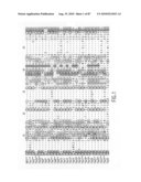 | 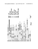 |
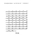 | 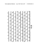 |
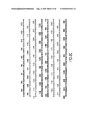 | 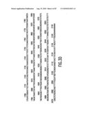 |
 |  |
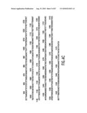 | 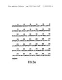 |
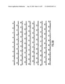 | 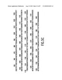 |
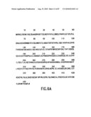 | 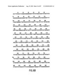 |
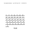 | 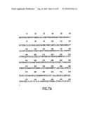 |
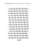 | 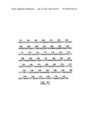 |
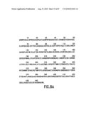 | 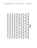 |
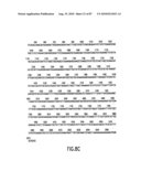 | 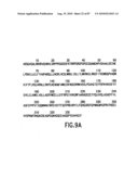 |
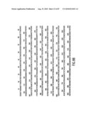 | 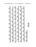 |
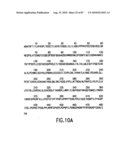 | 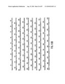 |
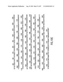 | 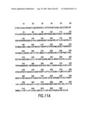 |
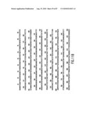 |  |
 |  |
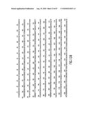 | 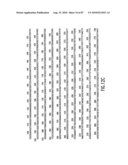 |
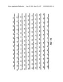 |  |
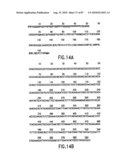 | 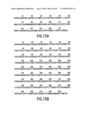 |
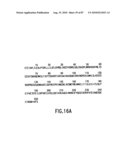 | 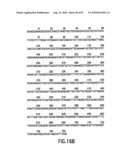 |
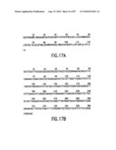 |  |
 |  |
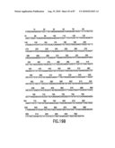 | 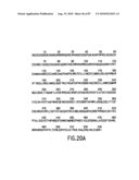 |
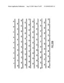 | 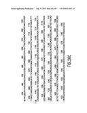 |
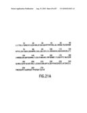 | 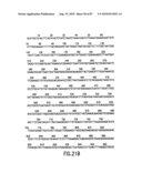 |
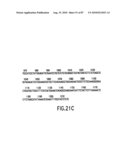 | 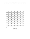 |
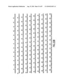 | 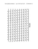 |
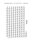 | 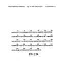 |
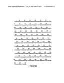 | 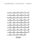 |
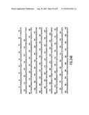 | 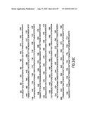 |
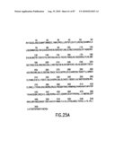 | 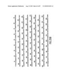 |
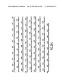 | 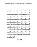 |
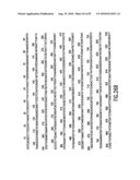 | 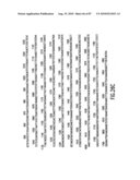 |
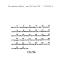 | 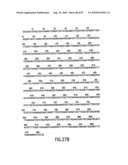 |
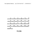 | 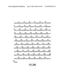 |
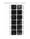 | 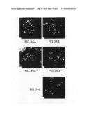 |
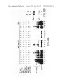 | 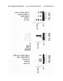 |
| New patent applications from these inventors: | |
| Date | Title |
|---|---|
| 2018-06-07 | Anti-activin a antibodies and uses thereof |
| 2015-02-12 | Methods to identify compounds useful for the treatment of proliferative and differentiative disorders |
| 2015-02-05 | Anti-activin a antibodies and uses thereof |
| 2014-12-04 | Antibodies to human gdf8 |
| 2014-05-22 | Method of treating cancer with modulators of scfskp2 |
| Top Inventors for class "Multicellular living organisms and unmodified parts thereof and related processes" | |
| Rank | Inventor's name |
|---|---|
| 1 | Gregory J. Holland |
| 2 | William H. Eby |
| 3 | Richard G. Stelpflug |
| 4 | Laron L. Peters |
| 5 | Justin T. Mason |