Patent application title: DETERMINATION OF RESPONDERS TO CHEMOTHERAPY
Inventors:
Ulrich Brennscheidt (Riehen, CH)
Otmar Herrgott (Freiburg, DE)
Astrid Heller (Penzberg, DE)
Verena Lutz (Muenchen, DE)
Verena Lutz (Muenchen, DE)
Joachim Moecks (Mannehim, DE)
Carol Ward (Helfrantzkirch, FR)
IPC8 Class: AG01N3353FI
USPC Class:
435 723
Class name: Involving a micro-organism or cell membrane bound antigen or cell membrane bound receptor or cell membrane bound antibody or microbial lysate animal cell tumor cell or cancer cell
Publication date: 2010-08-05
Patent application number: 20100196931
Claims:
1. A method of determining whether a sample comprising human lung cancer
is sensitive to a combination of an epidermal growth factor receptor
(EGFR) inhibitor and a chemotherapeutic agent, the method comprising
determining the overexpression of a phosphorylated AKT protein and/or a
phosphorylated MAPK protein in the sample, wherein the overexpression of
phosphorylated AKT protein and/or phosphorylated MAPK protein in the
sample is determined bya) determining the level of expression of
phosphorylated AKT protein and/or phosphorylated MAPK protein in the
sample:b) determining the level of expression of phosphorylated AKT
protein and/or phosphorylated MAPK protein in a sample comprising human
lung cancer that are not sensitive to a combination of a epidermal growth
factor inhibitor and a chemotherapeutic agent; andc) determining the
difference of the level of expression of phosphorylated AKT protein
and/or phosphorylated MAPK protein determined in step a) and b) thereby
determining the overexpression of phosphorylated AKT protein and/or
phosphorylated MAPK protein.whereby the overexpression of the
phosphorylated AKT protein and/or the phosphorylated MAPK protein is an
indication that the sample comprising human lung cancer is sensitive to a
combination of a epidermal growth factor receptor inhibitor and a
chemotherapeutic agent.
2. The method of claim 1 whereby the difference of the level of expression of phosphorylated AKT protein and/or phosphorylated MAPK protein determined in step a) and b) is at least 10%.
3. The method of claim 2 whereby the difference of the level of expression of phosphorylated AKT protein and/or phosphorylated MAPK protein determined in step a) and b) is at least 25%.
4. The method of claim 1, wherein the sample is a primary lung tumor or a metastasis.
5. The method of claim 1 wherein the EGFR inhibitor is erlotinib (N-(3-ethynylphenyl)-6,7-bis(2-methoxyethoxy)quinazolin-4-amine).
6. The method according to any of the claims 1 to 6 wherein the chemotherapeutic agent is selected from the group consisting of gemcitabine or cis-platin.
7. The method of claim 1, wherein the overexpression of the phosphorylated AKT protein or of the phosphorylated MAPK protein is determined using a reagent which specifically binds the phosphorylated protein.
8. The method of claim 7, wherein the reagent which specifically binds the phosphorylated protein is an antibody, antibody derivative, or an antibody fragment.
9. The method of claim 1, wherein the phosphorylated AKT protein is phosphorylated at an amino acid position corresponding to amino acid position 473 of the AKT1 protein or the MAPK protein is phosphorylated at amino acid positions corresponding to amino acid positions 202 and 204 of MAPK1.
10. The method of claim 1 wherein the amino acid sequence of the MAPK protein is selected from the group consisting of the amino acid sequence SEQ ID NO: 1 or 2 and the amino acid sequence of the AKT protein is selected from the group consisting of amino acid sequence SEQ ID NO: 6, 7 or 8.
11. The method of claim 1 wherein the overexpression of phosphorylated AKT protein and/or phosphorylated MAPK protein is determined bya) immunohistochemically staining the sample,b) assigning a grade selected from the numbers 1, 2, 3 and 4 for the level of expression of the phosphorylated AKT protein and/or the phosphorylated MAPK protein upon visual inspection of the staining of the cells in the sample whereby the highest detectable grade for the level of expression is assigned,c) determining the percentage of cells with the highest detectable grade in the immunohistochemically stained biological sample,d) multiplying the assigned grade with the percentage of cells with the highest detectable grade in the immunohistochemically stained biological sample and with the number 100, ande) determining overexpression of phosphorylated AKT protein and/or phosphorylated MAPK protein in the biological sample when the result of the multiplication in step d) is above 100.
12. A method of selecting a composition for inhibiting the progression of lung cancer in a patient, the method comprising:a) separately exposing aliquots of a sample comprising lung cancer that are sensitive to a combination of an EGFR inhibitor and a chemotherapeutic agent from the patient in the presence of a plurality of test compositions;b) comparing the level of expression of a phosphorylated AKT protein and/or a phosphorylated MAPK protein in the aliquots of the sample contacted with the test compositions and the level of expression of the phosphorylated AKT protein and/or the phosphorylated MAPK protein in an aliquot of the sample not contacted with the test compositions,c) selecting one of the test compositions which alters the level of expression of the phosphorylated AKT protein and/or a phosphorylated MAPK protein in the aliquot containing that test composition, relative to the aliquot not contacted with the test composition wherein an at least 10% difference between the level of expression of the phosphorylated AKT protein and/or the phosphorylated MAPK protein in the aliquot of the sample contacted with the test composition and the level of expression of the phosphorylated AKT protein and/or the phosphorylated MAPK protein in the aliquot of the sample not contacted with the test composition is an indication for the selection of the test composition.
13. A kit comprising an antibody against phosphorylated MAPK and/or phosphorylated AKT protein.
Description:
PRIORITY TO RELATED APPLICATIONS
[0001]This application is a Division of U.S. application Ser. No. 11/31,241, filed May 10, 2006 now allowed. This application claims the benefit of European Application No. 05010244.1, filed May 11, 2005 and European Patent Application No. 05011070.9, filed May 23, 2005, which are hereby incorporated by reference in their entirety.
FIELD OF THE INVENTION
[0002]The present invention relates to a method of determining whether a biological sample comprising human lung cancer cells is sensitive to a combination of an epidermal growth factor receptor inhibitor and a chemotherapeutic agent by determining the overexpression of a phosphorylated AKT protein and/or a phosphorylated MAPK protein in the biological sample.
[0003]The invention is also related to methods for deriving a candidate agent or for selecting a composition for inhibiting the progression of lung cancer in a patient wherein a phosphorylated AKT protein and/or a phosphorylated MAPK protein is used.
BACKGROUND OF THE INVENTION
[0004]EGFR, encoded by the erbB1 gene, has been causally implicated in human malignancy. In particular, increased expression of EGFR has been observed in breast, bladder, lung, head, neck and stomach cancer as well as glioblastomas. The Epidermal Growth Factor Receptor (EGFR), a 170-kD glycoprotein, is composed of an N-terminus extracellular domain, a hydrophobic transmembrane domain, and a C-terminus intracellular region containing the kinase domain. EGFR ligand-induced dimerization activates the intrinsic RTK domain (an Src homology domain 1, SH1), resulting in autophosphorylation on six specific EGFR tyrosine residues in the noncatalytic tail of the cytoplasmic domain.
[0005]The cellular effects of EGFR activation in a cancer cell include increased proliferation, promotion of cell motility, adhesion, invasion, angiogenesis, and enhanced cell survival by inhibition of apoptosis. Activated EGFR induces tumor cell proliferation through stimulation of the mitogen-activated protein kinase (MAPK) cascade. Upon ligand binding to the EGFR, the SOS guanine nucleotide exchange factor is recruited to the plasma membrane via the Grb2 adaptor protein, which stimulates the exchange of GTP for GDP on the small G-protein Ras, subsequently activating the MAPK cascade consisting of Raf, MEK, and ERK. Activated ERKs (pMAPK, pERK1/2) in turn phosphorylate and activate transcription factors such as ELK-1 or c-Myc, promoting cell growth.
[0006]Multiple growth factor pathways contribute to the progression and survival of NSCLC cells through activation of multiple kinases. The EGFR enhances cancer cell survival also by signaling through the phosphatidylinositol-3-kinase (PI3K)/AKT pathway and the STAT pathway. AKT is stimulated also by other growth factors, including insulin growth factor-1, basic fibroblast growth factor, and interleukins 3 and 6. The three isoforms of AKT 1-3 are all phosphorylated (pAKT) in a similar fashion at residues T308 in the activation domain and S473 in the COOH-terminal domain.
[0007]Erlotinib (Tarceva® Genentech/OSI) is a potent epidermal growth factor receptor (HER1/EGFR) tyrosine-kinase inhibitor (TKI) that provides survival benefit to patients with non-small-cell lung cancer (NSCLC) who have failed previous chemotherapy when used as a single agent (WO 01/34574). The efficacy of Tarceva®was studied in various trials. Its chemical name is N-(3-ethynylphenyl)-6,7-bis(2-methoxyethoxy)quinazolin-4-amine.
[0008]The TALENT trial was a placebo-controlled phase III study in first-line NSCLC patients who received gemcitabine and cisplatin (this concurrent chemoradiotherapy was the non-US standard of care) in combination with erlotinib (Tarceva® at 150 mg/day or placebo with. The primary endpoint was survival duration, with secondary endpoints of time to progression, response rate; duration of response; pharmacokinetic and pharmacodynamic parameters, and quality of life. HER1/EGFR and HER2 expression rates were also assessed. A standard safety analysis was done. The overall outcome of the TALENT trial was negative. For the primary and secondary endpoints there was no demonstrable benefit for erlotinib (Tarceva® plus chemotherapy (gemcitabine and cisplatin) compared with gemcitabine and cisplatin alone (Gatzemeier, U., et al., Proc Am Soc Clin Oncol 23 (2004) 617 (Abstract 7010)). Identical results were seen in the US-based TRIBUTE study, with erlotinib plus carboplatin and paclitaxel (Herbst, R. S., et al., J Clin Oncol (2004) ASCO Annual Meeting Proceedings. Post-Meeting Edition; 22 (July 15 Suppl.) (Abstract 7011)). A randomised, placebo-controlled phase III study of single-agent erlotinib as second- or third-line therapy for non-small-cell lung cancer (NSCLC) (BR.21; NCIC/OSIP) found a statistically significant improvement in survival with erlotinib (6.7 months) compared with placebo (4.7 months).
[0009]Various studies are related to the investigation of biomarkers in non-small cell lung cancer and their relation to certain EGFR inhibitor drugs. Han, et al., Int J Cancer 113 (2005) 109-115 investigate 65 patients with Gefitinib (Iressa®, EGFR TKI) monotherapy. They analyse EGFR downstream molecules as response predictive markers for gefitinib in chemotherapy-resistant non-small cell lung cancer. Cappuzzo, F. et al., JNCI 96 (2004) 1133-1141 investigate 106 patients with Gefitinib (Iressa; EGFR TKI) monotherapy. They investigate AKT phosphorylation and gefitinib efficacy in patients with advanced non-small-cell lung cancer and find that patients with P-AKT-positive tumors who received gefitinib benefited more from the therapy that patients with P-AKT-negative tumors. Vicent, S. et al., Br J Cancer 90 (2004) 1047-1052 investigate 111 NSCLC patients. They find that pERK is activated in non-small-cell lung cancer and associated with advanced tumors. Han, S. W. et al., J Clin Oncol 23 (2005) 2493-2501 investigate 90 patients with Gefitinib (EGFR TKI) monotherapy. They analyse the predictive and prognostic impact of Epidermal Growth Factor Receptor Mutation in Non-Small-Cell lung cancer patients treated with gefitinib. Mukohara, T. et al., Lung Cancer 41 (2003) 123-130 investigate 60 patients, 20 patients per stage who either underwent neoadjuvant chemotherapy or radiation. The EGFR expression correlates with pERK and pAKT expression. The sample size is too low as mentioned by the authors themselves. Raben, D. et al., Int J Radiation Oncology Biol. Phys 59 (2004) 27-38 investigate targeted therapies for non-small-cell lung cancer. Ono, M. et al., Mol Cancer Ther 3 (2004) 465-472 assay 9 NSCLC cell lines and treated with gefitinib. Hirsch, F. R. et al., Curr Opin Oncol 17 (2005) 118-122 review the phosphorylation status of AKT and MAPK as potential marker for gefitinib resistance. Meert, et al., Clinical Cancer Research 9 (2003) 2316-2326 investigate NSCLC cell lines in aspect of EGFR inhibitor activity. Neither EGFR nor Her2 expression levels correlate with sensitivity to EGFR inhibitors. Brognard, J. et al., Cell Death and Differentiation 9 (2002) 893-904 analysed 19 NSCLC cell lines were analysed whereby 17 exhibited phosphorylation of Erk1/2 and constitutive activity. David, O. et al., Clinical Cancer Research 10 (2004) 6865-6871 disclose that overexpression of pAKT is an independent prognostic factor in NSCLC. Kakiuchi, S. et al., Human Molecular Genetics 13 (2004) 3029-3043 investigate a genome wide cDNA microarray of 33 NSCLC patients. All were given gefitinib in a monotherapeutical setting. No evidence was found for correlation between AKT/pAKT expression level, EGFR gene status or pEGFR staining and gefitinib response. Kim, R. H. et al., Cancer Cell 7 (2005) 263-273 disclosed that DJ-1 expression, an oncogene, equals pAKT level. Balsara, B. R. et al., Carcinogenesis 25 (2004) 2053-2059 investigate 110 NSCLC patients with TMA pAKT expression. No significant difference in survival exists between pAKT negativity and positivity. Hirami, Y. et al., Cancer Letters 214 (2004) 157-164 investigate the relation of epidermal growth factor receptor, pAKT and hypoxia-inducible factor-1alpha in non-small cell lung cancers. Lee, S. H. et al., APMIS 110 (2002) 587-592 analyses 43 LN metastasis of NSCLC patients. AKT activation in NSCLC plays a role in tumor development rather than progression. Engelman, J. A. et al., Proc. Natl. Acad. Sci. USA 102 (2004) 3788-3793 analyse erbB-3 mediates phosphoinositide 3-kinase activity in gefitinib-sensitive non-small cell lung cancer cell lines. David, O., J Cell Mol Med 5 (2001) 430-433 discussess the role of AKT and PTEN as new diagnostics markers in lung cancer. Mantha, A. et al., Clin. Cancer Res. 11 (2005) 2398-2407 investigate the targeting of the mevalonate pathway which inhibits the function of the epidermal growth factor receptor.
[0010]Prognostic markers associated with EGFR positive cancer are investigated in WO 2004/046386. Gene expression markers for response to EGFR inhibitor drugs are disclosed by US 2004/0157255. Biomarkers and methods for determining sensitivity to epidermal growth factor receptor modulators are disclosed in WO 2004/063709. WO 01/00245 describes humanized anti-ErbB2 antibodies and methods for treating cancer with anti-ErbB2 antibodies, such as humanized anti-erbB2 antibodies.
[0011]However, and as suggested by the above cited art, there is still a need to provide methods for determining the sensitivity to EGFR inhibitor therapy, in particular combination therapies of an EGFR inhibitor with a chemotherapeutic agent.
SUMMARY OF THE INVENTION
[0012]Accordingly, in one embodiment of the invention, a method is provided of determining whether a biological sample comprising human lung cancer cells is sensitive to a combination of an epidermal growth factor receptor inhibitor and a chemotherapeutic agent, the method comprising determining the overexpression of a phosphorylated AKT protein and/or a phosphorylated MAPK protein in the biological sample whereby the overexpression of the phosphorylated AKT protein and/or the phosphorylated MAPK protein is an indication that the biological sample comprising human lung cancer cells is sensitive to a combination of a epidermal growth factor receptor inhibitor and a chemotherapeutic agent.
[0013]In another embodiment of the invention, an antibody that binds to the phosphorylated AKT protein or an antibody that binds to the phosphorylated MAPK protein is used for determining whether a biological sample comprising human lung cancer cells is sensitive to a combination of a epidermal growth factor inhibitor and a chemotherapeutic agent.
[0014]In another embodiment of the invention, a method of selecting a composition for inhibiting the progression of lung cancer in a patient is provided, the method comprising:
separately exposing aliquots of a biological sample comprising lung cancer cells that are sensitive to a combination of an EGFR inhibitor and a chemotherapeutic agent from the patient in the presence of a plurality of test compositions;comparing the level of expression of a phosphorylated AKT protein and/or a phosphorylated MAPK protein in the aliquots of the biological sample contacted with the test compositions and the level of expression of the phosphorylated AKT protein and/or the phosphorylated MAPK protein in an aliquot of the biological sample not contacted with the test compositions, selecting one of the test compositions which alters the level of expression of the marker genes in the aliquot containing that test composition, relative to the aliquot not contacted with the test composition wherein an at least 10% difference between the level of expression of the phosphorylated AKT protein and/or the phosphorylated MAPK protein in the aliquot of the biological sample contacted with the test composition and the level of expression of the phosphorylated AKT protein and/or the phosphorylated MAPK protein in the aliquot of the biological sample not contacted with the test composition is an indication for the selection of the test composition.
[0015]In yet another embodiment of the invention, a method of deriving a candidate agent is provided, said method comprising:
a) contacting an aliquot of a biological sample containing lung cancer cells that are sensitive to an EGFR inhibitor and a chemotherapeutic agent with the candidate agent,b) determining the level of expression of a phosphorylated AKT protein and/or a phosphorylated MAPK protein in the aliquot of the biological sample contacted with the candidate agent and determining the level of expression of the phosphorylated AKT protein and/or the phosphorylated MAPK protein in an aliquot of the biological sample not contacted with the candidate agent,c) observing the effect of the candidate agent by comparing the level of expression of the phosphorylated AKT protein and/or the phosphorylated MAPK protein in the aliquot of the biological sample contacted with the candidate agent and the level of expression of the phosphorylated AKT protein and/or the phosphorylated MAPK protein in the aliquot of the biological sample not contacted with the candidate agent,d) deriving said agent from said observed effect, wherein an at least 10% difference between the level of expression of the phosphorylated AKT protein and/or the phosphorylated MAPK protein in the aliquot of the biological sample contacted with the candidate agent and the level of expression of the phosphorylated AKT protein and/or the phosphorylated MAPK protein in the aliquot of the biological sample not contacted with the candidate agent is an indication of an effect of the candidate agent.
[0016]In another embodiment of the invention, a candidate agent derived by the method according to the invention is provided and a pharmaceutical preparation comprising an agent according the invention.
[0017]In still another embodiment of the invention an agent according to the invention is used for the preparation of a composition for the inhibition of progression of lung cancer.
[0018]In yet another embodiment of the invention, a method of producing a drug comprising the steps of the method of the invention and
(i) synthesizing the candidate agent identified in step (c) or an analog or derivative thereof in an amount sufficient to provide said drug in a therapeutically effective amount to a subject; and/or(ii) combining the drug candidate the candidate agent identified in step (c) or an analog or derivative thereof with a pharmaceutically acceptable carrier.
[0019]In still another embodiment of the invention, a kit comprising an antibody against phosphorylated MAPK and/or phosphorylated AKT protein is provided.
DESCRIPTION OF THE FIGURES
[0020]FIGS. 1-11 Kaplan-Meier plots of the results of example 1.
[0021]FIG. 1
[0022]Kaplan-Meier curves for time to death (OS) analysis among all patients randomly assigned to Erlotinib/Gemcitabine/Cisplatin (A) or Placebo/Gemcitabine/Cisplatin treatment (circles indicate censored observation times, when the observation was terminated before the event occurred)
[0023]FIG. 2
[0024]Kaplan-Meier curves for time to death (OS) analysis among all patients with biomarker data, randomly assigned to Erlotinib/Gemcitabine/Cisplatin (A) or Placebo/Gemcitabine/Cisplatin treatment (circles indicate censored observation times, when the observation was terminated before the event occurred)
[0025]FIG. 3
[0026]Kaplan-Meier curves for time to progression/death (PFS) analysis among all patients randomly assigned to Erlotinib/Gemcitabine/Cisplatin (A) or Placebo/Gemcitabine/Cisplatin treatment (circles indicate censored observation times, when the observation was terminated before the event occurred)
[0027]FIG. 4
[0028]Kaplan-Meier curves for time to progression/death (PFS) analysis among all patients with biomarker data, randomly assigned to Erlotinib/Gemcitabine/Cisplatin (A) or Placebo/Gemcitabine/Cisplatin treatment (circles indicate censored observation times, when the observation was terminated before the event occurred)
[0029]FIG. 5
[0030]Kaplan-Meier curves for time to death (OS) analysis among all biomarker patients treated with Placebo/Gemcitabine/Cisplatin comparing patients with pMAPK H-Score 100 (H) with patients with pMAPK H-Score<100 (circles indicate censored observation times, when the observation was terminated before the event occurred)
[0031]FIG. 6
[0032]Kaplan-Meier curves for time to death (OS) analysis among all biomarker patients with pMAPK H-Score<100 comparing Erlotinib/Gemcitabine/Cisplatin (A) with Placebo/Gemcitabine/Cisplatin treatment (circles indicate censored observation time, when the observation was terminated before the event occurred)
[0033]FIG. 7
[0034]Kaplan-Meier curves for time to death (OS) analysis among all biomarker patients with pMAPK H-Score<100 and pMAPK H-Score 100 comparing Erlotinib/Gemcitabine/Cisplatin (LA, HA) with Placebo/Gemcitabine/Cisplatin (H) treatment, respectively (circles indicate censored observation times, when the observation was terminated before the event occurred)
[0035]FIG. 8
[0036]Kaplan-Meier curves for time to progression/death (PFS) analysis among all biomarker patients with pMAPK H-Score<100 and pMAPK H-Score 100 comparing Erlotinib/Gemcitabine/Cisplatin (LA, HA) with Placebo/Gemcitabine/Cisplatin (H) treatment, respectively (circles indicate censored observation times, when the observation was terminated before the event occurred)
[0037]FIG. 9
[0038]Kaplan-Meier curves for time to death (OS) analysis among all Placebo/Gemcitabine/Cisplatin treated biomarker patients comparing pAKT1H-Score<300 (L) and pAKT1H-Score 300 (H) (circles indicate censored observation times, when the observation was terminated before the event occurred)
[0039]FIG. 10
[0040]Kaplan-Meier curves for time to death (OS) analysis among all Erlotinib/Gemcitabine/Cisplatin treated biomarker patients comparing pAKT1H-Score<300 (L) and pAKT1H-Score 300 (H) (circles indicate censored observation times, when the observation was terminated before the event occurred)
[0041]FIG. 11
[0042]Kaplan-Meier curves for time to death (OS) analysis among all biomarker patients with pAKT1H-Score<300 and pAKT1H-Score 300 comparing Erlotinib/Gemcitabine/Cisplatin (LA, HA) with Placebo/Gemcitabine/Cisplatin (H) treatment, respectively (circles indicate censored observation times, when the observation was terminated before the event occurred)
DETAILED DESCRIPTION
Definitions
[0043]The term "biological sample" shall generally mean any sample obtained from an individual, body fluid, cell line, tissue culture, or other biological source. Body fluids are e.g. lymph, sera, plasma, urine, semen, synovial fluid and spinal fluid. According to the invention, the biological sample comprises lung cancer cells and non-lung cancer cells (other cells). Methods for obtaining tissue biopsies and body fluids from mammals are well known in the art.
[0044]The term "level of expression" or "expression level" generally refers to the amount of an amino acid product or protein in the sample, preferably the amount of a phosphorylated amino acid product or phosphorylated protein in the sample according to the invention. "Expression" refers to the process by which a gene coded information is converted into the structures present and operating in the cell including their phosphorylation according to the invention. As used herein, "expressed genes" include those that are transcribed into mRNA and then translated into protein and post translationally modified e.g. phosphorylated. Just for the sake of completeness, this term shall also include the expressed genes that are transcribed into RNA but not translated into a protein (for example, transfer and ribosomal RNAs). The terms "overexpression" and "underexpression" refer to an upward or a downward deviation respectively in levels of expression as compared to the baseline expression level in a sample used as a control. "Overexpression" is therefore also "increased expression" and "underexpression" is "decreased expression".
[0045]The term "antibody" herein is used in the broadest sense and specifically covers intact monoclonal antibodies, polyclonal antibodies, multispecific antibodies (e.g., bispecific antibodies) formed from at least two intact antibodies, and antibody fragments, so long as they exhibit the desired biological activity.
[0046]The term "monoclonal antibody" as used herein refers to an antibody obtained from a population of substantially homogeneous antibodies, i.e., the individual antibodies comprising the population are identical except for possible naturally occurring mutations that may be present in minor amounts. Monoclonal antibodies are highly specific, being directed against a single antigenic site. Furthermore, in contrast to polyclonal antibody preparations which include different antibodies directed against different determinants (epitopes), each monoclonal antibody is directed against a single determinant on the antigen. In addition to their specificity, the monoclonal antibodies are advantageous in that they may be synthesized uncontaminated by other antibodies. The modifier "monoclonal" indicates the character of the antibody as being obtained from a substantially homogeneous population of antibodies, and is not to be constructed as requiring production of the antibody by any particular method. For example, the monoclonal antibodies to be used in accordance with the present invention may be made by the hybridoma method first described by Kohler, G. et al., Nature 256 (1975) 495, or may be made by recombinant DNA methods (see, e.g., U.S. Pat. No. 4,816,567). "Antibody fragments" comprise a portion of an intact antibody.
[0047]An antibody "which binds" an antigen of interest according to the invention, i.e., the phosphorylated MAPK or the phosphorylated pAKT protein, is one capable of binding that antigen with sufficient affinity such that the antibody is useful in detecting the presence of the antigen. The antibody according to the invention is one which binds phosphorylated MAPK or phosphorylated pAKT protein, it will usually preferentially bind phosphorylated MAPK or phosphorylated pAKT protein as opposed to the non-phosphorylated MAPK or the non-phosphorylated pAKT protein or does not significantly cross-react with non-phosphorylated MAPK or non-phosphorylated pAKT protein. In such embodiments, the extent of binding of the antibody to the non-phosphorylated proteins will be less than 10% as determined by fluorescence activated cell sorting (FACS) analysis or radioimmunoprecipitation (RIA). In other words, it will specifically bind phosphorylated MAPK or phosphorylated pAKT protein and does not specifically bind or does not all bind non-phosphorylated MAPK or non-phosphorylated pAKT protein.
[0048]A "chemotherapeutic agent" is a chemical compound useful in the treatment of cancer. Examples of chemotherapeutic agents include alkylating agents such as thiotepa and cyclosphosphamide (CYTOXAN®), alkyl sulfonates such as busulfan, improsulfan and piposulfan; aziridines such as benzodopa, carboquone, meturedopa, and uredopa; ethylenimines and methylamelamines including altretamine, triethylenemelamine, trietylenephosphoramide, triethylenethiophosphaoramide and trimethylolomelamine; nitrogen mustards such as chlorambucil, chlornaphazine, cholophosphamide, estramustine, ifosfamide, mechlorethamine, mechlorethamine oxide hydrochloride, melphalan, novembichin, phenesterine, prednimustine, trofosfamide, uracil mustard; nitrosureas such as carmustine, chlorozotocin, fotemustine, lomustine, nimustine, ranimustine; antibiotics such as aclacinomysins, actinomycin, authramycin, azaserine, bleomycins, cactinomycin, calicheamicin, carabicin, caminomycin, carzinophilin, chromomycins, dactinomycin, daunorubicin, detorubicin, 6-diazo-5-oxo-L-norleucine, doxorubicin, epirubicin, esorubicin, idarubicin, marcellomycin, mitomycins, mycophenolic acid, nogalamycin, olivomycins, peplomycin, poffiromycin, puromycin, quelamycin, rodorubicin, streptonigrin, streptozocin, tubercidin, ubenimex, zinostatin, zorubicin; anti-metabolites such as methotrexate and 5-fluorouracil (5-FU); folic acid analogues such as denopterin, methotrexate, pteropterin, trimetrexate; purine analogs such as fludarabine, 6-mercaptopurine, thiamiprine, thioguanine; pyrimidine analogs such as ancitabine, azacitidine, 6-azauridine, carmofur, cytarabine, dideoxyuridine, doxifluridine, enocitabine, floxuridine, 5-FU; androgens such as calusterone, dromostanolone propionate, epitiostanol, mepitiostane, testolactone; anti-adrenals such as aminoglutethimide, mitotane, trilostane; folic acid replenisher such as frolinic acid; aceglatone; aldophosphamide glycoside; aminolevulinic acid; amsacrine; bestrabucil; bisantrene; edatraxate; defofamine; demecolcine; diaziquone; elformithine; elliptinium acetate; etoglucid; gallium nitrate; hydroxyurea; lentinan; lonidamine; mitoguazone; mitoxantrone; mopidamol; nitracrine; pentostatin; phenamet; pirarubicin; podophyllinic acid; 2-ethylhydrazide; procarbazine; PSK®; razoxane; sizofuran; spirogermanium; tenuazonic acid; triaziquone; 2,2', 2''-trichlorotriethylamine; urethan; vindesine; dacarbazine; mannomustine; mitobronitol; mitolactol; pipobroman; gacytosine; arabinoside ("Ara-C"); cyclophosphamide; thiotepa; taxanes, e.g., paclitaxel (TAXOL®, Bristol-Myers Squibb Oncology, Princeton, N.J.) and docetaxel (TAXOTERE®, Rhone-Poulenc Rorer, Antony, France); chlorambucil; gemcitabine; 6-thioguanine; mercaptopurine; methotrexate; platinum analogs such as cisplatin and carboplatin; vinblastine; platinum; etoposide (VP-16); ifosfamide; mitomycin C; mitoxantrone; vincristine; vinorelbine; navelbine; novantrone; teniposide; daunomycin; aminopterin; xeloda; ibandronate; CPT-11; topoisomerase inhibitor RFS 2000; difluoromethylornithine (DMFO); retinoic acid; esperamicins; capecitabine; and pharmaceutically acceptable salts, acids or derivatives of any of the above. Also included in this definition, are anti-hormonal agents that act to regulate or inhibit hormone action on tumors such as anti-estrogens including for example tamoxifen, raloxifene, aromatase inhibiting 4(5)-imidazoles, 4-hydroxytamoxifen, trioxifene, keoxifene, LY117018, onapristone, and toremifene (Fareston); and anti-androgens such as flutamide, nilutamide, bicalutamide, leuprolide, and goserelin; and pharmaceutically acceptable salts, acids or derivatives of any of the above. The "chemotherapeutic agent" itself can be a combination of chemical compounds useful in the treatment of cancer combination as mentioned above, i.e. the combination may be gemcitabine/cis-platin, but also e.g. cis-platin/paclitaxel, cis-platin/docetaxel, cis-platin/vinorelbine, gemcitabine/carboplatin, or carboplatin/docetaxel.
[0049]The term "EGFR inhibitor" refers to a therapeutic agent that binds to EGFR and, optionally, inhibits EGFR activation. Examples of such agents include antibodies and small molecules that bind to EGFR. Examples of antibodies which bind to EGFR include MAb 579 (ATCC CRL HB 8506), MAb 455 (ATCC CRL HB8507), MAb 225 (ATCC CRL 8508), MAb 528 (ATCC CRL 8509) (see, U.S. Pat. No. 4,943,533, Mendelsohn et al.) and variants thereof, such as chimerized 225 (C225 or Cetuximab; ERBUTIX®) and reshaped human 225 (H225) (see, WO 96/40210, Imclone Systems Inc.); antibodies that bind type 11 mutant EGFR (U.S. Pat. No. 5,212,290); humanized and chimeric antibodies that bind EGFR as described in U.S. Pat. No. 5,891,996; and human antibodies that bind EGFR, such as ABX-EGF (see WO 98/50433, Abgenix). The anti-EGFR antibody may be conjugated with a cytotoxic agent, thus generating an immunoconjugate (see, e.g., EP 0 659 439 A2, Merck patent GmbH). Examples of small molecules that bind to EGFR include ZD1839 or Gefitinib (IRESSA®; Astra Zeneca), CP-358774 or Erlotimils (Tarceva®; Genentech/OSI) and AG1478, AG1571 (SU 5271; Sugen). Particularly preferred in this application are EGFR tyrosine kinase inhibitors, particularly small molecule EGFR tyrosine kinase inhibitors as e.g. erlotinils (Tarceva®). A "small molecule" can be e.g. a peptide or a peptidomimetics with a molecular weight less than about 10,000 grams per mole, preferably less than about 5,000 grams per mole. Preferably, a "small molecule" is compound, i.e. an organic or inorganic compound, with a molecular weight less than about 5,000 grams per mole, preferably less than about 1,000 grams per mole, more preferably less than 500 grams per mole, and salts, esters, and other pharmaceutically acceptable forms of such compounds. Therefore, in a preferred embodiment of the invention, the EGFR inhibitor is a EGFR tyrosine kinase inhibitor that is a compound with a molecular weight less than about 5,000 grams per mole, preferably less than about 1,000 grams per mole, and salts, esters, and other pharmaceutically acceptable forms of such a compound. In other words, the EGFR inhibitor is a compound that inhibts EGFR tyrosine kinase activity and that has a molecular weight less than about 5,000 grams per mole, preferably less than about 1,000 grams per mole, more preferably less than 500 grams per mole, and salts, esters, and other pharmaceutically acceptable forms of such a compound.
[0050]"Gemcitabine" is the chemotherapeutic agent 2',2'-difluorodeoxycytidine (dFdC) which is a pyrimidine analogue of deoxycytidine in which the deoxyribose moiety contains two fluorine atoms at the 2'-position (see Heinemann, V. et al., Cancer Res 48 (1988) 4024). It is commerciably available as Gemzar® from Eli Lilly and Company, Indianapolis, Ind., USA.
[0051]"Cis-platin" as used throughout this application is the chemotherapeutic agent cis-diamminedichloroplatinum (see U.S. Pat. No. 5,562,925) commercially available as Platinol® from Bristol-Myers Squibb Company, New York, N.Y., USA. "Cis-platin" is a heavy metal complex containing a central atom of platinum surrounded by two chloride atoms and two ammonia molecules in the cis position.
[0052]According to the invention, the expression that "a biological sample comprising human lung cancer cells is sensitive to a combination of an epidermal growth factor receptor inhibitor and a chemotherapeutic agent" shall mean that the biological sample comprising human lung cancer cells is sensitive to a treatment with a combination of an epidermal growth factor receptor inhibitor and a chemotherapeutic agent in contrast to a treatment with an epidermal growth factor receptor inhibitor alone. "Sensitive" can also be understood as "reacting to" or "showing a reaction to", particularly such a reaction that is of benefit to a lung cancer patient. Thereby, it can be determined whether a lung cancer patient is sensitive to a treatment with a combination of an epidermal growth factor receptor inhibitor and a chemotherapeutic agent in contrast to a treatment with an epidermal growth factor receptor inhibitor alone. This means that the patient will benefit from such a treatment.
[0053]A "MAPK" protein is a member of a highly conserved cytosolic serine/threonine protein kinase family known as mitogen-activated protein kinases (MAPKs) or extracellular signal-regulated kinases (ERKs). This protein family has several subgroups. ERKs are activated and tyrosine- or threonine-phosphorylated in response to a wide variety of extracellular signals, including osmotic stress, heat shock, pro-inflammatory cytokines, hormones, and mitogens. The term "MAPK protein" as used in this invention preferably refers to a member of the MAPK protein family comprising or preferably consisting of MAPK1 and MAPK3. The amino acid sequences of MAPK1 (ERK2) are SEQ ID NO: 1) and MAPK3 (ERK1) (SEQ ID NO: 2). These amino amino acid sequences are encoded by the mRNA sequences, i.e. cDNA sequences SEQ ID NO: 3 and 4 for MAPK1 ans SEQ ID NO: 5 for MAPK 3. The primary phosphorylation sites in MAPK1 are Thr185 and Tyr185 and the primary phosphorylation in MAPK3 are Thr202 and Tyr204. These phosphorylation sites are also recognized by the antibody used in the present invention, i.e. preferably the polyclonal antibody serum against the phosphorylated forms of MAPK1 and MAPK3.
[0054]The term "AKT" protein refers to a protein of the AKT/PKB subfamily of second-messenger regulated serine/threonine protein kinases which has three members termed AKT1/PKBalpha, AKT2/PKBbeta (Staal, S. P., Proc. Natl. Acad. Sci. USA 84 (1987) 5034-5037) and AKT3/PKBgamma (Nakatani, K. et al., Biochem. Biophys. Res. Comm. 257 (1999) 906-910; U.S. Pat. No. 6,881,555) respectively. The isoforms are homologous and are activated by phosphorylation in response to phosphatidylinositol 3'-OH kinase (PI3K) signaling. The PI3K/AKT/PKB pathway appears to be important for regulating cell survival/cell death (Dudek, H. et al., Science 275 (1997) 661-665) also in tumorigenesis. The term "AKT protein" as used in this invention preferably refers to a member of the AKT protein family comprising or preferably consisting of AKT1, AKT2 and AKT3. Phosphorylation of AKT1/PKBα occurs on two sites Thr308 and on Ser473 (Meier, R., et al., J. Biol. Chem. 272 (1997) 30491-30497). Equivalent phosphorylation sites occur in AKT2/PKBbeta (Thr309 and Ser474) and AKT3/PKBgamma (Thr305 and Ser472). The term "phosphorylated AKT" protein refers to a phosphorylated "AKT" protein, preferably phosphorylated at the sites described above. The term "MAPK protein" as used in this invention preferably refers to a member of the MAPK protein family comprising or preferably consisting of MAPK1 and MAPK3. AKT 1 is also known as human RAC-alpha serine/threonine-protein kinase (EC 2.7.1.37) (RAC-PK-alpha), Protein kinase B (PKB) (C-AKT) and the amino acid sequence of AKT 1 is SEQ ID NO: 6. AKT2 is also known as human RAC-beta serine/threonine-protein kinase (EC 2.7.1.37) (RAC-PK-beta), Protein kinase AKT-2 or Protein kinase B, beta (PKB beta) and the amino acid sequence of AKT 2 is SEQ ID NO: 7. AKT3 is also known as human RAC-gamma serine/threonine-protein kinase (EC 2.7.1.37) (RAC-PK-gamma), protein kinase AKT-3 or Protein kinase B, gamma (PKB gamma) (STK-2) and the amino acid sequence of AKT 3 is SEQ ID NO: 8.
[0055]Conventional techniques of molecular biology and nucleic acid chemistry, which are within the skill of the art, are explained in the literature. See, for example, Sambrook, J. et al., Molecular Cloning: A Laboratory Manual, Cold Spring Harbor Laboratory Press, Cold Spring Harbor, N.Y., 1989; Gait, M. J. (ed.), Oligonucleotide Synthesis--A Practical Approach, IRL Press, 1984; Hames, B. D., and Higgins, S. J. (eds.), Nucleic Acid Hybridisation--A Practical Approach, IRL Press, 1985; and a series, Methods in Enzymology, Academic Press, Inc., all of which are incorporated herein by reference. All patents, patent applications, and publications mentioned herein, both supra and infra, are incorporated herein by reference.
DETAILED DESCRIPTION OF THE INVENTION
[0056]In one embodiment of the invention, a method is provided of determining whether a biological sample comprising human lung cancer cells is sensitive to a combination of an epidermal growth factor receptor inhibitor and a chemotherapeutic agent, the method comprising determining the overexpression of a phosphorylated AKT protein and/or a phosphorylated MAPK protein in the biological sample whereby the overexpression of the phosphorylated AKT protein and/or the phosphorylated MAPK protein is an indication that the biological sample, comprising human lung cancer cells is sensitive to a combination of a epidermal growth factor receptor inhibitor and a chemotherapeutic agent.
[0057]Preferably, in the method according to the invention, the overexpression of phosphorylated AKT protein and/or phosphorylated MAPK protein in the biological sample is determined by
a) determining the level of expression of phosphorylated AKT protein and/or phosphorylated MAPK protein in the biological sample,b) determining the level of expression of phosphorylated AKT protein and/or phosphorylated MAPK protein in a biological sample comprising human lung cancer cells that are not sensitive a combination of a epidermal growth factor inhibitor and a chemotherapeutic agent,c) determining the difference of the level of expression of phosphorylated AKT protein and/or phosphorylated MAPK protein determined in step a) and b) thereby determining the overexpression of phosphorylated AKT protein and/or phosphorylated MAPK protein.
[0058]Preferably, the difference of the level of expression of phosphorylated AKT protein and/or phosphorylated MAPK protein determined in step a) and b) is at least 10%. More preferably, the difference of the level of expression of phosphorylated AKT protein and/or phosphorylated MAPK protein determined in step a) and b) is at least 25%. In another embodiment, the difference of the level of expression of phosphorylated AKT protein and/or phosphorylated MAPK protein determined in step a) and b) is at least 50%, 75%, 100%, 125%, 150%, 175%, 200%, 300%, 400%, 500% or 1,000%. The difference of the level of expression of phosphorylated AKT protein and/or phosphorylated MAPK protein determined in step a) and b) can be up to 10,000 or 50,000%. The difference of the level of expression of phosphorylated AKT protein and/or phosphorylated MAPK protein determined in step a) and b) is preferably between 10% to 10,000%, more preferably 25% to 10,000%, 50% to 10,000%, 100% to 10,000%, even more preferably 25% to 5,000%, 50% to 5,000%, 100% to 5,000%.
[0059]In a preferred embodiment of the invention, the biological sample is a primary lung tumor or a metastasis (regional or distant) which can be obtained e.g. by lung biopsy or from other organs by way of biopsy. A metastasis can also be a distant metastasis e.g. from the liver or lymph node. It has to be noted that such distant metastasis also contain lung cancer cells as the metastases originate from the lung.
[0060]In another preferred embodiment the cancer is another cancer than lung cancer as pancreatic cancer. However, other cancers with solid tumours are also feasible such as ovarian, colorectal, head and neck, renal cell carcinoma, glioma and gastrointestinal cancers, particularly stomach cancer.
[0061]In another preferred embodiment the EGFR inhibitor is a EGFR tyrosine kinase inhibitors, particularly small molecule EGFR tyrosine kinase inhibitors as e.g. Tarceva®. Therefore in other words, in a particularly preferred embodiment of the invention, the EGFR inhibitor is erlotinib or N-(3-ethynylphenyl)-6,7-bis(2-methoxyethoxy)quinazolin-4-amine.
[0062]In yet another preferred embodiment of the invention, the chemotherapeutic agent(s) is selected from the group consisting of gemcitabine and/or cis-platin.
[0063]In another preferred embodiment of the invention, the overexpression of the phosphorylated AKT protein or of the phosphorylated MAPK protein is determined using a reagent (such as for example an antibody) which specifically binds the phosphorylated protein and preferably not specifically or not at all to the non-phosphorylated protein. Preferably the reagent is an antibody, an antibody derivative, or an antibody fragment which specifically binds to the phosphorylated AKT protein or the phosphorylated MAPK protein and preferably not specifically or not at all to the non-phosphorylated AKT protein or the non-phosphorylated MAPK protein.
[0064]In yet another preferred embodiment of the invention, the phosphorylated AKT protein is phosphorylated at an amino acid position corresponding to amino acid position 473 of the AKT1 protein or the MAPK protein is phosphorylated at amino acid positions corresponding to amino acid positions 202 and 204 of MAPK1. Preferably, the amino acid sequence of the MAPK protein is selected from the group consisting of the amino acid sequence SEQ ID NO: 1 or 2 and the amino acid sequence of the AKT protein is selected from the group consisting of the amino acid sequence SEQ ID NO: 6, 7 or 8.
[0065]There are many different types of immunoassays which may be used in the method of the present invention, e.g. enzyme linked immunoabsorbent assay (ELISA), fluorescent immunosorbent assay (FIA), chemical linked immunosorbent assay (CLIA), radioimmuno assay (RIA), and immunoblotting. For a review of the different immunoassays which may be used, see: Lottspeich and Zorbas (eds.), Bioanalytik, 1st edition 1998, Spektrum Akademischer Verlag, Heidelberg, Berlin, Germany. Therefore, in yet another preferred embodiment of the invention, the expression level is determined using a method selected from the group consisting of proteomics, flow cytometry, immunocytochemistry, immunohistochemistry and enzyme-linked immunosorbent assay.
[0066]In a preferred embodiment of the invention, the overexpression of phosphorylated AKT protein and/or phosphorylated MAPK protein is determined by
a) immunohistochemically staining the biological sample,b) assigning a grade selected from the numbers 1, 2, 3 and 4 for the level of expression of the phosphorylated AKT protein and/or the phosphorylated MAPK protein upon visual inspection of the staining of the cells in the biological sample whereby the highest detectable grade for the level of expression is assigned,c) determining the percentage of cells with the highest detectable grade in the immunohistochemically stained biological sample,d) multiplying the assigned grade with the percentage of cells with the highest detectable grade in the immunohistochemically stained biological sample and with the number 100, ande) determining overexpression of phosphorylated AKT protein and/or phosphorylated
[0067]MAPK protein in the biological sample when the result of the multiplication in step d) is above 100.
[0068]In another embodiment of the invention, a method is provided of determining whether a lung cancer patient benefits from a combination of an epidermal growth factor receptor inhibitor and a chemotherapeutic agent, the method comprising determining the overexpression of a phosphorylated AKT protein and/or a phosphorylated MAPK protein in a sample from the patient whereby the overexpression of the phosphorylated AKT protein and/or the phosphorylated MAPK protein is an indication that the patient benefits from a combination of a epidermal growth factor receptor inhibitor and a chemotherapeutic agent. All other preferred embodiments described above equally apply to this embodiment. Preferably, in the method according to the invention, the overexpression of phosphorylated AKT protein and/or phosphorylated MAPK protein in the sample from the patient is determined by
a) determining the level of expression of phosphorylated AKT protein and/or phosphorylated MAPK protein in the sample from the patient,b) determining the level of expression of phosphorylated AKT protein and/or phosphorylated MAPK protein in a sample from a lung cancer patient who does not benefit from a combination of a epidermal growth factor inhibitor and a chemotherapeutic agent,c) determining the difference of the level of expression of phosphorylated AKT protein and/or phosphorylated MAPK protein determined in step a) and b) thereby determining the overexpression of phosphorylated AKT protein and/or phosphorylated MAPK protein. The term "benefit" means that the patient does not have a benefit from a treatment with combination of an epidermal growth factor receptor inhibitor and a chemotherapeutic agent in contrast to a treatment with an epidermal growth factor receptor inhibitor alone.
[0069]In another preferred embodiment of the invention, an antibody that binds to the phosphorylated AKT protein or an antibody that binds to the phosphorylated MAPK protein is used for determining whether a biological sample comprising human lung cancer cells is sensitive to a combination of a epidermal growth factor inhibitor and a chemotherapeutic agent.
[0070]In still another embodiment of the invention, a method of selecting a composition for inhibiting the progression of lung cancer in a patient is provided, the method comprising:
a) separately exposing aliquots of a biological sample comprising lung cancer cells that are sensitive to a combination of an EGFR inhibitor and a chemotherapeutic agent from the patient in the presence of a plurality of test compositions;b) comparing the level of expression of a phosphorylated AKT protein and/or a phosphorylated MAPK protein in the aliquots of the biological sample contacted with the test compositions and the level of expression of the phosphorylated AKT protein and/or the phosphorylated MAPK protein in an aliquot of the biological sample not contacted with the test compositions,c) selecting one of the test compositions which alters the level of expression of the phosphorylated AKT protein and/or phosphorylated MAPK protein (marker genes) in the aliquot containing that test composition, relative to the aliquot not contacted with the test composition wherein an at least 10% difference between the level of expression of the phosphorylated AKT protein and/or the phosphorylated MAPK protein in the aliquot of the biological sample contacted with the test composition and the level of expression of the phosphorylated AKT protein and/or the phosphorylated MAPK protein in the aliquot of the biological sample not contacted with the test composition is an indication for the selection of the test composition.
[0071]Preferably, the difference of the level of expression of phosphorylated AKT protein and/or phosphorylated MAPK protein in step c) is at least 25%. More preferably, the difference of the level of expression of phosphorylated AKT protein and/or phosphorylated MAPK protein in step c) is at least 50%. In another embodiment, the difference of the level of expression of phosphorylated AKT protein and/or phosphorylated MAPK protein in step c) is at least 75%, 100%, 125%, 150%, 175%, 200%, 300%, 400%, 500% or 1,000%. The difference of the level of expression of phosphorylated AKT protein and/or phosphorylated MAPK protein determined in step c) can be up to 10,000 or 50,000%. The difference of the level of expression of phosphorylated AKT protein and/or phosphorylated MAPK protein determined in step c) is preferably between 10% to 10,000%, more preferably 25% to 10,000%, 50% to 10,000%, 100% to 10,000%, even more preferably 25% to 5,000%, 50% to 5,000%, 100% to 5,000%.
[0072]In yet another embodiment of the invention, a method of deriving a candidate agent is provided, said method comprising:
a) contacting an aliquot of a biological sample containing lung cancer cells that are sensitive to an EGFR inhibitor and a chemotherapeutic agent with the candidate agent,b) determining the level of expression of a phosphorylated AKT protein and/or a phosphorylated MAPK protein in the aliquot of the biological sample contacted with the candidate agent and determining the level of expression of the phosphorylated AKT protein and/or the phosphorylated MAPK protein in an aliquot of the biological sample not contacted with the candidate agent,c) observing the effect of the candidate agent by comparing the level of expression of the phosphorylated AKT protein and/or the phosphorylated MAPK protein in the aliquot of the biological sample contacted with the candidate agent and the level of expression of the phosphorylated AKT protein and/or the phosphorylated MAPK protein in the aliquot of the biological sample not contacted with the candidate agent,d) deriving said agent from said observed effect, wherein an at least 10% difference between the level of expression of the phosphorylated AKT protein and/or the phosphorylated MAPK protein in the aliquot of the biological sample contacted with the candidate agent and the level of expression of the phosphorylated AKT protein and/or the phosphorylated MAPK protein in the aliquot of the biological sample not contacted with the candidate agent is an indication of an effect of the candidate agent.
[0073]Preferably, the difference of the level of expression of phosphorylated AKT protein and/or phosphorylated MAPK protein in step d) is at least 25%. More preferably, the difference of the level of expression of phosphorylated AKT protein and/or phosphorylated MAPK protein in step d) is at least 50%. In another embodiment, the difference of the level of expression of phosphorylated AKT protein and/or phosphorylated MAPK protein in step d) is at least 75%, 100%, 125%, 150%, 175%, 200%, 300%, 400%, 500% or 1,000%. The difference of the level of expression of phosphorylated AKT protein and/or phosphorylated MAPK protein determined in step d) can be up to 10,000 or 50,000%. The difference of the level of expression of phosphorylated AKT protein and/or phosphorylated MAPK protein determined in step d) is preferably between 10% to 10,000%, more preferably 25% to 10,000%, 50% to 10,000%, 100% to 10,000%, even more preferably 25% to 5,000%, 50% to 5,000%, 100% to 5,000%.
[0074]In a preferred embodiment said candidate agent is a candidate inhibitory agent or a candidate enhancing agent.
[0075]In another embodiment of the invention a candidate agent derived by the method according to the invention is provided.
[0076]In yet another embodiment a pharmaceutical preparation comprising an agent according to the invention is provided.
[0077]In still another embodiment an agent according to the invention is used for the preparation of a composition for the inhibition of progression of lung cancer.
[0078]In yet another embodiment of the invention, a method of producing a drug comprising the steps of the method of the invention is provided and
(i) synthesizing the candidate agent identified in step (c) or an analog or derivative thereof in an amount sufficient to provide said drug in a therapeutically effective amount to a subject; and/or(ii) combining the drug candidate the candidate agent identified in step (c) or an analog or derivative thereof with a pharmaceutically acceptable carrier.
[0079]In still another embodiment an AKT protein, a MAPK protein, a phosphorylated AKT protein, a phosphorylated MAPK protein, an antibody selectively binding to a phosphorylated AKT protein or a phosphorylated MAPK protein is used for deriving a candidate agent or for selecting a composition for inhibiting the progression of lung cancer in a patient.
[0080]In another embodiment of the invention, a kit is contemplated comprising an antibody against phosphorylated MAPK and/or phosphorylated AKT protein. Such kits known in the art further comprise plastics ware which can be used during the amplification procedure as e.g. microtitre plates in the 96 or 384 well format or just ordinary reaction tubes manufactured e.g. by Eppendorf, Hamburg, Germany and all other reagents for carrying out the method according to the invention, preferably an immunoassay, e.g. enzyme linked immunoabsorbent assay (ELISA), fluorescent immunosorbent assay (FIA), chemical linked immunosorbent assay (CLIA), radioimmuno assay (RIA), and immunoblotting. For a review of the different immunoassays and reagents which may be used, see: Lottspeich and Zorbas (eds.), Bioanalytik, 1st edition 1998, Spektrum Akademischer Verlag, Heidelberg, Berlin, Germany.
[0081]The following examples, references, sequence listing and figures are provided to aid the understanding of the present invention, the true scope of which is set forth in the appended claims. It is understood that modifications can be made in the procedures set forth without departing from the spirit of the invention.
Example 1
Biomarker Analysis on Tumor Tissue Samples
[0082]The objective of the exploratory tumor biomarker analyses for the clinical study was the identification of those markers or combinations of markers which predict best for positive or negative clinical outcome of Tarceva® treatment. As the clinical results of the study did not allow to generate hypotheses about how to select patient populations that derive more benefit from the treatment with Tarceva®, special emphasis was on the identification of markers that discriminate between patients (subgroups) that specifically benefit in the Tarceva® combination vs. the chemotherapy-alone control arm. Additionally, the identification of markers that discriminate between patients (subgroup) that have a detrimental effect from the specific combination with Tarceva® vs. the chemotherapy-alone control arm was investigated.
[0083]The aim of this study was to analyze tumor-specific biomarker related to the EGFR signaling pathway e.g. EGFR, HER2, pAKT, and pMAPK.
[0084]Biomarker data was correlated with clinical data (overall and therapy-specific analysis).
Material and Methods:
Clinical Samples:
[0085]Biomarker analyses were performed on a sample subset of 141 patients, for which formalin-fixed paraffin embedded (FFPE) tissue blocks from initial diagnosis had been received.
[0086]Antibodies for IHC Testing:
TABLE-US-00001 Antibody Target protein Slide pretreatment Dilution Abcam ab8932 pAKT (antibody pressure cooker 120° C./5 min, 1:450 (available from Abcam, against citrate buffer pH 9 Cambridge, United Kingdom) phosphorylated Ser 473 of AKT) Zymed 36-8800 pMAPK (ERK1+2 pressure cooker 120° C./5 min, 1:150 (available from Zytomed Thr202/Tyr204) citrate buffer pH 6 GmbH, Berlin, Germany or Invitrogen, Carlsbad, CA, USA)
[0087]pMAPK IHC Protocol:
1. Cut about 3-4 um thick sections from the Paraffin-Array-blocks.2. Mount sections on glass slides and let them dry over night.3. Deparaffinize the slides in xylene followed by descending ethanol series: [0088]xylene over night [0089]xylene 2×10 minutes [0090]abs.Ethanol 2×10 min. [0091]96% Ethanol 2×5 min. [0092]80% Ethanol 1×5 min. [0093]70% Ethanol 2×5 min. [0094]PBS Buffer 10 min. (change buffer once or twice)4. Antigene retrival/sample pretreatmentPressure cooker 5 minutes at 120° C. in 1× citrate buffer pH 6 (Biocyc GmbH, order number 400300692). Wash in TBS/PBS (1:10) buffer for 5 minutes5. Peroxidase blocking
[0095]Slides are incubated in 3% H2O2 for 10 minutes.
[0096]Wash 2×5 minutes in TBS/PBS buffer.
6. Antibody Incubation
[0097]Put the slides in normal serum (1.5%) diluted in Tris-Buffer or a other blocking solution.
[0098]Put the primary antibody (Zymed Rabbit anti phospho-ERK1+2 cat no. 36-8800) diluted 1:150 on the slides and incubate it in a humified chamber at 30° C. for 2 hours.
[0099]Wash 2×5 minutes in TBS/PBS buffer.
[0100]Put Envision Polymer HRP (Dako) on the slides and incubate in a humified chamber at 30° C. for 30 minutes.
[0101]Wash 2×5 minutes in TBS/PBS buffer.
7. Detection
[0102]Wash the slides with 0.05 M Tris buffer pH 7.6 for 20 minutes
[0103]Cover slides for 5 minutes with DAB-Chromogen (Liquid DAB Dako code no. K3467) and incubate it for 5-10 minutes.
[0104]Wash the slides with demin. water for 5 minutes to stop the color reaction.
[0105]Counterstain with hematoxyline (Harris Hamatoxylin HTX 31000, Medite GmbH)
[0106]Rinse with water
[0107]Differentiate in HCL-Ethanol
[0108]"blue" for 5 min in water
[0109]Ascending ethanol series
[0110]xylene
[0111]cover
[0112]pAKT IHC Protocol:
Protocol as for pMAPK, except
[0113]Compare 4: Sample pretreatment in citrate buffer pH 9 (Dako code no. S2367)
[0114]Compare 6: Primary antibody (abcam AKT phospho S473) diluted 1:450 (compare 6.)
[0115]IHC Data Reporting:
One pathologist evaluated all immunostainings. The nuclear staining intensity (pAKT, pMAPK) was estimated by visual inspection in a four step scale (0, 1, 2, 3). In addition to nuclear staining intensity, the percentage of positive cells, and the reason for analysis failure (i.e. lack of tumor cells in the tissue spot or lack of the tissue spot on the TMA slide) was recorded.
Exploratory Statistical Analysis:
[0116]The statistical analyses of the biomarker data aimed at exploring the potential to predict clinical benefit and/or toxicities, by each marker separately and/or by suitable combinations.
[0117]According to experience many biomarkers show a skewed statistical distribution across patients and within patient. Frequently there is also some biochemical background of this skewness, in that the variation process has a multiplicative structure. Skewed distributions present with problems when linear statistical approaches (e.g. regression) are to be used. When used as covariates in statistical models the skewness as well can obscure the results. Therefore suitable transformations need to be found which transform these measurements into distributions with an approximate Gaussian shape. Typical choices in the biomarker area are transformations of the form log(x+c). These transformations do not change the order of the values, such that non-parametric analyses based on ranks or cut-offs remain unchanged by the transformation. Such transformations are also a prerequisite when linear multivariate approaches like e.g. Discriminant Analysis and Principle Component Analysis are to be used.
[0118]The basic statistics and interdependencies of the different markers were descriptively investigated. Methodological analyses comparing different measurement approaches, e.g. for IHC, were performed with regard to reliability and validity. Benefit to Tarceva® is defined by the clinical endpoints survival time (or Time to Death, TTD), PFS time (time to progression, TTP/D), objective response, best response (CR/PR/SD/PD).
[0119]The p-values emerging from these analyses are not to be interpreted in a confirmative sense; they are to be seen as a special descriptive tool in order to guide the exploration towards an efficient candidate prediction rule. Markers were evaluated on a univariate level regarding their potential for prediction (e.g. search for cut-offs) of the clinical endpoints. Further multivariate techniques (e.g. Linear Discriminant Analysis, Multiple Logistic Regression, Principal Component Analysis with Rotation, Cluster Analysis, CART methodology) were employed in order to study combinations of markers. Biomarker and response correlations with clinical covariates were investigated. Candidate groupings derived from biomarkers were checked with time to event variables (Kaplan-Meier curves, Cox proportional hazard model, logrank test).
Results:
[0120]Analysis of the TMA Sample Subset in Comparison to the Overall Study Population
[0121]The patient subset with samples for biomarker analysis was compared to the overall study population regarding baseline patient characteristics and clinical outcome parameters. The summary is shown in the table below.
TABLE-US-00002 Patient subset with IHC/ Main Clinical Population FISH biomarker data Placebo Tarceva ® Placebo Tarceva ® (N = 582) (N = 580) (N = 70) (N = 71) Age (years) 59.1 59.9 57.5 59.1 Male (%) 75.3 78.6 80.0 74.6 Disease 67.2 64.8 74.3 80.3 stage IV (%) Adeno 37.6 37.9 41.4 46.5 carcinoma (%) # of metastatic 3.7 3.7 3.4 4.0 sites # of affected 2.5 2.5 2.1 2.4 organs Sum longest 92.8 95.4 80.1 93.5 diameter Average symptom 26.5 26.7 24.3 25.3 burden Responders (%) 38.3 42.4 42.3 43.5 (N = 418) (N = 396) (N = 52) (N = 46) Hazard ratio TTD 1.035 1.217 Hazard ratio TTP 0.980 1.262
Findings:
[0122]The patient subset with biomarker data is not representative for the BO16411 study population. There are differences in hazard ratios for TTD and TTP between main population and the biomarker subgroup. Tarceva®-treated patients of the biomarker subgroup have a worse prognosis compared to the main clinical population. Several baseline covariates indicate that the biomarker subgroup--and within this subgroup particularly Tarceva®-related patients represented a more morbid case mix as compared to the main study population. KM plots (FIGS. 1 to 4) should be taken into account as well.
Results of pMAPK IHC Analysis
[0123]pMAPK IHC data showed sufficient scatter to be eligible for further statistical analysis.
For determining correlation between pMAPK expression and clincal outcome, cut-off values were established by descriptive statistical analysis: Franklin H-score was determined by combining staining intensity and percentage of stained tumor cells (pMAPK_hsco=(pMAPK_Nuclear_Staining+1)*pMAPK_Nuclear_Pos_Cells; range: 0-400). "Positive" pMAPK staining was defined by H-scores of =/>100, else the staining was "negative". Kaplan-Meier plots are shown in FIGS. 5 to 8
TABLE-US-00003 pMAPK H-Score (Cutpoint 100) --- COX Model without covariates Parameter Standard Hazard 95% Hazard Ratio Variable Estimate Error Chi-Square Pr > ChiSq Ratio Confidence Limits Time to progression/death trtgr only 0.27747 0.21560 1.6562 0.1981 1.320 0.865 2.014 trtgr 0.12352 0.22058 0.3136 0.5755 1.131 0.734 1.743 pMAPK (≧100) 0.90942 0.23706 14.7164 0.0001 2.483 1.560 3.951 trtgr pMAPK < 100 0.49122 0.30087 2.6656 0.1025 1.634 0.906 2.947 trtgr pMAPK ≧ 100 -0.25703 0.30706 0.7007 0.4026 0.773 0.424 1.412 Time to death trtgr only 0.15228 0.27818 0.2997 0.5841 1.164 0.675 2.009 trtgr -0.02494 0.28668 0.0076 0.9307 0.975 0.556 1.711 pMAPK (≧100) 0.73352 0.29272 6.2797 0.0122 2.082 1.173 3.696 trtgr pMAPK < 100 0.98292 0.45017 4.7674 0.0290 2.672 1.106 6.457 trtgr pMAPK ≧ 100 -0.70524 0.36205 3.7943 0.0514 0.494 0.243 1.004
Findings:
[0124]For patients treated with chemotherapy/placebo "positive" pMAPK expression is associated with worse prognosis (TTD: HR 4.882, p=0.0001), while "negative" pAKT expression appears to be associated with longer survival
Trend: "Positive pMAPK" patients might benefit from the chemotherapy/Tarceva® combo (HR 0.500, p: 0.0516) ( - - - )Results of pAKT IHC Analysis
[0125]pAKT IHC data showed sufficient scatter to be eligible for further statistical analysis For determining correlation between pAKT expression and clincal outcome, cut-off values were established by descriptive statistical analysis: Franklin H-score was determined by combining nuclear staining intensity and percentage of stained tumor cells (pAKT_hsco=(pAKT_Nuclear_Staining+1)*pAKT_Nuclear_Pos_Cells; range: 0-400). "Positive" pAKT staining was defined by H-scores of =/>300, else the staining was "negative".
Kaplan-Meier plots are shown in FIGS. 9 to 11
[0126]Correlation of IHC and Clinical Data
TABLE-US-00004 pAKT1 H-Score (Cutpoint 300) --- COX Model without covariates Parameter Standard Hazard 95% Hazard Ratio Variable Estimate Error Chi-Square Pr > ChiSq Ratio Confidence Limits Time to progression/death trtgr only 0.23796 0.20820 1.3064 0.2530 1.269 0.844 1.908 trtgr 0.19809 0.20973 0.8921 0.3449 1.219 0.808 1.839 pAKT1 (≧300) 0.66669 0.23379 8.1320 0.0043 1.948 1.232 3.080 trtgr pAKT1 < 300 0.12333 0.37441 0.1085 0.7419 1.131 0.543 2.356 trtgr pAKT1 ≧ 300 0.23300 0.25463 0.8373 0.3602 1.262 0.766 2.079 Time to death trtgr only 0.14794 0.27012 0.3000 0.5839 1.159 0.683 1.969 trtgr 0.10118 0.27209 0.1383 0.7100 1.106 0.649 1.886 pAKT1 (≧300) 0.39280 0.29959 1.7191 0.1898 1.481 0.823 2.664 trtgr pAKT1 < 300 0.69986 0.50449 1.9244 0.1654 2.013 0.749 5.412 trtgr pAKT1 ≧ 300 -0.14277 0.32063 0.1983 0.6561 0.867 0.462 1.625
Findings:
[0127]For patients treated with chemotherapy/placebo "positive" pAKT expression is associated with worse prognosis (TTD HR 2.258, p=0.0573), while "negative" pAKT expression appears to be associated with longer survival
A similar difference was not found for patients treated with chemotherapy/Tarceva® combo.
Sequence CWU
1
81360PRTHomo sapiens 1Met Ala Ala Ala Ala Ala Ala Gly Ala Gly Pro Glu Met
Val Arg Gly 1 5 10 15Gln
Val Phe Asp Val Gly Pro Arg Tyr Thr Asn Leu Ser Tyr Ile Gly
20 25 30Glu Gly Ala Tyr Gly Met Val Cys
Ser Ala Tyr Asp Asn Val Asn Lys 35 40
45Val Arg Val Ala Ile Lys Lys Ile Ser Pro Phe Glu His Gln Thr Tyr
50 55 60Cys Gln Arg Thr Leu Arg Glu
Ile Lys Ile Leu Leu Arg Phe Arg His 65 70
75 80Glu Asn Ile Ile Gly Ile Asn Asp Ile Ile Arg Ala
Pro Thr Ile Glu 85 90
95Gln Met Lys Asp Val Tyr Ile Val Gln Asp Leu Met Glu Thr Asp Leu
100 105 110Tyr Lys Leu Leu Lys Thr Gln
His Leu Ser Asn Asp His Ile Cys Tyr 115 120
125Phe Leu Tyr Gln Ile Leu Arg Gly Leu Lys Tyr Ile His Ser Ala
Asn 130 135 140Val Leu His Arg Asp Leu
Lys Pro Ser Asn Leu Leu Leu Asn Thr Thr145 150
155 160Cys Asp Leu Lys Ile Cys Asp Phe Gly Leu Ala
Arg Val Ala Asp Pro 165 170
175Asp His Asp His Thr Gly Phe Leu Thr Glu Tyr Val Ala Thr Arg Trp
180 185 190Tyr Arg Ala Pro Glu Ile
Met Leu Asn Ser Lys Gly Tyr Thr Lys Ser 195 200
205Ile Asp Ile Trp Ser Val Gly Cys Ile Leu Ala Glu Met Leu
Ser Asn 210 215 220Arg Pro Ile Phe Pro
Gly Lys His Tyr Leu Asp Gln Leu Asn His Ile225 230
235 240Leu Gly Ile Leu Gly Ser Pro Ser Gln Glu
Asp Leu Asn Cys Ile Ile 245 250
255Asn Leu Lys Ala Arg Asn Tyr Leu Leu Ser Leu Pro His Lys Asn Lys
260 265 270Val Pro Trp Asn Arg
Leu Phe Pro Asn Ala Asp Ser Lys Ala Leu Asp 275
280 285Leu Leu Asp Lys Met Leu Thr Phe Asn Pro His Lys
Arg Ile Glu Val 290 295 300Glu Gln Ala
Leu Ala His Pro Tyr Leu Glu Gln Tyr Tyr Asp Pro Ser305
310 315 320Asp Glu Pro Ile Ala Glu Ala
Pro Phe Lys Phe Asp Met Glu Leu Asp 325
330 335Asp Leu Pro Lys Glu Lys Leu Lys Glu Leu Ile Phe
Glu Glu Thr Ala 340 345 350Arg
Phe Gln Pro Gly Tyr Arg Ser 355 3602379PRTHomo
sapiens 2Met Ala Ala Ala Ala Ala Gln Gly Gly Gly Gly Gly Glu Pro Arg Arg
1 5 10 15Thr Glu Gly Val
Gly Pro Gly Val Pro Gly Glu Val Glu Met Val Lys 20
25 30Gly Gln Pro Phe Asp Val Gly Pro Arg Tyr Thr
Gln Leu Gln Tyr Ile 35 40 45Gly
Glu Gly Ala Tyr Gly Met Val Ser Ser Ala Tyr Asp His Val Arg 50
55 60Lys Thr Arg Val Ala Ile Lys Lys Ile Ser
Pro Phe Glu His Gln Thr 65 70 75
80Tyr Cys Gln Arg Thr Leu Arg Glu Ile Gln Ile Leu Leu Arg Phe
Arg 85 90 95His Glu Asn
Val Ile Gly Ile Arg Asp Ile Leu Arg Ala Ser Thr Leu 100
105 110Glu Ala Met Arg Asp Val Tyr Ile Val Gln
Asp Leu Met Glu Thr Asp 115 120
125Leu Tyr Lys Leu Leu Lys Ser Gln Gln Leu Ser Asn Asp His Ile Cys 130
135 140Tyr Phe Leu Tyr Gln Ile Leu Arg
Gly Leu Lys Tyr Ile His Ser Ala145 150
155 160Asn Val Leu His Arg Asp Leu Lys Pro Ser Asn Leu
Leu Ile Asn Thr 165 170
175Thr Cys Asp Leu Lys Ile Cys Asp Phe Gly Leu Ala Arg Ile Ala Asp
180 185 190Pro Glu His Asp His Thr
Gly Phe Leu Thr Glu Tyr Val Ala Thr Arg 195 200
205Trp Tyr Arg Ala Pro Glu Ile Met Leu Asn Ser Lys Gly Tyr
Thr Lys 210 215 220Ser Ile Asp Ile Trp
Ser Val Gly Cys Ile Leu Ala Glu Met Leu Ser225 230
235 240Asn Arg Pro Ile Phe Pro Gly Lys His Tyr
Leu Asp Gln Leu Asn His 245 250
255Ile Leu Gly Ile Leu Gly Ser Pro Ser Gln Glu Asp Leu Asn Cys Ile
260 265 270Ile Asn Met Lys Ala
Arg Asn Tyr Leu Gln Ser Leu Pro Ser Lys Thr 275
280 285Lys Val Ala Trp Ala Lys Leu Phe Pro Lys Ser Asp
Ser Lys Ala Leu 290 295 300Asp Leu Leu
Asp Arg Met Leu Thr Phe Asn Pro Asn Lys Arg Ile Thr305
310 315 320Val Glu Glu Ala Leu Ala His
Pro Tyr Leu Glu Gln Tyr Tyr Asp Pro 325
330 335Thr Asp Glu Pro Val Ala Glu Glu Pro Phe Thr Phe
Ala Met Glu Leu 340 345 350Asp
Asp Leu Pro Lys Glu Arg Leu Lys Glu Leu Ile Phe Gln Glu Thr 355
360 365Ala Arg Phe Gln Pro Gly Val Leu Glu
Ala Pro 370 37532934DNAHomo sapiens 3gcccctccct
ccgcccgccc gccggcccgc ccgtcagtct ggcaggcagg caggcaatcg 60gtccgagtgg
ctgtcggctc ttcagctctc ccgctcggcg tcttccttcc tcctcccggt 120cagcgtcggc
ggctgcaccg gcggcggcgc agtccctgcg ggaggggcga caagagctga 180gcggcggccg
ccgagcgtcg agctcagcgc ggcggaggcg gcggcggccc ggcagccaac 240atggcggcgg
cggcggcggc gggcgcgggc ccggagatgg tccgcgggca ggtgttcgac 300gtggggccgc
gctacaccaa cctctcgtac atcggcgagg gcgcctacgg catggtgtgc 360tctgcttatg
ataatgtcaa caaagttcga gtagctatca agaaaatcag cccctttgag 420caccagacct
actgccagag aaccctgagg gagataaaaa tcttactgcg cttcagacat 480gagaacatca
ttggaatcaa tgacattatt cgagcaccaa ccatcgagca aatgaaagat 540gtatatatag
tacaggacct catggaaaca gatctttaca agctcttgaa gacacaacac 600ctcagcaatg
accatatctg ctattttctc taccagatcc tcagagggtt aaaatatatc 660cattcagcta
acgttctgca ccgtgacctc aagccttcca acctgctgct caacaccacc 720tgtgatctca
agatctgtga ctttggcctg gcccgtgttg cagatccaga ccatgatcac 780acagggttcc
tgacagaata tgtggccaca cgttggtaca gggctccaga aattatgttg 840aattccaagg
gctacaccaa gtccattgat atttggtctg taggctgcat tctggcagaa 900atgctttcta
acaggcccat ctttccaggg aagcattatc ttgaccagct gaaacacatt 960ttgggtattc
ttggatcccc atcacaagaa gacctgaatt gtataataaa tttaaaagct 1020aggaactatt
tgctttctct tccacacaaa aataaggtgc catggaacag gctgttccca 1080aatgctgact
ccaaagctct ggacttattg gacaaaatgt tgacattcaa cccacacaag 1140aggattgaag
tagaacaggc tctggcccac ccatatctgg agcagtatta cgacccgagt 1200gacgagccca
tcgccgaagc accattcaag ttcgacatgg aattggatga cttgcctaag 1260gaaaagctca
aagaactaat ttttgaagag actgctagat tccagccagg atacagatct 1320taaatttgtc
aggacaaggg ctcagaggac tggacgtgct cagacatcgg tgttcttctt 1380cccagttctt
gacccctggt cctgtctcca gcccgtcttg gcttatccac tttgactcct 1440ttgagccgtt
tggaggggcg gtttctggta gttgtggctt ttatgctttc aaagaatttc 1500ttcagtccag
agaattcctc ctggcagccc tgtgtgtgtc acccattggt gacctgcggc 1560agtatgtact
tcagtgcacc ttactgctta ctgttgcttt agtcactaat tgctttctgg 1620tttgaaagat
gcagtggttc ctccctctcc tgaatccttt tctacatgat gccctgctga 1680ccatgcagcc
gcaccagaga gagattcttc cccaattggc tctagtcact ggcatctcac 1740tttatgatag
ggaaggctac tacctagggc actttaagtc agtgacagcc ccttatttgc 1800acttcacctt
ttgaccataa ctgtttcccc agagcaggag cttgtggaaa taccttggct 1860gatgttgcag
cctgcagcaa gtgcttccgt ctccggaatc cttggggagc acttgtccac 1920gtcttttctc
atatcatggt agtcactaac atatataagg tatgtgctat tggcccagct 1980tttagaaaat
gcagtcattt ttctaaataa aaaggaagta ctgcacccag cagtgtcact 2040ctgtagttac
tgtggtcact tgtaccatat agaggtgtaa cacttgtcaa gaagcgttat 2100gtgcagtact
taatgtttgt aagacttaca aaaaaagatt taaagtggca gcttcactcg 2160acatttggtg
agagaagtac aaaggttgca gtgctgagct gtgggcggtt tctggggatg 2220tcccagggtg
gaactccaca tgctggtgca tatacgccct tgagctactt caaatgtggt 2280ttatacctcg
cagatacaag aatctttatg aatatacaat tctttttcct tctacagctt 2340agctccgtct
tttcaaccac gaacatttaa aacccgacct actagcactg ttctgtcctc 2400aagtactcaa
atatttctga tactgctgag tcagactgtc agaaaaagct agcactaact 2460cgtgtttgga
gctctatcca tattttactg atctctttaa gtatttgttc ctgccactgt 2520gtactgtgga
gttgactcgg tgttctgtcc cagtgcggtg cctcctcttg acttccccac 2580tgctctctgt
ggtgagaaat ttgccttgtt caataattac tgtaccctcg catgactgtt 2640acagctttct
gtgcagagat gactgtccaa gtgccacatg cctacgattg aaatgaaaac 2700tctattgtta
cctctgagtt gtgttccacg gaaaatgcta tccagcagat catttaggaa 2760aaataattct
atttttagct tttcatttct cagctgtcct tttttcttgt ttgatttttg 2820acagcaatgg
agaatgggtt atataaagac tgcctgctaa tatgaacaga aatgcatttg 2880taattcatga
aaataaatgt acatcttcta tcttcaaaaa aaaaaaaaaa aaaa 293441514DNAHomo
sapiens 4gcccctccct ccgcccgccc gccggcccgc ccgtcagtct ggcaggcagg
caggcaatcg 60gtccgagtgg ctgtcggctc ttcagctctc ccgctcggcg tcttccttcc
tcctcccggt 120cagcgtcggc ggctgcaccg gcggcggcgc agtccctgcg ggaggggcga
caagagctga 180gcggcggccg ccgagcgtcg agctcagcgc ggcggaggcg gcggcggccc
ggcagccaac 240atggcggcgg cggcggcggc gggcgcgggc ccggagatgg tccgcgggca
ggtgttcgac 300gtggggccgc gctacaccaa cctctcgtac atcggcgagg gcgcctacgg
catggtgtgc 360tctgcttatg ataatgtcaa caaagttcga gtagctatca agaaaatcag
cccctttgag 420caccagacct actgccagag aaccctgagg gagataaaaa tcttactgcg
cttcagacat 480gagaacatca ttggaatcaa tgacattatt cgagcaccaa ccatcgagca
aatgaaagat 540gtatatatag tacaggacct catggaaaca gatctttaca agctcttgaa
gacacaacac 600ctcagcaatg accatatctg ctattttctc taccagatcc tcagagggtt
aaaatatatc 660cattcagcta acgttctgca ccgtgacctc aagccttcca acctgctgct
caacaccacc 720tgtgatctca agatctgtga ctttggcctg gcccgtgttg cagatccaga
ccatgatcac 780acagggttcc tgacagaata tgtggccaca cgttggtaca gggctccaga
aattatgttg 840aattccaagg gctacaccaa gtccattgat atttggtctg taggctgcat
tctggcagaa 900atgctttcta acaggcccat ctttccaggg aagcattatc ttgaccagct
gaaccacatt 960ttgggtattc ttggatcccc atcacaagaa gacctgaatt gtataataaa
tttaaaagct 1020aggaactatt tgctttctct tccacacaaa aataaggtgc catggaacag
gctgttccca 1080aatgctgact ccaaagctct ggacttattg gacaaaatgt tgacattcaa
cccacacaag 1140aggattgaag tagaacaggc tctggcccac ccatatctgg agcagtatta
cgacccgagt 1200gacgagccca tcgccgaagc accattcaag ttcgacatgg aattggatga
cttgcctaag 1260gaaaagctca aagaactaat ttttgaagag actgctagat tccagccagg
atacagatct 1320taaatttgtc aggtacctgg agtttaatac agtgagctct agcaagggag
gcgctgcctt 1380ttgtttctag aatattatgt tcctcaaggt ccattatttt gtattctttt
ccaagctcct 1440tattggaagg tattttttta aatttagaat taaaaattat ttagaaaaaa
aaaaaaaaaa 1500aaaaaaaaaa aaaa
151451866DNAHomo sapiens 5cgttcctcgg cgccgccggg gccccagagg
gcagcggcag caacagcagc agcagcagca 60gcgggagtgg agatggcggc ggcggcggct
caggggggcg ggggcgggga gccccgtaga 120accgaggggg tcggcccggg ggtcccgggg
gaggtggaga tggtgaaggg gcagccgttc 180gacgtgggcc cgcgctacac gcagttgcag
tacatcggcg agggcgcgta cggcatggtc 240agctcggcct atgaccacgt gcgcaagact
cgcgtggcca tcaagaagat cagccccttc 300gaacatcaga cctactgcca gcgcacgctc
cgggagatcc agatcctgct gcgcttccgc 360catgagaatg tcatcggcat ccgagacatt
ctgcgggcgt ccaccctgga agccatgaga 420gatgtctaca ttgtgcagga cctgatggag
actgacctgt acaagttgct gaaaagccag 480cagctgagca atgaccatat ctgctacttc
ctctaccaga tcctgcgggg cctcaagtac 540atccactccg ccaacgtgct ccaccgagat
ctaaagccct ccaacctgct cagcaacacc 600acctgcgacc ttaagatttg tgatttcggc
ctggcccgga ttgccgatcc tgagcatgac 660cacaccggct tcctgacgga gtatgtggct
acgcgctggt accgggcccc agagatcatg 720ctgaactcca agggctatac caagtccatc
gacatctggt ctgtgggctg cattctggct 780gagatgctct ctaaccggcc catcttccct
ggcaagcact acctggatca gctcaaccac 840attctgggca tcctgggctc cccatcccag
gaggacctga attgtatcat caacatgaag 900gcccgaaact acctacagtc tctgccctcc
aagaccaagg tggcttgggc caagcttttc 960cccaagtcag actccaaagc ccttgacctg
ctggaccgga tgttaacctt taaccccaat 1020aaacggatca cagtggagga agcgctggct
cacccctacc tggagcagta ctatgacccg 1080acggatgagc cagtggccga ggagcccttc
accttcgcca tggagctgga tgacctacct 1140aaggagcggc tgaaggagct catcttccag
gagacagcac gcttccagcc cggagtgctg 1200gaggccccct agcccagaca gacatctctg
caccctgggg cctggacctg cctcctgcct 1260gcccctctcc cgccagactg ttagaaaatg
gacactgtgc ccagcccgga ccttggcagc 1320ccaggccggg gtggagcatg ggcctggcca
cctctctcct ttgctgaggc ctccagcttc 1380aggcaggcca aggccttctc ctccccaccc
gccctcccca cggggcctcg ggagctcagg 1440tggccccagt tcaatctccc gctgctgctg
ctgctgcgcc cttaccttcc ccagcgtccc 1500agtctctggc agttctggaa tggaagggtt
ctggctgccc caacctgctg aagggcagag 1560gtggagggtg gggggcgctg agtagggact
cagggccatg cctgcccccc tcatctcatt 1620caaaccccac cctagtttcc ctgaaggaac
attccttagt ctcaagggct agcatccctg 1680aggagccagg ccgggccgaa tcccctccct
gtcaaagctg tcacttcgcg tgccctcgct 1740gcttctgtgt gtggtgagca gaagtggagc
tggggggcgt ggagagcccg gcgcccctgc 1800cacctccctg acccgtctaa tatataaata
tagagatgtg tctatggctg aaaaaaaaaa 1860aaaaaa
18666480PRTHomo sapiens 6Met Ser Asp Val
Ala Ile Val Lys Glu Gly Trp Leu His Lys Arg Gly 1 5
10 15Glu Tyr Ile Lys Thr Trp Arg Pro Arg Tyr
Phe Leu Leu Lys Asn Asp 20 25
30Gly Thr Phe Ile Gly Tyr Lys Glu Arg Pro Gln Asp Val Asp Gln Arg
35 40 45Glu Ala Pro Leu Asn Asn Phe
Ser Val Ala Gln Cys Gln Leu Met Lys 50 55
60Thr Glu Arg Pro Arg Pro Asn Thr Phe Ile Ile Arg Cys Leu Gln Trp
65 70 75 80Thr Thr Val
Ile Glu Arg Thr Phe His Val Glu Thr Pro Glu Glu Arg 85
90 95Glu Glu Trp Thr Thr Ala Ile Gln Thr
Val Ala Asp Gly Leu Lys Lys 100 105
110Gln Glu Glu Glu Glu Met Asp Phe Arg Ser Gly Ser Pro Ser Asp Asn
115 120 125Ser Gly Ala Glu Glu Met
Glu Val Ser Leu Ala Lys Pro Lys His Arg 130 135
140Val Thr Met Asn Glu Phe Glu Tyr Leu Lys Leu Leu Gly Lys Gly
Thr145 150 155 160Phe Gly
Lys Val Ile Leu Val Lys Glu Lys Ala Thr Gly Arg Tyr Tyr
165 170 175Ala Met Lys Ile Leu Lys Lys
Glu Val Ile Val Ala Lys Asp Glu Val 180 185
190Ala His Thr Leu Thr Glu Asn Arg Val Leu Gln Asn Ser Arg
His Pro 195 200 205Phe Leu Thr Ala
Leu Lys Tyr Ser Phe Gln Thr His Asp Arg Leu Cys 210
215 220Phe Val Met Glu Tyr Ala Asn Gly Gly Glu Leu Phe
Phe His Leu Ser225 230 235
240Arg Glu Arg Val Phe Ser Glu Asp Arg Ala Arg Phe Tyr Gly Ala Glu
245 250 255Ile Val Ser Ala Leu
Asp Tyr Leu His Ser Glu Lys Asn Val Val Tyr 260
265 270Arg Asp Leu Lys Leu Glu Asn Leu Met Leu Asp Lys
Asp Gly His Ile 275 280 285Lys Ile
Thr Asp Phe Gly Leu Cys Lys Glu Gly Ile Lys Asp Gly Ala 290
295 300Thr Met Lys Thr Phe Cys Gly Thr Pro Glu Tyr
Leu Ala Pro Glu Val305 310 315
320Leu Glu Asp Asn Asp Tyr Gly Arg Ala Val Asp Trp Trp Gly Leu Gly
325 330 335Val Val Met Tyr
Glu Met Met Cys Gly Arg Leu Pro Phe Tyr Asn Gln 340
345 350Asp His Glu Lys Leu Phe Glu Leu Ile Leu Met
Glu Glu Ile Arg Phe 355 360 365Pro
Arg Thr Leu Gly Pro Glu Ala Lys Ser Leu Leu Ser Gly Leu Leu 370
375 380Lys Lys Asp Pro Lys Gln Arg Leu Gly Gly
Gly Ser Glu Asp Ala Lys385 390 395
400Glu Ile Met Gln His Arg Phe Phe Ala Gly Ile Val Trp Gln His
Val 405 410 415Tyr Glu Lys
Lys Leu Ser Pro Pro Phe Lys Pro Gln Val Thr Ser Glu 420
425 430Thr Asp Thr Arg Tyr Phe Asp Glu Glu Phe
Thr Ala Gln Met Ile Thr 435 440
445Ile Thr Pro Pro Asp Gln Asp Asp Ser Met Glu Cys Val Asp Ser Glu 450
455 460Arg Arg Pro His Phe Pro Gln Phe
Ser Tyr Ser Ala Ser Gly Thr Ala465 470
475 4807481PRTHomo sapiens 7Met Asn Glu Val Ser Val Ile
Lys Glu Gly Trp Leu His Lys Arg Gly 1 5
10 15Glu Tyr Ile Lys Thr Trp Arg Pro Arg Tyr Phe Leu Leu
Lys Ser Asp 20 25 30Gly Ser
Phe Ile Gly Tyr Lys Glu Arg Pro Glu Ala Pro Asp Gln Thr 35
40 45Leu Pro Pro Leu Asn Asn Phe Ser Val Ala
Glu Cys Gln Leu Met Lys 50 55 60Thr
Glu Arg Pro Arg Pro Asn Thr Phe Val Ile Arg Cys Leu Gln Trp 65
70 75 80Thr Thr Val Ile Glu Arg
Thr Phe His Val Asp Ser Pro Asp Glu Arg 85
90 95Glu Glu Trp Met Arg Ala Ile Gln Met Val Ala Asn
Ser Leu Lys Gln 100 105 110Arg
Ala Pro Gly Glu Asp Pro Met Asp Tyr Lys Cys Gly Ser Pro Ser 115
120 125Asp Ser Ser Thr Thr Glu Glu Met Glu
Val Ala Val Ser Lys Ala Arg 130 135
140Ala Lys Val Thr Met Asn Asp Phe Asp Tyr Leu Lys Leu Leu Gly Lys145
150 155 160Gly Thr Phe Gly
Lys Val Ile Leu Val Arg Glu Lys Ala Thr Gly Arg 165
170 175Tyr Tyr Ala Met Lys Ile Leu Arg Lys Glu
Val Ile Ile Ala Lys Asp 180 185
190Glu Val Ala His Thr Val Thr Glu Ser Arg Val Leu Gln Asn Thr Arg
195 200 205His Pro Phe Leu Thr Ala Leu
Lys Tyr Ala Phe Gln Thr His Asp Arg 210 215
220Leu Cys Phe Val Met Glu Tyr Ala Asn Gly Gly Glu Leu Phe Phe
His225 230 235 240Leu Ser
Arg Glu Arg Val Phe Thr Glu Glu Arg Ala Arg Phe Tyr Gly
245 250 255Ala Glu Ile Val Ser Ala Leu
Glu Tyr Leu His Ser Arg Asp Val Val 260 265
270Tyr Arg Asp Ile Lys Leu Glu Asn Leu Met Leu Asp Lys Asp
Gly His 275 280 285Ile Lys Ile Thr
Asp Phe Gly Leu Cys Lys Glu Gly Ile Ser Asp Gly 290
295 300Ala Thr Met Lys Thr Phe Cys Gly Thr Pro Glu Tyr
Leu Ala Pro Glu305 310 315
320Val Leu Glu Asp Asn Asp Tyr Gly Arg Ala Val Asp Trp Trp Gly Leu
325 330 335Gly Val Val Met Tyr
Glu Met Met Cys Gly Arg Leu Pro Phe Tyr Asn 340
345 350Gln Asp His Glu Arg Leu Phe Glu Leu Ile Leu Met
Glu Glu Ile Arg 355 360 365Phe Pro
Arg Thr Leu Ser Pro Glu Ala Lys Ser Leu Leu Ala Gly Leu 370
375 380Leu Lys Lys Asp Pro Lys Gln Arg Leu Gly Gly
Gly Pro Ser Asp Ala385 390 395
400Lys Glu Val Met Glu His Arg Phe Phe Leu Ser Ile Asn Trp Gln Asp
405 410 415Val Val Gln Lys
Lys Leu Leu Pro Pro Phe Lys Pro Gln Val Thr Ser 420
425 430Glu Val Asp Thr Arg Tyr Phe Asp Asp Glu Phe
Thr Ala Gln Ser Ile 435 440 445Thr
Ile Thr Pro Pro Asp Arg Tyr Asp Ser Leu Gly Leu Leu Glu Leu 450
455 460Asp Gln Arg Thr His Phe Pro Gln Phe Ser
Tyr Ser Ala Ser Ile Arg465 470 475
480Glu8479PRTHomo sapiens 8Met Ser Asp Val Thr Ile Val Lys Glu
Gly Trp Val Gln Lys Arg Gly 1 5 10
15Glu Tyr Ile Lys Asn Trp Arg Pro Arg Tyr Phe Leu Leu Lys Thr
Asp 20 25 30Gly Ser Phe Ile
Gly Tyr Lys Glu Lys Pro Gln Asp Val Asp Leu Pro 35
40 45Tyr Pro Leu Asn Asn Phe Ser Val Ala Lys Cys Gln
Leu Met Lys Thr 50 55 60Glu Arg Pro
Lys Pro Asn Thr Phe Ile Ile Arg Cys Leu Gln Trp Thr 65
70 75 80Thr Val Ile Glu Arg Thr Phe His
Val Asp Thr Pro Glu Glu Arg Glu 85 90
95Glu Trp Thr Glu Ala Ile Gln Ala Val Ala Asp Arg Leu Gln
Arg Gln 100 105 110Glu Glu Glu
Arg Met Asn Cys Ser Pro Thr Ser Gln Ile Asp Asn Ile 115
120 125Gly Glu Glu Glu Met Asp Ala Ser Thr Thr His
His Lys Arg Lys Thr 130 135 140Met Asn
Asp Phe Asp Tyr Leu Lys Leu Leu Gly Lys Gly Thr Phe Gly145
150 155 160Lys Val Ile Leu Val Arg Glu
Lys Ala Ser Gly Lys Tyr Tyr Ala Met 165
170 175Lys Ile Leu Lys Lys Glu Val Ile Ile Ala Lys Asp
Glu Val Ala His 180 185 190Thr
Leu Thr Glu Ser Arg Val Leu Lys Asn Thr Arg His Pro Phe Leu 195
200 205Thr Ser Leu Lys Tyr Ser Phe Gln Thr
Lys Asp Arg Leu Cys Phe Val 210 215
220Met Glu Tyr Val Asn Gly Gly Glu Leu Phe Phe His Leu Ser Arg Glu225
230 235 240Arg Val Phe Ser
Glu Asp Arg Thr Arg Phe Tyr Gly Ala Glu Ile Val 245
250 255Ser Ala Leu Asp Tyr Leu His Ser Gly Lys
Ile Val Tyr Arg Asp Leu 260 265
270Lys Leu Glu Asn Leu Met Leu Asp Lys Asp Gly His Ile Lys Ile Thr
275 280 285Asp Phe Gly Leu Cys Lys Glu
Gly Ile Thr Asp Ala Ala Thr Met Lys 290 295
300Thr Phe Cys Gly Thr Pro Glu Tyr Leu Ala Pro Glu Val Leu Glu
Asp305 310 315 320Asn Asp
Tyr Gly Arg Ala Val Asp Trp Trp Gly Leu Gly Val Val Met
325 330 335Tyr Glu Met Met Cys Gly Arg
Leu Pro Phe Tyr Asn Gln Asp His Glu 340 345
350Lys Leu Phe Glu Leu Ile Leu Met Glu Asp Ile Lys Phe Pro
Arg Thr 355 360 365Leu Ser Ser Asp
Ala Lys Ser Leu Leu Ser Gly Leu Leu Ile Lys Asp 370
375 380Pro Asn Lys Arg Leu Gly Gly Gly Pro Asp Asp Ala
Lys Glu Ile Met385 390 395
400Arg His Ser Phe Phe Ser Gly Val Asn Trp Gln Asp Val Tyr Asp Lys
405 410 415Lys Leu Val Pro Pro
Phe Lys Pro Gln Val Thr Ser Glu Thr Asp Thr 420
425 430Arg Tyr Phe Asp Glu Glu Phe Thr Ala Gln Thr Ile
Thr Ile Thr Pro 435 440 445Pro Glu
Lys Tyr Asp Glu Asp Gly Met Asp Cys Met Asp Asn Glu Arg 450
455 460Arg Pro His Phe Pro Gln Phe Ser Tyr Ser Ala
Ser Gly Arg Glu465 470 475
User Contributions:
Comment about this patent or add new information about this topic:
| People who visited this patent also read: | |
| Patent application number | Title |
|---|---|
| 20110144470 | BODY ADHERENT PATCH WITH ELECTRONICS FOR PHYSIOLOGIC MONITORING |
| 20110144469 | Bacterial/Cellular Recognition Impedance Algorithm |
| 20110144467 | FLEXIBLE 3D MICROPROBE STRUCTURE |
| 20110144466 | Catheter-Free Implantable Needle Biosensor |
 | 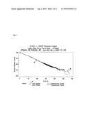 |
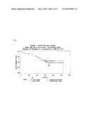 | 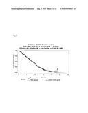 |
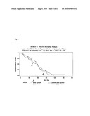 | 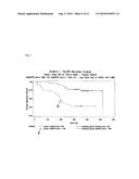 |
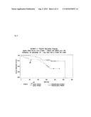 | 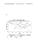 |
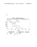 | 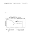 |
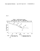 | 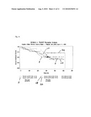 |
 | 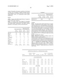 |
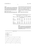 |  |
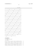 |  |
 |  |
 |  |
 |  |
 |
| Similar patent applications: | |
| Date | Title |
|---|---|
| 2011-06-02 | Gene expression markers for predicting response to chemotherapy |
| 2010-08-05 | Method for determination of onset risk of glaucoma |
| 2011-05-12 | Elimination of immune responses to viral vectors |
| 2011-06-02 | Determination of kir haplotypes associated with disease |
| 2011-06-02 | Microorganisms for the production of 1,4-butanediol, 4-hydroxybutanal, 4-hydroxybutyryl-coa, putrescine and related compounds, and methods related thereto |
| New patent applications from these inventors: | |
| Date | Title |
|---|---|
| 2022-07-28 | Quality control of automated whole-slide analyses |
| 2011-12-22 | Predictive marker for egfr inhibitor treatment |
| 2011-10-06 | Predictive marker for egfr inhibitor treatment |
| 2011-09-22 | Predictive marker for egfr inhibitor treatment |
| 2011-09-01 | Predictive marker for egfr inhibitor treatment |
| Top Inventors for class "Chemistry: molecular biology and microbiology" | |
| Rank | Inventor's name |
|---|---|
| 1 | Marshall Medoff |
| 2 | Anthony P. Burgard |
| 3 | Mark J. Burk |
| 4 | Robin E. Osterhout |
| 5 | Rangarajan Sampath |