Patent application title: APOCRINE CELL LINE
Inventors:
Jason Shaun Burry (Wirral, GB)
Richard Livesey Evans (Wirral, GB)
Mark Harker (Wirral, GB)
George Terence Evelyn Kealey (Buckingham, GB)
Donald Farquhar Mcdonald (London, GB)
IPC8 Class:
USPC Class:
435366
Class name: Animal cell, per se (e.g., cell lines, etc.); composition thereof; process of propagating, maintaining or preserving an animal cell or composition thereof; process of isolating or separating an animal cell or composition thereof; process of preparing a composition containing an animal cell; culture media therefore primate cell, per se human
Publication date: 2010-07-22
Patent application number: 20100184213
Claims:
1. An apocrine cell line exhibiting long term proliferation.
2. A cell line according to claim 1 derived from human apocrine glands
3. A cell line according to claim 1 that exhibits characteristic features of apocrine glands.
4. A cell line according to any preceding claim which is capable of proliferating indefinitely.
5. A cell line having the deposit number ECACC 07021301.
6. A method of obtaining a cultured cell line exhibiting long term proliferation comprising the steps of isolating an apocrine cell from primary tissue, culturing the isolated cell in a first culture medium, removing unattached cells from the first culture medium and transferring said unattached cells to a second culture medium comprising an effective concentration of a phorbol ester, and thereby establishing an apocrine cell line exhibiting long-term proliferation capability.
7. Use of an apocrine cell line according to any of claims 1 to 5 or produced according to claim 6 for diagnostic, therapeutic or cosmetic preparations.
8. Use of an apocrine cell line according to any of claims 1 to 5 or produced according to claim 6 for evaluating in vitro whether a material is a deodorant when topically applied to skin.
9. Use of an apocrine cell line according to any of claims 1 to 5 or produced according to claim 6 for the examination of the physiology or the pathophysiology of human apocrine gland.
10. Use of an apocrine cell line according to any of claims 1 to 5 or produced according to claim 6 for the testing of compounds or agents against diseases.
11. Use of an apocrine cell line according to any of claims 1 to 5 or produced according to claim 6 for the preparation of products derived from said cells.
Description:
[0001]The present invention relates to an apocrine cell line and in
particular to an apocrine cell line exhibiting long-term proliferation.
[0002]In addition to eccrine glands, apocrine glands secrete aqueous fluid containing various lipidic and aminoacid solutes which, when exposed on human skin to skin bacterial populations, are transformed to malodorous compounds, including steroids and short chain fatty acids. In other words, apocrine glands contribute to human sweating. Considerable research continues to be carried out to find means to counteract the generation of malodorous compounds on skin, but this is hampered by the absence of an apocrine cell line that closely mimics the functions of an apocrine gland in situ, i.e. in human skin. The development of the latter would be very valuable because it would offer one or more of the benefits mentioned herein. It would enable a greater number of in vitro, as opposed to in vivo, studies to be carried out. This would (i) assist in identifying the transport mechanisms within the apocrine gland that lead to sweating, (ii) facilitate the screening and identification of substances for reducing the formation of malodorous compounds, and (iii) help determine their effective concentration, i.e. the concentration required to act as a deodorant when applied topically to the body. The existence of a suitable cell line would also provide the opportunity to identify novel glandular functions other then sweating per se.
[0003]In common with attempts to sustain other secretory cells in long-term culture, primary cell lines obtained from apocrine cultures have not been passaged more than a few times before losing morphological, phenotypic and/or functional respects characteristic of an apocrine gland.
[0004]A paper by R Wicher et al in Andrologia 35, pp 342-350 entitled "Establishing of two in vitro models of epithelial cells from the apocrine secreting rat coagulating gland" relates to apocrine cells from rats, and does not disclose a human-derived apocrine cell line exhibiting long term proliferation. Furthermore, the cell markers used therein to demonstrate functionality are not necessarily relevant to the human apocrine model. Moreover, the authors seemingly did not seek to passage their cells after freezing and thawing, and thereby did not demonstrate their long term proliferation capability.
[0005]A paper by Z. Maras et al published in In Vitro Cell. Dev. Biol. Animal Vol 35, November-December (1999), pp 606-611 entitled "Cultivation of Epithelia from the Secretory Coil of the Ovine Apocrine Gland; Evidence of Secretory Cell Function and Ductal Morphogenesis in Vitro" relates to apocrine cells from sheep, and similarly does not disclose a human-derived apocrine cell line exhibiting long term proliferation.
[0006]A paper by Dieter C. Gruenert et al published in In Vitro Cell. Dev. Biol. Vol 26, April (1990), pp 411-416 entitled "Long Term Culture of Normal and Cystic Fibrosois
[0007]Epithelial Cells grown under Serum-free Conditions" relates to eccrine cells instead of apocrine cells and therefore does not disclose a human-derived apocrine cell line exhibiting long term proliferation.
OBJECT OF THE PRESENT INVENTION
[0008]It is an object of the present invention to provide an apocrine cell line that mimics the sweating function of an apocrine gland, which can be maintained in culture through subcultures, and exhibits proliferation in the long term.
BRIEF SUMMARY OF THE PRESENT INVENTION
[0009]According to the present invention there is provided a cultured apocrine cell line exhibiting long term proliferation.
[0010]Proliferation in the present context indicates that the cells retain the capability to divide when cultured in a suitable culture medium, but does not imply that the cell line is immortalised.
[0011]Long term, in the context of the present invention, indicates that cells retain their capability to proliferate after at least 30 sub-cultures (passages). In practice, when such long term proliferation has been achieved by a cell line, it can commonly exhibit proliferation through at least 100 or 1000 passages. Proliferation through such a large number of proliferations is indicative of indefinite proliferation, at least for practical purposes for cultured apocrine cell lines according to the present invention.
[0012]In particular, there is provided a human apocrine cell line ASG5 which has been deposited with the European Collection of Cell Cultures, (ECACC), Porton Down, Salisbury, SP4 0JG, England, under the depository number 07021301 and which exhibits at least long term proliferation.
Detailed Description of Preferred Embodiments of the Invention and Morphology
[0013]This text describes the generation of a long term proliferative apocrine cell line, and subsequently, its characterisation and comparison with primary cultured apocrine cells via electrophysiological, molecular, and biochemical techniques. Particular interest is paid to steroid synthesis and the analysis of odour precursor compounds with express reference to the apocrine cell line ASG5, that has demonstrated at least long term proliferation.
Isolation and Primary Culture of Apocrine Glands
[0014]An apocrine cell line that exhibited long term proliferation capability was obtained by the following method. Whole apocrine glands were isolated by shearing the axillary skin of women aged between 30 and 70, having previously obtained permission from the relevant ethical committee and informed consent of the subjects. Some apocrine sweat glands were fragmented by the shearing process, and cells could be cultured from those fragments. Microscopic observation and Neutral Red uptake experiments showed that the apocrine gland isolated by shearing consisted purely of coil separated from duct. On average between 10 and 100 apocrine coils were isolated from samples of axillary skin of about 50 mm×5 mm.
[0015]The apocrine coils were then incubated using a modified method, proposed by Lee et al in J. Cell Sci. 83:103-118, 1986, in a Williams E medium (herein for short WEM)-containing collagenase type II (at 2 mg ml-1) for 30 minutes at 37° C. in 5% CO2/air of 95% relative humidity. The coils were then washed in enzyme-free WEM supplemented with 10 ng ml-1 epidermal growth factor (EGF), 10 μg ml-1 insulin, 10 ng ml-1 hydrocortisone, 10 μg ml-1 holotransferrin, 1 mM L-glutamine, 100 U ml-1 penicillin and 100 μg ml-1 streptomycin.
[0016]Approximately 15 apocrine glands were plated out per 25 cm2 flasks in 1 ml of Mammary Epithelial Growth Medium (MEGM), supplemented with 10 ng ml-1 EGF, 10 μg ml-1 insulin, 0.5 μg ml-1 hydrocortisone, 30 μg ml-1 bovine pituitary extract, 50 μg ml-1 gentamicin and 50 ng ml-1 amphotericin. After 24 hr incubation in a 95% air/5% CO2 humidified incubator at 37° C., a further 3 ml MEGM was added to each flask and thereafter the medium was changed every 3 days.
Generation and Maintenance of a Long Term Proliferating Apocrine Cell Line (ASG5)--Indicative of an Indefinitely Proliferating Cell Line
Primary Sweat Gland Isolation and Culture
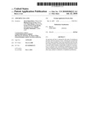
[0017]As indicated before, whole intact human apocrine sweat glands were isolated by shearing. Histological examination of apocrine glands revealed that shearing completely removed the short absorptive duct leaving only the secretory coil portion intact. Isolated glands were plated out in supplemented MEGM. Approximately 50% of glands plated out generated outgrowths, routinely observed after 2-3 days. Outgrowths continued to proliferate for up to 20 days and displayed typical `cobblestone` epithelial cell morphology (as shown in FIG. 1, which show phase contrast micrographs of (A) cells growing from an apocrine coil nine days after explantation, and (B) proliferating apocrine cells). At early stages of culture, before outgrowths were observed, a small number of cells with an elongated fibroblastoid morphology were often observed around explanted glands. However, these cells did not proliferate in MEGM.
[0018]A proliferating apocrine cell line was derived from primary cultures of apocrine secretory coils. After 7 days culture in MEGM, the potent phorbol ester, TPA (phorbol-12-myristate-13 acetate; ex Calbiochem, Nottingham, UK) was included in the medium at a concentration of 250 nM. It is an important feature of the process of obtaining a cell-line exhibiting long term proliferation capability that the culture medium contains an effective concentration of an ester of phorbol, phorbol being alternatively called 1,1a,1b,4,4a,7a,7b,8,9,9a-decahydro-4-a,7b,9,9a-tetrahydroxy-3-(hydroxyme- thyl)-1,1,6,8-tetramethyl-5H-cyclopropa[3,4]benz[1,2-e]azulen-5-one. Commonly, the ester comprises at least aliphatic substituent particularly suitably at the 12 and/or 13 positions, such as a long chain alkyl (C7 to C24) and optionally also a short chain alkyl (C2 to C6).
[0019]Accordingly, the method herein enables the preparation of an apocrine cell line exhibiting indefinite proliferation.
[0020]TPA induced premature multilayering 3-4 days after addition. However, the upper differentiated layers were removed by repeated washing to leave a proliferative monolayer of cells still attached to the flask. Cells of the proliferative layer were passaged into 96 well plates to generate clonal cell lines. Cells were maintained, passaged and continued to grow in MEGM containing TPA for approximately 2 months. After this period cells were cultured in MEGM alone and monitored for continued proliferation. Initially, many subclones continued to grow, but by the end of the next 10 passages, the majority had begun to senesce. One culture, in particular, ASG5, continued to grow and has now been maintained in culture, proliferating over more than 40 passages with no signs of a reduction in growth rate. This is indicative of exhibiting a proliferation capability indefinitely. This culture continues to proliferate in the absence of TPA, can survive cryopreservation and is considered to be stable. By comparison, primary apocrine sweat gland cultures grow for a maximum of 1 month and 4 passages and do not survive cryopreservation. The cell culture passaged herein exhibiting long term proliferation is morphologically similar to primary apocrine cultures, for example peak agonist responses. Such a cell culture is accordingly capable of being used to demonstrate whether and/or the extent to which a substance exhibits deodorising properties in vitro, i.e. is capable of acting as a personal deodorant.
Preparation of ASG5 Apocrine Cells for Electrophysiology
[0021]After 13-18 days, confluent ASG5 cultures were passaged onto Transwell collagen-coated permeable supports (0.33 cm2 area, Costar, cat. no. 3495, High Wycombe, Bucks, UK). This involved incubation in 2% EDTA for 20 minutes, followed by trypsin (0.5%)-EDTA (0.2%) for approximately five minutes. 3×10⁵ cells were transferred onto each Transwell supplemented with WEM. The Transwell supports were suspended in 24-well plates in 0.6 ml of the same medium. For primary cultures, only first passage cells were used in experiments. The medium bathing cultures on Transwells was changed every two days.
Electrophysiology
[0022]Transepithelial resistance (TER) across epithelia on Transwells was monitored using `chopstick` electrodes (WPI, Alresford, Hants, UK) connected to an ohmmeter. The TER was estimated by subtracting the measurement for a moistened filter blank. At peak TER, the Transwell supports were mounted in an Ussing Chamber. Epithelia were bathed in a modified Krebs buffer, oxygenated and maintained at 37° C., pH 7.4, with 95% O2/5% CO2. The buffer consisted of (mM): NaCl, 117; KCl, 4.7; CaCl2, 1.5; MgSO4, 1.2; NaHCO3, 25; and glucose, 11.1. Voltage and current electrodes (calomel and silver/silver chloride respectively, ABB Kent Taylor, Stonehouse, Glos, UK), connected to a voltage/current clamp were extended into the bath via bridges containing agar dissolved in 3 M potassium chloride.
[0023]Voltage electrodes were balanced in the absence of a Transwell and fluid resistance between the voltage electrodes was compensated for by the injection of an arbitrary DC current. After mounting a Transwell the epithelium was short-circuited, the current allowed to stabilise, typically for 15 minutes. Given the rarity of apocrine material, drugs were typically added in succession as disclosed by Brayden et al (1988) J. Physiol, 405: 657-675, Pedersen et al (1992) Exp. Physiol, 77; 863-871, and Shen et al, (1994) Am. J. Physiol, 266: L493-501 and changes in short circuit current (Isc) recorded. The duration of experiments was typically 15-60 min. TER was estimated using Ohm's Law, routinely by clamping the voltage at ±10 mV and measuring the current required to achieve this. Unless otherwise stated all drugs listed in the legend were added to the basolateral side, except for amiloride that was added to the apical side. Amiloride is an inhibitor of the epithelial Na+ channel (ENaC) and the Na+/H+ exchanger.
Modifications of Krebs Buffer
[0024]Chloride substitution experiments were carried out in Krebs buffer in which sodium gluconate was substituted for NaCl, potassium gluconate for KCl and CaSO4 for CaCl2. Glucose-free experiments were carried out with 11.1 mM mannitol as a replacement to maintain osmolality. Experiments in which barium was used as an inhibitor of potassium channels were carried out in the absence of sulphate ions, such that MgCl2 replaced MgSO4.
Basal Transepithelial Properties
[0025]Proliferating apocrine cultures were passaged onto permeable supports and transferred to an Ussing chamber at peak TER. The mean TER for such proliferating cultures was 395±40 Ωcm2 (P<0.001). Resting short circuit current (Isc) was 4.5±0.8 μA cm-2 (P<0.001) for proliferating apocrine cultures. Resting transepithelial potential difference (TEPD) was 1.0 mV for proliferating apocrine cultures, apically negative.
Effects of Ion Transport Inhibitors on Resting Isc Summarised in Table 1 Below.
[0026]Resting Isc in proliferating cultures was sensitive to 10 μM amiloride confirming that sodium reabsorption is a significant component of ion transport in cultured sweat gland epithelia. Apically applied amiloride (10 μM) reduced resting Isc by 4.5±1.0 μA cm-2 in proliferating apocrine cultures (n=20, P<0.001). Dose response curves reveal that the IC50 for amiloride is 2-4 μM and that 10 μM amiloride was a supramaximal dose.
[0027]The effects of furosemide, an inhibitor of NKCC1, the epithelial Na+--K+--Cl.sup.- cotransporter (and hence transepithelial chloride transport) was also examined in apocrine cultures. Basolateral addition of 1 mM furosemide reduced resting Isc in transformed cultures by 0.8±0.30.2 μA (n=20, P<0.001).
TABLE-US-00001 TABLE 1 The effects of amiloride and furosemide on the short circuit current in cultured apocrine (primary and proliferating) and eccrine sweat gland cells. Primary Proliferating Primary Apocrine Apocrine Eccrine Resting Isc 8.4 ± 0.9 4.5 ± 0.8 8.6 ± 1.0 (μA cm-2) (n = 57) (n = 40) (n = 22) ΔIsc (μA cm-2) Amiloride -6.0 ± 0.9** -4.2 ± 1.0 -7.1 ± 0.7 (10 μM) (n = 49) (n = 20) (n = 22) Furosemide -0.5 ± 0.2 -0.8 ± 0.3 nd (1 mM) (n = 13) (n = 20)
The Role of Sodium Reabsorption in Agonist Responses
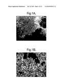
[0028]Primary apocrine and proliferating apocrine cultures were pre-treated with 10 μM amiloride to determine the role of sodium reabsorption in agonist responses. Appended FIG. 2 shows Peak increases in Isc in primary apocrine, eccrine and proliferating apocrine cultures in the presence of amiloride. The agonists were added to the basolateral side of cultures on Transwells. Each bar in the Figure represents a minimum of n=10. Peak increases in short circuit current in the presence of apical amiloride (10 μM) are shown. The agonists were added basolaterally: Carbachol (CCh; 20 μM), Isoprenaline (Iso; 10 μM), L-bradykinin (LBK; 170 nM), Histamine (His; 200 μM) and Adenosine triphosphate (ATP; 100 μM). Significance was tested using unpaired Student's t test and the Wilcoxon tests.
[0029]FIG. 2 demonstrates that amiloride pre-treatment significantly reduced responses to CCh, His and ATP in a long term proliferating apocrine cell line, ASG5, suggesting that sodium reabsorption is a component of these responses.
[0030]FIG. 3 shows peak increases in Isc in primary apocrine, eccrine and proliferating apocrine cultures. Agonists were added to the basolateral side of cultures on Transwells. Peak increases in short circuit current are shown. The agonists were added basolaterally: CCh (20 μM), Iso (10 μM), LBK (170 nM), His (200 μM) and ATP (100 μM).
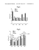
[0031]FIGS. 4A and 4B are representative I' records (traces) on the effects of ion channel inhibitors on the response to carbachol in ASG5 cells. FIG. 4A shows an apical pre-treatment with amiloride (10 μM) and the effects of addition of 1 mM furosemide (Fru) and 50 μM barium chloride (Ba; a maxi-K channel blocker), on the CCh (20 μM) response. FIG. 4B shows an apical pre-treatment with 1 mM furosemide and the effects of amiloride and barium chloride on CCh response.
[0032]The representative trace of proliferating apocrine cultures in FIG. 4A also demonstrated that compared to the observed values for agonist stimulation in FIG. 3, carbachol stimulation is reduced after amiloride pre-treatment.
[0033]In four cases, whole apocrine secretory coils were explanted directly onto permeable supports to determine if the primary culture and passage of apocrine cultures affected their electrophysiological properties. In the cultures examined, basal and agonist stimulated electrophysiological properties were not significantly different to primary cultures, whether in the presence or absence of amiloride (data not shown).
The Role of Transepithelial Chloride Transport in Transient Agonist Responses
[0034]To further characterise the ionic basis of agonist induced responses in primary and proliferating apocrine cultures, the role of transepithelial chloride transport was examined. Under chloride-free conditions, resting I' for primary and proliferating apocrine cultures was 6.0±0.8 μA cm-2 and 3.1±0.6 μA cm-2 respectively. Resting TEPD was -0.9 mV for primary cultures and -1.8 mV for proliferating cultures. Addition of 10 μM amiloride reduced basal Isc by 3.6 μA cm-2 in primary cultures and by 2.5 μA cm-2 in proliferating cultures.
[0035]Responses to carbachol, histamine and ATP were significantly reduced in both primary and proliferating apocrine cultures when compared to identical experiments in the presence of chloride (Table 2) suggesting that chloride transport does play a significant role in mediating agonist responses. Furthermore, FIG. 4B shows a representative trace from proliferating cultures demonstrating that pre-treatment with furosemide reduces the magnitude of response to carbachol compared to the values in FIG. 3. Addition of furosemide after agonist stimulation induced a rapid reduction in responses in both primary and transformed apocrine cultures, demonstrating the role of NKCC1 in regulating chloride uptake. (Table 3).
TABLE-US-00002 TABLE 2 Peak agonist responses of apocrine cultures in chloride-free buffer. Agonist Isc responses μA cm-2 Chloride-free n Control n A Carbachol (20 μM) 0.7 ± 0.6* 5 3.9 ± 0.7 34 Histamine (200 μM) 1.7 ± 0.5** 5 6.4 ± 1.3 14 ATP (100 μM) 2.9 ± 0.9* 9 7.4 ± 1.8 10 B Carbachol (20 μM) 3.0 ± 0.8* 6 4.0 ± 0.7 20 Histamine (200 μM) 3.3 ± 0.8** 6 4.8 ± 1.3 20 ATP (100 μM) 4.0 ± 0.5* 6 6.3 ± 1.8 20 A: Primary apocrine cultures B: proliferating apocrine culture (ASG5).
TABLE-US-00003 TABLE 3 Furosemide inhibition of agonist responses in primary and long term proliferating (AGS5) apocrine cultures. Inhibition by furosemide (90 μM) Δ Isc, μA cm-2 Agonist Primary Proliferating Carbachol (≧20 μM) -2.0 ± 0.5 -1.1 ± 0.4 (n = 4) (n = 10) Isoprenaline (10 μM) 0.8 ± 0.2 -1.2 ± 0.3 (n = 3) (n = 10) Histamine (200 μM) -1.3 ± 0.2 -1.0 ± 0.4 (n = 6) (n = 10) ATP (100 μM) -2.6 ± 0.5 -1.2 ± 0.3 (n = 8) (n = 10)
[0036]Agonist responses were significantly reduced with amiloride pre-treatment when compared to amiloride pre-treatment in chloride containing buffer, but they were not entirely abolished, particularly in the proliferating cultures, suggesting that other ion transport pathways may also play a role in these responses (Table 4).
TABLE-US-00004 TABLE 4 Peak agonist responses of apocrine cultures in chloride-free buffer after amiloride pre-treatment. Agonist Isc responses μA cm-2 Chloride-free n Control N A Carbachol (200 μM) 1.7 ± 1.2** 9 6.3 ± 1.4 11 Isoprenaline (≧1 μM) 0.7 ± 0.3* 15 1.8 ± 0.4 20 LBK (170 nM) 0.6 ± 0.4* 11 2.0 ± 0.5 18 Histamine (200 μM) 2.4 ± 1.4 8 4.2 ± 0.5 19 ATP (100 μM) 4.3 ± 2.4 6 4.9 ± 0.7 24 B Carbachol (200 μM) 1.3 ± 0.3 6 2.4 ± 0.6 20 Isoprenaline (≧1 μM) 0.8 ± 0.2 6 1.8 ± 0.4 20 LBK (170 nM) 0.9 ± 0.3 6 1.6 ± 0.3 20 Histamine (200 μM 1.0 ± 0.2 6 1.9 ± 0.4 20 ATP (100 μM) 0.8 ± 0.2 6 1.8 ± 0.3 20 A: Primary apocrine cultures B: proliferating apocrine cultures.
[0037]Taken together these results indicate that the proliferating cell line continued to exhibit the same characteristics as the primary cultures.
Steroidogenesis in a Long Term Proliferating Apocrine Gland Cell Line
[0038]Human skin and its associated appendages are capable of a variety of metabolic functions, including those involved in the metabolism and synthesis of hormones. Androgens (sex steroid hormones), are synthesized from the parental precursor cholesterol in a biosynthetic pathway requiring numerous enzymatic conversions. As apocrine glands become functional during puberty, it is likely that androgens play some role in modulating gland activity. This is supported further by the fact that the androgen receptor has been detected in the secretory cells of apocrine glands residing in axillary skin (Beier et al, (2005) Histochem. Cell Biol., 123: 61-65). Since the local or intracrine formation of steroids potentially plays an important role in mediating cell function (Labrie, F. et al, (1991), Mol. Cell. Endocrinol., 78: 113-118), the expression of transcripts responsible was investigated for the formation of active androgens and estrogens in the indefinitely proliferating apocrine cell line herein, as well as their associated receptors.
Materials & Methods in the Investigation
Chemicals
[0039]Taq DNA polymerase and molecular marker III were purchased from Roche, Mannheim Germany. Taq DNA polymerase was used according to the suppliers' recommendations. All other reagents were obtained from Sigma-Aldrich, Gillingham, UK.
Strains, Plasmids, Media and Culture Conditions
[0040]E. coli strain TOP10 (Invitrogen, Carlsbad, Calif., USA) was used as the host strain in all cloning procedures. Pre-digested vector pCR®4-TOPO® containing poly-T overhangs was obtained from Invitrogen and used according to the suppliers instructions. Bacteria were cultivated in LB medium (10 g/l tryptone, 5 g/l yeast extract, 5 g/l NaCl) supplemented with the appropriate selection pressure (kanamycin 50 μg/ml) on a rotary shaker (250 rpm) at 37° C.
Cell Material
[0041]Long term proliferating apocrine gland cells indicative of indefinite proliferation, were grown as described hereinbefore. For cloning purposes cells were harvested after 10 days growth at 37° C.
Oligonucleotide Synthesis and Sequencing
[0042]All oligonucleotides used were synthesized by Sigma-Genosys Ltd, Pampisford, UK. Sequencing was performed using an ABI Prism 3100 Genetic Analyser using the universal M13 forward and reverse primers.
Isolation of Plasmid DNA
[0043]The Qiagen (Crawley, UK) mini-prep kit was used to obtain plasmid DNA for cloning and sequencing.
Cloning
[0044]Fragments of transcripts involved in steroid androgen metabolism were cloned from cDNA derived from indefinitely proliferating apocrine gland cells. RNA was isolated from the cells using the RNAqueous®-4 PCR kit from Ambion, Huntingdon, UK. First strand cDNA synthesis was performed using the Ambion RETROscript® kit using oligo(dt). The PCR reaction programme used for amplification was: 1 cycle (120 s @ 95° C.), 30 cycles (30 s @ 95° C., 30 s @ 60° C., 45 s or 60 s @ 72° C.), 1 cycle (7 min @ 72° C.) with Taq DNA polymerase). The primers used to clone fragments from each transcript are outlined in Table 5.
TABLE-US-00005 TABLE 5 Primers used to clone gene fragments involved in androgen metabolism. Ex- pected size Primer sequence Gene (bp) Forward 5'- Steroid 392 CTTCACCCCCAACTTCAACCCCG-3' sulphatase Reverse 5'- CCA CAT GCG TCT GTC TGG TCC CC-3' Forward 5'- 3β-hydroxy-Δ- 813 GCCAGTCTTCATCTACACCAGTAG C-3' 5-steroid Reverse 5'- dehydrogenase CTCTGTCATCCTTAAATCACTGAGTC-3' 1 Forward 5'- Hydroxysteroid 436 CCTCATCCATTGTAACATCACCTCC-3' 17β- Reverse 5'- dehydrogenase CTCACCGCCTGGCTACCTGACC-3' 3 Forward 5'- Hydroxysteroid 990 GACAAGTGACAGGGAATGGATTCC-3' 17β- Reverse 5'- dehydrogenase CAGGGCTTCTGGTAGACATCAGG-3' 5 Forward 5'- Hydroxysteroid 722 GAAGTGTGAGTGCGCGAAGATGCG-3' 17β- Reverse 5'- dehydrogenase GTGCTGGAATTATAGGCATGAGCCAC-3' 7 Forward 5'- 5α Reductase 773 CCAGCCCTGGCGATGGCAACG-3' Reverse 5'- GAAATTCTGACCTGTTACACAGTAGG-3' Forward 5'- Aromatase 447 GCCTGTCGTGGACTTGGTCATGCG-3' Reverse 5'- GAGTAGGTACTGACCAGCCTTCTC-3' Forward 5'- Estrogen 564 CTTGGAGAGCTGTTGGATGGAGGTG-3' receptor β Reverse 5'- CAGGGCCAGGCGTCACTGAGA C-3' Forward 5'- Androgen 564 CTGGATGGGGCTCATGGTGTTTG-3' receptor Reverse 5'- GTTTCCAATGCTTCACTGGGTGTGG-3'
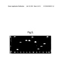
[0045]All the gene fragments that primer pairs were designed for, as outlined in table 5, could be detected. Appended FIG. 5 displays a 1.2% agarose gel of all the PCR products produced.
[0046]FIG. 5 shows the expression of gene fragments:
1. Steroid sulphatase; 2. 3βhydroxy-Δ-5-steroid dehydrogenase 1; 3. Hydroxysteroid 17β-dehydrogenase 3; 4. Hydroxysteroid 17β-dehydrogenase 5; 5. Hydroxysteroid 17β-dehydrogenase 7; 6. 5α Reductase I; 7. CYP19A1 Aromatase; 8. Androgen receptor; 9. Estrogen receptor β; M. DNA Marker III.
[0047]These PCR products were then cloned into the TOPO vector ready for sequencing. Sequencing was performed on each PCR product to confirm the identity of each cloned fragment. The results of the gene sequencing are given below.
TABLE-US-00006 Steroid sulphatase fragment sequence TCCCTTCTTCACCCCCAACTTCAACCCCGTGGGTTCCAACGGATGCTTTG CCACACACGTGTGCTTCTGTTTCGGGAGTTATGTCACCCATCACGACCCA CCTTTACTCTTTGATATTTCCAAAGATCCCAGAGAGAGAAACCCACTAAC TCCAGCATCCGAGCCCCGGTTTTATGAAATCCTCAAAGTCATGCAGGAAG CTGCGGACAGACACACCCAGACCCTGCCAGAGGTGCCCGATCAGTTTTCA TGGAACAACTTTCTTTGGAAGCCCTGGCTTCAGCTGTGCTGTCCTTCCAC CGGCCTGTCTTGCCAGTGTGATAGAGAAAAACAGGATAAGAGACTGAGCC GCTAGCAGCGCCTGGGGACCAGACAGACGCATGTGGAAGGGC 3β-hydroxy-Δ-5-steroid dehydrogenase 1 fragment sequence GCCCTTCTCTGTCATCCTTAAATCACTGAGTCTTGGACTTCACGGTCTCC TTGTGCCAGTCCACAAGGGAAACCCACTCCACGGTTCTAACAGACTCAGG TACATTCTTCTTGGCTTCCTCCCAGCTGTAAAGTGGCTTATATGCCAGAT CTTGCTGAGCCTTCTTGTAAGAGAAGGTGAACACGCTATTTGACAATGTC ACTCTGTGGTGGTTGAAGGGTGTCGATAGGTGTAAATTGGCCTCAGCAGG AAGCTCACTATTTCCAGCAGGAAGCCAATCCAGTACATCAGAGATAAAGG AAGGCTCCATCTAGAATCAAGGCAGAGGCCGAACTCTTTGCTCAGGATGT AATTAAGGTTATCATAGCTTTGGTGAGGCATGTCATCTGAGATGTAGTAG AACTGTCCTCGGACACTTGGGGCCTTCTTGGGCTCCCGCAGGGCCTCAAG GCCAGAATGTGGGCCCAGGCCATGTTTCCAATATAGACTGGGTTGACTCT GGAGAACTTGCTGAAACTTGACAGGATCCCATGGTTGTTTAGGGCCTCAT TTATACCGGCAGAAAGGATTGGGCTTCCTTCCCCATAGATAAACATTGGT CTTAAGGCACAAGTGTACAAGGTGCCACCGTTTTTCAGAGTCCACCCATT AGCCGCCAGCACAGCCTTCTCAGCAAGCTTTTTGCTGTGTGGGTATGGAG CGTACCATGTGTTTTCCAGAGGCTCTTCTTCGTGACTGTTCTGGATGATT TCCTTGTAGGAATTGGGCCCGGCTACCTCTGGGCTACTGGTGTAGATGAA GACTGGCAAGGGC Hydroxysteroid 17β-dehydrogenase 3 fragment sequence GCCCTTCTCACCCNCTGGCTACCTGACCTTGGTGTTGAGCTTCAGGTATG CCACATAGTGTGTCAGGAGCAGCCTTTGGAAGGCACCGCTGTAGAAGGCC CAGGCCGGGATCAGGCTCAGAAAGCCCGCCAAGATTTCATGGGCAAGGCA GCCGCAGGTTTCACCTCCAATTGTGACATAATTCAATGACTCTTTGACAA ACTCATCAGCAGTCTTGGTTATCACATTTGTATTTAGATACTTTGTCATT GCAGTCGGGACAGCATATGGGGTCAGCACCTGGATGATGACTTCTTTTGC TTTATATTCCTCTTGCAGGGCCTTGGAAAATGCGCACACAAACGCCTTGG AAGCTGAGTACATGGAGTAGAGAGGCCAAGGAAACAGGGCTATCCCAGAA GAAATGTTCAGGATGAGACCTTTCTGCCTTGATTCCATATGTTTCAAAAT TAGCTGTGTCATCTTGACTACGGAGGTGATGTTACAATGGATGAGGAAGG GC Hydroxysteroid 17β-dehydrogenase 5 fragment sequence GACAAGTGACAGGGAATGGATTCCAAACACCAGTGTGTAAAGCTAAAGGA TGGCCACTTCATGCCTGTATTGGGATTTGGCACCTATGCACCTCCAGAGG TTCCAAGAAGTAAAGCTTTGGAGGTCACAAAATTAGCAATAGAAGCTGGG TTCCGCCATATAGATTCTGCTCATTTATACAATAATGAGGAGCAGGTTGG ACTGGCCATCCGAAGCAAGATTGCAGATGGCAGTGTGAAGGGAGAAGACA TATTCTACACTTCAAAGCTTTGGTCCACTTTTCATCGACCAGAGTTGGTC CGACCAGCCTTGGAAAACTCACTGAAAAAAGCTCAATTGGACTATGTTGA CCTCTATCTTATTCATTCTCCAATGTCTCTAAAGCCAGGTGAGGAACTTT CACCAACAGATGAAAATGGAAAAGTAATATTTGACATAGTGGATCTCTGT ACCACCTGGGAGGCCATGGAGAAGTGTAAGGATGCAGGATTGGCCAAGTC CATTGGGGTGTCAAACTTCAACCGCAGGCAGCTGGAGATGATCCTCAACA AGCCAGGACTCAAGTACAAGCCTGTCTGCAACCAGGTAGAATGTCATCCG TATTTCAACCGGAGTAAATTGCTAGATTTCTGCAAGTCGAAAGATATTGT TCTGGTTGCCTATAGTGCTCTGGGATCTCAACGAGACAAACGATGGGTGG ACCCGAACTCCCCGGTGCTCTTGGAGGACCCAGTCCTTTGTGCCTTGGCA AAAAAGCACAAGCGAACCCCAGCCCTGATTGCCCTGCGCTACCAGCTGCA GCGTGGGGTTGTGGTCCTGGCCAAGAGCTACAATGAGCAGCGCATCAGAC AGAACGTGCAGGTTTTTGAGTTCCAGTTGACTGCAGAGGACATGAAAGCC ATAGATGGCCTAGACAGAAATCTCCACTATTTTAACAGTGATAGTTTTGC TAGCCACCCTAATTATCCATATTCAGATGAATATTAACATGGAGAGCTTT GCCTGATGTCTACCAGAAGCCCTG Hydroxysteroid 17β-dehydrogenase 7 fragment sequence GCCCTTGAAGTGTTAGTGCGCGAAGATGCGAAAGGTGGTTTTGATCACCG GGGCTAGCAGTGGCATTGGCCTGGCCCTCTGCAAGCGGCTGCTGGCGGAA GATGATGAGCTTCATCTGTGTTTGGCGTGCAGGAACATGAGCAAGGCAGA AGCTGTCTGTGCTGCTCTGCTGGCCTCTCACCCCACTGCTGAGGTCACCA TTGTCCAGGTGGATGTCAGCAACCTGCAGTCGGTCTTCCGGGCCTCCAAG GAACTTAAGCAAAGGTTTCAGAGATTAGACTGTATATATCTAAATGCTGG GATCATGCCTAATCCACAACTAAATATCAAAGCACTTTTCTTTGGCCTCT TTTCAAGAAAAGTGATTCATATGTTCTCCACAGCTGAAGGCCTGCTGACC CAGGGTGATAAGATCACTGCTGATGGACTTCAGGAGGTGTTTGAGACCAA TGTCTTTGGCCATTTTATCCTGATTCGGGAACTGGAGCCTCTCCTCTGTC ACAGTGACAATCCATCTCAGCTCATCTGGACATCATCTCGCAGTGCAAGG AAATCTAATTTCAGCCTCGAGGACTTCCAGCACAGCAAAGGCAAGGAACC CTACAGCTCTTCCAAATATGCCACTGACCTTTTGAGTGTGGCTTTGAACA GGAACTTCAACCAGCAGGGTCTCTATTCCAATGTGGCCTGTCCAGGTACA GCATTGACCAATTTGACATATGGAATTCTGCCTCCGTTTATATGGACGCT GTTGATGCCGGCAATATTGCTACTTCGCTTTTTTGCAAATGCATTCACTT TGACACCATATAATGGAACAGAAGCTCTGGTATGGCTTTTCCACCAAAAG CCTGAATCTCTCAATCCTCTGATCAAATATCTGAGTGCCACCACTGGCTT TGGAAGAAATTACATTATGACCCAGAAGATGGACCTAGATGAAGACACTG CTGAAAAATTTTATCAAAAGTTACTGGAACTGGAAAAGCACATTAGGGTC ACTATTCAAAAAACAGATAATCAGGCCAGGCTCAGTGGCTCATGCCTATA ATTCCAGCAC 5α Reductase I fragment sequence GCCCTTCCAGCCCTGGCGATGGCAACGGCGACGGGGGTGGCGGAGGAGCG CCTGCTGGCCGCGCTCGCCTACCTGCAGTGCGCCGTGGGCTGCGCGGTCT TCGCGCGCAATCGTCAGACGAACTCAGTGTACGGCCGCCCCGCGTCTCCG CAGCGCGCCCAACTGCATCCTCCTGGCCATGTTCCTCGTCCACTACGGGC ATCGGTGCTTAATTTACCCATTTCTGATGCGAGGAGGAAAGCCTATGCCA CTGTTGGCGTGTACAATGGCGATTATGTTCTGTACCTGTAACGGCTATTT GCAAAGCAGATACTTGAGCCATTGTGCAGTGTATGCTGATGACTGGGTAA CAGATCCCCGTTTTCTAATAGGTTTTGGCTTGTGGTTAACGGGCATGTTG ATAAACATCCATTCAGATCATATCCTAAGGAATCTCAGAAAACCAGGAGA TACTGGATACAAAATACCAAGGGGAGGCTTATTTGAATACGTAACTGCAG CCAACTATTTTGGAGAAATCATGGAGTGGTGTGGCTATGCCCTGGCCAGC TGGTCTGTCCAAGGCGCGGCTTTTGCTTTCTTCACGTTTTGTTTTTTATC TGGTAGAGCAAAAGAGCATCATGAGTGGTACCTCCGGAAATTTGAAGAGT ATCCAAAGTTCAGAAAAATTATAATTCCATTTTTGTTTTAAGTGCGTTTT TCATGAAATTATCTTCAACTTGAAGCTTTCCAATGGCGCTTCTCTATGGA CTTTGTAAATAAGTTATATCTTTGTAATTTTCCTGCTACTTTATCATTTT CAAGATGTCCTCTAGGAATTTTTTTTCTAGTAATTTTGCAATCTACCTAA TAAGTACCTAAATACGCTGAAATGGAGGTTGAATATCCTACTGTGTAACA GGTCAGAATTNCANGGGC CYP19A1 Aromatase fragment sequence GCCCTTGAGTAGNTACTGACCAGCCTTCTCTAGTGTTCCAGACACCTGTC TGAGTTTCTTGGGGTAAAGATCATTTCCAGCATGTTTTTAGTCTCATCTG GGTGCAAGGACAAGTCGTGTATCTTCTGTATGCTCTCAACACACTGTCCT TGCAATGTCTTCACGTGGAATCGTCTCAGAAGTGTAACGAGGATGGCTTT CATCATCACCATGGCGATGTACTTTCCTGCACAGCCACGGGGCCCAAAGC CAAATGGCTGAAAGTACCTATAAGGAACATTCTTTGCAAAATTTTCAAGA GTAAATTCATTGGGTTTGGGGAAAAACTCGAGTCTGTGCATCCTTCCAAT ATTCAGGATAATGTTTGTCCCCTTTTTCACTGGGTAGCCATCGATTACAT CATCTTCTAAGGCTTTGCGCATGACCAAGTCCACGACAGGCAAGGGC Androgen receptor fragment sequence GCCCTTGTTTCCNTGCTTCACTGGGTGTGGAAATAGATGGGCTTGACTTT CCCAGAAAGGATCTTGGGCACTTGCACAGAGATGATCTCTGCCATCATTT CCGGAAAGTCCACGCTCACCATGTGTGACTTGATTAGCAGGTCAAAAGTG AACTGATGCAGCTCTCTCGCAATAGGCTGCACGGAGTCCAGGAGCTTGGT GAGCTGGTAGAAGCGTCTTGAGCAGGATGTGGGATTTTTTCTTTTGCATG CAATGATACGATCGAGTTCCTTGATGTAGTTCATTCGAAGTTCATCAAAG AATTTTTGATTTTTCAGCCCATCCACTGGAATAATGCTGAAGAGTAGCAG TGCTTTCATGCACAGGAATTCCTGGGGGGTGATTTGGAGCCATCCAAACT CTTGAGAGAGGTGCCTCATTCGGACACACTGGCTGTACATCCGGGACTTG TGCATGCGGTACTCATTGAAAACCAGATCAGGGGCGAAGTAGAGCATCCT GGAGTTGACATTGGTGAAGGATCGCCAGCCCATGGCAAACACCATGAGCC CCATCCAGAAGGGC
Estrogen receptor β fragment sequence GCCCTTCAGGGNCNNGGCGTCACTGAGACTGTGGGTTCTGGGAGCCCTCT TTGCTTTTACTGTCCTCTGCCGGGCTGCACTCGGACCCCGTGATGGAGGA CTTGCACCCGCGAAGCACGTGGGCATTCAGCATCTCCAGCAGCAGGTCAT ACACTGGGACCACATTTTTGCACTTCATGTTGAGCAGATGTTCCATGCCC TTGTTACTCGCATGCCTGACGTGGGACAGGAGCATCAGGAGGTTAGCCAG GCGCATGGATTGCTGCTGGGAGGAGATGCCGCTCTTGGCAATCACCCAAA CCAAAGCATCGGTCACGGCGTTCAGCAAGTGAGCCAGCTTCCGGCTGCTG TCAGCATCCTGGGTCGCTGTGACCAGAGGGTACATACTGGAATTGAGCAG GATCATGGCCTTGACACAGAGATATTCTTTGTGTTGGAGTTTTAACTCTC GAAACCTTGAAGTAGTTGCCAGGAGCATGTCAAAGATTTCCAGAATTCCT TCTACGCATTTCCCCTCATCCGTCTGTAATCCCAACAATTTGGGAGGCTG AGGCTGGGGGATCACTTGAAGTCAGGACCTCGAGACCAGCTTGGCCAACA TGGTAAAACCCCGTCTCCACTGTCCAGAACAAGATCTGGAGCAAAGATGA GCCTGCCGGGGTGGTCAATTGAGCGCCACATCAGCCCCATCATTAACACC TCCATCCAACAGCTCTCCAAGAAGGGC
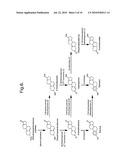
[0048]These results clearly demonstrate that the indefinitely proliferating apocrine gland cell line is expressing numerous genes required for androgen and estrogen synthesis. Appended FIG. 6 outlines the steroid synthesis pathway that can be elucidated given the genes that have shown to be expressed in the indefinitely proliferating apocrine gland cell line. In addition the cells also express the necessary receptors required to mediate the intracellular response to the production of these androgens and estrogens i.e. the androgen and estrogen β receptor. Interestingly, no transcript for the expression of the estrogen α receptor could be detected (data not shown). This is in agreement with a previous study that demonstrated that the estrogen β receptor could be detected in apocrine gland secretory cells in axillary skin sections but not the estrogen a receptor, using immunohistochemical staining for both receptor types (Beier, K. et al (2005) Histochem. Cell Biol., 123: 61-65). The same study also demonstrated the presence of the androgen receptor in apocrine gland secretory cells.
[0049]The data in appended FIG. 6 demonstrates that the indefinitely proliferating apocrine cell line retains many of the steroidogenic features of secretory apocrine gland cells observed in-vivo and primary culture. This cell line provides an excellent tool for studying steroid metabolism in apocrine glands and the role of steroids in regulating gland activity. Additionally, the cell line will serve as an invaluable tool for evaluating the efficacy of compounds in regulating gland activity via interference of the steroidogenic pathways in these cells.
Cell Markers of Apocrine Secretory Activity in the Indefinitely Proliferating Apocrine Gland Cell Line.
[0050]The major function of the apocrine sweat gland is to produce a lipid rich secretion which is delivered to the skin surface via the canal of the hair follicle in direct response to emotional stimuli. The secretion is non-odorous, but undergoes bacterial decomposition which results in the generation of axillary malodours. The secretory function of the apocrine glands is associated with a number of proteins involved in these key processes.
[0051]The ABCC11 protein belongs to the family of ATP-binding cassette transporters involved in the efflux of purine and pyrimidine nucleotide analogs such as cAMP and cGMP (Guo et al., (2003) J. Biol. Chem., 278: 29509-29514). This protein has been implicated in the secretion of earwax in the ceruminous apocrine glands of humans (Yoshiura et al., (2006) Nat. Gen., 38: 324-330). Human earwax normally consists of dry and wet types. Dry earwax is frequent in East Asians, whereas wet earwax is common in other populations. A SNP, 538GA in the ABCC11 gene is considered to be responsible for determination of earwax type. Cells with the A allele show a lower excretory activity for cGMP than those with the G allele. The A allele frequency shows a global North-South and East-West downward geographical gradient. Worldwide, it is highest in Chinese and Koreans where a dry earwax-type is retained amongst the various ethnic populations in these regions. Increased levels of axillary odour are associated with wet-type earwax, which is considered to be a direct consequence of axillary apocrine gland function.
[0052]Another protein which has been shown to play an important role in apocrine gland secretion is apolipoprotein D by Spielman, A. I. (1995) Experientia, 50: 40-47. This protein has been shown to act as a carrier vehicle for the abundant odour molecule E-3-methyl-2-hexanoic acid (3M2H). Studies have demonstrated that in apocrine secretions 3M2H is carried to the skin surface bound by two proteins, apocrine secretion odour-binding proteins 1 and 2 (ASOB1 and ASOB2). The ASOB2 protein was subsequently identified as apolipoprotein D (apoD), a known member of the α2μ-microglobulin superfamily of carrier proteins also known as lipocalins (Zeng et al., (1996), 93: 6626-6630). Immunoreactivity for apoD has been localised to the apocrine glands in axillary tissue sections (Spielman et al., (1998) 134, 813-818) indicating that at least one of the glycoprotein carriers for 3M2H is localized in the apocrine glands.
[0053]Gross cystic disease fluid is a pathologic secretion from breast composed of several glycoproteins, including GCDFP-15. GCDFP-15 has been localized in the apocrine metaplastic epithelium lining breast cysts and in apocrine glands of the axilla, vulva, eyelid, and ear canal (Mazoujian et al., 1983 Am. J. Pathol., 110: 105-112). GCDFP-15 is identical to the Gp17/secretory actin binding protein (SABP/extra-parotid glycoprotein (EP-GP) which has been identified in the seminal vesicles, the salivary glands, and the sweat glands (Autiero et al., (1991); Exp. Cell Res., 197: 268-271), this protein is also identical to the prolactin inducible protein (PIP) (Murphy et al., (1987); J. Biol. Chem., 262: 15236-15241). GCDFP-15 is therefore a specific tissue marker of apocrine epithelium.
[0054]Zinc-α-glycoprotein (ZAG) is a 43 kDa soluble glycoprotein first isolated from human plasma (Burgi et al., (1961); J. Biol Chem; 236, 1066-1074) and subsequently found in secretory epithelia cells of liver, breast, the gastrointestinal tract, and sweat glands (Tada et al., (1991) J. Histochem Cytochem, 39, 1221-1226). ZAG is over-expressed in certain malignant tumours such that it may serve as a cancer maker (Diez-Itza et al., (1993), Eur. J. Cancer A 29, 1256-1260; Hale et al., (2001), Cancer Res., 7, 846-853). The biological functions of ZAG are largely unknown, but it has been shown to act as a lipid mobilizing factor (Todorov et al., (1998), Cancer Res., 58, 2353-2358) and a novel adipokine, which may be involved in the local regulation of adipose tissue function (Bao, et al., (2005) FEBS Letters, 579, 41-47).
[0055]As ABCC11, ApoD, GCDFP-15 and Zinc-α-glycoprotein play important roles in the secretion and transport processes of apocrine glands, we investigated the expression of transcripts responsible for the translation of these proteins in the indefinitely proliferating apocrine cell line.
[0056]The primers used to clone the gene transcript fragments are displayed in Table 6.
TABLE-US-00007 TABLE 6 Primers used to clone gene fragments involved in apocrine secretory processes. Expected size Primer sequence Gene (bp) Forward 5'- ABCC11 254 CACCATCCCTCCACTGTCAGTCC-3' Reverse 5'- CAACATTCCCCAACTGCTCTTCTG-3' Forward 5'- Apolipoprotein 583 CGGTGCGGCAGAGGGACAAG-3' D Reverse 5'- GGGGAAAGCGAAGCAGAAGTAAC-3' Forward 5'- GCDFP-15 340 CCAGCCCTGCCACCCTGCTCC-3' Reverse 5'- GCAGATGCCTAATTCCCGAATAAC-3' Forward 5'- Zinc-α- 518 GGTGGAAGGAATGGAGGATTGG-3' glycoprotein Reverse 5'- GTGGCAGGAGTAGGGGGCTG-3'
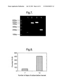
[0057]All the gene fragments that primer pairs were designed for, as outlined in table 6, could be detected. Appended FIG. 7 displays a 1.2% agarose gel of all the PCR products produced.
[0058]Appended FIG. 7. Agarose gel displaying the apocrine marker discloses: The following products are displayed in lanes 1 to 4 respectively. [0059]1. ABCC11 transporter; [0060]2. Apolipoprotein D; [0061]3. Zinc-α-glycoprotein; [0062]4. GCDFP-15
[0063]These PCR products were then cloned into the TOPO vector ready for sequencing. Sequencing was performed on each PCR product to confirm the identity of each cloned fragment. The results of the gene sequencing are given below.
TABLE-US-00008 ABCC11 fragment sequence CACCATCCCTCCACTGTCAGTCCATGATGCCTCAGACAAAAATGTCCAAA GGCTTCACCGTCTTTGGGAAGAAGAAGTCTCAAGGCGAGGGATTGAAAAA GCTTCAGTGCTTCTGGTGATGCTGAGGTTCCAGAGAACAAGGTTGATTTT CGATGCACTTCTGGGCATCTGCTTCTGCATTGCCAGTGTACTCGGGCCAA TATTGATTATACCAAAGATCCTGGAATATTCAGAAGAGCAGTTGGGGAAT GTTG Apolipoprotein D fragment sequence CGGTGCGGCAGAGGGACAAGCATTTCATCTTGGGAAGTGCCCCAATCCTC CGGTGCAGGAGAATTTTGACGTGAATAAGTATCTCGGAAGATGGTACGAA ATTGAGAAGATCCCAACAACCTTTGAGAATGGACGCTGCATCCAGGCCAA CTACTCACTAATGGAAAACGGAAAGATCAAAGTGTTAAACCAGGAGTTGA GAGCTGATGGAACTGTGAATCAAATCGAAGGTGAAGCCACCCCAGTTAAC CTCACAGAGCCTGCCAAGCTGGAAGTTAAGTTTTCCTGGTTTATGCCATC GGCACCGTACTGGATCCTGGCCACCGACTATGAGAACTATGCCCTCGTGT ATTCCTGTACCTGCATCATCCAACTTTTTCACGTGGATTTTGCTTGGATC TTGGCAAGAAACCCTAATCTCCCTCCAGAAACAGTGGACTCTCTAAAAAA TATCCTGACTTCTAATAACATTGATGTCAAGAAAATGACGGTCACAGACC AGGTGAACTGCCCCAAGCTCTCGTAACCAGGTTCTACAGGGAGGCTGCAC CCACTCCATGTTACTTCTGCTTCGCTTTCCCC GCDFP-15 fragment sequence GCCCTTCCAGCCCTGCCACCCTGCTCCTGGTTCTCTGCCTGCAGTTGGGG GCCAACAAAGCTCAGGACAACACTCGGAAGATCATAATAAAGAATTTTGA CATTCCCAAGTCAGTACGTCCAAATGACGAAGTCACTGCAGTGCTTGCAG TTCAAACAGAATTGAAAGAATGCATGGTGGTTAAAACTTACCTCATTAGC AGCATCCCTCTACAAGGTGCATTTAACTATAAGTATACTGCCTGCCTATG TGACGACAATCCAAAAACCTTCTACTGGGACTTTTACACCAACAGAACTG TGCAAATTGCAGCCGTCGTTGATGTTATTCGGGAATTAGGCATCTGCAAG GGC Zinc-α-glycoprotein GCCCTTGGTGGAAGGAATGGAGGATTGGAAGCAGGACAGCCAACTTCAGA AGGCCAGGGNAGGACATCTTTATGGAGACCCTGAAAGACATTGTGGAGTA TTACAACGACAGTAACGGGTCTCACGTATTGCAGGGAAGGTTTGGTTGTG AGATCGAGAATAACAGAAGCAGCGGAGCATTCTGGAAATATTACTATGAT GGAAAGGACTACATTGAATTCAACAAAGAAATCCCAGCCTGGGTCCCCTT CGACCCAGCAGCCCAGATAACCAAGCAGAAGTGGGAGGCAGAACCAGTCT ATGTGCAGCGGGCCAAGGCTTACCTGGAGGAGGAGTGCCCTGCGACTCTG CGGAAATACCTGAAATACAGCAAAAATATCCTGGACCGGCAAGATCCTCC CTCTGTGGTGGTCACCAGCCACCAGGCCCCAGGAGAAAAGAAGAAACTGA AGTGCCTGGCCTACGACTTCTACCCAGGGAAAATTGGTGTGCACTGGACT CGGGCCGGCGAGGTGCAGGAGCCTGAGTTACGGGGAGATGTTCTTCACAA TGGAAATGGCACTTACCAGTCCTGGGTGGTGGTGGCAGTGCCCCCGCAGG ACACAGCCCCCTACTCCTGCCACAAGGGC
[0064]The indefinitely proliferating apocrine cell line expresses the ABCC11 gene, which is associated with cellular secretory processes. The sequencing data confirmed the identity of the ABCC11 transporter gene. The genotype of the gene in this cell line is G (538G→A highlighted and underlined) and is associated with wet-earwax type and increased levels of axillary odour. The apolipoprotein D carrier gene is also expressed, indicating that the necessary machinery for the transport of odorants is also being manufactured in the cell line. The data provides further evidence of the apocrine phenotype of this cell line and its usefulness in providing a robust tool for the investigation of apocrine gland biology.
Cell Chemical Composition--Materials and Methods
[0065]Indefinitely proliferating apocrine cells (passage 44) were harvested at 7 and 14 days, respectively. The cells were centrifuged at 1000 rpm for 5 mins washed once in PBS re-centrifuged and stored at -80° C. ready for analysis.
3M2H Glutamine Conjugate Analysis
[0066]3-Methyl-2-hexanoic acid glutamine conjugate was extracted from the frozen cell pellets using 10 μl (N-methyl-N-tert-butyldimethylsilyl) trifluoroacetamide (MTBSTFA), 1% tert-butyldimethylsilane (TBDMS) and 10 μl pyridine and 1 μl triethylamine. Derivatisation was carried out in the reagent on the samples. The mixture was maintained at 70° C. for 1 hour. The 3M2H glutamine derivatives were analysed by gas chromatography using an Agilent Technologies 6890/5973 gas chromatography/mass selective data system and an Agilent HP5-MS column (30 m×0.25 mm id×0.25 μm film) with selected ion monitoring, id indicating internal diameter. The temperature programme used was 70° C. to 270° C. at 10° C./min. Peak areas were calculated automatically using the in-built software. Structures were identified by mass spectrometry and retention times to known standards.
Cholesterol and Squalene Analysis
[0067]Cholesterol and squalene were extracted from the frozen cell pellets using 20 μl of chloroform. This extract was then injected directly into the GC (details as above) and analysed under full scan using an Agilent HP5-MS column (30 m×0.25 mm id×0.25 μm film). Oven programme 70° C. to 270° C. at 10° C./min. Structures were identified by mass spectrometry and retention times to known standards.
Short Chain Fatty Acids
[0068]Short chain fatty acids were extracted from the frozen cell pellets using 20 μl of chloroform. This extract was then injected directly into the GC (details as above) and analysed with selected ion monitoring using an HP-Innowax column (30 m×0.25 mm id×0.25 μm film). Oven programme 70° C. to 240° C. at 5° C./min. Structures were identified by mass spectrometry and retention times to known standards.
[0069]The relative levels of the 3M2H glutamine conjugate are shown in appended FIG. 8.
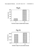
[0070]The relative levels of cholesterol in apocrine cells harvested after 7 and 14 days in culture are shown in appended FIG. 9.
[0071]The relative levels of squalene in apocrine cells harvested after 7 and 14 days in culture are shown in appended FIG. 10.
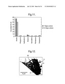
[0072]The relative levels of short chain fatty acids are shown in appended FIG. 11.
[0073]C16/18 fatty acids were also detected in the 14 day sample. The data summarised herein showing the presence of various volatile fatty acids demonstrates further that the indefinitely proliferating cell line mimicked successfully the functions of an apocrine gland.
Inhibitor Studies
[0074]The production of malodour precursor compounds and the proliferation of cell growth would be expected to be modulated by the action of compounds known to interfere with androgen metabolizing enzymes. The proliferation of cells and the production of malodour precursor compounds would be compromised by the addition to the culture medium of antagonists known to interfere with androgen metabolising enzymes or with the androgen receptor directly. Such compounds would be expected to reduce the synthesis and secretion of odour precursor molecules both in culture and in apocrine glands residing in axillary skin. Such compounds include, but are not limited to:
17α-substituted benzylestradiolsTricyclic coumarin sulfamates
Estrone-3-O-sulfamate
[0075]Tricyclic oxepin sulfamate2-methoxyestrone-3-O-sulfamateSubstituted chromenone sulfamates17α-benzyl (or 4'-tert-butylbenzyl)estra-1,3,5(10)-trienes17β(N-alkylcarbamoyl)-est- ra-1,3,5(10)-trien-3-O-sulfamates
Trilostane
Cyanoketone
[0076]Cyproterone acetate
Norgestrel
Norethindrone
Thiazolidinediones
[0077]16-(bromoalkyl)-estradiols
Flavonoids
Isoflavonoids
Lignans
[0078]Trifluoromethylacetylenic secoestradiol
[0079]6β-(thiaheptanamide) derivatives of estradiol
Estrone containing a spiro-gamma-lactone at position 177α-thioalkyl and 7α-thioarylDerivatives of spironolactoneN-butyl-N-methyl-11-(3'-hydroxy-21',17'-carbolactone-19'-no- r-17α-pregna-1',3',5'(10')-trien-7'α-yl)-undecanamide1,4-andro- stadiene-1,6,17-trioneAndrosterone 33-substituted derivatives4-azasteroids (MK386)6-azasteroidal 17β-carboxamide triaryls8-chloro-4-methyl-1,2,3,4,4a,5,6,10b-octaahydro-benzo[f]quinolin-- 3(2H)-one (LY 191704)
6-[4-(N,N-diisopropylcarbamoyl)phenyl]-N-methyl-quinolin-2-one 5)
[0080]Benzo[c]quinolizin-3-onesEpicatechin-3-gallateEpigallocatechin-3-gal- late
Suramin
Zinc
[0081]Azelaic acid
6-[4-(N,N-diisopropylcarbamoyl)phenyl]-1H-quinolin-2-one 4
[0082]4-[3-[5-benzyl-8-(2-methyl)propyl-10,11-dihydrodibenz[b,f]azepine-2-- carboxamido]phenoxy]butyric acid
Turosteride
MK-434
Dihydrofinasteride
[0083]Chlormadinone acetate
TZP-4238
Epristeride (SK&F 105657, ONO-9302)
[0084]17α-estradiol17-(5'-isoxazolyl)androsta-4,16-dien-3-oneN-(1,1,- 1,3,3,3-hexafluorophenyl-propyl)-3-oxo-4-aza-5α-androst-1-ene-17.bet- a.-carboxamide (PNU 157706)
Dutasteride
[0085]Oxendolone (TSAA-291: 16β-ethyl-17β-hydroxy-4-estren-3-one)
19-nor-10-azasteroidsProgesterone-based steroids bearing an oxime groupconnected to the steroidal D-ring
Benzoquinolinone
[0086]Serenoa repens extract permixonArtocarpus incisus
Alizarin
Curcumin
[0087]Phenazine derivativesMyristoleic acidγ-linolenic acid4-[3-[3-[bis(4-isobutylphenyl)-methylamino]benzoyl]-1H-indol-1-yl]but- yric acid (FK 143)
Flutamide
Spironolactone
[0088]A number of compounds have been shown to reduce the proliferation of cells in the skin, including those cells originating from cutaneous appendages such as the sebaceous glands. Such compounds when contacted with the apocrine cells would be expected to reduce the synthesis and secretion of odour precursor molecules both in culture and in apocrine glands residing in axillary skin. Such compounds include, but are not limited to:
13-cis-retinoic acidRetinoic acid
Ultrastructural Analysis of Human Apocrine Cell Line ASG5 by Transmission Electron Microscopy (TEM)
Sample Preparation
[0089]ASG5 cells passage number 47 were cultured in MEGM in a 95% air/5% CO2 humidified incubator at 37° C., as described previously. Cells were harvested after 3 and 14 days incubation, respectively. Prior to fixation cells were washed twice in phosphate buffered saline pH 7.5.
Transmission Electron Microscopy
[0090]The washed cell cultures were fixed in 3% glutaraldehyde in 0.1 M cacodylate buffer pH 7.4 for 1 hour at room temperature. The specimens were washed in 0.1 M cacodylate buffer pH 7.4 for three periods of 5 minutes. After primary fixation specimens were fixed in 1% osmium tetroxide in 0.1 M cacodylate buffer pH 7.4 for 1 hour at room temperature. The specimens were washed again in 0.1 M cacodylate buffer pH 7.4 for three periods of 5 minutes. Specimens were then dehydrated through a grade series of ethanol. The cells were embedded in Luft's Epon resin and were polymerised at 60° C. for 48 hours. The blocks were sectioned on a Reichert "Ultracut S" Ultramicrotome set to give sections ca. 120 nm thick. The sections were picked up onto 200 mesh hexagonal thin bar copper grids. The sections were stained in uranyl acetate followed by lead citrate on a Leica EM stain. Stained sections were examined in a Philips CM120 transmission electron microscope operated at 120 kV. Images were recorded digitally, as .tif files and are shown herein as FIGS. 12 to 15 in respect of 3 day old culture cells and FIGS. 16 and 17 in respect of 14 day old culture cells.
Results
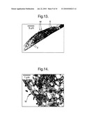
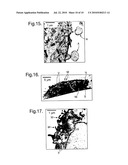
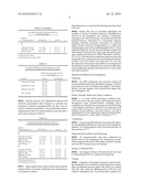
[0091]Cells shown in FIGS. 12, 13 and 16 exhibited a rounded or "cobblestone-like" appearance which is typical of epithelial cells in culture. The secretory nature of the cells was demonstrated by the presence of numerous microvilli identified as "M" (FIGS. 12, 13, 14 16 and 17) and apical blebs, designated as "A" (FIGS. 12, 15 and 16) at the luminal membrane, combined with the presence of numerous secretory granules, designated as "S", throughout the cell cytoplasm (FIGS. 12 to 17). The pinching off of small portions of apical cytoplasm i.e. the microvilli "M" and apical blebs "A" constitute the apocrine secretion process (Montagna et al. (1953) Histology and cytochemistry of human skin. V. Axillary apocrine sweat glands. Am. J. Anat., 92: 451-470). All cells contained numerous secretory granules designated as "S" (FIGS. 12 to 17), although it was not possible to distinguish between Type I and Type II granules in accordance with (Bell (1974) The ultrastructure of human axillary apocrine glands after epinephrine injection. J. Invest. Dermatol., 63: 147-159). The granules contained highly electron-opaque particles, presumably containing small lipid droplets. Numerous "empty" vesicles, designated as "V", were observed in most cells (FIGS. 12, 13, 14 16 and 17). This is probably as a consequence of the fixation process where the contents of the vesicles have been lost. Such vesicles are available to participate in the process of secretion. The cells in FIGS. 13 and 16 were rather flattened in appearance and clearly showed the nucleolus, designated "Nl".
[0092]In all cells from both 3 and 14 day old ASG5 cultures apical blebs, "A", luminal microvilli, "M" and numerous secretory granules, "S" were observed, confirming that these cells display typical active apocrine secretory morphology. These findings further substantiate the apocrine nature of the ASG5 cell line and provide additional evidence of the usefulness of this cell line as a functionally representative cell model of human axillary apocrine sweat glands.
Sequence CWU
1
39123DNAHomo sapiens 1cttcaccccc aacttcaacc ccg
23223DNAHomo sapiens 2ccacatgcgt ctgtctggtc ccc
23325DNAHomo sapiens 3gccagtcttc
atctacacca gtagc 25426DNAHomo
sapiens 4ctctgtcatc cttaaatcac tgagtc
26525DNAHomo sapiens 5cctcatccat tgtaacatca cctcc
25622DNAHomo sapiens 6ctcaccgcct ggctacctga cc
22724DNAHomo sapiens 7gacaagtgac
agggaatgga ttcc 24823DNAHomo
sapiens 8cagggcttct ggtagacatc agg
23924DNAHomo sapiens 9gaagtgtgag tgcgcgaaga tgcg
241026DNAHomo sapiens 10gtgctggaat tataggcatg
agccac 261121DNAHomo sapiens
11ccagccctgg cgatggcaac g
211226DNAHomo sapiens 12gaaattctga cctgttacac agtagg
261324DNAHomo sapiens 13gcctgtcgtg gacttggtca tgcg
241424DNAHomo sapiens
14gagtaggtac tgaccagcct tctc
241525DNAHomo sapiens 15cttggagagc tgttggatgg aggtg
251622DNAHomo sapiens 16cagggccagg cgtcactgag ac
221723DNAHomo sapiens
17ctggatgggg ctcatggtgt ttg
231825DNAHomo sapiens 18gtttccaatg cttcactggg tgtgg
2519392DNAHomo sapiens 19tcccttcttc acccccaact
tcaaccccgt gggttccaac ggatgctttg ccacacacgt 60gtgcttctgt ttcgggagtt
atgtcaccca tcacgaccca cctttactct ttgatatttc 120caaagatccc agagagagaa
acccactaac tccagcatcc gagccccggt tttatgaaat 180cctcaaagtc atgcaggaag
ctgcggacag acacacccag accctgccag aggtgcccga 240tcagttttca tggaacaact
ttctttggaa gccctggctt cagctgtgct gtccttccac 300cggcctgtct tgccagtgtg
atagagaaaa acaggataag agactgagcc gctagcagcg 360cctggggacc agacagacgc
atgtggaagg gc 39220813DNAHomo sapiens
20gcccttctct gtcatcctta aatcactgag tcttggactt cacggtctcc ttgtgccagt
60ccacaaggga aacccactcc acggttctaa cagactcagg tacattcttc ttggcttcct
120cccagctgta aagtggctta tatgccagat cttgctgagc cttcttgtaa gagaaggtga
180acacgctatt tgacaatgtc actctgtggt ggttgaaggg tgtcgatagg tgtaaattgg
240cctcagcagg aagctcacta tttccagcag gaagccaatc cagtacatca gagataaagg
300aaggctccat ctagaatcaa ggcagaggcc gaactctttg ctcaggatgt aattaaggtt
360atcatagctt tggtgaggca tgtcatctga gatgtagtag aactgtcctc ggacacttgg
420ggccttcttg ggctcccgca gggcctcaag gccagaatgt gggcccaggc catgtttcca
480atatagactg ggttgactct ggagaacttg ctgaaacttg acaggatccc atggttgttt
540agggcctcat ttataccggc agaaaggatt gggcttcctt ccccatagat aaacattggt
600cttaaggcac aagtgtacaa ggtgccaccg tttttcagag tccacccatt agccgccagc
660acagccttct cagcaagctt tttgctgtgt gggtatggag cgtaccatgt gttttccaga
720ggctcttctt cgtgactgtt ctggatgatt tccttgtagg aattgggccc ggctacctct
780gggctactgg tgtagatgaa gactggcaag ggc
81321502DNAHomo sapiensmisc_feature(14)..(14)n is a, c, g, or t
21gcccttctca cccnctggct acctgacctt ggtgttgagc ttcaggtatg ccacatagtg
60tgtcaggagc agcctttgga aggcaccgct gtagaaggcc caggccggga tcaggctcag
120aaagcccgcc aagatttcat gggcaaggca gccgcaggtt tcacctccaa ttgtgacata
180attcaatgac tctttgacaa actcatcagc agtcttggtt atcacatttg tatttagata
240ctttgtcatt gcagtcggga cagcatatgg ggtcagcacc tggatgatga cttcttttgc
300tttatattcc tcttgcaggg ccttggaaaa tgcgcacaca aacgccttgg aagctgagta
360catggagtag agaggccaag gaaacagggc tatcccagaa gaaatgttca ggatgagacc
420tttctgcctt gattccatat gtttcaaaat tagctgtgtc atcttgacta cggaggtgat
480gttacaatgg atgaggaagg gc
502221024DNAHomo sapiens 22gacaagtgac agggaatgga ttccaaacac cagtgtgtaa
agctaaagga tggccacttc 60atgcctgtat tgggatttgg cacctatgca cctccagagg
ttccaagaag taaagctttg 120gaggtcacaa aattagcaat agaagctggg ttccgccata
tagattctgc tcatttatac 180aataatgagg agcaggttgg actggccatc cgaagcaaga
ttgcagatgg cagtgtgaag 240ggagaagaca tattctacac ttcaaagctt tggtccactt
ttcatcgacc agagttggtc 300cgaccagcct tggaaaactc actgaaaaaa gctcaattgg
actatgttga cctctatctt 360attcattctc caatgtctct aaagccaggt gaggaacttt
caccaacaga tgaaaatgga 420aaagtaatat ttgacatagt ggatctctgt accacctggg
aggccatgga gaagtgtaag 480gatgcaggat tggccaagtc cattggggtg tcaaacttca
accgcaggca gctggagatg 540atcctcaaca agccaggact caagtacaag cctgtctgca
accaggtaga atgtcatccg 600tatttcaacc ggagtaaatt gctagatttc tgcaagtcga
aagatattgt tctggttgcc 660tatagtgctc tgggatctca acgagacaaa cgatgggtgg
acccgaactc cccggtgctc 720ttggaggacc cagtcctttg tgccttggca aaaaagcaca
agcgaacccc agccctgatt 780gccctgcgct accagctgca gcgtggggtt gtggtcctgg
ccaagagcta caatgagcag 840cgcatcagac agaacgtgca ggtttttgag ttccagttga
ctgcagagga catgaaagcc 900atagatggcc tagacagaaa tctccactat tttaacagtg
atagttttgc tagccaccct 960aattatccat attcagatga atattaacat ggagagcttt
gcctgatgtc taccagaagc 1020cctg
1024231060DNAHomo sapiens 23gcccttgaag tgttagtgcg
cgaagatgcg aaaggtggtt ttgatcaccg gggctagcag 60tggcattggc ctggccctct
gcaagcggct gctggcggaa gatgatgagc ttcatctgtg 120tttggcgtgc aggaacatga
gcaaggcaga agctgtctgt gctgctctgc tggcctctca 180ccccactgct gaggtcacca
ttgtccaggt ggatgtcagc aacctgcagt cggtcttccg 240ggcctccaag gaacttaagc
aaaggtttca gagattagac tgtatatatc taaatgctgg 300gatcatgcct aatccacaac
taaatatcaa agcacttttc tttggcctct tttcaagaaa 360agtgattcat atgttctcca
cagctgaagg cctgctgacc cagggtgata agatcactgc 420tgatggactt caggaggtgt
ttgagaccaa tgtctttggc cattttatcc tgattcggga 480actggagcct ctcctctgtc
acagtgacaa tccatctcag ctcatctgga catcatctcg 540cagtgcaagg aaatctaatt
tcagcctcga ggacttccag cacagcaaag gcaaggaacc 600ctacagctct tccaaatatg
ccactgacct tttgagtgtg gctttgaaca ggaacttcaa 660ccagcagggt ctctattcca
atgtggcctg tccaggtaca gcattgacca atttgacata 720tggaattctg cctccgttta
tatggacgct gttgatgccg gcaatattgc tacttcgctt 780ttttgcaaat gcattcactt
tgacaccata taatggaaca gaagctctgg tatggctttt 840ccaccaaaag cctgaatctc
tcaatcctct gatcaaatat ctgagtgcca ccactggctt 900tggaagaaat tacattatga
cccagaagat ggacctagat gaagacactg ctgaaaaatt 960ttatcaaaag ttactggaac
tggaaaagca cattagggtc actattcaaa aaacagataa 1020tcaggccagg ctcagtggct
catgcctata attccagcac 106024918DNAHomo
sapiensmisc_feature(911)..(911)n is a, c, g, or t 24gcccttccag ccctggcgat
ggcaacggcg acgggggtgg cggaggagcg cctgctggcc 60gcgctcgcct acctgcagtg
cgccgtgggc tgcgcggtct tcgcgcgcaa tcgtcagacg 120aactcagtgt acggccgccc
cgcgtctccg cagcgcgccc aactgcatcc tcctggccat 180gttcctcgtc cactacgggc
atcggtgctt aatttaccca tttctgatgc gaggaggaaa 240gcctatgcca ctgttggcgt
gtacaatggc gattatgttc tgtacctgta acggctattt 300gcaaagcaga tacttgagcc
attgtgcagt gtatgctgat gactgggtaa cagatccccg 360ttttctaata ggttttggct
tgtggttaac gggcatgttg ataaacatcc attcagatca 420tatcctaagg aatctcagaa
aaccaggaga tactggatac aaaataccaa ggggaggctt 480atttgaatac gtaactgcag
ccaactattt tggagaaatc atggagtggt gtggctatgc 540cctggccagc tggtctgtcc
aaggcgcggc ttttgctttc ttcacgtttt gttttttatc 600tggtagagca aaagagcatc
atgagtggta cctccggaaa tttgaagagt atccaaagtt 660cagaaaaatt ataattccat
ttttgtttta agtgcgtttt tcatgaaatt atcttcaact 720tgaagctttc caatggcgct
tctctatgga ctttgtaaat aagttatatc tttgtaattt 780tcctgctact ttatcatttt
caagatgtcc tctaggaatt ttttttctag taattttgca 840atctacctaa taagtaccta
aatacgctga aatggaggtt gaatatccta ctgtgtaaca 900ggtcagaatt ncangggc
91825447DNAHomo
sapiensmisc_feature(13)..(13)n is a, c, g, or t 25gcccttgagt agntactgac
cagccttctc tagtgttcca gacacctgtc tgagtttctt 60ggggtaaaga tcatttccag
catgttttta gtctcatctg ggtgcaagga caagtcgtgt 120atcttctgta tgctctcaac
acactgtcct tgcaatgtct tcacgtggaa tcgtctcaga 180agtgtaacga ggatggcttt
catcatcacc atggcgatgt actttcctgc acagccacgg 240ggcccaaagc caaatggctg
aaagtaccta taaggaacat tctttgcaaa attttcaaga 300gtaaattcat tgggtttggg
gaaaaactcg agtctgtgca tccttccaat attcaggata 360atgtttgtcc cctttttcac
tgggtagcca tcgattacat catcttctaa ggctttgcgc 420atgaccaagt ccacgacagg
caagggc 44726564DNAHomo
sapiensmisc_feature(13)..(13)n is a, c, g, or t 26gcccttgttt ccntgcttca
ctgggtgtgg aaatagatgg gcttgacttt cccagaaagg 60atcttgggca cttgcacaga
gatgatctct gccatcattt ccggaaagtc cacgctcacc 120atgtgtgact tgattagcag
gtcaaaagtg aactgatgca gctctctcgc aataggctgc 180acggagtcca ggagcttggt
gagctggtag aagcgtcttg agcaggatgt gggatttttt 240cttttgcatg caatgatacg
atcgagttcc ttgatgtagt tcattcgaag ttcatcaaag 300aatttttgat ttttcagccc
atccactgga ataatgctga agagtagcag tgctttcatg 360cacaggaatt cctggggggt
gatttggagc catccaaact cttgagagag gtgcctcatt 420cggacacact ggctgtacat
ccgggacttg tgcatgcggt actcattgaa aaccagatca 480ggggcgaagt agagcatcct
ggagttgaca ttggtgaagg atcgccagcc catggcaaac 540accatgagcc ccatccagaa
gggc 56427727DNAHomo
sapiensmisc_feature(12)..(12)n is a, c, g, or t 27gcccttcagg gncnnggcgt
cactgagact gtgggttctg ggagccctct ttgcttttac 60tgtcctctgc cgggctgcac
tcggaccccg tgatggagga cttgcacccg cgaagcacgt 120gggcattcag catctccagc
agcaggtcat acactgggac cacatttttg cacttcatgt 180tgagcagatg ttccatgccc
ttgttactcg catgcctgac gtgggacagg agcatcagga 240ggttagccag gcgcatggat
tgctgctggg aggagatgcc gctcttggca atcacccaaa 300ccaaagcatc ggtcacggcg
ttcagcaagt gagccagctt ccggctgctg tcagcatcct 360gggtcgctgt gaccagaggg
tacatactgg aattgagcag gatcatggcc ttgacacaga 420gatattcttt gtgttggagt
tttaactctc gaaaccttga agtagttgcc aggagcatgt 480caaagatttc cagaattcct
tctacgcatt tcccctcatc cgtctgtaat cccaacaatt 540tgggaggctg aggctggggg
atcacttgaa gtcaggacct cgagaccagc ttggccaaca 600tggtaaaacc ccgtctccac
tgtccagaac aagatctgga gcaaagatga gcctgccggg 660gtggtcaatt gagcgccaca
tcagccccat cattaacacc tccatccaac agctctccaa 720gaagggc
7272823DNAHomo sapiens
28caccatccct ccactgtcag tcc
232924DNAHomo sapiens 29caacattccc caactgctct tctg
243020DNAHomo sapiens 30cggtgcggca gagggacaag
203123DNAHomo sapiens
31ggggaaagcg aagcagaagt aac
233221DNAHomo sapiens 32ccagccctgc caccctgctc c
213324DNAHomo sapiens 33gcagatgcct aattcccgaa taac
243422DNAHomo sapiens
34ggtggaagga atggaggatt gg
223520DNAHomo sapiens 35gtggcaggag tagggggctg
2036254DNAHomo sapiens 36caccatccct ccactgtcag
tccatgatgc ctcagacaaa aatgtccaaa ggcttcaccg 60tctttgggaa gaagaagtct
caaggcgagg gattgaaaaa gcttcagtgc ttctggtgat 120gctgaggttc cagagaacaa
ggttgatttt cgatgcactt ctgggcatct gcttctgcat 180tgccagtgta ctcgggccaa
tattgattat accaaagatc ctggaatatt cagaagagca 240gttggggaat gttg
25437582DNAHomo sapiens
37cggtgcggca gagggacaag catttcatct tgggaagtgc cccaatcctc cggtgcagga
60gaattttgac gtgaataagt atctcggaag atggtacgaa attgagaaga tcccaacaac
120ctttgagaat ggacgctgca tccaggccaa ctactcacta atggaaaacg gaaagatcaa
180agtgttaaac caggagttga gagctgatgg aactgtgaat caaatcgaag gtgaagccac
240cccagttaac ctcacagagc ctgccaagct ggaagttaag ttttcctggt ttatgccatc
300ggcaccgtac tggatcctgg ccaccgacta tgagaactat gccctcgtgt attcctgtac
360ctgcatcatc caactttttc acgtggattt tgcttggatc ttggcaagaa accctaatct
420ccctccagaa acagtggact ctctaaaaaa tatcctgact tctaataaca ttgatgtcaa
480gaaaatgacg gtcacagacc aggtgaactg ccccaagctc tcgtaaccag gttctacagg
540gaggctgcac ccactccatg ttacttctgc ttcgctttcc cc
58238353DNAHomo sapiens 38gcccttccag ccctgccacc ctgctcctgg ttctctgcct
gcagttgggg gccaacaaag 60ctcaggacaa cactcggaag atcataataa agaattttga
cattcccaag tcagtacgtc 120caaatgacga agtcactgca gtgcttgcag ttcaaacaga
attgaaagaa tgcatggtgg 180ttaaaactta cctcattagc agcatccctc tacaaggtgc
atttaactat aagtatactg 240cctgcctatg tgacgacaat ccaaaaacct tctactggga
cttttacacc aacagaactg 300tgcaaattgc agccgtcgtt gatgttattc gggaattagg
catctgcaag ggc 35339629DNAHomo sapiensmisc_feature(60)..(60)n
is a, c, g, or t 39gcccttggtg gaaggaatgg aggattggaa gcaggacagc caacttcaga
aggccagggn 60aggacatctt tatggagacc ctgaaagaca ttgtggagta ttacaacgac
agtaacgggt 120ctcacgtatt gcagggaagg tttggttgtg agatcgagaa taacagaagc
agcggagcat 180tctggaaata ttactatgat ggaaaggact acattgaatt caacaaagaa
atcccagcct 240gggtcccctt cgacccagca gcccagataa ccaagcagaa gtgggaggca
gaaccagtct 300atgtgcagcg ggccaaggct tacctggagg aggagtgccc tgcgactctg
cggaaatacc 360tgaaatacag caaaaatatc ctggaccggc aagatcctcc ctctgtggtg
gtcaccagcc 420accaggcccc aggagaaaag aagaaactga agtgcctggc ctacgacttc
tacccaggga 480aaattggtgt gcactggact cgggccggcg aggtgcagga gcctgagtta
cggggagatg 540ttcttcacaa tggaaatggc acttaccagt cctgggtggt ggtggcagtg
cccccgcagg 600acacagcccc ctactcctgc cacaagggc
629
User Contributions:
Comment about this patent or add new information about this topic:
 |  |
 |  |
 |  |
 |  |
 |  |
 |  |
 |  |
 |  |
 |  |
 |  |
 |  |
 |  |
 |  |
| Similar patent applications: | |
| Date | Title |
|---|---|
| 2009-03-26 | Constitutively translocating cell line |
| 2010-01-28 | Production host cell lines |
| 2010-04-08 | Hepatocyte precursor cell lines |
| 2010-07-08 | Adipose-derived stem cells and lattices |
| 2010-12-02 | Rotation driving device for cell culturing |
| New patent applications in this class: | |
| Date | Title |
|---|---|
| 2019-05-16 | Pdx1-expressing dorsal and ventral foregut endoderm |
| 2019-05-16 | Three-dimensional culture device and methods for dynamic culture of cell aggregates |
| 2018-01-25 | Cell culture method, cell aggregates, cell aggregation control agent, and medium |
| 2017-08-17 | Novel synthetic peptide and use thereof |
| 2016-12-29 | Method for rejuvenating cells |
| New patent applications from these inventors: | |
| Date | Title |
|---|---|
| 2012-02-09 | Composition comprising azole fungicide and water soluble metal salt |
| 2011-10-13 | Personal care composition |
| 2011-01-06 | Hair care composition |
| Top Inventors for class "Chemistry: molecular biology and microbiology" | |
| Rank | Inventor's name |
|---|---|
| 1 | Marshall Medoff |
| 2 | Anthony P. Burgard |
| 3 | Mark J. Burk |
| 4 | Robin E. Osterhout |
| 5 | Rangarajan Sampath |