Patent application title: Detection, screening, and diagnosis of HPV-associated cancers
Inventors:
Shuling Cheng (Fremont, CA, US)
Shuling Cheng (Fremont, CA, US)
IPC8 Class: AG01N3353FI
USPC Class:
435 5
Class name: Chemistry: molecular biology and microbiology measuring or testing process involving enzymes or micro-organisms; composition or test strip therefore; processes of forming such composition or test strip involving virus or bacteriophage
Publication date: 2010-05-13
Patent application number: 20100120019
ion provide methods, polyclonal antibodies,
monoclonal antibodies, assays, and kits for detecting HPV infection,
including infection by various HPV genotypes, early and/or late
HPV-associated or HPV-specific proteins or antibodies. Monoclonal
antibodies are used to detect oncogenic high risk and low risk HPV types
in a single assay, which is not limited to assay type or format. Useful
tools for specific detection of various HPV associated cancers are
provided. HPV associated cancer biomarkers are identified and can be used
in a screening method for early stage precancerous lesions as well as
late stage cancer progression.Claims:
1. A method of assessing the risk for malignancy of a variety of cancers
and carcinomas in a clinical sample from a human subject,
comprising:providing a clinical sample from a source sample other than
cervical sample;conducting one or more detection assays on the clinical
sample from the human subject using one or more anti-HPV antibodies;
anddetecting the presence of HPV proteins present in the clinical sample
to assess the risk of malignancy for the human subject.
2. The method of claim 1, wherein the source sample is selected from the group consisting of bladder samples, head and neck samples, ovarian samples, bladder cancer samples, head and neck cancer samples, ovarian cancer samples, bladder transitional cell carcinoma (TCC), endometriod adenocarcinoma samples, serous papillary cystadenocarcinoma (SPC) samples, urothelial carcinoma samples, squamous cell carcinomas of cheeks samples, tonsillar carcinoma samples, squamous cell carcinoma of larynx samples, squamous cell carcinoma of nose samples, squamous cell carcinoma of upper jaw samples, low grade squamous cell carcinoma, of nasal cavity samples, clear cell carcinoma samples, carcinoma sarcomatodes of left ethmoid sinus samples, low grade endometriod adenocarcinoma samples, normal samples, and combination thereof.
3. The method of claim 1, wherein the detection assay is immunohistochemistry (IHC) assay.
4. The method of claim 1, wherein the HPV proteins is detected positively in the source sample selected from the group consisting of bladder cancer samples, head and neck cancer samples, ovarian cancer samples, bladder transitional cell carcinoma (TCC), urothelial carcinoma samples, squamous cell carcinomas of cheeks samples, tonsillar carcinoma samples, squamous cell carcinoma of larynx samples, squamous cell carcinoma of nose samples, squamous cell carcinoma of upper jaw samples, serous papillary cystadenocarcinoma (SPC) samples, endometriod adenocarcinoma samples and combination thereof.
5. The method of claim 1, wherein the HPV proteins is detected negatively in the source sample selected from the group consisting of low grade squamous cell carcinoma of nasal cavity samples, clear cell carcinoma samples, carcinoma sarcomatodes of left ethmoid sinus samples, low grade endometriod adenocarcinoma samples, normal samples, and combinations thereof.
6. The method of claim 1, wherein the HPV proteins are selected from the group consisting of HPV E6 proteins, HPV E7 proteins, HPV L1 proteins, and combination thereof.
7. A method of detecting papillomavirus infection in a human subject, comprising:obtaining a clinical sample from the human subject; and conducting one or more immunological assays on the clinical sample from the human subject using lab-generated anti-HPV antibodies to detect the presence of HPV infection from the presence of HPV proteins in the clinical sample and access the risk for malignancy of non-cervical cancer.
8. The method of claim 7, wherein the immunological assay is immunohistochemistry (IHC) assay.
9. The method of claim 7, wherein the HPV proteins is detected positive in the clinical sample selected from the group consisting of bladder cancer samples, head and neck cancer samples, ovarian cancer samples, bladder transitional cell carcinoma (TCC), endometriod adenocarcinoma samples, serous papillary cystadenocarcinoma (SPC) samples, urothelial carcinoma samples, squamous cell carcinomas of cheeks samples, tonsillar carcinoma samples, squamous cell carcinoma of larynx samples, squamous cell carcinoma of nose samples, squamous cell carcinoma of upper jaw samples, and combination thereof.
10. The method of claim 7, wherein the HPV proteins is detected negative in the clinical sample selected from the group consisting of low grade squamous cell carcinoma of nasal cavity samples, clear cell carcinoma samples, carcinoma sarcomatodes of left ethmoid sinus samples, low grade endometriod adenocarcinoma samples, normal samples, and combinations thereof.
11. The method of claim 7, wherein the HPV proteins are selected from the group consisting of HPV E6 proteins, HPV E7 proteins, HPV L1 proteins, and combination thereof.
12. The method of claim 7, wherein the anti-HPV antibodies are selected from the group consisting of anti-HPV E6 antibodies, anti-HPV E7 antibodies, anti-HPV antibodies, anti-HPV monoclonal antibodies, anti-HPV polyclonal antibodies, and combination thereof.Description:
CROSS-REFERENCE TO RELATED APPLICATIONS
[0001]This application claims benefit of U.S. provisional patent application Ser. No. 61/199,013, filed Nov. 12, 2008, and U.S. provisional patent application Ser. No. 61/215,589, filed May 7, 2009. Each of the aforementioned related patent applications is herein incorporated by reference.
BACKGROUND OF THE INVENTION
[0002]Infection by human papillomaviruses (HPV) at specific epithelium cells to induce epithelial proliferations plays an important role for cervical carcinogenesis. About 99 percent of confirmed cervical cancer cases are found to be associated with HPV infection with biopsy-confirmed squamous intraepithelial lesions (SIL) or cervical intraepithelial neoplasia (CIN). The incidence of HPV infection, primarily transmitted through sexual contact, is highest among young women and about 20 millions of sexually active men and women worldwide are currently infected.
[0003]In addition to cervical cancer, the presence of HPV DNA has been detected in tumor tissues of head and neck cancer, oral cancer, esophageal cancer, and some skin cancers, as well as lung cancer and colorectal cancer. The detection of HPV DNA in colorectal cancer tissues by in situ hybridization and PCR suggested that HPV infection might be associated with the carcinogenesis of colorectal cancer. However, HPV DNA was not detectable by regular PCR in one earlier study and a survey of HPV16 virus-like particle (VLP) antibodies in patients with epithelial cancers also failed to provide an association of HPV with colorectal cancer challenging the association of colorectal cancers with HPVs. However, colorectal HPV infection is common in patients with colorectal cancer, albeit at a low DNA copy number, with HPV16 being the most prevalent type. The inconsistent results may come from the issues of assay sensitivity for HPV detection. HPV infection may play a role in colorectal carcinogenesis. More sensitive assays for detection of HPV in colorectal cancer are required to demonstrate the association of HPV with carcinogenesis of colorectal cancer.
[0004]Some genital malignancies like vulvar or cervical neoplasms are associated with previous infection with human papillomavirus (HPV). Because of vicinity of bladder to mucosal surface of urogenital tract, HPV may play a significant role in transitional cell carcinoma (TCC). The etiology of TCC, which represents 90 percent of bladder malignancies, is not quite clear, while squamous cell carcinoma (5%) of the bladder is well associated with some factors like urinary stones and prolonged infections. HPV detection rate in clinical samples of invasive cervical cancer can be as high as 99%. Recently, conflicting findings have been reported on association of HPV infection and TCC. Using PCR techniques to detect human papillomavirus (HPV), HPV DNA can be observed prevalently in about 40% to 45% of penile carcinoma clinical samples, about 45% of vulvar carcinoma, and some bladder malignancies.
[0005]Detecting HPV infection by nucleic acid methods, such as "DNA Hybrid Capture", has been developed, but not ideal, due to not only its high cost, assay operation procedures, the requirements for facility, equipment, and highly trained personnel, but also its very low positive predictive value to CIN. In addition, DNA testing could not differentiate the diagnosis of LSIL from HSIL, or CIN lesions from non-transforming latent or remissive viral infection. What is needed is a low cost, simple, sensitive and specific assay that can be performed on routine practice of a clinical lab or doctor office and capable of detecting early stage of epithelial lesions, distinguish LSIL from HSIL, or predicting the risk of progression into cervical cancer.
[0006]Thus, there is a need to detect whether HPV proteins are expressed in a variety of carcinomas and cancers, and determine whether HPV proteins play any role in the progression of the different types of carcinomas/cancers; and if so, which HPV proteins are important in these carcinomas/cancers.
SUMMARY OF THE INVENTION
[0007]Embodiments of the invention provides a method of assessing the risk for malignancy of a variety of cancers and carcinomas in a clinical sample from a human subject including, providing a clinical sample from a source sample other than cervical sample; conducting one or more detection assays on the clinical sample from the human subject using one or more anti-HPV antibodies; and detecting the presence of HPV proteins present in the clinical sample to assess the risk of malignancy for the human subject. In another embodiment, the HPV proteins in the human subject are detected using antibodies raised against HPV recombinant proteins, including but not limiting to, various polyclonal and monoclonal antibodies against various HPV early and late proteins. In another embodiment, the one or more immunoassays include, but not limited to, immunohistochemistry (IHC) assay, immunocytochemistry (ICC) assay, and ELISA, protein chip assay, etc.
[0008]In one embodiment, a method of detecting papillomavirus infection in a human subject includes obtaining a clinical sample from the human subject; and conducting one or more immunological assays on the clinical sample from the human subject using lab-generated anti-HPV antibodies to detect the presence of HPV infection from the presence of HPV proteins in the clinical sample and access the risk for malignancy of non-cervical cancer.
[0009]In one embodiment, IHC staining for cancers other than cervical cancer, including, but not limited to, bladder cancers, head and neck cancers, ovarian cancers were provided herein. In one embodiment, the presence of HPV oncoproteins are herein detected in a variety of cancers and carcinomas, including, but not limiting to, bladder cancer, head and neck cancer, ovarian cancers, bladder transitional cell carcinoma (TCC), endometriod adenocarcinoma, serous papillary cystadenocarcinoma (SPC), urothelial carcinoma, squamous cell carcinomas of cheeks, tonsillar carcinoma, squamous cell carcinoma of larynx, squamous cell carcinoma of nose, squamous cell carcinoma of upper jaw, among others. In another embodiment, HPV oncoproteins are herein not detected in some low grade squamous cell carcinoma of nasal cavity, clear cell carcinoma, carcinoma sarcomatodes of left ethmoid sinus, low grade endometriod adenocarcinoma, among others.
[0010]Embodiments of the invention provides useful antibodies for in situ detection of HPV L1, E6, and E7 proteins in various cancers and carcinoma. The antibodies and assays as described herein are useful for detecting HPV oncoproteins as cancer biomarkers, screening for various HPV associated cancers, and/or accessing the risk for malignant transformation into these HPV associated cancers. As an example, it is possible that male subjects can be HPV carriers to transmit HPV to his partners, a robust test for HPV detection as described in this invention provides promise to develop assays in different formats for detection of HPV in different tissues for various HPV associated cancers.
[0011]Developing appropriate assays, such as HPV immunoassays, is needed for detection of HPV oncoproteins or biomarkers for various cancers associated with HPV infection. The presence of E6/E7 oncoproteins could be evidence to indicate high progression risk of various types of cancer. A robust tool for HPV detection as described herein provides promise to develop assays in different formats for detection of HPV in various tissues from different cancers.
SUMMARY OF DRAWING
[0012]FIG. 1A-1B: Images of IHC using anti-E6 antibody on cervical cancer tissue and its adjacent normal tissue. FIG. 1C-1D: Images of IHC using anti-E7 antibody on cervical cancer and its adjacent normal tissue.
[0013]FIG. 2A-2C: Images of IHC using anti-L1 antibody on bladder cancer tissue samples.
[0014]FIG. 3A-3B: Images of IHC using anti-E6 antibody on bladder cancer tissues. FIG. 3C-3D: Images of IHC using anti-E7 antibody on bladder cancer tissues.
[0015]FIG. 4A shows IHC staining of squamous cell carcinoma of nose by an Anti-E6 antibody
[0016]FIG. 4B shows IHC staining of cancer adjacent normal tissue of salivary gland by an Anti-E6 antibody.
[0017]FIG. 5A shows IHC staining of squamous cell carcinoma of larynx by Anti-E7 antibody.
[0018]FIG. 5B shows IHC staining of cancer adjacent normal tissue of tongue by an Anti-E7 antibody.
[0019]FIG. 6 shows IHC staining of squamous cell carcinoma of Larynx stained by an Anti-L1 antibody.
[0020]FIG. 7A shows IHC staining of serous papillary cystadnocarcinoma of ovary by Anti-E6 antibody.
[0021]FIG. 7B shows IHC staining of cancer adjacent ovary tissues of serous papillary cystadnocarcinoma by Anti-E6 antibody.
[0022]FIG. 8A shows IHC staining of serous papillary cystadnocarcinoma of ovary by Anti-E7 antibody.
[0023]FIG. 8B shows IHC staining of cancer adjacent ovary tissues of serous papillary cystadnocarcinoma by Anti-E7 antibody.
[0024]FIG. 9A shows IHC staining of serous papillary cystadnocarcinoma of ovary by an Anti-L1 antibody.
[0025]FIG. 9B shows IHC staining of another serous papillary cystadnocarcinoma of ovary by Anti-L1 antibody.
[0026]FIG. 10A shows IHC staining of cervical scrape samples (pap smear liquid based) by a mouse monoclonal Anti-HPV-E6 antibody.
[0027]FIG. 10B: IHC staining of cervical scrape samples (liquid based cytology) by a mouse monoclonal HPV E6 antibody.
DETAILED DESCRIPTION
[0028]Embodiments of the invention provide various methods, detection assays, and kits, polyclonal and monoclonal antibodies, polypeptides, recombinant proteins, and nucleic acids useful for detecting HPV proteins present in various cancers from various tissues. In one embodiment, various monoclonal antibodies against HPV proteins are provided such that HPV proteins can be detected by a single monoclonal antibody. Detecting HPV proteins of the invention include, but are not limited to, HPV E6 proteins, HPV E7 proteins, HPV L1 proteins, etc. The sources of HPV proteins for making monoclonal antibodies are not limited and can be from various HPV types/species.
[0029]In one embodiment, a method of assessing the risk for malignancy of a variety of cancers and carcinomas in a clinical sample from a human subject includes providing a clinical sample from a source sample other than cervical sample; conducting one or more detection assays on the clinical sample from the human subject using one or more anti-HPV antibodies; and detecting the presence of HPV proteins present in the clinical sample to assess the risk of malignancy for the human subject. In another embodiment, the HPV proteins in the human subject are detected using antibodies raised against HPV recombinant proteins, including but not limiting to, various polyclonal and monoclonal antibodies against various HPV early and late proteins. In another embodiment, the one or more immunoassays include, but not limited to, immunohistochemistry (IHC) assay, immunocytochemistry (ICC) assay, and ELISA, protein chip assay, etc.
[0030]In one embodiment, a method of detecting papillomavirus infection in a human subject includes obtaining a clinical sample from the human subject; and conducting one or more immunological assays on the clinical sample from the human subject using lab-generated anti-HPV antibodies to detect the presence of HPV infection from the presence of HPV proteins in the clinical sample and access the risk for malignancy of non-cervical cancer.
[0031]As an example, the invention provides one or more anti-E7, anti-E6, or anti-L1 monoclonal antibody useful for detecting HPV infection and predicting HPV associated cancers including, but not limited to, head and neck cancers, bladder cancers, etc. with 82% to 100% of positive predictive value (PPV). The obtained monoclonal antibodies can be very useful in screening clinical samples for various invasive cancers. In an IHC assay on oral biopsy samples, the best results using the monoclonal antibodies of the invention result in about 100% positive predictive value (PPV) for head and neck cancer. Also, monoclonal antibodies with 100% positive predictive value (PPV) for IHC staining of other clinical samples like bladder cancer, head and neck cancer, and ovarian cancer, can also be obtained.
[0032]In another embodiment, a negative predictive value (NPV) ranging from more than about 31% to about 100% for clinical various cancer samples can be observed in an IHC assay for one or more anti-E7 or anti-E6 monoclonal antibody. NPV value of about 100% for clinical biopsy samples as observed for some of the antibodies provided herein supports the use of these antibodies for diagnosing and screening HPV associated cancers.
[0033]In one embodiment, IHC staining for cancers other than cervical cancer, including, but not limited to, bladder cancers, head and neck cancers, ovarian cancers were provided herein. In one embodiment, the presence of HPV oncoproteins are herein detected in a variety of cancers and carcinomas, including, but not limiting to, bladder cancer, head and neck cancer, ovarian cancers, bladder transitional cell carcinoma (TCC), endometriod adenocarcinoma, serous papillary cystadenocarcinoma (SPC), urothelial carcinoma, squamous cell carcinomas of cheeks, tonsillar carcinoma, squamous cell carcinoma of larynx, squamous cell carcinoma of nose, squamous cell carcinoma of upper jaw, among others. In another embodiment, HPV oncoproteins are herein not detected in some low grade squamous cell carcinoma of nasal cavity, clear cell carcinoma, carcinoma sarcomatodes of left ethmoid sinus, low grade endometriod adenocarcinoma, among others.
[0034]As an example, all monoclonal antibodies optimized on IHC assay and tested on each tissue microarray includes providing a thin section containing one or more kinds of tissue cells from a clinical tissue sample of the human subject, applying the thin section on a slide, conducting one or more immunohistochemical assays on the slide containing the thin section of the clinical tissue sample, staining the thin layer of human cells using one or more antibodies generated against one or more purified recombinant papillomavirus proteins, wherein at least one antibody is capable of recognizing a papillomavirus early protein, and detecting in situ one or more proteins from one or more papillomavirus types present in the thin section of the clinical tissue sample on the slide. All figures show representative IHC staining by at least one monoclonal Anti-E6 antibodies, one monoclonal Anti-E7 antibodies or one monoclonal Anti-L1 antibodies. Staining of tumor cells from various cancers show distinguishable staining compared to its normal adjacent tissues. Both cytoplasm and nucleus staining were found in the dysplasia or tumor cells.
[0035]As an example, using normal epithelial staining as base line, the percentage of IHC staining over 10% is considered as positive, otherwise, is negative for the IHC assay. In one embodiment, E6 and E7 oncoproteins expression, the assays or sample formats in detecting the presence of HPV proteins in various HPV associated cancers are not limited and can be used for various tissues like cervical, bladder, head and neck, colon, cervical cells, cervical scrapes, oral scrapes, nose scrapes, serum, body fluids, etc. The useful screening or diagnosing assay can be IHC, ICC, flow cytometry assay, antibodies coupled to beads, rapid tests, protein chips, dot blots, slots, as well a conventional ELISA assay. HPV proteins can be detected by the antibodies of the invention to be present in tumor cells as evidenced by IHC staining after scoring by a pathologist.
[0036]As another example, on the cellular level, HPV proteins can be observed in the nucleus and cytoplasm, but not in the membrane of a cell. It is found that the HPV proteins are present in the nucleus and/or cytoplasm of dysplasia from most of the samples tested. The staining of tumor cells by the antibodies of the invention results in diffused staining. However, there is focused staining as well. As an example, for invasive cancer like SCC and ADC described here in this invention, staining of the tumor cells by anti-HPV protein antibodies displays distinct high levels of staining on both the nucleus and cytoplasm as compared to the staining of the adjacent normal tissues.
[0037]Comparing various HPV proteins related to HPV early genes and late genes, it is found that HPV E6 and E7 oncoprotein is present at high level in head and neck cancers. However, the E6 E7 oncoproteins are detected at lower positive rate in bladder cancer and ovarian cancer compared to detection of L1 protein in the same tumor tissues. Results indicate that HPV E6 or E7 can be served as a cancer biomarker and the invention provides various antibodies tested to high PPV and high NPV values, high specificity and high sensitivity for clinical samples from all stages of a variety of cancers associated with HPV infection. It is possible to use one single monoclonal antibody to detect HPV E6 or E7 proteins present in various early precancerous lesions as well as late stage invasive cancer and other HPV associated cancers. For example, the antibodies are very successful in screening CIN2 and CIN 3 lesions as well as various types of cervical cancers during cancer development. These tools apply to other HPV associated cancers as demonstrated in this invention.
[0038]Head and neck cancer: as an example, Tonsillar carcinoma is the most prevalent oropharyngeal carcinoma. Cigarette smoking and alcohol are the primary risk factors traditionally associated with the development of this malignancy. The association of human papillomavirus (HPV) infection with tonsillar carcinoma has been suggested by the following observations: HPV DNA has been detected in around 50% of tonsillar carcinomas with HPV-16 as the predominant virus type. HPV-16 DNA is transcribed and present in episomal form in most tonsillar carcinomas and can be detected in carcinoma cells by in situ hybridization. Patients with HPV-16 positive tonsillar tumors seem to have a better survival than HPV negative patients, thus presenting a distinct group among patients with tonsillar carcinoma. Patients with anogenital cancer might have an increased risk for tonsillar carcinoma; HPV DNA has also been detected in lymph node metastases. It should be emphasised that the detection of viral DNA per se does not confirm that the virus has a causal connection with malignant transformation. However, so far, it seems that HPV-16 E6 and E7 are actively transcribed in most tonsillar carcinomas that have been analysed. Independent of the physical state of the virus, all tonsillar tumours expressed E7 encoding HPV-33 E6*I mRNA, it has been suggested that the transcription of HPV-16 E6/E7 mRNA in tonsillar carcinomas is not necessarily dependent on viral DNA integration.
[0039]To demonstrate a variety of tumors from head and neck cancers can be detected by HPV IHC as described in this invention, Table 7 shows IHC results by anti-E6 or anti-E7 antibody stain positively on tissues including Squamous cell carcinoma (SCC) of left cheek, SCC of cheek, SCC of larynx, and SCC of nose. Results of IHC by anti-L1 antibody demonstrated in Table 8 show positive staining on tumor tissues including Squamous cell carcinoma (SCC) of upper jaw, SCC of cheek, SCC of larynx, and SCC of left gingival while negatively staining of IHC by anti-L1 antibody including little tissue of SCC of larynx, SCC of nasal cavity, Carcinoma sarcomatodes of left ethmoid sinus, and SCC of left gingiva. These results indicate HPV proteins expressed and distributed in different part of tumors from different organs.
[0040]Staining of cytoplasm is found most distinguishable in tumor cells compared to its corresponding normal cells. These data suggest the IHC staining by E6 or E7 monoclonal antibody is specific in the cytoplasm of tumor cells. In addition, the expression of the L1 viral protein in the tumor cells is also detected in various organs/tissues at the various stages of head and neck cancer. However, the IHC assay sensitivity of about 56% by the Anti-L1 antibody is much lower than assay sensitivity of 100% by the Anti-E7 antibody and Anti-the E6 antibody. These data suggest that detection of E6 or E7 oncoproteins are more relevant to cancer than the detection of the viral capsid protein in head and neck cancer. Previous studies have shown HPV detection of head and neck cancers by DNA test. Together, the results support previous study that HPV-16 E6 and E7 are actively transcribed in most of the tonsillar carcinomas that have been analysed and all tonsillar tumours expressed E7 encoding HPV-33 E6*I mRNA independent of the physical state of the virus. Therefore, E6 and E7 oncoproteins can better serve as biomarkers for detection of head and neck cancers.
[0041]Bladder cancer: As an another example, results demonstrate that HPV L1 viral protein can be detected in the tumor cells of urothelial tissues of bladder cancer by IHC staining showing HPV present in urothelial carcinoma, transitional cell carcinoma and bladder Adenocarcinoma with positive rates varies from 95%, 71% and 33% respectively. HPV IHC on bladder cancer tissues using anti-E6 and anti-E7 mouse monoclonal antibody demonstrate HPV E6 and E7 oncogenic proteins can be detected in the tumor cells of urothelial tissues of bladder cancer by IHC. The nucleus and/or cytoplasmic staining of tumor cells by Anti-E6, or Anti-E7 antibody confirms HPV oncogenic proteins present in situ in the tumor cells of bladder cancer. Data indicated HPV E6 and E7 oncoproteins can be detected in situ present in TCC with positive rates of 19% and 26% respectively. Total of 19 TCC HPV DNA positive tissues (confirmed by PCR using L1 primer) were further analyzed by performing HPV E6 or E7 RNA and/or HPV IHC assays. Three out of 18 (14%) and 4 out of 19 (19%) are positive staining by IHC using anti-HPV E6 and anti-HPV E7 antibody respectively. Among the four of IHC/HPV E7 positive samples, two are HPV16 E7 RNA positive confirmed by RT-PCR while the other two were not tested yet. Among the three of IHC/HPV E6 positive samples, one is HPV16 E6 RNA positive confirmed by RT-PCR while the other 2 were not tested yet. These data suggest correlation of E6 and E7 oncogenes expressed at RNA and protein level.
[0042]In summary, HPV E6, E7 and L1 proteins expressed in the tumor cells of bladder cancer can be detected by IHC with positive rate of 17%, 26%, and 81% using anti-E6, anti-E7, and anti-L1 antibody, respectively. The IHC positive rate of E6 and E7 expression found in bladder cancer is much lower than found in cervical cancer. Data suggest that in addition to HPV infection and/or overexpression of E6 E7 oncoproteins, other factors may have contributed to the progression and development of bladder cancer. Most of E6 or E7 IHC positive samples are found in grade 1 and grade 2, very few found in grade 3 of TCC. Comparing HPV DNA/RNA and IHC assay, three samples found to be HPV DNA, RNA and IHC E6/E7 positive were diagnosed as grade 2 of TCC; four samples found to be HPV DNA/RNA positive, IHC E6/E7 negative were diagnosed as grade 3 of TCC. HPV E6 E7 oncoproteins can be early detection of transforming TCC and may serve as potential biomarkers for early stage of HPV infected bladder cancers. More studies with more cases are mandatory.
[0043]Ovarian cancer: As another example, various stages of ovary tissues were used to demonstrate the expression of the E6 or E7 oncoprotein in the tumor cells of serous papillary cystadenocarcinoma of ovarian cancers using the monoclonal anti-E6 and anti-E7 antibody provided in this invention. The staining results indicate localization of the E6 or E7 proteins expressed in the cytoplasm of tumor cells, but not in the adjacent normal cells, nor stroma cells. The expression of the L1 viral protein is also detected in the tumor cells from the tissue samples at various ovarian cancer stages. The results of the assay sensitivity for an Anti-E7 antibody show about 33% (2 out of 6) for serous papillary cystadenocarcinoma (SPC) with 100% specificity using normal adjacent tissues as negative control. The results of assay sensitivity for an Anti-E6 antibody at about 60% (3 out of 5) for serous papillary cystadenocarcinoma (SPC) with 100% specificity using normal adjacent tissues as negative control. The results of assay sensitivity for an Anti-L1 antibody at 64% (7 out of 11) for serous papillary cystadenocarcinoma (SPC) and 17% (1 out of 6) for other carcinoma with 100% specificity using normal adjacent tissues as negative control. The positive predictive value (PPV) is at about 100% and negative predictive value (NPV) is at about 31%.
[0044]It's worth noting that HPV is associated with serous papillary cystadenocarcinoma (SPC) more often than other ovarian cancer as data demonstrated by HPV IHC using anti-E6, anti-E7 and anti-L1 antibody as data shown in Table 13-16. HPV IHC using anti-E6 or anti-E7 detects most of SPC, but not endometrioid adenocarcinoma. Using anti-L1 antibody, there is about 64% (7 out 11) of SPC samples show HPV positive as compared to only about 17% (1 out of 6) of other non-SPC carcinoma. The 6 non-SPC ovary carcinomas include endometrioid adenocarcinoma (3), clear cell carcinoma (2), and necrosis tissue (1). The only one out of the 6 non-SPC shown positive on HPV L1 is endometrioid adenocarcinoma. To average assay sensitivity of ovary cancer for IHC L1 is about 46% (8 out of 17). However, the assay sensitivity obtained herein is much higher sensitivity than any previous reported connection between HPV and serous papillary cystadenocarcinoma, having about 10% reported HPV DNA positive rate for serous papillary cystadenocarcinoma. This HPV IHC assay provide more robust tool to detect HPV in ovarian cancer, and to access the risk of cancer progression by HPV infection.
[0045]Detection of HPV DNAs, genomes, early viral proteins, late viral proteins, oncoproteins, and/or capsid proteins from various HPV genotypes can be performed by the method and detection assays as described herein and can be very useful in general clinical screening for HPV infection. Detection of HPV antibodies and/or oncoproteins by immunological assays can be used in early clinical screening for HPV infection and general diagnosis for HPV associated cancers and can be performed in a single rapid test or in multiplexed test. Comparative detection of altered levels of HPV proteins and host proteins can be performed in the same or different assays. It can also be used in diagnosing HPV-associated carcinomas with epithelial cell abnormalities induced by HPV infection, pre-malignant and malignant HPV-associated epithelial cell lesions, and those at risk of developing HPV-associated carcinoma and adenocarcinoma. The methods as described herein can be used independently or as an adjunct histological tests and the results thereof can be compared for follow-up patient management.
[0046]Developing appropriate assays, such as HPV immunoassays, is needed for detection of HPV oncoproteins or biomarkers for various cancers associated with HPV infection. The presence of E6/E7 oncoproteins could be evidence to indicate high progression risk of various types of cancer. Embodiments of the invention provided useful antibodies for in situ detection of HPV L1, E6, and E7 proteins in various cancers and carcinoma. The antibodies and assays as described herein are useful for detecting HPV oncoproteins as cancer biomarkers, screening for various HPV associated cancers, and/or accessing the risk for malignant transformation into these HPV associated cancers. As an example, it is possible that male subjects can be HPV carriers to transmit HPV to his partners, a robust test for HPV detection as described in this invention provides promise to develop assays in different formats for detection of HPV in different tissues for various HPV associated cancers.
[0047]The one or more immunological assays as developed herein lend themselves to the high quality and properly purified recombinant proteins encoded by HPV early and late genes, as well as high quality polyclonal and monoclonal antibodies, resulting in immunological assays with very high sensitivity and specificity for screening HPV infection. The one or more immunological assays include, but are not limited to, protein chip assays, antigen assays for papillomavirus proteins, antibody assays for antibodies against papillomavirus proteins ELISA assays for papillomavirus immunocomplexes, radioimmunoprecipitation assays, rapid membrane immunochromatographic assays, rapid stick immunochromatographic assays, immunohistochemistry for tissues and/or cervical cells among others, and immunocytological assays followed by flow cytometry. The one or more immunological assays may be non-invasive with minimal or no additional instrument required. The basic techniques for conducting the immunological assays can be found in "Antibodies: A Laboratory Manual", Harlow and Lane, Cold Spring Harbor Laboratory, Cold Spring Harbor, N.Y. 1989; "Molecular Cloning", A Laboratory Manual, eds. Sambrook, Fritsch and Maniatis, Cold Spring Harbor Laboratory Press, 1989, and others books and manuals known in the art.
EXAMPLES
1. The Anti-HPV Antibody
[0048]HPV recombinant proteins can be any kinds of HPV viral proteins, HPV proteins of early genes and/or late genes, including, but not limited to, E2, E6, E7, L1, L2 and can be from various HPV types. One aspect of the invention provides recombinant proteins, such as recombinant hybrid proteins containing a partial sequence or a full length sequence of HPV oncogenic proteins. For example, full-length E6, E7, and/or L1 polypeptide sequence, which have been found very difficult to obtain and purify due to undesirable aggregation during protein purification, protein instability, low levels of expression, low immunogenic responses of purified proteins. For example, many early E6 oncoproteins contain many cysteine amino acids and thus the correct topography of the E6 oncoproteins requires formation of many disulfide bonds properly. In addition, it was known that certain immunological assays using small peptides of early E6 and E7 proteins results in extremely low assay specificity and sensitivity and thus unsuitable as commercialized diagnostic tools.
[0049]HPV recombinant proteins were produced to use as immunogens for generating antiserum, and screening of monoclonal antibody from hybridoma cell lines: Cloning and production of various recombinant proteins include genes encoded by HPV16 E6 and HPV18 E6 gene, HPV16 E7 and HPV18 E7 gene, HPV16 L1 and HPV18 L1 gene. To provide the recombinant proteins mostly in (or close to) their native forms with much desirable conformation, recombinant HPV E6, E7 or L1 proteins expressed in E coli was purified from soluble fraction, then concentrated, and dialyzed with PBS to be used as immunogen. Immunization of mice and fusion was done by standard procedure to select clones met our screening criteria on ELISA. Each hybridoma cell line was grown in tissue culture and injected to mice for ascites production. Ascites were collected, isotyped, and purified by Protein G column for use in HPV immunoassays.
[0050]For example, a number of samples from various organs are tested in an immunohistochemistry (IHC) assay concurrently as a tissue microarray format using a monoclonal antibody to detect HPV proteins from a variety of HPV types. In addition, the monoclonal antibodies generated using methods of the invention are useful to detect infection by oncogenic HPVs, such as infection by high risk HPV types and/or low risk HPV types. As an example, antibodies raised against a recombinant protein HPV16 E6 oncoprotein generated by the method of invention are able to recognize E6 proteins present inside the cells of clinical samples due to single or multiple HPV infection, and react with E6 proteins from high risk HPV types (such as HPV-16, HPV-18, HPV-31, HPV-33, HPV-45, HPV-52, HPV-58, etc.) or low risk HPV types (HPV-6, etc). In addition, a single anti-E6 monoclonal antibody can detect multiple HPV infection in a clinical sample, having two or more HPV types, such as the combinations of HPV-16, HPV-18, HPV-51, HPV-52, HPV-58, among others.
[0051]As another example, antibodies raised against a recombinant protein HPV16 L1 capsid protein generated by the method of invention are able to recognize L1 proteins present inside the cells of clinical samples due to HPV infection, and react with L1 proteins from high risk HPV types (such as HPV-16, HPV-18, HPV-31, HPV-33, HPV-45, HPV-52, HPV-58, etc.) or low risk HPV types (HPV-6, etc). In addition, a single anti-L1 monoclonal antibody can detect multiple HPV infection in a clinical sample, having two or more HPV types, such as the combination of HPV-16, HPV-18, HPV-51, HPV-52, HPV-58, among others.
2. Immunohistochemistry
IHC
[0052]Sample preparation: Paraffin tissues blocks sectioned into 4 microns were placed on slide and baked at 60 C overnight. Deparaffin/hydrate sections were unmasked followed by standard IHC staining procedures. Purified monoclonal antibody against HPV proteins were diluted to use as the primary antibody. Staining procedure is followed by secondary antibody solution, washing, followed by appropriate substrate reagent to each section. As soon as the sections develop, immerse slides in dH2O, counterstain sections with hematoxylin, dehydrate and mount coverslips.
[0053]Once the tissues are processed and fixed, the Immunohistochemistry (IHC) assay is performed by boiling the tissues on the slide with antigen retrieval buffer for a period of time. The slides were then cool down to room temperature, blocked with pre-antibody blocking solution for a period of time, then incubated with the HPV antibodies. The slides were then washed 3 to 5 times with PBS or H2O, or other solution to get rid of any unbound HPV antibody. Then the slides were incubated with the secondary antibody, for example, anti-mouse IgG HRP, followed by appropriate substrate for detection. As an example, DAB is oxidized in the presence of peroxidase and hydrogen peroxide resulting in the deposition of a brown, alcohol-insoluble precipitate at the site of enzymatic activity. The precipitate may range in color from a light golden brown to dark golden brown depending upon the amount of enzyme present. The golden brown precipitate viewed under a microscope indicates the specific binding of HPV antibodies with HPV proteins present in the cells. The assay can be performed at room temperature or higher temperature to accelerate the binding reaction. This IHC assay can be performed manually, or operated by IHC automation, thus provides a powerful tool to detect HPV infection and HPV oncoproteins in situ localization in the epithelium cells from various tissues including, but not limited to cervical, colon, lung, bladder, head and neck, ovarian, etc.
[0054]For the dysplasia or tumor cells identified, HPV IHC staining may provide additional information for status of HPV infection, stages of diseases, and/or expression of HPV oncoproteins. Therefore, HPV IHC staining assay is very useful as a confirmatory test. In addition, overexpression of HPV E6 and E7 oncoproteins in various stage of cervical dysplasia may indicate progression of CIN and/or cervical cancer development.
[0055]In addition to cervical cancer, IHC staining for other cancers like bladder cancers, head and neck cancers, ovarian cancers were investigated in this invention. The images of each dot on the other tissue microarray slides were viewed and stained cells are visualized under microscope. Areas of tumor cells were looked up to find the cells stained, with staining intensity of score 0-3. Adjacent normal epithelium or normal tissue away from its corresponding tumors was also scored to use as internal control of the assay. Stained intensity score 1 or above was used as cut off to determine positive or negative of the assay. All data were shown in Tables 1-6 for bladder cancers, Tables 7-11 for head and neck cancers, and Tables 12-16 for ovarian cancers.
3. HPV Detection in Bladder Cancer
[0056]The prevalence of HPV DNA in cancers of the urinary bladder varies a lot depending on the techniques applied in the studies. For transitional-cell carcinomas using PCR-based studies, HPV was detected in 3-80% of the samples of transitional cell Carcinomas. Only genital (low- and high-risk) HPV types have been assessed in these studies and HPV 16 and 18 were detected most frequently. Relatively few studies addressed HPV positivity in squamous-cell carcinoma of the bladder using PCR.
[0057]According to the International Expert Group that evidence of the etiological role of HPV for bladder cancer is currently inadequate. There are fewer studies on the association between HPV and cancers at sites other than the cervix, and the number of cases reported is much smaller. To allow for a preliminary assessment of the association between HPV and these cancers, a wider variety of techniques and methods should be considered to be compared in studies. In this invention, we use HPV IHC method to in situ detect the presence of HPV proteins in bladder cancer tissues.
[0058]High rate of HPV positivity in studies suggests that other sexually transmissible viruses may play some roles in development and progression of transitional cell carcinoma of the bladder. There may be a high synergism between Human Papillomaviruses type 16 and 18 (HPV 16, HPV 18), Epstein-Barr virus (EBV), cytomegalovirus (CMV) and herpes simplex virus type 2 (HSV-2) and bladder carcinogenesis. In this regard, further investigation with a large number of patients sounds to be required. Public education regarding HPV transmission and high-risk behavior may be helpful for decreasing the incidence of urogenital malignancies. Males may be carriers of oncogenic HPVs and male partners may markedly contribute to the risk of developing cervical cancer in their female partners.
[0059]HPV may have a great role in progression of transitional cell carcinoma (TCC) by inactivation of the tumor suppressors or other unknown mechanisms. Previous studies showed presence of HPV correlated with malignancy of urothelium transformation, but some reported contradictory results. In this regards, more sensitive detection of HPV in bladder cancers is required to demonstrate the association of HPV and cancer progression in TCC. Detecting expression and localization of HPV oncoproteins in different stages of bladder cancer will help understanding the mechanism. In this invention, we detect HPV proteins in TCC by immunohistochemistry (IHC) using our novel antibodies to HPV E6, E7 and L1 proteins. We also compared HPV E6, E7 oncoprotein expression with HPV DNA and HPV mRNA transcript in TCC specimen.
[0060]The bladder cancer (BC) specimen: Tissue microarrays (TMA) containing various stages of bladder cancer and its adjacent normal tissues were obtained from commercially available vendor. Additional HPV typed TCC specimens performed in this study was obtained from patients of N. N. Blokhin Cancer Research Center or P.A. Hertzen Research Institute of Oncology. The patients consent had been informed. DNA isolated from frozen tissues was screened by PCR using primer GP 5/6 and My 09/11 for L1 region. The amplification products obtained by the My 09/11 primers were subjected to the restriction analysis to determine HPV types 6, 11, 16, 18, 31, 33 and 35. RNA preparation was used to perform RT-PCR with random hexanucleotide primers ("Lytekh", Russia) and Super Script II reverse transcriptase ("Invitrogene") according to the instructions of the manufacturer. Analysis of the HPV16 E7 transcripts was performed using primers: sense, 5'-CGG ACA GAG CCC ATT ACA AT-3'; antisense, 5'-GAA CAG ATG GGG CAC ACA AT-3' for amplification product length of 144 bp.
[0061]1). HPV IHC on cervical cancer tissues: There is limited study reported for detection of HPV proteins in bladder cancer. To perform HPV IHC assay on bladder cancer, cervical cancer tissues were used as positive control to demonstrate detection of HPV proteins in situ using our novel mouse monoclonal anti-HPV antibodies. FIGS. 1A-1C show IHC staining of cervical cancer tissues using mouse monoclonal anti-HPV L1, anti-HPV E6, and anti-HPV E7 antibody, respectively. Results indicate that HPV proteins are present in the nucleus and/or cytoplasm of dysplasia cells from most of the samples tested. In the cellular level, HPV proteins can be detected in the nucleus and cytoplasm, but not in the membrane of a cell. The staining of tumor cells results in focused staining and diffused staining as well.
[0062]2). HPV IHC on bladder cancer tissues using anti-HPV L1 antibody: To demonstrate HPV IHC assay on BC samples, TMA containing various BC samples were used to perform IHC assay using a mouse monoclonal anti-HPV L1 as results demonstrated in FIG. 2A-2C and summarized in Table 1. FIG. 2A shows representative images of IHC staining of urothelial carcinoma of bladder tissue demonstrated by a mouse monoclonal Anti-HPV-L1. FIG. 2B shows representative images of IHC staining of another urothelial carcinoma of bladder tissue demonstrated by the same Anti-HPV-L1 antibody at high power magnification. FIG. 2C shows representative images of IHC staining of another urothelial carcinoma with glandular metaplasia demonstrated by the same mouse monoclonal Anti-HPV-L1. The results indicate that HPV L1 viral protein can be detected in the tumor cells of urothelial tissues of bladder cancer by IHC staining using a mouse monoclonal anti-HPV L1 antibody.
[0063]The images of each dot on the tissue microarray slides were viewed and scored with percentage and staining intensity by certified pathologist. Adjacent normal epithelium or normal tissue away from its corresponding tumors was also scored. Stained percentage over 10% or intensity score 1 or above was used as cut off for assay positivity. Table 1 shows summary of IHC data from TMA, containing bladder samples including urothelial carcinoma (20), transitional cell carcinoma (14), adenocarcinoma (3), and normal urocystic tissue (6). As data indicated, nucleus and/or cytoplasmic staining are found in most clinical samples of tumor cells stained by an Anti-L1 antibody. These results confirm HPV-protein-specific IHC staining of tumor cells in urothelial cancer. The IHC staining results indicate HPV L1 proteins can be detected in situ present in urothelial carcinoma, transitional cell carcinoma and bladder Adenocarcinoma. These results confirm HPV-protein-specific staining of tumor cells. Staining of cytoplasm is found most distinguishable in dysplasia or tumor cells compared to its corresponding normal cells.
TABLE-US-00001 TABLE 1 IHC staining results (stain intensity score; 0-3) for biopsy samples from urothelial carcinoma (20 samples) and adjacent normal tissue (4 samples, as negative controls) using monoclonal Anti-HPV-L1 antibody. No. organ pathology grade type Anti-L1 Ab 1 Bladder Urothelial I Malignant 3+ carcinoma (UCC) 2 Bladder UCC I Malignant 3+ 3 Bladder UCC with glandular II Malignant 3+ metaplasia 4 Bladder UCC with glandular II Malignant 3+ metaplasia 5 Bladder UCC I Malignant 2+ 6 Bladder UCC I Malignant 2+ 7 Bladder UCC I Malignant 0 8 Bladder UCC I Malignant 3+ 9 Bladder UCC I Malignant 3+ 10 Bladder UCC I Malignant 3+ 11 Bladder UCC II Malignant 3+ 12 Bladder UCC II Malignant 2+ 13 Bladder UCC III Malignant 3+ 14 Bladder UCC III Malignant 3+ 15 Bladder UCC III Malignant 3+ 16 Bladder UCC III Malignant 3+ 17 Bladder UCC II Malignant 3+ 20 Bladder UCC III Malignant 3+ 21 Bladder adjacent bladder -- Normal 0 tissue of No. 01 22 Bladder adjacent bladder -- Normal 0 tissue of No. 01 23 Bladder adjacent bladder -- Normal 0 tissue of No. 03 24 Bladder adjacent bladder -- Normal 0 tissue of No. 03
[0064]Another tissue microarray containing 19 bladder samples including transitional cell carcinoma (14), adenocarcinoma (2), normal urocystic tissue (2) and skin malignant melanoma (1) was also tested using anti-HPV-L1 antibody (data shown in table 3). Combined data from the two tissue microarray, Table 2 shows summary of the IHC staining results of bladder cancer tissues using an anti-HPV-L1 antibody.
TABLE-US-00002 TABLE 2 Summary of the IHC staining results of bladder cancer tissues using an Anti-L1 mouse monoclonal antibody Bladder Cancer Normal Urothelial Transitional cell Adeno- normal uro- carcinoma carcinoma carcinoma cystic tissue Anti-L1 95% (19/20) 71% (10/14) 33% (1/3) 33% (2/6) antibody
[0065]2). HPV IHC on Bladder Cancer Tissues Using Anti-E6 and Anti-E7 Mouse Monoclonal Antibody:
[0066]As another example, various stages of urothelial tissues from bladder cancer were used to show the expression of the E6 or E7 oncoprotein in the tumor cells. To demonstrate detection of HPV E6 and E7 oncogenic proteins on BC samples, TMA containing various BC samples were used to perform IHC assay using a mouse monoclonal anti-HPV E6, or anti-HPV E7 antibody. As results demonstrated, FIG. 3A-3B shows representative images of IHC staining of transitional cell carcinoma of bladder tissue using a mouse monoclonal HPV E6 antibody. FIG. 3C-3D shows IHC staining of transitional cell carcinoma of bladder tissue using a mouse monoclonal Anti-E7 antibody. FIG. 3D shows representative images of IHC staining of adenocarcinoma of bladder tissue using the same mouse monoclonal anti-HPV E7 antibody. These results demonstrate HPV E6 and E7 oncogenic proteins can be detected in the tumor cells of urothelial tissues of bladder cancer by IHC. The nucleus and/or cytoplasmic staining of tumor cells by Anti-E6, or Anti-E7 antibody confirms HPV oncogenic proteins present in situ in the tumor cells of bladder cancer.
[0067]Table 3 contains data from 19 bladder samples including transitional cell carcinoma (14), adenocarcinoma (2), normal urocystic tissue (2) and skin malignant melanoma (1). IHC score for staining of cytoplasm, and/or nucleus followed by the intensity of staining with an anti-HPV-E7 antibody or an anti-HPV-E6 antibody were shown. As data shown in Table 3, nucleus and/or cytoplasmic staining are found in clinical samples of tumor cells stained by an Anti-E7 antibody, an Anti-E6 antibody or an anti-L1 antibody. These results confirm HPV-protein-specific IHC staining of tumor cells. However, most of staining by anti-L1 antibody (8 out 10 anti-L1 positive samples) is found in cytoplasm compared to only 2 out of 10 found nucleus staining by anti-L1 antibody.
TABLE-US-00003 TABLE 3 Monoclonal anti-HPV E6 or E7 or L1 antibody IHC staining results (stain intensity score; 0-3) for biopsy samples from bladder cancers including transitional cell carcinoma (14) and adenocarcinoma (2) compared to normal urocystic tissue (2) as negative controls. Anti-E7 Anti-E6 Anti-L1 No. pathology grade notes type antibody antibody antibody 1 Transitional cell 1 T1N0M1 Malignant 0 0 0 carcinoma (TCC) 2 TCC 1 T2N0M0 Malignant -- 0 1+ 3 TCC 1 T2N0M0 Malignant 0-1 1+ 2+ 4 TCC 1 T1N0M0 Malignant 1+ 1+ 2+ 5 TCC 1 T1N0M0 Malignant 1+ 0 1+ 6 TCC 1 T1N0M0 Malignant 0 0 0 7 TCC 1 T1N0M1 Malignant 0 0 0 8 TCC 2 T3N0M0 Malignant 0 0 0 9 TCC 2 T3N0M0 Malignant 0 0 1+ 10 TCC 2 T2N0M0 Malignant 0 0 1+ 11 TCC 2 T2N0M0 Malignant 0 0 1+ 12 TCC 3 T2N0M0 Malignant 1-2+ 0-1 1+ 13 TCC 3 T2N0M0 Malignant 1-2+ 0-1 1+ 14 TCC 3 T1N0M0 Malignant 0 0 0 15 Adenocarcinoma 1 T3N0M0 Malignant 0 0 1+ (ADC) 16 ADC 1 T3N0M0 Malignant 0-1 0 0 17 ADC 1 T3N0M0 Malignant -- 0 0 18 Normal urocystic -- -- Normal 0 0 2+ tissue 19 Normal urocystic -- -- Normal 0 0 2+ tissue
[0068]To summarize data from Table 3, Table 4 shown assay sensitivity of about 28% (4 out of 14) and about 0% (2 out of 2) using an Anti-E7 antibody is detected for bladder transitional cell carcinoma (TCC) and bladder Adenocarcinoma (ADC) respectively, with about 100% specificity for normal urothelial as negative control; about 100% of positive predictive value (PPV) and about 14% of negative predictive value (NPV) were also obtained.
[0069]For detecting HPV E6 protein in situ in bladder cancer tissues as shown in Table 4, assay sensitivity of about 14% (2 out of 14) and about 0% (0 out of 2) using an Anti-E6 antibody is detected for bladder transitional cell carcinoma (TCC) and bladder Adenocarcinoma (ADC) respectively, with about 100% specificity using normal urothelial or skin melanoma as negative control; about 100% of positive predictive value (PPV) and about 12% of negative predictive value (NPV) were also obtained.
TABLE-US-00004 TABLE 4 Summary of the IHC staining results from Table 3 for an anti-E7 antibody and anti-E6 antibody on bladder cancer samples BladderCancer Normal Transitional Adeno- normal uro- cell carcinoma carcinoma cystic tissue Anti-E7 28% (4/13) 0% (0/3) 0% (0/2) antibody Anti-E6 14% (2/14) 0% (0/3) 0% (0/2) antibody
[0070]4. Analysis of HPV DNA/RNA and HPV oncogenic protein expression in bladder cancer tissues: To analyze HPV DNA/RNA and HPV oncoprotein expression on BC samples, 21 TCC HPV DNA positive tissues (confirmed by PCR using L1 primer) and 4 normal urothelial tissues from non oncological patients were further analyzed by performing HPV E7 RNA and/or HPV IHC assays. Four out of 19 (21%) and 3 out of 18 (17%) are positive staining by IHC using anti-HPV E7 and anti-HPV E6 antibody respectively. These data seem correlating to our overall positive rate of IHC assay shown in Table 4. Among the four of IHC/HPV E7 positive samples, two are HPV16 E7 RNA positive confirmed by RT-PCR while the other two were not tested yet. Among the three of IHC/HPV E6 positive samples, one is HPV16 E6 RNA positive confirmed by RT-PCR while the other 2 were not tested yet. These data suggest correlation of E6 and E7 oncogenes expressed at RNA and protein level. There are four TCC samples shows both HPV DNA and RNA positive, but IHC negative stained by anti-E6 or anti-E7 antibody. These four HPV DNA/RNA positive, IHC negative samples were diagnosed grade 3 while those HPV DNA/RNA and IHC positive samples are diagnosed as grade 2 of TCC. These data suggest that E6 E7 oncoproteins can be detected in grade 2 as early detection of transformation for TCC and may serve as potential biomarkers for HPV infected bladder cancers. More studies with more cases are required.
[0071]To demonstrate the specific detection of E6 E7 proteins in bladder cancer, additional 12 cases of tissues from various organs were also tested on IHC by anti-E6 or anti-E7 antibody. To summarize the cases tested by IHC assay for detection of E6 and E7 proteins in bladder cancer, Table 5 shows combined IHC data from Table 4 (containing transitional cell carcinoma (14), adenocarcinoma (3), normal urocystic tissue (2) and 18 TCC specimen from Russia and 12 normal tissues from various organs. These data also indicate higher positive rate of IHC using anti-HPV E7 antibody detecting E7 proteins in TCC (transitional cell carcinoma) compared to IHC using anti-HPV E6 antibody detecting E6 proteins in TCC. More cases to be studied are mandatory. Table 5 shows summary of IHC data indicating HPV E7 and E6 oncoproteins can be detected in situ present in TCC with positive rates of 26% and 16% respectively.
[0072]To demonstrate the detection of HPV proteins on bladder cancer and to compare the expression of HPV viral proteins (L1 as an example) and oncoproteins (E6, E7 as example), Table 6 shows summary of IHC assay sensitivity and specificity using anti-HPV L1, E7, and E6 antibody. About 81% of bladder cancer shows HPV L1 positive while about 23% and 14% shows positive staining by anti-HPV E7 and anti-HPV E6 antibody respectively. These IHC data correlate with HPV DNA/RNA test on bladder cancer that about 33% (7 out of 21) HPV DNA positive TCC specimen show positive HPV E7 mRNA. However, HPV IHC assays are more robust compared to HPV DNA or RNA by PCR which involves many steps in target nucleic acid amplification and issues in contamination among samples. It is advantageous to use the HPV IHC assays provided in this invention to detect the association of HPV with bladder cancer and the role of HPV in transformation and malignancy of bladder cancers.
TABLE-US-00005 TABLE 5 Summary of the IHC staining results of bladder cancer tissues using an Anti-E7 and anti-E6 mouse monoclonal antibody. Bladder Cancer Normal tissue Transitional Adeno- normal uro- various cell carcinoma carcinoma cystic tissue organ Anti-E7 26% (8/32) 0% (0/3) 0% (0/6) 8% (1/12) antibody Anti-E6 16% (5/32) 0% (0/3) 0% (0/6) 8% (1/12) antibody
TABLE-US-00006 TABLE 6 Summary of the HPV IHC assay for bladder cancer samples sensitivity (Bladder Cancer Specificity (Normal tissue IHC stained positively) stained negatively) Anti-L1 81% (30/37) 67% (2/6) Anti-E7 23% (8/35) 94% (1/18) Anti-E6 14% (5/35) 94% (1/18)
[0073]The invention described herein provides a robust tool for detection of HPV in bladder cancer tissues. In summary: 1). HPV E6, E7 and L1 proteins expressed in the tumor cells of bladder cancer can be detected by IHC with positive rate of 14%, 23%, and 81% using anti-E6, anti-E7, and anti-L1 antibody, respectively as shown in Table 6. 2). The IHC positive rate of E6 and E7 expression found in bladder cancer is much lower than found in cervical cancer. Data suggest that in addition to HPV infection and/or overexpression of E6 E7 oncoproteins, other factors may have contributed to the progression and development of bladder cancer. 3). Most of E6 or E7 IHC positive samples are found in grade 1 and grade 2, very few found in grade 3 of TCC. 4). Comparing HPV DNA/RNA and IHC assay, three samples found to be HPV DNA, RNA and IHC E6/E7 positive were diagnosed as grade 2 of TCC; four samples found to be HPV DNA/RNA positive, IHC E6/E7 negative were diagnosed as grade 3 of TCC. 5. HPV E6 E7 oncoproteins can be early detection of transforming TCC and may serve as potential biomarkers for early stage of HPV infected bladder cancers. More studies with more cases are required.
4. Head and Neck Cancer
[0074]As another example, various organs with various stages of tissues from head and neck cancer were used to show the expression of the E6 or E7 oncoprotein in the tumor cells. FIGS. 4A-4B shows representative images of IHC staining of squamous cell carcinoma of nose tissue (FIG. 4A) and its adjacent normal tissue (FIG. 4B) by staining with a mouse monoclonal Anti-HPV E6 antibody. FIGS. 5A-5B shows representative images of IHC staining of squamous cell carcinoma of nose tissue (FIG. 5A) and its adjacent normal tissue (FIG. 5B) demonstrated by Anti-E7 antibody (mouse monoclonal HPV E7).
[0075]The IHC staining results indicate E6 or E7 oncoproteins can be detected in SCC (squamous cell carcinoma) but not in the normal adjacent tissues. Empty blank arrows indicate the specific staining of E6 or E7 protein in tumor cells, while solid black arrows indicate the normal cells with no stain. Highly magnified images indicate localization of the E6 or E7 proteins expressed in the cytoplasm of tumor cells, but not in the adjacent normal cells, nor stroma cells. These data suggest the IHC staining by E6 or E7 monoclonal antibody is specific in the cytoplasm of tumor cells.
[0076]In addition, the expression of the L1 viral protein in the tumor cells is also detected in various organs/tissues at the various stages of head and neck cancer. FIG. 6 shows representative images of IHC staining of squamous cell carcinoma of larynx tissue demonstrated by a mouse monoclonal Anti-HPV-L1 antibody.
[0077]As an example, tonsillar carcinoma is the most prevalent oropharyngeal carcinoma. Cigarette smoking and alcohol are the primary risk factors traditionally associated with the development of this malignancy. The association of human papillomavirus (HPV) infection with tonsillar carcinoma has been suggested by the following observations: HPV DNA has been detected in around 50% of tonsillar carcinomas with HPV-16 as the predominant virus type. HPV-16 DNA is transcribed and present in episomal form in most tonsillar carcinomas and can be detected in carcinoma cells by in situ hybridization. Patients with HPV-16 positive tonsillar tumours seem to have a better survival than HPV negative patients, thus presenting a distinct group among patients with tonsillar carcinoma. Patients with anogenital cancer might have an increased risk for tonsillar carcinoma; HPV DNA has also been detected in lymph node metastases. It should be emphasised that the detection of viral DNA per se does not confirm that the virus has a causal connection with malignant transformation. However, so far, it seems that HPV-16 E6 and E7 are actively transcribed in most tonsillar carcinomas that have been analysed. Independent of the physical state of the virus, all tonsillar tumours expressed E7 encoding HPV-33 E6*I mRNA, it has been suggested that the transcription of HPV-16 E6/E7 mRNA in tonsillar carcinomas is not necessarily dependent on viral DNA integration.
[0078]To demonstrate a variety of tumors from head and neck cancers tested by HPV IHC, Table 7 contains the results from 19 clinical samples including head and neck squamous cell carcinoma (9) and adjacent normal tissue (10) as negative controls. IHC intensity score for staining of cytoplasm and/or nucleus with an anti-HPV-E7 antibody or an Anti-HPV-E6 antibody were shown. Both nucleus and cytoplasmic staining are found the tumor cells of these clinical samples stained with the Anti-E7 antibody or Anti-E6 antibody. The tissues tested in this microarray and shown positive staining of IHC by anti-E6 or anti-E7 antibody include Squamous cell carcinoma (SCC) of left cheek, SCC of cheek, SCC of larynx, and SCC of nose. The results confirm HPV-protein-specific staining of tumor cells from various parts of head and neck cancer. Staining of cytoplasm is found most distinguishable in tumor cells compared to its corresponding normal cells.
TABLE-US-00007 TABLE 7 IHC staining results (stain intensity score; 0-3) for biopsy samples from head and neck squamous cell carcinoma (9) and cancer adjacent normal tissue (10) as negative controls using monoclonal Anti-HPV-E6 or Anti-HPV-E7 antibody. α-E7 α-E6 No. Organ pathology grade type Ab Ab 1 Cheek Squamous cell I Malignant 1-2+ 3+ part carcinoma (SCC) of left cheek 2 Tongue adjacent normal tissue -- Adjacent 0 0 3 Tongue adjacent normal tissue -- Adjacent 0 0 4 Cheek SCC of cheek I Malignant 1-2+ 3+ part 5 Cheek SCC of cheek I Malignant 1-2+ NA part 6 Parotid adjacent normal tissue -- Adjacent 0 0-1+ 7 Parotid adjacent normal tissue -- Adjacent 0 0-1+ 8 Larynx SCC of larynx II Malignant 2+ 2+ 9 Larynx SCC of larynx III Malignant 1+ 1+ 10 Larynx SCC of larynx III Malignant 1+ 2+ 11 Tongue adjacent normal tissue -- Adjacent 3+ 3+ 12 Tongue adjacent normal tissue -- Adjacent 3+ 3+ 13 Nose SCC of nose III Malignant 2+ 2+ 14 Salivary adjacent normal tissue -- Adjacent 0 0 gland 15 Salivary adjacent normal tissue -- Adjacent 0 0 gland 16 Nose SCC of nose III Malignant 1+ 2+ 17 Nose SCC of nose III Malignant 1+ 2+ 18 Tongue adjacent normal tissue -- Adjacent NA 0 19 Tongue adjacent normal tissue -- Adjacent NA 0
[0079]To demonstrate a variety of tumors from head and neck cancers can be detected by HPV IHC, additional tissue microarray containing 20 clinical samples including head and neck squamous cell carcinoma (16) and cancer adjacent normal tissue (4) as negative controls are tested using anti-L1 antibody. IHC score for staining of cytoplasm, and/or nucleus followed by the intensity of staining with an Anti-HPV-L1 antibody were shown in Table 8. Both nucleus and cytoplasmic portions of these clinical samples are stained by the anti-L1 antibody. The tumor tissues tested in this microarray and shown positive staining of IHC by anti-L1 antibody include Squamous cell carcinoma (SCC) of upper jaw, SCC of cheek, SCC of larynx, and SCC of left gingiva. The tumor tissues tested in this microarray and shown negative staining of IHC by anti-L1 antibody include little tissue of SCC of larynx, SCC of nasal cavity, Carcinoma sarcomatodes of left ethmoid sinus, and SCC of left gingiva. The results confirm HPV-protein-specific staining of tumor cells as compared to its corresponding normal cells.
TABLE-US-00008 TABLE 8 IHC staining results (stain intensity score; 0-3) for biopsy samples from head and neck squamous cell carcinoma (16 samples) and cancer adjacent normal tissue (4 samples, as negative controls) using a monoclonal anti HPV L1 antibody. Anti-L1 No. organ pathology grade type antibody 1 Upper SCC of upper jaw I Malignant 1+ jaw 2 Upper SCC of upper jaw I Malignant 1+ jaw 3 Cheek SCC of cheek II Malignant 2+ 4 Cheek SCC of cheek II Malignant 1+ 5 Larynx SCC of larynx I Malignant 2+ 6 Larynx SCC of larynx I Malignant 1+ 7 Larynx SCC of larynx II Malignant 1-2+ 8 Larynx SCC of larynx II Malignant 1-2+ 9 Larynx Little SCC tissue -- Malignant 0 10 Nose SCC of nasal cavity III Malignant 0-1 11 Nose SCC of nasal cavity III Malignant 0-1 12 Nose SCC of nasal cavity II Malignant 0-1 13 Nose SCC of nasal cavity II Malignant 0-1 14 Nose Carcinoma sarcomatodes -- Malignant 0 of left ethmoid sinus 15 Gingiva SCC of left gingiva I Malignant 0-1 16 Gingiva SCC of left gingiva I Malignant 1+ 17 Tongue adjacent normal tissue -- Adjacent 0 18 Tongue adjacent normal tissue -- Adjacent 0 19 Larynx adjacent normal tissue -- Adjacent 0-1 20 Larynx adjacent normal tissue -- Adjacent 0-1
TABLE-US-00009 TABLE 9 Summary of the IHC staining results for anAnti-E7 antibody on head and neck cancer samples. H&N Normal carcinoma adjacent total Anti-E7 positive 9 2 11 82% PPV antibody negative 0 6 6 100% NPV Total 9 8 17 Sensitivity 100% Specificity 75%
[0080]To summarize IHC results from table 7, Table 9 shows the results of IHC assay sensitivity for an Anti-E7 antibody at about 100% (9 out of 9) for head and neck cancers with 75% specificity using normal adjacent tissues as negative control; positive predictive value (PPV) at about 82% and negative predictive value (NPV) at about 100%. Two out of the 8 normal adjacent samples show positive staining are tongues tissues adjacent from squamous cell carcinoma of larynx. It requires further investigation to confirm if these two adjacent normal tissues are truly HPV negative
[0081]To summarize IHC results from table 7, Table 10 shows the results of IHC assay sensitivity for an Anti-E6 antibody at about 100% (8 out of 8) for head and neck cancers with 80% specificity using normal adjacent tissues as negative control; positive predictive value (PPV) at about 80% and negative predictive value (NPV) at about 100%. Two out of the 10 normal adjacent samples show positive staining are tongues tissues adjacent from squamous cell carcinoma of larynx. It requires further investigation to confirm if these two adjacent normal tissues are truly HPV negative.
[0082]To summarize IHC results from table 8, Table 11 shows the results of IHC assay sensitivity for an Anti-L1 antibody at about 56% (9 out of 16) for head and neck cancers with 100% specificity using normal adjacent tissues as negative control; positive predictive value (PPV) at about 100% and negative predictive value (NPV) at about 36%. The resulting 100% PPV and 100% specificity values demonstrate HPV-L1 specific IHC staining.
[0083]It's noted that the IHC assay sensitivity of about 56% by the Anti-L1 antibody is much lower than assay sensitivity of 100% by the Anti-E7 antibody and Anti-the E6 antibody. These data suggest that detection of E6 or E7 oncoproteins are more relevant to cancer than the detection of the viral capsid protein in head and neck cancer. Previous studies have shown HPV detection of head and neck cancers by DNA test. Together, the results support previous study that HPV-16 E6 and E7 are actively transcribed in most of the tonsillar carcinomas that have been analysed and all tonsillar tumours expressed E7 encoding HPV-33 E6*I mRNA independent of the physical state of the virus. Therefore, E6 and E7 oncoproteins can better serve as biomarkers for detection of head and neck cancers.
TABLE-US-00010 TABLE 10 Summary of the IHC staining results for Anti-E6 antibody on head and neck cancer samples. H&N Normal carcinoma adjacent total Anti-E6 positive 8 2 10 80% PPV antibody negative 0 8 8 100% NPV Total 8 10 18 Sensitivity 100% Specificity 80%
TABLE-US-00011 TABLE 11 Summary of the IHC staining results for Anti-L1 antibody on head and neck cancer samples. H&N Normal carcinoma adjacent total Anti-L1 positive 9 0 9 100% PPV antibody negative 7 4 11 36% NPV Total 16 4 20 Sensitivity 56% Specificity 100%
5. Ovarian Cancer
[0084]As another example, various stages of ovary tissues were used to demonstrate the expression of the E6 or E7 oncoprotein in the tumor cells of serous papillary cystadenocarcinoma of ovarian cancers. FIGS. 7A-7B shows representative images of IHC staining of serous papillary cystadnocarcinoma of ovary tissue (FIG. 7A) and its adjacent normal tissue (FIG. 7B) demonstrated by the anti-HPV E6 antibody. FIGS. 8A-8B shows representative images of IHC staining of serous papillary cystadnocarcinoma of ovary tissue (FIG. 8A) and its adjacent normal tissue (FIG. 8B) by staining with an Anti-E7 mouse monoclonal antibody.
[0085]The staining results indicate E6 or E7 oncoproteins can be detected in tumor cells of serous papillary cystadnocarcinoma of ovary but not in the normal adjacent tissues. Empty blank arrows indicate the specific staining of E6 or E7 protein in tumor cells, while solid black arrows indicate the normal cells with no stain. Highly magnified images indicate localization of the E6 or E7 proteins expressed in the cytoplasm of tumor cells, but not in the adjacent normal cells, nor stroma cells. These data suggest the IHC staining by E6 or E7 monoclonal antibody is specific in the cytoplasm of tumor cells.
[0086]The expression of the L1 viral protein is also detected in the tumor cells from the tissue samples at various ovarian cancer stages. FIGS. 9A-9B show representative images of IHC staining of serous papillary cystadnocarcinoma of ovary tissue demonstrated by mouse monoclonal HPV L1 antibody. The IHC staining results indicate expression of L1 viral protein can be detected in tumor cells of serous papillary cystadnocarcinoma of ovary tissue but not in its adjacent normal tissue. Highly magnified images indicate localization of the L1 proteins expressed in the cytoplasm of tumor cells, but not in the adjacent normal cells, nor stroma cells. These data suggest the IHC staining by L1 monoclonal antibody is specific in the cytoplasm of tumor cells.
TABLE-US-00012 TABLE 12 IHC staining results (stain intensity score; 0-3) for biopsy samples from ovarian carcinoma (11 samples) and adjacent normal tissue (12 samples, as negative controls) using a mouse monoclonal Anti-HPV-E6 or a mose Anti-HPV-E7 antibody. Anti-E7 Anti-E6 No. organ pathology grade type antibody antibody 1 Ovary Serous papillary III Malignant 0 0 cystadenocarcinoma 2 Ovary Cancer adjacent ovary tissue of -- Adjacent 0 0 No. 01 3 Ovary Cancer adjacent ovary tissue of -- Adjacent 0 0 No. 01 4 Ovary Endometrioid adenocarcinoma II Malignant 1-2+ 1-2+ 5 Ovary Endometrioid adenocarcinoma II Malignant 1-2+ 1-2 6 Ovary Cancer adjacent ovary tissue of -- Adjacent 1-2+ 3+ No. 05 7 Ovary Cancer adjacent ovary tissue of -- Adjacent 1-2+ 1 No. 05 8 Ovary Serous papillary III Malignant 0 0 cystadenocarcinoma 9 Ovary A little serous papillary III Malignant 0-1 0 cystadenocarcinoma 10 Ovary Cancer adjacent ovary tissue -- Adjacent 0 0 11 Ovary Cancer adjacent ovary tissue -- Adjacent 0 0 12 Ovary Serous papillary II Malignant 1+ 2-3+ cystadenocarcinoma 13 Ovary Serous papillary II Malignant 1+ 2+ cystadenocarcinoma 14 Ovary Cancer adjacent ovary tissue -- Adjacent 0 0 15 Ovary Cancer adjacent ovary tissue -- Adjacent 0 0 16 Ovary Serous papillary I Malignant 1+ 1-2+ cystadenocarcinoma 17 Ovary Serous papillary I Malignant 1+ 1-2+ cystadenocarcinoma 18 Ovary Cancer adjacent ovary tissue -- Adjacent 0 1 19 Ovary Cancer adjacent ovary tissue -- Adjacent 0 1 20 Ovary Serous papillary I Malignant 0 1 cystadenocarcinoma 21 Ovary Serous papillary I Malignant 0 1+ cystadenocarcinoma 22 Ovary Cancer adjacent ovary tissue -- Adjacent 0 0 23 Ovary Cancer adjacent ovary tissue -- Adjacent 0 0
TABLE-US-00013 TABLE 13 IHC staining results (stain intensity score; 0-3) for biopsy samples from ovarian carcinoma (17 samples) and adjacent normal tissue (4 saples, as negative controls) using a monoclonal Anti-HPV-L1 antibody. Anti-L1 No. organ pathology grade type antibody 1 Ovary Endometrioid II Malignant 1+ adenocarcinoma 2 Ovary Necrosis tissue -- Malignant 0 3 Ovary Serous papillary II Malignant 1+ cystadenocarcinoma 4 Ovary Serous papillary II Malignant 1+ cystadenocarcinoma 5 Ovary Clear cell carcinoma -- Malignant 0 6 Ovary Clear cell carcinoma -- Malignant 0 7 Ovary Serous papillary II Malignant 1+ cystadenocarcinoma 8 Ovary Serous papillary II Malignant 0-1+ cystadenocarcinoma 9 Ovary Serous papillary III Malignant 1+ cystadenocarcinoma 10 Ovary Endometrioid I Malignant 0 adenocarcinoma 11 Ovary Endometrioid I Malignant 0 adenocarcinoma 12 Ovary Serous papillary III Malignant 1-2+ cystadenocarcinoma 13 Ovary Serous papillary III Malignant 0-1 cystadenocarcinoma 14 Ovary Serous papillary II Malignant 1+ cystadenocarcinoma 15 Ovary Serous papillary II Malignant 1+ cystadenocarcinoma 16 Ovary Serous papillary III Malignant 0-1+ cystadenocarcinoma 17 Ovary Serous papillary III Malignant 0-1+ cystadenocarcinoma 18 Ovary Cancer adjacent -- Adjacent 0 ovary tissue 19 Ovary Cancer adjacent -- Adjacent 0 ovary tissue 20 Ovary Cancer adjacent -- Adjacent 0 ovary tissue 21 Ovary Cancer adjacent -- Adjacent 0 ovary tissue
[0087]Limited reports suggest HPV infection is associated with ovarian cancer. As a histopathologic diagnosis, a study found that the majority of the patients had serous papillary tumors (81%). HPV was found to be positive in eight patients (8.5%). All of the positive patients had serous papillary tumors ( 8/76, 10.5%) and advanced stage disease. Six patients had HPV type 16, and the remaining two patients had HPV type 33. None of the patients had more than one type of HPV. These data suggest HPV may have a role in the carcinogenesis of ovarian cancer. It requires investigating this possible relation both in large case-control studies and in vitro models by using more sensitive techniques.
[0088]To demonstrate a variety of tumors from ovarian cancers can be detected by HPV IHC, Table 12 shows the results of IHC cytoplasm and/or nucleus staining scores from 21 clinical samples including ovarian carcinoma (11 tissues, 2 tissues per case) and cancer adjacent normal tissue (12 tissues, 2 tissues per case) as negative controls by staining with an anti-HPV-E7 antibody or an Anti-HPV-E6 antibody. The tumor tissues tested in this microarray and shown positive staining of IHC by anti-E6 or anti-E7 antibody on Serous papillary cystadenocarcinoma (SPC). One endometrioid adenocarcinoma tested positively by anti-E6 antibody also positively stained in its normal adjacent tissues. The same case of tissues shows negatively stained by anti-E7 antibody. This endometrioid adenocarcinoma staining by anti-E6 antibody is not considered truly positive as compared to its normal adjacent. These data indicate that this case might be a false positive of IHC assay. However, most SPC tumor samples displayed cytoplasmic staining by the Anti-E7 antibody and/or the Anti-E6 antibody, confirming HPV-specific staining of tumor cells, most distinguishable as compared to corresponding normal cells.
[0089]To demonstrate a variety of tumors from ovarian cancers can be detected by HPV IHC, Table 13 shows the results of IHC cytoplasm and/or nucleus staining scores from 21 clinical samples including ovarian carcinoma (17) and cancer adjacent normal tissue (4) as negative controls by staining with an Anti-HPV-L1 antibody. The tumor tissues tested in this microarray and shown positive staining of IHC by anti-E6 or anti-E7 antibody on Serous papillary cystadenocarcinoma (7) and endometrioid adenocarcinoma (1). The tumor tissues tested in this microarray and shown negative staining of IHC by anti-E6 or anti-E7 antibody include endometrioid adenocarcinoma (2), clear cell carcinoma (2), and necrosis tissue (1). Cytoplasmic staining is found in most dysplasia cells samples, confirming HPV-specific staining of dysplasia cells, distinguishable in tumor cells as compared to corresponding normal cells.
TABLE-US-00014 TABLE 14 Summary of the IHC staining results for Anti-E7 antibody on serous papillary cystadenocarcinoma (SPC) cancer samples. Anti-E7 Ovary Normal Endometrioid Normal antibody SPC adjacent adenocarcinoma adjacent Positive 2 0 0 0 negative 3 5 1 1 total 5 5 1 1 Sensitivity 33% Specificity 100%
TABLE-US-00015 TABLE 15 Summary of the IHC staining results for Anti-E6 antibody on serous papillary cystadenocarcinoma (SPC) cancer samples. Anti-E6 Ovary Normal Endometrioid Normal antibody SPC adjacent adenocarcinoma adjacent Positive 3 1 1 1 negative 2 4 0 0 total 5 5 1 1 Sensitivity 67% Specificity 80%
[0090]To summarize data from table 12 using anti-E7 antibody, Table 14 shows the results of assay sensitivity for HPV IHC using an Anti-E7 antibody at about 33% (2 out of 6 cases) for serous papillary cystadenocarcinoma (SPC) with 100% specificity using normal adjacent tissues as negative control. To summarize data from table 12 using anti-E6 antibody, Table 15 shows the results of assay sensitivity for HPV IHC using an Anti-E6 antibody at about 67% (4 out of 6 cases) for serous papillary cystadenocarcinoma (SPC) and endometrioid adenocarcinoma with 80% specificity using normal adjacent tissues as negative control.
[0091]To summarize data from table 13 using anti-L1 antibody, Table 16 shows the results of assay sensitivity for HPV IHC using an Anti-L1 antibody at 64% (7 out of 11) for serous papillary cystadenocarcinoma (SPC) and 17% (1 out of 6) for other carcinoma with 100% specificity using normal adjacent tissues as negative control. The positive predictive value (PPV) is at about 100% and negative predictive value (NPV) is at about 31%.
TABLE-US-00016 TABLE 16 Summary of the IHC staining results for Anti-L1 antibody on serous papillary cystadenocarcinoma (SPC) and other ovarian cancer samples. Anti-L1 Ovary Other Normal antibody SPC carcinoma adjacent total Positive 7 1 0 8 100% PPV negative 4 5 4 13 31% NPV total 11 6 4 21 Sensitivity 64% 17% Specificity 100%
[0092]It's worth noting that HPV is associated with serous papillary cystadenocarcinoma (SPC) more often than other ovarian cancer as data demonstrated by HPV IHC using anti-E6, anti-E7 and anti-L1 antibody as data shown in Table 13-16. HPV IHC using anti-E6 or anti-E7 detects most of SPC, but not endometrioid adenocarcinoma. Using anti-L1 antibody, there is about 64% (7 out 11) of SPC samples show HPV positive as compared to only about 17% (1 out of 6) of other non-SPC carcinoma. The 6 non-SPC ovary carcinomas include endometrioid adenocarcinoma. (3), clear cell carcinoma (2), and necrosis tissue (1). The only one out of the 6 non-SPC shown positive on HPV L1 is endometrioid adenocarcinoma. To average assay sensitivity of ovary cancer for IHC L1 is about 46% (8 out of 17). However, the assay sensitivity obtained herein is much higher sensitivity than any previous reported connection between HPV and serous papillary cystadenocarcinoma, having about 10% reported HPV DNA positive rate for serous papillary cystadenocarcinoma. This HPV IHC assay provide more robust tool to detect HPV in ovarian cancer, and to access the risk of cancer progression by HPV infection.
6. Immunocytochemistry
ICC
[0093]Cervical scrapes collected by liquid based solution were processed according to the manufacture instruction. The cell preparation was divided into two parts, one for conventional papsmear, the other one for immunostaining. Monolayer of cervical cells on slide was processed by cytospin or thin prep techniques. The cells were then fixed and stained followed by immunostaining protocol. Stained cells are visualized under microscope. The same techniques can be applied to various HPV associated cancers for sample scrapes from different part including, but not limited to, oral, nose, respiratory, etc.
[0094]FIGS. 10A-10B show ICC staining of cervical scrapes cells from liquid base solution. Staining on different types of sample collection like: Digene sample collection, sure path, thin prep; and conventional pap smear sample: demonstrate our ICC staining method using our HPV monoclonal antibody can be applied on all types of samples collected from different solution. FIG. 10A shows ICC staining of cervical scrape samples (pap smear liquid base solution) by a mouse monoclonal Anti-HPV-E6 antibody indicating both nuclear and cytoplasm staining in the SCC cells from cervical cancer sample. FIG. 10B shows IHC staining of cervical scrape samples (pap smear liquid based) by a mouse monoclonal Anti-HPV-E6 antibody indicating nuclear staining in the cells from LSIL sample
[0095]Samples at different stages of cancer development are confirmed by either Digene DNA test or cytology/pathology for LSIL, ASCUS, HSIL, SCC, etc. Our results show that our antibodies are able to stain all stages of clinical samples in ICC staining. Antibodies for E6, E7, and L1 are tested to demonstrate the ICC staining method works to distinguish positive staining only on abnormal cells, but not normal cells. The same techniques should apply to scrape samples from different part of tissue, including, but not limited to urothelial, head and neck, colon, etc., to demonstrate detection of HPV proteins in epithelial cell from various cancers/tissues.
7. Cell Staining Followed by Flow Cytometry
[0096]As another example of immunoassay for HPV protein detection, flow cytometry assay is used. Cells from cervical scrapes were collected, centrifuged, washed, and immunostained followed by the ICC procedure described above. The HPV E6, E7, or L1 protein can be detected by the specific antibody followed by the 2nd antibody labeled with fluorescent dye (FITC, PE, Cy5, etc. conjugated). The cells analyzed by flow cytometry can be gated by size or other parameter to look at the cell population with or without staining. The stain intensity from cell population with smaller cell size can be compared to the cell population with bigger cell size as control of normal cells in the assay. This assay provides specific staining for individual cells. Number of cells stained or unstained can be counted, and the intensity of staining can also be quantitated by analysis of data from flow cytometry. This assay can be high throughput, requires no microscope, nor cytologist to score the staining results. The powerful computer software from the flow cytometry provides all data for analysis with no bias nor trained personnel in cytology required. This assay should apply well in the clinical setting as screening test or accompanion test for detection of HPV associated proteins.
[0097]To detect HPV proteins followed by flow cytometry, scrape cells from various source of tissues were harvested, washed, cytospinned monolayer on slide, fixation, then add primary antibody and 2nd antibody. Store the cell suspension immediately at 4° C. in the dark. Analysis: For best results, analyze the cells on the flow cytometer as soon as possible. If longer than an hour before flow analysis, it's necessary to fix the cells. This can preserve them for at least several days. This will stabilize the light scatter and inactivate most biohazardous agents. The fixation for different antigens will require optimization for different assays. Format 1. Paraformaldehyde 0.01% to 1% for 10-15 minutes only, 100 μl per sample. Format 2: Acetone or methanol: Add 1 ml ice cold acetone to each sample. Mix gently. Place at -20° C. for 5 to 10 minutes. Centrifuge, wash twice in PBS 1% BSA
[0098]For intracellular staining, cells can be fixed first to ensure stability of soluble antigens or antigens with short half life. This should retain the target protein in the original cellular location. Detection of intracellular antigens requires a cell permeabilization step prior to staining. Antibodies should be prepared in permeabilization buffer to ensure the cells remain permeable. When gating on cell populations, the light-scatter profiles of the cells on the flow cytometer will change considerably after permeabilization. Antigens in cytoplasmic organelles and granules will require a fixation and permeabilization method depending on the antigen. The epitope needs to remain accessible.
[0099]Indirect labelling requires two incubation steps; the first with a primary antibody followed by a compatible secondary antibody. The secondary antibodies have the fluorescent dye (FITC, PE, Cy5, etc.) conjugated. Fixation could be critical for the quality of staining assay. There are several methods available for fixation: (1) Formaldehyde followed by detergent: Fixation in 0.01% formaldehyde for 10-15 minutes (this will stabilise proteins), followed by disruption of membrane by detergent. Detergents: Triton or NP-40 (0.1 to 1% in PBS). These will also partially dissolve the nuclear membrane and are therefore very suitable for nuclear antigen staining. It should be noted that loss of cell membrane and cytoplasm will result in decreased light scattering and also in reduced non-specific fluorescence. Tween 20, Saponin, Digitonin and Leucoperm are mild membrane solubilisers. Use at 0.5% in PBS. These give large enough pores for antibodies to go through without dissolving plasma membrane. Suitable for antigens in the cytoplasm or the cytoplasmic face of the plasma membrane. Also suitable for soluble nuclear antigens. (2) Formaldehyde (0.01%) followed by methanol. Methanol followed by detergent. Add 1 ml ice cold methanol to each sample. Mix gently. Place at -20° C. for 10 minutes. Centrifuge, wash twice in PBS 1% BSA. Acetone fixation and permeabilization: Add 1 ml ice cold acetone to each sample. Mix gently. Place at -20° C. for 5 to 10 minutes. Centrifuge, wash twice in PBS 1% BSA.
8. Various Immunoassays for Detection of HPV Proteins
[0100]As an example, ELISA (Enzyme Linked Immuno Sandwich Assay) or EIA (Enzyme Immuno Assay) can be used as the assay format for HPV protein detection. First, cells, samples or cultured cells to be tested were collected, centrifuged, washed, and lysed to generate cell late as analyte. The protein in the cell lysate was quantitated and coated to microtiter plate using the same amount of protein for coating of each sample in each well. The plate was blocked, and detected by the HPV monoclonal antibody followed by HRP conjugated anti-mouse IgG. TMB substrate was added followed by stopping solution. OD 450 was taken by an ELISA plate reader. Cell lysate from cervical cancer cell lines, including cervical cancer cell lines like Caski, Siha, Cxca, Hela, and endometrial cancer cell line like HEC-1A (non-HPV infected) were used to demonstrate the detection of HPV E6, E7, or L1 protein by various Anti-HPV monoclonal antibodies on EIA format. The same assay can be used for cell lysate from different type of malignant cells with various cancers associated with HPV infection.
[0101]In one embodiment, the early gene that can be used herein may include papillomavirus E6 genes, papillomavirus E7 genes, among others. In another embodiment, the late gene that can be used herein may include papillomavirus genes, papillomavirus L2 genes, among others.
[0102]In high grade CIN lesions, E6 and E7 are strongly expressed in host basal epithelial cells and interfere substantially with cell cycle control of these replication competent host cells. Expression of HPV oncoproteins interferes with G1-S-Phase regulation in host cells. The HPV E6 and E7 proteins target a plethora of cellular interactions, such as the inactivation of pRB by E7 and the degradation of p53 by E6. High level of HPV E7 proteins inactivates pRB and leads to disruption of E2F-Rb binding. Usually, binding of pRB to E2F blocks E2F driven cell cycle activation. In replicating cells, E2F is regulated by phosphorylation of RB. Rb phosphorylation is normally mediated by cyclin dependent kinases (CDK4, CDK6) that are controlled by several kinase inhibitors (INKs).
[0103]In addition; host cell proteins important for proliferation and host cell genome replication may be overexpressed as a result of HPV infection. These host cell proteins include, ki67 (MIB-1), MYC cellular oncogene, Cyclin proteins (e.g., cyclin A, B, E, etc.), CDKN2A/p161NK4a, telomerase (e.g., TERC), replication complex proteins (e.g., MCM5, CDC6, topoisomerase II alpha (TOP2A), MCM2, minchromosome maintenance proteins 2, 4, and 5, etc.).
[0104]As an example, the immunological assays for detection of HPV proteins, such as E6, E7, L1, etc., or immune response thereof due to HPV infection can be performed in high throughput ELISA screening assays, rapid immunological screening assays, and additional multiplexed protein chip assays, etc., and combinations thereof. Embodiments of the invention provides one or more assays, including an antibody, antigen, or immunocomplex assays developed to detect HPV viral proteins encoded by early genes (e.g., E6 and E7) and late genes (e.g., L1). In addition, the developed antibody, antigen, or immunocomplex assays for E6, E7, L1, protein or their antibodies thereof in one format, for example, a microplate format, can be adapted into a one-step immunochromatographic assay for the direct measurement of E6, E7, L1 proteins or antibodies induced by HPV infection.
[0105]The one or more protein chip assays, immunological assays, nucleic acid assays, as provided herein aims to employ user friendly procedures with simple instrument or no additional instrument to perform in a short period of time. Comparison of the results of the various immunological assays, nucleic acid hybridization assays with cytological and histological data for the human subjects as well as demographic information serve to validate the correlation and accuracy in diagnosing HPV infection and/or cervical cancer, and other HPV associated cancers.
[0106]The one or more diagnostic immunological assays as described therein may also include obtaining polyclonal antibodies, monoclonal antibodies, and/or antiserum specific against the one or more recombinant proteins as obtained and described herein, taking a clinical sample likely to contain HPV associated proteins and/or antigens, reacting it with the obtained polyclonal antibodies, monoclonal antibodies, and/or antiserum specific for the one or more recombinant proteins, and assaying for the presence of any antibody-antigen complexes by suitable detection systems. Suitable detection system may employ various colormetric, chemiluminescent, fluorescent substrates, etc., specific for a secondary antibody used in each immunological assay.
[0107]Early diagnosis of HPV infection is important for successful prevention and treatment of cervical cancer. Strategies to prevent cervical cancer requires improved HPV testing/screening to cover a broad range of the worldwide population in addition to closely follow-up those subjects with past or present HPV infection and/or pre-cancerous lesions. Importantly, it is known that infection in women for 12-15 years with HPV is required before invasive cancer to develop. It is thus important to be able to assay biomarkers for HPV infection as described herein to pre-screen women early, such that it will be possible to treat HPV infection early and prevent cervical cancer development, rather than having to rely on chemotherapy or radiation to treat cancer malignancy. It is also impoart to detect HPV infection in different cancers, as infection of HPV on male can be carrier transmitting HPV to his partners. A robust test for HPV detection as described in this invention provides promise to develop assays in different formats for detection of HPV in various tissues from different cancers.
Claims:
1. A method of assessing the risk for malignancy of a variety of cancers
and carcinomas in a clinical sample from a human subject,
comprising:providing a clinical sample from a source sample other than
cervical sample;conducting one or more detection assays on the clinical
sample from the human subject using one or more anti-HPV antibodies;
anddetecting the presence of HPV proteins present in the clinical sample
to assess the risk of malignancy for the human subject.
2. The method of claim 1, wherein the source sample is selected from the group consisting of bladder samples, head and neck samples, ovarian samples, bladder cancer samples, head and neck cancer samples, ovarian cancer samples, bladder transitional cell carcinoma (TCC), endometriod adenocarcinoma samples, serous papillary cystadenocarcinoma (SPC) samples, urothelial carcinoma samples, squamous cell carcinomas of cheeks samples, tonsillar carcinoma samples, squamous cell carcinoma of larynx samples, squamous cell carcinoma of nose samples, squamous cell carcinoma of upper jaw samples, low grade squamous cell carcinoma, of nasal cavity samples, clear cell carcinoma samples, carcinoma sarcomatodes of left ethmoid sinus samples, low grade endometriod adenocarcinoma samples, normal samples, and combination thereof.
3. The method of claim 1, wherein the detection assay is immunohistochemistry (IHC) assay.
4. The method of claim 1, wherein the HPV proteins is detected positively in the source sample selected from the group consisting of bladder cancer samples, head and neck cancer samples, ovarian cancer samples, bladder transitional cell carcinoma (TCC), urothelial carcinoma samples, squamous cell carcinomas of cheeks samples, tonsillar carcinoma samples, squamous cell carcinoma of larynx samples, squamous cell carcinoma of nose samples, squamous cell carcinoma of upper jaw samples, serous papillary cystadenocarcinoma (SPC) samples, endometriod adenocarcinoma samples and combination thereof.
5. The method of claim 1, wherein the HPV proteins is detected negatively in the source sample selected from the group consisting of low grade squamous cell carcinoma of nasal cavity samples, clear cell carcinoma samples, carcinoma sarcomatodes of left ethmoid sinus samples, low grade endometriod adenocarcinoma samples, normal samples, and combinations thereof.
6. The method of claim 1, wherein the HPV proteins are selected from the group consisting of HPV E6 proteins, HPV E7 proteins, HPV L1 proteins, and combination thereof.
7. A method of detecting papillomavirus infection in a human subject, comprising:obtaining a clinical sample from the human subject; and conducting one or more immunological assays on the clinical sample from the human subject using lab-generated anti-HPV antibodies to detect the presence of HPV infection from the presence of HPV proteins in the clinical sample and access the risk for malignancy of non-cervical cancer.
8. The method of claim 7, wherein the immunological assay is immunohistochemistry (IHC) assay.
9. The method of claim 7, wherein the HPV proteins is detected positive in the clinical sample selected from the group consisting of bladder cancer samples, head and neck cancer samples, ovarian cancer samples, bladder transitional cell carcinoma (TCC), endometriod adenocarcinoma samples, serous papillary cystadenocarcinoma (SPC) samples, urothelial carcinoma samples, squamous cell carcinomas of cheeks samples, tonsillar carcinoma samples, squamous cell carcinoma of larynx samples, squamous cell carcinoma of nose samples, squamous cell carcinoma of upper jaw samples, and combination thereof.
10. The method of claim 7, wherein the HPV proteins is detected negative in the clinical sample selected from the group consisting of low grade squamous cell carcinoma of nasal cavity samples, clear cell carcinoma samples, carcinoma sarcomatodes of left ethmoid sinus samples, low grade endometriod adenocarcinoma samples, normal samples, and combinations thereof.
11. The method of claim 7, wherein the HPV proteins are selected from the group consisting of HPV E6 proteins, HPV E7 proteins, HPV L1 proteins, and combination thereof.
12. The method of claim 7, wherein the anti-HPV antibodies are selected from the group consisting of anti-HPV E6 antibodies, anti-HPV E7 antibodies, anti-HPV antibodies, anti-HPV monoclonal antibodies, anti-HPV polyclonal antibodies, and combination thereof.
Description:
CROSS-REFERENCE TO RELATED APPLICATIONS
[0001]This application claims benefit of U.S. provisional patent application Ser. No. 61/199,013, filed Nov. 12, 2008, and U.S. provisional patent application Ser. No. 61/215,589, filed May 7, 2009. Each of the aforementioned related patent applications is herein incorporated by reference.
BACKGROUND OF THE INVENTION
[0002]Infection by human papillomaviruses (HPV) at specific epithelium cells to induce epithelial proliferations plays an important role for cervical carcinogenesis. About 99 percent of confirmed cervical cancer cases are found to be associated with HPV infection with biopsy-confirmed squamous intraepithelial lesions (SIL) or cervical intraepithelial neoplasia (CIN). The incidence of HPV infection, primarily transmitted through sexual contact, is highest among young women and about 20 millions of sexually active men and women worldwide are currently infected.
[0003]In addition to cervical cancer, the presence of HPV DNA has been detected in tumor tissues of head and neck cancer, oral cancer, esophageal cancer, and some skin cancers, as well as lung cancer and colorectal cancer. The detection of HPV DNA in colorectal cancer tissues by in situ hybridization and PCR suggested that HPV infection might be associated with the carcinogenesis of colorectal cancer. However, HPV DNA was not detectable by regular PCR in one earlier study and a survey of HPV16 virus-like particle (VLP) antibodies in patients with epithelial cancers also failed to provide an association of HPV with colorectal cancer challenging the association of colorectal cancers with HPVs. However, colorectal HPV infection is common in patients with colorectal cancer, albeit at a low DNA copy number, with HPV16 being the most prevalent type. The inconsistent results may come from the issues of assay sensitivity for HPV detection. HPV infection may play a role in colorectal carcinogenesis. More sensitive assays for detection of HPV in colorectal cancer are required to demonstrate the association of HPV with carcinogenesis of colorectal cancer.
[0004]Some genital malignancies like vulvar or cervical neoplasms are associated with previous infection with human papillomavirus (HPV). Because of vicinity of bladder to mucosal surface of urogenital tract, HPV may play a significant role in transitional cell carcinoma (TCC). The etiology of TCC, which represents 90 percent of bladder malignancies, is not quite clear, while squamous cell carcinoma (5%) of the bladder is well associated with some factors like urinary stones and prolonged infections. HPV detection rate in clinical samples of invasive cervical cancer can be as high as 99%. Recently, conflicting findings have been reported on association of HPV infection and TCC. Using PCR techniques to detect human papillomavirus (HPV), HPV DNA can be observed prevalently in about 40% to 45% of penile carcinoma clinical samples, about 45% of vulvar carcinoma, and some bladder malignancies.
[0005]Detecting HPV infection by nucleic acid methods, such as "DNA Hybrid Capture", has been developed, but not ideal, due to not only its high cost, assay operation procedures, the requirements for facility, equipment, and highly trained personnel, but also its very low positive predictive value to CIN. In addition, DNA testing could not differentiate the diagnosis of LSIL from HSIL, or CIN lesions from non-transforming latent or remissive viral infection. What is needed is a low cost, simple, sensitive and specific assay that can be performed on routine practice of a clinical lab or doctor office and capable of detecting early stage of epithelial lesions, distinguish LSIL from HSIL, or predicting the risk of progression into cervical cancer.
[0006]Thus, there is a need to detect whether HPV proteins are expressed in a variety of carcinomas and cancers, and determine whether HPV proteins play any role in the progression of the different types of carcinomas/cancers; and if so, which HPV proteins are important in these carcinomas/cancers.
SUMMARY OF THE INVENTION
[0007]Embodiments of the invention provides a method of assessing the risk for malignancy of a variety of cancers and carcinomas in a clinical sample from a human subject including, providing a clinical sample from a source sample other than cervical sample; conducting one or more detection assays on the clinical sample from the human subject using one or more anti-HPV antibodies; and detecting the presence of HPV proteins present in the clinical sample to assess the risk of malignancy for the human subject. In another embodiment, the HPV proteins in the human subject are detected using antibodies raised against HPV recombinant proteins, including but not limiting to, various polyclonal and monoclonal antibodies against various HPV early and late proteins. In another embodiment, the one or more immunoassays include, but not limited to, immunohistochemistry (IHC) assay, immunocytochemistry (ICC) assay, and ELISA, protein chip assay, etc.
[0008]In one embodiment, a method of detecting papillomavirus infection in a human subject includes obtaining a clinical sample from the human subject; and conducting one or more immunological assays on the clinical sample from the human subject using lab-generated anti-HPV antibodies to detect the presence of HPV infection from the presence of HPV proteins in the clinical sample and access the risk for malignancy of non-cervical cancer.
[0009]In one embodiment, IHC staining for cancers other than cervical cancer, including, but not limited to, bladder cancers, head and neck cancers, ovarian cancers were provided herein. In one embodiment, the presence of HPV oncoproteins are herein detected in a variety of cancers and carcinomas, including, but not limiting to, bladder cancer, head and neck cancer, ovarian cancers, bladder transitional cell carcinoma (TCC), endometriod adenocarcinoma, serous papillary cystadenocarcinoma (SPC), urothelial carcinoma, squamous cell carcinomas of cheeks, tonsillar carcinoma, squamous cell carcinoma of larynx, squamous cell carcinoma of nose, squamous cell carcinoma of upper jaw, among others. In another embodiment, HPV oncoproteins are herein not detected in some low grade squamous cell carcinoma of nasal cavity, clear cell carcinoma, carcinoma sarcomatodes of left ethmoid sinus, low grade endometriod adenocarcinoma, among others.
[0010]Embodiments of the invention provides useful antibodies for in situ detection of HPV L1, E6, and E7 proteins in various cancers and carcinoma. The antibodies and assays as described herein are useful for detecting HPV oncoproteins as cancer biomarkers, screening for various HPV associated cancers, and/or accessing the risk for malignant transformation into these HPV associated cancers. As an example, it is possible that male subjects can be HPV carriers to transmit HPV to his partners, a robust test for HPV detection as described in this invention provides promise to develop assays in different formats for detection of HPV in different tissues for various HPV associated cancers.
[0011]Developing appropriate assays, such as HPV immunoassays, is needed for detection of HPV oncoproteins or biomarkers for various cancers associated with HPV infection. The presence of E6/E7 oncoproteins could be evidence to indicate high progression risk of various types of cancer. A robust tool for HPV detection as described herein provides promise to develop assays in different formats for detection of HPV in various tissues from different cancers.
SUMMARY OF DRAWING
[0012]FIG. 1A-1B: Images of IHC using anti-E6 antibody on cervical cancer tissue and its adjacent normal tissue. FIG. 1C-1D: Images of IHC using anti-E7 antibody on cervical cancer and its adjacent normal tissue.
[0013]FIG. 2A-2C: Images of IHC using anti-L1 antibody on bladder cancer tissue samples.
[0014]FIG. 3A-3B: Images of IHC using anti-E6 antibody on bladder cancer tissues. FIG. 3C-3D: Images of IHC using anti-E7 antibody on bladder cancer tissues.
[0015]FIG. 4A shows IHC staining of squamous cell carcinoma of nose by an Anti-E6 antibody
[0016]FIG. 4B shows IHC staining of cancer adjacent normal tissue of salivary gland by an Anti-E6 antibody.
[0017]FIG. 5A shows IHC staining of squamous cell carcinoma of larynx by Anti-E7 antibody.
[0018]FIG. 5B shows IHC staining of cancer adjacent normal tissue of tongue by an Anti-E7 antibody.
[0019]FIG. 6 shows IHC staining of squamous cell carcinoma of Larynx stained by an Anti-L1 antibody.
[0020]FIG. 7A shows IHC staining of serous papillary cystadnocarcinoma of ovary by Anti-E6 antibody.
[0021]FIG. 7B shows IHC staining of cancer adjacent ovary tissues of serous papillary cystadnocarcinoma by Anti-E6 antibody.
[0022]FIG. 8A shows IHC staining of serous papillary cystadnocarcinoma of ovary by Anti-E7 antibody.
[0023]FIG. 8B shows IHC staining of cancer adjacent ovary tissues of serous papillary cystadnocarcinoma by Anti-E7 antibody.
[0024]FIG. 9A shows IHC staining of serous papillary cystadnocarcinoma of ovary by an Anti-L1 antibody.
[0025]FIG. 9B shows IHC staining of another serous papillary cystadnocarcinoma of ovary by Anti-L1 antibody.
[0026]FIG. 10A shows IHC staining of cervical scrape samples (pap smear liquid based) by a mouse monoclonal Anti-HPV-E6 antibody.
[0027]FIG. 10B: IHC staining of cervical scrape samples (liquid based cytology) by a mouse monoclonal HPV E6 antibody.
DETAILED DESCRIPTION
[0028]Embodiments of the invention provide various methods, detection assays, and kits, polyclonal and monoclonal antibodies, polypeptides, recombinant proteins, and nucleic acids useful for detecting HPV proteins present in various cancers from various tissues. In one embodiment, various monoclonal antibodies against HPV proteins are provided such that HPV proteins can be detected by a single monoclonal antibody. Detecting HPV proteins of the invention include, but are not limited to, HPV E6 proteins, HPV E7 proteins, HPV L1 proteins, etc. The sources of HPV proteins for making monoclonal antibodies are not limited and can be from various HPV types/species.
[0029]In one embodiment, a method of assessing the risk for malignancy of a variety of cancers and carcinomas in a clinical sample from a human subject includes providing a clinical sample from a source sample other than cervical sample; conducting one or more detection assays on the clinical sample from the human subject using one or more anti-HPV antibodies; and detecting the presence of HPV proteins present in the clinical sample to assess the risk of malignancy for the human subject. In another embodiment, the HPV proteins in the human subject are detected using antibodies raised against HPV recombinant proteins, including but not limiting to, various polyclonal and monoclonal antibodies against various HPV early and late proteins. In another embodiment, the one or more immunoassays include, but not limited to, immunohistochemistry (IHC) assay, immunocytochemistry (ICC) assay, and ELISA, protein chip assay, etc.
[0030]In one embodiment, a method of detecting papillomavirus infection in a human subject includes obtaining a clinical sample from the human subject; and conducting one or more immunological assays on the clinical sample from the human subject using lab-generated anti-HPV antibodies to detect the presence of HPV infection from the presence of HPV proteins in the clinical sample and access the risk for malignancy of non-cervical cancer.
[0031]As an example, the invention provides one or more anti-E7, anti-E6, or anti-L1 monoclonal antibody useful for detecting HPV infection and predicting HPV associated cancers including, but not limited to, head and neck cancers, bladder cancers, etc. with 82% to 100% of positive predictive value (PPV). The obtained monoclonal antibodies can be very useful in screening clinical samples for various invasive cancers. In an IHC assay on oral biopsy samples, the best results using the monoclonal antibodies of the invention result in about 100% positive predictive value (PPV) for head and neck cancer. Also, monoclonal antibodies with 100% positive predictive value (PPV) for IHC staining of other clinical samples like bladder cancer, head and neck cancer, and ovarian cancer, can also be obtained.
[0032]In another embodiment, a negative predictive value (NPV) ranging from more than about 31% to about 100% for clinical various cancer samples can be observed in an IHC assay for one or more anti-E7 or anti-E6 monoclonal antibody. NPV value of about 100% for clinical biopsy samples as observed for some of the antibodies provided herein supports the use of these antibodies for diagnosing and screening HPV associated cancers.
[0033]In one embodiment, IHC staining for cancers other than cervical cancer, including, but not limited to, bladder cancers, head and neck cancers, ovarian cancers were provided herein. In one embodiment, the presence of HPV oncoproteins are herein detected in a variety of cancers and carcinomas, including, but not limiting to, bladder cancer, head and neck cancer, ovarian cancers, bladder transitional cell carcinoma (TCC), endometriod adenocarcinoma, serous papillary cystadenocarcinoma (SPC), urothelial carcinoma, squamous cell carcinomas of cheeks, tonsillar carcinoma, squamous cell carcinoma of larynx, squamous cell carcinoma of nose, squamous cell carcinoma of upper jaw, among others. In another embodiment, HPV oncoproteins are herein not detected in some low grade squamous cell carcinoma of nasal cavity, clear cell carcinoma, carcinoma sarcomatodes of left ethmoid sinus, low grade endometriod adenocarcinoma, among others.
[0034]As an example, all monoclonal antibodies optimized on IHC assay and tested on each tissue microarray includes providing a thin section containing one or more kinds of tissue cells from a clinical tissue sample of the human subject, applying the thin section on a slide, conducting one or more immunohistochemical assays on the slide containing the thin section of the clinical tissue sample, staining the thin layer of human cells using one or more antibodies generated against one or more purified recombinant papillomavirus proteins, wherein at least one antibody is capable of recognizing a papillomavirus early protein, and detecting in situ one or more proteins from one or more papillomavirus types present in the thin section of the clinical tissue sample on the slide. All figures show representative IHC staining by at least one monoclonal Anti-E6 antibodies, one monoclonal Anti-E7 antibodies or one monoclonal Anti-L1 antibodies. Staining of tumor cells from various cancers show distinguishable staining compared to its normal adjacent tissues. Both cytoplasm and nucleus staining were found in the dysplasia or tumor cells.
[0035]As an example, using normal epithelial staining as base line, the percentage of IHC staining over 10% is considered as positive, otherwise, is negative for the IHC assay. In one embodiment, E6 and E7 oncoproteins expression, the assays or sample formats in detecting the presence of HPV proteins in various HPV associated cancers are not limited and can be used for various tissues like cervical, bladder, head and neck, colon, cervical cells, cervical scrapes, oral scrapes, nose scrapes, serum, body fluids, etc. The useful screening or diagnosing assay can be IHC, ICC, flow cytometry assay, antibodies coupled to beads, rapid tests, protein chips, dot blots, slots, as well a conventional ELISA assay. HPV proteins can be detected by the antibodies of the invention to be present in tumor cells as evidenced by IHC staining after scoring by a pathologist.
[0036]As another example, on the cellular level, HPV proteins can be observed in the nucleus and cytoplasm, but not in the membrane of a cell. It is found that the HPV proteins are present in the nucleus and/or cytoplasm of dysplasia from most of the samples tested. The staining of tumor cells by the antibodies of the invention results in diffused staining. However, there is focused staining as well. As an example, for invasive cancer like SCC and ADC described here in this invention, staining of the tumor cells by anti-HPV protein antibodies displays distinct high levels of staining on both the nucleus and cytoplasm as compared to the staining of the adjacent normal tissues.
[0037]Comparing various HPV proteins related to HPV early genes and late genes, it is found that HPV E6 and E7 oncoprotein is present at high level in head and neck cancers. However, the E6 E7 oncoproteins are detected at lower positive rate in bladder cancer and ovarian cancer compared to detection of L1 protein in the same tumor tissues. Results indicate that HPV E6 or E7 can be served as a cancer biomarker and the invention provides various antibodies tested to high PPV and high NPV values, high specificity and high sensitivity for clinical samples from all stages of a variety of cancers associated with HPV infection. It is possible to use one single monoclonal antibody to detect HPV E6 or E7 proteins present in various early precancerous lesions as well as late stage invasive cancer and other HPV associated cancers. For example, the antibodies are very successful in screening CIN2 and CIN 3 lesions as well as various types of cervical cancers during cancer development. These tools apply to other HPV associated cancers as demonstrated in this invention.
[0038]Head and neck cancer: as an example, Tonsillar carcinoma is the most prevalent oropharyngeal carcinoma. Cigarette smoking and alcohol are the primary risk factors traditionally associated with the development of this malignancy. The association of human papillomavirus (HPV) infection with tonsillar carcinoma has been suggested by the following observations: HPV DNA has been detected in around 50% of tonsillar carcinomas with HPV-16 as the predominant virus type. HPV-16 DNA is transcribed and present in episomal form in most tonsillar carcinomas and can be detected in carcinoma cells by in situ hybridization. Patients with HPV-16 positive tonsillar tumors seem to have a better survival than HPV negative patients, thus presenting a distinct group among patients with tonsillar carcinoma. Patients with anogenital cancer might have an increased risk for tonsillar carcinoma; HPV DNA has also been detected in lymph node metastases. It should be emphasised that the detection of viral DNA per se does not confirm that the virus has a causal connection with malignant transformation. However, so far, it seems that HPV-16 E6 and E7 are actively transcribed in most tonsillar carcinomas that have been analysed. Independent of the physical state of the virus, all tonsillar tumours expressed E7 encoding HPV-33 E6*I mRNA, it has been suggested that the transcription of HPV-16 E6/E7 mRNA in tonsillar carcinomas is not necessarily dependent on viral DNA integration.
[0039]To demonstrate a variety of tumors from head and neck cancers can be detected by HPV IHC as described in this invention, Table 7 shows IHC results by anti-E6 or anti-E7 antibody stain positively on tissues including Squamous cell carcinoma (SCC) of left cheek, SCC of cheek, SCC of larynx, and SCC of nose. Results of IHC by anti-L1 antibody demonstrated in Table 8 show positive staining on tumor tissues including Squamous cell carcinoma (SCC) of upper jaw, SCC of cheek, SCC of larynx, and SCC of left gingival while negatively staining of IHC by anti-L1 antibody including little tissue of SCC of larynx, SCC of nasal cavity, Carcinoma sarcomatodes of left ethmoid sinus, and SCC of left gingiva. These results indicate HPV proteins expressed and distributed in different part of tumors from different organs.
[0040]Staining of cytoplasm is found most distinguishable in tumor cells compared to its corresponding normal cells. These data suggest the IHC staining by E6 or E7 monoclonal antibody is specific in the cytoplasm of tumor cells. In addition, the expression of the L1 viral protein in the tumor cells is also detected in various organs/tissues at the various stages of head and neck cancer. However, the IHC assay sensitivity of about 56% by the Anti-L1 antibody is much lower than assay sensitivity of 100% by the Anti-E7 antibody and Anti-the E6 antibody. These data suggest that detection of E6 or E7 oncoproteins are more relevant to cancer than the detection of the viral capsid protein in head and neck cancer. Previous studies have shown HPV detection of head and neck cancers by DNA test. Together, the results support previous study that HPV-16 E6 and E7 are actively transcribed in most of the tonsillar carcinomas that have been analysed and all tonsillar tumours expressed E7 encoding HPV-33 E6*I mRNA independent of the physical state of the virus. Therefore, E6 and E7 oncoproteins can better serve as biomarkers for detection of head and neck cancers.
[0041]Bladder cancer: As an another example, results demonstrate that HPV L1 viral protein can be detected in the tumor cells of urothelial tissues of bladder cancer by IHC staining showing HPV present in urothelial carcinoma, transitional cell carcinoma and bladder Adenocarcinoma with positive rates varies from 95%, 71% and 33% respectively. HPV IHC on bladder cancer tissues using anti-E6 and anti-E7 mouse monoclonal antibody demonstrate HPV E6 and E7 oncogenic proteins can be detected in the tumor cells of urothelial tissues of bladder cancer by IHC. The nucleus and/or cytoplasmic staining of tumor cells by Anti-E6, or Anti-E7 antibody confirms HPV oncogenic proteins present in situ in the tumor cells of bladder cancer. Data indicated HPV E6 and E7 oncoproteins can be detected in situ present in TCC with positive rates of 19% and 26% respectively. Total of 19 TCC HPV DNA positive tissues (confirmed by PCR using L1 primer) were further analyzed by performing HPV E6 or E7 RNA and/or HPV IHC assays. Three out of 18 (14%) and 4 out of 19 (19%) are positive staining by IHC using anti-HPV E6 and anti-HPV E7 antibody respectively. Among the four of IHC/HPV E7 positive samples, two are HPV16 E7 RNA positive confirmed by RT-PCR while the other two were not tested yet. Among the three of IHC/HPV E6 positive samples, one is HPV16 E6 RNA positive confirmed by RT-PCR while the other 2 were not tested yet. These data suggest correlation of E6 and E7 oncogenes expressed at RNA and protein level.
[0042]In summary, HPV E6, E7 and L1 proteins expressed in the tumor cells of bladder cancer can be detected by IHC with positive rate of 17%, 26%, and 81% using anti-E6, anti-E7, and anti-L1 antibody, respectively. The IHC positive rate of E6 and E7 expression found in bladder cancer is much lower than found in cervical cancer. Data suggest that in addition to HPV infection and/or overexpression of E6 E7 oncoproteins, other factors may have contributed to the progression and development of bladder cancer. Most of E6 or E7 IHC positive samples are found in grade 1 and grade 2, very few found in grade 3 of TCC. Comparing HPV DNA/RNA and IHC assay, three samples found to be HPV DNA, RNA and IHC E6/E7 positive were diagnosed as grade 2 of TCC; four samples found to be HPV DNA/RNA positive, IHC E6/E7 negative were diagnosed as grade 3 of TCC. HPV E6 E7 oncoproteins can be early detection of transforming TCC and may serve as potential biomarkers for early stage of HPV infected bladder cancers. More studies with more cases are mandatory.
[0043]Ovarian cancer: As another example, various stages of ovary tissues were used to demonstrate the expression of the E6 or E7 oncoprotein in the tumor cells of serous papillary cystadenocarcinoma of ovarian cancers using the monoclonal anti-E6 and anti-E7 antibody provided in this invention. The staining results indicate localization of the E6 or E7 proteins expressed in the cytoplasm of tumor cells, but not in the adjacent normal cells, nor stroma cells. The expression of the L1 viral protein is also detected in the tumor cells from the tissue samples at various ovarian cancer stages. The results of the assay sensitivity for an Anti-E7 antibody show about 33% (2 out of 6) for serous papillary cystadenocarcinoma (SPC) with 100% specificity using normal adjacent tissues as negative control. The results of assay sensitivity for an Anti-E6 antibody at about 60% (3 out of 5) for serous papillary cystadenocarcinoma (SPC) with 100% specificity using normal adjacent tissues as negative control. The results of assay sensitivity for an Anti-L1 antibody at 64% (7 out of 11) for serous papillary cystadenocarcinoma (SPC) and 17% (1 out of 6) for other carcinoma with 100% specificity using normal adjacent tissues as negative control. The positive predictive value (PPV) is at about 100% and negative predictive value (NPV) is at about 31%.
[0044]It's worth noting that HPV is associated with serous papillary cystadenocarcinoma (SPC) more often than other ovarian cancer as data demonstrated by HPV IHC using anti-E6, anti-E7 and anti-L1 antibody as data shown in Table 13-16. HPV IHC using anti-E6 or anti-E7 detects most of SPC, but not endometrioid adenocarcinoma. Using anti-L1 antibody, there is about 64% (7 out 11) of SPC samples show HPV positive as compared to only about 17% (1 out of 6) of other non-SPC carcinoma. The 6 non-SPC ovary carcinomas include endometrioid adenocarcinoma (3), clear cell carcinoma (2), and necrosis tissue (1). The only one out of the 6 non-SPC shown positive on HPV L1 is endometrioid adenocarcinoma. To average assay sensitivity of ovary cancer for IHC L1 is about 46% (8 out of 17). However, the assay sensitivity obtained herein is much higher sensitivity than any previous reported connection between HPV and serous papillary cystadenocarcinoma, having about 10% reported HPV DNA positive rate for serous papillary cystadenocarcinoma. This HPV IHC assay provide more robust tool to detect HPV in ovarian cancer, and to access the risk of cancer progression by HPV infection.
[0045]Detection of HPV DNAs, genomes, early viral proteins, late viral proteins, oncoproteins, and/or capsid proteins from various HPV genotypes can be performed by the method and detection assays as described herein and can be very useful in general clinical screening for HPV infection. Detection of HPV antibodies and/or oncoproteins by immunological assays can be used in early clinical screening for HPV infection and general diagnosis for HPV associated cancers and can be performed in a single rapid test or in multiplexed test. Comparative detection of altered levels of HPV proteins and host proteins can be performed in the same or different assays. It can also be used in diagnosing HPV-associated carcinomas with epithelial cell abnormalities induced by HPV infection, pre-malignant and malignant HPV-associated epithelial cell lesions, and those at risk of developing HPV-associated carcinoma and adenocarcinoma. The methods as described herein can be used independently or as an adjunct histological tests and the results thereof can be compared for follow-up patient management.
[0046]Developing appropriate assays, such as HPV immunoassays, is needed for detection of HPV oncoproteins or biomarkers for various cancers associated with HPV infection. The presence of E6/E7 oncoproteins could be evidence to indicate high progression risk of various types of cancer. Embodiments of the invention provided useful antibodies for in situ detection of HPV L1, E6, and E7 proteins in various cancers and carcinoma. The antibodies and assays as described herein are useful for detecting HPV oncoproteins as cancer biomarkers, screening for various HPV associated cancers, and/or accessing the risk for malignant transformation into these HPV associated cancers. As an example, it is possible that male subjects can be HPV carriers to transmit HPV to his partners, a robust test for HPV detection as described in this invention provides promise to develop assays in different formats for detection of HPV in different tissues for various HPV associated cancers.
[0047]The one or more immunological assays as developed herein lend themselves to the high quality and properly purified recombinant proteins encoded by HPV early and late genes, as well as high quality polyclonal and monoclonal antibodies, resulting in immunological assays with very high sensitivity and specificity for screening HPV infection. The one or more immunological assays include, but are not limited to, protein chip assays, antigen assays for papillomavirus proteins, antibody assays for antibodies against papillomavirus proteins ELISA assays for papillomavirus immunocomplexes, radioimmunoprecipitation assays, rapid membrane immunochromatographic assays, rapid stick immunochromatographic assays, immunohistochemistry for tissues and/or cervical cells among others, and immunocytological assays followed by flow cytometry. The one or more immunological assays may be non-invasive with minimal or no additional instrument required. The basic techniques for conducting the immunological assays can be found in "Antibodies: A Laboratory Manual", Harlow and Lane, Cold Spring Harbor Laboratory, Cold Spring Harbor, N.Y. 1989; "Molecular Cloning", A Laboratory Manual, eds. Sambrook, Fritsch and Maniatis, Cold Spring Harbor Laboratory Press, 1989, and others books and manuals known in the art.
EXAMPLES
1. The Anti-HPV Antibody
[0048]HPV recombinant proteins can be any kinds of HPV viral proteins, HPV proteins of early genes and/or late genes, including, but not limited to, E2, E6, E7, L1, L2 and can be from various HPV types. One aspect of the invention provides recombinant proteins, such as recombinant hybrid proteins containing a partial sequence or a full length sequence of HPV oncogenic proteins. For example, full-length E6, E7, and/or L1 polypeptide sequence, which have been found very difficult to obtain and purify due to undesirable aggregation during protein purification, protein instability, low levels of expression, low immunogenic responses of purified proteins. For example, many early E6 oncoproteins contain many cysteine amino acids and thus the correct topography of the E6 oncoproteins requires formation of many disulfide bonds properly. In addition, it was known that certain immunological assays using small peptides of early E6 and E7 proteins results in extremely low assay specificity and sensitivity and thus unsuitable as commercialized diagnostic tools.
[0049]HPV recombinant proteins were produced to use as immunogens for generating antiserum, and screening of monoclonal antibody from hybridoma cell lines: Cloning and production of various recombinant proteins include genes encoded by HPV16 E6 and HPV18 E6 gene, HPV16 E7 and HPV18 E7 gene, HPV16 L1 and HPV18 L1 gene. To provide the recombinant proteins mostly in (or close to) their native forms with much desirable conformation, recombinant HPV E6, E7 or L1 proteins expressed in E coli was purified from soluble fraction, then concentrated, and dialyzed with PBS to be used as immunogen. Immunization of mice and fusion was done by standard procedure to select clones met our screening criteria on ELISA. Each hybridoma cell line was grown in tissue culture and injected to mice for ascites production. Ascites were collected, isotyped, and purified by Protein G column for use in HPV immunoassays.
[0050]For example, a number of samples from various organs are tested in an immunohistochemistry (IHC) assay concurrently as a tissue microarray format using a monoclonal antibody to detect HPV proteins from a variety of HPV types. In addition, the monoclonal antibodies generated using methods of the invention are useful to detect infection by oncogenic HPVs, such as infection by high risk HPV types and/or low risk HPV types. As an example, antibodies raised against a recombinant protein HPV16 E6 oncoprotein generated by the method of invention are able to recognize E6 proteins present inside the cells of clinical samples due to single or multiple HPV infection, and react with E6 proteins from high risk HPV types (such as HPV-16, HPV-18, HPV-31, HPV-33, HPV-45, HPV-52, HPV-58, etc.) or low risk HPV types (HPV-6, etc). In addition, a single anti-E6 monoclonal antibody can detect multiple HPV infection in a clinical sample, having two or more HPV types, such as the combinations of HPV-16, HPV-18, HPV-51, HPV-52, HPV-58, among others.
[0051]As another example, antibodies raised against a recombinant protein HPV16 L1 capsid protein generated by the method of invention are able to recognize L1 proteins present inside the cells of clinical samples due to HPV infection, and react with L1 proteins from high risk HPV types (such as HPV-16, HPV-18, HPV-31, HPV-33, HPV-45, HPV-52, HPV-58, etc.) or low risk HPV types (HPV-6, etc). In addition, a single anti-L1 monoclonal antibody can detect multiple HPV infection in a clinical sample, having two or more HPV types, such as the combination of HPV-16, HPV-18, HPV-51, HPV-52, HPV-58, among others.
2. Immunohistochemistry
IHC
[0052]Sample preparation: Paraffin tissues blocks sectioned into 4 microns were placed on slide and baked at 60 C overnight. Deparaffin/hydrate sections were unmasked followed by standard IHC staining procedures. Purified monoclonal antibody against HPV proteins were diluted to use as the primary antibody. Staining procedure is followed by secondary antibody solution, washing, followed by appropriate substrate reagent to each section. As soon as the sections develop, immerse slides in dH2O, counterstain sections with hematoxylin, dehydrate and mount coverslips.
[0053]Once the tissues are processed and fixed, the Immunohistochemistry (IHC) assay is performed by boiling the tissues on the slide with antigen retrieval buffer for a period of time. The slides were then cool down to room temperature, blocked with pre-antibody blocking solution for a period of time, then incubated with the HPV antibodies. The slides were then washed 3 to 5 times with PBS or H2O, or other solution to get rid of any unbound HPV antibody. Then the slides were incubated with the secondary antibody, for example, anti-mouse IgG HRP, followed by appropriate substrate for detection. As an example, DAB is oxidized in the presence of peroxidase and hydrogen peroxide resulting in the deposition of a brown, alcohol-insoluble precipitate at the site of enzymatic activity. The precipitate may range in color from a light golden brown to dark golden brown depending upon the amount of enzyme present. The golden brown precipitate viewed under a microscope indicates the specific binding of HPV antibodies with HPV proteins present in the cells. The assay can be performed at room temperature or higher temperature to accelerate the binding reaction. This IHC assay can be performed manually, or operated by IHC automation, thus provides a powerful tool to detect HPV infection and HPV oncoproteins in situ localization in the epithelium cells from various tissues including, but not limited to cervical, colon, lung, bladder, head and neck, ovarian, etc.
[0054]For the dysplasia or tumor cells identified, HPV IHC staining may provide additional information for status of HPV infection, stages of diseases, and/or expression of HPV oncoproteins. Therefore, HPV IHC staining assay is very useful as a confirmatory test. In addition, overexpression of HPV E6 and E7 oncoproteins in various stage of cervical dysplasia may indicate progression of CIN and/or cervical cancer development.
[0055]In addition to cervical cancer, IHC staining for other cancers like bladder cancers, head and neck cancers, ovarian cancers were investigated in this invention. The images of each dot on the other tissue microarray slides were viewed and stained cells are visualized under microscope. Areas of tumor cells were looked up to find the cells stained, with staining intensity of score 0-3. Adjacent normal epithelium or normal tissue away from its corresponding tumors was also scored to use as internal control of the assay. Stained intensity score 1 or above was used as cut off to determine positive or negative of the assay. All data were shown in Tables 1-6 for bladder cancers, Tables 7-11 for head and neck cancers, and Tables 12-16 for ovarian cancers.
3. HPV Detection in Bladder Cancer
[0056]The prevalence of HPV DNA in cancers of the urinary bladder varies a lot depending on the techniques applied in the studies. For transitional-cell carcinomas using PCR-based studies, HPV was detected in 3-80% of the samples of transitional cell Carcinomas. Only genital (low- and high-risk) HPV types have been assessed in these studies and HPV 16 and 18 were detected most frequently. Relatively few studies addressed HPV positivity in squamous-cell carcinoma of the bladder using PCR.
[0057]According to the International Expert Group that evidence of the etiological role of HPV for bladder cancer is currently inadequate. There are fewer studies on the association between HPV and cancers at sites other than the cervix, and the number of cases reported is much smaller. To allow for a preliminary assessment of the association between HPV and these cancers, a wider variety of techniques and methods should be considered to be compared in studies. In this invention, we use HPV IHC method to in situ detect the presence of HPV proteins in bladder cancer tissues.
[0058]High rate of HPV positivity in studies suggests that other sexually transmissible viruses may play some roles in development and progression of transitional cell carcinoma of the bladder. There may be a high synergism between Human Papillomaviruses type 16 and 18 (HPV 16, HPV 18), Epstein-Barr virus (EBV), cytomegalovirus (CMV) and herpes simplex virus type 2 (HSV-2) and bladder carcinogenesis. In this regard, further investigation with a large number of patients sounds to be required. Public education regarding HPV transmission and high-risk behavior may be helpful for decreasing the incidence of urogenital malignancies. Males may be carriers of oncogenic HPVs and male partners may markedly contribute to the risk of developing cervical cancer in their female partners.
[0059]HPV may have a great role in progression of transitional cell carcinoma (TCC) by inactivation of the tumor suppressors or other unknown mechanisms. Previous studies showed presence of HPV correlated with malignancy of urothelium transformation, but some reported contradictory results. In this regards, more sensitive detection of HPV in bladder cancers is required to demonstrate the association of HPV and cancer progression in TCC. Detecting expression and localization of HPV oncoproteins in different stages of bladder cancer will help understanding the mechanism. In this invention, we detect HPV proteins in TCC by immunohistochemistry (IHC) using our novel antibodies to HPV E6, E7 and L1 proteins. We also compared HPV E6, E7 oncoprotein expression with HPV DNA and HPV mRNA transcript in TCC specimen.
[0060]The bladder cancer (BC) specimen: Tissue microarrays (TMA) containing various stages of bladder cancer and its adjacent normal tissues were obtained from commercially available vendor. Additional HPV typed TCC specimens performed in this study was obtained from patients of N. N. Blokhin Cancer Research Center or P.A. Hertzen Research Institute of Oncology. The patients consent had been informed. DNA isolated from frozen tissues was screened by PCR using primer GP 5/6 and My 09/11 for L1 region. The amplification products obtained by the My 09/11 primers were subjected to the restriction analysis to determine HPV types 6, 11, 16, 18, 31, 33 and 35. RNA preparation was used to perform RT-PCR with random hexanucleotide primers ("Lytekh", Russia) and Super Script II reverse transcriptase ("Invitrogene") according to the instructions of the manufacturer. Analysis of the HPV16 E7 transcripts was performed using primers: sense, 5'-CGG ACA GAG CCC ATT ACA AT-3'; antisense, 5'-GAA CAG ATG GGG CAC ACA AT-3' for amplification product length of 144 bp.
[0061]1). HPV IHC on cervical cancer tissues: There is limited study reported for detection of HPV proteins in bladder cancer. To perform HPV IHC assay on bladder cancer, cervical cancer tissues were used as positive control to demonstrate detection of HPV proteins in situ using our novel mouse monoclonal anti-HPV antibodies. FIGS. 1A-1C show IHC staining of cervical cancer tissues using mouse monoclonal anti-HPV L1, anti-HPV E6, and anti-HPV E7 antibody, respectively. Results indicate that HPV proteins are present in the nucleus and/or cytoplasm of dysplasia cells from most of the samples tested. In the cellular level, HPV proteins can be detected in the nucleus and cytoplasm, but not in the membrane of a cell. The staining of tumor cells results in focused staining and diffused staining as well.
[0062]2). HPV IHC on bladder cancer tissues using anti-HPV L1 antibody: To demonstrate HPV IHC assay on BC samples, TMA containing various BC samples were used to perform IHC assay using a mouse monoclonal anti-HPV L1 as results demonstrated in FIG. 2A-2C and summarized in Table 1. FIG. 2A shows representative images of IHC staining of urothelial carcinoma of bladder tissue demonstrated by a mouse monoclonal Anti-HPV-L1. FIG. 2B shows representative images of IHC staining of another urothelial carcinoma of bladder tissue demonstrated by the same Anti-HPV-L1 antibody at high power magnification. FIG. 2C shows representative images of IHC staining of another urothelial carcinoma with glandular metaplasia demonstrated by the same mouse monoclonal Anti-HPV-L1. The results indicate that HPV L1 viral protein can be detected in the tumor cells of urothelial tissues of bladder cancer by IHC staining using a mouse monoclonal anti-HPV L1 antibody.
[0063]The images of each dot on the tissue microarray slides were viewed and scored with percentage and staining intensity by certified pathologist. Adjacent normal epithelium or normal tissue away from its corresponding tumors was also scored. Stained percentage over 10% or intensity score 1 or above was used as cut off for assay positivity. Table 1 shows summary of IHC data from TMA, containing bladder samples including urothelial carcinoma (20), transitional cell carcinoma (14), adenocarcinoma (3), and normal urocystic tissue (6). As data indicated, nucleus and/or cytoplasmic staining are found in most clinical samples of tumor cells stained by an Anti-L1 antibody. These results confirm HPV-protein-specific IHC staining of tumor cells in urothelial cancer. The IHC staining results indicate HPV L1 proteins can be detected in situ present in urothelial carcinoma, transitional cell carcinoma and bladder Adenocarcinoma. These results confirm HPV-protein-specific staining of tumor cells. Staining of cytoplasm is found most distinguishable in dysplasia or tumor cells compared to its corresponding normal cells.
TABLE-US-00001 TABLE 1 IHC staining results (stain intensity score; 0-3) for biopsy samples from urothelial carcinoma (20 samples) and adjacent normal tissue (4 samples, as negative controls) using monoclonal Anti-HPV-L1 antibody. No. organ pathology grade type Anti-L1 Ab 1 Bladder Urothelial I Malignant 3+ carcinoma (UCC) 2 Bladder UCC I Malignant 3+ 3 Bladder UCC with glandular II Malignant 3+ metaplasia 4 Bladder UCC with glandular II Malignant 3+ metaplasia 5 Bladder UCC I Malignant 2+ 6 Bladder UCC I Malignant 2+ 7 Bladder UCC I Malignant 0 8 Bladder UCC I Malignant 3+ 9 Bladder UCC I Malignant 3+ 10 Bladder UCC I Malignant 3+ 11 Bladder UCC II Malignant 3+ 12 Bladder UCC II Malignant 2+ 13 Bladder UCC III Malignant 3+ 14 Bladder UCC III Malignant 3+ 15 Bladder UCC III Malignant 3+ 16 Bladder UCC III Malignant 3+ 17 Bladder UCC II Malignant 3+ 20 Bladder UCC III Malignant 3+ 21 Bladder adjacent bladder -- Normal 0 tissue of No. 01 22 Bladder adjacent bladder -- Normal 0 tissue of No. 01 23 Bladder adjacent bladder -- Normal 0 tissue of No. 03 24 Bladder adjacent bladder -- Normal 0 tissue of No. 03
[0064]Another tissue microarray containing 19 bladder samples including transitional cell carcinoma (14), adenocarcinoma (2), normal urocystic tissue (2) and skin malignant melanoma (1) was also tested using anti-HPV-L1 antibody (data shown in table 3). Combined data from the two tissue microarray, Table 2 shows summary of the IHC staining results of bladder cancer tissues using an anti-HPV-L1 antibody.
TABLE-US-00002 TABLE 2 Summary of the IHC staining results of bladder cancer tissues using an Anti-L1 mouse monoclonal antibody Bladder Cancer Normal Urothelial Transitional cell Adeno- normal uro- carcinoma carcinoma carcinoma cystic tissue Anti-L1 95% (19/20) 71% (10/14) 33% (1/3) 33% (2/6) antibody
[0065]2). HPV IHC on Bladder Cancer Tissues Using Anti-E6 and Anti-E7 Mouse Monoclonal Antibody:
[0066]As another example, various stages of urothelial tissues from bladder cancer were used to show the expression of the E6 or E7 oncoprotein in the tumor cells. To demonstrate detection of HPV E6 and E7 oncogenic proteins on BC samples, TMA containing various BC samples were used to perform IHC assay using a mouse monoclonal anti-HPV E6, or anti-HPV E7 antibody. As results demonstrated, FIG. 3A-3B shows representative images of IHC staining of transitional cell carcinoma of bladder tissue using a mouse monoclonal HPV E6 antibody. FIG. 3C-3D shows IHC staining of transitional cell carcinoma of bladder tissue using a mouse monoclonal Anti-E7 antibody. FIG. 3D shows representative images of IHC staining of adenocarcinoma of bladder tissue using the same mouse monoclonal anti-HPV E7 antibody. These results demonstrate HPV E6 and E7 oncogenic proteins can be detected in the tumor cells of urothelial tissues of bladder cancer by IHC. The nucleus and/or cytoplasmic staining of tumor cells by Anti-E6, or Anti-E7 antibody confirms HPV oncogenic proteins present in situ in the tumor cells of bladder cancer.
[0067]Table 3 contains data from 19 bladder samples including transitional cell carcinoma (14), adenocarcinoma (2), normal urocystic tissue (2) and skin malignant melanoma (1). IHC score for staining of cytoplasm, and/or nucleus followed by the intensity of staining with an anti-HPV-E7 antibody or an anti-HPV-E6 antibody were shown. As data shown in Table 3, nucleus and/or cytoplasmic staining are found in clinical samples of tumor cells stained by an Anti-E7 antibody, an Anti-E6 antibody or an anti-L1 antibody. These results confirm HPV-protein-specific IHC staining of tumor cells. However, most of staining by anti-L1 antibody (8 out 10 anti-L1 positive samples) is found in cytoplasm compared to only 2 out of 10 found nucleus staining by anti-L1 antibody.
TABLE-US-00003 TABLE 3 Monoclonal anti-HPV E6 or E7 or L1 antibody IHC staining results (stain intensity score; 0-3) for biopsy samples from bladder cancers including transitional cell carcinoma (14) and adenocarcinoma (2) compared to normal urocystic tissue (2) as negative controls. Anti-E7 Anti-E6 Anti-L1 No. pathology grade notes type antibody antibody antibody 1 Transitional cell 1 T1N0M1 Malignant 0 0 0 carcinoma (TCC) 2 TCC 1 T2N0M0 Malignant -- 0 1+ 3 TCC 1 T2N0M0 Malignant 0-1 1+ 2+ 4 TCC 1 T1N0M0 Malignant 1+ 1+ 2+ 5 TCC 1 T1N0M0 Malignant 1+ 0 1+ 6 TCC 1 T1N0M0 Malignant 0 0 0 7 TCC 1 T1N0M1 Malignant 0 0 0 8 TCC 2 T3N0M0 Malignant 0 0 0 9 TCC 2 T3N0M0 Malignant 0 0 1+ 10 TCC 2 T2N0M0 Malignant 0 0 1+ 11 TCC 2 T2N0M0 Malignant 0 0 1+ 12 TCC 3 T2N0M0 Malignant 1-2+ 0-1 1+ 13 TCC 3 T2N0M0 Malignant 1-2+ 0-1 1+ 14 TCC 3 T1N0M0 Malignant 0 0 0 15 Adenocarcinoma 1 T3N0M0 Malignant 0 0 1+ (ADC) 16 ADC 1 T3N0M0 Malignant 0-1 0 0 17 ADC 1 T3N0M0 Malignant -- 0 0 18 Normal urocystic -- -- Normal 0 0 2+ tissue 19 Normal urocystic -- -- Normal 0 0 2+ tissue
[0068]To summarize data from Table 3, Table 4 shown assay sensitivity of about 28% (4 out of 14) and about 0% (2 out of 2) using an Anti-E7 antibody is detected for bladder transitional cell carcinoma (TCC) and bladder Adenocarcinoma (ADC) respectively, with about 100% specificity for normal urothelial as negative control; about 100% of positive predictive value (PPV) and about 14% of negative predictive value (NPV) were also obtained.
[0069]For detecting HPV E6 protein in situ in bladder cancer tissues as shown in Table 4, assay sensitivity of about 14% (2 out of 14) and about 0% (0 out of 2) using an Anti-E6 antibody is detected for bladder transitional cell carcinoma (TCC) and bladder Adenocarcinoma (ADC) respectively, with about 100% specificity using normal urothelial or skin melanoma as negative control; about 100% of positive predictive value (PPV) and about 12% of negative predictive value (NPV) were also obtained.
TABLE-US-00004 TABLE 4 Summary of the IHC staining results from Table 3 for an anti-E7 antibody and anti-E6 antibody on bladder cancer samples BladderCancer Normal Transitional Adeno- normal uro- cell carcinoma carcinoma cystic tissue Anti-E7 28% (4/13) 0% (0/3) 0% (0/2) antibody Anti-E6 14% (2/14) 0% (0/3) 0% (0/2) antibody
[0070]4. Analysis of HPV DNA/RNA and HPV oncogenic protein expression in bladder cancer tissues: To analyze HPV DNA/RNA and HPV oncoprotein expression on BC samples, 21 TCC HPV DNA positive tissues (confirmed by PCR using L1 primer) and 4 normal urothelial tissues from non oncological patients were further analyzed by performing HPV E7 RNA and/or HPV IHC assays. Four out of 19 (21%) and 3 out of 18 (17%) are positive staining by IHC using anti-HPV E7 and anti-HPV E6 antibody respectively. These data seem correlating to our overall positive rate of IHC assay shown in Table 4. Among the four of IHC/HPV E7 positive samples, two are HPV16 E7 RNA positive confirmed by RT-PCR while the other two were not tested yet. Among the three of IHC/HPV E6 positive samples, one is HPV16 E6 RNA positive confirmed by RT-PCR while the other 2 were not tested yet. These data suggest correlation of E6 and E7 oncogenes expressed at RNA and protein level. There are four TCC samples shows both HPV DNA and RNA positive, but IHC negative stained by anti-E6 or anti-E7 antibody. These four HPV DNA/RNA positive, IHC negative samples were diagnosed grade 3 while those HPV DNA/RNA and IHC positive samples are diagnosed as grade 2 of TCC. These data suggest that E6 E7 oncoproteins can be detected in grade 2 as early detection of transformation for TCC and may serve as potential biomarkers for HPV infected bladder cancers. More studies with more cases are required.
[0071]To demonstrate the specific detection of E6 E7 proteins in bladder cancer, additional 12 cases of tissues from various organs were also tested on IHC by anti-E6 or anti-E7 antibody. To summarize the cases tested by IHC assay for detection of E6 and E7 proteins in bladder cancer, Table 5 shows combined IHC data from Table 4 (containing transitional cell carcinoma (14), adenocarcinoma (3), normal urocystic tissue (2) and 18 TCC specimen from Russia and 12 normal tissues from various organs. These data also indicate higher positive rate of IHC using anti-HPV E7 antibody detecting E7 proteins in TCC (transitional cell carcinoma) compared to IHC using anti-HPV E6 antibody detecting E6 proteins in TCC. More cases to be studied are mandatory. Table 5 shows summary of IHC data indicating HPV E7 and E6 oncoproteins can be detected in situ present in TCC with positive rates of 26% and 16% respectively.
[0072]To demonstrate the detection of HPV proteins on bladder cancer and to compare the expression of HPV viral proteins (L1 as an example) and oncoproteins (E6, E7 as example), Table 6 shows summary of IHC assay sensitivity and specificity using anti-HPV L1, E7, and E6 antibody. About 81% of bladder cancer shows HPV L1 positive while about 23% and 14% shows positive staining by anti-HPV E7 and anti-HPV E6 antibody respectively. These IHC data correlate with HPV DNA/RNA test on bladder cancer that about 33% (7 out of 21) HPV DNA positive TCC specimen show positive HPV E7 mRNA. However, HPV IHC assays are more robust compared to HPV DNA or RNA by PCR which involves many steps in target nucleic acid amplification and issues in contamination among samples. It is advantageous to use the HPV IHC assays provided in this invention to detect the association of HPV with bladder cancer and the role of HPV in transformation and malignancy of bladder cancers.
TABLE-US-00005 TABLE 5 Summary of the IHC staining results of bladder cancer tissues using an Anti-E7 and anti-E6 mouse monoclonal antibody. Bladder Cancer Normal tissue Transitional Adeno- normal uro- various cell carcinoma carcinoma cystic tissue organ Anti-E7 26% (8/32) 0% (0/3) 0% (0/6) 8% (1/12) antibody Anti-E6 16% (5/32) 0% (0/3) 0% (0/6) 8% (1/12) antibody
TABLE-US-00006 TABLE 6 Summary of the HPV IHC assay for bladder cancer samples sensitivity (Bladder Cancer Specificity (Normal tissue IHC stained positively) stained negatively) Anti-L1 81% (30/37) 67% (2/6) Anti-E7 23% (8/35) 94% (1/18) Anti-E6 14% (5/35) 94% (1/18)
[0073]The invention described herein provides a robust tool for detection of HPV in bladder cancer tissues. In summary: 1). HPV E6, E7 and L1 proteins expressed in the tumor cells of bladder cancer can be detected by IHC with positive rate of 14%, 23%, and 81% using anti-E6, anti-E7, and anti-L1 antibody, respectively as shown in Table 6. 2). The IHC positive rate of E6 and E7 expression found in bladder cancer is much lower than found in cervical cancer. Data suggest that in addition to HPV infection and/or overexpression of E6 E7 oncoproteins, other factors may have contributed to the progression and development of bladder cancer. 3). Most of E6 or E7 IHC positive samples are found in grade 1 and grade 2, very few found in grade 3 of TCC. 4). Comparing HPV DNA/RNA and IHC assay, three samples found to be HPV DNA, RNA and IHC E6/E7 positive were diagnosed as grade 2 of TCC; four samples found to be HPV DNA/RNA positive, IHC E6/E7 negative were diagnosed as grade 3 of TCC. 5. HPV E6 E7 oncoproteins can be early detection of transforming TCC and may serve as potential biomarkers for early stage of HPV infected bladder cancers. More studies with more cases are required.
4. Head and Neck Cancer
[0074]As another example, various organs with various stages of tissues from head and neck cancer were used to show the expression of the E6 or E7 oncoprotein in the tumor cells. FIGS. 4A-4B shows representative images of IHC staining of squamous cell carcinoma of nose tissue (FIG. 4A) and its adjacent normal tissue (FIG. 4B) by staining with a mouse monoclonal Anti-HPV E6 antibody. FIGS. 5A-5B shows representative images of IHC staining of squamous cell carcinoma of nose tissue (FIG. 5A) and its adjacent normal tissue (FIG. 5B) demonstrated by Anti-E7 antibody (mouse monoclonal HPV E7).
[0075]The IHC staining results indicate E6 or E7 oncoproteins can be detected in SCC (squamous cell carcinoma) but not in the normal adjacent tissues. Empty blank arrows indicate the specific staining of E6 or E7 protein in tumor cells, while solid black arrows indicate the normal cells with no stain. Highly magnified images indicate localization of the E6 or E7 proteins expressed in the cytoplasm of tumor cells, but not in the adjacent normal cells, nor stroma cells. These data suggest the IHC staining by E6 or E7 monoclonal antibody is specific in the cytoplasm of tumor cells.
[0076]In addition, the expression of the L1 viral protein in the tumor cells is also detected in various organs/tissues at the various stages of head and neck cancer. FIG. 6 shows representative images of IHC staining of squamous cell carcinoma of larynx tissue demonstrated by a mouse monoclonal Anti-HPV-L1 antibody.
[0077]As an example, tonsillar carcinoma is the most prevalent oropharyngeal carcinoma. Cigarette smoking and alcohol are the primary risk factors traditionally associated with the development of this malignancy. The association of human papillomavirus (HPV) infection with tonsillar carcinoma has been suggested by the following observations: HPV DNA has been detected in around 50% of tonsillar carcinomas with HPV-16 as the predominant virus type. HPV-16 DNA is transcribed and present in episomal form in most tonsillar carcinomas and can be detected in carcinoma cells by in situ hybridization. Patients with HPV-16 positive tonsillar tumours seem to have a better survival than HPV negative patients, thus presenting a distinct group among patients with tonsillar carcinoma. Patients with anogenital cancer might have an increased risk for tonsillar carcinoma; HPV DNA has also been detected in lymph node metastases. It should be emphasised that the detection of viral DNA per se does not confirm that the virus has a causal connection with malignant transformation. However, so far, it seems that HPV-16 E6 and E7 are actively transcribed in most tonsillar carcinomas that have been analysed. Independent of the physical state of the virus, all tonsillar tumours expressed E7 encoding HPV-33 E6*I mRNA, it has been suggested that the transcription of HPV-16 E6/E7 mRNA in tonsillar carcinomas is not necessarily dependent on viral DNA integration.
[0078]To demonstrate a variety of tumors from head and neck cancers tested by HPV IHC, Table 7 contains the results from 19 clinical samples including head and neck squamous cell carcinoma (9) and adjacent normal tissue (10) as negative controls. IHC intensity score for staining of cytoplasm and/or nucleus with an anti-HPV-E7 antibody or an Anti-HPV-E6 antibody were shown. Both nucleus and cytoplasmic staining are found the tumor cells of these clinical samples stained with the Anti-E7 antibody or Anti-E6 antibody. The tissues tested in this microarray and shown positive staining of IHC by anti-E6 or anti-E7 antibody include Squamous cell carcinoma (SCC) of left cheek, SCC of cheek, SCC of larynx, and SCC of nose. The results confirm HPV-protein-specific staining of tumor cells from various parts of head and neck cancer. Staining of cytoplasm is found most distinguishable in tumor cells compared to its corresponding normal cells.
TABLE-US-00007 TABLE 7 IHC staining results (stain intensity score; 0-3) for biopsy samples from head and neck squamous cell carcinoma (9) and cancer adjacent normal tissue (10) as negative controls using monoclonal Anti-HPV-E6 or Anti-HPV-E7 antibody. α-E7 α-E6 No. Organ pathology grade type Ab Ab 1 Cheek Squamous cell I Malignant 1-2+ 3+ part carcinoma (SCC) of left cheek 2 Tongue adjacent normal tissue -- Adjacent 0 0 3 Tongue adjacent normal tissue -- Adjacent 0 0 4 Cheek SCC of cheek I Malignant 1-2+ 3+ part 5 Cheek SCC of cheek I Malignant 1-2+ NA part 6 Parotid adjacent normal tissue -- Adjacent 0 0-1+ 7 Parotid adjacent normal tissue -- Adjacent 0 0-1+ 8 Larynx SCC of larynx II Malignant 2+ 2+ 9 Larynx SCC of larynx III Malignant 1+ 1+ 10 Larynx SCC of larynx III Malignant 1+ 2+ 11 Tongue adjacent normal tissue -- Adjacent 3+ 3+ 12 Tongue adjacent normal tissue -- Adjacent 3+ 3+ 13 Nose SCC of nose III Malignant 2+ 2+ 14 Salivary adjacent normal tissue -- Adjacent 0 0 gland 15 Salivary adjacent normal tissue -- Adjacent 0 0 gland 16 Nose SCC of nose III Malignant 1+ 2+ 17 Nose SCC of nose III Malignant 1+ 2+ 18 Tongue adjacent normal tissue -- Adjacent NA 0 19 Tongue adjacent normal tissue -- Adjacent NA 0
[0079]To demonstrate a variety of tumors from head and neck cancers can be detected by HPV IHC, additional tissue microarray containing 20 clinical samples including head and neck squamous cell carcinoma (16) and cancer adjacent normal tissue (4) as negative controls are tested using anti-L1 antibody. IHC score for staining of cytoplasm, and/or nucleus followed by the intensity of staining with an Anti-HPV-L1 antibody were shown in Table 8. Both nucleus and cytoplasmic portions of these clinical samples are stained by the anti-L1 antibody. The tumor tissues tested in this microarray and shown positive staining of IHC by anti-L1 antibody include Squamous cell carcinoma (SCC) of upper jaw, SCC of cheek, SCC of larynx, and SCC of left gingiva. The tumor tissues tested in this microarray and shown negative staining of IHC by anti-L1 antibody include little tissue of SCC of larynx, SCC of nasal cavity, Carcinoma sarcomatodes of left ethmoid sinus, and SCC of left gingiva. The results confirm HPV-protein-specific staining of tumor cells as compared to its corresponding normal cells.
TABLE-US-00008 TABLE 8 IHC staining results (stain intensity score; 0-3) for biopsy samples from head and neck squamous cell carcinoma (16 samples) and cancer adjacent normal tissue (4 samples, as negative controls) using a monoclonal anti HPV L1 antibody. Anti-L1 No. organ pathology grade type antibody 1 Upper SCC of upper jaw I Malignant 1+ jaw 2 Upper SCC of upper jaw I Malignant 1+ jaw 3 Cheek SCC of cheek II Malignant 2+ 4 Cheek SCC of cheek II Malignant 1+ 5 Larynx SCC of larynx I Malignant 2+ 6 Larynx SCC of larynx I Malignant 1+ 7 Larynx SCC of larynx II Malignant 1-2+ 8 Larynx SCC of larynx II Malignant 1-2+ 9 Larynx Little SCC tissue -- Malignant 0 10 Nose SCC of nasal cavity III Malignant 0-1 11 Nose SCC of nasal cavity III Malignant 0-1 12 Nose SCC of nasal cavity II Malignant 0-1 13 Nose SCC of nasal cavity II Malignant 0-1 14 Nose Carcinoma sarcomatodes -- Malignant 0 of left ethmoid sinus 15 Gingiva SCC of left gingiva I Malignant 0-1 16 Gingiva SCC of left gingiva I Malignant 1+ 17 Tongue adjacent normal tissue -- Adjacent 0 18 Tongue adjacent normal tissue -- Adjacent 0 19 Larynx adjacent normal tissue -- Adjacent 0-1 20 Larynx adjacent normal tissue -- Adjacent 0-1
TABLE-US-00009 TABLE 9 Summary of the IHC staining results for anAnti-E7 antibody on head and neck cancer samples. H&N Normal carcinoma adjacent total Anti-E7 positive 9 2 11 82% PPV antibody negative 0 6 6 100% NPV Total 9 8 17 Sensitivity 100% Specificity 75%
[0080]To summarize IHC results from table 7, Table 9 shows the results of IHC assay sensitivity for an Anti-E7 antibody at about 100% (9 out of 9) for head and neck cancers with 75% specificity using normal adjacent tissues as negative control; positive predictive value (PPV) at about 82% and negative predictive value (NPV) at about 100%. Two out of the 8 normal adjacent samples show positive staining are tongues tissues adjacent from squamous cell carcinoma of larynx. It requires further investigation to confirm if these two adjacent normal tissues are truly HPV negative
[0081]To summarize IHC results from table 7, Table 10 shows the results of IHC assay sensitivity for an Anti-E6 antibody at about 100% (8 out of 8) for head and neck cancers with 80% specificity using normal adjacent tissues as negative control; positive predictive value (PPV) at about 80% and negative predictive value (NPV) at about 100%. Two out of the 10 normal adjacent samples show positive staining are tongues tissues adjacent from squamous cell carcinoma of larynx. It requires further investigation to confirm if these two adjacent normal tissues are truly HPV negative.
[0082]To summarize IHC results from table 8, Table 11 shows the results of IHC assay sensitivity for an Anti-L1 antibody at about 56% (9 out of 16) for head and neck cancers with 100% specificity using normal adjacent tissues as negative control; positive predictive value (PPV) at about 100% and negative predictive value (NPV) at about 36%. The resulting 100% PPV and 100% specificity values demonstrate HPV-L1 specific IHC staining.
[0083]It's noted that the IHC assay sensitivity of about 56% by the Anti-L1 antibody is much lower than assay sensitivity of 100% by the Anti-E7 antibody and Anti-the E6 antibody. These data suggest that detection of E6 or E7 oncoproteins are more relevant to cancer than the detection of the viral capsid protein in head and neck cancer. Previous studies have shown HPV detection of head and neck cancers by DNA test. Together, the results support previous study that HPV-16 E6 and E7 are actively transcribed in most of the tonsillar carcinomas that have been analysed and all tonsillar tumours expressed E7 encoding HPV-33 E6*I mRNA independent of the physical state of the virus. Therefore, E6 and E7 oncoproteins can better serve as biomarkers for detection of head and neck cancers.
TABLE-US-00010 TABLE 10 Summary of the IHC staining results for Anti-E6 antibody on head and neck cancer samples. H&N Normal carcinoma adjacent total Anti-E6 positive 8 2 10 80% PPV antibody negative 0 8 8 100% NPV Total 8 10 18 Sensitivity 100% Specificity 80%
TABLE-US-00011 TABLE 11 Summary of the IHC staining results for Anti-L1 antibody on head and neck cancer samples. H&N Normal carcinoma adjacent total Anti-L1 positive 9 0 9 100% PPV antibody negative 7 4 11 36% NPV Total 16 4 20 Sensitivity 56% Specificity 100%
5. Ovarian Cancer
[0084]As another example, various stages of ovary tissues were used to demonstrate the expression of the E6 or E7 oncoprotein in the tumor cells of serous papillary cystadenocarcinoma of ovarian cancers. FIGS. 7A-7B shows representative images of IHC staining of serous papillary cystadnocarcinoma of ovary tissue (FIG. 7A) and its adjacent normal tissue (FIG. 7B) demonstrated by the anti-HPV E6 antibody. FIGS. 8A-8B shows representative images of IHC staining of serous papillary cystadnocarcinoma of ovary tissue (FIG. 8A) and its adjacent normal tissue (FIG. 8B) by staining with an Anti-E7 mouse monoclonal antibody.
[0085]The staining results indicate E6 or E7 oncoproteins can be detected in tumor cells of serous papillary cystadnocarcinoma of ovary but not in the normal adjacent tissues. Empty blank arrows indicate the specific staining of E6 or E7 protein in tumor cells, while solid black arrows indicate the normal cells with no stain. Highly magnified images indicate localization of the E6 or E7 proteins expressed in the cytoplasm of tumor cells, but not in the adjacent normal cells, nor stroma cells. These data suggest the IHC staining by E6 or E7 monoclonal antibody is specific in the cytoplasm of tumor cells.
[0086]The expression of the L1 viral protein is also detected in the tumor cells from the tissue samples at various ovarian cancer stages. FIGS. 9A-9B show representative images of IHC staining of serous papillary cystadnocarcinoma of ovary tissue demonstrated by mouse monoclonal HPV L1 antibody. The IHC staining results indicate expression of L1 viral protein can be detected in tumor cells of serous papillary cystadnocarcinoma of ovary tissue but not in its adjacent normal tissue. Highly magnified images indicate localization of the L1 proteins expressed in the cytoplasm of tumor cells, but not in the adjacent normal cells, nor stroma cells. These data suggest the IHC staining by L1 monoclonal antibody is specific in the cytoplasm of tumor cells.
TABLE-US-00012 TABLE 12 IHC staining results (stain intensity score; 0-3) for biopsy samples from ovarian carcinoma (11 samples) and adjacent normal tissue (12 samples, as negative controls) using a mouse monoclonal Anti-HPV-E6 or a mose Anti-HPV-E7 antibody. Anti-E7 Anti-E6 No. organ pathology grade type antibody antibody 1 Ovary Serous papillary III Malignant 0 0 cystadenocarcinoma 2 Ovary Cancer adjacent ovary tissue of -- Adjacent 0 0 No. 01 3 Ovary Cancer adjacent ovary tissue of -- Adjacent 0 0 No. 01 4 Ovary Endometrioid adenocarcinoma II Malignant 1-2+ 1-2+ 5 Ovary Endometrioid adenocarcinoma II Malignant 1-2+ 1-2 6 Ovary Cancer adjacent ovary tissue of -- Adjacent 1-2+ 3+ No. 05 7 Ovary Cancer adjacent ovary tissue of -- Adjacent 1-2+ 1 No. 05 8 Ovary Serous papillary III Malignant 0 0 cystadenocarcinoma 9 Ovary A little serous papillary III Malignant 0-1 0 cystadenocarcinoma 10 Ovary Cancer adjacent ovary tissue -- Adjacent 0 0 11 Ovary Cancer adjacent ovary tissue -- Adjacent 0 0 12 Ovary Serous papillary II Malignant 1+ 2-3+ cystadenocarcinoma 13 Ovary Serous papillary II Malignant 1+ 2+ cystadenocarcinoma 14 Ovary Cancer adjacent ovary tissue -- Adjacent 0 0 15 Ovary Cancer adjacent ovary tissue -- Adjacent 0 0 16 Ovary Serous papillary I Malignant 1+ 1-2+ cystadenocarcinoma 17 Ovary Serous papillary I Malignant 1+ 1-2+ cystadenocarcinoma 18 Ovary Cancer adjacent ovary tissue -- Adjacent 0 1 19 Ovary Cancer adjacent ovary tissue -- Adjacent 0 1 20 Ovary Serous papillary I Malignant 0 1 cystadenocarcinoma 21 Ovary Serous papillary I Malignant 0 1+ cystadenocarcinoma 22 Ovary Cancer adjacent ovary tissue -- Adjacent 0 0 23 Ovary Cancer adjacent ovary tissue -- Adjacent 0 0
TABLE-US-00013 TABLE 13 IHC staining results (stain intensity score; 0-3) for biopsy samples from ovarian carcinoma (17 samples) and adjacent normal tissue (4 saples, as negative controls) using a monoclonal Anti-HPV-L1 antibody. Anti-L1 No. organ pathology grade type antibody 1 Ovary Endometrioid II Malignant 1+ adenocarcinoma 2 Ovary Necrosis tissue -- Malignant 0 3 Ovary Serous papillary II Malignant 1+ cystadenocarcinoma 4 Ovary Serous papillary II Malignant 1+ cystadenocarcinoma 5 Ovary Clear cell carcinoma -- Malignant 0 6 Ovary Clear cell carcinoma -- Malignant 0 7 Ovary Serous papillary II Malignant 1+ cystadenocarcinoma 8 Ovary Serous papillary II Malignant 0-1+ cystadenocarcinoma 9 Ovary Serous papillary III Malignant 1+ cystadenocarcinoma 10 Ovary Endometrioid I Malignant 0 adenocarcinoma 11 Ovary Endometrioid I Malignant 0 adenocarcinoma 12 Ovary Serous papillary III Malignant 1-2+ cystadenocarcinoma 13 Ovary Serous papillary III Malignant 0-1 cystadenocarcinoma 14 Ovary Serous papillary II Malignant 1+ cystadenocarcinoma 15 Ovary Serous papillary II Malignant 1+ cystadenocarcinoma 16 Ovary Serous papillary III Malignant 0-1+ cystadenocarcinoma 17 Ovary Serous papillary III Malignant 0-1+ cystadenocarcinoma 18 Ovary Cancer adjacent -- Adjacent 0 ovary tissue 19 Ovary Cancer adjacent -- Adjacent 0 ovary tissue 20 Ovary Cancer adjacent -- Adjacent 0 ovary tissue 21 Ovary Cancer adjacent -- Adjacent 0 ovary tissue
[0087]Limited reports suggest HPV infection is associated with ovarian cancer. As a histopathologic diagnosis, a study found that the majority of the patients had serous papillary tumors (81%). HPV was found to be positive in eight patients (8.5%). All of the positive patients had serous papillary tumors ( 8/76, 10.5%) and advanced stage disease. Six patients had HPV type 16, and the remaining two patients had HPV type 33. None of the patients had more than one type of HPV. These data suggest HPV may have a role in the carcinogenesis of ovarian cancer. It requires investigating this possible relation both in large case-control studies and in vitro models by using more sensitive techniques.
[0088]To demonstrate a variety of tumors from ovarian cancers can be detected by HPV IHC, Table 12 shows the results of IHC cytoplasm and/or nucleus staining scores from 21 clinical samples including ovarian carcinoma (11 tissues, 2 tissues per case) and cancer adjacent normal tissue (12 tissues, 2 tissues per case) as negative controls by staining with an anti-HPV-E7 antibody or an Anti-HPV-E6 antibody. The tumor tissues tested in this microarray and shown positive staining of IHC by anti-E6 or anti-E7 antibody on Serous papillary cystadenocarcinoma (SPC). One endometrioid adenocarcinoma tested positively by anti-E6 antibody also positively stained in its normal adjacent tissues. The same case of tissues shows negatively stained by anti-E7 antibody. This endometrioid adenocarcinoma staining by anti-E6 antibody is not considered truly positive as compared to its normal adjacent. These data indicate that this case might be a false positive of IHC assay. However, most SPC tumor samples displayed cytoplasmic staining by the Anti-E7 antibody and/or the Anti-E6 antibody, confirming HPV-specific staining of tumor cells, most distinguishable as compared to corresponding normal cells.
[0089]To demonstrate a variety of tumors from ovarian cancers can be detected by HPV IHC, Table 13 shows the results of IHC cytoplasm and/or nucleus staining scores from 21 clinical samples including ovarian carcinoma (17) and cancer adjacent normal tissue (4) as negative controls by staining with an Anti-HPV-L1 antibody. The tumor tissues tested in this microarray and shown positive staining of IHC by anti-E6 or anti-E7 antibody on Serous papillary cystadenocarcinoma (7) and endometrioid adenocarcinoma (1). The tumor tissues tested in this microarray and shown negative staining of IHC by anti-E6 or anti-E7 antibody include endometrioid adenocarcinoma (2), clear cell carcinoma (2), and necrosis tissue (1). Cytoplasmic staining is found in most dysplasia cells samples, confirming HPV-specific staining of dysplasia cells, distinguishable in tumor cells as compared to corresponding normal cells.
TABLE-US-00014 TABLE 14 Summary of the IHC staining results for Anti-E7 antibody on serous papillary cystadenocarcinoma (SPC) cancer samples. Anti-E7 Ovary Normal Endometrioid Normal antibody SPC adjacent adenocarcinoma adjacent Positive 2 0 0 0 negative 3 5 1 1 total 5 5 1 1 Sensitivity 33% Specificity 100%
TABLE-US-00015 TABLE 15 Summary of the IHC staining results for Anti-E6 antibody on serous papillary cystadenocarcinoma (SPC) cancer samples. Anti-E6 Ovary Normal Endometrioid Normal antibody SPC adjacent adenocarcinoma adjacent Positive 3 1 1 1 negative 2 4 0 0 total 5 5 1 1 Sensitivity 67% Specificity 80%
[0090]To summarize data from table 12 using anti-E7 antibody, Table 14 shows the results of assay sensitivity for HPV IHC using an Anti-E7 antibody at about 33% (2 out of 6 cases) for serous papillary cystadenocarcinoma (SPC) with 100% specificity using normal adjacent tissues as negative control. To summarize data from table 12 using anti-E6 antibody, Table 15 shows the results of assay sensitivity for HPV IHC using an Anti-E6 antibody at about 67% (4 out of 6 cases) for serous papillary cystadenocarcinoma (SPC) and endometrioid adenocarcinoma with 80% specificity using normal adjacent tissues as negative control.
[0091]To summarize data from table 13 using anti-L1 antibody, Table 16 shows the results of assay sensitivity for HPV IHC using an Anti-L1 antibody at 64% (7 out of 11) for serous papillary cystadenocarcinoma (SPC) and 17% (1 out of 6) for other carcinoma with 100% specificity using normal adjacent tissues as negative control. The positive predictive value (PPV) is at about 100% and negative predictive value (NPV) is at about 31%.
TABLE-US-00016 TABLE 16 Summary of the IHC staining results for Anti-L1 antibody on serous papillary cystadenocarcinoma (SPC) and other ovarian cancer samples. Anti-L1 Ovary Other Normal antibody SPC carcinoma adjacent total Positive 7 1 0 8 100% PPV negative 4 5 4 13 31% NPV total 11 6 4 21 Sensitivity 64% 17% Specificity 100%
[0092]It's worth noting that HPV is associated with serous papillary cystadenocarcinoma (SPC) more often than other ovarian cancer as data demonstrated by HPV IHC using anti-E6, anti-E7 and anti-L1 antibody as data shown in Table 13-16. HPV IHC using anti-E6 or anti-E7 detects most of SPC, but not endometrioid adenocarcinoma. Using anti-L1 antibody, there is about 64% (7 out 11) of SPC samples show HPV positive as compared to only about 17% (1 out of 6) of other non-SPC carcinoma. The 6 non-SPC ovary carcinomas include endometrioid adenocarcinoma. (3), clear cell carcinoma (2), and necrosis tissue (1). The only one out of the 6 non-SPC shown positive on HPV L1 is endometrioid adenocarcinoma. To average assay sensitivity of ovary cancer for IHC L1 is about 46% (8 out of 17). However, the assay sensitivity obtained herein is much higher sensitivity than any previous reported connection between HPV and serous papillary cystadenocarcinoma, having about 10% reported HPV DNA positive rate for serous papillary cystadenocarcinoma. This HPV IHC assay provide more robust tool to detect HPV in ovarian cancer, and to access the risk of cancer progression by HPV infection.
6. Immunocytochemistry
ICC
[0093]Cervical scrapes collected by liquid based solution were processed according to the manufacture instruction. The cell preparation was divided into two parts, one for conventional papsmear, the other one for immunostaining. Monolayer of cervical cells on slide was processed by cytospin or thin prep techniques. The cells were then fixed and stained followed by immunostaining protocol. Stained cells are visualized under microscope. The same techniques can be applied to various HPV associated cancers for sample scrapes from different part including, but not limited to, oral, nose, respiratory, etc.
[0094]FIGS. 10A-10B show ICC staining of cervical scrapes cells from liquid base solution. Staining on different types of sample collection like: Digene sample collection, sure path, thin prep; and conventional pap smear sample: demonstrate our ICC staining method using our HPV monoclonal antibody can be applied on all types of samples collected from different solution. FIG. 10A shows ICC staining of cervical scrape samples (pap smear liquid base solution) by a mouse monoclonal Anti-HPV-E6 antibody indicating both nuclear and cytoplasm staining in the SCC cells from cervical cancer sample. FIG. 10B shows IHC staining of cervical scrape samples (pap smear liquid based) by a mouse monoclonal Anti-HPV-E6 antibody indicating nuclear staining in the cells from LSIL sample
[0095]Samples at different stages of cancer development are confirmed by either Digene DNA test or cytology/pathology for LSIL, ASCUS, HSIL, SCC, etc. Our results show that our antibodies are able to stain all stages of clinical samples in ICC staining. Antibodies for E6, E7, and L1 are tested to demonstrate the ICC staining method works to distinguish positive staining only on abnormal cells, but not normal cells. The same techniques should apply to scrape samples from different part of tissue, including, but not limited to urothelial, head and neck, colon, etc., to demonstrate detection of HPV proteins in epithelial cell from various cancers/tissues.
7. Cell Staining Followed by Flow Cytometry
[0096]As another example of immunoassay for HPV protein detection, flow cytometry assay is used. Cells from cervical scrapes were collected, centrifuged, washed, and immunostained followed by the ICC procedure described above. The HPV E6, E7, or L1 protein can be detected by the specific antibody followed by the 2nd antibody labeled with fluorescent dye (FITC, PE, Cy5, etc. conjugated). The cells analyzed by flow cytometry can be gated by size or other parameter to look at the cell population with or without staining. The stain intensity from cell population with smaller cell size can be compared to the cell population with bigger cell size as control of normal cells in the assay. This assay provides specific staining for individual cells. Number of cells stained or unstained can be counted, and the intensity of staining can also be quantitated by analysis of data from flow cytometry. This assay can be high throughput, requires no microscope, nor cytologist to score the staining results. The powerful computer software from the flow cytometry provides all data for analysis with no bias nor trained personnel in cytology required. This assay should apply well in the clinical setting as screening test or accompanion test for detection of HPV associated proteins.
[0097]To detect HPV proteins followed by flow cytometry, scrape cells from various source of tissues were harvested, washed, cytospinned monolayer on slide, fixation, then add primary antibody and 2nd antibody. Store the cell suspension immediately at 4° C. in the dark. Analysis: For best results, analyze the cells on the flow cytometer as soon as possible. If longer than an hour before flow analysis, it's necessary to fix the cells. This can preserve them for at least several days. This will stabilize the light scatter and inactivate most biohazardous agents. The fixation for different antigens will require optimization for different assays. Format 1. Paraformaldehyde 0.01% to 1% for 10-15 minutes only, 100 μl per sample. Format 2: Acetone or methanol: Add 1 ml ice cold acetone to each sample. Mix gently. Place at -20° C. for 5 to 10 minutes. Centrifuge, wash twice in PBS 1% BSA
[0098]For intracellular staining, cells can be fixed first to ensure stability of soluble antigens or antigens with short half life. This should retain the target protein in the original cellular location. Detection of intracellular antigens requires a cell permeabilization step prior to staining. Antibodies should be prepared in permeabilization buffer to ensure the cells remain permeable. When gating on cell populations, the light-scatter profiles of the cells on the flow cytometer will change considerably after permeabilization. Antigens in cytoplasmic organelles and granules will require a fixation and permeabilization method depending on the antigen. The epitope needs to remain accessible.
[0099]Indirect labelling requires two incubation steps; the first with a primary antibody followed by a compatible secondary antibody. The secondary antibodies have the fluorescent dye (FITC, PE, Cy5, etc.) conjugated. Fixation could be critical for the quality of staining assay. There are several methods available for fixation: (1) Formaldehyde followed by detergent: Fixation in 0.01% formaldehyde for 10-15 minutes (this will stabilise proteins), followed by disruption of membrane by detergent. Detergents: Triton or NP-40 (0.1 to 1% in PBS). These will also partially dissolve the nuclear membrane and are therefore very suitable for nuclear antigen staining. It should be noted that loss of cell membrane and cytoplasm will result in decreased light scattering and also in reduced non-specific fluorescence. Tween 20, Saponin, Digitonin and Leucoperm are mild membrane solubilisers. Use at 0.5% in PBS. These give large enough pores for antibodies to go through without dissolving plasma membrane. Suitable for antigens in the cytoplasm or the cytoplasmic face of the plasma membrane. Also suitable for soluble nuclear antigens. (2) Formaldehyde (0.01%) followed by methanol. Methanol followed by detergent. Add 1 ml ice cold methanol to each sample. Mix gently. Place at -20° C. for 10 minutes. Centrifuge, wash twice in PBS 1% BSA. Acetone fixation and permeabilization: Add 1 ml ice cold acetone to each sample. Mix gently. Place at -20° C. for 5 to 10 minutes. Centrifuge, wash twice in PBS 1% BSA.
8. Various Immunoassays for Detection of HPV Proteins
[0100]As an example, ELISA (Enzyme Linked Immuno Sandwich Assay) or EIA (Enzyme Immuno Assay) can be used as the assay format for HPV protein detection. First, cells, samples or cultured cells to be tested were collected, centrifuged, washed, and lysed to generate cell late as analyte. The protein in the cell lysate was quantitated and coated to microtiter plate using the same amount of protein for coating of each sample in each well. The plate was blocked, and detected by the HPV monoclonal antibody followed by HRP conjugated anti-mouse IgG. TMB substrate was added followed by stopping solution. OD 450 was taken by an ELISA plate reader. Cell lysate from cervical cancer cell lines, including cervical cancer cell lines like Caski, Siha, Cxca, Hela, and endometrial cancer cell line like HEC-1A (non-HPV infected) were used to demonstrate the detection of HPV E6, E7, or L1 protein by various Anti-HPV monoclonal antibodies on EIA format. The same assay can be used for cell lysate from different type of malignant cells with various cancers associated with HPV infection.
[0101]In one embodiment, the early gene that can be used herein may include papillomavirus E6 genes, papillomavirus E7 genes, among others. In another embodiment, the late gene that can be used herein may include papillomavirus genes, papillomavirus L2 genes, among others.
[0102]In high grade CIN lesions, E6 and E7 are strongly expressed in host basal epithelial cells and interfere substantially with cell cycle control of these replication competent host cells. Expression of HPV oncoproteins interferes with G1-S-Phase regulation in host cells. The HPV E6 and E7 proteins target a plethora of cellular interactions, such as the inactivation of pRB by E7 and the degradation of p53 by E6. High level of HPV E7 proteins inactivates pRB and leads to disruption of E2F-Rb binding. Usually, binding of pRB to E2F blocks E2F driven cell cycle activation. In replicating cells, E2F is regulated by phosphorylation of RB. Rb phosphorylation is normally mediated by cyclin dependent kinases (CDK4, CDK6) that are controlled by several kinase inhibitors (INKs).
[0103]In addition; host cell proteins important for proliferation and host cell genome replication may be overexpressed as a result of HPV infection. These host cell proteins include, ki67 (MIB-1), MYC cellular oncogene, Cyclin proteins (e.g., cyclin A, B, E, etc.), CDKN2A/p161NK4a, telomerase (e.g., TERC), replication complex proteins (e.g., MCM5, CDC6, topoisomerase II alpha (TOP2A), MCM2, minchromosome maintenance proteins 2, 4, and 5, etc.).
[0104]As an example, the immunological assays for detection of HPV proteins, such as E6, E7, L1, etc., or immune response thereof due to HPV infection can be performed in high throughput ELISA screening assays, rapid immunological screening assays, and additional multiplexed protein chip assays, etc., and combinations thereof. Embodiments of the invention provides one or more assays, including an antibody, antigen, or immunocomplex assays developed to detect HPV viral proteins encoded by early genes (e.g., E6 and E7) and late genes (e.g., L1). In addition, the developed antibody, antigen, or immunocomplex assays for E6, E7, L1, protein or their antibodies thereof in one format, for example, a microplate format, can be adapted into a one-step immunochromatographic assay for the direct measurement of E6, E7, L1 proteins or antibodies induced by HPV infection.
[0105]The one or more protein chip assays, immunological assays, nucleic acid assays, as provided herein aims to employ user friendly procedures with simple instrument or no additional instrument to perform in a short period of time. Comparison of the results of the various immunological assays, nucleic acid hybridization assays with cytological and histological data for the human subjects as well as demographic information serve to validate the correlation and accuracy in diagnosing HPV infection and/or cervical cancer, and other HPV associated cancers.
[0106]The one or more diagnostic immunological assays as described therein may also include obtaining polyclonal antibodies, monoclonal antibodies, and/or antiserum specific against the one or more recombinant proteins as obtained and described herein, taking a clinical sample likely to contain HPV associated proteins and/or antigens, reacting it with the obtained polyclonal antibodies, monoclonal antibodies, and/or antiserum specific for the one or more recombinant proteins, and assaying for the presence of any antibody-antigen complexes by suitable detection systems. Suitable detection system may employ various colormetric, chemiluminescent, fluorescent substrates, etc., specific for a secondary antibody used in each immunological assay.
[0107]Early diagnosis of HPV infection is important for successful prevention and treatment of cervical cancer. Strategies to prevent cervical cancer requires improved HPV testing/screening to cover a broad range of the worldwide population in addition to closely follow-up those subjects with past or present HPV infection and/or pre-cancerous lesions. Importantly, it is known that infection in women for 12-15 years with HPV is required before invasive cancer to develop. It is thus important to be able to assay biomarkers for HPV infection as described herein to pre-screen women early, such that it will be possible to treat HPV infection early and prevent cervical cancer development, rather than having to rely on chemotherapy or radiation to treat cancer malignancy. It is also impoart to detect HPV infection in different cancers, as infection of HPV on male can be carrier transmitting HPV to his partners. A robust test for HPV detection as described in this invention provides promise to develop assays in different formats for detection of HPV in various tissues from different cancers.
User Contributions:
Comment about this patent or add new information about this topic:
| People who visited this patent also read: | |
| Patent application number | Title |
|---|---|
| 20150035137 | SOLDER JOINT STRUCTURE, POWER MODULE, POWER MODULE SUBSTRATE WITH HEAT SINK AND METHOD OF MANUFACTURING THE SAME, AND PASTE FOR FORMING SOLDER BASE LAYER |
| 20150035136 | SEMICONDUCTOR DEVICE TO BE ATTACHED TO HEAT RADIATION MEMBER |
| 20150035135 | 3DIC Packages with Heat Sinks Attached to Heat Dissipating Rings |
| 20150035134 | 3DIC PACKAGES WITH HEAT DISSIPATION STRUCTURES |
| 20150035133 | ELECTRONIC MODULES AND METHODS OF MAKING ELECTRONIC MODULES |
Images included with this patent application:
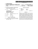 | 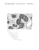 |
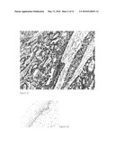 | 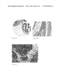 |
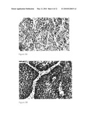 | 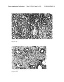 |
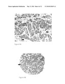 |  |
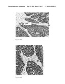 |  |
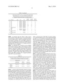 | 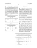 |
 |
| Similar patent applications: | |
| Date | Title |
|---|---|
| 2011-05-19 | Detection and prognosis of lung cancer |
| 2011-07-21 | Methods and devices for micro-isolation, extraction, and/or analysis of microscale components |
| 2011-07-14 | Process for predicting the prognosis of squamous cell lung cancer |
| 2011-06-30 | Method for the diagnosis of the presence of an ovarian cancer |
| 2011-07-14 | Microrna expression profile associated with pancreatic cancer |
| New patent applications in this class: | |
| Date | Title |
|---|---|
| 2022-05-05 | Method for diagnosing human t-cell leukemia virus type 1 (htlv-1) associated diseases |
| 2022-05-05 | Systems and methods for assay processing |
| 2022-05-05 | Rapid pathology/cytology without wash |
| 2019-05-16 | Biofluidic triggering system and method |
| 2019-05-16 | Method and system for detection of disease agents in blood |
| New patent applications from these inventors: | |
| Date | Title |
|---|---|
| 2015-05-28 | In situ detection of early stages and late stages hpv infection |
| 2014-06-26 | Detection of early stages and late stages hpv infection |
| 2012-12-06 | Detection of early stages and late stages hpv infection |
| 2012-11-08 | High throughput cell-based hpv immunoassays for diagnosis and screening of hpv-associated cancers |
| 2011-06-30 | Protein chips for hpv detection |
| Top Inventors for class "Chemistry: molecular biology and microbiology" | |
| Rank | Inventor's name |
|---|---|
| 1 | Marshall Medoff |
| 2 | Anthony P. Burgard |
| 3 | Mark J. Burk |
| 4 | Robin E. Osterhout |
| 5 | Rangarajan Sampath |