Patent application title: SPLIT RECOMBINANT LUCIFERASE, AND ANALYSIS METHOD USING THEREOF
Inventors:
IPC8 Class: AC12N902FI
USPC Class:
1 1
Class name:
Publication date: 2017-06-29
Patent application number: 20170183637
Abstract:
Disclosed is a split recombinant protein that includes N-terminal and
C-terminal fragments of a firefly luciferase, and a linker peptide. The
N-terminal fragment is one of two fragments of a firefly luciferase split
into two at a splitting position specific to the firefly luciferase. The
C-terminal fragment includes a C-terminal fragment of a firefly
luciferase split into two at a splitting position specific to the firefly
luciferase, and 58 to 78 amino acid residues toward the N-terminal beyond
the splitting position. When the N-terminal and C-terminal fragments are
bound together, firefly luciferase activity is exhibited.Claims:
1. A split recombinant protein comprising: an N-terminal fragment of a
firefly luciferase, the N-terminal fragment being one of two fragments of
the firefly luciferase split into two such that activity of the firefly
luciferase is restored when the two fragments are bound together; a
C-terminal fragment of a firefly luciferase, the C-terminal fragment
including 58 to 78 amino acid residues toward an N-terminal beyond a
splitting position at which the firefly luciferase can be split into two
such that activity of the firefly luciderase is restored when the two
fragments are hound together; and a linker polypeptide, wherein firefly
luciferase activity is exhibited when the N-terminal and C-terminal
fragments are bound together.
2. The split recombinant protein according to claim 1, wherein the N-terminal and C-terminal fragments are each derived from a different firefly luciferase of a different firefly species.
3. The split recombinant protein according to claim 1, wherein the N-terminal and C-terminal fragments are each derived from a firefly luciferase of a firefly selected from the group consisting of Pyrocoelia matsumurai, Drilaster Kumejimensis, and Stenocladius flavipennis.
4. The split recombinant protein according to claim 3, wherein the N-terminal fragment is derived from a firefly luciferase of Pyrocoelia matsumurai.
5. The split recombinant protein according to claim 1, wherein the N-terminal fragment is positioned on a C-terminal side in the split recombinant protein, and the C-terminal fragment is positioned on an N-terminal side in the split recombinant protein.
6. The split recombinant protein according to claim 1, further comprising, between the N-terminal and C-terminal fragments, a calcium-binding region and an interaction region that can reversibly bind to or dissociate from the calcium-binding region.
7. The split recombinant protein according to claim 6, wherein the calcium-binding region is derived from calmodulin, and the interaction region is M13 peptide.
8. A gene that encodes the split recombinant protein according to claim 1.
9. A vector comprising: a promoter sequence; and the gene according to claim 8 expressibly linked to the promoter sequence.
10. A cell comprising the vector according to claim 9.
11. A method of analyzing intracellular calcium ions, comprising: preparing a cell containing a vector that contains a promoter sequence and a gene that encodes the split recombinant protein according to claim 6, the gene being expressibly linked to the promoter sequence; adding a firefly luciferin to the cell from outside the cell; measuring a luminescence level in the cell over time; and analyzing changes in a calcium ion concentration in the cell based on changes in the luminescence level measured.
12. The method according to claim 11, wherein the method uses two or more vectors each containing a different gene that encodes the split recombinant protein having a different luminescence color.
13. The method according to claim 11, wherein the method analyzes changes in a calcium ion concentration within a single cell.
14. A method of analyzing intracellular gene expression, comprising: preparing a cell containing a vector that contains a promoter sequence, a target gene and a gene that encodes the split recombinant protein according to claim 1, the target gene and the gene being expressibly linked to the promoter sequence; adding a firefly luciferin to the cell from outside the cell; measuring a luminescence level in the cell; and analyzing an expression level of the target gene in the cell based on the luminescence level measured.
15. The method according to claim 14, wherein the method uses two or more vectors each containing a different gene that encodes the split recombinant protein having a different luminescence color.
16. A vector set comprising: A first vector containing a first promoter sequence and a first gene that encodes an N-terminal fragment of a firefly luciferase is expressibly linked to the first promoter sequence, the N-terminal fragment being one of two fragments of the firefly luciferase split into two such that activity of the firefly luciferase is restored when the two fragments are bound together; and a second vector containing a second promoter sequence and a second gene that encodes a C-terminal fragment of a firefly luciferase is expressibly linked to the second promoter sequence, the C-terminal fragment including 58 to 78 amino acid residues toward an N-terminal beyond a splitting position at which the firefly luciferase can be split into two such that activity of the firefly luciferase is restored when the two fragments are bound together.
17. The vector set according to claim 16, wherein the N-terminal and C-terminal fragments are each derived from a different firefly luciferase of a different firefly species.
18. The vector set according to claim 16, wherein either one of the and second promoter sequences is an inducible promoter sequence.
19. A method of analyzing intracellular gene expression, comprising: preparing a cell containing the vector set according to claim 18; adding a firefly luciferin to the cell from outside the cell; adding to the cell an inducer that stimulates the inducible promoter sequence; measuring a luminescence level in the cell; and analyzing an activity of the inducible promoter sequence in the cell based on the luminescence level measured.
20. A cell comprising the vector set according to claim 16.
Description:
CROSS-REFERENCE TO RELATED APPLICATION
[0001] The present application is a Continuing Application based on International Application PCT/JP2014/074606 filed on Sep. 10, 2014, the entire disclosure of this earlier application being herein incorporated by reference.
TECHNICAL FIELD
[0002] The present disclosure relates a recombinant protein, a gene encoding the recombinant protein, and an analysis method using the recombinant protein and the gene.
BACKGROUND
[0003] Reporter assay techniques and cell imaging techniques have heretofore been used for observing various biological phenomena. In particular, cell imaging techniques involve the use of genes that encode fluorescent proteins or photoproteins to modify biological cells for the analysis of various biological phenomena in the cells using fluorescence or luminescence as an indicator. Cell imaging techniques that involve the use of fluorescent proteins, however, have the drawbacks of low signal-to-noise ratios due to high levels of background fluorescence as a result of self-fluorescence of cells; a limited range of available measurement targets due to a narrow dynamic range; and so forth.
[0004] This led to the recent development of cell imaging techniques that use luciferase-luciferin luminescence. For example, PTL 1 discloses a cell-specific gene expression imaging method that uses split luciferase derived from the North American firefly (Photinus pyralis). PTL 2 discloses a calcium indicator that changes luminescence intensity in a calcium ion concentration-dependent manner. The calcium indicator is produced using a luciferase gene derived from the North American firefly (Photinus pyralis), and calmodulin gene and M13 gene whose products interact with each other in a calcium-dependent manner. PLT 2 also discloses an imaging method using this calcium indicator.
[0005] Further, PTL 3 discloses a calcium indicator that changes luminescence intensity in a calcium ion concentration-dependent manner. The calcium indicator is produced using a luciferase gene derived from the North American firefly (Photinus pyralis) and calmodulin gene whose product undergoes conformational changes in a calcium dependent manner. PTL 4 discloses split luciferases having various luminescence intensities produced using luciferase derived from the North American firefly (Photinus pyralis).
CITATION LIST
Patent Literature
[0006] PTL1: JP2007155558A
[0007] PTL2: JP201251824A
[0008] PTL3: JP201290635A
[0009] PTL4: U.S. Pat. No. 7,601,517B
Non-Patent Literature
[0010] NPL1: Miyawaki A. Llopis J, Heim R, McCaffery J M, Adams I A, Ikura M, Tsien R Y. "Fluorescent indicators for Ca2+ based on green fluorescent proteins and calmodulin." Nature, vol. 388(6645), pp. 882-887, 1997
SUMMARY
[0011] The split recombinant protein disclosed herein includes:
[0012] an N-terminal fragment of a firefly luciferase, the N-terminal fragment being one of two fragments of the firefly luciferase split into two such that activity of the firefly luciferase is restored when the two fragments are bound together;
[0013] a C-terminal fragment of a firefly luciferase, the C-terminal fragment including 58 to 78 amino acid residues toward the N-terminal beyond a splitting position at which the firefly luciferase can be split into two such that activity of the firefly luciferase is restored when the two fragments are bound together; and
[0014] a linker polypeptide,
[0015] wherein firefly luciferase activity is exhibited when the N-terminal and C-terminal fragments are bound together.
[0016] In the split recombinant protein disclosed herein, the N-terminal and C-terminal fragments may be each derived from a different firefly luciferase of a different firefly species.
[0017] In the split recombinant protein disclosed herein, the N-terminal and C-terminal fragments are each preferably derived from a firefly luciferase of a firefly selected from the group consisting of Pyrocoelia matsumurai, Drilaster Kumejimensis, and Stenocladius flavipennis.
[0018] In the split recombinant protein disclosed herein, the N-terminal fragment is preferably derived from a firefly luciferase of Pyrocoelia matsumurai.
[0019] In the split recombinant protein disclosed herein, it is preferred that the N-terminal fragment be positioned on the C-terminal side in the split recombinant protein and the C-terminal fragment on the N-terminal side in the split recombinant protein.
[0020] The split recombinant protein disclosed herein preferably further includes, a calcium-binding region and an interaction region that can reversibly bind to or dissociate from the calcium-binding region between the N-terminal and C-terminal fragments. More preferably, the calcium-binding region is derived from calmodulin, and the interaction region is M13 peptide.
[0021] The gene disclosed herein encodes the split recombinant protein.
[0022] The vector disclosed herein includes a promoter sequence, and the gene expressibly linked to the promoter sequence.
[0023] The cell disclosed herein includes the vector.
[0024] The disclosed method of analyzing intracellular calcium ions includes:
[0025] preparing a cell containing a vector that contains a promoter sequence and a gene that encodes a split recombinant protein, the gene being expressibly linked to the promoter sequence;
[0026] adding a firefly luciferin to the cell from outside the cell;
[0027] measuring a luminescence level in the cell over time: and
[0028] analyzing changes in a calcium ion concentration in the cell based on changes in the luminescence level measured,
[0029] wherein the split recombinant protein includes the calcium-binding region and the interaction region. The analysis method can also use two or more vectors each containing a different gene that encodes the split recombinant protein having a different luminescence color. The analysis method can also analyze changes in a calcium ion concentration within a single cell.
[0030] The disclosed method of analyzing intracellular gene expression includes:
[0031] preparing a cell containing a vector that contains a promoter sequence, a target gene and a gene that encodes the split recombinant protein, the both genes being expressibly linked to the promoter sequence;
[0032] adding a firefly luciferin to the cell from outside the cell;
[0033] measuring a luminescence level in the cell over time; and
[0034] analyzing changes in a expression level of the target gene in the cell based on changes in the luminescence level measured. The analysis method can also use two or more vectors each containing a different gene that encodes the split recombinant protein having a different luminescence color.
[0035] The vector set disclosed herein includes:
[0036] a first vector containing a first promoter sequence and a first gene that encodes an N-terminal fragment of firefly luciferase is expressibly linked to the first promoter sequence, the N-terminal fragment being one of two fragments of the firefly luciferase split into two such that activity of the firefly luciferase is restored when the two fragments are bound together; and
[0037] a second vector containing a second promoter sequence and a second gene that encodes a C-terminal fragment of the firefly luciferase is expressibly linked to another promoter sequence, the C-terminal fragment including 58 to 78 amino acid residues toward an N-terminal beyond a splitting position at which the firefly luciferase can be split into two such that activity of the firefly luciferase is restored when the two fragments are bound together.
[0038] In the vector set disclosed herein, the N-terminal and C-terminal fragments may be each derived from a firefly luciferase of a different firefly specie. Either one of the promoter sequences may be an inducible promoter sequence.
[0039] The disclosed method of analyzing intracellular gene expression includes:
[0040] preparing a cell containing the vector set in which either one of the first and second promoter sequences is an inducible promoter sequence;
[0041] adding a firefly luciferin to the cell from outside the cell;
[0042] adding to the cell an inducer that stimulates the inducible promoter sequence, from outside the cell;
[0043] measuring a luminescence level in the cell over time; and
[0044] analyzing changes in the activity of the inducible promoter gene in the cell based on changes in the luminescence level measured.
[0045] The cell disclosed herein includes the vector set disclosed herein.
BRIEF DESCRIPTION OF THE DRAWINGS
[0046] In the accompanying drawings:
[0047] FIG. 1 shows a schematic illustration for explaining an N-terminal fragment (NLuc) and a C-terminal fragment (CLuc);
[0048] FIG. 2 shows a schematic illustration of an example of a split recombinant protein according to an embodiment of the present disclosure, wherein an N-terminal fragment (NLuc) is positioned on the C-terminal side of a C-terminal fragment (CLuc);
[0049] FIG. 3 shows firefly luciferase activities of split recombinant proteins according to an embodiment of the present disclosure;
[0050] FIG. 4 shows measurement results of the luminescent wavelengths of permutated luciferases in E. coli and HEK293 cells (Experiment 3);
[0051] FIG. 5 shows measurement results of an experiment wherein firefly luciferase activity is restored by expressing N-terminal and C-terminal fragments of a split recombinant protein according to an embodiment of the present disclosure within a cell using separate vectors;
[0052] FIG. 6 shows images of results of promoter activity imaging in HEK293 cells using a vector set according to an embodiment of the present disclosure;
[0053] FIG. 7 shows analysis results of promoter activity in HEK293 cells using a vector set according to an embodiment of the present disclosure;
[0054] FIG. 8 shows images of results of promoter activity imaging in neuronal cells using a vector set according to an embodiment of the present disclosure;
[0055] FIG. 9 shows analysis results of promoter activity in neuronal cells using a vector set according to an embodiment of the present disclosure;
[0056] FIG. 10 shows a schematic illustration of a split recombinant protein (luminescent calcium indicator) according to an embodiment of the present disclosure;
[0057] FIG. 11 shows comparison results of luminescence levels of split recombinant proteins (luminescent calcium indicators) according to an embodiment of the present disclosure;
[0058] FIG. 12 shows results of calcium calibration of luminescent calcium indicators (Experiment 8);
[0059] FIG. 13 shows analysis results of intracellular calcium ion concentrations in HEK293 cells upon ATP stimulation (Experiment 9);
[0060] FIG. 14 shows images and graphs of results of luminescence-based calcium imaging in HEK293 cells upon ATP stimulation (Experiment 10); and
[0061] FIG. 15 shows images and graphs of results of high-speed. luminescence imaging of changes in calcium ion concentrations in HEK293 cells (Experiment 11).
DETAILED DESCRIPTION
[0062] The present disclosure will now be described in detail based on embodiments.
[0063] <Split Recombinant Protein>
[0064] A split recombinant protein disclosed herein includes:
[0065] an N-terminal fragment of firefly luciferase, the N-terminal fragment being one of two fragments of the firefly luciferase split into two such that luciferase activity is restored when the two fragments are bound together;
[0066] a C-terminal fragment of the firefly luciferase, the C-terminal fragment including 58 to 78 amino acid residues toward the N-terminal beyond a splitting position at which the firefly luciferase can be split into two such that luciferase activity is restored when the two fragments are bound together; and
[0067] a linker polypeptide that links together the N-terminal and C-terminal fragments,
[0068] wherein firefly luciferase activity is restored when the N-terminal and C-terminal fragments are bound together.
[0069] Luciferase generally refers to a class of enzymes that catalyze a chemical reaction that generates luminescence. The substance that serves as the substrate for the enzyme is called luciferin. Luminescence occurs when luciferin undergoes a chemical change by the catalytic action of luciferase in the presence of adenosine tri phosphate (ATP). Currently available luciferases are those from fireflies and bacteria, but they greatly differ for example in their protein structure and substrate. The present disclosure is directed to firefly luciferase, which is luciferase derived from firefly. The substrate for firefly luciferase is firefly luciferin. Preferably, the firefly luciferin is D-luciferin. By "luciferase activity" is meant an ability to trigger luminescence by catalyzing chemical changes in the substrate luciferin in the presence of ATP. Namely, by "firefly luciferase activity" is meant an ability to trigger luminescence by catalyzing chemical changes in the substrate firefly luciferin in the presence of ATP.
[0070] Luciferases are known that are deactivated when split at a specific position into two N-terminal and C-terminal fragments which do not exhibit luciferase activity alone, but can restore luciferase activity once the fragments are bound together to reconstruct the protein (see, e.g., PTL 1 and PTL 4). The N-terminal and C-terminal fragments of luciferase split in this way are also collectively called "split luciferase." The term "bind" and any modified form thereof as used in the context of the present disclosure refers to binding by any bond, which may be accomplished by a covalent bond, or a noncovalent bond such as ionic bond, hydrogen bond, Van der Waals force or hydrophobic interaction. Binding also may encompass states wherein two or more "bound" targets come close, contact, associate or interact with each other. Specifically, the "binding" between the N-terminal and C-terminal fragments may be accomplished by any bond, and may be accomplished by a covalent bond or a noncovalent bond such as ionic bond, hydrogen bond, Van der Waals force or hydrophobic interaction. The mode of "binding" between the N-terminal and C-terminal fragments may encompass modes wherein the two fragments come close, contact, associate or interact with each other to an extent that they may play a function of luciferase. It should be noted however that modes of binding wherein the N-terminal and C-terminal fragments have only a linkage via a linker peptide are excluded.
[0071] By "firefly" is meant an insect that belongs to the Arthropoda: Insecta: Coleoptera: Lampyridae. Any species of firefly can be used for luciferases usable in the present disclosure. Examples of fireflies include Pyrocoelia matsumurai, Drilaster Kumejimensis, Stenocladius flavipennis, North American firefly (Photinus pyralis), Luciola cruciata, Luciola Lateralis, East European firefly (Luciola mingrelica), and Lampyris noctiluca, with Pyrocoelia matsumurai, Drilaster Kumejimensis, and Stenocladius flavipennis being preferred.
[0072] The amino acid sequence of firefly luciferase is generally unique to each species of firefly from which the firefly luciferase is derived and thus may differ depending on the species of firefly from which the firefly luciferase is derived. The "splitting position" at which firefly luciferase is split into two N-terminal and C-terminal fragments such that luciferase activity is restored when the fragments are bound together can be at any position so long as it is a position at which the firefly luciferase can be split into two such that luciferase activity is restored when the split fragments are bound together, and may differ depending on the species of firefly from which the firefly luciferase is derived. A person skilled in the art would be able to determine the splitting position in firefly luciferase using any of the methods known in the art. Any information known in the art can also be utilized as to the splitting position. The amino acid sequences of the "N-terminal fragment" and "C-terminal fragment" included in the split recombinant protein disclosed herein may also differ depending on the species of firefly from which firefly luciferase is derived.
[0073] The "N-terminal fragment" included in the split recombinant protein disclosed herein is an N-terminal fragment of firefly luciferase split into two such that luciferase activity is restored when the two fragments are bound together, and has an amino acid sequence from residue 1 to the N-terminal side residue adjacent to the "split position" of the firefly luciferase. The "C-terminal fragment" included in the split recombinant protein disclosed herein may derived from firefly luciferase of a firefly which is different from the firefly from which the firefly luciferase for the N-terminal fragment is derived. The "C-terminal fragment" is a C-terminal fragment which contains 58 to 78 amino acid residues toward the N-terminal beyond a splitting position at which the firefly luciferase can be split into two such that firefly luciferase activity is restored when the fragments are bound together. These "N-terminal fragment" and "C-terminal fragment" may be derived from wide-type firefly luciferase and may also have a mutation such as substitution, deletion or addition of one or more amino acid residues to an extent that firefly luciferase activity can be restored when the fragments are bound together.
[0074] For example, wide-type firefly luciferase derived from Pyrocoelia matsumurai consists of 562 amino acids encoded by a nucleotide sequence having SEQ ID NO: 1, and has an amino acid sequence having SEQ ID NO: 2. By way of one example, the firefly luciferase can be split into an N-terminal fragment having residues 1 to 416 (SEQ ID NO: 3) and a C-terminal fragment having residues 417 to 562 (SEQ ID NO: 4) such that firefly luciferase activity can be restored when the fragments are bound together. Thus, the "N-terminal fragment" derived from Pyrocoelia matsumurai included in the split recombinant protein disclosed herein may have an amino acid sequence from residues 1 to 416 of wide-type luciferase of Pyrocoelia matsumnurai. The "C-terminal fragment" derived from Pyrocoelia matsumurai included in the split recombinant protein disclosed herein may have an amino acid sequence from any one of residues 339 to 359 to residue 562 of wide-type luciferase of Pyrocoelia matsumurai.
[0075] Wide-type firefly luciferase derived from Drilaster Kumejimensis consists of 547 amino acids encoded by a nucleotide sequence having SEQ ID NO: 5, and has an amino acid sequence having SEQ ID NO: 6. By way of one example, the firefly luciferase can be split into an N-terminal fragment having residues 1 to 416 (SEQ ID NO: 7) and a C-terminal fragment having residues 417 to 547 (SEQ ID NO: 8) such that firefly luciferase activity can be restored when the fragments are bound together. Thus, the "N-terminal fragment" derived from Drilaster Kumejimensis included in the split recombinant protein disclosed herein may have an amino acid sequence from residues 1 to 416 of wide-type luciferase of Drilaster Kumejimensis. The "C-terminal fragment" derived from Drilaster Kumejimensis included in the split recombinant protein disclosed herein may have an amino acid sequence from any one of residues 339-359 to residue 547 of wide-type luciferase of Drilaster Kumejimensis.
[0076] Wide-type firefly luciferase derived from Stenocladius flavipennis consists of 555 amino acids encoded by a nucleotide sequence having SEQ ID NO: 9, and has an amino acid sequence having SEQ ID NO: 10. By way of one example, the firefly luciferase can be split into an N-terminal fragment having residues 1 to 424 (SEQ ID NO: 11) and a C-terminal fragment having residues 425 to 555 (SEQ ID NO: 12) such that firefly luciferase activity can be restored when the fragments are bound together. Thus, the "N-terminal fragment" derived from Stenocladius flavipennis included in the split recombinant protein disclosed herein may have an amino acid sequence from residue 1 to 424 of wide-type luciferase of Stenocladius flavipennis. The "C-terminal fragment" derived from Stenocladius flavipennis included in the split recombinant protein disclosed herein may have an amino acid sequence from any one of residues 347-367 to residue 555 of wide-type luciferase of Stenocladius flavipennis.
[0077] Wide-type firefly luciferase derived from the North American firefly (Photinus pyralis) consists of 550 amino acids. By way of one example, the firefly luciferase can be split into an N-terminal fragment having residues 1 to 416 and a C-terminal fragment having residues 417 to 550 such that firefly luciferase activity can be restored when the fragments are bound together. Thus, the "N-terminal fragment" derived from the North American firefly included in the split recombinant protein disclosed herein may have an amino acid sequence from residues 1 to 416 of wide-type luciferase of the North American firefly. The "C-terminal fragment" derived from the North American firefly included in the split recombinant protein disclosed herein may have an amino acid sequence from any one of residues 339-364 to residue 550 of wide-type luciferase of the North American firefly.
[0078] The split recombinant protein disclosed herein can restore firefly luciferase activity that provides at least several-fold higher luminescence intensity than that of wide-type firefly luciferase derived from the North American firefly (Photinus pyralis), when the "N-terminal fragment" and "C-terminal fragment" are "bound" together to reconstruct the protein. The N-terminal and C-terminal fragments may be derived from firefly luciferase of the same or different species of firefly. The N-terminal and C-terminal fragments of the luciferase can be derived from any combination of fireflies, but are each preferably selected from the group consisting of Pyrocoelia matsumurai, Drilaster Kumejimensis, and Stenocladius flavipennis. The N-terminal fragment can be derived from any species of firefly but is preferably derived from Pyrocoelia matsumurai.
[0079] The restored firefly luciferase activity provides luminescence with different luminescence colors, i.e., different peak wavelengths, depending on the species of firefly from which the N-terminal and C-terminal fragments of firefly luciferase are derived, the number of N-terminal side amino acid residues of the C-terminal fragment, or combinations thereof. A specific example thereof is demonstrated in Experiment 3 in Examples (FIG. 4).
[0080] The split recombinant protein disclosed herein includes a linker peptide in addition to the N-terminal and C-terminal fragments disclosed herein. Any number and any type of amino acids can be used for the linker peptide so long as the linker peptide can link together the N-terminal and C-terminal fragments disclosed herein, or the N-terminal or C-terminal fragment disclosed herein and another protein, polypeptide or fragment thereof having some function, without compromising their function. By "split" is meant that "N-terminal fragment" and "C-terminal fragment" do not exhibit firefly luciferase activity alone but can restore firefly luciferase activity once they are bound together to reconstruct the protein.
[0081] In the split recombinant protein disclosed herein, the N-terminal and C-terminal fragments may assume the original position in firefly luciferase, i.e., the N-terminal fragment may be positioned on the N-terminal side of the C-terminal fragment. Alternatively, the N-terminal and C-terminal fragments may be circularly permutated from the original position in firefly luciferase, i.e., the N-terminal fragment may be positioned on the C-terminal side of the C-terminal fragment.
[0082] The split recombinant protein disclosed herein can include one or more proteins, polypeptides or fragments thereof having some function, in addition to the N-terminal fragment, C-terminal fragment and linker peptide disclosed herein. Such proteins are generally also called fusion proteins. In the present disclosure, a peptide portion composed of the one or more proteins, polypeptides or fragments thereof is also referred to as a "region." Examples of such a functional region include, but not limited to, calcium-binding regions, cyclic AMP-binding regions, cyclic GMP-binding regions, and interaction regions that interact with these binding regions. These regions are preferably positioned between the N-terminal and C-terminal fragments.
[0083] By way of one example, the following describes a split recombinant protein that includes a calcium-binding region and an interaction region that can interact with the calcium-binding region. By "calcium-binding region" is meant a peptide portion that can reversibly bind to or dissociate from calcium ion (Ca.sup.2+) within a cell. By "interaction region" that can interact with a calcium-binding region is meant a peptide portion that can reversibly bind to or dissociate from a calcium-binding region within a cell. Examples of calcium-binding regions include calmodulin (CaM) or fragments thereof. The interaction region can bind to a calcium-binding region bound to calcium ion, and can dissociate from a calcium-binding region from which calcium ion has dissociated. Examples of interaction regions that can interact with a calcium-binding region include M13 peptide. A split recombinant protein that includes a calcium-binding region and an interaction region is expressed for example within a cell, whereby intracellular calcium ion dynamics can be analyzed.
[0084] As the calcium-binding region and interaction region, it is possible to use a peptide in which the calcium-binding protein calmodulin and M13 (which binds with calcium-bound calmodulin) are sequentially linked (NPL 1). NPL 1 uses this peptide in which calmodulin and M13 are sequentially linked to produce a calcium sensor protein in which CaM and M13 are flanked by two different fluorescent proteins, and this fluorescent protein is called a "cameleon protein" (hereinafter also referred to as "cameleon"). In particular, as the calcium-binding region and interaction region, it is possible to use a peptide having an amino acid sequence corresponding to residues 230 to 406 of cameleon; a peptide having an amino acid sequence corresponding to residues 230 to 396 of cameleon; a peptide having an amino acid sequence corresponding to residues 230 to 401 of cameleon, a peptide having an amino acid sequence corresponding to residues 230 to 411 of cameleon; or a peptide having an amino acid sequence corresponding to residues 230 to 416 of cameleon. These peptides with specific amino acid sequences can be used for the split recombinant protein disclosed herein even when the peptides have mutations for example for improving sensitivity so long as their function is not compromised.
[0085] <Gene>
[0086] The present disclosure also relates to a gene that encodes a split recombinant protein, a gene that encodes an N-terminal fragment, and a gene that encodes a C-terminal fragment. In the present disclosure, a "gene" that encodes a protein, polypeptide or fragment thereof may be a DNA or RNA strand, and is intended to mean a DNA or RNA strand having a nucleotide sequence capable of expressing the protein, polypeptide or fragment thereof within a cell or other target. The DNA and RNA strands may consist only of a nucleotide sequence that encodes an amino acid sequence of the protein, polypeptide or fragment thereof, or may include, in addition to the nucleotide sequence, additional nucleotide(s) so long as the function and expression of the protein are not compromised. These nucleotide sequences may have a mutation such as substitution, deletion or addition of one or more nucleotides so long as the function and expression of the protein are not compromised.
[0087] <Vector>
[0088] The vector disclosed herein is an expression vector containing a gene that encodes a split recombinant protein, a gene that encodes an N-terminal fragment, or a gene that encodes a C-terminal fragment in such a manner that the split recombinant protein, the N-terminal fragment or the C-terminal fragment can be expressed. By "expression vector" is meant a vector in which a gene to be expressed (e.g., a gene that encodes a split recombinant protein, a gene that encodes an N-terminal fragment, a gene that encodes a C-terminal fragment, or a target gene) is expressibly linked. For example, a gene that encodes a split recombinant protein, a gene that encodes an N-terminal fragment, and a gene that encodes a C-terminal fragment are expressibly linked downstream of the promoter region in an expression vector. Any type of vector can be used. Examples of usable vectors include plasmid vectors, phage vectors, and cosmids. A person skilled in the art would be able to select a proper vector based on such conditions as cloning site, promoter sequence, products to be expressed (split recombinant protein, N-terminal fragment, or C-terminal fragment) and/or expression cells in light of common technical knowledge, to construct a desired expression vector in accordance with common procedures. The promoter sequence can also be selected as appropriate in accordance, for example, with the purpose of study. The promoter sequence may be a constitutive promoter sequence, inducible promoter sequence, or tissue-specific promoter sequence.
[0089] <Vector Set>
[0090] Another embodiment of the present disclosure is directed to a vector set including:
[0091] a first vector containing a first promoter sequence and a first gene that encodes an N-terminal fragment of firefly luciferase is expressibly linked to the first promoter sequence, the N-terminal fragment being one of two fragments of the firefly luciferase split into two such that luciferase activity is restored when the two fragments are bound together; and
[0092] a second vector containing a second promoter sequence and a second gene that encodes a C-terminal fragment of the firefly luciferase is expressibly linked to the second promoter sequence, the C-terminal fragment including 58 to 78 amino acid residues toward the N-terminal beyond a splitting position at which the firefly luciferase can be split into two such that firefly luciferase activity is restored when the two fragments are bound together.
[0093] In this vector set, the N-terminal and C-terminal fragments may be derived from a firefly luciferase of the same or different species of firefly. The vector set may further include another vector in which another N-terminal or C-terminal fragment gene is expressibly linked to another promoter sequence. Further, the vector set can be used in combination with another vector set in which at least one of the gene that encodes an N-terminal fragment, the gene that encodes a C-terminal fragment and the promoter sequence is different. Such a vector set can be constructed as appropriate in accordance with common procedures based on the purpose of study and/or conditions in light of common technical knowledge. One of the promoter sequences used in the vector set may be an inducible promoter sequence. Such a vector set can be used for example in the analysis methods of intracellular gene expression described below.
[0094] <Cell>
[0095] The cell disclosed herein contains the vector disclosed herein, i.e., a vector in which a promoter sequence, and a gene that encodes a split recombinant protein, a gene that encodes an N-terminal fragment or a gene that encodes a C-terminal fragment are expressibly linked; or the vector set described above. The cell disclosed herein may be any cell so long as a product to be expressed from an expression vector can be expressed therein, and may be animal or plant cell. Usable cells may be of any origin which can be determined as appropriate in accordance, for example, with the purpose of study. Any method can be used to transfer an expression vector into a cell. Examples of transfer methods include transfection, in vitro packaging, freezing and thawing, and electroporation. The transfer method can be determined as appropriate in accordance with the types of vector and cell used.
[0096] <Analysis Method>
[0097] The phenomenon in which firefly luciferase activity is restored by reconstruction of the split recombinant protein disclosed herein can be used in various analysis methods. For example, the capability of using high luminescence intensity as an indicator and the availability of different luminescence colors enable calcium ion behavior, expression of target genes and promoter sequences, protein-protein interactions, receptor-receptor interactions and other like events in living cells to be analyzed highly precisely either on a single-cell basis or in comparison with a plurality of cells or genes. Thus, the analysis method disclosed herein can be used for imaging, single-cell imaging, high-speed imaging, multicolor imaging and other techniques in cells with weak expression of foreign genes.
[0098] An example of an embodiment of the analysis method will now be described.
[0099] <Method of Analyzing Intracellular Calcium Ions>
[0100] An embodiment of the present disclosure is directed to a method of analyzing intracellular calcium ions which includes:
[0101] preparing a cell containing a vector that contains a promoter sequence and a gene that encodes a split recombinant protein, the gene being expressibly linked to the promoter sequence;
[0102] adding firefly luciferin to the cell from outside the cell;
[0103] measuring the luminescence level in the cell; and
[0104] analyzing the calcium ion concentration in the cell based on the luminescence level measured. The split recombinant protein includes a calcium-binding region and an interaction region that interacts with the calcium-binding region. In the present disclosure, the split recombinant protein is also referred to as a luminescent calcium indicator.
[0105] In the cell preparation step, the expression vector properly constructed according to the purpose of study and conditions may be transferred into a cell properly selected according to the purpose of study and conditions, as described above. In the cell prepared in this way, a luminescent calcium indicator is expressed. When the promoter sequence is a constitutive promoter sequence, the luminescent calcium indicator is expressed without stimulation such as addition of an inducer. When the promoter sequence is an inducible promoter sequence, the luminescent calcium indicator is expressed in the presence of stimulation such as addition of an inducer. When the promoter sequence is a tissue-specific promoter sequence, the luminescent calcium indicator is expressed only within a cell derived from specific tissue.
[0106] In the step of adding firefly luciferin to the prepared cell from outside the cell, addition of firefly luciferin from outside the cell allows the firefly luciferin to penetrate through the plasma membrane into the cell. In the presence of firefly luciferin, the luminescent calcium indicator can restore or lose its firefly luciferase activity according to the presence of calcium ions. It is the conformation of the luminescent calcium indicator, as an entire protein, that determines whether firefly luciferase activity is restored or lost in the presence of calcium ions.
[0107] By way of one example, a luminescent calcium indicator will be described that includes, in the order from the N-terminal, a C-terminal fragment, a CaM-M13 fragment of cameleon (residues 230 to 406), and an N-terminal fragment. In the absence of calcium ions; the C-terminal and N-terminal fragments moderately come close with each other and bind together to restore firefly luciferase activity to produce luminescence. On the other hand, in the presence of calcium ions, the calcium ions bind to the calcium-binding region (CaM) and then the interaction region (M13) binds with CaM, resulting in conformational changes of the luminescent calcium indicator as an entire protein to lose firefly luciferase activity and therefore luminescence. Based on such a mechanism, the intracellular calcium ion concentration is reflected by the luminescence level of firefly luciferase.
[0108] Based on the measured luminescence level, the intracellular calcium ion concentration can be analyzed. Measurements of luminescence level over time enables changes in intracellular calcium ion concentrations to be analyzed using changes in luminescence levels as an indicator. Any of the commonly used devices and analysis software can be used for the measurement of luminescence level and for the analysis based on the measured luminescence level. Measurements may be made by image capturing. Examples of such devices include luminometers, luminescence microscopes, luminescence imagers, and luminescence detectors.
[0109] <Method of Analyzing Intracellular Gene Expression>
[0110] Another embodiment of the present disclosure is directed to a method of analyzing intracellular gene expression which includes:
[0111] preparing a cell containing an expression vector that contains a target gene and a gene that encodes a split recombinant protein;
[0112] adding firefly luciferin to the cell from outside the cell;
[0113] measuring the luminescence level in the cell; and
[0114] analyzing the expression level of the target gene in the cell based on the luminescence level measured.
[0115] In the cell preparation step, the expression vector properly constructed according to the purpose of study and conditions may be transferred into a cell properly selected according to the purpose of study and conditions, as described above. The target gene may be a promoter sequence. In the cell prepared in this way, a target gene and a split recombinant protein are expressed. The target gene may be included in the gene that encodes the split recombinant protein. When the promoter sequence is a constitutive promoter sequence, the target gene and the split recombinant protein are expressed without stimulation such as addition of an inducer. When the promoter sequence is an inducible promoter sequence, the target gene and the split recombinant protein are expressed in the presence of stimulation such as addition of an inducer. When the promoter sequence is a tissue-specific promoter sequence, the target gene and the split recombinant protein are expressed only within a cell derived from specific tissue.
[0116] In the step of adding firefly luciferin to the prepared cell from outside the cell, addition of firefly luciferin from outside the cell allows the firefly luciferin to penetrate through the plasma membrane into the cell. When the target gene and the split recombinant protein have been expressed in the presence of firefly luciferin, the split recombinant protein restores firefly luciferase activity and thus luminescence is detected.
[0117] Based on the measured luminescence level, the intracellular expression level of the target gene can be analyzed. Measurements of luminescence level over time enables changes in intracellular expression levels to be analyzed using changes in luminescence levels as an indicator. Any of devices and analysis software that can be used for the above-described analysis method of intracellular calcium ions can be used for the measurement of luminescence level and for the analysis based on the measured luminescence level.
[0118] In the analysis methods described above, it is possible to use two or more expression vectors containing genes that encode the split recombinant proteins having different luminescence colors. Transfer of such two or more different expression vectors into separate cells and subsequence measurement of the luminescence color unique to each split recombinant protein expressed from the transferred expression vector enables comparison of intracellular calcium ion concentration or intracellular gene expression level between or among the cells. Further, transfer of such two or more different expression vectors into a single cell enables comparison of intracellular calcium ion concentration or intracellular gene expression level within the cell.
[0119] <Method of Analyzing Intracellular Gene Expression Using Vector Set>
[0120] Another embodiment of the present disclosure is directed to a method of analyzing intracellular gene expression which includes:
[0121] preparing a cell containing the vector set in which either one of the promoter sequences is an inducible promoter sequence;
[0122] adding firefly luciferin to the cell from outside the cell;
[0123] adding to the cell an inducer that stimulates the inducible promoter sequence;
[0124] measuring the luminescence level in the cell over time; and
[0125] analyzing changes in the activity of the promoter sequence in the cell based on changes in the luminescence level measured.
[0126] In the cell preparation step, the expression vector properly constructed according to the purpose of study and conditions may be transferred into a cell properly selected according to the purpose of study and conditions, as described above. In the cell prepared in this way, N-terminal and C-terminal fragments are separately expressed under the regulation of separate promoter sequences. The gene linked to the constitutive promoter sequence is expressed without stimulation such as addition of an inducer. On the other hand, the gene linked to the inducible promoter sequence is expressed to produce an N-terminal or C-terminal fragment in the presence of stimulation such as addition of an inducer. When the promoter sequence is a tissue-specific promoter sequence, the N-terminal or C-terminal fragment is expressed only within a cell derived from specific tissue.
[0127] In the step of adding firefly luciferin to the prepared cell from outside the cell, addition of firefly luciferin from outside the cell allows the firefly luciferin to penetrate through the plasma membrane into the cell. When both the N-terminal and C-terminal fragments have been expressed in the presence of firefly luciferin, the split recombinant protein can be reconstructed to restore firefly luciferase activity and thus luminescence is detected. Since one of the promoter sequences is an inducible promoter sequence in the above-described analysis method, either one of the split recombinant protein fragments whose expression is regulated by the inducible promoter sequence is expressed only in the presence of an inducer that stimulates the inducible promoter sequence.
[0128] Thus, the above-described analysis method includes adding an inducer that stimulates the inducible promoter sequence. Addition of a proper inducer results in stimulation and thus expression of the inducible promoter sequence; which in turn results in the expression of a split recombinant protein fragment under the regulation of the inducible promoter sequence. As a consequence, both the N-terminal and C-terminal fragments are present within a cell allowing a split recombinant protein to be reconstructed to restore firefly luciferase activity and thus luminescence is detected. Depletion of the inducer for example by intracellular metabolism stops the expression of the inducible promoter sequence and therefore the expression of a split recombinant protein fragment under the regulation of the inducible promoter sequence as well.
[0129] In this way it is possible to analyze the intracellular expression level of the inducible promoter sequence based on the luminescence level measured. Measurements of luminescence level over time enables changes in intracellular expression levels of the inducible promoter sequence to be analyzed using changes in luminescence levels as an indicator. Any of devices and analysis software that can be used for the above-described analysis methods of intracellular calcium ions and intracellular gene expression can be used for the measurement of luminescence level and for the analysis based on the measured luminescence level.
[0130] In the above-described analysis method, the vector set may further include another vector in which another N-terminal or C-terminal fragment gene is expressibly linked to another promoter sequence. Further, the vector set can be used in combination with another vector set in which at least one of the gene that encodes an N-terminal fragment, the gene that encodes a C-terminal fragment and the promoter sequence is different.
EXAMPLES
[0131] The present disclosure will now be described in detail based on Examples, which however shall not be construed as being limiting in any way. In Examples, a split recombinant protein in which N-terminal and C-terminal fragments are derived from firefly luciferase of the same species of firefly and in which the N-terminal fragment is positioned on the C-terminal side of the C-terminal fragment is also called a permutated firefly luciferase. Further, in Examples, a split recombinant protein in which N-terminal and C-terminal fragments are derived from firefly luciferase of different species of firefly and in which the N-terminal fragment is positioned on the C-terminal side of the C-terminal fragment is also called a heterologous permutated firefly luciferase.
[0132] [Pre-Preparation 1: Cloning of N-Terminal Fragment and C-Terminal Fragment Genes of Firefly Luciferase for Preparation of Permutated Firefly Luciferases]
[Procedure 1]
[0133] For the production of N-terminal fragment (NLuc) genes and C-terminal fragment (CLuc) genes of firefly luciferases derived from Pyrocoelia Matsumurai (hereinafter also designated as "OKI"), Drilaster Kumejimensis (hereinafter also designated as "KUME"), and Stenocladius flavipennis (hereinafter also designated as "SfRE"), synthetic oligo DNAs for PCR having the following sequences were prepared.
[0134] <Synthetic Oligo DNAs for Preparation of OKI Luciferase Fragments>
TABLE-US-00001 OKI-N_Bgl_Fw: (SEQ ID NO: 13) 5'-AGATCTGAGGACGACCACAAGAACATCGTG-3' OKI-N_Eco_Rv: (SEQ ID NO: 14) 5'-GAATTCATCCTTATCGATCAGGGCATTGGT-3' OKI399_Bam_Fw: (SEQ ID NO: 15) 5'-GGATCCGCCACCATGAAGGGCTACGTGAACAACCCC-3' OKI394_Bam_Fw: (SEQ ID NO: 16) 5'-GGATCCGCCACCATGAAGGGCCCCATGATTATGAAG-3' OKI389_Bam_Fw: (SEQ ID NO: 17) 5'-GGATCCGCCACCATGGGAGAGCTGTGCCTGAAGGGC-3' OKI384_Bam_Fw: (SEQ ID NO: 18) 5'-GGATCCGCCACCATGGGCGTGAACCAGCGCGGAGAG-3' OKI379_Bam_Fw: (SEQ ID NO: 19) 5'-GGATCCGCCACCATGACCAGCAAGACGCTGGGCGTG-3' OKI374_Bam_Fw: (SEQ ID NO: 20) 5'-GGATCCGCCACCATGATTGTGGATCTGGATACCAGC-3' OKI369_Bam_Fw: (SEQ ID NO: 21) 5'-GGATCCGCCACCATGTTCTTCAGCGCCAAGATTGTG-3' OKI364_Bam_Fw: (SEQ ID NO: 22) 5'-GGATCCGCCACCATGGGAAAGGTGGCCCCATTCTTC-3' OKI359_Bam_Fw: (SEQ ID NO: 23) 5'-GGATCCGCCACCATGAAGCCAGGCGCCTGCGGAAAG-3' OKI354_Bam_Fw:: (SEQ ID NO: 24) 5'-GGATCCGCCACCATGCCACGCGGCGACGATAAGCCAGGC-3' OKI349_Bam_Fw: (SEQ ID NO: 25) 5'-GGATCCGCCACCATGGCCGTGATTATTACCCCACGCGGC-3' OKI344_Bam_Fw: (SEQ ID NO: 26) 5'-GGATCCGCCACCATGACCGAGACCACCAGCGCCGTGATT-3' OKI339_Bam_Fw: (SEQ ID NO: 27) 5'-GGATCCGCCACCATGCAGGGCTATGGCCTGACCGAGACC-3' OKI-C_Xho_Rv: (SEQ ID NO: 1) 5'-CTCGAGCAGCTTGGACTTCTTGCCCATCGT-3'
[0135] <Synthetic Oligo DNAs for Preparation of KUME Luciferase Fragments>
TABLE-US-00002 KUME-N_Bgl_Fw: (SEQ ID NO: 28) 5'-AGATCTGACATGGAAGATAAGAACGTGGTG-3' KUME-N_Eco_Rv: (SEQ ID NO: 29) 5'-GAATTCATCCTTGTCGATCAGGGCGTTGGT-3' KUME399_Bam_Fw: (SEQ ID NO: 30) 5'-GGATCCGCCACCATGAAGGGCTACGCCAACAACCCC-3' KUME394_Bam_Fw: (SEQ ID NO: 31) 5'-GGATCCGCCACCATGAAGGGCGACATGATCATGAAG-3' KUME389_Bam_Fw: (SEQ ID NO: 32) 5'-GGATCCGCCACCATGGGCGAGCTGTGCCTGAAGGGC-3' KUME384_Bam_Fw: (SEQ ID NO: 33) 5'-GGATCCGCCACCATGGGCCCTCACCAGAAAGGCGAG-3' KUME379_Bam_Fw: (SEQ ID NO: 34) 5'-GGATCCGCCACCATGACCAGACAGAGCCTGGGCCCT-3' KUME374_Bam_Fw: (SEQ ID NO: 35) 5'-GGATCCGCCACCATGATCATCGACCTGGACACCAGA-3' KUME369_Bam_Fw: (SEQ ID NO: 36) 5'-GGATCCGCCACCATGTTCTTCAGCGCCAAGATCATC-3' KUME364_Bam_Fw: (SEQ ID NO: 37) 5'-GGATCCGCCACCATGGGCAAGGTGGTGCCATTCTTC-3' KUME359_Bam_Fw: (SEQ ID NO: 38) 5'-GGATCCGCCACCATGAAGGCCGGCTCTACAGGCAAG-3' KUME354_Bam_Fw: (SEQ ID NO: 39) 5'-GGATCCGCCACCATGCCCGAGGGCGAAGATAAGGCCGGC-3' KUME349_Bam_Fw: (SEQ ID NO: 40) 5'-GGATCCGCCACCATGGCCGTGATCATCACACCCGAGGGC-3' KUME344_Bam_Fw: (SEQ ID NO: 41) 5'-GGATCCGCCACCATGACAGAGACAACCAGCGCCGTGATC-3' KUME339_Bam_Fw: (SEQ ID NO: 42) 5'-GGATCCGCCACCATGCAGGGCTACGGACTGACAGAGACA-3' KUME-C_Xho_Rv: (SEQ ID NO: 43) 5'-CTCGAGCATCTTGCTCTGGGGCCGCTTCAG-3'
[0136] <Synthetic Oligo DNAs for Preparation of SfRE Luciferase Fragments>
TABLE-US-00003 SfRE-N_Bgl_Fw: (SEQ ID NO: 44) 5'-AGATCTGCCAGCAGCATGATGAGCAAGAAG-3' SfRE-N_Eco_Rv: (SEQ ID NO: 45) 5'-GAATTCATCCTTGTCGATCATCTCGICGGT-3' SfRE407_Bam_Fw: (SEQ ID NO: 46) 5'-GGATCCGCCACCATGATGGGCTACTGCAACAACAAG-3' SfRE402_Bam_Fw: (SEQ ID NO: 47) 5'-GGATCCGCCACCATGAAGGGCGACATGATCATGATG-3' SfRE397_Bam_Fw: (SEQ ID NO: 48) 5'-GGATCCGCCACCATGGGAGAACTCTACCTGAAGGGC-3' SfRE392_Bam_Fw: (SEQ ID NO: 49) 5'-GGATCCGCCACCATGGGCCCTCACCAGAGGGGAGAA-3' SfRE387_Bam_Fw: (SEQ ID NO: 50) 5'-GGATCCGCCACCATGAGCGGCAAGAGCGTGGGCCCT-3' SfRE382_Bam_Fw: (SEQ ID NO: 51) 5'-GGATCCGCCACCATGATCGTGGACCTGAACAGCGGC-3' SfRE377_Bam_Fw: (SEQ ID NO: 52) 5'-GGATCCGCCACCATGTTCTTCAGCGCCAAGATCGTG-3' SfRE372_Bam_Fw: (SEQ ID NO: 53) 5'-GGATCCGCCACCATGGGCAAGGTGGTGCCATTCTTC-3' SfRE367_Bam_Fw: (SEQ ID NO: 54) 5'-GGATCCGCCACCATGAAGCCTGGCTCTACAGGCAAG-3' SfRE362_Bam_Fw: (SEQ ID NO: 55) 5'-GGATCCGCCACCATGCCCGAGGGCGAGGATAAGCCTGGC-3' SfRE357_Bam_Fw: (SEQ ID NO: 56) 5'-GGATCCGCCACCATGGCCGTGATCATCACCCCCGAGGGC-3' SfRE352_Bam_Fw: (SEQ ID NO: 57) 5'-GGATCCGCCACCATGACCGAGACAACCAGCGCCGTGATC-3' SfRE347_Bam_Fw: (SEQ ID NO: 58) 5'-GGATCCGCCACCATGCAGGGCTACGGCCTGACCGAGACA-3' SfRE-C_Xho_Rv: (SEQ ID NO: 59) 5'-CTCGAGCACTTGCTCTGGGGTTTCTTCAG-3'
[0137] [Procedure 2] (PCR Cloning of Firefly Luciferase Fragments)
[0138] Using the OKI gene as a template and the above-described synthetic oligo DNAs as primers, the following genes for production of permutated firefly luciferases were amplified by PCR: N-terminal fragment gene (OKI-N: including a nucleotide sequence encoding residues 1 to 416 of OKI-derived luciferase), and C-terminal fragment genes encoding C-terminal fragments with different numbers of amino acid residues (OKI399: including a nucleotide sequence encoding residues 399 to 562 of OKI-derived luciferase; OKI394: including a nucleotide sequence encoding residues 394 to 562 of OKI-derived luciferase; OKI389: including a nucleotide sequence encoding residues 389 to 562 of OKI-derived luciferase; OKI384: including a nucleotide sequence encoding residues 384 to 562 of OKI-derived luciferase; OKI379: including a nucleotide sequence encoding residues 379 to 562 of OKI-derived luciferase; OKI374: including a nucleotide sequence encoding residues 374 to 562 of OKI-derived luciferase; OKI369: including a nucleotide sequence encoding residues 369 to 562 of OKI-derived luciferase; OKI364: including a nucleotide sequence encoding residues 364 to 562 of OKI-derived luciferase; OKI359: including a nucleotide sequence encoding residues 359 to 562 of OKI-derived luciferase; OKI354: including a nucleotide sequence encoding residues 354 to 562 of OKI-derived luciferase; OKI349: including a nucleotide sequence encoding residues 349 to 562 of OKI-derived luciferase; OKI344: including a nucleotide sequence encoding residues 344 to 562 of OKI-derived luciferase and OKI339: including a nucleotide sequence encoding residues 339 to 562 of OKI-derived luciferase).
[0139] Using the KUME gene as a template and the above-described synthetic oligo DNAs as primers, the following genes for production of permutated firefly luciferases were amplified by PCR: N-terminal fragment gene (KUME-N: including a nucleotide sequence encoding residues 1 to 416 of KUME-derived luciferase), and C-terminal fragment genes that encode C-terminal fragments with different numbers of amino acid residues (KUME399: including a nucleotide sequence encoding residues 399 to 547 of KUME-derived luciferase; KUME394: including a nucleotide sequence encoding residues 394 to 547 of KUME-derived luciferase; KUME389: including a nucleotide sequence encoding residues 389 to 547 of KUME-derived luciferase; KUME384: including a nucleotide sequence encoding residues 384 to 547 of KUME-derived luciferase; KUME379: including a nucleotide sequence encoding residues 379 to 547 of KUME-derived luciferase; KUME374: including a nucleotide sequence encoding residues 374 to 547 of KUME-derived luciferase; KUME369: including a nucleotide sequence encoding residues 369 to 547 of KUME-derived luciferase; KUME364: including a nucleotide sequence encoding residues 364 to 547 of KUME-derived luciferase; KUME359: including a nucleotide sequence encoding residues 359 to 547 of KUME-derived luciferase; KUME354: including a nucleotide sequence encoding residues 354 to 547 of KUME-derived luciferase; KUME349: including a nucleotide sequence encoding residues 349 to 547 of KUME-derived luciferase; KUME344: including a nucleotide sequence encoding residues 344 to 547 of KUME-derived luciferase; and KUME339: including a nucleotide sequence encoding residues 339 to 547 of KUME-derived luciferase).
[0140] Using the SfRE gene as a template and the above-described synthetic oligo DNAs as primers, the following genes for production of permutated firefly luciferases were amplified by PCR: N-terminal fragment gene (SfRE-N: including a nucleotide sequence encoding residues 1 to 424 of SfRE-derived luciferase), and C-terminal fragment genes that encode C-terminal fragments with different numbers of amino acid residues (SfRE407: including a nucleotide sequence encoding residues 407 to 555 of SfRE-derived luciferase; SfRE402: including a nucleotide sequence encoding residues 402 to 555 of SfRE-derived luciferase; SfRE397: including a nucleotide sequence encoding residues 397 to 555 of SfRE-derived luciferase; SfRE392: including a nucleotide sequence encoding residues 392 to 555 of SfRE-derived luciferase; SfRE387: including a nucleotide sequence encoding residues 387 to 555 of SfRE-derived luciferase; SfRE382: including a nucleotide sequence encoding residues 382 to 555 of SfRE-derived luciferase; SfRE377: including a nucleotide sequence encoding residues 377 to 555 of SfRE-derived luciferase; SfRE372: including a nucleotide sequence encoding residues 372 to 555 of SfRE-derived luciferase; SfRE367: including a nucleotide sequence encoding residues 367 to 555 of SfRE-derived luciferase; SfRE362: including a nucleotide sequence encoding residues 362 to 555 of SfRE-derived luciferase; SfRE 357: including a nucleotide sequence encoding residues 357 to 555 of SfRE-derived luciferase; SfRE352: including a nucleotide sequence encoding residues 352 to 555 of SfRE-derived luciferase; and SfRE347: including a nucleotide sequence encoding residues 347 to 555 of SfRE-derived luciferase).
[0141] [Pre-Preparation 2: Preparation of E. coli Expression Plasmid Encoding Permutated Firefly Luciferase Gene]
[Procedure]
[0142] A PCR-amplified N-terminal fragment gene was ligated between the BglII and EcoRI sites of E. coli expression plasmid pRSET/A (Invitrogen), and a C-terminal fragment gene from the same species of firefly was ligated between the BamHI and XhoI sites to prepare an E. coli expression plasmid encoding a permutated firefly luciferase gene. In this plasmid, the C-terminal fragment gene is positioned on the 5' side of the N-terminal fragment gene, so that in the expressed permutated firefly luciferase, the N-terminal fragment is positioned on the C-terminal side of the C-terminal fragment and the C-terminal fragment is positioned on the N-terminal side of the N-terminal fragment.
[0143] [Experiment 1: Measurement of Luminescence Activity of Permutated Firefly Luciferase Gene]
[Procedure]
[0144] The pRSET vector containing the permutated firefly luciferase gene was transformed into JM109(DE3) strain of E. coli and cultured overnight at 37.degree. C. To 50 .mu.L of culture was added 50 .mu.L of Bright-Glo (Promega) and the culture was allowed to stand for 5 min at room temperature. Luminescence level for 10 sec was then measured using a luminometer (Luminescencer-JNR II, ATTO).
[0145] [Results of Experiment 1]
[0146] The prepared expression vector was transformed into E. coli (JM109 (DE3)) and cultured overnight in LB medium. To 50 .mu.L of culture was added an equal volume of Bright-Glo and the culture was allowed to stand for 5 min at room temperature. Luminescence level of the reaction solution was reported as counts per 10 sec measured with the luminometer. As a control, permutated luciferase (GL4) derived from the North American firefly (Pholinus pyralis) luciferase was used. In FIG. 3, luminescence activities of GL4-derived and OKI-derived permutated luciferases are shown in terms of relative values with the activity of GL4 being 1. The vertical axis in FIG. 3 represents relative luminescence level. Among different types of permutated luciferase, OKI-derived permutated luciferases OK359, OKI354, OKI349, OKI344 and OKI339 were shown to exhibit high levels of luminescence activity, and OKI349, OKI344 and OKI339 were shown to exhibit higher levels of luminescence activity than GL4. KUME- or SfRE-derived permutated luciferases exhibited no luminescence activity (data not shown).
[0147] [Pre-Preparation 3: Preparation of E. coli Expression Plasmid Encoding Heterologous Permutated Luciferase Gene]
[Procedure]
[0148] A PCR-amplified OKI-derived N-terminal fragment (OKI-N) gene was ligated between the BglII and EcoRI sites of E. coli expression plasmid pRSET/A (Invitrogen), and a KUME- or SfRE-derived C-terminal fragment gene was ligated between the BamHI and XhoI sites to prepare an E. coli expression plasmid encoding a heterologous permutated luciferase gene. In this plasmid, the KUME- or SfRE-derived C-terminal fragment gene is positioned on the 5' side of the OKI-derived N-terminal fragment gene, so that in the expressed permutated firefly luciferase, the OKI-derived N-terminal fragment is positioned on the C-terminal side of the KUME- or SfRE-derived C-terminal fragment and the KUME- or SfRE-derived C-terminal fragment is positioned on the N-terminal side of the OKI-derived N-terminal fragment.
[0149] [Experiment 2: Measurement of Luminescence Activity of Heterologous Permutated Luciferase Gene]
[Procedure]
[0150] The pRSET vector containing the heterologous permutated luciferase gene was transformed into JM109(DE3) strain of E. coli and cultured overnight at 37.degree. C. in LB medium. To 50 .mu.L of culture was added 50 .mu.L of Bright-Glo (Promega) and the culture was allowed to stand for 5 min at room temperature. Luminescence level for 10 sec was then measured using a luminometer (Luminescencer-JNR II, ATTO).
[0151] [Results of Experiment 2]
[0152] An expression vector encoding a heterologous permutated luciferase gene derived from different luciferases was prepared by combining an NLuc gene of OKI and a CLuc gene of KUME or SfRE in a vector. The prepared heterologous permutated luciferase expression vector was transformed into E. coli (JM109 (DE3)) and cultured overnight in LB medium. To 50 .mu.L of culture was added an equal volume of Bright-Glo and the culture was allowed to stand for 5 min at room temperature. Luminescence level of the reaction solution was reported as counts per 10 sec measured with the luminometer. As a control, permutated luciferase (GL4) derived from the North American firefly (Pholinus pyralis) luciferase GL4 was used. In FIG. 3, luminescence activities of heterologous permutated luciferases are shown in terms of relative values with the luminescence activity of GL4 being 1. The vertical axis in FIG. 3 represents relative luminescence level. Heterologous permutated luciferases expressed using a KUME- or SfRE-derived CLuc gene and an OKI-derived NLuc gene were also shown to exhibit strong luminescence activity.
[0153] [Pre-Preparation 4: Preparation of Animal Expression Plasmid Encoding Permutated Luciferase Gene]
[Procedure]
[0154] The permutated luciferase gene or heterologous permutated luciferase gene incorporated into pRSET was excised at the BamHI and EcoRI sites, purified, and ligated between the BamHI and EcoRI sites of animal expression plasmid pcDNA3.1 (Invitrogen) to prepare an animal expression plasmid encoding the (heterologous) permutated luciferase gene.
[0155] [Experiment 3: Measurement of Luminescence Wavelength of Permutated Luciferase in E. coli and HEK293 Cells]
[Procedure 1] (Measurement of Luminescence Wavelength of Permutated Luciferase Gene in E. coli)
[0156] A pRSET vector containing a permutated luciferase gene or heterologous permutated luciferase gene was transformed into JM109(DE3) strain of E. coli and cultured overnight at 37.degree. C. To 100 .mu.L of culture was added 1 mM luciferin and the culture was allowed to stand for 5 min at room temperature. Luminescence wavelength was then measured using LumiFL Spectro Capture (model AB-1850, ATTO).
[0157] [Procedure 2] (Culture of HEK293 Cells)
[0158] HEK293 cells acquired from American Type Culture Collection (ATCC) were cultured in a 5% CO.sub.2 incubator in Earle's MEM culture medium (GIBCO) supplemented with 10% Fetal Bovine Serum and 1.times. Nonessential amino acids.
[0159] [Procedure 3] (Transfer of Luciferase Fragment Expression Plasmid into HEK293 Cells)
[0160] The animal expression plasmid encoding the permutated luciferase gene or heterologous permutated luciferase gene was transferred into the HEK293 cells by electroporation using an electroporator (NEPA21 Super Electroporator, Nepa Gene Co., Ltd.). The transfected HEK293 cells were seeded onto 35 mm-diameter glass bottom dishes at a density of 2.times.10.sup.5 cells/dish and cultured overnight in a 5% CO.sub.2 incubator.
[0161] [Procedure 4] (Measurement of Active Wavelength of Permutated Luciferase in HEK293 Cells)
[0162] 1 mM luciferin was added to culture medium and the culture was allowed to stand for 5 min at room temperature. Luminescence wavelength was then measured using LumiFL Spectro Capture (model AB-1850, ATTO).
[0163] [Results of Experiment 3]
[0164] The measured luminescence wavelengths of permutated luciferases and heterologous permutated luciferases in E. coli and HEK.293 cells are shown in FIG. 4, where the vertical axis represents relative luminescence level and the horizontal axis represents luminescence wavelength. The upper graph shows luminescence wavelengths in E. coli, and the lower graph shows luminescence wavelengths in HEK293 cells. Permutated luciferase based on OKI-C and OKI-N showed maximum luminescence at 603 nm (E. coli, HEK293 cells); heterologous permutated luciferase based on KUME-C and OKI-N at 583 nm (E. coli) and 568 nm (HEK293 cells); heterologous permutated luciferase based on SfRE-C and OKI-N at 568 nm (E. coli, HEK293 cells); and permutated luciferase based on GL4-C and GL4-N at 583 nm (E. coli) and 612 nm (HEK293 cells). These results show that in HEK293 cells the luminescence wavelength of OKI- KUME- and SfRE-based permutated luciferases was blue-shifted by 9-44 nm with respect to the luminescence wavelength of GL4-based permutated luciferase. Further, while the luminescence wavelength of the OKI-based luminescent indicator and SfRE-based luminescent indicator did not change between E. coli and HEK293 cells, the luminescence wavelength of the KUME-based luminescent indicator was blue-shifted in HEK293 cells and the luminescence wavelength of the GL4-based luminescent indicator was red-shifted in HEK.293 cells.
[0165] [Pre-Preparation 5: Preparation of Animal Expression Plasmid Encoding Luciferase Fragment Gene]
[Procedure]
[0166] The OKI-derived N-terminal fragment gene incorporated into pRSET was excised at the BglII and EcoRI sites, purified, and ligated between the BamHI and EcoRI sites of animal expression plasmid pcDNA3.1 (Invitrogen) to prepare an animal expression plasmid encoding the luciferase fragment gene. Further, the OKI-derived C-terminal fragment gene, KUME-derived C-terminal fragment gene or SfRE-derived C-terminal fragment gene incorporated in pRSET was excised at the BamHI and XhoI sites, purified, and ligated between the BamHI and XhoI sites of animal expression plasmid pcDNA3.1 (Invitrogen) to prepare an animal expression plasmid encoding the C-terminal fragment.
[0167] [Experiment 4: Restoration of Luminescence by Reconstruction of Luciferase Fragments in HEK293 Cells]
[0168] [Procedure 1] (Transfer of Luciferase Fragment Expression Plasmids in HEK293 Cells)
[0169] The OKI-derived N-terminal fragment expression plasmid and OKI-derived C-terminal fragment expression plasmid; the OKI-derived N-terminal fragment expression plasmid and KUME-derived C-terminal fragment expression plasmid; or the OKI-derived N-terminal fragment expression plasmid and SfRE-derived C-terminal fragment expression plasmid were mixed and transferred into HEK293 cells by electroporation using NEPA21 (Nepa Gene Co., Ltd.), As an internal control for gene transfer, renilla luciferase (hRL) gene whose expression is induced by the human EF1.alpha. promoter was used. The transfected HEK293 cells were seeded into a 96-well multiplate at a concentration of 1.times.10.sup.4 cells/well and cultured overnight in a 5% CO.sub.2 incubator.
[0170] [Procedure 2] (Measurement of Activity of HEK293 Cells Expressing Luciferase Fragments) 1 mM luciferin (Wako Pure Chemical industries, Ltd.) was added to culture medium and the culture was allowed to stand for 15 min at room temperature, and luminescence level for 10 sec was measured using a liminometer (Luminescencer-JNR II, ATTO). Next, 10 .mu.M coelenterazine was added, and luminescence level for 10 sec was measured through a 470-490 nm band-pass filter to correct experimental errors due to variation in gene transfer efficiency among the wells.
[0171] [Results of Experiment 4]
[0172] To compare luminescence intensities in living cells when luciferase fragments are separately expressed, the OKI-N gene expression plasmid and OKI-C gene expression plasmid; OKI-N gene expression plasmid and KUME-C gene expression plasmid; or OKI-N gene expression plasmid and SfRE-C gene expression plasmid were transferred into REK293 cells, luciferin (final conc.=1 mM) was added, and luminescence intensities were measured. The results are shown in FIG. 5, where the vertical axis represents relative luminescence level with the luminescence level of GL4 being 1. As seen from FIG. 5, in living cells, all the combinations of luciferase fragments except for the combination of OKI-N and KUME354 provided higher luminescent intensities than GL4-based luciferase fragments.
[0173] [Pre-Preparation 6: Preparation of Luciferase Fragment Expression Vector in which Expression is Induced by c-fos Promoter]
[Procedure 1]
[0174] For cloning of the c-fos promoter region, synthetic oligo DNAs for PCR were prepared. The nucleotides sequences of the synthetic oligo DNAs are shown below.
(Nucleotide Sequences of Synthetic oligo DNAs for Preparation of c-fos Promoter Region)
TABLE-US-00004 c-fos_pro_Fw: (SEQ ID NO: 60) 5'-AGCTCGAGAGCAGTTCCCGTCAATCCCT-3' c-fos_pro_Rv: (SEQ ID NO: 61) 5'-CAAAGCTTTGCAGAAGTCCTAGAACAA-3'
[0175] [Procedure 2]
[0176] Using genomic DNA of HeLa cells as a template and c-fos_pro_Fw and c-fos_pro-Rv as primers, the human c-fos promoter region was amplified by PCR and subcloned into pBluescript II vector.
[0177] [Procedure 3]
[0178] Expression vector (pfos/OKI-N) in which the OKI-N gene is expressibly linked to the c-fos promoter region such that expression of OKI-N is induced by the c-fos promoter was constructed in the manner described below. Specifically, the Luc2 gene in pGL4.10 was excised at the HindIII and XbaI sites and the OKI-N gene previously digested at the HindIII, and XbaI sites was ligated into the vector. Next, the c-fos promoter region subcloned into the pBluescript II vector was digested at the XhoI. and HindIII sites and ligated between the XhoI and HindIII sites positioned upstream of the OKI-N gene. In this way, a c-fos promoter-inducible OKI-N gene expression vector (pfos/OKI-N) was prepared.
[0179] [Experiment 5: Imaging of Changes in c-fos Promoter Activity by Forskolin Stimulation in HEK293 Cells]
[Procedure 1]
[0180] The first vector in which gene expression is induced by the CMV promoter (pCMV/OKI359) and the second vector in which gene expression is induced by the c-fos promoter (pfos/OKI-N) were transferred into HEK293 cells by electroporation using NEPA21 (Nepa Gene Co., Ltd.). The transfected HEK293 cells were seeded onto 35 mm-diameter glass bottom dishes at a density of 2.times.10.sup.5 cells/dish and cultured overnight in a 5% CO.sub.2 incubator.
[0181] [Procedure 2]
[0182] After overnight culture, the culture medium was exchanged with serum-free CO.sub.2-independent culture medium (Invitrogen) and the culture was incubated for 4 h. Luciferin was then added to a final concentration of and the culture was incubated for an additional 1 h.
[0183] [Procedure 3]
[0184] The cell-containing culture dish was loaded onto a luminescence microscope (LV-200, Olympus). An EM-CCD camera (iXon, Andor) was used as an image capturing device to acquire luminescence images. The acquired images were transferred to a personal computer. Stimulation was effected using forskolin (final conc.=5 .mu.M), a cAMP synthesis activator. Immediately after stimulation, luminescence images were continuously acquired every 10 min by LV-200. The luminescence images were analyzed using MetaMorph software (Universal Imaging).
[0185] [Results of Experiment 5]
[0186] Luminescence images of HEK293 cells transfected with the first expression vector containing a C-terminal luciferase fragment gene whose expression is induced by the CMV promoter (pCMV/OKI359, pCMV/KUME359 or pCMV/SfRE352) and the second expression vector (pfos/OKI-N) are shown in FIG. 6. The left images are luminescence images prior to forskolin stimulation, and the right images are luminescence images taken 6 h after forskolin stimulation.
[0187] Changes in luminescence intensity (indicative of c-fos promoter activity per cell) associated with forskolin stimulation in HEK293 cells were also analyzed based on the acquired luminescence images. These changes in luminescence intensity are shown in FIG. 7, where the horizontal axis represents time, the vertical axis represents relative luminescence intensity, and the trace represents changes in luminescence intensity in a cell.
[0188] As shown in FIG. 7, HEK293 cells were observed to exhibit elevated levels of c-fos promoter activity immediately after forskolin stimulation for every combination of luciferase fragments.
[0189] [Pre-Preparation 7: Preparation of Luciferase Fragment Expression Vector in Which Expression is Induced by Synapsin I (SYN) Promoter]
[Procedure 1]
[0190] For cloning of the SYN promoter region, synthetic oligo DNAs for PCR were prepared. The nucleotides sequences of the synthetic oligo DNAs are shown below.
(Nucleotide Sequences of Synthetic Oligo DNAs for Preparation of SYN Promoter Region)
TABLE-US-00005
[0191] SYN_pro_Fw: (SEQ ID NO: 62) 5'-CTCGAGGCCACATTGGCACTGGATGTTTCC-3' SYN_pro_Rv: (SEQ ID NO: 63) 5'-AAGCTTGACTTGGGGCAGGGGGTCCTAGGG-3'
[0192] [Procedure 2]
[0193] Using genomic DNA of HeLa cells as a template and SYN_pro_Fw and SYN_pro-Rv as primers, the human SYN promoter region was amplified by PCR and subcloned into pBluescript II vector.
[0194] [Procedure 3]
[0195] Expression vector (pSYN/OKI359) in which the OKI359 gene is expressibly linked to the SYN promoter region such that expression of a C-terminal fragment of OKI (OKI359) is induced by the SYN promoter was constructed in the manner described below. Specifically, the Luc2 gene in pGL4.10 was excised at the HindIII and XbaI sites, and the OKI359 gene previously digested at the HindIII and XbaI sites was ligated into the vector. Next, the SYN promoter region subcloned into the pBluescript II vector was digested at the XhoI and HindIII sites and ligated between the XhoI and HindIII sites positioned upstream of the OKI359 gene. In this way, a SYN promoter-inducible OKI359 gene expression vector (pSYN/OKI359) was prepared.
[0196] [Experiment 6: Imaging of Changes in c-fos Promoter Activity by Forskolin Stimulation in Rat Brain Slice Culture]
[0197] As a specimen in which different types of neuronal cells are mixed, a sliced specimen of the rat hippocampus was used. Specifically, in this measurement, the c-fos promoter activity of neuronal cells was measured under near in vitro conditions.
[0198] [Procedure 1]
[0199] First, the whole brain was collected from a 7-9 day-old SD rat. A portion of the brain containing the hippocampus was cut into a 400 .mu.m thick slice using LinearSlicer PRO7 (DOSAKA EM). The hippocampus slice was placed on Millicell culture insert (Millipore) and incubated in a 5% CO.sub.2 incubator in Earle's MEM culture medium supplemented with 25% horse serum and 25% Hank's solution. At day 5-7 after initiation of culture, neuronal cells in the rat hippocampus section were transfected with the first expression vector (pSYN/OKI359) and the second expression vector (pfos/OKI-N) by electroporation. The transfected section sample was cultured overnight in a 5% CO.sub.2 incubator in Earle's MEM culture medium supplemented with 25% horse serum and 25% Hank's solution.
[0200] [Procedure 2]
[0201] After overnight culture, the culture medium was exchanged with serum-free CO.sub.2-independent culture medium (Invitrogen) and the culture was incubated for 4 h. Luciferin was then added to a final concentration of 1 mM and the culture was incubated for an additional 1 h.
[0202] [Procedure 3]
[0203] A section-containing culture dish was loaded onto a luminescence microscope (LV-200, Olympus). An EM-CCD camera (iXon, Andor) was used as an image capturing device to acquire luminescence images. The acquired images were transferred to a personal computer. Stimulation was effected using forskolin (final conc.=5 .mu.M), a cAMP synthesis activator. Immediately after stimulation, luminescence images were continuously acquired every 10 min by LV-200. The luminescence images were analyzed using MetaMorph software (Universal Imaging).
[0204] [Results of Experiment 6]
[0205] Luminescence images of the section of cultured hippocampus transfected with the first expression vector containing a C-terminal luciferase fragment gene whose expression is induced by the neuronal cell-specific promoter (SYN promoter)(pSYN/OKI359) and the second expression vector (pfos/OKI-N) are shown in FIG. 8. The left image is a luminescence image prior to forskolin stimulation, and the right image is a luminescence image taken 8 h after forskolin stimulation.
[0206] Changes in luminescence intensity (indicative of c-fos promoter activity per cell) associated with forskolin stimulation in neuronal cells were also analyzed based on the acquired luminescence images. The changes in luminescence intensity are shown in FIG. 9, where the horizontal axis represents time, the vertical axis represents relative luminescence intensity, and each trace represents changes in luminescence intensity in each cell.
[0207] As shown in FIG. 9, some neuronal cells exhibited elevated levels of c-fos promoter activity immediately after forskolin stimulation, while the others about 6 h after forskolin stimulation. Thus, in the case of neuronal cells, changes in c-fos promoter activity with time after forskolin stimulation differ greatly among cells.
[0208] [Pre-Preparation 8: Preparation of Luminescent Calcium Indicator Expression Vector]
[Procedure 1]
[0209] For cloning of calmodulin and M13, synthetic oligo DNAs for PCR having the following nucleotide sequences were prepared.
[Nucleotide Sequences of Synthetic Oligo DNAs for Preparation of CaM-M13 Gene]
TABLE-US-00006
[0210] CaM-M13_Fw: (SEQ ID NO: 64) 5'-GCCCTCGAGCATGACCAACTGACAGAAGAG-3' CaM-M13_Rv: (SEQ ID NO: 65) 5'-CATGGATCCCAGTGCCCCGGAGCTGGAGAT-3'
[0211] [Procedure 2] (PCR Cloning of CaM-M13 Gene)
[0212] Using cDNA of the cameleon gene (YC2.1) (NPL 1) as a template and the above synthetic oligo DNAs as primers, the CaM-M13 gene (including a region encoding an amino acid sequence corresponding to residues 230 to 406 of the cameleon protein) was amplified by PCR to amply a region containing a sequence encoding calmodulin (CaM; calcium-binding protein) and a sequence encoding a peptide (M13) capable of reversibly binding to or dissociating from calmodulin.
[0213] [Procedure 3] (Preparation of Luminescent Calcium Indicator Gene Expression Vector)
[0214] The PCR-amplified CaM-M13 gene was digested at the XhoI and BamHI sites and ligated between the XhoI and BglII sites of pRSET containing a permutated luciferase gene or a heterologous permutated luciferase gene to prepare an E. coli expression vector encoding a luminescent calcium indicator gene. Further, the luminescent calcium indicator gene was digested at the BamHI and EcoRI sites and ligated between the BamHI and EcoRI sites of animal expression vector pcDNA3.1 (Invitrogen) to prepare an animal expression vector encoding a luminescent calcium indicator gene.
[0215] [Experiment 7: Measurement of Luminescence Activity of Luminescent Calcium Indicator]
[Procedure 1] (Transfer of Luminescent Calcium Indicator Expression Vector into HEK293 Cells)
[0216] The luminescent calcium indicator expression vector was transferred into HEK293 cells by electroporation using NEPA21 (Nepa Gene Co., Ltd), As an internal control for gene transfer, renilla luciferase (hRL) whose expression is induced by the human EF1.alpha. promoter was used. The transfected HEK293 cells were seeded into a 96-well multiplate at a concentration of 1.times.10.sup.4 cells/well and cultured overnight in a 5% CO.sub.2 incubator.
[0217] [Procedure 2] (Measurement of Luminescence Activity of HEK293 Cells Expressing Luminenscent Calcium Indicator)
[0218] 1 mM luciferin (Wako Pure Chemical Industries, Ltd.) was added to culture medium and the culture was allowed to stand for 15 min at room temperature, and luminescence level for 10 sec was measured using a luminometer (Luminescencer-JNR II ATTO). Next, 10 .mu.M coelenterazine was added, and luminescence level for 10 sec was measured through a 470-490 nm band-pass filter to correct experimental errors due to variation in gene transfer efficiency among the wells.
[0219] [Results of Experiment 7]
[0220] To compare luminescence intensities in mammalian living cells, different luminescent calcium indicator genes were transferred into HEK293 cells and their luminescence level after addition of luciferin (final conc. 1 mM) was measured. The results are shown in FIG. 11, where the vertical axis represents relative luminescence intensity. As seen from FIG. 11, it was discovered that all the luminescent calcium indicators except for cpOKI344-CaM, cpKUME339-CaM and cpSfRE357-CaM exhibited higher luminescence intensities than the GL4-based calcium indicator (cpGL4-CaM) in living cells.
[0221] [Experiment 8: Calcium Calibration of Luminescent Calcium Indicator]
[Procedure 1]
[0222] A pRSET vector containing a luminescent calcium indicator gene was transformed into JM109(DE3) strain of E. coli and cultured overnight at 37.degree. C. A His-tagged expressed protein was then purified using QIAexpressionist (QIAGEN).
[0223] [Procedure 2]
[0224] Protein quantification was performed using Protein Assay Reagent (Bio-Rad), and the protein solution was diluted to a concentration of 1 .mu.g/.mu.L.
[0225] [Procedure 3]
[0226] To 5 .mu.L of the diluted luciferase solution (5 .mu.g protein) was added 35 .mu.L of 0 .mu.M, 0.017 .mu.M, 0.038 .mu.M, 0.065 .mu.M, 0.10 .mu.M. 0.15 .mu.M, 0.23 .mu.M, 0.35 .mu.M, 0.60 .mu.M, 1.35 .mu.M or 3.50 .mu.M Ca.sup.2+ buffer solution (Ca.sup.2+ calibration kit, Invitrogen) and the solution was allowed to stand for 15 min at room temperature.
[0227] [Procedure 4]
[0228] 40 .mu.L of Bright-Glo (Promega) was then added and the solution was allowed to stand for 15 min at room temperature. Luminescence level for 10 sec was then measured at room temperature using a luminometer (Luminescencer-JNR II, ATTO).
[0229] [Results of Experiment 8]
[0230] To investigate different luminescent calcium indicators for their sensitivity to calcium, effects of Ca.sup.2+ concentration in solution on their luminescence activity were investigated using the Ca.sup.2+ calibration buffer kit. The results are shown in FIG. 12, where the vertical axis represents relative luminescence intensity, and the horizontal axis represents calcium ion concentration. As shown in FIG. 12, the luminescence intensity of every luminescent calcium indictor was shown to decrease with increasing Ca.sup.2+ concentration.
[0231] Further, the luminescent calcium indicators had Kd values for Ca.sup.2+ in the range of 212-288 nM, indicating that their affinity for Ca.sup.2+ was slightly lower than that of cpGL4-CaM (Kd value=165 nM). However, these Kd values are comparable with those of high-affinity classes of commercially available Ca.sup.2+ indicators, indicating that the prepared luminescent calcium indicators are capable of monitoring changes in cytoplasmic Ca.sup.2+ concentrations. It was also shown that for luminescent calcium indicators containing KUME-C and OKI-N or SfRE-C and OKI-N, a difference in luminescence intensity between the calcium-bound form and calcium-unbound form is large compared to luminescent calcium indicators composed of GL4 or OKI fragments (luminescence intensity ratio of Ca.sup.2+-bound form to Ca.sup.2+-unbound form is 1:0.17 for luminescent calcium indicator containing KUME-C and OKI-N; 1:0.16 for luminescent calcium indicator containing SfRE-C and OKI-N; 1:0.42 for luminescent calcium indicator containing OKI; and 1:0.48 for luminescent calcium indicator containing. It was thus shown that luminescent calcium indicators containing KUME-C and OKI-N or SfRE-C and OKI-N may greatly change luminescence intensity with changes in Ca.sup.2+ concentration.
[0232] [Experiment 9: Luminescence Measurement for Changes in Intracellular Calcium Level in HEK293 Cells Upon ATP Stimulation]
[Procedure 1]
[0233] HEK293 cells (1.times.10.sup.6 cells) were collected by centrifugation and luminescent calcium indicator expression vectors were transferred into the cells by electroporation using NEPA21. The transfected HEK293 cells were seeded into a 24-well multiplate at a concentration of 1.times.10.sup.5 cells/well and cultured overnight in a 5% CO.sub.2 incubator.
[0234] [Procedure 2]
[0235] After rinsing the cells with CO.sub.2-independent culture medium twice, 200 .mu.L of CO.sub.2-independent culture medium containing D-luciferin (final conc.=2 mM) was added and the plate was allowed to stand for 15 min.
[0236] [Procedure 3]
[0237] The plate was placed in a luminometer (Luminescencer-JNR II, ATTO), and luminescence intensity for pre- and post-ATP (200 .mu.M) stimulation was measured over time.
[0238] [Results of Experiment 9]
[0239] Luminescent calcium indicators were transferred into HEK293 cells and the cells were cultured overnight. The cells were rinsed with CO.sub.2-independent culture medium, luciferin was added to the CO.sub.2-independent culture medium to a final luciferin concentration of 2 mM, and the culture was allowed to stand for 15 min, followed by measurement of luminescence intensity using a luminometer. Luminescence intensity for pre- and post-ATP (200 .mu.M) stimulation was measured over time and plotted in a graph as shown in FIG. 13, where the vertical axis represents relative luminescence intensity and the horizontal axis represents time.
[0240] ATP stimulation caused reductions in luminescence intensity for every luminescent calcium indicator, revealing that changes in intracellular calcium ion concentration can be monitored as changes in luminescence intensity. Further, indicators containing a KUME- or SfRE-derived luciferase sequence were shown to exhibit large changes in luminescence intensity in response to changes in calcium ion concentration compared to the GL4-derived indicator. These results are well consistent with the calcium calibration data shown in FIG. 12.
[0241] [Experiment 10: Luminescence-Based Calcium Imaging in HEK293 Cells Upon ATP Stimulation]
[Procedure 1] (Culture of HEK293 Cells)
[0242] HEK293 cells were acquired from ATCC and cultured in a 5% CO.sub.2 incubator in Earle's MEM culture medium (GIBCO) supplemented with 10% Fetal Bovine Serum and 1.times. Nonessential amino acids.
[0243] [Procedure 2] (Transfer of Luminescent Calcium Indicator Expression Vector)
[0244] The cultured HEK293 cells were seeded onto 35 mm-diameter glass bottom dishes at a density of 2.times.10.sup.5 cells/dish and cultured overnight in a 5% CO.sub.2 incubator. Luminescent calcium indicator expression plasmids were transferred into the HEK293 cells by electroporation using NEPA21 and cultured overnight in a 5% CO.sub.2 incubator.
[0245] [Procedure 3] (Capturing of Luminescence Images)
[0246] 2 mM luciferin (Wako Pure Chemical industries, Ltd.) was added to the culture medium and the culture was allowed to stand for 1 h. The culture dish was then loaded onto a luminescence microscope (LV-200, Olympus) and time-lapse image capturing was performed to acquire luminescence images at an interval of 10 sec. Luminescence observation conditions were as follows: magnification of objective lens=40.times., exposure time=5 sec, and binning=1.times.1. An EM-CCD camera (iXon, Andor) was used as a CCD camera, and luminescence images were transferred to a personal computer configured as an image analyzer.
[0247] [Results of Experiment 10]
[0248] Changes in luminescence intensity of luminescent calcium indicators in response to changes in intracellular calcium ion concentration upon addition of ATP in culture medium were imaged. Luminescence was observed on a single-cell basis when EM gain was set to 1,000 and exposure time to 5 sec. The luminescence images were transferred to the PC every 10 sec. In FIG. 14, the left images are pre-stimulation images, and the right images are post-stimulation images. As shown in FIG. 14, the cells were observed to exhibit decreased levels of luminescence intensity by ATP stimulation. Further, when the luminescence intensity change was plotted versus time, it was observed that the luminescence intensity change caused by ATP stimulation is transient as shown in FIG. 14, where the vertical axis represents relative luminescence intensity with pre-stimulation luminescence intensity being 1, and the horizontal represents time (min). These results show that the luminescent calcium indicators were capable of monitoring intracellular calcium ion concentrations.
[0249] [Pre-Preparation 9: Cloning of 5HT2A Receptor Gene]
[Procedure 1]
[0250] For the preparation of the human 5HT2A receptor gene, synthetic oligo DNAs for PCR having the following nucleotide sequences were prepared.
[Nucleotide Sequences of Synthetic Oligo DNAs for Preparation of Human 5HT2A Receptor Gene]
TABLE-US-00007
[0251] HU_5HT2AR_Fw: (SEQ ID NO: 66) 5'-GCCACCATGGATATTCTTTGTGAAGAAAAT-3' HU_5HT2AR_Rv: (SEQ ID NO: 67) 5'-TCACACACAGCTCACCTTTTCATTCACTCC-3'
[0252] [Procedure 2] (PCR Cloning of Human 5HT2A Receptor Gene)
[0253] Using a human brain cDNA library (TAKARA BIO Inc.) as a template and the above synthetic oligo DNAs as primers, a 5HT2A receptor gene (including a region corresponding to the full length of the human 5HT2A receptor gene) was amplified by PCR.
[0254] [Procedure 3] (Preparation of Animal Expression Plasmid Encoding Human 5HT2A Receptor Gene]
[0255] The cloned gene was incorporated into mammalian expression vector pcDNA3.1(+) (Invitrogen) to prepare a human 5HT2A receptor expression plasmid pcDNA/5HT2AR.
[0256] [Experiment 11: High-Speed Luminescence Imaging of Changes in Calcium Ion Concentrations in HEK293 Cells]
[Procedure 1] (Culture of HEK293 Cells)
[0257] HEK293 cells were acquired from ATCC and cultured in a 5% CO.sub.2 incubator in Earle's MEM culture medium (GIBCO) supplemented with 10% Fetal Bovine Serum and 1.times. Nonessential amino acids.
[0258] [Procedure 2] (Transfer of Luminescent Calcium Indicator Expression Vector)
[0259] The cells cultured in Procedure 1 were seeded onto 35 mm-diameter glass bottom dishes at a density of 2.times.10.sup.5 cells/dish and cultured overnight in a 5% CO.sub.2 incubator. The human 5HT2A receptor expression plasmid pcDNA/5HT2AR and luminescent calcium indicator expression plasmid were transferred into the cells by electroporation using NEPA21 and the cells were cultured overnight in a 5% CO.sub.2 incubator.
[0260] [Procedure 3] (Capturing of Luminescence Images)
[0261] 2 mM luciferin (Promega) was added to the culture medium of the culture obtained in Procedure 2 and the culture was allowed to stand for 1 h. The culture dish was loaded onto a luminescence microscope (LV-200, Olympus) and time-lapse image capturing was performed to acquire luminescence images at an interval of 500 milliseconds. Luminescence observation conditions were as follows: magnification of objective lens:=40.times., exposure time=200 milliseconds, and binning=2.times.2. Using an EM-CCD camera (iXon, Andor), luminescence images were taken and transferred to a personal computer.
[0262] [Procedure 4] (Time-Lapse Luminescence Image Capturing After Serotonin Stimulation)
[0263] Stimulation by 5HT (final conc.=10 .mu.M) was performed 30 sec after the initiation of the time-lapse image capturing, and time-lapse image capturing was continued.
[0264] [Procedure 5]
[0265] Regions of interest (ROIs) were designated for each luminescence image captured in Procedure 3 and for each luminescence image captured in Procedure 4. The luminescence intensity of each of the designated ROIs was measured for each luminescence image, and changes in luminescence intensity with time were graphically represented. The luminescence images were analyzed using MetaMorph software (Universal imaging).
[0266] [Results of Experiment 11]
[0267] Using a luminescent calcium indicator that has a higher luminescence intensity than a conventional one (cpGL4-CaM), studies were made to confirm whether the luminescent calcium indicator can monitor rapid changes in intracellular concentrations upon receptor stimulation. cpSfRE55-CaM was transferred into 5HT2A receptor-expressing HEK293 cells, and luminescence images were acquired using LV-200. Luminescence was observed on a single-cell basis when EM gain was set to 1,000, exposure time to 200 milliseconds, and binning to 2.times.2. The cells were stimulated by 5HT (10 .mu.M) while transferring luminescence images to the PC every 500 milliseconds. The results are shown in FIG. 15. In the right graph, the vertical axis represents relative luminescence intensity with pre-stimulation luminescence intensity being 1, and the horizontal represents time (min). Analysis of the pre- and post-stimulation luminescence images revealed the presence of cells that exhibited oscillation, as well as cells that exhibited a transient reduction in luminescence intensity. This result shows that the novel luminescent calcium indicator is also applicable to high-speed calcium imaging where exposure time is 1 sec or less.
[0268] As seen from the Examples described above, the novel split recombinant protein provided by the present disclosure was able to restore firefly luciferase activity that provides a notably higher luminescence intensity than that of the conventional firefly luciferase derived from the North American firefly (Photinus pyralis). These Examples also demonstrate that expression of a gene that encodes the split recombinant protein allows gene expression and calcium ion dynamics in cells with weak expression of foreign genes to be analyzed by imaging, and also enables single-cell imaging and high-speed imaging. The gene that encodes the split recombinant protein is similarly suitably applicable to methods of analyzing protein-protein interactions or receptor-receptor interactions.
[0269] Further, as seen from the Examples described above, the firefly luciferase activity restored by the split recombinant protein was able to provide different luminescence colors depending on the species of firefly, the number of N-terminal side amino acid residues of the C-terminal fragment, or combinations thereof. This means that the split recombinant protein and the gene encoding the protein are also suitably applicable to multicolor imaging and other techniques.
Sequence CWU
1
1
6811647DNAPyrocoelia matsumurai 1atggaggacg accacaagaa catcgtgcac
ggcccagccc cgttctaccc actggaggag 60ggaaccgccg gagagcagct gcaccgcgcc
atgaagcgct atgcccaggt gccaggcacc 120attgccttca ccgatgccca cgtggaagtg
aacatcacct acagcgagta cttcgagatg 180gcctgccgcc tggccgagac gatgaagcgc
tacggactgg gcctgcagca ccacattgcc 240gtgtgcagcg agaacagcct gcagttcttc
atgcccgtgt gcggagccct gttcatcgga 300gtgggagtgg cccccaccaa cgacatctac
aacgagcgcg agctgtacaa cagcctgagc 360atcagccagc ccaccatcgt gttctgcagc
aagcgcgccc tgcagaagat cctgggcgtg 420cagaagaagc tgcccgtgat cgagaagatc
gtgatcctgg atagccgcga ggattacatg 480ggcaagcaga gcatgtacag cttcatcgag
agccatctgc cagccggctt caacgagtac 540gattacgtgc ccgatacctt cgatcgcgag
accgccaccg ccctgatcat gaatagcagc 600ggcagcaccg gcctgcccaa gggcgtggag
ctgacccaca agaatgtgtg cgtgcgcttc 660agccactgcc gcgatcccgt gttcggcaac
cagatcatcc ccgataccgc catcctgacc 720gtgatcccgt tccaccacgg cttcggcatg
ttcaccaccc tgggctacct gacctgcggc 780ttccgcatcg tgctgatgta ccgcttcgag
gaggagctgt tcctgcgctc cctgcaggat 840tacaagatcc agagcgccct gctggtgccg
accctgttct cgttcttcgc caagagcacc 900ctggtggata agtacgatct gagcaacctg
cacgagatcg ccagcggcgg agccccactg 960gccaaggaag tgggagaggc cgtggccaag
cgcttcaagc tgccaggcat tcgccagggc 1020tatggcctga ccgagaccac cagcgccgtg
attattaccc cacgcggcga cgataagcca 1080ggcgcctgcg gaaaggtggc cccattcttc
agcgccaaga ttgtggatct ggataccagc 1140aagacgctgg gcgtgaacca gcgcggagag
ctgtgcctga agggccccat gattatgaag 1200ggctacgtga acaaccccga ggccaccaat
gccctgatcg ataaggatgg ctggctgcac 1260agcggcgatc tggcctacta cgataaggac
ggccacttct tcatcgtgga tcgcctgaag 1320tccctgatca agtaccaggg ctaccaggtg
cccccagccg agctggagag tatcctgctg 1380cagcatccct tcatcttcga tgccggcgtg
gccggcatcc cagatgccga cgccggcgag 1440ctgccagccg ccgtggtggt gctggaggag
ggcaagacca tgaccgagca ggaagtgatg 1500gattacgtgg ccggacaagt gaccgcctcc
aagcgcctgc gcggaggcgt gaagtttgtg 1560gatgaggtgc caaagggcct gacgggcaag
atcgatagcc gcaagatccg cgagatgctg 1620acgatgggca agaagtccaa gctgtag
16472548PRTPyrocoelia matsumurai 2Met
Glu Asp Asp His Lys Asn Ile Val His Gly Pro Ala Pro Phe Tyr 1
5 10 15 Pro Leu Glu Glu Gly Thr
Ala Gly Glu Gln Leu His Arg Ala Met Lys 20
25 30 Arg Tyr Ala Gln Val Pro Gly Thr Ile Ala
Phe Thr Asp Ala His Val 35 40
45 Glu Val Asn Ile Thr Tyr Ser Glu Tyr Phe Glu Met Ala Cys
Arg Leu 50 55 60
Ala Glu Thr Met Lys Arg Tyr Gly Leu Gly Leu Gln His His Ile Ala 65
70 75 80 Val Cys Ser Glu Asn
Ser Leu Gln Phe Phe Met Pro Val Cys Gly Ala 85
90 95 Leu Phe Ile Gly Val Gly Val Ala Pro Thr
Asn Asp Ile Tyr Asn Glu 100 105
110 Arg Glu Leu Tyr Asn Ser Leu Ser Ile Ser Gln Pro Thr Ile Val
Phe 115 120 125 Cys
Ser Lys Arg Ala Leu Gln Lys Ile Leu Gly Val Gln Lys Lys Leu 130
135 140 Pro Val Ile Glu Lys Ile
Val Ile Leu Asp Ser Arg Glu Asp Tyr Met 145 150
155 160 Gly Lys Gln Ser Met Tyr Ser Phe Ile Glu Ser
His Leu Pro Ala Gly 165 170
175 Phe Asn Glu Tyr Asp Tyr Val Pro Asp Thr Phe Asp Arg Glu Thr Ala
180 185 190 Thr Ala
Leu Ile Met Asn Ser Ser Gly Ser Thr Gly Leu Pro Lys Gly 195
200 205 Val Glu Leu Thr His Lys Asn
Val Cys Val Arg Phe Ser His Cys Arg 210 215
220 Asp Pro Val Phe Gly Asn Gln Ile Ile Pro Asp Thr
Ala Ile Leu Thr 225 230 235
240 Val Ile Pro Phe His His Gly Phe Gly Met Phe Thr Thr Leu Gly Tyr
245 250 255 Leu Thr Cys
Gly Phe Arg Ile Val Leu Met Tyr Arg Phe Glu Glu Glu 260
265 270 Leu Phe Leu Arg Ser Leu Gln Asp
Tyr Lys Ile Gln Ser Ala Leu Leu 275 280
285 Val Pro Thr Leu Phe Ser Phe Phe Ala Lys Ser Thr Leu
Val Asp Lys 290 295 300
Tyr Asp Leu Ser Asn Leu His Glu Ile Ala Ser Gly Gly Ala Pro Leu 305
310 315 320 Ala Lys Glu Val
Gly Glu Ala Val Ala Lys Arg Phe Lys Leu Pro Gly 325
330 335 Ile Arg Gln Gly Tyr Gly Leu Thr Glu
Thr Thr Ser Ala Val Ile Ile 340 345
350 Thr Pro Arg Gly Asp Asp Lys Pro Gly Ala Cys Gly Lys Val
Ala Pro 355 360 365
Phe Phe Ser Ala Lys Ile Val Asp Leu Asp Thr Ser Lys Thr Leu Gly 370
375 380 Val Asn Gln Arg Gly
Glu Leu Cys Leu Lys Gly Pro Met Ile Met Lys 385 390
395 400 Gly Tyr Val Asn Asn Pro Glu Ala Thr Asn
Ala Leu Ile Asp Lys Asp 405 410
415 Gly Trp Leu His Ser Gly Asp Leu Ala Tyr Tyr Asp Lys Asp Gly
His 420 425 430 Phe
Phe Ile Val Asp Arg Leu Lys Ser Leu Ile Lys Tyr Gln Gly Tyr 435
440 445 Gln Val Pro Pro Ala Glu
Leu Glu Ser Ile Leu Leu Gln His Pro Phe 450 455
460 Ile Phe Asp Ala Gly Val Ala Gly Ile Pro Asp
Ala Asp Ala Gly Glu 465 470 475
480 Leu Pro Ala Ala Val Val Val Leu Glu Glu Gly Lys Thr Met Thr Glu
485 490 495 Gln Glu
Val Met Asp Tyr Val Ala Gly Gln Val Thr Ala Ser Lys Arg 500
505 510 Leu Arg Gly Gly Val Lys Phe
Val Asp Glu Val Pro Lys Gly Leu Thr 515 520
525 Gly Lys Ile Asp Ser Arg Lys Ile Arg Glu Met Leu
Thr Met Gly Lys 530 535 540
Lys Ser Lys Leu 545 3416PRTPyrocoelia matsumurai 3Met
Glu Asp Asp His Lys Asn Ile Val His Gly Pro Ala Pro Phe Tyr 1
5 10 15 Pro Leu Glu Glu Gly Thr
Ala Gly Glu Gln Leu His Arg Ala Met Lys 20
25 30 Arg Tyr Ala Gln Val Pro Gly Thr Ile Ala
Phe Thr Asp Ala His Val 35 40
45 Glu Val Asn Ile Thr Tyr Ser Glu Tyr Phe Glu Met Ala Cys
Arg Leu 50 55 60
Ala Glu Thr Met Lys Arg Tyr Gly Leu Gly Leu Gln His His Ile Ala 65
70 75 80 Val Cys Ser Glu Asn
Ser Leu Gln Phe Phe Met Pro Val Cys Gly Ala 85
90 95 Leu Phe Ile Gly Val Gly Val Ala Pro Thr
Asn Asp Ile Tyr Asn Glu 100 105
110 Arg Glu Leu Tyr Asn Ser Leu Ser Ile Ser Gln Pro Thr Ile Val
Phe 115 120 125 Cys
Ser Lys Arg Ala Leu Gln Lys Ile Leu Gly Val Gln Lys Lys Leu 130
135 140 Pro Val Ile Glu Lys Ile
Val Ile Leu Asp Ser Arg Glu Asp Tyr Met 145 150
155 160 Gly Lys Gln Ser Met Tyr Ser Phe Ile Glu Ser
His Leu Pro Ala Gly 165 170
175 Phe Asn Glu Tyr Asp Tyr Val Pro Asp Thr Phe Asp Arg Glu Thr Ala
180 185 190 Thr Ala
Leu Ile Met Asn Ser Ser Gly Ser Thr Gly Leu Pro Lys Gly 195
200 205 Val Glu Leu Thr His Lys Asn
Val Cys Val Arg Phe Ser His Cys Arg 210 215
220 Asp Pro Val Phe Gly Asn Gln Ile Ile Pro Asp Thr
Ala Ile Leu Thr 225 230 235
240 Val Ile Pro Phe His His Gly Phe Gly Met Phe Thr Thr Leu Gly Tyr
245 250 255 Leu Thr Cys
Gly Phe Arg Ile Val Leu Met Tyr Arg Phe Glu Glu Glu 260
265 270 Leu Phe Leu Arg Ser Leu Gln Asp
Tyr Lys Ile Gln Ser Ala Leu Leu 275 280
285 Val Pro Thr Leu Phe Ser Phe Phe Ala Lys Ser Thr Leu
Val Asp Lys 290 295 300
Tyr Asp Leu Ser Asn Leu His Glu Ile Ala Ser Gly Gly Ala Pro Leu 305
310 315 320 Ala Lys Glu Val
Gly Glu Ala Val Ala Lys Arg Phe Lys Leu Pro Gly 325
330 335 Ile Arg Gln Gly Tyr Gly Leu Thr Glu
Thr Thr Ser Ala Val Ile Ile 340 345
350 Thr Pro Arg Gly Asp Asp Lys Pro Gly Ala Cys Gly Lys Val
Ala Pro 355 360 365
Phe Phe Ser Ala Lys Ile Val Asp Leu Asp Thr Ser Lys Thr Leu Gly 370
375 380 Val Asn Gln Arg Gly
Glu Leu Cys Leu Lys Gly Pro Met Ile Met Lys 385 390
395 400 Gly Tyr Val Asn Asn Pro Glu Ala Thr Asn
Ala Leu Ile Asp Lys Asp 405 410
415 4132PRTPyrocoelia matsumurai 4Gly Trp Leu His Ser Gly Asp
Leu Ala Tyr Tyr Asp Lys Asp Gly His 1 5
10 15 Phe Phe Ile Val Asp Arg Leu Lys Ser Leu Ile
Lys Tyr Gln Gly Tyr 20 25
30 Gln Val Pro Pro Ala Glu Leu Glu Ser Ile Leu Leu Gln His Pro
Phe 35 40 45 Ile
Phe Asp Ala Gly Val Ala Gly Ile Pro Asp Ala Asp Ala Gly Glu 50
55 60 Leu Pro Ala Ala Val Val
Val Leu Glu Glu Gly Lys Thr Met Thr Glu 65 70
75 80 Gln Glu Val Met Asp Tyr Val Ala Gly Gln Val
Thr Ala Ser Lys Arg 85 90
95 Leu Arg Gly Gly Val Lys Phe Val Asp Glu Val Pro Lys Gly Leu Thr
100 105 110 Gly Lys
Ile Asp Ser Arg Lys Ile Arg Glu Met Leu Thr Met Gly Lys 115
120 125 Lys Ser Lys Leu 130
51644DNADrilaster Kumejimensis 5atggacatgg aagataagaa cgtggtgcac
ggccctgcca gcttctaccc tgtggatgat 60ggcacagccg gccagcagct gcacaagacc
atcctgagat acgcccagct gcccggcaca 120atcgccttta ccgatgccca caccgagcag
aacctgacct acgccgagta cttcgacctg 180acctgcagac tggccgagag catgaagaga
tacggcctgg gcctgaagca cgccattgcc 240gtgtgcagcg agaacagcct gaacttcttc
gcccccgtga tcgccgccct gtatctggga 300gtggccgtgg cccccaccaa cgacatctac
aacgagagag agctgtacaa ctccctgaac 360atcagccagc ccaccatcgt gttctgcagc
aagcgggccc tgctgaagat catggaagtg 420cagaaaaagc tgcccgtgat ccagaaaatc
atcgtgctgg accacaaaga ggacttccag 480ggcctgcaca gcatgcacag cttcatcaag
cactacctgc ccagccactt caacgagcac 540gacttcaagc ccgaagagtt cgaccggaac
gaccaggtgg ccctgatcat gaacagcagc 600ggcagcaccg gcctgcctaa aggcgtgatg
ctgacccaca agaatgtggt cgtgcggttc 660agccactgca aggaccccgt gttcggcaac
cagatcatcc ccgacaccgc tatcctgacc 720gtgatccctt tccaccacgg cttcggcatg
ttcaccaccc tgggctacct gacctgtggc 780ttccggatcg tggtcatgca caagttcgag
gaagaactgt tcctgcggag cctgcaggac 840tacaagatcc agagcgccct gctggtgcct
accctgttca gcttcttcgc caagagcacc 900ctggtggata agtacgacct gagcaacctg
aaagagatcg ccagcggcgg agcccccctg 960gctaaagaag tgggagaggc cgtcgccaag
cggtttaagc tgcctggcgt gcggcagggc 1020tacggactga cagagacaac cagcgccgtg
atcatcacac ccgagggcga agataaggcc 1080ggctctacag gcaaggtggt gccattcttc
agcgccaaga tcatcgacct ggacaccaga 1140cagagcctgg gccctcacca gaaaggcgag
ctgtgcctga agggcgacat gatcatgaag 1200ggctacgcca acaaccccga ggccaccaac
gccctgatcg acaaggatgg ctggctgcac 1260agcggcgacc tggcctacta cgacgaggac
ggccacttct tcatcgtgga ccggctgaag 1320tccctgatca agtacaaggg ctatcaggtg
gcccctgccg agctggaaag cgtgctgctg 1380cagcatccct acatcttcga cgctggcgtg
accggcatcc ctgatgaagt ggcaggcgaa 1440ctgcctgccg cctgtgtggt gctggaaaag
ggcaagcacc tgaccgagaa agaagtgatg 1500gactacgtcg ccagccaggt gtccagcagc
aagaaactga gaggcggcgt gcgcttcatc 1560gacgagatcc ctaagggcct gaccggcaag
atcgacagca gaggcctgaa agaaatcctg 1620aagcggcccc agagcaagat gtga
16446547PRTDrilaster Kumejimensis 6Met
Asp Met Glu Asp Lys Asn Val Val His Gly Pro Ala Ser Phe Tyr 1
5 10 15 Pro Val Asp Asp Gly Thr
Ala Gly Gln Gln Leu His Lys Thr Ile Leu 20
25 30 Arg Tyr Ala Gln Leu Pro Gly Thr Ile Ala
Phe Thr Asp Ala His Thr 35 40
45 Glu Gln Asn Leu Thr Tyr Ala Glu Tyr Phe Asp Leu Thr Cys
Arg Leu 50 55 60
Ala Glu Ser Met Lys Arg Tyr Gly Leu Gly Leu Lys His Ala Ile Ala 65
70 75 80 Val Cys Ser Glu Asn
Ser Leu Asn Phe Phe Ala Pro Val Ile Ala Ala 85
90 95 Leu Tyr Leu Gly Val Ala Val Ala Pro Thr
Asn Asp Ile Tyr Asn Glu 100 105
110 Arg Glu Leu Tyr Asn Ser Leu Asn Ile Ser Gln Pro Thr Ile Val
Phe 115 120 125 Cys
Ser Lys Arg Ala Leu Leu Lys Ile Met Glu Val Gln Lys Lys Leu 130
135 140 Pro Val Ile Gln Lys Ile
Ile Val Leu Asp His Lys Glu Asp Phe Gln 145 150
155 160 Gly Leu His Ser Met His Ser Phe Ile Lys His
Tyr Leu Pro Ser His 165 170
175 Phe Asn Glu His Asp Phe Lys Pro Glu Glu Phe Asp Arg Asn Asp Gln
180 185 190 Val Ala
Leu Ile Met Asn Ser Ser Gly Ser Thr Gly Leu Pro Lys Gly 195
200 205 Val Met Leu Thr His Lys Asn
Val Val Val Arg Phe Ser His Cys Lys 210 215
220 Asp Pro Val Phe Gly Asn Gln Ile Ile Pro Asp Thr
Ala Ile Leu Thr 225 230 235
240 Val Ile Pro Phe His His Gly Phe Gly Met Phe Thr Thr Leu Gly Tyr
245 250 255 Leu Thr Cys
Gly Phe Arg Ile Val Val Met His Lys Phe Glu Glu Glu 260
265 270 Leu Phe Leu Arg Ser Leu Gln Asp
Tyr Lys Ile Gln Ser Ala Leu Leu 275 280
285 Val Pro Thr Leu Phe Ser Phe Phe Ala Lys Ser Thr Leu
Val Asp Lys 290 295 300
Tyr Asp Leu Ser Asn Leu Lys Glu Ile Ala Ser Gly Gly Ala Pro Leu 305
310 315 320 Ala Lys Glu Val
Gly Glu Ala Val Ala Lys Arg Phe Lys Leu Pro Gly 325
330 335 Val Arg Gln Gly Tyr Gly Leu Thr Glu
Thr Thr Ser Ala Val Ile Ile 340 345
350 Thr Pro Glu Gly Glu Asp Lys Ala Gly Ser Thr Gly Lys Val
Val Pro 355 360 365
Phe Phe Ser Ala Lys Ile Ile Asp Leu Asp Thr Arg Gln Ser Leu Gly 370
375 380 Pro His Gln Lys Gly
Glu Leu Cys Leu Lys Gly Asp Met Ile Met Lys 385 390
395 400 Gly Tyr Ala Asn Asn Pro Glu Ala Thr Asn
Ala Leu Ile Asp Lys Asp 405 410
415 Gly Trp Leu His Ser Gly Asp Leu Ala Tyr Tyr Asp Glu Asp Gly
His 420 425 430 Phe
Phe Ile Val Asp Arg Leu Lys Ser Leu Ile Lys Tyr Lys Gly Tyr 435
440 445 Gln Val Ala Pro Ala Glu
Leu Glu Ser Val Leu Leu Gln His Pro Tyr 450 455
460 Ile Phe Asp Ala Gly Val Thr Gly Ile Pro Asp
Glu Val Ala Gly Glu 465 470 475
480 Leu Pro Ala Ala Cys Val Val Leu Glu Lys Gly Lys His Leu Thr Glu
485 490 495 Lys Glu
Val Met Asp Tyr Val Ala Ser Gln Val Ser Ser Ser Lys Lys 500
505 510 Leu Arg Gly Gly Val Arg Phe
Ile Asp Glu Ile Pro Lys Gly Leu Thr 515 520
525 Gly Lys Ile Asp Ser Arg Gly Leu Lys Glu Ile Leu
Lys Arg Pro Gln 530 535 540
Ser Lys Met 545 7416PRTDrilaster Kumejimensis 7Met Asp Met
Glu Asp Lys Asn Val Val His Gly Pro Ala Ser Phe Tyr 1 5
10 15 Pro Val Asp Asp Gly Thr Ala Gly
Gln Gln Leu His Lys Thr Ile Leu 20 25
30 Arg Tyr Ala Gln Leu Pro Gly Thr Ile Ala Phe Thr Asp
Ala His Thr 35 40 45
Glu Gln Asn Leu Thr Tyr Ala Glu Tyr Phe Asp Leu Thr Cys Arg Leu 50
55 60 Ala Glu Ser Met
Lys Arg Tyr Gly Leu Gly Leu Lys His Ala Ile Ala 65 70
75 80 Val Cys Ser Glu Asn Ser Leu Asn Phe
Phe Ala Pro Val Ile Ala Ala 85 90
95 Leu Tyr Leu Gly Val Ala Val Ala Pro Thr Asn Asp Ile Tyr
Asn Glu 100 105 110
Arg Glu Leu Tyr Asn Ser Leu Asn Ile Ser Gln Pro Thr Ile Val Phe
115 120 125 Cys Ser Lys Arg
Ala Leu Leu Lys Ile Met Glu Val Gln Lys Lys Leu 130
135 140 Pro Val Ile Gln Lys Ile Ile Val
Leu Asp His Lys Glu Asp Phe Gln 145 150
155 160 Gly Leu His Ser Met His Ser Phe Ile Lys His Tyr
Leu Pro Ser His 165 170
175 Phe Asn Glu His Asp Phe Lys Pro Glu Glu Phe Asp Arg Asn Asp Gln
180 185 190 Val Ala Leu
Ile Met Asn Ser Ser Gly Ser Thr Gly Leu Pro Lys Gly 195
200 205 Val Met Leu Thr His Lys Asn Val
Val Val Arg Phe Ser His Cys Lys 210 215
220 Asp Pro Val Phe Gly Asn Gln Ile Ile Pro Asp Thr Ala
Ile Leu Thr 225 230 235
240 Val Ile Pro Phe His His Gly Phe Gly Met Phe Thr Thr Leu Gly Tyr
245 250 255 Leu Thr Cys Gly
Phe Arg Ile Val Val Met His Lys Phe Glu Glu Glu 260
265 270 Leu Phe Leu Arg Ser Leu Gln Asp Tyr
Lys Ile Gln Ser Ala Leu Leu 275 280
285 Val Pro Thr Leu Phe Ser Phe Phe Ala Lys Ser Thr Leu Val
Asp Lys 290 295 300
Tyr Asp Leu Ser Asn Leu Lys Glu Ile Ala Ser Gly Gly Ala Pro Leu 305
310 315 320 Ala Lys Glu Val Gly
Glu Ala Val Ala Lys Arg Phe Lys Leu Pro Gly 325
330 335 Val Arg Gln Gly Tyr Gly Leu Thr Glu Thr
Thr Ser Ala Val Ile Ile 340 345
350 Thr Pro Glu Gly Glu Asp Lys Ala Gly Ser Thr Gly Lys Val Val
Pro 355 360 365 Phe
Phe Ser Ala Lys Ile Ile Asp Leu Asp Thr Arg Gln Ser Leu Gly 370
375 380 Pro His Gln Lys Gly Glu
Leu Cys Leu Lys Gly Asp Met Ile Met Lys 385 390
395 400 Gly Tyr Ala Asn Asn Pro Glu Ala Thr Asn Ala
Leu Ile Asp Lys Asp 405 410
415 8131PRTDrilaster Kumejimensis 8Gly Trp Leu His Ser Gly Asp Leu
Ala Tyr Tyr Asp Glu Asp Gly His 1 5 10
15 Phe Phe Ile Val Asp Arg Leu Lys Ser Leu Ile Lys Tyr
Lys Gly Tyr 20 25 30
Gln Val Ala Pro Ala Glu Leu Glu Ser Val Leu Leu Gln His Pro Tyr
35 40 45 Ile Phe Asp Ala
Gly Val Thr Gly Ile Pro Asp Glu Val Ala Gly Glu 50
55 60 Leu Pro Ala Ala Cys Val Val Leu
Glu Lys Gly Lys His Leu Thr Glu 65 70
75 80 Lys Glu Val Met Asp Tyr Val Ala Ser Gln Val Ser
Ser Ser Lys Lys 85 90
95 Leu Arg Gly Gly Val Arg Phe Ile Asp Glu Ile Pro Lys Gly Leu Thr
100 105 110 Gly Lys Ile
Asp Ser Arg Gly Leu Lys Glu Ile Leu Lys Arg Pro Gln 115
120 125 Ser Lys Met 130
91668DNAStenocladius flavipennis 9atggccagca gcatgatgag caagaaggac
ctggaagata agaacgtggt gcacggcccc 60gacccctact acctggtgga tgagggcaat
gccggccagc agctgcacaa gaccatcctg 120agatacgccc agctgcccga cacaatcgcc
ttcaccgacg gccacaccaa gcgggatgtg 180acctacgccc actacttcga cctgacctgc
agactggccg agagcctgaa gagatacggc 240ctgaacctgc agagccggat cgccgtgtgc
agcgagaaca acgtggaatt tttcatcccc 300gtggtggcca gcctgtacct gggagtggga
gtggccccca ccaacgacat ctacaacgag 360acagagctgt tcaacagcct gaacatcagc
cagcccacca tcgtgttcgt gtccaagcgg 420gccctgcaca agatcctgga agtgaagaag
cgcatcccca tcatcaagac cgtggtggtg 480ctggacaccg aagaggactt catgggctac
cactgcctgc acagctttat gaagcactac 540ctgcccccca acttcgacat catgagctac
aagcccgaag agttcgcccg ggatggacag 600ctggccctga tcatgaacag cagcggcagc
accggcctgc ctaaaggcgt gatgctggcc 660cacagatccg tggtcgtgcg gttcagccac
tgcaaggacc ccgtgttcgg caaccagatc 720atccccgaca ccgctatcct gaccgtgatc
cctttccacc acggcttcgg catgttcacc 780accctgggct acctgacctg tggcttccgg
atcgtgctgc tgcggaagtt cgacgagcac 840tactttctga agtgcctgca ggactacaag
atccagtttg ccctgctggt gcctaccctg 900ttcagcttct tcgccaagag caccctggtg
gaccagtacg acctgagcaa cctgaaagag 960atcgccagcg gcggagcccc cctggctaaa
gaagtgggag aggccgtcgc caagcggttt 1020aagctgcccg gcatcagaca gggctacggc
ctgaccgaga caaccagcgc cgtgatcatc 1080acccccgagg gcgaggataa gcctggctct
acaggcaagg tggtgccatt cttcagcgcc 1140aagatcgtgg acctgaacag cggcaagagc
gtgggccctc accagagggg agaactctac 1200ctgaagggcg acatgatcat gatgggctac
tgcaacaaca aggccgccac cgacgagatg 1260atcgacaagg atggctggct gcactccggc
gacgttgcct actacgacga ggacggccac 1320ttcttcatcg tggaccggct gaagtccctg
atcaagtaca agggctacca ggtggcccct 1380gccgaactgg aagctgtgct gctgcagcat
ccctgcatct tcgatgccgg cgtgaccggc 1440gtgccagatg atgtggacgg cgaactgcct
ggcgcctgtg tggtcctgga aaagggcaag 1500cacgtgaccg agcaggaagt gatggactac
gtcgccggcc agctgagctg ctacaagaga 1560ctgagaggcg gtgtgcgctt catcgatgag
atccctaagg gcctgaccgg caagatcgac 1620cggaaggccc tgaaagaaat cctgaagaaa
ccccagagca agatgtga 166810555PRTStenocladius flavipennis
10Met Ala Ser Ser Met Met Ser Lys Lys Asp Leu Glu Asp Lys Asn Val 1
5 10 15 Val His Gly Pro
Asp Pro Tyr Tyr Leu Val Asp Glu Gly Asn Ala Gly 20
25 30 Gln Gln Leu His Lys Thr Ile Leu Arg
Tyr Ala Gln Leu Pro Asp Thr 35 40
45 Ile Ala Phe Thr Asp Gly His Thr Lys Arg Asp Val Thr Tyr
Ala His 50 55 60
Tyr Phe Asp Leu Thr Cys Arg Leu Ala Glu Ser Leu Lys Arg Tyr Gly 65
70 75 80 Leu Asn Leu Gln Ser
Arg Ile Ala Val Cys Ser Glu Asn Asn Val Glu 85
90 95 Phe Phe Ile Pro Val Val Ala Ser Leu Tyr
Leu Gly Val Gly Val Ala 100 105
110 Pro Thr Asn Asp Ile Tyr Asn Glu Thr Glu Leu Phe Asn Ser Leu
Asn 115 120 125 Ile
Ser Gln Pro Thr Ile Val Phe Val Ser Lys Arg Ala Leu His Lys 130
135 140 Ile Leu Glu Val Lys Lys
Arg Ile Pro Ile Ile Lys Thr Val Val Val 145 150
155 160 Leu Asp Thr Glu Glu Asp Phe Met Gly Tyr His
Cys Leu His Ser Phe 165 170
175 Met Lys His Tyr Leu Pro Pro Asn Phe Asp Ile Met Ser Tyr Lys Pro
180 185 190 Glu Glu
Phe Ala Arg Asp Gly Gln Leu Ala Leu Ile Met Asn Ser Ser 195
200 205 Gly Ser Thr Gly Leu Pro Lys
Gly Val Met Leu Ala His Arg Ser Val 210 215
220 Val Val Arg Phe Ser His Cys Lys Asp Pro Val Phe
Gly Asn Gln Ile 225 230 235
240 Ile Pro Asp Thr Ala Ile Leu Thr Val Ile Pro Phe His His Gly Phe
245 250 255 Gly Met Phe
Thr Thr Leu Gly Tyr Leu Thr Cys Gly Phe Arg Ile Val 260
265 270 Leu Leu Arg Lys Phe Asp Glu His
Tyr Phe Leu Lys Cys Leu Gln Asp 275 280
285 Tyr Lys Ile Gln Phe Ala Leu Leu Val Pro Thr Leu Phe
Ser Phe Phe 290 295 300
Ala Lys Ser Thr Leu Val Asp Gln Tyr Asp Leu Ser Asn Leu Lys Glu 305
310 315 320 Ile Ala Ser Gly
Gly Ala Pro Leu Ala Lys Glu Val Gly Glu Ala Val 325
330 335 Ala Lys Arg Phe Lys Leu Pro Gly Ile
Arg Gln Gly Tyr Gly Leu Thr 340 345
350 Glu Thr Thr Ser Ala Val Ile Ile Thr Pro Glu Gly Glu Asp
Lys Pro 355 360 365
Gly Ser Thr Gly Lys Val Val Pro Phe Phe Ser Ala Lys Ile Val Asp 370
375 380 Leu Asn Ser Gly Lys
Ser Val Gly Pro His Gln Arg Gly Glu Leu Tyr 385 390
395 400 Leu Lys Gly Asp Met Ile Met Met Gly Tyr
Cys Asn Asn Lys Ala Ala 405 410
415 Thr Asp Glu Met Ile Asp Lys Asp Gly Trp Leu His Ser Gly Asp
Val 420 425 430 Ala
Tyr Tyr Asp Glu Asp Gly His Phe Phe Ile Val Asp Arg Leu Lys 435
440 445 Ser Leu Ile Lys Tyr Lys
Gly Tyr Gln Val Ala Pro Ala Glu Leu Glu 450 455
460 Ala Val Leu Leu Gln His Pro Cys Ile Phe Asp
Ala Gly Val Thr Gly 465 470 475
480 Val Pro Asp Asp Val Asp Gly Glu Leu Pro Gly Ala Cys Val Val Leu
485 490 495 Glu Lys
Gly Lys His Val Thr Glu Gln Glu Val Met Asp Tyr Val Ala 500
505 510 Gly Gln Leu Ser Cys Tyr Lys
Arg Leu Arg Gly Gly Val Arg Phe Ile 515 520
525 Asp Glu Ile Pro Lys Gly Leu Thr Gly Lys Ile Asp
Arg Lys Ala Leu 530 535 540
Lys Glu Ile Leu Lys Lys Pro Gln Ser Lys Met 545 550
555 11424PRTStenocladius flavipennis 11Met Ala Ser Ser
Met Met Ser Lys Lys Asp Leu Glu Asp Lys Asn Val 1 5
10 15 Val His Gly Pro Asp Pro Tyr Tyr Leu
Val Asp Glu Gly Asn Ala Gly 20 25
30 Gln Gln Leu His Lys Thr Ile Leu Arg Tyr Ala Gln Leu Pro
Asp Thr 35 40 45
Ile Ala Phe Thr Asp Gly His Thr Lys Arg Asp Val Thr Tyr Ala His 50
55 60 Tyr Phe Asp Leu Thr
Cys Arg Leu Ala Glu Ser Leu Lys Arg Tyr Gly 65 70
75 80 Leu Asn Leu Gln Ser Arg Ile Ala Val Cys
Ser Glu Asn Asn Val Glu 85 90
95 Phe Phe Ile Pro Val Val Ala Ser Leu Tyr Leu Gly Val Gly Val
Ala 100 105 110 Pro
Thr Asn Asp Ile Tyr Asn Glu Thr Glu Leu Phe Asn Ser Leu Asn 115
120 125 Ile Ser Gln Pro Thr Ile
Val Phe Val Ser Lys Arg Ala Leu His Lys 130 135
140 Ile Leu Glu Val Lys Lys Arg Ile Pro Ile Ile
Lys Thr Val Val Val 145 150 155
160 Leu Asp Thr Glu Glu Asp Phe Met Gly Tyr His Cys Leu His Ser Phe
165 170 175 Met Lys
His Tyr Leu Pro Pro Asn Phe Asp Ile Met Ser Tyr Lys Pro 180
185 190 Glu Glu Phe Ala Arg Asp Gly
Gln Leu Ala Leu Ile Met Asn Ser Ser 195 200
205 Gly Ser Thr Gly Leu Pro Lys Gly Val Met Leu Ala
His Arg Ser Val 210 215 220
Val Val Arg Phe Ser His Cys Lys Asp Pro Val Phe Gly Asn Gln Ile 225
230 235 240 Ile Pro Asp
Thr Ala Ile Leu Thr Val Ile Pro Phe His His Gly Phe 245
250 255 Gly Met Phe Thr Thr Leu Gly Tyr
Leu Thr Cys Gly Phe Arg Ile Val 260 265
270 Leu Leu Arg Lys Phe Asp Glu His Tyr Phe Leu Lys Cys
Leu Gln Asp 275 280 285
Tyr Lys Ile Gln Phe Ala Leu Leu Val Pro Thr Leu Phe Ser Phe Phe 290
295 300 Ala Lys Ser Thr
Leu Val Asp Gln Tyr Asp Leu Ser Asn Leu Lys Glu 305 310
315 320 Ile Ala Ser Gly Gly Ala Pro Leu Ala
Lys Glu Val Gly Glu Ala Val 325 330
335 Ala Lys Arg Phe Lys Leu Pro Gly Ile Arg Gln Gly Tyr Gly
Leu Thr 340 345 350
Glu Thr Thr Ser Ala Val Ile Ile Thr Pro Glu Gly Glu Asp Lys Pro
355 360 365 Gly Ser Thr Gly
Lys Val Val Pro Phe Phe Ser Ala Lys Ile Val Asp 370
375 380 Leu Asn Ser Gly Lys Ser Val Gly
Pro His Gln Arg Gly Glu Leu Tyr 385 390
395 400 Leu Lys Gly Asp Met Ile Met Met Gly Tyr Cys Asn
Asn Lys Ala Ala 405 410
415 Thr Asp Glu Met Ile Asp Lys Asp 420
12131PRTStenocladius flavipennis 12Gly Trp Leu His Ser Gly Asp Val Ala
Tyr Tyr Asp Glu Asp Gly His 1 5 10
15 Phe Phe Ile Val Asp Arg Leu Lys Ser Leu Ile Lys Tyr Lys
Gly Tyr 20 25 30
Gln Val Ala Pro Ala Glu Leu Glu Ala Val Leu Leu Gln His Pro Cys
35 40 45 Ile Phe Asp Ala
Gly Val Thr Gly Val Pro Asp Asp Val Asp Gly Glu 50
55 60 Leu Pro Gly Ala Cys Val Val Leu
Glu Lys Gly Lys His Val Thr Glu 65 70
75 80 Gln Glu Val Met Asp Tyr Val Ala Gly Gln Leu Ser
Cys Tyr Lys Arg 85 90
95 Leu Arg Gly Gly Val Arg Phe Ile Asp Glu Ile Pro Lys Gly Leu Thr
100 105 110 Gly Lys Ile
Asp Arg Lys Ala Leu Lys Glu Ile Leu Lys Lys Pro Gln 115
120 125 Ser Lys Met 130
1330DNAArtificial SequenceOKI-N_Bgl_Fw 13agatctgagg acgaccacaa gaacatcgtg
301430DNAArtificial
SequenceOKI-N_Eco_Rv 14gaattcatcc ttatcgatca gggcattggt
301536DNAArtificial SequenceOKI399_Bam_Fw 15ggatccgcca
ccatgaaggg ctacgtgaac aacccc
361636DNAArtificial SequenceOKI394_Bam_Fw 16ggatccgcca ccatgaaggg
ccccatgatt atgaag 361736DNAArtificial
SequenceOKI389_Bam_Fw 17ggatccgcca ccatgggaga gctgtgcctg aagggc
361836DNAArtificial SequenceOKI384_Bam_Fw
18ggatccgcca ccatgggcgt gaaccagcgc ggagag
361936DNAArtificial SequenceOKI379_Bam_Fw 19ggatccgcca ccatgaccag
caagacgctg ggcgtg 362036DNAArtificial
SequenceOKI374_Bam_Fw 20ggatccgcca ccatgattgt ggatctggat accagc
362136DNAArtificial SequenceOKI369_Bam_Fw
21ggatccgcca ccatgttctt cagcgccaag attgtg
362236DNAArtificial SequenceOKI364_Bam_Fw 22ggatccgcca ccatgggaaa
ggtggcccca ttcttc 362336DNAArtificial
SequenceOKI359_Bam_Fw 23ggatccgcca ccatgaagcc aggcgcctgc ggaaag
362439DNAArtificial SequenceOKI354_Bam_Fw
24ggatccgcca ccatgccacg cggcgacgat aagccaggc
392539DNAArtificial SequenceOKI349_Bam_Fw 25ggatccgcca ccatggccgt
gattattacc ccacgcggc 392639DNAArtificial
SequenceOKI344_Bam_Fw 26ggatccgcca ccatgaccga gaccaccagc gccgtgatt
392739DNAArtificial SequenceOKI339_Bam_Fw
27ggatccgcca ccatgcaggg ctatggcctg accgagacc
392830DNAArtificial SequenceOKI-C_Xho_Rv 28ctcgagcagc ttggacttct
tgcccatcgt 302930DNAArtificial
SequenceKUME-N_Bgl_Fw 29agatctgaca tggaagataa gaacgtggtg
303030DNAArtificial SequenceKUME-N_Eco_Rv
30gaattcatcc ttgtcgatca gggcgttggt
303136DNAArtificial SequenceKUME399_Bam_Fw 31ggatccgcca ccatgaaggg
ctacgccaac aacccc 363236DNAArtificial
SequenceKUME394_Bam_Fw 32ggatccgcca ccatgaaggg cgacatgatc atgaag
363336DNAArtificial SequenceKUME389_Bam_Fw
33ggatccgcca ccatgggcga gctgtgcctg aagggc
363436DNAArtificial SequenceKUME384_Bam_Fw 34ggatccgcca ccatgggccc
tcaccagaaa ggcgag 363536DNAArtificial
SequenceKUME379_Bam_Fw 35ggatccgcca ccatgaccag acagagcctg ggccct
363636DNAArtificial SequenceKUME374_Bam_Fw
36ggatccgcca ccatgatcat cgacctggac accaga
363736DNAArtificial SequenceKUME369_Bam_Fw 37ggatccgcca ccatgttctt
cagcgccaag atcatc 363836DNAArtificial
SequenceKUME364_Bam_Fw 38ggatccgcca ccatgggcaa ggtggtgcca ttcttc
363936DNAArtificial SequenceKUME359_Bam_Fw
39ggatccgcca ccatgaaggc cggctctaca ggcaag
364039DNAArtificial SequenceKUME354_Bam_Fw 40ggatccgcca ccatgcccga
gggcgaagat aaggccggc 394139DNAArtificial
SequenceKUME349_Bam_Fw 41ggatccgcca ccatggccgt gatcatcaca cccgagggc
394239DNAArtificial SequenceKUME344_Bam_Fw
42ggatccgcca ccatgacaga gacaaccagc gccgtgatc
394339DNAArtificial SequenceKUME339_Bam_Fw 43ggatccgcca ccatgcaggg
ctacggactg acagagaca 394430DNAArtificial
SequenceKUME-C_Xho_Rv 44ctcgagcatc ttgctctggg gccgcttcag
304530DNAArtificial SequenceSfRE-N_Bgl_Fw
45agatctgcca gcagcatgat gagcaagaag
304630DNAArtificial SequenceSfRE-N_Eco_Rv 46gaattcatcc ttgtcgatca
tctcgtcggt 304736DNAArtificial
SequenceSfRE407_Bam_Fw 47ggatccgcca ccatgatggg ctactgcaac aacaag
364836DNAArtificial SequenceSfRE402_Bam_Fw
48ggatccgcca ccatgaaggg cgacatgatc atgatg
364936DNAArtificial SequenceSfRE397_Bam_Fw 49ggatccgcca ccatgggaga
actctacctg aagggc 365036DNAArtificial
SequenceSfRE392_Bam_Fw 50ggatccgcca ccatgggccc tcaccagagg ggagaa
365136DNAArtificial SequenceSfRE387_Bam_Fw
51ggatccgcca ccatgagcgg caagagcgtg ggccct
365236DNAArtificial SequenceSfRE382_Bam_Fw 52ggatccgcca ccatgatcgt
ggacctgaac agcggc 365336DNAArtificial
SequenceSfRE377_Bam_Fw 53ggatccgcca ccatgttctt cagcgccaag atcgtg
365436DNAArtificial SequenceSfRE372_Bam_Fw
54ggatccgcca ccatgggcaa ggtggtgcca ttcttc
365536DNAArtificial SequenceSfRE367_Bam_Fw 55ggatccgcca ccatgaagcc
tggctctaca ggcaag 365639DNAArtificial
SequenceSfRE362_Bam_Fw 56ggatccgcca ccatgcccga gggcgaggat aagcctggc
395739DNAArtificial SequenceSfRE357_Bam_Fw
57ggatccgcca ccatggccgt gatcatcacc cccgagggc
395839DNAArtificial SequenceSfRE352_Bam_Fw 58ggatccgcca ccatgaccga
gacaaccagc gccgtgatc 395939DNAArtificial
SequenceSfRE347_Bam_Fw 59ggatccgcca ccatgcaggg ctacggcctg accgagaca
396030DNAArtificial SequenceSfRE-C_Xho_Rv
60ctcgagcatc ttgctctggg gtttcttcag
306128DNAArtificial SequenceC-fos_pro_Fw 61agctcgagag cagttcccgt caatccct
286227DNAArtificial
SequenceC-fos_pro_Rv 62caaagctttg cagaagtcct agaacaa
276330DNAArtificial SequenceSYN_pro_Fw 63ctcgaggcca
cattggcact ggatgtttcc
306430DNAArtificial SequenceSYN_pro_Rv 64aagcttgact tggggcaggg ggtcctaggg
306530DNAArtificial
SequenceCaM-M13_Fw 65gccctcgagc atgaccaact gacagaagag
306630DNAArtificial SequenceCaM-M13_Rv 66catggatccc
agtgccccgg agctggagat
306730DNAArtificial SequenceHU_5HT2AR_Fw 67gccaccatgg atattctttg
tgaagaaaat 306830DNAArtificial
SequenceHU_5HT2AR_Rv 68tcacacacag ctcacctttt cattcactcc
30
User Contributions:
Comment about this patent or add new information about this topic:
 |  |
 | 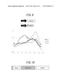 |
 |  |
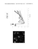 |  |
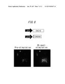 |  |
 |  |
 |  |
 |  |
 |  |
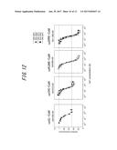 |  |
 |  |
 |  |
 | 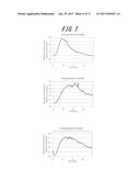 |
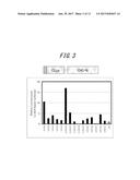 |  |
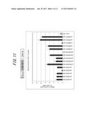 | 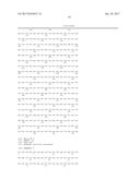 |
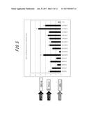 | 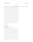 |
 |  |
| New patent applications in this class: | |
| Date | Title |
|---|---|
| 2022-09-22 | Electronic device |
| 2022-09-22 | Front-facing proximity detection using capacitive sensor |
| 2022-09-22 | Touch-control panel and touch-control display apparatus |
| 2022-09-22 | Sensing circuit with signal compensation |
| 2022-09-22 | Reduced-size interfaces for managing alerts |