Patent application title: TUMOR MARKER, MONOCLONAL ANTIBODIES AND METHODS OF USE THEREOF
Inventors:
Renata Grifantini (Siena, IT)
Renata Grifantini (Siena, IT)
Piero Pileri (Siena, IT)
Piero Pileri (Siena, IT)
Susanna Campagnoli (Siena, IT)
Susanna Campagnoli (Siena, IT)
Alberto Grandi (Siena, IT)
Alberto Grandi (Siena, IT)
Matteo Parri (Siena, IT)
Matteo Parri (Siena, IT)
Jin Boquan (Xi'An, CN)
IPC8 Class: AG01N33574FI
USPC Class:
435 611
Class name: Measuring or testing process involving enzymes or micro-organisms; composition or test strip therefore; processes of forming such composition or test strip involving nucleic acid nucleic acid based assay involving a hybridization step with a nucleic acid probe, involving a single nucleotide polymorphism (snp), involving pharmacogenetics, involving genotyping, involving haplotyping, or involving detection of dna methylation gene expression
Publication date: 2016-05-12
Patent application number: 20160131656
Abstract:
Newly identified proteins as markers for the detection of colon, ovary,
kidney, esophagus and prostate tumors, or as therapeutic targets for
their treatment; affinity ligands and particularly antibodies capable of
selectively interacting with the tumor markers and methods for tumor
diagnosis and thereapy using such antibodies.Claims:
1. A method for in vitro determining the presence of tumor marker in a
colon, ovary, esophagus, kidney or prostate tumor in a subject, which
comprises the steps of: (a) providing a test sample which is a sample of
tissue suspected of containing tumor cells or a biological fluid sample
containing exosomes of suspected tumor cells; (b) determining the
presence of a tumor marker, wherein said tumor marker is selected from
(i) a FAT1 protein, in one of its isoforms SEQ ID NO:1, SEQ ID NO:2, SEQ
ID NO:3, SEQ ID NO:4, SEQ ID NO:5 or a different isoform having sequence
identity of at least 80% to SEQ ID NO:1, SEQ ID NO:2, SEQ ID NO:3, SEQ ID
NO:4 or SEQ ID NO:5, or (ii) a RNA molecule coding for a FAT1 protein as
defined in (i), the encoding sequence being preferably selected from SEQ
ID NO:6, SEQ ID NO:7, SEQ ID NO:8, SEQ ID NO:9 and SEQ ID NO:10 or from
complementary sequences thereof in said test sample; wherein the presence
of said tumor marker in the test sample is indicative of a tumor state in
said subject.
2. A method in vitro for determining the presence of a colon, ovary, esophagus, kidney or prostate tumor in a subject, which comprises the steps of: (a) providing a test sample which is a sample of tissue suspected of containing tumor cells or a biological fluid sample containing exosomes of suspected tumor cells; (b) determining the amount of a tumor marker according to claim 1 in said test sample; (c) comparing said amount of tumor marker with that of a control sample obtained from healthy individuals, wherein an increased amount of tumor marker in the test sample compared to the control sample is indicative of a tumor state in said subject.
3. A method according to claim 2, wherein the tumor marker is a FAT1 protein in one of its isoforms SEQ ID NO:1, SEQ ID NO:2, SEQ ID NO:3, SEQ ID NO:4, SEQ ID NO:5 or a different isoform having sequence identity of at least 80% to SEQ ID NO:1, SEQ ID NO:2, SEQ ID NO:3, SEQ ID NO:4 or SEQ ID NO:5 and determination of its amount is effected by means of an antibody.
4. A method according to claim 3, wherein the antibody is a monoclonal antibody.
5. A method according to claim 2, which is based on immunoradiometric, immunoenzymatic or immunohistochemical techniques.
6. An isolated antibody or fragment thereof which specifically binds the FAT1 protein, wherein the heavy- and light-chain variable regions of said antibody contain complementarity determining regions 1, 2 and 3 (CDR-H 1-3 and CDR-L 1-3, respectively) and: said CDR-H 1-3 comprises the amino acid sequences set forth in SEQ ID NOs:17, 18 and 19, respectively, or an amino acid sequence identical to: SEQ ID NO:17 by at least 60; and to SEQ ID NO:19 by at least 90; said CDR-L 1-3 comprises the amino acid sequences set forth in SEQ ID NOs:20, 21 and 22, respectively, or an amino acid sequence identical to: SEQ ID NO:20 by at least 80; and to SEQ ID NO:22 by at least 88%.
7. An isolated antibody or fragment thereof according to claim 6, wherein said CDR-H 1-3 comprises the amino acid sequences set forth in SEQ ID NOs:37, 38 and 39, respectively, and said CDR-L 1-3 comprises the amino acid sequences set forth in SEQ ID NOs:40, 41 and 42, respectively.
8. An antibody or fragment thereof according to claim 6, wherein said variable region contains heavy and light chains set forth in SEQ ID NO:25 and SEQ ID NO:26, respectively, or heavy and light chain sequences identical to SEQ ID NOs:25 or 26 by at least 85.
9. An antibody or fragment thereof according to claim 7, wherein said variable region contains the heavy and light chains set forth in SEQ ID NO:35 and SEQ ID NO:36, respectively, or heavy and light chain sequences identical to SEQ ID NOs:35 or 36 by at least 90.
10. An antibody according to claim 6, containing a constant region of a human IgG1, IgG2, IgG3 or Ig4.
11. An isolated monoclonal antibody or fragment thereof which specifically binds to an epitope of the FAT1 protein, wherein said epitope is selected from SEQ ID NOs:27, 28, 29, 30, 31 and 32.
12. An antibody according to claim 6, which is a IgG, IgM, IgA, IgD or IgE antibody.
13. An antibody fragment according to claim 6, which is a Fab, F(ab')2, Fv, ScFv or SCA fragment.
14. An antibody according to claim 6, which is one of the following: a humanized antibody, a human antibody, a chimeric antibody, a diabody, or a linear antibody.
15. An antibody or fragment thereof according to claim 6, which is conjugated to an antitumor compound.
16. A method in vitro for determining the presence of a colon, ovary, prostate, esophagus or renal tumor with the antibody or fragment thereof according to claim 6 said method comprising (a) providing a test sample which is a sample of tissue suspected of containing tumor cells or a biological fluid sample containing exosomes of suspected tumor cells; (b) determining the amount of a tumor marker comprising a FAT1 protein, in one of its isoforms SEQ ID NO:1, SEQ ID NO:2, SEQ ID NO:3, SEQ ID NO:4, SEQ ID NO:5 or a different isoform having sequence identity of at least 80% to SEQ ID NO:1, SEQ ID NO:2, SEQ ID NO:3, SEQ ID NO:4 or SEQ ID NO:5, (c) comparing said amount of tumor marker with that of a control sample obtained from healthy individuals, wherein an increased amount of tumor marker in the test sample compared to the control sample is indicative of a tumor state in said subject.
17. A method for the treatment of a colon, ovary, prostate, esophagus or renal tumor with the antibody or fragment thereof according to claim 6.
18. An isolated peptide antigen the amino acid sequence of which is selected from SEQ ID NOs:27, 28, 29, 30, 31 and 32, or from sequences identical to SEQ ID NOs: 27, 28, 29, 30, 31 or 32 by at least 48%.
19. A pharmaceutical composition containing an antibody to a FAT1 protein or a fragment thereof according to claim 6 or a peptide antigen the amino acid sequence of which is selected from SEQ ID NOs:27, 28, 29, 30, 31 and 32, or from sequences identical to SEQ ID NOs: 27, 28, 29, 30, 31 or 32 by at least 48%.
20. A diagnostic kit containing an antibody or a fragment thereof according to claim 6 and optionally reagents, buffers, solutions and materials to carry out an immunoassay.
21. A method for the treatment of colon, ovary or prostate tumor, said method comprising administering to a subject in need thereof a siRNA molecule having a sequence complementary to one of SEQ ID NO:6, SEQ ID NO:7, SEQ ID NO:8, SEQ ID NO:9 or SEQ ID NO:10.
Description:
[0001] The present invention relates to the use of the FAT Tumor
Suppressor Homolog 1 (FAT1) protein or encoding polynucleotides as
markers for the detection of tumors, or as targets for their treatment,
particularly of tumors affecting colon, ovary, prostate, esophagus and
kidneys. Also provided are affinity ligands capable of selectively
interacting with the newly identified markers, as well as methods for
tumor diagnosis and therapy using such ligands.
BACKGROUND OF THE INVENTION
Tumor Markers (or Biomarkers)
[0002] Tumor markers are substances that can be produced by tumor cells or by other cells of the body in response to cancer. In particular, a protein biomarker is either a single protein or a panel of different proteins that could be used to unambiguously distinguish a disease state. Ideally, a biomarker would have both a high specificity and sensitivity, being represented in a significant percentage of the cases of given disease and not in healthy state.
[0003] Biomarkers can be identified in different biological samples, like tissue biopsies or preferably biological fluids (saliva, urine, blood-derivatives and other body fluids) whose collection does not necessitate invasive treatments. Tumor marker levels may be categorized in three major classes on the basis of their clinical use. Diagnostic markers can be used in the detection and diagnosis of cancer. Prognostics markers are indicative of specific outcomes of the disease and can be used to define predictive models that allow the clinicians to predict the likely prognosis of the disease at time of diagnosis. Moreover, prognosis markers are helpful to monitor the patient response to a drug therapy and facilitate a more personalized patient management. A decrease or return to a normal level may indicate that the cancer is responding to therapy, whereas an increase may indicate that the cancer is not responding. After treatment has ended, tumor marker levels may be used to check for recurrence of the tumor. Finally, therapeutic markers can be used to develop tumor-specific drugs or affinity ligand (i.e. antibodies) for a tumor treatment.
[0004] Currently, although an abnormal tumor marker level may suggest cancer, this alone is usually not enough to accurately diagnose cancer and their measurement in body fluids is frequently combined with other tests, such as a biopsy and radioscopic examination. Frequently, tumor marker levels are not altered in all of people with a certain cancer disease, especially if the cancer is at early stage. Some tumor marker levels can also be altered in patients with noncancerous conditions. Most biomarkers commonly used in clinical practice do not reach a sufficiently high level of specificity and sensitivity to unambiguously distinguish a tumor from a normal state.
[0005] To date the number of markers that are expressed abnormally is limited to certain types/subtypes of cancer, some of which are also found in other diseases. (http://www.cancer.gov/cancertopics/factsheet).
[0006] For example, prostate-specific antigen (PSA) levels are often used to screen men for prostate cancer, but this is controversial since elevated PSA levels can be caused by both prostate cancer or benign conditions, and most men with elevated PSA levels turn out not to have prostate cancer.
[0007] Another tumor marker, Cancer Antigen 125, (CA 125), is sometimes used to screen women who have an increased risk for ovarian cancer. Scientists are studying whether measurement of CA 125, along with other tests and exams, is useful to find ovarian cancer before symptoms develop. So far, CA 125 measurement is not sensitive or specific enough to be used to screen all women for ovarian cancer. Mostly, CA 125 is used to monitor response to treatment and check for recurrence in women with ovarian cancer. Finally, human epidermal growth factor receptor (HER2) is a marker protein overproduced in about 20% of breast cancers, whose expression is typically associated with a more aggressive and recurrent tumors of this class.
Routine Screening Test for Tumor Diagnosis
[0008] Screening tests are a way of detecting cancer early, before there are any symptoms. For a screening test to be helpful, it should have high sensitivity and specificity. Sensitivity refers to the test's ability to identify people who have the disease. Specificity refers to the test's ability to identify people who do not have the disease. Different molecular biology approaches such as analysis of DNA sequencing, small nucleotide polymorphisms, in situ hybridization and whole transcriptional profile analysis have done remarkable progresses to discriminate a tumor state from a normal state and are accelerating the knowledge process in the tumor field. However so far different reasons are delaying their use in the common clinical practice, including the higher analysis complexity and their expensiveness. Other diagnosis tools whose application is increasing in clinics include in situ hybridization and gene sequencing.
[0009] Currently, Immuno-HistoChemistry (IHC), a technique that allows the detection of proteins expressed in tissues and cells using specific antibodies, is the most commonly used method for the clinical diagnosis of tumor samples. This technique enables the analysis of cell morphology and the classification of tissue samples on the basis of their immunoreactivity. However, at present, IHC can be used in clinical practice to detect cancerous cells of tumor types for which protein markers and specific antibodies are available. In this context, the identification of a large panel of markers for the most frequent cancer classes would have a great impact in the clinical diagnosis of the disease.
Anti-cancer Therapies
[0010] In the last decades, an overwhelming number of studies remarkably contributed to the comprehension of the molecular mechanisms leading to cancer. However, this scientific progress in the molecular oncology field has not been paralleled by a comparable progress in cancer diagnosis and therapy. Surgery and/or radiotherapy are still the main modality of local treatment of cancer in the majority of patients. However, these treatments are effective only at initial phases of the disease and in particular for solid tumors of epithelial origin, as is the case of colon, breast, ovary, prostate and others, while they are not effective for distant recurrence of the disease. In some tumor classes, chemotherapeutic treatments have been developed, which generally relies on drugs, hormones and antibodies, targeting specific biological processes used by cancers to grow and spread. However, so far many cancer therapies had limited efficacy due to severity of side effects and overall toxicity. Indeed, a major effort in cancer therapy is the development of treatments able to target specifically tumor cells causing limited damages to surrounding normal cells thereby decreasing adverse side effects. Recent developments in cancer therapy in this direction are encouraging, indicating that in some cases a cancer specific therapy is feasible. In particular, the development and commercialization of humanized monoclonal antibodies that recognize specifically tumor-associated markers and promote the elimination of cancer is one of the most promising solutions that appears to be an extremely favorable market opportunity for pharmaceutical companies. However, at present the number of therapeutic antibodies available on the market or under clinical studies is very limited and restricted to specific cancer classes. So far licensed monoclonal antibodies currently used in clinics for the therapy of specific tumor classes show only a partial efficacy and are frequently associated with chemotherapies to increase their therapeutic effect. Administration of Trastuzumab (Herceptin), a commercial monoclonal antibody targeting HER2in conjunction with Taxol adjuvant chemotherapy induces tumor remission in about 42% of the cases (1). Bevacizumab (Avastin) and Cetuximab (Erbitux) are two monoclonal antibodies recently licensed for use in humans, targeting the endothelial and epithelial growth factors respectively that, combined with adjuvant chemotherapy, proved to be effective against different tumor diseases. Bevacizumab proved to be effective in prolonging the life of patients with metastatic colorectal and breast cancers. Cetuximab efficacy has been demonstrated in patients with tumor types refractory to standard chemotherapeutic treatments (1).
[0011] In summary, available screening tests for tumor diagnosis are uncomfortable or invasive and this sometimes limits their applications. Moreover tumor markers available today have a limited utility in clinics due to either their incapability to detect all tumor subtypes of the defined cancers types and/or to distinguish unambiguously tumor vs. normal tissues. Similarly, licensed monoclonal antibodies combined with standard chemotherapies are not effective against the majority of cases. Therefore, there is a great demand for new tools to advance the diagnosis and treatment of cancer.
Cancer Derived Exosomes.
[0012] Exosomes are nanoscale (30-100 nm) membrane vesicles formed by "inward/reverse budding" of the limiting membrane of the multivesicular bodies (MVBs) in the late endocytic compartment and released upon the fusion of MVB with the plasma membrane. Exosome secretion is observed from most cell types under both physiological and pathological conditions, particularly tumour cells and hematopoietic cells. Exosomes contain cytosolic and membrane proteins, as well as nucleic acid derived from the parental cells. The protein content is generally enriched for certain molecules, including targeting/adhesion molecules (e.g. tetraspanins, lactadherin and integrins), membrane trafficking molecules (e.g. annexins and Rab proteins), cytoskeleton molecules (e.g. actin and tubulin), proteins involved in MVB formation (e.g. Alix, Tsg101 and clathrin), chaperones (e.g., Hsp70 and Hsp90), signal transduction proteins (e.g. protein kinases, 14-3-3, and heterotrimeric G proteins) and cytoplasmic enzymes (e.g. GAPDH, peroxidases, and pyruvate kinases) (2). Other animal vesicles also contain various active molecules, such as those described above for exosomes. Depending on their cellular origin the protein composition of animal vesicles can be enriched in specific proteins. For instance, tumour-derived animal vesicles usually contain tumor-specific antigens (TAAs) expressed in the parental tumour cells such as melan-A, Silv, carcinoembryonic antigen (CEA), and mesothelin. Thus, cancer vaccine strategies have used tumour-derived exosomes as a source of TAAs to pulse DCs, resulting in the transfer of tumour antigens to DCs that were able to induce tumour-specific CD8+CTL response in mice (3) and humans (4). Methods of altering exosome protein expression are well known and include, for example, genetic modification, inhibition by small molecule inhibitors, enzymes or other inhibitory/activating proteins or peptides, and antisense technology (or other nucleic acid technologies). For example, exosomes can be modified to contain high levels of proinflammatory factors to stimulate Th1-polarized immune responses (2), e.g. by subjecting the cell that the vesicle is derived from to stress conditions under which proinflammatory cytokine and/or Hsp70 levels increase. Alternatively, the parent cell may be modified to reduce the expression of immunosuppressive molecules, such as FasL, TRAIL or TGF-beta. Exosomes can also be modified by incorporation of additional immunogenic proteins e.g. fusion with the superantigen staphylococcal enterotoxin A (SEA) (5).
Experimental Approaches Commonly Used to Identify Tumor Markers
[0013] Most popular approaches used to discover new tumor markers are based on genome-wide transcription profile or total protein content analyses of tumor. These studies usually lead to the identification of groups of mRNAs and proteins which are differentially expressed in tumors. Validation experiments then follow to eventually single out, among the hundreds of RNAs/proteins identified, the very few that have the potential to become useful markers. Although often successful, these approaches have several limitations and often, do not provide firm indications on the association of protein markers with tumor. A first limitation is that, since frequently mRNA levels not always correlate with corresponding protein abundance (approx. 50% correlation), studies based on transcription profile do not provide solid information regarding the expression of protein markers in tumor (6, 7, 8, 9,10).
[0014] A second limitation is that neither transcription profiles nor analysis of total protein content discriminate post-translation modifications, which often occur during oncogenesis. These modifications, including phosphorylations, acetylations, and glycosylations, or protein cleavages influence significantly protein stability, localization, interactions, and functions (11).
[0015] As a consequence, large scale studies generally result in long lists of differentially expressed genes that would require complex experimental paths in order to validate the potential markers. However, large-scale genomic/proteomic studies reporting novel tumor markers frequently lack of confirmation data on the reported potential novel markers and thus do not provide solid demonstration on the association of the described protein markers with tumor.
SUMMARY OF THE INVENTION
[0016] The present invention provides new means for the detection and treatment of colon, ovary, prostate, kidney and esophagus tumors, based on the identification of the FAT Tumor Suppressor Homo log 1 (FAT1) marker specific for these tumor types.
[0017] The invention also provides a method for the diagnosis of these cancer types, comprising a step of detecting the above-identified marker in a biological sample, e.g. in a tissue or biological fluid sample of a subject suspected of having or at risk of developing malignancies or susceptible to cancer recurrences.
[0018] In addition, the tumor marker identifies a novel target for affinity ligands, which can be used for therapeutic applications. Also provided are specific affinity ligands, particularly antibodies, capable of selectively interacting with the newly identified protein marker expressed on the cell surface. The antibodies can be used to specifically discriminate cancer cells, based on the recognition of the marker. The invention also provides monoclonal antibodies able to recognize the marker on the cell surface and, upon binding, to be internalized by cancer cells. Finally, the invention provides antibodies that can be used to directly kill or promote killing of cancer cells either as unconjugated or conjugated with cell payloads (e.g. radioisotopes, drugs, or toxins).
STATE OF THE ART
General Information.
[0019] Human FAT1 gene is an ortholog of the Drosophila fat gene, which encodes a tumor suppressor essential for controlling cell proliferation during Drosophila development. The gene product FAT1 is a member of the cadherin superfamily, a group of integral membrane proteins characterized by the presence of cadherin-type repeats. In addition to containing 34 tandem cadherin-type repeats, the gene product has five epidermal growth factor (EGF)-like repeats and one laminin A-G domain. It was first identified as a tumor suppressor in Drosophila melanogaster, acting via the Salvador-Warts-Hippo signaling pathway (12).
[0020] This gene is expressed at high levels in a number of fetal epithelia. Transcript variants derived from alternative splicing and/or alternative promoter usage exist, but they have not been fully described. The protein product probably functions as an adhesion molecule and/or signaling receptor, and is likely to be important in developmental processes and cell communication. FAT1 is known to interact with Ena/VASP, thereby it is involved in promoting actin polymerization and cell motility (13).
Role in Cancer
[0021] Scientific literature has reported that FAT1 has a predominant tumor-suppressive effect. The human FAT1 gene is homozygously deleted in 23% of oral cancer cell lines and in 80% of primary oral cancer cases and FAT1 mRNA expression is repressed in oral cancer cell lines due to homozygous deletion and/or promoter CpG hypermethylation (14). Loss of heterozygosity (LOH) of the FAT1 gene occurs in 42% of low-grade diffuse astrocytoma and 63% of glioblastoma multiforme (15). FAT1 mRNA level in ductal carcinoma in situ is significantly higher than that in invasive breast cancer and FAT1 knockdown promotes progression from ductal carcinoma in situ to invasive breast cancer, indicating that lower FAT1 expression is associated with aggressive breast cancers (16).
[0022] Available in vitro and in vivo studies reported that FAT1 depletion leads to markedly accelerated cell growth and proliferation, while expression of FAT1 robustly suppresses tumor growth. These growth-suppressive effects are abrogated when mutations observed in tumors are present (17). Recent data now implicates FAT1 mutation as a driver of Wnt activation in many cancers, through the involvement the b-catenin. As other cadherins, FAT1 can bind to b-catenin and limits its translocation to the nucleus. Mutations in FAT1 intracytoplasmic domain result in a loss of this ability to regulate b-catenin. Consequently, loss of FAT1 in cells activates the Wnt signaling pathway, unleashing b-catenin-dependent transcriptional activity and upregulating pro-growth wnt transcriptional targets. Consistent with this, primary cancer samples with FAT1 alterations are characterized by significant enhancement of Wnt signaling. The growth-suppressive functions of FAT1 are mediated by its intracytoplasmic, b-catenin binding domain, but the extracellular domain also mediates cell adhesion, which may be a secondary mechanism by which FAT1 loss promotes tumor growth.
[0023] In a recent study on melanoma (18) showed that melanocytes and keratinocytes express FAT1 at similar level. However in melanoma the protein shows an altered processing. In keratinocytes FAT1 is cleaved by the proprotein convertase furin forming two fragments of 430 and 85 kDa that form a non-covalent heterodimer before achieving cell surface expression. Differently, in melanoma cells, the non-cleaved proform of FAT1 is also expressed at the cell surface together with the furin-cleaved heterodimer. Moreover, furin-independent processing generates an aberrant proteolytic product of 65-kDa no longer in association with the extracellular fragment. In vitro localization studies of FAT1 showed that melanoma cells display high levels of cytosolic FAT1 protein, whereas keratinocytes, despite comparable FAT1 expression levels, exhibited mainly cell-cell junctional staining These differences in protein distribution are compatible with the different protein products generated by dual FAT1 processing. The authors suggest that the uncleaved FAT1 could promote altered signaling, and the novel products of alternate processing provide a dominant negative function in melanoma.
[0024] Only few examples of FAT1 upregulation in cancer have been so far reported. FAT1 mRNA expression is upregulated in 11% of acute myeloid leukemia (AML), 29% of preB acute lymphoblastic leukemia (ALL) and 63% of T-ALL, and FAT1 upregulation in preB-ALL is associated with shorter relapse-free survival as well as shorter overall survival (19). FAT1 immunoreactivity is strong in 29% of cholangiocarcinoma (20).
[0025] Thus, it is conceivable that FAT1 has a multifaceted role such that it may operate in different mechanisms, and may act as a tumor suppressive or oncogenic depending on the specific cell context and in a manner that cannot be predicted based on available knowledge.
[0026] Despite the involvement of FAT1 in tumor has been partially investigated, so far no previous evidence documents the FAT1 association with colon, ovary, kidney, esophagus and prostate tumors. In particular, no study reports FAT1 over-expression in these cancers. Finally, FAT1 antibodies able to recognize FAT1 over-expressed on the surface of colon, ovary, kidney, esophagus and prostate cancers and reduce cancer growth have not been reported so far.
DISCLOSURE OF THE INVENTION
[0027] The present invention is based on the surprising finding that FAT1 is over-expressed and acts as tumor promoter in colon, ovary, kidney, esophagus and prostate cancers. Moreover, it was found that antibodies specific for FAT1 are able to specifically recognize colon, ovary, esophagus, prostate and kidney tumor tissues from patients, while they show negative or very poor staining in corresponding normal tissues.
[0028] Accordingly, the present invention provides FAT1 as a protein marker for colon, ovary, esophagus, kidney, and prostate tumors and in general for cancers of these types. Antibodies generated towards the FAT1 protein show a selective immunoreactivity in histological preparation of colon, ovary, esophagus, prostate and kidney cancer tissues with concomitant negligible expression in corresponding normal samples, which indicates a specific over-expression of FAT1 protein in these cancer samples, and makes FAT1 protein and specific antibodies thereto novel tools for specifically distinguishing these cancer types from a normal state.
[0029] Antibodies generated against FAT1 are able to specifically recognize the protein on the surface of different cancer cell lines. In particular, experiments carried out with two specific anti-FAT1 antibodies showed that the latter, upon surface binding, are efficiently internalized by cancer cells, indicating that they are suitable for the generation of antibody-drug conjugate (ADC). In addition one of these antibodies, when administered to mice bearing tumor, is able to reduce cancer growth.
[0030] Finally, antibodies generated against FAT1 are also able to detect the protein in exosomes from cancer cells, indicating that the marker can be detected in biological fluids of oncologic patients. The exosomes carrying FAT1 can be exploited as tools for the development of vaccines to prevent or treat cancers affecting colon, ovary, esophagus, kidney, and prostate.
[0031] Overall, these findings indicate that FAT1 can be conveniently used as a diagnostic marker as well as a target for anti-cancer therapies (e.g. based on small molecules, antibodies, nucleic acids, toxins) and cancer vaccines. Moreover, they provide experimental evidences that the FAT1 antibodies can be exploited as novel therapeutic agents.
[0032] Hence, in a first aspect, the invention provides the use of a marker for colon, ovary, esophagus, kidney, and prostate tumor, which is selected from:
[0033] (i) FAT1 protein, in one of its isoforms SEQ ID NO:1, SEQ ID NO:2, SEQ ID NO:3, SEQ ID NO:4, SEQ ID NO:5, or a different isoform having sequence identity of at least 80%, preferably at least 90%, more preferably at least 95% to SEQ ID NO:1, SEQ ID NO:2, SEQ ID NO:3, SEQ ID NO:4, SEQ ID NO:5, or
[0034] (ii) a RNA molecule coding for a FAT1 protein, wherein the encoding sequence is preferably selected from SEQ ID NO:6, SEQ ID NO:7, SEQ ID NO:8, SEQ ID NO:9 and SEQ ID NO:10 or from complementary sequences thereof.
[0035] As used herein the "% amino acid sequence identity" with respect to the marker protein sequences identified herein indicates the percentage of amino acid residues in a protein variant or iso form, or in a portion thereof, that are identical to the amino acid residues in the specific marker sequence, after aligning the sequences and introducing gaps, if necessary, to achieve the maximum percent sequence identity, and not considering any conservative substitution as part of the sequence identity.
[0036] Identity between nucleotide sequences is preferably determined by the Smith-Waterman homology search algorithm as implemented in the SSEARCH program (Oxford Molecular), using an affine gap search with parameters gap open penalty=12 and gap extension penalty=1.
[0037] A further aspect of this invention is a method of screening a tissue sample for malignancy, which comprises determining the presence in said sample of the above-mentioned tumor marker. This method includes detecting either the marker protein, e.g. by means of labeled monoclonal or polyclonal antibodies that specifically bind to the target protein, or the respective mRNA, e.g. by means of polymerase chain reaction techniques such as RT-PCR. The methods for detecting proteins in a tissue sample are known to one skilled in the art and include immunoradiometric, immunoenzymatic or immunohistochemical techniques, such as radioimmunoassays, immunofluorescent assays or enzyme-linked immunoassays. Other known protein analysis techniques, such as polyacrylamide gel electrophoresis (PAGE), Western blot or Dot blot are equally suitable. Preferably, the detection of the protein marker is carried out with the immune-histochemistry technology, particularly by means of High Through-Put methods that allow the analyses of the antibody immune-reactivity simultaneously on different tissue samples immobilized on a microscope slide. Briefly, each Tissue Micro Array (TMA) slide includes tissue samples suspected of malignancy taken from different patients, and an equal number of normal tissue samples from the same patients as controls. The direct comparison of samples by qualitative or quantitative measurement, e.g. by enzymatic or colorimetric reactions, allows the identification of tumors.
[0038] In one embodiment, the invention provides a method of screening a sample of colon, ovary, esophagus, kidney, and prostate tissue for malignancy, which comprises determining the presence in said sample of the FAT1 protein tumor marker, variants or iso forms thereof as described above.
[0039] A further aspect of the invention is a method in vitro for determining the presence of a colon, ovary, esophagus, kidney, or prostate tumor in a subject, which comprises the steps of:
[0040] (a) providing a test sample which is a sample of tissue suspected of containing tumor cells or a biological fluid sample preferably containing exosomes of the suspected tumor cells;
[0041] (b) determining the amount of FAT1 tumor marker in the test sample;
[0042] (c) comparing said amount of tumor marker with that obtained in a control sample from healthy subjects;
[0043] wherein an increased amount of tumor marker in the test sample compared to the control sample is indicative of a tumor state in the subject.
[0044] The control sample may be a colon, ovary, esophagus, kidney or prostate tissue sample from healthy individuals.
[0045] In alternative to detecting altered FAT1 expression level, the test sample can be assayed for processed form of the protein marker or for differential expression of the respective mRNA transcripts.
[0046] The methods and techniques for carrying out the assay are known to one skilled in the art and are preferably based on immunoreactions for detecting proteins and on PCR methods for the detection of mRNAs. The same methods for detecting proteins or mRNAs from a tissue sample as disclosed above can be applied.
[0047] A further aspect of this invention is the use of the FAT1 tumor marker herein provided as target for the identification of candidate antitumor agents for the treatment of colon, ovary, kidney, esophagus or prostate cancers. Accordingly, the invention provides a method for screening compounds which comprises contacting cells expressing the FAT1 protein from colon, ovary, kidney, esophagus or prostate tissues, with the test compound, and determining the binding of said compound to said tumor-associated protein or the cellular or intracellular effects elicited by that interaction. In addition, the ability of the test compound to modulate the activity of each target molecule can be assayed.
[0048] A further aspect of the invention is an antibody or a fragment thereof, which is able to specifically recognize and bind to one of the FAT1 tumor-associated proteins described above, for use in a method in vitro for determining the presence of a colon, ovary, esophagus, kidney or prostate tumor as defined above or for use in the treatment of the same tumors. The term "antibody" as used herein refers to any type of immunoglobulins, including IgG, IgM, IgA, IgD and IgE and it may be selected from the group consisting of a polyclonal antibody; a monoclonal antibody including a Human Engineered antibody; a humanized antibody; a human antibody; a chimeric antibody; Fab, F(ab')2; Fv; Sc Fv or SCA antibody fragment; a diabody; linear antibody; or a mutein of any one of these antibodies.
[0049] The antibodies may be of various origin, including human, mouse, rat, rabbit and horse, or chimeric antibodies. The production of antibodies is well known in the art. For the production of antibodies in experimental animals, various hosts including goats, rabbits, rats, mice, and others, may be immunized by injection with polypeptides of the present invention or any fragment or oligopeptide or derivative thereof which has immunogenic properties or forms a suitable epitope. Monoclonal antibodies may be produced following the procedures described in Kohler and Milstein, Nature 265:495 (1975) or other techniques known in the art.
[0050] In a preferred embodiment, the invention provides an isolated antibody or fragment thereof which specifically binds the FAT1 protein, wherein the heavy- and light-chain variable regions of said antibody contain complementarity determining regions 1, 2 and 3 (CDR-H 1-3 and CDR-L 1-3, respectively) and:
[0051] said CDR-H 1-3 comprises the amino acid sequences set forth in SEQ ID NOs:17, 18 and 19, respectively, or an amino acid sequence identical to: SEQ ID NO:17 by at least 60%, preferably at least 85%; SEQ ID NO:18 by at least 85%, preferably at least 90%; and to SEQ ID NO:19 by at least 90%, preferably at least 95%;
[0052] said CDR-L 1-3 comprises the amino acid sequences set forth in SEQ ID NOs:20, 21 and 22, respectively, or an amino acid sequence identical to: SEQ ID NO:20 by at least 80%, preferably at least 90%; SEQ ID NO:21 by at least 85%, preferably at least 90%; and to SEQ ID NO:22 by at least 88%, preferably at least 95%. In a further preferred embodiment, said CDR-H 1-3 contain the amino acid sequences SEQ ID NOs:37, 38 and 39, respectively, and said CDR-L 1-3 contain the amino acid sequences SEQ ID NOs:40, 41 and 42, respectively.
[0053] In another preferred embodiment said CDR-H 1-3 and CDR-L 1-3 are encoded by the polynucleotides SEQ ID NOs:11-13 and SEQ ID NOs:14-16, respectively, or variants thereof due to the degeneracy of genetic code.
[0054] In another preferred embodiment the antibody of invention contains the heavy and light chains set forth in SEQ ID NO:25 and SEQ ID NO:26, respectively, or heavy and light chain sequences identical to SEQ ID NOs:25 or 26 by at least 85%, preferably at least 95%.
[0055] In a likewise preferred embodiment, the antibody of invention contains the heavy and light chains set forth in SEQ ID NO:35 and SEQ ID NO:36, respectively, or heavy and light chain sequences identical to SEQ ID NOs:35 and 36 by at least 85%, preferably at least 95%.
[0056] In another preferred embodiment, the heavy and light chains of the invention antibody are encoded by the polynucleotides of SEQ ID NOs:23 and 24, respectively, or variants thereof due to the degeneracy of genetic code.
[0057] In yet another preferred embodiment, the heavy and light chains of the invention antibody are encoded by the polynucleotides of SEQ ID NOs:33 and 34, respectively, or variants thereof due to the degeneracy of genetic code.
[0058] In related embodiments, the invention antibody comprises a modified or unmodified constant region of a human IgG1, IgG2, IgG3 or IgG4. In a preferred embodiment, the constant region is human IgG1, yet more preferably IgG1k which may optionally be modified to enhance or decrease certain properties. In the case of IgG1, modifications to the constant region, particularly the hinge or CH2 region, may increase or decrease effector function, including ADCC and/or CDC activity.
[0059] Epitope-mapping was carried out on FAT1 molecule to identify the molecule regions recognized by monoclonal antibodies on cancer cells. Overlapping fragments were isolated and further investigated for more accurate epitope identification and eventually a region of FAT1 molecule including cadherin domains 8 and 12 and a panel of shorter peptides were identified and validated as FAT1 epitopes in ELISA and FACS competition experiments.
[0060] Accordingly, in a further embodiment the invention provides a FAT1 epitope which is selected from the group consisting of SEQ ID NOs: 27, 28, 29, 30, 31 and 32, or peptide sequences identical to SEQ ID NOs: 27, 28, 29, 30, 31 and 32 by at least 48%, preferably at least 75%, more preferably at least 84%, and the use thereof as tumor antigens for rising an immune response against FAT1-expressing tumors. For example, the FAT1 epitope could be used to generate antibodies or T lymphocytes able to impair growth of FAT1-expressing tumors.
[0061] In a yet further embodiment the invention provides an isolated monoclonal antibody or fragment thereof which specifically binds to one or more of said FAT1 protein epitopes.
In a preferred embodiment, the monoclonal antibody or fragment thereof contains the CDR-H, CDR-L, heavy and light chain sequences specified above.
[0062] Chimeric monoclonal antibodies, in which the variable Ig domains of a mouse monoclonal antibody are fused to human constant Ig domains, can be generated using standard procedures known in the art (See Morrison, S. L., et al. (1984) Chimeric Human Antibody Molecules; Mouse Antigen Binding Domains with Human Constant Region Domains, Proc. Natl. Acad. Sci. USA 81, 6841-6855; and, Boulianne, G. L., et al, Nature 312, 643-646. (1984)).
[0063] Humanized antibodies may be achieved by a variety of methods including, for example: (1) grafting the non-human complementarity determining regions (CDRs) onto a human framework and constant region (a process referred to in the art as humanizing through "CDR grafting"), or, alternatively, (2) transplanting the entire non-human variable domains, but "cloaking" them with a human-like surface by replacement of surface residues (a process referred to in the art as "veneering").
[0064] The antibodies to the tumor markers of the invention can be used to detect the presence of the marker in histologic preparations or to distinguish tumor cells from normal cells. To that purpose, the antibodies may be labeled with radioactive, fluorescent or enzyme labels.
[0065] In addition, the antibodies of the invention can be used for treating proliferative diseases by modulating, e g inhibiting or abolishing the activity of the target protein according to the invention.
[0066] Therefore, in a further aspect the invention provides the use of antibodies to FAT1 protein for the preparation of a therapeutic agent for the treatment of proliferative diseases of colon, ovary, esophagus, kidney and prostate tissues. For use in therapy, the antibodies can be formulated with suitable carriers and excipients, optionally with the addition of adjuvants to enhance their effects.
[0067] A further aspect of the invention relates to a diagnostic kit containing suitable means for detection, in particular FAT1 polypeptides or polynucleotides, antibodies or fragments or derivatives thereof described above, reagents, buffers, solutions and materials needed for setting up and carrying out the immunoassays, nucleic acid hybridization or PCR assays described above. Parts of the kit of the invention can be packaged individually in vials or bottles or in combination in containers or multicontainer units.
[0068] In a further embodiment, the invention provides a pharmaceutical composition containing an antibody to a FAT1 protein or a fragment thereof as herein disclosed, for use in a method of treatment of subjects affected by colon, ovary, esophagus, kidney, or prostate tumor.
[0069] In a further embodiment, the invention provides a method for suppressing or reducing the expression of the FAT1 protein in a subject affected by a colon, ovary, esophagus, kidney or prostate tumor, which comprises administering to that subject a siRNA molecule having a sequence complementary to SEQ ID NOs: 6-10.
DESCRIPTION OF THE FIGURES
[0070] FIG. 1. Frequency of positive IHC staining of colon, ovary, esophagus, kidney and prostate cancer using the anti-FAT1 monoclonal antibody mAb91.3.
[0071] Graph represents the percentage of cancer samples showing positive IHC reactivity to mAb91.3. As shown in the graph, FAT1 is over-expressed in approximately 80% of colon cancer, 18% of ovary cancer, 20% of esophagus and kidney carcinoma, and 100% of prostate cancer.
[0072] FIG. 2. The anti-FAT1 mAb91.3 specifically recognizes FAT1 over-expressed in colon cancer by IHC.
[0073] IHC images of colon tumor and normal colon tissue samples stained with the anti-FAT1 monoclonal antibody mAb91.3. The antibody stains specifically tumor cells, visible in dark gray.
[0074] FIG. 3. The anti-FAT1 mAb91.3 recognizes FAT1 over-expressed in prostate cancer by IHC. IHC images of prostate tumor (lower panel) and normal tissue samples (upper panel) stained with the anti-FAT1 antibody mAb91.3. The antibody mAb91.3 stains specifically tumor cells, visible in dark gray.
[0075] FIG. 4. FAT1 is expressed of in colon tumor cell lines.
[0076] Western blot analysis of total protein extracts (corresponding to 2×10⁵ cells) from colon cancer cell lines. Cell extracts (EXT) and culture supernatants (SN) were separated by SDS-PAGE, transferred onto nitrocellulose membranes and probed with mAb91.3. FAT1 is detected at very high molecular weight bands. Moreover, possible degradation products are also visible. Molecular weight markers are reported on the right.
[0077] FIG. 5. FAT1 is associated to cell derived exosomes.
[0078] Western blot analysis of the exosomal fraction and the exosome-depleted supernatant derived from colon cancer cells stained with the anti-FAT1 monoclonal antibody. Molecular weight markers are reported on the left. The protein mainly associated with exosomes and is detectable at low level in the exosome-depleted supernatant
[0079] FIG. 6. FAT1 is exposed on the surface of colon cancer cells.
[0080] FAT1 surface expression was confirmed by confocal microscopy and flow cytometry of different colon cell lines stained with the anti-FAT1 mAb91.3 or unrelated antibodies. A) Confocal microscopy analysis. Cells were fixed with 3% formaldehyde and incubated with mAb91.3 or an irrelevant mouse monoclonal antibody. The antibody binding was detected by incubation with an Alexa 488-conjugated goat anti-mouse antibody. DAPI was used to visualize nuclei, visible in the left images. Arrows mark examples FAT1 staining visible at the cell surface with mAb91.3. B) FACS. Cells were incubated with mAb91.3 (white peaks) or an irrelevant mouse monoclonal antibody (grey peaks). The antibody binding was detected by incubation with an R-Phycoerythrin (PE)-conjugated secondary antibody. X axis, Fluorescence scale; Y axis, Cells (expressed as % relatively to major peaks).
[0081] FIG. 7. Specificity of the anti-FAT1 antibody.
[0082] HCT15 cells were transfected with two FAT1 specific siRNA or a scrambled siRNA and the loss of expression and protein disappearance from the cell surface was assessed respectively by Western blot (left panel) and flow cytometry (right panel), using mAb91.3 and two additional anti-FAT1 antibodies that were negative on cancer tissues. Actin and CD81-specific antibodies were used as internal reference for immunoblot or flow cytometry surface staining As shown in the figure, both antibodies specifically detect FAT 1.
[0083] FIG. 8. mAb91.3 is internalized by colon cancer cells.
[0084] mAb91.3 (10 micrograms/ml) was incubated with cells 30' at 4° C. to allow surface binding. Then cells were shifted for lhour at 37° C. to permit the antibody internalization. A) At defined time-points mAb91.3 disappearance from the cell surface was monitored by flow cytometry with R-Phycoerythrin (PE)-conjugated secondary antibody. B) Confocal microscopy analysis was also used to monitor the loss of antibody from the cell surface and the concomitant accumulation of antibody complexes in the intracellular compartments of cells permeabilized with cold methanol and incubated with a-mouse AlexaFluor488-conjugated secondary antibody. DAPI was used to visualize nuclei. Upper and lower images show 2D and 3D representations, respectively.
[0085] FIG. 9. The anti-FAT1 mAb91.3 specifically binds colon cancer and reduces its growth in xenograft colon mouse models.
[0086] A) mAb therapeutic activity. HCT15 and HT29 colon cancer cells were engrafted subcutaneously in athymic nude mice (6 per group) and when they reached approx. 60-100 mm3 mice were administered intravenously with either mAb91.3 or an irrelevant mAb as isotype control (300 micrograms/dose, equal to approximately 12 mg mAb per Kg of animal weight, 3 doses per week). Tumor growth measured with a caliper over a 2 week-period. Graph represents the tumor growth rate. For each mouse tumor growth rate was calculated by dividing the tumor volume at time points vs the tumor volume before treatment start. Horizontal bars represent mean values of each animal group (squares, untreated; triangles, isotype control; circles, mAb91.3).
[0087] B) Preventive activity. HCT15 colon cancer cells were engrafted subcutaneously in athymic nude mice (8 per group). The day before and subsequently mice were administered intravenously with either mAb91.3 or an isotype control irrelevant antibody at the indicated dose regimen. Graph represents the tumor mass ratio at time points between treated and control mice for the irrelevant isotype control (gray square) or the mAb91.3 (black triangles). The specificity of mAb91.3 antibody binding on cancer xenografts was monitored by Near-Infrared (NI) Optical Imaging in mice injected NI-labelled antibodies.
[0088] FIG. 10. The anti-Fat1 mAb91.3 promotes killing of cancer cells in an indirect saporin killing assay.
[0089] HCT15 cells were incubated with different concentrations of mAb91.3 for 30' at 4° C. Cells were washed with PBS to remove unbound antibody and further incubated for 30' with a saporin-conjugated secondary antibody (ATS system). Afterwards, cells were shifted at 37° C. for 72 hours. Cell viability was assessed with the MTT.
[0090] FIG. 11. mAb91.4 binds the surface of colon cancer and is internalized upon binding
[0091] Cells were incubated with mAb91.4 (10 micrograms/ml) was incubated with cells 30' at 4° C. to allow surface binding. Then cells were shifted for 1 hour at 37° C. to permit the antibody internalization. The antibody binding was detected by FACS by incubation with an R-Phycoerythrin (PE)-conjugated secondary antibody. X axis, Fluorescence scale; Y axis, Cells (expressed as % relatively to major peaks). At defined time-points mAb91.4 disappearance from the cell surface was monitored by FACS as described before.
[0092] FIG. 12. mAb91.3 binds two FAT1 regions in Western blot. Western blot analysis of HEK-293T or Hela cells transfected with a plasmid encoding the FAT1 fragments. Cells were transfected with plasmids encoding Fat1 fragment A, Fragment B, the short form of fragment A lacking the overlapping region with Fragment B, Domain 8 or the empty vector (mock). After 48 hours cell were lysed and total extracts were loaded on PAGE-SDS, along with recombinant domain 12, and subjected to Western blot using mAb91.3.
[0093] FIG. 13. Identification of mAb91.3 surface exposed epitopes by FACS peptide competition.
[0094] HCT15 cells were incubated with mAb 91.3 in the presence of different concentration of the 25 mer FAT1 peptides encompassing the epitopes and an irrelevant peptide. The residual antibody binding on the cell surface was assessed by FACS.
[0095] FIG. 14. Identification of the mAb91.3 peptides recognized in colon cancer by IHC peptide competition.
[0096] Representative IHC image of colon tumor and normal colon tissue samples stained with the anti-FAT1 monoclonal antibody mAb91.3 (5 micrograms/ml) in the presence of one of the 25 mer peptides encompassing the antibody epitope.
[0097] FIG. 15. Total IgGs elicited by the HCT15 exosomes formulations against FAT1. Groups of CD1 mice were immunized with exosomes (15 micrograms dose, three doses) purified from the culture supernatant of HCT15. Two weeks after the last immunization mice were bled and serum was collected. Sera from mice immunized with exosomes were pooled and analyzed by ELISA on two recombinant FAT1 domains as compared to pre-immune sera
EXAMPLES
Example 1
Discovery and Confirmation of FAT1 Over-Expression in Cancer by Immune-Histochemistry
[0098] A proprietary collection of polyclonal and monoclonal antibodies raised against human recombinant proteins was used to identify proteins over-expressed in cancer by immune-histochemistry (IHC). The antibody library was used screen clinical samples by Tissue Micro Array (TMA), a miniaturized immuno-histochemistry technology suitable for HTP analysis that allows to analyse the antibody immuno-reactivity simultaneously on different tissue samples immobilized on a microscope slide. Since the TMAs include both tumor and healthy tissues, the specificity of the antibodies for the tumors can be immediately appreciated. The use of this technology, differently from approaches based on transcription profile, has the important advantage of giving a first hand evaluation on the potential of the markers in clinics. Conversely, since mRNA levels not always correlate with protein levels (approx. 50% correlation), studies based on transcription profile do not provide solid information regarding the expression of protein markers.
Methods
[0099] The tissue microarrays were prepared containing formalin-fixed paraffin-embedded cores of human tissues from patients affected by breast, colon, lung, ovary, esophagus, kidney, and prostate cancers and corresponding normal tissues as controls and subsequently analyzed using the specific antibody sample. For each tumor class two TMA designs were generated and used for IHC with the anti-FAT1 mAb91.3. A first TMA design consisted in pathological and normal tissue samples from 5 patients of known clinical history per each of the five organs (all samples in duplicate) and was used to identify promising target molecules differentially expressed in cancer and normal cells. The direct comparison between tumor and normal tissues of each patient allowed the identification of antibodies that stain specifically tumor cells and provided indication of target expression in tumor. A second expanded TMA design represented samples from 50 patients from each of the five organs and was used to confirm the marker over-expression in the cancer in which the antibody showed specific reactivity.
[0100] Corresponding whole tissue sections were examined to confirm diagnosis and tumor classification, and to select representative areas in donor blocks. Normal tissues were defined as microscopically normal (non-neoplastic) and were generally selected from specimens collected from the vicinity of surgically removed tumors. The TMA production was performed essentially as previously described (21, 22). Briefly, a hole was made in the recipient TMA block. A cylindrical core tissue sample (1 mm in diameter) from the donor block was acquired and deposited in the recipient TMA block. This was repeated in an automated tissue arrayer "Galileo TMA CK 3500" BioRep (Milan) until a complete TMA design was produced. TMA recipient blocks were baked at 42 <0> C for 2 h prior to sectioning. The TMA blocks were sectioned with 2-3 mm thickness using a waterfall microtome (Leica), and placed onto poly-L-lysinated glass slides for immunohistochemical analysis. For automated immunohistochemistry, glass slides were incubated for 30' min in 60° C., de-paraffinized in xylene (2×15 min) using the Bio-Clear solution (Midway. Scientific, Melbourne, Australia), and re-hydrated in graded alcohols.
[0101] For antigen retrieval, slides were immersed 0.01 M Na-citrate buffer, pH 6.0 at 99° C. for 30 min Slides were placed in the Autostainer (R) (DakoCytomation) and endogenous peroxidase was initially blocked with 3% H2O2, for 5 min. Slides were then blocked in Dako Cytomation Wash Buffer containing 5% Bovine serum albumin (BSA) and subsequently incubated with mouse antibodies for 30' (dilution 1:200 in Dako Real® dilution buffer). After washing with DakoCytomation wash buffer, slides were incubated with the goat anti-mouse peroxidase conjugated Envision(R) for 30 min each at room temperature (DakoCytomation). Finally, diaminobenzidine (DakoCytomation) was used as chromogen and Harris hematoxylin (Sigma-Aldrich) was used for counterstaining The slides were mounted with Pertex(R) (Histolab).
[0102] The staining results have been evaluated by a trained pathologist at the light microscope, and scored according to both the percentage of immunostained cells and the intensity of staining The individual values and the combined score (from 0 to 300) were recorded in a custom-tailored database. Digital images of the immunocytochemical findings have been taken at a Leica DM LB light microscope, equipped with a Leica DFC289 color camera.
Results
[0103] The results from tissue profiling showed that a monoclonal antibody specific for human FAT1 mAb91.3 was strongly immunoreactive on tissues from colon cancer (approximately 80%), while no or poor reactivity was detected in corresponding normal samples. This monoclonal antibody also showed selective reactivity on ovary, esophagus and kidney cancers (approximately 18%, 20% and 20% of positive staining, respectively). Moreover, this monoclonal antibody also showed very strong reactivity on 100% of prostate cancer sample, with concomitant moderate reactivity on normal prostate tissues. The antibody staining accumulated at the plasma membrane of tumor cells. FIG. 1 shows the frequency of IHC positive staining with mAb91.3, based on analysis of 50 patients/tumor. Representative examples of microscopic enlargements of colon and prostate tissue samples stained by the anti-FAT1 monoclonal antibody are reported in FIGS. 2-3. Other antibodies towards FAT1 were used in the tissue profile analysis and these did not prove as much efficient as mAb91.3 to selectively recognize cancer tissues.
[0104] Based on this finding, the detection of FAT1 protein in tumor tissue samples can be associated with colon, ovary, esophagus, kidney and prostate tumors. Moreover, the FAT1 localization at the plasma membrane makes this protein a suitable target for anti-cancer therapies.
Example 2
Expression and Localization of FAT1 Protein in Cancer Cells
[0105] The expression and localization of FAT 1 protein in cancer cells was investigated using an anti-FAT1 monoclonal antibody to confirm that FAT1 is expressed by cancer cell lines derived from the human cancers found positive in the IHC screening. Moreover, FAT1 surface localization surface was verified to confirm that FAT1 could be exploited as therapeutic target of anti-cancer therapies. FAT-1 affinity ligands, such as small molecules or antibodies, able to recognize the protein on the cell surface can be developed as novel therapeutic antigens. Finally the association of FAT1 with cell-derived exosomes was investigated to assess whether FAT 1 is released by cancer cells and could be detected in patients' biological fluids. This property would allow developing non-invasive diagnostic assays based on FAT1 detection. Moreover, exosomes could be exploited in vaccines based on the elicitation of antibody and T cell response against FAT1.
Methods
[0106] FAT1 expression was first assessed by WB on total extracts from a panel of colon cancer epithelial cell lines. In the analysis, cells were cultured in under ATCC recommended conditions, and sub-confluent cell monolayers were detached with PBS-0.5 mM EDTA and lysed by several freeze-thaw passages in PBS-1% Triton. Total protein extracts were loaded on SDS-PAGE (2×10⁵ cells/lane), and subjected to WB with anti-FAT1 specific antibodies.
[0107] To analyse the presence of FAT1 in cancer cell exosomes, exosomes were purified from 10 ml culture supernatant of different colon cancer cell lines using the Exoquick-TC purification kit (SBI). The exosomal pellet (corresponding to approximately 5×10⁶ cells) were lysed with Laemmli buffer under reducing condition, loaded of SDS-PAGE gradient gels (NuPage 4-12% Bis-Tris gel, Invitrogen) under reducing conditions, and subjected to immunoblot with anti-FAT1 antibodies as described (see example 1). The culture supernatants deprived of exosomes were concentrated, loaded on the gel and analysed in parallel by immunoblot. The quality of the exosomal preparation was verified by probing the blots with antibodies specific for known exosomal markers (e.g. CD81) or exosome-associated proteins (e.g IFITM3).
[0108] Western blot was performed by separation of the protein extracts on pre-cast SDS-PAGE gradient gels (NuPage 4-12% Bis-Tris gel, Invitrogen) under reducing conditions, followed by electro-transfer to nitrocellulose membranes (Invitrogen) according to the manufacturer's recommendations. The membranes were blocked in blocking buffer composed of 1× PBS-0.1% Tween 20 (PBST) added with 10% dry milk, for 1 h at room temperature, incubated with the antibody diluted 1:2500 in blocking buffer containing 1% dry milk and washed in PBST-1%. The secondary HRP-conjugated antibody (goat anti-mouse immunoglobulin/HRP, Perkin Elmer) was diluted 1:5000 in blocking buffer and chemiluminescence detection was carried out using a Chemidoc-IT UVP CCD camera (UVP) and the Western lightning® cheminulescence Reagent Plus (Perkin Elmer), according to the manufacturer's protocol.
[0109] FAT1 surface localization was assessed by Flow cytometry (FACS) and confocal microscopy analyses on colon and prostate cancer cells.
[0110] For Flow Cytometry analysis, cells (2×10⁴ per well) were pelleted in 96 U-bottom microplates by centrifugation at 200× g for 5 min at 4° C. and incubated for 1 hour at 4° C. with the appropriate dilutions of anti-FAT1-monoclonal antibody. The cells were washed twice in PBS-5% FCS and incubated for 20 min with the appropriate dilution of R-Phycoerythrin (PE)-conjugated secondary antibodies (Jackson Immuno Research, PA, USA) at 4° C. After washing, cells were analysed by a FACS Canto II flow cytometer (Becton Dickinson). Data were analyzed with FlowJo 8.3.3 program.
[0111] For confocal microscopy, cells were plated on glass cover slips and after 48 h were washed with PBS and fixed with 3% formaldehyde solution in PBS for 20 min at RT. Then, after extensive washing in PBS, the cells were incubated with the anti-FAT1 antibodies overnight at 4° C. (1:200) with or without a previous permeabilization step with 0.01% BriJ96® (Fluka). Cells were then stained with Alexafluor 488-labeled goat anti-mouse antibodies (Molecular Probes). DAPI (Molecular Probes) was used to visualize nuclei. The cells were mounted with glycerol plastine and observed under a laser-scanning confocal microscope (LeicaSPS).
Results
[0112] FAT1 expression was confirmed in a panel of colon tumor cell lines, including HCT15, HCT116, HCC2998, Colo205, HT29 and Caco2, examples of which are given in
[0113] FIG. 4. In all tested cell lines a peculiar protein pattern was observed in which different high molecular weight protein bands (around 200 kDa and higher mass) were detected by the antibody, and other proteins species of lower molecular weight (ranging from 100 to 30 kDa), that could correspond to the annotated FAT1 isoforms as well as processed form of it.
[0114] FAT1 protein was also clearly detected in exosomes derived from cancer cells using specific antibodies (FIG. 5) whereas it was marginally detected in the exosome-free supernatants. This indicated that the protein detected in the cell supernatant is mainly associated to exosomes. This result suggests that FAT1 could be released in biological fluids and could be detectable in patients' derived exosomes.
[0115] Surface staining of a panel of tumor cell lines with flow cytometry and confocal microscopy indicated that FAT1 protein is clearly exposed on the surface of colon and prostate cancer cells, as judged by the capability of the anti-FAT1 monoclonal antibody to bind the cell surface (FIG. 6). This evidence suggests that FAT1 could be exploited as therapeutic target of anticancer therapies.
Example 3
Confirmation of the Specificity of the Anti-FAT1 Antibody by Gene Silencing
[0116] The specifity of the anti-FAT1 monoclonal antibody mAb91.3 showing selective cancer reactivity in IHC was further verified by specific FAT1 knock-down in FAT1 positive tumor cell lines by the siRNA technology and the knock-down of FAT1 expression was monitored at transcriptional and protein level.
Method
[0117] FAT1 expression was silenced in the HCT15 colon cell lines with two FAT1-specific siRNAs (10 nM) (whose target sequences are reported in the Table) using the HiPerfect transfection reagent (QIAGEN) following the manufacturer's protocol. As control, cells treated with equal concentrations of irrelevant siRNA (scrambled siRNA) were analysed in parallel. At different time points (ranging from 24 to 72 hours) post transfection, the reduction of gene transcription was assessed by quantitative RT-PCR (Q-RT-PCR) on total RNA, by evaluating the relative marker transcript level, using the beta-actin, GAPDH or MAPK genes as internal normalization control. Western blot was carried out on cells transfected with FAT1 or the scrambled siRNA and the reduced FAT1 protein expression was measured by Western blot with mAb91.3, using antibodies for beta-actin as normalization standard. Furthermore, the FAT1 disappearance from the cell surface was assessed with mAb91.3, using the surface marker CD81 as internal standard. Finally, FACS analysis of silenced cells was also extended to other two antibodies generated against FAT1 (Ab623 and Ab624) that did not react with cancer tissues.
Results
[0118] Gene silencing experiments with both FAT1-specific siRNA significantly reduced the marker transcripts, as determined by all Q-RT-PCR. A significant reduction of the FAT1 expression was clearly visible by Western blot (FIG. 7A). Flow cytometry analysis also showed the disappearance of FAT1 surface staining in silenced cells using mAb91.3 as well other two anti-FAT1 antibodies, whereas CD81 staining remained unchanged (FIG. 7B). These results confirmed that mAb91.3 and the two other antibodies unambiguously recognize FAT1 on the surface of cancer cells. Moreover, the data highlighted that mAb91.3, compared to other anti-FAT1 antibodies, has unique and unexpected properties to detect cancer tissues.
TABLE-US-00001 TABLE NCBI gene siRNA Target Sequence siRNA # FAT1 CAGGACGTGTATGATACTCTA #2 (SEQ ID NO: 43) CAGGCTGGATTACAACTTTAA #8 (SEQ ID NO: 44)
Example 4
Internalization of the Anti-FAT1 Monoclonal Antibody mAb91.3 by Cancer Cells
[0119] The ability of the anti-FAT1 monoclonal antibody to be internalized by cancer cells was assessed in different cancer model. Indeed, monoclonal antibodies able to be efficiently internalized by cancer cells are ideal candidate to generate ADC, in which they can be linked to therapeutic drugs, such as small molecules, toxins, radionucleotides, epigenetic agents and others.
Method
[0120] The ability of the anti-FAT1 antibody to be internalized was first assessed by flow cytometry, monitoring the kinetics by with the surface-bound antibody disappeared from the cell surface upon temperature shift from 0° C. to 37° C. In parallel, confocal microscopy was used to confirm the accumulation of antibody complexes in the intracellular milieu.
[0121] For Flow cytometry analysis Colo205 cells were incubated with the anti-FAT1 monoclonal antibody (10 micrograms/ml) for 30' at 4° C., as described (see Example 2) to allow antibody binding on the cell surface. Then cells were washed with PBS-5% FCS to remove unbound antibody and shifted to 37° C. At time points, cells were incubated for 20 min with the appropriate dilution of R-Phycoerythrin (PE)-conjugated secondary antibodies at 4° C. After washing, cells were analysed by a flow cytometer and the data were analyzed, as described.
[0122] For confocal microscopy analysis HCT15 cells were plated on microscope coverslips as described and after 48 h were washed with PBS. Cells were incubated with the anti-FAT1 monoclonal antibody mAb91.3 for 1 hour at 4° C. (10 micrograms/ml) and subsequently shifted at 37° C. Cells were then fixed with 10 minute incubation with 90% cold methanol and stained with Alexafluor 488-labeled goat anti-mouse antibodies. DAPI (Molecular Probes) was used to visualize nuclei. The cells were mounted with glycerol plastine and observed under a laser-scanning confocal microscope (LeicaSPS).
Results
[0123] Flow cytometry and confocal microscopy analyses showed that the anti-FAT1 antibody is able to bind the cells surface and upon temperature shift to 37° C. it disappears from the cell surface and accumulates in the intracellular milieu (FIG. 8) These results confirm that the anti-FAT1 antibody is efficiently internalized by cancer cells and indicate that the antibody is a suitable vehicle to drive therapeutic/cancer cytotoxic agents within cancer cells.
Example 5
Therapeutic Activity of the Anti-FAT1 Antibody in Mouse Colon Cancer Models
[0124] Marker specific monoclonal antibodies able to selectively bind and reduce human cancers engrafted in appropriate mouse models can be developed as therapeutic agents either as naked antibody or as ADC. Such antibodies can be used in therapeutic treatment regimens of oncologic patients, to reduce the tumor burden in patients affected by primary or metastatic cancers. Moreover, they can be used in preventive treatments to prevent or delay the formation of cancer, for instance in the adjuvant therapy of patients subjected to surgery.
Method
[0125] The efficacy of the anti-FAT1 mAb91.3 against cancer growth was assessed in colon cancer xenograft mouse models in two experimental setting aimed at measuring the ability of the antibody to reduce tumor growth or delay tumor formation.
[0126] Therapeutic setting. The HCT15 and the HT29 human colon carcinoma cells (5×10⁶ cells) were injected subcutaneously into nude athymic mice. Mice (6 per group) bearing xenografts of approx. 60-100 mm3 were administered i.v. of either mAb91.3 or the irrelevant mAb 61 (300 micrograms/dose, equal to approximately 12 mg mAb per Kg of animal weight, 2 doses per week). The specificity of the antibody binding to the tumor was also assessed by injecting mice with Near Infrared (NI) fluorescence-labeled antibodies (300 micrograms) followed by optical imaging 2-3 days after mAb injection. Tumor growth measured with a caliper over approximately a 2 week-period. Mice bearing tumors higher than 700 mm3 were sacrificed.
[0127] Preventive Setting. The HCT15 human colon carcinoma cells (5×10⁶ cells) were injected subcutaneously into nude athymic mice (8 per group). Mice were administered i.v. with repeated doses of either mAb91.3 or the irrelevant mAb61 (IgG1k isotype control) (300 micrograms/dose, equal to approximately 12 mg mAb per Kg of animal weight, 2 doses per week starting from day -1 before cancer injection). Tumor growth measured with a caliper over a 3week-period.
Results
[0128] The anti-FAT1 monoclonal antibody was able to bind colon cancer xenograft (FIG. 9A) and showed negligible background distribution in other animal anatomical districts. Moreover, the anti-FAT1 antibody was able to significant reduce growth rate in both HCT15 and HT29 colon cancer (FIGS. 9A and 9B) both in the therapeutic and the preventive experimental settings.
Example 6
Therapeutic Activity of the Anti-FAT mAb91.3 in an Indirect Antibody-Drug Conjugate.
[0129] mAbs can be used as naked molecules or conjugated with cell payloads (radioisotopes, drugs or toxins) to direct kill tumor cells or to activate pro-drugs specifically within the tumors. These antibody-drug conjugates (ADC) can deliver a toxic load selectively to the tumor site while normal tissues are generally spared. ADC are of particular interest in that their therapeutic efficacy is stronger than that of naked antibodies.
[0130] The most important property of antibodies to be used for the generation of ADC is their specificity for cancer cells, and the ability to be efficiently internalized by them so as to deliver the toxic compound in the intracellular compartment.
[0131] In order to minimize toxicity, conjugates are usually engineered based on molecules with a short serum half-life (e.g. the use of IgG3 or IgG4 isotypes). Different linker chemistry can be used to link the cell payload to the antibody. Labile linkers allow a rapid dissociation of the drug from the antibody within the cells (e.g. pH sensitive linkers dissociates from the antibody at pH below 6, allowing the drug release within endosomes or lysosomes). Stable linkers require complete proteolytic digestion of the ADC to release the cytotoxic drug as the active metabolite. An in vitro assay generally used to predict the potential of an antibody as ADC exploits the use of a secondary antibody conjugated to saporin, the most potent of the plant ribosome-inactivating proteins. In this assay the primary monoclonal antibody is incubated with cancer cells to allow surface binding. Afterwards a saporin-conjugated secondary antibody is added that recognizes the cell-bound primary antibody. After shift to 37° C. the immunocomplex is internalized in the cell and cell death is induced.
Method
[0132] HCT15 cells were seeded on 96w plates (2000 cells per well) and incubated 4° C. for 30' with mAb91.3 at concentrations ranging from 1 to 40 micrograms/ml. After washing, cells were incubated for 30' with a saporin-conjugated secondary antibody (FABZAP system, ATS) according to the manufacturer's recommendation and shifted at 37° C. for 72 hours to allow internalization and cell killing. The percentage of killing was evaluated with the MTT assay. Results are from triplicate samples.
Results
[0133] The anti-FAT1 mAb91.3 incubated in the presence of a saporin-conjugated secondary showed a significant killing on HCT15 cancer cells, indicating that this antibody has a high potential for the generation of ADC (see FIG. 10).
Example 7
Identification of Monoclonal Antibodies Reactive with Peculiar FAT1 Epitopes able to be Bind the Surface of Cancer Cells and Internalized Upon Binding
[0134] To reinforce the validity of FAT1 as potential target of monoclonal antibody therapy a panel of anti-FAT1 monoclonal antibodies were screened in search for other antibodies that recognize the FAT1 region exposed on the surface of cancer cells and are internalized by cancer cells upon binding.
Method
[0135] A panel of monoclonal antibodies secreted by distinct hybridoma cells able to recognize FAT1 in ELISA were tested for the ability to recognize colon cancer cells in Western blot and FACS (see Western blot and FACS methods described in Example 2) and of being internalized upon binding (see Internalization methods described in Example 4).
Results
[0136] The FACS selection process allowed to identify another anti-FAT1 monoclonal antibody (namely mAb91.4) able to bind the surface of HCT15 and Colo205 colon cancer cells and to internalize upon temperature shift to 37° C. (FIG. 11).
Example 8
Identification of the FAT1 Region Recognized by the Monoclonal Antibodies and Epitope Mapping
[0137] The epitopes specifically recognized by monoclonal antibodies on cancer cells can be exploited as diagnostic tools for the development of diagnostic assay. Moreover, they can be used as targets for the development of affinity drugs with therapeutic properties. Given the high molecular weight of FAT1, a selection of the FAT1 regions recognized by mAb91.3 and mAb91.4 was done by transfecting cells with plasmids encoding overlapping FAT1. These regions were further subcloned in smaller fragments and expressed in a recombinant forms and analyzed by enzyme-linked immunosorbent assay (ELISA) and Western blot. Finally, overlapping peptides were generated and used to identify the antibody epitope/s. The specificity of the monoclonal antibodies for the target epitopes was demonstrated by peptide competition experiments in ELISA, FACS and IHC. Overall the results unambiguously led to the identification of the FAT1 epitopes recognized on the surface of cancer cells and detected in cancer tissues by IHC.
Method
[0138] Overlapping FAT1 cDNAs regions encoding approximately 600-800 amino acids were cloned in the mammalian expression vector pcDNA3.1 so as to generate a series of plasmids globally covering the FAT1 extracellular region from amino acid 1 to amino acid 4181. For cloning, cDNA were generated from pools of total RNA derived from
[0139] Human testis, Human placenta, Human bone marrow, Human fetal brain, in reverse transcription reactions and the entire coding regions were PCR-amplified with specific primers pairs. PCR products were cloned into plasmid pcDNA3 (Invitrogen) so as to generate His6-V5 tagged fusions. HeLa and Hek-293T cells were grown in DMEM-10% FCS supplemented with 1 mM Glutamine were transiently transfected with preparation of the resulting plasmid and with the empty vector as negative control using the Lipofectamine-2000 transfection reagent (Invitrogen). After 48 hours, cells were collected and analysed by Western blot as described in Example 2, using the anti-FAT1 mAb91.3 or an anti-V5 antibody.
[0140] Shorter FAT1 domains covering Region A and Region B (see example before) were also cloned, used for transfection and analysed the ability of the mAb91.3 to recognize them in Western blot. Moreover, these FAT1 regions were expressed in recombinant form, purified from E.coli and used for ELISA. Finally, 25 mer peptides were obtained by chemical synthesis covering selected FAT1 regions and used for competition of the antibody binding in ELISA, FACS and IHC, thus leading to the unambiguous identification of the mAb91.3 target epitopes.
[0141] For ELISA competition experiments, ELISA plates (Nunc Maxisorp) were coated with 1 μg of the FAT1 recombinant proteins in PBS (pH 7.4) at 4° C. overnight. The plates were washed, treated for 1 h at 37° C. with PBS-1% BSA, and 100 μl aliquots of mAb91.3 at (10 micrograms/ml) in PBS-0.1% Tween were added to the wells in the presence of the 25 mer peptides at concentration ranging from 0.001 to 100 micrograms/ml. After incubation for 2 h at 37° C., plates were again washed and incubated for 1 h at 37° C. with alkaline-phosphatase conjugated goat anti-mouse IgG (Sigma) diluted 1:2500 in PBS-Tween. Thereafter 100 μl of PNPP substrate (Sigma) were added to the samples and incubated for 30 min at room temperature and optical densities were read at 405 nm.
[0142] For FACS competition, Colo205 and HCT15 cells were co-incubated with mAb91.3 in the presence of different concentrations of selected 25 mer peptides (ranging from 0 to 100 micrograms per ml) and the inhibition of surface binding was assessed by FACS, as described in Example 2, compared to samples incubated with an irrelevant peptide.
[0143] For IHC competition experiments, cancer tissues were co-incubated with mAb91.3 in the presence of selected 25 mer peptides (100 micrograms per ml) and residual antibody binding to cancer cells was assessed (see Method in Example 1).
Results
[0144] HeLa and HEK-293T were transfected with plasmids encoding overlapping FAT1 regions and their expression was first confirmed by Western blot using the anti-V5 antibody. Two positive FAT1 regions were recognized by mAb91.3 overlapping for 102 amino acids (Fragment A, encompassing the amino acid regions from amino acid 723 to amino acid 1352, and Fragment B encompassing the FAT1 region from amino acid 1246 to amino acid 1879) were recognized by the mAb91.3. Moreover, a shorter form of Fragment A lacking the region overlapping with Fragment B (referred as fragment A short) was also recognized by the antibody. This evidence indicates that, unexpectedly, this monoclonal antibody has two binding sites on FAT1 protein (FIG. 12). Fragment A-transfected cells were also recognized by mAb91.4, suggesting that mAb91.3 and mAb91.4 share at least in part the same epitopes.
[0145] HeLa and HEK-293T were also transfected with plasmids encoding shorter FAT1 regions included in Fragment A and Fragment B, which allowed to discriminate that the two antibody binding sites map on two distinct cadherin domains, namely Domain 8 (from amino acid 823 to amino acid 927--SEQ ID NO:28) and Domain 12 (from amino acid 1246 to 1352--SEQ ID NO:29). Both Domains 8 and 12 are included in the original Region A (SEQ ID NO:27) while Domain 12 is located in the overlapping region between Region A and Region B.
[0146] FAT1 recombinant proteins encompassing the two FAT1 cadherin Domain 8 and 12 were also recognized in ELISA by mAb91.3, further confirming antibody specificity. Afterwards, for a more accurate epitope identification, a panel of overlapping peptides were designed on Domain 8 and Domain 12 and used in ELISA and FACS competition experiments. Among them, three 25 mer peptides encompassing the FAT1 regions were recognized by mAb91.3 in ELISA. Among them, Peptide 1 and 2 map on the cadherin Domain 12 and partially overlap, whereas peptide 3 maps on the Domain 8. The peptide sequences are reported below.
TABLE-US-00002 (SEQ ID NO: 30) Peptide 1: REPLYHVIATDKDEGPNAEISYSIE (SEQ ID NO: 31) Peptide 2: YHVIATDKDEGPNAEISYSIEDGNE (SEQ ID NO: 32) Peptide 3: IQVEATDKDLGPNGHVTYSIVTDTD
[0147] ELISA competition experiments showed that the binding of mAb91.3 on the two FAT1 recombinant domains was almost abolished by competition with each of the three peptides.
[0148] Similarly, FACS competition experiments showed that the antibody ability to bind the surface of Colo205 and HCT15 cells was dramatically reduced by competition the three peptides (FIG. 13).
[0149] IHC competition experiment was carried out with peptide 1 on colon cancer tissue. Under this condition the antibody recognition of cancer cells was almost abolished (FIG. 14).
[0150] Overall, results demonstrate that mAb91.3 and mAb91.4 epitopes are included Region A. For mAb 91.3, a more precise epitope mapping showed that its target epitopes are located in peptides 1, 2 and 3. The three peptides show differences in their primary sequence and have amino acid identity between 84 to 48%. Moreover, shorter peptides encompassing the central conserved region are not able to compete for the antibody binding. Thus, a plausible hypothesis is that they have a structural similarity and that anti-FAT1 monoclonal antibodies recognize a structural motif in common between them.
[0151] (see annexed sequence listings).
Example 9
Sequencing of mAb91.3 and mAb91.4
[0152] Complementarity determining regions (CDRs) are the most variable part of antibody molecules determining the diversity of these molecules, and represent a unique feature of the antibody. CDRs are the antibody regions that complement an antigen's shape and determine the antibody specificity for given antigens. Differences in the CDRs are also responsible for the affinity constant of the monoclonal antibodies for their epitopes. RNA was isolated from the hybridoma clone secreting mAb91.3 and subjected to RT-PCR using degenerated primers able to amplify the variable regions of heavy and light chains (23). Since the subclass of mAb91.3 and mAb91.4 is IgG1k, the following primers were used:
Heavy Chain:
TABLE-US-00003
[0153] (SEQ ID NO: 45) IgG1:HC ATAGACAGATGGGGGTGTCGTTTTGGC (SEQ ID NO: 47) MH1 SARGTNMAGCTGSAGSAGTC (SEQ ID NO: 48) MH2 SARGTNMAGCTGSAGSAGTCWGG
Light Chain:
TABLE-US-00004
[0154] (SEQ ID NO: 46) Kc GGATACAGTTGGTGCAGCATC (SEQ ID NO: 49) Mk GAYATTGTGMTSACMCARWCTMCA
[0155] The amplification products were sequenced and the variable regions were analyzed using three different sequence analysis tools: Ig-BLAST http://www.ncbi.nlm.nih.gov/igblast/ (Ye J1, Ma N, Madden T L, Ostell J M.) IgBLAST: an immunoglobulin variable domain sequence analysis tool. Nucleic Acids Res. 2013 July;41(Web Server issue):W34-40. doi: 10.1093/nar/gkt382. Epub 2013 May 13.), V-BASE2 http://www.vbase2.org/ (Retter I, Althaus H H, Munch R, Muller W: VBASE2, an integrative V gene database. Nucleic Acids Res. 2005 Jan. 1;33(Database issue):D671-4) and the fully integrated antibody discovery system AbYsis http://www.bioinforg.uk/abysis.
[0156] For either of the two monoclonal antibodies, sets of CDRs were determined with the three predictors.
[0157] The CDR and variable region sequences of mAb91.3 and mAb91.4 are identified in the annexed sequence listing, wherein:
[0158] mAb 91.3 heavy chain CDRs: SEQ ID NOs:17-19
[0159] mAb 91.3 light chain CDRs: SEQ ID NOs: 20-22
[0160] mAb 91.3 heavy chain: SEQ ID NO:25
[0161] mAb 91.3 light chain: SEQ ID NO:26
[0162] mAb 91.4 heavy chain CDRs: SEQ ID NOs: 37-39
[0163] mAb 91.4 light chain CDRs: SEQ ID NOs:40-42
[0164] mAb 91.4 heavy chain: SEQ ID NO:35
[0165] mAb 91.4 light chain: SEQ ID NO:36
Example 10
Determination of the Affinity Constant of the anti-FAT1 mAb91.3
Method
[0166] The kinetics of association and dissociation and the Dissociation constants of the anti-FAT1 mAb91.3 were determined using Surface Plasmon Resonance Analysis. Experiments were performed at 25° C. with a BIACORE T100 instrument (Biacore A B, Uppsala, Sweden). The recombinant FAT1 proteins were immobilised on a carboxymethylated dextran-coated (CM5) sensor chip by amine coupling. Briefly, a mixture of 0.2 M 1-ethyl-3-diaminopropyl-carbodiimide (EDC) and 0.05 M N-hydroxysuccinimide (NHS) was used for sensor chip surface activation. Proteins pre-concentrated in 0.01 M sodium acetate pH 3.5 were injected at concentration ranging from 2.5 and 5 μg/ml for 10 min and then 1 M ethanolamine pH 8.5 was used to block any remaining activated groups. mAb91.3 was diluted in HBS-EP+buffer at different concentrations from 0.625 μg/ml to 5 μg/ml and injected for 120 s at a flow rate of 300 min on flow cell 2 and 3. HBS-EP+ buffer was run as control. Dissociation was followed for 450 sec, regeneration was achieved with a short pulse of glycine 10 mM pH2.0. kon, koff and KD were calculated with the 1:1 Langmuir model using "BiaEvaluation 4.1".
Results
[0167] mAb91.3 Dissociation constants (KD) on FAT Domain 8 and 12, reported below, ranged from 10-8 to 10-9 M
TABLE-US-00005 FAT Domain ka (1/Ms) kd (1/s) KD (M) Domain 8 3.29E+05 0.003132 9.52E-09 Domain 12 3.89E+05 0.007075 1.82E-08
Example 11
Generation of a Recombinant Monoclonal Antibody Reproducing mAb91.3 Properties
[0168] Recombinant monoclonal antibodies can be prepared in order to improve the characteristic and functions of antibodies. Changes in the variable region can be made in order to improve the antigen binding characteristics. Changes in the constant region can, in general, be made in order to improve the cellular process characteristics, such as ADCC activity, complement fixation, interaction with membranes, and other effector functions. Alterations can be made by standard recombinant techniques.
[0169] Moreover, recombinant monoclonal antibodies can be also prepared to confirm the specificity and molecular properties of the original antibodies produced by hybridoma clones.
Method
[0170] RNA was isolated from the hybridoma clone secreting mAb91.3 and subjected to RT-PCR using primers able to amplify the variable regions of heavy and light chains. These primers contain convenient modification of the immunoglobulin sequences in order to clone the variable regions in the light (Igk) and heavy (IgG2a) pFUSEss expression vectors (Invivogen). The plasmids obtained containing the variable regions of mAb91.3 were sequenced and the corrected clones (light and heavy chains) were used to cotransfect HEK-293T cells. Total cell extracts and culture supernatant were analyzed by Western blot using a secondary antibody to confirm the expression and correct assembly of the recombinant antibody.
Results
[0171] A recombinant mouse IgG2a antibody bearing the variable regions of mAb91.3 has been produced and detected both in total extracts and in the culture supernatant. The recombinant antibody produced in the cell culture supernatant was used in WB, ELISA and FACS analysis and confirmed its ability to bind the surface of Colo205 cells and to recognize specific FAT1 polypeptides.
Example 12
Immunogenicity of Cell-Derived Exosomes Bearing FAT1
[0172] Exosomes released by cancer cells can be used as antigens for vaccines, able to elicit strong immune responses. Cancer derived exosomes bearing FAT1 can be exploited for the development of vaccines against FAT1 positive cancer types, such as colon, ovary, esophagus, kidney and prostate.
Methods
Preparation of Exosomes for Immunization Studies
[0173] For immunization studies, exosomes from cell culture supernatants were isolated by differential centrifugation as described (24). Briefly, 1×10⁸ HCT15 cells were cultured in DMEM-10% FCS until confluency in 18 175 cm2 flasks until pre-confluence. For exosomes preparation, the culture medium was replaced with serum-free medium (PFHM-II Gibco-Life Technologies), cultured for 24 h and then centrifuged at 200×g for 10 min (pellet P1). The supernatant was collected and centrifuged twice at 500 g for 10 min (pellet P2). The second supernatant was sequentially centrifuged at 2,000×g twice for 15 min (pellet P3), once at 10,000×g for 30 min (pellet P4), and once at 70,000×g for 60 min (pellet P5), using a SW28 rotor (Beckman instruments, Inc.). The cellular pellet P1 was solubilized in 1 ml of C-RIPA buffer (50 mM Tris-HCl pH7.5, 150 mM NaCl, 1% Nonidet P-40; 2 mM EGTA, 1 mM orthovanadate, 0.1% SDS, 0.5% Na-deoxycholate, 1 mM phenyl-methane-sulphonylfluoride, 10 μg/ml leupeptin, 10 μg/ml aprotinin) while each of the supernatant-derived pellets P2-P5 were solubilized in 0.5 μl of the same buffer. After clarification, the protein concentration of each sample was determined by Bradford.
[0174] As quality controls of the exosomal preparation, 20 μg of P1 extract and 10 μg of P2-P5 extracts (corresponding to approximately 2×10⁵ and 2×10⁷ cells, respectively) were loaded on SDS-PAGE (4-12%) and analyzed by Western blot with antibodies targeting the exosomal marker CD81. Moreover, the presence of FAT1 in exosomes was also assessed by Western blot as described.
Immunizations 5/6 week old CD1 outbred female mice (5 mice per group) were immunised intra-peritoneally at days 1, 14 and 28 with exosomes (15 micrograms, in 100 microliters) formulated with an equal volume of Alum Hydroxide as adjuvant at the final concentration of 3 mg/ml. Two weeks after the last immunization mice were bled and sera from individual mice were pooled.
ELISA Analysis
[0175] Total IgG titers elicited by immunizing mice with exosomes was tested on two
[0176] FAT1 recombinant domains covering the protein cadherin domains 7-10 and 11-12 (Domain 7-10 from amino acid 723 to amino acid 1148; Domain 11-12 regions from amino acid 1136 to amino acid 1352) as proteins were assayed by enzyme-linked immunosorbent assay (ELISA). Individual wells of micro-ELISA plates (Nunc Maxisorp) were coated with 1 μg of each recombinant protein in PBS (pH 7.4) at 4° C. overnight. The plates were washed, treated for 1 h at 37° C. with PBS--1% BSA, and 100 μl aliquots of anti-sera towards exosomes, at different serial dilutions in PBS--0.1% Tween, were added to the wells. After incubation for 2 h at 37° C., plates were again washed and incubated for 1 h at 37° C. with alkaline-phosphatase conjugated goat anti-mouse IgG (Sigma) diluted 1:2500 in PBS-Tween. Thereafter 100 μl of PNPP (Sigma) were added to the samples and incubated for 30 min. at room temperature. Optical densities were read at 405 nm and the serum-antibody titers were defined as the serum dilution yielding an OD value of 0.5.
Results
[0177] To verify the ability of the FAT1-bearing exosomes to elicit high antibody titers against FAT1, CD1 mice were mice immunized with the combination exosomes (15 micrograms) formulated in Alum Hydroxide. Sera collected after the last immunization were pooled and analyzed by ELISA on plates coated with FAT1 recombinant proteins. As shown in FIG. 15, exosomes induced high antibodies titers against both recombinant FAT1 domains. During the entire experiments mice did not showed any evident sign of toxicity or pain. Results confirm that that the FAT1-Exosomes formulation is safe and highly immunogenic, thus could be exploited for as candidate antigen for anticancer vaccines.
REFERENCES
[0178] 1. Adams G. P. and Weiner L. M. (2005) Monoclonal antibody therapy cancer. Nat Biotechnol. 23:1147-1157.
[0179] 2. Yang C. & Robbins D. B. (2011) The role of tumor-derived exosomes in cancer pathogenesis. Clinical and Developmental Immunology, doi:10.1155/2011/842849).
[0180] 3. Wolfers J, Lozier A, Raposo G, et al. Tumor-derived exosomes are a source of shared tumor rejection antigens for CTL cross-priming. Nature Medicine. 2001;7(3):297-303).
[0181] 4. Bu N, Wu H, Sun B, et al. Exosome-loaded dendritic cells elicit tumor-specific CD8(+) cytotoxic T cells in patients with glioma. Journal of Neuro-Oncology. 104(3):659-667).
[0182] 5. Xiu F, Cai Z, Yang Y, Wang X, Wang J, Cao X. Surface anchorage of superantigen SEA promotes induction of specific antitumor immune response by tumor-derived exosomes. Journal of Molecular Medicine. 2007; 85 (5):511-521.
[0183] 6. Anderson, L., and Seilhamer, J. (1997). A comparison of selected mRNA and protein abundances in human liver. Electrophoresis 18: 533-537.
[0184] 7. Chen, G., Gharib, T. G., Wang, H., Huang, C. C., Kuick, R., Thomas, D. G., Shedden, K. A., Misek, D. E., Taylor, J. M., Giordano, T. J., Kardia, S. L., Iannettoni, M. D., Yee, J., Hogg, P. J., Orringer, M. B., Hanash, S. M., and Beer, D. G. (2003) Protein profiles associated with survival in adenocarcinoma. Proc. Natl. Acad. Sci. U. S. A 100: 13537-13542.
[0185] 8. Ginestier, C., Charafe-Jauffret, E., Bertucci, F., Eisinger, F., Geneix, J., Bechlian, D., Conte, N., Adelaide, J., Toiron, Y., Nguyen, C., Viens, P., Mozziconacci, M. J., Houlgatte, R., Birnbaum, D., and Jacquemier, J. (2002) Distinct and complementary information provided by use of tissue and DNA microarrays in the study of breast tumor markers. Am. J. Pathol. 161:1223-1233.
[0186] 9. Gygi, S. P., Rochon, Y., Franza, B. R., and Aebersold, R. (1999) Correlation between protein and mRNA abundance in yeast. Mol. Cell. Biol. 19, 1720-1730.
[0187] 10. Nishizuka, S., Charboneau, L., Young, L., Major, S., Reinhold, W. C., Waltham, M., Kouros-Mehr, H., Bussey, K. J., Lee, J. K., Espina, V., Munson, P. J., Petricoin, E., III, Liotta, L. A., and Weinstein, J. N. (2003) Proteomic profiling of the NCI-60 cancer cell lines using new high-density reverse-phase lysate microarrays. Proc. Natl. Acad. Sci. U.S.A. 100, 14229-14234.
[0188] 11. Tyers, M., and Mann, M. (2003) From genomics to proteomics. Nature 422: 193-197.
[0189] 12. Ivan Hateren N J, Das R M, Hautbergue G M, Borycki A G, Placzek M and Wilson S A: FatJ acts via the Hippo mediator Yap1 to restrict the size of neural progenitor cell pools. Development 138: 1893-1902, 2011.
[0190] 13. Moeller M J, Soofi A, Braun G S, et al: Protocadherin FAT1 binds Ena/VASP proteins and is necessary for actin dynamics and cell polarization. EMBO J 23: 3769-3779, 2004.
[0191] 14. Nakaya K, Yamagata H D, Arita N, et al: Identification of homozygous deletions of tumor suppressor gene FAT in oral cancer using CGH-array. Oncogene 26: 5300-5308, 2007.
[0192] 15. Chosdol K, Misra A, Puri S, et al: Frequent loss of heterozygosity and altered expression of the candidate tumor suppressor gene "FAT" in human astrocytic tumors. BMC Cancer 9: 5, 2009.
[0193] 16. Lee S, Stewart S, Nagtegaal I, et al: Differentially expressed genes regulating the progression of ductal carcinoma in situ to invasive breast cancer. Cancer Res 72: 4574-4586, 2012.
[0194] 17. Morris L G, Kaufman A M, Gong Y, Ramaswami D, Walsh L A, Turcan , Eng S, Kannan K, Zou Y, Peng L, Banuchi V E, Paty P, Zeng Z, Vakiani E, Solit D, Singh B, Ganly I, Liau L, Cloughesy T C, Mischel P S, Mellinghoff I K, Chan T A. Recurrent somatic mutation of FAT1 in multiple human cancers leads to aberrant Wnt activation. Nat Genet. 2013 45:253-61. doi: 10.1038/ng.2538.
[0195] 18. Sadeqzadeh E, de Bock C E, Zhang X D, Shipman K L, Scott N M, Song C, Yeadon T, Oliveira C S, Jin B, Hersey P, Boyd A W, Burns G F, Thorne R F. Dual processing of FAT1 cadherin protein by human melanoma cells generates distinct protein products. J Biol Chem. 2011 Aug. 12;286(32):28181-91. doi: 10.1074/jbc.M111.234419.
[0196] 19. de Bock C E, Ardjmand A, Molloy T J, et al: The Fat1 cadherin is overexpressed and an independent prognostic factor for survival in paired diagnosis-relapse samples of precursor B-cell acute lymphoblastic leukemia. Leukemia 26: 918-926, 2012.
[0197] 20. Settakorn J, Kaewpila N, Burns G F and Leong A S: FAT, E-cadherin, β-catenin, HER 2/neu, Ki67 immuno-expression, and histological grade in intrahepatic cholangiocarcinoma. J Clin Pathol 58: 1249-1254, 2005.
[0198] 21. Kononen J, Bubendorf L, Kallioniemi A, Barlund M, Schraml P, Leighton S, Torhorst J, Mihatsch M J, Sauter G, Kallioniemi O P. Tissue microarrays for high-throughput molecular profiling of tumor specimens. (1998) Nat Med. 4:844-7.
[0199] 22. Kallioniemi O P, Wagner U, Kononen J, Sauter G. (2001) Tissue microarray technology for high-throughput molecular profiling of cancer. Hum Mol Genet. 10:657-62.
[0200] 23. Wang, Z. Raifu M, Howard M, Smith L, Hansen D, Goldsby R, Ratner D (2000) Universal PCR amplification of mouse immunoglobulin gene variable regions: the design of degenerate primers and an assessment of the effect of DNA polymerase 3' to 5' exonuclease activity. Journal of immunological methods 233, 167-77.
[0201] 24. Raposo G, Nijman H W, Stoorvogel W, Liejendekker R, Harding C V, Melief C J, Geuze H J (1996) B lymphocytes secrete antigen-presenting vesicles. J Exp Med 183:1161-1172.
Sequence CWU
1
1
4914588PRTHomo sapiens 1Met Gly Arg His Leu Ala Leu Leu Leu Leu Leu Leu
Leu Leu Phe Gln 1 5 10
15 His Phe Gly Asp Ser Asp Gly Ser Gln Arg Leu Glu Gln Thr Pro Leu
20 25 30 Gln Phe Thr
His Leu Glu Tyr Asn Val Thr Val Gln Glu Asn Ser Ala 35
40 45 Ala Lys Thr Tyr Val Gly His Pro
Val Lys Met Gly Val Tyr Ile Thr 50 55
60 His Pro Ala Trp Glu Val Arg Tyr Lys Ile Val Ser Gly
Asp Ser Glu 65 70 75
80 Asn Leu Phe Lys Ala Glu Glu Tyr Ile Leu Gly Asp Phe Cys Phe Leu
85 90 95 Arg Ile Arg Thr
Lys Gly Gly Asn Thr Ala Ile Leu Asn Arg Glu Val 100
105 110 Lys Asp His Tyr Thr Leu Ile Val Lys
Ala Leu Glu Lys Asn Thr Asn 115 120
125 Val Glu Ala Arg Thr Lys Val Arg Val Gln Val Leu Asp Thr
Asn Asp 130 135 140
Leu Arg Pro Leu Phe Ser Pro Thr Ser Tyr Ser Val Ser Leu Pro Glu 145
150 155 160 Asn Thr Ala Ile Arg
Thr Ser Ile Ala Arg Val Ser Ala Thr Asp Ala 165
170 175 Asp Ile Gly Thr Asn Gly Glu Phe Tyr Tyr
Ser Phe Lys Asp Arg Thr 180 185
190 Asp Met Phe Ala Ile His Pro Thr Ser Gly Val Ile Val Leu Thr
Gly 195 200 205 Arg
Leu Asp Tyr Leu Glu Thr Lys Leu Tyr Glu Met Glu Ile Leu Ala 210
215 220 Ala Asp Arg Gly Met Lys
Leu Tyr Gly Ser Ser Gly Ile Ser Ser Met 225 230
235 240 Ala Lys Leu Thr Val His Ile Glu Gln Ala Asn
Glu Cys Ala Pro Val 245 250
255 Ile Thr Ala Val Thr Leu Ser Pro Ser Glu Leu Asp Arg Asp Pro Ala
260 265 270 Tyr Ala
Ile Val Thr Val Asp Asp Cys Asp Gln Gly Ala Asn Gly Asp 275
280 285 Ile Ala Ser Leu Ser Ile Val
Ala Gly Asp Leu Leu Gln Gln Phe Arg 290 295
300 Thr Val Arg Ser Phe Pro Gly Ser Lys Glu Tyr Lys
Val Lys Ala Ile 305 310 315
320 Gly Gly Ile Asp Trp Asp Ser His Pro Phe Gly Tyr Asn Leu Thr Leu
325 330 335 Gln Ala Lys
Asp Lys Gly Thr Pro Pro Gln Phe Ser Ser Val Lys Val 340
345 350 Ile His Val Thr Ser Pro Gln Phe
Lys Ala Gly Pro Val Lys Phe Glu 355 360
365 Lys Asp Val Tyr Arg Ala Glu Ile Ser Glu Phe Ala Pro
Pro Asn Thr 370 375 380
Pro Val Val Met Val Lys Ala Ile Pro Ala Tyr Ser His Leu Arg Tyr 385
390 395 400 Val Phe Lys Ser
Thr Pro Gly Lys Ala Lys Phe Ser Leu Asn Tyr Asn 405
410 415 Thr Gly Leu Ile Ser Ile Leu Glu Pro
Val Lys Arg Gln Gln Ala Ala 420 425
430 His Phe Glu Leu Glu Val Thr Thr Ser Asp Arg Lys Ala Ser
Thr Lys 435 440 445
Val Leu Val Lys Val Leu Gly Ala Asn Ser Asn Pro Pro Glu Phe Thr 450
455 460 Gln Thr Ala Tyr Lys
Ala Ala Phe Asp Glu Asn Val Pro Ile Gly Thr 465 470
475 480 Thr Val Met Ser Leu Ser Ala Val Asp Pro
Asp Glu Gly Glu Asn Gly 485 490
495 Tyr Val Thr Tyr Ser Ile Ala Asn Leu Asn His Val Pro Phe Ala
Ile 500 505 510 Asp
His Phe Thr Gly Ala Val Ser Thr Ser Glu Asn Leu Asp Tyr Glu 515
520 525 Leu Met Pro Arg Val Tyr
Thr Leu Arg Ile Arg Ala Ser Asp Trp Gly 530 535
540 Leu Pro Tyr Arg Arg Glu Val Glu Val Leu Ala
Thr Ile Thr Leu Asn 545 550 555
560 Asn Leu Asn Asp Asn Thr Pro Leu Phe Glu Lys Ile Asn Cys Glu Gly
565 570 575 Thr Ile
Pro Arg Asp Leu Gly Val Gly Glu Gln Ile Thr Thr Val Ser 580
585 590 Ala Ile Asp Ala Asp Glu Leu
Gln Leu Val Gln Tyr Gln Ile Glu Ala 595 600
605 Gly Asn Glu Leu Asp Phe Phe Ser Leu Asn Pro Asn
Ser Gly Val Leu 610 615 620
Ser Leu Lys Arg Ser Leu Met Asp Gly Leu Gly Ala Lys Val Ser Phe 625
630 635 640 His Ser Leu
Arg Ile Thr Ala Thr Asp Gly Glu Asn Phe Ala Thr Pro 645
650 655 Leu Tyr Ile Asn Ile Thr Val Ala
Ala Ser His Lys Leu Val Asn Leu 660 665
670 Gln Cys Glu Glu Thr Gly Val Ala Lys Met Leu Ala Glu
Lys Leu Leu 675 680 685
Gln Ala Asn Lys Leu His Asn Gln Gly Glu Val Glu Asp Ile Phe Phe 690
695 700 Asp Ser His Ser
Val Asn Ala His Ile Pro Gln Phe Arg Ser Thr Leu 705 710
715 720 Pro Thr Gly Ile Gln Val Lys Glu Asn
Gln Pro Val Gly Ser Ser Val 725 730
735 Ile Phe Met Asn Ser Thr Asp Leu Asp Thr Gly Phe Asn Gly
Lys Leu 740 745 750
Val Tyr Ala Val Ser Gly Gly Asn Glu Asp Ser Cys Phe Met Ile Asp
755 760 765 Met Glu Thr Gly
Met Leu Lys Ile Leu Ser Pro Leu Asp Arg Glu Thr 770
775 780 Thr Asp Lys Tyr Thr Leu Asn Ile
Thr Val Tyr Asp Leu Gly Ile Pro 785 790
795 800 Gln Lys Ala Ala Trp Arg Leu Leu His Val Val Val
Val Asp Ala Asn 805 810
815 Asp Asn Pro Pro Glu Phe Leu Gln Glu Ser Tyr Phe Val Glu Val Ser
820 825 830 Glu Asp Lys
Glu Val His Ser Glu Ile Ile Gln Val Glu Ala Thr Asp 835
840 845 Lys Asp Leu Gly Pro Asn Gly His
Val Thr Tyr Ser Ile Val Thr Asp 850 855
860 Thr Asp Thr Phe Ser Ile Asp Ser Val Thr Gly Val Val
Asn Ile Ala 865 870 875
880 Arg Pro Leu Asp Arg Glu Leu Gln His Glu His Ser Leu Lys Ile Glu
885 890 895 Ala Arg Asp Gln
Ala Arg Glu Glu Pro Gln Leu Phe Ser Thr Val Val 900
905 910 Val Lys Val Ser Leu Glu Asp Val Asn
Asp Asn Pro Pro Thr Phe Ile 915 920
925 Pro Pro Asn Tyr Arg Val Lys Val Arg Glu Asp Leu Pro Glu
Gly Thr 930 935 940
Val Ile Met Trp Leu Glu Ala His Asp Pro Asp Leu Gly Gln Ser Gly 945
950 955 960 Gln Val Arg Tyr Ser
Leu Leu Asp His Gly Glu Gly Asn Phe Asp Val 965
970 975 Asp Lys Leu Ser Gly Ala Val Arg Ile Val
Gln Gln Leu Asp Phe Glu 980 985
990 Lys Lys Gln Val Tyr Asn Leu Thr Val Arg Ala Lys Asp Lys
Gly Lys 995 1000 1005
Pro Val Ser Leu Ser Ser Thr Cys Tyr Val Glu Val Glu Val Val 1010
1015 1020 Asp Val Asn Glu Asn
Leu His Pro Pro Val Phe Ser Ser Phe Val 1025 1030
1035 Glu Lys Gly Thr Val Lys Glu Asp Ala Pro
Val Gly Ser Leu Val 1040 1045 1050
Met Thr Val Ser Ala His Asp Glu Asp Ala Arg Arg Asp Gly Glu
1055 1060 1065 Ile Arg
Tyr Ser Ile Arg Asp Gly Ser Gly Val Gly Val Phe Lys 1070
1075 1080 Ile Gly Glu Glu Thr Gly Val
Ile Glu Thr Ser Asp Arg Leu Asp 1085 1090
1095 Arg Glu Ser Thr Ser His Tyr Trp Leu Thr Val Phe
Ala Thr Asp 1100 1105 1110
Gln Gly Val Val Pro Leu Ser Ser Phe Ile Glu Ile Tyr Ile Glu 1115
1120 1125 Val Glu Asp Val Asn
Asp Asn Ala Pro Gln Thr Ser Glu Pro Val 1130 1135
1140 Tyr Tyr Pro Glu Ile Met Glu Asn Ser Pro
Lys Asp Val Ser Val 1145 1150 1155
Val Gln Ile Glu Ala Phe Asp Pro Asp Ser Ser Ser Asn Asp Lys
1160 1165 1170 Leu Met
Tyr Lys Ile Thr Ser Gly Asn Pro Gln Gly Phe Phe Ser 1175
1180 1185 Ile His Pro Lys Thr Gly Leu
Ile Thr Thr Thr Ser Arg Lys Leu 1190 1195
1200 Asp Arg Glu Gln Gln Asp Glu His Ile Leu Glu Val
Thr Val Thr 1205 1210 1215
Asp Asn Gly Ser Pro Pro Lys Ser Thr Ile Ala Arg Val Ile Val 1220
1225 1230 Lys Ile Leu Asp Glu
Asn Asp Asn Lys Pro Gln Phe Leu Gln Lys 1235 1240
1245 Phe Tyr Lys Ile Arg Leu Pro Glu Arg Glu
Lys Pro Asp Arg Glu 1250 1255 1260
Arg Asn Ala Arg Arg Glu Pro Leu Tyr His Val Ile Ala Thr Asp
1265 1270 1275 Lys Asp
Glu Gly Pro Asn Ala Glu Ile Ser Tyr Ser Ile Glu Asp 1280
1285 1290 Gly Asn Glu His Gly Lys Phe
Phe Ile Glu Pro Lys Thr Gly Val 1295 1300
1305 Val Ser Ser Lys Arg Phe Ser Ala Ala Gly Glu Tyr
Asp Ile Leu 1310 1315 1320
Ser Ile Lys Ala Val Asp Asn Gly Arg Pro Gln Lys Ser Ser Thr 1325
1330 1335 Thr Arg Leu His Ile
Glu Trp Ile Ser Lys Pro Lys Pro Ser Leu 1340 1345
1350 Glu Pro Ile Ser Phe Glu Glu Ser Phe Phe
Thr Phe Thr Val Met 1355 1360 1365
Glu Ser Asp Pro Val Ala His Met Ile Gly Val Ile Ser Val Glu
1370 1375 1380 Pro Pro
Gly Ile Pro Leu Trp Phe Asp Ile Thr Gly Gly Asn Tyr 1385
1390 1395 Asp Ser His Phe Asp Val Asp
Lys Gly Thr Gly Thr Ile Ile Val 1400 1405
1410 Ala Lys Pro Leu Asp Ala Glu Gln Lys Ser Asn Tyr
Asn Leu Thr 1415 1420 1425
Val Glu Ala Thr Asp Gly Thr Thr Thr Ile Leu Thr Gln Val Phe 1430
1435 1440 Ile Lys Val Ile Asp
Thr Asn Asp His Arg Pro Gln Phe Ser Thr 1445 1450
1455 Ser Lys Tyr Glu Val Val Ile Pro Glu Asp
Thr Ala Pro Glu Thr 1460 1465 1470
Glu Ile Leu Gln Ile Ser Ala Val Asp Gln Asp Glu Lys Asn Lys
1475 1480 1485 Leu Ile
Tyr Thr Leu Gln Ser Ser Arg Asp Pro Leu Ser Leu Lys 1490
1495 1500 Lys Phe Arg Leu Asp Pro Ala
Thr Gly Ser Leu Tyr Thr Ser Glu 1505 1510
1515 Lys Leu Asp His Glu Ala Val His Gln His Thr Leu
Thr Val Met 1520 1525 1530
Val Arg Asp Gln Asp Val Pro Val Lys Arg Asn Phe Ala Arg Ile 1535
1540 1545 Val Val Asn Val Ser
Asp Thr Asn Asp His Ala Pro Trp Phe Thr 1550 1555
1560 Ala Ser Ser Tyr Lys Gly Arg Val Tyr Glu
Ser Ala Ala Val Gly 1565 1570 1575
Ser Val Val Leu Gln Val Thr Ala Leu Asp Lys Asp Lys Gly Lys
1580 1585 1590 Asn Ala
Glu Val Leu Tyr Ser Ile Glu Ser Gly Asn Ile Gly Asn 1595
1600 1605 Ser Phe Met Ile Asp Pro Val
Leu Gly Ser Ile Lys Thr Ala Lys 1610 1615
1620 Glu Leu Asp Arg Ser Asn Gln Ala Glu Tyr Asp Leu
Met Val Lys 1625 1630 1635
Ala Thr Asp Lys Gly Ser Pro Pro Met Ser Glu Ile Thr Ser Val 1640
1645 1650 Arg Ile Phe Val Thr
Ile Ala Asp Asn Ala Ser Pro Lys Phe Thr 1655 1660
1665 Ser Lys Glu Tyr Ser Val Glu Leu Ser Glu
Thr Val Ser Ile Gly 1670 1675 1680
Ser Phe Val Gly Met Val Thr Ala His Ser Gln Ser Ser Val Val
1685 1690 1695 Tyr Glu
Ile Lys Asp Gly Asn Thr Gly Asp Ala Phe Asp Ile Asn 1700
1705 1710 Pro His Ser Gly Thr Ile Ile
Thr Gln Lys Ala Leu Asp Phe Glu 1715 1720
1725 Thr Leu Pro Ile Tyr Thr Leu Ile Ile Gln Gly Thr
Asn Met Ala 1730 1735 1740
Gly Leu Ser Thr Asn Thr Thr Val Leu Val His Leu Gln Asp Glu 1745
1750 1755 Asn Asp Asn Ala Pro
Val Phe Met Gln Ala Glu Tyr Thr Gly Leu 1760 1765
1770 Ile Ser Glu Ser Ala Ser Ile Asn Ser Val
Val Leu Thr Asp Arg 1775 1780 1785
Asn Val Pro Leu Val Ile Arg Ala Ala Asp Ala Asp Lys Asp Ser
1790 1795 1800 Asn Ala
Leu Leu Val Tyr His Ile Val Glu Pro Ser Val His Thr 1805
1810 1815 Tyr Phe Ala Ile Asp Ser Ser
Thr Gly Ala Ile His Thr Val Leu 1820 1825
1830 Ser Leu Asp Tyr Glu Glu Thr Ser Ile Phe His Phe
Thr Val Gln 1835 1840 1845
Val His Asp Met Gly Thr Pro Arg Leu Phe Ala Glu Tyr Ala Ala 1850
1855 1860 Asn Val Thr Val His
Val Ile Asp Ile Asn Asp Cys Pro Pro Val 1865 1870
1875 Phe Ala Lys Pro Leu Tyr Glu Ala Ser Leu
Leu Leu Pro Thr Tyr 1880 1885 1890
Lys Gly Val Lys Val Ile Thr Val Asn Ala Thr Asp Ala Asp Ser
1895 1900 1905 Ser Ala
Phe Ser Gln Leu Ile Tyr Ser Ile Thr Glu Gly Asn Ile 1910
1915 1920 Gly Glu Lys Phe Ser Met Asp
Tyr Lys Thr Gly Ala Leu Thr Val 1925 1930
1935 Gln Asn Thr Thr Gln Leu Arg Ser Arg Tyr Glu Leu
Thr Val Arg 1940 1945 1950
Ala Ser Asp Gly Arg Phe Ala Gly Leu Thr Ser Val Lys Ile Asn 1955
1960 1965 Val Lys Glu Ser Lys
Glu Ser His Leu Lys Phe Thr Gln Asp Val 1970 1975
1980 Tyr Ser Ala Val Val Lys Glu Asn Ser Thr
Glu Ala Glu Thr Leu 1985 1990 1995
Ala Val Ile Thr Ala Ile Gly Asn Pro Ile Asn Glu Pro Leu Phe
2000 2005 2010 Tyr His
Ile Leu Asn Pro Asp Arg Arg Phe Lys Ile Ser Arg Thr 2015
2020 2025 Ser Gly Val Leu Ser Thr Thr
Gly Thr Pro Phe Asp Arg Glu Gln 2030 2035
2040 Gln Glu Ala Phe Asp Val Val Val Glu Val Thr Glu
Glu His Lys 2045 2050 2055
Pro Ser Ala Val Ala His Val Val Val Lys Val Ile Val Glu Asp 2060
2065 2070 Gln Asn Asp Asn Ala
Pro Val Phe Val Asn Leu Pro Tyr Tyr Ala 2075 2080
2085 Val Val Lys Val Asp Thr Glu Val Gly His
Val Ile Arg Tyr Val 2090 2095 2100
Thr Ala Val Asp Arg Asp Ser Gly Arg Asn Gly Glu Val His Tyr
2105 2110 2115 Tyr Leu
Lys Glu His His Glu His Phe Gln Ile Gly Pro Leu Gly 2120
2125 2130 Glu Ile Ser Leu Lys Lys Gln
Phe Glu Leu Asp Thr Leu Asn Lys 2135 2140
2145 Glu Tyr Leu Val Thr Val Val Ala Lys Asp Gly Gly
Asn Pro Ala 2150 2155 2160
Phe Ser Ala Glu Val Ile Val Pro Ile Thr Val Met Asn Lys Ala 2165
2170 2175 Met Pro Val Phe Glu
Lys Pro Phe Tyr Ser Ala Glu Ile Ala Glu 2180 2185
2190 Ser Ile Gln Val His Ser Pro Val Val His
Val Gln Ala Asn Ser 2195 2200 2205
Pro Glu Gly Leu Lys Val Phe Tyr Ser Ile Thr Asp Gly Asp Pro
2210 2215 2220 Phe Ser
Gln Phe Thr Ile Asn Phe Asn Thr Gly Val Ile Asn Val 2225
2230 2235 Ile Ala Pro Leu Asp Phe Glu
Ala His Pro Ala Tyr Lys Leu Ser 2240 2245
2250 Ile Arg Ala Thr Asp Ser Leu Thr Gly Ala His Ala
Glu Val Phe 2255 2260 2265
Val Asp Ile Ile Val Asp Asp Ile Asn Asp Asn Pro Pro Val Phe 2270
2275 2280 Ala Gln Gln Ser Tyr
Ala Val Thr Leu Ser Glu Ala Ser Val Ile 2285 2290
2295 Gly Thr Ser Val Val Gln Val Arg Ala Thr
Asp Ser Asp Ser Glu 2300 2305 2310
Pro Asn Arg Gly Ile Ser Tyr Gln Met Phe Gly Asn His Ser Lys
2315 2320 2325 Ser His
Asp His Phe His Val Asp Ser Ser Thr Gly Leu Ile Ser 2330
2335 2340 Leu Leu Arg Thr Leu Asp Tyr
Glu Gln Ser Arg Gln His Thr Ile 2345 2350
2355 Phe Val Arg Ala Val Asp Gly Gly Met Pro Thr Leu
Ser Ser Asp 2360 2365 2370
Val Ile Val Thr Val Asp Val Thr Asp Leu Asn Asp Asn Pro Pro 2375
2380 2385 Leu Phe Glu Gln Gln
Ile Tyr Glu Ala Arg Ile Ser Glu His Ala 2390 2395
2400 Pro His Gly His Phe Val Thr Cys Val Lys
Ala Tyr Asp Ala Asp 2405 2410 2415
Ser Ser Asp Ile Asp Lys Leu Gln Tyr Ser Ile Leu Ser Gly Asn
2420 2425 2430 Asp His
Lys His Phe Val Ile Asp Ser Ala Thr Gly Ile Ile Thr 2435
2440 2445 Leu Ser Asn Leu His Arg His
Ala Leu Lys Pro Phe Tyr Ser Leu 2450 2455
2460 Asn Leu Ser Val Ser Asp Gly Val Phe Arg Ser Ser
Thr Gln Val 2465 2470 2475
His Val Thr Val Ile Gly Gly Asn Leu His Ser Pro Ala Phe Leu 2480
2485 2490 Gln Asn Glu Tyr Glu
Val Glu Leu Ala Glu Asn Ala Pro Leu His 2495 2500
2505 Thr Leu Val Met Glu Val Lys Thr Thr Asp
Gly Asp Ser Gly Ile 2510 2515 2520
Tyr Gly His Val Thr Tyr His Ile Val Asn Asp Phe Ala Lys Asp
2525 2530 2535 Arg Phe
Tyr Ile Asn Glu Arg Gly Gln Ile Phe Thr Leu Glu Lys 2540
2545 2550 Leu Asp Arg Glu Thr Pro Ala
Glu Lys Val Ile Ser Val Arg Leu 2555 2560
2565 Met Ala Lys Asp Ala Gly Gly Lys Val Ala Phe Cys
Thr Val Asn 2570 2575 2580
Val Ile Leu Thr Asp Asp Asn Asp Asn Ala Pro Gln Phe Arg Ala 2585
2590 2595 Thr Lys Tyr Glu Val
Asn Ile Gly Ser Ser Ala Ala Lys Gly Thr 2600 2605
2610 Ser Val Val Lys Val Leu Ala Ser Asp Ala
Asp Glu Gly Ser Asn 2615 2620 2625
Ala Asp Ile Thr Tyr Ala Ile Glu Ala Asp Ser Glu Ser Val Lys
2630 2635 2640 Glu Asn
Leu Glu Ile Asn Lys Leu Ser Gly Val Ile Thr Thr Lys 2645
2650 2655 Glu Ser Leu Ile Gly Leu Glu
Asn Glu Phe Phe Thr Phe Phe Val 2660 2665
2670 Arg Ala Val Asp Asn Gly Ser Pro Ser Lys Glu Ser
Val Val Leu 2675 2680 2685
Val Tyr Val Lys Ile Leu Pro Pro Glu Met Gln Leu Pro Lys Phe 2690
2695 2700 Ser Glu Pro Phe Tyr
Thr Phe Thr Val Ser Glu Asp Val Pro Ile 2705 2710
2715 Gly Thr Glu Ile Asp Leu Ile Arg Ala Glu
His Ser Gly Thr Val 2720 2725 2730
Leu Tyr Ser Leu Val Lys Gly Asn Thr Pro Glu Ser Asn Arg Asp
2735 2740 2745 Glu Ser
Phe Val Ile Asp Arg Gln Ser Gly Arg Leu Lys Leu Glu 2750
2755 2760 Lys Ser Leu Asp His Glu Thr
Thr Lys Trp Tyr Gln Phe Ser Ile 2765 2770
2775 Leu Ala Arg Cys Thr Gln Asp Asp His Glu Met Val
Ala Ser Val 2780 2785 2790
Asp Val Ser Ile Gln Val Lys Asp Ala Asn Asp Asn Ser Pro Val 2795
2800 2805 Phe Glu Ser Ser Pro
Tyr Glu Ala Phe Ile Val Glu Asn Leu Pro 2810 2815
2820 Gly Gly Ser Arg Val Ile Gln Ile Arg Ala
Ser Asp Ala Asp Ser 2825 2830 2835
Gly Thr Asn Gly Gln Val Met Tyr Ser Leu Asp Gln Ser Gln Ser
2840 2845 2850 Val Glu
Val Ile Glu Ser Phe Ala Ile Asn Met Glu Thr Gly Trp 2855
2860 2865 Ile Thr Thr Leu Lys Glu Leu
Asp His Glu Lys Arg Asp Asn Tyr 2870 2875
2880 Gln Ile Lys Val Val Ala Ser Asp His Gly Glu Lys
Ile Gln Leu 2885 2890 2895
Ser Ser Thr Ala Ile Val Asp Val Thr Val Thr Asp Val Asn Asp 2900
2905 2910 Ser Pro Pro Arg Phe
Thr Ala Glu Ile Tyr Lys Gly Thr Val Ser 2915 2920
2925 Glu Asp Asp Pro Gln Gly Gly Val Ile Ala
Ile Leu Ser Thr Thr 2930 2935 2940
Asp Ala Asp Ser Glu Glu Ile Asn Arg Gln Val Thr Tyr Phe Ile
2945 2950 2955 Thr Gly
Gly Asp Pro Leu Gly Gln Phe Ala Val Glu Thr Ile Gln 2960
2965 2970 Asn Glu Trp Lys Val Tyr Val
Lys Lys Pro Leu Asp Arg Glu Lys 2975 2980
2985 Arg Asp Asn Tyr Leu Leu Thr Ile Thr Ala Thr Asp
Gly Thr Phe 2990 2995 3000
Ser Ser Lys Ala Ile Val Glu Val Lys Val Leu Asp Ala Asn Asp 3005
3010 3015 Asn Ser Pro Val Cys
Glu Lys Thr Leu Tyr Ser Asp Thr Ile Pro 3020 3025
3030 Glu Asp Val Leu Pro Gly Lys Leu Ile Met
Gln Ile Ser Ala Thr 3035 3040 3045
Asp Ala Asp Ile Arg Ser Asn Ala Glu Ile Thr Tyr Thr Leu Leu
3050 3055 3060 Gly Ser
Gly Ala Glu Lys Phe Lys Leu Asn Pro Asp Thr Gly Glu 3065
3070 3075 Leu Lys Thr Ser Thr Pro Leu
Asp Arg Glu Glu Gln Ala Val Tyr 3080 3085
3090 His Leu Leu Val Arg Ala Thr Asp Gly Gly Gly Arg
Phe Cys Gln 3095 3100 3105
Ala Ser Ile Val Leu Thr Leu Glu Asp Val Asn Asp Asn Ala Pro 3110
3115 3120 Glu Phe Ser Ala Asp
Pro Tyr Ala Ile Thr Val Phe Glu Asn Thr 3125 3130
3135 Glu Pro Gly Thr Leu Leu Thr Arg Val Gln
Ala Thr Asp Ala Asp 3140 3145 3150
Ala Gly Leu Asn Arg Lys Ile Leu Tyr Ser Leu Ile Asp Ser Ala
3155 3160 3165 Asp Gly
Gln Phe Ser Ile Asn Glu Leu Ser Gly Ile Ile Gln Leu 3170
3175 3180 Glu Lys Pro Leu Asp Arg Glu
Leu Gln Ala Val Tyr Thr Leu Ser 3185 3190
3195 Leu Lys Ala Val Asp Gln Gly Leu Pro Arg Arg Leu
Thr Ala Thr 3200 3205 3210
Gly Thr Val Ile Val Ser Val Leu Asp Ile Asn Asp Asn Pro Pro 3215
3220 3225 Val Phe Glu Tyr Arg
Glu Tyr Gly Ala Thr Val Ser Glu Asp Ile 3230 3235
3240 Leu Val Gly Thr Glu Val Leu Gln Val Tyr
Ala Ala Ser Arg Asp 3245 3250 3255
Ile Glu Ala Asn Ala Glu Ile Thr Tyr Ser Ile Ile Ser Gly Asn
3260 3265 3270 Glu His
Gly Lys Phe Ser Ile Asp Ser Lys Thr Gly Ala Val Phe 3275
3280 3285 Ile Ile Glu Asn Leu Asp Tyr
Glu Ser Ser His Glu Tyr Tyr Leu 3290 3295
3300 Thr Val Glu Ala Thr Asp Gly Gly Thr Pro Ser Leu
Ser Asp Val 3305 3310 3315
Ala Thr Val Asn Val Asn Val Thr Asp Ile Asn Asp Asn Thr Pro 3320
3325 3330 Val Phe Ser Gln Asp
Thr Tyr Thr Thr Val Ile Ser Glu Asp Ala 3335 3340
3345 Val Leu Glu Gln Ser Val Ile Thr Val Met
Ala Asp Asp Ala Asp 3350 3355 3360
Gly Pro Ser Asn Ser His Ile His Tyr Ser Ile Ile Asp Gly Asn
3365 3370 3375 Gln Gly
Ser Ser Phe Thr Ile Asp Pro Val Arg Gly Glu Val Lys 3380
3385 3390 Val Thr Lys Leu Leu Asp Arg
Glu Thr Ile Ser Gly Tyr Thr Leu 3395 3400
3405 Thr Val Gln Ala Ser Asp Asn Gly Ser Pro Pro Arg
Val Asn Thr 3410 3415 3420
Thr Thr Val Asn Ile Asp Val Ser Asp Val Asn Asp Asn Ala Pro 3425
3430 3435 Val Phe Ser Arg Gly
Asn Tyr Ser Val Ile Ile Gln Glu Asn Lys 3440 3445
3450 Pro Val Gly Phe Ser Val Leu Gln Leu Val
Val Thr Asp Glu Asp 3455 3460 3465
Ser Ser His Asn Gly Pro Pro Phe Phe Phe Thr Ile Val Thr Gly
3470 3475 3480 Asn Asp
Glu Lys Ala Phe Glu Val Asn Pro Gln Gly Val Leu Leu 3485
3490 3495 Thr Ser Ser Ala Ile Lys Arg
Lys Glu Lys Asp His Tyr Leu Leu 3500 3505
3510 Gln Val Lys Val Ala Asp Asn Gly Lys Pro Gln Leu
Ser Ser Leu 3515 3520 3525
Thr Tyr Ile Asp Ile Arg Val Ile Glu Glu Ser Ile Tyr Pro Pro 3530
3535 3540 Ala Ile Leu Pro Leu
Glu Ile Phe Ile Thr Ser Ser Gly Glu Glu 3545 3550
3555 Tyr Ser Gly Gly Val Ile Gly Lys Ile His
Ala Thr Asp Gln Asp 3560 3565 3570
Val Tyr Asp Thr Leu Thr Tyr Ser Leu Asp Pro Gln Met Asp Asn
3575 3580 3585 Leu Phe
Ser Val Ser Ser Thr Gly Gly Lys Leu Ile Ala His Lys 3590
3595 3600 Lys Leu Asp Ile Gly Gln Tyr
Leu Leu Asn Val Ser Val Thr Asp 3605 3610
3615 Gly Lys Phe Thr Thr Val Ala Asp Ile Thr Val His
Ile Arg Gln 3620 3625 3630
Val Thr Gln Glu Met Leu Asn His Thr Ile Ala Ile Arg Phe Ala 3635
3640 3645 Asn Leu Thr Pro Glu
Glu Phe Val Gly Asp Tyr Trp Arg Asn Phe 3650 3655
3660 Gln Arg Ala Leu Arg Asn Ile Leu Gly Val
Arg Arg Asn Asp Ile 3665 3670 3675
Gln Ile Val Ser Leu Gln Ser Ser Glu Pro His Pro His Leu Asp
3680 3685 3690 Val Leu
Leu Phe Val Glu Lys Pro Gly Ser Ala Gln Ile Ser Thr 3695
3700 3705 Lys Gln Leu Leu His Lys Ile
Asn Ser Ser Val Thr Asp Ile Glu 3710 3715
3720 Glu Ile Ile Gly Val Arg Ile Leu Asn Val Phe Gln
Lys Leu Cys 3725 3730 3735
Ala Gly Leu Asp Cys Pro Trp Lys Phe Cys Asp Glu Lys Val Ser 3740
3745 3750 Val Asp Glu Ser Val
Met Ser Thr His Ser Thr Ala Arg Leu Ser 3755 3760
3765 Phe Val Thr Pro Arg His His Arg Ala Ala
Val Cys Leu Cys Lys 3770 3775 3780
Glu Gly Arg Cys Pro Pro Val His His Gly Cys Glu Asp Asp Pro
3785 3790 3795 Cys Pro
Glu Gly Ser Glu Cys Val Ser Asp Pro Trp Glu Glu Lys 3800
3805 3810 His Thr Cys Val Cys Pro Ser
Gly Arg Phe Gly Gln Cys Pro Gly 3815 3820
3825 Ser Ser Ser Met Thr Leu Thr Gly Asn Ser Tyr Val
Lys Tyr Arg 3830 3835 3840
Leu Thr Glu Asn Glu Asn Lys Leu Glu Met Lys Leu Thr Met Arg 3845
3850 3855 Leu Arg Thr Tyr Ser
Thr His Ala Val Val Met Tyr Ala Arg Gly 3860 3865
3870 Thr Asp Tyr Ser Ile Leu Glu Ile His His
Gly Arg Leu Gln Tyr 3875 3880 3885
Lys Phe Asp Cys Gly Ser Gly Pro Gly Ile Val Ser Val Gln Ser
3890 3895 3900 Ile Gln
Val Asn Asp Gly Gln Trp His Ala Val Ala Leu Glu Val 3905
3910 3915 Asn Gly Asn Tyr Ala Arg Leu
Val Leu Asp Gln Val His Thr Ala 3920 3925
3930 Ser Gly Thr Ala Pro Gly Thr Leu Lys Thr Leu Asn
Leu Asp Asn 3935 3940 3945
Tyr Val Phe Phe Gly Gly His Ile Arg Gln Gln Gly Thr Arg His 3950
3955 3960 Gly Arg Ser Pro Gln
Val Gly Asn Gly Phe Arg Gly Cys Met Asp 3965 3970
3975 Ser Ile Tyr Leu Asn Gly Gln Glu Leu Pro
Leu Asn Ser Lys Pro 3980 3985 3990
Arg Ser Tyr Ala His Ile Glu Glu Ser Val Asp Val Ser Pro Gly
3995 4000 4005 Cys Phe
Leu Thr Ala Thr Glu Asp Cys Ala Ser Asn Pro Cys Gln 4010
4015 4020 Asn Gly Gly Val Cys Asn Pro
Ser Pro Ala Gly Gly Tyr Tyr Cys 4025 4030
4035 Lys Cys Ser Ala Leu Tyr Ile Gly Thr His Cys Glu
Ile Ser Val 4040 4045 4050
Asn Pro Cys Ser Ser Lys Pro Cys Leu Tyr Gly Gly Thr Cys Val 4055
4060 4065 Val Asp Asn Gly Gly
Phe Val Cys Gln Cys Arg Gly Leu Tyr Thr 4070 4075
4080 Gly Gln Arg Cys Gln Leu Ser Pro Tyr Cys
Lys Asp Glu Pro Cys 4085 4090 4095
Lys Asn Gly Gly Thr Cys Phe Asp Ser Leu Asp Gly Ala Val Cys
4100 4105 4110 Gln Cys
Asp Ser Gly Phe Arg Gly Glu Arg Cys Gln Ser Asp Ile 4115
4120 4125 Asp Glu Cys Ser Gly Asn Pro
Cys Leu His Gly Ala Leu Cys Glu 4130 4135
4140 Asn Thr His Gly Ser Tyr His Cys Asn Cys Ser His
Glu Tyr Arg 4145 4150 4155
Gly Arg His Cys Glu Asp Ala Ala Pro Asn Gln Tyr Val Ser Thr 4160
4165 4170 Pro Trp Asn Ile Gly
Leu Ala Glu Gly Ile Gly Ile Val Val Phe 4175 4180
4185 Val Ala Gly Ile Phe Leu Leu Val Val Val
Phe Val Leu Cys Arg 4190 4195 4200
Lys Met Ile Ser Arg Lys Lys Lys His Gln Ala Glu Pro Lys Asp
4205 4210 4215 Lys His
Leu Gly Pro Ala Thr Ala Phe Leu Gln Arg Pro Tyr Phe 4220
4225 4230 Asp Ser Lys Leu Asn Lys Asn
Ile Tyr Ser Asp Ile Pro Pro Gln 4235 4240
4245 Val Pro Val Arg Pro Ile Ser Tyr Thr Pro Ser Ile
Pro Ser Asp 4250 4255 4260
Ser Arg Asn Asn Leu Asp Arg Asn Ser Phe Glu Gly Ser Ala Ile 4265
4270 4275 Pro Glu His Pro Glu
Phe Ser Thr Phe Asn Pro Glu Ser Val His 4280 4285
4290 Gly His Arg Lys Ala Val Ala Val Cys Ser
Val Ala Pro Asn Leu 4295 4300 4305
Pro Pro Pro Pro Pro Ser Asn Ser Pro Ser Asp Ser Asp Ser Ile
4310 4315 4320 Gln Lys
Pro Ser Trp Asp Phe Asp Tyr Asp Thr Lys Val Val Asp 4325
4330 4335 Leu Asp Pro Cys Leu Ser Lys
Lys Pro Leu Glu Glu Lys Pro Ser 4340 4345
4350 Gln Pro Tyr Ser Ala Arg Glu Ser Leu Ser Glu Val
Gln Ser Leu 4355 4360 4365
Ser Ser Phe Gln Ser Glu Ser Cys Asp Asp Asn Gly Tyr His Trp 4370
4375 4380 Asp Thr Ser Asp Trp
Met Pro Ser Val Pro Leu Pro Asp Ile Gln 4385 4390
4395 Glu Phe Pro Asn Tyr Glu Val Ile Asp Glu
Gln Thr Pro Leu Tyr 4400 4405 4410
Ser Ala Asp Pro Asn Ala Ile Asp Thr Asp Tyr Tyr Pro Gly Gly
4415 4420 4425 Tyr Asp
Ile Glu Ser Asp Phe Pro Pro Pro Pro Glu Asp Phe Pro 4430
4435 4440 Ala Ala Asp Glu Leu Pro Pro
Leu Pro Pro Glu Phe Ser Asn Gln 4445 4450
4455 Phe Glu Ser Ile His Pro Pro Arg Asp Met Pro Ala
Ala Gly Ser 4460 4465 4470
Leu Gly Ser Ser Ser Arg Asn Arg Gln Arg Phe Asn Leu Asn Gln 4475
4480 4485 Tyr Leu Pro Asn Phe
Tyr Pro Leu Asp Met Ser Glu Pro Gln Thr 4490 4495
4500 Lys Gly Thr Gly Glu Asn Ser Thr Cys Arg
Glu Pro His Ala Pro 4505 4510 4515
Tyr Pro Pro Gly Tyr Gln Arg His Phe Glu Ala Pro Ala Val Glu
4520 4525 4530 Ser Met
Pro Met Ser Val Tyr Ala Ser Thr Ala Ser Cys Ser Asp 4535
4540 4545 Val Ser Ala Cys Cys Glu Val
Glu Ser Glu Val Met Met Ser Asp 4550 4555
4560 Tyr Glu Ser Gly Asp Asp Gly His Phe Glu Glu Val
Thr Ile Pro 4565 4570 4575
Pro Leu Asp Ser Gln Gln His Thr Glu Val 4580 4585
2488PRTHomo sapiens 2Met Gly Arg His Leu Ala Leu Leu Leu Leu
Leu Leu Leu Leu Phe Gln 1 5 10
15 His Phe Gly Asp Ser Asp Gly Ser Gln Arg Leu Glu Gln Thr Pro
Leu 20 25 30 Gln
Phe Thr His Leu Glu Tyr Asn Val Thr Val Gln Glu Asn Ser Ala 35
40 45 Ala Lys Thr Tyr Val Gly
His Pro Val Lys Met Gly Val Tyr Ile Thr 50 55
60 His Pro Ala Trp Glu Val Arg Tyr Lys Ile Val
Ser Gly Asp Ser Glu 65 70 75
80 Asn Leu Phe Lys Ala Glu Glu Tyr Ile Leu Gly Asp Phe Cys Phe Leu
85 90 95 Arg Ile
Arg Thr Lys Gly Gly Asn Thr Ala Ile Leu Asn Arg Glu Val 100
105 110 Lys Asp His Tyr Thr Leu Ile
Val Lys Ala Leu Glu Lys Asn Thr Asn 115 120
125 Val Glu Ala Arg Thr Lys Val Arg Val Gln Val Leu
Asp Thr Asn Asp 130 135 140
Leu Arg Pro Leu Phe Ser Pro Thr Ser Tyr Ser Val Ser Leu Pro Glu 145
150 155 160 Asn Thr Ala
Ile Arg Thr Ser Ile Ala Arg Val Ser Ala Thr Asp Ala 165
170 175 Asp Ile Gly Thr Asn Gly Glu Phe
Tyr Tyr Ser Phe Lys Asp Arg Thr 180 185
190 Asp Met Phe Ala Ile His Pro Thr Ser Gly Val Ile Val
Leu Thr Gly 195 200 205
Arg Leu Asp Tyr Leu Glu Thr Lys Leu Tyr Glu Met Glu Ile Leu Ala 210
215 220 Ala Asp Arg Gly
Met Lys Leu Tyr Gly Ser Ser Gly Ile Ser Ser Met 225 230
235 240 Ala Lys Leu Thr Val His Ile Glu Gln
Ala Asn Glu Cys Ala Pro Val 245 250
255 Ile Thr Ala Val Thr Leu Ser Pro Ser Glu Leu Asp Arg Asp
Pro Ala 260 265 270
Tyr Ala Ile Val Thr Val Asp Asp Cys Asp Gln Gly Ala Asn Gly Asp
275 280 285 Ile Ala Ser Leu
Ser Ile Val Ala Gly Asp Leu Leu Gln Gln Phe Arg 290
295 300 Thr Val Arg Ser Phe Pro Gly Ser
Lys Glu Tyr Lys Val Lys Ala Ile 305 310
315 320 Gly Gly Ile Asp Trp Asp Ser His Pro Phe Gly Tyr
Asn Leu Thr Leu 325 330
335 Gln Ala Lys Asp Lys Gly Thr Pro Pro Gln Phe Ser Ser Val Lys Val
340 345 350 Ile His Val
Thr Ser Pro Gln Phe Lys Ala Gly Pro Val Lys Phe Glu 355
360 365 Lys Asp Val Tyr Arg Ala Glu Ile
Ser Glu Phe Ala Pro Pro Asn Thr 370 375
380 Pro Val Val Met Val Lys Ala Ile Pro Ala Tyr Ser His
Leu Arg Tyr 385 390 395
400 Val Phe Lys Ser Thr Pro Gly Lys Ala Lys Phe Ser Leu Asn Tyr Asn
405 410 415 Thr Gly Leu Ile
Ser Ile Leu Glu Pro Val Lys Arg Gln Gln Ala Ala 420
425 430 His Phe Glu Leu Glu Val Thr Thr Ser
Asp Arg Lys Ala Ser Thr Lys 435 440
445 Val Leu Val Lys Val Leu Gly Ala Asn Ser Asn Pro Pro Glu
Phe Thr 450 455 460
Gln Thr Ala Tyr Lys Ala Ala Phe Asp Glu Asn Val Pro Ile Gly Thr 465
470 475 480 Thr Val Met Ser Leu
Ser Ala Val 485 3368PRTHomo
sapiensmisc_feature(1)..(1)Xaa can be any naturally occurring amino acid
3Xaa Asp Ser Lys Leu Asn Lys Asn Ile Tyr Ser Asp Ile Pro Pro Gln 1
5 10 15 Val Pro Val Arg
Pro Ile Ser Tyr Thr Pro Ser Ile Pro Ser Asp Ser 20
25 30 Arg Asn Asn Leu Asp Arg Asn Ser Phe
Glu Gly Ser Ala Ile Pro Glu 35 40
45 His Pro Glu Phe Ser Thr Phe Asn Pro Glu Ser Val His Gly
His Arg 50 55 60
Lys Ala Val Ala Val Cys Ser Val Ala Pro Asn Leu Pro Pro Pro Pro 65
70 75 80 Pro Ser Asn Ser Pro
Ser Asp Ser Asp Ser Ile Gln Lys Pro Ser Trp 85
90 95 Asp Phe Asp Tyr Asp Thr Lys Val Val Asp
Leu Asp Pro Cys Leu Ser 100 105
110 Lys Lys Pro Leu Glu Glu Lys Pro Ser Gln Pro Tyr Ser Ala Arg
Glu 115 120 125 Ser
Leu Ser Glu Val Gln Ser Leu Ser Ser Phe Gln Ser Glu Ser Cys 130
135 140 Asp Asp Asn Glu Ser Leu
Ala Ala Pro Asp Leu Ser Lys Pro Arg Gly 145 150
155 160 Tyr His Trp Asp Thr Ser Asp Trp Met Pro Ser
Val Pro Leu Pro Asp 165 170
175 Ile Gln Glu Phe Pro Asn Tyr Glu Val Ile Asp Glu Gln Thr Pro Leu
180 185 190 Tyr Ser
Ala Asp Pro Asn Ala Ile Asp Thr Asp Tyr Tyr Pro Gly Gly 195
200 205 Tyr Asp Ile Glu Ser Asp Phe
Pro Pro Pro Pro Glu Asp Phe Pro Ala 210 215
220 Ala Asp Glu Leu Pro Pro Leu Pro Pro Glu Phe Ser
Asn Gln Phe Glu 225 230 235
240 Ser Ile His Pro Pro Arg Asp Met Pro Ala Ala Gly Ser Leu Gly Ser
245 250 255 Ser Ser Arg
Asn Arg Gln Arg Phe Asn Leu Asn Gln Tyr Leu Pro Asn 260
265 270 Phe Tyr Pro Leu Asp Met Ser Glu
Pro Gln Thr Lys Gly Thr Gly Glu 275 280
285 Asn Ser Thr Cys Arg Glu Pro His Ala Pro Tyr Pro Pro
Gly Tyr Gln 290 295 300
Arg His Phe Glu Ala Pro Ala Val Glu Ser Met Pro Met Ser Val Tyr 305
310 315 320 Ala Ser Thr Ala
Ser Cys Ser Asp Val Ser Ala Cys Cys Glu Val Glu 325
330 335 Ser Glu Val Met Met Ser Asp Tyr Glu
Ser Gly Asp Asp Gly His Phe 340 345
350 Glu Glu Val Thr Ile Pro Pro Leu Asp Ser Gln Gln His Thr
Glu Val 355 360 365
4285PRTHomo sapiensmisc_feature(1)..(1)Xaa can be any naturally occurring
amino acid 4Xaa Ser Thr Thr Glu Ala Leu Phe Ala Ser Val Glu Asp Tyr Ile
Leu 1 5 10 15 Val
Arg Gly Val Ser Leu Val His Thr Ala Lys Met Asn Pro Val Arg
20 25 30 Met Ala Glu His Ala
Leu Thr Val Trp Met Ala Pro Phe Val Ser Val 35
40 45 Ile Arg Val Leu Gly Glu Lys Gly Val
Arg Val Ile Ser Thr Ser Ala 50 55
60 Leu Glu Thr Leu Ala Cys Thr Gly Pro Ser Val Arg Thr
Arg Thr Ala 65 70 75
80 Pro Ile Thr Ala Thr Ala Ala Thr Ser Thr Gly Asp Val Thr Ala Arg
85 90 95 Met Leu Arg Pro
Thr Thr Lys Val Val Asp Leu Asp Pro Cys Leu Ser 100
105 110 Lys Lys Pro Leu Glu Glu Lys Pro Ser
Gln Pro Tyr Ser Ala Arg Glu 115 120
125 Ser Leu Ser Glu Val Gln Ser Leu Ser Ser Phe Gln Ser Glu
Ser Cys 130 135 140
Asp Asp Asn Gly Tyr His Trp Asp Thr Ser Asp Trp Met Pro Ser Val 145
150 155 160 Pro Leu Pro Asp Ile
Gln Glu Phe Pro Asn Tyr Glu Val Ile Asp Glu 165
170 175 Gln Thr Pro Leu Tyr Ser Ala Asp Pro Asn
Ala Ile Asp Thr Asp Tyr 180 185
190 Tyr Pro Gly Gly Tyr Asp Ile Glu Ser Asp Phe Pro Pro Pro Pro
Glu 195 200 205 Asp
Phe Pro Ala Ala Asp Glu Leu Pro Pro Leu Pro Pro Glu Phe Ser 210
215 220 Asn Gln Phe Glu Ser Ile
His Pro Pro Arg Asp Met Pro Ala Ala Gly 225 230
235 240 Ser Leu Gly Ser Ser Ser Arg Asn Arg Gln Arg
Phe Asn Leu Asn Gln 245 250
255 Tyr Leu Pro Asn Phe Tyr Pro Leu Asp Met Ser Glu Pro Gln Thr Lys
260 265 270 Gly Thr
Gly Glu Asn Ser Thr Cys Arg Glu Pro His Ala 275
280 285 5119PRTHomo sapiens 5Leu Ser Lys Lys Pro Leu Glu
Glu Lys Pro Ser Gln Pro Tyr Ser Ala 1 5
10 15 Arg Glu Ser Leu Ser Glu Val Gln Ser Leu Ser
Ser Phe Gln Ser Glu 20 25
30 Ser Cys Asp Asp Asn Ala Ser Ile Val Thr Val Met His Leu Val
Asn 35 40 45 Ala
Val Val Asp Thr Val Ser Thr Glu Glu Ser Leu Ala Ala Pro Asp 50
55 60 Leu Ser Lys Pro Arg Gly
Tyr His Trp Asp Thr Ser Asp Trp Met Pro 65 70
75 80 Ser Val Pro Leu Pro Asp Ile Gln Glu Phe Pro
Asn Tyr Glu Val Ile 85 90
95 Asp Glu Gln Thr Pro Leu Tyr Ser Ala Asp Pro Asn Ala Ile Asp Thr
100 105 110 Asp Tyr
Tyr Pro Gly Gly Tyr 115 614786DNAHomo sapiens
6agcgtgaggc gccggcgccg agctgggcgg ccgggcgcgg ggagagggcg cgggagcggc
60tcgtgcggca ggtaccatgc ggacgcgcga gcccggcgag ggccccggca ggcccggtcc
120ctgctcgggg gcgcgctgag acggcgggtg agctccacga gagcgccgtc gccacttcgg
180gccaactttg cgattcccga cagttaagca atggggagac atttggcttt gctcctgctt
240ctgctccttc tcttccaaca ttttggagac agtgatggca gccaacgact tgaacagact
300cctctgcagt ttacacacct cgagtacaac gtcaccgtgc aggagaactc tgcagctaag
360acttatgtgg ggcatcctgt caagatgggt gtttacatta cacatccagc gtgggaagta
420aggtacaaaa ttgtttccgg agacagtgaa aacctgttca aagctgaaga gtacattctc
480ggagactttt gctttctaag aataaggacc aaaggaggaa atacagctat tcttaataga
540gaagtgaagg atcactacac attgatagtg aaagcacttg aaaaaaatac taatgtggag
600gcgcgaacaa aggtcagggt gcaggtgctg gatacaaatg acttgagacc gttattctca
660cccacctcat acagcgtttc tttacctgaa aacacagcta taaggaccag tatcgcaaga
720gtcagcgcca cggatgcaga cataggaacc aacggggaat tttactacag ttttaaagat
780cgaacagata tgtttgctat tcacccaacc agtggtgtga tagtgttaac tggtagactt
840gattacctag agaccaagct ctatgagatg gaaatcctcg ctgcggaccg tggcatgaag
900ttgtatggga gcagtggcat cagcagcatg gccaagctaa cggtgcacat cgaacaggcc
960aatgaatgtg ctccggtgat aacagcagtg acattgtcac catcagaact ggacagggac
1020ccagcatatg caattgtgac agtggatgac tgcgatcagg gtgccaatgg tgacatagca
1080tctttaagca tcgtggcagg tgaccttctc cagcagttta gaacagtgag gtcctttcca
1140gggagtaagg agtataaagt caaagccatc ggtggcattg attgggacag tcatcctttc
1200ggctacaatc tcacactaca ggctaaagat aaaggaactc cgccccagtt ctcttctgtt
1260aaagtcattc acgtgacttc tccacagttc aaagccgggc cagtcaagtt tgaaaaggat
1320gtttacagag cagaaataag tgaatttgct cctcccaaca cacctgtggt catggtaaag
1380gccattcctg cttattccca tttgaggtat gtttttaaaa gtacacctgg aaaagctaaa
1440ttcagtttaa attacaacac tggtctcatt tctattttag aaccagttaa aagacagcag
1500gcagcccatt ttgaacttga agtaacaaca agtgacagaa aagcgtccac caaggtcttg
1560gtgaaagtct taggtgcaaa tagcaatccc cctgaattta cccagacagc gtacaaagct
1620gcttttgatg agaacgtgcc cattggtact actgtcatga gcctgagtgc cgtagaccct
1680gatgagggtg agaacgggta cgtgacatac agtatcgcaa atttaaatca tgtgccgttt
1740gcgattgacc atttcactgg tgccgtgagt acgtcagaaa acctggacta cgaactgatg
1800cctcgggttt atactctgag gattcgtgca tcagactggg gcttgccgta ccgccgggaa
1860gtcgaagtcc ttgctacaat tactctcaat aacttgaatg acaacacacc tttgtttgag
1920aaaataaatt gtgaagggac aattcccaga gatctaggcg tgggagagca aataaccact
1980gtttctgcta ttgatgcaga tgaacttcag ttggtacagt atcagattga agctggaaat
2040gaactggatt tctttagttt aaaccccaac tcgggggtat tgtcattaaa gcgatcgcta
2100atggatggct taggtgcaaa ggtgtctttc cacagtctga gaatcacagc tacagatgga
2160gaaaattttg ccacaccatt atatatcaac ataacagtgg ctgccagtca caagctggta
2220aacttgcagt gtgaagagac tggtgttgcc aaaatgctgg cagagaagct cctgcaggca
2280aataaattac acaaccaggg agaggtggag gatattttct tcgattctca ctctgtcaat
2340gctcacatac cgcagtttag aagcactctt ccgactggta ttcaggtaaa ggaaaaccag
2400cctgtgggtt ccagtgtaat tttcatgaac tccactgacc ttgacactgg cttcaatgga
2460aaactggtct atgctgtttc tggaggaaat gaggatagtt gcttcatgat tgatatggaa
2520acaggaatgc tgaaaatttt atctcctctt gaccgtgaaa caacagacaa atacaccctg
2580aatattaccg tctatgacct tgggataccc cagaaggctg cgtggcgtct tctacatgtc
2640gtggttgtcg atgccaatga taatccaccc gagtttttac aggagagcta ttttgtggaa
2700gtgagtgaag acaaggaggt acatagtgaa atcatccagg ttgaagccac agataaagac
2760ctggggccca acggacacgt gacgtactca attgttacag acacagacac attttcaatt
2820gacagcgtga cgggtgttgt taacatcgca cgccctctgg atcgagagct gcagcatgag
2880cactccttaa agattgaggc cagggaccaa gccagagaag agcctcagct gttctccact
2940gtcgttgtga aagtatcact agaagatgtt aatgacaacc cacctacatt tattccacct
3000aattatcgtg tgaaagtccg agaggatctt ccagaaggaa ccgtcatcat gtggttagaa
3060gcccacgatc ctgatttagg tcagtctggt caggtgagat acagccttct ggaccacgga
3120gaaggaaact tcgatgtgga taaactcagt ggagcagtta ggatcgtcca gcagttggac
3180tttgagaaga agcaagtgta taatctcact gtgagggcca aagacaaggg aaagccagtt
3240tctctgtctt ctacttgcta tgttgaagtt gaggtggttg atgtgaatga gaacctgcac
3300ccacccgtgt tttccagctt tgtggaaaag gggacagtga aagaagatgc acctgttggt
3360tcattggtaa tgacggtgtc ggctcatgat gaggacgcca gaagagatgg ggagatccga
3420tactccatta gagatggctc tggcgttggt gttttcaaaa taggtgaaga gacaggtgtc
3480atagagacgt cagatcgact ggaccgtgaa tcgacctccc attattggct aacagtcttt
3540gcaaccgatc agggtgtcgt gcctctttca tcgttcatag agatctacat agaggttgag
3600gatgtcaatg acaatgcacc acagacatca gagcctgttt attacccaga aatcatggaa
3660aattctccta aagatgtatc tgtggtccag atcgaggcat ttgatccaga ttcgagctct
3720aatgacaagc tcatgtacaa aattacaagt ggaaatccac aaggattctt ttcaatacat
3780cctaaaacag gtctcatcac aactacgtca aggaagctag accgagaaca gcaagatgaa
3840cacatattag aggttactgt gacagacaat ggtagtcccc ccaaatcaac cattgcaaga
3900gtcattgtga aaatccttga tgaaaatgac aacaaacctc agtttctgca aaagttctac
3960aaaatcagac tccctgagcg ggaaaagcca gaccgagaaa gaaatgccag acgggagccg
4020ctctatcacg tcatagccac cgacaaggat gagggcccca atgcagaaat ctcctacagc
4080atcgaagacg ggaatgagca tggcaaattt ttcatcgaac cgaaaactgg agtggtttcg
4140tccaagaggt tttcagcagc tggagaatat gatattcttt caattaaggc agttgacaat
4200ggtcgccctc aaaagtcatc aaccaccaga ctccatattg aatggatctc caagcccaaa
4260ccgtccctgg agcccatttc atttgaagaa tcatttttta cctttactgt gatggaaagt
4320gaccccgttg ctcacatgat tggagtaata tctgtggagc ctcctggcat acccctttgg
4380tttgacatca ctggtggcaa ctacgacagt cacttcgatg tggacaaggg aactggaacc
4440atcattgttg ccaaacctct tgatgcagaa cagaagtcaa actacaacct cacagtcgag
4500gctacagatg gaaccaccac tatcctcact caggtattca tcaaagtaat agacacaaat
4560gaccatcgtc ctcagttttc tacatcaaag tatgaagttg ttattcctga agatacagcg
4620ccagaaacag aaattttgca aatcagtgct gtggatcagg atgagaaaaa caaactaatc
4680tacactctgc agagcagtag agatccactg agtctcaaga aatttcgtct tgatcctgca
4740accggctctc tctatacttc tgagaaactg gatcatgaag ctgttcacca gcacaccctc
4800acggtcatgg tacgagatca agatgtgcct gtaaaacgca actttgcaag gattgtggtc
4860aatgtcagcg acacgaatga ccacgccccg tggttcaccg cttcctccta caaagggcgg
4920gtttatgaat cggcagccgt tggctcagtt gtgttgcagg tgacggctct ggacaaggac
4980aaagggaaaa atgctgaagt gctgtactcg atcgagtcag gaaatattgg aaattctttt
5040atgattgatc ctgtcttggg ctctattaaa actgccaaag aattagatcg aagtaaccaa
5100gcggagtatg atttaatggt aaaagctaca gataagggca gtccaccaat gagtgaaata
5160acttctgtgc gtatctttgt cacaattgct gacaacgcct ctccgaagtt tacatcaaaa
5220gaatattctg ttgaacttag tgaaactgtc agcattggga gtttcgttgg gatggttaca
5280gcccatagtc aatcatcagt ggtgtatgaa ataaaagatg gaaatacagg tgatgctttt
5340gatattaatc cacattctgg aactatcatc actcagaaag ccctggactt tgaaactttg
5400cccatttaca cattgataat acaaggaact aacatggctg gtttgtccac taatacaacg
5460gttctagttc acttgcagga tgagaatgac aacgcgccag tttttatgca ggcagaatat
5520acaggactca ttagtgaatc agcctcaatt aacagcgtgg tcctaacaga caggaatgtc
5580ccactggtga ttcgagcagc tgatgctgat aaagactcaa atgctttgct tgtatatcac
5640attgttgaac catctgtaca cacatatttt gctattgatt ctagcactgg tgctattcat
5700acagtactaa gtctggacta tgaagaaaca agtatttttc actttaccgt ccaagtgcat
5760gacatgggaa ccccacgttt atttgctgag tatgcagcga atgtaacagt acatgtaatt
5820gacattaatg actgcccccc tgtgtttgcc aagccattat atgaagcatc tcttttgtta
5880ccaacataca aaggagtaaa agtcatcaca gtaaatgcta cagatgctga ttcaagtgca
5940ttctcacagt tgatttactc catcaccgaa ggcaacatcg gggagaagtt ttctatggac
6000tacaagactg gtgctctcac tgtccaaaac acaactcagt taagaagccg ctacgagcta
6060accgttagag cttccgatgg cagatttgcc ggccttacct ctgtcaaaat taatgtgaaa
6120gaaagcaaag aaagtcacct aaagtttacc caggatgtct actctgcggt agtgaaagag
6180aattccaccg aggccgaaac attagctgtc attactgcta ttgggaatcc aatcaatgag
6240cctttgtttt atcacatcct caacccagat cgcagattta aaataagccg cacttcagga
6300gttctgtcaa ccactggcac gcccttcgat cgtgagcagc aggaggcgtt tgatgtggtt
6360gtagaagtga cagaggaaca taagccttct gcagtggccc acgttgtcgt gaaggtcatt
6420gtagaagacc aaaatgataa tgcgccggtg tttgtcaacc ttccctacta cgccgttgtt
6480aaagtggaca ctgaggtggg ccatgtcatt cgctatgtca ctgctgtaga cagagacagt
6540ggcagaaacg gggaagtgca ttactacctc aaggaacatc atgaacactt tcaaattgga
6600cccttgggtg aaatttcact gaaaaagcaa tttgagcttg acaccttaaa taaagaatat
6660cttgttacag tggttgcaaa agatggaggg aacccggcct tttcagcgga agttatcgtt
6720ccgatcactg tcatgaataa agccatgcct gtgtttgaaa aacctttcta cagtgcagag
6780attgcagaga gcatccaggt gcacagccct gtggtccacg tgcaggctaa cagcccggaa
6840ggcctgaaag tgttctacag catcacagac ggagaccctt tcagccagtt cactattaac
6900ttcaatactg gagttatcaa tgtcatagct cctctggact ttgaggccca cccggcatat
6960aagctgagca tacgcgcaac tgactccttg acgggcgctc atgctgaagt atttgtggac
7020atcatagtag acgacatcaa tgataaccct cctgtgtttg ctcagcagtc ttatgcggtg
7080accctgtctg aggcatctgt aattggaacg tctgttgttc aagttagagc caccgattct
7140gattcagaac caaatagagg aatctcatac cagatgtttg ggaatcacag caagagtcat
7200gatcattttc atgtagacag cagcactggc ctcatctcac tactcagaac cctggattac
7260gagcagtccc ggcagcacac gatttttgtg agggcagttg atggtggtat gcccacgctg
7320agcagtgatg tgattgtcac ggtggacgtt accgacctca atgataatcc accactcttt
7380gaacaacaga tttatgaagc cagaattagc gagcacgccc ctcatgggca tttcgtgacc
7440tgtgtaaaag cctatgatgc agacagttca gacatagaca agttgcagta ttccattctg
7500tctggcaatg atcataaaca ttttgtcatt gacagtgcaa cagggattat caccctctca
7560aacctgcacc ggcacgccct gaagccattt tacagtctta acctgtcagt gtctgatgga
7620gtttttagaa gttccaccca ggttcatgta actgtaattg gaggcaattt gcacagtcct
7680gctttccttc agaacgaata tgaagtggaa ctagctgaaa acgctcccct acataccctg
7740gtgatggagg tgaaaactac ggatggggat tctggtattt atggtcacgt tacttaccat
7800attgtaaatg actttgccaa agacagattt tacataaatg agagaggaca gatatttact
7860ttggaaaaac ttgatcgaga aaccccggcg gagaaagtga tctcagtccg tttaatggct
7920aaggatgctg gaggaaaagt tgctttctgc accgtgaatg tcatccttac agatgacaat
7980gacaatgcac cacaatttcg agcaaccaaa tacgaagtga atatcgggtc cagtgctgct
8040aaagggactt cagtcgttaa agttcttgca agtgatgccg atgagggctc caatgccgac
8100atcacctatg ccattgaagc agactctgaa agtgtaaaag agaatttgga aattaacaaa
8160ctgtccggcg taatcactac aaaggagagc ctcattggct tggaaaatga attcttcact
8220ttctttgtta gagctgtgga taatgggtct ccatcaaaag aatctgttgt tcttgtctat
8280gttaaaatcc ttccaccgga aatgcagctt ccaaaatttt cagaaccttt ctataccttt
8340acagtgtcag aggacgtgcc tattggaaca gagatagatc tcatccgagc agaacatagt
8400gggactgttc tttacagcct ggtcaaaggg aatactccag aaagcaatag ggatgagtcc
8460tttgtgattg acagacagag cgggagactg aagttggaga agagtcttga tcatgagaca
8520actaagtggt atcagttttc catactggcc aggtgcactc aagatgacca tgagatggtg
8580gcttctgtag atgttagtat ccaagtgaaa gatgcaaatg acaacagccc ggtctttgaa
8640tctagtccat atgaggcatt cattgttgaa aacctgccag ggggaagtag agtaattcag
8700atcagggcat ctgatgctga ctcaggaacc aacggccaag ttatgtatag cctggatcag
8760tcacaaagtg tggaagtcat tgaatccttt gccattaaca tggaaacagg ctggattaca
8820actttaaagg aacttgacca tgaaaagaga gacaattacc agattaaagt ggttgcatca
8880gatcatggtg aaaagatcca gctatcctcc acagccattg tggatgttac cgtcaccgat
8940gtcaacgata gtccaccacg attcacggcc gagatctata aagggactgt gagtgaggat
9000gacccccaag gtggggtgat tgccatctta agtaccacgg atgctgattc tgaagagatc
9060aacagacaag ttacatattt cataacagga ggggatcctt taggacagtt tgccgttgaa
9120actatacaga atgaatggaa ggtatatgtg aagaaacctc tagacaggga aaaaagggac
9180aattaccttc ttactatcac ggcaactgat ggcaccttct catcaaaagc gatagttgaa
9240gtgaaagttc tggatgcaaa tgacaacagt ccagtttgtg aaaagacttt atattcagac
9300actattcctg aagacgtcct tcctggaaaa ttgatcatgc agatctctgc tacagacgca
9360gacatccgct ctaacgctga aattacttac acgttattgg gttcaggtgc agaaaaattc
9420aaactaaatc cagacacagg tgaactgaaa acgtcaaccc cccttgatcg tgaggagcaa
9480gctgtttatc atcttctcgt cagggccaca gatggaggag gaagattctg ccaagccagt
9540attgtgctca cgctagaaga tgtgaacgat aacgcccccg aattctctgc cgatccttat
9600gccatcaccg tgtttgaaaa cacagagccg ggaacgctgc tgacaagagt gcaggccaca
9660gatgccgacg caggattaaa tcggaagatt ttatactcac tgattgactc tgctgatggg
9720cagttctcca ttaacgaatt atctggaatt attcagttag aaaaaccttt ggacagagaa
9780ctccaggcag tatacaccct ctctttgaaa gctgtggatc aaggcttgcc aaggaggctg
9840actgccactg gcactgtgat tgtatcagtt cttgacataa atgacaaccc ccctgtgttt
9900gagtaccgtg aatatggtgc caccgtgtct gaggacattc ttgttggaac tgaagttctt
9960caagtgtatg cagcaagtcg ggatattgaa gcaaatgcag aaatcaccta ctcaataata
10020agtggaaatg aacatgggaa attcagcata gattctaaaa caggggccgt atttatcatt
10080gagaatctgg attatgagag ctctcatgag tattacctaa cagtagaggc cactgatgga
10140ggcacgcctt cactgagcga cgttgccact gtgaacgtta atgtaacaga tatcaacgat
10200aatacccctg tgttcagcca agacacctac acgacagtca tcagtgaaga tgccgttctt
10260gagcagtctg tcatcacggt tatggccgat gatgccgatg gaccttccaa cagccacatc
10320cactactcaa ttatagatgg caaccaagga agctcgttca caattgaccc cgtcagggga
10380gaagtcaaag tgaccaaact tctcgaccga gaaacgattt caggttacac gctcacggtt
10440caagcttctg ataatggcag tccacccaga gtcaacacga cgaccgtgaa catcgatgtg
10500tccgatgtca atgacaacgc gcccgtcttc tccaggggaa actacagtgt cattatccag
10560gaaaataagc cagtgggctt cagcgtgctg cagctggtag taacagatga ggattcttcc
10620cataacggtc cacccttctt ctttactatt gtaactggaa atgatgagaa ggcttttgaa
10680gttaacccgc aaggagtcct cctgacatca tctgccatca agaggaagga gaaagatcat
10740tacttactgc aggtgaaggt ggcagataat ggaaagcctc agttgtcatc tttgacatac
10800attgacatta gggtaattga ggagagcatc tatccgcctg cgattttgcc cctggagatt
10860ttcatcacct cttctggaga agaatactca ggtggcgtca ttgggaagat ccatgccaca
10920gaccaggacg tgtatgatac tctaacctac agtctcgacc ctcagatgga caacctgttc
10980tctgtttcca gcacaggggg caagctgata gcacacaaaa agctagacat agggcaatac
11040cttctcaatg tcagcgtaac agatgggaag ttcacgacgg tggccgacat cacagtgcat
11100atcagacaag tcacacagga gatgttgaac cacaccatcg cgatccgctt tgccaacctc
11160actccggaag aattcgttgg tgactactgg cgcaacttcc agcgagcttt acggaacatc
11220ctgggtgtga ggaggaacga catacagatt gttagtttgc agtcctctga acctcaccca
11280catctggacg tcttactttt tgtagagaaa ccaggtagtg ctcagatctc aacaaaacaa
11340cttctgcaca agattaactc ttccgtgact gacattgagg aaatcattgg agttaggata
11400ctgaatgtat tccagaaact ctgcgcggga ctggactgcc cctggaagtt ctgcgatgaa
11460aaggtgtctg tggatgaaag tgtgatgtca acacacagca cagccagact gagttttgtg
11520actccccgcc accacagggc agcggtgtgt ctctgcaaag agggaaggtg cccacctgtc
11580caccatggct gtgaagatga tccgtgccct gagggatccg aatgtgtgtc tgatccctgg
11640gaggagaaac acacctgtgt ctgtcccagc ggcaggtttg gtcagtgccc agggagttca
11700tctatgacac tgactggaaa cagctacgtg aaataccgtc tgacggaaaa tgaaaacaaa
11760ttagagatga aactgaccat gaggctcaga acatattcca cgcatgcggt tgtcatgtat
11820gctcgaggaa ctgactatag catcttggag attcatcatg gaaggctgca gtacaagttt
11880gactgtggaa gtggccctgg aattgtctct gttcagagca ttcaggtcaa tgatgggcag
11940tggcacgcag tggccctgga agtgaatgga aactatgctc gcttggttct agaccaagtt
12000catactgcat cgggcacagc cccagggact ctgaaaaccc tgaacctgga taactatgtg
12060ttttttggtg gccacatccg tcagcaggga acaaggcatg gaagaagtcc tcaagttggt
12120aatggtttca ggggttgtat ggactccatt tatttgaatg ggcaggagct ccctttaaac
12180agcaaaccca gaagctatgc acacatcgaa gagtcggtgg atgtatctcc aggctgcttc
12240ctgacggcca cggaagactg cgccagcaac ccttgccaga atggaggcgt ttgcaatccg
12300tcacctgctg gaggttatta ctgcaaatgc agtgccttgt acatagggac ccactgtgag
12360ataagcgtca atccgtgttc ctccaagcca tgcctctatg ggggcacgtg tgttgtcgac
12420aacggaggct ttgtttgcca gtgtagagga ttatatactg gtcagaggtg tcagcttagt
12480ccatactgca aagatgaacc ctgtaagaat ggcggaacat gctttgacag tttggatggc
12540gccgtttgtc agtgtgattc gggttttagg ggagaaaggt gtcagagtga tatcgacgag
12600tgctctggaa acccttgcct gcacggggcc ctctgtgaga acacgcacgg ctcctatcac
12660tgcaactgca gccacgagta caggggacgt cactgcgagg atgctgcgcc caaccagtat
12720gtgtccacgc cgtggaacat tgggttggcg gaaggaattg gaatcgttgt gtttgttgca
12780gggatatttt tactggtggt ggtgtttgtt ctctgccgta agatgattag tcggaaaaag
12840aagcatcagg ctgaacctaa agacaagcac ctgggacccg ctacggcttt cttgcaaaga
12900ccgtattttg attccaagct aaataagaac atttactcag acataccacc ccaggtgcct
12960gtccggccta tttcctacac cccgagtatt ccaagtgact caagaaacaa tctggaccga
13020aattccttcg aaggatctgc tatcccagag catcccgaat tcagcacttt taaccccgag
13080tctgtgcacg ggcaccgaaa agcagtggcg gtctgcagcg tggcgccaaa cctgcctccc
13140ccaccccctt caaactcccc ttctgacagc gactccatcc agaagcctag ctgggacttt
13200gactatgaca caaaagtggt ggatcttgat ccctgtcttt ccaagaagcc tctagaggaa
13260aagccttccc agccatacag tgcccgggaa agcctgtctg aagtgcagtc tctgagctcc
13320ttccagtccg aatcgtgcga tgacaatggg tatcactggg atacatcaga ttggatgcca
13380agcgttcctc tgccggacat acaagagttc cccaactatg aggtgattga tgagcagaca
13440cccctgtact cagcagatcc aaacgccatc gatacggact attaccctgg aggctacgac
13500atcgaaagtg attttcctcc acccccagaa gacttccccg cagctgatga gctaccaccg
13560ttaccgcccg aattcagcaa tcagtttgaa tccatccacc ctcctagaga catgcctgcc
13620gcgggtagct tgggttcttc atcaagaaac cggcagaggt tcaacttgaa tcagtatttg
13680cccaattttt atcccctcga tatgtctgaa cctcaaacaa aaggcactgg tgagaatagt
13740acttgtagag aaccccatgc cccttacccg ccagggtatc aaagacactt cgaggcgccc
13800gctgtcgaga gcatgcccat gtctgtgtac gcctccaccg cctcctgctc tgacgtgtca
13860gcctgctgcg aagtggagtc cgaggtcatg atgagtgact atgagagcgg ggacgacggc
13920cacttcgaag aggtgacgat cccgcccctg gattcccagc agcacacgga agtctgactc
13980tcaactcccc ccaaagtgcc tgactttagt gaacctagag gtgatgtgag taatccgcgc
14040tgttctttgc agcagtgctt ccaagctttt tttggtgagc cgaatgggca tggctgcgct
14100ggatcctgcg cctctggacg tgctagccat ttccagtgtc ccaactactg tcatcgtgag
14160gttttcatcg gctgtgccat ttcccaacgt cttttgggat ttacatctgt ctgtgttaaa
14220ataatcaaac gaaaaatcag tcctgtgttg tcagcatgat tcatgtattt atatagattt
14280gattatttta attttcctgt ctcttttttt tgtaaatttt atgtacagat ttgatttttc
14340atagttttaa ctagatttcc aagatatttt gtgcatttgt ttcaactgaa ttttggtggt
14400gtcagtgcca ttatctagca ccctgatttt ttttttttta ctataaccag ggtttcattc
14460tgtctttttc cactgaagtg tgacattttg ttagtacatt tcagtgtagt cattcatttc
14520tagctgtaca taggatgaag gagagatcag atacatgaac atgtcttaca tgggttgctg
14580tatttagaat tataaacatt tttcattatt ggaaagtgta acggggacct tctgcatacc
14640tgtttagaac caaaaccacc atgacacagt ttttatagtg tctgtatatt tgtgatgcaa
14700tggtcttgta aaggttttta atgaaaacta ccattagcca gtctttctta ctgacaataa
14760attattaata aaatacttga gcttta
1478671852DNAHomo sapiens 7aagggggagg gagtggggaa agaggcagcg agtcgccgca
gccggcggcc gcaactcctc 60aactcggcgg ctggctgcct gggcacgtcc tgacaaactt
ggctgctccc gggccctcgt 120tccggccccg cgcactctct tttcgcctgc gtttggtgct
taagtttgcc ctggtcggaa 180gcctccggag atgcggccgc ctgggtcccg gcggcacgga
cgttggcact ctggttaggg 240acatgctgaa gatcccgggg aggcaaagtt ggtacttcaa
acccggctct taacacatat 300ctacaccgcg aggggatcac ggagctgccg aggactttag
agcccagggc cgaggactcg 360gctagactga ttcccgacag ttaagcaatg gggagacatt
tggctttgct cctgcttctg 420ctccttctct tccaacattt tggagacagt gatggcagcc
aacgacttga acagactcct 480ctgcagttta cacacctcga gtacaacgtc accgtgcagg
agaactctgc agctaagact 540tatgtggggc atcctgtcaa gatgggtgtt tacattacac
atccagcgtg ggaagtaagg 600tacaaaattg tttccggaga cagtgaaaac ctgttcaaag
ctgaagagta cattctcgga 660gacttttgct ttctaagaat aaggaccaaa ggaggaaata
cagctattct taatagagaa 720gtgaaggatc actacacatt gatagtgaaa gcacttgaaa
aaaatactaa tgtggaggcg 780cgaacaaagg tcagggtgca ggtgctggat acaaatgact
tgagaccgtt attctcaccc 840acctcataca gcgtttcttt acctgaaaac acagctataa
ggaccagtat cgcaagagtc 900agcgccacgg atgcagacat aggaaccaac ggggaatttt
actacagttt taaagatcga 960acagatatgt ttgctattca cccaaccagt ggtgtgatag
tgttaactgg tagacttgat 1020tacctagaga ccaagctcta tgagatggaa atcctcgctg
cggaccgtgg catgaagttg 1080tatgggagca gtggcatcag cagcatggcc aagctaacgg
tgcacatcga acaggccaat 1140gaatgtgctc cggtgataac agcagtgaca ttgtcaccat
cagaactgga cagggaccca 1200gcatatgcaa ttgtgacagt ggatgactgc gatcagggtg
ccaatggtga catagcatct 1260ttaagcatcg tggcaggtga ccttctccag cagtttagaa
cagtgaggtc ctttccaggg 1320agtaaggagt ataaagtcaa agccatcggt ggcattgatt
gggacagtca tcctttcggc 1380tacaatctca cactacaggc taaagataaa ggaactccgc
cccagttctc ttctgttaaa 1440gtcattcacg tgacttctcc acagttcaaa gccgggccag
tcaagtttga aaaggatgtt 1500tacagagcag aaataagtga atttgctcct cccaacacac
ctgtggtcat ggtaaaggcc 1560attcctgctt attcccattt gaggtatgtt tttaaaagta
cacctggaaa agctaaattc 1620agtttaaatt acaacactgg tctcatttct attttagaac
cagttaaaag acagcaggca 1680gcccattttg aacttgaagt aacaacaagt gacagaaaag
cgtccaccaa ggtcttggtg 1740aaagtcttag gtgcaaatag caatccccct gaatttaccc
agacagcgta caaagctgct 1800tttgatgaga acgtgcccat tggtactact gtcatgagcc
tgagtgccgt ag 185281305DNAHomo sapiens 8tgattccaag ctaaataaga
acatttactc agacatacca ccccaggtgc ctgtccggcc 60tatttcctac accccgagta
ttccaagtga ctcaagaaac aatctggacc gaaattcctt 120cgaaggatct gctatcccag
agcatcccga attcagcact tttaaccccg agtctgtgca 180cgggcaccga aaagcagtgg
cggtctgcag cgtggcgcca aacctgcctc ccccaccccc 240ttcaaactcc ccttctgaca
gcgactccat ccagaagcct agctgggact ttgactatga 300cacaaaagtg gtggatcttg
atccctgtct ttccaagaag cctctagagg aaaagccttc 360ccagccatac agtgcccggg
aaagcctgtc tgaagtgcag tctctgagct ccttccagtc 420cgaatcgtgc gatgacaatg
aatctttggc tgctcctgac ctcagcaaac caagagggta 480tcactgggat acatcagatt
ggatgccaag cgttcctctg ccggacatac aagagttccc 540caactatgag gtgattgatg
agcagacacc cctgtactca gcagatccaa acgccatcga 600tacggactat taccctggag
gctacgacat cgaaagtgat tttcctccac ccccagaaga 660cttccccgca gctgatgagc
taccaccgtt accgcccgaa ttcagcaatc agtttgaatc 720catccaccct cctagagaca
tgcctgccgc gggtagcttg ggttcttcat caagaaaccg 780gcagaggttc aacttgaatc
agtatttgcc caatttttat cccctcgata tgtctgaacc 840tcaaacaaaa ggcactggtg
agaatagtac ttgtagagaa ccccatgccc cttacccgcc 900agggtatcaa agacacttcg
aggcgcccgc tgtcgagagc atgcccatgt ctgtgtacgc 960ctccaccgcc tcctgctctg
acgtgtcagc ctgctgcgaa gtggagtccg aggtcatgat 1020gagtgactat gagagcgggg
acgacggcca cttcgaagag gtgacgatcc cgcccctgga 1080ttcccagcag cacacggaag
tctgactctc aactcccccc aaagtgcctg actttagtga 1140acctagaggt gatgtgagta
atccgcgctg ttctttgcag cagtgcttcc aagctttttt 1200tggtgagccg aatgggcatg
gctgcgctgg atcctgcgcc tctggacgtg ctagccattt 1260ccagtgtccc aactactgtc
atcgtgaggt tttcatcggc tgtgc 13059854DNAHomo sapiens
9gtcgacaacg gaggctttgt ttgccagtgt agaggattat atactggtca gaggtgtcag
60cttagtccat actgcaaaga tgaaccctgt aagaatggcg gaacatgctt tgacagtttg
120gatggcgccg tttgtcagtg tgattcgggt tttaggggag aaaggtgtca gagtgatatc
180gacgagtgct ctggaaaccc ttgcctgcac ggggccctct gtgagaacac gcacggctcc
240tatcactgca actgcagcca cgagtacagg ggacgtcact gcgaggatgc tgcgcccaac
300cacaaaagtg gtggatcttg atccctgtct ttccaagaag cctctagagg aaaagccttc
360ccagccatac agtgcccggg aaagcctgtc tgaagtgcag tctctgagct ccttccagtc
420cgaatcgtgc gatgacaatg ggtatcactg ggatacatca gattggatgc caagcgttcc
480tctgccggac atacaagagt tccccaacta tgaggtgatt gatgagcaga cacccctgta
540ctcagcagat ccaaacgcca tcgatacgga ctattaccct ggaggctacg acatcgaaag
600tgattttcct ccacccccag aagacttccc cgcagctgat gagctaccac cgttaccgcc
660cgaattcagc aatcagtttg aatccatcca ccctcctaga gacatgcctg ccgcgggtag
720cttgggttct tcatcaagaa accggcagag gttcaacttg aatcagtatt tgcccaattt
780ttatcccctc gatatgtctg aacctcaaac aaaaggcact ggtgagaata gtacttgtag
840agaaccccat gccc
85410357DNAHomo sapiens 10ctttccaaga agcctctaga ggaaaagcct tcccagccat
acagtgcccg ggaaagcctg 60tctgaagtgc agtctctgag ctccttccag tccgaatcgt
gcgatgacaa tgcttccata 120gtgaccgtaa tgcaccttgt caatgctgtg gttgacacgg
tgagcacaga agaatctttg 180gctgctcctg acctcagcaa accaagaggg tatcactggg
atacatcaga ttggatgcca 240agcgttcctc tgccggacat acaagagttc cccaactatg
aggtgattga tgagcagaca 300cccctgtact cagcagatcc aaacgccatc gatacggact
attaccctgg aggctac 3571130DNAHomo sapiens 11ggctacacat tcaccaggtc
ctggatgcac 301224DNAHomo sapiens
12atttatcctg gtagtggtgg tact
241330DNAHomo sapiens 13acaagatcag ggggtcagag gttcgactac
301430DNAHomo sapiens 14agtgccagct caagtgtaag
ttccatgcac 301521DNAHomo sapiens
15gacacatcca aactggcttc t
211627DNAHomo sapiens 16tttcagggga gtgggtatcc attcacg
27178PRTHomo sapiens 17Gly Tyr Thr Phe Thr Arg Ser
Trp 1 5 188PRTHomo sapiens 18Ile Tyr Pro Gly
Ser Gly Gly Thr 1 5 1910PRTHomo sapiens 19Thr
Arg Ser Gly Gly Gln Arg Phe Asp Tyr 1 5
10 2010PRTHomo sapiens 20Ser Ala Ser Ser Ser Val Ser Ser Met His 1
5 10 217PRTHomo sapiens 21Asp Thr Ser Lys Leu
Ala Ser 1 5 229PRTHomo sapiens 22Phe Gln Gly Ser
Gly Tyr Pro Phe Thr 1 5 23372DNAHomo
sapiens 23gttcagctgc agcagtctgg gtctgagctg gtgaggcctg gagcttcagt
gaagctgtcc 60tgcaaggctt ctggctacac attcaccagg tcctggatgc actggataaa
gcagaggcct 120ggacaaggcc ttgaatggat tggaaatatt tatcctggta gtggtggtac
taactacgat 180gagaagttca agagcaaggc cacactgact gtagacacgt cctccagcac
agcctacatg 240cagctcagca gcctgacatc tgaggactct gcgatctatt actgtacaag
atcagggggt 300cagaggttcg actactgggg ccaaggcacc actctcacag tctcctcagc
caaaacgaca 360cccccatctg tc
37224338DNAHomo sapiens 24attgtgatga cccagtctcc agcaatcatg
tctgcatttt caggggaaaa ggtcaccatg 60acctgcagtg ccagctcaag tgtaagttcc
atgcactggt accagcagaa gtcaagcacc 120tcccccaaat tctggattta tgacacatcc
aaactggctt ctggagtccc aggtcgcttc 180agtggcagtg ggtctggaaa ctcttactct
ctcacgatca gcagcatgga ggctgaagat 240gttgccactt attactgttt tcaggggagt
gggtatccat tcacgttcgg aggggggacc 300aagctggaaa taaaacgggc tgatgctgca
ccaattgt 33825124PRTHomo sapiens 25Val Gln Leu
Gln Gln Ser Gly Ser Glu Leu Val Arg Pro Gly Ala Ser 1 5
10 15 Val Lys Leu Ser Cys Lys Ala Ser
Gly Tyr Thr Phe Thr Arg Ser Trp 20 25
30 Met His Trp Ile Lys Gln Arg Pro Gly Gln Gly Leu Glu
Trp Ile Gly 35 40 45
Asn Ile Tyr Pro Gly Ser Gly Gly Thr Asn Tyr Asp Glu Lys Phe Lys 50
55 60 Ser Lys Ala Thr
Leu Thr Val Asp Thr Ser Ser Ser Thr Ala Tyr Met 65 70
75 80 Gln Leu Ser Ser Leu Thr Ser Glu Asp
Ser Ala Ile Tyr Tyr Cys Thr 85 90
95 Arg Ser Gly Gly Gln Arg Phe Asp Tyr Trp Gly Gln Gly Thr
Thr Leu 100 105 110
Thr Val Ser Ser Ala Lys Thr Thr Pro Pro Ser Val 115
120 26112PRTHomo sapiens 26Ile Val Met Thr Gln Ser Pro
Ala Ile Met Ser Ala Phe Ser Gly Glu 1 5
10 15 Lys Val Thr Met Thr Cys Ser Ala Ser Ser Ser
Val Ser Ser Met His 20 25
30 Trp Tyr Gln Gln Lys Ser Ser Thr Ser Pro Lys Phe Trp Ile Tyr
Asp 35 40 45 Thr
Ser Lys Leu Ala Ser Gly Val Pro Gly Arg Phe Ser Gly Ser Gly 50
55 60 Ser Gly Asn Ser Tyr Ser
Leu Thr Ile Ser Ser Met Glu Ala Glu Asp 65 70
75 80 Val Ala Thr Tyr Tyr Cys Phe Gln Gly Ser Gly
Tyr Pro Phe Thr Phe 85 90
95 Gly Gly Gly Thr Lys Leu Glu Ile Lys Arg Ala Asp Ala Ala Pro Ile
100 105 110
27630PRTHomo sapiens 27Gly Ile Gln Val Lys Glu Asn Gln Pro Val Gly Ser
Ser Val Ile Phe 1 5 10
15 Met Asn Ser Thr Asp Leu Asp Thr Gly Phe Asn Gly Lys Leu Val Tyr
20 25 30 Ala Val Ser
Gly Gly Asn Glu Asp Ser Cys Phe Met Ile Asp Met Glu 35
40 45 Thr Gly Met Leu Lys Ile Leu Ser
Pro Leu Asp Arg Glu Thr Thr Asp 50 55
60 Lys Tyr Thr Leu Asn Ile Thr Val Tyr Asp Leu Gly Ile
Pro Gln Lys 65 70 75
80 Ala Ala Trp Arg Leu Leu His Val Val Val Val Asp Ala Asn Asp Asn
85 90 95 Pro Pro Glu Phe
Leu Gln Glu Ser Tyr Phe Val Glu Val Ser Glu Asp 100
105 110 Lys Glu Val His Ser Glu Ile Ile Gln
Val Glu Ala Thr Asp Lys Asp 115 120
125 Leu Gly Pro Asn Gly His Val Thr Tyr Ser Ile Val Thr Asp
Thr Asp 130 135 140
Thr Phe Ser Ile Asp Ser Val Thr Gly Val Val Asn Ile Ala Arg Pro 145
150 155 160 Leu Asp Arg Glu Leu
Gln His Glu His Ser Leu Lys Ile Glu Ala Arg 165
170 175 Asp Gln Ala Arg Glu Glu Pro Gln Leu Phe
Ser Thr Val Val Val Lys 180 185
190 Val Ser Leu Glu Asp Val Asn Asp Asn Pro Pro Thr Phe Ile Pro
Pro 195 200 205 Asn
Tyr Arg Val Lys Val Arg Glu Asp Leu Pro Glu Gly Thr Val Ile 210
215 220 Met Trp Leu Glu Ala His
Asp Pro Asp Leu Gly Gln Ser Gly Gln Val 225 230
235 240 Arg Tyr Ser Leu Leu Asp His Gly Glu Gly Asn
Phe Asp Val Asp Lys 245 250
255 Leu Ser Gly Ala Val Arg Ile Val Gln Gln Leu Asp Phe Glu Lys Lys
260 265 270 Gln Val
Tyr Asn Leu Thr Val Arg Ala Lys Asp Lys Gly Lys Pro Val 275
280 285 Ser Leu Ser Ser Thr Cys Tyr
Val Glu Val Glu Val Val Asp Val Asn 290 295
300 Glu Asn Leu His Pro Pro Val Phe Ser Ser Phe Val
Glu Lys Gly Thr 305 310 315
320 Val Lys Glu Asp Ala Pro Val Gly Ser Leu Val Met Thr Val Ser Ala
325 330 335 His Asp Glu
Asp Ala Arg Arg Asp Gly Glu Ile Arg Tyr Ser Ile Arg 340
345 350 Asp Gly Ser Gly Val Gly Val Phe
Lys Ile Gly Glu Glu Thr Gly Val 355 360
365 Ile Glu Thr Ser Asp Arg Leu Asp Arg Glu Ser Thr Ser
His Tyr Trp 370 375 380
Leu Thr Val Phe Ala Thr Asp Gln Gly Val Val Pro Leu Ser Ser Phe 385
390 395 400 Ile Glu Ile Tyr
Ile Glu Val Glu Asp Val Asn Asp Asn Ala Pro Gln 405
410 415 Thr Ser Glu Pro Val Tyr Tyr Pro Glu
Ile Met Glu Asn Ser Pro Lys 420 425
430 Asp Val Ser Val Val Gln Ile Glu Ala Phe Asp Pro Asp Ser
Ser Ser 435 440 445
Asn Asp Lys Leu Met Tyr Lys Ile Thr Ser Gly Asn Pro Gln Gly Phe 450
455 460 Phe Ser Ile His Pro
Lys Thr Gly Leu Ile Thr Thr Thr Ser Arg Lys 465 470
475 480 Leu Asp Arg Glu Gln Gln Asp Glu His Ile
Leu Glu Val Thr Val Thr 485 490
495 Asp Asn Gly Ser Pro Pro Lys Ser Thr Ile Ala Arg Val Ile Val
Lys 500 505 510 Ile
Leu Asp Glu Asn Asp Asn Lys Pro Gln Phe Leu Gln Lys Phe Tyr 515
520 525 Lys Ile Arg Leu Pro Glu
Arg Glu Lys Pro Asp Arg Glu Arg Asn Ala 530 535
540 Arg Arg Glu Pro Leu Tyr His Val Ile Ala Thr
Asp Lys Asp Glu Gly 545 550 555
560 Pro Asn Ala Glu Ile Ser Tyr Ser Ile Glu Asp Gly Asn Glu His Gly
565 570 575 Lys Phe
Phe Ile Glu Pro Lys Thr Gly Val Val Ser Ser Lys Arg Phe 580
585 590 Ser Ala Ala Gly Glu Tyr Asp
Ile Leu Ser Ile Lys Ala Val Asp Asn 595 600
605 Gly Arg Pro Gln Lys Ser Ser Thr Thr Arg Leu His
Ile Glu Trp Ile 610 615 620
Ser Lys Pro Lys Pro Ser 625 630 2898PRTHomo sapiens
28Ser Tyr Phe Val Glu Val Ser Glu Asp Lys Glu Val His Ser Glu Ile 1
5 10 15 Ile Gln Val Glu
Ala Thr Asp Lys Asp Leu Gly Pro Asn Gly His Val 20
25 30 Thr Tyr Ser Ile Val Thr Asp Thr Asp
Thr Phe Ser Ile Asp Ser Val 35 40
45 Thr Gly Val Val Asn Ile Ala Arg Pro Leu Asp Arg Glu Leu
Gln His 50 55 60
Glu His Ser Leu Lys Ile Glu Ala Arg Asp Gln Ala Arg Glu Glu Pro 65
70 75 80 Gln Leu Phe Ser Thr
Val Val Val Lys Val Ser Leu Glu Asp Val Asn 85
90 95 Asp Asn 2995PRTHomo sapiens 29Tyr Lys
Ile Arg Leu Pro Glu Arg Glu Lys Pro Asp Arg Glu Arg Asn 1 5
10 15 Ala Arg Arg Glu Pro Leu Tyr
His Val Ile Ala Thr Asp Lys Asp Glu 20 25
30 Gly Pro Asn Ala Glu Ile Ser Tyr Ser Ile Glu Asp
Gly Asn Glu His 35 40 45
Gly Lys Phe Phe Ile Glu Pro Lys Thr Gly Val Val Ser Ser Lys Arg
50 55 60 Phe Ser Ala
Ala Gly Glu Tyr Asp Ile Leu Ser Ile Lys Ala Val Asp 65
70 75 80 Asn Gly Arg Pro Gln Lys Ser
Ser Thr Thr Arg Leu His Ile Glu 85 90
95 3025PRTHomo sapiens 30Arg Glu Pro Leu Tyr His Val Ile
Ala Thr Asp Lys Asp Glu Gly Pro 1 5 10
15 Asn Ala Glu Ile Ser Tyr Ser Ile Glu 20
25 3125PRTHomo sapiens 31Tyr His Val Ile Ala Thr Asp Lys
Asp Glu Gly Pro Asn Ala Glu Ile 1 5 10
15 Ser Tyr Ser Ile Glu Asp Gly Asn Glu 20
25 3225PRTHomo sapiens 32Ile Gln Val Glu Ala Thr Asp Lys
Asp Leu Gly Pro Asn Gly His Val 1 5 10
15 Thr Tyr Ser Ile Val Thr Asp Thr Asp 20
25 33336DNAUnknownhybridoma 33cagtcagggt ctgggctggt
gaggcctgga acttcagtga agctgtcctg caaggcttct 60ggtttcacat tcaccagcaa
ctggatgcac tgggtgaaac agaggcctgg acaaggcctt 120gattggatcg gaaatattta
tcctggtagt ggtaaaacta attacggtga gaagttcaag 180agcaaggcca cactgactgt
tgacacatcc tccagcacag cctacattca gctcagcagc 240ctgacatctg aggactctgc
ggtctattac tgtacaagat ctgggggtca gaggttcgac 300tattggggcc aaggcaccac
tctcacagtc tcctca
33634306DNAUnknownhybridoma 34cagtctccag caatcatgtc tgcatttaca ggggaaaagg
tcaccatgac ctgcagtgcc 60agctcaagtg tagcttgcat gcactggtac cagcagaagt
caagcacctc ccccaaactc 120ttgatttatg acacatccga actggcttct ggagtcccag
gtcgcttcag tggcagtggg 180tctggaaact cttactctct cacgatcagc agcatggagg
ctgaagatgt tgccacttat 240tactgttttc aggggagtgg gtacccactc acgttcggag
gggggaccaa gctggaaata 300aaacgg
30635112PRTUnknownhybridoma 35Gln Ser Gly Ser Gly
Leu Val Arg Pro Gly Thr Ser Val Lys Leu Ser 1 5
10 15 Cys Lys Ala Ser Gly Phe Thr Phe Thr Ser
Asn Trp Met His Trp Val 20 25
30 Lys Gln Arg Pro Gly Gln Gly Leu Asp Trp Ile Gly Asn Ile Tyr
Pro 35 40 45 Gly
Ser Gly Lys Thr Asn Tyr Gly Glu Lys Phe Lys Ser Lys Ala Thr 50
55 60 Leu Thr Val Asp Thr Ser
Ser Ser Thr Ala Tyr Ile Gln Leu Ser Ser 65 70
75 80 Leu Thr Ser Glu Asp Ser Ala Val Tyr Tyr Cys
Thr Arg Ser Gly Gly 85 90
95 Gln Arg Phe Asp Tyr Trp Gly Gln Gly Thr Thr Leu Thr Val Ser Ser
100 105 110
36102PRTUnknownhybridoma 36Gln Ser Pro Ala Ile Met Ser Ala Phe Thr Gly
Glu Lys Val Thr Met 1 5 10
15 Thr Cys Ser Ala Ser Ser Ser Val Ala Cys Met His Trp Tyr Gln Gln
20 25 30 Lys Ser
Ser Thr Ser Pro Lys Leu Leu Ile Tyr Asp Thr Ser Glu Leu 35
40 45 Ala Ser Gly Val Pro Gly Arg
Phe Ser Gly Ser Gly Ser Gly Asn Ser 50 55
60 Tyr Ser Leu Thr Ile Ser Ser Met Glu Ala Glu Asp
Val Ala Thr Tyr 65 70 75
80 Tyr Cys Phe Gln Gly Ser Gly Tyr Pro Leu Thr Phe Gly Gly Gly Thr
85 90 95 Lys Leu Glu
Ile Lys Arg 100 378PRTUnknownhybridoma 37Gly Phe Thr
Phe Thr Ser Asn Trp 1 5
388PRTUnknownhybridoma 38Ile Tyr Pro Gly Ser Gly Lys Thr 1
5 3910PRTUnknownhybridoma 39Thr Arg Ser Gly Gly Gln Arg Phe
Asp Tyr 1 5 10 4010PRTUnknownhybridoma
40Ser Ala Ser Ser Ser Val Ala Cys Met His 1 5
10 417PRTUnknownhybridoma 41Asp Thr Ser Glu Leu Ala Ser 1
5 429PRTUnknownhybridoma 42Phe Gln Gly Ser Gly Tyr Pro Leu
Thr 1 5 4321DNAHomo sapiens 43caggacgtgt
atgatactct a 214421DNAHomo
sapiens 44caggctggat tacaacttta a
214527DNAUnknownsynthetic oligonucleotide 45atagacagat gggggtgtcg
ttttggc 274621DNAUnknownsynthetic
oligonucleotide 46ggatacagtt ggtgcagcat c
214720DNAUnknownsynthetic oligonucleotide 47nangtnnagc
tgnagnagtc
204823DNAUnknownsynthetic oligonucleotide 48nangtnnagc tgnagnagtc ngg
234924DNAUnknownsynthetic
oligonucleotide 49ganattgtgn tnacncarnc tnca
24
User Contributions:
Comment about this patent or add new information about this topic:
 | 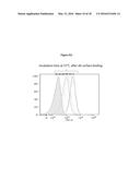 |
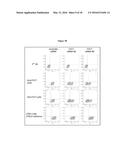 |  |
 | 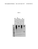 |
 |  |
 |  |
 |  |
 | 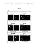 |
 |  |
 |  |
 |  |
 |  |
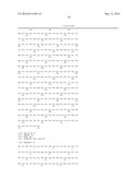 | 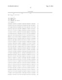 |
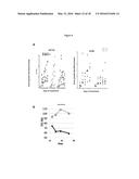 | 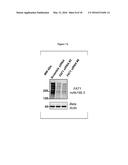 |
 | 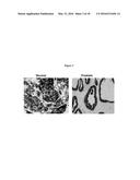 |
 |  |
 |  |
 |  |
 |  |
 |  |
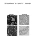 | 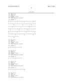 |
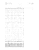 | 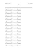 |
 |  |
 | 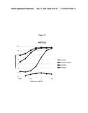 |
 | 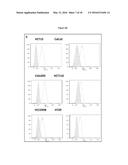 |
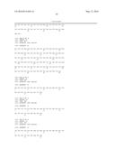 | 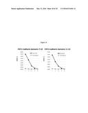 |
 | 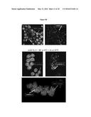 |
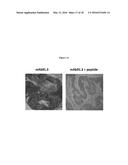 |
| Similar patent applications: | |
| Date | Title |
|---|---|
| 2016-04-28 | Hybridoma clones, monoclonal antibodies, and methods of use |
| 2016-02-18 | Kir-binding agents and methods of use thereof |
| 2016-05-05 | Centrifuge/magnet-based analyzers and method of operating thereof |
| 2016-05-19 | Monoclonal antibodies that react with the capsule of bacillus anthracis |
| 2015-12-24 | Tumor specific antibodies and uses therefor |
| New patent applications in this class: | |
| Date | Title |
|---|---|
| 2022-05-05 | Photocleavable mass-tags for multiplexed mass spectrometric imaging of tissues using biomolecular probes |
| 2022-05-05 | Macrophage expression in breast cancer |
| 2022-05-05 | Characterizing methylated dna, rna, and proteins in the detection of lung neoplasia |
| 2022-05-05 | Methods for identifying and improving t cell multipotency |
| 2022-05-05 | Sequence analysis using meta-stable nucleic acid molecules |
| New patent applications from these inventors: | |
| Date | Title |
|---|---|
| 2016-05-05 | Pharmaceutical compositions comprising vesicles |
| 2015-04-02 | Prostate tumor markers and methods of use thereof |
| Top Inventors for class "Chemistry: molecular biology and microbiology" | |
| Rank | Inventor's name |
|---|---|
| 1 | Marshall Medoff |
| 2 | Anthony P. Burgard |
| 3 | Mark J. Burk |
| 4 | Robin E. Osterhout |
| 5 | Rangarajan Sampath |