Patent application title: MULTIPLEXED DETECTION AND QUANTIFICATION OF NUCLEIC ACIDS IN SINGLE-CELLS
Inventors:
Andreas Philipp Frei (San Francisco, CA, US)
Garry P. Nolan (Redwood City, CA, US)
Garry P. Nolan (Redwood City, CA, US)
Pier Federico Gherardini (Palo Alto, CA, US)
Felice Alessio Bava (Menlo Park, CA, US)
IPC8 Class: AC12Q168FI
USPC Class:
506 9
Class name: Combinatorial chemistry technology: method, library, apparatus method of screening a library by measuring the ability to specifically bind a target molecule (e.g., antibody-antigen binding, receptor-ligand binding, etc.)
Publication date: 2016-04-21
Patent application number: 20160108458
Abstract:
Proximity Ligation Assay for RNA (PLAYR) provides cost-efficient
detection of specific nucleic acids in single cells, and may be combined
with flow cytometry to simultaneously analyze large numbers of cells for
a plurality of nucleic acids, e.g. at least one, to up to 5, up to 10, up
to 15, up to 20 or more transcripts can be simultaneously analyzed, at a
rate of up to about 50, 100, 250, 500 or more cells/second. An advantage
of PLAYR includes the ability to simultaneously analyze multiple nucleic
acids and proteins in single cells, as the method is compatible with
conventional antibody staining for proteins, intracellular
phosphorylation sites, and other cellular antigens. This enables the
simultaneous detection of multiple nucleic acid molecules in combination
with additional cellular parameters.Claims:
1. A method for determining the abundance of a target nucleic acid in a
single cell, the method comprising: contacting a fixed and permeabilized
cell with at least one pair of oligonucleotide primers under conditions
permissive for specific hybridization, wherein each oligonucleotide in
the pair comprises: (i) a target binding region that hybridizes to the
target nucleic acid; (ii) a spacer region that does not bind to the
target nucleic acid or to any region of a padlock probe; and (iii) a
PLAYR1 or PLAYR2 region that specifically binds to a padlock probe;
washing the cell free of unbound primer contacting the cell with a
padlock probe under conditions permissive for specific hybridization,
wherein the padlock probe comprises separate polynucleotides of (i) a
backbone and (ii) an insert; contacting the cell with ligase wherein
bound backbone and insert polynucleotides are ligated to generate a
closed circle; performing rolling circle amplification using the closed
circle as a template and PLAYR1 or PLAYR2 as a primer for a polymerase;
contacting the cell with a detection probe under conditions permissive
for specific hybridization; and detecting the level of bound detection
probes to determine the abundance of the target nucleic acid.
2. The method of claim 1, wherein the oligonucleotide primer pairs are denatured by heating before contacting the sample.
3. The method of claim 1, wherein the cell is present in a population of cells.
4. The method of claim 3, wherein the cell population comprises a plurality of cell types.
5. The method of claim 1, wherein a plurality of oligonucleotide primers are used.
6. The method of claim 5, wherein at least 5 different target nucleic acids are detected.
7. The method of claim 1, wherein the target nucleic acid is RNA.
8. The method of claim 7, wherein the RNA is mRNA.
9. The method of claim 1, wherein the target nucleic acid is DNA.
10. The method of claim 1, wherein the cell is simultaneously profiled for expression of one or more non-nucleic acid markers.
11. The method of claim 10, wherein the one or more markers are protein markers.
12. The method of any one of claim 1, wherein the detecting is performed by flow cytometry.
13. The method of claim 12, wherein the flow cytometry is mass cytometry or fluorescence-activated flow cytometry.
14. The method of any one of claim 1, wherein the detecting is performed by microscopy or nano-SIMS.
15. The method of claim 1, wherein each target binding region of a primer pair binds to a region of about 15-30 nucleotides of the target nucleic acid, wherein in a pair, each target site is different, and the target sites are adjacent on the target nucleic acid
16. The method of claim 13, wherein the pair of oligonucleotide primers are selected such that each primer in the pair has a similar melting temperature for binding to its cognate target site.
17. The method of claim 14, wherein the Tm is from about 50.degree. C. to about 70.degree. C.
18. The method of claim 15, wherein the Tm is from about 58.degree. to about 62.degree. C.
19. The method of claim 1, wherein the sequence of the PLAYR 1 and/or PLAYR 2 regions provides barcoding information for identification of the target nucleic acid for use in multiplex analysis.
20. A kit for use in the method of any one of claims 1.
Description:
BACKGROUND OF THE INVENTION
[0002] High-throughput measurements of gene expression using microarray technology or high throughput sequencing contribute tremendously to our understanding of how genetic networks coordinately function in normal cells and tissues and how they malfunction in disease. Such measurements allow one to infer the function of genes based on their expression patterns, to detect which genes have altered expression in disease, and to identify expression signatures that are predictive of disease progression. However, bulk transcriptome measurements only inform on the average gene expression in a sample. Thus, in a complex sample containing several cell types with different gene expression signatures, only the most abundant signature but not necessarily the most meaningful will be captured. Accordingly, the variability in single-cell gene expression in most biological systems and especially in tissues and tumors generates a need for techniques aimed at characterizing gene expression programs in individual cells of interest.
[0003] The increasing appreciation for the importance of single-cell measurements is reflected in the vast number of single-cell analysis platforms that have been successfully commercialized in recent years, including mass cytometry and microfluidic-based approaches. While flow cytometry provides an excellent platform for the detection of proteins in single cells using antibodies, no comparable solution exists for the detection of nucleic acids. Microfluidic technologies for the detection and quantification of mRNA in single cells are very costly and their throughput is several orders of magnitude lower compared with what can be achieved for proteins using flow cytometry.
[0004] To overcome the limitations of bulk analyses, a number of technologies have been developed that measure gene expression in single cells. In one such method, up to 20 short oligonucleotide probe pairs hybridize in adjacent positions to a target RNA. These binding events are subsequently amplified using branched DNA technology, where the addition of sets of oligonucleotides in subsequent hybridization steps gives rise to a branched DNA molecule. The presence of such a branched DNA structure can then be detected and quantified by flow cytometry using a fluorescent probe. This technology enables the detection of only few RNA copy numbers in millions of single cells but is currently limited to the simultaneous detection of small numbers of measured transcripts. Furthermore, the protocol is long and laborious and the buffers used are not compatible with some fluorophores commonly used in flow cytometry and cannot be used at all in mass cytometry.
[0005] Another method (Larsson et al. (2010) Nature Methods), uses padlock probes, i.e. linear probes that can be converted into a circular DNA molecule by target-dependent ligation upon hybridization to a target RNA molecule. The resulting circularized single-stranded DNA probe can then be amplified using the enzyme phi29 polymerase in a process termed Rolling Circle Amplification (RCA). This process produces a single-stranded DNA molecule containing hundreds of complementary tandem repeats of the original DNA circle. This RCA product can be made visible through the addition of fluorescently labeled detection probes that will hybridize to a detection sequence in the product. This technology enables the multiplex detection of transcripts but requires reverse transcription of target mRNAs using specific primers and RNAseH digestion of the original transcript before hybridization of the padlock probe. Therefore, it introduces additional variability in the assay and requires the design and optimization of both probes and primers.
[0006] Another commercially available solution for single-cell mRNA measurements is based on the physical separation of single cells using a microfluidic device followed by library preparation and sequencing. This is currently the only genome-wide solution but the very limited throughput (96 cells per run) makes it unsuitable for the analysis of samples with multiple cell populations such as blood samples or tumors. Additionally, the technology is expensive compared to the other approaches, and does not allow for the simultaneous detection of proteins and mRNAs in the same cell.
[0007] There is a need for methods that can provide information on multiple transcripts in single cells, particularly that can be usefully combined with protein analysis. Such methods can help analyze how biological networks coordinately function in normal and diseased cells and tissues. The present invention addresses this need.
PUBLICATIONS
[0008] Larsson et al. In situ detection and genotyping of individual mRNA molecules. Nat. Methods 7, 395-397 (2010). Player et al. Single-copy gene detection using branched DNA (bDNA) in situ hybridization. J. Histochem. Cytochem. 49, 603-612 (2001). Porichis, F. et al. High-throughput detection of miRNAs and gene-specific mRNA at the single-cell level by flow cytometry. Nature Communications 5, 5641 (2014). Bendall, S. C. et al. Single-cell mass cytometry of differential immune and drug responses across a human hematopoietic continuum. Science 332, 687-696 (2011). Wolf-Yadlin, A. et al. Effects of HER2 overexpression on cell signaling networks governing proliferation and migration. Mol Syst Biol 2, 54 (2006). Angelo, M. et al. Multiplexed ion beam imaging of human breast tumors. Nat Med 20, 436-442 (2014). Fredriksson, S. et al. Protein detection using proximity-dependent DNA ligation assays. Nat Biotechnol 20, 473-477 (2002). Soderberg, O. et al. Direct observation of individual endogenous protein complexes in situ by proximity ligation. Nat. Methods 3, 995-1000 (2006).
[0009] International patent applications WO2012/160083; WO2001/061037; WO2013/173774.
SUMMARY OF THE INVENTION
[0010] Methods and compositions are provided for multiplexed analysis of target nucleic acids in single cells by a method herein termed PLAYR (Proximity Ligation Assay for RNA). The methods of the invention enable cost-efficient detection of specific nucleic acids in single cells, and may be combined with flow cytometry or mass cytometry to simultaneously analyze large numbers of cells for a plurality of nucleic acids, e.g. at least one, to up to 5, up to 10, up to 15, up to 20, up to 30, up to 40 or more transcripts can be simultaneously analyzed, at a rate of up to about 50, 100, 250, 500, up to 750, up to 1000 or more cells/second. An advantage of PLAYR includes the ability to simultaneously analyze multiple nucleic acids and proteins in single cells, as the method is compatible with conventional antibody staining for proteins, intracellular phosphorylation sites, and other cellular antigens. This enables the simultaneous detection of multiple nucleic acid molecules in combination with additional cellular parameters. It can be combined with various different platforms, including without limitation FACS, mass cytometry, microscopy, nano-SIMS imaging, and the like.
[0011] In the methods of the invention, a pair of short oligonucleotide probes are designed that specifically hybridize to adjacent regions of a target nucleic acid. Target nucleic acids include, without limitation, mRNA, pre-mRNA, rRNA, miRNA, lincRNA, denatured DNA, and the like. Each probe in the pair further comprises a linker and a "PLAYR 1" or "PLAYR 2" sequence that does not hybridize to the target nucleic acid. When the probes are bound to the target nucleic acid, the PLAYR 1 and PLAYR 2 regions of the probe act as template for the hybridization, circularization, and ligation of the components of a DNA padlock probe that are added in a subsequent step. The resulting circular single-stranded DNA product is amplified by rolling circle amplification (RCA), which produces a single-stranded DNA molecule containing complementary tandem repeats of the original DNA circle. The amplification product is detected with a complementary detection probe labeled with a detectable marker, e.g. fluorophore, metal conjugate, etc. A high level of specificity results from the requirement that both probes hybridize to adjacent locations for the amplification reaction to take place, resulting in excellent specificity, low background, and high signal-to-noise ratios.
[0012] In some embodiments, a method is provided for determining the abundance of a target nucleic acid in a single cell, the method comprising contacting a fixed and permeabilized cell with at least one pair of oligonucleotide primers under conditions permissive for specific hybridization, wherein each oligonucleotide in the pair comprises: a target binding region that hybridizes to the target nucleic acid; a spacer region that does not bind to the target nucleic acid or to any region of a padlock probe; and an PLAYR 1 or PLAYR 2 region that specifically binds to the padlock probe, wherein each padlock probe comprises two polynucleotides: a backbone and an insert, and wherein the PLAYR 1 or PLAYR 2 region binds to both insert and backbone; washing the cells free of unbound primers; contacting the cells with backbone and insert polynucleotides under conditions permissive for specific hybridization; washing the cells free of unbound backbone insert; performing a ligation reaction, in which bound backbone insert polynucleotides are ligated to generate a circle; amplifying the ligated backbone/insert circle by rolling circle amplification; washing the cells free of polymerase; hybridizing detection primers to the amplified circle; washing the cells free of unbound detection probes, and quantitating the level of bound detection primers to determine the abundance of the target nucleic acid. Quantitation may include use of a detection probe conjugated to a fluorescent or metal label, and determination of the level of fluorescent or metal label present, e.g. by nano-SIMS, mass cytometry, FACS, etc. In many embodiments, a plurality of target nucleic acids are simultaneously analyzed.
[0013] In some embodiments of the invention, PLAYR is used in combination with cytometry gating on specific cell populations, as defined by other cellular parameters measured simultaneously, for example in combination with antibody staining and mass cytometry or FACS to define a subpopulation of interest. In such embodiments, a complex cell population may be analyzed, e.g. a biopsy or blood sample potentially including immune cells, progenitor or stem cells, cancer cells, etc. For example, a method is provided for determining the abundance of one or more target nucleic acids in a defined cell type within a complex cell population, where the quantification of detection probes is combined with detection of cellular markers, including without limitation protein markers, that serve to define the cell type of interest.
[0014] In other embodiments, the methods of the invention are used for multiplexed detection and quantification of specific splice variants of mRNA transcripts in single cells.
[0015] In yet another embodiment, the methods of the invention are combined with Proximity Ligation Assay (PLA) for the simultaneous detection and quantification of nucleic acid molecules and protein-protein interactions.
[0016] With prior denaturation of endogenous cellular DNA (by heat, enzymatic methods, or any other suitable procedure), the technology is modified for the detection of specific DNA sequences (genotyping of single cells). In this adaptation, the technology enables the quantification of gene copy number variations as well as the detection of genomic translocation/fusion events. For example, in the detection of a fusion event, if a first gene is fused to a second gene the PLAYR method can be adapted, where one or more primers are targeted to gene 1, with an PLAYR 1 sequence; and one or more primers are targeted to gene 2 with an PLAYR 2 sequence. A signal is obtained only when the fusion transcript is present, as the individual probes do not give rise to an amplification product. A plurality of individual primers may be designed for each of gene 1 and gene 2, e.g. 2, 3, 4, 5, 6 or more.
BRIEF DESCRIPTION OF THE DRAWINGS
[0017] FIG. 1: Overview of the PLAYR technology, see text for details.
[0018] FIG. 2: Varying the Insert and PLAYR1/PLAYR2 sequence allows probes targeting different transcripts to be barcoded. This enables the multiplexed detection of multiple transcripts in the same cell.
[0019] FIG. 3A-3B: FIG. 3A) PLAYR specifically detects target transcripts. Jurkat cells express CD3E and do not express CD10 and CD179a. Conversely Nalm-6 cells express CD10 and CD179a but not CD3E. The histograms depict the fluorescence intensity of the two cell lines when treated with probes specifically targeting these transcripts. A strong positive signal is only observed in the cell line expressing the transcript targeted by the PLAYR probes. Cells were also incubated with two single probes targeting the Actin and Gapdh transcript respectively. These two probes never hybridize in close proximity, as they target different transcripts. Accordingly no signal is observed. FIG. 3B) The signal can be increased by using multiple probe pairs targeting the same transcript.
[0020] FIG. 4: The PLAYR signal decreases with the distance between the two probes in a pair. Multiple adjacent probe pairs spanning a transcript were designed. Each PLAYR1 probe was then tested in combination with all the PLAYR2 probes from all the other pairs. The y-axis represents the ratio between the signal obtained with a given PLAYR1/PLAYR2 combination, and the signal obtained with the corresponding adjacent PLAYR1/PLAYR2 pair (i.e. the one that was originally designed as an adjacent pair). There is a clear tendency for the signal to decrease as the distance between the PLAYR1 and PLAYR2 probes increases.
[0021] FIG. 5: Simultaneous detection of nine transcripts in Jurkat cells. Nine different inserts are used to barcode probe sets targeting three different transcripts (CD90, CD3, KRAS, NRAS, PLCG, LCK, ZAP70, ACTB, GAPDH). Nine different detection oligonucleotides, specific for each Insert system, were also conjugated to a polymer chelating nine different stable transition element isotopes (150Nd, 162Dy, 153Eu, 156Gd, 148Nd, 176Yb, 160Gd, 167Er, 168Er respectively). The probe sets and detection oligonucleotides for each gene were incubated simultaneously and the signal intensity was measured on a CyTOF mass cytometer.
[0022] FIG. 6A-6D: PLAYR enables the simultaneous quantification of specific transcripts and proteins in single cells FIG. 6A) Main steps of the PLAYR protocol: 1) Fixation of cells captures their native state and permeabilization enables intracellular antibody staining and blocking of endogenous RNAses with inhibitors. 2) PLAYR probe pairs are added for proximal hybridization to target transcripts. 3) Backbone and insert oligonucleotides are added and form a circle if hybridized to PLAYR probes that are in close proximity (bound to a transcript). Insert sequences serve as cognate barcodes for targeted transcripts. 4) Backbone and insert oligonucleotides are ligated into a single-stranded DNA circle by T4 DNA ligase. 5) Rolling circle amplification of the DNA circle by phy29 polymerase. 6) Detection of rolling circle amplicons with suitably labeled oligonucleotides that bind to the insert regions. FIG. 6B) Detection of transcripts for three housekeeping genes that span a wide abundance range in U937 cells by mass cytometry. FIG. 6C) Quantification of CCL4 and IFNG mRNA by PLAYR and qPCR in NKL cells after stimulation with PMA/ionomycin. FIG. 6D) Simultaneous IFNG mRNA and protein quantification by mass cytometry in NKL cells at indicated time points after stimulation with PMA/ionomycin.
[0023] FIG. 7A-7C: Highly multiplexed measurement of different transcripts in single cells (FIG. 7A) Detection of 14 different transcripts in Jurkat cells by mass cytometry. PLAYR probes to transcripts not expressed in T cells (HLA-DRA) or to those encoding T cell surface markers, T cell signaling molecules, and housekeeping proteins of different abundance levels were used. Each row represents a sample to which probe pairs for one gene only or all genes simultaneously (bottom row) were added. Each column represents a mass cytometry acquisition channel that monitors a metal reporter used to detect transcripts of a given gene. Non-cognate probes that are using the same insert system but bind to different target transcripts were included as an additional control (CTL). b) NKL cells were primed with IL2/IL12/IL18 and stimulated with PMA/ionomycin for 3 hours. Contour plots display co-expression of NKL effector transcripts as measured by mass cytometry. FIG. 7C) 10000 cells were randomly sampled from the data in (FIG. 7B) and transcript expression was represented in heat map format. Each column corresponds to a single cell and rows denote different effector transcripts. Rows and columns of the heat map were clustered for visual clarity.
[0024] FIG. 8A-8E: Highly multiplexed measurement of transcripts within cell types defined by other transcripts or protein epitopes. (FIG. 8A) viSNE analysis of embryonic stem cells, differentiating embryonic stem cells, and embryonic fibroblasts of mice based on expression of 15 transcripts (CD44, MKI67, CDH1, CD47, KLF4, ESRRB, ACTB, SOX2, LINCENC1, ZFP42, SALL4, CD9, POU5F1 (OCT4), THY1, NANOG) with overlays showing the location of the three cell populations. FIG. 8B) Color-coded expression levels of selected transcripts used to construct the viSNE map. FIG. 8C) viSNE analysis of PBMCs based on expression of 10 surface protein markers (CD19, CD4, CD8, CD20, PTPRC (CD45), PTPRCRA (CD45RA), CD33, ITGAX (CD11c), CD3, HLA-DRA) with overlays showing the location of major cell populations. FIG. 8D) Expression of selected proteins and the corresponding transcripts was overlaid in the viSNE map shown in (FIG. 8C) and color-coded by signal intensity. FIG. 8E) Contour plots displaying correlations of protein and transcript levels for HLA-DRA and ITGAX in individual PBMCs.
[0025] FIG. 9A-9E: Measurements of cytokine transcript induction in human PBMCs. (FIG. 9A) Mass cytometry gating strategy for human PBMCs. FIG. 9B) Heat map representing the mean expression values of cytokine transcripts at different time points after stimulation with LPS in different cellular populations defined by protein surface markers. FIG. 9C) Cytokine expression in the CD14+ monocyte population as measured by fluorescence flow cytometry. FIG. 9D) Cytokine transcript expression in the CD14+ monocyte population as measured by mass cytometry. FIG. 9E) Contour plots showing interleukin 8 (CXCL8) and tumor necrosis factor alpha (TNF) transcript expression in CD14+ monocytes.
[0026] FIG. 10A-10C: Single-cell resolution map of cytokine induction in human PBMCs. PBMCs were stimulated with LPS and analyzed after 4 hours. Cells were analyzed with antibodies against cell surface proteins (CD19, CD38, CD4, CD8, CD7, CD14, IL3RA (CD123), PTPRC (CD45), PTPRCRA (CD45RA), CD33, ITGAX (CD11c), FCGR3A (CD16), CD3, CD20, HLA-DRA, NCAM1 (CD56) and phosphorylation sites pP38 MAPK (pT180/pY182), pERK1/2 (pT202/pY204). FIG. 10A) viSNE map based on cell surface marker expression with overlays showing the location of major cell populations. FIG. 10B) Selected protein markers used to define myeloid cell populations and MAPK signaling were color-coded by expression level. FIG. 10C) Measurements for 8 different cytokine transcripts were overlaid and color-coded by expression level.
[0027] FIG. 11: Graphical display of the PLAYRDesign software tool for user-friendly design of PLAYR probe pairs. Each potential probe is represented by a red rectangle. The Primer3 score of each probe is represented by a color gradient from light pink to red, where red probes have higher scores and are preferred over light red probes. The position of probes along the transcript is represented together with sequence features that can guide probe selection. Different graphs represent: maximum sequence identity of BLAST matches to a database of repetitive sequences (red); maximum sequence identity of BLAST matches to other transcripts (blue); predicted melting temperature in a window of 20 residues (green); number of ESTs that skip an exon but include the exons flanking it (blue). The actual melting temperature of probes is independently calculated by Primer3, while the purpose of the green graph is to give an indication on whether certain regions of the transcript have a melting temperature that is too low or too high to be amenable for probe design. Blue and red graphs represent sequence features that are not considered in the scoring of Primer3 probes.
[0028] FIG. 12: Specificity control experiments for PLAYR. Detection of the Beta-actin transcript in Jurkat cells (ACTB). No signal is detected when PLAYR is performed in absence of probes (NO PROBES), in absence of insert (NO INSERT), in absence of backbone (NO BACKBONE), in absence of ligase (NO LIGATION), in absence of detection oligo (NO DETECTION OLIGO), in presence of probes directed against the anti-sense Beta-actin transcript (SENSE PROBES), in presence of probes with the same half of the insert-complementary sequence (ORIENTATION CONTROL), or in presence of non-cognate probe pairs targeting different transcripts (ACTB and GAPDH, GENE-SPECIFICITY CONTROL). Signals were detected by flow cytometry. 4 probe pairs were used per gene.
[0029] FIG. 13A-13B: Detection of specific transcripts in single cells by flow cytometry using multiple probe pairs. FIG. 13A) Detection of CD10 and CD3E by PLAYR. Jurkat and NALM-6 cells were incubated with the indicated number of probe pairs and analyzed by flow cytometry. FIG. 13B) The intensity of PLAYR signals depends on the distance between PLAYR probe binding sites on a target transcript. Multiple adjacent probe pairs spanning a transcript were designed and tested in all possible pairwise combinations. The x-axis represents the distance between each pair of probes, and the y-axis represents the ratio between the signal obtained with a given combination and the signal obtained with the corresponding adjacent probe (i.e., the one that was originally designed to be used in the pair).
[0030] FIG. 14: Fluorescence flow cytometry gating strategy for human PBMCs.
DETAILED DESCRIPTION OF THE EMBODIMENTS
[0031] Before the present invention is further described, it is to be understood that this invention is not limited to particular embodiments described, as such may, of course, vary. It is also to be understood that the terminology used herein is for the purpose of describing particular embodiments only, and is not intended to be limiting, since the scope of the present invention will be limited only by the appended claims.
[0032] Where a range of values is provided, it is understood that each intervening value, to the tenth of the unit of the lower limit unless the context clearly dictates otherwise, between the upper and lower limit of that range and any other stated or intervening value in that stated range, is encompassed within the invention. The upper and lower limits of these smaller ranges may independently be included in the smaller ranges and are also encompassed within the invention, subject to any specifically excluded limit in the stated range. Where the stated range includes one or both of the limits, ranges excluding either or both of those included limits are also included in the invention.
[0033] Methods recited herein may be carried out in any order of the recited events which is logically possible, as well as the recited order of events.
[0034] Unless defined otherwise, all technical and scientific terms used herein have the same meaning as commonly understood by one of ordinary skill in the art to which this invention belongs. Although any methods and materials similar or equivalent to those described herein can also be used in the practice or testing of the present invention, the preferred methods and materials are now described.
[0035] All publications mentioned herein are incorporated herein by reference to disclose and describe the methods and/or materials in connection with which the publications are cited.
[0036] It must be noted that as used herein and in the appended claims, the singular forms "a", "an", and "the" include plural referents unless the context clearly dictates otherwise. It is further noted that the claims may be drafted to exclude any optional element. As such, this statement is intended to serve as antecedent basis for use of such exclusive terminology as "solely," "only" and the like in connection with the recitation of claim elements, or use of a "negative" limitation.
[0037] The publications discussed herein are provided solely for their disclosure prior to the filing date of the present application. Nothing herein is to be construed as an admission that the present invention is not entitled to antedate such publication by virtue of prior invention. Further, the dates of publication provided may be different from the actual publication dates which may need to be independently confirmed.
DEFINITIONS
[0038] Target Nucleic Acid.
[0039] As used herein, a target nucleic acid is any polynucleotide nucleic acid molecule (e.g., DNA molecule; RNA molecule, modified nucleic acid, etc.) present in a single cell. In some embodiments, the target nucleic acid is a coding RNA (e.g., mRNA). In some embodiments, the target nucleic acid is a non-coding RNA (e.g., tRNA, rRNA, microRNA (miRNA), mature miRNA, immature miRNA; etc). In some embodiments, the target nucleic acid is a splice variant of an RNA molecule (e.g., mRNA, pre-mRNA, etc.) in the context of a cell. A suitable target nucleic acid can therefore be an unspliced RNA (e.g., pre-mRNA, mRNA), a partially spliced RNA, or a fully spliced RNA, etc.
[0040] Target nucleic acids of interest may be variably expressed, i.e. have a differing abundance, within a cell population, wherein the methods of the invention allow profiling and comparison of the expression levels of nucleic acids, including without limitation RNA transcripts, in individual cells.
[0041] A target nucleic acid can also be a DNA molecule, e.g. a denatured genomic, viral, plasmid, etc. For example the methods can be used to detect copy number variants, e.g. in a cancer cell population in which a target nucleic acid is present at different abundance in the genome of cells in the population; a virus-infected cells to determine the virus load and kinetics, and the like.
[0042] Target Specific Oligonucleotide Primer Pairs.
[0043] In the methods of the invention, one or more pairs of target specific oligonucleotide primers are contacted with a cell comprising target nucleic acids. Each oligonucleotide in a pair comprises 3 regions: a target binding site, a spacer, and a padlock probe binding site, which is referred to herein as PLAYR 1 or PLAYR 2. See FIG. 1. A plurality of oligonucleotide pairs can be used in a reaction, where one or more pairs specifically bind to each target nucleic acid. For example, two primer pairs can be used for one target nucleic acid in order to improve sensitivity and reduce variability. It is also of interest to detect a plurality of different target nucleic acids in a cell, e.g. detecting up to 2, up to 3, up to 4, up to 5, up to 6, up to 7, up to 8, up to 9, up to 10, up to 12, up to 15, up to 18, up to 20, up to 25, up to 30, up to 40 or more distinct target nucleic acids. The primers are typically denatured prior to use, typically by heating to a temperature of at least about 50° C., at least about 60° C., at least about 70° C., at least about 80° C., and up to about 99° C., up to about 95° C., up to about 90° C.
[0044] The target binding site binds to a region of the target nucleic acid. In a pair, each target site is different, and the pair are complementary adjacent sites on the target nucleic acid, e.g. usually not more than 10 nt distant, not more than 9, 8, 7, 6, 5, 4, 3, 2, or 1 nt. distant from the other site, and may be contiguous sites. Target sites are typically present on the same strand of the target nucleic acid in the same orientation. Target sites are also selected to provide a unique binding site, relative to other nucleic acids present in the cell. Each target site is generally from about 18 to about 25 nt in length, e.g. from about 18 to 23, from about 18-21, etc. The pair of oligonucleotide probes are selected such that each probe in the pair has a similar melting temperature for binding to its cognate target site, e.g. the Tm may be from about 50° C., from about 52° C., from about 55° C., and up to about 70° C., up to about 72° C., up to about 70° C., up to about 65° C., up to about 62° C., and may be from about 58° to about 62° C. The GC content of the target site is generally selected to be no more than about 20%, no more than about 30%, no more than about 40%, no more than about 50%, no more than about 60%, no more than about 70%,
[0045] The spacer region is between the target specific region and the PLAYR 1 or PLAYR 2 region, and is preferably not complementary to target nucleic acids or the padlock probe, and is selected to provide for a low background. In some embodiments the spacer is a poly-A tract. The spacers are typically of even length on both probes in the pair, and may be from about 2 to about 20 nt in length, e.g. up to about 20, up to about 18, up to about 15, up to about 12, up to about 10, up to about 7, up to about 5, up to about 3 nt. in length. In some embodiments the spacer is from 8- to 12 nt in length.
[0046] The PLAYR 1 or PLAYR 2 regions specifically bind to components of the padlock probes, and are selected to distribute the binding between the insert and backbone sequences. The sequence of the PLAYR region is arbitrary, and can be chosen to provide bar-coding information, etc. Different PLAYR regions used in a reaction, particularly a multiplex reaction, may be selected to provide equivalent melting temperatures, e.g. Tm that are not more than 1-2 degrees different. The distribution in sequence complementary to insert and complementary to backbone is roughly equal, for example where 9-13 nt. are complementary to each of the insert and backbone of the padlock probe, and where the backbone and insert of the padlock probe hybridize to contiguous sequences on the PLAYR site. It is preferable for the PLAYR 1 sequence to differ from the PLAYR 2 sequence.
[0047] Padlock Probe.
[0048] As shown in FIG. 1, the two polynucleotides of the padlock probe are complementary to the PLAYR 1 and PLAYR 2 regions, where the PLAYR 1 or PLAYR 2 sequence is complementary to adjacent sequences of the insert and backbone, and where the PLAYR 1 binding sequence of the insert is adjacent to the PLAYR 2 binding sequence of the insert. When both PLAYR 1 and PLAYR 2 probes are present and properly aligned, the insert and backbone form an open circular molecule that can be ligated to create a closed circle. The insert sequence is therefore fully complementary to the insert binding sequences of the PLAYR 1 and RL2 probe regions, and is generally from about 18 to about 25 nt in length, e.g. from about 18 to 23, from about 18-21, etc.
[0049] Where a plurality of target nucleic acids are being detected, each insert sequence is specific for each target specific primer pair. In other words, all inserts are substantially different from the other in sequence, generally having not more than 4 nt in a common string. This ensures that the resulting amplification products barcode for the detected target and can be detected with different detection oligonucleotides conjugated to corresponding reporters.
[0050] The backbone of the padlock probe is selected to be of a length that allows circularization with steric strain, with low background hybridization to sequences present in the cell of interest, with the exception of the specific PLAYR 1/2 binding sites. The terminal ends of the backbone specifically bind to a portion of the PLAYR 1 and PLAYR 2 sequences, e.g. a region of about 6-12 nt in length. The overall length of the backbone is from about 50 to about 250 nt. in length, e.g. from about 50 to about 200, from about 50 to about 150, from about 50 to about 100 nt. in length.
[0051] Ligase. The term "ligase" as used herein refers to an enzyme that is commonly used to join polynucleotides together or to join the ends of a single polynucleotide. Ligases include ATP-dependent double-strand polynucleotide ligases, NAD+-dependent double-strand DNA or RNA ligases and single-strand polynucleotide ligases, for example any of the ligases described in EC 6.5.1.1 (ATP-dependent ligases), EC 6.5.1.2 (NAD+-dependent ligases), EC 6.5.1.3 (RNA ligases). Specific examples of ligases include bacterial ligases such as E. coli DNA ligase and Taq DNA ligase, Ampligase® thermostable DNA ligase (Epicentre®Technologies Corp., part of Illumina®, Madison, Wis.) and phage ligases such as T3 DNA ligase, T4 DNA ligase and T7 DNA ligase and mutants thereof.
[0052] Rolling Circle Amplification.
[0053] A single-stranded, circular polynucleotide template is formed by ligation of the backbone and insert polynucleotides, which circular polynucleotide comprises a region that is complementary to the PLAYR 1 and PLAYR 2 sequences. Upon addition of a DNA polymerase in the presence of appropriate dNTP precursors and other cofactors, either the PLAYR 1 or the PLAYR 2 sequence, which can both act as primers for the polymerase, is elongated by replication of multiple copies of the template. This amplification product can be readily detected by binding to a detection probe.
[0054] Techniques for rolling circle amplification are known in the art (see, e.g., Baner et al, Nucleic Acids Research, 26:5073-5078, 1998; Lizardi et al, Nature Genetics 19:226, 1998; Schweitzer et al. Proc. Natl Acad. Sci. USA 97:10113-119, 2000; Faruqi et al, BMC Genomics 2:4, 2000; Nallur et al, Nucl. Acids Res. 29:el 18, 2001; Dean et al. Genome Res. 11:1095-1099, 2001; Schweitzer et al, Nature Biotech. 20:359-365, 2002; U.S. Pat. Nos. 6,054,274, 6,291,187, 6,323,009, 6,344,329 and 6,368,801). In some embodiments the polymerase is phi29 DNA polymerase.
[0055] Detection Probe.
[0056] The presence and quantitation of an amplified PLAYR padlock sequence in a cell is determined by contacting the cell with an oligonucleotide probe under conditions in which the probe binds to the amplified product. The probe comprises a detectable label, that can be measured and quantitated. A labeled nucleic acid probe is a nucleic acid that is labeled with any label moiety. In some embodiments, the nucleic acid detection agent is a single labeled molecule (i.e., a labeled nucleic acid probe) that specifically binds to the amplification product. In some embodiments, the nucleic acid detection agent includes multiple molecules, one of which specifically binds to the amplification product. In such embodiments, when a labeled nucleic acid probe is present, the labeled nucleic acid probe does not specifically bind to the target nucleic acid, but instead specifically binds to one of the other molecules of the nucleic acid detection agent. A hybridization probe can be any convenient length that provides for specific binding, e.g. it may be from about 16 to about 50 nt. in length, and more usually is from about 18 nt. to about 30 nt. length.
[0057] A "label" or "label moiety" for a nucleic acid probe is any moiety that provides for signal detection and may vary widely depending on the particular nature of the assay. Label moieties of interest include both directly and indirectly detectable labels. Suitable labels for use in the methods described herein include any moiety that is indirectly or directly detectable by spectroscopic, photochemical, biochemical, immunochemical, electrical, optical, chemical, or other means. For example, suitable labels include antigenic labels (e.g., digoxigenin (DIG), fluorescein, dinitrophenol(DNP), etc.), biotin for staining with labeled streptavidin conjugate, a fluorescent dye (e.g., fluorescein, Texas red, rhodamine, a fluorophore label such as an ALEXA FLUOR® label, and the like), a radiolabel (e.g., 3H, 125I, 35S, 14C, or 32P), an enzyme (e.g., peroxidase, alkaline phosphatase, galactosidase, and others commonly used in an ELISA), a fluorescent protein (e.g., green fluorescent protein, red fluorescent protein, yellow fluorescent protein, and the like), a synthetic polymer chelating a metal, a colorimetric label, and the like. An antigenic label can be incorporated into the nucleic acid on any nucleotide (e.g., A,U,G,C).
[0058] Fluorescent labels can be detected using a photodetector (e.g., in a flow cytometer) to detect emitted light. Enzymatic labels are typically detected by providing the enzyme with a substrate and detecting the reaction product produced by the action of the enzyme on the substrate, colorimetric labels can be detected by simply visualizing the colored label, and antigenic labels can be detected by providing an antibody (or a binding fragment thereof) that specifically binds to the antigenic label. An antibody that specifically binds to an antigenic label can be directly or indirectly detectable. For example, the antibody can be conjugated to a label moiety (e.g., a fluorophore) that provides the signal (e.g., fluorescence); the antibody can be conjugated to an enzyme (e.g., peroxidase, alkaline phosphatase, etc.) that produces a detectable product (e.g., fluorescent product) when provided with an appropriate substrate (e.g., fluorescent-tyramide, FastRed, etc.); etc.
[0059] Metal labels (e.g., Sm152, Tb159, Er170, Nd146, Nd142, and the like) can be detected (e.g., the amount of label can be measured) using any convenient method, including, for example, nano-SIMS, by mass cytometry (see, for example: U.S. Pat. No. 7,479,630; Wang et al. (2012) Cytometry A. 2012 July; 81(7):567-75; Bandura et. al., Anal Chem. 2009 Aug. 15; 81(16):6813-22; and Ornatsky et. al., J Immunol Methods. 2010 Sep. 30; 361(1-2):1-20. As described above, mass cytometry is a real-time quantitative analytical technique whereby cells or particles are individually introduced into a mass spectrometer (e.g., Inductively Coupled Plasma Mass Spectrometer (ICP-MS)), and a resultant ion cloud (or multiple resultant ion clouds) produced by a single cell is analyzed (e.g., multiple times) by mass spectrometry (e.g., time of-flight mass spectrometry). Mass cytometry can use elements (e.g., a metal) or stable isotopes, attached as label moieties to a detection reagent (e.g., an antibody and/or a nucleic acid detection agent).
[0060] Nucleic Acids, Analogs and Mimetics.
[0061] In defining the component oligonucleotide primers, probes, etc., used in the methods of the invention, it is to be understood that such probes, primers etc. encompass native and synthetic or modified polynucleotides, particularly the probes, primers etc. that are not themselves substrates for enzymatic modification during the performance of the method, e.g. the target specific oligonucleotide primers, and the detection probes.
[0062] A modified nucleic acid has one or more modifications, e.g., a base modification, a backbone modification, etc, to provide the nucleic acid with a new or enhanced feature (e.g., improved stability). A nucleoside can be a base-sugar combination, the base portion of which is a heterocyclic base. Heterocyclic bases include the purines and the pyrimidines. Nucleotides are nucleosides that further include a phosphate group covalently linked to the sugar portion of the nucleoside. For those nucleosides that include a pentofuranosyl sugar, the phosphate group can be linked to the 2', the 3', or the 5' hydroxyl moiety of the sugar. In forming oligonucleotides, the phosphate groups covalently link adjacent nucleosides to one another to form a linear polymeric compound. In some cases, the respective ends of this linear polymeric compound can be further joined to form a circular compound. In addition, linear compounds may have internal nucleotide base complementarity and may therefore fold in a manner as to produce a fully or partially double-stranded compound. Within oligonucleotides, the phosphate groups can be referred to as forming the internucleoside backbone of the oligonucleotide. The linkage or backbone of RNA and DNA can be a 3' to 5' phosphodiester linkage.
[0063] Examples of suitable nucleic acids containing modifications include nucleic acids with modified backbones or non-natural internucleoside linkages. Nucleic acids having modified backbones include those that retain a phosphorus atom in the backbone and those that do not have a phosphorus atom in the backbone. Suitable modified oligonucleotide backbones containing a phosphorus atom therein include, for example, phosphorothioates, chiral phosphorothioates, phosphorodithioates, phosphotriesters, aminoalkylphosphotriesters, methyl and other alkyl phosphonates including 3'-alkylene phosphonates, 5'-alkylene phosphonates and chiral phosphonates, phosphinates, phosphoramidates including 3'-amino phosphoramidate and aminoalkylphosphoramidates, phosphorodiamidates, thionophosphoramidates, thionoalkylphosphonates, thionoalkylphosphotriesters, selenophosphates and boranophosphates having normal 3'-5' linkages, 2'-5' linked analogs of these, and those having inverted polarity wherein one or more internucleotide linkages is a 3' to 3', 5' to 5' or 2' to 2' linkage. Suitable oligonucleotides having inverted polarity include a single 3' to 3' linkage at the 3'-most internucleotide linkage i.e. a single inverted nucleoside residue which may be a basic (the nucleobase is missing or has a hydroxyl group in place thereof). Various salts (such as, for example, potassium or sodium), mixed salts and free acid forms are also included.
[0064] In some embodiments, a subject nucleic acid has one or more phosphorothioate and/or heteroatom internucleoside linkages, in particular --CH2--NH--O--CH2--, --CH2--N(CH3)--O--CH2-(known as a methylene (methylimino) or MMI backbone), --CH2--O--N(CH3)--CH2--, --CH2--N(CH3)--N(CH3)--CH2-- and --O--N(CH3)--CH2--CH2-- (wherein the native phosphodiester internucleotide linkage is represented as --O--P(═O)(OH)--O--CH2--). MMI type internucleoside linkages are disclosed in the above referenced U.S. Pat. No. 5,489,677. Suitable amide internucleoside linkages are disclosed in U.S. Pat. No. 5,602,240.
[0065] Also suitable are nucleic acids having morpholino backbone structures as described in, e.g., U.S. Pat. No. 5,034,506. For example, in some embodiments, a subject nucleic acid includes a 6-membered morpholino ring in place of a ribose ring. In some of these embodiments, a phosphorodiamidate or other non-phosphodiester internucleoside linkage replaces a phosphodiester linkage.
[0066] Suitable modified polynucleotide backbones that do not include a phosphorus atom therein have backbones that are formed by short chain alkyl or cycloalkyl internucleoside linkages, mixed heteroatom and alkyl or cycloalkyl internucleoside linkages, or one or more short chain heteroatomic or heterocyclic internucleoside linkages. These include those having morpholino linkages (formed in part from the sugar portion of a nucleoside); siloxane backbones; sulfide, sulfoxide and sulfone backbones; formacetyl and thioformacetyl backbones; methylene formacetyl and thioformacetyl backbones; riboacetyl backbones; alkene containing backbones; sulfamate backbones; methyleneimino and methylenehydrazino backbones; sulfonate and sulfonamide backbones; amide backbones; and others having mixed N, O, S and CH2 component parts.
[0067] Also included are nucleic acid mimetics. The term "mimetic" as it is applied to polynucleotides encompasses polynucleotides where only the furanose ring or both the furanose ring and the internucleotide linkage are replaced with non-furanose groups, replacement of only the furanose ring is also referred to as being a sugar surrogate. The heterocyclic base moiety or a modified heterocyclic base moiety is maintained for hybridization with an appropriate target nucleic acid. One such nucleic acid, a polynucleotide mimetic that has been shown to have excellent hybridization properties, is referred to as a peptide nucleic acid (PNA). In PNA, the sugar-backbone of a polynucleotide is replaced with an amide containing backbone, in particular an aminoethylglycine backbone. The nucleotides are retained and are bound directly or indirectly to aza nitrogen atoms of the amide portion of the backbone.
[0068] One polynucleotide mimetic that has excellent hybridization properties is a peptide nucleic acid (PNA). The backbone in PNA compounds is two or more linked aminoethylglycine units which gives PNA an amide containing backbone. The heterocyclic base moieties are bound directly or indirectly to aza nitrogen atoms of the amide portion of the backbone. Representative U.S. patents that describe the preparation of PNA compounds include, but are not limited to: U.S. Pat. Nos. 5,539,082; 5,714,331; and 5,719,262.
[0069] Another class of suitable polynucleotide mimetic is based on linked morpholino units (morpholino nucleic acid) having heterocyclic bases attached to the morpholino ring. A number of linking groups have been reported that can link the morpholino monomeric units in a morpholino nucleic acid. One class of linking groups has been selected to give a non-ionic oligomeric compound. The non-ionic morpholino-based oligomeric compounds are less likely to have undesired interactions with cellular proteins. Morpholino-based polynucleotides are non-ionic mimics of oligonucleotides which are less likely to form undesired interactions with cellular proteins (Dwaine A. Braasch and David R. Corey, Biochemistry, 2002, 41(14), 4503-4510). Morpholino-based polynucleotides are disclosed in U.S. Pat. No. 5,034,506. A variety of compounds within the morpholino class of polynucleotides have been prepared, having a variety of different linking groups joining the monomeric subunits.
[0070] Another suitable class of polynucleotide mimetic is referred to as cyclohexenyl nucleic acids (CeNA). The furanose ring normally present in a DNA/RNA molecule is replaced with a cyclohexenyl ring. CeNA DMT protected phosphoramidite monomers have been prepared and used for oligomeric compound synthesis following classical phosphoramidite chemistry. Fully modified CeNA oligomeric compounds and oligonucleotides having specific positions modified with CeNA have been prepared and studied (see Wang et al., J. Am. Chem. Soc., 2000, 122, 8595-8602). The incorporation of CeNA monomers into a DNA chain increases the stability of a DNA/RNA hybrid. CeNA oligoadenylates formed complexes with RNA and DNA complements with similar stability to the native complexes. The incorporation CeNA structures into natural nucleic acid structures was shown by NMR and circular dichroism to proceed with conformational adaptation.
[0071] Also suitable as modified nucleic acids are Locked Nucleic Acids (LNAs) and/or LNA analogs. In an LNA, the 2'-hydroxyl group is linked to the 4' carbon atom of the sugar ring thereby forming a 2'-C,4'-C-oxymethylene linkage, and thereby forming a bicyclic sugar moiety. The linkage can be a methylene (--CH2--), group bridging the 2' oxygen atom and the 4' carbon atom wherein n is 1 or 2 (Singh et al., Chem. Commun., 1998, 4, 455-456). LNA and LNA analogs display very high duplex thermal stabilities with complementary DNA and RNA (Tm=+3 to +10° C.), stability towards 3'-exonucleolytic degradation and good solubility properties. Potent and nontoxic oligonucleotides containing LNAs have been described (Wahlestedt et al., Proc. Natl. Acad. Sci. U.S.A., 2000, 97, 5633-5638).
[0072] The synthesis and preparation of the LNA monomers adenine, cytosine, guanine, 5-methyl-cytosine, thymine and uracil, along with their oligomerization, and nucleic acid recognition properties have been described (Koshkin et al., Tetrahedron, 1998, 54, 3607-3630). LNAs and preparation thereof are also described in WO98/39352 and WO99/14226, both of which are hereby incorporated by reference in their entirety. Exemplary LNA analogs are described in U.S. Pat. Nos. 7,399,845 and 7,569,686, both of which are hereby incorporated by reference in their entirety.
[0073] A nucleic acid can also include one or more substituted sugar moieties. Suitable polynucleotides include a sugar substituent group selected from: OH; F; O-, S-, or N-alkyl; O-, S-, or N-alkenyl; O-, S- or N-alkynyl; or O-alkyl-O-alkyl, wherein the alkyl, alkenyl and alkynyl may be substituted or unsubstituted C1 to C10 alkyl or C2 to C10 alkenyl and alkynyl. Also suitable are O((CH2)nO)mCH3, O(CH2)nOCH3, O(CH2)nNH2, O(CH2)nCH3, O(CH2)nONH2, and O(CH2)nON((CH2)nCH3)2, where n and m are from 1 to about 10. Other suitable polynucleotides include a sugar substituent group selected from: C1 to C10 lower alkyl, substituted lower alkyl, alkenyl, alkynyl, alkaryl, aralkyl, O-alkaryl or O-aralkyl, SH, SCH3, OCN, Cl, Br, CN, CF3, OCF3, SOCH3, SO2CH3, ONO2, NO2, N3, NH2, heterocycloalkyl, heterocycloalkaryl, aminoalkylamino, polyalkylamino, substituted silyl, an RNA cleaving group, a reporter group, an intercalator, and other substituents having similar properties. A suitable modification can include 2'-methoxyethoxy (2'-O--CH2CH2OCH3, also known as 2'-O-(2-methoxyethyl) or 2'-MOE) (Martin et al., Helv. Chim. Acta, 1995, 78, 486-504) i.e., an alkoxyalkoxy group. A suitable modification can include 2'-dimethylaminooxyethoxy, i.e., a O(CH2)2ON(CH3)2 group, also known as 2'-DMAOE, as described in examples hereinbelow, and 2'-dimethylaminoethoxyethoxy (also referred to as 2'-O-dimethyl-amino-ethoxy-ethyl or 2'-DMAEOE), i.e., 2'-O--CH2--O--CH2--N(CH3)2.
[0074] Other suitable sugar substituent groups include methoxy (--O--CH3), aminopropoxy (--O CH2CH2CH2NH2), allyl (--CH2--CH═CH2), --O-allyl (--O--CH2--CH═CH2) and fluoro (F). 2'-sugar substituent groups may be in the arabino (up) position or ribo (down) position. A suitable 2'-arabino modification is 2'-F. Similar modifications may also be made at other positions on the oligomeric compound, particularly the 3' position of the sugar on the 3' terminal nucleoside or in 2'-5' linked oligonucleotides and the 5' position of 5' terminal nucleotide. Oligomeric compounds may also have sugar mimetics such as cyclobutyl moieties in place of the pentofuranosyl sugar.
[0075] A nucleic acid may also include a nucleobase (also referred to as "base") modifications or substitutions. As used herein, "unmodified" or "natural" nucleobases include the purine bases adenine (A) and guanine (G), and the pyrimidine bases thymine (T), cytosine (C) and uracil (U). Modified nucleobases include other synthetic and natural nucleobases such as 5-methylcytosine (5-me-C), 5-hydroxymethyl cytosine, xanthine, hypoxanthine, 2-aminoadenine, 6-methyl and other alkyl derivatives of adenine and guanine, 2-propyl and other alkyl derivatives of adenine and guanine, 2-thiouracil, 2-thiothymine and 2-thiocytosine, 5-halouracil and cytosine, 5-propynyl (--C═C--CH3) uracil and cytosine and other alkynyl derivatives of pyrimidine bases, 6-azo uracil, cytosine and thymine, 5-uracil (pseudouracil), 4-thiouracil, 8-halo, 8-amino, 8-thiol, 8-thioalkyl, 8-hydroxyl and other 8-substituted adenines and guanines, 5-halo particularly 5-bromo, 5-trifluoromethyl and other 5-substituted uracils and cytosines, 7-methylguanine and 7-methyladenine, 2-F-adenine, 2-amino-adenine, 8-azaguanine and 8-azaadenine, 7-deazaguanine and 7-deazaadenine and 3-deazaguanine and 3-deazaadenine. Modified nucleobases also include tricyclic pyrimidines such as phenoxazine cytidine(1H-pyrimido(5,4-b)(1,4)benzoxazin-2(3H)-one), phenothiazine cytidine (1H-pyrimido(5,4-b)(1,4)benzothiazin-2(3H)-one), G-clamps such as a substituted phenoxazine cytidine (e.g. 9-(2-aminoethoxy)-H-pyrimido(5,4-(b) (1,4)benzoxazin-2(3H)-one), carbazole cytidine (2H-pyrimido(4,5-b)indol-2-one), and pyridoindole cytidine (H-pyrido(3',2':4,5)pyrrolo(2,3-d)pyrimidin-2-one).
[0076] Heterocyclic base moieties may also include those in which the purine or pyrimidine base is replaced with other heterocycles, for example 7-deaza-adenine, 7-deazaguanosine, 2-aminopyridine and 2-pyridone. Further nucleobases include those disclosed in U.S. Pat. No. 3,687,808, those disclosed in The Concise Encyclopedia Of Polymer Science And Engineering, pages 858-859, Kroschwitz, J. I., ed. John Wiley & Sons, 1990, those disclosed by Englisch et al., Angewandte Chemie, International Edition, 1991, 30, 613, and those disclosed by Sanghvi, Y. S., Chapter 15, Antisense Research and Applications, pages 289-302, Crooke, S. T. and Lebleu, B., ed., CRC Press, 1993. Certain of these nucleobases are useful for increasing the binding affinity of an oligomeric compound. These include 5-substituted pyrimidines, 6-azapyrimidines and N-2, N-6 and O-6 substituted purines, including 2-aminopropyladenine, 5-propynyluracil and 5-propynylcytosine. 5-methylcytosine substitutions have been shown to increase nucleic acid duplex stability by 0.6-1.2° C. (Sanghvi et al., eds., Antisense Research and Applications, CRC Press, Boca Raton, 1993, pp. 276-278) and are suitable base substitutions, e.g., when combined with 2'-O-methoxyethyl sugar modifications.
[0077] Quantitation of Detectable Label.
[0078] Various methods can be utilized for quantifying the presence of a detectable label, either on the detection probe, or present in a combined method with analysis of cellular markers used to define the cell being analyzed. For measuring the amount of a detection probe, or other specific binding partner that is present, a convenient method is to label with a detectable moiety, which may be a metal, fluorescent, luminescent, radioactive, enzymatically active, etc.
[0079] Fluorescent moieties are readily available for labeling virtually any biomolecule, structure, or cell type. Immunofluorescent moieties can be directed to bind not only to specific proteins but also specific conformations, cleavage products, or site modifications like phosphorylation. Individual peptides and proteins can be engineered to autofluoresce, e.g. by expressing them as green fluorescent protein chimeras inside cells (for a review see Jones et al. (1999) Trends Biotechnol. 17(12):477-81).
[0080] Mass cytometry is a variation of flow cytometry in which probes are labeled with heavy metal ion tags rather than fluorochromes. Readout is by time-of-flight mass spectrometry This allows for the combination of many more specificities in a single samples, without significant spillover between channels. For example, see Bendall et al. (2011) Science 332 (6030): 687-696, herein specifically incorporated by reference. Nano-SIMS is an alternative method of detecting metal labels.
[0081] Multiple fluorescent or metal labels can be used on the same sample and individually detected quantitatively, permitting simultaneous multiplex analysis. Many quantitative techniques have been developed to harness the unique properties of fluorescence including: direct fluorescence measurements, fluorescence resonance energy transfer (FRET), fluorescence polarization or anisotropy (FP), time resolved fluorescence (TRF), fluorescence lifetime measurements (FLM), fluorescence correlation spectroscopy (FCS), and fluorescence photobleaching recovery (FPR) (Handbook of Fluorescent Probes and Research Chemicals, Seventh Edition, Molecular Probes, Eugene Oreg.).
[0082] Flow or mass cytometry may be used to quantitate parameters such as the presence of cell surface proteins or conformational or posttranslational modification thereof; intracellular or secreted protein, where permeabilization allows antibody (or probe) access, and the like. Both single cell multiparameter and multicell multiparameter multiplex assays, where input cell types are identified and parameters are read by quantitative imaging and fluorescence and confocal microscopy are used in the art, see Confocal Microscopy Methods and Protocols (Methods in Molecular Biology Vol. 122.) Paddock, Ed., Humana Press, 1998.
[0083] Cells.
[0084] Cells for use in the assays of the invention can be an organism, a single cell type derived from an organism, or can be a mixture of cell types. Included are naturally occurring cells and cell populations, genetically engineered cell lines, cells derived from transgenic animals, etc. Virtually any cell type and size can be accommodated. Suitable cells include bacterial, fungal, plant and animal cells. In one embodiment of the invention, the cells are mammalian cells, e.g. complex cell populations such as naturally occurring tissues, for example blood, liver, pancreas, neural tissue, bone marrow, skin, and the like. Some tissues may be disrupted into a monodisperse suspension. Alternatively, the cells may be a cultured population, e.g. a culture derived from a complex population, a culture derived from a single cell type where the cells have differentiated into multiple lineages, or where the cells are responding differentially to stimulus, and the like.
[0085] Cell types that can find use in the subject invention include stem and progenitor cells, e.g. embryonic stem cells, hematopoietic stem cells, mesenchymal stem cells, neural crest cells, etc., endothelial cells, muscle cells, myocardial, smooth and skeletal muscle cells, mesenchymal cells, epithelial cells; hematopoietic cells, such as lymphocytes, including T-cells, such as Th1 T cells, Th2 T cells, Th0 T cells, cytotoxic T cells; B cells, pre-B cells, etc.; monocytes; dendritic cells; neutrophils; and macrophages; natural killer cells; mast cells; etc.; adipocytes, cells involved with particular organs, such as thymus, endocrine glands, pancreas, brain, such as neurons, glia, astrocytes, dendrocytes, etc. and genetically modified cells thereof. Hematopoietic cells may be associated with inflammatory processes, autoimmune diseases, etc., endothelial cells, smooth muscle cells, myocardial cells, etc. may be associated with cardiovascular diseases; almost any type of cell may be associated with neoplasias, such as sarcomas, carcinomas and lymphomas; liver diseases with hepatic cells; kidney diseases with kidney cells; etc.
[0086] The cells may also be transformed or neoplastic cells of different types, e.g. carcinomas of different cell origins, lymphomas of different cell types, etc. The American Type Culture Collection (Manassas, Va.) has collected and makes available over 4,000 cell lines from over 150 different species, over 950 cancer cell lines including 700 human cancer cell lines. The National Cancer Institute has compiled clinical, biochemical and molecular data from a large panel of human tumor cell lines, these are available from ATCC or the NCI (Phelps et al. (1996) Journal of Cellular Biochemistry Supplement 24:32-91). Included are different cell lines derived spontaneously, or selected for desired growth or response characteristics from an individual cell line; and may include multiple cell lines derived from a similar tumor type but from distinct patients or sites.
[0087] Cells may be non-adherent, e.g. blood cells including monocytes, T cells, B-cells; tumor cells, etc., or adherent cells, e.g. epithelial cells, endothelial cells, neural cells, etc. In order to profile adherent cells, they may be dissociated from the substrate that they are adhered to, and from other cells, in a manner that maintains their ability to recognize and bind to probe molecules.
[0088] Such cells can be acquired from an individual using, e.g., a draw, a lavage, a wash, surgical dissection etc., from a variety of tissues, e.g., blood, marrow, a solid tissue (e.g., a solid tumor), ascites, by a variety of techniques that are known in the art. Cells may be obtained from fixed or unfixed, fresh or frozen, whole or disaggregated samples. Disaggregation of tissue may occur either mechanically or enzymatically using known techniques.
[0089] Various methods and devices exist for pre-separating component parts of the sample. These methods include filters, centrifuges, chromatographs, and other well-known fluid separation methods; gross separation using columns, centrifuges, filters, separation by killing of unwanted cells, separation with fluorescence activated cell sorters, separation by directly or indirectly binding cells to a ligand immobilized on a physical support, such as panning techniques, separation by column immunoadsorption, and separation using magnetic immunobeads.
[0090] Fixation and Permeabilization.
[0091] Aspects of the invention include "fixing" a cellular sample. The term "fixing" or "fixation" as used herein is the process of preserving biological material (e.g., tissues, cells, organelles, molecules, etc.) from decay and/or degradation. Fixation may be accomplished using any convenient protocol. Fixation can include contacting the cellular sample with a fixation reagent (i.e., a reagent that contains at least one fixative). Cellular samples can be contacted by a fixation reagent for a wide range of times, which can depend on the temperature, the nature of the sample, and on the fixative(s). For example, a cellular sample can be contacted by a fixation reagent for 24 or less hours, 18 or less hours, 12 or less hours, 8 or less hours, 6 or less hours, 4 or less hours, 2 or less hours, 60 or less minutes, 45 or less minutes, 30 or less minutes, 25 or less minutes, 20 or less minutes, 15 or less minutes, 10 or less minutes, 5 or less minutes, or 2 or less minutes.
[0092] A cellular sample can be contacted by a fixation reagent for a period of time in a range of from 5 minutes to 24 hours (e.g., from 10 minutes to 20 hours, from 10 minutes to 18 hours, from 10 minutes to 12 hours, from 10 minutes to 8 hours, from 10 minutes to 6 hours, from 10 minutes to 4 hours, from 10 minutes to 2 hours, from 15 minutes to 20 hours, from 15 minutes to 18 hours, from 15 minutes to 12 hours, from 15 minutes to 8 hours, from 15 minutes to 6 hours, from 15 minutes to 4 hours, from 15 minutes to 2 hours, from 15 minutes to 1.5 hours, from 15 minutes to 1 hour, from 10 minutes to 30 minutes, from 15 minutes to 30 minutes, from 30 minutes to 2 hours, from 45 minutes to 1.5 hours, or from 55 minutes to 70 minutes).
[0093] A cellular sample can be contacted by a fixation reagent at various temperatures, depending on the protocol and the reagent used. For example, in some instances a cellular sample can be contacted by a fixation reagent at a temperature ranging from -22° C. to 55° C., where specific ranges of interest include, but are not limited to: 50 to 54° C., 40 to 44° C., 35 to 39° C., 28 to 32° C., 20 to 26° C., 0 to 6° C., and -18 to -22° C. In some instances a cellular sample can be contacted by a fixation reagent at a temperature of -20° C., 4° C., room temperature (22-25° C.), 30° C., 37° C., 42° C., or 52° C.
[0094] Any convenient fixation reagent can be used. Common fixation reagents include crosslinking fixatives, precipitating fixatives, oxidizing fixatives, mercurials, and the like. Crosslinking fixatives chemically join two or more molecules by a covalent bond and a wide range of cross-linking reagents can be used. Examples of suitable cross-liking fixatives include but are not limited to aldehydes (e.g., formaldehyde, also commonly referred to as "paraformaldehyde" and "formalin"; glutaraldehyde; etc.), imidoesters, NHS (N-Hydroxysuccinimide) esters, and the like. Examples of suitable precipitating fixatives include but are not limited to alcohols (e.g., methanol, ethanol, etc.), acetone, acetic acid, etc. In some embodiments, the fixative is formaldehyde (i.e., paraformaldehyde or formalin). A suitable final concentration of formaldehyde in a fixation reagent is 0.1 to 10%, 1-8%, 1-4%, 1-2%, 3-5%, or 3.5-4.5%, including about 1.6% for 10 minutes. In some embodiments the cellular sample is fixed in a final concentration of 4% formaldehyde (as diluted from a more concentrated stock solution, e.g., 38%, 37%, 36%, 20%, 18%, 16%, 14%, 10%, 8%, 6%, etc.). In some embodiments the cellular sample is fixed in a final concentration of 10% formaldehyde. In some embodiments the cellular sample is fixed in a final concentration of 1% formaldehyde. In some embodiments, the fixative is glutaraldehyde. A suitable concentration of glutaraldehyde in a fixation reagent is 0.1 to 1%.
[0095] A fixation reagent can contain more than one fixative in any combination. For example, in some embodiments the cellular sample is contacted with a fixation reagent containing both formaldehyde and glutaraldehyde.
[0096] Permeabilization.
[0097] Aspects of the invention include "permeabilizing" a cellular sample. The terms "permeabilization" or "permeabilize" as used herein refer to the process of rendering the cells (cell membranes etc.) of a cellular sample permeable to experimental reagents such as nucleic acid probes, antibodies, chemical substrates, etc. Any convenient method and/or reagent for permeabilization can be used. Suitable permeabilization reagents include detergents (e.g., Saponin, Triton X-100, Tween-20, etc.), organic fixatives (e.g., acetone, methanol, ethanol, etc.), enzymes, etc. Detergents can be used at a range of concentrations. For example, 0.001%-1% detergent, 0.05%-0.5% detergent, or 0.1%-0.3% detergent can be used for permeabilization (e.g., 0.1% Saponin, 0.2% tween-20, 0.1-0.3% triton X-100, etc.). In some embodiments methanol on ice for at least 10 minutes is used to permeabilize.
[0098] In some embodiments, the same solution can be used as the fixation reagent and the permeabilization reagent. For example, in some embodiments, the fixation reagent contains 0.1%-10% formaldehyde and 0.001%-1% saponin. In some embodiments, the fixation reagent contains 1% formaldehyde and 0.3% saponin.
[0099] A cellular sample can be contacted by a permeabilization reagent for a wide range of times, which can depend on the temperature, the nature of the sample, and on the permeabilization reagent(s). For example, a cellular sample can be contacted by a permeabilization reagent for 24 or more hours (see storage described below), 24 or less hours, 18 or less hours, 12 or less hours, 8 or less hours, 6 or less hours, 4 or less hours, 2 or less hours, 60 or less minutes, 45 or less minutes, 30 or less minutes, 25 or less minutes, 20 or less minutes, 15 or less minutes, 10 or less minutes, 5 or less minutes, or 2 or less minutes. A cellular sample can be contacted by a permeabilization reagent at various temperatures, depending on the protocol and the reagent used. For example, in some instances a cellular sample can be contacted by a permeabilization reagent at a temperature ranging from -82° C. to 55° C., where specific ranges of interest include, but are not limited to: 50 to 54° C., 40 to 44° C., 35 to 39° C., 28 to 32° C., 20 to 26° C., 0 to 6° C., -18 to -22° C., and -78 to -82° C. In some instances a cellular sample can be contacted by a permeabilization reagent at a temperature of -80° C., -20° C., 4° C., room temperature (22-25° C.), 30° C., 37° C., 42° C., or 52° C.
[0100] In some embodiments, a cellular sample is contacted with an enzymatic permeabilization reagent. Enzymatic permeabilization reagents that permeabilize a cellular sample by partially degrading extracellular matrix or surface proteins that hinder the permeation of the cellular sample by assay reagents. Contact with an enzymatic permeabilization reagent can take place at any point after fixation and prior to target detection. In some instances the enzymatic permeabilization reagent is proteinase K, a commercially available enzyme. In such cases, the cellular sample is contacted with proteinase K prior to contact with a post-fixation reagent (described below). Proteinase K treatment (i.e., contact by proteinase K; also commonly referred to as "proteinase K digestion") can be performed over a range of times at a range of temperatures, over a range of enzyme concentrations that are empirically determined for each cell type or tissue type under investigation. For examples, a cellular sample can be contacted by proteinase K for 30 or less minutes, 25 or less minutes, 20 or less minutes, 15 or less minutes, 10 or less minutes, 5 or less minutes, or 2 or less minutes. A cellular sample can be contacted by 1 ug/ml or less, 2 ug/m or less I, 4 ug/ml or less, Bug/ml or less, 10 ug/ml or less, 20 ug/ml or less, 30 ug/ml or less, 50 ug/ml or less, or 100 ug/ml or less proteinase K. A cellular sample can be contacted by proteinase K at a temperature ranging from 2° C. to 55° C., where specific ranges of interest include, but are not limited to: 50 to 54° C., 40 to 44° C., 35 to 39° C., 28 to 32° C., 20 to 26° C., and 0 to 6° C. In some instances a cellular sample can be contacted by proteinase K at a temperature of 4° C., room temperature (22-25° C.), 30° C., 37° C., 42° C., or 52° C. In some embodiments, a cellular sample is not contacted with an enzymatic permeabilization reagent. In some embodiments, a cellular sample is not contacted with proteinase K.
[0101] Contact of a cellular sample with at least a fixation reagent and a permeabilization reagent results in the production of a fixed/permeabilized cellular sample.
[0102] Nuclease Inhibition.
[0103] Aspects of the invention include contacting a cellular sample with a nuclease inhibitor during hybridization steps, particularly during binding of the target specific oligonucleotide pair to RNA molecules present in the cell. As used herein, a "nuclease inhibitor" is any molecule that can be used to inhibit nuclease activity within the cellular sample such that integrity of the nucleic acids within the cells of the cellular sample is preserved. In other words, degradation of the nucleic acids within the cells of the cellular sample by nuclease activity is inhibited by contacting the cellular sample with a nuclease inhibitor.
[0104] In some embodiments, the nuclease inhibitor is an RNase inhibitor (i.e., the inhibitor inhibits RNase activity). Examples of suitable commercially available nuclease inhibitors include, protein and non-protein based inhibitors, e.g. vanadyl ribonucleoside complexes, Oligo(vinylsulfonic Acid) (OVS), 2.5%, aurintricarboxylic acid (ATA); Diethyl Pyrocarbonate (DEPC); RNAsecure® Reagent from Life Technologies; and the like) and protein based inhibitors (e.g., ribonuclease inhibitor from EMD Millipore; RNaseOUT® Recombinant Ribonuclease Inhibitor, SUPERaseIn®, ANTI-RNase, and RNase Inhibitor from Life Technologies; RNase Inhibitor and Protector RNase Inhibitor from Roche; RNAsin from Promega, and the like). Nuclease inhibitors can be used at a range of concentrations as recommended by their commercial sources.
[0105] Marker Detection Reagents.
[0106] Aspects of the invention may include contacting the cells in a sample with a detection reagent in order to profile cells simultaneously for markers in addition to the target nucleic acids. Such methods are particularly useful in detecting the phenotype of cells in complex populations, e.g. populations of immune cells, populations of neural cells, complex biopsy cell populations, and the like. The term "marker detection reagent" as used herein refers to any reagent that specifically binds to a target marker (e.g., a target protein of a cell of the cellular sample) and facilitates the qualitative and/or quantitative detection of the target protein. The terms "specific binding," "specifically binds," and the like, refer to the preferential binding to a molecule relative to other molecules or moieties in a solution or reaction mixture. In some embodiments, the affinity between detection reagent and the target protein to which it specifically binds when they are specifically bound to each other in a binding complex is characterized by a Kd (dissociation constant) of 10-6 M or less, such as 10-7 M or less, including 10-8 M or less, e.g., 10-9 M or less, 10-10 M or less, 10-11 M or less, 10-12 M or less, 10-13 M or less, 10-14 M or less, including 10-15 M or less. "Affinity" refers to the strength of binding, increased binding affinity being correlated with a lower Kd.
[0107] In some embodiments, a protein detection reagent includes a label or a labeled binding member. A "label" or "label moiety" is any moiety that provides for signal detection and may vary widely depending on the particular nature of the assay, and includes any of the labels suitable for use with the oligonucleotide detection probe, described above.
[0108] In some instances, a protein detection reagent is a polyclonal or monoclonal antibody or a binding fragment thereof (i.e., an antibody fragment that is sufficient to bind to the target of interest, e.g., the protein target). Antibody fragments (i.e., binding fragments) can be, for example, monomeric Fab fragments, monomeric Fab' fragments, or dimeric F(ab)'2 fragments. Also within the scope of the term "antibody or a binding fragment thereof" are molecules produced by antibody engineering, such as single-chain antibody molecules (scFv) or humanized or chimeric antibodies produced from monoclonal antibodies by replacement of the constant regions of the heavy and light chains to produce chimeric antibodies or replacement of both the constant regions and the framework portions of the variable regions to produce humanized antibodies.
[0109] Markers of interest include cytoplasmic, cell surface or secreted biomolecules, frequently biopolymers, e.g. polypeptides, polysaccharides, polynucleotides, lipids, etc. Where the marker is a protein the detection may include states of phosphorylation, glycosylation, and the like as known in the art.
Methods of Use
[0110] Multiplexed assays as demonstrated here save time and effort, as well as precious clinical material, and permit analysis of genetic events such as copy number amplification, RNA expression etc. at a single cell level. More importantly, the ability to simultaneously assess multiple concurrent molecular events within the same cells can provide entirely new opportunities to elucidate the intricate networks of interactions within cells. Multiplexed analysis can be used to measure and quantify the balance between genetic interactions for an improved understanding of cellular functions.
[0111] Aspects of the invention include methods of assaying a cellular sample for the presence of a target nucleic acid (e.g., deoxyribonucleic acid, ribonucleic acid) at the single cell level, usually a plurality of target nucleic acids at a single cell level. The analysis can be combined with analysis of additional markers that define cells within the population, e.g. protein markers.
[0112] As such, methods of the invention are methods of evaluating the amount (i.e., level) of a target nucleic acid in a cell of a cellular sample. In some embodiments, methods of the invention are methods of evaluating whether a target nucleic acid is present in a sample, where the detection of the target nucleic acid is qualitative. In some embodiments, methods of the invention are methods of evaluating whether a target nucleic acid is present in a sample, where the detection of the target nucleic acid is quantitative. The methods can include determining a quantitative measure of the amount of a target nucleic acid in a cell of a cellular sample. In some embodiments, quantifying the level of expression of a target nucleic acid includes comparing the level of expression of one nucleic acid to the level of expression of another nucleic acid in order to determine a relative level of expression. In some embodiments, the methods include determining whether a target nucleic acid is present above or below a predetermined threshold in a cell of a cellular sample. As such, when the detected signal is greater than a particular threshold (also referred to as a "predetermined threshold"), the amount of target nucleic acid of interest is present above the predetermined threshold in the cell of a cellular sample. When the detected signal is weaker than a predetermined threshold, the amount of target nucleic acid of interest is present below the predetermined threshold in the cell of a cellular sample.
[0113] The term "cellular sample," as used herein means any sample containing one or more individual cells in suspension at any desired concentration. For example, the cellular sample can contain 1011 or less, 1010 or less, 109 or less, 108 or less, 107 or less, 106 or less, 105 or less, 104 or less, 103 or less, 500 or less, 100 or less, 10 or less, or one cell per milliliter. The sample can contain a known number of cells or an unknown number of cells. Suitable cells include eukaryotic cells (e.g., mammalian cells) and/or prokaryotic cells (e.g., bacterial cells or archaeal cells).
[0114] In practicing the methods of the invention, the cellular sample can be obtained from an in vitro source (e.g., a suspension of cells from laboratory cells grown in culture) or from an in vivo source (e.g., a mammalian subject, a human subject, etc.). In some embodiments, the cellular sample is obtained from an in vitro source. In vitro sources include, but are not limited to, prokaryotic (e.g., bacterial, archaeal) cell cultures, environmental samples that contain prokaryotic and/or eukaryotic (e.g., mammalian, protest, fungal, etc.) cells, eukaryotic cell cultures (e.g., cultures of established cell lines, cultures of known or purchased cell lines, cultures of immortalized cell lines, cultures of primary cells, cultures of laboratory yeast, etc.), tissue cultures, and the like.
[0115] In some embodiments, the sample is obtained from an in vivo source and can include samples obtained from tissues (e.g., cell suspension from a tissue biopsy, cell suspension from a tissue sample, etc.) and/or body fluids (e.g., whole blood, fractionated blood, plasma, serum, saliva, lymphatic fluid, interstitial fluid, etc.). In some cases, cells, fluids, or tissues derived from a subject are cultured, stored, or manipulated prior to evaluation. In vivo sources include living multi-cellular organisms and can yield non-diagnostic or diagnostic cellular samples.
[0116] Cellular samples can be obtained from a variety of different types of subjects. In some embodiments, a sample is from a subject within the class mammalia, including e.g., the orders carnivore (e.g., dogs and cats), rodentia (e.g., mice, guinea pigs, and rats), lagomorpha (e.g. rabbits) and primates (e.g., humans, chimpanzees, and monkeys), and the like. In certain embodiments, the animals or hosts, i.e., subjects (also referred to herein as patients) are humans.
[0117] Aspects of the invention may include contacting the cellular sample with a "stimulating agent", also referred to herein as a "stimulator." By stimulating agent it is meant any compound that affects at least one cellular activity or that alters the cellular steady state (i.e., induced or reduced in abundance or activity). Contacting a cellular sample with a stimulating agent can be used to ascertain the cellular response to the agent. By "effective amount" of a stimulating agent, it is meant that a stimulating agent is present in an amount to affect at least one cellular activity that alters the cellular steady state (i.e., induced or reduced in abundance or activity). A stimulating agent can be provided as a powder or as a liquid. As such, a stimulating agent can include various compounds and formulations, such as intracellular signal inducing and immunomodulatory agents. Examples include small molecule drugs as well as peptides, proteins, lipids carbohydrates and the like. Of particular interest are compounds such as peptide hormones, chemokines, cytokines, e.g. type I interferons (e.g., IFN-α, IFN-β), interleukins (e.g., interleukin-2 (IL-2), IL-4, IL-6, IL-7, IL-10, IL-12, IL-15, IL-21), tumor necrosis factor alpha (TNF-α), gamma interferon (IFN-γ), transforming growth factor R, and the like.
Target Nucleic Acid Detection
[0118] The subject methods are methods of assaying for the presence of a target nucleic acid. As such, the subject methods are methods (when a target nucleic acid is present in a cell of a cellular sample) of detecting the target nucleic acid, producing a signal in response to target nucleic acid detection, and detecting the produced signal. The signal produced by a detected target nucleic acid can be any detectable signal (e.g., a fluorescent signal, an amplified fluorescent signal, a chemiluminescent signal, etc.)
[0119] Aspects of the invention include methods of detecting a target nucleic acid (i.e., target nucleic acid detection). In some embodiments, the cellular sample is contacted with a nucleic acid detection agent. As used herein, the term "nucleic acid detection agent" means any reagent that can specifically bind to a target nucleic acid. For example, suitable nucleic acid detection agents can be nucleic acids (or modified nucleic acids) that are at least partially complementary to and hybridize with a sequence of the target nucleic acid. In some embodiments, the nucleic acid detection agent includes a probe or set of probes (i.e., probe set), each of which specifically binds (i.e., hybridizes to) a sequence (i.e., target sequence) of the target nucleic acid.
[0120] In some embodiments, a method is provided for determining the abundance of a target nucleic acid in a single cell, the method comprising contacting a fixed and permeabilized cell with at least one pair of oligonucleotide primers under conditions permissive for specific hybridization, wherein each oligonucleotide in the pair comprises: a target binding region that hybridizes to the target nucleic acid; a spacer region that does not bind to the target nucleic acid or to any region of a padlock probe; and an PLAYR 1 or PLAYR 2 region that specifically binds to the padlock probe, wherein the padlock probe comprises two polynucleotides, a backbone and an insert, and wherein the PLAYR 1 or PLAYR 2 region binds to both insert and backbone; washing the cells free of unbound primers; contacting the cells with backbone and insert polynucleotides under conditions permissive for specific hybridization; washing the cells free of unbound backbone insert; performing a ligation reaction, in which bound backbone insert polynucleotides are ligated to generate a circle; amplifying the ligated backbone/insert circle by rolling circle amplification; hybridizing detection primers to the amplified circle; and quantitating the level of bound detection primers to determine the abundance of the target nucleic acid.
[0121] In some embodiments of the invention, PLAYR is used in combination with cytometry gating on specific cell populations, as defined by other cellular parameters measured simultaneously, for example in combination with antibody staining and mass cytometry or FACS to define a subpopulation of interest. In such embodiments, a complex cell population may be analyzed, e.g. a biopsy or blood sample potentially including immune cells, progenitor or stem cells, cancer cells, etc. For example, a method is provided for determining the abundance of one or more target nucleic acids in a defined cell type within a complex cell population, where the quantification of detection probes is combined with detection of cellular markers, including without limitation protein markers, that serve to define the cell type of interest.
[0122] In other embodiments, the methods of the invention are used for multiplexed detection and quantification of specific splice variants of mRNA transcripts in single cells.
[0123] In yet another embodiment, the methods of the invention are combined with Proximity Ligation Assay (PLA) for the simultaneous detection and quantification of nucleic acid molecules and protein-protein interactions.
[0124] With prior denaturation of endogenous cellular DNA (by heat, enzymatic methods, or any other suitable procedure), the technology is modified for the detection of specific DNA sequences (genotyping of single cells). In this adaptation, the technology enables the quantification of gene copy number variations as well as the detection of genomic translocation/fusion events.
[0125] Signal detection and quantitation can be carried out using any instrument (e.g., liquid assay device) that can measure the fluorescent, luminescent, light-scattering or colorimetric signal(s) output from the subject methods. In some embodiments, the signal resulting from the detection of a target nucleic acid is detected by a flow cytometer. In some embodiments, a liquid assay device for evaluating a cellular sample for the presence of the target nucleic acid is a flow cytometer, e.g. mass cytometer, FACS, MACS, etc. As such, in some instances, the evaluation of whether a target nucleic acid is present in a cell of a cellular sample includes flow cytometrically analyzing the cellular sample. In flow cytometry, cells of a cellular sample are suspended in a stream of fluid, which is passed, one cell at a time, by at least one beam of light (e.g., a laser light of a single wavelength). A number of detectors, including one or more fluorescence detectors, detect scattered light as well as light emitted from the cellular sample (e.g., fluorescence). In this way, the flow cytometer acquires data that can be used to derive information about the physical and chemical structure of each individual cell that passes through the beam(s) of light. If a signal specific to the detection of a target nucleic acid is detected in a cell by the flow cytometer, then the target nucleic acid is present in the cell. In some embodiments, the detected signal is quantified using the flow cytometer.
[0126] The readout may be a mean, average, median or the variance or other statistically or mathematically-derived value associated with the measurement. The readout information may be further refined by direct comparison with the corresponding reference or control, e.g. by reference to a standard polynucleotide sample, housekeeping gene expression, etc. The absolute values obtained for under identical conditions may display a variability that is inherent in live biological systems.
[0127] In certain embodiments, the obtained data is compared to a single reference/control profile to obtain information regarding the phenotype of the cell being assayed. In yet other embodiments, the obtained data is compared to two or more different reference/control profiles to obtain more in depth information regarding the phenotype of the cell. For example, the obtained data may be compared to a positive and negative controls to obtain confirmed information regarding whether a cell has a phenotype of interest.
Utility
[0128] The methods, devices, compositions and kits of the invention find use in a variety of different applications. Methods of the invention are methods of evaluating cells of a cellular sample, where the target nucleic acid may or may not be present. In some cases, it is unknown prior to performing the assay whether a cell of the cellular sample expresses the target nucleic acid. In other instances, it is unknown prior to performing the assay whether a cell of the cellular sample expresses the target nucleic acid in an amount (or relative amount, e.g., relative to another nucleic acid or relative to the amount of the target nucleic acid in a normal cell) that is greater than (exceeds) a predetermined threshold amount (or relative amount). In such cases, the methods are methods of evaluating cells of a cellular sample in which the target nucleic acid of interest may or may not be present in an amount that is greater than (exceeds) or below than a predetermined threshold. In some embodiments, the methods of the invention can be used to determine the expression level (or relative expression level) of a nucleic acid in individual cell(s) of a cellular sample, usually a multiplex analysis of multiple nucleic acids in a cell. Optionally additional markers such as proteins are also analyzed.
[0129] The methods of the invention can be used to identify specific cells in a sample as aberrant or non-aberrant. For example, some mRNAs are known to be expressed above a particular level, or relative level, (i.e., above a predetermined threshold) in aberrant cells (e.g., cancerous cells). Thus, when the level (or relative level) of signal (as detected using the subject methods) for a particular target nucleic acid (e.g., mRNA) of a cell of the cellular sample indicates that the level (or relative level) of the target nucleic acid is equal to or greater than the level (or relative level) known to be associated with an aberrant cell, then the cell of the cellular sample is determined to be aberrant. To the contrary, some mRNAs (and/or miRNAs) are known to be expressed below a particular level, or relative level, (i.e., below a predetermined threshold) in aberrant cells (e.g., cancerous cells). Thus, when the level (or relative level) of signal (as detected using the subject methods) for a particular target nucleic acid of a cell of the cellular sample indicates that the level (or relative level) of the target nucleic acid is equal to or less than the level (or relative level) known to be associated with an aberrant cell, then the cell of the cellular sample is determined to be aberrant. Therefore, the subject methods can be used to detect and count the number and/or frequency of aberrant cells in a cellular sample. Any identified cell of interest can be profiled for additional information with respect to protein or other markers.
[0130] In some instances, it is unknown whether the expression of a particular target nucleic acid varies in aberrant cells and the methods of the invention can be used to determine whether expression of the target nucleic varies in aberrant cells. For example, a cellular sample known to contain no aberrant cells can be evaluated and the results can be compared to an evaluation of a cellular sample known (or suspected) to contain aberrant cells.
[0131] In some instances, an aberrant cell is a cell in an aberrant state (e.g., aberrant metabolic state; state of stimulation; state of signaling; state of disease; e.g., cell proliferative disease, cancer; etc.). In some instances, an aberrant cell is a cell that contains a prokaryotic, eukaryotic, or viral pathogen. In some cases, an aberrant pathogen-containing cell (i.e., an infected cell) expresses a pathogenic mRNA or a host cell mRNA at a level above cells that are not infected. In some cases, such a cell expresses a host cell mRNA at a level below cells that are not infected.
[0132] In embodiments that employ a flow cytometer to flow cytometrically analyze the cellular sample, evaluation of cells of the cellular sample for the presence of a target nucleic acid can be accomplished quickly, cells can be sorted, and large numbers of cells can be evaluated. Gating can be used to evaluate a selected subset of cells of the cellular sample (e.g., cells within a particular range of morphologies, e.g., forward and side-scattering characteristics; cells that express a particular combination of surface proteins; cells that express particular surface proteins at particular levels; etc.) for the presence or the level (or relative level) of expression of a target nucleic acid.
[0133] In some embodiments, the methods are methods of determining whether an aberrant cell is present in a diagnostic cellular sample. In other words, the sample has been obtained from or derived from an in vivo source (i.e., a living multi-cellular organism, e.g., mammal) to determine the presence of a target nucleic acid in one or more aberrant cells in order to make a diagnosis (i.e., diagnose a disease or condition). Accordingly, the methods are diagnostic methods. As the methods are "diagnostic methods," they are methods that diagnose (i.e., determine the presence or absence of) a disease (e.g., cancer, circulating tumor cell(s), minimal residual disease (MRD), a cellular proliferative disease state, viral infection, e.g., HIV, etc.) or condition (e.g., presence of a pathogen) in a living organism, such as a mammal (e.g., a human). As such, certain embodiments of the present disclosure are methods that are employed to determine whether a living subject has a given disease or condition (e.g., cancer, circulating tumor cell(s), minimal residual disease (MRD), a cellular proliferative disease state, a viral infection, presence of a pathogen, etc.). "Diagnostic methods" also include methods that determine the severity or state of a given disease or condition based on the level (or relative level) of expression of at least one target nucleic acid.
[0134] In some embodiments, the methods are methods of determining whether an aberrant cell is present in a non-diagnostic cellular sample. A non-diagnostic cellular sample is a cellular sample that has been obtained from or derived from any in vitro or in vivo source, including a living multi-cellular organism (e.g., mammal), but not in order to make a diagnosis. In other words, the sample has been obtained to determine the presence of a target nucleic acid, but not in order to diagnose a disease or condition. Accordingly, such methods are non-diagnostic methods.
[0135] The results of such analysis may be compared to results obtained from reference compounds, concentration curves, controls, etc. The comparison of results is accomplished by the use of suitable deduction protocols, artificial evidence systems, statistical comparisons, etc. In particular embodiments, the method described above may be employed in a multiplex assay in which a heterogeneous population of cells is labeled with a plurality of distinguishably labeled binding agents.
[0136] A database of analytic information can be compiled. These databases may include results from known cell types, references from the analysis of cells treated under particular conditions, and the like. A data matrix may be generated, where each point of the data matrix corresponds to a readout from a cell, where data for each cell may comprise readouts from multiple labels. The readout may be a mean, median or the variance or other statistically or mathematically derived value associated with the measurement. The output readout information may be further refined by direct comparison with the corresponding reference readout. The absolute values obtained for each output under identical conditions will display a variability that is inherent in live biological systems and also reflects individual cellular variability as well as the variability inherent between individuals.
Kits
[0137] Also provided by the present disclosure are kits for practicing the method as described above. The subject kit contains reagents for performing the method described above and in certain embodiments may contain a plurality of probes and primers, including for example at least one pair of target specific oligonucleotide primers; a corresponding insert and backbone for a padlock probe; and a detection probe optionally labeled with a detectable moiety. The kit may also contain a reference sample to which results obtained from a test sample may be compared.
[0138] In addition to above-mentioned components, the subject kit may further include instructions for using the components of the kit to practice the methods described herein. The instructions for practicing the subject method are generally recorded on a suitable recording medium. For example, the instructions may be printed on a substrate, such as paper or plastic, etc. As such, the instructions may be present in the kits as a package insert, in the labeling of the container of the kit or components thereof (i.e., associated with the packaging or sub-packaging), etc. In other embodiments, the instructions are present as an electronic storage data file present on a suitable computer readable storage medium, e.g. CD-ROM, diskette, etc. In yet other embodiments, the actual instructions are not present in the kit, but means for obtaining the instructions from a remote source, e.g. via the internet, are provided. An example of this embodiment is a kit that includes a web address where the instructions can be viewed and/or from which the instructions can be downloaded. As with the instructions, this means for obtaining the instructions is recorded on a suitable substrate. In addition to above-mentioned components, the subject kit may include software to perform comparison of data.
[0139] It is to be understood that this invention is not limited to the particular methodology, protocols, cell lines, animal species or genera, and reagents described, as such may vary. It is also to be understood that the terminology used herein is for the purpose of describing particular embodiments only, and is not intended to limit the scope of the present invention which will be limited only by the appended claims.
[0140] As used herein the singular forms "a", "and", and "the" include plural referents unless the context clearly dictates otherwise. All technical and scientific terms used herein have the same meaning as commonly understood to one of ordinary skill in the art to which this invention belongs unless clearly indicated otherwise.
[0141] The following examples are put forth so as to provide those of ordinary skill in the art with a complete disclosure and description of how to make and use the subject invention, and are not intended to limit the scope of what is regarded as the invention. Efforts have been made to ensure accuracy with respect to the numbers used (e.g. amounts, temperature, concentrations, etc.) but some experimental errors and deviations should be allowed for. Unless otherwise indicated, parts are parts by weight, molecular weight is average molecular weight, temperature is in degrees centigrade; and pressure is at or near atmospheric.
EXPERIMENTAL
[0142] The invention will now be more fully described in association with some examples which are not to be construed as limiting for the invention.
Example 1
[0143] Measurements of gene expression are a fundamental tool to understand how genetic networks coordinately function in normal cells and tissues and how they malfunction in disease. The most commonly used methods (e.g. qPCR, microarrays or RNA-seq) are bulk assays that only measure the average expression in a sample. As such they cannot detect expression signature that are specific to a small population of cells within a complex sample.
[0144] Recently, microfluidics-based methods have been developed to perform RNAseq in single cells by physically separating the cells and running separate sequencing reactions. This is a powerful genome-wide technique but only a few hundred cells can be analyzed and the costs of the procedure are very high, impeding routine applications.
[0145] To overcome these limitations, we developed a method to simultaneously quantify ˜20 RNAs of interest in single cells with the following major advantages: (a) hundreds of cells can be analyzed per second with a conventional flow-cytometer or with a mass-cytometer. The technology is thus well-suited for the analysis of complex samples comprising large numbers of cells; and (b) RNAs can be detected simultaneously with proteins and other cellular antigens. The functional state of each cell can thus be analyzed e.g. with antibodies directed against intracellular phosphorylation sites.
[0146] Our technology uses pairs of short oligonucleotide probes that specifically hybridize to adjacent regions of the target RNA. Each probe in the pair is extended with specific sequences that jointly act as a template for the hybridization of a second set of oligonucleotides, which are added in a subsequent step. If hybridized correctly, these additional oligonucleotides can be ligated to form a continuous single-stranded DNA circle. This circular product is then amplified using Rolling Circle Amplification, which produces a molecule that contains hundreds of concatenated complementary copies of the original single-stranded DNA circle. This RCA product can then be detected with a suitably-labeled complementary oligonucleotide. In this approach, a high level of specificity results from the fact that both of the primary probes need to hybridize to adjacent locations of a target RNA for the amplification reaction to take place. Non-specific, off-target binding of a single probe does not produce any signal.
[0147] The RNA Ligation Assay (PLAYR) of the invention enables the quantitation of specific RNAs in single cells by detecting the simultaneous binding of two probes to adjacent regions of a RNA target. The proximal binding of such two probes is converted by a number of steps into a linear, single-stranded DNA product, which can be bound by hundreds of suitably labeled detection oligonucleotides and the resulting signal is measured with an appropriate analysis platform. The technology is very specific despite the fairly short target hybridization sequence (˜20 nucleotides) of the individual single probes. This high specificity stems from the fact that any off-target binding of a single probe does not generate any signal. In contrast, the binding of two probes in close proximity, which only happens on the intended target, leads to greatly amplified and easily detectable signals.
[0148] The protocol comprises the following steps (see FIG. 1).
[0149] PLAYR Probe pair hybridization: each probe consists of: i) a sequence complementary to the target RNA, ˜20 bp in length; ii) a ˜10 bp spacer, iii) a synthetic sequence, either PLAYR 1 or PLAYR 2. The two probes in a pair are designed to hybridize to adjacent regions of the target (˜3-50 bp distance between binding sites on target). One probe is extended with the PLAYR 1 sequence, while the other is extended with PLAYR 2. When brought into proximity by binding of both probes to an intended target, PLAYR 1 and PLAYR 2 combined serve as a template for the hybridization of two subsequently added oligonucleotides.
[0150] Backbone/Insert hybridization: the two probes added after the initial target binding of the PLAYR probes are termed Backbone and Insert, respectively. The Insert consists of two adjacent regions, which are complementary to PLAYR 1 and PLAYR 2, respectively. The Backbone is also complementary to both PLAYR 1 and PLAYR 2 but the hybridization regions are located at the two ends of the oligo, separated by a spacer. When two PLAYR probes bind to adjacent regions of a target RNA, the PLAYR 1 and PLAYR 2 sequences serve as template for the hybridization of Backbone and Insert which, by virtue of their designed sequences, form a circular, single-stranded DNA structure.
[0151] Ligation: the Insert and the Backbone termini are ligated by the enzyme T4 DNA ligase, resulting in a continuous circle of DNA consisting of the Backbone and Insert. This step crucially enforces the specificity of the system because the termini can be ligated only if the Backbone and Insert are correctly hybridized to both the PLAYR 1 and PLAYR 2 sequences. The fact that two ligation events are necessary for the formation of a circular product makes PLAYR template-independent ligation of Backbone and Insert virtually impossible, increasing the specificity of the approach.
[0152] Amplification: the enzyme phi29 polymerase, using one of the free termini of the PLAYR probes as a primer and the DNA circle as a template, produces hundreds to thousands of concatenated complementary copies of the DNA circle in a process termed Rolling Circle Amplification (RCA). This great degree of amplification produces RCA products that can be detected and counted individually using a microscope and lead to detectable increases in signal intensity on a per-cell basis when analyzed by flow or mass cytometry.
[0153] Detection: a labeled detection oligo, which is complementary to a sequence that is present hundreds to thousands of times in the linear RCA product, is added to the sample and unbound detection oligos are washed away. The resulting signal can then be measured with an appropriate detection platform depending on how the oligo was labeled. For analysis by microscopy or flow cytometry, fluorescently labeled detection oligos are used, while metal-conjugated oligos enable mass cytometric or nano-SIMS analyses. The detection oligo is complementary to the RCA product, which is itself a copy of the DNA circle originally formed by the Backbone and Insert. Therefore, the sequence of the detection oligo is identical to a region of the Backbone, the Insert, or a combination of the two.
[0154] The technology can easily be multiplexed by varying the synthetic sequences comprising the signal amplification system. This is most effectively achieved by designing different PLAYR 1 and PLAYR 2 sequences and complementary Inserts (FIG. 2). Specific PLAYR 1/PLAYR 2 sequences are then attached to one or several different PLAYR probe pairs that are specific for a given transcript. This way, the PLAYR 1/PLAYR 2 sequences barcode for the RNA target bound by the PLAYR probe pairs. The Backbone sequence can be kept constant by only varying the portion of PLAYR 1 and PLAYR 2 that are complementary to the Insert, while keeping the Backbone-complementary portions constant. This minimizes differences in the amount of RCA product that is generated while making the products different enough for template-specific detection.
[0155] PLAYR Probe Design.
[0156] To ensure the specificity of the technology and to reduce the variability between different PLAYR probes for the same or different transcripts, a number of parameters were considered when designing the probes. The melting temperatures for the hybridization to the RNA targets were similar for all probes, typically in the range of 58-62 degrees Celsius. The hybridization to the target typically spanned 18-25 bp, the GC content of all probes was kept below 70%, and probes did not contain more than three consecutive guanine nucleobases. Furthermore, the probes were typically designed such that they target constitutive exons and do not span exon boundaries. Lastly, BLAST searches were run with the designed PLAYR probes to ensure that there is no cross-reactivity with other transcripts that might be expressed in the samples to be analyzed.
[0157] PLAYR can Detect Specific RNAs in Single Cells.
[0158] The following negative controls show the PLAYR signal to be specific for the target RNA (FIG. 3A): If two probes targeting two different genes are used, no signal is obtained. Therefore, for a signal to be generated, the two probes must bind in close proximity on the same target nucleic acid. Probes against a specific transcript do not produce signals when incubated with cell types that are known not to express the transcript. Using multiple probe pairs against the same transcript leads to an increase in signal
[0159] The PLAYR signal can be increased by using multiple probe sets directed against the same transcript (FIG. 3B). Interestingly, the signal increase can be more than additive, because a probe in one set can also pair to a second probe in a different set on the same transcript, even though the target regions of the two sets are not immediately adjacent. This spatial proximity of bound probes despite their distant binding sites can be explained by the folded secondary structure of RNA molecules in three dimensions. Indeed, we have observed that pairs of distant probes on the same transcript can still give rise to a signal. Accordingly, there is a strong, albeit not perfect, inverse relationship between the strength of the signal and the distance in the target hybridization regions of the two probes in a pair (FIG. 4). Besides increasing the sensitivity, using more than one probe pair per transcript can also make results for individual genes more reproducible since signals for individual transcripts are less affected by sequence accessibility and alternative splicing.
[0160] Multiple transcript can be detected simultaneously in single cells. Specific PLAYR 1/PLAYR 2 sequences can be attached to any transcript-targeting sequence and can be used to barcode for a targeted transcript after RCA. Using this strategy, multiple targets can be detected simultaneously within individual cells (FIG. 5). In such a system, the number of targets that can be detected within the same cell is only limited by the number of reporters that can be conjugated to detection oligonucleotides and analyzed simultaneously with a given platform (typically 4-5 with fluorescence reporters or 30-40 with metal reporters). We have designed PLAYR 1/PLAYR 2 probe sequences that hybridize to the same Backbone but are complementary to different Inserts. These systems were designed to have highly similar thermodynamic properties, i.e. both PLAYR arms have identical melting temperatures across all insert systems. This ensures that all systems are equally efficient templates for the formation of RCA products and the detection thereof. At the same time, all Inserts are substantially different in their sequence and have a longest common substring of 4 bases. This ensures that the resulting RCA products barcode for the detected transcript and can be detected with different detection oligonucleotides conjugated to corresponding reporters. Using this strategy, we have detected several genes simultaneously without any compromise in signal intensity compared to the detection of the same transcripts one at a time.
Materials and Methods
[0161] The protocol comprises the following steps, which are described in more detail in the following paragraphs: PLAYR probe design, Cell fixation/permeabilization, Probes hybridization, Stringency wash, Backbone/Insert hybridization, Ligation, Amplification, Detection.
[0162] The carrier solution for most of the protocol is PBSTR (PBS+0.1% Tween+Promega RNAsin (1 uL/10 mL)). The reaction volume in each step was typically 50 μL, which is appropriate for 104-1×10⁶ cells per sample. The number of cells in a sample has a strong effect on the amount of signals and should be the same in all samples to enable relative transcript quantification across samples. It is therefore important that the number of cells be consistent across samples for the results to be comparable.
[0163] Probe design. Whenever possible, probes are designed so that they target constitutive exons within transcripts as determined by public databases. When using multiple PLAYR probe pairs per transcript, different pairs are typically designed to target different exons and not to span exon boundaries to minimize variability in the measurements introduced by alternative splicing and varying sequence accessibility. All probes used for a given experiment have highly similar DNA/RNA melting temperatures, usually 60+/-2 degrees Celsius for the target specific hybridization. Also, the RNA targeting sequences are of similar length for all probes, typically 18-25 base pairs and have a GC content between 30-70%. Finally, suitable probes are BLAST search to avoid cross-hybridization to other transcripts that may be present in the samples. The RNA targeting sequences are then extended by a 10 base pair spacer, typically poly A, and a corresponding PLAYR 1 or PLAYR 2 sequence.
[0164] Cell fixation/permeabilization. We use the standard protocol described in (Krutzik & Nolan (2003) Cytometry. Part A: the journal of the International Society for Analytical Cytology, 55A(2), pp. 61-70), with minor modifications. Briefly: Resuspend live cells to a density of 1 million/mL in growth medium without FBS. Add paraformaldehyde (PFA) to a final concentration of 1.6% and incubate for 10 minutes at room temperature with gentle agitation. Centrifuge cells at 300 g for 5 minutes and aspirate supernatant. Vortex cells in the residual volume and add ice-cold methanol drop-wise with continuous vortexing. Incubate for 10 minutes on ice. The cells can be stored in methanol for months as long as the temperature is below 4 C.
[0165] Probe hybridization. Once the cells are transferred from methanol back in an aqueous phase, RNA starts to be degraded by endogenous RNAses present in the cells, which survive the fixation/permeabilization procedure. We have experimented with a number of different inhibitors and we are currently using the following cocktail:
[0166] Promega RNAsin (1 μL/mL)
[0167] Vanadyl ribonucleoside complexes (VRC, 20 mM)
[0168] Oligo(vinylsulfonic Acid) (OVS, 2.5%)(Smith et al. (2003) J Biochem, 278(23), pp.20934-20938) RNAse inhibition is necessary and greatly improves the results, although no single inhibitor is absolutely required per se. The amount of RNAse activity, and thus the need for inhibition, varies in different cell types.
[0169] The oligonucleotide probes are typically used at a concentration of 100 nM and they need to be denatured at 90 C for 5 minutes and then chilled on ice before being added to the cells. This step is critical, failure to denature the probes will result in very high background. Moreover, if this step is omitted, it is possible to get signals even for probe pairs that do not target the same gene.
[0170] The hybridization buffer was composed as follows: RNAse inhibitor cocktail, as described above, 3×SSC, 1% Tween, Salmon Sperm DNA (100 μg/mL). Starting from a 100 μM stock of probes: Dilute the probes 1:50 in water. Heat up the probes at 90 C for 5 minutes then chill on ice. Add 2.5 uL of probes to 47.5 of cells that have already been resuspended in hybridization buffer. This makes the final concentration of the probes 100 nM (1:1000 dilution of the 100 μM stock). Incubate for 60 min. at 40 C. Wash three times with PBSTR, at a temperature from 30-40° C., a salt concentration from 0.5×-5×SSC, and formamide from 0-50%.
[0171] Stringency wash. This washing step after the hybridization markedly improves the signal/noise ratio. The wash buffer was as follows: 5×SSC, 0.1% Tween, RNAsin (1 μL/mL). The cells were incubated for 20 min. at 40 C on a shaker in 50 μL of the wash buffer, and washed twice with PBSTR.
[0172] Incubation with Backbone/Insert. Hybridization buffer: 1×SSC+0.1% Tween. Backbone concentration: 100 nM. Insert concentration: 100 nM. RNAsin: 1 μL/mL. Incubate at 37 C for 30 minutes, reaction volume 50 μL. Wash twice with PBSTR.
[0173] Ligation. The Backbone/Insert are ligated using T4 DNA ligase. Reaction buffer: as recommended by vendor. Enzyme: 0.005 U/μL, RNAsin: 1 μL/mL. Incubate at 37 C for 30 minutes, reaction volume 50 μL. Wash twice with PBSTR.
[0174] Amplification. The DNA circles are amplified using phi29 DNA polymerase. Reaction buffer: as recommended by vendor, Enzyme: 0.125 U/μL, RNAsin: 1 μL/mL. Incubate at 30 C for 120 minutes to overnight, reaction volume 50 μL. Wash twice with PBSTR.
[0175] Detection. Hybridization buffer: 1×SSC+0.1% Tween, Labeled detection oligo: 5 nM (for fluorophore-labeled oligos), RNAsin: 1 μL/mL. Incubate at 37 C for 30 minutes, reaction volume 50 μL Wash twice with PBSTR.
Example 2
Highly Multiplexed Simultaneous Detection of RNAs and Proteins in Single Cells
[0176] Precise gene expression measurement has been fundamental to developing an advanced understanding of the roles of biological networks in health and disease. To enable detection of expression signatures specific to individual cells we developed PLAYR (Proximity Ligation Assay for RNA). PLAYR enables highly multiplexed quantification of transcripts in single cells by flow- and mass-cytometry and is compatible with standard antibody staining of proteins. This therefore enables simultaneous quantification of more than 40 different mRNAs and proteins. The technology was demonstrated in primary cells to be capable of quantifying multiple gene expression transcripts while the identity and the functional state of each analyzed cell was defined based on the expression of other transcripts or proteins. PLAYR now enables high throughput deep phenotyping of cells to readily expand beyond protein epitopes to include RNA expression, thereby opening a new venue on the characterization of cellular metabolism.
[0177] Biological systems operate through the functional interaction and coordination of multiple cell types. Whether one is trying to delineate the complexity of an immune response, or characterize the intrinsic cellular diversity of cancer, the ability to perform single-cell measurements of gene expression within such complex samples can lead to a better understanding of system-wide interactions and overall function.
[0178] A current method of choice for study of transcript expression in individual cells is single-cell RNA-seq. This approach involves physical separation of cells using FACS sorting or microfluidic-based devices, followed by lysis and library preparation with protocols that have been optimized for extremely small amounts of input RNA. Barcoding of physically separated cells before sequence analysis makes possible the analysis of thousands of individual cells in a single experiment. However, sample handling (such as physical separation of live cells before lysis and library preparation) has been shown to induce significant alterations in the transcriptome. Moreover RNA-seq requires cDNA synthesis and does not currently enable simultaneous detection of protein epitopes and transcripts. The complexity of protocols and the associated costs further limit the applicability of this technology in clinical settings and population studies, where sample throughput is essential. Finally, the number of cells that can be analyzed is limited by the overall sequencing depth available.
[0179] A complementary approach is to quantify a smaller number of transcripts while increasing the number of cells that can be analyzed. Flow cytometry allows multiple parameters to be measured in hundreds to thousands of cells per second. For such a purpose, for instance, fluorescence in situ hybridization (FISH) protocols have been adapted to quantify gene expression on cytometry platforms. In such experiments very bright FISH signals with excellent signal-to-noise ratios are necessary since flow cytometry does not provide the subcellular imaging resolution necessary to distinguish individual RNA signals from diffuse background. Different techniques have been adapted for the generation and amplification of specific hybridization signals including DNA padlock probes in combination with rolling circle amplification (RCA) or branched DNA technology. Recently the branched DNA approach has been successfully applied to flow cytometry, thus enabling the simultaneous detection of transcripts and proteins in intact cells. However, the current availability of only three non-interfering branched DNA amplification systems and the spectral overlap of fluorescent reporters limit multiplexing, which in turn limits studies of multiple transcripts and gene regulatory networks of complex cellular populations. Each of the latter techniques has their place and relevance. What was missing for higher parameter purposes was a technology that allowed full access to the parameterization enabled by mass cytometry and multiplexed ion beam imaging, but which also allowed for protein epitopes to be simultaneously measured.
[0180] The Proximity Ligation Assay for RNA (PLAYR) system as described here addresses these limitations by enabling routine analyses of thousands of cells per second by flow cytometric approaches and simultaneous detection of protein epitopes and multiple RNA targets. The method preserves the native state of cells in the first step of the protocol and detects transcripts in intact cells without the need for cDNA synthesis. PLAYR is compatible with flow cytometry, mass cytometry, and imaging systems. With mass cytometry especially, this enables the simultaneous quantitative acquisition of more than 40 cellular parameters of protein and/or RNA transcripts. Thus, PLAYR provides a unique and flexible capability to the growing list of technologies that merge 'omics datasets (transcript, protein, and signaling levels) in single cells. We expect that a tool such as PLAYR will allow for deeper insights into complex cell populations such as exist in immune infiltrates of cancer as well as measures of cancer cell proteins and gene expression profiles.
Results
[0181] Overview of the Technology and PLAYR Probe Design.
[0182] PLAYR uses the concept of proximity ligation to detect individual transcripts in single cells, as shown schematically in FIG. 6a, and is compatible with immunostaining. Pairs of DNA oligonucleotide probes (probe pairs) are designed to hybridize to two adjacent regions of target transcripts in fixed and permeabilized cells. Each probe in a pair is composed of two regions with distinct function. The role of the first region is to selectively hybridize to its cognate target RNA sequence. The second region, separated from the first by a short spacer, acts as template for the binding and circularization of two additional oligonucleotides (termed backbone and insert). When hybridized to two adjacent probes the backbone and insert oligonucleotides form a single-stranded DNA circle that can be ligated. The ligated, closed circle is then amplified through rolling circle amplification by phi29 polymerase initiated by the 3' OH of one of the probes in a pair. As phi29 continues to polymerize, it creates a linear molecule that contains hundreds of concatenated complementary copies of the original circle. Then, using a labeled oligonucleotide that is complementary to the insert region of the amplicon, one can detect any given probe pair through binding to the amplified product. For analysis by flow cytometry fluorescently labeled oligonucleotides are used for detection. Alternatively, metal-conjugated oligonucleotides enable mass cytometric analyses using a CyTOF instrument.
[0183] Lowering of background binding events and increased specificity result from the fact that both PLAYR probes must hybridize independently to adjacent locations of a target RNA in order for the two independent ligation events and subsequent RCA to take place. Non-specific, off-target binding of single probes did not result in a signal (since single probes cannot serve as templates for backbone/insert circle formation). PLAYR can be multiplexed by designing oligonucleotides with different insert regions that act as cognate barcodes for given transcripts. Insert sequences are designed to have similar melting temperatures and base compositions to ensure they act as equally efficient templates for the formation of RCA products. To ensure that the resulting RCA products uniquely barcode a particular transcript the insert sequences do not have common substrings longer than 4 bases, as per our design specification software.
[0184] An open-source R software package with a GUI front end has been developed for rapid, user-friendly design of PLAYR probes (FIG. 11). Candidate probe pairs with similar thermodynamic properties are first produced using the Primer3 software. The application then displays the location of the probes along the target transcript sequence and other characteristics including BLAST matches to other transcripts or to repetitive sequences of the genome and the position of non-constitutively spliced exons. These features are used to guide the selection of specific probe pairs in a manner similar to that used in the OligoWiz microarray probe design software. For each gene, the user can then manually select the best probe pairs in combination with one of the PLAYR insert systems for multiplexing. Based on these selection criteria the software outputs the complete sequences of PLAYR probes that can be used to detect transcripts of interest. The sequences of all probes and backbone/insert systems used in this manuscript can be found in Table 1.
[0185] Simultaneous Quantification of Specific Proteins and Transcripts in Single Cells by Flow and Mass Cytometry.
[0186] In a first experiment probe pairs specific for beta-actin (ACTB) were designed. In Jurkat T cells that had been fixed and permeabilized, the PLAYR protocol led to a signal that was detected well above background by flow cytometry (FIG. 12). No signal was observed if any component of the signal generation or amplification cascade was omitted. Similarly, no signal was observed when sense probes (i.e. identical to the transcript sequence instead of complementary), probes with the same half of the insert-targeted sequence, or combinations of probes targeting different genes were used (FIG. 12). To further demonstrate the specificity of the approach, the protocol was used with one or several probe pairs designed to detect CD10 and CD3E transcripts, which are known to be expressed in pre-B cells and T cells, respectively. CD10 mRNA was detected in NALM-6 cells, the pre-B cells, but not in Jurkat cells, a T cell line, whereas CD3E was detected in Jurkat cells but not NALM-6 cells (FIG. 13). As expected, signal intensities for these transcripts increased when multiple PLAYR probe pairs were used simultaneously. Interestingly, the resulting signal increase was in certain cases more than additive. This may be due to formation of RCA products generated from probes of two different pairs on the same transcript as might occur when the target regions of the two probes are not immediately adjacent. For instance, bound probes may be brought into proximity in unexpected manners by the structure of RNA molecules in three dimensions. Supporting this there was a strong, albeit not perfect, inverse relationship between the strength of the signal and the distance in the target hybridization regions of the two probes in a pair when multiple probe pairs were evaluated (FIG. 13). Thus, using more than one probe pair per transcript leads to an increase in signal and can also make results for individual genes more reproducible as it limits variability due to differences in probe accessibility to target sites.
[0187] In general we found that 4-5 probe pairs per gene led to reliable detection of both rare and highly abundant transcripts and we optimized the post-hybridization washes accordingly. We note that careful design of probe pairs and insert sequences could be used to delineate splice variant complexities and genomic translocations in genes of interest. Using 5 probe pairs per gene we detected the three housekeeping genes HMBS, PPIB, and GAPDH in U937 cells by mass cytometry (FIG. 6b). This application demonstrates a dynamic range of PLAYR that enables the detection of highly abundant transcripts (GAPDH) as well as low abundant transcripts (HMBS) that have been detected at only about 10 copies per U937 cell using other technologies. To further investigate to what extent PLAYR signals correlate with the underlying abundance of a transcript, results obtained with PLAYR and with RT-qPCR were compared for the induction of the cytokines interferon gamma (IFNG) and chemokine ligand 4 (CCL4) in the natural killer cell line NKL at different time points after stimulation with PMA/ionomycin. As shown in FIG. 6c, PLAYR and qPCR measurements were correlated (R-squared values of 0.93 (CCL4) and 0.72 (IFNG)), indicating that PLAYR reliably quantifies changes in transcript abundance across different biological conditions.
[0188] An additional important requirement in the optimization of the PLAYR protocol was that the approach should enable the simultaneous detection of transcripts and proteins. The protocol was therefore optimized using conditions that preserve binding of antibodies. Best results were obtained when antibody staining was performed immediately after cell fixation (i.e. at the beginning of the protocol). After antibody staining amine-to-amine crosslinking using the BS3 crosslinker was used to prevent antibodies from being washed away during the procedure. Critically, we found that transient permeabilization of cells by the addition of 0.2% saponin in the presence of RNase inhibitors during antibody staining greatly enhanced the preservation of RNA integrity. Furthermore, this transient permeabilization can be leveraged to stain intracellular proteins with antibodies. Using this protocol NKL cells were stimulated with PMA/ionomycin, in presence of protein-secretion inhibitors, and changes in IFNG protein and transcript levels were determined as a function of time (FIG. 1d). The IFNG mRNA was detected beginning at 30 minutes, and protein accumulation was first observed by 1 hour. Thus, PLAYR allows studies of the dynamic nature of transcription and translation at the single-cell level. Moreover, by monitoring gene expression directly, it is possible to detect early cell activation events, as transcription precedes translation.
[0189] Highly Multiplexed Detection of Specific Proteins and Transcripts in Single Cells by Mass Cytometry.
[0190] Using the insert-based multiplexing strategy illustrated in FIG. 1a, multiple targets can be detected simultaneously within populations of individual cells. We designed probes to target 14 different transcripts and first evaluated them individually and then together (simultaneously) in Jurkat T cells by mass cytometry (FIG. 2a). For this experiment cells were incubated either with probes against individual transcripts or with a mixture of all probes. Appropriate control combinations of non-cognate probe pairs were included to demonstrate probe pair specificity. Critically, the presence of insert/backbone oligonucleotides did not lead to observable signals if corresponding cognate probes were not also present in the reaction. Furthermore, the signal amplitude for any given target in the multiplexed sample was not affected by the presence of oligonucleotides against non-cognate targets and corresponding amplification products. This suggests that the number of transcripts that can be quantified within the same cell is only limited by the number of reporters that can be conjugated to detection oligonucleotides and analyzed simultaneously with a given platform.
[0191] We made use of the multiplexing capability of PLAYR to simultaneously detect the transcripts of 11 different cytokines and other effector molecules in NKL cells that had been activated with three cytokines (IL2/IL12/IL18) and stimulated with PMA/ionomycin. Instead of a uniform cellular response, simultaneous transcript quantification revealed complex combinatorial RNA expression patterns in this supposedly homogenous clonal cell line (FIG. 7b). Based on such multiplexed measurements, high-dimensional analysis methods can be leveraged to identify functional NKL subpopulations based on transcript expression profiles. To that end we clustered cells based on the expression of induced effector transcripts, which revealed a remarkable complexity of cellular responses and distinct subpopulation of NKL cells that expressed defined combinations of effector molecules (FIG. 7c). A number of studies have shown that supposedly homogenous cell populations in primary samples also express such a remarkable diversity of cytokine combinations. While the functional implications of this observation are still poorly understood, the study of any such combinatorial phenomenon clearly benefits from the increased parameterization enabled by PLAYR.
[0192] The increased multiplexing capabilities of PLAYR also enable RNA-only experiments, where transcript expression is used to define different cell types in which expression patterns of other transcripts can then be studied. Such experiments can be set up at a fraction of the costs typically associated with antibody-based experiments and are not limited by the availability of antibodies for genes of interest. We analyzed an artificial mixture of cells that contained mouse embryonic fibroblasts (MEFs), mouse embryonic stem cells (mESCs), and differentiating mESCs based on the expression of 15 different transcripts. We then visualized the data using viSNE, an algorithm that maps high-dimensional cytometry data onto two dimensions in a manner that best separates cell populations from the original high-dimensional space. This type of analysis clearly defined the three different populations of cells in the mixture based on RNA expression (FIG. 8a). Subsequently, different markers of pluripotency (e.g. NANOG), differentiation (e.g. THY1), proliferation (MKI67), as well as pluripotency-associated long intergenic non-coding RNAs (LINCENC1) could be studied in the context of this cellular system (FIG. 8b).
[0193] We further validated this approach by making use of the protein co-detection and multiplexing capabilities of PLAYR. For this experiment we analyzed primary human peripheral blood mononuclear cells (PBMCs) for 10 cell surface proteins and corresponding transcripts. In contrast to the previous experiment, antibody stained protein markers were used to create a viSNE35 analysis. These protein epitope measurements enabled the visualization of the major cell types in human peripheral blood (FIG. 8c). Subsequent addition of the data on expression of corresponding transcripts demonstrated remarkable cell-type specificity in mRNA expression patterns (FIG. 8d). Moreover, this analysis revealed a discrepancy in the case of ITGAX, for which the protein but not the transcript was detected in a distinct subpopulation of cells (FIG. 8e). This demonstrates the potential of PLAYR to study the relationship of transcripts and proteins in subpopulations of cells within complex primary samples.
[0194] Profiling of Cytokine Transcript Induction in Complex Primary Samples.
[0195] We next used PLAYR to monitor cytokine transcript induction in PBMCs upon stimulation with lipopolysaccharide (LPS) to correlate protein marker expression with the functional capacity of individual cells. Cytokine expression in single cells is traditionally evaluated on the protein-level by flow cytometry after treatment with secretion inhibitors that lead to accumulation of cytokines in the cells. This approach precludes the study of (and is complicated by) paracrine effects, such as intercellular communication and feedback loops. We used antibodies against surface markers to distinguish different cell populations within human PBMCs while monitoring the expression of a panel of cytokine genes at the transcript level with PLAYR. Similar experiments were performed using fluorescence-based flow cytometry and mass cytometry. The fluorescence experiment involved the detection of four transcripts and four surface markers, whereas mass cytometry allowed for the simultaneous quantification of 8 transcripts and 18 protein epitopes including phosphorylation sites.
[0196] In both experiments, antibody staining enabled gating of different cell populations (gating for mass cytometry shown in FIG. 9a, see FIG. 14 for flow cytometry). As expected cytokine production was restricted to the CD33.sup.+ monocyte compartment and therein mostly to individual cells that expressed the LPS co-receptor CD14 (shown in heat map form in FIG. 4b). Moreover, different cytokines consistently exhibited distinct expression dynamics. For example, tumor necrosis factor alpha (TNF) and interleukin 8 (CXCL8) were induced early and the former peaked between 2 and 4 hours, while the latter continued to increase during the entire time course. Conversely, expression of interleukin 6 (IL6) was delayed and only strongly induced after 4 hours (FIGS. 9c and 9d). These results recapitulated previous individual observations and confirmed that PLAYR effectively detects RNA expression in specific cellular subpopulations. Interestingly, while CXCL8 at its peak is expressed in the entire CD14+ monocyte compartment, there was a distinct population of CD14+ cells that did not express TNF (FIG. 9e). This observation underscores the usefulness of protein and RNA co-detection in identifying functional differences in cellular populations.
[0197] viSNE analysis using the CyTOF data for the cytokine induction experiment demonstrated that all major PBMC populations clustered in unique areas of the viSNE map (FIG. 10a) and could be identified by looking at the restricted expression of canonical markers (FIG. 10b). Similarly, MAP kinase signaling as measured by p38 MAP kinase phosphorylation could be monitored and was restricted to the myeloid compartment. When cytokine transcript expression was overlaid on the map, cells that responded to LPS were mostly restricted to the CD14+ monocytes region (FIG. 10c). This analysis provides a single-cell resolution map of cytokine induction and MAP kinase signaling in PBMCs, highlighting the potential of PLAYR in combination with mass cytometry for system-wide analyses of transcriptional networks in complex samples.
[0198] PLAYR enables highly multiplexed measurement of gene expression in hundreds to thousands of intact cells per second. On the protein level, single cell measurements have been shown to have prognostic and diagnostic value in multiple clinical settings. PLAYR extends such analyses to include measurements on the transcript level and could supplement the use of antibodies especially where exon-specific expression is concerned and no relevant antibody reagents exist. Immediate measurement of mRNA as enabled by PLAYR could overcome issues introduced with ex vivo processing of live cells in RNA-seq and related protocols. Experimental artifacts would also be further minimized since PLAYR assays for RNA molecules through direct binding and without the need for cDNA synthesis.
[0199] PLAYR can simultaneously measure transcripts and their encoded proteins, thus enabling the characterization of the interplay between transcription and translation at the single-cell level. Post-transcriptional and translational regulation of gene expression has been shown to be particularly important in several contexts, including early development, synaptic plasticity, inflammation and cancer, and PLAYR can be deployed to shed light on the underlying mechanisms with single-cell resolution. Other applications include clustering of complex cellular populations purely on the basis of transcript abundance, which is particularly useful when the availability or quality of antibodies is limiting. We believe that such an approach will help in the definition of cellular populations that share specific patterns of temporal or spatial regulation of RNA expression. Of relevance to this last point, PLAYR can be deployed for imaging approaches such as fluorescence microscopy and multiplexed ion beam imaging, making it a flexible tool to study gene expression in single intact cells on a variety of platforms.
Methods
[0200] Tissue Culture.
[0201] Jurkat E6-1 (ATCC TIP-152), NALM-6 (DSMZ ACC128), and NKL (gift from Dr. Lewis Lanier, UCSF) cells were cultured in RPMI 1640 medium (Life Technologies) supplemented with 10% fetal bovine serum (Omega Scientific), 100 U/mL penicillin and 100 μg/mL streptomycin (Life Technologies), and 2 mM L-glutamine (Life Technologies) at 37° C. with 5% CO2. For measurements of individual cytokine transcripts (FIG. 6), NKL cells were treated with 1×Protein Transport Inhibitor Cocktail (eBioscience) and 1×Cell Stimulation Cocktail (eBioscience). For combinatorial measurements of cytokine transcripts (FIG. 7), NKL cells were cultured as described above with the addition of 200 U/ml of rhlL-2 (NCI Biological Resources Branch), activated with 200 U/ml of rhlL-2, 10 ng/mL rhlL-12 (Peprotech), and 20 ng/mL rhlL-18 (R&D Systems) for 24 hours and treated with 150 ng/ml PMA (Sigma-Aldrich) plus 1 μM ionomycin (Sigma-Aldrich) for 3 hours in the presence of 1×Brefeldin A (eBioscience) and 1×Monensin (eBioscience). Mouse embryonic fibroblasts were prepared as described elsewhere and cultured in DMEM (Life Technologies), 10% fetal bovine serum, 2-mercaptoethanol (Sigma Aldrich), 1 mM sodium pyruvate (Life Technologies), 1×non-essential amino acids (Life Technologies), 100 U/mL penicillin and 100 μg/mL streptomycin. Mouse embryonic stem cells (ATCC CRL18-21) were grown on gelatin coated plates in DMEM, 10% fetal bovine serum, 2-mercaptoethanol, 1 mM sodium pyruvate, 1×non-essential amino acids, 100 U/mL penicillin, 100 μg/mL streptomycin, 1000 U/mL LIF (ESGRO, EMD Millipore), and 1×2i (MEK/GSK3 Inhibitor Supplement, EMD Millipore). Differentiation of embryonic stem cells was induced by withdrawal of 2i and LIF from the culture medium for two days. Human peripheral blood was purchased from the Stanford Blood Bank and was collected according to a Stanford University IRB-approved protocol. PBMCs were separated from whole blood using Ficoll (Thermo) and cryopreserved in liquid nitrogen. For analysis, PBMCs were thawed, washed with complete RPMI medium, and rested for 30 min at 37° C. under 5% CO2 in complete RPMI medium. PBMCs were stimulated with LPS (InvivoGen) at a concentration of 10 ng/mL in complete RPMI medium under gentle agitation.
[0202] Cell fixation, permeabilization, and antibody staining. Cells at a density of ˜1×10⁶/mL were fixed in RPMI medium without serum in 1.6% paraformaldehyde (Electronic Microscopy Sciences) for 10 min at room temperature under gentle agitation as described previously48. For detection of protein epitopes, cells were stained with antibodies in PBS (Life Technologies) supplemented with 5 mg/mL UltraPure BSA (Life Technologies), 0.2% saponin (Sigma-Aldrich), 2.5% v/v polyvinylsulfonic acid (Polysciences), and 40 U/mL RNasin (Promega) for 30 min at room temperature. After washing, antibodies were crosslinked to the cells with 5 mM bis(sulfosuccinimidyl) suberate (Pierce) in a buffer containing PBS, 0.2% saponin, and 40 U/mL RNasin for 30 min at room temperature at a density of ˜20×106 cells/mL. Glycine was added to a final concentration of 100 mM, and samples were incubated for 5 min. Cells were pelleted and permeabilized with ice-cold methanol for at least 10 min on ice. Once in methanol cells can be stored at -80° C. for several weeks without loss of antibody signal or RNA degradation. For detection of RNA only, cells were permeabilized in ice-cold methanol immediately after fixation with paraformaldehyde. Antibodies used for mass cytometry: CD19 (HIB19), CD38 (HIT2), CD4 (RPA-T4), CD8 (RPA-T8), CD7 (CD7-6B7), CD14 (RM052), CD123 (6H6), CD45 (HI30), CD45RA (HI100), CD33 (WM53), CD11c (Bu15), CD16 (3G8), CD3 (UCHT1), CD20 (2H7), HLA-DRA (L243), CD56 (NCAM 16.2) and phosphorylation sites pP38 MAPK (pT180/pY182), pERK1/2 (pT202/pY204). Antibodies used for flow cytometry: CD3 (UCHT1, Bv510), CD7 (M-T701, Alexa700), CD16 (3G8, Bv605), CD14 (HCD14, Bv421), BrdU (Bu20a, PE), Biotin (Streptavidin, PE-Cy7).
[0203] PLAYR Protocol.
[0204] PLAYR probes were designed using the PLAYRDesign software developed in-house. PLAYR probes were synthesized at the Stanford Protein and Nucleic Acid Facility and resuspended in DEPC-treated water at a concentration of 100 μM. The carrier solution for most of the protocol steps, including washes, was PBS, 0.1% Tween (Sigma-Aldrich), and 4 U/mL RNasin. Paraformaldehyde-fixed and methanol-permeabilized cells (see above) were pelleted by centrifugation at 600 g for 3 min. Hybridizations with PLAYR probes were performed in a buffer based on DEPC-treated water (Life Technologies) containing 1×SSC (Affymetrix), 2.5% v/v polyvinylsulfonic acid, 20 mM ribonucleoside vanadyl complex (New England Biolabs), 40 U/mL RNasin, 1% Tween, and 100 μg/mL salmon sperm DNA (Life Technologies). PLAYR probes for all target transcripts of an experiment were mixed and heated to 90° C. for 5 min. Probes were then chilled on ice and added to cells in hybridization buffer at a final concentration of 100 nM. Cells were incubated for 1 h at 40° C. under vigorous agitation, and subsequently washed three times. Cells were then incubated for 20 min in a buffer containing PBS, 4×SSC, 40 U/mL RNasin at 40° C. under vigorous agitation. Samples to be analyzed by mass cytometry were barcoded at this step as described previously. After two washes, cells were incubated with 100 nM insert/backbone oligonucleotides in PBS, 1×SSC, 40 U/mL RNasin for 30 min at 37° C. After two washes, cells were incubated for 30 min with T4 DNA ligase (Thermo) at room temperature with gentle agitation, followed by a 2 h (flow cytometry) or 6 h (mass cytometry) incubation with phi29 DNA polymerase (Thermo) at 30° C. under agitation. Longer amplification (up to 16 h) generally increases signal intensity. Both enzymes were used according to manufacturers' instructions, with the addition of 40 U/mL RNasin. For flow cytometry, cells were incubated with detection oligonucleotides at a concentration of 5 nM for 30 minutes at 37° C. in PBS, 1×SSC, 0.1% Tween, 40 U/mL RNasin. Two fluorophore-conjugated (Alexa488 and Alexa647) oligonucleotides were used as detection probes. Also used were a biotinylated oligonucleotide and an oligonucleotide labeled with a single BrdU nucleotide at the 5' end; cells were then incubated with PE-Cy7-streptavidin or an anti-BrdU-PE antibody conjugate as appropriate. For mass cytometry, cells were incubated with metal-conjugated detection oligonucleotides at a concentration of 10 nM for 30 minutes at 37° C. in PBS, 5 mg/mL BSA, 0.02% sodium azide. After washing, cells were processed immediately on a fluorescence-based flow cytometer or further processed for CyTOF acquisition as described elsewhere.
[0205] Preparation of Metal-Conjugated Detection Oligonucleotides.
[0206] Maleimide-activated Maxpar metal chelating X8 polymers (Fluidigm, Maxpar labeling kit) were loaded with metals and purified using centrifugal filters as per the manufacturer's instructions. Detection oligonucleotides carrying a 5' Thiol-Modifier C6 S-S(Glen Research) were synthesized at the Stanford Protein and Nucleic Acid Facility. Oligonucleotides were resuspended in DEPC-treated water at 250 μM, and the thiol was reduced by treatment with 50 mM TCEP (Pierce) for 30 min at room temperature. After ethanol precipitation, oligonucleotides were resuspended in C buffer (Fluidigm, Maxpar labeling kit) and conjugation reactions were performed with 2 nmol of oligonucleotide per reaction with X8 polymer. After 2 h at room temperature, TCEP was added to a final concentration of 5 mM, and samples were incubated for 30 min to reduce unconjugated oligonucleotides. Conjugates were filtered through 30-kDa centrifugal filter units (EMD Millipore) in a total of 500 μl water, spun at 14000 g for 12 min, and washed twice with DEPC-treated water (Life Technologies). Purified detection oligonucleotide conjugates were resuspended in DEPC-treated water at a concentration of 1 μM and stored at 4° C.
[0207] RT-qPCR.
[0208] RNA was extracted using RNeasy Plus Micro Kit (Qiagen), following the manufacturer's instructions. Reverse transcription was performed using SuperScript III First-Strand Synthesis System (Life Technologies), following the manufacturer's instructions. PCR was carried out in a LightCycler 480 II (Roche) using SYBRGreen I Master (Roche).
TABLE-US-00001 TABLE 1 Primer sequences PLAYR1 Insert 1 AAAAAAAAAACTCAGTCGTGACACTCTT PLAYR1 Insert 4 AAAAAAAAAACTACCTTGGGACACTCTT PLAYR1 Insert 7 AAAAAAAAAACCGCTTATGGACACTCTT PLAYR1 Insert 8 AAAAAAAAAACTCGATCTGGACACTCTT PLAYR1 Insert 11 AAAAAAAAAATGACTCTCGGACACTCTT PLAYR1 Insert 13 AAAAAAAAAATTCTCCAGGGACACTCTT PLAYR1 Insert 15 AAAAAAAAAACTTCTGCAGGACACTCTT PLAYR1 Insert 16 AAAAAAAAAATCTATCCGGGACACTCTT PLAYR1 Insert 17 AAAAAAAAAACGCATCTTGGACACTCTT PLAYR1 Insert 19 AAAAAAAAAATCGCTACTGGACACTCTT PLAYR1 Insert 20 AAAAAAAAAATACGCTCTGGACACTCTT PLAYR1 Insert 22 AAAAAAAAAACCATTCGTGGACACTCTT PLAYR1 Insert 25 AAAAAAAAAATTCGCACTGGACACTCTT PLAYR1 Insert 26 AAAAAAAAAATCCTTCAGGGACACTCTT PLAYR2 Insert 1 AAAAAAAAAAGACGCTAATATCGTGACC PLAYR2 Insert 4 AAAAAAAAAAGACGCTAATCAGGCTACT PLAYR2 Insert 7 AAAAAAAAAAGACGCTAATCTACATGGC PLAYR2 Insert 8 AAAAAAAAAAGACGCTAATCAACCTGGT PLAYR2 Insert 11 AAAAAAAAAAGACGCTAATCTCGGAATC PLAYR2 Insert 13 AAAAAAAAAAGACGCTAATCTCAATCGG PLAYR2 Insert 15 AAAAAAAAAAGACGCTAATCCAGGATCT PLAYR2 Insert 16 AAAAAAAAAAGACGCTAATCTGTAGACC PLAYR2 Insert 17 AAAAAAAAAAGACGCTAATCTGGCACAT PLAYR2 Insert 19 AAAAAAAAAAGACGCTAATCGCCATGAT PLAYR2 Insert 20 AAAAAAAAAAGACGCTAATCACACTTGG PLAYR2 Insert 22 AAAAAAAAAAGACGCTAATCATCAGCGT PLAYR2 Insert 25 AAAAAAAAAAGACGCTAATCAATTCCGG PLAYR2 Insert 26 AAAAAAAAAAGACGCTAATCCGCTAAGT BACKBONE AND INSERT SEQUENCES BACKBONE P-ATTAGCGTCCAGTGAATGCGAGTCCGTCTAGGAGAGTAGTACAGCAGC CGTCAAGAGTGTC Insert 1 P-ACGACTGAGTTTGGTCACGAT Insert 4 P-CCAAGGTAGTTTAGTAGCCTG Insert 7 P-CATAAGCGGTTTGCCATGTAG Insert 8 P-CAGATCGAGTTTACCAGGTTG Insert 11 P-CGAGAGTCATTTGATTCCGAG Insert 13 P-CCTGGAGAATTTCCGATTGAG Insert 15 P-CTGCAGAAGTTTAGATCCTGG Insert 16 P-CCGGATAGATTTGGTCTACAG Insert 17 P-CAAGATGCGTTTATGTGCCAG Insert 19 P-CAGTAGCGATTTATCATGGCG Insert 20 P-CAGAGCGTATTTCCAAGTGTG Insert 22 P-CACGAATGGTTTACGCTGATG Insert 25 P-CAGTGCGAATTTCCGGAATTG Insert 26 P-CCTGAAGGATTTACTTAGCGG DETECTION SEQUENCES Backbone Detection ZCAGTGAATGCGAGTCCGTCT Detection 1 ZACGACTGAGTTTGGTCACGAT Detection 4 ZCCAAGGTAGTTTAGTAGCCTG Detection 7 ZCATAAGCGGTTTGCCATGTAG Detection 8 ZCAGATCGAGTTTACCAGGTTG Detection 11 ZCGAGAGTCATTTGATTCCGAG Detection 13 ZCCTGGAGAATTTCCGATTGAG Detection 15 ZCTGCAGAAGTTTAGATCCTGG Detection 16 ZCCGGATAGATTTGGTCTACAG Detection 17 ZCAAGATGCGTTTATGTGCCAG Detection 19 ZCAGTAGCGATTTATCATGGCG Detection 20 ZCAGAGCGTATTTCCAAGTGTG Detection 22 ZCACGAATGGTTTACGCTGATG Detection 25 ZCAGTGCGAATTTCCGGAATTG Detection 26 ZCCTGAAGGATTTACTTAGCGG HMBS_1481_INS1 TTCAAGCTCCTTGGTAAACAGGCTAAAAAAAAAAGACGCTAATATCGTGA CC HMBS_1482_INS1 GTCCACTTCATTCTTCTCCAGGGCAAAAAAAAAACTCAGTCGTGACACTC TT HMBS_1483_INS1 TGGGTGAAAGACAACAGCATCATGAAAAAAAAAAAGACGCTAATATCGTG ACC HMBS_1484_INS1 TCTGGCAGGGTTTCTAGGGTCTTCAAAAAAAAAACTCAGTCGTGACACTC TT HMBS_1485_INS1 GAACTCCAGATGCGGGAACTTTCTAAAAAAAAAAGACGCTAATATCGTGA CC HMBS_1486_INS1 GGTGTTGAGGTTTCCCCGAATACTAAAAAAAAAACTCAGTCGTGACACTC TT HMBS_1487_INS1 CTACCAACTGTGGGTCATCCTCAGAAAAAAAAAAGACGCTAATATCGTGA CC HMBS_1488_INS1 TCGTGGAATGTTACGAGCAGTGATAAAAAAAAAACTCAGTCGTGACACTC TT HMBS_1489_INS1 CAGATAGCAGTGAGAATGGGGCACAAAAAAAAAAGACGCTAATATCGTGA CC HMBS_1490_INS1 TTCAGTCTCCCGGGGTAATCACTCAAAAAAAAAACTCAGTCGTGACACTC TT PPIB_1491_INS13 GATCACCCGGCCTACATCTTCATCAAAAAAAAAAGACGCTAATCTCAATC GG PPIB_1492_INS13 GGAACAGTCTTTCCGAAGAGACCAAAAAAAAAAATTCTCCAGGGACACTC TT PPIB_1493_INS13 GCTCACCGTAGATGCTCTTTCCTCAAAAAAAAAAGACGCTAATCTCAATC GG PPIB_1494_INS13 TCAGTTTGAAGTTCTCATCGGGGAAAAAAAAAAATTCTCCAGGGACACTC TT PPIB_1495_INS13 GTGATGAAGAACTGGGAGCCGTTGAAAAAAAAAAGACGCTAATCTCAATC GG PPIB_1496_INS13 CATCTAGCCAGGCTGTCTTGACTGAAAAAAAAAATTCTCCAGGGACACTC TT PPIB_1497_INS13 AAAGGGCTTCTCCACCTCGATCTTAAAAAAAAAAGACGCTAATCTCAATC GG
PPIB_1498_INS13 GAAAGATGTCCCTGTGCCCTACTCAAAAAAAAAATTCTCCAGGGACACTC TT PPIB_1499_INS13 CAAAAGTGAGTCCATGGGCCTGTGAAAAAAAAAAGACGCTAATCTCAATC GG PPIB_1500_INS13 TGGTCAGTGTTGGTAGGAGTTTGTAAAAAAAAAATTCTCCAGGGACACTC TT GAPDH_1501_INS4 TGGTTCACACCCATGACGAACATAAAAAAAAAAGACGCTAATCAGGCTAC T GAPDH_1502_INS4 TGCTGATGATCTTGAGGCTGTTGTAAAAAAAAAACTACCTTGGGACACTC TT GAPDH_1503_INS4 GACTGTGGTCATGAGTCCTTCCACAAAAAAAAAAGACGCTAATCAGGCTA CT GAPDH_1504_INS4 CAGTCTTCTGGGTGGCAGTGATGAAAAAAAAAACTACCTTGGGACACTCT T GAPDH_1505_INS4 CAGGTTTTTCTAGACGGCAGGTCAAAAAAAAAAAGACGCTAATCAGGCTA CT GAPDH_1506_INS4 CCTGCTTCACCACCTTCTTGATGTAAAAAAAAAACTACCTTGGGACACTC TT GAPDH_1507_INS4 GTCCAGGGGTCTTACTCCTTGGAGAAAAAAAAAAGACGCTAATCAGGCTA CT GAPDH_1508_INS4 TCTCTTCCTCTTGTGCTCTTGCTGAAAAAAAAAACTACCTTGGGACACTC TT GAPDH_1509_INS4 TGTGAGGAGGGGAGATTCAGTGTGAAAAAAAAAAGACGCTAATCAGGCTA CT GAPDH_1510_INS4 CCTCTTCAAGGGGTCTACATGGCAAAAAAAAAAACTACCTTGGGACACTC TT IFNG_281_INS11 TCGACCTCGAAACAGCATCTGAAAAAAAAAAATGACTCTCGGACACTCTT IFNG_282_INS11 CAGGACAACCATTACTGGGATGCTAAAAAAAAAAGACGCTAATCTCGGAA TC IFNG_283_INS11 TCAAACCGGCAGTAACTGGATAGAAAAAAAAAATGACTCTCGGACACTCT T IFNG_284_INS11 AAGCACTGGCTCAGATTGCAGGCATAAAAAAAAAAGACGCTAATCTCGGA ATC IFNG_285_INS11 AGAACCCAAAACGATGCAGAGCTAAAAAAAAAATGACTCTCGGACACTCT T IFNG_286_INS11 ATATGGGTCCTGGCAGTAACAGCCAAAAAAAAAAGACGCTAATCTCGGAA TC IFNG_287_INS11 TGGAAGCACCAGGCATGAAATCTCAAAAAAAAAATGACTCTCGGACACTC TT IFNG_288_INS11 GGGTACAGTCACAGTTGTCAACAATAAAAAAAAAAGACGCTAATCTCGGA ATC CCL4_2101_INS22 CAGTCACGCAGAGCTTCATGGTATAAAAAAAAAAGACGCTAATCATCAGC GT CCL4_2102_INS22 AGAAGGCAGCTACTAGCATGAGGAAAAAAAAAAACCATTCGTGGACACTC TT CCL4_2103_INS22 TCAGGTGACCTTCCCTGAAGACTTAAAAAAAAAAGACGCTAATCATCAGC GT CCL4_2104_INS22 ATGTGTCTCATGGAGAAGCATCCGAAAAAAAAAACCATTCGTGGACACTC TT CCL4_2105_INS22 CCATAGGGGACACTTATCCTTTGGCAAAAAAAAAAGACGCTAATCATCAG CGT CCL4_2106_INS22 ACAGCAGAGAAACAGTGACAGTGGAAAAAAAAAACCATTCGTGGACACTC TT Primers used in FIG. 7 CD3E_1005_INS1 CCACTTTGCTCCAATTCTGAAAAAAAAAAAGACGCTAATATCGTGACC CD3E_1006_INS1 TCCTCTGGGGTAGCAGACATAAAAAAAAAACTCAGTCGTGACACTCTT CD3E_1007_INS1 GTAAACCAGCAGCAGCAAGCAAAAAAAAAAGACGCTAATATCGTGACC CD3E_1008_INS1 CCTTGGCCTTTCTATTCTTGCAAAAAAAAAACTCAGTCGTGACACTCTT CD3E_1009_INS1 TGGTGGCCTCTCCTTGTTTTAAAAAAAAAAGACGCTAATATCGTGACC CD3E_1010_INS1 CTCATAGTCTGGGTTGGGAACAAAAAAAAAACTCAGTCGTGACACTCTT CD3E_1011_INS1 CGTCTCTGATTCAGGCCAGAAAAAAAAAAAGACGCTAATATCGTGACC CD3E_1012_INS1 CAGTGTTCTCCAGAGGGTCAGAAAAAAAAAACTCAGTCGTGACACTCTT PTPRC_1017_INS7 TTTTGCAATGATGTAGGCATGAAAAAAAAAAGACGCTAATCTACATGGC PTPRC_1018_INS7 GCAGCACTTCCATTACGTTGAAAAAAAAAACCGCTTATGGACACTCTT PTPRC_1021_INS7 TTCCAACAAAATATCTGCATGGAAAAAAAAAAGACGCTAATCTACATGGC PTPRC_1022_INS7 CCTTCATCAGCAATCTTCCTCAAAAAAAAAACCGCTTATGGACACTCTT PTPRC_1023_INS7 GAAACTTGCTGAACACCCGAAAAAAAAAAGACGCTAATCTACATGGC PTPRC_1024_INS7 TAAAGGGCTTTCGAGCTTCCAAAAAAAAAACCGCTTATGGACACTCTT PTPRC_1025_INS7 CAGTTTGAGGAGCAAGTGAGGAAAAAAAAAAGACGCTAATCTACATGGC PTPRC_1026_INS7 GCTGAAGGCATTCACTCTCCAAAAAAAAAACCGCTTATGGACACTCTT GAPDH_1033_INS4 GTTAAAAGCAGCCCTGGTGAAAAAAAAAAAGACGCTAATCAGGCTACT GAPDH_1034_INS4 TGATGGCAACAATATCCACTTTAAAAAAAAAACTACCTTGGGACACTCTT GAPDH_1035_INS4 ATTGATGACAAGCTTCCCGTAAAAAAAAAAGACGCTAATCAGGCTACT GAPDH_1036_INS4 TGGAAGATGGTGATGGGATTAAAAAAAAAACTACCTTGGGACACTCTT GAPDH_1037_INS4 CATCGCCCCACTTGATTTTAAAAAAAAAAGACGCTAATCAGGCTACT GAPDH_1038_INS4 TGGACTCCACGACGTACTCAAAAAAAAAAACTACCTTGGGACACTCTT GAPDH_1039_INS4 TCATCATATTTGGCAGGTTTTTAAAAAAAAAAGACGCTAATCAGGCTACT GAPDH_1040_INS4 CCTGCTTCACCACCTTCTTGAAAAAAAAAACTACCTTGGGACACTCTT ACTB_1057_INS8 GTCAGGCAGCTCGTAGCTCTAAAAAAAAAAGACGCTAATCAACCTGGT ACTB_1058_INS8 TGCCAATGGTGATGACCTGAAAAAAAAAACTCGATCTGGACACTCTT ACTB_1059_INS8 ATGTCCACGTCACACTTCATGAAAAAAAAAAGACGCTAATCAACCTGGT ACTB_1060_INS8 TGTTGGCGTACAGGTCTTTGAAAAAAAAAACTCGATCTGGACACTCTT ACTB_1063_INS8 ATCTGCTGGAAGGTGGACAGAAAAAAAAAAGACGCTAATCAACCTGGT ACTB_1064_INS8 CGTCATACTCCTGCTTGCTGAAAAAAAAAACTCGATCTGGACACTCTT ACTB_1065_INS8 TCAAGAAAGGGTGTAACGCAAAAAAAAAAAGACGCTAATCAACCTGGT ACTB_1066_INS8 TGTTTTCTGCGCAAGTTAGGTAAAAAAAAAACTCGATCTGGACACTCTT HLA-DRA_1141_INS20 CAGATAGAACTCGGCCTGGAAAAAAAAAAAGACGCTAATCACACTTGG HLA-DRA_1142_INS20 TAAACTCGCCTGATTGGTCAAAAAAAAAAATACGCTCTGGACACTCTT HLA-DRA_1143_INS20 TTGTCCACAGCTATGTTGGCAAAAAAAAAAGACGCTAATCACACTTGG HLA-DRA_1144_INS20 CGCTTTGTCATGATTTCCAGAAAAAAAAAATACGCTCTGGACACTCTT HLA-DRA_1145_INS20 GTGACATTGACCACTGGTGGAAAAAAAAAAGACGCTAATCACACTTGG HLA-DRA_1146_INS20 CAGGTTTTCCATTTCGAAGCAAAAAAAAAATACGCTCTGGACACTCTT HLA-DRA_1151_INS20 CGTTCTGCTGCATTGCTTTAAAAAAAAAAGACGCTAATCACACTTGG HLA-DRA_1152_INS20 CTCCATGTGCCTTACAGAGGAAAAAAAAAATACGCTCTGGACACTCTT LCK_1203_INS16 ACCACAGCGTAGAGCCGAAAAAAAAAAAGACGCTAATCTGTAGACC LCK_1204_INS16 GATGATGTAGATGGGCTCCTGAAAAAAAAAATCTATCCGGGACACTCTT LCK_1205_INS16 CTCTTCAATGAATGCCATGCAAAAAAAAAAGACGCTAATCTGTAGACC LCK_1206_INS16 CCCGAAGGTCACGATGAATAAAAAAAAAAATCTATCCGGGACACTCTT LCK_1207_INS16 CAATGAGGCGTGCTAGGCAAAAAAAAAAGACGCTAATCTGTAGACC LCK_1208_INS16 CTCCCTGGCTGTGTACTCGTAAAAAAAAAATCTATCCGGGACACTCTT LCK_1209_INS16 ACCTCCGGGTTGGTCATCAAAAAAAAAAGACGCTAATCTGTAGACC LCK_1210_INS16 GTAGCCTCGCTCCAGGTTCTAAAAAAAAAATCTATCCGGGACACTCTT
ZAP70_1211_INS17 AGTGGTACACCGTCTTCCCAAAAAAAAAAAGACGCTAATCTGGCACAT ZAP70_1212_INS17 GCCTTGTCTTGGCTGATGAAAAAAAAAAACGCATCTTGGACACTCTT ZAP70_1213_INS17 TATGAGGAGGTTATCGCGCTAAAAAAAAAAGACGCTAATCTGGCACAT ZAP70_1214_INS17 CGCAGCCAAGTTCAATGTCAAAAAAAAAACGCATCTTGGACACTCTT ZAP70_1215_INS17 CTCCAGGTACTTCATCCCCAAAAAAAAAAAGACGCTAATCTGGCACAT ZAP70_1216_INS17 AGGTCACGGTGCACAAAGTTAAAAAAAAAACGCATCTTGGACACTCTT ZAP70_1217_INS17 CCATAGCTCCAGACATCGCTAAAAAAAAAAGACGCTAATCTGGCACAT ZAP70_1218_INS17 AGGACAAGGCCTCCCACATAAAAAAAAAACGCATCTTGGACACTCTT LAT_1221_INS19 ACACAGTGCCATCAACATGGAAAAAAAAAAGACGCTAATCGCCATGAT LAT_1222_INS19 CTGGCAGTCTGTGGCAGTGAAAAAAAAAATCGCTACTGGACACTCTT LAT_1225_INS19 CTCATCCGCATCCTCACAGAAAAAAAAAAGACGCTAATCGCCATGAT LAT_1226_INS19 GCCTGGGTTGTGATAGTCGTAAAAAAAAAATCGCTACTGGACACTCTT LAT_1227_INS19 GCTGAGTGCAGGAGCTGATAAAAAAAAAAGACGCTAATCGCCATGAT LAT_1228_INS19 AGAAGGCACTGTCTCGGATGAAAAAAAAAATCGCTACTGGACACTCTT LAT_1229_INS19 CAGTTCCTGGGACACATTCAAAAAAAAAAAGACGCTAATCGCCATGAT LAT_1230_INS19 CTCAGTCTTAGCCGCTCCAGAAAAAAAAAATCGCTACTGGACACTCTT PLCG1_1241_INS22 TGGAGAACAGGAAGGTGACAAAAAAAAAAAGACGCTAATCATCAGCGT PLCG1_1242_INS22 TGCGAGTTCCACACACTGTTAAAAAAAAAACCATTCGTGGACACTCTT PLCG1_1243_INS22 TCTTGGTGGTAAGGGTGTGCAAAAAAAAAAGACGCTAATCATCAGCGT PLCG1_1244_INS22 ATGGTGTGCAGGACATCTGAAAAAAAAAAACCATTCGTGGACACTCTT PLCG1_1245_INS22 AGTGGTCCTCAATGGACAGGAAAAAAAAAAGACGCTAATCATCAGCGT PLCG1_1246_INS22 ATGTTTCTCTGCTGGGCAATAAAAAAAAAACCATTCGTGGACACTCTT PLCG1_1247_1N522 CCTCAGCCAGCTTCTTGTGAAAAAAAAAAGACGCTAATCATCAGCGT PLCG1_1248_INS22 GTAGGCACCTCCTCGTAGGCAAAAAAAAAACCATTCGTGGACACTCTT PTP4A2_1403_INS11 GCACAGGCTAATGTTCTGCTGAAAAAAAAAAGACGCTAATCTCGGAATC PTP4A2_1404_INS11 GAGTGTGCTCCCCAAACAGAAAAAAAAAAATGACTCTCGGACACTCTT PTP4A2_1405_INS11 CGCACATCTACAGCAACAAGGAAAAAAAAAAGACGCTAATCTCGGAATC PTP4A2_1406_INS11 CTTAAGGCTGCCAGACTGCAAAAAAAAAAATGACTCTCGGACACTCTT PTP4A2_1407_INS11 CCTGTTATGGCCTGTGGACAAAAAAAAAAAGACGCTAATCTCGGAATC PTP4A2_1408_INS11 TGGGTTGCACGATGTCTCTCAAAAAAAAAATGACTCTCGGACACTCTT PTP4A2_1411_INS11 CAGTGTGAGACAAGGTCCCGAAAAAAAAAAGACGCTAATCTCGGAATC PTP4A2_1412_INS11 GCCGGTCAACATGTGAGGTAAAAAAAAAAATGACTCTCGGACACTCTT OAZ1_1413_INS15 GGGAGGATTTCACCATCCGGAAAAAAAAAAGACGCTAATCCAGGATCT OAZ1_1414_INS15 GCTATTGAGGATCCGCTGCAAAAAAAAAAACTTCTGCAGGACACTCTT OAZ1_1415_INS15 ACAAACCCAGGCGAGATGAGAAAAAAAAAAGACGCTAATCCAGGATCT OAZ1_1416_INS15 TGCACGATTACAACATGCGGAAAAAAAAAACTTCTGCAGGACACTCTT OAZ1_1417_INS15 CTCTCTCGAACGTGTAGGCCAAAAAAAAAAGACGCTAATCCAGGATCT OAZ1_1418_INS15 CTACTCCTCCTCCTCTCCCGAAAAAAAAAACTTCTGCAGGACACTCTT OAZ1_1419_INS15 CCTTCCTTCTCTCTGGCGAAAAAAAAAAAAGACGCTAATCCAGGATCT OAZ1_1420_INS15 ATGGTGGCGCTGGGTTTATCAAAAAAAAAACTTCTGCAGGACACTCTT NAP1L1_1423_INS26 ATCCTGCTTCACTGCTGCTTAAAAAAAAAAGACGCTAATCCGCTAAGT NAP1L1_1424_INS26 GCAGGTTATCCTCAAGGCCAAAAAAAAAAATCCTTCAGGGACACTCTT NAP1L1_1425_INS26 TGCTTAGTGTCACACTGGCAAAAAAAAAAAGACGCTAATCCGCTAAGT NAP1L1_1426_INS26 TCAAAGCGTGGTGTGAACAAAAAAAAAAAATCCTTCAGGGACACTCTT NAP1L1_1427_1N526 CAGCACGTAGGGCCCTTTCAAAAAAAAAAGACGCTAATCCGCTAAGT NAP1L1_1428_INS26 CCGCTCGCGCTCATATACAAAAAAAAAAAATCCTTCAGGGACACTCTT NAP1L1_1429_1N526 TGGGCTGCTGGAAACTACAAAAAAAAAAAAGACGCTAATCCGCTAAGT NAP1L1_1430_INS26 GAGACTTACTTGGCAGTGTGCAAAAAAAAAATCCTTCAGGGACACTCTT HINT1_1433_INS13 GACCTGAGCCTTGGCAATCTAAAAAAAAAAGACGCTAATCTCAATCGG HINT1_1434_INS13 AAAGATCGTGTCGCCACCAGAAAAAAAAAATTCTCCAGGGACACTCTT HINT1_1435_INS13 TTTGCCGACCTCCAAGAACAAAAAAAAAAAGACGCTAATCTCAATCGG HINT1_1436_INS13 CCCAAAACGTGCTTAACCAGGAAAAAAAAAATTCTCCAGGGACACTCTT HINT1_1437_INS13 ATTCAGGCCCAGATCAGCAGAAAAAAAAAAGACGCTAATCTCAATCGG HINT1_1438_INS13 ACCTTCATTCACCACCATTCGAAAAAAAAAAATTCTCCAGGGACACTCTT HINT1_1441_INS13 TCGATAACCCTTATTCAGGCCCAAAAAAAAAAGACGCTAATCTCAATCGG HINT1_1442_INS13 TCTGAACCTTCATTCACCACCAAAAAAAAAAATTCTCCAGGGACACTCTT CXCR4_1465_INS25 CCAGACGCCAACATAGACCAAAAAAAAAAAGACGCTAATCAATTCCGG CXCR4_1466_INS25 GGAATAGTCAGCAGGAGGGCAAAAAAAAAATTCGCACTGGACACTCTT CXCR4_1467_INS25 AAGAACTTGGCCACAGGTCCAAAAAAAAAAGACGCTAATCAATTCCGG CXCR4_1468_INS25 ACCACGAGACATACAGCAACTAAAAAAAAAATTCGCACTGGACACTCTT CXCR4_1469_INS25 AAGTAGTGGGCTAAGGGCACAAAAAAAAAAGACGCTAATCAATTCCGG CXCR4_1470_INS25 TCAGGCTTGCTTTCTTCAGGAAAAAAAAAAATTCGCACTGGACACTCTT CXCR4_1471_INS25 TCATAGTCCCCTGAGCCCATAAAAAAAAAAGACGCTAATCAATTCCGG CXCR4_1472_INS25 ACGGAAACAGGGTTCCTTCAAAAAAAAAAATTCGCACTGGACACTCTT TNF_2057_INS7 TCGGGGTTCGAGAAGATGATCTGAAAAAAAAAAAGACGCTAATCTACATG GC TNF_2058_INS7 GAGGGTTTGCTACAACATGGGCTAAAAAAAAAAACCGCTTATGGACACTC TT TNF_2059_INS7 CTCACAGGGCAATGATCCCAAAGTAAAAAAAAAAGACGCTAATCTACATG GC TNF_2060_INS7 TTTGGGAAGGTTGGATGTTCGTCCAAAAAAAAAACCGCTTATGGACACTC TT TNF_2061_INS7 AAGTTCTAAGCTTGGGTTCCGACCAAAAAAAAAAGACGCTAATCTACATG GC TNF_2062_INS7 GTTTCGAAGTGGTGGTCTTGTTGCAAAAAAAAAACCGCTTATGGACACTC TT IL2_h_2691_INS6 GGCAGGAGTTGAGGTTACTGTGAGAAAAAAAAAAGACGCTAATCCAGACT GT IL2_h_2692_INS6 GCAAGACAGGAGTTGCATCCTGTAAAAAAAAAAACTTAGCCTGGACACTC TT IL2_h_2693_INS6 TGTGACAAGTGCAAGACTTAGTGCAAAAAAAAAAAGACGCTAATCCAGAC TGT IL2_h_2694_INS6 TGTAGAACTTGAAGTAGGTGCACTGTAAAAAAAAAACTTAGCCTGGACAC TCTT IL2_h_2695_INS6 TGCTCCAGTTGTAGCTGTGTTTTCTAAAAAAAAAAGACGCTAATCCAGAC TGT IL2_h_2696_INS6 AATGTGAGCATCCTGGTGAGTTTGAAAAAAAAAACTTAGCCTGGACACTC TT IL2_h_2697_INS6 TGAAGATGTTTCAGTTCTGTGGCCTAAAAAAAAAAGACGCTAATCCAGAC TGT IL2_h_2698_INS6 CCTCCAGAGGTTTGAGTTCTTCTTCTAAAAAAAAAACTTAGCCTGGACAC TCTT CSF2_h_2591_INS14 TCTCTACTCAGGTTCAGGAGACGCAAAAAAAAAAGACGCTAATCAGATGC
CT CSF2_h_2592_INS14 ACTGTTTCATTCATCTCAGCAGCAAAAAAAAAAACACTTGTCGGACACTC TT CSF2_h_2593_INS14 GCTCTTGTTTCATGAGAGAGCAGCAAAAAAAAAAGACGCTAATCAGATGC CT CSF2_h_2594_INS14 TCCCTCCAAGATGACCATCCTGAGAAAAAAAAAACACTTGTCGGACACTC TT CSF2_h_2595_INS14 AGCAGAAAGTCCTTCAGGTTCTCTTAAAAAAAAAAGACGCTAATCAGATG CCT CSF2_h_2596_INS14 CTCCCAGCAGTCAAAGGGGATGAAAAAAAAAACACTTGTCGGACACTCTT CSF2_h_2597_INS14 CAGTGCTGCTTGTAGTGGCTGGAAAAAAAAAAGACGCTAATCAGATGCCT CSF2_h_2598_INS14 AATCTGGGTTGCACAGGAAGTTTCAAAAAAAAAACACTTGTCGGACACTC TT CCL3_h_2581_INS24 ATGGTGCAGAGGAGGACAGCAAGAAAAAAAAAAGACGCTAATCAACGCGT T CCL3_h_2582_INS24 AAGTGATGCAGAGAACTGGTTGCAAAAAAAAAAACTCCGATTGGACACTC TT CCL3_h_2583_INS24 GAAATTCTGTGGAATCTGCCGGGAAAAAAAAAAAGACGCTAATCAACGCG TT CCL3_h_2584_INS24 GGCTGCTCGTCTCAAAGTAGTCAGAAAAAAAAAACTCCGATTGGACACTC TT CCL3_h_2585_INS24 CAGCTCCAGGTCGCTGACATATTTAAAAAAAAAAGACGCTAATCAACGCG TT CCL3_h_2586_INS24 CTCGAAGCTTCTGGACCCCTCAGAAAAAAAAAACTCCGATTGGACACTCT T CCL3_h_2587_INS24 GAGGTCACACGCATGTTCCCAAGAAAAAAAAAAGACGCTAATCAACGCGT T CCL3_h_2588_INS24 GCAACAACCAGTCCATAGAAGAGGTAAAAAAAAAACTCCGATTGGACACT CTT IFNG_2561_INS17 CGATGCAGAGCTGAAAAGCCAAGAAAAAAAAAAAGACGCTAATCTGGCAC AT IFNG_2562_INS17 GCAGTAACAGCCAAGAGAACCCAAAAAAAAAAAACGCATCTTGGACACTC TT IFNG_2563_INS17 AGTCAGCTTTTCGAAGTCATCTCGTAAAAAAAAAAGACGCTAATCTGGCA CAT IFNG_2564_INS17 TTGCGTTGGACATTCAAGTCAGTTAAAAAAAAAACGCATCTTGGACACTC TT IFNG_2565_INS17 GCCTTGTAATCACATAGCCTTGCCAAAAAAAAAAGACGCTAATCTGGCAC AT IFNG_2566_INS17 GCTTAGGTTGGCTGCCTAGTTGGAAAAAAAAAACGCATCTTGGACACTCT T IFNG_2567_INS17 ACCGGCAGTAACTGGATAGTATCACAAAAAAAAAAGACGCTAATCTGGCA CAT IFNG_2568_INS17 TGGCTCAGATTGCAGGCATATTTTAAAAAAAAAACGCATCTTGGACACTC TT IFNG_2569_INS17 TTGGAAGCACCAGGCATGAAATCTAAAAAAAAAAGACGCTAATCTGGCAC AT IFNG_2570_INS17 TGGGTACAGTCACAGTTGTCAACAAAAAAAAAAACGCATCTTGGACACTC TT CCL4_2101_INS22 CAGTCACGCAGAGCTTCATGGTATAAAAAAAAAAGACGCTAATCATCAGC GT CCL4_2102_INS22 AGAAGGCAGCTACTAGCATGAGGAAAAAAAAAAACCATTCGTGGACACTC TT CCL4_2103_INS22 TCAGGTGACCTTCCCTGAAGACTTAAAAAAAAAAGACGCTAATCATCAGC GT CCL4_2104_INS22 ATGTGTCTCATGGAGAAGCATCCGAAAAAAAAAACCATTCGTGGACACTC TT CCL4_2105_INS22 CCATAGGGGACACTTATCCTTTGGCAAAAAAAAAAGACGCTAATCATCAG CGT CCL4_2106_INS22 ACAGCAGAGAAACAGTGACAGTGGAAAAAAAAAACCATTCGTGGACACTC TT PRF1_h_2641_INS8 GTGCTGAAGCTGTACTGGTCCTGAAAAAAAAAAGACGCTAATCAACCTGG T PRF1_h_2642_INS8 GAAACTGTAGAAGCGGCACTCCACAAAAAAAAAACTCGATCTGGACACTC TT PRF1_h_2643_INS8 TTCAGGTTGCATCTCACCTCATGGAAAAAAAAAAGACGCTAATCAACCTG GT PRF1_h_2644_INS8 TGATAGCGGAATTTTAGGTGGCCAAAAAAAAAAACTCGATCTGGACACTC TT PRF1_h_2645_INS8 TTCCAAGCTCACTGTTCTCACCACAAAAAAAAAAGACGCTAATCAACCTG GT PRF1_h_2646_INS8 CCCTTCAGTCCAAGCATACTGGTCAAAAAAAAAACTCGATCTGGACACTC TT PRF1_h_2647_INS8 CTGTGAGAACCCCTTCAGTCCAAGAAAAAAAAAAGACGCTAATCAACCTG GT PRF1_h_2648_INS8 AATGGGAATACGAAGACAGCCCTGAAAAAAAAAACTCGATCTGGACACTC TT CCL2_2129_INS1 AAGTCTTCGGAGTTTGGGTTTGCTAAAAAAAAAAGACGCTAATATCGTGA CC CCL2_2130_INS1 GCAGATTCTTGGGTTGTGGAGTGAAAAAAAAAAACTCAGTCGTGACACTC TT CCL2_2131_INS1 AGCCTCTGCACTGAGATCTTCCTAAAAAAAAAAAGACGCTAATATCGTGA CC CCL2_2132_INS1 TGCTGGTGATTCTTCTATAGCTCGCAAAAAAAAAACTCAGTCGTGACACT CTT CCL2_2133_INS1 GAGAGTGCGAGCTTCAGTTTGAGAAAAAAAAAAAGACGCTAATATCGTGA CC CCL2_2134_INS1 GGCAGAGACTTTCATGCTGGAGGAAAAAAAAAACTCAGTCGTGACACTCT T GZMB_h_2601_INS3 CGAAGTCGTCTCGTATCAGGAAGCAAAAAAAAAAGACGCTAATCTGCCAA TG GZMB_h_2602_INS3 TTATGGAGCTTCCCCAACAGTGAGAAAAAAAAAAGATTCCTCGGACACTC TT GZMB_h_2603_INS3 AGTGTGTGTGAGTGTTTTCCCAGGAAAAAAAAAAGACGCTAATCTGCCAA TG GZMB_h_2604_INS3 TCCTGCACTGTCATCTTCACCTCTAAAAAAAAAAGATTCCTCGGACACTC TT GZMB_h_2605_INS3 GCGTAAGTCAGATTCGCACTTTCGAAAAAAAAAAGACGCTAATCTGCCAA TG GZMB_h_2606_INS3 CGCACAACTCAATGGTACTGTCGTAAAAAAAAAAGATTCCTCGGACACTC TT GZMB_h_2607_INS3 GGCATGCCATTGTTTCGTCCATAGAAAAAAAAAAGACGCTAATCTGCCAA TG GZMB_h_2608_INS3 CAAAGCTTGAGACTTTGGTGCAGGAAAAAAAAAAGATTCCTCGGACACTC TT CXCL8_2067_INS14 TCACACAGAGCTGCAGAAATCAGGAAAAAAAAAAGACGCTAATCAGATGC CT CXCL8_2068_INS14 TTTAGCACTCCTTGGCAAAACTGCAAAAAAAAAACACTTGTCGGACACTC TT CXCL8_2069_INS14 TTGTGGATCCTGGCTAGCAGACTAAAAAAAAAAAGACGCTAATCAGATGC CT CXCL8_2070_INS14 AGAAACCAAGGCACAGTGGAACAAAAAAAAAAAACACTTGTCGGACACTC TT CXCL8_2071_INS14 TCCAGACAGAGCTCTCTTCCATCAAAAAAAAAAAGACGCTAATCAGATGC CT CXCL8_2072_INS14 AAACTTCTCCACAACCCTCTGCACAAAAAAAAAACACTTGTCGGACACTC TT Primers used in FIG. 8 CD44_mouse_2121_INS25 CGTAGGCACTACACCCCAATCTTCAAAAAAAAAAGACGCTAATCAATTCC GG CD44_mouse_2122_INS25 ACGTCTCCAATCGTGCTGTCTTTTAAAAAAAAAATTCGCACTGGACACTC TT CD44_mouse_2123_INS25 TAGCATACCCTGGTAATGCAAGGCAAAAAAAAAAGACGCTAATCAATTCC GG CD44_mouse_2124_INS25 AGGGACCAAGAATGTGCATTTGGTAAAAAAAAAATTCGCACTGGACACTC
TT CD44_mouse_2125_INS25 AGTGCTCCTGTCCCTGATCTTCAAAAAAAAAAAAGACGCTAATCAATTCC GG CD44_mouse_2126_INS25 GAGAGAAACCCCTTTGAGAGCCTGAAAAAAAAAATTCGCACTGGACACTC TT CD44_mouse_2127_INS25 CCCTGGGACTTTGGGTAGGGATAAAAAAAAAAAAGACGCTAATCAATTCC GG CD44_mouse_2128_INS25 CTGGGGAAAATCCTTAGCTGGTGGAAAAAAAAAATTCGCACTGGACACTC TT MKI67_mouse_2141_INS14 CCTAGTTCCATCTGCTGCAGTCTGAAAAAAAAAAGACGCTAATCAGATGC CT MKI67_mouse_2142_INS14 CTTAGGTGTTTGTGGCTGTCTGGTAAAAAAAAAACACTTGTCGGACACTC TT MKI67_mouse_2143_INS14 AGGCATACCAGCTGTTGAATCCAGAAAAAAAAAAGACGCTAATCAGATGC CT MKI67_mouse_2144_INS14 ATCCTTTGAAGAACAGCGCATCCTAAAAAAAAAACACTTGTCGGACACTC TT MKI67_mouse_2145_INS14 TTCTCCAAGTGTGAGGGTTTGCATAAAAAAAAAAGACGCTAATCAGATGC CT MKI67_mouse_2146_INS14 CCCATCTTTGGTCTCTCTGCCATGAAAAAAAAAACACTTGTCGGACACTC TT MKI67_mouse_2147_INS14 ATCACTCATCTGCTGCTGCTTCTCAAAAAAAAAAGACGCTAATCAGATGC CT MKI67_mouse_2148_INS14 GCTGAATTGGAAAGTACGGAGCCTAAAAAAAAAACACTTGTCGGACACTC TT CDH1_mouse_2181_INS27 CAGTGTCCCTCCAAATCCGATACGAAAAAAAAAAGACGCTAATCTTAAGC GC CDH1_mouse_2182_INS27 AGTCTCTGGGTTAATCTCCAGCCAAAAAAAAAAACGTTACTCGGACACTC TT CDH1_mouse_2183_INS27 TGTTCTTCACATGCTCAGCGTCTTAAAAAAAAAAGACGCTAATCTTAAGC GC CDH1_mouse_2184_INS27 ATCATCTGTGGCGATGATGAGAGCAAAAAAAAAACGTTACTCGGACACTC TT CDH1_mouse_2185_INS27 ACCCACACCAAGATACCTGTCTCTAAAAAAAAAAGACGCTAATCTTAAGC GC CDH1_mouse_2186_INS27 TTGGGAACACACACACTATCCAGCAAAAAAAAAACGTTACTCGGACACTC TT CDH1_mouse_2187_INS27 GGAAGGTCTGGATCCAAGATGGTGAAAAAAAAAAGACGCTAATCTTAAGC GC CDH1_mouse_2188_INS27 GTTAGCTCAGCAGTAAAGGGGGACAAAAAAAAAACGTTACTCGGACACTC TT CD47_mouse_2191_INS24 CATTACGGACGATGCAAGGGATGAAAAAAAAAAAGACGCTAATCAACGCG TT CD47_mouse_2192_INS24 CAAACATTTCTTCGGTGCTTTGCGAAAAAAAAAACTCCGATTGGACACTC TT CD47_mouse_2193_INS24 GTCGTGAAGACCTGGTGCTTAGACAAAAAAAAAAGACGCTAATCAACGCG TT CD47_mouse_2194_INS24 CTGTTGTCTGTTCCTTCCAGCTGTAAAAAAAAAACTCCGATTGGACACTC TT CD47_mouse_2195_INS24 AAGGCTTGCTGGATACCACTGTTTAAAAAAAAAAGACGCTAATCAACGCG TT CD47_mouse_2196_INS24 AAAGCTGCTGGCAACCTGAGTTTAAAAAAAAAAACTCCGATTGGACACTC TT CD47_mouse_2197_INS24 GGATTTGCCCAACCACATTCGTTTAAAAAAAAAAGACGCTAATCAACGCG TT CD47_mouse_2198_INS24 ATATCATAGCACACAGAGGGGCCAAAAAAAAAAACTCCGATTGGACACTC TT KLF4_mouse_2031_INS8 TGAGCCCCAAAGTCAACGAAGATTAAAAAAAAAAGACGCTAATCAACCTG GT KLF4_mouse_2032_INS8 GAGTCCGAAGAAGAGAGAGGGGTAAAAAAAAAAACTCGATCTGGACACTC TT KLF4_mouse_2033_INS8 TAGGTCCAGGAGGTCGTTGAACTCAAAAAAAAAAGACGCTAATCAACCTG GT KLF4_mouse_2034_INS8 CTGGTGGGTTAGCGAGTTGGAAAGAAAAAAAAAACTCGATCTGGACACTC TT KLF4_mouse_2035_INS8 CCGGGCATGTTCAAGTTGGATTTGAAAAAAAAAAGACGCTAATCAACCTG GT KLF4_mouse_2036_INS8 CCAGTCACCCCTTGGCATTTTGTAAAAAAAAAAACTCGATCTGGACACTC TT KLF4_mouse_2037_INS8 ACTGAACTCTCTCTCCTGGCAGTGAAAAAAAAAAGACGCTAATCAACCTG GT KLF4_mouse_2038_INS8 CGTGGGAAGACAGTGTGAAAGGTTAAAAAAAAAACTCGATCTGGACACTC TT ESRRB_mouse_2071_INS16 GGTACACGATGCCCAAGATGAGAAAAAAAAAAAAGACGCTAATCTGTAGA CC ESRRB_mouse_2072_INS16 TATGCCAGCTTGTCATCGTATGGGAAAAAAAAAATCTATCCGGGACACTC TT ESRRB_mouse_2073_INS16 GCTGGGTCTCTCTGCTATCCTACAAAAAAAAAAAGACGCTAATCTGTAGA CC ESRRB_mouse_2074_INS16 TTCCGAGGTGCAATGAGACTTTCCAAAAAAAAAATCTATCCGGGACACTC TT ESRRB_mouse_2075_INS16 GAGGACTTGTCATGAAAGTGGCGTAAAAAAAAAAGACGCTAATCTGTAGA CC ESRRB_mouse_2076_INS16 GGAAGGGATCAGAGCAGGTAAAGCAAAAAAAAAATCTATCCGGGACACTC TT ESRRB_mouse_2077_INS16 GTGACCAGTCTCCTAGAGGTGTCAAAAAAAAAAAGACGCTAATCTGTAGA CC ESRRB_mouse_2078_INS16 GGCTCTCTGGGGAAGTTTAGCATTAAAAAAAAAATCTATCCGGGACACTC TT ACTB_mouse_2021_INS7 ACAGCTTCTCTTTGATGTCACGCAAAAAAAAAAAGACGCTAATCTACATG GC ACTB_mouse_2022_INS7 GCCATCTCCTGCTCGAAGTCTAGAAAAAAAAAAACCGCTTATGGACACTC TT ACTB_mouse_2023_INS7 ACGGATGTCAACGTCACACTTCATAAAAAAAAAAGACGCTAATCTACATG GC ACTB_mouse_2024_INS7 AGACAGCACTGTGTTGGCATAGAGAAAAAAAAAACCGCTTATGGACACTC TT ACTB_mouse_2025_INS7 CAATGCCTGGGTACATGGTGGTACAAAAAAAAAAGACGCTAATCTACATG GC ACTB_mouse_2026_INS7 GCCAGAGCAGTAATCTCCTTCTGCAAAAAAAAAACCGCTTATGGACACTC TT ACTB_mouse_2027_INS7 CCTGAGTCAAAAGCGCCAAAACAAAAAAAAAAAAGACGCTAATCTACATG GC ACTB_mouse_2028_INS7 TCGCCTTCACCGTTCCAGTTTTTAAAAAAAAAAACCGCTTATGGACACTC TT SOX2_mouse_2041_INS11 GCGCCTAACGTACCACTAGAACTTAAAAAAAAAAGACGCTAATCTCGGAA TC SOX2_mouse_2042_INS11 TAAAGACTTTTGCGAACTCCCTGCAAAAAAAAAATGACTCTCGGACACTC TT SOX2_mouse_2043_INS11 AGTCCCCCAAAAAGAAGTCCCAAGAAAAAAAAAAGACGCTAATCTCGGAA TC SOX2_mouse_2044_INS11 CCGCCCTCAGGTTTTCTCTGTACAAAAAAAAAATGACTCTCGGACACTCT T SOX2_mouse_2045_INS11 GCGTTAATTTGGATGGGATTGGTGGAAAAAAAAAAGACGCTAATCTCGGA ATC SOX2_mouse_2046_INS11 AGTTTTCTAGTCGGCATCACGGTTAAAAAAAAAATGACTCTCGGACACTC TT SOX2_mouse_2047_INS11 AATCTCTCCCCTTCTCCAGTTCGCAAAAAAAAAAGACGCTAATCTCGGAA TC SOX2_mouse_2048_INS11 ACCCCTCCCAATTCCCTTGTATCTAAAAAAAAAATGACTCTCGGACACTC TT Lincenc1_mouse_3011_INS15 AGAACTGGTACACAGCAAACCACAAAAAAAAAAAGACGCTAATCCAGGAT CT Lincenc1_mouse_3012_INS15 GAATCCACACTTCCCTAGGCCCTAAAAAAAAAAACTTCTGCAGGACACTC TT Lincenc1_mouse_3013_INS15
AAAGGTCACACCCAGCAAAGAACAAAAAAAAAAAGACGCTAATCCAGGAT CT Lincenc1_mouse_3014_INS15 TGGTTTCAGCATGGAACCCTGAAGAAAAAAAAAACTTCTGCAGGACACTC TT Lincenc1_mouse_3015_INS15 CTGTTGATCTAACCAGTGGCAGCAAAAAAAAAAAGACGCTAATCCAGGAT CT Lincenc1_mouse_3016_INS15 ATGCAGGCAAGGTTCAGTGTCTAGAAAAAAAAAACTTCTGCAGGACACTC TT Lincenc1_mouse_3017_INS15 TGACGTATGGAGATTGAGCTGTGCAAAAAAAAAAGACGCTAATCCAGGAT CT Lincenc1_mouse_3018_INS15 AAGACACTTTTGGACCGATCTGGCAAAAAAAAAACTTCTGCAGGACACTC TT ZFP42_mouse_2111_INS22 CGTCTTGCTTTAGGGTCAGTCTGTAAAAAAAAAAGACGCTAATCATCAGC GT ZFP42_mouse_2112_INS22 ACTCTGGTATTCTGGACTGGCCTTAAAAAAAAAACCATTCGTGGACACTC TT ZFP42_mouse_2113_INS22 GCTTCGTCCCCTTTGTCATGTACTAAAAAAAAAAGACGCTAATCATCAGC GT ZFP42_mouse_2114_INS22 TCTCTTGCGTGACCTCTCTCTTCTAAAAAAAAAACCATTCGTGGACACTC TT ZFP42_mouse_2115_INS22 ACTCTAGGTATCCGTCAGGGAAGCAAAAAAAAAAGACGCTAATCATCAGC GT ZFP42_mouse_2116_INS22 GGGTTCGGAAAACTCACCTCGTATAAAAAAAAAACCATTCGTGGACACTC TT ZFP42_mouse_2117_INS22 ACACTCCAGCATCGATAAGACACCAAAAAAAAAAGACGCTAATCATCAGC GT ZFP42_mouse_2118_INS22 TATCCCTCAGCTTCTTCTTGCACCAAAAAAAAAACCATTCGTGGACACTC TT SALL4_mouse_2091_INS19 GGAGTTATTGTTGGCCCCATGAGTAAAAAAAAAAGACGCTAATCGCCATG AT SALL4_mouse_2092_INS19 GTTCTCTATGGCCAGCTTCCTTCCAAAAAAAAAATCGCTACTGGACACTC TT SALL4_mouse_2093_INS19 GCTATGGTCACAAGCCACATCACTAAAAAAAAAAGACGCTAATCGCCATG AT SALL4_mouse_2094_INS19 ACTCTCGTGATTGTAGGATTGCCCAAAAAAAAAATCGCTACTGGACACTC TT SALL4_mouse_2095_INS19 CGCATTAGTCACCACAGAAGGACAAAAAAAAAAAGACGCTAATCGCCATG AT SALL4_mouse_2096_INS19 GGAGGCATAAAACCAGGTCCCTACAAAAAAAAAATCGCTACTGGACACTC TT SALL4_mouse_2097_INS19 AGGAAGCAGCAGGAGAAATTGTGGAAAAAAAAAAGACGCTAATCGCCATG AT SALL4_mouse_2098_INS19 GGAGGCATACTCTAAGGGCTCTGAAAAAAAAAAATCGCTACTGGACACTC TT CD9_mouse_2161_INS18 CACCTCATCCTTGTGGGTATAGCCAAAAAAAAAAGACGCTAATCCTCGAA TG CD9_mouse_2162_INS18 GGTGTCCTTGTAAAACTCCTGGAGTAAAAAAAAAATCTCACGTGGACACT CTT CD9_mouse_2163_INS18 GGGTTCATCCTTGCTCCGTAACTTAAAAAAAAAAGACGCTAATCCTCGAA TG CD9_mouse_2164_INS18 ATGGATGGCTTTGAGTGTTTCCCGAAAAAAAAAATCTCACGTGGACACTC TT CD9_mouse_2165_INS18 GGTGTCCGAGATAAACTGCTCCAAAAAAAAAAAAAGACGCTAATCCTCGA ATG CD9_mouse_2166_INS18 TTTCCAAAAGCTGTTTCTTGGGGCAAAAAAAAAATCTCACGTGGACACTC TT CD9_mouse_2167_INS18 GGTTGGGCAGACTCTAGACCATTTAAAAAAAAAAGACGCTAATCCTCGAA TG CD9_mouse_2168_INS18 GTCTTCAGGGCCGTTGTTCCTGAAAAAAAAAATCTCACGTGGACACTCTT POU5F1_mouse_2051_INS13 TGGTTCCACCTTCTCCAACTTCACAAAAAAAAAAGACGCTAATCTCAATC GG POU5F1_mouse_2052_INS13 GCTTTCATGTCCTGGGACTCCTCAAAAAAAAAATTCTCCAGGGACACTCT T POU5F1_mouse_2053_INS13 AGGTTCTCATTGTTGTCGGCTTCCAAAAAAAAAAGACGCTAATCTCAATC GG POU5F1_mouse_2054_INS13 GGGTCTCCGATTTGCATATCTCCTGAAAAAAAAAATTCTCCAGGGACACT CTT POU5F1_mouse_2055_INS13 GATTGGCGATGTGAGTGATCTGCTAAAAAAAAAAGACGCTAATCTCAATC GG POU5F1_mouse_2056_INS13 AACCACATCCTTCTCTAGCCCAAGAAAAAAAAAATTCTCCAGGGACACTC TT POU5F1_mouse_2057_INS13 ACGGTTCTCAATGCTAGTTCGCTTAAAAAAAAAAGACGCTAATCTCAATC GG POU5F1_mouse_2058_INS13 GAAACATGGTCTCCAGACTCCACCAAAAAAAAAATTCTCCAGGGACACTC TT THY1_mouse_2001_INS1 GAAGCTCACAAAAGTAGTCGCCCTAAAAAAAAAAGACGCTAATATCGTGA CC THY1_mouse_2002_INS1 TTATTGGAGCTCATGGGATTCGCGAAAAAAAAAACTCAGTCGTGACACTC TT THY1_mouse_2003_INS1 TCTTTCAGGCATCTGGCTTGGTTGAAAAAAAAAAGACGCTAATATCGTGA CC THY1_mouse_2004_INS1 CACAGTCCAACTTCCCTCATCCATAAAAAAAAAACTCAGTCGTGACACTC TT THY1_mouse_2005_INS1 CCCCTCCCCGATGATTCTTTCAACAAAAAAAAAAGACGCTAATATCGTGA CC THY1_mouse_2006_INS1 TCAGCAGAGCTCTCCCATCTTGAGAAAAAAAAAACTCAGTCGTGACACTC TT THY1_mouse_2007_INS1 AGATCCTGGAGTCAGAGTTCTGGCAAAAAAAAAAGACGCTAATATCGTGA CC THY1_mouse_2008_INS1 ATGGTTCTAGGATCCCCTTCCTGCAAAAAAAAAACTCAGTCGTGACACTC TT NANOG_mouse_2011_INS4 CCTCAGAACTAGGCAAACTGTGGGAAAAAAAAAAGACGCTAATCAGGCTA CT NANOG_mouse_2012_INS4 GATGAGGCGTTCCCAGAATTCGATAAAAAAAAAACTACCTTGGGACACTC TT NANOG_mouse_2013_INS4 AGGCTGAGGTACTTCTGCTTCTGAAAAAAAAAAAGACGCTAATCAGGCTA CT NANOG_mouse_2014_INS4 ATGGAGGAGAGTTCTTGCATCTGCAAAAAAAAAACTACCTTGGGACACTC TT NANOG_mouse_2015_INS4 GCAGAGAAGTTTTGCTGCAACTGTAAAAAAAAAAGACGCTAATCAGGCTA CT NANOG_mouse_2016_INS4 AGTGGCTTCCAAATTCACCTCCAAAAAAAAAAAACTACCTTGGGACACTC TT NANOG_mouse_2017_INS4 AAGCCCAGATGTTGCGTAAGTCTCAAAAAAAAAAGACGCTAATCAGGCTA CT NANOG_mouse_2018_INS4 GGAAGAAGGAAGGAACCTGGCTTTAAAAAAAAAACTACCTTGGGACACTC TT PTPRC_1017_INS7 TTTTGCAATGATGTAGGCATGAAAAAAAAAAGACGCTAATCTACATGGC PTPRC_1018_INS7 GCAGCACTTCCATTACGTTGAAAAAAAAAACCGCTTATGGACACTCTT PTPRC_1021_INS7 TTCCAACAAAATATCTGCATGGAAAAAAAAAAGACGCTAATCTACATGGC PTPRC_1022_INS7 CCTTCATCAGCAATCTTCCTCAAAAAAAAAACCGCTTATGGACACTCTT PTPRC_1023_INS7 GAAACTTGCTGAACACCCGAAAAAAAAAAGACGCTAATCTACATGGC PTPRC_1024_INS7 TAAAGGGCTTTCGAGCTTCCAAAAAAAAAACCGCTTATGGACACTCTT PTPRC_1025_INS7 CAGTTTGAGGAGCAAGTGAGGAAAAAAAAAAGACGCTAATCTACATGGC PTPRC_1026_INS7 GCTGAAGGCATTCACTCTCCAAAAAAAAAACCGCTTATGGACACTCTT CD8A_1103_INS16 ATGTGATGTCACCCGAAGCAAAAAAAAAAGACGCTAATCTGTAGACC CD8A_1104_INS16 GGAGCCTGATTTCGCATTTAAAAAAAAAATCTATCCGGGACACTCTT CD8A_1105_INS16 CAACCTCTTGCCCGAGAACAAAAAAAAAAGACGCTAATCTGTAGACC CD8A_1106_INS16 AGGGTGAGGACGAAGGTGTAAAAAAAAAATCTATCCGGGACACTCTT CD8A_1107_INS16
TTGTCTCCCGATTTGACCACAAAAAAAAAAGACGCTAATCTGTAGACC CD8A_1108_INS16 AGACGTATCTCGCCGAAAGGAAAAAAAAAATCTATCCGGGACACTCTT CD8A_1111_1N516 GAACTCTGCGGGTAGCTCTGAAAAAAAAAAGACGCTAATCTGTAGACC CD8A_1112_INS16 TCCAGCTCTCTCAGCATGATTAAAAAAAAAATCTATCCGGGACACTCTT HLA-DRA_1141_INS20 CAGATAGAACTCGGCCTGGAAAAAAAAAAAGACGCTAATCACACTTGG HLA-DRA_1142_INS20 TAAACTCGCCTGATTGGTCAAAAAAAAAAATACGCTCTGGACACTCTT HLA-DRA_1143_INS20 TTGTCCACAGCTATGTTGGCAAAAAAAAAAGACGCTAATCACACTTGG HLA-DRA_1144_INS20 CGCTTTGTCATGATTTCCAGAAAAAAAAAATACGCTCTGGACACTCTT HLA-DRA_1145_INS20 GTGACATTGACCACTGGTGGAAAAAAAAAAGACGCTAATCACACTTGG HLA-DRA_1146_INS20 CAGGTTTTCCATTTCGAAGCAAAAAAAAAATACGCTCTGGACACTCTT HLA-DRA_1151_INS20 CGTTCTGCTGCATTGCTTTAAAAAAAAAAGACGCTAATCACACTTGG HLA-DRA_1152_INS20 CTCCATGTGCCTTACAGAGGAAAAAAAAAATACGCTCTGGACACTCTT ITGAX_1157_INS22 TGACAATGAGAATTTTGGCGAAAAAAAAAAGACGCTAATCATCAGCGT ITGAX_1158_INS22 GCCTTCTTTCTTCCCATCAGAAAAAAAAAACCATTCGTGGACACTCTT ITGAX_1159_INS22 AAACAGGTAGACAGCACCCCAAAAAAAAAAGACGCTAATCATCAGCGT ITGAX_1160_INS22 ATGCTGGGTCCCAAGACTCAAAAAAAAAACCATTCGTGGACACTCTT ITGAX_1161_INS22 ACTCGGACTCGGCTCAGACAAAAAAAAAAGACGCTAATCATCAGCGT ITGAX_1162_INS22 TTTCACAGTGTGCCTTCAGCAAAAAAAAAACCATTCGTGGACACTCTT ITGAX_1163_INS22 GATATGGTCGGCTCCACAGTAAAAAAAAAAGACGCTAATCATCAGCGT ITGAX_1164_INS22 GAGATGCCGAGATTGTCCTGAAAAAAAAAACCATTCGTGGACACTCTT Primers used in FIG. 9 (Flow Cytometry) CCL4_2101_INS22 CAGTCACGCAGAGCTTCATGGTATAAAAAAAAAAGACGCTAATCATCAGC GT CCL4_2102_INS22 AGAAGGCAGCTACTAGCATGAGGAAAAAAAAAAACCATTCGTGGACACTC TT CCL4_2103_INS22 TCAGGTGACCTTCCCTGAAGACTTAAAAAAAAAAGACGCTAATCATCAGC GT CCL4_2104_INS22 ATGTGTCTCATGGAGAAGCATCCGAAAAAAAAAACCATTCGTGGACACTC TT CCL4_2105_INS22 CCATAGGGGACACTTATCCTTTGGCAAAAAAAAAAGACGCTAATCATCAG CGT CCL4_2106_INS22 ACAGCAGAGAAACAGTGACAGTGGAAAAAAAAAACCATTCGTGGACACTC TT TNF_2057_INS7 TCGGGGTTCGAGAAGATGATCTGAAAAAAAAAAAGACGCTAATCTACATG GC TNF_2058_INS7 GAGGGTTTGCTACAACATGGGCTAAAAAAAAAAACCGCTTATGGACACTC TT TNF_2059_INS7 CTCACAGGGCAATGATCCCAAAGTAAAAAAAAAAGACGCTAATCTACATG GC TNF_2060_INS7 TTTGGGAAGGTTGGATGTTCGTCCAAAAAAAAAACCGCTTATGGACACTC TT TNF_2061_INS7 AAGTTCTAAGCTTGGGTTCCGACCAAAAAAAAAAGACGCTAATCTACATG GC TNF_2062_INS7 GTTTCGAAGTGGTGGTCTTGTTGCAAAAAAAAAACCGCTTATGGACACTC TT CXCL8_2067_INS14 TCACACAGAGCTGCAGAAATCAGGAAAAAAAAAAGACGCTAATCAGATGC CT CXCL8_2068_INS14 TTTAGCACTCCTTGGCAAAACTGCAAAAAAAAAACACTTGTCGGACACTC TT CXCL8_2069_INS14 TTGTGGATCCTGGCTAGCAGACTAAAAAAAAAAAGACGCTAATCAGATGC CT CXCL8_2070_INS14 AGAAACCAAGGCACAGTGGAACAAAAAAAAAAAACACTTGTCGGACACTC TT CXCL8_2071_INS14 TCCAGACAGAGCTCTCTTCCATCAAAAAAAAAAAGACGCTAATCAGATGC CT CXCL8_2072_INS14 AAACTTCTCCACAACCCTCTGCACAAAAAAAAAACACTTGTCGGACACTC TT IL6_2083_INS1 CCTGGAGGGGAGATAGAGCTTCTCAAAAAAAAAAGACGCTAATATCGTGA CC IL6_2084_1N51 GCGCTTGTGGAGAAGGAGTTCATAAAAAAAAAAACTCAGTCGTGACACTC TT IL6_2085_1N51 TTCACCAGGCAAGTCTCCTCATTGAAAAAAAAAAGACGCTAATATCGTGA CC IL6_2086_1N51 ACCTCAAACTCCAAAAGACCAGTGAAAAAAAAAAACTCAGTCGTGACACT CTT IL6_2087_INS1 TCTGGCTTGTTCCTCACTACTCTCAAAAAAAAAAAGACGCTAATATCGTG ACC IL6_2088_INS1 GGACTTTTGTACTCATCTGCACAGCAAAAAAAAAACTCAGTCGTGACACT CTT Primers used in FIGS. 9 and 10 (Mass Cytometry) CCL4_2101_INS22 CAGTCACGCAGAGCTTCATGGTATAAAAAAAAAAGACGCTAATCATCAGC GT CCL4_2102_INS22 AGAAGGCAGCTACTAGCATGAGGAAAAAAAAAAACCATTCGTGGACACTC TT CCL4_2103_INS22 TCAGGTGACCTTCCCTGAAGACTTAAAAAAAAAAGACGCTAATCATCAGC GT CCL4_2104_INS22 ATGTGTCTCATGGAGAAGCATCCGAAAAAAAAAACCATTCGTGGACACTC TT CCL4_2105_INS22 CCATAGGGGACACTTATCCTTTGGCAAAAAAAAAAGACGCTAATCATCAG CGT CCL4_2106_INS22 ACAGCAGAGAAACAGTGACAGTGGAAAAAAAAAACCATTCGTGGACACTC TT TNF_2057_INS7 TCGGGGTTCGAGAAGATGATCTGAAAAAAAAAAAGACGCTAATCTACATG GC TNF_2058_INS7 GAGGGTTTGCTACAACATGGGCTAAAAAAAAAAACCGCTTATGGACACTC TT TNF_2059_INS7 CTCACAGGGCAATGATCCCAAAGTAAAAAAAAAAGACGCTAATCTACATG GC TNF_2060_INS7 TTTGGGAAGGTTGGATGTTCGTCCAAAAAAAAAACCGCTTATGGACACTC TT TNF_2061_INS7 AAGTTCTAAGCTTGGGTTCCGACCAAAAAAAAAAGACGCTAATCTACATG GC TNF_2062_INS7 GTTTCGAAGTGGTGGTCTTGTTGCAAAAAAAAAACCGCTTATGGACACTC TT IL1B_2091_INS11 GTGCACATAAGCCTCGTTATCCCAAAAAAAAAAAGACGCTAATCTCGGAA TC IL1B_2092_INS11 GTGCAGTTCAGTGATCGTACAGGTAAAAAAAAAATGACTCTCGGACACTC TT IL1B_2095_INS11 CAACACGCAGGACAGGTACAGATTAAAAAAAAAAGACGCTAATCTCGGAA TC IL1B_2096_INS11 TCCAGCTGTAGAGTGGGCTTATCAAAAAAAAAAATGACTCTCGGACACTC TT IL1B_2097_INS11 GAAGACGGGCATGTTTTCTGCTTGAAAAAAAAAAGACGCTAATCTCGGAA TC IL1B_2098_INS11 GTCAGTTATATCCTGGCCGCCTTTAAAAAAAAAATGACTCTCGGACACTC TT CCL2_2129_INS1 AAGTCTTCGGAGTTTGGGTTTGCTAAAAAAAAAAGACGCTAATATCGTGA CC CCL2_2130_INS1 GCAGATTCTTGGGTTGTGGAGTGAAAAAAAAAAACTCAGTCGTGACACTC TT CCL2_2131_INS1 AGCCTCTGCACTGAGATCTTCCTAAAAAAAAAAAGACGCTAATATCGTGA CC CCL2_2132_INS1 TGCTGGTGATTCTTCTATAGCTCGCAAAAAAAAAACTCAGTCGTGACACT CTT CCL2_2133_INS1 GAGAGTGCGAGCTTCAGTTTGAGAAAAAAAAAAAGACGCTAATATCGTGA CC CCL2_2134_INS1 GGCAGAGACTTTCATGCTGGAGGAAAAAAAAAACTCAGTCGTGACACTCT
T IL1A_2075_INS16 ACGCCAATGAAATGACTCCCTCTCAAAAAAAAAAGACGCTAATCTGTAGA CC IL1A_2076_INS16 TGGCCATCTTGACTTCTTTGCTGAAAAAAAAAAATCTATCCGGGACACTC TT IL1A_2079_INS16 AGAGGAGGTTGGTCTCACTACCTGAAAAAAAAAAGACGCTAATCTGTAGA CC IL1A_2080_INS16 GTTCTTAGTGCCGTGAGTTTCCCAAAAAAAAAAATCTATCCGGGACACTC TT IL1A_2081_INS16 AAGCACAACTTGGACCAAAATGCCAAAAAAAAAAGACGCTAATCTGTAGA CC IL1A_2082_INS16 AATGCAGAGTTTCCTGGCTATGGGAAAAAAAAAATCTATCCGGGACACTC TT IL6_2083_INS20 CCTGGAGGGGAGATAGAGCTTCTCAAAAAAAAAAGACGCTAATCACACTT GG IL6_2084_1N520 GCGCTTGTGGAGAAGGAGTTCATAAAAAAAAAAATACGCTCTGGACACTC TT IL6_2085_INS20 TTCACCAGGCAAGTCTCCTCATTGAAAAAAAAAAGACGCTAATCACACTT GG IL6_2086_INS20 ACCTCAAACTCCAAAAGACCAGTGAAAAAAAAAAATACGCTCTGGACACT CTT IL6_2087_INS20 TCTGGCTTGTTCCTCACTACTCTCAAAAAAAAAAAGACGCTAATCACACT TGG IL6_2088_INS20 GGACTTTTGTACTCATCTGCACAGCAAAAAAAAAATACGCTCTGGACACT CTT IL1RN_2111_INS4 CAGGTTGTTGTGACGCCTTCTGAGAAAAAAAAAAGACGCTAATCAGGCTA CT IL1RN_2112_INS4 GTCAGTTGAAGAGGAGGCAGAGTCAAAAAAAAAACTACCTTGGGACACTC TT IL1RN_2113_INS4 GGTAAAGTACTGCAGGCAGCTGTAAAAAAAAAAAGACGCTAATCAGGCTA CT IL1RN_2114_INS4 GAGCCTTGGGAGCTGAGAAACTTCAAAAAAAAAACTACCTTGGGACACTC TT IL1RN_2117_INS4 GGAAGGTGGAATGAGGGAGGAAGAAAAAAAAAAAGACGCTAATCAGGCTA CT IL1RN_2118_INS4 GTCATCAAGTGGCCTGATGGATCCAAAAAAAAAACTACCTTGGGACACTC TT CXCL8_2067_INS13 TCACACAGAGCTGCAGAAATCAGGAAAAAAAAAAGACGCTAATCTCAATC GG CXCL8_2068_INS13 TTTAGCACTCCTTGGCAAAACTGCAAAAAAAAAATTCTCCAGGGACACTC TT CXCL8_2069_INS13 TTGTGGATCCTGGCTAGCAGACTAAAAAAAAAAAGACGCTAATCTCAATC GG CXCL8_2070_INS13 AGAAACCAAGGCACAGTGGAACAAAAAAAAAAAATTCTCCAGGGACACTC TT CXCL8_2071_INS13 TCCAGACAGAGCTCTCTTCCATCAAAAAAAAAAAGACGCTAATCTCAATC GG CXCL8_2072_INS13 AAACTTCTCCACAACCCTCTGCACAAAAAAAAAATTCTCCAGGGACACTC TT Primers used in FIG. 12 ACTB_1057_INS8 GTCAGGCAGCTCGTAGCTCTAAAAAAAAAAGACGCTAATCAACCTGGT ACTB_1058_INS8 TGCCAATGGTGATGACCTGAAAAAAAAAACTCGATCTGGACACTCTT ACTB_1059_INS8 ATGTCCACGTCACACTTCATGAAAAAAAAAAGACGCTAATCAACCTGGT ACTB_1060_INS8 TGTTGGCGTACAGGTCTTTGAAAAAAAAAACTCGATCTGGACACTCTT ACTB_1063_INS8 ATCTGCTGGAAGGTGGACAGAAAAAAAAAAGACGCTAATCAACCTGGT ACTB_1064_INS8 CGTCATACTCCTGCTTGCTGAAAAAAAAAACTCGATCTGGACACTCTT ACTB_1065_INS8 TCAAGAAAGGGTGTAACGCAAAAAAAAAAAGACGCTAATCAACCTGGT ACTB_1066_INS8 TGTTTTCTGCGCAAGTTAGGTAAAAAAAAAACTCGATCTGGACACTCTT ACTB_1057_INS8_sense AGAGCTACGAGCTGCCTGACAAAAAAAAAAGACGCTAATCAACCTGGT ACTB_1058_1N58_sense CAGGTCATCACCATTGGCAAAAAAAAAAACTCGATCTGGACACTCTT ACTB_1059_INS8_sense CATGAAGTGTGACGTGGACATAAAAAAAAAAGACGCTAATCAACCTGGT ACTB_1060_INS8_sense CAAAGACCTGTACGCCAACAAAAAAAAAAACTCGATCTGGACACTCTT ACTB_1063_INS8_sense CTGTCCACCTTCCAGCAGATAAAAAAAAAAGACGCTAATCAACCTGGT ACTB_1064_INS8_sense CAGCAAGCAGGAGTATGACGAAAAAAAAAACTCGATCTGGACACTCTT ACTB_1065_INS8_sense GCGTTACACCCTTTCTTGAAAAAAAAAAAAGACGCTAATCAACCTGGT ACTB_1066_INS8_sense ACCTAACTTGCGCAGAAAACAAAAAAAAAAACTCGATCTGGACACTCTT ACTB_1056_INS8_PLAYR1 CACACGCAGCTCATTGTAGAAAAAAAAAAAGACGCTAATCAACCTGGT ACTB_1058_INS8_PLAYR1 TGCCAATGGTGATGACCTGAAAAAAAAAAGACGCTAATCAACCTGGT ACTB_1060_INS8_PLAYR1 TGTTGGCGTACAGGTCTTTGAAAAAAAAAAGACGCTAATCAACCTGGT ACTB_1062_INS8_PLAYR1 TGATCTCCTTCTGCATCCTGAAAAAAAAAAGACGCTAATCAACCTGGT ACTB_1064_INS8_PLAYR1 CGTCATACTCCTGCTTGCTGAAAAAAAAAAGACGCTAATCAACCTGGT ACTB_1066_INS8_PLAYR1 TGTTTTCTGCGCAAGTTAGGTAAAAAAAAAAGACGCTAATCAACCTGGT ACTB_1068_INS8_PLAYR1 GTGAACTTTGGGGGATGCTAAAAAAAAAAGACGCTAATCAACCTGGT GAPDH_1030_INS8 CTTGAGGCCTGAGCTACGTGAAAAAAAAAACTCGATCTGGACACTCTT GAPDH_1032_INS8 CAAAAGAAGATGCGGCTGACAAAAAAAAAACTCGATCTGGACACTCTT GAPDH_1034_INS8 TGATGGCAACAATATCCACTTTAAAAAAAAAACTCGATCTGGACACTCTT GAPDH_1036_INS8 TGGAAGATGGTGATGGGATTAAAAAAAAAACTCGATCTGGACACTCTT Primers used in FIG. 13 PTPRC_213_INS1 TCTGTGTCCAGAAAGGCAAAGCCAAAAAAAAAAGACGCTAATATCGTGAC C PTPRC_214_INS1 GTGGGGGAAGGTGTTGGGCAAAAAAAAAACTCAGTCGTGACACTCTT PTPRC_215_INS1 TCAGAGGCATTAAGGTAGGCATAAAAAAAAAAGACGCTAATATCGTGACC PTPRC_216_INS1 GCTTCCAGAAGGGCTCAGAGTGAAAAAAAAAACTCAGTCGTGACACTCTT PTPRC_217_INS1 CAGGAGCAGTACATGAATTATGAGAAAAAAAAAAAGACGCTAATATCGTG ACC PTPRC_218_INS1 TCAACCCCTGGTGGCACATCTAATAAAAAAAAAAACTCAGTCGTGACACT CTT ACTB_7_INS1 TTGCCAATGGTGATGACCTGAAAAAAAAAAGACGCTAATATCGTGACC ACTB_8_INS1 GCCTCAGGGCAGCGGAACCGAAAAAAAAAACTCAGTCGTGACACTCTT ACTB_157_INS1 GGGCGACGTAGCACAGCTAAAAAAAAAAGACGCTAATATCGTGACC ACTB_158_INS1 CCGTGGCCATCTCTTGCTCGAAGTAAAAAAAAAACTCAGTCGTGACACTC TT ACTB_159_INS1 GGCCTCGGTCAGCAGCACAAAAAAAAAAGACGCTAATATCGTGACC ACTB_160_INS1 CGCGGTTGGCCTTGGGGTTCAAAAAAAAAAACTCAGTCGTGACACTCTT CD10_73_INS1 TGTCACAGCTATGATGGTGAGGAGAAAAAAAAAAGACGCTAATATCGTGA CC CD10_74_INS1 CATCGTAGGTTGCATAGAGTGCGATAAAAAAAAAACTCAGTCGTGACACT CTT CD10_75_INS1 CAACCAGCCTCCGCAAGCATATAAAAAAAAAAGACGCTAATATCGTGACC CD10_76_INS1 CTGGTCTCGGGAATGACATTACGTAAAAAAAAAACTCAGTCGTGACACTC TT CD10_77_INS1 TGGATCAGTCGAGCAGCTGAAAAAAAAAAAGACGCTAATATCGTGACC CD10_78_INS1 GTACAAGGCTCAGTGGTGGCATAAAAAAAAAACTCAGTCGTGACACTCTT CD10_79_INS1 AGAATGCCGGCTGGGAAGACTAAAAAAAAAAAGACGCTAATATCGTGACC CD10_80_INS1 GGACTGCTGGGCACTAAAGAAGGAAAAAAAAAACTCAGTCGTGACACTCT T CD10_81_INS1 CGTGTCCTATGACCATGCCGATGCCAAAAAAAAAAGACGCTAATATCGTG
ACC CD10_82_INS1 GCCATTGTCATCGAAGCCATGGGTGAAAAAAAAAACTCAGTCGTGACACT CTT CD10_83_INS1 CCACACCTGTGCAAAGTTCAAGAAAAAAAAAAAAGACGCTAATATCGTGA CC CD10_84_INS1 CGCATACTCTGGCCTATAGGTTCCAAAAAAAAAAACTCAGTCGTGACACT CTT CD3E_93_INS1 AGGCCCAGAACTCTCCAGTAAAAAAAAAAGACGCTAATATCGTGACC CD3E_94_INS1 CCAAACGCCAACTGATAAGAGGCAAAAAAAAAACTCAGTCGTGACACTCT T CD3E_95_INS1 ACTGTGGTTCCAGAGATGGAGACTAAAAAAAAAAGACGCTAATATCGTGA CC CD3E_96_INS1 CAGGATACTGAGGGCATGTCAATATAAAAAAAAAACTCAGTCGTGACACT CTT CD3E_97_INS1 TCCATCTCCATGCAGTTCTCACACAAAAAAAAAAAGACGCTAATATCGTG ACC CD3E_98_INS1 TGACAATTGTGGCCACCGACATCACAAAAAAAAAACTCAGTCGTGACACT CTT CD3E_99_INS1 CCCAGTGATGCAGATGTCCACTAAAAAAAAAAGACGCTAATATCGTGACC CD3E_100_INS1 CAGTAGTAAACCAGCAGCAGCAAAAAAAAAAAACTCAGTCGTGACACTCT T CD3E_101_INS1 GAACAGGTGGTGGCCTCTCCAAAAAAAAAAGACGCTAATATCGTGACC CD3E_102_INS1 TGGCCTTTCCGGATGGGCTCATAGTAAAAAAAAAACTCAGTCGTGACACT CTT HLA-DRA_243_INS14 CCACAGGGCTGTTTGTGAGCACAGAAAAAAAAAAGACGCTAATCAGATGC CT HLA-DRA_244_INS14 ATGAGGACGTTGGGCTCTCTCAGAAAAAAAAAACACTTGTCGGACACTCT T HLA-DRA_245_INS14 TGGGCAGGAAGACTGTCTCTGAAAAAAAAAAGACGCTAATCAGATGCCT HLA-DRA_246_INS14 TGGAACTTGCGGAAAAGGTGGTCTAAAAAAAAAACACTTGTCGGACACTC TT HLA-DRA_247_INS14 CTCAGTTGAGGGCAGGAAGAAAAAAAAAAGACGCTAATCAGATGCCT HLA-DRA_248_INS14 AGTGCTCCACCCTGCAGTCGTAAACAAAAAAAAAACACTTGTCGGACACT CTT
Sequence CWU
1
1
579128DNAArtificial sequencesynthetic oligonucleotide 1aaaaaaaaaa
ctcagtcgtg acactctt
28228DNAArtificial sequencesynthetic oligonucleotide 2aaaaaaaaaa
ctaccttggg acactctt
28328DNAArtificial sequencesynthetic oligonucleotide 3aaaaaaaaaa
ccgcttatgg acactctt
28428DNAArtificial sequencesynthetic oligonucleotide 4aaaaaaaaaa
ctcgatctgg acactctt
28528DNAArtificial sequencesynthetic oligonucleotide 5aaaaaaaaaa
tgactctcgg acactctt
28628DNAArtificial sequencesynthetic oligonucleotide 6aaaaaaaaaa
ttctccaggg acactctt
28728DNAArtificial sequencesynthetic oligonucleotide 7aaaaaaaaaa
cttctgcagg acactctt
28828DNAArtificial sequencesynthetic oligonucleotide 8aaaaaaaaaa
tctatccggg acactctt
28928DNAArtificial sequencesynthetic oligonucleotide 9aaaaaaaaaa
cgcatcttgg acactctt
281028DNAArtificial sequencesynthetic oligonucleotide 10aaaaaaaaaa
tcgctactgg acactctt
281128DNAArtificial sequencesynthetic oligonucleotide 11aaaaaaaaaa
tacgctctgg acactctt
281228DNAArtificial sequencesynthetic oligonucleotide 12aaaaaaaaaa
ccattcgtgg acactctt
281328DNAArtificial sequencesynthetic oligonucleotide 13aaaaaaaaaa
ttcgcactgg acactctt
281428DNAArtificial sequencesynthetic oligonucleotide 14aaaaaaaaaa
tccttcaggg acactctt
281528DNAArtificial sequencesynthetic oligonucleotide 15aaaaaaaaaa
gacgctaata tcgtgacc
281628DNAArtificial sequencesynthetic oligonucleotide 16aaaaaaaaaa
gacgctaatc aggctact
281728DNAArtificial sequencesynthetic oligonucleotide 17aaaaaaaaaa
gacgctaatc tacatggc
281828DNAArtificial sequencesynthetic oligonucleotide 18aaaaaaaaaa
gacgctaatc aacctggt
281928DNAArtificial sequencesynthetic oligonucleotide 19aaaaaaaaaa
gacgctaatc tcggaatc
282028DNAArtificial sequencesynthetic oligonucleotide 20aaaaaaaaaa
gacgctaatc tcaatcgg
282128DNAArtificial sequencesynthetic oligonucleotide 21aaaaaaaaaa
gacgctaatc caggatct
282228DNAArtificial sequencesynthetic oligonucleotide 22aaaaaaaaaa
gacgctaatc tgtagacc
282328DNAArtificial sequencesynthetic oligonucleotide 23aaaaaaaaaa
gacgctaatc tggcacat
282428DNAArtificial sequencesynthetic oligonucleotide 24aaaaaaaaaa
gacgctaatc gccatgat
282528DNAArtificial sequencesynthetic oligonucleotide 25aaaaaaaaaa
gacgctaatc acacttgg
282628DNAArtificial sequencesynthetic oligonucleotide 26aaaaaaaaaa
gacgctaatc atcagcgt
282728DNAArtificial sequencesynthetic oligonucleotide 27aaaaaaaaaa
gacgctaatc aattccgg
282828DNAArtificial sequencesynthetic oligonucleotide 28aaaaaaaaaa
gacgctaatc cgctaagt
282961DNAArtificial sequencesynthetic oligonucleotide 29attagcgtcc
agtgaatgcg agtccgtcta ggagagtagt acagcagccg tcaagagtgt 60c
613021DNAArtificial sequencesynthetic oligonucleotide 30acgactgagt
ttggtcacga t
213121DNAArtificial sequencesynthetic oligonucleotide 31ccaaggtagt
ttagtagcct g
213221DNAArtificial sequencesynthetic oligonucleotide 32cataagcggt
ttgccatgta g
213321DNAArtificial sequencesynthetic oligonucleotide 33cagatcgagt
ttaccaggtt g
213421DNAArtificial sequencesynthetic oligonucleotide 34cgagagtcat
ttgattccga g
213521DNAArtificial sequencesynthetic oligonucleotide 35cctggagaat
ttccgattga g
213621DNAArtificial sequencesynthetic oligonucleotide 36ctgcagaagt
ttagatcctg g
213721DNAArtificial sequencesynthetic oligonucleotide 37ccggatagat
ttggtctaca g
213821DNAArtificial sequencesynthetic oligonucleotide 38caagatgcgt
ttatgtgcca g
213921DNAArtificial sequencesynthetic oligonucleotide 39cagtagcgat
ttatcatggc g
214021DNAArtificial sequencesynthetic oligonucleotide 40cagagcgtat
ttccaagtgt g
214121DNAArtificial sequencesynthetic oligonucleotide 41cacgaatggt
ttacgctgat g
214221DNAArtificial sequencesynthetic oligonucleotide 42cagtgcgaat
ttccggaatt g
214321DNAArtificial sequencesynthetic oligonucleotide 43cctgaaggat
ttacttagcg g
214420DNAArtificial sequencesynthetic oligonucleotide 44cagtgaatgc
gagtccgtct
204521DNAArtificial sequencesynthetic oligonucleotide 45acgactgagt
ttggtcacga t
214621DNAArtificial sequencesynthetic oligonucleotide 46ccaaggtagt
ttagtagcct g
214721DNAArtificial sequencesynthetic oligonucleotide 47cataagcggt
ttgccatgta g
214821DNAArtificial sequencesynthetic oligonucleotide 48cagatcgagt
ttaccaggtt g
214921DNAArtificial sequencesynthetic oligonucleotide 49cgagagtcat
ttgattccga g
215021DNAArtificial sequencesynthetic oligonucleotide 50cctggagaat
ttccgattga g
215121DNAArtificial sequencesynthetic oligonucleotide 51ctgcagaagt
ttagatcctg g
215221DNAArtificial sequencesynthetic oligonucleotide 52ccggatagat
ttggtctaca g
215321DNAArtificial sequencesynthetic oligonucleotide 53caagatgcgt
ttatgtgcca g
215421DNAArtificial sequencesynthetic oligonucleotide 54cagtagcgat
ttatcatggc g
215521DNAArtificial sequencesynthetic oligonucleotide 55cagagcgtat
ttccaagtgt g
215621DNAArtificial sequencesynthetic oligonucleotide 56cacgaatggt
ttacgctgat g
215721DNAArtificial sequencesynthetic oligonucleotide 57cagtgcgaat
ttccggaatt g
215821DNAArtificial sequencesynthetic oligonucleotide 58cctgaaggat
ttacttagcg g
215952DNAArtificial sequencesynthetic oligonucleotide 59ttcaagctcc
ttggtaaaca ggctaaaaaa aaaagacgct aatatcgtga cc
526052DNAArtificial sequencesynthetic oligonucleotide 60gtccacttca
ttcttctcca gggcaaaaaa aaaactcagt cgtgacactc tt
526153DNAArtificial sequencesynthetic oligonucleotide 61tgggtgaaag
acaacagcat catgaaaaaa aaaaagacgc taatatcgtg acc
536252DNAArtificial sequencesynthetic oligonucleotide 62tctggcaggg
tttctagggt cttcaaaaaa aaaactcagt cgtgacactc tt
526352DNAArtificial sequencesynthetic oligonucleotide 63gaactccaga
tgcgggaact ttctaaaaaa aaaagacgct aatatcgtga cc
526452DNAArtificial sequencesynthetic oligonucleotide 64ggtgttgagg
tttccccgaa tactaaaaaa aaaactcagt cgtgacactc tt
526552DNAArtificial sequencesynthetic oligonucleotide 65ctaccaactg
tgggtcatcc tcagaaaaaa aaaagacgct aatatcgtga cc
526652DNAArtificial sequencesynthetic oligonucleotide 66tcgtggaatg
ttacgagcag tgataaaaaa aaaactcagt cgtgacactc tt
526752DNAArtificial sequencesynthetic oligonucleotide 67cagatagcag
tgagaatggg gcacaaaaaa aaaagacgct aatatcgtga cc
526852DNAArtificial sequencesynthetic oligonucleotide 68ttcagtctcc
cggggtaatc actcaaaaaa aaaactcagt cgtgacactc tt
526952DNAArtificial sequencesynthetic oligonucleotide 69gatcacccgg
cctacatctt catcaaaaaa aaaagacgct aatctcaatc gg
527052DNAArtificial sequencesynthetic oligonucleotide 70ggaacagtct
ttccgaagag accaaaaaaa aaaattctcc agggacactc tt
527152DNAArtificial sequencesynthetic oligonucleotide 71gctcaccgta
gatgctcttt cctcaaaaaa aaaagacgct aatctcaatc gg
527252DNAArtificial sequencesynthetic oligonucleotide 72tcagtttgaa
gttctcatcg gggaaaaaaa aaaattctcc agggacactc tt
527352DNAArtificial sequencesynthetic oligonucleotide 73gtgatgaaga
actgggagcc gttgaaaaaa aaaagacgct aatctcaatc gg
527452DNAArtificial sequencesynthetic oligonucleotide 74catctagcca
ggctgtcttg actgaaaaaa aaaattctcc agggacactc tt
527552DNAArtificial sequencesynthetic oligonucleotide 75aaagggcttc
tccacctcga tcttaaaaaa aaaagacgct aatctcaatc gg
527652DNAArtificial sequencesynthetic oligonucleotide 76gaaagatgtc
cctgtgccct actcaaaaaa aaaattctcc agggacactc tt
527752DNAArtificial sequencesynthetic oligonucleotide 77caaaagtgag
tccatgggcc tgtgaaaaaa aaaagacgct aatctcaatc gg
527852DNAArtificial sequencesynthetic oligonucleotide 78tggtcagtgt
tggtaggagt ttgtaaaaaa aaaattctcc agggacactc tt
527951DNAArtificial sequencesynthetic oligonucleotide 79tggttcacac
ccatgacgaa cataaaaaaa aaagacgcta atcaggctac t
518052DNAArtificial sequencesynthetic oligonucleotide 80tgctgatgat
cttgaggctg ttgtaaaaaa aaaactacct tgggacactc tt
528152DNAArtificial sequencesynthetic oligonucleotide 81gactgtggtc
atgagtcctt ccacaaaaaa aaaagacgct aatcaggcta ct
528251DNAArtificial sequencesynthetic oligonucleotide 82cagtcttctg
ggtggcagtg atgaaaaaaa aaactacctt gggacactct t
518352DNAArtificial sequencesynthetic oligonucleotide 83caggtttttc
tagacggcag gtcaaaaaaa aaaagacgct aatcaggcta ct
528452DNAArtificial sequencesynthetic oligonucleotide 84cctgcttcac
caccttcttg atgtaaaaaa aaaactacct tgggacactc tt
528552DNAArtificial sequencesynthetic oligonucleotide 85gtccaggggt
cttactcctt ggagaaaaaa aaaagacgct aatcaggcta ct
528652DNAArtificial sequencesynthetic oligonucleotide 86tctcttcctc
ttgtgctctt gctgaaaaaa aaaactacct tgggacactc tt
528752DNAArtificial sequencesynthetic oligonucleotide 87tgtgaggagg
ggagattcag tgtgaaaaaa aaaagacgct aatcaggcta ct
528852DNAArtificial sequencesynthetic oligonucleotide 88cctcttcaag
gggtctacat ggcaaaaaaa aaaactacct tgggacactc tt
528950DNAArtificial sequencesynthetic oligonucleotide 89tcgacctcga
aacagcatct gaaaaaaaaa aatgactctc ggacactctt
509052DNAArtificial sequencesynthetic oligonucleotide 90caggacaacc
attactggga tgctaaaaaa aaaagacgct aatctcggaa tc
529151DNAArtificial sequencesynthetic oligonucleotide 91tcaaaccggc
agtaactgga tagaaaaaaa aaatgactct cggacactct t
519253DNAArtificial sequencesynthetic oligonucleotide 92aagcactggc
tcagattgca ggcataaaaa aaaaagacgc taatctcgga atc
539351DNAArtificial sequencesynthetic oligonucleotide 93agaacccaaa
acgatgcaga gctaaaaaaa aaatgactct cggacactct t
519452DNAArtificial sequencesynthetic oligonucleotide 94atatgggtcc
tggcagtaac agccaaaaaa aaaagacgct aatctcggaa tc
529552DNAArtificial sequencesynthetic oligonucleotide 95tggaagcacc
aggcatgaaa tctcaaaaaa aaaatgactc tcggacactc tt
529653DNAArtificial sequencesynthetic oligonucleotide 96gggtacagtc
acagttgtca acaataaaaa aaaaagacgc taatctcgga atc
539752DNAArtificial sequencesynthetic oligonucleotide 97cagtcacgca
gagcttcatg gtataaaaaa aaaagacgct aatcatcagc gt
529852DNAArtificial sequencesynthetic oligonucleotide 98agaaggcagc
tactagcatg aggaaaaaaa aaaaccattc gtggacactc tt
529952DNAArtificial sequencesynthetic oligonucleotide 99tcaggtgacc
ttccctgaag acttaaaaaa aaaagacgct aatcatcagc gt
5210052DNAArtificial sequencesynthetic oligonucleotide 100atgtgtctca
tggagaagca tccgaaaaaa aaaaccattc gtggacactc tt
5210153DNAArtificial sequencesynthetic oligonucleotide 101ccatagggga
cacttatcct ttggcaaaaa aaaaagacgc taatcatcag cgt
5310252DNAArtificial sequencesynthetic oligonucleotide 102acagcagaga
aacagtgaca gtggaaaaaa aaaaccattc gtggacactc tt
5210348DNAArtificial sequencesynthetic oligonucleotide 103ccactttgct
ccaattctga aaaaaaaaaa gacgctaata tcgtgacc
4810448DNAArtificial sequencesynthetic oligonucleotide 104tcctctgggg
tagcagacat aaaaaaaaaa ctcagtcgtg acactctt
4810548DNAArtificial sequencesynthetic oligonucleotide 105gtaaaccagc
agcagcaagc aaaaaaaaaa gacgctaata tcgtgacc
4810649DNAArtificial sequencesynthetic oligonucleotide 106ccttggcctt
tctattcttg caaaaaaaaa actcagtcgt gacactctt
4910748DNAArtificial sequencesynthetic oligonucleotide 107tggtggcctc
tccttgtttt aaaaaaaaaa gacgctaata tcgtgacc
4810849DNAArtificial sequencesynthetic oligonucleotide 108ctcatagtct
gggttgggaa caaaaaaaaa actcagtcgt gacactctt
4910948DNAArtificial sequencesynthetic oligonucleotide 109cgtctctgat
tcaggccaga aaaaaaaaaa gacgctaata tcgtgacc
4811049DNAArtificial sequencesynthetic oligonucleotide 110cagtgttctc
cagagggtca gaaaaaaaaa actcagtcgt gacactctt
4911149DNAArtificial sequencesynthetic oligonucleotide 111ttttgcaatg
atgtaggcat gaaaaaaaaa agacgctaat ctacatggc
4911248DNAArtificial sequencesynthetic oligonucleotide 112gcagcacttc
cattacgttg aaaaaaaaaa ccgcttatgg acactctt
4811350DNAArtificial sequencesynthetic oligonucleotide 113ttccaacaaa
atatctgcat ggaaaaaaaa aagacgctaa tctacatggc
5011449DNAArtificial sequencesynthetic oligonucleotide 114ccttcatcag
caatcttcct caaaaaaaaa accgcttatg gacactctt
4911547DNAArtificial sequencesynthetic oligonucleotide 115gaaacttgct
gaacacccga aaaaaaaaag acgctaatct acatggc
4711648DNAArtificial sequencesynthetic oligonucleotide 116taaagggctt
tcgagcttcc aaaaaaaaaa ccgcttatgg acactctt
4811749DNAArtificial sequencesynthetic oligonucleotide 117cagtttgagg
agcaagtgag gaaaaaaaaa agacgctaat ctacatggc
4911848DNAArtificial sequencesynthetic oligonucleotide 118gctgaaggca
ttcactctcc aaaaaaaaaa ccgcttatgg acactctt
4811948DNAArtificial sequencesynthetic oligonucleotide 119gttaaaagca
gccctggtga aaaaaaaaaa gacgctaatc aggctact
4812050DNAArtificial sequencesynthetic oligonucleotide 120tgatggcaac
aatatccact ttaaaaaaaa aactaccttg ggacactctt
5012148DNAArtificial sequencesynthetic oligonucleotide 121attgatgaca
agcttcccgt aaaaaaaaaa gacgctaatc aggctact
4812248DNAArtificial sequencesynthetic oligonucleotide 122tggaagatgg
tgatgggatt aaaaaaaaaa ctaccttggg acactctt
4812347DNAArtificial sequencesynthetic oligonucleotide 123catcgcccca
cttgatttta aaaaaaaaag acgctaatca ggctact
4712448DNAArtificial sequencesynthetic oligonucleotide 124tggactccac
gacgtactca aaaaaaaaaa ctaccttggg acactctt
4812550DNAArtificial sequencesynthetic oligonucleotide 125tcatcatatt
tggcaggttt ttaaaaaaaa aagacgctaa tcaggctact
5012648DNAArtificial sequencesynthetic oligonucleotide 126cctgcttcac
caccttcttg aaaaaaaaaa ctaccttggg acactctt
4812748DNAArtificial sequencesynthetic oligonucleotide 127gtcaggcagc
tcgtagctct aaaaaaaaaa gacgctaatc aacctggt
4812847DNAArtificial sequencesynthetic oligonucleotide 128tgccaatggt
gatgacctga aaaaaaaaac tcgatctgga cactctt
4712949DNAArtificial sequencesynthetic oligonucleotide 129atgtccacgt
cacacttcat gaaaaaaaaa agacgctaat caacctggt
4913048DNAArtificial sequencesynthetic oligonucleotide 130tgttggcgta
caggtctttg aaaaaaaaaa ctcgatctgg acactctt
4813148DNAArtificial sequencesynthetic oligonucleotide 131atctgctgga
aggtggacag aaaaaaaaaa gacgctaatc aacctggt
4813248DNAArtificial sequencesynthetic oligonucleotide 132cgtcatactc
ctgcttgctg aaaaaaaaaa ctcgatctgg acactctt
4813348DNAArtificial sequencesynthetic oligonucleotide 133tcaagaaagg
gtgtaacgca aaaaaaaaaa gacgctaatc aacctggt
4813449DNAArtificial sequencesynthetic oligonucleotide 134tgttttctgc
gcaagttagg taaaaaaaaa actcgatctg gacactctt
4913548DNAArtificial sequencesynthetic oligonucleotide 135cagatagaac
tcggcctgga aaaaaaaaaa gacgctaatc acacttgg
4813648DNAArtificial sequencesynthetic oligonucleotide 136taaactcgcc
tgattggtca aaaaaaaaaa tacgctctgg acactctt
4813748DNAArtificial sequencesynthetic oligonucleotide 137ttgtccacag
ctatgttggc aaaaaaaaaa gacgctaatc acacttgg
4813848DNAArtificial sequencesynthetic oligonucleotide 138cgctttgtca
tgatttccag aaaaaaaaaa tacgctctgg acactctt
4813948DNAArtificial sequencesynthetic oligonucleotide 139gtgacattga
ccactggtgg aaaaaaaaaa gacgctaatc acacttgg
4814048DNAArtificial sequencesynthetic oligonucleotide 140caggttttcc
atttcgaagc aaaaaaaaaa tacgctctgg acactctt
4814147DNAArtificial sequencesynthetic oligonucleotide 141cgttctgctg
cattgcttta aaaaaaaaag acgctaatca cacttgg
4714248DNAArtificial sequencesynthetic oligonucleotide 142ctccatgtgc
cttacagagg aaaaaaaaaa tacgctctgg acactctt
4814346DNAArtificial sequencesynthetic oligonucleotide 143accacagcgt
agagccgaaa aaaaaaaaga cgctaatctg tagacc
4614449DNAArtificial sequencesynthetic oligonucleotide 144gatgatgtag
atgggctcct gaaaaaaaaa atctatccgg gacactctt
4914548DNAArtificial sequencesynthetic oligonucleotide 145ctcttcaatg
aatgccatgc aaaaaaaaaa gacgctaatc tgtagacc
4814648DNAArtificial sequencesynthetic oligonucleotide 146cccgaaggtc
acgatgaata aaaaaaaaaa tctatccggg acactctt
4814746DNAArtificial sequencesynthetic oligonucleotide 147caatgaggcg
tgctaggcaa aaaaaaaaga cgctaatctg tagacc
4614848DNAArtificial sequencesynthetic oligonucleotide 148ctccctggct
gtgtactcgt aaaaaaaaaa tctatccggg acactctt
4814946DNAArtificial sequencesynthetic oligonucleotide 149acctccgggt
tggtcatcaa aaaaaaaaga cgctaatctg tagacc
4615048DNAArtificial sequencesynthetic oligonucleotide 150gtagcctcgc
tccaggttct aaaaaaaaaa tctatccggg acactctt
4815148DNAArtificial sequencesynthetic oligonucleotide 151agtggtacac
cgtcttccca aaaaaaaaaa gacgctaatc tggcacat
4815247DNAArtificial sequencesynthetic oligonucleotide 152gccttgtctt
ggctgatgaa aaaaaaaaac gcatcttgga cactctt
4715348DNAArtificial sequencesynthetic oligonucleotide 153tatgaggagg
ttatcgcgct aaaaaaaaaa gacgctaatc tggcacat
4815447DNAArtificial sequencesynthetic oligonucleotide 154cgcagccaag
ttcaatgtca aaaaaaaaac gcatcttgga cactctt
4715548DNAArtificial sequencesynthetic oligonucleotide 155ctccaggtac
ttcatcccca aaaaaaaaaa gacgctaatc tggcacat
4815648DNAArtificial sequencesynthetic oligonucleotide 156aggtcacggt
gcacaaagtt aaaaaaaaaa cgcatcttgg acactctt
4815748DNAArtificial sequencesynthetic oligonucleotide 157ccatagctcc
agacatcgct aaaaaaaaaa gacgctaatc tggcacat
4815847DNAArtificial sequencesynthetic oligonucleotide 158aggacaaggc
ctcccacata aaaaaaaaac gcatcttgga cactctt
4715948DNAArtificial sequencesynthetic oligonucleotide 159acacagtgcc
atcaacatgg aaaaaaaaaa gacgctaatc gccatgat
4816047DNAArtificial sequencesynthetic oligonucleotide 160ctggcagtct
gtggcagtga aaaaaaaaat cgctactgga cactctt
4716147DNAArtificial sequencesynthetic oligonucleotide 161ctcatccgca
tcctcacaga aaaaaaaaag acgctaatcg ccatgat
4716248DNAArtificial sequencesynthetic oligonucleotide 162gcctgggttg
tgatagtcgt aaaaaaaaaa tcgctactgg acactctt
4816347DNAArtificial sequencesynthetic oligonucleotide 163gctgagtgca
ggagctgata aaaaaaaaag acgctaatcg ccatgat
4716448DNAArtificial sequencesynthetic oligonucleotide 164agaaggcact
gtctcggatg aaaaaaaaaa tcgctactgg acactctt
4816548DNAArtificial sequencesynthetic oligonucleotide 165cagttcctgg
gacacattca aaaaaaaaaa gacgctaatc gccatgat
4816648DNAArtificial sequencesynthetic oligonucleotide 166ctcagtctta
gccgctccag aaaaaaaaaa tcgctactgg acactctt
4816748DNAArtificial sequencesynthetic oligonucleotide 167tggagaacag
gaaggtgaca aaaaaaaaaa gacgctaatc atcagcgt
4816848DNAArtificial sequencesynthetic oligonucleotide 168tgcgagttcc
acacactgtt aaaaaaaaaa ccattcgtgg acactctt
4816948DNAArtificial sequencesynthetic oligonucleotide 169tcttggtggt
aagggtgtgc aaaaaaaaaa gacgctaatc atcagcgt
4817048DNAArtificial sequencesynthetic oligonucleotide 170atggtgtgca
ggacatctga aaaaaaaaaa ccattcgtgg acactctt
4817148DNAArtificial sequencesynthetic oligonucleotide 171agtggtcctc
aatggacagg aaaaaaaaaa gacgctaatc atcagcgt
4817248DNAArtificial sequencesynthetic oligonucleotide 172atgtttctct
gctgggcaat aaaaaaaaaa ccattcgtgg acactctt
4817347DNAArtificial sequencesynthetic oligonucleotide 173cctcagccag
cttcttgtga aaaaaaaaag acgctaatca tcagcgt
4717448DNAArtificial sequencesynthetic oligonucleotide 174gtaggcacct
cctcgtaggc aaaaaaaaaa ccattcgtgg acactctt
4817549DNAArtificial sequencesynthetic oligonucleotide 175gcacaggcta
atgttctgct gaaaaaaaaa agacgctaat ctcggaatc
4917648DNAArtificial sequencesynthetic oligonucleotide 176gagtgtgctc
cccaaacaga aaaaaaaaaa tgactctcgg acactctt
4817749DNAArtificial sequencesynthetic oligonucleotide 177cgcacatcta
cagcaacaag gaaaaaaaaa agacgctaat ctcggaatc
4917848DNAArtificial sequencesynthetic oligonucleotide 178cttaaggctg
ccagactgca aaaaaaaaaa tgactctcgg acactctt
4817948DNAArtificial sequencesynthetic oligonucleotide 179cctgttatgg
cctgtggaca aaaaaaaaaa gacgctaatc tcggaatc
4818048DNAArtificial sequencesynthetic oligonucleotide 180tgggttgcac
gatgtctctc aaaaaaaaaa tgactctcgg acactctt
4818148DNAArtificial sequencesynthetic oligonucleotide 181cagtgtgaga
caaggtcccg aaaaaaaaaa gacgctaatc tcggaatc
4818248DNAArtificial sequencesynthetic oligonucleotide 182gccggtcaac
atgtgaggta aaaaaaaaaa tgactctcgg acactctt
4818348DNAArtificial sequencesynthetic oligonucleotide 183gggaggattt
caccatccgg aaaaaaaaaa gacgctaatc caggatct
4818448DNAArtificial sequencesynthetic oligonucleotide 184gctattgagg
atccgctgca aaaaaaaaaa cttctgcagg acactctt
4818548DNAArtificial sequencesynthetic oligonucleotide 185acaaacccag
gcgagatgag aaaaaaaaaa gacgctaatc caggatct
4818648DNAArtificial sequencesynthetic oligonucleotide 186tgcacgatta
caacatgcgg aaaaaaaaaa cttctgcagg acactctt
4818748DNAArtificial sequencesynthetic oligonucleotide 187ctctctcgaa
cgtgtaggcc aaaaaaaaaa gacgctaatc caggatct
4818848DNAArtificial sequencesynthetic oligonucleotide 188ctactcctcc
tcctctcccg aaaaaaaaaa cttctgcagg acactctt
4818948DNAArtificial sequencesynthetic oligonucleotide 189ccttccttct
ctctggcgaa aaaaaaaaaa gacgctaatc caggatct
4819048DNAArtificial sequencesynthetic oligonucleotide 190atggtggcgc
tgggtttatc aaaaaaaaaa cttctgcagg acactctt
4819148DNAArtificial sequencesynthetic oligonucleotide 191atcctgcttc
actgctgctt aaaaaaaaaa gacgctaatc cgctaagt
4819248DNAArtificial sequencesynthetic oligonucleotide 192gcaggttatc
ctcaaggcca aaaaaaaaaa tccttcaggg acactctt
4819348DNAArtificial sequencesynthetic oligonucleotide 193tgcttagtgt
cacactggca aaaaaaaaaa gacgctaatc cgctaagt
4819448DNAArtificial sequencesynthetic oligonucleotide 194tcaaagcgtg
gtgtgaacaa aaaaaaaaaa tccttcaggg acactctt
4819547DNAArtificial sequencesynthetic oligonucleotide 195cagcacgtag
ggccctttca aaaaaaaaag acgctaatcc gctaagt
4719648DNAArtificial sequencesynthetic oligonucleotide 196ccgctcgcgc
tcatatacaa aaaaaaaaaa tccttcaggg acactctt
4819748DNAArtificial sequencesynthetic oligonucleotide 197tgggctgctg
gaaactacaa aaaaaaaaaa gacgctaatc cgctaagt
4819849DNAArtificial sequencesynthetic oligonucleotide 198gagacttact
tggcagtgtg caaaaaaaaa atccttcagg gacactctt
4919948DNAArtificial sequencesynthetic oligonucleotide 199gacctgagcc
ttggcaatct aaaaaaaaaa gacgctaatc tcaatcgg
4820048DNAArtificial sequencesynthetic oligonucleotide 200aaagatcgtg
tcgccaccag aaaaaaaaaa ttctccaggg acactctt
4820148DNAArtificial sequencesynthetic oligonucleotide 201tttgccgacc
tccaagaaca aaaaaaaaaa gacgctaatc tcaatcgg
4820249DNAArtificial sequencesynthetic oligonucleotide 202cccaaaacgt
gcttaaccag gaaaaaaaaa attctccagg gacactctt
4920348DNAArtificial sequencesynthetic oligonucleotide 203attcaggccc
agatcagcag aaaaaaaaaa gacgctaatc tcaatcgg
4820450DNAArtificial sequencesynthetic oligonucleotide 204accttcattc
accaccattc gaaaaaaaaa aattctccag ggacactctt
5020550DNAArtificial sequencesynthetic oligonucleotide 205tcgataaccc
ttattcaggc ccaaaaaaaa aagacgctaa tctcaatcgg
5020650DNAArtificial sequencesynthetic oligonucleotide 206tctgaacctt
cattcaccac caaaaaaaaa aattctccag ggacactctt
5020748DNAArtificial sequencesynthetic oligonucleotide 207ccagacgcca
acatagacca aaaaaaaaaa gacgctaatc aattccgg
4820848DNAArtificial sequencesynthetic oligonucleotide 208ggaatagtca
gcaggagggc aaaaaaaaaa ttcgcactgg acactctt
4820948DNAArtificial sequencesynthetic oligonucleotide 209aagaacttgg
ccacaggtcc aaaaaaaaaa gacgctaatc aattccgg
4821049DNAArtificial sequencesynthetic oligonucleotide 210accacgagac
atacagcaac taaaaaaaaa attcgcactg gacactctt
4921148DNAArtificial sequencesynthetic oligonucleotide 211aagtagtggg
ctaagggcac aaaaaaaaaa gacgctaatc aattccgg
4821249DNAArtificial sequencesynthetic oligonucleotide 212tcaggcttgc
tttcttcagg aaaaaaaaaa attcgcactg gacactctt
4921348DNAArtificial sequencesynthetic oligonucleotide 213tcatagtccc
ctgagcccat aaaaaaaaaa gacgctaatc aattccgg
4821448DNAArtificial sequencesynthetic oligonucleotide 214acggaaacag
ggttccttca aaaaaaaaaa ttcgcactgg acactctt
4821552DNAArtificial sequencesynthetic oligonucleotide 215tcggggttcg
agaagatgat ctgaaaaaaa aaaagacgct aatctacatg gc
5221652DNAArtificial sequencesynthetic oligonucleotide 216gagggtttgc
tacaacatgg gctaaaaaaa aaaaccgctt atggacactc tt
5221752DNAArtificial sequencesynthetic oligonucleotide 217ctcacagggc
aatgatccca aagtaaaaaa aaaagacgct aatctacatg gc
5221852DNAArtificial sequencesynthetic oligonucleotide 218tttgggaagg
ttggatgttc gtccaaaaaa aaaaccgctt atggacactc tt
5221952DNAArtificial sequencesynthetic oligonucleotide 219aagttctaag
cttgggttcc gaccaaaaaa aaaagacgct aatctacatg gc
5222052DNAArtificial sequencesynthetic oligonucleotide 220gtttcgaagt
ggtggtcttg ttgcaaaaaa aaaaccgctt atggacactc tt
5222152DNAArtificial sequencesynthetic oligonucleotide 221ggcaggagtt
gaggttactg tgagaaaaaa aaaagacgct aatccagact gt
5222252DNAArtificial sequencesynthetic oligonucleotide 222gcaagacagg
agttgcatcc tgtaaaaaaa aaaacttagc ctggacactc tt
5222353DNAArtificial sequencesynthetic oligonucleotide 223tgtgacaagt
gcaagactta gtgcaaaaaa aaaaagacgc taatccagac tgt
5322454DNAArtificial sequencesynthetic oligonucleotide 224tgtagaactt
gaagtaggtg cactgtaaaa aaaaaactta gcctggacac tctt
5422553DNAArtificial sequencesynthetic oligonucleotide 225tgctccagtt
gtagctgtgt tttctaaaaa aaaaagacgc taatccagac tgt
5322652DNAArtificial sequencesynthetic oligonucleotide 226aatgtgagca
tcctggtgag tttgaaaaaa aaaacttagc ctggacactc tt
5222753DNAArtificial sequencesynthetic oligonucleotide 227tgaagatgtt
tcagttctgt ggcctaaaaa aaaaagacgc taatccagac tgt
5322854DNAArtificial sequencesynthetic oligonucleotide 228cctccagagg
tttgagttct tcttctaaaa aaaaaactta gcctggacac tctt
5422952DNAArtificial sequencesynthetic oligonucleotide 229tctctactca
ggttcaggag acgcaaaaaa aaaagacgct aatcagatgc ct
5223052DNAArtificial sequencesynthetic oligonucleotide 230actgtttcat
tcatctcagc agcaaaaaaa aaaacacttg tcggacactc tt
5223152DNAArtificial sequencesynthetic oligonucleotide 231gctcttgttt
catgagagag cagcaaaaaa aaaagacgct aatcagatgc ct
5223252DNAArtificial sequencesynthetic oligonucleotide 232tccctccaag
atgaccatcc tgagaaaaaa aaaacacttg tcggacactc tt
5223353DNAArtificial sequencesynthetic oligonucleotide 233agcagaaagt
ccttcaggtt ctcttaaaaa aaaaagacgc taatcagatg cct
5323450DNAArtificial sequencesynthetic oligonucleotide 234ctcccagcag
tcaaagggga tgaaaaaaaa aacacttgtc ggacactctt
5023550DNAArtificial sequencesynthetic oligonucleotide 235cagtgctgct
tgtagtggct ggaaaaaaaa aagacgctaa tcagatgcct
5023652DNAArtificial sequencesynthetic oligonucleotide 236aatctgggtt
gcacaggaag tttcaaaaaa aaaacacttg tcggacactc tt
5223751DNAArtificial sequencesynthetic oligonucleotide 237atggtgcaga
ggaggacagc aagaaaaaaa aaagacgcta atcaacgcgt t
5123852DNAArtificial sequencesynthetic oligonucleotide 238aagtgatgca
gagaactggt tgcaaaaaaa aaaactccga ttggacactc tt
5223952DNAArtificial sequencesynthetic oligonucleotide 239gaaattctgt
ggaatctgcc gggaaaaaaa aaaagacgct aatcaacgcg tt
5224052DNAArtificial sequencesynthetic oligonucleotide 240ggctgctcgt
ctcaaagtag tcagaaaaaa aaaactccga ttggacactc tt
5224152DNAArtificial sequencesynthetic oligonucleotide 241cagctccagg
tcgctgacat atttaaaaaa aaaagacgct aatcaacgcg tt
5224251DNAArtificial sequencesynthetic oligonucleotide 242ctcgaagctt
ctggacccct cagaaaaaaa aaactccgat tggacactct t
5124351DNAArtificial sequencesynthetic oligonucleotide 243gaggtcacac
gcatgttccc aagaaaaaaa aaagacgcta atcaacgcgt t
5124453DNAArtificial sequencesynthetic oligonucleotide 244gcaacaacca
gtccatagaa gaggtaaaaa aaaaactccg attggacact ctt
5324552DNAArtificial sequencesynthetic oligonucleotide 245cgatgcagag
ctgaaaagcc aagaaaaaaa aaaagacgct aatctggcac at
5224652DNAArtificial sequencesynthetic oligonucleotide 246gcagtaacag
ccaagagaac ccaaaaaaaa aaaacgcatc ttggacactc tt
5224753DNAArtificial sequencesynthetic oligonucleotide 247agtcagcttt
tcgaagtcat ctcgtaaaaa aaaaagacgc taatctggca cat
5324852DNAArtificial sequencesynthetic oligonucleotide 248ttgcgttgga
cattcaagtc agttaaaaaa aaaacgcatc ttggacactc tt
5224952DNAArtificial sequencesynthetic oligonucleotide 249gccttgtaat
cacatagcct tgccaaaaaa aaaagacgct aatctggcac at
5225051DNAArtificial sequencesynthetic oligonucleotide 250gcttaggttg
gctgcctagt tggaaaaaaa aaacgcatct tggacactct t
5125153DNAArtificial sequencesynthetic oligonucleotide 251accggcagta
actggatagt atcacaaaaa aaaaagacgc taatctggca cat
5325252DNAArtificial sequencesynthetic oligonucleotide 252tggctcagat
tgcaggcata ttttaaaaaa aaaacgcatc ttggacactc tt
5225352DNAArtificial sequencesynthetic oligonucleotide 253ttggaagcac
caggcatgaa atctaaaaaa aaaagacgct aatctggcac at
5225452DNAArtificial sequencesynthetic oligonucleotide 254tgggtacagt
cacagttgtc aacaaaaaaa aaaacgcatc ttggacactc tt
5225552DNAArtificial sequencesynthetic oligonucleotide 255cagtcacgca
gagcttcatg gtataaaaaa aaaagacgct aatcatcagc gt
5225652DNAArtificial sequencesynthetic oligonucleotide 256agaaggcagc
tactagcatg aggaaaaaaa aaaaccattc gtggacactc tt
5225752DNAArtificial sequencesynthetic oligonucleotide 257tcaggtgacc
ttccctgaag acttaaaaaa aaaagacgct aatcatcagc gt
5225852DNAArtificial sequencesynthetic oligonucleotide 258atgtgtctca
tggagaagca tccgaaaaaa aaaaccattc gtggacactc tt
5225953DNAArtificial sequencesynthetic oligonucleotide 259ccatagggga
cacttatcct ttggcaaaaa aaaaagacgc taatcatcag cgt
5326052DNAArtificial sequencesynthetic oligonucleotide 260acagcagaga
aacagtgaca gtggaaaaaa aaaaccattc gtggacactc tt
5226151DNAArtificial sequencesynthetic oligonucleotide 261gtgctgaagc
tgtactggtc ctgaaaaaaa aaagacgcta atcaacctgg t
5126252DNAArtificial sequencesynthetic oligonucleotide 262gaaactgtag
aagcggcact ccacaaaaaa aaaactcgat ctggacactc tt
5226352DNAArtificial sequencesynthetic oligonucleotide 263ttcaggttgc
atctcacctc atggaaaaaa aaaagacgct aatcaacctg gt
5226452DNAArtificial sequencesynthetic oligonucleotide 264tgatagcgga
attttaggtg gccaaaaaaa aaaactcgat ctggacactc tt
5226552DNAArtificial sequencesynthetic oligonucleotide 265ttccaagctc
actgttctca ccacaaaaaa aaaagacgct aatcaacctg gt
5226652DNAArtificial sequencesynthetic oligonucleotide 266cccttcagtc
caagcatact ggtcaaaaaa aaaactcgat ctggacactc tt
5226752DNAArtificial sequencesynthetic oligonucleotide 267ctgtgagaac
cccttcagtc caagaaaaaa aaaagacgct aatcaacctg gt
5226852DNAArtificial sequencesynthetic oligonucleotide 268aatgggaata
cgaagacagc cctgaaaaaa aaaactcgat ctggacactc tt
5226952DNAArtificial sequencesynthetic oligonucleotide 269aagtcttcgg
agtttgggtt tgctaaaaaa aaaagacgct aatatcgtga cc
5227052DNAArtificial sequencesynthetic oligonucleotide 270gcagattctt
gggttgtgga gtgaaaaaaa aaaactcagt cgtgacactc tt
5227152DNAArtificial sequencesynthetic oligonucleotide 271agcctctgca
ctgagatctt cctaaaaaaa aaaagacgct aatatcgtga cc
5227253DNAArtificial sequencesynthetic oligonucleotide 272tgctggtgat
tcttctatag ctcgcaaaaa aaaaactcag tcgtgacact ctt
5327352DNAArtificial sequencesynthetic oligonucleotide 273gagagtgcga
gcttcagttt gagaaaaaaa aaaagacgct aatatcgtga cc
5227451DNAArtificial sequencesynthetic oligonucleotide 274ggcagagact
ttcatgctgg aggaaaaaaa aaactcagtc gtgacactct t
5127552DNAArtificial sequencesynthetic oligonucleotide 275cgaagtcgtc
tcgtatcagg aagcaaaaaa aaaagacgct aatctgccaa tg
5227652DNAArtificial sequencesynthetic oligonucleotide 276ttatggagct
tccccaacag tgagaaaaaa aaaagattcc tcggacactc tt
5227752DNAArtificial sequencesynthetic oligonucleotide 277agtgtgtgtg
agtgttttcc caggaaaaaa aaaagacgct aatctgccaa tg
5227852DNAArtificial sequencesynthetic oligonucleotide 278tcctgcactg
tcatcttcac ctctaaaaaa aaaagattcc tcggacactc tt
5227952DNAArtificial sequencesynthetic oligonucleotide 279gcgtaagtca
gattcgcact ttcgaaaaaa aaaagacgct aatctgccaa tg
5228052DNAArtificial sequencesynthetic oligonucleotide 280cgcacaactc
aatggtactg tcgtaaaaaa aaaagattcc tcggacactc tt
5228152DNAArtificial sequencesynthetic oligonucleotide 281ggcatgccat
tgtttcgtcc atagaaaaaa aaaagacgct aatctgccaa tg
5228252DNAArtificial sequencesynthetic oligonucleotide 282caaagcttga
gactttggtg caggaaaaaa aaaagattcc tcggacactc tt
5228352DNAArtificial sequencesynthetic oligonucleotide 283tcacacagag
ctgcagaaat caggaaaaaa aaaagacgct aatcagatgc ct
5228452DNAArtificial sequencesynthetic oligonucleotide 284tttagcactc
cttggcaaaa ctgcaaaaaa aaaacacttg tcggacactc tt
5228552DNAArtificial sequencesynthetic oligonucleotide 285ttgtggatcc
tggctagcag actaaaaaaa aaaagacgct aatcagatgc ct
5228652DNAArtificial sequencesynthetic oligonucleotide 286agaaaccaag
gcacagtgga acaaaaaaaa aaaacacttg tcggacactc tt
5228752DNAArtificial sequencesynthetic oligonucleotide 287tccagacaga
gctctcttcc atcaaaaaaa aaaagacgct aatcagatgc ct
5228852DNAArtificial sequencesynthetic oligonucleotide 288aaacttctcc
acaaccctct gcacaaaaaa aaaacacttg tcggacactc tt
5228952DNAArtificial sequencesynthetic oligonucleotide 289cgtaggcact
acaccccaat cttcaaaaaa aaaagacgct aatcaattcc gg
5229052DNAArtificial sequencesynthetic oligonucleotide 290acgtctccaa
tcgtgctgtc ttttaaaaaa aaaattcgca ctggacactc tt
5229152DNAArtificial sequencesynthetic oligonucleotide 291tagcataccc
tggtaatgca aggcaaaaaa aaaagacgct aatcaattcc gg
5229252DNAArtificial sequencesynthetic oligonucleotide 292agggaccaag
aatgtgcatt tggtaaaaaa aaaattcgca ctggacactc tt
5229352DNAArtificial sequencesynthetic oligonucleotide 293agtgctcctg
tccctgatct tcaaaaaaaa aaaagacgct aatcaattcc gg
5229452DNAArtificial sequencesynthetic oligonucleotide 294gagagaaacc
cctttgagag cctgaaaaaa aaaattcgca ctggacactc tt
5229552DNAArtificial sequencesynthetic oligonucleotide 295ccctgggact
ttgggtaggg ataaaaaaaa aaaagacgct aatcaattcc gg
5229652DNAArtificial sequencesynthetic oligonucleotide 296ctggggaaaa
tccttagctg gtggaaaaaa aaaattcgca ctggacactc tt
5229752DNAArtificial sequencesynthetic oligonucleotide 297cctagttcca
tctgctgcag tctgaaaaaa aaaagacgct aatcagatgc ct
5229852DNAArtificial sequencesynthetic oligonucleotide 298cttaggtgtt
tgtggctgtc tggtaaaaaa aaaacacttg tcggacactc tt
5229952DNAArtificial sequencesynthetic oligonucleotide 299aggcatacca
gctgttgaat ccagaaaaaa aaaagacgct aatcagatgc ct
5230052DNAArtificial sequencesynthetic oligonucleotide 300atcctttgaa
gaacagcgca tcctaaaaaa aaaacacttg tcggacactc tt
5230152DNAArtificial sequencesynthetic oligonucleotide 301ttctccaagt
gtgagggttt gcataaaaaa aaaagacgct aatcagatgc ct
5230252DNAArtificial sequencesynthetic oligonucleotide 302cccatctttg
gtctctctgc catgaaaaaa aaaacacttg tcggacactc tt
5230352DNAArtificial sequencesynthetic oligonucleotide 303atcactcatc
tgctgctgct tctcaaaaaa aaaagacgct aatcagatgc ct
5230452DNAArtificial sequencesynthetic oligonucleotide 304gctgaattgg
aaagtacgga gcctaaaaaa aaaacacttg tcggacactc tt
5230552DNAArtificial sequencesynthetic oligonucleotide 305cagtgtccct
ccaaatccga tacgaaaaaa aaaagacgct aatcttaagc gc
5230652DNAArtificial sequencesynthetic oligonucleotide 306agtctctggg
ttaatctcca gccaaaaaaa aaaacgttac tcggacactc tt
5230752DNAArtificial sequencesynthetic oligonucleotide 307tgttcttcac
atgctcagcg tcttaaaaaa aaaagacgct aatcttaagc gc
5230852DNAArtificial sequencesynthetic oligonucleotide 308atcatctgtg
gcgatgatga gagcaaaaaa aaaacgttac tcggacactc tt
5230952DNAArtificial sequencesynthetic oligonucleotide 309acccacacca
agatacctgt ctctaaaaaa aaaagacgct aatcttaagc gc
5231052DNAArtificial sequencesynthetic oligonucleotide 310ttgggaacac
acacactatc cagcaaaaaa aaaacgttac tcggacactc tt
5231152DNAArtificial sequencesynthetic oligonucleotide 311ggaaggtctg
gatccaagat ggtgaaaaaa aaaagacgct aatcttaagc gc
5231252DNAArtificial sequencesynthetic oligonucleotide 312gttagctcag
cagtaaaggg ggacaaaaaa aaaacgttac tcggacactc tt
5231352DNAArtificial sequencesynthetic oligonucleotide 313cattacggac
gatgcaaggg atgaaaaaaa aaaagacgct aatcaacgcg tt
5231452DNAArtificial sequencesynthetic oligonucleotide 314caaacatttc
ttcggtgctt tgcgaaaaaa aaaactccga ttggacactc tt
5231552DNAArtificial sequencesynthetic oligonucleotide 315gtcgtgaaga
cctggtgctt agacaaaaaa aaaagacgct aatcaacgcg tt
5231652DNAArtificial sequencesynthetic oligonucleotide 316ctgttgtctg
ttccttccag ctgtaaaaaa aaaactccga ttggacactc tt
5231752DNAArtificial sequencesynthetic oligonucleotide 317aaggcttgct
ggataccact gtttaaaaaa aaaagacgct aatcaacgcg tt
5231852DNAArtificial sequencesynthetic oligonucleotide 318aaagctgctg
gcaacctgag tttaaaaaaa aaaactccga ttggacactc tt
5231952DNAArtificial sequencesynthetic oligonucleotide 319ggatttgccc
aaccacattc gtttaaaaaa aaaagacgct aatcaacgcg tt
5232052DNAArtificial sequencesynthetic oligonucleotide 320atatcatagc
acacagaggg gccaaaaaaa aaaactccga ttggacactc tt
5232152DNAArtificial sequencesynthetic oligonucleotide 321tgagccccaa
agtcaacgaa gattaaaaaa aaaagacgct aatcaacctg gt
5232252DNAArtificial sequencesynthetic oligonucleotide 322gagtccgaag
aagagagagg ggtaaaaaaa aaaactcgat ctggacactc tt
5232352DNAArtificial sequencesynthetic oligonucleotide 323taggtccagg
aggtcgttga actcaaaaaa aaaagacgct aatcaacctg gt
5232452DNAArtificial sequencesynthetic oligonucleotide 324ctggtgggtt
agcgagttgg aaagaaaaaa aaaactcgat ctggacactc tt
5232552DNAArtificial sequencesynthetic oligonucleotide 325ccgggcatgt
tcaagttgga tttgaaaaaa aaaagacgct aatcaacctg gt
5232652DNAArtificial sequencesynthetic oligonucleotide 326ccagtcaccc
cttggcattt tgtaaaaaaa aaaactcgat ctggacactc tt
5232752DNAArtificial sequencesynthetic oligonucleotide 327actgaactct
ctctcctggc agtgaaaaaa aaaagacgct aatcaacctg gt
5232852DNAArtificial sequencesynthetic oligonucleotide 328cgtgggaaga
cagtgtgaaa ggttaaaaaa aaaactcgat ctggacactc tt
5232952DNAArtificial sequencesynthetic oligonucleotide 329ggtacacgat
gcccaagatg agaaaaaaaa aaaagacgct aatctgtaga cc
5233052DNAArtificial sequencesynthetic oligonucleotide 330tatgccagct
tgtcatcgta tgggaaaaaa aaaatctatc cgggacactc tt
5233152DNAArtificial sequencesynthetic oligonucleotide 331gctgggtctc
tctgctatcc tacaaaaaaa aaaagacgct aatctgtaga cc
5233252DNAArtificial sequencesynthetic oligonucleotide 332ttccgaggtg
caatgagact ttccaaaaaa aaaatctatc cgggacactc tt
5233352DNAArtificial sequencesynthetic oligonucleotide 333gaggacttgt
catgaaagtg gcgtaaaaaa aaaagacgct aatctgtaga cc
5233452DNAArtificial sequencesynthetic oligonucleotide 334ggaagggatc
agagcaggta aagcaaaaaa aaaatctatc cgggacactc tt
5233552DNAArtificial sequencesynthetic oligonucleotide 335gtgaccagtc
tcctagaggt gtcaaaaaaa aaaagacgct aatctgtaga cc
5233652DNAArtificial sequencesynthetic oligonucleotide 336ggctctctgg
ggaagtttag cattaaaaaa aaaatctatc cgggacactc tt
5233752DNAArtificial sequencesynthetic oligonucleotide 337acagcttctc
tttgatgtca cgcaaaaaaa aaaagacgct aatctacatg gc
5233852DNAArtificial sequencesynthetic oligonucleotide 338gccatctcct
gctcgaagtc tagaaaaaaa aaaaccgctt atggacactc tt
5233952DNAArtificial sequencesynthetic oligonucleotide 339acggatgtca
acgtcacact tcataaaaaa aaaagacgct aatctacatg gc
5234052DNAArtificial sequencesynthetic oligonucleotide 340agacagcact
gtgttggcat agagaaaaaa aaaaccgctt atggacactc tt
5234152DNAArtificial sequencesynthetic oligonucleotide 341caatgcctgg
gtacatggtg gtacaaaaaa aaaagacgct aatctacatg gc
5234252DNAArtificial sequencesynthetic oligonucleotide 342gccagagcag
taatctcctt ctgcaaaaaa aaaaccgctt atggacactc tt
5234352DNAArtificial sequencesynthetic oligonucleotide 343cctgagtcaa
aagcgccaaa acaaaaaaaa aaaagacgct aatctacatg gc
5234452DNAArtificial sequencesynthetic oligonucleotide 344tcgccttcac
cgttccagtt tttaaaaaaa aaaaccgctt atggacactc tt
5234552DNAArtificial sequencesynthetic oligonucleotide 345gcgcctaacg
taccactaga acttaaaaaa aaaagacgct aatctcggaa tc
5234652DNAArtificial sequencesynthetic oligonucleotide 346taaagacttt
tgcgaactcc ctgcaaaaaa aaaatgactc tcggacactc tt
5234752DNAArtificial sequencesynthetic oligonucleotide 347agtcccccaa
aaagaagtcc caagaaaaaa aaaagacgct aatctcggaa tc
5234851DNAArtificial sequencesynthetic oligonucleotide 348ccgccctcag
gttttctctg tacaaaaaaa aaatgactct cggacactct t
5134953DNAArtificial sequencesynthetic oligonucleotide 349gcgttaattt
ggatgggatt ggtggaaaaa aaaaagacgc taatctcgga atc
5335052DNAArtificial sequencesynthetic oligonucleotide 350agttttctag
tcggcatcac ggttaaaaaa aaaatgactc tcggacactc tt
5235152DNAArtificial sequencesynthetic oligonucleotide 351aatctctccc
cttctccagt tcgcaaaaaa aaaagacgct aatctcggaa tc
5235252DNAArtificial sequencesynthetic oligonucleotide 352acccctccca
attcccttgt atctaaaaaa aaaatgactc tcggacactc tt
5235352DNAArtificial sequencesynthetic oligonucleotide 353agaactggta
cacagcaaac cacaaaaaaa aaaagacgct aatccaggat ct
5235452DNAArtificial sequencesynthetic oligonucleotide 354gaatccacac
ttccctaggc cctaaaaaaa aaaacttctg caggacactc tt
5235552DNAArtificial sequencesynthetic oligonucleotide 355aaaggtcaca
cccagcaaag aacaaaaaaa aaaagacgct aatccaggat ct
5235652DNAArtificial sequencesynthetic oligonucleotide 356tggtttcagc
atggaaccct gaagaaaaaa aaaacttctg caggacactc tt
5235752DNAArtificial sequencesynthetic oligonucleotide 357ctgttgatct
aaccagtggc agcaaaaaaa aaaagacgct aatccaggat ct
5235852DNAArtificial sequencesynthetic oligonucleotide 358atgcaggcaa
ggttcagtgt ctagaaaaaa aaaacttctg caggacactc tt
5235952DNAArtificial sequencesynthetic oligonucleotide 359tgacgtatgg
agattgagct gtgcaaaaaa aaaagacgct aatccaggat ct
5236052DNAArtificial sequencesynthetic oligonucleotide 360aagacacttt
tggaccgatc tggcaaaaaa aaaacttctg caggacactc tt
5236152DNAArtificial sequencesynthetic oligonucleotide 361cgtcttgctt
tagggtcagt ctgtaaaaaa aaaagacgct aatcatcagc gt
5236252DNAArtificial sequencesynthetic oligonucleotide 362actctggtat
tctggactgg ccttaaaaaa aaaaccattc gtggacactc tt
5236352DNAArtificial sequencesynthetic oligonucleotide 363gcttcgtccc
ctttgtcatg tactaaaaaa aaaagacgct aatcatcagc gt
5236452DNAArtificial sequencesynthetic oligonucleotide 364tctcttgcgt
gacctctctc ttctaaaaaa aaaaccattc gtggacactc tt
5236552DNAArtificial sequencesynthetic oligonucleotide 365actctaggta
tccgtcaggg aagcaaaaaa aaaagacgct aatcatcagc gt
5236652DNAArtificial sequencesynthetic oligonucleotide 366gggttcggaa
aactcacctc gtataaaaaa aaaaccattc gtggacactc tt
5236752DNAArtificial sequencesynthetic oligonucleotide 367acactccagc
atcgataaga caccaaaaaa aaaagacgct aatcatcagc gt
5236852DNAArtificial sequencesynthetic oligonucleotide 368tatccctcag
cttcttcttg caccaaaaaa aaaaccattc gtggacactc tt
5236952DNAArtificial sequencesynthetic oligonucleotide 369ggagttattg
ttggccccat gagtaaaaaa aaaagacgct aatcgccatg at
5237052DNAArtificial sequencesynthetic oligonucleotide 370gttctctatg
gccagcttcc ttccaaaaaa aaaatcgcta ctggacactc tt
5237152DNAArtificial sequencesynthetic oligonucleotide 371gctatggtca
caagccacat cactaaaaaa aaaagacgct aatcgccatg at
5237252DNAArtificial sequencesynthetic oligonucleotide 372actctcgtga
ttgtaggatt gcccaaaaaa aaaatcgcta ctggacactc tt
5237352DNAArtificial sequencesynthetic oligonucleotide 373cgcattagtc
accacagaag gacaaaaaaa aaaagacgct aatcgccatg at
5237452DNAArtificial sequencesynthetic oligonucleotide 374ggaggcataa
aaccaggtcc ctacaaaaaa aaaatcgcta ctggacactc tt
5237552DNAArtificial sequencesynthetic oligonucleotide 375aggaagcagc
aggagaaatt gtggaaaaaa aaaagacgct aatcgccatg at
5237652DNAArtificial sequencesynthetic oligonucleotide 376ggaggcatac
tctaagggct ctgaaaaaaa aaaatcgcta ctggacactc tt
5237752DNAArtificial sequencesynthetic oligonucleotide 377cacctcatcc
ttgtgggtat agccaaaaaa aaaagacgct aatcctcgaa tg
5237853DNAArtificial sequencesynthetic oligonucleotide 378ggtgtccttg
taaaactcct ggagtaaaaa aaaaatctca cgtggacact ctt
5337952DNAArtificial sequencesynthetic oligonucleotide 379gggttcatcc
ttgctccgta acttaaaaaa aaaagacgct aatcctcgaa tg
5238052DNAArtificial sequencesynthetic oligonucleotide 380atggatggct
ttgagtgttt cccgaaaaaa aaaatctcac gtggacactc tt
5238153DNAArtificial sequencesynthetic oligonucleotide 381ggtgtccgag
ataaactgct ccaaaaaaaa aaaaagacgc taatcctcga atg
5338252DNAArtificial sequencesynthetic oligonucleotide 382tttccaaaag
ctgtttcttg gggcaaaaaa aaaatctcac gtggacactc tt
5238352DNAArtificial sequencesynthetic oligonucleotide 383ggttgggcag
actctagacc atttaaaaaa aaaagacgct aatcctcgaa tg
5238450DNAArtificial sequencesynthetic oligonucleotide 384gtcttcaggg
ccgttgttcc tgaaaaaaaa aatctcacgt ggacactctt
5038552DNAArtificial sequencesynthetic oligonucleotide 385tggttccacc
ttctccaact tcacaaaaaa aaaagacgct aatctcaatc gg
5238651DNAArtificial sequencesynthetic oligonucleotide 386gctttcatgt
cctgggactc ctcaaaaaaa aaattctcca gggacactct t
5138752DNAArtificial sequencesynthetic oligonucleotide 387aggttctcat
tgttgtcggc ttccaaaaaa aaaagacgct aatctcaatc gg
5238853DNAArtificial sequencesynthetic oligonucleotide 388gggtctccga
tttgcatatc tcctgaaaaa aaaaattctc cagggacact ctt
5338952DNAArtificial sequencesynthetic oligonucleotide 389gattggcgat
gtgagtgatc tgctaaaaaa aaaagacgct aatctcaatc gg
5239052DNAArtificial sequencesynthetic oligonucleotide 390aaccacatcc
ttctctagcc caagaaaaaa aaaattctcc agggacactc tt
5239152DNAArtificial sequencesynthetic oligonucleotide 391acggttctca
atgctagttc gcttaaaaaa aaaagacgct aatctcaatc gg
5239252DNAArtificial sequencesynthetic oligonucleotide 392gaaacatggt
ctccagactc caccaaaaaa aaaattctcc agggacactc tt
5239352DNAArtificial sequencesynthetic oligonucleotide 393gaagctcaca
aaagtagtcg ccctaaaaaa aaaagacgct aatatcgtga cc
5239452DNAArtificial sequencesynthetic oligonucleotide 394ttattggagc
tcatgggatt cgcgaaaaaa aaaactcagt cgtgacactc tt
5239552DNAArtificial sequencesynthetic oligonucleotide 395tctttcaggc
atctggcttg gttgaaaaaa aaaagacgct aatatcgtga cc
5239652DNAArtificial sequencesynthetic oligonucleotide 396cacagtccaa
cttccctcat ccataaaaaa aaaactcagt cgtgacactc tt
5239752DNAArtificial sequencesynthetic oligonucleotide 397cccctccccg
atgattcttt caacaaaaaa aaaagacgct aatatcgtga cc
5239852DNAArtificial sequencesynthetic oligonucleotide 398tcagcagagc
tctcccatct tgagaaaaaa aaaactcagt cgtgacactc tt
5239952DNAArtificial sequencesynthetic oligonucleotide 399agatcctgga
gtcagagttc tggcaaaaaa aaaagacgct aatatcgtga cc
5240052DNAArtificial sequencesynthetic oligonucleotide 400atggttctag
gatccccttc ctgcaaaaaa aaaactcagt cgtgacactc tt
5240152DNAArtificial sequencesynthetic oligonucleotide 401cctcagaact
aggcaaactg tgggaaaaaa aaaagacgct aatcaggcta ct
5240252DNAArtificial sequencesynthetic oligonucleotide 402gatgaggcgt
tcccagaatt cgataaaaaa aaaactacct tgggacactc tt
5240352DNAArtificial sequencesynthetic oligonucleotide 403aggctgaggt
acttctgctt ctgaaaaaaa aaaagacgct aatcaggcta ct
5240452DNAArtificial sequencesynthetic oligonucleotide 404atggaggaga
gttcttgcat ctgcaaaaaa aaaactacct tgggacactc tt
5240552DNAArtificial sequencesynthetic oligonucleotide 405gcagagaagt
tttgctgcaa ctgtaaaaaa aaaagacgct aatcaggcta ct
5240652DNAArtificial sequencesynthetic oligonucleotide 406agtggcttcc
aaattcacct ccaaaaaaaa aaaactacct tgggacactc tt
5240752DNAArtificial sequencesynthetic oligonucleotide 407aagcccagat
gttgcgtaag tctcaaaaaa aaaagacgct aatcaggcta ct
5240852DNAArtificial sequencesynthetic oligonucleotide 408ggaagaagga
aggaacctgg ctttaaaaaa aaaactacct tgggacactc tt
5240949DNAArtificial sequencesynthetic oligonucleotide 409ttttgcaatg
atgtaggcat gaaaaaaaaa agacgctaat ctacatggc
4941048DNAArtificial sequencesynthetic oligonucleotide 410gcagcacttc
cattacgttg aaaaaaaaaa ccgcttatgg acactctt
4841150DNAArtificial sequencesynthetic oligonucleotide 411ttccaacaaa
atatctgcat ggaaaaaaaa aagacgctaa tctacatggc
5041249DNAArtificial sequencesynthetic oligonucleotide 412ccttcatcag
caatcttcct caaaaaaaaa accgcttatg gacactctt
4941347DNAArtificial sequencesynthetic oligonucleotide 413gaaacttgct
gaacacccga aaaaaaaaag acgctaatct acatggc
4741448DNAArtificial sequencesynthetic oligonucleotide 414taaagggctt
tcgagcttcc aaaaaaaaaa ccgcttatgg acactctt
4841549DNAArtificial sequencesynthetic oligonucleotide 415cagtttgagg
agcaagtgag gaaaaaaaaa agacgctaat ctacatggc
4941648DNAArtificial sequencesynthetic oligonucleotide 416gctgaaggca
ttcactctcc aaaaaaaaaa ccgcttatgg acactctt
4841747DNAArtificial sequencesynthetic oligonucleotide 417atgtgatgtc
acccgaagca aaaaaaaaag acgctaatct gtagacc
4741847DNAArtificial sequencesynthetic oligonucleotide 418ggagcctgat
ttcgcattta aaaaaaaaat ctatccggga cactctt
4741947DNAArtificial sequencesynthetic oligonucleotide 419caacctcttg
cccgagaaca aaaaaaaaag acgctaatct gtagacc
4742047DNAArtificial sequencesynthetic oligonucleotide 420agggtgagga
cgaaggtgta aaaaaaaaat ctatccggga cactctt
4742148DNAArtificial sequencesynthetic oligonucleotide 421ttgtctcccg
atttgaccac aaaaaaaaaa gacgctaatc tgtagacc
4842248DNAArtificial sequencesynthetic oligonucleotide 422agacgtatct
cgccgaaagg aaaaaaaaaa tctatccggg acactctt
4842348DNAArtificial sequencesynthetic oligonucleotide 423gaactctgcg
ggtagctctg aaaaaaaaaa gacgctaatc tgtagacc
4842449DNAArtificial sequencesynthetic oligonucleotide 424tccagctctc
tcagcatgat taaaaaaaaa atctatccgg gacactctt
4942548DNAArtificial sequencesynthetic oligonucleotide 425cagatagaac
tcggcctgga aaaaaaaaaa gacgctaatc acacttgg
4842648DNAArtificial sequencesynthetic oligonucleotide 426taaactcgcc
tgattggtca aaaaaaaaaa tacgctctgg acactctt
4842748DNAArtificial sequencesynthetic oligonucleotide 427ttgtccacag
ctatgttggc aaaaaaaaaa gacgctaatc acacttgg
4842848DNAArtificial sequencesynthetic oligonucleotide 428cgctttgtca
tgatttccag aaaaaaaaaa tacgctctgg acactctt
4842948DNAArtificial sequencesynthetic oligonucleotide 429gtgacattga
ccactggtgg aaaaaaaaaa gacgctaatc acacttgg
4843048DNAArtificial sequencesynthetic oligonucleotide 430caggttttcc
atttcgaagc aaaaaaaaaa tacgctctgg acactctt
4843147DNAArtificial sequencesynthetic oligonucleotide 431cgttctgctg
cattgcttta aaaaaaaaag acgctaatca cacttgg
4743248DNAArtificial sequencesynthetic oligonucleotide 432ctccatgtgc
cttacagagg aaaaaaaaaa tacgctctgg acactctt
4843348DNAArtificial sequencesynthetic oligonucleotide 433tgacaatgag
aattttggcg aaaaaaaaaa gacgctaatc atcagcgt
4843448DNAArtificial sequencesynthetic oligonucleotide 434gccttctttc
ttcccatcag aaaaaaaaaa ccattcgtgg acactctt
4843548DNAArtificial sequencesynthetic oligonucleotide 435aaacaggtag
acagcacccc aaaaaaaaaa gacgctaatc atcagcgt
4843647DNAArtificial sequencesynthetic oligonucleotide 436atgctgggtc
ccaagactca aaaaaaaaac cattcgtgga cactctt
4743747DNAArtificial sequencesynthetic oligonucleotide 437actcggactc
ggctcagaca aaaaaaaaag acgctaatca tcagcgt
4743848DNAArtificial sequencesynthetic oligonucleotide 438tttcacagtg
tgccttcagc aaaaaaaaaa ccattcgtgg acactctt
4843948DNAArtificial sequencesynthetic oligonucleotide 439gatatggtcg
gctccacagt aaaaaaaaaa gacgctaatc atcagcgt
4844048DNAArtificial sequencesynthetic oligonucleotide 440gagatgccga
gattgtcctg aaaaaaaaaa ccattcgtgg acactctt
4844152DNAArtificial sequencesynthetic oligonucleotide 441cagtcacgca
gagcttcatg gtataaaaaa aaaagacgct aatcatcagc gt
5244252DNAArtificial sequencesynthetic oligonucleotide 442agaaggcagc
tactagcatg aggaaaaaaa aaaaccattc gtggacactc tt
5244352DNAArtificial sequencesynthetic oligonucleotide 443tcaggtgacc
ttccctgaag acttaaaaaa aaaagacgct aatcatcagc gt
5244452DNAArtificial sequencesynthetic oligonucleotide 444atgtgtctca
tggagaagca tccgaaaaaa aaaaccattc gtggacactc tt
5244553DNAArtificial sequencesynthetic oligonucleotide 445ccatagggga
cacttatcct ttggcaaaaa aaaaagacgc taatcatcag cgt
5344652DNAArtificial sequencesynthetic oligonucleotide 446acagcagaga
aacagtgaca gtggaaaaaa aaaaccattc gtggacactc tt
5244752DNAArtificial sequencesynthetic oligonucleotide 447tcggggttcg
agaagatgat ctgaaaaaaa aaaagacgct aatctacatg gc
5244852DNAArtificial sequencesynthetic oligonucleotide 448gagggtttgc
tacaacatgg gctaaaaaaa aaaaccgctt atggacactc tt
5244952DNAArtificial sequencesynthetic oligonucleotide 449ctcacagggc
aatgatccca aagtaaaaaa aaaagacgct aatctacatg gc
5245052DNAArtificial sequencesynthetic oligonucleotide 450tttgggaagg
ttggatgttc gtccaaaaaa aaaaccgctt atggacactc tt
5245152DNAArtificial sequencesynthetic oligonucleotide 451aagttctaag
cttgggttcc gaccaaaaaa aaaagacgct aatctacatg gc
5245252DNAArtificial sequencesynthetic oligonucleotide 452gtttcgaagt
ggtggtcttg ttgcaaaaaa aaaaccgctt atggacactc tt
5245352DNAArtificial sequencesynthetic oligonucleotide 453tcacacagag
ctgcagaaat caggaaaaaa aaaagacgct aatcagatgc ct
5245452DNAArtificial sequencesynthetic oligonucleotide 454tttagcactc
cttggcaaaa ctgcaaaaaa aaaacacttg tcggacactc tt
5245552DNAArtificial sequencesynthetic oligonucleotide 455ttgtggatcc
tggctagcag actaaaaaaa aaaagacgct aatcagatgc ct
5245652DNAArtificial sequencesynthetic oligonucleotide 456agaaaccaag
gcacagtgga acaaaaaaaa aaaacacttg tcggacactc tt
5245752DNAArtificial sequencesynthetic oligonucleotide 457tccagacaga
gctctcttcc atcaaaaaaa aaaagacgct aatcagatgc ct
5245852DNAArtificial sequencesynthetic oligonucleotide 458aaacttctcc
acaaccctct gcacaaaaaa aaaacacttg tcggacactc tt
5245952DNAArtificial sequencesynthetic oligonucleotide 459cctggagggg
agatagagct tctcaaaaaa aaaagacgct aatatcgtga cc
5246052DNAArtificial sequencesynthetic oligonucleotide 460gcgcttgtgg
agaaggagtt cataaaaaaa aaaactcagt cgtgacactc tt
5246152DNAArtificial sequencesynthetic oligonucleotide 461ttcaccaggc
aagtctcctc attgaaaaaa aaaagacgct aatatcgtga cc
5246253DNAArtificial sequencesynthetic oligonucleotide 462acctcaaact
ccaaaagacc agtgaaaaaa aaaaactcag tcgtgacact ctt
5346353DNAArtificial sequencesynthetic oligonucleotide 463tctggcttgt
tcctcactac tctcaaaaaa aaaaagacgc taatatcgtg acc
5346453DNAArtificial sequencesynthetic oligonucleotide 464ggacttttgt
actcatctgc acagcaaaaa aaaaactcag tcgtgacact ctt
5346552DNAArtificial sequencesynthetic oligonucleotide 465cagtcacgca
gagcttcatg gtataaaaaa aaaagacgct aatcatcagc gt
5246652DNAArtificial sequencesynthetic oligonucleotide 466agaaggcagc
tactagcatg aggaaaaaaa aaaaccattc gtggacactc tt
5246752DNAArtificial sequencesynthetic oligonucleotide 467tcaggtgacc
ttccctgaag acttaaaaaa aaaagacgct aatcatcagc gt
5246852DNAArtificial sequencesynthetic oligonucleotide 468atgtgtctca
tggagaagca tccgaaaaaa aaaaccattc gtggacactc tt
5246953DNAArtificial sequencesynthetic oligonucleotide 469ccatagggga
cacttatcct ttggcaaaaa aaaaagacgc taatcatcag cgt
5347052DNAArtificial sequencesynthetic oligonucleotide 470acagcagaga
aacagtgaca gtggaaaaaa aaaaccattc gtggacactc tt
5247152DNAArtificial sequencesynthetic oligonucleotide 471tcggggttcg
agaagatgat ctgaaaaaaa aaaagacgct aatctacatg gc
5247252DNAArtificial sequencesynthetic oligonucleotide 472gagggtttgc
tacaacatgg gctaaaaaaa aaaaccgctt atggacactc tt
5247352DNAArtificial sequencesynthetic oligonucleotide 473ctcacagggc
aatgatccca aagtaaaaaa aaaagacgct aatctacatg gc
5247452DNAArtificial sequencesynthetic oligonucleotide 474tttgggaagg
ttggatgttc gtccaaaaaa aaaaccgctt atggacactc tt
5247552DNAArtificial sequencesynthetic oligonucleotide 475aagttctaag
cttgggttcc gaccaaaaaa aaaagacgct aatctacatg gc
5247652DNAArtificial sequencesynthetic oligonucleotide 476gtttcgaagt
ggtggtcttg ttgcaaaaaa aaaaccgctt atggacactc tt
5247752DNAArtificial sequencesynthetic oligonucleotide 477gtgcacataa
gcctcgttat cccaaaaaaa aaaagacgct aatctcggaa tc
5247852DNAArtificial sequencesynthetic oligonucleotide 478gtgcagttca
gtgatcgtac aggtaaaaaa aaaatgactc tcggacactc tt
5247952DNAArtificial sequencesynthetic oligonucleotide 479caacacgcag
gacaggtaca gattaaaaaa aaaagacgct aatctcggaa tc
5248052DNAArtificial sequencesynthetic oligonucleotide 480tccagctgta
gagtgggctt atcaaaaaaa aaaatgactc tcggacactc tt
5248152DNAArtificial sequencesynthetic oligonucleotide 481gaagacgggc
atgttttctg cttgaaaaaa aaaagacgct aatctcggaa tc
5248252DNAArtificial sequencesynthetic oligonucleotide 482gtcagttata
tcctggccgc ctttaaaaaa aaaatgactc tcggacactc tt
5248352DNAArtificial sequencesynthetic oligonucleotide 483aagtcttcgg
agtttgggtt tgctaaaaaa aaaagacgct aatatcgtga cc
5248452DNAArtificial sequencesynthetic oligonucleotide 484gcagattctt
gggttgtgga gtgaaaaaaa aaaactcagt cgtgacactc tt
5248552DNAArtificial sequencesynthetic oligonucleotide 485agcctctgca
ctgagatctt cctaaaaaaa aaaagacgct aatatcgtga cc
5248653DNAArtificial sequencesynthetic oligonucleotide 486tgctggtgat
tcttctatag ctcgcaaaaa aaaaactcag tcgtgacact ctt
5348752DNAArtificial sequencesynthetic oligonucleotide 487gagagtgcga
gcttcagttt gagaaaaaaa aaaagacgct aatatcgtga cc
5248851DNAArtificial sequencesynthetic oligonucleotide 488ggcagagact
ttcatgctgg aggaaaaaaa aaactcagtc gtgacactct t
5148952DNAArtificial sequencesynthetic oligonucleotide 489acgccaatga
aatgactccc tctcaaaaaa aaaagacgct aatctgtaga cc
5249052DNAArtificial sequencesynthetic oligonucleotide 490tggccatctt
gacttctttg ctgaaaaaaa aaaatctatc cgggacactc tt
5249152DNAArtificial sequencesynthetic oligonucleotide 491agaggaggtt
ggtctcacta cctgaaaaaa aaaagacgct aatctgtaga cc
5249252DNAArtificial sequencesynthetic oligonucleotide 492gttcttagtg
ccgtgagttt cccaaaaaaa aaaatctatc cgggacactc tt
5249352DNAArtificial sequencesynthetic oligonucleotide 493aagcacaact
tggaccaaaa tgccaaaaaa aaaagacgct aatctgtaga cc
5249452DNAArtificial sequencesynthetic oligonucleotide 494aatgcagagt
ttcctggcta tgggaaaaaa aaaatctatc cgggacactc tt
5249552DNAArtificial sequencesynthetic oligonucleotide 495cctggagggg
agatagagct tctcaaaaaa aaaagacgct aatcacactt gg
5249652DNAArtificial sequencesynthetic oligonucleotide 496gcgcttgtgg
agaaggagtt cataaaaaaa aaaatacgct ctggacactc tt
5249752DNAArtificial sequencesynthetic oligonucleotide 497ttcaccaggc
aagtctcctc attgaaaaaa aaaagacgct aatcacactt gg
5249853DNAArtificial sequencesynthetic oligonucleotide 498acctcaaact
ccaaaagacc agtgaaaaaa aaaaatacgc tctggacact ctt
5349953DNAArtificial sequencesynthetic oligonucleotide 499tctggcttgt
tcctcactac tctcaaaaaa aaaaagacgc taatcacact tgg
5350053DNAArtificial sequencesynthetic oligonucleotide 500ggacttttgt
actcatctgc acagcaaaaa aaaaatacgc tctggacact ctt
5350152DNAArtificial sequencesynthetic oligonucleotide 501caggttgttg
tgacgccttc tgagaaaaaa aaaagacgct aatcaggcta ct
5250252DNAArtificial sequencesynthetic oligonucleotide 502gtcagttgaa
gaggaggcag agtcaaaaaa aaaactacct tgggacactc tt
5250352DNAArtificial sequencesynthetic oligonucleotide 503ggtaaagtac
tgcaggcagc tgtaaaaaaa aaaagacgct aatcaggcta ct
5250452DNAArtificial sequencesynthetic oligonucleotide 504gagccttggg
agctgagaaa cttcaaaaaa aaaactacct tgggacactc tt
5250552DNAArtificial sequencesynthetic oligonucleotide 505ggaaggtgga
atgagggagg aagaaaaaaa aaaagacgct aatcaggcta ct
5250652DNAArtificial sequencesynthetic oligonucleotide 506gtcatcaagt
ggcctgatgg atccaaaaaa aaaactacct tgggacactc tt
5250752DNAArtificial sequencesynthetic oligonucleotide 507tcacacagag
ctgcagaaat caggaaaaaa aaaagacgct aatctcaatc gg
5250852DNAArtificial sequencesynthetic oligonucleotide 508tttagcactc
cttggcaaaa ctgcaaaaaa aaaattctcc agggacactc tt
5250952DNAArtificial sequencesynthetic oligonucleotide 509ttgtggatcc
tggctagcag actaaaaaaa aaaagacgct aatctcaatc gg
5251052DNAArtificial sequencesynthetic oligonucleotide 510agaaaccaag
gcacagtgga acaaaaaaaa aaaattctcc agggacactc tt
5251152DNAArtificial sequencesynthetic oligonucleotide 511tccagacaga
gctctcttcc atcaaaaaaa aaaagacgct aatctcaatc gg
5251252DNAArtificial sequencesynthetic oligonucleotide 512aaacttctcc
acaaccctct gcacaaaaaa aaaattctcc agggacactc tt
5251348DNAArtificial sequencesynthetic oligonucleotide 513gtcaggcagc
tcgtagctct aaaaaaaaaa gacgctaatc aacctggt
4851447DNAArtificial sequencesynthetic oligonucleotide 514tgccaatggt
gatgacctga aaaaaaaaac tcgatctgga cactctt
4751549DNAArtificial sequencesynthetic oligonucleotide 515atgtccacgt
cacacttcat gaaaaaaaaa agacgctaat caacctggt
4951648DNAArtificial sequencesynthetic oligonucleotide 516tgttggcgta
caggtctttg aaaaaaaaaa ctcgatctgg acactctt
4851748DNAArtificial sequencesynthetic oligonucleotide 517atctgctgga
aggtggacag aaaaaaaaaa gacgctaatc aacctggt
4851848DNAArtificial sequencesynthetic oligonucleotide 518cgtcatactc
ctgcttgctg aaaaaaaaaa ctcgatctgg acactctt
4851948DNAArtificial sequencesynthetic oligonucleotide 519tcaagaaagg
gtgtaacgca aaaaaaaaaa gacgctaatc aacctggt
4852049DNAArtificial sequencesynthetic oligonucleotide 520tgttttctgc
gcaagttagg taaaaaaaaa actcgatctg gacactctt
4952148DNAArtificial sequencesynthetic oligonucleotide 521agagctacga
gctgcctgac aaaaaaaaaa gacgctaatc aacctggt
4852247DNAArtificial sequencesynthetic oligonucleotide 522caggtcatca
ccattggcaa aaaaaaaaac tcgatctgga cactctt
4752349DNAArtificial sequencesynthetic oligonucleotide 523catgaagtgt
gacgtggaca taaaaaaaaa agacgctaat caacctggt
4952448DNAArtificial sequencesynthetic oligonucleotide 524caaagacctg
tacgccaaca aaaaaaaaaa ctcgatctgg acactctt
4852548DNAArtificial sequencesynthetic oligonucleotide 525ctgtccacct
tccagcagat aaaaaaaaaa gacgctaatc aacctggt
4852648DNAArtificial sequencesynthetic oligonucleotide 526cagcaagcag
gagtatgacg aaaaaaaaaa ctcgatctgg acactctt
4852748DNAArtificial sequencesynthetic oligonucleotide 527gcgttacacc
ctttcttgaa aaaaaaaaaa gacgctaatc aacctggt
4852849DNAArtificial sequencesynthetic oligonucleotide 528acctaacttg
cgcagaaaac aaaaaaaaaa actcgatctg gacactctt
4952948DNAArtificial sequencesynthetic oligonucleotide 529cacacgcagc
tcattgtaga aaaaaaaaaa gacgctaatc aacctggt
4853047DNAArtificial sequencesynthetic oligonucleotide 530tgccaatggt
gatgacctga aaaaaaaaag acgctaatca acctggt
4753148DNAArtificial sequencesynthetic oligonucleotide 531tgttggcgta
caggtctttg aaaaaaaaaa gacgctaatc aacctggt
4853248DNAArtificial sequencesynthetic oligonucleotide 532tgatctcctt
ctgcatcctg aaaaaaaaaa gacgctaatc aacctggt
4853348DNAArtificial sequencesynthetic oligonucleotide 533cgtcatactc
ctgcttgctg aaaaaaaaaa gacgctaatc aacctggt
4853449DNAArtificial sequencesynthetic oligonucleotide 534tgttttctgc
gcaagttagg taaaaaaaaa agacgctaat caacctggt
4953547DNAArtificial sequencesynthetic oligonucleotide 535gtgaactttg
ggggatgcta aaaaaaaaag acgctaatca acctggt
4753648DNAArtificial sequencesynthetic oligonucleotide 536cttgaggcct
gagctacgtg aaaaaaaaaa ctcgatctgg acactctt
4853748DNAArtificial sequencesynthetic oligonucleotide 537caaaagaaga
tgcggctgac aaaaaaaaaa ctcgatctgg acactctt
4853850DNAArtificial sequencesynthetic oligonucleotide 538tgatggcaac
aatatccact ttaaaaaaaa aactcgatct ggacactctt
5053948DNAArtificial sequencesynthetic oligonucleotide 539tggaagatgg
tgatgggatt aaaaaaaaaa ctcgatctgg acactctt
4854051DNAArtificial sequencesynthetic oligonucleotide 540tctgtgtcca
gaaaggcaaa gccaaaaaaa aaagacgcta atatcgtgac c
5154147DNAArtificial sequencesynthetic oligonucleotide 541gtgggggaag
gtgttgggca aaaaaaaaac tcagtcgtga cactctt
4754250DNAArtificial sequencesynthetic oligonucleotide 542tcagaggcat
taaggtaggc ataaaaaaaa aagacgctaa tatcgtgacc
5054350DNAArtificial sequencesynthetic oligonucleotide 543gcttccagaa
gggctcagag tgaaaaaaaa aactcagtcg tgacactctt
5054453DNAArtificial sequencesynthetic oligonucleotide 544caggagcagt
acatgaatta tgagaaaaaa aaaaagacgc taatatcgtg acc
5354553DNAArtificial sequencesynthetic oligonucleotide 545tcaacccctg
gtggcacatc taataaaaaa aaaaactcag tcgtgacact ctt
5354648DNAArtificial sequencesynthetic oligonucleotide 546ttgccaatgg
tgatgacctg aaaaaaaaaa gacgctaata tcgtgacc
4854748DNAArtificial sequencesynthetic oligonucleotide 547gcctcagggc
agcggaaccg aaaaaaaaaa ctcagtcgtg acactctt
4854846DNAArtificial sequencesynthetic oligonucleotide 548gggcgacgta
gcacagctaa aaaaaaaaga cgctaatatc gtgacc
4654952DNAArtificial sequencesynthetic oligonucleotide 549ccgtggccat
ctcttgctcg aagtaaaaaa aaaactcagt cgtgacactc tt
5255046DNAArtificial sequencesynthetic oligonucleotide 550ggcctcggtc
agcagcacaa aaaaaaaaga cgctaatatc gtgacc
4655149DNAArtificial sequencesynthetic oligonucleotide 551cgcggttggc
cttggggttc aaaaaaaaaa actcagtcgt gacactctt
4955252DNAArtificial sequencesynthetic oligonucleotide 552tgtcacagct
atgatggtga ggagaaaaaa aaaagacgct aatatcgtga cc
5255353DNAArtificial sequencesynthetic oligonucleotide 553catcgtaggt
tgcatagagt gcgataaaaa aaaaactcag tcgtgacact ctt
5355450DNAArtificial sequencesynthetic oligonucleotide 554caaccagcct
ccgcaagcat ataaaaaaaa aagacgctaa tatcgtgacc
5055552DNAArtificial sequencesynthetic oligonucleotide 555ctggtctcgg
gaatgacatt acgtaaaaaa aaaactcagt cgtgacactc tt
5255648DNAArtificial sequencesynthetic oligonucleotide 556tggatcagtc
gagcagctga aaaaaaaaaa gacgctaata tcgtgacc
4855750DNAArtificial sequencesynthetic oligonucleotide 557gtacaaggct
cagtggtggc ataaaaaaaa aactcagtcg tgacactctt
5055850DNAArtificial sequencesynthetic oligonucleotide 558agaatgccgg
ctgggaagac taaaaaaaaa aagacgctaa tatcgtgacc
5055951DNAArtificial sequencesynthetic oligonucleotide 559ggactgctgg
gcactaaaga aggaaaaaaa aaactcagtc gtgacactct t
5156053DNAArtificial sequencesynthetic oligonucleotide 560cgtgtcctat
gaccatgccg atgccaaaaa aaaaagacgc taatatcgtg acc
5356153DNAArtificial sequencesynthetic oligonucleotide 561gccattgtca
tcgaagccat gggtgaaaaa aaaaactcag tcgtgacact ctt
5356252DNAArtificial sequencesynthetic oligonucleotide 562ccacacctgt
gcaaagttca agaaaaaaaa aaaagacgct aatatcgtga cc
5256353DNAArtificial sequencesynthetic oligonucleotide 563cgcatactct
ggcctatagg ttccaaaaaa aaaaactcag tcgtgacact ctt
5356447DNAArtificial sequencesynthetic oligonucleotide 564aggcccagaa
ctctccagta aaaaaaaaag acgctaatat cgtgacc
4756551DNAArtificial sequencesynthetic oligonucleotide 565ccaaacgcca
actgataaga ggcaaaaaaa aaactcagtc gtgacactct t
5156652DNAArtificial sequencesynthetic oligonucleotide 566actgtggttc
cagagatgga gactaaaaaa aaaagacgct aatatcgtga cc
5256753DNAArtificial sequencesynthetic oligonucleotide 567caggatactg
agggcatgtc aatataaaaa aaaaactcag tcgtgacact ctt
5356853DNAArtificial sequencesynthetic oligonucleotide 568tccatctcca
tgcagttctc acacaaaaaa aaaaagacgc taatatcgtg acc
5356953DNAArtificial sequencesynthetic oligonucleotide 569tgacaattgt
ggccaccgac atcacaaaaa aaaaactcag tcgtgacact ctt
5357050DNAArtificial sequencesynthetic oligonucleotide 570cccagtgatg
cagatgtcca ctaaaaaaaa aagacgctaa tatcgtgacc
5057151DNAArtificial sequencesynthetic oligonucleotide 571cagtagtaaa
ccagcagcag caaaaaaaaa aaactcagtc gtgacactct t
5157248DNAArtificial sequencesynthetic oligonucleotide 572gaacaggtgg
tggcctctcc aaaaaaaaaa gacgctaata tcgtgacc
4857353DNAArtificial sequencesynthetic oligonucleotide 573tggcctttcc
ggatgggctc atagtaaaaa aaaaactcag tcgtgacact ctt
5357452DNAArtificial sequencesynthetic oligonucleotide 574ccacagggct
gtttgtgagc acagaaaaaa aaaagacgct aatcagatgc ct
5257551DNAArtificial sequencesynthetic oligonucleotide 575atgaggacgt
tgggctctct cagaaaaaaa aaacacttgt cggacactct t
5157649DNAArtificial sequencesynthetic oligonucleotide 576tgggcaggaa
gactgtctct gaaaaaaaaa agacgctaat cagatgcct
4957752DNAArtificial sequencesynthetic oligonucleotide 577tggaacttgc
ggaaaaggtg gtctaaaaaa aaaacacttg tcggacactc tt
5257847DNAArtificial sequencesynthetic oligonucleotide 578ctcagttgag
ggcaggaaga aaaaaaaaag acgctaatca gatgcct
4757953DNAArtificial sequencesynthetic oligonucleotide 579agtgctccac
cctgcagtcg taaacaaaaa aaaaacactt gtcggacact ctt 53
User Contributions:
Comment about this patent or add new information about this topic:
 |  |
 |  |
 | 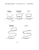 |
 | 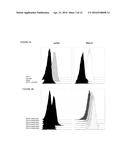 |
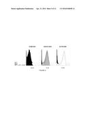 | 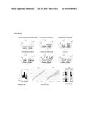 |
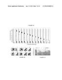 | 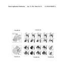 |
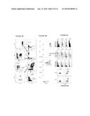 |  |
 |  |
 |  |
 | 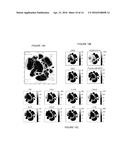 |
 |  |
 |  |
 |  |
 |  |
 |  |
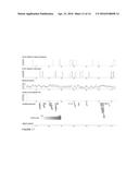 |  |
 |  |
 |  |
 |  |
 |  |
 | 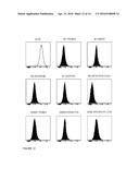 |
 |  |
 |  |
 |  |
 |  |
 |  |
 |  |
 |  |
 |  |
 |  |
 |  |
 |  |
 |  |
 |  |
 |  |
 |  |
 |  |
 |  |
 |  |
 |  |
 |  |
 |  |
 |  |
 |  |
 |  |
 |  |
 |  |
 |  |
 |  |
 |  |
 |  |
 | 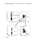 |
 |  |
 |  |
 |  |
 |  |
 |  |
 |  |
 |  |
 |  |
| New patent applications in this class: | |
| Date | Title |
|---|---|
| 2022-05-05 | Microfluidic system for amplifying and detecting polynucleotides in parallel |
| 2019-05-16 | Reagents and methods for detecting protein lysine 2-hydroxyisobutyrylation |
| 2019-05-16 | Lateral flow analyte detection |
| 2019-05-16 | Mutations in the bcr-abl tyrosine kinase associated with resistance to sti-571 |
| 2019-05-16 | Enhanced methods of ribonucleic acid hybridization |
| New patent applications from these inventors: | |
| Date | Title |
|---|---|
| 2022-08-25 | Highly-multiplexed fluorescent imaging |
| 2021-12-23 | Spatially oriented quantum barcoding of cellular targets |
| 2021-10-07 | Single cell analysis using secondary ion mass spectrometry |
| 2018-04-19 | On-slide staining by primer extension |
| 2017-06-22 | Apparatus and method for sub-micrometer elemental image analysis by mass spectrometry |
| Top Inventors for class "Combinatorial chemistry technology: method, library, apparatus" | |
| Rank | Inventor's name |
|---|---|
| 1 | Mehdi Azimi |
| 2 | Kia Silverbrook |
| 3 | Geoffrey Richard Facer |
| 4 | Alireza Moini |
| 5 | William Marshall |