Patent application title: TARGETED TGF INHIBITION
Inventors:
Kin-Ming Lo (Lexington, MA, US)
IPC8 Class: AC07K1628FI
USPC Class:
Class name:
Publication date: 2015-08-13
Patent application number: 20150225483
Abstract:
This invention relates generally to bifunctional molecules including (a)
a TGFβRII or fragment thereof capable of binding TGFβ and (b)
an antibody, or antigen binding fragment thereof, that binds to an immune
checkpoint protein, such as Programmed Death Ligand 1 (PD-L1), uses of
such molecules (e.g., for treating cancer), and methods of making such
molecules.Claims:
1. A protein comprising: a) human TGFβRII, or a fragment thereof
capable of binding TGFβ; and b) an antibody, or an antigen-binding
fragment thereof, that binds human protein Programmed Death Ligand 1
(PD-L1).
2. A polypeptide comprising: a) at least a variable domain of a heavy chain of an antibody that binds human protein Programmed Death Ligand 1 (PD-L1); and b) human TGFβRII, or a fragment thereof capable of binding TGFβ.
3. The polypeptide of claim 2, further comprising an amino acid linker connecting the C-terminus of the variable domain to the N-terminus of the human TGFβRII or fragment thereof.
4. The polypeptide of claim 3, comprising the amino acid sequence of SEQ ID NO: 3.
5. A nucleic acid comprising a nucleotide sequence encoding a polypeptide according to claim 2.
6. The nucleic acid of claim 5, further comprising a second nucleotide sequence encoding at least a variable domain of a light chain of an antibody which, when combined with the polypeptide, forms an antigen-binding site that binds PD-L1.
7. The nucleic acid of claim 6, wherein the second nucleotide sequence encodes the amino acid sequence of SEQ ID NO: 1 (secreted anti-PD-L1 lambda light chain).
8. A cell comprising the nucleic acid of claim 5 and a second nucleic acid encoding at least a variable domain of a light chain of an antibody which, when combined with the polypeptide, forms an antigen-binding site that binds PD-L1.
9. The cell of claim 8, wherein the second nucleic acid encodes the amino acid sequence of SEQ ID NO: 1.
10. A cell comprising the nucleic acid of claim 6.
11. A method of producing a protein comprising a) TGFβRII, or a fragment thereof capable of binding TGFβ, and b) an antibody, or an antigen-binding fragment thereof, that binds human protein Programmed Death Ligand 1 (PD-L1), the method comprising maintaining a cell according to claim 8 under conditions permitting expression of the protein.
12. The method of claim 11, further comprising harvesting the protein.
13. A protein comprising: a polypeptide according to claim 2; and at least a variable domain of a light chain of an antibody which, when combined with the polypeptide, forms an antigen-binding site that binds PD-L1.
14. The protein of claim 13, comprising two polypeptides each having an amino acid sequence consisting of the amino acid sequence of SEQ ID NO: 3, and two additional polypeptides each having an amino acid sequence consisting of the amino acid sequence of SEQ ID NO: 1.
15. A protein according to claim 1, for use in therapy.
16. A protein according to claim 15, wherein the therapy comprises administration of radiation.
17. A protein according to claim 15, wherein the therapy comprises administration of a chemotherapeutic.
18. A protein according to claim 1, for use in promoting local depletion of TGFβ at a tumor.
19. A protein according to claim 1, for use in inhibiting SMAD3 phosphorylation in a cell.
20. The protein of claim 19, wherein the cell is a tumor cell.
21. A protein according to claim 1, for use in treating cancer.
22. A protein according to claim 1, for use in inhibiting tumor growth.
23. A protein according to claim 21, wherein the cancer or tumor is selected from the group consisting of colorectal, breast, ovarian, pancreatic, gastric, prostate, renal, cervical, myeloma, lymphoma, leukemia, thyroid, endometrial, uterine, bladder, neuroendocrine, head and neck, liver, nasopharyngeal, testicular, small cell lung cancer, non-small cell lung cancer, melanoma, basal cell skin cancer, squamous cell skin cancer, dermatofibrosarcoma protuberans, Merkel cell carcinoma, glioblastoma, glioma, sarcoma, mesothelioma, and myelodisplastic syndromes.
24. A protein according to claim 21, wherein the use comprises administration of radiation.
25. A protein according to claim 21, wherein the use comprises administration of a chemotherapeutic.
26. A method of promoting local depletion of TGFβ, the method comprising administering a protein according to claim 1, wherein the protein binds TGFβ in solution, binds PD-L1 on a cell surface, and carries the bound TGFβ into the cell.
27. A method of inhibiting SMAD3 phosphorylation in a cell, the method comprising exposing the cell to a protein according to claim 1.
28. A method according to claim 26, wherein the cell is a cancer cell.
29. A method of inhibiting tumor growth, the method comprising exposing the tumor to a protein according to claim 1.
30. The method of claim 29, further comprising exposing the tumor to radiation.
31. A method according to claim 29, further comprising exposing the tumor to a chemotherapeutic.
32. A method of treating cancer, the method comprising administering to a cancer patient a protein according to claim 1.
33. The method of claim 32, further comprising exposing the cancer patient to radiation.
34. A method according to claim 32, further comprising administering a chemotherapeutic.
35. A method according to claim 29, wherein the tumor or cancer is selected from the group consisting of colorectal, breast, ovarian, pancreatic, gastric, prostate, renal, cervical, myeloma, lymphoma, leukemia, thyroid, endometrial, uterine, bladder, neuroendocrine, head and neck, liver, nasopharyngeal, testicular, small cell lung cancer, non-small cell lung cancer, melanoma, basal cell skin cancer, squamous cell skin cancer, dermatofibrosarcoma protuberans, Merkel cell carcinoma, glioblastoma, glioma, sarcoma, mesothelioma, and myelodisplastic syndromes.
Description:
CROSS REFERENCE TO RELATED APPLICATIONS
[0001] This application claims priority to pending U.S. Provisional Patent Application No. 61/938,048, filed on Feb. 10, 2014, entitled "Targeted TGFβ Inhibition," the disclosure of which is incorporated herein by reference in its entirety.
FIELD OF THE INVENTION
[0002] This invention relates generally to bifunctional molecules including (a) a TGFβRII or fragment thereof capable of binding TGFβ and (b) an antibody, or antigen binding fragment thereof, that binds to an immune checkpoint protein, such as Programmed Death Ligand 1 (PD-L1), uses of such molecules (e.g., for treating cancer), and methods of making such molecules.
BACKGROUND
[0003] In cancer treatment, it has long been recognized that chemotherapy is associated with high toxicity and can lead to emergence of resistant cancer cell variants. Even with targeted therapy against overexpressed or activated oncoproteins important for tumor survival and growth, cancer cells invariably mutate and adapt to reduce dependency on the targeted pathway, such as by utilizing a redundant pathway. Cancer immunotherapy is a new paradigm in cancer treatment that instead of targeting cancer cells, focuses on the activation of the immune system. Its principle is to rearm the host's immune response, especially the adaptive T cell response, to provide immune surveillance to kill the cancer cells, in particular, the minimal residual disease that has escaped other forms of treatment, hence achieving long-lasting protective immunity.
[0004] FDA approval of the anti-CTLA-4 antibody ipilimumab for the treatment of melanoma in 2011 ushered in a new era of cancer immunotherapy. The demonstration that anti-PD-1 or anti-PD-L1 therapy induced durable responses in melanoma, kidney, and lung cancer in clinical trials further signify its coming of age (Pardoll, D. M., Nat Immunol. 2012; 13:1129-32). However, ipilimumab therapy is limited by its toxicity profile, presumably because anti-CTLA-4 treatment, by interfering with the primary T cell inhibitory checkpoint, can lead to the generation of new autoreactive T cells. While inhibiting the PD-L1/PD-1 interaction results in dis-inhibiting existing chronic immune responses in exhausted T cells that are mostly antiviral or anticancer in nature (Wherry, E. J., Nat Immunol. 2011; 12:492-9), anti-PD-1 therapy can nevertheless sometimes result in potentially fatal lung-related autoimmune adverse events. Despite the promising clinical activities of anti-PD1 and anti-PD-L1 so far, increasing the therapeutic index, either by increasing therapeutic activity or decreasing toxicity, or both, remains a central goal in the development of immunotherapeutics.
SUMMARY OF THE INVENTION
[0005] The present invention is based on the discovery that a bifunctional protein containing at least portion of TGFβ Receptor II (TGFβRII) that is capable of binding TGFβ and antibody or antigen-binding fragment that binds to an immune checkpoint protein such as human protein Programmed Death Ligand 1 (PD-L1) can be an effective anti-tumor and anti-cancer therapeutic. The protein can exhibit a synergistic effect in cancer treatment, as compared to the effect of administering the two agents separately.
[0006] Accordingly, in a first aspect, the present invention features a protein including (a) human TGFβRII, or a fragment thereof capable of binding TGFβ (e.g., a soluble fragment); and (b) an antibody, or an antigen-binding fragment thereof, that binds PD-L1 (e.g., any of the antibodies or antibody fragments described herein).
[0007] In a related aspect, the invention features a polypeptide including (a) at least a variable domain of a heavy chain of an antibody that binds PD-L1 (e.g., amino acids 1-120 of SEQ ID NO: 2); and (b) human TGFβRII, or a soluble fragment thereof capable of binding TGFβ (e.g., a human TGFβRII extra-cellular domain (ECD), amino acids 24-159 of SEQ ID NO: 9, or any of those described herein). The polypeptide may further include an amino acid linker connecting the C-terminus of the variable domain to the N-terminus of the human TGFβRII or soluble fragment thereof capable of binding TGFβ. The polypeptide may include the amino acid sequence of SEQ ID NO: 3 or an amino acid sequence substantially identical to SEQ ID NO: 3. The antibody fragment may be an scFv, Fab, F(ab')2, or Fv fragment.
[0008] In certain embodiments, the protein or polypeptide includes an antibody or antigen-binding fragment thereof that includes SEQ ID NO: 2 and human TGFβRII. The antibody may optionally include a modified constant region (e.g., any described herein, including a C-terminal Lys→Ala substitution, a mutation of the Leu-Ser-Leu-Ser (SEQ ID NO: 19) sequence to Ala-Thr-Ala-Thr (SEQ ID NO: 20), or a hybrid constant region including an IgG1 hinge region and an IgG2 CH2 domain).
[0009] In certain embodiments, the protein or polypeptide includes an antibody or antigen-binding fragment thereof that includes SEQ ID NO: 2 and a fragment of human TGFβRII capable of binding TGFβ (e.g., a soluble fragment). The antibody may optionally include a modified constant region (e.g., any described herein, including a C-terminal Lys→Ala substitution, a mutation of the Leu-Ser-Leu-Ser (SEQ ID NO: 19) sequence to Ala-Thr-Ala-Thr (SEQ ID NO: 20), or a hybrid constant region including an IgG1 hinge region and an IgG2 CH2 domain).
[0010] In certain embodiments, the protein or polypeptide includes an antibody or antigen-binding fragment thereof that includes SEQ ID NO: 2 and a human TGFβRII ECD. The antibody may include a modified constant region (e.g., any described herein, including a C-terminal Lys→Ala substitution, a mutation of the Leu-Ser-Leu-Ser (SEQ ID NO: 19) sequence to Ala-Thr-Ala-Thr (SEQ ID NO: 20), or a hybrid constant region including an IgG1 hinge region and an IgG2 CH2 domain).
[0011] In certain embodiments, the protein or polypeptide includes an antibody or antigen-binding fragment thereof that includes amino acids 1-120 of SEQ ID NO: 2 and human TGFβRII. The antibody may include a modified constant region (e.g., any described herein, including a C-terminal Lys→Ala substitution, a mutation of the Leu-Ser-Leu-Ser (SEQ ID NO: 19) sequence to Ala-Thr-Ala-Thr (SEQ ID NO: 20), or a hybrid constant region including an IgG1 hinge region and an IgG2 CH2 domain).
[0012] In certain embodiments, the protein or polypeptide includes an antibody or antigen-binding fragment thereof that includes amino acids 1-120 of SEQ ID NO: 2 and a fragment of human TGFβRII capable of binding TGFβ (e.g., a soluble fragment). The antibody may include a modified constant region (e.g., any described herein, including a C-terminal Lys→Ala substitution, a mutation of the Leu-Ser-Leu-Ser (SEQ ID NO: 19) sequence to Ala-Thr-Ala-Thr (SEQ ID NO: 20), or a hybrid constant region including an IgG1 hinge region and an IgG2 CH2 domain).
[0013] In certain embodiments, the protein or polypeptide includes an antibody or antigen-binding fragment thereof that includes amino acids 1-120 of SEQ ID NO: 2 and a human TGFβRII ECD. The antibody may include a modified constant region (e.g., any described herein, including a C-terminal Lys→Ala substitution, a mutation of the Leu-Ser-Leu-Ser (SEQ ID NO: 19) sequence to Ala-Thr-Ala-Thr (SEQ ID NO: 20), or a hybrid constant region including an IgG1 hinge region and an IgG2 CH2 domain).
[0014] In certain embodiments, the protein or polypeptide includes an antibody or antigen-binding fragment thereof that includes the hypervariable regions present in SEQ ID NO: 2 and human TGFβRII. The antibody may include a modified constant region (e.g., any described herein, including a C-terminal Lys→Ala substitution, a mutation of the Leu-Ser-Leu-Ser (SEQ ID NO: 19) sequence to Ala-Thr-Ala-Thr (SEQ ID NO: 20), or a hybrid constant region including an IgG1 hinge region and an IgG2 CH2 domain).
[0015] In certain embodiments, the protein or polypeptide includes an antibody or antigen-binding fragment thereof that includes the hypervariable regions present in SEQ ID NO: 2 and a fragment of human TGFβRII capable of binding TGFβ (e.g., a soluble fragment). The antibody may include a modified constant region (e.g., any described herein, including a C-terminal Lys→Ala substitution, a mutation of the Leu-Ser-Leu-Ser (SEQ ID NO: 19) sequence to Ala-Thr-Ala-Thr (SEQ ID NO: 20), or a hybrid constant region including an IgG1 hinge region and an IgG2 CH2 domain).
[0016] In certain embodiments, the protein or polypeptide includes an antibody or antigen-binding fragment thereof that includes the hypervariable regions present in SEQ ID NO: 2 and a human TGFβRII ECD. The antibody may include a modified constant region (e.g., any described herein, including a C-terminal Lys→Ala substitution, a mutation of the Leu-Ser-Leu-Ser (SEQ ID NO: 19) sequence to Ala-Thr-Ala-Thr (SEQ ID NO: 20), or a hybrid constant region including an IgG1 hinge region and an IgG2 CH2 domain).
[0017] In certain embodiments, the protein or polypeptide includes an antibody or antigen-binding fragment thereof that includes SEQ ID NO: 12 and human TGFβRII. The antibody may include a modified constant region (e.g., any described herein, including a C-terminal Lys→Ala substitution, a mutation of the Leu-Ser-Leu-Ser (SEQ ID NO: 19) sequence to Ala-Thr-Ala-Thr (SEQ ID NO: 20), or a hybrid constant region including an IgG1 hinge region and an IgG2 CH2 domain).
[0018] In certain embodiments, the protein or polypeptide includes an antibody or antigen-binding fragment thereof that includes SEQ ID NO: 12 and a fragment of human TGFβRII capable of binding TGFβ (e.g., a soluble fragment). The antibody may include a modified constant region (e.g., any described herein, including a C-terminal Lys→Ala substitution, a mutation of the Leu-Ser-Leu-Ser (SEQ ID NO: 19) sequence to Ala-Thr-Ala-Thr (SEQ ID NO: 20), or a hybrid constant region including an IgG1 hinge region and an IgG2 CH2 domain).
[0019] In certain embodiments, the protein or polypeptide includes an antibody or antigen-binding fragment thereof that includes SEQ ID NO: 12 and a human TGFβRII ECD. The antibody may include a modified constant region (e.g., any described herein, including a C-terminal Lys→Ala substitution, a mutation of the Leu-Ser-Leu-Ser (SEQ ID NO: 19) sequence to Ala-Thr-Ala-Thr (SEQ ID NO: 20), or a hybrid constant region including an IgG1 hinge region and an IgG2 CH2 domain).
[0020] In certain embodiments, the protein or polypeptide includes an antibody or antigen-binding fragment thereof that includes the hypervariable regions present in SEQ ID NO: 12 and human TGFβRII. The antibody may include a modified constant region (e.g., any described herein, including a C-terminal Lys→Ala substitution, a mutation of the Leu-Ser-Leu-Ser (SEQ ID NO: 19) sequence to Ala-Thr-Ala-Thr (SEQ ID NO: 20), or a hybrid constant region including an IgG1 hinge region and an IgG2 CH2 domain).
[0021] In certain embodiments, the protein or polypeptide includes an antibody or antigen-binding fragment thereof that includes the hypervariable regions present in SEQ ID NO: 12 and a fragment of human TGFβRII capable of binding TGFβ (e.g., a soluble fragment). The antibody may include a modified constant region (e.g., any described herein, including a C-terminal Lys→Ala substitution, a mutation of the Leu-Ser-Leu-Ser (SEQ ID NO: 19) sequence to Ala-Thr-Ala-Thr (SEQ ID NO: 20), or a hybrid constant region including an IgG1 hinge region and an IgG2 CH2 domain).
[0022] In certain embodiments, the protein or polypeptide includes an antibody or antigen-binding fragment thereof that includes the hypervariable regions present in SEQ ID NO: 12 and a human TGFβRII ECD. The antibody may include a modified constant region (e.g., any described herein, including a C-terminal Lys→Ala substitution, a mutation of the Leu-Ser-Leu-Ser (SEQ ID NO: 19) sequence to Ala-Thr-Ala-Thr (SEQ ID NO: 20), or a hybrid constant region including an IgG1 hinge region and an IgG2 CH2 domain).
[0023] In certain embodiments, the protein or polypeptide includes an antibody or antigen-binding fragment thereof that includes SEQ ID NO: 14 and human TGFβRII. The antibody may include a modified constant region (e.g., any described herein, including a C-terminal Lys→Ala substitution, a mutation of the Leu-Ser-Leu-Ser (SEQ ID NO: 19) sequence to Ala-Thr-Ala-Thr (SEQ ID NO: 20), or a hybrid constant region including an IgG1 hinge region and an IgG2 CH2 domain).
[0024] In certain embodiments, the protein or polypeptide includes an antibody or antigen-binding fragment thereof that includes SEQ ID NO: 14 and a fragment of human TGFβRII capable of binding TGFβ (e.g., a soluble fragment). The antibody may include a modified constant region (e.g., any described herein, including a C-terminal Lys→Ala substitution, a mutation of the Leu-Ser-Leu-Ser (SEQ ID NO: 19) sequence to Ala-Thr-Ala-Thr (SEQ ID NO: 20), or a hybrid constant region including an IgG1 hinge region and an IgG2 CH2 domain).
[0025] In certain embodiments, the protein or polypeptide includes an antibody or antigen-binding fragment thereof that includes SEQ ID NO: 14 and a human TGFβRII ECD. The antibody may include a modified constant region (e.g., any described herein, including a C-terminal Lys→Ala substitution, a mutation of the Leu-Ser-Leu-Ser (SEQ ID NO: 19) sequence to Ala-Thr-Ala-Thr (SEQ ID NO: 20), or a hybrid constant region including an IgG1 hinge region and an IgG2 CH2 domain).
[0026] The invention also features a nucleic acid that includes a nucleotide sequence that encodes a polypeptide described above. In certain embodiments, the nucleic acid further includes a second nucleotide sequence encoding at least a variable domain of a light chain of an antibody which, when combined with the polypeptide, forms an antigen-binding site that binds PD-L1 (e.g., including amino acids 1-110 of SEQ ID NO: 1). The second nucleotide sequence may encode the amino acid sequence of SEQ ID NO: 1 (secreted anti-PD-L1 lambda light chain) or an amino acid sequence substantially identical to SEQ ID NO: 1. The invention also features a cell including any of the nucleic acids described above.
[0027] The invention also features a method of producing a protein including (a) the extracellular domain of the human TGFβRII, or a fragment thereof capable of binding TGFβ (e.g., a soluble fragment), and (b) an antibody, or an antigen-binding fragment thereof, that binds human PD-L1. The method includes maintaining a cell described under conditions that permit expression of the protein. The method may further include harvesting the protein.
[0028] The invention also features a protein including the polypeptide described above and at least a variable domain of a light chain of an antibody which, when combined with the polypeptide, forms an antigen-binding site that binds PD-L1. The protein may include (a) two polypeptides, each having an amino acid sequence consisting of the amino acid sequence of SEQ ID NO: 3, and (b) two additional polypeptides each having an amino acid sequence consisting of the amino acid sequence of SEQ ID NO: 1.
[0029] The invention also features a protein described above for use in therapy. The therapy may include administration of radiation or administration of a chemotherapeutic, a biologic, or a vaccine.
[0030] The invention also features a protein described above for use in promoting local depletion of TGFβ at a tumor.
[0031] The invention also features a protein described above for use in inhibiting SMAD3 phosphorylation in a cell (e.g., a tumor cell or an immune cell).
[0032] The invention also features a protein described above for use in treating cancer or for use in inhibiting tumor growth. The cancer or tumor may be selected from the group consisting of colorectal, breast, ovarian, pancreatic, gastric, prostate, renal, cervical, myeloma, lymphoma, leukemia, thyroid, endometrial, uterine, bladder, neuroendocrine, head and neck, liver, nasopharyngeal, testicular, small cell lung cancer, non-small cell lung cancer, melanoma, basal cell skin cancer, squamous cell skin cancer, dermatofibrosarcoma protuberans, Merkel cell carcinoma, glioblastoma, glioma, sarcoma, mesothelioma, and myelodisplastic syndromes. The use may further include administration of radiation or administration of a chemotherapeutic, a biologic, or a vaccine.
[0033] The invention also features a method of promoting local depletion of TGFβ. The method includes administering a protein described above, where the protein binds TGFβ in solution, binds PD-L1 on a cell surface, and carries the bound TGFβ into the cell (e.g., a cancer cell).
[0034] The invention also features a method of inhibiting SMAD3 phosphorylation in a cell (e.g., a cancer cell or an immune cell), the method including exposing the cell in the tumor microenvironment to a protein described above.
[0035] The invention also features a method of inhibiting tumor growth or treating cancer. The method includes exposing the tumor to a protein described above. The method may further include exposing the tumor to radiation or to a chemotherapeutic, a biologic, or a vaccine. In certain embodiments, the tumor or cancer is selected from the group consisting of colorectal, breast, ovarian, pancreatic, gastric, prostate, renal, cervical, myeloma, lymphoma, leukemia, thyroid, endometrial, uterine, bladder, neuroendocrine, head and neck, liver, nasopharyngeal, testicular, small cell lung cancer, non-small cell lung cancer, melanoma, basal cell skin cancer, squamous cell skin cancer, dermatofibrosarcoma protuberans, Merkel cell carcinoma, glioblastoma, glioma, sarcoma, mesothelioma, and myelodisplastic syndromes.
[0036] By "TGFβRII" or "TGFβ Receptor II" is meant a polypeptide having the wild-type human TGFβ Receptor Type 2 Isoform A sequence (e.g., the amino acid sequence of NCBI Reference Sequence (RefSeq) Accession No. NP--001020018 (SEQ ID NO: 8)), or a polypeptide having the wild-type human TGFβ Receptor Type 2 Isoform B sequence (e.g., the amino acid sequence of NCBI RefSeq Accession No. NP--003233 (SEQ ID NO: 9)) or having a sequence substantially identical the amino acid sequence of SEQ ID NO: 8 or of SEQ ID NO: 9. The TGFβRII may retain at least 0.1%, 0.5%, 1%, 5%, 10%, 25%, 35%, 50%, 75%, 90%, 95%, or 99% of the TGFβ-binding activity of the wild-type sequence. The polypeptide of expressed TGFβRII lacks the signal sequence.
[0037] By a "fragment of TGFβRII capable of binding TGFβ" is meant any portion of NCBI RefSeq Accession No. NP--001020018 (SEQ ID NO: 8) or of NCBI RefSeq Accession No. NP--003233 (SEQ ID NO: 9), or a sequence substantially identical to SEQ ID NO: 8 or SEQ ID NO: 9 that is at least 20 (e.g., at least 30, 40, 50, 60, 70, 80, 90, 100, 110, 120, 130, 140, 150, 160, 175, or 200) amino acids in length that retains at least some of the TGFβ-binding activity (e.g., at least 0.1%, 0.5%, 1%, 5%, 10%, 25%, 35%, 50%, 75%, 90%, 95%, or 99%) of the wild-type receptor or of the corresponding wild-type fragment. Typically such fragment is a soluble fragment. An exemplary such fragment is a TGFβRII extra-cellular domain having the sequence of SEQ ID NO: 10.
[0038] By "substantially identical" is meant a polypeptide exhibiting at least 50%, desirably 60%, 70%, 75%, or 80%, more desirably 85%, 90%, or 95%, and most desirably 99% amino acid sequence identity to a reference amino acid sequence. The length of comparison sequences will generally be at least 10 amino acids, desirably at least 15 contiguous amino acids, more desirably at least 20, 25, 50, 75, 90, 100, 150, 200, 250, 300, or 350 contiguous amino acids, and most desirably the full-length amino acid sequence.
[0039] By "patient" is meant either a human or non-human animal (e.g., a mammal).
[0040] By "treating" a disease, disorder, or condition (e.g., a cancer) in a patient is meant reducing at least one symptom of the disease, disorder, or condition by administrating a therapeutic agent to the patient.
[0041] By "cancer" is meant a collection of cells multiplying in an abnormal manner.
[0042] Other embodiments and details of the invention are presented herein below.
BRIEF DESCRIPTION OF THE DRAWINGS
[0043] FIG. 1A is a schematic drawing of an anti-PD-L1/TGFβ Trap molecule comprising one anti-PD-L1 antibody fused to two extracellular domain (ECD) of TGFβ Receptor II via a (Gly4Ser)4Gly linker. FIG. 1B is a photograph of a sodium dodecyl sulfate-polyacrylamide gel electrophoresis (SDS-PAGE) analysis of anti-PD-L1/TGFβ Trap under non-reducing and reducing conditions.
[0044] FIG. 2 is photograph of an SDS-PAGE gel showing analysis of extent of clipping of anti-PD-L1/TGFβ Trap expressed by clone 02B15 at various population doubling levels. Anti-PD-L1/TGFβ Trap from clone 02B15 after a single protein A chromatography step was analyzed by SDS-PAGE under reducing conditions. Lanes 1 and 10, See Blue Plus 2 MW Standard; lane 2, purified anti-PD-L1/TGFβ Trap reference; lane 3, clone 02B15 at PDL0; lane 4, clone 02B15 at PDL30; lane 5, clone 02B15 at PDL60; and lane 6, clone 02B15 at PDL90. (PDL, population doubling level).
[0045] FIG. 3 is a graph showing FACS analysis of anti-PD-L1/TGFβ Trap binding to HEK cells transfected to express human PD-L1.
[0046] FIG. 4 is a graph showing the ability of anti-PD-L1/TGFβ Trap to inhibit TGFβ-induced phosphorylation of SMAD3 using a pSMAD3-luciferase reporter cell line (filled circle: anti-PD-L1; X: anti-PD-L1 (mut); filled square: anti-PD-L1/TGFβ Trap; filled triangle: anti-PD-L1(mut)/TGFβ Trap; +: anti-TGFβ antibody 1D11; star: TGFβ RII-Fc).
[0047] FIGS. 5A and 5B are graphs showing pharmacokinetics of intravenously administered anti-PD-L1/TGFβ Trap and related proteins in mice.
[0048] FIG. 6A is a graph showing PD-L1 target-mediated endocytosis of anti-PD-L1/TGFβ Trap. FIG. 6B is a graph showing PD-L1 target-mediated endocytosis of anti-PD-L1. FIG. 6C is a graph showing percent internalization of anti-PD-L1/TGFβ Trap and anti-PD-L1 bound on HEK/PD-L1 cells.
[0049] FIGS. 7A-7C are graphs showing anti-tumor efficacy of anti-PD-L1/TGFβ Trap and related proteins in the EMT-6 breast carcinoma subcutaneous model (Example 7). FIG. 7A shows tumor growth curves of average tumor volumes of surviving mice in different treatment groups (star: Group 1: filled circle: Group 2; filled triangle: Group 3; filled square: Group 4; open square: Group 5; filled square/dashed line: Group 6; filled square/stippled line: Group 7). FIG. 7B shows tumor growth curves of individual tumor volumes in different treatment groups. FIG. 7C is a Kaplan-Meier plot of percent survival in different treatment groups (symbols as in 7A).
[0050] FIG. 8 is a graph showing anti-tumor efficacy of anti-PD-L1/TGFβ Trap and related proteins in the MC38 colorectal carcinoma subcutaneous tumor model (Example 8; star: Group 1; filled circle: Group 2; filled circle/dashed line: Group 3; filled triangle: Group 4; filled triangle/dashed line: Group 5; filled square: Group 6; filled square/dashed line: Group 7).
[0051] FIG. 9 is a graph showing anti-tumor efficacy of anti-PDL1/TGFβ Trap and related proteins in an orthotopic EMT-6 breast cancer model (Example 9; star: Group 1; filled circle/dashed line: Group 2; filled triangle: Group 3; filled triangle/dashed line: Group 4; filled diamond: Group 5).
[0052] FIG. 10 is a graph showing anti-tumor efficacy of anti-PDL1/TGFβ Trap and related proteins in an intramuscular MC38 colorectal carcinoma model (Example 10; star: Group 1; filled circle: Group 2; filled circle/dashed line: Group 3: filled diamond/dashed line: Group 4; filled square: Group 5; filled square/dashed line: Group 6; filled diamond: Group 7).
[0053] FIG. 11 is a graph showing anti-tumor efficacy of anti-PD-L1/TGF-β Trap and the combination of anti-PD-L1 and TGFβ Trap control administered to give equivalent in vivo exposure in an orthotopic EMT-6 breast tumor model (Example 11; star: Group 1; filled square: Group 2; open square: Group 3; filled diamond: Group 4; open diamond: Group 5).
[0054] FIGS. 12A-12C are graphs showing anti-tumor efficacy of anti-PD-L1/TGF-β Trap and the combination of anti-PD-L1 and TGFβ Trap control administered to give equivalent in vivo exposure in an intramuscular MC38 colorectal carcinoma model (Example 12). FIG. 12A shows tumor growth curves of mice treated with both intermediate and low doses of the proteins (star: Group 1; filled squares: Group 2; open squares: Group 3; filled diamonds: Group 4; open diamonds Group 5). FIG. 12B (star: Group 1; filled square: Group 2; filled diamond: Group 4; *: p<0.0001 compared to Group 1; **: p<0.0001 compared to Group 2) and 12C (star: Group 1; filled square: Group 3; filled diamond: Group 5; *: p<0.0001 compared to Group 1; **: p<0.0001 compared to Group 3) show statistical analysis of tumor growth curves of mice treated with intermediate and low doses of the proteins, respectively
[0055] FIGS. 13A-13B are graphs showing anti-tumor efficacy of anti-PD-L1(YW)/TGF-β Trap and related proteins in an orthotopic EMT-6 breast tumor model (Example 13; star: Group 1; filled circle: Group 2; filled triangle: Group 3; filled square: Group 4; filled diamond: Group 5). FIG. 13A shows tumor growth curves of mice in different treatment groups. FIG. 13B is a Kaplan-Meier plot of percent survival in different treatment groups.
[0056] FIGS. 14A-14B are graphs showing anti-tumor efficacy of anti-PD-L1(YW)/TGF-β Trap and related proteins based on (A) tumor volumes and (B) tumor weights, in an intramuscular MC38 colorectal carcinoma model (Example 14; star: Group 1; filled circle: Group 2; filled triangle: Group 3; filled square: Group 4; filled diamond: Group 5).
[0057] FIG. 15 is a graph comparing the anti-tumor efficacy of an anti-PD-1 antibody treatment with and without TGFβ Trap control in an orthotopic EMT-6 breast tumor model (Example 15; star: Group 1; filled square: Group 2; filled inverted triangle: Group 3; open inverted triangle: Group 4).
[0058] FIG. 16 is a graph comparing the anti-tumor efficacy of an anti-PD-1 antibody treatment with and without TGFβ Trap control in an intramuscular MC38 colorectal tumor model (Example 16; star: Group 1; filled square: Group 2; filled inverted triangle: Group 3; open inverted triangle: Group 4).
[0059] FIG. 17 is a graph comparing the anti-tumor efficacy of an anti-LAG3 or anti-TIM3 antibody treatment with and without TGFβ Trap control in an orthotopic EMT-6 breast tumor model (Example 17; star: Group 1; filled square: Group 2; filled triangle: Group 3; filled inverted triangle: Group 4; open triangle: Group 5; open inverted triangle: Group 6).
[0060] FIG. 18 is a graph comparing the anti-tumor efficacy of an anti-LAG3 or anti-TIM3 antibody treatment with and without TGFβ Trap control in an intramuscular MC38 colorectal tumor model (Example 18; star: Group 1; filled square: Group 2; filled triangle: Group 3; filled inverted triangle: Group 4; open triangle: Group 5; open inverted triangle: Group 6).
DETAILED DESCRIPTION
[0061] The current invention permits localized reduction in TGFβ in a tumor microenvironment by capturing the TGFβ using a soluble cytokine receptor (TGFβRII) tethered to an antibody moiety targeting a cellular immune checkpoint receptor found on the exterior surface of certain tumor cells or immune cells. An example of an antibody moiety of the invention to an immune checkpoint protein is anti-PD-L1. This bifunctional molecule, sometimes referred to in this document as an "antibody-cytokine trap," is effective precisely because the anti-receptor antibody and cytokine trap are physically linked. The resulting advantage (over, for example, administration of the antibody and the receptor as separate molecules) is partly because cytokines function predominantly in the local environment through autocrine and paracrine functions. The antibody moiety directs the cytokine trap to the tumor microenvironment where it can be most effective, by neutralizing the local immunosuppressive autocrine or paracrine effects. Furthermore, in cases where the target of the antibody is internalized upon antibody binding, an effective mechanism for clearance of the cytokinecytokine receptor complex is provided. Antibody-mediated target internalization has been shown for PD-L1. This is a distinct advantage over using an anti-TGFβ antibody because first, an anti-TGFβ antibody might not be completely neutralizing; and second, the antibody can act as a carrier extending the half-life of the cytokine, and antibodycytokine complexes often act as a circulating sink that builds up and ultimately dissociates to release the cytokine back in circulation (Montero-Julian et al., Blood. 1995; 85:917-24). The use of a cytokine trap to neutralize the ligand can also be a better strategy than blockading the receptor with an antibody, as in the case of CSF-1. Because CSF-1 is cleared from the circulation by receptor-mediated endocytosis, an anti-CSF-1 receptor antibody blockade caused a significant increase in circulating CSF-1 concentration (Hume et al., Blood. 2012; 119:1810-20)
[0062] Indeed, as described below, treatment with the anti-PD-L1/TGFβ Trap elicits a synergistic anti-tumor effect due to the simultaneous blockade of the interaction between PD-L1 on tumor cells and PD-1 on immune cells, and the neutralization of TGFβ in the tumor microenvironment. As demonstrated in the following examples, anti-PDL1/TGFβ Trap has efficacy superior to that of the single agent anti-PD-L1 or TGFβ Trap control. Without being bound by theory, this presumably is due to a synergistic effect obtained from simultaneous blocking the two major immune escape mechanisms, and in addition, the targeted depletion of the TGFβ in the tumor microenvironment by a single molecular entity. This depletion is achieved by (1) anti-PD-L1 targeting of tumor cells; (2) binding of the TGFβ autocrine/paracrine in the tumor microenvironment by the TGFβ Trap; and (3) destruction of the bound TGFβ through the PD-L1 receptor-mediated endocytosis. The aforementioned mechanisms of action cannot be achieved by the combination therapy of the two single agents anti-PD-L1 and TGFβ Trap. Furthermore, the TGFβRII fused to the C-terminus of Fc (fragment of crystallization of IgG) was several-fold more potent than the TGFβRII-Fc that places the TGFβRII at the N-terminus of Fc (see Example 3). The superb efficacy obtained with anti-PDL1/TGFβ Trap also allays some concerns that the TGFβRII does not trap TGFβ2. As pointed out by Yang et al., Trends Immunol. 2010; 31:220-227, although some tumor types do secrete TGFβ2 initially, as the tumor progresses, the TGFβ in the tumor microenvironment is predominantly secreted by myeloid-derived suppressor cells, which secrete TGFβ1. In addition to showing great promise as an effective immuno-oncology therapeutic, treatment with soluble TGFβRII can potentially reduce the cardiotoxicity concerns of TGFβ targeting therapies, especially the TGFβRI kinase inhibitors. This is because of the important roles TGFβ2 plays in embryonic development of the heart as well as in repair of myocardial damage after ischemia and reperfusion injury (Roberts et al., J Clin Invest. 1992; 90:2056-62).
TGFβ as a Cancer Target
[0063] TGFβ had been a somewhat questionable target in cancer immunotherapy because of its paradoxical roles as the molecular Jekyll and Hyde of cancer (Bierie et al., Nat Rev Cancer. 2006; 6:506-20). Like some other cytokines, TGFβ activity is developmental stage and context dependent. Indeed TGFβ can act as either a tumor promoter or a tumor suppressor, affecting tumor initiation, progression and metastasis. The mechanisms underlying this dual role of TGFβ remain unclear (Yang et al., Trends Immunol. 2010; 31:220-227). Although it has been postulated that Smad-dependent signaling mediates the growth inhibition of TGFβ signaling, while the Smad independent pathways contribute to its tumor-promoting effect, there are also data showing that the Smad-dependent pathways are involved in tumor progression (Yang et al., Cancer Res. 2008; 68:9107-11).
[0064] Both the TGFβ ligand and the receptor have been studied intensively as therapeutic targets. There are three ligand isoforms, TGFβ1, 2 and 3, all of which exist as homodimers. There are also three TGFβ receptors (TGFβR), which are called TGFβR type I, II and III (Lopez-Casillas et al., J Cell Biol. 1994; 124:557-68). TGFβRI is the signaling chain and cannot bind ligand. TGFβRII binds the ligand TGFβ1 and 3, but not TGFβ2, with high affinity. The TGFβRII/TGFβ complex recruits TGFβRI to form the signaling complex (Won et al., Cancer Res. 1999; 59:1273-7). TGFβRIII is a positive regulator of TGFβ binding to its signaling receptors and binds all 3 TGFβ isoforms with high affinity. On the cell surface, the TGFβ/TGFβRIII complex binds TGFβRII and then recruits TGFβRI, which displaces TGFβRIII to form the signaling complex.
[0065] Although the three different TGFβ isoforms all signal through the same receptor, they are known to have differential expression patterns and non-overlapping functions in vivo. The three different TGF-β isoform knockout mice have distinct phenotypes, indicating numerous non-compensated functions (Bujak et al., Cardiovasc Res. 2007; 74:184-95). While TGFβ1 null mice have hematopoiesis and vasculogenesis defects and TGFβ3 null mice display pulmonary development and defective palatogenesis, TGFβ2 null mice show various developmental abnormalities, the most prominent being multiple cardiac deformities (Bartram et al., Circulation. 2001; 103:2745-52; Yamagishi et al., Anat Rec. 2012; 295:257-67). Furthermore, TGFβ is implicated to play a major role in the repair of myocardial damage after ischemia and reperfusion injury. In an adult heart, cardiomyocytes secrete TGFβ, which acts as an autocrine to maintain the spontaneous beating rate. Importantly, 70-85% of the TGFβ secreted by cardiomyocytes is TGFβ2 (Roberts et al., J Clin Invest. 1992; 90:2056-62). In summary, given the predominant roles of TGFβ1 and TGFβ2 in the tumor microenvironment and cardiac physiology, respectively, a therapeutic agent that neutralizes TGFβ1 but not TGFβ2 could provide an optimal therapeutic index by minimizing the cardiotoxicity without compromising the anti-tumor activity. This is consistent with the findings by the present inventors, who observed a lack of toxicity, including cardiotoxicity, for anti-PD-L1/TGFβ Trap in monkeys.
[0066] Therapeutic approaches to neutralize TGFβ include using the extracellular domains of TGFβ receptors as soluble receptor traps and neutralizing antibodies. Of the receptor trap approach, soluble TGFβRIII may seem the obvious choice since it binds all the three TGFβ ligands. However, TGFβRIII, which occurs naturally as a 280-330 kD glucosaminoglycan (GAG)-glycoprotein, with extracellular domain of 762 amino acid residues, is a very complex protein for biotherapeutic development. The soluble TGFβRIII devoid of GAG could be produced in insect cells and shown to be a potent TGFβ neutralizing agent (Vilchis-Landeros et al, Biochem J 355:215, 2001). The two separate binding domains (the endoglin-related and the uromodulin-related) of TGFβRIII could be independently expressed, but they were shown to have affinities 20 to 100 times lower than that of the soluble TGFβRIII, and much diminished neutralizing activity (Mendoza et al., Biochemistry. 2009; 48:11755-65). On the other hand, the extracellular domain of TGFβRII is only 136 amino acid residues in length and can be produced as a glycosylated protein of 25-35 kD. The recombinant soluble TGFβRII was further shown to bind TGFβ1 with a KD of 200 pM, which is fairly similar to the KD of 50 pM for the full length TGFβRII on cells (Lin et al., J Biol Chem. 1995; 270:2747-54). Soluble TGFβRII-Fc was tested as an anti-cancer agent and was shown to inhibit established murine malignant mesothelioma growth in a tumor model (Suzuki et al., Clin Cancer Res. 2004; 10:5907-18). Since TGFβRII does not bind TGFβ2, and TGFβRIII binds TGFβ1 and 3 with lower affinity than TGFβRII, a fusion protein of the endoglin domain of TGFβRIII and extracellular domain of TGFβRII was produced in bacteria and was shown to inhibit the signaling of TGFβ1 and 2 in cell based assays more effectively than either TGFβRII or RIII (Verona et al., Protein Eng Des Sel. 2008; 21:463-73). Despite some encouraging anti-tumor activities in tumor models, to our knowledge no TGFβ receptor trap recombinant proteins have been tested in the clinic.
[0067] Still another approach to neutralize all three isoforms of the TGFβ ligands is to screen for a pan-neutralizing anti-TGFβ antibody, or an anti-receptor antibody that blocks the receptor from binding to TGFβ1, 2 and 3. GC1008, a human antibody specific for all isoforms of TGFβ, was in a Phase I/II study in patients with advanced malignant melanoma or renal cell carcinoma (Morris et al., J Clin Oncol 2008; 26:9028 (Meeting abstract)). Although the treatment was found to be safe and well tolerated, only limited clinical efficacy was observed, and hence it was difficult to interpret the importance of anti-TGFβ therapy without further characterization of the immunological effects (Flavell et al., Nat Rev Immunol. 2010; 10:554-67). There were also TGFβ-isoform-specific antibodies tested in the clinic. Metelimumab, an antibody specific for TGFβ1 was tested in Phase 2 clinical trial as a treatment to prevent excessive post-operative scarring for glaucoma surgery; and Lerdelimumab, an antibody specific for TGFβ2, was found to be safe but ineffective at improving scarring after eye surgery in a Phase 3 study (Khaw et al., Ophthalmology 2007; 114:1822-1830). Anti-TGFβRII antibodies that block the receptor from binding to all three TGFβ isoforms, such as the anti-human TGFβRII antibody TR1 and anti-mouse TGFβRII antibody MT1, have also shown some therapeutic efficacy against primary tumor growth and metastasis in mouse models (Zhong et al., Clin Cancer Res. 2010; 16:1191-205). To date, the vast majority of the studies on TGFβ targeted anticancer treatment, including small molecule inhibitors of TGFβ signaling that often are quite toxic, are mostly in the preclinical stage and the anti-tumor efficacy obtained has been limited (Calone et al., Exp Oncol. 2012; 34:9-16; Connolly et al., Int J Biol Sci. 2012; 8:964-78).
[0068] The antibody-TGFβ trap of the invention is a bifunctional protein containing at least portion of a human TGFβ Receptor II (TGFβRII) that is capable of binding TGFβ. In one embodiment, the TGFβ trap polypeptide is a soluble portion of the human TGFβ Receptor Type 2 Isoform A (SEQ ID NO: 8) that is capable of binding TGFβ. In a further embodiment, TGFβ trap polypeptide contains at least amino acids 73-184 of SEQ ID NO:8. In yet a further embodiment, the TGFβ trap polypeptide contains amino acids 24-184 of SEQ ID NO:8. In another embodiment, the TGFβ trap polypeptide is a soluble portion of the human TGFβ Receptor Type 2 Isoform B (SEQ ID NO: 9) that is capable of binding TGFβ. In a further embodiment, TGFβ trap polypeptide contains at least amino acids 48-159 of SEQ ID NO:9. In yet a further embodiment, the TGFβ trap polypeptide contains amino acids 24-159 of SEQ ID NO:9. In yet a further embodiment, the TGFβ trap polypeptide contains amino acids 24-105 of SEQ ID NO:9.
Immune Checkpoint Dis-Inhibition
[0069] The approach of targeting T cell inhibition checkpoints for dis-inhibition with therapeutic antibodies is an area of intense investigation (for a review, see Pardoll, Nat Rev Cancer. 2012; 12:253-264). In one approach, the antibody moiety or antigen binding fragment thereof targets T cell inhibition checkpoint receptor proteins on the T cell, such as, for example: CTLA-4, PD-1, BTLA, LAG-3, TIM-3, and LAIR1. In another approach, the antibody moiety targets the counter-receptors on antigen presenting cells and tumor cells (which co-opt some of these counter-receptors for their own immune evasion), such as, for example: PD-L1 (B7-H1), B7-DC, HVEM, TIM-4, B7-H3, or B7-H4.
[0070] The invention contemplates antibody TGFβ traps that target, through their antibody moiety or antigen binding fragment thereof, T cell inhibition checkpoints for dis-inhibition. To that end the present inventors have tested the anti-tumor efficacy of combining a TGFβ trap with antibodies targeting various T cell inhibition checkpoint receptor proteins, such as anti-PD-1, anti-PD-L1, anti-TIM-3 and anti-LAG3. The experimental results are further detailed in Examples 7-18. The present inventors found that combining a TGFβ trap with an anti-PD-L1 antibody exhibited remarkable anti-tumor activity beyond what was observed with the monotherapies. In contrast, none of the other combinations with antibodies to the targets listed above showed any superior efficacy. In particular, one may have expected that a combination treatment of a TGFβ trap with an anti-PD-1 antibody would demonstrate similar activity to the one observed with anti-PD-L1, as PD-1 PD-L1 are cognate receptors that bind to each other to effect the immune checkpoint inhibition. However, this is not what the present inventors have found.
Anti-PD-L1 Antibodies
[0071] The invention can include any anti-PD-L1 antibody, or antigen-binding fragment thereof, described in the art. Anti-PD-L1 antibodies are commercially available, for example, the 29E2A3 antibody (Biolegend, Cat. No. 329701). Antibodies can be monoclonal, chimeric, humanized, or human. Antibody fragments include Fab, F(ab')2, scFv and Fv fragments, which are described in further detail below.
[0072] Exemplary antibodies are described in PCT Publication WO 2013079174. These antibodies can include a heavy chain variable region polypeptide including an HVR-H1, HVR-H2, and HVR-H3 sequence, where:
TABLE-US-00001 (a) the HVR-H1 sequence is X1YX2MX3; (b) the HVR-H2 sequence is SIYPSGGX4TFYADX5VKG; (c) the HVR-H3 sequence is IKLGTVTTVX6Y;
further where: X1 is K, R, T, Q, G, A, W, M, I, or S; X2 is V, R, K, L, M, or I; X3 is H, T, N, Q, A, V, Y, W, F, or M; X4 is F or I; X5 is S or T; X6 is E or D.
[0073] In a one embodiment, X1 is M, I, or S; X2 is R, K, L, M, or I; X3 is F or M; X4 is F or I; X5 is S or T; X6 is E or D.
[0074] In another embodiment X1 is M, I, or S; X2 is L, M, or I; X3 is F or M; X4 is I; X5 is S or T; X6 is D.
[0075] In still another embodiment, X1 is S; X2 is I; X3 is M; X4 is I; X5 is T; X6 is D.
[0076] In another aspect, the polypeptide further includes variable region heavy chain framework sequences juxtaposed between the HVRs according to the formula: (HC-FR1)-(HVR-H1)-(HC-FR2)-(HVR-H2)-(HC-FR3)-(HVR-H3)-(HC-FR4).
[0077] In yet another aspect, the framework sequences are derived from human consensus framework sequences or human germline framework sequences.
[0078] In a still further aspect, at least one of the framework sequences is the following:
TABLE-US-00002 HC-FR1 is EVQLLESGGGLVQPGGSLRLSCAASGFTFS; HC-FR2 is WVRQAPGKGLEWVS; HC-FR3 is RFTISRDNSKNTLYLQMNSLRAEDTAVYYCAR; HC-FR4 is WGQGTLVTVSS.
[0079] In a still further aspect, the heavy chain polypeptide is further combined with a variable region light chain including an HVR-L1, HVR-L2, and HVR-L3, where:
TABLE-US-00003 (a) the HVR-L1 sequence is TGTX7X8DVGX9YNYVS; (b) the HVR-L2 sequence is X10VX11X12RPS; (c) the HVR-L3 sequence is SSX13TX14X15X16X17RV;
further where: X7 is N or S; X8 is T, R, or S; X9 is A or G; X10 is E or D; X11 is I, N or S; X12 is D, H or N; X13 is F or Y; X14 is N or S; X15 is R, T or S; X16 is G or S; X17 is I or T.
[0080] In another embodiment, X7 is N or S; X8 is T, R, or S; X9 is A or G; X10 is E or D; X11 is N or S; X12 is N; X13 is F or Y; X14 is S; X15 is S; X16 is G or S; X17 is T.
[0081] In still another embodiment, X7 is S; X8 is S; X9 is G; X10 is D; X11 is S; X12 is N; X13 is Y; X14 is S; X15 is S; X16 is S; X17 is T.
[0082] In a still further aspect, the light chain further includes variable region light chain framework sequences juxtaposed between the HVRs according to the formula: (LC-FR1MHVR-L1)-(LC-FR2)-(HVR-L2)-(LC-FR3)-(HVR-L3)-(LC-FR4).
[0083] In a still further aspect, the light chain framework sequences are derived from human consensus framework sequences or human germline framework sequences.
[0084] In a still further aspect, the light chain framework sequences are lambda light chain sequences.
[0085] In a still further aspect, at least one of the framework sequence is the following:
TABLE-US-00004 LC-FR1 is QSALTQPASVSGSPGQSITISC; LC-FR2 is WYQQHPGKAPKLMIY; LC-FR3 is GVSNRFSGSKSGNTASLTISGLQAEDEADYYC; LC-FR4 is FGTGTKVTVL.
[0086] In another embodiment, the invention provides an anti-PD-L1 antibody or antigen binding fragment including a heavy chain and a light chain variable region sequence, where:
[0087] (a) the heavy chain includes an HVR-H1, HVR-H2, and HVR-H3, wherein further: (i) the HVR-H1 sequence is X1YX2MX3; (ii) the HVR-H2 sequence is SIYPSGGX4TFYADX5VKG; (iii) the HVR-H3 sequence is IKLGTVTTVX6Y, and;
[0088] (b) the light chain includes an HVR-L1, HVR-L2, and HVR-L3, wherein further: (iv) the HVR-L1 sequence is TGTX7X8DVGX9YNYVS; (v) the HVR-L2 sequence is X10VX11X12RPS; (vi) the HVR-L3 sequence is SSX13TX14X15X16X17RV; wherein: X1 is K, R, T, Q, G, A, W, M, I, or S; X2 is V, R, K, L, M, or I; X3 is H, T, N, Q, A, V, Y, W, F, or M; X4 is F or I; X5 is S or T; X6 is E or D; X7 is N or S; X8 is T, R, or S; X9 is A or G; X10 is E or D; X11 is I, N, or S; X12 is D, H, or N; X13 is F or Y; X14 is N or S; X15 is R, T, or S; X16 is G or S; X17 is I or T.
[0089] In one embodiment, X1 is M, I, or S; X2 is R, K, L, M, or I; X3 is F or M; X4 is F or I; X5 is S or T; X6 is E or D; X7 is N or S; X8 is T, R, or S; X9 is A or G; X10 is E or D; X11 is N or S; X12 is N; X13 is F or Y; X14 is S; X15 is S; X16 is G or S; X17 is T.
[0090] In another embodiment, X1 is M, I, or S; X2 is L, M, or I; X3 is F or M; X4 is I; X5 is S or T; X6 is D; X7 is N or S; X8 is T, R, or S; X9 is A or G; X10 is E or D; X11 is N or S; X12 is N; X13 is F or Y; X14 is S; X15 is S; X16 is G or S; X17 is T.
[0091] In still another embodiment, X1 is S; X2 is I; X3 is M; X4 is I; X5 is T; X6 is D; X7 is S; X8 is S; X9 is G; X10 is D; X11 is S; X12 is N; X13 is Y; X14 is S; X15 is S; X16 is S; X17 is T.
[0092] In a further aspect, the heavy chain variable region includes one or more framework sequences juxtaposed between the HVRs as: (HC-FR1)-(HVR-H1)-(HC-FR2)-(HVR-H2)-(HC-FR3)-(HVR-H3)-(HC-FR4), and the light chain variable regions include one or more framework sequences juxtaposed between the HVRs as: (LC-FR1 MHVR-L1)-(LC-FR2)-(HVR-L2)-(LC-FR3)-(HVR-L3)-(LC-FR4).
[0093] In a still further aspect, the framework sequences are derived from human consensus framework sequences or human germline sequences.
[0094] In a still further aspect, one or more of the heavy chain framework sequences is the following:
TABLE-US-00005 HC-FR1 is EVQLLESGGGLVQPGGSLRLSCAASGFTFS; HC-FR2 is WVRQAPGKGLEWVS; HC-FR3 is RFTISRDNSKNTLYLQMNSLRAEDTAVYYCAR; HC-FR4 is WGQGTLVTVSS.
[0095] In a still further aspect, the light chain framework sequences are lambda light chain sequences.
[0096] In a still further aspect, one or more of the light chain framework sequences is the following:
TABLE-US-00006 LC-FR1 is QSALTQPASVSGSPGQSITISC; LC-FR2 is WYQQHPGKAPKLMIY; LC-FR3 is GVSNRFSGSKSGNTASLTISGLQAEDEADYYC; LC-FR4 is FGTGTKVTVL.
[0097] In a still further aspect, the heavy chain variable region polypeptide, antibody, or antibody fragment further includes at least a CH1 domain.
[0098] In a more specific aspect, the heavy chain variable region polypeptide, antibody, or antibody fragment further includes a CH1, a CH2, and a CH3 domain.
[0099] In a still further aspect, the variable region light chain, antibody, or antibody fragment further includes a CL domain.
[0100] In a still further aspect, the antibody further includes a CH1, a CH2, a CH3, and a CL domain.
[0101] In a still further specific aspect, the antibody further includes a human or murine constant region.
[0102] In a still further aspect, the human constant region is selected from the group consisting of IgG1, IgG2, IgG2, IgG3, IgG4.
[0103] In a still further specific aspect, the human or murine constant region is lgG1.
[0104] In yet another embodiment, the invention features an anti-PD-L1 antibody including a heavy chain and a light chain variable region sequence, where:
[0105] (a) the heavy chain includes an HVR-H1, an HVR-H2, and an HVR-H3, having at least 80% overall sequence identity to SYIMM, SIYPSGGITFYADTVKG, and IKLGTVTTVDY, respectively, and
[0106] (b) the light chain includes an HVR-L1, an HVR-L2, and an HVR-L3, having at least 80% overall sequence identity to TGTSSDVGGYNYVS, DVSNRPS, and SSYTSSSTRV, respectively.
[0107] In a specific aspect, the sequence identity is 81%, 82%, 83%, 84%, 85%, 86%, 87%, 88%, 89%, 90%, 91%, 92%, 93%, 94%, 95%, 96%, 97%, 98%, 99%, or 100%.
[0108] In yet another embodiment, the invention features an anti-PD-L1 antibody including a heavy chain and a light chain variable region sequence, where:
[0109] (a) the heavy chain includes an HVR-H1, an HVR-H2, and an HVR-H3, having at least 80% overall sequence identity to MYMMM, SIYPSGGITFYADSVKG, and IKLGTVTTVDY, respectively, and
[0110] (b) the light chain includes an HVR-L1, an HVR-L2, and an HVR-L3, having at least 80% overall sequence identity to TGTSSDVGAYNYVS, DVSNRPS, and SSYTSSSTRV, respectively.
[0111] In a specific aspect, the sequence identity is 81%, 82%, 83%, 84%, 85%, 86%, 87%, 88%, 89%, 90%, 91%, 92%, 93%, 94%, 95%, 96%, 97%, 98%, 99%, or 100%.
[0112] In a still further aspect, in the antibody or antibody fragment according to the invention, as compared to the sequences of HVR-H1, HVR-H2, and HVR-H3, at least those amino acids remain unchanged that are highlighted by underlining as follows:
TABLE-US-00007 (a) in HVR-H1 SYIMM, (b) in HVR-H2 SIYPSGGITFYADTVKG, (c) in HVR-H3 IKLGTVTTVDY;
[0113] and further where, as compared to the sequences of HVR-L1, HVR-L2, and HVR-L3 at least those amino acids remain unchanged that are highlighted by underlining as follows:
TABLE-US-00008 (a) HVR-L1 TGTSSDVGGYNYVS (b) HVR-L2 DVSNRPS (c) HVR-L3 SSYTSSSTRV.
[0114] In another aspect, the heavy chain variable region includes one or more framework sequences juxtaposed between the HVRs as: (HC-FR1)-(HVR-H1)-(HC-FR2)-(HVR-H2)-(HC-FR3)-(HVR-H3)-(HC-FR4), and the light chain variable regions include one or more framework sequences juxtaposed between the HVRs as: (LC-FR1)-(HVR-L1)-(LC-FR2)-(HVR-L2)-(LC-FR3)-(HVR-L3)-(LC-FR4).
[0115] In yet another aspect, the framework sequences are derived from human germline sequences.
[0116] In a still further aspect, one or more of the heavy chain framework sequences is the following:
TABLE-US-00009 HC-FR1 is EVQLLESGGGLVQPGGSLRLSCAASGFTFS; HC-FR2 is WVRQAPGKGLEWVS; HC-FR3 is RFTISRDNSKNTLYLQMNSLRAEDTAVYYCAR; HC-FR4 is WGQGTLVTVSS.
[0117] In a still further aspect, the light chain framework sequences are derived from a lambda light chain sequence.
[0118] In a still further aspect, one or more of the light chain framework sequences is the following:
TABLE-US-00010 LC-FR1 is QSALTQPASVSGSPGQSITISC; LC-FR2 is WYQQHPGKAPKLMIY; LC-FR3 is GVSNRFSGSKSGNTASLTISGLQAEDEADYYC; LC-FR4 is FGTGTKVTVL.
[0119] In a still further specific aspect, the antibody further includes a human or murine constant region.
[0120] In a still further aspect, the human constant region is selected from the group consisting of IgG1, IgG2, IgG2, IgG3, IgG4.
[0121] In a still further embodiment, the invention features an anti-PD-L1 antibody including a heavy chain and a light chain variable region sequence, where:
[0122] (a) the heavy chain sequence has at least 85% sequence identity to the heavy chain sequence:
TABLE-US-00011 EVQLLESGGGLVQPGGSLRLSCAASGFTFSSYIMMVWRQAPGKGLEWVSS IYPSGGITFYADWKGRFTISRDNSKNTLYLQMNSLRAEDTAVYYCARIKL GTVTTVDYWGQGTLVTVSS,
and
[0123] (b) the light chain sequence has at least 85% sequence identity to the light chain sequence:
TABLE-US-00012
[0123] QSALTQPASVSGSPGQSITISCTGTSSDVGGYNYVSWYQQHPGKAPKLMI YDVSNRPSGVSNRFSGSKSGNTASLTISGLQAEDEADYYCSSYTSSSTRV FGTGTKVTVL.
[0124] In a specific aspect, the sequence identity is 86%, 87%, 88%.sub., 89%.sub., 90%.sub., 91%.sub., 92%.sub., 93%, 94%, 95%, 96%, 97%, 98%, 99%, or 100%.
[0125] In a still further embodiment, the invention provides for an anti-PD-L1 antibody including a heavy chain and a light chain variable region sequence, where:
[0126] (a) the heavy chain sequence has at least 85% sequence identity to the heavy chain sequence:
TABLE-US-00013 EVQLLESGGGLVQPGGSLRLSCAASGFTFSMYMMMWVRQAPGKGLEVWSS IYPSGGITFYADSVKGRFTISRDNSKNTLYLQMNSLRAEDTAIYYCARIK LGTVTTVDYWGQGTLVTVSS,
and
[0127] (b) the light chain sequence has at least 85% sequence identity to the light chain sequence:
TABLE-US-00014 QSALTQPASVSGSPGQSITISCTGTSSDVGAYNYVSWYQQHPGKAPKLMI YDVSNRPSGVSNRFSGSKSGNTASLTISGLQAEDEADYYCSSYTSSSTRV FGTGTKVTVL.
[0128] In a specific aspect, the sequence identity is 86%, 87%, 88%, 89%, 90%, 91%, 92%, 93%, 94%, 95%, 96%, 97%, 98%, 99% or 100%.
[0129] In another embodiment the antibody binds to human, mouse, or cynomolgus monkey PD-L1. In a specific aspect the antibody is capable of blocking the interaction between human, mouse, or cynomolgus monkey PD-L1 and the respective human, mouse, or cynomolgus monkey PD-1 receptors.
[0130] In another embodiment, the antibody binds to human PD-L1 with a KD of 5×10⁻⁹ M or less, preferably with a KD of 2×10⁻⁹ M or less, and even more preferred with a KD of 1×10⁻⁹ M or less.
[0131] In yet another embodiment, the invention relates to an anti-PD-L1 antibody or antigen binding fragment thereof which binds to a functional epitope including residues Y56 and D61 of human PD-L1.
[0132] In a specific aspect, the functional epitope further includes E58, E60, Q66, R113, and M115 of human PD-L1.
[0133] In a more specific aspect, the antibody binds to a conformational epitope, including residues 54-66 and 112-122 of human PD-L1.
[0134] In a further embodiment, the invention is related to an anti-PD-L1 antibody, or antigen binding fragment thereof, which cross-competes for binding to PD-L1 with an antibody according to the invention as described herein.
[0135] In a still further embodiment, the invention features proteins and polypeptides including any of the above described anti-PD-L1 antibodies in combination with at least one pharmaceutically acceptable carrier.
[0136] In a still further embodiment, the invention features an isolated nucleic acid encoding a polypeptide, or light chain or a heavy chain variable region sequence of an anti-PD-L1 antibody, or antigen binding fragment thereof, as described herein. In a still further embodiment, the invention provides for an isolated nucleic acid encoding a light chain or a heavy chain variable region sequence of an anti-PD-L1 antibody, wherein:
[0137] (a) the heavy chain includes an HVR-H1, an HVR-H2, and an HVR-H3 sequence having at least 80% sequence identity to SYIMM, SIYPSGGITFYADTVKG, and IKLGTVTTVDY, respectively, or
[0138] (b) the light chain includes an HVR-L1, an HVR-L2, and an HVR-L3 sequence having at least 80% sequence identity to TGTSSDVGGYNYVS, DVSNRPS, and SSYTSSSTRV, respectively.
[0139] In a specific aspect, the sequence identity is 81%, 82%, 83%, 84%, 85%, 86%, 87%, 88%, 89%, 90%, 91%, 92%, 93%, 94%, 95%, 96%, 97%, 98%, 99%, or 100%.
[0140] In a further aspect, the nucleic acid sequence for the heavy chain is:
TABLE-US-00015 atggagttgc ctgttaggct gttggtgctg atgttctgga ttcctgctag ctccagcgag 60 gtgcagctgc tggaatccgg cggaggactg gtgcagcctg gcggctccct gagactgtct 120 tgcgccgcct ccggcttcac cttctccagc tacatcatga tgtgggtgcg acaggcccct 180 ggcaagggcc tggaatgggt gtcctccatc tacccctccg gcggcatcac cttctacgcc 240 gacaccgtga agggccggtt caccatctcc cgggacaact ccaagaacac cctgtacctg 300 cagatgaact ccctgcgggc cgaggacacc gccgtgtact actgcgcccg gatcaagctg 360 ggcaccgtga ccaccgtgga ctactggggc cagggcaccc tggtgacagt gtcctccgcc 420 tccaccaagg gcccatcggt cttccccctg gcaccctcct ccaagagcac ctctgggggc 480 acagcggccc tgggctgcct ggtcaaggac tacttccccg aaccggtgac ggtgtcgtgg 540 aactcaggcg ccctgaccag cggcgtgcac accttcccgg ctgtcctaca gtcctcagga 600 ctctactccc tcagcagcgt ggtgaccgtg ccctccagca gcttgggcac ccagacctac 660 atctgcaacg tgaatcacaa gcccagcaac accaaggtgg acaagaaagt tgagcccaaa 720 tcttgtgaca aaactcacac atgcccaccg tgcccagcac ctgaactcct ggggggaccg 780 tcagtcttcc tcttcccccc aaaacccaag gacaccctca tgatctcccg gacccctgag 840 gtcacatgcg tggtggtgga cgtgagccac gaagaccctg aggtcaagtt caactggtac 900 gtggacggcg tggaggtgca taatgccaag acaaagccgc gggaggagca gtacaacagc 960 acgtaccgtg tggtcagcgt cctcaccgtc ctgcaccagg actggctgaa tggcaaggag 1020 tacaagtgca aggtctccaa caaagccctc ccagccccca tcgagaaaac catctccaaa 1080 gccaaagggc agccccgaga accacaggtg tacaccctgc ccccatcacg ggatgagctg 1140 accaagaacc aggtcagcct gacctgcctg gtcaaaggct tctatcccag cgacatcgcc 1200 gtggagtggg agagcaatgg gcagccggag aacaactaca agaccacgcc tcccgtgctg 1260 gactccgacg gctccttctt cctctatagc aagctcaccg tggacaagag caggtggcag 1320 caggggaacg tcttctcatg ctccgtgatg catgaggctc tgcacaacca ctacacgcag 1380 aagagcctct ccctgtcccc gggtaaa 1407
and the nucleic acid sequence for the light chain is:
TABLE-US-00016 atggagttgc ctgttaggct gttggtgctg atgttctgga ttcctgcttc cttaagccag 60 tccgccctga cccagcctgc ctccgtgtct ggctcccctg gccagtccat caccatcagc 120 tgcaccggca cctccagcga cgtgggcggc tacaactacg tgtcctggta tcagcagcac 180 cccggcaagg cccccaagct gatgatctac gacgtgtcca accggccctc cggcgtgtcc 240 aacagattct ccggctccaa gtccggcaac accgcctccc tgaccatcag cggactgcag 300 gcagaggacg aggccgacta ctactgctcc tcctacacct cctccagcac cagagtgttc 360 ggcaccggca caaaagtgac cgtgctgggc cagcccaagg ccaacccaac cgtgacactg 420 ttccccccat cctccgagga actgcaggcc aacaaggcca ccctggtctg cctgatctca 480 gatttctatc caggcgccgt gaccgtggcc tggaaggctg atggctcccc agtgaaggcc 540 ggcgtggaaa ccaccaagcc ctccaagcag tccaacaaca aatacgccgc ctcctcctac 600 ctgtccctga cccccgagca gtggaagtcc caccggtcct acagctgcca ggtcacacac 660 gagggctcca ccgtggaaaa gaccgtcgcc cccaccgagt gctca 705
[0141] Further exemplary anti-PD-L1 antibodies that can be used in an anti-PD-L1/TGFβ Trap are described in US patent application publication US 2010/0203056. In one embodiment of the invention, the antibody moiety is YW243.55S70. In another embodiment of the invention, the antibody moiety is MPDL3280A.
[0142] In a further embodiment, the invention features an anti-PD-L1 antibody moiety including a heavy chain and a light chain variable region sequence, where:
[0143] (a) the heavy chain sequence has at least 85% sequence identity to the heavy chain sequence:
TABLE-US-00017 (SEQ ID NO: 12) EVQLVESGGGLVQPGGSLRLSCAASGFTFSDSWIHWVRQAPGKGLEWVAW ISPYGGSTYYADSVKGRFTISADTSKNTAYLQMNSLRAEDTAVYYCARRH WPGGFDYWGQGTLVTVSS,
and
[0144] (b) the light chain sequence has at least 85% sequence identity to the light chain sequence:
TABLE-US-00018 (SEQ ID NO: 13) DIQMTQSPSSLSASVGDRVTITCRASQDVSTAVAWYQQKPGKAPKLLIYS ASFLYSGVPSRFSGSGSGTDFTLTISSLQPEDFATYYCQQYLYHPATFGQ GTKVEIKR.
[0145] In a specific aspect, the sequence identity is 86%, 87%, 88%, 89%, 90%, 91%, 92%, 93%, 94%, 95%, 96%, 97%, 98%, or 99%.
[0146] In a further embodiment, the invention features an anti-PD-L1 antibody moiety including a heavy chain and a light chain variable region sequence, where:
[0147] (a) the heavy chain variable region sequence is:
TABLE-US-00019 (SEQ ID NO: 12) EVQLVESGGGLVQPGGSLRLSCAASGFTFSDSWIHWVRQAPGKGLEWVAW ISPYGGSTYYADSVKGRFTISADTSKNTAYLQMNSLRAEDTAVYYCARRH WPGGFDYWGQGTLVTVSS,
and
[0148] (b) the light chain variable region sequence is:
TABLE-US-00020 (SEQ ID NO: 13) DIQMTQSPSSLSASVGDRVTITCRASQDVSTAVAWYQQKPGKAPKLLIYS ASFLYSGVPSRFSGSGSGTDFTLTISSLQPEDFATYYCQQYLYHPATFGQ GTKVEIKR.
[0149] In a further embodiment, the invention features an anti-PD-L1 antibody moiety including a heavy chain and a light chain variable region sequence, where:
[0150] (a) the heavy chain variable region sequence is:
TABLE-US-00021 (SEQ ID NO: 14) EVQLVESGGGLVQPGGSLRLSCAASGFTFSDSWIHWVRQAPGKGLEWVAW ISPYGGSTYYADSVKGRFTISADTSKNTAYLQMNSLRAEDTAVYYCARRH WPGGFDYWGQGTLVTVSA,
and
[0151] (b) the light chain variable region sequence is:
TABLE-US-00022 (SEQ ID NO: 13) DIQMTQSPSSLSASVGDRVTITCRASQDVSTAVAWYQQKPGKAPKLLIYS ASFLYSGVPSRFSGSGSGTDFTLTISSLQPEDFATYYCQQYLYHPATFGQ GTKVEIKR.
[0152] Yet further exemplary anti-PD-L1 antibodies that can be used in an anti-PD-L1/TGFβ Trap are described in US patent publication U.S. Pat. No. 7,943,743.
[0153] In one embodiment of the invention, the anti-PD-L1 antibody is MDX-1105.
[0154] In yet a further embodiment, the anti-PD-L1 antibody is MEDI-4736.
Constant Region
[0155] The proteins and peptides of the invention can include a constant region of an immunoglobulin or a fragment, analog, variant, mutant, or derivative of the constant region. In preferred embodiments, the constant region is derived from a human immunoglobulin heavy chain, for example, IgG1, IgG2, IgG3, IgG4, or other classes. In one embodiment, the constant region includes a CH2 domain. In another embodiment, the constant region includes CH2 and CH3 domains or includes hinge-CH2-CH3. Alternatively, the constant region can include all or a portion of the hinge region, the CH2 domain and/or the CH3 domain.
[0156] In one embodiment, the constant region contains a mutation that reduces affinity for an Fc receptor or reduces Fc effector function. For example, the constant region can contain a mutation that eliminates the glycosylation site within the constant region of an IgG heavy chain. In some embodiments, the constant region contains mutations, deletions, or insertions at an amino acid position corresponding to Leu234, Leu235, Gly236, Gly237, Asn297, or Pro331 of IgG1 (amino acids are numbered according to EU nomenclature). In a particular embodiment, the constant region contains a mutation at an amino acid position corresponding to Asn297 of IgG1. In alternative embodiments, the constant region contains mutations, deletions, or insertions at an amino acid position corresponding to Leu281, Leu282, Gly283, Gly284, Asn344, or Pro378 of IgG1.
[0157] In some embodiments, the constant region contains a CH2 domain derived from a human IgG2 or IgG4 heavy chain. Preferably, the CH2 domain contains a mutation that eliminates the glycosylation site within the CH2 domain. In one embodiment, the mutation alters the asparagine within the Gln-Phe-Asn-Ser (SEQ ID NO: 15) amino acid sequence within the CH2 domain of the IgG2 or IgG4 heavy chain. Preferably, the mutation changes the asparagine to a glutamine. Alternatively, the mutation alters both the phenylalanine and the asparagine within the Gln-Phe-Asn-Ser (SEQ ID NO: 15) amino acid sequence. In one embodiment, the Gln-Phe-Asn-Ser (SEQ ID NO: 15) amino acid sequence is replaced with a Gln-Ala-Gln-Ser (SEQ ID NO: 16) amino acid sequence. The asparagine within the Gln-Phe-Asn-Ser (SEQ ID NO: 15) amino acid sequence corresponds to Asn297 of IgG1.
[0158] In another embodiment, the constant region includes a CH2 domain and at least a portion of a hinge region. The hinge region can be derived from an immunoglobulin heavy chain, e.g., IgG1, IgG2, IgG3, IgG4, or other classes. Preferably, the hinge region is derived from human IgG1, IgG2, IgG3, IgG4, or other suitable classes. More preferably the hinge region is derived from a human IgG1 heavy chain. In one embodiment the cysteine in the Pro-Lys-Ser-Cys-Asp-Lys (SEQ ID NO: 17) amino acid sequence of the IgG1 hinge region is altered. In a preferred embodiment the Pro-Lys-Ser-Cys-Asp-Lys (SEQ ID NO: 17) amino acid sequence is replaced with a Pro-Lys-Ser-Ser-Asp-Lys (SEQ ID NO: 18) amino acid sequence. In one embodiment, the constant region includes a CH2 domain derived from a first antibody isotype and a hinge region derived from a second antibody isotype. In a specific embodiment, the CH2 domain is derived from a human IgG2 or IgG4 heavy chain, while the hinge region is derived from an altered human IgG1 heavy chain.
[0159] The alteration of amino acids near the junction of the Fc portion and the non-Fc portion can dramatically increase the serum half-life of the Fc fusion protein (PCT publication WO 0158957, the disclosure of which is hereby incorporated by reference). Accordingly, the junction region of a protein or polypeptide of the present invention can contain alterations that, relative to the naturally-occurring sequences of an immunoglobulin heavy chain and erythropoietin, preferably lie within about 10 amino acids of the junction point. These amino acid changes can cause an increase in hydrophobicity. In one embodiment, the constant region is derived from an IgG sequence in which the C-terminal lysine residue is replaced. Preferably, the C-terminal lysine of an IgG sequence is replaced with a non-lysine amino acid, such as alanine or leucine, to further increase serum half-life. In another embodiment, the constant region is derived from an IgG sequence in which the Leu-Ser-Leu-Ser (SEQ ID NO: 19) amino acid sequence near the C-terminus of the constant region is altered to eliminate potential junctional T-cell epitopes. For example, in one embodiment, the Leu-Ser-Leu-Ser amino acid sequence is replaced with an Ala-Thr-Ala-Thr (SEQ ID NO: 20) amino acid sequence. In other embodiments, the amino acids within the Leu-Ser-Leu-Ser (SEQ ID NO: 19) segment are replaced with other amino acids such as glycine or proline. Detailed methods of generating amino acid substitutions of the Leu-Ser-Leu-Ser (SEQ ID NO: 19) segment near the C-terminus of an IgG1, IgG2, IgG3, IgG4, or other immunoglobulin class molecule have been described in U.S. Patent Publication No. 20030166877, the disclosure of which is hereby incorporated by reference.
[0160] Suitable hinge regions for the present invention can be derived from IgG1, IgG2, IgG3, IgG4, and other immunoglobulin classes. The IgG1 hinge region has three cysteines, two of which are involved in disulfide bonds between the two heavy chains of the immunoglobulin. These same cysteines permit efficient and consistent disulfide bonding formation between Fc portions. Therefore, a preferred hinge region of the present invention is derived from IgG1, more preferably from human IgG1. In some embodiments, the first cysteine within the human IgG1 hinge region is mutated to another amino acid, preferably serine. The IgG2 isotype hinge region has four disulfide bonds that tend to promote oligomerization and possibly incorrect disulfide bonding during secretion in recombinant systems. A suitable hinge region can be derived from an IgG2 hinge; the first two cysteines are each preferably mutated to another amino acid. The hinge region of IgG4 is known to form interchain disulfide bonds inefficiently. However, a suitable hinge region for the present invention can be derived from the IgG4 hinge region, preferably containing a mutation that enhances correct formation of disulfide bonds between heavy chain-derived moieties (Angal S, et al. (1993) Mol. Immunol., 30:105-8).
[0161] In accordance with the present invention, the constant region can contain CH2 and/or CH3 domains and a hinge region that are derived from different antibody isotypes, i.e., a hybrid constant region. For example, in one embodiment, the constant region contains CH2 and/or CH3 domains derived from IgG2 or IgG4 and a mutant hinge region derived from IgG1. Alternatively, a mutant hinge region from another IgG subclass is used in a hybrid constant region. For example, a mutant form of the IgG4 hinge that allows efficient disulfide bonding between the two heavy chains can be used. A mutant hinge can also be derived from an IgG2 hinge in which the first two cysteines are each mutated to another amino acid. Assembly of such hybrid constant regions has been described in U.S. Patent Publication No. 20030044423, the disclosure of which is hereby incorporated by reference.
[0162] In accordance with the present invention, the constant region can contain one or more mutations described herein. The combinations of mutations in the Fc portion can have additive or synergistic effects on the prolonged serum half-life and increased in vivo potency of the bifunctional molecule. Thus, in one exemplary embodiment, the constant region can contain (i) a region derived from an IgG sequence in which the Leu-Ser-Leu-Ser (SEQ ID NO: 19) amino acid sequence is replaced with an Ala-Thr-Ala-Thr (SEQ ID NO: 20) amino acid sequence; (ii) a C-terminal alanine residue instead of lysine; (iii) a CH2 domain and a hinge region that are derived from different antibody isotypes, for example, an IgG2 CH2 domain and an altered IgG1 hinge region; and (iv) a mutation that eliminates the glycosylation site within the IgG2-derived CH2 domain, for example, a Gln-Ala-Gln-Ser (SEQ ID NO: 16) amino acid sequence instead of the Gln-Phe-Asn-Ser (SEQ ID NO: 15) amino acid sequence within the IgG2-derived CH2 domain.
Antibody Fragments
[0163] The proteins and polypeptides of the invention can also include antigen-binding fragments of antibodies. Exemplary antibody fragments include scFv, Fv, Fab, F(ab')2, and single domain VHH fragments such as those of camelid origin.
[0164] Single-chain antibody fragments, also known as single-chain antibodies (scFvs), are recombinant polypeptides which typically bind antigens or receptors; these fragments contain at least one fragment of an antibody variable heavy-chain amino acid sequence (VH) tethered to at least one fragment of an antibody variable light-chain sequence (VL) with or without one or more interconnecting linkers. Such a linker may be a short, flexible peptide selected to assure that the proper three-dimensional folding of the VL and VH domains occurs once they are linked so as to maintain the target molecule binding-specificity of the whole antibody from which the single-chain antibody fragment is derived. Generally, the carboxyl terminus of the VL or VH sequence is covalently linked by such a peptide linker to the amino acid terminus of a complementary VL and VH sequence. Single-chain antibody fragments can be generated by molecular cloning, antibody phage display library or similar techniques. These proteins can be produced either in eukaryotic cells or prokaryotic cells, including bacteria.
[0165] Single-chain antibody fragments contain amino acid sequences having at least one of the variable regions or CDRs of the whole antibodies described in this specification, but are lacking some or all of the constant domains of those antibodies. These constant domains are not necessary for antigen binding, but constitute a major portion of the structure of whole antibodies. Single-chain antibody fragments may therefore overcome some of the problems associated with the use of antibodies containing part or all of a constant domain. For example, single-chain antibody fragments tend to be free of undesired interactions between biological molecules and the heavy-chain constant region, or other unwanted biological activity. Additionally, single-chain antibody fragments are considerably smaller than whole antibodies and may therefore have greater capillary permeability than whole antibodies, allowing single-chain antibody fragments to localize and bind to target antigen-binding sites more efficiently. Also, antibody fragments can be produced on a relatively large scale in prokaryotic cells, thus facilitating their production. Furthermore, the relatively small size of single-chain antibody fragments makes them less likely than whole antibodies to provoke an immune response in a recipient.
[0166] Fragments of antibodies that have the same or comparable binding characteristics to those of the whole antibody may also be present. Such fragments may contain one or both Fab fragments or the F(ab')2 fragment. The antibody fragments may contain all six CDRs of the whole antibody, although fragments containing fewer than all of such regions, such as three, four or five CDRs, are also functional.
Protein Production
[0167] The antibody-cytokine trap proteins are generally produced recombinantly, using mammalian cells containing a nucleic acid engineered to express the protein. Although one example of a suitable cell line and protein production method is described in Examples 1 and 2, a wide variety of suitable vectors, cell lines and protein production methods have been used to produce antibody-based biopharmaceuticals and could be used in the synthesis of these antibody-cytokine trap proteins.
Therapeutic Indications
[0168] The anti-PD-L1/TGFβ Trap proteins described in the application can be used to treat cancer or reduce tumor growth in a patient. Exemplary cancers include colorectal, breast, ovarian, pancreatic, gastric, prostate, renal, cervical, myeloma, lymphoma, leukemia, thyroid, endometrial, uterine, bladder, neuroendocrine, head and neck, liver, nasopharyngeal, testicular, small cell lung cancer, non-small cell lung cancer, melanoma, basal cell skin cancer, squamous cell skin cancer, dermatofibrosarcoma protuberans, Merkel cell carcinoma, glioblastoma, glioma, sarcoma, mesothelioma, and myelodisplastic syndromes.
[0169] The cancer or tumor to be treated with an anti-PD-L1 TGFβ Trap may be selected based on the expression or elevated expression of PD-L1 and TGFβ in the tumor, the correlation of their expression levels with prognosis or disease progression, and preclinical and clinical experience on the sensitivity of the tumor to treatments targeting PD-L1 and TGFβ. Such cancers or tumors include but are not limited to colorectal, breast, ovarian, pancreatic, gastric, prostate, renal, cervical, bladder, head and neck, liver, non-small cell lung cancer, melanoma, Merkel cell carcinoma, and mesothelioma.
Pharmaceutical Compositions
[0170] The present invention also features pharmaceutical compositions that contain a therapeutically effective amount of a protein described herein. The composition can be formulated for use in a variety of drug delivery systems. One or more physiologically acceptable excipients or carriers can also be included in the composition for proper formulation. Suitable formulations for use in the present invention are found in Remington's Pharmaceutical Sciences, Mack Publishing Company, Philadelphia, Pa., 17th ed., 1985. For a brief review of methods for drug delivery, see, e.g., Langer (Science 249:1527-1533, 1990).
[0171] The pharmaceutical compositions are intended for parenteral, intranasal, topical, oral, or local administration, such as by a transdermal means, for therapeutic treatment. The pharmaceutical compositions can be administered parenterally (e.g., by intravenous, intramuscular, or subcutaneous injection), or by oral ingestion, or by topical application or intraarticular injection at areas affected by the vascular or cancer condition. Additional routes of administration include intravascular, intra-arterial, intratumor, intraperitoneal, intraventricular, intraepidural, as well as nasal, ophthalmic, intrascleral, intraorbital, rectal, topical, or aerosol inhalation administration. Thus, the invention provides compositions for parenteral administration that comprise the above mention agents dissolved or suspended in an acceptable carrier, preferably an aqueous carrier, e.g., water, buffered water, saline, PBS, and the like. The compositions may contain pharmaceutically acceptable auxiliary substances as required to approximate physiological conditions, such as pH adjusting and buffering agents, tonicity adjusting agents, wetting agents, detergents and the like. The invention also provides compositions for oral delivery, which may contain inert ingredients such as binders or fillers for the formulation of a tablet, a capsule, and the like. Furthermore, this invention provides compositions for local administration, which may contain inert ingredients such as solvents or emulsifiers for the formulation of a cream, an ointment, and the like.
[0172] These compositions may be sterilized by conventional sterilization techniques, or may be sterile filtered. The resulting aqueous solutions may be packaged for use as-is, or lyophilized, the lyophilized preparation being combined with a sterile aqueous carrier prior to administration. The pH of the preparations typically will be between 3 and 11, more preferably between 5 and 9 or between 6 and 8, and most preferably between 7 and 8, such as 7 to 7.5. The resulting compositions in solid form may be packaged in multiple single dose units, each containing a fixed amount of the above-mentioned agent or agents, such as in a sealed package of tablets or capsules. The composition in solid form can also be packaged in a container for a flexible quantity, such as in a squeezable tube designed for a topically applicable cream or ointment.
[0173] The optimal dose of the antibody-TGFβ trap is based on the percent receptor occupancy by the antibody moiety to achieve maximal therapeutic effect because the cytokine trap is used in a large excess. For example, the therapeutic dose for a monoclonal antibody targeting a cellular receptor is determined such that the trough level is around 10 to 100 μg/ml, i.e., 60 to 600 nM (for antibody with a dissociation constant (KD) of 6 nM, this trough level would ensure that between 90 to 99% of the target receptors on the cells are occupied by the antibody). This is in large excess of cytokines, which are typically present in pg to ng/ml in circulation.
[0174] The optimal dose of antibody-TGFβ trap polypeptide of the invention will depend on the disease being treated, the severity of the disease, and the existence of side effects. The optimal dose can be determined by routine experimentation. For parenteral administration a dose between 0.1 mg/kg and 100 mg/kg, alternatively between 0.5 mg/kg and 50 mg/kg, alternatively, between 1 mg/kg and 25 mg/kg, alternatively between 2 mg/kg and 10 mg/kg, alternatively between 5 mg/kg and 10 mg/kg is administered and may be given, for example, once weekly, once every other week, once every third week, or once monthly per treatment cycle.
EXAMPLES
[0175] The invention now being generally described, will be more readily understood by reference to the following examples, which are included merely for purposes of illustration of certain aspects and embodiments of the present invention, and are not intended to limit the scope of the invention in any way.
Example 1
DNA Construction and Protein Expression
[0176] Anti-PD-L1/TGFβ Trap is an anti-PD-L1 antibody-TGFβ Receptor II fusion protein. The light chain of the molecule is identical to the light chain of the anti-PD-L1 antibody (SEQ ID NO:1). The heavy chain of the molecule (SEQ ID NO:3) is a fusion protein comprising the heavy chain of the anti-PD-L1 antibody (SEQ ID NO:2) genetically fused to via a flexible (Gly4Ser)4Gly linker (SEQ ID NO:11) to the N-terminus of the soluble TGFβ Receptor II (SEQ ID NO:10). At the fusion junction, the C-terminal lysine residue of the antibody heavy chain was mutated to alanine to reduce proteolytic cleavage. For expression of anti-PD-L1/TGFβ Trap, the DNA encoding the anti-PD-L1 light chain (SEQ ID NO:4) and the DNA encoding the anti-PD-L1/TGFβ Receptor II (SEQ ID NO:5) in either the same expression vector or separate expression vectors were used to transfect mammalian cells using standard protocols for transient or stable transfection. Conditioned culture media were harvested and the anti-PD-L1/TGFβ Trap fusion protein was purified by standard Protein A Sepharose chromatography. The purified protein comprising one anti-PD-L1 antibody and two soluble TGFβ Receptor II molecules (FIG. 1A) has an estimated molecular weight (MW) of about 190 kilodaltons on size exclusion chromatography and SDS-polyacrylamide electrophoresis under non-reducing conditions. Under reducing conditions, the light and heavy chains have apparent MW of 28 and 75 kilodaltons, respectively (FIG. 1B).
[0177] The anti-PD-L1(mut)/TGFβ Trap fusion protein, which contains an analogous heavy chain fusion polypeptide (SEQ ID NO:7) and a light chain with the mutations A31G, D52E, R99Y in the variable region that abrogate the binding to PD-L1 (SEQ ID NO:6), was similarly prepared. It was used in subsequent experiments as a TGFβ Trap control.
Example 2 Production of Anti-PD-L1/TGFβ Trap as a Biotherapeutic
[0178] The anti-PD-L1/TGFβ Trap produced by transient transfection of human embryonic kidney 293 (HEK) cells was found to contain varying degrees of a clipped species, which appeared as a faint band with an apparent MW of about 60 kD on SDS-PAGE under reducing conditions (FIG. 1B). This band was confirmed to be the heavy chain of the anti-PD-L1/TGFβ Trap cleaved at a site in the N-terminal portion of TGFβRII close to the fusion junction.
[0179] Stable clones expressing anti-PD-L1/TGFβ Trap were generated in the CHO-S host cell line, which was pre-adapted for growth in serum-free media in suspension culture. Cells were transfected with an expression vector containing a gene encoding the anti-PD-L1-TGFβRII protein and a glutamine synthetase selection marker. Subsequent selection of stable integrants was made with L-methionine sulfoximine (MSX). Anti-PD-L1/TGFβ Trap expressing cell lines were generated using a minipool approach, followed by the deposition of single cells into 384-well plates, using a Beckton-Dickinson fluorescence activated cell sorter (FACS Aria II). Growth, productivity, and protein quality were evaluated in a generic platform fed-batch assay. Based on these analyses, 14 clones were selected as lead candidates for further studies. A stability study with the clones was carried out to ˜90 PDL (population doubling level) from research cell banks established during scale up of clones. At the conclusion of minipool development it was discovered that the heavy chain-linker-TGFβRII subunit underwent clipping, as was seen in transient expression. All clones in the stability study produced the clipped species, although it was shown in the protein A-purified material that the percent clipped species relative to the intact subunit varied with each clone. In addition, an improved purification process consisting a protein A chromatography followed by strong cation exchange was developed to reduce co-purification of the clipped species. Even with the improved process, purified material with the required final levels of clipped species of <5% could only be achieved using clones producing low levels of clipping. Based on these combined analyses, clone 02B15 was selected as the final candidate clone. Analysis of anti-PD-L1/TGFβ Trap expressed by this clone at zero PDL, thirty PDL, sixty PDL and ninety PDL shows that the percentage of clipping did not increase with population doubling levels (FIG. 2).
Example 3
Fluorescence-Activated Cell Sorting (FACS) Analysis of Binding of Anti-PD-L1/TGFβ Trap and Controls to Human PD-L1 on Cells
[0180] The binding of anti-PD-L1 antibody and fusion proteins on HEK cells stably transfected to express human PD-L1 was studied using the following procedure.
[0181] The following exemplary procedure was used determine PD-L1 binding by FACS:
[0182] a. 50 μl serial dilutions of test samples were set up in FACS buffer.
[0183] b. 50 μl of HEK cells stably transfected to express human PD-L1 at 5×10⁶ cells/ml were dispensed to the wells with test samples and mixed.
[0184] c. Plate(s) were incubated in the dark on ice for 1 hour.
[0185] d. Cells were pelleted at 300× g for 5 minutes.
[0186] e. Supernatant was decanted and cells were resuspended in 300 μl FACS buffer and re-pelleted at 300× g for 5.
[0187] f. Sample rinse was repeated.
[0188] g. Cells were resuspended in 100 μl FACS buffer containing DyLight 488 conjugated whole IgG Goat Anti-Human IgG, Fcγ (1:300 diluted).
[0189] h. Plate(s) was incubated in the dark on ice for 45 minutes.
[0190] i. Cells were pelleted at 300× g for 5.
[0191] j. Supernatant was decanted and cells were resuspended in 300 μl FACS buffer and re-pelleted at 300× g for 5 minutes
[0192] k. Sample rinse was repeated and cells were finally resuspended in 200 μl FACS buffer.
[0193] l. Data was acquired on FACS Caliber and was analyzed using Microsoft Excel. EC50 was calculated using non-linear regression (Sigmoidal dose-response) with Graphpad Prism5.
[0194] As shown in FIG. 3, FACS analysis showed that the anti-PD-L1/TGFβ Trap fusion protein retains similar binding affinity as the positive control anti-PD-L1 antibody on HEK cells stably transfected to express human PD-L1 (HEK/PD-L1 cells). The EC50's for anti-PD-L1/TGFβ Trap and anti-PD-L1 are 0.116 μg/ml (0.64 nM) and 0.061 μg/ml (0.41 nM), respectively. The observed MFI (mean fluorescent intensity) was specific to binding to human PD-L1 since no MFI was observed on the parental HEK cells that were not transfected. The anti-PD-L1(mut)/TGFβ Trap negative control did not show any binding to the HEK cells stably transfected to express human PD-L1.
Example 4 Determination of Ability of Anti-PD-L1/TGFβ Trap to Inhibit TGFβ Induced Phosphorylation of SMAD3
[0195] The ability of anti-PD-L1/TGFβ Trap to neutralize TGFβ was determined using 4T1 cells carrying a SMAD3-luciferase reporter. In the assay detailed below, inhibition of TGFβ-induced phosphorylation of SMAD3 was measured using a luciferase reporter under the control of the SMAD3 promoter.
[0196] An exemplary assay to evaluate potency to inhibit TGFβ-induced reporter activity was performed as follows.
[0197] 1. One day prior to the study, 4T1 cells carrying SMAD3-luciferase reporter were fed.
[0198] 2. On day 0, cells were plated in a Biocoat 96-well plate at a concentration of 5×10⁴ cells/well in 100 μl of fresh media and incubated overnight at 37° C. and 5% CO2.
[0199] 3. On day 1:
[0200] i. 50 μl of fresh complete media containing indicated concentration of anti-PD-L1/TGFβ trap samples to be tested or its controls was added to the wells and incubated for one hour. All samples were tested in triplicates.
[0201] ii. 50 μl of fresh complete media containing 20 ng/ml human TGFβ was added to each well and samples were incubated overnight (final concentration in the well is 5 ng/ml).
[0202] 4. On day 2:
[0203] i. 100 μl culture supernatant was removed and 100 μl fresh complete media, containing 150 μg/ml D-Luciferin was added, and samples were incubated for at least five minutes.
[0204] ii. Luminescence was measured using Envision 2104 plate reader by recording CPM.
[0205] 5. Data was analyzed using MS Excel or Graphpad prism 5. Luciferase activity was recorded as CPM Inhibitory Activity of (%) was calculated using the following equation:
[0205] Inhibition (%)=(1-CPM of sample CPM max of anti-PD-L1 treated sample)×100
[0206] 6. Nonlinear regression fit was carried out using Sigmoidal dose-response (variable slope) of Graphpad prism 5. IC50 values were calculated.
[0207] FIG. 4 shows that anti-PD-L1/TGFβ Trap inhibits TGFβ-induced pSMAD3 reporter activity in a dose dependent manner. The fact that the anti-PD-L1(mut)/TGFβ Trap control had comparable potency and IC50 (concentration required to inhibit 50% of the maximal activity) plus the fact that the anti-PD-L1 antibody had no effect showed that this inhibition of signaling is independent of anti-PD-L1 activity. Surprisingly, anti-PD-L1/TGFβ Trap was several-fold more potent than TGFβRII-Fc (R&D Systems), which places the TGFβRII at the N-terminus instead of the C-terminus of the fusion protein. It is also noteworthy that anti-PD-L1/TGFβ Trap is significantly more potent than 1D11 (GC1008), the anti-TGFβ antibody that was tested in patients with advanced malignant melanoma or renal cell carcinoma (Morris et al., J Clin Oncol 2008; 26:9028 (Meeting abstract)). In this assay, 1D11 and TGFβRII-Fc showed similar activity.
Example 5 Pharmacokinetic (PK) Analysis in Mice
[0208] Eighteen male C57BL6 mice, 5-6 weeks old, were randomly assigned to 3 groups (N=6/group), and each group received one of the three proteins (anti-PD-L1/TGFβ Trap, anti-PD-L1(mut)/TGFβ Trap, and anti-PD-L1). Mouse body weight was recorded before dosing. After a brief warm-up under a heating lamp, each mouse received 120 ng of protein in 200 μl intravenously (IV) via the tail vein regardless of its body weight. Each group dosed with the same protein was further divided into 2 subgroups (n=3). Blood samples were alternately taken from each of two subgroups, i.e. one subgroup was withdrawn for blood samples at 1 h, 24 h, 72 h, and 168 h, whereas another subgroup was for blood samples at 7 h, 48 h, 120 h, and 240 h. At each time point, approximate 50 μl of blood samples were collected from each mouse via tail vein using a heparinized micro glass capillary (100 μl in capacity). The blood sample was then transferred to a tube pre-coated with Li-Heparin and kept at 4° C. Within 10 min of collection, the blood samples were spun at 14,000 rpm for 10 min. At least 20 μl of plasma sample was transferred into a new set of pre-labeled tubes and stored at -20° C. until the day of analysis.
[0209] The ELISA to measure total human IgG used goat anti-Human IgG (H+L) (heavy and light chains) (Jackson ImmunoResearch Laboratories) coated wells for capture and peroxidase-AffiniPure mouse anti-Human IgG, F(ab')2 (Jackson ImmunoResearch Laboratories) for detection. The ELISA to measure fully functional anti-PD-L1 antibody and/or fusion protein used PD-L1-Fc (extracellular domain of human PD-L1 fused to Fc) coated wells (coated at 1.25 μg/ml) for capture and peroxidase-AffiniPure mouse anti-Human IgG, F(ab')2 for detection. The ELISA to measure fully functional anti-PD-L1 and intact TGFβRII used PD-L1-Fc coated wells for capture and biotinylated anti-human TGFβRII (R&D Systems) for detection.
[0210] FIG. 5A shows that the anti-PD-L1/TGFβ Trap fusion protein had a PK profile very similar to that of the anti-PD-L1 antibody. For example, as measured by the total human IgG ELISA, the serum concentrations at the 168 hr time point of anti-PD-L1/TGFβ Trap and anti-PD-L1 were 16.8 and 16.2 ng/ml, respectively, and the respective area under the curve (AUC) from 0 to 168 hr were 4102 and 3841 hr-μg/ml. Similarly, when the serum concentrations were measured by the total functional anti-PD-L1 ELISA, the serum concentrations at the 168 hr time point of anti-PD-L1/TGFβ Trap and anti-PD-L1 were 9.5 and 11.1 ng/ml, respectively, and the respective AUC from 0 to 168 hr were 3562 and 3086 hr-μg/ml. The serum concentration of intact anti-PD-L1/TGFβ Trap fusion protein was determined by the ELISA, which detects fully functional anti-PD-L1 and the fused TGFβRII. In this case, the serum concentration of anti-PD-L1/TGFβ Trap was 5.9 ng/ml at the 168 hr time point and the AUC (0 to 168 hr) was 2656 hr-μg/ml, which were somewhat lower than those from the fully functional anti-PD-L1 ELISA, presumably due to degradation of the TGFβRII moiety after receptor-mediated endocytosis. Antibody binding to PD-L1 has been shown to result in PD-L1-mediated endocytosis, and an antibody-X fusion protein is known to undergo degradation of the X moiety after receptor-mediated endocytosis (Gillies et al., Clin Cancer Res. 2002; 8:210-6). This is supported by the finding in FIG. 5 that when the antibody moiety does not bind PD-L1, as in the anti-PD-L1(mut)/TGFβ Trap control, the exposure is about 3 times higher, with a serum concentration of 53 μg/ml at the 168 hr time point and AUC (0 to 168 hr) of 9585 hr-μg/ml, suggesting that at least part of the clearance is receptor-mediated.
[0211] In order to confirm the 3-fold difference in exposure between anti-PD-L1/TGFβ Trap and anti-PD-L1(mut)/TGFβ Trap, the pharmacokinetics experiment was repeated and the concentrations of the intact fusion proteins in the serum samples were determined Mice (B6.129S2 female mice, 8 wks old, Jackson Lab) were injected with anti-PD-L1/TGFβ Trap or anti-PD-L1(mut)/TGFβ Trap (164 μg/mouse). The serum concentrations of the two fusion proteins were measured by an ELISA using anti-human IgG Fab (Jackson Immunoresearch, West Grove, Pa.) for capture and biotinylated anti-human TGFβRII (R&D Systems, Minneapolis, Minn.) and peroxidase-conjugated streptavidin (Zymed/ThermoFisher Scientific, Grand Island, N.Y.) to detect intact anti-PD-L1/TGFβ Trap proteins. The serum concentrations of the intact fusion proteins at various time points were shown in the Table below and plotted in FIG. 5B. The total area under the curve (AUC) up to 336 hr is 11781 hr-μg/m; for anti-PD-L1/TGFβ Trap and 35575 hr-μg/ml for anti-PD-L1(mut)/TGFβ Trap (Table 1), therefore confirming the three-fold higher exposure of the Trap control molecule.
TABLE-US-00023 TABLE 1 Exposures of anti-PD-L1/TGFβ Trap and the anti-PD- L1(mut)/TGFβ Trap control as determined by the area under the curve (AUC) in the pharmacokinetics graph in FIG. 5B. AUC (h*μg/ml) Time (h) Anti-PD-L1/TGFβ Trap Anti-PD-L1(mut)/TGFβ Trap 7 72 173 24 1161 2789 48 1306 3511 72 1113 2968 120 2327 5192 168 2014 5225 240 2159 7530 336 1629 8188 total 11781 35575
Example 6 PD-L1 Target-Mediated Endocytosis of Anti-PD-L1/TGFβ Trap
[0212] Receptor-mediated endocytosis was studied using the Alexa Fluor 488 quenching techniques according to manufacturer's protocol (Life Technologies, Carlsbad, Calif.). Briefly, HEK cells expressing PD-L1 (HEK/PD-L1 cells) were incubated with 10 μg/ml Alexa Fluor 488-conjugated anti-PD-L1/TGFβ Trap on ice for about 1 hr and washed 4 times with cold media. Washed cells were then pulsed at 37° C. for 0.25, 0.5, 0.75, 1, 1.5, 2, 3 and 4 hr to allow internalization. Cell samples at each time point were then divided into two portions. One portion was incubated on ice and total fluorescence from the Alexa Fluor 488-conjugated anti-PD-L1/TGFβ Trap bound on the cell surface and internalized was measured; the other portion was incubated with anti-Alexa Fluor 488 at 4° C. for about an hour and the non-quenchable fluorescence from the internalized Alexa Fluor 488-conjugated anti-PD-L1/TGFβ Trap was measured. A graph showing a time course of the non-quenchable and total mean fluorescence intensity (MFI) of anti-PD-L1/TGFβ Trap at 37° C. is shown in FIG. 6A. The receptor-mediated internalization kinetics is very similar to that of the anti-PD-L1 antibody, which is shown in FIG. 6B. The percentage of receptor-mediated internalization of anti-PD-L1/TGFβ Trap and anti-PD-L1 on HEK/PD-L1 cells at various time points at 37° C. is shown in FIG. 6C, using the following formula to account for the fact that the quenching by the anti-Alexa Fluor 488 is not 100%:
Internalized fluorescence=Total MFI-(Total MFI-Non-quenchable MFI)/Quenching efficiency
Example 7
Anti-PD-L1/TGFβ Trap Demonstrated a Superior Anti-Tumor Effect that is Synergistic of Anti-PD-L1 and TGFβ Trap Activities in the EMT-6 (Breast Carcinoma) Subcutaneous Model
[0213] 8-12 week old female Jh (Igh-J.sup.tm1Dhu) Balb/C mice (Taconic Farms, Hudson, N.Y.) were inoculated with 0.5×10⁶ viable EMT6 cells in 0.1 ml PBS on the right flanks subcutaneously. About five days later, when tumors reached an average size of 20-30 mm3, mice were sorted into groups (N=10) so that the average tumor sizes of all groups were similar, and treatment by intravenous injections was initiated (Day 0). Group 1 received 400 μg of isotype antibody control three times weekly (or "eod" (every other day); Group 2 received 400 μg of anti-PD-L1 antibody three times weekly; Group 3 received 164 μg of anti-PD-L1(mut)/TGFβ Trap three times weekly; Group 4 received 492 μg of anti-PD-L1/TGFβ Trap three times weekly; Group 5 received 492 μg of anti-PD-L1/TGFβ Trap twice weekly (equimolar to 400 μg of anti-PD-L1 antibody); Group 6 received 164 μg of anti-PD-L1/TGFβ Trap three times weekly; and Group 7 received 55 μg of anti-PD-L1/TGFβ Trap three times weekly. Body weights were measured twice weekly to monitor toxicity. Tumor volumes were determined at different time points using the formula: tumor volume (mm3)=length×width×height×0.5236. Any mice with tumors over 2500 mm3 were sacrificed following the institute's animal health protocol. Anti-tumor efficacy was reported as a T/C ratio, where T and C are the average tumor volumes of the group treated with antibody or fusion protein, and the group treated with the isotype control, respectively.
[0214] All the treatments were well tolerated. The inhibition of tumor growth by the various treatments is shown in FIG. 7A, which showed the average tumor volumes of the surviving mice, and FIG. 7B, which showed the individual tumor volume of the surviving mice, noting that mice with tumors over 2500 mm3 had to be euthanized. Anti-PD-L1/TGFβ Trap demonstrated potent anti-tumor efficacy, achieving T/C ratios of 0.30, 0.40, and 0.44 for the high (492 μg, Group 4), medium (164 μg, Group 6), and low (55 μg, Group 7) dose groups, respectively on Day 28.). While the anti-PD-L1 antibody (Group 2, T/C=0.87, p>0.05, on Day 16, the last day for which the average tumor volume of all the mice were available, i.e., before mice with tumors over 2500 mm3 were euthanized) or the TGFβ Trap control (Group 2, T/C=0.97 on Day 16, p>0.05) alone had marginal efficacy in this model, combining the two agents in a single molecule resulted in profound synergistic anti-tumor effect. This is evident in the median survival times observed for the 492 μg dose (58 and greater than 80 days, respectively, for three times weekly dosing and twice weekly dosing) and 164 μg dose (35 days) of the fusion protein (log rank test: p<0.0001) (FIG. 7C). Importantly, anti-PD-L1/TGFβ Trap at the medium dose of 164 μg (Group 6), with a median survival of 35 days, was far more efficacious than the same dose of anti-PD-L1(mut)/TGFβ Trap (Group 3) or three times the equivalent dose of anti-PD-L1 (Group 2), both of which yielded a median survival of 22 days, respectively (log rank test: p<0.0001). This synergistic anti-tumor activity is especially striking because the exposure of the TGFβ Trap moiety of the 164 μg dose of PD-L1(mut)/TGFβ Trap should be about 3 times higher than that of the 164 μg dose of PD-L1/TGFβ Trap due to receptor-mediated clearance of the latter (see Examples 5 and 6). It is remarkable that tumors in mice which received the high dose of anti-PD-L1/TGFβ Trap continued to regress after dosing was stopped on Day 18 (3 of 10 from Group 4 and 6 of 10 from Group 5 with complete regressions at day 78), demonstrating the long-lasting immunologic anti-tumor effect of targeting the two immunosuppressive mechanisms simultaneously (FIG. 7C). It is also noteworthy that the efficacy for Group 4 is not any better than that of Group 5, suggesting that the dose of 492 μg administered twice weekly was near the saturating dose, or was a more optimal dosing regimen than the 492 μg administered three times weekly.
[0215] The protective effect of the anti-tumor immunity elicited by the anti-PD-L1/TGFβ Trap treatment was evident when the mice with tumors in complete regression were challenged with 25,000 viable EMT6 cells injected subcutaneously. While all ten nave mice in a control group developed tumors to an average tumor volume of 726 mm3 by Day 18 post challenge, none of the eleven mice previously treated with PD-L1/TGFβ Trap (three from Group 4, six from Group 5, and one each from Groups 6 and 7) showed any sign of tumor growth.
Example 8
Anti-PD-L1/TGF-β Trap Showed Profound Synergistic Anti-Tumor Activity in the MC38 (Colorectal Carcinoma) Subcutaneous Tumor Model
[0216] 8-12 week old female B6.129S2-Ighm.sup.tm1Cgn/J mice (Jackson Laboratory, Bar Harbor, Me.) were injected with 0.5×10⁶ viable MC38 tumor cells in 0.1 ml PBS subcutaneously into the right flank. About eight days later, when average tumor size reached about 80-100 mm3, mice were sorted into groups (N=10) so that the average tumor sizes of all groups were similar, and treatment by intravenous injections was initiated (Day 0). Group 1 received 400 μg of isotype antibody control; Group 2 received 400 μg of anti-PD-L1 antibody; Group 3 received 133 μg of anti-PD-L1 antibody; Group 4 received 492 μg of anti-PD-L1(mut)/TGFβ Trap; Group 5 received 164 μg of anti-PD-L1(mut)/TGFβ Trap; Group 6 received 492 μg of anti-PD-L1/TGFβ Trap; and Group 7 received 164 μg of anti-PD-L1/TGFβ Trap. The treatment was administered three times weekly for two weeks. Body weights were measured twice weekly to monitor toxicity. Tumor volumes were determined at different time points using the formula: tumor volume (mm3)=length×width×height×0.5236. Any mice with tumors over 2500 mm3 were sacrificed following the institute's animal health protocol. Anti-tumor efficacy was reported as a T/C ratio, where T and C are the average tumor volumes of the group treated with antibody or fusion protein, and the group treated with the isotype control, respectively.
[0217] All the treatments were well tolerated. The inhibition of tumor growth by the various treatments is shown in FIG. 8. On day 19 of the study, anti-PD-L1/TGFβ Trap demonstrated potent dose-dependent anti-tumor efficacy, achieving T/C ratios of 0.18 (p<0.001) and 0.38 (p<0.001) for the high (492 μg, Group 6) and low (164 μg, Group 7) dose groups, respectively. On the other hand, neither anti-PD-L1 or anti-PD-L1(mut)/TGFβ Trap showed any anti-tumor activity at all. Therefore, a profound syngergistic anti-tumor activity was obtained when the anti-PD-L1 antibody and the TGFβ Trap moiety were combined into one molecule to target these two immunosuppressive mechanisms simultaneously.
Example 9
Anti-PDL1/TGFβ Trap was Effective in the EMT-6 Orthotopic Model of Metastatic Breast Cancer
[0218] 8-12 week old female Jh (Igh-J.sup.tm1Dhu) Balb/C mice (Taconic Farms, Hudson, N.Y.) were inoculated with 0.25×10⁶ viable EMT6 cells in 0.1 ml PBS into the right mammary pad. About a week later, when average tumor size reached about 50 mm3, mice were sorted into groups (N=10) so that the average tumor sizes of all groups were similar, and treatment by intravenous injections was initiated (Day 0). Group 1 received 133 μg of isotype antibody control; Group 2 received 133 μg of anti-PD-L1 antibody; Group 3 received 164 μg of anti-PD-L1(mut)/TGFβ Trap; Group 4 received 164 μg of anti-PD-L1/TGFβ Trap; and Group 5 received a combination of 133 μg of anti-PD-L1 and 164 μg of anti-PD-L1(mut)/TGFβ Trap. Treatment was repeated on Days 0, 2, 4, 7, 9, 11 (i.e. 3 times weekly for two weeks). Body weights were measured twice weekly to monitor toxicity. Tumor volumes were determined at different time points using the formula: tumor volume (mm3)=length×width×height×0.5236. Any mice with tumors over 2500 mm3 were sacrificed following the institute's animal health protocol. Anti-tumor efficacy was reported as a T/C ratio, where T and C are the average tumor volumes of the group treated with antibody or fusion protein, and the group treated with the isotype control, respectively.
[0219] All treatments were well tolerated. The inhibition of tumor growth by the various treatments is shown in FIG. 9. Anti-PD-L1/TGFβ Trap demonstrated potent anti-tumor efficacy, achieving T/C ratio of 0.03 (p<0.001) on Day 21. On the other hand, equimolar doses of anti-PD-L1 or anti-PD-L1(mut)/TGFβ Trap were less efficacious, giving T/C ratios of 0.31 (p<0.001 vs. Group 1; p<0.001 vs. Group 4) and 0.68 (p<0.001 vs. Group 1; p<0.001 vs. Group 4), respectively. The combination therapy of equimolar doses of anti-PD-L1 and anti-PD-L1(mut)/TGFβ Trap achieved almost identical anti-tumor efficacy as the fusion protein, although the exposure of the TGFβ Trap of the fusion protein (Group 4) was estimated to be about 3-fold lower than that of the anti-PD-L1(mut)/TGFβ Trap in the combination (Group 5) based on pharmacokinetics analysis (see Example 5). It is also remarkable that the tumors in Groups 4 and 5 continued to regress after the last day of dosing, e.g., average tumor size decreased from 212 mm3 on Day 11, the last day of dosing, to 26 mm3 on Day 24 for anti-PD-L1/TGFβ Trap treatment, demonstrating the long-lasting immunologic anti-tumor effect of targeting the two immunosuppressive mechanisms simultaneously.
Example 10
Anti-PD-L1/TGFβ Trap has Better Anti-Tumor Efficacy than the Combination of Anti-PD-L1 and TGFβ Trap in an Intramuscular MC38 Colorectal Carcinoma Model
[0220] 8-12 week old female B6.129S2-Ighm.sup.tm1Cgn/J mice (Jackson Laboratory, Bar Harbor, Me.) were injected with 0.5×10⁶ viable MC38 tumor cells in 0.1 ml PBS intramuscularly in the right thigh. About a week later, when average tumor size reaches about 50 mm3, mice were sorted into groups (N=8) so that the average tumor sizes of all groups were similar, and treatment by intravenous injections was initiated (Day 0) and repeated again two days later (Day 2). Group 1 received 400 μg of isotype antibody control; Group 2 received 400 μg of anti-PD-L1 antibody; Group 3 received 133 μg of anti-PD-L1 antibody; Group 4 received 164 μg of anti-PD-L1(mut)/TGFβ Trap; Group 5 received 492 μg of anti-PD-L1/TGFβ Trap; Group 6 received 164 μg of anti-PD-L1/TGFβ Trap; and Group 7 received a combination of 133 μg of anti-PD-L1 and 164 μg of anti-PD-L1(mut)/TGFβ Trap. Body weights were measured twice weekly to monitor toxicity. Tumor volumes were determined at different time points using the formula: tumor volume (mm3)=length×width×height×0.5236. Any mice with tumors over 2500 mm3 were sacrificed following the institute's animal health protocol. Anti-tumor efficacy was reported as a T/C ratio, where T and C are the average tumor volumes of the group treated with antibody or fusion protein, and the group treated with the isotype control, respectively.
[0221] All the treatments were well tolerated. The inhibition of tumor growth by the various treatments is shown in FIG. 10. Anti-PD-L1/TGFβ Trap demonstrated very potent anti-tumor efficacy, achieving T/C ratios of 0.024 (p<0.001) and 0.052 (p<0.001) for the high (492 μg, Group 5) and low (164 μg, Group 6) dose groups, respectively, on Day 15. On the other hand, equimolar doses of anti-PD-L1 were less efficacious, giving T/C ratios of 0.59 (p<0.001) and 0.45 (p<0.001) for the high (400 μg, Group 2) and low (133 μg, Group 3) dose groups, respectively. Anti-PD-L1(mut)/TGFβ Trap at 164 μg (Group 4) was completely ineffective, and it should be pointed out that although this dose is equimolar with the low dose anti-PD-L1/TGFβ Trap group (Group 6), the exposure of the TGFβ Trap should be fairly similar to that of the high dose anti-PD-L1/TGFβ Trap group (Group 5) because of the differences in pharmacokinetics (see Example 5). Therefore, the data demonstrated that anti-PD-L1/TGFβ Trap had potent synergistic anti-tumor activity in this model. It is especially noteworthy that, anti-PD-L1/TGFβ Trap was more efficacious than the combination therapy of equimolar doses of anti-PD-L1 and anti-PD-L1(mut)/TGFβ Trap, which had a T/C ratio of 0.16 (p<0.001 vs. Group 1 and p>0.05 vs. Group 6) despite a higher TGFβ Trap exposure of about threefold (see Example 5). In addition, anti-PD-L1/TGFβ Trap treatment resulted in 4 out of 10 mice with complete tumor regression, while the combination of anti-PD-L1 and the Trap control induced complete regression in only 2 out of 10 mice (data not shown). It is also remarkable that the tumors in the mice treated with anti-PD-L1/TGFβ Trap continued to regress after the last day of dosing on day 2, and stayed completely regressed thereafter (until at least Day 102), demonstrating the profound and long-lasting immunologic anti-tumor effect of this fusion protein. Without being bound by theory, the data supports a mechanism in which the anti-PD-L1/TGFβ Trap fusion protein not only exploits the synergistic effect of blocking the two major immune escape pathways, but is superior to the combination therapy due to the targeting of the tumor microenvironment by a single molecular entity. Many immunosuppressive cytokines secreted by tumor cells or subverted immune cells (e.g. tumor associated macrophages, myeloid-derived suppressor cells) have autocrines or paracrine functions. Therefore, anti-PD-L1/TGFβ Trap has the capability to deliver the TGFβ Trap to the tumor microenvironment via binding to PD-L1+ tumor cells, where the Trap neutralizes the locally secreted TGFβ. In addition, instead of acting just like a sink for bound TGFβ that accumulates in circulation, anti-PD-L1/TGFβ Trap bound TGFβ could be effectively destroyed through the PD-L1 receptor-mediated endocytosis (Examples 5 and 6).
Example 11
Treatment with Anti-PDL1/TGFβ Trap or the Combination of Anti-PD-L1 and TGFβ Trap Control at Equivalent Exposure in the EMT-6 Orthotopic Model of Metastatic Breast Cancer
[0222] At equimolar doses, anti-PDL1/TGFβ Trap had similar efficacy as the combination of anti-PD-L1 and TGFβ Trap control in the orthotopic EMT-6 breast cancer model (Example 9). In the following study the efficacy of anti-PDL1/TGFβ Trap or the combination of anti-PD-L1 and TGFβ Trap control administered for equivalent exposure was tested.
[0223] 8-12 week old female Jh (Igh-J.sup.tm1Dhu) Balb/C mice (Taconic Farms, Hudson, N.Y.) were inoculated with 0.25×10⁶ viable EMT6 cells in 0.1 ml PBS into the right mammary pad. About a week later, when average tumor size reached about 80 mm3, mice were sorted into groups (N=12) so that the average tumor sizes of all groups were similar, and treatment by intravenous injections was initiated on Day 0 and repeated 7 days later. Group 1 received 133 μg of isotype antibody control; Group 2 received 164 μg of anti-PD-L1/TGFβ Trap; Group 3 received 55 μg of anti-PD-L1/TGFβ Trap; Group 4 received a combination of 133 μg of anti-PD-L1 and 55 μg of anti-PD-L1(mut)/TGFβ Trap; and Group 5 received a combination of 44.3 μg of anti-PD-L1 and 18.3 μg of anti-PD-L1(mut)/TGFβ Trap. Body weights were measured twice weekly to monitor toxicity. Tumor volumes were determined at different time points using the formula: tumor volume (mm3)=length×width×height×0.5236. Any mice with tumors over 2500 mm3 were sacrificed following the institute's animal health protocol. Anti-tumor efficacy is reported as a T/C ratio, where T and C are the average tumor volumes of the group treated with antibody or fusion protein, and the group treated with the isotype control, respectively.
[0224] All the treatments were well tolerated. Anti-PD-L1/TGFβ Trap and the combination therapy demonstrated potent anti-tumor efficacy at both dose levels tested.
Example 12
Anti-PD-L1/TGF-β Trap has Better Antitumor Efficacy than the Combination of Anti-PD-L1 and TGFβ Trap Administered to Give Equivalent Exposure in an Intramuscular MC38 Colorectal Carcinoma Model
[0225] The results in Example 10 suggested that at equimolar doses the anti-PD-L1/TGF-β Trap has better antitumor efficacy than the combination of anti-PD-L1 and TGFβ Trap control even though the in vivo exposure of anti-PD-L1(mut)/TGFβ Trap control is about 3 times that of anti-PD-L1/TGFβ Trap (Example 5). In a follow-up study the anti-tumor efficacy of anti-PD-L1/TGFβ Trap and the combination of anti-PD-L1 and anti-PD-L1(mut)/TGFβ Trap based on equal exposure was compared. Lower doses than in Example 10 were administered to avoid dosing near saturating levels.
[0226] 8-12 week old female B6.129S2-Ighm.sup.tm1Cgn/J mice (Jackson Laboratory, Bar Harbor, Me.) were injected with 0.5×10⁶ viable MC38 tumor cells in 0.1 ml PBS intramuscularly in the right thigh. A week later, when average tumor size reached about 200 mm3, mice were sorted into groups (N=12) so that the average tumor sizes of all groups were similar. Treatment by intravenous injections was initiated (Day 0) and repeated again on Day 4. Group 1 received 133 μg of isotype antibody control; Group 2 received 164 μg of anti-PD-L1/TGFβ Trap; Group 3 received 55 μg of anti-PD-L1/TGFβ Trap; Group 4 received a combination of 133 μg of anti-PD-L1 and 55 μg of anti-PD-L1(mut)/TGFβ Trap; and Group 5 received a combination of 44.3 μg of anti-PD-L1 and 18.3 μg of anti-PD-L1(mut)/TGFβ Trap. Body weights were measured twice weekly to monitor toxicity. Tumor volumes were determined at different time points using the formula: tumor volume (mm3)=length×width×height×0.5236. Any mice with tumors over 2500 mm3 were sacrificed following the institute's animal health protocol. Anti-tumor efficacy is reported as a T/C ratio, where T and C are the average tumor volumes of the group treated with antibody or fusion protein, and the group treated with the isotype control, respectively.
[0227] All the treatments were well tolerated. Anti-PD-L1/TGFβ Trap demonstrated very potent anti-tumor efficacy, achieving T/C ratios of 0.13 (p<0.001) and 0.19 (p<0.001) for the intermediate (164 ng, Group 2, called intermediate dose relative to the high dose of 492 ng that seemed to be saturating in Example 10) and low (55 ng, Group 3) dose groups, respectively, on Day 9. On the other hand, the combination of anti-PD-L1 and anti-PD-L1(mut)/TGFβ Trap were less efficacious, giving T/C ratios of 0.34 (p<0.001) and 0.37 (p<0.001) for the intermediate (Group 4) and low (Group 5) dose groups, respectively (FIG. 12A or Table). It is especially noteworthy that when administered to give equivalent in vivo exposure of the anti-PD-L1 antibody and the TGFβ Trap component, anti-PD-L1/TGFβ Trap was significantly more efficacious than the combination therapy of anti-PD-L1 and anti-PD-L1(mut)/TGFβ Trap at both dose levels (at the intermediate dose, T/C of 0.13 for anti-PD-L1/TGFβ Trap vs. 0.34 for the combination p<0.0001 (FIG. 12B); at the low dose, T/C of 0.19 for anti-PD-L1/TGFβ Trap vs. 0.37 for the combination p<0.0001 (FIG. 12C)).
Example 13
Anti-PD-L1(YW)/TGFβ Trap has Superior Anti-Tumor Effect that is Synergistic of Anti-PD-L1 and TGFβ Trap Activities in the EMT-6 (Breast Carcinoma) Orthotopic Model
[0228] YW243.55S70 is a human antibody that recognizes both human and murine PD-L1 (US Patent Application Publication No. US2010/0203056 A1). Its variable region sequence of the heavy chain (VH) and variable region sequence of the light chain (VL) (provided as SEQ ID NO: 14 and SEQ ID NO: 13, respectively) were used to replace the corresponding variable region sequences of the anti-PD-L1/TGFβ Trap described in Example 1 to give anti-PD-L1(YW)/TGFβ Trap by standard molecular biology techniques. After construction of the DNA coding for anti-PD-L1(YW)/TGFβ Trap, the antibody fusion protein was expressed as described in Example 1. The anti-PD-L1 antibody YW243.55570 is similarly expressed for comparison of efficacy in murine tumor models.
[0229] 8-12 week old female Jh (Igh-Jtm1Dhu) Balb/C mice (Taconic Farms, Hudson, N.Y.) were inoculated with 0.25×10⁶ viable EMT6 cells in 0.1 ml PBS into the right mammary pad. About a week later, when average tumor size reached about 50-100 mm3, mice were sorted into groups (N=10) so that the average tumor sizes of all groups were similar, and treatment by intravenous injections was initiated (Day 0). Group 1 received 133 μg of isotype antibody control; Group 2 received 133 μg of anti-PD-L1(YW) antibody; Group 3 received 164 μg of anti-PD-L1(mut)/TGFβ Trap; Group 4 received 164 μg of anti-PD-L1(YW)/TGFβ Trap; and Group 5 received a combination of 133 μg of anti-PD-L1(YW) and 164 μg of anti-PD-L1 (mut)/TGFβ Trap. Treatment was repeated on Days 4 and 7. Body weights were measured twice weekly to monitor toxicity. Tumor volumes were determined at different time points using the formula tumor volume (mm3)=length×width×height×0.5236. Any mice with tumors over 2500 mm3 were sacrificed following the institute's animal health protocol. Anti-tumor efficacy is reported as a T/C ratio, where T and C are the average tumor volumes of the group treated with antibody or fusion protein, and the group treated with the isotype control, respectively.
[0230] All the treatments were well tolerated. The inhibition of tumor growth by the various treatments is shown in FIG. 13A, which showed the average tumor volumes of the mice on Day 17, the last day for which the average tumor volume of all the mice were available, i.e., before mice with tumors over 2500 mm3 were euthanized. Anti-PD-L1(YW)/TGFβ Trap demonstrated potent anti-tumor efficacy, achieving a T/C ratio of 0.25 (p<0.0001) that is slightly better than that of the combination treatment in Group 5 (T/C=0.31, p<0.0001), but superior to that of the anti-PD-L1(YW) antibody in Group 2 (T/C=0.57, p<0.0001) and the TGFβ Trap control in Group 3 (T/C=0.66, p<0.0001). The synergistic anti-tumor effect of the antibody fusion protein also resulted in prolonged survival of the treated mice, as shown in FIG. 13B. The anti-PD-L1/TGFβ Trap treated group had a median survival time of 65 days, which was significantly better than that of the anti-PD-L1(YW) antibody treated group (24 days) or the TGFβ Trap control treated group (21 days). It also compares favorably with the median survival time of 53.5 days for the combination treatment group. Despite dosing stopped after day 7, the continual tumor growth inhibition and the prolonged survival of the anti-PD-L1(YW)/TGFβ Trap treated mice demonstrate the long-lasting immunologic anti-tumor effect resulting from dual blockade of the two major immunosuppressive pathways.
Example 14
Anti-PD-L1(YW)/TGF-β Trap has Superior Anti-Tumor Effect that is Synergistic of Anti-PD-L1 and TGFβ Trap Activities in the MC38 (Colorectal Carcinoma) Intramuscular Tumor Model
[0231] 8-12 week old female B6.129S2-Ighm.sup.tm1Cgn/J mice (Jackson Laboratory, Bar Harbor, Me.) were injected with 0.5×10⁶ viable MC38 tumor cells in 0.1 ml PBS intramuscularly in the right thigh. About a week later, when average tumor size reaches about 150-200 mm3, mice were sorted into groups (N=10) so that the average tumor sizes of all groups were similar, and treatment by intravenous injections was initiated (Day 0) and repeated again four days later (Day 4). Group 1 received 133 μg of isotype antibody control; Group 2 received 133 μg of anti-PD-L1(YW) antibody; Group 3 received 164 μg of anti-PD-L1(mut)/TGFβ Trap; Group 4 received 164 μg of anti-PD-L1(YW)/TGFβ Trap; and Group 5 received a combination of 133 μg of anti-PD-L1(YW) and 164 μg of anti-PD-L1(mut)/TGFβ Trap. Body weights were measured twice weekly to monitor toxicity. Tumor volumes were determined at different time points using the formula tumor volume (mm3)=length×width×height×0.5236. Any mice with tumors over 2500 mm3 were sacrificed following the institute's animal health protocol. Anti-tumor efficacy was reported as a T/C ratio, where T and C are the average tumor volumes of the group treated with antibody or fusion protein, and the group treated with the isotype control, respectively.
[0232] All the treatments were well tolerated. The inhibition of tumor growth by the various treatments is shown in FIG. 14A, which showed the average tumor volumes of the mice on Day 10, the last day for which the average tumor volume of all the mice were available. Anti-PD-L1(YW)/TGFβ Trap demonstrated very potent anti-tumor efficacy, achieving a T/C ratio of 0.14 (p<0.0001) that is slightly better than that of the combination treatment in Group 5 (T/C=0.19, p<0.0001), but superior to that of the anti-PD-L1(YW) antibody in Group 2 (T/C=0.34, p<0.0001) and the TGFβ Trap control in Group 3 (T/C=0.99, p<0.0001), which had no activity in this model. The anti-tumor efficacy of anti-PD-L1(YW)/TGFβ Trap was further confirmed by tumor weight measurements taken on Day 11. By this time, the isotype control group had to be euthanized because the tumors had grown beyond 2500 mm3. Therefore, the experiment was terminated and all the groups were euthanized and the tumor weights determined. The individual tumor weights are shown in FIG. 14B. The analysis of tumor weights confirmed that anti-PD-L1(YW)/TGFβ Trap therapy significantly inhibited MC38 tumor growth (T/C=0.13; p<0.0001). The efficacy of anti-PD-L1(YW)/TGFβ Trap was significantly better than that observed with anti-PD-L1 (T/C=0.37; p=0.003) or the TGFβ Trap control (T/C=1.0, p<0.0001). The anti-tumor efficacy of anti-PD-L1(YW)/TGFβ Trap, based on the tumor weight analysis, was not statistically better than the mice treated with the combination of anti-PD-L1 and the TGFβ Trap control (T/C=0.17; p=0.96).
Example 15
Combination Treatment of Anti-PD-1 and TGFβ Trap do not Provide any Additive Anti-Tumor Effect in an EMT-6 (Breast Carcinoma) Orthotopic Model
[0233] In this study we tested if the combination treatment of anti-PD-1 and TGFβ Trap provides any additive anti-tumor effect in the EMT-6 orthotopic model. CT-011, also known as pidiluzumab, is a humanized anti-human PD1 antibody that was tested in the clinic for treatment of hematological malignancies (Berger et al, Clin Cancer Res. 2008; 14:3044-3051). It also recognizes murine PD-1 and has shown anti-tumor activity that synergizes with cyclophosphamide and vaccine treatment in syngeneic tumor models (Mkrtichyan et al., Eur J Immunol. 2011; 41:2977-86). The VH and VL sequences of CT-011 were used to produce a recombinant antibody with human IgG1/kappa constant regions by standard molecular biology techniques.
[0234] 8-12 week old female Jh (Igh-Jtm1Dhu) Balb/C mice (Taconic Farms, Hudson, N.Y.) were inoculated with 0.25×10⁶ viable EMT6 cells in 0.1 ml PBS into the right mammary pad. About a week later, when average tumor size reached about 100 mm3, mice were sorted into groups (N=10) so that the average tumor sizes of all groups were similar, and treatment by intravenous injections was initiated (Day 0). Group 1 received 364 μg of isotype antibody control; Group 2 received 164 μg of anti-PD-L1(mut)/TGFβ Trap, which served as the TGFβ Trap control; Group 3 received 200 μg of anti-PD-1(CT-011); and Group 4 received a combination of 200 μg of anti-PD-1(CT-011) and 164 μg of anti-PD-L1(mut)/TGFβ Trap control. Treatment was repeated on Days 2, 4, 7, 9, and 11, i.e. 3 times weekly for two weeks. Body weights were measured twice weekly to monitor toxicity. Tumor volumes were determined at different time points using the formula tumor volume (mm3)=length×width×height×0.5236. Any mice with tumors over 2500 mm3 were sacrificed following the institute's animal health protocol. Anti-tumor efficacy was reported as a T/C ratio, where T and C are the average tumor volumes of the group treated with antibody or fusion protein, and the group treated with the isotype control, respectively.
[0235] All the treatments were well tolerated. Anti-PD-1(CT-011) showed very modest anti-tumor efficacy (T/C=0.87, p>0.05) in this model, while its combination with the TGFβ Trap control had the same efficacy as the TGFβ Trap control alone (FIG. 15).
Example 16
Combination Treatment of Anti-PD-1 and TGFβ Trap do not Provide any Additive Anti-Tumor Effect in an MC38 (Colorectal Carcinoma) Intramuscular Tumor Model
[0236] In this study we tested if the combination treatment of anti-PD-1 and TGFβ Trap provides any additive anti-tumor effect in the intramuscular MC38 colorectal tumor model. 8-12 week old female B6.129S2-Ighm.sup.tm1Cgn/J mice (Jackson Laboratory, Bar Harbor, Me.) were injected with 0.5×10⁶ viable MC38 tumor cells in 0.1 mL PBS intramuscularly in the right thigh. About a week later, when average tumor size reaches about 190 mm3, mice are sorted into groups (N=10) so that the average tumor sizes of all groups are similar, and treatment by intravenous injections is initiated (Day 0). Group 1 received 364 μg of isotype antibody control on Days 0, 2, 4, and 7; Group 2 received 164 μg of the anti-PD-L1(mut)/TGFβ Trap control on Days 0 and 2; Group 3 received 200 μg of anti-PD-1(CT-011) on Days 0, 2, 4, and 7; and Group 4 received a combination of 200 μg of anti-PD-1(CT-011) on Days 0, 2, 4, and 7, and 164 μg of anti-PD-L1(mut)/TGFβ Trap control on Days 0 and 2. Body weights were measured twice weekly to monitor toxicity. Tumor volumes were determined at different time points using the formula tumor volume (mm3)=length×width×height×0.5236. Any mice with tumors over 2500 mm3 were sacrificed following the institute's animal health protocol. Anti-tumor efficacy was reported as a T/C ratio, where T and C are the average tumor volumes of the group treated with antibody or fusion protein, and the group treated with the isotype control, respectively.
[0237] All the treatments were well tolerated. Anti-PD-1(CT-011) showed very modest anti-tumor efficacy (T/C=0.87, p>0.05), while the anti-PD-L1(mut)/TGFβ Trap control had no efficacy in this model, as seen in previous examples. The combination of anti-PD-1(CT-011) with the TGFβ Trap control had no efficacy at all (FIG. 15).
Example 17
Combination Treatment of TGFβ Trap with Either Anti-LAG3 or Anti-TIM-3 do not Provide any Additive Anti-Tumor Effect in an EMT-6 (Breast Carcinoma) Orthotopic Model
[0238] In this study we tested if the combination treatment of TGFβ Trap with either anti-LAG3 or anti-TIM3 provides any additive anti-tumor effect in the orthotopic EMT-6 breast tumor model. The anti-LAG3 antibody used is a rat IgG1 monoclonal anti-murine LAG3 antibody C9B7W (BioXcell, Beverly, Mass.), which was shown to synergize with anti-murine PD-1 treatment in syngeneic tumor models (Woo et al, Cancer Res, 2011; 72:917-27). The anti-TIM-3 antibody used is a rat IgG2a monoclonal anti-murine TIM3 antibody RMT3-23 (BioXcell, Beverly, Mass.), which also was shown to synergize with anti-murine PD-1 treatment in syngeneic tumor models, although its efficacy as a single agent was relatively modest (Ngiow et al, Cancer Res, 2011; 71:3540-51).
[0239] 8-12 week old female Jh (Igh-Jtm1Dhu) Balb/C mice (Taconic Farms, Hudson, N.Y.) were inoculated with 0.25×10⁶ viable EMT6 cells in 0.1 ml PBS into the right mammary pad. About a week later, when average tumor size reached about 110 mm3, mice were sorted into groups (N=9) so that the average tumor sizes of all groups were similar, and treatment by intravenous injections was initiated (Day 0). Group 1 received 133 μg of isotype antibody control; Group 2 received 164 ng of the anti-PD-L1(mut)/TGFβ Trap control; Group 3 received 200 ng of anti-LAG3; Group 4 received 250 ng of anti-TIM3; Group 5 received a combination of 200 μg of anti-LAG3 and 164 μg of anti-PD-L1(mut)/TGFβ Trap control; and Group 6 received a combination of 250 ng of anti-TIM3 and 164 ng of anti-PD-L1(mut)/TGFβ Trap control. Treatment was repeated on Days 2, 4, 7, 9, and 11, i.e. 3 times weekly for two weeks. Body weights were measured twice weekly to monitor toxicity. Tumor volumes were determined at different time points using the formula tumor volume (mm3)=length×width×height×0.5236. Any mice with tumors over 2500 mm3 were sacrificed following the institute's animal health protocol. Anti-tumor efficacy was reported as a T/C ratio, where T and C are the average tumor volumes of the group treated with antibody or fusion protein, and the group treated with the isotype control, respectively.
[0240] As observed previously, the anti-PD-L1(mut)/TGFβ Trap control (Group 2) showed very modest efficacy in this EMT-6 model. Anti-TIM3 (Group 4) as a single agent showed a similarly modest efficacy as the Trap control, and in combination therapy with the Trap control (Group 6) showed no additive effect. Anti-LAG3 either as a single agent (Group 3) or in combination therapy with the Trap control (Group 5) did not show any efficacy.
Example 18
Combination Treatment of TGFβ Trap with Either Anti-LAG3 or Anti-TIM-3 do not Provide any Additive Anti-Tumor Effect in an MC38 (Colorectal Carcinoma) Intramuscular Tumor Model
[0241] In this study we tested if the combination treatment of TGFβ Trap with either anti-LAG3 (C9B7W) or anti-TIM3 (RMT3-23) provides any additive anti-tumor effect in the intramuscular MC38 colorectal tumor model.
[0242] 8-12 week old female B6.129S2-Ighm.sup.tm1Cgn/J mice (Jackson Laboratory, Bar Harbor, Me.) were injected with 0.5×10⁶ viable MC38 tumor cells in 0.1 mL PBS intramuscularly in the right thigh. About a week later, when average tumor size reaches about 50 mm3, mice were sorted into groups (N=8) so that the average tumor sizes of all groups were similar, and treatment by intravenous injections is initiated (Day 0). Group 1 received 133 μg of isotype antibody control; Group 2 received 164 μg of the anti-PD-L1(mut)/TGFβ Trap control; Group 3 received 200 μg of anti-LAG3; Group 4 received 250 μg of anti-TIM3; Group 5 received a combination of 200 μg of anti-LAG3 and 164 μg of anti-PD-L1(mut)/TGFβ Trap control; and Group 6 received a combination of 250 μg of anti-TIM3 and 164 μg of anti-PD-L1(mut)/TGFβ Trap control. Treatment was repeated on Days 2, 4, 7, 9, 11, 15 and 18. Body weights were measured twice weekly to monitor toxicity. Tumor volumes were determined at different time points using the formula tumor volume (mm3)=length×width×height×0.5236. Any mice with tumors over 2500 mm3 were sacrificed following the institute's animal health protocol. Anti-tumor efficacy was reported as a T/C ratio, where T and C are the average tumor volumes of the group treated with antibody or fusion protein, and the group treated with the isotype control, respectively.
[0243] As observed previously, the anti-PD-L1(mut)/TGFβ Trap control (Group 2) did not have any efficacy in this MC38 model. Anti-LAG3 as a single agent (Group 3) showed a moderate efficacy, achieving a T/C of 0.66 (p<0.05). However, combination with the Trap control (Group 5) did not improve its efficacy. Anti-TIM3 either as a single agent (Group 4) or in combination therapy with the Trap control (Group 6) did not show any efficacy.
SEQUENCES
TABLE-US-00024
[0244] Peptide sequence of the secreted anti-PD-L1 lambda light chain SEQ ID NO: 1 QSALTQPASVSGSPGQSITISCTGTSSDVGGYNYVSWYQQHPGKAPKLMIYDVSNRPSGVSNRF SGSKSGNTASLTISGLQAEDEADYYCSSYTSSSTRVFGTGTKVTVLGQPKANPTVTLFPPSSEE LQANKATLVCLISDFYPGAVTVAWKADGSPVKAGVETTKPSKQSNNKYAASSYLSLTPEQWKSH RSYSCQVTHEGSTVEKTVAPTECS Peptide sequence of the secreted H chain of anti-PDL1 SEQ ID NO: 2 EVQLLESGGGLVQPGGSLRLSCAASGFTFSSYIMMWVRQAPGKGLEWVSSIYPSGGITFYADTV KGRFTISRDNSKNTLYLQMNSLRAEDTAVYYCARIKLGTVTTVDYWGQGTLVTVSSASTKGPSV FPLAPSSKSTSGGTAALGCLVKDYFPEPVTVSWNSGALTSGVHTFPAVLQSSGLYSLSSVVTVP SSSLGTQTYICNVNHKPSNTKVDKRVEPKSCDKTHTCPPCPAPELLGGPSVFLFPPKPKDTLMI SRTPEVTCVVVDVSHEDPEVKFNWYVDGVEVHNAKTKPREEQYNSTYRVVSVLTVLHQDWLNGK EYKCKVSNKALPAPIEKTISKAKGQPREPQVYTLPPSREEMTKNQVSLTCLVKGFYPSDIAVEW ESNGQPENNYKTTPPVLDSDGSFFLYSKLTVDKSRWQQGNVFSCSVMHEALHNHYTQKSLSLSP GK Peptide sequence of the secreted H chain of anti-PDL1/TGFβ Trap SEQ ID NO: 3 EVQLLESGGGLVQPGGSLRLSCAASGFTFSSYIMMWVRQAPGKGLEWVSSIYPSGGITFYADTV KGRFTISRDNSKNTLYLQMNSLRAEDTAVYYCARIKLGTVTTVDYWGQGTLVTVSSASTKGPSV FPLAPSSKSTSGGTAALGCLVKDYFPEPVTVSWNSGALTSGVHTFPAVLQSSGLYSLSSVVTVP SSSLGTQTYICNVNHKPSNTKVDKRVEPKSCDKTHTCPPCPAPELLGGPSVFLFPPKPKDTLMI SRTPEVTCVVVDVSHEDPEVKFNWYVDGVEVHNAKTKPREEQYNSTYRVVSVLTVLHQDWLNGK EYKCKVSNKALPAPIEKTISKAKGQPREPQVYTLPPSREEMTKNQVSLTCLVKGFYPSDIAVEW ESNGQPENNYKTTPPVLDSDGSFFLYSKLTVDKSRWQQGNVFSCSVMHEALHNHYTQKSLSLSP GAGGGGSGGGGSGGGGSGGGGSGIPPHVQKSVNNDMIVTDNNGAVKFPQLCKFCDVRFSTCDNQ KSCMSNCSITSICEKPQEVCVAVWRKNDENITLETVCHDPKLPYHDFILEDAASPKCIMKEKKK PGETFFMCSCSSDECNDNIIFSEEYNTSNPD DNA sequence from the translation initiation codon to the translation stop codon of the anti-PD-L1 lambda light chain (the leader sequence preceding the VL is the signal peptide fromurokinase plasminogen activator) SEQ ID NO: 4 atgagggccctgctggctagactgctgctgtgcgtgctggtcgtgtccgacagcaagggcCAGT CCGCCCTGACCCAGCCTGCCTCCGTGTCTGGCTCCCCTGGCCAGTCCATCACCATCAGCTGCAC CGGCACCTCCAGCGACGTGGGCGGCTACAACTACGTGTCCTGGTATCAGCAGCACCCCGGCAAG GCCCCCAAGCTGATGATCTACGACGTGTCCAACCGGCCCTCCGGCGTGTCCAACAGATTCTCCG GCTCCAAGTCCGGCAACACCGCCTCCCTGACCATCAGCGGACTGCAGGCAGAGGACGAGGCCGA CTACTACTGCTCCTCCTACACCTCCTCCAGCACCAGAGTGTTCGGCACCGGCACAAAAGTGACC GTGCTGggccagcccaaggccaacccaaccgtgacactgttccccccatcctccgaggaactgc aggccaacaaggccaccctggtctgcctgatctcagatttctatccaggcgccgtgaccgtggc ctggaaggctgatggctccccagtgaaggccggcgtggaaaccaccaagccctccaagcagtcc aacaacaaatacgccgcctcctcctacctgtccctgacccccgagcagtggaagtcccaccggt cctacagctgccaggtcacacacgagggctccaccgtggaaaagaccgtcgcccccaccgagtg ctcaTGA DNA sequence from the translation initiation codon to the translation stop codon (mVK SP leader: small underlined; VH: capitals; IgG1m3 with K to A mutation: small letters; (G4S)x4-Glinker: bold capital letters; TGFβRII: bold underlined small letters; two stop codons: bold underlined capital letters) SEQ ID NO: 5 atggaaacagacaccctgctgctgtgggtgctgctgctgtgggtgcccggctccacaggcGAGG TGCAGCTGCTGGAATCCGGCGGAGGACTGGTGCAGCCTGGCGGCTCCCTGAGACTGTCTTGCGC CGCCTCCGGCTTCACCTTCTCCAGCTACATCATGATGTGGGTGCGACAGGCCCCTGGCAAGGGC CTGGAATGGGTGTCCTCCATCTACCCCTCCGGCGGCATCACCTTCTACGCCGACACCGTGAAGG GCCGGTTCACCATCTCCCGGGACAACTCCAAGAACACCCTGTACCTGCAGATGAACTCCCTGCG GGCCGAGGACACCGCCGTGTACTACTGCGCCCGGATCAAGCTGGGCACCGTGACCACCGTGGAC TACTGGGGCCAGGGCACCCTGGTGACAGTGTCCTCCgctagcaccaagggcccatcggtcttcc ccctggcaccctcctccaagagcacctctgggggcacagcggccctgggctgcctggtcaagga ctacttccccgaaccggtgacggtgtcgtggaactcaggcgccctgaccagcggcgtgcacacc ttcccggctgtcctacagtcctcaggactctactccctcagcagcgtggtgaccgtgccctcca gcagcttgggcacccagacctacatctgcaacgtgaatcacaagcccagcaacaccaaggtgga caagagagttgagcccaaatcttgtgacaaaactcacacatgcccaccgtgcccagcacctgaa ctcctggggggaccgtcagtcttcctcttccccccaaaacccaaggacaccctcatgatctccc ggacccctgaggtcacatgcgtggtggtggacgtgagccacgaagaccctgaggtcaagttcaa ctggtacgtggacggcgtggaggtgcataatgccaagacaaagccgcgggaggagcagtacaac agcacgtaccgtgtggtcagcgtcctcaccgtcctgcaccaggactggctgaatggcaaggagt acaagtgcaaggtctccaacaaagccctcccagcccccatcgagaaaaccatctccaaagccaa agggcagccccgagaaccacaggtgtacaccctgcccccatcccgggaggagatgaccaagaac caggtcagcctgacctgcctggtcaaaggcttctatcccagcgacatcgccgtggagtgggaga gcaatgggcagccggagaacaactacaagaccacgcctcccgtgctggactccgacggctcctt cttcctctatagcaagctcaccgtggacaagagcaggtggcagcaggggaacgtcttctcatgc tccgtgatgcatgaggctctgcacaaccactacacgcagaagagcctctccctgtccccgggtg ctGGCGGCGGAGGAAGCGGAGGAGGTGGCAGCGGTGGCGGTGGCTCCGGCGGAGGTGGCTCCGG Aatccctccccacgtgcagaagtccgtgaacaacgacatgatcgtgaccgacaacaacggcgcc gtgaagttccctcagctgtgcaagttctgcgacgtgaggttcagcacctgcgacaaccagaagt cctgcatgagcaactgcagcatcacaagcatctgcgagaagccccaggaggtgtgtgtggccgt gtggaggaagaacgacgaaaacatcaccctcgagaccgtgtgccatgaccccaagctgccctac cacgacttcatcctggaagacgccgcctcccccaagtgcatcatgaaggagaagaagaagcccg gcgagaccttcttcatgtgcagctgcagcagcgacgagtgcaatgacaacatcatctttagcga ggagtacaacaccagcaaccccgacTGATAA Polypeptide sequence of the secreted lambda light chain of anti-PD- L1(mut)/TGFβ Trap, with mutations A31G, D52E, R99Y SEQ ID NO: 6 QSALTQPASVSGSPGQSITISCTGTSSDVGGYNYVSWYQQHPGKAPKLMIYEVSNRPSGVSNRF SGSKSGNTASLTISGLQAEDEADYYCSSYTSSSTYVFGTGTKVTVLGQPKANPTVTLFPPSSEE LQANKATLVCLISDFYPGAVTVAWKADGSPVKAGVETTKPSKQSNNKYAASSYLSLTPEQWKSH RSYSCQVTHEGSTVEKTVAPTECS Polypeptide sequence of the secreted heavy chain of anti-PD-L1(mut)/ TGFβ Trap SEQ ID NO: 7 EVQLLESGGGLVQPGGSLRLSCAASGFTFSMYMMMWVRQAPGKGLEWVSSIYPSGGITFYADSV KGRFTISRDNSKNTLYLQMNSLRAEDTAIYYCARIKLGTVTTVDYWGQGTLVTVSSASTKGPSV FPLAPSSKSTSGGTAALGCLVKDYFPEPVTVSWNSGALTSGVHTFPAVLQSSGLYSLSSVVTVP SSSLGTQTYICNVNHKPSNTKVDKRVEPKSCDKTHTCPPCPAPELLGGPSVFLFPPKPKDTLMI SRTPEVTCVVVDVSHEDPEVKFNWYVDGVEVHNAKTKPREEQYNSTYRVVSVLTVLHQDWLNGK EYKCKVSNKALPAPIEKTISKAKGQPREPQVYTLPPSREEMTKNQVSLTCLVKGFYPSDIAVEW ESNGQPENNYKTTPPVLDSDGSFFLYSKLTVDKSRWQQGNVFSCSVMHEALHNHYTQKSLSLSP GAGGGGSGGGGSGGGGSGGGGSGIPPHVQKSVNNDMIVTDNNGAVKFPQLCKFCDVRFSTCDNQ KSCMSNCSITSICEKPQEVCVAVWRKNDENITLETVCHDPKLPYHDFILEDAASPKCIMKEKKK PGETFFMCSCSSDECNDNIIFSEEYNTSNPD Human TGFβRII Isoform A Precursor Polypeptide (NCBI RefSeq Accession No: NP_001020018) SEQ ID NO: 8 MGRGLLRGLWPLHIVLWTRIASTIPPHVQKSDVEMEAQKDEIICPSCNRTAHPLRHINNDMIVT DNNGAVKFPQLCKFCDVRFSTCDNQKSCMSNCSITSICEKPQEVCVAVWRKNDENITLETVCHD PKLPYHDFILEDAASPKCIMKEKKKPGETFFMCSCSSDECNDNIIFSEEYNTSNPDLLLVIFQV TGISLLPPLGVAISVIIIFYCYRVNRQQKLSSTWETGKTRKLMEFSEHCAIILEDDRSDISSTC ANNINHNTELLPIELDTLVGKGRFAEVYKAKLKQNTSEQFETVAVKIFPYEEYASWKTEKDIFS DINLKHENILQFLTAEERKTELGKQYWLITAFHAKGNLQEYLTRHVISWEDLRKLGSSLARGIA HLHSDHTPCGRPKMPIVHRDLKSSNILVKNDLTCCLCDFGLSLRLDPTLSVDDLANSGQVGTAR YMAPEVLESRMNLENVESFKQTDVYSMALVLWEMTSRCNAVGEVKDYEPPFGSKVREHPCVESM KDNVLRDRGRPEIPSFWLNHQGIQMVCETLTECWDHDPEARLTAQCVAERFSELEHLDRLSGRS CSEEKIPEDGSLNTTK Human TGFβRII Isoform B Precursor Polypeptide (NCBI RefSeq Accession No: NP_003233 SEQ ID NO: 9 MGRGLLRGLWPLHIVLWTRIASTIPPHVQKSVNNDMIVTDNNGAVKFPQLCKFCDVRFSTCDNQ KSCMSNCSITSICEKPQEVCVAVWRKNDENITLETVCHDPKLPYHDFILEDAASPKCIMKEKKK PGETFFMCSCSSDECNDNIIFSEEYNTSNPDLLLVIFQVTGISLLPPLGVAISVIIIFYCYRVN RQQKLSSTWETGKTRKLMEFSEHCAIILEDDRSDISSTCANNINHNTELLPIELDTLVGKGRFA EVYKAKLKQNTSEQFETVAVKIFPYEEYASWKTEKDIFSDINLKHENILQFLTAEERKTELGKQ YWLITAFHAKGNLQEYLTRHVISWEDLRKLGSSLARGIAHLHSDHTPCGRPKMPIVHRDLKSSN ILVKNDLTCCLCDFGLSLRLDPTLSVDDLANSGQVGTARYMAPEVLESRMNLENVESFKQTDVY SMALVLWEMTSRCNAVGEVKDYEPPFGSKVREHPCVESMKDNVLRDRGRPEIPSFWLNHQGIQM VCETLTECWDHDPEARLTAQCVAERFSELEHLDRLSGRSCSEEKIPEDGSLNTTK A Human TGFβRII Isoform B Extracellular Domain Polypeptide SEQ ID NO: 10 IPPHVQKSVNNDMIVTDNNGAVKFPQLCKFCDVRFSTCDNQKSCMSNCSITSICEKPQEVCVAV WRKNDENITLETVCHDPKLPYHDFILEDAASPKCIMKEKKKPGETFFMCSCSSDECNDNIIFSE EYNTSNPD (Gly4Ser)4Gly linker SEQ ID NO: 11 GGGGSGGGGSGGGGSGGGGSG Polypeptide sequence of the secreted heavy chain variable region of anti-PD-L1 antibody MPDL3280A SEQ ID NO: 12 EVQLVESGGGLVQPGGSLRLSCAASGFTFSDSWIHWVRQAPGKGLEWVAWISPYGGSTYY ADSVKGRFTISADTSKNTAYLQMNSLRAEDTAVYYCARRHWPGGFDYWGQGTLVTVSS Polypeptide sequence of the secreted light chain variable region of anti-PD-L1 antibody MPDL3280A and the anti-PD-L1 antibody YW243.55S70 SEQ ID NO: 13 DIQMTQSPSSLSASVGDRVTITCRASQDVSTAVAWYQQKPGKAPKLLIYSASFLYSGVPSRFSG SGSGTDFTLTISSLQPEDFATYYCQQYLYHPATFGQGTKVEIKR Polypeptide sequence of the secreted heavy chain variable region of anti-PD-L1 antibody YW243.55S70 SEQ ID NO: 14 EVQLVESGGGLVQPGGSLRLSCAASGFTFSDSWIHWVRQAPGKGLEWVAWISPYGGSTYYADSV KGRFTISADTSKNTAYLQMNSLRAEDTAVYYCARRHWPGGFDYWGQGTLVTVSA
INCORPORATION BY REFERENCE
[0245] The entire disclosure of each of the patent documents and scientific articles referred to herein is incorporated by reference for all purposes.
EQUIVALENTS
[0246] The invention may be embodied in other specific forms without departing from the spirit or essential characteristics thereof. The foregoing embodiments are therefore to be considered in all respects illustrative rather than limiting the invention described herein. Various structural elements of the different embodiments and various disclosed method steps may be utilized in various combinations and permutations, and all such variants are to be considered forms of the invention. Scope of the invention is thus indicated by the appended claims rather than by the foregoing description, and all changes that come within the meaning and range of equivalency of the claims are intended to be embraced therein.
Sequence CWU
1
1
491216PRTArtificial SequenceDescription of Artificial Sequence Synthetic
polypeptide 1Gln Ser Ala Leu Thr Gln Pro Ala Ser Val Ser Gly Ser Pro
Gly Gln 1 5 10 15
Ser Ile Thr Ile Ser Cys Thr Gly Thr Ser Ser Asp Val Gly Gly Tyr
20 25 30 Asn Tyr Val Ser Trp
Tyr Gln Gln His Pro Gly Lys Ala Pro Lys Leu 35
40 45 Met Ile Tyr Asp Val Ser Asn Arg Pro
Ser Gly Val Ser Asn Arg Phe 50 55
60 Ser Gly Ser Lys Ser Gly Asn Thr Ala Ser Leu Thr Ile
Ser Gly Leu 65 70 75
80 Gln Ala Glu Asp Glu Ala Asp Tyr Tyr Cys Ser Ser Tyr Thr Ser Ser
85 90 95 Ser Thr Arg Val
Phe Gly Thr Gly Thr Lys Val Thr Val Leu Gly Gln 100
105 110 Pro Lys Ala Asn Pro Thr Val Thr Leu
Phe Pro Pro Ser Ser Glu Glu 115 120
125 Leu Gln Ala Asn Lys Ala Thr Leu Val Cys Leu Ile Ser Asp
Phe Tyr 130 135 140
Pro Gly Ala Val Thr Val Ala Trp Lys Ala Asp Gly Ser Pro Val Lys 145
150 155 160 Ala Gly Val Glu Thr
Thr Lys Pro Ser Lys Gln Ser Asn Asn Lys Tyr 165
170 175 Ala Ala Ser Ser Tyr Leu Ser Leu Thr Pro
Glu Gln Trp Lys Ser His 180 185
190 Arg Ser Tyr Ser Cys Gln Val Thr His Glu Gly Ser Thr Val Glu
Lys 195 200 205 Thr
Val Ala Pro Thr Glu Cys Ser 210 215
2450PRTArtificial SequenceDescription of Artificial Sequence Synthetic
polypeptide 2Glu Val Gln Leu Leu Glu Ser Gly Gly Gly Leu Val Gln Pro
Gly Gly 1 5 10 15
Ser Leu Arg Leu Ser Cys Ala Ala Ser Gly Phe Thr Phe Ser Ser Tyr
20 25 30 Ile Met Met Trp Val
Arg Gln Ala Pro Gly Lys Gly Leu Glu Trp Val 35
40 45 Ser Ser Ile Tyr Pro Ser Gly Gly Ile
Thr Phe Tyr Ala Asp Thr Val 50 55
60 Lys Gly Arg Phe Thr Ile Ser Arg Asp Asn Ser Lys Asn
Thr Leu Tyr 65 70 75
80 Leu Gln Met Asn Ser Leu Arg Ala Glu Asp Thr Ala Val Tyr Tyr Cys
85 90 95 Ala Arg Ile Lys
Leu Gly Thr Val Thr Thr Val Asp Tyr Trp Gly Gln 100
105 110 Gly Thr Leu Val Thr Val Ser Ser Ala
Ser Thr Lys Gly Pro Ser Val 115 120
125 Phe Pro Leu Ala Pro Ser Ser Lys Ser Thr Ser Gly Gly Thr
Ala Ala 130 135 140
Leu Gly Cys Leu Val Lys Asp Tyr Phe Pro Glu Pro Val Thr Val Ser 145
150 155 160 Trp Asn Ser Gly Ala
Leu Thr Ser Gly Val His Thr Phe Pro Ala Val 165
170 175 Leu Gln Ser Ser Gly Leu Tyr Ser Leu Ser
Ser Val Val Thr Val Pro 180 185
190 Ser Ser Ser Leu Gly Thr Gln Thr Tyr Ile Cys Asn Val Asn His
Lys 195 200 205 Pro
Ser Asn Thr Lys Val Asp Lys Arg Val Glu Pro Lys Ser Cys Asp 210
215 220 Lys Thr His Thr Cys Pro
Pro Cys Pro Ala Pro Glu Leu Leu Gly Gly 225 230
235 240 Pro Ser Val Phe Leu Phe Pro Pro Lys Pro Lys
Asp Thr Leu Met Ile 245 250
255 Ser Arg Thr Pro Glu Val Thr Cys Val Val Val Asp Val Ser His Glu
260 265 270 Asp Pro
Glu Val Lys Phe Asn Trp Tyr Val Asp Gly Val Glu Val His 275
280 285 Asn Ala Lys Thr Lys Pro Arg
Glu Glu Gln Tyr Asn Ser Thr Tyr Arg 290 295
300 Val Val Ser Val Leu Thr Val Leu His Gln Asp Trp
Leu Asn Gly Lys 305 310 315
320 Glu Tyr Lys Cys Lys Val Ser Asn Lys Ala Leu Pro Ala Pro Ile Glu
325 330 335 Lys Thr Ile
Ser Lys Ala Lys Gly Gln Pro Arg Glu Pro Gln Val Tyr 340
345 350 Thr Leu Pro Pro Ser Arg Glu Glu
Met Thr Lys Asn Gln Val Ser Leu 355 360
365 Thr Cys Leu Val Lys Gly Phe Tyr Pro Ser Asp Ile Ala
Val Glu Trp 370 375 380
Glu Ser Asn Gly Gln Pro Glu Asn Asn Tyr Lys Thr Thr Pro Pro Val 385
390 395 400 Leu Asp Ser Asp
Gly Ser Phe Phe Leu Tyr Ser Lys Leu Thr Val Asp 405
410 415 Lys Ser Arg Trp Gln Gln Gly Asn Val
Phe Ser Cys Ser Val Met His 420 425
430 Glu Ala Leu His Asn His Tyr Thr Gln Lys Ser Leu Ser Leu
Ser Pro 435 440 445
Gly Lys 450 3607PRTArtificial SequenceDescription of Artificial
Sequence Synthetic polypeptide 3Glu Val Gln Leu Leu Glu Ser Gly Gly
Gly Leu Val Gln Pro Gly Gly 1 5 10
15 Ser Leu Arg Leu Ser Cys Ala Ala Ser Gly Phe Thr Phe Ser
Ser Tyr 20 25 30
Ile Met Met Trp Val Arg Gln Ala Pro Gly Lys Gly Leu Glu Trp Val
35 40 45 Ser Ser Ile Tyr
Pro Ser Gly Gly Ile Thr Phe Tyr Ala Asp Thr Val 50
55 60 Lys Gly Arg Phe Thr Ile Ser Arg
Asp Asn Ser Lys Asn Thr Leu Tyr 65 70
75 80 Leu Gln Met Asn Ser Leu Arg Ala Glu Asp Thr Ala
Val Tyr Tyr Cys 85 90
95 Ala Arg Ile Lys Leu Gly Thr Val Thr Thr Val Asp Tyr Trp Gly Gln
100 105 110 Gly Thr Leu
Val Thr Val Ser Ser Ala Ser Thr Lys Gly Pro Ser Val 115
120 125 Phe Pro Leu Ala Pro Ser Ser Lys
Ser Thr Ser Gly Gly Thr Ala Ala 130 135
140 Leu Gly Cys Leu Val Lys Asp Tyr Phe Pro Glu Pro Val
Thr Val Ser 145 150 155
160 Trp Asn Ser Gly Ala Leu Thr Ser Gly Val His Thr Phe Pro Ala Val
165 170 175 Leu Gln Ser Ser
Gly Leu Tyr Ser Leu Ser Ser Val Val Thr Val Pro 180
185 190 Ser Ser Ser Leu Gly Thr Gln Thr Tyr
Ile Cys Asn Val Asn His Lys 195 200
205 Pro Ser Asn Thr Lys Val Asp Lys Arg Val Glu Pro Lys Ser
Cys Asp 210 215 220
Lys Thr His Thr Cys Pro Pro Cys Pro Ala Pro Glu Leu Leu Gly Gly 225
230 235 240 Pro Ser Val Phe Leu
Phe Pro Pro Lys Pro Lys Asp Thr Leu Met Ile 245
250 255 Ser Arg Thr Pro Glu Val Thr Cys Val Val
Val Asp Val Ser His Glu 260 265
270 Asp Pro Glu Val Lys Phe Asn Trp Tyr Val Asp Gly Val Glu Val
His 275 280 285 Asn
Ala Lys Thr Lys Pro Arg Glu Glu Gln Tyr Asn Ser Thr Tyr Arg 290
295 300 Val Val Ser Val Leu Thr
Val Leu His Gln Asp Trp Leu Asn Gly Lys 305 310
315 320 Glu Tyr Lys Cys Lys Val Ser Asn Lys Ala Leu
Pro Ala Pro Ile Glu 325 330
335 Lys Thr Ile Ser Lys Ala Lys Gly Gln Pro Arg Glu Pro Gln Val Tyr
340 345 350 Thr Leu
Pro Pro Ser Arg Glu Glu Met Thr Lys Asn Gln Val Ser Leu 355
360 365 Thr Cys Leu Val Lys Gly Phe
Tyr Pro Ser Asp Ile Ala Val Glu Trp 370 375
380 Glu Ser Asn Gly Gln Pro Glu Asn Asn Tyr Lys Thr
Thr Pro Pro Val 385 390 395
400 Leu Asp Ser Asp Gly Ser Phe Phe Leu Tyr Ser Lys Leu Thr Val Asp
405 410 415 Lys Ser Arg
Trp Gln Gln Gly Asn Val Phe Ser Cys Ser Val Met His 420
425 430 Glu Ala Leu His Asn His Tyr Thr
Gln Lys Ser Leu Ser Leu Ser Pro 435 440
445 Gly Ala Gly Gly Gly Gly Ser Gly Gly Gly Gly Ser Gly
Gly Gly Gly 450 455 460
Ser Gly Gly Gly Gly Ser Gly Ile Pro Pro His Val Gln Lys Ser Val 465
470 475 480 Asn Asn Asp Met
Ile Val Thr Asp Asn Asn Gly Ala Val Lys Phe Pro 485
490 495 Gln Leu Cys Lys Phe Cys Asp Val Arg
Phe Ser Thr Cys Asp Asn Gln 500 505
510 Lys Ser Cys Met Ser Asn Cys Ser Ile Thr Ser Ile Cys Glu
Lys Pro 515 520 525
Gln Glu Val Cys Val Ala Val Trp Arg Lys Asn Asp Glu Asn Ile Thr 530
535 540 Leu Glu Thr Val Cys
His Asp Pro Lys Leu Pro Tyr His Asp Phe Ile 545 550
555 560 Leu Glu Asp Ala Ala Ser Pro Lys Cys Ile
Met Lys Glu Lys Lys Lys 565 570
575 Pro Gly Glu Thr Phe Phe Met Cys Ser Cys Ser Ser Asp Glu Cys
Asn 580 585 590 Asp
Asn Ile Ile Phe Ser Glu Glu Tyr Asn Thr Ser Asn Pro Asp 595
600 605 4711DNAArtificial
SequenceDescription of Artificial Sequence Synthetic polynucleotide
4atgagggccc tgctggctag actgctgctg tgcgtgctgg tcgtgtccga cagcaagggc
60cagtccgccc tgacccagcc tgcctccgtg tctggctccc ctggccagtc catcaccatc
120agctgcaccg gcacctccag cgacgtgggc ggctacaact acgtgtcctg gtatcagcag
180caccccggca aggcccccaa gctgatgatc tacgacgtgt ccaaccggcc ctccggcgtg
240tccaacagat tctccggctc caagtccggc aacaccgcct ccctgaccat cagcggactg
300caggcagagg acgaggccga ctactactgc tcctcctaca cctcctccag caccagagtg
360ttcggcaccg gcacaaaagt gaccgtgctg ggccagccca aggccaaccc aaccgtgaca
420ctgttccccc catcctccga ggaactgcag gccaacaagg ccaccctggt ctgcctgatc
480tcagatttct atccaggcgc cgtgaccgtg gcctggaagg ctgatggctc cccagtgaag
540gccggcgtgg aaaccaccaa gccctccaag cagtccaaca acaaatacgc cgcctcctcc
600tacctgtccc tgacccccga gcagtggaag tcccaccggt cctacagctg ccaggtcaca
660cacgagggct ccaccgtgga aaagaccgtc gcccccaccg agtgctcatg a
71151887DNAArtificial SequenceDescription of Artificial Sequence
Synthetic polynucleotide 5atggaaacag acaccctgct gctgtgggtg
ctgctgctgt gggtgcccgg ctccacaggc 60gaggtgcagc tgctggaatc cggcggagga
ctggtgcagc ctggcggctc cctgagactg 120tcttgcgccg cctccggctt caccttctcc
agctacatca tgatgtgggt gcgacaggcc 180cctggcaagg gcctggaatg ggtgtcctcc
atctacccct ccggcggcat caccttctac 240gccgacaccg tgaagggccg gttcaccatc
tcccgggaca actccaagaa caccctgtac 300ctgcagatga actccctgcg ggccgaggac
accgccgtgt actactgcgc ccggatcaag 360ctgggcaccg tgaccaccgt ggactactgg
ggccagggca ccctggtgac agtgtcctcc 420gctagcacca agggcccatc ggtcttcccc
ctggcaccct cctccaagag cacctctggg 480ggcacagcgg ccctgggctg cctggtcaag
gactacttcc ccgaaccggt gacggtgtcg 540tggaactcag gcgccctgac cagcggcgtg
cacaccttcc cggctgtcct acagtcctca 600ggactctact ccctcagcag cgtggtgacc
gtgccctcca gcagcttggg cacccagacc 660tacatctgca acgtgaatca caagcccagc
aacaccaagg tggacaagag agttgagccc 720aaatcttgtg acaaaactca cacatgccca
ccgtgcccag cacctgaact cctgggggga 780ccgtcagtct tcctcttccc cccaaaaccc
aaggacaccc tcatgatctc ccggacccct 840gaggtcacat gcgtggtggt ggacgtgagc
cacgaagacc ctgaggtcaa gttcaactgg 900tacgtggacg gcgtggaggt gcataatgcc
aagacaaagc cgcgggagga gcagtacaac 960agcacgtacc gtgtggtcag cgtcctcacc
gtcctgcacc aggactggct gaatggcaag 1020gagtacaagt gcaaggtctc caacaaagcc
ctcccagccc ccatcgagaa aaccatctcc 1080aaagccaaag ggcagccccg agaaccacag
gtgtacaccc tgcccccatc ccgggaggag 1140atgaccaaga accaggtcag cctgacctgc
ctggtcaaag gcttctatcc cagcgacatc 1200gccgtggagt gggagagcaa tgggcagccg
gagaacaact acaagaccac gcctcccgtg 1260ctggactccg acggctcctt cttcctctat
agcaagctca ccgtggacaa gagcaggtgg 1320cagcagggga acgtcttctc atgctccgtg
atgcatgagg ctctgcacaa ccactacacg 1380cagaagagcc tctccctgtc cccgggtgct
ggcggcggag gaagcggagg aggtggcagc 1440ggtggcggtg gctccggcgg aggtggctcc
ggaatccctc cccacgtgca gaagtccgtg 1500aacaacgaca tgatcgtgac cgacaacaac
ggcgccgtga agttccctca gctgtgcaag 1560ttctgcgacg tgaggttcag cacctgcgac
aaccagaagt cctgcatgag caactgcagc 1620atcacaagca tctgcgagaa gccccaggag
gtgtgtgtgg ccgtgtggag gaagaacgac 1680gaaaacatca ccctcgagac cgtgtgccat
gaccccaagc tgccctacca cgacttcatc 1740ctggaagacg ccgcctcccc caagtgcatc
atgaaggaga agaagaagcc cggcgagacc 1800ttcttcatgt gcagctgcag cagcgacgag
tgcaatgaca acatcatctt tagcgaggag 1860tacaacacca gcaaccccga ctgataa
18876216PRTArtificial
SequenceDescription of Artificial Sequence Synthetic polypeptide
6Gln Ser Ala Leu Thr Gln Pro Ala Ser Val Ser Gly Ser Pro Gly Gln 1
5 10 15 Ser Ile Thr Ile
Ser Cys Thr Gly Thr Ser Ser Asp Val Gly Gly Tyr 20
25 30 Asn Tyr Val Ser Trp Tyr Gln Gln His
Pro Gly Lys Ala Pro Lys Leu 35 40
45 Met Ile Tyr Glu Val Ser Asn Arg Pro Ser Gly Val Ser Asn
Arg Phe 50 55 60
Ser Gly Ser Lys Ser Gly Asn Thr Ala Ser Leu Thr Ile Ser Gly Leu 65
70 75 80 Gln Ala Glu Asp Glu
Ala Asp Tyr Tyr Cys Ser Ser Tyr Thr Ser Ser 85
90 95 Ser Thr Tyr Val Phe Gly Thr Gly Thr Lys
Val Thr Val Leu Gly Gln 100 105
110 Pro Lys Ala Asn Pro Thr Val Thr Leu Phe Pro Pro Ser Ser Glu
Glu 115 120 125 Leu
Gln Ala Asn Lys Ala Thr Leu Val Cys Leu Ile Ser Asp Phe Tyr 130
135 140 Pro Gly Ala Val Thr Val
Ala Trp Lys Ala Asp Gly Ser Pro Val Lys 145 150
155 160 Ala Gly Val Glu Thr Thr Lys Pro Ser Lys Gln
Ser Asn Asn Lys Tyr 165 170
175 Ala Ala Ser Ser Tyr Leu Ser Leu Thr Pro Glu Gln Trp Lys Ser His
180 185 190 Arg Ser
Tyr Ser Cys Gln Val Thr His Glu Gly Ser Thr Val Glu Lys 195
200 205 Thr Val Ala Pro Thr Glu Cys
Ser 210 215 7607PRTArtificial SequenceDescription
of Artificial Sequence Synthetic polypeptide 7Glu Val Gln Leu Leu
Glu Ser Gly Gly Gly Leu Val Gln Pro Gly Gly 1 5
10 15 Ser Leu Arg Leu Ser Cys Ala Ala Ser Gly
Phe Thr Phe Ser Met Tyr 20 25
30 Met Met Met Trp Val Arg Gln Ala Pro Gly Lys Gly Leu Glu Trp
Val 35 40 45 Ser
Ser Ile Tyr Pro Ser Gly Gly Ile Thr Phe Tyr Ala Asp Ser Val 50
55 60 Lys Gly Arg Phe Thr Ile
Ser Arg Asp Asn Ser Lys Asn Thr Leu Tyr 65 70
75 80 Leu Gln Met Asn Ser Leu Arg Ala Glu Asp Thr
Ala Ile Tyr Tyr Cys 85 90
95 Ala Arg Ile Lys Leu Gly Thr Val Thr Thr Val Asp Tyr Trp Gly Gln
100 105 110 Gly Thr
Leu Val Thr Val Ser Ser Ala Ser Thr Lys Gly Pro Ser Val 115
120 125 Phe Pro Leu Ala Pro Ser Ser
Lys Ser Thr Ser Gly Gly Thr Ala Ala 130 135
140 Leu Gly Cys Leu Val Lys Asp Tyr Phe Pro Glu Pro
Val Thr Val Ser 145 150 155
160 Trp Asn Ser Gly Ala Leu Thr Ser Gly Val His Thr Phe Pro Ala Val
165 170 175 Leu Gln Ser
Ser Gly Leu Tyr Ser Leu Ser Ser Val Val Thr Val Pro 180
185 190 Ser Ser Ser Leu Gly Thr Gln Thr
Tyr Ile Cys Asn Val Asn His Lys 195 200
205 Pro Ser Asn Thr Lys Val Asp Lys Arg Val Glu Pro Lys
Ser Cys Asp 210 215 220
Lys Thr His Thr Cys Pro Pro Cys Pro Ala Pro Glu Leu Leu Gly Gly 225
230 235 240 Pro Ser Val Phe
Leu Phe Pro Pro Lys Pro Lys Asp Thr Leu Met Ile 245
250 255 Ser Arg Thr Pro Glu Val Thr Cys Val
Val Val Asp Val Ser His Glu 260 265
270 Asp Pro Glu Val Lys Phe Asn Trp Tyr Val Asp Gly Val Glu
Val His 275 280 285
Asn Ala Lys Thr Lys Pro Arg Glu Glu Gln Tyr Asn Ser Thr Tyr Arg 290
295 300 Val Val Ser Val Leu
Thr Val Leu His Gln Asp Trp Leu Asn Gly Lys 305 310
315 320 Glu Tyr Lys Cys Lys Val Ser Asn Lys Ala
Leu Pro Ala Pro Ile Glu 325 330
335 Lys Thr Ile Ser Lys Ala Lys Gly Gln Pro Arg Glu Pro Gln Val
Tyr 340 345 350 Thr
Leu Pro Pro Ser Arg Glu Glu Met Thr Lys Asn Gln Val Ser Leu 355
360 365 Thr Cys Leu Val Lys Gly
Phe Tyr Pro Ser Asp Ile Ala Val Glu Trp 370 375
380 Glu Ser Asn Gly Gln Pro Glu Asn Asn Tyr Lys
Thr Thr Pro Pro Val 385 390 395
400 Leu Asp Ser Asp Gly Ser Phe Phe Leu Tyr Ser Lys Leu Thr Val Asp
405 410 415 Lys Ser
Arg Trp Gln Gln Gly Asn Val Phe Ser Cys Ser Val Met His 420
425 430 Glu Ala Leu His Asn His Tyr
Thr Gln Lys Ser Leu Ser Leu Ser Pro 435 440
445 Gly Ala Gly Gly Gly Gly Ser Gly Gly Gly Gly Ser
Gly Gly Gly Gly 450 455 460
Ser Gly Gly Gly Gly Ser Gly Ile Pro Pro His Val Gln Lys Ser Val 465
470 475 480 Asn Asn Asp
Met Ile Val Thr Asp Asn Asn Gly Ala Val Lys Phe Pro 485
490 495 Gln Leu Cys Lys Phe Cys Asp Val
Arg Phe Ser Thr Cys Asp Asn Gln 500 505
510 Lys Ser Cys Met Ser Asn Cys Ser Ile Thr Ser Ile Cys
Glu Lys Pro 515 520 525
Gln Glu Val Cys Val Ala Val Trp Arg Lys Asn Asp Glu Asn Ile Thr 530
535 540 Leu Glu Thr Val
Cys His Asp Pro Lys Leu Pro Tyr His Asp Phe Ile 545 550
555 560 Leu Glu Asp Ala Ala Ser Pro Lys Cys
Ile Met Lys Glu Lys Lys Lys 565 570
575 Pro Gly Glu Thr Phe Phe Met Cys Ser Cys Ser Ser Asp Glu
Cys Asn 580 585 590
Asp Asn Ile Ile Phe Ser Glu Glu Tyr Asn Thr Ser Asn Pro Asp 595
600 605 8592PRTHomo sapiens 8Met
Gly Arg Gly Leu Leu Arg Gly Leu Trp Pro Leu His Ile Val Leu 1
5 10 15 Trp Thr Arg Ile Ala Ser
Thr Ile Pro Pro His Val Gln Lys Ser Asp 20
25 30 Val Glu Met Glu Ala Gln Lys Asp Glu Ile
Ile Cys Pro Ser Cys Asn 35 40
45 Arg Thr Ala His Pro Leu Arg His Ile Asn Asn Asp Met Ile
Val Thr 50 55 60
Asp Asn Asn Gly Ala Val Lys Phe Pro Gln Leu Cys Lys Phe Cys Asp 65
70 75 80 Val Arg Phe Ser Thr
Cys Asp Asn Gln Lys Ser Cys Met Ser Asn Cys 85
90 95 Ser Ile Thr Ser Ile Cys Glu Lys Pro Gln
Glu Val Cys Val Ala Val 100 105
110 Trp Arg Lys Asn Asp Glu Asn Ile Thr Leu Glu Thr Val Cys His
Asp 115 120 125 Pro
Lys Leu Pro Tyr His Asp Phe Ile Leu Glu Asp Ala Ala Ser Pro 130
135 140 Lys Cys Ile Met Lys Glu
Lys Lys Lys Pro Gly Glu Thr Phe Phe Met 145 150
155 160 Cys Ser Cys Ser Ser Asp Glu Cys Asn Asp Asn
Ile Ile Phe Ser Glu 165 170
175 Glu Tyr Asn Thr Ser Asn Pro Asp Leu Leu Leu Val Ile Phe Gln Val
180 185 190 Thr Gly
Ile Ser Leu Leu Pro Pro Leu Gly Val Ala Ile Ser Val Ile 195
200 205 Ile Ile Phe Tyr Cys Tyr Arg
Val Asn Arg Gln Gln Lys Leu Ser Ser 210 215
220 Thr Trp Glu Thr Gly Lys Thr Arg Lys Leu Met Glu
Phe Ser Glu His 225 230 235
240 Cys Ala Ile Ile Leu Glu Asp Asp Arg Ser Asp Ile Ser Ser Thr Cys
245 250 255 Ala Asn Asn
Ile Asn His Asn Thr Glu Leu Leu Pro Ile Glu Leu Asp 260
265 270 Thr Leu Val Gly Lys Gly Arg Phe
Ala Glu Val Tyr Lys Ala Lys Leu 275 280
285 Lys Gln Asn Thr Ser Glu Gln Phe Glu Thr Val Ala Val
Lys Ile Phe 290 295 300
Pro Tyr Glu Glu Tyr Ala Ser Trp Lys Thr Glu Lys Asp Ile Phe Ser 305
310 315 320 Asp Ile Asn Leu
Lys His Glu Asn Ile Leu Gln Phe Leu Thr Ala Glu 325
330 335 Glu Arg Lys Thr Glu Leu Gly Lys Gln
Tyr Trp Leu Ile Thr Ala Phe 340 345
350 His Ala Lys Gly Asn Leu Gln Glu Tyr Leu Thr Arg His Val
Ile Ser 355 360 365
Trp Glu Asp Leu Arg Lys Leu Gly Ser Ser Leu Ala Arg Gly Ile Ala 370
375 380 His Leu His Ser Asp
His Thr Pro Cys Gly Arg Pro Lys Met Pro Ile 385 390
395 400 Val His Arg Asp Leu Lys Ser Ser Asn Ile
Leu Val Lys Asn Asp Leu 405 410
415 Thr Cys Cys Leu Cys Asp Phe Gly Leu Ser Leu Arg Leu Asp Pro
Thr 420 425 430 Leu
Ser Val Asp Asp Leu Ala Asn Ser Gly Gln Val Gly Thr Ala Arg 435
440 445 Tyr Met Ala Pro Glu Val
Leu Glu Ser Arg Met Asn Leu Glu Asn Val 450 455
460 Glu Ser Phe Lys Gln Thr Asp Val Tyr Ser Met
Ala Leu Val Leu Trp 465 470 475
480 Glu Met Thr Ser Arg Cys Asn Ala Val Gly Glu Val Lys Asp Tyr Glu
485 490 495 Pro Pro
Phe Gly Ser Lys Val Arg Glu His Pro Cys Val Glu Ser Met 500
505 510 Lys Asp Asn Val Leu Arg Asp
Arg Gly Arg Pro Glu Ile Pro Ser Phe 515 520
525 Trp Leu Asn His Gln Gly Ile Gln Met Val Cys Glu
Thr Leu Thr Glu 530 535 540
Cys Trp Asp His Asp Pro Glu Ala Arg Leu Thr Ala Gln Cys Val Ala 545
550 555 560 Glu Arg Phe
Ser Glu Leu Glu His Leu Asp Arg Leu Ser Gly Arg Ser 565
570 575 Cys Ser Glu Glu Lys Ile Pro Glu
Asp Gly Ser Leu Asn Thr Thr Lys 580 585
590 9567PRTHomo sapiens 9Met Gly Arg Gly Leu Leu Arg
Gly Leu Trp Pro Leu His Ile Val Leu 1 5
10 15 Trp Thr Arg Ile Ala Ser Thr Ile Pro Pro His
Val Gln Lys Ser Val 20 25
30 Asn Asn Asp Met Ile Val Thr Asp Asn Asn Gly Ala Val Lys Phe
Pro 35 40 45 Gln
Leu Cys Lys Phe Cys Asp Val Arg Phe Ser Thr Cys Asp Asn Gln 50
55 60 Lys Ser Cys Met Ser Asn
Cys Ser Ile Thr Ser Ile Cys Glu Lys Pro 65 70
75 80 Gln Glu Val Cys Val Ala Val Trp Arg Lys Asn
Asp Glu Asn Ile Thr 85 90
95 Leu Glu Thr Val Cys His Asp Pro Lys Leu Pro Tyr His Asp Phe Ile
100 105 110 Leu Glu
Asp Ala Ala Ser Pro Lys Cys Ile Met Lys Glu Lys Lys Lys 115
120 125 Pro Gly Glu Thr Phe Phe Met
Cys Ser Cys Ser Ser Asp Glu Cys Asn 130 135
140 Asp Asn Ile Ile Phe Ser Glu Glu Tyr Asn Thr Ser
Asn Pro Asp Leu 145 150 155
160 Leu Leu Val Ile Phe Gln Val Thr Gly Ile Ser Leu Leu Pro Pro Leu
165 170 175 Gly Val Ala
Ile Ser Val Ile Ile Ile Phe Tyr Cys Tyr Arg Val Asn 180
185 190 Arg Gln Gln Lys Leu Ser Ser Thr
Trp Glu Thr Gly Lys Thr Arg Lys 195 200
205 Leu Met Glu Phe Ser Glu His Cys Ala Ile Ile Leu Glu
Asp Asp Arg 210 215 220
Ser Asp Ile Ser Ser Thr Cys Ala Asn Asn Ile Asn His Asn Thr Glu 225
230 235 240 Leu Leu Pro Ile
Glu Leu Asp Thr Leu Val Gly Lys Gly Arg Phe Ala 245
250 255 Glu Val Tyr Lys Ala Lys Leu Lys Gln
Asn Thr Ser Glu Gln Phe Glu 260 265
270 Thr Val Ala Val Lys Ile Phe Pro Tyr Glu Glu Tyr Ala Ser
Trp Lys 275 280 285
Thr Glu Lys Asp Ile Phe Ser Asp Ile Asn Leu Lys His Glu Asn Ile 290
295 300 Leu Gln Phe Leu Thr
Ala Glu Glu Arg Lys Thr Glu Leu Gly Lys Gln 305 310
315 320 Tyr Trp Leu Ile Thr Ala Phe His Ala Lys
Gly Asn Leu Gln Glu Tyr 325 330
335 Leu Thr Arg His Val Ile Ser Trp Glu Asp Leu Arg Lys Leu Gly
Ser 340 345 350 Ser
Leu Ala Arg Gly Ile Ala His Leu His Ser Asp His Thr Pro Cys 355
360 365 Gly Arg Pro Lys Met Pro
Ile Val His Arg Asp Leu Lys Ser Ser Asn 370 375
380 Ile Leu Val Lys Asn Asp Leu Thr Cys Cys Leu
Cys Asp Phe Gly Leu 385 390 395
400 Ser Leu Arg Leu Asp Pro Thr Leu Ser Val Asp Asp Leu Ala Asn Ser
405 410 415 Gly Gln
Val Gly Thr Ala Arg Tyr Met Ala Pro Glu Val Leu Glu Ser 420
425 430 Arg Met Asn Leu Glu Asn Val
Glu Ser Phe Lys Gln Thr Asp Val Tyr 435 440
445 Ser Met Ala Leu Val Leu Trp Glu Met Thr Ser Arg
Cys Asn Ala Val 450 455 460
Gly Glu Val Lys Asp Tyr Glu Pro Pro Phe Gly Ser Lys Val Arg Glu 465
470 475 480 His Pro Cys
Val Glu Ser Met Lys Asp Asn Val Leu Arg Asp Arg Gly 485
490 495 Arg Pro Glu Ile Pro Ser Phe Trp
Leu Asn His Gln Gly Ile Gln Met 500 505
510 Val Cys Glu Thr Leu Thr Glu Cys Trp Asp His Asp Pro
Glu Ala Arg 515 520 525
Leu Thr Ala Gln Cys Val Ala Glu Arg Phe Ser Glu Leu Glu His Leu 530
535 540 Asp Arg Leu Ser
Gly Arg Ser Cys Ser Glu Glu Lys Ile Pro Glu Asp 545 550
555 560 Gly Ser Leu Asn Thr Thr Lys
565 10136PRTHomo sapiens 10Ile Pro Pro His Val Gln Lys
Ser Val Asn Asn Asp Met Ile Val Thr 1 5
10 15 Asp Asn Asn Gly Ala Val Lys Phe Pro Gln Leu
Cys Lys Phe Cys Asp 20 25
30 Val Arg Phe Ser Thr Cys Asp Asn Gln Lys Ser Cys Met Ser Asn
Cys 35 40 45 Ser
Ile Thr Ser Ile Cys Glu Lys Pro Gln Glu Val Cys Val Ala Val 50
55 60 Trp Arg Lys Asn Asp Glu
Asn Ile Thr Leu Glu Thr Val Cys His Asp 65 70
75 80 Pro Lys Leu Pro Tyr His Asp Phe Ile Leu Glu
Asp Ala Ala Ser Pro 85 90
95 Lys Cys Ile Met Lys Glu Lys Lys Lys Pro Gly Glu Thr Phe Phe Met
100 105 110 Cys Ser
Cys Ser Ser Asp Glu Cys Asn Asp Asn Ile Ile Phe Ser Glu 115
120 125 Glu Tyr Asn Thr Ser Asn Pro
Asp 130 135 1121PRTArtificial SequenceDescription
of Artificial Sequence Synthetic peptide 11Gly Gly Gly Gly Ser Gly
Gly Gly Gly Ser Gly Gly Gly Gly Ser Gly 1 5
10 15 Gly Gly Gly Ser Gly 20
12118PRTArtificial SequenceDescription of Artificial Sequence Synthetic
polypeptide 12Glu Val Gln Leu Val Glu Ser Gly Gly Gly Leu Val Gln Pro
Gly Gly 1 5 10 15
Ser Leu Arg Leu Ser Cys Ala Ala Ser Gly Phe Thr Phe Ser Asp Ser
20 25 30 Trp Ile His Trp Val
Arg Gln Ala Pro Gly Lys Gly Leu Glu Trp Val 35
40 45 Ala Trp Ile Ser Pro Tyr Gly Gly Ser
Thr Tyr Tyr Ala Asp Ser Val 50 55
60 Lys Gly Arg Phe Thr Ile Ser Ala Asp Thr Ser Lys Asn
Thr Ala Tyr 65 70 75
80 Leu Gln Met Asn Ser Leu Arg Ala Glu Asp Thr Ala Val Tyr Tyr Cys
85 90 95 Ala Arg Arg His
Trp Pro Gly Gly Phe Asp Tyr Trp Gly Gln Gly Thr 100
105 110 Leu Val Thr Val Ser Ser 115
13108PRTArtificial SequenceDescription of Artificial Sequence
Synthetic polypeptide 13Asp Ile Gln Met Thr Gln Ser Pro Ser Ser Leu
Ser Ala Ser Val Gly 1 5 10
15 Asp Arg Val Thr Ile Thr Cys Arg Ala Ser Gln Asp Val Ser Thr Ala
20 25 30 Val Ala
Trp Tyr Gln Gln Lys Pro Gly Lys Ala Pro Lys Leu Leu Ile 35
40 45 Tyr Ser Ala Ser Phe Leu Tyr
Ser Gly Val Pro Ser Arg Phe Ser Gly 50 55
60 Ser Gly Ser Gly Thr Asp Phe Thr Leu Thr Ile Ser
Ser Leu Gln Pro 65 70 75
80 Glu Asp Phe Ala Thr Tyr Tyr Cys Gln Gln Tyr Leu Tyr His Pro Ala
85 90 95 Thr Phe Gly
Gln Gly Thr Lys Val Glu Ile Lys Arg 100 105
14118PRTArtificial SequenceDescription of Artificial Sequence
Synthetic polypeptide 14Glu Val Gln Leu Val Glu Ser Gly Gly Gly Leu
Val Gln Pro Gly Gly 1 5 10
15 Ser Leu Arg Leu Ser Cys Ala Ala Ser Gly Phe Thr Phe Ser Asp Ser
20 25 30 Trp Ile
His Trp Val Arg Gln Ala Pro Gly Lys Gly Leu Glu Trp Val 35
40 45 Ala Trp Ile Ser Pro Tyr Gly
Gly Ser Thr Tyr Tyr Ala Asp Ser Val 50 55
60 Lys Gly Arg Phe Thr Ile Ser Ala Asp Thr Ser Lys
Asn Thr Ala Tyr 65 70 75
80 Leu Gln Met Asn Ser Leu Arg Ala Glu Asp Thr Ala Val Tyr Tyr Cys
85 90 95 Ala Arg Arg
His Trp Pro Gly Gly Phe Asp Tyr Trp Gly Gln Gly Thr 100
105 110 Leu Val Thr Val Ser Ala
115 154PRTArtificial SequenceDescription of Artificial
Sequence Synthetic peptide 15Gln Phe Asn Ser 1
164PRTArtificial SequenceDescription of Artificial Sequence Synthetic
peptide 16Gln Ala Gln Ser 1 176PRTArtificial
SequenceDescription of Artificial Sequence Synthetic peptide 17Pro
Lys Ser Cys Asp Lys 1 5 186PRTArtificial
SequenceDescription of Artificial Sequence Synthetic peptide 18Pro
Lys Ser Ser Asp Lys 1 5 194PRTArtificial
SequenceDescription of Artificial Sequence Synthetic peptide 19Leu
Ser Leu Ser 1 204PRTArtificial SequenceDescription of
Artificial Sequence Synthetic peptide 20Ala Thr Ala Thr 1
215PRTArtificial SequenceDescription of Artificial Sequence Synthetic
peptide 21Xaa Tyr Xaa Met Xaa 1 5 2217PRTArtificial
SequenceDescription of Artificial Sequence Synthetic peptide 22Ser
Ile Tyr Pro Ser Gly Gly Xaa Thr Phe Tyr Ala Asp Xaa Val Lys 1
5 10 15 Gly 2311PRTArtificial
SequenceDescription of Artificial Sequence Synthetic peptide 23Ile
Lys Leu Gly Thr Val Thr Thr Val Xaa Tyr 1 5
10 2430PRTArtificial SequenceDescription of Artificial Sequence
Synthetic polypeptide 24Glu Val Gln Leu Leu Glu Ser Gly Gly Gly Leu
Val Gln Pro Gly Gly 1 5 10
15 Ser Leu Arg Leu Ser Cys Ala Ala Ser Gly Phe Thr Phe Ser
20 25 30 2514PRTArtificial
SequenceDescription of Artificial Sequence Synthetic peptide 25Trp
Val Arg Gln Ala Pro Gly Lys Gly Leu Glu Trp Val Ser 1 5
10 2632PRTArtificial SequenceDescription
of Artificial Sequence Synthetic polypeptide 26Arg Phe Thr Ile Ser
Arg Asp Asn Ser Lys Asn Thr Leu Tyr Leu Gln 1 5
10 15 Met Asn Ser Leu Arg Ala Glu Asp Thr Ala
Val Tyr Tyr Cys Ala Arg 20 25
30 2711PRTArtificial SequenceDescription of Artificial
Sequence Synthetic peptide 27Trp Gly Gln Gly Thr Leu Val Thr Val Ser
Ser 1 5 10 2814PRTArtificial
SequenceDescription of Artificial Sequence Synthetic peptide 28Thr
Gly Thr Xaa Xaa Asp Val Gly Xaa Tyr Asn Tyr Val Ser 1 5
10 297PRTArtificial SequenceDescription of
Artificial Sequence Synthetic peptide 29Xaa Val Xaa Xaa Arg Pro Ser
1 5 3010PRTArtificial SequenceDescription of
Artificial Sequence Synthetic peptide 30Ser Ser Xaa Thr Xaa Xaa Xaa
Xaa Arg Val 1 5 10 3122PRTArtificial
SequenceDescription of Artificial Sequence Synthetic peptide 31Gln
Ser Ala Leu Thr Gln Pro Ala Ser Val Ser Gly Ser Pro Gly Gln 1
5 10 15 Ser Ile Thr Ile Ser Cys
20 3215PRTArtificial SequenceDescription of
Artificial Sequence Synthetic peptide 32Trp Tyr Gln Gln His Pro Gly
Lys Ala Pro Lys Leu Met Ile Tyr 1 5 10
15 3332PRTArtificial SequenceDescription of Artificial
Sequence Synthetic polypeptide 33Gly Val Ser Asn Arg Phe Ser Gly Ser
Lys Ser Gly Asn Thr Ala Ser 1 5 10
15 Leu Thr Ile Ser Gly Leu Gln Ala Glu Asp Glu Ala Asp Tyr
Tyr Cys 20 25 30
3410PRTArtificial SequenceDescription of Artificial Sequence Synthetic
peptide 34Phe Gly Thr Gly Thr Lys Val Thr Val Leu 1 5
10 355PRTArtificial SequenceDescription of Artificial
Sequence Synthetic peptide 35Ser Tyr Ile Met Met 1 5
3617PRTArtificial SequenceDescription of Artificial Sequence Synthetic
peptide 36Ser Ile Tyr Pro Ser Gly Gly Ile Thr Phe Tyr Ala Asp Thr Val
Lys 1 5 10 15 Gly
3711PRTArtificial SequenceDescription of Artificial Sequence Synthetic
peptide 37Ile Lys Leu Gly Thr Val Thr Thr Val Asp Tyr 1 5
10 3814PRTArtificial SequenceDescription of
Artificial Sequence Synthetic peptide 38Thr Gly Thr Ser Ser Asp Val
Gly Gly Tyr Asn Tyr Val Ser 1 5 10
397PRTArtificial SequenceDescription of Artificial Sequence
Synthetic peptide 39Asp Val Ser Asn Arg Pro Ser 1 5
4010PRTArtificial SequenceDescription of Artificial Sequence
Synthetic peptide 40Ser Ser Tyr Thr Ser Ser Ser Thr Arg Val 1
5 10 415PRTArtificial SequenceDescription of
Artificial Sequence Synthetic peptide 41Met Tyr Met Met Met 1
5 4217PRTArtificial SequenceDescription of Artificial Sequence
Synthetic peptide 42Ser Ile Tyr Pro Ser Gly Gly Ile Thr Phe Tyr Ala
Asp Ser Val Lys 1 5 10
15 Gly 4314PRTArtificial SequenceDescription of Artificial Sequence
Synthetic peptide 43Thr Gly Thr Ser Ser Asp Val Gly Ala Tyr Asn Tyr
Val Ser 1 5 10
44119PRTArtificial SequenceDescription of Artificial Sequence Synthetic
polypeptide 44Glu Val Gln Leu Leu Glu Ser Gly Gly Gly Leu Val Gln Pro
Gly Gly 1 5 10 15
Ser Leu Arg Leu Ser Cys Ala Ala Ser Gly Phe Thr Phe Ser Ser Tyr
20 25 30 Ile Met Met Val Trp
Arg Gln Ala Pro Gly Lys Gly Leu Glu Trp Val 35
40 45 Ser Ser Ile Tyr Pro Ser Gly Gly Ile
Thr Phe Tyr Ala Asp Trp Lys 50 55
60 Gly Arg Phe Thr Ile Ser Arg Asp Asn Ser Lys Asn Thr
Leu Tyr Leu 65 70 75
80 Gln Met Asn Ser Leu Arg Ala Glu Asp Thr Ala Val Tyr Tyr Cys Ala
85 90 95 Arg Ile Lys Leu
Gly Thr Val Thr Thr Val Asp Tyr Trp Gly Gln Gly 100
105 110 Thr Leu Val Thr Val Ser Ser
115 45110PRTArtificial SequenceDescription of Artificial
Sequence Synthetic polypeptide 45Gln Ser Ala Leu Thr Gln Pro Ala Ser
Val Ser Gly Ser Pro Gly Gln 1 5 10
15 Ser Ile Thr Ile Ser Cys Thr Gly Thr Ser Ser Asp Val Gly
Gly Tyr 20 25 30
Asn Tyr Val Ser Trp Tyr Gln Gln His Pro Gly Lys Ala Pro Lys Leu
35 40 45 Met Ile Tyr Asp
Val Ser Asn Arg Pro Ser Gly Val Ser Asn Arg Phe 50
55 60 Ser Gly Ser Lys Ser Gly Asn Thr
Ala Ser Leu Thr Ile Ser Gly Leu 65 70
75 80 Gln Ala Glu Asp Glu Ala Asp Tyr Tyr Cys Ser Ser
Tyr Thr Ser Ser 85 90
95 Ser Thr Arg Val Phe Gly Thr Gly Thr Lys Val Thr Val Leu
100 105 110 46120PRTArtificial
SequenceDescription of Artificial Sequence Synthetic polypeptide
46Glu Val Gln Leu Leu Glu Ser Gly Gly Gly Leu Val Gln Pro Gly Gly 1
5 10 15 Ser Leu Arg Leu
Ser Cys Ala Ala Ser Gly Phe Thr Phe Ser Met Tyr 20
25 30 Met Met Met Trp Val Arg Gln Ala Pro
Gly Lys Gly Leu Glu Val Trp 35 40
45 Ser Ser Ile Tyr Pro Ser Gly Gly Ile Thr Phe Tyr Ala Asp
Ser Val 50 55 60
Lys Gly Arg Phe Thr Ile Ser Arg Asp Asn Ser Lys Asn Thr Leu Tyr 65
70 75 80 Leu Gln Met Asn Ser
Leu Arg Ala Glu Asp Thr Ala Ile Tyr Tyr Cys 85
90 95 Ala Arg Ile Lys Leu Gly Thr Val Thr Thr
Val Asp Tyr Trp Gly Gln 100 105
110 Gly Thr Leu Val Thr Val Ser Ser 115
120 47110PRTArtificial SequenceDescription of Artificial Sequence
Synthetic polypeptide 47Gln Ser Ala Leu Thr Gln Pro Ala Ser Val Ser
Gly Ser Pro Gly Gln 1 5 10
15 Ser Ile Thr Ile Ser Cys Thr Gly Thr Ser Ser Asp Val Gly Ala Tyr
20 25 30 Asn Tyr
Val Ser Trp Tyr Gln Gln His Pro Gly Lys Ala Pro Lys Leu 35
40 45 Met Ile Tyr Asp Val Ser Asn
Arg Pro Ser Gly Val Ser Asn Arg Phe 50 55
60 Ser Gly Ser Lys Ser Gly Asn Thr Ala Ser Leu Thr
Ile Ser Gly Leu 65 70 75
80 Gln Ala Glu Asp Glu Ala Asp Tyr Tyr Cys Ser Ser Tyr Thr Ser Ser
85 90 95 Ser Thr Arg
Val Phe Gly Thr Gly Thr Lys Val Thr Val Leu 100
105 110 481407DNAArtificial SequenceDescription of
Artificial Sequence Synthetic polynucleotide from human Fab library
48atggagttgc ctgttaggct gttggtgctg atgttctgga ttcctgctag ctccagcgag
60gtgcagctgc tggaatccgg cggaggactg gtgcagcctg gcggctccct gagactgtct
120tgcgccgcct ccggcttcac cttctccagc tacatcatga tgtgggtgcg acaggcccct
180ggcaagggcc tggaatgggt gtcctccatc tacccctccg gcggcatcac cttctacgcc
240gacaccgtga agggccggtt caccatctcc cgggacaact ccaagaacac cctgtacctg
300cagatgaact ccctgcgggc cgaggacacc gccgtgtact actgcgcccg gatcaagctg
360ggcaccgtga ccaccgtgga ctactggggc cagggcaccc tggtgacagt gtcctccgcc
420tccaccaagg gcccatcggt cttccccctg gcaccctcct ccaagagcac ctctgggggc
480acagcggccc tgggctgcct ggtcaaggac tacttccccg aaccggtgac ggtgtcgtgg
540aactcaggcg ccctgaccag cggcgtgcac accttcccgg ctgtcctaca gtcctcagga
600ctctactccc tcagcagcgt ggtgaccgtg ccctccagca gcttgggcac ccagacctac
660atctgcaacg tgaatcacaa gcccagcaac accaaggtgg acaagaaagt tgagcccaaa
720tcttgtgaca aaactcacac atgcccaccg tgcccagcac ctgaactcct ggggggaccg
780tcagtcttcc tcttcccccc aaaacccaag gacaccctca tgatctcccg gacccctgag
840gtcacatgcg tggtggtgga cgtgagccac gaagaccctg aggtcaagtt caactggtac
900gtggacggcg tggaggtgca taatgccaag acaaagccgc gggaggagca gtacaacagc
960acgtaccgtg tggtcagcgt cctcaccgtc ctgcaccagg actggctgaa tggcaaggag
1020tacaagtgca aggtctccaa caaagccctc ccagccccca tcgagaaaac catctccaaa
1080gccaaagggc agccccgaga accacaggtg tacaccctgc ccccatcacg ggatgagctg
1140accaagaacc aggtcagcct gacctgcctg gtcaaaggct tctatcccag cgacatcgcc
1200gtggagtggg agagcaatgg gcagccggag aacaactaca agaccacgcc tcccgtgctg
1260gactccgacg gctccttctt cctctatagc aagctcaccg tggacaagag caggtggcag
1320caggggaacg tcttctcatg ctccgtgatg catgaggctc tgcacaacca ctacacgcag
1380aagagcctct ccctgtcccc gggtaaa
140749705DNAArtificial SequenceDescription of Artificial Sequence
Synthetic polynucleotide from human Fab library 49atggagttgc
ctgttaggct gttggtgctg atgttctgga ttcctgcttc cttaagccag 60tccgccctga
cccagcctgc ctccgtgtct ggctcccctg gccagtccat caccatcagc 120tgcaccggca
cctccagcga cgtgggcggc tacaactacg tgtcctggta tcagcagcac 180cccggcaagg
cccccaagct gatgatctac gacgtgtcca accggccctc cggcgtgtcc 240aacagattct
ccggctccaa gtccggcaac accgcctccc tgaccatcag cggactgcag 300gcagaggacg
aggccgacta ctactgctcc tcctacacct cctccagcac cagagtgttc 360ggcaccggca
caaaagtgac cgtgctgggc cagcccaagg ccaacccaac cgtgacactg 420ttccccccat
cctccgagga actgcaggcc aacaaggcca ccctggtctg cctgatctca 480gatttctatc
caggcgccgt gaccgtggcc tggaaggctg atggctcccc agtgaaggcc 540ggcgtggaaa
ccaccaagcc ctccaagcag tccaacaaca aatacgccgc ctcctcctac 600ctgtccctga
cccccgagca gtggaagtcc caccggtcct acagctgcca ggtcacacac 660gagggctcca
ccgtggaaaa gaccgtcgcc cccaccgagt gctca 705
User Contributions:
Comment about this patent or add new information about this topic:
| People who visited this patent also read: | |
| Patent application number | Title |
|---|---|
| 20220038032 | LIQUID FLOW INDUCED POWER GENERATION USING NANOSCALE METAL LAYERS |
| 20220038031 | TRIBOELECTRIC ENERGY HARVESTING WITH PIPE-IN-PIPE STRUCTURE |
| 20220038030 | CLASS PHI-2 POWER CONVERTER COMPRISING A SELF-OSCILLATING SWITCH CONTROL CIRCUIT |
| 20220038029 | SOFT SWITCHED VOLTAGE SOURCE INVERTER |
| 20220038028 | POWER CONVERSION APPARATUS WITH DUAL-MODE CONTROL |
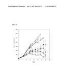 | 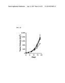 |
 |  |
 |  |
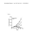 |  |
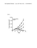 |  |
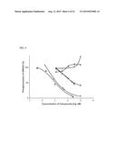 |  |
 |  |
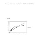 | 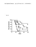 |
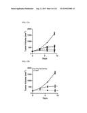 |  |
 | 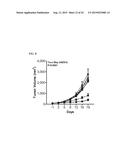 |
 | 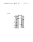 |
 |  |
 | 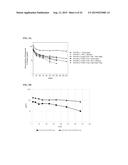 |
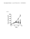 |  |
 | 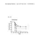 |
 | 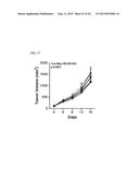 |
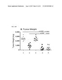 |  |
 |  |
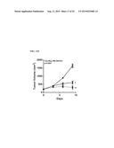 | 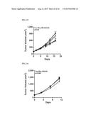 |
 |  |
 |  |
 |  |
 |  |
 | 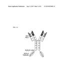 |
 | 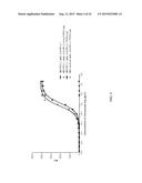 |
 |  |
 |
| New patent applications in this class: | |
| Date | Title |
|---|---|
| 2022-09-08 | Shrub rose plant named 'vlr003' |
| 2022-08-25 | Cherry tree named 'v84031' |
| 2022-08-25 | Miniature rose plant named 'poulty026' |
| 2022-08-25 | Information processing system and information processing method |
| 2022-08-25 | Data reassembly method and apparatus |
| New patent applications from these inventors: | |
| Date | Title |
|---|---|
| 2015-10-01 | Antibody fusion proteins with a modified fcrn binding site |
| 2014-01-02 | Fc-interferon-beta fusion proteins |
| 2013-06-27 | Expression and export of angiogenesis inhibitors as immunofusins |
| 2013-02-07 | Interleukin-12 p40 variants with improved stability |
| 2013-01-17 | Methods for enhancing the efficacy of il-2 mediated immune responses |