Patent application title: USE OF DNA IN CIRCULATING EXOSOMES AS A DIAGNOSTIC MARKER FOR METASTATIC DISEASE
Inventors:
David C. Lyden (New York, NY, US)
Hector Peinado Selgas (New York, NY, US)
Haiying Zhang (New York, NY, US)
Jacqueline Bromberg (New York, NY, US)
Assignees:
CORNELL UNIVERSITY
IPC8 Class: AC12Q168FI
USPC Class:
Class name:
Publication date: 2015-08-06
Patent application number: 20150218651
Abstract:
The present invention is directed to methods of diagnosing, prognosing,
and monitoring cancer in a subject. These methods involves selecting a
subject having cancer, and obtaining, from the selected subject, a sample
containing exosomal DNA. The presence or absence of one or more mutations
in BRAF and/or EGFR is detected in the exosomal DNA sample from the
subject, and a diagnosis and/or prognosis of the cancer is given based on
the detection of the one or more mutations in BRAF and/or EGFR. The
present invention further relates to methods of treating a subject having
cancer and/or monitoring a subject response to therapy based on the
detection of one or more mutations in BRAF and/or EGFR in the exosomal
DNA sample.Claims:
1. A method of prognosing cancer in a subject, said method comprising:
selecting a subject having cancer; obtaining, from the selected subject,
a sample containing exosomal DNA; contacting the exosomal DNA from the
sample with one or more reagents suitable to detect presence or absence
of one or more mutations in BRAF and/or EGFR that are associated with the
cancer; and prognosing the subject based on said contacting.
2. The method of claim 1 further comprising: selecting a suitable cancer therapeutic based on said prognosing and administering the selected cancer therapeutic to said selected subject.
3. The method of claim 1, wherein the cancer is selected from the group consisting of melanoma, breast cancer, brain cancer, pancreatic cancer, ovarian cancer, colorectal cancer, liver cancer, renal cancer, prostate cancer, and lung cancer.
4. The method of claim 1, wherein said prognosing comprises: determining the metastatic potential of the cancer based on said contacting.
5. The method of claim 1, wherein said one or more reagents suitable for detecting the presence or absence of one or more mutations in BRAF and/or EGFR in the sample are suitable for carrying out allele-specific polymerase chain reaction (PCR), high resolution melting curve analysis, or genomic sequencing.
6. The method of claim 1, wherein the selected subject has melanoma and the presence or absence of a mutation in BRAF is detected.
7. The method of claim 6, wherein detection of the presence of a V600E BRAF mutation in the sample indicates the selected subject has a poor prognosis.
8. The method of claim 1, wherein the presence or absence of one or more mutations in EGFR is detected.
9. The method of claim 5, wherein the one or more mutations in EGFR is selected from the group consisting of an exon 19 deletion, L858R, T790M, and any combination thereof.
10. A method of treating a subject having cancer, said method comprising: selecting a subject having cancer; obtaining, from the selected subject, a sample containing exosomal DNA; detecting in the exosomal DNA from the sample, the presence or absence of one or more mutations in BRAF and/or EGFR associated with the cancer; selecting a suitable cancer therapeutic based on said detecting; and administering the selected cancer therapeutic to the selected subject having cancer.
11. The method of claim 10, wherein the cancer is selected from the group consisting of melanoma, breast cancer, brain cancer, pancreatic cancer, ovarian cancer, colorectal cancer, liver cancer, renal cancer, prostate cancer, and lung cancer
12. The method of claim 10, wherein said detecting comprises: contacting the sample containing exosomal DNA with one or more reagents suitable for detecting the one or more mutations in BRAF and/or EGFR by allele-specific polymerase chain reaction (PCR), high resolution melting curve analysis, or genomic sequencing.
13. The method of claim 10, wherein the presence or absence of a mutation in BRAF is detected.
14. The method of claim 10, wherein the presence or absence of one or more mutation in EGFR is detected, said mutation selected from the group consisting of an exon 19 deletion, L858R, T790M, and combinations thereof.
Description:
[0001] This application claims the priority benefit of U.S. Provisional
Patent Application Ser. Nos. 61/684,224, filed Aug. 17, 2012, and
61/794,384, filed Mar. 15, 2013, which are hereby incorporated by
reference in their entirety.
FIELD OF THE INVENTION
[0002] The present invention relates to methods and compositions for diagnosing, prognosing, and monitoring cancer in a subject.
BACKGROUND OF THE INVENTION
[0003] The identification of cancer biomarkers suitable for the early detection, diagnosis, prognosis, and monitoring of cancer holds great promise to improve the clinical outcome of patients by facilitating a personalized approach to treatment. At present, biomarkers (proteins, peptides, lipids, RNAs, and DNA) for conditions and diseases, such as cancer, rely almost exclusively on obtaining samples from tissue to identify the condition or disease. Methods to obtain these tissues of interest for analysis are often invasive, costly and can pose complication risks for the patient. Furthermore, use of bodily fluids to isolate or detect biomarkers often significantly dilutes a biomarker resulting in readouts that lack requisite sensitivity. Additionally, most biomarkers are produced in low or moderate amounts in normal tissues other than the diseased tissue and thus this lack of specificity can also be problematic. Despite considerable effort directed at early detection, few reliable and cost-effective screening tests have been developed that can detect, diagnose, prognose, or monitor cancer at an early stage.
[0004] The present invention is directed to overcoming these and other deficiencies in the art.
SUMMARY OF THE INVENTION
[0005] A first aspect of the present invention is directed to a method for detecting one or more BRAF and/or epidermal growth factor receptor (EGFR) mutations in a subject. This method involves isolating a sample containing exosomal DNA from the subject, and contacting the exosomal DNA from the sample with one or more reagents suitable to detect presence or absence of one or more mutations in BRAF and/or EGFR. The one or more mutations in BRAF and/or EGFR are detected based on the contacting.
[0006] Another aspect of the present invention is directed to method of prognosing cancer in a subject. This method involves selecting a subject having cancer, and obtaining, from the selected subject, a sample containing exosomal DNA. The method further involves contacting the exosomal DNA from the sample with one or more reagents suitable to detect presence or absence of one or more mutations in BRAF and/or EGFR that are associated with the cancer, and prognosing the subject based on said contacting.
[0007] Another aspect of the present invention is directed to a method of diagnosing cancer in a subject. This method involves selecting a subject having cancer, and obtaining, from the selected subject, a sample containing cancer cell-derived exosomal DNA. The method further involves detecting in the exosomal DNA from the sample, the presence or absence of one or more mutations in BRAF and/or EGFR that are associated with the cancer, and diagnosing the subject based on said contacting.
[0008] Another aspect of the present invention is directed to a method of monitoring cancer progression in a subject that involves obtaining first and second samples containing exosomal DNA, at different points in time, from the subject having cancer. The exosomal DNA in the sample is contacted with one or more reagents suitable for detecting the presence or absence of one or more mutations in BRAF and/or EGFR, and the presence or absence of the one or more mutations in BRAF and/or EGFR is detected. The method further involves comparing the presence or absence of the one or more mutations detected in the first exosomal DNA sample to the presence or absence of the one or more mutations detected in the second sample, and monitoring cancer progression in the subject based on the comparison.
[0009] Another aspect of the present invention is directed to a method of identifying a primary tumor of unknown origin in a subject having metastatic cancer. This method involves obtaining, from the subject having metastatic cancer, a sample containing exosomal DNA, and contacting the exosomal DNA from the sample with one or more reagents suitable to detect presence or absence of one or more mutations in BRAF and/or EGFR. The presence or absence of one or more mutations in BRAF and/or EGFR in the exosomal DNA of the sample are detected based on the contacting and the primary tumor of unknown origin is identified based on the detection of one or more BRAF and/or EGFR mutations.
[0010] Another aspect of the present invention is directed to a method of treating a subject having cancer. This method involves obtaining, from the subject, a sample containing exosomal DNA, and detecting in the exosomal DNA from the sample, the presence or absence of one or more mutations in BRAF and/or EGFR associated with the cancer. The method further involves selecting a suitable cancer therapeutic based on the detecting, and administering the selected cancer therapeutic to the subject having cancer.
[0011] Another aspect of the present invention is directed to a method of assessing a subject's response to treatment with a BRAF inhibitor. This method involves obtaining first and second samples containing exosomal DNA, at different points in time, from a subject being treated with a BRAF inhibitor. The first and second samples containing exosomal DNA are contacted with one or more reagents suitable for detecting the presence or absence of one or more mutations in BRAF, and the presence or absence of the one or more mutations in BRAF is detected. The presence or absence of the one or more mutations detected in the first exosomal DNA sample is compared to the presence or the absence of the one or more mutations detected in the second sample, and the subject's response to BRAF inhibitor treatment is assessed based on this comparison.
[0012] Another aspect of the present invention is directed to a method of assessing a subject's response to treatment with an EGFR inhibitor. This method involves obtaining first and second samples containing exosomal DNA, at different points in time, from a subject being treated with an EGFR inhibitor. The first and second samples containing exosomal DNA are contacted with one or more reagents suitable for detecting the presence or absence of one or more mutations in EGFR, and the presence or absence of the one or more mutations in EGFR is detected. The presence or absence of the one or more mutations detected in the first exosomal DNA sample is compared to the presence or the absence of the one or more mutations detected in the second sample, and the subject's response to EGFR inhibitor treatment is assessed based on this comparison.
[0013] Another aspect of the present invention is directed to a method of determining the metastatic potential of a cancer in a subject. This method involves obtaining a sample containing cancer cell-derived exosomes from the subject, and measuring the concentration of cancer cell-derived exosomal DNA in the sample. The concentration of cancer cell-derived exosomal DNA in the sample from the subject is compared to the concentration of exosomal DNA in a reference exosomal sample, and the metastatic potential of the cancer in the subject is determined based on the comparison.
[0014] The present invention is based on the inventors' discovery that circulating tumor exosomes contain DNA that phenocopies the mutational status of primary tumors and metastatic tumors. Accordingly, tumor derived exosomal DNA can serve as a non-invasive, diagnostic and prognostic tool by facilitating the rapid genotyping of cancers to enable early detection and optimized treatment of disease. Importantly, diagnoses and prognoses are rendered feasible using this technique in cases where a biopsy is difficult to obtain (due to inaccessibility) or when a patient has multiple sites of disease. Moreover, this tool allows for frequent monitoring of the dynamics of tumor progression and molecular changes during treatment. In addition to prognostic and diagnostic utility, the molecular information gathered from exosomal DNA analysis can be used to guide and develop personalized therapeutic regimes. Finally, because exosomes are secreted from tumors constitutively, and isolation of exosomes requires no special equipment, exosome DNA-based testing can be readily employed in all standard laboratories.
BRIEF DESCRIPTION OF THE DRAWINGS
[0015] FIGS. 1A-1C show that tumor-derived exosomes contain DNA that is representative of the genotype of corresponding parent tumor cells. FIG. 1A is a graph depicting the abundance of exosomal DNA ("exoDNA") derived from different types of cancer cells. FIG. 1B is a circular view of the readings of fragments along each chromosome in the whole genome sequencing analysis of exoDNA isolated from murine melanoma B16-F10 cell-derived exosomes. FIG. 1C is a graph showing the levels of exoDNA isolated from healthy human primary dermal fibroblasts ("DF") and endothelial cells ("097").
[0016] FIG. 2 is an immunogold electron microscopy image of exosomes derived from B16-F10 cells using an anti-DNA antibody.
[0017] FIGS. 3A-3C depict the characterization of exoDNA. FIG. 3A shows S1 nuclease digestion of genomic DNA ("gDNA") and exoDNA derived from B16-F10 cells, indicating that exoDNA is predominantly single-stranded. FIG. 3B shows size distribution profiles of exoDNA from cultured cells. FIG. 3C is a dot blot analysis of the methylation status of exoDNA and genomic DNA using anti-5'-me-cytosine antibody. Probing of the same blot using anti-DNA antibody serves as loading control. FIG. 3D shows the results of a comparative genomic hybridization (CGH) array analysis comparing exoDNA and gDNA derived from the B16-F10 cell.
[0018] FIG. 4 shows the sensitivity and specificity of allele-specific polymerase chain reaction (AS-PCR) for detecting the BRAF(V600E) mutation. Genomic DNA samples containing no BRAF(V600E) mutation or 0.1% of this mutation were used as template for AS-PCR to assess the sensitivity and specificity of the assay. Different amounts of template DNA (as low as 2.5 ng) were examined. The results indicate that the assay can detect the presence of mutation in as low as 5 ng of template containing 0.1% mutation without false positive identification of the mutation.
[0019] FIGS. 5A-5D show the detection of genetic mutations in exoDNA isolated from cultured tumor cells. The mutational status for BRAF and EGFR was examined using exoDNA derived from cell lines originated from melanoma and non-small cell lung carcinoma ("NSCLC"), respectively. AS-PCR was used for detection of the BRAF(V600E) mutation in melanoma cell lines as shown in FIG. 5A ("V" represents wildtype (WT) allele; "E" represents mutant allele); the EGFR exon 19 deletion in NSCLC cell lines as shown in FIG. 5B ("I" represents internal control for both wildtype and mutant alleles; "W" represents wildtype allele; "del" represents deletion of amino acid residues 746-750 of Exon 19), and the T790M mutation in NSCLC cell lines as shown in FIG. 5C ("T" represents wildtype allele; "M" represents mutant allele). High Resolution Melt ("FIRM") analysis was used for detecting the EGFR L858R mutation in NSCLC cells (FIG. 5D). The existence of a point mutation results in distinct melting curve of the amplicon that spans the mutation from that of the wildtype amplicon, and identical melting curves of amplicons originated from gDNA and exoDNA indicate their identical genotypes.
[0020] FIG. 6 shows the detection of BRAF V600E mutation in circulating exoDNA isolated from melanoma-bearing mice. Circulating exosomes were isolated from the plasma of mice bearing melanoma (subcutaneously implanted with human melanoma cell line, Sk-Mel-28). AS-PCR was employed to detect the BRAF V600E mutation in the extracted exoDNA, with gDNA isolated from Sk-Mel-28 and Sk-Mel-103 cells as positive and negative control for V600E mutation (WT (V) and mutant (E) alleles). Asterisk indicates the band of expected PCR products.
[0021] FIG. 7 shows AS-PCR analysis of exoDNA isolated from the plasma of patients with melanoma or healthy subjects for determination of BRAF(V600E) mutation. Genomic DNA isolated from Sk-Mel-28 and Sk-Mel-103 cells served as positive and negative controls for the V600E mutation (WT (V) and mutant (E) alleles). The BRAF mutation was detected in exoDNA of 7 of the melanoma patient samples and one of the healthy control patient samples.
DETAILED DESCRIPTION OF THE INVENTION
[0022] The present invention is directed to methods for detecting one or more BRAF and/or epidermal growth factor receptor (EGFR) mutations in a subject. These methods involve isolating a sample containing exosomal DNA from the subject, and contacting the exosomal DNA from the sample with one or more reagents suitable to detect presence or absence of one or more mutations in BRAF and/or EGFR genes. The one or more mutations in BRAF and/or EGFR are detected based on the contacting.
[0023] One aspect of the present invention is directed to a method of detecting the one or more BRAF and/or EGFR mutations to diagnose or prognose cancer in a subject. This method involves selecting a subject having cancer, and obtaining, from the selected subject, a sample containing cancer or tumor cell-derived exosomal DNA. The method further involves contacting the cancer or tumor cell-derived exosomal DNA from the sample with one or more reagents suitable to detect presence or absence of one or more mutations in BRAF and/or EGFR associated with the cancer diagnosis or prognosis, and diagnosing or prognosing the subject based on the contacting.
[0024] Cancer diagnosis as described herein refers to determining or classifying the nature of the cancer state, e.g., the mutational or genetic phenotype of a cancer or tumor, the clinical stage of a cancer associated with its progression, and/or the metastatic nature of the cancer. Cancer diagnosis based on genetic phenotyping can help guide proper therapeutic intervention as described herein. For example, a subject diagnosed as having melanoma or brain cancer positive for a BRAF mutation is a candidate for treatment with a BRAF inhibitor. Likewise, a subject diagnosed as having lung cancer or other cancer positive for an EGFR mutation is a candidate for treatment with an EGFR inhibitor.
[0025] Cancer prognosis as described herein includes determining the probable progression and course of the cancerous condition, and determining the chances of recovery and survival of a subject with the cancer, e.g., a favorable prognosis indicates an increased probability of recovery and/or survival for the cancer patient, while an unfavorable prognosis indicates a decreased probability of recovery and/or survival for the cancer patient. A subject's prognosis can be determined by the availability of a suitable treatment (i.e., a treatment that will increase the probability of recovery and survival of the subject with cancer). For example, if the subject has a cancer, such as melanoma or brain cancer that is positive for one or more BRAF mutations as described herein, the subject has a favorable prognosis because he/she is a candidate for treatment with BRAF inhibitor therapy. Likewise, if the subject has lung cancer or other cancer that is positive for one or more EGFR mutations as described herein, the subject has a favorable prognosis because he/she is a candidate for treatment with an EGFR inhibitor therapy. Accordingly, this aspect of the present invention may further include selecting a suitable cancer therapeutic based on the determined prognosis and administering the selected therapeutic to the subject.
[0026] Prognosis also encompasses the metastatic potential of a cancer. For example, a favorable prognosis based on the presence or absence of a genetic phenotype can indicate that the cancer is a type of cancer having low metastatic potential, and the patient has an increased probability of long term recovery and/or survival. Alternatively, an unfavorable prognosis, based on the presence or absence of a genetic phenotype can indicate that the cancer is a type of cancer having a high metastatic potential, and the patient has a decreased probability of long term recovery and/or survival.
[0027] Another aspect of the present invention is directed to a method of monitoring cancer progression in a subject that involves obtaining first and second samples containing exosomal DNA, at different points in time, from the subject having cancer. The exosomal DNA in the samples is contacted with one or more reagents suitable for detecting the presence or absence of one or more mutations in BRAF and/or EGFR, and the presence or absence of the one or more mutations in BRAF and/or EGFR is detected. The method further involves comparing the presence or absence of the one or more mutations detected in the first exosomal DNA sample to the presence or absence of the one or more mutations detected in the second sample and monitoring cancer progression in the subject based on the comparison.
[0028] A change in the mutational status of BRAF and/or EGFR, for example, detecting the presence of a BRAF and/or EGFR mutation in the second exosomal DNA sample whereas no BRAF and/or EGFR mutation was detected in the first exosomal DNA sample, indicates that a change in the cancer phenotype has occurred with disease progression. This change may have therapeutic implications, i.e., it may signal the need to change the subject's course of treatment. The change can also be indicative of the progression of the cancer to a metastatic phenotype. Therefore, periodic monitoring of exosomal DNA mutational status provides a means for detecting primary tumor progression, metastasis, and facilitating optimal targeted or personalized treatment of the cancerous condition.
[0029] The time between obtaining a first exosomal sample and a second, or any additional subsequent exosomal samples can be any desired period of time, for example, weeks, months, years, as determined is suitable by a physician and based on the characteristics of the primary tumor (tumor type, stage, location, etc.). In one embodiment of this aspect of the present invention, the first sample is obtained before treatment and the second sample is obtained after treatment. Alternatively, both samples can be obtained after one or more treatments; the second sample obtained at some point in time later than the first sample.
[0030] Another aspect of the present invention is directed to a method of identifying a primary tumor of unknown origin in a subject having metastatic cancer. This method involves obtaining, from the subject having metastatic cancer, a sample containing exosomal DNA, and contacting the exosomal DNA from the sample with one or more reagents suitable to detect presence or absence of one or more mutations in BRAF and/or EGFR. The presence or absence of one or more mutations in BRAF and/or EGFR in the exosomal DNA sample are detected based on the contacting and the primary tumor of unknown origin is identified based on the detection of one or more BRAF and/or EGFR mutations.
[0031] In accordance with this aspect of the present invention, the detection of one or more BRAF mutations in a metastatic tumor or cancer cell-derived exosomal sample indicates that the primary tumor or cancer was melanoma or a form of brain cancer, e.g., glioblastoma. The detection of one or more EGFR mutations in a metastatic tumor or cancer cell derived exosomal DNA indicates that the primary tumor originated in the lung, or alternatively the primary cancer was head and neck cancer, ovarian cancer, cervical cancer, bladder cancer, or esophageal cancer.
[0032] In accordance with this aspect of the present invention, the subject may have any type of metastatic cancer, including, without limitation, metastatic melanoma, metastatic breast cancer, metastatic brain cancer, metastatic pancreatic cancer, metastatic ovarian cancer, metastatic colorectal cancer, metastatic prostate cancer, metastatic lung cancer, metastatic liver cancer, metastatic bladder cancer, metastatic bone cancer, metastatic renal cancer, and metastatic pediatric cancers.
[0033] Another aspect of the present invention is directed to a method of treating a subject having cancer. This method involves obtaining, from the subject, a sample containing exosomal DNA, and detecting in the exosomal DNA from the sample, the presence or absence of one or more mutations in BRAF and/or EGFR associated with the cancer. The method further involves selecting a suitable cancer therapeutic based on the detecting, and administering the selected cancer therapeutic to the subject having cancer.
[0034] In accordance with all aspects of the present invention, a "subject" or "patient" encompasses any animal, but preferably a mammal, e.g., human, non-human primate, a dog, a cat, a horse, a cow, or a rodent. More preferably, the subject or patient is a human. In some embodiments of the present invention, the subject has cancer, for example and without limitation, melanoma, breast cancer, brain cancer, pancreatic cancer, gastrointestinal cancer, ovarian cancer, cervical cancer, colorectal cancer, liver cancer, renal cancer, prostate cancer, lung cancer, bladder cancer, head and neck cancer, or esophageal cancer. In some embodiments, the cancer is a primary tumor, while in other embodiments, the cancer is a secondary or metastatic tumor.
[0035] In one embodiment of the present invention, the selected subject has melanoma or brain cancer (e.g., glioblastoma, ganglioblastoma, astrocytoma) and the presence or absence of a mutation in BRAF is detected in an exosomal DNA sample from the subject. BRAF is a serine/threonine protein kinase that is encoded on chromosome 7q34. The amino acid sequence and nucleotide sequence of human BRAF are provided below as SEQ ID NO: 1 and SEQ ID NO: 2, respectively.
TABLE-US-00001 Human BRAF SEQ ID NO: 1 Met Ala Ala Leu Ser Gly Gly Gly Gly Gly Gly Ala Glu Pro Gly Gln 1 5 10 15 Ala Leu Phe Asn Gly Asp Met Glu Pro Glu Ala Gly Ala Gly Ala Gly 20 25 30 Ala Ala Ala Ser Ser Ala Ala Asp Pro Ala Ile Pro Glu Glu Val Trp 35 40 45 Asn Ile Lys Gln Met Ile Lys Leu Thr Gln Glu His Ile Glu Ala Leu 50 55 60 Leu Asp Lys Phe Gly Gly Glu His Asn Pro Pro Ser Ile Tyr Leu Glu 65 70 75 80 Ala Tyr Glu Glu Tyr Thr Ser Lys Leu Asp Ala Leu Gln Gln Arg Glu 85 90 95 Gln Gln Leu Leu Glu Ser Leu Gly Asn Gly Thr Asp Phe Ser Val Ser 100 105 110 Ser Ser Ala Ser Met Asp Thr Val Thr Ser Ser Ser Ser Ser Ser Leu 115 120 125 Ser Val Leu Pro Ser Ser Leu Ser Val Phe Gln Asn Pro Thr Asp Val 130 135 140 Ala Arg Ser Asn Pro Lys Ser Pro Gln Lys Pro Ile Val Arg Val Phe 145 150 155 160 Leu Pro Asn Lys Gln Arg Thr Val Val Pro Ala Arg Cys Gly Val Thr 165 170 175 Val Arg Asp Ser Leu Lys Lys Ala Leu Met Met Arg Gly Leu Ile Pro 180 185 190 Glu Cys Cys Ala Val Tyr Arg Ile Gln Asp Gly Glu Lys Lys Pro Ile 195 200 205 Gly Trp Asp Thr Asp Ile Ser Trp Leu Thr Gly Glu Glu Leu His Val 210 215 220 Glu Val Leu Glu Asn Val Pro Leu Thr Thr His Asn Phe Val Arg Lys 225 230 235 240 Thr Phe Phe Thr Leu Ala Phe Cys Asp Phe Cys Arg Lys Leu Leu Phe 245 250 255 Gln Gly Phe Arg Cys Gln Thr Cys Gly Tyr Lys Phe His Gln Arg Cys 260 265 270 Ser Thr Glu Val Pro Leu Met Cys Val Asn Tyr Asp Gln Leu Asp Leu 275 280 285 Leu Phe Val Ser Lys Phe Phe Glu His His Pro Ile Pro Gln Glu Glu 290 295 300 Ala Ser Leu Ala Glu Thr Ala Leu Thr Ser Gly Ser Ser Pro Ser Ala 305 310 315 320 Pro Ala Ser Asp Ser Ile Gly Pro Gln Ile Leu Thr Ser Pro Ser Pro 325 330 335 Ser Lys Ser Ile Pro Ile Pro Gln Pro Phe Arg Pro Ala Asp Glu Asp 340 345 350 His Arg Asn Gln Phe Gly Gln Arg Asp Arg Ser Ser Ser Ala Pro Asn 355 360 365 Val His Ile Asn Thr Ile Glu Pro Val Asn Ile Asp Asp Leu Ile Arg 370 375 380 Asp Gln Gly Phe Arg Gly Asp Gly Gly Ser Thr Thr Gly Leu Ser Ala 385 390 395 400 Thr Pro Pro Ala Ser Leu Pro Gly Ser Leu Thr Asn Val Lys Ala Leu 405 410 415 Gln Lys Ser Pro Gly Pro Gln Arg Glu Arg Lys Ser Ser Ser Ser Ser 420 425 430 Glu Asp Arg Asn Arg Met Lys Thr Leu Gly Arg Arg Asp Ser Ser Asp 435 440 445 Asp Trp Glu Ile Pro Asp Gly Gln Ile Thr Val Gly Gln Arg Ile Gly 450 455 460 Ser Gly Ser Phe Gly Thr Val Tyr Lys Gly Lys Trp His Gly Asp Val 465 470 475 480 Ala Val Lys Met Leu Asn Val Thr Ala Pro Thr Pro Gln Gln Leu Gln 485 490 495 Ala Phe Lys Asn Glu Val Gly Val Leu Arg Lys Thr Arg His Val Asn 500 505 510 Ile Leu Leu Phe Met Gly Tyr Ser Thr Lys Pro Gln Leu Ala Ile Val 515 520 525 Thr Gln Trp Cys Glu Gly Ser Ser Leu Tyr His His Leu His Ile Ile 530 535 540 Glu Thr Lys Phe Glu Met Ile Lys Leu Ile Asp Ile Ala Arg Gln Thr 545 550 555 560 Ala Gln Gly Met Asp Tyr Leu His Ala Lys Ser Ile Ile His Arg Asp 565 570 575 Leu Lys Ser Asn Asn Ile Phe Leu His Glu Asp Leu Thr Val Lys Ile 580 585 590 Gly Asp Phe Gly Leu Ala Thr Val Lys Ser Arg Trp Ser Gly Ser His 595 600 605 Gln Phe Glu Gln Leu Ser Gly Ser Ile Leu Trp Met Ala Pro Glu Val 610 615 620 Ile Arg Met Gln Asp Lys Asn Pro Tyr Ser Phe Gln Ser Asp Val Tyr 625 630 635 640 Ala Phe Gly Ile Val Leu Tyr Glu Leu Met Thr Gly Gln Leu Pro Tyr 645 650 655 Ser Asn Ile Asn Asn Arg Asp Gln Ile Ile Phe Met Val Gly Arg Gly 660 665 670 Tyr Leu Ser Pro Asp Leu Ser Lys Val Arg Ser Asn Cys Pro Lys Ala 675 680 685 Met Lys Arg Leu Met Ala Glu Cys Leu Lys Lys Lys Arg Asp Glu Arg 690 695 700 Pro Leu Phe Pro Gln Ile Leu Ala Ser Ile Glu Leu Leu Ala Arg Ser 705 710 715 720 Leu Pro Lys Ile His Arg Ser Ala Ser Glu Pro Ser Leu Asn Arg Ala 725 730 735 Gly Phe Gln Thr Glu Asp Phe Ser Leu Tyr Ala Cys Ala Ser Pro Lys 740 745 750 Thr Pro Ile Gln Ala Gly Gly Tyr Gly Ala Phe Pro Val His 755 760 765 Human BRAF SEQ ID NO: 2 cgcctccctt ccccctcccc gcccgacagc ggccgctcgg gccccggctc tcggttataa 60 gatggcggcg ctgagcggtg gcggtggtgg cggcgcggag ccgggccagg ctctgttcaa 120 cggggacatg gagcccgagg ccggcgccgg cgccggcgcc gcggcctctt cggctgcgga 180 ccctgccatt ccggaggagg tgtggaatat caaacaaatg attaagttga cacaggaaca 240 tatagaggcc ctattggaca aatttggtgg ggagcataat ccaccatcaa tatatctgga 300 ggcctatgaa gaatacacca gcaagctaga tgcactccaa caaagagaac aacagttatt 360 ggaatctctg gggaacggaa ctgatttttc tgtttctagc tctgcatcaa tggataccgt 420 tacatcttct tcctcttcta gcctttcagt gctaccttca tctctttcag tttttcaaaa 480 tcccacagat gtggcacgga gcaaccccaa gtcaccacaa aaacctatcg ttagagtctt 540 cctgcccaac aaacagagga cagtggtacc tgcaaggtgt ggagttacag tccgagacag 600 tctaaagaaa gcactgatga tgagaggtct aatcccagag tgctgtgctg tttacagaat 660 tcaggatgga gagaagaaac caattggttg ggacactgat atttcctggc ttactggaga 720 agaattgcat gtggaagtgt tggagaatgt tccacttaca acacacaact ttgtacgaaa 780 aacgtttttc accttagcat tttgtgactt ttgtcgaaag ctgcttttcc agggtttccg 840 ctgtcaaaca tgtggttata aatttcacca gcgttgtagt acagaagttc cactgatgtg 900 tgttaattat gaccaacttg atttgctgtt tgtctccaag ttctttgaac accacccaat 960 accacaggaa gaggcgtcct tagcagagac tgccctaaca tctggatcat ccccttccgc 1020 acccgcctcg gactctattg ggccccaaat tctcaccagt ccgtctcctt caaaatccat 1080 tccaattcca cagcccttcc gaccagcaga tgaagatcat cgaaatcaat ttgggcaacg 1140 agaccgatcc tcatcagctc ccaatgtgca tataaacaca atagaacctg tcaatattga 1200 tgacttgatt agagaccaag gatttcgtgg tgatggagga tcaaccacag gtttgtctgc 1260 taccccccct gcctcattac ctggctcact aactaacgtg aaagccttac agaaatctcc 1320 aggacctcag cgagaaagga agtcatcttc atcctcagaa gacaggaatc gaatgaaaac 1380 acttggtaga cgggactcga gtgatgattg ggagattcct gatgggcaga ttacagtggg 1440 acaaagaatt ggatctggat catttggaac agtctacaag ggaaagtggc atggtgatgt 1500 ggcagtgaaa atgttgaatg tgacagcacc tacacctcag cagttacaag ccttcaaaaa 1560 tgaagtagga gtactcagga aaacacgaca tgtgaatatc ctactcttca tgggctattc 1620 cacaaagcca caactggcta ttgttaccca gtggtgtgag ggctccagct tgtatcacca 1680 tctccatatc attgagacca aatttgagat gatcaaactt atagatattg cacgacagac 1740 tgcacagggc atggattact tacacgccaa gtcaatcatc cacagagacc tcaagagtaa 1800 taatatattt cttcatgaag acctcacagt aaaaataggt gattttggtc tagctacagt 1860 gaaatctcga tggagtgggt cccatcagtt tgaacagttg tctggatcca ttttgtggat 1920 ggcaccagaa gtcatcagaa tgcaagataa aaatccatac agctttcagt cagatgtata 1980 tgcatttgga attgttctgt atgaattgat gactggacag ttaccttatt caaacatcaa 2040 caacagggac cagataattt ttatggtggg acgaggatac ctgtctccag atctcagtaa 2100 ggtacggagt aactgtccaa aagccatgaa gagattaatg gcagagtgcc tcaaaaagaa 2160 aagagatgag agaccactct ttccccaaat tctcgcctct attgagctgc tggcccgctc 2220 attgccaaaa attcaccgca gtgcatcaga accctccttg aatcgggctg gtttccaaac 2280 agaggatttt agtctatatg cttgtgcttc tccaaaaaca cccatccagg cagggggata 2340 tggtgcgttt cctgtccact gaaacaaatg agtgagagag ttcaggagag tagcaacaaa 2400 aggaaaataa atgaacatat gtttgcttat atgttaaatt gaataaaata ctctcttttt 2460 ttttaaggtg aaccaaagaa cacttgtgtg gttaaagact agatataatt tttccccaaa 2520 ctaaaattta tacttaacat tggattttta acatccaagg gttaaaatac atagacattg 2580 ctaaaaattg gcagagcctc ttctagaggc tttactttct gttccgggtt tgtatcattc 2640 acttggttat tttaagtagt aaacttcagt ttctcatgca acttttgttg ccagctatca 2700 catgtccact agggactcca gaagaagacc ctacctatgc ctgtgtttgc aggtgagaag 2760 ttggcagtcg gttagcctgg gttagataag gcaaactgaa cagatctaat ttaggaagtc 2820 agtagaattt aataattcta ttattattct taataatttt tctataacta tttcttttta 2880 taacaatttg gaaaatgtgg atgtctttta tttccttgaa gcaataaact aagtttcttt 2940 ttataaaaa 2949
[0036] BRAF activates the MAP kinase/ERK-signaling pathway, and mutations in BRAF are associated with approximately 50% of pediatric and adult malignant melanomas (Daniotti et al., "Cutaneous Melanoma in Childhood and Adolescence Shows Frequent Loss of INK4A and Gain of KIT," J. Invest. Dermatol. 129 (7): 1759-68 (2009), which is hereby incorporated by reference in its entirety). In addition, BRAF point mutations have been reported to occur in several low- and high-grade tumor types in pediatric and adult patients, including approximately 50-60% of gangliogliomas (MacConaill et al., "Profiling Critical Cancer Gene Mutations in Clinical Tumor Samples," PloSOne 4(11):e7887 (2009), and Dougherty et al. "Activating Mutations in BRAF Characterize a Spectrum of Pediatric Low-Grade Gliomas," Neuro Oncol 12 (7): 621-630 (2010), which are hereby incorporated by reference in their entirety), approximately 2-12% of pilocytic astrocytomas (Forshew et al., "Activation of the ERK/MAPK Pathway: A Signature Genetic Defect in Posterior Fossa Pilocytic Astrocytomas," J Pathol. 218:172-181 (2009); Pfister et al., "BRAF Gene Duplication Constitutes a Mechanism of MAPK Pathway Activation in Low-Grade Astrocytomas," J Clin Invest. 118:1739-1749 (2008); MacConaill et al., "Profiling Critical Cancer Gene Mutations in Clinical Tumor Samples," PloSOne 4(11):e7887 (2009); Qaddoumi et al., "Paediatric Low-Grade Gliomas and the Need for New Options for Therapy," Cancer Biol Ther. 8:1-7 (2009); Jacob et al., "Duplication of 7q34 is Specific to Juvenile Pilocytic Astrocytomas and a Hallmark of Cerebellar and Optic Pathway Tumors," Brit J Cancer; 101:722-733 (2009); and Dias-Santagata et al., "BRAF V600E Mutations Are Common in Pleomorphic Xanthoastrocytoma: Diagnostic and Therapeutic Implications," PLoS ONE 6(3): e17948 (2011), which are hereby incorporated by reference in their entirety), and in as many as 30% of high-grade astrocytomas. Glioma accounts for 90% of malignant central nervous system (CNS) tumors in adults and 50% in the pediatric population (Central Brain Tumor Registry of the United States, 2010).
[0037] Over 90% of BRAF mutations in melanoma are at amino acid residue 600 (SEQ ID NO: 1), and over 90% of these involve a single nucleotide mutation that causes a valine→glutamic acid change (BRAF V600E: nucleotide 1799 T>A of SEQ ID NO: 2; codon GTG>GAG) (Ascierto et al., "The Role of BRAF V600 Mutation in Melanoma," J. Translational Med. 10:85 (2012), which is hereby incorporated by reference in its entirety). Other mutations at this same valine residue of BRAF include a lysine substitution (BRAFV600K), an arginine substitution (BRAFV600R), and an aspartic acid substitution (BRAFV600D). The detection of any one of these BRAF V600 mutations, or other known BRAF mutations (i.e., insertions, deletions, duplications, etc.) in an exosomal DNA sample from a subject has diagnostic/prognostic and therapeutic implications in accordance with the methods of the present invention.
[0038] The BRAF V600 mutations cause constitutive activation of BRAF, which leads to activation of the downstream MEK/ERK pathway, evasion of senescence and apoptosis, uncheck replicative potential, angiogenesis, tissue invasion, metastasis, as well as evasion of immune response (Maurer et al., "Raf Kinases in Cancer-Roles and Therapeutic Opportunities," Oncogene 30: 3477-3488 (2011), which is hereby incorporated by reference in its entirety). Melanoma patients and patients having brain cancer identified as having a BRAF V600 mutation or other BRAF activating mutations are candidates for treatment with a BRAF inhibitor, such as vemurafenib (PLX/RG7204/RO5185426) (Sosman et al., "Survival in BRAF V600-Mutant Advanced Melanoma Treated with Vemurafenib," N Engl J Med 366:707-14 (2012) and Chapman et al., "Improved Survival with Vemurafenib in Melanoma with BRAF V600E Mutation," N Engl J Med 364''2507-2516 (2011), which are hereby incorporated by reference in their entirety), dabrafenib (Tafinlar; GSK2118436) (Gibney et al., "Clinical Development of Dabrafenib in BRAF mutant Melanoma and Other Malignancies" Expert Opin Drug Metab Toxicol 9(7):893-9 (2013), which is hereby incorporated by reference in its entirety), RAF265 (Su et al., "RAF265 Inhibits the Growth of Advanced Human Melanoma Tumors," Clin Cancer Res 18(8): 2184-98 (2012), which is hereby incorporated by reference in its entirety), and LGX818 (Stuart et al., "Preclinical Profile of LGX818: A Potent and Selective RAF Kinase Inhibitor," Cancer Res 72(8) Suppl 1 (2012), which is hereby incorporated by reference in its entirety).
[0039] Another aspect of the present invention is directed to a method of assessing a subject's response to treatment with a BRAF inhibitor. This method involves obtaining first and second samples containing exosomal DNA, at different points in time, from a subject being treated with a BRAF inhibitor. Suitable subjects being treated with a BRAF inhibitor include, without limitation, those having melanoma or brain cancer. The first and second samples containing exosomal DNA are contacted with one or more reagents suitable for detecting the presence or absence of one or more mutations in BRAF, and the presence or absence of the one or more mutations in BRAF is detected. The presence or absence of the one or more mutations detected in the first exosomal DNA sample is compared to the presence or absence of the one or more mutations detected in the second sample, and the subject's response to BRAF inhibitor treatment is assessed based on this comparison. If there is a decrease in the presence of BRAF mutations in the second exosomal DNA sample as compared to the first exosomal DNA sample, than the subject is responding to BRAF inhibitor treatment, i.e., the BRAF inhibitor is effectively killing tumor cells containing the BRAF mutation. If there is no decrease in the presence of BRAF mutations in the second exosomal DNA sample as compared to the first exosomal DNA sample, then the subject is likely not responding to the BRAF inhibitor treatment. This method may further include adjusting the subject's treatment regimen based on the assessment of the subject's responsiveness to therapy.
[0040] The time between obtaining a first exosomal sample and a second, or any additional subsequent exosomal samples can be any desired period of time, for example, weeks, months, years, as determined is suitable by a physician and based on the characteristics of the primary tumor (tumor type, stage, location, etc.). In one embodiment of the present invention, the first sample is obtained before treatment and the second sample is obtained after treatment. Alternatively, both samples can be obtained after one or more treatments; the second sample obtained at some point in time later than the first sample.
[0041] In another embodiment of the present invention, the presence of absence of one or more mutations in the epidermal growth factor receptor (EGFR) is detected. EGFR is a transmembrane glycoprotein with an extracellular ligand-binding domain and an intracellular domain possessing intrinsic tyrosine kinase activity. Upon receptor dimerization following ligand binding, the tyrosine kinase domain is activated and recruited for phosphorylation of intracellular targets that drive normal cell growth and differentiation. The amino acid sequence and nucleotide sequence of human EGFR are provided below as SEQ ID NO: 3 and SEQ ID NO: 4, respectively.
TABLE-US-00002 Human EGFR SEQ ID NO: 3 Arg Pro Ser Gly Thr Ala Gly Ala Ala Leu Leu Ala Leu Leu Ala Ala 1 5 10 15 Leu Cys Pro Ala Ser Arg Ala Leu Glu Glu Lys Lys Val Cys Gln Gly 20 25 30 Thr Ser Asn Lys Leu Thr Gln Leu Gly Thr Phe Glu Asp His Phe Leu 35 40 45 Ser Leu Gln Arg Met Phe Asn Asn Cys Glu Val Val Leu Gly Asn Leu 50 55 60 Glu Ile Thr Tyr Val Gln Arg Asn Tyr Asp Leu Ser Phe Leu Lys Thr 65 70 75 80 Ile Gln Glu Val Ala Gly Tyr Val Leu Ile Ala Leu Asn Thr Val Glu 85 90 95 Arg Ile Pro Leu Glu Asn Leu Gln Ile Ile Arg Gly Asn Met Tyr Tyr 100 105 110 Glu Asn Ser Tyr Ala Leu Ala Val Leu Ser Asn Tyr Asp Ala Asn Lys 115 120 125 Thr Gly Leu Lys Glu Leu Pro Met Arg Asn Leu Gln Glu Ile Leu His 130 135 140 Gly Ala Val Arg Phe Ser Asn Asn Pro Ala Leu Cys Asn Val Glu Ser 145 150 155 160 Ile Gln Trp Arg Asp Ile Val Ser Ser Asp Phe Leu Ser Asn Met Ser 165 170 175 Met Asp Phe Gln Asn His Leu Gly Ser Cys Gln Lys Cys Asp Pro Ser 180 185 190 Cys Pro Asn Gly Ser Cys Trp Gly Ala Gly Glu Glu Asn Cys Gln Lys 195 200 205 Leu Thr Lys Ile Ile Cys Ala Gln Gln Cys Ser Gly Arg Cys Arg Gly 210 215 220 Lys Ser Pro Ser Asp Cys Cys His Asn Gln Cys Ala Ala Gly Cys Thr 225 230 235 240 Gly Pro Arg Glu Ser Asp Cys Leu Val Cys Arg Lys Phe Arg Asp Glu 245 250 255 Ala Thr Cys Lys Asp Thr Cys Pro Pro Leu Met Leu Tyr Asn Pro Thr 260 265 270 Thr Tyr Gln Met Asp Val Asn Pro Glu Gly Lys Tyr Ser Phe Gly Ala 275 280 285 Thr Cys Val Lys Lys Cys Pro Arg Asn Tyr Val Val Thr Asp His Gly 290 295 300 Ser Cys Val Arg Ala Cys Gly Ala Asp Ser Tyr Glu Met Glu Glu Asp 305 310 315 320 Gly Val Arg Lys Cys Lys Lys Cys Glu Gly Pro Cys Arg Lys Val Cys 325 330 335 Asn Gly Ile Gly Ile Gly Glu Phe Lys Asp Ser Leu Ser Ile Asn Ala 340 345 350 Thr Asn Ile Lys His Phe Lys Asn Cys Thr Ser Ile Ser Gly Asp Leu 355 360 365 His Ile Leu Pro Val Ala Phe Arg Gly Asp Ser Phe Thr His Thr Pro 370 375 380 Pro Leu Asp Pro Gln Glu Leu Asp Ile Leu Lys Thr Val Lys Glu Ile 385 390 395 400 Thr Gly Phe Leu Leu Ile Gln Ala Trp Pro Glu Asn Arg Thr Asp Leu 405 410 415 His Ala Phe Glu Asn Leu Glu Ile Ile Arg Gly Arg Thr Lys Gln His 420 425 430 Gly Gln Phe Ser Leu Ala Val Val Ser Leu Asn Ile Thr Ser Leu Gly 435 440 445 Leu Arg Ser Leu Lys Glu Ile Ser Asp Gly Asp Val Ile Ile Ser Gly 450 455 460 Asn Lys Asn Leu Cys Tyr Ala Asn Thr Ile Asn Trp Lys Lys Leu Phe 465 470 475 480 Gly Thr Ser Gly Gln Lys Thr Lys Ile Ile Ser Asn Arg Gly Glu Asn 485 490 495 Ser Cys Lys Ala Thr Gly Gln Val Cys His Ala Leu Cys Ser Pro Glu 500 505 510 Gly Cys Trp Gly Pro Glu Pro Arg Asp Cys Val Ser Cys Arg Asn Val 515 520 525 Ser Arg Gly Arg Glu Cys Val Asp Lys Cys Asn Leu Leu Glu Gly Glu 530 535 540 Pro Arg Glu Phe Val Glu Asn Ser Glu Cys Ile Gln Cys His Pro Glu 545 550 555 560 Cys Leu Pro Gln Ala Met Asn Ile Thr Cys Thr Gly Arg Gly Pro Asp 565 570 575 Asn Cys Ile Gln Cys Ala His Tyr Ile Asp Gly Pro His Cys Val Lys 580 585 590 Thr Cys Pro Ala Gly Val Met Gly Glu Asn Asn Thr Leu Val Trp Lys 595 600 605 Tyr Ala Asp Ala Gly His Val Cys His Leu Cys His Pro Asn Cys Thr 610 615 620 Tyr Gly Cys Thr Gly Pro Gly Leu Glu Gly Cys Pro Thr Asn Gly Pro 625 630 635 640 Lys Ile Pro Ser Ile Ala Thr Gly Met Val Gly Ala Leu Leu Leu Leu 645 650 655 Leu Val Val Ala Leu Gly Ile Gly Leu Phe Met Arg Arg Arg His Ile 660 665 670 Val Arg Lys Arg Thr Leu Arg Arg Leu Leu Gln Glu Arg Glu Leu Val 675 680 685 Glu Pro Leu Thr Pro Ser Gly Glu Ala Pro Asn Gln Ala Leu Leu Arg 690 695 700 Ile Leu Lys Glu Thr Glu Phe Lys Lys Ile Lys Val Leu Gly Ser Gly 705 710 715 720 Ala Phe Gly Thr Val Tyr Lys Gly Leu Trp Ile Pro Glu Gly Glu Lys 725 730 735 Val Lys Ile Pro Val Ala Ile Lys Glu Leu Arg Glu Ala Thr Ser Pro 740 745 750 Lys Ala Asn Lys Glu Ile Leu Asp Glu Ala Tyr Val Met Ala Ser Val 755 760 765 Asp Asn Pro His Val Cys Arg Leu Leu Gly Ile Cys Leu Thr Ser Thr 770 775 780 Val Gln Leu Ile Thr Gln Leu Met Pro Phe Gly Cys Leu Leu Asp Tyr 785 790 795 800 Val Arg Glu His Lys Asp Asn Ile Gly Ser Gln Tyr Leu Leu Asn Trp 805 810 815 Cys Val Gln Ile Ala Lys Gly Met Asn Tyr Leu Glu Asp Arg Arg Leu 820 825 830 Val His Arg Asp Leu Ala Ala Arg Asn Val Leu Val Lys Thr Pro Gln 835 840 845 His Val Lys Ile Thr Asp Phe Gly Leu Ala Lys Leu Leu Gly Ala Glu 850 855 860 Glu Lys Glu Tyr His Ala Glu Gly Gly Lys Val Pro Ile Lys Trp Met 865 870 875 880 Ala Leu Glu Ser Ile Leu His Arg Ile Tyr Thr His Gln Ser Asp Val 885 890 895 Trp Ser Tyr Gly Val Thr Val Trp Glu Leu Met Thr Phe Gly Ser Lys 900 905 910 Pro Tyr Asp Gly Ile Pro Ala Ser Glu Ile Ser Ser Ile Leu Glu Lys 915 920 925 Gly Glu Arg Leu Pro Gln Pro Pro Ile Cys Thr Ile Asp Val Tyr Met 930 935 940 Ile Met Val Lys Cys Trp Met Ile Asp Ala Asp Ser Arg Pro Lys Phe 945 950 955 960 Arg Glu Leu Ile Ile Glu Phe Ser Lys Met Ala Arg Asp Pro Gln Arg 965 970 975 Tyr Leu Val Ile Gln Gly Asp Glu Arg Met His Leu Pro Ser Pro Thr 980 985 990 Asp Ser Asn Phe Tyr Arg Ala Leu Met Asp Glu Glu Asp Met Asp Asp 995 1000 1005 Val Val Asp Ala Asp Glu Tyr Leu Ile Pro Gln Gln Gly Phe Phe 1010 1015 1020 Ser Ser Pro Ser Thr Ser Arg Thr Pro Leu Leu Ser Ser Leu Ser 1025 1030 1035 Ala Thr Ser Asn Asn Ser Thr Val Ala Cys Ile Asp Arg Asn Gly 1040 1045 1050 Leu Gln Ser Cys Pro Ile Lys Glu Asp Ser Phe Leu Gln Arg Tyr 1055 1060 1065 Ser Ser Asp Pro Thr Gly Ala Leu Thr Glu Asp Ser Ile Asp Asp 1070 1075 1080 Thr Phe Leu Pro Val Pro Glu Tyr Ile Asn Gln Ser Val Pro Lys 1085 1090 1095 Arg Pro Ala Gly Ser Val Gln Asn Pro Val Tyr His Asn Gln Pro 1100 1105 1110 Leu Asn Pro Ala Pro Ser Arg Asp Pro His Tyr Gln Asp Pro His 1115 1120 1125 Ser Thr Ala Val Gly Asn Pro Glu Tyr Leu Asn Thr Val Gln Pro 1130 1135 1140 Thr Cys Val Asn Ser Thr Phe Asp Ser Pro Ala His Trp Ala Gln 1145 1150 1155 Lys Gly Ser His Gln Ile Ser Leu Asp Asn Pro Asp Tyr Gln Gln 1160 1165 1170 Asp Phe Phe Pro Lys Glu Ala Lys Pro Asn Gly Ile Phe Lys Gly 1175 1180 1185 Ser Thr Ala Glu Asn Ala Glu Tyr Leu Arg Val Ala Pro Gin Ser 1190 1195 1200 Ser Glu Phe Ile Gly Ala 1205 Human EGFR SEQ ID NO: 4 ccccggcgca gcgcggccgc agcagcctcc gccccccgca cggtgtgagc gcccgacgcg 60 gccgaggcgg ccggagtccc gagctagccc cggcggccgc cgccgcccag accggacgac 120 aggccacctc gtcggcgtcc gcccgagtcc ccgcctcgcc gccaacgcca caaccaccgc 180 gcacggcccc ctgactccgt ccagtattga tcgggagagc cggagcgagc tcttcgggga 240 gcagcgatgc gaccctccgg gacggccggg gcagcgctcc tggcgctgct ggctgcgctc 300 tgcccggcga gtcgggctct ggaggaaaag aaagtttgcc aaggcacgag taacaagctc 360 acgcagttgg gcacttttga agatcatttt ctcagcctcc agaggatgtt caataactgt 420 gaggtggtcc ttgggaattt ggaaattacc tatgtgcaga ggaattatga tctttccttc 480
ttaaagacca tccaggaggt ggctggttat gtcctcattg ccctcaacac agtggagcga 540 attcctttgg aaaacctgca gatcatcaga ggaaatatgt actacgaaaa ttcctatgcc 600 ttagcagtct tatctaacta tgatgcaaat aaaaccggac tgaaggagct gcccatgaga 660 aatttacagg aaatcctgca tggcgccgtg cggttcagca acaaccctgc cctgtgcaac 720 gtggagagca tccagtggcg ggacatagtc agcagtgact ttctcagcaa catgtcgatg 780 gacttccaga accacctggg cagctgccaa aagtgtgatc caagctgtcc caatgggagc 840 tgctggggtg caggagagga gaactgccag aaactgacca aaatcatctg tgcccagcag 900 tgctccgggc gctgccgtgg caagtccccc agtgactgct gccacaacca gtgtgctgca 960 ggctgcacag gcccccggga gagcgactgc ctggtctgcc gcaaattccg agacgaagcc 1020 acgtgcaagg acacctgccc cccactcatg ctctacaacc ccaccacgta ccagatggat 1080 gtgaaccccg agggcaaata cagctttggt gccacctgcg tgaagaagtg tccccgtaat 1140 tatgtggtga cagatcacgg ctcgtgcgtc cgagcctgtg gggccgacag ctatgagatg 1200 gaggaagacg gcgtccgcaa gtgtaagaag tgcgaagggc cttgccgcaa agtgtgtaac 1260 ggaataggta ttggtgaatt taaagactca ctctccataa atgctacgaa tattaaacac 1320 ttcaaaaact gcacctccat cagtggcgat ctccacatcc tgccggtggc atttaggggt 1380 gactccttca cacatactcc tcctctggat ccacaggaac tggatattct gaaaaccgta 1440 aaggaaatca cagggttttt gctgattcag gcttggcctg aaaacaggac ggacctccat 1500 gcctttgaga acctagaaat catacgcggc aggaccaagc aacatggtca gttttctctt 1560 gcagtcgtca gcctgaacat aacatccttg ggattacgct ccctcaagga gataagtgat 1620 ggagatgtga taatttcagg aaacaaaaat ttgtgctatg caaatacaat aaactggaaa 1680 aaactgtttg ggacctccgg tcagaaaacc aaaattataa gcaacagagg tgaaaacagc 1740 tgcaaggcca caggccaggt ctgccatgcc ttgtgctccc ccgagggctg ctggggcccg 1800 gagcccaggg actgcgtctc ttgccggaat gtcagccgag gcagggaatg cgtggacaag 1860 tgcaaccttc tggagggtga gccaagggag tttgtggaga actctgagtg catacagtgc 1920 cacccagagt gcctgcctca ggccatgaac atcacctgca caggacgggg accagacaac 1980 tgtatccagt gtgcccacta cattgacggc ccccactgcg tcaagacctg cccggcagga 2040 gtcatgggag aaaacaacac cctggtctgg aagtacgcag acgccggcca tgtgtgccac 2100 ctgtgccatc caaactgcac ctacggatgc actgggccag gtcttgaagg ctgtccaacg 2160 aatgggccta agatcccgtc catcgccact gggatggtgg gggccctcct cttgctgctg 2220 gtggtggccc tggggatcgg cctcttcatg cgaaggcgcc acatcgttcg gaagcgcacg 2280 ctgcggaggc tgctgcagga gagggagctt gtggagcctc ttacacccag tggagaagct 2340 cccaaccaag ctctcttgag gatcttgaag gaaactgaat tcaaaaagat caaagtgctg 2400 ggctccggtg cgttcggcac ggtgtataag ggactctgga tcccagaagg tgagaaagtt 2460 aaaattcccg tcgctatcaa ggaattaaga gaagcaacat ctccgaaagc caacaaggaa 2520 atcctcgatg aagcctacgt gatggccagc gtggacaacc cccacgtgtg ccgcctgctg 2580 ggcatctgcc tcacctccac cgtgcagctc atcacgcagc tcatgccctt cggctgcctc 2640 ctggactatg tccgggaaca caaagacaat attggctccc agtacctgct caactggtgt 2700 gtgcagatcg caaagggcat gaactacttg gaggaccgtc gcttggtgca ccgcgacctg 2760 gcagccagga acgtactggt gaaaacaccg cagcatgtca agatcacaga ttttgggctg 2820 gccaaactgc tgggtgcgga agagaaagaa taccatgcag aaggaggcaa agtgcctatc 2880 aagtggatgg cattggaatc aattttacac agaatctata cccaccagag tgatgtctgg 2940 agctacgggg tgaccgtttg ggagttgatg acctttggat ccaagccata tgacggaatc 3000 cctgccagcg agatctcctc catcctggag aaaggagaac gcctccctca gccacccata 3060 tgtaccatcg atgtctacat gatcatggtc aagtgctgga tgatagacgc agatagtcgc 3120 ccaaagttcc gtgagttgat catcgaattc tccaaaatgg cccgagaccc ccagcgctac 3180 cttgtcattc agggggatga aagaatgcat ttgccaagtc ctacagactc caacttctac 3240 cgtgccctga tggatgaaga agacatggac gacgtggtgg atgccgacga gtacctcatc 3300 ccacagcagg gcttcttcag cagcccctcc acgtcacgga ctcccctcct gagctctctg 3360 agtgcaacca gcaacaattc caccgtggct tgcattgata gaaatgggct gcaaagctgt 3420 cccatcaagg aagacagctt cttgcagcga tacagctcag accccacagg cgccttgact 3480 gaggacagca tagacgacac cttcctccca gtgcctgaat acataaacca gtccgttccc 3540 aaaaggcccg ctggctctgt gcagaatcct gtctatcaca atcagcctct gaaccccgcg 3600 cccagcagag acccacacta ccaggacccc cacagcactg cagtgggcaa ccccgagtat 3660 ctcaacactg tccagcccac ctgtgtcaac agcacattcg acagccctgc ccactgggcc 3720 cagaaaggca gccaccaaat tagcctggac aaccctgact accagcagga cttctttccc 3780 aaggaagcca agccaaatgg catctttaag ggctccacag ctgaaaatgc agaataccta 3840 agggtcgcgc cacaaagcag tgaatttatt ggagcatgac cacggaggat agtatgagcc 3900 ctaaaaatcc agactctttc gatacccagg accaagccac agcaggtcct ccatcccaac 3960 agccatgccc gcattagctc ttagacccac agactggttt tgcaacgttt acaccgacta 4020 gccaggaagt acttccacct cgggcacatt ttgggaagtt gcattccttt gtcttcaaac 4080 tgtgaagcat ttacagaaac gcatccagca agaatattgt ccctttgagc agaaatttat 4140 ctttcaaaga ggtatatttg aaaaaaaaaa aaagtatatg tgaggatttt tattgattgg 4200 ggatcttgga gtttttcatt gtcgctattg atttttactt caatgggctc ttccaacaag 4260 gaagaagctt gctggtagca cttgctaccc tgagttcatc caggcccaac tgtgagcaag 4320 gagcacaagc cacaagtctt ccagaggatg cttgattcca gtggttctgc ttcaaggctt 4380 ccactgcaaa acactaaaga tccaagaagg ccttcatggc cccagcaggc cggatcggta 4440 ctgtatcaag tcatggcagg tacagtagga taagccactc tgtcccttcc tgggcaaaga 4500 agaaacggag gggatggaat tcttccttag acttactttt gtaaaaatgt ccccacggta 4560 cttactcccc actgatggac cagtggtttc cagtcatgag cgttagactg acttgtttgt 4620 cttccattcc attgttttga aactcagtat gctgcccctg tcttgctgtc atgaaatcag 4680 caagagagga tgacacatca aataataact cggattccag cccacattgg attcatcagc 4740 atttggacca atagcccaca gctgagaatg tggaatacct aaggatagca ccgcttttgt 4800 tctcgcaaaa acgtatctcc taatttgagg ctcagatgaa atgcatcagg tcctttgggg 4860 catagatcag aagactacaa aaatgaagct gctctgaaat ctcctttagc catcacccca 4920 accccccaaa attagtttgt gttacttatg gaagatagtt ttctcctttt acttcacttc 4980 aaaagctttt tactcaaaga gtatatgttc cctccaggtc agctgccccc aaaccccctc 5040 cttacgcttt gtcacacaaa aagtgtctct gccttgagtc atctattcaa gcacttacag 5100 ctctggccac aacagggcat tttacaggtg cgaatgacag tagcattatg agtagtgtgg 5160 aattcaggta gtaaatatga aactagggtt tgaaattgat aatgctttca caacatttgc 5220 agatgtttta gaaggaaaaa agttccttcc taaaataatt tctctacaat tggaagattg 5280 gaagattcag ctagttagga gcccaccttt tttcctaatc tgtgtgtgcc ctgtaacctg 5340 actggttaac agcagtcctt tgtaaacagt gttttaaact ctcctagtca atatccaccc 5400 catccaattt atcaaggaag aaatggttca gaaaatattt tcagcctaca gttatgttca 5460 gtcacacaca catacaaaat gttccttttg cttttaaagt aatttttgac tcccagatca 5520 gtcagagccc ctacagcatt gttaagaaag tatttgattt ttgtctcaat gaaaataaaa 5580 ctatattcat ttccactcta aaaaaaaaaa aaaaaa 5616
[0042] Several EGFR mutations leading to constitutive activation have been associated with neoplastic growth and cancer progression in a variety of cancers, including lung cancer (in particular non-small cell lung carcinoma), head and neck cancer, ovarian cancer, cervical cancer, bladder cancer, and esophageal cancer (Nicholson et al., "EGFR and Cancer Prognosis," Eur J Cancer 37(4):9-15 (2001), which is hereby incorporated by reference in its entirety). Therefore, subjects suitable for EGFR mutational detection in accordance with this embodiment of the present invention include subjects having any one of the aforementioned cancers.
[0043] A gain of function mutation suitable for detection in exosomal DNA samples in accordance with the present invention, includes, without limitation, the L858R mutation which results in leucine to arginine amino acid substitution at amino acid position 858 of human EGFR (SEQ ID NO: 3). This mutation occurs within the kinase domain (exon 21) and arises from a T>G nucleotide mutation at position 2573 of the EGFR gene sequence (SEQ ID NO: 4) (NCBI dbSNP reference SNP rs121434568; Mitsudomi et al., "Epidermal Growth Factor Receptor in Relation to Tumor Development: EGFR Gene and Cancer," FEBS J 277(2): 301-8 (2010), which are hereby incorporated by reference in their entirety).
[0044] Another gain of function mutation in EGFR suitable for detection in accordance with the present invention is the T790M mutation which results in a threonine to methionine mutation at amino acid position 790 in EGFR (SEQ ID NO: 3). This mutation occurs within the kinase domain (exon 20) and arises from a C>T mutation at nucleotide 2369 of the EGFR gene (SEQ ID NO: 4) (NCBI dbSNP reference SNP rs121434569; Tam et al., "Distinct Epidermal Growth Factor Receptor and KRAS Mutation Patterns in Non-Small Cell Lung Cancer Patients with Different Tobacco Exposure and Clinicopathologic Features," Clin Cancer Res 12:1647 (2006), which are hereby incorporated by reference in their entirety).
[0045] Another gain of function mutation in EGFR suitable for detection in accordance with the present invention is an in-frame deletion in exon 19. For example, deletions in amino acid residues 746-750, 746-751, 746-752, 747-751, 747-749, and 752-759 (SEQ ID NO: 3) have all been associated with lung cancer (see e.g., Mitsudomi et al., "Epidermal Growth Factor Receptor in Relation to Tumor Development: EGFR Gene and Cancer," FEBS J 277(2): 301-8 (2010), which is hereby incorporated by reference in its entirety). Detection of any one of these exon 19 deletions in exosomal DNA from a subject has prognostic/diagnostic and therapeutic implications in accordance with the present invention.
[0046] Subjects identified as having any of the above described EGFR mutations, or any other known EGFR mutations (i.e., insertions, deletions, duplications, etc), particularly gain-of-function mutations, are candidates for treatment using EGFR inhibitory agents which induce apoptosis and reduce proliferation of tumor growth (Ciardiello et al., "A Novel Approach in the Treatment of Cancer: Targeting the Epidermal Growth Factor Receptor," Clin Cancer Res 7:2958-2970 (2001); Ritter et al., "The Epidermal Growth Factor Receptor-Tyrosine Kinase: A Promising Therapeutic Target in Solid Tumors," Semin Oncol 30:3-11 (2003), which are hereby incorporated by reference in their entirety). Suitable EGFR inhibitors include, without limitation, small-molecule inhibitors of EGFR such as Gefitnib, Erlotinib (Tarceva), Afatinib (Gilotrif), Lapatinib (Tyverb) and monoclonal antibody inhibitors such as Panitumumab (Vectibix) and Cetuximab (Erbitux). Other EGFR inhibitors that are known in the art are also suitable for use in accordance with the methods of the present invention.
[0047] Another aspect of the present invention is directed to a method of assessing a subject's response to treatment with an EGFR inhibitor. This method involves obtaining first and second samples containing exosomal DNA, at different points in time, from a subject being treated with an EGFR inhibitor. Suitable subjects being treated with an EGFR inhibitor include, without limitation, those having lung cancer, head and neck cancer, ovarian cancer, cervical cancer, bladder cancer and esophageal cancer. The first and second samples containing exosomal DNA are contacted with one or more reagents suitable for detecting the presence or absence of one or more mutations in EGFR, and the presence or absence of the one or more mutations in EGFR is detected. The presence or absence of the one or more mutations detected in the first exosomal DNA sample is compared to the presence or absence of one or more mutations detected in the second sample, and the subject's response to EGFR inhibitor treatment is assessed based on this comparison. If there is a decrease in the presence of EGFR mutations in the second exosomal DNA sample as compared to the first exosomal DNA sample, then the subject is responding to EGFR inhibitor treatment, i.e., the EGFR inhibitor is effectively killing tumor cells containing the EGFR mutation. If there is no decrease in the presence of EGFR mutations in the second exosomal DNA sample as compared to the first exosomal DNA sample, then the subject is not responsive to the EGFR inhibitor treatment. This method may further include adjusting the subject's treatment regimen based on the assessment of the subject's responsiveness to therapy.
[0048] As noted above, the first sample can be obtained before treatment and the second sample obtained after treatment. Alternatively, both samples can be obtained after one or more treatments; the second sample obtained at some point in time later than the first sample.
[0049] Another aspect of the present invention is directed to a method of determining the metastatic potential of a cancer in a subject. This method involves obtaining a sample containing cancer cell-derived exosomes from the subject, and measuring the concentration of exosomal DNA in the sample. The concentration of exosomal DNA in the sample from the subject is compared to the concentration of exosomal DNA in a reference exosomal sample, and the metastatic potential of the cancer in the subject is determined based on the comparison.
[0050] In accordance with this aspect of the present invention, and as described herein, exosomes derived from tumors having high metastatic potential contain much higher levels of DNA than exosomes derived from tumors having a low or no metastatic potential. Therefore, in one embodiment of the present invention, the reference exosomal sample is an exosomal sample derived from tumor cells known to have low metastatic potential such as B16F1 melanoma cells, H1975 and H1650 lung cancer cells, or U87 glioblastoma cells. A higher concentration of DNA in the exosomal sample from the subject as compared to the concentration of DNA in exosomes derived from cells of low metastatic potential indicates the subject has a cancer with a high metastatic potential. If the exosomal sample from the subject has the same or lower concentration of DNA as compared to the concentration of DNA in exosomes derived from cells of low metastatic potential, then the subject has a cancer with a low metastatic potential. Alternatively, a reference exosomal sample can be derived from tumor cells having a high metastatic potential, such as B16F10 melanoma cells or Lewis lung carcinoma cells. If the exosomal sample from the subject has the same or a higher concentration of DNA as compared to exosomes derived from tumor cells of high metastatic potential, then the subject has a cancer with high metastatic potential. If the exosomal sample from the subject has a lower concentration of DNA as compared to exosomes derived from tumor cells of high metastatic potential, then the subject has a cancer with low metastatic potential
[0051] "Exosomes" are microvesicles released from a variety of different cells, including cancer cells (i.e., "cancer-derived exosomes"). These small vesicles (50-100 nm in diameter) derive from large multivesicular endosomes and are secreted into the extracellular milieu. The precise mechanisms of exosome release/shedding remain unclear; however, this release is an energy-requiring phenomenon, modulated by extracellular signals. They appear to form by invagination and budding from the limiting membrane of late endosomes, resulting in vesicles that contain cytosol and that expose the extracellular domain of membrane-bound cellular proteins on their surface. Using electron microscopy, studies have shown fusion profiles of multivesicular endosomes with the plasma membrane, leading to the secretion of the internal vesicles into the extracellular environment. The rate of exosome release is significantly increased in most neoplastic cells and occurs continuously. Increased release of exosomes and their accumulation appear to be important in the malignant transformation process.
[0052] In accordance with the methods of the present invention, exosomes can be isolated or obtained from most biological fluids including, without limitation, blood, serum, plasma, ascites, cyst fluid, pleural fluid, peritoneal fluid, cerebral spinal fluid, tears, urine, saliva, sputum, nipple aspirates, lymph fluid, fluid of the respiratory, intestinal, and genitourinary trances, breast milk, intra-organ system fluid, or combinations thereof.
[0053] An enriched population of exosomes can be obtained from a biological sample using methods known in the art. For example, exosomes may be concentrated or isolated from a biological sample using size exclusion chromatography, density gradient centrifugation, differential centrifugation (Raposo et al. "B lymphocytes secrete antigen-presenting vesicles," J Exp Med 183(3): 1161-72 (1996), which is hereby incorporated by reference in its entirety), anion exchange and/or gel permeation chromatography (for example, as described in U.S. Pat. No. 6,899,863 to Dhellin et al., and U.S. Pat. No. 6,812,023 to Lamparski et al., which are hereby incorporated by reference in their entirety), sucrose density gradients or organelle electrophoresis (for example, as described in U.S. Pat. No. 7,198,923), magnetic activated cell sorting (MACS) (Taylor et al., "MicroRNA signatures of tumor-derived exosomes as diagnostic biomarkers of ovarian cancer" Gynecol Oncol 110(1): 13-21 (2008), which is hereby incorporated by reference in its entirety), nanomembrane ultrafiltration (Cheruvanky et al., "Rapid isolation of urinary exosomal biomarkers using a nanomembrane ultrafiltration concentrator," Am J Physiol Renal Physiol 292(5): F1657-61 (2007), which is hereby incorporated by reference in its entirety), immunoabsorbent capture, affinity purification, microfluidic separation, or combinations thereof.
[0054] Exosomes isolated from a bodily fluid can be enriched for those originating from a specific cell type, for example, lung, pancreas, stomach, intestine, bladder, kidney, ovary, testis, skin, colorectal, breast, prostate, brain, esophagus, liver, placenta, fetus cells. Because the exosomes often carry surface molecules such as antigens from their donor cells, surface molecules may be used to identify, isolate and/or enrich for exosomes from a specific donor cell type. In this way, exosomes originating from distinct cell populations can be analyzed for their nucleic acid content. For example, tumor (malignant and non-malignant) exosomes carry tumor-associated surface antigens and may be detected, isolated and/or enriched via these specific tumor-associated surface antigens. In one example, the surface antigen is epithelial-cell-adhesion-molecule (EpCAM), which is specific to exosomes from carcinomas of lung, colorectal, breast, prostate, head and neck, and hepatic origin, but not of hematological cell origin (Balzar et al. "The Biology of the 17-1A Antigen (Ep-CAM)," J Mol Med 77(10): 699-712 (1999); Went et al. "Frequent EpCam Protein Expression in Human Carcinomas," Hum Pathol 35(1): 122-8 (2004), which are hereby incorporated by reference in their entirety). In another example, the surface antigen is CD24, which is a glycoprotein specific to urine microvesicles (Keller et al. "CD24 is a Marker of Exosomes Secreted into Urine and Amniotic Fluid," Kidney Int 72(9): 1095-102 (2007), which is hereby incorporated by reference in its entirety). In yet another example, the surface antigen is CD70, carcinoembryonic antigen (CEA), EGFR, EGFRvIII and other variants, Fas ligand, TRAIL, tranferrin receptor, p38.5, p97 and HSP72. Alternatively, tumor specific exosomes may be characterized by the lack of surface markers, such as the lack of CD80 and CD86 expression.
[0055] The isolation of exosomes from specific cell types can be accomplished, for example, by using antibodies, aptamers, aptamer analogs or molecularly imprinted polymers specific for a desired surface antigen. In one embodiment, the surface antigen is specific for a cancer type. In another embodiment, the surface antigen is specific for a cell type which is not necessarily cancerous. One example of a method of exosome separation based on cell surface antigen is provided in U.S. Pat. No. 7,198,923, which is hereby incorporated by reference in its entirety. As described in, e.g., U.S. Pat. No. 5,840,867 to Toole and U.S. Pat. No. 5,582,981 to Toole, which are hereby incorporated by reference in their entirety, aptamers and their analogs specifically bind surface molecules and can be used as a separation tool for retrieving cell type-specific exosomes. Molecularly imprinted polymers also specifically recognize surface molecules as described in, e.g., U.S. Pat. Nos. 6,525,154, 7,332,553 and 7,384,589, which are hereby incorporated by reference in their entirety, and are a tool for retrieving and isolating cell type-specific exosomes.
[0056] The exosomal fraction from a bodily fluid of a subject can be pre-treated with DNase to eliminate or substantially eliminate any DNA located on the surface or outside of the exosomes. Without DNAse pre-treatment, short DNA fragments on the outside of the exosomes may remain and co-isolate with nucleic acids extracted from inside the exosomes. Thus, elimination of all or substantially all DNA associated with the outside or surface of the exosomes by pre-treatment of with DNase, has the ability to enrich for internal exosomal nucleic acids (i.e., DNA or RNA).
[0057] It may be beneficial or otherwise desirable to extract DNA or RNA from the exosomes prior to or for analysis. In accordance with all aspects of the present invention, analysis of BRAF and/or EGFR mutations can be carried out using exosomal DNA or RNA. In some embodiments of the present invention, it is desirable only to analyze single-stranded exosomal DNA. DNA and RNA molecules can be isolated from an exosome and the concentration of each (i.e., total DNA or total RNA) quantified using any number of procedures, which are well-known in the art, the particular extraction procedure chosen based on the particular biological sample. For example, methods for extracting nucleic acids from urinary exosomes are described in Miranda et al. "Nucleic Acids within Urinary Exosomes/Microvesicles are Potential Biomarkers for Renal Disease," Kidney Int. 78:191-9 (2010) and in PCT/U.S. Ser. No. 10/042,365 to Russo, which are hereby incorporated by reference in their entirety. In some instances, with some techniques, it may also be possible to analyze the nucleic acid without extraction from the exosome.
[0058] In one embodiment, the extracted nucleic acids, including DNA and/or RNA, are analyzed directly without an amplification step. Direct analysis may be performed with different methods including, but not limited to, nanostring technology. NanoString technology enables identification and quantification of individual target molecules in a biological sample by attaching a color coded fluorescent reporter to each target molecule. This approach is similar to the concept of measuring inventory by scanning barcodes. Reporters can be made with hundreds or even thousands of different codes allowing for highly multiplexed analysis. The technology is described in a publication by Geiss et al. "Direct Multiplexed Measurement of Gene Expression with Color-Coded Probe Pairs," Nat Biotechnol 26(3): 317-25 (2008), which is hereby incorporated by reference in its entirety.
[0059] In another embodiment, it may be beneficial or otherwise desirable to amplify the nucleic acid of the exosome prior to analyzing it. Methods of nucleic acid amplification are commonly used and generally known in the art. If desired, the amplification can be performed such that it is quantitative. Quantitative amplification will allow quantitative determination of relative amounts of the various exosomal nucleic acids.
[0060] In one embodiment, the extracted nucleic acid is DNA. In another embodiment, the extracted nucleic acid is RNA. RNAs are preferably reverse-transcribed into complementary DNAs. Such reverse transcription may be performed alone or in combination with an amplification step, e.g., using reverse transcription polymerase chain reaction (RT-PCR), which may be further modified to be quantitative, e.g., quantitative RT-PCR as described in U.S. Pat. No. 5,639,606, which is hereby incorporated by reference in its entirety.
[0061] Nucleic acid amplification methods include, without limitation, polymerase chain reaction (PCR) (U.S. Pat. No. 5,219,727, which is hereby incorporated by reference in its entirety) and its variants such as in situ polymerase chain reaction (U.S. Pat. No. 5,538,871, which is hereby incorporated by reference in its entirety), quantitative polymerase chain reaction (U.S. Pat. No. 5,219,727, which is hereby incorporated by reference in its entirety), nested polymerase chain reaction (U.S. Pat. No. 5,556,773), self sustained sequence replication and its variants (Guatelli et al. "Isothermal, In vitro Amplification of Nucleic Acids by a Multienzyme Reaction Modeled after Retroviral Replication," Proc Natl Acad Sci USA 87(5): 1874-8 (1990), which is hereby incorporated by reference in its entirety), transcriptional amplification system and its variants (Kwoh et al. "Transcription-based Amplification System and Detection of Amplified Human Immunodeficiency Virus type 1 with a Bead-Based Sandwich Hybridization Format," Proc Natl Acad Sci USA 86(4): 1173-7 (1989), which is hereby incorporated by reference in its entirety), Qb Replicase and its variants (Miele et al. "Autocatalytic Replication of a Recombinant RNA." J Mol Biol 171(3): 281-95 (1983), which is hereby incorporated by reference in its entirety), cold-PCR (Li et al. "Replacing PCR with COLD-PCR Enriches Variant DNA Sequences and Redefines the Sensitivity of Genetic Testing." Nat Med 14(5): 579-84 (2008), which is hereby incorporated by reference in its entirety) or any other nucleic acid amplification methods, followed by the detection of the amplified molecules using techniques known to those of skill in the art. Especially useful are those detection schemes designed for the detection of nucleic acid molecules if such molecules are present in very low numbers.
[0062] Detecting the presence or absence of one or more mutations in BRAF and/or EGFR genes in a tumor or cancer cell-derived exosomal DNA sample from a subject can be carried out using methods that are well known in the art.
[0063] In one embodiment of the present invention, the one or more mutations in the one or more identified genes is detected using a hybridization assay. In a hybridization assay, the presence or absence of a gene mutation is determined based on the hybridization of one or more allele-specific oligonucleotide probes to one or more nucleic acid molecules in the exosomal DNA sample from the subject. The oligonucleotide probe or probes comprise a nucleotide sequence that is complementary to at least the region of the gene that contains the mutation of interest. The oligonucleotide probes are designed to be complementary to the wildtype, non-mutant nucleotide sequence and/or the mutant nucleotide sequence of the one or more genes to effectuate the detection of the presence or the absence of the mutation in the sample from the subject upon contacting the sample with the oligonucleotide probes. A variety of hybridization assays that are known in the art are suitable for use in the methods of the present invention. These methods include, without limitation, direct hybridization assays, such as northern blot or Southern blot (see e.g., Ausabel et al., Current Protocols in Molecular Biology, John Wiley & Sons, NY (1991)). Alternatively, direct hybridization can be carried out using an array based method where a series of oligonucleotide probes designed to be complementary to a particular non-mutant or mutant gene region are affixed to a solid support (glass, silicon, nylon membranes). A labeled DNA or cDNA sample from the subject is contacted with the array containing the oligonucleotide probes, and hybridization of nucleic acid molecules from the sample to their complementary oligonucleotide probes on the array surface is detected. Examples of direct hybridization array platforms include, without limitation, the Affymetrix GeneChip or SNP arrays and Illumina's Bead Array. Alternatively sample is bound to a solid support (often DNA or PCR amplified DNA) and labeled with oligonucleotides in solution (either allele specific or short so as to allow sequencing by hybridization).
[0064] Other common genotyping methods include, but are not limited to, restriction fragment length polymorphism assays; amplification based assays such as molecular beacon assays, nucleic acid arrays, high resolution melting curve analysis (Reed and Wittwer, "Sensitivity and Specificity of Single-Nucleotide Polymorphism Scanning by High Resolution Melting Analysis," Clinical Chem 50(10): 1748-54 (2004), which is hereby incorporated by reference in its entirety); allele-specific PCR (Gaudet et al., "Allele-Specific PCR in SNP Genotyping," Methods Mol Biol 578: 415-24 (2009), which is hereby incorporated by reference in its entirety); primer extension assays, such as allele-specific primer extension (e.g., Illumina® Infinium® assay), arrayed primer extension (see Krjutskov et al., "Development of a Single Tube 640-plex Genotyping Method for Detection of Nucleic Acid Variations on Microarrays," Nucleic Acids Res. 36(12) e75 (2008), which is hereby incorporated by reference in its entirety), homogeneous primer extension assays, primer extension with detection by mass spectrometry (e.g., Sequenom® iPT EX SNP genotyping assay) (see Zheng et al., "Cumulative Association of Five Genetic Variants with Prostate Cancer," N. Eng. J. Med. 358(9):910-919 (2008), which is hereby incorporated by reference in its entirety), multiplex primer extension sorted on genetic arrays; flap endonuclease assays (e.g., the Invader® assay) (see Olivier M., "The Invader Assay for SNP Genotyping," Mutat. Res. 573 (1-2) 103-10 (2005), which is hereby incorporated by reference in its entirety); 5' nuclease assays, such as the TaqMan® assay (see U.S. Pat. No. 5,210,015 to Gelfand et al. and U.S. Pat. No. 5,538,848 to Livak et al., which are hereby incorporated by reference in their entirety); and oligonucleotide ligation assays, such as ligation with rolling circle amplification, homogeneous ligation, OLA (see U.S. Pat. No. 4,988,617 to Landgren et al., which is hereby incorporated by reference in its entirety), multiplex ligation reactions followed by PCR, wherein zipcodes are incorporated into ligation reaction probes, and amplified PCR products are determined by electrophoretic or universal zipcode array readout (see U.S. Pat. Nos. 7,429,453 and 7,312,039 to Barany et al., which are hereby incorporated by reference in their entirety). Such methods may be used in combination with detection mechanisms such as, for example, luminescence or chemiluminescence detection, fluorescence detection, time-resolved fluorescence detection, fluorescence resonance energy transfer, fluorescence polarization, mass spectrometry, and electrical detection. In general, the methods for analyzing genetic aberrations are reported in numerous publications, not limited to those cited herein, and are available to those skilled in the art. The appropriate method of analysis will depend upon the specific goals of the analysis, the condition/history of the patient, and the specific cancer(s), diseases or other medical conditions to be detected, monitored or treated.
[0065] Alternatively, the presence or absence of one or more mutations identified supra can be detected by direct sequencing of the genes, or preferably particular gene regions comprising the one or more identified mutations, from the patient sample. Direct sequencing assays typically involve isolating DNA sample from the subject using any suitable method known in the art, and cloning the region of interest to be sequenced into a suitable vector for amplification by growth in a host cell (e.g. bacteria) or direct amplification by PCR or other amplification assay. Following amplification, the DNA can be sequenced using any suitable method. As preferable sequencing method involves high-throughput next generation sequencing (NGS) to identify genetic variation. Various NGS sequencing chemistries are available and suitable for use in carrying out the claimed invention, including pyrosequencing (Roche® 454), sequencing by reversible dye terminators (Illumina® HiSeq, Genome Analyzer and MiSeq systems), sequencing by sequential ligation of oligonucleotide probes (Life Technologies® SOLiD), and hydrogen ion semiconductor sequencing (Life Technologies®, Ion Torrent®). Alternatively, classic sequencing methods, such as the Sanger chain termination method or Maxam-Gilbert sequencing, which are well known to those of skill in the art, can be used to carry out the methods of the present invention.
EXAMPLES
[0066] The examples below are intended to exemplify the practice of the present invention but are by no means intended to limit the scope thereof.
Materials and Methods for Examples 1-4
[0067] Exosomes were prepared using differential ultracentrifugation methods. Cells were cultured under standard condition for 2-3 days and the conditioned medium was harvested and subjected to 500 g and 20,000 g centrifugation for 10 min and 20 min, respectively to get rid of dead cells, cell debris, and large particles. Exosomes were pelleted down by ultracentrifugation at 100,000 g for 70 min, followed by washing with PBS once. The exosomes were resuspended in PBS for downstream analysis.
[0068] For plasma samples, the plasma was filtered through a 1.2 μm membrane to remove debris and large particles, then subjected to ultracentrifugation to pellet and wash exosomes. DNA was extracted from exosomes using QIAamp DNA mini kit (QIAGEN) following manufacturer's protocol and eluted with 50 μl of 10 mM Tris pH8.0. DNA quality and quantity were analyzed using Nanodrop and Agilent Bioanalyzer RNA chip.
[0069] For detecting mutations using allele-specific PCR, standard PCR reactions were conducted using primers (Table 1) that can distinguish wild type alleles versus the mutant allele, and the PCR product was analyzed by agarose gel electrophoresis.
[0070] High resolution melting curve analysis (HRM) analysis was performed using primers spanning the mutations and High Resolution Melting Master mix (Roche) and the real time PCR and analysis were conducted using Light Cycler 480 (Roche).
TABLE-US-00003 TABLE 1 Primers used in AS-PCR and HRM Analyses SEQ ID NO: BRAF AS-PCR Primers V allele AGGTGATTTTGGTCTAGCTACAGT 5 forward primer E allele AGGTGATTTTGGTCTAGCTACAGA 6 forward primer Common TAGTAACTCAGCAG CATCTCAGGGC 7 reverse primer EGFR T790M AS-PCR primers: T allele gaagccacactgacgtgcct 8 forward primer T allele gccgaagggcatgagctgTg 9 reverse primer M allele accatgcgaagccacactgacg 10 forward primer M allele gccgaagggcatgagctgGa 11 reverse primer EGFR exon 19 deletion AS-PCR (multiplex) primers Multiplex Primer 1 GTAACATCCACCCAGATCACTG 12 Multiplex Primer 2 GTGTCAAGAAACTAGTGCTGGG 13 Multiplex Primer 3 CCCGTCGCTATCAAGGAATTAA 14 Multiplex Primer 4 GTTGGCTTTCGGAGATGTTTTGATAG 15 EGFR L858 HRM Primers Forward primer CCTCACAGCAGGGTCTTCTCTG 16 Reverse primer TGGCTGACCTAAAGCCACCTC 17
Example 1
Characterization of Tumor Cell Derived Exosomal DNA
[0071] Exosomes are small membrane vesicles (30-100 nm) of endocytotic origin which are secreted by most cell types and are crucial for intercellular communication in various biological processes including tumorigenesis and metastatic progression. Tumor cell-derived exosomes can be detected in patients' plasma, and the molecules selectively packaged within these particles, such as miRNAs and proteins represent potential diagnostic and prognostic biomarkers for guiding therapeutic decisions. Recently, DNA has been associated with exosomes derived from cell lines. However, whether the association of DNA with exosomes is a general feature of exosomes, especially those derived from tumor cells, or whether exoDNA has potential as a diagnostic biomarker, has not been previously investigated.
[0072] To address these questions the abundance of DNA associated with exosomes isolated from a variety of murine and human tumor derived cell lines was examined (FIG. 1A). This analysis revealed that DNA is associated with exosomes derived from all tumor cells examined, including melanoma (B16-F10, B16-F1, A375P, and A375M), breast cancer (EO771, 4T1, 67NR, DB7, MBMDA-MB-231, MDA-1833, and MDA-4175), pancreatic cancer (AsPC1 and PANC1), glioblastoma multiforme (U87), and prostate cancer (22RV1). These results indicate that the presence of exoDNA is a general feature of different types of cancer cells. Interestingly, exosomes from tumor derived cell lines with high metastatic potential (e.g. melanoma and pancreatic) appeared to have higher levels of exoDNA than those with low metastatic potential, indicating that the relative abundance of exoDNA may serve as an indicator of the metastatic potential of cancer cells. Whether DNA is present in exosomes derived from normal stromal cells, such as fibroblasts, was also examined. Although exoDNA could be isolated from exosomes derived from fibroblasts (human primary dermal fibroblasts, fibroblast isolated from mammary tissue), the level was ˜20-fold less than that derived from tumor cells (FIG. 1C). Therefore, the abundance of exoDNA varies by cell type and cell of origin, with tumor cell exosomes containing considerably more DNA than healthy cell exosomes.
[0073] DNA-immunogold electron microscopy of exosomes from the B16-F10 murine melanoma model showed that ˜10% of the exosomes contained DNA (FIG. 2). Further analysis revealed that exoDNA is predominantly single-stranded, as demonstrated by its sensitivity to S1 nuclease digestion (FIG. 3A), consistent with previous findings reported by Balaj et al., "Tumour Microvesicles Contain Retrotransposon Elements and Amplified Oncogene Sequences," Nature Communications 2:180 (2011), which is hereby incorporated by reference in its entirety. The size distribution profiles of exoDNA from plasma were examined using Agilent Bioanalyzer RNA chips (FIG. 3B). The majority of the exoDNA isolated from the highly metastatic B16-F10 and 4T1 cell lines centered around 2000 nucleotides (nt). In contrast, the size distribution of exoDNA from poorly metastatic cell lines (B16-F1 and DB7) was predominantly in the 100-200 nt range with a much smaller contribution in the 2000 nt range. This finding reflects that exoDNA biogenesis varies by cell type with longer fragments observed in the exosomes from more malignant tumors as compared to those with lower malignant potential.
[0074] The methylation status of exoDNA was also examined. 5'-cytosine methylation is a major modification of DNA involved in various biological processes, such as transcription and DNA repair. As shown in the dot blot analysis of FIG. 3C, exoDNA is methylated to a level similar to that of genomic DNA.
[0075] Both high throughput whole genome sequencing (FIG. 1B) and comparative genomic hybridization array analyses (FIG. 3D) were carried out to determine the nature of exoDNA. These analyses revealed that whole genomic DNA (but not mitochondrial DNA) was represented in exoDNA. No bias for gene-coding versus intergenic regions and sense versus antisense strands of genecoding regions was observed in the exoDNA. In addition, no specific fragments were highly enriched or depleted in the exoDNA pool compared to the genomic DNA.
Example 2
Mutational Analysis of Tumor Cell Line Derived Exosomal DNA
[0076] The finding that exoDNA represents genomic DNA prompted the determination of whether exoDNA could be utilized as a surrogate for tumor tissues or cells to detect tumor-associated genetic mutations. To this end, exoDNA isolated from cell line derived exosomes of various cancers, including melanoma and lung cancer was examined. The BRAF(V600E) mutation is present in ˜50% of malignant melanomas. Allele-specific polymerase chain reaction (AS-PCR) analysis was performed to evaluate the mutational status of BRAF in exoDNA isolated from several human primary melanoma cell lines which harbor either wild type (WT; SK-Mel146 and SK-Mel 147) or mutated BRAF (SK-Mel 28, SK-Mel 133, SK-Mel 192, and SK-Mel 267) (Jarry et al., "Real-time Allele-specific Amplification for Sensitive Detection of the BRAF Mutation V600E," Mol. Cell. Probes 18: 349-52 (2004), which is hereby incorporated by reference in its entirety). Genomic DNA containing no BRAF(V600E) mutation or 0.1% of this mutation was used as template for AS-PCR to assess the sensitivity and specificity of the assay. Different amount of template DNA (as low as 2.5 ng) was examined. The results indicate that the assay can detect the presence of mutation in as low as 5 ng of template with 0.1% mutation without false positive identification of the mutation (FIG. 4).
[0077] Using primers that distinguished between wildtype ("V") and mutant alleles ("E") of BRAF, the mutant allele was detected in exoDNA of all cell lines containing the mutation, whereas only the wildtype allele was detected in those cell lines with non-mutated BRAF (FIG. 5A). These findings demonstrated that exoDNA reflects the mutational BRAF status of the parental cell lines.
[0078] A second example of a well-described tumor-associated mutation is the epidermal growth factor receptor (EGFR), which is mutated in several types of cancers, including non-small cell lung cancer (NSCLC). Gain of function mutations within the kinase domain of EGFR, such as the L858R point mutation, a deletion in 19 exon (19Del), and the T790M gate-keeper mutation, are crucial for selecting those patients who will benefit from targeted therapy using tyrosine kinase inhibitors. Here, AS-PCR and high resolution melting curve analysis (HRM) were utilized to assess exosomal DNA from several NSCLC cell lines, including the H292 cell line (wildtype), the H1975 cell line (having the L858R and T790M point mutations), and the H1650 and PC9 cell lines (having a deletion in exon 19). EGFR mutations were positively identified in exoDNA isolated from cultured NSCLC cell lines having these known EGFR mutations as shown in FIGS. 5B-5D.
Example 3
Mutational Analysis of Exosomal DNA in Animal Model of Melanoma
[0079] To assess the potential of detecting tumor-associated genetic mutations using circulating exoDNA, an animal model of melanoma was employed. Human melanoma cells harboring BRAF(V600E) mutation (Sk-Mel 28) were subcutaneously implanted in the flanks of NOD/SCID mice. Plasma was harvested when the tumor reached the size limit allowed by the standard animal protocol. Circulating exosomes were isolated using ultracentrifugation procedure, and DNA was extracted and assayed for the BRAF(V600E) mutation. As demonstrated in FIG. 6, the V600E mutation was readily detected in the circulating exoDNA isolated from melanoma-bearing mice ("exoDNA from plasma").
Example 4
Mutational Analysis of Exosomal DNA in Clinical Samples of Melanoma
[0080] Tumor-derived exosomes in patients are released into the peripheral circulation and can be isolated from a small volume of plasma. To explore the potential clinical application of exoDNA as a novel non-invasive alternative strategy to biopsies, melanoma was utilized as a model system. ExoDNA was isolated from the plasma of patients with melanoma (N=19) or healthy subjects (N=10), and AS-PCR was applied to detect the BRAF(V600E) mutation. As shown in FIG. 7, the BRAF(V600E) mutation was readily detected in ˜37% (7/19) of melanoma patients (expected frequency) but in none of the healthy subjects.
[0081] In a follow-up study the BRAF(V600E) mutational status in a healthy control group (n=8) and a group of melanoma patients with disease at different stages (I-IV) (n=12) was examined. As summarized in Table 2, no BRAF V600E mutation in circulating exosomes from the healthy control subjects (n=8) was detected, indicating no false positive identification using this assay. All patients selected for this study (n=12) had their primary tumors or lymph nodes positive for metastasis and examined for BRAF mutational status, as listed in Table 2. The BRAF V600E mutation was detected in the circulating exosomes of three out of six patients who were determined to be positive for the BRAF mutation in their primary tumors, suggesting the possibility that BRAF mutational status can change during metastatic progression. In support of these findings, the BRAF V600E mutation was detected in circulating exosomes of two out of six patients who were found to be negative for the BRAF V600E mutation in their primary tumor, implicating that circulating exosomes, which are shed from not only primary tumors but also metastatic tumors, have the diagnostic/prognostic potential of revealing a more comprehensive mutational status of overall metastatic disease. Exosomes in circulation represent comprehensively the genetic information from tumors that could be potentially missed by biopsy, therefore providing higher sensitivity of mutation detection. In addition, circulating tumor-derived exosomes may reflect the patients' response to therapy and can serve as a means for monitoring dynamic changes of tumor burden in patients undergoing therapy.
TABLE-US-00004 TABLE 2 Examination of BRAF V600E Mutation Status in Melanoma Patient Plasma-Derived ExoDNA Tumor exoDNA Cont5 - - Cont12 - - Cont16 - - Cont17 - - Cont18 - - Cont19 - - Cont20 - - Cont21 - - Mel6 - - Mel57 - + Mel61 + - Mel63 - + Mel71 + + Mel94 - - Mel95 + + Mel96 - - Mel99 - - Mel102 + - Mel103 + - Mel105 + + AS-PCR was employed to detect BRAF V600E mutation in exoDNA isolated from plasma of healthy control subjects ("Cont #") and melanoma patients ("Mel #") with known mutational status in primary tumor tissues.
Example 5
Mutational Analysis of Exosomal DNA in Cell Line and Clinical Samples of Non-Small Cell Lung Cancer
[0082] The utility of exoDNA mutational analysis was also examined in a second tumor model, i.e., the non-small-cell lung cancer (NSCLC) model. Mutations in oncogene EGFR has been reported at high frequency in NSCLC (Lynch et al., "Activating Mutations in the Epidermal Growth Factor Receptor Underlying Responsiveness of Non-small-cell Lung Cancer to Gefitinib," N Engl J Med 350(21): 2129-39 (2004); Paez et al., "EGFR Mutations in Lung Cancer: Correlation with Clinical Response to Gefitinib Therapy," Science 304(5676): 1497-500 (2004); and Pao et al., "EGF Receptor Gene Mutations are Common in Lung Cancers From "Never Smokers" and are Associated with Sensitivity of Tumors to Gefitinib and Erlotinib," Proc Natl Acad Sci USA 101(36): 13306-11 (2004), which are hereby incorporated by reference in their entirety). In addition, many NSCLC patients have disease that is difficult to biopsy and/or the amount of tissue is insufficient for genotyping, limiting their treatment options. Thus, the development of alternative non-invasive diagnostic tests facilitating molecular diagnosis of NSCLC is of significant clinical interest.
[0083] The mutational status of EGFR was examined in exoDNA extracted from banked specimens of a small group of lung cancer patients whose EGFR mutational status had been examined in primary tumor tissue (see Table 3). Similar to the melanoma study, AS-PCR analysis of exon 19 deletion in exoDNA samples was consistent with exon 19 deletion analysis in 9 out of 14 primary tumor tissue samples. Exon 19 deletions were missed exoDNA samples from two patients that were positive for the deletion by primary tumor tissue analysis, and deletions were detected in exoDNA samples from three patients that were negative for the deletion by primary tumor tissue sample analysis. AS-PCR analysis of T790M mutational status in exoDNA samples was consistent in 11 out of 14 patient tumor tissue examined samples. T790M mutation detection was missed in 3 exoDNA samples. HRM analysis of L858R mutational status in exoDNA samples was consistent with primary tumor tissue samples for 9 of 14 patients. Two mutations were missed in exoDNA samples, while mutations were detected in exoDNA samples of three patients that were negative for the mutation by primary tumor tissue sample analysis.
TABLE-US-00005 TABLE 3 Examination of EGFR Mutation Status in NSCLC Patient Plasma-Derived ExoDNA. Lung cancer samples EGFR Exon 19 del EGFR L858R EGFR T790M exoDNA- exoDNA- exoDNA- exoDNA- exoDNA- Tumor AS-PCR HRM Tumor HRM Tumor HRM AS-PCR 2 + 5 9 + + + 11 + + ? 15 + ? + + + + 17 + + 22 + + + + 48 + + 51 + + 52 + 55 + + + + + + 65 + + 66 + ? 73 + + + + ExoDNA was isolated from the plasma of NSCLC patients with known EGFR mutational status in primary tumor tissues. AS-PCR and HRM curve analysis were employed to detect deletion of exon 19, L858R point mutation, and T790M point mutation. "?" indicates questionable cases.
[0084] In conclusion, exoDNA phenocopies the mutational status of parental cells and has significant potential as a non-invasive, diagnostic and prognostic tool by offering the rapid genotyping of cancers enabling early detection of disease. Furthermore, diagnosis is feasible using this technique in cases when a biopsy is difficult to obtain (due to inaccessibility) or when a patient has multiple sites of disease. Moreover, this tool allows for frequent monitoring of the dynamics of tumor progression and molecular changes during treatment. The molecular diagnostic information gathered can guide therapeutic decisions. ExoDNA is likely more stable compared to free circulating nucleic acid due to the protection of exosomes from serum nucleases. Furthermore, enrichment of exosomes derived specifically from tumors is feasible through positive and/or negative immuno-selection, which can enhance the quality of the specimen and the sensitivity of the mutational analysis. Finally, exosomes are secreted from tumors constitutively, and isolation of exosomes requires no special equipment. Therefore, an exoDNA-based test may be possible for all standard laboratories.
[0085] Although preferred embodiments have been depicted and described in detail herein, it will be apparent to those skilled in the relevant art that various modifications, additions, substitutions, and the like can be made without departing from the spirit of the invention and these are therefore considered to be within the scope of the invention as defined in the claims which follow.
Sequence CWU
1
1
171766PRTHomo sapiens 1Met Ala Ala Leu Ser Gly Gly Gly Gly Gly Gly Ala Glu
Pro Gly Gln 1 5 10 15
Ala Leu Phe Asn Gly Asp Met Glu Pro Glu Ala Gly Ala Gly Ala Gly
20 25 30 Ala Ala Ala Ser
Ser Ala Ala Asp Pro Ala Ile Pro Glu Glu Val Trp 35
40 45 Asn Ile Lys Gln Met Ile Lys Leu Thr
Gln Glu His Ile Glu Ala Leu 50 55
60 Leu Asp Lys Phe Gly Gly Glu His Asn Pro Pro Ser Ile
Tyr Leu Glu 65 70 75
80 Ala Tyr Glu Glu Tyr Thr Ser Lys Leu Asp Ala Leu Gln Gln Arg Glu
85 90 95 Gln Gln Leu Leu
Glu Ser Leu Gly Asn Gly Thr Asp Phe Ser Val Ser 100
105 110 Ser Ser Ala Ser Met Asp Thr Val Thr
Ser Ser Ser Ser Ser Ser Leu 115 120
125 Ser Val Leu Pro Ser Ser Leu Ser Val Phe Gln Asn Pro Thr
Asp Val 130 135 140
Ala Arg Ser Asn Pro Lys Ser Pro Gln Lys Pro Ile Val Arg Val Phe 145
150 155 160 Leu Pro Asn Lys Gln
Arg Thr Val Val Pro Ala Arg Cys Gly Val Thr 165
170 175 Val Arg Asp Ser Leu Lys Lys Ala Leu Met
Met Arg Gly Leu Ile Pro 180 185
190 Glu Cys Cys Ala Val Tyr Arg Ile Gln Asp Gly Glu Lys Lys Pro
Ile 195 200 205 Gly
Trp Asp Thr Asp Ile Ser Trp Leu Thr Gly Glu Glu Leu His Val 210
215 220 Glu Val Leu Glu Asn Val
Pro Leu Thr Thr His Asn Phe Val Arg Lys 225 230
235 240 Thr Phe Phe Thr Leu Ala Phe Cys Asp Phe Cys
Arg Lys Leu Leu Phe 245 250
255 Gln Gly Phe Arg Cys Gln Thr Cys Gly Tyr Lys Phe His Gln Arg Cys
260 265 270 Ser Thr
Glu Val Pro Leu Met Cys Val Asn Tyr Asp Gln Leu Asp Leu 275
280 285 Leu Phe Val Ser Lys Phe Phe
Glu His His Pro Ile Pro Gln Glu Glu 290 295
300 Ala Ser Leu Ala Glu Thr Ala Leu Thr Ser Gly Ser
Ser Pro Ser Ala 305 310 315
320 Pro Ala Ser Asp Ser Ile Gly Pro Gln Ile Leu Thr Ser Pro Ser Pro
325 330 335 Ser Lys Ser
Ile Pro Ile Pro Gln Pro Phe Arg Pro Ala Asp Glu Asp 340
345 350 His Arg Asn Gln Phe Gly Gln Arg
Asp Arg Ser Ser Ser Ala Pro Asn 355 360
365 Val His Ile Asn Thr Ile Glu Pro Val Asn Ile Asp Asp
Leu Ile Arg 370 375 380
Asp Gln Gly Phe Arg Gly Asp Gly Gly Ser Thr Thr Gly Leu Ser Ala 385
390 395 400 Thr Pro Pro Ala
Ser Leu Pro Gly Ser Leu Thr Asn Val Lys Ala Leu 405
410 415 Gln Lys Ser Pro Gly Pro Gln Arg Glu
Arg Lys Ser Ser Ser Ser Ser 420 425
430 Glu Asp Arg Asn Arg Met Lys Thr Leu Gly Arg Arg Asp Ser
Ser Asp 435 440 445
Asp Trp Glu Ile Pro Asp Gly Gln Ile Thr Val Gly Gln Arg Ile Gly 450
455 460 Ser Gly Ser Phe Gly
Thr Val Tyr Lys Gly Lys Trp His Gly Asp Val 465 470
475 480 Ala Val Lys Met Leu Asn Val Thr Ala Pro
Thr Pro Gln Gln Leu Gln 485 490
495 Ala Phe Lys Asn Glu Val Gly Val Leu Arg Lys Thr Arg His Val
Asn 500 505 510 Ile
Leu Leu Phe Met Gly Tyr Ser Thr Lys Pro Gln Leu Ala Ile Val 515
520 525 Thr Gln Trp Cys Glu Gly
Ser Ser Leu Tyr His His Leu His Ile Ile 530 535
540 Glu Thr Lys Phe Glu Met Ile Lys Leu Ile Asp
Ile Ala Arg Gln Thr 545 550 555
560 Ala Gln Gly Met Asp Tyr Leu His Ala Lys Ser Ile Ile His Arg Asp
565 570 575 Leu Lys
Ser Asn Asn Ile Phe Leu His Glu Asp Leu Thr Val Lys Ile 580
585 590 Gly Asp Phe Gly Leu Ala Thr
Val Lys Ser Arg Trp Ser Gly Ser His 595 600
605 Gln Phe Glu Gln Leu Ser Gly Ser Ile Leu Trp Met
Ala Pro Glu Val 610 615 620
Ile Arg Met Gln Asp Lys Asn Pro Tyr Ser Phe Gln Ser Asp Val Tyr 625
630 635 640 Ala Phe Gly
Ile Val Leu Tyr Glu Leu Met Thr Gly Gln Leu Pro Tyr 645
650 655 Ser Asn Ile Asn Asn Arg Asp Gln
Ile Ile Phe Met Val Gly Arg Gly 660 665
670 Tyr Leu Ser Pro Asp Leu Ser Lys Val Arg Ser Asn Cys
Pro Lys Ala 675 680 685
Met Lys Arg Leu Met Ala Glu Cys Leu Lys Lys Lys Arg Asp Glu Arg 690
695 700 Pro Leu Phe Pro
Gln Ile Leu Ala Ser Ile Glu Leu Leu Ala Arg Ser 705 710
715 720 Leu Pro Lys Ile His Arg Ser Ala Ser
Glu Pro Ser Leu Asn Arg Ala 725 730
735 Gly Phe Gln Thr Glu Asp Phe Ser Leu Tyr Ala Cys Ala Ser
Pro Lys 740 745 750
Thr Pro Ile Gln Ala Gly Gly Tyr Gly Ala Phe Pro Val His 755
760 765 22949DNAHomo sapiens 2cgcctccctt
ccccctcccc gcccgacagc ggccgctcgg gccccggctc tcggttataa 60gatggcggcg
ctgagcggtg gcggtggtgg cggcgcggag ccgggccagg ctctgttcaa 120cggggacatg
gagcccgagg ccggcgccgg cgccggcgcc gcggcctctt cggctgcgga 180ccctgccatt
ccggaggagg tgtggaatat caaacaaatg attaagttga cacaggaaca 240tatagaggcc
ctattggaca aatttggtgg ggagcataat ccaccatcaa tatatctgga 300ggcctatgaa
gaatacacca gcaagctaga tgcactccaa caaagagaac aacagttatt 360ggaatctctg
gggaacggaa ctgatttttc tgtttctagc tctgcatcaa tggataccgt 420tacatcttct
tcctcttcta gcctttcagt gctaccttca tctctttcag tttttcaaaa 480tcccacagat
gtggcacgga gcaaccccaa gtcaccacaa aaacctatcg ttagagtctt 540cctgcccaac
aaacagagga cagtggtacc tgcaaggtgt ggagttacag tccgagacag 600tctaaagaaa
gcactgatga tgagaggtct aatcccagag tgctgtgctg tttacagaat 660tcaggatgga
gagaagaaac caattggttg ggacactgat atttcctggc ttactggaga 720agaattgcat
gtggaagtgt tggagaatgt tccacttaca acacacaact ttgtacgaaa 780aacgtttttc
accttagcat tttgtgactt ttgtcgaaag ctgcttttcc agggtttccg 840ctgtcaaaca
tgtggttata aatttcacca gcgttgtagt acagaagttc cactgatgtg 900tgttaattat
gaccaacttg atttgctgtt tgtctccaag ttctttgaac accacccaat 960accacaggaa
gaggcgtcct tagcagagac tgccctaaca tctggatcat ccccttccgc 1020acccgcctcg
gactctattg ggccccaaat tctcaccagt ccgtctcctt caaaatccat 1080tccaattcca
cagcccttcc gaccagcaga tgaagatcat cgaaatcaat ttgggcaacg 1140agaccgatcc
tcatcagctc ccaatgtgca tataaacaca atagaacctg tcaatattga 1200tgacttgatt
agagaccaag gatttcgtgg tgatggagga tcaaccacag gtttgtctgc 1260taccccccct
gcctcattac ctggctcact aactaacgtg aaagccttac agaaatctcc 1320aggacctcag
cgagaaagga agtcatcttc atcctcagaa gacaggaatc gaatgaaaac 1380acttggtaga
cgggactcga gtgatgattg ggagattcct gatgggcaga ttacagtggg 1440acaaagaatt
ggatctggat catttggaac agtctacaag ggaaagtggc atggtgatgt 1500ggcagtgaaa
atgttgaatg tgacagcacc tacacctcag cagttacaag ccttcaaaaa 1560tgaagtagga
gtactcagga aaacacgaca tgtgaatatc ctactcttca tgggctattc 1620cacaaagcca
caactggcta ttgttaccca gtggtgtgag ggctccagct tgtatcacca 1680tctccatatc
attgagacca aatttgagat gatcaaactt atagatattg cacgacagac 1740tgcacagggc
atggattact tacacgccaa gtcaatcatc cacagagacc tcaagagtaa 1800taatatattt
cttcatgaag acctcacagt aaaaataggt gattttggtc tagctacagt 1860gaaatctcga
tggagtgggt cccatcagtt tgaacagttg tctggatcca ttttgtggat 1920ggcaccagaa
gtcatcagaa tgcaagataa aaatccatac agctttcagt cagatgtata 1980tgcatttgga
attgttctgt atgaattgat gactggacag ttaccttatt caaacatcaa 2040caacagggac
cagataattt ttatggtggg acgaggatac ctgtctccag atctcagtaa 2100ggtacggagt
aactgtccaa aagccatgaa gagattaatg gcagagtgcc tcaaaaagaa 2160aagagatgag
agaccactct ttccccaaat tctcgcctct attgagctgc tggcccgctc 2220attgccaaaa
attcaccgca gtgcatcaga accctccttg aatcgggctg gtttccaaac 2280agaggatttt
agtctatatg cttgtgcttc tccaaaaaca cccatccagg cagggggata 2340tggtgcgttt
cctgtccact gaaacaaatg agtgagagag ttcaggagag tagcaacaaa 2400aggaaaataa
atgaacatat gtttgcttat atgttaaatt gaataaaata ctctcttttt 2460ttttaaggtg
aaccaaagaa cacttgtgtg gttaaagact agatataatt tttccccaaa 2520ctaaaattta
tacttaacat tggattttta acatccaagg gttaaaatac atagacattg 2580ctaaaaattg
gcagagcctc ttctagaggc tttactttct gttccgggtt tgtatcattc 2640acttggttat
tttaagtagt aaacttcagt ttctcatgca acttttgttg ccagctatca 2700catgtccact
agggactcca gaagaagacc ctacctatgc ctgtgtttgc aggtgagaag 2760ttggcagtcg
gttagcctgg gttagataag gcaaactgaa cagatctaat ttaggaagtc 2820agtagaattt
aataattcta ttattattct taataatttt tctataacta tttcttttta 2880taacaatttg
gaaaatgtgg atgtctttta tttccttgaa gcaataaact aagtttcttt 2940ttataaaaa
294931209PRTHomo
sapiens 3Arg Pro Ser Gly Thr Ala Gly Ala Ala Leu Leu Ala Leu Leu Ala Ala
1 5 10 15 Leu Cys
Pro Ala Ser Arg Ala Leu Glu Glu Lys Lys Val Cys Gln Gly 20
25 30 Thr Ser Asn Lys Leu Thr Gln
Leu Gly Thr Phe Glu Asp His Phe Leu 35 40
45 Ser Leu Gln Arg Met Phe Asn Asn Cys Glu Val Val
Leu Gly Asn Leu 50 55 60
Glu Ile Thr Tyr Val Gln Arg Asn Tyr Asp Leu Ser Phe Leu Lys Thr 65
70 75 80 Ile Gln Glu
Val Ala Gly Tyr Val Leu Ile Ala Leu Asn Thr Val Glu 85
90 95 Arg Ile Pro Leu Glu Asn Leu Gln
Ile Ile Arg Gly Asn Met Tyr Tyr 100 105
110 Glu Asn Ser Tyr Ala Leu Ala Val Leu Ser Asn Tyr Asp
Ala Asn Lys 115 120 125
Thr Gly Leu Lys Glu Leu Pro Met Arg Asn Leu Gln Glu Ile Leu His 130
135 140 Gly Ala Val Arg
Phe Ser Asn Asn Pro Ala Leu Cys Asn Val Glu Ser 145 150
155 160 Ile Gln Trp Arg Asp Ile Val Ser Ser
Asp Phe Leu Ser Asn Met Ser 165 170
175 Met Asp Phe Gln Asn His Leu Gly Ser Cys Gln Lys Cys Asp
Pro Ser 180 185 190
Cys Pro Asn Gly Ser Cys Trp Gly Ala Gly Glu Glu Asn Cys Gln Lys
195 200 205 Leu Thr Lys Ile
Ile Cys Ala Gln Gln Cys Ser Gly Arg Cys Arg Gly 210
215 220 Lys Ser Pro Ser Asp Cys Cys His
Asn Gln Cys Ala Ala Gly Cys Thr 225 230
235 240 Gly Pro Arg Glu Ser Asp Cys Leu Val Cys Arg Lys
Phe Arg Asp Glu 245 250
255 Ala Thr Cys Lys Asp Thr Cys Pro Pro Leu Met Leu Tyr Asn Pro Thr
260 265 270 Thr Tyr Gln
Met Asp Val Asn Pro Glu Gly Lys Tyr Ser Phe Gly Ala 275
280 285 Thr Cys Val Lys Lys Cys Pro Arg
Asn Tyr Val Val Thr Asp His Gly 290 295
300 Ser Cys Val Arg Ala Cys Gly Ala Asp Ser Tyr Glu Met
Glu Glu Asp 305 310 315
320 Gly Val Arg Lys Cys Lys Lys Cys Glu Gly Pro Cys Arg Lys Val Cys
325 330 335 Asn Gly Ile Gly
Ile Gly Glu Phe Lys Asp Ser Leu Ser Ile Asn Ala 340
345 350 Thr Asn Ile Lys His Phe Lys Asn Cys
Thr Ser Ile Ser Gly Asp Leu 355 360
365 His Ile Leu Pro Val Ala Phe Arg Gly Asp Ser Phe Thr His
Thr Pro 370 375 380
Pro Leu Asp Pro Gln Glu Leu Asp Ile Leu Lys Thr Val Lys Glu Ile 385
390 395 400 Thr Gly Phe Leu Leu
Ile Gln Ala Trp Pro Glu Asn Arg Thr Asp Leu 405
410 415 His Ala Phe Glu Asn Leu Glu Ile Ile Arg
Gly Arg Thr Lys Gln His 420 425
430 Gly Gln Phe Ser Leu Ala Val Val Ser Leu Asn Ile Thr Ser Leu
Gly 435 440 445 Leu
Arg Ser Leu Lys Glu Ile Ser Asp Gly Asp Val Ile Ile Ser Gly 450
455 460 Asn Lys Asn Leu Cys Tyr
Ala Asn Thr Ile Asn Trp Lys Lys Leu Phe 465 470
475 480 Gly Thr Ser Gly Gln Lys Thr Lys Ile Ile Ser
Asn Arg Gly Glu Asn 485 490
495 Ser Cys Lys Ala Thr Gly Gln Val Cys His Ala Leu Cys Ser Pro Glu
500 505 510 Gly Cys
Trp Gly Pro Glu Pro Arg Asp Cys Val Ser Cys Arg Asn Val 515
520 525 Ser Arg Gly Arg Glu Cys Val
Asp Lys Cys Asn Leu Leu Glu Gly Glu 530 535
540 Pro Arg Glu Phe Val Glu Asn Ser Glu Cys Ile Gln
Cys His Pro Glu 545 550 555
560 Cys Leu Pro Gln Ala Met Asn Ile Thr Cys Thr Gly Arg Gly Pro Asp
565 570 575 Asn Cys Ile
Gln Cys Ala His Tyr Ile Asp Gly Pro His Cys Val Lys 580
585 590 Thr Cys Pro Ala Gly Val Met Gly
Glu Asn Asn Thr Leu Val Trp Lys 595 600
605 Tyr Ala Asp Ala Gly His Val Cys His Leu Cys His Pro
Asn Cys Thr 610 615 620
Tyr Gly Cys Thr Gly Pro Gly Leu Glu Gly Cys Pro Thr Asn Gly Pro 625
630 635 640 Lys Ile Pro Ser
Ile Ala Thr Gly Met Val Gly Ala Leu Leu Leu Leu 645
650 655 Leu Val Val Ala Leu Gly Ile Gly Leu
Phe Met Arg Arg Arg His Ile 660 665
670 Val Arg Lys Arg Thr Leu Arg Arg Leu Leu Gln Glu Arg Glu
Leu Val 675 680 685
Glu Pro Leu Thr Pro Ser Gly Glu Ala Pro Asn Gln Ala Leu Leu Arg 690
695 700 Ile Leu Lys Glu Thr
Glu Phe Lys Lys Ile Lys Val Leu Gly Ser Gly 705 710
715 720 Ala Phe Gly Thr Val Tyr Lys Gly Leu Trp
Ile Pro Glu Gly Glu Lys 725 730
735 Val Lys Ile Pro Val Ala Ile Lys Glu Leu Arg Glu Ala Thr Ser
Pro 740 745 750 Lys
Ala Asn Lys Glu Ile Leu Asp Glu Ala Tyr Val Met Ala Ser Val 755
760 765 Asp Asn Pro His Val Cys
Arg Leu Leu Gly Ile Cys Leu Thr Ser Thr 770 775
780 Val Gln Leu Ile Thr Gln Leu Met Pro Phe Gly
Cys Leu Leu Asp Tyr 785 790 795
800 Val Arg Glu His Lys Asp Asn Ile Gly Ser Gln Tyr Leu Leu Asn Trp
805 810 815 Cys Val
Gln Ile Ala Lys Gly Met Asn Tyr Leu Glu Asp Arg Arg Leu 820
825 830 Val His Arg Asp Leu Ala Ala
Arg Asn Val Leu Val Lys Thr Pro Gln 835 840
845 His Val Lys Ile Thr Asp Phe Gly Leu Ala Lys Leu
Leu Gly Ala Glu 850 855 860
Glu Lys Glu Tyr His Ala Glu Gly Gly Lys Val Pro Ile Lys Trp Met 865
870 875 880 Ala Leu Glu
Ser Ile Leu His Arg Ile Tyr Thr His Gln Ser Asp Val 885
890 895 Trp Ser Tyr Gly Val Thr Val Trp
Glu Leu Met Thr Phe Gly Ser Lys 900 905
910 Pro Tyr Asp Gly Ile Pro Ala Ser Glu Ile Ser Ser Ile
Leu Glu Lys 915 920 925
Gly Glu Arg Leu Pro Gln Pro Pro Ile Cys Thr Ile Asp Val Tyr Met 930
935 940 Ile Met Val Lys
Cys Trp Met Ile Asp Ala Asp Ser Arg Pro Lys Phe 945 950
955 960 Arg Glu Leu Ile Ile Glu Phe Ser Lys
Met Ala Arg Asp Pro Gln Arg 965 970
975 Tyr Leu Val Ile Gln Gly Asp Glu Arg Met His Leu Pro Ser
Pro Thr 980 985 990
Asp Ser Asn Phe Tyr Arg Ala Leu Met Asp Glu Glu Asp Met Asp Asp
995 1000 1005 Val Val Asp
Ala Asp Glu Tyr Leu Ile Pro Gln Gln Gly Phe Phe 1010
1015 1020 Ser Ser Pro Ser Thr Ser Arg Thr
Pro Leu Leu Ser Ser Leu Ser 1025 1030
1035 Ala Thr Ser Asn Asn Ser Thr Val Ala Cys Ile Asp Arg
Asn Gly 1040 1045 1050
Leu Gln Ser Cys Pro Ile Lys Glu Asp Ser Phe Leu Gln Arg Tyr 1055
1060 1065 Ser Ser Asp Pro Thr
Gly Ala Leu Thr Glu Asp Ser Ile Asp Asp 1070 1075
1080 Thr Phe Leu Pro Val Pro Glu Tyr Ile Asn
Gln Ser Val Pro Lys 1085 1090 1095
Arg Pro Ala Gly Ser Val Gln Asn Pro Val Tyr His Asn Gln Pro
1100 1105 1110 Leu Asn
Pro Ala Pro Ser Arg Asp Pro His Tyr Gln Asp Pro His 1115
1120 1125 Ser Thr Ala Val Gly Asn Pro
Glu Tyr Leu Asn Thr Val Gln Pro 1130 1135
1140 Thr Cys Val Asn Ser Thr Phe Asp Ser Pro Ala His
Trp Ala Gln 1145 1150 1155
Lys Gly Ser His Gln Ile Ser Leu Asp Asn Pro Asp Tyr Gln Gln 1160
1165 1170 Asp Phe Phe Pro Lys
Glu Ala Lys Pro Asn Gly Ile Phe Lys Gly 1175 1180
1185 Ser Thr Ala Glu Asn Ala Glu Tyr Leu Arg
Val Ala Pro Gln Ser 1190 1195 1200
Ser Glu Phe Ile Gly Ala 1205 45616DNAHomo
sapiens 4ccccggcgca gcgcggccgc agcagcctcc gccccccgca cggtgtgagc
gcccgacgcg 60gccgaggcgg ccggagtccc gagctagccc cggcggccgc cgccgcccag
accggacgac 120aggccacctc gtcggcgtcc gcccgagtcc ccgcctcgcc gccaacgcca
caaccaccgc 180gcacggcccc ctgactccgt ccagtattga tcgggagagc cggagcgagc
tcttcgggga 240gcagcgatgc gaccctccgg gacggccggg gcagcgctcc tggcgctgct
ggctgcgctc 300tgcccggcga gtcgggctct ggaggaaaag aaagtttgcc aaggcacgag
taacaagctc 360acgcagttgg gcacttttga agatcatttt ctcagcctcc agaggatgtt
caataactgt 420gaggtggtcc ttgggaattt ggaaattacc tatgtgcaga ggaattatga
tctttccttc 480ttaaagacca tccaggaggt ggctggttat gtcctcattg ccctcaacac
agtggagcga 540attcctttgg aaaacctgca gatcatcaga ggaaatatgt actacgaaaa
ttcctatgcc 600ttagcagtct tatctaacta tgatgcaaat aaaaccggac tgaaggagct
gcccatgaga 660aatttacagg aaatcctgca tggcgccgtg cggttcagca acaaccctgc
cctgtgcaac 720gtggagagca tccagtggcg ggacatagtc agcagtgact ttctcagcaa
catgtcgatg 780gacttccaga accacctggg cagctgccaa aagtgtgatc caagctgtcc
caatgggagc 840tgctggggtg caggagagga gaactgccag aaactgacca aaatcatctg
tgcccagcag 900tgctccgggc gctgccgtgg caagtccccc agtgactgct gccacaacca
gtgtgctgca 960ggctgcacag gcccccggga gagcgactgc ctggtctgcc gcaaattccg
agacgaagcc 1020acgtgcaagg acacctgccc cccactcatg ctctacaacc ccaccacgta
ccagatggat 1080gtgaaccccg agggcaaata cagctttggt gccacctgcg tgaagaagtg
tccccgtaat 1140tatgtggtga cagatcacgg ctcgtgcgtc cgagcctgtg gggccgacag
ctatgagatg 1200gaggaagacg gcgtccgcaa gtgtaagaag tgcgaagggc cttgccgcaa
agtgtgtaac 1260ggaataggta ttggtgaatt taaagactca ctctccataa atgctacgaa
tattaaacac 1320ttcaaaaact gcacctccat cagtggcgat ctccacatcc tgccggtggc
atttaggggt 1380gactccttca cacatactcc tcctctggat ccacaggaac tggatattct
gaaaaccgta 1440aaggaaatca cagggttttt gctgattcag gcttggcctg aaaacaggac
ggacctccat 1500gcctttgaga acctagaaat catacgcggc aggaccaagc aacatggtca
gttttctctt 1560gcagtcgtca gcctgaacat aacatccttg ggattacgct ccctcaagga
gataagtgat 1620ggagatgtga taatttcagg aaacaaaaat ttgtgctatg caaatacaat
aaactggaaa 1680aaactgtttg ggacctccgg tcagaaaacc aaaattataa gcaacagagg
tgaaaacagc 1740tgcaaggcca caggccaggt ctgccatgcc ttgtgctccc ccgagggctg
ctggggcccg 1800gagcccaggg actgcgtctc ttgccggaat gtcagccgag gcagggaatg
cgtggacaag 1860tgcaaccttc tggagggtga gccaagggag tttgtggaga actctgagtg
catacagtgc 1920cacccagagt gcctgcctca ggccatgaac atcacctgca caggacgggg
accagacaac 1980tgtatccagt gtgcccacta cattgacggc ccccactgcg tcaagacctg
cccggcagga 2040gtcatgggag aaaacaacac cctggtctgg aagtacgcag acgccggcca
tgtgtgccac 2100ctgtgccatc caaactgcac ctacggatgc actgggccag gtcttgaagg
ctgtccaacg 2160aatgggccta agatcccgtc catcgccact gggatggtgg gggccctcct
cttgctgctg 2220gtggtggccc tggggatcgg cctcttcatg cgaaggcgcc acatcgttcg
gaagcgcacg 2280ctgcggaggc tgctgcagga gagggagctt gtggagcctc ttacacccag
tggagaagct 2340cccaaccaag ctctcttgag gatcttgaag gaaactgaat tcaaaaagat
caaagtgctg 2400ggctccggtg cgttcggcac ggtgtataag ggactctgga tcccagaagg
tgagaaagtt 2460aaaattcccg tcgctatcaa ggaattaaga gaagcaacat ctccgaaagc
caacaaggaa 2520atcctcgatg aagcctacgt gatggccagc gtggacaacc cccacgtgtg
ccgcctgctg 2580ggcatctgcc tcacctccac cgtgcagctc atcacgcagc tcatgccctt
cggctgcctc 2640ctggactatg tccgggaaca caaagacaat attggctccc agtacctgct
caactggtgt 2700gtgcagatcg caaagggcat gaactacttg gaggaccgtc gcttggtgca
ccgcgacctg 2760gcagccagga acgtactggt gaaaacaccg cagcatgtca agatcacaga
ttttgggctg 2820gccaaactgc tgggtgcgga agagaaagaa taccatgcag aaggaggcaa
agtgcctatc 2880aagtggatgg cattggaatc aattttacac agaatctata cccaccagag
tgatgtctgg 2940agctacgggg tgaccgtttg ggagttgatg acctttggat ccaagccata
tgacggaatc 3000cctgccagcg agatctcctc catcctggag aaaggagaac gcctccctca
gccacccata 3060tgtaccatcg atgtctacat gatcatggtc aagtgctgga tgatagacgc
agatagtcgc 3120ccaaagttcc gtgagttgat catcgaattc tccaaaatgg cccgagaccc
ccagcgctac 3180cttgtcattc agggggatga aagaatgcat ttgccaagtc ctacagactc
caacttctac 3240cgtgccctga tggatgaaga agacatggac gacgtggtgg atgccgacga
gtacctcatc 3300ccacagcagg gcttcttcag cagcccctcc acgtcacgga ctcccctcct
gagctctctg 3360agtgcaacca gcaacaattc caccgtggct tgcattgata gaaatgggct
gcaaagctgt 3420cccatcaagg aagacagctt cttgcagcga tacagctcag accccacagg
cgccttgact 3480gaggacagca tagacgacac cttcctccca gtgcctgaat acataaacca
gtccgttccc 3540aaaaggcccg ctggctctgt gcagaatcct gtctatcaca atcagcctct
gaaccccgcg 3600cccagcagag acccacacta ccaggacccc cacagcactg cagtgggcaa
ccccgagtat 3660ctcaacactg tccagcccac ctgtgtcaac agcacattcg acagccctgc
ccactgggcc 3720cagaaaggca gccaccaaat tagcctggac aaccctgact accagcagga
cttctttccc 3780aaggaagcca agccaaatgg catctttaag ggctccacag ctgaaaatgc
agaataccta 3840agggtcgcgc cacaaagcag tgaatttatt ggagcatgac cacggaggat
agtatgagcc 3900ctaaaaatcc agactctttc gatacccagg accaagccac agcaggtcct
ccatcccaac 3960agccatgccc gcattagctc ttagacccac agactggttt tgcaacgttt
acaccgacta 4020gccaggaagt acttccacct cgggcacatt ttgggaagtt gcattccttt
gtcttcaaac 4080tgtgaagcat ttacagaaac gcatccagca agaatattgt ccctttgagc
agaaatttat 4140ctttcaaaga ggtatatttg aaaaaaaaaa aaagtatatg tgaggatttt
tattgattgg 4200ggatcttgga gtttttcatt gtcgctattg atttttactt caatgggctc
ttccaacaag 4260gaagaagctt gctggtagca cttgctaccc tgagttcatc caggcccaac
tgtgagcaag 4320gagcacaagc cacaagtctt ccagaggatg cttgattcca gtggttctgc
ttcaaggctt 4380ccactgcaaa acactaaaga tccaagaagg ccttcatggc cccagcaggc
cggatcggta 4440ctgtatcaag tcatggcagg tacagtagga taagccactc tgtcccttcc
tgggcaaaga 4500agaaacggag gggatggaat tcttccttag acttactttt gtaaaaatgt
ccccacggta 4560cttactcccc actgatggac cagtggtttc cagtcatgag cgttagactg
acttgtttgt 4620cttccattcc attgttttga aactcagtat gctgcccctg tcttgctgtc
atgaaatcag 4680caagagagga tgacacatca aataataact cggattccag cccacattgg
attcatcagc 4740atttggacca atagcccaca gctgagaatg tggaatacct aaggatagca
ccgcttttgt 4800tctcgcaaaa acgtatctcc taatttgagg ctcagatgaa atgcatcagg
tcctttgggg 4860catagatcag aagactacaa aaatgaagct gctctgaaat ctcctttagc
catcacccca 4920accccccaaa attagtttgt gttacttatg gaagatagtt ttctcctttt
acttcacttc 4980aaaagctttt tactcaaaga gtatatgttc cctccaggtc agctgccccc
aaaccccctc 5040cttacgcttt gtcacacaaa aagtgtctct gccttgagtc atctattcaa
gcacttacag 5100ctctggccac aacagggcat tttacaggtg cgaatgacag tagcattatg
agtagtgtgg 5160aattcaggta gtaaatatga aactagggtt tgaaattgat aatgctttca
caacatttgc 5220agatgtttta gaaggaaaaa agttccttcc taaaataatt tctctacaat
tggaagattg 5280gaagattcag ctagttagga gcccaccttt tttcctaatc tgtgtgtgcc
ctgtaacctg 5340actggttaac agcagtcctt tgtaaacagt gttttaaact ctcctagtca
atatccaccc 5400catccaattt atcaaggaag aaatggttca gaaaatattt tcagcctaca
gttatgttca 5460gtcacacaca catacaaaat gttccttttg cttttaaagt aatttttgac
tcccagatca 5520gtcagagccc ctacagcatt gttaagaaag tatttgattt ttgtctcaat
gaaaataaaa 5580ctatattcat ttccactcta aaaaaaaaaa aaaaaa
5616524DNAArtificialPrimer 5aggtgatttt ggtctagcta cagt
24624DNAArtificialPrimer 6aggtgatttt
ggtctagcta caga
24725DNAArtificialPrimer 7tagtaactca gcagcatctc agggc
25820DNAArtificialPrimer 8gaagccacac tgacgtgcct
20920DNAArtificialPrimer
9gccgaagggc atgagctgtg
201022DNAArtificialPrimer 10accatgcgaa gccacactga cg
221120DNAArtificialPrimer 11gccgaagggc atgagctgga
201222DNAArtificialPrimer
12gtaacatcca cccagatcac tg
221322DNAArtificialPrimer 13gtgtcaagaa actagtgctg gg
221422DNAArtificialPrimer 14cccgtcgcta tcaaggaatt
aa 221526DNAArtificialPrimer
15gttggctttc ggagatgttt tgatag
261622DNAArtificialPrimer 16cctcacagca gggtcttctc tg
221721DNAArtificialPrimer 17tggctgacct aaagccacct
c 21
User Contributions:
Comment about this patent or add new information about this topic:
 |  |
 | 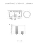 |
 |  |
 | 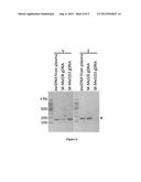 |
 |  |
 |  |
 |  |
 | 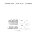 |
 |  |
 |  |
 | 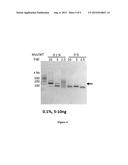 |
 |  |
 |  |
 |  |
| New patent applications in this class: | |
| Date | Title |
|---|---|
| 2022-09-08 | Shrub rose plant named 'vlr003' |
| 2022-08-25 | Cherry tree named 'v84031' |
| 2022-08-25 | Miniature rose plant named 'poulty026' |
| 2022-08-25 | Information processing system and information processing method |
| 2022-08-25 | Data reassembly method and apparatus |
| New patent applications from these inventors: | |
| Date | Title |
|---|---|
| 2017-06-22 | Use of double-stranded dna in exosomes: a novel biomarker in cancer detection |
| 2014-02-06 | Circulating exosomes as diagnostic/prognostic indicators and therapeutic targets of melanoma and other cancers |