Patent application title: METHOD FOR DETECTING DISEASE BIOMARKERS
Inventors:
Kai Tang (San Diego, CA, US)
Duangmanee Sanmun (Singapore, SG)
Suthat Fucharoen (Singapore, SG)
Hai Yang Law (Singapore, SG)
Ivy Ng (Singapore, SG)
IPC8 Class: AC12Q137FI
USPC Class:
435 23
Class name: Measuring or testing process involving enzymes or micro-organisms; composition or test strip therefore; processes of forming such composition or test strip involving hydrolase involving proteinase
Publication date: 2015-04-23
Patent application number: 20150111238
Abstract:
There is presently provided a method of detecting a bio-marker for a
genetic disease in an individual. The method involves comprising
identifying the presence of a proteolytic peptide in a polypeptide
fraction obtained from a cellular extract of cells from the individual.
The cells are cells by the disease to be diagnosed, and the proteolytic
peptide contains a mutation associated with the genetic disease or
directly related to the disease condition.Claims:
1. A method of detecting a biomarker for a hemoglobinopathy or
thalassemia genetic disease in an individual, the method comprising:
identifying the presence of a proteolytic peptide in a polypeptide
fraction, the polypeptide fraction obtained from a cellular extract of
cells of the individual, which cells are affected by the disease, the
proteolytic peptide containing a mutation associated with the genetic
disease or directly related to the disease condition, the proteolytic
peptide consisting of a sequence set forth in any one of SEQ ID NOs.
6-201.
2. The method of claim 1, wherein the mutation comprises an alteration in post-translational modification as compared to a non-disease proteolytic peptide.
3. The method of claim 2, wherein the post-translational modification comprises dimethylation of R166.
4. The method of claim 1, further comprising separating the polypeptide fraction from cellular debris in the cellular extract prior to the identifying.
5. The method of claim 1, further comprising lysing cells from the individual that are affected by the disease to obtain the cellular extract.
6. The method of claim 1, wherein the identifying comprises mass spectrometry.
7. The method of claim 6, wherein the mass spectrometry comprises MALDI-TOF, LC/MS/MS, high resolution LC/MS, MRM or SRM techniques.
8. The method of claim 1, wherein the cells affected by the disease are red blood cells.
9. The method of claim 1, further comprising lysing cells from the individual that are affected by the disease to obtain the cellular extract and separating the polypeptide fraction from cellular debris in the cellular extract prior to the identifying, wherein the mutation comprises an alteration in post-translational modification that is dimethylation of R166 and the identifying comprises mass spectrometry.
Description:
CROSS-REFERENCE TO RELATED APPLICATION
[0001] This application claims benefit of, and priority from, U.S. provisional application No. 61/491,489, filed on May 31, 2011, the contents of which are hereby incorporated herein by reference.
FIELD OF THE INVENTION
[0002] The present invention relates to methods for identifying an expressed biomarker for a genetic disease.
BACKGROUND OF THE INVENTION
[0003] Genetic testing is often used to detect genetic diseases in an individual. However, when the disease allele results from a minor mutation in the DNA, it may be more straightforward to use the expressed protein, rather than the gene, as a biomarker for disease.
[0004] With the advances of proteomic technologies, hundreds-to-thousands of candidate protein biomarkers have been identified. These markers not only provide better understanding of disease mechanisms, but also may be useful clinically as diagnostic markers.
[0005] The human plasma proteome and peptidome have been extensively exploited as providing secreted plasma proteins as protein biomarkers [1-3]. However, there are a few drawbacks to this general approach. First, not all disease-related proteins in disease cells are secreted into the bloodstream. Second, even when marker proteins or peptides are secreted into blood circulation, the existence of such secreted proteins or peptides may be difficult to confirm, given the several-order-of-magnitude higher abundance of the classical plasma proteins such as albumin and immunoglobulins. Third, many of the candidate markers discovered result from the body's response to diseases, rather than the direct cause of diseases [4], which raises the question of specificity of these candidate markers. Because of this potential lack of specificity, extensive verification processes are necessary to determine their potential clinical utility [5].
SUMMARY OF THE INVENTION
[0006] In one aspect, the invention provides a method of detecting a biomarker for a genetic disease in an individual, the method comprising: identifying the presence of a proteolytic peptide in a polypeptide fraction, the polypeptide fraction obtained from a cellular extract of cells of the individual, which cells are affected by the disease, the proteolytic peptide containing a mutation associated with the genetic disease or directly related to the disease condition.
[0007] The mutation may comprise insertion, deletion or substitution of one or more amino acids as compared to a non-disease proteolytic peptide, or may comprise an alteration in post-translational modification as compared to a non-disease proteolytic peptide.
[0008] The method may further include one or more of the following: separating the polypeptide fraction from cellular debris in the cellular extract prior to the identifying; proteolyitcally digesting the cellular extract prior to separation of the polypeptide fraction from cellular debris or the polypeptide fraction is proteolytically digested subsequent to separation from cellular debris in the cellular extract; lysing cells from the individual that are affected by the disease to obtain the cellular extract.
[0009] In the method, identifying may comprise mass spectrometry techniques, including for example MALDI-TOF, LC/MS/MS, high resolution LC/MS, MRM or SRM techniques.
[0010] In some embodiments, the genetic disease is hemoglobinopathy or thalassernia, and the cells affected by the disease may be red blood cells. The mutation may be in an a-globin chain or in a β-globin chain. In some embodiments, the proteolytic peptide may consist of a portion of SEQ ID NO. 2 or SEQ ID NO. 3, which portion includes at least some of the sequence as defined by residues 142-172 of SEQ ID NO. 2 or SEQ ID NO. 3. In some embodiments, the proteolytic peptide consists of a sequence as set forth in any one of SEQ ID NOs. 22-78. In some embodiments, the proteolytic peptide consists of portion of SEQ ID NO. 5, which portion includes residue 26 of SEQ ID NO. 5. In some embodiments, the proteolytic peptide consists of a sequence as set forth in any one of SEQ ID NOs. 93-201. In some embodiments, the proteolytic peptide consists of a sequence as set forth in any one of SEQ ID NOs. 6-21. In some embodiments, the proteolytic peptide consists of a sequence as set forth in any one of SEQ ID NOs. 79-92.
[0011] Other aspects and features of the present invention will become apparent to those of ordinary skill in the art upon review of the following description of specific embodiments of the invention in conjunction with the accompanying figures and tables.
BRIEF DESCRIPTION OF THE DRAWINGS
[0012] The figures and tables, which illustrate, by way of example only, embodiments of the present invention, are as follows.
[0013] FIG. 1 illustrates Homozygous CS (1A), Hb H CS (1B) and Hb H PS (1C) detected by MALDI-TOFMS. Monomers of globin chains were represented as average mass. Although the elongated a chain was very small it could be clearly detected by MALDI-TOFMS. The mass accuracy of the linear mode MALDI-TOF, at 0.1%, is enough to differentiate between αCS (theoretical mass 18481.2 Da) and αPS (theoretical mass 18516.2 Da).
[0014] FIG. 2 illustrates MALDI-TOF mass spectra of proteolytic peptides from different genotypes. The peptide profiles were obtained after concentrating and desalting of the hemolysates of peripheral blood samples. (A) Hb CS heterozygote (αCSα/αα, β/62 ), (B) Hb CS heterozygote with α3.7 deletion (-3.7α/αCSα, β,β), (C) Hb H-CS disease (αCSα/- -, β/β) (D) Hb CS homozygote (αCSα/αCSα, β/β) and (E) normal control.
[0015] FIG. 3 illustrates MS/MS spectra of precursor ion of 1638.76 Da and 1666.87 Da from alpha-globin CS samples. When the two peptides were subjected to collision-induced-dissociation (CID) under MALDI-TOF/TOF, peptide bonds connecting two neighboring amino acids were fragmented, generating b (N-terminal) and y (C-terminal) types of ions. Dimethylation site can be deduced from the unchanging b7 fragment (the N-terminal fragment after cleavage of the 7th peptide bond from the N-terminal, i.e. between H and R) and the mass shift of +28 Da of the b8 (the N-terminal fragment after cleavage of the 8th peptide bond from the N-terminal, i.e. between R and P), y7 (the C-terminal fragment after cleavage of the 7th peptide bond from the C-terminal, i.e. between R and H), and y8 (the C-terminal fragment after cleavage of the 8th peptide bond from the C-tenninal, i.e. between H and L) fragments.
[0016] FIG. 4 illustrates total ion chromatogram of selected Hb CS heterozygote (αCSα/αα) (A), Hb CS homozygote (αCSα/αCSα) (B) and Hb H-CS disease (αCSα/- -) (C) samples showing more proteolytic peptides were detected with increasing intensity respectively.
[0017] FIG. 5 illustrates MRM spectra obtained in the triple quadrupole mass spectrometer showing a few specific transitions selected for detection of the proteolytic fragments from the αCS allele ([SEQ ID NOs. 56, 63, 71, 75]).
[0018] FIG. 6 illustrates the interface of DeNovo Explorer software, and depicts de novo sequence and MS/MS spectrum. The peak with m/z value of 2010.0940 from αCS 156-172peptide was selected for CID fragmentation ([SEQ ID NOs. 57, 68, 202, 203]).
[0019] Table 1 lists proteolytic peptides detected from samples of nine genotypes, including heterozygous Hb CS, heterozygous Hb PS, homozygous Hb CS, HbH-PS disease, HbH-CS disease, compound heterozygous HbCS/alpha-thalassemia 2, triple heterozygous HbE/alpha-thalassemia 1 and HbCS, as well as normal alpha-globin type.
[0020] Table 2 lists the proteolytic peptides detected from heterozygous Hb CS, homozygous Hb CS, and Hb H-CS disease samples ([SEQ ID NOs. 26, 40-45, 48, 49, 52, 53, 57, 58, 60, 62, 69, 71, 72, 204-217]).
[0021] Table 3 lists proteolytic peptides carrying partial Hb CS sequences, detected using LC-MS from 14 different patient samples carrying the Hb CS allele. The results were filtered with the Trans Proteomic Pipeline with PeptideProphet at the 5% false discovery level ([SEQ ID NOs. 22-78]).
[0022] Table 4 lists Hb E proteolytic peptides found in several form of hemoglobinopathes including heterozygous Hb E, homozygous Hb E, and β-thalassemia/Hb E disease samples ([SEQ ID NOs. 93-201]).
DETAILED DESCRIPTION
[0023] Disease-causing protein mutations, if known and easily detected, may serve as definitive biomarkers for a given disease due to the specificity of the mutation for the disease. However, proteins carrying such mutations are rare and often in low abundance, making them difficult to detect.
[0024] The methods as described herein relate to direct detection of disease mutations within a protein, by detecting proteolytic fragments of the mutated proteins as the proteins are degraded within the cell. The methods relate to the hypothesis that the mutant proteins involved in disease are typically unstable within the cell, and thus tend to be degraded within the cell by endogenous proteases. Proteolytic peptides carrying the disease mutation should therefore be detectable within the cell, and could serve as potential biomarkers for disease.
[0025] Previous methods have focussed on detecting protein biomarkers that have been secreted into blood plasma. However, not all disease-related proteins expressed in cells associated with disease are secreted into the bloodstream. Some of the most specific markers may still remain in the diseased cells. As well, even when biomarker proteins or peptides containing a disease mutation leak into circulation, such proteins and peptides may be difficult to isolate given the several-order-of-magnitude higher abundance of the classical plasma proteins such as albumin and immunoglobulins.
[0026] Another drawback to detecting disease protein biomarkers in blood plasma is that many of the candidate biomarkers identified in fact result from the body's response to disease, such as inflammation, angiogenesis, fibrosis, etc., and are not related to the direct cause of a specific disease [4]. Proteins involved in a response to disease may be present for more than one type of disease or disorder. This raises the question of specificity of such candidate biomarkers in the bloodstream. Thus, the use of secreted proteins as biomarkers often requires extensive verification processes to determine the potential clinical utility of the secreted biomarker [5].
[0027] The methods described herein focus on proteins and peptides contained within cells affected by the disease that is to be screened, cells that would be expressing the mutant version of the protein involved in the relevant disease for which an individual is to be screened. Accordingly, there is presently provided a method of detecting a biomarker for a genetic disease in an individual.
[0028] As used herein, disease allele, or mutant allele refer to alleles associated with or having a mutation, the mutation resulting in, causing, or being directly associated with, a disease or disorder in an individual, and which results in an expressed protein containing a mutation. Accordingly, disease protein and mutant protein refer to a protein expressed from a disease allele that contains a mutation, which mutation results in, causes or is directly associated with the disease or disorder. The mutation may be dominant or recessive, and may occur in combination with other mutations involved in disease. The mutation may be an alteration or difference in post-translational modification of a specific protein, which differently modified protein is implicated in or associated with disease.
[0029] In contrast, a non-disease allele is an allele that is not associated with disease or disorder and which results in expression of a protein that exhibits normal protein function as is found in a healthy individual not afflicted with the disease or disorder. A non-disease protein is a protein expressed from a non-disease allele. A non-disease allele or protein does not contain the mutation that results in disease associated with or directly related to a disease allele or protein, and may be referred to as a wildtype allele or protein, or healthy allele or protein.
[0030] A disease allele or protein is considered a biomarker, in that detection of a mutation associated with disease can be used to confirm that an individual carries a mutation associated with a genetic disorder, and may be at risk of developing the disease or disorder or may have the disease or disorder. A protein biomarker is a disease protein that can be used in a diagnostic method to confirm that the individual carries at least one disease allele.
[0031] The method is performed on a sample taken from an individual to be screened for the genetic disorder. The genetic disorder may be any genetic disorder that results in expression of a mutant protein, including a mutation in primary sequence or a difference in post-translational modification, as indicated above.
[0032] For example, the genetic disorder may be a disorder that arises from a point mutation, insertion mutation or deletion mutation in a gene, resulting in a defective or altered protein, leading to disease or disorder. The point mutation may result in substitution of one amino acid for another, or may result in introduction of alteration of stop codon or exon splice site and thus produce a protein that is longer or shorter, or has deleted or inserted amino acids relative to the non-disease protein. The deletion or insertion mutations in the gene may result in a frame shift partway down a coding sequence or may result in an in-frame insertion or deletion of amino acids in the coding sequence.
[0033] The genetic disorder may be a disorder that results in a change in the post-translational modification of a protein. For example, the protein may lack a modification found on a healthy or wildtype protein, may be modified at a different position relative to a healthy or wildtype protein, or may have additional modifications not found on a healthy or wildtype protein.
[0034] The genetic mutation as described above results in expression of a disease protein. The disease protein will thus have a sequence or post-translational modification that is altered from a non-disease protein, for example as expressed from an un-mutated, non-disease allele in a healthy individual. Thus, at least some proteolytic peptide fragments of the disease protein will be distinguishable from proteolytic peptide fragments generated from a non-disease protein.
[0035] A proteolytic peptide refers to a peptide fragment of a protein resulting from digestion with one or more proteases. Thus, a proteolytic peptide consists of a sequence found within the full length protein from which the proteolytic peptide is derived. Therefore, reference to a disease peptide, proteolytic disease peptide or proteolytic peptide associated with a disease or disorder is reference to a proteolytic fragment of a disease protein that contains a mutation in the amino acid sequence, namely one or more substituted, inserted or deleted amino acid, or that contain an amino acid residue that is differently post-translationally modified, compared to proteolytic peptides generated from a non-disease protein. Similarly, reference to a non-disease peptide, wildtype peptide or proteolytic peptide from a non-disease gene is reference to a proteolytic fragment of a non-disease protein, which contains a wildtype sequence, i.e. no mutation, or contains an amino acid residue that is post-translationally modified as found in the non-disease protein.
[0036] The sample obtained from an individual contains cells affected by the disease, meaning that the cells express the mutant disease protein. Thus, where the protein is expressed in a cell-specific manner, the sample will contain the cell type in which the disease protein is expressed.
[0037] The individual is any individual that is to be diagnosed for a particular genetic disease or disorder, or to be identified as a carrier of a disease allele if the disease or disorder is associated with a recessive genetic mutation. The individual may be symptomatic or asymptomatic for the disease or disorder, and may be any individual in which the disease or disorder is suspected, a predisposition to disease or disorder is suspected, or with a family history of the disease or disorder. The individual may be suspected of being homozygous or heterozygous for a disease allele.
[0038] The method is performed on peptides obtained from cells of the individual that will express the protein associated with or directly related to the disease. The peptides may include soluble peptides found in the soluble fraction of a cell lysate (also referred to as cellular extract), or may include membrane-bound peptides by first solubilising such peptides, using routine laboratory methods such as performing a detergent extraction of proteins and peptides, such that the membrane-bound proteins become soluble in the soluble fraction of the cellular extract following treatment.
[0039] Thus, in the method, cells obtained from an individual may be lysed. The cellular extract will contain proteolytic peptides, including proteolytic non-disease peptides and may also contain proteolytic disease peptides. The cellular extract may be further fractionated, as indicated below, and the fraction or portion of a cellular extract (including the whole cellular extract if not further fractionated) that contains the proteolytic peptides is considered a polypeptide fraction of the cellular extract.
[0040] Thus, if desired, cell debris may be removed in order to obtain a polypeptide cell fraction containing soluble proteins and peptides, including subsequent to a solubilization step such as detergent treatment. Alternatively, the whole cellular extract, which will contain the polypeptide fraction along with other insoluble cell debris, may be used directly as a polypeptide fraction.
[0041] Cell lysis and optional removal of debris may be achieved using standard molecular biology laboratory techniques. For example, cells may be lysed using chemical disruption, mechanical grinding, freeze/thaw, liquid homogenization, or sonication techniques. Insoluble cell debris may be separated from the soluble polypeptide fraction by centrifugation techniques. For example, cells may be lysed by suspension in a hypotonic or other lysis buffer, which may include detergent, followed by centrifugation.
[0042] If desired, protease inhibitors may be added before, during or after cell lysis to inhibit any further degradation of peptides in the cell. Protease inhibitors, including protease inhibitor cocktails are readily commercially available, and include for example general inhibitors for inhibition of serine and/or cysteine proteaSes, including inhibitors such as PMSF, Pefabloc SC, aprotinin, leupeptin, etc. For example, a complete EDTA-free protease inhibitor cocktail tablet can be purchased from Roche Applied Science.
[0043] In addition, or alternatively, particular proteases, which may be non-targets for any protease inhibitors included, may be added before, during or after cell lysis in order to target particular peptide sequences for further proteolysis. Proteolytic digestion of polypeptides in the sample may be performed subsequent to cell lysis but before any further separation of the polypeptide fraction from insoluble cell debris, or may be performed on the polypeptide fraction obtained following removal of insoluble cell debris. Specific proteases may be added in order to produce specific desired fragments of known target proteins, including specific serine or cysteine proteases.
[0044] If desired, peptides may be separated from larger soluble proteins in the soluble polypeptide fraction, for example by using size exclusion techniques. Filtration, chromatography, ultracentrifugation, precipitation, and dialysis techniques may be used to isolate smaller molecular weight peptides away from larger proteins.
[0045] Once a sample polypeptide fraction containing soluble peptides from the cells that express the biomarker protein is obtained, particular peptides within the fraction are then detected in order to confirm the presence of a proteolytic peptide fragment of a disease protein containing the mutation.
[0046] Routine laboratory methods may be used to identify proteolytic peptide fragments.
[0047] For example, techniques including gel electrophoresis, isoelectric focusing (IEF), cation exchange HPLC, or mass spectrometry may be used.
[0048] In one embodiment of the method, mass spectrometry methods are used to identify proteolytic disease peptides derived from mutant disease proteins. Such methods are efficient, accurate methods to isolate and identify biomolecules and are well suited to separation and identification of proteolytic disease peptides having a disease mutation that may differ by a single amino acid or that may differ with respect to post-translational modifications as compared to a proteolytic non-disease peptide which may be contained in the same sample of a heterozygous individual.
[0049] Thus, techniques such as MALDI-TOF mass spectrometry, LC/MS/MS (MRM) or high resolution LC/MS may be used. Preparation of the sample fraction containing the proteolytic peptides may be done in accordance with routine methods. For example, the fraction of the sample containing the proteolytic peptides may be mixed with a UV-absorbing matrix prior to laser irradiation in a mass spectrometer or injected for LC/MS.
[0050] Techniques such as tandem mass spectroscopy (MS/MS) and liquid chromatography/mass spectroscopy (LC/MS and LC/MS/MS) can be used to obtain the sequence of individual peptides in the sample. Briefly, in LC/MS, different peptides are separated by a reverse phase column according to their hydrophobicity and sprayed into a mass spectrometer for mass measurement. In LC/MS/MS, the peptide is further fragmented in the mass spectrometer to generate fragments, and one, a few, or all of the fragments can be measured in the mass spectrometer to increase specificity.
[0051] Thus, for example, the use of selected reaction monitoring (SRM) mode and multiple reaction monitoring mode (MRM) can be performed using tandem MS methods in order to identify the sequences of the particular peptides contained within the sample.
[0052] As indicated above, mass spectroscopy methods can also be used to identify differences in post-translational modification between proteolytic peptides containing a disease mutation and non-disease peptides.
[0053] If desired, the results obtained with the sample from the individual to be diagnosed may be compared with samples containing known non-disease proteolytic peptides without any mutation and/or with samples containing known disease proteolytic peptides containing known disease mutations.
[0054] An exemplified embodiment of the method is described below, although it will be appreciated that the method is not limited to the following embodiment.
[0055] In the exemplified embodiment, the disease allele is an altered hemoglobin gene. Certain mutations within the a-hemoglobin or β-hemoglobin genes result in altered proteins that give rise to hemoglobinopathy blood disorders such as α-thalassemia, β-thalassemia, sickle cell anemia or hemolyticanemia. β-hemoglobin variants include, without limitation, Hb E, Hb S, Hb C, Hb D-Punjab or Hb 0-Arab. α-hemoglobin variants include, without limitation, Hb CS, Hb PS, Hb Queens, Hb J-Buda or Hb Q-Thailand. As will be appreciated, there are many other disorders and specific mutations that have been identified for the α-hemoglobin or β-hemoglobin genes, all of which are included within the scope of the method.
[0056] Hemoglobin disorders are most prevalent in Southeast Asia. Thalassemias are characterized by a reduction or absence of haemoglobin chain (either α or β) production.
[0057] α-Thalassemia is caused by deletional or non-deletional mutations in the α-hemoglobin gene. The non-disease (i.e. wildtype) sequence for α2-hemoglobin protein is [SEQ ID NO. 1]:
TABLE-US-00001 VLSPADKTNV KAAWGKVGAHAGEYGAEALE RMFLSFPTTK TYFPHFDLSH GSAQVKGHGK KVADALTNAV AHVDDMPNAL SALSDLHAHK LRVDPVNFKL LSHCLLVTLA AHLPAEFTPA VHASLDKFLA SVSTVLTSKYR
[0058] The Hemoglobin Constant Spring (Hb CS) genotype, the most common non-deletional α-thalassemia in Thai and Chinese, is caused by a mutation at the stop codon of the α2-hemoglobin gene, TAA→CAA, resulting in a 31-amino acid elongation of the of the α2-hemoglobin protein [6] (additional amino acids shown in boxed sequence) [SEQ ID NO. 2]:
TABLE-US-00002 VLSPADKTNVKAAWGKVGAHAGEYGAEALERMFLSFPTTK TYFPHFDLSHGSAQVKGHGKKVADALTNAVAHVDDMPNAL SALSDLHAHKLRVDPVNFKLLSHCLLVTLAAHLPAEFTPA VHASLDKFLASVSTVLTSKYRQAGASVAVPPARWASQRALL PSLHRPFLVFE
[0059] Another less common non-deletional type, Hemoglobin Pakse (Hb PS), also occurs from a different mutation at the same position on a2 hemoglobin allele, TAA→TAT [6, 7], again resulting in a 31-amino acid elongation of the of the α2-hemoglobin protein (additional amino acids shown in boxed sequence) [SEQ ID NO. 3]:
TABLE-US-00003 VLSPADKTNVKAAWGKVGAHAGEYGAEALERMFLSFPTTK TYFPHFDLSHGSAQVKGHGKKVADALTNAVAHVDDMPNAL SALSDLHAHKLRVDPVNFKLLSHCLLVTLAAHLPAEFTPA VHASLDKFLASVSTVLTSKYRYAGASVAVPPARWASQRALL PSLHRPFLVFE
[0060] In comparison with Hb H-CS disease, the clinical and hematological data in patients with Hb-PS has no significant difference [8, 9]. These α-hemoglobin elongation mutations can interact with α-thalassemia 1 causing Hb H disease, especially in Thai population [7, 8].
[0061] Molecular characterization by using mismatched polymerase chain reaction-restriction fragment length polymorphism (PCR-RFLP) has previously been applied in screening programs [9] to identify Hb CS and PS mutations. An initial diagnosis of Hb CS and PS has also been previously qualitatively performed using ion exchange high performance liquid chromatography (IE-HPLC; Bio-Rad Variant) [9, 10]. However, it is difficult to detect the peak chromatogram in its heterozygous state because of the low abnormal Hb level due to the instability of Hb CS and Hb PS-globin mRNAs and proteins. The appearance of Hb CS or Hb PS peak can lead to the diagnosis of either homozygous Hb CS or severe a-thalassemia intermediate phenotype for Hb H-CS or Hb H-PS disease [11].
[0062] Initially, reports suggested an unexpected increase of α/β ratio (>1) in Hb CS heterozygotes and homozygotes [22]. It was later demonstrated that there was a deficit of a chain production in a short period of in vitro incubation and the excess p chain was gradually lost with a longer period of 3-hour incubation, so that the α/β globin chain synthesis ratio appeared to be more than 1[23]. The erythroblastic marrow cells can degrade unstable protein quickly but the unstable globin chain that precipitate in mature erythrocytic cells will also be destroyed, leading to a greater amount of circulating polypeptides of hemoglobin [24]. Additionally, minute amounts of the αCS hemoglobin may still remain in peripheral blood as it can be detected in homozygous αCS by HPLC.
[0063] Therefore, the present method was performed using red blood cells from individuals, on the basis that minor globin proteins (intact form of Hb variants) could be detected directly by mass spectrometry without HPLC separation, as described in Example 1 below. Moreover, due to proteolysis of the elongated hemoglobin chain, peptide fragments may be found as well by mass spectrometric analysis.
[0064] Thus, the methods as described herein can be used to directly detect intact HbαCS and αPS peptide biomarkers from peripheral blood samples, for example by using matrix assisted laser desorption/ionization time-of-flight(MALDI-TOF) mass spectrometry as described in Example 1 below. Furthermore, proteolytic peptide fragments can be found in red cellular extract and sequenced using tandem mass spectrometry. The proteolytic disease peptides have been identified to originate from the elongated C-terminal sequence, thus including peptides having any portion of [SEQ ID NO. 2] or [SEQ ID NO. 3] that encompasses at least part of the extended C-terminal region of the α2-globin gene, i.e. the part defined by amino acids 142-172 of [SEQ ID NO. 2] or [SEQ ID NO. 3].
[0065] In the β-hemoglobin gene, the non-disease sequence is as follows [SEQ ID NO. 4]:
TABLE-US-00004 VHFTAEEKAAVTSLWSKMNVEEAGGEALGRLLVVYPWTQRFFDSFG NLSSPSAILGNPKVKAHGKKVLTSFGDAIKNMDNLKPAFAKLSELH CDKLHVDENFKLLGNVMVIILATHFGKEFTPEVQAAWQKLVSAVAI ALAHKYH
[0066] The Hb E mutation results in single amino acid change E→K at position 26 in the β-hemoglobin protein (the substituted amino acid is indicated in bold and underline) [SEQ ID NO. 5]:
TABLE-US-00005 VHFTAEEKAAVTSLWSKMNVEEAGGKALGRLLVVYPWTQRFFDSFG NLSSPSAILGNPKVKAHGKKVLTSFGDAIKNMDNLKPAFAKLSELH CDKLHVDPENFKLLGNVMVIILATHFGKEFTPEVQAAWQKLVSAVA IALAHKYH
[0067] Heterozygous Hb E and homozygous Hb E are asymptomatic, but the compound heterozygote with Hb E and β-thalassemia usually exhibits a diseased phenotype. Therefore, prevention and control of thalassemias should be considered [37], since Hb E is an abnormal hemoglobin with reduced synthesis. Instability of Hb E was also observed in Hb E patients [38]. The dichlorophenolindophenol (DCIP) test was used to detect for unstable hemoglobin such as Hb E. The DCIP test is a rapid and sensitive screening technique. Thus, the diagnostic methods as described herein can be used as an accurate approach to screening and diagnosis for Hb E patients or couples at risk.
[0068] The proteolytic disease peptides for Hb E genotype have been identified to include the mutation of amino acid 26, thus including peptides having any portion of [SEQ ID NO. 5] that encompasses residue 26.
[0069] Thus, in the exemplified embodiment, the disease or disorder to be detected may be a hemoglobinopathy, including for example α-thalassemia, β-thalassemia, sickle cell anemia or hemolyticanemia. In one embodiment, the disease is α-thalassemia. In one embodiment, the disease is β-thalassemia.
[0070] The sample may be a peripheral blood sample containing red blood cells from an individual.
[0071] The disease allele may be the α-hemoglobin gene or the β-hemoglobin gene. For example, the disease allele may be an α-hemoglobin gene containing the Hb CS, Hb PS, Hb Queens, Hb J-Buda or Hb Q-Thailand mutation. For example, the disease allele may be a β-hemoglobin gene containing the Hb E, Hb S, Hb C, Hb D-Punjab or Hb O-Arab mutation.
[0072] In some embodiments, the disease or disorder is α-thalassemia.
[0073] In some embodiments, the proteolytic peptide may consist of one of the following sequences:
TABLE-US-00006 [SEQ ID NO. 6] VHLTPEEKSAVTALW, [SEQ ID NO. 7] TALWGKVNVDEVGGEALG, [SEQ ID NO. 8] ALWGKVNVDEVGGEALG, [SEQ ID NO. 9] WGKVNVDEVGGEAL, [SEQ ID NO. 10] GKVNVDEVGGEAL, [SEQ ID NO. 11] VDEVGGEALGRLLV, [SEQ ID NO. 12] LVVYPWTQRF, [SEQ ID NO. 13] RFFESFGDLSTPDAV, [SEQ ID NO. 14] FFESFGDLSTPDAV, [SEQ ID NO. 15] AFSDGLAHLDNLKGTFAT, [SEQ ID NO. 16] AFSDGLAHLDNLKGTFA, [SEQ ID NO. 17] SDGLAHLDNLKGTF, [SEQ ID NO. 18] DGLAHLDNLKGTFA, [SEQ ID NO. 19] DGLAHLDNLKGTFATL, [SEQ ID NO. 20] YQKVVAGVANALAHKYH or [SEQ ID NO. 21] VAGVANALAHKYH, [SEQ ID NO. 22] GASVAVPPARWASQ, [SEQ ID NO. 23] GASVAVPPARWAS, [SEQ ID NO. 24] GASVAVPPARWA, [SEQ ID NO. 25] GASVAVPPARW, [SEQ ID NO. 26] ASVAVPPARWASQ, [SEQ ID NO. 27] SVAVPPARWASQRALLPS, [SEQ ID NO. 28] SVAVPPARWASQ, [SEQ ID NO. 29] SVAVPPARWAS, [SEQ ID NO. 30] SVAVPPARW, [SEQ ID NO. 31] VAVPPARWASQ, [SEQ ID NO. 32] AVPPARWASQRALLPSLHRPFLVFE, [SEQ ID NO. 33] AVPPARWASQRALLPSL, [SEQ ID NO. 34] AVPPARWASQRALLPS, [SEQ ID NO. 35] VPPARWASQRALLPSL, [SEQ ID NO. 36] VPPARWASQRALLPS, [SEQ ID NO. 37] VPPARWASQR, [SEQ ID NO. 38] VPPARWASQ, [SEQ ID NO. 39] VPPARWAS, [SEQ ID NO. 40] ASQRALLPSLHRPFLVFE, [SEQ ID NO. 41] ASQRALLPSLHRPFL, [SEQ ID NO. 42] ASQRALLPSLHRPF, [SEQ ID NO. 43] SQRALLPSLHRPFLVFE, [SEQ ID NO. 44] SQRALLPSLHRPFL, [SEQ ID NO. 45] SQRALLPSLHRPF, [SEQ ID NO. 46] SQRALLPSLHRP, [SEQ ID NO. 47] SQRALLPSL, [SEQ ID NO. 48] QRALLPSLHRPFLVFE, [SEQ ID NO. 49] QRALLPSLHRPFL, [SEQ ID NO. 50] QRALLPSLHRPF, [SEQ ID NO. 51] QRALLPSLHRP, [SEQ ID NO. 52] RALLPSLHRPFLVFE, [SEQ ID NO. 53] RALLPSLHRPFL, [SEQ ID NO. 54] RALLPSLHRPF, [SEQ ID NO. 55] RALLPSLHRP, [SEQ ID NO. 56] ALLPSLHRPFLVFE, [SEQ ID NO. 57] ALLPSLHRPFL, [SEQ ID NO. 58] ALLPSLHRPF; [SEQ ID NO. 59] ALLPSLHRP, [SEQ ID NO. 60] LLPSLHRPFLVFE, [SEQ ID NO. 61] LLPSLHRPFLVF, [SEQ ID NO. 62] LLPSLHRPF, [SEQ ID NO. 63] LPSLHRPFLVFE, [SEQ ID NO. 64] LPSLHRPFLVF, [SEQ ID NO. 65] LPSLHRPFLV, [SEQ ID NO. 66] LPSLHRPFL, [SEQ ID NO. 67] LPSLHRPF, [SEQ ID NO. 68] LPSLHRP, [SEQ ID NO. 69] PSLHRPFLVFE, [SEQ ID NO. 70] PSLHRPF, [SEQ ID NO. 71] SLHRPFLVFE, [SEQ ID NO. 72] LHRPFLVFE, [SEQ ID NO. 73] HRPFLVFE, [SEQ ID NO. 74] HRPFLVF, [SEQ ID NO. 75] RPFLVFE, [SEQ ID NO. 76] SVAVPPARWASQRALLPSLHRPFLVFE, [SEQ ID NO. 77] PARWASQR, or [SEQ ID NO. 78] ALLPSLHR.
[0074] In some embodiments, the disease or disorder is β-thalassemia.
[0075] In some embodiments, the proteolytic peptide may consist of one of the following sequences:
TABLE-US-00007 [SEQ ID NO. 79] VLSPADKTNV, [SEQ ID NO. 80] VLSPADKTNVKAAWGKV, [SEQ ID NO. 81] AAWGKVGAHAGEYGAEALE, [SEQ ID NO. 82] GKVGAHAGEYGAEALERM, [SEQ ID NO. 83] AHAGEYGAEALE, [SEQ ID NO. 84] TYFPHFDLSHGSAQV, [SEQ ID NO. 85] ALTNAVAHVDDMPN, [SEQ ID NO. 86] VDDMPNALSAL, [SEQ ID NO. 87] TLAAHLPAEFTPAVH, [SEQ ID NO. 88] LAAHLPAEFTPAVH, [SEQ ID NO. 89] SLDKFLASVSTVLTSKYR, [SEQ ID NO. 90] DKFLASVSTVLTSKYR, [SEQ ID NO. 91] KFLASVSTVLTSKYR or [SEQ ID NO. 92] ASVSTVLTSKYR, [SEQ ID NO. 93] VHLTPEEKSAVTALWGKVNVDEVGGKALGRLLVVYPWTQRF, [SEQ ID NO. 94] VHLTPEEKSAVTALWGKVNVDEVGGKALGRLLVVYPWTQ, [SEQ ID NO. 95] VHLTPEEKSAVTALWGKVNVDEVGGKALGRLLVV, [SEQ ID NO. 96] VHLTPEEKSAVTALWGKVNVDEVGGKALGRLLV, [SEQ ID NO. 97] VHLTPEEKSAVTALWGKVNVDEVGGKALGRLL, [SEQ ID NO. 98] VHLTPEEKSAVTALWGKVNVDEVGGKALGRL, [SEQ ID NO. 99] VHLTPEEKSAVTALWGKVNVDEVGGKALG, [SEQ ID NO. 100] VHLTPEEKSAVTALWGKVNVDEVGGKA, [SEQ ID NO. 101] LTPEEKSAVTALWGKVNVDEVGGKALG, [SEQ ID NO. 102] TPEEKSAVTALWGKVNVDEVGGKALGRLLV, [SEQ ID NO. 103] KSAVTALWGKVNVDEVGGKALGR, [SEQ ID NO. 104] KSAVTALWGKVNVDEVGGKALG, [SEQ ID NO. 105] SAVTALWGKVNVDEVGGKALGRLLV, [SEQ ID NO. 106] SAVTALWGKVNVDEVGGKALG, [SEQ ID NO. 107] TALWGKVNVDEVGGKALGRLLVVYPWTQRF, [SEQ ID NO. 108] TALWGKVNVDEVGGKALGRLLVVYPWTQ, [SEQ ID NO. 109] TALWGKVNVDEVGGKALGRLLVVYP, [SEQ ID NO. 110] TALWGKVNVDEVGGKALGRLLVV, [SEQ ID NO. 111] TALWGKVNVDEVGGKALGRLLV, [SEQ ID NO. 112] TALWGKVNVDEVGGKALGRLL, [SEQ ID NO. 113] TALWGKVNVDEVGGKALGRL, [SEQ ID NO. 114] TALWGKVNVDEVGGKALGR, [SEQ ID NO. 115] TALWGKVNVDEVGGKALG, [SEQ ID NO. 116] TALWGKVNVDEVGGKAL, [SEQ ID NO. 117] TALWGKVNVDEVGGKA, [SEQ ID NO. 118] TALWGKVNVDEVGGK, [SEQ ID NO. 119] ALWGKVNVDEVGGKALGRLLV, [SEQ ID NO. 120] ALWGKVNVDEVGGKALGRLL, [SEQ ID NO. 121] ALWGKVNVDEVGGKALGR, [SEQ ID NO. 122] ALWGKVNVDEVGGKALG, [SEQ ID NO. 123] ALWGKVNVDEVGGKAL, [SEQ ID NO. 124] ALWGKVNVDEVGGKA, [SEQ ID NO. 125] ALWGKVNVDEVGGK, [SEQ ID NO. 126] LWGKVNVDEVGGKALGRLLVVYPWTQRF, [SEQ ID NO. 127] LWGKVNVDEVGGKALGRLLVVYPWTQ, [SEQ ID NO. 128] LWGKVNVDEVGGKALGRLLVVYP, [SEQ ID NO. 129] LWGKVNVDEVGGKALGRLLV, [SEQ ID NO. 130] LWGKVNVDEVGGKALGRL, [SEQ ID NO. 131] LWGKVNVDEVGGKALGR, [SEQ ID NO. 132] LWGKVNVDEVGGKALG, [SEQ ID NO. 133] LWGKVNVDEVGGKAL, [SEQ ID NO. 134] LWGKVNVDEVGGK, [SEQ ID NO. 135] WGKVNVDEVGGKALGRLLVVYPWTQ, [SEQ ID NO. 136] WGKVNVDEVGGKALGRLLVVYP, [SEQ ID NO. 137] WGKVNVDEVGGKALGRLLV, [SEQ ID NO. 138] WGKVNVDEVGGKALGRLL, [SEQ ID NO. 139] WGKVNVDEVGGKALGRL, [SEQ ID NO. 140] WGKVNVDEVGGKALGR, [SEQ ID NO. 141] WGKVNVDEVGGKALG, [SEQ ID NO. 142] WGKVNVDEVGGKAL, [SEQ ID NO. 143] WGKVNVDEVGGK, [SEQ ID NO. 144] GKVNVDEVGGKALGRLLVVYPWTQ, [SEQ ID NO. 145] GKVNVDEVGGKALGRLLVVYP, [SEQ ID NO. 146] GKVNVDEVGGKALGRLLV, [SEQ ID NO. 147] GKVNVDEVGGKALGRLL, [SEQ ID NO. 148] GKVNVDEVGGKALGRL, [SEQ ID NO. 149] GKVNVDEVGGKALGR, [SEQ ID NO. 150] GKVNVDEVGGKALG, [SEQ ID NO. 151] GKVNVDEVGGKAL, [SEQ ID NO. 152] GKVNVDEVGGKA, [SEQ ID NO. 153] GKVNVDEVGGK, [SEQ ID NO. 154] KVNVDEVGGKALGRLLV, [SEQ ID NO. 155] KVNVDEVGGKALGRLL, [SEQ ID NO. 156] KVNVDEVGGKALGRL, [SEQ ID NO. 157] KVNVDEVGGKALGR, [SEQ ID NO. 158] KVNVDEVGGKALG, [SEQ ID NO. 159] KVNVDEVGGKAL, [SEQ ID NO. 160] KVNVDEVGGKA, [SEQ ID NO. 161] KVNVDEVGGK,
[SEQ ID NO. 162] VNVDEVGGKALGRLLVVYPWTQ, [SEQ ID NO. 163] VNVDEVGGKALGRLLVVYP, [SEQ ID NO. 164] VNVDEVGGKALGRLLV, [SEQ ID NO. 165] VNVDEVGGKALGRL, [SEQ ID NO. 166] VNVDEVGGKALGR, [SEQ ID NO. 167] VNVDEVGGKALG, [SEQ ID NO. 168] VNVDEVGGKAL, [SEQ ID NO. 169] VNVDEVGGKA, [SEQ ID NO. 170] VNVDEVGGK, [SEQ ID NO. 171] NVDEVGGKALGRLLVVYPWTQRF, [SEQ ID NO. 172] NVDEVGGKALGRLLVVYPWTQ, [SEQ ID NO. 173] NVDEVGGKALGRLLV, [SEQ ID NO. 174] NVDEVGGKALGRLL, [SEQ ID NO. 175] NVDEVGGKALGR, [SEQ ID NO. 176] NVDEVGGKALG, [SEQ ID NO. 177] NVDEVGGKAL, [SEQ ID NO. 178] VDEVGGKALGRLLVVYPWTQRF, [SEQ ID NO. 179] VDEVGGKALGRLLVVYPWTQ, [SEQ ID NO. 180] VDEVGGKALGRLLV, [SEQ ID NO. 181] VDEVGGKALGRLL, [SEQ ID NO. 182] VDEVGGKALGR, [SEQ ID NO. 183] VDEVGGKALG, [SEQ ID NO. 184] VDEVGGKAL, [SEQ ID NO. 185] VDEVGGKA, [SEQ ID NO. 186] DEVGGKALGRLLVVYPWTQRF, [SEQ ID NO. 187] DEVGGKALGRLLVVYPWTQ, [SEQ ID NO. 188] DEVGGKALGRLLV, [SEQ ID NO. 189] DEVGGKALGRLL, [SEQ ID NO. 190] DEVGGKALGR, [SEQ ID NO. 191] EVGGKALGRLLVVYPWTQRF, [SEQ ID NO. 192] EVGGKALGRLLVVYPWTQ, [SEQ ID NO. 193] EVGGKALGRLLV, [SEQ ID NO. 194] EVGGKALGRLL, [SEQ ID NO. 195] EVGGKALGR, [SEQ ID NO. 196] VGGKALG, [SEQ ID NO. 197] GGKALGRLLVVYPWTQRF, [SEQ ID NO. 198] GGKALGRLLVVYPWTQ, [SEQ ID NO. 199] GGKALGRLLVVY, [SEQ ID NO. 200] GGKALGRLLV, or [SEQ ID NO. 201] GGKALGRLL.
[0076] Where the disease allele is an α-hemoglobin gene containing the Hb CS or Hb PS mutation, the proteolytic disease peptide that is used as a biomarker may be any peptide consisting of one of the peptide sequences set out in Table 3.
[0077] Where the disease allele is a β-hemoglobin gene containing the Hb E mutation, the disease proteolytic peptide that is used as a biomarker may be any peptide consisting of one of the peptide sequences set out in Table 4.
[0078] The present methods and uses are further exemplified by way of the following non-limiting examples.
EXAMPLES
Example 1
[0079] As an example disease, the method was tested in α-thalassemia patients with stop-codon mutations. The mutations result in elongated α-globin that is longer than the α-globin found in a healthy individual not carrying a disease allele, and thus would result in unique, proteolytic peptides specific to the mutant disease allele. In the results set out below, several peptides were identified that matched with the C-terminal fragments of the elongated α-globin, as predicted. The mutations identified are those that have been identified in peripheral blood carrying the Hb CS (Hb Constant Spring) or PS (Pakse) allele, including heterozygotes and CS double heterozygotes with HbE.
[0080] Materials and Methods
[0081] Blood sample collection: Peripheral blood samples in EDTA were collected from Nalchonpathom hospital of Mahidol University in Thailand, and KK Women's and Children's Hospital (KKH) in Singapore. Ethical clearances including informed consent were approved by the Institutional Review Board of Mahidol University, KKH and Nanyang Technological University. Whole blood, washed (with normal saline) and unwashed packed red cells were processed between 6 hrs and a few days in various conditions with and without adding a cocktail of protease inhibitors. All blood samples were stored at -80° C.
[0082] MALDI-TOFMS analysis: One microliter of whole blood was diluted to 500 μl with 50% acetonitrile/0.1% trifluoroacetic acid. About 0.3 μl of the diluted blood was spotted onto a MALDI target, immediately mixed with an equal volume of sinapinic acid (MALDI grade, Sigma-Aldrich, Steinheim, Switzerland) solution (8 mg/ml in 50% acetonitrile/0.1% trifluoroacetic acid) and let dry in ambient temperature. The crystallized sample was introduced into a MALDI-TOF/TOF mass spectrometer (4800, Applied Biosystems, California, USA). Intact globin chains were detected using the linear mode of the TOF/TOF, which was calibrated using a mixture of standard proteins in a neighbouring spot.
[0083] Peptide extraction for MALDI: Tris powder (Sigma-Aldrich, Steinem, Switzerland), hydrochloric acid (Merck, Darmstadt, Germany) and cocktail protease inhibitors (Roche Diagnostics GmbH, Roche Applied Science, Mannheim, Germany) were used to prepare 50 mMTris-HCl buffer in 18.2 MΩ milliQ water, pH 7.8. Twenty microliters of frozen blood were dissolved and diluted 10 times in the Tris-HCl buffer. Diluted protein was further denatured in equal volume of 50% acetonitrile with 0.2% formic acid (Merk, Damstadt, Germany). To remove cell debris, hemolysate was spun down with 10,000 g for 7 minutes and only the supernatant was collected. The supernatant containing peptides and proteins was further separated using a 10 kDa molecular weight cut-off (MWCO) centrifugal filter device (Microcon, Millipore Corporation, Bedford, USA). The eluent that passed through the Microcon spin column was collected and peptides were concentrated and desalted using a reverse-phase C18 ZipTip (Millipore Corporation, Billerica, USA) and eluted with 2.5 μl of 70% acetonitrile and 0.1% TFA.
[0084] MALDI-MS and MS/MS analysis for peptide identification: Purified peptides (0.3 μl) were spotted onto the MALDI plate, followed by an equal volume of 8 mg/mL α-cyano-4-hydroxycinnaamic acid (MALDI grade, Sigma-Aldrich, Steinheim, Switzerland) matrix dissolved in 50% acetronitile/0.1% trifluoroacetic acid. The plate was let dry in ambient environment and the crystallized samples were analyzed in a MALDI-TOF/TOF mass spectrometer (4800, Applied Biosystems, California, USA) using reflector mode for positive ions. MS spectra were generated and precursor ions were chosen to be fragmented in tandem MS by using collision-induced dissociation (CID). After obtaining tandem mass spectra, a de novo sequencing software, DeNovo Explorer (Applied Biosystems), was used to generate a list of candidate peptide sequences, which were automatically submitted for protein identification using ProBLAST database search.
[0085] Peptide extraction for LC/MS: 50 mM Tris-HCl buffer in the presence of cocktail protease inhibitors, pH 7.8 (Roche Diagnostics GmbH, Mannheim, Germany), was used to lyse red blood cells at a 1:1 proportion. 200 microliters of hemolysate was further diluted 2 times in the 50% acetonitrile and 0.2% formic acid buffer (Merk, Darmstadt, Gennany). Diluted blood was then spun down in order to remove cell debris, with 10,000 g for 5 minutes and the supernatant was filtrated using the Microcon 10 kDa molecular weight cut-off centrifugal filter devices. The eluent that passed through the spin column was collected and peptides were then concentrated and desalted using a reverse-phase C18 ZipTip and eluted with 5 μl of 70% acetonitrile and 0.1% trifluoroacetic acid. Purified peptides were dried using a speed-vacuum concentrator (Eppendorf 5301, Hamburg, Germany) and reconstituted in 20 μl of 2% acetonitrile and 0.1% formic acid before submitting to LC-MS/MS and MRM-LC/MS.
[0086] LC/MS and MS/MS: A nano LC system (LC Packings, Amsterdam, The Netherlands) was used for LC/MS. Purified peptide samples were injected onto a reverse phase column using an autosampler. The injection volume was 1 μl . A nano column (Integrafrit, New Objective, Woburn Mass., USA, 75 μm ID, 360 μm OD, 10 cm) self-packed with 4 μm C12 reverse phase particles (Jupiter Proteo, Phenomenex, Torrance, Calif., USA) was used for LC separation. A 50-min gradient with mobile phases A (2% acetonitrile, 0.1% formic acid) and B (98% acetonitrile, 0.1% formic acid) at a flow rate of 300 nl/min was used to elute peptides. The MS and MS/MS spectra were acquired with an LTQ-Orbitrap (Thermo, Bremen, Germany). The results were searched with UniProt human protein database with added Hb CS and Hb PS sequences using the Sequest search engine (Bioworks 3.3, Thermo). Key search parameters were: enzyme specificity=none, MS accuracy=20 ppm, MS/MS accuracy=0.5 Da. Post-search filtering was used with the following setting: different peptides, with peptide probability >0.5.
[0087] Multiple reaction monitoring (MRM): Selected peptide precursors and fragments were monitored using an LTQ linear ion trap. SRM mode was selected for precursor/fragment transition monitoring, with mass window of 1 Da m/z (±0.5 Da). LC gradient was 3.2% acetonitrile from 0 to 5 minutes, 3.2% acetonitrile to 32% acetonitrile from 5 to 15 minutes, followed by washing at 90% acetonitrile for 10 minutes and re-equilibration of 3.2% acetonitrile for 15 minutes. The flow rate was 4 μl/min, using Dionex3000 RSLC with a 5 μm C18 column (Michrom Biosciences, 0.1 mm×15 mm) and a Michrom Captive Spray ESI source.
[0088] Results
[0089] Determination of minor globin molecular weight: Detection of the minor elongated α-globin chain by direct MALDI-TOF measurement of diluted blood samples can be achieved in homozygous Hb CS (FIG. 1A), Hb H-CS (FIG. 1B), and Hb H-PS (FIG. 1C) with molecular mass around 18481 kDa for αCS and 18516 Da for αPS, respectively. Homozygous Hb PS was not tested as this genotype was not available. All samples tested by MALDI are listed in Table 1. In heterozygous Hb CS, the elongated α-globin chain was marginally detected. This is expected as heterozygotes for this mutation have only ˜1% Hb CS in their erythrocytes.
[0090] MALDI-TOF MS/MS for identification of selected peptide sequences: In order to find biomarkers that are specific to disease genotypes, we tried to look for proteolytic peptides in red blood cells. Intact proteins were removed by using a 10-kDa molecular weight cut-off (MWCO) membrane. The filtrate containing peptides was desalted and concentrated with a C18 ZipTip and then subject to MALDI-TOFMS analysis. Normal donor samples (data not shown) were tested as controls but few peptides could be found and they did not match with the masses from αCS protein fragments. In patient samples with abnormal hemoglobin variant of Hb CS or PS, peptides were consistently observed with [M+H].sup.+ values of 1638.7 Da or 2010.1 Da among others (FIG. 2 and Table 1). The 2010.1 Da peak was usually observed in freshly collected samples, mostly with added protease inhibitors. The sequences of these peptides were identified in MS/MS spectra and matched to the C-terminal end of the elongated α-globin chain αCS159-172 and αCS156-172, respectively (FIG. 3A and FIG. 6). The dominant fragment (1881.08 Da) in the MS/MS spectra of 2010.10 Da (FIG. 6) was identified to be b16+H2O. Such type of CID fragmentation (bn-1+H2O) is often observed for peptides with a basic amino acid residual at any non-C-terminal position [25].
[0091] Identification of modified peptides: The elongated part of αCS globin was suspected to be partially modified since two fragments showed the same sequence with different m/z values 1638.74 Da→4 1666.77 Da and 1454.60 Da→1482.67 Da, respectively. As shown in FIG. 3B, an addition of 28 Da could be detected on arginine at amino acid position 166 (159ALLPSLHRPFLVFE [SEQ ID NO. 56]). This modification could be dimethylation of arginine. A few other peptides identified also include this position although not all of them were methylated (1044.5 Da,αCS165-172, 1567.7 Da,αCS160-172; and 2010.1 Da,αCS156-172).
[0092] Summary of samples tested by MALDI-MS: All samples tested are listed in Table 1. Signature peptides of αCS degradation products were identified with MS/MS as follows: 907.4 Da (αCS166-172), 1044.5 Da (αCS165-172), 1244.6 Da (αCS163-172), 1263.7 Da (αCS159-169), 1454.7 Da (αCS161-172), 1482.7 Da (αCS161-172dimethylated),1567.8 Da (αCS160-172), 1638.7 Da (αCS159-172), 1666.9 Da (αCS159-172 dimethylated), 1794.8 Da (αCS158-172), 1922.9 Da (αCS157-172) and 2010.1 Da (αCS156-172). Similar fragments were also found in αPS samples. As the only amino acid difference between αCS and αPS is at α142 (Q142Y), proteolytic fragments not including this particular position are identical for both αCS and αPS.
[0093] Identification of proteolytic peptides with LC/MS/MS: In order to identify more proteolytic peptides and possibly peptides from proteins other than hemoglobin, nano-flow LC/MS was used to separate the purified peptide mixture. Representative total ion chromatograms are shown in FIG. 4 for αCS heterozygote, homozygote and Hb H CS genotypes respectively. These results indicate that increasing peptide fragments were detected in homozygous and Hb H CS samples respectively when compared with CS trait samples. This may represent the severity of the disease. Corresponding database search results are listed in Table 2. As expected, proteolytic peptides from the elongated a globin chain contributed to all the peptides used to identify the Hb CS protein. No peptides from other parts of the α globin were detected, indicating that the proteases in the red blood cells were targeting the elongated portion of the Hb CS specifically. Hemoglobin β chain fragments were also detected extensively and their sequences matched to different parts of the β globin chain. This observation supports the assumption that in α-thalassemia, excess β globin chain are degraded preferentially as a body's response to the disease by attempting to restore the 1:1 ratio of α/β. Interestingly, degradation of heat shock protein beta-1 and glycophorin-C was exclusively at the C-terminal end in all three genotypes, similarly to that of the Hb CS. Other fragments observed came from erythrocyte proteins that may related to the disease in someway. Proteolytic peptides carrying partial Hb CS sequences were detected using LC/MS from 14 different patient samples carrying the Hb CS allele. The results were filtered with the Trans Proteomic Pipeline with Peptide Prophet at the 5% false discovery level and summarized in Table 3.
[0094] Multiple reactionmonitoring of αCS proteolytic peptides: From the LC/MSMS data obtained above, selected precursor-fragment pairs were chosen for MRM using a linear ion trap mass spectrometer running at high LC flow rate. Representative spectra are shown in FIG. 5. It is a proof-of-principle demonstration that these specific precursor-fragment pairs can be monitored in a high throughput fashion, similar to neonatal metabolic screening. And it can be used for large scale validation of these selected candidate markers.
[0095] Discussion
[0096] The common forms of α-thalassemia in Southeast Asia region are Hb H disease and Hb Bart's hydropsfetalis. The affected children who are compound heterozygous for non-deletional mutations, especially Hb CS and Hb PS, and α-thalassemia 1 (2-gene deletion) usually result in a more severe phenotype than the classical Hb H disease due to 3 gene deletion (α-thalassemia 1/α-thalassemia 2). Furthermore, Bart's hydropsfetalis syndrome can occur even in homozygosity of Hb CS [26]. It is therefore very desirable for diagnostic laboratories to have a simple and standardized test for high-throughput screening. Traditionally, when Hb stability (unstable Hb variant, unstable Hb tetramer) is suspected from clinical symptoms, special tests such as Heinz body preparation, heat stability and erythrocyte inclusion bodies staining can be used for screening. However, these procedures are less sensitive and more labour-intensive, leading to misdiagnosis [27, 28]. On the other hand, a validated biomarker specific for certain disease alleles hold promise for high-throughput screening before selected individuals are identified for detailed DNA sequence analysis.
[0097] Proteolytic peptides identified in this study have the potential to serve as valuable biomarkers. Since these peptides originated from the elongated portion of the α-globin chain, they are specific for stop codon mutations. Such biomarkers can be screened the same way as metabolic profiling for newborns using selected reaction monitoring (SRM). The proteolytic peptide makers identified in this study do not distinguish between Hb CS and Hb PS since the peptide sequences do not cover the stop codon position. To confirm the genotype, DNA analysis can be performed. Direct measurement of protein masses by MS provides another alternative. The mass accuracy of simple MALDI-MS in linear TOF mode is sufficient to distinguish between Hb CS (UAA142CAA, Gln) and Hb PS (UAA142UAU/UAC, Tyr). Other types of chain-termination mutations such as Hb Koya Dora (UAA142UCA, Ser) and the predicted one (UAA142UAU, Leu), can also be uniquely identified by direct mass measurement. The remaining two mutations, Hb Icaria(UAA142AAA, Lys) and Hb Seal Rock (UAA142GAA, Glu) may be differentiated from Hb CS by high resolution ESI-FTICR-MS with electron capture dissociation (ECD) capability, or by an endoproteinase (such as trypsin, Lys-C or Glu-C) digest followed by peptide mass fingerprinting.
[0098] The same signature peptides may be observed in other more complex genotypes, such as Hb CS heterozygote combined with P-thalassemia, which usually interacts with the common β-chain variant Hb E. It should be pointed out that misdiagnosis of Hb CS in individuals with Hb E can happen in routine hematological analysis. Some of the double heterozygous Hb CS/Hb E was detected by DNA analysis only because the hematological parameter could vary greatly in this group [29].
[0099] Hb H-CS disease is associated with increased amounts of Hb Bart's (γ4) and Hb H inclusion bodies (β4) [30]. Moreover, precipitation of unpaired β-globin chain was found in the erythropoietic cells from the bone marrow of Hb H patients [31]. A few studies reported that accumulation of the β-globin chain and αCS-globin chain was mostly observed at the cytoskeleton part of the ghost red cell membrane [32, 33]. However, the more complex procedures of extracting the membrane associated proteins and peptides may undermine the purpose for high-throughput screening. Proteolytic peptides from the excess β-globin were identified even in peripheral blood, indicating the excess β-globin is also degraded by the endogenous proteases.
[0100] The proteolytic peptides detected in all the samples containing the αCS allele originate from the elongated portion of the α-globin chain. There seems to be no cleavage in the normal sequence (α-globin 1-141). The shortened αCS chains were detected by Weatherall and Clegg using starch-gel electrophoresis three decades ago [34]. Beside the full length αCS, shortened αCS chains were detected as CS2 (1-169) and CS3 (1-154). In the experiments described herein, the C-terminal tri-peptide cleavage resulted in CS2 was not detected. The small peptide αCS170-172 could be lost during the Ziptip desalting procedure. The larger proteolytic peptide αCS155-172 could be further degraded from its N-terminal to yield smaller peptides detected in this report.
[0101] Amino acids in human proteins can be covalently modified by enzyme-catalyzed in vivo or chemical induced in vitro reactions. Modifications to arginine such as mono- and di-methylation might be related to pathophysiology of the thalassemic patient. In addition, arginine methylation is a common post-translational modification since protein arginine methyltransferase (PRMT) is a major component in eukaryotic cells. The role of arginine methylation has been implicated in RNA processing, transcriptional regulation, signal transduction, and DNA repair. However, cellular functions regulated by this modification in normal and diseased cells still remain to be clarified [35, 36].
Example 2
[0102] The above LC/MS/MS experiments were repeated using a different LC system and a different database search engine, and the identified proteolytic disease fragments are listed in Tables 3 and 4. LC/MS/MS was used to generate these tables as this technique may provide a more comprehensive list of proteolytic peptides compared to MALDITOF. Sample preparation procedures for LC/MS remained unchanged as described in Example 1. LC/MS/MS procedures that differ from those described in Example 1 are described as follows.
[0103] A nano LC system (Dionex Ultimate 3000 RSLC, Sunnyvale, Calif., USA) was used for LC/MS. Purified peptide samples were injected onto a reverse phase column using the LC autosampler. The injection volume was 1-10 μl. A nano column (Dionex Acclaim PepMap RSLC, 75 μm ID, 15 cm, C18 2μm) was used for LC separation. A 30-min linear gradient (3-40% B) with mobile phases A (100% water, 0.1% formic acid) and B (100% acetonitrile, 0.1% formic acid) at a flow rate of 300 nl/min was used to elute peptides. The MS and MS/MS spectra were acquired with an LTQ-Orbitrap (Thermo, Bremen, Germany). The results were searched with Swiss-Prot protein database with added Hb CS and Hb E sequences using the Sequest search engine in the Trans Proteomic Pipeline or the ProLuCID search engine in the IP2 Integrated Proteomics Pipeline. Key search parameters were: enzyme specificity=none, MS accuracy=30 ppm, MS/MS accuracy=0.5 Da: Post-search filtering was done with peptide probability >0.95.
[0104] All publications and patent applications cited in this specification are herein incorporated by reference as if each individual publication or patent application were specifically and individually indicated to be incorporated by reference. The citation of any publication is for its disclosure prior to the filing date and should not be construed as an admission that the present invention is not entitled to antedate such publication by virtue of prior invention.
[0105] As used in this specification and the appended claims, the singular forms "a", "an" and "the" include plural reference unless the context clearly dictates otherwise. As used in this specification and the appended claims, the terms "comprise", "comprising", "comprises" and other forms of these terms are intended in the non-limiting inclusive sense, that is, to include particular recited elements or components without excluding any other element or component. As used in this specification and the appended claims, all ranges or lists as given are intended to convey any intermediate value or range or any sublist contained therein. Unless defined otherwise all technical and scientific terms used herein have the same meaning as commonly understood to one of ordinary skill in the art to which this invention belongs.
[0106] Although the foregoing invention has been described in some detail by way of illustration and example for purposes of clarity of understanding, it is readily apparent to those of ordinary skill in the art in light of the teachings of this invention that certain changes and modifications may be made thereto without departing from the spirit or scope of the appended claims.
REFERENCES
[0107] [1] Anderson, N. L., Anderson, N. G., The human plasma proteome: history, character, and diagnostic prospects. Mol Cell Proteomics 2002, 1, 845-867.
[0108] [2] Liotta, L. A., Petricoin, E. F., Serum peptidome for cancer detection: spinning biologic trash into diagnostic gold. J Clin Invest 2006, 116, 26-30.
[0109] [3] Villanueva, J., Shaffer, D. R., Philip, J., Chaparro, C. A., et al., Differential exoprotease activities confer tumor-specific serum peptidome patterns. J Clin Invest 2006, 116, 271-284.
[0110] [4] Diamandis, E. P., Analysis of serum proteomic patterns for early cancer diagnosis: drawing attention to potential problems. J Natl Cancer Inst 2004, 96, 353-356.
[0111] [5] Paulovich, A. G., Whiteaker, J. R., Hoofnagle, A. N., Wang, P., The interface between biomarker discovery and clinical validation: The tar pit of the protein biomarker pipeline. Proteomics Clin. Appl. 2008, 2.
[0112] [6] Weatherall, D. J., Clegg, J. B., The Thalassaemia Syndrome, Blackwell Science Ltd., London 2001.
[0113] [7] Sanchaisuriya, K., Fucharoen, G., Fucharoen, S., Hb Pakse [(alpha2) codon 142 (TAA→TAT or Term→Tyr)] in Thai patients with EAbart's disease and Hb H Disease. Hemoglobin 2002, 26, 227-235.
[0114] [8] Pootrakul, P., Winichagoon, P., Fucharoen, S., Pravatmuang, P., et al., Homozygous haemoglobin Constant Spring: a need for revision of concept. Hum Genet 1981, 59, 250-255.
[0115] [9] Viprakasit, V., Tanphaichitr, V. S., Pung-Amritt, P., Petrarat, S., et al., Clinical phenotypes and molecular characterization of Hb H-Pakse disease. Haematologica 2002, 87, 117-125.
[0116] [10] Fucharoen, S., Winichagoon, P., Wisedpanichkij, R., Sae-Ngow, B., et al., Prenatal and postnatal diagnoses of thalassemias and hemoglobinopathies by HPLC. Clin Chem 1998, 44, 740-748.
[0117] [11] Singsanan, S., Fucharoen, G., Savongsy, O., Sanchaisuriya, K., Fucharoen, S., Molecular characterization and origins of Hb Constant Spring and Hb Pakse in Southeast Asian populations. Annals of hematology 2007, 86, 665-669.
[0118] [12] Brennan, S. O., Fifty-eight years of hemoglobin analysis. Clin Chem 2008, 54, 8-10.
[0119] [13] Wild, B. J., Green, B. N., Cooper, E. K., Lalloz, M. R., et al., Rapid identification of hemoglobin variants by electrospray ionization mass spectrometry. Blood Cells Mol Dis 2001, 27, 691-704.
[0120] [14] Rai, D. K., Griffiths, W. J., Landin, B., Wild, B. J., et al., Accurate mass measurement by electrospray ionization quadrupole mass spectrometry: detection of variants differing by <6 Da from normal in human hemoglobin heterozygotes. Anal Chem 2003, 75, 1978-1982.
[0121] [15] Rai, D. K., Green, B. N., Landin, B., Alvelius, G., Griffiths, W. J., Accurate mass measurement and tandem mass spectrometry of intact globin chains identify the low proportion variant hemoglobin Lepore-Boston-Washington from the blood of a heterozygote. J Mass Spectrom 2004, 39, 289-294.
[0122] [16] Daniel, Y. A., Turner, C., Haynes, R. M., Hunt, B. J., Dalton, R. N., Rapid and specific detection of clinically significant haemoglobinopathies using electrospray mass spectrometry-mass spectrometry. Br J Haematol 2005, 130, 635-643.
[0123] [17] Kleinert, P., Schmid, M., Zurbriggen, K., Speer, O., et al., Mass spectrometry: a tool for enhanced detection of hemoglobin variants. Clin Chem 2008, 54, 69-76.
[0124] [18] Veenstra, T. D., Global and targeted quantitative proteomics for biomarker discovery. J Chromatogr B Analyt Technol Biomed Life Sci 2007, 847, 3-11.
[0125] [19] Hortin, G. L., A new era in protein quantification in clinical laboratories: application of liquid chromatography-tandem mass spectrometry. Clin Chem 2007, 53, 543-544.
[0126] [20] Petricoin, E. F., Belluco, C., Araujo, R. P., Liotta, L. A., The blood peptidome: a higher dimension of information content for cancer biomarker discovery. Nat Rev Cancer 2006, 6, 961-967.
[0127] [21] Daniel, Y. A., Turner, C., Haynes, R. M., Hunt, B. J., Dalton, R. N., Quantification of hemoglobin A2 by tandem mass spectrometry. Clin Chem 2007, 53, 1448-1454.
[0128] [22] Pongsamart, S., Pootrakul, S., Wasi, P., Na-Nakorn, S., Hemoglobin Constant Spring: hemoglobin synthesis in heterozygous and homozygous states. Biochem Biophys Res Commun 1975, 64, 681-686.
[0129] [23] Derry, S., Wood, W. G., Pippard, M., Clegg, J. B., et al., Hematologic and biosynthetic studies in homozygous hemoglobin Constant Spring. J Clin Invest 1984, 73, 1673-1682.
[0130] [24] Williamson, D., The unstable haemoglobins. Blood reviews 1993, 7, 146-163.
[0131] [25] She, Y. M., Krokhin, O., Spicer, V., Loboda, A., et al., Formation of (bn-1+H2O) ions by collisional activation of MALDI-formed peptide [M+H]+ ions in a QqTOF mass spectrometer. J Am Soc Mass Spectrom 2007, 18, 1024-1037.
[0132] [26] Charoenkwan, P., Sirichotiyakul, S., Chanprapaph, P., Tongprasert, F., et al., Anemia and hydrops in a fetus with homozygous hemoglobin constant spring. J Pediatr Hematol Oncol 2006, 28, 827-830.
[0133] [27] Clarke, G. M., Higgins, T. N., Laboratory investigation of hemoglobinopathies and thalassemias: review and update. Clinical chemisby 2000, 46, 1284-1290.
[0134] [28] Chui, D. H., Alpha-thalassemia: Hb H disease and Hb Barts hydrops fetalis. Annals of the New York Academy of Sciences 2005, 1054, 25-32.
[0135] [29] Sanchaisuriya, K., Fucharoen, G., Sae-ung, N., Jetsrisuparb, A., Fucharoen, S., Molecular and hematologic features of hemoglobin E heterozygotes with different forms of alpha-thalassemia in Thailand. Annals of hematology 2003, 82, 612-616.
[0136] [30] Chui, D. H., Fucharoen, S., Chan, V., Hemoglobin H disease: not necessarily a benign disorder. Blood 2003, 101, 791-800.
[0137] [31] Wickramasinghe, S. N., Lee, M. J., Evidence that the ubiquitin proteolytic pathway is involved in the degradation of precipitated globin chains in thalassaemia. British journal of haematology 1998, 101, 245-250.
[0138] [32] Schrier, S. L., Bunyaratvej, A., Khuhapinant, A., Fucharoen, S., et al., The unusual pathobiology of hemoglobin constant spring red blood cells. Blood 1997, 89, 1762-1769.
[0139] [33] Advani, R., Sorenson, S., Shinar, E., Lande, W., et al., Characterization and comparison of the red blood cell membrane damage in severe human alpha- and beta-thalassemia. Blood 1992, 79, 1058-1063.
[0140] [34] Weatherall, D. J., Clegg, J. B., The alpha-chain-termination mutants and their relation to the alpha-thalassaemias. Philos Trans R Soc Lond B Biol Sci 1975, 271, 411-455.
[0141] [35] Pahlich, S., Zakaryan, R. P., Gehring, H., Protein arginine methylation: Cellular functions and methods of analysis. Biochimica et biophysica acta 2006, 1764, 1890-1903.
[0142] [36] Bedford, M. T., Richard, S., Arginine methylation an emerging regulator of protein function. Molecular cell 2005, 18, 263-272.
[0143] [37] Vichinsky, E., Hemoglobin E syndromes. Hematology 2007, 79-83.
[0144] [38] Yuan, J., Bunyaratvei, A., Fucharoen, S., Fung, C., Shinar, E., Schrier, S. L., The instability of the membrane skeleton in thalassemic red blood cells. Blood 1995, 86, 3945-3950.
Sequence CWU
1
1
2171141PRTHomo sapiens 1Val Leu Ser Pro Ala Asp Lys Thr Asn Val Lys Ala
Ala Trp Gly Lys 1 5 10
15 Val Gly Ala His Ala Gly Glu Tyr Gly Ala Glu Ala Leu Glu Arg Met
20 25 30 Phe Leu Ser
Phe Pro Thr Thr Lys Thr Tyr Phe Pro His Phe Asp Leu 35
40 45 Ser His Gly Ser Ala Gln Val Lys
Gly His Gly Lys Lys Val Ala Asp 50 55
60 Ala Leu Thr Asn Ala Val Ala His Val Asp Asp Met Pro
Asn Ala Leu 65 70 75
80 Ser Ala Leu Ser Asp Leu His Ala His Lys Leu Arg Val Asp Pro Val
85 90 95 Asn Phe Lys Leu
Leu Ser His Cys Leu Leu Val Thr Leu Ala Ala His 100
105 110 Leu Pro Ala Glu Phe Thr Pro Ala Val
His Ala Ser Leu Asp Lys Phe 115 120
125 Leu Ala Ser Val Ser Thr Val Leu Thr Ser Lys Tyr Arg
130 135 140 2172PRTHomo sapiens 2Val
Leu Ser Pro Ala Asp Lys Thr Asn Val Lys Ala Ala Trp Gly Lys 1
5 10 15 Val Gly Ala His Ala Gly
Glu Tyr Gly Ala Glu Ala Leu Glu Arg Met 20
25 30 Phe Leu Ser Phe Pro Thr Thr Lys Thr Tyr
Phe Pro His Phe Asp Leu 35 40
45 Ser His Gly Ser Ala Gln Val Lys Gly His Gly Lys Lys Val
Ala Asp 50 55 60
Ala Leu Thr Asn Ala Val Ala His Val Asp Asp Met Pro Asn Ala Leu 65
70 75 80 Ser Ala Leu Ser Asp
Leu His Ala His Lys Leu Arg Val Asp Pro Val 85
90 95 Asn Phe Lys Leu Leu Ser His Cys Leu Leu
Val Thr Leu Ala Ala His 100 105
110 Leu Pro Ala Glu Phe Thr Pro Ala Val His Ala Ser Leu Asp Lys
Phe 115 120 125 Leu
Ala Ser Val Ser Thr Val Leu Thr Ser Lys Tyr Arg Gln Ala Gly 130
135 140 Ala Ser Val Ala Val Pro
Pro Ala Arg Trp Ala Ser Gln Arg Ala Leu 145 150
155 160 Leu Pro Ser Leu His Arg Pro Phe Leu Val Phe
Glu 165 170 3172PRTHomo sapiens
3Val Leu Ser Pro Ala Asp Lys Thr Asn Val Lys Ala Ala Trp Gly Lys 1
5 10 15 Val Gly Ala His
Ala Gly Glu Tyr Gly Ala Glu Ala Leu Glu Arg Met 20
25 30 Phe Leu Ser Phe Pro Thr Thr Lys Thr
Tyr Phe Pro His Phe Asp Leu 35 40
45 Ser His Gly Ser Ala Gln Val Lys Gly His Gly Lys Lys Val
Ala Asp 50 55 60
Ala Leu Thr Asn Ala Val Ala His Val Asp Asp Met Pro Asn Ala Leu 65
70 75 80 Ser Ala Leu Ser Asp
Leu His Ala His Lys Leu Arg Val Asp Pro Val 85
90 95 Asn Phe Lys Leu Leu Ser His Cys Leu Leu
Val Thr Leu Ala Ala His 100 105
110 Leu Pro Ala Glu Phe Thr Pro Ala Val His Ala Ser Leu Asp Lys
Phe 115 120 125 Leu
Ala Ser Val Ser Thr Val Leu Thr Ser Lys Tyr Arg Tyr Ala Gly 130
135 140 Ala Ser Val Ala Val Pro
Pro Ala Arg Trp Ala Ser Gln Arg Ala Leu 145 150
155 160 Leu Pro Ser Leu His Arg Pro Phe Leu Val Phe
Glu 165 170 4145PRTHomo sapiens
4Val His Phe Thr Ala Glu Glu Lys Ala Ala Val Thr Ser Leu Trp Ser 1
5 10 15 Lys Met Asn Val
Glu Glu Ala Gly Gly Glu Ala Leu Gly Arg Leu Leu 20
25 30 Val Val Tyr Pro Trp Thr Gln Arg Phe
Phe Asp Ser Phe Gly Asn Leu 35 40
45 Ser Ser Pro Ser Ala Ile Leu Gly Asn Pro Lys Val Lys Ala
His Gly 50 55 60
Lys Lys Val Leu Thr Ser Phe Gly Asp Ala Ile Lys Asn Met Asp Asn 65
70 75 80 Leu Lys Pro Ala Phe
Ala Lys Leu Ser Glu Leu His Cys Asp Lys Leu 85
90 95 His Val Asp Glu Asn Phe Lys Leu Leu Gly
Asn Val Met Val Ile Ile 100 105
110 Leu Ala Thr His Phe Gly Lys Glu Phe Thr Pro Glu Val Gln Ala
Ala 115 120 125 Trp
Gln Lys Leu Val Ser Ala Val Ala Ile Ala Leu Ala His Lys Tyr 130
135 140 His 145 5146PRTHomo
sapiens 5Val His Phe Thr Ala Glu Glu Lys Ala Ala Val Thr Ser Leu Trp Ser
1 5 10 15 Lys Met
Asn Val Glu Glu Ala Gly Gly Lys Ala Leu Gly Arg Leu Leu 20
25 30 Val Val Tyr Pro Trp Thr Gln
Arg Phe Phe Asp Ser Phe Gly Asn Leu 35 40
45 Ser Ser Pro Ser Ala Ile Leu Gly Asn Pro Lys Val
Lys Ala His Gly 50 55 60
Lys Lys Val Leu Thr Ser Phe Gly Asp Ala Ile Lys Asn Met Asp Asn 65
70 75 80 Leu Lys Pro
Ala Phe Ala Lys Leu Ser Glu Leu His Cys Asp Lys Leu 85
90 95 His Val Asp Pro Glu Asn Phe Lys
Leu Leu Gly Asn Val Met Val Ile 100 105
110 Ile Leu Ala Thr His Phe Gly Lys Glu Phe Thr Pro Glu
Val Gln Ala 115 120 125
Ala Trp Gln Lys Leu Val Ser Ala Val Ala Ile Ala Leu Ala His Lys 130
135 140 Tyr His 145
615PRTHomo sapiens 6Val His Leu Thr Pro Glu Glu Lys Ser Ala Val Thr Ala
Leu Trp 1 5 10 15
718PRTHomo sapiens 7Thr Ala Leu Trp Gly Lys Val Asn Val Asp Glu Val Gly
Gly Glu Ala 1 5 10 15
Leu Gly 817PRTHomo sapiens 8Ala Leu Trp Gly Lys Val Asn Val Asp Glu Val
Gly Gly Glu Ala Leu 1 5 10
15 Gly 914PRTHomo sapiens 9Trp Gly Lys Val Asn Val Asp Glu Val Gly
Gly Glu Ala Leu 1 5 10
1013PRTHomo sapiens 10Gly Lys Val Asn Val Asp Glu Val Gly Gly Glu Ala Leu
1 5 10 1114PRTHomo sapiens
11Val Asp Glu Val Gly Gly Glu Ala Leu Gly Arg Leu Leu Val 1
5 10 1210PRTHomo sapiens 12Leu Val Val
Tyr Pro Trp Thr Gln Arg Phe 1 5 10
1315PRTHomo sapiens 13Arg Phe Phe Glu Ser Phe Gly Asp Leu Ser Thr Pro Asp
Ala Val 1 5 10 15
1414PRTHomo sapiens 14Phe Phe Glu Ser Phe Gly Asp Leu Ser Thr Pro Asp Ala
Val 1 5 10 1518PRTHomo
sapiens 15Ala Phe Ser Asp Gly Leu Ala His Leu Asp Asn Leu Lys Gly Thr Phe
1 5 10 15 Ala Thr
1617PRTHomo sapiens 16Ala Phe Ser Asp Gly Leu Ala His Leu Asp Asn Leu Lys
Gly Thr Phe 1 5 10 15
Ala 1714PRTHomo sapiens 17Ser Asp Gly Leu Ala His Leu Asp Asn Leu Lys
Gly Thr Phe 1 5 10
1814PRTHomo sapiens 18Asp Gly Leu Ala His Leu Asp Asn Leu Lys Gly Thr Phe
Ala 1 5 10 1916PRTHomo
sapiens 19Asp Gly Leu Ala His Leu Asp Asn Leu Lys Gly Thr Phe Ala Thr Leu
1 5 10 15
2017PRTHomo sapiens 20Tyr Gln Lys Val Val Ala Gly Val Ala Asn Ala Leu Ala
His Lys Tyr 1 5 10 15
His 2113PRTHomo sapiens 21Val Ala Gly Val Ala Asn Ala Leu Ala His Lys
Tyr His 1 5 10 2214PRTHomo
sapiens 22Gly Ala Ser Val Ala Val Pro Pro Ala Arg Trp Ala Ser Gln 1
5 10 2313PRTHomo sapiens 23Gly
Ala Ser Val Ala Val Pro Pro Ala Arg Trp Ala Ser 1 5
10 2412PRTHomo sapiens 24Gly Ala Ser Val Ala Val
Pro Pro Ala Arg Trp Ala 1 5 10
2511PRTHomo sapiens 25Gly Ala Ser Val Ala Val Pro Pro Ala Arg Trp 1
5 10 2613PRTHomo sapiens 26Ala Ser Val Ala
Val Pro Pro Ala Arg Trp Ala Ser Gln 1 5
10 2718PRTHomo sapiens 27Ser Val Ala Val Pro Pro Ala Arg Trp
Ala Ser Gln Arg Ala Leu Leu 1 5 10
15 Pro Ser 2812PRTHomo sapiens 28Ser Val Ala Val Pro Pro
Ala Arg Trp Ala Ser Gln 1 5 10
2911PRTHomo sapiens 29Ser Val Ala Val Pro Pro Ala Arg Trp Ala Ser 1
5 10 309PRTHomo sapiens 30Ser Val Ala Val
Pro Pro Ala Arg Trp 1 5 3111PRTHomo
sapiens 31Val Ala Val Pro Pro Ala Arg Trp Ala Ser Gln 1 5
10 3225PRTHomo sapiens 32Ala Val Pro Pro Ala Arg Trp
Ala Ser Gln Arg Ala Leu Leu Pro Ser 1 5
10 15 Leu His Arg Pro Phe Leu Val Phe Glu
20 25 3317PRTHomo sapiens 33Ala Val Pro Pro Ala Arg
Trp Ala Ser Gln Arg Ala Leu Leu Pro Ser 1 5
10 15 Leu 3416PRTHomo sapiens 34Ala Val Pro Pro
Ala Arg Trp Ala Ser Gln Arg Ala Leu Leu Pro Ser 1 5
10 15 3516PRTHomo sapiens 35Val Pro Pro
Ala Arg Trp Ala Ser Gln Arg Ala Leu Leu Pro Ser Leu 1 5
10 15 3615PRTHomo sapiens 36Val Pro
Pro Ala Arg Trp Ala Ser Gln Arg Ala Leu Leu Pro Ser 1 5
10 15 3710PRTHomo sapiens 37Val Pro Pro
Ala Arg Trp Ala Ser Gln Arg 1 5 10
389PRTHomo sapiens 38Val Pro Pro Ala Arg Trp Ala Ser Gln 1
5 398PRTHomo sapiens 39Val Pro Pro Ala Arg Trp Ala Ser
1 5 4018PRTHomo sapiens 40Ala Ser Gln Arg Ala
Leu Leu Pro Ser Leu His Arg Pro Phe Leu Val 1 5
10 15 Phe Glu 4115PRTHomo sapiens 41Ala Ser
Gln Arg Ala Leu Leu Pro Ser Leu His Arg Pro Phe Leu 1 5
10 15 4214PRTHomo sapiens 42Ala Ser Gln
Arg Ala Leu Leu Pro Ser Leu His Arg Pro Phe 1 5
10 4317PRTHomo sapiens 43Ser Gln Arg Ala Leu Leu
Pro Ser Leu His Arg Pro Phe Leu Val Phe 1 5
10 15 Glu 4414PRTHomo sapiens 44Ser Gln Arg Ala
Leu Leu Pro Ser Leu His Arg Pro Phe Leu 1 5
10 4513PRTHomo sapiens 45Ser Gln Arg Ala Leu Leu Pro
Ser Leu His Arg Pro Phe 1 5 10
4612PRTHomo sapiens 46Ser Gln Arg Ala Leu Leu Pro Ser Leu His Arg Pro 1
5 10 479PRTHomo sapiens 47Ser
Gln Arg Ala Leu Leu Pro Ser Leu 1 5
4816PRTHomo sapiens 48Gln Arg Ala Leu Leu Pro Ser Leu His Arg Pro Phe Leu
Val Phe Glu 1 5 10 15
4913PRTHomo sapiens 49Gln Arg Ala Leu Leu Pro Ser Leu His Arg Pro Phe
Leu 1 5 10 5012PRTHomo
sapiens 50Gln Arg Ala Leu Leu Pro Ser Leu His Arg Pro Phe 1
5 10 5111PRTHomo sapiens 51Gln Arg Ala Leu Leu
Pro Ser Leu His Arg Pro 1 5 10
5215PRTHomo sapiens 52Arg Ala Leu Leu Pro Ser Leu His Arg Pro Phe Leu Val
Phe Glu 1 5 10 15
5312PRTHomo sapiens 53Arg Ala Leu Leu Pro Ser Leu His Arg Pro Phe Leu 1
5 10 5411PRTHomo sapiens 54Arg Ala
Leu Leu Pro Ser Leu His Arg Pro Phe 1 5
10 5510PRTHomo sapiens 55Arg Ala Leu Leu Pro Ser Leu His Arg Pro 1
5 10 5614PRTHomo sapiens 56Ala Leu Leu Pro
Ser Leu His Arg Pro Phe Leu Val Phe Glu 1 5
10 5711PRTHomo sapiens 57Ala Leu Leu Pro Ser Leu His
Arg Pro Phe Leu 1 5 10 5810PRTHomo
sapiens 58Ala Leu Leu Pro Ser Leu His Arg Pro Phe 1 5
10 599PRTHomo sapiens 59Ala Leu Leu Pro Ser Leu His Arg Pro
1 5 6013PRTHomo sapiens 60Leu Leu Pro Ser
Leu His Arg Pro Phe Leu Val Phe Glu 1 5
10 6112PRTHomo sapiens 61Leu Leu Pro Ser Leu His Arg Pro Phe
Leu Val Phe 1 5 10 629PRTHomo
sapiens 62Leu Leu Pro Ser Leu His Arg Pro Phe 1 5
6312PRTHomo sapiens 63Leu Pro Ser Leu His Arg Pro Phe Leu Val Phe
Glu 1 5 10 6411PRTHomo sapiens
64Leu Pro Ser Leu His Arg Pro Phe Leu Val Phe 1 5
10 6510PRTHomo sapiens 65Leu Pro Ser Leu His Arg Pro Phe Leu
Val 1 5 10 669PRTHomo sapiens 66Leu Pro
Ser Leu His Arg Pro Phe Leu 1 5
678PRTHomo sapiens 67Leu Pro Ser Leu His Arg Pro Phe 1 5
687PRTHomo sapiens 68Leu Pro Ser Leu His Arg Pro 1
5 6911PRTHomo sapiens 69Pro Ser Leu His Arg Pro Phe Leu Val
Phe Glu 1 5 10 707PRTHomo sapiens
70Pro Ser Leu His Arg Pro Phe 1 5 7110PRTHomo
sapiens 71Ser Leu His Arg Pro Phe Leu Val Phe Glu 1 5
10 729PRTHomo sapiens 72Leu His Arg Pro Phe Leu Val Phe Glu
1 5 738PRTHomo sapiens 73His Arg Pro Phe
Leu Val Phe Glu 1 5 747PRTHomo sapiens 74His
Arg Pro Phe Leu Val Phe 1 5 757PRTHomo sapiens
75Arg Pro Phe Leu Val Phe Glu 1 5 7627PRTHomo
sapiens 76Ser Val Ala Val Pro Pro Ala Arg Trp Ala Ser Gln Arg Ala Leu Leu
1 5 10 15 Pro Ser
Leu His Arg Pro Phe Leu Val Phe Glu 20 25
778PRTHomo sapiens 77Pro Ala Arg Trp Ala Ser Gln Arg 1
5 788PRTHomo sapiens 78Ala Leu Leu Pro Ser Leu His Arg 1
5 7910PRTHomo sapiens 79Val Leu Ser Pro Ala Asp
Lys Thr Asn Val 1 5 10 8017PRTHomo
sapiens 80Val Leu Ser Pro Ala Asp Lys Thr Asn Val Lys Ala Ala Trp Gly Lys
1 5 10 15 Val
8119PRTHomo sapiens 81Ala Ala Trp Gly Lys Val Gly Ala His Ala Gly Glu Tyr
Gly Ala Glu 1 5 10 15
Ala Leu Glu 8218PRTHomo sapiens 82Gly Lys Val Gly Ala His Ala Gly Glu
Tyr Gly Ala Glu Ala Leu Glu 1 5 10
15 Arg Met 8312PRTHomo sapiens 83Ala His Ala Gly Glu Tyr
Gly Ala Glu Ala Leu Glu 1 5 10
8415PRTHomo sapiens 84Thr Tyr Phe Pro His Phe Asp Leu Ser His Gly Ser Ala
Gln Val 1 5 10 15
8514PRTHomo sapiens 85Ala Leu Thr Asn Ala Val Ala His Val Asp Asp Met Pro
Asn 1 5 10 8611PRTHomo
sapiens 86Val Asp Asp Met Pro Asn Ala Leu Ser Ala Leu 1 5
10 8715PRTHomo sapiens 87Thr Leu Ala Ala His Leu Pro
Ala Glu Phe Thr Pro Ala Val His 1 5 10
15 8814PRTHomo sapiens 88Leu Ala Ala His Leu Pro Ala Glu
Phe Thr Pro Ala Val His 1 5 10
8918PRTHomo sapiens 89Ser Leu Asp Lys Phe Leu Ala Ser Val Ser Thr
Val Leu Thr Ser Lys 1 5 10
15 Tyr Arg 9016PRTHomo sapiens 90Asp Lys Phe Leu Ala Ser Val Ser
Thr Val Leu Thr Ser Lys Tyr Arg 1 5 10
15 9115PRTHomo sapiens 91Lys Phe Leu Ala Ser Val Ser
Thr Val Leu Thr Ser Lys Tyr Arg 1 5 10
15 9212PRTHomo sapiens 92Ala Ser Val Ser Thr Val Leu Thr
Ser Lys Tyr Arg 1 5 10
9341PRTHomo sapiens 93Val His Leu Thr Pro Glu Glu Lys Ser Ala Val Thr Ala
Leu Trp Gly 1 5 10 15
Lys Val Asn Val Asp Glu Val Gly Gly Lys Ala Leu Gly Arg Leu Leu
20 25 30 Val Val Tyr Pro
Trp Thr Gln Arg Phe 35 40 9439PRTHomo
sapiens 94Val His Leu Thr Pro Glu Glu Lys Ser Ala Val Thr Ala Leu Trp Gly
1 5 10 15 Lys Val
Asn Val Asp Glu Val Gly Gly Lys Ala Leu Gly Arg Leu Leu 20
25 30 Val Val Tyr Pro Trp Thr Gln
35 9534PRTHomo sapiens 95Val His Leu Thr Pro Glu
Glu Lys Ser Ala Val Thr Ala Leu Trp Gly 1 5
10 15 Lys Val Asn Val Asp Glu Val Gly Gly Lys Ala
Leu Gly Arg Leu Leu 20 25
30 Val Val 9633PRTHomo sapiens 96Val His Leu Thr Pro Glu Glu Lys
Ser Ala Val Thr Ala Leu Trp Gly 1 5 10
15 Lys Val Asn Val Asp Glu Val Gly Gly Lys Ala Leu Gly
Arg Leu Leu 20 25 30
Val 9732PRTHomo sapiens 97Val His Leu Thr Pro Glu Glu Lys Ser Ala Val
Thr Ala Leu Trp Gly 1 5 10
15 Lys Val Asn Val Asp Glu Val Gly Gly Lys Ala Leu Gly Arg Leu Leu
20 25 30
9831PRTHomo sapiens 98Val His Leu Thr Pro Glu Glu Lys Ser Ala Val Thr Ala
Leu Trp Gly 1 5 10 15
Lys Val Asn Val Asp Glu Val Gly Gly Lys Ala Leu Gly Arg Leu
20 25 30 9929PRTHomo sapiens 99Val
His Leu Thr Pro Glu Glu Lys Ser Ala Val Thr Ala Leu Trp Gly 1
5 10 15 Lys Val Asn Val Asp Glu
Val Gly Gly Lys Ala Leu Gly 20 25
10027PRTHomo sapiens 100Val His Leu Thr Pro Glu Glu Lys Ser Ala Val
Thr Ala Leu Trp Gly 1 5 10
15 Lys Val Asn Val Asp Glu Val Gly Gly Lys Ala 20
25 10127PRTHomo sapiens 101Leu Thr Pro Glu Glu Lys
Ser Ala Val Thr Ala Leu Trp Gly Lys Val 1 5
10 15 Asn Val Asp Glu Val Gly Gly Lys Ala Leu Gly
20 25 10230PRTHomo sapiens 102Thr
Pro Glu Glu Lys Ser Ala Val Thr Ala Leu Trp Gly Lys Val Asn 1
5 10 15 Val Asp Glu Val Gly Gly
Lys Ala Leu Gly Arg Leu Leu Val 20 25
30 10323PRTHomo sapiens 103Lys Ser Ala Val Thr Ala Leu Trp Gly
Lys Val Asn Val Asp Glu Val 1 5 10
15 Gly Gly Lys Ala Leu Gly Arg 20
10422PRTHomo sapiens 104Lys Ser Ala Val Thr Ala Leu Trp Gly Lys Val Asn
Val Asp Glu Val 1 5 10
15 Gly Gly Lys Ala Leu Gly 20 10525PRTHomo
sapiens 105Ser Ala Val Thr Ala Leu Trp Gly Lys Val Asn Val Asp Glu Val
Gly 1 5 10 15 Gly
Lys Ala Leu Gly Arg Leu Leu Val 20 25
10621PRTHomo sapiens 106Ser Ala Val Thr Ala Leu Trp Gly Lys Val Asn Val
Asp Glu Val Gly 1 5 10
15 Gly Lys Ala Leu Gly 20 10730PRTHomo sapiens
107Thr Ala Leu Trp Gly Lys Val Asn Val Asp Glu Val Gly Gly Lys Ala 1
5 10 15 Leu Gly Arg Leu
Leu Val Val Tyr Pro Trp Thr Gln Arg Phe 20
25 30 10828PRTHomo sapiens 108Thr Ala Leu Trp Gly Lys
Val Asn Val Asp Glu Val Gly Gly Lys Ala 1 5
10 15 Leu Gly Arg Leu Leu Val Val Tyr Pro Trp Thr
Gln 20 25 10925PRTHomo sapiens
109Thr Ala Leu Trp Gly Lys Val Asn Val Asp Glu Val Gly Gly Lys Ala 1
5 10 15 Leu Gly Arg Leu
Leu Val Val Tyr Pro 20 25 11023PRTHomo
sapiens 110Thr Ala Leu Trp Gly Lys Val Asn Val Asp Glu Val Gly Gly Lys
Ala 1 5 10 15 Leu
Gly Arg Leu Leu Val Val 20 11122PRTHomo sapiens
111Thr Ala Leu Trp Gly Lys Val Asn Val Asp Glu Val Gly Gly Lys Ala 1
5 10 15 Leu Gly Arg Leu
Leu Val 20 11221PRTHomo sapiens 112Thr Ala Leu Trp
Gly Lys Val Asn Val Asp Glu Val Gly Gly Lys Ala 1 5
10 15 Leu Gly Arg Leu Leu 20
11320PRTHomo sapiens 113Thr Ala Leu Trp Gly Lys Val Asn Val Asp Glu
Val Gly Gly Lys Ala 1 5 10
15 Leu Gly Arg Leu 20 11419PRTHomo sapiens 114Thr Ala
Leu Trp Gly Lys Val Asn Val Asp Glu Val Gly Gly Lys Ala 1 5
10 15 Leu Gly Arg 11518PRTHomo
sapiens 115Thr Ala Leu Trp Gly Lys Val Asn Val Asp Glu Val Gly Gly Lys
Ala 1 5 10 15 Leu
Gly 11617PRTHomo sapiens 116Thr Ala Leu Trp Gly Lys Val Asn Val Asp Glu
Val Gly Gly Lys Ala 1 5 10
15 Leu 11716PRTHomo sapiens 117Thr Ala Leu Trp Gly Lys Val Asn Val
Asp Glu Val Gly Gly Lys Ala 1 5 10
15 11815PRTHomo sapiens 118Thr Ala Leu Trp Gly Lys Val Asn
Val Asp Glu Val Gly Gly Lys 1 5 10
15 11921PRTHomo sapiens 119Ala Leu Trp Gly Lys Val Asn Val Asp
Glu Val Gly Gly Lys Ala Leu 1 5 10
15 Gly Arg Leu Leu Val 20 12020PRTHomo
sapiens 120Ala Leu Trp Gly Lys Val Asn Val Asp Glu Val Gly Gly Lys Ala
Leu 1 5 10 15 Gly
Arg Leu Leu 20 12118PRTHomo sapiens 121Ala Leu Trp Gly Lys
Val Asn Val Asp Glu Val Gly Gly Lys Ala Leu 1 5
10 15 Gly Arg 12217PRTHomo sapiens 122Ala Leu
Trp Gly Lys Val Asn Val Asp Glu Val Gly Gly Lys Ala Leu 1 5
10 15 Gly 12316PRTHomo sapiens
123Ala Leu Trp Gly Lys Val Asn Val Asp Glu Val Gly Gly Lys Ala Leu 1
5 10 15 12415PRTHomo
sapiens 124Ala Leu Trp Gly Lys Val Asn Val Asp Glu Val Gly Gly Lys Ala 1
5 10 15 12514PRTHomo
sapiens 125Ala Leu Trp Gly Lys Val Asn Val Asp Glu Val Gly Gly Lys 1
5 10 12628PRTHomo sapiens
126Leu Trp Gly Lys Val Asn Val Asp Glu Val Gly Gly Lys Ala Leu Gly 1
5 10 15 Arg Leu Leu Val
Val Tyr Pro Trp Thr Gln Arg Phe 20 25
12726PRTHomo sapiens 127Leu Trp Gly Lys Val Asn Val Asp Glu Val Gly
Gly Lys Ala Leu Gly 1 5 10
15 Arg Leu Leu Val Val Tyr Pro Trp Thr Gln 20
25 12823PRTHomo sapiens 128Leu Trp Gly Lys Val Asn Val Asp
Glu Val Gly Gly Lys Ala Leu Gly 1 5 10
15 Arg Leu Leu Val Val Tyr Pro 20
12920PRTHomo sapiens 129Leu Trp Gly Lys Val Asn Val Asp Glu Val Gly
Gly Lys Ala Leu Gly 1 5 10
15 Arg Leu Leu Val 20 13018PRTHomo sapiens 130Leu Trp
Gly Lys Val Asn Val Asp Glu Val Gly Gly Lys Ala Leu Gly 1 5
10 15 Arg Leu 13117PRTHomo
sapiens 131Leu Trp Gly Lys Val Asn Val Asp Glu Val Gly Gly Lys Ala Leu
Gly 1 5 10 15 Arg
13216PRTHomo sapiens 132Leu Trp Gly Lys Val Asn Val Asp Glu Val Gly Gly
Lys Ala Leu Gly 1 5 10
15 13315PRTHomo sapiens 133Leu Trp Gly Lys Val Asn Val Asp Glu Val
Gly Gly Lys Ala Leu 1 5 10
15 13413PRTHomo sapiens 134Leu Trp Gly Lys Val Asn Val Asp Glu Val Gly
Gly Lys 1 5 10 13525PRTHomo
sapiens 135Trp Gly Lys Val Asn Val Asp Glu Val Gly Gly Lys Ala Leu Gly
Arg 1 5 10 15 Leu
Leu Val Val Tyr Pro Trp Thr Gln 20 25
13622PRTHomo sapiens 136Trp Gly Lys Val Asn Val Asp Glu Val Gly Gly Lys
Ala Leu Gly Arg 1 5 10
15 Leu Leu Val Val Tyr Pro 20 13719PRTHomo
sapiens 137Trp Gly Lys Val Asn Val Asp Glu Val Gly Gly Lys Ala Leu Gly
Arg 1 5 10 15 Leu
Leu Val 13818PRTHomo sapiens 138Trp Gly Lys Val Asn Val Asp Glu Val Gly
Gly Lys Ala Leu Gly Arg 1 5 10
15 Leu Leu 13917PRTHomo sapiens 139Trp Gly Lys Val Asn Val Asp
Glu Val Gly Gly Lys Ala Leu Gly Arg 1 5
10 15 Leu 14016PRTHomo sapiens 140Trp Gly Lys Val
Asn Val Asp Glu Val Gly Gly Lys Ala Leu Gly Arg 1 5
10 15 14115PRTHomo sapiens 141Trp Gly Lys
Val Asn Val Asp Glu Val Gly Gly Lys Ala Leu Gly 1 5
10 15 14214PRTHomo sapiens 142Trp Gly Lys Val
Asn Val Asp Glu Val Gly Gly Lys Ala Leu 1 5
10 14312PRTHomo sapiens 143Trp Gly Lys Val Asn Val Asp
Glu Val Gly Gly Lys 1 5 10
14424PRTHomo sapiens 144Gly Lys Val Asn Val Asp Glu Val Gly Gly Lys Ala
Leu Gly Arg Leu 1 5 10
15 Leu Val Val Tyr Pro Trp Thr Gln 20
14521PRTHomo sapiens 145Gly Lys Val Asn Val Asp Glu Val Gly Gly Lys Ala
Leu Gly Arg Leu 1 5 10
15 Leu Val Val Tyr Pro 20 14618PRTHomo sapiens
146Gly Lys Val Asn Val Asp Glu Val Gly Gly Lys Ala Leu Gly Arg Leu 1
5 10 15 Leu Val
14717PRTHomo sapiens 147Gly Lys Val Asn Val Asp Glu Val Gly Gly Lys Ala
Leu Gly Arg Leu 1 5 10
15 Leu 14816PRTHomo sapiens 148Gly Lys Val Asn Val Asp Glu Val Gly
Gly Lys Ala Leu Gly Arg Leu 1 5 10
15 14915PRTHomo sapiens 149Gly Lys Val Asn Val Asp Glu Val
Gly Gly Lys Ala Leu Gly Arg 1 5 10
15 15014PRTHomo sapiens 150Gly Lys Val Asn Val Asp Glu Val Gly
Gly Lys Ala Leu Gly 1 5 10
15113PRTHomo sapiens 151Gly Lys Val Asn Val Asp Glu Val Gly Gly Lys Ala
Leu 1 5 10 15212PRTHomo
sapiens 152Gly Lys Val Asn Val Asp Glu Val Gly Gly Lys Ala 1
5 10 15311PRTHomo sapiens 153Gly Lys Val Asn
Val Asp Glu Val Gly Gly Lys 1 5 10
15417PRTHomo sapiens 154Lys Val Asn Val Asp Glu Val Gly Gly Lys Ala Leu
Gly Arg Leu Leu 1 5 10
15 Val 15516PRTHomo sapiens 155Lys Val Asn Val Asp Glu Val Gly Gly
Lys Ala Leu Gly Arg Leu Leu 1 5 10
15 15615PRTHomo sapiens 156Lys Val Asn Val Asp Glu Val Gly
Gly Lys Ala Leu Gly Arg Leu 1 5 10
15 15714PRTHomo sapiens 157Lys Val Asn Val Asp Glu Val Gly Gly
Lys Ala Leu Gly Arg 1 5 10
15813PRTHomo sapiens 158Lys Val Asn Val Asp Glu Val Gly Gly Lys Ala Leu
Gly 1 5 10 15912PRTHomo
sapiens 159Lys Val Asn Val Asp Glu Val Gly Gly Lys Ala Leu 1
5 10 16011PRTHomo sapiens 160Lys Val Asn Val
Asp Glu Val Gly Gly Lys Ala 1 5 10
16110PRTHomo sapiens 161Lys Val Asn Val Asp Glu Val Gly Gly Lys 1
5 10 16222PRTHomo sapiens 162Val Asn Val Asp Glu
Val Gly Gly Lys Ala Leu Gly Arg Leu Leu Val 1 5
10 15 Val Tyr Pro Trp Thr Gln 20
16319PRTHomo sapiens 163Val Asn Val Asp Glu Val Gly Gly Lys Ala
Leu Gly Arg Leu Leu Val 1 5 10
15 Val Tyr Pro 16416PRTHomo sapiens 164Val Asn Val Asp Glu Val
Gly Gly Lys Ala Leu Gly Arg Leu Leu Val 1 5
10 15 16514PRTHomo sapiens 165Val Asn Val Asp Glu
Val Gly Gly Lys Ala Leu Gly Arg Leu 1 5
10 16613PRTHomo sapiens 166Val Asn Val Asp Glu Val Gly
Gly Lys Ala Leu Gly Arg 1 5 10
16712PRTHomo sapiens 167Val Asn Val Asp Glu Val Gly Gly Lys Ala Leu Gly
1 5 10 16811PRTHomo sapiens
168Val Asn Val Asp Glu Val Gly Gly Lys Ala Leu 1 5
10 16910PRTHomo sapiens 169Val Asn Val Asp Glu Val Gly Gly
Lys Ala 1 5 10 1709PRTHomo sapiens
170Val Asn Val Asp Glu Val Gly Gly Lys 1 5
17123PRTHomo sapiens 171Asn Val Asp Glu Val Gly Gly Lys Ala Leu Gly Arg
Leu Leu Val Val 1 5 10
15 Tyr Pro Trp Thr Gln Arg Phe 20
17221PRTHomo sapiens 172Asn Val Asp Glu Val Gly Gly Lys Ala Leu Gly Arg
Leu Leu Val Val 1 5 10
15 Tyr Pro Trp Thr Gln 20 17315PRTHomo sapiens
173Asn Val Asp Glu Val Gly Gly Lys Ala Leu Gly Arg Leu Leu Val 1
5 10 15 17414PRTHomo sapiens
174Asn Val Asp Glu Val Gly Gly Lys Ala Leu Gly Arg Leu Leu 1
5 10 17512PRTHomo sapiens 175Asn Val
Asp Glu Val Gly Gly Lys Ala Leu Gly Arg 1 5
10 17611PRTHomo sapiens 176Asn Val Asp Glu Val Gly Gly Lys Ala
Leu Gly 1 5 10 17710PRTHomo sapiens
177Asn Val Asp Glu Val Gly Gly Lys Ala Leu 1 5
10 17822PRTHomo sapiens 178Val Asp Glu Val Gly Gly Lys Ala Leu Gly
Arg Leu Leu Val Val Tyr 1 5 10
15 Pro Trp Thr Gln Arg Phe 20
17920PRTHomo sapiens 179Val Asp Glu Val Gly Gly Lys Ala Leu Gly Arg Leu
Leu Val Val Tyr 1 5 10
15 Pro Trp Thr Gln 20 18014PRTHomo sapiens 180Val Asp
Glu Val Gly Gly Lys Ala Leu Gly Arg Leu Leu Val 1 5
10 18113PRTHomo sapiens 181Val Asp Glu Val Gly
Gly Lys Ala Leu Gly Arg Leu Leu 1 5 10
18211PRTHomo sapiens 182Val Asp Glu Val Gly Gly Lys Ala Leu Gly
Arg 1 5 10 18310PRTHomo sapiens
183Val Asp Glu Val Gly Gly Lys Ala Leu Gly 1 5
10 1849PRTHomo sapiens 184Val Asp Glu Val Gly Gly Lys Ala Leu 1
5 1858PRTHomo sapiens 185Val Asp Glu Val Gly
Gly Lys Ala 1 5 18621PRTHomo sapiens 186Asp
Glu Val Gly Gly Lys Ala Leu Gly Arg Leu Leu Val Val Tyr Pro 1
5 10 15 Trp Thr Gln Arg Phe
20 18719PRTHomo sapiens 187Asp Glu Val Gly Gly Lys Ala Leu
Gly Arg Leu Leu Val Val Tyr Pro 1 5 10
15 Trp Thr Gln 18813PRTHomo sapiens 188Asp Glu Val Gly
Gly Lys Ala Leu Gly Arg Leu Leu Val 1 5
10 18912PRTHomo sapiens 189Asp Glu Val Gly Gly Lys Ala Leu
Gly Arg Leu Leu 1 5 10
19010PRTHomo sapiens 190Asp Glu Val Gly Gly Lys Ala Leu Gly Arg 1
5 10 19120PRTHomo sapiens 191Glu Val Gly Gly Lys
Ala Leu Gly Arg Leu Leu Val Val Tyr Pro Trp 1 5
10 15 Thr Gln Arg Phe 20
19218PRTHomo sapiens 192Glu Val Gly Gly Lys Ala Leu Gly Arg Leu Leu Val
Val Tyr Pro Trp 1 5 10
15 Thr Gln 19312PRTHomo sapiens 193Glu Val Gly Gly Lys Ala Leu Gly
Arg Leu Leu Val 1 5 10
19411PRTHomo sapiens 194Glu Val Gly Gly Lys Ala Leu Gly Arg Leu Leu 1
5 10 1959PRTHomo sapiens 195Glu Val Gly
Gly Lys Ala Leu Gly Arg 1 5 1967PRTHomo
sapiens 196Val Gly Gly Lys Ala Leu Gly 1 5
19718PRTHomo sapiens 197Gly Gly Lys Ala Leu Gly Arg Leu Leu Val Val Tyr
Pro Trp Thr Gln 1 5 10
15 Arg Phe 19816PRTHomo sapiens 198Gly Gly Lys Ala Leu Gly Arg Leu
Leu Val Val Tyr Pro Trp Thr Gln 1 5 10
15 19912PRTHomo sapiens 199Gly Gly Lys Ala Leu Gly Arg
Leu Leu Val Val Tyr 1 5 10
20010PRTHomo sapiens 200Gly Gly Lys Ala Leu Gly Arg Leu Leu Val 1
5 10 2019PRTHomo sapiens 201Gly Gly Lys Ala Leu
Gly Arg Leu Leu 1 5 2025PRTHomo sapiens
202Ser Leu His Arg Pro 1 5 2035PRTHomo sapiens 203Pro Ser
Leu His Arg 1 5 20415PRTHomo sapiens 204Gly Ala Ser Val
Ala Val Pro Pro Ala Arg Trp Ala Ser Gln Arg 1 5
10 15 20510PRTHomo sapiens 205Leu Leu Pro Ser Leu
His Arg Pro Phe Leu 1 5 10 20611PRTHomo
sapiens 206Leu Leu Pro Ser Leu His Arg Pro Phe Leu Val 1 5
10 20719PRTHomo sapiens 207Val Ala Val Pro Pro Ala
Arg Trp Ala Ser Gln Arg Ala Leu Leu Pro 1 5
10 15 Ser Leu His 20812PRTHomo sapiens 208Ala Val
Pro Pro Ala Arg Trp Ala Ser Gln Arg Ala 1 5
10 20910PRTHomo sapiens 209Ala Val Pro Pro Ala Arg Trp Ala Ser
Gln 1 5 10 21011PRTHomo sapiens 210Ala
Val Pro Pro Ala Arg Trp Ala Ser Gln Arg 1 5
10 21113PRTHomo sapiens 211Ser Val Ala Val Pro Pro Ala Arg Trp Ala
Ser Gln Arg 1 5 10
21214PRTHomo sapiens 212Ala Ser Val Ala Val Pro Pro Ala Arg Trp Ala Ser
Gln Arg 1 5 10
21316PRTHomo sapiens 213Ala Gly Ala Ser Val Ala Val Pro Pro Ala Arg Trp
Ala Ser Gln Arg 1 5 10
15 21416PRTHomo sapiens 214Ala Ser Gln Arg Ala Leu Leu Pro Ser Leu
His Arg Pro Phe Leu Val 1 5 10
15 21514PRTHomo sapiens 215Ala Leu Leu Pro Ser Leu His Arg Pro
Phe Leu Val Phe Glu 1 5 10
21618PRTHomo sapiens 216Ala Val Pro Pro Ala Arg Trp Ala Ser Gln Arg Ala
Leu Leu Pro Ser 1 5 10
15 Leu His 21712PRTHomo sapiens 217Ala Leu Leu Pro Ser Leu His Arg
Pro Phe Leu Val 1 5 10
User Contributions:
Comment about this patent or add new information about this topic:
 |  |
 |  |
 | 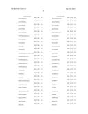 |
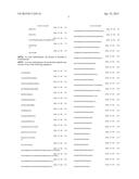 | 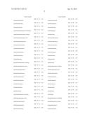 |
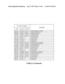 | 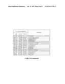 |
 | 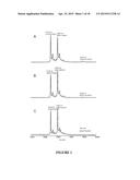 |
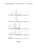 | 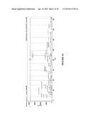 |
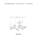 |  |
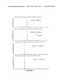 |  |
 | 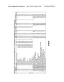 |
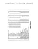 | 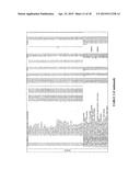 |
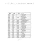 | 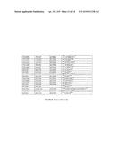 |
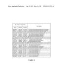 | 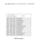 |
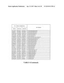 |  |
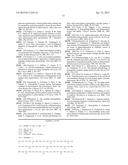 | 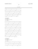 |
 |  |
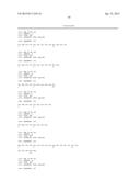 | 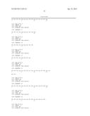 |
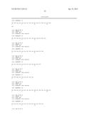 | 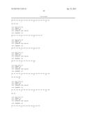 |
 | 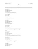 |
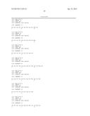 |  |
 |  |
 |  |
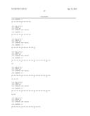 |  |
 |  |
 |  |
 |  |
 |  |
 |  |
 |  |
 |  |
 |
| New patent applications in this class: | |
| Date | Title |
|---|---|
| 2019-05-16 | Methods and uses for remotely triggered protease activity measurements |
| 2016-07-07 | Method for determining the cleaning performance of formulations |
| 2016-07-07 | Genetically encoded fret-based mmp-9 activity biosensor and use thereof |
| 2016-06-16 | Method for diagnosing cancer through detection of deglycosylation of glycoprotein |
| 2016-06-09 | Reagent for measuring endotoxin and method for measuring endotoxin |
| New patent applications from these inventors: | |
| Date | Title |
|---|---|
| 2010-04-22 | Apparatus and method for providing a portable broadband service using a wireless convergence platform |
| Top Inventors for class "Chemistry: molecular biology and microbiology" | |
| Rank | Inventor's name |
|---|---|
| 1 | Marshall Medoff |
| 2 | Anthony P. Burgard |
| 3 | Mark J. Burk |
| 4 | Robin E. Osterhout |
| 5 | Rangarajan Sampath |