Patent application title: STABLE PROTEINS
Inventors:
Seyed Ali Jazayeri-Dezfuly (Hertfordshire, GB)
Fiona Hamilton Marshall (Hertfordshire, GB)
Assignees:
Heptares Therapeutics Limited
IPC8 Class: AC07K1472FI
USPC Class:
435348
Class name: Chemistry: molecular biology and microbiology animal cell, per se (e.g., cell lines, etc.); composition thereof; process of propagating, maintaining or preserving an animal cell or composition thereof; process of isolating or separating an animal cell or composition thereof; process of preparing a composition containing an animal cell; culture media therefore insect cell, per se
Publication date: 2014-10-23
Patent application number: 20140315299
Abstract:
The invention provides a fusion protein comprising, from N-terminus to
C-terminus: a) a first portion of a Family B G-protein coupled receptor
(GPCR) that comprises transmembrane helix (TM)-1, TM2 and TM3 of the
GPCR; b) a stable protein domain; and c) a second portion of the GPCR
comprising TM4, TM5, TM6 and TM7 of the GPCR. The invention also provides
a method of crystallising a GPCR comprising providing the fusion protein
of the invention and crystallising it to obtain crystals.Claims:
1. A fusion protein comprising, from N-terminus to C-terminus: a) a first
portion of a Family B G-protein coupled receptor (GPCR) that comprises
transmembrane helix (TM)-1, TM2 and TM3 of the GPCR; b) a stable protein
domain; and c) a second portion of the GPCR comprising TM4, TM5, TM6 and
TM7 of the GPCR.
2. A fusion protein according to claim 1, wherein the GPCR is a Family B GPCR such as any of a glucagon-like peptide 1 receptor (GLP1R), glucagon-like peptide 2 receptor (GLP2R), calcitonin receptor (CT), amylin/CGRP receptor (AMY.sub.1.alpha.), amylin receptor (AMY.sub.2.alpha.), amylin/CGRP receptor (AMY.sub.3.alpha.), CGRP/adrenomedullin receptor (CGRP.sub.1.alpha.), adrenomedullin/CGRP receptor (AM.sub.1.alpha.), adrenomedullin/CGRP receptor (AM.sub.2.alpha. receptor), corticotropin releasing factor receptor (CRF1), urocortins receptor (CRF2), growth hormone releasing hormone receptor (GHRH), gastric inhibitory polypeptide receptor (GIP), glucagon receptor, secretin receptor, TIP-39 receptor (PTH2), parathyroid hormone receptor (PTH1), VIP/PACAP receptor (VPAC1), PACAP receptor (PAC2), and VIP/PACAP receptor (VPAC2).
3. A fusion protein according to claim 1, wherein the GPCR is a mutant GPCR that has increased conformational stability relative to its parent GPCR.
4. A fusion protein according to claim 1, wherein the stable protein domain is inserted into the intracellular loop 2 (ICL2) region of the GPCR which loop joins the first and second portion of the GPCR.
5. A fusion protein according to claim 4, wherein the stable protein domain is inserted into the ICL2 region of the GPCR at a position between amino acid residues that correspond to amino acids Phe 257 and Ser 261 according to the numbering of human GLP1R as set out in FIG. 6.
6. A fusion protein according to claim 5, wherein the stable protein domain is inserted into the ICL2 region of the GPCR after an amino acid corresponding to amino acid Phe 257 and before an amino acid corresponding to amino acid Ser 261 or Phe 260 or Val 259, according to the numbering of human GLP1R as set out in FIG. 6.
7. A fusion protein according to claim 5, wherein the stable protein domain is inserted into the ICL2 region of the GPCR after an amino acid corresponding to amino acid Ser 258 and before an amino acid corresponding to amino acid Ser 261 or Phe 260 or Val 259, according to the numbering of human GLP1R as set out in FIG. 6.
8. A fusion protein according to claim 1, wherein the stable protein domain comprises the amino acid sequence of lysozyme, such as T4 lysozyme.
9. A fusion protein according to claim 1, further comprising a detectable moiety such as any of a fluorescent label, a radiolabel or an enzymatic label.
10. A fusion protein according to claim 9, wherein the detectable moiety is EGFP.
11. A polynucleotide encoding the fusion protein according to claim 1.
12. A host cell comprising a polynucleotide according to claim 11.
13. A crystal comprising a fusion protein of claim 1.
14. A fusion protein according to claim 1, which is in a solubilised form or which is substantially free of other proteins or which is immobilised to a solid support.
15. Use of a fusion protein according to claim 1 for crystallisation, or for drug discovery such as in a ligand binding screen or in assay development, or as a biosensor.
16. A method of crystallising a fusion protein according to claim 1, the method comprising providing the fusion protein and crystallising it to obtain crystals.
17. A method according to claim 16, wherein the fusion protein is provided by culturing a host cell comprising a polynucleotide encoding the fusion protein to express the fusion protein and isolating the protein.
18. A method according to claim 16, wherein the crystallisation is carried out using lipidic cubic phase crystallography.
Description:
[0001] The invention relates to proteins that are not readily
crystallisable and particularly to GPCRs that are not readily stabilised
and therefore not readily crystallised. The invention also relates to
methods for crystallising such proteins and various uses of them. The
proteins are useful for drug discovery and development studies.
[0002] GPCRs constitute a very large family of proteins that control many physiological processes and are the targets of many effective drugs. Reference is made particularly to Overington et al (2006) Nature Rev. Drug Discovery 5, 993-996 which indicates that over a quarter of present drugs have a GPCR as a target. They are of considerable pharmacological importance. A list of GPCRs is given in Foord et al (2005) Pharmacol Rev. 57, 279-288, which is incorporated herein by reference.
[0003] GPCRs are generally unstable when isolated, and despite considerable efforts, it has only been possible to crystallise a few GPCRs including bovine rhodopsin, which naturally is exceptionally stable and the beta 2 adrenergic receptor which was crystallised as a fusion protein or in complex with an antibody fragment.
[0004] GPCRs are thought to exist in multiple distinct conformations which are associated with different pharmacological classes of ligand such as agonists and antagonists, and to cycle between these conformations in order to function (Kenakin T. (1997) Ann N Y Acad Sci 812, 116-125). Switching between conformations also contributes to the difficulty in obtaining crystal structures of receptors.
[0005] Based on sequence homology and molecular architecture, GPCRs can be classified into three families (A, B and C), although they all share the characteristic seven transmembrane (TM) domain. Family A, the largest group, consists of receptors that are homologous to Rhodopsin. Family B, also referred to as Secretin receptor family, are receptors that are regulated by large peptides hormones such as the glucagon hormone family; the members of this family are characterised by a relatively large extracellular N-terminus which contains several cysteines that form a network of disulphide bridges and is part of the ligand binding pocket. Family C consists of receptors that are homologous to metabotropic glutamate receptors; these receptors are characterised by a very long extra-cellular N-terminus as well as a long carboxy-tail, and the N-terminus forms the ligand binding pocket that has been shown to form a disulphide linked dimer, resembling a Venus fly trap in its shape.
[0006] Over the last few years the structure of a number of Family A GPCRs has been solved, and these milestones have been achieved by developing a number of key techniques. One such technology is the insertion of T4 lysozyme (T4L) in the intracellular cytoplasmic loop (ICL) 3, which is thought to create a large hydrophilic area that allows crystal contacts to form [2] [3]. The application of this technology in combination with Lipidic Cubic Phase crystallography has allowed high resolution structure determination of Beta2, A2a, CXCR4 and D3 receptors [2]. Thus, significant information has been gleaned from these structures about the orientation and organisation of the TM bundle of Family A receptors. However, little information is available for the members of Family B and C receptors and given the high sequence divergence, it is likely that significant differences exist in the architecture and organisation of the TM domains between families [1].
[0007] In Family A receptors, T4L was inserted in the ICL3 because it is thought that the distance between helices 5 and 6 is similar to the distance between the N- and C-termini of T4L. It is thus possible to accommodate the fusion protein in this position, whereas the distances between other helices are not believed to be conducive to the insertion of a fusion partner. Indeed, T4L has been fused to a number of different Family A receptors in ICL3 and functional protein has been expressed in each case with the added benefit of reducing the flexibility of the receptor, thus increasing overall stability.
[0008] We tested the effect of inserting T4L in the internal loops of Family B receptors and particularly in ICL3. Our data indicate that Family B receptors cannot tolerate T4L fusion in ICL3, however, surprisingly and unexpectedly, in view of the architecture of Family A receptors, adding T4L to ICL2 improves the biochemical properties of Family B receptors. ICL2 connects the portion of the GPCR comprising transmembrane helix (TM)-1, TM2 and TM3 to the portion of the GPCR comprising TM4, TM5, TM6 and TM7. The present data suggests that unlike Family A receptors, the distance between helices 3 and 4 in Family B receptors is more similar to the distance between the N- and C-termini of T4L, than is the distance between helices 5 and 6. Thus, insertion of a stable protein domain between these two portions of a GPCR is believed to represent a new technique for facilitating crystallisation of GPCRs that could not have been previously predicted.
[0009] Accordingly, a first aspect of the invention provides a fusion protein comprising, from N-terminus to C-terminus:
[0010] a) a first portion of a Family B G-protein coupled receptor (GPCR) that comprises TM1, TM2 and TM3 of the GPCR;
[0011] b) a stable protein domain; and
[0012] c) a second portion of the GPCR comprising TM4, TM5, TM6 and TM7 of the GPCR.
[0013] By "GPCR" we mean a G protein coupled receptor or polypeptide that has the signalling activity of a GPCR and retains an intact 7TM region. Standard nomenclature in the art designates the transmembrane helices of a GPCR from N-terminus to C-terminus as TM1, TM2, TM3, TM4, TM5, TM6 and TM7. The transmembrane helices are joined by stretches of amino acids extracellularly between TM2 and TM3, between TM4 and TM5, and between TM6 and TM7, referred to as extracellular loop (ECL)s 1, 2 and 3, respectively. The transmembrane helices are joined by stretches of amino acids intracellularly between TM1 and TM2, between TM3 and TM4, and between TM5 and TM6, referred to as intracellular loop (ICL)s 1, 2 and 3, respectively. Thus, the first and second GPCR portions as defined above are naturally joined by the ICL2 region, i.e. ICL2 connects the first portion N-terminal to ICL2 comprising TM1, TM2 and TM3 to the second portion C-terminal to ICL2 comprising TM4, TM5, TM6 and TM7.
[0014] The GPCR is preferably derived from full length wild type sequences including natural polymorphisms or mutant GPCR molecules that have been altered, for example so as to improve one or more properties of the GPCR eg stability.
[0015] The GPCR may be derived from wildtype and mutant GPCRs wherein mutant GPCRs may be stabilised GPCRs biased towards a particular conformation such as agonist or antagonist. For example, a stable protein domain may then be inserted between TM3 and TM4 of a conformationally stabilised GPCR.
[0016] We have previously developed a methodology for the stabilisation of a GPCR in a biologically relevant conformation (see WO 2008/114020) describing the production of stabilised GPCRs known as StaRs® that enables the purification of recombinant G protein coupled receptors that maintain their conformation, stability and function when purified from the cell membrane. In addition, this platform technology also provides the means to engineer receptors biased either towards agonist conformation or the antagonist conformation (see also Magnani et al, 2008; Serrano-Vega et al, 2008; Shibata et al, 2009), i.e. they have increased stability in a particular conformation. Such stabilised receptors may be used in the present invention and have a number of advantages, for example stability, elevated yields of purified protein, reduced denaturation and reduced non-specific binding. Where a stable mutant GPCR is used in the present invention it is preferably selected and prepared using any of the methods as described in PCT applications WO 2008/114020, WO 2009/114020 and WO 2009/081136. Preferably the first and second GPCR portions are from a GPCR which has increased stability in a particular conformation relative to a parent GPCR (i.e. increased conformation stability). By increased conformational stability we include the meaning that a particular conformation of a mutant GPCR has, compared to the same conformation of the parent GPCR, increased stability (eg an extended lifetime) when exposed to a denaturant or denaturing conditions. Examples of denaturants/denaturing conditions include heat, detergent, a chaotropic agent and an extreme of pH. As is well known in the art, such denaturants or denaturing conditions can affect secondary and tertiary structures of a protein but not the primary sequence.
[0017] Suitable GPCRs for use in the practice of the invention include any Family B GPCR such as any of a glucagon-like peptide 1 receptor (GLP1R), glucagon-like peptide 2 receptor (GLP2R), calcitonin receptor (CT), amylin/CGRP receptor (AMY1α), amylin receptor (AMY2α), amylin/CGRP receptor (AMY3α), CGRP/adrenomedullin receptor (CGRP1α), adrenomedullin/CGRP receptor (AM1α), adrenomedullin/CGRP receptor (AM2α receptor), corticotropin releasing factor receptor (CRF1), urocortins receptor (CRF2), growth hormone releasing hormone receptor (GHRH), gastric inhibitory polypeptide receptor (GIP), glucagon receptor, secretin receptor, TIP-39 receptor (PTH2), parathyroid hormone receptor (PTH1), VIP/PACAP receptor (VPAC1), PACAP receptor (PAC2) and VIP/PACAP receptor (VPAC2). In a particularly preferred embodiment, the GPCR is GLP1R. Other suitable GPCRs are well known in the art and include those listed in Overington et al supra. In addition, the International Union of Pharmacology produce a list of GPCRs that includes Family B GPCRs (Foord et al (2005) Pharmacol. Rev. 57, 279-288, and this list is periodically updated at http://www.iuphar-db.org/GPCR/ReceptorFamiliesForward; Family B GPCRs are listed in Table 2 as Class 2 GPCRs).
[0018] The amino acid sequences (and the nucleotide sequences of the cDNAs which encode them) of many GPCRs are readily available, for example by reference to GenBank. In particular, Foord et al supra gives the human gene symbols and human, mouse and rat gene IDs from Entrez Gene (http://www.ncbi.nlm.nihdov/entrez). It should be noted, also, that because the sequence of the human genome is substantially complete, the amino acid sequences of human GPCRs can be deduced therefrom.
[0019] Although the GPCR may be derived from any source, it is particularly preferred if it is from a eukaryotic source. It is particularly preferred if it is derived from a vertebrate source such as a mammal. It is particularly preferred if the GPCR is derived from rat, mouse, rabbit or dog or non-human primate or man. For the avoidance of doubt, we include within the meaning of "derived from" that a cDNA or gene was originally obtained using genetic material from the source, but that the protein may be expressed in any host cell subsequently. Thus, it will be plain that a eukaryotic GPCR (such as an avian or mammalian GPCR) may be expressed in a prokaryotic host cell, such as E. coli, but be considered to be avian- or mammalian-derived, as the case may be.
[0020] In some instances, the GPCR may be composed of more than one different subunit. For example, the calcitonin gene-related peptide receptor requires the binding of a single transmembrane helix protein (RAMP1) to acquire its physiological ligand binding characteristics. Effector, accessory, auxiliary or GPCR-interacting proteins which combine with the GPCR to form or modulate a functional complex are well known in the art and include, for example, receptor kinases, G-proteins and arrestins (Bockaert et al (2004) Curr Opinion Drug Discov and Dev 7, 649-657). In some instances, the GPCR may be bound to a GPCR ligand. By "ligand" we include any molecule which binds to the GPCR. Many ligands are known, for example from WO 2008/114020 and Neubig et al (2003) Pharmacol. Rev. 55, 597-606, both of which are incorporated herein by reference. Thus, the fusion protein may comprise a portion of the GPCR comprising TM1, TM2 and TM3 connected to a portion of the GPCR comprising TM4, TM5, TM6 and TM7, wherein the GPCR is bound to a GPCR binding partner. In this way, it is possible to gain structural insights into GPCR interactions by being able to crystallise complexes of GPCR with other molecules. It is preferred if the molecules are not ones that bind to ICL2 of the GPCR.
[0021] For any given GPCR, the TM helices can be determined by the skilled person using standard techniques in the art. For example, computer programs are available which model transmembrane regions of GPCRs based on hydrophobicity (Kyle & Dolittle (1982) J. Mol. Biol. 157, 105-132). Likewise transmembrane prediction algorithm servers are widely available on the World Wide Web (eg Expasy), many of which rely on hydropathy analysis. TMHMM is a membrane protein topology prediction method that may be used, based on a hidden Markov model (TMHMM Server v. 2.0; http://www.cbs.dtu.dk/services/TMHMM/). Where the transmembrane regions are already known for a given GPCR, for example by structural analysis or hydropathy analysis, the analogous regions in a further GPCR can also be identified by multiple or pairwise sequence alignment. For example, the alignment may be carried out using the Clustal W program (Thompson et al., 1994). The parameters used may be as follows: Fast pairwise alignment parameters: K-tuple(word) size; 1, window size; 5, gap penalty; 3, number of top diagonals; 5. Scoring method: x percent. Multiple alignment parameters: gap open penalty; 10, gap extension penalty; 0.05. Scoring matrix: BLOSUM.
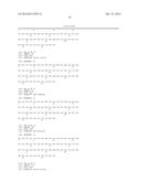
[0022] FIG. 6 lists the amino acid sequences of Family B GPCRs and highlights the position of TM3, ICL2 and TM4. For example, for human GLP1R, TM3 ends with Phe 257, ICL2 corresponds to Ser 258 to Ser 261, and TM4 begins with Glu 262. Conveniently therefore, when the GPCR is a Family B receptor, the positions of TM3, ICL2 and TM4 can be identified by locating the amino acid residues that correspond to the amino acids that define the boundaries of TM3, ICL2 and TM4 in FIG. 6, when the sequences are aligned, for instance using CLUSTAL W.
[0023] It will be appreciated, however, that the boundaries are not absolute and they may well depend on the model provided for GLP1R that has been used to define them. In FIG. 1, for example, TM3 of human GLP1R ends with Leu 254, ICL2 corresponds to Leu 255 to Trp 264, and TM4 begins with Ile 265. Also, the loop regions may be defined as amino acid structures that join alpha helices or amino acid structures that are predicted to be outside the membrane, and depending on which definition is used, the boundaries will change.
[0024] In one embodiment, the stable protein domain is inserted into ICL2. Thus, the invention provides a GPCR into which a stable protein domain has been inserted into ICL2. By `inserted into ICL2` we include both the addition of the amino acid sequence that defines the stable protein domain into the amino acid sequence of ICL2 without the deletion of any amino acids of ICL2, and also the replacement of one or more or all amino acids of ICL2 with the amino acid sequence encoding the stable protein domain. It will be appreciated that in this embodiment, the first and/or second portion of the GPCR may comprise at least part of ICL2, in addition to the requisite transmembrane helices. The first portion of the GPCR may comprise TM1, TM2 and TM3 and an N-terminal part of ICL2. The second portion of the GPCR may comprise TM4, TM5, TM6 and TM7 and a C-terminal part of ICL2.
[0025] It is appreciated that the stable protein domain may be inserted into ICL2 and flanked by one or two spacer moieties at its N- and/or C-terminus. In this way, the stable protein domain is not directly linked to ICL2 but is indirectly linked. The spacer moieties may be used to help reduce tension on the helices.
[0026] Preferably, the stable protein domain is inserted into ICL2 by replacing one or more consecutive amino acids (eg 2, 3, 4 or 5 or more amino acids) in the amino acid sequence of ICL2 with the amino acid sequence of the stable protein domain. In one embodiment, the one or more amino acids that are replaced is/are at least one or two amino acids from the C-terminus of TM3 and/or the N-terminus of TM4. In other words, the fusion protein may have at least one or two amino acids of ICL2 on at least one of the sides of the stable protein domain.
[0027] As described in Example 1, we have inserted T4 lysozyme at various positions in ICL2 of GLP1R, and insertions between Phe 257 and Ser 261 resulted in productive fusion GLP1R receptors. Thus, it is particularly preferred that the stable protein domain is inserted into the ICL2 region of the GPCR at a position between amino acid residues that correspond to amino acids Phe 257 and Ser 261 according to the numbering of human GLP1R as set out in FIG. 6.
[0028] Accordingly, the amino acid of the stable protein domain may be inserted into the ICL2 region of the GPCR after an amino acid corresponding to amino acid Phe 257 and before an amino acid corresponding to amino acid Ser 261 or Phe 260 or Val 259, according to the numbering of human GLP1R as set out in FIG. 6. For example, the amino acid sequence of the stable protein domain may replace the amino acid corresponding to Ser 258, or it may replace amino acids corresponding to Ser 258 and Val 259, or it may replace amino acids corresponding to Ser 258, Val 259 and Phe 260, according to the numbering of human GLP1R as set out in FIG. 6. Inserting the stable protein domain at such positions corresponds to the GLP1R-T4 lysozyme fusion constructs 1c, 2c and 3c shown in FIG. 1.
[0029] Similarly, the amino acid of the stable protein domain may be inserted into the ICL2 region of the GPCR after an amino acid corresponding to amino acid Ser 258 and before an amino acid corresponding to amino acid Ser 261 or Phe 260 or Val 259, according to the numbering of human GLP1R as set out in FIG. 6. For example, the amino acid sequence of the stable protein domain may replace the amino acid corresponding to Val 259, or it may replace amino acids corresponding to Val 259 and Phe 260, according to the numbering of human GLP1R as set out in FIG. 6. Inserting the stable protein domain at such positions corresponds to the GLP1R-T4 lysozyme fusion constructs 1d, 2d and 3d shown in FIG. 1.
[0030] By "corresponding amino acid residue" we include the meaning of the amino acid residue in another GPCR that aligns to the given amino acid residue in human GLP1R when the human GLP1R receptor and the other GPCR are compared using MacVector and CLUSTALW.
[0031] Although it is preferred that the stable protein domain is inserted into the ICL2 region of the GPCR at a position between amino acid residues that correspond to amino acids Phe 257 and Ser 261 according to the numbering of human GLPIR as set out in FIG. 6, it is appreciated that it may be inserted outside of this region.
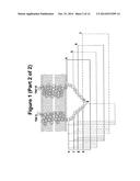
[0032] It is appreciated that the function of the stable protein domain is to increase the hydrophilic surface for crystal contacts and to reduce the inherent flexibility of GPCRs in order to, for example, improve the crystallisation properties of the GPCR. Accordingly, by `stable protein domain` we include the meaning of any soluble, folded polypeptide that provides a hydrophilic surface for crystal lattice contacts. Further, the protein domain is stable such that in its folded form it is resistant to denaturation (eg is stable to heat, detergents and chaotropic agents etc.). Tests for protein stability are well known in the art and include those described in WO2008/114020.
[0033] Typically, the stable protein domain is one that folds autonomously from the GPCR portions of the fusion protein in the cell.
[0034] Conveniently, the stable protein domain is one that is readily crystallisable. Thus, the stable protein domain may be a protein whose crystal structure has been solved, for example one whose coordinates has been deposited in the Protein Data Bank (http://www.pdb.org/).
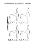
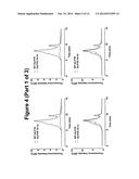
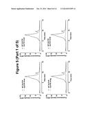
[0035] Particularly preferred characteristics of the stable protein domain are:
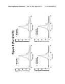
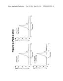
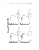
[0036] 1. the domain is soluble, well folded and can be expressed easily in one or more expression systems;
[0037] 2. the N- and C-termini of the domain are close together in space; typically in the range of 5-17 Å eg 6-16 Å, 7-15 Å, 7-10 Å, 10-13 Å or 12-15 Å;
[0038] 3. the domain is resistant to thermal and chemical denaturation as well as to proteolytic degration;
[0039] 4. the domain is highly crystallisable in a variety of space groups and crystal packing arrangements.
[0040] It is preferred that the domain does not contain cysteine residues so as to prevent disulphide bond formation either within the domain or with the GPCR portion of the fusion protein. It will be understood that since the domain is soluble it should not be hydrophobic or have the propensity to aggregate in a disordered fashion.
[0041] In one embodiment, the length of the stable protein domain is between 50 and 1000 amino acids, preferably between 50 and 300 amino acids or 100 and 300 amino acids, or between 150 and 250 amino acids.
[0042] Once a suitable polypeptide has been found for the stable protein domain, it may be necessary to modify the polypeptide by deleting or adding amino acid residues from or to the N-terminus, the C-terminus or both termini of the polypeptide such that the closest alpha carbon atoms in the backbone at the termini of the polypeptide are spaced by a distance of in the range of 5-17 Å eg 6-16 Å, 7-15 Å, 7-10 Å, 10-13 Å or 12-15 Å.
[0043] It is preferred if insertion of the stable protein domain does not affect a biological activity of the GPCR, such as a binding activity or a signalling pathway modulation activity. Ideally, the fusion protein should retain at least 60% or 70% or 80% or 90% of its biological activity, and most ideally 100% of its biological activity relative to the level of the same activity in the absence of the stable protein domain. Methods for assessing GPCR binding and GPCR signalling are well known in the art and are described for example in WO 2008/114020 and WO 2009/101383, both of which are incorporated herein by reference. Thus, where the biological activity is a binding activity, binding to any GPCR binding partner may be assessed using routine binding assays known in the art; where the biological activity is a signalling pathway modulating activity, the activity can be assessed by any suitable assay for the particular signalling pathway (eg reporter gene assays).
[0044] It is appreciated that retaining ligand binding ability is more important for crystallisation purposes than is retaining signalling activity, and that it may be desirable to only assess ligand binding ability prior to crystallisation. Thus in a particularly preferred embodiment, the stable protein domain does not affect a binding activity of a GPCR.
[0045] For the avoidance of doubt, the stable protein domain is not ICL2 or part thereof of the particular GPCR.
[0046] In a preferred embodiment, the stable protein domain is lysozyme. Lysozyme is known to be readily crystallisable and the structures of various wild type and variant lysozymes have been deposited in the Protein Data Bank (www.rcsb.org). Suitable examples include 135L, 193L, 194L, 1AKI, 1GBS, 1IEE, 1LZ1, 1P7S, 1REX, 1VDQ, 2ANV, 2ANX, 2D4K, 2FBB, 2IHL, 2NWD, 2XBR, 2XBS, 2Z2F, 2ZYP, 3A8Z, 3K2R, 3N9A, 3N9C, 3N9E and 3OD9
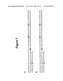
[0047] Although lysozyme derived from any source may be used, it is particularly preferred if the lysozyme is derived from T4 phage. Two amino acid sequences of T4 phage lysozyme are provided in FIG. 7, and either of the sequences may be used in the context of the invention. The lysozyme may be derived from full length wild type sequences including natural polymorphisms or it may be a mutant lysozyme that has been altered, for example to improve one or more properties. Thus, it is understood that variants of the amino acid sequences provided in FIG. 7 may be used, such as amino acid sequences with at least 60%, 65%, 70%, 75%, 80%, 85% or 90% sequence identity with either of the sequences set out in FIG. 7, and more preferably at least 95% or 99% sequence identity with either of the sequences set out in FIG. 7.
[0048] Sequence identity may be measured by the use of algorithms such as BLAST or PSI-BLAST (Altschul et al, NAR (1997), 25, 3389-3402) or methods based on Hidden Markov Models (Eddy S et al, J Comput Biol (1995) Spring 2 (1) 9-23). Typically, the percent sequence identity between two polypeptides may be determined using any suitable computer program, for example the GAP program of the University of Wisconsin Genetic Computing Group and it will be appreciated that percent identity is calculated in relation to polypeptides whose sequence has been aligned optimally. The alignment may alternatively be carried out using the Clustal W program (Thompson et al., 1994) as mentioned above.
[0049] While lysozyme is a preferred example of a stable protein domain, the general principles may be used to employ any number of polypeptides that have the characteristics discussed above. Thus, suitable candidates include those containing the amino acid sequence of proteins that are readily crystallisable, for example as found by interrogating the protein data bank or other crystallization databases known in the art. Other examples include those mentioned in Engel et al (2002) BBA 1564: 38-46 (incorporated herein by reference), such as cytochrome.sub.b562, flavodoxin, β-lactamase and 70 kDa heat shock ATPase domain.
[0050] The fusion protein may be modified so that it can be more easily detected, for example by biotinylating it or by incorporating any detectable label known in the art such as radiolabels, fluorescent labels or enzymatic labels. In a particularly preferred embodiment, the label is a fluorescent label such as EGFP. Similarly, the fusion protein may be modified to facilitate purification, for example by incorporating any affinity moiety known in the art such as a GST tag, 6x His tag, MBP or other epitope tag. Such modifications may be at the N-terminus or C-terminus of the GPCR or in an external loop.
[0051] As demonstrated in Example 1, it is believed that the fusion proteins of the invention have improved biochemical properties compared to the biochemical properties of the GPCR without the insertion of the stable protein domain. Such improved properties make the fusion protein more amenable to crystallisation. Thus, the fusion protein is expected to have a larger hydrophilic surface for crystal contacts. Similarly, the fusion protein is expected to be more soluble, eg displaying less aggregation in detergent solution, than the GPCR without insertion of the stable protein domain. Methods for assessing GPCR solubility are well known in the art and include size exclusion chromatography such as fluorescent size exclusion chromatography used to assess solubility in DDM as described in Example 1. The fusion protein may also be more stable (eg to any of heat, detergent or chaotropic agents) than the GPCR without the insertion of the stable protein domain. Methods for assessing GPCR stability are known in the art, including those described in WO 2008/114020.
[0052] Conveniently, the fusion protein is produced by standard molecular biology and recombinant DNA techniques. For example, DNA fragments encoding the first and second GPCR portions and the stable protein domain may be made using standard cloning techniques and PCR as is well known in the art. The fragments can then be ligated together in-frame in accordance with conventional practice, e.g., by employing blunt-ended or stagger-ended termini for ligation, restriction enzyme digestion to provide for appropriate termini, filling-in of cohesive ends as appropriate, alkaline phosphatase treatment to avoid undesirable joining, and enzymatic ligation.
[0053] Equally, the fusion constructs can be made using ligase independent cloning strategies such as InFusion or Gateway. The construct may also be made synthetically through de novo gene synthesis.
[0054] It is appreciated that one or both of the GPCR and stable protein domain may be mutated so as to improve any of solubility, stability, expression and crystallisability.
[0055] Molecular biological methods for cloning and engineering genes and cDNAs, for mutating DNA, and for expressing polypeptides from polynucleotides in host cells are well known in the art, as exemplified in "Molecular cloning, a laboratory manual", third edition, Sambrook, J. & Russell, D. W. (eds), Cold Spring Harbor Laboratory Press, Cold Spring Harbor, N.Y., incorporated herein by reference.
[0056] Suitable expression systems include constitutive or inducible expression systems in bacteria or yeasts, virus expression systems such as baculovirus, semliki forest virus and lentiviruses, or transient transfection in insect or mammalian cells. Suitable host cells include E. coli, Lactococcus lactis, Saccharomyces cerevisiae, Schizosaccharomyces pombe, Pichia pastoris, Spodoptera frugiperda and Trichoplusiani cells. Suitable animal host cells include HEK 293, COS, S2, CHO, NSO, DT40 and so on. It is known that some GPCRs require specific lipids (eg cholesterol) to function. In that case, it is desirable to select a host cell which contains the lipid. Additionally or alternatively the lipid may be added during isolation and purification of the fusion protein. Purification may be carried out by standard techniques such as affinity chromatography.
[0057] A second aspect of the invention provides a polynucleotide encoding the fusion protein according to the first aspect of the invention. The polynucleotide may be RNA (eg mRNA) or DNA, although typically it is DNA.
[0058] It will be appreciated that the polynucleotide may be incorporated into a vector, and so the invention also provides a vector comprising a polynucleotide according to the second aspect of the invention.
[0059] Suitable vectors are ones which propagate in and/or allow expression of the fusion protein in prokaryotic (eg bacterial) or eukaryotic (eg mammalian) cells. For example, the vector may be a plasmid, a cosmid, a phage or a bacterial artificial chromosome (BAC). The polynucleotide sequence of the vector will depend upon the nature of the intended host cell, the manner of the introduction of the polynucleotide of the second aspect of the invention into the host cell, and whether episomal maintenance or integration is desired. Conveniently, the vector comprises at least one selectable marker such as antibiotic resistance (eg kanamycin or neomycin).
[0060] Vectors are useful to replicate the polynucleotide of the second aspect of the invention, and are also useful to transfect cells with the polynucleotide, and may also promote expression of the fusion protein.
[0061] Typical prokaryotic vector plasmids are: pUC18, pUC19, pBR322 and pBR329 available from Biorad Laboratories (Richmond, Calif., USA); pTrc99A, pKK223-3, pKK233-3, pDR540 and pRIT5 available from Pharmacia (Piscataway, N.J., USA); pBS vectors, Phagescript vectors, Bluescript vectors, pNH8A, pNH16A, pNH18A, pNH46A available from Stratagene Cloning Systems (La Jolla, Calif. 92037, USA).
[0062] A typical mammalian cell vector plasmid is pSVL available from Pharmacia (Piscataway, N.J., USA). This vector uses the SV40 late promoter to drive expression of cloned genes, the highest level of expression being found in T-antigen-producing cells, such as COS-1 cells. Another example is pcDNA3.1 (neo) (Invitrogen) for use in COS-1 or COS-7 cells. An example of an inducible mammalian expression vector is pMSG, also available from Pharmacia (Piscataway, N.J., USA). This vector uses the glucocorticoid-inducible promoter of the mouse mammary tumour virus long terminal repeat to drive expression of the cloned gene.
[0063] Useful yeast plasmid vectors are pRS403-406 and pRS413-416 and are generally available from Stratagene Cloning Systems (La Jolla, Calif. 92037, USA). Plasmids pRS403, pRS404, pRS405 and pRS406 are Yeast Integrating plasmids (YIps) and incorporate the yeast selectable markers HIS3, TRP1, LEU2 and URA3. Plasmids pRS413-416 are Yeast Centromere plasmids (YCps).
[0064] In a preferred embodiment, the vector comprising the polynucleotide of the second aspect of the invention is pcDNA3.1 (http://products.invitrogen.com/ivgn/product/V79020).
[0065] Any suitable method known in the art may be used to construct vectors containing the polynucleotide of the second aspect of the invention including the ligation techniques described above.
[0066] A third aspect of the invention provides a cell comprising a polynucleotide according to the second aspect of the invention, or a vector comprising said polynucleotide. Such cells may be used to replicate the polynucleotide of the second aspect of the invention, or may be used to express the fusion protein of the first aspect of the invention.
[0067] The cell can be either prokaryotic or eukaryotic.
[0068] It is appreciated that construction and amplification of the polynucleotide of the second aspect of the invention is conveniently performed in bacterial cells. Expression of the polynucleotide may be carried out in cells such as mammalian cells or bacterial cells.
[0069] Bacterial cells are preferred prokaryotic host cells and typically are a strain of E. coli such as, for example, the E. coli strains DH5 available from Bethesda Research Laboratories Inc., Bethesda, Md., USA, and RR1 available from the American Type Culture Collection (ATCC) of Rockville, Md., USA (No ATCC 31343). Preferred eukaryotic host cells include yeast, insect and mammalian cells or cell lines, preferably vertebrate cells or cell lines such as those from a mouse, rat, monkey or human. Particularly preferred cells are human embryonic kidney cells such as HEK293T cells.
[0070] Cells used for expressing the fusion protein may be either stably or non-stably transfected.
[0071] A fourth aspect of the invention provides a method of crystallising a fusion protein according to the first aspect of the invention, the method comprising providing a fusion protein according to the first aspect of the invention and crystallising it to obtain crystals.
[0072] In an embodiment, the fusion protein is provided by culturing a host cell according to the third aspect of the invention to express the fusion protein and isolating the protein.
[0073] Any suitable crystallisation method may be used to crystallise the fusion protein, such as any of those reviewed in "Crystallisation of Biological Macromolecules" (Alexander McPherson; ISBN: 0-87969-617-6), which is incorporated herein by reference.
[0074] In a preferred embodiment, the crystallisation is carried out using lipidic cubic phase crystallography (see US 2011/0031438 incorporated herein by reference).
[0075] A fifth aspect of the invention provides a crystal comprising a fusion protein of the first aspect of the invention.
[0076] The fusion proteins disclosed herein are useful for crystallisation studies and are useful in drug discovery programmes. They may be used in biophysical measurements of receptor/ligand kinetic and thermodynamic parameters eg by surface plasmon resonance or fluorescence based techniques. They may be used in ligand binding screens, and may be coupled to solid surfaces for use in high throughput screens or as biosensor chips. Biosensor chips containing the fusion proteins may be used to detect molecules, especially biomolecules.
[0077] The invention will now be described with the aid of the following figures and examples.
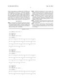
[0078] FIG. 1. Design of T4L fusion constructs with GLP1R. T4L was inserted after the indicated residues in ICL2 (left) and ICL3 (right). Construct 1a means that T4L was inserted between L255 and S261. The model of TM domains and loops are based on reference [4].
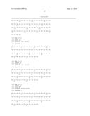
[0079] FIG. 2. Total EGFP signal of GLP1R-T4L fusion constructs compared to the wild-type (WT) and the mock transfected (U) samples. Each measurement was done on 50 ug of total cellular material in duplicate. Error bars represent standard deviation from mean.
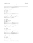
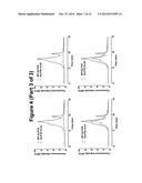
[0080] FIG. 3. Typical fSEC elution profile of wild-type GLP1R.
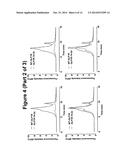
[0081] FIG. 4. fSEC elution profiles DDM solubilised GLP1R-T4L fusion constructs in the ICL2 overlaid the wild-type profile. In each case, the wild-type profile is shown in red the fusion constructs in blue.
[0082] FIG. 5. fSEC elution profiles DDM solubilised GLP1R-T4L fusion constructs in the ICL3 overlaid the wild-type profile. In each case, the wild-type profile is shown in red the fusion constructs in blue.
[0083] FIG. 6. Amino acid sequences of Family B GPCRs showing position of TM3, ICL2 and TM4 (SEQ ID Nos: 1-22). The portion of ICL2 that was replaced with T4L in GLP1R fusion construct 1c is highlighted in other Family B receptors from mouse, rat and human.
[0084] FIG. 7. Amino acid sequences of T4 phage lysozyme: (A) Sequence inserted into ICL3 of Family A receptors [2], [3] (SEQ ID No: 23); (B) Sequence inserted into ICL2 of Family B receptors (SEQ ID No: 24) (see Examples). Differences are indicated in boxes.
EXAMPLE 1
Insertion of T4L into ICL2 Improves Biochemical Properties of GLP1R
Summary
[0085] We have tested the effect of inserting T4L in the internal loops of Family B receptors. Our data indicate that Family B receptors cannot tolerate T4L fusion in ICL3, however, adding T4L to ICL2 improves the biochemical properties of the receptor.
Results
[0086] The loop regions of GLP1R were determined according to the model of GLP1R and the DNA encoding T4L was inserted in different locations within ICL2 and ICL3 (FIG. 1). The GLP1R construct was C-terminally tagged with the EGFP in order to monitor total expression as well as monodispersity using fluorescent-detection size exclusion chromatography.
[0087] Following sequence confirmation, these constructs were expressed in HEK293T transiently. As an initial analysis, the EGFP signal in whole cells was measured to assess the total levels of expression. Interestingly, the constructs in ICL3 failed to produce any EGFP signal, indicating that T4L fusion in this region of GLP1R is incompatible with the overall architecture of this receptor. However, fusions in the ICL2 resulted in the robust expression of GLP1R (FIG. 2).
[0088] In order to analyse the biochemical properties of GLP1R-T4L fusions, cells expressing these constructs were solubilised in dodecyl maltoside (DDM) and applied to fluorescence-detection size-exclusion chromatography (fSEC). fSEC has been used widely to provide data regarding the monodispersity and the aggregation status of the proteins, particularly in pre-crystallisation screens [5]. In general, the more favourable conditions will result in more monodispersity and reduced aggregation. The fSEC elution profile of DDM-solubilised wild-type GLP1R shows the presence of the main monodispersed peak with an aggregation shoulder as well as free EGFP species that are the result of proteolytic degredation (FIG. 3).
[0089] Consistent with the EGFP signal data showed in FIG. 2, the elution profiles of ICL2 T4L fusion constructs 1a, 1b, 2a, 2b, 3a and 3b indicates that cells failed to express these fusions (FIG. 4). This is most likely due to the proximity of the N-terminus of T4L to TM III, leading to the disruption of the overall structure. In contrast, the elution profiles of constructs 1c, 1 d, 2c, 2d, 3c and 3d revealed that productive fusion receptors were expressed and more significantly it appears that T4L fusion results in the reduction of the aggregation peak and concomitant improvement of the monodispersed peak, which together indicate that T4L insertion in this region of the receptor has beneficial effect on the biochemical properties of solubilised receptor (FIG. 4). This effect is most pronounced in the constructs 1c and 2c.
[0090] The same analysis was carried out for the ICL3 fusion constructs and in agreement with the EGFP signal data showed in FIG. 2, none of the T4L fusions in the ICL3 resulted in the expression of any productive fusion receptor (FIG. 5).
[0091] Taken together, these data indicate that T4L fusion in the third cytoplasmic loop of GLP1R is not tolerated, however, insertion of T4L in certain positions in the second cytoplasmic loop not only is tolerated, it also improves the biochemical properties of the solubilised receptor. Given the high sequence homology amongst members of the Family B GPCRs, we suggest that these observations can be extended to other members of this Family. The portion of the ICL2 that was replaced in the best construct (1c) is highlighted in other Family B members as shown in FIG. 6.
Methods and Material
[0092] T4 lysozyme was inserted in the second and third cytoplasmic loops of human GLP1R using standard molecular biology techniques. These constructs were transiently expressed from a modified pcDNA3.1 in HEK293T cells, generating receptors fused to EEGFP at their C-termini. Transfections were carried out using GeneJuice (Merck Biosciences) according to the manufacturer's guideline. Typically, 6 ug of DNA was used to transfect 3×10⁶ adherent cells in 10 cm plates. Cells were harvested about 40 hours post transfection and re-suspended in 50 mM HEPES pH 7.5/150 mM NaCl/0.5 mM EDTA complemented with Complete EDTA-free protease inhibitor cocktail (Roche). Typically 650 ug of each sample was solubilised with 1% DDM in total volume of 200 uL for 1 hour at 4° C. followed by centrifugation at 50000 rpm for 30 minutes. 50 uL of the supernatant was loaded onto BioSEep-SEC-S3000 column (Phenomenex), pre-equilibrated with SEC buffer (50 mM HEPES pH 7.5/150 mM NaCl/0.5 mM EDTA/0.03% DDM) and run at the flow rate of 1 mL/minute for 15 minutes. The eluent was passed through an on-line fluorometer with the following settings: excitation 490 nm, emission 513 nm and gain of 13.
REFERENCES
[0093] [1] Kristiansen, K. Molecular mechanisms of ligand binding, signaling, and regulation within the superFamily of G-protein-coupled receptors: molecular modeling and mutagenesis approaches to receptor structure and function. Pharma & Therap 103, 21-80 (2004).
[0094] [2] Bill R M, Henderson P J, Iwata S, Kunji E R, Michel H, Neutze R, Newstead S, Poolman B, Tate C G and Vogel H. Overcoming barriers to membrane protein structure determination. Nat Biotechnol. 29(4), 335-340 (2011).
[0095] [3] Kobilka B K, Kobilka T S, Daniel K, Regan J W, Caron M G and Lefkowitz R J. Chimeric alpha 2-,beta 2-adrenergic receptors: delineation of domains involved in effector coupling and ligand binding specificity. Science 240(4857) 1310-6 (1988).
[0096] [4] Frimurer T M and Bywater R P. Structure of the integral membrane domain of the GLP1 receptor. Proteins 35(4), 375-86 (1999).
[0097] [5] Kawate T and Gouaux E. Fluorescence-detection size-exclusion chromatography for precrystallization screening of integral membrane proteins. Structure 14(4), 673-81 (2004).
Sequence CWU
1
1
44158PRTRattus rattus 1Ser Leu Ile Phe Met Ala Phe Phe Ser Glu Lys Lys Tyr
Leu Trp Gly 1 5 10 15
Phe Thr Ile Phe Gly Trp Gly Leu Pro Ala Val Phe Val Ala Val Trp
20 25 30 Val Gly Val Arg
Ala Thr Leu Ala Asn Thr Gly Cys Trp Asp Leu Ser 35
40 45 Ser Gly His Lys Lys Trp Ile Ile Gln
Val 50 55 258PRTMus musculus 2Ser Leu
Ile Phe Met Ala Phe Phe Ser Glu Lys Lys Tyr Leu Trp Gly 1 5
10 15 Phe Thr Ile Phe Gly Trp Gly
Leu Pro Ala Val Phe Val Ala Val Trp 20 25
30 Val Gly Val Arg Ala Thr Leu Ala Asn Thr Gly Cys
Trp Asp Leu Ser 35 40 45
Ser Gly His Lys Lys Trp Ile Ile Gln Val 50 55
358PRTHomo sapiens 3Ser Leu Ile Phe Met Ala Phe Phe Ser Glu
Lys Lys Tyr Leu Trp Gly 1 5 10
15 Phe Thr Val Phe Gly Trp Gly Leu Pro Ala Val Phe Val Ala Val
Trp 20 25 30 Val
Ser Val Arg Ala Thr Leu Ala Asn Thr Gly Cys Trp Asp Leu Ser 35
40 45 Ser Gly Asn Lys Lys Trp
Ile Ile Gln Val 50 55 457PRTMus
musculus 4Asn Leu Ile Phe Val Ser Phe Phe Ser Asp Thr Lys Tyr Leu Trp Gly
1 5 10 15 Phe Ile
Ser Ile Gly Trp Gly Phe Pro Ala Val Phe Val Val Ala Trp 20
25 30 Ala Val Ala Arg Ala Thr Leu
Ala Asp Thr Arg Cys Trp Glu Leu Ser 35 40
45 Ala Gly Asp Arg Trp Ile Tyr Gln Ala 50
55 557PRTRattus rattusUNSURE(56)..(57)UNSURE 5Asn
Leu Ile Phe Val Ser Phe Phe Ser Asp Thr Lys Tyr Leu Trp Gly 1
5 10 15 Phe Ile Leu Ile Gly Trp
Gly Phe Pro Ala Val Phe Val Val Ala Trp 20
25 30 Ala Val Ala Arg Ala Thr Leu Ala Asp Thr
Arg Cys Trp Glu Leu Ser 35 40
45 Ala Gly Asp Arg Trp Ile Tyr Xaa Xaa 50
55 658PRTHomo sapiens 6Asn Leu Ile Phe Val Ala Phe Phe Ser Asp
Thr Lys Tyr Leu Trp Gly 1 5 10
15 Phe Ile Leu Ile Gly Trp Gly Phe Pro Ala Ala Phe Val Ala Ala
Trp 20 25 30 Ala
Val Ala Arg Ala Thr Leu Ala Asp Ala Arg Cys Trp Glu Leu Ser 35
40 45 Ala Gly Asp Ile Lys Trp
Ile Tyr Gln Ala 50 55 759PRTMus
musculus 7Cys Leu Leu Ala Ser Thr Ser Pro Arg Ser Lys Pro Ala Phe Trp Trp
1 5 10 15 Leu Val
Leu Ala Gly Trp Gly Leu Pro Val Leu Cys Thr Gly Thr Trp 20
25 30 Val Gly Cys Lys His Ser Phe
Glu Asp Thr Glu Cys Trp Asp Leu Asp 35 40
45 Asn Ser Ser Pro Cys Trp Trp Ile Ile Lys Gly
50 55 859PRTRattus rattus 8Cys Leu Leu
Ala Ser Thr Ser Pro Arg Ser Lys Pro Ala Phe Trp Trp 1 5
10 15 Leu Val Leu Ala Gly Trp Gly Leu
Pro Val Leu Cys Thr Gly Thr Trp 20 25
30 Val Gly Cys Lys Leu Ala Phe Glu Asp Thr Ala Cys Trp
Asp Leu Asp 35 40 45
Asp Ser Ser Pro Tyr Trp Trp Ile Ile Lys Gly 50 55
959PRTHomo sapiens 9Cys Leu Leu Ala Ser Thr Ser Pro Ser
Ser Arg Arg Ala Phe Trp Trp 1 5 10
15 Leu Val Leu Ala Gly Trp Gly Leu Pro Val Leu Phe Thr Gly
Thr Trp 20 25 30
Val Ser Cys Lys Leu Ala Phe Glu Asp Ile Ala Cys Trp Asp Leu Asp
35 40 45 Asp Thr Ser Pro
Tyr Trp Trp Ile Ile Lys Gly 50 55
1059PRTMus musculus 10Thr Leu Leu Val Glu Thr Phe Phe Pro Glu Arg Arg Tyr
Phe Tyr Trp 1 5 10 15
Tyr Thr Ile Ile Gly Trp Gly Thr Pro Thr Val Cys Val Thr Val Trp
20 25 30 Ala Val Leu Arg
Leu Tyr Phe Asp Asp Ala Gly Cys Trp Asp Met Asn 35
40 45 Asp Ser Thr Ala Leu Trp Trp Val Ile
Lys Gly 50 55 1159PRTRattus rattus
11Thr Leu Leu Val Glu Thr Phe Phe Pro Glu Arg Arg Tyr Phe Tyr Trp 1
5 10 15 Tyr Thr Ile Ile
Gly Trp Gly Thr Pro Thr Val Cys Val Thr Val Trp 20
25 30 Ala Val Leu Arg Leu Tyr Phe Asp Asp
Ala Gly Cys Trp Asp Met Asn 35 40
45 Asp Ser Thr Ala Leu Trp Trp Val Ile Lys Gly 50
55 1258PRTHomo sapiens 12Thr Leu Leu Val Glu
Thr Phe Phe Pro Glu Arg Arg Tyr Phe Tyr Trp 1 5
10 15 Thr Ile Ile Gly Trp Gly Thr Pro Thr Val
Cys Val Thr Val Trp Ala 20 25
30 Thr Leu Arg Leu Tyr Phe Asp Asp Thr Gly Cys Trp Asp Met Asn
Asp 35 40 45 Ser
Thr Ala Leu Trp Trp Val Ile Lys Gly 50 55
1358PRTMus musculus 13Thr Leu Leu Val Ala Ile Leu Pro Pro Ser Arg Cys
Phe Leu Ala Tyr 1 5 10
15 Leu Leu Ile Gly Trp Gly Ile Pro Ser Val Cys Ile Gly Ala Trp Thr
20 25 30 Ala Thr Arg
Leu Ser Leu Glu Asp Thr Gly Cys Trp Asp Thr Asn Asp 35
40 45 His Ser Ile Pro Trp Trp Val Ile
Arg Met 50 55 1458PRTRattus rattus
14Thr Leu Leu Val Ala Ile Leu Pro Pro Ser Arg Cys Phe Leu Ala Tyr 1
5 10 15 Leu Leu Ile Gly
Trp Gly Ile Pro Ser Val Cys Ile Gly Ala Trp Ile 20
25 30 Ala Thr Arg Leu Ser Leu Glu Asp Thr
Gly Cys Trp Asp Thr Asn Asp 35 40
45 His Ser Ile Pro Trp Trp Val Ile Arg Met 50
55 1558PRTHomo sapiens 15Thr Leu Leu Val Ala Met Leu
Pro Pro Arg Arg Cys Phe Leu Ala Tyr 1 5
10 15 Leu Leu Ile Gly Trp Gly Leu Pro Thr Val Cys
Ile Gly Ala Trp Thr 20 25
30 Ala Ala Arg Leu Tyr Leu Glu Asp Thr Gly Cys Trp Asp Thr Asn
Asp 35 40 45 His
Ser Val Pro Trp Trp Val Ile Arg Ile 50 55
1659PRTMus musculus 16Thr Leu Leu Ala Val Ser Phe Phe Ser Glu Arg Lys
Tyr Phe Trp Gly 1 5 10
15 Tyr Ile Leu Ile Gly Trp Gly Val Pro Ser Val Phe Ile Met Ile Trp
20 25 30 Thr Ile Val
Arg Ile His Phe Glu Asp Phe Gly Cys Trp Asp Thr Ile 35
40 45 Ile Asn Ser Ser Leu Trp Trp Ile
Ile Lys Gly 50 55 1759PRTRattus
rattus 17Thr Leu Leu Ala Val Ser Phe Phe Ser Glu Arg Lys Tyr Phe Trp Gly
1 5 10 15 Tyr Ile
Leu Ile Gly Trp Gly Val Pro Ser Val Phe Ile Thr Ile Trp 20
25 30 Thr Val Val Arg Ile Tyr Phe
Glu Asp Phe Gly Cys Trp Asp Thr Ile 35 40
45 Ile Asn Ser Ser Leu Trp Trp Ile Ile Lys Ala
50 55 1858PRTHomo sapiens 18Thr Leu Leu
Ala Val Ser Phe Phe Ser Glu Arg Lys Tyr Phe Trp Gly 1 5
10 15 Tyr Ile Leu Ile Gly Trp Gly Val
Pro Ser Thr Phe Thr Met Val Trp 20 25
30 Thr Ile Ala Arg Ile His Phe Glu Asp Tyr Gly Cys Trp
Asp Thr Ile 35 40 45
Asn Ser Ser Leu Trp Trp Ile Ile Lys Gly 50 55
1959PRTHomo sapiens 19Thr Leu Leu Ala Ile Ser Phe Phe Ser Glu Arg
Lys Tyr Leu Gln Gly 1 5 10
15 Phe Val Ala Phe Gly Trp Gly Ser Pro Ala Ile Phe Val Ala Leu Trp
20 25 30 Ala Ile
Ala Arg His Phe Leu Glu Asp Val Gly Cys Trp Asp Ile Asn 35
40 45 Ala Asn Ala Ser Ile Trp Trp
Ile Ile Arg Gly 50 55 2059PRTRattus
rattus 20Thr Leu Leu Ala Ile Ser Phe Phe Ser Glu Arg Lys Tyr Leu Gln Ala
1 5 10 15 Phe Val
Leu Leu Gly Trp Gly Ser Pro Ala Ile Phe Val Ala Leu Trp 20
25 30 Ala Ile Thr Arg His Phe Leu
Glu Asn Thr Gly Cys Trp Asp Ile Asn 35 40
45 Ala Asn Ala Ser Val Trp Trp Val Ile Arg Gly
50 55 2159PRTMus musculus 21Ser Leu Leu
Ser Leu Ala Thr Phe Ser Glu Arg Ser Phe Phe Ser Leu 1 5
10 15 Tyr Leu Gly Ile Gly Trp Gly Ala
Pro Leu Leu Phe Val Ile Pro Trp 20 25
30 Val Val Val Lys Cys Leu Phe Glu Asn Val Gln Cys Trp
Thr Ser Asn 35 40 45
Asp Asn Met Gly Phe Trp Trp Ile Leu Arg Ile 50 55
2259PRTRattus rattus 22Ser Leu Leu Ser Ile Thr Thr Phe
Ser Glu Lys Ser Phe Phe Ser Leu 1 5 10
15 Tyr Leu Cys Ile Gly Trp Gly Ser Pro Leu Leu Phe Val
Ile Pro Trp 20 25 30
Val Val Val Lys Cys Leu Phe Glu Asn Val Gln Cys Trp Thr Ser Asn
35 40 45 Asp Asn Met Gly
Phe Trp Trp Ile Leu Arg Ile 50 55
23164PRTT4 phage 23Met Asn Ile Phe Glu Met Leu Arg Ile Asp Glu Gly Leu
Arg Leu Lys 1 5 10 15
Ile Tyr Lys Asp Thr Glu Gly Tyr Tyr Thr Ile Gly Ile Gly His Leu
20 25 30 Leu Thr Lys Ser
Pro Ser Leu Asn Ala Ala Lys Ser Glu Leu Asp Lys 35
40 45 Ala Ile Gly Arg Asn Thr Asn Gly Val
Ile Thr Lys Asp Glu Ala Glu 50 55
60 Lys Leu Phe Asn Gln Asp Val Asp Ala Ala Val Arg Gly
Ile Leu Arg 65 70 75
80 Asn Ala Lys Leu Lys Pro Val Tyr Asp Ser Leu Asp Ala Val Arg Arg
85 90 95 Ala Ala Leu Ile
Asn Met Val Phe Gln Met Gly Glu Thr Gly Val Ala 100
105 110 Gly Phe Thr Asn Ser Leu Arg Met Leu
Gln Gln Lys Arg Trp Asp Glu 115 120
125 Ala Ala Val Asn Leu Ala Lys Ser Arg Trp Tyr Asn Gln Thr
Pro Asn 130 135 140
Arg Ala Lys Arg Val Ile Thr Thr Phe Arg Thr Gly Thr Trp Asp Ala 145
150 155 160 Tyr Lys Asn Leu
24164PRTT4 phage 24Met Asn Ile Phe Glu Met Leu Arg Ile Asp Glu Gly Leu
Arg Leu Lys 1 5 10 15
Ile Tyr Lys Asp Thr Glu Gly Tyr Tyr Thr Ile Gly Ile Gly His Leu
20 25 30 Leu Thr Lys Ser
Pro Ser Leu Ser Val Ala Lys Ser Glu Leu Asp Lys 35
40 45 Ala Ile Gly Arg Asn Ser Asn Gly Val
Ile Thr Lys Asp Glu Ala Glu 50 55
60 Lys Leu Phe Asn Gln Asp Val Asp Ala Ala Val Arg Gly
Ile Leu Arg 65 70 75
80 Asn Ala Lys Leu Lys Pro Val Tyr Asp Ser Leu Asp Ala Val Arg Arg
85 90 95 Ser Ala Leu Ile
Asn Met Val Phe Gln Met Gly Glu Thr Gly Val Ala 100
105 110 Gly Phe Thr Asn Ser Leu Arg Met Leu
Gln Gln Lys Arg Trp Asp Glu 115 120
125 Ala Ala Val Asn Leu Ala Lys Ser Arg Trp Tyr Asn Gln Thr
Pro Asn 130 135 140
Arg Ala Lys Arg Val Ile Ala Thr Phe Arg Thr Gly Thr Trp Asp Ala 145
150 155 160 Tyr Lys Asn Leu
2559PRTHomo sapiens 25Asn Leu Leu Gly Leu Ala Thr Leu Pro Glu Arg Ser Phe
Phe Ser Leu 1 5 10 15
Tyr Leu Gly Ile Gly Trp Gly Ala Pro Met Leu Phe Val Val Pro Trp
20 25 30 Ala Val Val Lys
Cys Leu Phe Glu Asn Val Gln Cys Trp Thr Ser Asn 35
40 45 Asp Asn Met Gly Phe Trp Trp Ile Leu
Arg Phe 50 55 2659PRTHomo sapiens
26Ser Leu Leu Val Leu Val Gly Gly Ser Glu Glu Gly His Phe Arg Tyr 1
5 10 15 Tyr Leu Leu Leu
Gly Trp Gly Ala Pro Ala Leu Phe Val Ile Pro Trp 20
25 30 Val Ile Val Arg Tyr Leu Tyr Glu Asn
Thr Gln Cys Trp Glu Arg Asn 35 40
45 Glu Val Lys Ala Ile Trp Trp Ile Ile Arg Thr 50
55 2759PRTRattus rattus 27His Leu Leu Val
Val Val Arg Arg Ser Glu Lys Gly His Phe Arg Cys 1 5
10 15 Tyr Leu Leu Leu Gly Trp Gly Ala Pro
Ala Leu Phe Val Ile Pro Trp 20 25
30 Val Ile Val Arg Tyr Leu Tyr Glu Asn Thr Gln Cys Trp Glu
Arg Asn 35 40 45
Glu Val Lys Ala Ile Trp Trp Ile Ile Arg Thr 50 55
2859PRTMus musculus 28Thr Thr Leu Ala Phe Ser Val Phe Ser
Glu Gln Arg Ile Phe Lys Leu 1 5 10
15 Tyr Leu Ser Ile Gly Trp Gly Val Pro Leu Leu Phe Val Ile
Pro Trp 20 25 30
Gly Ile Val Lys Tyr Leu Tyr Glu Asp Glu Gly Cys Trp Thr Arg Asn
35 40 45 Ser Asn Met Asn
Tyr Trp Leu Ile Ile Arg Leu 50 55
2959PRTRattus rattus 29Thr Leu Leu Ala Phe Ser Val Phe Ser Glu Gln Arg
Ile Phe Lys Leu 1 5 10
15 Tyr Leu Ser Ile Gly Trp Gly Val Pro Leu Leu Phe Val Ile Pro Trp
20 25 30 Gly Ile Val
Lys Tyr Leu Tyr Glu Asp Glu Gly Cys Trp Thr Arg Asn 35
40 45 Ser Asn Met Asn Tyr Trp Leu Ile
Ile Arg Leu 50 55 3059PRTHomo
sapiens 30Thr Leu Leu Ala Phe Ser Val Phe Ser Glu Gln Trp Ile Phe Arg Leu
1 5 10 15 Tyr Val
Ser Ile Gly Trp Gly Val Pro Leu Leu Phe Val Val Pro Trp 20
25 30 Gly Ile Val Lys Tyr Leu Tyr
Glu Asp Glu Gly Cys Trp Thr Arg Asn 35 40
45 Ser Asn Met Asn Tyr Trp Leu Ile Ile Arg Leu
50 55 3159PRTHomo sapiens 31Thr Leu Leu
Glu Pro Thr Val Leu Pro Glu Arg Arg Leu Trp Pro Arg 1 5
10 15 Tyr Leu Leu Leu Gly Trp Ala Phe
Pro Val Leu Phe Val Val Pro Trp 20 25
30 Gly Phe Ala Arg Ala His Leu Glu Asn Thr Gly Cys Trp
Thr Thr Asn 35 40 45
Gly Asn Lys Lys Ile Trp Trp Ile Ile Arg Gly 50 55
3259PRTRattus rattus 32Thr Leu Leu Glu Pro Thr Val Phe
Pro Glu Arg Arg Leu Trp Pro Lys 1 5 10
15 Tyr Leu Val Val Gly Trp Ala Phe Pro Met Leu Phe Val
Ile Pro Trp 20 25 30
Gly Phe Ala Arg Ala His Leu Glu Asn Thr Arg Cys Trp Ala Thr Asn
35 40 45 Gly Asn Leu Lys
Ile Trp Trp Ile Ile Arg Gly 50 55
3360PRTHomo sapiens 33Thr Ala Ile Val Leu Thr Tyr Ser Thr Asp Arg Leu Arg
Lys Trp Met 1 5 10 15
Phe Ile Cys Ile Gly Trp Gly Val Pro Phe Pro Ile Ile Val Ala Trp
20 25 30 Ala Ile Gly Lys
Leu Tyr Tyr Asp Asn Glu Lys Cys Trp Phe Gly Lys 35
40 45 Arg Pro Gly Val Tyr Thr Asp Tyr Ile
Tyr Gln Gly 50 55 60 3460PRTMus
musculus 34Thr Ala Ile Val Leu Thr Tyr Ser Thr Asp Arg Leu Arg Lys Trp
Met 1 5 10 15 Phe
Val Cys Ile Gly Trp Gly Val Pro Phe Pro Ile Ile Val Ala Trp
20 25 30 Ala Ile Gly Lys Leu
Tyr Tyr Asp Asn Glu Lys Cys Trp Phe Gly Lys 35
40 45 Arg Pro Gly Val Tyr Thr Asp Tyr Ile
Tyr Gln Gly 50 55 60 3560PRTRattus
rattus 35Thr Ala Ile Val Leu Thr Tyr Ser Thr Asp Arg Leu Arg Lys Trp Met
1 5 10 15 Phe Val
Cys Ile Gly Trp Gly Val Pro Phe Pro Ile Ile Val Ala Trp 20
25 30 Ala Ile Gly Lys Leu His Tyr
Asp Asn Glu Lys Cys Trp Phe Gly Lys 35 40
45 Arg Pro Gly Val Tyr Thr Asp Tyr Ile Tyr Gln Gly
50 55 60 3660PRTHomo sapiens 36Thr
Ala Ile Val Met Thr Tyr Ser Thr Glu Arg Leu Arg Lys Cys Leu 1
5 10 15 Phe Leu Phe Ile Gly Trp
Cys Ile Pro Phe Pro Ile Ile Val Ala Trp 20
25 30 Ala Ile Gly Lys Leu Tyr Tyr Glu Asn Glu
Gln Cys Trp Phe Gly Lys 35 40
45 Glu Pro Gly Asp Leu Val Asp Tyr Ile Tyr Gln Gly 50
55 60 3760PRTRattus rattus 37Thr Ala Ile
Val Met Thr Tyr Ser Thr Glu His Leu Arg Lys Trp Leu 1 5
10 15 Phe Leu Phe Ile Gly Trp Cys Ile
Pro Cys Pro Ile Ile Val Ala Trp 20 25
30 Ala Val Gly Lys Leu Tyr Tyr Glu Asn Glu Gln Cys Trp
Phe Gly Lys 35 40 45
Glu Pro Gly Asp Leu Val Asp Tyr Ile Tyr Gln Gly 50
55 60 3860PRTMus musculus 38Thr Ala Ile Val Met Thr Tyr
Ser Thr Glu His Leu Arg Lys Trp Leu 1 5
10 15 Phe Leu Phe Ile Gly Trp Cys Ile Pro Cys Pro
Ile Ile Ile Ala Trp 20 25
30 Ala Val Gly Lys Leu Tyr Tyr Glu Asn Glu Gln Cys Trp Phe Gly
Lys 35 40 45 Glu
Ala Gly Asp Leu Val Asp Tyr Ile Tyr Gln Gly 50 55
60 3958PRTMus musculus 39Thr Leu Ile Val Met Ala Val Phe
Thr Asp Glu Gln Arg Leu Arg Trp 1 5 10
15 Tyr Tyr Leu Leu Gly Trp Gly Phe Pro Ile Val Pro Thr
Ile Ile His 20 25 30
Ala Ile Thr Arg Ala Leu Tyr Tyr Asn Asp Asn Cys Trp Leu Ser Ala
35 40 45 Glu Thr His Leu
Leu Tyr Ile Ile His Gly 50 55
4058PRTRattus rattus 40Thr Leu Ile Val Met Ala Val Phe Thr Glu Asp Gln
Arg Leu Arg Trp 1 5 10
15 Tyr Tyr Leu Leu Gly Trp Gly Phe Pro Ile Val Pro Thr Ile Ile His
20 25 30 Ala Ile Thr
Arg Ala Val Tyr Tyr Asn Asp Asn Cys Trp Leu Ser Thr 35
40 45 Glu Thr His Leu Leu Tyr Ile Ile
His Gly 50 55 4158PRTHomo sapiens 41Thr
Leu Ile Val Val Ala Val Phe Thr Glu Lys Gln Arg Leu Arg Trp 1
5 10 15 Tyr Tyr Leu Leu Gly Trp
Gly Phe Pro Leu Val Pro Thr Thr Ile His 20
25 30 Ala Ile Thr Arg Ala Val Tyr Phe Asn Asp
Asn Cys Trp Leu Ser Val 35 40
45 Glu Thr His Leu Leu Tyr Ile Ile His Gly 50
55 4258PRTMus musculus 42Thr Leu Ile Val Val Ala Val
Phe Ala Glu Lys Gln His Leu Met Trp 1 5
10 15 Tyr Tyr Phe Leu Gly Trp Gly Phe Pro Leu Leu
Pro Ala Cys Ile His 20 25
30 Ala Ile Ala Arg Ser Leu Tyr Tyr Asn Asp Asn Cys Trp Ile Ser
Ser 35 40 45 Asp
Thr His Leu Leu Tyr Ile Ile His Gly 50 55
4358PRTRattus rattus 43Thr Leu Ile Val Val Ala Val Phe Ala Glu Lys Gln
His Leu Met Trp 1 5 10
15 Tyr Tyr Phe Leu Gly Trp Gly Phe Pro Leu Leu Pro Ala Cys Ile His
20 25 30 Ala Ile Ala
Arg Ser Leu Tyr Tyr Asn Asp Asn Cys Trp Ile Ser Ser 35
40 45 Asp Thr His Leu Leu Tyr Ile Ile
His Gly 50 55 4458PRTHomo sapiens 44Thr
Leu Ile Val Val Ala Val Phe Ala Glu Lys Gln His Leu Met Trp 1
5 10 15 Tyr Tyr Phe Leu Gly Trp
Gly Phe Pro Leu Ile Pro Ala Cys Ile His 20
25 30 Ala Ile Ala Arg Ser Leu Tyr Tyr Asn Asp
Asn Cys Trp Ile Ser Ser 35 40
45 Asp Thr His Leu Leu Tyr Ile Ile His Gly 50
55
User Contributions:
Comment about this patent or add new information about this topic:
 |  |
 |  |
 |  |
 |  |
 |  |
 |  |
 |  |
 |  |
 |  |
 |  |
 |  |
 |  |
 |  |
 |  |
 |
| Similar patent applications: | |
| Date | Title |
|---|---|
| 2014-10-09 | Novel listeria bacteriophage tailspike protein and uses thereof |
| 2014-10-16 | Sustained polypeptide expression from synthetic, modified rnas and uses thereof |
| 2014-10-30 | Sensors and assays for ubiquitin or ubiquitin-like proteins |
| 2014-11-20 | Reagent for labeling primary amine groups in proteins |
| 2014-12-04 | Method for measuring beta-glucan, and beta-glucan-binding protein for use in the method |
| New patent applications in this class: | |
| Date | Title |
|---|---|
| 2019-05-16 | Ketoreductase polypeptides for the reduction of acetophenones |
| 2016-07-07 | Recombinant virus-like particles encoded by multi-gene vector |
| 2016-07-07 | Bioreactor with addition tube |
| 2016-06-30 | Modified cell by putting material into the cell without using delivery vehicle |
| 2016-06-23 | Cd40l collectin fusion proteins |
| New patent applications from these inventors: | |
| Date | Title |
|---|---|
| 2015-05-28 | Assays |
| 2014-01-30 | Mutant g-protein coupled receptor proteins and methods for producing them |
| 2013-08-29 | Gpcr as vaccines or for removing/inhibiting autoantibodies, toxins or ligands binding to the gpcr |
| 2012-06-28 | Mutant proteins and methods for producing them |
| Top Inventors for class "Chemistry: molecular biology and microbiology" | |
| Rank | Inventor's name |
|---|---|
| 1 | Marshall Medoff |
| 2 | Anthony P. Burgard |
| 3 | Mark J. Burk |
| 4 | Robin E. Osterhout |
| 5 | Rangarajan Sampath |